EP2108706A1 - New method for selecting compounds useful for treating and/or preventing microbial Shigella infection - Google Patents
New method for selecting compounds useful for treating and/or preventing microbial Shigella infection Download PDFInfo
- Publication number
- EP2108706A1 EP2108706A1 EP08305094A EP08305094A EP2108706A1 EP 2108706 A1 EP2108706 A1 EP 2108706A1 EP 08305094 A EP08305094 A EP 08305094A EP 08305094 A EP08305094 A EP 08305094A EP 2108706 A1 EP2108706 A1 EP 2108706A1
- Authority
- EP
- European Patent Office
- Prior art keywords
- expression
- gene
- promoter
- shigella
- human
- Prior art date
- Legal status (The legal status is an assumption and is not a legal conclusion. Google has not performed a legal analysis and makes no representation as to the accuracy of the status listed.)
- Withdrawn
Links
- 150000001875 compounds Chemical class 0.000 title claims abstract description 64
- 238000000034 method Methods 0.000 title claims abstract description 45
- 230000000813 microbial effect Effects 0.000 title claims abstract description 8
- 208000004429 Bacillary Dysentery Diseases 0.000 title claims description 10
- 206010040550 Shigella infections Diseases 0.000 title claims description 9
- 230000014509 gene expression Effects 0.000 claims abstract description 89
- 108090000623 proteins and genes Proteins 0.000 claims abstract description 89
- 208000015181 infectious disease Diseases 0.000 claims abstract description 57
- 230000000845 anti-microbial effect Effects 0.000 claims abstract description 35
- 238000012360 testing method Methods 0.000 claims abstract description 28
- 108700008625 Reporter Genes Proteins 0.000 claims abstract description 21
- 150000007523 nucleic acids Chemical class 0.000 claims abstract description 19
- 108091028043 Nucleic acid sequence Proteins 0.000 claims abstract description 11
- 108020004707 nucleic acids Proteins 0.000 claims abstract description 7
- 102000039446 nucleic acids Human genes 0.000 claims abstract description 7
- 206010028980 Neoplasm Diseases 0.000 claims abstract description 5
- 239000008194 pharmaceutical composition Substances 0.000 claims abstract description 4
- 201000011510 cancer Diseases 0.000 claims abstract description 3
- 210000004027 cell Anatomy 0.000 claims description 83
- 101000912247 Homo sapiens Beta-defensin 103 Proteins 0.000 claims description 52
- 241000607768 Shigella Species 0.000 claims description 47
- 102100026887 Beta-defensin 103 Human genes 0.000 claims description 45
- 101000884714 Homo sapiens Beta-defensin 4A Proteins 0.000 claims description 25
- 101000741320 Homo sapiens Cathelicidin antimicrobial peptide Proteins 0.000 claims description 24
- 102100038608 Cathelicidin antimicrobial peptide Human genes 0.000 claims description 23
- 101000713099 Homo sapiens C-C motif chemokine 20 Proteins 0.000 claims description 21
- 108010048367 enhanced green fluorescent protein Proteins 0.000 claims description 18
- 102100036848 C-C motif chemokine 20 Human genes 0.000 claims description 16
- 210000002919 epithelial cell Anatomy 0.000 claims description 16
- 239000013612 plasmid Substances 0.000 claims description 15
- 108700022109 ropocamptide Proteins 0.000 claims description 14
- 239000013598 vector Substances 0.000 claims description 14
- 102100026886 Beta-defensin 104 Human genes 0.000 claims description 13
- 101000912243 Homo sapiens Beta-defensin 104 Proteins 0.000 claims description 13
- 101000716068 Homo sapiens C-C chemokine receptor type 6 Proteins 0.000 claims description 12
- 241000607762 Shigella flexneri Species 0.000 claims description 12
- 230000001580 bacterial effect Effects 0.000 claims description 10
- 102100025074 C-C chemokine receptor-like 2 Human genes 0.000 claims description 9
- 108010043121 Green Fluorescent Proteins Proteins 0.000 claims description 6
- 102000004144 Green Fluorescent Proteins Human genes 0.000 claims description 6
- 210000004922 colonic epithelial cell Anatomy 0.000 claims description 6
- 239000005090 green fluorescent protein Substances 0.000 claims description 6
- 102000055779 human DEFB103A Human genes 0.000 claims description 5
- 241000588724 Escherichia coli Species 0.000 claims description 4
- 101100112098 Homo sapiens CAMP gene Proteins 0.000 claims description 4
- 102000049262 human DEFB4A Human genes 0.000 claims description 4
- 239000003937 drug carrier Substances 0.000 claims description 3
- 102000045734 human CCL20 Human genes 0.000 claims description 3
- FWMNVWWHGCHHJJ-SKKKGAJSSA-N 4-amino-1-[(2r)-6-amino-2-[[(2r)-2-[[(2r)-2-[[(2r)-2-amino-3-phenylpropanoyl]amino]-3-phenylpropanoyl]amino]-4-methylpentanoyl]amino]hexanoyl]piperidine-4-carboxylic acid Chemical compound C([C@H](C(=O)N[C@H](CC(C)C)C(=O)N[C@H](CCCCN)C(=O)N1CCC(N)(CC1)C(O)=O)NC(=O)[C@H](N)CC=1C=CC=CC=1)C1=CC=CC=C1 FWMNVWWHGCHHJJ-SKKKGAJSSA-N 0.000 claims description 2
- 102000044238 human CCR6 Human genes 0.000 claims description 2
- LWGJTAZLEJHCPA-UHFFFAOYSA-N n-(2-chloroethyl)-n-nitrosomorpholine-4-carboxamide Chemical compound ClCCN(N=O)C(=O)N1CCOCC1 LWGJTAZLEJHCPA-UHFFFAOYSA-N 0.000 claims description 2
- 101150026623 mxiE gene Proteins 0.000 description 32
- 241000894006 Bacteria Species 0.000 description 31
- 102200024899 rs121918123 Human genes 0.000 description 30
- 210000001519 tissue Anatomy 0.000 description 26
- 101000952040 Homo sapiens Beta-defensin 1 Proteins 0.000 description 23
- 238000002474 experimental method Methods 0.000 description 22
- 102100037437 Beta-defensin 1 Human genes 0.000 description 20
- 239000004599 antimicrobial Substances 0.000 description 19
- 101150038034 sctC gene Proteins 0.000 description 18
- 230000015788 innate immune response Effects 0.000 description 17
- 239000002773 nucleotide Substances 0.000 description 17
- 125000003729 nucleotide group Chemical group 0.000 description 17
- 239000012636 effector Substances 0.000 description 14
- 238000013518 transcription Methods 0.000 description 13
- 230000035897 transcription Effects 0.000 description 13
- 101000880066 Homo sapiens Chromosome alignment-maintaining phosphoprotein 1 Proteins 0.000 description 12
- 238000006243 chemical reaction Methods 0.000 description 12
- 230000000968 intestinal effect Effects 0.000 description 12
- 108010083700 Chemokine CCL20 Proteins 0.000 description 10
- 102000006432 Chemokine CCL20 Human genes 0.000 description 10
- POIUWJQBRNEFGX-XAMSXPGMSA-N cathelicidin Chemical compound C([C@@H](C(=O)N[C@@H](CCCNC(N)=N)C(=O)N[C@@H](CCCCN)C(=O)N[C@@H](CO)C(=O)N[C@@H](CCCCN)C(=O)N[C@@H](CCC(O)=O)C(=O)N[C@@H](CCCCN)C(=O)N[C@@H]([C@@H](C)CC)C(=O)NCC(=O)N[C@@H](CCCCN)C(=O)N[C@@H](CCC(O)=O)C(=O)N[C@@H](CC=1C=CC=CC=1)C(=O)N[C@@H](CCCCN)C(=O)N[C@@H](CCCNC(N)=N)C(=O)N[C@@H]([C@@H](C)CC)C(=O)N[C@@H](C(C)C)C(=O)N[C@@H](CCC(N)=O)C(=O)N[C@@H](CCCNC(N)=N)C(=O)N[C@@H]([C@@H](C)CC)C(=O)N[C@@H](CCCCN)C(=O)N[C@@H](CC(O)=O)C(=O)N[C@@H](CC=1C=CC=CC=1)C(=O)N[C@@H](CC(C)C)C(=O)N[C@@H](CCCNC(N)=N)C(=O)N[C@@H](CC(N)=O)C(=O)N[C@@H](CC(C)C)C(=O)N[C@@H](C(C)C)C(=O)N1[C@@H](CCC1)C(=O)N[C@@H](CCCNC(N)=N)C(=O)N[C@@H]([C@@H](C)O)C(=O)N[C@@H](CCC(O)=O)C(=O)N[C@@H](CO)C(O)=O)NC(=O)[C@H](CC=1C=CC=CC=1)NC(=O)[C@H](CC(O)=O)NC(=O)CNC(=O)[C@H](CC(C)C)NC(=O)[C@@H](N)CC(C)C)C1=CC=CC=C1 POIUWJQBRNEFGX-XAMSXPGMSA-N 0.000 description 10
- 230000001419 dependent effect Effects 0.000 description 10
- 230000028709 inflammatory response Effects 0.000 description 10
- 244000052769 pathogen Species 0.000 description 10
- 101100274508 Oryza sativa subsp. japonica CKI1 gene Proteins 0.000 description 9
- 239000002609 medium Substances 0.000 description 9
- 210000004400 mucous membrane Anatomy 0.000 description 9
- 239000000047 product Substances 0.000 description 9
- 102100038326 Beta-defensin 4A Human genes 0.000 description 8
- 238000001727 in vivo Methods 0.000 description 8
- 238000010186 staining Methods 0.000 description 8
- 108091032973 (ribonucleotides)n+m Proteins 0.000 description 7
- 238000011529 RT qPCR Methods 0.000 description 7
- 239000013553 cell monolayer Substances 0.000 description 7
- 238000004519 manufacturing process Methods 0.000 description 7
- 230000001717 pathogenic effect Effects 0.000 description 7
- 230000007115 recruitment Effects 0.000 description 7
- 102000040650 (ribonucleotides)n+m Human genes 0.000 description 6
- 230000015572 biosynthetic process Effects 0.000 description 6
- 102000004169 proteins and genes Human genes 0.000 description 6
- 238000003786 synthesis reaction Methods 0.000 description 6
- 230000002103 transcriptional effect Effects 0.000 description 6
- 102000044503 Antimicrobial Peptides Human genes 0.000 description 5
- 108700042778 Antimicrobial Peptides Proteins 0.000 description 5
- 108020004414 DNA Proteins 0.000 description 5
- 239000006144 Dulbecco’s modified Eagle's medium Substances 0.000 description 5
- 229930182566 Gentamicin Natural products 0.000 description 5
- CEAZRRDELHUEMR-URQXQFDESA-N Gentamicin Chemical compound O1[C@H](C(C)NC)CC[C@@H](N)[C@H]1O[C@H]1[C@H](O)[C@@H](O[C@@H]2[C@@H]([C@@H](NC)[C@@](C)(O)CO2)O)[C@H](N)C[C@@H]1N CEAZRRDELHUEMR-URQXQFDESA-N 0.000 description 5
- 241001465754 Metazoa Species 0.000 description 5
- 238000004458 analytical method Methods 0.000 description 5
- 230000000844 anti-bacterial effect Effects 0.000 description 5
- 229960002518 gentamicin Drugs 0.000 description 5
- 210000002865 immune cell Anatomy 0.000 description 5
- 230000036039 immunity Effects 0.000 description 5
- 238000012744 immunostaining Methods 0.000 description 5
- 238000000338 in vitro Methods 0.000 description 5
- 239000000203 mixture Substances 0.000 description 5
- 238000012216 screening Methods 0.000 description 5
- 101150102105 CCR6 gene Proteins 0.000 description 4
- 108010012236 Chemokines Proteins 0.000 description 4
- 101100005653 Homo sapiens CCR6 gene Proteins 0.000 description 4
- 241000713666 Lentivirus Species 0.000 description 4
- 241000700605 Viruses Species 0.000 description 4
- 238000003491 array Methods 0.000 description 4
- 238000003556 assay Methods 0.000 description 4
- 102000012265 beta-defensin Human genes 0.000 description 4
- 108050002883 beta-defensin Proteins 0.000 description 4
- 102000014509 cathelicidin Human genes 0.000 description 4
- 108060001132 cathelicidin Proteins 0.000 description 4
- 230000000052 comparative effect Effects 0.000 description 4
- 238000001514 detection method Methods 0.000 description 4
- LOKCTEFSRHRXRJ-UHFFFAOYSA-I dipotassium trisodium dihydrogen phosphate hydrogen phosphate dichloride Chemical compound P(=O)(O)(O)[O-].[K+].P(=O)(O)([O-])[O-].[Na+].[Na+].[Cl-].[K+].[Cl-].[Na+] LOKCTEFSRHRXRJ-UHFFFAOYSA-I 0.000 description 4
- 230000003828 downregulation Effects 0.000 description 4
- 239000000839 emulsion Substances 0.000 description 4
- 239000001963 growth medium Substances 0.000 description 4
- 230000007062 hydrolysis Effects 0.000 description 4
- 238000006460 hydrolysis reaction Methods 0.000 description 4
- 230000001771 impaired effect Effects 0.000 description 4
- 230000006698 induction Effects 0.000 description 4
- 238000002372 labelling Methods 0.000 description 4
- 210000003097 mucus Anatomy 0.000 description 4
- 239000002953 phosphate buffered saline Substances 0.000 description 4
- 108090000765 processed proteins & peptides Proteins 0.000 description 4
- 230000001105 regulatory effect Effects 0.000 description 4
- 239000000758 substrate Substances 0.000 description 4
- 230000001018 virulence Effects 0.000 description 4
- 101150102665 CCL20 gene Proteins 0.000 description 3
- 102000019034 Chemokines Human genes 0.000 description 3
- 108090000204 Dipeptidase 1 Proteins 0.000 description 3
- 241000186805 Listeria innocua Species 0.000 description 3
- 241001353148 Shigella flexneri 5a Species 0.000 description 3
- 239000000654 additive Substances 0.000 description 3
- 102000006635 beta-lactamase Human genes 0.000 description 3
- 238000010367 cloning Methods 0.000 description 3
- 230000002222 downregulating effect Effects 0.000 description 3
- 230000000694 effects Effects 0.000 description 3
- 239000000411 inducer Substances 0.000 description 3
- 238000002347 injection Methods 0.000 description 3
- 239000007924 injection Substances 0.000 description 3
- 239000010410 layer Substances 0.000 description 3
- 210000002540 macrophage Anatomy 0.000 description 3
- 210000004962 mammalian cell Anatomy 0.000 description 3
- 210000004877 mucosa Anatomy 0.000 description 3
- 239000013641 positive control Substances 0.000 description 3
- 102000004196 processed proteins & peptides Human genes 0.000 description 3
- 102000005962 receptors Human genes 0.000 description 3
- 108020003175 receptors Proteins 0.000 description 3
- 230000006798 recombination Effects 0.000 description 3
- 238000005215 recombination Methods 0.000 description 3
- 238000010839 reverse transcription Methods 0.000 description 3
- 210000002966 serum Anatomy 0.000 description 3
- 230000019491 signal transduction Effects 0.000 description 3
- 108091006106 transcriptional activators Proteins 0.000 description 3
- 239000000304 virulence factor Substances 0.000 description 3
- 230000007923 virulence factor Effects 0.000 description 3
- MZOFCQQQCNRIBI-VMXHOPILSA-N (3s)-4-[[(2s)-1-[[(2s)-1-[[(1s)-1-carboxy-2-hydroxyethyl]amino]-4-methyl-1-oxopentan-2-yl]amino]-5-(diaminomethylideneamino)-1-oxopentan-2-yl]amino]-3-[[2-[[(2s)-2,6-diaminohexanoyl]amino]acetyl]amino]-4-oxobutanoic acid Chemical compound OC[C@@H](C(O)=O)NC(=O)[C@H](CC(C)C)NC(=O)[C@H](CCCN=C(N)N)NC(=O)[C@H](CC(O)=O)NC(=O)CNC(=O)[C@@H](N)CCCCN MZOFCQQQCNRIBI-VMXHOPILSA-N 0.000 description 2
- CURLTUGMZLYLDI-UHFFFAOYSA-N Carbon dioxide Chemical compound O=C=O CURLTUGMZLYLDI-UHFFFAOYSA-N 0.000 description 2
- 108010035563 Chloramphenicol O-acetyltransferase Proteins 0.000 description 2
- 102000000541 Defensins Human genes 0.000 description 2
- 108010002069 Defensins Proteins 0.000 description 2
- 101000937544 Homo sapiens Beta-2-microglobulin Proteins 0.000 description 2
- 101000617830 Homo sapiens Sterol O-acyltransferase 1 Proteins 0.000 description 2
- 206010061218 Inflammation Diseases 0.000 description 2
- 102100037850 Interferon gamma Human genes 0.000 description 2
- 108010074328 Interferon-gamma Proteins 0.000 description 2
- 241000186779 Listeria monocytogenes Species 0.000 description 2
- 108060001084 Luciferase Proteins 0.000 description 2
- 239000005089 Luciferase Substances 0.000 description 2
- 108091054455 MAP kinase family Proteins 0.000 description 2
- 102000043136 MAP kinase family Human genes 0.000 description 2
- 102000015441 Nod Signaling Adaptor Proteins Human genes 0.000 description 2
- 108010039903 Nod Signaling Adaptor Proteins Proteins 0.000 description 2
- 102000035195 Peptidases Human genes 0.000 description 2
- 108091005804 Peptidases Proteins 0.000 description 2
- 239000004365 Protease Substances 0.000 description 2
- 238000011530 RNeasy Mini Kit Methods 0.000 description 2
- 102100021993 Sterol O-acyltransferase 1 Human genes 0.000 description 2
- 101000697584 Streptomyces lavendulae Streptothricin acetyltransferase Proteins 0.000 description 2
- 210000001744 T-lymphocyte Anatomy 0.000 description 2
- 102000006601 Thymidine Kinase Human genes 0.000 description 2
- 108020004440 Thymidine kinase Proteins 0.000 description 2
- 102000002689 Toll-like receptor Human genes 0.000 description 2
- 108020000411 Toll-like receptor Proteins 0.000 description 2
- 108060008682 Tumor Necrosis Factor Proteins 0.000 description 2
- 102000000852 Tumor Necrosis Factor-alpha Human genes 0.000 description 2
- 239000012639 bacterial effector Substances 0.000 description 2
- 230000004888 barrier function Effects 0.000 description 2
- 230000003833 cell viability Effects 0.000 description 2
- 239000005482 chemotactic factor Substances 0.000 description 2
- 238000010276 construction Methods 0.000 description 2
- 238000007796 conventional method Methods 0.000 description 2
- 230000002596 correlated effect Effects 0.000 description 2
- 230000002950 deficient Effects 0.000 description 2
- 210000004443 dendritic cell Anatomy 0.000 description 2
- 238000009792 diffusion process Methods 0.000 description 2
- 238000010790 dilution Methods 0.000 description 2
- 239000012895 dilution Substances 0.000 description 2
- 239000003814 drug Substances 0.000 description 2
- 230000002708 enhancing effect Effects 0.000 description 2
- 230000017188 evasion or tolerance of host immune response Effects 0.000 description 2
- 238000000605 extraction Methods 0.000 description 2
- 210000001035 gastrointestinal tract Anatomy 0.000 description 2
- 230000005745 host immune response Effects 0.000 description 2
- 238000009396 hybridization Methods 0.000 description 2
- 239000012642 immune effector Substances 0.000 description 2
- 229940121354 immunomodulator Drugs 0.000 description 2
- 238000011534 incubation Methods 0.000 description 2
- 230000001939 inductive effect Effects 0.000 description 2
- 230000004054 inflammatory process Effects 0.000 description 2
- 238000011081 inoculation Methods 0.000 description 2
- 210000002490 intestinal epithelial cell Anatomy 0.000 description 2
- 210000004347 intestinal mucosa Anatomy 0.000 description 2
- 230000003834 intracellular effect Effects 0.000 description 2
- 230000009545 invasion Effects 0.000 description 2
- 238000002843 lactate dehydrogenase assay Methods 0.000 description 2
- 239000002502 liposome Substances 0.000 description 2
- 230000004807 localization Effects 0.000 description 2
- 230000009854 mucosal lesion Effects 0.000 description 2
- 230000035515 penetration Effects 0.000 description 2
- 239000003910 polypeptide antibiotic agent Substances 0.000 description 2
- 230000008569 process Effects 0.000 description 2
- 238000003757 reverse transcription PCR Methods 0.000 description 2
- 150000003839 salts Chemical class 0.000 description 2
- 239000000523 sample Substances 0.000 description 2
- 230000003584 silencer Effects 0.000 description 2
- DAEPDZWVDSPTHF-UHFFFAOYSA-M sodium pyruvate Chemical compound [Na+].CC(=O)C([O-])=O DAEPDZWVDSPTHF-UHFFFAOYSA-M 0.000 description 2
- UCSJYZPVAKXKNQ-HZYVHMACSA-N streptomycin Chemical compound CN[C@H]1[C@H](O)[C@@H](O)[C@H](CO)O[C@H]1O[C@@H]1[C@](C=O)(O)[C@H](C)O[C@H]1O[C@@H]1[C@@H](NC(N)=N)[C@H](O)[C@@H](NC(N)=N)[C@H](O)[C@H]1O UCSJYZPVAKXKNQ-HZYVHMACSA-N 0.000 description 2
- 230000001988 toxicity Effects 0.000 description 2
- 231100000419 toxicity Toxicity 0.000 description 2
- 238000010200 validation analysis Methods 0.000 description 2
- XLYOFNOQVPJJNP-UHFFFAOYSA-N water Substances O XLYOFNOQVPJJNP-UHFFFAOYSA-N 0.000 description 2
- VIOJNMVMOZPCTD-BUHFOSPRSA-N 1,3-dibutyl-5-[(e)-3-(1,3-dibutyl-4-hydroxy-2,6-dioxopyrimidin-5-yl)prop-2-enylidene]-1,3-diazinane-2,4,6-trione Chemical compound O=C1N(CCCC)C(=O)N(CCCC)C(=O)C1=C\C=C\C1=C(O)N(CCCC)C(=O)N(CCCC)C1=O VIOJNMVMOZPCTD-BUHFOSPRSA-N 0.000 description 1
- 229920001817 Agar Polymers 0.000 description 1
- 102000002260 Alkaline Phosphatase Human genes 0.000 description 1
- 108020004774 Alkaline Phosphatase Proteins 0.000 description 1
- 102100027314 Beta-2-microglobulin Human genes 0.000 description 1
- FERIUCNNQQJTOY-UHFFFAOYSA-M Butyrate Chemical compound CCCC([O-])=O FERIUCNNQQJTOY-UHFFFAOYSA-M 0.000 description 1
- FERIUCNNQQJTOY-UHFFFAOYSA-N Butyric acid Natural products CCCC(O)=O FERIUCNNQQJTOY-UHFFFAOYSA-N 0.000 description 1
- 102100021933 C-C motif chemokine 25 Human genes 0.000 description 1
- 102000009410 Chemokine receptor Human genes 0.000 description 1
- 108050000299 Chemokine receptor Proteins 0.000 description 1
- 206010009944 Colon cancer Diseases 0.000 description 1
- 102000004127 Cytokines Human genes 0.000 description 1
- 108090000695 Cytokines Proteins 0.000 description 1
- 238000000018 DNA microarray Methods 0.000 description 1
- 241000196324 Embryophyta Species 0.000 description 1
- 108010067770 Endopeptidase K Proteins 0.000 description 1
- 102000004190 Enzymes Human genes 0.000 description 1
- 108090000790 Enzymes Proteins 0.000 description 1
- 108700039887 Essential Genes Proteins 0.000 description 1
- 108090000331 Firefly luciferases Proteins 0.000 description 1
- 102000002464 Galactosidases Human genes 0.000 description 1
- 108010093031 Galactosidases Proteins 0.000 description 1
- 206010017915 Gastroenteritis shigella Diseases 0.000 description 1
- 108700028146 Genetic Enhancer Elements Proteins 0.000 description 1
- 108700039691 Genetic Promoter Regions Proteins 0.000 description 1
- 102000053187 Glucuronidase Human genes 0.000 description 1
- 108010060309 Glucuronidase Proteins 0.000 description 1
- WZUVPPKBWHMQCE-UHFFFAOYSA-N Haematoxylin Chemical compound C12=CC(O)=C(O)C=C2CC2(O)C1C1=CC=C(O)C(O)=C1OC2 WZUVPPKBWHMQCE-UHFFFAOYSA-N 0.000 description 1
- 101000934394 Homo sapiens C-C chemokine receptor-like 2 Proteins 0.000 description 1
- 101000897486 Homo sapiens C-C motif chemokine 25 Proteins 0.000 description 1
- 101001003135 Homo sapiens Interleukin-13 receptor subunit alpha-1 Proteins 0.000 description 1
- 101001044893 Homo sapiens Interleukin-20 receptor subunit alpha Proteins 0.000 description 1
- 101000638903 Homo sapiens U3 small nucleolar RNA-associated protein 25 homolog Proteins 0.000 description 1
- 102100034343 Integrase Human genes 0.000 description 1
- 102100022297 Integrin alpha-X Human genes 0.000 description 1
- 108010064593 Intercellular Adhesion Molecule-1 Proteins 0.000 description 1
- 102000015271 Intercellular Adhesion Molecule-1 Human genes 0.000 description 1
- 102000003810 Interleukin-18 Human genes 0.000 description 1
- 108090000171 Interleukin-18 Proteins 0.000 description 1
- 102000004557 Interleukin-18 Receptors Human genes 0.000 description 1
- 108010017537 Interleukin-18 Receptors Proteins 0.000 description 1
- 102100022706 Interleukin-20 receptor subunit alpha Human genes 0.000 description 1
- 102000000704 Interleukin-7 Human genes 0.000 description 1
- 108010002586 Interleukin-7 Proteins 0.000 description 1
- 108090001007 Interleukin-8 Proteins 0.000 description 1
- 102000004890 Interleukin-8 Human genes 0.000 description 1
- 241000235649 Kluyveromyces Species 0.000 description 1
- 102000003855 L-lactate dehydrogenase Human genes 0.000 description 1
- 108700023483 L-lactate dehydrogenases Proteins 0.000 description 1
- 241000254158 Lampyridae Species 0.000 description 1
- 208000008771 Lymphadenopathy Diseases 0.000 description 1
- 241000124008 Mammalia Species 0.000 description 1
- 102000016943 Muramidase Human genes 0.000 description 1
- 108010014251 Muramidase Proteins 0.000 description 1
- 241000699666 Mus <mouse, genus> Species 0.000 description 1
- 101100019396 Mus musculus Itgax gene Proteins 0.000 description 1
- 241000699670 Mus sp. Species 0.000 description 1
- 108010062010 N-Acetylmuramoyl-L-alanine Amidase Proteins 0.000 description 1
- 102000003945 NF-kappa B Human genes 0.000 description 1
- 108010014632 NF-kappa B kinase Proteins 0.000 description 1
- 206010034133 Pathogen resistance Diseases 0.000 description 1
- 229930182555 Penicillin Natural products 0.000 description 1
- JGSARLDLIJGVTE-MBNYWOFBSA-N Penicillin G Chemical compound N([C@H]1[C@H]2SC([C@@H](N2C1=O)C(O)=O)(C)C)C(=O)CC1=CC=CC=C1 JGSARLDLIJGVTE-MBNYWOFBSA-N 0.000 description 1
- 102000003992 Peroxidases Human genes 0.000 description 1
- 241000233805 Phoenix Species 0.000 description 1
- 102000015439 Phospholipases Human genes 0.000 description 1
- 108010064785 Phospholipases Proteins 0.000 description 1
- 238000002123 RNA extraction Methods 0.000 description 1
- 239000013614 RNA sample Substances 0.000 description 1
- 108010092799 RNA-directed DNA polymerase Proteins 0.000 description 1
- 108010052090 Renilla Luciferases Proteins 0.000 description 1
- 238000011579 SCID mouse model Methods 0.000 description 1
- 241000235070 Saccharomyces Species 0.000 description 1
- 101150107526 Socs2 gene Proteins 0.000 description 1
- 101710117021 Tyrosine-protein phosphatase YopH Proteins 0.000 description 1
- 102100031542 U3 small nucleolar RNA-associated protein 25 homolog Human genes 0.000 description 1
- 241000219995 Wisteria Species 0.000 description 1
- 206010000269 abscess Diseases 0.000 description 1
- 230000009471 action Effects 0.000 description 1
- 230000004913 activation Effects 0.000 description 1
- 230000000996 additive effect Effects 0.000 description 1
- 239000002671 adjuvant Substances 0.000 description 1
- 239000008272 agar Substances 0.000 description 1
- 230000004075 alteration Effects 0.000 description 1
- 229960000723 ampicillin Drugs 0.000 description 1
- AVKUERGKIZMTKX-NJBDSQKTSA-N ampicillin Chemical compound C1([C@@H](N)C(=O)N[C@H]2[C@H]3SC([C@@H](N3C2=O)C(O)=O)(C)C)=CC=CC=C1 AVKUERGKIZMTKX-NJBDSQKTSA-N 0.000 description 1
- 230000000259 anti-tumor effect Effects 0.000 description 1
- 239000000427 antigen Substances 0.000 description 1
- 108091007433 antigens Proteins 0.000 description 1
- 102000036639 antigens Human genes 0.000 description 1
- 230000006907 apoptotic process Effects 0.000 description 1
- 238000013459 approach Methods 0.000 description 1
- DTYHPIGVFDJOMR-UHFFFAOYSA-N argon iron Chemical compound [Ar].[Fe].[Fe].[Fe].[Fe].[Fe].[Fe].[Fe].[Fe].[Fe] DTYHPIGVFDJOMR-UHFFFAOYSA-N 0.000 description 1
- 210000004082 barrier epithelial cell Anatomy 0.000 description 1
- 102000005936 beta-Galactosidase Human genes 0.000 description 1
- 108010005774 beta-Galactosidase Proteins 0.000 description 1
- 230000003115 biocidal effect Effects 0.000 description 1
- 229930189065 blasticidin Natural products 0.000 description 1
- 230000000903 blocking effect Effects 0.000 description 1
- 239000001506 calcium phosphate Substances 0.000 description 1
- 229910000389 calcium phosphate Inorganic materials 0.000 description 1
- 235000011010 calcium phosphates Nutrition 0.000 description 1
- 125000002091 cationic group Chemical group 0.000 description 1
- 238000004113 cell culture Methods 0.000 description 1
- 230000030833 cell death Effects 0.000 description 1
- 230000024245 cell differentiation Effects 0.000 description 1
- 230000003822 cell turnover Effects 0.000 description 1
- 230000001413 cellular effect Effects 0.000 description 1
- 230000008859 change Effects 0.000 description 1
- 239000003795 chemical substances by application Substances 0.000 description 1
- 230000003399 chemotactic effect Effects 0.000 description 1
- 238000002512 chemotherapy Methods 0.000 description 1
- 230000000112 colonic effect Effects 0.000 description 1
- 239000002299 complementary DNA Substances 0.000 description 1
- 230000001010 compromised effect Effects 0.000 description 1
- IQFVPQOLBLOTPF-HKXUKFGYSA-L congo red Chemical compound [Na+].[Na+].C1=CC=CC2=C(N)C(/N=N/C3=CC=C(C=C3)C3=CC=C(C=C3)/N=N/C3=C(C4=CC=CC=C4C(=C3)S([O-])(=O)=O)N)=CC(S([O-])(=O)=O)=C21 IQFVPQOLBLOTPF-HKXUKFGYSA-L 0.000 description 1
- 230000000875 corresponding effect Effects 0.000 description 1
- 238000005520 cutting process Methods 0.000 description 1
- 230000010250 cytokine signaling pathway Effects 0.000 description 1
- 230000006378 damage Effects 0.000 description 1
- 230000003247 decreasing effect Effects 0.000 description 1
- 238000011161 development Methods 0.000 description 1
- 230000018109 developmental process Effects 0.000 description 1
- 230000004069 differentiation Effects 0.000 description 1
- 239000003085 diluting agent Substances 0.000 description 1
- 238000009826 distribution Methods 0.000 description 1
- 239000002552 dosage form Substances 0.000 description 1
- 238000012377 drug delivery Methods 0.000 description 1
- 238000004520 electroporation Methods 0.000 description 1
- 238000005516 engineering process Methods 0.000 description 1
- 244000000021 enteric pathogen Species 0.000 description 1
- 210000001842 enterocyte Anatomy 0.000 description 1
- 230000002255 enzymatic effect Effects 0.000 description 1
- 229940088598 enzyme Drugs 0.000 description 1
- 230000004890 epithelial barrier function Effects 0.000 description 1
- 210000000981 epithelium Anatomy 0.000 description 1
- 239000003797 essential amino acid Substances 0.000 description 1
- 235000020776 essential amino acid Nutrition 0.000 description 1
- 230000001747 exhibiting effect Effects 0.000 description 1
- 239000013613 expression plasmid Substances 0.000 description 1
- 239000000284 extract Substances 0.000 description 1
- 210000002950 fibroblast Anatomy 0.000 description 1
- 238000002866 fluorescence resonance energy transfer Methods 0.000 description 1
- 210000002175 goblet cell Anatomy 0.000 description 1
- 230000012010 growth Effects 0.000 description 1
- 210000003494 hepatocyte Anatomy 0.000 description 1
- 102000047279 human B2M Human genes 0.000 description 1
- 102000046975 human DEFB1 Human genes 0.000 description 1
- 244000052637 human pathogen Species 0.000 description 1
- 230000001900 immune effect Effects 0.000 description 1
- 230000028993 immune response Effects 0.000 description 1
- 210000000987 immune system Anatomy 0.000 description 1
- 229940088592 immunologic factor Drugs 0.000 description 1
- 239000000367 immunologic factor Substances 0.000 description 1
- 238000010874 in vitro model Methods 0.000 description 1
- 230000002757 inflammatory effect Effects 0.000 description 1
- 230000003993 interaction Effects 0.000 description 1
- 238000002955 isolation Methods 0.000 description 1
- SBUJHOSQTJFQJX-NOAMYHISSA-N kanamycin Chemical compound O[C@@H]1[C@@H](O)[C@H](O)[C@@H](CN)O[C@@H]1O[C@H]1[C@H](O)[C@@H](O[C@@H]2[C@@H]([C@@H](N)[C@H](O)[C@@H](CO)O2)O)[C@H](N)C[C@@H]1N SBUJHOSQTJFQJX-NOAMYHISSA-N 0.000 description 1
- -1 lactamase Proteins 0.000 description 1
- 210000000265 leukocyte Anatomy 0.000 description 1
- 150000002632 lipids Chemical class 0.000 description 1
- 239000007788 liquid Substances 0.000 description 1
- 238000004020 luminiscence type Methods 0.000 description 1
- 210000004698 lymphocyte Anatomy 0.000 description 1
- 229960000274 lysozyme Drugs 0.000 description 1
- 239000004325 lysozyme Substances 0.000 description 1
- 235000010335 lysozyme Nutrition 0.000 description 1
- 238000005259 measurement Methods 0.000 description 1
- 230000007246 mechanism Effects 0.000 description 1
- 210000004379 membrane Anatomy 0.000 description 1
- 239000012528 membrane Substances 0.000 description 1
- 239000004530 micro-emulsion Substances 0.000 description 1
- 238000002493 microarray Methods 0.000 description 1
- 238000005497 microtitration Methods 0.000 description 1
- 230000005012 migration Effects 0.000 description 1
- 238000013508 migration Methods 0.000 description 1
- 239000003068 molecular probe Substances 0.000 description 1
- 231100000252 nontoxic Toxicity 0.000 description 1
- 230000003000 nontoxic effect Effects 0.000 description 1
- 208000015380 nutritional deficiency disease Diseases 0.000 description 1
- 230000003287 optical effect Effects 0.000 description 1
- 101150089609 ospF gene Proteins 0.000 description 1
- 101150036705 ospG gene Proteins 0.000 description 1
- 238000003909 pattern recognition Methods 0.000 description 1
- 239000008188 pellet Substances 0.000 description 1
- 229940049954 penicillin Drugs 0.000 description 1
- 108040007629 peroxidase activity proteins Proteins 0.000 description 1
- 239000000546 pharmaceutical excipient Substances 0.000 description 1
- 230000010287 polarization Effects 0.000 description 1
- 229920000642 polymer Polymers 0.000 description 1
- 229920001184 polypeptide Polymers 0.000 description 1
- 238000007781 pre-processing Methods 0.000 description 1
- 238000001556 precipitation Methods 0.000 description 1
- 238000002360 preparation method Methods 0.000 description 1
- 239000003755 preservative agent Substances 0.000 description 1
- 230000000770 proinflammatory effect Effects 0.000 description 1
- 210000001236 prokaryotic cell Anatomy 0.000 description 1
- 230000017854 proteolysis Effects 0.000 description 1
- 238000000746 purification Methods 0.000 description 1
- 238000004451 qualitative analysis Methods 0.000 description 1
- 238000011002 quantification Methods 0.000 description 1
- 238000004445 quantitative analysis Methods 0.000 description 1
- 230000002829 reductive effect Effects 0.000 description 1
- 238000011160 research Methods 0.000 description 1
- 230000004044 response Effects 0.000 description 1
- 238000012552 review Methods 0.000 description 1
- 238000002805 secondary assay Methods 0.000 description 1
- 230000035945 sensitivity Effects 0.000 description 1
- 201000005113 shigellosis Diseases 0.000 description 1
- 239000002356 single layer Substances 0.000 description 1
- 229940054269 sodium pyruvate Drugs 0.000 description 1
- 239000003381 stabilizer Substances 0.000 description 1
- 238000007619 statistical method Methods 0.000 description 1
- 229960005322 streptomycin Drugs 0.000 description 1
- 239000000126 substance Substances 0.000 description 1
- 239000006228 supernatant Substances 0.000 description 1
- 230000002195 synergetic effect Effects 0.000 description 1
- 238000005382 thermal cycling Methods 0.000 description 1
- 230000032258 transport Effects 0.000 description 1
- QORWJWZARLRLPR-UHFFFAOYSA-H tricalcium bis(phosphate) Chemical compound [Ca+2].[Ca+2].[Ca+2].[O-]P([O-])([O-])=O.[O-]P([O-])([O-])=O QORWJWZARLRLPR-UHFFFAOYSA-H 0.000 description 1
- 241001529453 unidentified herpesvirus Species 0.000 description 1
- 238000011144 upstream manufacturing Methods 0.000 description 1
- 230000035899 viability Effects 0.000 description 1
- 230000003313 weakening effect Effects 0.000 description 1
- 210000005253 yeast cell Anatomy 0.000 description 1
- 150000003952 β-lactams Chemical class 0.000 description 1
Images


Classifications
-
- C—CHEMISTRY; METALLURGY
- C12—BIOCHEMISTRY; BEER; SPIRITS; WINE; VINEGAR; MICROBIOLOGY; ENZYMOLOGY; MUTATION OR GENETIC ENGINEERING
- C12Q—MEASURING OR TESTING PROCESSES INVOLVING ENZYMES, NUCLEIC ACIDS OR MICROORGANISMS; COMPOSITIONS OR TEST PAPERS THEREFOR; PROCESSES OF PREPARING SUCH COMPOSITIONS; CONDITION-RESPONSIVE CONTROL IN MICROBIOLOGICAL OR ENZYMOLOGICAL PROCESSES
- C12Q1/00—Measuring or testing processes involving enzymes, nucleic acids or microorganisms; Compositions therefor; Processes of preparing such compositions
- C12Q1/68—Measuring or testing processes involving enzymes, nucleic acids or microorganisms; Compositions therefor; Processes of preparing such compositions involving nucleic acids
- C12Q1/6897—Measuring or testing processes involving enzymes, nucleic acids or microorganisms; Compositions therefor; Processes of preparing such compositions involving nucleic acids involving reporter genes operably linked to promoters
Definitions
- the present invention relates to a new method for selecting compounds which can be useful for treating and/or preventing microbial infection, to new compounds identified by such method, and to pharmaceutical composition comprising such compounds.
- TLRs host cell Toll-like receptors
- NOD nucleotide oligomerization domain proteins
- PAMPs pathogen-associated molecular patterns
- Shigella Among enteric pathogens, virulent Shigella are highly contagious Gram-negative enteroinvasive bacteria that cause bacillary dysentery. In malnourished toddlers in the developing world, untreated infections can be fatal. Shigella is unable to invade intestinal epithelial cells through the apical surface but needs to translocate the intestinal epithelium for the development of infection ( MOUNIER et al., Infection and immunity, vol.60, p:237-248, 1992 ). The pathogen translocates through M cells of the follicle-associated epithelium that covers the lymphoid nodules associated with the colonic mucosa.
- Shigella causes apoptosis of resident macrophages, allowing bacterial escape into the tissues and efficient basolateral entry into epithelial cells, followed by cell-to-cell spread and efficient intracellular growth ( WASSEF et al., Infection and immunity, vol.57, p:858-863, 1989 ; ZYCHLINSKY et al., Nature, vol.358, p:167-169, 1992 ).
- Subsequent inflammation destroys cohesion of the epithelial barrier, facilitating further invasion of luminal bacteria and propagation of the infection ( PERDOMO et al., The Journal of experimental medicine, vol.180, p:1307-1319, 1994 ).
- Shigella may interfere with this host barrier defence system, which immune escape strategy may also be used by other human pathogens.
- MxiE-dependent effectors are also able to downregulate additional innate immunity genes, such as chemokine genes (e.g., CCL20), leading to a negative regulation of the DCs recruitment to the lamina intestinal of infected human tissues.
- chemokine genes e.g., CCL20
- the production of antimicrobial molecules and recruitment of immune cells is one of the major immune responses dedicated to the protection of the mucosal surfaces.
- the present invention provides a method for the selection of compounds, which permit to enhance the level of expression of a gene implicated in antimicrobial defence comprising the step of:
- compound or “test compound”
- test compound compounds of different nature, structure and origin, particularly biological compounds, nuclear factors, cofactors, and the like, chemical, synthetic compounds and the like, which are tested for their capacity of enhancing the level of expression of said gene implicated in antimicrobial defence.
- the concentration of said test compound can be adjusted by the skilled person according to the characteristics of said compound (its toxicity, ability to penetrate cells, etc.), the number of cells, the length of the incubation period, etc. Generally, the cells are exposed to concentrations of test compounds ranging from 1 nM to 1 mM. Of course it is possible to test other concentrations without deviating from the invention, and also to test simultaneously different test compound concentrations.
- Different adjuvants and/or vectors and/or products facilitating the penetration of the test compounds into the host cell such as liposomes, cationic lipids or polymers can also be used, when necessary.
- the expression level of a gene implicated in antimicrobial defence is improved compared to a control level.
- the level of expression of a gene implicated in antimicrobial defence is considered to be improved, when its level of expression after induction by the test compound is greater (e.g., at least two fold) than its basal level.
- the level of expression of a gene implicated in antimicrobial defence is improved, when its expression is induced by the test compound whereas this gene is not expressed at its basal level.
- said expression level of a gene implicated in antimicrobial defence is correlated to the expression level of the reporter gene in the method of the invention, which reporter gene is placed under the control of a promoter comprising all or part of the promoter sequence of said gene implicated in antimicrobial defence.
- a test compound can enhance the expression level of the gene implicated in antimicrobial defence from the capacity of said compound to obtain an enhanced expression level of the reporter gene in the method of the invention.
- control level can be determined, by example, by measuring the expression level of the reporter gene in the absence of the test compound.
- the method according to the invention further comprises a step c) of comparing the level of expression of the reporter gene as measured in step b) with the level of expression of the reporter gene in the absence of said test compound.
- a gene implicated in antimicrobial defence should be understood as a gene coding for an antimicrobial factor and/or a chemotactic factor for immune cells.
- the gene implicated in antimicrobial defence codes for an antimicrobial factor and/or a chemotactic factor for immune cells, which is active against Gram-positive or Gram-negative bacteria infection, more preferably against Shigella, and even more preferably against Shigella flexneri.
- the gene implicated in antimicrobial defence is selected in the group comprising beta-defensins, cathelicidins, chemokins and chemokin receptors.
- beta-defensins As an example of beta-defensins, one can cite DEFB4 (hBD2; Accession number NC_000008.9, nucleotide 7789609 to 7791647) and DEFB103 (hBD-3; Accession number AC_000140.1, nucleotide 6934691 to 6936070).
- CAMP LL37, Accession number NC_000003.10, nucleotide 48239866 to 48241979.
- CCL20 gene (Accession number NC_007869.1, nucleotide 91699160 to 91702333).
- CCR6 gene As an example of chemokin receptors, one can cite CCR6 gene (Accession number NC_000006.10, nucleotide 167445285 to 167472619).
- the gene implicated in antimicrobial defence is selected in the group comprising DEFB4 (hBD2; Accession number NC_000008.9, nucleotide 7789609 to 7791647), DEFB103 (hBD-3; Accession number AC_000140.1, nucleotide 6934691 to 6936070), CAMP (LL37, Accession number NC_000003.10, nucleotide 48239866 to 48241979), CCL20 gene (Accession number NC_007869.1, nucleotide 91699160 to 91702333), and CCR6 gene (Accession number NC_000006.10, nucleotide 167445285 to 167472619).
- said gene implicated in antimicrobial defence is selected in the group consisting of DEFB4 (hBD2; Accession number NC_000008.9, nucleotide 7789609 to 7791647), DEFB103 (hBD-3; Accession number AC_000140.1, nucleotide 6934691 to 6936070), CAMP (LL37, Accession number NC_000003.10, nucleotide 48239866 to 48241979), and CCL20 gene (Accession number NC_007869.1, nucleotide 91699160 to 91702333).
- the selected compound does not enhance the level of expression of a gene implicated in inflammatory response, preferably said compounds does not induce inflammation.
- the expression "does not enhance the level of expression of a gene” means that the expression of said gene is not induced by the test compound or that the expression of said gene is induced from less than two fold by this test compound compared to the basal level of expression from this gene.
- the method of the invention further include the step d) of measuring the level of expression of at least one gene implicated in inflammatory response.
- this step d) can be followed by a further step of comparing the level of expression of the gene implicated in inflammatory response in absence of the test compound.
- Genes implicated in inflammatory response include, as an example, ICAM-1, TNF- ⁇ and IL-8.
- the selected compound does not enhance the level of expression of a gene implicated in inflammatory response, but enhance the level of expression of a gene implicated in epithelial cell turnover.
- the step of "contacting" the test compound with the host cell can be achieved on any suitable support and in particular on a plate, dish, in a tube or flask. Generally, said contacting step is carried out on a multiwell plate which allows many different assays to be carried out at the same time.
- Typical supports include microtitration plates and more particularly plates with 96 or 384 wells (or more), which are easy to manipulate.
- the protocol of said contacting step may depend on the nature of the test compound, on its concentration, on the nature of the host cell, and on other technical considerations.
- Said contacting step is typically maintained for several minutes to several hours, generally between 1 and 48 hours.
- the host cell and test compound should preferably remain in contact long enough to allow de novo synthesis of the reporter gene expression product.
- a reporter means any polypeptide known to those skilled in the art whose activity or presence in biological extracts is easily measurable, and which can be used as reporter to carry out the screening method.
- said reporter can be luciferase, and more particularly firefly or Renilla luciferase, secreted alkaline phosphatase, galactosidase, lactamase, chloramphenicol acetyl transferase (CAT), ⁇ -glucuronidase (Gluc) and green fluorescent protein (GFP).
- said reporter corresponds to green fluorescent protein (GFP) and more particularly to enhanced green fluorescent protein (eGFP).
- the nucleic acid sequence coding for the reporter is comprised in a reporter nucleic acid, wherein said nucleic acid sequence, whatever it may be, is placed under the control of a promoter containing all or part of the promoter sequence of the gene implicated in antimicrobial defence.
- Said part of said promoter sequence advantageously comprises at least 10 consecutive nucleotides of said sequence, preferably at least 20, more preferably at least 30, even more preferably at least 50 consecutive nucleotides.
- the promoter can also comprise heterologous regions, originating from other genes, such as for example silencer or enhancer sequences conferring regulated or tissue-specific features, etc.
- Said particular sequences can be present in one or more copies in the promoter (preferably 1 to 10 and more preferably 1 to 6), upstream, downstream, or internally to the promoter, in the same orientation or in the opposite orientation.
- the promoter can be a hybrid promoter combining regions from other promoters sequences, for example promoter sequences of the herpes virus thymidine kinase (TK) gene, the CMV early promoter, the PGK promoter, the SV40 promoter, etc.
- TK herpes virus thymidine kinase
- the reporter nucleic acid comprises the promoter sequence of the gene implicated in antimicrobial defence, preferably said promoter sequence is selected in the group comprising beta-defensins promoters, cathelicidins promoters, chemokins promoters and chemokin receptors promoters.
- said promoter sequence is selected from the group comprising DEFB4 promoter sequence, DEFB103 promoter sequence, CAMP promoter sequence, CCL20 promoter sequence and CCR6 promoter sequence.
- the promoter sequence is selected from the group comprising human DEFB4 promoter sequence (SEQ ID NO: 1), human DEFB103 promoter sequence (SEQ ID NO: 2), human CAMP promoter sequence (SEQ ID NO: 3), human CCL20 promoter sequence (SEQ ID NO: 4), and human CCR6 promoter sequence.
- the different aforementioned functional domains i.e., the promoter and the reporter gene
- the host cell has been transformed with a vector.
- any suitable vector can be used in order to facilitate penetration of the reporter nucleic acid in the host cell.
- plasmids e.g., plasmids, cosmids, phages, or virus.
- said vector is a virus, and more particularly a lentivirus.
- said vector is selected in the group consisting of plasmids pLenti/R4R2/V5DEST-pBS5 comprising human DEFB4 promoter fused to the eGFP reporter nucleic acid sequence, pLenti/R4R2/V5DEST-pBS7 comprising human DEFB103 promoter fused to the eGFP reporter nucleic acid sequence, and pLenti/R4R2/V5DEST-pBS9 comprising human CAMP(LL37) promoter fused to the eGFP reporter nucleic acid sequence, which plasmids are present in the Stbl3 E. coli bacterial strains deposited at the CNCM on April 4, 2008 with the numbers I-3965, I-3966 and I-3967 respectively.
- said vector is selected in the group consisting of lentivirus obtained by recombination of the abovementioned plasmids pLenti/R4R2/V5DEST-pBS5, pLenti/R4R2/V5DEST-pBS7 and pLenti/R4R2/V5DEST-pBS9.
- the construct or vector can be introduced into a host cell by any conventional method, including in particular electroporation, calcium phosphate precipitation, liposomes, transfectants, infection, etc.
- the host cells which are used can be selected from among any cells that can be cultured in the laboratory.
- the host cell is a mammalian cell (e.g., hepatocytes, fibroblasts, or epithelial cells, etc.). Even more preferably, said host cell is of human origin. Said host cell can be obtained from primary cultures or established cell lines.
- said host cell is infected with Shigella, preferably Shigella flexneri. This infection can be achieved as described in the examples.
- said host cell is an epithelial cell, preferably a colonic epithelial cell, and more preferably human colonic epithelial cells HT29 (ATCC HTB-38D), Caco-2 (ATCC HTB-37) or TC7 cell line which is a sub clone of Caco-2 disclosed in CHANTRET et al. (J. Cell Sci., vol.107, p:213-225, 1994 ).
- said host cell is selected in the group comprising TC7 human colonic epithelial cells expressing the eGFP gene under the control of DEFB4, DEFB103, or CAMP promoter and HT29 human colonic epithelial cells expressing the eGFP gene under the control of DEFB4, DEFB103, or CAMP promoter.
- said host cell is a leucocyte, preferably a macrophage, and more preferably a U937 cell (ATCC CRL-1593.2).
- prokaryotic cells bacteria
- yeast cells Saccharomyces, Kluyveromyces, etc.
- plant cells etc.
- variable amounts of host cell can be used to carry out the aforementioned methods.
- host cells Classically, between 10 3 and 10 6 host cells are contacted with a particular test compound, in a suitable culture medium, and preferably between 10 4 and 10 5 host cells.
- 10 5 host cells can be incubated in each well with a desired quantity of a test compound; in a 384-well plate, fewer than 10 5 host cells and typically between 1 ⁇ 10 4 and 4 ⁇ 10 4 host cells are generally incubated in each well with the test compound.
- the step of "measuring” can be achieved by different methods, the nature of which depends on the type of the reporter gene used.
- the measurement can correspond to an optical density or a fluorescence emission in the case where the ⁇ -galactosidase, luciferase or green fluorescence protein gene is used as reporter gene.
- the expression of the reporter gene can also be measured in terms of the hydrolysis of a substrate by the reporter gene expression product, such as for example a substrate of ⁇ -lactamase.
- a substrate of ⁇ -lactamase such as for example a substrate of ⁇ -lactamase.
- Preferred substrates are those specific of ⁇ -lactamase (i.e., they are generally not hydrolyzed in mammalian cells in the absence of ⁇ -lactamase), those which are nontoxic to mammalian cells and/or whose hydrolysis product can be easily measured, for example by methods based on fluorescence, radioactivity, an enzymatic activity or any other method of detection.
- the presence of the reporter gene product can be determined by conventional methods known to those skilled in the art [fluorescence, radioactivity, OD, luminescence, FRET (See. WO 00/37077 ), biochips, immunological methods, etc.]
- the invention is particularly adapted to the selection of a large number of compounds which permit to enhance the level of expression of a gene implicated in antimicrobial defence, which compounds can be useful for treating and/or preventing microbial infection (e.g., Shigella infections).
- microbial infection e.g., Shigella infections
- the method according to the invention permits to select compounds which can be useful for treating and/or preventing a microbial infection, preferably for treating and/or preventing a Shigella infection, and more preferably for treating and/or preventing a Shigella flexneri infection.
- the invention is also adapted for the selection of compounds which permit to enhance the level of expression of a gene implicated in antimicrobial defence, which genes are also tumour repressors such as DEFB103 as an example.
- the method according to the invention permits to select compounds which can be useful for treating and/or preventing cancers.
- the method according to the invention can further comprises a step e) of selecting the test compound which induces an enhanced level of expression of the reporter gene.
- Said simple and efficient screening can be accomplished in a very short time.
- the described methods can be partially automated, thereby enabling the efficient and simultaneous screening of many different compounds, either in the form of a mixture or separately.
- a secondary assay can be carried out whereby the selected compounds can be further validated, for example by determining their toxicity on cells culture or on animals, or by determining their efficiency, by example, on infected cells or animals.
- a second aspect of the invention consists in a compound identified by the method according to the present invention.
- a third aspect of the present invention consists in a pharmaceutical composition comprising at least one compound identified by the method according to the present invention and, optionally, a pharmaceutically acceptable carrier.
- the composition can comprise emulsions, microemulsions, oil-in water emulsions, anhydric lipids and water in oil emulsions, or other types of emulsions.
- composition of the invention may further comprises one or more additives such as diluents, excipients, stabilizers and preservatives.
- additives are well known to one skilled in the art and are for example, described in " Ullmann' Encyclopedia of Industrial Chemistry, 6th Ed.” (various editors, 1989-1998, Marcel Dekker ); and Pharmaceutical Dosage Forms and Drug Delivery Systems (ANSEL et al., 1994, WILLIAMS & WILKINS ).
- a fourth aspect of the invention consists in the use of at least one compound identified by the method according to the present invention for preventing and/or treating a microbial infection or for preventing and/or treating cancer in a subject.
- the term "subject" refers to a mammal, preferably to a human being.
- the use of the invention is directed to preventing and/or treating a Shigella infection in a subject, and more preferably of a Shigella flexneri infection.
- Shigella flexneri serotype 5a strains were isolated on Congo red agar plates.
- the invasive M90T wild-type strain (SANSONETTI et al., Infection and immunity, vol.35, p:852-860, 1982 ) and its isogenic derivatives, the invasive mxiE mutant ( MAVRIS et al., Molecular microbiology, vol.43, p:1543-1553, 2002 ) (impaired for the MxiE transcriptional activator regulating expression of several virulence plasmid-encoded effectors), the non-invasive mxiD mutant ( ALLAOUI et al., Molecular microbiology, vol.7, p:59-68, 1993 ) (impaired for the MxiD protein, a component of the TTSS required for its functionality) and the non-invasive plasmid-cured BS176 strain ( SANSONETTI et al., abovementioned, 1982 ), were used.
- strains were cultured overnight in BTCS medium (DIFCO) at 37°C with shacking.
- DIFCO BTCS medium
- human intestinal xenograft infections overnight cultures were directly used.
- subcultures were performed for 2 hours to reach the exponential phase, spun gently and used after resuspension of bacteria in tissue culture DMEM medium (INVITROGEN).
- the wild-type Shigella impairs expression of specific innate immunity genes by injecting virulence factors into epithelial cells through its TTSS
- This model mimics the epithelial lining and allows detection of transcripts for antimicrobial factors whose expression are correlated with the degree of cellular differentiation ( HASE et al., Infection and immunity, vol.70, p:953-963, 2002 ).
- TC7 CHANTRET et al., Journal of cell science, vol.107 ( Pt 1), p:213-225, 1994
- HT29 epithelial cells derived from a colonic carcinoma were cultured in 6-well plates (1.5 ⁇ 10 6 cells/well) with DMEM (INVITROGEN) supplemented with 10% FCS (INVITROGEN), 1% non-essential amino acids (INVITROGEN), 100 U/ml penicillin and 100 ⁇ g/ml streptomycin (INVITROGEN), in 10% CO2.
- FCS INVITROGEN
- INPVITROGEN 1% non-essential amino acids
- Infections were performed on cell monolayers for 1 h 30 with the invasive wild-type Shigella flexneri strain M90T and its isogenic derivatives: the invasive mxiE mutant, the non-invasive mxiD mutant, and the non-invasive plasmid-cured BS176 strain.
- the mxiE mutant is impaired for the MxiE transcriptional activator regulating expression of several virulence factors, such as the Osp and IpaH proteins, encoded by the virulence plasmid and injected into cells upon infection, whereas the mxiD mutant is impaired for the assembly of a functional TTSS.
- the cell culture plates were spun gently for 10 min at 1000 rpm at room temperature and then placed at 37°C.
- RNAs were harvested at 1 h 30, 3 h and 4 h 30 post-infection, washed with phosphate buffered saline (PBS) and then lysed for RNAs isolations. qRT-PCR analysis were performed for the expression of a selected set of innate immunity genes.
- PBS phosphate buffered saline
- RNAs were isolated using the RNEASY MINI kit (QIAGEN). Quality and quantity were determined using the AGILENT nanochip technology (AGILENT). Reverse transcription-PCR reactions were performed on 4 ⁇ g RNA samples using the SUPERSCRIPT II REVERSE TRANSCRIPTASE (INVITROGEN) and the oligo(dT)15 primers (PROMEGA) as recommended by suppliers.
- Gene specific primers were purchased from PROLIGO (SIGMA-GENOSYS): HBD1 5'-CAGGTGGTAACTTTCTCACAGG-3' (SEQ ID NO:5) / 5'-AATAGAGACATTGCCCTCCACT-3' (SEQ ID NO: 6), HBD2 5'-GCCATGAGGGTCTTGTATCTC-3' (SEQ ID NO: 7) / 5'-TTAAGGCAGGTAACAGGATCG-3' (SEQ ID NO: 8), HBD3 5'-GTGTGTTCTGCATGGTGAGAG-3' (SEQ ID NO: 9) / 5'-TATAAAGGTTCCAGCCACAGC-3 (SEQ ID NO: 10)', LL37 5'-CGGAAATCTA AAGAGAAGATTGG-3' (SEQ ID NO: 11) / 5'-TAGGGCACACACTAGGACTCTG-3' (SEQ ID NO: 12), CCL20 5'-GACATAGCCCAAGAACAGAAAG-3' (SEQ ID NO: 13
- Quantitative RT-PCRs were carried-out in a 20 ⁇ l volume containing 8 ⁇ l of cDNA (diluted at 1/100), specific primers (0,2 ⁇ M each) and 10 ⁇ l of Power SYBR GREEN mix (APPLIED BIOSYSTEMS). Reactions were run on an ABI 7900HT instrument (APPLIED BIOSYSTEMS) with conditions of the recommended universal thermal cycling parameters. Each reaction was run in duplicate. Relative quantification of gene expression was performed using the comparative Ct method. Results were normalized using the human ⁇ -2-microglobulin (B2M) housekeeping gene.
- B2M human ⁇ -2-microglobulin
- the figure 1 A and B show the expression of ⁇ -defensins HBD1, HBD2 and HBD3, cathelicidin LL37, chemokine CCL20, and chemokine receptor CCR6 encoding genes in TC7 ( figure 1A ) or HT29 ( figure 1B ) cell monolayers 4 h 30 post-infection (3 h after gentamicin addition).
- TC7 figure 1A
- HT29 figure 1B
- qRT-PCR were performed on each samples with specific primers to determine the relative expression of genes in each conditions, using the comparative Ct method. The results are presented on a logarithmic scale as the ratio of gene expression in infected cells compared to non-infected cells.
- Black bars wild-type strain M90T; gray bars: plasmid-cured BS176 strain; white bars: mxiD mutant strain; hashed bars: mxiE mutant strain.
- N represents 3 independent biological experiments. Error bars represent the SD. Stars: P ⁇ 0,01 for infections with the wild-type strain compared to the other strains.
- the figure 1C shows Shigella infection experiments on polarized and differentiated TC7 or HT29 epithelial cells.
- the gentamicin assays were performed in conditions similar to those used for infection experiments (1.5 ⁇ 10 6 cells/well; MOI: 100; time course: 1 h 30).
- the values represent the percentage of intracellular CFU.
- Black bars represent TC7 cells and grey bars represents HT29 cells.
- N represents 3 independent biological experiments.
- the error bars represent the SD.
- the figure 1D shows the cellular viability for TC7 or HT29 cell monolayers infected with the wild-type Shigella strain and its derivatives.
- the lactate dehydrogenase assays were performed in 6-well plates (1.5 ⁇ 10 6 cells/well; MOI: 100; time course: 1 h 30). Black bars represent TC7 cells and grey bars represents HT29 cells. Values represent the percentage of cell death. N represents 3 independent biological experiments. Error bars represent the SD.
- HBD1, HBD2 and CCL20 genes were found similar for all infections with the two cell lines ( Fig. 1 A and B ).
- Fig. 1 A and B As a control, it has been established that the cellular viability was the same for monolayers infected with the wild-type Shigella strain and its derivatives, as assessed by lactate dehydrogenase assays ( Fig. 1D ).
- the Shigella MxiE regulon includes more than ten virulence proteins delivered through the TTSS upon contact of the bacteria with cells.
- the OspF and OspG effectors have been shown to target the host NF- ⁇ B and mitogen-activated protein kinases (MAPKs) signaling pathways ( ARBIBE et al., Nature immunology, vol.8, p:47-56, 2007 ; KIM et al., Proceedings of the National Academy of Sciences of the United States of America, vol.102, p:14046-14051, 2005 ; KRAMER et al., PLoS pathogens, vol.3, p:e21, 2007 ; LI et al., Science (New York, N.Y), vol.315, p:1000-1003, 2007 ).
- MAPKs mitogen-activated protein kinases
- the mxiE mutant still has a functional TTSS for injection of the MxiE-independent effectors, allowing an efficient invasion of non-polarized epithelial cells, comparable to the M90T wild-type strain ( LE GALL et al., abovementioned, 2005 ; MAVRIS et al., abovementioned, 2002 ).
- MxiE-dependent effectors was involved in the host innate immunity genes downregulation
- the mxiE mutant was unable to significantly invade polarized and differentiated TC7 or HT29 cells s ( Fig. 1C ).
- the mxiE strain was unable to modulate transcription of HBD3, LL37 and CCR6 genes in infected cell mono layers ( Fig. 1A and B ). Transcription of these genes, as well as the other genes studied, was similar in cells upon mxiE-, mxiD- or BS176-infection ( Fig. 1A and B ).
- the pool of MxiE-dependent effectors injected in cells upon infection is responsible for the observed manipulation of the host immune response through transcriptional dampening.
- Shigella is able to modulate transcription of innate immunity genes in cells induced to undergo an inflammatory response pattern
- TC7 cells monolayers were stimulated by 20 ng/ml of IL-1 ⁇ (R&D Systems) for 2 hours, the IL-1 ⁇ -containing medium was then removed and Shigella infections were performed as described above.
- IL-1 ⁇ R&D Systems
- the figure 2 shows the expression of ⁇ -defensins HBD1, HBD2 and HBD3, cathelicidin LL37, chemokine CCL20, and chemokine receptor CCR6 encoding genes at 3 h after gentamicin addition in TC7 cells pre-stimulated by IL-1 ⁇ .
- qRT-PCR were performed on each samples with specific primers to determine the relative expression of genes in each conditions, using the comparative Ct method. The results are presented on a logarithmic scale as the ratio of gene expression in stimulated and infected cells compared to non-stimulated and non-infected cells.
- the wild-type Shigella strain M90T conserved its capacity to actively repress HBD3 gene expression in IL-1 ⁇ stimulated cells ( Fig. 2 ).
- the mxiE mutant strains as well as the mxiD and BS176 strains were still unable to modulate HBD3 gene induction in IL-1 ⁇ treated cells ( Fig. 2 ).
- Antimicrobial factors whose transcription is repressed by Shigella are those affecting highest bactericidal activity against the pathogen
- DiBAC4-fluorescent bacteria Bacteria were then incubated for 10 min at room temperature with 10 ⁇ g/ml bis-(1,3-dibutylbarbituric acid)-trimethine oxonol fluorescent molecule (DiBAC4, MOLECULAR PROBES), which crosses depolarized bacterial membrane only, before acquisition. Analysis of DiBAC4-fluorescent bacteria was performed with a FACsCalibur apparatus (BD BIOSCIENCES) using the CELLQUESTPRO software. The mean fluorescence is presented in arbitrary fluorescent units (a.f.u.).
- the figure 3A shows the bactericidal activities of synthetic ⁇ -defensins hBD-1, hBD-2 and hBD-3, cathelicidin LL37, or recombinant chemokine CCL20 on Shigella exponential cultures growing in DMEM medium for 2 hours at 37°C.
- the values expressed in arbitrary fluorescence units (a.f.u.) represent the mean fluorescence of the bacterial population analyzed.
- Black bars wild-type strain M90T; gray bars: plasmid-cured BS176 strain; white bars: mxiD mutant strain; hashed bars: mxiE mutant strain.
- N represents 3 independent experiments. Error bars represent the SD.
- the figure 3B shows the antimicrobial activity of synthetic ⁇ -defensins hBD-1, hBD-2 and hBD-3, cathelicidin LL37, or recombinant chemokine CCL20 on Wisteria monocytogenes and Listeria innocua exponential cultures.
- N represents 3 independent experiments. Error bars represent the SD.
- the plasmid-cured BS176 strain appeared 2-fold more sensitive to hBD-3 than the other strains, as confirmed by CFU assays , suggesting that protein(s) encoded by the virulence plasmid achieve higher bacterial resistance to this antimicrobial factor.
- cathelicidin LL37 and chemokine CCL20 exhibited an intermediary antibacterial action on Shigella compared to hBD-1/hBD-2 and hBD-3 ( Fig. 3A ).
- Fig. 3B Listeria monocytogenes and Listeria innocua were tested and found sensitive to all the antimicrobial peptides tested excepted cathelicidin LL37, at equivalent concentrations ( Fig. 3B ).
- Shigella is unlikely to proteolytically degrade the antimicrobial factors.
- a group was mock infected by inoculation with media alone as a control.
- mice were sacrificed, and grafts were removed and frozen for Trizol RNA isolation (Invitrogen), or for immuno-histological analysis. In the later case, they were embedded on Optimal Cutting Temperature (OCT) compound (Sakura) and frozen in isopentane-dry ice.
- OCT Optimal Cutting Temperature
- cRNA 10 ⁇ g were fragmented and hybridized to the Affymetrix U133A GeneChip Human Array (AFFYMETRIX). After PE-Streptavidin staining, arrays were scanned at 488 nm using an argon-iron laser. Data were analyzed with the SPLUS ARRAYANALYSER software (INSIGHTFUL1). Preprocessing by Robust Multi-chip Average (RMA) was applied to process individual probe values (perfect match) and to generate summary values for each probe set (transcript) ( IRIZARRY et al., Biostatistics (Oxford, England), vol.4, p:249-264, 2003 ).
- RMA Robust Multi-chip Average
- the figure 4 illustrates validation/determination by qRT-PCR of ⁇ -defensins HBD1, HBD2 and HBD3, cathelicidin LL37, chemokine CCL20, and chemokine receptor CCR6 genes expression in infected tissues used for GENECHIP experiments.
- qRT-PCR were performed on each samples with specific primers to determine the relative expression of genes in each conditions using the comparative Ct method.
- the results are presented on a logarithmic scale as the ratio of gene expression in infected tissues compared to non-infected tissues.
- Black bars wild-type strain M90T; gray bars: plasmid-cured BS176 strain; hashed bars: mxiE mutant strain.
- N represents 2 independent biological replicates per condition. Error bars represent the SD.
- chemokines and chemokine receptors included genes encoding chemokines and chemokine receptors such as CCL3, CCL4, CCL20, CCL25 and CCRL2. Mainly produced by epithelial cells, these proteins are involved in the recruitment and activation of inflammatory and immune effectors cells such as macrophages, dendritic cells and T cells ( DIEU-NOSJEAN et al., Journal of leukocyte biology, vol.66, p:252-262, 1999 ).
- IL7 and IL18 are also less expressed in human tissues infected by the M90T strain, compared to the mxiE mutant.
- IL7 is produced locally by intestinal epithelial and epithelial goblet cells, and may serve as a regulatory factor for intestinal mucosal lymphocytes, whereas IL18 induces IFN- ⁇ production by T cells.
- Shigella is able to manipulate the host innate immune response, through its MxiE-dependent effectors, by up- or downregulating expression of crucial innate immunity genes.
- Shigella blocks antimicrobial factors expression in vivo
- Shigella causes mucosal lesions
- Xenotransplants were infected for 2 hours with the wild-type strain M90T, the mxiE mutant, the non-invasive mxiD mutant harboring an inactive TTSS, or injected with bacterial culture medium alone as control.
- Shigella causes poor hBD-1, hBD-3 and CCL20 synthesis
- Immunolabelings were performed on infected tissues with antisera to human ⁇ -defensins hBD-1, hBD-3 and chemokine CCL20.
- Cryostat sections (7 ⁇ m) were prepared from human intestinal xenografts conserved in OCT. Sections were rehydrated, blocked for 30 min in PBS with 1% BSA, and immunostained with the following antibodies: polyclonal antibody to Shigella flexneri 5a LPS, polyclonal antibody to human hBD-1 (FL-68; SANTA CRUZ BIOTECHNOLOGY), polyclonal antibody to human hBD-3 (FL-67; SANTA CRUZ BIOTECHNOLOGY), polyclonal antibody to human CCL20 (AF360; R&D Systems). Slides were washed twice in PBS before addition of the appropriate peroxidase-conjugated secondary antibody. Detection was accomplished by addition of the Dako liquid DAB Substrate-Chromogen System (DAKO).
- DAKO Dako liquid DAB Substrate-Chromogen System
- Non-infected tissues revealed weak staining for hBD-1 and no labeling for hBD-3 and CCL20, in agreement with the respective constitutive and inducible production of these molecules by epithelial cells in vivo .
- ⁇ -defensin hBD-2 immunostaining was similar in human xenografts infected by wild-type and mxiE mutant bacteria.
- CCL20 labeling showed strong production of this chemokine by enterocytes along villi with luminal efflux in the transplants infected by the mxiE strain, whereas only a weak signal was detected in tissues infected by the M90T strain.
- the progression of Shigella towards intestinal crypts of infected xenograft tissues was evaluated by determining and comparing the amount of bacteria from the top of the villi to the crypts of the mucosa.
- sections from xenograft tissues infected by the wild-type Shigella strain M90T, and the mxiD and mxiE mutants were immunostained with a polyclonal antibody to Shigella 5a LPS, and used to count the bacteria localized at the top of the villi (upper third), in the luminal intervilli spaces (intermediate third), and in the crypts (lower third) of the mucosal tissues.
- a representative set of 10 villi per section was systematically analysed and 3 sections were read for 4 independent infected xenograft tissues. Data are presented as a percentage of the amount of bacteria. The 100% is defined by the amount of M90T bacteria determined at the upper third of the villi.
- Figure 5 illustrates bacterial counts in the mucosal tissue.
- the anti- Shigella 5a LPS immunostaining sections were used to count the bacteria localized at the top of the villi (upper third), in the luminal intervilli spaces (intermediate third), and in the crypts (lower third).
- a representative set of 10 villi per section was analyzed and 3 sections were read for 4 independent biological replicates per condition. The results are presented as a percentage of the amount of bacteria found at each location. The 100% is defined by the amount of M90T bacteria determined at the upper third of the villi. Black bars represent wild-type strain M90T, hashed bars represent mxiE mutant strain and white bars represent mxiD mutant strain. N represents 4 independent biological replicates per condition.
- CD11c staining of mxiE-infected tissues revealed massive recruitment of DCs into the lamina limbal regions, probably attracted by the gradient of chemotactic molecules, such as CCL20, emanating from epithelial cells. Similar observations were obtained in mxiD-infected tissues (data not shown ).
- M90T-infected xenotransplants showed a very different pattern characterized by a restricted presence of DCs in submucosal areas as well as a poor migration into the lamina propria with much less staining than observed in non-infected tissues for resident DCs.
- PCR reactions were performed on TC7 genomic DNA using primers specific for the DEFB4 (hBD-2), DEFB103 (hBD-3), CAMP (LL37) and CCL20 promoter regions.
- the eGFP reporter gene was cloned in pENTR TM /D-TOPO ® vector using pENTR TM /D-TOPO ® Cloning ® Kit (INVITOGEN) according to the manufacturer's instruction.
- the plasmid of interest was then purified using PURELINK TM HiPure Plasmid Midiprep Kit (INVITROGEN) as previously.
- a recombination reaction was then performed for obtaining each recombinant lentivirus of interest comprising eGFP under the control of each promoter of interest - i.e., DEFB4, DEF, CAMP, and CCL20-.
- the transformants were then selected using 100 ⁇ g/ml ampicillin (SIGMA) and 50 ⁇ g/ml Blastidicin (SIGMA).
- Each lentiviral DNA was then purified and sequenced.
- Each of the purified pLenti6/R2R4/V5-DEST expression plasmid comprising the promoter DEFB4, DEFB103, CAMP or CCL20 and the eGFP gene was resuspended in TE Buffer (pH 8.0) to a final concentration of 1 ⁇ g/ ⁇ l.
- the DNA-lipid complexes were then used to transformed 6 x 10 6 293FT cells, which were incubated overnight at 37°C in a CO 2 incubator. The next day, the medium was replaced with complete culture medium containing sodium pyruvate.
- virus-containing supernatants were harvested 48 hours posttransfection, centrifuged at 3,000 rpm for 15 minutes at 4°C to pellet cell debris and stored at -80°C.
- TC7 and U937 cells were transduced with each lentiviral construct and the transformants were selected using Blasticidin.
- TC7 and U937 cells expressing eGFP under the control of DEFB4, DEFB103, CAMP or CCL20 promoter were contacted with different compounds.
- the inventors select the compounds inducing the expression of eGFP as potential antimicrobial and/or anti-tumoral agents.
- IL-1 ⁇ is used ( SINGH et al., Proc. Natl. Acad. Sci. U.S.A., vol.95(25), p:14961-6, 1998 ).
- IFN ⁇ is used ( HARDER et al., J. Invest. Dermatol., vol.123(3), p:522-9, 2004 )
Landscapes
- Chemical & Material Sciences (AREA)
- Organic Chemistry (AREA)
- Life Sciences & Earth Sciences (AREA)
- Health & Medical Sciences (AREA)
- Genetics & Genomics (AREA)
- Zoology (AREA)
- Wood Science & Technology (AREA)
- Proteomics, Peptides & Aminoacids (AREA)
- Engineering & Computer Science (AREA)
- Analytical Chemistry (AREA)
- Immunology (AREA)
- Microbiology (AREA)
- Molecular Biology (AREA)
- Biotechnology (AREA)
- Biophysics (AREA)
- Physics & Mathematics (AREA)
- Biochemistry (AREA)
- Bioinformatics & Cheminformatics (AREA)
- General Engineering & Computer Science (AREA)
- General Health & Medical Sciences (AREA)
- Micro-Organisms Or Cultivation Processes Thereof (AREA)
- Measuring Or Testing Involving Enzymes Or Micro-Organisms (AREA)
Abstract
The present invention provides a method for the selection of compounds, which permit to enhance the level of expression of a gene implicated in antimicrobial defence comprising the step of: a) contacting a test compound with an host cell expressing a reporter nucleic acid comprising a nucleic acid sequence coding for a reporter placed under the control of a promoter, which promoter comprises all or part of the promoter sequence of said gene implicated in antimicrobial defence, and b) measuring the level of expression of the reporter gene. The present invention further relate to new compounds identified by such method, and to pharmaceutical composition comprising such compounds, and the use of such compound for preventing and/or treating an microbial infection or for preventing and/or treating cancer in a subject.
Description
- The present invention relates to a new method for selecting compounds which can be useful for treating and/or preventing microbial infection, to new compounds identified by such method, and to pharmaceutical composition comprising such compounds.
- The efficiency of antimicrobial host defence in the human intestinal tract relies on the capacity of the mucosal immune system to recognize and neutralize pathogens. In this context of innate immunity, sensing of the bacteria occurs mainly through engagement of host cell Toll-like receptors (TLRs) and other pattern-recognition molecules, such as the nucleotide oligomerization domain proteins (NOD), by pathogen-associated molecular patterns (PAMPs)(FRITZ et al.. Nature immunology, vol.7, p:1250-1257, 2006; HOFFMANN & KAFATOS, Science, vol.284, p:1313-1318, 1999 ; JANEWAY & MEDZHITOV, Annual review of immunology, vol.20, p:197-216, 2006). Following the pathogen recognition step, the subsequent antimicrobial response is achieved by recruitment of immune cells and synthesis of antimicrobial factors by the injured mucosa, including production of molecules such as defensins, cathelicidins, lysozyme, phospholipases and proteases that exhibit a broad range of actions against a variety of pathogens (SELSTED & OUELLETTE,. Nature immunology, vol.6, p:551-557, 2005; ZASLOFF, Nature, vol.415, p:389-395, 2002).
- Among enteric pathogens, virulent Shigella are highly contagious Gram-negative enteroinvasive bacteria that cause bacillary dysentery. In malnourished toddlers in the developing world, untreated infections can be fatal. Shigella is unable to invade intestinal epithelial cells through the apical surface but needs to translocate the intestinal epithelium for the development of infection (MOUNIER et al., Infection and immunity, vol.60, p:237-248, 1992). The pathogen translocates through M cells of the follicle-associated epithelium that covers the lymphoid nodules associated with the colonic mucosa. In the subepithelial location, Shigella causes apoptosis of resident macrophages, allowing bacterial escape into the tissues and efficient basolateral entry into epithelial cells, followed by cell-to-cell spread and efficient intracellular growth ( WASSEF et al., Infection and immunity, vol.57, p:858-863, 1989; ZYCHLINSKY et al., Nature, vol.358, p:167-169, 1992). Subsequent inflammation destroys cohesion of the epithelial barrier, facilitating further invasion of luminal bacteria and propagation of the infection (PERDOMO et al., The Journal of experimental medicine, vol.180, p:1307-1319, 1994).
- Recent studies reported that Shigella may interfere with this host barrier defence system, which immune escape strategy may also be used by other human pathogens.
- In the present application, by combining in vitro and in vivo approaches, the inventors now demonstrate that Shigella flexneri actively dampens host innate immune responses by suppressing transcription of specific antimicrobial peptide genes. The inventors identified bacterial effectors controlled by the MxiE transcriptional activator as the molecules responsible for this mechanism. In vivo, the strategy employed by Shigella to modulate transcription of these target genes was followed by a decrease in the production of these epithelial cell encoded peptides, coupled to a deeper progression of denser population of luminal bacteria towards the crypts. The inventors demonstrate that MxiE-dependent effectors are also able to downregulate additional innate immunity genes, such as chemokine genes (e.g., CCL20), leading to a negative regulation of the DCs recruitment to the lamina propria of infected human tissues. The production of antimicrobial molecules and recruitment of immune cells is one of the major immune responses dedicated to the protection of the mucosal surfaces. Thus, this work highlights the strategy developed by Shigella to subvert the dynamic defence processes of the intestinal epithelium and has enabled to develop a method for selecting compounds with the capacity to thwart this strategy.
- The present invention provides a method for the selection of compounds, which permit to enhance the level of expression of a gene implicated in antimicrobial defence comprising the step of:
- a) contacting a test compound with an host cell expressing a reporter nucleic acid comprising a nucleic acid sequence coding for a reporter placed under the control of a promoter, which promoter comprises all or part of the promoter sequence of said gene implicated in antimicrobial defence, and
- b) measuring the level of expression of the reporter.
- By "compound" or "test compound", one should understand compounds of different nature, structure and origin, particularly biological compounds, nuclear factors, cofactors, and the like, chemical, synthetic compounds and the like, which are tested for their capacity of enhancing the level of expression of said gene implicated in antimicrobial defence.
- The concentration of said test compound can be adjusted by the skilled person according to the characteristics of said compound (its toxicity, ability to penetrate cells, etc.), the number of cells, the length of the incubation period, etc. Generally, the cells are exposed to concentrations of test compounds ranging from 1 nM to 1 mM. Of course it is possible to test other concentrations without deviating from the invention, and also to test simultaneously different test compound concentrations.
- Different adjuvants and/or vectors and/or products facilitating the penetration of the test compounds into the host cell such as liposomes, cationic lipids or polymers can also be used, when necessary.
- By "enhance the level of expression of a gene implicated in antimicrobial defence", one should understand that the expression level of a gene implicated in antimicrobial defence is improved compared to a control level. As an example, the level of expression of a gene implicated in antimicrobial defence is considered to be improved, when its level of expression after induction by the test compound is greater (e.g., at least two fold) than its basal level. As another example, the level of expression of a gene implicated in antimicrobial defence is improved, when its expression is induced by the test compound whereas this gene is not expressed at its basal level.
- It should be noticed that said expression level of a gene implicated in antimicrobial defence is correlated to the expression level of the reporter gene in the method of the invention, which reporter gene is placed under the control of a promoter comprising all or part of the promoter sequence of said gene implicated in antimicrobial defence. In fact, one of skilled in the art can deduce that a test compound can enhance the expression level of the gene implicated in antimicrobial defence from the capacity of said compound to obtain an enhanced expression level of the reporter gene in the method of the invention.
- In the present invention, the control level can be determined, by example, by measuring the expression level of the reporter gene in the absence of the test compound.
- Thus, in a preferred embodiment, the method according to the invention further comprises a step c) of comparing the level of expression of the reporter gene as measured in step b) with the level of expression of the reporter gene in the absence of said test compound.
- As used herein, the expression "a gene implicated in antimicrobial defence" should be understood as a gene coding for an antimicrobial factor and/or a chemotactic factor for immune cells.
- Preferably, the gene implicated in antimicrobial defence codes for an antimicrobial factor and/or a chemotactic factor for immune cells, which is active against Gram-positive or Gram-negative bacteria infection, more preferably against Shigella, and even more preferably against Shigella flexneri.
- More preferably, the gene implicated in antimicrobial defence is selected in the group comprising beta-defensins, cathelicidins, chemokins and chemokin receptors.
- As an example of beta-defensins, one can cite DEFB4 (hBD2; Accession number NC_000008.9, nucleotide 7789609 to 7791647) and DEFB103 (hBD-3; Accession number AC_000140.1, nucleotide 6934691 to 6936070).
- As an example of cathelicidins, one can cite CAMP (LL37, Accession number NC_000003.10, nucleotide 48239866 to 48241979).
- As an example of chemokins, one can cite CCL20 gene (Accession number NC_007869.1, nucleotide 91699160 to 91702333).
- As an example of chemokin receptors, one can cite CCR6 gene (Accession number NC_000006.10, nucleotide 167445285 to 167472619).
- In a preferred embodiment, the gene implicated in antimicrobial defence is selected in the group comprising DEFB4 (hBD2; Accession number NC_000008.9, nucleotide 7789609 to 7791647), DEFB103 (hBD-3; Accession number AC_000140.1, nucleotide 6934691 to 6936070), CAMP (LL37, Accession number NC_000003.10, nucleotide 48239866 to 48241979), CCL20 gene (Accession number NC_007869.1, nucleotide 91699160 to 91702333), and CCR6 gene (Accession number NC_000006.10, nucleotide 167445285 to 167472619).
- Preferably, said gene implicated in antimicrobial defence is selected in the group consisting of DEFB4 (hBD2; Accession number NC_000008.9, nucleotide 7789609 to 7791647), DEFB103 (hBD-3; Accession number AC_000140.1, nucleotide 6934691 to 6936070), CAMP (LL37, Accession number NC_000003.10, nucleotide 48239866 to 48241979), and CCL20 gene (Accession number NC_007869.1, nucleotide 91699160 to 91702333).
- In a preferred embodiment, the selected compound does not enhance the level of expression of a gene implicated in inflammatory response, preferably said compounds does not induce inflammation.
- As used herein, the expression "does not enhance the level of expression of a gene" means that the expression of said gene is not induced by the test compound or that the expression of said gene is induced from less than two fold by this test compound compared to the basal level of expression from this gene.
- Preferably, the method of the invention further include the step d) of measuring the level of expression of at least one gene implicated in inflammatory response.
- Optionally, this step d) can be followed by a further step of comparing the level of expression of the gene implicated in inflammatory response in absence of the test compound.
- Genes implicated in inflammatory response are well known from the skilled person and include, as an example, ICAM-1, TNF-α and IL-8.
- Again advantageously, the selected compound does not enhance the level of expression of a gene implicated in inflammatory response, but enhance the level of expression of a gene implicated in epithelial cell turnover.
- The step of "contacting" the test compound with the host cell can be achieved on any suitable support and in particular on a plate, dish, in a tube or flask. Generally, said contacting step is carried out on a multiwell plate which allows many different assays to be carried out at the same time. Typical supports include microtitration plates and more particularly plates with 96 or 384 wells (or more), which are easy to manipulate.
- The protocol of said contacting step may depend on the nature of the test compound, on its concentration, on the nature of the host cell, and on other technical considerations.
- Said contacting step is typically maintained for several minutes to several hours, generally between 1 and 48 hours. In particular, the host cell and test compound should preferably remain in contact long enough to allow de novo synthesis of the reporter gene expression product.
- The term "a reporter" means any polypeptide known to those skilled in the art whose activity or presence in biological extracts is easily measurable, and which can be used as reporter to carry out the screening method.
- For example, said reporter can be luciferase, and more particularly firefly or Renilla luciferase, secreted alkaline phosphatase, galactosidase, lactamase, chloramphenicol acetyl transferase (CAT), β-glucuronidase (Gluc) and green fluorescent protein (GFP). Preferably, said reporter corresponds to green fluorescent protein (GFP) and more particularly to enhanced green fluorescent protein (eGFP).
- The nucleic acid sequence coding for the reporter is comprised in a reporter nucleic acid, wherein said nucleic acid sequence, whatever it may be, is placed under the control of a promoter containing all or part of the promoter sequence of the gene implicated in antimicrobial defence.
- Said part of said promoter sequence advantageously comprises at least 10 consecutive nucleotides of said sequence, preferably at least 20, more preferably at least 30, even more preferably at least 50 consecutive nucleotides. The promoter can also comprise heterologous regions, originating from other genes, such as for example silencer or enhancer sequences conferring regulated or tissue-specific features, etc. Said particular sequences can be present in one or more copies in the promoter (preferably 1 to 10 and more preferably 1 to 6), upstream, downstream, or internally to the promoter, in the same orientation or in the opposite orientation. Finally, the promoter can be a hybrid promoter combining regions from other promoters sequences, for example promoter sequences of the herpes virus thymidine kinase (TK) gene, the CMV early promoter, the PGK promoter, the SV40 promoter, etc.
- Advantageously, the reporter nucleic acid comprises the promoter sequence of the gene implicated in antimicrobial defence, preferably said promoter sequence is selected in the group comprising beta-defensins promoters, cathelicidins promoters, chemokins promoters and chemokin receptors promoters. Preferably, said promoter sequence is selected from the group comprising DEFB4 promoter sequence, DEFB103 promoter sequence, CAMP promoter sequence, CCL20 promoter sequence and CCR6 promoter sequence.
- More preferably the promoter sequence is selected from the group comprising human DEFB4 promoter sequence (SEQ ID NO: 1), human DEFB103 promoter sequence (SEQ ID NO: 2), human CAMP promoter sequence (SEQ ID NO: 3), human CCL20 promoter sequence (SEQ ID NO: 4), and human CCR6 promoter sequence.
- Furthermore, the different aforementioned functional domains (i.e., the promoter and the reporter gene) can directly flank each other, or be separated by nucleotides which do not significantly affect the functionality of the reporter nucleic acid or which give the system improved performance or characteristics (amplifier, silencer, intron, splicing site, etc.).
- Advantageously, the host cell has been transformed with a vector.
- Any suitable vector can be used in order to facilitate penetration of the reporter nucleic acid in the host cell. As an example of such vectors, one can cites plasmids, cosmids, phages, or virus. Preferably, said vector is a virus, and more particularly a lentivirus.
- Preferably, said vector is selected in the group consisting of plasmids pLenti/R4R2/V5DEST-pBS5 comprising human DEFB4 promoter fused to the eGFP reporter nucleic acid sequence, pLenti/R4R2/V5DEST-pBS7 comprising human DEFB103 promoter fused to the eGFP reporter nucleic acid sequence, and pLenti/R4R2/V5DEST-pBS9 comprising human CAMP(LL37) promoter fused to the eGFP reporter nucleic acid sequence, which plasmids are present in the Stbl3 E. coli bacterial strains deposited at the CNCM on April 4, 2008 with the numbers I-3965, I-3966 and I-3967 respectively.
- More preferably, said vector is selected in the group consisting of lentivirus obtained by recombination of the abovementioned plasmids pLenti/R4R2/V5DEST-pBS5, pLenti/R4R2/V5DEST-pBS7 and pLenti/R4R2/V5DEST-pBS9.
- Said recombination reaction is done according to the manufacturer's instruction of the VIRAPOWERTM PROMOTERLESS LENTIVIRAL GATEWAY®) kit (INVITROGEN) disclosed hereafter in the examples.
- The construct or vector can be introduced into a host cell by any conventional method, including in particular electroporation, calcium phosphate precipitation, liposomes, transfectants, infection, etc.
- The host cells which are used can be selected from among any cells that can be cultured in the laboratory.
- In another preferred embodiment, the host cell is a mammalian cell (e.g., hepatocytes, fibroblasts, or epithelial cells, etc.). Even more preferably, said host cell is of human origin. Said host cell can be obtained from primary cultures or established cell lines.
- Preferably, said host cell is infected with Shigella, preferably Shigella flexneri. This infection can be achieved as described in the examples.
- In a specific embodiment, said host cell is an epithelial cell, preferably a colonic epithelial cell, and more preferably human colonic epithelial cells HT29 (ATCC HTB-38D), Caco-2 (ATCC HTB-37) or TC7 cell line which is a sub clone of Caco-2 disclosed in CHANTRET et al. (J. Cell Sci., vol.107, p:213-225, 1994).
- Advantageously, said host cell is selected in the group comprising TC7 human colonic epithelial cells expressing the eGFP gene under the control of DEFB4, DEFB103, or CAMP promoter and HT29 human colonic epithelial cells expressing the eGFP gene under the control of DEFB4, DEFB103, or CAMP promoter.
- In another specific embodiment, said host cell is a leucocyte, preferably a macrophage, and more preferably a U937 cell (ATCC CRL-1593.2).
- In another alternative embodiment, it is also possible to use prokaryotic cells (bacteria), yeast cells (Saccharomyces, Kluyveromyces, etc.), plant cells, etc.
- Depending on the support and the nature of the test compound, variable amounts of host cell can be used to carry out the aforementioned methods. Classically, between 103 and 106 host cells are contacted with a particular test compound, in a suitable culture medium, and preferably between 104 and 105 host cells. As an example, in a 96-well plate, 105 host cells can be incubated in each well with a desired quantity of a test compound; in a 384-well plate, fewer than 105 host cells and typically between 1×10⁴ and 4×10⁴ host cells are generally incubated in each well with the test compound.
- The step of "measuring" can be achieved by different methods, the nature of which depends on the type of the reporter gene used. For example, the measurement can correspond to an optical density or a fluorescence emission in the case where the β-galactosidase, luciferase or green fluorescence protein gene is used as reporter gene.
- The expression of the reporter gene can also be measured in terms of the hydrolysis of a substrate by the reporter gene expression product, such as for example a substrate of β-lactamase. One can mention in particular any product containing a β-lactam nucleus and the hydrolysis of which can be measured. Preferred substrates are those specific of β-lactamase (i.e., they are generally not hydrolyzed in mammalian cells in the absence of β-lactamase), those which are nontoxic to mammalian cells and/or whose hydrolysis product can be easily measured, for example by methods based on fluorescence, radioactivity, an enzymatic activity or any other method of detection.
- In a general manner, the presence of the reporter gene product (or the hydrolysis product of a substrate from said reporter gene product) can be determined by conventional methods known to those skilled in the art [fluorescence, radioactivity, OD, luminescence, FRET (See.
WO 00/37077 - The invention is particularly adapted to the selection of a large number of compounds which permit to enhance the level of expression of a gene implicated in antimicrobial defence, which compounds can be useful for treating and/or preventing microbial infection (e.g., Shigella infections).
- Thus, in another preferred embodiment, the method according to the invention permits to select compounds which can be useful for treating and/or preventing a microbial infection, preferably for treating and/or preventing a Shigella infection, and more preferably for treating and/or preventing a Shigella flexneri infection.
- The invention is also adapted for the selection of compounds which permit to enhance the level of expression of a gene implicated in antimicrobial defence, which genes are also tumour repressors such as DEFB103 as an example.
- Thus, in another preferred embodiment, the method according to the invention permits to select compounds which can be useful for treating and/or preventing cancers.
- The method according to the invention can further comprises a step e) of selecting the test compound which induces an enhanced level of expression of the reporter gene.
- Said simple and efficient screening can be accomplished in a very short time. In particular, the described methods can be partially automated, thereby enabling the efficient and simultaneous screening of many different compounds, either in the form of a mixture or separately.
- A secondary assay can be carried out whereby the selected compounds can be further validated, for example by determining their toxicity on cells culture or on animals, or by determining their efficiency, by example, on infected cells or animals.
- Moreover, a second aspect of the invention consists in a compound identified by the method according to the present invention.
- A third aspect of the present invention consists in a pharmaceutical composition comprising at least one compound identified by the method according to the present invention and, optionally, a pharmaceutically acceptable carrier.
- As example of pharmaceutically acceptable carrier, the composition can comprise emulsions, microemulsions, oil-in water emulsions, anhydric lipids and water in oil emulsions, or other types of emulsions.
- The composition of the invention may further comprises one or more additives such as diluents, excipients, stabilizers and preservatives. Such additives are well known to one skilled in the art and are for example, described in " Ullmann' Encyclopedia of Industrial Chemistry, 6th Ed." (various editors, 1989-1998, Marcel Dekker); and Pharmaceutical Dosage Forms and Drug Delivery Systems (ANSEL et al., 1994, WILLIAMS & WILKINS).
- A fourth aspect of the invention consists in the use of at least one compound identified by the method according to the present invention for preventing and/or treating a microbial infection or for preventing and/or treating cancer in a subject.
- As used herein, the term "subject" refers to a mammal, preferably to a human being.
- One of skill in the art can simply determined the effective amount of said compound in order to obtain an antimicrobial activity.
- In a preferred embodiment, the use of the invention is directed to preventing and/or treating a Shigella infection in a subject, and more preferably of a Shigella flexneri infection.
- In the following, the invention is described in more detail with reference to nucleic acid sequences and the examples. Yet, no limitation of the invention is intended by the details of the examples. Rather, the invention pertains to any embodiment which comprises details which are not explicitly mentioned in the examples herein, but which the skilled person finds without undue effort.
- Shigella flexneri serotype 5a strains were isolated on Congo red agar plates. The invasive M90T wild-type strain (SANSONETTI et al., Infection and immunity, vol.35, p:852-860, 1982) and its isogenic derivatives, the invasive mxiE mutant (MAVRIS et al., Molecular microbiology, vol.43, p:1543-1553, 2002) (impaired for the MxiE transcriptional activator regulating expression of several virulence plasmid-encoded effectors), the non-invasive mxiD mutant (ALLAOUI et al., Molecular microbiology, vol.7, p:59-68, 1993) (impaired for the MxiD protein, a component of the TTSS required for its functionality) and the non-invasive plasmid-cured BS176 strain (SANSONETTI et al., abovementioned, 1982), were used. For infection experiments, strains were cultured overnight in BTCS medium (DIFCO) at 37°C with shacking. For human intestinal xenograft infections, overnight cultures were directly used. For polarized and differentiated monolayer cell infections, after fresh dilutions, subcultures were performed for 2 hours to reach the exponential phase, spun gently and used after resuspension of bacteria in tissue culture DMEM medium (INVITROGEN).
- To investigate whether Shigella modulates expression of genes involved in the intestinal innate immune response, such as the genes encoding antimicrobial factors, an in vitro model of infection based on polarized and differentiated TC7 or HT29 colonic epithelial cells was established.
- This model mimics the epithelial lining and allows detection of transcripts for antimicrobial factors whose expression are correlated with the degree of cellular differentiation (HASE et al., Infection and immunity, vol.70, p:953-963, 2002).
- TC7 (CHANTRET et al., Journal of cell science, vol.107 ( Pt 1), p:213-225, 1994) and HT29 epithelial cells derived from a colonic carcinoma were cultured in 6-well plates (1.5×10⁶ cells/well) with DMEM (INVITROGEN) supplemented with 10% FCS (INVITROGEN), 1% non-essential amino acids (INVITROGEN), 100 U/ml penicillin and 100 µg/ml streptomycin (INVITROGEN), in 10% CO2. To obtain polarized and differentiated cell monolayers, cells were grown for 3 weeks to reach full differentiation and polarization. Culture medium was changed three times per week.
- Infections were performed on cell monolayers for 1
h 30 with the invasive wild-type Shigella flexneri strain M90T and its isogenic derivatives: the invasive mxiE mutant, the non-invasive mxiD mutant, and the non-invasive plasmid-cured BS176 strain. The mxiE mutant is impaired for the MxiE transcriptional activator regulating expression of several virulence factors, such as the Osp and IpaH proteins, encoded by the virulence plasmid and injected into cells upon infection, whereas the mxiD mutant is impaired for the assembly of a functional TTSS. - To facilitate Shigella infection, the cell culture plates were spun gently for 10 min at 1000 rpm at room temperature and then placed at 37°C.
- After 1
h 30 of infection, 50 µg/ml gentamicin antibiotic (INVITROGEN) was added in order to stop the infection. As a control, lactate dehydrogenase was used (CYTOTOX, PROMEGA). - Host cell RNAs were harvested at 1
h 30, 3 h and 4h 30 post-infection, washed with phosphate buffered saline (PBS) and then lysed for RNAs isolations. qRT-PCR analysis were performed for the expression of a selected set of innate immunity genes. - RNAs were isolated using the RNEASY MINI kit (QIAGEN). Quality and quantity were determined using the AGILENT nanochip technology (AGILENT). Reverse transcription-PCR reactions were performed on 4 µg RNA samples using the SUPERSCRIPT II REVERSE TRANSCRIPTASE (INVITROGEN) and the oligo(dT)15 primers (PROMEGA) as recommended by suppliers. Gene specific primers were purchased from PROLIGO (SIGMA-GENOSYS): HBD1 5'-CAGGTGGTAACTTTCTCACAGG-3' (SEQ ID NO:5) / 5'-AATAGAGACATTGCCCTCCACT-3' (SEQ ID NO: 6), HBD2 5'-GCCATGAGGGTCTTGTATCTC-3' (SEQ ID NO: 7) / 5'-TTAAGGCAGGTAACAGGATCG-3' (SEQ ID NO: 8), HBD3 5'-GTGTGTTCTGCATGGTGAGAG-3' (SEQ ID NO: 9) / 5'-TATAAAGGTTCCAGCCACAGC-3 (SEQ ID NO: 10)', LL37 5'-CGGAAATCTA AAGAGAAGATTGG-3' (SEQ ID NO: 11) / 5'-TAGGGCACACACTAGGACTCTG-3' (SEQ ID NO: 12), CCL20 5'-GACATAGCCCAAGAACAGAAAG-3' (SEQ ID NO: 13) / 5'-TAATTGGACAAGTCCAGTGAGG-3' (SEQ ID NO: 14), CCR6 5'-CAGTCATCATCTCCAGCTCAAC -3' (SEQ ID NO:15) / 5'-CTCCGAGACAGTCTGGTACTTG -3' (SEQ ID NO:16),.
- Quantitative RT-PCRs were carried-out in a 20 µl volume containing 8 µl of cDNA (diluted at 1/100), specific primers (0,2 µM each) and 10 µl of Power SYBR GREEN mix (APPLIED BIOSYSTEMS). Reactions were run on an ABI 7900HT instrument (APPLIED BIOSYSTEMS) with conditions of the recommended universal thermal cycling parameters. Each reaction was run in duplicate. Relative quantification of gene expression was performed using the comparative Ct method. Results were normalized using the human β-2-microglobulin (B2M) housekeeping gene.
- The
figure 1 A and B show the expression of β-defensins HBD1, HBD2 and HBD3, cathelicidin LL37, chemokine CCL20, and chemokine receptor CCR6 encoding genes in TC7 (figure 1A ) or HT29 (figure 1B ) cell monolayers 4h 30 post-infection (3 h after gentamicin addition). After RNAs extractions and reverse transcription reactions, qRT-PCR were performed on each samples with specific primers to determine the relative expression of genes in each conditions, using the comparative Ct method. The results are presented on a logarithmic scale as the ratio of gene expression in infected cells compared to non-infected cells. Black bars: wild-type strain M90T; gray bars: plasmid-cured BS176 strain; white bars: mxiD mutant strain; hashed bars: mxiE mutant strain. N represents 3 independent biological experiments. Error bars represent the SD. Stars: P < 0,01 for infections with the wild-type strain compared to the other strains. - The results show that in non-infected cell monolayers, a basal expression of β-defensins HBD1, HBD2 and HBD3, cathelicidin LL37, chemokine CCL20 and chemokine receptor CCR6 encoding-genes was observed. In contrast, no detection of transcription of the β-defensin HBD4 gene in all tested conditions, including infection experiments was observed.
- The results further established that infection of TC7 or HT29 cells with the non-invasive mxiD or BS176 strains was followed by a strong transcriptional induction of HBD2, HBD3, LL37, CCL20 and CCR6 genes, over their basal level that could be observed as early as 1
h 30 following infection, whereas the HBD1 gene remained expressed at its basal level of expression throughout the time course experiments (Fig. 1A ). - The
figure 1C shows Shigella infection experiments on polarized and differentiated TC7 or HT29 epithelial cells. The gentamicin assays were performed in conditions similar to those used for infection experiments (1.5×10⁶ cells/well; MOI: 100; time course: 1 h 30). The values represent the percentage of intracellular CFU. Black bars represent TC7 cells and grey bars represents HT29 cells. N represents 3 independent biological experiments. Finally, the error bars represent the SD. - The
figure 1D shows the cellular viability for TC7 or HT29 cell monolayers infected with the wild-type Shigella strain and its derivatives. The lactate dehydrogenase assays were performed in 6-well plates (1.5×10⁶ cells/well; MOI: 100; time course: 1 h 30). Black bars represent TC7 cells and grey bars represents HT29 cells. Values represent the percentage of cell death. N represents 3 independent biological experiments. Error bars represent the SD. - The results show that the invasive wild-type strain M90T was unable to significantly invade polarized and differentiated cell monolayers, as assessed by gentamicin assays (
Fig. 1C ), in agreement with data obtained in apically-infected polarized Caco-2 cells (MOUNIER et al., abovementioned, 1992). - However, the previous results have established that infection experiments with the strain M90T have shown differential transcriptional expression of the cellular genes HBD3 and CCR6 in both cell types, and for LL37 gene in TC7 cells only, compared to infections with the non-invasive mxiD and BS176 strains (
Fig. 1A and B ). At 4h 30 post-infection, transcription of these genes in infected cells was induced to a lesser extent by the wild-type strain M90T compared to the mxiD and BS176 strains. In TC7 cells, the HBD3 gene expression was induced only 4 fold by M90T compared to more than 110 fold by BS176 (Fig. 1 A ). Conversely, expression of HBD1, HBD2 and CCL20 genes was found similar for all infections with the two cell lines (Fig. 1 A and B ). As a control, it has been established that the cellular viability was the same for monolayers infected with the wild-type Shigella strain and its derivatives, as assessed by lactate dehydrogenase assays (Fig. 1D ). - Finally, these results taken together indicate that wild-type Shigella impairs expression of specific innate immunity genes by injecting virulence factors into epithelial cells through its TTSS, thereby affecting the host immune response.
- The Shigella MxiE regulon includes more than ten virulence proteins delivered through the TTSS upon contact of the bacteria with cells. Among them, the OspF and OspG effectors have been shown to target the host NF-κB and mitogen-activated protein kinases (MAPKs) signaling pathways (ARBIBE et al., Nature immunology, vol.8, p:47-56, 2007; KIM et al., Proceedings of the National Academy of Sciences of the United States of America, vol.102, p:14046-14051, 2005; KRAMER et al., PLoS pathogens, vol.3, p:e21, 2007; LI et al., Science (New York, N.Y), vol.315, p:1000-1003, 2007). Expression of the genes encoding these proteins is dramatically affected in a mxiE mutant, therefore preventing their synthesis and injection into cells upon infection ( LE GALL et al., Microbiology (Reading, England), vol.151, p:951-962, 2005). However, the mxiE mutant still has a functional TTSS for injection of the MxiE-independent effectors, allowing an efficient invasion of non-polarized epithelial cells, comparable to the M90T wild-type strain (LE GALL et al., abovementioned, 2005; MAVRIS et al., abovementioned, 2002).To determine whether the pool of MxiE-dependent effectors was involved in the host innate immunity genes downregulation, the same infection experiments as described above were conducted with the invasive mxiE mutant.
- As observed previously for the wild-type strain M90T, the mxiE mutant was unable to significantly invade polarized and differentiated TC7 or HT29 cells s (
Fig. 1C ). However, in contrast to M90T, the mxiE strain was unable to modulate transcription of HBD3, LL37 and CCR6 genes in infected cell mono layers (Fig. 1A and B ). Transcription of these genes, as well as the other genes studied, was similar in cells upon mxiE-, mxiD- or BS176-infection (Fig. 1A and B ). - In addition, infection experiments performed with mutant strains defective for the ospF or ospG genes revealed that the encoded effectors repressed antimicrobial factor genes transcription. However, the extent of repression was inferior to the one observed with the whole set of effector proteins delivered by the M90T strain, suggesting additive synergistic action of these effectors to get full repression of the antimicrobial peptide genes expression.
- Thus, the pool of MxiE-dependent effectors injected in cells upon infection is responsible for the observed manipulation of the host immune response through transcriptional dampening.
- Expression of several genes from the innate immune response is known to be upregulated, in intestinal epithelial cells, by treatment with pro-inflammatory molecules such as IL-1β, TNF-α or IFN-γ (SELSTED & OUELLETTE, abovementioned , 2005).
- In order to determine whether Shigella was still able to modulate transcription of innate immunity genes in cells induced to undergo an inflammatory response pattern, TC7 cells monolayers were stimulated by 20 ng/ml of IL-1β (R&D Systems) for 2 hours, the IL-1β-containing medium was then removed and Shigella infections were performed as described above.
- The
figure 2 shows the expression of β-defensins HBD1, HBD2 and HBD3, cathelicidin LL37, chemokine CCL20, and chemokine receptor CCR6 encoding genes at 3 h after gentamicin addition in TC7 cells pre-stimulated by IL-1β. After RNAs extractions and reverse transcription reactions, qRT-PCR were performed on each samples with specific primers to determine the relative expression of genes in each conditions, using the comparative Ct method. The results are presented on a logarithmic scale as the ratio of gene expression in stimulated and infected cells compared to non-stimulated and non-infected cells. Dotted bars: non-infected; black bars: wild-type strain M90T; gray bars: plasmid-cured BS176 strain; white bars: mxiD mutant strain; hashed bars: mxiE mutant strain. N represents 3 independent biological experiments. Error bars represent the SD. Stars: P < 0,01 for infections with the wild-type strain compared to the other strains.. - The results show that IL-1β treatment was followed by an induction of the HBD2, HBD3 and CCL20 genes transcription in non-infected cells (
Fig. 2 ). In the same conditions, expression of the HBD1 gene remained unchanged, whereas a weak downregulation was observed for CCR6 gene expression (Fig. 2 ). - Interestingly, the wild-type Shigella strain M90T conserved its capacity to actively repress HBD3 gene expression in IL-1β stimulated cells (
Fig. 2 ). - As expected, the mxiE mutant strains as well as the mxiD and BS176 strains were still unable to modulate HBD3 gene induction in IL-1β treated cells (
Fig. 2 ). - Similarly, wild-type bacteria repressed CCR6 gene transcription in stimulated cells, whereas mxiD and mxiE mutants failed to dampen its expression in similar conditions (
Fig. 2 ). - Taken together, these in vitro results indicate that virulent Shigella manipulates the host innate immune response, through injection of the MxiE-dependent effectors, even in cells expressing an inflammatory response pattern, a situation that is likely to prevail regarding the dynamics of interactions between luminal bacteria and the epithelial lining in the course of an infection.
- In order to evaluate the antimicrobial activity of innate immune factors such as β-defensins hBD-1, hBD-2 and hBD-3, cathelicidin LL37 and chemokine CCL20 on Shigella viability, the in vitro antimicrobial activity of each of these peptides against the pathogen, using synthetic or recombinant molecules was determined.
- Antimicrobial activities of the synthetic human β-defensins hBD-1, hBD-2, hBD-3 (PEPTANOVA), cathelicidin LL37 (PHOENIX PHARMACEUTICALS), and the recombinant chemokine CCL20 (PEPROTECH) were tested on the different Shigella flexneri strains, as described before (JEPRAS et al., Antimicrobial agents and chemotherapy, vol.41, p:2001-2005, 1997).
- Briefly, fresh dilutions of over-night Shigella cultures were subcultured for 2 hours, bacteria were spun gently, resuspended in DMEM medium (INVITROGEN) containing 5 µg/ml (CCL20) or 50 µg/ml (hBD1-3 and LL37) of the relevant peptide, and incubated for 2 hours at 37°C with intermittent shaking, in a 100 µl final volume. Because the activity of most antimicrobial factors is antagonized by high salt concentrations, DMEM medium was diluted to 1:2 for these experiments (
salts 0,08 M;pH 7,2; no serum) (SELSTED & OUELLETTE, abovementioned, 2005). Bacteria were then incubated for 10 min at room temperature with 10 µg/ml bis-(1,3-dibutylbarbituric acid)-trimethine oxonol fluorescent molecule (DiBAC4, MOLECULAR PROBES), which crosses depolarized bacterial membrane only, before acquisition. Analysis of DiBAC4-fluorescent bacteria was performed with a FACsCalibur apparatus (BD BIOSCIENCES) using the CELLQUESTPRO software. The mean fluorescence is presented in arbitrary fluorescent units (a.f.u.). - The
figure 3A shows the bactericidal activities of synthetic β-defensins hBD-1, hBD-2 and hBD-3, cathelicidin LL37, or recombinant chemokine CCL20 on Shigella exponential cultures growing in DMEM medium for 2 hours at 37°C. The values expressed in arbitrary fluorescence units (a.f.u.) represent the mean fluorescence of the bacterial population analyzed. Black bars: wild-type strain M90T; gray bars: plasmid-cured BS176 strain; white bars: mxiD mutant strain; hashed bars: mxiE mutant strain. N represents 3 independent experiments. Error bars represent the SD. Stars: P < 0,01 for the fluorescence obtained with the BS 176 strain compared to the other strains. - The
figure 3B shows the antimicrobial activity of synthetic β-defensins hBD-1, hBD-2 and hBD-3, cathelicidin LL37, or recombinant chemokine CCL20 on Wisteria monocytogenes and Listeria innocua exponential cultures. Black bars: L. monocytogenes; grey bars: L. innocua. N represents 3 independent experiments. Error bars represent the SD. - As assessed by the weak fluorescence values obtained (
Fig. 3A ), the results established that β-defensins hBD-1 and hBD-2 were inactive on the Shigella wild-type strains and its derivatives. Conversely, the β-defensin hBD-3 exhibited a strong bactericidal activity on the pathogen at a similar concentration (Fig. 3A ). - Interestingly, the plasmid-cured BS176 strain appeared 2-fold more sensitive to hBD-3 than the other strains, as confirmed by CFU assays , suggesting that protein(s) encoded by the virulence plasmid achieve higher bacterial resistance to this antimicrobial factor.
- Finally, among the antimicrobial factors tested, cathelicidin LL37 and chemokine CCL20 exhibited an intermediary antibacterial action on Shigella compared to hBD-1/hBD-2 and hBD-3 (
Fig. 3A ). As a control of our experimental conditions, Listeria monocytogenes and Listeria innocua were tested and found sensitive to all the antimicrobial peptides tested excepted cathelicidin LL37, at equivalent concentrations (Fig. 3B ). - In order to explore the possibility of plasmid-encoded proteolytic degradation of hBD-3, the activity of the defensin in the presence of Shigella mutants defective for the expression of the surface/secreted proteases SepA or SopA that are encoded by the virulence plasmid, and the sepA-BS176 complemented strain, was tested (SANSONETTI & EGILE, vol.74, p:191-197, 1998; BENJELLOUN-TOUIMI et al., Microbiology (Reading, England), vol.144 ( Pt 7), p:1815-1822, 1998). The results have established that no change in bacterial sensitivity was observed with any of the mutant strains, suggesting that Shigella is unlikely to proteolytically degrade the antimicrobial factors.
- Collectively, these results show that among the molecules tested in vitro, the most active antimicrobial factors for Shigella are those whose expression is suppressed by the virulent bacterium at the transcriptional level.
- In order to confirm in vivo that virulent Shigella downregulates expression of antimicrobial factors, a transcriptomic analysis of xenotransplanted human intestinal segments following infection for 2 hours by the wild-type strain M90T, the mxiE mutant and the avirulent strain BS176 was performed.
- Human intestinal xenografts were placed into the subscapular region of 6-8 week old SCID mice as previously described (SEYDEL et al., Infection and immunity, vol.65, p:1631-1639, 1997). Grafts were allowed to develop for 10 weeks before use, then human tissues on mice were infected by direct intraluminal inoculation with 5×10⁷ S. flexneri bacteria.
- A group was mock infected by inoculation with media alone as a control.
- At 2 hours post-infection, animals were sacrificed, and grafts were removed and frozen for Trizol RNA isolation (Invitrogen), or for immuno-histological analysis. In the later case, they were embedded on Optimal Cutting Temperature (OCT) compound (Sakura) and frozen in isopentane-dry ice.
- Animal experiments were done according to the guidelines of the Institut Pasteur's ethical committee for animal use in research.
- Infection of these tissues for short time periods allowed for study of gene transcription in human epithelial cell, thanks to the use of human-specific arrays.
- After infections, a section of each graft was removed and homogenized with an Ultra-Turrax apparatus (JANKE & KUNKEL) in 2 ml of Trizol (INVITROGEN). Total RNA was extracted according to the manufacturer's instructions, and cleaned by a subsequent purification using the RNEASY MINI kit (QIAGEN). RNAs quantity and quality were analyzed by the Agilent Bioanalyzer (AGILENT). The cRNA synthesis, hybridization and labeling were done as described before (PEDRON et al., The Journal of biological chemistry, vol.278, p:33878-33886, 2003). 10 µg of cRNA were fragmented and hybridized to the Affymetrix U133A GeneChip Human Array (AFFYMETRIX). After PE-Streptavidin staining, arrays were scanned at 488 nm using an argon-iron laser. Data were analyzed with the SPLUS ARRAYANALYSER software (INSIGHTFUL1). Preprocessing by Robust Multi-chip Average (RMA) was applied to process individual probe values (perfect match) and to generate summary values for each probe set (transcript) (IRIZARRY et al., Biostatistics (Oxford, England), vol.4, p:249-264, 2003). As two replicates were done, statistical analyses were performed using the Local Pool Error test, an algorithm dedicated to small number of replicate arrays (JAIN et al., Bioinformatics (Oxford, England), vol.19, p:1945-1951, 2003). The p-values were adjusted using the BONFERRONI algorithm. The dChip software was used for hierarchical clustering with Euclidean distance and average as a linkage method (LI &. WONG, Genome informatics, vol.12, p:3-13, 2001). Before clustering, expression values for one gene across all samples were standardized to produce a mean of zero. Increased or decreased values were then ranged compared to that mean. GEO database accession code: microarray data, GSE8636.
- Two independent array hybridization experiments were performed for each condition and the hierarchical clustering of modulated genes expression was determined. Validation of data was performed on a set of genes, including HBD2 and HBD3 whose probe-sets were absent from the used arrays, using qRT-PCR.
- The
figure 4 illustrates validation/determination by qRT-PCR of β-defensins HBD1, HBD2 and HBD3, cathelicidin LL37, chemokine CCL20, and chemokine receptor CCR6 genes expression in infected tissues used for GENECHIP experiments. After reverse transcription reactions, qRT-PCR were performed on each samples with specific primers to determine the relative expression of genes in each conditions using the comparative Ct method. The results are presented on a logarithmic scale as the ratio of gene expression in infected tissues compared to non-infected tissues. Black bars: wild-type strain M90T; gray bars: plasmid-cured BS176 strain; hashed bars: mxiE mutant strain. N represents 2 independent biological replicates per condition. Error bars represent the SD. Stars: P < 0,01. - From this genes cluster, a total of 46 genes whose expression was significantly modulated by Shigella in infected intestinal xenotransplants was identified.
- Interestingly, 11 genes were less transcribed upon infection with the wild-type strain M90T, compared to the invasive mxiE mutant, or the non-invasive BS176 strain (
Fig. 4 ). These targets of the MxiE-dependent effectors included genes encoding chemokines and chemokine receptors such as CCL3, CCL4, CCL20, CCL25 and CCRL2. Mainly produced by epithelial cells, these proteins are involved in the recruitment and activation of inflammatory and immune effectors cells such as macrophages, dendritic cells and T cells (DIEU-NOSJEAN et al., Journal of leukocyte biology, vol.66, p:252-262, 1999). Expression of genes encoding cytokines IL7 and IL18, and cytokine receptors IL13RA1 and IL20RA was also less expressed in human tissues infected by the M90T strain, compared to the mxiE mutant. IL7 is produced locally by intestinal epithelial and epithelial goblet cells, and may serve as a regulatory factor for intestinal mucosal lymphocytes, whereas IL18 induces IFN-γ production by T cells. - Finally, among downregulated genes, as measured by qRT-PCR, expression of β-defensins HBD1 and HBD3 was strongly reduced in xenotransplants infected by wild-type bacteria compared to mxiE mutants: ratios 0.09 vs 1.09 for HBD1, and 0.1 vs 0.82 for HBD3, respectively (
Fig. 4 ). - In addition to downregulating the expression of several genes encoding chemokines, cytokines and antimicrobial peptides, transcriptional analysis indicated that MxiE-dependent bacterial effectors upregulate the expression of the SOCS2 gene (Fig. 4A). Interestingly, members of this family are cytokine-inducible negative regulators of cytokine signaling pathways whose primary function is to attenuate the JAK/STAT signaling pathway. In turn, a potential role of the JAK/STAT signaling pathway in downregulating expression of HBD1 and HBD3 has been suggested (JOLY et al., Molecular immunology, vol.42, p:1073-1084, 2005).
- Taken together, these in vivo results showed that Shigella is able to manipulate the host innate immune response, through its MxiE-dependent effectors, by up- or downregulating expression of crucial innate immunity genes.
- The in vivo consequences of this strategy in intestinal tissues infected by the pathogen were investigated.
- Xenotransplants were infected for 2 hours with the wild-type strain M90T, the mxiE mutant, the non-invasive mxiD mutant harboring an inactive TTSS, or injected with bacterial culture medium alone as control.
- Hematoxyline staining revealed that mxiE mutant caused less mucosal lesions than wild-type bacteria, showing only some areas of epithelial destruction and abscesses
- Conversely, no significant alteration of tissue structures was observed in xenotransplants infected with non-invasive mxiD mutants, as compared to non-infected tissue.
- S. flexneri 5a LPS immunostaining revealed that M90T wild-type bacteria were distributed more diffusely in the mucus layer than mxiE mutants, whose localization remained close to the top of the villi.
- In addition, mxiE mutants were rarely seen localized in lumenal intervilli spaces near the cryps, compared to wild-type bacteria that affected a deeper diffusion pattern. Finally, the presence of M90T and mxiE strains was observed in the lamina propria, in a few occasions at this early time point of infection.
- Immunolabelings were performed on infected tissues with antisera to human β-defensins hBD-1, hBD-3 and chemokine CCL20.
- Cryostat sections (7 µm) were prepared from human intestinal xenografts conserved in OCT. Sections were rehydrated, blocked for 30 min in PBS with 1% BSA, and immunostained with the following antibodies: polyclonal antibody to Shigella flexneri 5a LPS, polyclonal antibody to human hBD-1 (FL-68; SANTA CRUZ BIOTECHNOLOGY), polyclonal antibody to human hBD-3 (FL-67; SANTA CRUZ BIOTECHNOLOGY), polyclonal antibody to human CCL20 (AF360; R&D Systems). Slides were washed twice in PBS before addition of the appropriate peroxidase-conjugated secondary antibody. Detection was accomplished by addition of the Dako liquid DAB Substrate-Chromogen System (DAKO).
- Non-infected tissues revealed weak staining for hBD-1 and no labeling for hBD-3 and CCL20, in agreement with the respective constitutive and inducible production of these molecules by epithelial cells in vivo.
- Interestingly, in tissues infected by the wild-type strain M90T, hBD-1 and hBD-3 staining exhibited a weak labeling following the villi contours, suggesting poor synthesis and release of these molecules in the lumen.
- Conversely, in mxiE mutant-infected tissues, massive luminal release of these antimicrobial peptides was observed from epithelial cells exposed to the colonizing bacteria, imprinting the portion of the mucus layer proximal to the epithelial surface.
- As expected for their profile, β-defensin hBD-2 immunostaining was similar in human xenografts infected by wild-type and mxiE mutant bacteria.
- In agreement with the transcriptional data from xenografts, CCL20 labeling showed strong production of this chemokine by enterocytes along villi with luminal efflux in the transplants infected by the mxiE strain, whereas only a weak signal was detected in tissues infected by the M90T strain.
- Other defensins- and chemokine-staining experiments indicated a similar staining pattern for mxiD- compared to mxiE-infected xenografts . Taken together, these experiments indicate that MxiE-dependent Shigella effectors affect the production of hBD-1, hBD-3 and CCL20, molecules exhibiting antimicrobial activities, thereby weakening the antimicrobial defence barrier at infected mucosal surfaces.
- The progression of Shigella towards intestinal crypts of infected xenograft tissues was evaluated by determining and comparing the amount of bacteria from the top of the villi to the crypts of the mucosa.
- Briefly, sections from xenograft tissues infected by the wild-type Shigella strain M90T, and the mxiD and mxiE mutants, were immunostained with a polyclonal antibody to Shigella 5a LPS, and used to count the bacteria localized at the top of the villi (upper third), in the luminal intervilli spaces (intermediate third), and in the crypts (lower third) of the mucosal tissues. A representative set of 10 villi per section was systematically analysed and 3 sections were read for 4 independent infected xenograft tissues. Data are presented as a percentage of the amount of bacteria. The 100% is defined by the amount of M90T bacteria determined at the upper third of the villi.
- The results show that at 2 hours post-infection, the presence of the M90T wild-type strain and the mxiE mutant in the lamina propria of infected xenotransplants was observed only in few occasions. However, the strong epithelial expression of antimicrobial factors observed at the same time indicates the establishment of a mucosal inflammatory response.
- Thus, it was studied how the epithelial inflammatory response affected the behaviour of bacteria at the mucosal surface, especially their ability to progress through intervilli spaces, towards crypts. For this purpose, a qualitative and quantitative analysis of the bacterial distribution from the top of the villi to the crypts of xenotransplants infected for 2 hours with the wild-type strain M90T, or the mxiE and mxiD mutants was performed.
-
Figure 5 illustrates bacterial counts in the mucosal tissue. The anti-Shigella 5a LPS immunostaining sections were used to count the bacteria localized at the top of the villi (upper third), in the luminal intervilli spaces (intermediate third), and in the crypts (lower third). A representative set of 10 villi per section was analyzed and 3 sections were read for 4 independent biological replicates per condition. The results are presented as a percentage of the amount of bacteria found at each location. The 100% is defined by the amount of M90T bacteria determined at the upper third of the villi. Black bars represent wild-type strain M90T, hashed bars represent mxiE mutant strain and white bars represent mxiD mutant strain. N represents 4 independent biological replicates per condition. - Qualitatively, S. flexneri 5a LPS immunostainings revealed that wild-type bacteria were distributed more diffusely in the mucus layer than mxiE and mxiD mutants, whose localization remained close to the top of the villi, essentially trapped in the luminal mucus. In addition, mxiE and mxiD mutants were barely seen localized in luminal intervilli spaces or deep near the crypts, compared to wild-type bacteria that affected a much deeper diffusion pattern. Quantitatively, the amount of M90T bacteria observed in the lower third of the villi, including the crypt areas, was close to the half of the one determined at the upper third (
Fig. 5 ). - Collectively, these results highlight the correlation existing between the ability of virulent Shigella to block antimicrobial factors expression, and their capacity to progress deeply and massively towards intestinal crypts, at the early time point of infection.
- To correlate these findings to mouse immune cell trafficking, additional immunostaining experiments were performed using a monoclonal antibody to mouse CD11c (HL3; BDBiosciences), one of the main antigens present on the surface of dendritic cells.
- CD11c staining of mxiE-infected tissues revealed massive recruitment of DCs into the lamina propria and submucosal regions, probably attracted by the gradient of chemotactic molecules, such as CCL20, emanating from epithelial cells. Similar observations were obtained in mxiD-infected tissues (data not shown ).
- In contrast, M90T-infected xenotransplants showed a very different pattern characterized by a restricted presence of DCs in submucosal areas as well as a poor migration into the lamina propria with much less staining than observed in non-infected tissues for resident DCs.
- Taken together, these experiments highlight the existence of a dedicated MxiE-dependent system allowing Shigella to suppress expression of immune effectors, leading to compromised recruitment of DCs to the lamina propria of infected tissues.
- The construction of recombinant lentivirus comprising each of the promoters of interest was based on VIRA POWER™ Promoterless Lentiviral GATEWAY® KIT (INVITROGEN).
- PCR reactions were performed on TC7 genomic DNA using primers specific for the DEFB4 (hBD-2), DEFB103 (hBD-3), CAMP (LL37) and CCL20 promoter regions.
- The primers containing engineered restriction sites in their sequences were as follow:
- *DEFB4: 5'CACTGTACCACCAGTAGCAATAACCG 3' (SEQ ID NO: 17)
5'CTGATGGCTGGGAGCTTCACCAGGAG 3' (SEQ ID NO: 18) - *DEFB103: 5' GAGGCACATGTTGCAAAACGGTCAGG 3' (SEQ ID NO: 19)
5' CTGCTAGGCTTCACCCCACGCTGAGA 3' (SEQ ID NO: 20) - *CAMP: 5'GAGTGGGAGAGGGTAGCTCCAGCATC 3'(SEQ ID NO: 21)
5' CATGTCTGCCTCCCTCTAGCCCACAG 3'(SEQ ID NO: 22) - *CCL20: 5' TCTACAAAGAAGCCAGCCAGGATTTT 3'(SEQ ID NO: 23)
5' GACAGCAGCCTGGGATGGCCCTATTT 3'(SEQ ID NO: 24) - The DNA products corresponding to the previously disclosed promoters were then cloned in pENTR/5'-TOPO vector using pENTR™5'-TOPO®TA Cloning ® Kit (INVITROGEN) by a 5-minute bench-top ligation according to the manufacturer's instruction.
- Chemically competent E. coli were transformed with the TOPO® Cloning reactions. The transformants were then selected on kanamycine. The plasmids of interest were then purified using PURELINK™HiPure Plasmid Midiprep Kit (INVITROGEN) according to manufacturer's instruction.
- Simultaneously, the eGFP reporter gene was cloned in pENTR™/D-TOPO ® vector using pENTR™/D-TOPO ® Cloning ® Kit (INVITOGEN) according to the manufacturer's instruction. The plasmid of interest was then purified using PURELINK™HiPure Plasmid Midiprep Kit (INVITROGEN) as previously.
- A recombination reaction was then performed for obtaining each recombinant lentivirus of interest comprising eGFP under the control of each promoter of interest - i.e., DEFB4, DEF, CAMP, and CCL20-.
- For each promoter, 10 fmoles of pENTR/5'-TOPO promoter vector, 10 fmoles of pENTR™/D-TOPO eGFP vector and 20 fmoles of pLenti6/R4R2/V5-DEST in 8 µl final volume were mixed with 2 µl of LR Clonase ™ II Plus enzyme mix (INVITROGEN). The reactions were then incubated overnight at room temperature.
- The reactions were stopped by addition of Proteinase K and were used to transformed One Shot® Stb13™ chemically competent E. coli (INVITROGEN).
- The transformants were then selected using 100 µg/ml ampicillin (SIGMA) and 50 µg/ml Blastidicin (SIGMA).
- Each lentiviral DNA was then purified and sequenced.
- Each of the purified pLenti6/R2R4/V5-DEST expression plasmid comprising the promoter DEFB4, DEFB103, CAMP or CCL20 and the eGFP gene was resuspended in TE Buffer (pH 8.0) to a final concentration of 1 µg/µl.
- In a sterile 5ml tube, 9 µg of the ViraPower™ Mix and 3 µg of pLenti-based plasmid DNA were diluted in 1.5 ml of Opti-MEME® I Medium without serum and mixed gently. This operation was done for each pLenti plasmid.
- In another sterile 5 ml tube, 36 µl of LIPOFECTAMINE™ 2000 were diluted in 1.5 ml of Opti-MEME® I Medium without serum and mixed gently.
- After 5 min incubation, the diluted DNA and diluted LIPOFECTAMINE™ 2000 were combined and incubated 20 minutes at room temperature.
- The DNA-lipid complexes were then used to transformed 6 x 10⁶ 293FT cells, which were incubated overnight at 37°C in a CO2 incubator. The next day, the medium was replaced with complete culture medium containing sodium pyruvate.
- Finally, the virus-containing supernatants were harvested 48 hours posttransfection, centrifuged at 3,000 rpm for 15 minutes at 4°C to pellet cell debris and stored at -80°C.
- TC7 and U937 cells were transduced with each lentiviral construct and the transformants were selected using Blasticidin.
- TC7 and U937 cells expressing eGFP under the control of DEFB4, DEFB103, CAMP or CCL20 promoter were contacted with different compounds. The inventors select the compounds inducing the expression of eGFP as potential antimicrobial and/or anti-tumoral agents.
- As a positive control of DEFB4 promoter inducer, IL-1β is used (SINGH et al., Proc. Natl. Acad. Sci. U.S.A., vol.95(25), p:14961-6, 1998).
- As a positive control of DEFB103 promoter inducer, IFNγ is used (HARDER et al., J. Invest. Dermatol., vol.123(3), p:522-9, 2004)
- Finally, and as a positive control of CAMP promoter inducer, butyrate is used (SCHAUBER et al., Gut, vol.52(5), p:735-41, 2003).


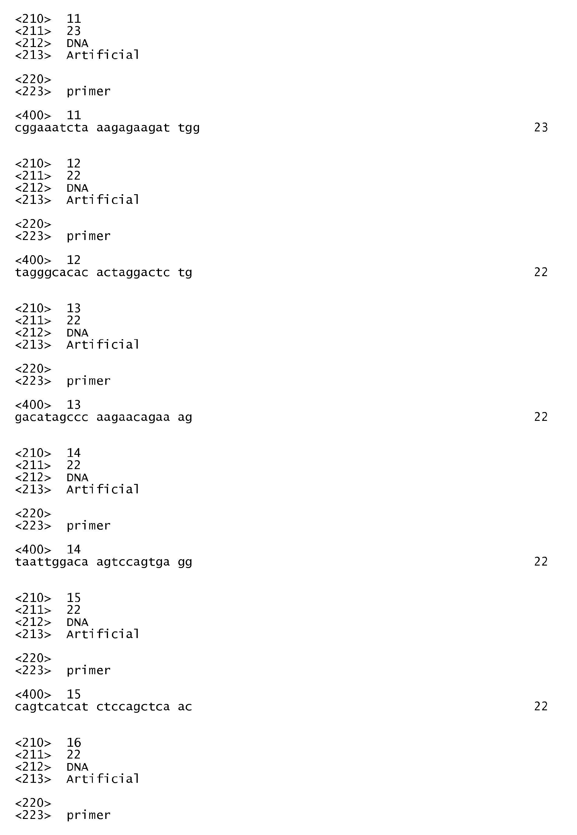


Claims (12)
- Method for the selection of compounds which permit to enhance the level of expression of a gene implicated in antimicrobial defence comprising the step of:a) contacting a test compound with an host cell expressing a reporter nucleic acid comprising a nucleic acid sequence coding for a reporter placed under the control of a promoter, which promoter comprises all or part of the promoter sequence of said gene implicated in antimicrobial defence, andb) measuring the level of expression of the reporter gene.
- The method according to claim 1, wherein the reporter gene is the GFP (Green Fluorescent Protein) gene, preferably the eGFP (enhanced Green Fluorescent Protein) gene.
- The method according to claim 1 or 2, wherein the promoter sequence is selected from the group comprising DEFB4 promoter sequence, DEFB103 promoter sequence, CAMP promoter sequence, CCL20 promoter sequence and CCR6 promoter sequence.
- The method according to claim 3, wherein the promoter sequence is selected from the group comprising human DEFB4 promoter sequence (SEQ ID NO: 1), human DEFB103 promoter sequence (SEQ ID NO: 2), human CAMP promoter sequence (SEQ ID NO: 3), human CCL20 promoter sequence (SEQ ID NO: 4) and human CCR6 promoter sequence.
- The method according to any of the claims 1 to 4, wherein said method further comprises a step c) of comparing the level of expression of the reporter gene as measured in step b) with the level of expression of the reporter gene in the absence of said test compound.
- The method according to any of the claims 1 to 5, wherein the host cell is infected by Shigella, preferably Shigella flexneri.
- The method according to any of the claims 1 to 6, wherein the host cell has been transformed with a vector selected in the group consiting of plasmids pLenti/R4R2/V5DEST-pBS5 comprising human DEFB4 promoter fused to the eGFP reporter nucleic acid sequence, pLenti/R4R2/V5DEST-pBS7 comprising human DEFB103 promoter fused to the eGFP reporter nucleic acid sequence, and pLenti/R4R2/V5DEST-pBS9 comprising human CAMP(LL37) promoter fused to the eGFP reporter nucleic acid sequence, which plasmids are present in the Stb13 E. coli bacterial strains deposited at the CNCM on April 4, 2008 with the numbers I-3965, I-3966 and I-3967 respectively.
- The method according to any of the claims 1 to 6, wherein the host cell is an epithelial cell selected from the group comprising human colonic epithelial cells HT29 (ATCC HTB-38D), Caco-2 (ATCC HTB-37) or TC7 cell line which is a subclone of Caco-2..
- The method according to any one of claims 1 to 8, wherein said method permits to select compounds which can be useful for treating and/or preventing a microbial infection, preferably a Shigella infection, and more preferably a Shigella flexneri infection.
- Pharmaceutical composition comprising at least one compound identified by the method according to any of the claims 1 to 9 and, optionally, a pharmaceutically acceptable carrier.
- Use of at least one compound identified by the method according to any of the claims 1 to 9 for preventing and/or treating an microbial infection or for preventing and/or treating cancer in a subject.
- The use according to claim 11 for preventing and/or treating a Shigella infection in a subject, preferably a Shigella flexneri infection.
Priority Applications (2)
| Application Number | Priority Date | Filing Date | Title |
|---|---|---|---|
| EP08305094A EP2108706A1 (en) | 2008-04-10 | 2008-04-10 | New method for selecting compounds useful for treating and/or preventing microbial Shigella infection |
| PCT/EP2009/054347 WO2009125016A1 (en) | 2008-04-10 | 2009-04-10 | New method for selecting compounds useful for treating and/or preventing microbial shigella infection |
Applications Claiming Priority (1)
| Application Number | Priority Date | Filing Date | Title |
|---|---|---|---|
| EP08305094A EP2108706A1 (en) | 2008-04-10 | 2008-04-10 | New method for selecting compounds useful for treating and/or preventing microbial Shigella infection |
Publications (1)
| Publication Number | Publication Date |
|---|---|
| EP2108706A1 true EP2108706A1 (en) | 2009-10-14 |
Family
ID=39734968
Family Applications (1)
| Application Number | Title | Priority Date | Filing Date |
|---|---|---|---|
| EP08305094A Withdrawn EP2108706A1 (en) | 2008-04-10 | 2008-04-10 | New method for selecting compounds useful for treating and/or preventing microbial Shigella infection |
Country Status (2)
| Country | Link |
|---|---|
| EP (1) | EP2108706A1 (en) |
| WO (1) | WO2009125016A1 (en) |
Cited By (2)
| Publication number | Priority date | Publication date | Assignee | Title |
|---|---|---|---|---|
| US20120232133A1 (en) * | 2011-02-22 | 2012-09-13 | California Institute Of Technology | Delivery of proteins using adeno-associated virus (aav) vectors |
| US9943611B2 (en) | 2012-11-01 | 2018-04-17 | California Institute Of Technology | Reversible gene expression |
Citations (4)
| Publication number | Priority date | Publication date | Assignee | Title |
|---|---|---|---|---|
| WO1999049876A2 (en) * | 1998-03-31 | 1999-10-07 | Abbott Laboratories | Bacterial or yeast extracts which stimulate the production of defensins and methods of use thereof |
| WO2000037077A1 (en) | 1998-12-23 | 2000-06-29 | Glaxo Group Limited | Assays for ligands for nuclear receptors |
| US20070077254A1 (en) * | 2005-09-15 | 2007-04-05 | Philippe Sansonetti | Targeting the histone code as a bacterial strategy for selectively modulating gene expression |
| US20070134711A1 (en) * | 2002-02-04 | 2007-06-14 | Bonagura Vincent R | Compositions of eleutherosides capable of modulating protein expresion |
Family Cites Families (1)
| Publication number | Priority date | Publication date | Assignee | Title |
|---|---|---|---|---|
| WO2004087076A2 (en) * | 2003-03-31 | 2004-10-14 | The Rockefeller University | Methods for inhibiting adipogenesis and for treating type 2 diabetes |
-
2008
- 2008-04-10 EP EP08305094A patent/EP2108706A1/en not_active Withdrawn
-
2009
- 2009-04-10 WO PCT/EP2009/054347 patent/WO2009125016A1/en not_active Ceased
Patent Citations (4)
| Publication number | Priority date | Publication date | Assignee | Title |
|---|---|---|---|---|
| WO1999049876A2 (en) * | 1998-03-31 | 1999-10-07 | Abbott Laboratories | Bacterial or yeast extracts which stimulate the production of defensins and methods of use thereof |
| WO2000037077A1 (en) | 1998-12-23 | 2000-06-29 | Glaxo Group Limited | Assays for ligands for nuclear receptors |
| US20070134711A1 (en) * | 2002-02-04 | 2007-06-14 | Bonagura Vincent R | Compositions of eleutherosides capable of modulating protein expresion |
| US20070077254A1 (en) * | 2005-09-15 | 2007-04-05 | Philippe Sansonetti | Targeting the histone code as a bacterial strategy for selectively modulating gene expression |
Non-Patent Citations (46)
| Title |
|---|
| "Ullmann' Encyclopedia of Industrial Chemistry", 1989, MARCEL DEKKER |
| ALLAOUI ET AL., MOLECULAR MICROBIOLOGY, vol. 7, 1993, pages 59 - 68 |
| ANSEL ET AL.: "Pharmaceutical Dosage Forms and Drug Delivery Systems", 1994, WILLIAMS & WILKINS |
| ARBIBE ET AL., NATURE IMMUNOLOGY, vol. 8, 2007, pages 47 - 56 |
| BENJELLOUN-TOUIMI ET AL., MICROBIOLOGY (READING, ENGLAND, vol. 144, no. 7, 1998, pages 1815 - 1822 |
| BERGMAN PETER ET AL: "Neisseria gonorrhoeae downregulates expression of the human antimicrobial peptide LL-37.", CELLULAR MICROBIOLOGY JUL 2005, vol. 7, no. 7, July 2005 (2005-07-01), pages 1009 - 1017, XP002495635, ISSN: 1462-5814 * |
| CHANTRET ET AL., J. CELL SCI., vol. 107, 1994, pages 213 - 225 |
| CHANTRET ET AL., JOURNAL OF CELL SCIENCE, vol. 107, no. 1, 1994, pages 213 - 225 |
| DIEU-NOSJEAN ET AL., JOURNAL OF LEUKOCYTE BIOLOGY, vol. 66, 1999, pages 252 - 262 |
| FERNANDEZ M I ET AL: "A newborn mouse model for the study of intestinal pathogenesis of shigellosis.", CELLULAR MICROBIOLOGY JUL 2003, vol. 5, no. 7, July 2003 (2003-07-01), pages 481 - 491, XP002495638, ISSN: 1462-5814 * |
| FRITZ ET AL., NATURE IMMUNOLOGY, vol. 7, 2006, pages 1250 - 1257 |
| HARDER ET AL., J. INVEST. DERMATOL., vol. 123, no. 3, 2004, pages 522 - 9 |
| HASE ET AL., INFECTION AND IMMUNITY, vol. 70, 2002, pages 953 - 963 |
| HOFFMANN; KAFATOS, SCIENCE, vol. 284, 1999, pages 1313 - 1318 |
| IRIZARRY ET AL., BIOSTATISTICS, vol. 4, 2003, pages 249 - 264 |
| ISLAM D ET AL: "Downregulation of bactericidal peptides in enteric infections: a novel immune escape mechanism with bacterial DNA as a potential regulator.", NATURE MEDICINE FEB 2001, vol. 7, no. 2, February 2001 (2001-02-01), pages 180 - 185, XP002495633, ISSN: 1078-8956 * |
| JAIN ET AL., BIOINFORMATICS, vol. 19, 2003, pages 1945 - 1951 |
| JANEWAY; MEDZHITOV, ANNUAL REVIEW OF IMMUNOLOGY, vol. 20, 2006, pages 197 - 216 |
| JEPRAS ET AL., ANTIMICROBIAL AGENTS AND CHEMOTHERAPY, vol. 41, 1997, pages 2001 - 2005 |
| JOLY ET AL., MOLECULAR IMMUNOLOGY, vol. 42, 2005, pages 1073 - 1084 |
| KIM ET AL., PROCEEDINGS OF THE NATIONAL ACADEMY OF SCIENCES OF THE UNITED STATES OF AMERICA, vol. 102, 2005, pages 14046 - 14051 |
| KRAMER ET AL., PLOS PATHOGENS, vol. 3, 2007, pages 21 |
| LE GALL ET AL., MICROBIOLOGY (READING, ENGLAND, vol. 151, 2005, pages 951 - 962 |
| LEHRER R: "Shigellae control the Gates of LL.", NATURE MEDICINE FEB 2001, vol. 7, no. 2, February 2001 (2001-02-01), pages 158 - 159, XP002495634, ISSN: 1078-8956 * |
| LI ET AL., SCIENCE, vol. 315, 2007, pages 1000 - 1003 |
| LI; WONG, GENOME INFORMATICS, vol. 12, 2001, pages 3 - 13 |
| MAVRIS ET AL., MOLECULAR MICROBIOLOGY, vol. 43, 2002, pages 1543 - 1553 |
| MOUNIER ET AL., INFECTION AND IMMUNITY, vol. 60, 1992, pages 237 - 248 |
| ONG P Y ET AL: "ENDOGENOUS ANTIMICROBIAL PEPTIDES AND SKIN INFECTIONS IN ATOPIC DERMATITIS", NEW ENGLAND JOURNAL OF MEDICINE, THE, MASSACHUSETTS MEDICAL SOCIETY, WALTHAM, MA, US, vol. 347, no. 15, 10 October 2002 (2002-10-10), pages 1151 - 1160, XP008028910, ISSN: 0028-4793 * |
| PEDRON ET AL., JOURNAL OF BIOLOGICAL CHEMISTRY, vol. 278, 2003, pages 33878 - 33886 |
| PERDOMO ET AL., JOURNAL OF EXPERIMENTAL MEDICINE, vol. 180, 1994, pages 1307 - 1319 |
| RAQIB RUBHANA ET AL: "Improved outcome in shigellosis associated with butyrate induction of an endogenous peptide antibiotic.", PROCEEDINGS OF THE NATIONAL ACADEMY OF SCIENCES OF THE UNITED STATES OF AMERICA 13 JUN 2006, vol. 103, no. 24, 13 June 2006 (2006-06-13), pages 9178 - 9183, XP002495632, ISSN: 0027-8424 * |
| SANSONETTI & EGILE, vol. 74, 1998, pages 191 - 197 |
| SANSONETTI ET AL., INFECTION AND IMMUNITY, vol. 35, 1982, pages 852 - 860 |
| SAWAMURA D ET AL: "Beta defensin-3 engineered epidermis shows highly protective effect for bacterial infection.", GENE THERAPY MAY 2005, vol. 12, no. 10, May 2005 (2005-05-01), pages 857 - 861, XP002495637, ISSN: 0969-7128 * |
| SCHAUBER ET AL., GUT, vol. 52, no. 5, 2003, pages 735 - 41 |
| SCHUTYSER E ET AL: "The CC chemokine CCL20 and its receptor CCR6", CYTOKINE AND GROWTH FACTOR REVIEWS, OXFORD, GB, vol. 14, no. 5, 1 October 2003 (2003-10-01), pages 409 - 426, XP002984466, ISSN: 1359-6101 * |
| SELSTED; OUELLETTE, NATURE IMMUNOLOGY, vol. 6, 2005, pages 551 - 557 |
| SEYDEL ET AL., INFECTION AND IMMUNITY, vol. 65, 1997, pages 1631 - 1639 |
| SINGH ET AL., PROC. NATL. ACAD. SCI. USA., vol. 95, no. 25, 1998, pages 14961 - 6 |
| SPERANDIO BRICE ET AL: "Virulent Shigella flexneri subverts the host innate immune response through manipulation of antimicrobial peptide gene expression.", THE JOURNAL OF EXPERIMENTAL MEDICINE 12 MAY 2008, vol. 205, no. 5, 12 May 2008 (2008-05-12), pages 1121 - 1132, XP002495639, ISSN: 1540-9538 * |
| TAGGART CLIFFORD C ET AL: "Inactivation of human beta-defensins 2 and 3 by elastolytic cathepsins.", JOURNAL OF IMMUNOLOGY (BALTIMORE, MD. : 1950) 15 JUL 2003, vol. 171, no. 2, 15 July 2003 (2003-07-15), pages 931 - 937, XP002495636, ISSN: 0022-1767 * |
| WASSEF ET AL., INFECTION AND IMMUNITY, vol. 57, 1989, pages 858 - 863 |
| YANG D ET AL: "(beta)-Defensins: linking innate and adaptive immunity through dendritic and T cell CCR6", SCIENCE, AMERICAN ASSOCIATION FOR THE ADVANCEMENT OF SCIENCE, US, WASHINGTON, DC, vol. 286, 15 October 1999 (1999-10-15), pages 525 - 528, XP002955546, ISSN: 0036-8075 * |
| ZASLOFF, NATURE, vol. 415, 2002, pages 389 - 395 |
| ZYCHLINSKY ET AL., NATURE, vol. 358, 1992, pages 167 - 169 |
Cited By (4)
| Publication number | Priority date | Publication date | Assignee | Title |
|---|---|---|---|---|
| US20120232133A1 (en) * | 2011-02-22 | 2012-09-13 | California Institute Of Technology | Delivery of proteins using adeno-associated virus (aav) vectors |
| US8865881B2 (en) * | 2011-02-22 | 2014-10-21 | California Institute Of Technology | Delivery of proteins using adeno-associated virus (AAV) vectors |
| US9527904B2 (en) | 2011-02-22 | 2016-12-27 | California Institute Of Technology | Delivery of proteins using adeno-associated virus (AAV) vectors |
| US9943611B2 (en) | 2012-11-01 | 2018-04-17 | California Institute Of Technology | Reversible gene expression |
Also Published As
| Publication number | Publication date |
|---|---|
| WO2009125016A1 (en) | 2009-10-15 |
Similar Documents
| Publication | Publication Date | Title |
|---|---|---|
| Sperandio et al. | Virulent Shigella flexneri subverts the host innate immune response through manipulation of antimicrobial peptide gene expression | |
| CN108192972B (en) | Methods for the diagnosis, prognosis and treatment of breast cancer metastasis | |
| Desai et al. | Hypoxia and temperature regulated morphogenesis in Candida albicans | |
| US20050037955A1 (en) | Therapeutic protein and treatments | |
| Lau et al. | Cell division defects in Escherichia coli deficient in the multidrug efflux transporter AcrEF-TolC | |
| WO2017122815A1 (en) | Novel fusant and method for detecting same | |
| JP2002505849A (en) | Virulence-related nucleic acid sequences and uses thereof | |
| He et al. | The CDT of Helicobacter hepaticus induces pro-survival autophagy and nucleoplasmic reticulum formation concentrating the RNA binding proteins UNR/CSDE1 and P62/SQSTM1 | |
| US6068972A (en) | Methods and compositions for reducing bacterial tolerance to antibacterials, disinfectants and organic solvents | |
| Lemberg | Martinez de San Vicente K, Fróis-Martins R, Altmeier S, Tran VDT, Mertens S, et al.(2022) Candida albicans commensalism in the oral mucosa is favoured by limited virulence and metabolic adaptation | |
| Buisson et al. | Macrophages versus Escherichia coli: a decisive fight in Crohn's disease | |
| US20160250303A1 (en) | Perforin-2 activators and inhibitors as drug targets for infectious disease and gut inflammation | |
| WO2022244807A1 (en) | Ltk fusion gene | |
| WO2009125016A1 (en) | New method for selecting compounds useful for treating and/or preventing microbial shigella infection | |
| US8685393B2 (en) | Methods and compositions for the treatment and diagnosis of systemic anthrax infection | |
| KR20240161931A (en) | Discovery of target for inhibition of fibrosis in inflammatory bowel disease | |
| CN111979328B (en) | Biomarker for predicting liver cancer lung metastasis and application thereof | |
| Čerina et al. | The DanRI regulatory system in uropathogenic Escherichia coli subverts neutrophil responses | |
| US9176135B2 (en) | Method for predicting and preventing cardiovascular disease | |
| Chen et al. | PGLYRP-1 mediated intracellular peptidoglycan detection promotes mucosal protection | |
| JP6806440B2 (en) | New fusion and its detection method | |
| Wottawa | Epithelial endoplasmic reticulum stress predisposes for CMV infection by suppression of cGAS/STING/type I interferon-signalling | |
| Fróis-Martins et al. | Candida albicans commensalism in the oral mucosa is favoured by limited virulence and metabolic adaptation | |
| Reinecker et al. | ARHGEF2 Isoform Switching Couples Intestinal Epithelial Barrier Function with Autophagic Response to Pathogens | |
| Taylor | Il-17-Mediated Transcription Factors in Disease |
Legal Events
| Date | Code | Title | Description |
|---|---|---|---|
| PUAI | Public reference made under article 153(3) epc to a published international application that has entered the european phase |
Free format text: ORIGINAL CODE: 0009012 |
|
| AK | Designated contracting states |
Kind code of ref document: A1 Designated state(s): AT BE BG CH CY CZ DE DK EE ES FI FR GB GR HR HU IE IS IT LI LT LU LV MC MT NL NO PL PT RO SE SI SK TR |
|
| AX | Request for extension of the european patent |
Extension state: AL BA MK RS |
|
| AKX | Designation fees paid | ||
| REG | Reference to a national code |
Ref country code: DE Ref legal event code: 8566 |
|
| STAA | Information on the status of an ep patent application or granted ep patent |
Free format text: STATUS: THE APPLICATION IS DEEMED TO BE WITHDRAWN |
|
| 18D | Application deemed to be withdrawn |
Effective date: 20100415 |