EP2096120B1 - Use of secreted protein products for preventing and treating pancreatic diseases and/or obesity and/or metabolic syndrome - Google Patents
Use of secreted protein products for preventing and treating pancreatic diseases and/or obesity and/or metabolic syndrome Download PDFInfo
- Publication number
- EP2096120B1 EP2096120B1 EP09163270A EP09163270A EP2096120B1 EP 2096120 B1 EP2096120 B1 EP 2096120B1 EP 09163270 A EP09163270 A EP 09163270A EP 09163270 A EP09163270 A EP 09163270A EP 2096120 B1 EP2096120 B1 EP 2096120B1
- Authority
- EP
- European Patent Office
- Prior art keywords
- nucleic acid
- seq
- acid molecule
- polypeptide
- protein
- Prior art date
- Legal status (The legal status is an assumption and is not a legal conclusion. Google has not performed a legal analysis and makes no representation as to the accuracy of the status listed.)
- Expired - Lifetime
Links
Images







Classifications
-
- A—HUMAN NECESSITIES
- A61—MEDICAL OR VETERINARY SCIENCE; HYGIENE
- A61K—PREPARATIONS FOR MEDICAL, DENTAL OR TOILETRY PURPOSES
- A61K38/00—Medicinal preparations containing peptides
- A61K38/16—Peptides having more than 20 amino acids; Gastrins; Somatostatins; Melanotropins; Derivatives thereof
- A61K38/55—Protease inhibitors
- A61K38/57—Protease inhibitors from animals; from humans
-
- A—HUMAN NECESSITIES
- A61—MEDICAL OR VETERINARY SCIENCE; HYGIENE
- A61K—PREPARATIONS FOR MEDICAL, DENTAL OR TOILETRY PURPOSES
- A61K38/00—Medicinal preparations containing peptides
- A61K38/16—Peptides having more than 20 amino acids; Gastrins; Somatostatins; Melanotropins; Derivatives thereof
- A61K38/17—Peptides having more than 20 amino acids; Gastrins; Somatostatins; Melanotropins; Derivatives thereof from animals; from humans
- A61K38/1703—Peptides having more than 20 amino acids; Gastrins; Somatostatins; Melanotropins; Derivatives thereof from animals; from humans from vertebrates
- A61K38/1709—Peptides having more than 20 amino acids; Gastrins; Somatostatins; Melanotropins; Derivatives thereof from animals; from humans from vertebrates from mammals
-
- A—HUMAN NECESSITIES
- A61—MEDICAL OR VETERINARY SCIENCE; HYGIENE
- A61K—PREPARATIONS FOR MEDICAL, DENTAL OR TOILETRY PURPOSES
- A61K38/00—Medicinal preparations containing peptides
- A61K38/16—Peptides having more than 20 amino acids; Gastrins; Somatostatins; Melanotropins; Derivatives thereof
- A61K38/17—Peptides having more than 20 amino acids; Gastrins; Somatostatins; Melanotropins; Derivatives thereof from animals; from humans
- A61K38/177—Receptors; Cell surface antigens; Cell surface determinants
-
- A—HUMAN NECESSITIES
- A61—MEDICAL OR VETERINARY SCIENCE; HYGIENE
- A61K—PREPARATIONS FOR MEDICAL, DENTAL OR TOILETRY PURPOSES
- A61K38/00—Medicinal preparations containing peptides
- A61K38/16—Peptides having more than 20 amino acids; Gastrins; Somatostatins; Melanotropins; Derivatives thereof
- A61K38/17—Peptides having more than 20 amino acids; Gastrins; Somatostatins; Melanotropins; Derivatives thereof from animals; from humans
- A61K38/177—Receptors; Cell surface antigens; Cell surface determinants
- A61K38/179—Receptors; Cell surface antigens; Cell surface determinants for growth factors; for growth regulators
-
- A—HUMAN NECESSITIES
- A61—MEDICAL OR VETERINARY SCIENCE; HYGIENE
- A61K—PREPARATIONS FOR MEDICAL, DENTAL OR TOILETRY PURPOSES
- A61K38/00—Medicinal preparations containing peptides
- A61K38/16—Peptides having more than 20 amino acids; Gastrins; Somatostatins; Melanotropins; Derivatives thereof
- A61K38/17—Peptides having more than 20 amino acids; Gastrins; Somatostatins; Melanotropins; Derivatives thereof from animals; from humans
- A61K38/39—Connective tissue peptides, e.g. collagen, elastin, laminin, fibronectin, vitronectin, cold insoluble globulin [CIG]
-
- A—HUMAN NECESSITIES
- A61—MEDICAL OR VETERINARY SCIENCE; HYGIENE
- A61K—PREPARATIONS FOR MEDICAL, DENTAL OR TOILETRY PURPOSES
- A61K38/00—Medicinal preparations containing peptides
- A61K38/16—Peptides having more than 20 amino acids; Gastrins; Somatostatins; Melanotropins; Derivatives thereof
- A61K38/43—Enzymes; Proenzymes; Derivatives thereof
- A61K38/46—Hydrolases (3)
- A61K38/48—Hydrolases (3) acting on peptide bonds (3.4)
- A61K38/4813—Exopeptidases (3.4.11. to 3.4.19)
-
- A—HUMAN NECESSITIES
- A61—MEDICAL OR VETERINARY SCIENCE; HYGIENE
- A61K—PREPARATIONS FOR MEDICAL, DENTAL OR TOILETRY PURPOSES
- A61K38/00—Medicinal preparations containing peptides
- A61K38/16—Peptides having more than 20 amino acids; Gastrins; Somatostatins; Melanotropins; Derivatives thereof
- A61K38/43—Enzymes; Proenzymes; Derivatives thereof
- A61K38/52—Isomerases (5)
-
- A—HUMAN NECESSITIES
- A61—MEDICAL OR VETERINARY SCIENCE; HYGIENE
- A61P—SPECIFIC THERAPEUTIC ACTIVITY OF CHEMICAL COMPOUNDS OR MEDICINAL PREPARATIONS
- A61P1/00—Drugs for disorders of the alimentary tract or the digestive system
- A61P1/18—Drugs for disorders of the alimentary tract or the digestive system for pancreatic disorders, e.g. pancreatic enzymes
-
- A—HUMAN NECESSITIES
- A61—MEDICAL OR VETERINARY SCIENCE; HYGIENE
- A61P—SPECIFIC THERAPEUTIC ACTIVITY OF CHEMICAL COMPOUNDS OR MEDICINAL PREPARATIONS
- A61P3/00—Drugs for disorders of the metabolism
- A61P3/04—Anorexiants; Antiobesity agents
-
- A—HUMAN NECESSITIES
- A61—MEDICAL OR VETERINARY SCIENCE; HYGIENE
- A61P—SPECIFIC THERAPEUTIC ACTIVITY OF CHEMICAL COMPOUNDS OR MEDICINAL PREPARATIONS
- A61P3/00—Drugs for disorders of the metabolism
- A61P3/08—Drugs for disorders of the metabolism for glucose homeostasis
- A61P3/10—Drugs for disorders of the metabolism for glucose homeostasis for hyperglycaemia, e.g. antidiabetics
Definitions
- This invention rotates to the use of secreted SF06 proteins, and to the use of polynucleotides encoding these, in the diagnosis, and study of diabetes, obesity and/or metabolic syndrome.
- human proteins serve as pharmaceutical active compounds.
- Several classes of human proteins that serve as such active compounds include hormones, cytokines, can growth factors, and cell differentiation factors.
- Most proteins that can be used as a pharmaceutical active compound fall within the family of secreted proteins.
- Secreted proteins are generally produced within cells at rough endoplasmic reticulum, are then exported to the golgl complex and then move to secretory vesicles or granules, where they are secreted to the exterior of the cell via oxocytosis.
- Examples for commercially used secreted proteins are human insulin, thrombolytic agents, interferons, interleukins, colony stimulating factors, human growth hormone, transforming growth factor beta, tissue plasminogen activator, erythropoletin, and various other proteins.
- Receptors of secreted proteins which are membrane-bound proteins, also have potential as therapeutic or diagnostic agents. It is, therefore, important for developing new pharmaceutical compounds to identify secreted proteins that can be tested for activity in a variety of animal models.
- the pancreas is an essential organ possessing both an exocrine function involved In the delivery of enzymes into the digestive tract and an endocrine function by which various hormones are secreted into the blood stream.
- the exocrine function is assured by acinar and centroacinar cells that produce various digestive enzymes and intercalated ducts that transport these enzyme in alkaline solution to the duodenum.
- the functional unit of the endocrine pancreas is the islet of Langerhans. Islets are scattered throughout the exocrine portion of the pancreas and are composed of four cell types: alpha-, beta-, delta- and PP-cells, reviewed for example in Kim S.K. and Hebrok M., (2001) Genes Dev.
- Beta-cells produce insulin, represent the majority of the endocrine cells and form the core of the islets, while alpha-cells secrete glucagon and are located in the periphery. Delta-cells and PP-cells are less numerous and secrete somatostatin and pancreatic polypeptide, respectively.
- pancreatic development has been well studied in different species, including chicken, zebrafish, and mice (for a detailed review, see Kim & Hebrok, 2001, supra).
- the pancreas develops from distinct dorsal and ventral anlagen.
- Pancreas development requires specification of the pancreas strom along both anterior-posterior and dorsal-ventral axes.
- a number of factors, which are critical for proper pancreatic development have been identified (see Kim & Hebrok, 2001, supra; Wilson M.E. et al., (2003) Mech Dev. 120: 65-80 ).
- Pancreatic beta-cells secrete insulin, which is stimulated by high blood glucose levels. Insulin amongst other hormones plays a key role in the regulation of the fuel metabolism. Insulin leads to the storage of glycogen and triglycerides and to the synthesis of proteins. The entry of glucose into muscles and adipose cells is stimulated by insulin. In patients who suffer from diabetes mellitus the amount of insulin produced by the pancreatic islet cells is too low, resulting in elevated blood glucose levels (hyperglycemia). In diabetes type I beta cells are lost due to autoimmune destruction. In type 2 diabetic patients, liver and muscle cells loose their ability to respond to normal blood insulin levels (insulin resistance).
- pancreatic islets In type I diabetics, the lifespan of pancreatic islets is dramatically shortened due to autoimmune destruction. Treatments have been devised which modulate the immune system and may be able to stop or strongly reduce islet destruction ( Raz I. et al., (2001) Lancet 358: 1749-1753 ; Chatenoud L. et al., (2003) Nat Rev Immunol. 3: 123-132 ). However, due to the relatively slow regeneration of human beta cells such treatments could only be fully successful at improving the diabetic condition if they are combined with an agent which can stimulate beta cell regeneration.
- Diabetes is a very disabling disease, because medications do not control blood sugar levels well enough to prevent swinging between high and low blood sugar levels.
- Patients with diabetes are at risk for major complications, including diabetic ketoacidosis, end-stage renal disease, diabetic retinopathy and amputation.
- diabetic ketoacidosis a host of related conditions, such as metabolic syndrome, obesity, hypertension, heart disease, peripheral vascular disease, and infections, for which persons with diabetes are at substantially increased risk.
- the treatment of these complications contributes to a considerable degree to the enormous cost which is imposed by diabetes on health care systems world wide.
- Obesity is one of the most prevalent metabolic disorders in the world. It is still a poorly understood human disease that becomes as a major health problem more and more relevant for western society. Obesity is defined as a body weight more than 20% in excess of the ideal body weight, frequently resulting in a significant impairment of health. Obesity may be measured by body mass index, an indicator of adiposity or fatness. Further parameters for defining obesity are waist circumferences, skinfold thickness and bioimpedance. It is associated with an increased risk for cardiovascular disease, hypertension, diabetes mellitus type II, hyperlipidaemia and an increased mortality rate.
- Obesity is influenced by genetic, metabolic, biochemical, psychological, and behavioral factors and can be caused by different reasons such as non-insulin dependent diabetes, increase in triglycerides, increase in carbohydrate bound energy and low energy expenditure ( Kopelman P.G., (2000) Nature 404: 635-643 ).
- metabolic syndrome (syndrome x, insulin-resistance syndrome, deadly quartet) was first described 1966 by Camus and reintroduced 1988 by Reaven ( Camus J.P., (1966) Rev Rhum Mal Osteoartic 33: 10-14 ; Reaven G.M., (1988), Diabetes 37: 1595-1607 ).
- metabolic syndrome is commonly defined as clustering of cardiovascular risk factors like hypertension, abdominal obesity, high blood levels of triglycerides and fasting glucose as well as low blood levels of HDL cholesterol. Insulin resistance greatly increases the risk of developing the metabolic syndrome ( Reaven G., (2002) Circulation 106: 286-288 ).
- the metabolic syndrome often precedes the development of type II diabetes and cardiovascular disease ( Lakka H.M. et al., (2002) JAMA 288: 2709-2716 ).
- the control of blood lipid levels and blood glucose levels is essential for the treatment of the metabolic syndrome (see, for example, Santomauro A.T. et al., (1999) Diabetes, 48: 1836-1841 ).
- pancreatic tissues There is a need in the prior art for the identification of candidate genes that are specifically expressed in early development in certain pancreatic tissues. These genes and the thereby encoded proteins can provide tools to the diagnosis and treatment of severe pancreatic disorders and related diseases. Therefore, this specification describes secreted proteins that are specifically expressed in pancreatic tissues early in the development. Further described is the use of these genes and proteins in the diagnosis, prevention and/or treatment of pancreatic dysfunctions, such as diabetes, and other related diseases such as obesity and/or metabolic syndrome. These proteins and genes are especially useful in regeneration processes, such as regeneration of the pancreas cells.
- SF01-SF13 secreted factory referred to as SF01-SF13, which are involved in pancreas development regeneration, and in the regulation of energy homeostasis.
- SF01-SF13 corresponds to mammalian proteins as described in Table 1.
- the invention relates to the use of a composition
- a composition comprising a SF06 protein as characterized by SEQ ID Nos:30 or 32 and/or a nucleic acid molecule encoding a SF06 protein as characterized by SEQ ID Nos:30 or 32, the nucleic acid molecule being preferably a DNA molecule, more preferably a cDNA or a genomic DNA, and optionally pharmaceutically acceptable carriers, diluents, and/or additives for the preparation of an agent for detecting diseases or dysfunctions selected from the group consisting of diabetes, obesity, and/or metabolic syndrome.
- the invention relates to the identification of a (poly)peptide involved in the regulation of energy homeostasis and/or metabolism of a mammal and to the screening of an agent, which modulates/effects the activity of a SF06 polypeptide. Any information about other SF01-05 and SF07-13 proteins is provided for the purpose of comparison only.
- SF01 a TNF-related molecule
- SF01 a TNF-related molecule
- SF02 is conserved from Drosophila to human.
- SF02 is a developmentally regulated vital protein.
- Drosophila SF02 mutants are dying from neural system defects.
- SF02 seems to be enriched in endoderm and embryo, and to be present in pancreas.
- SF02 Is present in pancreas, liver, thymus, and spleen.
- Glypicans are a family of glycosylphosphatidylinositol (GPI)-anchored cell surface heparan sulfate proteoglycans (HSPGs)
- HSPGs glycosylphosphatidylinositol
- SF03 is a member of the glypican family of heparan sulfate proteoglycans, which attaches to the cell membrane via a GPI anchor.
- SF03 is mutated in the Simpson-Golabi-Behmel syndrome (SGBS).
- SGBS is characterized by pre- and post-natal overgrowth and is a recessive X-linked condition.
- SF03 is expressed in embryonic mesodermal lung, liver and kidney tissues and is thought to interact with various growth factors to regulate tissue and organ growth.
- the SF04 gene is a shared precursor for alpha-microglobulin and bikunin.
- Alpha microglobulin is a lipocalin with immunosuppressive properties
- bikunin is a plasma proteinase inhibitor.
- the SF04 mRNA is strongly transcribed in liver parenchyma, pancreas, and intestine epithelium. The both encoded proteins are accordingly present in developing hepatocytes, pancreas, kidney, and gut.
- Bikunin functions as tumor suppressor.
- SF05 is a neural-specific serine protease inhibitor, which is expressed in the whole developing CNS in mouse. SF05 inhibits the extracellular protease tissue-type plasminogen activator and plasmin, but not thrombin. SF05 deficient mice are were viable and healthy, except behavioral defects. Mutant SF05 protein aggregates and causes familial dementia in humans.
- SF06 is secreted by brain, adrenal cortex and adrenocortical tumors. SF06 is involved in the regulation of steroid hormone secretion and the proliferation of adrenocortical cells as autocrine and/or paracrine factor.
- SF07 is a putative tumor suppressor gene, which is inactivated in hepatocarcinomas, colorectal cancer and non-small cell lung cancers.
- SF08 is a carboxypeptidase which has no known enzymatic activity. SF08 is expressed in developing bones and cartilage.
- the SF09 protein is 145 aa long and contains calcium ion binding EF-hand motifs. According to expressed sequence tag (EST) assembly, SF09 is expressed in many tissues including the pancreas.
- EST expressed sequence tag
- the mouse homologue of human SF10 is an extracellular integrin-binding matrix protein. Mutations of SF10 are frequent in patients with Smith-Magenis syndrome (SMS), a clinically recognizable multiple congenital anomaly/mental retardation syndrome.
- SMS Smith-Magenis syndrome
- SF11 is a cysteine-proteinase inhibitor for cathepsins B and L, which is well characterised.
- the expression of SF11 is controlled by TGF-beta and EGF in decidual cultures and by TGF-beta in astrocyte precursors.
- a glycosylated form of SF11 is required for a FGF-2-responsive neural stem cell proliferation.
- Combined delivery of FGF-2 and SF11 to the adult dentate gyrus stimulated neurogenesis.
- SF11 deficient mice showed reduced tumor growth.
- SF12 Is an extracellular matrix and plasma glycoprotein. Expression of SF12 in the adult pancreatic islet is mostly confined to the blood vessels.
- SF13 is a ubiquitously expressed cytosolic peptidyl-prolyl cis-trans Isomerase that is inhibited by the immunosuppressive drug cyclosporin.
- SF13 is a proinflammatory factor for T-lymphocytes.
- SF13 signals through CD147 (basigin) preceptor, and is expressed in acinar but not in islet membranes or MIN-6 cells.
- pancreatic developmental processes Described are specific genes and proteins encoded thereby and offectors/modulators thereof involved in the regulation of pancreatic function and metabolism, especially in pancreas diseases such as diabetes mellitus, e.g, insulin dependent diabetes mellitus and/or non insulin dependent diabetes mellitus, and/or metabolic syndrome, obesity, and/or related disorders such as coronary heart disease, eating disorder, cachexia, hypertension, hypercholesterolemia (dyslipidemia), liver fibrosis, and/or galistones. Further, described are specific genes and proteins encoded thereby and effectors/modulators thereof involved in the regeneration of pancreatic cells or tissues, e.g.
- cells having exocrinous functions such as acinar cells, centroacinar cells and/or ductal cells and/or cells having endocrinous functions, particularly cells in Langerhans islets such as alpha-, beta-, delta- and/or PP-cells, more particularly beta-cells.
- SF01-SP13 homologous proteins and nucleic acid molecules coding therefore are obtainable from vertebrate species.
- nucleic acids encoding the human SF06 protein as characterized by SEQ ID NOs: 30 or 32 is as defined in claim 4.
- the function of the mammalian SF01-SF13 in mammalian metabolism was validated by analysing the expression of the transcripts in different tissues and by analizing the role in adipocyte differentiation (see Examples 3 and 4 for more detail).
- Microarrays are analytical tools routinely used in bioanalysis.
- a microarray has molecules distributed over, and stably associated with, the surface of a Solid support.
- the term "microarray” refers to an arrangement of a plurality of polynucleotides, polypeptides, antibodies, or other chemical compounds on a substrate.
- Microarrays of polypeptides, polynucleotides, and/or antibodies have been developed and find use in a variety of applications, such as monitoring gene expression, drug discovery, gene sequencing, gene mapping, bacterial identification, and combinatorial chemistry.
- One area in particular in which microarrays find use is in gene expression analysis (see Example 4).
- array technology can be used to explore the expression of a single polymorphic gene or the expression profile of a large number of related or unrelated genes.
- arrays are employed to detect the expression of a specific gene or its variants.
- arrays provide a platform for identifying genes that are tissue specific, are affected by a substance being tested in a toxicology assay, are part of a signaling cascade, carry out housekeeping functions, or are specifically related to a particular genetic predisposition, condition, disease, or disorder.
- Microarrays may be prepared, used, and analyzed using methods known in the art (see for example, Brennan T.M., (1995) U.S. Patent No. 5,474,796 ; Schena M. et al., (1996) Proc. Natl. Acad. Sci. USA 93:10614-10619 ; Baldeschwieler J.D. et al., (1995) PCT application WO 95/251116 ; Shalon T.D. and Brown P.O., (1995) PCT application WO 95/35505 ; Heller R.A. et al., (1997) Proc. Natl. Acad. Sci. USA 94: 2150-2155 ; Heller, M.J.
- Oligonucleotides or longer fragments derived from any of the polynucleotides described herein may be used as elements on a microarray.
- the microarray can be used in transcript imaging techniques, which monitor the relative expression levels of large numbers of genes simultaneously as described below.
- the microarray may also be used to identify genetic variants, mutations, and polymorphisms. This information may be used to determine gene function, to understand the genetic basis of a disorder, to diagnose a disorder, to monitor progression/regression of disease as a function of gene expression, and to develop and monitor the activities of therapeutic agents in the treatment of disease. In particular, this information may be used to develop a pharmacogenomic profile of a patient in order to select the most appropriate and effective treatment regimen for that patient. For example, therapeutic agents, which are highly effective and display the fewest side effects may be selected for a patient based on his/her pharmacogenomic profile.
- SF02, SF03, and SF13 show differential expression in human primary adipocytes.
- a strong up-regulation is observed concerning the expression of SF02 and SF03 during the human adipocyte differentiation (see Fig. 3 and 5 ) and a strong down-regulation is observed concerning the expression of SF13 during the human adipocyte differentiation (see Fig. 9 ).
- the SF02 and SF03 proteins in preadipocyctes have the potential to enhance adipocyte differentiation
- the SF13 protein in preadipocyctes has the potential to enhance adipocyte differentiation at a very early stage. Therefore, the SF02, SF03, and SF13 proteins might play an essential role in adipogenesis.
- SF02, SF03, and SF13 are strong candidates for the manufacture of pharmaceutical compositions and medicaments for the treatment of conditions related to human metabolism, such as diabetes, obesity, and/or metabolic syndrome.
- the nucleic acid sequence encoding the mouse SF01 protein Is expressed in the region of the ventral pancreas.
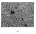
- the nucleic acid sequence encoding the mouse SF05 and SF06 proteins are expressed in the pancreas tissue (see Fig. 7 and 8 ).
- Hybridization conditions are based on the melting temperature (Tm) of the nucleic acid binding complex or probe, as described in Wahl G.M. et al., (1987; Methods Enzymol. 152: 399-407 ) and Kimmel A.R. (1987; Methods Enzymol. 162: 607-611 ), and may be used at a defined stringency.
- Tm melting temperature
- hybridization under stringent conditions means that after washing for 1 h with 1 x SSC and 0.1 % SDS at 50°C, preferably at 55°C, more preferably at 62°C and most preferably at 65°C, particularly for 1 h in 0.2 x SSC and 0.1% SDS at 50°C, preferably at 55°C, more preferably at 62°C and most preferably at 65°C, a positive hybridization signal is observed.
- the nucleotide sequences encoding the proteins or functional equivalents may be inserted into appropriate Expression vectors, i.e., a vector which contains the necessary elements for the transcription and translation of the inserted coding sequences.
- appropriate Expression vectors i.e., a vector which contains the necessary elements for the transcription and translation of the inserted coding sequences.
- Methods which are well known to those skilled in the art, may be used to construct expression vectors containing sequences encoding the proteins and the appropriate transcriptional and translational control elements.
- Regulatory elements include for Example a promoter, an initiation colon, a stop codon, a mRNA stability regulatory element, and a polyadenylation signal.
- a polynucleotide can be assured by (i) constitutive promoters such as the Cytomegalovirus (CMV) promoter/enhancer region, (ii) tissue specific promoters such as the insulin promoter (see, Soria B. et al., (2000), Diabetes 49: 157-162 ), SOX2 gene promoter (see U M. et al., (1998) Curr. Biol. 8: 971-974 ).
- Msi-1 promoter see Sakakibara S. and Okano H., (1997) J. Neuroscience 17: 8300-8312 ), alpha-cardia myosin heavy chain promoter or human atrial natriuretic factor promoter ( Klug M.G.
- Expression vectors can also contain a selection agent or marker gene that confers antibiotic resistance such as the neomycin, hygromycin or puromycin resistance genes.
- selection agent or marker gene confers antibiotic resistance such as the neomycin, hygromycin or puromycin resistance genes.
- natural, iodized or recombinant nucleic acid sequences encoding the SF06 proteins may be ligated to a heterologous sequence to encode a fusion protein.
- a variety of expression vector/host systems may be utilized to contain and express sequences encoding the proteins or fusion proteins. These include, but are not limited to, micro-organisms such as bacteria transformed with recombinant bacteriophage, plasmid or cosmic DNA expression vectors; yeast transformed with yeast expression vectors; insect cell systems infected with virus expression vectors (e.g., baculovirus, adenovirus, adeno-associated virus, lentiverus, retrovirus); plant cell systems transformed with virus expression vectors (e.g., cauliflower mosaic virus, CaMV; tobacco mosaic virus. TMV) or with bacterial expression vectors (e.g., Ti or PBR322 plasmids); or animal cell systems.
- micro-organisms such as bacteria transformed with recombinant bacteriophage, plasmid or cosmic DNA expression vectors; yeast transformed with yeast expression vectors; insect cell systems infected with virus expression vectors (e.g., baculovirus, adenovirus,
- polynucleotide sequences in a sample can be detected by DNA-DNA or DNA-RNA hybridization and/or amplification using probes or portions or fragments of said polynucleotides.
- Nucleic add amplification based assays involve the use of oligonucleotides or oligomers based on the sequences specific for the gene to detect transformants containing DNA or DNA encoding the corresponding protein.
- 'oligonucleotides' or 'oligomers' refer to a nucleic acid sequence of at least about 10 nucleotides and as many as about 60 nucleotides, preferably about 15 to 30 nucleotides, and more preferably about 20-25 nucleotides, which can be used as a probe or amplimer.
- Means for producing labeled hybridization or PCR probes for detecting polynucleotide sequences include oligo-labeling, nick translation, end-labeling of RNA probes, PCR amplification using a labeled nucleotide, or enzymatic synthesis. These procedures may be conducted using a variety of commercially available kits (Pharmacia & Upjohn, (Kalamazoo, Mich.); Promega (Madison Wis.); and U.S. Biochemical Corp., (Cleveland, Ohio).
- the presence of SF01-SF13 in a sample can be determined by immunological methods or activity measurement.
- a variety of protocols for detecting and measuring the expression of proteins, using either polyclonal or monoclonal antibodies specific for the protein or reagents for determining protein activity are known in the art. Examples include enzyme-linked immunosorbent assay (ELISA), radioimmunoassay (RIA), and fluoresoence activated cell sorting (FACS).
- ELISA enzyme-linked immunosorbent assay
- RIA radioimmunoassay
- FACS fluoresoence activated cell sorting
- a two-site, monoclonal-based immunoassay utilizing monoclonal antibodies reactive to two non-interfering epitopes on the protein is preferred, but a competitive binding assay may be employed.
- Suitable reporter molecules or labels include radionuclides, enzymes, fluorescent, chemiluminescent or chromogenic agents as well as substrates, co-factors, inhibitors, magnetic particles, and the like.
- the nucleic acids encoding the SF06 proteins can be used to generate non-human transgenic animal or site specific gene modifications in cell lines non-human Transgenic animals may be made through homologous combination, where the normal locus of the genes encoding the proteins of the invention is altered. Alternatively, a nucleic acid construct is randomly integrated into the genome.
- Vectors for stable integration include plasmids, retroviruses and other animal viruses, YACs, and the like.
- the modified cells or animal are useful in the study of the function and regulation of the SF06 proteins. For example, a series of small deletions and/or substitutions may be made in the genes that encode the proteins to determine the role of particular domains of the protein, functions in pancreatic differentiation, etc.
- Specific constructs of interest include anti-sense molecules, which will block the expression of the SF06 proteins, or expression of dominant negative mutations.
- a detectable marker such as for example lac-Z, may be introduced in the locus of the genes, where up-regulation of expression of the genes will result In an easily detected change In phenotype.
- by providing expression of the SF06 proteins in cells in which they are not normally produced one can induce changes in cell behavior.
- DNA constructs for homologous recombination will comprise at least portions of the genes with the desired genetic modification, and will include regions of homology to the target locus.
- DNA constructs for random integration need not include regions of homology to mediate recombination.
- markers for positive and/or negative selection are includes.
- Methods for generating cells having targeted gene modifications through homologous recombination are known in the art.
- ES non-human embryonic stem
- an ES cell line may be employed, or embryonic cells may be obtained freshly from a host, e.g. mouse, rat, guinea pig etc. Such cells are grown on an appropriate fibroblast-feeder layer or grown in presence of leukemia inhibiting factor (LIF).
- LIF leukemia inhibiting factor
- the SF06 nucleic acids and proteins are useful in diagnostic applications concerning diabetes, (such as insulin dependent diabetes mellitus and/or non insulin dependent diabetes mellitus), obesity, and/or metabolic syndrome.
- diagnostic uses for the nucleic acids and proteins are, for example but not limited to, the following: (i) antibody target (diagnostic), (ii) diagnostic and/or prognostic marker, and / or (iii) research tools.
- SF06 nucleic acids and proteins are useful in diagnostic applications implicated in various embodiments as described below.
- the nucleic acids may further be useful in diagnostic applications, wherein the presence or amount of the nucleic acids or the proteins are to be assessed, selected diabetes, obesity and metabolic syndrome.
- a variety of protocols including ELISA, RIA, and FACS for measuring proteins are known in the art and provide a basis for diagnosing altered or abnormal levels of gene expression.
- Normal or standard values for gene expression are established by combining body fluids or cell extracts taken from normal mammalian subjects, preferably human, with antibodies to the protein under conditions suitable for complex formation. The amount of standard complex formation may be quantified by various methods, but preferably by photometric means. Quantities of protein expressed in control and diseases, samples e.g.. from biopsied tissues are compared with the standard values. Deviation between standard and subject values establishes the parameters for diagnosing disease.
- the polynucleotides specific for the SF06 proteins may be used for diagnostic purposes.
- the polynucleotides, which may be used, include oligonucleotide sequences, antisense . RNA and DNA molecules, and PNAs.
- the polynucleotides may be used to detect and quantitate gene expression in biopsied tissues in which gene expression may be correlated with disease.
- the diagnostic assay may be used to distinguish between absence, presence, and excess gene expression, and to monitor regulation of protein levels during therapeutic intervention.
- hybridization with probes which are capable of detecting polynucleotide sequences, including genomic sequences, encoding the SF06 proteins may be used to identify nucleic acid sequences which encode the respective protein.
- the hybridization probes may be DNA or RNA and are preferably derived from the nucleotide sequence of the polynucleotide encoding the SF06 proteins or from a genomic sequence including promoter, enhancer elements, and introns of the naturally occurring gene.
- Hybridization probes may be labeled by a variety of reporter groups, for example, radionuclides such as 32 P or 35 S or enzymatic labels, such as alkaline phosphatase coupled to the probe via avidin/biotin coupling systems, and the like.
- Polynucleotide sequences specific for SF06 proteins may be used for the diagnosis of conditions or diseases, which are associated with the expression of the proteins, selected from diabetes, obesity and metabolic syndrome. Polynucleotide sequences specific for the SF06 proteins may also be used to monitor the progress of patients receiving treatment for diabetes, obesity, and/or metabotic syndrome.
- the polynucleotide sequences may be used qualitative or quantitative assays, e.g. in Southern or Northern analysis, dot blot or other membrane-based technologies: in PCR technologies; or in dip stick, pin, ELISA or chip assays utilizing fluids or tissues from patient biopsies to detect altered gene expression.
- the SF06 nucleotide sequences may be useful in assays that detect activation or induction of the above metabolic diseases or dysfunctions.
- the nucleotide sequences may be labeled by standard methods, and added to a fluid or tissue sample from a patient under conditions suitable for the formation of hybridization complexes. After a suitable incubation period, the sample is washed and the signal is quantitated and compared with a standard value. The presence of altered levels of nucleotide sequences encoding the SF06 proteins in the sample indicates the presence of the associated disease.
- Such assays may also be used to evaluate the efficacy of a particular therapeutic treatment regimen in animal studies, in clinical trials or in monitoring the treatment of an individual patient.
- a normal or standard profile for expression is established. This may be accomplished by combining body fluids or cell extracts taken from normal subjects, either animal or human, with a sequence or a fragment thereof, which is specific for the nucleic acids encoding the SF06 proteins, under conditions suitable for hybridization or amplification. Standard hybridization may be quantified by comparing the values obtained from normal subjects with those from an experiments where a known amount of a substantially purified polynucleotide is used. Standard values obtained from normal samples may be compared with values obtained from samples from patients who are symptomatic for disease. Deviation between standard and subject values is used to establish the presence of disease.
- hybridization assays may be repeated on a regular basis to evaluate whether the level of expression in the patient begins to approximate that, which is observed in the normal patient.
- the results obtained from successive assays may be used to show the efficacy of treatment over a period ranging from several days to months.
- the presence of an unusual amount of transcript in biopsled tissue from an individual may indicate a predisposition for the development of the disease or may provide a means for detecting the disease prior to the appearance of actual clinical symptoms.
- a more definitive diagnosis of this type may allow health professionals to employ preventative measures or aggressive treatment earlier thereby preventing the development or further progression of the metabolic diseases and disorders.
- oligonucleotides designed from the sequences encoding the SF06 proteins may involve the use of PCR.
- Such oligomers may be chemically synthesized, generated enzymatically or produced from a recombinant source. Oligomers will preferably consist of two nucleotide sequences, one with sense orientation (5prime.fwdarw.3prime) and another with antisense (3prime.rarw.5prime), employed under optimized conditions for identification of a specific gene or condition. The same two oligomers, nested sets of oligomers or even a degenerate pool of oligomers may be employed under less stringent conditions for detection and/or quantification of closely related DNA or RNA sequences.
- the nucleic acid sequences may also be used to generate hybridization probes, which are useful for mapping the naturally occurring genomic sequence.
- the sequences may be mapped to a particular chromosome or to a specific region of the chromosome using well known techniques.
- Such techniques include FISH, FACS or artificial chromosome constructions, such as yeast artificial chromosomes, bacterial artificial chromosomes, bacterial P1 constructions or single chromosome cDNA libraries as reviewed in Price C.M., (1993) Blood Rev..7: 127-134 , and Trask B.J., (1991) Trends Genet. 7: 149-154 .
- FISH as described in Verma RS.
- the nucleotide sequences may be used to detect differences in gene sequences between normal, carrier or affected individuals.
- An analysis of polymorphisms e.g. single nucleotide polymorphism may be carried out
- in situ hybridization of chromosomal preparations and physical mapping techniques such as linkage analysis using established chromosomal markers may be used for extending genetic maps.
- physical mapping techniques such as linkage analysis using established chromosomal markers may be used for extending genetic maps.
- Often the placement of a gene on the chromosome of another mammalian species, such as mouse, may reveal associated markers even if the number or arm of a particular human chromosome is not known.
- New sequences can be assigned to chromosomal arms or parts thereof, by physical mapping. This provides valuable information to investigators searching for disease genes using positional cloning or other gene discovery techniques.
- any sequences mapping to that area may represent associated or regulatory genes for further investigation.
- the nucleotide sequences may also be used to detect differences in the chromosomal location due to translocation, inversion, etc. among normal, carrier or affected individuals.
- the SF06 proteins can be used for screening libraries of compounds in any of a variety of drug screening techniques.
- the protein or fragment thereof employed In such screening may be free in solution, affixed to a solid support, borne on a cell surface, or located intracellulary. The formation of binding complexes, between the SF06 proteins and the agent tested, may be measured. Agents could also, either directly or indirectly, influence the activity of the SF06 proteins.
- agents may also interfere with posttranslational modifications of the protein, such as phosphorylation and dephosphorylation, farnesylation, palmitoylation, acetylation, alkylation, ubiquitination, proteolytic processing, subcellular localization and degradation.
- agents could influence the dimerization or oligomerization of the SF06 proteins or, in a heterologous manner, of the SF06 proteins with other proteins, for example, but not exclusively, docking proteins, enzymes, receptors, or translation factors.
- Agents could also act on the physical interaction of the SF06 proteins with other proteins, which are required for protein function, for example, but not exclusively, their downstream signaling.
- binding of a fluorescently labeled peptide derived from the interacting proteins to the SF06 protein, or vice versa could be detected by a change in polarisation.
- binding partners which can be either the full length proteins as well as one binding partner as the full length protein and the other just represented as a peptide are fluorescently labeled
- binding could be detected by fluorescence energy transfer (FRET) from one fluorophore to the other.
- FRET fluorescence energy transfer
- SF06 proteins a variety of commercially available assay principles suitable for detection of protein-protein Interaction are well known in the art, for example but not exclusively AlphaScreen (PeridnElmer) or Scintillation Proximity Assays (SPA) by Amersham.
- SPA Scintillation Proximity Assays
- the interaction of the SF06 proteins with cellular proteins could be the basis for a cell-based screening assay, in which both proteins are fluorescently labeled and interaction of both proteins is detected by analysing cotranslocation of both proteins with a cellular imaging reader, as has been developed for example, but not exclusively, by Cellomics or EvotecOAI.
- the two or more binding partners can be different proteins with one being the SF06 protein, or in case of dimerization and/or oligomerization the SF06 protein itself.
- agent as used herein describes any molecule, e.g. protein or pharmaceutical, with the capability of altering or mimicking the physiological function of one or more of the SF06 proteins.
- Candidate agents encompass numerous chemical classes, though typically they are organic molecules, preferably small organic compounds having a molecular weight of more than 50 and less than about 2,500 Daltons.
- Candidate agents comprise functional groups necessary for structural Interaction with proteins, particularly hydrogen bonding, and typically include at least an amine, carbonyl, hydroxyl or carboxyl group, preferably at least two of the functional chemical groups.
- the candidate agents often comprise carbocyclic or heterocyclic structures and/or aromatic or polyaromatic structures substituted with one or more of the above functional groups.
- Candidate agents are also found among biomolecules including peptides, saccharides, fatty acids, steroids, purines, pyrimidines, nucleic acids and derivatives, structural analogs or combinations thereof.
- Candidate agents are obtained from a wide variety of sources including libraries of synthetic or natural compounds. For example, numerous means are available for random and directed synthesis of a wide variety of organic compounds and biomolecules, including expression of randomized oligonucleotides and oligopeptides.
- libraries of natural compounds in the form of bacterial, fungal, plant and animal extracts are available or readily produced.
- natural or synthetically produced libraries and compounds are readily modified through conventional chemical, physical and biochemical means, and may be used to produce combinatorial libraries.
- pharmacological agents may be subjected to directed or random chemical modifications, such as acylation, alkylation, esterification, amidification, etc. to produce structural analogs.
- the screening assay is a binding assay
- one or more of the molecules may be joined to a label, where the label can directly or indirectly provide a detectable signal.
- Another technique for drug screening provides for high throughput screening of compounds having suitable binding affinity to the protein of interest as described in published PCT application WO84/03564 .
- large numbers of different small test compounds e.g. aptamers, peptides, low-molecular weight compounds etc.
- the test compounds are reacted with the proteins or fragments thereof, and washed. Bound proteins are then detected by methods well known in the art. Purified proteins can also be coated directly onto plates for use in the aforementioned drug screening techniques.
- non-neutralizing antibodies can be used to capture the peptide and immobilize it on a solid support.
- SF06 proteins e.g. antibodies
- Such cell populations are useful in transplantation, for experimental evaluation, and as source of lineage and cell specific products, including mRNA species useful in identifying genes specifically expressed in these cells, and as target for the identification of factors of molecules that can affect them.
- Cells expressing the SF06 protein or which have been treated with the SF06 protein are useful in transplantation to provide a recipient with pancreatic islet cells, including insulin producing beta cells; for drug screening; experimental models of islet differentiation and interaction with other cell types; in vitro screening assays to define growth and differentiation factors, and to additionally characterize genes involved In islet development and regulation; and the like.
- the native cells may be used for these purposes, or they may be genetically modified to provide altered capabilities.
- Cells from a regenerating pancreas, from embryonic foregut, stomach and duodenum, or other sources of pancreatic progenitor cells may be used as a starting population.
- the progenitor cells may be obtained from any mammalian species, e.g. equine, bovine, porcine, canine, feline, rodent, e.g. mice, rats, hamster, primate, etc. particularly human.
- the cells are transfected with a DNA construct, e.g. a viral or non-viral vector containing a reporter gene. e.g. the lacZ gene or the GFP gene, under regulatory control of a promoter of a gene involved in for example beta-cell differentiation, e.g. a promoter of a gene stimulation beta-cell differentiation, preferably a Pax4 promoter.
- a promoter of a gene involved in for example beta-cell differentiation e.g. a promoter of a gene stimulation beta-cell differentiation, preferably a Pax4 promoter.
- the transfected cells are divided into aliquots and each aliquot is contacted with a test substance, e.g., candidate 1, candidate 2 and candidate 3.
- the activity of the reporter gene corresponds to the capability of the test compound to induce beta-cell differentiation.
- a medium throughput validation is carried out.
- the test compound is added to stem cells being cultivated and the Insulin production is determined.
- an initial high throughput assay such as the cell based assay outlined above where for example a Pax4 promoter is used as marker for beta-cell regeneration
- the activity of candidate molecules to induce beta-cell differentiation is tested in a validation assay comprising adding said compounds to the culture media of the embryoid bodies. Differentiation into insulin-producing cells is then evaluated, e.g. by comparison to wild type and/or Pax4 expressing ES cells to assess the effectiveness of a compound.
- the nucleic acids encoding the SF06 proteins can be used to generate transgenic cell lines and non-human animals. These transgenic non-human animals are useful in the study of the function and regulation of the SF06 proteins in vivo.
- Non-human transgenic animals particularly mammalian transgenic animals, can serve as a model system for the investigation of many developmental and cellular processes common to humans.
- a variety of non-human models of metabolic disorders can be used to test modulators of the SF06.
- Misexpression for example, over-expression or lack of expression
- of the SF06 protein, particular feeding conditions, and/or administration of biologically active compounds can create models of metabolic disorders.
- such assays use mouse models of insulin resistance and/or diabetes, such as mice carrying gene knockouts in the leptin pathway (for example, ob (leptin) or db (leptin receptor) mice), as described above.
- these mice could be used to test whether administration of a candidate modulator alters for example lipid accumulation In the liver, in plasma, or adipose tissues using standard assays well known in the art, such as FPLC, colorimetric assays, blood glucose level tests, Insulin tolerance tests and others.
- Non-human transgenic animals may be made through homologous recombination In embryonic stem cells, where the normal locus of the gene encoding the SF06 protein is mutated.
- a nucleic add construct encoding the protein is injected into cocytes and is randomly Integrated into the genome.
- One may also express the genes in tissues where they are not normally expressed or at abnormal times of development.
- variants of the genes like specific constructs expressing anti-sense molecules or expression of dominant negative mutations, which will block or alter the expression of the SF06, proteins may be randomly integrated into the genome.
- a detectable marker such as lac Z or luciferase may be introduced into the locus of the genes, where up-regulation of expression of the genes will result in an easily detectable change in phenotype.
- Vectors for stable integration include plasmids, retroviruses and other animal viruses, yeast artificial chromosomes (YACs), and the like.
- DNA constructs for homologous recombination will contain at least portions of the genes with the desired genetic modifications, and will include regions of homology to the target locus. Conveniently, markers for positive and negative selection are included. DNA constructs for random integration do not need to contain regions of homology to mediate recombination.
- DNA constructs for random integration will consist of the nucleic acids encoding the proteins, a regulatory element (promoter), an intron and a poly-adenylation signal.
- Methods for generating cells having targeted gene modifications through homologous recombination are known in the field.
- embryonic stem (ES) cells an ES cell line may be employed, or embryonic cells may be obtained freshly from a host, e.g. mouse, rat, guinea pig, etc. Such cells are grown on an appropriate fibroblast-feeder layer and are grown in the presence of leukemia inhibiting factor (LIF).
- LIF leukemia inhibiting factor
- ES or embryonic cells may be transfected and can then be used to produce transgenic animals. After transfection, the ES cells are plated onto a feeder layer in an appropriate medium.
- Cells containing the construct may be selected by employing a selection medium. After sufficient time for colonies to grow, they are picked and analyzed for the occurrence of homologous recombination. Colonies that are positive may then be used for embryo manipulation and morula aggregation. Briefly, morulae are obtained from 4 to 6 week old superovulated females, the Zona Pellucida is removed and the morulae are put into small depressions of a tissue culture dish. The ES cells are trypsinized, and the modified cells are placed into the depression closely to the morulae. On the following day the aggregates are transfered into the uterine horns of pseudopregnant females. Females are then allowed to go to term.
- Chimeric offsprings can be readily detected by a change in coat color and are subsequently screened for the transmission of the mutation Into the next generation (F1-generation). Offspring of the F1-generation are screened for the presence of the modified gene and males and females having the modification are mated to produce homozygous progeny. If the gene alterations cause lethality at some point in development, tissues or organs can be maintained as allogenic or congenic grafts or transplants, or In vitro culture.
- the transgenic animals may be any non-human mammal, such as laboratory animal, domestic animals, etc., for example, mouse, rat, guinea pig, sheep, cow, pig, and others. The transgenic animals may be used in functional studies, drug screening, and other applications and are useful in the study of the function and regulation of the SF06 proteins in vivo.
- a screen for secreted factors expressed in developing mouse pancreas was carried out according to methods known by those skilled in the art (see, for example Pera E.M. and De Robertis E.M., (2000) Mech Dev 96(2): 183-195 ) with several modifications.
- pancreatic bud During organogenesis, the pancreatic bud is surrounded and influenced by the associated mesenchyme. (see for example, Madsen O.D. et al., (1996) Eur. J. Biochem. 242: 435-445 and Slack, J.M., (1995) Development 121: 1569-1580 ). Recently, it was suggested, that white adipocytes origin directly from mesenchymal cells ( Atanossova P.K., (2003) Folia Med. 45: 41-45 ). During embryogenesis, the innervation and vascularization of the pancreas can be observed. Therefore, the tissue used in the screen might have contained besides pancreatic cells some adipocyte precursors, blood vessels, as well as neuronal cells.
- a mouse embryonic stage 9.5-15 pancreatic bud library was prepared in pCMVSPORT-6 vector using SUPERSCRIPT Plasmid System from Invitrogen according to the manufacturer's instructions.
- the non-amplified library was electroporated into MaxEff DH10B cells (Invitrogen).
- Bacterial clones were picked with sterile toothpicks from agar plates and cultured in 96-deep-well microtiter plates in LB-ampicillin (see Sambrook et al., supra). Aliquots of 8 cultures were pooled, and plasmid DNA was isolated using the BioRobot_9600 apparatus according to the manufacturer's instructions (Qiagen; QIAprep(r) Turbo BioRobot Kit. Human 293 cell culture cells were cultured in 75 ml tissue culture flasks in DMEM and 10% fetal calf serum. At 90-99% confluence, the cells were splitted at 1:3 ratio and plated onto poly-D-lysine (Sigma) coated 96-well plates.
- Cells were transfected with 100-500 ng plasmid using lipofectamine 2000 (Invitrogen). After 6 hours, the medium was exchanged for fresh complete growth medium. 24 hours after transfection, the cells were washed twice with DMEM without cysteine and methionine (Invitrogen), supplemented with 1% dialysed Bovine serum (Sigma) with 50 microgram per ml Heparin (Sigma) and glutamine. The cells were labeled radioactively ('S35 Met-label', from Hartmann Analytic GmbH).
- polynucleotide comprising the nucleotide sequence as shown in GenBank Accession number relates to the expressible gene of the nucleotide sequences deposited under the corresponding GenBank Accession number.
- GenBank Accession number relates to NCBI GenBank database entries (Ref.: Benson D.A. et al., (2000) Nucleic Acids Res. 28: 15-18 ).
- SF01-SF13 proteins and nucleic acid molecules coding therefore are obtainable from insect or vertebrate species, e.g. mammals or fish. Particularly preferred are nucleic add molecules and proteins encoded thereby comprising human SF01-SF13 and mouse SF01-SF13 sequences identified in the "secreted factor screen", as described in Table 2.
- Table 2 Mammalian genes and proteins of the invention (SF01-SF13) Name Genbank Accession Numbers Mus musculus genes and proteins Homo sapiens genes and proteins cDNA Protein cDNA Protein SF01 NM_026161 NP_080437 NM_031909 NP_114115 SF02 NM_178644 NP_848759 NM_178507 NP_848602 SF03 NM_016697 NP_057906 NM_004484 NP_004475 SF04 NM_007443 NP_031469 NM_001633 NP_001624 SF05 NM_009250 NP_033276 NM_005025 NP_005016 SF06 NM_172633 NP_766221 NM_182511 NP_872317 SF07 NM_026840 NP_081116 NM_006207 NP_006198 SF08 NM_019696 NP_062670
- Example 3 Analysis of the expression of the described nucleic acids in mammalian (mouse) tissues
- mice strains C57BI/6J, C57BI/6 ob/ob, C57BI/KS db/db, and Non-Obese-Diabetic (NOD) mice which are standard model systems In obesity and diabetes research
- Harlan Winkelmann 33178 Borchen, Germany
- Taconic M & B Germantown, NY 12526, U.S.A
- mice were fed a standard chow (for example, from ssniff Spezialitäten GmbH, order number ssniff M-Z V1126-000).
- wild-type mice were fed a control diet (preferably Altromin C1057 mod control, 4.5% crude fat) or high fat diet (preferably Altromin C1057mod. high fat, 23.5% crude fat). Animals were sacrificed at an age of 6 to 8 weeks. The animal tissues were isolated according to standard procedures known to those skilled in the art, snap frozen in liquid nitrogen and stored at -80°C until needed.
- mammalian fibroblast (3T3-L1) cells e.g., Green H. and Kehinde O., (1974) Cell 1: 113-116
- 3T3-L1 cells were obtained from the American Tissue Culture Collection (ATCC, Hanassas, VA, USA; ATCC- CL 173).
- 3T3-L1 cells were maintained as fibroblasts and differentiated into adipocytes as described in the prior art (e.g., Qiu Z. et al., (2001) J. Biol. Chem. 276: 11988-11995 ; Slieker L.J.
- d4 Four days after confluence (d4), cells were kept in SF medium, containing bovine insulin (5 ⁇ g/ml) until differentiation was completed. At various time points of the differentiation procedure, beginning with day 0 (day of confluence) and day 2 (hormone addition; for example, dexamethasone and 3-Isobutyl-1-methylxanthine), up to 10 days of differentiation, suitable aliquots of cells were taken every two days,
- Trizol Reagent for example, from Invitrogen, Düsseldorf, Germany
- RNeasy Kit for example, from Qiagen, Germany
- Prime/probe pairs were used for the TaqMan analysis (GenBank Accession Number NM_178644 (mouse) for the mouse SF02 sequence):
- Prime/probe pairs were used for the TaqMan analysis (GenBank Accession Number NM_016697 (mouse) for the mouse SF03 sequence):
- Prime/probe pairs were used for the TaqMan analysis (GenBank Accession Number NM_007443 (mouse) for the mouse SF04 sequence):
- the function of the mammalian SF02, SF03, and SF04 in metabolism was further validated by analyzing the expression of the transcripts in different tissues.
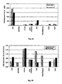
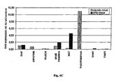
- RNA-expression is shown on the Y-axis.
- the tissues tested are given on the X-axis, "WAT" refers to white adipose tissue.
- the panel of the wild type mice tissues comprises liver, pancreas, muscle, small intestine, WAT, hypothalamus, and heart
- the panel of the control diet-mice tissues comprises liver, muscle, small intestine, WAT, brain, and heart.
- mice carrying gene knockouts In the leptin pathway for example, ob/ob (leptin) or db/db (leptin receptor/ligand) mice
- mice developing typical symptoms of diabetes show hepatic lipid accumulation and frequently have increased plasma lipid levels (see Bruning J.C. et al, (1998) Mol. Cell. 2: 559-569 ).
- SF02 is expressed in several mammalian tissues, showing highest level of expression in liver, and higher levels In further tissues, e.g. WAT, small intestine, heart, brain, muscle. Furthermore SF02 is expressed on lower but still robust levels in the hypothalamus and pancreas as depicted in Fig. 2A .
- WAT small intestine
- Fig. 2A the expression of SF02 is up-regulated in the muscle of ob/ob mice compared to wild-type mice (see Fig. 2B ).
- the expression of SF02 is up-regulated in muscle and down-regulated in WAT compared to mice fed a control diet.
- the high expression of SF02 in metabolic active tissues e.g. liver and WAT
- the regulation of gene expression in different mouse models used to study metabolic disorders as described above suggests that it plays a role in the regulation of energy homeostasis.
- SF03 is expressed in several mammalian tissues, showing highest level of expression in WAT and hypothalamus, and higher levels in further tissues, e.g. brain and heart. Furthermore SF03 is expressed on lower but still robust levels in the pancreas, muscle, small intestine, and liver as depicted in Fig. 4A .
- Fig. 4A we found, for example, that the expression of SF03 is up-regulated in muscle, liver, and small intestine and down-regulated in the pancreas of ob/ob mice compared to wild-type mice (see Fig. 4B ). In wild type mice fed a high fat diet, the expression of SF03 is not regulated.
- SF04 is expressed in several mammalian tissues, showing highest level of expression in liver ( Fig. 6A ), and lower but still robust levels in further tissues, e.g., small intestine, heart, muscle, pancreas, WAT, brain, but not in the hypothalamus, as shown in Fig. 6B .
- Fig. 6A the expression of SF04 is strongly up-regulated in the hypothalamus and down-regulated in the heart, muscle, and WAT of ob/ob mice compared to wild-type mice (see Fig. 6C ).
- SF04 In wild type mice fed a high fat diet, the expression of SF04 is up-regulated in the WAT and down-regulated in muscle, heart, and brain when compared to control diet mice.
- the high expression levels of SF04 in liver suggest that it plays an essential role in metabolism.
- the regulation of gene expression in different mouse models used to study metabolic disorders as described above suggests that it also plays a role in the regulation of energy homeostasis.
- RNA preparation from human primary adipose tissues was done as described In Example 3.
- the target preparation, hybridization, and scanning was performed as described in the manufactures manual (see Affymetrix Technical Manual, 2002, obtained from Affmetrix, Santa Clara, USA).
- the Y-axis represents fluorescence intensity and the X-axis represents the time axis.
- “d0” refers to day 0 (start of the experiment)
- “d12” refers to day 12 of adipocyte differentiation.
- the experiments show that the SF02 and SF03 transcripts are most abundant at day 12 compared to day 0 during differentiation (see Fig. 3 and 5 ) and that the SF13 transcript is most abundant at day 0 compared to day 12 during differentiation (see Fig. 9 ).
- the SF02 and SF03 proteins have to be increased, and the SF13 proteins have to be decreased in order for the preadipocyctes to differentiate into mature adipocyctes.
- the SF02 and SF03 proteins in preadipocyctes have the potential to enhance adipose differentiation, and the SF13 protein in preadipocyctes has the potential to inhibit adipose differentiation. Therefore, the SF02, SF03, and SF13 proteins play an essential role in the regulation of human metabolism, in particular in the regulation of adipogenesis and thus it might be an essential role in pancreatic diseases (e.g. diabetes), obesity, and/or metabolic syndrome.
- the nucleic add sequence encoding the mouse SF01 protein is expressed in the ventral panceas (see Fig. 1 ).
- the nucleic acid sequences encoding the mouse SF05 (see Fig. 7 ) and mouse SF06 (see Fig. 8 ) proteins are expressed in the pancreas.
Landscapes
- Health & Medical Sciences (AREA)
- Life Sciences & Earth Sciences (AREA)
- Engineering & Computer Science (AREA)
- Animal Behavior & Ethology (AREA)
- Veterinary Medicine (AREA)
- Bioinformatics & Cheminformatics (AREA)
- General Health & Medical Sciences (AREA)
- Public Health (AREA)
- Chemical & Material Sciences (AREA)
- Pharmacology & Pharmacy (AREA)
- Medicinal Chemistry (AREA)
- Epidemiology (AREA)
- Gastroenterology & Hepatology (AREA)
- Immunology (AREA)
- Proteomics, Peptides & Aminoacids (AREA)
- Zoology (AREA)
- Cell Biology (AREA)
- Diabetes (AREA)
- General Chemical & Material Sciences (AREA)
- Organic Chemistry (AREA)
- Chemical Kinetics & Catalysis (AREA)
- Nuclear Medicine, Radiotherapy & Molecular Imaging (AREA)
- Hematology (AREA)
- Obesity (AREA)
- Biomedical Technology (AREA)
- Marine Sciences & Fisheries (AREA)
- Endocrinology (AREA)
- Emergency Medicine (AREA)
- Child & Adolescent Psychology (AREA)
- Medicines That Contain Protein Lipid Enzymes And Other Medicines (AREA)
- Pharmaceuticals Containing Other Organic And Inorganic Compounds (AREA)
- Measuring Or Testing Involving Enzymes Or Micro-Organisms (AREA)
- Peptides Or Proteins (AREA)
Abstract
Description
- This invention rotates to the use of secreted SF06 proteins, and to the use of polynucleotides encoding these, in the diagnosis, and study of diabetes, obesity and/or metabolic syndrome.
- Many human proteins serve as pharmaceutical active compounds. Several classes of human proteins that serve as such active compounds include hormones, cytokines, can growth factors, and cell differentiation factors. Most proteins that can be used as a pharmaceutical active compound fall within the family of secreted proteins. Secreted proteins are generally produced within cells at rough endoplasmic reticulum, are then exported to the golgl complex and then move to secretory vesicles or granules, where they are secreted to the exterior of the cell via oxocytosis. Examples for commercially used secreted proteins are human insulin, thrombolytic agents, interferons, interleukins, colony stimulating factors, human growth hormone, transforming growth factor beta, tissue plasminogen activator, erythropoletin, and various other proteins. Receptors of secreted proteins, which are membrane-bound proteins, also have potential as therapeutic or diagnostic agents. It is, therefore, important for developing new pharmaceutical compounds to identify secreted proteins that can be tested for activity in a variety of animal models. Thus, in light of the pervasive role of secreted proteins in human physiology, a need exists for identifying and characterizing novel functions for human secreted proteins and the genes that encode them. This knowledge will allow one to detect, to treat, and to prevent medical diseases, disorders, and/or conditions by using secreted proteins or the gene that encode them.
- The pancreas is an essential organ possessing both an exocrine function involved In the delivery of enzymes into the digestive tract and an endocrine function by which various hormones are secreted into the blood stream. The exocrine function is assured by acinar and centroacinar cells that produce various digestive enzymes and intercalated ducts that transport these enzyme in alkaline solution to the duodenum. The functional unit of the endocrine pancreas is the islet of Langerhans. Islets are scattered throughout the exocrine portion of the pancreas and are composed of four cell types: alpha-, beta-, delta- and PP-cells, reviewed for example in Kim S.K. and Hebrok M., (2001) Genes Dev. 15: 111-127. Beta-cells produce insulin, represent the majority of the endocrine cells and form the core of the islets, while alpha-cells secrete glucagon and are located in the periphery. Delta-cells and PP-cells are less numerous and secrete somatostatin and pancreatic polypeptide, respectively.
- Early pancreatic development has been well studied in different species, including chicken, zebrafish, and mice (for a detailed review, see Kim & Hebrok, 2001, supra). The pancreas develops from distinct dorsal and ventral anlagen. Pancreas development requires specification of the pancreas anlage along both anterior-posterior and dorsal-ventral axes. A number of factors, which are critical for proper pancreatic development have been identified (see Kim & Hebrok, 2001, supra; Wilson M.E. et al., (2003) Mech Dev. 120: 65-80).
- In postnatal/adult humans, the acinar and ductal cells retain a significant proliferative capacity that can ensure cell renewal and growth, whereas the islet cells become mostly mitotically inactive. This is in contrast to rodents where beta-cell replication is an important mechanism in the generation of new beta cells. It has been suggested, that during embryonic development, pancreatic islets of Langerhans originate from differentiating duct cells or other cells with epithelial morphology (Bonner-Weir S. and Sharma A., (2002) J Pathol. 197: 519-526; Gu G. et al., (2003) Mech Dev. 120: 35-43). In adult humans, new beta cells arise in the vicinity of ducts (Butler A.E. et al., (2003) Diabetes 52: 102-110; Bouwens L. and Pipeleers D.G., (1998) Diabetologia 41: 629-633). However, also an intra-islet location or an origin in the bone marrow has been suggested for precursor cells of adult beta cells (Zulewski H. et al., (2001) Diabetes 50: 521-533; lanus A. et al., (2003) J Clin Invest. 111: 843-850). Pancreatic islet growth is dynamic and responds to changes in insulin demand, such as during pregnancy or during the increase in body mass occuring during childhood. In adults, there is a good correlation between body mass and islet mass (Yoon K.H. et al., (2003) J Clin Endocrinol Metab. 88: 2300-2308).
- Pancreatic beta-cells secrete insulin, which is stimulated by high blood glucose levels. Insulin amongst other hormones plays a key role in the regulation of the fuel metabolism. Insulin leads to the storage of glycogen and triglycerides and to the synthesis of proteins. The entry of glucose into muscles and adipose cells is stimulated by insulin. In patients who suffer from diabetes mellitus the amount of insulin produced by the pancreatic islet cells is too low, resulting in elevated blood glucose levels (hyperglycemia). In diabetes type I beta cells are lost due to autoimmune destruction. In type 2 diabetic patients, liver and muscle cells loose their ability to respond to normal blood insulin levels (insulin resistance). High blood glucose levels (and also high blood lipid levels) lead to an impairment of beta-cell function and to an increase in beta-cell apoptosis. It is interesting to note that the rate of beta-cell neogenesis does not appear to change in type 11 diabetics (Butler et al., 2003 supra), thus causing a reduction in total beta-cell mass over time. Eventually the application of exogenous insulin becomes necessary in type 2 diabetics.
- Improving metabolic parameters such as blood sugar and blood lipid levels (e.g. through dietary changes, exercise, medication or combinations thereof) before beta cell mass has fallen below a critical threshold leads to a relatively rapid restoration of beta cell function. However, after such a treatment the pancreatic endocrine function would remain impaired due to the only slightly increased regeneration rate.
- In type I diabetics, the lifespan of pancreatic islets is dramatically shortened due to autoimmune destruction. Treatments have been devised which modulate the immune system and may be able to stop or strongly reduce islet destruction (Raz I. et al., (2001) Lancet 358: 1749-1753; Chatenoud L. et al., (2003) Nat Rev Immunol. 3: 123-132). However, due to the relatively slow regeneration of human beta cells such treatments could only be fully successful at improving the diabetic condition if they are combined with an agent which can stimulate beta cell regeneration.
- Thus, both for type I and type II diabetes (early and late stages) there is a need to find novel agents which stimulate beta cell regeneration.
- Diabetes is a very disabling disease, because medications do not control blood sugar levels well enough to prevent swinging between high and low blood sugar levels. Patients with diabetes are at risk for major complications, including diabetic ketoacidosis, end-stage renal disease, diabetic retinopathy and amputation. There are also a host of related conditions, such as metabolic syndrome, obesity, hypertension, heart disease, peripheral vascular disease, and infections, for which persons with diabetes are at substantially increased risk. The treatment of these complications contributes to a considerable degree to the enormous cost which is imposed by diabetes on health care systems world wide.
- Obesity is one of the most prevalent metabolic disorders in the world. It is still a poorly understood human disease that becomes as a major health problem more and more relevant for western society. Obesity is defined as a body weight more than 20% in excess of the ideal body weight, frequently resulting in a significant impairment of health. Obesity may be measured by body mass index, an indicator of adiposity or fatness. Further parameters for defining obesity are waist circumferences, skinfold thickness and bioimpedance. It is associated with an increased risk for cardiovascular disease, hypertension, diabetes mellitus type II, hyperlipidaemia and an increased mortality rate. Obesity is influenced by genetic, metabolic, biochemical, psychological, and behavioral factors and can be caused by different reasons such as non-insulin dependent diabetes, increase in triglycerides, increase in carbohydrate bound energy and low energy expenditure (Kopelman P.G., (2000) Nature 404: 635-643).
- The concept of 'metabolic syndrome' (syndrome x, insulin-resistance syndrome, deadly quartet) was first described 1966 by Camus and reintroduced 1988 by Reaven (Camus J.P., (1966) Rev Rhum Mal Osteoartic 33: 10-14; Reaven G.M., (1988), Diabetes 37: 1595-1607). Today, metabolic syndrome is commonly defined as clustering of cardiovascular risk factors like hypertension, abdominal obesity, high blood levels of triglycerides and fasting glucose as well as low blood levels of HDL cholesterol. Insulin resistance greatly increases the risk of developing the metabolic syndrome (Reaven G., (2002) Circulation 106: 286-288). The metabolic syndrome often precedes the development of type II diabetes and cardiovascular disease (Lakka H.M. et al., (2002) JAMA 288: 2709-2716). The control of blood lipid levels and blood glucose levels is essential for the treatment of the metabolic syndrome (see, for example, Santomauro A.T. et al., (1999) Diabetes, 48: 1836-1841).
- The molecular factors regulating food intake and body weight balance are Incompletely understood. Even if several candidate genes have been described which are supposed to influence the homeostatic system(s) that regulate body mass/weight, like leptin or the peroxisome proliferator-activated receptor-gamma co-activator, the distinct molecular mechanisms and/or molecules influencing obesity or body weight/body mass regulations are not known.
- There is a need in the prior art for the identification of candidate genes that are specifically expressed in early development in certain pancreatic tissues. These genes and the thereby encoded proteins can provide tools to the diagnosis and treatment of severe pancreatic disorders and related diseases. Therefore, this specification describes secreted proteins that are specifically expressed in pancreatic tissues early in the development. Further described is the use of these genes and proteins in the diagnosis, prevention and/or treatment of pancreatic dysfunctions, such as diabetes, and other related diseases such as obesity and/or metabolic syndrome. These proteins and genes are especially useful in regeneration processes, such as regeneration of the pancreas cells.
- In this specification, we describe secreted factory referred to as SF01-SF13, which are involved in pancreas development regeneration, and in the regulation of energy homeostasis. SF01-SF13 corresponds to mammalian proteins as described in Table 1.
- The invention relates to the use of a composition comprising a SF06 protein as characterized by SEQ ID Nos:30 or 32 and/or a nucleic acid molecule encoding a SF06 protein as characterized by SEQ ID Nos:30 or 32, the nucleic acid molecule being preferably a DNA molecule, more preferably a cDNA or a genomic DNA, and optionally pharmaceutically acceptable carriers, diluents, and/or additives for the preparation of an agent for detecting diseases or dysfunctions selected from the group consisting of diabetes, obesity, and/or metabolic syndrome. Further, the invention relates to the identification of a (poly)peptide involved in the regulation of energy homeostasis and/or metabolism of a mammal and to the screening of an agent, which modulates/effects the activity of a SF06 polypeptide. Any information about other SF01-05 and SF07-13 proteins is provided for the purpose of comparison only.
- The function of SF01, a TNF-related molecule, was previously unknown. Homologues of SF01 exist In human, mouse and Danio rerio. Human SF01 is expressed in brain, hippocampus, and islets of Langerhans. In fish, SF01 is expressed in the brain.
- SF02 is conserved from Drosophila to human. SF02 is a developmentally regulated vital protein. Drosophila SF02 mutants are dying from neural system defects. In mouse, SF02 seems to be enriched in endoderm and embryo, and to be present in pancreas. In humans, SF02 Is present in pancreas, liver, thymus, and spleen.
- Glypicans are a family of glycosylphosphatidylinositol (GPI)-anchored cell surface heparan sulfate proteoglycans (HSPGs) SF03 is a member of the glypican family of heparan sulfate proteoglycans, which attaches to the cell membrane via a GPI anchor. SF03 is mutated in the Simpson-Golabi-Behmel syndrome (SGBS). SGBS is characterized by pre- and post-natal overgrowth and is a recessive X-linked condition. SF03 is expressed in embryonic mesodermal lung, liver and kidney tissues and is thought to interact with various growth factors to regulate tissue and organ growth.
- The SF04 gene is a shared precursor for alpha-microglobulin and bikunin. Alpha microglobulin is a lipocalin with immunosuppressive properties, bikunin is a plasma proteinase inhibitor. The SF04 mRNA is strongly transcribed in liver parenchyma, pancreas, and intestine epithelium. The both encoded proteins are accordingly present in developing hepatocytes, pancreas, kidney, and gut. Bikunin functions as tumor suppressor.
- SF05 is a neural-specific serine protease inhibitor, which is expressed in the whole developing CNS in mouse. SF05 inhibits the extracellular protease tissue-type plasminogen activator and plasmin, but not thrombin. SF05 deficient mice are were viable and healthy, except behavioral defects. Mutant SF05 protein aggregates and causes familial dementia in humans.
- SF06 is secreted by brain, adrenal cortex and adrenocortical tumors. SF06 is involved in the regulation of steroid hormone secretion and the proliferation of adrenocortical cells as autocrine and/or paracrine factor.
- SF07 is a putative tumor suppressor gene, which is inactivated in hepatocarcinomas, colorectal cancer and non-small cell lung cancers.
- SF08 is a carboxypeptidase which has no known enzymatic activity. SF08 is expressed in developing bones and cartilage.
- The SF09 protein is 145 aa long and contains calcium ion binding EF-hand motifs. According to expressed sequence tag (EST) assembly, SF09 is expressed in many tissues including the pancreas.
- The mouse homologue of human SF10 is an extracellular integrin-binding matrix protein. Mutations of SF10 are frequent in patients with Smith-Magenis syndrome (SMS), a clinically recognizable multiple congenital anomaly/mental retardation syndrome.
- SF11 is a cysteine-proteinase inhibitor for cathepsins B and L, which is well characterised. The expression of SF11 is controlled by TGF-beta and EGF in decidual cultures and by TGF-beta in astrocyte precursors. A glycosylated form of SF11 is required for a FGF-2-responsive neural stem cell proliferation. Combined delivery of FGF-2 and SF11 to the adult dentate gyrus stimulated neurogenesis. SF11 deficient mice showed reduced tumor growth.
- SF12 Is an extracellular matrix and plasma glycoprotein. Expression of SF12 in the adult pancreatic islet is mostly confined to the blood vessels.
- SF13 is a ubiquitously expressed cytosolic peptidyl-prolyl cis-trans Isomerase that is inhibited by the immunosuppressive drug cyclosporin. SF13 is a proinflammatory factor for T-lymphocytes. SF13 signals through CD147 (basigin) preceptor, and is expressed in acinar but not in islet membranes or MIN-6 cells.
- Accordingly, the present specification describes secreted proteins with functions in the human metabolism, regeneration, and pancreatic developmental processes. Described are specific genes and proteins encoded thereby and offectors/modulators thereof involved in the regulation of pancreatic function and metabolism, especially in pancreas diseases such as diabetes mellitus, e.g, insulin dependent diabetes mellitus and/or non insulin dependent diabetes mellitus, and/or metabolic syndrome, obesity, and/or related disorders such as coronary heart disease, eating disorder, cachexia, hypertension, hypercholesterolemia (dyslipidemia), liver fibrosis, and/or galistones. Further, described are specific genes and proteins encoded thereby and effectors/modulators thereof involved in the regeneration of pancreatic cells or tissues, e.g. cells having exocrinous functions such as acinar cells, centroacinar cells and/or ductal cells and/or cells having endocrinous functions, particularly cells in Langerhans islets such as alpha-, beta-, delta- and/or PP-cells, more particularly beta-cells.
- In this specification, we used a screen for secreted factors expressed in developing mammalian (mouse) pancreas, as described in more detail in the Examples section (see Example 1). This screen identified SF01-SF13 as secreted factors expressed in developing mouse pancreas. Mammalian SF01-SF13 proteins and the polynucleotides encoding these, in particular human SF01-SF13, are involved in the conditions and processes mentioned above.
- SF01-SP13 homologous proteins and nucleic acid molecules coding therefore are obtainable from vertebrate species. For use in the present invention, particularly preferred are nucleic acids encoding the human SF06 protein as characterized by SEQ ID NOs: 30 or 32 is as defined in claim 4.
- The function of the mammalian SF01-SF13 in mammalian metabolism was validated by analysing the expression of the transcripts in different tissues and by analizing the role in adipocyte differentiation (see Examples 3 and 4 for more detail).
- Expression profiling studies (see Examples for more detail) confirm the particular relevance of SF02-SF04 as regulators of energy metabolism in mammals.
- Microarrays are analytical tools routinely used in bioanalysis. A microarray has molecules distributed over, and stably associated with, the surface of a Solid support. The term "microarray" refers to an arrangement of a plurality of polynucleotides, polypeptides, antibodies, or other chemical compounds on a substrate. Microarrays of polypeptides, polynucleotides, and/or antibodies have been developed and find use in a variety of applications, such as monitoring gene expression, drug discovery, gene sequencing, gene mapping, bacterial identification, and combinatorial chemistry. One area in particular in which microarrays find use is in gene expression analysis (see Example 4). Array technology can be used to explore the expression of a single polymorphic gene or the expression profile of a large number of related or unrelated genes. When the expression of a single gene is examined, arrays are employed to detect the expression of a specific gene or its variants. When an expression profile is examined, arrays provide a platform for identifying genes that are tissue specific, are affected by a substance being tested in a toxicology assay, are part of a signaling cascade, carry out housekeeping functions, or are specifically related to a particular genetic predisposition, condition, disease, or disorder.
- Microarrays may be prepared, used, and analyzed using methods known in the art (see for example,
Brennan T.M., (1995) U.S. Patent No. 5,474,796 ; Schena M. et al., (1996) Proc. Natl. Acad. Sci. USA 93:10614-10619;Baldeschwieler J.D. et al., (1995) PCT application WO 95/251116 Shalon T.D. and Brown P.O., (1995) PCT application WO 95/35505 Heller, M.J. and Tu E., (1997) U.S. Patent No. 5,605,662 ). Various types of microarrays are well known and thoroughly described in Schena M., ed. (1999; DNA Microarrays: A Practical Approach, Oxford University Press, London). - Oligonucleotides or longer fragments derived from any of the polynucleotides described herein may be used as elements on a microarray. The microarray can be used in transcript imaging techniques, which monitor the relative expression levels of large numbers of genes simultaneously as described below. The microarray may also be used to identify genetic variants, mutations, and polymorphisms. This information may be used to determine gene function, to understand the genetic basis of a disorder, to diagnose a disorder, to monitor progression/regression of disease as a function of gene expression, and to develop and monitor the activities of therapeutic agents in the treatment of disease. In particular, this information may be used to develop a pharmacogenomic profile of a patient in order to select the most appropriate and effective treatment regimen for that patient. For example, therapeutic agents, which are highly effective and display the fewest side effects may be selected for a patient based on his/her pharmacogenomic profile.
- As determined by microarray analysis, SF02, SF03, and SF13 show differential expression in human primary adipocytes. A strong up-regulation is observed concerning the expression of SF02 and SF03 during the human adipocyte differentiation (see
Fig. 3 and5 ) and a strong down-regulation is observed concerning the expression of SF13 during the human adipocyte differentiation (seeFig. 9 ). The SF02 and SF03 proteins in preadipocyctes have the potential to enhance adipocyte differentiation, and the SF13 protein in preadipocyctes has the potential to enhance adipocyte differentiation at a very early stage. Therefore, the SF02, SF03, and SF13 proteins might play an essential role in adipogenesis. The results are suggesting a role of SF02, SF03, and SF13 in the regulation in human metabolism, for example, as effectors/modulators (for example, enhancers or inhibitors) of adipogenesis. Thus, SF02, SF03, and SF13 are strong candidates for the manufacture of pharmaceutical compositions and medicaments for the treatment of conditions related to human metabolism, such as diabetes, obesity, and/or metabolic syndrome. - Further, we show whole mount and sectional in situ hybridisations (see Examples and
Fig. 1 ,7 , and8 ). The nucleic acid sequence encoding the mouse SF01 protein Is expressed in the region of the ventral pancreas. The nucleic acid sequence encoding the mouse SF05 and SF06 proteins are expressed in the pancreas tissue (seeFig. 7 and8 ). - Hybridization conditions are based on the melting temperature (Tm) of the nucleic acid binding complex or probe, as described in Wahl G.M. et al., (1987; Methods Enzymol. 152: 399-407) and Kimmel A.R. (1987; Methods Enzymol. 162: 607-611), and may be used at a defined stringency. Preferably, hybridization under stringent conditions means that after washing for 1 h with 1 x SSC and 0.1 % SDS at 50°C, preferably at 55°C, more preferably at 62°C and most preferably at 65°C, particularly for 1 h in 0.2 x SSC and 0.1% SDS at 50°C, preferably at 55°C, more preferably at 62°C and most preferably at 65°C, a positive hybridization signal is observed.
- In order to express a biologically active protein, the nucleotide sequences encoding the proteins or functional equivalents, may be inserted into appropriate Expression vectors, i.e., a vector which contains the necessary elements for the transcription and translation of the inserted coding sequences. Methods, which are well known to those skilled in the art, may be used to construct expression vectors containing sequences encoding the proteins and the appropriate transcriptional and translational control elements. Regulatory elements include for Example a promoter, an initiation colon, a stop codon, a mRNA stability regulatory element, and a polyadenylation signal. Expression of a polynucleotide can be assured by (i) constitutive promoters such as the Cytomegalovirus (CMV) promoter/enhancer region, (ii) tissue specific promoters such as the insulin promoter (see, Soria B. et al., (2000), Diabetes 49: 157-162), SOX2 gene promoter (see U M. et al., (1998) Curr. Biol. 8: 971-974). Msi-1 promoter (see Sakakibara S. and Okano H., (1997) J. Neuroscience 17: 8300-8312), alpha-cardia myosin heavy chain promoter or human atrial natriuretic factor promoter (Klug M.G. et al., (1998) J. Clin. Invest 98: 216-224: Wu J. et al., (1989) J. Biol. Chem. 264: 8472-8479) or (III) inducible promoters such as the tetracycline inducible system. Expression vectors can also contain a selection agent or marker gene that confers antibiotic resistance such as the neomycin, hygromycin or puromycin resistance genes. These methods include in vitro recombinant DNA techniques, synthetic techniques, and in vivo genetic recombination. Such techniques are described in Sambrook J. et al., (1989) Molecular Cloning, A Laboratory Manual, Cold Spring Harbor Press, Plainview, N.Y. and Ausubel F.M. et al., (1989) Current Protocols in Molecular Biology, John Wiley 8 Sons, New York, N.Y.
- In a further embodiment of the invention, natural, iodized or recombinant nucleic acid sequences encoding the SF06 proteins may be ligated to a heterologous sequence to encode a fusion protein.
- A variety of expression vector/host systems, as known in the art, may be utilized to contain and express sequences encoding the proteins or fusion proteins. These include, but are not limited to, micro-organisms such as bacteria transformed with recombinant bacteriophage, plasmid or cosmic DNA expression vectors; yeast transformed with yeast expression vectors; insect cell systems infected with virus expression vectors (e.g., baculovirus, adenovirus, adeno-associated virus, lentiverus, retrovirus); plant cell systems transformed with virus expression vectors (e.g., cauliflower mosaic virus, CaMV; tobacco mosaic virus. TMV) or with bacterial expression vectors (e.g., Ti or PBR322 plasmids); or animal cell systems.
- The presence of polynucleotide sequences in a sample can be detected by DNA-DNA or DNA-RNA hybridization and/or amplification using probes or portions or fragments of said polynucleotides. Nucleic add amplification based assays involve the use of oligonucleotides or oligomers based on the sequences specific for the gene to detect transformants containing DNA or DNA encoding the corresponding protein. As used herein 'oligonucleotides' or 'oligomers' refer to a nucleic acid sequence of at least about 10 nucleotides and as many as about 60 nucleotides, preferably about 15 to 30 nucleotides, and more preferably about 20-25 nucleotides, which can be used as a probe or amplimer.
- A wide variety of labels and conjugation techniques are known by those skilled in the art and may be used in various nucleic add and amino acid assays. Means for producing labeled hybridization or PCR probes for detecting polynucleotide sequences include oligo-labeling, nick translation, end-labeling of RNA probes, PCR amplification using a labeled nucleotide, or enzymatic synthesis. These procedures may be conducted using a variety of commercially available kits (Pharmacia & Upjohn, (Kalamazoo, Mich.); Promega (Madison Wis.); and U.S. Biochemical Corp., (Cleveland, Ohio).
- The presence of SF01-SF13 in a sample can be determined by immunological methods or activity measurement. A variety of protocols for detecting and measuring the expression of proteins, using either polyclonal or monoclonal antibodies specific for the protein or reagents for determining protein activity are known in the art. Examples include enzyme-linked immunosorbent assay (ELISA), radioimmunoassay (RIA), and fluoresoence activated cell sorting (FACS). A two-site, monoclonal-based immunoassay utilizing monoclonal antibodies reactive to two non-interfering epitopes on the protein is preferred, but a competitive binding assay may be employed. These and other assays are described, among other places, In Hampton R. at al. (1990; Serological Methods, a Laboratory Manual, APS Press, St Paul, Minn.) and Maddox D.E. et al. (1983; J. Exp. Med. 158: 1211-1226).
- Suitable reporter molecules or labels, which may be used, include radionuclides, enzymes, fluorescent, chemiluminescent or chromogenic agents as well as substrates, co-factors, inhibitors, magnetic particles, and the like. The nucleic acids encoding the SF06 proteins can be used to generate non-human transgenic animal or site specific gene modifications in cell lines non-human Transgenic animals may be made through homologous combination, where the normal locus of the genes encoding the proteins of the invention is altered. Alternatively, a nucleic acid construct is randomly integrated into the genome. Vectors for stable integration include plasmids, retroviruses and other animal viruses, YACs, and the like. The modified cells or animal are useful in the study of the function and regulation of the SF06 proteins. For example, a series of small deletions and/or substitutions may be made in the genes that encode the proteins to determine the role of particular domains of the protein, functions in pancreatic differentiation, etc.
- Specific constructs of interest include anti-sense molecules, which will block the expression of the SF06 proteins, or expression of dominant negative mutations. A detectable marker, such as for example lac-Z, may be introduced in the locus of the genes, where up-regulation of expression of the genes will result In an easily detected change In phenotype.
- One may also provide for expression of the genes in cells or tissues where it is not normally expressed or at abnormal times of development. In addition, by providing expression of the SF06 proteins in cells in which they are not normally produced, one can induce changes in cell behavior.
- DNA constructs for homologous recombination will comprise at least portions of the genes with the desired genetic modification, and will include regions of homology to the target locus. DNA constructs for random integration need not include regions of homology to mediate recombination. Conveniently, markers for positive and/or negative selection are includes. Methods for generating cells having targeted gene modifications through homologous recombination are known in the art. For non-human embryonic stem (ES) cells, an ES cell line may be employed, or embryonic cells may be obtained freshly from a host, e.g. mouse, rat, guinea pig etc. Such cells are grown on an appropriate fibroblast-feeder layer or grown in presence of leukemia inhibiting factor (LIF).
- The data described in this specification show that the SF06 nucleic acids and proteins are useful in diagnostic applications concerning diabetes, (such as insulin dependent diabetes mellitus and/or non insulin dependent diabetes mellitus), obesity, and/or metabolic syndrome. Hence, diagnostic uses for the nucleic acids and proteins are, for example but not limited to, the following: (i) antibody target (diagnostic), (ii) diagnostic and/or prognostic marker, and / or (iii) research tools.
- The SF06 nucleic acids and proteins are useful in diagnostic applications implicated in various embodiments as described below.
- The nucleic acids may further be useful in diagnostic applications, wherein the presence or amount of the nucleic acids or the proteins are to be assessed, selected diabetes, obesity and metabolic syndrome.
- A variety of protocols including ELISA, RIA, and FACS for measuring proteins are known in the art and provide a basis for diagnosing altered or abnormal levels of gene expression. Normal or standard values for gene expression are established by combining body fluids or cell extracts taken from normal mammalian subjects, preferably human, with antibodies to the protein under conditions suitable for complex formation. The amount of standard complex formation may be quantified by various methods, but preferably by photometric means. Quantities of protein expressed in control and diseases, samples e.g.. from biopsied tissues are compared with the standard values. Deviation between standard and subject values establishes the parameters for diagnosing disease.
- In another embodiment of the invention, the polynucleotides specific for the SF06 proteins may be used for diagnostic purposes, The polynucleotides, which may be used, include oligonucleotide sequences, antisense . RNA and DNA molecules, and PNAs. The polynucleotides may be used to detect and quantitate gene expression in biopsied tissues in which gene expression may be correlated with disease. The diagnostic assay may be used to distinguish between absence, presence, and excess gene expression, and to monitor regulation of protein levels during therapeutic intervention.
- In one aspect, hybridization with probes which are capable of detecting polynucleotide sequences, including genomic sequences, encoding the SF06 proteins may be used to identify nucleic acid sequences which encode the respective protein. The hybridization probes may be DNA or RNA and are preferably derived from the nucleotide sequence of the polynucleotide encoding the SF06 proteins or from a genomic sequence including promoter, enhancer elements, and introns of the naturally occurring gene. Hybridization probes may be labeled by a variety of reporter groups, for example, radionuclides such as 32P or 35S or enzymatic labels, such as alkaline phosphatase coupled to the probe via avidin/biotin coupling systems, and the like.
- Polynucleotide sequences specific for SF06 proteins may be used for the diagnosis of conditions or diseases, which are associated with the expression of the proteins, selected from diabetes, obesity and metabolic syndrome. Polynucleotide sequences specific for the SF06 proteins may also be used to monitor the progress of patients receiving treatment for diabetes, obesity, and/or metabotic syndrome. The polynucleotide sequences may be used qualitative or quantitative assays, e.g. in Southern or Northern analysis, dot blot or other membrane-based technologies: in PCR technologies; or in dip stick, pin, ELISA or chip assays utilizing fluids or tissues from patient biopsies to detect altered gene expression.
- In a particular aspect, the SF06 nucleotide sequences may be useful in assays that detect activation or induction of the above metabolic diseases or dysfunctions. The nucleotide sequences may be labeled by standard methods, and added to a fluid or tissue sample from a patient under conditions suitable for the formation of hybridization complexes. After a suitable incubation period, the sample is washed and the signal is quantitated and compared with a standard value. The presence of altered levels of nucleotide sequences encoding the SF06 proteins in the sample indicates the presence of the associated disease. Such assays may also be used to evaluate the efficacy of a particular therapeutic treatment regimen in animal studies, in clinical trials or in monitoring the treatment of an individual patient.
- In order to provide a basis for the diagnosis of a disease associated with expression of the SF06 proteins, a normal or standard profile for expression is established. This may be accomplished by combining body fluids or cell extracts taken from normal subjects, either animal or human, with a sequence or a fragment thereof, which is specific for the nucleic acids encoding the SF06 proteins, under conditions suitable for hybridization or amplification. Standard hybridization may be quantified by comparing the values obtained from normal subjects with those from an experiments where a known amount of a substantially purified polynucleotide is used. Standard values obtained from normal samples may be compared with values obtained from samples from patients who are symptomatic for disease. Deviation between standard and subject values is used to establish the presence of disease. Once disease is established and a treatment protocol is initiate, hybridization assays may be repeated on a regular basis to evaluate whether the level of expression in the patient begins to approximate that, which is observed in the normal patient. The results obtained from successive assays may be used to show the efficacy of treatment over a period ranging from several days to months.
- With respect to diabetes, obesity, and/or metabolic syndrome, the presence of an unusual amount of transcript in biopsled tissue from an individual may indicate a predisposition for the development of the disease or may provide a means for detecting the disease prior to the appearance of actual clinical symptoms. A more definitive diagnosis of this type may allow health professionals to employ preventative measures or aggressive treatment earlier thereby preventing the development or further progression of the metabolic diseases and disorders.
- Additional diagnostic uses for oligonucleotides designed from the sequences encoding the SF06 proteins may involve the use of PCR. Such oligomers may be chemically synthesized, generated enzymatically or produced from a recombinant source. Oligomers will preferably consist of two nucleotide sequences, one with sense orientation (5prime.fwdarw.3prime) and another with antisense (3prime.rarw.5prime), employed under optimized conditions for identification of a specific gene or condition. The same two oligomers, nested sets of oligomers or even a degenerate pool of oligomers may be employed under less stringent conditions for detection and/or quantification of closely related DNA or RNA sequences.
- The nucleic acid sequences may also be used to generate hybridization probes, which are useful for mapping the naturally occurring genomic sequence. The sequences may be mapped to a particular chromosome or to a specific region of the chromosome using well known techniques. Such techniques include FISH, FACS or artificial chromosome constructions, such as yeast artificial chromosomes, bacterial artificial chromosomes, bacterial P1 constructions or single chromosome cDNA libraries as reviewed in Price C.M., (1993) Blood Rev..7: 127-134, and Trask B.J., (1991) Trends Genet. 7: 149-154. FISH (as described in Verma RS. and Babu A., (1989) Human Chromosomes: A Manual of Basic Techniques, Pergamon Press, New York, N.Y.). The results may be correlated with other physical chromosome mapping techniques and genetic map data. Examples of genetic map data can be found in the 1994 Genome Issue of Science (265: 1981f). Correlation between the location of the gene encoding the SF06 proteins on a physical chromosomal map and a specific disease or predisposition to a specific disease, may help to delimit the region of DNA associated with that genetic disease.
- The nucleotide sequences may be used to detect differences in gene sequences between normal, carrier or affected individuals. An analysis of polymorphisms, e.g. single nucleotide polymorphism may be carried out Further, in situ hybridization of chromosomal preparations and physical mapping techniques such as linkage analysis using established chromosomal markers may be used for extending genetic maps. Often the placement of a gene on the chromosome of another mammalian species, such as mouse, may reveal associated markers even if the number or arm of a particular human chromosome is not known. New sequences can be assigned to chromosomal arms or parts thereof, by physical mapping. This provides valuable information to investigators searching for disease genes using positional cloning or other gene discovery techniques. Once the disease or syndrome has been crudely localized by genetic linkage to a particular genomic region, for example, AT to 11q22-23 (Gatti R.A. et al., (1988) Nature 336: 577-580), any sequences mapping to that area may represent associated or regulatory genes for further investigation. The nucleotide sequences may also be used to detect differences in the chromosomal location due to translocation, inversion, etc. among normal, carrier or affected individuals.
- In another embodiment of the invention, the SF06 proteins can be used for screening libraries of compounds in any of a variety of drug screening techniques. One can identify effectors, e.g. receptors, enzymes, proteins, ligands, or substrates that bind to modulate or mimic the action of one or more of the SF06 proteins. The protein or fragment thereof employed In such screening may be free in solution, affixed to a solid support, borne on a cell surface, or located intracellulary. The formation of binding complexes, between the SF06 proteins and the agent tested, may be measured. Agents could also, either directly or indirectly, influence the activity of the SF06 proteins.
- In addition activity of the SF06 proteins against their physiological substrate(s) or derivatives thereof could be measured in cell-based or cell-free assays. Agents may also interfere with posttranslational modifications of the protein, such as phosphorylation and dephosphorylation, farnesylation, palmitoylation, acetylation, alkylation, ubiquitination, proteolytic processing, subcellular localization and degradation. Moreover, agents could influence the dimerization or oligomerization of the SF06 proteins or, in a heterologous manner, of the SF06 proteins with other proteins, for example, but not exclusively, docking proteins, enzymes, receptors, or translation factors. Agents could also act on the physical interaction of the SF06 proteins with other proteins, which are required for protein function, for example, but not exclusively, their downstream signaling.
- Methods for determining protein-protein interaction are well known In the art. For example binding of a fluorescently labeled peptide derived from the interacting proteins to the SF06 protein, or vice versa, could be detected by a change in polarisation. In case that both binding partners, which can be either the full length proteins as well as one binding partner as the full length protein and the other just represented as a peptide are fluorescently labeled, binding could be detected by fluorescence energy transfer (FRET) from one fluorophore to the other. In addition, a variety of commercially available assay principles suitable for detection of protein-protein Interaction are well known in the art, for example but not exclusively AlphaScreen (PeridnElmer) or Scintillation Proximity Assays (SPA) by Amersham. Alternatively, the interaction of the SF06 proteins with cellular proteins could be the basis for a cell-based screening assay, in which both proteins are fluorescently labeled and interaction of both proteins is detected by analysing cotranslocation of both proteins with a cellular imaging reader, as has been developed for example, but not exclusively, by Cellomics or EvotecOAI. In all cases the two or more binding partners can be different proteins with one being the SF06 protein, or in case of dimerization and/or oligomerization the SF06 protein itself.
- Of particular interest are screening assays for agents that have a low toxicity for mammalian cells. The term "agent" as used herein describes any molecule, e.g. protein or pharmaceutical, with the capability of altering or mimicking the physiological function of one or more of the SF06 proteins. Candidate agents encompass numerous chemical classes, though typically they are organic molecules, preferably small organic compounds having a molecular weight of more than 50 and less than about 2,500 Daltons. Candidate agents comprise functional groups necessary for structural Interaction with proteins, particularly hydrogen bonding, and typically include at least an amine, carbonyl, hydroxyl or carboxyl group, preferably at least two of the functional chemical groups. The candidate agents often comprise carbocyclic or heterocyclic structures and/or aromatic or polyaromatic structures substituted with one or more of the above functional groups.
- Candidate agents are also found among biomolecules including peptides, saccharides, fatty acids, steroids, purines, pyrimidines, nucleic acids and derivatives, structural analogs or combinations thereof. Candidate agents are obtained from a wide variety of sources including libraries of synthetic or natural compounds. For example, numerous means are available for random and directed synthesis of a wide variety of organic compounds and biomolecules, including expression of randomized oligonucleotides and oligopeptides. Alternatively, libraries of natural compounds in the form of bacterial, fungal, plant and animal extracts are available or readily produced. Additionally, natural or synthetically produced libraries and compounds are readily modified through conventional chemical, physical and biochemical means, and may be used to produce combinatorial libraries. Known pharmacological agents may be subjected to directed or random chemical modifications, such as acylation, alkylation, esterification, amidification, etc. to produce structural analogs. Where the screening assay is a binding assay, one or more of the molecules may be joined to a label, where the label can directly or indirectly provide a detectable signal.
- Another technique for drug screening, which may be used, provides for high throughput screening of compounds having suitable binding affinity to the protein of interest as described in published
PCT application WO84/03564 - Compounds that bind SF06 proteins, e.g. antibodies, are useful for the identification or enrichment of cells, which are positive for the expression of the SF06 proteins, from complex cell mixtures. Such cell populations are useful in transplantation, for experimental evaluation, and as source of lineage and cell specific products, including mRNA species useful in identifying genes specifically expressed in these cells, and as target for the identification of factors of molecules that can affect them. Cells expressing the SF06 protein or which have been treated with the SF06 protein are useful in transplantation to provide a recipient with pancreatic islet cells, including insulin producing beta cells; for drug screening; experimental models of islet differentiation and interaction with other cell types; in vitro screening assays to define growth and differentiation factors, and to additionally characterize genes involved In islet development and regulation; and the like. The native cells may be used for these purposes, or they may be genetically modified to provide altered capabilities. Cells from a regenerating pancreas, from embryonic foregut, stomach and duodenum, or other sources of pancreatic progenitor cells may be used as a starting population. The progenitor cells may be obtained from any mammalian species, e.g. equine, bovine, porcine, canine, feline, rodent, e.g. mice, rats, hamster, primate, etc. particularly human.
- In another embodiment, in a high-throughput screening method, the cells are transfected with a DNA construct, e.g. a viral or non-viral vector containing a reporter gene. e.g. the lacZ gene or the GFP gene, under regulatory control of a promoter of a gene involved in for example beta-cell differentiation, e.g. a promoter of a gene stimulation beta-cell differentiation, preferably a Pax4 promoter. The transfected cells are divided into aliquots and each aliquot is contacted with a test substance, e.g., candidate 1, candidate 2 and candidate 3. The activity of the reporter gene corresponds to the capability of the test compound to induce beta-cell differentiation.
- In a further embodiment, which may be combined with the high-throughput screening as described above, a medium throughput validation is carried out. Therein, the test compound is added to stem cells being cultivated and the Insulin production is determined. Following an initial high throughput assay, such as the cell based assay outlined above where for example a Pax4 promoter is used as marker for beta-cell regeneration, the activity of candidate molecules to induce beta-cell differentiation is tested in a validation assay comprising adding said compounds to the culture media of the embryoid bodies. Differentiation into insulin-producing cells is then evaluated, e.g. by comparison to wild type and/or Pax4 expressing ES cells to assess the effectiveness of a compound.
- The nucleic acids encoding the SF06 proteins can be used to generate transgenic cell lines and non-human animals. These transgenic non-human animals are useful in the study of the function and regulation of the SF06 proteins in vivo. Non-human transgenic animals, particularly mammalian transgenic animals, can serve as a model system for the investigation of many developmental and cellular processes common to humans. A variety of non-human models of metabolic disorders can be used to test modulators of the SF06. Misexpression (for example, over-expression or lack of expression) of the SF06 protein, particular feeding conditions, and/or administration of biologically active compounds can create models of metabolic disorders.
- In one embodiment of the invention, such assays use mouse models of insulin resistance and/or diabetes, such as mice carrying gene knockouts in the leptin pathway (for example, ob (leptin) or db (leptin receptor) mice), as described above. In addition to testing the expression of the SF06 proteins in such mouse strains (see Examples), these mice could be used to test whether administration of a candidate modulator alters for example lipid accumulation In the liver, in plasma, or adipose tissues using standard assays well known in the art, such as FPLC, colorimetric assays, blood glucose level tests, Insulin tolerance tests and others.
- Non-human transgenic animals may be made through homologous recombination In embryonic stem cells, where the normal locus of the gene encoding the SF06 protein is mutated. Alternatively, a nucleic add construct encoding the protein is injected into cocytes and is randomly Integrated into the genome. One may also express the genes in tissues where they are not normally expressed or at abnormal times of development. Furthermore, variants of the genes like specific constructs expressing anti-sense molecules or expression of dominant negative mutations, which will block or alter the expression of the SF06, proteins may be randomly integrated into the genome. A detectable marker, such as lac Z or luciferase may be introduced into the locus of the genes, where up-regulation of expression of the genes will result in an easily detectable change in phenotype. Vectors for stable integration include plasmids, retroviruses and other animal viruses, yeast artificial chromosomes (YACs), and the like. DNA constructs for homologous recombination will contain at least portions of the genes with the desired genetic modifications, and will include regions of homology to the target locus. Conveniently, markers for positive and negative selection are included. DNA constructs for random integration do not need to contain regions of homology to mediate recombination. DNA constructs for random integration will consist of the nucleic acids encoding the proteins, a regulatory element (promoter), an intron and a poly-adenylation signal. Methods for generating cells having targeted gene modifications through homologous recombination are known in the field. For embryonic stem (ES) cells, an ES cell line may be employed, or embryonic cells may be obtained freshly from a host, e.g. mouse, rat, guinea pig, etc. Such cells are grown on an appropriate fibroblast-feeder layer and are grown in the presence of leukemia inhibiting factor (LIF). ES or embryonic cells may be transfected and can then be used to produce transgenic animals. After transfection, the ES cells are plated onto a feeder layer in an appropriate medium. Cells containing the construct may be selected by employing a selection medium. After sufficient time for colonies to grow, they are picked and analyzed for the occurrence of homologous recombination. Colonies that are positive may then be used for embryo manipulation and morula aggregation. Briefly, morulae are obtained from 4 to 6 week old superovulated females, the Zona Pellucida is removed and the morulae are put into small depressions of a tissue culture dish. The ES cells are trypsinized, and the modified cells are placed into the depression closely to the morulae. On the following day the aggregates are transfered into the uterine horns of pseudopregnant females. Females are then allowed to go to term. Chimeric offsprings can be readily detected by a change in coat color and are subsequently screened for the transmission of the mutation Into the next generation (F1-generation). Offspring of the F1-generation are screened for the presence of the modified gene and males and females having the modification are mated to produce homozygous progeny. If the gene alterations cause lethality at some point in development, tissues or organs can be maintained as allogenic or congenic grafts or transplants, or In vitro culture. The transgenic animals may be any non-human mammal, such as laboratory animal, domestic animals, etc., for example, mouse, rat, guinea pig, sheep, cow, pig, and others. The transgenic animals may be used in functional studies, drug screening, and other applications and are useful in the study of the function and regulation of the SF06 proteins in vivo.
- The Figures show:
-
Fig. 1 shows the in situ hybridization results for the SF01 protein.
Fig. 1A shows a whole mount in situ hybridisation of mouse embryonic pancreas at day E11.5 (lateral view).
Fig. 1B shows a whole mount in situ hybridisation of mouse embryonic pancreas at day E11.6 (ventral view; liver removed; higher magnification of the stained region). -
Fig. 2 shows the expression of SF02 In mammalian (mouse) tissues.
Fig. 2A shows the real-time PCR analysis of SF02 expression in mouse wild type (referred to as wt-mice) and control diet (referred to as controldiet) tissues.
Fig. 2B shows the real-time PCR analysis of SF02 expression in genetically obese mice (referred to as ob/ob-mice) compared to wild type mice and in mice fed with a high fat diet (referred to as HFD-mice) compared to mice fed with a control diet. -
Fig. 3 shows the microarray analysis of SF02 expression in human abdominal adipocyte cells, during the differentiation from preadipocytes to mature adipocytes. -
Fig. 4 shows the expression of SF03 in mammalian (mouse) tissues.
Fig. 4A shows the real-time PCR analysis of SF03 expression in mouse wild type (referred to as wt-mice) and control diet (referred to as controldiet) tissues.
Fig. 4B shows the real-time PCR analysis of SF03 expression in genetically obese mice (referred to as ob/ob-mice) compared to wild type mice and in mice fed with a high fat diet (referred to as HFD-mice) compared to mice fed with a control diet. -
Fig. 5 shows the microarray analysis of SF03 expression in human abdominal adipocyte cells during the differentiation from preadipocytes to mature adipocytes. -
Fig. 6 shows the expression of SF04 in mammalian (mouse) tissues.
Fig. 6A shows the real-time PCR analysis of SF04 expression in mouse wild type (referred to as wt-mice) and control diet (referred to as controldiet) tissues (including liver).
Fig. 6B shows the real-time PCR analysis of SF04 expression in mouse wild type and control diet tissues (without liver).
Fig. 6C shows the real-time PCR analysis of SF04 expression in genetically obese mice (referred to as ob/ob-mice) compared to wild type mice and in mice fed with a high fat diet (referred to as HFD-mice) compared to mice fed with a control diet. -
Fig. 7 shows in situ hybridization results for the SF05 protein.
Fig. 7A shows a cryosection of mouse embryonic pancreas at day E17.5.
Fig. 7B shows the cryosection of mouse embryonic pancreas at day E17.5 in a larger magnification. -
Fig. 8 shows in situ hybridization results for the SF06 protein. Shown is the cryosection of mouse embryonic pancreas at day E17.5. -
Fig. 9 shows the microarray analysis of SF13 expression in human adipocyte cells during the differentiation from preadipocytes to mature adipocytes.
Fig. 9A shows the microarray analysis of SF13 expression in human primary abdominal adipocyte cells during the differentiation from preadipocytes to mature adipocytes.
Fig. 9B shows the microarray analysis of SF13 expression in human SGBS cells during the differentiation from preadipocytes to mature adipocytes. - A screen for secreted factors expressed in developing mouse pancreas was carried out according to methods known by those skilled in the art (see, for example Pera E.M. and De Robertis E.M., (2000) Mech Dev 96(2): 183-195) with several modifications.
- During organogenesis, the pancreatic bud is surrounded and influenced by the associated mesenchyme. (see for example, Madsen O.D. et al., (1996) Eur. J. Biochem. 242: 435-445 and Slack, J.M., (1995) Development 121: 1569-1580). Recently, it was suggested, that white adipocytes origin directly from mesenchymal cells (Atanossova P.K., (2003) Folia Med. 45: 41-45). During embryogenesis, the innervation and vascularization of the pancreas can be observed. Therefore, the tissue used in the screen might have contained besides pancreatic cells some adipocyte precursors, blood vessels, as well as neuronal cells.
- A mouse embryonic stage 9.5-15 pancreatic bud library was prepared in pCMVSPORT-6 vector using SUPERSCRIPT Plasmid System from Invitrogen according to the manufacturer's instructions. The non-amplified library was electroporated into MaxEff DH10B cells (Invitrogen).
- Bacterial clones were picked with sterile toothpicks from agar plates and cultured in 96-deep-well microtiter plates in LB-ampicillin (see Sambrook et al., supra). Aliquots of 8 cultures were pooled, and plasmid DNA was isolated using the BioRobot_9600 apparatus according to the manufacturer's instructions (Qiagen; QIAprep(r) Turbo BioRobot Kit. Human 293 cell culture cells were cultured in 75 ml tissue culture flasks in DMEM and 10% fetal calf serum. At 90-99% confluence, the cells were splitted at 1:3 ratio and plated onto poly-D-lysine (Sigma) coated 96-well plates. Cells were transfected with 100-500 ng plasmid using lipofectamine 2000 (Invitrogen). After 6 hours, the medium was exchanged for fresh complete growth medium. 24 hours after transfection, the cells were washed twice with DMEM without cysteine and methionine (Invitrogen), supplemented with 1% dialysed Bovine serum (Sigma) with 50 microgram per ml Heparin (Sigma) and glutamine. The cells were labeled radioactively ('S35 Met-label', from Hartmann Analytic GmbH). After 12 hours, aliquots of the supernatants were harvested in 96-well PCR plates and subjected to SDS gel electrophoresis in precast 4□20% gradient polyacrylamide Criterion gels (Biorad) under reducing conditions, using Criterion Dodeca Cell gel running chamber (Biorad). The gels were fixed in 10% acetic acid, 25% isopropanol for 30 min, soaked 15-30 min in AMPLIFY reagent (Amersham), dried and exposed to X-OMAT (AR) film (Kodak). Positive clones were identified and regrown in 96-well-plates. DNA of individual clones was prepared and used for transfection as described above. If one of the clones yielded proteins of the same size as that of the original pool, a positive clone was identified. Positive clones were partially sequenced from the 5' end (SEQLAB, Goettingen).
- The term "polynucleotide comprising the nucleotide sequence as shown in GenBank Accession number" relates to the expressible gene of the nucleotide sequences deposited under the corresponding GenBank Accession number. The term "GenBank Accession number" relates to NCBI GenBank database entries (Ref.: Benson D.A. et al., (2000) Nucleic Acids Res. 28: 15-18).
- Sequences homologous to the mouse sequences were identified using the publicly available program BLASTP 2.2.3 of the non-redundant protein data base of the National Center for Biotechnology information (NCBI) (see, Altschul S.F. et al., (1997) Nucleic Acids Res. 25: 3389-3402).
- SF01-SF13 proteins and nucleic acid molecules coding therefore are obtainable from insect or vertebrate species, e.g. mammals or fish. Particularly preferred are nucleic add molecules and proteins encoded thereby comprising human SF01-SF13 and mouse SF01-SF13 sequences identified in the "secreted factor screen", as described in Table 2.
Table 2: Mammalian genes and proteins of the invention (SF01-SF13) Name Genbank Accession Numbers Mus musculus genes and proteins Homo sapiens genes and proteins cDNA Protein cDNA Protein SF01 NM_026161 NP_080437 NM_031909 NP_114115 SF02 NM_178644 NP_848759 NM_178507 NP_848602 SF03 NM_016697 NP_057906 NM_004484 NP_004475 SF04 NM_007443 NP_031469 NM_001633 NP_001624 SF05 NM_009250 NP_033276 NM_005025 NP_005016 SF06 NM_172633 NP_766221 NM_182511 NP_872317 SF07 NM_026840 NP_081116 NM_006207 NP_006198 SF08 NM_019696 NP_062670 NM_019609 NP_062555 SF09 NM_139295 NP_647456 NM_139279 NP_644808 SF10 NM_029568 NP_083844 NM_002404 NP_002395 SF11 NM_009976 NP_034106 NM_000099 NP_600090 SF12 NM_010180 NP_034310 NM_006486 NP_006477 SF13 NM_011149 NP_035279 NM_000942 NP_000933 - To analyse the expression of the mRNAs described in this specification in mammalian tissues, several mouse strains (preferably mice strains C57BI/6J, C57BI/6 ob/ob, C57BI/KS db/db, and Non-Obese-Diabetic (NOD) mice, which are standard model systems In obesity and diabetes research) were purchased from Harlan Winkelmann (33178 Borchen, Germany) and Taconic M & B (Germantown, NY 12526, U.S.A), respectively, and maintained under constant temperature (preferably 22°C), 40% humidity and a light / dark cycle of preferably 14 / 10 hours. The mice were fed a standard chow (for example, from ssniff Spezialitäten GmbH, order number ssniff M-Z V1126-000). In a further experiment wild-type (wt) mice were fed a control diet (preferably Altromin C1057 mod control, 4.5% crude fat) or high fat diet (preferably Altromin C1057mod. high fat, 23.5% crude fat). Animals were sacrificed at an age of 6 to 8 weeks. The animal tissues were isolated according to standard procedures known to those skilled in the art, snap frozen in liquid nitrogen and stored at -80°C until needed.
- For analyzing the role of the described proteins in the in vitro differentiation of mammalian cell culture cells for the conversion of preadipocytes to adipocytes, mammalian fibroblast (3T3-L1) cells (e.g., Green H. and Kehinde O., (1974) Cell 1: 113-116) were obtained from the American Tissue Culture Collection (ATCC, Hanassas, VA, USA; ATCC- CL 173). 3T3-L1 cells were maintained as fibroblasts and differentiated into adipocytes as described in the prior art (e.g., Qiu Z. et al., (2001) J. Biol. Chem. 276: 11988-11995; Slieker L.J. et al., (1998) BBRC 251: 225-229). In brief, cells were plated In DMEM/10% FCS (Invitrogen, Karisruhe, Germany) at 50,000 cells/well in duplicates in 6-well plastic dishes and cultured in a humidified atmosphere of 5% CO2 at 37°C. At confluence (defined as day 0: d0) cells were transferred to serum-free (SF) medium, containing DMEM/HarnF12 (3:1; invitrogen), fetuin (300 µg/ml: Sigma, Munich, Germany), transferrin (2 µg/ml; Sigma), pantothenate (17 µM; Sigma), biotin (1 µM; Sigma), and EGF (0.8 nM; Hoffmann-La Roche, Basel, Switzerland). Differentiation was induced by adding dexamethasone (DEX: 1 µM; Sigma), 3-methyl-isobutyl-1-methylxanthine (MIX; 0.5 mM; Sigma), and bovine insulin (5 µg/ml; Invitrogen). Four days after confluence (d4), cells were kept in SF medium, containing bovine insulin (5 µg/ml) until differentiation was completed. At various time points of the differentiation procedure, beginning with day 0 (day of confluence) and day 2 (hormone addition; for example, dexamethasone and 3-Isobutyl-1-methylxanthine), up to 10 days of differentiation, suitable aliquots of cells were taken every two days,
- RNA was isolated from mouse tissues or cell culture cells using Trizol Reagent (for example, from Invitrogen, Karlsruhe, Germany) and further purified with the RNeasy Kit (for example, from Qiagen, Germany) in combination with an DNase-treatment according to the instructions of the manufacturers and as known to those skilled in the art. Total RNA was reverse transcribed (preferably using Superscript II RNaseH- Reverse Transcriptase, from Invitrogen, Karlsruhe, Germany) and subjected to Taqman analysis preferably using the Taqman 2xPCR Master Mix (from Applied Biosystems, Weiterstadt, Germany; the Mix contains according to the Manufacturer for example AmpliTaq Gold DNA Polymerase, AmpErase UNG, dNTPs with dUTP, passive reference Rox and optimized buffer components) on a GeneAmp 5700 Sequence Detection System (from Applied Biosystems, Weiterstadt, Germany).
- The following prime/probe pairs were used for the TaqMan analysis (GenBank Accession Number NM_178644 (mouse) for the mouse SF02 sequence):
- Mouse SF02 forward primer (Seq ID NO:1): 5'- CGG ACA GCA TCA GCC TTG A -3'; mouse SF02 reverse primer (Seq ID NO:2): 5'- CCG CGA TGA AGG AGA TGA GA -3'; mouse SF02 Taqman probe (Seq ID NO:3): (5/6-FAM)- CTG CGC AAA CCC GAC GGC A -(5/6-TAMRA).
- The following prime/probe pairs were used for the TaqMan analysis (GenBank Accession Number NM_016697 (mouse) for the mouse SF03 sequence):
- Mouse SF03 forward primer (Seq ID NO:4): 5'- GTT GTT CGC CAT GCC AAG A -3'; mouse SF03 reverse primer (Seq ID NO:5): 5'- CAA AAG CTT GTG GAG TCA GGC T -3'; mouse SF03 Taqman probe (Seq ID NO:6): (5/6-FAM)- ACA CCA ACG CCA TGT TCA AGA ATA ACT ACC C -(5/6-TAMRA).
- The following prime/probe pairs were used for the TaqMan analysis (GenBank Accession Number NM_007443 (mouse) for the mouse SF04 sequence):
- Mouse SF04 forward primer (Seq ID NO:7): 5'- GGT ACA ACC TGG CGG TGG -3'; mouse SF04 reverse primer (Seq ID NO:8): 5'- GCT CAC GCT CAT CTT GTC CTT AA -3'; mouse SF04 Taqman probe (Seq ID NO:9): (5/6-FAM)- TGC CCG TGG CTG AGC CGC -(5/6-TAMRA).
- The function of the mammalian SF02, SF03, and SF04 in metabolism was further validated by analyzing the expression of the transcripts in different tissues.
- In
Fig. 2 ,4 , and6 , the relative RNA-expression is shown on the Y-axis. InFig. 2 ,4 , and6 , the tissues tested are given on the X-axis, "WAT" refers to white adipose tissue. InFig. 2 ,4 , and6 , the panel of the wild type mice tissues comprises liver, pancreas, muscle, small intestine, WAT, hypothalamus, and heart, and the panel of the control diet-mice tissues comprises liver, muscle, small intestine, WAT, brain, and heart. - The function of the SF02, SF03, and SF04 proteins in metabolism was further validated by analyzing the expression of the transcripts in different tissues. Mouse models of insulin resistance and/or diabetes were used, such as mice carrying gene knockouts In the leptin pathway (for example, ob/ob (leptin) or db/db (leptin receptor/ligand) mice) to study the expression of the proteins. Such mice develop typical symptoms of diabetes, show hepatic lipid accumulation and frequently have increased plasma lipid levels (see Bruning J.C. et al, (1998) Mol. Cell. 2: 559-569).
- Expression of the mRNAs encoding the described proteins was also examined in susceptible wild type mice (for example, C57BI/6) that show symptoms of diabetes, lipid accumulation, and high plasma lipid levels, if fed a high fat diet. Expression profiling studies confirm the particular relevance of the proteins as regulators of energy metabolism in mammals.
- Expression, profiling studies confirm the particular relevance of SF02, SF03, and SF04 as regulators of energy metabolism in mammals.
- Taqman analysis revealed that SF02 is expressed in several mammalian tissues, showing highest level of expression in liver, and higher levels In further tissues, e.g. WAT, small intestine, heart, brain, muscle. Furthermore SF02 is expressed on lower but still robust levels in the hypothalamus and pancreas as depicted in
Fig. 2A . We found, for example, that the expression of SF02 is up-regulated in the muscle of ob/ob mice compared to wild-type mice (seeFig. 2B ). In wild type mice fed a high fat diet, the expression of SF02 is up-regulated in muscle and down-regulated in WAT compared to mice fed a control diet. The high expression of SF02 in metabolic active tissues (e.g. liver and WAT) and the regulation of gene expression in different mouse models used to study metabolic disorders as described above suggests that it plays a role in the regulation of energy homeostasis. - Taqman analysis revealed that SF03 is expressed in several mammalian tissues, showing highest level of expression in WAT and hypothalamus, and higher levels in further tissues, e.g. brain and heart. Furthermore SF03 is expressed on lower but still robust levels in the pancreas, muscle, small intestine, and liver as depicted in
Fig. 4A . We found, for example, that the expression of SF03 is up-regulated in muscle, liver, and small intestine and down-regulated in the pancreas of ob/ob mice compared to wild-type mice (seeFig. 4B ). In wild type mice fed a high fat diet, the expression of SF03 is not regulated. The high expression of SF03 in WAT and in the hypothalamus, which is known to be involved in appetite control, as well as he regulation of gene expression in the mouse model for the metabolic syndrome as described above, suggests that it plays a role in the regulation of energy homeostasis. - Taqman analysis revealed that SF04 is expressed in several mammalian tissues, showing highest level of expression in liver (
Fig. 6A ), and lower but still robust levels in further tissues, e.g., small intestine, heart, muscle, pancreas, WAT, brain, but not in the hypothalamus, as shown inFig. 6B . We found, for example, that the expression of SF04 is strongly up-regulated in the hypothalamus and down-regulated in the heart, muscle, and WAT of ob/ob mice compared to wild-type mice (seeFig. 6C ). In wild type mice fed a high fat diet, the expression of SF04 is up-regulated in the WAT and down-regulated in muscle, heart, and brain when compared to control diet mice. The high expression levels of SF04 in liver suggest that it plays an essential role in metabolism. The regulation of gene expression in different mouse models used to study metabolic disorders as described above suggests that it also plays a role in the regulation of energy homeostasis. - RNA preparation from human primary adipose tissues was done as described In Example 3. The target preparation, hybridization, and scanning was performed as described in the manufactures manual (see Affymetrix Technical Manual, 2002, obtained from Affmetrix, Santa Clara, USA).
- In
Fig. 3 ,5 , and9 , the Y-axis represents fluorescence intensity and the X-axis represents the time axis. "d0" refers to day 0 (start of the experiment), "d12" refers today 12 of adipocyte differentiation. - The expression analysis (using Affymetrix GeneChips) of the genes using primary human abdominal adipocycte differentiation clearly shows differential expression of the human SF02. SF03, and SF13 genes in adipocytes. Several independent experiments where done. The experiments show that the SF02 and SF03 transcripts are most abundant at
day 12 compared today 0 during differentiation (seeFig. 3 and5 ) and that the SF13 transcript is most abundant atday 0 compared today 12 during differentiation (seeFig. 9 ). Thus, the SF02 and SF03 proteins have to be increased, and the SF13 proteins have to be decreased in order for the preadipocyctes to differentiate into mature adipocyctes. The SF02 and SF03 proteins in preadipocyctes have the potential to enhance adipose differentiation, and the SF13 protein in preadipocyctes has the potential to inhibit adipose differentiation. Therefore, the SF02, SF03, and SF13 proteins play an essential role in the regulation of human metabolism, in particular in the regulation of adipogenesis and thus it might be an essential role in pancreatic diseases (e.g. diabetes), obesity, and/or metabolic syndrome. - Whole-mount and sectional in situ hybridizations were performed according to standard protocols as known to those skilled in the art and as described previously (for example, Pelton, R.W. et al., (1990) Development 110,609-620; Belo, J. A. et al., (1997) Mech. Dev. 68. 45-57).
- The nucleic add sequence encoding the mouse SF01 protein is expressed in the ventral panceas (see
Fig. 1 ). The nucleic acid sequences encoding the mouse SF05 (seeFig. 7 ) and mouse SF06 (seeFig. 8 ) proteins are expressed in the pancreas. - Although the invention has been described in connection with specific preferred embodiments, it should be understood that the invention as claimed should not be unduly limited to such specific embodiments. Indeed, various modifications of the described modes for carrying out the invention which are obvious to those skilled In molecular biology or related fields are intended to be within the scope of the following claims.
-
- <110> DeveloGen Aktiengesellschaft für entwicklungsbiologische Forschung
- <120> Use of secreted protein products for preventing and treating pancreatic diseases and/or obesity and/or metabolic syndrome
- <130> 32600PWO
- <140>
PCT/EP2005/001711
<141> 2005-02-18 - <150>
EP04003914.1
<151> 2004-02-20 - <160> 61
- <170> PatentIn version 3.3
- <210> 1
<211> 19
<212> DNA
<213> Artificial - <220>
<223> Artificial Sequence - <220>
<221> misc_feature
<223> Primer: Mouse SF02 forward primer - <400> 1
cggacagcat cagccttga 19 - <210> 2
<211> 20
<212> DNA
<213> Artificial - <220>
<223> Artificial Sequence - <220>
<221> misc_feature
<223> Primer: Mouse SF02 reverse primer - <400> 2
ccgcgatgaa ggagatgaga 20 - <210> 3
<211> 19
<212> DNA
<213> Artificial - <220>
<223> Artificial Sequence - <220>
<221> misc_feature
<223> Taqman probe: Mouse SF02 Taqman probe - <400> 3
ctgcgcaaac ccgacggca 19 - <210> 4
<211> 19
<212> DNA
<213> Artificial - <220>
<223> Artificial Sequence - <220>
<221> misc_feature
<223> Primer: Mouse SF03 forward primer - <400> 4
gttgttcgcc atgccaaga 19 - <210> 5
<211> 22
<212> DNA
<213> Artificial - <220>
<223> Artificial Sequence - <220>
<221> misc_feature
<223> Primer: Mouse SF03 reverse primer - <400> 5
caaaagcttg tggagtcagg ct 22 - <210> 6
<211> 31
<212> DNA
<213> Artificial - <220>
<223> Artificial Sequence - <220>
<221> misc_feature
<223> Taqman probe: Mouse SF03 Taqman probe - <400> 6
acaccaacgc catgttcaag aataactacc c 31 - <210> 7
<211> 18
<212> DNA
<213> Artificial - <220>
<223> Artificial Sequence - <220>
<221> misc_feature
<223> Primer: Mouse SF04 forward primer - <400> 7
ggtacaacct ggcggtgg 18 - <210> 8
<211> 23
<212> DNA
<213> Artificial - <220>
<223> Artificial Sequence - <220>
<221> misc_feature
<223> Primer: Mouse SF04 reverse primer - <400> 8
gctcacgctc atcttgtcct taa 23 - <210> 9
<211> 18
<212> DNA
<213> Artificial - <220>
<223> Artificial Sequence - <220>
<221> misc_feature
<223> Taqman probe: Mouse SF04 Taqman probe - <400> 9
tgcccgtggc tgagccgc 18 - <210> 10
<211> 1285
<212> DNA
<213> Mus musculus - <220>
<221> CDS
<222> (514)..(1131)
<223> SF01, cDNA: NM_026161, Protein: NP_080437 - <400> 10


- <210> 11
<211> 205
<212> PRT
<213> Mus musculus - <400> 11


- <210> 12
<211> 1393
<212> DNA
<213> Homo sapiens - <220>
<221> CDS
<222> (225)..(1214)
<223> SF01, cDNA: NM_031909, Protein: NP_114115 - <400> 12


- <210> 13
<211> 329
<212> PRT
<213> Homo sapiens - <400> 13


- <210> 14
<211> 2388
<212> DNA
<213> Mus musculus - <220>
<221> CDS
<222> (244)..(1092)
<223> SF02, cDNA: NM_178644, Protein: NP_848759 - <400> 14


- <210> 15
<211> 282
<212> PRT
<213> Mus musculus - <400> 15


- <210> 16
<211> 1957
<212> DNA
<213> Homo sapiens - <220>
<221> CDS
<222> (242)..(1063)
<223> SF02, cDNA: NM_178507, Protein: NP_848602 - <400> 16


- <210> 17
<211> 273
<212> PRT
<213> Homo sapiens - <400> 17

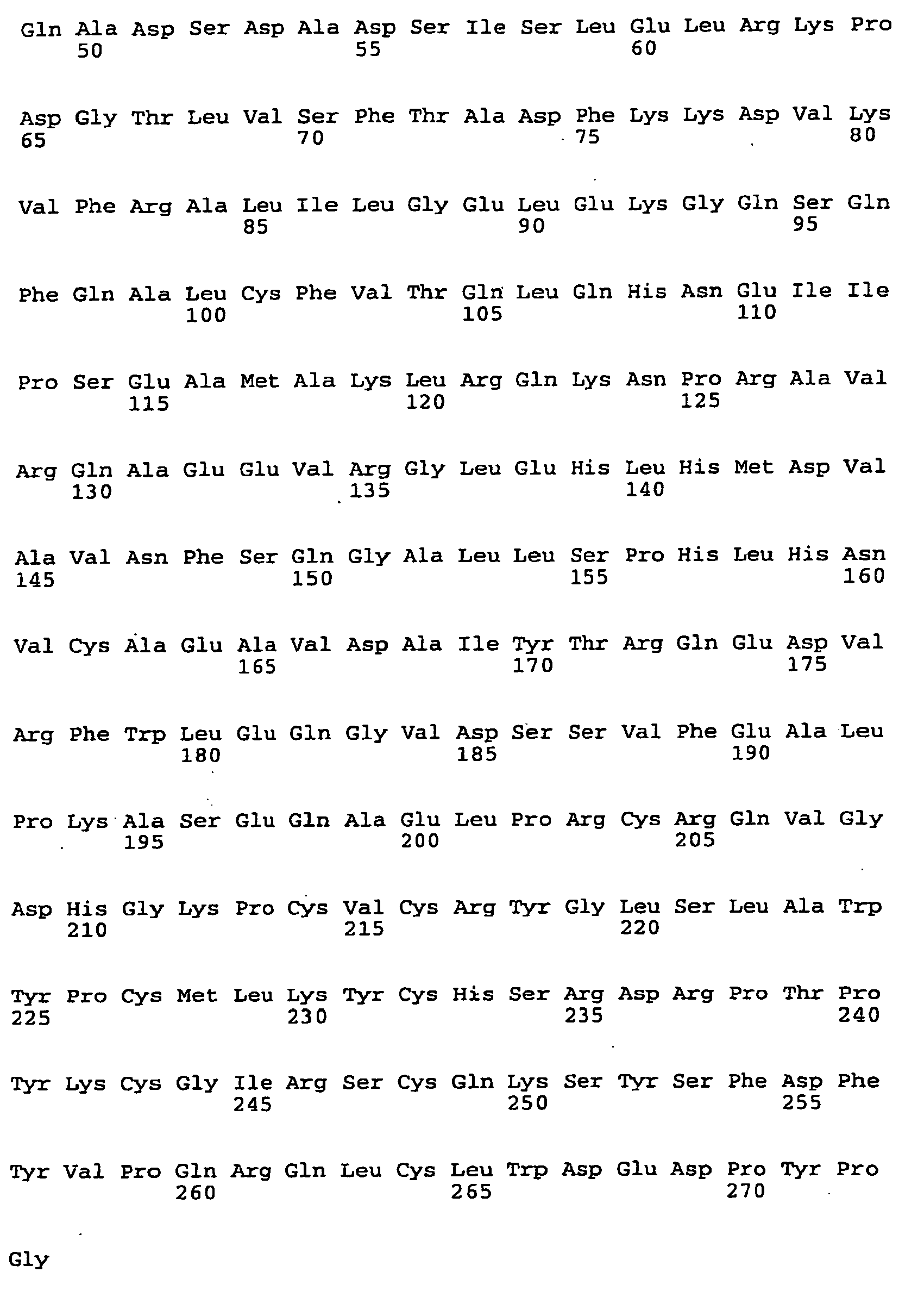
- <210> 18
<211> 2312
<212> DNA
<213> Mus musculus - <220>
<221> CDS
<222> (118)..(1857)
<223> SF03, cDNA: NM_016697, Protein: NP_057906 - <400> 18


- <210> 19
<211> 579
<212> PRT
<213> Mus musculus - <400> 19


- <210> 20
<211> 2382
<212> DNA
<213> Homo sapiens - <220>
<221> CDS
<222> (191)..(1933)
<223> SF03, cDNA: NM_004484, Protein: NP_004475 - <400> 20


- <210> 21
<211> 580
<212> PRT
<213> Homo sapiens - <400> 21


- <210> 22
<211> 1234
<212> DNA
<213> Mus musculus - <220>
<221> CDS
<222> (89)..(1138)
<223> SF04, cDNA: NM_007443, Protein: NP_031469 - <400> 22


- <210> 23
<211> 349
<212> PRT
<213> Mus musculus - <400> 23


- <210> 24
<211> 1413
<212> DNA
<213> Homo sapiens - <220>
<221> CDS
<222> (227)..(1285)
<223> SF04, cDNA: NM_001633, Protein: NP_001624 - <400> 24


- <210> 25
<211> 352
<212> PRT
<213> Homo sapiens - <400> 25


- <210> 26
<211> 2944
<212> DNA
<213> Mus musculus - <220>
<221> CDS
<222> (98)..(1330)
<223> SF05, cDNA: NM_009250, Protein: NP_033276 - <400> 26


- <210> 27
<211> 410
<212> PRT
<213> Mus musculus - <400> 27



- <210> 28
<211> 1910
<212> DNA
<213> Homo sapiens - <220>
<221> CDS
<222> (421)..(1653)
<223> SF05, cDNA: NM_005025, Protein: NP_005016 - <400> 28


- <210> 29
<211> 410
<212> PRT
<213> Homo sapiens - <400> 29


- <210> 30
<211> 2202
<212> DNA
<213> Mus musculus - <220>
<221> CDS
<222> (23)..(697)
<223> SF06, cDNA: NM_172633, Protein: NP_766221 - <400> 30


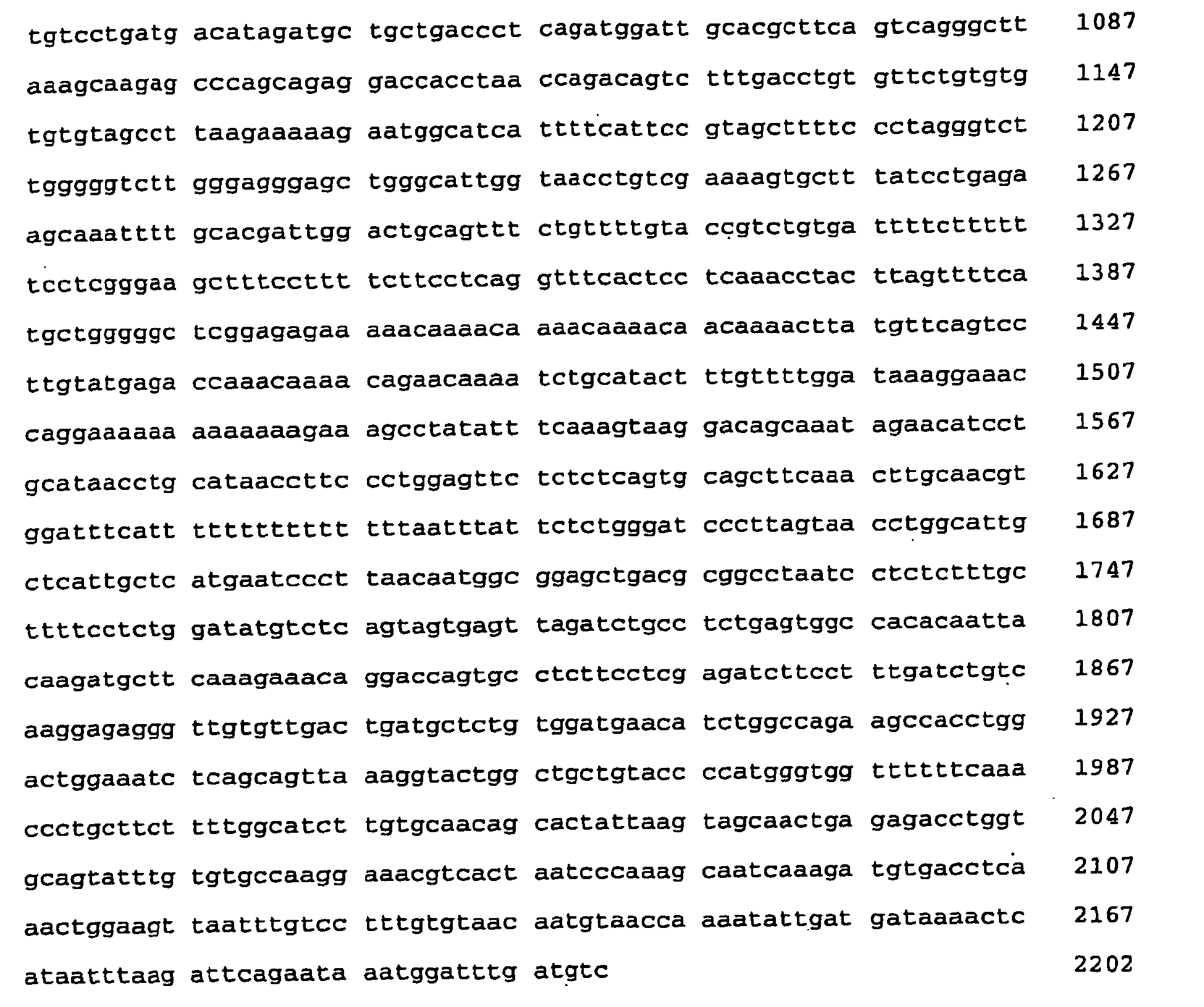
- <210> 31
<211> 224
<212> PRT
<213> Mus musculus - <400> 31


- <210> 32
<211> 2750
<212> DNA
<213> Homo sapiens - <220>
<221> CDS
<222> (564)..(1238)
<223> SF06, cDNA: NM_182511, Protein: NP_872317 - <400> 32


- <210> 33
<211> 224
<212> PRT
<213> Homo sapiens - <400> 33


- <210> 34
<211> 1542
<212> DNA
<213> Mus musculus - <220>
<221> CDS
<222> (131)..(1258)
<223> SF07, cDNA: NM_026840, Protein: NP_081116 - <400> 34


- <210> 35
<211> 375
<212> PRT
<213> Mus musculus - <400> 35


- <210> 36
<211> 1502
<212> DNA
<213> Homo sapiens - <220>
<221> CDS
<222> (62)..(1189)
<223> SF07, cDNA: NM_006207, Protein: NP_006198 - <400> 36


- <210> 37
<211> 375
<212> PRT
<213> Homo sapiens - <400> 37


- <210> 38
<211> 2379
<212> DNA
<213> Mus musculus - <220>
<221> CDS
<222> (69)..(2237)
<223> SF08, cDNA: NM_019696, Protein: NP_062670 - <400> 38



- <210> 39
<211> 722
<212> PRT
<213> Mus musculus - <400> 39
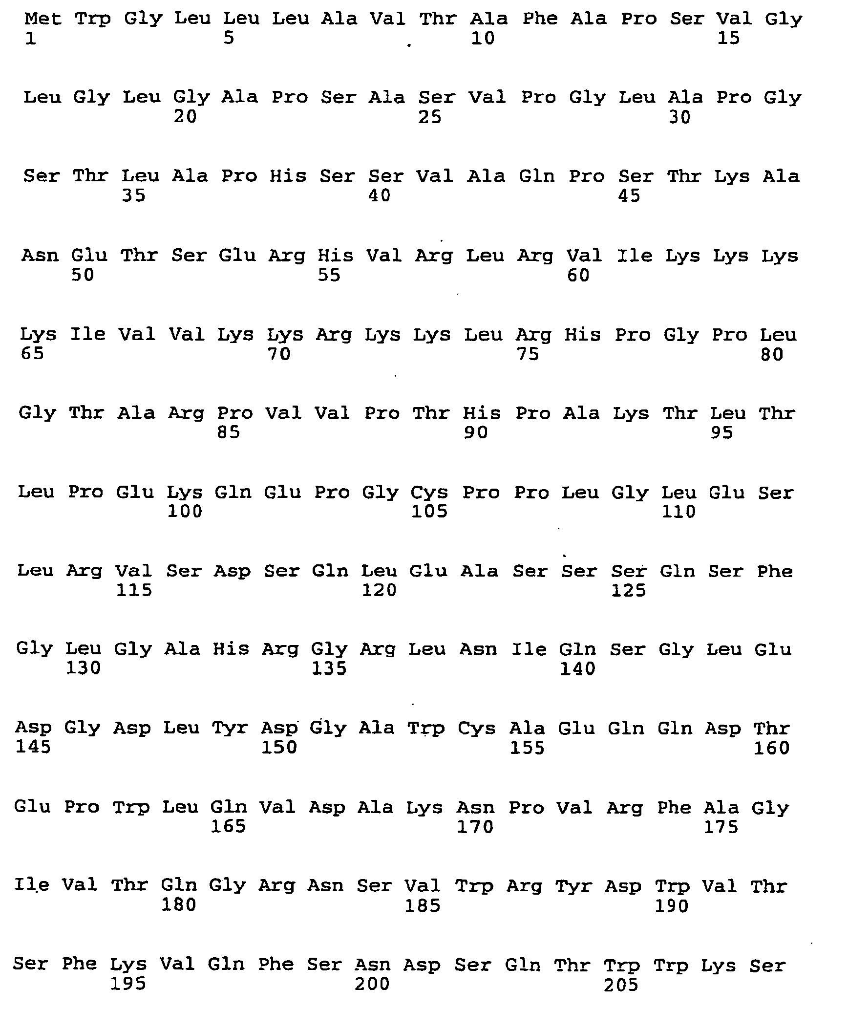


- <210> 40
<211> 2390
<212> DNA
<213> Homo sapiens - <220>
<221> CDS
<222> (65)..(2269)
<223> SF08, cDNA: NM_019609, Protein: NP_062555 - <400> 40


- <210> 41
<211> 734
<212> PRT
<213> Homo sapiens - <400> 41


- <210> 42
<211> 1815
<212> DNA
<213> Mus musculus - <220>
<221> CDS
<222> (98)..(535)
<223> SF09, cDNA: NM_139295, Protein: NP_647456 - <400> 42


- <210> 43
<211> 145
<212> PRT
<213> Mus musculus - <400> 43

- <210> 44
<211> 4144
<212> DNA
<213> Homo sapiens - <220>
<221> CDS
<222> (95)..(535)
<223> SF09, cDNA: NM_139279, Protein: NP_644808 - <400> 44


- <210> 45
<211> 146
<212> PRT
<213> Homo sapiens - <400> 45


- <210> 46
<211> 1513
<212> DNA
<213> Mus musculus - <220>
<221> CDS
<222> (37)..(810)
<223> SF10, cDNA: NM_029568, Protein: NP_083844 - <400> 46


- <210> 47
<211> 257
<212> PRT
<213> Mus musculus - <400> 47
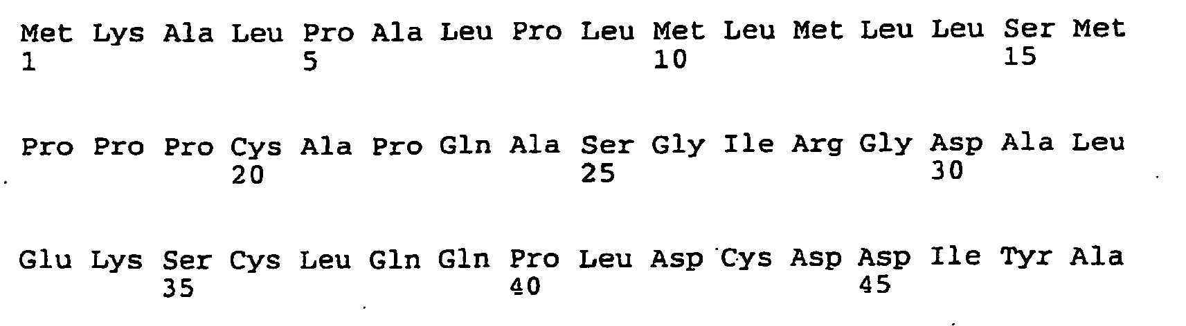
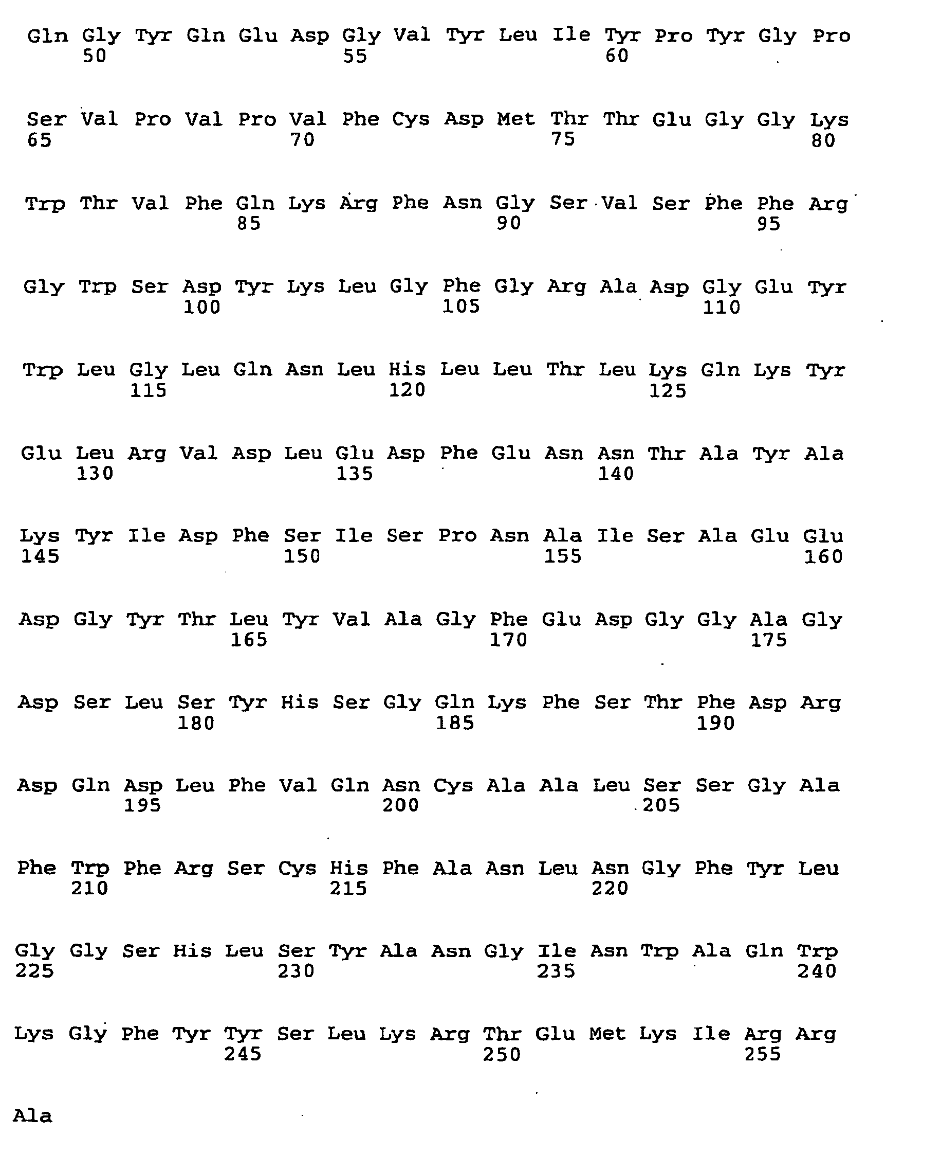
- <210> 48
<211> 1830
<212> DNA
<213> Homo sapiens - <220>
<221> CDS
<222> (26)..(793)
<223> SF10, cDNA: NM_002404, Protein: NP_002395 - <400> 48


- <210> 49
<211> 255
<212> PRT
<213> Homo sapiens - <400> 49

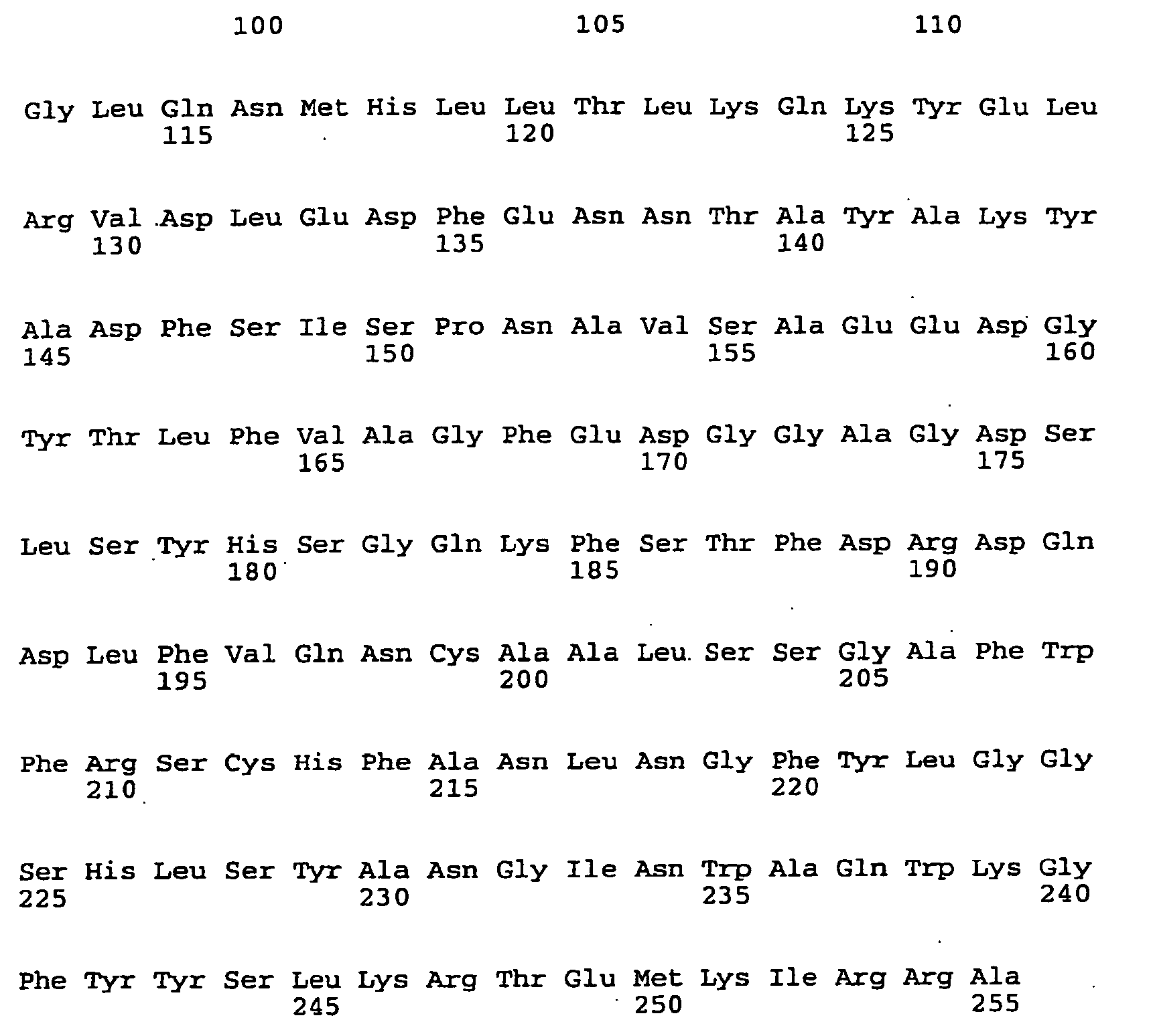
- <210> 50
<211> 749
<212> DNA
<213> Mus musculus - <220>
<221> CDS
<222> (93) .. (515)
<223> SF11, cDNA: NM_009976, Protein: NP_034106 - <400> 50


- <210> 51
<211> 140
<212> PRT
<213> Mus musculus - <400> 51


- <210> 52
<211> 818
<212> DNA
<213> Homo sapiens - <220>
<221> CDS
<222> (76)..(516)
<223> SF11, cDNA: NM_000099, Protein: NP_000090 - <400> 52


- <210> 53
<211> 146
<212> PRT
<213> Homo sapiens - <400> 53

- <210> 54
<211> 2709
<212> DNA
<213> Mus musculus - <220>
<221> CDS
<222> (124)..(2241)
<223> SF12, cDNA: NM_010180, Protein: NP_034310 - <400> 54


- <210> 55
<211> 705
<212> PRT
<213> Mus musculus - <400> 55


- <210> 56
<211> 2947
<212> DNA
<213> Homo sapiens - <220>
<221> CDS
<222> (148)..(2259)
<223> SF12, cDNA: NM_006486, Protein: NP_006477 - <400> 56


- <210> 57
<211> 703
<212> PRT
<213> Homo sapiens - <400> 57

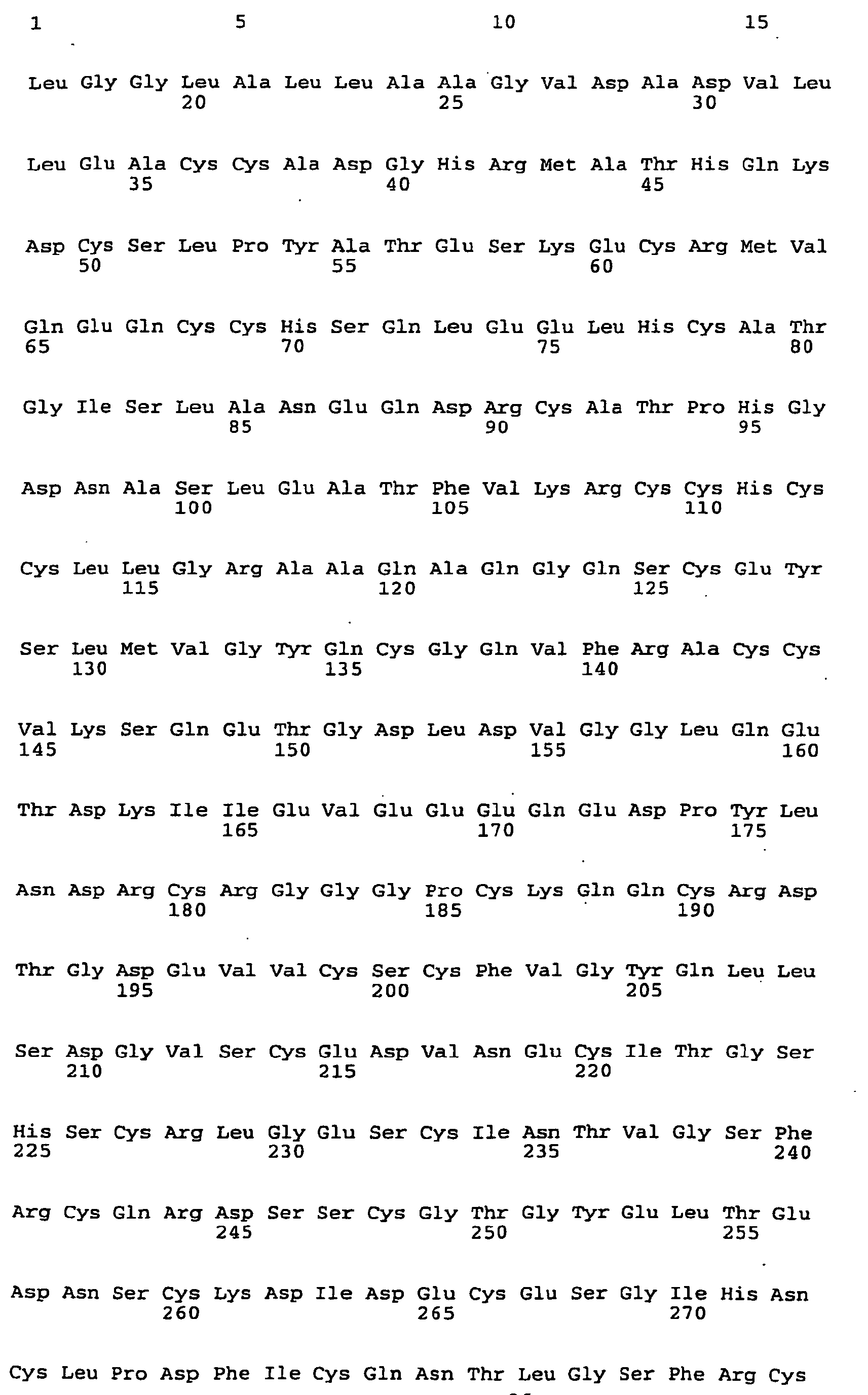


- <210> 58
<211> 979
<212> DNA
<213> Mus musculus - <220>
<221> CDS
<222> (98) .. (748)
<223> SF13, cDNA: NM_011149, Protein: NP_035279 - <400> 58
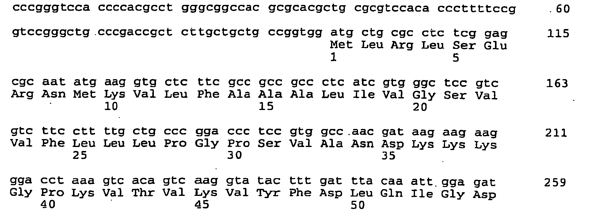

- <210> 59
<211> 216
<212> PRT
<213> Mus musculus - <400> 59


- <210> 60
<211> 1045
<212> DNA
<213> Homo sapiens - <220>
<221> CDS
<222> (170)..(820)
<223> SF13, cDNA: NM_000942, Protein: NP_000933 - <400> 60


- <210> 61
<211> 216
<212> PRT
<213> Homo sapiens - <400> 61

Claims (9)
- Use of a composition comprising a SF06 protein as characterized by SEQ ID Nos:30 or 32 and/or a nucleic acid molecule encoding a SF06 protein as characterized by SEQ ID Nos:30 or 32, the nucleic acid molecule being preferably a DNA molecule, more preferably a cDNA or a genomic DNA, and optionally pharmaceutically acceptable carriers, diluents, and/or additives for the preparation of an agent for detecting diseases or dysfunctions selected from the group consisting of diabetes, obesity, and/or metabolic syndrome.
- Use of a composition comprising an SF06 protein as characterized by SEQ ID Nos:30 or 32 and/or a nucleic acid molecule encoding an SF06 protein as characterized by SEQ ID Nos:30 or 32, that nucleic acid molecule being preferably a DNA molecule, more preferably a cDNA or genomic DNA, and optionally pharmaceutically acceptable carriers, diluents, and/or additives for the preparation of a medicament for the treatment, alleviation and/or prevention of diseases or dysfunctions selected from the group consisting of diabetes, obesity and/or metabolic syndrome.
- Use according to claim 1 or 2, wherein the nucleic acid molecule is a mammalian SF06 nucleic acid as characterized by SEQ ID Nos:30 or 32, particularly encoding the human SF06 polypeptide as characterized by SEQ ID No:32 and/or a nucleic acid molecule, which is complementary thereto.
- Use according to any one of claims 1-3, wherein said nucleic acid molecule is selected from the group consisting of(a) a nucleic acid molecule encoding a polypeptide, the polypeptide having a sequence as shown in SEQ ID NO: 30 or SEQ ID NO: 32, or an isoform of the polypeptide according to SEQ ID NO:30 or SEQ ID NO: 32;(b) a nucleic acid molecule which comprises or is the nucleic acid molecule according to SEQ ID NO: 30 or SEQ ID NO: 32;(c) a nucleic acid molecule being degenerate with as a result of the genetic code to the nucleic acid sequences as defined in (a) or (b),(d) a nucleic acid molecule that hybridizes at 50°C in a solution containing 1 x SSC and 0.1% SDS to a nucleic acid molecule as defined in claim 3 or as defined in (a) to (c) and/or a nucleic acid molecule which is complementary thereto;(e) a nucleic acid molecule that encodes a polypeptide which is at least 85%, preferably at least 90%, more preferably at least 95%, more preferably at least 98% and up to 99,6% identical to the human SF06 as defined in claim 3 or to a polypeptide as defined in (a).
- Use according to any one of claims 1-4, wherein said nucleic acid molecule is a recombinant nucleic acid molecule and/or a vector, particularly an expression vector and/or a hybridization probe, a primer or an anti-sense oligonucleotide.
- Use according to any one of claims 1-5, wherein the polypeptide is a recombinant polypeptide and/or a fusion polypeptide.
- Use according to any one of claims 1-6 for monotherapy or combination therapy together with at least one further pharmaceutical agent, the at least one further pharmaceutical agent being selected from the group consisting of 2-amino-1,3-propanediol derivatives, 2-amino-2[2-(4-octylphenyl)ethyl] propane-1,3-diol hydrochloride, 40-O(2-hydroxyethyl)-rapamycin, SDZ-RAD, Everolimus, 6-(3-dimethyl-aminopropionyl) forskolin, 6-mercaptopurine (6-MP), A-420983, ABX-CBL (CBL-1), Alefacept, Antisense ICAM-1 inhibitor (ISIS 2302), Enlimomab, BIRR1, Alicaforsen, Antithymocyte immunoglobulin (ATGAM), Azathioprine, Baohuoside-1, basiliximab, BMS-279700, BTI-322, Cladnbine, CP-690550, Cyclophosphamide (CTX), Cyclosporine (cyclosporin A, cyclosporin), Daclizumab, HAT (Humanized-Anti-Tac), SMART anti-Tac, anti-CD25, humanized anti-IL2-receptor, Dexamethasone (Decadron, Dexone, Dexasone), DIAPEP-277, Dipeptide Boronic Acid (DPBA), Docosahexaenoic acid (DHA), efalizumab, Efomycine M, FTY720 (oral myriocin derivative), Glatiramer acetate (co-polymer-1), Glial Fibrillary acidic protein (GFAP), Gusperimus (15-deoxyspergualin), HLA-B2702 peptide, hu1124 (anti-CD11a), hOKT31y (Ala-Ala), Infliximab, Interferon, ISAtx247, isotretinoin, L-683,742, Leflunomide (ARAVA), Medi-500 (T10B9), Medi-507, Methotrexate, Mitoxantrone, mycophenolate mofetil, OKT4A, Muromonab-CD3, Prednisolone, Psora-4, Rifampicin, Rituximab, S100β, Sirolimus, Rapamycin, Tacrolimus (Prograf; FK-506), or Triptofide.
- Method of identifying a (poly)peptide involved in the regulation of energy homeostasis and/or metabolism in a mammal comprising the steps of(a) contacting a collection of (poly)peptides with a SF06 polypeptide as characterized by SEQ ID Nos:30 or 32 or a SF06 protein encoded by a nucleic acid selected from the group consisting of(i) a nucleic acid molecule encoding a polypeptide, the polypeptide having a sequence as shown in SEQ ID NO: 30 or SEQ ID NO: 32, or an isoform of the polypeptide according to SEQ ID NO:30 or SEQ ID NO: 32;(ii) a nucleic acid molecule which comprises or is the nucleic acid molecule according to SEQ ID NO: 30 or SEQ ID NO: 32;(iii) a nucleic acid molecule being degenerate with as a result of the genetic code to the nucleic acid sequences as defined in (i) or (ii),(iv) a nucleic add molecule that hybridizes at 50°C in a solution containing 1 x SSC and 0.1% SDS to a mammalian SF06 nucleic acid as characterized by SEQ ID Nos 30 or 32 or as defined in (i) to (iii) and/or a nucleic add molecule which is complementary thereto;(v) a nucleic add molecule that encodes a polypeptide which is at least 85%, preferably at least 90%, more preferably at least 95%, more preferably at least 98% and up to 99,6% identical to the human SF06 as characterized by SEQ ID No. 32 or to a polypeptide as defined in (i) under conditions that allow binding of said (poly)peptides;(b) removing (poly)peptides which do not bind and(c) identifying (poly)peptides that bind to said SF06 polypeptide as characterized by SEQ ID Nos:30 or 32 or to said SF06 protein encoded by a nucleic acid selected from the group consisting of(i) a nucleic acid molecule encoding a polypeptide, the polypeptide having a sequence as shown in SEQ ID NO: 30 or SEQ ID NO: 32, or an isoform of the polypeptide according to SEQ ID NO:30 or SEQ ID NO: 32;(ii) a nucleic acid molecule which comprises or is the nucleic acid molecule according to SEQ ID NO: 30 or SEQ ID NO: 32;(iii) a nucleic acid molecule being degenerate with as a result of the genetic code to the nucleic acid sequences as defined in (i) or (ii),(iv) a nucleic acid molecule that hybridizes at 50°C in a solution containing 1 x SSC and 0.1% SDS to a mammalian SF06 nucleic add as characterized by SEQ ID Nos 30 or 32 or as defined in (i) to (iii) and/or a nucleic acid molecule which is complementary thereto;(v) a nucleic acid molecule that encodes a polypeptide which is at least 85%, preferably at least 90%, more preferably at least 95%, more preferably at least 98% and up to 99,6% identical to the human SF06 as characterized by SEQ ID No. 32 or to a polypeptide as defined in (i) under conditions that allow binding of said (poly)peptides;
- Method for screening for an agent, which effects/modulates the activity of a SF06 polypeptide as characterized by SEQ ID Nos:30 or 32, comprising the steps of(a) incubating a mixture comprising(aa) a SF06 polypeptide as characterized by SEQ ID Nos:30 or 32; and(ab) a candidate agent
under conditions whereby said SF06 polypeptide or fragment thereof exhibits a reference activity,(b) detecting the activity of said SF06 polypeptide to determine an activity for the agent; and(c) determining a difference between activity for the agent and reference activity.
Priority Applications (1)
| Application Number | Priority Date | Filing Date | Title |
|---|---|---|---|
| EP09163270A EP2096120B1 (en) | 2004-02-20 | 2005-02-18 | Use of secreted protein products for preventing and treating pancreatic diseases and/or obesity and/or metabolic syndrome |
Applications Claiming Priority (3)
| Application Number | Priority Date | Filing Date | Title |
|---|---|---|---|
| EP04003914 | 2004-02-20 | ||
| EP05701416A EP1730189A2 (en) | 2004-02-20 | 2005-02-18 | Use of secreted protein products for preventing and treating pancreatic diseases and/or obesity and/or metabolic syndrome |
| EP09163270A EP2096120B1 (en) | 2004-02-20 | 2005-02-18 | Use of secreted protein products for preventing and treating pancreatic diseases and/or obesity and/or metabolic syndrome |
Related Parent Applications (2)
| Application Number | Title | Priority Date | Filing Date |
|---|---|---|---|
| EP05701416A Division EP1730189A2 (en) | 2004-02-20 | 2005-02-18 | Use of secreted protein products for preventing and treating pancreatic diseases and/or obesity and/or metabolic syndrome |
| EP05701416.9 Division | 2005-02-18 |
Publications (3)
| Publication Number | Publication Date |
|---|---|
| EP2096120A2 EP2096120A2 (en) | 2009-09-02 |
| EP2096120A3 EP2096120A3 (en) | 2009-12-02 |
| EP2096120B1 true EP2096120B1 (en) | 2012-03-14 |
Family
ID=34878165
Family Applications (2)
| Application Number | Title | Priority Date | Filing Date |
|---|---|---|---|
| EP05701416A Withdrawn EP1730189A2 (en) | 2004-02-20 | 2005-02-18 | Use of secreted protein products for preventing and treating pancreatic diseases and/or obesity and/or metabolic syndrome |
| EP09163270A Expired - Lifetime EP2096120B1 (en) | 2004-02-20 | 2005-02-18 | Use of secreted protein products for preventing and treating pancreatic diseases and/or obesity and/or metabolic syndrome |
Family Applications Before (1)
| Application Number | Title | Priority Date | Filing Date |
|---|---|---|---|
| EP05701416A Withdrawn EP1730189A2 (en) | 2004-02-20 | 2005-02-18 | Use of secreted protein products for preventing and treating pancreatic diseases and/or obesity and/or metabolic syndrome |
Country Status (6)
| Country | Link |
|---|---|
| US (2) | US20070248579A1 (en) |
| EP (2) | EP1730189A2 (en) |
| AT (1) | ATE549352T1 (en) |
| DK (1) | DK2096120T3 (en) |
| ES (1) | ES2384134T3 (en) |
| WO (1) | WO2005079840A2 (en) |
Cited By (4)
| Publication number | Priority date | Publication date | Assignee | Title |
|---|---|---|---|---|
| KR102065150B1 (en) | 2018-04-27 | 2020-01-10 | (주)케어젠 | A composition for preventing or treating obesity comprising isotretinoin-peptide conjugate as an effective ingredient |
| US11434291B2 (en) | 2019-05-14 | 2022-09-06 | Provention Bio, Inc. | Methods and compositions for preventing type 1 diabetes |
| US12006366B2 (en) | 2020-06-11 | 2024-06-11 | Provention Bio, Inc. | Methods and compositions for preventing type 1 diabetes |
| US12565529B2 (en) | 2021-05-24 | 2026-03-03 | Provention Bio, Inc. | Methods for treating type 1 diabetes |
Families Citing this family (4)
| Publication number | Priority date | Publication date | Assignee | Title |
|---|---|---|---|---|
| WO2008092214A1 (en) * | 2007-02-02 | 2008-08-07 | Minomic International Limited | Biomarkers for diabetes |
| WO2013102209A1 (en) * | 2011-12-30 | 2013-07-04 | Joslin Diabetes Center, Inc. | Glypican-4 based compositions and methods for treating and diagnosing insulin resistance |
| CN103113468B (en) * | 2013-01-17 | 2014-05-21 | 中国人民解放军军事医学科学院基础医学研究所 | an anti-tumor protein |
| BR112022017718A2 (en) | 2020-03-05 | 2022-11-16 | Diamedica Usa Inc | ULINASTATIN POLYPEPTIDES |
Family Cites Families (12)
| Publication number | Priority date | Publication date | Assignee | Title |
|---|---|---|---|---|
| NZ207394A (en) | 1983-03-08 | 1987-03-06 | Commw Serum Lab Commission | Detecting or determining sequence of amino acids |
| US5474796A (en) | 1991-09-04 | 1995-12-12 | Protogene Laboratories, Inc. | Method and apparatus for conducting an array of chemical reactions on a support surface |
| US5605662A (en) | 1993-11-01 | 1997-02-25 | Nanogen, Inc. | Active programmable electronic devices for molecular biological analysis and diagnostics |
| US6015880A (en) | 1994-03-16 | 2000-01-18 | California Institute Of Technology | Method and substrate for performing multiple sequential reactions on a matrix |
| US5807522A (en) | 1994-06-17 | 1998-09-15 | The Board Of Trustees Of The Leland Stanford Junior University | Methods for fabricating microarrays of biological samples |
| US20020137890A1 (en) * | 1997-03-31 | 2002-09-26 | Genentech, Inc. | Secreted and transmembrane polypeptides and nucleic acids encoding the same |
| US5886029A (en) * | 1997-09-05 | 1999-03-23 | Dhaliwal; Kirpal S. | Method and composition for treatment of diabetes |
| WO2001000672A1 (en) * | 1999-06-29 | 2001-01-04 | Millennium Pharmaceuticals, Inc. | Secreted proteins and uses thereof |
| US20030170781A1 (en) * | 1999-07-01 | 2003-09-11 | Zymogenetics, Inc. | Secreted protein zacrp4 |
| WO2002057453A2 (en) * | 2000-12-19 | 2002-07-25 | Curagen Corporation | Polypetides and nucleic acids encoding same |
| US20040248156A1 (en) * | 2001-12-03 | 2004-12-09 | Tianhua Hu | Methods and materials relating to novel C1q domain-containing polypeptides and polynucleotides |
| AU2003219167A1 (en) * | 2002-03-19 | 2003-09-29 | Genset Sa | Treatment of metabolic disorders with a tnf receptor family member (fradj and/or cryptic) agonists or antagonists |
-
2005
- 2005-02-18 WO PCT/EP2005/001711 patent/WO2005079840A2/en not_active Ceased
- 2005-02-18 AT AT09163270T patent/ATE549352T1/en active
- 2005-02-18 US US10/589,677 patent/US20070248579A1/en not_active Abandoned
- 2005-02-18 DK DK09163270.3T patent/DK2096120T3/en active
- 2005-02-18 ES ES09163270T patent/ES2384134T3/en not_active Expired - Lifetime
- 2005-02-18 EP EP05701416A patent/EP1730189A2/en not_active Withdrawn
- 2005-02-18 EP EP09163270A patent/EP2096120B1/en not_active Expired - Lifetime
-
2009
- 2009-05-08 US US12/437,851 patent/US20090298771A1/en not_active Abandoned
Cited By (4)
| Publication number | Priority date | Publication date | Assignee | Title |
|---|---|---|---|---|
| KR102065150B1 (en) | 2018-04-27 | 2020-01-10 | (주)케어젠 | A composition for preventing or treating obesity comprising isotretinoin-peptide conjugate as an effective ingredient |
| US11434291B2 (en) | 2019-05-14 | 2022-09-06 | Provention Bio, Inc. | Methods and compositions for preventing type 1 diabetes |
| US12006366B2 (en) | 2020-06-11 | 2024-06-11 | Provention Bio, Inc. | Methods and compositions for preventing type 1 diabetes |
| US12565529B2 (en) | 2021-05-24 | 2026-03-03 | Provention Bio, Inc. | Methods for treating type 1 diabetes |
Also Published As
| Publication number | Publication date |
|---|---|
| US20090298771A1 (en) | 2009-12-03 |
| DK2096120T3 (en) | 2012-07-09 |
| EP1730189A2 (en) | 2006-12-13 |
| ATE549352T1 (en) | 2012-03-15 |
| WO2005079840A3 (en) | 2006-10-26 |
| EP2096120A3 (en) | 2009-12-02 |
| EP2096120A2 (en) | 2009-09-02 |
| ES2384134T3 (en) | 2012-06-29 |
| WO2005079840A2 (en) | 2005-09-01 |
| US20070248579A1 (en) | 2007-10-25 |
Similar Documents
| Publication | Publication Date | Title |
|---|---|---|
| US20090298771A1 (en) | Use of secreted protein products for preventing and treating pancreatic diseases and/or obesity and/or metabolic syndrome | |
| EP1644406B1 (en) | Use of dg153 secreted protein products for preventing and treating pancreatic diseases and/or obesity and/or metabolic syndrome | |
| EP1605965B1 (en) | Use of saposin-related proteins for preventing and treating obesity, diabetes and/or metabolic syndrome | |
| EP2135618B1 (en) | SF6 for use in the treatment of diabetes and / or obesity, methods of screening for modulators and kit | |
| US20060259988A1 (en) | Use of dg931 protein for treating diabetes, obesity and metabolic syndrome | |
| EP1644027B1 (en) | Use of pleiotrophin for preventing and treating pancreatic diseases and/or obesity and/or metabolic syndrome | |
| WO2004012758A1 (en) | Use of tgf beta ig-h3 for preventing and treating obesity, diabetes and/or metabolic syndrome | |
| US20060234930A1 (en) | Use of dg008,dg065,dg210 or dg239 secreted protein products for preventing and treating pancreatic diseases and/or obesity and/or metabolic syndrome | |
| US20070050856A1 (en) | Use of protein products for preventing and treating pancreatic diseases and/or obesity and/or metabolic syndrome | |
| US20060015951A1 (en) | Proteins involved in the regulation of energy homeostasis | |
| US20080107639A1 (en) | Use of a Dg147 Protein Product for Preventing and Treating Metabolic Disorders | |
| US20060168667A1 (en) | Minibrain homologous proteins involved in the regulation of energy homeostasis | |
| US20060135419A1 (en) | Proteins involved in the regulation of energy homeostasis | |
| US20050283842A1 (en) | Mipp1 homologous nucleic acids and proteins involved in the regulation of energy homeostatis | |
| EP0903147A2 (en) | Use of REG-2 as a Schwann cell mitogen |
Legal Events
| Date | Code | Title | Description |
|---|---|---|---|
| PUAI | Public reference made under article 153(3) epc to a published international application that has entered the european phase |
Free format text: ORIGINAL CODE: 0009012 |
|
| 17P | Request for examination filed |
Effective date: 20090619 |
|
| AC | Divisional application: reference to earlier application |
Ref document number: 1730189 Country of ref document: EP Kind code of ref document: P |
|
| AK | Designated contracting states |
Kind code of ref document: A2 Designated state(s): AT BE BG CH CY CZ DE DK EE ES FI FR GB GR HU IE IS IT LI LT LU MC NL PL PT RO SE SI SK TR |
|
| PUAL | Search report despatched |
Free format text: ORIGINAL CODE: 0009013 |
|
| AK | Designated contracting states |
Kind code of ref document: A3 Designated state(s): AT BE BG CH CY CZ DE DK EE ES FI FR GB GR HU IE IS IT LI LT LU MC NL PL PT RO SE SI SK TR |
|
| 17Q | First examination report despatched |
Effective date: 20100412 |
|
| GRAJ | Information related to disapproval of communication of intention to grant by the applicant or resumption of examination proceedings by the epo deleted |
Free format text: ORIGINAL CODE: EPIDOSDIGR1 |
|
| GRAP | Despatch of communication of intention to grant a patent |
Free format text: ORIGINAL CODE: EPIDOSNIGR1 |
|
| GRAS | Grant fee paid |
Free format text: ORIGINAL CODE: EPIDOSNIGR3 |
|
| GRAA | (expected) grant |
Free format text: ORIGINAL CODE: 0009210 |
|
| AC | Divisional application: reference to earlier application |
Ref document number: 1730189 Country of ref document: EP Kind code of ref document: P |
|
| AK | Designated contracting states |
Kind code of ref document: B1 Designated state(s): AT BE BG CH CY CZ DE DK EE ES FI FR GB GR HU IE IS IT LI LT LU MC NL PL PT RO SE SI SK TR |
|
| REG | Reference to a national code |
Ref country code: GB Ref legal event code: FG4D |
|
| REG | Reference to a national code |
Ref country code: AT Ref legal event code: REF Ref document number: 549352 Country of ref document: AT Kind code of ref document: T Effective date: 20120315 Ref country code: CH Ref legal event code: EP |
|
| REG | Reference to a national code |
Ref country code: IE Ref legal event code: FG4D |
|
| REG | Reference to a national code |
Ref country code: DE Ref legal event code: R096 Ref document number: 602005033207 Country of ref document: DE Effective date: 20120510 |
|
| REG | Reference to a national code |
Ref country code: SE Ref legal event code: TRGR |
|
| REG | Reference to a national code |
Ref country code: CH Ref legal event code: NV Representative=s name: E. BLUM & CO. AG PATENT- UND MARKENANWAELTE VSP |
|
| REG | Reference to a national code |
Ref country code: NL Ref legal event code: T3 |
|
| REG | Reference to a national code |
Ref country code: ES Ref legal event code: FG2A Ref document number: 2384134 Country of ref document: ES Kind code of ref document: T3 Effective date: 20120629 |
|
| REG | Reference to a national code |
Ref country code: DK Ref legal event code: T3 |
|
| PG25 | Lapsed in a contracting state [announced via postgrant information from national office to epo] |
Ref country code: LT Free format text: LAPSE BECAUSE OF FAILURE TO SUBMIT A TRANSLATION OF THE DESCRIPTION OR TO PAY THE FEE WITHIN THE PRESCRIBED TIME-LIMIT Effective date: 20120314 |
|
| LTIE | Lt: invalidation of european patent or patent extension |
Effective date: 20120314 |
|
| PG25 | Lapsed in a contracting state [announced via postgrant information from national office to epo] |
Ref country code: GR Free format text: LAPSE BECAUSE OF FAILURE TO SUBMIT A TRANSLATION OF THE DESCRIPTION OR TO PAY THE FEE WITHIN THE PRESCRIBED TIME-LIMIT Effective date: 20120615 Ref country code: FI Free format text: LAPSE BECAUSE OF FAILURE TO SUBMIT A TRANSLATION OF THE DESCRIPTION OR TO PAY THE FEE WITHIN THE PRESCRIBED TIME-LIMIT Effective date: 20120314 |
|
| PG25 | Lapsed in a contracting state [announced via postgrant information from national office to epo] |
Ref country code: CY Free format text: LAPSE BECAUSE OF FAILURE TO SUBMIT A TRANSLATION OF THE DESCRIPTION OR TO PAY THE FEE WITHIN THE PRESCRIBED TIME-LIMIT Effective date: 20120314 |
|
| PG25 | Lapsed in a contracting state [announced via postgrant information from national office to epo] |
Ref country code: EE Free format text: LAPSE BECAUSE OF FAILURE TO SUBMIT A TRANSLATION OF THE DESCRIPTION OR TO PAY THE FEE WITHIN THE PRESCRIBED TIME-LIMIT Effective date: 20120314 Ref country code: RO Free format text: LAPSE BECAUSE OF FAILURE TO SUBMIT A TRANSLATION OF THE DESCRIPTION OR TO PAY THE FEE WITHIN THE PRESCRIBED TIME-LIMIT Effective date: 20120314 Ref country code: SI Free format text: LAPSE BECAUSE OF FAILURE TO SUBMIT A TRANSLATION OF THE DESCRIPTION OR TO PAY THE FEE WITHIN THE PRESCRIBED TIME-LIMIT Effective date: 20120314 Ref country code: PL Free format text: LAPSE BECAUSE OF FAILURE TO SUBMIT A TRANSLATION OF THE DESCRIPTION OR TO PAY THE FEE WITHIN THE PRESCRIBED TIME-LIMIT Effective date: 20120314 Ref country code: IS Free format text: LAPSE BECAUSE OF FAILURE TO SUBMIT A TRANSLATION OF THE DESCRIPTION OR TO PAY THE FEE WITHIN THE PRESCRIBED TIME-LIMIT Effective date: 20120714 Ref country code: CZ Free format text: LAPSE BECAUSE OF FAILURE TO SUBMIT A TRANSLATION OF THE DESCRIPTION OR TO PAY THE FEE WITHIN THE PRESCRIBED TIME-LIMIT Effective date: 20120314 |
|
| REG | Reference to a national code |
Ref country code: DE Ref legal event code: R082 Ref document number: 602005033207 Country of ref document: DE Representative=s name: WEICKMANN & WEICKMANN, DE |
|
| PG25 | Lapsed in a contracting state [announced via postgrant information from national office to epo] |
Ref country code: SK Free format text: LAPSE BECAUSE OF FAILURE TO SUBMIT A TRANSLATION OF THE DESCRIPTION OR TO PAY THE FEE WITHIN THE PRESCRIBED TIME-LIMIT Effective date: 20120314 Ref country code: PT Free format text: LAPSE BECAUSE OF FAILURE TO SUBMIT A TRANSLATION OF THE DESCRIPTION OR TO PAY THE FEE WITHIN THE PRESCRIBED TIME-LIMIT Effective date: 20120716 |
|
| REG | Reference to a national code |
Ref country code: DE Ref legal event code: R082 Ref document number: 602005033207 Country of ref document: DE Representative=s name: WEICKMANN & WEICKMANN, DE Effective date: 20121126 Ref country code: DE Ref legal event code: R081 Ref document number: 602005033207 Country of ref document: DE Owner name: EVOTEC INTERNATIONAL GMBH, DE Free format text: FORMER OWNER: DEVELOGEN AKTIENGESELLSCHAFT, 37079 GOETTINGEN, DE Effective date: 20121126 Ref country code: DE Ref legal event code: R082 Ref document number: 602005033207 Country of ref document: DE Representative=s name: PATENTANWAELTE WEICKMANN & WEICKMANN, DE Effective date: 20121126 Ref country code: DE Ref legal event code: R082 Ref document number: 602005033207 Country of ref document: DE Representative=s name: WEICKMANN & WEICKMANN PATENTANWAELTE - RECHTSA, DE Effective date: 20121126 |
|
| PLBE | No opposition filed within time limit |
Free format text: ORIGINAL CODE: 0009261 |
|
| STAA | Information on the status of an ep patent application or granted ep patent |
Free format text: STATUS: NO OPPOSITION FILED WITHIN TIME LIMIT |
|
| REG | Reference to a national code |
Ref country code: CH Ref legal event code: PFUS Owner name: EVOTEC NEUROSCIENCES GMBH, DE Free format text: FORMER OWNER: EVOTEC (GOETTINGEN) AG, DE Ref country code: CH Ref legal event code: PFA Owner name: EVOTEC (GOETTINGEN) AG, DE Free format text: FORMER OWNER: DEVELOGEN AKTIENGESELLSCHAFT, DE Ref country code: CH Ref legal event code: PFA Owner name: EVOTEC INTERNATIONAL GMBH, DE Free format text: FORMER OWNER: EVOTEC NEUROSCIENCES GMBH, DE |
|
| REG | Reference to a national code |
Ref country code: CH Ref legal event code: PFA Owner name: EVOTEC (GOETTINGEN) AG, DE Free format text: FORMER OWNER: DEVELOGEN AKTIENGESELLSCHAFT, DE Ref country code: CH Ref legal event code: PFUS Owner name: EVOTEC NEUROSCIENCES GMBH, DE Free format text: FORMER OWNER: EVOTEC (GOETTINGEN) AG, DE Ref country code: CH Ref legal event code: PFA Owner name: EVOTEC INTERNATIONAL GMBH, DE Free format text: FORMER OWNER: EVOTEC NEUROSCIENCES GMBH, DE |
|
| 26N | No opposition filed |
Effective date: 20121217 |
|
| REG | Reference to a national code |
Ref country code: DE Ref legal event code: R097 Ref document number: 602005033207 Country of ref document: DE Effective date: 20121217 |
|
| REG | Reference to a national code |
Ref country code: GB Ref legal event code: 732E Free format text: REGISTERED BETWEEN 20130321 AND 20130327 |
|
| BECH | Be: change of holder |
Owner name: EVOTEC INTERNATIONAL G.M.B.H. Effective date: 20130409 |
|
| BECN | Be: change of holder's name |
Owner name: EVOTEC INTERNATIONAL G.M.B.H. Effective date: 20130409 |
|
| REG | Reference to a national code |
Ref country code: ES Ref legal event code: PC2A Owner name: EVOTEC INTERNATIONAL GMBH Effective date: 20130517 |
|
| REG | Reference to a national code |
Ref country code: AT Ref legal event code: PC Ref document number: 549352 Country of ref document: AT Kind code of ref document: T Owner name: EVOTEC INTERNATIONAL GMBH, DE Effective date: 20130419 |
|
| PG25 | Lapsed in a contracting state [announced via postgrant information from national office to epo] |
Ref country code: BG Free format text: LAPSE BECAUSE OF FAILURE TO SUBMIT A TRANSLATION OF THE DESCRIPTION OR TO PAY THE FEE WITHIN THE PRESCRIBED TIME-LIMIT Effective date: 20120614 |
|
| PG25 | Lapsed in a contracting state [announced via postgrant information from national office to epo] |
Ref country code: MC Free format text: LAPSE BECAUSE OF NON-PAYMENT OF DUE FEES Effective date: 20130228 |
|
| REG | Reference to a national code |
Ref country code: NL Ref legal event code: TD Effective date: 20131017 Ref country code: NL Ref legal event code: SD Effective date: 20131017 |
|
| REG | Reference to a national code |
Ref country code: FR Ref legal event code: TP Owner name: EVOTEC INTERNATIONAL GMBH, DE Effective date: 20131002 Ref country code: FR Ref legal event code: CD Owner name: EVOTEC INTERNATIONAL GMBH, DE Effective date: 20131002 |
|
| REG | Reference to a national code |
Ref country code: IE Ref legal event code: MM4A |
|
| PG25 | Lapsed in a contracting state [announced via postgrant information from national office to epo] |
Ref country code: IE Free format text: LAPSE BECAUSE OF NON-PAYMENT OF DUE FEES Effective date: 20130218 |
|
| PGFP | Annual fee paid to national office [announced via postgrant information from national office to epo] |
Ref country code: DK Payment date: 20140218 Year of fee payment: 10 Ref country code: NL Payment date: 20140218 Year of fee payment: 10 |
|
| PGFP | Annual fee paid to national office [announced via postgrant information from national office to epo] |
Ref country code: IT Payment date: 20140224 Year of fee payment: 10 Ref country code: AT Payment date: 20140212 Year of fee payment: 10 |
|
| REG | Reference to a national code |
Ref country code: FR Ref legal event code: PLFP Year of fee payment: 11 |
|
| PGFP | Annual fee paid to national office [announced via postgrant information from national office to epo] |
Ref country code: ES Payment date: 20150326 Year of fee payment: 11 Ref country code: CH Payment date: 20150319 Year of fee payment: 11 Ref country code: DE Payment date: 20150227 Year of fee payment: 11 |
|
| PGFP | Annual fee paid to national office [announced via postgrant information from national office to epo] |
Ref country code: GB Payment date: 20150227 Year of fee payment: 11 Ref country code: SE Payment date: 20150227 Year of fee payment: 11 Ref country code: FR Payment date: 20150227 Year of fee payment: 11 |
|
| PG25 | Lapsed in a contracting state [announced via postgrant information from national office to epo] |
Ref country code: TR Free format text: LAPSE BECAUSE OF FAILURE TO SUBMIT A TRANSLATION OF THE DESCRIPTION OR TO PAY THE FEE WITHIN THE PRESCRIBED TIME-LIMIT Effective date: 20120314 |
|
| PGFP | Annual fee paid to national office [announced via postgrant information from national office to epo] |
Ref country code: BE Payment date: 20150227 Year of fee payment: 11 |
|
| PG25 | Lapsed in a contracting state [announced via postgrant information from national office to epo] |
Ref country code: HU Free format text: LAPSE BECAUSE OF FAILURE TO SUBMIT A TRANSLATION OF THE DESCRIPTION OR TO PAY THE FEE WITHIN THE PRESCRIBED TIME-LIMIT; INVALID AB INITIO Effective date: 20050218 Ref country code: LU Free format text: LAPSE BECAUSE OF NON-PAYMENT OF DUE FEES Effective date: 20130218 |
|
| REG | Reference to a national code |
Ref country code: NL Ref legal event code: V1 Effective date: 20150901 |
|
| REG | Reference to a national code |
Ref country code: DK Ref legal event code: EBP Effective date: 20150228 |
|
| PG25 | Lapsed in a contracting state [announced via postgrant information from national office to epo] |
Ref country code: NL Free format text: LAPSE BECAUSE OF NON-PAYMENT OF DUE FEES Effective date: 20150901 |
|
| REG | Reference to a national code |
Ref country code: AT Ref legal event code: MM01 Ref document number: 549352 Country of ref document: AT Kind code of ref document: T Effective date: 20150218 |
|
| PG25 | Lapsed in a contracting state [announced via postgrant information from national office to epo] |
Ref country code: AT Free format text: LAPSE BECAUSE OF NON-PAYMENT OF DUE FEES Effective date: 20150218 |
|
| PG25 | Lapsed in a contracting state [announced via postgrant information from national office to epo] |
Ref country code: IT Free format text: LAPSE BECAUSE OF NON-PAYMENT OF DUE FEES Effective date: 20150218 |
|
| PG25 | Lapsed in a contracting state [announced via postgrant information from national office to epo] |
Ref country code: DK Free format text: LAPSE BECAUSE OF NON-PAYMENT OF DUE FEES Effective date: 20150228 |
|
| PG25 | Lapsed in a contracting state [announced via postgrant information from national office to epo] |
Ref country code: BE Free format text: LAPSE BECAUSE OF NON-PAYMENT OF DUE FEES Effective date: 20160229 |
|
| REG | Reference to a national code |
Ref country code: DE Ref legal event code: R119 Ref document number: 602005033207 Country of ref document: DE |
|
| REG | Reference to a national code |
Ref country code: CH Ref legal event code: PL |
|
| REG | Reference to a national code |
Ref country code: SE Ref legal event code: EUG |
|
| GBPC | Gb: european patent ceased through non-payment of renewal fee |
Effective date: 20160218 |
|
| PG25 | Lapsed in a contracting state [announced via postgrant information from national office to epo] |
Ref country code: CH Free format text: LAPSE BECAUSE OF NON-PAYMENT OF DUE FEES Effective date: 20160229 Ref country code: LI Free format text: LAPSE BECAUSE OF NON-PAYMENT OF DUE FEES Effective date: 20160229 |
|
| REG | Reference to a national code |
Ref country code: FR Ref legal event code: ST Effective date: 20161028 |
|
| PG25 | Lapsed in a contracting state [announced via postgrant information from national office to epo] |
Ref country code: SE Free format text: LAPSE BECAUSE OF NON-PAYMENT OF DUE FEES Effective date: 20160219 |
|
| PG25 | Lapsed in a contracting state [announced via postgrant information from national office to epo] |
Ref country code: DE Free format text: LAPSE BECAUSE OF NON-PAYMENT OF DUE FEES Effective date: 20160901 Ref country code: GB Free format text: LAPSE BECAUSE OF NON-PAYMENT OF DUE FEES Effective date: 20160218 Ref country code: FR Free format text: LAPSE BECAUSE OF NON-PAYMENT OF DUE FEES Effective date: 20160229 |
|
| PG25 | Lapsed in a contracting state [announced via postgrant information from national office to epo] |
Ref country code: ES Free format text: LAPSE BECAUSE OF NON-PAYMENT OF DUE FEES Effective date: 20160219 |