EP2084267B1 - Cancer stem cell antigen vaccines and methods - Google Patents
Cancer stem cell antigen vaccines and methods Download PDFInfo
- Publication number
- EP2084267B1 EP2084267B1 EP07843269.7A EP07843269A EP2084267B1 EP 2084267 B1 EP2084267 B1 EP 2084267B1 EP 07843269 A EP07843269 A EP 07843269A EP 2084267 B1 EP2084267 B1 EP 2084267B1
- Authority
- EP
- European Patent Office
- Prior art keywords
- cells
- tumor
- cell
- seq
- cancer
- Prior art date
- Legal status (The legal status is an assumption and is not a legal conclusion. Google has not performed a legal analysis and makes no representation as to the accuracy of the status listed.)
- Active
Links
- 206010028980 Neoplasm Diseases 0.000 title claims description 218
- 201000011510 cancer Diseases 0.000 title claims description 114
- 210000000130 stem cell Anatomy 0.000 title claims description 96
- 102000036639 antigens Human genes 0.000 title claims description 61
- 108091007433 antigens Proteins 0.000 title claims description 61
- 239000000427 antigen Substances 0.000 title claims description 60
- 238000000034 method Methods 0.000 title claims description 42
- 229960005486 vaccine Drugs 0.000 title claims description 20
- 210000004443 dendritic cell Anatomy 0.000 claims description 39
- 230000001537 neural effect Effects 0.000 claims description 22
- 239000000203 mixture Substances 0.000 claims description 18
- 206010018338 Glioma Diseases 0.000 claims description 17
- 208000032612 Glial tumor Diseases 0.000 claims description 10
- 239000006166 lysate Substances 0.000 claims description 3
- 210000004027 cell Anatomy 0.000 description 193
- 101000610551 Homo sapiens Prominin-1 Proteins 0.000 description 90
- 102100040120 Prominin-1 Human genes 0.000 description 89
- 230000014509 gene expression Effects 0.000 description 40
- 241000700159 Rattus Species 0.000 description 38
- 108090000765 processed proteins & peptides Proteins 0.000 description 35
- 210000004881 tumor cell Anatomy 0.000 description 34
- 150000001413 amino acids Chemical group 0.000 description 32
- 102000004196 processed proteins & peptides Human genes 0.000 description 28
- 239000002356 single layer Substances 0.000 description 26
- 239000002609 medium Substances 0.000 description 24
- 210000001744 T-lymphocyte Anatomy 0.000 description 23
- 108090000623 proteins and genes Proteins 0.000 description 23
- 230000004083 survival effect Effects 0.000 description 23
- 208000005017 glioblastoma Diseases 0.000 description 21
- 108091003079 Bovine Serum Albumin Proteins 0.000 description 20
- 102000008730 Nestin Human genes 0.000 description 20
- 108010088225 Nestin Proteins 0.000 description 20
- 239000012091 fetal bovine serum Substances 0.000 description 20
- 210000005055 nestin Anatomy 0.000 description 20
- 210000001178 neural stem cell Anatomy 0.000 description 20
- 210000001519 tissue Anatomy 0.000 description 20
- 210000000612 antigen-presenting cell Anatomy 0.000 description 19
- 208000003174 Brain Neoplasms Diseases 0.000 description 18
- 241001465754 Metazoa Species 0.000 description 17
- 238000002255 vaccination Methods 0.000 description 16
- 239000003550 marker Substances 0.000 description 15
- 102100024299 Maternal embryonic leucine zipper kinase Human genes 0.000 description 14
- 101710154611 Maternal embryonic leucine zipper kinase Proteins 0.000 description 14
- 102100021663 Baculoviral IAP repeat-containing protein 5 Human genes 0.000 description 13
- 108010002687 Survivin Proteins 0.000 description 13
- 238000011282 treatment Methods 0.000 description 13
- 239000006144 Dulbecco’s modified Eagle's medium Substances 0.000 description 12
- 241000283973 Oryctolagus cuniculus Species 0.000 description 12
- 190000008236 carboplatin Chemical compound 0.000 description 12
- 210000004248 oligodendroglia Anatomy 0.000 description 12
- 102100021569 Apoptosis regulator Bcl-2 Human genes 0.000 description 11
- 108091007065 BIRCs Proteins 0.000 description 11
- 102100021676 Baculoviral IAP repeat-containing protein 1 Human genes 0.000 description 11
- 101000971171 Homo sapiens Apoptosis regulator Bcl-2 Proteins 0.000 description 11
- 102000055031 Inhibitor of Apoptosis Proteins Human genes 0.000 description 11
- 101100058550 Mus musculus Bmi1 gene Proteins 0.000 description 11
- 108010006696 Neuronal Apoptosis-Inhibitory Protein Proteins 0.000 description 11
- 102100033237 Pro-epidermal growth factor Human genes 0.000 description 11
- 229960004562 carboplatin Drugs 0.000 description 11
- 208000002409 gliosarcoma Diseases 0.000 description 11
- 102000004169 proteins and genes Human genes 0.000 description 11
- 102100032912 CD44 antigen Human genes 0.000 description 10
- 201000010915 Glioblastoma multiforme Diseases 0.000 description 10
- 101000868273 Homo sapiens CD44 antigen Proteins 0.000 description 10
- 241000124008 Mammalia Species 0.000 description 10
- 239000002246 antineoplastic agent Substances 0.000 description 10
- 229940127089 cytotoxic agent Drugs 0.000 description 10
- 208000037265 diseases, disorders, signs and symptoms Diseases 0.000 description 10
- 101150069931 Abcg2 gene Proteins 0.000 description 9
- 102100022595 Broad substrate specificity ATP-binding cassette transporter ABCG2 Human genes 0.000 description 9
- 102100031650 C-X-C chemokine receptor type 4 Human genes 0.000 description 9
- 102100039289 Glial fibrillary acidic protein Human genes 0.000 description 9
- 101710193519 Glial fibrillary acidic protein Proteins 0.000 description 9
- 101000922348 Homo sapiens C-X-C chemokine receptor type 4 Proteins 0.000 description 9
- 101000800116 Homo sapiens Thy-1 membrane glycoprotein Proteins 0.000 description 9
- 102100025825 Methylated-DNA-protein-cysteine methyltransferase Human genes 0.000 description 9
- 108010065129 Patched-1 Receptor Proteins 0.000 description 9
- 102000012850 Patched-1 Receptor Human genes 0.000 description 9
- BPEGJWRSRHCHSN-UHFFFAOYSA-N Temozolomide Chemical compound O=C1N(C)N=NC2=C(C(N)=O)N=CN21 BPEGJWRSRHCHSN-UHFFFAOYSA-N 0.000 description 9
- 102100033523 Thy-1 membrane glycoprotein Human genes 0.000 description 9
- 108700031544 X-Linked Inhibitor of Apoptosis Proteins 0.000 description 9
- 210000005046 glial fibrillary acidic protein Anatomy 0.000 description 9
- 239000000463 material Substances 0.000 description 9
- -1 BCL-XL Proteins 0.000 description 8
- 108700003785 Baculoviral IAP Repeat-Containing 3 Proteins 0.000 description 8
- 102100021662 Baculoviral IAP repeat-containing protein 3 Human genes 0.000 description 8
- 102100037024 E3 ubiquitin-protein ligase XIAP Human genes 0.000 description 8
- 206010014967 Ependymoma Diseases 0.000 description 8
- 108090000379 Fibroblast growth factor 2 Proteins 0.000 description 8
- 108010016200 Zinc Finger Protein GLI1 Proteins 0.000 description 8
- 102100035535 Zinc finger protein GLI1 Human genes 0.000 description 8
- 210000004556 brain Anatomy 0.000 description 8
- 238000002512 chemotherapy Methods 0.000 description 8
- 238000001943 fluorescence-activated cell sorting Methods 0.000 description 8
- 108020004999 messenger RNA Proteins 0.000 description 8
- 108040008770 methylated-DNA-[protein]-cysteine S-methyltransferase activity proteins Proteins 0.000 description 8
- 229960004964 temozolomide Drugs 0.000 description 8
- 241000237858 Gastropoda Species 0.000 description 7
- 101000979001 Homo sapiens Methionine aminopeptidase 2 Proteins 0.000 description 7
- 101000969087 Homo sapiens Microtubule-associated protein 2 Proteins 0.000 description 7
- 102100023174 Methionine aminopeptidase 2 Human genes 0.000 description 7
- 241000699666 Mus <mouse, genus> Species 0.000 description 7
- 108010083674 Myelin Proteins Proteins 0.000 description 7
- 102000006386 Myelin Proteins Human genes 0.000 description 7
- 108090000608 Phosphoric Monoester Hydrolases Proteins 0.000 description 7
- 102000004160 Phosphoric Monoester Hydrolases Human genes 0.000 description 7
- 102000001708 Protein Isoforms Human genes 0.000 description 7
- 108010029485 Protein Isoforms Proteins 0.000 description 7
- 102100034026 RNA-binding protein Musashi homolog 1 Human genes 0.000 description 7
- 101100247004 Rattus norvegicus Qsox1 gene Proteins 0.000 description 7
- 102000004243 Tubulin Human genes 0.000 description 7
- 108090000704 Tubulin Proteins 0.000 description 7
- 208000036815 beta tubulin Diseases 0.000 description 7
- 239000003814 drug Substances 0.000 description 7
- VJJPUSNTGOMMGY-MRVIYFEKSA-N etoposide Chemical compound COC1=C(O)C(OC)=CC([C@@H]2C3=CC=4OCOC=4C=C3[C@@H](O[C@H]3[C@@H]([C@@H](O)[C@@H]4O[C@H](C)OC[C@H]4O3)O)[C@@H]3[C@@H]2C(OC3)=O)=C1 VJJPUSNTGOMMGY-MRVIYFEKSA-N 0.000 description 7
- 210000005012 myelin Anatomy 0.000 description 7
- 210000002569 neuron Anatomy 0.000 description 7
- 230000035755 proliferation Effects 0.000 description 7
- 230000000306 recurrent effect Effects 0.000 description 7
- 238000010186 staining Methods 0.000 description 7
- FWBHETKCLVMNFS-UHFFFAOYSA-N 4',6-Diamino-2-phenylindol Chemical compound C1=CC(C(=N)N)=CC=C1C1=CC2=CC=C(C(N)=N)C=C2N1 FWBHETKCLVMNFS-UHFFFAOYSA-N 0.000 description 6
- OFNXOACBUMGOPC-HZYVHMACSA-N 5'-hydroxystreptomycin Chemical compound CN[C@H]1[C@H](O)[C@@H](O)[C@H](CO)O[C@H]1O[C@@H]1[C@](C=O)(O)[C@H](CO)O[C@H]1O[C@@H]1[C@@H](NC(N)=N)[C@H](O)[C@@H](NC(N)=N)[C@H](O)[C@H]1O OFNXOACBUMGOPC-HZYVHMACSA-N 0.000 description 6
- 102000043129 MHC class I family Human genes 0.000 description 6
- 108091054437 MHC class I family Proteins 0.000 description 6
- 101710129077 RNA-binding protein Musashi homolog 1 Proteins 0.000 description 6
- 108010081750 Reticulin Proteins 0.000 description 6
- 230000001464 adherent effect Effects 0.000 description 6
- 210000001151 cytotoxic T lymphocyte Anatomy 0.000 description 6
- 230000004069 differentiation Effects 0.000 description 6
- 201000010099 disease Diseases 0.000 description 6
- 239000003937 drug carrier Substances 0.000 description 6
- OFNXOACBUMGOPC-UHFFFAOYSA-N hydroxystreptomycin Natural products CNC1C(O)C(O)C(CO)OC1OC1C(C=O)(O)C(CO)OC1OC1C(N=C(N)N)C(O)C(N=C(N)N)C(O)C1O OFNXOACBUMGOPC-UHFFFAOYSA-N 0.000 description 6
- 238000001727 in vivo Methods 0.000 description 6
- OKPOKMCPHKVCPP-UHFFFAOYSA-N isoorientaline Natural products C1=C(O)C(OC)=CC(CC2C3=CC(OC)=C(O)C=C3CCN2C)=C1 OKPOKMCPHKVCPP-UHFFFAOYSA-N 0.000 description 6
- 239000008194 pharmaceutical composition Substances 0.000 description 6
- 239000000546 pharmaceutical excipient Substances 0.000 description 6
- 230000004044 response Effects 0.000 description 6
- JTQHYPFKHZLTSH-UHFFFAOYSA-N reticulin Natural products COC1CC(OC2C(CO)OC(OC3C(O)CC(OC4C(C)OC(CC4OC)OC5CCC6(C)C7CCC8(C)C(CCC8(O)C7CC=C6C5)C(C)O)OC3C)C(O)C2OC)OC(C)C1O JTQHYPFKHZLTSH-UHFFFAOYSA-N 0.000 description 6
- 210000004988 splenocyte Anatomy 0.000 description 6
- 102100024785 Fibroblast growth factor 2 Human genes 0.000 description 5
- 108060001084 Luciferase Proteins 0.000 description 5
- 239000005089 Luciferase Substances 0.000 description 5
- 102100021762 Phosphoserine phosphatase Human genes 0.000 description 5
- 210000001185 bone marrow Anatomy 0.000 description 5
- 238000009566 cancer vaccine Methods 0.000 description 5
- 229940022399 cancer vaccine Drugs 0.000 description 5
- 238000001516 cell proliferation assay Methods 0.000 description 5
- 238000010586 diagram Methods 0.000 description 5
- 229940079593 drug Drugs 0.000 description 5
- 230000000694 effects Effects 0.000 description 5
- 229960005420 etoposide Drugs 0.000 description 5
- 239000001963 growth medium Substances 0.000 description 5
- 238000002513 implantation Methods 0.000 description 5
- 238000000338 in vitro Methods 0.000 description 5
- 230000008595 infiltration Effects 0.000 description 5
- 238000001764 infiltration Methods 0.000 description 5
- 238000002955 isolation Methods 0.000 description 5
- 239000005022 packaging material Substances 0.000 description 5
- 108010076573 phosphoserine phosphatase Proteins 0.000 description 5
- 210000002966 serum Anatomy 0.000 description 5
- JUJBNYBVVQSIOU-UHFFFAOYSA-M sodium;4-[2-(4-iodophenyl)-3-(4-nitrophenyl)tetrazol-2-ium-5-yl]benzene-1,3-disulfonate Chemical compound [Na+].C1=CC([N+](=O)[O-])=CC=C1N1[N+](C=2C=CC(I)=CC=2)=NC(C=2C(=CC(=CC=2)S([O-])(=O)=O)S([O-])(=O)=O)=N1 JUJBNYBVVQSIOU-UHFFFAOYSA-M 0.000 description 5
- RCINICONZNJXQF-MZXODVADSA-N taxol Chemical compound O([C@@H]1[C@@]2(C[C@@H](C(C)=C(C2(C)C)[C@H](C([C@]2(C)[C@@H](O)C[C@H]3OC[C@]3([C@H]21)OC(C)=O)=O)OC(=O)C)OC(=O)[C@H](O)[C@@H](NC(=O)C=1C=CC=CC=1)C=1C=CC=CC=1)O)C(=O)C1=CC=CC=C1 RCINICONZNJXQF-MZXODVADSA-N 0.000 description 5
- 230000001225 therapeutic effect Effects 0.000 description 5
- 102100034540 Adenomatous polyposis coli protein Human genes 0.000 description 4
- 102000010565 Apoptosis Regulatory Proteins Human genes 0.000 description 4
- 108010063104 Apoptosis Regulatory Proteins Proteins 0.000 description 4
- 229940088872 Apoptosis inhibitor Drugs 0.000 description 4
- 102000004127 Cytokines Human genes 0.000 description 4
- 108090000695 Cytokines Proteins 0.000 description 4
- 238000012413 Fluorescence activated cell sorting analysis Methods 0.000 description 4
- 108010017213 Granulocyte-Macrophage Colony-Stimulating Factor Proteins 0.000 description 4
- 102100039620 Granulocyte-macrophage colony-stimulating factor Human genes 0.000 description 4
- 102000040104 IAP family Human genes 0.000 description 4
- 108091069885 IAP family Proteins 0.000 description 4
- 102000004388 Interleukin-4 Human genes 0.000 description 4
- 108090000978 Interleukin-4 Proteins 0.000 description 4
- 102100031455 NAD-dependent protein deacetylase sirtuin-1 Human genes 0.000 description 4
- 201000010133 Oligodendroglioma Diseases 0.000 description 4
- 229930012538 Paclitaxel Natural products 0.000 description 4
- 229930040373 Paraformaldehyde Natural products 0.000 description 4
- 108010041191 Sirtuin 1 Proteins 0.000 description 4
- FAPWRFPIFSIZLT-UHFFFAOYSA-M Sodium chloride Chemical compound [Na+].[Cl-] FAPWRFPIFSIZLT-UHFFFAOYSA-M 0.000 description 4
- 239000002253 acid Substances 0.000 description 4
- 230000000890 antigenic effect Effects 0.000 description 4
- 239000000158 apoptosis inhibitor Substances 0.000 description 4
- 210000001130 astrocyte Anatomy 0.000 description 4
- 208000035475 disorder Diseases 0.000 description 4
- 238000007490 hematoxylin and eosin (H&E) staining Methods 0.000 description 4
- 208000029824 high grade glioma Diseases 0.000 description 4
- 229940028885 interleukin-4 Drugs 0.000 description 4
- 201000011614 malignant glioma Diseases 0.000 description 4
- 230000009826 neoplastic cell growth Effects 0.000 description 4
- 210000000056 organ Anatomy 0.000 description 4
- 229960001592 paclitaxel Drugs 0.000 description 4
- 229920002866 paraformaldehyde Polymers 0.000 description 4
- 230000005855 radiation Effects 0.000 description 4
- 230000035945 sensitivity Effects 0.000 description 4
- 239000012679 serum free medium Substances 0.000 description 4
- 239000000243 solution Substances 0.000 description 4
- 108091032973 (ribonucleotides)n+m Proteins 0.000 description 3
- 102100022900 Actin, cytoplasmic 1 Human genes 0.000 description 3
- 108010085238 Actins Proteins 0.000 description 3
- 206010006187 Breast cancer Diseases 0.000 description 3
- 208000026310 Breast neoplasm Diseases 0.000 description 3
- 102100025752 CASP8 and FADD-like apoptosis regulator Human genes 0.000 description 3
- IAZDPXIOMUYVGZ-UHFFFAOYSA-N Dimethylsulphoxide Chemical compound CS(C)=O IAZDPXIOMUYVGZ-UHFFFAOYSA-N 0.000 description 3
- 206010059866 Drug resistance Diseases 0.000 description 3
- 101800003838 Epidermal growth factor Proteins 0.000 description 3
- 102000003974 Fibroblast growth factor 2 Human genes 0.000 description 3
- 101000914211 Homo sapiens CASP8 and FADD-like apoptosis regulator Proteins 0.000 description 3
- 101000914484 Homo sapiens T-lymphocyte activation antigen CD80 Proteins 0.000 description 3
- 108091054438 MHC class II family Proteins 0.000 description 3
- 102100035423 POU domain, class 5, transcription factor 1 Human genes 0.000 description 3
- 101710126211 POU domain, class 5, transcription factor 1 Proteins 0.000 description 3
- 230000005867 T cell response Effects 0.000 description 3
- 102100034922 T-cell surface glycoprotein CD8 alpha chain Human genes 0.000 description 3
- 102100027222 T-lymphocyte activation antigen CD80 Human genes 0.000 description 3
- 125000000539 amino acid group Chemical group 0.000 description 3
- 238000004458 analytical method Methods 0.000 description 3
- 230000002424 anti-apoptotic effect Effects 0.000 description 3
- 230000002502 anti-myelin effect Effects 0.000 description 3
- 238000003556 assay Methods 0.000 description 3
- 230000008901 benefit Effects 0.000 description 3
- 230000027455 binding Effects 0.000 description 3
- 210000004369 blood Anatomy 0.000 description 3
- 239000008280 blood Substances 0.000 description 3
- 239000013592 cell lysate Substances 0.000 description 3
- 230000004663 cell proliferation Effects 0.000 description 3
- 230000001413 cellular effect Effects 0.000 description 3
- 239000002299 complementary DNA Substances 0.000 description 3
- 150000001875 compounds Chemical class 0.000 description 3
- 210000000448 cultured tumor cell Anatomy 0.000 description 3
- 230000009977 dual effect Effects 0.000 description 3
- 238000010828 elution Methods 0.000 description 3
- 229940116977 epidermal growth factor Drugs 0.000 description 3
- 238000002474 experimental method Methods 0.000 description 3
- 238000000684 flow cytometry Methods 0.000 description 3
- 238000009472 formulation Methods 0.000 description 3
- 239000003102 growth factor Substances 0.000 description 3
- 230000028993 immune response Effects 0.000 description 3
- 230000003053 immunization Effects 0.000 description 3
- 238000002649 immunization Methods 0.000 description 3
- 238000011534 incubation Methods 0.000 description 3
- 238000001802 infusion Methods 0.000 description 3
- 230000002401 inhibitory effect Effects 0.000 description 3
- 238000002347 injection Methods 0.000 description 3
- 239000007924 injection Substances 0.000 description 3
- 230000003834 intracellular effect Effects 0.000 description 3
- 238000007917 intracranial administration Methods 0.000 description 3
- 238000007912 intraperitoneal administration Methods 0.000 description 3
- 238000002372 labelling Methods 0.000 description 3
- 208000032839 leukemia Diseases 0.000 description 3
- 239000007788 liquid Substances 0.000 description 3
- 238000001000 micrograph Methods 0.000 description 3
- 239000003226 mitogen Substances 0.000 description 3
- VMGAPWLDMVPYIA-HIDZBRGKSA-N n'-amino-n-iminomethanimidamide Chemical compound N\N=C\N=N VMGAPWLDMVPYIA-HIDZBRGKSA-N 0.000 description 3
- 230000017074 necrotic cell death Effects 0.000 description 3
- 230000000926 neurological effect Effects 0.000 description 3
- 108020004707 nucleic acids Proteins 0.000 description 3
- 102000039446 nucleic acids Human genes 0.000 description 3
- 150000007523 nucleic acids Chemical class 0.000 description 3
- 230000007170 pathology Effects 0.000 description 3
- 229920000729 poly(L-lysine) polymer Polymers 0.000 description 3
- 238000001959 radiotherapy Methods 0.000 description 3
- 238000003753 real-time PCR Methods 0.000 description 3
- BOLDJAUMGUJJKM-LSDHHAIUSA-N renifolin D Natural products CC(=C)[C@@H]1Cc2c(O)c(O)ccc2[C@H]1CC(=O)c3ccc(O)cc3O BOLDJAUMGUJJKM-LSDHHAIUSA-N 0.000 description 3
- 238000011160 research Methods 0.000 description 3
- 238000002271 resection Methods 0.000 description 3
- 239000007787 solid Substances 0.000 description 3
- 238000012360 testing method Methods 0.000 description 3
- 238000002560 therapeutic procedure Methods 0.000 description 3
- VBEQCZHXXJYVRD-GACYYNSASA-N uroanthelone Chemical compound C([C@@H](C(=O)N[C@H](C(=O)N[C@@H](CS)C(=O)N[C@@H](CC(N)=O)C(=O)N[C@@H](CS)C(=O)N[C@H](C(=O)N[C@@H]([C@@H](C)CC)C(=O)NCC(=O)N[C@@H](CC=1C=CC(O)=CC=1)C(=O)N[C@@H](CO)C(=O)NCC(=O)N[C@@H](CC(O)=O)C(=O)N[C@@H](CCCNC(N)=N)C(=O)N[C@@H](CS)C(=O)N[C@@H](CCC(N)=O)C(=O)N[C@@H]([C@@H](C)O)C(=O)N[C@@H](CCCNC(N)=N)C(=O)N[C@@H](CC(O)=O)C(=O)N[C@@H](CC(C)C)C(=O)N[C@@H](CCCNC(N)=N)C(=O)N[C@@H](CC=1C2=CC=CC=C2NC=1)C(=O)N[C@@H](CC=1C2=CC=CC=C2NC=1)C(=O)N[C@@H](CCC(O)=O)C(=O)N[C@@H](CC(C)C)C(=O)N[C@@H](CCCNC(N)=N)C(O)=O)C(C)C)[C@@H](C)O)NC(=O)[C@H](CO)NC(=O)[C@H](CC(O)=O)NC(=O)[C@H](CC(C)C)NC(=O)[C@H](CO)NC(=O)[C@H](CCC(O)=O)NC(=O)[C@@H](NC(=O)[C@H](CC=1NC=NC=1)NC(=O)[C@H](CCSC)NC(=O)[C@H](CS)NC(=O)[C@@H](NC(=O)CNC(=O)CNC(=O)[C@H](CC(N)=O)NC(=O)[C@H](CC(C)C)NC(=O)[C@H](CS)NC(=O)[C@H](CC=1C=CC(O)=CC=1)NC(=O)CNC(=O)[C@H](CC(O)=O)NC(=O)[C@H](CC=1C=CC(O)=CC=1)NC(=O)[C@H](CO)NC(=O)[C@H](CO)NC(=O)[C@H]1N(CCC1)C(=O)[C@H](CS)NC(=O)CNC(=O)[C@H]1N(CCC1)C(=O)[C@H](CC=1C=CC(O)=CC=1)NC(=O)[C@H](CO)NC(=O)[C@@H](N)CC(N)=O)C(C)C)[C@@H](C)CC)C1=CC=C(O)C=C1 VBEQCZHXXJYVRD-GACYYNSASA-N 0.000 description 3
- MZOFCQQQCNRIBI-VMXHOPILSA-N (3s)-4-[[(2s)-1-[[(2s)-1-[[(1s)-1-carboxy-2-hydroxyethyl]amino]-4-methyl-1-oxopentan-2-yl]amino]-5-(diaminomethylideneamino)-1-oxopentan-2-yl]amino]-3-[[2-[[(2s)-2,6-diaminohexanoyl]amino]acetyl]amino]-4-oxobutanoic acid Chemical compound OC[C@@H](C(O)=O)NC(=O)[C@H](CC(C)C)NC(=O)[C@H](CCCN=C(N)N)NC(=O)[C@H](CC(O)=O)NC(=O)CNC(=O)[C@@H](N)CCCCN MZOFCQQQCNRIBI-VMXHOPILSA-N 0.000 description 2
- JKMHFZQWWAIEOD-UHFFFAOYSA-N 2-[4-(2-hydroxyethyl)piperazin-1-yl]ethanesulfonic acid Chemical compound OCC[NH+]1CCN(CCS([O-])(=O)=O)CC1 JKMHFZQWWAIEOD-UHFFFAOYSA-N 0.000 description 2
- IJGRMHOSHXDMSA-UHFFFAOYSA-N Atomic nitrogen Chemical compound N#N IJGRMHOSHXDMSA-UHFFFAOYSA-N 0.000 description 2
- 208000009798 Craniopharyngioma Diseases 0.000 description 2
- 108020004414 DNA Proteins 0.000 description 2
- 206010014968 Ependymoma malignant Diseases 0.000 description 2
- 239000007995 HEPES buffer Substances 0.000 description 2
- 102100028972 HLA class I histocompatibility antigen, A alpha chain Human genes 0.000 description 2
- 108010075704 HLA-A Antigens Proteins 0.000 description 2
- WZUVPPKBWHMQCE-UHFFFAOYSA-N Haematoxylin Chemical compound C12=CC(O)=C(O)C=C2CC2(O)C1C1=CC=C(O)C(O)=C1OC2 WZUVPPKBWHMQCE-UHFFFAOYSA-N 0.000 description 2
- 208000037564 High-grade astrocytoma Diseases 0.000 description 2
- 241000282412 Homo Species 0.000 description 2
- 102100032352 Leukemia inhibitory factor Human genes 0.000 description 2
- 108090000581 Leukemia inhibitory factor Proteins 0.000 description 2
- 102000043131 MHC class II family Human genes 0.000 description 2
- 208000000172 Medulloblastoma Diseases 0.000 description 2
- 108010090054 Membrane Glycoproteins Proteins 0.000 description 2
- 102000012750 Membrane Glycoproteins Human genes 0.000 description 2
- 101150025362 Msi1 gene Proteins 0.000 description 2
- 206010029260 Neuroblastoma Diseases 0.000 description 2
- 206010060860 Neurological symptom Diseases 0.000 description 2
- 108700020796 Oncogene Proteins 0.000 description 2
- 206010033799 Paralysis Diseases 0.000 description 2
- 208000007913 Pituitary Neoplasms Diseases 0.000 description 2
- 239000012980 RPMI-1640 medium Substances 0.000 description 2
- 206010039491 Sarcoma Diseases 0.000 description 2
- 208000001662 Subependymal Glioma Diseases 0.000 description 2
- 108060008682 Tumor Necrosis Factor Proteins 0.000 description 2
- 102000000852 Tumor Necrosis Factor-alpha Human genes 0.000 description 2
- 102000038627 Zinc finger transcription factors Human genes 0.000 description 2
- 108091007916 Zinc finger transcription factors Proteins 0.000 description 2
- 230000002159 abnormal effect Effects 0.000 description 2
- 239000000443 aerosol Substances 0.000 description 2
- 208000014534 anaplastic ependymoma Diseases 0.000 description 2
- 238000010171 animal model Methods 0.000 description 2
- 230000030741 antigen processing and presentation Effects 0.000 description 2
- 230000006907 apoptotic process Effects 0.000 description 2
- 239000007975 buffered saline Substances 0.000 description 2
- 230000010261 cell growth Effects 0.000 description 2
- 239000003795 chemical substances by application Substances 0.000 description 2
- 230000003021 clonogenic effect Effects 0.000 description 2
- 208000029742 colonic neoplasm Diseases 0.000 description 2
- 230000000139 costimulatory effect Effects 0.000 description 2
- 238000012258 culturing Methods 0.000 description 2
- 230000003247 decreasing effect Effects 0.000 description 2
- 239000003085 diluting agent Substances 0.000 description 2
- 239000000975 dye Substances 0.000 description 2
- MHMNJMPURVTYEJ-UHFFFAOYSA-N fluorescein-5-isothiocyanate Chemical compound O1C(=O)C2=CC(N=C=S)=CC=C2C21C1=CC=C(O)C=C1OC1=CC(O)=CC=C21 MHMNJMPURVTYEJ-UHFFFAOYSA-N 0.000 description 2
- 235000013305 food Nutrition 0.000 description 2
- 239000012737 fresh medium Substances 0.000 description 2
- 239000011521 glass Substances 0.000 description 2
- 230000012010 growth Effects 0.000 description 2
- 238000003365 immunocytochemistry Methods 0.000 description 2
- 238000009169 immunotherapy Methods 0.000 description 2
- QWTDNUCVQCZILF-UHFFFAOYSA-N isopentane Chemical compound CCC(C)C QWTDNUCVQCZILF-UHFFFAOYSA-N 0.000 description 2
- 230000000670 limiting effect Effects 0.000 description 2
- 208000014018 liver neoplasm Diseases 0.000 description 2
- 210000004698 lymphocyte Anatomy 0.000 description 2
- 238000007898 magnetic cell sorting Methods 0.000 description 2
- 230000003211 malignant effect Effects 0.000 description 2
- 238000004519 manufacturing process Methods 0.000 description 2
- 239000011159 matrix material Substances 0.000 description 2
- 206010027191 meningioma Diseases 0.000 description 2
- 229920000609 methyl cellulose Polymers 0.000 description 2
- 239000001923 methylcellulose Substances 0.000 description 2
- 238000000386 microscopy Methods 0.000 description 2
- 210000005087 mononuclear cell Anatomy 0.000 description 2
- 201000004057 myxopapillary ependymoma Diseases 0.000 description 2
- 210000003061 neural cell Anatomy 0.000 description 2
- 210000005155 neural progenitor cell Anatomy 0.000 description 2
- 210000004498 neuroglial cell Anatomy 0.000 description 2
- 201000000393 papillary ependymoma Diseases 0.000 description 2
- 230000001575 pathological effect Effects 0.000 description 2
- 230000004962 physiological condition Effects 0.000 description 2
- 238000010837 poor prognosis Methods 0.000 description 2
- 239000002243 precursor Substances 0.000 description 2
- 239000013615 primer Substances 0.000 description 2
- 208000029340 primitive neuroectodermal tumor Diseases 0.000 description 2
- 230000000861 pro-apoptotic effect Effects 0.000 description 2
- 230000008569 process Effects 0.000 description 2
- 238000011002 quantification Methods 0.000 description 2
- 238000003757 reverse transcription PCR Methods 0.000 description 2
- 239000011780 sodium chloride Substances 0.000 description 2
- 210000000952 spleen Anatomy 0.000 description 2
- 239000011550 stock solution Substances 0.000 description 2
- UCSJYZPVAKXKNQ-HZYVHMACSA-N streptomycin Chemical compound CN[C@H]1[C@H](O)[C@@H](O)[C@H](CO)O[C@H]1O[C@@H]1[C@](C=O)(O)[C@H](C)O[C@H]1O[C@@H]1[C@@H](NC(N)=N)[C@H](O)[C@@H](NC(N)=N)[C@H](O)[C@H]1O UCSJYZPVAKXKNQ-HZYVHMACSA-N 0.000 description 2
- 229960005322 streptomycin Drugs 0.000 description 2
- 208000030819 subependymoma Diseases 0.000 description 2
- 239000000758 substrate Substances 0.000 description 2
- 238000001356 surgical procedure Methods 0.000 description 2
- 229940124597 therapeutic agent Drugs 0.000 description 2
- 238000001890 transfection Methods 0.000 description 2
- 238000005406 washing Methods 0.000 description 2
- XLYOFNOQVPJJNP-UHFFFAOYSA-N water Substances O XLYOFNOQVPJJNP-UHFFFAOYSA-N 0.000 description 2
- HBZBAMXERPYTFS-SECBINFHSA-N (4S)-2-(6,7-dihydro-5H-pyrrolo[3,2-f][1,3]benzothiazol-2-yl)-4,5-dihydro-1,3-thiazole-4-carboxylic acid Chemical compound OC(=O)[C@H]1CSC(=N1)c1nc2cc3CCNc3cc2s1 HBZBAMXERPYTFS-SECBINFHSA-N 0.000 description 1
- GHCZTIFQWKKGSB-UHFFFAOYSA-N 2-hydroxypropane-1,2,3-tricarboxylic acid;phosphoric acid Chemical compound OP(O)(O)=O.OC(=O)CC(O)(C(O)=O)CC(O)=O GHCZTIFQWKKGSB-UHFFFAOYSA-N 0.000 description 1
- HSTOKWSFWGCZMH-UHFFFAOYSA-N 3,3'-diaminobenzidine Chemical compound C1=C(N)C(N)=CC=C1C1=CC=C(N)C(N)=C1 HSTOKWSFWGCZMH-UHFFFAOYSA-N 0.000 description 1
- 101710159080 Aconitate hydratase A Proteins 0.000 description 1
- 101710159078 Aconitate hydratase B Proteins 0.000 description 1
- 206010003497 Asphyxia Diseases 0.000 description 1
- 101000796998 Bacillus subtilis (strain 168) Methylated-DNA-protein-cysteine methyltransferase, inducible Proteins 0.000 description 1
- 206010005003 Bladder cancer Diseases 0.000 description 1
- 241000283690 Bos taurus Species 0.000 description 1
- 101100402341 Caenorhabditis elegans mpk-1 gene Proteins 0.000 description 1
- 241000282472 Canis lupus familiaris Species 0.000 description 1
- 241000283707 Capra Species 0.000 description 1
- 201000009030 Carcinoma Diseases 0.000 description 1
- 108010078791 Carrier Proteins Proteins 0.000 description 1
- 241000700198 Cavia Species 0.000 description 1
- 241000282693 Cercopithecidae Species 0.000 description 1
- 206010008342 Cervix carcinoma Diseases 0.000 description 1
- 102000009410 Chemokine receptor Human genes 0.000 description 1
- 108050000299 Chemokine receptor Proteins 0.000 description 1
- 206010009944 Colon cancer Diseases 0.000 description 1
- IGXWBGJHJZYPQS-SSDOTTSWSA-N D-Luciferin Chemical compound OC(=O)[C@H]1CSC(C=2SC3=CC=C(O)C=C3N=2)=N1 IGXWBGJHJZYPQS-SSDOTTSWSA-N 0.000 description 1
- 102000053602 DNA Human genes 0.000 description 1
- 230000004544 DNA amplification Effects 0.000 description 1
- 239000003155 DNA primer Substances 0.000 description 1
- 230000033616 DNA repair Effects 0.000 description 1
- 229920002307 Dextran Polymers 0.000 description 1
- KCXVZYZYPLLWCC-UHFFFAOYSA-N EDTA Chemical compound OC(=O)CN(CC(O)=O)CCN(CC(O)=O)CC(O)=O KCXVZYZYPLLWCC-UHFFFAOYSA-N 0.000 description 1
- 238000002965 ELISA Methods 0.000 description 1
- 102000004190 Enzymes Human genes 0.000 description 1
- 108090000790 Enzymes Proteins 0.000 description 1
- 241000283086 Equidae Species 0.000 description 1
- 241000282326 Felis catus Species 0.000 description 1
- 229930182566 Gentamicin Natural products 0.000 description 1
- CEAZRRDELHUEMR-URQXQFDESA-N Gentamicin Chemical compound O1[C@H](C(C)NC)CC[C@@H](N)[C@H]1O[C@H]1[C@H](O)[C@@H](O[C@@H]2[C@@H]([C@@H](NC)[C@@](C)(O)CO2)O)[C@H](N)C[C@@H]1N CEAZRRDELHUEMR-URQXQFDESA-N 0.000 description 1
- WQZGKKKJIJFFOK-GASJEMHNSA-N Glucose Natural products OC[C@H]1OC(O)[C@H](O)[C@@H](O)[C@@H]1O WQZGKKKJIJFFOK-GASJEMHNSA-N 0.000 description 1
- 102000003886 Glycoproteins Human genes 0.000 description 1
- 108090000288 Glycoproteins Proteins 0.000 description 1
- 102000003693 Hedgehog Proteins Human genes 0.000 description 1
- 108090000031 Hedgehog Proteins Proteins 0.000 description 1
- 108010068250 Herpes Simplex Virus Protein Vmw65 Proteins 0.000 description 1
- 102000018713 Histocompatibility Antigens Class II Human genes 0.000 description 1
- 102000009331 Homeodomain Proteins Human genes 0.000 description 1
- 108010048671 Homeodomain Proteins Proteins 0.000 description 1
- 241001272567 Hominoidea Species 0.000 description 1
- 101000599852 Homo sapiens Intercellular adhesion molecule 1 Proteins 0.000 description 1
- 101000695187 Homo sapiens Protein patched homolog 1 Proteins 0.000 description 1
- 101000591115 Homo sapiens RNA-binding protein Musashi homolog 1 Proteins 0.000 description 1
- 108010001336 Horseradish Peroxidase Proteins 0.000 description 1
- 108060003951 Immunoglobulin Proteins 0.000 description 1
- 102100034343 Integrase Human genes 0.000 description 1
- 102100037877 Intercellular adhesion molecule 1 Human genes 0.000 description 1
- 108010002350 Interleukin-2 Proteins 0.000 description 1
- 102000012411 Intermediate Filament Proteins Human genes 0.000 description 1
- 108010061998 Intermediate Filament Proteins Proteins 0.000 description 1
- 238000010824 Kaplan-Meier survival analysis Methods 0.000 description 1
- YQEZLKZALYSWHR-UHFFFAOYSA-N Ketamine Chemical compound C=1C=CC=C(Cl)C=1C1(NC)CCCCC1=O YQEZLKZALYSWHR-UHFFFAOYSA-N 0.000 description 1
- 208000008839 Kidney Neoplasms Diseases 0.000 description 1
- 206010058467 Lung neoplasm malignant Diseases 0.000 description 1
- 108700041567 MDR Genes Proteins 0.000 description 1
- 206010064912 Malignant transformation Diseases 0.000 description 1
- 206010027476 Metastases Diseases 0.000 description 1
- 241001529936 Murinae Species 0.000 description 1
- 241000699670 Mus sp. Species 0.000 description 1
- ZRKWMRDKSOPRRS-UHFFFAOYSA-N N-Methyl-N-nitrosourea Chemical compound O=NN(C)C(N)=O ZRKWMRDKSOPRRS-UHFFFAOYSA-N 0.000 description 1
- 108010070047 Notch Receptors Proteins 0.000 description 1
- 108091005461 Nucleic proteins Proteins 0.000 description 1
- 206010033128 Ovarian cancer Diseases 0.000 description 1
- 206010061535 Ovarian neoplasm Diseases 0.000 description 1
- 241000282579 Pan Species 0.000 description 1
- 206010061902 Pancreatic neoplasm Diseases 0.000 description 1
- 241001494479 Pecora Species 0.000 description 1
- 229930182555 Penicillin Natural products 0.000 description 1
- JGSARLDLIJGVTE-MBNYWOFBSA-N Penicillin G Chemical compound N([C@H]1[C@H]2SC([C@@H](N2C1=O)C(O)=O)(C)C)C(=O)CC1=CC=CC=C1 JGSARLDLIJGVTE-MBNYWOFBSA-N 0.000 description 1
- 102000007079 Peptide Fragments Human genes 0.000 description 1
- 108010033276 Peptide Fragments Proteins 0.000 description 1
- 241000288906 Primates Species 0.000 description 1
- 206010060862 Prostate cancer Diseases 0.000 description 1
- 208000000236 Prostatic Neoplasms Diseases 0.000 description 1
- 102000001253 Protein Kinase Human genes 0.000 description 1
- 102100028680 Protein patched homolog 1 Human genes 0.000 description 1
- 238000002123 RNA extraction Methods 0.000 description 1
- 102000044126 RNA-Binding Proteins Human genes 0.000 description 1
- 101710105008 RNA-binding protein Proteins 0.000 description 1
- 108010092799 RNA-directed DNA polymerase Proteins 0.000 description 1
- 238000011529 RT qPCR Methods 0.000 description 1
- 101000746366 Rattus norvegicus Granulocyte-macrophage colony-stimulating factor Proteins 0.000 description 1
- 101000960939 Rattus norvegicus Interleukin-4 Proteins 0.000 description 1
- 206010038389 Renal cancer Diseases 0.000 description 1
- 208000007660 Residual Neoplasm Diseases 0.000 description 1
- 241000283984 Rodentia Species 0.000 description 1
- CGNLCCVKSWNSDG-UHFFFAOYSA-N SYBR Green I Chemical compound CN(C)CCCN(CCC)C1=CC(C=C2N(C3=CC=CC=C3S2)C)=C2C=CC=CC2=[N+]1C1=CC=CC=C1 CGNLCCVKSWNSDG-UHFFFAOYSA-N 0.000 description 1
- 208000005718 Stomach Neoplasms Diseases 0.000 description 1
- 108010090804 Streptavidin Proteins 0.000 description 1
- 241000282887 Suidae Species 0.000 description 1
- 230000006044 T cell activation Effects 0.000 description 1
- 230000006052 T cell proliferation Effects 0.000 description 1
- 208000024770 Thyroid neoplasm Diseases 0.000 description 1
- 108091023040 Transcription factor Proteins 0.000 description 1
- 102000040945 Transcription factor Human genes 0.000 description 1
- 102000004142 Trypsin Human genes 0.000 description 1
- 108090000631 Trypsin Proteins 0.000 description 1
- 102000001742 Tumor Suppressor Proteins Human genes 0.000 description 1
- 108010040002 Tumor Suppressor Proteins Proteins 0.000 description 1
- 208000007097 Urinary Bladder Neoplasms Diseases 0.000 description 1
- 208000006593 Urologic Neoplasms Diseases 0.000 description 1
- 208000006105 Uterine Cervical Neoplasms Diseases 0.000 description 1
- 108050003627 Wnt Proteins 0.000 description 1
- 102100036976 X-ray repair cross-complementing protein 6 Human genes 0.000 description 1
- 101710124907 X-ray repair cross-complementing protein 6 Proteins 0.000 description 1
- 101001074046 Xenopus laevis Zinc finger protein GLI1 Proteins 0.000 description 1
- 230000001594 aberrant effect Effects 0.000 description 1
- 238000002835 absorbance Methods 0.000 description 1
- 238000010521 absorption reaction Methods 0.000 description 1
- 230000002378 acidificating effect Effects 0.000 description 1
- 239000000853 adhesive Substances 0.000 description 1
- 230000001070 adhesive effect Effects 0.000 description 1
- 208000030002 adult glioblastoma Diseases 0.000 description 1
- 238000001042 affinity chromatography Methods 0.000 description 1
- 230000016571 aggressive behavior Effects 0.000 description 1
- 230000001476 alcoholic effect Effects 0.000 description 1
- 208000026935 allergic disease Diseases 0.000 description 1
- 230000000735 allogeneic effect Effects 0.000 description 1
- VREFGVBLTWBCJP-UHFFFAOYSA-N alprazolam Chemical compound C12=CC(Cl)=CC=C2N2C(C)=NN=C2CN=C1C1=CC=CC=C1 VREFGVBLTWBCJP-UHFFFAOYSA-N 0.000 description 1
- 230000003321 amplification Effects 0.000 description 1
- 238000002617 apheresis Methods 0.000 description 1
- 230000009286 beneficial effect Effects 0.000 description 1
- 210000005013 brain tissue Anatomy 0.000 description 1
- 239000000872 buffer Substances 0.000 description 1
- 238000010804 cDNA synthesis Methods 0.000 description 1
- 238000010805 cDNA synthesis kit Methods 0.000 description 1
- 238000004113 cell culture Methods 0.000 description 1
- 239000006143 cell culture medium Substances 0.000 description 1
- 230000006037 cell lysis Effects 0.000 description 1
- 239000002771 cell marker Substances 0.000 description 1
- 239000002458 cell surface marker Substances 0.000 description 1
- 239000006285 cell suspension Substances 0.000 description 1
- 230000003833 cell viability Effects 0.000 description 1
- 201000007455 central nervous system cancer Diseases 0.000 description 1
- 208000025997 central nervous system neoplasm Diseases 0.000 description 1
- 238000005119 centrifugation Methods 0.000 description 1
- 201000010881 cervical cancer Diseases 0.000 description 1
- 238000010367 cloning Methods 0.000 description 1
- 239000011248 coating agent Substances 0.000 description 1
- 238000000576 coating method Methods 0.000 description 1
- 239000003636 conditioned culture medium Substances 0.000 description 1
- 210000001653 corpus striatum Anatomy 0.000 description 1
- 230000002596 correlated effect Effects 0.000 description 1
- 229940111134 coxibs Drugs 0.000 description 1
- 210000004748 cultured cell Anatomy 0.000 description 1
- 239000003255 cyclooxygenase 2 inhibitor Substances 0.000 description 1
- 230000016396 cytokine production Effects 0.000 description 1
- 230000009089 cytolysis Effects 0.000 description 1
- 229940029030 dendritic cell vaccine Drugs 0.000 description 1
- 238000001514 detection method Methods 0.000 description 1
- 239000003599 detergent Substances 0.000 description 1
- 238000011161 development Methods 0.000 description 1
- 239000012636 effector Substances 0.000 description 1
- 210000001671 embryonic stem cell Anatomy 0.000 description 1
- 108010048367 enhanced green fluorescent protein Proteins 0.000 description 1
- YQGOJNYOYNNSMM-UHFFFAOYSA-N eosin Chemical compound [Na+].OC(=O)C1=CC=CC=C1C1=C2C=C(Br)C(=O)C(Br)=C2OC2=C(Br)C(O)=C(Br)C=C21 YQGOJNYOYNNSMM-UHFFFAOYSA-N 0.000 description 1
- 230000001605 fetal effect Effects 0.000 description 1
- 210000003754 fetus Anatomy 0.000 description 1
- 239000000945 filler Substances 0.000 description 1
- 239000007850 fluorescent dye Substances 0.000 description 1
- 239000011888 foil Substances 0.000 description 1
- 206010017758 gastric cancer Diseases 0.000 description 1
- 229960002518 gentamicin Drugs 0.000 description 1
- 230000002518 glial effect Effects 0.000 description 1
- 239000008103 glucose Substances 0.000 description 1
- 210000003714 granulocyte Anatomy 0.000 description 1
- 239000001046 green dye Substances 0.000 description 1
- 201000010536 head and neck cancer Diseases 0.000 description 1
- 208000014829 head and neck neoplasm Diseases 0.000 description 1
- 210000003958 hematopoietic stem cell Anatomy 0.000 description 1
- 206010073071 hepatocellular carcinoma Diseases 0.000 description 1
- 238000004128 high performance liquid chromatography Methods 0.000 description 1
- 102000044752 human PROM1 Human genes 0.000 description 1
- 230000007062 hydrolysis Effects 0.000 description 1
- 238000006460 hydrolysis reaction Methods 0.000 description 1
- 210000000987 immune system Anatomy 0.000 description 1
- 230000036039 immunity Effects 0.000 description 1
- 230000005847 immunogenicity Effects 0.000 description 1
- 102000018358 immunoglobulin Human genes 0.000 description 1
- 230000002055 immunohistochemical effect Effects 0.000 description 1
- 238000011532 immunohistochemical staining Methods 0.000 description 1
- 238000003364 immunohistochemistry Methods 0.000 description 1
- 238000012744 immunostaining Methods 0.000 description 1
- 238000000126 in silico method Methods 0.000 description 1
- 238000000099 in vitro assay Methods 0.000 description 1
- 230000006698 induction Effects 0.000 description 1
- 230000028709 inflammatory response Effects 0.000 description 1
- 239000004615 ingredient Substances 0.000 description 1
- 238000011081 inoculation Methods 0.000 description 1
- 210000003963 intermediate filament Anatomy 0.000 description 1
- 238000001361 intraarterial administration Methods 0.000 description 1
- 238000007918 intramuscular administration Methods 0.000 description 1
- 238000007913 intrathecal administration Methods 0.000 description 1
- 238000001990 intravenous administration Methods 0.000 description 1
- 230000009545 invasion Effects 0.000 description 1
- 230000007794 irritation Effects 0.000 description 1
- 239000000644 isotonic solution Substances 0.000 description 1
- 229960003299 ketamine Drugs 0.000 description 1
- 201000010982 kidney cancer Diseases 0.000 description 1
- 230000021633 leukocyte mediated immunity Effects 0.000 description 1
- 201000007270 liver cancer Diseases 0.000 description 1
- 238000011068 loading method Methods 0.000 description 1
- 201000005202 lung cancer Diseases 0.000 description 1
- 208000020816 lung neoplasm Diseases 0.000 description 1
- 210000001165 lymph node Anatomy 0.000 description 1
- 239000008176 lyophilized powder Substances 0.000 description 1
- 230000036212 malign transformation Effects 0.000 description 1
- 230000036210 malignancy Effects 0.000 description 1
- 208000015486 malignant pancreatic neoplasm Diseases 0.000 description 1
- 238000004949 mass spectrometry Methods 0.000 description 1
- 238000005259 measurement Methods 0.000 description 1
- 239000012092 media component Substances 0.000 description 1
- 201000001441 melanoma Diseases 0.000 description 1
- 230000009401 metastasis Effects 0.000 description 1
- 230000001035 methylating effect Effects 0.000 description 1
- 210000003470 mitochondria Anatomy 0.000 description 1
- 230000002438 mitochondrial effect Effects 0.000 description 1
- 108091005601 modified peptides Proteins 0.000 description 1
- 238000012544 monitoring process Methods 0.000 description 1
- 210000001616 monocyte Anatomy 0.000 description 1
- 230000000877 morphologic effect Effects 0.000 description 1
- 230000036457 multidrug resistance Effects 0.000 description 1
- 238000002703 mutagenesis Methods 0.000 description 1
- 231100000350 mutagenesis Toxicity 0.000 description 1
- 239000013642 negative control Substances 0.000 description 1
- 210000001577 neostriatum Anatomy 0.000 description 1
- 229910052757 nitrogen Inorganic materials 0.000 description 1
- 231100000252 nontoxic Toxicity 0.000 description 1
- 230000003000 nontoxic effect Effects 0.000 description 1
- 238000003199 nucleic acid amplification method Methods 0.000 description 1
- 229920002113 octoxynol Polymers 0.000 description 1
- 201000002528 pancreatic cancer Diseases 0.000 description 1
- 208000008443 pancreatic carcinoma Diseases 0.000 description 1
- 239000000123 paper Substances 0.000 description 1
- 239000002245 particle Substances 0.000 description 1
- 230000037361 pathway Effects 0.000 description 1
- 229940049954 penicillin Drugs 0.000 description 1
- 210000005259 peripheral blood Anatomy 0.000 description 1
- 239000011886 peripheral blood Substances 0.000 description 1
- 210000003819 peripheral blood mononuclear cell Anatomy 0.000 description 1
- 230000003285 pharmacodynamic effect Effects 0.000 description 1
- 230000000144 pharmacologic effect Effects 0.000 description 1
- 239000008363 phosphate buffer Substances 0.000 description 1
- BZQFBWGGLXLEPQ-REOHCLBHSA-N phosphoserine Chemical compound OC(=O)[C@@H](N)COP(O)(O)=O BZQFBWGGLXLEPQ-REOHCLBHSA-N 0.000 description 1
- 229920003023 plastic Polymers 0.000 description 1
- 239000004033 plastic Substances 0.000 description 1
- 108010055896 polyornithine Proteins 0.000 description 1
- 238000002360 preparation method Methods 0.000 description 1
- 210000005238 principal cell Anatomy 0.000 description 1
- 239000000047 product Substances 0.000 description 1
- 230000002062 proliferating effect Effects 0.000 description 1
- 230000002035 prolonged effect Effects 0.000 description 1
- 230000000069 prophylactic effect Effects 0.000 description 1
- 230000001681 protective effect Effects 0.000 description 1
- 238000002731 protein assay Methods 0.000 description 1
- 108060006633 protein kinase Proteins 0.000 description 1
- 238000000746 purification Methods 0.000 description 1
- 238000011552 rat model Methods 0.000 description 1
- 102000005962 receptors Human genes 0.000 description 1
- 108020003175 receptors Proteins 0.000 description 1
- 238000010188 recombinant method Methods 0.000 description 1
- 208000016691 refractory malignant neoplasm Diseases 0.000 description 1
- 230000008439 repair process Effects 0.000 description 1
- 230000003252 repetitive effect Effects 0.000 description 1
- 230000004043 responsiveness Effects 0.000 description 1
- 230000002441 reversible effect Effects 0.000 description 1
- 238000012552 review Methods 0.000 description 1
- 238000011808 rodent model Methods 0.000 description 1
- 238000012216 screening Methods 0.000 description 1
- 230000003248 secreting effect Effects 0.000 description 1
- 238000000926 separation method Methods 0.000 description 1
- 239000004017 serum-free culture medium Substances 0.000 description 1
- 239000002904 solvent Substances 0.000 description 1
- 238000002798 spectrophotometry method Methods 0.000 description 1
- 238000001228 spectrum Methods 0.000 description 1
- 238000012453 sprague-dawley rat model Methods 0.000 description 1
- 238000010561 standard procedure Methods 0.000 description 1
- 201000011549 stomach cancer Diseases 0.000 description 1
- 238000003860 storage Methods 0.000 description 1
- 238000007920 subcutaneous administration Methods 0.000 description 1
- 239000000126 substance Substances 0.000 description 1
- 239000006228 supernatant Substances 0.000 description 1
- 239000013589 supplement Substances 0.000 description 1
- 230000001629 suppression Effects 0.000 description 1
- 239000000725 suspension Substances 0.000 description 1
- 230000002459 sustained effect Effects 0.000 description 1
- 208000024891 symptom Diseases 0.000 description 1
- 238000003786 synthesis reaction Methods 0.000 description 1
- 229940061353 temodar Drugs 0.000 description 1
- 201000002510 thyroid cancer Diseases 0.000 description 1
- 231100000419 toxicity Toxicity 0.000 description 1
- 230000001988 toxicity Effects 0.000 description 1
- 102000035160 transmembrane proteins Human genes 0.000 description 1
- 108091005703 transmembrane proteins Proteins 0.000 description 1
- 238000011269 treatment regimen Methods 0.000 description 1
- GPRLSGONYQIRFK-MNYXATJNSA-N triton Chemical compound [3H+] GPRLSGONYQIRFK-MNYXATJNSA-N 0.000 description 1
- 239000012588 trypsin Substances 0.000 description 1
- 201000005112 urinary bladder cancer Diseases 0.000 description 1
- 239000003981 vehicle Substances 0.000 description 1
- 230000035899 viability Effects 0.000 description 1
- 230000036642 wellbeing Effects 0.000 description 1
- BPICBUSOMSTKRF-UHFFFAOYSA-N xylazine Chemical compound CC1=CC=CC(C)=C1NC1=NCCCS1 BPICBUSOMSTKRF-UHFFFAOYSA-N 0.000 description 1
- 229960001600 xylazine Drugs 0.000 description 1
Images


Classifications
-
- A—HUMAN NECESSITIES
- A61—MEDICAL OR VETERINARY SCIENCE; HYGIENE
- A61K—PREPARATIONS FOR MEDICAL, DENTAL OR TOILETRY PURPOSES
- A61K39/00—Medicinal preparations containing antigens or antibodies
- A61K39/0005—Vertebrate antigens
- A61K39/0011—Cancer antigens
-
- A—HUMAN NECESSITIES
- A61—MEDICAL OR VETERINARY SCIENCE; HYGIENE
- A61K—PREPARATIONS FOR MEDICAL, DENTAL OR TOILETRY PURPOSES
- A61K39/00—Medicinal preparations containing antigens or antibodies
- A61K39/0005—Vertebrate antigens
- A61K39/0011—Cancer antigens
- A61K39/001102—Receptors, cell surface antigens or cell surface determinants
- A61K39/001128—CD44 not IgG
-
- A—HUMAN NECESSITIES
- A61—MEDICAL OR VETERINARY SCIENCE; HYGIENE
- A61K—PREPARATIONS FOR MEDICAL, DENTAL OR TOILETRY PURPOSES
- A61K39/00—Medicinal preparations containing antigens or antibodies
- A61K39/0005—Vertebrate antigens
- A61K39/0011—Cancer antigens
- A61K39/001102—Receptors, cell surface antigens or cell surface determinants
- A61K39/001129—Molecules with a "CD" designation not provided for elsewhere
-
- A—HUMAN NECESSITIES
- A61—MEDICAL OR VETERINARY SCIENCE; HYGIENE
- A61K—PREPARATIONS FOR MEDICAL, DENTAL OR TOILETRY PURPOSES
- A61K39/00—Medicinal preparations containing antigens or antibodies
- A61K39/0005—Vertebrate antigens
- A61K39/0011—Cancer antigens
- A61K39/001148—Regulators of development
- A61K39/00115—Apoptosis related proteins, e.g. survivin or livin
-
- A—HUMAN NECESSITIES
- A61—MEDICAL OR VETERINARY SCIENCE; HYGIENE
- A61K—PREPARATIONS FOR MEDICAL, DENTAL OR TOILETRY PURPOSES
- A61K39/00—Medicinal preparations containing antigens or antibodies
- A61K39/0005—Vertebrate antigens
- A61K39/0011—Cancer antigens
- A61K39/001154—Enzymes
- A61K39/001162—Kinases, e.g. Raf or Src
-
- A—HUMAN NECESSITIES
- A61—MEDICAL OR VETERINARY SCIENCE; HYGIENE
- A61K—PREPARATIONS FOR MEDICAL, DENTAL OR TOILETRY PURPOSES
- A61K39/00—Medicinal preparations containing antigens or antibodies
- A61K39/0005—Vertebrate antigens
- A61K39/0011—Cancer antigens
- A61K39/001154—Enzymes
- A61K39/001163—Phosphatases
-
- A—HUMAN NECESSITIES
- A61—MEDICAL OR VETERINARY SCIENCE; HYGIENE
- A61K—PREPARATIONS FOR MEDICAL, DENTAL OR TOILETRY PURPOSES
- A61K39/00—Medicinal preparations containing antigens or antibodies
- A61K39/46—Cellular immunotherapy
- A61K39/461—Cellular immunotherapy characterised by the cell type used
- A61K39/4615—Dendritic cells
-
- A—HUMAN NECESSITIES
- A61—MEDICAL OR VETERINARY SCIENCE; HYGIENE
- A61K—PREPARATIONS FOR MEDICAL, DENTAL OR TOILETRY PURPOSES
- A61K39/00—Medicinal preparations containing antigens or antibodies
- A61K39/46—Cellular immunotherapy
- A61K39/462—Cellular immunotherapy characterized by the effect or the function of the cells
- A61K39/4622—Antigen presenting cells
-
- A—HUMAN NECESSITIES
- A61—MEDICAL OR VETERINARY SCIENCE; HYGIENE
- A61K—PREPARATIONS FOR MEDICAL, DENTAL OR TOILETRY PURPOSES
- A61K39/00—Medicinal preparations containing antigens or antibodies
- A61K39/46—Cellular immunotherapy
- A61K39/464—Cellular immunotherapy characterised by the antigen targeted or presented
- A61K39/4643—Vertebrate antigens
- A61K39/4644—Cancer antigens
- A61K39/464402—Receptors, cell surface antigens or cell surface determinants
- A61K39/464428—CD44 not IgG
-
- A—HUMAN NECESSITIES
- A61—MEDICAL OR VETERINARY SCIENCE; HYGIENE
- A61K—PREPARATIONS FOR MEDICAL, DENTAL OR TOILETRY PURPOSES
- A61K39/00—Medicinal preparations containing antigens or antibodies
- A61K39/46—Cellular immunotherapy
- A61K39/464—Cellular immunotherapy characterised by the antigen targeted or presented
- A61K39/4643—Vertebrate antigens
- A61K39/4644—Cancer antigens
- A61K39/464402—Receptors, cell surface antigens or cell surface determinants
- A61K39/464429—Molecules with a "CD" designation not provided for elsewhere
-
- A—HUMAN NECESSITIES
- A61—MEDICAL OR VETERINARY SCIENCE; HYGIENE
- A61K—PREPARATIONS FOR MEDICAL, DENTAL OR TOILETRY PURPOSES
- A61K39/00—Medicinal preparations containing antigens or antibodies
- A61K39/46—Cellular immunotherapy
- A61K39/464—Cellular immunotherapy characterised by the antigen targeted or presented
- A61K39/4643—Vertebrate antigens
- A61K39/4644—Cancer antigens
- A61K39/464448—Regulators of development
- A61K39/46445—Apoptosis related proteins, e.g. survivin or livin
-
- A—HUMAN NECESSITIES
- A61—MEDICAL OR VETERINARY SCIENCE; HYGIENE
- A61K—PREPARATIONS FOR MEDICAL, DENTAL OR TOILETRY PURPOSES
- A61K39/00—Medicinal preparations containing antigens or antibodies
- A61K39/46—Cellular immunotherapy
- A61K39/464—Cellular immunotherapy characterised by the antigen targeted or presented
- A61K39/4643—Vertebrate antigens
- A61K39/4644—Cancer antigens
- A61K39/464454—Enzymes
- A61K39/464462—Kinases, e.g. Raf or Src
-
- A—HUMAN NECESSITIES
- A61—MEDICAL OR VETERINARY SCIENCE; HYGIENE
- A61K—PREPARATIONS FOR MEDICAL, DENTAL OR TOILETRY PURPOSES
- A61K39/00—Medicinal preparations containing antigens or antibodies
- A61K39/46—Cellular immunotherapy
- A61K39/464—Cellular immunotherapy characterised by the antigen targeted or presented
- A61K39/4643—Vertebrate antigens
- A61K39/4644—Cancer antigens
- A61K39/464499—Undefined tumor antigens, e.g. tumor lysate or antigens targeted by cells isolated from tumor
-
- A—HUMAN NECESSITIES
- A61—MEDICAL OR VETERINARY SCIENCE; HYGIENE
- A61P—SPECIFIC THERAPEUTIC ACTIVITY OF CHEMICAL COMPOUNDS OR MEDICINAL PREPARATIONS
- A61P35/00—Antineoplastic agents
-
- A—HUMAN NECESSITIES
- A61—MEDICAL OR VETERINARY SCIENCE; HYGIENE
- A61P—SPECIFIC THERAPEUTIC ACTIVITY OF CHEMICAL COMPOUNDS OR MEDICINAL PREPARATIONS
- A61P37/00—Drugs for immunological or allergic disorders
- A61P37/02—Immunomodulators
- A61P37/04—Immunostimulants
-
- C—CHEMISTRY; METALLURGY
- C12—BIOCHEMISTRY; BEER; SPIRITS; WINE; VINEGAR; MICROBIOLOGY; ENZYMOLOGY; MUTATION OR GENETIC ENGINEERING
- C12N—MICROORGANISMS OR ENZYMES; COMPOSITIONS THEREOF; PROPAGATING, PRESERVING, OR MAINTAINING MICROORGANISMS; MUTATION OR GENETIC ENGINEERING; CULTURE MEDIA
- C12N5/00—Undifferentiated human, animal or plant cells, e.g. cell lines; Tissues; Cultivation or maintenance thereof; Culture media therefor
- C12N5/06—Animal cells or tissues; Human cells or tissues
- C12N5/0602—Vertebrate cells
- C12N5/0693—Tumour cells; Cancer cells
- C12N5/0695—Stem cells; Progenitor cells; Precursor cells
-
- A—HUMAN NECESSITIES
- A61—MEDICAL OR VETERINARY SCIENCE; HYGIENE
- A61K—PREPARATIONS FOR MEDICAL, DENTAL OR TOILETRY PURPOSES
- A61K39/00—Medicinal preparations containing antigens or antibodies
- A61K2039/51—Medicinal preparations containing antigens or antibodies comprising whole cells, viruses or DNA/RNA
- A61K2039/515—Animal cells
- A61K2039/5154—Antigen presenting cells [APCs], e.g. dendritic cells or macrophages
-
- A—HUMAN NECESSITIES
- A61—MEDICAL OR VETERINARY SCIENCE; HYGIENE
- A61K—PREPARATIONS FOR MEDICAL, DENTAL OR TOILETRY PURPOSES
- A61K2239/00—Indexing codes associated with cellular immunotherapy of group A61K39/46
- A61K2239/31—Indexing codes associated with cellular immunotherapy of group A61K39/46 characterized by the route of administration
Definitions
- the 9L gliosarcoma which was generated from inbred Fisher rats, is a widely used syngeneic rat model for brain tumors. Originally produced by N-methyl-nitrosourea mutagenesis in Fisher rats, the tumor was cloned and designated 9L gliosarcoma because of its dual appearance of a glioblastoma and a sarcoma ( Benda et al., J. Neurosurg., 34:310-323, 1971 ; Schmidek et al., J. Neurosurg., 34:335-340, 1971 ).
- the tumor could be proliferated under in vivo and in vitro conditions, making it a useful candidate as a glioma tumor model.
- the 9L gliosarcoma model clinically mimics rapidly growing and fatal intracerebral tumors, making it the most widely used rat brain tumor model.
- Stem cells have been defined as multipotent, self-renewing cells with the potential to differentiate into multiple cell types.
- Systems have been developed to identify the first neural stem cells in a defined media, whereby striatal embryonic progenitors could be harvested and grown in culture as undifferentiated neurospheres (clonally derived aggregates of cells derived from a single stem cell) under the influence of the mitogens EGF and bFGF.
- Many of these cells expressed nestin (an intermediate filament found in neuroepithelial stem cells), but not markers for the more differentiated principal cell types of the CNS-neuronal and glial cells.
- the isolated striatal cells fulfilled the critical features expected from neural stem cells: an unlimited capacity for self-renewal and capacity to differentiate into the principal mature neural cells ( Potten et al., Development, 110:1001-1020, 1990 ; Lee et al., Nat. Neurosci., 8:723-729, 2005 ; Maric et al., J. Neurosci., 23:240-251, 2003 ; Weissman et al., Annu. Rev. Cell. Dev.
- This invention is based, inter alia, on the discovery that vaccines based on cancer stem cell antigens are exceptionally useful for therapy of cancer.
- Immunization of animals with dendritic cells pulsed with antigens from isolated cancer stem cells provided a significant survival benefit as compared to immunization with dendritic cells pulsed with differentiated tumor cells.
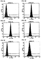
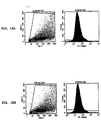
- Cancer stem cells were found to express major histocompatibility (MHC), indicating that they can display antigens.
- MHC major histocompatibility
- proteins differentially expressed in cancer stem cells as compared to differentiated tumor cells were identified. These proteins can be useful in providing antigenic compositions for treatment of cancers (e.g., neural cancers such as gliomas).
- the invention is directed to a method of preparing a glioma vaccine, the method comprising:
- “Beneficial results” may include, but are in no way limited to, lessening or alleviating the severity of a disease condition, inhibiting the disease condition from worsening, improving symptoms of a disease condition, and prolonging a patient's life or life expectancy.
- Cancer and “cancerous” refer to or describe the physiological condition in mammals that is typically characterized by unregulated cell growth.
- Examples of cancer include, but are not limited to, breast cancer, colon cancer, lung cancer, prostate cancer, hepatocellular cancer, gastric cancer, pancreatic cancer, cervical cancer, ovarian cancer, liver cancer, bladder cancer, cancer of the urinary tract, thyroid cancer, renal cancer, carcinoma, melanoma, head and neck cancer, and brain cancer; including, but not limited to, gliomas, glioblastomas, glioblastoma multiforme (GBM), oligodendrogliomas, primitive neuroectodermal tumors, low, mid and high grade astrocytomas, ependymomas (e.g., myxopapillary ependymoma papillary ependymoma, subependymoma, anaplastic ependymoma), oligodendrogliomas, medul
- Constants and “disease conditions,” as used herein may include, but are in no way limited to any form of neoplastic cell growth and proliferation, whether malignant or benign, pre-cancerous and cancerous cells and tissues; in particular, gliomas, glioblastomas, glioblastoma multiforme (GBM), oligodendrogliomas, primitive neuroectodermal tumors, low, mid and high grade astrocytomas, ependymomas (e.g., myxopapillary ependymoma papillary ependymoma, subependymoma, anaplastic ependymoma), oligodendrogliomas, medulloblastomas, meningiomas, pituitary adenomas, neuroblastomas, and craniopharyngiomas.
- GBM glioblastomas
- GBM glioblastoma multiforme
- “Mammal” as used herein refers to any member of the class Mammalia , including, without limitation, humans and nonhuman primates such as chimpanzees and other apes and monkey species; farm animals such as cattle, sheep, pigs, goats and horses; domestic mammals such as dogs and cats; laboratory animals including rodents such as mice, rats and guinea pigs, and the like.
- the term does not denote a particular age or sex. Thus, adult and newborn subjects, as well as fetuses, whether male or female, are intended to be included within the scope of this term.
- the terms "patient” and “subject” are used herein interchangeably, and cover mammals, including humans.
- “Pathology” of cancer includes all phenomena that compromise the well-being of the patient. This includes, without limitation, abnormal or uncontrollable cell growth, metastasis, interference with the normal functioning of neighboring cells, release of cytokines or other secretory products at abnormal levels, suppression or aggravation of inflammatory or immunological response, neoplasia, premalignancy, malignancy, invasion of surrounding or distant tissues or organs, such as lymph nodes, etc.
- “Stem-like” or “stem,” as used herein refers to cells that are able to self renew from a single clone, differentiate into terminal cell types, and be serially transplantable in immunodeficient animals.
- Treatment and “treating,” as used herein refer to both therapeutic treatment and prophylactic or preventative measures, wherein the object is to prevent or slow down (lessen) the targeted pathologic condition or disorder even if the treatment is ultimately unsuccessful.
- Those in need of treatment include those already diagnosed as having the disorder as well as those prone to have the disorder or those in whom the disorder is to be prevented.
- a therapeutic agent may directly decrease the pathology of tumor cells, or render the tumor cells more susceptible to treatment by other therapeutic agents or by the subject's own immune system.
- Tumor refers to all neoplastic cell growth and proliferation, whether malignant or benign, and all pre-cancerous and cancerous cells and tissues.
- compositions useful as vaccines for treating cancer that include dendritic cells pulsed with antigens obtained from cancer stem cells (e.g., neurospheres); methods of producing vaccines that include dendritic cells pulsed with antigens obtained from cancer stem cells; methods of treating cancer with vaccines that include dendritic cells pulsed with antigens obtained from cancer stem cells; and kits for treating cancer that include dendritic cells pulsed with antigens obtained from cancer stem cells.
- cancer stem cells e.g., neurospheres
- cancer stem cells obtained from brain tumors were capable of self-renewal and proliferation, and could recapitulate the tumor when injected into rats. Isolated cancer stem cells formed more aggressive tumors as compared to differentiated tumor cells in vitro, and the cancer stem cells showed a higher resistance to chemotherapeutic agents.
- CD133-positive cancer stem cells were obtained from human tumors. These cells were similarly resistant to chemotherapeutic agents and CD133-positive cells were found at a higher level in patients in whom tumors had recurred following resection.
- compositions for use in methods of vaccinating a subject e.g., to treat cancer (e.g., neural cancer, e.g., gliomas) with antigen-presenting cells ("APC"), e.g., dendritic cells (“DC”), that include antigens from cancer stem cells, e.g., presented on the surface of the antigen-presenting cells.
- cancer e.g., neural cancer, e.g., gliomas
- APC antigen-presenting cells
- DC dendritic cells
- Dendritic cells e.g., autologous or allogeneic dendritic cells
- cancer stem cell antigens as a cell lysate, acid elution, cell extract, partially purified antigens, purified antigens, isolated antigens, partially purified peptides, purified peptides, isolated peptides, synthetic peptides, or a combination of two or more of the above.
- the antigen-presenting cells are then administered to a subject in need of cancer vaccination (e.g., a subject diagnosed with or at risk for cancer) to treat the cancer.
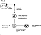
- Figure 1 is a schematic diagram of an exemplary process for vaccination of a patient with cancer stem cell antigen-pulsed dendritic cells.
- cancer stem cell The "cancer stem cell” hypothesis proposes that only a small portion of a tumor is represented by the “cancer stem cell,” which allows the tumor to proliferate and self renew, and eventually differentiate into the phenotypically diverse and heterogeneous tumor cell population ( Bjerkvig et al., Nat. Rev. Cancer, 5:899-904, 2005 ).
- Cancer stem cells can be isolated from any type of cancers, e.g., leukemias ( Bonnet and Dick, Nat. Med., 3:730-737, 1997 ), breast cancers ( Al-Hajj et al., Proc. Natl. Acad. Sci.
- Cancer stem cells are characterized by their ability to self-renew and proliferate, and recapitulate through differentiation the tumor from which it is isolated. Additionally, neural cancer stem cells form clonally derived neurospheres in culture.
- Cancer stem cells can be isolated by dissociating tumor cells and culturing them under conditions that promote proliferation of stem cells (e.g., conditions that inhibit differentiation of the stem cells).
- Methods and conditions for isolating stem cells are known in the art. Exemplary methods and conditions can be found in U.S. Patent No. 5,589,376 , U.S. Patent No. 5,643,741 , U.S. Patent No. 5,650,299 , U.S. Patent No. 5,824,489 , U.S. Patent No. 5,849,553 , U.S. Patent No. 5,928,947 , U.S. Patent No. 5,981,708 , U.S. Patent No. 6,337,184 , U.S. Patent No.
- cancer stem cells can be identified or isolated (e.g., isolated from non-stem tumor cells) on the basis of expression (e.g., nucleic acid or protein expression) of molecular markers, e.g., molecular markers described in the U.S. patents referred to in the above paragraph.
- molecular markers include CD133, Bmi-1, Notch, Sonic hedgehog, and Wnt.
- exemplary molecular markers of neural cancer stem cells include CD90, CD44, CXCR4, Nestin, Musashi-1 (Msil), maternal embryonic leucine zipper kinase (MELK), GLI1, PTCH1, Bmi-1, phosphoserine phosphatase (PSP), Snail, OCT4, BCRP1, MGMT, Bcl-2, FLIP, BCL-XL, XIAP, cIAP1, cIAP2, NAIP, and survivin.
- Msil Musashi-1
- MELK maternal embryonic leucine zipper kinase
- GLI1 PTCH1
- Bmi-1 phosphoserine phosphatase
- PSP phosphoserine phosphatase
- Snail OCT4, BCRP1, MGMT, Bcl-2, FLIP, BCL-XL, XIAP, cIAP1, cIAP2, NAIP, and survivin.
- Isolation or identification of cancer stem cells can be performed by standard means, e.g., cell sorting (e.g., fluorescence activated cell sorting (FACS) or magnetic cell sorting (MACS)).
- cell sorting e.g., fluorescence activated cell sorting (FACS) or magnetic cell sorting (MACS)
- FACS fluorescence activated cell sorting
- MCS magnetic cell sorting
- Antigenic peptides useful for loading DCs for vaccination are peptides that stimulate a T cell mediated immune response (e.g., a cytotoxic T cell response) by presentation to T cells on MHC molecules.
- Useful antigenic peptides and proteins include those derived from cancer stem cells (e.g., neural cancer stem cells, CD133 + tumor cells, or neurospheres derived from tumors).
- cancer stem cell antigens can be presented as a lysate of the cancer stem cells.
- the cancer stem cell antigens can be obtained by acid elution of peptides presented on MHC molecules of the cancer stem cells.
- cancer stem cells are washed with an isotonic solution (e.g., Hank's buffered saline solution) to remove media components.
- the cells are then treated with acid (e.g., citrate phosphate buffer, pH 3.2) to dissociate peptides from surface MHCs, and the cells removed from the solution containing the soluble peptides.
- acid e.g., citrate phosphate buffer, pH 3.2
- the acid-eluted cancer stem cell peptide antigens can be further purified (e.g., on a C18 column) and frozen for storage prior to use.
- Specific antigens that can be used in the methods described herein include portions of the amino acid sequences of CD133, CD90, CD44, CXCR4, Nestin, Musashi-1 (Msil), maternal embryonic leucine zipper kinase (MELK), GLI1, PTCH1, Bmi-1, phosphoserine phosphatase (PSP), Snail, OCT4, BCRP1, MGMT, Bcl-2, FLIP, BCL-XL, XIAP, cIAP1, cIAP2, NAIP, and survivin that bind to MHC molecules and are presented to T cells.
- Peptides that bind to MHC class I molecules are generally 8-10 amino acids in length.
- Peptides that bind to MHC class II molecules are generally 13 amino acids or longer (e.g., 13-17 amino acids long).
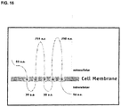
- CD133 is a 120 kDa, five-transmembrane domain glycoprotein expressed on neural and hematopoietic stem and progenitor cells ( Yin et al., Blood, 90:5002-12, 1997 ).
- Table 1 provides an amino acid sequence ofCD133 (also available in GenBank under accession number NP_006008.1, GI:5174387).
- the structure of CD133 includes an extracellular N-terminus, two short intracellular loops, two large extracellular loops, and an intracellular C-terminus ( FIG. 18 ).
- Exemplary CD133 T cell epitopes include 8-10 or 13-20 contiguous amino acid residues of amino acid residues 325-350 of SEQ ID NO:53.
- An alternately spliced version of CD133 is described in Yu et al., J. Biol. Chem., 277:20711-16, 2002 .
- CD90 is a cell surface glycoprotein found on T cells and neurons.
- Table 1 provides an amino acid sequence of CD90 (also available in GenBank under accession number NP_006279.2, GI:19923362).
- CD44 is a cell surface glycoprotein that may be involved in matrix adhesion.
- Table 1 provides an amino acid sequence of CD44 (also available in GenBank under accession number NP_000601.3, GI:48255935).
- isoforms of CD44 are produced, primarily by alternative splicing (see Marhaba et al., J. Mol. Histol., 35:211-31, 2004 ; Zoeller, Cancer Immunol. Immunother., 53:567-79, 2004 ).
- CXCR4 is a chemokine receptor that has been found to be expressed in breast cancers ( Muller et al., Nature, 410:50-56, 2001 ).
- Table 1 provides an amino acid sequence of CXCR4 (also available in GenBank under accession number NP_001008540.1, GI:56790927).
- An alternative spliced variant is available in GenBank under accession number NP_003458.1, GI:4503175.
- Nestin is an intermediate filament protein expressed in neural progenitor cells ( Dahlstrand et al., J. Cell Sci., 103:589-597, 1992 ).
- Table 1 provides an amino acid sequence of Nestin (also available in GenBank under accession number NP_006608.1, GI:38176300).
- Musashi-1 (Msi1) is an RNA-binding protein expressed in neural progenitor cells ( Good et al., Genomics, 52:382-384, 1998 ; Siddall et al., Proc. Nat. Acad. Sci. USA, 103:84-8407, 2006 ; Okano et al., Exp. Cell Res., 306:349-356, 2005 ).
- Table 1 provides an amino acid sequence of Musashi-1 (also available in GenBank under accession number NP_002433.1, GI:4505255).
- MELK Maternal embryonic leucine zipper kinase
- GLI1 is a zinc-finger transcription factor upregulated in cancers, including gliomas ( Kinzler et al., Science, 236:70-73, 1987 ; Kinzler et al., Nature, 332:371-374, 1988 ; Kasper et al., Eur. J. Cancer, 42:437-445, 2006 ).
- Table 1 provides an amino acid sequence of GLI1 (also available in GenBank under accession number NP_005260.1, GI:4885279).
- PTCH1 is a transmembrane protein that is believed to function as a tumor suppressor ( Katoh et al., Cancer Biol. Ther., 4:1050-54, 2005 ).
- Table 1 provides an amino acid sequence of PTCH1 (also available in GenBank under accession number NP_000255.2, GI: 134254446). Five isoforms of PTCH1 are produced by alternative splicing ( Nagao et al., Genomics, 85:462-71, 2005 ).
- Bmi-1 is a polycomb ring finger protein involved in proliferation of progenitor cells ( Lessard et al., Nature, 423:255-260, 2003 ; Park et al., Nature, 423:302-305, 2003 ; Molofsky et al., Nature, 425:962-967, 2003 ). Bmi-1 can play a role in the malignant transformation of the HOX A9/MEIS-induced murine leukemia model ( Lessard et al., Nature, 423:255-260, 2003 ) as well as in tumors of neural origin ( van Lohuizen et al., Nature, 353:353-355, 1991 ).
- Bmi-1 T cell epitopes include TLQDIVYKL (SEQ ID NO:86), CLPSPSTPV (SEQ ID NO:87), VRYLETSKY (SEQ ID NO:88), KRYLRCPAA (SEQ ID NO:89), YEEEPLKDY (SEQ ID NO:90), and KEEVNDKRY (SEQ ID NO:91) ( Steele et al., Br. J. Cancer 95:1202-11, 2006 ).
- Phosphoserine phosphatase is an enzyme that catalyzes the hydrolysis of O-phosphoserine.
- Table 1 provides an amino acid sequence of PSP (also available in GenBank under accession number NP_004568.2, GI:46249388).
- Snail is a zinc-finger transcription factor and anti-apoptotic protein ( Vega et al., Genes Dev., 18:1131-1143, 2004 ).
- Table 1 provides an amino acid sequence of Snail (also available in GenBank under accession number NP_005976.2, GI:18765741).
- OCT4 is a POU homeodomain-containing transcription factor expressed in pluripotent cells ( Nichols et al., Cell, 95:379-391, 1998 ).
- Table 1 provides an amino acid sequence of OCT4 (also available in GenBank under accession number NP_002692.2, GI:42560248).
- An alternate isoform of OCT4 is available in GenBank under accession number NP_976034.3, GI:116235491.
- BCRP1 is an ATP-binding cassette (ABC) transporter protein involved in multidrug resistance of tumors ( Doyle et al., Proc. Nat. Acad. Sci. USA, 95:1566570, 1998 ).
- Table 1 provides an amino acid sequence of BCRP1 (also available in GenBank under accession number NP_004818.2, GI:62526033).
- MGMT is an O-6-methylguanine-DNA methyltransferase DNA -mismatch repair protein that can provide resistance to some methylating and chloroethylating agents, such as temozolomide ( Rabik et al., Cancer Treat. Rev., 32:261-276, 2006 ; Cai et al., Cancer Res., 65:3319-27, 2005 ).
- Table 1 provides an amino acid sequence of MGMT (also available in GenBank under accession number NP_002403.1, GI:4505177).
- BCL-2 is a mitochondrial anti-apoptotic protein correlated with chemotherapy resistant cancers and decreased overall survival ( Campos et al., Blood, 81:3091-3096, 1993 ).
- Table 1 provides an amino acid sequence of BCL-2 (also available in GenBank under accession number NP_000624.2, GI:72198189).
- An alternatively spliced isoform of BCL-2 is available in GenBank under accession number NP_000648.2, GI:72198346.
- FLIP is an anti-apoptotic protein ( Irmler et al., Nature, 388:190-195, 1997 ).
- Table 1 provides an amino acid sequence of FLIP (also available in GenBank under accession number NP_003870.3, GI:21361769).
- BCL-XL is an anti-apoptotic protein related to BCL-2, which may be involved in chemoresistance ( Boise et al., Cell, 74:597-608, 1993 ; Andreeff et al., Leukemia, 13:1881-92, 1999 ).
- Table 1 provides an amino acid sequence of BCL-XL (also available in GenBank under accession number NP_612815.1, GI:20336335).
- Exemplary T cell epitopes of BCL-XL include Bcl-xL1 18-126 (TAYQSFEQV; SEQ ID NO:92), Bcl-xL173-182 (YLNDHLEPWI; SEQ ID NO:93), and Bcl-xL169-178 (WMATYLNDHL; SEQ ID NO:94) ( Andersen et al., J. Immunol., 175:2709-14, 2005 ).
- XIAP is a member of the inhibitor of apoptosis protein (IAP) family ( Deveraux et al., Nature, 388:300-304, 1997 ).
- Table 1 provides an amino acid sequence of XIAP (also available in GenBank under accession number NP_001158.2, GI:32528299).
- cIAP1 is a member of the IAP family of apoptosis inhibitors ( Rothe et al., Cell, 83: 1243-1252, 1995 ; Liston et al., Nature, 379:349-353, 1996 ).
- Table 1 provides an amino acid sequence of cIAP1 (also available in GenBank under accession number NP_001157.1, GI:4502141).
- cIAP2 is a member of the IAP family of apoptosis inhibitors ( Liston et al., Nature, 379:349-353, 1996 ).
- Table 1 provides an amino acid sequence of cIAP2 (also available in GenBank under accession number NP_031490.1, GI:6680696).
- NAIP is a member of the IAP family of apoptosis inhibitors ( Roy et al., Cell, 80:167-178, 1995 ).
- Table 1 provides an amino acid sequence of NAIP (also available in GenBank under accession number NP_004527.2, GI:119393878).
- An alternatively spliced isoform of NAIP is available in GenBank under accession number NP_075043.1, GI:119393876.
- Survivin is a member of the IAP family of apoptosis inhibitors ( Li et al., Nature, 396:580-584, 1998 ).
- Table 1 provides an amino acid sequence of NAIP (also available in GenBank under accession number NP_001159.2, GI:59859878).
- NAIP also available in GenBank under accession number NP_001159.2, GI:59859878.
- spliced isoforms of survivin have been identified (see Wheatley et al., Int. Rev. Cytol., 247:35-88, 2005 ; Noton et al., J. Biol. Chem., 281:1286-95, 2006 ; and Taubert et al., Oncogene, 24:5258-61, 2005 ).
- T cell epitopes of survivin include Sur20-28 (STFKNWPFL; SEQ ID NO:95), Sur96-104 (LTLGEFLKL; SEQ ID NO:96), Sur133-141 (RAIEQLAAM; SEQ ID NO:97), and Sur126-135 (ETAKKVRRAI; SEQ ID NO:98) ( Bachinsky et al., Cancer Immun., 5:6, 2005 ).
- T cell epitopes of survivin include Sur92-101 (QFEELTLGEF; SEQ ID NO:99), Sur54-62 (LAQCFFCFK; SEQ ID NO: 100), Sur1 12-120 (KIAKETNNK; SEQ ID NO:101), Sur53-62 (DLAQCFFCFK; SEQ ID NO:102), Sur112-121 (KIAKETNNKK; SEQ ID NO:103), Sur18-28 (RISTFKNWPFL; SEQ ID NO:104), Sur86-96 (FLSVKKQFEEL; SEQ ID NO:105), and the modified peptides Sur92T2 (QTEELTLGEF; SEQ ID NO:106), Sur93T2 (FTELTLGEF; SEQ ID NO:107), Sur93S2 (FSELTLGEF; SEQ ID NO:108), Sur38Y9 (MAEAGFIHY; SEQ ID NO:109), Sur47Y10 (PTENEPDLAY; SEQ ID NO: 110), Sur5K9 (TLPPAWQPK; SEQ ID NO:
- T cell epitopes of survivin include ELTLGEFLKL (SEQ ID NO:114) and TLPPAWQPFL (SEQ ID NO:115) ( Schmitz et al., Cancer Res. 60:4845-4849, 2000 ). Additional survivin epitopes are described in Siegel et al., Br. J. Haematol., 122:911-914, 2003 . Table 1.
- T cell epitopes can be identified by a number of different methods.
- Naturally processed MHC epitopes can be identified by mass spectrophotometric analysis of peptides eluted from antigen-loaded APC (e.g., APC that have taken up antigen, or that have been engineered to produce the protein intracellularly). After incubation at 37 °C, cells are lysed in detergent and the MHC protein is purified (e.g., by affinity chromatography). Treatment of the purified MHC with a suitable chemical medium (e.g., under acidic conditions) results in the elution of peptides from the MHC. This pool of peptides is separated and the profile compared with peptides from control APC treated in the same way. The peaks unique to the protein expressing/fed cells are analyzed (for example by mass spectrometry) and the peptide fragments identified. This protocol identifies peptides generated from a particular antigen by antigen processing.
- epitopes can be identified by screening a synthetic library of peptides that overlap and span the length of the antigen in an in vitro assay. For example, peptides that are 9 amino acids in length and which overlap by 5 amino acids may be used. The peptides are tested in an antigen presentation system that includes antigen presenting cells and T cells. T cell activation in the presence of APCs presenting the peptide can be measured (e.g., by measuring T cell proliferation or cytokine production) and compared to controls, to determine whether a particular epitope is recognized by the T cells.
- T cell epitopes can be predicted in silico, e.g., using the methods described in Parker et al., J. Immunol., 152:163, 1994 and Rammensee et al., Immunogenet., 50:213-219, 1999 .
- Antigenic peptides can be obtained by chemical synthesis using a commercially available automated peptide synthesizer. Synthetic peptides can be precipitated and further purified, for example by high performance liquid chromatography (HPLC). Alternatively, isolated peptides can be obtained by purification and/or recombinant methods using host cell and vector expression systems.
- HPLC high performance liquid chromatography
- Antigen presenting cells such as DCs
- suitable for administration to subjects can be isolated or obtained from any tissue in which such cells are found, or may be otherwise cultured and provided using standard techniques. Methods of preparing antigen presenting cells are well-known to those of skill in the art. Mature dendritic cells are typically identified as having the following cell surface marker phenotype: MAC3 - , CD80 + , CD86 + , CD40 low , CD54 + , MHC Class I and MHC Class II, and are capable of FITC-dextran uptake.
- APCs can be found, by way of example, in the bone marrow or PBMCs of a mammal, in the spleen of a mammal or in the skin of a mammal (i.e., Langerhan's cells, which possess certain qualities similar to that of DC, may be found in the skin).
- bone marrow can be harvested from a mammal and cultured in a medium that promotes the growth of DCs.
- GM-CSF, IL-4 and/or other cytokines (e.g., TNF- ⁇ ), growth factors and supplements may be included in this medium.
- clusters of DCs are cultured in the presence of a sufficient number of antigens of interest (e.g., in the presence of cancer stem cell lysate, acid eluted peptides of cancer stem cells, peptides of CD 133, CD90, CD44, CXCR4, Nestin, Musashi-1 (Msil), maternal embryonic leucine zipper kinase (MELK), GLI1, PTCH1, Bmi-1, phosphoserine phosphatase (PSP), Snail, OCT4, BCRP1, MGMT, Bcl-2, FLIP, BCL-XL, XIAP, cIAP1, cIAP2, NAIP, or survivin, or a combination of two or more of the above antigens) and harvested for use in a cancer
- a sufficient number of antigens of interest e.g., in the presence of cancer stem cell lysate, acid eluted peptides of cancer stem cells, peptides of CD 133,
- antigens can be transgenically expressed in DCs, e.g., by transfection of nucleic acids encoding one or more of the antigens or portions of antigens.
- APCs are isolated from a subject (e.g., a human) according to the following exemplary procedure.
- Mononuclear cells are isolated from blood using leukapheresis (e.g., using a COBE Spectra Apheresis System). The mononuclear cells are allowed to become adherent by incubation in tissue culture flasks for 2 hours at 37 °C. Nonadherent cells are removed by washing.
- Adherent cells are cultured in medium supplemented with granulocyte macrophage colony stimulating factor (GM-CSF) and interleukin-4 (IL-4) for five days. On day five, TNF- ⁇ is added to the culture medium for another 3-4 days.
- GM-CSF granulocyte macrophage colony stimulating factor
- IL-4 interleukin-4
- DCs occur in low numbers in all tissues in which they reside, making isolation and enrichment of DCs a requirement. Any of a number of procedures entailing repetitive density gradient separation, fluorescence activated cell sorting techniques, positive selection, negative selection or a combination thereof are routinely used to obtain enriched populations or isolated DCs. Guidance on such methods for isolating DCs can be found in O'Doherty et al., J. Exp. Med., 178:1067-78, 1993 ; Young and Steinman, J. Exp. Med., 171:1315-32, 1990 ; Freudenthal and Steinman, Proc. Nat. Acad. Sci.
- the APC-based cancer vaccine may be delivered to a recipient by any suitable delivery route, which can include injection, infusion, inoculation, direct surgical delivery, or any combination thereof.
- the cancer vaccine can be administered to a human in the deltoid region or axillary region.
- the vaccine can be administered to a subject locally to the site of a tumor, within the tumor, or to an area from which a tumor has been surgically resected.
- an appropriate carrier for administering the cells may be selected by one of skill in the art by routine techniques.
- the pharmaceutical carrier can be a buffered saline solution, e.g., cell culture media.
- the quantity of APC appropriate for administration to a patient as a cancer vaccine to effect the methods disclosed in the application and the most convenient route of such administration may be based upon a variety of factors, as may the formulation of the vaccine itself. Some of these factors include the physical characteristics of the patient (e.g., age, weight, and sex), the physical characteristics of the tumor (e.g., location, size, rate of growth, and accessibility), and the extent to which other therapeutic methodologies (e.g., chemotherapy, and beam radiation therapy) are being implemented in connection with an overall treatment regimen.
- factors include the physical characteristics of the patient (e.g., age, weight, and sex), the physical characteristics of the tumor (e.g., location, size, rate of growth, and accessibility), and the extent to which other therapeutic methodologies (e.g., chemotherapy, and beam radiation therapy) are being implemented in connection with an overall treatment regimen.
- a mammal can be administered with from about 10 5 to about 10 9 APC (e.g., 10 7 APC) in from about 0.05 mL to about 5 mL solution (e.g., saline) in a single administration. Additional administrations can be carried out, depending upon the above-described and other factors, such as the severity of tumor pathology. From about one to about five administrations of about 10 6 APC can be performed at two-week intervals.
- DC vaccination can be accompanied by other treatments.
- a patient receiving DC vaccination may also be receiving chemotherapy, radiation, and/or surgical therapy concurrently.
- Methods of treating cancer using DC vaccination in conjunction with chemotherapy are described in Wheeler et al., U.S. Pat. Pub. No. 2007/0020297 .
- a patient receiving DC vaccination can have already received chemotherapy, radiation, and/or surgical treatment for the cancer.
- a patient receiving DC vaccination can be treated with a COX-2 inhibitor, as described in Yu and Akasaki, WO 2005/037995 .
- the present application provides pharmaceutical compositions including a pharmaceutically acceptable excipient along with a therapeutically effective amount of the inventive vaccine comprising dendritic cells pulsed with cancer stem cell antigens as described herein.
- “Pharmaceutically acceptable excipient” means an excipient that is useful in preparing a pharmaceutical composition that is generally safe, non-toxic, and desirable, and includes excipients that are acceptable for veterinary use as well as for human pharmaceutical use. Such excipients may be solid, liquid, semisolid, or, in the case of an aerosol composition, gaseous.
- the pharmaceutical compositions may be formulated for delivery via any route of administration.
- Route of administration may refer to any administration pathway known in the art, including but not limited to aerosol, nasal, transmucosal, transdermal, or parenteral.
- Parenteral refers to a route of administration that is generally associated with injection, including intraorbital, infusion, intraarterial, intracapsular, intracardiac, intradermal, intramuscular, intraperitoneal, intrapulmonary, intraspinal, intrasternal, intrathecal, intrauterine, intravenous, subarachnoid, subcapsular, subcutaneous, transmucosal, or transtracheal.
- the compositions may be in the form of solutions or suspensions for infusion or for injection, or as lyophilized powders.
- compositions can also contain any pharmaceutically acceptable carrier.
- “Pharmaceutically acceptable carrier” as used herein refers to a pharmaceutically acceptable material, composition, or vehicle that is involved in carrying or transporting a compound of interest from one tissue, organ, or portion of the body to another tissue, organ, or portion of the body.
- the carrier may be a liquid or solid filler, diluent, excipient, solvent, or encapsulating material, or a combination thereof.
- Each component of the carrier must be “pharmaceutically acceptable” in that it must be compatible with the other ingredients of the formulation. It must also be suitable for use in contact with any tissues or organs with which it may come in contact, meaning that it must not carry a risk of toxicity, irritation, allergic response, immunogenicity, or any other complication that excessively outweighs its therapeutic benefits.
- the pharmaceutical compositions may be delivered in a therapeutically effective amount.
- the precise therapeutically effective amount is that amount of the composition that will yield the most effective results in terms of efficacy of treatment in a given subject. This amount will vary depending upon a variety of factors, including but not limited to the characteristics of the therapeutic compound (including activity, pharmacokinetics, pharmacodynamics, and bioavailability), the physiological condition of the subject (including age, sex, disease type and stage, general physical condition, responsiveness to a given dosage, and type of medication), the nature of the pharmaceutically acceptable carrier or carriers in the formulation, and the route of administration.
- Kits to treat cancer are also contemplated.
- the kits are useful for practicing the inventive method of treating cancer with a vaccine comprising dendritic cells pulsed with cancer stem cell antigens as described herein.
- the kit is an assemblage of materials or components, including at least one of the inventive compositions.
- the kit contains a composition including a vaccine comprising dendritic cells pulsed with cancer stem cell antigens as described herein.
- the kit is configured for the purpose of treating a particular cancer.
- the kit is configured for the purpose of treating brain tumors.
- the brain tumor is a glioma.
- the brain tumor is GBM.
- the kit is configured particularly for the purpose of treating mammalian subjects.
- the kit is configured particularly for the purpose of treating human subjects.
- the kit is configured for veterinary applications, treating subjects such as, but not limited to, farm animals, domestic animals, and laboratory animals.
- Instructions for use may be included in the kit.
- Instructions for use typically include a tangible expression describing the technique to be employed in using the components of the kit to effect a desired outcome, such as induction of an immune response against a tumor, to treat a cancer.
- the instructions may comprise instructions to administer a vaccine comprising dendritic cells pulsed with cancer stem cell antigens to the patient.
- the kit also contains other useful components, such as, diluents, buffers, pharmaceutically acceptable carriers, syringes, catheters, applicators, pipetting or measuring tools, or other useful paraphernalia as will be readily recognized by those of skill in the art.
- useful components such as, diluents, buffers, pharmaceutically acceptable carriers, syringes, catheters, applicators, pipetting or measuring tools, or other useful paraphernalia as will be readily recognized by those of skill in the art.
- the materials or components assembled in the kit can be provided to the practitioner stored in any convenient and suitable ways that preserve their operability and utility.
- the components can be in dissolved, dehydrated, or lyophilized form; they can be provided at room, refrigerated or frozen temperatures.
- the components are typically contained in suitable packaging material(s).
- packaging material refers to one or more physical structures used to house the contents of the kit, such as inventive compositions and the like.
- the packaging material is constructed by well known methods, preferably to provide a sterile, contaminant-free environment.
- the packaging materials employed in the kit are those customarily utilized in cancer treatments or in vaccinations.
- a package refers to a suitable solid matrix or material such as glass, plastic, paper, foil, and the like, capable of holding the individual kit components.
- a package can be a glass vial used to contain suitable quantities of an inventive composition containing for example, a vaccine comprising dendritic cells pulsed with cancer stem cell antigens as described herein.
- the packaging material generally has an external label which indicates the contents and/or purpose of the kit and/or its components.
- 9L gliosarcoma To determine if a population of self-renewing stem cells exist within the phenotypically heterogeneous 9L gliosarcoma tumor, the cells were grown as monolayers in the presence of 10% FBS and subsequently grew them in serum free media containing mitogens.
- a schematic diagram of culturing of tumor stem cells is presented in Fig. 2 .
- 9L gliosarcomas were resuspended in Dulbecco's modified Eagle's medium/F-12 medium containing 10% fetal bovine serum (FBS) and plated at a density of 1 ⁇ 10 6 live cells per 75 cm 2 flask. The cells attached and grew as monolayers and were passaged upon confluency.
- FBS fetal bovine serum
- Spheres were derived by placing the 9L gliosarcomas cells grown as monolayers into a defined serum-free NSC medium ( Reynolds et al., J. Neurosci., 12: 4565-74, 1992 ; Reynolds et al., Science, 255:1707-10, 1992 ) consisting of Dulbecco's modified Eagle's medium/F-12 medium supplemented with 20 ng/mL of both epidermal growth factor (EGF; Peprotech, Rocky Hill, NJ) and basic fibroblast growth factor (bFGF; Peprotech, Rocky Hill, NJ). Cells were fed every 2 days by adding fresh NSC media supplemented with growth factors.
- a defined serum-free NSC medium consisting of Dulbecco's modified Eagle's medium/F-12 medium supplemented with 20 ng/mL of both epidermal growth factor (EGF; Peprotech, Rocky Hill, NJ) and basic fibroblast growth factor (bFGF; Peprotech, Rocky Hill, NJ).
- the cells were harvested, dissociated into single cells using trypsin and EDTA (GIBCO BRL) and mechanical pipetting, strained through a cell strainer, and plated at a clonal density of 1,000 cells/mL in neurosphere-conditioned medium to generate clonally derived subspheres ( Geschwind et al., Neuron, 29:325-339, 2001 ; Groszer et al., Science, 294: 2186-89, 2001 ). The cells were fed every 2 days by adding fresh NSC media supplemented with mitogens. Cells and subsequent spheres were observed daily for 18 days, and passaged into fresh media. Subspheres ranging from approximately 15 cells to 40 cells were evident after 18 days and displayed the self-renewing and proliferative capacity of the 9L spheres.
- neurospheres were immunostained for NSC markers nestin and Sox2. Neurospheres were also stained for the lineage markers for astrocytes, GFAP, neurons, beta-tubulin III and MAP2, and oligodendrocytes, myelin/oligodendrocyte. Cells in the outer region of the neurosphere labeled for nestin, while cells throughout the neurosphere were labeled for Sox2. A large number of cells within the tumor spheres were also found to be positive for the lineage marker GFAP, while relatively few cells expressed the neuronal lineage markers ⁇ -tubulin III, MAP2, and myelin/oligodendrocyte.
- spheres were seeded into chamber slides (Lab-TekII, Nalge Nunc International) for differentiation assay.
- the cells were grown for 14 days in medium devoid of growth factors bFGF and EGF but permissive for differentiation, and processed for immunocytochemistry as described below.
- the medium included Dulbecco's modified Eagle's medium/F-12 medium containing 10% fetal bovine serum (FBS).
- the primary antibodies were detected with Cy3 or FITC-conjugated anti-mouse or anti-rabbit IgG antibody (1:200, Jackson Immuno Research).
- the cells were counterstained with 4',6-diamidino-2-phenylindole (DAPI; Vector Laboratories) to identify all nuclei.
- DAPI 4',6-diamidino-2-phenylindole
- the stained sections were examined and photographed using a QED cell scanner program and Nikon Eclipse TE2000-E microscope, and analyzed using Image J (NIH).
- NIH Image J
- CSLCs cancer stem-like cells
- GFAP GFAP
- MAP2 GFAP
- the expression pattern of the differentiated progeny was similar in profile to that of primary cultured tumor cells from which the spheres had originally been isolated and predominantly differentiated into GFAP and MAP2 positive cells that recapitulated the parental tumor phenotype. Additionally, the level of labeling for the NSC markers nestin and Sox2 still remained high, even after 14 days of differentiation.
- the staining pattern for the monolayer population was similar to neurospheres differentiated for 14 days, except for the expression of Sox2, which was greater in neurospheres differentiated for 14 days.
- 9L cells grown as monolayer or neurospheres differ in their ability to grow as a tumor after implantation 5,000 cells from both populations of cells were injected into rats and survival and tumor volume were assayed.
- tumor aggression could be determined.
- Animals in the tumor volume group were sacrificed 18 days after tumor implantation. Tumor volume was assessed by using the formula for an ellipsoid, (length x width x height)/2 ( Advani et al., Cancer Res., 59:2055-58, 1999 ), with the height and the width of the tumor being approximately equal because of the well-defined circumference of the tumors generated by the 9L gliosarcoma (see Sibenaller et al., Neurosurg. Focus, 19:E1, 2005 ). A significantly (P ⁇ 0.02) greater tumor volume was observed in the rats implanted with neurosphere cells as compared to rats implanted with monolayer cells ( FIG. 4A ).
- Six rats in the neurosphere group had large tumors that lead to terminal neurological symptoms, whereas 3 showed evidence of small tumors after 40 days of survival as seen by H&E staining of brain sections.
- only 4 rats had tumors large enough to create terminal neurological symptoms in the monolayer group, whereas 5 showed evidence of small tumor or engraftment after 40 days of survival as determined by H&E staining.
- the rats in the monolayer group which developed terminal neurological signs of tumor did so at a later time (36 days compared to 29 days in the neurosphere group), which was significant when analyzed using the Kaplan-Meier test (P ⁇ 0.02).
- Luciferase substrate D-luciferin Biosynth, International, Inc., Naperville, IL
- luciferase scans were taken 15 minutes later. Luciferase scans generated at 14 days post implantation demonstrated a greater proportion of animals with tumor burden in the neurosphere group compared to the monolayer group, even though a higher expression level of luciferase in vitro was observed in monolayer cells as compared to neurosphere cells.
- the tumor-cell-implanted rat brains were cut with a cryostat into 20 ⁇ m coronal sections and fixed in 4% paraformaldehyde, washed with PBS, and air dried.
- Tumors from the neurosphere cell population were large and well-circumferential, and showed cells positive for the NSC marker nestin, as well as cells positive for the lineage markers GFAP, ⁇ -tubulin III, and myelin/oligodendrocytes. Most of the labeling within the tumor volume was directed against GFAP, and a lesser degree of labeling was observed for ⁇ -tubulin III, myelin/oligodendrocyte, and nestin. A significant portion (> 75%) of the tumor volume stained positive for reticulin, consistent with a sarcomatous component.
- H&E staining revealed a high grade glioma with necrosis consistent with a glioblastoma, displaying the dual nature of the gliosarcoma.
- Neurospheres formed high grade gliomas with necrosis as seen on H&E ( FIG. 5A ).
- the tumors were large and well circumferential as evidenced in non-stained sections ( FIG. 5B ) and stained for the nuclear marker DAPI ( FIG. 5D ).
- a comparison of non-tumor area ( FIG. 5D ) with tumor area ( FIG. 5E ) stained for reticulin revealed high levels of reticulin in tumor engulfed regions, showing the histological sarcomatous component of the gliosarcoma.
- the staining patterns suggest that 9L neurospheres recapitulated the original tumor by differentiating into both neural and glial lineages in vivo.
- 2,000 healthy 9L sphere and monolayer cells were exposed to either Dulbecco's modified Eagle's medium/F-12 medium containing 10% fetal bovine serum (FBS), or 100 ⁇ M stock solution of Temozolamide or Carboplatin dissolved in PBS at concentrations of 1,000 ⁇ M, 500 ⁇ M, 250 ⁇ M, and 125 ⁇ M for 2 days.
- FBS Dulbecco's modified Eagle's medium/F-12 medium containing 10% fetal bovine serum
- Temozolamide or Carboplatin dissolved in PBS at concentrations of 1,000 ⁇ M, 500 ⁇ M, 250 ⁇ M, and 125 ⁇ M for 2 days.
- the viability of the cells was scored by measurement of the absorption of formazan dye (the amount of formazan dye formed directly correlates to the number of metabolically active cells) using the cellular proliferation assay WST-1 (Roche Molecular Biochemicals, Mannheim, Germany).
- Formazan was measured with the use of a microplate reader (Tecan) and spectrophotometer set at a wavelength of 440 nm and a reference wavelength of 890 nm.
- Cellular viability was determined by exposing cells to WST-1 for 4 hours, and calculating the percentage of viable cells. Proliferation was also assessed by using manual cell counting after 7 days in culture, with an initial cellular concentration of 100,000 cells/mL in a 25 mm 2 flask.
- the neurospheres demonstrated significantly (P ⁇ 0.05) greater resistance to temozolamide ( FIG. 6B ) and carboplatin ( FIG. 6A ) when compared to the 9L cells grown as a monolayer under the same conditions.
- FIG. 7 is a bar graph that shows a greater increase in cells in the monolayer group.
- a similar trend in the untreated cells was also observed using the manual cell count method, which showed a greater increase in monolayers by a factor of 1.35.
- This example demonstrates that neurosphere cells have a greater resistance to chemotherapeutic agents.
- Glioblastoma specimens were obtained from patients (with informed consent) via the Brain Tumor Registry and were reviewed and released by a pathologist in the operating room. Independent pathologists classified the tumors by type and grade in accordance with the WHO histological grading of central nervous system tumors. IRB certified technicians processed the glioma tissues under sterile conditions in a laminar flow hood. Tumor cells were cultured in the following complete medium: Ham's F-12/DMEM with high glucose (Irvine scientific, Santa Ana, CA), 10 mM HEPES (Invitrogen, Carlsbad, CA), 0.1 mg/ml Gentamicin (Invitrogen) and 10% heat-inactivated FBS (Irvine Scientific, Santa Ana, CA). The cultured cells were maintained for 3-4 passages. Floating neurosphere-like cells were obtained that were capable of forming new spheres in medium containing FBS for 3-4 passages.
- glioblastoma primary tumor cell lines (Nos. 1049, 377, and 66) were derived by the above method and analyzed by FACS for CD133 expression. Tumor cells were collected and stained with anti-CD133 antibody (mouse monoclonal IgG1; 1: 10; Milteny Biotec) or IgG1 isotype control antibody (BD Pharmingen, San Diego, CA). After PE-anti-mouse IgG1 (BD Pharmingen) staining for 30 minutes, CD133 staining was analyzed by flow cytometry using a FACSCaliburTM fluorescence activated cell sorter (Becton Dickinson, San Jose, CA). CD133 expression was observed in 10.2% (No. 66; FIG. 9C ), 27% (No. 1049, FIG. 9A ) and 69.7% (No. 377; FIG. 9B ) of the total population examined.
- CD133 expression was observed in 10.2% (No. 66; FIG. 9C ), 27% (No
- CD133 positive cancer stem cell was isolated by DAKOcytomationTM (DAKO, Carpinteria, CA) sorting and cultured in a defined serum-free NSC medium ( Kabos et al., Exp. Neurol., 178:288-293, 2002 ) containing 20 ng/ml of basic fibroblast growth factor (bFGF, Peprotech, Rocky Hill, NJ), 20 ng/ml of epidermal growth factor (EGF, Peprotech) and 20 ng/ml leukemia inhibitory factor (LIF, Chemicon, Temecula, CA).
- bFGF basic fibroblast growth factor
- EGF epidermal growth factor
- LIF leukemia inhibitory factor
- CD133 positive cells and CD133 negative cells were obtained by FACS sorting as described in Example 5 and real-time PCR was used to analyze some markers associated with neural precursors in these two populations.
- Total RNA was extracted from the isolated CD133 positive and CD133 negative cells using an RNA4PCRTM kit (Ambion, Austin, TX) according to the manufacturer's protocol.
- RNA4PCRTM kit (Ambion, Austin, TX) according to the manufacturer's protocol.
- cDNA synthesis ⁇ 1 ⁇ g total RNA was reverse-transcribed into cDNA using Oligo dT primer and iScriptTM cDNA synthesis kit reverse transcriptase.
- cDNA was stored at -20 °C.
- Oligonucleotide primers sequences used for SYBR Green real-time PCR Gene Forward Reverse Beta-actin 5'-TTCTACAATGAGCTGCGTGTG-3' (SEQ ID NO:1) 5'-GGGGTGTTGAAGGTCTCAAA-3' (SEQ ID NO:2) CD133 5'-GCATTGGCATCTTCTATGGTT-3' (SEQ ID NO:3) 5'-CGCCTTGTCCTTGGTAGTGT-3' (SEQ ID NO:4)
- MSI1 5'-GAGACTGACGCGCCCCAGCC-3' (SEQ ID NO:5) 5'-CGCCTGGTCCATGAAAGTGACG-3' (SEQ ID NO:6)
- MELK 5'-CTTGGATCAGAGGCAGATGTTTGGAG-3' (SEQ ID NO:7) 5'-GTTGTAATCTTGCATGATCCAGG-3' (SEQ ID NO:8) PSP 5'-GGCGGGGCAGTGCCTTTCAAA-3' (SEQ ID NO:9) 5'-TGTTGGCT
- Quantification of target gene mRNA as compared to an internal control (beta-actin) was performed by following a ⁇ C T method. An amplification plot that had been the plot of fluorescence signal vs. cycle number was drawn. The difference ( ⁇ C T ) between the mean values in the duplicated samples of target gene and those of beta-actin were calculated by Microsoft Excel and the relative quantified value (RQV) was expressed as 2 - ⁇ CT . The relative expression of each gene was compared to autologous CD133 negative cells. The results of the QT-PCT analysis are presented in Table 3. Table 3. Relative Expression of Genes in CD133+ Cancer Stem Cells No. 66 No. 377 No.
- CD90, CD44, CXCR4, Nestin, Musashi-1 (Msil), and maternal embryonic leucine zipper kinase (MELK) mRNA expression on CD133 positive cancer stem cells was upregulated by an average of 15.6, 5.7, 337.8, 2.14, 84, and 1351 fold, respectively, compared to the levels found on autologous CD133 negative tumor cells.
- mRNA levels for GLI1 and PTCH1 were upregulated an average of 46 and 16 times, respectively, in CD133 positive cells, as compared to CD133 negative cells.
- Bmi-1, phosphoserine phosphatase (PSP), SHH, OCT4 and Snail mRNA were expressed in CD133 positive cells derived from the three cell lines; none of the five genes were detectable on CD133 negative cells. Additionally, anti-apoptotic genes were also upregulated (see Example 8).
- CD133 positive cancer stem cells were resistant to chemotherapeutic agents
- the WST-1 Cell Proliferation Assay was used to examine the drug sensitivity of CD133 positive cells and CD133 negative cells (both collected by FACS sorting from the three glioblastoma patients' primary cultured tumor cells as described above).
- CD133 positive and negative cells were exposed to conventional chemotherapeutic agents, temozolomide, carboplatin, VP-16 or taxol at various concentrations, for up to 48 hours in 10% FBS/F-12/DMEM culture medium.
- Temozolomide was supplied by the Schering-Plough Research Institute (Kenilworth, NJ) and was dissolved in DMSO (Sigma Chemical Co., St Louis, MO) at 100 mM stock solution.
- CD133 positive cells isolated from No. 66 showed dramatic drug resistance to the above four agents including temozolomide ( FIG. 10C ), carboplatin ( FIG. 10D ), VP-16 ( FIG. 10A ), and Taxol ( FIG. 10B ) as compared to autologous CD133 negative cells.
- CD133 positive cells isolated from No. 377 showed significant resistance to carboplatin at all concentrations tested ( FIG. 11A ) and to VP-16 at 200 ⁇ M ( FIG. 11B ).
- CD133 positive cells isolated from No. 1049 showed significant resistance to carboplatin at 200 ⁇ M compared to autologous CD133 negative cells ( FIG. 11C ). This example demonstrates increased drug resistance of CD133 positive cancer stem cells as compared to autologous CD133 negative cells.
- BCRP1 has been demonstrated to play an important role in the drug resistance of normal stem cells and tumor stem cells ( Zhou et al., Nat. Med., 7:1028-34, 2001 ; Hirschmann-Jax et al., Proc. Natl. Acad. Sci. USA, 101:14228-33, 2004 ). Higher expression of BCRP1 (6.5 fold) was found in CD133 positive cells as compared to that of autologous CD133 negative cells.
- anti-apoptotic genes such as FLIP, BCL-2, and BCL-XL
- FLIP FLIP
- BCL-2 BCL-2
- BCL-XL anti-apoptotic genes
- IAPs inhibitor of apoptosis protein family members
- XIAP XIAP
- cIAP1 cIAP2
- NAIP apoptosis protein
- survivin were found at higher expression levels on CD133 positive cells 21.9, 39.04, 3.03, 12.1, 6.73, and 1.6 times higher, respectively, than in CD133 negative cells.
- SIRT1 deacetylates the DNA repair factor Ku70, causing it to sequester the pro-apoptotic factor, Bax, away from mitochondria, thereby inhibiting stress-induced apoptotic cell death ( Cohen et al., Science, 305:390-392, 2004 ).
- SIRT1 deacetylase mRNA expression was increased 4.92 times in CD133 positive cells.
- the pro-apoptotic gene BAX was decreased 3 times in CD133 positive cells as compared to autologous CD133 negative cells. Thus, gene expression differences were observed in CD133 positive cells as compared to CD133 negative cells.
- glioma is a highly recurrent tumor even after surgery, chemotherapy, radiation and immunotherapy.
- CD133 positive tumor cells in glioblastoma recurrence, the CD133 expression upon first and second resection of tumor tissue from the same patient were compared.
- RNA extraction and RT-PCR was performed for CD133 as described above.
- CD133 expression was significantly higher in recurrent tumor tissue (relative expression ⁇ 2.5-19.1) than that in autologous primary tumor tissue (relative expression ⁇ 1)( FIG. 12 ). This example implicates a resistant CD133 positive tumor population in tumor recurrence.
- a dendritic cell vaccine was generated using antigens from neurospheres, daughter cells, and monolayer cells. Immature dendritic cells were generated from the bone marrow of 6-12-week-old Fisher F344 rats as previously described ( Talmor et al., Eur. J. Immunol., 28:811-817, 1998 ).
- bone marrow was harvested from the femoral and tibial marrow cavities and cultured in RPMI 1640 media supplemented with 10% fetal bovine serum (Gemini Biotechnologies, Calabasas, CA), 1% Penicillin/Streptomycin (Invitrogen, Carlsbad, CA), 50 ng/ml recombinant rat GM-CSF and 100 ng/ml recombinant rat IL-4 (R & D Systems, Minneapolis, MN). Cultures were fed every 2 days by removing 75% of the media and replacing it with fresh media containing cytokines (this washed away most of the lymphocytes and granulocytes).
- Soluble peptides were generated for dendritic cell pulsing by cell lysis.
- 9L neurospheres, daughter cells, and monolayer (adherent) cells were processed in the laboratory to produce a single cell suspension.
- the cells were then lysed by 4 to 5 freeze cycles (on liquid nitrogen) and thaw cycles (room temperature). Lysis was monitored by light microscopy, and larger particles were removed by centrifugation (10 minutes at 600 ⁇ g).
- the supernatants were passed through a 0.2 ⁇ m filter, and protein concentration was determined by BioRad protein assay and aliquots frozen at -80 °C until use.
- Vaccinations were given subcutaneously in the flanks on days 7, 14, and 21 with 50,000 DCs pulsed with antigens from either 9L neurospheres (NS), daughter cells (DtC), monolayer (adherent) cells (AC), or saline control. The animals were followed for survival and euthanized when terminal neurological signs developed, for example, inability to access food, water, seizure activity, weakness, and paralysis.
- a cytotoxic T lymphocyte (CTL) assay was performed. Spleens were removed on day 28 post-intracranial 9L-luciferase tumor cells implantation from groups of rats treated with either control or 9L peptide-pulsed dendritic cells. Splenocytes were isolated and re-stimulated in vitro, as described ( Wunderlich et al., "Assays for T-cell function," In: Coligan et al., eds., Current Protocols in Immunology, New York, NY, John Wiley & Sons, Inc; 1997:3.11.1-3.11.20 ; Ehtesham et al., J.
- CTL cytotoxic T lymphocyte
- a representative number of brains from DC-DtC and DC-NS vaccinated rats were carefully removed and post-fixed in 4% paraformaldehyde. Coronal sections of 20 ⁇ m were cut on a cryostat and blocked with normal horse serum for 1 hour. Slides were then incubated with anti-CD4 (clone OX-38 monoclonal antibody diluted in 1:200 in PBS) for 2 hours at room temperature, followed by a 20 minute incubation at room temperature with the linking antibody (BioGenex biotinylated anti-mouse immunoglobulin). After washing in PBS, the labeled moiety (BioGenex Horse Radish Peroxidase-conjugated streptavidin) was added for 20 minutes at room temperature. DAB (3,3'-diaminobenzidine) was used as the chromogen. For analysis of CD8 expression, slides were incubated overnight with Anti-CD8 alpha Chain, clone OX-8 antibody (Chemicon).
- FIG. 15B Immunohistochemical assessment of brain sections from rats vaccinated with dendritic cells pulsed with NS vaccinated showed that there was a robust infiltration of CD4 + ( FIG. 15B ) and CD8 + lymphocytes that was not observed in the brain sections obtained from rats vaccinated with dendritic cells pulsed with daughter cell antigens ( FIG. 15A ). This infiltration of T cells correlates with increased survival of rats vaccinated with NS pulsed DCs.
- GBM glioblastoma multiforme
- CSC cancer stem cells
- Tumor cells were resuspended in DMEM/F12 medium containing 10% fetal bovine serum (FBS) as growth medium and plated at a density of 2x10 6 live cells per 75 cm 2 flask. The cells attached and grew as a monolayer in flasks. All the five monolayer growing adult GBM cells were switched into a defined serum-free NSC medium ( Reynolds et al., Science, 255:1707-10, 1992 ) containing 20 ng/ml of basic fibroblast growth factor (bFGF, Peprotech, Rocky Hill, NJ) and 20 ng/ml of epidermal growth factor (EGF, Peprotech).
- FBS fetal bovine serum
- NSC Normal human fetal neural stem cells
- CSC and NSC cells were stained by FITC-HLA-A,B,C antibody and isotype control antibody (BD Bioscience, San Diego, CA) and analyzed by flow cytometry. Representative CSC and NSC results were shown in the FIGs. 17A-17B and 18A-18B , respectively.
- HLA expression was seen in 5 of 5 cancer stem cells from different patients. CSCs expressed high levels of HLA-A,B,C, however, NSC did not expression MHC class I (HLA-A,B,C) antigens on the surface. This unexpected result indicates that specific cancer stem cell antigens on cancer stem cells can be targeted using vaccines that generate T cells that recognize and kill cancer stem cell antigens in the context of MHC. Therefore cancer stem cells will be targeted, whereas normal stem cells will not.
- a nucleic acid encoding a portion of CD133 extracellular domain 1 is used in cloning and expression on the surface of DC.
- DC are transfected with CD133-1 cDNA construct or empty vector (mock) for 48 hours.
- the successful transfection of DC-CD 133 cells is identified either by anti-CD 133 monoclonal antibody using flow cytometry or by EGFP cloned vector under fluorescent microscopy.
- Antigen presenting cells with CD133 receptor are stimulated by CD133 T cell epitopes.
- Overlapping peptides of 8-10 amino acids of residues 325-350 of SEQ ID NO:53 are produced as MHC class I epitopes.
- overlapping peptides of 13-20 amino acid of residues 325-350 are produced as MHC class II epitopes.
- Stimulated APC using the peptide epitopes lead to enhanced production of CD8 T cells targeted to CD 13 3 molecular on stem/progenitor cells in brain tumors.
Landscapes
- Health & Medical Sciences (AREA)
- Life Sciences & Earth Sciences (AREA)
- Chemical & Material Sciences (AREA)
- General Health & Medical Sciences (AREA)
- Immunology (AREA)
- Microbiology (AREA)
- Pharmacology & Pharmacy (AREA)
- Animal Behavior & Ethology (AREA)
- Medicinal Chemistry (AREA)
- Public Health (AREA)
- Veterinary Medicine (AREA)
- Cell Biology (AREA)
- Oncology (AREA)
- Mycology (AREA)
- Epidemiology (AREA)
- Engineering & Computer Science (AREA)
- Biomedical Technology (AREA)
- Organic Chemistry (AREA)
- Bioinformatics & Cheminformatics (AREA)
- Biotechnology (AREA)
- Zoology (AREA)
- Wood Science & Technology (AREA)
- Genetics & Genomics (AREA)
- General Engineering & Computer Science (AREA)
- Developmental Biology & Embryology (AREA)
- Biochemistry (AREA)
- Chemical Kinetics & Catalysis (AREA)
- General Chemical & Material Sciences (AREA)
- Nuclear Medicine, Radiotherapy & Molecular Imaging (AREA)
- Medicines Containing Antibodies Or Antigens For Use As Internal Diagnostic Agents (AREA)
- Medicines Containing Material From Animals Or Micro-Organisms (AREA)
- Medicines That Contain Protein Lipid Enzymes And Other Medicines (AREA)
Description
- Over the past 30 years, a wealth of information has been generated concerning the in vivo and in vitro properties of brain tumors and rodent models of brain tumor. The 9L gliosarcoma, which was generated from inbred Fisher rats, is a widely used syngeneic rat model for brain tumors. Originally produced by N-methyl-nitrosourea mutagenesis in Fisher rats, the tumor was cloned and designated 9L gliosarcoma because of its dual appearance of a glioblastoma and a sarcoma (Benda et al., J. Neurosurg., 34:310-323, 1971; Schmidek et al., J. Neurosurg., 34:335-340, 1971). The tumor could be proliferated under in vivo and in vitro conditions, making it a useful candidate as a glioma tumor model. The 9L gliosarcoma model clinically mimics rapidly growing and fatal intracerebral tumors, making it the most widely used rat brain tumor model.
- Stem cells have been defined as multipotent, self-renewing cells with the potential to differentiate into multiple cell types. Systems have been developed to identify the first neural stem cells in a defined media, whereby striatal embryonic progenitors could be harvested and grown in culture as undifferentiated neurospheres (clonally derived aggregates of cells derived from a single stem cell) under the influence of the mitogens EGF and bFGF. Many of these cells expressed nestin (an intermediate filament found in neuroepithelial stem cells), but not markers for the more differentiated principal cell types of the CNS-neuronal and glial cells. However, when grown on coverslips treated with poly-L-ornithine, a substrate that allows cellular adhesion, many of the cells within the neurospheres differentiated into neurons and astrocytes with discontinued nestin expression. The isolated striatal cells fulfilled the critical features expected from neural stem cells: an unlimited capacity for self-renewal and capacity to differentiate into the principal mature neural cells (Potten et al., Development, 110:1001-1020, 1990; Lee et al., Nat. Neurosci., 8:723-729, 2005; Maric et al., J. Neurosci., 23:240-251, 2003; Weissman et al., Annu. Rev. Cell. Dev. Biol., 17:387-403, 2001; Seaberg et al., Trends Neurosci., 26:125-131, 2003; Reya et al., Nature, 414:105-111, 2001). Under similar circumstances "cancer stem cells" appear to have the same characteristics of self-renewal and multipotentcy.
- Tunici, Patrizia et al. report on brain tumor cells and their potential use as new targets for clinical targets ("Brain tumor stem cells: new targets for clinical treatments?", Neurosurgical Focus, 2006, vol. 20, no. 4, page E27).
- Malignant brain tumors carry a poor prognosis even in the midst of surgical, radio-, and chemotherapy. With the poor prognosis of brain tumors amidst the available therapeutic treatments, there exists a significant need for more effective therapies to treat such tumors.
- This invention is based, inter alia, on the discovery that vaccines based on cancer stem cell antigens are exceptionally useful for therapy of cancer. Immunization of animals with dendritic cells pulsed with antigens from isolated cancer stem cells provided a significant survival benefit as compared to immunization with dendritic cells pulsed with differentiated tumor cells. Cancer stem cells were found to express major histocompatibility (MHC), indicating that they can display antigens. Further, proteins differentially expressed in cancer stem cells as compared to differentiated tumor cells were identified. These proteins can be useful in providing antigenic compositions for treatment of cancers (e.g., neural cancers such as gliomas).
- The invention is directed to a method of preparing a glioma vaccine, the method comprising:
- obtaining a population of dendritic cells; and
- contacting the dendritic cells with a neural cancer stem cell antigen composition under conditions such that the dendritic cells present neural cancer stem cell antigens, thus preparing a glioma vaccine, wherein the neural cancer stem cell antigen composition is a lysate of the cancer stem cell neurospheres, wherein the neurospheres are obtained from glioma.
- "Beneficial results" may include, but are in no way limited to, lessening or alleviating the severity of a disease condition, inhibiting the disease condition from worsening, improving symptoms of a disease condition, and prolonging a patient's life or life expectancy.
- "Cancer" and "cancerous" refer to or describe the physiological condition in mammals that is typically characterized by unregulated cell growth. Examples of cancer include, but are not limited to, breast cancer, colon cancer, lung cancer, prostate cancer, hepatocellular cancer, gastric cancer, pancreatic cancer, cervical cancer, ovarian cancer, liver cancer, bladder cancer, cancer of the urinary tract, thyroid cancer, renal cancer, carcinoma, melanoma, head and neck cancer, and brain cancer; including, but not limited to, gliomas, glioblastomas, glioblastoma multiforme (GBM), oligodendrogliomas, primitive neuroectodermal tumors, low, mid and high grade astrocytomas, ependymomas (e.g., myxopapillary ependymoma papillary ependymoma, subependymoma, anaplastic ependymoma), oligodendrogliomas, medulloblastomas, meningiomas, pituitary adenomas, neuroblastomas, and craniopharyngiomas.
- "Conditions" and "disease conditions," as used herein may include, but are in no way limited to any form of neoplastic cell growth and proliferation, whether malignant or benign, pre-cancerous and cancerous cells and tissues; in particular, gliomas, glioblastomas, glioblastoma multiforme (GBM), oligodendrogliomas, primitive neuroectodermal tumors, low, mid and high grade astrocytomas, ependymomas (e.g., myxopapillary ependymoma papillary ependymoma, subependymoma, anaplastic ependymoma), oligodendrogliomas, medulloblastomas, meningiomas, pituitary adenomas, neuroblastomas, and craniopharyngiomas.
- "Mammal" as used herein refers to any member of the class Mammalia, including, without limitation, humans and nonhuman primates such as chimpanzees and other apes and monkey species; farm animals such as cattle, sheep, pigs, goats and horses; domestic mammals such as dogs and cats; laboratory animals including rodents such as mice, rats and guinea pigs, and the like. The term does not denote a particular age or sex. Thus, adult and newborn subjects, as well as fetuses, whether male or female, are intended to be included within the scope of this term. The terms "patient" and "subject" are used herein interchangeably, and cover mammals, including humans.
- "Pathology" of cancer includes all phenomena that compromise the well-being of the patient. This includes, without limitation, abnormal or uncontrollable cell growth, metastasis, interference with the normal functioning of neighboring cells, release of cytokines or other secretory products at abnormal levels, suppression or aggravation of inflammatory or immunological response, neoplasia, premalignancy, malignancy, invasion of surrounding or distant tissues or organs, such as lymph nodes, etc.
- "Stem-like" or "stem," as used herein refers to cells that are able to self renew from a single clone, differentiate into terminal cell types, and be serially transplantable in immunodeficient animals.
- "Treatment" and "treating," as used herein refer to both therapeutic treatment and prophylactic or preventative measures, wherein the object is to prevent or slow down (lessen) the targeted pathologic condition or disorder even if the treatment is ultimately unsuccessful. Those in need of treatment include those already diagnosed as having the disorder as well as those prone to have the disorder or those in whom the disorder is to be prevented. For example, in tumor (e.g., cancer) treatment, a therapeutic agent may directly decrease the pathology of tumor cells, or render the tumor cells more susceptible to treatment by other therapeutic agents or by the subject's own immune system.
- "Tumor," as used herein refers to all neoplastic cell growth and proliferation, whether malignant or benign, and all pre-cancerous and cancerous cells and tissues.
- Unless otherwise defined, all technical and scientific terms used herein have the same meaning as commonly understood by one of ordinary skill in the art to which this invention belongs. Although methods and materials similar or equivalent to those described herein can be used in the practice or testing of the present invention, suitable methods and materials are described below. In case of conflict, the present specification, including definitions, will control. In addition, the materials, methods, and examples are illustrative only and not intended to be limiting.
- Other features and advantages of the invention will be apparent from the following detailed description, and from the claims.
-
-
Figure 1 is a schematic diagram of an exemplary process for vaccination of a patient with tumor antigen-pulsed dendritic cells. Monocytes are isolated from a patient and used to generate immature dendritic cells. After excision of a tumor or a portion of the tumor, brain tumor stem-like cells are isolated and used to prepare antigens for vaccination. The dendritic cells are pulsed with the antigens derived from the brain tumor stem-like cells. The mature antigen-presenting dendritic cells are then used to vaccinate the patient, providing an increased immune response against the tumor or residual tumor cells. -
Figure 2 is a schematic diagram depicting an exemplary method of isolation and generation of tumor neurospheres. -
Figures 3A-3F are a set of histograms depicting FACS analysis of neurospheres differentiated for 2 weeks. Neurospheres differentiated for 2 weeks show (3B) 96% of cells positive for the astrocyte marker GFAP; (3C) 88% of cells positive for the NSC marker nestin; (3D) 29% of cells positive for the neuronal marker MAP2; (3E) 0% of cells positive for the neuronal marker beta-tubulin III; (3F) 6% of cells positive for the oligodendrocyte marker myelin/oligodendroctye. The samples were compared to a negative control as shown (3A). -
Figure 4A is a graph depicting volume of tumors induced by neurospheres (n = 4) and monolayer cells (n = 4) at day 18. Tumors induced by neurospheres were significantly larger (p < 0.02) than those from the monolayer group. -
Figure 4B is a line chart depicting survival of rats with tumors induced by neurospheres (n = 9) and monolayer cells (n = 9). On average, animals in the neurosphere group died at a significantly (p < 0.02) earlier time point compared to those in the monolayer group. -
Figures 5A-5E are a set of micrographs of tumors formed by implantation of neurospheres. (5A) neurospheres formed high grade gliomas with necrosis as seen on H&E; (5B) large and well circumferential tumors grew as evidenced in non-stained sections; (5C) large and well circumferential tumors grew as evidenced in sections stained for the nuclear marker DAPI; (5D) & (5E) comparison of non-tumor area (5D) and tumor area (5E) stained for reticulin reveals the high levels of reticulin formed in tumor engulfed regions, showing the histological sarcomatous component of the gliosarcoma. Scale bar = 3000 µm (5A) Scale bar = 1250 µm (5B, 5C); Scale bar = 250 µm (5D, 5E). -
Figures 6A-6B are line graphs depicting chemoresistance of neurospheres and monolayer cells to the indicated concentrations of carboplatin (6A) and Temodar (6B). Each value represents the mean of three independent trials. Neurospheres were significantly more resistant (p < 0.05) to both chemotherapeutic agents as compared to monolayer cells. -
Figure 7 is a bar graph depicting proliferation rates of neurospheres and monolayer cells in serum-containing media for 2 days. The WST-1 proliferation assay was used to measure absorbance, which directly correlates to the cell number. The monolayer cells proliferated at a significantly (p > 0.05) greater rate than the neurosphere cells. -
Figures 8A-8C are micrographs depicting primary culture of adult human glioblastoma cells. (8A) Neurosphere-like tumor cells were found in glioblastoma primary cell culture in F-12/DMEM 10% FBS medium. Scale bar = 200 µm. (8B) Neurosphere derived from a single isolated CD133+ cell cultured in serum free medium with EGF/FGF. Scale bar = 200 µm. (8C) CD133 expression on a neurosphere derived from a single isolated CD133+ cell in serum free medium with EGF/FGF. Staining indicates CD133 expression. Scale bar = 50 µm. -
Figures 9A-9C are histograms depicting CD133 protein expression on primary cultured tumor cells. Tumor cells were cultured in F-12/DMEM 10% FBS medium for 3-4 passages and stained with specific mAb to CD133 and isotype control-matched mAb. Results are given as the percentage of CD133 positive cells in the total population. In the histograms, the thick lines represent staining with CD133 mAb, and the thin lines represent the isotype control-matched mAb. (9A) tumor cells from patient No. 1049; (9B) tumor cells from patient No. 377; (9C) tumor cells from patient No. 66. -
Figures 10A-10D are line graphs depicting drug sensitivity of CD133 positive cancer stem cells derived from patient No. 66. Tumor cells of No.66 were cultured in FBS/F-12/DMEM medium for 4 passages. Both CD133 positive and negative tumor cells were collected by FACS sorting. 1x10⁴ cells/well were plated in 96-well plate and treated with the indicated concentrations of VP-16, Taxol, temozolomide, and carboplatin for 48 hours in FBS/F-12/ DMEM medium. * indicates p < 0.05 compared to autologous CD133 negative cells. Data are representative of two independent experiments. (10A) VP16; (10B) Taxol; (10C) temozolomide; (10D) carboplatin. -
Figures 11A-11C are a set of line graphs depicting drug sensitivity of CD133 positive cancer stem cells derived from patient Nos. 377 and 1049. Tumor cells of patient No.377 and No. 1049 were cultured in FBS/F-12/DMEM medium for 3 passages. Both CD133 positive and negative tumor cells were collected by FACS sorting. 1×10⁴ cells/well were plated in 96-well plate and treated with the indicated concentrations of carboplatin (11A and 11C) or VP-16 (11B) for 48 hours in FBS/F-12/DMAEM medium. * indicates p < 0.05 compared to autologous CD133 negative cells. Data are representative of two independent experiments. -
Figure 12 is a bar graph depicting CD133 mRNA expression in primary (P) and recurrent (R) tumor tissue. Total RNA was extracted from both primary and recurrent tumor tissue derived from five patients, and CD133 mRNA expression was measured by real-time qPCR. The relative CD133 mRNA level of recurrent tumor was presented as the fold increase compared to that of autologous primary tumor tissue. Data are representative of two independent experiments. -
Figure 13 is a line graph depicting survival of 9L tumor-bearing rats vaccinated with dendritic cells pulsed with antigens from 9L adherent cells (9L-AC), 9L daughter cells (9L-DC), 9L neurospheres (9L-NS), and dendritic cells alone (CONTROL). Kaplan-Meier survival curve showed that the 9L-DC group had significantly longer survival than the other groups (p = 0.0015). -
Figure 14 is a bar graph depicting IFNγ production of splenocytes harvested from tumor bearing rats that were re-stimulated and by either re-exposure to a 9L tumor cell target (Target) or to no target (NO Target). The re-stimulated splenocytes from NS vaccinated rats released high level of IFNγ when re-exposed to NS, whereas splenocytes from rats vaccinated with AC or DtC had no detectable IFNγ in response to re-exposure to AC or DC. -
Figures 15A and 15B are a pair of micrographs of tumor slices from rats vaccinated with dendritic cells pulsed with daughter cells (17A) and neurospheres (17B) and stained with anti-CD4 antibody. Greater infiltration of CD4+ cells was observed in the tumors of rats vaccinated with neurosphere-pulsed dendritic cells. (Magnification ×40). -
Figure 16 is a structural diagram of CD133 depicting extracellular, intracellular, and transmembrane regions of CD133. -
Figures 17A and 17B are cell plots and histograms depicting expression of isotype control (17A) and MHC class I expression (17B) in human cancer stem cells. -
Figures 18A and 18B are cell plots and histograms depicting expression of isotype control (18A) and MHC class I expression (18B) in human neural stem cells. - The present application describes compositions useful as vaccines for treating cancer (e.g., neural cancers, e.g., gliomas) that include dendritic cells pulsed with antigens obtained from cancer stem cells (e.g., neurospheres); methods of producing vaccines that include dendritic cells pulsed with antigens obtained from cancer stem cells; methods of treating cancer with vaccines that include dendritic cells pulsed with antigens obtained from cancer stem cells; and kits for treating cancer that include dendritic cells pulsed with antigens obtained from cancer stem cells.
- Greater tumor infiltration of cytotoxic T cells was observed in animals vaccinated against cancer stem cell antigens, and a stronger response against tumor cells was observed in T cells isolated from animals vaccinated with cancer stem cell antigens, than in responses in which antigens were not prepared from stem cell enriched cell populations. Cancer stem cells obtained from brain tumors were capable of self-renewal and proliferation, and could recapitulate the tumor when injected into rats. Isolated cancer stem cells formed more aggressive tumors as compared to differentiated tumor cells in vitro, and the cancer stem cells showed a higher resistance to chemotherapeutic agents. Similarly, CD133-positive cancer stem cells were obtained from human tumors. These cells were similarly resistant to chemotherapeutic agents and CD133-positive cells were found at a higher level in patients in whom tumors had recurred following resection.
- Described herein are compositions for use in methods of vaccinating a subject, e.g., to treat cancer (e.g., neural cancer, e.g., gliomas) with antigen-presenting cells ("APC"), e.g., dendritic cells ("DC"), that include antigens from cancer stem cells, e.g., presented on the surface of the antigen-presenting cells. Dendritic cells (e.g., autologous or allogeneic dendritic cells) are contacted with cancer stem cell antigens as a cell lysate, acid elution, cell extract, partially purified antigens, purified antigens, isolated antigens, partially purified peptides, purified peptides, isolated peptides, synthetic peptides, or a combination of two or more of the above. The antigen-presenting cells are then administered to a subject in need of cancer vaccination (e.g., a subject diagnosed with or at risk for cancer) to treat the cancer.
Figure 1 is a schematic diagram of an exemplary process for vaccination of a patient with cancer stem cell antigen-pulsed dendritic cells. - The "cancer stem cell" hypothesis proposes that only a small portion of a tumor is represented by the "cancer stem cell," which allows the tumor to proliferate and self renew, and eventually differentiate into the phenotypically diverse and heterogeneous tumor cell population (Bjerkvig et al., Nat. Rev. Cancer, 5:899-904, 2005). Cancer stem cells can be isolated from any type of cancers, e.g., leukemias (Bonnet and Dick, Nat. Med., 3:730-737, 1997), breast cancers (Al-Hajj et al., Proc. Natl. Acad. Sci. USA, 100:3983-88, 2003), colon cancers (O'Brien et al., Nature, 445:106-110, 2007), and brain cancers (Singh et al., Nature, 432:396-401, 2004; Hemmati et al., Proc. Natl. Acad. Sci. USA, 100:15178-83, 2003; Singh et al., Cancer Res., 63:5821-28, 2003; Sanai et al., N. Engl. J. Med., 353:811-822, 2005; Tunici et al., Mol. Cancer, 3:25, 2004). Cancer stem cells are characterized by their ability to self-renew and proliferate, and recapitulate through differentiation the tumor from which it is isolated. Additionally, neural cancer stem cells form clonally derived neurospheres in culture.
- Cancer stem cells can be isolated by dissociating tumor cells and culturing them under conditions that promote proliferation of stem cells (e.g., conditions that inhibit differentiation of the stem cells). Methods and conditions for isolating stem cells are known in the art. Exemplary methods and conditions can be found in
U.S. Patent No. 5,589,376 ,U.S. Patent No. 5,643,741 ,U.S. Patent No. 5,650,299 ,U.S. Patent No. 5,824,489 ,U.S. Patent No. 5,849,553 ,U.S. Patent No. 5,928,947 ,U.S. Patent No. 5,981,708 ,U.S. Patent No. 6,337,184 ,U.S. Patent No. 6,645,763 ,U.S. Patent No. 6,800,790 ,U.S. Patent No. 6,875,607 ,U.S. Patent No. 6,984,522 ,U.S. Patent No. 7,109,03 ,U.S. Patent No. 7,115,267 , andU.S. Patent No. 7,115,360 . - Additionally, cancer stem cells can be identified or isolated (e.g., isolated from non-stem tumor cells) on the basis of expression (e.g., nucleic acid or protein expression) of molecular markers, e.g., molecular markers described in the U.S. patents referred to in the above paragraph. Exemplary molecular markers include CD133, Bmi-1, Notch, Sonic hedgehog, and Wnt. Additionally, exemplary molecular markers of neural cancer stem cells include CD90, CD44, CXCR4, Nestin, Musashi-1 (Msil), maternal embryonic leucine zipper kinase (MELK), GLI1, PTCH1, Bmi-1, phosphoserine phosphatase (PSP), Snail, OCT4, BCRP1, MGMT, Bcl-2, FLIP, BCL-XL, XIAP, cIAP1, cIAP2, NAIP, and survivin.
- Isolation or identification of cancer stem cells can be performed by standard means, e.g., cell sorting (e.g., fluorescence activated cell sorting (FACS) or magnetic cell sorting (MACS)).
- Antigenic peptides useful for loading DCs for vaccination are peptides that stimulate a T cell mediated immune response (e.g., a cytotoxic T cell response) by presentation to T cells on MHC molecules. Useful antigenic peptides and proteins include those derived from cancer stem cells (e.g., neural cancer stem cells, CD133+ tumor cells, or neurospheres derived from tumors). Disclosed is that the cancer stem cell antigens can be presented as a lysate of the cancer stem cells. The cancer stem cell antigens can be obtained by acid elution of peptides presented on MHC molecules of the cancer stem cells. In an exemplary method, cancer stem cells are washed with an isotonic solution (e.g., Hank's buffered saline solution) to remove media components. The cells are then treated with acid (e.g., citrate phosphate buffer, pH 3.2) to dissociate peptides from surface MHCs, and the cells removed from the solution containing the soluble peptides. The acid-eluted cancer stem cell peptide antigens can be further purified (e.g., on a C18 column) and frozen for storage prior to use.
- Specific antigens that can be used in the methods described herein include portions of the amino acid sequences of CD133, CD90, CD44, CXCR4, Nestin, Musashi-1 (Msil), maternal embryonic leucine zipper kinase (MELK), GLI1, PTCH1, Bmi-1, phosphoserine phosphatase (PSP), Snail, OCT4, BCRP1, MGMT, Bcl-2, FLIP, BCL-XL, XIAP, cIAP1, cIAP2, NAIP, and survivin that bind to MHC molecules and are presented to T cells. Peptides that bind to MHC class I molecules are generally 8-10 amino acids in length. Peptides that bind to MHC class II molecules are generally 13 amino acids or longer (e.g., 13-17 amino acids long).
- CD133 is a 120 kDa, five-transmembrane domain glycoprotein expressed on neural and hematopoietic stem and progenitor cells (Yin et al., Blood, 90:5002-12, 1997). Table 1 provides an amino acid sequence ofCD133 (also available in GenBank under accession number NP_006008.1, GI:5174387). The structure of CD133 includes an extracellular N-terminus, two short intracellular loops, two large extracellular loops, and an intracellular C-terminus (
FIG. 18 ). Exemplary CD133 T cell epitopes include 8-10 or 13-20 contiguous amino acid residues of amino acid residues 325-350 of SEQ ID NO:53. An alternately spliced version of CD133 is described in Yu et al., J. Biol. Chem., 277:20711-16, 2002. - CD90 is a cell surface glycoprotein found on T cells and neurons. Table 1 provides an amino acid sequence of CD90 (also available in GenBank under accession number NP_006279.2, GI:19923362).
- CD44 is a cell surface glycoprotein that may be involved in matrix adhesion. Table 1 provides an amino acid sequence of CD44 (also available in GenBank under accession number NP_000601.3, GI:48255935). Several isoforms of CD44 are produced, primarily by alternative splicing (see Marhaba et al., J. Mol. Histol., 35:211-31, 2004; Zoeller, Cancer Immunol. Immunother., 53:567-79, 2004).
- CXCR4 is a chemokine receptor that has been found to be expressed in breast cancers (Muller et al., Nature, 410:50-56, 2001). Table 1 provides an amino acid sequence of CXCR4 (also available in GenBank under accession number NP_001008540.1, GI:56790927). An alternative spliced variant is available in GenBank under accession number NP_003458.1, GI:4503175.
- Nestin is an intermediate filament protein expressed in neural progenitor cells (Dahlstrand et al., J. Cell Sci., 103:589-597, 1992). Table 1 provides an amino acid sequence of Nestin (also available in GenBank under accession number NP_006608.1, GI:38176300).
- Musashi-1 (Msi1) is an RNA-binding protein expressed in neural progenitor cells (Good et al., Genomics, 52:382-384, 1998; Siddall et al., Proc. Nat. Acad. Sci. USA, 103:84-8407, 2006; Okano et al., Exp. Cell Res., 306:349-356, 2005). Table 1 provides an amino acid sequence of Musashi-1 (also available in GenBank under accession number NP_002433.1, GI:4505255).
- Maternal embryonic leucine zipper kinase (MELK) is a protein kinase expressed in multiple cancers (Gray et al., Cancer Res., 65:9751-61, 2005). Table 1 provides an amino acid sequence of MELK (also available in GenBank under accession number NP_055606.1, GI:7661974).
- GLI1 is a zinc-finger transcription factor upregulated in cancers, including gliomas (Kinzler et al., Science, 236:70-73, 1987; Kinzler et al., Nature, 332:371-374, 1988; Kasper et al., Eur. J. Cancer, 42:437-445, 2006). Table 1 provides an amino acid sequence of GLI1 (also available in GenBank under accession number NP_005260.1, GI:4885279).
- PTCH1 is a transmembrane protein that is believed to function as a tumor suppressor (Katoh et al., Cancer Biol. Ther., 4:1050-54, 2005). Table 1 provides an amino acid sequence of PTCH1 (also available in GenBank under accession number NP_000255.2, GI: 134254446). Five isoforms of PTCH1 are produced by alternative splicing (Nagao et al., Genomics, 85:462-71, 2005).
- Bmi-1 is a polycomb ring finger protein involved in proliferation of progenitor cells (Lessard et al., Nature, 423:255-260, 2003; Park et al., Nature, 423:302-305, 2003; Molofsky et al., Nature, 425:962-967, 2003). Bmi-1 can play a role in the malignant transformation of the HOX A9/MEIS-induced murine leukemia model (Lessard et al., Nature, 423:255-260, 2003) as well as in tumors of neural origin (van Lohuizen et al., Nature, 353:353-355, 1991). Table 1 provides an amino acid sequence of Bmi-1 (also available in GenBank under accession number NP_005171.4, GI:27883842). Exemplary Bmi-1 T cell epitopes include TLQDIVYKL (SEQ ID NO:86), CLPSPSTPV (SEQ ID NO:87), VRYLETSKY (SEQ ID NO:88), KRYLRCPAA (SEQ ID NO:89), YEEEPLKDY (SEQ ID NO:90), and KEEVNDKRY (SEQ ID NO:91) (Steele et al., Br. J. Cancer 95:1202-11, 2006).
- Phosphoserine phosphatase (PSP) is an enzyme that catalyzes the hydrolysis of O-phosphoserine. Table 1 provides an amino acid sequence of PSP (also available in GenBank under accession number NP_004568.2, GI:46249388).
- Snail is a zinc-finger transcription factor and anti-apoptotic protein (Vega et al., Genes Dev., 18:1131-1143, 2004). Table 1 provides an amino acid sequence of Snail (also available in GenBank under accession number NP_005976.2, GI:18765741).
- OCT4 is a POU homeodomain-containing transcription factor expressed in pluripotent cells (Nichols et al., Cell, 95:379-391, 1998). Table 1 provides an amino acid sequence of OCT4 (also available in GenBank under accession number NP_002692.2, GI:42560248). An alternate isoform of OCT4 is available in GenBank under accession number NP_976034.3, GI:116235491.
- BCRP1 is an ATP-binding cassette (ABC) transporter protein involved in multidrug resistance of tumors (Doyle et al., Proc. Nat. Acad. Sci. USA, 95:1566570, 1998). Table 1 provides an amino acid sequence of BCRP1 (also available in GenBank under accession number NP_004818.2, GI:62526033).
- MGMT is an O-6-methylguanine-DNA methyltransferase DNA -mismatch repair protein that can provide resistance to some methylating and chloroethylating agents, such as temozolomide (Rabik et al., Cancer Treat. Rev., 32:261-276, 2006; Cai et al., Cancer Res., 65:3319-27, 2005). Table 1 provides an amino acid sequence of MGMT (also available in GenBank under accession number NP_002403.1, GI:4505177).
- BCL-2 is a mitochondrial anti-apoptotic protein correlated with chemotherapy resistant cancers and decreased overall survival (Campos et al., Blood, 81:3091-3096, 1993). Table 1 provides an amino acid sequence of BCL-2 (also available in GenBank under accession number NP_000624.2, GI:72198189). An alternatively spliced isoform of BCL-2 is available in GenBank under accession number NP_000648.2, GI:72198346.
- FLIP is an anti-apoptotic protein (Irmler et al., Nature, 388:190-195, 1997). Table 1 provides an amino acid sequence of FLIP (also available in GenBank under accession number NP_003870.3, GI:21361769).
- BCL-XL is an anti-apoptotic protein related to BCL-2, which may be involved in chemoresistance (Boise et al., Cell, 74:597-608, 1993; Andreeff et al., Leukemia, 13:1881-92, 1999). Table 1 provides an amino acid sequence of BCL-XL (also available in GenBank under accession number NP_612815.1, GI:20336335). Exemplary T cell epitopes of BCL-XL include Bcl-xL1 18-126 (TAYQSFEQV; SEQ ID NO:92), Bcl-xL173-182 (YLNDHLEPWI; SEQ ID NO:93), and Bcl-xL169-178 (WMATYLNDHL; SEQ ID NO:94) (Andersen et al., J. Immunol., 175:2709-14, 2005).
- XIAP is a member of the inhibitor of apoptosis protein (IAP) family (Deveraux et al., Nature, 388:300-304, 1997). Table 1 provides an amino acid sequence of XIAP (also available in GenBank under accession number NP_001158.2, GI:32528299).
- cIAP1 is a member of the IAP family of apoptosis inhibitors (Rothe et al., Cell, 83: 1243-1252, 1995; Liston et al., Nature, 379:349-353, 1996). Table 1 provides an amino acid sequence of cIAP1 (also available in GenBank under accession number NP_001157.1, GI:4502141).
- cIAP2 is a member of the IAP family of apoptosis inhibitors (Liston et al., Nature, 379:349-353, 1996). Table 1 provides an amino acid sequence of cIAP2 (also available in GenBank under accession number NP_031490.1, GI:6680696).
- NAIP is a member of the IAP family of apoptosis inhibitors (Roy et al., Cell, 80:167-178, 1995). Table 1 provides an amino acid sequence of NAIP (also available in GenBank under accession number NP_004527.2, GI:119393878). An alternatively spliced isoform of NAIP is available in GenBank under accession number NP_075043.1, GI:119393876.
- Survivin is a member of the IAP family of apoptosis inhibitors (Li et al., Nature, 396:580-584, 1998). Table 1 provides an amino acid sequence of NAIP (also available in GenBank under accession number NP_001159.2, GI:59859878). Alternatively spliced isoforms of survivin have been identified (see Wheatley et al., Int. Rev. Cytol., 247:35-88, 2005; Noton et al., J. Biol. Chem., 281:1286-95, 2006; and Taubert et al., Oncogene, 24:5258-61, 2005). Sequences of exemplary alternative isoforms are available in GenBank under accession numbers NP_001012270.1, GI:59859880, and NP_001012271.1, GI:59859882. Exemplary T cell epitopes of survivin include Sur20-28 (STFKNWPFL; SEQ ID NO:95), Sur96-104 (LTLGEFLKL; SEQ ID NO:96), Sur133-141 (RAIEQLAAM; SEQ ID NO:97), and Sur126-135 (ETAKKVRRAI; SEQ ID NO:98) (Bachinsky et al., Cancer Immun., 5:6, 2005). Other exemplary T cell epitopes of survivin include Sur92-101 (QFEELTLGEF; SEQ ID NO:99), Sur54-62 (LAQCFFCFK; SEQ ID NO: 100), Sur1 12-120 (KIAKETNNK; SEQ ID NO:101), Sur53-62 (DLAQCFFCFK; SEQ ID NO:102), Sur112-121 (KIAKETNNKK; SEQ ID NO:103), Sur18-28 (RISTFKNWPFL; SEQ ID NO:104), Sur86-96 (FLSVKKQFEEL; SEQ ID NO:105), and the modified peptides Sur92T2 (QTEELTLGEF; SEQ ID NO:106), Sur93T2 (FTELTLGEF; SEQ ID NO:107), Sur93S2 (FSELTLGEF; SEQ ID NO:108), Sur38Y9 (MAEAGFIHY; SEQ ID NO:109), Sur47Y10 (PTENEPDLAY; SEQ ID NO: 110), Sur5K9 (TLPPAWQPK; SEQ ID NO: 111), Sur54L2 (LLQCFFCFK; SEQ ID NO:112), and Sur18K10 (RISTFKNWPK; SEQ ID NO:113) (Reker et al., Cancer Biol. Ther., 3:173-179, 2004). Other exemplary T cell epitopes of survivin include ELTLGEFLKL (SEQ ID NO:114) and TLPPAWQPFL (SEQ ID NO:115) (Schmitz et al., Cancer Res. 60:4845-4849, 2000). Additional survivin epitopes are described in Siegel et al., Br. J. Haematol., 122:911-914, 2003.
Table 1. Sequences of Human Antigens Antigen Amino acid sequence CD133 
CD90 
CD44 
CXCR4 
Nestin 
Msi1 
MELK 
GLI1 
PTCH1 
Bmi-1 
PSP 
Snail 
OCT4 
BCRP1 
MGMT 
BCL-2 
FLIP 
BCL-XL 
XIAP 
cIAP1 
cIAP2 
NAIP 
Survivin 
- T cell epitopes can be identified by a number of different methods. Naturally processed MHC epitopes can be identified by mass spectrophotometric analysis of peptides eluted from antigen-loaded APC (e.g., APC that have taken up antigen, or that have been engineered to produce the protein intracellularly). After incubation at 37 °C, cells are lysed in detergent and the MHC protein is purified (e.g., by affinity chromatography). Treatment of the purified MHC with a suitable chemical medium (e.g., under acidic conditions) results in the elution of peptides from the MHC. This pool of peptides is separated and the profile compared with peptides from control APC treated in the same way. The peaks unique to the protein expressing/fed cells are analyzed (for example by mass spectrometry) and the peptide fragments identified. This protocol identifies peptides generated from a particular antigen by antigen processing.
- Alternatively, epitopes can be identified by screening a synthetic library of peptides that overlap and span the length of the antigen in an in vitro assay. For example, peptides that are 9 amino acids in length and which overlap by 5 amino acids may be used. The peptides are tested in an antigen presentation system that includes antigen presenting cells and T cells. T cell activation in the presence of APCs presenting the peptide can be measured (e.g., by measuring T cell proliferation or cytokine production) and compared to controls, to determine whether a particular epitope is recognized by the T cells.
- T cell epitopes can be predicted in silico, e.g., using the methods described in Parker et al., J. Immunol., 152:163, 1994 and Rammensee et al., Immunogenet., 50:213-219, 1999.
- Antigenic peptides can be obtained by chemical synthesis using a commercially available automated peptide synthesizer. Synthetic peptides can be precipitated and further purified, for example by high performance liquid chromatography (HPLC). Alternatively, isolated peptides can be obtained by purification and/or recombinant methods using host cell and vector expression systems.
- Antigen presenting cells (APC), such as DCs, suitable for administration to subjects (e.g., glioma patients) can be isolated or obtained from any tissue in which such cells are found, or may be otherwise cultured and provided using standard techniques. Methods of preparing antigen presenting cells are well-known to those of skill in the art. Mature dendritic cells are typically identified as having the following cell surface marker phenotype: MAC3-, CD80+, CD86+, CD40low, CD54+, MHC Class I and MHC Class II, and are capable of FITC-dextran uptake.
- APCs (e.g., DCs) can be found, by way of example, in the bone marrow or PBMCs of a mammal, in the spleen of a mammal or in the skin of a mammal (i.e., Langerhan's cells, which possess certain qualities similar to that of DC, may be found in the skin). For instance, bone marrow can be harvested from a mammal and cultured in a medium that promotes the growth of DCs. GM-CSF, IL-4 and/or other cytokines (e.g., TNF-α), growth factors and supplements may be included in this medium.
- After a suitable amount of time in culture in medium containing appropriate cytokines (e.g., time suitable to expand and differentiate the DCs into mature DCs, e.g., 2, 4, 6, 8, 10, 12, or 15 days), clusters of DCs are cultured in the presence of a sufficient number of antigens of interest (e.g., in the presence of cancer stem cell lysate, acid eluted peptides of cancer stem cells, peptides of CD 133, CD90, CD44, CXCR4, Nestin, Musashi-1 (Msil), maternal embryonic leucine zipper kinase (MELK), GLI1, PTCH1, Bmi-1, phosphoserine phosphatase (PSP), Snail, OCT4, BCRP1, MGMT, Bcl-2, FLIP, BCL-XL, XIAP, cIAP1, cIAP2, NAIP, or survivin, or a combination of two or more of the above antigens) and harvested for use in a cancer vaccine. For example, peptide antigens can be added to the culture medium at a concentration of about 1.0 to 50, e.g., 5, 10, 15, 20, or 30 µg/ml (per antigen).
- Alternately, or in combination, antigens can be transgenically expressed in DCs, e.g., by transfection of nucleic acids encoding one or more of the antigens or portions of antigens.
- In one exemplary method, APCs are isolated from a subject (e.g., a human) according to the following exemplary procedure. Mononuclear cells are isolated from blood using leukapheresis (e.g., using a COBE Spectra Apheresis System). The mononuclear cells are allowed to become adherent by incubation in tissue culture flasks for 2 hours at 37 °C. Nonadherent cells are removed by washing. Adherent cells are cultured in medium supplemented with granulocyte macrophage colony stimulating factor (GM-CSF) and interleukin-4 (IL-4) for five days. On day five, TNF-α is added to the culture medium for another 3-4 days. On
day 8 or 9, cells are harvested and washed, and incubated with peptide antigens for 16-20 hours on a tissue rotator. Peptide antigens are added to the cultures at a concentration of ∼ 10 µg/ml (per antigen). - Various other methods can be used to isolate the APCs, as would be recognized by one of skill in the art. DCs occur in low numbers in all tissues in which they reside, making isolation and enrichment of DCs a requirement. Any of a number of procedures entailing repetitive density gradient separation, fluorescence activated cell sorting techniques, positive selection, negative selection or a combination thereof are routinely used to obtain enriched populations or isolated DCs. Guidance on such methods for isolating DCs can be found in O'Doherty et al., J. Exp. Med., 178:1067-78, 1993; Young and Steinman, J. Exp. Med., 171:1315-32, 1990; Freudenthal and Steinman, Proc. Nat. Acad. Sci. USA, 57:7698-7702, 1990; Macatonia et al., 67:285-289, 1989; Markowicz and Engleman, J. Clin. Invest., 85:955-961, 1990; Mehta-Damani et al., J. Immunol., 153:996-1003, 1994; and Thomas et al., J. Immunol., 151:6840-6852, 1993. One method for isolating DCs from human peripheral blood is described in
U.S. Patent No. 5,643,786 . Methods of producing DCs from embryonic stem cells are described inU.S. Patent No. 7,247,480 . - The APC-based cancer vaccine may be delivered to a recipient by any suitable delivery route, which can include injection, infusion, inoculation, direct surgical delivery, or any combination thereof. Disclosed is that the cancer vaccine can be administered to a human in the deltoid region or axillary region. Disclosed is that the vaccine can be administered to a subject locally to the site of a tumor, within the tumor, or to an area from which a tumor has been surgically resected.
- An appropriate carrier for administering the cells may be selected by one of skill in the art by routine techniques. For example, the pharmaceutical carrier can be a buffered saline solution, e.g., cell culture media.
- The quantity of APC appropriate for administration to a patient as a cancer vaccine to effect the methods disclosed in the application and the most convenient route of such administration may be based upon a variety of factors, as may the formulation of the vaccine itself. Some of these factors include the physical characteristics of the patient (e.g., age, weight, and sex), the physical characteristics of the tumor (e.g., location, size, rate of growth, and accessibility), and the extent to which other therapeutic methodologies (e.g., chemotherapy, and beam radiation therapy) are being implemented in connection with an overall treatment regimen. Notwithstanding the variety of factors one should consider in implementing the methods disclosed in the application to treat a disease condition, a mammal can be administered with from about 105 to about 109 APC (e.g., 107 APC) in from about 0.05 mL to about 5 mL solution (e.g., saline) in a single administration. Additional administrations can be carried out, depending upon the above-described and other factors, such as the severity of tumor pathology. From about one to about five administrations of about 106 APC can be performed at two-week intervals.
- DC vaccination can be accompanied by other treatments. For example, a patient receiving DC vaccination may also be receiving chemotherapy, radiation, and/or surgical therapy concurrently. Methods of treating cancer using DC vaccination in conjunction with chemotherapy are described in
Wheeler et al., U.S. Pat. Pub. No. 2007/0020297 . Disclosed is that a patient receiving DC vaccination can have already received chemotherapy, radiation, and/or surgical treatment for the cancer. Disclosed is that a patient receiving DC vaccination can be treated with a COX-2 inhibitor, as described in Yu and Akasaki,WO 2005/037995 . - In various disclosures of the applications, the present application provides pharmaceutical compositions including a pharmaceutically acceptable excipient along with a therapeutically effective amount of the inventive vaccine comprising dendritic cells pulsed with cancer stem cell antigens as described herein. "Pharmaceutically acceptable excipient" means an excipient that is useful in preparing a pharmaceutical composition that is generally safe, non-toxic, and desirable, and includes excipients that are acceptable for veterinary use as well as for human pharmaceutical use. Such excipients may be solid, liquid, semisolid, or, in the case of an aerosol composition, gaseous.
- In various disclosures of the applications, the pharmaceutical compositions may be formulated for delivery via any route of administration. "Route of administration" may refer to any administration pathway known in the art, including but not limited to aerosol, nasal, transmucosal, transdermal, or parenteral. "Parenteral" refers to a route of administration that is generally associated with injection, including intraorbital, infusion, intraarterial, intracapsular, intracardiac, intradermal, intramuscular, intraperitoneal, intrapulmonary, intraspinal, intrasternal, intrathecal, intrauterine, intravenous, subarachnoid, subcapsular, subcutaneous, transmucosal, or transtracheal. Via the parenteral route, the compositions may be in the form of solutions or suspensions for infusion or for injection, or as lyophilized powders.
- The pharmaceutical compositions can also contain any pharmaceutically acceptable carrier. "Pharmaceutically acceptable carrier" as used herein refers to a pharmaceutically acceptable material, composition, or vehicle that is involved in carrying or transporting a compound of interest from one tissue, organ, or portion of the body to another tissue, organ, or portion of the body. For example, the carrier may be a liquid or solid filler, diluent, excipient, solvent, or encapsulating material, or a combination thereof. Each component of the carrier must be "pharmaceutically acceptable" in that it must be compatible with the other ingredients of the formulation. It must also be suitable for use in contact with any tissues or organs with which it may come in contact, meaning that it must not carry a risk of toxicity, irritation, allergic response, immunogenicity, or any other complication that excessively outweighs its therapeutic benefits.
- The pharmaceutical compositions may be delivered in a therapeutically effective amount. The precise therapeutically effective amount is that amount of the composition that will yield the most effective results in terms of efficacy of treatment in a given subject. This amount will vary depending upon a variety of factors, including but not limited to the characteristics of the therapeutic compound (including activity, pharmacokinetics, pharmacodynamics, and bioavailability), the physiological condition of the subject (including age, sex, disease type and stage, general physical condition, responsiveness to a given dosage, and type of medication), the nature of the pharmaceutically acceptable carrier or carriers in the formulation, and the route of administration. One skilled in the clinical and pharmacological arts will be able to determine a therapeutically effective amount through routine experimentation, for instance, by monitoring a subject's response to administration of a compound and adjusting the dosage accordingly. For additional guidance, see Remington: The Science and Practice of Pharmacy (Gennaro ed. 20th edition, Williams & Wilkins PA, USA) (2000).
- Kits to treat cancer are also contemplated. The kits are useful for practicing the inventive method of treating cancer with a vaccine comprising dendritic cells pulsed with cancer stem cell antigens as described herein. The kit is an assemblage of materials or components, including at least one of the inventive compositions. Thus, in some disclosures of the application the kit contains a composition including a vaccine comprising dendritic cells pulsed with cancer stem cell antigens as described herein.
- The exact nature of the components configured in the inventive kit depends on its intended purpose. For example, some disclosures of the application are configured for the purpose of treating a particular cancer. In one disclosure, the kit is configured for the purpose of treating brain tumors. In one particular disclosure, the brain tumor is a glioma. In another disclosure, the brain tumor is GBM. In one disclosure, the kit is configured particularly for the purpose of treating mammalian subjects. In another disclosure, the kit is configured particularly for the purpose of treating human subjects. In further disclosures of the application, the kit is configured for veterinary applications, treating subjects such as, but not limited to, farm animals, domestic animals, and laboratory animals.
- Instructions for use may be included in the kit. "Instructions for use" typically include a tangible expression describing the technique to be employed in using the components of the kit to effect a desired outcome, such as induction of an immune response against a tumor, to treat a cancer. For example, the instructions may comprise instructions to administer a vaccine comprising dendritic cells pulsed with cancer stem cell antigens to the patient.
- Optionally, the kit also contains other useful components, such as, diluents, buffers, pharmaceutically acceptable carriers, syringes, catheters, applicators, pipetting or measuring tools, or other useful paraphernalia as will be readily recognized by those of skill in the art.
- The materials or components assembled in the kit can be provided to the practitioner stored in any convenient and suitable ways that preserve their operability and utility. For example the components can be in dissolved, dehydrated, or lyophilized form; they can be provided at room, refrigerated or frozen temperatures. The components are typically contained in suitable packaging material(s). As employed herein, the phrase "packaging material" refers to one or more physical structures used to house the contents of the kit, such as inventive compositions and the like. The packaging material is constructed by well known methods, preferably to provide a sterile, contaminant-free environment. The packaging materials employed in the kit are those customarily utilized in cancer treatments or in vaccinations. As used herein, the term "package" refers to a suitable solid matrix or material such as glass, plastic, paper, foil, and the like, capable of holding the individual kit components. Thus, for example, a package can be a glass vial used to contain suitable quantities of an inventive composition containing for example, a vaccine comprising dendritic cells pulsed with cancer stem cell antigens as described herein. The packaging material generally has an external label which indicates the contents and/or purpose of the kit and/or its components.
- The following examples are provided to better illustrate the claimed invention and are not to be interpreted as limiting the scope of the invention. To the extent that specific materials are mentioned, it is merely for purposes of illustration and is not intended to limit the invention.
- To determine if a population of self-renewing stem cells exist within the phenotypically heterogeneous 9L gliosarcoma tumor, the cells were grown as monolayers in the presence of 10% FBS and subsequently grew them in serum free media containing mitogens. A schematic diagram of culturing of tumor stem cells is presented in
Fig. 2 . 9L gliosarcomas were resuspended in Dulbecco's modified Eagle's medium/F-12 medium containing 10% fetal bovine serum (FBS) and plated at a density of 1×10⁶ live cells per 75 cm2 flask. The cells attached and grew as monolayers and were passaged upon confluency. Spheres were derived by placing the 9L gliosarcomas cells grown as monolayers into a defined serum-free NSC medium (Reynolds et al., J. Neurosci., 12: 4565-74, 1992; Reynolds et al., Science, 255:1707-10, 1992) consisting of Dulbecco's modified Eagle's medium/F-12 medium supplemented with 20 ng/mL of both epidermal growth factor (EGF; Peprotech, Rocky Hill, NJ) and basic fibroblast growth factor (bFGF; Peprotech, Rocky Hill, NJ). Cells were fed every 2 days by adding fresh NSC media supplemented with growth factors. After primary spheres formed and reached 10-200 cells per sphere, the cells were harvested, dissociated into single cells using trypsin and EDTA (GIBCO BRL) and mechanical pipetting, strained through a cell strainer, and plated at a clonal density of 1,000 cells/mL in neurosphere-conditioned medium to generate clonally derived subspheres (Geschwind et al., Neuron, 29:325-339, 2001; Groszer et al., Science, 294: 2186-89, 2001). The cells were fed every 2 days by adding fresh NSC media supplemented with mitogens. Cells and subsequent spheres were observed daily for 18 days, and passaged into fresh media. Subspheres ranging from approximately 15 cells to 40 cells were evident after 18 days and displayed the self-renewing and proliferative capacity of the 9L spheres. - Subtle differences were observed in surface adhesion properties between the two cell populations when using different coating solutions. It was found that poly-L-lysine proved less effective in binding to neurospheres than did laminin. Even after 2 days, only a small portion of the cells adhered (∼ 5%). However, when neurospheres were allowed to adhere to chamber slides coated with laminin, nearly all the cells (∼ 70%) adhered within the first 3 hours. As a consequence, poly-L-lysine was not used as an adhesive substrate in further studies. A similar trend was observed when comparing adhesion of monolayers to chamber slides coated with either poly-L-lysine or laminin-cellular morphology was more apparent in monolayers grown on laminin coated chamber slides within a 6 hour time period. Furthermore, a distinct morphological characteristic was apparent in the differentiated neurospheres, where arms of differentiated cells originating from one neurosphere homed to arms formed by another nearby neurosphere (
FIG. 8A ). These examples of extracellular adhesion and cellular homing can be used to distinguish cancer stem-like cells (CSLCs) from non-CSLCs within a tumor. - To determine the expression of markers for stem cells, neurospheres were immunostained for NSC markers nestin and Sox2. Neurospheres were also stained for the lineage markers for astrocytes, GFAP, neurons, beta-tubulin III and MAP2, and oligodendrocytes, myelin/oligodendrocyte. Cells in the outer region of the neurosphere labeled for nestin, while cells throughout the neurosphere were labeled for Sox2. A large number of cells within the tumor spheres were also found to be positive for the lineage marker GFAP, while relatively few cells expressed the neuronal lineage markers β-tubulin III, MAP2, and myelin/oligodendrocyte.
- To test whether spheres have multipotent capabilities and produce progenies of different lineages, spheres were seeded into chamber slides (Lab-TekII, Nalge Nunc International) for differentiation assay. The cells were grown for 14 days in medium devoid of growth factors bFGF and EGF but permissive for differentiation, and processed for immunocytochemistry as described below. The medium included Dulbecco's modified Eagle's medium/F-12 medium containing 10% fetal bovine serum (FBS).
- To examine the expression of NSC markers and lineage markers, immuncyto- and immunohistochemical staining was performed. For staining of differentiated spheres, spheres and 9L monolayers, cells growing in chamber slides were fixed with 4% paraformaldehyde for 15 minutes at 4 °C, treated with 5% NHS (normal horse serum)/0.1% Triton-X, and then stained with the following antibodies: rabbit anti-nestin (1:200, Chemicon), rabbit anti-Sox2 (1:1,000, Chemicon), rabbit anti-MAP2 (1:1,000, Sigma), mouse anti-p-tubulin III (1:200, Chemicon), rabbit anti-GFAP (1:1000, Chemicon), mouse anti-myelin/oligodendrocyte (1:1,000, Chemicon). The primary antibodies were detected with Cy3 or FITC-conjugated anti-mouse or anti-rabbit IgG antibody (1:200, Jackson Immuno Research). The cells were counterstained with 4',6-diamidino-2-phenylindole (DAPI; Vector Laboratories) to identify all nuclei. The stained sections were examined and photographed using a QED cell scanner program and Nikon Eclipse TE2000-E microscope, and analyzed using Image J (NIH). For immunostaining of spheres, the spheres where allowed to adhere to precoated (with laminin) chamber slides for 3 hours before fixation, while monolayers were allowed to adhere overnight in non-fixed chamber slides.
- Cells stained positive for the NSC markers nestin and Sox2, as well as the lineage markers for astrocytes, GFAP, neurons, MAP2, and oligodendrocytes, myelin/oligodendrocyte. However, no cells were labeled with the neuronal marker beta-tubulin III. FACS analysis showed similar results to those derived via immunocytochemistry, which showed differentiated cells labeled for nestin (88%;
FIG. 3C ), Sox2 (24%), GFAP (96%;FIG. 3B ), MAP2 (29%;FIG. 3D ), and myelin/oligodendrocyte (6%;FIG. 3F ), but not for β-tubulin III (0%;FIG. 3E ). For FACS analysis, cells were fixed with 4% paraformaldehyde for 15 minutes at 4 °C, treated with 5% NHS (normal horse serum)/0.1% Triton ™-X, and then stained with the following antibodies: rabbit anti-nestin (1:1,000, Chemicon), rabbit anti-Sox2 (1:1,000, Chemicon), rabbit anti-MAP2 (1:1,000, Sigma), mouse anti-p-tubulin III (1:500, Chemicon), rabbit anti-GFAP (1:1000, Chemicon), and mouse anti-myelin/oligodendrocyte (1:1,000, Chemicon). The primary antibodies were detected with FITC-conjugated anti-mouse or anti-rabbit IgG antibody (1:200, Jackson Immuno Research) using 104 cells in a FACSVantage™ fluorescence activated cell sorter (Becton Dickinson). These results indicate that these cancer stem-like cells (CSLCs), unlike normal stem cells, differentiate into aberrant cells that are positive for multiple differentiation markers, notably GFAP and MAP2. Such a dual nature is apparent in the majority of cells differentiated from neurospheres. Furthermore, many cells still remained highly positive for nestin and Sox2. The expression pattern of the differentiated progeny was similar in profile to that of primary cultured tumor cells from which the spheres had originally been isolated and predominantly differentiated into GFAP and MAP2 positive cells that recapitulated the parental tumor phenotype. Additionally, the level of labeling for the NSC markers nestin and Sox2 still remained high, even after 14 days of differentiation. The staining pattern for the monolayer population was similar to neurospheres differentiated for 14 days, except for the expression of Sox2, which was greater in neurospheres differentiated for 14 days. These results reveal that 9L spheres are multipotent for the three neural cell types and differentiate into cells found in the original tumor from which they were obtained. - To determine if 9L cells grown as monolayer or neurospheres differ in their ability to grow as a tumor after implantation 5,000 cells from both populations of cells were injected into rats and survival and tumor volume were assayed.
- Fisher F344 rats 6-8 weeks old (Harlan Sprague-Dawley, Indianapolis, IN) were anesthetized with i.p. ketamine and xylazine, and stereotactically implanted in the right striatum (from bregma in mm: anterior-posterior +1 mm; medial-lateral -3 mm; dorsal-ventral -5 mm) with either isolated 9L sphere cells containing the luciferase gene (5,000 cells) or non-sphere-forming monolayer cells containing the luciferase gene (5,000 cells) in 4µL of 1.2% methylcellulose/PBS (Rehemtulla et al., Neoplasia, 2:491-495, 2000). Rats were portioned to either the tumor volume group (n = 10) or the survival group (n = 18), with control rats (n = 6) receiving 4µL of 1.2% methylcellulose/PBS only.
- Because half of each group consisted of either animals implanted with spheres or monolayers, tumor aggression could be determined. Animals in the tumor volume group were sacrificed 18 days after tumor implantation. Tumor volume was assessed by using the formula for an ellipsoid, (length x width x height)/2 (Advani et al., Cancer Res., 59:2055-58, 1999), with the height and the width of the tumor being approximately equal because of the well-defined circumference of the tumors generated by the 9L gliosarcoma (see Sibenaller et al., Neurosurg. Focus, 19:E1, 2005). A significantly (P < 0.02) greater tumor volume was observed in the rats implanted with neurosphere cells as compared to rats implanted with monolayer cells (
FIG. 4A ). - Animals in the survival group were followed for survival and euthanized via CO2 asphyxiation when terminal neurological signs developed (e.g., inability to access food, water, seizure activity, weakness, and paralysis) or if animals exceeded a survival period of 40 days. Following euthanization, brains were harvested and frozen in 2-methylbutane (Sigma), cooled to -20 °C, and stored at -80 °C until sectioning. For H&E staining, 20 µm coronal brain sections described above were mounted on slide and stained with Harris hematoxylin for 2 minutes and then counterstained with alcoholic eosin. Reticulin stains were performed on 12 µm coronal brain sections.
- Similar to the results seen in the tumor size groups, rats implanted with neurospheres on average had shorter survival times than rats implanted with monolayer cells (
FIG. 4B ). Six rats in the neurosphere group had large tumors that lead to terminal neurological symptoms, whereas 3 showed evidence of small tumors after 40 days of survival as seen by H&E staining of brain sections. In contrast, only 4 rats had tumors large enough to create terminal neurological symptoms in the monolayer group, whereas 5 showed evidence of small tumor or engraftment after 40 days of survival as determined by H&E staining. Furthermore, the rats in the monolayer group which developed terminal neurological signs of tumor did so at a later time (36 days compared to 29 days in the neurosphere group), which was significant when analyzed using the Kaplan-Meier test (P < 0.02). - To determine the establishment of tumor in vivo, 150 mg/kg of the luciferase substrate D-luciferin (Biosynth, International, Inc., Naperville, IL) was administered i.p. to animals in the survival group, and luciferase scans were taken 15 minutes later. Luciferase scans generated at 14 days post implantation demonstrated a greater proportion of animals with tumor burden in the neurosphere group compared to the monolayer group, even though a higher expression level of luciferase in vitro was observed in monolayer cells as compared to neurosphere cells.
- To determine if the 9L neurospheres could recapitulate the 9L sarcoma in vivo, histological analysis of the tumors was performed. The tumor-cell-implanted rat brains were cut with a cryostat into 20 µm coronal sections and fixed in 4% paraformaldehyde, washed with PBS, and air dried. To characterize the brain tissue by immunohistochemistry, free-floating sections were blocked with 5% NHS (normal horse serum) for 30 minutes at room temperature and then stained with the following antibodies: rabbit anti-nestin (1:200, Chemicon), rabbit anti-Sox2 (1:1,000, Chemicon), rabbit anti-MAP2 (1:1,000, Sigma), mouse anti-p-tubulin III (1:200, Chemicon), rabbit anti-GFAP (1:1000, Chemicon), mouse anti-myelin/oligodendrocyte (1:1000; Chemicon). The primary antibodies were detected as in Example 1. The cells were counterstained with 4',6-diamidino-2-phenylindole (DAPI; Vector Laboratories) to identify all nuclei. The stained sections were examined and photographed using the Zeiss Axiovision 3.1 program in conjunction with Zeiss Axioskop™ 2 microscope, and analyzed using Image J (NIH).
- Tumors from the neurosphere cell population were large and well-circumferential, and showed cells positive for the NSC marker nestin, as well as cells positive for the lineage markers GFAP, β-tubulin III, and myelin/oligodendrocytes. Most of the labeling within the tumor volume was directed against GFAP, and a lesser degree of labeling was observed for β-tubulin III, myelin/oligodendrocyte, and nestin. A significant portion (> 75%) of the tumor volume stained positive for reticulin, consistent with a sarcomatous component. Additionally, H&E staining revealed a high grade glioma with necrosis consistent with a glioblastoma, displaying the dual nature of the gliosarcoma. Neurospheres formed high grade gliomas with necrosis as seen on H&E (
FIG. 5A ). The tumors were large and well circumferential as evidenced in non-stained sections (FIG. 5B ) and stained for the nuclear marker DAPI (FIG. 5D ). A comparison of non-tumor area (FIG. 5D ) with tumor area (FIG. 5E ) stained for reticulin revealed high levels of reticulin in tumor engulfed regions, showing the histological sarcomatous component of the gliosarcoma. The staining patterns suggest that 9L neurospheres recapitulated the original tumor by differentiating into both neural and glial lineages in vivo. - To compare the differential proliferation rates and resistance to chemotherapeutic agents, 2,000 healthy 9L sphere and monolayer cells were exposed to either Dulbecco's modified Eagle's medium/F-12 medium containing 10% fetal bovine serum (FBS), or 100 µM stock solution of Temozolamide or Carboplatin dissolved in PBS at concentrations of 1,000 µM, 500 µM, 250 µM, and 125 µM for 2 days. The viability of the cells was scored by measurement of the absorption of formazan dye (the amount of formazan dye formed directly correlates to the number of metabolically active cells) using the cellular proliferation assay WST-1 (Roche Molecular Biochemicals, Mannheim, Germany). Formazan was measured with the use of a microplate reader (Tecan) and spectrophotometer set at a wavelength of 440 nm and a reference wavelength of 890 nm. Cellular viability was determined by exposing cells to WST-1 for 4 hours, and calculating the percentage of viable cells. Proliferation was also assessed by using manual cell counting after 7 days in culture, with an initial cellular concentration of 100,000 cells/mL in a 25 mm2 flask.
- The neurospheres demonstrated significantly (P < 0.05) greater resistance to temozolamide (
FIG. 6B ) and carboplatin (FIG. 6A ) when compared to the 9L cells grown as a monolayer under the same conditions. When the cells were not treated with the chemotherapeutic agents and simply grown in medium, there was a significantly (P < 0.05) greater increase in cell number in the monolayer group by a factor of 1.48 when using the WST-1 proliferation assay when the cells were not treated with the chemotherapeutic agents. The results of the proliferation assays are depicted inFIG. 7 , which is a bar graph that shows a greater increase in cells in the monolayer group. A similar trend in the untreated cells was also observed using the manual cell count method, which showed a greater increase in monolayers by a factor of 1.35. This example demonstrates that neurosphere cells have a greater resistance to chemotherapeutic agents. - Glioblastoma specimens were obtained from patients (with informed consent) via the Brain Tumor Registry and were reviewed and released by a pathologist in the operating room. Independent pathologists classified the tumors by type and grade in accordance with the WHO histological grading of central nervous system tumors. IRB certified technicians processed the glioma tissues under sterile conditions in a laminar flow hood. Tumor cells were cultured in the following complete medium: Ham's F-12/DMEM with high glucose (Irvine scientific, Santa Ana, CA), 10 mM HEPES (Invitrogen, Carlsbad, CA), 0.1 mg/ml Gentamicin (Invitrogen) and 10% heat-inactivated FBS (Irvine Scientific, Santa Ana, CA). The cultured cells were maintained for 3-4 passages. Floating neurosphere-like cells were obtained that were capable of forming new spheres in medium containing FBS for 3-4 passages.
- Three adult glioblastoma primary tumor cell lines (Nos. 1049, 377, and 66) were derived by the above method and analyzed by FACS for CD133 expression. Tumor cells were collected and stained with anti-CD133 antibody (mouse monoclonal IgG1; 1: 10; Milteny Biotec) or IgG1 isotype control antibody (BD Pharmingen, San Diego, CA). After PE-anti-mouse IgG1 (BD Pharmingen) staining for 30 minutes, CD133 staining was analyzed by flow cytometry using a FACSCalibur™ fluorescence activated cell sorter (Becton Dickinson, San Jose, CA). CD133 expression was observed in 10.2% (No. 66;
FIG. 9C ), 27% (No. 1049,FIG. 9A ) and 69.7% (No. 377;FIG. 9B ) of the total population examined. - To investigate the capacity of self-renewal and clonogenic potential of CD133+ cell, a single isolated CD133 positive cancer stem cell was isolated by DAKOcytomation™ (DAKO, Carpinteria, CA) sorting and cultured in a defined serum-free NSC medium (Kabos et al., Exp. Neurol., 178:288-293, 2002) containing 20 ng/ml of basic fibroblast growth factor (bFGF, Peprotech, Rocky Hill, NJ), 20 ng/ml of epidermal growth factor (EGF, Peprotech) and 20 ng/ml leukemia inhibitory factor (LIF, Chemicon, Temecula, CA). Single isolated CD133 positive cancer stem cells were able to form neurospheres (
FIG. 8A ), and were demonstrated to have the capacity for self-renewal and clonogenic potential (FIG. 8B ), and sustained expression of CD133 in serum-free medium containing EGF/FGF (FIG. 8C ). - To determine the expression of other genes in CD133 positive cells, CD133 positive cells and CD133 negative cells were obtained by FACS sorting as described in Example 5 and real-time PCR was used to analyze some markers associated with neural precursors in these two populations. Total RNA was extracted from the isolated CD133 positive and CD133 negative cells using an RNA4PCR™ kit (Ambion, Austin, TX) according to the manufacturer's protocol. For cDNA synthesis, ~1 µg total RNA was reverse-transcribed into cDNA using Oligo dT primer and iScript™ cDNA synthesis kit reverse transcriptase. cDNA was stored at -20 °C. Gene expression was quantified by real-time quantitative RT-PCR using QuantiTect™ SYBR Green dye (Qiagen, Valencia, CA). DNA amplification was carried out using Icycler™ (BIO-RAD, Hercules, CA), and the detection was performed by measuring the binding of the fluorescence dye SYBR Green I to double-stranded DNA. All the primer sets were obtained from Qiagen (see Table 1).
Table 2. Oligonucleotide primers sequences used for SYBR Green real-time PCR Gene Forward Reverse Beta-actin 5'-TTCTACAATGAGCTGCGTGTG-3' (SEQ ID NO:1) 5'-GGGGTGTTGAAGGTCTCAAA-3' (SEQ ID NO:2) CD133 5'-GCATTGGCATCTTCTATGGTT-3' (SEQ ID NO:3) 5'-CGCCTTGTCCTTGGTAGTGT-3' (SEQ ID NO:4) MSI1 5'-GAGACTGACGCGCCCCAGCC-3' (SEQ ID NO:5) 5'-CGCCTGGTCCATGAAAGTGACG-3' (SEQ ID NO:6) MELK 5'-CTTGGATCAGAGGCAGATGTTTGGAG-3' (SEQ ID NO:7) 5'-GTTGTAATCTTGCATGATCCAGG-3' (SEQ ID NO:8) PSP 5'-GGCGGGGCAGTGCCTTTCAAA-3' (SEQ ID NO:9) 5'-TGTTGGCTGCGTCTCATCAAAACC-3' (SEQ ID NO:10) CD90 5' -CGCTCTCCTGCT AACAGTCTT -3' (SEQ ID NO: 11) 5'-CAGGCTGAACTCGTACTGGA-3' (SEQ ID NO:12) NESTIN 5-ATCGCTCAGGTCCTGGAA-3' (SEQ ID NO:13) 5'-AAGCTGAGGGAAGTCTTGGA-3' (SEQ ID NO:14) CD44 5' -AGAAGGTGTGGGCAGAAGAA-3' (SEQ ID NO:15) 5'-AAATGCACCATTTCCTGAGA-3' (SEQ ID NO:16) GLI1 5' -AGGGAGGAAAGCAGACTGAC-3' (SEQ ID NO:17) 5'-CCAGTCATTTCCACACCACT-3' (SEQ ID NO: 18) CXCR4 5' -GA TCAGCA TCGACTCCTTCA-3' (SEQ ID NO:19) 5' -GGCTCCAAGGAAAGCA T AGA-3' (SEQ ID NO:20) Bmi-1 5'-GGAGACCAGCAAGTATTGTCCTTTTG-3' (SEQ ID NO:21) 5'-CATTGCTGCTGGGCATCGTAAG-3' (SEQ ID NO:22) PTCH1 5'-CGCCTATGCCTGTCTAACCATGC-3' (SEQ ID NO:23) 5'-AAATGGCAAAACCTGAGTTG-3' (SEQ ID NO:24) Snail 5'-ACCACTATGCCGCGCTCTT-3' (SEQ ID NO:25) 5'-GGTCGTAGGGCTGCTGGAA-3' (SEQ ID NO:26) SIRT1 5'-ACTTGTACGACGAAGACGAC-3' (SEQ ID NO:27) 5'-CAGAAGGTTATCTCGGTACC-3' (SEQ ID NO:28) Survivin 5'-TGCCTGGCAGCCCTTTC-3' (SEQ ID NO:29) 5'-CCTCCAAGAAGGGCCAGTTC-3' (SEQ ID NO:30) CIAP1 5'-CAGCCTGAGCAGCTTGCAA-3' (SEQ ID NO:31) 5'-CAAGCCACCATCACAACAAAA-3' (SEQ ID NO:32) CIAP2 5'-TCCGTCAAGTTCAAGCCAGTT-3' (SEQ ID NO:33) 5'-TCTCCTGGGCTGTCTGATGTG-3' (SEQ ID NO:34) NAIP 5'-GCTTCACAGCGCATCGAA-3' (SEQ ID NO:35) 5'-GCTGGGCGGATGCTTTC-3' (SEQ ID NO:36) XIAP 5'-AGTGGTAGTCCTGTTTCAGCATCA-3' (SEQ ID NO:37) 5'-CCGCACGGTATCTCCTTCA-3' (SEQ ID NO:38) BCL-2 5'-CATGCTGGGGCCGTACAG-3' (SEQ ID NO:39) 5'-GAACCGGCACCTGCACAC-3' (SEQ ID NO:40) BCL-XL 5'-TGCATTGTTCCCATAGAGTTCCA-3' (SEQ ID NO:41) 5'-CCTGAATGACCACCTAGAGCCTT-3' (SEQ ID NO:42) FLIP 5'-CATCCACAGAATAGACCTGAAGACAA-3' (SEQ ID NO:43) 5'-GCTTGGAGAACATTCCTGTAACTTG-3' (SEQ ID NO:44) BAX 5'-TGG AGCTGCAGAGGATGATTG-3' (SEQ ID NO:45) 5'-GAAGTTGCCGTCAGAAAACATG-3' (SEQ ID NO:46) BCRP-1 5'-TGGCTGTCATGGCTTCAGTA-3' (SEQ ID NO:47) 5'-GCCACGTGATTCTTCCACAA-3' (SEQ ID NO:48) MGMT 5'-CTGGCTGAATGCCTACTTCC-3' (SEQ ID NO:49) 5'- CAACCTTCAGCAGCTTCCAT-3' (SEQ ID NO:50) OCT4 5'-CCTGAAGCAGAAGAGGATCA-3' (SEQ ID NO:51) 5'-CCGCAGCTTACACATGTTCT-3' (SEQ ID NO:52) - Quantification of target gene mRNA as compared to an internal control (beta-actin) was performed by following a ΔCT method. An amplification plot that had been the plot of fluorescence signal vs. cycle number was drawn. The difference (ΔCT) between the mean values in the duplicated samples of target gene and those of beta-actin were calculated by Microsoft Excel and the relative quantified value (RQV) was expressed as 2-ΔCT. The relative expression of each gene was compared to autologous CD133 negative cells. The results of the QT-PCT analysis are presented in Table 3.
Table 3. Relative Expression of Genes in CD133+ Cancer Stem Cells No. 66 No. 377 No. 1049 Gene name CD133- CD133+ CD133- CD133+ CD133- CD133+ CD90 1 15.6 ± 0.66 1 12.8 ± 0.94 1 13.5 ± 0.75 CD44 1 5.7 ± 0.48 1 2.5 ± 0.22 1 2.8 ± 0.19 CXCR4 1 337.8 ± 29.2 1 251.5 ± 22.1 1 264.9 ± 22.9 Nestin 1 21.4 ± 1.25 1 23.2 ± 1.65 1 22.1 ± 1.54 MSI 1 84 ± 7.6 1 75.4 ± 7.03 1 53.5 ± 6.2 MELK 1 1351 ± 95.8 1 467.7 ± 40.5 1 514.6 ± 45.6 GLI-1 1 46 ± 3.8 1 43 ± 4.5 1 49 ± 5.9 PTCH 1 16 ± 1.48 1 13.5 ± 0.85 1 14.3 ± 1.24 MGMT 1 32.4 ± 2.5 1 34.7 ± 2.9 1 56.3 ± 4.2 BCRP1 1 6.5 ± 0.43 1 4.3 ± 0.25 1 4.8 ± 0.24 SIRT1 1 4.9 ± 0.34 1 4.2 ± 0.26 1 5.4 ± 0.29 FLIP 1 294 ± 25.5 1 157.6 ± 14.2 1 145.6 ± 13.7 BCL-2 1 13.9 ± 0.95 1 4.9 ± 0.54 1 3.8 ± 0.54 BCL-XL 1 5.6 ± 0.39 1 3.2 ± 0.16 1 2.5 ± 0.14 cIAP1 1 39.0 ± 3.5 1 4.3 ± 0.53 1 5.6 ± 0.65 cIAP2 1 3 ± 0.25 1 1.9 ± 0.12 1 1.7 ± 0.14 XIAP 1 21.9 ± 2.2 1 9.7 ± 0.68 1 10.3 ± 0.91 NAIP 1 12.1 ± 0.75 1 6.4 ± 0.43 1 4.5 ± 0.62 Survivin 1 1.6 ± 0.08 1 2.3 ± 0.18 1 2.4 ± 0.18 BAX 1 0.33 ± 0.03 1 0.49 ± 0.06 1 0.21 ± 0.05 - CD90, CD44, CXCR4, Nestin, Musashi-1 (Msil), and maternal embryonic leucine zipper kinase (MELK) mRNA expression on CD133 positive cancer stem cells was upregulated by an average of 15.6, 5.7, 337.8, 2.14, 84, and 1351 fold, respectively, compared to the levels found on autologous CD133 negative tumor cells. mRNA levels for GLI1 and PTCH1 were upregulated an average of 46 and 16 times, respectively, in CD133 positive cells, as compared to CD133 negative cells. Furthermore, Bmi-1, phosphoserine phosphatase (PSP), SHH, OCT4 and Snail mRNA were expressed in CD133 positive cells derived from the three cell lines; none of the five genes were detectable on CD133 negative cells. Additionally, anti-apoptotic genes were also upregulated (see Example 8).
- To determine whether CD133 positive cancer stem cells were resistant to chemotherapeutic agents, the WST-1 Cell Proliferation Assay was used to examine the drug sensitivity of CD133 positive cells and CD133 negative cells (both collected by FACS sorting from the three glioblastoma patients' primary cultured tumor cells as described above). CD133 positive and negative cells were exposed to conventional chemotherapeutic agents, temozolomide, carboplatin, VP-16 or taxol at various concentrations, for up to 48 hours in 10% FBS/F-12/DMEM culture medium. Temozolomide was supplied by the Schering-Plough Research Institute (Kenilworth, NJ) and was dissolved in DMSO (Sigma Chemical Co., St Louis, MO) at 100 mM stock solution. Carboplatin, etoposide (VP-16) and paclitaxel (Taxol) were obtained from Sigma-Aldrich (St. Louis, MO). CD133 positive cells isolated from No. 66 showed dramatic drug resistance to the above four agents including temozolomide (
FIG. 10C ), carboplatin (FIG. 10D ), VP-16 (FIG. 10A ), and Taxol (FIG. 10B ) as compared to autologous CD133 negative cells. CD133 positive cells isolated from No. 377 showed significant resistance to carboplatin at all concentrations tested (FIG. 11A ) and to VP-16 at 200 µM (FIG. 11B ). CD133 positive cells isolated from No. 1049 showed significant resistance to carboplatin at 200 µM compared to autologous CD133 negative cells (FIG. 11C ). This example demonstrates increased drug resistance of CD133 positive cancer stem cells as compared to autologous CD133 negative cells. - Real-time PCR of FACs-sorted CD133 positive and CD133 negative cells was used to investigate the relative expression of multi-drug resistance genes and genes related to inhibiting cell apoptosis between these two populations. BCRP1 has been demonstrated to play an important role in the drug resistance of normal stem cells and tumor stem cells (Zhou et al., Nat. Med., 7:1028-34, 2001; Hirschmann-Jax et al., Proc. Natl. Acad. Sci. USA, 101:14228-33, 2004). Higher expression of BCRP1 (6.5 fold) was found in CD133 positive cells as compared to that of autologous CD133 negative cells. Furthermore, anti-apoptotic genes, such as FLIP, BCL-2, and BCL-XL, were found at significantly higher levels (294, 13.9 and 5.6 times higher) in CD133 positive cells than in CD133 negative cells. Also, inhibitor of apoptosis protein (IAPs) family members, XIAP, cIAP1, cIAP2, NAIP, and survivin were found at higher expression levels on CD133 positive cells 21.9, 39.04, 3.03, 12.1, 6.73, and 1.6 times higher, respectively, than in CD133 negative cells. It has been demonstrated that SIRT1 deacetylates the DNA repair factor Ku70, causing it to sequester the pro-apoptotic factor, Bax, away from mitochondria, thereby inhibiting stress-induced apoptotic cell death (Cohen et al., Science, 305:390-392, 2004). SIRT1 deacetylase mRNA expression was increased 4.92 times in CD133 positive cells. The pro-apoptotic gene BAX was decreased 3 times in CD133 positive cells as compared to autologous CD133 negative cells. Thus, gene expression differences were observed in CD133 positive cells as compared to CD133 negative cells.
- Malignant glioma is a highly recurrent tumor even after surgery, chemotherapy, radiation and immunotherapy. To address the potential role of CD133 positive tumor cells in glioblastoma recurrence, the CD133 expression upon first and second resection of tumor tissue from the same patient were compared. We obtained primary tumor tissue from five pathological confirmed grade IV astrocytoma (GBM) patients, and then re-operated following radiation, chemotherapy, and/or immunotherapy to obtain recurrent tumor tissue. RNA extraction and RT-PCR was performed for CD133 as described above. For all tested patients, CD133 expression was significantly higher in recurrent tumor tissue (relative expression ∼2.5-19.1) than that in autologous primary tumor tissue (relative expression ∼1)(
FIG. 12 ). This example implicates a resistant CD133 positive tumor population in tumor recurrence. - A dendritic cell vaccine was generated using antigens from neurospheres, daughter cells, and monolayer cells. Immature dendritic cells were generated from the bone marrow of 6-12-week-old Fisher F344 rats as previously described (Talmor et al., Eur. J. Immunol., 28:811-817, 1998). Briefly, bone marrow was harvested from the femoral and tibial marrow cavities and cultured in RPMI 1640 media supplemented with 10% fetal bovine serum (Gemini Biotechnologies, Calabasas, CA), 1% Penicillin/Streptomycin (Invitrogen, Carlsbad, CA), 50 ng/ml recombinant rat GM-CSF and 100 ng/ml recombinant rat IL-4 (R & D Systems, Minneapolis, MN). Cultures were fed every 2 days by removing 75% of the media and replacing it with fresh media containing cytokines (this washed away most of the lymphocytes and granulocytes). To determine the percentage of immature dendritic cells generated and cultured from the bone marrow of Fisher rats after 1 week of exposure to GM-CSF and IL-4, FACS analysis was run on cells immunostained with antibodies for CD86 (costimulatory marker B7-1; DC marker), CD80 (costimulatory marker B7-2; DC marker), CD3 (T cell marker), or MHC II (DC marker). The dendritic cell populations obtained were positive for CD86, CD80, and MHC II, while negative for CD3.
- Soluble peptides were generated for dendritic cell pulsing by cell lysis. 9L neurospheres, daughter cells, and monolayer (adherent) cells were processed in the laboratory to produce a single cell suspension. The cells were then lysed by 4 to 5 freeze cycles (on liquid nitrogen) and thaw cycles (room temperature). Lysis was monitored by light microscopy, and larger particles were removed by centrifugation (10 minutes at 600× g). The supernatants were passed through a 0.2 µm filter, and protein concentration was determined by BioRad protein assay and aliquots frozen at -80 °C until use.
- To establish intracranial tumors, adult Fisher F344 rats were stereotactically inoculated in the right corpus striatum (from bregma in mm: anterior-posterior +1 mm; medial-lateral -3 mm; dorsal-ventral -5 mm) with 25,000 9L-luciferase tumor cells as described above. For vaccination of the rats, freshly cultured immature dendritic cells were cocultured overnight for 24 hours with 80-100 µg of cell lysate one day prior to the vaccination at
days 7, 14, and 21 post-operatively. Vaccinations were given subcutaneously in the flanks ondays 7, 14, and 21 with 50,000 DCs pulsed with antigens from either 9L neurospheres (NS), daughter cells (DtC), monolayer (adherent) cells (AC), or saline control. The animals were followed for survival and euthanized when terminal neurological signs developed, for example, inability to access food, water, seizure activity, weakness, and paralysis. - Tumor bearing rats injected with three successive vaccines (once per week) of dendritic cells pulsed with control, AC , DtC, or NS antigens, had median survival dates of 26.5, 32, 29 and 50 days, respectively (
FIG. 13 ). Kaplan-Meier analysis showed that rats treated with 9L neurosphere lysate-pulsed DC had significantly longer survival time than each of the other groups (p = 0.0015). - To determine whether the relative protective effect of NS-DC vaccination on survival is due to tumor-specific immunity, a cytotoxic T lymphocyte (CTL) assay was performed. Spleens were removed on day 28 post-intracranial 9L-luciferase tumor cells implantation from groups of rats treated with either control or 9L peptide-pulsed dendritic cells. Splenocytes were isolated and re-stimulated in vitro, as described (Wunderlich et al., "Assays for T-cell function," In: Coligan et al., eds., Current Protocols in Immunology, New York, NY, John Wiley & Sons, Inc; 1997:3.11.1-3.11.20; Ehtesham et al., J. Immunother., 26:107-116, 2003) with either irradiated (10,000 rads) 9L adherent cells (AC), daughter cells (DtC) or neurospheres (NS). The effector: target (E:T) ratio was 6:1. Re-stimulated cells were cultured in RPMI-1640 with 10% fetal bovine serum, 1% penicillin-streptomycin, and 1% HEPES in 6-well flat-bottom plates in a humidified 37 °C, 5% CO2 incubator. Cells were incubated for 11 days with addition of IL-2 (300 units/mL) every 3 days. After 11 days of re-stimulation, the cells were re-exposed to their initial target (i.e., irradiated 9L AC, DtC, or NS). 24 hours later, culture media was harvested from the remaining cells and used for IFNγ protein quantification by ELISA (Ehtesham et al., J. Immunother., 26:107-116, 2003). Re-stimulated splenocytes from rats treated with NS pulsed DCs showed significantly higher IFNγ release in response to exposure to tumor cell targets than re-stimulated splenocytes from rats treated with AC or DtC (
FIG. 14 ), indicating that a higher cytotoxic T cell response was obtained in the NS-DC vaccinated rats. These data were also confirmed by PCR for IFNγ levels (data not shown). The higher IFNγ response in the NS-DC vaccinated group corresponds to the higher survival rate observed in the same group. These data show that a cancer stem cell antigen vaccine targets tumor cells more potently than vaccines based on tumor antigens from daughter cells, or cell populations that are not enriched for cancer stem cells. - A representative number of brains from DC-DtC and DC-NS vaccinated rats were carefully removed and post-fixed in 4% paraformaldehyde. Coronal sections of 20 µm were cut on a cryostat and blocked with normal horse serum for 1 hour. Slides were then incubated with anti-CD4 (clone OX-38 monoclonal antibody diluted in 1:200 in PBS) for 2 hours at room temperature, followed by a 20 minute incubation at room temperature with the linking antibody (BioGenex biotinylated anti-mouse immunoglobulin). After washing in PBS, the labeled moiety (BioGenex Horse Radish Peroxidase-conjugated streptavidin) was added for 20 minutes at room temperature. DAB (3,3'-diaminobenzidine) was used as the chromogen. For analysis of CD8 expression, slides were incubated overnight with Anti-CD8 alpha Chain, clone OX-8 antibody (Chemicon).
- Immunohistochemical assessment of brain sections from rats vaccinated with dendritic cells pulsed with NS vaccinated showed that there was a robust infiltration of CD4+ (
FIG. 15B ) and CD8+ lymphocytes that was not observed in the brain sections obtained from rats vaccinated with dendritic cells pulsed with daughter cell antigens (FIG. 15A ). This infiltration of T cells correlates with increased survival of rats vaccinated with NS pulsed DCs. - Five glioblastoma multiforme (GBM) cancer stem cells (CSC) were generated by the methods described in Yuan et al. (Oncogene, 23:9392-9400, 2004). Briefly, tumor specimens were obtained within half an hour of surgical resection from five adult GBM patients, as approved by the Institutional Review Boards at the Cedars-Sinai Medical Center. Tumor tissue was washed, minced, and enzymatically dissociated (Reynolds et al., Science, 255:1707-10, 1992). Tumor cells were resuspended in DMEM/F12 medium containing 10% fetal bovine serum (FBS) as growth medium and plated at a density of 2x10⁶ live cells per 75 cm2 flask. The cells attached and grew as a monolayer in flasks. All the five monolayer growing adult GBM cells were switched into a defined serum-free NSC medium (Reynolds et al., Science, 255:1707-10, 1992) containing 20 ng/ml of basic fibroblast growth factor (bFGF, Peprotech, Rocky Hill, NJ) and 20 ng/ml of epidermal growth factor (EGF, Peprotech). Normal human fetal neural stem cells (NSC) were cultured in the same defined serum-free medium as for cancer stem cells. CSC and NSC cells were stained by FITC-HLA-A,B,C antibody and isotype control antibody (BD Bioscience, San Diego, CA) and analyzed by flow cytometry. Representative CSC and NSC results were shown in the
FIGs. 17A-17B and18A-18B , respectively. - HLA expression was seen in 5 of 5 cancer stem cells from different patients. CSCs expressed high levels of HLA-A,B,C, however, NSC did not expression MHC class I (HLA-A,B,C) antigens on the surface. This unexpected result indicates that specific cancer stem cell antigens on cancer stem cells can be targeted using vaccines that generate T cells that recognize and kill cancer stem cell antigens in the context of MHC. Therefore cancer stem cells will be targeted, whereas normal stem cells will not.
- A nucleic acid encoding a portion of CD133 extracellular domain 1 (amino acid residues 116-270 of SEQ ID NO:53) is used in cloning and expression on the surface of DC. DC are transfected with CD133-1 cDNA construct or empty vector (mock) for 48 hours. The successful transfection of DC-CD 133 cells is identified either by anti-CD 133 monoclonal antibody using flow cytometry or by EGFP cloned vector under fluorescent microscopy.
- Antigen presenting cells with CD133 receptor are stimulated by CD133 T cell epitopes. Overlapping peptides of 8-10 amino acids of residues 325-350 of SEQ ID NO:53 are produced as MHC class I epitopes. Similarly, overlapping peptides of 13-20 amino acid of residues 325-350 are produced as MHC class II epitopes. Stimulated APC using the peptide epitopes lead to enhanced production of CD8 T cells targeted to CD 13 3 molecular on stem/progenitor cells in brain tumors.
Claims (1)
- A method of preparing a glioma vaccine, the method comprising:obtaining a population of dendritic cells; andcontacting the dendritic cells with a neural cancer stem cell antigen composition under conditions such that the dendritic cells present neural cancer stem cell antigens, thus preparing a glioma vaccine, wherein the neural cancer stem cell antigen composition is a lysate of the cancer stem cell neurospheres, wherein the neurospheres are obtained from glioma.
Applications Claiming Priority (2)
| Application Number | Priority Date | Filing Date | Title |
|---|---|---|---|
| US82695506P | 2006-09-26 | 2006-09-26 | |
| PCT/US2007/079600 WO2008039874A2 (en) | 2006-09-26 | 2007-09-26 | Cancer stem cell antigen vaccines and methods |
Publications (3)
| Publication Number | Publication Date |
|---|---|
| EP2084267A2 EP2084267A2 (en) | 2009-08-05 |
| EP2084267A4 EP2084267A4 (en) | 2011-03-02 |
| EP2084267B1 true EP2084267B1 (en) | 2018-04-11 |
Family
ID=39230952
Family Applications (1)
| Application Number | Title | Priority Date | Filing Date |
|---|---|---|---|
| EP07843269.7A Active EP2084267B1 (en) | 2006-09-26 | 2007-09-26 | Cancer stem cell antigen vaccines and methods |
Country Status (4)
| Country | Link |
|---|---|
| US (2) | US8129184B2 (en) |
| EP (1) | EP2084267B1 (en) |
| CA (1) | CA2700573C (en) |
| WO (1) | WO2008039874A2 (en) |
Families Citing this family (45)
| Publication number | Priority date | Publication date | Assignee | Title |
|---|---|---|---|---|
| US20080199484A1 (en) * | 2003-10-06 | 2008-08-21 | Cedars-Sinai Medical Center | Use Of Cox-2 Inhibitor to Prevent T-Cell Anergy Induced By Dendritic Cell Therapy |
| EP1676132B1 (en) * | 2003-10-21 | 2014-01-22 | Cedars-Sinai Medical Center | Combination of chemotherapy and administration of glioma-antigen-pulsed dendritic cells in the treatment of glioma |
| WO2008039874A2 (en) | 2006-09-26 | 2008-04-03 | Cedars-Sinai Medical Center | Cancer stem cell antigen vaccines and methods |
| US8097256B2 (en) | 2006-09-28 | 2012-01-17 | Cedars-Sinai Medical Center | Cancer vaccines and vaccination methods |
| US20100068142A1 (en) * | 2006-10-03 | 2010-03-18 | Craig Gedye | Melanoma repopulating cells |
| ITMI20062100A1 (en) * | 2006-10-31 | 2008-05-01 | Fondazione I R C C S Istituto Neurologico | METHOD FOR THE STIMULATION OF DENDRITIC CELLS AND CELL PRODUCT SO OBTAINED FOR AUTOLOGOUS IMMUNOTHERAPY OF HUMAN SOLID TUMORS |
| CN101855339A (en) * | 2007-01-22 | 2010-10-06 | 雷文生物技术公司 | Human cancer stem cells |
| US8877206B2 (en) | 2007-03-22 | 2014-11-04 | Pds Biotechnology Corporation | Stimulation of an immune response by cationic lipids |
| BRPI0814793A2 (en) * | 2007-08-02 | 2015-02-03 | Glaxosmithkline Biolog Sa | LOS MOLECULAR TYPING METHOD OF A NEISSERIA CEPA, KIT, AND DIAGNOSTIC METHOD AND CLASSIFICATION OF A NEISSERIA COLONIZATION AND / OR INFECTION IN A NEISSERIA COLONIZATION HOST. |
| US20100048914A1 (en) | 2008-03-14 | 2010-02-25 | Angela Brodie | Novel C-17-Heteroaryl Steroidal Cyp17 Inhibitors/Antiandrogens, In Vitro Biological Activities, Pharmacokinetics and Antitumor Activity |
| US9789129B2 (en) | 2008-04-17 | 2017-10-17 | Pds Biotechnology Corporation | Stimulation of an immune response by enantiomers of cationic lipids |
| WO2010011893A1 (en) * | 2008-07-24 | 2010-01-28 | University Of Central Florida Research Foundation, Inc. | Therapy targeting cancer stem cells |
| US9068020B2 (en) | 2008-09-02 | 2015-06-30 | Cedars-Sinai Medical Center | CD133 epitopes |
| EP2172211B1 (en) * | 2008-10-01 | 2014-12-03 | Immatics Biotechnologies GmbH | Composition of tumor-associated peptides and related anti-cancer vaccine for the treatment of glioblastoma (GBM) and other cancers |
| AU2016204711B2 (en) * | 2008-10-01 | 2018-03-01 | Immatics Biotechnologies Gmbh | Novel immunotherapy against several tumors including neuronal and brain tumors |
| EP3023433A1 (en) | 2009-02-05 | 2016-05-25 | Tokai Pharmaceuticals, Inc. | Novel prodrugs of steroidal cyp17 inhibitors/antiandrogens |
| WO2010124498A1 (en) * | 2009-04-30 | 2010-11-04 | Beijing Cellonis Biotechnology Co., Ltd | A resistance-screened tumor stem cell, its antigen composition, an anti-tumor dendritic cell loading with said antigens, their preparation methods, uses and kits thereof as well as a dendritic cell vaccine |
| DK2427485T3 (en) | 2009-05-07 | 2017-03-13 | Immunocellular Therapeutics Ltd | CD133 epitopes |
| EP2454363B1 (en) | 2009-07-13 | 2020-07-22 | Biogencell, Ltd. | Method for using directing cells for specific stem/progenitor cell activation and differentiation |
| JP2011135864A (en) * | 2009-12-30 | 2011-07-14 | Korea Univ Research & Business Foundation | Composition for retrodifferentiating somatic cell to embryonic stem cell-like cell, by using oct4 in combination with bmi1 or upstream regulator thereof, and method for generating embryonic stem cell-like cell using the same |
| CN102692506A (en) * | 2011-03-21 | 2012-09-26 | 复旦大学 | Use of CD133 in preparation of tumor marker and kit of CD133 |
| AR087380A1 (en) * | 2011-07-27 | 2014-03-19 | Baylor Res Inst | DENDRITIC CELL VACCINES (CD) FOR PANCREAS CANCER THERAPY |
| US8956870B2 (en) | 2012-01-19 | 2015-02-17 | Biogencell, Ltd. | Method for using directing cells for specific stem/progenitor cell activation and differentiation |
| WO2014165101A1 (en) * | 2013-03-13 | 2014-10-09 | California Stem Cell, Inc. | Individualized high purity colon carcinoma stem cells, methods and use of the same |
| WO2014028274A1 (en) * | 2012-08-15 | 2014-02-20 | California Stem Cell, Inc. | Rapid method production high purity cancer stem cells and population of high purity cancer stem cells |
| WO2014164464A1 (en) * | 2013-03-12 | 2014-10-09 | California Stem Cell, Inc. | High purity ovarian cancer stem cells for active autologous immune therapy |
| AU2013317805B2 (en) | 2012-09-21 | 2018-07-26 | Pds Biotechnology Corporation | Improved vaccine compositions and methods of use |
| US10173074B2 (en) | 2012-10-24 | 2019-01-08 | The Regents Of The University Of Michigan | Cancer stem cell vaccination and treatment |
| US20140234351A1 (en) | 2013-02-14 | 2014-08-21 | Immunocellular Therapeutics, Ltd. | Cancer vaccines and vaccination methods |
| US20140234350A1 (en) * | 2013-02-14 | 2014-08-21 | Cedars-Sinai Medical Center | Ovarian cancer vaccines and vaccination methods |
| US8975290B2 (en) | 2013-03-01 | 2015-03-10 | Colorado State University Research Foundation | Methods and compositions for enhancing an immune response, blocking monocyte migration, amplifying vaccine immunity and inhibiting tumor growth and metastasis |
| EP2964754A4 (en) * | 2013-03-07 | 2016-08-17 | Neostem Oncology Llc | Individualized high purity hepatocellular carcinoma stem cells, methods and use of the same |
| US20160030537A1 (en) * | 2013-03-12 | 2016-02-04 | Neostem Oncology, Llc | Individualized High Purity Glioblastoma Multiforme Stem Cells and Methods for Stimulating Immune Response |
| US9884067B2 (en) | 2013-03-14 | 2018-02-06 | University Of Maryland, Baltimore | Androgen receptor down-regulating agents and uses thereof |
| WO2015023710A1 (en) | 2013-08-12 | 2015-02-19 | Tokai Pharmaceuticals, Inc. | Biomarkers for treatment of neoplastic disorders using androgen-targeted therapies |
| WO2015143074A1 (en) * | 2014-03-20 | 2015-09-24 | University Of Southern California | Brain cancer immunotherapy |
| US20170216419A1 (en) * | 2014-07-22 | 2017-08-03 | The Trustees Of The University Of Pennsylvania | Compositions and methods for cancer immunotherapy |
| TWI563085B (en) * | 2014-08-29 | 2016-12-21 | China Medidal University Hospital | Dendritic cell tumor vaccine and preparation method thereof |
| WO2016161309A1 (en) * | 2015-04-01 | 2016-10-06 | Colorado State University Research Foundation | Optimized cancer stem cell vaccines |
| EP3373939A4 (en) | 2015-11-10 | 2019-06-26 | B.G. Negev Technologies and Applications Ltd., at Ben-Gurion University | Means and methods for reducing tumorigenicity of cancer stem cells |
| WO2017083820A1 (en) * | 2015-11-13 | 2017-05-18 | Pds Biotechnology Corporation | Lipids as synthetic vectors to enhance antigen processing and presentation ex-vivo in dendritic cell therapy |
| KR102591136B1 (en) | 2017-03-03 | 2023-10-17 | 트레오스 바이오 리미티드 | Population-based immunogenic peptide identification platform |
| SG11202101888YA (en) | 2018-09-04 | 2021-03-30 | Treos Bio Ltd | Peptide vaccines |
| CN109620952A (en) * | 2019-01-04 | 2019-04-16 | 北京中台恒基生物技术有限公司 | A kind of tumor vaccine and preparation method thereof |
| AU2020267768A1 (en) * | 2019-05-09 | 2021-12-23 | Figene, Llc | Fibroblast generated patient-specific vaccines |
Family Cites Families (108)
| Publication number | Priority date | Publication date | Assignee | Title |
|---|---|---|---|---|
| US710903A (en) | 1902-06-21 | 1902-10-07 | Louis J Sticklin | Lid-controlling device. |
| US4690915A (en) | 1985-08-08 | 1987-09-01 | The United States Of America As Represented By The Department Of Health And Human Services | Adoptive immunotherapy as a treatment modality in humans |
| US4844893A (en) | 1986-10-07 | 1989-07-04 | Scripps Clinic And Research Foundation | EX vivo effector cell activation for target cell killing |
| JP3040121B2 (en) | 1988-01-12 | 2000-05-08 | ジェネンテク,インコーポレイテッド | Methods of treating tumor cells by inhibiting growth factor receptor function |
| ATE155813T1 (en) | 1989-05-19 | 1997-08-15 | Genentech Inc | HER2 EXTRACELLULAR DOMAIN |
| US5061620A (en) | 1990-03-30 | 1991-10-29 | Systemix, Inc. | Human hematopoietic stem cell |
| US5541104A (en) | 1991-05-23 | 1996-07-30 | Ludwig Institute For Cancer Research | Monoclonal antibodies which bind to tumor rejection antigen precursor mage-1 |
| US5342774A (en) | 1991-05-23 | 1994-08-30 | Ludwig Institute For Cancer Research | Nucleotide sequence encoding the tumor rejection antigen precursor, MAGE-1 |
| US5925729A (en) | 1991-05-23 | 1999-07-20 | Ludwig Institute For Cancer Research | Tumor rejection antigen precursors, tumor rejection antigens and uses thereof |
| US6037135A (en) | 1992-08-07 | 2000-03-14 | Epimmune Inc. | Methods for making HLA binding peptides and their uses |
| PT633929E (en) | 1992-04-01 | 2004-07-30 | Merix Bioscience Inc | METHOD FOR PROLIFERATION (IN VITRO) OF DENTIFICAL CELL PRECURSORS AND THEIR USE TO PRODUCE IMMUNOGENES |
| US5589376A (en) | 1992-07-27 | 1996-12-31 | California Institute Of Technology | Mammalian neural crest stem cells |
| US5849553A (en) | 1992-07-27 | 1998-12-15 | California Institute Of Technology | Mammalian multipotent neural stem cells |
| US5693482A (en) | 1992-07-27 | 1997-12-02 | California Institute Of Technology | Neural chest stem cell assay |
| NZ256154A (en) | 1992-07-27 | 1997-02-24 | California Inst Of Techn | Production of mammalian multipotent neural stem cells, antibodies |
| US6077519A (en) | 1993-01-29 | 2000-06-20 | University Of Pittsburgh | Methods for isolation and use of T cell epitopes eluted from viable cells in vaccines for treating cancer patients |
| US5801005A (en) | 1993-03-17 | 1998-09-01 | University Of Washington | Immune reactivity to HER-2/neu protein for diagnosis of malignancies in which the HER-2/neu oncogene is associated |
| US5869445A (en) | 1993-03-17 | 1999-02-09 | University Of Washington | Methods for eliciting or enhancing reactivity to HER-2/neu protein |
| US5419900A (en) | 1993-05-19 | 1995-05-30 | The United States Of America As Represented By The Department Of Of Health And Human Services | Immunologic enhancement with intermittent interleukin-2 therapy |
| ATE274577T1 (en) | 1993-10-06 | 2004-09-15 | Univ Florida | STEM CELL PROLIFERATION FACTOR |
| US5550214A (en) | 1994-02-10 | 1996-08-27 | Brigham And Women's Hospital | Isolated antigenic oncogene peptide fragments and uses |
| EP1630229B1 (en) | 1994-04-22 | 2013-04-03 | THE UNITED STATES OF AMERICA, as represented by the Secretary of the Department of Health and Human Services | Melanoma antigens |
| US5874560A (en) | 1994-04-22 | 1999-02-23 | The United States Of America As Represented By The Department Of Health And Human Services | Melanoma antigens and their use in diagnostic and therapeutic methods |
| US6300090B1 (en) | 1994-07-29 | 2001-10-09 | The Rockefeller University | Methods of use of viral vectors to deliver antigen to dendritic cells |
| WO1996018409A1 (en) | 1994-12-14 | 1996-06-20 | The Scripps Research Institute | In vivo activation of tumor-specific cytotoxic t cells |
| US6010905A (en) | 1995-01-27 | 2000-01-04 | The United States Of America As Represented By The Department Of Health & Human Services | Method for inducing monocytes to exhibit the phenotype of activated myeloid dendritic cells |
| US5643786A (en) | 1995-01-27 | 1997-07-01 | The United States Of America As Represented By The Department Of Health And Human Services | Method for isolating dendritic cells |
| US5635363A (en) | 1995-02-28 | 1997-06-03 | The Board Of Trustees Of The Leland Stanford Junior University | Compositions and methods for the detection, quantitation and purification of antigen-specific T cells |
| US6514942B1 (en) | 1995-03-14 | 2003-02-04 | The Board Of Regents, The University Of Texas System | Methods and compositions for stimulating T-lymphocytes |
| US5788963A (en) | 1995-07-31 | 1998-08-04 | Pacific Northwest Cancer Foundation | Isolation and/or preservation of dendritic cells for prostate cancer immunotherapy |
| US5840839A (en) | 1996-02-09 | 1998-11-24 | The United States Of America As Represented By The Secretary Of The Department Of Health And Human Services | Alternative open reading frame DNA of a normal gene and a novel human cancer antigen encoded therein |
| US5849589A (en) | 1996-03-11 | 1998-12-15 | Duke University | Culturing monocytes with IL-4, TNF-α and GM-CSF TO induce differentiation to dendric cells |
| US5861483A (en) | 1996-04-03 | 1999-01-19 | Pro-Neuron, Inc. | Inhibitor of stem cell proliferation and uses thereof |
| US5843633A (en) | 1996-04-26 | 1998-12-01 | Amcell Corporation | Characterization of a human hematopoietic progenitor cell antigen |
| US6455678B1 (en) | 1996-04-26 | 2002-09-24 | Amcell Corporation | Human hematopoietic stem and progenitor cell antigen |
| US6458585B1 (en) | 1996-08-14 | 2002-10-01 | Nexell Therapeutics Inc. | Cytokine-free culture of dendritic cells |
| WO1999031117A1 (en) | 1997-12-18 | 1999-06-24 | Human Genome Sciences, Inc. | 110 human secreted proteins |
| US7368531B2 (en) | 1997-03-07 | 2008-05-06 | Human Genome Sciences, Inc. | Human secreted proteins |
| US6337184B1 (en) | 1997-04-01 | 2002-01-08 | Jeffrey B. Miller | Molecular marker for muscle stem cells |
| WO1998053048A1 (en) | 1997-05-21 | 1998-11-26 | The Government Of The United States Of America As Represented By The Secretary Of The Department Of Health And Human Services | Methods and compositions for making dendritic cells from expanded populations of monocytes and for activating t cells |
| US20020034819A1 (en) | 1998-02-23 | 2002-03-21 | Alan K. Smith | Human lineage committed cell composition with enhanced proliferative potential, biological effector function, or both; methods for obtaining same; and their uses |
| US20020192211A1 (en) | 1998-03-17 | 2002-12-19 | Hudziak Robert M. | Method of treating tumor cells by inhibiting growth factor receptor function |
| US6248329B1 (en) | 1998-06-01 | 2001-06-19 | Ramaswamy Chandrashekar | Parasitic helminth cuticlin nucleic acid molecules and uses thereof |
| JP2002521025A (en) | 1998-07-24 | 2002-07-16 | ザ カーネギー インスチチューション オブ ワシントン | Methods for germline stem cell maintenance and expansion using members of the TGF-β family of growth factors. |
| US7186409B2 (en) | 1998-08-14 | 2007-03-06 | The Children's Medical Center Corporation | Neural stem cells and use thereof for brain tumor therapy |
| US20020115213A1 (en) | 1998-08-14 | 2002-08-22 | Northeastern Ohio Universities Of Medicine | Engraftable neural progenitor & stem cells for brain tumor therapy |
| AU6144499A (en) | 1998-09-15 | 2000-04-03 | University Of Pittsburgh | In situ injection of antigen-presenting cells with genetically enhanced cytokineexpression |
| US7504490B1 (en) | 1998-10-16 | 2009-03-17 | Oscient Pharmaceuticals Corporation | Nucleic acid and amino acid sequences relating to Apergillus fumigatus for diagnostics and therapeutics |
| AU1598500A (en) | 1998-10-26 | 2000-05-15 | Government Of The United States Of America, As Represented By The Secretary Of The Department Of Health And Human Services, The | Hla-a2 and hla-dr specific peptide epitopes from the melanoma antigen trp2 |
| GB9824306D0 (en) | 1998-11-05 | 1998-12-30 | Isis Innovation | Method for producing dendritic dells |
| WO2000027995A1 (en) | 1998-11-09 | 2000-05-18 | Monash University | Embryonic stem cells |
| CA2356748A1 (en) | 1998-12-23 | 2000-07-06 | G.D. Searle & Co. | Method of using an integrin antagonist and radiation therapy as combination therapy in the treatment of neoplasia |
| AU2604200A (en) | 1999-01-08 | 2000-07-24 | Panacos Pharmaceuticals, Inc. | Methods of eliciting broadly neutralizing antibodies targeting hiv-1 gp41 |
| AU4752700A (en) | 1999-05-03 | 2000-11-17 | Karolinska Innovations Ab | Materials and methods relating to neuronal development |
| US6566395B1 (en) | 1999-05-25 | 2003-05-20 | Biomedicines, Inc. | Methods of treating proliferative disorders |
| WO2001008636A2 (en) | 1999-08-03 | 2001-02-08 | The Ohio State University | Polypeptides and polynucleotides for enhancing immune reactivity to her-2 protein |
| US20070098776A1 (en) | 1999-12-13 | 2007-05-03 | Fikes John D | HLA class I A2 tumor associated antigen peptides and vaccine compositions |
| EP1242049A4 (en) | 1999-12-13 | 2005-01-05 | Epimmune Inc | Hla class i a2 tumor associated antigen peptides and vaccine compositions |
| CA2398136A1 (en) | 2000-02-08 | 2001-08-16 | The Penn State Research Foundation | Immunotherapy using interleukin 13 receptor subunit alpha 2 |
| US20060003323A1 (en) | 2000-03-01 | 2006-01-05 | John Alsobrook | Therapeutic polypeptides, nucleic acids encoding same, and methods of use |
| AU4744201A (en) | 2000-03-15 | 2001-09-24 | Childrens Medical Center | Systemic gene delivery vehicles for the treatment of tumors |
| US20020076707A1 (en) | 2000-06-30 | 2002-06-20 | David Mack | Novel methods of diagnosing cancer, compositions, and methods of screening for cancer modulators |
| US6984522B2 (en) | 2000-08-03 | 2006-01-10 | Regents Of The University Of Michigan | Isolation and use of solid tumor stem cells |
| DK1326961T3 (en) | 2000-09-15 | 2008-01-07 | Ortho Mcneil Pharm Inc | Compositions and Methods for Inducing Specific Cytolytic T Cell Reactions |
| EP1356043A2 (en) | 2000-10-04 | 2003-10-29 | Curagen Corporation | Novel proteins and nucleic acids encoding same and antibodies directed against these proteins |
| US20030202963A1 (en) | 2000-10-12 | 2003-10-30 | Cornell Research Foundation, Inc. | Method of treating cancer |
| US6632459B2 (en) | 2000-12-11 | 2003-10-14 | Nutricia N.V. | Chlorogenic acid and an analog thereof for immune system stimulation |
| US20040121946A9 (en) | 2000-12-11 | 2004-06-24 | John Fikes | Inducing cellular immune responses to her2/neu using peptide and nucleic acid compositions |
| IL157366A0 (en) | 2001-02-20 | 2004-02-19 | Ortho Mcneil Pharm Inc | A cell therapy method for the treatment of tumors |
| EP1236740B1 (en) | 2001-02-28 | 2012-07-18 | Bio Life Science Forschungs- und Entwicklungsges.m.b.H. | Vaccine against HER-2/neu oncogene-associated cancers |
| CN1168740C (en) | 2001-04-04 | 2004-09-29 | 上海美恩生物技术有限公司 | Cell factor gene modified antigen presentation cell/tumor cell conjugate, its preparatino and use |
| DE10132502A1 (en) | 2001-07-05 | 2003-01-23 | Gsf Forschungszentrum Umwelt | Attack on tumor cells with missing, low or abnormal MHC expression by combining non-MHC-restricted T cells / NK cells and MHC-restricted cells |
| WO2003010301A1 (en) | 2001-07-25 | 2003-02-06 | I.D.M. Immuno-Designed Molecules | New isolated dendritic cells, a process for preparing the same and their use in pharmaceutical compositions |
| GB0119346D0 (en) | 2001-08-08 | 2001-10-03 | Bioclones Proprietary Ltd | Process for the maturation of dendritic cells |
| US7763243B2 (en) | 2001-08-17 | 2010-07-27 | Roger Williams Medical Center | In situ immunization |
| DK2322603T3 (en) | 2001-09-06 | 2020-01-27 | Northwest Biotherapeutics Inc | COMPOSITIONS AND METHODS FOR ACTIVATING MONOCYTIC DENDRITIC CELLS AND T-CELLS FOR TH1 RESPONSE |
| US20040072246A1 (en) | 2001-09-11 | 2004-04-15 | Roland Martin | System and method for identifying t cell and other epitopes and the like |
| US6645763B2 (en) | 2001-10-12 | 2003-11-11 | Naoya Kobayashi | Immortalized bone marrow mesenchymal stem cell |
| JP2005510491A (en) | 2001-10-26 | 2005-04-21 | イミュノ−アールエックス, インコーポレイテッド | Immunotherapy to reverse immunosuppression |
| US20050119198A1 (en) | 2002-02-08 | 2005-06-02 | Peter Carmeliet | Novel target to inhibit angiogenesis |
| WO2003092717A1 (en) | 2002-03-22 | 2003-11-13 | The Penn State Research Foundation | Cancer immunotherapy |
| US7413869B2 (en) | 2002-04-05 | 2008-08-19 | Dendreon Corporation | Method for determining potency of antigenic presenting cell based vaccines |
| US7204982B2 (en) | 2002-07-02 | 2007-04-17 | The Regents Of The University Of California | Compositions and methods for treatment and detection of multiple cancers |
| US20090110702A1 (en) | 2002-07-12 | 2009-04-30 | The Johns Hopkins University | Mesothelin Vaccines and Model Systems and Control of Tumors |
| US20040057935A1 (en) | 2002-09-20 | 2004-03-25 | Cedars-Sinai Medical Center | Intratumoral delivery of dendritic cells |
| US20040197903A1 (en) | 2003-01-31 | 2004-10-07 | Northwest Biotherapeutics, Inc. | Method for induction of proliferation of natural killer cells by dendritic cells cultured with GM-CSF and IL-15 |
| PL209998B1 (en) | 2003-02-27 | 2011-11-30 | Northwest Biotherapeutics Inc | Generation of dendritic cells from monocytic dendritic precursor cells with gm-csf in the absence of additional cytokines |
| US20080199484A1 (en) | 2003-10-06 | 2008-08-21 | Cedars-Sinai Medical Center | Use Of Cox-2 Inhibitor to Prevent T-Cell Anergy Induced By Dendritic Cell Therapy |
| EP1676132B1 (en) * | 2003-10-21 | 2014-01-22 | Cedars-Sinai Medical Center | Combination of chemotherapy and administration of glioma-antigen-pulsed dendritic cells in the treatment of glioma |
| WO2005079581A1 (en) | 2004-01-20 | 2005-09-01 | Cedars-Sinai Medical Center | Intratumoral delivery of dendritic cells |
| US7612162B2 (en) | 2004-09-21 | 2009-11-03 | University Of Pittsburgh - Of The Commonwealth System Of Higher Education | Peptide analogs capable of enhancing stimulation of a glioma-specific CTL response |
| WO2006099448A2 (en) * | 2005-03-14 | 2006-09-21 | University Of Iowa Research Foundation | Accelerated cd8+ t-cell memory after dendritic cell vaccination |
| US7842466B1 (en) | 2005-09-16 | 2010-11-30 | Celera Corporation | Colon disease targets and uses thereof |
| WO2007062138A2 (en) | 2005-11-23 | 2007-05-31 | Applera Corporation | Methods and compositions for treating diseases targeting human prominin-1(cd133) |
| US20080107668A1 (en) | 2006-08-30 | 2008-05-08 | Immunotope, Inc. | Cytotoxic t-lymphocyte-inducing immunogens for prevention, treatment, and diagnosis of cancer |
| WO2008039874A2 (en) | 2006-09-26 | 2008-04-03 | Cedars-Sinai Medical Center | Cancer stem cell antigen vaccines and methods |
| US8097256B2 (en) | 2006-09-28 | 2012-01-17 | Cedars-Sinai Medical Center | Cancer vaccines and vaccination methods |
| ITMI20062100A1 (en) | 2006-10-31 | 2008-05-01 | Fondazione I R C C S Istituto Neurologico | METHOD FOR THE STIMULATION OF DENDRITIC CELLS AND CELL PRODUCT SO OBTAINED FOR AUTOLOGOUS IMMUNOTHERAPY OF HUMAN SOLID TUMORS |
| WO2008054716A2 (en) | 2006-10-31 | 2008-05-08 | National Stem Cell Holding Inc. | Methods for identifying, isolating, and utilizing endocrine progenitor cells from adult human pancreata |
| DE102007001370A1 (en) | 2007-01-09 | 2008-07-10 | Curevac Gmbh | RNA-encoded antibodies |
| US8168586B1 (en) | 2007-11-21 | 2012-05-01 | Celera Corporation | Cancer targets and uses thereof |
| US9068020B2 (en) | 2008-09-02 | 2015-06-30 | Cedars-Sinai Medical Center | CD133 epitopes |
| DK2427485T3 (en) | 2009-05-07 | 2017-03-13 | Immunocellular Therapeutics Ltd | CD133 epitopes |
| MX353165B (en) | 2010-08-24 | 2017-12-20 | Univ Of Pittsburgh Of The Commonwealth System Of Higher Education Star | Interleukin-13 receptor alpha 2 peptide-based brain cancer vaccines. |
| JP5947311B2 (en) | 2010-12-09 | 2016-07-06 | ザ トラスティーズ オブ ザ ユニバーシティ オブ ペンシルバニア | Use of chimeric antigen receptor modified T cells for the treatment of cancer |
| US9187534B2 (en) | 2011-03-11 | 2015-11-17 | Universite De Geneve | Multi-epitopic vaccine |
| US20140234350A1 (en) | 2013-02-14 | 2014-08-21 | Cedars-Sinai Medical Center | Ovarian cancer vaccines and vaccination methods |
| US20140234351A1 (en) | 2013-02-14 | 2014-08-21 | Immunocellular Therapeutics, Ltd. | Cancer vaccines and vaccination methods |
-
2007
- 2007-09-26 WO PCT/US2007/079600 patent/WO2008039874A2/en active Application Filing
- 2007-09-26 US US11/862,135 patent/US8129184B2/en active Active
- 2007-09-26 EP EP07843269.7A patent/EP2084267B1/en active Active
- 2007-09-26 CA CA2700573A patent/CA2700573C/en active Active
-
2012
- 2012-02-03 US US13/365,666 patent/US9023338B2/en active Active
Non-Patent Citations (3)
| Title |
|---|
| GALLI ROSSELLA ET AL: "Isolation and characterization of tumorigenic, stem-like neural precursors from human glioblastoma.", CANCER RESEARCH 1 OCT 2004, vol. 64, no. 19, 1 October 2004 (2004-10-01), pages 7011 - 7021, ISSN: 0008-5472 * |
| WANG JIAN ET AL: "CD133 negative glioma cells form tumors in nude rats and give rise to CD133 positive cells.", INTERNATIONAL JOURNAL OF CANCER. JOURNAL INTERNATIONAL DU CANCER 15 FEB 2008, vol. 122, no. 4, 15 February 2008 (2008-02-15), pages 761 - 768, ISSN: 1097-0215 * |
| YU JOHN S ET AL: "Vaccination with tumor lysate-pulsed dendritic cells elicits antigen-specific, cytotoxic T-cells in patients with malignant glioma.", CANCER RESEARCH 15 JUL 2004, vol. 64, no. 14, 15 July 2004 (2004-07-15), pages 4973 - 4979, ISSN: 0008-5472 * |
Also Published As
| Publication number | Publication date |
|---|---|
| WO2008039874A3 (en) | 2008-07-10 |
| US20080206286A1 (en) | 2008-08-28 |
| CA2700573A1 (en) | 2008-04-03 |
| US8129184B2 (en) | 2012-03-06 |
| WO2008039874A2 (en) | 2008-04-03 |
| CA2700573C (en) | 2016-11-22 |
| US20120189664A1 (en) | 2012-07-26 |
| EP2084267A2 (en) | 2009-08-05 |
| EP2084267A4 (en) | 2011-03-02 |
| US9023338B2 (en) | 2015-05-05 |
Similar Documents
| Publication | Publication Date | Title |
|---|---|---|
| EP2084267B1 (en) | Cancer stem cell antigen vaccines and methods | |
| US20220000930A1 (en) | Natural killer cell containing exogenous mitochondrium and pharmaceutical composition comprising same | |
| AU2002307243A1 (en) | Antigen-presenting cell populations and their use as reagents for enhancing or reducing immune tolerance | |
| JP2016510756A (en) | Individual high-purity hepatocellular carcinoma stem cells, preparation method thereof and use thereof | |
| US20160030537A1 (en) | Individualized High Purity Glioblastoma Multiforme Stem Cells and Methods for Stimulating Immune Response | |
| EP2968407A1 (en) | Individualized high purity colon carcinoma stem cells, methods and use of the same | |
| Pellegatta et al. | Dendritic cell vaccines for cancer stem cells | |
| EP2575867B1 (en) | Novel interferon-alpha-producing bone marrow dendritic cells | |
| Vichchatorn et al. | Dendritic cells pulsed with total tumor RNA for activation NK-like T cells against glioblastoma multiforme | |
| RU2395572C1 (en) | Human melanoma cell line ig which secretes recombinant granulocytic-macrophagal colony-stimulating factor | |
| A Chistiakov et al. | Approaches to improve efficiency of dendritic cell-based therapy of high grade gliomas | |
| EP1178822B1 (en) | Whole cell cancer vaccine comprising co-cultured malignant and non-malignant cells | |
| US10022402B2 (en) | Allogenic mesendritic vector for ovarian cancer | |
| CN117402218B (en) | Individualized dendritic cell vaccine for Survivin positive tumor and preparation method thereof | |
| EP1957631B1 (en) | Renal carcinoma cell line and use thereof | |
| CN116999461A (en) | HLA-DR + CD8 + Application of Treg in preparation of kit or medicine | |
| CN115927200A (en) | Dendritic cell vaccine targeting receptor HER2 | |
| RU2395574C1 (en) | Human melanoma cell line ilg which secretes recombinant granulocytic-macrophagal colony-stimulating factor | |
| AU2008200315B2 (en) | Antigen-presenting cell populations and their use as reagents for enhancing or reducing immune tolerance | |
| RU2395570C1 (en) | Human melanoma cell line pg which secretes recombinant granulocytic-macrophagal colony-stimulating factor |
Legal Events
| Date | Code | Title | Description |
|---|---|---|---|
| PUAI | Public reference made under article 153(3) epc to a published international application that has entered the european phase |
Free format text: ORIGINAL CODE: 0009012 |
|
| 17P | Request for examination filed |
Effective date: 20090421 |
|
| AK | Designated contracting states |
Kind code of ref document: A2 Designated state(s): AT BE BG CH CY CZ DE DK EE ES FI FR GB GR HU IE IS IT LI LT LU LV MC MT NL PL PT RO SE SI SK TR |
|
| DAX | Request for extension of the european patent (deleted) | ||
| A4 | Supplementary search report drawn up and despatched |
Effective date: 20110202 |
|
| 17Q | First examination report despatched |
Effective date: 20120223 |
|
| REG | Reference to a national code |
Ref country code: DE Ref legal event code: R079 Ref document number: 602007054555 Country of ref document: DE Free format text: PREVIOUS MAIN CLASS: C12N0005060000 Ipc: C12N0005070000 |
|
| RIC1 | Information provided on ipc code assigned before grant |
Ipc: A61K 39/00 20060101ALI20170817BHEP Ipc: C12N 5/095 20100101ALI20170817BHEP Ipc: C12N 5/07 20100101AFI20170817BHEP |
|
| GRAP | Despatch of communication of intention to grant a patent |
Free format text: ORIGINAL CODE: EPIDOSNIGR1 |
|
| INTG | Intention to grant announced |
Effective date: 20171031 |
|
| GRAS | Grant fee paid |
Free format text: ORIGINAL CODE: EPIDOSNIGR3 |
|
| GRAA | (expected) grant |
Free format text: ORIGINAL CODE: 0009210 |
|
| AK | Designated contracting states |
Kind code of ref document: B1 Designated state(s): AT BE BG CH CY CZ DE DK EE ES FI FR GB GR HU IE IS IT LI LT LU LV MC MT NL PL PT RO SE SI SK TR |
|
| REG | Reference to a national code |
Ref country code: GB Ref legal event code: FG4D |
|
| REG | Reference to a national code |
Ref country code: CH Ref legal event code: EP |
|
| REG | Reference to a national code |
Ref country code: AT Ref legal event code: REF Ref document number: 988039 Country of ref document: AT Kind code of ref document: T Effective date: 20180415 |
|
| REG | Reference to a national code |
Ref country code: IE Ref legal event code: FG4D |
|
| REG | Reference to a national code |
Ref country code: DE Ref legal event code: R096 Ref document number: 602007054555 Country of ref document: DE |
|
| REG | Reference to a national code |
Ref country code: NL Ref legal event code: MP Effective date: 20180411 |
|
| REG | Reference to a national code |
Ref country code: LT Ref legal event code: MG4D |
|
| REG | Reference to a national code |
Ref country code: FR Ref legal event code: PLFP Year of fee payment: 12 |
|
| PG25 | Lapsed in a contracting state [announced via postgrant information from national office to epo] |
Ref country code: NL Free format text: LAPSE BECAUSE OF FAILURE TO SUBMIT A TRANSLATION OF THE DESCRIPTION OR TO PAY THE FEE WITHIN THE PRESCRIBED TIME-LIMIT Effective date: 20180411 |
|
| PG25 | Lapsed in a contracting state [announced via postgrant information from national office to epo] |
Ref country code: SE Free format text: LAPSE BECAUSE OF FAILURE TO SUBMIT A TRANSLATION OF THE DESCRIPTION OR TO PAY THE FEE WITHIN THE PRESCRIBED TIME-LIMIT Effective date: 20180411 Ref country code: ES Free format text: LAPSE BECAUSE OF FAILURE TO SUBMIT A TRANSLATION OF THE DESCRIPTION OR TO PAY THE FEE WITHIN THE PRESCRIBED TIME-LIMIT Effective date: 20180411 Ref country code: BG Free format text: LAPSE BECAUSE OF FAILURE TO SUBMIT A TRANSLATION OF THE DESCRIPTION OR TO PAY THE FEE WITHIN THE PRESCRIBED TIME-LIMIT Effective date: 20180711 Ref country code: FI Free format text: LAPSE BECAUSE OF FAILURE TO SUBMIT A TRANSLATION OF THE DESCRIPTION OR TO PAY THE FEE WITHIN THE PRESCRIBED TIME-LIMIT Effective date: 20180411 Ref country code: LT Free format text: LAPSE BECAUSE OF FAILURE TO SUBMIT A TRANSLATION OF THE DESCRIPTION OR TO PAY THE FEE WITHIN THE PRESCRIBED TIME-LIMIT Effective date: 20180411 Ref country code: PL Free format text: LAPSE BECAUSE OF FAILURE TO SUBMIT A TRANSLATION OF THE DESCRIPTION OR TO PAY THE FEE WITHIN THE PRESCRIBED TIME-LIMIT Effective date: 20180411 |
|
| PG25 | Lapsed in a contracting state [announced via postgrant information from national office to epo] |
Ref country code: GR Free format text: LAPSE BECAUSE OF FAILURE TO SUBMIT A TRANSLATION OF THE DESCRIPTION OR TO PAY THE FEE WITHIN THE PRESCRIBED TIME-LIMIT Effective date: 20180712 Ref country code: LV Free format text: LAPSE BECAUSE OF FAILURE TO SUBMIT A TRANSLATION OF THE DESCRIPTION OR TO PAY THE FEE WITHIN THE PRESCRIBED TIME-LIMIT Effective date: 20180411 |
|
| REG | Reference to a national code |
Ref country code: AT Ref legal event code: MK05 Ref document number: 988039 Country of ref document: AT Kind code of ref document: T Effective date: 20180411 |
|
| PG25 | Lapsed in a contracting state [announced via postgrant information from national office to epo] |
Ref country code: PT Free format text: LAPSE BECAUSE OF FAILURE TO SUBMIT A TRANSLATION OF THE DESCRIPTION OR TO PAY THE FEE WITHIN THE PRESCRIBED TIME-LIMIT Effective date: 20180813 |
|
| REG | Reference to a national code |
Ref country code: DE Ref legal event code: R097 Ref document number: 602007054555 Country of ref document: DE |
|
| PG25 | Lapsed in a contracting state [announced via postgrant information from national office to epo] |
Ref country code: RO Free format text: LAPSE BECAUSE OF FAILURE TO SUBMIT A TRANSLATION OF THE DESCRIPTION OR TO PAY THE FEE WITHIN THE PRESCRIBED TIME-LIMIT Effective date: 20180411 Ref country code: CZ Free format text: LAPSE BECAUSE OF FAILURE TO SUBMIT A TRANSLATION OF THE DESCRIPTION OR TO PAY THE FEE WITHIN THE PRESCRIBED TIME-LIMIT Effective date: 20180411 Ref country code: EE Free format text: LAPSE BECAUSE OF FAILURE TO SUBMIT A TRANSLATION OF THE DESCRIPTION OR TO PAY THE FEE WITHIN THE PRESCRIBED TIME-LIMIT Effective date: 20180411 Ref country code: AT Free format text: LAPSE BECAUSE OF FAILURE TO SUBMIT A TRANSLATION OF THE DESCRIPTION OR TO PAY THE FEE WITHIN THE PRESCRIBED TIME-LIMIT Effective date: 20180411 Ref country code: SK Free format text: LAPSE BECAUSE OF FAILURE TO SUBMIT A TRANSLATION OF THE DESCRIPTION OR TO PAY THE FEE WITHIN THE PRESCRIBED TIME-LIMIT Effective date: 20180411 Ref country code: DK Free format text: LAPSE BECAUSE OF FAILURE TO SUBMIT A TRANSLATION OF THE DESCRIPTION OR TO PAY THE FEE WITHIN THE PRESCRIBED TIME-LIMIT Effective date: 20180411 |
|
| PLBE | No opposition filed within time limit |
Free format text: ORIGINAL CODE: 0009261 |
|
| STAA | Information on the status of an ep patent application or granted ep patent |
Free format text: STATUS: NO OPPOSITION FILED WITHIN TIME LIMIT |
|
| PG25 | Lapsed in a contracting state [announced via postgrant information from national office to epo] |
Ref country code: IT Free format text: LAPSE BECAUSE OF FAILURE TO SUBMIT A TRANSLATION OF THE DESCRIPTION OR TO PAY THE FEE WITHIN THE PRESCRIBED TIME-LIMIT Effective date: 20180411 |
|
| 26N | No opposition filed |
Effective date: 20190114 |
|
| PG25 | Lapsed in a contracting state [announced via postgrant information from national office to epo] |
Ref country code: MC Free format text: LAPSE BECAUSE OF FAILURE TO SUBMIT A TRANSLATION OF THE DESCRIPTION OR TO PAY THE FEE WITHIN THE PRESCRIBED TIME-LIMIT Effective date: 20180411 |
|
| PG25 | Lapsed in a contracting state [announced via postgrant information from national office to epo] |
Ref country code: SI Free format text: LAPSE BECAUSE OF FAILURE TO SUBMIT A TRANSLATION OF THE DESCRIPTION OR TO PAY THE FEE WITHIN THE PRESCRIBED TIME-LIMIT Effective date: 20180411 |
|
| REG | Reference to a national code |
Ref country code: BE Ref legal event code: MM Effective date: 20180930 |
|
| REG | Reference to a national code |
Ref country code: IE Ref legal event code: MM4A |
|
| PG25 | Lapsed in a contracting state [announced via postgrant information from national office to epo] |
Ref country code: LU Free format text: LAPSE BECAUSE OF NON-PAYMENT OF DUE FEES Effective date: 20180926 |
|
| PG25 | Lapsed in a contracting state [announced via postgrant information from national office to epo] |
Ref country code: IE Free format text: LAPSE BECAUSE OF NON-PAYMENT OF DUE FEES Effective date: 20180926 |
|
| PG25 | Lapsed in a contracting state [announced via postgrant information from national office to epo] |
Ref country code: BE Free format text: LAPSE BECAUSE OF NON-PAYMENT OF DUE FEES Effective date: 20180930 |
|
| PG25 | Lapsed in a contracting state [announced via postgrant information from national office to epo] |
Ref country code: MT Free format text: LAPSE BECAUSE OF NON-PAYMENT OF DUE FEES Effective date: 20180926 |
|
| PG25 | Lapsed in a contracting state [announced via postgrant information from national office to epo] |
Ref country code: TR Free format text: LAPSE BECAUSE OF FAILURE TO SUBMIT A TRANSLATION OF THE DESCRIPTION OR TO PAY THE FEE WITHIN THE PRESCRIBED TIME-LIMIT Effective date: 20180411 |
|
| PG25 | Lapsed in a contracting state [announced via postgrant information from national office to epo] |
Ref country code: HU Free format text: LAPSE BECAUSE OF FAILURE TO SUBMIT A TRANSLATION OF THE DESCRIPTION OR TO PAY THE FEE WITHIN THE PRESCRIBED TIME-LIMIT; INVALID AB INITIO Effective date: 20070926 |
|
| PG25 | Lapsed in a contracting state [announced via postgrant information from national office to epo] |
Ref country code: CY Free format text: LAPSE BECAUSE OF FAILURE TO SUBMIT A TRANSLATION OF THE DESCRIPTION OR TO PAY THE FEE WITHIN THE PRESCRIBED TIME-LIMIT Effective date: 20180411 |
|
| PG25 | Lapsed in a contracting state [announced via postgrant information from national office to epo] |
Ref country code: IS Free format text: LAPSE BECAUSE OF FAILURE TO SUBMIT A TRANSLATION OF THE DESCRIPTION OR TO PAY THE FEE WITHIN THE PRESCRIBED TIME-LIMIT Effective date: 20180811 |
|
| PGFP | Annual fee paid to national office [announced via postgrant information from national office to epo] |
Ref country code: CH Payment date: 20211004 Year of fee payment: 15 |
|
| REG | Reference to a national code |
Ref country code: CH Ref legal event code: PL |
|
| PG25 | Lapsed in a contracting state [announced via postgrant information from national office to epo] |
Ref country code: LI Free format text: LAPSE BECAUSE OF NON-PAYMENT OF DUE FEES Effective date: 20220930 Ref country code: CH Free format text: LAPSE BECAUSE OF NON-PAYMENT OF DUE FEES Effective date: 20220930 |
|
| PGFP | Annual fee paid to national office [announced via postgrant information from national office to epo] |
Ref country code: GB Payment date: 20231127 Year of fee payment: 17 |
|
| PGFP | Annual fee paid to national office [announced via postgrant information from national office to epo] |
Ref country code: FR Payment date: 20231127 Year of fee payment: 17 Ref country code: DE Payment date: 20231129 Year of fee payment: 17 |