EP2076276B1 - Matériaux sol-gel pour modulation cellulaire - Google Patents
Matériaux sol-gel pour modulation cellulaire Download PDFInfo
- Publication number
- EP2076276B1 EP2076276B1 EP07868421.4A EP07868421A EP2076276B1 EP 2076276 B1 EP2076276 B1 EP 2076276B1 EP 07868421 A EP07868421 A EP 07868421A EP 2076276 B1 EP2076276 B1 EP 2076276B1
- Authority
- EP
- European Patent Office
- Prior art keywords
- seq
- peptide
- sol
- gel
- silane
- Prior art date
- Legal status (The legal status is an assumption and is not a legal conclusion. Google has not performed a legal analysis and makes no representation as to the accuracy of the status listed.)
- Not-in-force
Links
- 0 *C(C=C(C([Cn])=C(*)C(O)=C(C(*)=C(*)C(*)=C(C(N)=CCC(C=C1*)=*)N=O)N)O)=C1N=O Chemical compound *C(C=C(C([Cn])=C(*)C(O)=C(C(*)=C(*)C(*)=C(C(N)=CCC(C=C1*)=*)N=O)N)O)=C1N=O 0.000 description 1
Images


Classifications
-
- C—CHEMISTRY; METALLURGY
- C07—ORGANIC CHEMISTRY
- C07K—PEPTIDES
- C07K17/00—Carrier-bound or immobilised peptides; Preparation thereof
- C07K17/14—Peptides being immobilised on, or in, an inorganic carrier
Definitions
- Sol-gel derived materials produced under biologically benign conditions have demonstrated an ability to serve as substrates or supports for numerous cell types including mammalian cells, bacteria, and yeast.
- the sol-gel method of producing amorphous inorganic or organically modified porous solids from liquid precursors is a functionally diverse technique that allows for simple manipulation of both chemical features as well as nanoscale morphology through processing procedures.
- Metal alkoxides, such as tetramethoxysilane are common liquid precursors.
- Organically modified silica (ormosil) can be created by modifying the functional groups on the silane precursors (see Briones, et al., Physical Review Letters 2004, 93, (20 )), resulting in a material that is decorated with the chemical functionality of choice.
- a novel sol-gel method for producing biologically active peptide-modified porous silica matrices from peptide-silane precursors.
- a variety of materials have been prepared that have taken advantage of known cell binding peptide sequences from proteins, such as extracellular matrix proteins and cell-cell adhesion proteins, at the biointerface to enhance cell adhesion.
- Peptides such as the RGD group from fibronectin type III repeats, and the YIGSR peptide from laminin, enhance cell adhesion of various cell types to two dimensional materials.
- bioactive peptides as a self-assembled monolayer immobilized using various chemistries, including thiol attachment ( Faucheux, et al., Biomat. 2004, 25, 2721 ), covalent assembly using amine linkers ( Kim, T. G.; Park, T. G. Biotechnology Progress 2006, 22, 1108 ), or physioadsorption onto the surface of interest ( Betancor, et al., Enzyme and Microbial Technology 2006, 39, 877 ). Controlling the percentage of the peptides at the surface is difficult to achieve, as multilayer organization and environmental factors all play a role in the efficiency of the coupling chemistry, leading to a lack of consistency from surface to surface.
- D1 is directed to implants that include therapeutic molecules bonded to their surfaces.
- the therapeutic molecules interact with cells that are adjacent, near or adhering to the implant.
- it discloses a material composed of silanopeptides implanted on a solid phase, i.e. binary compounds (solid-phase-silanopeptides)
- D2 relates to a method for obtaining biomolecule-grafted PDMS (poly(dimethylsiloxane)) microchannels which can be used for cell immobilization and incubation, semiquantitative DNA hybridization and immunoassay.
- Said biomolecule can be amino-terminated biomolecules, such as tripeptides. These biomolecules form binary compounds.
- the present invention provides such a process, producing a sol-gel wherein one or more peptides of choice are covalently linked to the resulting film such that the peptides do not leach out of the material matrix.
- a modified sol-gel material and method of making the same is provided herein. More particularly, the sol-gels disclosed herein have been modified to have one or more bioactive peptides covalently bound to the sol-gel matrix. The method allows for the control and calibration of peptide density on the surface of the material.
- the peptide presenting sol-gels are prepared as thin film coatings. In one embodiment the coatings are applied to biological implants prior to their implantation. In one embodiment the sol-gels are combined with living cells.
- the sol-gel-based hybrid material comprises at least one peptide-silane complex bound within a silica thin film, wherein the peptide silane complex is functional in modulating cell adhesion, signaling or differentiation.
- the peptide-silane complex may comprise any of the peptides shown in Table 1 below, or any combination thereof.
- the present disclosure is intended to encompass any peptide-silane that can be synthesized using standard techniques known to those skilled in the art.
- Table 1 Examples of peptides suitable for use in peptide-silane complexes Peptide Origin SIDRVEPYSSTAQG (SEQ ID NO: 18) NCAM CSVTCGG (SEQ ID NO: 19) Thrombospondin CYFQRYLI (SEQ ID NO: 6) Laminin ⁇ RDIAEIIKDIG (SEQ ID NO: 12) Laminin ⁇ 1 YAVTGRGDSPAS (SEQ ID NO: 11) Fibronectin Type III repeat DPGYIGSRGA (SEQ ID NO: 13) Laminin VSWFSRHRYSPFAVS (SEQ ID NO: 20) Laminin, ⁇ 6 ⁇ 1 Integrin AASIKVAVSADRG (SEQ ID NO: 21) Laminin ⁇ 1 NDNIDPNAVA (SEQ ID NO: 14) Tenascin, Laminin SLVRNRRVITIQG (SEQ ID NO: 22) Laminin ⁇ 1
- the present disclosure is also directed to a novel one vessel reaction process for preparing the sol-gel-based hybrid material.
- the present invention relates to a method as defined in independent claim 1.
- the peptide-silica hybrid can be designed to fit any geometry or presentation, such as bulk monoliths, powders, coatings, aerogels, or nanoparticles.
- peptide codes are used throughout as a description, and include the following (RGD, IKV, YIG, KDI, CYF, SLVR, VSW, SIDR); which may or may not correspond directly to the biologically functional region of the full length peptide-silane of interest.
- NID peptide or NID Silane refers to a compound that comprises an amino acid sequence that contains the contiguous amino acids NID and may include additional amino acids in addition to those three amino acids.
- RGD peptide or RGD Silane refers to a compound that comprises an amino acid sequence that contains the contiguous amino acids RGD and may include additional amino acids in addition to those three amino acids.
- KDI peptide or KDI Silane refers to a compound that comprises an amino acid sequence that contains the contiguous amino acids KDI and may include additional amino acids in addition to those three amino acids.
- YIG peptide or YIG Silane refers to a compound that comprises an amino acid sequence that contains the contiguous amino acids YIG and may include additional amino acids in addition to those three amino acids.
- sol gel refers a composition formed from a solution containing metal alkoxide or metal chloride colloidal precursors (a sol), which undergo hydrolysis and polycondensation reactions to form an inorganic network containing a liquid phase (gel).
- a sol metal alkoxide or metal chloride colloidal precursors
- the formed matrix can be subjected to a drying process to remove the liquid phase from the gel thus forming a porous material.
- a sol-gel is formed from orthosilicates, including for example, tetramethylorthosilicate and tetraethylorthosilicate.
- a "bioactive peptide” relates to peptides, which are capable of exerting a biological effect on cells grown in vitro and/or in vivo.
- the biological effect may be either a qualitative or quantitative change in the cell's physiology.
- peptide-silane is intended to encompass a compound comprising a peptide covalently bound to a silica bearing moiety, including for example silicates.
- C 1 -C n alkyl wherein n is an integer greater than or equal to 2, as used herein, represents a branched or linear alkyl group having from one to the specified number of carbon atoms.
- C 1 -C 4 alkyl groups include, but are not limited to, methyl, ethyl, n-propyl, iso-propyl, butyl, iso-butyl, sec-butyl, tert-butyl and the like.
- sol-gel materials are provided, as well as a novel method for making the same.
- the sol-gel materials are modified to have bioactive peptides covalently bound to the sol-gel matrix, wherein the peptides mimic the natural biological signals.
- bioactive peptides covalently bound to the sol-gel matrix, wherein the peptides mimic the natural biological signals.
- a simple one reaction vessel methodology for preparing sol-gel cell substrates that.have been modified to present bioactive peptides.
- the bioactive peptides present native cellular signals.
- the method advantageously allows for the covalent binding of one or more peptides to the surface of a porous sol-gel in a controlled manner, allowing for the presentation of specific ratios of the individual bioactive peptides.
- the peptide-silane compounds are incorporated into a sol-gel at near 100% efficiencies.
- the concentration of the covalently bound peptides in the final sol-gel product is directly linear with respect to the concentration of the peptide-silane present in the precursor liquid peptide-silane composition. Therefore, using the methods described herein, the peptide concentration of one or more covalently bound peptides in the formed sol-gel can be predetermined based on the concentration of the peptide silane(s) in the precursor liquid peptide-silane composition.
- sol-gels can also be prepared wherein the sol-gel presents multiple (2 to 5 or 3 to 4, as defined in claim 9) distinct peptides covalently bond to the sol-gel matrix and wherein the relative concentration of each distinct peptide to one another is predetermined.
- the peptide-silanes themselves can be formed using standard techniques to covalently bind a peptide to a silicon bearing compound.
- a peptide-silane is formed using carbonyldiimadazole (CDI) as a linking molecule.
- CDI carbonyldiimadazole
- other linking agents known to those skilled in the art can also are used.
- the present invention also relates to a sol-gel-hybrid material as defined in independent claim 20, more particularly the group (C 1 -C 4 ) bound to N-amino is propyl and the group tri(C 1 -C 4 )oxy is trimethoxy or triethoxy.
- the peptide moiety of the peptide-silane complex is synthesized using standard solid state techniques and the linkage of the silicon moiety at the N-terminal end of the growing peptide is the last step of the synthesis.
- the method comprises the preparation of a first composition comprising the peptide-silane (at a predetermined concentration) which is then mixed with a standard second composition comprising an orthosilicate.
- a standard second composition comprising an orthosilicate.
- the mole ratio of total peptide-silane to silicon dioxide in the first composition is selected from a range of about 0.001 to about 0.5 and more typically the mole ratio of total peptide-silane to silicon dioxide is about 0.0025 to about 0.2.
- the mole ratio of total peptide-silane to silicon dioxide in the first composition is selected from a range of about 0.025 to about 0.1.
- the orthosilicate of the second composition is hydrolyzed under acid conditions to produce a silica sol prior to combining the first and second compositions.
- the ratio (v/v) of the peptide-silane composition to the orthosilicate composition in the resultant mixture can be varied depending on the application, but can be selected from volume to volume ratios of 9:1, 8:2, 7:3, 6:4 or 1:1. In one embodiment the volume to volume ratio of the peptide-silane composition to the orthosilicate composition is 7:3.
- a low molecular weight alcohol such as methanol can be added to the mixture to slow the condensation of the gel.
- the precursor sol mixture is deposited on a substrate to form a film using standard techniques, including by dip-coating or spin-coating.
- a thin film is formed on a substrate by dipping the substrate into the precursor sol mixture at a rate no faster than 35 mm/second.
- the substrate is immersed in the precursor sol mixture and removed at a rate of about 40, 3 0, 20, or 10 mm/second.
- the substrate is dipped multiple times into one, or more, precursor sol mixtures (either the same or differing from each other) to create multiple layers on a particular substrate.
- the resulting layers may differ from one another based on the pore size of the sol-gel (i.e., fiber density) or by the peptides that are presented by each layer (either in terms of types of peptides presented or the relative quantity of one or more of the peptides).
- the precursor sol mixture can be cast into a suitable container with the desired shape (e.g. to obtain a monolithic ceramics, glasses, fibers, membranes, aerogels), or used to synthesize powders (e.g. microspheres, nanospheres).
- the present invention also relates to a sol-gel-hybrid material as defined in independent claim 17.
- the peptide component of the peptide-silanes may comprise naturally occurring amino acids or synthetic (non-naturally occurring) amino acids or a mixture of naturally occurring and synthetic amino acids.
- Synthetic or non-naturally occurring amino acids refer to amino acids that do not naturally occur in vivo but which, nevertheless, can be incorporated into the peptide structures described herein.
- a general reference to a "peptide” or “amino acid” is intended to encompass the possible inclusion of synthetic or non-naturally occurring amino acids.
- the present disclosure also encompasses the possible further modification of the covalently bound peptides to include additional biochemical functional groups such as acetate, phosphate, lipid and carbohydrate moieties.
- the covalently bound peptides can be of any length, however, typically they are less than 100 amino acids in length and more typically less than 50 amino acids in length, and in one embodiment the peptides are 30 amino acids or less in length. In one embodiment the peptides are selected to be approximately 3-30 amino acids in length, and more particularly about 6 to 30 amino acids, and in one embodiment about 6 to 14 amino acids in length to limit secondary structure formation on the resin.
- the nanotexture of the modified sol-gel films formed in accordance with the present disclosure is impacted by the selection of the peptide and the concentration of the formed peptide-silane in the sol-gel.
- Surface features of a cell substrate are known to be important to cellular processes. For example, bulk silica monoliths, with features ranging from 100-250 nm, are non-permissive to PC12 neuron adhesion, whereas, thin-film sol-gel morphology, with height features ranging between 25-100 nm, supports PC12 neuronal adhesion. Accordingly, in one embodiment a modified sol-gel film is provided wherein the surface features are less than 100 nm.
- the nanotexture of the silica modified films does not vary dramatically from the native silica films prepared in the standard way.
- a modified sol-gel wherein the sol-gel comprises two or more peptides covalently bound to the sol-gel matrix wherein the peptides differ from one another by their amino acid sequence or by the presence of a biochemical functional group.
- the peptide is a bioactive peptide.
- the peptide is 3-30 amino acids in length and comprises an amino acid sequence selected from the group consisting of RGD (SEQ ID NO:1), KDI (SEQ ID NO:2), YIG (SEQ ID NO:3), NID (SEQ ID NO:4), RNNR (SEQ ID NO:5), SNNR (SEQ ID NO: 24) and CYFQRYLI (SEQ ID NO:6).
- the peptide moiety of the peptide-silane component comprises an amino acid sequence selected from the group consisting of YIGSR (SEQ ID NO:7), GRGDNP (SEQ ID NO:8), RDIAEIIKDISLVRNRR (SEQ ID NO:9), CYFQRYLI (SEQ ID NO:6), NDNIDPNAVA (SEQ ID NO:10), YAVTGRGDSPAS (SEQ ID NO:11), RDIAEIIKDIG (SEQ ID NO:12), DPGYIGSRGA (SEQ ID NO:13), ANDNIDPNAVAA (SEQ ID NO:14), AYAVTGRGDSPASA (SEQ ID NO:15), ARDIAEIIKDIGA (SEQ ID NO:16) ADPGYIGSRGAA (SEQ ID NO:17), SIDRVEPYSSTAQG (SEQ ID NO: 18), CSVTCGG (SEQ ID NO: 19), VSWFSRHRYSPFAVS (SEQ ID NO: 20), AAS
- the peptide moiety of the peptide-silane component comprises an amino acid sequence selected from the group consisting of NDNIDPNAVA (SEQ ID NO:10), YAVTGRGDSPAS (SEQ ID NO:11), RDIAEIIKDIG (SEQ ID NO:12) and DPGYIGSRGA (SEQ ID NO:13).
- the peptide moiety of the peptide-silane component comprises DPGYIGSRGA (SEQ ID NO:13) and/or YAVTGRGDSPAS (SEQ ID NO:11).
- the present invention also relates to a nano-textured sol-gel-based hybrid material for cellular modulation as defined in independent claim 13.
- the sol-gel based hybrid material comprises a film that has been coated onto a biological implant prior to implantation.
- the sol-gel based hybrid material may be used to supplement in vitro culturing of cells either as an additive to media or as a film for coating labware, including cell culture substrates.
- a method for differentiating totipotent or pluripotent cells along a defined pathway, or to change the end cell phenotype of a homogeneous cell population.
- the method comprises contacting the cells (either in vitro or in vivo ) with a sol-gel composition disclosed herein and culturing the cells using standard culture techniques, wherein the peptides displayed by the sol-gel have been selected based on their known bioactivities to direct cell differentiation.
- a method of altering, or inducing the formation of, the relative ratios of the cell phenotypes in a heterogeneous cell population is provided wherein the heterogeneous cell population (or an initially homogenous cell population) is contacted (either in vitro or in vivo ) with the sol-gel compositions disclosed herein.
- a method for enhancing the ability of cells to adhere and survive on an inorganic surface.
- the method comprises the steps of contacting the cells (either in vitro or in vivo ) with the sol-gel composition of the present invention.
- the cells are contacted with a sol-gel composition comprising a covalently bound peptide comprising an amino acid sequence selected from the group consisting of RGD (SEQ ID NO:1), KDI (SEQ ID NO:2), YIG (SEQ ID NO:3), NID (SEQ ID NO:4).
- the sol-gel comprises two different, covalently bound peptides, wherein the different peptides comprise an amino acid sequence selected from the group consisting of RGD (SEQ ID NO:1), KDI (SEQ ID NO:2), YIG (SEQ ID NO:3), NID (SEQ ID NO:4).
- the sol-gel comprises three different, covalently bound peptides, wherein the different peptides comprise an amino acid sequence selected from the group consisting of RGD (SEQ ID NO:1), KDI (SEQ ID NO:2), YIG (SEQ ID NO:3), NID (SEQ ID NO:4).
- the sol-gel comprises four different, covalently bound peptides, wherein the different peptides comprise an amino acid sequence of RGD (SEQ ID NO:1), KDI (SEQ ID NO:2), YIG (SEQ ID NO:3), NID (SEQ ID NO:4), respectively.
- the sol-gel comprises two different, covalently bound peptides, wherein the different peptides comprise an amino acid sequence selected from the group consisting of RNNR (SEQ ID NO:5), CYFQRYLI (SEQ ID NO:6), YIGSR (SEQ ID NO:7), GRGDNP (SEQ ID NO:8), RDIAEIIKDISLVRNRR (SEQ ID NO:9), NDNIDPNAVA (SEQ ID NO:10), YAVTGRGDSPAS (SEQ ID NO:11), RDIAEIIKDIG (SEQ ID NO:12), DPGYIGSRGA (SEQ ID NO:13), ANDNIDPNAVAA (SEQ ID NO:14), AYAVTGRGDSPASA (SEQ ID NO:15), ARDIAEIIKDIGA (SEQ ID NO:16) ADPGYIGSRGAA (SEQ ID NO:17), SIDRVEPYSSTAQG (SEQ ID NO: 18), CSVTCGG (SEQ ID NO: 19
- the sol-gel comprises three different, covalently bound peptides, wherein the different peptides comprise an amino acid sequence selected from the group consisting of RNNR (SEQ ID NO:5), CYFQRYLI (SEQ ID NO:6), YIGSR (SEQ ID NO:7), GRGDNP (SEQ ID NO:8), RDIAEIIKDISLVRNRR (SEQ ID NO:9), NDNIDPNAVA (SEQ ID NO:10), YAVTGRGDSPAS (SEQ ID NO:11), RDIAEIIKDIG (SEQ ID NO:12), DPGYIGSRGA (SEQ ID NO:13), ANDNIDPNAVAA (SEQ ID NO:14), AYAVTGRGDSPASA (SEQ ID NO:15), ARDIAEIIKDIGA (SEQ ID NO:16) ADPGYIGSRGAA (SEQ ID NO:17), SIDRVEPYSSTAQG (SEQ ID NO: 18), CSVTCGG (SEQ ID NO: 19
- kits for preparing a sol-gel having a defined concentration of peptides covalently bound to the sol-gel comprises the necessary reagents for preparing a sol-gel in accordance with the methods disclosed herein.
- the kit comprises a plurality of first containers, wherein each of the first containers contains one peptide-silane compound suitable for forming a sol-gel.
- a second container of the kit comprises a silicate, and more particularly in one embodiment, an orthosilicate, including for example, tetramethylorthosilicate or tetraethylorthosilicate.
- the kit allows one to prepare a sol-gel having a predetermined concentration of one or more peptides covalently bound to the formed sol-gel, as well as determine which of the plurality of peptides provided with the kit to include in the formed sol-gel.
- the second container of the kit comprises a silicate selected from the group consisting of tetraethylorthosilicate, tetramethylorthosilicate and tetrapropylorthosilicate.
- the kit comprises peptide-silane species wherein the peptide moiety of the peptide-silane comprises an amino acid sequence selected from the group consisting of RGD (SEQ ID NO:1), KDI (SEQ ID NO:2), YIG (SEQ ID NO:3), NID (SEQ ID NO:4), RNNR (SEQ ID NO:5), CYFQRYLI (SEQ ID NO:6), YIGSR (SEQ ID NO:7), GRGDNP (SEQ ID NO:8), RDIAEIIKDISLVRNRR (SEQ ID NO:9), NDNIDPNAVA (SEQ ID NO:10), YAVTGRGDSPAS (SEQ ID NO:11), RDIAEIIKDIG (SEQ ID NO:12), DPGYIGSRGA (SEQ ID NO:13), ANDNIDPNAVAA (SEQ ID NO:14), AYAVTGRGDSPASA (SEQ ID NO:15), ARDIAEIIKDIGA (SEQ ID NO:15),
- the present disclosure also encompasses the nano-textured sol-gel materials formed using the methods of the present disclosure.
- the nano-textured sol-gel-based hybrid material comprises at least one peptide-silane complex bound to a silica thin film, wherein the peptide silane complex functions in modulating neuronal signaling or differentiation.
- the modified sol-gel comprises two or more peptides covalently bound to the sol-gel matrix, wherein the two or more peptides comprise an amino acid sequence independently selected from the group consisting of RGD (SEQ ID NO:1), KDI (SEQ ID NO:2), YIG (SEQ ID NO:3) and NID (SEQ ID NO:4).
- the two or more peptides comprise an amino acid sequence independently selected from the group consisting of RGD (SEQ ID NO:1), KDI (SEQ ID NO:2), YIG (SEQ ID NO:3), NIDSLVRNRR (SEQ ID NO:4), RNNR (SEQ ID NO:5), and CYFQRYLI (SEQ ID NO:6), YIGSR (SEQ ID NO:7), GRGDNP (SEQ ID NO:8), RDIAEIIKDI (SEQ ID NO:9), NDNIDPNAVA (SEQ ID NO:10), YAVTGRGDSPAS (SEQ ID NO:11), RDIAEIIKDIG (SEQ ID NO:12), DPGYIGSRGA (SEQ ID NO:13), ANDNIDPNAVAA (SEQ ID NO:14), AYAVTGRGDSPASA (SEQ ID NO:15), ARDIAEIIKDIGA (SEQ ID NO:16), ADPGYIGSRGAA (SEQ ID NO:17
- a modified sol-gel composition wherein the peptide-silane composition is selected for optimizing a neuronal phenotype.
- the sol-gel is modified to include covalently linked peptides comprising a sequence selected form the group consisting of YIGSRSLVRNRR (SEQ ID NO:7), RNNR (SEQ ID NO:5), RGD (SEQ ID NO:1) and CYFQRYLI (SEQ ID NO:6).
- the peptides are present in a mole ratio of total peptide-silane to silicon dioxide of 0.0025 for both the YIGSR (SEQ ID NO:7) and RGD (SEQ ID NO:1) peptides and at a mole ratio of total peptide-silane to silicon dioxide of 0.025 for the SLVRNRR (SEQ ID NO: 23) and CYFQRYLI (SEQ ID NO:6) peptides.
- peptide-silane compositions are then combined with an orthosilicate sol to form a sol-gel comprising covalently linked peptides YIGSRSLVNRR (SEQ ID NO:7), RNNR (SEQ ID NO:5), RGD (SEQ ID NO:1) and CYFQRYLI (SEQ ID NO:6).
- nano-textured sol-gel-based hybrid material as defined in independent claim 18.
- nano-textured sol-gel-based hybrid material is provided that comprises multiple sol-gel layers on a substrate.
- the resulting layers may differ from one another based on the pore size of the sol-gel (i.e., fiber density) or by the peptides that are presented by each layer (either in terms of types of peptides presented or the relative quantity of one or more of the peptides).
- the present invention also relates to the use of a sol-gel-hybrid material as defined in independent claim 24.
- the present invention also relates to the use of a sol-gel-hybrid material as defined in independent claim 25.
- Chemical surface characterization of biologically modified sol-gel derived silica is an important factor in designing appropriate surfaces for cell substrates, but is somewhat limited.
- Attenuated Total Reflection Fourier Transform Infrared (ATR FT-IR) spectrometry has been used but is challenging due to the large peak derived from the Si-O bonds, typically covering up the fingerprint region of many organic molecules.
- atomic force microscopy can provide information on the material topography and can give limited chemical interaction information via phase interactions. Electron microscopy gives structural information and limited chemical information as well.
- the presentation of peptides at the surface of materials requires a precise technique to analyze the chemical nature of the surface of the material for available surface chemistry for critical cellular interactions, such as integrin receptor binding. Such precise characterization of biologically modified sol-gel derived silica has been unobtainable with these analytical techniques.
- X-ray Photoelectron Spectroscopy (XPS) analysis can be used to provide a detailed analysis of the surface of peptide modified silica sol-gel derived materials.
- XPS X-ray Photoelectron Spectroscopy
- XPS is a surface sensitive technique that has become widely used for studying properties of atoms, molecules, solids, and surfaces.
- intensities of core level photoelectron peaks are used for quantitative analysis, and binding energies of core level photoelectrons exhibit chemically induced shifts.
- the main success of the XPS technique is associated with studies of the physical and chemical phenomena on the surface of solids. These investigations were limited to relatively simple inorganic reactions and few biologically-relevant problems have been approached using XPS.
- XPS X-ray photoelectron spectroscopy
- HBTU O-Benzotriazole-N,N,N',N'-tetramethyl-uronium-hexafluoro-phosphate
- TMOS Tetramethylorthosilicate
- APITMS 3-aminopropyltrimethoxysilane
- Free peptides and conjugated peptide silanes were prepared as described in Example 2. Peptides were purified using ether precipitation, followed by at least 5 ether washes prior to peptide drying, to remove a majority of the protective groups from the peptide-silanes prior to sol-gel synthesis.
- TMOS Tetramethylorthosilicate
- Thin films were dipped onto piranha-cleaned (sulfuric acid:H2O2, 4:1) WPI coverglass at a rate of 35 mm/second.
- the peptide silica films were briefly allowed to gel, and then transferred to a closed container in the dry state to reduce surface contamination.
- the survey and high-resolution spectra were collected at fixed analyzer pass energy of 160 and 20 eV, respectively.
- the spectra were collected at 0° and 60° angle in the respect to the surface normal.
- the atomic concentrations of the chemical elements in the near-surface region were estimated after the subtraction of a Shirley type background, taking into account the corresponding Scofield atomic sensitivity factors and inelastic mean free pass (IMFP) of photoelectrons as a standard procedure of CasaXPS software.
- the peak areas without the IMFP correction were used in Eqs.
- the binding energy (BE) values referred to the Fermi level were corrected using the C 1s 284.80 eV; the standard deviation of the peak position associated with the calibration procedure was ⁇ 0.05 eV.
- a commercial Kratos charge neutralizer was used to achieve a resolution of 1.0-1.2 eV measured as a Full Width at Half Maximum (FWHM) of the C 1s deconvoluted peaks.
- the XPS spectra were fitted by CasaXPS software assuming line shape to be a Gaussian-Lorentzian function. No X-ray damage of the samples was detected: the spectra did not degrade with time under X-ray beam and the sample color was also unchanged.
- the peptide molecules consist of oxygen, nitrogen and carbon.
- the O 1s spectra typically show a broad featureless peak of ⁇ 1.6 eV FWHM at ⁇ 531.5 eV with a high BE shoulder.
- the N 1s peak is usually at ⁇ 400 eV and is also featureless.
- the C 1s peak is the most promising for XPS characterization.
- the shape of the C 1s should vary depending on the free peptide-silane structure.
- carbon atoms in the peptide structure can be in one of six chemical environments.
- the first type of carbon is bonded only to carbon and/or hydrogen.
- the second type of carbon is coordinated with one nitrogen atom along with carbon and/or hydrogen atoms.
- Type 3 is a carbon atom with a single bond to oxygen atom.
- Type 4 is a carbon from an amide group.
- Type 5 is a carbon atom from a carboxyl group.
- Type 6 is carbon coordinating with three nitrogen atoms, as in arginine.
- the carbon atoms in these six different positions should have different chemical shifts, and the intensity of the individual components should be proportional to the population of the particular carbon type, therefore, different peptide-silanes are predicted to show the C 1s spectrum with different shape.
- the number of carbon atoms in the different position and their expected binding energies (BE) for different peptide-silanes are summarized in Table 2.
- the characteristic BE of the C 1s peaks observed in this study are summarized in Table 3.
- the C 1s peak obtained from the free RGD peptide-silane at 0° collection angle was obtained and subjected to curve-fitting analysis according to the technique described above.
- the relative ratio between six C 1s components was fixed according to the number of the corresponding atoms in the certain peptide structure. It should be noted that no angle-dependence was expected and the spectra obtained from the free peptide-silane samples did not change with the collection angle.
- Extra components were added to the curve-fitting analysis to accommodate the peaks originating from residual hydrocarbons and the CFx species. Also, in order to simplify the curve-fitting, the C-O and C-N components were simulated by one peak.
- XPS characterization of the peptide thin film surface The next step was XPS identification of the RGD, NID, KDI and YIG thin-film bound peptide-silanes, which were produced using 1 mole% final peptide-silane concentration to silicon. XPS detected the presence of sodium, fluoride, oxygen, carbon, phosphorus, silicon, sulfur, and potassium on the surfaces of all samples. Sodium, potassium, phosphorous and sulfur are all byproducts of TMOS thin film preparation. Specifically, the sodium, potassium, and phosphorous are all components of the phosphate buffer used during the condensation reaction of the sol-gel silica films.
- the sulfur is a contaminant from dimethyl sulfoxide, used to enhance the solubility of the peptide-silanes.
- Fluoride is an impurity derived from trifluoroacetic acid cleavage, and trace amounts remain in the peptide-silanes post-synthesis and purification.
- the K 2p3/2 and K 2p1/2 contributions were added to the curve-fitting profile comparing with the fitting profile for free peptides.
- the level of residual hydrocarbon was 25-30% of total carbon signal, which is higher than for the free peptide-silanes. Regardless, the surface presented peptides were reliably identified by XPS.
- the ratio between nitrogen and carbon calculated from XPS data was close to the ideal ratio for the certain peptide-silane structures as shown in Table 5. Inconsistently high O/C ratio can be explained by oxygen contributions from silica.
- the curve-fitting analysis resolved two components from the O I s spectrum obtained from the KDI surface peptide. The high BE component at 532.5 eV is assigned to silica, whereas the low BE peak at ⁇ 531 eV is due to peptides. Unfortunately, the strong broad feature of silica oxygen result in poor fitting of peptide component and this causes inconsistence in O/C ratio.
- the XPS data were acquired at the collection angle of 60° in respect to the surface normal.
- XPS X-ray photoelectron spectroscopy
- I A,k is the intensity (area) of the k electron level peak (Is) of atom A (in our case carbon or nitrogen) from a certain layer thickness
- I 0 A, k is the intensity from an "infinitely" thick layer
- d is the thickness of the layers in ⁇
- ⁇ A , k is the inelastic mean free pass (IMFP) of the photoelectrons emitted from the k level inside of the covering layer in ⁇
- IMFP inelastic mean free pass
- ⁇ was 0° and 60°.
- I 0 A,k I 0 C 1 s 1 and I 0 N 1 s1 ) can be obtained from the free peptide measurements assuming the same roughness for the powder samples and surface peptide thin films.
- the procedure for calculating 0 ⁇ ( ⁇ ) index is given in Cumpson, P. J. Surface and Interface Analysis 2001, 31, 23 .
- Peptide coverage is also valuable information, which can be extracted from the XPS data.
- the intensities of C 1s/N 1s and Si 2p can be used to calculate peptide coverage.
- the thickness of peptide layers measured using the C 1s peak varies from 10 A to 30 A depending on the peptide (Table 6).
- NIST SRD-82 NIST ElectronEffective-Attenuation-Length Database, SRD-82; version 1.1 ed.; National Institute of Standards and Technology: Gaitherburg, MD 20899, USA, 2003
- 10 ⁇ and 30 ⁇ layers attenuate the substrate signal to 75% and 40%, respectively.
- Attenuations are within the boundary region between non-attenuating adlayer and attenuating adlayer approximations. Coverages were calculated using the non-attenuating adlayer approximation because (i) adsorbed layers should not be dense and therefore the apparent attenuation should be less than one predicted by NIST SRD-82,41 (ii) the solution for the non-attenuating adlayer approximation is analytical. This approach was verified with a number of adsorption systems, for instance, such as a self assembled monolayers on Au, GaAs, InP, GaP, etc. surfaces. Fadley (Electron Spectrosc.: Theory, Tech. Appl.
- N l ⁇ N k ⁇ ⁇ 0 E l ⁇ A 0 E l ⁇ D 0 E l ⁇ f ⁇ ⁇ 1 d ⁇ ⁇ ⁇ d ⁇ 0 E k ⁇ A 0 E k ⁇ D 0 E k ⁇ d ⁇ ⁇ k d ⁇ ⁇ ⁇ A e subst E k ⁇ cos ⁇ ⁇ s overl s subst , where N l ( ⁇ ) and N k ( ⁇ ) are the peak intensity of the overlayer and substrate, respectively; ⁇ 0 is the acceptance solid angle of the electron analyzer; A 0 the effective area of specimen over which ⁇ 0 ⁇ 0; Do is the instrument detection efficiency, is the angle between surface normal and electron emission direction; d ⁇ ⁇ k / d ⁇ ⁇ is differential
- IMFP can be replaced by the electron attenuation length for quantitative analysis, QEAL, and which is calculated by NIST SRD-82 41 to be equal to 33.39 A.
- the mean distance d depends on the structure of SiO 2 and because the exact structure of substrate is unknown, d was assumed to be equal to 2.5 ⁇ , which is "average" value for the different SiO2 structures.
- the peptide coverages calculated using Eq. (4) are summarized in Table 6. The values were corrected on the real peptide concentration; the contribution of residual hydrocarbons was subtracted. The coverage is measured in monolayer, ML, which is the ratio between the numbers of peptide molecules and surface silicon atoms.
- the surface coverages calculated based on the C 1s and N 1s signals are consistent.
- the N 1s signal originates exclusively from peptide, whereas the C 1s features of peptide were obtained from the curve-fitting procedure.
- the consistency between two values unambiguously validates the curve-fitting protocol and also validates the coverage calculations presented by Eq. (4).
- the thickness calculated based on the N 1s peak was much lower than those estimated using the C 1s peak.
- the KDI thickness is 29.9 and 30 ⁇ as calculated from the C 1s and N 1s peaks, respectively.
- these values are 15.0 and 3.7 ⁇ (Table 6).
- peptide-silane thin films In addition to the morphology of the thin film structure, several other factors may contribute to the increased peptide concentration and variable molecular height at the surface of the peptide-silane thin films.
- a primary factor to consider is the likelihood of peptide folding.
- Each of the peptide-silanes exhibits a variety of chemical characteristics that contribute to hydrophobicity, molecular "bends", and interfacial structure.
- the nature of peptides to adopt structure based on hydrophobic and electrostatic interactions may also contribute to the conformation of the peptide molecules on the surface.
- N and C are located at different depths in the free peptide and in the surface peptide-silane. Due to peptide folding nitrogen atoms are hidden inside of peptide structure and therefore are closer to the silica surface, while carbon atoms are more exposed. This also implies that peptide configuration is different in the powder form and in the surface state. This conformation potentially explains the larger attenuation of the N 1s electrons compared to the C 1s electrons and results in the apparent lower thickness of nitrogen layer in comparison with carbon layer.
- the BE of peptide shifts slightly from the expected when the peptide-silane is incorporated into the sol-gel matrix. This shift is very likely due to peptide folding characteristics on the surface of the thin films.
- the NID and RGD peptides have C/N and C/O ratios that are somewhat more skewed from the expected, given the spectral characteristics of the free peptide-silanes. These peptides are of a length sufficient to induce minor secondary structure formation, which can affect both the presentation of particular carbon bonds through masking effects as well as increase the possibility of hydrogen bonding leading to slight BE shifts. This hypothesis is supported by the relatively low measured height of the NID and RGD peptides.
- the curve-fitting analysis was employed.
- the peptide constraint was constructed to account for the different chemical states of carbon atoms in the peptide structure.
- the curve-fitting procedure was validated by analyzing free peptides, RGD, NID, KDI and YIG, in powder forms.
- the XPS measured ratios between nitrogen and carbon and between oxygen and carbon were very close to those predicted from the peptide structures. This finding supports the appropriateness of the curve-fitting procedure, which was then applied for thin film studies.
- the XPS measured ratio between nitrogen and carbon for the peptide thin film was very close to the corresponding value calculated from the peptide structures, whereas XPS ratio between oxygen and carbon was different due to substrate oxygen contribution.
- the thin films of peptides also contained the residual hydrocarbons in the amount of approximately 30% of total carbon content.
- the characteristic binding energies are shown in Table 5. The coverage and thickness of the peptide-silane thin films were calculated. The results are shown Table 6.
- the difference in the peptide film thickness calculated from the N 1s and C 1s peaks was assigned to peptide folding. Based on the chemical structural characteristics of the individual peptides and the experimental XPS data, it can be reasonably assumed that the RGD and NID peptides adopt a globular conformation on the surface of the peptide ormosils, while the YIG and KDI peptides are more linear in nature.
- biomaterial platforms used in mammalian cell culture applications or that are designed for potential biomedical implantable devices have exploited bioactive peptides or extracellular matrix proteins to enhance cell adhesion or direct cell differentiation.
- bioactive peptides or extracellular matrix proteins to enhance cell adhesion or direct cell differentiation.
- One difficulty surrounding this approach is the ability to express biomolecules at biologically relevant concentrations. The cause for this is multi-fold, with the most common issues being a lack of biological data supporting definite concentrations and material preparation techniques.
- peptide-ormosil system may offer increased benefit over traditional cell culture substrates, as it can be designed to interact with specific cell receptors, depending upon the peptide ligands chosen; subsequently activating downstream cell signaling pathways.
- the peptide-ormosil platform has been shown to present cellular ligands at the surface through the peptide modifications, however, concentration dependent expression at the surface was not examined.
- Peptide-silanes were synthesized as described above. Briefly, peptides were prepared on Wang resin using standard HBTU (O-Benzotriazole-N,N,N',N'-tetramethyl-uronium-hexafluoro-phosphate) chemistry on an Intavis Multi-Pep automated synthesizer. The resin bound peptides were then conjugated to 3-(aminopropyl) trimethoxysilane (APTMS) in a dry nitrogen environment using 1,1'-Carbonyldiimidazole as a cross linker to the free N-terminus, resulting in the peptide-silane.
- ATMS 3-(aminopropyl) trimethoxysilane
- the peptide-silanes were then cleaved from the resin with trifluoroacetic acid and crudely purified with repeated ether precipitations prior to use.
- the NID Peptide-silane was chosen as an example to demonstrate concentration dependent surface presentation.
- Silica sol was prepared by hydrolyzing tetramethylorthosilicate (3.8 mL) (TMOS) was under acid-based conditions with 850 ⁇ L purified H 2 O (18 M ⁇ ) and 55 ⁇ L 0.04 N HCl. The homogenous sol was then filtered through a 0.2 ⁇ m Whatman filter and aged overnight at 4°C. Peptide-silanes were solubilized by first suspending in 100 ⁇ L of dimethylsulfoxide (DMSO), and then adding this suspension to 0.02 M phosphate buffer (pH 6.0), followed by brief sonication.
- DMSO dimethylsulfoxide
- TMOS sol 0.02 M pH 6.0 phosphate buffer
- Thin films were dipped onto clean glass coverslips at 35 mm/second. The peptide-silica films were briefly allowed to gel, and then transferred to a closed container in the dry state. The films were used within 24 hours after synthesis.
- XPS spectra of the peptide-silica films were obtained using the methods described in Example 1, above.
- the NID peptide-silane was chosen as an example.
- the NID bioactive peptide-silanes were incorporated into a silica thin film at various concentrations to determine if the peptide surface presentation was concentration dependent.
- the peptide-silane, ANDNIDPNAVAA, simply denoted "NID” appears to follow a hyperbolic surface saturation curve with respect to both the C 1s and N 1s elemental percentages. Both the carbon and the nitrogen elemental percentages are shown below at the zero degree collection angle. At the zero degree collection angle, more bulk properties of the material are examined, and there appears to be some biochemical structural changes occurring at low concentrations, however, the amount of peptide at the surface does not become significant until reaching 0.05 mole% (See Figs. 2A & 2B ).
- the concentration dependence was also examined at a 60 degree collection angle (See Figs. 3A & 3B ).
- the hyperbolic profile persists, and the elemental percentages are slightly increased, leading to the conclusion that the peptides are available at the surface of the silica thin films.
- the peptide-silanes appear to saturate the surface above 0.5 mole %. While this molar ratio to silicon dioxide seems somewhat low for a saturation profile, it is likely that the peptides are interacting with one another and potentially lying parallel to the surface.
- the surfaces were not specifically designed to present an ordered peptide profile, therefore, the saturation concentration can reasonably be assumed to be low.
- Molecules will not necessarily order themselves into a monolayer-type brush pattern, as seen in self assembled monolayer research; therefore, the peptides are not likely in similar conformations with respect to the surface itself.
- the peptides are designed to fold, however, thus any secondary structure many decrease the apparent carbon detected at the surface, if the peptides are folded in a structure that is deeper than the depth of surface sensitive XPS analysis.
- NID peptide demonstrates a linearized surface presentation dependence upon concentration.
- the peptide surface concentration can be maintained through simple rational addition of the peptide-silane precursor.
- neural stem cells are confined to localized regions, or niches, where rich extracellular biological cues contribute to the maintenance, proliferation, and commitment of these cells. During development, these cues help to guide cells down specialization paths to mature phenotypes. While knowledge of stem cell niches is far from complete, current research suggests that extracellular proteins contribute in synergistic and concentration dependent ways.
- Biologically rich but well-defined, in vitro environments will be important tools for the development of neural stem cell technologies and therapeutics.
- Surfaces presenting peptide sequences from extracellular matrix and cell-cell adhesion proteins can modulate cell fate and function.
- Many biomaterial studies implementing peptide chemistry have been based on peptide amphiphiles and self-assembled monolayers. Such materials are powerful model systems and excellent biomimetics; however a lack of stability over time and at air and organic solvent interfaces currently limits this approach for some applications.
- a sol-gel based peptide material was investigated as a potential alternative to self-assembled systems.
- the sol-gel method of producing organically modified silica provides a particularly attractive platform for creating biological functionality, as the materials can be doped with a wide variety of organic polymers, biological molecules, biomolecular structures, and living cells.
- covalent modification by manipulation of the starting precursor chemistry is possible.
- the highly porous network enables the diffusion of small molecules for sensing applicants and also for controlled release. Because the gels do not swell, they are an excellent alternative to hydrogels in applications where the leaching of pore contents cannot be tolerated.
- the materials are optically transparent allowing for the integration of materials with traditional and emerging cytomic tools such as laser scanning adherent cell cytometers.
- Peptide silanes were formed in accordance with the present disclosure by the covalent attachment of 3-aminopropyl)trimethoxysilane-tetramethoxysilane (APTMS), using carbonyldiimadazole (CDI) as a linking molecule. This linkage at the N-terminal end of the growing peptide was the last step in a standard solid-state FMOC peptide synthesis before peptide cleavage from the resin. The linkage and peptide molecular weight was confirmed with MALDI-MS and supported by XPS. Peptide silanes were designed based on known binding sequences of the extracellular matrix proteins, fibronectin, laminin and tenascin.
- TMOS tetramethoxysilane
- AFM atomic force microscopy
- XPS x-ray photoelectron spectroscopy
- Peptides were synthesized using standard FMOC solid state synthesis on an Intavis Multi-Pep synthesizer. Wang resin was preloaded with the C-terminal amino acid using dimethylaminopyridine (DMAP) catalyzed esterification (Benoiton, N. L., Chemistry of peptide synthesis. Taylor & Francis: Boca Raton, 2006). Protected amino acids were added to the growing peptide chain with the activating reagent 2-(1H-benzotriazol-1-yl)-1,1,3,3,-tetramethyluronium hexafluorophosphate (HBTU), see Fields, et al., Peptide Research 1991, 4, (2), 95-101 .
- DMAP dimethylaminopyridine
- HBTU 2-(1H-benzotriazol-1-yl)-1,1,3,3,-tetramethyluronium hexafluorophosphate
- peptide-silanes were dissolved in a 50 ⁇ L drop of dimethylsulfoxide (DMSO), suspended in buffer and added in appropriate molar ratios to 30% tetramethoxysilane (TMOS) acid-hydrolyzed sol in sterile 0.02 M pH 7.4 phosphate buffer, with 10% of the final volume of methanol added to slow gelation.
- Cleaned glass 8 mm glass coverslips (WPI) were dip- coated into the mixture at a constant rate (35 mm/second) to create films of approximately 100 nm in thickness. Films were briefly disinfected in isopropanol or ethanol prior to use and then soaked in buffer for at least 48 hours to remove any synthesis byproducts and to stabilize the peptides.
- the peptide-silanes contain short peptide chains (6-14mer), and were designed to limit secondary structure formation on the resin, and no significant degradation or denaturation under the mild alcohol conditions used was observed.
- the thin-film materials were also sterilized under a UV lamp for a minimum of 16 hours.
- Peptide TMOS thin films were imaged in buffer using a fluid cell and a closed-loop atomic force microscope (Asylum Research) operating in AC-mode at a scan rate of 1 Hz. The AFM was used to determine the nanotopographical characteristics.
- the thin films were prepared using filtered materials (0.2 ⁇ m pore size) as described and transferred to a sterile hood. After preparation, materials were quickly attached to a glass window of the fluid cell with waterproof, fast setting resin-based adhesive. After allowing 15-30 seconds to adhere, the samples then were placed in gel purified pH 7.4 phosphate buffer. The gels were rinsed and transferred to clean buffer, to prevent any resin byproducts from interfering with the gel structure.
- Samples were sealed into the fluid cell, immediately covered with the phosphate buffer, and imaged.
- 60 ⁇ m long SiN bio-lever probes (Olympus) with a 0.027 N/m spring constant were used and Z-series, phase, and amplitude traces and retraces were collected and compared.
- the probe-tips had an approximate radius of curvature of 40 nm.
- the 512 x 512 pixel images were scanned at a rate of 1 Hz. The images were flattened under a first order correction and analyzed for height distributions using IgorPro software.
- P19 embryonic carcinoma cells were obtained from ATCC (CRL - 1825). Cells were routinely cultured in ⁇ -MEM (Mediatech) supplemented with 7.5% bovine calf serum (BCS) and 2.5% fetal bovine serum (FBS) (Hyclone). Cells were passaged at 75% confluency or every four days using trypsin EDTA (Hyclone). To induce neuronal differentiation, cells were exposed to 5x10⁻⁷ M all-trans retinoic acid (Sigma Aldrich) in ⁇ -MEM media supplemented with 0.5% FBS in 10 cm bacteriological grade petri dishes (Falcon) for 5 days.
- ⁇ -MEM Mediatech
- FBS fetal bovine serum
- Cells were assessed using both flow cytometry and immunofluorescence quantification. Flow cytometrical cell type analysis of the differentiated cell populations were acquired at 8-10 days post-seeding. Cells were detached from the materials using trypsin-EDTA. Following detachment, cells were resuspended in one part 50% FBS in PBS (pH 7.4), and fixed with three parts ice-cold 70% ethanol overnight at 4 °C. Fixed cells were then permeabilized for 10 minutes in PBS (pH 7.4) with 0.1% Triton X-100, 1% bovine serum albumin and 1% sodium azide (PBST). Following permeabilization, cells were blocked for one hour at room temperature using PBST supplemented with 10% normal goat serum.
- Alexa 488 conjugated antibody to beta-tubulin III (Tuj1, Covance) and Cy3 conjugated antibody to glial fibrillary acidic protein (GFAP, Sigma) were used to detect neurons and astrocytes, respectively.
- the primary antibodies were added to the blocked cells in PBS-T supplemented with 1% NGS, and the cells gently agitated overnight at 4 °C, followed by washing three times in PBS-T. The cells were subsequently washed three times and analyzed using a Beckman Coulter Altra Cell Sorter. The percentage of cells positive for the immunochemical markers were averaged over at least four independent cell populations. A students t-test was performed to determine significance of the population differences ( ⁇ 0.05).
- In situ immunofluorescence staining and confocal analysis was also performed to confirm the flow cytometric data and brightfield morphological analysis. Briefly, cells were fixed in situ using 4% paraformaldehyde for 15 minutes, followed by washing in PBS-T and permeabilization with 0.1% Triton X-100. Cells were blocked in PBS-T with 10% normal goat serum. Antibodies identical to the flow cytometric protocols were added to the plates following the timing as discussed above. After the final washing, the cells were analyzed using a Bio-Rad Radiance Multiphoton confocal microscope.
- Peptide-silanes were synthesized and then characterized using MALDI-MS. The peptide-silanes were compared to free peptides to obtain fractionation characteristics of the precursors of interest.
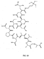
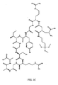
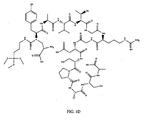
- Fig. 1 shows the structures of the four peptide-silanes created.
- Table 7 provides the sequence of the four unmodified peptides along with their natural source proteins. The four peptides were chosen for their known biological functions in neuronal development.
- the YIGSR (denoted YIG in data) and RGD peptides are cell binding regions found in the extracellular matrix proteins, fibronectin and laminin, and are commonly employed in biomaterial surfaces to improve cell adhesion and survival on artificial materials.
- the KDI sequence directs cellular migration, facilitates axon guidance, and enhances the formation of basic neuronal architecture.
- the NID peptide sequence is a common EGF repeat found in the extracellular matrix proteins, laminin and tenascin, which contribute to the neural stem cell niche within the sub-ventricular zone and modulates the effects of growth factors. Table 7. Peptides chosen for study. Peptides were chosen to represent the developing embryonic neural environment. Bolded residue indicate cell binding sequence.
- NID PNAVAA NID EGF Repeat Laminin, Tenascin Basement Membrane Organization AYAVTG RGD SPASA SEQ ID NO: 15 RGD Fibronectin Type III Repeat Adhesion, Synapse Formation ARDIAEII KDI GA SEQ ID NO: 16 KDI Laminin Migration, Guidance, Neuronal Architecture ADPG YIG SRGAA SEQ ID NO: 17 YIG Laminin Adhesion, Synapse Formation
- Peptides with a longer sequence length than simply the primary bioactive peptide were used, as they are more likely to mimic the natural conformations found in ECM protein secondary structure. Flanking amino acids were determined from the native protein sequence, and an alanine was added to the C-terminus during synthesis through the use of a preconjugated Wang-Ala resin. In addition a minor modification to the N-terminus by the addition of an alanine improved the overall linkage capability of the CDI/APTMS terminating group.
- MALDI-MS was used to characterize the average m/z ratios of the resulting peptidesilanes.
- the peptides and peptide-silanes were partially purified through multiple ether extractions and subjected to standard MALDI conditions.
- the free peptides and peptide silanes of each sequence of interest were analyzed for comparison.
- Many of the peptide-silanes appear to have undergone partial hydrolysis of the APTMS methyl groups. This hydrolysis is expected, given the trifluoroacetic acid conditions used for peptide cleavage from the resin.
- the molecular ions generated for each sequence product are within experimental limits of the expected nominal molecular mass, demonstrating a successful conjugation of the APTMS with the CDI linker.
- the MALDI-MS data confirms that the peptide is conjugating effectively to the APTMS molecule, and that the peptides themselves are of an expected molecular mass given the chosen sequence. As this technique is developed further for potential therapeutic applications, a more sensitive technique, such as LC-ESI-MS/MS may be applied to confirm peptide sequence and purity.
- MALDI-MS was chosen as an analysis technique for these molecules over the traditional silane characterization techniques of nuclear magnetic resonance spectroscopy and Fourier transform infrared spectroscopy (FT-IR) due to the peptide-based nature of the molecular structure and the tendency for hydrolysis under acid cleavage.
- FT-IR Fourier transform infrared spectroscopy
- FT-IR was initially used for basic analysis of the peptides, however, this method could not be used for confirmation of conjugation due to the carbon fingerprint region overlap with the identifying Si-O peak (1000-1100 cm-1).
- XPS X-Ray Photoelectron Spectroscopy
- X-Ray photoelectron spectroscopy was employed to analyze the atomic composition of the peptide-silane thin films (See Example 1). Thin films were prepared as described below, using a final concentration of 0.1 mole% peptide-silane to TMOS derived silica. This ratio, while significantly higher than many of the potential biological ratios that will be used in the future, represents a quantity that is easily detected by XPS. Future work will include calibration curve analysis to determine the lowest surface relevant concentration. XPS spectra were analyzed and deconvoluted to yield atomic percentages present on the surface of the materials. Organic contamination was eliminated from the sample spectrum through mathematical means.
- the XPS data revealed the presence of carbon and nitrogen species on the SiO 2 surface.
- the ratio of N:C was compared to the ideal N:C derived from the expected structure.
- the structural N:C was obtained by enumerating the carbon and nitrogen atoms in the expected molecular structure. These ratios are within reasonable limits of the ideal N:C, providing reasonable confirmation that the organic species present on the surface of the peptide-derived silica thin films is from the incorporated peptide.
- XPS analysis was performed on the free peptides and free APTMS conjugated peptides (peptide-silanes).
- the carbon, nitrogen, and oxygen profiles of the free peptides and peptide-silanes are similar between the samples, with the primary differences occurring in the oxygen and silicon peaks.
- the silicon peak is apparent in the silane conjugated peptides, at 2.38%, while there appears to be only background contaminant silicon in the peptide, with a spectral percentage of 0.01%.
- the oxygen percentage is considerably larger in the peptide-silane spectra (+5%), which may be attributed to the silane-derived methoxy groups that may still be attached to the molecule. This comparison between the free peptides and peptide-silanes provides additional confirmation of the successful conjugation between the APTMS molecule and peptide chain.
- the XPS data indicated the availability of the peptide on the surface of the silica thin films. In addition, it serves as a confirmation of the successful silane conjugation on the amine terminus of the peptide. Given the bulky nature of these molecules, the surface nanotopography may be altered by the addition of peptide quantities at biologically relevant concentrations. To test this theory, AFM was employed to examine the nanotopographical and phase interactions of the thin film ormosils.
- AFM images were collected of thin films produced from each of the four peptide-silane precursors at mole percentages used in Jedlicka, et al, Journal of Materials Chemistry 2006, 16, (31), 3221-3230 . These images principally serve to confirm that the addition of bulky peptide molecules at biologically relevant ratios does not significantly alter the thin film features outside of the range of native thin films. Any changes in cell function on the peptide ormosils are therefore likely due to the peptides themselves and not secondary changes in surface features.
- the NID and KDI materials were prepared at a higher mole percentages compared to the basic cell adhesion peptides, YIG and RGD. Both the NID and KDI materials have well defined surface features in the range of 50-75 nm. In addition, a small amount of noise resulted from the tip sticking to the surface indicating tip-material interactions were present. This interaction is not observed in unmodified TMOS films and is likely due to the presence of the peptide.
- YIG and RGD were used at extremely low concentrations (0.0025 mole %) based on biological function. Resulting films lack defined features and appear similar to unmodified TMOS derived films. These AFM images, therefore, indicate that surface nanotopography is likely a function of the concentration of peptide at the surface. The nanotopography of the thin film materials is consistent with the morphology observed in TMOS thin films, which are capable of supporting neuronal adhesion. Therefore, it is reasonable to assume that the peptide modified silica films will be able to support cellular adhesion.
- P19 cells are an established model for studying neuronal differentiation and were selected for demonstrating the biological functionality of the peptide ormosils. Established by McBurney in 1982 (see McBurney, M.; Rogers, B., Developmental Biology 1982, 89, (2), 503-508 and McBurney, M. W., International Journal of Developmental Biology 1993, 37, (1), 135-140 ).
- P19 cells are a pluripotent embryonic carcinoma cell line with many features of embryonic stem cells. When exposed to retinoic acid (RA) and allowed to aggregate, P19 cells can differentiate into fibroblast-like, glial, and neuronal cells. The ratio of cell types varies with extracellular environment conditions in culture. The resulting neurons can mature to form both inhibitory and excitatory synaptic connections.
- RA retinoic acid
- the peptide ligands in the material composition were anticipated to impact the differentiated cell composition and neuronal morphology after retinoic acid treatment. Due to the dramatic differences in the morphology and biochemical characterization of neurons compared to astrocytes and fibroblasts, these cell population compositional changes should be observed by microscopy and immunofluorescent techniques. After retinoic acid treatment, cells were plated on thin films containing a combination of the basic adhesion peptides, RGD and YIG, as well as on films containing the three peptides RGD/YIG/NID (see Table 8). Three peptide-silanes in two combinations were explored to explore the potential utility of the peptide-silane thin films.
- the first composition was chosen to determine if the simple addition of two commonly used adhesion peptides to the surfaces of the thin film materials would enhance the utility of silica thin films as a cell culture substrate.
- the second material was chosen to explore the potential effects of adding a peptide known to play a role in extracellular patterning and basement membrane formation (NID), creating a more "rich" extracellular environment.
- KDI The final peptide discussed in this paper (KDI) was not used in cell studies, but will be explored in future work.
- the primary function of the KDI peptide is neuronal pathfinding and could potentially be useful in spatial cell direction.
- fibronectin RGD group was used at a very low molar ratio.
- the YIG group known as a neuronal cell adhesion molecule, was also limited in molar ratio for the purposes of this study.
- protein quantification in the absolute sense is somewhat elusive, these ratios represent a combination of developmental ratios, primarily based on whole protein analysis, and quantities used in existing peptide-modified biomaterial work.
- TMOS derived thin film were plated on the peptide materials and compared to cells on the unmodified TMOS derived thin film and on a collagen coated tissue culture plate control.
- the cells were maintained on the materials in ⁇ -MEM supplemented with 1% FBS to ensure that the cells were interacting with the peptide materials and not a layer of serum proteins.
- This treatment has also been shown to increase the proportion of neuronal cells in the differentiated population, due to the starvation of non-neuronal and undifferentiated cells.
- the cellular response to the materials was qualitatively analyzed using standard morphological observation.
- the unmodified TMOS films result in few neurons. Of noteworthy importance, in the low serum environment, the cells only rarely migrated out of the plated aggregates on the TMOS derived silica.
- the RGD/YIG films appear to enhance the neuronal morphology of the cells over standard TMOS derived silica thin films.
- NID peptide known to facilitate extracellular matrix interactions
- an increase in neuronal processes per field of view was observed.
- a network of long cellular processes forms on both peptide modified materials.
- both of these peptide ormosils appear to support a wide variety of morphologically-distinct cell types, while the collagen control seems to support a larger proportion of neuronal cells. As these observations are purely based on cell morphology, a quantitative analysis of cell type was conducted.
- ⁇ -tubulin III was performed to examine the neurite patterns of the adherent neuronal cells.
- the confocal images indicate that while each material type does support neuronal cells, the neurite processes on each of the material types are altered in agreement with the initial morphological observations.
- On the collagen coated tissue culture control there are several ⁇ -tubulin positive cells in each view, however, the neurite processes of these cells are relatively short.
- the lack of nuclei not associated with the neuronal cells demonstrates a lack of supporting glial cells.
- the RGD/YIG and RGD/YIG/NID peptide-silane thin films supported a variety of cells, shown by the density of nuclear staining.
- the neurite processes on these two material types are also much longer than the collagen control.
- the RGD/YG/NID peptide-silane films also seemed to support more neurite processes per field.
- the peptide-modified silica films appear to enhance the neuronal maturation of the P19 cells over the collagen control surfaces and native silica films.
- neurite length was quantified using the NeuronJ plugin of Image J 42. Neurites were traced from the cell body and quantified based on pixel conversion to micrometers. At least 5 images were analyzed per experiment, with at least 10 neurite traces performed per image. This data is presented in Fig. 2 .
- the peptide-silane materials supported longer neurite processes, which is likely due to the density of glial cells enhancing the maturation of the adherent neurons.
- the RGD/YIG surprisingly, supported longer neurites than the RGD/YIG/NID combination, although the RGD/YIG/NID combination did appear to support more neurite processes per field.
- the collagen control surfaces do sustain the neuronal population, however, based on neurite length, it can be reasonably deduced that the neuronal cells are less mature than those associated with the peptide ormosil materials.
- FACS analysis was performed to quantify the percentage of neuronal and astrocytic cells in the culture population.
- Neuronal cells were labeled with AlexaFluor 488 conjugated beta-tubulin III, and astrocytic cells labeled with Cy3 conjugated GFAP.
- Table 10 Material-exposed P19 embryonic carcinoma cells were grown on the described material types for 8-10 days. Cells were then removed from the materials and subjected to immunocytochemical protocols and FACS analysis. The percentage of cells positive for the various anticipated (GFAP - astrocytic cells, Tuj1 -- neuronal cells) are presented. The standard error across the experiments is designated by the number within the parenthesis.
- TMOS silica RGD/YIG RGD/YIG/NID Collagen Control Tuj1+ *** 0.875 (0.325) 2.86 (0.35) 5.325 (0.48) 15.58 (0.82) GFAP+ 9.625 (1.57) 9.75 (2.46) 13.725 (1.23) 6.9 (0.66)
- the collagen control supports the largest percentage of neuronal cells and a smaller percentage of astrocytic "support" cells over the peptide modified silica films.
- the TMOS derived silica supports very few neurons (0.875%), as well a population of astrocytic cells.
- the peptide materials appear to have a significant effect on the cell population.
- the RGD/YIG "adhesion" material appears to promote a significantly smaller population of neuronal cells than the collagen control and the RGD/YIG/NID composition.
- the RGD/YIG/NID composition espouses a larger number of astrocytic cells, which may enhance the ability of neurons to develop, survive, and ultimately mature.
- the cell population analysis appears to correlate with the neurite length quantification and confocal analysis of the differentiated P19 cells.
- the NID peptide is known to be involved in a variety of cellular signals. While the specific pathways that are being modulated cannot be ascertained with certainty, it is evident that the NID peptide does enhance the neuronal population in terms of neuronal percentages over the RGD/YIG material. The numerical count of neurons is less then that on the collagen control surfaces. In addition, given the increased length of neurites and formation of a network of processes on the RGD/YIG/NID surfaces, it is possible that the neurons have reached functional maturity.
- Integrins are the major class of receptors utilized by cells to interact with ECM protein ligands. Upon ligand binding, integrins cluster on the cell surface at sites termed focal adhesions, leading to the assembly of intracellular multiprotein complexes associated with the cytoskeleton. Focal adhesions, beyond acting as structural links between the ECM and cytoskeleton, are sights of signal transduction from the ECM.
- the manipulation of P19 population phenotype demonstrates that the incorporated peptides are available to the receptors at the cell surface and the peptide precursors are able to impart biological functionality into the sol-gel thin films.
- a method for the synthesis of chemically conjugated peptides to silane precursors has been provided. These precursors can then be added at particular mole percentages to standard TMOS derived silica thin films during condensation to improve the biological compatibility and ultimately neuronal differentiation patterns of P19 embryonic carcinoma cells. Through the use of selected cellular interactions, the identity and maturity of the experimental cells can be modified.
Landscapes
- Chemical & Material Sciences (AREA)
- Organic Chemistry (AREA)
- General Health & Medical Sciences (AREA)
- Life Sciences & Earth Sciences (AREA)
- Biochemistry (AREA)
- Biophysics (AREA)
- Health & Medical Sciences (AREA)
- Genetics & Genomics (AREA)
- Medicinal Chemistry (AREA)
- Molecular Biology (AREA)
- Proteomics, Peptides & Aminoacids (AREA)
- Inorganic Chemistry (AREA)
- Peptides Or Proteins (AREA)
- Medicinal Preparation (AREA)
Claims (25)
- Procédé de préparation d'un sol-gel présentant un peptide, ledit procédé consistant à
fournir une composition de peptide-silane ;
combiner la composition de peptide-silane avec une seconde composition comprenant des silicates pour former un mélange ;
appliquer le mélange sur une surface d'un support ;
gélifier le mélange pour former un réseau de silice poreuse présentant des peptides liés de façon covalente. - Procédé selon la revendication 1 dans lequel ladite composition de peptide-silane comprend une pluralité de composés peptide-silane qui diffèrent les uns des autres par la séquence de la fraction peptidique.
- Procédé selon la revendication 1 dans lequel la fraction peptidique dudit peptide-silane a une longueur de 6 à 30 acides aminés, ou une longueur de 6 à 14 acides aminés.
- Procédé selon la revendication 1 dans lequel le mélange est appliqué sur la surface dudit support en plongeant le support dans ledit mélange et en le retirant.
- Procédé selon la revendication 4 comprenant en outre l'étape consistant à plonger le support revêtu dans un second mélange, ledit second mélange ayant une composition de peptide-silane différente du mélange dans lequel le support a été plongé en premier.
- Procédé selon la revendication 4 ou 5 dans lequel le support est plongé et retiré dudit mélange à une vitesse ne dépassant pas 35 mm/seconde.
- Procédé de préparation d'un sol-gel présentant un peptide ayant une concentration prédéterminée d'un peptide lié de façon covalente au sol-gel, ledit procédé consistant à
fournir une composition de peptide-silane, dans laquelle ledit peptide-silane est présent à ladite concentration prédéterminée ;
combiner la composition de peptide-silane avec une seconde composition comprenant des silicates pour former un mélange ;
appliquer le mélange sur une surface d'un support ;
gélifier le mélange pour former un sol-gel poreux comprenant des peptides liés de façon covalente à ladite concentration prédéterminée. - Procédé selon la revendication 7 dans lequel ladite composition de peptide-silane comprend une pluralité de composés peptide-silane qui diffèrent les uns des autres par la séquence de la fraction peptidique, chacun de ladite pluralité de composés peptide-silane étant lié de façon covalente au sol-gel à chaque concentration prédéterminée.
- Procédé selon la revendication 8 dans lequel ladite composition de peptide-silane comprend 2 à 5 ou 3 à 4 composés peptide-silane différents, et ledit sol-gel comprend lesdits 2 à 5 ou lesdits 3 à 4 composés peptide-silane, respectivement, liés de façon covalente au sol-gel à chacune de leurs concentrations prédéterminées.
- Kit pour préparer un sol-gel ayant une concentration définie de peptides liés de façon covalente à un sol-gel selon les revendications 1 à 9, ledit kit comprenant
une pluralité de premiers contenants, lesdits premiers contenants comprenant chacun un composé peptide-silane unique ; et
un second contenant comprenant des silicates. - Procédé selon la revendication 3 ou kit selon la revendication 10 dans lequel lesdits silicates sont choisis parmi le groupe composé de tétraéthylorthosilicate, tétraméthylorthosilicate et tétrapropylorthosilicate.
- Kit selon la revendication 10 dans lequel le peptide-silane de chaque contenant comprend une séquence d'acides aminés choisie parmi le groupe composé de RGD (SEQ ID N°1), KDI (SEQ ID N°2), YIG (SEQ ID N°3), NID (SEQ ID N°4), RNNR (SEQ ID N°5), CYFQRYLI (SEQ ID N°6), YIGSR (SEQ ID N°7), GRGDNP (SEQ ID N°8), RDIAEIIKDISLVRNRR (SEQ ID N°9), NDNIDPNAVA (SEQ ID N°10), YAVTGRGDSPAS (SEQ ID N°11), RDIAEIIKDIG (SEQ ID N°12), DPGYIGSRGA (SEQ ID N°13), ANDNIDPNAVAA (SEQ ID N°14), AYAVTGRGDSPASA (SEQ ID N°15), ARDIAEIIKDIGA (SEQ ID N°16) ADPGYIGSRGAA (SEQ ID N°17), SIDRVEPYSSTAQG (SEQ ID N°18), CSVTCGG (SEQ ID N°19), VSWFSRHRYSPFAVS (SEQ ID N°20), AASIKVAVSADRG (SEQ ID N°21) et SLVRNRRVITIQG (SEQ ID N°22).
- Matériau hybride à base de sol-gel nano-texturé pour modulation cellulaire formé à partir du processus de la revendication 1, ledit sol-gel comprenant :un réseau de silice poreuse, etun complexe de peptide-silane lié de façon covalente audit réseau de silice et dispersé à l'intérieur du sol-gel.
- Procédé selon la revendication 3, ou matériau hybride à base de sol-gel selon la revendication 13, dans lequel la fraction peptidique dudit peptide-silane comprend une séquence d'acides aminés choisie parmi le groupe composé de RGD (SEQ ID N°1), KDI (SEQ ID N°2), YIG (SEQ ID N°3), NID (SEQ ID N°4), RNNR (SEQ ID N°5), CYFQRYLI (SEQ ID N°6), YIGSR (SEQ ID N°7), GRGDNP (SEQ ID N°8), RDIAEIIKDISLVRNRR (SEQ ID N°9), NDNIDPNAVA (SEQ ID N°10), YAVTGRGDSPAS (SEQ ID N°11), RDIAEIIKDIG (SEQ ID N°12), DPGYIGSRGA (SEQ ID N°13), ANDNIDPNAVAA (SEQ ID N°14), AYAVTGRGDSPASA (SEQ ID N°15), ARDIAEIIKDIGA (SEQ ID N°16) ADPGYIGSRGAA (SEQ ID N°17), SIDRVEPYSSTAQG (SEQ ID N°18), CSVTCGG (SEQ ID N°19), VSWFSRHRYSPFAVS (SEQ ID N°20), AASIKVAVSADRG (SEQ ID N°21), SLVRNRRVITIQG (SEQ ID N°22) et SLVRNRR (SEQ ID N°23).
- Matériau hybride à base de sol-gel selon la revendication 13, dans lequel le sol-gel comprend deux peptide-silanes différents ou plus liés de façon covalente au réseau de silice.
- Matériau hybride à base de sol-gel selon la revendication 13, dans lequel le sol-gel comprend trois peptides différents liés de façon covalente au réseau de silice, lesdits peptides différents comprenant indépendamment une séquence d'acides aminés choisie parmi le groupe composé de RNNR (SEQ ID N°5), CYFQRYLI (SEQ ID N°6), YIGSR (SEQ ID N°7), GRGDNP (SEQ ID N°8), RDIAEIIKDISLVRNRR (SEQ ID N°9), NDNIDPNAVA (SEQ ID N°10), YAVTGRGDSPAS (SEQ ID N°11), RDIAEIIKDIG (SEQ ID N°12), DPGYIGSRGA (SEQ ID N°13), ANDNIDPNAVAA (SEQ ID N°14), AYAVTGRGDSPASA (SEQ ID N°15), ARDIAEIIKDIGA (SEQ ID N°16) ADPGYIGSRGAA (SEQ ID N°17), SIDRVEPYSSTAQG (SEQ ID N°18), CSVTCGG (SEQ ID N°19), VSWFSRHRYSPFAVS (SEQ ID N°20), AASIKVAVSADRG (SEQ ID N°21) et SLVRNRRVITIQG (SEQ ID N°22).
- Matériau hybride à base de sol-gel produit par le procédé de la revendication 5.
- Matériau hybride à base de sol-gel nano-texturé comprenant :au moins un complexe de peptide-silane ;un film mince de silice auquel est lié ledit au moins un complexe de peptide-silane, etune population de cellules,ledit au moins un complexe de peptide-silane lié au film mince de silice étant dispersé à l'intérieur du sol-gel.
- Matériau hybride à base de sol-gel selon la revendication 18, dans lequel le complexe de peptide-silane fonctionne en modulant la signalisation ou la différenciation neuronale.
- Matériau hybride à base de sol-gel comprenant :un composé peptide-silane, le peptide étant lié de façon covalente à un silicate de formule générale N-amino(C1-C4)tri(C1-C4)oxysilane, etun réseau de silice poreuse formé sur la surface d'un support solide, ledit composé peptide-silane étant lié de façon covalente audit réseau de silice et ledit peptide-silane lié au réseau de silice étant dispersé à l'intérieur du sol-gel.
- Matériau hybride à base de sol-gel selon la revendication 20, dans lequel le groupe (C1-C4) lié au N-amino est un propyle et le groupe tri(C1-C4)oxy est un triméthoxy ou un triéthoxy.
- Matériau hybride à base de sol-gel selon l'une quelconque des revendications 20 à 21, dans lequel le peptide lié de façon covalente au silicate comprend une séquence d'acides aminés choisie parmi RGD (SEQ ID N°1), KDI (SEQ ID N°2), YIG (SEQ ID N°3), NID (SEQ ID N°4), RNNR (SEQ ID N°5), CYFQRYLI (SEQ ID N°6), YIGSR (SEQ ID N°7), GRGDNP (SEQ ID N°8), RDIAEIIKDISLVRNRR (SEQ ID N°9), NDNIDPNAVA (SEQ ID N°10), YAVTGRGDSPAS (SEQ ID N°11), RDIAEIIKDIG (SEQ ID N°12), DPGYIGSRGA (SEQ ID N°13), ANDNIDPNAVAA (SEQ ID N°14), AYAVTGRGDSPASA (SEQ ID N°15), ARDIAEIIKDIGA (SEQ ID N°16) ADPGYIGSRGAA (SEQ ID N°17), SIDRVEPYSSTAQG (SEQ ID N°18), CSVTCGG (SEQ ID N°19), VSWFSRHRYSPFAVS (SEQ ID N°20), AASIKVAVSADRG (SEQ ID N°21), SLVRNRRVITIQG (SEQ ID N°22), SLVRNRR (SEQ ID N°23) et SNNR (SEQ ID N°24).
- Matériau hybride à base de sol-gel selon la revendication 22, dans lequel le groupe (C1-C4) lié au N-amino est un propyle et le groupe tri(C1C4)oxy est un triméthoxy et le peptide a une séquence d'acides aminés choisie parmi le groupe composé de RGD (SEQ ID N°1), KDI (SEQ ID N°2), YIG (SEQ ID N°3), NID (SEQ ID N°4).
- Utilisation du matériau hybride à base de sol-gel selon l'une quelconque des revendications 13 à 23 en tant que substrat ou support de cellules.
- Utilisation du matériau hybride à base de sol-gel selon la revendication 24 en tant que modulateur du phénotype cellulaire.
Applications Claiming Priority (2)
| Application Number | Priority Date | Filing Date | Title |
|---|---|---|---|
| US85089306P | 2006-10-11 | 2006-10-11 | |
| PCT/US2007/081122 WO2008057709A2 (fr) | 2006-10-11 | 2007-10-11 | Matériaux sol-gel pour modulation cellulaire |
Publications (3)
| Publication Number | Publication Date |
|---|---|
| EP2076276A2 EP2076276A2 (fr) | 2009-07-08 |
| EP2076276A4 EP2076276A4 (fr) | 2009-10-21 |
| EP2076276B1 true EP2076276B1 (fr) | 2014-01-22 |
Family
ID=39365182
Family Applications (1)
| Application Number | Title | Priority Date | Filing Date |
|---|---|---|---|
| EP07868421.4A Not-in-force EP2076276B1 (fr) | 2006-10-11 | 2007-10-11 | Matériaux sol-gel pour modulation cellulaire |
Country Status (3)
| Country | Link |
|---|---|
| US (1) | US8183043B2 (fr) |
| EP (1) | EP2076276B1 (fr) |
| WO (1) | WO2008057709A2 (fr) |
Families Citing this family (3)
| Publication number | Priority date | Publication date | Assignee | Title |
|---|---|---|---|---|
| AU2010276255A1 (en) | 2009-07-21 | 2012-03-01 | Purdue Research Foundation | Cell-mediated silica sol-gel encapsulation of living cells and tissues |
| EP2638507B1 (fr) * | 2010-11-12 | 2016-01-27 | AbbVie Inc. | Procédé et système optique à rendement élevé pour déterminer l'effet d'une substance d'essai sur des cellules vivantes |
| US9114092B2 (en) * | 2012-01-27 | 2015-08-25 | Rutgers, The State University Of New Jersey | Nanotopography-mediated reverse uptake platform for nucleic acid delivery and applications thereof |
Family Cites Families (6)
| Publication number | Priority date | Publication date | Assignee | Title |
|---|---|---|---|---|
| US6022748A (en) * | 1997-08-29 | 2000-02-08 | Sandia Corporation - New Mexico Regents Of The University Of California | Sol-gel matrices for direct colorimetric detection of analytes |
| GB0203453D0 (en) * | 2002-02-14 | 2002-04-03 | Ici Plc | Peptide synthesis |
| US7105347B2 (en) * | 2002-07-30 | 2006-09-12 | Corning Incorporated | Method and device for protein delivery into cells |
| US7554021B2 (en) * | 2002-11-12 | 2009-06-30 | Northwestern University | Composition and method for self-assembly and mineralization of peptide amphiphiles |
| US20040202637A1 (en) * | 2003-04-11 | 2004-10-14 | Masato Yoshioka | Hair treatment agent, shampoo and hair coloring agent |
| CA2539151A1 (fr) * | 2003-09-15 | 2005-03-31 | Eric Wickstrom | Biomateriaux ameliores et procede de fixation d'agents therapeutiques a ceux-ci |
-
2007
- 2007-10-11 EP EP07868421.4A patent/EP2076276B1/fr not_active Not-in-force
- 2007-10-11 WO PCT/US2007/081122 patent/WO2008057709A2/fr active Application Filing
- 2007-10-11 US US12/445,015 patent/US8183043B2/en active Active
Also Published As
| Publication number | Publication date |
|---|---|
| US8183043B2 (en) | 2012-05-22 |
| EP2076276A2 (fr) | 2009-07-08 |
| WO2008057709A2 (fr) | 2008-05-15 |
| EP2076276A4 (fr) | 2009-10-21 |
| US20100221836A1 (en) | 2010-09-02 |
| WO2008057709A3 (fr) | 2008-10-30 |
Similar Documents
| Publication | Publication Date | Title |
|---|---|---|
| Sorribas et al. | Photolithographic generation of protein micropatterns for neuron culture applications | |
| Motta et al. | Enhancing Schwann cell migration using concentration gradients of laminin-derived peptides | |
| Hawkes et al. | Probing the nanoscale organisation and multivalency of cell surface receptors: DNA origami nanoarrays for cellular studies with single-molecule control | |
| Huang et al. | Controlled silanization using functional silatrane for thin and homogeneous antifouling coatings | |
| Landoulsi et al. | Silanization with APTES for controlling the interactions between stainless steel and biocomponents: reality vs expectation | |
| EP2076276B1 (fr) | Matériaux sol-gel pour modulation cellulaire | |
| Salvagni et al. | A bioactive elastin-like recombinamer reduces unspecific protein adsorption and enhances cell response on titanium surfaces | |
| Fu et al. | The effects of gelatin–dopamine coating on polydimethylsiloxane substrates on pluripotency maintenance and myocardial differentiation of cultured mouse embryonic stem cells | |
| WO2017187717A1 (fr) | Sonde fluorescente, procédé de détection de fluorescence et procédé d'utilisation de sonde fluorescente | |
| Li et al. | Biofunctionalization of a “Clickable” organic layer photochemically grafted on titanium substrates | |
| Jedlicka et al. | Peptide ormosils as cellular substrates | |
| US20160096865A1 (en) | Peptide-Silica Hybrid Materials | |
| Kumar et al. | Molecular control of interfacial fibronectin structure on graphene oxide steers cell fate | |
| Jedlicka et al. | Sol-gel derived materials as substrates for neuronal differentiation: effects of surface features and protein conformation | |
| Jiang et al. | bFGF and poly‐RGD cooperatively establish biointerface for stem cell adhesion, proliferation, and differentiation | |
| Chen et al. | Versatile Thiol-and Amino-Functionalized Silatranes for in-situ polymerization and Immobilization of Gold Nanoparticles | |
| JP4750211B1 (ja) | 表面改質方法及び表面改質材料 | |
| Matsuzawa et al. | Micropatterning of neurons using organic substrates in culture | |
| Wang et al. | Designed peptide-grafted hydrogels for human pluripotent stem cell culture and differentiation | |
| Jedlicka | Design and characterization of hybrid peptide sol-gel materials for the solid state induction of neuronal differentiation | |
| US11987780B2 (en) | L-glass: a novel functionalization method for covalently attaching ECM protein to optical glass | |
| US10975350B2 (en) | Cell culture substrate and method of making thereof | |
| Beganskiene et al. | Modified sol-gel coatings for biotechnological applications | |
| Kaiser et al. | Differential adhesion of fibroblast and neuroblastoma cells on size-and geometry-controlled nanofibrils of the extracellular matrix | |
| Kosovari et al. | Silanization Strategies for Tailoring Peptide Functionalization on Silicon Surfaces: Implications for Enhancing Stem Cell Adhesion |
Legal Events
| Date | Code | Title | Description |
|---|---|---|---|
| PUAI | Public reference made under article 153(3) epc to a published international application that has entered the european phase |
Free format text: ORIGINAL CODE: 0009012 |
|
| 17P | Request for examination filed |
Effective date: 20090508 |
|
| AK | Designated contracting states |
Kind code of ref document: A2 Designated state(s): AT BE BG CH CY CZ DE DK EE ES FI FR GB GR HU IE IS IT LI LT LU LV MC MT NL PL PT RO SE SI SK TR |
|
| RIN1 | Information on inventor provided before grant (corrected) |
Inventor name: RICKUS, JENNA LEIGH Inventor name: JEDLICKA, SABRINA S. |
|
| A4 | Supplementary search report drawn up and despatched |
Effective date: 20090921 |
|
| DAX | Request for extension of the european patent (deleted) | ||
| 17Q | First examination report despatched |
Effective date: 20100716 |
|
| GRAP | Despatch of communication of intention to grant a patent |
Free format text: ORIGINAL CODE: EPIDOSNIGR1 |
|
| INTG | Intention to grant announced |
Effective date: 20130725 |
|
| GRAS | Grant fee paid |
Free format text: ORIGINAL CODE: EPIDOSNIGR3 |
|
| GRAP | Despatch of communication of intention to grant a patent |
Free format text: ORIGINAL CODE: EPIDOSNIGR1 |
|
| GRAA | (expected) grant |
Free format text: ORIGINAL CODE: 0009210 |
|
| INTG | Intention to grant announced |
Effective date: 20131125 |
|
| AK | Designated contracting states |
Kind code of ref document: B1 Designated state(s): AT BE BG CH CY CZ DE DK EE ES FI FR GB GR HU IE IS IT LI LT LU LV MC MT NL PL PT RO SE SI SK TR |
|
| REG | Reference to a national code |
Ref country code: GB Ref legal event code: FG4D |
|
| REG | Reference to a national code |
Ref country code: CH Ref legal event code: EP |
|
| REG | Reference to a national code |
Ref country code: AT Ref legal event code: REF Ref document number: 650441 Country of ref document: AT Kind code of ref document: T Effective date: 20140215 |
|
| REG | Reference to a national code |
Ref country code: IE Ref legal event code: FG4D |
|
| REG | Reference to a national code |
Ref country code: DE Ref legal event code: R096 Ref document number: 602007034967 Country of ref document: DE Effective date: 20140306 |
|
| REG | Reference to a national code |
Ref country code: NL Ref legal event code: VDEP Effective date: 20140122 |
|
| REG | Reference to a national code |
Ref country code: AT Ref legal event code: MK05 Ref document number: 650441 Country of ref document: AT Kind code of ref document: T Effective date: 20140122 |
|
| REG | Reference to a national code |
Ref country code: LT Ref legal event code: MG4D |
|
| PG25 | Lapsed in a contracting state [announced via postgrant information from national office to epo] |
Ref country code: IS Free format text: LAPSE BECAUSE OF FAILURE TO SUBMIT A TRANSLATION OF THE DESCRIPTION OR TO PAY THE FEE WITHIN THE PRESCRIBED TIME-LIMIT Effective date: 20140522 Ref country code: LT Free format text: LAPSE BECAUSE OF FAILURE TO SUBMIT A TRANSLATION OF THE DESCRIPTION OR TO PAY THE FEE WITHIN THE PRESCRIBED TIME-LIMIT Effective date: 20140122 |
|
| PG25 | Lapsed in a contracting state [announced via postgrant information from national office to epo] |
Ref country code: AT Free format text: LAPSE BECAUSE OF FAILURE TO SUBMIT A TRANSLATION OF THE DESCRIPTION OR TO PAY THE FEE WITHIN THE PRESCRIBED TIME-LIMIT Effective date: 20140122 Ref country code: FI Free format text: LAPSE BECAUSE OF FAILURE TO SUBMIT A TRANSLATION OF THE DESCRIPTION OR TO PAY THE FEE WITHIN THE PRESCRIBED TIME-LIMIT Effective date: 20140122 Ref country code: SE Free format text: LAPSE BECAUSE OF FAILURE TO SUBMIT A TRANSLATION OF THE DESCRIPTION OR TO PAY THE FEE WITHIN THE PRESCRIBED TIME-LIMIT Effective date: 20140122 Ref country code: CY Free format text: LAPSE BECAUSE OF FAILURE TO SUBMIT A TRANSLATION OF THE DESCRIPTION OR TO PAY THE FEE WITHIN THE PRESCRIBED TIME-LIMIT Effective date: 20140122 Ref country code: NL Free format text: LAPSE BECAUSE OF FAILURE TO SUBMIT A TRANSLATION OF THE DESCRIPTION OR TO PAY THE FEE WITHIN THE PRESCRIBED TIME-LIMIT Effective date: 20140122 Ref country code: ES Free format text: LAPSE BECAUSE OF FAILURE TO SUBMIT A TRANSLATION OF THE DESCRIPTION OR TO PAY THE FEE WITHIN THE PRESCRIBED TIME-LIMIT Effective date: 20140122 Ref country code: PT Free format text: LAPSE BECAUSE OF FAILURE TO SUBMIT A TRANSLATION OF THE DESCRIPTION OR TO PAY THE FEE WITHIN THE PRESCRIBED TIME-LIMIT Effective date: 20140522 |
|
| PG25 | Lapsed in a contracting state [announced via postgrant information from national office to epo] |
Ref country code: LV Free format text: LAPSE BECAUSE OF FAILURE TO SUBMIT A TRANSLATION OF THE DESCRIPTION OR TO PAY THE FEE WITHIN THE PRESCRIBED TIME-LIMIT Effective date: 20140122 Ref country code: BE Free format text: LAPSE BECAUSE OF FAILURE TO SUBMIT A TRANSLATION OF THE DESCRIPTION OR TO PAY THE FEE WITHIN THE PRESCRIBED TIME-LIMIT Effective date: 20140122 |
|
| REG | Reference to a national code |
Ref country code: DE Ref legal event code: R097 Ref document number: 602007034967 Country of ref document: DE |
|
| PG25 | Lapsed in a contracting state [announced via postgrant information from national office to epo] |
Ref country code: EE Free format text: LAPSE BECAUSE OF FAILURE TO SUBMIT A TRANSLATION OF THE DESCRIPTION OR TO PAY THE FEE WITHIN THE PRESCRIBED TIME-LIMIT Effective date: 20140122 Ref country code: RO Free format text: LAPSE BECAUSE OF FAILURE TO SUBMIT A TRANSLATION OF THE DESCRIPTION OR TO PAY THE FEE WITHIN THE PRESCRIBED TIME-LIMIT Effective date: 20140122 Ref country code: DK Free format text: LAPSE BECAUSE OF FAILURE TO SUBMIT A TRANSLATION OF THE DESCRIPTION OR TO PAY THE FEE WITHIN THE PRESCRIBED TIME-LIMIT Effective date: 20140122 Ref country code: CZ Free format text: LAPSE BECAUSE OF FAILURE TO SUBMIT A TRANSLATION OF THE DESCRIPTION OR TO PAY THE FEE WITHIN THE PRESCRIBED TIME-LIMIT Effective date: 20140122 |
|
| PG25 | Lapsed in a contracting state [announced via postgrant information from national office to epo] |
Ref country code: SK Free format text: LAPSE BECAUSE OF FAILURE TO SUBMIT A TRANSLATION OF THE DESCRIPTION OR TO PAY THE FEE WITHIN THE PRESCRIBED TIME-LIMIT Effective date: 20140122 Ref country code: PL Free format text: LAPSE BECAUSE OF FAILURE TO SUBMIT A TRANSLATION OF THE DESCRIPTION OR TO PAY THE FEE WITHIN THE PRESCRIBED TIME-LIMIT Effective date: 20140122 |
|
| PLBE | No opposition filed within time limit |
Free format text: ORIGINAL CODE: 0009261 |
|
| STAA | Information on the status of an ep patent application or granted ep patent |
Free format text: STATUS: NO OPPOSITION FILED WITHIN TIME LIMIT |
|
| 26N | No opposition filed |
Effective date: 20141023 |
|
| REG | Reference to a national code |
Ref country code: DE Ref legal event code: R097 Ref document number: 602007034967 Country of ref document: DE Effective date: 20141023 |
|
| REG | Reference to a national code |
Ref country code: DE Ref legal event code: R119 Ref document number: 602007034967 Country of ref document: DE |
|
| PG25 | Lapsed in a contracting state [announced via postgrant information from national office to epo] |
Ref country code: SI Free format text: LAPSE BECAUSE OF FAILURE TO SUBMIT A TRANSLATION OF THE DESCRIPTION OR TO PAY THE FEE WITHIN THE PRESCRIBED TIME-LIMIT Effective date: 20140122 Ref country code: LU Free format text: LAPSE BECAUSE OF FAILURE TO SUBMIT A TRANSLATION OF THE DESCRIPTION OR TO PAY THE FEE WITHIN THE PRESCRIBED TIME-LIMIT Effective date: 20141011 Ref country code: MC Free format text: LAPSE BECAUSE OF FAILURE TO SUBMIT A TRANSLATION OF THE DESCRIPTION OR TO PAY THE FEE WITHIN THE PRESCRIBED TIME-LIMIT Effective date: 20140122 |
|
| REG | Reference to a national code |
Ref country code: CH Ref legal event code: PL |
|
| GBPC | Gb: european patent ceased through non-payment of renewal fee |
Effective date: 20141011 |
|
| REG | Reference to a national code |
Ref country code: IE Ref legal event code: MM4A |
|
| PG25 | Lapsed in a contracting state [announced via postgrant information from national office to epo] |
Ref country code: CH Free format text: LAPSE BECAUSE OF NON-PAYMENT OF DUE FEES Effective date: 20141031 Ref country code: GB Free format text: LAPSE BECAUSE OF NON-PAYMENT OF DUE FEES Effective date: 20141011 Ref country code: LI Free format text: LAPSE BECAUSE OF NON-PAYMENT OF DUE FEES Effective date: 20141031 Ref country code: DE Free format text: LAPSE BECAUSE OF NON-PAYMENT OF DUE FEES Effective date: 20150501 |
|
| REG | Reference to a national code |
Ref country code: FR Ref legal event code: ST Effective date: 20150630 |
|
| PG25 | Lapsed in a contracting state [announced via postgrant information from national office to epo] |
Ref country code: FR Free format text: LAPSE BECAUSE OF NON-PAYMENT OF DUE FEES Effective date: 20141031 |
|
| PG25 | Lapsed in a contracting state [announced via postgrant information from national office to epo] |
Ref country code: IE Free format text: LAPSE BECAUSE OF NON-PAYMENT OF DUE FEES Effective date: 20141011 |
|
| PG25 | Lapsed in a contracting state [announced via postgrant information from national office to epo] |
Ref country code: BG Free format text: LAPSE BECAUSE OF FAILURE TO SUBMIT A TRANSLATION OF THE DESCRIPTION OR TO PAY THE FEE WITHIN THE PRESCRIBED TIME-LIMIT Effective date: 20140122 |
|
| PG25 | Lapsed in a contracting state [announced via postgrant information from national office to epo] |
Ref country code: IT Free format text: LAPSE BECAUSE OF FAILURE TO SUBMIT A TRANSLATION OF THE DESCRIPTION OR TO PAY THE FEE WITHIN THE PRESCRIBED TIME-LIMIT Effective date: 20140122 Ref country code: GR Free format text: LAPSE BECAUSE OF FAILURE TO SUBMIT A TRANSLATION OF THE DESCRIPTION OR TO PAY THE FEE WITHIN THE PRESCRIBED TIME-LIMIT Effective date: 20140423 |
|
| PG25 | Lapsed in a contracting state [announced via postgrant information from national office to epo] |
Ref country code: TR Free format text: LAPSE BECAUSE OF FAILURE TO SUBMIT A TRANSLATION OF THE DESCRIPTION OR TO PAY THE FEE WITHIN THE PRESCRIBED TIME-LIMIT Effective date: 20140122 Ref country code: MT Free format text: LAPSE BECAUSE OF FAILURE TO SUBMIT A TRANSLATION OF THE DESCRIPTION OR TO PAY THE FEE WITHIN THE PRESCRIBED TIME-LIMIT Effective date: 20140122 Ref country code: HU Free format text: LAPSE BECAUSE OF FAILURE TO SUBMIT A TRANSLATION OF THE DESCRIPTION OR TO PAY THE FEE WITHIN THE PRESCRIBED TIME-LIMIT; INVALID AB INITIO Effective date: 20071011 |