EP2056882B1 - Identification of a micro-rna that activates expression of beta-myosin heavy chain - Google Patents
Identification of a micro-rna that activates expression of beta-myosin heavy chain Download PDFInfo
- Publication number
- EP2056882B1 EP2056882B1 EP07813595A EP07813595A EP2056882B1 EP 2056882 B1 EP2056882 B1 EP 2056882B1 EP 07813595 A EP07813595 A EP 07813595A EP 07813595 A EP07813595 A EP 07813595A EP 2056882 B1 EP2056882 B1 EP 2056882B1
- Authority
- EP
- European Patent Office
- Prior art keywords
- mir
- expression
- cell
- inhibitor
- cardiac
- Prior art date
- Legal status (The legal status is an assumption and is not a legal conclusion. Google has not performed a legal analysis and makes no representation as to the accuracy of the status listed.)
- Not-in-force
Links
Images


Classifications
-
- C—CHEMISTRY; METALLURGY
- C12—BIOCHEMISTRY; BEER; SPIRITS; WINE; VINEGAR; MICROBIOLOGY; ENZYMOLOGY; MUTATION OR GENETIC ENGINEERING
- C12N—MICROORGANISMS OR ENZYMES; COMPOSITIONS THEREOF; PROPAGATING, PRESERVING, OR MAINTAINING MICROORGANISMS; MUTATION OR GENETIC ENGINEERING; CULTURE MEDIA
- C12N15/00—Mutation or genetic engineering; DNA or RNA concerning genetic engineering, vectors, e.g. plasmids, or their isolation, preparation or purification; Use of hosts therefor
- C12N15/09—Recombinant DNA-technology
- C12N15/63—Introduction of foreign genetic material using vectors; Vectors; Use of hosts therefor; Regulation of expression
- C12N15/79—Vectors or expression systems specially adapted for eukaryotic hosts
- C12N15/85—Vectors or expression systems specially adapted for eukaryotic hosts for animal cells
- C12N15/8509—Vectors or expression systems specially adapted for eukaryotic hosts for animal cells for producing genetically modified animals, e.g. transgenic
-
- A—HUMAN NECESSITIES
- A01—AGRICULTURE; FORESTRY; ANIMAL HUSBANDRY; HUNTING; TRAPPING; FISHING
- A01K—ANIMAL HUSBANDRY; CARE OF BIRDS, FISHES, INSECTS; FISHING; REARING OR BREEDING ANIMALS, NOT OTHERWISE PROVIDED FOR; NEW BREEDS OF ANIMALS
- A01K67/00—Rearing or breeding animals, not otherwise provided for; New breeds of animals
- A01K67/027—New breeds of vertebrates
- A01K67/0275—Genetically modified vertebrates, e.g. transgenic
- A01K67/0276—Knockout animals
-
- A—HUMAN NECESSITIES
- A61—MEDICAL OR VETERINARY SCIENCE; HYGIENE
- A61P—SPECIFIC THERAPEUTIC ACTIVITY OF CHEMICAL COMPOUNDS OR MEDICINAL PREPARATIONS
- A61P21/00—Drugs for disorders of the muscular or neuromuscular system
- A61P21/04—Drugs for disorders of the muscular or neuromuscular system for myasthenia gravis
-
- A—HUMAN NECESSITIES
- A61—MEDICAL OR VETERINARY SCIENCE; HYGIENE
- A61P—SPECIFIC THERAPEUTIC ACTIVITY OF CHEMICAL COMPOUNDS OR MEDICINAL PREPARATIONS
- A61P9/00—Drugs for disorders of the cardiovascular system
-
- A—HUMAN NECESSITIES
- A61—MEDICAL OR VETERINARY SCIENCE; HYGIENE
- A61P—SPECIFIC THERAPEUTIC ACTIVITY OF CHEMICAL COMPOUNDS OR MEDICINAL PREPARATIONS
- A61P9/00—Drugs for disorders of the cardiovascular system
- A61P9/04—Inotropic agents, i.e. stimulants of cardiac contraction; Drugs for heart failure
-
- A—HUMAN NECESSITIES
- A61—MEDICAL OR VETERINARY SCIENCE; HYGIENE
- A61P—SPECIFIC THERAPEUTIC ACTIVITY OF CHEMICAL COMPOUNDS OR MEDICINAL PREPARATIONS
- A61P9/00—Drugs for disorders of the cardiovascular system
- A61P9/10—Drugs for disorders of the cardiovascular system for treating ischaemic or atherosclerotic diseases, e.g. antianginal drugs, coronary vasodilators, drugs for myocardial infarction, retinopathy, cerebrovascula insufficiency, renal arteriosclerosis
-
- C—CHEMISTRY; METALLURGY
- C12—BIOCHEMISTRY; BEER; SPIRITS; WINE; VINEGAR; MICROBIOLOGY; ENZYMOLOGY; MUTATION OR GENETIC ENGINEERING
- C12N—MICROORGANISMS OR ENZYMES; COMPOSITIONS THEREOF; PROPAGATING, PRESERVING, OR MAINTAINING MICROORGANISMS; MUTATION OR GENETIC ENGINEERING; CULTURE MEDIA
- C12N15/00—Mutation or genetic engineering; DNA or RNA concerning genetic engineering, vectors, e.g. plasmids, or their isolation, preparation or purification; Use of hosts therefor
- C12N15/09—Recombinant DNA-technology
- C12N15/11—DNA or RNA fragments; Modified forms thereof; Non-coding nucleic acids having a biological activity
- C12N15/113—Non-coding nucleic acids modulating the expression of genes, e.g. antisense oligonucleotides; Antisense DNA or RNA; Triplex- forming oligonucleotides; Catalytic nucleic acids, e.g. ribozymes; Nucleic acids used in co-suppression or gene silencing
-
- C—CHEMISTRY; METALLURGY
- C12—BIOCHEMISTRY; BEER; SPIRITS; WINE; VINEGAR; MICROBIOLOGY; ENZYMOLOGY; MUTATION OR GENETIC ENGINEERING
- C12Q—MEASURING OR TESTING PROCESSES INVOLVING ENZYMES, NUCLEIC ACIDS OR MICROORGANISMS; COMPOSITIONS OR TEST PAPERS THEREFOR; PROCESSES OF PREPARING SUCH COMPOSITIONS; CONDITION-RESPONSIVE CONTROL IN MICROBIOLOGICAL OR ENZYMOLOGICAL PROCESSES
- C12Q1/00—Measuring or testing processes involving enzymes, nucleic acids or microorganisms; Compositions therefor; Processes of preparing such compositions
- C12Q1/68—Measuring or testing processes involving enzymes, nucleic acids or microorganisms; Compositions therefor; Processes of preparing such compositions involving nucleic acids
- C12Q1/6876—Nucleic acid products used in the analysis of nucleic acids, e.g. primers or probes
- C12Q1/6883—Nucleic acid products used in the analysis of nucleic acids, e.g. primers or probes for diseases caused by alterations of genetic material
-
- A—HUMAN NECESSITIES
- A01—AGRICULTURE; FORESTRY; ANIMAL HUSBANDRY; HUNTING; TRAPPING; FISHING
- A01K—ANIMAL HUSBANDRY; CARE OF BIRDS, FISHES, INSECTS; FISHING; REARING OR BREEDING ANIMALS, NOT OTHERWISE PROVIDED FOR; NEW BREEDS OF ANIMALS
- A01K2217/00—Genetically modified animals
- A01K2217/07—Animals genetically altered by homologous recombination
- A01K2217/075—Animals genetically altered by homologous recombination inducing loss of function, i.e. knock out
-
- A—HUMAN NECESSITIES
- A01—AGRICULTURE; FORESTRY; ANIMAL HUSBANDRY; HUNTING; TRAPPING; FISHING
- A01K—ANIMAL HUSBANDRY; CARE OF BIRDS, FISHES, INSECTS; FISHING; REARING OR BREEDING ANIMALS, NOT OTHERWISE PROVIDED FOR; NEW BREEDS OF ANIMALS
- A01K2227/00—Animals characterised by species
- A01K2227/10—Mammal
- A01K2227/105—Murine
-
- A—HUMAN NECESSITIES
- A01—AGRICULTURE; FORESTRY; ANIMAL HUSBANDRY; HUNTING; TRAPPING; FISHING
- A01K—ANIMAL HUSBANDRY; CARE OF BIRDS, FISHES, INSECTS; FISHING; REARING OR BREEDING ANIMALS, NOT OTHERWISE PROVIDED FOR; NEW BREEDS OF ANIMALS
- A01K2267/00—Animals characterised by purpose
- A01K2267/03—Animal model, e.g. for test or diseases
- A01K2267/035—Animal model for multifactorial diseases
- A01K2267/0375—Animal model for cardiovascular diseases
-
- A—HUMAN NECESSITIES
- A61—MEDICAL OR VETERINARY SCIENCE; HYGIENE
- A61K—PREPARATIONS FOR MEDICAL, DENTAL OR TOILETRY PURPOSES
- A61K48/00—Medicinal preparations containing genetic material which is inserted into cells of the living body to treat genetic diseases; Gene therapy
-
- C—CHEMISTRY; METALLURGY
- C12—BIOCHEMISTRY; BEER; SPIRITS; WINE; VINEGAR; MICROBIOLOGY; ENZYMOLOGY; MUTATION OR GENETIC ENGINEERING
- C12N—MICROORGANISMS OR ENZYMES; COMPOSITIONS THEREOF; PROPAGATING, PRESERVING, OR MAINTAINING MICROORGANISMS; MUTATION OR GENETIC ENGINEERING; CULTURE MEDIA
- C12N2310/00—Structure or type of the nucleic acid
- C12N2310/10—Type of nucleic acid
- C12N2310/11—Antisense
-
- C—CHEMISTRY; METALLURGY
- C12—BIOCHEMISTRY; BEER; SPIRITS; WINE; VINEGAR; MICROBIOLOGY; ENZYMOLOGY; MUTATION OR GENETIC ENGINEERING
- C12N—MICROORGANISMS OR ENZYMES; COMPOSITIONS THEREOF; PROPAGATING, PRESERVING, OR MAINTAINING MICROORGANISMS; MUTATION OR GENETIC ENGINEERING; CULTURE MEDIA
- C12N2310/00—Structure or type of the nucleic acid
- C12N2310/10—Type of nucleic acid
- C12N2310/14—Type of nucleic acid interfering N.A.
- C12N2310/141—MicroRNAs, miRNAs
-
- C—CHEMISTRY; METALLURGY
- C12—BIOCHEMISTRY; BEER; SPIRITS; WINE; VINEGAR; MICROBIOLOGY; ENZYMOLOGY; MUTATION OR GENETIC ENGINEERING
- C12N—MICROORGANISMS OR ENZYMES; COMPOSITIONS THEREOF; PROPAGATING, PRESERVING, OR MAINTAINING MICROORGANISMS; MUTATION OR GENETIC ENGINEERING; CULTURE MEDIA
- C12N2330/00—Production
- C12N2330/10—Production naturally occurring
-
- C—CHEMISTRY; METALLURGY
- C12—BIOCHEMISTRY; BEER; SPIRITS; WINE; VINEGAR; MICROBIOLOGY; ENZYMOLOGY; MUTATION OR GENETIC ENGINEERING
- C12Q—MEASURING OR TESTING PROCESSES INVOLVING ENZYMES, NUCLEIC ACIDS OR MICROORGANISMS; COMPOSITIONS OR TEST PAPERS THEREFOR; PROCESSES OF PREPARING SUCH COMPOSITIONS; CONDITION-RESPONSIVE CONTROL IN MICROBIOLOGICAL OR ENZYMOLOGICAL PROCESSES
- C12Q2600/00—Oligonucleotides characterized by their use
- C12Q2600/106—Pharmacogenomics, i.e. genetic variability in individual responses to drugs and drug metabolism
-
- C—CHEMISTRY; METALLURGY
- C12—BIOCHEMISTRY; BEER; SPIRITS; WINE; VINEGAR; MICROBIOLOGY; ENZYMOLOGY; MUTATION OR GENETIC ENGINEERING
- C12Q—MEASURING OR TESTING PROCESSES INVOLVING ENZYMES, NUCLEIC ACIDS OR MICROORGANISMS; COMPOSITIONS OR TEST PAPERS THEREFOR; PROCESSES OF PREPARING SUCH COMPOSITIONS; CONDITION-RESPONSIVE CONTROL IN MICROBIOLOGICAL OR ENZYMOLOGICAL PROCESSES
- C12Q2600/00—Oligonucleotides characterized by their use
- C12Q2600/158—Expression markers
Definitions
- the present invention relates generally to the fields of developmental biology and molecular biology. More particularly, it concerns gene regulation and cellular physiology in cardiomyocytes and skeletal muscle cells. Specifically, the invention relates to the inhibition of an miRNA that results in reduced expression of ⁇ -myosin heavy chain ( ⁇ -MHC), thereby treating cardiac hypertrophy and heart failure.
- ⁇ -MHC ⁇ -myosin heavy chain
- Cardiac hypertrophy in response to an increased workload imposed on the heart is a fundamental adaptive mechanism. It is a specialized process reflecting a quantitative increase in cell size and mass (rather than cell number) as the result of any, or a combination of, neural, endocrine or mechanical stimuli.
- Hypertension another factor involved in cardiac hypertrophy, is a frequent precursor of congestive heart failure. When heart failure occurs, the left ventricle usually is hypertrophied and dilated and indices of systolic function, such as ejection fraction, are reduced.
- the cardiac hypertrophic response is a complex syndrome and the elucidation of the pathways leading to cardiac hypertrophy will be beneficial in the treatment of heart disease resulting from various stimuli.
- Pathological myocardial hypertrophy is characterized by an increase in cardiomyocyte protein and the expression of a gene profile reminiscent of early embryonic development. Specifically, expression of ß-myosin heavy chain (ß-MHC), skeletal ⁇ -actin (sACT), and both atrial and brain natriuretic peptides (ANP and BNP, respectively) increases, whereas that of the adult cardiac muscle-specific genes, ⁇ -myosin heavy chain ( ⁇ -MHC) and sarcoplasmic reticulum Ca 2+ -ATPase (SERCA), decreases.
- ß-MHC ß-myosin heavy chain
- sACT skeletal ⁇ -actin
- ANP atrial and brain natriuretic peptides
- SERCA sarcoplasmic reticulum Ca 2+ -ATPase
- ⁇ -MHC mRNA and protein levels are markedly reduced in failing human hearts, and improvement of left-ventricular ejection fraction through beta-blocker therapy is associated with normalization of ⁇ -MHC expression.
- a mutation in the human ⁇ -MHC gene was identified in association with hypertrophic cardiomyopathy, which demonstrates that, despite its low abundance, the level of ⁇ -MHC expression is critical for normal heart function.
- a method of regulating cardiac contractility and remodeling comprising administering a modulator of miR-208 expression or activity to heart cells.
- a method of regulating cardiac contractile protein gene expression comprising administering a modulator of miR-208 expression or activity to heart cells.
- the modulator may be an agonist or an antagonist of miR-208 expression or activity.
- a method of reducing ⁇ -MHC expression in heart cells comprising administering an inhibitor of miR-208 expression or activity to the heart cells is disclosed.
- a method of elevating ⁇ -MHC expression in heart cells comprising increasing endogenous miR-208 expression or activity or administering exogenous miR-208 to the heart cells is disclosed.
- a method of increasing the expression of a fast skeletal muscle contractile protein gene in heart cells comprising administering to the heart cells an inhibitor of miR-208 expression or activity is disclosure.
- a method of decreasing the expression of a fast skeletal muscle contractile protein gene in heart cells comprising increasing endogenous miR-208 expression or activity or administering exogenous miR-208 to the heart cells is disclosed.
- fast skeletal muscle contractile protein genes that may be increased or decreased according to the disclosed methods include: skeletal troponin I; troponin T3, myosin light chain, or ⁇ -skeletal actin.
- a method for treating pathologic cardiac hypertrophy or heart failure comprising: identifying a patient having cardiac hypertrophy, heart failure, or post myocardial infarction remodeling; and inhibiting expression or activity of miR-208 in heart cells of the patient is disclosed.
- a method of preventing pathologic hypertrophy or heart failure comprising: identifying a patient at risk of developing pathologic cardiac hypertrophy or heart failure; and inhibiting expression or activity of miR-208 in heart cells of the patient is disclosed.
- a method of treating myocardial infarct comprising inhibiting expression or activity of miR-208 in heart cells of said subject is disclosed.
- a method of preventing cardiac hypertrophy and dilated cardiomyopathy comprising inhibiting expression or activity of miR-208 in heart cells of a subject is disclosed.
- a method of inhibiting progression of cardiac hypertrophy comprising inhibiting expression or activity of miR-208 in heart cells of a subject is disclosed.
- a method of increasing exercise tolerance, reducing hospitalization, improving quality of life, decreasing morbidity, and/or decreasing mortality in a subject with heart failure or cardiac hypertrophy comprising inhibiting expression or activity of miR-208 in heart cells of the subject is disclosed.
- a method of increasing ⁇ -MHC expression in heart cells by administering an inhibitor of miR-208 under pathological conditions is disclosed.
- an antagomir of miR-208 is provided for use in a method for inhibiting the expression or activity of miR-208 wherein the antagomir is administered.
- the present invention provides an miR-208 antagomir.
- the administering of the antagomir or other modulator of miR-208 expression or activity may be by any method known to those in the art suitable for delivery to the targeted organ, tissue, or cell type.
- the modulator of miR-208 may be administered by intravenous injection, intraarterial injection, intrapericardial injection, or direct injection into the tissue ( e.g ., cardiac tissue, skeletal muscle tissue).
- administering comprises oral, transdermal, intraperitoneal, subcutaneous, sustained release, controlled release, delayed release, suppository or sublingual administration of miR-208.
- an inhibitor of mir-208 is provided for use in a method for treating or preventing pathologic cardiac hypertrophy or heart failure in a patient further comprising administering to the patient a second cardiac hypertrophic therapy.
- the second therapy may be, for example, a beta blocker, an ionotrope, a diuretic, ACE-I, AII antagonist, BNP, a Ca ++ -blocker, and ERA, or an HDAC inhibitor.
- the said second therapy may be administered before, at the same time, or after the inhibition of miR-208.
- the treatment of pathologic cardiac hypertrophy or heart failure may be defined as improving one or more symptoms of pathologic cardiac hypertrophy or heart failure.
- the improved symptoms may be, for example, increased exercise capacity, increased cardiac ejection volume, decreased left ventricular end diastolic pressure, decreased pulmonary capillary wedge pressure, increased cardiac output, or cardiac index, lowered pulmonary artery pressures, decreased left ventricular end systolic and diastolic dimensions, decreased left and right ventricular wall stress, decreased wall tension, increased quality of life, and decreased disease related morbidity or mortality.
- the treatment of pathologic cardiac hypertrophy may also be defined as delaying the transition from cardiac hypertrophy to heart failure.
- the patient at risk of developing pathologic cardiac hypertrophy or heart failure may exhibit one or more risk factors including, for example, long standing uncontrolled hypertension, uncorrected valvular disease, chronic angina, recent myocardial infarction, congenital predisposition to heart disease or pathological hypertrophy.
- the patient at risk may be diagnosed as having a genetic predisposition to cardiac hypertrophy.
- the patient at risk may have a familial history of cardiac hypertrophy.
- a method of decreasing the expression or activity of a fast skeletal muscle contractile protein gene in skeletal muscle cells comprising administering miR-208 to the skeletal muscle cells is disclosed.
- a method of treating or preventing a musculoskeletal disorder in a subject comprising: identifying a patient having or at risk of a musculoskeletal disorder; and increasing the expression and/or activity of miR-208 in skeletal muscle cells of said patient is disclosed.
- the musculoskeletal disorder may be, for example, disuse atrophy, muscle wasting in response to microgravity, or denervation.
- the method of treating or preventing the musculoskeletal disorder further comprises administering a second non-miR-208 therapy.
- Increasing the expression and/or activity of miR-208 may comprise administering miR-208 to the subject or administering an expression vector that expresses miR-208 to the subject.
- the expression vector is a viral expression vector.
- the viral expression vector may be, for example, an adenoviral or retroviral expression vector.
- the expression vector is a non-viral expression vector.
- miR-208 or an expression vector encoding miR-208 is associated with a lipid vehicle. Alternatively, one may simply provide miR-208 by itself, optionally included within a delivery vehicle, such as a liposome or nanoparticle. The fact that miR 208 is cardiac specific will prevent unwanted side-effects in other organs.
- a method for identifying a modulator of miR-208 comprising: (a) contacting a cell with a candidate compound; assessing miR-208 activity or expression; and (b) comparing the activity or expression in step (b) with the activity or expression in the absence of the candidate compound, wherein a difference between the measured activities or expression indicates that the candidate compound is a modulator of miR-208 is disclosed.
- the cell is contacted with the candidate compound in vitro.
- the cell is contacted with the candidate compound in vivo.
- the modualtor of miR-208 may be an agonist or antagonist of miR-208. Examples of candidate compounds that may be screened according to the described methods are peptides, polypeptides, polynucleotides, or small molecules.
- Assessing the miR-208 activity or expression may comprise assessing the expression level of miR-208. Those in the art will be familiar with a variety of methods for assessing RNA expression levels including, for example, northern blotting or RT-PCR. Assessing the miR-208 activity or expression may comprise assessing the activity of miR-208. In some aspects assessing the activity of miR-208 comprises assessing expression or activity of gene regulated by miR-208. Genes regulated by miR-208 include, for example, ⁇ and ⁇ -myosin heavy chain and fast skeletal muscle protein genes, such as fast skeletal troponin I, troponin T3, myosin light chain, and alpha skeletal actin.
- assessing the activity of miR-208 comprises assessing the ratio of ⁇ -myosin heavy chain expression level to ⁇ -myosin, heavy chain expression level.
- Those in the art will be familiar with a variety of methods for assessing the activity or expression of genes regulated by miR-208. Such methods include, for example, northern blotting, RT-PCR, ELISA, or western blotting.
- Modulators of miR-208 may be included in pharmaceutical compositions for the treatment of cardiac disorders and/or musculoskeletal disorders according to the methods of the present invention.
- a method for treating pathologic cardiac hypertrophy or heart failure comprising: identifying a patient having cardiac hypertrophy or heart failure; and administering an miR-208 inhibitor to the patient.
- the miR-208 inhibitor may be identified by a method comprising: (a) contacting a cell with a candidate compound; (b) assessing miR-208 activity or expression; and (c) comparing the activity or expression in step (b) with the activity or expression in the absence of the candidate compound, wherein a reduction in the activity or expression of miR-208 in the cell contacted with the candidate compound compared to the activity or expression in the cell in the absence of the candidate compound indicates that the candidate compound is an inhibitor of miR-208 is disclosed.
- a method for treating musculoskeletal disorder comprising: identifying a patient having a musculoskeletal disorder or at risk for developing a musculoskeletal disorder; and administering an miR-208 agonist to the patient is disclosed.
- the miR-208 agonist may be identified by a method comprising: (a) contacting a cell with a candidate compound; (b) assessing miR-208 activity or expression; and (c) comparing the activity or expression in step (b) with the activity or expression in the absence of the candidate compound, wherein an increase in the activity or expression of miR-208 in the cell contacted with the candidate compound compared to the activity or expression in the cell in the absence of the candidate compound indicates that the candidate compound is an agonist of miR-208.
- a transgenic, non-human mammal, the cells of which fail to express a functional miR-208 is disclosed.
- a transgenic, non-human mammal, the cells of which comprise a miR-208 coding region under the control of a heterologous promoter active in the cells of said non-human mammal is disclosed.
- the transgenic mammal may be, for example, a mouse or a rat.
- the promoter may be a tissue specific promoter such as, for example, a skeletal muscle specific promoter or a cardiac muscle specific promoter.
- a transgenic, non-human mammalian cell lacking one or both native miR-208 alleles is disclosed.
- compositions of the invention can be used to achieve the disclosed methods
- the words “comprising” (and any form of comprising, such as “comprise” and “comprises”), “having” (and any form of having, such as “have” and “has”), "including” (and any form of including, such as “includes” and “include”) or “containing” (and any form of containing, such as “contains” and “contain”) are inclusive or open-ended and do not exclude additional, unrecited elements or method steps.
- DCM Dilated cardiomyopathy
- congestive cardiomyopathy is the most common form of the cardiomyopathies and has an estimated prevalence of nearly 40 per 100,000 individuals (Durand et al., 1995).
- familiar dilated cardiomyopathy has been indicated as representing approximately 20% of "idiopathic" DCM. Approximately half of the DCM cases are idiopathic, with the remainder being associated with known disease processes.
- doxorubicin and daunoribucin drugs used in cancer chemotherapy
- DCM patients are chronic alcoholics. Fortunately, for these patients, the progression of myocardial dysfunction may be stopped or reversed if alcohol consumption is reduced or stopped early in the course of disease.
- Peripartum cardiomyopathy is another idiopathic form of DCM, as is disease associated with infectious sequelae. In sum, cardiomyopathies, including DCM, are significant public health problems.
- Heart disease and its manifestations including coronary artery disease, myocardial infarction, congestive heart failure and cardiac hypertrophy, clearly presents a major health risk in the United States today.
- the cost to diagnose, treat and support patients suffering from these diseases is well into the billions of dollars.
- Two particularly severe manifestations of heart disease are myocardial infarction and cardiac hypertrophy.
- myocardial infarction typically an acute thrombocytic coronary occlusion occurs in a coronary artery as a result of atherosclerosis and causes myocardial cell death.
- cardiomyocytes the heart muscle cells, are terminally differentiated and generally incapable of cell division, they are generally replaced by scar tissue when they die during the course of an acute myocardial infarction.
- Scar tissue is not contractile, fails to contribute to cardiac function, and often plays a detrimental role in heart function by expanding during cardiac contraction, or by increasing the size and effective radius of the ventricle, for example, becoming hypertrophic.
- cardiac hypertrophy one theory regards this as a disease that resembles aberrant development and, as such, raises the question of whether developmental signals in the heart can contribute to hypertrophic disease.
- Cardiac hypertrophy is an adaptive response of the heart to virtually all forms of cardiac disease, including those arising from hypertension, mechanical load, myocardial infarction, cardiac arrhythmias, endocrine disorders, and genetic mutations in cardiac contractile protein genes. While the hypertrophic response is initially a compensatory mechanism that augments cardiac output, sustained hypertrophy can lead to DCM, heart failure, and sudden death. In the United States, approximately half a million individuals are diagnosed with heart failure each year, with a mortality rate approaching 50%.
- cardiac hypertrophy The causes and effects of cardiac hypertrophy have been extensively documented, but the underlying molecular mechanisms have not been elucidated. Understanding these mechanisms is a major concern in the prevention and treatment of cardiac disease and will be crucial as a therapeutic modality in designing new drugs that specifically target cardiac hypertrophy and cardiac heart failure.
- pathologic cardiac hypertrophy typically does not produce any symptoms until the cardiac damage is severe enough to produce heart failure
- the symptoms of cardiomyopathy are those associated with heart failure. These symptoms include shortness of breath, fatigue with exertion, the inability to lie flat without becoming short of breath (orthopnea), paroxysmal nocturnal dyspnea, enlarged cardiac dimensions, and/or swelling in the lower legs.
- DCM causes decreased ejection fractions (i.e ., a measure of both intrinsic systolic function and remodeling).
- the disease is further characterized by ventricular dilation and grossly impaired systolic function due to diminished myocardial contractility, which results in dilated heart failure in many patients.
- Affected hearts also undergo cell/chamber remodeling as a result of the myocyte/myocardial dysfunction, which contributes to the "DCM phenotype.” As the disease progresses so do the symptoms.
- DCM patients with DCM also have a greatly increased incidence of life-threatening arrhythmias, including ventricular tachycardia and ventricular fibrillation. In these patients, an episode of syncope (dizziness) is regarded as a harbinger of sudden death.
- Diagnosis of dilated cardiomyopathy typically depends upon the demonstration of enlarged heart chambers, particularly enlarged ventricles. Enlargement is commonly observable on chest X-rays, but is more accurately assessed using echocardiograms. DCM is often difficult to distinguish from acute myocarditis, valvular heart disease, coronary artery disease, and hypertensive heart disease. Once the diagnosis of dilated cardiomyopathy is made, every effort is made to identify and treat potentially reversible causes and prevent further heart damage. For example, coronary artery disease and valvular heart disease must be ruled out. Anemia, abnormal tachycardias, nutritional deficiencies, alcoholism, thyroid disease and/or other problems need to be addressed and controlled.
- diuretics constitute the first line of treatment for mild-to-moderate heart failure.
- diuretics e.g ., the thiazides
- certain diuretics may increase serum cholesterol and triglycerides.
- diuretics are generally ineffective for patients suffering from severe heart failure.
- vasodilatory agents may be used; the angiotensin converting (ACE) inhibitors (e.g ., enalopril and lisinopril) not only provide symptomatic relief, they also have been reported to decrease mortality (Young et al., 1989). Again, however, the ACE inhibitors are associated with adverse effects that result in their being contraindicated in patients with certain disease states (e.g ., renal artery stenosis). Similarly, inotropic agent therapy (i.e ., a drug that improves cardiac output by increasing the force of myocardial muscle contraction) is associated with a panoply of adverse reactions, including gastrointestinal problems and central nervous system dysfunction.
- ACE angiotensin converting
- the currently used pharmacological agents have severe shortcomings in particular patient populations.
- the availability of new, safe and effective agents would undoubtedly benefit patients who either cannot use the pharmacological modalities presently available, or who do not receive adequate relief from those modalities.
- the prognosis for patients with DCM is variable, and depends upon the degree of ventricular dysfunction, with the majority of deaths occurring within five years of diagnosis.
- the ratio of ⁇ - to ⁇ -MHC isoforms in the adult heart is a major determinant of cardiac contractility.
- ⁇ -MHC the major myosin isoform in the adult heart, displays relatively low ATPase activity, whereas ⁇ -MHC has high ATPase activity.
- pathological stimuli such as myocardial infarction, hypertension, and other disorders
- the ⁇ -MHC expression increases and ⁇ -MHC expression decreases with consequent reduction in myofibrillar ATPase activity and reduced shortening velocity of cardiac myofibers, leading to eventual contractile dysfunction.
- minor changes in ⁇ -MHC content of the heart can have a profound influence on cardiac performance.
- MicroRNAs are small ⁇ 22-nucleotide RNAs that are derived from larger pre-miRs. MiRs act as repressors of target mRNAs by promoting their degradation, when their sequences are perfectly complementary, or inhibiting translation when their sequences contain mismatches.
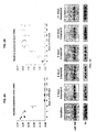
- microRNA-208 (miR-208) is encoded by an intron of the ⁇ -MHC gene and is expressed specifically in the heart. The inventors created miR-208 knockout mice and discovered that miR-208 is required for activation of ⁇ -MHC gene expression in the adult heart, as well as for expression of several other contractile protein genes. In addition, miR-208 inhibition leads to a severe reduction in cardiac fibrosis.
- Agonism of miR-208 expression or activity either by therapeutically activating the endogenous miR-208 gene or introducing exogenous miR-208 into the heart using adenoviral vectors or other -again no need for using an adenoviral system means of ectopic expression to elevate ⁇ -MHC expression, for treatment of individuals with a mutation in the ⁇ -MHC gene is disclosed.
- the up-regulation of several fast skeletal muscle contractile protein genes in the hearts of miR-208 mutant mice also suggests that miR-208 typically represses the fast skeletal muscle gene program. Activation of these genes in the heart represents a potential approach to regulate cardiac contractility.
- miR-208 to repress fast fiber genes in skeletal muscle and thereby activate the reciprocal expression of slow fiber genes, which are coupled to enhanced insulin sensitivity and skeletal muscle endurance is disclosed. Repression of slow fiber genes and activation of fast fiber genes in skeletal muscle is associated with numerous musculoskeletal disorders including disuse atrophy, muscle wasting in response to anti-gravity, and denervation.
- miR-208 is a muscle-specific and essential regulator of ⁇ -MHC gene expression in the heart that in addition regulates cardiac fibrosis.
- the discovery that miR-208 regulates ⁇ -MHC expression and expression of fast skeletal muscle genes is completely novel as is the use of this microRNA to control cardiac contractility and skeletal muscle function.
- miRNAs small molecules
- C. elegans, Drosophila, and humans Lagos-Quintana et al ., 2001; Lau et al ., 2001; Lee and Ambros, 2001.
- miRNAs Several hundreds of miRNAs have been identified in plants and animals-including humans-which do not appear to have endogenous siRNAs. Thus, while similar to siRNAs, miRNAs are nonetheless distinct.
- miRNAs thus far observed have been approximately 21-22 nucleotides in length and they arise from longer precursors, which are transcribed from non-protein-encoding genes. See review of Carrington et al. (2003). The precursors form structures that fold back on each other in self-complementary regions; they are then processed by the nuclease Dicer in animals or DCL1 in plants. miRNA molecules interrupt translation through precise or imprecise base-pairing with their targets.
- miRNAs are involved in gene regulation. Some miRNAs, including lin-4 and let- 7, inhibit protein synthesis by binding to partially complementary 3' untranslated regions (3' UTRs) of target mRNAs. Others, including the Scarecrow miRNA found in plants, function like siRNA and bind to perfectly complementary mRNA sequences to destroy the target transcript (Grishok et al., 2001).
- miRNAs that play critical roles in cell differentiation, early development, and cellular processes like apoptosis and fat metabolism.
- lin-4 and let-7 both regulate passage from one larval state to another during C. elegans development (Ambros, 2003).
- mir-14 and bantam are drosophila miRNAs that regulate cell death, apparently by regulating the expression of genes involved in apoptosis (Brennecke et al., 2003, Xu et al., 2003).
- MiR14 has also been implicated in fat metabolism (Xu et al., 2003).
- Lsy-6 and miR-273 are C. elegans miRNAs that regulate asymmetry in chemosensory neurons (Chang et al., 2004).
- miRNA-181 Another animal miRNA that regulates cell differentiation is miR-181, which guides hematopoietic cell differentiation (Chen et al., 2004). These molecules represent the full range of animal miRNAs with known functions. Enhanced understanding of the functions of miRNAs will undoubtedly reveal regulatory networks that contribute to normal development, differentiation, inter-and intra-cellular communication, cell cycle, angiogenesis, apoptosis, and many other cellular processes. Given their important roles in many biological functions, it is likely that miRNAs will offer important points for therapeutic intervention or diagnostic analysis.
- Characterizing the functions of biomolecules like miRNAs often involves introducing the molecules into cells or removing the molecules from cells and measuring the result. If introducing a miRNA into cells results in apoptosis, then the miRNA undoubtedly participates in an apoptotic pathway. Methods for introducing and removing miRNAs from cells have been described. Two recent publications describe antisense molecules that can be used to inhibit the activity of specific miRNAs (Meister et al., 2004; Hutvagner et al., 2004). Another publication describes the use of plasmids that are transcribed by endogenous RNA polymerases and yield specific miRNAs when transfected into cells (Zeng et al., 2002). These two reagent sets have been used to evaluate single miRNAs.
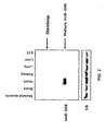
- MiR-208 is an intronic miRNA that is located within the 27 th intron of the ⁇ -MHC gene.
- FIG. 1 The pre-miRNA encoding sequences for miR-208 for human, mouse, rat, and canine are provided in SEQ ID NO:14, SEQ ID NO:15, SEQ ID NO:16, SEQ ID NO:17, respectively.
- the mature miR-208 sequence is provided in SEQ ID NO:5.
- miR-208 is expressed solely in the heart.
- THRAP1 thyroid hormone receptor associated protein 1
- SEQ ID NO:6 SEQ ID NO:7, SEQ ID NO:8, SEQ ID NO:9, SEQ ID NO:10, SEQ ID NO:11, SEQ ID NO:12, and SEQ ID NO:13, respectively.
- inhibitors of miRNAs take the form of "antagomirs," short, chemically-engineered single-stranded oligonucleotides complementary to miRNAs that block the function of miRNAs (Krützfeldt et al., 2005).
- Other approaches include inhibition of miRNAs with antisense 2'-O-methyl (2'-OMe) oligoribonucleotides and small interfering double-stranded RNAs (siRNAs) engineered with certain "drug-like" properties (chemical modifications for stability; cholesterol conjugation for delivery) (Kritzfeldt et al., 2005).
- Non-pharmacological treatment is primarily used as an adjunct to pharmacological treatment.
- One means of non-pharmacological treatment involves reducing the sodium in the diet.
- non-pharmacological treatment also entails the elimination of certain precipitating drugs, including negative inotropic agents (e.g ., certain calcium channel blockers and antiarrhythmic drugs like disopyramide), cardiotoxins (e.g ., amphetamines), and plasma volume expanders (e.g ., nonsteroidal antiinflammatory agents and glucocorticoids).
- negative inotropic agents e.g ., certain calcium channel blockers and antiarrhythmic drugs like disopyramide
- cardiotoxins e.g ., amphetamines
- plasma volume expanders e.g ., nonsteroidal antiinflammatory agents and glucocorticoids.
- inhibitors of miR-208 are provided for use in methods for the treatment of cardiac hypertrophy or heart failure.
- treatment comprises reducing one or more of the symptoms of cardiac hypertrophy, such as reduced exercise capacity, reduced blood ejection volume, increased left ventricular end diastolic pressure, increased pulmonary capillary wedge pressure, reduced cardiac output, cardiac index, increased pulmonary artery pressures, increased left ventricular end systolic and diastolic dimensions, and increased left ventricular wall stress, wall tension and wall thickness-same for right ventricle.
- use of inhibitors of miR-208 may prevent cardiac hypertrophy and its associated symptoms from arising.
- Treatment regimens would vary depending on the clinical situation. However, long-term maintenance would appear to be appropriate in most circumstances. It also may be desirable treat hypertrophy with inhibitors of miR-208 intermittently, such as within a brief window during disease progression.
- an inhibitor of miR-208 of the invention in combination with other therapeutic modalities.
- other therapies include, without limitation, so-called “beta blockers,” anti-hypertensives, cardiotonics, anti-thrombotics, vasodilators, hormone antagonists, iontropes, diuretics, endothelin receptor antagonists, calcium channel blockers, phosphodiesterase inhibitors, ACE inhibitors, angiotensin type 2 antagonists and cytokine blockers/inhibitors, and HDAC inhibitors.
- Combinations may be achieved by contacting cardiac cells with a single composition or pharmacological formulation that includes both agents, or by contacting the cell with two distinct compositions or formulations, at the same time, wherein one composition includes the expression construct and the other includes the agent.
- the therapy using an inhibitor of miR-208 may precede or follow administration of the other agent(s) by intervals ranging from minutes to weeks.
- the other agent and expression construct are applied separately to the cell, one would generally ensure that a significant period of time did not expire between the time of each delivery, such that the agent and expression construct would still be able to exert an advantageously combined effect on the cell.
- Examples of a pharmacological therapeutic agent that may be used in the present invention include an antihyperlipoproteinemic agent, an antiarteriosclerotic 23 agent, an antithrombotic/fibrinolytic agent, a blood coagulant, an antiarrhythmic agent, an antihypertensive agent, a vasopressor, a treatment agent for congestive heart failure, an antianginal agent, an antibacterial agent or a combination thereof.
- any of the following may be used to develop new sets of cardiac therapy target genes as ⁇ -blockers were used in the present examples (see below). While it is expected that many of these genes may overlap, new gene targets likely can be developed.
- an antihyperlipoproteinemic may be combined with a cardiovascular therapy herewith disclosed, particularly in treatment of athersclerosis and thickenings or blockages of vascular tissues.
- an antihyperlipoproteinemic agent may comprise an aryloxyalkanoic/fibric acid derivative, a resin/bile acid sequesterant, a HMG CoA reductase inhibitor, a nicotinic acid derivative, a thyroid hormone or thyroid hormone analog, a miscellaneous agent or a combination thereof.
- Non-limiting examples of aryloxyalkanoic/fibric acid derivatives include beclobrate, enzafibrate, binifibrate, ciprofibrate, clinofibrate, clofibrate (atromide-S), clofibric acid, etofibrate, fenofibrate, gemfibrozil (lobid), nicofibrate, pirifibrate, ronifibrate, simfibrate and theofibrate.
- Non-limiting examples of resins/bile acid sequesterants include cholestyramine (cholybar, questran), colestipol (colestid) and polidexide.
- HMG CoA reductase inhibitors include lovastatin (mevacor), pravastatin (pravochol) or simvastatin (zocor).
- nicotinic acid derivatives include nicotinate, acepimox, niceritrol, nicoclonate, nicomol and oxiniacic acid.
- thyroid hormones and analogs thereof include etoroxate, thyropropic acid and thyroxine.
- miscellaneous antihyperlipoproteinemics include acifran, azacosterol, benfluorex, ⁇ -benzalbutyramide, carnitine, chondroitin sulfate, clomestrone, detaxtran, dextran sulfate sodium, 5,8, 11, 14, 17-eicosapentaenoic acid, eritadenine, furazabol, meglutol, melinamide, mytatrienediol, ornithine, ⁇ -oryzanol, pantethine, pentaerythritol tetraacetate, ⁇ -phenylbutyramide, pirozadil, probucol (lorelco), ⁇ -sitosterol, sultosilic acid-piperazine salt, tiadenol, triparanol and xenbucin.
- an antiarteriosclerotic examples include pyridinol carbamate.
- administration of an agent that aids in the removal or prevention of blood clots may be combined with administration of a modulator, particularly in treatment of athersclerosis and vasculature (e.g ., arterial) blockages.
- a modulator particularly in treatment of athersclerosis and vasculature (e.g ., arterial) blockages.
- antithrombotic and/or fibrinolytic agents include anticoagulants, anticoagulant antagonists, antiplatelet agents, thrombolytic agents, thrombolytic agent antagonists or combinations thereof.
- antithrombotic agents that can be administered orally, such as, for example, aspirin and wafarin (coumadin), are preferred.
- a Example of an anticoagulant include acenocoumarol, ancrod, anisindione, bromindione, clorindione, coumetarol, cyclocumarol, dextran sulfate sodium, dicumarol, diphenadione, ethyl biscoumacetate, ethylidene dicoumarol, fluindione, heparin, hirudin, lyapolate sodium, oxazidione, pentosan polysulfate, phenindione, phenprocoumon, phosvitin, picotamide, tioclomarol and warfarin.
- antiplatelet agents examples include aspirin, a dextran, dipyridamole (persantin), heparin, sulfinpyranone (anturane) and ticlopidine (ticlid).
- thrombolytic agents include tissue plaminogen activator (activase), plasmin, pro-urokinase, urokinase (abbokinase) streptokinase (streptase), anistreplase/APSAC (eminase).
- an agent that may enhance blood coagulation may be used.
- a blood coagulation promoting agent include thrombolytic agent antagonists and anticoagulant antagonists.
- anticoagulant antagonists examples include protamine and vitamine K1.
- thrombolytic agent antagonists include amiocaproic acid (amicar) and tranexamic acid (amstat).
- antithrombotics include anagrelide, argatroban, cilstazol, daltroban, defibrotide, enoxaparin, fraxiparine, indobufen, lamoparan, ozagrel, picotamide, plafibride, tedelparin, ticlopidine and triflusal.
- antiarrhythmic agents include Class I antiarrythmic agents (sodium channel blockers), Class II antiarrythmic agents (beta-adrenergic blockers), Class II antiarrythmic agents (repolarization prolonging drugs), Class IV antiarrhythmic agents (calcium channel blockers) and miscellaneous antiarrythmic agents.
- Examples of sodium channel blockers include Class IA, Class IB and Class IC antiarrhythmic agents.
- Class IA antiarrhythmic agents include disppyramide (norpace), procainamide (pronestyl) and quinidine (quinidex).
- Non-limiting examples of Class IB antiarrhythmic agents include lidocaine (xylocaine), tocainide (tonocard) and mexiletine (mexitil).
- Examples of Class IC antiarrhythmic agents include encainide (enkaid) and flecainide (tambocor).
- beta blocker otherwise known as a ⁇ -adrenergic blocker, a ⁇ -adrenergic antagonist or a Class II antiarrhythmic agent
- examples of a beta blocker include acebutolol (sectral), alprenolol, amosulalol, arotinolol, atenolol, befunolol, betaxolol, bevantolol, bisoprolol, bopindolol, bucumolol, bufetolol, bufuralol, bunitrolol, bupranolol, butidrine hydrochloride, butofilolol, carazolol, carteolol, carvedilol, celiprolol, cetamolol, cloranolol, dilevalol, epanolol, esmolol (brevibloc), indenolol,
- aryloxypropanolamine derivatives include acebutolol, alprenolol, arotinolol, atenolol, betaxolol, bevantolol, bisoprolol, bopindolol, bunitrolol, butofilolol, carazolol, carteolol, carvedilol, celiprolol, cetamolol, epanolol, indenolol, mepindolol, metipranolol, metoprolol, moprolol, nadolol, nipradilol, oxprenolol, penbutolol, pindolol, propanolol, talinolol, tertatolol, timolol and toliprolol.
- an agent that prolong repolarization also known as a Class III antiarrhythmic agent
- examples of an agent that prolong repolarization include amiodarone (cordarone) and sotalol (bumblece).
- Examples of a calcium channel blocker include an arylalkylamine (e.g ., bepridile, diltiazem, fendiline, gallopamil, prenylamine, terodiline, verapamil), a dihydropyridine derivative (felodipine, isradipine, nicardipine, nifedipine, nimodipine, nisoldipine, nitrendipine) a piperazinde derivative (e.g ., cinnarizine, flunarizine, lidoflazine) or a micellaneous calcium channel blocker such as bencyclane, etafenone, magnesium, mibefradil or perhexiline.
- a calcium channel blocker comprises a long-acting dihydropyridine (nifedipine-type) calcium antagonist.
- miscellaneous antiarrhymic agents include adenosine (adenocard), digoxin (lanoxin), acecainide, ajmaline, amoproxan, aprindine, bretylium tosylate, bunaftine, butobendine, capobenic acid, cifenline, disopyranide, hydroquinidine, indecainide, ipatropium bromide, lidocaine, lorajmine, lorcainide, meobentine, moricizine, pirmenol, prajmaline, propafenone, pyrinoline, quinidine polygalacturonate, quinidine sulfate and viquidil.
- antihypertensive agents include sympatholytic, alpha/beta blockers, alpha blockers, anti-angiotensin II agents, beta blockers, calcium channel blockers, vasodilators and miscellaneous antihypertensives.
- an alpha blocker also known as an ⁇ -adrenergic blocker or an ⁇ -adrenergic antagonist
- examples of an alpha blocker include amosulalol, arotinolol, dapiprazole, doxazosin, ergoloid mesylates, fenspiride, indoramin, labetalol, nicergoline, prazosin, terazosin, tolazoline, trimazosin and yohimbine.
- an alpha blocker may comprise a quinazoline derivative. Examples of quinazoline derivatives include alfuzosin, bunazosin, doxazosin, prazosin, terazosin and trimazosin.
- an antihypertensive agent is both an alpha and beta adrenergic antagonist.
- alpha/beta blocker comprise labetalol (normodyne, trandate).
- anti-angiotension II agents include include angiotensin converting enzyme inhibitors and angiotension II receptor antagonists.
- angiotension converting enzyme inhibitors include alacepril, enalapril (vasotec), captopril, cilazapril, delapril, enalaprilat, fosinopril, lisinopril, moveltopril, perindopril, quinapril and ramipril.
- angiotensin II receptor blocker also known as an angiotension II receptor antagonist, an ANG receptor blocker or an ANG-II type-I receptor blocker (ARBS)
- angiocandesartan eprosartan, irbesartan, losartan and valsartan.
- Examples of a sympatholytic include a centrally acting sympatholytic or a peripherially acting sympatholytic.
- Examples of a peripherally acting sympatholytic include a ganglion blocking agent, an adrenergic neuron blocking agent, a ß-adrenergic blocking agent or a alphal-adrenergic blocking agent.
- Examples of a ganglion blocking agent include mecamylamine (inversine) and trimethaphan (arfonad).
- Examples of an adrenergic neuron blocking agent include guanethidine (ismelin) and reserpine (serpasil).
- Examples of a ß-adrenergic blocker include acenitolol (sectral), atenolol (tenormin), betaxolol (kerlone), carteolol (cartrol), labetalol (normodyne, trandate), metoprolol (lopressor), nadanol (corgard), penbutolol (levatol), pindolol (visken), propranolol (inderal) and timolol (blocadren).
- Examples of alphal-adrenergic blocker include prazosin (minipress), doxazocin (cardura) and terazosin (hytrin).
- a cardiovasculator therapeutic agent may comprise a vasodilator (e.g ., a cerebral vasodilator, a coronary vasodilator or a peripheral vasodilator).
- a vasodilator comprises a coronary vasodilator.
- Examples of a coronary vasodilator include amotriphene, bendazol, benfurodil hemisuccinate, benziodarone, chloracizine, chromonar, clobenfurol, clonitrate, dilazep, dipyridamole, droprenilamine, efloxate, erythrityl tetranitrane, etafenone, fendiline, floredil, ganglefene, herestrol bis( ⁇ -diethylaminoethyl ether), hexobendine, itramin tosylate, khellin, lidoflanine, mannitol hexanitrane, medibazine, nicorglycerin, pentaerythritol tetranitrate, pentrinitrol, perhexiline, pimefylline, trapidil, tricromyl, trimetazidine,
- a vasodilator may comprise a chronic therapy vasodilator or a hypertensive emergency vasodilator.
- a chronic therapy vasodilator include hydralazine (apresoline) and minoxidil (loniten).
- a hypertensive emergency vasodilator include nitroprusside (nipride), diazoxide (hyperstat IV), hydralazine (apresoline), minoxidil (loniten) and verapamil.
- miscellaneous antihypertensives include ajmaline, ⁇ -aminobutyric acid, bufeniode, cicletainine, ciclosidomine, a cryptenamine tannate, fenoldopam, flosequinan, ketanserin, mebutamate, mecamylamine, methyldopa, methyl 4-pyridyl ketone thiosemicarbazone, muzolimine, pargyline, pempidine, pinacidil, piperoxan, primaperone, a protoveratrine, raubasine, rescimetol, rilmenidene, saralasin, sodium nitrorusside, ticrynafen, trimethaphan camsylate, tyrosinase and urapidil.
- an antihypertensive may comprise an arylethanolamine derivative, a benzothiadiazine derivative, a N -carboxyalkyl(peptide/lactam) derivative, a dihydropyridine derivative, a guanidine derivative, a hydrazines/phthalazine, an imidazole derivative, a quantemary ammonium compound, a reserpine derivative or a suflonamide derivative.
- Arylethanolamine Derivatives examples include amosulalol, bufuralol, dilevalol, labetalol, pronethalol, sotalol and sulfinalol.
- Benzothiadiazine Derivatives examples include althizide, bendroflumethiazide, benzthiazide, benzylhydrochlorothiazide, buthiazide, chlorothiazide, chlorthalidone, cyclopenthiazide, cyclothiazide, diazoxide, epithiazide, ethiazide, fenquizone, hydrochlorothizide, hydroflumethizide, methyclothiazide, meticrane, metolazone, paraflutizide, polythizide, tetrachlormethiazide and trichlormethiazide.
- N- carboxyalkyl(peptide/lactam) Derivatives examples include alacepril, captopril, cilazapril, delapril, enalapril, enalaprilat, fosinopril, lisinopril, moveltipril, perindopril, quinapril and ramipril.
- Dihydropyridine Derivatives examples include amlodipine, felodipine, isradipine, nicardipine, nifedipine, nilvadipine, nisoldipine and nitrendipine.
- guanidine Derivatives examples include bethanidine, debrisoquin, guanabenz, guanacline, guanadrel, guanazodine, guanethidine, guanfacine, guanochlor, guanoxabenz and guanoxan.
- Hydrazines/Phthalazines examples include budralazine, cadralazine, dihydralazine, endralazine, hydracarbazine, hydralazine, pheniprazine, pildralazine and todralazine.
- imidazole Derivatives examples include clonidine, lofexidine, phentolamine, tiamenidine and tolonidine.
- Quanternary Ammonium Compounds include azamethonium bromide, chlorisondamine chloride, hexamethonium, pentacynium bis(methylsulfate), pentamethonium bromide, pentolinium tartrate, phenactropinium chloride and trimethidinium methosulfate.
- Reserpine Derivatives examples include bietaserpine, deserpidine, rescinnamine, reserpine and syrosingopine.
- Suflonamide Derivatives examples include ambuside, clopamide, furosemide, indapamide, quinethazone, tripamide and xipamide.
- Vasopressors generally are used to increase blood pressure during shock, which may occur during a surgical procedure.
- a vasopressor also known as an antihypotensive, include amezinium methyl sulfate, angiotensin amide, dimetofrine, dopamine, etifelmin, etilefrin, gepefrine, metaraminol, midodrine, norepinephrine, pholedrine and synephrine.
- agents for the treatment of congestive heart failure include anti-angiotension II agents, afterload-preload reduction treatment, diuretics and inotropic agents.
- an animal patient that can not tolerate an angiotension antagonist may be treated with a combination therapy.
- Such therapy may combine adminstration of hydralazine (apresoline) and isosorbide dinitrate (isordil, sorbitrate).
- Examples of a diuretic include a thiazide or benzothiadiazine derivative (e.g ., althiazide, bendroflumethazide, benzthiazide, benzylhydrochlorothiazide, buthiazide, chlorothiazide, chlorothiazide, chlorthalidone, cyclopenthiazide, epithiazide, ethiazide, ethiazide, fenquizone, hydrochlorothiazide, hydroflumethiazide, methyclothiazide, meticrane, metolazone, paraflutizide, polythizide, tetrachloromethiazide, trichlormethiazide), an organomercurial (e.g ., chlormerodrin, meralluride, mercamphamide, mercaptomerin sodium, mercumallylic acid, mercumatilin dodium, mercurous chloride
- Examples of a positive inotropic agent also known as a cardiotonic, include acefylline, an acetyldigitoxin, 2-amino-4-picoline, amrinone, benfurodil hemisuccinate, bucladesine, cerberosine, camphotamide, convallatoxin, cymarin, denopamine, deslanoside, digitalin, digitalis, digitoxin, digoxin, dobutamine, dopamine, dopexamine, enoximone, erythrophleine, fenalcomine, gitalin, gitoxin, glycocyamine, heptaminol, hydrastinine, ibopamine, a lanatoside, metamivam, milrinone, nerifolin, oleandrin, ouabain, oxyfedrine, prenalterol, proscillaridine, resibufogenin, scillaren, scilla
- an intropic agent is a cardiac glycoside, a beta-adrenergic agonist or a phosphodiesterase inhibitor.
- a cardiac glycoside includes digoxin (lanoxin) and digitoxin (crystodigin).
- Examples of a ⁇ -adrenergic agonist include albuterol, bambuterol, bitolterol, carbuterol, clenbuterol, clorprenaline, denopamine, dioxethedrine, dobutamine (dobutrex), dopamine (intropin), dopexamine, ephedrine, etafedrine, ethylnorepinephrine, fenoterol, formoterol, hexoprenaline, ibopamine, isoetharine, isoproterenol, mabuterol, metaproterenol, methoxyphenamine, oxyfedrine, pirbuterol, procaterol, protokylol, reproterol, rimiterol, ritodrine, soterenol, terbutaline, tretoquinol, tulobuterol and xamoterol.
- Antianginal agents may comprise organonitrates, calcium channel blockers, beta blockers and combinations thereof.
- organonitrates also known as nitrovasodilators
- organonitrates include nitroglycerin (nitro-bid, nitrostat), isosorbide dinitrate (isordil, sorbitrate) and amyl nitrate (aspirol, vaporole).
- Endothelin is a 21-amino acid peptide that has potent physiologic and pathophysiologic effects that appear to be involved in the development of heart failure.
- the effects of ET are mediated through interaction with two classes of cell surface receptors.
- the type A receptor (ET-A) is associated with vasoconstriction and cell growth while the type B receptor (ET-B) is associated with endothelial-cell mediated vasodilation and with the release of other neurohormones, such as aldosterone.
- Pharmacologic agents that can inhibit either the production of ET or its ability to stimulate relevant cells are known in the art.
- Inhibiting the production of ET involves the use of agents that block an enzyme termed endothelin-converting enzyme that is involved in the processing of the active peptide from its precursor. Inhibitng the ability of ET to stimulate cells involves the use of agents that block the interaction of ET with its receptors.
- endothelin receptor antagonists include Bosentan, Enrasentan, Ambrisentan, Darusentan, Tezosentan, Atrasentan, Avosentan, Clazosentan, Edonentan, sitaxsentan, TBC 3711, BQ 123, and BQ 788.
- the secondary therapeutic agent may comprise a surgery of some type, which includes, for example, preventative, diagnostic or staging, curative and palliative surgery.
- Surgery and in particular a curative surgery, may be used in conjunction with other therapies, such as herewith disclosed and one or more other agents.
- Such surgical therapeutic agents for vascular and cardiovascular diseases and disorders are well known to those of skill in the art, and may comprise, but are not limited to, performing surgery on an organism, providing a cardiovascular mechanical prostheses, angioplasty, coronary artery reperfusion, catheter ablation, providing an implantable cardioverter defibrillator to the subject, mechanical circulatory support or a combination thereof.
- a mechanical circulatory support that may be used in the present invention comprise an intra-aortic balloon counterpulsation, left ventricular assist device or combination thereof.
- compositions will be prepared in a form appropriate for the intended application. Generally, this will entail preparing compositions that are essentially free of pyrogens, as well as other impurities that could be harmful to humans or animals.
- Aqueous compositions of the present invention comprise an effective amount of the vector or cells, dissolved or dispersed in a pharmaceutically acceptable carrier or aqueous medium.
- pharmaceutically acceptable or “pharmacologically acceptable” refers to molecular entities and compositions that do not produce adverse, allergic, or other untoward reactions when administered to an animal or a human.
- pharmaceutically acceptable carrier includes solvents, buffers, solutions, dispersion media, coatings, antibacterial and antifungal agents, isotonic and absorption delaying agents and the like acceptable for use in formulating pharmaceuticals, such as pharmaceuticals suitable for administration to humans.
- the use of such media and agents for pharmaceutically active substances is well known in the art. Except insofar as any conventional media or agent is incompatible with the active ingredients of the present invention, its use in therapeutic compositions is contemplated. Supplementary active ingredients also can be incorporated into the compositions, provided they do not inactivate the vectors or cells of the compositions.
- compositions of the present invention may include classic pharmaceutical preparations. Administration of these compositions according to the present invention may be via any common route so long as the target tissue is available via that route. This includes oral, nasal, or buccal. Alternatively, administration may be by intradermal, subcutaneous, intramuscular, intraperitoneal or intravenous injection, or by direct injection into cardiac tissue. Such compositions would normally be administered as pharmaceutically acceptable compositions, as described supra.
- the active compounds may also be administered parenterally or intraperitoneally.
- solutions of the active compounds as free base or pharmacologically acceptable salts can be prepared in water suitably mixed with a surfactant, such as hydroxypropylcellulose.
- Dispersions can also be prepared in glycerol, liquid polyethylene glycols, and mixtures thereof and in oils. Under ordinary conditions of storage and use, these preparations generally contain a preservative to prevent the growth of microorganisms.
- the pharmaceutical forms suitable for injectable use include, for example, sterile aqueous solutions or dispersions and sterile powders for the extemporaneous preparation of sterile injectable solutions or dispersions.
- these preparations are sterile and fluid to the extent that easy injectability exists.
- Preparations should be stable under the conditions of manufacture and storage and should be preserved against the contaminating action of microorganisms, such as bacteria and fungi.
- Appropriate solvents or dispersion media may contain, for example, water, ethanol, polyol (for example, glycerol, propylene glycol, and liquid polyethylene glycol, and the like), suitable mixtures thereof, and vegetable oils.
- the proper fluidity can be maintained, for example, by the use of a coating, such as lecithin, by the maintenance of the required particle size in the case of dispersion and by the use of surfactants.
- a coating such as lecithin
- surfactants for example, sodium sulfate, sodium sulfate, sodium sulfate, sodium sulfate, sodium sulfate, sodium sulfate, sodium sulfate, sodium sorbic acid, thimerosal, and the like.
- isotonic agents for example, sugars or sodium chloride.
- Prolonged absorption of the injectable compositions can be brought about by the use in the compositions of agents delaying absorption, for example, aluminum monostearate and gelatin.
- Sterile injectable solutions may be prepared by incorporating the active compounds in an appropriate amount into a solvent along with any other ingredients (for example as enumerated above) as desired, followed by filtered sterilization.
- dispersions are prepared by incorporating the various sterilized active ingredients into a sterile vehicle which contains the basic dispersion medium and the desired other ingredients, e.g ., as enumerated above.
- the preferred methods of preparation include vacuum-drying and freeze-drying techniques which yield a powder of the active ingredient(s) plus any additional desired ingredient from a previously sterile-filtered solution thereof.
- the polypeptides of the present invention generally may be incorporated with excipients and used in the form of non-ingestible mouthwashes and dentifrices.
- a mouthwash may be prepared incorporating the active ingredient in the required amount in an appropriate solvent, such as a sodium borate solution (Dobell's Solution).
- the active ingredient may be incorporated into an antiseptic wash containing sodium borate, glycerin and potassium bicarbonate.
- the active ingredient may also be dispersed in dentifrices, including: gels, pastes, powders and slurries.
- the active ingredient may be added in a therapeutically effective amount to a paste dentifrice that may include water, binders, abrasives, flavoring agents, foaming agents, and humectants.
- compositions of the present invention generally may be formulated in a neutral or salt form.
- Pharmaceutically-acceptable salts include, for example, acid addition salts (formed with the free amino groups of the protein) derived from inorganic acids (e.g ., hydrochloric or phosphoric acids, or from organic acids ( e.g ., acetic, oxalic, tartaric, mandelic, and the like. Salts formed with the free carboxyl groups of the protein can also be derived from inorganic bases (e.g ., sodium, potassium, ammonium, calcium, or ferric hydroxides) or from organic bases (e.g ., isopropylamine, trimethylamine, histidine, procaine and the like.
- inorganic acids e.g ., hydrochloric or phosphoric acids
- organic acids e.g acetic, oxalic, tartaric, mandelic, and the like.
- Salts formed with the free carboxyl groups of the protein can
- solutions are preferably administered in a manner compatible with the dosage formulation and in such amount as is therapeutically effective.
- the formulations may easily be administered in a variety of dosage forms such as injectable solutions, drug release capsules and the like.
- aqueous solution for example, the solution generally is suitably buffered and the liquid diluent first rendered isotonic for example with sufficient saline or glucose.
- aqueous solutions may be used, for example, for intravenous, intramuscular, subcutaneous and intraperitoneal administration.
- sterile aqueous media are employed as is known to those of skill in the art, particularly in light of the present disclosure.
- a single dose may be dissolved in 1 ml of isotonic NaCl solution and either added to 1000 ml of hypodermoclysis fluid or injected at the proposed site of infusion, (see for example, " Remington's Pharmaceutical Sciences” 15th Edition, pages 1035-1038 and 1570-1580 ).
- Some variation in dosage will necessarily occur depending on the condition of the subject being treated.
- the person responsible for administration will, in any event, determine the appropriate dose for the individual subject.
- preparations should meet sterility, pyrogenicity, general safety and purity standards as required by FDA Office of Biologics standards.
- the up-regulation of several fast skeletal muscle contractile protein genes was observed in the hearts of miR-208 mutant mice.
- This up-regulation of fast skeletal muscle contractile protein genes in the hearts of miR-208 mutant mice indicates that miR-208 represses the fast skeletal muscle gene program.
- the repression of slow fiber genes and activation of fast fiber genes is associated with numerous musculoskeletal disorders including disuse atrophy, muscle wasting in response to anti-gravity, and denervation.
- expression of miR-208 in skeletal muscle cells may be useful in repressing fast fiber genes and thereby activating the reciprocal expression of slow fiber genes.
- musculoskeletal disorders by administering miR-208 to the skeletal muscle of a subject who has, or is at risk for developing, a musculoskeletal disorder are disclosed.
- Adult skeletal muscle fibers can be categorized into fast and slow twitch subtypes based on specialized contractile and metabolic properties. These properties reflect the expression of specific sets of fast and slow contractile protein isoforms of myosin heavy and light chains, tropomyosin, and troponins, as well as myoglobin (Naya et al ., 2000).
- Slow-twitch muscles are primarily used in chronic activities such as posture maintenance and sustained locomotor activity.
- Fast-twitch fibers are used primarily for high-force burst activities.
- the adult skeletal muscle phenotype is not static but instead retains the ability to adjust to variations in load bearing and contractile usage patterns, resulting in adaptations in morphology, phenotype, and contractile properties.
- the removal of body loading in the microgravity environment of space flight results in a marked degree of muscle atrophy and an altered protein phenotype that correlates with a slow-to-fast change in contractile and metabolic properties for both rodents and humans (Tsika et al. , 2002; Baldwin and Haddad, 2001; Edgerton and Roy, (2000); Fitts et al., 2000).
- Disuse atrophy is a muscular atrophy that results from lack of muscle use. Disuse atrophy is typically seen in bedridden people, people with limbs in casts, or those who are inactive for other reasons. In addition, disruptions in myofiber electrical activity, including denervation, lead to muscle atrophy. After short periods of disuse, muscle atrophy is reversible. Extreme disuse of a muscle, however, may result in a permanent loss of skeletal muscle fibers and the replacement of those fibers by connective tissue. It is contemplated that by repressing fast fiber genes in skeletal muscle and thereby activating the reciprocal expression of slow fiber genes, the symptoms of muscle atrophy may be reduced or prevented. Thus, methods of treating or preventing muscle atrophy by administering miR-208 to the skeletal muscle are disclosed.
- the ubiquitous expression of the miR-29 family of molecules means that it also can play a role in other fibrotic indications, such as those involving the kidney, liver and lungs. Fibrosis is also observed secondary to diabetes. Type 1 and type 2 diabetic patients are at increased risk of cardiomyopathy. Cardiomyopathy in diabetes is associated with a cluster of features including decreased diastolic compliance, interstitial fibrosis and myocyte hypertrophy. Since miR-208 inhibits miR-29, inhibition of miR-208 can be used to block both cardiac fibrosis, as well as non-cardiac fibrosis.
- Congenital Hepatic Fibrosis is a rare disease that affects both the liver and kidneys.
- the patient inherits as an autosomal recessive trait.
- Liver abnormalities are hepatomegaly, increased pressure in the venous system that carries blood from different organs to the liver (portal hypertension), and fiber-like connective tissue that spreads over and through the liver (hepatic fibrosis), often referred to as hepatic lesions.
- Affected individuals also have impaired renal function, usually caused by an autosomal recessive polycystic kidney disease (ARPKD).
- ARPKD autosomal recessive polycystic kidney disease
- Impaired renal function associated with CHF in adults is caused by an autosomal dominant polycystic kidney disease (ADPKD).
- ADPKD autosomal dominant polycystic kidney disease
- Interstitial fibrosis is characterized by the destruction of renal tubules and interstitial capillaries as well as by the accumulation of extracellular matrix proteins.
- the severity of tubulointerstitial fibrosis has long been considered as a crucial determinant of progressive renal injury in both human and experimental glomerulonephritis.
- Pulmonary fibrosis results from the gradual replacement of normal lung air sacs with fibrotic tissue. When the scar forms, the tissue becomes thicker, causing an irreversible loss of the tissue's ability to transfer oxygen into the bloodstream. Symptoms include shortness of breath (particularly with exertion), chronic dry, hacking cough, fatigue and weakness, discomfort in the chest, loss of appetite and rapid weight loss.
- pulmonary fibrosis might be an autoimmune disorder, or the after effects of a viral infection.
- genetic predisposition is a key factor.
- a mutation in the SP-C protein has been found to exist in families with a history of pulmonary fibrosis. The most current thinking is that the fibrotic process is a reaction (predisposed by genetics) to microscopic injury to the lung. While the exact cause remains unknown, associations have been made with inhaled environmental and occupational pollutants, cigarette smoking, diseases such as scleroderma, rheumatoid arthritis, lupus and sarcoidosis, certain medications and therapeutic radiation.
- Diabetic cardiomyopathy in patients is characterized by myocardial hypertrophy, interstitial fibrosis, capillary endothelial changes, and capillary basal laminae thickening and is secondary to alterations in collagen structure.
- the increased accumulation of collagen is primarily found in the epicardial and perivascular regions, where is induces an impairment of LV diastolic function often leading to heart failure.
- compositions described herein may be comprised in a kit.
- an individual miRNA is included in a kit.
- the kit may further include water and hybridization buffer to facilitate hybridization of the two strands of the miRNAs.
- the kit may also include one or more transfection reagent(s) to facilitate delivery of the miRNA to cells.
- kits may be packaged either in aqueous media or in lyophilized form.
- the container means of the kits will generally include at least one vial, test tube, flask, bottle, syringe or other container means, into which a component may be placed, and preferably, suitably aliquoted. Where there is more than one component in the kit (labeling reagent and label may be packaged together), the kit also will generally contain a second, third or other additional container into which the additional components may be separately placed. However, various combinations of components may be comprised in a vial.
- the kits also will typically include a means for containing the nucleic acids, and any other reagent containers in close confinement for commercial sale. Such containers may include injection or blow-molded plastic containers into which the desired vials are retained.
- the liquid solution is an aqueous solution, with a sterile aqueous solution being particularly preferred.
- the components of the kit may be provided as dried powder(s).
- the powder can be reconstituted by the addition of a suitable solvent. It is envisioned that the solvent may also be provided in another container means.
- the container means will generally include at least one vial, test tube, flask, bottle, syringe and/or other container means, into which the nucleic acid formulations are placed, preferably, suitably allocated.
- the kits may also comprise a second container means for containing a sterile, pharmaceutically acceptable buffer and/or other diluent.
- kits will also typically include a means for containing the vials in close confinement for commercial sale, such as, e.g ., injection and/or blow-molded plastic containers into which the desired vials are retained.
- a means for containing the vials in close confinement for commercial sale such as, e.g ., injection and/or blow-molded plastic containers into which the desired vials are retained.
- kits may also include components that preserve or maintain the miRNA or that protect against its degradation. Such components may be RNAse-free or protect against RNAses.
- kits generally will comprise, in suitable means, distinct containers for each individual reagent or solution.
- kits will also include instructions for employing the kit components as well the use of any other reagent not included in the kit. Instructions may include variations that can be implemented.
- kits are components of kits. Such kits, however, are not limited to the particular items identified above and may include any reagent used for the manipulation or characterization of miRNA.
- These assays may comprise random screening of large libraries of candidate substances; alternatively, the assays may be used to focus on particular classes of compounds selected with an eye towards structural attributes that are believed to make them more likely to inhibit the expression and/or function of miR-208.
- a method generally comprises:
- the term “candidate substance” refers to any molecule that may potentially modulate the ⁇ -MHC-inducing function of miR-208.
- a quick, inexpensive and easy assay to run is an in vitro assay.
- Such assays generally use isolated molecules, can be run quickly and in large numbers, thereby increasing the amount of information obtainable in a short period of time.
- a variety of vessels may be used to run the assays, including test tubes, plates, dishes and other surfaces such as dipsticks or beads.
- the screening of compounds for their ability to modulate miR-208 express and function in cells is disclosed.
- Various cell lines including those derived from skeletal muscle cells, can be utilized for such screening assays, including cells specifically engineered for this purpose.
- Primary cardiac cells also may be used, as can the H9C2 cell line.
- mice are a preferred embodiment, especially for transgenics.
- other animals are suitable as well, including rats, rabbits, hamsters, guinea pigs, gerbils, woodchucks, cats, dogs, sheep, goats, pigs, cows, horses and monkeys (including chimps, gibbons and baboons).
- Assays for inhibitors may be conducted using an animal model derived from any of these species.
- Treatment of animals with test compounds will involve the administration of the compound; in an appropriate form, to the animal. Administration will be by any route that could be utilized for clinical purposes. Determining the effectiveness of a compound in vivo may involve a variety of different criteria, including but not limited to alteration of hypertrophic signaling pathways and physical symptoms of hypertrophy. Also, measuring toxicity and dose response can be performed in animals in a more meaningful fashion than in in vitro or in cyto assays.
- expression vectors are employed to express miR-208 or an inhibitor thereof. Expression requires that appropriate signals be provided in the vectors, and which include various regulatory elements, such as enhancers/promoters from both viral and mammalian sources that drive expression of the genes of interest in host cells. Elements designed to optimize messenger RNA stability and translatability in host cells also are defined. The conditions for the use of a number of dominant drug selection markers for establishing permanent, stable cell clones expressing the products are also provided, as is an element that links expression of the drug selection markers to expression of the polypeptide.
- expression construct is meant to include any type of genetic construct containing a nucleic acid coding for a gene product in which part or all of the nucleic acid encoding sequence is capable of being transcribed.
- the transcript may be translated into a protein, but it need not be.
- expression includes both transcription of a gene and translation of mRNA into a gene product. In other embodiments, expression only includes transcription of the nucleic acid encoding a gene of interest.
- the nucleic acid encoding a gene product is under transcriptional control of a promoter.
- a “promoter” refers to a DNA sequence recognized by the synthetic machinery of the cell, or introduced synthetic machinery, required to initiate the specific transcription of a gene.
- under transcriptional control means that the promoter is in the correct location and orientation in relation to the nucleic acid to control RNA polymerase initiation and expression of the gene.
- promoter will be used here to refer to a group of transcriptional control modules that are clustered around the initiation site for RNA polymerase II.
- Much of the thinking about how promoters are organized derives from analyses of several viral promoters, including those for the HSV thymidine kinase ( tk ) and SV40 early transcription units. These studies, augmented by more recent work, have shown that promoters are composed of discrete functional modules, each consisting of approximately 7-20 bp of DNA, and containing one or more recognition sites for transcriptional activator or repressor proteins.
- At least one module in each promoter functions to position the start site for RNA synthesis.
- the best known example of this is the TATA box, but in some promoters lacking a TATA box, such as the promoter for the mammalian terminal deoxynucleotidyl transferase gene and the promoter for the SV40 late genes, a discrete element overlying the start site itself helps to fix the place of initiation.
- promoter elements regulate the frequency of transcriptional initiation. Typically, these are located in the region 30-110 bp upstream of the start site, although a number of promoters have recently been shown to contain functional elements downstream of the start site as well.
- the spacing between promoter elements frequently is flexible, so that promoter function is preserved when elements are inverted or moved relative to one another. In the tk promoter, the spacing between promoter elements can be increased to 50 bp apart before activity begins to decline. Depending on the promoter, it appears that individual elements can function either co-operatively or independently to activate transcription.
- the human cytomegalovirus (CMV) immediate early gene promoter can be used to obtain high-level expression of the coding sequence of interest.
- CMV cytomegalovirus
- the use of other viral or mammalian cellular or bacterial phage promoters which are well-known in the art to achieve expression of a coding sequence of interest is contemplated as well, provided that the levels of expression are sufficient for a given purpose.
- Tables 1 and 2 list several regulatory elements that may be employed, in the context of the present invention, to regulate the expression of the gene of interest. This list is not intended to be exhaustive of all the possible elements involved in the promotion of gene expression but, merely, to be exemplary thereof.
- Enhancers are genetic elements that increase transcription from a promoter located at a distant position on the same molecule of DNA. Enhancers are organized much like promoters. That is, they are composed of many individual elements, each of which binds to one or more transcriptional proteins.
- enhancers The basic distinction between enhancers and promoters is operational. An enhancer region as a whole must be able to stimulate transcription at a distance; this need not be true of a promoter region or its component elements. On the other hand, a promoter must have one or more elements that direct initiation of RNA synthesis at a particular site and in a particular orientation, whereas enhancers lack these specificities. Promoters and enhancers are often overlapping and contiguous, often seeming to have a very similar modular organization.
- Eukaryotic promoters can support cytoplasmic transcription from certain bacterial promoters if the appropriate bacterial polymerase is provided, either as part of the delivery complex or as an additional genetic expression construct.
- muscle specific promoters and more particularly, cardiac specific promoters.
- myosin light chain-2 promoter (Franz et al., 1994; Kelly et al., 1995), the alpha actin promoter (Moss et al., 1996), the troponin 1 promoter (Bhavsar et al., 1996); the Na + /Ca 2+ exchanger promoter (Bames et al., 1997), the dystrophin promoter (Kimura et al., 1997), the alpha 7 integrin promoter (Ziober and Kramer, 1996), the brain natriuretic peptide promoter (LaPointe et al., 1996) and the alpha B-crystallin/small heat shock protein promoter (Gopal-Srivastava, 1995), alpha myosin heavy chain promoter (Yamauchi-Takihara et al., 1989) and the ANF promoter (LaPointe et al.
- a cDNA insert where a cDNA insert is employed, one will typically desire to include a polyadenylation signal to effect proper polyadenylation of the gene transcript.
- the nature of the polyadenylation signal is not believed to be crucial to the successful practice of the invention, and any such sequence may be employed such as human growth hormone and SV40 polyadenylation signals.
- a terminator Also contemplated as an element of the expression cassette is a terminator. These elements can serve to enhance message levels and to minimize read through from the cassette into other sequences.
- the cells contain nucleic acid constructs a cell may be identified in vitro or in vivo by including a marker in the expression construct.
- markers would confer an identifiable change to the cell permitting easy identification of cells containing the expression construct.
- a drug selection marker aids in cloning and in the selection of transformants, for example, genes that confer resistance to neomycin, puromycin, hygromycin, DHFR, GPT, zeocin and histidinol are useful selectable markers.
- enzymes such as herpes simplex virus thymidine kinase ( tk ) or chloramphenicol acetyltransferase (CAT) may be employed.
- Immunologic markers also can be employed.
- the selectable marker employed is not believed to be important, so long as it is capable of being expressed simultaneously with the nucleic acid encoding a gene product. Further examples of selectable markers are well known to one of skill in the art.
- IRES elements are used to create multigene, or polycistronic, messages.
- IRES elements are able to bypass the ribosome scanning model of 5' methylated Cap dependent translation and begin translation at internal sites (Pelletier and Sonenberg, 1988).
- IRES elements from two members of the picanovirus family polio and encephalomyocarditis have been described (Pelletier and Sonenberg, 1988), as well an IRES from a mammalian message (Macejak and Samow, 1991).
- IRES elements can be linked to heterologous open reading frames. Multiple open reading frames can be transcribed together, each separated by an IRES, creating polycistronic messages. By virtue of the IRES element, each open reading frame is accessible to ribosomes for efficient translation. Multiple genes can be efficiently expressed using a single promoter/enhancer to transcribe a single message.
- Any heterologous open reading frame can be linked to IRES elements. This includes genes for secreted proteins, multi-subunit proteins, encoded by independent genes, intracellular or membrane-bound proteins and selectable markers. In this way, expression of several proteins can be simultaneously engineered into a cell with a single construct and a single selectable marker.
- the expression construct comprises a virus or engineered construct derived from a viral genome.
- the first viruses used as gene vectors were DNA viruses including the papovaviruses (simian virus 40, bovine papilloma virus, and polyoma) (Ridgeway, 1988; Baichwal and Sugden, 1986) and adenoviruses (Ridgeway, 1988; Baichwal and Sugden, 1986). These have a relatively low capacity for foreign DNA sequences and have a restricted host spectrum. Furthermore, their oncogenic potential and cytopathic effects in permissive cells raise safety concerns. They can accommodate only up to 8 kB of foreign genetic material but can be readily introduced in a variety of cell lines and laboratory animals (Nicolas and Rubenstein, 1988; Temin, 1986).
- adenovirus expression vector is meant to include those constructs containing adenovirus sequences sufficient to (a) support packaging of the construct and (b) to express an antisense polynucleotide that has been cloned therein. In this context, expression does not require that the gene product be synthesized.
- the expression vector comprises a genetically engineered form of adenovirus.
- retrovirus the adenoviral infection of host cells does not result in chromosomal integration because adenoviral DNA can replicate in an episomal manner without potential genotoxicity.
- adenoviruses are structurally stable, and no genome rearrangement has been detected after extensive amplification. Adenovirus can infect virtually all epithelial cells regardless of their cell cycle stage. So far, adenoviral infection appears to be linked only to mild disease such as acute respiratory disease in humans.
- Adenovirus is particularly suitable for use as a gene transfer vector because of its mid-sized genome, ease of manipulation, high titer, wide target cell range and high infectivity. Both ends of the viral genome contain 100-200 base pair inverted repeats (ITRs), which are cis elements necessary for viral DNA replication and packaging.
- ITRs inverted repeats
- the early (E) and late (L) regions of the genome contain different transcription units that are divided by the onset of viral DNA replication.
- the E1 region (E1A and E1B) encodes proteins responsible for the regulation of transcription of the viral genome and a few cellular genes.
- the expression of the E2 region results in the synthesis of the proteins for viral DNA replication.
- MLP major late promoter
- TPL 5'-tripartite leader
- recombinant adenovirus is generated from homologous recombination between shuttle vector and provirus vector. Due to the possible recombination between two proviral vectors, wild-type adenovirus may be generated from this process. Therefore, it is critical to isolate a single clone of virus from an individual plaque and examine its genomic structure.
- adenovirus generation and propagation of the current adenovirus vectors, which are replication deficient, depend on a unique helper cell line, designated 293, which was transformed from human embryonic kidney cells by Ad5 DNA fragments and constitutively expresses E1 proteins (Graham et al., 1977). Since the E3 region is dispensable from the adenovirus genome (Jones and Shenk, 1978), the current adenovirus vectors, with the help of 293 cells, carry foreign DNA in either the E1, the D3 or both regions (Graham and Prevec, 1991). In nature, adenovirus can package approximately 105% of the wild-type genome (Ghosh-Choudhury et al ., 1987), providing capacity for about 2 extra kb of DNA.
- the maximum capacity of the current adenovirus vector is under 7.5 kb, or about 15% of the total length of the vector. More than 80% of the adenovirus viral genome remains in the vector backbone and is the source of vector-borne cytotoxicity. Also, the replication deficiency of the E1-deleted virus is incomplete.
- Helper cell lines may be derived from human cells such as human embryonic kidney cells, muscle cells, hematopoietic cells or other human embryonic mesenchymal or epithelial cells.
- the helper cells may be derived from the cells of other mammalian species that are permissive for human adenovirus. Such cells include, e.g., Vero cells or other monkey embryonic mesenchymal or epithelial cells.
- the preferred helper cell line is 293.
- Racher et al. (1995) disclosed improved methods for culturing 293 cells and propagating adenovirus.
- natural cell aggregates are grown by inoculating individual cells into 1 liter siliconized spinner flasks (Techne, Cambridge, UK) containing 100-200 ml of medium. Following stirring at 40 rpm, the cell viability is estimated with trypan blue.
- Fibra-Cel microcarriers (Bibby Sterlin, Stone, UK) (5 g/l) is employed as follows.
- the adenovirus may be of any of the 42 different known serotypes or subgroups A-F.
- Adenovirus type 5 of subgroup C is the preferred starting material in order to obtain the conditional replication-defective adenovirus vector. This is because Adenovirus type 5 is a human adenovirus about which a great deal of biochemical and genetic information is known, and it has historically been used for most constructions employing adenovirus as a vector.
- the typical vector is replication defective and will not have an adenovirus E1 region.
- the position of insertion of the construct within the adenovirus sequences is not critical to the invention.
- the polynucleotide encoding the gene of interest may also be inserted in lieu of the deleted E3 region in E3 replacement vectors, as described by Karlsson et al. (1986), or in the E4 region where a helper cell line or helper virus complements the E4 defect.
- Adenovirus is easy to grow and manipulate and exhibits broad host range in vitro and in vivo. This group of viruses can be obtained in high titers, e.g ., 10 9 -10 12 plaque-forming units per ml, and they are highly infective. The life cycle of adenovirus does not require integration into the host cell genome. The foreign genes delivered by adenovirus vectors are episomal and, therefore, have low genotoxicity to host cells. No side effects have been reported in studies of vaccination with wild-type adenovirus (Couch et al., 1963; Top et al., 1971), demonstrating their safety and therapeutic potential as in vivo gene transfer vectors.
- Adenovirus vectors have been used in eukaryotic gene expression (Levrero et al., 1991; Gomez-Foix et al., 1992) and vaccine development (Grunhaus and Horwitz, 1992; Graham and Prevec, 1991). Recently, animal studies suggested that recombinant adenovirus could be used for gene therapy (Stratford-Perricaudet and Perricaudet, 1991; Stratford-Perricaudet et al ., 1990; Rich et al ., 1993).
- the retroviruses are a group of single-stranded RNA viruses characterized by an ability to convert their RNA to double-stranded DNA in infected cells by a process of reverse-transcription (Coffin, 1990).
- the resulting DNA then stably integrates into cellular chromosomes as a provirus and directs synthesis of viral proteins.
- the integration results in the retention of the viral gene sequences in the recipient cell and its descendants.
- the retroviral genome contains three genes, gag, pol, and env that code for capsid proteins, polymerase enzyme, and envelope components, respectively.
- a sequence found upstream from the gag gene contains a signal for packaging of the genome into virions.
- Two long terminal repeat (LTR) sequences are present at the 5' and 3' ends of the viral genome. These contain strong promoter and enhancer sequences and are also required for integration in the host cell genome (Coffin, 1990).
- a nucleic acid encoding a gene of interest is inserted into the viral genome in the place of certain viral sequences to produce a virus that is replication-defective.
- a packaging cell line containing the gag, pol, and env genes but without the LTR and packaging components is constructed (Mann et al., 1983).
- Retroviral vectors are able to infect a broad variety of cell types. However, integration and stable expression require the division of host cells (Paskind et al., 1975).
- retrovirus vectors usually integrate into random sites in the cell genome. This can lead to insertional mutagenesis through the interruption of host genes or through the insertion of viral regulatory sequences that can interfere with the function of flanking genes (Varmus et al., 1981).
- Another concern with the use of defective retrovirus vectors is the potential appearance of wild-type replication-competent virus in the packaging cells. This can result from recombination events in which the intact- sequence from the recombinant virus inserts upstream from the gag, pol, env sequence integrated in the host cell genome.
- new packaging cell lines are now available that should greatly decrease the likelihood of recombination (Markowitz et al., 1988; Hersdorffer et al ., 1990).
- viral vectors may be employed as expression constructs in the present invention.
- Vectors derived from viruses such as vaccinia virus (Ridgeway, 1988; Baichwal and Sugden, 1986; Coupar et al., 1988) adeno-associated virus (AAV) (Ridgeway, 1988; Baichwal and Sugden, 1986; Hermonat and Muzycska, 1984) and herpesviruses may be employed. They offer several attractive features for various mammalian cells (Friedmann, 1989; Ridgeway, 1988; Baichwal and Sugden, 1986; Coupar et al., 1988; Horwich et al ., 1990).
- the expression construct In order to effect expression of sense or antisense gene constructs, the expression construct must be delivered into a cell. This delivery may be accomplished in vitro, as in laboratory procedures for transforming cells lines, or in vivo or ex vivo, as in the treatment of certain disease states.
- One mechanism for delivery is via viral infection where the expression construct is encapsidated in an infectious viral particle.
- Non-viral methods for the transfer of expression constructs into cultured mammalian cells include calcium phosphate precipitation (Graham and Van Der Eb, 1973; Chen and Okayama, 1987; Rippe et al., 1990) DEAE-dextran (Gopal, 1985), electroporation (Tur-Kaspa et al., 1986; Potter et al., 1984), direct microinjection (Harland and Weintraub, 1985), DNA-loaded liposomes (Nicolau and Sene, 1982; Fraley et al., 1979) and lipofectamine-DNA complexes, cell sonication (Fechheimer et al., 1987), gene bombardment using high velocity microprojectiles (Yang et al., 1990), and receptor-mediated transfection (Wu and Wu, 1987; Wu and Wu, 1988). Some of these techniques may be successfully adapted for in vivo or ex vivo use.
- the nucleic acid encoding the gene of interest may be positioned and expressed at different sites.
- the nucleic acid encoding the gene may be stably integrated into the genome of the cell. This integration may be in the cognate location and orientation via homologous recombination (gene replacement) or it may be integrated in a random, nonspecific location (gene augmentation).
- the nucleic acid may be stably maintained in the cell as a separate, episomal segment of DNA. Such nucleic acid segments or "episomes" encode sequences sufficient to permit maintenance and replication independent of or in synchronization with the host cell cycle. How the expression construct is delivered to a cell and where in the cell the nucleic acid remains is dependent on the type of expression construct employed.
- the expression construct may simply consist of naked recombinant DNA or plasmids. Transfer of the construct may be performed by any of the methods mentioned above which physically or chemically permeabilize the cell membrane. This is particularly applicable for transfer in vitro but it may be applied to in vivo use as well.
- Dubensky et al . (1984) successfully injected polyomavirus DNA in the form of calcium phosphate precipitates into liver and spleen of adult and newborn mice demonstrating active viral replication and acute infection. Benvenisty and Neshif (1986) also demonstrated that direct intraperitoneal injection of calcium phosphate-precipitated plasmids results in expression of the transfected genes. It is envisioned that DNA encoding a gene of interest may also be transferred in a similar manner in vivo and express the gene product.
- transferring a naked DNA expression construct into cells may involve particle bombardment.
- This method depends on the ability to accelerate DNA-coated microprojectiles to a high velocity allowing them to pierce cell membranes and enter cells without killing them (Klein et al ., 1987).
- Several devices for accelerating small particles have been developed.
- One such device relies on a high voltage discharge to generate an electrical current, which in turn provides the motive force (Yang et al., 1990).
- the microprojectiles used have consisted of biologically inert substances such as tungsten or gold beads.
- Selected organs including the liver, skin, and muscle tissue of rats and mice have been bombarded in vivo (Yang et al., 1990; Zelenin et al., 1991). This may require surgical exposure of the tissue or cells, to eliminate any intervening tissue between the gun and the target organ, i.e., ex vivo treatment. Again, DNA encoding a particular gene may be delivered via this method and still be incorporated by the present invention.
- the expression construct may be entrapped in a liposome.
- Liposomes are vesicular structures characterized by a phospholipid bilayer membrane and an inner aqueous medium. Multilamellar liposomes have multiple lipid layers separated by aqueous medium. They form spontaneously when phospholipids are suspended in an excess of aqueous solution. The lipid components undergo self-rearrangement before the formation of closed structures and entrap water and dissolved solutes between the lipid bilayers (Ghosh and Bachhawat, 1991). Also contemplated are lipofectamine-DNA complexes.
- Liposome-mediated nucleic acid delivery and expression of foreign DNA in vitro has been very successful.
- Wong et al., (1980) demonstrated the feasibility of liposome-mediated delivery and expression of foreign DNA in cultured chick embryo, HeLa and hepatoma cells.
- Nicolau et al ., (1987) accomplished successful liposome-mediated gene transfer in rats after intravenous injection.
- the liposome may be complexed with a hemagglutinating virus (HVJ). This has been shown to facilitate fusion with the cell membrane and promote cell entry of liposome-encapsulated DNA (Kaneda et al., 1989).
- the liposome may be complexed or employed in conjunction with nuclear non-histone chromosomal proteins (HMG-1) (Kato et al., 1991).
- HMG-1 nuclear non-histone chromosomal proteins
- the liposome may be complexed or employed in conjunction with both HVJ and HMG-1.
- expression constructs have been successfully employed in transfer and expression of nucleic acid in vitro and in vivo , then they are applicable for the present invention.
- a bacterial promoter is employed in the DNA construct, it also will be desirable to include within the liposome an appropriate bacterial polymerase.
- receptor-mediated delivery vehicles which can be employed to deliver a nucleic acid encoding a particular gene into cells. These take advantage of the selective uptake of macromolecules by receptor-mediated endocytosis in almost all eukaryotic cells. Because of the cell type-specific distribution of various receptors, the delivery can be highly specific (Wu and Wu, 1993).
- Receptor-mediated gene targeting vehicles generally consist of two components: a cell receptor-specific ligand and a DNA-binding agent.
- ligands have been used for receptor-mediated gene transfer. The most extensively characterized ligands are asialoorosomucoid (ASOR) (Wu and Wu, 1987) and transferrin (Wagner et al., 1990).
- ASOR asialoorosomucoid
- transferrin Wang and Wu, 1990
- the delivery vehicle may comprise a ligand and a liposome.
- a ligand and a liposome For example, Nicolau et al. (1987) employed lactosyl-ceramide, a galactose-terminal asialganglioside, incorporated into liposomes and observed an increase in the uptake of the insulin gene by hepatocytes.
- a nucleic acid encoding a particular gene also may be specifically delivered into a cell type by any number of receptor-ligand systems with or without liposomes.
- epidermal growth factor (EGF) may be used as the receptor for mediated delivery of a nucleic acid into cells that exhibit upregulation of EGF receptor.
- Mannose can be used to target the mannose receptor on liver cells.
- antibodies to CD5 (CLL), CD22 (lymphoma), CD25 (T-cell leukemia) and MAA (melanoma) can similarly be used as targeting moieties.
- the oligonucleotide may be administered in combination with a cationic lipid.
- cationic lipids include, but are not limited to, lipofectin, DOTMA, DOPE, and DOTAP.
- the publication of WO0071096 describes different formulations, such as a DOTAP:cholesterol or cholesterol derivative formulation that can effectively be used for gene therapy.
- Other disclosures also discuss different lipid or liposomal formulations including nanoparticles and methods of administration; these include, but are not limited to, U.S. Patent Publication 20030203865 , 20020150626 , 20030032615 , and 20040048787 .
- Ex vivo gene therapy refers to the isolation of cells from an animal, the delivery of a nucleic acid into the cells in vitro, and then the return of the modified cells back into an animal. This may involve the surgical removal of tissue/organs from an animal or the primary culture of cells and tissues.
- Transgenic animals that lack one or both functional miR-208 alleles are disclosed. Also, transgenic animals that express miR-208 under the control of an inducible, tissue selective or a constitutive promoter, recombinant cell lines derived from such animals, and transgenic embryos may be useful in determining the exact role that miR-208 plays in the development and differentiation of cardiomyocytes and in the development of pathologic cardiac hypertrophy and heart failure. Furthermore, these transgenic animals may provide an insight into heart development.
- the use of constitutively expressed miR-208 encoding nucleic acid provides a model for over- or unregulated expression. Also, transgenic animals that are "knocked out" for miR-208, in one or both alleles, are contemplated.
- a transgenic animal is produced by the integration of a given transgene into the genome in a manner that permits the expression of the transgene.
- Methods for producing transgenic animals are generally described by Wagner and Hoppe ( U.S. Patent 4,873,191), and Brinster et al. (1985 ).
- a gene flanked by genomic sequences is transferred by microinjection into a fertilized egg.
- the microinjected eggs are implanted into a host female, and the progeny are screened for the expression of the transgene.
- Transgenic animals may be produced from the fertilized eggs from a number of animals including, but not limited to reptiles, amphibians, birds, mammals, and fish.
- DNA clones for microinjection can be prepared by any means known in the art.
- DNA clones for microinjection can be cleaved with enzymes appropriate for removing the bacterial plasmid sequences, and the DNA fragments electrophoresed on 1% agarose gels in TBE buffer, using standard techniques.
- the DNA bands are visualized by staining with ethidium bromide, and the band containing the expression sequences is excised. The excised band is then placed in dialysis bags containing 0.3 M sodium acetate, pH 7.0. DNA is electroeluted into the dialysis bags, extracted with a 1:1 phenol:chloroform solution and precipitated by two volumes of ethanol.
- the DNA is redissolved in 1 ml of low salt buffer (0.2 M NaCl, 20 mM Tris, pH 7.4, and 1 mM EDTA) and purified on an Elutip-DTM column.
- the column is first primed with 3 ml of high salt buffer (1 M NaCl, 20 mM Tris, pH 7.4, and 1 mM EDTA) followed by washing with 5 ml of low salt buffer.
- the DNA solutions are passed through the column three times to bind DNA to the column matrix. After one wash with 3 ml of low salt buffer, the DNA is eluted with 0.4 ml high salt buffer and precipitated by two volumes of ethanol. DNA concentrations are measured by absorption at 260 nm in a UV spectrophotometer.
- DNA concentrations are adjusted to 3 ⁇ g/ml in 5 mM Tris, pH 7.4 and 0.1 mM EDTA.
- Other methods for purification of DNA for microinjection are described in in Palmiter et al. (1982); and in Sambrook et al. (2001).
- mice six weeks of age are induced to superovulate with a 5 IU injection (0.1 cc, ip) of pregnant mare serum gonadotropin (PMSG; Sigma) followed 48 hours later by a 5 IU injection (0.1 cc, ip) of human chorionic gonadotropin (hCG; Sigma).
- PMSG pregnant mare serum gonadotropin
- hCG human chorionic gonadotropin
- Females are placed with males immediately after hCG injection. Twenty-one hours after hCG injection, the mated females are sacrificed by C02 asphyxiation or cervical dislocation and embryos are recovered from excised oviducts and placed in Dulbecco's phosphate buffered saline with 0.5% bovine serum albumin (BSA; Sigma).
- BSA bovine serum albumin
- Embryos can be implanted at the two-cell stage.
- Randomly cycling adult female mice are paired with vasectomized males. C57BL/6 or Swiss mice or other comparable strains can be used for this purpose.
- Recipient females are mated at the same time as donor females.
- the recipient females are anesthetized with an intraperitoneal injection of 0.015 ml of 2.5 % avertin per gram of body weight.
- the oviducts are exposed by a single midline dorsal incision. An incision is then made through the body wall directly over the oviduct. The ovarian bursa is then torn with watchmakers forceps.
- Embryos to be transferred are placed in DPBS (Dulbecco's phosphate buffered saline) and in the tip of a transfer pipet (about 10 to 12 embryos). The pipet tip is inserted into the infundibulum and the embryos transferred. After the transfer, the incision is closed by two sutures.
- DPBS Dynamic Bisphosphate buffered saline
- heart failure is broadly used to mean any condition that reduces the ability of the heart to pump blood. As a result, congestion and edema develop in the tissues. Most frequently, heart failure is caused by decreased contractility of the myocardium, resulting from reduced coronary blood flow; however, many other factors may result in heart failure, including damage to the heart valves, vitamin deficiency, and primary cardiac muscle disease. Though the precise physiological mechanisms of heart failure are not entirely understood, heart failure is generally believed to involve disorders in several cardiac autonomic properties, including sympathetic, parasympathetic, and baroreceptor responses.
- heart failure is used broadly to encompass all of the sequelae associated with heart failure, such as shortness of breath, pitting edema, an enlarged tender liver, engorged neck veins, pulmonary rales and the like including laboratory findings associated with heart failure.
- treatment encompasses the improvement and/or reversal of the symptoms of heart failure (i.e ., the ability of the heart to pump blood).
- "Improvement in the physiologic function" of the heart may be assessed using any of the measurements described herein (e.g ., measurement of ejection fraction, fractional shortening, left ventricular internal dimension, heart rate, etc .), as well as any effect upon the animal's survival.
- the response of treated transgenic animals and untreated transgenic animals is compared using any of the assays described herein (in addition, treated and untreated non-transgenic animals may be included as controls).
- a compound which causes an improvement in any parameter associated with heart failure used in the screening methods, herewith disclosed may thereby be identified as a therapeutic compound.
- diastolic cardiomyopathy refers to a type of heart failure characterized by the presence of a symmetrically dilated left ventricle with poor systolic contractile function and, in addition, frequently involves the right ventricle.
- the term "compound” refers to any chemical entity, pharmaceutical, drug, and the like that can be used to treat or prevent a disease, illness, sickness, or disorder of bodily function.
- Compounds comprise both known and potential therapeutic compounds.
- a compound can be determined to be therapeutic by screening using the screening methods herewith disclosed
- a "known therapeutic compound” refers to a therapeutic compound that has been shown ( e.g ., through animal trials or prior experience with administration to humans) to be effective in such treatment. In other words, a known therapeutic compound is not limited to a compound efficacious in the treatment of heart failure.
- agonist refers to molecules or compounds that mimic the action of a "native” or “natural” compound.
- Agonists may be homologous to these natural compounds in respect to conformation, charge or other characteristics. Thus, agonists may be recognized by receptors expressed on cell surfaces. This recognition may result in physiologic and/or biochemical changes within the cell, such that the cell reacts to the presence of the agonist in the same manner as if the natural compound was present.
- Agonists may include proteins, nucleic acids, carbohydrates, or any other molecules that interact with a molecule, receptor, and/or pathway of interest.
- cardiac hypertrophy refers to the process in which adult cardiac myocytes respond to stress through hypertrophic growth. Such growth is characterized by cell size increases without cell division, assembling of additional sarcomeres within the cell to maximize force generation, and an activation of a fetal cardiac gene program. Cardiac hypertrophy is often associated with increased risk of morbidity and mortality, and thus studies aimed at understanding the molecular mechanisms of cardiac hypertrophy could have a significant impact on human health.
- Antagonists and inhibitors refer to molecules, compounds, or nucleic acids that inhibit the action of a cellular factor that may be involved in cardiac hypertrophy. Antagonists may or may not be homologous to these natural compounds in respect to conformation, charge or other characteristics. Thus, antagonists may be recognized by the same or different receptors that are recognized by an agonist. Antagonists may have allosteric effects that prevent the action of an agonist. Alternatively, antagonists may prevent the function of the agonist. In contrast to the agonists, antagonistic compounds do not result in pathologic and/or biochemical changes within the cell such that the cell reacts to the presence of the antagonist in the same manner as if the cellular factor was present. Antagonists and inhibitors may include proteins, nucleic acids, carbohydrates, or any other molecules that bind or interact with a receptor, molecule, and/or pathway of interest.
- modulate refers to a change or an alteration in a biological activity. Modulation may be an increase or a decrease in protein activity, a change in kinase activity, a change in binding characteristics, or any other change in the biological, functional, or immunological properties associated with the activity of a protein or other structure of interest.
- modulator refers to any molecule or compound which is capable of changing or altering biological activity as described above.
- ⁇ -adrenergic receptor antagonist refers to a chemical compound or entity that is capable of blocking, either partially or completely, the beta ( ⁇ ) type of adrenoreceptors (i.e ., receptors of the adrenergic system that respond to catecholamines, especially norepinephrine).
- Some ⁇ -adrenergic receptor antagonists exhibit a degree of specificity for one receptor subtype (generally ⁇ 1 ); such antagonists are termed “ ⁇ 1 -specific adrenergic receptor antagonists" and " ⁇ 2 -specific adrenergic receptor antagonists.”
- ⁇ -adrenergic receptor antagonist refers to chemical compounds that are selective and non-selective antagonists.
- ⁇ -adrenergic receptor antagonists include, but are not limited to, acebutolol, atenolol, butoxamine, carteolol, esmolol, labetolol, metoprolol, nadolol, penbutolol, propanolol, and timolol.
- acebutolol, atenolol, butoxamine carteolol, esmolol, labetolol, metoprolol, nadolol, penbutolol, propanolol, and timolol.
- derivatives of known ⁇ -adrenergic receptor antagonists is encompassed by the methods herewith disclosed. Indeed any compound, which functionally behaves as a ⁇ -adrenergic receptor antagonist is encompassed by the methods of the present invention.
- angiotensin-converting enzyme inhibitor or "ACE inhibitor” refer to a chemical compound or entity that is capable of inhibiting, either partially or completely, the enzyme involved in the conversion of the relatively inactive angiotensin I to the active angiotensin II in the rennin-angiotensin system.
- the ACE inhibitors concomitantly inhibit the degradation of bradykinin, which likely significantly enhances the antihypertensive effect of the ACE inhibitors.
- ACE inhibitors include, but are not limited to, benazepril, captopril, enalopril, fosinopril, lisinopril, quiapril and ramipril.
- the use of derivatives of known ACE inhibitors is encompassed by the methods herewith disclosed. Indeed any compound, which functionally behaves as an ACE inhibitor, is encompassed by the methods herewith disclosed.
- the term "genotypes” refers to the actual genetic make-up of an organism, while “phenotype” refers to physical traits displayed by an individual.
- the "phenotype” is the result of selective expression of the genome (i.e ., it is an expression of the cell history and its response to the extracellular environment). Indeed, the human genome contains an estimated 30,000-35,000 genes. In each cell type, only a small ( i.e ., 10-15%) fraction of these genes are expressed.
- a U6 probe served as a loading control (U6 forward: 5-GTGCTCGCTTCGGCAGC-3 (SEQ ID NO:18), U6 reverse: 5-AAAATATGGAACGCTTCACGAATTTGCG-3 (SEQ ID NO:19)).
- RNA from cardiac tissue of both adult wild-type and miR-208 mutant animals was probed with a cDNA fragment of ⁇ MHC covering a part of the 5'UTR region and first exon.
- Thyroid hormone deficiency was induced by feeding animals for the indicated durations with iodine-free chow supplemented with 0.15% PTU purchased from Harlan Teklad Co. (TD 97061) (Madison, WI).
- RNA samples were isolated using Trizol (Invitrogen).
- Microarray analysis was performed using Mouse Genome 430 2.0 array (Affymetrix).
- RT-PCR with random hexamer primers was performed on RNA samples, after which the expression of a subset of genes was analyzed by quantitative real time PCR using Taqman probes purchased from ABI.
- miR-208 mutant mice To generate the miR-208 targeting vector, a 0.4 kb fragment (5' arm) extending upstream of the miR-208 coding region was digested with SacII and NotI and ligated into the pGKneoF2L2dta targeting plasmid upstream of the loxP sites and the Frt-flanked neomycin cassette. A 3.3 kb fragment (3' arm) was digested with SaII and HindIII and ligated into the vector between the neomycin resistance and Dta negative selection cassettes. Targeted ES-cells carrying the disrupted allele were identified by Southern blot analysis with 5' and 3' probes. Three miR-208 targeted ES clones were identified and used for blastocyst injection. The resulting chimeric mice were bred to C57BL/6 to obtain germline transmission of the mutant allele. PCR primer sequences are available upon request.
- RNA in situ hybridization Tissues used for histology were incubated in Krebs-Henselheit solution, fixed in 4% paraformaldehyde, sectioned, and processed for hematoxylin and eosin (H&E) and Masson's Trichrome staining or in situ hybridization by standard techniques (Krenz and Robbins, 2004). 35 S-labeled RNA probes were generated using Maxiscript kit (Amersham). Signals were pseudocolored in red using Adobe Photoshop.
- mice A mouse genomic fragment flanking the miRNA of interest was subcloned into a cardiac-specific expression plasmid containing the ⁇ -MHC and human GH poly(A)+ signal (Kiriazis and Kranias, 2000). Genomic DNA was isolated from mouse tail biopsies and analyzed by PCR using primers specific for the human GH poly(A)+ signal.
- a 305 bp genomic fragment encompassing the miR-208 coding region was amplified by PCR and ligated into pCMV6.
- a 1 kb fragment encompassing the entire murine THRAP1-UTR was PCR-amplified and ligated into an HA-tagged pCMV6 expression construct and the firefly luciferase (f-luc) reporter construct (pMIR-REPORTTM, Ambion).
- f-luc firefly luciferase reporter construct
- miR-208 is a central regulator in cardiac contractile function. Intronic microRNAs are transcribed as part of the host gene transcript, spliced out and processed into the mature miRNA. MiR-208 is an intronic miRNA that is located within the 27 th intron of the ⁇ -MHC gene.
- FIG. 1 Like ⁇ -MHC, miR-208 is expressed solely in the heart.
- FIG. 2 Post-natally thyroid hormone regulates the expression of ventricular myosin isoenzymes by stimulating synthesis of ⁇ -MHC and inhibiting expression of ⁇ -MHC.
- PTU propylthiouracil
- FIG. 5 Although this did not interfere with ⁇ -MHC transcription or translation, microarray analysis on cardiac tissue of 2-month old wild-type and miR-208 KO mice, indicated that removal of miR-208 lead to strong induction of fast skeletal muscle genes.
- FIGS. 6A-B , and FIG. 7 are examples of miR-208 null mice.
- TAB transverse aortic band constriction
- miR-208 represses fast skeletal muscle genes in the heart, such that its deletion results in a dramatic increase in fast skeletal muscle gene expression ( FIG. 13 ). miR-208 is also required to up-regulate ⁇ -MHC in the heart. Since microRNAs act as repressors, it is postulated that miR-208 represses a repressor of ⁇ -MHC expression as illustrated in FIG. 9 . During cardiac stress, this miRNA is responsible for the induction of ⁇ -MHC, both at the level of RNA and protein, while in the absence of miR-208 this induction is totally absent and ⁇ -MHC remains the sole myosin heavy chain isoform.
- thyroid hormone receptor associated protein 1 (THRAP1) was identified as a predicted target for miR-208.
- FIG. 12 shows the alignment of miR-208 with THRAP1 3' UTR sequences from human, chimp, mouse, rat, canine, chicken, fugu, and zebrafish.
- miR-208 regulates pathological cardiac remodeling. Mice homozygous for the miR-208 deletion were viable and did not display obvious abnormalities in size, shape or structure of the heart up to 20 weeks of age.
- TAB thoracic aortic banding
- FIGS. 15B-C Other stress responsive genes, such as those encoding the natriuretic peptides ANF and BNP, were strongly induced in miR-208 mutant animals ( FIGS. 15B-C ).
- Table 4 Microarray analysis of cardiac tissue from wild-type and miR-208 -/- animals. Top 20 of genes that are differentially expressed in miR208 -/- compared to wild-type animals are shown in each category.
- miR-208 -/- mice were also resistant to fibrosis and cardiomyocyte hypertrophy in response to transgenic expression of activated calcineurin ( FIG. 15D ), an especially powerful stimulus for cardiac hypertrophy and heart failure.
- ⁇ MHC mRNA and protein failed to be up-regulated in hearts of miR-208 -/- ; CnA-Tg mice at 6 weeks of age, whereas ANF and BNP were strongly induced ( FIGS. 15E-F ).
- miR-208 is necessary for up-regulation of ⁇ MHC and cellular remodeling, but not for expression of other markers of cardiac stress.
- ⁇ MHC-miR-208 transgenic mice were viable and expressed miR-208 at a level ⁇ 3-fold above that of wild type hearts ( FIG. 14D ).
- Hearts from a transgenic line representing the average overexpression of the transgene showed no overt signs of pathological remodeling at 2 months of age but, remarkably, displayed a dramatic up-regulation of ⁇ MHC expression ( FIG. 15G and FIG. 14E ).
- miR-208 This activity of miR-208 was specific, as transgenic overexpression of miR-214, which is induced during cardiac hypertrophy, had no effect on ⁇ MHC expression. Given that the endogenous level of miR-208 in the adult mouse heart is insufficient to up-regulate ⁇ MHC expression, the finding that a 3-fold increase in miR-208 expression in these transgenic mice results in up-regulation of ⁇ MHC expression suggests that there is a sharp threshold for the control of ⁇ MHC expression by this microRNA.
- MiR-208 regulates T3-dependent repression of ⁇ MHC.
- T3 signaling induces ⁇ MHC transcription via a positive T3 response element (TRE), whereas a negative TRE in the promoter of the ⁇ MHC gene mediates transcriptional repression (Ojamaa et al ., 2000).
- TRE T3 response element
- a negative TRE in the promoter of the ⁇ MHC gene mediates transcriptional repression
- MiR-208 targets TR Associated Protein 1.
- THRAP1 thyroid hormone receptor Associated Protein 1
- TRAP240 a component of the TR-associated TRAP complex
- the putative miR-208 binding site in the 3'-UTR of the THRAP1 mRNA showed high complementarity with the 5' arm of miR-208, the most critical determinant of miRNA targeting, as well as evolutionary conservation ( FIG. 18A ). Based on the imperfect complementarity of miR-208 and THRAP1 3'-UTR sequence, miR-208 would be expected to inhibit translation of THRAP1.
- the inventors inserted the full length 3'-UTR of the THRAP1 transcript into a luciferase expression plasmid, which was transfected into COS1 cells. Increasing amounts of CMV-driven miR-208 resulted in a dose-dependent decrease in luciferase activity, while comparable amounts of miR-126, as a control, had no effect ( FIG. 18B ).
- CMV-miR-208 also dose-dependently abrogated translation of an HA-tagged malonyl CoA decarboxylase (MCD) expression cassette linked to the THRAP1 3' UTR binding sequence, but not a mutant miR-208 target sequence ( FIG. 18C ).
- MCD malonyl CoA decarboxylase
- THRAP1 protein expression was increased in cardiac protein lysates from miR-208 -/- mice compared to wild-type littermates ( FIG. 18D ), whereas THRAP1 mRNA was comparable in hearts of the two genotypes ( FIG. 19 ), consistent with the conclusion that miR-208 acts as a negative regulator of THRAP1 translation in vivo.
- miR-208 is required for expression of miR-499 .
- the inventors defined the microRNA expression patterns in hearts from wild type and miR-208 null mice by microarray analysis. Among several microRNAs that were up- and down-regulated in mutant hearts, the inventors discovered that miR-499 was highly abundant in normal hearts, but was not expressed above background levels in miR-208 mutants. These findings were confirmed by Northern blot ( FIG. 21 ).
- the genomic location of the miR-499 gene showed it to be contained within the 20 th intron of the Myh7b gene, a homolog of the alpha-Mhc gene (Myh7b) ( FIG. 22 ).
- Myh7b gene is conserved in vertebrates and is expressed solely in the heart and slow skeletal muscle (soleus) ( FIG. 23 ).
- miR-499 is down-regulated during cardiac hypertrophy ( FIG. 24 ).
- MEF2 regulates miR-499 expression in cardiac and skeletal muscle .
- the inventors identified a potential MEF2 consensus sequence that was conserved across species. This sequence bound MEF2 avidly in gel mobility shift assays, and mutation of this sequence abolished expression of a lacZ reporter in transgenic mice.
- the MEF2 site was juxtaposed to a conserved E-box sequence (CANNTG), which serves as a binding site for members of the MyoD family of bHLH proteins that drive skeletal muscle gene expression with MEF2.
- CANNTG conserved E-box sequence
- MyoD together with the ubiquitous bHLH protein E12 bound the E-box from the promoter. Mutation of this sequence prevented expression of the lacZ transgene in skeletal muscle, but did not affect expression in the heart.
- Target ID the MEF2-regulated expression of the Myh7b gene additionally induces the expression of a slow muscle and cardiac specific miRNA that down-regulates the expression of the fast skeletal muscle gene program.
- miRNA 499 as a central regulator in skeletal muscle fiber type.
- MiR-208 is highly homologous to miR-499 and, the remarkable fact that both microRNAs are encoded by introns of Mhc genes, suggests that they share common regulatory mechanisms. Since miRNAs negatively influence gene expression in a sequence specific manner, the high degree of homology predisposes miR-208 and miR-499 to exert comparable functions due to overlap in target genes.
- the inventors have identified transcriptional regulators of Mhc expression that appear to serve as targets of miR-499. They have also shown that miR-499 expression is controlled by miR-208 in the heart, such that knockdown of miR-208 eliminates miR-499 expression.
- miR-499 has a comparable function in skeletal muscle and could act as a dominant regulator of fiber type.
- promoter analysis of this transcript indicates that the expression of miR-499 and its host transcript are regulated by the myogenic transcription factor MEF2, a central regulator of skeletal muscle fiber type and slow fiber gene expression.
- MEF2 activity promotes muscle endurance and prevents muscle fatigue following prolonged exercise. Thus, they propose that these actions of MEF2 are dependent, at least in part, on the direct activation of miR-499 expression ( FIG. 25 )
- the inventors have identified transcriptional regulators of Mhc expression that appear to serve as targets of miR-499, and they also have shown that miR-499 expression is controlled by miR-208 in the heart, such that knockdown of miR-208 eliminates miR-499 expression.
- TAB thoracic aortic banding
- CnA activated calcineurin
- RNA isolated from hearts of mice subjected to TAB showed increased expression of 27 miRNAs compared to sham-operated controls, and CnA Tg mice showed increased expression of 33 miRNAs compared with non-transgenic littermate controls, of which 21 were up-regulated in both models.
- TAB and CnA-induced hypertrophy were accompanied by reduced expression of 15 and 14 miRNAs, respectively, of which 7 miRNAs were down-regulated in common ( FIG. 26B ).
- miR-29 family as down-stream targets for regulation by miR-208.
- the inventors performed a miRNA microarray on hearts from wild-type and miR-208 null mice in an effort to identify downstream miRNAs that might mediate the actions ofmiR-208 ( FIG. 28 ). They discovered that multiple members of the miR-29 family were up-regulated in miR-208 null mice ( FIG. 29 ). Target prediction indicated that miR-29 family members targeted mRNAs encoding multiple collagens and other components of the extracellular matrix ( FIG. 30 ). Thus, the upregulation of miR-29 family members in miR-208 null mice is likely to account for the block to fibrosis seen in these animals ( FIG. 31 ).
- miR-29 is down-regulated in the diseased heart and targets mRNAs encoding collagens and extracellular matrix proteins suggests that strategies to enhance expression of miR-29 or its association with target mRNAs are likely to have beneficial effects on the heart in the settings of pathological cardiac remodeling and fibrosis. Moreover, elevation of miR-29 expression or function are likely to prevent fibrosis associated with many.diseases in tissues such as liver, lung, kidney and others. In addition, the discovery that miR-208 represses miR-29 expression, and that loss of miR-208 upregulates miR-29 expression, indicates that miR-29 is a downstream mediator of the actions of miR-208 on the heart.
- miR-208 which is encoded by an intron of the ⁇ MHC gene, regulates stress-dependent cardiomyocyte growth and gene expression.
- the expression of ⁇ MHC is severely blunted in the adult heart in response to pressure overload, activated calcineurin, or hypothyroidism, suggesting that the pathways through which these stimuli induce ⁇ MHC transcription share a common miR-208-sensitive component ( FIG. 9 ).
- ⁇ MHC expression was unaltered in the hearts of newborn miR-208 -/- mice, demonstrating that miR-208 participates specifically in the mechanism for stress-dependent regulation of ⁇ MHC expression.
- miR-208 A clue to the mechanism of action of miR-208 comes from the resemblance of miR-208 -/- hearts to hyperthyroid hearts, both of which display a block to ⁇ MHC expression, up-regulation of stress-response genes (Wei et al ., 2005; Pantos et al ., 2006), and protection against pathological hypertrophy and fibrosis (Yao and Eghbali, 1992; Chen et al ., 2000). The up-regulation of fast skeletal muscle genes in miR-208 -/- hearts also mimics the induction of fast skeletal muscle fibers in the hyperthyroid state (Vadaszova et al., 2004).
- T3 signaling represses ⁇ MHC expression in the post-natal heart
- PTU which causes hypothyroidism, induces ⁇ MHC (Morkin, 2000; Schuyler and Yarbrough, 1990).
- the inability of PTU to induce ⁇ MHC expression in miR-208 -/- hearts further implicates miR-208 in the T3 signaling pathway.
- miR-208 acts, at least in part, by repressing expression of the TR co-regulator THRAP1, which can exert positive and negative effects on transcription (Pavri et al ., 2005; Park et al ., 2005).
- the TR acts through a negative TRE to repress ⁇ MHC expression in the adult heart (Morkin, 2000).
- THRAP1 expression in the absence of miR-208 would be predicted to enhance the repressive activity of the TR toward ⁇ MHC expression, consistent with the blockade to ⁇ MHC expression in miR-208 -/- hearts.
- ⁇ - and ⁇ MHC expression during development is independent of T3 signaling (Morkin, 2000) and is unaffected by miR-208. It is notable that other TR target genes, such as phospholamban (PLB) and sarco(endo)plasmic reticulum calcium ATPase (SERCA) 2a and glucose transporter (GLUT) 4 were expressed normally in miR-208 -/- mice ( FIG. 20 ). It has been proposed that the ⁇ MHC gene may respond to specific TR isoforms (Kinugawa et al ., 2001; Mansen et al ., 2001; Kinugawa et al ., 2001).
- THRAP1 acts on specific TR isoforms or selectively on a subset of TR-dependent genes via interactions with promoter-specific factors. Because miRNAs generally act through multiple downstream targets to exert their effects, additional targets are also likely to contribute to the effects of miR-208 on cardiac growth and gene expression.
- ⁇ MHC composition Relatively minor increases in ⁇ MHC composition, as occur during cardiac hypertrophy and heart failure, can reduce myofibrillar ATPase activity and systolic function (Abraham et al ., 2002).
- therapeutic manipulation of miR-208 expression or interaction with its mRNA targets could potentially enhance cardiac function by suppressing ⁇ MHC expression.
- miRNAs Based on the profound influence of miR-208 on the cardiac stress response, and the regulation of numerous miRNAs in the diseased heart (van Rooij et al ., 2006), the inventors anticipate that miRNAs will prove to be key regulators of the functions and responses to disease of the adult heart and possibly other organs.
Description
- The present invention relates generally to the fields of developmental biology and molecular biology. More particularly, it concerns gene regulation and cellular physiology in cardiomyocytes and skeletal muscle cells. Specifically, the invention relates to the inhibition of an miRNA that results in reduced expression of β-myosin heavy chain (β-MHC), thereby treating cardiac hypertrophy and heart failure.
- Cardiac hypertrophy in response to an increased workload imposed on the heart is a fundamental adaptive mechanism. It is a specialized process reflecting a quantitative increase in cell size and mass (rather than cell number) as the result of any, or a combination of, neural, endocrine or mechanical stimuli. Hypertension, another factor involved in cardiac hypertrophy, is a frequent precursor of congestive heart failure. When heart failure occurs, the left ventricle usually is hypertrophied and dilated and indices of systolic function, such as ejection fraction, are reduced. Clearly, the cardiac hypertrophic response is a complex syndrome and the elucidation of the pathways leading to cardiac hypertrophy will be beneficial in the treatment of heart disease resulting from various stimuli.
- Pathological myocardial hypertrophy is characterized by an increase in cardiomyocyte protein and the expression of a gene profile reminiscent of early embryonic development. Specifically, expression of ß-myosin heavy chain (ß-MHC), skeletal α-actin (sACT), and both atrial and brain natriuretic peptides (ANP and BNP, respectively) increases, whereas that of the adult cardiac muscle-specific genes, α-myosin heavy chain (α-MHC) and sarcoplasmic reticulum Ca2+-ATPase (SERCA), decreases. In particular, there is compelling evidence vindicating a role for changes in MHC isoform expression in the pathogenesis of heart failure in humans. Indeed, α-MHC mRNA and protein levels are markedly reduced in failing human hearts, and improvement of left-ventricular ejection fraction through beta-blocker therapy is associated with normalization of α-MHC expression. Additionally, a mutation in the human α-MHC gene was identified in association with hypertrophic cardiomyopathy, which demonstrates that, despite its low abundance, the level of α-MHC expression is critical for normal heart function. Thus, it is clear that both α- and β-MHC play a role in the development of cardiac hypertrophy, but the precise features by which these products act in creating and/or maintaining the pathologic state remain unknown.
- In accordance with the present invention, there is provided the following:
- 1. An inhibitor of miR-208 for use in a method for preventing or treating pathologic cardiac hypertrophy, heart failure, or myocardial infarction, wherein the inhibitor of miR-208 is an antisense oligonucleotide having a sequence that is complementary to a miR-208 sequence.
- 2. The inhibitor for use according to
item 1, wherein the inhibitor is a short, chemically-engineered single-stranded oligonucleotide complementary to miR-208 that blocks the function of miR-208. - 3. The inhibitor for use according to
item 1, wherein the antisense oligonucleotide has a sequence complementary to SEQ ID NO: 5. - 4. The inhibitor for use according to
item 1, wherein the antisense oligonucleotide contains at least one 2'-O-methyl-modified nucleotide. - 5. The inhibitor for use according to
item 1, wherein the inhibitor is administered to a subject in need thereof by intravenous, oral, transdermal, sustained release, controlled release, delayed release, suppository, subcutaneous, intraperitoneal, or sublingual administration or direct injection into cardiac tissue. - 6. The inhibitor for use according to
item 1, wherein the inhibitor is administered to a subject in need thereof in combination with a second cardiac therapy. - 7. The inhibitor for use according to
item 6, wherein said second therapy is selected from the group consisting of a beta blocker, an ionotrope, a diuretic, ACE-I, AII antagonist, BNP, a Ca++-blocker, an endothelin receptor antagonist, and an HDAC inhibitor. - 8. The inhibitor for use according to
item 1, wherein administration of the inhibitor improves one or more symptoms of pathologic cardiac hypertrophy, heart failure, or myocardial infarction in a subject, wherein said improved one or more symptoms is selected from the group consisting of increased exercise capacity, increased cardiac ejection volume, decreased left ventricular end diastolic pressure, decreased pulmonary capillary wedge pressure, increased cardiac output, or cardiac index, lowered pulmonary artery pressures, decreased left ventricular end systolic and diastolic dimensions, decreased left and right ventricular wall stress, decreased wall tension, increased quality of life, and decreased disease related morbidity or mortality. - 9. The use of an inhibitor of miR-208 for the manufacture of a pharmaceutical composition for use in a method for preventing or treating pathologic cardiac hypertrophy, heart failure, or myocardial infarction, wherein the inhibitor of miR-208 is an antisense oligonucleotide having a sequence that is complementary to a miR-208 sequence.
- A method of regulating cardiac contractility and remodeling comprising administering a modulator of miR-208 expression or activity to heart cells is disclosed. A method of regulating cardiac contractile protein gene expression comprising administering a modulator of miR-208 expression or activity to heart cells is disclosed. The modulator may be an agonist or an antagonist of miR-208 expression or activity. A method of reducing β-MHC expression in heart cells comprising administering an inhibitor of miR-208 expression or activity to the heart cells is disclosed. A method of elevating β-MHC expression in heart cells comprising increasing endogenous miR-208 expression or activity or administering exogenous miR-208 to the heart cells is disclosed. A method of increasing the expression of a fast skeletal muscle contractile protein gene in heart cells comprising administering to the heart cells an inhibitor of miR-208 expression or activity is disclosure.
- A method of decreasing the expression of a fast skeletal muscle contractile protein gene in heart cells comprising increasing endogenous miR-208 expression or activity or administering exogenous miR-208 to the heart cells is disclosed. Examples of fast skeletal muscle contractile protein genes that may be increased or decreased according to the disclosed methods include: skeletal troponin I; troponin T3, myosin light chain, or α-skeletal actin.
- A method for treating pathologic cardiac hypertrophy or heart failure comprising: identifying a patient having cardiac hypertrophy, heart failure, or post myocardial infarction remodeling; and inhibiting expression or activity of miR-208 in heart cells of the patient is disclosed.
- A method of preventing pathologic hypertrophy or heart failure comprising: identifying a patient at risk of developing pathologic cardiac hypertrophy or heart failure; and inhibiting expression or activity of miR-208 in heart cells of the patient is disclosed.
- A method of treating myocardial infarct comprising inhibiting expression or activity of miR-208 in heart cells of said subject is disclosed. A method of preventing cardiac hypertrophy and dilated cardiomyopathy comprising inhibiting expression or activity of miR-208 in heart cells of a subject is disclosed. A method of inhibiting progression of cardiac hypertrophy comprising inhibiting expression or activity of miR-208 in heart cells of a subject is disclosed. A method of increasing exercise tolerance, reducing hospitalization, improving quality of life, decreasing morbidity, and/or decreasing mortality in a subject with heart failure or cardiac hypertrophy comprising inhibiting expression or activity of miR-208 in heart cells of the subject is disclosed. A method of increasing α-MHC expression in heart cells by administering an inhibitor of miR-208 under pathological conditions is disclosed.
- In certain aspects of the invention, an antagomir of miR-208 is provided for use in a method for inhibiting the expression or activity of miR-208 wherein the antagomir is administered. In one embodiment, the present invention provides an miR-208 antagomir. The administering of the antagomir or other modulator of miR-208 expression or activity may be by any method known to those in the art suitable for delivery to the targeted organ, tissue, or cell type. For example, the modulator of miR-208 may be administered by intravenous injection, intraarterial injection, intrapericardial injection, or direct injection into the tissue (e.g., cardiac tissue, skeletal muscle tissue). In some aspects, administering comprises oral, transdermal, intraperitoneal, subcutaneous, sustained release, controlled release, delayed release, suppository or sublingual administration of miR-208.
- In certain aspects of the invention, an inhibitor of mir-208 is provided for use in a method for treating or preventing pathologic cardiac hypertrophy or heart failure in a patient further comprising administering to the patient a second cardiac hypertrophic therapy. The second therapy may be, for example, a beta blocker, an ionotrope, a diuretic, ACE-I, AII antagonist, BNP, a Ca++-blocker, and ERA, or an HDAC inhibitor. The said second therapy may be administered before, at the same time, or after the inhibition of miR-208.
- The treatment of pathologic cardiac hypertrophy or heart failure may be defined as improving one or more symptoms of pathologic cardiac hypertrophy or heart failure. The improved symptoms may be, for example, increased exercise capacity, increased cardiac ejection volume, decreased left ventricular end diastolic pressure, decreased pulmonary capillary wedge pressure, increased cardiac output, or cardiac index, lowered pulmonary artery pressures, decreased left ventricular end systolic and diastolic dimensions, decreased left and right ventricular wall stress, decreased wall tension, increased quality of life, and decreased disease related morbidity or mortality. The treatment of pathologic cardiac hypertrophy may also be defined as delaying the transition from cardiac hypertrophy to heart failure.
- Methods of preventing pathologic hypertrophy or heart failure in a patient at risk of developing pathologic cardiac hypertrophy or heart failure are disclosed. The patient at risk of developing pathologic cardiac hypertrophy or heart failure may exhibit one or more risk factors including, for example, long standing uncontrolled hypertension, uncorrected valvular disease, chronic angina, recent myocardial infarction, congenital predisposition to heart disease or pathological hypertrophy. In certain aspects, the patient at risk may be diagnosed as having a genetic predisposition to cardiac hypertrophy. In some aspects the patient at risk may have a familial history of cardiac hypertrophy.
- A method of decreasing the expression or activity of a fast skeletal muscle contractile protein gene in skeletal muscle cells comprising administering miR-208 to the skeletal muscle cells is disclosed. A method of treating or preventing a musculoskeletal disorder in a subject comprising: identifying a patient having or at risk of a musculoskeletal disorder; and increasing the expression and/or activity of miR-208 in skeletal muscle cells of said patient is disclosed. The musculoskeletal disorder may be, for example, disuse atrophy, muscle wasting in response to microgravity, or denervation. In certain aspects the method of treating or preventing the musculoskeletal disorder further comprises administering a second non-miR-208 therapy.
- Increasing the expression and/or activity of miR-208 may comprise administering miR-208 to the subject or administering an expression vector that expresses miR-208 to the subject. The expression vector is a viral expression vector. The viral expression vector may be, for example, an adenoviral or retroviral expression vector. In certain aspects, the expression vector is a non-viral expression vector. In certain aspects of the invention, miR-208 or an expression vector encoding miR-208 is associated with a lipid vehicle. Alternatively, one may simply provide miR-208 by itself, optionally included within a delivery vehicle, such as a liposome or nanoparticle. The fact that
miR 208 is cardiac specific will prevent unwanted side-effects in other organs. - A method for identifying a modulator of miR-208 comprising: (a) contacting a cell with a candidate compound; assessing miR-208 activity or expression; and (b) comparing the activity or expression in step (b) with the activity or expression in the absence of the candidate compound, wherein a difference between the measured activities or expression indicates that the candidate compound is a modulator of miR-208 is disclosed. In certain aspects the cell is contacted with the candidate compound in vitro. In other aspects the cell is contacted with the candidate compound in vivo. The modualtor of miR-208 may be an agonist or antagonist of miR-208. Examples of candidate compounds that may be screened according to the described methods are peptides, polypeptides, polynucleotides, or small molecules.
- Assessing the miR-208 activity or expression may comprise assessing the expression level of miR-208. Those in the art will be familiar with a variety of methods for assessing RNA expression levels including, for example, northern blotting or RT-PCR. Assessing the miR-208 activity or expression may comprise assessing the activity of miR-208. In some aspects assessing the activity of miR-208 comprises assessing expression or activity of gene regulated by miR-208. Genes regulated by miR-208 include, for example, α and β-myosin heavy chain and fast skeletal muscle protein genes, such as fast skeletal troponin I, troponin T3, myosin light chain, and alpha skeletal actin. In certain aspects, assessing the activity of miR-208 comprises assessing the ratio of α-myosin heavy chain expression level to β-myosin, heavy chain expression level. Those in the art will be familiar with a variety of methods for assessing the activity or expression of genes regulated by miR-208. Such methods include, for example, northern blotting, RT-PCR, ELISA, or western blotting.
- A modulator of miR-208 identified by a method comprising: (a) contacting a cell with a candidate compound; assessing miR-208 activity or expression; and (b) comparing the activity or expression in step (b) with the activity or expression in the absence of the candidate compound, wherein a difference between the measured activities or expression indicates that the candidate compound is a modulator of miR-208 is disclosed. Modulators of miR-208 may be included in pharmaceutical compositions for the treatment of cardiac disorders and/or musculoskeletal disorders according to the methods of the present invention.
- An inhibitor of miR-208 identified by a method comprising: (a) contacting a cell with a candidate compound; (b) assessing miR-208 activity or expression; and (c) comparing the activity or expression in step (b) with the activity or expression in the absence of the candidate compound, wherein a reduction in the activity or expression in the cell contacted with the candidate compound compared to the activity or expression in the cell in the absence of the candidate compound indicates that the candidate compound is an inhibitor of miR-208 is disclosed.
- A method for treating pathologic cardiac hypertrophy or heart failure comprising: identifying a patient having cardiac hypertrophy or heart failure; and administering an miR-208 inhibitor to the patient. In certain aspects the miR-208 inhibitor may be identified by a method comprising: (a) contacting a cell with a candidate compound; (b) assessing miR-208 activity or expression; and (c) comparing the activity or expression in step (b) with the activity or expression in the absence of the candidate compound, wherein a reduction in the activity or expression of miR-208 in the cell contacted with the candidate compound compared to the activity or expression in the cell in the absence of the candidate compound indicates that the candidate compound is an inhibitor of miR-208 is disclosed.
- A method for treating musculoskeletal disorder comprising: identifying a patient having a musculoskeletal disorder or at risk for developing a musculoskeletal disorder; and administering an miR-208 agonist to the patient is disclosed. In certain aspects the miR-208 agonist may be identified by a method comprising: (a) contacting a cell with a candidate compound; (b) assessing miR-208 activity or expression; and (c) comparing the activity or expression in step (b) with the activity or expression in the absence of the candidate compound, wherein an increase in the activity or expression of miR-208 in the cell contacted with the candidate compound compared to the activity or expression in the cell in the absence of the candidate compound indicates that the candidate compound is an agonist of miR-208.
- A transgenic, non-human mammal, the cells of which fail to express a functional miR-208 is disclosed. A transgenic, non-human mammal, the cells of which comprise a miR-208 coding region under the control of a heterologous promoter active in the cells of said non-human mammal is disclosed. The transgenic mammal may be, for example, a mouse or a rat. The promoter may be a tissue specific promoter such as, for example, a skeletal muscle specific promoter or a cardiac muscle specific promoter. A transgenic, non-human mammalian cell lacking one or both native miR-208 alleles is disclosed.
- The use of the word "a" or "an" when used in conjunction with the term " comprising" in the claims and/or the specification may mean "one," but it is also consistent with the meaning of "one or more," "at least one," and "one or more than one."
- It is contemplated that any embodiment discussed herein can be implemented with respect to any composition of the invention, and vice versa. Furthermore, compositions of the invention can be used to achieve the disclosed methods
- Throughout this application, the term "about" is used to indicate that a value includes the standard deviation of error for the device or method being employed to determine the value.
- The use of the term "or" in the claims is used to mean "and/or" unless explicitly indicated to refer to alternatives only or the alternatives are mutually exclusive, although the disclosure supports a definition that refers to only alternatives and "and/or."
- As used in this specification and claim(s), the words "comprising" (and any form of comprising, such as "comprise" and "comprises"), "having" (and any form of having, such as "have" and "has"), "including" (and any form of including, such as "includes" and "include") or "containing" (and any form of containing, such as "contains" and "contain") are inclusive or open-ended and do not exclude additional, unrecited elements or method steps.
- The following drawings form part of the present specification and are included to further demonstrate certain aspects of the present invention. The invention may be better understood by reference to one or more of these drawings in combination with the detailed description of specific embodiments presented herein.
-
FIG. 1 - miR-208 is contained in the cardiac α-MHC gene. miR-208 is encoded by intron 27 of the αMHC gene. Asterisks indicate sequence conservation. -
FIG. 2 - miR-208 has the same expression pattern as α-MHC. Detection of miR-208 transcripts by Northern analysis of adult mouse tissues. U6 mRNA serves as a loading control. -
FIGS. 3A-C - Regulation of miR-208 expression by thyroid hormone. (FIG. 3A ) Diagram for PTU/T3 regulation of α- and βMHC. (FIG. 3B ) Rats were treated with PTU for one week in the presence and absence of T3 and αMHC and βMHC mRNAs were detected by real-time PCR. (FIG 3C ) Rats were treated with PTU or PTU + T3, as indicated, for a week, and the expression of miR-208 was detected by Northern blot. Hearts from four animals under each condition were analyzed. -
FIGS. 4A-C - Inhibition of α-MHC expression leads to decreased levels of miR-208. (FIGS. 4A-B ) Relative expression levels fo α- and βMHC transcripts at 0, 3, 6, 9, 12, 15, 18 and 21 days. (FIG. 4C ) Northern blot analysis of miR-208 in cardiac rat tissue at the indicated time points during PTU treatment. - FIGS. 5A-B - An miR-208 gene knockout. (FIG. 5A) Strategy to generate miR-208 mutant mice by homologous recombination. The pre-miRNA sequence was replaced with a neomycin resistance cassette (Neo) flanked by loxP sites. The neomycin cassette was removed in the mouse germ line by breeding heterozygous mice to transgenic mice harboring the CAG-Cre transgene. DTA, diphtheria toxin A. (FIG. 5B) Detection of miR-208 transcripts by Northern analysis of hearts from wild-type (WT) and miR-208 mutant (KO) mice.
-
FIGS. 6A-B - miR-208 deletion does not alter α-MHC expression. (FIG. 6A ) Analysis of αMHC transcripts by RT-PCR from RNA of hearts of mice of the indicated genotypes. Positions of primer relative to αMHC exons are shown and primer pairs are above each set of samples. (FIG. 6B ) Western analysis of aMHC and bMHC protein levels in hearts of neonatal mice of the indicated genotypes. Two mice of each genotype were analyzed.. Glyceraldehyde-3-phosphate dehydrogenase (GAPDH) was detected as a loading control. -
FIG. 7 - Upregulation of fast skeletal genes in miR-208 knock-out. -
FIG. 8 - Dysregulation of cardiac stress response genes in miR-208 knock-out mice. -
FIG. 9 - A model for the role of miR-208 in cardiac gene regulation. The αMHC gene encodes miR-208, which negatively regulates expression of THRAP1 and skeletal muscle genes (and probably additional targets). The α- and βMHC genes are linked and miR-208 is required for up-regulation of βMHC in response to stress signaling and blockade to T3 signaling by PTU. α- and βMHC promote fast and slow contractility, respectively. -
FIG. 10 - Human heart samples: non-failing vs. failing. -
FIG. 11 - THRAP1 as a predicted target of miR-208. Sequence alignment of putative miR-208 binding site in 3' UTR of THRAP1 shows a high level of complementarity and sequence conservation. -
FIG. 12 - 3' UTR THRAP1 luciferase assay. -
FIG. 13 - Up-regulation of fast skeletal muscle genes in hearts of miR-208 mutant mice. -
FIGS. 14A-E - Analysis of wild-type and miR-208 -/- animals after TAB. (FIG. 14A ) αMHC mRNA expression was detected by realtime PCR in wild-type and miR-208-/- mice following sham operation or TAB for 21 days. (FIG. 14B ) MiR-208 was detected by Northern blot of heart tissue from wild-type and miR-208-/- mice. (FIG. 14C ) Echocardiographic analysis indicated a decrease in FS due to an increase in LV in systole (LVIDs) dilation in both miR-208-/- and wild-type littermates in response to TAB. Anterior and posterior wall (AW and PW) thickness in either systole (s) or diastole (d) after TAB indicate a blunted hypertrophic response in miR-208-/- animals compared to wild-type animals (n = 5-7 per group). * p < 0.05 compared to corresponding wild-type group. (FIG. 14D ) Detection of miR-208 transcripts by Northern analysis of hearts from wild-type and miR-208 transgenic mice. (FIG. 14E ) Transcripts for αMHC, βMHC, ANF and BNP were detected by real-time PCR in hearts from the indicated genotype. Values are expressed as fold-increase in expression (± SEM) compared to wild-type mice (n = 3). -
FIGS. 15A-G - MiR-208 -/- mice show reduced cardiac hypertrophy in response to pressure overload. (FIG. 15A ) Histological sections of hearts of wild-type and miR-208-/- mice stained for Masson trichrome. The absence of miR-208 diminishes hypertrophy and fibrosis seen in wild-type mice subjected to TAB for 21 days. Scale bar equals 2 mm in top panel and 20 µm for bottom panel. (FIG. 15B ) Transcripts for βMHC, ANF and BNP were detected by real-time PCR in hearts from wild-type and miR-208-/- mice following sham or TAB surgery. Values are expressed as fold-increase in expression (± SEM) compared to sham operated wild-type mice (n = 3). (FIG. 15C ) Western analysis of αMHC and βMHC protein levels in adult wild-type and miR-208mutant mice 21 days after sham and TAB. (FIG. 15D ) Histological sections of hearts of 6 week-old mice expressing a calcineurin transgene (CnA-Tg) and hearts of miR-208-/-; CnA-Tg stained for Masson trichrome. The absence of miR-208 diminishes hypertrophy and fibrosis seen in CnA-Tg mice. Scale bar equals 2 mm in top panel and 20 µm for bottom panel. (FIG. 15E ) Transcripts for βMHC, ANF and BNP were detected by real-time PCR in hearts from the indicated genotype. Values are expressed as fold-increase in expression (± SEM) compared to wild-type mice (n = 3). (FIG. 15F ) Western analysis of αMHC and βMHC protein levels in adult wild-type and miR-208 mutant mice with and without the presence of the CnA transgene. (FIG. 15G ) Western analysis of αMHC and βMHC protein levels in adult wild-type and miR-208 transgenic animals. -
FIGS. 16A-C - Analysis of wild-type and miR-208 -/- animals after PTU treatment. (FIG. 16A ) MiR-208 was detected by Northern blot of heart tissue from wild type and miR-208-/- mice after PTU treatment. (FIG. 16B ) Echocardiography showed a comparable decrease in heart rate (HR) and fractional shortening (FS) in wild-type and miR-208-/- mice in response to PTU, due to increase in LV dilation (LVID) in both diastole (d) and systole (s) and thinning of the anterior and posterior LV wall (AW, PW) (n = 6). * p < 0.05 compared to corresponding wild-type group. (FIG. 16C ) Transcripts for ANF and BNP were detected by real-time PCR in hearts from wild-type and miR-208-/- mice following PTU treatment. Values are expressed as -fold increase in expression (± SEM) compared to wild-type mice that received regular chow (n = 3). -
FIGS. 17A-B - Regulation of thyroid hormone responsiveness of the βMHC gene by miR-208. (FIG. 17A ) Western analysis of αMHC and βMHC expression in wild-type, and miR-208 mutant mice at baseline and 2 weeks after PTU treatment. (FIG. 17B ) Transcripts for αMHC and βMHC were detected by real-time PCR in hearts from wild-type and miR-208-/- mice following PTU treatment. Values are expressed as fold-increase in expression (± SEM) compared to wild-type mice that received regular chow (n = 3). -
FIGS. 18A-D - MiR-208 targets THRAP1. (FIG. 18A ) Sequence alignment of putative miR-208 binding site in 3' UTR of THRAP1 shows a high level of complementarity and sequence conservation. (FIG. 18B ) COS1 cells were transfected with a THRAP1 3'UTR luciferase construct, along with expression plasmids for miR-126 and miR-208. Values are as fold-change in luciferase expression (± SD) compared to the reporter alone. (FIG. 18 ) COS1 were transfected with either HA-MCD- WT UTR or HA-MCD-mutated UTR along with increasing dosages of pCMV-miR-208 ranging from 0.1-2 µg. HA-levels were detected using immunoblot. (FIG. 18D ) THRAP1 Western blot using a THRAP1 specific antibody on THRAP1-immunoprecipitated cardiac cell lysates using 400 µg of protein from either wild-type or miR-208-/- animals. -
FIG. 19 - RT-PCR analysis of THRAP1 transcripts. Analysis of THRAP1 transcripts by RT-PCR of RNA from hearts of mice of from wild-type and miR-208-/- mice. Positions of primers in mRNA transcript are indicated. -
FIG. 20 - Real-time PCR analysis of thyroid hormone receptor signaling targets. Transcripts for SERCA2a and PLB, and GLUT4 were detected by real-time PCR in hearts from wild-type and miR-208-/- mice. Values are expressed as fold-change in expression (± SEM) compared to wild-type mice. -
FIG. 21 - Northern blot showing expression of miR-499 in hearts of wild type miR-208 +/- and miR-208 -/- mice. There is a direct correlation between the expression of miR-208 and miR-499, as well as Myh7b in wild-type and mutant mice. -
FIG. 22 - Structure of the Myh7b locus and the position of the miR-499 coding region within it. -
FIG. 23 - RNA blot showing expression of miR-499 in heart and soleus. Mir-499 is not expressed in fast skeletal muscle fibers such as gastrocnemius/plantaris (GP), tibialis anterior (TA) or extensor digitorum longus (EDL). -
FIG. 24 - Northern blot showing expression of miR-499 in wild-type mice mice with heart disease. MI, myocardial infarction. CnA Tg, calcineurin transgenic mice. -
FIG. 25 - Schematic diagram of the regulation of miR-499 by miR-208 in cardiac muscle. -
FIGS. 26A-C - MiRNA expression during cardiac hypertrophy. (FIG. 26A ) H&E stained sections of representative hearts from mice following sham and TAB for 21 days and from CnA Tg mice. Scale bar equals 2 mm. (FIG. 26B ) Venn diagrams showing numbers of microRNAs that changed in expression in each type of heart are shown below. (FIG. 26C ) Northern blots of microRNAs that change in expression during hypertrophy. U6 RNA was detected as a loading control. -
FIG. 27 - MiR-29 expression is down-regulated in response to cardiac stress. Hearts from wild-type mice (WT) and mice with hypertrophy and fibrosis induced by a calcineurin transgene (CnA) or TAB are shown on the left. The relative level of expression of miR-29 in each type of heart is shown on the right. -
FIG. 28 - Microarray analysis of hearts from miR-208 knockout mice compared to wild-type. Microarray analysis was performed on mRNA isolated from wild-type and miR-208 null hearts at 6 weeks of age. The most down-regulated miRNA, next to miR-208, is miR-499. -
FIG. 29 - MiR-29 family is dramatically up-regulated in miR-208 null hearts. -
FIG. 30 - miR-29 family targets mRNAs encoding collagens and other components of the extracellular matrix involved in fibrosis. -
FIG. 31 - Model for the control of cardiac fibrosis by miR-208 and miR-29 family. In the normal heart, miR-208 inhibits the expression of miR-29. In the absence of miR-208, miR-29 expression is up-regulated, preventing the expression of extracellular matrix and fibrosis in response to stress. The functions of miR-208, -499 and -29 are interlinked. Loss of miR-208 can be cardioprotective by preventing expression of miR-499 and up-regulating expression of miR-29, with consequent blockade to fibrosis. - Heart failure is one of the leading causes of morbidity and mortality in the world. In the U.S. alone, estimates indicate that 3 million people are currently living with cardiomyopathy and another 400,000 are diagnosed on a yearly basis. Dilated cardiomyopathy (DCM), also referred to as "congestive cardiomyopathy," is the most common form of the cardiomyopathies and has an estimated prevalence of nearly 40 per 100,000 individuals (Durand et al., 1995). Although there are other causes of DCM, familiar dilated cardiomyopathy has been indicated as representing approximately 20% of "idiopathic" DCM. Approximately half of the DCM cases are idiopathic, with the remainder being associated with known disease processes. For example, serious myocardial damage can result from certain drugs used in cancer chemotherapy (e.g., doxorubicin and daunoribucin). In addition, many DCM patients are chronic alcoholics. Fortunately, for these patients, the progression of myocardial dysfunction may be stopped or reversed if alcohol consumption is reduced or stopped early in the course of disease. Peripartum cardiomyopathy is another idiopathic form of DCM, as is disease associated with infectious sequelae. In sum, cardiomyopathies, including DCM, are significant public health problems.
- Heart disease and its manifestations, including coronary artery disease, myocardial infarction, congestive heart failure and cardiac hypertrophy, clearly presents a major health risk in the United States today. The cost to diagnose, treat and support patients suffering from these diseases is well into the billions of dollars. Two particularly severe manifestations of heart disease are myocardial infarction and cardiac hypertrophy. With respect to myocardial infarction, typically an acute thrombocytic coronary occlusion occurs in a coronary artery as a result of atherosclerosis and causes myocardial cell death. Because cardiomyocytes, the heart muscle cells, are terminally differentiated and generally incapable of cell division, they are generally replaced by scar tissue when they die during the course of an acute myocardial infarction. Scar tissue is not contractile, fails to contribute to cardiac function, and often plays a detrimental role in heart function by expanding during cardiac contraction, or by increasing the size and effective radius of the ventricle, for example, becoming hypertrophic. With respect to cardiac hypertrophy, one theory regards this as a disease that resembles aberrant development and, as such, raises the question of whether developmental signals in the heart can contribute to hypertrophic disease. Cardiac hypertrophy is an adaptive response of the heart to virtually all forms of cardiac disease, including those arising from hypertension, mechanical load, myocardial infarction, cardiac arrhythmias, endocrine disorders, and genetic mutations in cardiac contractile protein genes. While the hypertrophic response is initially a compensatory mechanism that augments cardiac output, sustained hypertrophy can lead to DCM, heart failure, and sudden death. In the United States, approximately half a million individuals are diagnosed with heart failure each year, with a mortality rate approaching 50%.
- The causes and effects of cardiac hypertrophy have been extensively documented, but the underlying molecular mechanisms have not been elucidated. Understanding these mechanisms is a major concern in the prevention and treatment of cardiac disease and will be crucial as a therapeutic modality in designing new drugs that specifically target cardiac hypertrophy and cardiac heart failure. As pathologic cardiac hypertrophy typically does not produce any symptoms until the cardiac damage is severe enough to produce heart failure, the symptoms of cardiomyopathy are those associated with heart failure. These symptoms include shortness of breath, fatigue with exertion, the inability to lie flat without becoming short of breath (orthopnea), paroxysmal nocturnal dyspnea, enlarged cardiac dimensions, and/or swelling in the lower legs. Patients also often present with increased blood pressure, extra heart sounds, cardiac murmurs, pulmonary and systemic emboli, chest pain, pulmonary congestion, and palpitations. In addition, DCM causes decreased ejection fractions (i.e., a measure of both intrinsic systolic function and remodeling). The disease is further characterized by ventricular dilation and grossly impaired systolic function due to diminished myocardial contractility, which results in dilated heart failure in many patients. Affected hearts also undergo cell/chamber remodeling as a result of the myocyte/myocardial dysfunction, which contributes to the "DCM phenotype." As the disease progresses so do the symptoms. Patients with DCM also have a greatly increased incidence of life-threatening arrhythmias, including ventricular tachycardia and ventricular fibrillation. In these patients, an episode of syncope (dizziness) is regarded as a harbinger of sudden death.
- Diagnosis of dilated cardiomyopathy typically depends upon the demonstration of enlarged heart chambers, particularly enlarged ventricles. Enlargement is commonly observable on chest X-rays, but is more accurately assessed using echocardiograms. DCM is often difficult to distinguish from acute myocarditis, valvular heart disease, coronary artery disease, and hypertensive heart disease. Once the diagnosis of dilated cardiomyopathy is made, every effort is made to identify and treat potentially reversible causes and prevent further heart damage. For example, coronary artery disease and valvular heart disease must be ruled out. Anemia, abnormal tachycardias, nutritional deficiencies, alcoholism, thyroid disease and/or other problems need to be addressed and controlled.
- As mentioned above, treatment with pharmacological agents still represents the primary mechanism for reducing or eliminating the manifestations of heart failure. Diuretics constitute the first line of treatment for mild-to-moderate heart failure. Unfortunately, many of the commonly used diuretics (e.g., the thiazides) have numerous adverse effects. For example, certain diuretics may increase serum cholesterol and triglycerides. Moreover, diuretics are generally ineffective for patients suffering from severe heart failure.
- If diuretics are ineffective, vasodilatory agents may be used; the angiotensin converting (ACE) inhibitors (e.g., enalopril and lisinopril) not only provide symptomatic relief, they also have been reported to decrease mortality (Young et al., 1989). Again, however, the ACE inhibitors are associated with adverse effects that result in their being contraindicated in patients with certain disease states (e.g., renal artery stenosis). Similarly, inotropic agent therapy (i.e., a drug that improves cardiac output by increasing the force of myocardial muscle contraction) is associated with a panoply of adverse reactions, including gastrointestinal problems and central nervous system dysfunction.
- Thus, the currently used pharmacological agents have severe shortcomings in particular patient populations. The availability of new, safe and effective agents would undoubtedly benefit patients who either cannot use the pharmacological modalities presently available, or who do not receive adequate relief from those modalities. The prognosis for patients with DCM is variable, and depends upon the degree of ventricular dysfunction, with the majority of deaths occurring within five years of diagnosis.
- The ratio of α- to β-MHC isoforms in the adult heart is a major determinant of cardiac contractility. β-MHC, the major myosin isoform in the adult heart, displays relatively low ATPase activity, whereas α-MHC has high ATPase activity. In response to a variety of pathological stimuli such as myocardial infarction, hypertension, and other disorders, the β-MHC expression increases and α-MHC expression decreases with consequent reduction in myofibrillar ATPase activity and reduced shortening velocity of cardiac myofibers, leading to eventual contractile dysfunction. Remarkably, minor changes in α-MHC content of the heart can have a profound influence on cardiac performance.
- MicroRNAs (miRs) are small ~22-nucleotide RNAs that are derived from larger pre-miRs. MiRs act as repressors of target mRNAs by promoting their degradation, when their sequences are perfectly complementary, or inhibiting translation when their sequences contain mismatches. microRNA-208 (miR-208) is encoded by an intron of the α-MHC gene and is expressed specifically in the heart. The inventors created miR-208 knockout mice and discovered that miR-208 is required for activation of β-MHC gene expression in the adult heart, as well as for expression of several other contractile protein genes. In addition, miR-208 inhibition leads to a severe reduction in cardiac fibrosis. These findings suggest that strategies to modulate the expression of miR-208 will have profound effects on cardiac contractility in humans, for example, inhibition of miR-208 to prevent β-MHC expression and maintain α-MHC expression in the heart following cardiac injury.
- Agonism of miR-208 expression or activity, either by therapeutically activating the endogenous miR-208 gene or introducing exogenous miR-208 into the heart using adenoviral vectors or other -again no need for using an adenoviral system means of ectopic expression to elevate β-MHC expression, for treatment of individuals with a mutation in the α-MHC gene is disclosed. The up-regulation of several fast skeletal muscle contractile protein genes in the hearts of miR-208 mutant mice also suggests that miR-208 typically represses the fast skeletal muscle gene program. Activation of these genes in the heart represents a potential approach to regulate cardiac contractility.
- The use of miR-208 to repress fast fiber genes in skeletal muscle and thereby activate the reciprocal expression of slow fiber genes, which are coupled to enhanced insulin sensitivity and skeletal muscle endurance is disclosed. Repression of slow fiber genes and activation of fast fiber genes in skeletal muscle is associated with numerous musculoskeletal disorders including disuse atrophy, muscle wasting in response to anti-gravity, and denervation.
- Thus, the present inventors have discovered that miR-208 is a muscle-specific and essential regulator of β-MHC gene expression in the heart that in addition regulates cardiac fibrosis. The discovery that miR-208 regulates β-MHC expression and expression of fast skeletal muscle genes is completely novel as is the use of this microRNA to control cardiac contractility and skeletal muscle function.
- In 2001, several groups used a novel cloning method to isolate and identify a large group of "microRNAs" (miRNAs) from C. elegans, Drosophila, and humans (Lagos-Quintana et al., 2001; Lau et al., 2001; Lee and Ambros, 2001). Several hundreds of miRNAs have been identified in plants and animals-including humans-which do not appear to have endogenous siRNAs. Thus, while similar to siRNAs, miRNAs are nonetheless distinct.
- miRNAs thus far observed have been approximately 21-22 nucleotides in length and they arise from longer precursors, which are transcribed from non-protein-encoding genes. See review of Carrington et al. (2003). The precursors form structures that fold back on each other in self-complementary regions; they are then processed by the nuclease Dicer in animals or DCL1 in plants. miRNA molecules interrupt translation through precise or imprecise base-pairing with their targets.
- miRNAs are involved in gene regulation. Some miRNAs, including lin-4 and let-7, inhibit protein synthesis by binding to partially complementary 3' untranslated regions (3' UTRs) of target mRNAs. Others, including the Scarecrow miRNA found in plants, function like siRNA and bind to perfectly complementary mRNA sequences to destroy the target transcript (Grishok et al., 2001).
- Research on microRNAs is increasing as scientists are beginning to appreciate the broad role that these molecules play in the regulation of eukaryotic gene expression. The two best understood miRNAs, lin-4 and let-7, regulate developmental timing in C. elegans by regulating the translation of a family of key mRNAs (reviewed in Pasquinelli, 2002). Several hundred miRNAs have been identified in C. elegans, Drosophila, mouse, and humans. As would be expected for molecules that regulate gene expression, miRNA levels have been shown to vary between tissues and developmental states. In addition, one study shows a strong correlation between reduced expression of two miRNAs and chronic lymphocytic leukemia, providing a possible link between miRNAs and cancer (Calin, 2002). Although the field is still young, there is speculation that miRNAs could be as important as transcription factors in regulating gene expression in higher eukaryotes.
- There are a few examples of miRNAs that play critical roles in cell differentiation, early development, and cellular processes like apoptosis and fat metabolism. lin-4 and let-7 both regulate passage from one larval state to another during C. elegans development (Ambros, 2003). mir-14 and bantam are drosophila miRNAs that regulate cell death, apparently by regulating the expression of genes involved in apoptosis (Brennecke et al., 2003, Xu et al., 2003). MiR14 has also been implicated in fat metabolism (Xu et al., 2003). Lsy-6 and miR-273 are C. elegans miRNAs that regulate asymmetry in chemosensory neurons (Chang et al., 2004). Another animal miRNA that regulates cell differentiation is miR-181, which guides hematopoietic cell differentiation (Chen et al., 2004). These molecules represent the full range of animal miRNAs with known functions. Enhanced understanding of the functions of miRNAs will undoubtedly reveal regulatory networks that contribute to normal development, differentiation, inter-and intra-cellular communication, cell cycle, angiogenesis, apoptosis, and many other cellular processes. Given their important roles in many biological functions, it is likely that miRNAs will offer important points for therapeutic intervention or diagnostic analysis.
- Characterizing the functions of biomolecules like miRNAs often involves introducing the molecules into cells or removing the molecules from cells and measuring the result. If introducing a miRNA into cells results in apoptosis, then the miRNA undoubtedly participates in an apoptotic pathway. Methods for introducing and removing miRNAs from cells have been described. Two recent publications describe antisense molecules that can be used to inhibit the activity of specific miRNAs (Meister et al., 2004; Hutvagner et al., 2004). Another publication describes the use of plasmids that are transcribed by endogenous RNA polymerases and yield specific miRNAs when transfected into cells (Zeng et al., 2002). These two reagent sets have been used to evaluate single miRNAs.
- MiR-208 is an intronic miRNA that is located within the 27th intron of the α-MHC gene.
FIG. 1 . The pre-miRNA encoding sequences for miR-208 for human, mouse, rat, and canine are provided in SEQ ID NO:14, SEQ ID NO:15, SEQ ID NO:16, SEQ ID NO:17, respectively. The mature miR-208 sequence is provided in SEQ ID NO:5. Like α-MHC, miR-208 is expressed solely in the heart.FIG. 2 . - Using the PicTar algorithm for the identification of miRNA targets (Krek et al., 2005), the inventors identified thyroid hormone receptor associated protein 1 (THRAP1) as a predicted target for miR-208. THRAP1 3' UTR sequences from human, chimp, mouse, rat, canine, chicken, fugu, and zebrafish are provided in SEQ ID NO:6, SEQ ID NO:7, SEQ ID NO:8, SEQ ID NO:9, SEQ ID NO:10, SEQ ID NO:11, SEQ ID NO:12, and SEQ ID NO:13, respectively.
- In general, inhibitors of miRNAs take the form of "antagomirs," short, chemically-engineered single-stranded oligonucleotides complementary to miRNAs that block the function of miRNAs (Krützfeldt et al., 2005). Other approaches include inhibition of miRNAs with antisense 2'-O-methyl (2'-OMe) oligoribonucleotides and small interfering double-stranded RNAs (siRNAs) engineered with certain "drug-like" properties (chemical modifications for stability; cholesterol conjugation for delivery) (Krützfeldt et al., 2005).
- Current medical management of cardiac hypertrophy in the setting of a cardiovascular disorder includes the use of at least two types of drugs: inhibitors of the rennin-angiotensin system, and β-adrenergic blocking agents (Bristow, 1999). Therapeutic agents to treat pathologic hypertrophy in the setting of heart failure include angiotensin II converting enzyme (ACE) inhibitors and β-adrenergic receptor blocking agents (Eichhorn and Bristow, 1996). Other pharmaceutical agents that have been disclosed for treatment of cardiac hypertrophy include angiotensin II receptor antagonists (
U.S. Patent 5,604,251 ) and neuropeptide Y antagonists (WO 98/33791 - Non-pharmacological treatment is primarily used as an adjunct to pharmacological treatment. One means of non-pharmacological treatment involves reducing the sodium in the diet. In addition, non-pharmacological treatment also entails the elimination of certain precipitating drugs, including negative inotropic agents (e.g., certain calcium channel blockers and antiarrhythmic drugs like disopyramide), cardiotoxins (e.g., amphetamines), and plasma volume expanders (e.g., nonsteroidal antiinflammatory agents and glucocorticoids).
- In one embodiment of the present invention, inhibitors of miR-208 are provided for use in methods for the treatment of cardiac hypertrophy or heart failure. For the purposes of the present application, treatment comprises reducing one or more of the symptoms of cardiac hypertrophy, such as reduced exercise capacity, reduced blood ejection volume, increased left ventricular end diastolic pressure, increased pulmonary capillary wedge pressure, reduced cardiac output, cardiac index, increased pulmonary artery pressures, increased left ventricular end systolic and diastolic dimensions, and increased left ventricular wall stress, wall tension and wall thickness-same for right ventricle. In addition, use of inhibitors of miR-208 may prevent cardiac hypertrophy and its associated symptoms from arising.
- Treatment regimens would vary depending on the clinical situation. However, long-term maintenance would appear to be appropriate in most circumstances. It also may be desirable treat hypertrophy with inhibitors of miR-208 intermittently, such as within a brief window during disease progression.
- In another embodiment, it is envisioned to use an inhibitor of miR-208 of the invention in combination with other therapeutic modalities. Thus, in addition to the therapies described above, one may also provide to the patient more "standard" pharmaceutical cardiac therapies. Examples of other therapies include, without limitation, so-called "beta blockers," anti-hypertensives, cardiotonics, anti-thrombotics, vasodilators, hormone antagonists, iontropes, diuretics, endothelin receptor antagonists, calcium channel blockers, phosphodiesterase inhibitors, ACE inhibitors,
angiotensin type 2 antagonists and cytokine blockers/inhibitors, and HDAC inhibitors. - Combinations may be achieved by contacting cardiac cells with a single composition or pharmacological formulation that includes both agents, or by contacting the cell with two distinct compositions or formulations, at the same time, wherein one composition includes the expression construct and the other includes the agent. Alternatively, the therapy using an inhibitor of miR-208 may precede or follow administration of the other agent(s) by intervals ranging from minutes to weeks. In embodiments where the other agent and expression construct are applied separately to the cell, one would generally ensure that a significant period of time did not expire between the time of each delivery, such that the agent and expression construct would still be able to exert an advantageously combined effect on the cell. In such instances, it is contemplated that one would typically contact the cell witch both modalities within about 12-24 hours of each other and, more preferably, within about 6-12 hours of each other, with a delay time of only about 12 hours being most preferred. In some situations, it may be desirable to extend the time period for treatment significantly, however, where several days (2, 3, 4, 5, 6 or 7) to several weeks (1, 2, 3, 4, 5, 6, 7 or 8) lapse between the respective administrations.
- It also is conceivable that more than one administration of either an inhibitor of miR-208, or the other agent will be desired. In this regard, various combinations may be employed. By way of illustration, where the inhibitor of miR-208 is "A" and the other agent is "B", the following permutations based on 3 and 4 total administrations are exemplary:
- A/B/A B/A/B B/B/A A/A/B B/A/A A/B/B B/B/B/A B/B/A/B
- A/A/B/B A/B/A/B A/B/B/A B/B/A/A B/A/B/A B/A/A/B B/B/B/A
- A/A/A/B B/A/A/A A/B/A/A A/A/B/A A/B/B/B B/A/B/B B/B/A/B
- Pharmacological therapeutic agents and methods of administration, dosages, etc., are well known to those of skill in the art (see for example, the "Physicians Desk Reference", Klaassen's "The Pharmacological Basis of Therapeutics", "Remington's Pharmaceutical Sciences", and "The Merck Index, Eleventh Edition", and may be combined with the invention in light of the disclosures herein. Some variation in dosage will necessarily occur depending on the condition of the subject being treated. The person responsible for administration will, in any event, determine the appropriate dose for the individual subject, and such individual determinations are within the skill of those of ordinary skill in the art.
- Examples of a pharmacological therapeutic agent that may be used in the present invention include an antihyperlipoproteinemic agent, an antiarteriosclerotic 23 agent, an antithrombotic/fibrinolytic agent, a blood coagulant, an antiarrhythmic agent, an antihypertensive agent, a vasopressor, a treatment agent for congestive heart failure, an antianginal agent, an antibacterial agent or a combination thereof.
- In addition, it should be noted that any of the following may be used to develop new sets of cardiac therapy target genes as β-blockers were used in the present examples (see below). While it is expected that many of these genes may overlap, new gene targets likely can be developed.
- In certain instances, administration of an agent that lowers the concentration of one of more blood lipids and/or lipoproteins, known herein as an "antihyperlipoproteinemic," may be combined with a cardiovascular therapy herewith disclosed, particularly in treatment of athersclerosis and thickenings or blockages of vascular tissues. In certain aspects, an antihyperlipoproteinemic agent may comprise an aryloxyalkanoic/fibric acid derivative, a resin/bile acid sequesterant, a HMG CoA reductase inhibitor, a nicotinic acid derivative, a thyroid hormone or thyroid hormone analog, a miscellaneous agent or a combination thereof.
- Non-limiting examples of aryloxyalkanoic/fibric acid derivatives include beclobrate, enzafibrate, binifibrate, ciprofibrate, clinofibrate, clofibrate (atromide-S), clofibric acid, etofibrate, fenofibrate, gemfibrozil (lobid), nicofibrate, pirifibrate, ronifibrate, simfibrate and theofibrate.
- Non-limiting examples of resins/bile acid sequesterants include cholestyramine (cholybar, questran), colestipol (colestid) and polidexide.
- Non-limiting examples of HMG CoA reductase inhibitors include lovastatin (mevacor), pravastatin (pravochol) or simvastatin (zocor).
- Examples of nicotinic acid derivatives include nicotinate, acepimox, niceritrol, nicoclonate, nicomol and oxiniacic acid.
- Examples of thyroid hormones and analogs thereof include etoroxate, thyropropic acid and thyroxine.
- Examples of miscellaneous antihyperlipoproteinemics include acifran, azacosterol, benfluorex, β-benzalbutyramide, carnitine, chondroitin sulfate, clomestrone, detaxtran, dextran sulfate sodium, 5,8, 11, 14, 17-eicosapentaenoic acid, eritadenine, furazabol, meglutol, melinamide, mytatrienediol, ornithine, γ-oryzanol, pantethine, pentaerythritol tetraacetate, α-phenylbutyramide, pirozadil, probucol (lorelco), β-sitosterol, sultosilic acid-piperazine salt, tiadenol, triparanol and xenbucin.
- Examples of an antiarteriosclerotic include pyridinol carbamate.
- In certain embodiments, administration of an agent that aids in the removal or prevention of blood clots may be combined with administration of a modulator, particularly in treatment of athersclerosis and vasculature (e.g., arterial) blockages. Non-limiting examples of antithrombotic and/or fibrinolytic agents include anticoagulants, anticoagulant antagonists, antiplatelet agents, thrombolytic agents, thrombolytic agent antagonists or combinations thereof.
- In certain aspects, antithrombotic agents that can be administered orally, such as, for example, aspirin and wafarin (coumadin), are preferred.
- A Example of an anticoagulant include acenocoumarol, ancrod, anisindione, bromindione, clorindione, coumetarol, cyclocumarol, dextran sulfate sodium, dicumarol, diphenadione, ethyl biscoumacetate, ethylidene dicoumarol, fluindione, heparin, hirudin, lyapolate sodium, oxazidione, pentosan polysulfate, phenindione, phenprocoumon, phosvitin, picotamide, tioclomarol and warfarin.
- Examples of antiplatelet agents include aspirin, a dextran, dipyridamole (persantin), heparin, sulfinpyranone (anturane) and ticlopidine (ticlid).
- Examples of thrombolytic agents include tissue plaminogen activator (activase), plasmin, pro-urokinase, urokinase (abbokinase) streptokinase (streptase), anistreplase/APSAC (eminase).
- In certain embodiments wherein a patient is suffering from a hemmorage or an increased likelyhood of hemmoraging, an agent that may enhance blood coagulation may be used. Non-limiting examples of a blood coagulation promoting agent include thrombolytic agent antagonists and anticoagulant antagonists.
- Examples of anticoagulant antagonists include protamine and vitamine K1.
- Examples of thrombolytic agent antagonists include amiocaproic acid (amicar) and tranexamic acid (amstat). Non-limiting examples of antithrombotics include anagrelide, argatroban, cilstazol, daltroban, defibrotide, enoxaparin, fraxiparine, indobufen, lamoparan, ozagrel, picotamide, plafibride, tedelparin, ticlopidine and triflusal.
- Examples of antiarrhythmic agents include Class I antiarrythmic agents (sodium channel blockers), Class II antiarrythmic agents (beta-adrenergic blockers), Class II antiarrythmic agents (repolarization prolonging drugs), Class IV antiarrhythmic agents (calcium channel blockers) and miscellaneous antiarrythmic agents.
- Examples of sodium channel blockers include Class IA, Class IB and Class IC antiarrhythmic agents. Examples of Class IA antiarrhythmic agents include disppyramide (norpace), procainamide (pronestyl) and quinidine (quinidex). Non-limiting examples of Class IB antiarrhythmic agents include lidocaine (xylocaine), tocainide (tonocard) and mexiletine (mexitil). Examples of Class IC antiarrhythmic agents include encainide (enkaid) and flecainide (tambocor).
- Examples of a beta blocker, otherwise known as a β-adrenergic blocker, a β-adrenergic antagonist or a Class II antiarrhythmic agent, include acebutolol (sectral), alprenolol, amosulalol, arotinolol, atenolol, befunolol, betaxolol, bevantolol, bisoprolol, bopindolol, bucumolol, bufetolol, bufuralol, bunitrolol, bupranolol, butidrine hydrochloride, butofilolol, carazolol, carteolol, carvedilol, celiprolol, cetamolol, cloranolol, dilevalol, epanolol, esmolol (brevibloc), indenolol, labetalol, levobunolol, mepindolol, metipranolol, metoprolol, moprolol, nadolol, nadoxolol, nifenalol, nipradilol, oxprenolol, penbutolol, pindolol, practolol, pronethalol, propanolol (inderal), sotalol (betapace), sulfinalol, talinolol, tertatolol, timolol, toliprolol and xibinolol. In certain aspects, the beta blocker comprises an aryloxypropanolamine derivative.
- Examples of aryloxypropanolamine derivatives include acebutolol, alprenolol, arotinolol, atenolol, betaxolol, bevantolol, bisoprolol, bopindolol, bunitrolol, butofilolol, carazolol, carteolol, carvedilol, celiprolol, cetamolol, epanolol, indenolol, mepindolol, metipranolol, metoprolol, moprolol, nadolol, nipradilol, oxprenolol, penbutolol, pindolol, propanolol, talinolol, tertatolol, timolol and toliprolol.
- Examples of an agent that prolong repolarization, also known as a Class III antiarrhythmic agent, include amiodarone (cordarone) and sotalol (betapace).
- Examples of a calcium channel blocker, otherwise known as a Class IV antianythmic agent, include an arylalkylamine (e.g., bepridile, diltiazem, fendiline, gallopamil, prenylamine, terodiline, verapamil), a dihydropyridine derivative (felodipine, isradipine, nicardipine, nifedipine, nimodipine, nisoldipine, nitrendipine) a piperazinde derivative (e.g., cinnarizine, flunarizine, lidoflazine) or a micellaneous calcium channel blocker such as bencyclane, etafenone, magnesium, mibefradil or perhexiline. In certain embodiments a calcium channel blocker comprises a long-acting dihydropyridine (nifedipine-type) calcium antagonist.
- Examples of miscellaneous antiarrhymic agents include adenosine (adenocard), digoxin (lanoxin), acecainide, ajmaline, amoproxan, aprindine, bretylium tosylate, bunaftine, butobendine, capobenic acid, cifenline, disopyranide, hydroquinidine, indecainide, ipatropium bromide, lidocaine, lorajmine, lorcainide, meobentine, moricizine, pirmenol, prajmaline, propafenone, pyrinoline, quinidine polygalacturonate, quinidine sulfate and viquidil.
- Examples of antihypertensive agents include sympatholytic, alpha/beta blockers, alpha blockers, anti-angiotensin II agents, beta blockers, calcium channel blockers, vasodilators and miscellaneous antihypertensives.
- Examples of an alpha blocker, also known as an α-adrenergic blocker or an α-adrenergic antagonist, include amosulalol, arotinolol, dapiprazole, doxazosin, ergoloid mesylates, fenspiride, indoramin, labetalol, nicergoline, prazosin, terazosin, tolazoline, trimazosin and yohimbine. In certain embodiments, an alpha blocker may comprise a quinazoline derivative. Examples of quinazoline derivatives include alfuzosin, bunazosin, doxazosin, prazosin, terazosin and trimazosin.
- In certain embodiments, an antihypertensive agent is both an alpha and beta adrenergic antagonist. Examples of an alpha/beta blocker comprise labetalol (normodyne, trandate).
- Examples of anti-angiotension II agents include include angiotensin converting enzyme inhibitors and angiotension II receptor antagonists. Examples of angiotension converting enzyme inhibitors (ACE inhibitors) include alacepril, enalapril (vasotec), captopril, cilazapril, delapril, enalaprilat, fosinopril, lisinopril, moveltopril, perindopril, quinapril and ramipril. Examples of an angiotensin II receptor blocker, also known as an angiotension II receptor antagonist, an ANG receptor blocker or an ANG-II type-I receptor blocker (ARBS), include angiocandesartan, eprosartan, irbesartan, losartan and valsartan.
- Examples of a sympatholytic include a centrally acting sympatholytic or a peripherially acting sympatholytic. Examples of a centrally acting sympatholytic, also known as an central nervous system (CNS) sympatholytic, include clonidine (catapres), guanabenz (wytensin) guanfacine (tenex) and methyldopa (aldomet). Examples of a peripherally acting sympatholytic include a ganglion blocking agent, an adrenergic neuron blocking agent, a ß-adrenergic blocking agent or a alphal-adrenergic blocking agent. Examples of a ganglion blocking agent include mecamylamine (inversine) and trimethaphan (arfonad). Examples of an adrenergic neuron blocking agent include guanethidine (ismelin) and reserpine (serpasil). Examples of a ß-adrenergic blocker include acenitolol (sectral), atenolol (tenormin), betaxolol (kerlone), carteolol (cartrol), labetalol (normodyne, trandate), metoprolol (lopressor), nadanol (corgard), penbutolol (levatol), pindolol (visken), propranolol (inderal) and timolol (blocadren). Examples of alphal-adrenergic blocker include prazosin (minipress), doxazocin (cardura) and terazosin (hytrin).
- In certain embodiments a cardiovasculator therapeutic agent may comprise a vasodilator (e.g., a cerebral vasodilator, a coronary vasodilator or a peripheral vasodilator). In certain preferred embodiments, a vasodilator comprises a coronary vasodilator. Examples of a coronary vasodilator include amotriphene, bendazol, benfurodil hemisuccinate, benziodarone, chloracizine, chromonar, clobenfurol, clonitrate, dilazep, dipyridamole, droprenilamine, efloxate, erythrityl tetranitrane, etafenone, fendiline, floredil, ganglefene, herestrol bis(β-diethylaminoethyl ether), hexobendine, itramin tosylate, khellin, lidoflanine, mannitol hexanitrane, medibazine, nicorglycerin, pentaerythritol tetranitrate, pentrinitrol, perhexiline, pimefylline, trapidil, tricromyl, trimetazidine, trolnitrate phosphate and visnadine.
- In certain aspects, a vasodilator may comprise a chronic therapy vasodilator or a hypertensive emergency vasodilator. Examples of a chronic therapy vasodilator include hydralazine (apresoline) and minoxidil (loniten). Examples of a hypertensive emergency vasodilator include nitroprusside (nipride), diazoxide (hyperstat IV), hydralazine (apresoline), minoxidil (loniten) and verapamil.
- Examples of miscellaneous antihypertensives include ajmaline, γ-aminobutyric acid, bufeniode, cicletainine, ciclosidomine, a cryptenamine tannate, fenoldopam, flosequinan, ketanserin, mebutamate, mecamylamine, methyldopa, methyl 4-pyridyl ketone thiosemicarbazone, muzolimine, pargyline, pempidine, pinacidil, piperoxan, primaperone, a protoveratrine, raubasine, rescimetol, rilmenidene, saralasin, sodium nitrorusside, ticrynafen, trimethaphan camsylate, tyrosinase and urapidil.
- In certain aspects, an antihypertensive may comprise an arylethanolamine derivative, a benzothiadiazine derivative, a N-carboxyalkyl(peptide/lactam) derivative, a dihydropyridine derivative, a guanidine derivative, a hydrazines/phthalazine, an imidazole derivative, a quantemary ammonium compound, a reserpine derivative or a suflonamide derivative.
- Arylethanolamine Derivatives. Examples of arylethanolamine derivatives include amosulalol, bufuralol, dilevalol, labetalol, pronethalol, sotalol and sulfinalol.
- Benzothiadiazine Derivatives. Examples of benzothiadiazine derivatives include althizide, bendroflumethiazide, benzthiazide, benzylhydrochlorothiazide, buthiazide, chlorothiazide, chlorthalidone, cyclopenthiazide, cyclothiazide, diazoxide, epithiazide, ethiazide, fenquizone, hydrochlorothizide, hydroflumethizide, methyclothiazide, meticrane, metolazone, paraflutizide, polythizide, tetrachlormethiazide and trichlormethiazide.
- N-carboxyalkyl(peptide/lactam) Derivatives. Examples of N-carboxyalkyl(peptide/lactam) derivatives include alacepril, captopril, cilazapril, delapril, enalapril, enalaprilat, fosinopril, lisinopril, moveltipril, perindopril, quinapril and ramipril.
- Dihydropyridine Derivatives. Examples of dihydropyridine derivatives include amlodipine, felodipine, isradipine, nicardipine, nifedipine, nilvadipine, nisoldipine and nitrendipine.
- Guanidine Derivatives. Examples of guanidine derivatives include bethanidine, debrisoquin, guanabenz, guanacline, guanadrel, guanazodine, guanethidine, guanfacine, guanochlor, guanoxabenz and guanoxan.
- Hydrazines/Phthalazines. Examples of hydrazines/phthalazines include budralazine, cadralazine, dihydralazine, endralazine, hydracarbazine, hydralazine, pheniprazine, pildralazine and todralazine.
- Imidazole Derivatives. Examples of imidazole derivatives include clonidine, lofexidine, phentolamine, tiamenidine and tolonidine.
- Quanternary Ammonium Compounds. Examples of quanternary ammonium compounds include azamethonium bromide, chlorisondamine chloride, hexamethonium, pentacynium bis(methylsulfate), pentamethonium bromide, pentolinium tartrate, phenactropinium chloride and trimethidinium methosulfate.
- Reserpine Derivatives. Examples of reserpine derivatives include bietaserpine, deserpidine, rescinnamine, reserpine and syrosingopine.
- Suflonamide Derivatives. Examples of sulfonamide derivatives include ambuside, clopamide, furosemide, indapamide, quinethazone, tripamide and xipamide.
- Vasopressors generally are used to increase blood pressure during shock, which may occur during a surgical procedure. Examples of a vasopressor, also known as an antihypotensive, include amezinium methyl sulfate, angiotensin amide, dimetofrine, dopamine, etifelmin, etilefrin, gepefrine, metaraminol, midodrine, norepinephrine, pholedrine and synephrine.
- Examples of agents for the treatment of congestive heart failure include anti-angiotension II agents, afterload-preload reduction treatment, diuretics and inotropic agents.
- In certain embodiments, an animal patient that can not tolerate an angiotension antagonist may be treated with a combination therapy. Such therapy may combine adminstration of hydralazine (apresoline) and isosorbide dinitrate (isordil, sorbitrate).
- Examples of a diuretic include a thiazide or benzothiadiazine derivative (e.g., althiazide, bendroflumethazide, benzthiazide, benzylhydrochlorothiazide, buthiazide, chlorothiazide, chlorothiazide, chlorthalidone, cyclopenthiazide, epithiazide, ethiazide, ethiazide, fenquizone, hydrochlorothiazide, hydroflumethiazide, methyclothiazide, meticrane, metolazone, paraflutizide, polythizide, tetrachloromethiazide, trichlormethiazide), an organomercurial (e.g., chlormerodrin, meralluride, mercamphamide, mercaptomerin sodium, mercumallylic acid, mercumatilin dodium, mercurous chloride, mersalyl), a pteridine (e.g., furterene, triamterene), purines (e.g., acefylline, 7-morpholinomethyltheophylline, pamobrom, protheobromine, theobromine), steroids including aldosterone antagonists (e.g., canrenone, oleandrin, spironolactone), a sulfonamide derivative (e.g., acetazolamide, ambuside, azosemide, bumetanide, butazolamide, chloraminophenamide, clofenamide, clopamide, clorexolone, diphenylmethane-4,4'-disulfonamide, disulfamide, ethoxzolamide, furosemide, indapamide, mefruside, methazolamide, piretanide, quinethazone, torasemide, tripamide, xipamide), a uracil (e.g., aminometradine, amisometradine), a potassium sparing antagonist (e.g., amiloride, triamterene)or a miscellaneous diuretic such as aminozine, arbutin, chlorazanil, ethacrynic acid, etozolin, hydracarbazine, isosorbide, mannitol, metochalcone, muzolimine, perhexiline, tiernafen and urea.
- Examples of a positive inotropic agent, also known as a cardiotonic, include acefylline, an acetyldigitoxin, 2-amino-4-picoline, amrinone, benfurodil hemisuccinate, bucladesine, cerberosine, camphotamide, convallatoxin, cymarin, denopamine, deslanoside, digitalin, digitalis, digitoxin, digoxin, dobutamine, dopamine, dopexamine, enoximone, erythrophleine, fenalcomine, gitalin, gitoxin, glycocyamine, heptaminol, hydrastinine, ibopamine, a lanatoside, metamivam, milrinone, nerifolin, oleandrin, ouabain, oxyfedrine, prenalterol, proscillaridine, resibufogenin, scillaren, scillarenin, strphanthin, sulmazole, theobromine and xamoterol.
- In particular aspects, an intropic agent is a cardiac glycoside, a beta-adrenergic agonist or a phosphodiesterase inhibitor. Examples of a cardiac glycoside includes digoxin (lanoxin) and digitoxin (crystodigin). Examples of a β-adrenergic agonist include albuterol, bambuterol, bitolterol, carbuterol, clenbuterol, clorprenaline, denopamine, dioxethedrine, dobutamine (dobutrex), dopamine (intropin), dopexamine, ephedrine, etafedrine, ethylnorepinephrine, fenoterol, formoterol, hexoprenaline, ibopamine, isoetharine, isoproterenol, mabuterol, metaproterenol, methoxyphenamine, oxyfedrine, pirbuterol, procaterol, protokylol, reproterol, rimiterol, ritodrine, soterenol, terbutaline, tretoquinol, tulobuterol and xamoterol. Examples of a phosphodiesterase inhibitor include amrinone (inocor).
- Antianginal agents may comprise organonitrates, calcium channel blockers, beta blockers and combinations thereof.
- Examples of organonitrates, also known as nitrovasodilators, include nitroglycerin (nitro-bid, nitrostat), isosorbide dinitrate (isordil, sorbitrate) and amyl nitrate (aspirol, vaporole).
- Endothelin (ET) is a 21-amino acid peptide that has potent physiologic and pathophysiologic effects that appear to be involved in the development of heart failure. The effects of ET are mediated through interaction with two classes of cell surface receptors. The type A receptor (ET-A) is associated with vasoconstriction and cell growth while the type B receptor (ET-B) is associated with endothelial-cell mediated vasodilation and with the release of other neurohormones, such as aldosterone. Pharmacologic agents that can inhibit either the production of ET or its ability to stimulate relevant cells are known in the art. Inhibiting the production of ET involves the use of agents that block an enzyme termed endothelin-converting enzyme that is involved in the processing of the active peptide from its precursor. Inhibitng the ability of ET to stimulate cells involves the use of agents that block the interaction of ET with its receptors. Non-limiting examples of endothelin receptor antagonists (ERA) include Bosentan, Enrasentan, Ambrisentan, Darusentan, Tezosentan, Atrasentan, Avosentan, Clazosentan, Edonentan, sitaxsentan, TBC 3711, BQ 123, and BQ 788.
- In certain aspects, the secondary therapeutic agent may comprise a surgery of some type, which includes, for example, preventative, diagnostic or staging, curative and palliative surgery. Surgery, and in particular a curative surgery, may be used in conjunction with other therapies, such as herewith disclosed and one or more other agents.
- Such surgical therapeutic agents for vascular and cardiovascular diseases and disorders are well known to those of skill in the art, and may comprise, but are not limited to, performing surgery on an organism, providing a cardiovascular mechanical prostheses, angioplasty, coronary artery reperfusion, catheter ablation, providing an implantable cardioverter defibrillator to the subject, mechanical circulatory support or a combination thereof. Non-limiting examples of a mechanical circulatory support that may be used in the present invention comprise an intra-aortic balloon counterpulsation, left ventricular assist device or combination thereof.
- Where clinical applications are contemplated, pharmaceutical compositions will be prepared in a form appropriate for the intended application. Generally, this will entail preparing compositions that are essentially free of pyrogens, as well as other impurities that could be harmful to humans or animals.
- One will generally desire to employ appropriate salts and buffers to render delivery vectors stable and allow for uptake by target cells. Buffers also will be employed when recombinant cells are introduced into a patient. Aqueous compositions of the present invention comprise an effective amount of the vector or cells, dissolved or dispersed in a pharmaceutically acceptable carrier or aqueous medium. The phrases "pharmaceutically acceptable" or "pharmacologically acceptable" refers to molecular entities and compositions that do not produce adverse, allergic, or other untoward reactions when administered to an animal or a human. As used herein, "pharmaceutically acceptable carrier" includes solvents, buffers, solutions, dispersion media, coatings, antibacterial and antifungal agents, isotonic and absorption delaying agents and the like acceptable for use in formulating pharmaceuticals, such as pharmaceuticals suitable for administration to humans. The use of such media and agents for pharmaceutically active substances is well known in the art. Except insofar as any conventional media or agent is incompatible with the active ingredients of the present invention, its use in therapeutic compositions is contemplated. Supplementary active ingredients also can be incorporated into the compositions, provided they do not inactivate the vectors or cells of the compositions.
- The active compositions of the present invention may include classic pharmaceutical preparations. Administration of these compositions according to the present invention may be via any common route so long as the target tissue is available via that route. This includes oral, nasal, or buccal. Alternatively, administration may be by intradermal, subcutaneous, intramuscular, intraperitoneal or intravenous injection, or by direct injection into cardiac tissue. Such compositions would normally be administered as pharmaceutically acceptable compositions, as described supra.
- The active compounds may also be administered parenterally or intraperitoneally. By way of illustration, solutions of the active compounds as free base or pharmacologically acceptable salts can be prepared in water suitably mixed with a surfactant, such as hydroxypropylcellulose. Dispersions can also be prepared in glycerol, liquid polyethylene glycols, and mixtures thereof and in oils. Under ordinary conditions of storage and use, these preparations generally contain a preservative to prevent the growth of microorganisms.
- The pharmaceutical forms suitable for injectable use include, for example, sterile aqueous solutions or dispersions and sterile powders for the extemporaneous preparation of sterile injectable solutions or dispersions. Generally, these preparations are sterile and fluid to the extent that easy injectability exists. Preparations should be stable under the conditions of manufacture and storage and should be preserved against the contaminating action of microorganisms, such as bacteria and fungi. Appropriate solvents or dispersion media may contain, for example, water, ethanol, polyol (for example, glycerol, propylene glycol, and liquid polyethylene glycol, and the like), suitable mixtures thereof, and vegetable oils. The proper fluidity can be maintained, for example, by the use of a coating, such as lecithin, by the maintenance of the required particle size in the case of dispersion and by the use of surfactants. The prevention of the action of microorganisms can be brought about by various antibacterial an antifungal agents, for example, parabens, chlorobutanol, phenol, sorbic acid, thimerosal, and the like. In many cases, it will be preferable to include isotonic agents, for example, sugars or sodium chloride. Prolonged absorption of the injectable compositions can be brought about by the use in the compositions of agents delaying absorption, for example, aluminum monostearate and gelatin.
- Sterile injectable solutions may be prepared by incorporating the active compounds in an appropriate amount into a solvent along with any other ingredients (for example as enumerated above) as desired, followed by filtered sterilization. Generally, dispersions are prepared by incorporating the various sterilized active ingredients into a sterile vehicle which contains the basic dispersion medium and the desired other ingredients, e.g., as enumerated above. In the case of sterile powders for the preparation of sterile injectable solutions, the preferred methods of preparation include vacuum-drying and freeze-drying techniques which yield a powder of the active ingredient(s) plus any additional desired ingredient from a previously sterile-filtered solution thereof.
- For oral administration the polypeptides of the present invention generally may be incorporated with excipients and used in the form of non-ingestible mouthwashes and dentifrices. A mouthwash may be prepared incorporating the active ingredient in the required amount in an appropriate solvent, such as a sodium borate solution (Dobell's Solution). Alternatively, the active ingredient may be incorporated into an antiseptic wash containing sodium borate, glycerin and potassium bicarbonate. The active ingredient may also be dispersed in dentifrices, including: gels, pastes, powders and slurries. The active ingredient may be added in a therapeutically effective amount to a paste dentifrice that may include water, binders, abrasives, flavoring agents, foaming agents, and humectants.
- The compositions of the present invention generally may be formulated in a neutral or salt form. Pharmaceutically-acceptable salts include, for example, acid addition salts (formed with the free amino groups of the protein) derived from inorganic acids (e.g., hydrochloric or phosphoric acids, or from organic acids (e.g., acetic, oxalic, tartaric, mandelic, and the like. Salts formed with the free carboxyl groups of the protein can also be derived from inorganic bases (e.g., sodium, potassium, ammonium, calcium, or ferric hydroxides) or from organic bases (e.g., isopropylamine, trimethylamine, histidine, procaine and the like.
- Upon formulation, solutions are preferably administered in a manner compatible with the dosage formulation and in such amount as is therapeutically effective. The formulations may easily be administered in a variety of dosage forms such as injectable solutions, drug release capsules and the like. For parenteral administration in an aqueous solution, for example, the solution generally is suitably buffered and the liquid diluent first rendered isotonic for example with sufficient saline or glucose. Such aqueous solutions may be used, for example, for intravenous, intramuscular, subcutaneous and intraperitoneal administration. Preferably, sterile aqueous media are employed as is known to those of skill in the art, particularly in light of the present disclosure. By way of illustration, a single dose may be dissolved in 1 ml of isotonic NaCl solution and either added to 1000 ml of hypodermoclysis fluid or injected at the proposed site of infusion, (see for example, "Remington's Pharmaceutical Sciences" 15th Edition, pages 1035-1038 and 1570-1580). Some variation in dosage will necessarily occur depending on the condition of the subject being treated. The person responsible for administration will, in any event, determine the appropriate dose for the individual subject. Moreover, for human administration, preparations should meet sterility, pyrogenicity, general safety and purity standards as required by FDA Office of Biologics standards.
- The up-regulation of several fast skeletal muscle contractile protein genes was observed in the hearts of miR-208 mutant mice. This up-regulation of fast skeletal muscle contractile protein genes in the hearts of miR-208 mutant mice indicates that miR-208 represses the fast skeletal muscle gene program. In skeletal muscle, the repression of slow fiber genes and activation of fast fiber genes is associated with numerous musculoskeletal disorders including disuse atrophy, muscle wasting in response to anti-gravity, and denervation. Thus, expression of miR-208 in skeletal muscle cells may be useful in repressing fast fiber genes and thereby activating the reciprocal expression of slow fiber genes. Accordingly, methods for treating musculoskeletal disorders by administering miR-208 to the skeletal muscle of a subject who has, or is at risk for developing, a musculoskeletal disorder are disclosed. Adult skeletal muscle fibers can be categorized into fast and slow twitch subtypes based on specialized contractile and metabolic properties. These properties reflect the expression of specific sets of fast and slow contractile protein isoforms of myosin heavy and light chains, tropomyosin, and troponins, as well as myoglobin (Naya et al., 2000). Slow-twitch muscles are primarily used in chronic activities such as posture maintenance and sustained locomotor activity. Fast-twitch fibers are used primarily for high-force burst activities. The adult skeletal muscle phenotype is not static but instead retains the ability to adjust to variations in load bearing and contractile usage patterns, resulting in adaptations in morphology, phenotype, and contractile properties. For example, the removal of body loading in the microgravity environment of space flight results in a marked degree of muscle atrophy and an altered protein phenotype that correlates with a slow-to-fast change in contractile and metabolic properties for both rodents and humans (Tsika et al., 2002; Baldwin and Haddad, 2001; Edgerton and Roy, (2000); Fitts et al., 2000). Thus, methods of treating or preventing muscle wasting in response to a reduced gravity environment by administering miR-208 to the skeletal muscle are disclosed.
- Disuse atrophy is a muscular atrophy that results from lack of muscle use. Disuse atrophy is typically seen in bedridden people, people with limbs in casts, or those who are inactive for other reasons. In addition, disruptions in myofiber electrical activity, including denervation, lead to muscle atrophy. After short periods of disuse, muscle atrophy is reversible. Extreme disuse of a muscle, however, may result in a permanent loss of skeletal muscle fibers and the replacement of those fibers by connective tissue. It is contemplated that by repressing fast fiber genes in skeletal muscle and thereby activating the reciprocal expression of slow fiber genes, the symptoms of muscle atrophy may be reduced or prevented. Thus, methods of treating or preventing muscle atrophy by administering miR-208 to the skeletal muscle are disclosed.
- In addition, to playing an important role in controlling fibrosis in the heart, the ubiquitous expression of the miR-29 family of molecules means that it also can play a role in other fibrotic indications, such as those involving the kidney, liver and lungs. Fibrosis is also observed secondary to diabetes.
Type 1 andtype 2 diabetic patients are at increased risk of cardiomyopathy. Cardiomyopathy in diabetes is associated with a cluster of features including decreased diastolic compliance, interstitial fibrosis and myocyte hypertrophy. Since miR-208 inhibits miR-29, inhibition of miR-208 can be used to block both cardiac fibrosis, as well as non-cardiac fibrosis. - Congenital Hepatic Fibrosis (CHF) is a rare disease that affects both the liver and kidneys. The patient inherits as an autosomal recessive trait. Liver abnormalities are hepatomegaly, increased pressure in the venous system that carries blood from different organs to the liver (portal hypertension), and fiber-like connective tissue that spreads over and through the liver (hepatic fibrosis), often referred to as hepatic lesions. Affected individuals also have impaired renal function, usually caused by an autosomal recessive polycystic kidney disease (ARPKD). Impaired renal function associated with CHF in adults is caused by an autosomal dominant polycystic kidney disease (ADPKD).
- Progressive loss of renal function is associated not only with development of glomerulosclerosis, but also with that of interstitial fibrosis. Interstitial fibrosis is characterized by the destruction of renal tubules and interstitial capillaries as well as by the accumulation of extracellular matrix proteins. The severity of tubulointerstitial fibrosis has long been considered as a crucial determinant of progressive renal injury in both human and experimental glomerulonephritis.
- Pulmonary fibrosis, or scarring of the lung, results from the gradual replacement of normal lung air sacs with fibrotic tissue. When the scar forms, the tissue becomes thicker, causing an irreversible loss of the tissue's ability to transfer oxygen into the bloodstream. Symptoms include shortness of breath (particularly with exertion), chronic dry, hacking cough, fatigue and weakness, discomfort in the chest, loss of appetite and rapid weight loss.
- Some have postulated that pulmonary fibrosis might be an autoimmune disorder, or the after effects of a viral infection. However, there is a growing belief that genetic predisposition is a key factor. A mutation in the SP-C protein has been found to exist in families with a history of pulmonary fibrosis. The most current thinking is that the fibrotic process is a reaction (predisposed by genetics) to microscopic injury to the lung. While the exact cause remains unknown, associations have been made with inhaled environmental and occupational pollutants, cigarette smoking, diseases such as scleroderma, rheumatoid arthritis, lupus and sarcoidosis, certain medications and therapeutic radiation.
- Diabetic cardiomyopathy in patients is characterized by myocardial hypertrophy, interstitial fibrosis, capillary endothelial changes, and capillary basal laminae thickening and is secondary to alterations in collagen structure. The increased accumulation of collagen is primarily found in the epicardial and perivascular regions, where is induces an impairment of LV diastolic function often leading to heart failure.
- Any of the compositions described herein may be comprised in a kit. In an example, an individual miRNA is included in a kit. The kit may further include water and hybridization buffer to facilitate hybridization of the two strands of the miRNAs. The kit may also include one or more transfection reagent(s) to facilitate delivery of the miRNA to cells.
- The components of the kits may be packaged either in aqueous media or in lyophilized form. The container means of the kits will generally include at least one vial, test tube, flask, bottle, syringe or other container means, into which a component may be placed, and preferably, suitably aliquoted. Where there is more than one component in the kit (labeling reagent and label may be packaged together), the kit also will generally contain a second, third or other additional container into which the additional components may be separately placed. However, various combinations of components may be comprised in a vial. The kits also will typically include a means for containing the nucleic acids, and any other reagent containers in close confinement for commercial sale. Such containers may include injection or blow-molded plastic containers into which the desired vials are retained.
- When the components of the kit are provided in one and/or more liquid solutions, the liquid solution is an aqueous solution, with a sterile aqueous solution being particularly preferred.
- However, the components of the kit may be provided as dried powder(s). When reagents and/or components are provided as a dry powder, the powder can be reconstituted by the addition of a suitable solvent. It is envisioned that the solvent may also be provided in another container means.
- The container means will generally include at least one vial, test tube, flask, bottle, syringe and/or other container means, into which the nucleic acid formulations are placed, preferably, suitably allocated. The kits may also comprise a second container means for containing a sterile, pharmaceutically acceptable buffer and/or other diluent.
- The kits will also typically include a means for containing the vials in close confinement for commercial sale, such as, e.g., injection and/or blow-molded plastic containers into which the desired vials are retained.
- Such kits may also include components that preserve or maintain the miRNA or that protect against its degradation. Such components may be RNAse-free or protect against RNAses. Such kits generally will comprise, in suitable means, distinct containers for each individual reagent or solution.
- A kit will also include instructions for employing the kit components as well the use of any other reagent not included in the kit. Instructions may include variations that can be implemented.
- It is contemplated that such reagents are components of kits. Such kits, however, are not limited to the particular items identified above and may include any reagent used for the manipulation or characterization of miRNA.
- Methods for identifying inhibitors of miR-208 that are useful in the prevention or treatment or reversal of cardiac hypertrophy or heart failure are disclosed. These assays may comprise random screening of large libraries of candidate substances; alternatively, the assays may be used to focus on particular classes of compounds selected with an eye towards structural attributes that are believed to make them more likely to inhibit the expression and/or function of miR-208.
- To identify a modulator of miR-208, one generally will determine the function of a miR-208 in the presence and absence of the candidate substance. For example, a method generally comprises:
- (a) providing a candidate modulator;
- (b) admixing the candidate modulator with a miR-208;
- (c) measuring miR-208 activity, and
- (d) comparing the activity in step (c) with the activity in the absence of the candidate modulator,
- It will, of course, be understood that all the screening methods are useful in themselves notwithstanding the fact that effective candidates may not be found.
- As used herein the term "candidate substance" refers to any molecule that may potentially modulate the β-MHC-inducing function of miR-208. One will typically acquire, from various commercial sources, molecular libraries that are believed to meet the basic criteria for useful drugs in an effort to "brute force" the identification of useful compounds. Screening of such libraries, including combinatorially-generated libraries (e.g., antagomir libraries), is a rapid and efficient way to screen large number of related (and unrelated) compounds for activity. Combinatorial approaches also lend themselves to rapid evolution of potential drugs by the creation of second, third, and fourth generation compounds modeled on active, but otherwise undesirable compounds.
- A quick, inexpensive and easy assay to run is an in vitro assay. Such assays generally use isolated molecules, can be run quickly and in large numbers, thereby increasing the amount of information obtainable in a short period of time. A variety of vessels may be used to run the assays, including test tubes, plates, dishes and other surfaces such as dipsticks or beads.
- A technique for high throughput screening of compounds is described in
WO 84/03564 - The screening of compounds for their ability to modulate miR-208 express and function in cells is disclosed. Various cell lines, including those derived from skeletal muscle cells, can be utilized for such screening assays, including cells specifically engineered for this purpose. Primary cardiac cells also may be used, as can the H9C2 cell line.
- In vivo assays involve the use of various animal models of heart disease or musculoskeletal disease, including transgenic animals, that have been engineered to have specific defects, or carry markers that can be used to measure the ability of a candidate substance to reach and effect different cells within the organism. Due to their size, ease of handling, and information on their physiology and genetic make-up, mice are a preferred embodiment, especially for transgenics. However, other animals are suitable as well, including rats, rabbits, hamsters, guinea pigs, gerbils, woodchucks, cats, dogs, sheep, goats, pigs, cows, horses and monkeys (including chimps, gibbons and baboons). Assays for inhibitors may be conducted using an animal model derived from any of these species.
- Treatment of animals with test compounds will involve the administration of the compound; in an appropriate form, to the animal. Administration will be by any route that could be utilized for clinical purposes. Determining the effectiveness of a compound in vivo may involve a variety of different criteria, including but not limited to alteration of hypertrophic signaling pathways and physical symptoms of hypertrophy. Also, measuring toxicity and dose response can be performed in animals in a more meaningful fashion than in in vitro or in cyto assays.
- Within certain embodiments expression vectors are employed to express miR-208 or an inhibitor thereof. Expression requires that appropriate signals be provided in the vectors, and which include various regulatory elements, such as enhancers/promoters from both viral and mammalian sources that drive expression of the genes of interest in host cells. Elements designed to optimize messenger RNA stability and translatability in host cells also are defined. The conditions for the use of a number of dominant drug selection markers for establishing permanent, stable cell clones expressing the products are also provided, as is an element that links expression of the drug selection markers to expression of the polypeptide.
- Throughout this application, the term "expression construct" is meant to include any type of genetic construct containing a nucleic acid coding for a gene product in which part or all of the nucleic acid encoding sequence is capable of being transcribed. The transcript may be translated into a protein, but it need not be. In certain aspects , expression includes both transcription of a gene and translation of mRNA into a gene product. In other embodiments, expression only includes transcription of the nucleic acid encoding a gene of interest.
- In certain aspects the nucleic acid encoding a gene product is under transcriptional control of a promoter. A "promoter" refers to a DNA sequence recognized by the synthetic machinery of the cell, or introduced synthetic machinery, required to initiate the specific transcription of a gene. The phrase "under transcriptional control" means that the promoter is in the correct location and orientation in relation to the nucleic acid to control RNA polymerase initiation and expression of the gene.
- The term promoter will be used here to refer to a group of transcriptional control modules that are clustered around the initiation site for RNA polymerase II. Much of the thinking about how promoters are organized derives from analyses of several viral promoters, including those for the HSV thymidine kinase (tk) and SV40 early transcription units. These studies, augmented by more recent work, have shown that promoters are composed of discrete functional modules, each consisting of approximately 7-20 bp of DNA, and containing one or more recognition sites for transcriptional activator or repressor proteins.
- At least one module in each promoter functions to position the start site for RNA synthesis. The best known example of this is the TATA box, but in some promoters lacking a TATA box, such as the promoter for the mammalian terminal deoxynucleotidyl transferase gene and the promoter for the SV40 late genes, a discrete element overlying the start site itself helps to fix the place of initiation.
- Additional promoter elements regulate the frequency of transcriptional initiation. Typically, these are located in the region 30-110 bp upstream of the start site, although a number of promoters have recently been shown to contain functional elements downstream of the start site as well. The spacing between promoter elements frequently is flexible, so that promoter function is preserved when elements are inverted or moved relative to one another. In the tk promoter, the spacing between promoter elements can be increased to 50 bp apart before activity begins to decline. Depending on the promoter, it appears that individual elements can function either co-operatively or independently to activate transcription.
- In other aspects, the human cytomegalovirus (CMV) immediate early gene promoter, the SV40 early promoter, the Rous sarcoma virus long terminal repeat, rat insulin promoter and glyceraldehyde-3-phosphate dehydrogenase can be used to obtain high-level expression of the coding sequence of interest. The use of other viral or mammalian cellular or bacterial phage promoters which are well-known in the art to achieve expression of a coding sequence of interest is contemplated as well, provided that the levels of expression are sufficient for a given purpose.
- By employing a promoter with well-known properties, the level and pattern of expression of the protein of interest following transfection or transformation can be optimized. Further, selection of a promoter that is regulated in response to specific physiologic signals can permit inducible expression of the gene product. Tables 1 and 2 list several regulatory elements that may be employed, in the context of the present invention, to regulate the expression of the gene of interest. This list is not intended to be exhaustive of all the possible elements involved in the promotion of gene expression but, merely, to be exemplary thereof.
- Enhancers are genetic elements that increase transcription from a promoter located at a distant position on the same molecule of DNA. Enhancers are organized much like promoters. That is, they are composed of many individual elements, each of which binds to one or more transcriptional proteins.
- The basic distinction between enhancers and promoters is operational. An enhancer region as a whole must be able to stimulate transcription at a distance; this need not be true of a promoter region or its component elements. On the other hand, a promoter must have one or more elements that direct initiation of RNA synthesis at a particular site and in a particular orientation, whereas enhancers lack these specificities. Promoters and enhancers are often overlapping and contiguous, often seeming to have a very similar modular organization.
- Below is a list of viral promoters, cellular promoters/enhancers and inducible promoters/enhancers that could be used in combination with the nucleic acid encoding a gene of interest in an expression construct (Table 1 and Table 2). Additionally, any promoter/enhancer combination (as per the Eukaryotic Promoter Data Base EPDB) could also be used to drive expression of the gene. Eukaryotic cells can support cytoplasmic transcription from certain bacterial promoters if the appropriate bacterial polymerase is provided, either as part of the delivery complex or as an additional genetic expression construct.
TABLE 1 Promoter and/or Enhancer Promoter/Enhancer References Immunoglobulin Heavy Chain Banerji et al., 1983; Gilles et al., 1983; Grosschedl et al., 1985; Atchinson et al., 1986, 1987; Imler et al., 1987; Weinberger et al., 1984; Kiledjian et al., 1988; Porton et al., 1990 Immunoglobulin Light Chain Queen et al., 1983; Picard et al., 1984 T-Cell Receptor Luria et al., 1987; Winoto et al., 1989; Redondo et al.; 1990 HLA DQ a and/or DQ β Sullivan et al., 1987 β-Interferon Goodboum et al., 1986; Fujita et al., 1987; Goodboum et al., 1988 Interleukin-2 Greene et al., 1989 Interleukin-2 Receptor Greene et al., 1989; Lin et al., 1990 MHC Class II 5 Koch et al., 1989 MHC Class II HLA-DRa Sherman et al., 1989 β-Actin Kawamoto et al., 1988; Ng et al.; 1989 Muscle Creatine Kinase (MCK) Jaynes et al., 1988; Horlick et al., 1989; Johnson et al., 1989 Prealbumin (Transthyretin) Costa et al., 1988 Elastase I Omitz et al., 1987 Metallothionein (MTII) Karin et al., 1987; Culotta et al., 1989 Collagenase Pinkert et al., 1987; Angel et al., 1987a Albumin Pinkert et al., 1987; Tronche et al., 1989, 1990 α-Fetoprotein Godbout et al., 1988; Campere et al., 1989 t-Globin Bodine et al., 1987; Perez-Stable et al., 1990 β-Globin Trudel et al., 1987 c-fos Cohen et al., 1987 c-HA-ras Triesman, 1986; Deschamps et al., 1985 Insulin Edlund et al., 1985 Neural Cell Adhesion Molecule (NCAM) Hirsh et al., 1990 Promoter/Enhancer References α1-Antitrypain Latimer et al., 1990 H2B (TH2B) Histone Hwang et al., 1990 Mouse and/or Type I Collagen Ripe et al., 1989 Glucose-Regulated Proteins (GRP94 and GRP78) Chang et al., 1989 Rat Growth Hormone Larsen et al., 1986 Human Serum Amyloid A (SAA) Edbrooke et al., 1989 Troponin I (TN I) Yutzey et al., 1989 Platelet-Derived Growth Factor (PDGF) Pech et al., 1989 Duchenne Muscular Dystrophy Klamut et al., 1990 SV40 Banerji et al., 1981; Moreau et al., 1981; Sleigh et al., 1985; Firak et al., 1986; Herr et al., 1986; Imbra et al., 1986; Kadesch et al., 1986; Wang et al., 1986; Ondek et al., 1987; Kuhl et al., 1987; Schaffner et al., 1988 Polyoma Swartzendruber et al., 1975; Vasseur et al., 1980; Katinka et al., 1980, 1981; Tyndell et al., 1981; Dandolo et al., 1983; de Villiers et al., 1984; Hen et al., 1986; Satake et al., 1988; Campbell and/or Villarreal, 1988 Retroviruses Kriegler et al., 1982, 1983; Levinson et al., 1982; Kriegler et al., 1983, 1984a, b, 1988; Bosze et al., 1986; Miksicek et al., 1986; Celander et al., 1987; Thiesen et al., 1988; Celander et al., 1988; Choi et al., 1988; Reisman et al., 1989 Papilloma Virus Campo et al., 1983; Lusky et al., 1983; Spandidos and/or Wilkie, 1983; Spalholz et al., 1985; Lusky et al., 1986; Cripe et al., 1987; Gloss et al., 1987; Hirochika et al., 1987; Stephens et al., 1987 Hepatitis B Virus Bulla et al., 1986; Jameel et al., 1986; Shaul et al., 1987; Spandau et al., 1988; Vannice et al., 1988 Human Immunodeficiency Virus Muesing et al., 1987; Hauber et al., 1988; Jakobovits et al., 1988; Feng et al., 1988; Takebe et al., 1988; Rosen et al., 1988; Berkhout et al., 1989; Laspia et al., 1989; Sharp et al., 1989; Braddock et al., 1989 Cytomegalovirus (CMV) Weber et al., 1984; Boshart et al., 1985; Foecking et al., 1986 Gibbon Ape Leukemia Virus Holbrook et al., 1987; Quinn et al., 1989 TABLE 2 Inducible Elements Element Inducer References MTII Phorbol Ester (TFA) Heavy metals Palmiter et al., 1982; Haslinger et al., 1985; Searle et al., 1985; Stuart et al., 1985; Imagawa et al., 1987, Karin et al., 1987; Angel et al., 1987b; McNeall et al., 1989 MMTV (mouse mammary tumor virus) Glucocorticoids Huang et al., 1981; Lee et al., 1981; Majors et al., 1983; Chandler et al., 1983; Ponta et al., 1985; Sakai et al., 1988 β-Interferon poly(rI)x Tavernier et al., 1983 poly(rc) Adenovirus 5 E2 E1A Imperiale et al., 1984 Collagenase Phorbol Ester (TPA) Angel et al., 1987a Stromelysin Phorbol Ester (TPA) Angel et al., 1987b SV40 Phorbol Ester (TPA) Angel et al., 1987b Murine MX Gene Interferon, Newcastle Disease Virus Hug et al., 1988 GRP78 Gene A23187 Resendez et al., 1988 α-2-Macroglobulin IL-6 Kunz et al., 1989 Vimentin Serum Rittling et al., 1989 MHC Class I Gene H-2κb Interferon Blanar et al., 1989 HSP70 EIA, SV40 Large T Antigen Taylor et al., 1989, 1990a, 1990b Proliferin Phorbol Ester-TPA Mordacq et al., 1989 Tumor Necrosis Factor PMA Hensel et al., 1989 Thyroid Stimulating Hormone α Gene Thyroid Hormone Chatterjee et al., 1989 - Of particular interest are muscle specific promoters, and more particularly, cardiac specific promoters. These include the myosin light chain-2 promoter (Franz et al., 1994; Kelly et al., 1995), the alpha actin promoter (Moss et al., 1996), the
troponin 1 promoter (Bhavsar et al., 1996); the Na+/Ca2+ exchanger promoter (Bames et al., 1997), the dystrophin promoter (Kimura et al., 1997), thealpha 7 integrin promoter (Ziober and Kramer, 1996), the brain natriuretic peptide promoter (LaPointe et al., 1996) and the alpha B-crystallin/small heat shock protein promoter (Gopal-Srivastava, 1995), alpha myosin heavy chain promoter (Yamauchi-Takihara et al., 1989) and the ANF promoter (LaPointe et al., 1988). - Where a cDNA insert is employed, one will typically desire to include a polyadenylation signal to effect proper polyadenylation of the gene transcript. The nature of the polyadenylation signal is not believed to be crucial to the successful practice of the invention, and any such sequence may be employed such as human growth hormone and SV40 polyadenylation signals. Also contemplated as an element of the expression cassette is a terminator. These elements can serve to enhance message levels and to minimize read through from the cassette into other sequences.
- In certain aspects, the cells contain nucleic acid constructs a cell may be identified in vitro or in vivo by including a marker in the expression construct. Such markers would confer an identifiable change to the cell permitting easy identification of cells containing the expression construct Usually the inclusion of a drug selection marker aids in cloning and in the selection of transformants, for example, genes that confer resistance to neomycin, puromycin, hygromycin, DHFR, GPT, zeocin and histidinol are useful selectable markers. Alternatively, enzymes such as herpes simplex virus thymidine kinase (tk) or chloramphenicol acetyltransferase (CAT) may be employed. Immunologic markers also can be employed. The selectable marker employed is not believed to be important, so long as it is capable of being expressed simultaneously with the nucleic acid encoding a gene product. Further examples of selectable markers are well known to one of skill in the art.
- In certain aspects, the use of internal ribosome binding sites (IRES) elements are used to create multigene, or polycistronic, messages. IRES elements are able to bypass the ribosome scanning model of 5' methylated Cap dependent translation and begin translation at internal sites (Pelletier and Sonenberg, 1988). IRES elements from two members of the picanovirus family (polio and encephalomyocarditis) have been described (Pelletier and Sonenberg, 1988), as well an IRES from a mammalian message (Macejak and Samow, 1991). IRES elements can be linked to heterologous open reading frames. Multiple open reading frames can be transcribed together, each separated by an IRES, creating polycistronic messages. By virtue of the IRES element, each open reading frame is accessible to ribosomes for efficient translation. Multiple genes can be efficiently expressed using a single promoter/enhancer to transcribe a single message.
- Any heterologous open reading frame can be linked to IRES elements. This includes genes for secreted proteins, multi-subunit proteins, encoded by independent genes, intracellular or membrane-bound proteins and selectable markers. In this way, expression of several proteins can be simultaneously engineered into a cell with a single construct and a single selectable marker.
- There are a number of ways in which expression vectors may introduced into cells. In certain aspects, the expression construct comprises a virus or engineered construct derived from a viral genome. The ability of certain viruses to enter cells via receptor-mediated endocytosis, to integrate into host cell genome and express viral genes stably and efficiently have made them attractive candidates for the transfer of foreign genes into mammalian cells (Ridgeway, 1988; Nicolas and Rubenstein, 1988; Baichwal and Sugden, 1986; Temin, 1986). The first viruses used as gene vectors were DNA viruses including the papovaviruses (
simian virus 40, bovine papilloma virus, and polyoma) (Ridgeway, 1988; Baichwal and Sugden, 1986) and adenoviruses (Ridgeway, 1988; Baichwal and Sugden, 1986). These have a relatively low capacity for foreign DNA sequences and have a restricted host spectrum. Furthermore, their oncogenic potential and cytopathic effects in permissive cells raise safety concerns. They can accommodate only up to 8 kB of foreign genetic material but can be readily introduced in a variety of cell lines and laboratory animals (Nicolas and Rubenstein, 1988; Temin, 1986). - One of the preferred methods for in vivo delivery involves the use of an adenovirus expression vector. "Adenovirus expression vector" is meant to include those constructs containing adenovirus sequences sufficient to (a) support packaging of the construct and (b) to express an antisense polynucleotide that has been cloned therein. In this context, expression does not require that the gene product be synthesized.
- The expression vector comprises a genetically engineered form of adenovirus. Knowledge of the genetic organization of adenovirus, a 36 kB, linear, double-stranded DNA virus, allows substitution of large pieces of adenoviral DNA with foreign sequences up to 7 kB (Grunhaus and Horwitz, 1992). In contrast to retrovirus, the adenoviral infection of host cells does not result in chromosomal integration because adenoviral DNA can replicate in an episomal manner without potential genotoxicity. Also, adenoviruses are structurally stable, and no genome rearrangement has been detected after extensive amplification. Adenovirus can infect virtually all epithelial cells regardless of their cell cycle stage. So far, adenoviral infection appears to be linked only to mild disease such as acute respiratory disease in humans.
- Adenovirus is particularly suitable for use as a gene transfer vector because of its mid-sized genome, ease of manipulation, high titer, wide target cell range and high infectivity. Both ends of the viral genome contain 100-200 base pair inverted repeats (ITRs), which are cis elements necessary for viral DNA replication and packaging. The early (E) and late (L) regions of the genome contain different transcription units that are divided by the onset of viral DNA replication. The E1 region (E1A and E1B) encodes proteins responsible for the regulation of transcription of the viral genome and a few cellular genes. The expression of the E2 region (E2A and E2B) results in the synthesis of the proteins for viral DNA replication. These proteins are involved in DNA replication, late gene expression and host cell shut-off (Renan, 1990). The products of the late genes, including the majority of the viral capsid proteins, are expressed only after significant processing of a single primary transcript issued by the major late promoter (MLP). The MLP, (located at 16.8 m.u.) is particularly efficient during the late phase of infection, and all the mRNA's issued from this promoter possess a 5'-tripartite leader (TPL) sequence which makes them preferred mRNA's for translation.
- In a current system, recombinant adenovirus is generated from homologous recombination between shuttle vector and provirus vector. Due to the possible recombination between two proviral vectors, wild-type adenovirus may be generated from this process. Therefore, it is critical to isolate a single clone of virus from an individual plaque and examine its genomic structure.
- Generation and propagation of the current adenovirus vectors, which are replication deficient, depend on a unique helper cell line, designated 293, which was transformed from human embryonic kidney cells by Ad5 DNA fragments and constitutively expresses E1 proteins (Graham et al., 1977). Since the E3 region is dispensable from the adenovirus genome (Jones and Shenk, 1978), the current adenovirus vectors, with the help of 293 cells, carry foreign DNA in either the E1, the D3 or both regions (Graham and Prevec, 1991). In nature, adenovirus can package approximately 105% of the wild-type genome (Ghosh-Choudhury et al., 1987), providing capacity for about 2 extra kb of DNA. Combined with the approximately 5.5 kb of DNA that is replaceable in the E1 and E3 regions, the maximum capacity of the current adenovirus vector is under 7.5 kb, or about 15% of the total length of the vector. More than 80% of the adenovirus viral genome remains in the vector backbone and is the source of vector-borne cytotoxicity. Also, the replication deficiency of the E1-deleted virus is incomplete.
- Helper cell lines may be derived from human cells such as human embryonic kidney cells, muscle cells, hematopoietic cells or other human embryonic mesenchymal or epithelial cells. Alternatively, the helper cells may be derived from the cells of other mammalian species that are permissive for human adenovirus. Such cells include, e.g., Vero cells or other monkey embryonic mesenchymal or epithelial cells. As stated above, the preferred helper cell line is 293.
- Racher et al. (1995) disclosed improved methods for culturing 293 cells and propagating adenovirus. In one format, natural cell aggregates are grown by inoculating individual cells into 1 liter siliconized spinner flasks (Techne, Cambridge, UK) containing 100-200 ml of medium. Following stirring at 40 rpm, the cell viability is estimated with trypan blue. In another format, Fibra-Cel microcarriers (Bibby Sterlin, Stone, UK) (5 g/l) is employed as follows. A cell inoculum, resuspended in 5 ml of medium, is added to the carrier (50 ml) in a 250 ml Erlenmeyer flask and left stationary, with occasional agitation, for 1 to 4 h. The medium is then replaced with 50 ml of fresh medium and shaking initiated. For virus production, cells are allowed to grow to about 80% confluence, after which time the medium is replaced (to 25% of the final volume) and adenovirus added at an MOI of 0.05. Cultures are left stationary overnight, following which the volume is increased to 100% and shaking commenced for another 72 h.
- Other than the requirement that the adenovirus vector be replication defective, or at least conditionally defective, the nature of the adenovirus vector is not believed to be crucial to the successful practice of the invention. The adenovirus may be of any of the 42 different known serotypes or subgroups A-F.
Adenovirus type 5 of subgroup C is the preferred starting material in order to obtain the conditional replication-defective adenovirus vector. This is becauseAdenovirus type 5 is a human adenovirus about which a great deal of biochemical and genetic information is known, and it has historically been used for most constructions employing adenovirus as a vector. - As stated above, the typical vector is replication defective and will not have an adenovirus E1 region. Thus, it will be most convenient to introduce the polynucleotide encoding the gene of interest at the position from which the E1-coding sequences have been removed. However, the position of insertion of the construct within the adenovirus sequences is not critical to the invention. The polynucleotide encoding the gene of interest may also be inserted in lieu of the deleted E3 region in E3 replacement vectors, as described by Karlsson et al. (1986), or in the E4 region where a helper cell line or helper virus complements the E4 defect.
- Adenovirus is easy to grow and manipulate and exhibits broad host range in vitro and in vivo. This group of viruses can be obtained in high titers, e.g., 109-1012 plaque-forming units per ml, and they are highly infective. The life cycle of adenovirus does not require integration into the host cell genome. The foreign genes delivered by adenovirus vectors are episomal and, therefore, have low genotoxicity to host cells. No side effects have been reported in studies of vaccination with wild-type adenovirus (Couch et al., 1963; Top et al., 1971), demonstrating their safety and therapeutic potential as in vivo gene transfer vectors.
- Adenovirus vectors have been used in eukaryotic gene expression (Levrero et al., 1991; Gomez-Foix et al., 1992) and vaccine development (Grunhaus and Horwitz, 1992; Graham and Prevec, 1991). Recently, animal studies suggested that recombinant adenovirus could be used for gene therapy (Stratford-Perricaudet and Perricaudet, 1991; Stratford-Perricaudet et al., 1990; Rich et al., 1993). Studies in administering recombinant adenovirus to different tissues include trachea instillation (Rosenfeld et al., 1991; Rosenfeld et al., 1992), muscle injection (Ragot et al., 1993), peripheral intravenous injections (Herz and Gerard, 1993) and stereotactic inoculation into the brain (Le Gal La Salle et al., 1993).
- The retroviruses are a group of single-stranded RNA viruses characterized by an ability to convert their RNA to double-stranded DNA in infected cells by a process of reverse-transcription (Coffin, 1990). The resulting DNA then stably integrates into cellular chromosomes as a provirus and directs synthesis of viral proteins. The integration results in the retention of the viral gene sequences in the recipient cell and its descendants. The retroviral genome contains three genes, gag, pol, and env that code for capsid proteins, polymerase enzyme, and envelope components, respectively. A sequence found upstream from the gag gene contains a signal for packaging of the genome into virions. Two long terminal repeat (LTR) sequences are present at the 5' and 3' ends of the viral genome. These contain strong promoter and enhancer sequences and are also required for integration in the host cell genome (Coffin, 1990).
- In order to construct a retroviral vector, a nucleic acid encoding a gene of interest is inserted into the viral genome in the place of certain viral sequences to produce a virus that is replication-defective. In order to produce virions, a packaging cell line containing the gag, pol, and env genes but without the LTR and packaging components is constructed (Mann et al., 1983). When a recombinant plasmid containing a cDNA, together with the retroviral LTR and packaging sequences is introduced into this cell line (by calcium phosphate precipitation for example), the packaging sequence allows the RNA transcript of the recombinant plasmid to be packaged into viral particles, which are then secreted into the culture media (Nicolas and Rubenstein, 1988; Temin, 1986; Mann et al., 1983). The media containing the recombinant retroviruses is then collected, optionally concentrated, and used for gene transfer. Retroviral vectors are able to infect a broad variety of cell types. However, integration and stable expression require the division of host cells (Paskind et al., 1975).
- A novel approach designed to allow specific targeting of retrovirus vectors was recently developed based on the chemical modification of a retrovirus by the chemical addition of lactose residues to the viral envelope. This modification could permit the specific infection ofhepatocytes via sialoglycoprotein receptors.
- A different approach to targeting of recombinant retroviruses was designed in which biotinylated antibodies against a retroviral envelope protein and against a specific cell receptor were used. The antibodies were coupled via the biotin components by using streptavidin (Roux et al., 1989). Using antibodies against major histocompatibility complex class I and class II antigens, they demonstrated the infection of a variety of human cells that bore those surface antigens with an ecotropic virus in vitro (Roux et al., 1989).
- There are certain limitations to the use of retrovirus vectors.
- For example, retrovirus vectors usually integrate into random sites in the cell genome. This can lead to insertional mutagenesis through the interruption of host genes or through the insertion of viral regulatory sequences that can interfere with the function of flanking genes (Varmus et al., 1981). Another concern with the use of defective retrovirus vectors is the potential appearance of wild-type replication-competent virus in the packaging cells. This can result from recombination events in which the intact- sequence from the recombinant virus inserts upstream from the gag, pol, env sequence integrated in the host cell genome. However, new packaging cell lines are now available that should greatly decrease the likelihood of recombination (Markowitz et al., 1988; Hersdorffer et al., 1990).
- Other viral vectors may be employed as expression constructs in the present invention. Vectors derived from viruses such as vaccinia virus (Ridgeway, 1988; Baichwal and Sugden, 1986; Coupar et al., 1988) adeno-associated virus (AAV) (Ridgeway, 1988; Baichwal and Sugden, 1986; Hermonat and Muzycska, 1984) and herpesviruses may be employed. They offer several attractive features for various mammalian cells (Friedmann, 1989; Ridgeway, 1988; Baichwal and Sugden, 1986; Coupar et al., 1988; Horwich et al., 1990).
- With the recognition of defective hepatitis B viruses, new insight was gained into the structure-function relationship of different viral sequences. In vitro studies showed that the virus could retain the ability for helper-dependent packaging and reverse transcription despite the deletion of up to 80% of its genome (Horwich et al., 1990). This suggested that large portions of the genome could be replaced with foreign genetic material. The hepatotropism and persistence (integration) were particularly attractive properties for liver-directed gene transfer. Chang et al., introduced the chloramphenicol acetyltransferase (CAT) gene into duck hepatitis B virus genome in the place of the polymerase, surface, and pre-surface coding sequences. It was co-transfected with wild-type virus into an avian hepatoma cell line. Culture media containing high titers of the recombinant virus were used to infect primary duckling hepatocytes. Stable CAT gene expression was detected for at least 24 days after transfection (Chang et al., 1991).
- In order to effect expression of sense or antisense gene constructs, the expression construct must be delivered into a cell. This delivery may be accomplished in vitro, as in laboratory procedures for transforming cells lines, or in vivo or ex vivo, as in the treatment of certain disease states. One mechanism for delivery is via viral infection where the expression construct is encapsidated in an infectious viral particle.
- Several non-viral methods for the transfer of expression constructs into cultured mammalian cells also are contemplated by the present invention. These include calcium phosphate precipitation (Graham and Van Der Eb, 1973; Chen and Okayama, 1987; Rippe et al., 1990) DEAE-dextran (Gopal, 1985), electroporation (Tur-Kaspa et al., 1986; Potter et al., 1984), direct microinjection (Harland and Weintraub, 1985), DNA-loaded liposomes (Nicolau and Sene, 1982; Fraley et al., 1979) and lipofectamine-DNA complexes, cell sonication (Fechheimer et al., 1987), gene bombardment using high velocity microprojectiles (Yang et al., 1990), and receptor-mediated transfection (Wu and Wu, 1987; Wu and Wu, 1988). Some of these techniques may be successfully adapted for in vivo or ex vivo use.
- Once the expression construct has been delivered into the cell the nucleic acid encoding the gene of interest may be positioned and expressed at different sites. In certain embodiments, the nucleic acid encoding the gene may be stably integrated into the genome of the cell. This integration may be in the cognate location and orientation via homologous recombination (gene replacement) or it may be integrated in a random, nonspecific location (gene augmentation). In yet further embodiments, the nucleic acid may be stably maintained in the cell as a separate, episomal segment of DNA. Such nucleic acid segments or "episomes" encode sequences sufficient to permit maintenance and replication independent of or in synchronization with the host cell cycle. How the expression construct is delivered to a cell and where in the cell the nucleic acid remains is dependent on the type of expression construct employed.
- In yet another aspect, the expression construct may simply consist of naked recombinant DNA or plasmids. Transfer of the construct may be performed by any of the methods mentioned above which physically or chemically permeabilize the cell membrane. This is particularly applicable for transfer in vitro but it may be applied to in vivo use as well. Dubensky et al. (1984) successfully injected polyomavirus DNA in the form of calcium phosphate precipitates into liver and spleen of adult and newborn mice demonstrating active viral replication and acute infection. Benvenisty and Neshif (1986) also demonstrated that direct intraperitoneal injection of calcium phosphate-precipitated plasmids results in expression of the transfected genes. It is envisioned that DNA encoding a gene of interest may also be transferred in a similar manner in vivo and express the gene product.
- In still another aspect transferring a naked DNA expression construct into cells may involve particle bombardment. This method depends on the ability to accelerate DNA-coated microprojectiles to a high velocity allowing them to pierce cell membranes and enter cells without killing them (Klein et al., 1987). Several devices for accelerating small particles have been developed. One such device relies on a high voltage discharge to generate an electrical current, which in turn provides the motive force (Yang et al., 1990). The microprojectiles used have consisted of biologically inert substances such as tungsten or gold beads.
- Selected organs including the liver, skin, and muscle tissue of rats and mice have been bombarded in vivo (Yang et al., 1990; Zelenin et al., 1991). This may require surgical exposure of the tissue or cells, to eliminate any intervening tissue between the gun and the target organ, i.e., ex vivo treatment. Again, DNA encoding a particular gene may be delivered via this method and still be incorporated by the present invention.
- In a further aspect, the expression construct may be entrapped in a liposome. Liposomes are vesicular structures characterized by a phospholipid bilayer membrane and an inner aqueous medium. Multilamellar liposomes have multiple lipid layers separated by aqueous medium. They form spontaneously when phospholipids are suspended in an excess of aqueous solution. The lipid components undergo self-rearrangement before the formation of closed structures and entrap water and dissolved solutes between the lipid bilayers (Ghosh and Bachhawat, 1991). Also contemplated are lipofectamine-DNA complexes.
- Liposome-mediated nucleic acid delivery and expression of foreign DNA in vitro has been very successful. Wong et al., (1980) demonstrated the feasibility of liposome-mediated delivery and expression of foreign DNA in cultured chick embryo, HeLa and hepatoma cells. Nicolau et al., (1987) accomplished successful liposome-mediated gene transfer in rats after intravenous injection.
- In certain aspects the liposome may be complexed with a hemagglutinating virus (HVJ). This has been shown to facilitate fusion with the cell membrane and promote cell entry of liposome-encapsulated DNA (Kaneda et al., 1989). In other embodiments, the liposome may be complexed or employed in conjunction with nuclear non-histone chromosomal proteins (HMG-1) (Kato et al., 1991). In yet further embodiments, the liposome may be complexed or employed in conjunction with both HVJ and HMG-1. In that such expression constructs have been successfully employed in transfer and expression of nucleic acid in vitro and in vivo, then they are applicable for the present invention. Where a bacterial promoter is employed in the DNA construct, it also will be desirable to include within the liposome an appropriate bacterial polymerase.
- Other expression constructs which can be employed to deliver a nucleic acid encoding a particular gene into cells are receptor-mediated delivery vehicles. These take advantage of the selective uptake of macromolecules by receptor-mediated endocytosis in almost all eukaryotic cells. Because of the cell type-specific distribution of various receptors, the delivery can be highly specific (Wu and Wu, 1993).
- Receptor-mediated gene targeting vehicles generally consist of two components: a cell receptor-specific ligand and a DNA-binding agent. Several ligands have been used for receptor-mediated gene transfer. The most extensively characterized ligands are asialoorosomucoid (ASOR) (Wu and Wu, 1987) and transferrin (Wagner et al., 1990). Recently, a synthetic neoglycoprotein, which recognizes the same receptor as ASOR, has been used as a gene delivery vehicle (Ferkol et al., 1993; Perales et al., 1994) and epidermal growth factor (EGF) has also been used to deliver genes to squamous carcinoma cells (
Myers, EPO 0273085 - In other aspects, the delivery vehicle may comprise a ligand and a liposome. For example, Nicolau et al. (1987) employed lactosyl-ceramide, a galactose-terminal asialganglioside, incorporated into liposomes and observed an increase in the uptake of the insulin gene by hepatocytes. Thus, it is feasible that a nucleic acid encoding a particular gene also may be specifically delivered into a cell type by any number of receptor-ligand systems with or without liposomes. For example, epidermal growth factor (EGF) may be used as the receptor for mediated delivery of a nucleic acid into cells that exhibit upregulation of EGF receptor. Mannose can be used to target the mannose receptor on liver cells. Also, antibodies to CD5 (CLL), CD22 (lymphoma), CD25 (T-cell leukemia) and MAA (melanoma) can similarly be used as targeting moieties.
- In a particular example, the oligonucleotide may be administered in combination with a cationic lipid. Examples of cationic lipids include, but are not limited to, lipofectin, DOTMA, DOPE, and DOTAP. The publication of
WO0071096 U.S. Patent Publication 20030203865 ,20020150626 ,20030032615 , and20040048787 . -
- In certain aspects gene transfer may more easily be performed under ex vivo conditions. Ex vivo gene therapy refers to the isolation of cells from an animal, the delivery of a nucleic acid into the cells in vitro, and then the return of the modified cells back into an animal. This may involve the surgical removal of tissue/organs from an animal or the primary culture of cells and tissues.
- Transgenic animals that lack one or both functional miR-208 alleles are disclosed. Also, transgenic animals that express miR-208 under the control of an inducible, tissue selective or a constitutive promoter, recombinant cell lines derived from such animals, and transgenic embryos may be useful in determining the exact role that miR-208 plays in the development and differentiation of cardiomyocytes and in the development of pathologic cardiac hypertrophy and heart failure. Furthermore, these transgenic animals may provide an insight into heart development. The use of constitutively expressed miR-208 encoding nucleic acid provides a model for over- or unregulated expression. Also, transgenic animals that are "knocked out" for miR-208, in one or both alleles, are contemplated.
- In a general aspect, a transgenic animal is produced by the integration of a given transgene into the genome in a manner that permits the expression of the transgene. Methods for producing transgenic animals are generally described by Wagner and Hoppe (
U.S. Patent 4,873,191), and Brinster et al. (1985 ). - Typically, a gene flanked by genomic sequences is transferred by microinjection into a fertilized egg. The microinjected eggs are implanted into a host female, and the progeny are screened for the expression of the transgene. Transgenic animals may be produced from the fertilized eggs from a number of animals including, but not limited to reptiles, amphibians, birds, mammals, and fish.
- DNA clones for microinjection can be prepared by any means known in the art. For example, DNA clones for microinjection can be cleaved with enzymes appropriate for removing the bacterial plasmid sequences, and the DNA fragments electrophoresed on 1% agarose gels in TBE buffer, using standard techniques. The DNA bands are visualized by staining with ethidium bromide, and the band containing the expression sequences is excised. The excised band is then placed in dialysis bags containing 0.3 M sodium acetate, pH 7.0. DNA is electroeluted into the dialysis bags, extracted with a 1:1 phenol:chloroform solution and precipitated by two volumes of ethanol. The DNA is redissolved in 1 ml of low salt buffer (0.2 M NaCl, 20 mM Tris, pH 7.4, and 1 mM EDTA) and purified on an Elutip-D™ column. The column is first primed with 3 ml of high salt buffer (1 M NaCl, 20 mM Tris, pH 7.4, and 1 mM EDTA) followed by washing with 5 ml of low salt buffer. The DNA solutions are passed through the column three times to bind DNA to the column matrix. After one wash with 3 ml of low salt buffer, the DNA is eluted with 0.4 ml high salt buffer and precipitated by two volumes of ethanol. DNA concentrations are measured by absorption at 260 nm in a UV spectrophotometer. For microinjection, DNA concentrations are adjusted to 3 µg/ml in 5 mM Tris, pH 7.4 and 0.1 mM EDTA. Other methods for purification of DNA for microinjection are described in in Palmiter et al. (1982); and in Sambrook et al. (2001).
- In an exemplary microinjection procedure, female mice six weeks of age are induced to superovulate with a 5 IU injection (0.1 cc, ip) of pregnant mare serum gonadotropin (PMSG; Sigma) followed 48 hours later by a 5 IU injection (0.1 cc, ip) of human chorionic gonadotropin (hCG; Sigma). Females are placed with males immediately after hCG injection. Twenty-one hours after hCG injection, the mated females are sacrificed by C02 asphyxiation or cervical dislocation and embryos are recovered from excised oviducts and placed in Dulbecco's phosphate buffered saline with 0.5% bovine serum albumin (BSA; Sigma). Surrounding cumulus cells are removed with hyaluronidase (1 mg/ml). Pronuclear embryos are then washed and placed in Earle's balanced salt solution containing 0.5 % BSA (EBSS) in a 37.5°C incubator with a humidified atmosphere at 5% CO2, 95% air until the time of injection. Embryos can be implanted at the two-cell stage.
- Randomly cycling adult female mice are paired with vasectomized males. C57BL/6 or Swiss mice or other comparable strains can be used for this purpose. Recipient females are mated at the same time as donor females. At the time of embryo transfer, the recipient females are anesthetized with an intraperitoneal injection of 0.015 ml of 2.5 % avertin per gram of body weight. The oviducts are exposed by a single midline dorsal incision. An incision is then made through the body wall directly over the oviduct. The ovarian bursa is then torn with watchmakers forceps. Embryos to be transferred are placed in DPBS (Dulbecco's phosphate buffered saline) and in the tip of a transfer pipet (about 10 to 12 embryos). The pipet tip is inserted into the infundibulum and the embryos transferred. After the transfer, the incision is closed by two sutures.
- As used herein, the term "heart failure" is broadly used to mean any condition that reduces the ability of the heart to pump blood. As a result, congestion and edema develop in the tissues. Most frequently, heart failure is caused by decreased contractility of the myocardium, resulting from reduced coronary blood flow; however, many other factors may result in heart failure, including damage to the heart valves, vitamin deficiency, and primary cardiac muscle disease. Though the precise physiological mechanisms of heart failure are not entirely understood, heart failure is generally believed to involve disorders in several cardiac autonomic properties, including sympathetic, parasympathetic, and baroreceptor responses. The phrase "manifestations of heart failure" is used broadly to encompass all of the sequelae associated with heart failure, such as shortness of breath, pitting edema, an enlarged tender liver, engorged neck veins, pulmonary rales and the like including laboratory findings associated with heart failure.
- The term "treatment" or grammatical equivalents encompasses the improvement and/or reversal of the symptoms of heart failure (i.e., the ability of the heart to pump blood). "Improvement in the physiologic function" of the heart may be assessed using any of the measurements described herein (e.g., measurement of ejection fraction, fractional shortening, left ventricular internal dimension, heart rate, etc.), as well as any effect upon the animal's survival. In use of animal models, the response of treated transgenic animals and untreated transgenic animals is compared using any of the assays described herein (in addition, treated and untreated non-transgenic animals may be included as controls). A compound which causes an improvement in any parameter associated with heart failure used in the screening methods, herewith disclosed may thereby be identified as a therapeutic compound.
- The term "dilated cardiomyopathy" refers to a type of heart failure characterized by the presence of a symmetrically dilated left ventricle with poor systolic contractile function and, in addition, frequently involves the right ventricle.
- The term "compound" refers to any chemical entity, pharmaceutical, drug, and the like that can be used to treat or prevent a disease, illness, sickness, or disorder of bodily function. Compounds comprise both known and potential therapeutic compounds. A compound can be determined to be therapeutic by screening using the screening methods herewith disclosed A "known therapeutic compound" refers to a therapeutic compound that has been shown (e.g., through animal trials or prior experience with administration to humans) to be effective in such treatment. In other words, a known therapeutic compound is not limited to a compound efficacious in the treatment of heart failure.
- As used herein, the term "agonist" refers to molecules or compounds that mimic the action of a "native" or "natural" compound. Agonists may be homologous to these natural compounds in respect to conformation, charge or other characteristics. Thus, agonists may be recognized by receptors expressed on cell surfaces. This recognition may result in physiologic and/or biochemical changes within the cell, such that the cell reacts to the presence of the agonist in the same manner as if the natural compound was present. Agonists may include proteins, nucleic acids, carbohydrates, or any other molecules that interact with a molecule, receptor, and/or pathway of interest.
- As used herein, the term "cardiac hypertrophy" refers to the process in which adult cardiac myocytes respond to stress through hypertrophic growth. Such growth is characterized by cell size increases without cell division, assembling of additional sarcomeres within the cell to maximize force generation, and an activation of a fetal cardiac gene program. Cardiac hypertrophy is often associated with increased risk of morbidity and mortality, and thus studies aimed at understanding the molecular mechanisms of cardiac hypertrophy could have a significant impact on human health.
- As used herein, the terms "antagonist" and "inhibitor" refer to molecules, compounds, or nucleic acids that inhibit the action of a cellular factor that may be involved in cardiac hypertrophy. Antagonists may or may not be homologous to these natural compounds in respect to conformation, charge or other characteristics. Thus, antagonists may be recognized by the same or different receptors that are recognized by an agonist. Antagonists may have allosteric effects that prevent the action of an agonist. Alternatively, antagonists may prevent the function of the agonist. In contrast to the agonists, antagonistic compounds do not result in pathologic and/or biochemical changes within the cell such that the cell reacts to the presence of the antagonist in the same manner as if the cellular factor was present. Antagonists and inhibitors may include proteins, nucleic acids, carbohydrates, or any other molecules that bind or interact with a receptor, molecule, and/or pathway of interest.
- As used herein, the term "modulate" refers to a change or an alteration in a biological activity. Modulation may be an increase or a decrease in protein activity, a change in kinase activity, a change in binding characteristics, or any other change in the biological, functional, or immunological properties associated with the activity of a protein or other structure of interest. The term "modulator" refers to any molecule or compound which is capable of changing or altering biological activity as described above.
- The term "β-adrenergic receptor antagonist" refers to a chemical compound or entity that is capable of blocking, either partially or completely, the beta (β) type of adrenoreceptors (i.e., receptors of the adrenergic system that respond to catecholamines, especially norepinephrine). Some β-adrenergic receptor antagonists exhibit a degree of specificity for one receptor subtype (generally β1); such antagonists are termed "β1-specific adrenergic receptor antagonists" and "β2-specific adrenergic receptor antagonists." The term β-adrenergic receptor antagonist" refers to chemical compounds that are selective and non-selective antagonists. Examples of β-adrenergic receptor antagonists include, but are not limited to, acebutolol, atenolol, butoxamine, carteolol, esmolol, labetolol, metoprolol, nadolol, penbutolol, propanolol, and timolol. The use of derivatives of known β-adrenergic receptor antagonists is encompassed by the methods herewith disclosed. Indeed any compound, which functionally behaves as a β-adrenergic receptor antagonist is encompassed by the methods of the present invention.
- The terms "angiotensin-converting enzyme inhibitor" or "ACE inhibitor" refer to a chemical compound or entity that is capable of inhibiting, either partially or completely, the enzyme involved in the conversion of the relatively inactive angiotensin I to the active angiotensin II in the rennin-angiotensin system. In addition, the ACE inhibitors concomitantly inhibit the degradation of bradykinin, which likely significantly enhances the antihypertensive effect of the ACE inhibitors. Examples of ACE inhibitors include, but are not limited to, benazepril, captopril, enalopril, fosinopril, lisinopril, quiapril and ramipril. The use of derivatives of known ACE inhibitors is encompassed by the methods herewith disclosed. Indeed any compound, which functionally behaves as an ACE inhibitor, is encompassed by the methods herewith disclosed.
- As used herein, the term "genotypes" refers to the actual genetic make-up of an organism, while "phenotype" refers to physical traits displayed by an individual. In addition, the "phenotype" is the result of selective expression of the genome (i.e., it is an expression of the cell history and its response to the extracellular environment). Indeed, the human genome contains an estimated 30,000-35,000 genes. In each cell type, only a small (i.e., 10-15%) fraction of these genes are expressed.
- The following examples are included to further illustrate various aspects of the invention. It should be appreciated by those of skill in the art that the techniques disclosed in the examples which follow represent techniques and/or compositions discovered by the inventor to function well in the practice of the invention, and thus can be considered to constitute preferred modes for its practice.
- Northern blot analysis. Cardiac tissue samples of left ventricles of anonymous humans diagnosed as having non-failing or failing hearts were obtained from Gilead Colorado (Westminster, CO). Total RNA was isolated from mouse, rat and human cardiac tissue samples by using Trizol reagent (Gibco/BRL). Northern blots to detect microRNAs were performed as described previously (1). A U6 probe served as a loading control (U6 forward: 5-GTGCTCGCTTCGGCAGC-3 (SEQ ID NO:18), U6 reverse: 5-AAAATATGGAACGCTTCACGAATTTGCG-3 (SEQ ID NO:19)). To detect αMHC expression, a Northern blot containing 10 µg of RNA from cardiac tissue of both adult wild-type and miR-208 mutant animals was probed with a cDNA fragment of αMHC covering a part of the 5'UTR region and first exon.
- PTU treatment. Thyroid hormone deficiency was induced by feeding animals for the indicated durations with iodine-free chow supplemented with 0.15% PTU purchased from Harlan Teklad Co. (TD 97061) (Madison, WI).
- Microarray and realtime PCR analysis. Total RNA from cardiac tissue was isolated using Trizol (Invitrogen). Microarray analysis was performed using Mouse Genome 430 2.0 array (Affymetrix). RT-PCR with random hexamer primers (Invitrogen) was performed on RNA samples, after which the expression of a subset of genes was analyzed by quantitative real time PCR using Taqman probes purchased from ABI.
- Generation of miR-208 mutant mice. To generate the miR-208 targeting vector, a 0.4 kb fragment (5' arm) extending upstream of the miR-208 coding region was digested with SacII and NotI and ligated into the pGKneoF2L2dta targeting plasmid upstream of the loxP sites and the Frt-flanked neomycin cassette. A 3.3 kb fragment (3' arm) was digested with SaII and HindIII and ligated into the vector between the neomycin resistance and Dta negative selection cassettes. Targeted ES-cells carrying the disrupted allele were identified by Southern blot analysis with 5' and 3' probes. Three miR-208 targeted ES clones were identified and used for blastocyst injection. The resulting chimeric mice were bred to C57BL/6 to obtain germline transmission of the mutant allele. PCR primer sequences are available upon request.
- Western blotting. Myosin was extracted from cardiac tissue as described (Morkin, 2000). MHC isoforms were separated by SDS PAGE and Western blotting was performed with mouse monoclonal αMHC (BA-G5) (ATCC, Rockville, MD) and mouse monoclonal antimyosin (slow, skeletal M8421) (Sigma, MO), which is highly specific for βMHC. To detect all striated myosin a pan specific antibody (mouse monoclonal 3-48; Accurate Chemical & Scientific Corporation, NY) was used. THRAP1 was detected by immunoprecipitation from 400 µg of cardiac protein lysate. After pre-clearing the samples for 1 hour at 4°C, the supernatant was incubated overnight at 4°C with 1 µl rabbit polyclonal anti-THRAP1 (a kind gift of R. Roeder, Rockefeller University) and 15 µl of protein A beads. The beads were washed three times with lysis buffer and boiled in SDS sample buffer. Immunoprecipitated THRAP1 protein was resolved by SDS-PAGE and analyzed using rabbit polyclonal anti-THRAP1 at a dilution of 1:3000 and anti-rabbit IgG. conjugated to horseradish peroxidase at a dilution of 1:5000 with detection by Luminol Reagent (Santa Cruz).
- Histological analysis and RNA in situ hybridization. Tissues used for histology were incubated in Krebs-Henselheit solution, fixed in 4% paraformaldehyde, sectioned, and processed for hematoxylin and eosin (H&E) and Masson's Trichrome staining or in situ hybridization by standard techniques (Krenz and Robbins, 2004). 35S-labeled RNA probes were generated using Maxiscript kit (Amersham). Signals were pseudocolored in red using Adobe Photoshop.
- Transthoracic echocardiography. Cardiac function and heart dimensions were evaluated by two-dimensional echocardiography in conscious mice using a Vingmed System (GE Vingmed Ultrasound, Horten, Norway) and a 11.5-MHz linear array transducer. M-mode tracings were used to measure anterior and posterior wall thicknesses at end diastole and end systole. Left ventricular (LV) internal diameter (LVID) was measured as the largest anteroposterior diameter in either diastole (LVIDd) or systole (LVIDs). The data were analyzed by a single observer blinded to mouse genotype. LV fractional shortening (FS) was calculated according to the following formula: FS (%) = [(LVIDd - LVIDs)/LVIDd] x 100.
- Generation of transgenic mice. A mouse genomic fragment flanking the miRNA of interest was subcloned into a cardiac-specific expression plasmid containing the α-MHC and human GH poly(A)+ signal (Kiriazis and Kranias, 2000). Genomic DNA was isolated from mouse tail biopsies and analyzed by PCR using primers specific for the human GH poly(A)+ signal.
- Plasmids and transfection assays. A 305 bp genomic fragment encompassing the miR-208 coding region was amplified by PCR and ligated into pCMV6. A 1 kb fragment encompassing the entire murine THRAP1-UTR was PCR-amplified and ligated into an HA-tagged pCMV6 expression construct and the firefly luciferase (f-luc) reporter construct (pMIR-REPORT™, Ambion). A mutation of the UCGUCUUA miR-208 seed binding sequence was constructed through PCR-based mutagenesis.
- miR-208 is a central regulator in cardiac contractile function. Intronic microRNAs are transcribed as part of the host gene transcript, spliced out and processed into the mature miRNA. MiR-208 is an intronic miRNA that is located within the 27th intron of the α-MHC gene.
FIG. 1 . Like α-MHC, miR-208 is expressed solely in the heart.FIG. 2 . Post-natally thyroid hormone regulates the expression of ventricular myosin isoenzymes by stimulating synthesis of α-MHC and inhibiting expression of β-MHC. To examine whether blockade of thyroid hormone signaling also influencesmiRNA 208 expression, the inventors used cardiac rat samples that were exposed to propylthiouracil (PTU) for a set period of time. PTU blocks thyroid hormone biosynthesis by inhibiting the "organification" of iodine - its incorporation into T3 and T4, and thereby represses α-MHC expression and increases β-MHC.. Northern blot analysis indicated a perfect correlation between the expression level of α-MHC and the level of pre-miRNA, the so-called "stemloop," while the mature miRNA remained present for weeks thereafter.FIGS. 3A-C andFIGS. 4A-C . - To study the role of miR-208, the inventors engineered miR-208 null mice.
FIG. 5 . Although this did not interfere with α-MHC transcription or translation, microarray analysis on cardiac tissue of 2-month old wild-type and miR-208 KO mice, indicated that removal of miR-208 lead to strong induction of fast skeletal muscle genes.FIGS. 6A-B , andFIG. 7 . - To examine the effect of miR-208 removal during cardiac stress, the inventors exposed both wild-type and miR-208 KO animals to transverse aortic band constriction (TAB). TAB is a potent inducer of cardiac hypertrophy, and concomitant hypertrophic gene expression. While the wild-type animals showed a severe increase in β-MHC expression, the KO animals failed to show this induction.
FIG. 8 .Table 3 - KO versus WT 3 weeks after TABGene Fold change compared to wild-type after TAB Cardiac troponin I, fast skeletal 194.0X upregulated Cardiac troponin T3, fast skeletal 194.0X upregulated MLC, fast skeletal 3.7X upregulated α skeletal actin 2.8X upregulated β MHC 29.8S downregulated FIG. 13 ). miR-208 is also required to up-regulate β-MHC in the heart. Since microRNAs act as repressors, it is postulated that miR-208 represses a repressor of β-MHC expression as illustrated inFIG. 9 . During cardiac stress, this miRNA is responsible for the induction of β-MHC, both at the level of RNA and protein, while in the absence of miR-208 this induction is totally absent and α-MHC remains the sole myosin heavy chain isoform. Analysis of α-MHC expression in failing and non-failing human heart samples showed that α-MHC expression in failing heart was reduced compared to α-MHC expression in non-failing heart (FIG. 10 ). These data demonstrate that miR-208 is a central regulator in cardiac contractile function and appears to be involved in the maladaptive myosin switching during cardiac disease. - Using the miRanda software (available from the Computational Biology Center at Memorial Sloan-Kettering Cancer Center) and the PicTar algorithm for the identification of miRNA targets (Krek et al., 2005), thyroid hormone receptor associated protein 1 (THRAP1) was identified as a predicted target for miR-208.
FIG. 12 shows the alignment of miR-208 with THRAP1 3' UTR sequences from human, chimp, mouse, rat, canine, chicken, fugu, and zebrafish. - miR-208 regulates pathological cardiac remodeling. Mice homozygous for the miR-208 deletion were viable and did not display obvious abnormalities in size, shape or structure of the heart up to 20 weeks of age. To further investigate the potential functions of miR-208, the inventors compared the response of wild-type and miR-208 mutant mice to thoracic aortic banding (TAB), which induces cardiac hypertrophy by increased afterload on the heart and is accompanied by down-regulation of αMHC and up-regulation of βMHC (Hill et al., 2000). αMHC mRNA expression declined as expected following TAB (
FIG. 14A ), but miR-208 was still abundantly expressed 21 days after TAB (FIG. 14B ), consistent with its relatively long half-life. - In response to TAB, wild-type mice showed a pronounced increase in cardiac mass accompanied by hypertrophic growth of cardiomyocytes and ventricular fibrosis (
FIG. 15A ). In contrast, miR-208 mutant animals showed virtually no hypertrophy of cardiomyocytes or fibrosis in response to TAB (FIG. 15A ). Echocardiography confirmed that miR-208-/- animals displayed a blunted hypertrophic response and a reduction in contractility (FIG. 14C ). Most remarkable, was the inability of mutant animals to up-regulate βMHC. Instead, αMHC protein expression increased in miR-208 mutant hearts in response to TAB, which may reflect a compensatory mechanism to maintain MHC expression in the absence of βMHC up-regulation. Other stress responsive genes, such as those encoding the natriuretic peptides ANF and BNP, were strongly induced in miR-208 mutant animals (FIGS. 15B-C ). Microarray analysis on hearts from wild-type and miR-208-/- animals confirmed that the absence of miR-208 resulted in a highly specific block to βMHC expression (Tables 4-5).Table 4 - Microarray analysis of cardiac tissue from wild-type and miR-208-/- animals. Top 20 of genes that are differentially expressed in miR208-/- compared to wild-type animals are shown in each category.Genes with elevated expression in miR-208 KO animals relative to wild-type animals miR-208 KO Wild-type Log Fold change Gene 349.1 8.3 6.3 78.8 Early growth response 2 (Egr2) 7920.4 128 6.2 73.5 Mus musculus proponin I, skeletal, fast 2 (Tnni2), mRNA 731.3 9.1 5.6 48.5 Early growth response 2 (Egr2) 6298.8 135.1 5.5 45.3 Heat shock protein, 70 kDa 1 (Hsp 70-1) 4546.4 91.9 5.3 39.4 Heat shock protein, 70 kDa 3 (Hsp 70-3) 695.3 17.6 5.2 36.8 Mus usculus troponin T3, skeletal, fast (Tnnt3), mRNA 6580.9 170.5 5.1 34.3 Heat shock protein, 70 kDa I (Hsp 70-1) 7772.6 360.6 4.5 22.6 Heat shock proten, 70 kDa 1 (Hsp 70-1) 4665.2 341.7 3.9 14.9 Mus musculus early growth response 1 (Egr1), mRNA 7909.7 119.1 3.6 12.1 Troponin I, skeletal, fast 2 (Tnni2) 3490.1 339.8 3.5 11.3 Mus musculus nuclear receptor subfamily 4, group A, member 1 (Nr4a1), mRNA 9451 486.7 3.5 11.3 Mus musculus myosin light chain, alkali, fast skeletal muscle (Mylf) 1206.8 171.1 3 8.0 FBJ osteosarcoma oncogene (Fos) 1277.2 219.9 2.8 7.0 Solute carrier family 11 member 1 (natural resistance-associated macrophage protein 1) 586.2 103.4 2.7 6.5 Activating transcription factor 3 695.4 110.1 2.6 6.1 Mus musculus parvalbumin (Pva), mRNA 2313.5 500.9 2.4 5.3 DnaJ (Hsp40) homolog, subfamily B, member 1 1291.9 287.4 2.4 5.3 Mus musculus pituitary tumor transforming gene protein (PTTG) mRNA, complete cds 174.7 30 2.3 4.9 Ubiquitin carboxy-terminal hydrolase L1, (Uchl1) 170.4 32 2.2 4.6 Mus musculus osteomodulin (Omd), mRNA Genes with diminished expression in miR-208 KO animals relative to wild-type animals 3 weeks after TAB miR-208 KO Wild-type Log Fold change Gene 2.2 142.9 -6.3 -78.8 Mouse nuclear-localized inactive X-specific transcript (Xist) mRNA 82.7 4674.2 -5.1 -34.3 X (inactive)-specific transcript, antisense 47.7 517.5 -3.5 -11.3 Mus musculus cytochrome P450, 2a5 (Cyp2a5), mRNA 315.6 2607.7 -3.4 -10.6 Mus musculus carnitine deficiency-associated gene expressed in ventricle 1 (Cdv1), mRNA 486.9 4827.9 -3.3 -9.8 Mus musculus carnitine deficiency-associated gene expressed in ventricle 1 (Cdv1), mRNA 34.9 205.2 -3.3 -9.8 betaGlcNAc beta 1,3-galactosyltransferase, polypeptide 2 (b3Galt2) 8 70.5 -3.2 -9.2 Mus musculus cadherin 1 (cdH1), mRNA 23.8 129.3 -2.9 -7.5 Mus musculus beta globin mRNA, partial cds 32.4 196.2 -2.8 -7.0 Mus musculus crystallin, beta A4 (Cryba4), mRNA 8.3 49.3 -2.7 -6.5 Mus musculus high mobility group box protein (sox2) mRNA, complete cds 2.1 21.5 -2.7 -6.5 betaGlcNAc beta 1,3-galactosyltransferase, polypeptide 2 (b3Galt2) 225.8 1498.4 -2.7 -6.5 Mus musculus cytochrome P450, 2f2 (Cyp2f2), mRNA 1294.9 8588.1 -2.7 -6.5 Mus musculus mRNA for clara cell10kD (CC10) protein, (Scgbla1) 421.7 4127.3 -2.6 -6.1 Mus musculus UGRP1A mRNA, completed ces, (Scgb3a2) /PROD=UGRP1A 90.5 434.8 -2.4 -5.3 Potassium voltage-gated channel, Shal-related family, member 2, (Kcnd2) 7.8 48.2 -2.4 -5.3 Mus musculus TNF-response element binding protein mRNA, completed cds, (Smarca3) 269.3 1118.5 -2.3 -4.9 Potassium voltage-gated channel, Shal-related family, member 2 (Kcnd2) 22.4 61.2 -2.2 -4.6 Leucine-rich, glioma inactivated 1, (Lgil) 85 469.8 -2.2 -4.6 Guanine nucleotide binding protein, alpha 12, (Gna12) 954.7 2196.7 -2.1 -4.3 Mus musculus heat shock protein 25 kDa 2 (cardiovascular) Hsp25-2), mRNA Table 5: Microarray analysis of cardiac tissue from wild-type and miR-208-/- animals 3 weeks post-TAB. Top 20 of genes that are differentially expressed in miR20S-/- compared to wild-type animals 3 weeks after TAB surgery are shown in each category.Genes with elevated expression in miR-208 KO animals relative to wild- type animals 3 weeks after TABmiR208 KO Wild-type Log Fold change Gene 7259.6 38.8 7.6 194.0 Mus musculus troponin I, skeletal, fast 2 (Tnni2), mRNA 2455.4 11.8 7.6 194.0 Mus musculus proponin T3, skeletal, fast (Tnnt3), mRNA 249.6 1.3 7.6 194.0 Mus musculus chitinase 3-like 3 (Chi313), mRNA 5624.4 49.9 6.1 68.6 X (inactive)-specific transcript, antisense (Tsix) 4267.5 62.9 4.6 24.3 Troponin I, skeletal, fast 2 (Tnni12) 427.9 31.8 3.7 13.0 Mus musculus serum amyloid A 3 (Saa3), mRNA 607.4 59.9 3.2 9.2 Mus musculus S100 calcium binding protein A8 (calgranulin A) (S100a8), mRNA 631 68.3 3 8.0 Mus musculus parvalbumin (Pva), mRNA 1023.2 195.3 2.9 7.5 Mus musculus integrian alpha 9 (Itga9), mRNA 553.5 51.1 2.9 7.5 Mus musculus S100 calcium binding protein A9 (calgranulin B) (S100a9), mRNA 1476 205.1 2.8 7.0 Thrombospondin 1 (Thbsl) 2697.7 415.7 2.8 7.0 Mus musculus calsequestrin 1 (Casq1), mRNA 1172.3 173.1 2.7 6.5 Mus musculus lysyl oxidase (Lox), mRNA 327 67.6 2.6 6.1 Mus musculus chromogranin B (Chgb), mRNA 1488.6 241.7 2.5 5.7 Procollagen, type III, alpha 1 (Col3a1) 1729.8 308.2 2.4 5.3 Elastin (Eln) 771.2 128 2.4 5.3 Synuclein, alpha (Snca) 914.2 185.1 2.3 4.9 Mus musculus latent transforming growth factor beta binding protein 2 (Ltbp2), mRNA 2753.4 425.7 2.3 4.9 Mus musculus serine protease inhibitor 2-2 (Spi2-2), mRNA 1509.5 256.9 2.3 4.9 Procollagen, type V, alpha 2 (Col5a2) Genes with diminished expression in miR-208 KO animals relative to wild-type animals 3 weeks after TAB miR208 KO Wild-type Log Fold change Gene 1.4 146.5 -5.9 -59.7 Tripartite motif protein 12 (TRIM12) 2.7 128.1 -5.1 -34.3 RNA-binding region (RNP1, RRM) containing 2 (Rnpc2) 485.2 9966.2 -4.9 -29.9 Mus musculus myosin, heavy polypeptide 7, cardiac muscle, beta (Myh7), mRNA 9.8 101 -3.3 -9.8 Mus musculus jumonji (jmj), mRNA 9.3 144.5 -3.2 -9.2 Mus musculus small inducible cytokine A11 (Scya11), mRNA 9.9 86.9 -3.1 -8.6 Mus musculus potassium voltage-gated channel, Isk-related subfamily, member 1 (Kcne1), mRNA 7.5 67.1 -3.1 -8.6 Leucine-rich, glioma inactivated 1 (Lgi1) 128.5 974.2 -3.1 -8.6 Mus musculus pituitary tumor transforming gene protein (PTTG) mRNA, completed cds 300.9 2502 -3.1 -8.6 Heat shock protein, 70 kDa 1 (Hsp 70-1) 252.4 2016.7 -3.1 -8.6 Heat shock protein, 70 kDa 3 (Hsp 70-3) 7.9 133.4 -3.0 -8.0 Mus musculus prostaglandin F receptor (Ptgfr), mRNA 423.9 3375.9 -3.0 -8.0 Pituitary tumor-transforming 1 (Pttg1) 29.5 211.8 -2.9 -7.5 Mus musculus glycine C-acetyitransferase (2-amino-3-ketobutyrate-coenzyme A ligase) (Gcat) 388 3048.8 -2.9 -7.5 Heat shock protein, 70 kDa 1 (Hsp 70-1) 557.2 3734.4 -2.8 -7.0 Mus musculus carnitine deficiency-associated gene expressed in ventricle 1 (Cdv1), mRNA 302.6 1731.8 -2.6 -6.1 Mus musculus carnitine deficiency-associated gene expressed in ventricle 1 (Cdv1), mRNA 49.3 211.2 -2.5 -5.7 Kidney androgen regulated protein (Kap) 12.3 56.6 -2.5 -5.7 Mus musculus serine hydroxymethyltransferase mRNA, complete cds 42 224.8 -2.5 -5.7 Macrophage activation 2 (Mpa2) 30.3 165.1 -2.4 -5.3 Peroxisome proliferative activated receptor, gamma, coactivator 1 (Ppargc1) - miR-208-/- mice were also resistant to fibrosis and cardiomyocyte hypertrophy in response to transgenic expression of activated calcineurin (
FIG. 15D ), an especially powerful stimulus for cardiac hypertrophy and heart failure. Similarly, βMHC mRNA and protein failed to be up-regulated in hearts of miR-208-/-; CnA-Tg mice at 6 weeks of age, whereas ANF and BNP were strongly induced (FIGS. 15E-F ). Thus, miR-208 is necessary for up-regulation of βMHC and cellular remodeling, but not for expression of other markers of cardiac stress. - To test whether miR-208 was sufficient for up-regulation of βMHC expression, the inventors generated transgenic mice that over-expressed miR-208 under control of the αMHC promoter. αMHC-miR-208 transgenic mice were viable and expressed miR-208 at a level ∼ 3-fold above that of wild type hearts (
FIG. 14D ). Hearts from a transgenic line representing the average overexpression of the transgene, showed no overt signs of pathological remodeling at 2 months of age but, remarkably, displayed a dramatic up-regulation of βMHC expression (FIG. 15G andFIG. 14E ). This activity of miR-208 was specific, as transgenic overexpression of miR-214, which is induced during cardiac hypertrophy, had no effect on βMHC expression. Given that the endogenous level of miR-208 in the adult mouse heart is insufficient to up-regulate βMHC expression, the finding that a 3-fold increase in miR-208 expression in these transgenic mice results in up-regulation of βMHC expression suggests that there is a sharp threshold for the control of βMHC expression by this microRNA. - MiR-208 regulates T3-dependent repression of βMHC. T3 signaling induces αMHC transcription via a positive T3 response element (TRE), whereas a negative TRE in the promoter of the βMHC gene mediates transcriptional repression (Ojamaa et al., 2000). To test whether miR-208 was required for T3-dependent regulation of βMHC, mutant and wild-type littermates were fed PTU-containing chow for 2 weeks to block T3 signaling. Northern blot analysis verified miR-208 to be abundantly present after 2 weeks of PTU treatment (
FIG. 16A ). PTU, as expected, induced a decline in heart rate and contractility and an increase in dilation, with no striking differences between wild-type and mutant animals (FIG. 16B ). However, whereas wild-type animals showed the expected decrease in α- and increase in βMHC in response to PTU, the miR-208-/- animals again appeared resistant to up-regulation of βMHC, although a trace of βMHC expression was detectable (FIGS. 17A-B ). ANF and BNP were up-regulated by PTU in miR-208-/- animals, confirming the specific role of miR-208 in βMHC expression (FIG. 16C ). Since PTU induces the α- to βMHC isoform switch by interfering solely with thyroid hormone receptor (TR) signaling, these findings suggest that miR-208 potentiates βMHC expression through a mechanism involving the TR. - MiR-208 targets
TR Associated Protein 1. Among the relatively few predicted targets of miR-208, the mRNA encoding thyroid hormone receptor Associated Protein 1 (THRAP1), also known as TRAP240, scored as the strongest predicted target with the PicTar target prediction program (Krek et al., 2005). THRAP1, a component of the TR-associated TRAP complex, modulates activity of the TR by recruitment of RNA polymerase II and general initiation factors (Ito and Roeder, 2001). The putative miR-208 binding site in the 3'-UTR of the THRAP1 mRNA showed high complementarity with the 5' arm of miR-208, the most critical determinant of miRNA targeting, as well as evolutionary conservation (FIG. 18A ). Based on the imperfect complementarity of miR-208 and THRAP1 3'-UTR sequence, miR-208 would be expected to inhibit translation of THRAP1. - To test whether the putative miR-208 target sequence in the THRAP1 3'-UTR could mediate translational repression, the inventors inserted the full length 3'-UTR of the THRAP1 transcript into a luciferase expression plasmid, which was transfected into COS1 cells. Increasing amounts of CMV-driven miR-208 resulted in a dose-dependent decrease in luciferase activity, while comparable amounts of miR-126, as a control, had no effect (
FIG. 18B ). CMV-miR-208 also dose-dependently abrogated translation of an HA-tagged malonyl CoA decarboxylase (MCD) expression cassette linked to the THRAP1 3' UTR binding sequence, but not a mutant miR-208 target sequence (FIG. 18C ). In addition, THRAP1 protein expression was increased in cardiac protein lysates from miR-208-/- mice compared to wild-type littermates (FIG. 18D ), whereas THRAP1 mRNA was comparable in hearts of the two genotypes (FIG. 19 ), consistent with the conclusion that miR-208 acts as a negative regulator of THRAP1 translation in vivo. Under situations of stress, the negative influence of miR-208 on THRAP1 protein expression may be even greater, in light of recent studies showing that stress augments repressive actions of miRNAs by promoting their association of miRNAs with Argonaute (Leung et al., 2006). - miR-208 is required for expression of miR-499. To further explore the mechanism of action of miR-208 in the heart, the inventors defined the microRNA expression patterns in hearts from wild type and miR-208 null mice by microarray analysis. Among several microRNAs that were up- and down-regulated in mutant hearts, the inventors discovered that miR-499 was highly abundant in normal hearts, but was not expressed above background levels in miR-208 mutants. These findings were confirmed by Northern blot (
FIG. 21 ). - Analysis of the genomic location of the miR-499 gene showed it to be contained within the 20th intron of the Myh7b gene, a homolog of the alpha-Mhc gene (Myh7b) (
FIG. 22 ). The Myh7b gene is conserved in vertebrates and is expressed solely in the heart and slow skeletal muscle (soleus) (FIG. 23 ). In addition, miR-499 is down-regulated during cardiac hypertrophy (FIG. 24 ). - MEF2 regulates miR-499 expression in cardiac and skeletal muscle. Within the 5' flanking region of the Myh7 gene, the inventors identified a potential MEF2 consensus sequence that was conserved across species. This sequence bound MEF2 avidly in gel mobility shift assays, and mutation of this sequence abolished expression of a lacZ reporter in transgenic mice. The MEF2 site was juxtaposed to a conserved E-box sequence (CANNTG), which serves as a binding site for members of the MyoD family of bHLH proteins that drive skeletal muscle gene expression with MEF2. Indeed, MyoD together with the ubiquitous bHLH protein E12 bound the E-box from the promoter. Mutation of this sequence prevented expression of the lacZ transgene in skeletal muscle, but did not affect expression in the heart.
- Target ID. Together, the data reported here indicate that the MEF2-regulated expression of the Myh7b gene additionally induces the expression of a slow muscle and cardiac specific miRNA that down-regulates the expression of the fast skeletal muscle gene program. These data provide evidence for
miRNA 499 as a central regulator in skeletal muscle fiber type. - MiR-208 is highly homologous to miR-499 and, the remarkable fact that both microRNAs are encoded by introns of Mhc genes, suggests that they share common regulatory mechanisms. Since miRNAs negatively influence gene expression in a sequence specific manner, the high degree of homology predisposes miR-208 and miR-499 to exert comparable functions due to overlap in target genes. The inventors have identified transcriptional regulators of Mhc expression that appear to serve as targets of miR-499. They have also shown that miR-499 expression is controlled by miR-208 in the heart, such that knockdown of miR-208 eliminates miR-499 expression.
- Since the inventors' previous data demonstrated that genetic disruption of miR-208 leads to strong induction of specifically fast skeletal muscle genes in the heart, it is likely that miR-499 has a comparable function in skeletal muscle and could act as a dominant regulator of fiber type. In line with this hypothesis, promoter analysis of this transcript indicates that the expression of miR-499 and its host transcript are regulated by the myogenic transcription factor MEF2, a central regulator of skeletal muscle fiber type and slow fiber gene expression. The inventors have shown that MEF2 activity promotes muscle endurance and prevents muscle fatigue following prolonged exercise. Thus, they propose that these actions of MEF2 are dependent, at least in part, on the direct activation of miR-499 expression (
FIG. 25 ) - Together, these data indicate that the MEF2-regulated expression of the Myh7b gene additionally induces the expression of a slow muscle and cardiac specific miRNA that downregulates the expression of the fast skeletal muscle gene program. The data provide evidence for
miRNA 499 as a central regulator in skeletal muscle fiber type. The remarkable fact that miR-208 and -499 are highly homologous and are both encoded by introns of Mhc genes suggests that they share common regulatory mechanisms. Since miRNAs negatively influence gene expression in a sequence specific manner, the high degree of homology predisposes miR-208 and miR-499 to exert comparable functions due to overlap in target genes. The inventors have identified transcriptional regulators of Mhc expression that appear to serve as targets of miR-499, and they also have shown that miR-499 expression is controlled by miR-208 in the heart, such that knockdown of miR-208 eliminates miR-499 expression. - Regulation of cardiac hypertrophy and heart failure by stress-responsive miRNAs. In light of their involvement in modulating cellular phenotypes, we hypothesized that miRNAs might play a role in regulating the response of the heart to cardiac stress, which is known to result in transcriptional and translational changes in gene expression. To investigate the potential involvement of miRNAs in cardiac hypertrophy, the inventors performed a side-by-side miRNA microarray analysis in 2 established mouse models of cardiac hypertrophy, using a microarray that represented 186 different miRNAs (Babak et al., 2004). Mice that were subjected to thoracic aortic banding (TAB), which induces hypertrophy by increased afterload on the heart (Hill et al., 2000), were compared to sham operated animals. In a second model, transgenic mice expressing activated calcineurin (CnA) in the heart, which results in a severe, well-characterized form of hypertrophy (Molkentin et al., 1998), were compared to wild-type littermates (
FIG. 26A ). RNA isolated from hearts of mice subjected to TAB showed increased expression of 27 miRNAs compared to sham-operated controls, and CnA Tg mice showed increased expression of 33 miRNAs compared with non-transgenic littermate controls, of which 21 were up-regulated in both models. Similarly, TAB and CnA-induced hypertrophy were accompanied by reduced expression of 15 and 14 miRNAs, respectively, of which 7 miRNAs were down-regulated in common (FIG. 26B ). Northern analysis of these miRNAs (our unpublished data) and previous microarray analyses (Barad et al., 2004; Sempere et al., 2004; Shingara et al., 2005; Liu et al., 2004) indicate that they are expressed in a wide range of tissues. Based on their relative expression levels, conservation of human, rat and mouse sequences, and levels of expression during hypertrophy, the inventors focused on 11 up- and 5 down-regulated miRNAs (FIG. 26C ). - Northern blot analysis of cardiac RNA from WT and CnA Tg animals confirmed an increased expression of miRs -21, -23, -24, -125b, -195, -199a, and -214, and decreased expression of miRs -29c, -93, -150 and -181b (
FIG. 26C andFIG. 27 ). Collectively, these data indicate that distinct miRNAs are regulated during cardiac hypertrophy, suggesting the possibility that they might function as modulators of this process. - miR-29 family as down-stream targets for regulation by miR-208. The inventors performed a miRNA microarray on hearts from wild-type and miR-208 null mice in an effort to identify downstream miRNAs that might mediate the actions ofmiR-208 (
FIG. 28 ). They discovered that multiple members of the miR-29 family were up-regulated in miR-208 null mice (FIG. 29 ). Target prediction indicated that miR-29 family members targeted mRNAs encoding multiple collagens and other components of the extracellular matrix (FIG. 30 ). Thus, the upregulation of miR-29 family members in miR-208 null mice is likely to account for the block to fibrosis seen in these animals (FIG. 31 ). - Summary. The discovery that miR-29 is down-regulated in the diseased heart and targets mRNAs encoding collagens and extracellular matrix proteins suggests that strategies to enhance expression of miR-29 or its association with target mRNAs are likely to have beneficial effects on the heart in the settings of pathological cardiac remodeling and fibrosis. Moreover, elevation of miR-29 expression or function are likely to prevent fibrosis associated with many.diseases in tissues such as liver, lung, kidney and others. In addition, the discovery that miR-208 represses miR-29 expression, and that loss of miR-208 upregulates miR-29 expression, indicates that miR-29 is a downstream mediator of the actions of miR-208 on the heart.
- These results demonstrate that miR-208, which is encoded by an intron of the αMHC gene, regulates stress-dependent cardiomyocyte growth and gene expression. In the absence of miR-208, the expression of βMHC is severely blunted in the adult heart in response to pressure overload, activated calcineurin, or hypothyroidism, suggesting that the pathways through which these stimuli induce βMHC transcription share a common miR-208-sensitive component (
FIG. 9 ). In contrast, βMHC expression was unaltered in the hearts of newborn miR-208-/- mice, demonstrating that miR-208 participates specifically in the mechanism for stress-dependent regulation of βMHC expression. - A clue to the mechanism of action of miR-208 comes from the resemblance of miR-208-/- hearts to hyperthyroid hearts, both of which display a block to βMHC expression, up-regulation of stress-response genes (Wei et al., 2005; Pantos et al., 2006), and protection against pathological hypertrophy and fibrosis (Yao and Eghbali, 1992; Chen et al., 2000). The up-regulation of fast skeletal muscle genes in miR-208-/- hearts also mimics the induction of fast skeletal muscle fibers in the hyperthyroid state (Vadaszova et al., 2004). T3 signaling represses βMHC expression in the post-natal heart, and PTU, which causes hypothyroidism, induces βMHC (Morkin, 2000; Schuyler and Yarbrough, 1990). The inability of PTU to induce βMHC expression in miR-208-/- hearts further implicates miR-208 in the T3 signaling pathway.
- These results suggest that miR-208 acts, at least in part, by repressing expression of the TR co-regulator THRAP1, which can exert positive and negative effects on transcription (Pavri et al., 2005; Park et al., 2005). The TR acts through a negative TRE to repress βMHC expression in the adult heart (Morkin, 2000). Thus, the increase in THRAP1 expression in the absence of miR-208 would be predicted to enhance the repressive activity of the TR toward βMHC expression, consistent with the blockade to βMHC expression in miR-208-/- hearts. In contrast, the regulation of α- and βMHC expression during development is independent of T3 signaling (Morkin, 2000) and is unaffected by miR-208. It is notable that other TR target genes, such as phospholamban (PLB) and sarco(endo)plasmic reticulum calcium ATPase (SERCA) 2a and glucose transporter (GLUT) 4 were expressed normally in miR-208-/- mice (
FIG. 20 ). It has been proposed that the βMHC gene may respond to specific TR isoforms (Kinugawa et al., 2001; Mansen et al., 2001; Kinugawa et al., 2001). Perhaps THRAP1 acts on specific TR isoforms or selectively on a subset of TR-dependent genes via interactions with promoter-specific factors. Because miRNAs generally act through multiple downstream targets to exert their effects, additional targets are also likely to contribute to the effects of miR-208 on cardiac growth and gene expression. - Relatively minor increases in βMHC composition, as occur during cardiac hypertrophy and heart failure, can reduce myofibrillar ATPase activity and systolic function (Abraham et al., 2002). Thus, therapeutic manipulation of miR-208 expression or interaction with its mRNA targets could potentially enhance cardiac function by suppressing βMHC expression. Based on the profound influence of miR-208 on the cardiac stress response, and the regulation of numerous miRNAs in the diseased heart (van Rooij et al., 2006), the inventors anticipate that miRNAs will prove to be key regulators of the functions and responses to disease of the adult heart and possibly other organs.
- All of the compositions and methods disclosed and claimed herein can be made and executed without undue experimentation in light of the present disclosure.
- More specifically, it will be apparent that certain agents which are both chemically and physiologically related may be substituted for the agents described herein while the same or similar results would be achieved.
- The following documents are referred to:
-
U.S. Patent 4,873,191 -
U.S. Patent 5,604,251 -
U.S. Patent 5,844,107 -
U.S. Patent 5,877,302 -
U.S. Patent 5,972,900 -
U.S. Patent 5,972,901 -
U.S. Patent 6,008,336 -
U.S. Patent 6,077,835 -
U.S. Patent 6,200,801 -
U.S. Publn. 20020150626 -
U.S. Publn. 20030032615 -
U.S. Publn. 20030203865 -
U.S. Publn. 20040048787 - Abraham et al., Mol. Med., 8:750-760, 2002.
- Ambros, Cell, 113(6):673-676, 2003.
- Angel et al., Cell, 49:729, 1987b.
- Angel et al., Mol. Cell. Biol., 7:2256, 1987a.
- Atchison and Perry, Cell, 46:253, 1986.
- Atchison and Perry, Cell, 48:121, 1987.
- Babak et al., RNA 10:1813-1819, 2004.
- Baichwal and Sugden, In: Gene Transfer, Kucherlapati (Ed.), Plenum Press, NY, 117-148, 1986.
- Baldwin and Haddad, J. Appl. Physiol., 90:345-357, 2001.
- Banerji et al., Cell, 27(2 Pt 1):299-308, 1981.
- Banerji et al., Cell, 33(3):729-740, 1983.
- Barad et al., Genome Res. 14:2486-2494, 1997.
- Barnes et al., J. Biol. Chem., 272(17):11510-11517, 1997.
- Benvenisty and Neshif, Proc. Natl. Acad. Sci. USA. 83(24):9551-9555, 1986.
- Berkhout et al., Cell, 59:273-282, 1989.
- Bhavsar et al., Genomics, 35(1):11-23, 1996.
- Blanar et al., EMBO J., 8:1139, 1989.
- Bodine and Ley, EMBO J, 6:2997, 1987.
- Boshart et al., Cell, 41:521, 1985.
- Bosze et al., EMBO J., 5(7):1615-1623, 1986.
- Braddock et al., Cell, 58:269, 1989.
- Brennecke et al., Cell, 113:25-36, 2003.
- Brinster et al., Proc. Natl. Acad. Sci. USA, 82(13):4438-4442, 1985.
- Bristow, Cardiology, 92:3-6, 1999.
- Bulla and Siddiqui, J. Virol., 62:1437, 1986.
- Calin et al., Proc. Natl. Acd. Sci. USA, 99:15524-15529, 2002.
- Campbell and Villarreal, Mol. Cell. Biol., 8:1993, 1988.
- Campere and Tilghman, Genes and Dev., 3:537, 1989.
- Campo et al., Nature, 303:77, 1983.
- Carrington et al. Science, 301(5631):336-338, 2003.
- Celander and Haseltine, J. Virology, 61:269, 1987.
- Celander et al., J. Virology, 62:1314, 1988.
- Chandler et al., Cell, 33:489, 1983.
- Chang and Karin, Nature, 410(6824):37-40, 2001.
- Chang et al., Biochim. Biophys. Acta, 1092(2):153-160, 1991.
- Chang et al., Mol. Cell. Biol., 9:2153, 1989.
- Chang et al., Nature, 430(7001):785-789, 2004.
- Chatterjee et al., Proc. Natl. Acad. Sci. USA, 86:9114, 1989.
- Chen and Okayama, Mol. Cell Biol., 7(8):2745-2752, 1987.
- Chen et al., Mol. Cell Endocrinol., 162:45-55, 2000.
- Chen et al., Science, 303(5654):83-86,2004.
- Choi et al., Cell, 53:519, 1988.
- Coffin, In: Virology, Fields et al. (Eds.), Raven Press, NY, 1437-1500, 1990.
- Cohen et al., J. Cell. Physiol., 5:75, 1987.
- Costa et al., Mol. Cell. Biol., 8:81, 1988.
- Couch et al., Am. Rev. Resp. Dis., 88:394-403, 1963.
- Coupar et al., Gene, 68:1-10, 1988.
- Cripe et al., EMBO J., 6:3745, 1987.
- Culotta and Hamer, Mol. Cell. Biol., 9:1376, 1989.
- Dandolo et al., J. Virology, 47:55-64, 1983.
- De Villiers et al., Nature, 312(5991):242-246, 1984.
- Deschamps et al., Science, 230:1174-1177, 1985.
- Dubensky et al., Proc. Natl. Acad. Sci. USA, 81:7529-7533, 1984.
- Durand et al., Ann. Med., 27:311-317, 1995.
- Edbrooke et al., Mol. Cell. Biol., 9:1908, 1989.
- Edgerton and Roy, J. Appl. Physiol., 89:1224-1231, 2000.
- Edlund et al., Science, 230:912-916, 1985.
- Eichhorn and Bristow, Circulation, 94:2285-2296, 1996.
EPO 0273085 - Fechheimer, et al., Proc Natl. Acad. Sci. USA, 84:8463-8467, 1987.
- Feng and Holland, Nature, 334:6178, 1988.
- Ferkol et al., FASEB J., 7:1081-1091, 1993.
- Firak and Subramanian, Mol. Cell. Biol., 6:3667, 1986.
- Fitts et al., J. Appl. Physiol., 89:823-839, 2000.
- Foecking and Hofstetter, Gene, 45(1):101-105, 1986.
- Fraley et al., Proc. Natl. Acad. Sci. USA, 76:3348-3352, 1979.
- Franz et al., Cardioscience, 5(4):235-43, 1994.
- Friedman et al., Genes Devel., 3:1314, 1989.
- Fujita et al., Cell, 49:357, 1987.
- Ghosh and Bachhawat, In: Liver Diseases, Targeted Diagnosis and Therapy Using Specific Receptors and Ligands, Wu et al. (Eds.), Marcel Dekker, NY, 87-104, 1991.
- Ghosh-Choudhury et al., EMBO J., 6:1733-1739, 1987.
- Gilles et al., Cell, 33:717, 1983.
- Gloss et al., EMBO J., 6:3735, 1987.
- Godbout et al., Mol. Cell. Biol., 8:1169, 1988.
- Gomez-Foix et al., J. Biol. Chem., 267:25129-25134, 1992.
- Goodbourn and Maniatis, Proc. Natl. Acad. Sci. USA, 85:1447, 1988.
- Goodbourn et al., Cell, 45:601, 1986.
- Gopal, Mol. Cell Biol., 5:1188-1190, 1985.
- Gopal-Srivastava et al., J. Mol. Cell. Biol. 15(12):7081-7090, 1995.
- Graham and Prevec, In: Methods in Molecular Biology: Gene Transfer and Expression Protocol, Murray (Ed.), Humana Press, Clifton, NJ, 7:109-128, 1991.
- Graham and Van Der Eb, Virology, 52:456-467, 1973.
- Graham et al., J. Gen. Virl., 36(1):59-74, 1977.
- Greene et al., Immunology Today, 10:272, 1989
- Grishok et al., Cell, 106:23-34, 2001.
- Grosschedl and Baltimore, Cell, 41:885, 1985.
- Grunhaus and Horwitz, Seminar in Virology, 3:237-252, 1992.
- Harland and Weintraub, J. Cell Biol., 101(3):1094-1099, 1985.
- Haslinger and Karin, Proc. Natl. Acad. Sci. USA, 82:8572, 1985.
- Hauber and Cullen, J. Virology, 62:673, 1988.
- Hen et al., Nature, 321:249, 1986.
- Hensel et al., Lymphokine Res., 8:347, 1989.
- Hermonat and Muzycska, Proc. Natl. Acad. Sci. USA, 81:6466-6470, 1984.
- Herr and Clarke, Cell, 45:461, 1986.
- Hersdorffer et al., DNA Cell Biol., 9:713-723, 1990.
- Herz and Gerard, Proc. Natl. Acad. Sci. USA, 90:2812-2816, 1993.
- Hill et al., Circulation, 101:2863-2869, 2000.
- Hirochika et al., J. Virol., 61:2599, 1987.
- Hirsch et al., Mol. Cell. Biol., 10:1959, 1990.
- Holbrook et al., Virology, 157:211, 1987.
- Horlick and Benfield, Mol. Cell. Biol., 9:2396, 1989.
- Horwich et al. J. Virol., 64:642-650, 1990.
- Huang et al., Cell, 27:245, 1981.
- Hug et al., Mol. Cell. Biol., 8:3065, 1988.
- Hutvagner et al., PLoS Biol., 2(4):E98, 2004.
- Hwang et al., Mol. Cell. Biol., 10:585, 1990.
- Imagawa et al., Cell, 51:251, 1987.
- Imbra and Karin, Nature, 323:555, 1986.
- Imler et al., Mol. Cell. Biol., 7:2558, 1987.
- Imperiale and Nevins, Mol. Cell. Biol., 4:875, 1984.
- Ito and Roeder, Trends Endocrinol. Metab., 12:127-134, 2001.
- Jakobovits et al., Mol. Cell. Biol., 8:2555, 1988.
- Jameel and Siddiqui, Mol. Cell. Biol., 6:710, 1986.
- Jaynes et al., Mol. Cell. Biol., 8:62, 1988.
- Johnson et al., Mol. Cell. Biol., 9:3393, 1989.
- Jones and Shenk, Cell, 13:181-188, 1978.
- Kadesch and Berg, Mol. Cell. Biol., 6:2593, 1986.
- Kaneda et al., Science, 243:375-378, 1989.
- Karin et al., Mol. Cell. Biol., 7:606, 1987.
- Karin et al., Mol. Cell. Biol., 7:606, 1987.
- Karlsson et al., EMBO J., 5:2377-2385, 1986.
- Katinka et al., Cell, 20:393, 1980.
- Katinka et al., Nature, 290:720, 1981.
- Kato et al, J. Biol. Chem., 266:3361-3364, 1991.
- Kawamoto et al., Mol. Cell. Biol., 8:267, 1988.
- Kelly et al., J. Cell Biol., 129(2):383-396, 1995.
- Kiledjian et al., Mol. Cell. Biol., 8:145, 1988.
- Kimura et al., Dev. Growth Differ. 39(3):257-265, 1997.
- Kinugawa et al., Circ. Res., 89:591-598, 2001.
- Kinugawa et al., J. Clin. Endocrinol. Metab., 86:5089-5090, 2001.
- Kiriazis and Kranias, Annu. Rev. Physiol., 62:321-351, 2000.
- Klamut et al., Mol. Cell. Biol., 10:193, 1990.
- Klein et al., Nature, 327:70-73, 1987.
- Koch et al., Mol. Cell. Biol., 9:303, 1989.
- Krek et al., Nat. Genet., 37:495-500, 2005.
- Krek et al., Nature Genetics, 37:495-500, 2005.
- Krenz and Robbins, J. Am. Coll. Cardiol., 44:2390-2397, 2004.
- Kriegler and Botchan, In: Eukaryotic Viral Vectors, Gluzman (Ed.), Cold Spring Harbor: Cold Spring Harbor Laboratory, NY, 1982.
- Kriegler and Botchan, Mol. Cell. Biol., 3:325, 1983a.
- Kriegler et al., Cell, 38:483, 1984.
- Kriegler et al., Cell, 53:45, 1988.
- Kriegler et al., In: Gene Expression, Alan Liss (Ed.), Hamer and Rosenberg, New York, 1983b.
- Krützfeldt et al., Nature, 438:685-689, 2005.
- Kuhl et al., Cell, 50:1057, 1987.
- Kunz et al., Nucl. Acids Res., 17:1121, 1989.
- Lagos-Quintana et al., Science, 294(5543):853-858, 2001.
- LaPointe et al., Hypertension 27(3 Pt 2):715-22, 1996.
- LaPointe et al., J. Biol. Chem., 263(19):9075-8, 1988.
- Larsen et al., Proc Natl. Acad. Sci. USA., 83:8283, 1986.
- Laspia et al., Cell, 59:283, 1989.
- Latimer et al., Mol. Cell. Biol., 10:760, 1990.
- Lau et al., Science, 294(5543):858-862, 2001.
- Le Gal La Salle et al., Science, 259:988-990, 1993.
- Lee and Ambros, Science, 294(5543):862-864, 2001.
- Lee et al., Nature, 294:228, 1981.
- Lee et al., Nucleic Acids Res., 12:4191-206, 1984.
- Leung et al., Proc. Natl. Acad. Sci. USA, 48:18125-18130, 2006.
- Levinson et al., Nature, 295:79, 1982.
- Levrero et al., Gene, 101:195-202, 1991.
- Lin et al., Mol. Cell. Biol., 10:850, 1990.
- Liu et al., Proc Natl Acad Sci USA 101:9740-9744, 2004.
- Luria et al., EMBO J., 6:3307, 1987.
- Lusky and Botchan, Proc. Natl. Acad. Sci. USA, 83:3609, 1986.
- Lusky et al., Mol. Cell. Biol., 3:1108, 1983.
- Macejak and Sarnow, Nature, 353:90-94, 1991.
- Majors and Varmus, Proc. Natl. Acad. Sci. USA, 80:5866, 1983.
- Mann et al., Cell, 33:153-159, 1983.
- Mansen et al., Mol. Endocrinol., 15:2106-2114, 2001.
- Markowitz et al., J. Virol., 62:1120-1124, 1988.
- McNeall et al., Gene, 76:81, 1989.
- Meister and Tuschl, Nature, 431:343-9, 2004.
- Miksicek et al., Cell, 46:203, 1986.
- Molkentin et al., Cell 93:215-228, 1998.
- Mordacq and Linzer, Genes and Dev., 3:760, 1989.
- Moreau et al., Nucl. Acids Res., 9:6047, 1981.
- Morkin, Microsc. Res. Tech., 50:522-531, 2000.
- Moss et al., Biol. Chem., 271 (49):31688-31694, 1996.
- Muesing et al., Cell, 48:691, 1987.
- Naya et al., J Biol Chem, 275(7):4545-4548, 2000.
- Ng et al., Nuc. Acids Res., 17:601, 1989.
- Nicolas and Rubinstein, In: Vectors: A survey of molecular cloning vectors and their uses, Rodriguez and Denhardt (Eds), Stoneham: Butterworth, 494-513, 1988.
- Nicolau and Sene, Biochim. Biophys. Acta, 721:185-190, 1982.
- Nicolau et al., Methods Enzymol., 149:157-176, 1987.
- Ojamaa et al., Endocrinology, 141:2139-2144, 2000.
- Ondek et al., EMBO J., 6:1017, 1987.
- Omitz et al., Mol. Cell. Biol., 7:3466, 1987.
- Palmiter et al., Nature, 300:611, 1982.
- Pantos et al., Horm. Metab. Res., 38:308-313, 2006.
- Park et al., Mol. Cell., 19:643-653, 2005.
- Paskind et al., Virology, 67:242-248, 1975.
- Pasquinelli and Ruvkun, Ann. Rev. Cell Dev. Biol., 18:495-513, 2002.
- Pavri et al., Mol. Cell., 18:83-96, 2005.
-
PCT Appln. WO 0071096 -
PCT Appln. WO 84/03564 -
PCT Appln. WO 98/33791 - Pech et al., Mol. Cell. Biol., 9:396, 1989.
- Pelletier and Sonenberg, Nature, 334(6180):320-325, 1988.
- Perales et al., Proc. Natl. Acad. Sci. USA, 91:4086-4090, 1994.
- Perez-Stable and Constantini, Mol. Cell. Biol., 10:1116, 1990.
- Physicians Desk Reference
- Picard and Schaffner, Nature, 307:83, 1984.
- Pinkert et al., Genes and Dev., 1:268, 1987.
- Ponta et al., Proc. Natl. Acad. Sci. USA, 82:1020, 1985.
- Porton et al., Mol. Cell. Biol., 10:1076, 1990.
- Potter et al., Proc. Natl. Acad. Sci. USA, 81:7161-7165, 1984.
- Queen and Baltimore, Cell, 35:741, 1983.
- Quinn et al., Mol. Cell. Biol., 9:4713, 1989.
- Racher et al., Biotechnology Techniques, 9:169-174, 1995.
- Ragot et al., Nature, 361:647-650, 1993.
- Redondo et al., Science, 247:1225, 1990.
- Reisman and Rotter, Mol. Cell. Biol., 9:3571, 1989.
- Remington's Pharmaceutical Sciences, 15th ed., pages 1035-1038 and 1570-1580, Mack Publishing Company, Easton, PA, 1980.
- Renan, Radiother. Oncol., 19:197-218, 1990.
- Resendez Jr. et al., Mol. Cell. Biol., 8:4579, 1988.
- Rich et al., Hum. Gene Ther., 4:461-476, 1993.
- Ridgeway, In: Vectors: A Survey of Molecular Cloning Vectors and Their Uses, Rodriguez et al. (Eds.), Stoneham: Butterworth, 467-492, 1988.
- Ripe et al., Mol. Cell. Biol., 9:2224, 1989.
- Rippe, et al., Mol. Cell Biol., 10:689-695, 1990.
- Rittling et al., Nuc. Acids Res., 17:1619, 1989.
- Rosen et al., Cell, 41:813, 1988.
- Rosenfeld et al., Science, 252:431-434, 1991.
- Rosenfeld, et al., Cell, 68:143-155, 1992.
- Roux et al., Proc. Natl. Acad. Sci. USA, 86:9079-9083, 1989.
- Sakai et al., Genes and Dev., 2:1144, 1988.
- Sambrook and Russell, Molecular Cloning: A Laboratory Manual 3rd Ed., Cold Spring Harbor Laboratory Press, 2001.
- Satake et al., J. Virology, 62:970, 1988.
- Schaffner et al., J. Mol. Biol., 201:81, 1988.
- Schuyler and Yarbrough, Basic Res. Cardiol., 85:481-494, 1990.
- Searle et al., Mol. Cell. Biol., 5:1480, 1985.
- Sempere et al., Genome Biol 5:R13, 2004.
- Sharp and Marciniak, Cell, 59:229, 1989.
- Shaul and Ben-Levy, EMBO J., 6:1913, 1987.
- Sherman et al., Mol. Cell. Biol., 9:50, 1989.
- Shingara et al., RNA 11: 1461-1470, 2005.
- Sleigh and Lockett, J. EMBO, 4:3831, 1985.
- Spalholz et al., Cell, 42:183, 1985.
- Spandau and Lee, J. Virology, 62:427, 1988.
- Spandidos and Wilkie, EMBO J., 2:1193, 1983.
- Stephens and Hentschel, Biochem. J., 248:1, 1987.
- Stratford-Perricaudet and Perricaudet, In: Human Gene Transfer, Eds, Cohen-Haguenauer and Boiron, John Libbey Eurotext, France, 51-61, 1991.
- Stratford-Perncaudet et al., Hum. Gene. Ther., 1:241-256, 1990.
- Stuart et al., Nature, 317:828, 1985.
- Sullivan and Peterlin, Mol. Cell. Biol., 7:3315, 1987.
- Swartzendruber and Lehman, J. Cell. Physiology, 85:179, 1975.
- Takebe et al., Mol. Cell. Biol., 8:466, 1988.
- Tavernier et al., Nature, 301:634, 1983.
- Taylor and Kingston, Mol. Cell. Biol., 10:165, 1990a.
- Taylor and Kingston, Mol. Cell. Biol., 10:176, 1990b.
- Taylor et al., J. Biol. Chem., 264:15160, 1989.
- Temin, In: Gene Transfer, Kucherlapati (Ed.), NY, Plenum Press, 149-188, 1986.
- The Merck Index, Eleventh Edition
- Thiesen et al., J. Virology, 62:614, 1988.
- Top et al., J. Infect. Dis., 124:155-160, 1971.
- Treisman, Cell, 46(4):567-174, 1986
- Tronche et al., Mol. Biol. Med., 7:173, 1990.
- Tronche et al., Mol. Cell Biol., 9(11):4759-4766, 1989.
- Trudel and Constantini, Genes and Dev., 6:954, 1987.
- Tsika et al., Am. J. Physiol. Cell Physiol., 283:C1761-C1775, 2002.
- Tur-Kaspa et al., Mol. Cell Biol., 6:716-718, 1986.
- Tyndell et al., Nuc. Acids. Res., 9:6231, 1981.
- Vadaszova et al., Physiol. Res. 53(1):S57-61, 2004.
- van Rooij et al., Proc. Natl. Acad. Sci. USA, 103(48):18255-18260, 2006.
- Vannice and Levinson, J. Virology, 62:1305, 1988.
- Varmus et al., Cell, 25:23-36, 1981.
- Vasseur et al., Proc Natl. Acad. Sci. USA, 77:1068, 1980.
- Wagner et al., Proc. Natl. Acad. Sci. USA 87(9):3410-3414, 1990.
- Wang and Calame, Cell, 47:241, 1986.
- Weber et al., Cell, 36:983, 1984.
- Wei et al., J. Endocrinol. Invest., 28:8-11, 2005.
- Weinberger et al. Mol. Cell. Biol., 8:988, 1984.
- Winoto and Baltimore, Cell, 59:649, 1989.
- Wong et al., Gene, 10:87-94, 1980.
- Wu and Wu, Adv. Drug Delivery Rev., 12:159-167, 1993.
- Wu and Wu, Biochemistry, 27: 887-892, 1988.
- Wu and Wu, J. Biol. Chem., 262:4429-4432, 1987.
- Xu et al., Curr. Biol., 13:790-795, 2003.
- Yamauchi-Takihara, et. al., Proc. Natl. Acad. Sci. USA, 86(10):3504-3508, 1989.
- Yang and Russell, Proc. Natl. Acad. Sci. USA, 87:4144-4148, 1990.
- Yao and Eghbali, Circ. Res. 71:831-839, 1992.
- Young et al., In: Handbook of Applied Therapeutics, 7.1-7.12 and 9.1-9.10, 1989.
- Yutzey et al. Mol. Cell. Biol., 9:1397, 1989.
- Zelenin et al., FEBS Lett., 287(1-2):118-120, 1991.
- Zeng et al., Cancer Res., 62(13):3630-3635, 2002.
- Ziober and Kramer, J. Bio. Chem., 271(37):22915-22, 1996.
Assays also may be conducted in isolated cells, organs, or in living organisms.
Claims (9)
- An inhibitor of miR-208 for use in a method for preventing or treating pathologic cardiac hypertrophy, heart failure, or myocardial infarction, wherein the inhibitor of miR-208 is an antisense oligonucleotide having a sequence that is complementary to a miR-208 sequence.
- The inhibitor for use according to claim 1, wherein the inhibitor is a short, chemically-engineered single-stranded oligonucleotide complementary to miR-208 that blocks the function of miR-208.
- The inhibitor for use according to claim 1, wherein the antisense oligonucleotide has a sequence complementary to SEQ ID NO: 5.
- The inhibitor for use according to claim 1, wherein the antisense oligonucleotide contains at least one 2'-O-methyl-modified nucleotide.
- The inhibitor for use according to claim 1, wherein the inhibitor is administered to a subject in need thereof by intravenous, oral, transdermal, sustained release, controlled release, delayed release, suppository, subcutaneous, intraperitoneal, or sublingual administration or direct injection into cardiac tissue.
- The inhibitor for use according to claim 1, wherein the inhibitor is administered to a subject in need thereof in combination with a second cardiac therapy.
- The inhibitor for use according to claim 6, wherein said second therapy is selected from the group consisting of a beta blocker, an ionotrope, a diuretic, ACE-I, AII antagonist, BNP, a Ca++-blocker, an endothelin receptor antagonist, and an HDAC inhibitor.
- The inhibitor for use according to claim 1, wherein administration of the inhibitor improves one or more symptoms of pathologic cardiac hypertrophy, heart failure, or myocardial infarction in a subject, wherein said improved one or more symptoms is selected from the group consisting of increased exercise capacity, increased cardiac ejection volume, decreased left ventricular end diastolic pressure, decreased pulmonary capillary wedge pressure, increased cardiac output, or cardiac index, lowered pulmonary artery pressures, decreased left ventricular end systolic and diastolic dimensions, decreased left and right ventricular wall stress, decreased wall tension, increased quality of life, and decreased disease related morbidity or mortality.
- The use of an inhibitor of miR-208 for the manufacture of a pharmaceutical composition for use in a method for preventing or treating pathologic cardiac hypertrophy, heart failure, or myocardial infarction, wherein the inhibitor of miR-208 is an antisense oligonucleotide having a sequence that is complementary to a miR-208 sequence.
Priority Applications (4)
| Application Number | Priority Date | Filing Date | Title |
|---|---|---|---|
| PL07813595T PL2056882T3 (en) | 2006-08-01 | 2007-07-31 | Identification of a micro-rna that activates expression of beta-myosin heavy chain |
| SI200731104T SI2056882T1 (en) | 2006-08-01 | 2007-07-31 | Identification of a micro-rna that activates expression of beta-myosin heavy chain |
| EP11190352A EP2434017A3 (en) | 2006-08-01 | 2007-07-31 | Identification of a micro-RNA that activates expression of beta-myosin heavy chain |
| CY20131100028T CY1113539T1 (en) | 2006-08-01 | 2013-01-11 | DETERMINATION OF A MIKPO-RNA THAT INCLUDES THE EXPRESSION OF HEAVY MYTOTINE HEAVY CHAIN |
Applications Claiming Priority (4)
| Application Number | Priority Date | Filing Date | Title |
|---|---|---|---|
| US83466706P | 2006-08-01 | 2006-08-01 | |
| US95291107P | 2007-07-31 | 2007-07-31 | |
| US95291707P | 2007-07-31 | 2007-07-31 | |
| PCT/US2007/074866 WO2008016924A2 (en) | 2006-08-01 | 2007-07-31 | Identification of a micro-rna that activates expression of beta-myosin heavy chain |
Related Child Applications (1)
| Application Number | Title | Priority Date | Filing Date |
|---|---|---|---|
| EP11190352A Division-Into EP2434017A3 (en) | 2006-08-01 | 2007-07-31 | Identification of a micro-RNA that activates expression of beta-myosin heavy chain |
Publications (2)
| Publication Number | Publication Date |
|---|---|
| EP2056882A2 EP2056882A2 (en) | 2009-05-13 |
| EP2056882B1 true EP2056882B1 (en) | 2012-10-24 |
Family
ID=38896739
Family Applications (2)
| Application Number | Title | Priority Date | Filing Date |
|---|---|---|---|
| EP11190352A Withdrawn EP2434017A3 (en) | 2006-08-01 | 2007-07-31 | Identification of a micro-RNA that activates expression of beta-myosin heavy chain |
| EP07813595A Not-in-force EP2056882B1 (en) | 2006-08-01 | 2007-07-31 | Identification of a micro-rna that activates expression of beta-myosin heavy chain |
Family Applications Before (1)
| Application Number | Title | Priority Date | Filing Date |
|---|---|---|---|
| EP11190352A Withdrawn EP2434017A3 (en) | 2006-08-01 | 2007-07-31 | Identification of a micro-RNA that activates expression of beta-myosin heavy chain |
Country Status (17)
| Country | Link |
|---|---|
| US (1) | US8304397B2 (en) |
| EP (2) | EP2434017A3 (en) |
| JP (2) | JP5718570B2 (en) |
| KR (1) | KR101485495B1 (en) |
| CN (1) | CN101522222B (en) |
| AU (1) | AU2007281261B2 (en) |
| BR (1) | BRPI0714794A2 (en) |
| CA (1) | CA2659364C (en) |
| CY (1) | CY1113539T1 (en) |
| DK (1) | DK2056882T3 (en) |
| ES (1) | ES2397439T3 (en) |
| HK (1) | HK1134042A1 (en) |
| MX (1) | MX2009001281A (en) |
| NZ (1) | NZ574563A (en) |
| PL (1) | PL2056882T3 (en) |
| PT (1) | PT2056882E (en) |
| WO (1) | WO2008016924A2 (en) |
Families Citing this family (25)
| Publication number | Priority date | Publication date | Assignee | Title |
|---|---|---|---|---|
| DK2056882T3 (en) | 2006-08-01 | 2012-11-19 | Univ Texas | IDENTIFICATION OF A MICRO-RNA THAT ACTIVATES EXPRESSION OF A HEAVY CHAIN WITH BETA-MYOSIN |
| US20090306181A1 (en) * | 2006-09-29 | 2009-12-10 | Children's Medical Center Corporation | Compositions and methods for evaluating and treating heart failure |
| WO2008063919A2 (en) * | 2006-11-10 | 2008-05-29 | The Uab Research Foundation | A microrna mediator of cardiomyopathy and heart failure |
| CA2672606A1 (en) * | 2006-12-14 | 2008-06-26 | Novartis Ag | Compositions and methods to treat muscular & cardiovascular disorders |
| NZ583025A (en) | 2007-07-31 | 2012-06-29 | Univ Texas | Micro-rnas that control myosin expression and myofiber identity |
| NZ583024A (en) | 2007-07-31 | 2012-04-27 | Regents The Univeristy Of Texas System Board Of | An agonist of miR-29a, miR-29b or miR-29c that prevents tissue fibrosis and uses thereof |
| NZ598702A (en) * | 2007-07-31 | 2014-01-31 | Univ Texas | A micro-rna family that modulates fibrosis and uses thereof |
| AU2009289239B2 (en) * | 2008-09-04 | 2015-11-12 | Universitat Zurich Prorektorat Mnw | Treatment of scleroderma |
| WO2010091204A1 (en) * | 2009-02-04 | 2010-08-12 | Board Of Regents, The University Of Texas System | Dual targeting of mir-208 and mir-499 in the treatment of cardiac disorders |
| US9012225B2 (en) | 2009-05-05 | 2015-04-21 | Miragen Therapeutics | Lipophilic polynucleotide conjugates |
| WO2011154553A2 (en) * | 2010-06-11 | 2011-12-15 | Cellartis Ab | Novel micrornas for the detection and isolaton of human embryonic stem cell-derived cardiac cell types |
| CA2817002C (en) | 2010-11-05 | 2019-01-15 | Miragen Therapeutics | Base modified oligonucleotides |
| EA201370139A1 (en) | 2010-12-15 | 2013-10-30 | Мираген Терапеутикс | MICRO-RNA INHIBITORS CONTAINING CLOSED NUCLEOTIDES |
| CA3065098C (en) | 2011-09-06 | 2023-08-08 | Max-Planck-Gesellschaft Zur Foerderung Der Wissenschaften E.V. | The mirna-212/132 family as a therapeutic target |
| AU2013201303C1 (en) * | 2011-10-06 | 2016-06-23 | MiRagen Therapeutics, Inc. | Control of whole body energy homeostasis by microRNA regulation |
| MX355408B (en) | 2012-06-21 | 2018-04-18 | Miragen Therapeutics Inc | Oligonucleotide-based inhibitors comprising locked nucleic acid motif. |
| CN102719436A (en) * | 2012-06-27 | 2012-10-10 | 南开大学 | Oligonucleotides and usage thereof in preparation of medicine for preventing and curing myocardial hypertrophy and heart failure |
| US10076384B2 (en) | 2013-03-08 | 2018-09-18 | Symple Surgical, Inc. | Balloon catheter apparatus with microwave emitter |
| CN105188715B (en) | 2013-03-15 | 2018-12-25 | 米拉根医疗股份有限公司 | Locked nucleic acid inhibitor of MIR-145 and application thereof |
| GB201400598D0 (en) | 2014-01-14 | 2014-03-05 | Univ Glasgow | Materials and methods for modulation of tendon healing |
| WO2016118612A2 (en) | 2015-01-20 | 2016-07-28 | MiRagen Therapeutics, Inc. | Mir-92 inhibitors and uses thereof |
| CN105695606B (en) * | 2016-04-07 | 2020-08-28 | 昆明理工大学 | Screening method for hypertrophic cardiomyopathy related pathogenic gene mutation for non-therapeutic purpose |
| US10150115B2 (en) * | 2016-07-21 | 2018-12-11 | Spacepharma SA | System and method for rehydrating powder and delivering the rehydrated powder to a reactor |
| WO2018047148A1 (en) | 2016-09-12 | 2018-03-15 | Novartis Ag | Compounds for the inhibition of mirna |
| CN109097461B (en) * | 2018-09-06 | 2021-07-30 | 北京中关村生命科学园生物医药科技孵化有限公司 | Detection reagent for cardiomyopathy related genes and application thereof |
Family Cites Families (48)
| Publication number | Priority date | Publication date | Assignee | Title |
|---|---|---|---|---|
| US4873191A (en) | 1981-06-12 | 1989-10-10 | Ohio University | Genetic transformation of zygotes |
| NZ207394A (en) | 1983-03-08 | 1987-03-06 | Commw Serum Lab Commission | Detecting or determining sequence of amino acids |
| EP0273085A1 (en) | 1986-12-29 | 1988-07-06 | IntraCel Corporation | A method for internalizing nucleic acids into eukaryotic cells |
| US6867195B1 (en) | 1989-03-21 | 2005-03-15 | Vical Incorporated | Lipid-mediated polynucleotide administration to reduce likelihood of subject's becoming infected |
| TW215434B (en) | 1992-03-07 | 1993-11-01 | Hoechst Ag | |
| DE69535853D1 (en) | 1994-03-23 | 2008-11-20 | Univ Case Western Reserve | COMPACT NUCLEIC ACID AND THEIR ADMINISTRATION IN CELLS |
| US5972901A (en) | 1994-03-23 | 1999-10-26 | Case Western Reserve University | Serpin enzyme complex receptor--mediated gene transfer |
| US5844107A (en) | 1994-03-23 | 1998-12-01 | Case Western Reserve University | Compacted nucleic acids and their delivery to cells |
| US6077835A (en) | 1994-03-23 | 2000-06-20 | Case Western Reserve University | Delivery of compacted nucleic acid to cells |
| EP0986553A4 (en) | 1997-02-04 | 2000-12-27 | Bristol Myers Squibb Co | Dihydropyrimidone derivatives as npy antagonists |
| DE60030970T2 (en) | 1999-05-24 | 2007-09-06 | Introgen Therapeutics Inc., Austin | METHOD AND COMPOSITIONS FOR NONVIRAL GENE THERAPY FOR THE TREATMENT OF HYPERPROLIFERATIVE DISEASES |
| EP1210453B1 (en) * | 1999-08-20 | 2009-02-11 | Board Of Regents, The University Of Texas System | Hdac4 and hdac5 in the regulation of cardiac gene expression |
| DE10004858A1 (en) * | 2000-02-03 | 2001-08-16 | Schering Ag | Use of protein kinase inhibitor alpha |
| US20020042388A1 (en) | 2001-05-01 | 2002-04-11 | Cooper Mark J. | Lyophilizable and enhanced compacted nucleic acids |
| WO2002032396A2 (en) | 2000-10-16 | 2002-04-25 | Massachusetts Institute Of Technology | Lipid-protein-sugar particles for delivery of nucleic acids |
| CA2445947A1 (en) | 2001-04-30 | 2002-11-07 | Targeted Genetics Corporation | Lipid-comprising drug delivery complexes and methods for their production |
| US20050124568A1 (en) * | 2001-05-18 | 2005-06-09 | Sirna Therapeutics, Inc. | RNA interference mediated inhibition of acetyl-CoA-carboxylase gene expression using short interfering nucleic acid (siNA) |
| EP2385123B1 (en) * | 2001-09-28 | 2018-04-25 | Max-Planck-Gesellschaft zur Förderung der Wissenschaften e.V. | Microrna molecules |
| US20080214437A1 (en) * | 2002-09-06 | 2008-09-04 | Mohapatra Shyam S | Methods and compositions for reducing activity of the atrial natriuretic peptide receptor and for treatment of diseases |
| US7781415B2 (en) * | 2003-02-07 | 2010-08-24 | Roche Madison Inc. | Process for delivering sirna to cardiac muscle tissue |
| CA2533701A1 (en) * | 2003-07-31 | 2005-02-17 | Isis Pharmaceuticals, Inc. | Oligomeric compounds and compositions for use in modulation of small non-coding rnas |
| WO2005047505A2 (en) * | 2003-08-07 | 2005-05-26 | Whitehead Institute For Biomedical Research | Methods and products for expression of micro rnas |
| WO2005017145A1 (en) | 2003-08-13 | 2005-02-24 | Japan Biological Informatics Consortium | Method of identifying or presuming gene under regulation regulated by functional rna and method of using the same |
| CA2535585A1 (en) * | 2003-08-21 | 2005-03-03 | Osaka University | Pharmaceutical composition for preventing or remedying cardiac hypertrophy and cardiocascular disease caused thereby |
| WO2005078096A2 (en) | 2004-02-09 | 2005-08-25 | University Of Massachusetts | Dual functional oligonucleotides for use in repressing mutant gene expression |
| WO2005078139A2 (en) | 2004-02-09 | 2005-08-25 | Thomas Jefferson University | DIAGNOSIS AND TREATMENT OF CANCERS WITH MicroRNA LOCATED IN OR NEAR CANCER-ASSOCIATED CHROMOSOMAL FEATURES |
| AU2005214904B2 (en) | 2004-02-13 | 2011-07-21 | Rockefeller University | Anti-microRNA oligonucleotide molecules |
| EP2290067B1 (en) | 2004-05-28 | 2014-12-10 | Asuragen, Inc. | Methods and compositions involving microRNA |
| US7635563B2 (en) * | 2004-06-30 | 2009-12-22 | Massachusetts Institute Of Technology | High throughput methods relating to microRNA expression analysis |
| ES2534302T3 (en) | 2004-11-12 | 2015-04-21 | Asuragen, Inc. | Procedures and compositions involving miRNA and miRNA inhibitor molecules |
| US9550990B2 (en) | 2004-12-10 | 2017-01-24 | Ionis Pharmaceuticals, Inc. | Regulation of epigenetic control of gene expression |
| US20060185027A1 (en) * | 2004-12-23 | 2006-08-17 | David Bartel | Systems and methods for identifying miRNA targets and for altering miRNA and target expression |
| WO2006069584A2 (en) | 2004-12-29 | 2006-07-06 | Exiqon A/S | NOVEL OLIGONUCLEOTIDE COMPOSITIONS AND PROBE SEQUENCES USEFUL FOR DETECTION AND ANALYSIS OF microRNAs AND THEIR TARGET mRNAs |
| US8071306B2 (en) * | 2005-01-25 | 2011-12-06 | Merck Sharp & Dohme Corp. | Methods for quantitating small RNA molecules |
| AU2006237317B8 (en) | 2005-04-19 | 2011-05-12 | Basf Plant Science Gmbh | Improved methods controlling gene expression |
| EP2002003B1 (en) | 2005-05-27 | 2015-12-30 | Ospedale San Raffaele S.r.l. | Gene vector comprising mi-rna |
| US20070087179A1 (en) * | 2005-10-17 | 2007-04-19 | Horn Donald R | Solid surface composite |
| CN104027818A (en) | 2005-12-12 | 2014-09-10 | 北卡罗来纳大学查珀尔希尔分校 | Micrornas That Regulate Muscle Cell Proliferation and Differentiation |
| EP2388327A1 (en) | 2006-01-27 | 2011-11-23 | Isis Pharmaceuticals, Inc. | Oligomeric compounds and compositions for the use in modulation of micrornas |
| CA3024953A1 (en) | 2006-04-03 | 2007-10-11 | Roche Innovation Center Copenhagen A/S | Pharmaceutical composition comprising anti-mirna antisense oligonucleotides |
| DK2056882T3 (en) | 2006-08-01 | 2012-11-19 | Univ Texas | IDENTIFICATION OF A MICRO-RNA THAT ACTIVATES EXPRESSION OF A HEAVY CHAIN WITH BETA-MYOSIN |
| US20090306181A1 (en) | 2006-09-29 | 2009-12-10 | Children's Medical Center Corporation | Compositions and methods for evaluating and treating heart failure |
| EP2476762B1 (en) | 2006-10-09 | 2014-01-08 | Julius-Maximilians-Universität Würzburg | Micro-RNA (miRNA) for diagnosing and treating heart disease |
| US20100021914A1 (en) | 2006-11-23 | 2010-01-28 | Querdenker Aps | Oligonucleotides for modulating target rna activity |
| CA2672606A1 (en) | 2006-12-14 | 2008-06-26 | Novartis Ag | Compositions and methods to treat muscular & cardiovascular disorders |
| WO2008074328A2 (en) | 2006-12-21 | 2008-06-26 | Exiqon A/S | Microrna target site blocking oligos and uses thereof |
| US9353368B2 (en) | 2007-05-23 | 2016-05-31 | Ge Healthcare Dharmacon, Inc. | Micro-RNA scaffolds and non-naturally occurring micro-RNAs |
| WO2009026576A1 (en) | 2007-08-23 | 2009-02-26 | Keren Pharmaceuticals | Targeting rna with external guide sequences |
-
2007
- 2007-07-31 DK DK07813595.1T patent/DK2056882T3/en active
- 2007-07-31 AU AU2007281261A patent/AU2007281261B2/en not_active Ceased
- 2007-07-31 CA CA2659364A patent/CA2659364C/en not_active Expired - Fee Related
- 2007-07-31 KR KR1020097004411A patent/KR101485495B1/en not_active IP Right Cessation
- 2007-07-31 CN CN2007800367837A patent/CN101522222B/en not_active Expired - Fee Related
- 2007-07-31 JP JP2009523016A patent/JP5718570B2/en not_active Expired - Fee Related
- 2007-07-31 NZ NZ574563A patent/NZ574563A/en not_active IP Right Cessation
- 2007-07-31 EP EP11190352A patent/EP2434017A3/en not_active Withdrawn
- 2007-07-31 PL PL07813595T patent/PL2056882T3/en unknown
- 2007-07-31 MX MX2009001281A patent/MX2009001281A/en active IP Right Grant
- 2007-07-31 ES ES07813595T patent/ES2397439T3/en active Active
- 2007-07-31 PT PT07813595T patent/PT2056882E/en unknown
- 2007-07-31 US US11/831,427 patent/US8304397B2/en not_active Expired - Fee Related
- 2007-07-31 BR BRPI0714794-5A patent/BRPI0714794A2/en not_active Application Discontinuation
- 2007-07-31 WO PCT/US2007/074866 patent/WO2008016924A2/en active Application Filing
- 2007-07-31 EP EP07813595A patent/EP2056882B1/en not_active Not-in-force
-
2010
- 2010-03-01 HK HK10102134.1A patent/HK1134042A1/en not_active IP Right Cessation
-
2013
- 2013-01-11 CY CY20131100028T patent/CY1113539T1/en unknown
- 2013-09-30 JP JP2013203794A patent/JP5798165B2/en not_active Expired - Fee Related
Also Published As
| Publication number | Publication date |
|---|---|
| MX2009001281A (en) | 2009-04-16 |
| CA2659364C (en) | 2017-08-22 |
| JP2014001243A (en) | 2014-01-09 |
| EP2434017A2 (en) | 2012-03-28 |
| JP5718570B2 (en) | 2015-05-13 |
| US8304397B2 (en) | 2012-11-06 |
| WO2008016924A3 (en) | 2008-08-07 |
| ES2397439T3 (en) | 2013-03-07 |
| WO2008016924A2 (en) | 2008-02-07 |
| HK1134042A1 (en) | 2010-04-16 |
| AU2007281261B2 (en) | 2013-03-21 |
| BRPI0714794A2 (en) | 2013-05-21 |
| PT2056882E (en) | 2012-11-19 |
| KR20090037968A (en) | 2009-04-16 |
| CA2659364A1 (en) | 2008-02-07 |
| EP2056882A2 (en) | 2009-05-13 |
| NZ574563A (en) | 2012-02-24 |
| AU2007281261A1 (en) | 2008-02-07 |
| DK2056882T3 (en) | 2012-11-19 |
| CY1113539T1 (en) | 2016-06-22 |
| CN101522222A (en) | 2009-09-02 |
| JP5798165B2 (en) | 2015-10-21 |
| EP2434017A3 (en) | 2012-09-05 |
| CN101522222B (en) | 2012-04-18 |
| PL2056882T3 (en) | 2013-03-29 |
| US20090180957A1 (en) | 2009-07-16 |
| KR101485495B1 (en) | 2015-01-22 |
| JP2009545615A (en) | 2009-12-24 |
Similar Documents
| Publication | Publication Date | Title |
|---|---|---|
| EP2056882B1 (en) | Identification of a micro-rna that activates expression of beta-myosin heavy chain | |
| US9078919B2 (en) | Micro-RNAs of the miR-15 family modulate cardiomyocyte survival and cardiac repair | |
| US8962588B2 (en) | Micro-RNAS that control myosin expression and myofiber identity | |
| AU2008283795B2 (en) | A micro-RNA family that modulates fibrosis and uses thereof | |
| WO2009058818A2 (en) | Compositions comprising a micro-rna and methods of their use in regulating cardiac remodeling | |
| AU2014200897A1 (en) | A micro-rna family that modulates fibrosis and uses thereof |
Legal Events
| Date | Code | Title | Description |
|---|---|---|---|
| PUAI | Public reference made under article 153(3) epc to a published international application that has entered the european phase |
Free format text: ORIGINAL CODE: 0009012 |
|
| 17P | Request for examination filed |
Effective date: 20090226 |
|
| AK | Designated contracting states |
Kind code of ref document: A2 Designated state(s): AT BE BG CH CY CZ DE DK EE ES FI FR GB GR HU IE IS IT LI LT LU LV MC MT NL PL PT RO SE SI SK TR |
|
| AX | Request for extension of the european patent |
Extension state: AL BA HR MK RS |
|
| DAX | Request for extension of the european patent (deleted) | ||
| 17Q | First examination report despatched |
Effective date: 20091123 |
|
| GRAP | Despatch of communication of intention to grant a patent |
Free format text: ORIGINAL CODE: EPIDOSNIGR1 |
|
| GRAS | Grant fee paid |
Free format text: ORIGINAL CODE: EPIDOSNIGR3 |
|
| GRAA | (expected) grant |
Free format text: ORIGINAL CODE: 0009210 |
|
| AK | Designated contracting states |
Kind code of ref document: B1 Designated state(s): AT BE BG CH CY CZ DE DK EE ES FI FR GB GR HU IE IS IT LI LT LU LV MC MT NL PL PT RO SE SI SK TR |
|
| REG | Reference to a national code |
Ref country code: GB Ref legal event code: FG4D |
|
| REG | Reference to a national code |
Ref country code: CH Ref legal event code: EP Ref country code: CH Ref legal event code: NV Representative=s name: NOVAGRAAF INTERNATIONAL SA |
|
| REG | Reference to a national code |
Ref country code: AT Ref legal event code: REF Ref document number: 580557 Country of ref document: AT Kind code of ref document: T Effective date: 20121115 |
|
| REG | Reference to a national code |
Ref country code: PT Ref legal event code: SC4A Free format text: AVAILABILITY OF NATIONAL TRANSLATION Effective date: 20121106 Ref country code: DK Ref legal event code: T3 |
|
| REG | Reference to a national code |
Ref country code: IE Ref legal event code: FG4D |
|
| REG | Reference to a national code |
Ref country code: NL Ref legal event code: T3 |
|
| REG | Reference to a national code |
Ref country code: RO Ref legal event code: EPE |
|
| REG | Reference to a national code |
Ref country code: DE Ref legal event code: R096 Ref document number: 602007026292 Country of ref document: DE Effective date: 20121227 |
|
| REG | Reference to a national code |
Ref country code: SE Ref legal event code: TRGR |
|
| REG | Reference to a national code |
Ref country code: ES Ref legal event code: FG2A Ref document number: 2397439 Country of ref document: ES Kind code of ref document: T3 Effective date: 20130307 |
|
| REG | Reference to a national code |
Ref country code: PL Ref legal event code: T3 Ref country code: GR Ref legal event code: EP Ref document number: 20130400075 Country of ref document: GR Effective date: 20130225 |
|
| REG | Reference to a national code |
Ref country code: SK Ref legal event code: T3 Ref document number: E 13315 Country of ref document: SK |
|
| REG | Reference to a national code |
Ref country code: EE Ref legal event code: FG4A Ref document number: E007557 Country of ref document: EE Effective date: 20130108 |
|
| REG | Reference to a national code |
Ref country code: HU Ref legal event code: AG4A Ref document number: E016319 Country of ref document: HU |
|
| PLBE | No opposition filed within time limit |
Free format text: ORIGINAL CODE: 0009261 |
|
| STAA | Information on the status of an ep patent application or granted ep patent |
Free format text: STATUS: NO OPPOSITION FILED WITHIN TIME LIMIT |
|
| 26N | No opposition filed |
Effective date: 20130725 |
|
| REG | Reference to a national code |
Ref country code: DE Ref legal event code: R097 Ref document number: 602007026292 Country of ref document: DE Effective date: 20130725 |
|
| PGFP | Annual fee paid to national office [announced via postgrant information from national office to epo] |
Ref country code: CY Payment date: 20150710 Year of fee payment: 9 |
|
| REG | Reference to a national code |
Ref country code: FR Ref legal event code: PLFP Year of fee payment: 10 |
|
| PGFP | Annual fee paid to national office [announced via postgrant information from national office to epo] |
Ref country code: MC Payment date: 20160609 Year of fee payment: 10 Ref country code: ES Payment date: 20160613 Year of fee payment: 10 Ref country code: BG Payment date: 20160610 Year of fee payment: 10 Ref country code: GR Payment date: 20160614 Year of fee payment: 10 |
|
| PGFP | Annual fee paid to national office [announced via postgrant information from national office to epo] |
Ref country code: IS Payment date: 20160608 Year of fee payment: 10 Ref country code: PL Payment date: 20160616 Year of fee payment: 10 Ref country code: LU Payment date: 20160712 Year of fee payment: 10 Ref country code: TR Payment date: 20160627 Year of fee payment: 10 Ref country code: BE Payment date: 20160613 Year of fee payment: 10 Ref country code: LT Payment date: 20160627 Year of fee payment: 10 Ref country code: EE Payment date: 20160628 Year of fee payment: 10 Ref country code: RO Payment date: 20160616 Year of fee payment: 10 |
|
| PGFP | Annual fee paid to national office [announced via postgrant information from national office to epo] |
Ref country code: NL Payment date: 20160711 Year of fee payment: 10 |
|
| PGFP | Annual fee paid to national office [announced via postgrant information from national office to epo] |
Ref country code: DK Payment date: 20160712 Year of fee payment: 10 Ref country code: FI Payment date: 20160711 Year of fee payment: 10 Ref country code: IE Payment date: 20160711 Year of fee payment: 10 |
|
| PGFP | Annual fee paid to national office [announced via postgrant information from national office to epo] |
Ref country code: SE Payment date: 20160712 Year of fee payment: 10 Ref country code: PT Payment date: 20160719 Year of fee payment: 10 Ref country code: CZ Payment date: 20160714 Year of fee payment: 10 Ref country code: SK Payment date: 20160706 Year of fee payment: 10 Ref country code: LV Payment date: 20160706 Year of fee payment: 10 Ref country code: AT Payment date: 20160630 Year of fee payment: 10 Ref country code: SI Payment date: 20160623 Year of fee payment: 10 Ref country code: HU Payment date: 20160629 Year of fee payment: 10 |
|
| PG25 | Lapsed in a contracting state [announced via postgrant information from national office to epo] |
Ref country code: CY Free format text: LAPSE BECAUSE OF NON-PAYMENT OF DUE FEES Effective date: 20160731 |
|
| REG | Reference to a national code |
Ref country code: FR Ref legal event code: PLFP Year of fee payment: 11 |
|
| PGFP | Annual fee paid to national office [announced via postgrant information from national office to epo] |
Ref country code: FR Payment date: 20170613 Year of fee payment: 11 |
|
| PGFP | Annual fee paid to national office [announced via postgrant information from national office to epo] |
Ref country code: DE Payment date: 20170725 Year of fee payment: 11 Ref country code: CH Payment date: 20170712 Year of fee payment: 11 Ref country code: IT Payment date: 20170720 Year of fee payment: 11 Ref country code: GB Payment date: 20170726 Year of fee payment: 11 |
|
| REG | Reference to a national code |
Ref country code: EE Ref legal event code: MM4A Ref document number: E007557 Country of ref document: EE Effective date: 20170731 |
|
| REG | Reference to a national code |
Ref country code: DK Ref legal event code: EBP Effective date: 20170731 Ref country code: LT Ref legal event code: MM4D Effective date: 20170731 |
|
| REG | Reference to a national code |
Ref country code: NL Ref legal event code: MM Effective date: 20170801 |
|
| REG | Reference to a national code |
Ref country code: AT Ref legal event code: MM01 Ref document number: 580557 Country of ref document: AT Kind code of ref document: T Effective date: 20170731 |
|
| REG | Reference to a national code |
Ref country code: SK Ref legal event code: MM4A Ref document number: E 13315 Country of ref document: SK Effective date: 20170731 |
|
| PG25 | Lapsed in a contracting state [announced via postgrant information from national office to epo] |
Ref country code: FI Free format text: LAPSE BECAUSE OF NON-PAYMENT OF DUE FEES Effective date: 20170731 Ref country code: HU Free format text: LAPSE BECAUSE OF NON-PAYMENT OF DUE FEES Effective date: 20170801 Ref country code: NL Free format text: LAPSE BECAUSE OF NON-PAYMENT OF DUE FEES Effective date: 20170801 Ref country code: SE Free format text: LAPSE BECAUSE OF NON-PAYMENT OF DUE FEES Effective date: 20170801 Ref country code: CZ Free format text: LAPSE BECAUSE OF NON-PAYMENT OF DUE FEES Effective date: 20170731 Ref country code: LT Free format text: LAPSE BECAUSE OF NON-PAYMENT OF DUE FEES Effective date: 20170731 Ref country code: RO Free format text: LAPSE BECAUSE OF NON-PAYMENT OF DUE FEES Effective date: 20170731 |
|
| REG | Reference to a national code |
Ref country code: SI Ref legal event code: KO00 Effective date: 20180320 |
|
| REG | Reference to a national code |
Ref country code: IE Ref legal event code: MM4A |
|
| PG25 | Lapsed in a contracting state [announced via postgrant information from national office to epo] |
Ref country code: LV Free format text: LAPSE BECAUSE OF NON-PAYMENT OF DUE FEES Effective date: 20170731 Ref country code: EE Free format text: LAPSE BECAUSE OF NON-PAYMENT OF DUE FEES Effective date: 20170731 Ref country code: SI Free format text: LAPSE BECAUSE OF NON-PAYMENT OF DUE FEES Effective date: 20170801 Ref country code: PT Free format text: LAPSE BECAUSE OF NON-PAYMENT OF DUE FEES Effective date: 20180131 Ref country code: GR Free format text: LAPSE BECAUSE OF NON-PAYMENT OF DUE FEES Effective date: 20180208 Ref country code: SK Free format text: LAPSE BECAUSE OF NON-PAYMENT OF DUE FEES Effective date: 20170731 Ref country code: AT Free format text: LAPSE BECAUSE OF NON-PAYMENT OF DUE FEES Effective date: 20170731 Ref country code: IS Free format text: LAPSE BECAUSE OF FAILURE TO SUBMIT A TRANSLATION OF THE DESCRIPTION OR TO PAY THE FEE WITHIN THE PRESCRIBED TIME-LIMIT Effective date: 20180131 |
|
| REG | Reference to a national code |
Ref country code: BE Ref legal event code: MM Effective date: 20170731 |
|
| PG25 | Lapsed in a contracting state [announced via postgrant information from national office to epo] |
Ref country code: LU Free format text: LAPSE BECAUSE OF NON-PAYMENT OF DUE FEES Effective date: 20170731 |
|
| PG25 | Lapsed in a contracting state [announced via postgrant information from national office to epo] |
Ref country code: IE Free format text: LAPSE BECAUSE OF NON-PAYMENT OF DUE FEES Effective date: 20170731 Ref country code: DK Free format text: LAPSE BECAUSE OF NON-PAYMENT OF DUE FEES Effective date: 20170731 |
|
| PG25 | Lapsed in a contracting state [announced via postgrant information from national office to epo] |
Ref country code: BE Free format text: LAPSE BECAUSE OF NON-PAYMENT OF DUE FEES Effective date: 20170731 |
|
| PG25 | Lapsed in a contracting state [announced via postgrant information from national office to epo] |
Ref country code: MT Free format text: LAPSE BECAUSE OF NON-PAYMENT OF DUE FEES Effective date: 20170731 |
|
| PGFP | Annual fee paid to national office [announced via postgrant information from national office to epo] |
Ref country code: MT Payment date: 20160731 Year of fee payment: 10 |
|
| REG | Reference to a national code |
Ref country code: ES Ref legal event code: FD2A Effective date: 20181105 |
|
| PG25 | Lapsed in a contracting state [announced via postgrant information from national office to epo] |
Ref country code: BG Free format text: LAPSE BECAUSE OF NON-PAYMENT OF DUE FEES Effective date: 20180206 Ref country code: PL Free format text: LAPSE BECAUSE OF NON-PAYMENT OF DUE FEES Effective date: 20170731 |
|
| REG | Reference to a national code |
Ref country code: DE Ref legal event code: R119 Ref document number: 602007026292 Country of ref document: DE |
|
| PG25 | Lapsed in a contracting state [announced via postgrant information from national office to epo] |
Ref country code: ES Free format text: LAPSE BECAUSE OF NON-PAYMENT OF DUE FEES Effective date: 20170801 |
|
| REG | Reference to a national code |
Ref country code: CH Ref legal event code: PL |
|
| GBPC | Gb: european patent ceased through non-payment of renewal fee |
Effective date: 20180731 |
|
| PG25 | Lapsed in a contracting state [announced via postgrant information from national office to epo] |
Ref country code: FR Free format text: LAPSE BECAUSE OF NON-PAYMENT OF DUE FEES Effective date: 20180731 Ref country code: CH Free format text: LAPSE BECAUSE OF NON-PAYMENT OF DUE FEES Effective date: 20180731 Ref country code: LI Free format text: LAPSE BECAUSE OF NON-PAYMENT OF DUE FEES Effective date: 20180731 Ref country code: DE Free format text: LAPSE BECAUSE OF NON-PAYMENT OF DUE FEES Effective date: 20190201 Ref country code: GB Free format text: LAPSE BECAUSE OF NON-PAYMENT OF DUE FEES Effective date: 20180731 |
|
| PG25 | Lapsed in a contracting state [announced via postgrant information from national office to epo] |
Ref country code: MC Free format text: LAPSE BECAUSE OF NON-PAYMENT OF DUE FEES Effective date: 20170731 |
|
| PG25 | Lapsed in a contracting state [announced via postgrant information from national office to epo] |
Ref country code: IT Free format text: LAPSE BECAUSE OF NON-PAYMENT OF DUE FEES Effective date: 20180731 |
|
| PG25 | Lapsed in a contracting state [announced via postgrant information from national office to epo] |
Ref country code: TR Free format text: LAPSE BECAUSE OF NON-PAYMENT OF DUE FEES Effective date: 20170731 |