EP2054852B1 - Computer aided analysis using video from endoscopes - Google Patents
Computer aided analysis using video from endoscopes Download PDFInfo
- Publication number
- EP2054852B1 EP2054852B1 EP07837227A EP07837227A EP2054852B1 EP 2054852 B1 EP2054852 B1 EP 2054852B1 EP 07837227 A EP07837227 A EP 07837227A EP 07837227 A EP07837227 A EP 07837227A EP 2054852 B1 EP2054852 B1 EP 2054852B1
- Authority
- EP
- European Patent Office
- Prior art keywords
- image
- analysis module
- image frames
- video
- analysis
- Prior art date
- Legal status (The legal status is an assumption and is not a legal conclusion. Google has not performed a legal analysis and makes no representation as to the accuracy of the status listed.)
- Not-in-force
Links
Images







Classifications
-
- G—PHYSICS
- G06—COMPUTING OR CALCULATING; COUNTING
- G06T—IMAGE DATA PROCESSING OR GENERATION, IN GENERAL
- G06T5/00—Image enhancement or restoration
- G06T5/77—Retouching; Inpainting; Scratch removal
-
- A—HUMAN NECESSITIES
- A61—MEDICAL OR VETERINARY SCIENCE; HYGIENE
- A61B—DIAGNOSIS; SURGERY; IDENTIFICATION
- A61B1/00—Instruments for performing medical examinations of the interior of cavities or tubes of the body by visual or photographical inspection, e.g. endoscopes; Illuminating arrangements therefor
- A61B1/00002—Operational features of endoscopes
- A61B1/00004—Operational features of endoscopes characterised by electronic signal processing
- A61B1/00009—Operational features of endoscopes characterised by electronic signal processing of image signals during a use of endoscope
- A61B1/000094—Operational features of endoscopes characterised by electronic signal processing of image signals during a use of endoscope extracting biological structures
-
- G—PHYSICS
- G06—COMPUTING OR CALCULATING; COUNTING
- G06T—IMAGE DATA PROCESSING OR GENERATION, IN GENERAL
- G06T5/00—Image enhancement or restoration
- G06T5/40—Image enhancement or restoration using histogram techniques
-
- G—PHYSICS
- G06—COMPUTING OR CALCULATING; COUNTING
- G06T—IMAGE DATA PROCESSING OR GENERATION, IN GENERAL
- G06T5/00—Image enhancement or restoration
- G06T5/80—Geometric correction
-
- G—PHYSICS
- G06—COMPUTING OR CALCULATING; COUNTING
- G06T—IMAGE DATA PROCESSING OR GENERATION, IN GENERAL
- G06T5/00—Image enhancement or restoration
- G06T5/90—Dynamic range modification of images or parts thereof
- G06T5/94—Dynamic range modification of images or parts thereof based on local image properties, e.g. for local contrast enhancement
-
- G—PHYSICS
- G06—COMPUTING OR CALCULATING; COUNTING
- G06T—IMAGE DATA PROCESSING OR GENERATION, IN GENERAL
- G06T7/00—Image analysis
- G06T7/0002—Inspection of images, e.g. flaw detection
- G06T7/0012—Biomedical image inspection
-
- G—PHYSICS
- G06—COMPUTING OR CALCULATING; COUNTING
- G06T—IMAGE DATA PROCESSING OR GENERATION, IN GENERAL
- G06T7/00—Image analysis
- G06T7/50—Depth or shape recovery
- G06T7/55—Depth or shape recovery from multiple images
- G06T7/593—Depth or shape recovery from multiple images from stereo images
-
- G—PHYSICS
- G06—COMPUTING OR CALCULATING; COUNTING
- G06T—IMAGE DATA PROCESSING OR GENERATION, IN GENERAL
- G06T2200/00—Indexing scheme for image data processing or generation, in general
- G06T2200/08—Indexing scheme for image data processing or generation, in general involving all processing steps from image acquisition to 3D model generation
-
- G—PHYSICS
- G06—COMPUTING OR CALCULATING; COUNTING
- G06T—IMAGE DATA PROCESSING OR GENERATION, IN GENERAL
- G06T2207/00—Indexing scheme for image analysis or image enhancement
- G06T2207/10—Image acquisition modality
- G06T2207/10016—Video; Image sequence
-
- G—PHYSICS
- G06—COMPUTING OR CALCULATING; COUNTING
- G06T—IMAGE DATA PROCESSING OR GENERATION, IN GENERAL
- G06T2207/00—Indexing scheme for image analysis or image enhancement
- G06T2207/10—Image acquisition modality
- G06T2207/10024—Color image
-
- G—PHYSICS
- G06—COMPUTING OR CALCULATING; COUNTING
- G06T—IMAGE DATA PROCESSING OR GENERATION, IN GENERAL
- G06T2207/00—Indexing scheme for image analysis or image enhancement
- G06T2207/20—Special algorithmic details
- G06T2207/20036—Morphological image processing
-
- G—PHYSICS
- G06—COMPUTING OR CALCULATING; COUNTING
- G06T—IMAGE DATA PROCESSING OR GENERATION, IN GENERAL
- G06T2207/00—Indexing scheme for image analysis or image enhancement
- G06T2207/20—Special algorithmic details
- G06T2207/20112—Image segmentation details
- G06T2207/20152—Watershed segmentation
-
- G—PHYSICS
- G06—COMPUTING OR CALCULATING; COUNTING
- G06T—IMAGE DATA PROCESSING OR GENERATION, IN GENERAL
- G06T2207/00—Indexing scheme for image analysis or image enhancement
- G06T2207/30—Subject of image; Context of image processing
- G06T2207/30004—Biomedical image processing
- G06T2207/30028—Colon; Small intestine
Definitions
- This invention generally relates to medical imaging and more specifically to image processing and computer aided diagnosis for diseases, such as colorectal cancer, using an automated image processing system providing a rapid, inexpensive analysis of video from a standard endoscope, optionally including a 3 dimensional ("3D") reconstructed view of the organ of interest, such as a patient's colon.
- This invention is to be used in real time employing video data from a conventional endoscope that is already being used during an examination, such as a colonoscopy, to provide an instantaneous second opinion without substantially prolonging the examination.
- Colorectal cancer is the second leading cause of cancer-related deaths in the United States. More than 130,000 people are diagnosed with colon cancer each year and about 55,000 people die from the disease annually. Colon cancer can be prevented and cured through early detection, so early diagnosis is of critical importance for patient survival (American Cancer Society, Cancer Facts and Figures, 2004).
- Changes in the cellular pattern (pit pattern) of the colon lining might be the very earliest sign of adenoma or tumors ( Muehldorfer, S.M., Muenzenmayer, C., Mayinger, B., Faller, G. et al., Optical tissue recognition based on color texture by magnifying endoscopy in patients with Barrett's esophagus, Gastrointestinal Endoscopy 57: AB179, 2003 ).
- Pit patterns can be used for a qualitative detection of lesions to measure these textural alterations of the colonic mucosal surface. Though the specificity using pit patterns is low, its relatively high sensitivity can highlight suspicious regions, permitting further examination by other sensors.
- Texture-based pit-pattern analysis can identify tissue types and disease severity.
- Image-based polyp reconstruction can provide 3 dimensional shape and size of a protruding polyp, using video from an endoscope and computer vision algorithms to synthesize multiple-views that can be converted into 3 dimensional images.
- image processing algorithms and enhancement techniques can be used to improve image quality.
- Colonoscopies must be done efficiently in order to be economical, so the colonoscopist must rapidly scan the comparatively large area of the interior surface of the large intestine. Accordingly, there is a risk that a lesion or polyp may be overlooked.
- a lesion or polyp is found during a colonoscopy, it is unnecessary to relocate it in a later procedure because the endosclope is already at the lesion or polyp and most endoscopes are equipped with a means by which to introduce cutting instruments.
- a polyp or lesion can be cut out during the same colonoscopy in which it was detected, either at the time it was first detected, or at a later time during the same colonoscopy.
- US2003/208107A1 discloses an encapsulated medical imaging device which is swallowed by a patient and passes through their gastrointestinal tract by natural action.
- the device includes a plurality of microlenses which each have a different focal length. A focused image from a microlens whose focal distance matches the distance of the device from the intestinal wall is selected and displayed, whereas unfocused data is discarded.
- the present invention is a complex multi-sensor, multi-data and multi-algorithm image processing system.
- the design provides a modular and open architecture built on phenomenology (feature) based processing.
- the feature set includes the same features used by the colonoscopists to assess the disease severity (polyp size, pit pattern, etc.).
- the image-based polyp reconstruction algorithm features several steps: distortion correction, image based modeling, 3D data stitching and reconstruction.
- the texture-based pit-pattern analysis employs morphological operators to extract the texture pattern, and then utilizes a statistical model and machine learning algorithms to classify the disease severity according to the color and texture information of pits. By analyzing the 3D poly shape and pit-pattern the colonoscopist is provided with diagnostic information for macroscopic inspection.
- the open architecture also allows for a seamless integration of additional features ( Maroulis, D.E., Iakovidis, D.K., Karkanis, S.A., and Karras, D.A., CoLD: A versatile detection system for colorectal lesions in endoscopy video-frames, Compute Methods Programs Biomed. 70(2): 151-166. 2003 ; Buchsbaum, P.E.
- the system described in this invention starts from RGB (Red-Green-Blue color space) videos acquired from a digital endoscope.
- a series of algorithms is employed to perform the image preprocessing ( Pascale, D., A Review of RGB Color Spaces, 5700 Hector Desloges, Montreal (Quebec), Canada, the Babel Color Company. 2003 ; Wolf, S., Color Correction Matrix for Digital Still and Video Imaging Systems, NTIA Technical Memorandum TM-04-406. 2003 ), and this is done by two modules, the first being the video quality analysis module, which aims to discard poor quality image frames and delete them from the video; the second being the video quality enhancement module, which aims to improve the image quality and reduce image artifacts.
- the whole framework is shown in FIG. 1 .
- the video quality analysis and enhancement comprises a glint removal algorithm, a blur detection algorithm, a contrast enhancement algorithm, a super-resolution reconstruction algorithm and a video stabilization algorithm.
- the framework is shown in FIG. 2 and FIG. 3 .
- the method is to extract a glint feature signal from the RGB image that provides a good glint to background ratio, finds the glint regions in the image, and then eliminates the glint regions by restoring the estimated image features for those regions.
- G Green
- Glint regions are either detected as saturated regions or small high contrasted regions. Saturated regions are detected using an adaptive thresholding method.
- Small high contrasted bright regions are detected using morphological top hat filters with different sizes and thresholds.
- the full extent of the glint regions are approximated using a morphological constraint watershed segmentation algorithm plus a constant dilation.
- the image features (R,G,B) are first interpolated from the surrounding regions based on Laplace's equation. Then the intensity image feature is restored by adding to the interpolated region intensity function a scaled intensity function that is based on the error function between the interpolated region intensity and the raw intensity data from the region and a signal based on the detected binary glint region.
- the glint detection and elimination algorithm consists of three consecutive processing steps: (1) Glint feature extraction, (2) Glint region detection, and (3) Glint region elimination and image feature reconstruction.
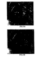
- FIG. 4 shows the algorithm flowchart of the glint detection and elimination algorithm, and the results of the glint detection and elimination algorithm can be viewed in FIGS. 5A and 5B .
- the blur detection algorithm utilizes a normalized image power spectrum method ( Gu J. and Li W., Automatic Image Quality Assessment for Cervical Imagery, SPIE Medical Imaging 2006 ; SPIE Proc. 6146, 2006 ), which can be described as the following steps:
- the method preferably used for contrast enhancement is adaptive histogram equalization.
- Adaptive histogram equalization enhances the contrast of images by transforming the values in the intensity image ( Zuiderveld, K., Contrast limited adaptive histogram equalization, Princeton, NJ: Academic Press, Graphics gems IV, ed. Heckbert, P., 1994 ).
- adaptive histogram equalization operates on small data regions (windows), rather than the entire image.
- Each window's contrast is enhanced, so that the histogram of the output region approximately matches the specified histogram.
- the neighboring windows are then combined using bilinear interpolation in order to eliminate artificially induced boundaries.
- the contrast especially in homogeneous areas, can be limited in order to avoid amplifying the noise which might be present in the image.
- the results of contrast enhancement can be viewed in FIGS. 8A and 8B .
- Super resolution is a technique to use multiple frames of the same object to achieve a higher resolution image
- Kim, S.P., Bose, N.K, and Valensuela, H.M. Recursive reconstruction of high resolution image from noisy undersampled multiframes, IEEE Transaction on Acoustics, Speech, and Signal Processing 38(6): 1031-1027, 1990 ; Irani, M. and Peleg, S., Improving resolution by image registration, CVGIP:GM 53: 231-239, 1991 ).
- Super resolution works when the frames are shifted by fractions of a pixel from each other.
- the super-resolution algorithm is able to produce a larger image that contains the information in the smaller original frames, first an image sub-pixel registration is employed to establish the correspondence between several low resolution images, and then a sub-pixel interpolation algorithm is used to reconstruct the higher resolution image.
- FIGS. 9A, 9B, 9C, 9D and 9E show a super-resolution reconstruction result.
- Video stabilization is the process of generating a compensated video sequence by removing image motion from the camera's undesirable shake or jiggle.
- the preferred video stabilization algorithm consists of a motion estimation (ME) block, a motion smooth (MS) block, and a motion correction (MC) block, as shown in FIG. 10 .
- the ME block estimates the motion between frames and can be divided into a local motion estimator and a global motion decision unit.
- the local motion estimator will return the estimated dense optical flow information between successive frames using typical block-based or gradient-based methods.
- the global motion decision unit will then determine an appropriate global transformation that best characterizes the motion described by the given optical flow information.
- the global motion parameters will be sent to the MS, where the motion parameters are often filtered to remove the unwanted camera motion but retaining intentional camera motion.
- Finally MC warps the current frame using the filtered global transformation information and generates the stabilized video sequence.
- FIGS. 11A and 11B show the result of video stabilization.
- a 3D colon model is a preferred component of a computer-aided diagnosis (CAD) system in colonoscopy, to assist surgeons in visualization, and surgical planning and training.
- CAD computer-aided diagnosis
- the ability to construct a 3D colon model from endoscopic videos (or images) is thus preferred in a CAD system for colonoscopy.
- the mathematical formulations and algorithms have been developed for modeling static, localized 3D anatomic structures within a colon that can be rendered from multiple novel view points for close scrutiny and precise dimensioning ( Mori, K., Deguchi, D., Sugiyama, J., Suenaga, Y. et al., Tracking of a bronchoscope using epipolar geometry analysis and intensity-based image registration of real and virtual endoscopic images, Med. Image Anal. 6(3): 321-336.
- the modeling system of the present invention uses only video images and follows a well-established computer-vision paradigm for image-based modeling. Prominent features are extracted from images and their correspondences are established across multiple images by continuous tracking and discrete matching. These feature correspondences are used to infer the camera's movement.
- the camera motion parameters allow rectifying of images into a standard stereo configuration and inferring of pixel movements (disparity) in these images.
- the inferred disparity is then used to recover 3D surface depth.
- the inferred 3D depth together with texture information recorded in images, allow constructing of a 3D model with both structure and appearance information that can be rendered from multiple novel view points. More precisely, the modeling system comprises the following components:
- FIG. 12 shows the algorithm flowchart of 3D colon modeling and reconstruction.
- FIGS. 13A, 13B, 13C and 13D show the distortion correction result and
- FIGS 14A, 14B, 14C and 14D show a result of 3D polyp reconstruction.
- the model will allow the clinician to mark areas (on the model) during the entry phase of the colonoscopic exam, and to treat these areas during withdrawal.
- the system of this invention can quantitatively calculate the physical size of the polyp, and send to the colonoscopist a statistical criterion for making diagnostic decisions.
- pit patterns Treatment decisions for flat and depressed lesions are based on a detailed examination of the macroscopic morphological appearance, including the luminal surface structure of the crypts of Lieberkühn, otherwise known as "pit patterns".
- Pit patterns analysis can offer a degree of positive predictive value for both the underlying histology and depth of vertical submucosal invasion.
- the preferred system utilizes the Kudo tissue classification method which describes seven types of pit patterns ( Kudo, S., Hirota, S., Nakajima, T., Hosobe, S., Kusaka, H., Kobayashi, T. et al., Colorectal tumours and pit pattern, Journal of Clinical Pathology. 47(10): 880-885. 1994 .
- the pit pattern analysis module starts from high magnification endoscopic images, and morphological operators are first preformed to extract the texture pattern ( Chen, C.H., Pau, L.F., and Wang, P.S.P., Segmentation Tools In Mathematical Morphology, Handbook of Pattern Recognition and Computer Vision, in Handbook of Pattern Recognition and Computer Vision, pp. 443-456. World Scientific Publishing Co., 1989 ).
- the training feature includes: pit size, pit shape and pit density.
- FIG. 16 shows the algorithm flowchart of color analysis.
- FIG. 17 shows the algorithm flowchart of texture analysis.
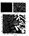
- FIG. 18 shows the result of pit pattern extraction and identification.
- This invention can be used whenever video data from an endoscope is available during an examination of an organ, and especially when it is necessary to diagnose potentially cancerous regions, and motion pictures of the regions are available.
Landscapes
- Engineering & Computer Science (AREA)
- Physics & Mathematics (AREA)
- General Physics & Mathematics (AREA)
- Theoretical Computer Science (AREA)
- Health & Medical Sciences (AREA)
- Computer Vision & Pattern Recognition (AREA)
- Life Sciences & Earth Sciences (AREA)
- General Health & Medical Sciences (AREA)
- Medical Informatics (AREA)
- Nuclear Medicine, Radiotherapy & Molecular Imaging (AREA)
- Radiology & Medical Imaging (AREA)
- Quality & Reliability (AREA)
- Surgery (AREA)
- Heart & Thoracic Surgery (AREA)
- Optics & Photonics (AREA)
- Pathology (AREA)
- Biophysics (AREA)
- Biomedical Technology (AREA)
- Signal Processing (AREA)
- Molecular Biology (AREA)
- Animal Behavior & Ethology (AREA)
- Public Health (AREA)
- Veterinary Medicine (AREA)
- Image Processing (AREA)
- Endoscopes (AREA)
Abstract
Description
- This invention generally relates to medical imaging and more specifically to image processing and computer aided diagnosis for diseases, such as colorectal cancer, using an automated image processing system providing a rapid, inexpensive analysis of video from a standard endoscope, optionally including a 3 dimensional ("3D") reconstructed view of the organ of interest, such as a patient's colon. This invention is to be used in real time employing video data from a conventional endoscope that is already being used during an examination, such as a colonoscopy, to provide an instantaneous second opinion without substantially prolonging the examination.
- Although this invention is being disclosed in connection with colorectal cancer, it is applicable to many other areas of medicine. Colorectal cancer is the second leading cause of cancer-related deaths in the United States. More than 130,000 people are diagnosed with colon cancer each year and about 55,000 people die from the disease annually. Colon cancer can be prevented and cured through early detection, so early diagnosis is of critical importance for patient survival (American Cancer Society, Cancer Facts and Figures, 2004).
- Screening for polyps using an endoscope is the current and most suitable prevention method for early detection and removal of colorectal polyps. If such polyps remain in the colon, they can possibly grow into malignant lesions (Hofstad, B., Vatn, M.H., Andersen, S.N., Huitfeldt, H.S. et al., Growth of colorectal polyps: redetection and evaluation of unresected polyps for a period of three years, Gut 39(3): 449-456. 1996). In the case of flat lesions, in which no protruding polyps are present, the colonic mucosal surface is granular and demarcated into small areas called nonspecific grooves. Changes in the cellular pattern (pit pattern) of the colon lining might be the very earliest sign of adenoma or tumors (Muehldorfer, S.M., Muenzenmayer, C., Mayinger, B., Faller, G. et al., Optical tissue recognition based on color texture by magnifying endoscopy in patients with Barrett's esophagus, Gastrointestinal Endoscopy 57: AB179, 2003). Pit patterns can be used for a qualitative detection of lesions to measure these textural alterations of the colonic mucosal surface. Though the specificity using pit patterns is low, its relatively high sensitivity can highlight suspicious regions, permitting further examination by other sensors. Texture-based pit-pattern analysis can identify tissue types and disease severity. Image-based polyp reconstruction can provide 3 dimensional shape and size of a protruding polyp, using video from an endoscope and computer vision algorithms to synthesize multiple-views that can be converted into 3 dimensional images. Optionally, several image processing algorithms and enhancement techniques can be used to improve image quality.
- Various non-invasive scanning techniques have been proposed to avoid the need for a colonoscopy, but if these scanning techniques disclose the possible existence of a polyp or lesion, a colonoscopy must be performed later anyway. Further, the colonoscopist must locate the actual polyp or lesion (which was shown in the scan) in the patient's colon at a remote time after the scan, which can be very difficult. Also, scans do not provide information about the color or texture of the interior surface of the colon, which would provide diagnostic information about vessel structure and pit patterns, especially for flat lesions. It is highly desirable to avoid false positives for flat lesions because they do not project outward from the colon wall, so that they must be removed by cutting into the colon wall, thus incurring greater risks of bleeding, infection and other adverse side effects. Also, scans may not be able to differentiate between polyps or lesions and residual stool or other material in the colon.
- Colonoscopies must be done efficiently in order to be economical, so the colonoscopist must rapidly scan the comparatively large area of the interior surface of the large intestine. Accordingly, there is a risk that a lesion or polyp may be overlooked.
- If a lesion or polyp is found during a colonoscopy, it is unnecessary to relocate it in a later procedure because the endosclope is already at the lesion or polyp and most endoscopes are equipped with a means by which to introduce cutting instruments. Thus, a polyp or lesion can be cut out during the same colonoscopy in which it was detected, either at the time it was first detected, or at a later time during the same colonoscopy.
- The skill of a colonoscopist in detecting and analyzing polyps and lesions depends on the individual colonoscopist's training and experience. Thus, to standardize detection and analysis in colonoscopies, it is desirable to provide independent expert analysis in real time during a colonoscopy to alert the colonoscopist to a potential polyp or lesion, or to confirm or question a diagnosis of any polyp or lesion that is found. It is also desirable to provide such expert knowledge in an inexpensive and readily available manner, without requiring the purchase of additional expensive hardware.
-
US2003/208107A1 discloses an encapsulated medical imaging device which is swallowed by a patient and passes through their gastrointestinal tract by natural action. The device includes a plurality of microlenses which each have a different focal length. A focused image from a microlens whose focal distance matches the distance of the device from the intestinal wall is selected and displayed, whereas unfocused data is discarded. - According to the present invention there is provided an apparatus as set forth in the appended claims. Other features of the invention will be apparent from the dependent claims, and the description which follows.
-
-
FIG. 1 shows the algorithm flowchart of a computer aided diagnosis system. -
FIG. 2 shows the algorithm flowchart of the video quality analysis module ofFig. 1 . -
FIG. 3 shows the algorithm flowchart of the video quality enhancement module ofFig. 1 . -
FIG. 4 shows the algorithm flowchart of the glint detection and elimination module ofFig. 2 . -
FIGS. 5A and 5B show the results of glint detection and elimination, withFIG. 5A showing the original image frame, andFIG. 5B showing glint removed from the image. -
FIG. 6 shows the algorithm flowchart of blur detection. -
FIGS. 7A and 7B show the results of blur detection, withFIG. 7A showing the original image frame, andFIG. 7B showing the results of blur detection (the black blocks overlapped on the image indicate the location of the blurry area). -
FIGS. 8A and 8B show the results of contrast enhancement, withFIG. 8A showing the original image frame, andFIG. 8B showing the contrast enhanced image. -
FIGS. 9A, 9B, 9C, 9D and 9E show the results of super-resolution reconstruction, withFIGS. 9A, 9B. 9C and 9D showing four frames of low resolution images, andFIG. 9E showing a reconstructed higher resolution image -
FIG. 10 shows the algorithm flowchart of video stabilization. -
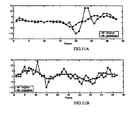
FIGS. 11A and 11B show the results of video stabilization, withFIGS. 11A and 11B showing motion fields of two colonoscopic videos (the curves with circles representing the motion field of original video with shaky movement, and the curves with squares representing the motion field of stabilized video). -
FIG. 12 shows the algorithm flowchart of 3D colon modeling and reconstruction. -
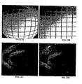
FIGS. 13A, 13B, 13C and 13D show the results of distortion correction, withFIG. 13A showing the original image of a grid target,FIG. 13B showing the corrected grid target,FIG. 13C showing the original image of a colon, andFIG. 13D showing the corrected colon image. -
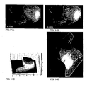
FIGS. 14A, 14B, 14C and 14D show the results of 3D polyp reconstruction, withFIGS. 14A and 14B showing two colonoscopic video frames,FIG. 14C showing a reconstructed 3D polyp model, andFIG. 14D showing a 3D polyp visualization with texture mapping. -
FIG. 15 shows the Kudo pit pattern classification patterns. -
FIG. 16 shows the algorithm flowchart of color analysis. -
FIG. 17 shows the algorithm flowchart of texture analysis. -
FIGS. 18A, 18B and 18C show the results of pit pattern extraction and identification, withFIG. 18A showing the original image,FIG. 18B showing image enhancement results, andFIG. 18C showing statistical information from the automated algorithm, in which numbers indicate the pit size, and the arrowed broken circles represents round pit shape, and "+"represents elongated pit shape. In this example, the algorithm identifies the tissue as Kudo IV pit, which matches the ground-truth. - The present invention is a complex multi-sensor, multi-data and multi-algorithm image processing system. The design provides a modular and open architecture built on phenomenology (feature) based processing. The feature set includes the same features used by the colonoscopists to assess the disease severity (polyp size, pit pattern, etc.). The image-based polyp reconstruction algorithm features several steps: distortion correction, image based modeling, 3D data stitching and reconstruction. The texture-based pit-pattern analysis employs morphological operators to extract the texture pattern, and then utilizes a statistical model and machine learning algorithms to classify the disease severity according to the color and texture information of pits. By analyzing the 3D poly shape and pit-pattern the colonoscopist is provided with diagnostic information for macroscopic inspection. The open architecture also allows for a seamless integration of additional features (Maroulis, D.E., Iakovidis, D.K., Karkanis, S.A., and Karras, D.A., CoLD: A versatile detection system for colorectal lesions in endoscopy video-frames, Compute Methods Programs Biomed. 70(2): 151-166. 2003; Buchsbaum, P.E. and Morris, M.J., Method for making monolithic patterning dichroic filter detector arrays for spectroscopic imaging, Ocean Optics, Inc.,
US Patent# 6,638,668 Barrie, J.D., Aitchison, K.A., Rossano, G.S., and Abraham, M.H., Patterning of multilayer dielectric optical coatings for multispectral CCDs, Thin Solid Films 270: 6-9, 1995) from other microscopic modalities (such as OCT: Optical Coherence Tomography, FTIR: Fourier Transform Infrared and Confocal Microscopy), to provide more accurate diagnostic information. Our system also allows for visualization and virtual navigation for Colonoscopist, and this is done by virtual reality techniques and a magnetic sensor, which provides us absolute spatial location and orientation. - The system described in this invention starts from RGB (Red-Green-Blue color space) videos acquired from a digital endoscope. A series of algorithms is employed to perform the image preprocessing (Pascale, D., A Review of RGB Color Spaces, 5700 Hector Desloges, Montreal (Quebec), Canada, the Babel Color Company. 2003; Wolf, S., Color Correction Matrix for Digital Still and Video Imaging Systems, NTIA Technical Memorandum TM-04-406. 2003), and this is done by two modules, the first being the video quality analysis module, which aims to discard poor quality image frames and delete them from the video; the second being the video quality enhancement module, which aims to improve the image quality and reduce image artifacts. The whole framework is shown in
FIG. 1 . - The video quality analysis and enhancement comprises a glint removal algorithm, a blur detection algorithm, a contrast enhancement algorithm, a super-resolution reconstruction algorithm and a video stabilization algorithm. The framework is shown in
FIG. 2 andFIG. 3 . - We incorporate the same glint removal algorithm that we designed for cervical cancer CAD.
- (Lange H.; Automatic glare removal in reflectance imagery of the uterine cervix; SPIE Medical Imaging 2005; SPIE Proc. 5747, 2005). The method is to extract a glint feature signal from the RGB image that provides a good glint to background ratio, finds the glint regions in the image, and then eliminates the glint regions by restoring the estimated image features for those regions. We have chosen the G (Green) image component as the glint feature signal, because it provides a high glint to background ratio and simplicity of calculation. Glint regions are either detected as saturated regions or small high contrasted regions. Saturated regions are detected using an adaptive thresholding method. Small high contrasted bright regions are detected using morphological top hat filters with different sizes and thresholds. The full extent of the glint regions are approximated using a morphological constraint watershed segmentation algorithm plus a constant dilation. The image features (R,G,B) are first interpolated from the surrounding regions based on Laplace's equation. Then the intensity image feature is restored by adding to the interpolated region intensity function a scaled intensity function that is based on the error function between the interpolated region intensity and the raw intensity data from the region and a signal based on the detected binary glint region. The glint detection and elimination algorithm consists of three consecutive processing steps: (1) Glint feature extraction, (2) Glint region detection, and (3) Glint region elimination and image feature reconstruction.
FIG. 4 shows the algorithm flowchart of the glint detection and elimination algorithm, and the results of the glint detection and elimination algorithm can be viewed inFIGS. 5A and 5B . - The blur detection algorithm utilizes a normalized image power spectrum method (Gu J. and Li W., Automatic Image Quality Assessment for Cervical Imagery, SPIE Medical Imaging 2006; SPIE Proc. 6146, 2006), which can be described as the following steps:
- 1. Divide the image into non-overlapping blocks.
- 2. For each block, compute local representatives based on frequency information.
- 3. Compute global statistics from local representatives obtained from Step 2.
- 4. Determine whether the image is blurred or not from the global statistics. The local representative is calculated by image power spectrum, and then it is normalized by the zero components. Afterward, this 2D image power spectrum is transformed into a 1D diagram. In order to analyze the energy property in each frequency band, polar coordinate integration is used according to each radial value. The power spectrum is separated into three parts and the low frequency area is considered to represent structure information invariant to blur, and the high frequency area is considered to represent detailed information, which is more sensitive to blur. By analyzing the ratio between these two integrations, the degree of blur can be calculated (the noise spectrum has been discarded previously by a threshold). After the blur degree of each small block has been determined, the global measurement a decision can be made as a whole. This can be done by using the percentage of the numbers of blurred blocks occupied in the entire image. Furthermore, different weights are given between those blocks in the center and those blocks in the periphery, since the quality of the image center is of more concern. Thus, if the coverage of blurred blocks is less than a certain threshold, the image is deemed to be satisfyingly clear, otherwise an error message will pop up and feedback to the operator as a blurred image.
FIG. 6 shows the algorithm flowchart and the result of blur detection can be seen inFIGS. 7A and 7B . - The method preferably used for contrast enhancement is adaptive histogram equalization. Adaptive histogram equalization enhances the contrast of images by transforming the values in the intensity image (Zuiderveld, K., Contrast limited adaptive histogram equalization, Princeton, NJ: Academic Press, Graphics gems IV, ed. Heckbert, P., 1994).
- Unlike global histogram equalization, adaptive histogram equalization operates on small data regions (windows), rather than the entire image. Each window's contrast is enhanced, so that the histogram of the output region approximately matches the specified histogram. The neighboring windows are then combined using bilinear interpolation in order to eliminate artificially induced boundaries. The contrast, especially in homogeneous areas, can be limited in order to avoid amplifying the noise which might be present in the image. The results of contrast enhancement can be viewed in
FIGS. 8A and 8B . - Super resolution is a technique to use multiple frames of the same object to achieve a higher resolution image (Kim, S.P., Bose, N.K, and Valensuela, H.M., Recursive reconstruction of high resolution image from noisy undersampled multiframes, IEEE Transaction on Acoustics, Speech, and Signal Processing 38(6): 1031-1027, 1990; Irani, M. and Peleg, S., Improving resolution by image registration, CVGIP:GM 53: 231-239, 1991). Super resolution works when the frames are shifted by fractions of a pixel from each other. The super-resolution algorithm is able to produce a larger image that contains the information in the smaller original frames, first an image sub-pixel registration is employed to establish the correspondence between several low resolution images, and then a sub-pixel interpolation algorithm is used to reconstruct the higher resolution image.
FIGS. 9A, 9B, 9C, 9D and 9E show a super-resolution reconstruction result. - Video stabilization is the process of generating a compensated video sequence by removing image motion from the camera's undesirable shake or jiggle. The preferred video stabilization algorithm consists of a motion estimation (ME) block, a motion smooth (MS) block, and a motion correction (MC) block, as shown in
FIG. 10 . The ME block estimates the motion between frames and can be divided into a local motion estimator and a global motion decision unit. The local motion estimator will return the estimated dense optical flow information between successive frames using typical block-based or gradient-based methods. The global motion decision unit will then determine an appropriate global transformation that best characterizes the motion described by the given optical flow information. The global motion parameters will be sent to the MS, where the motion parameters are often filtered to remove the unwanted camera motion but retaining intentional camera motion. Finally MC warps the current frame using the filtered global transformation information and generates the stabilized video sequence.FIGS. 11A and 11B show the result of video stabilization. - A 3D colon model is a preferred component of a computer-aided diagnosis (CAD) system in colonoscopy, to assist surgeons in visualization, and surgical planning and training. The ability to construct a 3D colon model from endoscopic videos (or images) is thus preferred in a CAD system for colonoscopy. The mathematical formulations and algorithms have been developed for modeling static, localized 3D anatomic structures within a colon that can be rendered from multiple novel view points for close scrutiny and precise dimensioning (Mori, K., Deguchi, D., Sugiyama, J., Suenaga, Y. et al., Tracking of a bronchoscope using epipolar geometry analysis and intensity-based image registration of real and virtual endoscopic images, Med. Image Anal. 6(3): 321-336. 2002; Lyon, R. and Hubel, P., Eyeing the camera: into the next century, 349-355. IS&T/TSID 10th Color Imaging Conference, Scottsdale, AZ, 2002; Zhang, X. and Payandeh, S., Toward Application of Image Tracking in Laparoscopic Surgery, in International Conference on Pattern Recognition, Proc. of International Conference on Pattern Recognition 364-367. ICPR2000, 2000. This ability is useful for the scenario when a surgeon notices some abnormal tissue growth and wants a closer inspection and precise dimensioning.
- The modeling system of the present invention uses only video images and follows a well-established computer-vision paradigm for image-based modeling. Prominent features are extracted from images and their correspondences are established across multiple images by continuous tracking and discrete matching. These feature correspondences are used to infer the camera's movement. The camera motion parameters allow rectifying of images into a standard stereo configuration and inferring of pixel movements (disparity) in these images. The inferred disparity is then used to recover 3D surface depth. The inferred 3D depth, together with texture information recorded in images, allow constructing of a 3D model with both structure and appearance information that can be rendered from multiple novel view points. More precisely, the modeling system comprises the following components:
- 1. Calibration: This is an offline process to estimate intrinsic camera parameters and to correct image distortion (e.g., lens distortion), if any,
- 2. Feature selection: This step identifies unique and invariant colon features for ensuing video analysis,
- 3. Feature matching: This step matches image features across multiple images or video frames to establish correspondences of these features,
- 4. Camera motion Inference: This step uses matched image features to infer camera movement between adjacent images or video frames,
- 5. Image rectification: This step is to rearrange image pixels in such a way that corresponding epipolar lines from two images are aligned and stacked as image scan lines. This step allows standard stereo matching techniques to be applicable regardless of camera movement,
- 6. Stereo matching: This step is to compute disparity (image movement) between two rectified images to allow 3D depth inference,
- 7. Model construction: This step is to infer 3D depth from pixel disparity and construct a 3D model that captures both structure and appearance information, and
- 8. Model rendering: This final step is for rendering the computer model from any novel view points.
-
FIG. 12 shows the algorithm flowchart of 3D colon modeling and reconstruction.FIGS. 13A, 13B, 13C and 13D show the distortion correction result andFIGS 14A, 14B, 14C and 14D show a result of 3D polyp reconstruction. - Creating a three-dimensional polyp model from endoscopic videos allows accomplishment of three goals. First, the model will allow the clinician to mark areas (on the model) during the entry phase of the colonoscopic exam, and to treat these areas during withdrawal. Second, for high-risk patients that require surveillance, it provides a framework for registering the patient's clinical state across exams, thereby enabling change detection. Third, after the 3D reconstruction, the system of this invention can quantitatively calculate the physical size of the polyp, and send to the colonoscopist a statistical criterion for making diagnostic decisions.
- Treatment decisions for flat and depressed lesions are based on a detailed examination of the macroscopic morphological appearance, including the luminal surface structure of the crypts of Lieberkühn, otherwise known as "pit patterns". Pit patterns analysis can offer a degree of positive predictive value for both the underlying histology and depth of vertical submucosal invasion. The preferred system utilizes the Kudo tissue classification method which describes seven types of pit patterns (Kudo, S., Hirota, S., Nakajima, T., Hosobe, S., Kusaka, H., Kobayashi, T. et al., Colorectal tumours and pit pattern, Journal of Clinical Pathology. 47(10): 880-885. 1994. Kudo, S., Rubio, C.A., Teixeira, C.R., Kashida, H., and Kogure, E., Pit pattern in colorectal neoplasia: endoscopic magnifying view, Endoscopy 33(4): 367-373. 2001. Kudo, S., Tamura, S., Nakajima, T., Yamano, H., Kusaka, H., and Watanabe, H., Diagnosis of colorectal tumorous lesions by magnifying endoscopy, Gastrointestinal Endoscopy. 44(1): 8-14. 1996), according to histological, macroscopic morphology and size. These pit patterns have been correlated with histopathology to relate surface patterns to the underlying tissue structure. Lesions can be categorized into basic clinical groups: Kudo crypt group I/II constitutes non-neoplastic, non-invasive patterns. Group IIII/IIIS/IV/VI represents neoplastic but non-invasive lesions. Group VN represents neoplasia with accompanying invasive characteristics. Detailed characteristics are stated as follows, and appearances and photographic examples can be seen in
FIG 15 . - The type I pit pattern of normal mucosa consists of rounded pits with a
- regular size and arrangement.
- The type II pit pattern of benign, hyperplastic polyps are larger than type I, and consist of relatively large star-like or onion-like pits.
- The type IIIL pit pattern is composed of tubular or rounded pits larger than type I, and is associated with polypoid adenomas.
- The type IIIS pit pattern is composed of tubular or rounded pits that are smaller than type I, and is associated with depressed lesions that are frequently high-grade dysplasia.
- The type IV pit pattern is a branched or gyrus-like pattern than is associated with adenomatous lesions.
- The type V pit pattern is divided into VI and VN. Type VI (irregular) has pits that are irregular in shape, size, and arrangement. Type VN (non-structural) shows an absence of pit pattern.
- The pit pattern analysis module starts from high magnification endoscopic images, and morphological operators are first preformed to extract the texture pattern (Chen, C.H., Pau, L.F., and Wang, P.S.P., Segmentation Tools In Mathematical Morphology, Handbook of Pattern Recognition and Computer Vision, in Handbook of Pattern Recognition and Computer Vision, pp. 443-456. World Scientific Publishing Co., 1989). A statistical model and machine learning algorithms are then utilized (Sonka, M., Image Processing Analysis and Machine Vision, in 1998) to classify the disease severity according to the color and texture information (Lin, H.-C., Wang, L.-L., and Yang, S.-N., Extracting periodicity of a regular texture based on autocorrelation functions, Patter Recognition Letters 18(5): 433-443, ELSVIER Science Direct. 1997; Argenti, F., Alparone, L., and Benelli, G., Fast algorithms for texture analysis using co-occurrence matrices, in Radar and Signal Processing, IEE Proceedings F, 137: 443-448. [6], 1990) of pits. A large number of reference images with labeled annotations are used for training purpose, and thus the test images can be clustered against these reference imabased on the likelihood to different classes (Magoulas, G.D., Plagianakos, V.P., and Vrahatis, M.N., Neural network-based colonoscopic diagnosis using on-line learning and differential evolution, Applied Soft Computing 4(4): 369-379, 2004; Liu, Y., Collins, R.T., and Tsin, Y., A computational model for periodic pattern perception based on frieze and wallpaper groups, Liu, Y., Collins, R.T., and Tsin, Y., Pattern Analysis and Machine Intelligence, IEEE Transactions on 26(3): 354-371, 2004; Liu, Y., Lazar, N., Rothfus, W.E., Dellaert, F., Moore, S., Scheider, J., and Kanade, T., Semantic Based Biomedical Image Indexing and Retrival, Trends in Advances in Content-Based Image and Video Retrival, eds. Shapiro, D., Kriegel, and Veltkamp, 2004; Zhang, J., Collins, R., and Liu, Y., Representation and matching of articulated shapes, in Computer Vision and Pattern Recognition, 2: II-342-II-349.IEEE Conference on Computer Vision and Pattern Recognition CVPR 2004, 2004). The training feature includes: pit size, pit shape and pit density.
FIG. 16 shows the algorithm flowchart of color analysis.FIG. 17 shows the algorithm flowchart of texture analysis. AndFIG. 18 shows the result of pit pattern extraction and identification. - Although a few preferred embodiments have been shown and described, it will be appreciated by those skilled in the art that various changes and modifications might be made without departing from the scope of the invention, as defined in the appended claims.
- This invention can be used whenever video data from an endoscope is available during an examination of an organ, and especially when it is necessary to diagnose potentially cancerous regions, and motion pictures of the regions are available.
Claims (5)
- An automated image processing system comprising:a computer coupled to an endoscope to receive video data during an examination by colonoscopy of a large intestine;a video quality analysis module arranged to analyze said video data to determine image frames in which the coverage of blurred blocks is less than a threshold to provide satisfying image frames from said video data, and wherein said video quality analysis module is arranged to perform a glint removal algorithm which performs glint feature extraction, glint region detection and glint region elimination and image feature reconstruction;a video quality enhancement module arranged to enhance contrast in said satisfying image frames;apply super resolution to said satisfying image frames by using multiple frames of the same object to achieve a higher resolution image; andprovide video stabilization to said satisfying image frames; andan analysis module arranged to detect and classify any lesions or polyps in said satisfying image frames to output diagnostic information regarding said lesions or polyps;wherein said video quality analysis module, said video quality enhancement module, and said analysis module all operate to output the diagnostic information in real time during the examination.
- The system according to claim 1, wherein said analysis module is arranged to perform color calibration, color analysis, texture analysis and feature detection.
- The system according to claim 1, wherein said analysis module is arranged to perform analysis of blood vessels in the satisfying image frames.
- The system according to claim 1, wherein said analysis module includes a pit pattern analysis module arranged to perform analysis of pit patterns in the satisfying image frames.
- The system according to claim 1, further comprising a three-dimensional modelling and reconstruction module arranged to:reconstruct a still image of said large intestine in three dimensions from said satisfying image frames to form a reconstructed model, wherein said reconstructing is performed in real time during said colonoscopyperform fisheye distortion correction, geometry calibration, image based modelling and three dimensional data stitching; andwherein said analysis module is further arranged to recover three-dimensional shape and size information of any lesions or polyps in said large intestine from said reconstructed model.
Applications Claiming Priority (2)
| Application Number | Priority Date | Filing Date | Title |
|---|---|---|---|
| US83927506P | 2006-08-21 | 2006-08-21 | |
| PCT/US2007/018600 WO2008024419A1 (en) | 2006-08-21 | 2007-08-21 | Computer aided analysis using video from endoscopes |
Publications (2)
| Publication Number | Publication Date |
|---|---|
| EP2054852A1 EP2054852A1 (en) | 2009-05-06 |
| EP2054852B1 true EP2054852B1 (en) | 2010-06-23 |
Family
ID=38828733
Family Applications (1)
| Application Number | Title | Priority Date | Filing Date |
|---|---|---|---|
| EP07837227A Not-in-force EP2054852B1 (en) | 2006-08-21 | 2007-08-21 | Computer aided analysis using video from endoscopes |
Country Status (6)
| Country | Link |
|---|---|
| US (1) | US20080058593A1 (en) |
| EP (1) | EP2054852B1 (en) |
| JP (1) | JP5113841B2 (en) |
| AT (1) | ATE472141T1 (en) |
| DE (1) | DE602007007340D1 (en) |
| WO (1) | WO2008024419A1 (en) |
Cited By (4)
| Publication number | Priority date | Publication date | Assignee | Title |
|---|---|---|---|---|
| WO2023039493A1 (en) * | 2021-09-13 | 2023-03-16 | Satisfai Health Inc. | System and methods for aggregating features in video frames to improve accuracy of ai detection algorithms |
| US11684241B2 (en) | 2020-11-02 | 2023-06-27 | Satisfai Health Inc. | Autonomous and continuously self-improving learning system |
| US11694114B2 (en) | 2019-07-16 | 2023-07-04 | Satisfai Health Inc. | Real-time deployment of machine learning systems |
| US12167829B2 (en) | 2019-12-19 | 2024-12-17 | Ambu A/S | Image capture selection |
Families Citing this family (163)
| Publication number | Priority date | Publication date | Assignee | Title |
|---|---|---|---|---|
| JP4533327B2 (en) * | 2006-02-23 | 2010-09-01 | キヤノン株式会社 | Image processing apparatus and method, computer program, and storage medium |
| WO2008053649A1 (en) * | 2006-11-02 | 2008-05-08 | Konica Minolta Holdings, Inc. | Wide angle image acquiring method and wide angle streo camera device |
| JP2008301968A (en) * | 2007-06-06 | 2008-12-18 | Olympus Medical Systems Corp | Endoscopic image processing device |
| US20090208143A1 (en) * | 2008-02-19 | 2009-08-20 | University Of Washington | Efficient automated urothelial imaging using an endoscope with tip bending |
| US20110032347A1 (en) * | 2008-04-15 | 2011-02-10 | Gerard Lacey | Endoscopy system with motion sensors |
| EP2293714B1 (en) | 2008-06-02 | 2014-08-13 | Lightlab Imaging, Inc. | Quantitative methods for obtaining tissue characteristics from optical coherence tomography images |
| US8508550B1 (en) * | 2008-06-10 | 2013-08-13 | Pixar | Selective rendering of objects |
| US20100033501A1 (en) * | 2008-08-11 | 2010-02-11 | Sti Medical Systems, Llc | Method of image manipulation to fade between two images |
| WO2010019114A1 (en) * | 2008-08-11 | 2010-02-18 | Sti Medical Systems, Llc | A method of image manipulation to fade between two frames of a dual frame image |
| CN102124471A (en) * | 2008-08-15 | 2011-07-13 | 国际科学技术医疗系统有限责任公司 | Method for enhancing vascular patterns in cervical imaging |
| JP2010068865A (en) * | 2008-09-16 | 2010-04-02 | Fujifilm Corp | Diagnostic imaging apparatus |
| JP5130171B2 (en) * | 2008-09-22 | 2013-01-30 | 株式会社日立製作所 | Image signal processing apparatus and image signal processing method |
| EP2344982A4 (en) * | 2008-10-10 | 2012-09-19 | Sti Medical Systems Llc | METHODS FOR CLASSIFYING TISSUE IN UTERUS COLLAR IMAGING |
| EP2335551B1 (en) * | 2008-10-22 | 2014-05-14 | Fujifilm Corporation | Endoscope apparatus and control method therefor |
| JP2012509715A (en) * | 2008-11-21 | 2012-04-26 | メイヨ・ファウンデーション・フォー・メディカル・エデュケーション・アンド・リサーチ | Colonoscopy tracking and evaluation system |
| US20110273535A1 (en) * | 2009-01-28 | 2011-11-10 | Avi Mendelson | Mole monitoring system |
| WO2010125481A1 (en) | 2009-04-29 | 2010-11-04 | Koninklijke Philips Electronics, N.V. | Real-time depth estimation from monocular endoscope images |
| WO2011005865A2 (en) * | 2009-07-07 | 2011-01-13 | The Johns Hopkins University | A system and method for automated disease assessment in capsule endoscopy |
| TWI425445B (en) * | 2009-09-29 | 2014-02-01 | Nat Univ Tsing Hua | Method and detecting system for determining quantity of self-motion of a moving platform |
| US8417062B2 (en) * | 2009-10-19 | 2013-04-09 | Ut-Battelle, Llc | System and method for stabilization of fisheye video imagery |
| TWI432168B (en) * | 2009-12-31 | 2014-04-01 | Univ Nat Yunlin Sci & Tech | Endoscope navigation method and endoscopy navigation system |
| US9269024B2 (en) * | 2010-02-01 | 2016-02-23 | Qualcomm Incorporated | Image recognition system based on cascaded over-complete dictionaries |
| US20110301447A1 (en) * | 2010-06-07 | 2011-12-08 | Sti Medical Systems, Llc | Versatile video interpretation, visualization, and management system |
| JP2012045130A (en) * | 2010-08-26 | 2012-03-08 | Hoya Corp | Image signal processing system and method for processing image signal |
| EP3540495A1 (en) | 2010-10-28 | 2019-09-18 | EndoChoice Innovation Center Ltd. | Optical systems for multi-sensor endoscopes |
| JP5435746B2 (en) * | 2011-01-24 | 2014-03-05 | 富士フイルム株式会社 | Endoscope device |
| CN102984990B (en) * | 2011-02-01 | 2015-05-20 | 奥林巴斯医疗株式会社 | diagnostic aids |
| KR101805624B1 (en) * | 2011-08-29 | 2017-12-08 | 삼성전자주식회사 | Method and apparatus for generating organ medel image |
| US8878918B2 (en) | 2011-09-16 | 2014-11-04 | The Invention Science Fund I, Llc | Creating a subsurface feature atlas of at least two subsurface features |
| US11871901B2 (en) | 2012-05-20 | 2024-01-16 | Cilag Gmbh International | Method for situational awareness for surgical network or surgical network connected device capable of adjusting function based on a sensed situation or usage |
| FR2996331B1 (en) * | 2012-09-28 | 2015-12-18 | Morpho | METHOD FOR DETECTING THE REALITY OF VENOUS NETWORKS FOR IDENTIFICATION OF INDIVIDUALS |
| KR101874482B1 (en) * | 2012-10-16 | 2018-07-05 | 삼성전자주식회사 | Apparatus and method of reconstructing 3-dimension super-resolution image from depth image |
| JP6275157B2 (en) * | 2012-11-30 | 2018-02-07 | コーニンクレッカ フィリップス エヌ ヴェKoninklijke Philips N.V. | Quantifying tissue surface roughness based on image data and determining the presence of disease based on it |
| US9107578B2 (en) | 2013-03-31 | 2015-08-18 | Gyrus Acmi, Inc. | Panoramic organ imaging |
| TWI503788B (en) * | 2013-10-02 | 2015-10-11 | Jar Ferr Yang | Method, device and system for restoring resized depth frame into original depth frame |
| KR20150049585A (en) | 2013-10-30 | 2015-05-08 | 삼성전자주식회사 | Polyp detection apparatus and operating method for the same |
| KR20160138502A (en) * | 2014-03-28 | 2016-12-05 | 인튜어티브 서지컬 오퍼레이션즈 인코포레이티드 | Alignment of q3d models with 3d images |
| KR102373714B1 (en) | 2014-03-28 | 2022-03-15 | 인튜어티브 서지컬 오퍼레이션즈 인코포레이티드 | Quantitative three-dimensional imaging of surgical scenes from multiport perspectives |
| KR102397670B1 (en) | 2014-03-28 | 2022-05-16 | 인튜어티브 서지컬 오퍼레이션즈 인코포레이티드 | Surgical system with haptic feedback based upon quantitative three-dimensional imaging |
| EP3122281B1 (en) | 2014-03-28 | 2022-07-20 | Intuitive Surgical Operations, Inc. | Quantitative three-dimensional imaging and 3d modeling of surgical implants |
| KR102387096B1 (en) | 2014-03-28 | 2022-04-15 | 인튜어티브 서지컬 오퍼레이션즈 인코포레이티드 | Quantitative three-dimensional visualization of instruments in a field of view |
| WO2015149040A1 (en) | 2014-03-28 | 2015-10-01 | Dorin Panescu | Quantitative three-dimensional imaging of surgical scenes |
| KR20150120774A (en) | 2014-04-18 | 2015-10-28 | 삼성전자주식회사 | System and method for detecting region of interest |
| WO2015164724A1 (en) * | 2014-04-24 | 2015-10-29 | Arizona Board Of Regents On Behalf Of Arizona State University | System and method for quality assessment of optical colonoscopy images |
| JP6482934B2 (en) * | 2014-06-03 | 2019-03-13 | キヤノンメディカルシステムズ株式会社 | Image processing apparatus, radiation detection apparatus, and image processing method |
| CN107624193A (en) * | 2015-04-29 | 2018-01-23 | 西门子公司 | The method and system of semantic segmentation in laparoscope and endoscope 2D/2.5D view data |
| ES2818174T3 (en) | 2015-05-17 | 2021-04-09 | Endochoice Inc | Endoscopic image enhancement using contrast-limited adaptive histogram equalization (CLAHE) implemented in a processor |
| US20180174311A1 (en) * | 2015-06-05 | 2018-06-21 | Siemens Aktiengesellschaft | Method and system for simultaneous scene parsing and model fusion for endoscopic and laparoscopic navigation |
| WO2017002184A1 (en) | 2015-06-29 | 2017-01-05 | オリンパス株式会社 | Image processing device, endoscope system, image processing method, and image processing program |
| GB201513449D0 (en) * | 2015-07-30 | 2015-09-16 | Optos Plc | Image processing |
| US10510144B2 (en) | 2015-09-10 | 2019-12-17 | Magentiq Eye Ltd. | System and method for detection of suspicious tissue regions in an endoscopic procedure |
| JP6580446B2 (en) * | 2015-10-09 | 2019-09-25 | サイバネットシステム株式会社 | Image processing apparatus and image processing method |
| CN105654495A (en) * | 2016-01-07 | 2016-06-08 | 太原工业学院 | Method for detecting internal defect of automobile brake disc |
| US10140554B2 (en) | 2016-01-21 | 2018-11-27 | Wizr Llc | Video processing |
| WO2017169139A1 (en) | 2016-03-29 | 2017-10-05 | ソニー株式会社 | Image processing device, image processing method, and medical system |
| WO2017201494A1 (en) * | 2016-05-19 | 2017-11-23 | Avantis Medical Systems, Inc. | Methods for polyp detection |
| US11055581B2 (en) * | 2016-10-07 | 2021-07-06 | Baylor Research Institute | Classification of polyps using learned image analysis |
| KR101730570B1 (en) * | 2016-11-11 | 2017-04-26 | 주식회사 마이디바이스 | Method for detecting glint |
| WO2018152248A1 (en) | 2017-02-14 | 2018-08-23 | Dignity Health | Systems, methods, and media for selectively presenting images captured by confocal laser endomicroscopy |
| WO2018216618A1 (en) * | 2017-05-25 | 2018-11-29 | 日本電気株式会社 | Information processing device, control method, and program |
| CN107688092B (en) * | 2017-08-18 | 2020-05-19 | 国家纳米科学中心 | Method for prognosis evaluation of cancer patient |
| US11317919B2 (en) | 2017-10-30 | 2022-05-03 | Cilag Gmbh International | Clip applier comprising a clip crimping system |
| US11510741B2 (en) | 2017-10-30 | 2022-11-29 | Cilag Gmbh International | Method for producing a surgical instrument comprising a smart electrical system |
| US11311342B2 (en) | 2017-10-30 | 2022-04-26 | Cilag Gmbh International | Method for communicating with surgical instrument systems |
| US11564756B2 (en) | 2017-10-30 | 2023-01-31 | Cilag Gmbh International | Method of hub communication with surgical instrument systems |
| KR20200106028A (en) * | 2017-10-30 | 2020-09-10 | 고에키자이단호진 간겐큐카이 | Image diagnosis support device, data collection method, image diagnosis support method, and image diagnosis support program |
| US11911045B2 (en) | 2017-10-30 | 2024-02-27 | Cllag GmbH International | Method for operating a powered articulating multi-clip applier |
| US11291510B2 (en) | 2017-10-30 | 2022-04-05 | Cilag Gmbh International | Method of hub communication with surgical instrument systems |
| US11801098B2 (en) | 2017-10-30 | 2023-10-31 | Cilag Gmbh International | Method of hub communication with surgical instrument systems |
| US10959744B2 (en) | 2017-10-30 | 2021-03-30 | Ethicon Llc | Surgical dissectors and manufacturing techniques |
| DE102017221297A1 (en) * | 2017-11-28 | 2019-05-29 | Siemens Healthcare Gmbh | Method and device for the automated evaluation of at least one image data set recorded with a medical image recording device, computer program and electronically readable data carrier |
| US20190201113A1 (en) | 2017-12-28 | 2019-07-04 | Ethicon Llc | Controls for robot-assisted surgical platforms |
| US12096916B2 (en) | 2017-12-28 | 2024-09-24 | Cilag Gmbh International | Method of sensing particulate from smoke evacuated from a patient, adjusting the pump speed based on the sensed information, and communicating the functional parameters of the system to the hub |
| US20190201090A1 (en) | 2017-12-28 | 2019-07-04 | Ethicon Llc | Capacitive coupled return path pad with separable array elements |
| US11818052B2 (en) | 2017-12-28 | 2023-11-14 | Cilag Gmbh International | Surgical network determination of prioritization of communication, interaction, or processing based on system or device needs |
| US11147607B2 (en) | 2017-12-28 | 2021-10-19 | Cilag Gmbh International | Bipolar combination device that automatically adjusts pressure based on energy modality |
| US11589888B2 (en) | 2017-12-28 | 2023-02-28 | Cilag Gmbh International | Method for controlling smart energy devices |
| US10758310B2 (en) | 2017-12-28 | 2020-09-01 | Ethicon Llc | Wireless pairing of a surgical device with another device within a sterile surgical field based on the usage and situational awareness of devices |
| US11832840B2 (en) | 2017-12-28 | 2023-12-05 | Cilag Gmbh International | Surgical instrument having a flexible circuit |
| US11744604B2 (en) | 2017-12-28 | 2023-09-05 | Cilag Gmbh International | Surgical instrument with a hardware-only control circuit |
| US11937769B2 (en) * | 2017-12-28 | 2024-03-26 | Cilag Gmbh International | Method of hub communication, processing, storage and display |
| US11659023B2 (en) | 2017-12-28 | 2023-05-23 | Cilag Gmbh International | Method of hub communication |
| US11998193B2 (en) | 2017-12-28 | 2024-06-04 | Cilag Gmbh International | Method for usage of the shroud as an aspect of sensing or controlling a powered surgical device, and a control algorithm to adjust its default operation |
| US11786251B2 (en) | 2017-12-28 | 2023-10-17 | Cilag Gmbh International | Method for adaptive control schemes for surgical network control and interaction |
| US11969216B2 (en) | 2017-12-28 | 2024-04-30 | Cilag Gmbh International | Surgical network recommendations from real time analysis of procedure variables against a baseline highlighting differences from the optimal solution |
| US10918310B2 (en) | 2018-01-03 | 2021-02-16 | Biosense Webster (Israel) Ltd. | Fast anatomical mapping (FAM) using volume filling |
| US11026751B2 (en) | 2017-12-28 | 2021-06-08 | Cilag Gmbh International | Display of alignment of staple cartridge to prior linear staple line |
| US11389164B2 (en) | 2017-12-28 | 2022-07-19 | Cilag Gmbh International | Method of using reinforced flexible circuits with multiple sensors to optimize performance of radio frequency devices |
| US12062442B2 (en) | 2017-12-28 | 2024-08-13 | Cilag Gmbh International | Method for operating surgical instrument systems |
| US12396806B2 (en) | 2017-12-28 | 2025-08-26 | Cilag Gmbh International | Adjustment of a surgical device function based on situational awareness |
| US11324557B2 (en) | 2017-12-28 | 2022-05-10 | Cilag Gmbh International | Surgical instrument with a sensing array |
| US11576677B2 (en) | 2017-12-28 | 2023-02-14 | Cilag Gmbh International | Method of hub communication, processing, display, and cloud analytics |
| US11666331B2 (en) | 2017-12-28 | 2023-06-06 | Cilag Gmbh International | Systems for detecting proximity of surgical end effector to cancerous tissue |
| US20190206569A1 (en) | 2017-12-28 | 2019-07-04 | Ethicon Llc | Method of cloud based data analytics for use with the hub |
| US11864728B2 (en) | 2017-12-28 | 2024-01-09 | Cilag Gmbh International | Characterization of tissue irregularities through the use of mono-chromatic light refractivity |
| BR112020013021A2 (en) | 2017-12-28 | 2020-11-24 | Ethicon Llc | adjustment of a function of the surgical device based on situational recognition |
| US11559308B2 (en) | 2017-12-28 | 2023-01-24 | Cilag Gmbh International | Method for smart energy device infrastructure |
| US11857152B2 (en) | 2017-12-28 | 2024-01-02 | Cilag Gmbh International | Surgical hub spatial awareness to determine devices in operating theater |
| US11633237B2 (en) | 2017-12-28 | 2023-04-25 | Cilag Gmbh International | Usage and technique analysis of surgeon / staff performance against a baseline to optimize device utilization and performance for both current and future procedures |
| US11257589B2 (en) | 2017-12-28 | 2022-02-22 | Cilag Gmbh International | Real-time analysis of comprehensive cost of all instrumentation used in surgery utilizing data fluidity to track instruments through stocking and in-house processes |
| US20190201039A1 (en) | 2017-12-28 | 2019-07-04 | Ethicon Llc | Situational awareness of electrosurgical systems |
| US11132462B2 (en) | 2017-12-28 | 2021-09-28 | Cilag Gmbh International | Data stripping method to interrogate patient records and create anonymized record |
| US11832899B2 (en) | 2017-12-28 | 2023-12-05 | Cilag Gmbh International | Surgical systems with autonomously adjustable control programs |
| US11304763B2 (en) | 2017-12-28 | 2022-04-19 | Cilag Gmbh International | Image capturing of the areas outside the abdomen to improve placement and control of a surgical device in use |
| US11786245B2 (en) | 2017-12-28 | 2023-10-17 | Cilag Gmbh International | Surgical systems with prioritized data transmission capabilities |
| US11678881B2 (en) | 2017-12-28 | 2023-06-20 | Cilag Gmbh International | Spatial awareness of surgical hubs in operating rooms |
| US11969142B2 (en) | 2017-12-28 | 2024-04-30 | Cilag Gmbh International | Method of compressing tissue within a stapling device and simultaneously displaying the location of the tissue within the jaws |
| US11304699B2 (en) | 2017-12-28 | 2022-04-19 | Cilag Gmbh International | Method for adaptive control schemes for surgical network control and interaction |
| US11311306B2 (en) | 2017-12-28 | 2022-04-26 | Cilag Gmbh International | Surgical systems for detecting end effector tissue distribution irregularities |
| US11419630B2 (en) | 2017-12-28 | 2022-08-23 | Cilag Gmbh International | Surgical system distributed processing |
| US11612408B2 (en) | 2017-12-28 | 2023-03-28 | Cilag Gmbh International | Determining tissue composition via an ultrasonic system |
| US12290231B2 (en) | 2017-12-28 | 2025-05-06 | Cilag Gmbh International | Method of hub communication, processing, storage and display |
| US12376855B2 (en) | 2017-12-28 | 2025-08-05 | Cilag Gmbh International | Safety systems for smart powered surgical stapling |
| US11058498B2 (en) | 2017-12-28 | 2021-07-13 | Cilag Gmbh International | Cooperative surgical actions for robot-assisted surgical platforms |
| US11896322B2 (en) | 2017-12-28 | 2024-02-13 | Cilag Gmbh International | Sensing the patient position and contact utilizing the mono-polar return pad electrode to provide situational awareness to the hub |
| US11076921B2 (en) | 2017-12-28 | 2021-08-03 | Cilag Gmbh International | Adaptive control program updates for surgical hubs |
| US11464559B2 (en) | 2017-12-28 | 2022-10-11 | Cilag Gmbh International | Estimating state of ultrasonic end effector and control system therefor |
| WO2019133144A1 (en) | 2017-12-28 | 2019-07-04 | Ethicon Llc | Detection and escalation of security responses of surgical instruments to increasing severity threats |
| US11612444B2 (en) | 2017-12-28 | 2023-03-28 | Cilag Gmbh International | Adjustment of a surgical device function based on situational awareness |
| US11166772B2 (en) | 2017-12-28 | 2021-11-09 | Cilag Gmbh International | Surgical hub coordination of control and communication of operating room devices |
| US11896443B2 (en) | 2017-12-28 | 2024-02-13 | Cilag Gmbh International | Control of a surgical system through a surgical barrier |
| US20190201112A1 (en) | 2017-12-28 | 2019-07-04 | Ethicon Llc | Computer implemented interactive surgical systems |
| US11696760B2 (en) | 2017-12-28 | 2023-07-11 | Cilag Gmbh International | Safety systems for smart powered surgical stapling |
| US12127729B2 (en) | 2017-12-28 | 2024-10-29 | Cilag Gmbh International | Method for smoke evacuation for surgical hub |
| US11202570B2 (en) | 2017-12-28 | 2021-12-21 | Cilag Gmbh International | Communication hub and storage device for storing parameters and status of a surgical device to be shared with cloud based analytics systems |
| US10892995B2 (en) | 2017-12-28 | 2021-01-12 | Ethicon Llc | Surgical network determination of prioritization of communication, interaction, or processing based on system or device needs |
| US11109866B2 (en) | 2017-12-28 | 2021-09-07 | Cilag Gmbh International | Method for circular stapler control algorithm adjustment based on situational awareness |
| US11903601B2 (en) | 2017-12-28 | 2024-02-20 | Cilag Gmbh International | Surgical instrument comprising a plurality of drive systems |
| US11376002B2 (en) | 2017-12-28 | 2022-07-05 | Cilag Gmbh International | Surgical instrument cartridge sensor assemblies |
| US11259830B2 (en) | 2018-03-08 | 2022-03-01 | Cilag Gmbh International | Methods for controlling temperature in ultrasonic device |
| US11707293B2 (en) | 2018-03-08 | 2023-07-25 | Cilag Gmbh International | Ultrasonic sealing algorithm with temperature control |
| US11701162B2 (en) | 2018-03-08 | 2023-07-18 | Cilag Gmbh International | Smart blade application for reusable and disposable devices |
| US12303159B2 (en) | 2018-03-08 | 2025-05-20 | Cilag Gmbh International | Methods for estimating and controlling state of ultrasonic end effector |
| US11589865B2 (en) | 2018-03-28 | 2023-02-28 | Cilag Gmbh International | Methods for controlling a powered surgical stapler that has separate rotary closure and firing systems |
| US11090047B2 (en) | 2018-03-28 | 2021-08-17 | Cilag Gmbh International | Surgical instrument comprising an adaptive control system |
| US11100633B2 (en) * | 2018-06-13 | 2021-08-24 | Cosmo Artificial Intelligence—Al Limited | Systems and methods for processing real-time video from a medical image device and detecting objects in the video |
| US10810460B2 (en) | 2018-06-13 | 2020-10-20 | Cosmo Artificial Intelligence—AI Limited | Systems and methods for training generative adversarial networks and use of trained generative adversarial networks |
| US10292769B1 (en) * | 2018-08-07 | 2019-05-21 | Sony Corporation | Surgical assistive device and method for providing assistance in surgery of anatomical portions of internal organ affected by intraoperative shift |
| JP7326308B2 (en) * | 2018-09-11 | 2023-08-15 | 富士フイルム株式会社 | MEDICAL IMAGE PROCESSING APPARATUS, OPERATION METHOD OF MEDICAL IMAGE PROCESSING APPARATUS, ENDOSCOPE SYSTEM, PROCESSOR DEVICE, DIAGNOSTIC SUPPORT DEVICE, AND PROGRAM |
| US10896494B1 (en) * | 2018-09-27 | 2021-01-19 | Snap Inc. | Dirty lens image correction |
| JP7593921B2 (en) * | 2018-10-19 | 2024-12-03 | 武田薬品工業株式会社 | Image scoring for intestinal pathology |
| CN109598716B (en) * | 2018-12-05 | 2020-08-07 | 武汉楚精灵医疗科技有限公司 | Computer vision-based real-time monitoring method and system for colonoscope retraction speed |
| US11751872B2 (en) | 2019-02-19 | 2023-09-12 | Cilag Gmbh International | Insertable deactivator element for surgical stapler lockouts |
| US11298129B2 (en) | 2019-02-19 | 2022-04-12 | Cilag Gmbh International | Method for providing an authentication lockout in a surgical stapler with a replaceable cartridge |
| EP3716207B1 (en) * | 2019-03-26 | 2023-11-29 | Active Medical B.V. | Method and apparatus for diagnostic analysis of the function and morphology of microcirculation alterations |
| JP7555181B2 (en) * | 2019-03-27 | 2024-09-24 | Hoya株式会社 | ENDOSCOPE PROCESSOR, INFORMATION PROCESSING APPARATUS, PROGRAM, INFORMATION PROCESSING METHOD, AND METHOD FOR GENERATING LEARNING MODEL |
| WO2020232374A1 (en) * | 2019-05-16 | 2020-11-19 | The Regents Of The University Of Michigan | Automated anatomic and regional location of disease features in colonoscopy videos |
| US10929669B2 (en) * | 2019-06-04 | 2021-02-23 | Magentiq Eye Ltd | Systems and methods for processing colon images and videos |
| US11191423B1 (en) | 2020-07-16 | 2021-12-07 | DOCBOT, Inc. | Endoscopic system and methods having real-time medical imaging |
| US10682108B1 (en) * | 2019-07-16 | 2020-06-16 | The University Of North Carolina At Chapel Hill | Methods, systems, and computer readable media for three-dimensional (3D) reconstruction of colonoscopic surfaces for determining missing regions |
| US11423318B2 (en) * | 2019-07-16 | 2022-08-23 | DOCBOT, Inc. | System and methods for aggregating features in video frames to improve accuracy of AI detection algorithms |
| KR20220058918A (en) * | 2019-08-30 | 2022-05-10 | 아우리스 헬스, 인코포레이티드 | Instrument image reliability system and method |
| KR102313749B1 (en) * | 2019-12-23 | 2021-10-18 | 주식회사 메가젠임플란트 | Apparatus for Automatically Transforming Color of Computerized Tomography Images on Oral Cavity Based on Artificial Intelligence and Driving Method Thereof |
| CN115066196B (en) | 2020-02-06 | 2025-05-13 | 富士胶片株式会社 | Medical image processing device, endoscope system, medical image processing method and recording medium |
| US11122248B1 (en) * | 2020-07-20 | 2021-09-14 | Black Sesame International Holding Limited | Stereo vision with weakly aligned heterogeneous cameras |
| US11963683B2 (en) | 2020-10-02 | 2024-04-23 | Cilag Gmbh International | Method for operating tiered operation modes in a surgical system |
| US11992372B2 (en) * | 2020-10-02 | 2024-05-28 | Cilag Gmbh International | Cooperative surgical displays |
| US12484897B2 (en) | 2020-10-02 | 2025-12-02 | Cilag Gmbh International | Surgical instrument with adaptive configuration control |
| US11849220B2 (en) * | 2021-04-14 | 2023-12-19 | Microsoft Technology Licensing, Llc | Systems and methods for generating depth information from low-resolution images |
| US11831931B2 (en) | 2021-04-14 | 2023-11-28 | Microsoft Technology Licensing, Llc | Systems and methods for generating high-resolution video or animated surface meshes from low-resolution images |
| KR102388535B1 (en) * | 2021-06-15 | 2022-04-22 | (주)제이엘케이 | Method and apparatus for analizing endoscopic image based on artificial intelligence |
| WO2024195100A1 (en) * | 2023-03-23 | 2024-09-26 | 日本電気株式会社 | Endoscopy assistance device, endoscopy assistance method, and recording medium |
| JP7733754B2 (en) * | 2024-01-18 | 2025-09-03 | 株式会社フジクラ | Image processing method, image processing device, image processing program, and endoscope system |
Family Cites Families (18)
| Publication number | Priority date | Publication date | Assignee | Title |
|---|---|---|---|---|
| US6075905A (en) * | 1996-07-17 | 2000-06-13 | Sarnoff Corporation | Method and apparatus for mosaic image construction |
| US6346940B1 (en) * | 1997-02-27 | 2002-02-12 | Kabushiki Kaisha Toshiba | Virtualized endoscope system |
| US5960108A (en) * | 1997-06-12 | 1999-09-28 | Apple Computer, Inc. | Method and system for creating an image-based virtual reality environment utilizing a fisheye lens |
| AU760402B2 (en) * | 1998-12-23 | 2003-05-15 | Medispectra, Inc. | Optical methods and systems for cervical screening |
| US7187810B2 (en) * | 1999-12-15 | 2007-03-06 | Medispectra, Inc. | Methods and systems for correcting image misalignment |
| IL134017A (en) * | 2000-01-13 | 2008-04-13 | Capsule View Inc | Camera for viewing inside intestines |
| JP2002291690A (en) * | 2001-04-02 | 2002-10-08 | Asahi Optical Co Ltd | Electronic endoscope apparatus and electronic endoscope system |
| US7257273B2 (en) * | 2001-04-09 | 2007-08-14 | Mingjing Li | Hierarchical scheme for blur detection in digital image using wavelet transform |
| CN1321616C (en) * | 2001-08-10 | 2007-06-20 | 皇家飞利浦电子股份有限公司 | X-ray examination appts. for reconstruction three-dimensional data set from projection images |
| EP1549992A2 (en) * | 2002-09-16 | 2005-07-06 | Rensselaer Polytechnic Institute | Microscope with extended field of vision |
| US7224827B2 (en) * | 2002-09-27 | 2007-05-29 | The Board Of Trustees Of The Leland Stanford Junior University | Method for matching and registering medical image data |
| JP4409166B2 (en) * | 2002-12-05 | 2010-02-03 | オリンパス株式会社 | Image processing device |
| US20050152588A1 (en) * | 2003-10-28 | 2005-07-14 | University Of Chicago | Method for virtual endoscopic visualization of the colon by shape-scale signatures, centerlining, and computerized detection of masses |
| US20050123179A1 (en) * | 2003-12-05 | 2005-06-09 | Eastman Kodak Company | Method and system for automatic axial rotation correction in vivo images |
| CN1976629A (en) * | 2004-04-26 | 2007-06-06 | D·F·杨克洛维茨 | Medical Imaging System for Accurate Determination of Oriented Tumor Changes |
| US7916173B2 (en) * | 2004-06-22 | 2011-03-29 | Canon Kabushiki Kaisha | Method for detecting and selecting good quality image frames from video |
| US8600125B2 (en) * | 2005-06-22 | 2013-12-03 | The Research Foundation Of State University Of New York | System and method for computer aided polyp detection |
| US8423123B2 (en) * | 2005-09-30 | 2013-04-16 | Given Imaging Ltd. | System and method for in-vivo feature detection |
-
2007
- 2007-08-21 EP EP07837227A patent/EP2054852B1/en not_active Not-in-force
- 2007-08-21 DE DE602007007340T patent/DE602007007340D1/en active Active
- 2007-08-21 WO PCT/US2007/018600 patent/WO2008024419A1/en not_active Ceased
- 2007-08-21 US US11/895,150 patent/US20080058593A1/en not_active Abandoned
- 2007-08-21 AT AT07837227T patent/ATE472141T1/en not_active IP Right Cessation
- 2007-08-21 JP JP2009525622A patent/JP5113841B2/en not_active Expired - Fee Related
Cited By (4)
| Publication number | Priority date | Publication date | Assignee | Title |
|---|---|---|---|---|
| US11694114B2 (en) | 2019-07-16 | 2023-07-04 | Satisfai Health Inc. | Real-time deployment of machine learning systems |
| US12167829B2 (en) | 2019-12-19 | 2024-12-17 | Ambu A/S | Image capture selection |
| US11684241B2 (en) | 2020-11-02 | 2023-06-27 | Satisfai Health Inc. | Autonomous and continuously self-improving learning system |
| WO2023039493A1 (en) * | 2021-09-13 | 2023-03-16 | Satisfai Health Inc. | System and methods for aggregating features in video frames to improve accuracy of ai detection algorithms |
Also Published As
| Publication number | Publication date |
|---|---|
| JP2010512173A (en) | 2010-04-22 |
| JP5113841B2 (en) | 2013-01-09 |
| US20080058593A1 (en) | 2008-03-06 |
| WO2008024419A1 (en) | 2008-02-28 |
| ATE472141T1 (en) | 2010-07-15 |
| EP2054852A1 (en) | 2009-05-06 |
| DE602007007340D1 (en) | 2010-08-05 |
Similar Documents
| Publication | Publication Date | Title |
|---|---|---|
| US20080058593A1 (en) | Computer aided diagnosis using video from endoscopes | |
| US11132794B2 (en) | System and method for detection of suspicious tissue regions in an endoscopic procedure | |
| JP4409166B2 (en) | Image processing device | |
| CN113573654A (en) | AI system for detecting and determining lesion size | |
| US8351667B2 (en) | Methods of contrast enhancement for images having blood vessel structures | |
| Sreeja et al. | An improved feature based image fusion technique for enhancement of liver lesions | |
| Maghsoudi et al. | A computer aided method to detect bleeding, tumor, and disease regions in Wireless Capsule Endoscopy | |
| Ševo et al. | Edge density based automatic detection of inflammation in colonoscopy videos | |
| Nur-A-Alam et al. | An automatic system to detect colorectal polyp using hybrid fused method from colonoscopy images | |
| CN116542952A (en) | Endoscopic coverage rate evaluation method and system based on three-dimensional reconstruction | |
| Ohura et al. | Computer-aided diagnosis method for detecting early esophageal cancer from endoscopic image by using dyadic wavelet transform and fractal dimension | |
| CN118941911B (en) | Spatial redundancy compression system based on spatial channel attention mechanism of neural network | |
| Häfner et al. | Pit pattern classification using extended local binary patterns | |
| CN119313725A (en) | Real-time positioning method, fusion display method and device for endoscopic image area | |
| Pedroso et al. | Fractal bilinear deep neural network models for gastric intestinal metaplasia detection | |
| EP2313848A1 (en) | Methods for enhancing vascular patterns in cervical imagery | |
| Aksenov et al. | An ensemble of convolutional neural networks for the use in video endoscopy | |
| Kadeem et al. | Skin Melanoma diagnosis using machine learning and ABCD rule | |
| Pooja et al. | Denoising technique and analysis of statistical parameters for endoscopic images of gastric cancer | |
| Zhao et al. | Automatic temporal subtraction of chest radiographs and its enhancement for lung cancers | |
| Gu et al. | Computer-aided diagnosis (cad) for colonoscopy | |
| Moharana et al. | Diffusion-Based Data Harmonization Across Multiple Oral Cancer Imaging Devices | |
| Junzhou et al. | Contourlet based feature extraction and classification for Wireless Capsule Endoscopic images | |
| Arasi et al. | A Study on Preprocessing Techniques Used for Gastrointestinal Cancer Detection in Endoscopy Images | |
| Siyue et al. | Real-Time Feature Extraction of Fine Mucosal Structures in Endoscopic Images Using Morphological Watershed Segmentation for Early Gastric Cancer Detection |
Legal Events
| Date | Code | Title | Description |
|---|---|---|---|
| PUAI | Public reference made under article 153(3) epc to a published international application that has entered the european phase |
Free format text: ORIGINAL CODE: 0009012 |
|
| 17P | Request for examination filed |
Effective date: 20090211 |
|
| AK | Designated contracting states |
Kind code of ref document: A1 Designated state(s): AT BE BG CH CY CZ DE DK EE ES FI FR GB GR HU IE IS IT LI LT LU LV MC MT NL PL PT RO SE SI SK TR |
|
| AX | Request for extension of the european patent |
Extension state: AL BA HR MK RS |
|
| 17Q | First examination report despatched |
Effective date: 20090713 |
|
| GRAP | Despatch of communication of intention to grant a patent |
Free format text: ORIGINAL CODE: EPIDOSNIGR1 |
|
| GRAS | Grant fee paid |
Free format text: ORIGINAL CODE: EPIDOSNIGR3 |
|
| GRAA | (expected) grant |
Free format text: ORIGINAL CODE: 0009210 |
|
| AK | Designated contracting states |
Kind code of ref document: B1 Designated state(s): AT BE BG CH CY CZ DE DK EE ES FI FR GB GR HU IE IS IT LI LT LU LV MC MT NL PL PT RO SE SI SK TR |
|
| REG | Reference to a national code |
Ref country code: CH Ref legal event code: EP |
|
| REG | Reference to a national code |
Ref country code: IE Ref legal event code: FG4D |
|
| REF | Corresponds to: |
Ref document number: 602007007340 Country of ref document: DE Date of ref document: 20100805 Kind code of ref document: P |
|
| REG | Reference to a national code |
Ref country code: NL Ref legal event code: VDEP Effective date: 20100623 |
|
| PG25 | Lapsed in a contracting state [announced via postgrant information from national office to epo] |
Ref country code: LT Free format text: LAPSE BECAUSE OF FAILURE TO SUBMIT A TRANSLATION OF THE DESCRIPTION OR TO PAY THE FEE WITHIN THE PRESCRIBED TIME-LIMIT Effective date: 20100623 Ref country code: SE Free format text: LAPSE BECAUSE OF FAILURE TO SUBMIT A TRANSLATION OF THE DESCRIPTION OR TO PAY THE FEE WITHIN THE PRESCRIBED TIME-LIMIT Effective date: 20100623 |
|
| LTIE | Lt: invalidation of european patent or patent extension |
Effective date: 20100623 |
|
| PG25 | Lapsed in a contracting state [announced via postgrant information from national office to epo] |
Ref country code: FI Free format text: LAPSE BECAUSE OF FAILURE TO SUBMIT A TRANSLATION OF THE DESCRIPTION OR TO PAY THE FEE WITHIN THE PRESCRIBED TIME-LIMIT Effective date: 20100623 Ref country code: LV Free format text: LAPSE BECAUSE OF FAILURE TO SUBMIT A TRANSLATION OF THE DESCRIPTION OR TO PAY THE FEE WITHIN THE PRESCRIBED TIME-LIMIT Effective date: 20100623 Ref country code: SI Free format text: LAPSE BECAUSE OF FAILURE TO SUBMIT A TRANSLATION OF THE DESCRIPTION OR TO PAY THE FEE WITHIN THE PRESCRIBED TIME-LIMIT Effective date: 20100623 Ref country code: AT Free format text: LAPSE BECAUSE OF FAILURE TO SUBMIT A TRANSLATION OF THE DESCRIPTION OR TO PAY THE FEE WITHIN THE PRESCRIBED TIME-LIMIT Effective date: 20100623 |
|
| PG25 | Lapsed in a contracting state [announced via postgrant information from national office to epo] |
Ref country code: PL Free format text: LAPSE BECAUSE OF FAILURE TO SUBMIT A TRANSLATION OF THE DESCRIPTION OR TO PAY THE FEE WITHIN THE PRESCRIBED TIME-LIMIT Effective date: 20100623 |
|
| PG25 | Lapsed in a contracting state [announced via postgrant information from national office to epo] |
Ref country code: EE Free format text: LAPSE BECAUSE OF FAILURE TO SUBMIT A TRANSLATION OF THE DESCRIPTION OR TO PAY THE FEE WITHIN THE PRESCRIBED TIME-LIMIT Effective date: 20100623 Ref country code: NL Free format text: LAPSE BECAUSE OF FAILURE TO SUBMIT A TRANSLATION OF THE DESCRIPTION OR TO PAY THE FEE WITHIN THE PRESCRIBED TIME-LIMIT Effective date: 20100623 |
|
| PG25 | Lapsed in a contracting state [announced via postgrant information from national office to epo] |
Ref country code: IS Free format text: LAPSE BECAUSE OF FAILURE TO SUBMIT A TRANSLATION OF THE DESCRIPTION OR TO PAY THE FEE WITHIN THE PRESCRIBED TIME-LIMIT Effective date: 20101023 Ref country code: RO Free format text: LAPSE BECAUSE OF FAILURE TO SUBMIT A TRANSLATION OF THE DESCRIPTION OR TO PAY THE FEE WITHIN THE PRESCRIBED TIME-LIMIT Effective date: 20100623 Ref country code: SK Free format text: LAPSE BECAUSE OF FAILURE TO SUBMIT A TRANSLATION OF THE DESCRIPTION OR TO PAY THE FEE WITHIN THE PRESCRIBED TIME-LIMIT Effective date: 20100623 Ref country code: CY Free format text: LAPSE BECAUSE OF FAILURE TO SUBMIT A TRANSLATION OF THE DESCRIPTION OR TO PAY THE FEE WITHIN THE PRESCRIBED TIME-LIMIT Effective date: 20100623 Ref country code: CZ Free format text: LAPSE BECAUSE OF FAILURE TO SUBMIT A TRANSLATION OF THE DESCRIPTION OR TO PAY THE FEE WITHIN THE PRESCRIBED TIME-LIMIT Effective date: 20100623 Ref country code: PT Free format text: LAPSE BECAUSE OF FAILURE TO SUBMIT A TRANSLATION OF THE DESCRIPTION OR TO PAY THE FEE WITHIN THE PRESCRIBED TIME-LIMIT Effective date: 20101025 Ref country code: BE Free format text: LAPSE BECAUSE OF FAILURE TO SUBMIT A TRANSLATION OF THE DESCRIPTION OR TO PAY THE FEE WITHIN THE PRESCRIBED TIME-LIMIT Effective date: 20100623 |
|
| PG25 | Lapsed in a contracting state [announced via postgrant information from national office to epo] |
Ref country code: IT Free format text: LAPSE BECAUSE OF FAILURE TO SUBMIT A TRANSLATION OF THE DESCRIPTION OR TO PAY THE FEE WITHIN THE PRESCRIBED TIME-LIMIT Effective date: 20100623 Ref country code: MC Free format text: LAPSE BECAUSE OF NON-PAYMENT OF DUE FEES Effective date: 20100831 |
|
| PG25 | Lapsed in a contracting state [announced via postgrant information from national office to epo] |
Ref country code: DK Free format text: LAPSE BECAUSE OF FAILURE TO SUBMIT A TRANSLATION OF THE DESCRIPTION OR TO PAY THE FEE WITHIN THE PRESCRIBED TIME-LIMIT Effective date: 20100623 |
|
| PLBE | No opposition filed within time limit |
Free format text: ORIGINAL CODE: 0009261 |
|
| STAA | Information on the status of an ep patent application or granted ep patent |
Free format text: STATUS: NO OPPOSITION FILED WITHIN TIME LIMIT |
|
| PG25 | Lapsed in a contracting state [announced via postgrant information from national office to epo] |
Ref country code: GR Free format text: LAPSE BECAUSE OF FAILURE TO SUBMIT A TRANSLATION OF THE DESCRIPTION OR TO PAY THE FEE WITHIN THE PRESCRIBED TIME-LIMIT Effective date: 20100924 |
|
| 26N | No opposition filed |
Effective date: 20110324 |
|
| REG | Reference to a national code |
Ref country code: DE Ref legal event code: R097 Ref document number: 602007007340 Country of ref document: DE Effective date: 20110323 |
|
| PG25 | Lapsed in a contracting state [announced via postgrant information from national office to epo] |
Ref country code: IE Free format text: LAPSE BECAUSE OF NON-PAYMENT OF DUE FEES Effective date: 20100821 |
|
| PGFP | Annual fee paid to national office [announced via postgrant information from national office to epo] |
Ref country code: CH Payment date: 20110824 Year of fee payment: 5 |
|
| PG25 | Lapsed in a contracting state [announced via postgrant information from national office to epo] |
Ref country code: MT Free format text: LAPSE BECAUSE OF FAILURE TO SUBMIT A TRANSLATION OF THE DESCRIPTION OR TO PAY THE FEE WITHIN THE PRESCRIBED TIME-LIMIT Effective date: 20100623 |
|
| PG25 | Lapsed in a contracting state [announced via postgrant information from national office to epo] |
Ref country code: LU Free format text: LAPSE BECAUSE OF NON-PAYMENT OF DUE FEES Effective date: 20100821 Ref country code: HU Free format text: LAPSE BECAUSE OF FAILURE TO SUBMIT A TRANSLATION OF THE DESCRIPTION OR TO PAY THE FEE WITHIN THE PRESCRIBED TIME-LIMIT Effective date: 20101224 Ref country code: BG Free format text: LAPSE BECAUSE OF FAILURE TO SUBMIT A TRANSLATION OF THE DESCRIPTION OR TO PAY THE FEE WITHIN THE PRESCRIBED TIME-LIMIT Effective date: 20100623 |
|
| PG25 | Lapsed in a contracting state [announced via postgrant information from national office to epo] |
Ref country code: TR Free format text: LAPSE BECAUSE OF FAILURE TO SUBMIT A TRANSLATION OF THE DESCRIPTION OR TO PAY THE FEE WITHIN THE PRESCRIBED TIME-LIMIT Effective date: 20100623 |
|
| PGFP | Annual fee paid to national office [announced via postgrant information from national office to epo] |
Ref country code: GB Payment date: 20120718 Year of fee payment: 6 |
|
| PGFP | Annual fee paid to national office [announced via postgrant information from national office to epo] |
Ref country code: DE Payment date: 20120822 Year of fee payment: 6 Ref country code: FR Payment date: 20120906 Year of fee payment: 6 |
|
| PG25 | Lapsed in a contracting state [announced via postgrant information from national office to epo] |
Ref country code: BG Free format text: LAPSE BECAUSE OF FAILURE TO SUBMIT A TRANSLATION OF THE DESCRIPTION OR TO PAY THE FEE WITHIN THE PRESCRIBED TIME-LIMIT Effective date: 20100923 |
|
| PG25 | Lapsed in a contracting state [announced via postgrant information from national office to epo] |
Ref country code: ES Free format text: LAPSE BECAUSE OF FAILURE TO SUBMIT A TRANSLATION OF THE DESCRIPTION OR TO PAY THE FEE WITHIN THE PRESCRIBED TIME-LIMIT Effective date: 20101004 |
|
| REG | Reference to a national code |
Ref country code: CH Ref legal event code: PL |
|
| GBPC | Gb: european patent ceased through non-payment of renewal fee |
Effective date: 20130821 |
|
| PG25 | Lapsed in a contracting state [announced via postgrant information from national office to epo] |
Ref country code: CH Free format text: LAPSE BECAUSE OF NON-PAYMENT OF DUE FEES Effective date: 20130831 Ref country code: DE Free format text: LAPSE BECAUSE OF NON-PAYMENT OF DUE FEES Effective date: 20140301 Ref country code: LI Free format text: LAPSE BECAUSE OF NON-PAYMENT OF DUE FEES Effective date: 20130831 |
|
| REG | Reference to a national code |
Ref country code: DE Ref legal event code: R119 Ref document number: 602007007340 Country of ref document: DE Effective date: 20140301 |
|
| REG | Reference to a national code |
Ref country code: FR Ref legal event code: ST Effective date: 20140430 |
|
| PG25 | Lapsed in a contracting state [announced via postgrant information from national office to epo] |
Ref country code: GB Free format text: LAPSE BECAUSE OF NON-PAYMENT OF DUE FEES Effective date: 20130821 |
|
| PG25 | Lapsed in a contracting state [announced via postgrant information from national office to epo] |
Ref country code: FR Free format text: LAPSE BECAUSE OF NON-PAYMENT OF DUE FEES Effective date: 20130902 |