EP2040740B1 - Compositions and methods for the delivery of nitric oxide - Google Patents
Compositions and methods for the delivery of nitric oxide Download PDFInfo
- Publication number
- EP2040740B1 EP2040740B1 EP07795153.1A EP07795153A EP2040740B1 EP 2040740 B1 EP2040740 B1 EP 2040740B1 EP 07795153 A EP07795153 A EP 07795153A EP 2040740 B1 EP2040740 B1 EP 2040740B1
- Authority
- EP
- European Patent Office
- Prior art keywords
- nox
- protein
- proteins
- nox protein
- wild
- Prior art date
- Legal status (The legal status is an assumption and is not a legal conclusion. Google has not performed a legal analysis and makes no representation as to the accuracy of the status listed.)
- Active
Links
Images


Classifications
-
- A—HUMAN NECESSITIES
- A61—MEDICAL OR VETERINARY SCIENCE; HYGIENE
- A61K—PREPARATIONS FOR MEDICAL, DENTAL OR TOILETRY PURPOSES
- A61K38/00—Medicinal preparations containing peptides
- A61K38/02—Peptides of undefined number of amino acids; Derivatives thereof
-
- C—CHEMISTRY; METALLURGY
- C07—ORGANIC CHEMISTRY
- C07K—PEPTIDES
- C07K14/00—Peptides having more than 20 amino acids; Gastrins; Somatostatins; Melanotropins; Derivatives thereof
- C07K14/435—Peptides having more than 20 amino acids; Gastrins; Somatostatins; Melanotropins; Derivatives thereof from animals; from humans
- C07K14/46—Peptides having more than 20 amino acids; Gastrins; Somatostatins; Melanotropins; Derivatives thereof from animals; from humans from vertebrates
- C07K14/47—Peptides having more than 20 amino acids; Gastrins; Somatostatins; Melanotropins; Derivatives thereof from animals; from humans from vertebrates from mammals
-
- A—HUMAN NECESSITIES
- A61—MEDICAL OR VETERINARY SCIENCE; HYGIENE
- A61P—SPECIFIC THERAPEUTIC ACTIVITY OF CHEMICAL COMPOUNDS OR MEDICINAL PREPARATIONS
- A61P13/00—Drugs for disorders of the urinary system
- A61P13/12—Drugs for disorders of the urinary system of the kidneys
-
- A—HUMAN NECESSITIES
- A61—MEDICAL OR VETERINARY SCIENCE; HYGIENE
- A61P—SPECIFIC THERAPEUTIC ACTIVITY OF CHEMICAL COMPOUNDS OR MEDICINAL PREPARATIONS
- A61P17/00—Drugs for dermatological disorders
- A61P17/02—Drugs for dermatological disorders for treating wounds, ulcers, burns, scars, keloids, or the like
-
- A—HUMAN NECESSITIES
- A61—MEDICAL OR VETERINARY SCIENCE; HYGIENE
- A61P—SPECIFIC THERAPEUTIC ACTIVITY OF CHEMICAL COMPOUNDS OR MEDICINAL PREPARATIONS
- A61P25/00—Drugs for disorders of the nervous system
-
- A—HUMAN NECESSITIES
- A61—MEDICAL OR VETERINARY SCIENCE; HYGIENE
- A61P—SPECIFIC THERAPEUTIC ACTIVITY OF CHEMICAL COMPOUNDS OR MEDICINAL PREPARATIONS
- A61P43/00—Drugs for specific purposes, not provided for in groups A61P1/00-A61P41/00
-
- A—HUMAN NECESSITIES
- A61—MEDICAL OR VETERINARY SCIENCE; HYGIENE
- A61P—SPECIFIC THERAPEUTIC ACTIVITY OF CHEMICAL COMPOUNDS OR MEDICINAL PREPARATIONS
- A61P7/00—Drugs for disorders of the blood or the extracellular fluid
-
- A—HUMAN NECESSITIES
- A61—MEDICAL OR VETERINARY SCIENCE; HYGIENE
- A61P—SPECIFIC THERAPEUTIC ACTIVITY OF CHEMICAL COMPOUNDS OR MEDICINAL PREPARATIONS
- A61P7/00—Drugs for disorders of the blood or the extracellular fluid
- A61P7/04—Antihaemorrhagics; Procoagulants; Haemostatic agents; Antifibrinolytic agents
-
- A—HUMAN NECESSITIES
- A61—MEDICAL OR VETERINARY SCIENCE; HYGIENE
- A61P—SPECIFIC THERAPEUTIC ACTIVITY OF CHEMICAL COMPOUNDS OR MEDICINAL PREPARATIONS
- A61P7/00—Drugs for disorders of the blood or the extracellular fluid
- A61P7/06—Antianaemics
-
- A—HUMAN NECESSITIES
- A61—MEDICAL OR VETERINARY SCIENCE; HYGIENE
- A61P—SPECIFIC THERAPEUTIC ACTIVITY OF CHEMICAL COMPOUNDS OR MEDICINAL PREPARATIONS
- A61P7/00—Drugs for disorders of the blood or the extracellular fluid
- A61P7/08—Plasma substitutes; Perfusion solutions; Dialytics or haemodialytics; Drugs for electrolytic or acid-base disorders, e.g. hypovolemic shock
-
- A—HUMAN NECESSITIES
- A61—MEDICAL OR VETERINARY SCIENCE; HYGIENE
- A61P—SPECIFIC THERAPEUTIC ACTIVITY OF CHEMICAL COMPOUNDS OR MEDICINAL PREPARATIONS
- A61P9/00—Drugs for disorders of the cardiovascular system
-
- A—HUMAN NECESSITIES
- A61—MEDICAL OR VETERINARY SCIENCE; HYGIENE
- A61P—SPECIFIC THERAPEUTIC ACTIVITY OF CHEMICAL COMPOUNDS OR MEDICINAL PREPARATIONS
- A61P9/00—Drugs for disorders of the cardiovascular system
- A61P9/04—Inotropic agents, i.e. stimulants of cardiac contraction; Drugs for heart failure
-
- A—HUMAN NECESSITIES
- A61—MEDICAL OR VETERINARY SCIENCE; HYGIENE
- A61P—SPECIFIC THERAPEUTIC ACTIVITY OF CHEMICAL COMPOUNDS OR MEDICINAL PREPARATIONS
- A61P9/00—Drugs for disorders of the cardiovascular system
- A61P9/10—Drugs for disorders of the cardiovascular system for treating ischaemic or atherosclerotic diseases, e.g. antianginal drugs, coronary vasodilators, drugs for myocardial infarction, retinopathy, cerebrovascula insufficiency, renal arteriosclerosis
-
- A—HUMAN NECESSITIES
- A61—MEDICAL OR VETERINARY SCIENCE; HYGIENE
- A61P—SPECIFIC THERAPEUTIC ACTIVITY OF CHEMICAL COMPOUNDS OR MEDICINAL PREPARATIONS
- A61P9/00—Drugs for disorders of the cardiovascular system
- A61P9/12—Antihypertensives
-
- C—CHEMISTRY; METALLURGY
- C07—ORGANIC CHEMISTRY
- C07K—PEPTIDES
- C07K14/00—Peptides having more than 20 amino acids; Gastrins; Somatostatins; Melanotropins; Derivatives thereof
- C07K14/195—Peptides having more than 20 amino acids; Gastrins; Somatostatins; Melanotropins; Derivatives thereof from bacteria
- C07K14/33—Peptides having more than 20 amino acids; Gastrins; Somatostatins; Melanotropins; Derivatives thereof from bacteria from Clostridium (G)
-
- C—CHEMISTRY; METALLURGY
- C07—ORGANIC CHEMISTRY
- C07K—PEPTIDES
- C07K14/00—Peptides having more than 20 amino acids; Gastrins; Somatostatins; Melanotropins; Derivatives thereof
- C07K14/435—Peptides having more than 20 amino acids; Gastrins; Somatostatins; Melanotropins; Derivatives thereof from animals; from humans
- C07K14/43504—Peptides having more than 20 amino acids; Gastrins; Somatostatins; Melanotropins; Derivatives thereof from animals; from humans from invertebrates
- C07K14/43536—Peptides having more than 20 amino acids; Gastrins; Somatostatins; Melanotropins; Derivatives thereof from animals; from humans from invertebrates from worms
- C07K14/4354—Peptides having more than 20 amino acids; Gastrins; Somatostatins; Melanotropins; Derivatives thereof from animals; from humans from invertebrates from worms from nematodes
- C07K14/43545—Peptides having more than 20 amino acids; Gastrins; Somatostatins; Melanotropins; Derivatives thereof from animals; from humans from invertebrates from worms from nematodes from Caenorhabditis
-
- C—CHEMISTRY; METALLURGY
- C07—ORGANIC CHEMISTRY
- C07K—PEPTIDES
- C07K14/00—Peptides having more than 20 amino acids; Gastrins; Somatostatins; Melanotropins; Derivatives thereof
- C07K14/435—Peptides having more than 20 amino acids; Gastrins; Somatostatins; Melanotropins; Derivatives thereof from animals; from humans
- C07K14/43504—Peptides having more than 20 amino acids; Gastrins; Somatostatins; Melanotropins; Derivatives thereof from animals; from humans from invertebrates
- C07K14/43563—Peptides having more than 20 amino acids; Gastrins; Somatostatins; Melanotropins; Derivatives thereof from animals; from humans from invertebrates from insects
- C07K14/43577—Peptides having more than 20 amino acids; Gastrins; Somatostatins; Melanotropins; Derivatives thereof from animals; from humans from invertebrates from insects from flies
- C07K14/43581—Peptides having more than 20 amino acids; Gastrins; Somatostatins; Melanotropins; Derivatives thereof from animals; from humans from invertebrates from insects from flies from Drosophila
-
- A—HUMAN NECESSITIES
- A61—MEDICAL OR VETERINARY SCIENCE; HYGIENE
- A61K—PREPARATIONS FOR MEDICAL, DENTAL OR TOILETRY PURPOSES
- A61K38/00—Medicinal preparations containing peptides
-
- Y—GENERAL TAGGING OF NEW TECHNOLOGICAL DEVELOPMENTS; GENERAL TAGGING OF CROSS-SECTIONAL TECHNOLOGIES SPANNING OVER SEVERAL SECTIONS OF THE IPC; TECHNICAL SUBJECTS COVERED BY FORMER USPC CROSS-REFERENCE ART COLLECTIONS [XRACs] AND DIGESTS
- Y02—TECHNOLOGIES OR APPLICATIONS FOR MITIGATION OR ADAPTATION AGAINST CLIMATE CHANGE
- Y02A—TECHNOLOGIES FOR ADAPTATION TO CLIMATE CHANGE
- Y02A50/00—TECHNOLOGIES FOR ADAPTATION TO CLIMATE CHANGE in human health protection, e.g. against extreme weather
- Y02A50/30—Against vector-borne diseases, e.g. mosquito-borne, fly-borne, tick-borne or waterborne diseases whose impact is exacerbated by climate change
-
- Y—GENERAL TAGGING OF NEW TECHNOLOGICAL DEVELOPMENTS; GENERAL TAGGING OF CROSS-SECTIONAL TECHNOLOGIES SPANNING OVER SEVERAL SECTIONS OF THE IPC; TECHNICAL SUBJECTS COVERED BY FORMER USPC CROSS-REFERENCE ART COLLECTIONS [XRACs] AND DIGESTS
- Y10—TECHNICAL SUBJECTS COVERED BY FORMER USPC
- Y10T—TECHNICAL SUBJECTS COVERED BY FORMER US CLASSIFICATION
- Y10T436/00—Chemistry: analytical and immunological testing
- Y10T436/10—Composition for standardization, calibration, simulation, stabilization, preparation or preservation; processes of use in preparation for chemical testing
- Y10T436/102499—Blood gas standard or control
Definitions
- H-NOX proteins and methods of using them to deliver nitric oxide (NO).
- H-NOX proteins provide a new therapeutic tool for delivering NO to humans and, for veterinary purposes, to animals.
- NO acts as a chemical messenger in the control of many important processes in vivo, including vasodilation, neurotransmission, inflammation, platelet aggregation, and regulation of gastrointestinal and vascular smooth muscle tone.
- GTN nitroglycerin
- NO which is synthesized in endothelial cells, diffuses to smooth muscle cells and activates soluble guanylate cyclase (sGC) to produce cyclic GMP, and thereby induce vasodilation.
- sGC soluble guanylate cyclase
- the clinical mechanism of action of organic nitrates is presumed to require their biotransformation to NO and subsequent activation of sGC.
- organic nitrates cease to be effective in patients after 24-48 hours, due to a phenomenon called tolerance.
- compounds such as ⁇ -blocker and ACE inhibitors are used, although they too have limitations and side effects.
- nitrovasodilators are most useful in treating acute situations where rapid vasodilation is required to alleviate symptoms such as angina and myocardial infarction.
- Prolonged administration of organic nitrates results in reduced efficacy, and the vasculature becomes non-responsive; this tolerance prevents their further use both in chronic and acute cases.
- non-continuous nitrovasodilator use is employed with limited effect.
- other avenues of treatment are employed, typically using a mixed regimen of organic nitrates and NO-independent blood pressure medications, with mixed success.
- a competing theory posits that the response to NO from organic nitrates becomes dampened in the target tissue, perhaps because the generation of NO and the by-products of the reaction eventually inhibit the response to NO, or because acute activation of the NO pathway has a feedback mechanism that desensitizes it to further stimulation.
- This theory is known as end-organ tolerance.
- a unifying theory has been proposed that includes aspects of the biotransformation of organic nitrates as well as end-organ desensitization to NO. Essentially, biotransformation of organic nitrates appears to result in higher levels of superoxide (O 2 - ) in tissues. Superoxide reacts at the rate of diffusion with NO to produce peroxynitrite (OONO).
- This reaction essentially traps and destroys basal NO, preventing it from activating sGC.
- Reduced NO levels leads to vasoconstriction, and OONO' is a powerful oxidant that damages tissues.
- Prolonged treatment with organic nitrates such as GTN can result in hypertension and tissue damage in patients, and this can be moderated with co-administration of antioxidants such as ascorbate.
- antioxidants such as ascorbate.
- hemoglobin-based carriers Some research has been conducted on the use of hemoglobin-based carriers to deliver NO.
- hemoglobin-based carriers are limited due to their reactivity with NO in the presence of O 2 , which leads to the inactivation of hemoglobin-based carriers.
- NO reacts directly with O 2 that is bound to hemoglobin to form methemoglobin and nitrate. Both the heme iron and NO become oxidized by the bound oxygen atoms, and the reaction occurs so rapidly that no replacement of O 2 by NO is observed (see, e.g. , U.S. Pat. No. 6,455,676 ).
- NO is also needed to mediate certain inflammatory responses.
- NO produced by the endothelium inhibits platelet aggregation. Consequently, as NO is bound by cell-free hemoglobin (with or without O 2 bound), platelet aggregation may increase. As platelets aggregate, they release potent vasoconstrictor compounds such as thromboxane A 2 and serotonin. These compounds may act synergistically with the reduced NO levels caused by hemoglobin scavenging to produce significant vasoconstriction. In addition to inhibiting platelet aggregation, NO also inhibits neutrophil attachment to cell walls, which in turn can lead to cell wall damage. Endothelial cell wall damage has been observed with the infusion of certain hemoglobin solutions.
- Hemoglobin-based NO carriers are also hindered by the rapid clearance of cell-free hemoglobin from plasma due the presence of receptors for hemoglobin that remove cell-free hemoglobin from plasma.
- Cell-free hemoglobin may also cause kidney toxicity, possibly due to NO depletion in glomeruli, causing constriction and subsequent dysfunction.
- NO carriers that produce less tolerance are needed.
- NO carriers with a low rate of inactivation by NO in the presence of O 2 are desired, such as NO carriers that have a low NO reactivity and/or a low affinity for O 2 .
- NO carriers with NO dissociation constants or NO dissociation rates that are appropriate for particular clinical or industrial applications are also needed.
- H-NOX family of heme sensor proteins was disclosed in Boon and Marietta (2005) Current Opinion in Chemical Biology, 9, p441-446 .
- the present invention is based in part on the surprising discovery that wild-type and mutant H-NOX proteins have a much lower NO reactivity than hemoglobin and thus are desirable NO carriers. If desired, mutations can be introduced into H-NOX proteins to alter their binding of NO and O 2 ligands to further optimize the use of H-NOX proteins as NO carriers. In some embodiments, use of an H-NOX protein as an NO carrier produces less tolerance than the use of current vasodilators, such as organic nitrates.
- the invention features pharmaceutical compositions for use in medicine as an NO carrier. Accordingly, described herein is an isolated H-NOX protein comprising at least one mutation that alters the NO dissociation constant or NO reactivity compared to that of a corresponding wild-type H-NOX protein.
- the NO dissociation constant of the mutant H-NOX protein is within 2 orders of magnitude of that of hemoglobin, and the NO reactivity of the mutant H-NOX protein is at least 10-fold lower than that of hemoglobin.
- the NO reactivity of the mutant H-NOX protein is at least 100-fold lower than that of hemoglobin, such as at least 1,000-fold lower than that of hemoglobin.
- the k off , k 1 , or k 2 for NO of the mutant H-NOX protein is between about 1 x 10 -4 s -1 to about 10 s -1 at 37 °C, such as about 1 x 10 -4 s -1 to about 0.012 s -1 or about 1 x 10 -4 s -1 to about 1 x 10 -3 s -1 at 37 °C.
- the O 2 dissociation constant of the mutant H-NOX protein is at least about 1 ⁇ M at 37 °C, such as at least about 10 ⁇ M or at least about 50 ⁇ M at 37 °C.
- an isolated H-NOX protein comprising at least one mutation that alters the k off , k 1 , or k 2 for NO or alters the O 2 dissociation constant compared to that of a corresponding wild-type H-NOX protein.
- the k off , k 1 , or k 2 for NO of the mutant H-NOX protein is between about 1 x 10 -4 s -1 to about 10 s -1 at 37 °C, and the O 2 dissociation constant of the mutant H-NOX protein is at least about 1 ⁇ M at 37 °C.
- the k off , k 1 , or k 2 for NO of the mutant H-NOX protein is between about 1 x 10 -4 s -1 to about 0.012 s -1 or about 1 x 10 -4 s -1 to about 1 x 10 -3 s -1 at 37 °C.
- the O 2 dissociation constant of the mutant H-NOX protein is at least about 10 ⁇ M, such as at least about 50 ⁇ M at 37 °C.
- the NO reactivity of the mutant H-NOX protein is at least 10-fold lower than that of hemoglobin, such as at least 100-fold lower than that of hemoglobin or at least 1,000-fold lower than that of hemoglobin.
- H-NOX protein selected from the group consisting of T. tengcongensis H-NOX 15A, T. tengcongensis H-NOX 15L, T. tengcongensis H-NOX I5L-P115A, T. tengcongensis H-NOX W9F, T. tengcongensis H-NOX W9F-Y140L, T. tengcongensis H-NOX W9F-Y140H T . tengcongensis H-NOX W9F-N74A, T. tengcongensis H-NOX W9Y, T. tengcongensis H-NOX W9N, T.
- the ⁇ 1 or ⁇ 2 protein is derived from a R. norvegicus or H. sapiens ⁇ 1 or ⁇ 2 protein.
- an isolated H-NOX protein selected from the group consisting of T. tengcongensis H-NOX I5A, T. tengcongensis H-NOX I5L, T. tengcongensis H-NOX I5L-P115A, T. tengcongensis H-NOX W9F-Y140L, T. tengcongensis H-NOX W9F-Y140H, T. tengcongensis H-NOX W9F-N74A, T. tengcongensis H-NOX W9Y, T. tengcongensis H-NOX W9N, T. tengcongensis H-NOX W9H, T.
- the ⁇ 1 or ⁇ 2 protein is derived from a R. norvegicus or H. sapiens ⁇ 1 or ⁇ 2 protein.
- the NO dissociation constant of the H-NOX protein is between 0.1 to 10-fold of that of hemoglobin, such as between 0.5 to 2-fold of that of hemoglobin.
- the NO dissociation constant of the H-NOX protein is within 2 orders of magnitude of that of Homo sapiens hemoglobin alpha, such as an NO dissociation constant between 0.1 to 10-fold or between 0.5 to 2-fold of that of Homo sapiens hemoglobin alpha.
- the NO reactivity of the H-NOX protein is at least 10-fold lower than that of Homo sapiens hemoglobin alpha, such as at least 100-fold or 1,000-fold lower than that of Homo sapiens hemoglobin alpha.
- the isolated H-NOX proteins the NO reactivity of the H-NOX protein is less than about 700 s -1 at 20 °C, such as less than about 600 s -1 , 500 s -1 , 400 s -1 , 300 s -1 , 200 s -1 , 100 s -1 , 75 s -1 , 50 s -1 , 25 s -1 , 20 s -1 , 10 s -1 , 50 s -1 , 3 s -1 , 2 s -1 , 1.8 s -1 , 1.5 s -1 , 1.2 s -1 , 1.0 s -1 , 0.8 s -1 , 0.7 s -1 , or 0.6 s -1 at 20 °C.
- the O 2 dissociation constant of the H-NOX protein is at least about 1 ⁇ M at 37 °C, such as at least about 10 ⁇ M or at least about 50 ⁇ M at 37 °C.
- the k off , k 1 , or k 2 for NO of the H-NOX protein is between about 1 x 10 -4 s -1 to about 10 s -1 at 37 °C, and the O 2 dissociation constant of the H-NOX protein is at least about 1 ⁇ M at 37 °C.
- the k off , k 1 , or k 2 for NO of the H-NOX protein is between about 1 x 10 -4 s -1 to about 10 s -1 at 37 °C, and the NO reactivity of the H-NOX protein is less than about 700 s -1 at 20 °C ( e.g., less than about 600 s -1 , 500 s -1 , 100 s -1 , 20 s -1 , or 1.8 s -1 at 20 °C).
- the O 2 dissociation constant of the H-NOX protein is at least about 1 ⁇ M at 37 °C, and the NO reactivity of the H-NOX protein is less than about 700 s -1 at 20 °C ( e.g., less than about 600 s -1 , 500 s -1 , 100 s -1 , 20 s -1 , or 1.8 s -1 at 20 °C).
- the rate of heme autoxidation of the H-NOX protein is less than about 1 h -1 at 37 °C.
- the k off , k 1 , or k 2 for NO of the H-NOX protein is between about 1 x 10 -4 s -1 to about 10 s -1 at 37 °C, and the rate of heme autoxidation of the H-NOX protein is less than about 1 h -1 at 37 °C.
- the O 2 dissociation constant of the H-NOX protein is at least about 1 ⁇ M at 37 °C, and the rate of heme autoxidation of the H-NOX protein is less than about 1 h -1 at 37 °C.
- the rate of heme autoxidation of the H-NOX protein is less than about 1 h -1 at 37 °C, and the NO reactivity of the H-NOX protein is less than about 700 s -1 at 20 °C ( e.g., less than about 600 s -1 , 500 s -1 , 100 s -1 , 20 s -1 , or 1.8 s -1 at 20 °C).
- the H-NOX protein may contain one or more mutations (e.g., 1, 2, 3, 4, 5, 6, 7, 8, 9, or 10 mutations) compared to the H-NOX protein from which it was derived.
- the H-NOX protein may contain less than 20, 15, 12, 10, 9, 8, 7, 6, 5, 4, 3, or 2 mutations compared to the H-NOX protein from which it was derived.
- the H-NOX protein may have at least one distal pocket mutation.
- the H-NOX protein may have at least one mutation that is not in the distal pocket.
- the H-NOX protein may have at least one mutation in which a residue that corresponds to Tyr140 of T. tengcongensis H-NOX or Phe142 of L. pneumophila 2 is replaced by any other amino acid.
- the H-NOX protein may have at least two mutations, wherein at least one mutation is the replacement of a residue that corresponds to Tyr140 of T. tengcongensis H-NOX or Phe142 of L. pneumophila 2 by any other amino acid.
- the mutation in the H-NOX protein corresponds to a Y140F mutation or a Y140L mutation of T. tengcongensis or a F 142Y mutation of L. pneumophila 2.
- At least one C-terminal amino acid (such as at least about 50 contiguous C-terminal amino acids or between about 25 to about 200 contiguous C-terminal amino acids) in the H-NOX protein may have been removed compared to the corresponding wild-type protein.
- the H-NOX protein may be a deletion that contains the first 194, 217, or 385 amino acids of an H-NOX protein such as R. norvegicus or H. sapiens ⁇ 1 or ⁇ 2 protein.
- the H-NOX protein may be derived from a mammalian protein (e.g. , a human protein such as ⁇ 1).
- the H-NOX protein may be derived from a bacterial protein (e.g., a T. tengcongensis protein).
- the H-NOX protein may be covalently bound to another molecule or moiety, such as polyethylene glycol. Heme may or may not be bound to the H-NOX protein. NO may be bound to the H-NOX protein.
- the H-NOX protein may be a fusion protein that includes an H-NOX domain and part or all of another protein, such as albumin ( e.g. , human serum albumin).
- the H-NOX protein is not T. tengcongensis H-NOX Y40L, wild-type T. tengcongensis H-NOX, wild-type R. norvegicus sGC, or L. pneumophilia 2 H-NOX F142Y.
- the H-NOX protein is not T. tengcongensis H-NOX F78Y/Y140L.
- the H-NOX protein is not wild-type L. pneumophilia 2 H-NOX, wild-type H. sapiens ⁇ 1 H-NOX, R.
- norvegicus sGC ⁇ 1 H-NOX (1-385), wild-type R. norvegicus ⁇ 1 H-NOX, wild-type D. melangaster ⁇ 1 H-NOX, wild-type D. melangaster CG 14885-PA H-NOX, wild-type C. elegans GCY-35 H-NOX, wild-type N. punctiforme H-NOX, wild-type C. crescentus H-NOX, wild-type S. oneidensis H-NOX, or wild-type C. acetobutylicum H-NOX.
- the H-NOX protein is not T. tengcongensis H-NOX W9F, T.
- H-NOX Y140F R. norvegicus sGC ⁇ 1 H-NOX H105G, R . norvegicus sGC ⁇ 1 H-NOX H105F, R. norvegicus sGC ⁇ 1 H-NOX I145Y, R. norvegicus sGC ⁇ 1 H-NOX C78S, or R. norvegicus sGC ⁇ 1 H-NOX C78E.
- the H-NOX protein is not R. norvegicus ⁇ 2(1-217), R. norvegicus ⁇ 1(1-194), R. norvegicus ⁇ 1(1-385), or R.
- the H-NOX protein is not T. tengcongensis H-NOX W9F, T. tengcongensis H-NOX Y140F, or H. sapiens ⁇ 1 H-NOX (1-385) I145Y. In some instances of the isolated H-NOX proteins, the H-NOX protein is not T. tengcongensis H-NOX Y140H, H. sapiens ⁇ 1 I140Y, or H. sapiens ⁇ 1 I145Y. In some instances of the isolated H-NOX proteins, the H-NOX protein is not T. tengcongensis H-NOX Y40L, T.
- norvegicus sGC ⁇ 1 H-NOX H105G R. norvegicus sGC ⁇ 1 H-NOX H105F, R. norvegicus sGC ⁇ 1 H-NOX I145Y, wild-type R. norvegicus ⁇ 1 H-NOX, wild-type D. melangaster ⁇ 1 H-NOX, wild-type D. melangaster CG14885-PA H-NOX, wild-type C. elegans GCY-35 H-NOX, wild-type N. punctiforme H-NOX, wild-type C. crescentus H-NOX, wild-type S. oneidensis H-NOX, or wild-type C.
- the H-NOX protein is not any of the following H-NOX proteins that are listed by their gene name, followed by their species abbreviation and Genbank Identifiers (such as the following protein sequences available as of May 21, 2006; May 22, 2006; May 21, 2007; or May 22, 2007): Npun5905_Npu_23129606, alr2278_Ana_17229770, S02144_Sone_24373702, Mdeg1343_Mde_23027521, VCA0720_Vch_15601476, CC2992_Ccr_16127222, Rsph2043_Rhsp_22958463 (gi:46192757), Mmc10739_Mcsp_22999020, Tar4_Tte_20807169, Ddes2822_Dde_23475919, CAC3243_Cac_15896488, gcy-31_Ce_
- the H-NOX protein is not any of the following H-NOX instances of the isolated H-NOX proteins, the H-NOX protein is not any of the following H-NOX proteins that are listed by their organism name and Pfam database accession number (such as the following protein sequences available as of May 21, 2006; May 22, 2006; May 17, 2007; May 21, 2007; or May 22, 2007): Caenorhabditis briggsae Q622M5_CAEBR, Caenorhabditis briggsae Q61P44_CAEBR, Caenorhabditis briggsae Q61R54_CAEBR, Caenorhabditis briggsae Q61V90_CAEBR, Caenorhabditis briggsae Q61A94_CAEBR, Caenorhabditis briggsae Q60TP4_CAEBR, Caenorhabditis briggsae Q60M10_CA
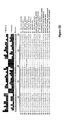
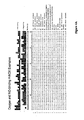
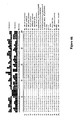
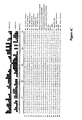
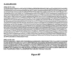
- the nucleic acid may include a segment of or the entire nucleic acid sequence of any of the nucleic acids shown in FIGS. 2-4D or 8A-8DD .
- the nucleic acid may encode a fusion protein that includes an H-NOX domain and part or all of another protein, such as albumin ( e.g. , human serum albumin).
- the nucleic acid includes at least about any of 50, 100, 150, 200, 300, 400, 500, 600, 700, 800, or more contiguous nucleotides from an H-NOX nucleic acid and contains one or more mutations (e.g., 1, 2, 3, 4, 5, 6, 7, 8, 9, or 10 mutations) compared to the H-NOX nucleic acid from which it was derived.
- a mutant H-NOX nucleic acid may contain less than about any of 20, 15, 12, 10, 9, 8, 7, 6, 5, 4, 3, or 2 mutations compared to the H-NOX nucleic acid from which it was derived.
- a vector that includes any one or more of the mutant H-NOX nucleic acids described herein.
- a further described feature is a cell that includes any one or more of the mutant H-NOX nucleic acids described herein.
- a cell may include any vector described herein.
- H-NOX protein There is herein described a method of producing an H-NOX protein. This method involves culturing a cell having a nucleic acid encoding any one or more of the mutant H-NOX proteins described herein under conditions suitable for production of the mutant H-NOX protein. There is also described the further inclusion of the step of purifying the mutant H-NOX protein.
- the invention features pharmaceutical compositions that include one or more H-NOX proteins, such as any of the wild-type or mutant H-NOX proteins described herein.
- the pharmaceutical composition includes a pharmaceutically acceptable amount of an H-NOX protein described herein and a pharmaceutically acceptable carrier.
- the k off , k 1 , or k 2 for NO of the H-NOX protein is between about 1 x 10 -4 s -1 to about 10 s -1 at 37 °C
- the O 2 dissociation constant of the H-NOX protein is at least about 1 ⁇ M at 37 °C.
- the NO dissociation constant of the H-NOX protein is within 2 orders of magnitude of that of hemoglobin, and the NO reactivity of the H-NOX protein is at least 10-fold lower than that of hemoglobin.
- the NO dissociation constant of the H-NOX protein is within 2 orders of magnitude of that of Homo sapiens hemoglobin alpha, such as an NO dissociation constant between 0.1 to 10-fold or between 0.5 to 2-fold of that of Homo sapiens hemoglobin alpha.
- the NO reactivity of the H-NOX protein is at least 10-fold lower than that of Homo sapiens hemoglobin alpha, such as at least 100-fold or 1,000-fold lower than that of Homo sapiens hemoglobin alpha.
- the H-NOX protein is a wild-type protein.
- the H-NOX protein is a mutant protein as described herein.
- the H-NOX protein has at least one mutation that alters the NO dissociation constant, the k off for NO, the k 1 for NO, the k 2 for NO, the O 2 dissociation constant, the NO stability, the NO reactivity the rate of heme autoxidation, or any combination of two or more of the foregoing compared to that of a corresponding wild-type protein.
- the H-NOX protein is a selected from the group consisting of wild-type T. tengcongensis H-NOX, T.
- H. sapiens ⁇ 1 H-NOX desulfuricans H-NOX Y139L, wild-type H. sapiens ⁇ 1 H-NOX, H. sapiens ⁇ 1 I140Y, H. sapiens ⁇ 1 I145Y, H. sapiens ⁇ 1(1-385), H. sapiens ⁇ 1(1-385) I145Y, H. sapiens ⁇ 1(1-385) I145H, H. sapiens ⁇ 1(1-194), H. sapiens ⁇ 1(1(1-194) I145Y, H. sapiens ⁇ 1(1-194) L9W-I145Y, H. sapiens ⁇ 2(1-217), H. sapiens ⁇ 2(1-217) I142Y, H. sapiens ⁇ 1 H-NOX H105G, H .
- norvegicus ⁇ 2(1-217) I142Y R. norvegicus ⁇ 1 H-NOX H105G, R. norvegicus ⁇ 1 H-NOX H105F, R. norvegicus sGC ⁇ 1 H-NOX C78S, R. norvegicus sGC ⁇ 1 H-NOX C78E, C . botulinum H-NOX(1-175), C . botulinum H-NOX(1-186), wild-type C . acetobutylicum H-NOX, C . acetobutylicum H-NOX(1-197), C . acetobutylicum H-NOX(1-183), wild-type C .
- the H-NOX protein is a selected from the group consisting of wild-type R. norvegicus sGC, wild-type R. norvegicus ⁇ 1(1-385), R. norvegicus ⁇ 1(1-217), R. norvegicus ⁇ 1(1-194), wild-type T. tengcongensis H-NOX, T. tengcongensis H-NOX Y140L, T.
- the pharmaceutical composition includes one or more liposomes or nanoparticles that include or encapsulate the H-NOX protein.
- the H-NOX protein is not T. tengcongensis H-NOX Y40L, wild-type T. tengcongensis H-NOX, wild-type R. norvegicus sGC, or L. pneumophilia 2 H-NOX F142Y. In some embodiments of the pharmaceutical compositions, the H-NOX protein is not T. tengcongensis H-NOX F78Y/Y140L. In some embodiments of the pharmaceutical compositions, the H-NOX protein is not wild-type L. pneumophilia 2 H-NOX, wild-type H. sapiens ⁇ 1 H-NOX, R.
- norvegicus sGC ⁇ 1 H-NOX (1-385), wild-type R. norvegicus ⁇ 1 H-NOX, wild-type D. melangaster ⁇ 1 H-NOX, wild-type D. melangaster CG 14885-PA H-NOX, wild-type C . elegans GCY-35 H-NOX, wild-type N. punctiforme H-NOX, wild-type C . crescentus H-NOX, wild-type S. oneidensis H-NOX, or wild-type C . acetobutylicum H-NOX.
- the H-NOX protein is not T.
- the H-NOX protein is not R. norvegicus ⁇ 2(1-217), R. norvegicus ⁇ 1(1-194), R.
- the H-NOX protein is not T. tengcongensis H-NOX W9F, T. tengcongensis H-NOX Y140F, or H. sapiens ⁇ 1 H-NOX (1-385) I145Y. In some embodiments of the pharmaceutical compositions, the H-NOX protein is not T. tengcongensis H-NOX Y140H, H. sapiens ⁇ 1 I140Y, or H. sapiens ⁇ 1 I145Y. In some embodiments of the pharmaceutical compositions, the H-NOX protein is not T.
- norvegicus sGC ⁇ 1 H-NOX (1-385) I145Y, R. norvegicus sGC ⁇ 1 H-NOX H105G, R. norvegicus sGC ⁇ 1 H-NOX H105F, R. norvegicus sGC ⁇ 1 H-NOX I145Y, wild-type R. norvegicus ⁇ 1 H-NOX, wild-type D. melangaster ⁇ 1 H-NOX, wild-type D. melangaster CG14885-PA H-NOX, wild-type C . elegans GCY-35 H-NOX, wild-type N. punctiforme H-NOX, wild-type C .
- the H-NOX protein is not any of the following H-NOX proteins that are listed by their gene name, followed by their species abbreviation and Genbank Identifiers (such as the following protein sequences available as of May 21, 2006; May 22, 2006; May 21, 2007; or May 22, 2007): Npun5905_Npu_23129606, alr2278_Ana_17229770, SO2144_Sone_24373702, Mdeg1343_Mde_23027521, VCA0720_Vch_15601476, CC2992_Ccr_16127222, Rsph2043_Rhsp_22958463 (gi:46192757), Mmc l0739_Mcsp_22999020, Tar4_Tte_20807169, Ddes2822_Dde
- the H-NOX protein is not any of the following H-NOX proteins that are listed by their organism name and Pfam database accession number (such as the following protein sequences available as of May 21, 2006; May 22, 2006; May 17, 2007; May 21, 2007; or May 22, 2007): Caenorhabditis briggsae Q622M5_CAEBR, Caenorhabditis briggsae Q61P44_CAEBR, Caenorhabditis briggsae Q61R54_CAEBR, Caenorhabditis briggsae Q61V90_CAEBR, Caenorhabditis briggsae Q61A94_CAEBR, Caenorhabditis briggsae Q60TP4_CAEBR, Caenorhabditis briggsae Q60M10_CAEBR, Caenorhabditis elegans GCY37_CAEEL, Caenorhabdititis briggsae Q
- H-NOX protein may or may not have heme and/or NO bound and may or may not be covalently bound to another molecule or moiety, such as polyethylene glycol.
- the H-NOX protein is a fusion protein that includes an H-NOX domain and part or all of another protein, such as albumin ( e.g ., human serum albumin).
- the invention also provides the use of such compositions to deliver NO to an individual (e.g., a mammal, such as a primate ( e.g., a human, a monkey, a gorilla, an ape, a lemur, etc ), a bovine, an equine, a porcine, a canine, or a feline) using an H-NOX protein.
- an individual e.g., a mammal, such as a primate (e.g., a human, a monkey, a gorilla, an ape, a lemur, etc ), a bovine, an equine, a porcine, a canine, or a feline) using an H-NOX protein.
- the individual is suffering from or at risk for a cardiovascular condition, hypertension, a condition exacerbated by hypertension, a vasoconstrictive condition, stroke, or a functional NO deficiency.
- the condition exacerbated by hypertension is heart
- the invention provides the use of compositions to deliver NO to an individual (e.g ., a human) by administering to an individual in need thereof an H-NOX protein in an amount sufficient to deliver an effective amount of NO to the individual.
- the k off , k 1 , or k 2 for NO of the H-NOX protein is between about 1 x 10 -4 s -1 to about 10 s -1 at 37 °C, and the O 2 dissociation constant of the H-NOX protein is at least about 1 ⁇ M at 37 °C.
- the NO dissociation constant of the H-NOX protein is within 2 orders of magnitude of that of hemoglobin, and the NO reactivity of the H-NOX protein is at least 10-fold lower than that of hemoglobin.
- NO is bound to the H-NOX protein prior to the administration of the H-NOX protein to the individual. In some embodiments of the uses, NO is not bound to the H-NOX protein prior to the administration of the H-NOX protein to the individual, and the H-NOX protein transports NO from one location in the individual to another location in the individual. In some embodiments of the uses, the H-NOX protein is administered orally, rectally, or to the blood of the individual. In particular embodiments of the uses, the H-NOX protein is administered to the blood of the individual. In some embodiments of the uses, the H-NOX protein is administered to the individual at least twice.
- the NO dissociation constant of the H-NOX protein is within 2 orders of magnitude of that of Homo sapiens hemoglobin alpha, such as an NO dissociation constant between 0.1 to 10-fold or between 0.5 to 2-fold of that of Homo sapiens hemoglobin alpha. In some embodiments of the uses, the NO reactivity of the H-NOX protein is at least 10-fold lower than that of Homo sapiens hemoglobin alpha, such as at least 100-fold or 1,000-fold lower than that of Homo sapiens hemoglobin alpha. In some embodiments of the uses, the H-NOX protein is a wild-type protein. In some embodiments of the uses, the H-NOX protein is a mutant protein as described herein.
- the H-NOX protein has at least one mutation that alters the NO dissociation constant, the k off for NO, the k 1 for NO, the k 2 for NO, the O 2 dissociation constant, the NO stability, the NO reactivity the rate of heme autoxidation, or any combination of two or more of the foregoing compared to that of a corresponding wild-type protein.
- the H-NOX protein is a selected from the group consisting of wild-type T. tengcongensis H-NOX, T. tengcongensis H-NOX I5A, T . tengcongensis H-NOX I5L, T.
- norvegicus ⁇ 1 H-NOX H105F R. norvegicus sGC ⁇ 1 H-NOX C78S, R. norvegicus sGC ⁇ 1 H-NOX C78E, C. botulinum H-NOX(1-175), C . botulinum H-NOX(1-186), wild-type C . acetobutylicum H-NOX, C . acetobutylicum H-NOX(1-197), C . acetobutylicum H-NOX(1-183), wild-type C. elegans GCY-35 H-NOX, C . elegans H-NOX GCY-35(1-252), wild-type D.
- melangaster ⁇ 1 H-NOX wild-type D. melangaster CG14885-PA, wild-type D. melangaster CG14886, wild-type D. melangaster CG4154; wild-type N. punctiforme H-NOX, wild-type C . crescentus H-NOX, wild-type S . oneidensis H-NOX, wild-type M. musculus H-NOX, wild-type C. familiaris H-NOX, wild-type B. Taurus H-NOX, wild-type R. norvegicus; wild-type X. laevis H-NOX, wild-type O. latipes H-NOX, wild-type O.
- H-NOX curivatus H-NOX, wild-type F. rubripes H-NOX, wild-type A. gambiae H-NOX, wild-type M sexta H-NOX; wild-type C . elegans gcy-31, C . elegans gcy-32, wild-type C . elegans gcy-33, wild-type C . elegans gcy-34, wild-type C . elegans gcy-35, wild-type C . elegans gcy-36, wild-type C . elegans gcy-37; wild-type V. cholera H-NOX, wild-type V.
- the H-NOX protein is a selected from the group consisting of wild-type R. norvegicus sGC, wild-type R. norvegicus ⁇ 1(1-385), R. norvegicus ⁇ 1(1-217), R. norvegicus ⁇ 1(1-194), wild-type T. tengcongensis H-NOX, T. tengcongensis H-NOX Y140L, T. tengcongensis H-NOX Y140F, wild-type L. pneumophilia 1 H-NOX, wild-type L. pneumophilia 2 H-NOX, and L. pneumophilia 2 H-NOX F142Y.
- one or more liposomes or nanoparticles that include or encapsulate the H-NOX protein are selected from the group consisting of wild-type R. norvegicus sGC, wild-type R. norvegicus ⁇ 1(1-385), R. norvegicus ⁇ 1
- the H-NOX protein is not T. tengcongensis H-NOX Y40L, wild-type T. tengcongensis H-NOX, wild-type R. norvegicus sGC, or L. pneumophilia 2 H-NOX F142Y. In some embodiments of the uses, the H-NOX protein is not T. tengcongensis H-NOX F78Y/Y140L. In some embodiments of the uses, the H-NOX protein is not wild-type L. pneumophilia 2 H-NOX, wild-type H. sapiens ⁇ 1 H-NOX, R. norvegicus sGC ⁇ 1 H-NOX (1-385), wild-type R.
- the H-NOX protein is not T. tengcongensis H-NOX W9F, T. tengcongensis H-NOX Y140F, R.
- the H-NOX protein is not R. norvegicus ⁇ 2(1-217), R. norvegicus ⁇ 1 (1-194), R. norvegicus ⁇ 1(1-385), or R. norvegicus ⁇ 1(1-385) I145Y.
- the H-NOX protein is not T. tengcongensis H-NOX W9F, T. tengcongensis H-NOX Y140F, or H. sapiens ⁇ 1 H-NOX (1-385) I145Y. In some embodiments of the uses, the H-NOX protein is not T . tengcongensis H-NOX Y140H, H. sapiens ⁇ 1 I140Y, or H. sapiens ⁇ 1 I145Y. In some embodiments of the uses, the H-NOX protein is not T. tengcongensis H-NOX Y40L, T. tengcongensis H-NOX F78Y/Y140L, T.
- norvegicus sGC ⁇ 1 H-NOX H105F R. norvegicus sGC ⁇ 1 H-NOX I145Y, wild-type R. norvegicus ⁇ 1 H-NOX, wild-type D. melangaster ⁇ 1 H-NOX, wild-type D. melangaster CG14885-PA H-NOX, wild-type C . elegans GCY-35 H-NOX, wild-type N. punctiforme H-NOX, wild-type C . crescentus H-NOX, wild-type S. oneidensis H-NOX, or wild-type C . acetobutylicum H-NOX.
- the H-NOX protein is not any of the following H-NOX proteins that are listed by their gene name, followed by their species abbreviation and Genbank Identifiers (such as the following protein sequences available as of May 21, 2006; May 22, 2006; May 21, 2007; or May 22, 2007): Npun5905-Npu-23129606, alr2278_Ana_17229770, SO2144_Sone_24373702, Mdeg1343_Mde_23027521, VCA0720_Vch_15601476, CC2992_Ccr_16127222, Rsph2043_Rhsp_22958463 (gi:46192757), Mmc10739_Mcsp_22999020, Tar4_Tte_20807169, Ddes2822_Dde_23475919, CAC3243_Cac_15896488, gcy-31_Ce_17568389, CG14885_Dm_246474
- the H-NOX protein is not any of the following H-NOX proteins that are listed by their organism name and Pfam database accession number (such as the following protein sequences available as of May 21, 2006; May 22, 2006; May 17, 2007; May 21, 2007; or May 22, 2007): Caenorhabditis briggsae Q622M5_CAEBR, Caenorhabditis briggsae Q6tP44_CAEBR, Caenorhabditis briggsae Q61R54_CAEBR, Caenorhabditis briggsae Q61V90_CAEBR, Caenorhabditis briggsae Q61A94_CAEBR, Caenorhabditis briggsae Q60TP4_CAEBR, Caenorhabditis briggsae Q60M10_CAEBR, Caenorhabditis elegans GCY37_CAEEL, Caenorhabditis
- the H-NOX protein may or may not have heme and/or NO bound and may or may not be covalently bound to another molecule or moiety, such as polyethylene glycol.
- the H-NOX protein is a fusion protein that includes an H-NOX domain and part or all of another protein, such as albumin ( e.g ., human serum albumin).
- kits that include one or more H-NOX proteins.
- a kit may include an H-NOX protein and instructions for using the kit to deliver NO to an individual.
- the k off , k 1 , or k 2 for NO of the H-NOX protein is between about 1 x 10 -4 s -1 to about 10 s -1 at 37 °C, and the O 2 dissociation constant of the H-NOX protein is at least about 1 ⁇ M at 37 °C.
- the NO dissociation constant of the H-NOX protein is within 2 orders of magnitude of that of hemoglobin, and the NO reactivity of the H-NOX protein is at least 10-fold lower than that of hemoglobin.
- the H-NOX protein may or may not have heme and/or NO bound and may or may not be covalently bound to another molecule or moiety, such as polyethylene glycol.
- the H-NOX protein is a fusion protein that includes an H-NOX domain and part or all of another protein, such as albumin ( e.g ., human serum albumin).
- an H-NOX protein (such as any of the wild-type or mutant proteins described herein) for use as a medicament.
- An H-NOX protein may be used in a method of delivering NO to an individual.
- the H-NOX protein may be used to treat any condition for which delivery of NO is beneficial, such as a cardiovascular condition, hypertension, a condition exacerbated by hypertension ( e.g ., heart failure, renal failure, or a stroke), a vasoconstrictive condition, stroke, or a functional NO deficiency.
- an H-NOX protein (such as any of the wild-type or mutant proteins described herein) for the manufacture of a medicament, such as a medicament for delivering NO to an individual.
- An H-NOX protein may be used for delivering NO to an individual.
- the H-NOX protein may be used to treat any condition for which delivery of NO is beneficial, such as a cardiovascular condition, hypertension, a condition exacerbated by hypertension ( e.g ., heart failure, renal failure, or a stroke), a vasoconstrictive condition, stroke, or a functional NO deficiency.
- the present invention is based in part on the surprising discovery that H-NOX proteins have a much lower NO reactivity than hemoglobin. This intrinsic low NO reactivity (and high NO stability) makes wild-type and mutant H-NOX proteins desirable NO carriers because of the lower probability of inactivation of H-NOX proteins by NO in the presence of O 2 . Importantly, the presence of a distal pocket tyrosine in some H-NOX proteins ( Pellicena, P. et al. (August 31, 2004). "Crystal Structure of An Oxygen-Binding Heme Domain Related to Soluble Guanylate Cyclases," Proc Natl.
- H-NOX proteins as NO carriers can be improved by modifying their affinities for NO or O 2 to maximize the amount of NO that is bound to the H-NOX protein and to reduce the amount of H-NOX protein that is oxidized by the reaction of NO with O 2 bound to the H-NOX protein.
- affinity of H-NOX proteins for NO or O 2 and the ability of H-NOX proteins to discriminate between NO and O 2 ligands can be altered by the introduction of one or more amino acid mutations, allowing H-NOX proteins to be tailored to bind NO or O 2 with desired affinities.
- the dissociation constant or dissociation rate for NO or O 2 binding by H-NOX proteins can be altered the introduction of a single amino acid mutation. Additional mutations can be introduced to further alter the affinity for NO and/or O 2 .
- the H-NOX protein family can therefore be manipulated to exhibit improved or optimal kinetic and thermodynamic properties for NO delivery.
- mutant H-NOX proteins have been generated with altered dissociation constants and/or dissociation rates for NO binding that improve the usefulness of H-NOX proteins for a variety of clinical and industrial applications.
- H-NOX protein with a low affinity for O 2 (such as an O 2 dissociation constant of at least about 1 ⁇ M at 37 °C) is used to minimize the amount of O 2 that binds the H-NOX protein, thereby facilitating the binding of NO to the H-NOX protein and reducing the amount of H-NOX protein that is oxidized due to the reaction of NO with O 2 bound to the heme of the H-NOX protein.
- This reduction in the oxidation of H-NNOX proteins results in less destruction of NO and O 2 that can be used by the organs, tissues, and cells of the treated individual.
- the ability to tune H-NOX proteins to bind and deliver NO is a therapeutic avenue that addresses and overcomes the central shortcomings of current vasodilators. Accordingly, there is herein described proteins, compositions, kits, and methods for the delivery of NO.
- H-NOX proteins for NO delivery.
- Organic nitrates are effective for a limited length of time due to tolerance.
- H-NOX proteins delivery NO directly to individuals without requiring the bioconversion of nitrates to NO, the effectiveness of H-NOX proteins as NO carriers is not limited by inhibition of this bioconversion pathway.
- Major limitations of hemoglobin-based NO carriers are their high affinity for O 2 and their propensity to be inactivated by NO. As mentioned above, destruction of even low levels of NO by hemoglobin-based carriers can have serious effects on the tonic resting state of the vasculature and organs and leads to hypertension and gastrointestinal distress.
- H-NOX proteins with desired dissociation constants and dissociation rates for NO can also minimize side-effects by preventing too much NO from being released (causing hypotension) and prevent NO from being released at undesired sites (e.g ., sites that are not vasoconstricted).
- Engineering H-NOX proteins to bind and deliver NO with minimal NO reactivity provides a new blood gas NO carrier where the H-NOX proteins deliver NO without being inactivated by NO.
- H-NOX proteins For delivery of NO, engineered H-NOX proteins represent an important alternative that overcomes the persistent problem of tolerance with current nitrovasodilators.
- the use of H-NOX proteins as delivery vehicles for NO provides a new therapeutic venue for treating diseases exacerbated by chronic hypertension.
- H-NOX protein means a protein that has an H-NOX domain (named for H eme- N itric oxide and OX ygen binding domain).
- An H-NOX protein may or may not contain one or more other domains in addition to the H-NOX domain.
- H-NOX proteins are members of a highly-conserved, well-characterized family of hemoproteins ( lyer, L. M. et al. (February 3, 2003).
- H-NOX proteins are also referred to as Pfam 07700 proteins or HNOB proteins (Pfam - A database of protein domain family alignments and Hidden Markov Models, Copyright (C) 1996-2006 The Pfam Consortium; GNU LGPL Free Software Foundation, Inc., 59 Temple Place - Suite 330, Boston, MA 02111-1307, USA).
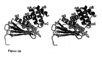
- an H-NOX protein has, or is predicted to have, a secondary structure that includes six alpha-helices, followed by two beta-strands, followed by one alpha-helix, followed by two beta-strands.
- An H-NOX protein can be an apoprotein that is capable of binding heme or a holoprotein with heme bound.
- H-NOX protein can covalently or non-covalently bind a heme group. Some H-NOX proteins bind NO but not O 2 , and others bind both NO and O 2 . H-NOX domains from facultative aerobes that have been isolated bind NO but not O 2 . H-NOX proteins from obligate aerobic prokaryotes, C . elegans, and D. melanogaster bind NO and O 2 . Mammals have two H-NOX proteins: ⁇ 1 and ⁇ 2. An alignment of mouse, rat, cow, and human H-NOX sequences shows that these species share >99% identity.
- the H-NOX domain of an H-NOX protein or the entire H-NOX protein is at least about any of 10, 15, 20, 25, 30, 40, 50, 60, 70, 80, 90, 95, 97, 98, 99, or 99.5% identical to that of the corresponding region of a naturally-occurring Thermoanaerobacter tengcongensis H-NOX protein or a naturally-occurring sGC protein ( e.g ., a naturally-occurring sGC ⁇ 1 protein).
- an H-NOX protein may optionally contain one or more mutations relative to the corresponding naturally-occurring H-NOX protein.
- the H-NOX protein includes one or more domains in addition to the H-NOX domain.
- the H-NOX protein includes one or more domains or the entire sequence from another protein.
- the H-NOX protein may be a fusion protein that includes an H-NOX domain and part or all of another protein, such as albumin (e.g ., human serum albumin). In some cases, only the H-NOX domain is present.
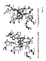
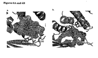
- the structure of the porphyrin is highly distorted.
- the conserved Y-S-R motif makes hydrogen-bonding interactions with the propionic acid side chains of the heme group.
- the conserved H102 is the proximal ligand to the heme ( FIG. 6B ).
- a "protein” includes proteins and fragments of proteins whether isolated from natural sources, produced by recombinant techniques, or chemically synthesized.
- a protein may have one or more modifications, such as a post-translational modification (e.g. , glycosylation, etc ) or any other modification ( e.g., PEGylation, etc ).
- the protein may contain one or more non-naturally-occurring amino acids (e.g ., such as an amino acid with a side chain modification).
- the H-NOX protein may have at least about 50, 100, 150, 181, 200, 250, 300, 350, 400, or more amino acids.
- the H-NOX proteins may include from about 50 to about 600 amino acids, such as about 100 to about 500 amino acids, about 150 to about 400 amino acids, about 150 to about 300 amino acids, or about 175 to about 200 amino acids.
- H-NOX proteins from any genus or species can be used in the compositions, kits, and methods described herein.
- the H-NOX protein is a protein from a mammal (e.g., a primate (e.g., human, monkey, gorilla, ape, lemur, etc ), a bovine, an equine, a porcine, a canine, or a feline), an insect, a yeast, or a bacteria or is derived from such a protein.
- exemplary mammalian H-NOX proteins include wild-type human and rat soluble guanylate cyclase (such as the ⁇ 1 subunit).
- H-NOX proteins include wild-type mammalian H-NOX proteins, e.g. H. sapiens, M. musculus, C. familiaris, B. taurus and R. norvegicus; and wild-type non-mammalian vertebrate H-NOX proteins, e.g., X. laevis, O. latipes, O. curivatus, and F . rubripes.
- non-mammalian wild-type NO-binding H-NOX proteins include wild-type H-NOX proteins of D. melanogaster, A. gambiae, and M.
- examples of non-mammalian wild-type O 2 -binding H-NOX proteins include wild-type H-NOX proteins of C . elegans gcy-31, gcy-32, gcy-33, gcy-34, gcy-35, gcy-36, and gcy-37; D. melanogaster CG 14885, CG 14886, and CG4154; and M. sexta beta-3; examples of prokaryotic wild-type H-NOX proteins include T. tengcongensis, V. cholera, V. fischerii, N. punctiforme, D. desulfuricans, L. pneumophila 1, L. pneumophila 2, and C . acelobutylicum.
- H-NOX proteins include the following: Homo sapiens ⁇ 1 [gi:2746083], Rattus norvegicus ⁇ 1 [gi:27127318], Drosophila melangaster ⁇ 1 [gi:861203], Drosophila melangaster CG 14885-PA [gi:23171476], Caenorhabditis elegans GCY-35 [gi:52782806], Nostoc punctiforme [gi:23129606], Caulobacter crescentus [gi:16127222], Shewanella oneidensis [gi:24373702], Legionella pneumophila (ORF 2) [CUCGC_272624], Clostridium acetobutylicum [gi: 15896488], and Thermoanaerobacter tengcongensis [gi:20807169].
- Exemplary H-NOX protein also include the following H-NOX proteins that are listed by their gene name, followed by their species abbreviation and Genbank Identifiers (such as the following protein sequences available as of May 21, 2006; May 22, 2006; May 21, 2007; or May 22, 2007: Npun5905_Npu_23 129606, alr2278_Ana_17229770, SO2144_Sone_24373702, Mdeg1343_Mde_23027521, VCA0720_Vch_15601476,CC2992_Ccr_16127222, Rsph2043_Rhsp_22958463 (gi:46192757), Mmc10739_Mcsp_22999020, Tar4_Tte_20807169,Ddes2822_Dde_23475919,CAC3243_Cac_15896488 ,gcy-31_Ce_17568389, CG14885_Dm_24647455,GUCY1B3_Hs
- H-NOX proteins include the following H-NOX proteins that are listed by their organism name and Pfam database accession number (such as the following protein sequences available as of May 21, 2006; May 22, 2006; May 17, 2007; May 21, 2007; or May 22, 2007, Caenorhabditis briggsae Q622M5_CAEBR, Caenorhabditis briggsae Q61P44_CAEBR, Caenorhabditis briggsae Q61R54_CAEBR, Caenorhabditis briggsae Q61V90_CAEBR, Caenorhabditis briggsae Q61A94_CAEBR, Caenorhabditis briggsae Q60TP4_CAEBR, Caenorhabditis briggsae Q60M10_CAEBR, Caenorhabditis elegans GCY37_CAEEL, Caenorhabditis elegans GCY31_CAEEL,
- H-NOX proteins and nucleic acids which may be suitable for use in the pharmaceutical compositions and methods described herein, can be identified using standard methods. For example, standard sequence alignment and/or structure prediction programs can be used to identify additional H-NOX proteins and nucleic acids based on the similarity of their primary and/or predicted protein secondary structure with that of known H-NOX proteins and nucleic acids.
- the Pfam database uses defined alignment algorithms and Hidden Markov Models (such as Pfam 21.0) to categorize proteins into families, such as the H-NOX protein family (Pfam - A database of protein domain family alignments and Hidden Markov Models, Copyright (C) 1996-2006 The Pfam Consortium; GNU LGPL Free Software Foundation, Inc., 59 Temple Place - Suite 330, Boston, MA 02111-1307, USA).
- Standard databases such as the swissprot-trembl database (world-wide web at "expasy.org", Swiss Institute of Bioinformatics Swiss-Prot group CMU - 1 rue Michel Servet CH-1211 Geneva 4 , Switzerland) can also be used to identify members of the H-NOX protein family.
- the secondary and/or tertiary structure of an H-NOX protein can be predicted using the default settings of standard structure prediction programs, such as PredictProtein (630 West, 168 Street, BB217, New York, N.Y. 10032, USA). Alternatively, the actual secondary and/or tertiary structure of an H-NOX protein can be determined using standard methods.
- the H-NOX protein may have the same amino acid in the corresponding position as any of following distal pocket residues in T. tengcongensis H-NOX: Thr4, Ile5, Thr8, Trp9, Trp67, Asn74, Ile75, Phe78, Phe82, Tyr140, Leu144, or any combination of two or more of the foregoing.
- the H-NOX protein may have a proline or an arginine in a position corresponding to that of Pro115 orArg135 of T. tengcongensis H-NOX, respectively, based on sequence alignment of their amino acid sequences.
- the H-NOX protein may have a histidine that corresponds to His 105 of R. norvegicus ⁇ 1 H-NOX.
- the H-NOX protein may have or is predicted to have a secondary structure that includes six alpha-helices, followed by two beta-strands, followed by one alpha-helix, followed by two beta-strands. This secondary structure has been reported for H-NOX proteins.
- a newly identified H-NOX protein can be tested to determine whether it binds heme using standard methods.
- the ability of an H-NOX protein to function as an NO carrier can be tested by determining whether the H-NOX protein binds NO using standard methods, such as those described herein.
- one or more of the mutations described herein can be introduced into the H-NOX protein to optimize its characteristics as an NO carrier. For example, one or more mutations can be introduced to alter its NO dissociation constant, k off for NO, k 1 for NO, k 2 for NO, O 2 dissociation constant, NO stability, NO reactivity, rate of heme autoxidation, or any combination of two or more of the foregoing. Standard techniques such as those described herein can be used to measure these parameters.
- mutant H-NOX proteins may be derived by mutagenesis from these or other natural wild-type source sequences (e.g. , the sequences listed in FIG. 2-4D or 8A-8DD or any other sequence described herein).
- derived from refers to the source of the protein into which one or more mutations is introduced.
- a protein that is "derived from a mammalian protein” refers to protein of interest that results from introducing one or more mutations into the sequence of a wild-type ( i.e. , a sequence occurring in nature) mammalian protein.
- an H-NOX protein may contain one or more mutations, such as a mutation that alters the NO dissociation constant, the k off for NO, the O 2 dissociation constant, the k off for O 2 , the rate of heme autoxidation, the NO reactivity, the NO stability, or any combination of two or more of the foregoing compared to that of the corresponding wild-type protein.
- Panels of engineered H-NOX proteins may be generated by random mutagenesis followed by empirical screening for requisite or desired dissociation constants, dissociation rates, NO-reactivity, stability, physio-compatibility, or any combination of two or more of the foregoing in view of the teaching provided herein using techniques as described herein and, additionally, as known by the skilled artisan.
- mutagenesis can be selectively targeted to particular regions or residues such as distal pocket residues apparent from the experimentally determined or predicted three-dimensional structure of an H-NOX protein ( FIG. 1A herein; and see, for example, Boon, E. M. et al. (2005).
- a mutant protein means a protein with one or more mutations compared to a protein occurring in nature.
- the mutant protein has a sequence that differs from that of all proteins occurring in nature.
- the amino acid sequence of the mutant protein is at least about any of 10, 15, 20, 25, 30, 40, 50, 60, 70, 80, 90, 95, 97, 98, 99, or 99.5% identical to that of the corresponding region of a protein occurring in nature.
- the mutant protein is a protein fragment that contains at least about any of 25, 50, 75, 100, 150, 200, 300, or 400 contiguous amino acids from a full-length protein.
- Sequence identity can be measured, for example, using sequence analysis software with the default parameters specified therein (e.g., Sequence Analysis Software Package of the Genetics Computer Group, University of Wisconsin Biotechnology Center, 1710 University Avenue, Madison, W1 53705). This software program matches similar sequences by assigning degrees of homology to various amino acids replacements, deletions, and other modifications.
- sequence analysis software with the default parameters specified therein (e.g., Sequence Analysis Software Package of the Genetics Computer Group, University of Wisconsin Biotechnology Center, 1710 University Avenue, Madison, W1 53705). This software program matches similar sequences by assigning degrees of homology to various amino acids replacements, deletions, and other modifications.
- a "mutation" means an alteration in a reference nucleic acid or amino acid sequence occurring in nature.
- Exemplary nucleic acid mutations include an insertion, deletion, frameshift mutation, silent mutation, nonsense mutation, or missense mutation. In some embodiments, the nucleic acid mutation is not a silent mutation.
- Exemplary protein mutations include the insertion of one or more amino acids (e.g. , the insertion of 2, 3, 4, 5, 6, 7, 8, 9, or 10 amino acids), the deletion of one or more amino acids (e.g.
- a deletion ofN-terminal, C-terminal, and/or internal residues such as the deletion of at least about any of 5, 10, 15, 25, 50, 75, 100, 150, 200, 300, or more amino acids or a deletion of about any of 5, 10, 15, 25, 50, 75, 100, 150, 200, 300, or 400 amino acids), the replacement of one or more amino acids (e.g. , the replacement of 2, 3, 4, 5, 6, 7, 8, 9, or 10 amino acids), or combinations of two or more of the foregoing.
- An exemplary functional truncation of an H-NOX protein includes residues 1-385 of the ⁇ 1 sequence.
- a mutant protein has at least one amino acid alteration compared to a protein occurring in nature.
- a mutant nucleic acid sequence encodes a protein that has at least one amino acid alteration compared to a protein occurring in nature.
- the nucleic acid is not a degenerate version of a nucleic acid occurring in nature that encodes a protein with an amino acid sequence identical to a protein occurring in nature.
- the nomenclature used in referring to a particular amino acid mutation first identifies the wild-type amino acid, followed by the residue number and finally the substitute amino acid. For example, Y140L means that tyrosine has been replaced by a leucine at residue number 140.
- An "evolutionary conserved mutation” is the replacement of an amino acid in one protein by an amino acid in the corresponding position of another protein in the same protein family.
- Exemplary evolutionary conserved mutations are listed in Table 1A.
- Table 1 A mutations are numbered/annotated according to the sequence of human ⁇ 1 H-NOX, but are analogous for all H-NOX sequences.
- the corresponding position in any other H-NOX protein can be mutated to the indicated residue.
- Phe4 of human ⁇ 1 H-NOX can be mutated to a tyrosine since other H-NOX proteins have a tyrosine in this position.
- the corresponding phenylalanine residue can be mutated to a tyrosine in any other H-NOX protein.
- the one or more mutations are confined to evolutionarily conserved residues.
- the one or more mutations may include at least one evolutionarily conserved mutation and at least one non-evolutionarily conserved mutation. If desired, these mutant H-NOX proteins are subjected to empirical screening for NO/O 2 dissociation constants, NO-reactivity, stability, and physio-compatibility in view of the teaching provided herein.
- Table 1A Exemplary Class I H-NOX mutations targeting evolutionary conserved residues F4Y Q30G 1145Y F4L E33P 1145H H7G N61G K151E A8E C78H 1157F L9W A109F E183F
- the mutation is a distal pocket mutation, such as mutation of a residue in alpha-helix A, D, E, or G ( Pellicena, P. et al. (August 31,2004). "Crystal Structure of An Oxygen-Binding Heme Domain Related to Soluble Guanylate Cyclases," Proc Natl. Acad Sci USA 101(35):12854-12859 ).
- Exemplary distal pocket mutations are listed in Table 1B. In Table 1B, mutations are numbered/annotated according to the sequence of human ⁇ 1 H-NOX, but are analogous for all H-NOX sequences.
- the mutation is a heme distal pocket mutation.
- a crucial molecular determinant that prevents O 2 binding in NO-binding members of the H-NOX family is the lack of a H-bond donor in the distal pocket of the heme. Accordingly, the mutation alters H-bonding between the H-NOX domain and the ligand within the distal pocket.
- the mutation disrupts an H-bond donor of the distal pocket and/or imparts reduced O 2 ligand-binding relative to the corresponding wild-type H-NOX domain.
- Exemplary distal pocket residues include Thr4, Ile5, Thr8, Trp9, Trp67, Asn74, Ile75, Phe78, Phe82, Tyr140, and Leu144of T. tengcongensis H-NOX and the corresponding residues in any other H-NOX protein.
- the H-NOX protein has one or more mutations outside of the distal pocket. Examples of residues that can be mutated but are not in the distal pocket include Pro115 and Arg135 of T. tengcongensis H-NOX. In some embodiments, the mutation is in the proximal pocket which includes His105 as a residue that ligates to the heme iron.
- At least one mutation is in the distal pocket, and at least one mutation is outside of the distal pocket (e.g., a mutation in the proximal pocket). In some cases, all the mutations are in the distal pocket.
- the amino acid sequence of the H-NOX protein is not necessarily identical to the sequence of a protein that is produced by an organism in nature. In some instances, the amino acid sequence of the H-NOX protein is not identical to a sequence found in any database on May 21, 2006 or May 22, 2006 (such as all known sequences predicted or known to be an H-NOX nucleic acid or amino acid sequence). In some instances, the amino acid sequence of the H-NOX protein is not identical to a sequence found in any database on May 21, 2007 or May 22, 2007 (such as all known sequences predicted or known to be an H-NOX nucleic acid or amino acid sequence).
- amino acids in an H-NOX protein can be mutated to the corresponding amino acids in a human H-NOX.
- one or more amino acids on the surface of the tertiary structure of a non-human H-NOX protein can be mutated to the corresponding amino acid in a human H-NOX proteins.
- mutation of one or more surface amino acids may be combined with mutation of two or more distal pocket residues, mutation of one or more residues outside of the distal pocket (e.g., a mutation in the proximal pocket), or combinations of two or more of the foregoing.
- Exemplary mutations are shown in Table 2.
- any of the residues listed in Table 2 can be mutated to any other amino acid.
- combinations of any of the mutations described herein can be made in the same H-NOX protein.
- mutations in equivalent positions in other mammalian or non-mammalian H-NOX proteins are also encompassed by this invention.
- residues other than the ones mentioned in Table 2 can also be mutated.
- Exemplary mutant H-NOX proteins comprise one or more mutations that impart altered NO or O 2 ligand-binding relative to the corresponding wild-type H-NOX domain and are operative as a physiologically compatible mammalian NO blood gas carrier.
- T. tengcongensis I5A refers to the replacement of isoleucine by alanine at the fifth position in T. tengcongensis H-NOX.
- the same isoleucine to alanine mutation can be made in the corresponding residue in any other H-NOX protein (this residue may or may not be the fifth residue in the sequence of other H-NOX proteins).
- the H-NOX protein may have at least one mutation in which a residue that corresponds to Ile5, Trp9, Asn74, Pro115, Arg135, or Tyr140 of T. tengcongensis H-NOX, 1145 of ⁇ 1(1-385), or Phe142 of L. pneumophila 2 is replaced by any other amino acid.
- the H-NOX protein has at least two mutations, wherein at least one mutation is the replacement of a residue that corresponds to Ile5, Trp9, Asn74, Pro115, Arg135, or Tyr140 of T. tengcongensis H-NOX, 1145 of ⁇ 1(1-385), or Phe142 of L. pneumophila 2 by any other amino acid.
- the mutation in the H-NOX protein corresponds to a I5A mutation, a I5L mutation, a W9F mutation, a Y140F mutation, a Y140L mutation, a Y140H mutation, a W9F Y140H double mutation, or a F78Y Y140F double mutation of T. tengcongensis or a I145Y mutation of ⁇ 1.
- the mutation in the H-NOX protein corresponds to a W9Y mutation, a W9H mutation, a W9N mutation, a N74H mutation, a N74E mutation, a N74A mutation, a P115A mutation, a R135Q mutation, a I5L P115A double mutant, a N74A Y140H double mutant, or a W9F N74A double mutant of T. tengcongensis.
- at least one C-terminal amino acid (such as at least about 50 contiguous C-terminal amino acids or between about 25 to about 200 contiguous C-terminal amino acids) in the H-NOX protein has been removed compared to the corresponding wild-type protein (such as R.
- H-NOX mutants from T. tengcongensis (Tt), L. pneumophila (Lp), D. desulfuricans (Dd), V. cholera (Yc), N. punctiforme (Np), C. botulinium (Cb), C acetobutylicum, (Ca), rat, human, C. elegans (Ce).
- any of the wild-type or mutant H-NOX proteins can be modified and/or formulated using standard methods to enhance therapeutic or industrial applications.
- a variety of methods are known in the art for insulating such agents from immune surveillance, including crosslinking, PEGylation, carbohydrate decoration, etc. (e.g., Rohlfs, R. J. et al. (May 15, 1998). "Arterial Blood Pressure Responses to Cell-Free Hemoglobin Solutions And The Reaction With Nitric Oxide," J. Biol. Chem. 273(20):12128-12134 ; Migita, R. et al. (June 1997).
- an H-NOX protein is modified during or after its synthesis to decrease its immunogenicity and/or to increase its plasma retention time.
- H-NOX proteins can also be encapsulated (such as encapsulation within liposomes or nanoparticles).
- an H-NOX protein has a similar or improved NO dissociation constant, O 2 dissociation constant, NO k off , O 2 k off , NO reactivity, autoxidation rate, plasma retention time, or any combination of two or more of the foregoing compared to any currently used compound for delivering NO, such as any organic nitrate for bioconversion into NO.
- an H-NOX protein has a low affinity for O 2 (such as an O 2 dissociation constant of at least about 1 ⁇ M at 37 °C) or no detectable affinity for O 2 . Since little, if any, O 2 is bound to the H-NOX protein, there is minimal oxidation by NO of O 2 bound to the heme of the H-NOX protein. Thus, minimal NO, O 2 , and H-NOX protein is inactivated by this NO oxidation. Thus, more NO can be delivered to desired sites in an individual and less O 2 that could be used by the tissues in the individual is destroyed.
- hemoglobin means a protein or a mutant thereof from the well-characterized family of hemoglobins, which are iron-containing O 2 -transport metalloproteins in red blood cells. Purified, stroma-free, human hemoglobin has a kinetic K D for O 2 of about 200-500 nM. This value is subunit dependent.
- a 6-coordinate Fe 11 -NO complex is meant a 6-coordinate ferrous-nitrosyl that produces a UV-Vis Soret peak at approximately 416-422 nm, as described, e.g., by Boon, E. M. et al., (August 2006), "Nitric Oxide Binding to Prokaryotic Homologs of the Solube Guanylate Cyclase ⁇ 1 HONOX Domain,” J. Biol. Chem. 281(31): 21892-21902 .
- a 5-coordinate Fe 11 -NO complex is meant a 5-coordinate ferrous-nitrosyl that produces a UV-Vis Soret peak at approximately 397-400 nm, as described, e . g ., by Boon, E. M. et al., (March 2006), "Nitric Oxide Binding to Prokaryotic Homologs of the Soluble Guanylate Cyclase ⁇ 1 H0NOX Domain,” J. Biol. Chem. 281(31): 21892-21902 .
- a "k off" means a dissociation rate, such as the rate of release of NO or O 2 from a protein.
- a lower numerical lower k off indicates a slower rate of dissociation.
- the k off for NO is calculated as described by Boon, E. M. et al., (March 2006), "Nitric Oxide Binding to Prokaryotic Homologs of the Soluble Guanylate Cyclase ⁇ 1 H0NOX Domain," J. Biol. Chem. 281(31): 21892-21902 and Boon, E.M. et al. (2005).
- the k off for NO is described by the k 1 for NO and the k 2 for NO, as described by Winger, J. A. et al., (January 2007) "Dissociation of Nitric Oxide from Soluble Guanylate Cyclase and Heme-Nitric Oxide/Oxygen Binding Domain Constructs" J. Biol. Chem. 282(2): 897-907 .
- the k off , k 1 , or k 2 for NO for the H-NOX protein is between about 1 x 10 -4 s -1 to about 10 s -1 at 37 °C, such as between about 1 x 10 -4 s -1 to about 0.012 s -1 , about 1 x 10 -4 s -1 to about 0.007 s -1 , about 0.005 s -1 to about 0.011 s -1 , or about 1 x 10 -4 s -1 to about 1 x 10 -3 s -1 at 37 °C.
- the k off for O 2 for an H-NOX protein is between about 1 to about 1,000 s -1 at 37 °C, such as about 1 to about 50 s -1 , about 50 to about 100 s -1 , about 100 to about 250 s -1 , about 250 to about 500 s -1 , about 500 to about 750 s -1 , or about 750 to about 1,000 s -1 at 37 °C.
- the k on for O 2 for an H-NOX protein is between about 0.14 to about 60 ⁇ M -1 s -1 at 20 °C, such as about 6 to about 60 ⁇ M -1 s -1 , about 6 to 12 ⁇ M -1 s -1 , about 15 to about 60 ⁇ M -1 s -1 , about 5 to about 18 ⁇ M - s -1 , or about 6 to about 15 ⁇ M -1 s -1 .
- dissociation constant is meant a “kinetic dissociation constant” or a “calculated dissociation constant.”
- a “kinetic dissociation constant” or “K D” means a ratio of kinetic off-rate (k off ) to kinetic on-rate (k on ), such as a K D value determined as an absolute value using standard methods ( e.g. , standard spectroscopic, stopped-flow, or flash-photolysis methods) including methods known to the skilled artisan and/or described herein.
- Calculated dissociation constant or “calculated K D” refers to an approximation of the kinetic dissociation constant based on a measured k off .
- the value for the k on for NO for an H-NOX protein is assumed to be 710 ⁇ M -1 s -1 , which is a reported k on for ⁇ 1(1-385) that was measured at 4 °C and does not increase significantly at 37 °C ( Zhao, et. al., (1999). "A Molecular Basis for Nitric Oxide Sensing by Soluble Guanylate Cyclase," PNAS. 96:14753-14758 .
- a value for the k on is derived via the correlation between kinetic K D and k off as described herein.
- the kinetic or calculated K D for NO binding by an H-NOX protein is within about 0.01 to about 100-fold of that of hemoglobin under the same conditions (such as at 20 °C), such as between about 0.1 to about 10-fold or between about 0.5 to about 2-fold of that of hemoglobin under the same conditions (such as at 20 °C).
- the NO dissociation constant of the H-NOX protein is between about 0.1 to about 20 pM at 37 °C, such as about 0.5 to about 15, about 0.5 to about 12, about 0.7 to about 4, or about 0.7 to about 3 at 37 °C.
- the NO dissociation constant of the H-NOX protein is at least about 0.1 pM at 37 °C, such as at least about any of 0.5, 1, 3, 5, 10, 12, 50, 100, 400, 500, 1000, 2000, 3000, or 4000 pM at 37 °C.
- the NO dissociation constant of the H-NOX protein is less than about 5000 pM at 37 °C, such as less than about any of 4000 pM, 3000 pM, 2000 pM, 1000 pM, 500 pM, 400 pM, 100 pM, 50 pM, 12 pM, 10 pM, 5 pM, 3 pM, or 1 pM at 37 °C.
- the kinetic or calculated K D for O 2 binding by an H-NOX protein is within about 0.01 to about 100-fold of that of hemoglobin under the same conditions (such as at 20 °C), such as between about 0.1 to about 10-fold or between about 0.5 to about 2-fold of that of hemoglobin under the same conditions (such as at 20 °C).
- the O 2 dissociation constant of the H-NOX protein it at least about 1 ⁇ M at 37 °C, such as at least about any of 5 ⁇ M, 10 ⁇ M, 20 ⁇ M, 30 ⁇ M, 40 ⁇ M, 50 ⁇ M, 60 ⁇ M, 70 ⁇ M, 80 ⁇ M, 90 ⁇ M, or 100 ⁇ M at 37 °C.
- NO affinity is a qualitative term that refers to the strength of NO binding to a protein (such as binding to a heme group or to an oxygen bound to a heme group associated with a protein). This affinity is affected by both the k off and k on for NO. A numerically lower NO K D value means a higher affinity.
- Oxygen affinity is a qualitative term that refers to the strength of oxygen binding to the heme moiety of a protein. This affinity is affected by both the k off and k on for oxygen. A numerically lower oxygen K D value means a higher affinity.
- NO stability refers to the stability or resistance of a protein to oxidation by NO in the presence of oxygen.
- the ability of the protein to not be oxidized when bound to NO in the presence of oxygen is indicative of the protein's NO stability.
- less than about any of 50, 40, 30, 10, or 5% of an H-NOX protein is oxidized after incubation for about any of 1, 2, 4, 6, 8, 10, 15, or 20 hours at 20 °C.
- NO reactivity refers to the rate at which iron in the heme of a heme-binding protein is oxidized by NO in the presence of oxygen at a concentration of 2 ⁇ M protein. A lower numerical value for NO reactivity in units of s -1 indicates a lower NO reactivity.
- the NO reactivity of an H-NOX protein is less than about 700 s -1 at 20 °C, such as less than about 600 s -1 , 500 s -1 , 400 s -1 , 300 s -1 , 200 s -1 , 100 s -1 , 75 s -1 , 50 s -1 , 25 s -1 , 20 s -1 , 10 s -1 , 50 s -1 , 3 s -1 , 2 s -1 , 1.8 s -1 , 1.5 s -1 , 1.2 s -1 , 1.0 s -1 , 0.8 s -1 , 0.7 s -1 , or 0.6 s -1 at 20 °C.
- the NO reactivity of an H-NOX protein is between about 0.1 to about 600 s -1 at 20 °C, such as between about 0.5 to about 400 s -1 , about 0.5 to about. 100 s -1 , about 0.5 to about 50 s -1 , about 0.5 to about 10 s -1 , about 1 to about 5 s -1 , or about 0.5 to about 2.1 s -1 at 20 °C.
- the reactivity of an H-NOX protein is at least about 10, 100, 1,000, or 10,000 fold lower than that of hemoglobin under the same conditions, such as at 20 °C.
- an "autoxidation rate” refers to the rate at which iron in the heme of a heme-binding protein is autoxidized.
- a lower numerical autoxidation rate in units of s -1 indicates a lower autoxidation rate.
- the rate of heme autoxidation of an H-NOX protein is less than about 1.0 h -1 at 37 °C, such as less than about any of 0.9 h -1 , 0.8 h -1 , 0.7 h -1 , 0.6 h -1 , 0.5 h -1 , 0.4 h -1 , 0.3 h -1 , 0.2 h -1 , 0.1 h -1 , or 0.05 h -1 at 37 °C.
- the rate of heme autoxidation of an H-NOX protein is between about 0.006 to about 5.0 h -1 at 37 °C, such as about 0.006 to about 1.0 h -1 , 0.006 to about 0.9 h -1 , or about 0.06 to about 0.5 h -1 at 37 °C.
- a mutant H-NOX protein has (a) an NO or O 2 dissociation constant, association rate (k on for NO or O 2 ), or dissociation rate (k off for NO or O 2 ) within 2 orders of magnitude of that of hemoglobin, (b) has an NO affinity weaker (e.g., at least about 10-fold, 100-fold, or 1000-fold weaker) than that of sGC ⁇ 1 (c) an NO reactivity with bound O 2 at least 1000-fold less than hemoglobin, (d) an in vivo plasma retention time at least 2, 10, 100, or 1000-fold higher than that of hemoglobin, or (e) any combination of two or more of the foregoing.
- an NO affinity weaker e.g., at least about 10-fold, 100-fold, or 1000-fold weaker
- an NO reactivity with bound O 2 at least 1000-fold less than hemoglobin
- an in vivo plasma retention time at least 2, 10, 100, or 1000-fold higher than that of hemoglobin, or (e) any combination of
- Exemplary suitable NO carriers provide dissociation constants within two orders of magnitude of that of hemoglobin, i.e. between about 0.01 and 100-fold, such as between about 0.1 and 10-fold, or between about 0.5 and 2-fold of that of hemoglobin.
- a variety of established techniques may be used to quantify dissociation constants, such as the techniques described herein ( Boon, E. M. et al. (2005). "Molecular Basis For NO Selectivity in Soluble Guanylate Cyclase," Nature Chem. Biol. 1:53-59 ; Boon, E. M. et al. (October 2005). "Ligand Discrimination in Soluble Guanylate Cyclase and the H-NOX Family of Heme Sensor Proteins," Curr. Opin. Chem.
- Exemplary NO carriers provide low or minimized NO reactivity of the H-NOX protein with bound O 2 , such as an NO reactivity lower than that of hemoglobin. In some cases, the NO reactivity is much lower, such as at least about 10, 100, 1,000, or 10,000-fold lower than that of hemoglobin.
- a variety of established techniques may be used to quantify NO reactivity ( Boon, E. M. et al. (2005). "Molecular Basis For NO Selectivity in Soluble Guanylate Cyclase," Nature Chem. Biol. 1:53-59 ; Boon, E. M. et al. (October 2005). "Ligand Discrimination in Soluble Guanylate Cyclase and the H-NOX Family of Heme Sensor Proteins," Curr.
- T. tengcongensis H-NOX Y140H has an NO reactivity similar to that of wild-type T. tengcongensis H-NOX.
- Exemplary mutants for NO delivery have an NO affinity weaker, preferably at least 10-fold, 100-fold, or 1000-fold weaker than that of sGC ⁇ 1.
- NO affinity weaker preferably at least 10-fold, 100-fold, or 1000-fold weaker than that of sGC ⁇ 1.
- a range of engineered H-NOX proteins with varying affinities are empirically tested for efficacy in particular disease states, with a range in some embodiments of NO affinities of 0.1 to 1000 nM.
- suitable NO carriers provide high or maximized stability, particularly in vivo stability.
- stability metrics may be used, such as oxidative stability (e.g. , stability to autoxidation or oxidation by NO), temperature stability, and in vivo stability.
- oxidative stability e.g. , stability to autoxidation or oxidation by NO
- temperature stability e.g., temperature stability
- in vivo stability e.g., temperature stability to oxidation by NO
- a variety of established techniques may be used to quantify stability, such as the techniques described herein ( Boon, E. M. et al. (2005). "Molecular Basis For NO Selectivity in Soluble Guanylate Cyclase," Nature Chem. Biol. 1:53-59 ; Boon, E. M. et al. (October 2005). "Ligand Discrimination in Soluble Guanylate Cyclase and the H-NOX Family of Heme Sensor Proteins,” Curr. Opin. Chem.
- H-NOX Domains From sGC to Bacterial NO Sensors
- J. Inorg. Biochem. 99(4):892-902 exemplary metrics of stability include retention time, rate of clearance, and half-life.
- H-NOX proteins from thermophilic organisms are expected to be stable at high temperatures. The plasma retention times are at least about 2-, 10-, 100-, or 1000-fold greater than that of hemoglobin ( e.g. Bobofchak, K. M. et al. (March 2003).
- hemoglobin-based carriers are limited by the rapid clearance of cell-free hemoglobin from plasma due the presence of receptors for hemoglobin that remove cell-free hemoglobin from plasma. Since there are no receptors for H-NOX proteins in plasma, wild-type and mutant H-NOX proteins are expected to have a longer plasma retention time than that of hemoglobin. If desired, the plasma retention time can be increased by PEGylating or crosslinking an H-NOX protein or fusing an H-NOX protein with another protein using standard methods (such as those described herein and those known to the skilled artisan).
- the NO dissociation constant of the H-NOX protein may be within 2 orders of magnitude of that of hemoglobin, and the NO reactivity of the H-NOX protein is at least 10-fold lower than that of hemoglobin.
- the k off , k 1 , or k 2 for NO of the H-NOX protein is between about 1 x 10 -4 s -1 to about 10 s -1 at 37 °C, and the O 2 dissociation constant of the H-NOX protein is at least about 1 ⁇ M at 37 °C.
- the k off , k 1 , or k 2 for NO of the H-NOX protein is between about 1 x 10 -4 s -1 to about 10 s -1 at 37 °C, and the O 2 dissociation constant of the H-NOX protein is at least about 1 ⁇ M at 37 °C.
- the k off , k 1 , or k 2 for NO of the H-NOX protein is between about 1 x 10 -4 s -1 to about 10 s -1 at 37 °C, and the NO reactivity of the H-NOX protein is less than about 700 s -1 at 20 °C ( e.g ., less than about 600 s -1 , 500 s -1 , 100 s -1 , 20 s -1 , or 1.8 s -1 at 20 °C).
- the O 2 dissociation constant of the H-NOX protein is at least about 1 ⁇ M at 37 °C, and the NO reactivity of the H-NOX protein is less than about 700 s -1 at 20 °C ( e.g ., less than about 600 s -1 , 500 s -1 , 100 s -1 , 20 s -1 , or 1.8 s -1 at 20 °C).
- the k off , k 1 , or k 2 for NO of the H-NOX protein is between about 1 x 10 -4 s -1 to about 10 s -1 at 37 °C, and the rate of heme autoxidation of the H-NOX protein is less than about 1 h -1 at 37 °C.
- the O 2 dissociation constant of the H-NOX protein is at least about 1 ⁇ M at 37 °C, and the rate of heme autoxidation of the H-NOX protein is less than about 1 h -1 at 37 °C.
- the rate of heme autoxidation of the H-NOX protein is less than about 1 h -1 at 37 °C, and the NO reactivity of the H-NOX protein is less than about 700 s -1 at 20 °C ( e.g ., less than about 600 s -1 , 500 s -1 , 100 s -1 , 20 s -1 , or 1.8 s -1 at 20 °C).
- the viscosity of the H-NOX protein solution is between 1 and 4 centipoise (cP).
- the colloid oncotic pressure of the H-NOX protein solution is between 20 and 50 mm Hg.
- Table 3 lists exemplary sizes, oxygen affinities, autoxidation stabilities, NO reactivity rates, and modifications for wild-type and mutant H-NOX proteins.
- vehicle size refers to the molecular weight of a modified (e.g ., PEGylated) or unmodified H-NOX protein.
- Table 3 Exemplary Embodiments for H-NOX proteins Vehicle size Oxygen Affinity Stability (autoxidation) NO affinity NO reactivity (s -1 ) Particle decoration >1 MD at least 1 ⁇ M 1 hour 1 pM 0.01 to 0.1 Cross-liking 0.5 kD to 1 MD at least 10 ⁇ M 1 h to 12 h 500 pM 0.1 to 1 PEGylation 0.1 kD to 0.5 kD at least 50 ⁇ M 12 h to 48 h 1 nM 1 to 10 Encapsulation 0.01 kD to 0.1 kD at least 75 ⁇ M 48 h to 2 weeks 1 ⁇ M 10 to 100
- ⁇ 1 and B2 refer to proteins derived from rat H-NOX proteins. Since the amino acid sequences of mammalian ⁇ 1 H-NOX domains differ by at most two amino acids, similar results are expected for the corresponding mutations in other mammalian ⁇ 1 H-NOX proteins, such as human ⁇ 1.
- Table 4 demonstrates that the dissociation constant for NO binding can be significantly changed by mutating one or more residues in H-NOX proteins. Additionally, the ability of allosteric regulators to dramatically affect the dissociation constant and dissociation rate of NO for sGC supports the ability of mutations that alter the structure of sGC or other H-NOX proteins to alter the dissociation constant and dissociation rate of NO. If desired, the dissociation constant for NO binding can be further altered by combining any of the mutations listed in Table 4 or by introducing one or more additional mutations into an H-NOX protein, as described herein. Table 4.
- d k values bracket broadest ranges of fits from NO dissociation from 5-coordinate and 6-coordinate, each averaged from 2 - 4 dissociation experiments using saturating CO and 30 mM Na 2 S 2 O 4 as the NO trap and containing 0.88 - 2.2 ⁇ M protein.
- Table 5 demonstrates that the dissociation rate (k off ) for NO binding can be significantly changed by mutating one or more residues in H-NOX proteins.
- the k off for these exemplary mutant H-NOX proteins range from 0.00013 to 0.011 s -1 at 37 °C.
- NO dissociation rates from hemoproteins are derived using chemical traps as indicated in each cited reference.
- NO dissociation rates from organic nitrates and NONOates are measured using a NO electrode and confirmed using an oxyhemoglobin trap. Where necessary, values are adjusted to 37 °C using the fact that rates double approximately for every 10 °C.
- the k off for NO binding can be further altered by combining any of the single or double mutations listed in Table 5 or by introducing one or more additional mutations into an H-NOX protein, as described herein. Table 5. Comparison of hemoprotein and nitrovasodilator NO dissociation rates at 37 °C.
- introducing one or more mutations into wild-type H-NOX proteins allows the autoxidation rate and O 2 dissociation rate to be altered. If desired, the autoxidation rate or O 2 dissociation rate can be further altered by combining any of the single or double mutations listed in Table 6 or by introducing one or more additional mutations into an H-NOX protein, as described herein. Table 6.
- Table 7 illustrates the alteration of the O 2 association rate ( k on ), O 2 dissociation rate ( k off ), O 2 dissociation constant ( K D ), and autoxidation rate ( k ox ) in H-NOX proteins by the introduction of one or more mutations.
- any of the single or double mutations listed in Table 7 are combined with another mutation (such as another mutation in Table 7 or any other mutation described herein) to further alter the O 2 association rate, O 2 dissociation rate, O 2 dissociation constant, autoxidation rate, or combinations of two or more of the foregoing.
- Table 7 illustrates the alteration of the O 2 association rate ( k on ), O 2 dissociation rate ( k off ), O 2 dissociation constant ( K D ), and autoxidation rate ( k ox ) in H-NOX proteins by the introduction of one or more mutations.
- any of the single or double mutations listed in Table 7 are combined with another mutation (such as another mutation in Table 7 or any other mutation described
- Table 8 illustrates that the O 2 association rate, O 2 dissociation rate, O 2 , autoxidation rate, NO reactivity, and stability of Fe II- O 2 complexes in H-NOX proteins may be altered by the introduction of one or more mutations.
- any of the single or double mutations listed in Table 8 are combined with another mutation (such as another mutation in Table 8 or any other mutation described herein) to further alter the O 2 association rate, O 2 dissociation rate, O 2 , autoxidation rate, NO reactivity, or stability of Fe II- O 2 complexes in an H-NOX protein.
- introduction of one or more additional mutations, such as those described herein, may be used to further alter these values. Table 8.
- the protein was rapidly mixed with NO in a 1:1 ratio with an integration time of 0.00125 seconds.
- the wavelengths of maximum change were fit to a single exponential, essentially measuring the rate-limiting step of oxidation by NO.
- the end products of the reaction were ferric-NO for the HNOX proteins and ferric-aquo for Hs Hb.
- c For Hs Hb, the reaction of the protein with NO was so fast that the reaction was completed within the dead time of the experiment (0.001 seconds).
- the NO reactivity for hemoglobin is approximately 7,000 s -1 at 20 °C based on Eich, R. F. et al. (1996) "Mechanism of NO-Induced Oxidation of Myoglobin and Hemoglobin," Biochemistry 35:6976-6983 .
- Table 9 demonstrates that the dissociation constant for O 2 binding can be significantly changed by mutating one or more residues in H-NOX proteins.
- the kinetic K D values for these exemplary H-NOX proteins range from 21.20 nM to 1000000.00 nM at 20 °C.
- the dissociation constant for O 2 binding can be further altered by combining any of the single or double mutations listed in Table 9 or by introducing one or more additional mutations into an H-NOX protein, as described herein. Table 9.
- Table 10 demonstrates that the dissociation rates for O 2 binding can be significantly changed by mutating one or more residues in H-NOX proteins.
- the dissociation rates for these exemplary H-NOX proteins range from 0.21 s -1 to 23.4 s -1 at 20 °C. If desired, the dissociation rate for O 2 binding can be further altered by combining any of the single or double mutations listed in Table 10 or by introducing one or more additional mutations into an H-NOX protein, as described herein. Table 10.
- Table 11 demonstrates that the association rates for O 2 binding can be significantly changed by mutating one or more residues in H-NOX proteins.
- the association rates for these exemplary H-NOX proteins range from 60 ⁇ M -1 s -1 to 0.14 ⁇ M -1 s -1 at 20 °C. If desired, the association rate for O 2 binding can be further altered by combining any of the single or double mutations listed in Table 11 or by introducing one or more additional mutations into an H-NOX protein, as described herein. Table 11.
- Table 12 illustrates the effect of exemplary H-NOX mutations on NO and O 2 -binding.
- Each number listed in Table 12 for the Fe-unligated form is for a single peak (which is listed in between the ⁇ and ⁇ columns). When NO or O 2 binds, this single peak splits into two peaks, ⁇ and ⁇ (which are listed below the ⁇ and ⁇ columns, respectively).
- NO or O 2 -binding can be further altered by combining any of the single or double mutations listed in Table 12 or by introducing one or more additional mutations into an H-NOX protein, as described herein.
- Table 12 UV-visible peak positions a for some histidyl-ligated Fe II heme protein complexes Protein Soret ⁇ ⁇ Fe II unligated complex sGC 431 555 ⁇ 1(1-385)I145Y 429 549 Tt H-NOX 431 565 Tt W9F-Y140L 430 560 Vc H-NOX 429 568 Np H-NOX 430 555 L2 H-NOX 428 557 L2 F142Y 428 557 Tt I75F-His6 431 569 Tt L144F-His6 433 564 Hb 430 555 Fe II -NO complex sGC 398 537 572 ⁇ 1(1-385)I145Y 399/416 542 574 Tt H-NOX 420 547 575 Tt W9F-Y140L 423 540 573 Vc H-NOX 398 540 573 Np H-NOX 416/400 543 576 L2 H-NOX 399/416
- Table 13 contains UV-visible peak positions for some Fe (II), Fe (III), Fe(II)-NO, and Fe(II)-O 2 complexes.
- a hemoglobin or H-NOX protein When a hemoglobin or H-NOX protein is anaerobic, it has a Soret peak at ⁇ 431 nm, and it is in an unligated state. If the H-NOX protein does not bind NO, then the Soret peak will not change when NO is added. If the H-NOX protein binds NO and forms a 6-coordinate ferrous-nitrosyl complex, then its Soret peak will shift to between 420 nm and 424 nm when NO is added.
- Soret peak will shift to ⁇ 399 nm. If the H-NOX protein does not bind O 2 , then the Soret peak will not change when O 2 is added. If the H-NOX protein does bind O 2 , then its Soret peak will shift to between 414 nm and 418 nm when O 2 is added, which is the same shift that occurs in hemoglobin, indicative of O 2 bound to the heme. Soret peaks for oxidized H-NOX (Fe(III)) may be relevant to the state of the H-NOX protein after storage or use.
- NO or O 2 -binding can be further altered by combining any of the single or double mutations listed in Table 13 or by introducing one or more additional mutations into an H-NOX protein, as described herein.
- Table 13 UV-visible peak positions for some Fe (II), Fe (III), Fe(II)-NO, and Fe(II)-O 2 complexes.
- Table 14 contains autoxidation rates for exemplary T. tengcongensis H-NOX proteins. If desired, the autoxidation rate can be further altered by combining any of the mutations listed in Table 14 or by introducing one or more additional mutations into an H-NOX protein, as described herein.
- the 2 nm and 3 nm values mean in Table 14 refer to a shift in the UV-Vis Soret peak by 2 to 3 nm over the time period of the observation; this extremely small change may be due to autoxidation.
- Table 14 Autoxidation rates for T.
- Tt H-NOX proteins Protein Autoxidation Rate (25 °C, hr -1 ) a Tt wt Stable Tt W9Y Stable Tt N74A Stable Tt N74H stable at 4 °C, very slow at RT (2 nm) Tt W9H Stable Tt N74E very slow at 4 °C (2 nm), slow at RT Tt W9N stable at 4 °C, very slow at RT (3 nm) Tt wt His 6 Stable Tt I75F-His6 Stable Tt L144F-His6 Stable a "Stable” denotes lack of heme oxidation after at least 24 hours. "RT" denotes room temperature.
- nucleic acids encoding any of the mutant H-NOX proteins described herein.
- a "nucleic acid” refers to two or more deoxyribonucleotides and/or ribonucleotides in either single or double-stranded form, and unless otherwise limited, encompasses known analogs of naturally-occurring nucleotides that hybridize to nucleic acids in a manner similar to nucleotides occurring in nature.
- the nucleic acid is a recombinant nucleic acid.
- recombinant nucleic acid means a nucleic acid of interest that is free of one or more nucleic acids (e.g.
- an H-NOX nucleic acid is operably linked to another nucleic acid encoding all or a portion of another protein such that the recombinant nucleic acid encodes a fusion protein that includes an H-NOX protein (e.g. , an H-NOX domain with or without another domain from an H-NOX protein) and all or part of another protein, such as human serum albumin.
- an H-NOX protein e.g. , an H-NOX domain with or without another domain from an H-NOX protein
- another protein such as human serum albumin
- the term therefore includes, for example, a recombinant DNA which is incorporated into a vector, into an autonomously replicating plasmid or virus, or into the genomic DNA of a prokaryote or eukaryote, or which exists as a separate molecule ( e.g. , a cDNA, a genomic DNA fragment, or a cDNA fragment produced by PCR or restriction endonuclease digestion) independent of other sequences.
- a recombinant DNA which is incorporated into a vector, into an autonomously replicating plasmid or virus, or into the genomic DNA of a prokaryote or eukaryote, or which exists as a separate molecule (e.g. , a cDNA, a genomic DNA fragment, or a cDNA fragment produced by PCR or restriction endonuclease digestion) independent of other sequences.
- a "vector” means a construct that is capable of delivering, and optionally expressing one or more nucleic acids of interest in a host cell.
- vectors include, but are not limited to, plasmids, viral vectors, DNA or RNA expression vectors, cosmids, and phage vectors.
- the vector contains a nucleic acid under the control of an expression control sequence.
- An "expression control sequence” means a nucleic acid sequence that directs transcription of a nucleic acid of interest.
- An expression control sequence can be a promoter, such as a constitutive or an inducible promoter, or an enhancer. The expression control sequence is operably linked to the nucleic acid segment to be transcribed.
- the nucleic acid includes a segment of or the entire nucleic acid sequence of any of the nucleic acids shown in FIGS. 2-4D or 8A-8DD .
- the nucleic acid includes at least about 50, 100, 150, 200, 300, 400, 500, 600, 700, 800, or more contiguous nucleotides from a H-NOX nucleic acid and contains one or more mutations ( e.g. , 1, 2, 3, 4, 5, 6, 7, 8, 9, or 10 mutations) compared to the H-NOX nucleic acid from which it was derived.
- a mutant H-NOX nucleic acid contains less than about 20, 15, 12, 10, 9, 8, 7, 6, 5, 4, 3, or 2. mutations compared to the H-NOX nucleic acid from which it was derived.
- the invention also features degenerate variants of any nucleic acid encoding a mutant H-NOX protein.
- a cell or population of cells containing at least one nucleic acid encoding a mutant H-NOX protein described herein exemplary cells include insect, plant, yeast, bacterial, and mammalian cells. These cells are useful for the production of mutant H-NOX proteins using standard methods, such as those described herein.
- Any wild-type or mutant H-NOX protein described herein may be used for the formulation of pharmaceutical or non-pharmaceutical compositions. As discussed further below, these formulations are useful in a variety of therapeutic and industrial applications.
- the pharmaceutical composition includes one or more wild-type or mutant H-NOX proteins (such as any of the H-NOX wild-type or mutant proteins described herein) and a pharmaceutically acceptable carrier.
- the H-NOX protein is an isolated or purified protein.
- pharmaceutically acceptable carrier is meant any material which, when combined with an active ingredient, allows the ingredient to retain biological activity and does not provoke an unacceptable immune response (e.g., a severe allergy or anaphylactic shock) based on the knowledge of a skilled practitioner. Examples include, but are not limited to, any of the standard pharmaceutical carriers such as phosphate buffered saline solutions, water, emulsions such as oil/water emulsion, and various types of wetting agents.
- Exemplary diluents for aerosol or parenteral administration are phosphate buffered saline or normal (0.9%) saline.
- Compositions comprising such carriers are formulated by well known conventional methods (see, for example, Remington's Pharmaceutical Sciences, 18th edition, A. Gennaro, ed., Mack Publishing Co., Easton, PA, 1990 ; and Remington, The Science and Practice of Pharmacy 20th Ed. Mack Publishing, 2000 ,
- compositions can be formulated for any appropriate manner of administration, including, for example, intravenous, intra-arterial, intravesicular, inhalation, intraperitoneal, intrapulmonary, intramuscular, subcutaneous, intra-tracheal, transmucosal, intraocular, intrathecal, or transdermal administration.
- the carrier may include, e.g., water, saline, alcohol, a fat, a wax, or a buffer.
- any of the above carriers or a solid carrier such as mannitol, lactose, starch, magnesium stearate, sodium saccharine, talcum, cellulose, glucose, sucrose, or magnesium carbonate, may be employed.
- Biodegradable microspheres e.g. , polylactate polyglycolate
- the pharmaceutical or non-pharmaceutical compositions include a buffer (e.g., neutral buffered saline, phosphate buffered saline, etc ), a carbohydrate (e.g., glucose, mannose, sucrose, dextran, etc ), an antioxidant, a chelating agent (e.g., EDTA, glutathione, etc. ), a preservative, another compound useful for binding and/or transporting NO, an inactive ingredient (e.g., a stabilizer, filler, etc ), or combinations of two or more of the foregoing.
- the composition is formulated as a lyophilizate.
- H-NOX proteins may also be encapsulated within liposomes or nanoparticles using well known technology.
- Other exemplary formulations that can be used for H-NOX proteins are described by, e.g., U.S. Pat. Nos. 6,974,795 , and 6,432,918 .
- compositions described herein may be administered as part of a sustained release formulation (e.g. , a formulation such as a capsule or sponge that produces a slow release of compound following administration).
- sustained release formulations may generally be prepared using well known technology and administered by, for example, oral, rectal or subcutaneous implantation, or by implantation at the desired target site.
- Sustained-release formulations may contain an H-NOX protein dispersed in a carrier matrix and/or contained within a reservoir surrounded by a rate controlling membrane. Carriers for use within such formulations are biocompatible, and may also be biodegradable.
- the formulation provides a relatively constant level of H-NOX protein release. The amount of H-NOX protein contained within a sustained release formulation depends upon the site of implantation, the rate and expected duration of release, and the nature of the condition to be treated or prevented.
- the pharmaceutical composition contains an effective amount of a wild-type or mutant H-NOX protein.
- the term "effective amount" intends such amount of one or more proteins described herein which in combination with its parameters of efficacy and toxicity should be effective in a given therapeutic form based on the knowledge of the practicing specialist.
- an effective amount can be in one or more doses.
- an effective dosage of a pharmaceutical composition may or may not be achieved in conjunction with another drug, compound, or pharmaceutical composition.
- an effective amount can be considered in the context of administering one or more therapeutic agents, and a single agent can be considered to be given in an effective amount if, in conjunction with one or more other agents, a desirable or beneficial result can be or is achieved.
- An exemplary dose of hemoglobin as a blood substitute is from about 10 mg to about 5 grams or more of extracellular hemoglobin per kilogram of patient body weight. Similar doses of H-NOX proteins can be use for the delivery of NO. Thus, in some embodiments, an effective amount of an H-NOX protein for administration to a human is between a few grams to over about 350 grams. Other exemplary doses of an H-NOX protein include about any of 4.4., 5, 10, or 13 G/DL (where G/DL is the concentration of the H-NOX protein solution prior to infusion into the circulation) at an appropriate infusion rate, such as about 0.5 ml/min (see, for example, Winslow, R. Chapter 12 In Blood Substitutes).
- a dose of less than 10 mg of H-NOX protein is used for temporary vasodilation.
- the unit content of active ingredients contained in an individual dose of each dosage form need not in itself constitute an effective amount since the necessary effective amount could be reached by the combined effect of a plurality of administrations.
- the selection of the amount of an H-NOX protein to include in a pharmaceutical composition depends upon the dosage form utilized, the condition being treated, and the particular purpose to be achieved according to the determination of the ordinarily skilled artisan in the field.
- compositions include genetically engineered, recombinant H-NOX proteins, which may be isolated or purified, comprising one or more mutations that collectively impart altered NO or O 2 ligand-binding relative to the corresponding wild-type H-NOX protein, and operative as a physiologically compatible mammalian blood gas NO carrier.
- mutant H-NOX proteins as described herein.
- the invention also provides NO carriers comprising or consisting essentially of one or more wild-type or mutant H-NOX proteins.
- Suitable buffers and other ingredients for formulating proteins are known in the art.
- human H-NOX proteins either wild-type human proteins or human proteins into which one or more mutations have been introduced
- other non-antigenic H-NOX proteins e.g. , mammalian H-NOX proteins
- amino acids in an H-NOX protein can be mutated to the corresponding amino acids in a human H-NOX.
- amino acids on the surface of the tertiary structure of a non-human H-NOX protein can be mutated to the corresponding amino acid in a human H-NOX protein.
- H-NOX proteins e.g. , isolated or purified H-NOX proteins
- pharmaceutical compositions described herein may be used in therapeutic applications.
- H-NOX proteins can be selected for such applications based on the desired
- H-NOX proteins can be used to treat any condition for which delivery of NO is beneficial.
- exemplary target indications include diseases of functional NO deficiency, such as where a vasodilator or an NO carrier is indicated, including conditions exacerbated by chronic hypertension, such as heart failure, renal failure, and stroke.
- the treated condition is a cardiovascular condition (e.g., myocardial infarction or heart surgery), hypertension, a vasoconstrictive condition (e.g.
- H-NOX proteins can be used to deliver NO to treat a sphincter control deficit, thereby relaxing the smooth muscle.
- H-NOX proteins that function in the digestive system can relax the smooth muscle of the ileum as the H-NOX proteins pass through the digestive system.
- the methods and compositions are applicable to both acute (providing rapid NO to tissues or a specific site, e.g. , acute myocardial infarction or stroke) and chronic situations ( e.g. , chronic hypertension or post-acute recovery from cardiac infarction or stroke).
- a method of delivering NO to an individual e.g. , a mammal, such as a primate (e.g., a human, a monkey, a gorilla, an ape, a lemur, etc ), a bovine, an equine, a porcine, a canine, or a feline) by administering to an individual in need thereof a wild-type or mutant H-NOX protein in an amount sufficient to deliver NO to the individual.
- methods of carrying or delivering blood gas to an individual comprising the step of delivering to the blood of the individual (e.g. , a mammal) one or more of H-NOX compositions.
- the H-NOX protein is an apoprotein that is capable of binding heme or is a holoprotein with heme bound.
- the H-NOX protein may or may not have heme bound prior to the administration of the H-NOX protein to the individual. NO may be bound to the H-NOX protein before it is delivered to the individual. In other cases, NO is not bound to the H-NOX protein prior to the administration of the protein to the individual, and the H-NOX protein transports NO from one location in the individual to another location in the individual.
- H-NOX proteins bind NO in the blood stream and only release it where NO concentrations are very low (such as sites of vasoconstriction). This targeted delivery of NO may produce fewer side-effects than conventional vasodilators that release NO independent of local NO concentration and thus function systemically, with side effects such as headaches and peripheral tingling.
- an individual intends a mammal, including but not limited to, a primate (e.g., a human, monkey, gorilla, ape, lemur, etc. ), a bovine, an equine, a porcine, a canine, and a feline.
- a primate e.g., a human, monkey, gorilla, ape, lemur, etc.
- bovine e.g., a bovine
- an equine e.g., a porcine
- a canine e.g., a canine
- feline e.g., a mammal that provides a mammal that provides a mammal.
- a primate e.g., a human, monkey, gorilla, ape, lemur, etc.
- bovine equine
- porcine e.g., a porcine
- canine e.g., a canine
- feline e.g
- hypertension a condition exacerbated by hypertension (e.g., heart failure, renal failure, or stroke), a vasoconstrictive condition (e.g. , stroke), a functional NO deficiency, erectile dysfunction, constipation, or bowel obstruction.
- the individual may exhibit one or more symptoms associated with the indication.
- the individual can be genetically or otherwise predisposed to developing such a condition.
- a condition or disease e.g. , as a cardiovascular condition such as myocardial infarction or heart surgery, hypertension, a condition exacerbated by hypertension such as heart failure, renal failure, or stroke, a vasoconstrictive condition such as stroke, a functional NO deficiency, erectile dysfunction, constipation, or bowel obstruction
- a condition or disease e.g. , as a cardiovascular condition such as myocardial infarction or heart surgery, hypertension, a condition exacerbated by hypertension such as heart failure, renal failure, or stroke, a vasoconstrictive condition such as stroke, a functional NO deficiency, erectile dysfunction, constipation, or bowel obstruction
- an “at risk” individual is an individual who is at risk of development of a condition, such as a cardiovascular condition (e.g. , myocardial infarction or heart surgery), hypertension, a condition exacerbated by hypertension (e.g.
- An individual "at risk” may or may not have a detectable disease or condition, and may or may not have displayed detectable disease prior to the treatment methods described herein.
- An individual “At risk” denotes that an individual has one or more so-called risk factors, which are measurable parameters that correlate with development of a disease or condition and are known in the art. An individual having one or more of these risk factors has a higher probability of developing the disease or condition than an individual without these risk factor(s).
- risk factors include, but are not limited to, age, sex, race, diet, history of previous disease, presence of precursor disease, genetic ( i.e., hereditary) considerations, and environmental exposure.
- beneficial or desired results include, but are not limited to, alleviation of symptoms associated with a condition (such as, but not limited to, a cardiovascular condition such as myocardial infarction or heart surgery, hypertension, a condition exacerbated by hypertension such as heart failure, renal failure, or stroke, a vasoconstrictive condition such as stroke, a functional NO deficiency, erectile dysfunction, constipation, or bowel obstruction) diminishment of the extent of the symptoms associated with a condition, or prevention of a worsening of the symptoms associated with a condition.
- Treatment with a one or more proteins disclosed herein is accompanied by no or fewer side effects than are associated with currently available therapies.
- delaying development of a disease or condition means to defer, hinder, slow, retard, stabilize and/or postpone development of the disease or condition, such as a cardiovascular condition (e.g. , myocardial infarction or heart surgery), hypertension, a condition exacerbated by hypertension (e.g. , heart failure, renal failure, or stroke), a vasoconstrictive condition (e.g. , stroke), a functional NO deficiency, erectile dysfunction, constipation, or bowel obstruction.
- This delay can be of varying lengths of time, depending on the history of the disease and/or individual being treated.
- a sufficient or significant delay can, in effect, encompass prevention, in that the individual does not develop the disease or condition.
- the method may reduce the probability of disease development in a given time frame and/or reduce the extent of the disease in a given time frame, when compared to not using the method. Such comparisons are based on clinical studies using a statistically significant number of subjects.
- Disease development can be detectable using standard clinical techniques. Development may also refer to disease progression that can be initially undetectable and includes occurrence, recurrence, and onset.
- the k off for NO is more important than the K D because NO is already bound to the protein (making the k on less important) and NO needs to be released at or near a particular site in the body (at a rate influenced by the k off ).
- the H-NOX protein has a relatively high k off , k 1 . or k 2 for NO (such as at least about any of 0.05 s -1 , 0.1 s -1 , or 1.0 s -1 ) so that vasodilation occurs rapidly.
- the H-NOX protein has a relatively low k off , k 1 , or k 2 for NO (such as at less than about any of 0.05 s -1 , 0.01 s -1 , or 0.001 s -1 ) so that NO is not released until the H-NOX protein reaches a site of low NO concentration (e.g. , a vasoconstricted site).
- a site of low NO concentration e.g. , a vasoconstricted site.
- H-NOX proteins can also be used for imaging.
- light imaging e.g. , optical coherence tomography; see, for example, Villard, J. W. (2002). "Use of a Blood Substitute to Determine Instantaneous Murine Right Ventricular Thickening with Optical Coherence Tomography," Circulation 105:1843-1849 is obfuscated by erythrocytes. Perfusion with an H-NOX solution allows for clearer images of the circulation and vessel walls because the H-NOX protein is much smaller than erythrocytes.
- H-NOX proteins and pharmaceutical compositions can be administered to an individual by any conventional means such as by oral, topical, intraocular, intrathecal, intrapulmonary, intra-tracheal, or aerosol administration; by transdermal or mucus membrane adsorption; or by injection ( e.g. , subcutaneous, intravenous, intra-arterial, intravesicular, or intramuscular injection).
- H-NOX proteins may also be included in large volume parenteral solutions for administration to the blood.
- the H-NOX protein is administered to the blood (e.g. , administration to a blood vessel such as a vein, artery, or capillary) of the individual.
- a sustained continuous release formulation of the composition may be used.
- Administration of an H-NOX protein can occur, e.g., for a period of seconds to hours depending on the purpose of the administration.
- an exemplary time course of administration is as rapid as possible.
- Other exemplary time courses include about 10, 20, 30, 40, 60, 90, or 120 minutes.
- Exemplary infusion rates for H-NOX solutions are from about 30 mL/hour to about 13,260 mL/hour, such as about 100 mL/hour to about 3,000 mL/hour.
- An exemplary total dose of H-NOX protein is about 900 mg/kg administered over 20 minutes at 13,260 mL/hour.
- An exemplary total dose of H-NOX protein for a swine is about 18.9 grams.
- Exemplary dosing frequencies include, but are not limited to, at least 1, 2, 3, 4, 5, 6, or 7 times (i.e., daily) a week.
- an H-NOX protein is administered at least 2, 3, 4, or 6 times a day.
- the H-NOX protein can be administered, e.g. , over a period of a few days or weeks.
- the H-NOX protein is administrated for a longer period, such as a few months or years.
- the dosing frequency of the composition may be adjusted over the course of the treatment based on the judgment of the administering physician.
- H-NOX protein for administration to human is between a few grams to over 350 grams.
- Two or more different H-NOX proteins may be administered simultaneously, sequentially, or concurrently.
- Another compound or therapy useful for the delivery of NO may be administered simultaneously, sequentially, or concurrently with the administration of one or more H-NOX proteins.
- H-NOX proteins and composition described herein can also be used for a number of in vitro or industrial applications (see, e.g., U.S. Pat. No. 6,455,676 ).
- Particular H-NOX proteins can be selected for such applications based on the desired NO dissociation constant, O 2 dissociation constant, NO k off , O 2 k off , NO reactivity, NO stability, autoxidation rate, half-life, or any combination of two or more of the foregoing for the particular application.
- the H-NOX protein is an apoprotein that is capable of binding heme or is a holoprotein with heme bound.
- H-NOX proteins can be used to add NO to solutions for which NO is desirable.
- H-NOX proteins are used to deliver NO delivery to cells.
- the delivery of NO to mitochondria may limit oxidative phosphorylation and enhance metabolism through the lactate pathway.
- the H-NOX protein in Clostridium acetobutylicum which is cultured under anaerobic fermentation as a biofuel generator, may naturally serve this function.
- the H-NOX proteins can be used to remove NO from solutions requiring the removal of NO.
- H-NOX proteins may be used to absorb or remove NO in bioreactors where NO is an inhibitor of cellular proliferation and/or mitochondrial function. Removing NO may improve mitochondrial function, limit apoptosis, increase per-cell productivity, or any combination of two or more of the foregoing.
- kits that include any of the H-NOX proteins described herein and suitable packaging.
- an H-NOX protein such as a wild-type or mutant H-NOX protein described herein or formulations thereof as described herein
- instructions for using the kit to deliver NO to an individual are also described.
- Suitable packaging for compositions described herein are known in the art, and include, for example, vials (e.g. , sealed vials), vessels, ampules, bottles, jars, flexible packaging ( e.g. , sealed Mylar or plastic bags), and the like. These articles of manufacture may further be sterilized and/or sealed. Also provided are unit dosage forms comprising the compositions described herein. These unit dosage forms can be stored in a suitable packaging in single or multiple unit dosages and may also be further sterilized and sealed. Instructions supplied in the kits of the invention are typically written instructions on a label or package insert (e.g. , a paper sheet included in the kit), but machine-readable instructions (e.g.
- instructions carried on a magnetic or optical storage disk are also acceptable.
- the instructions relating to the use of H-NOX proteins generally include information as to dosage, dosing schedule, and route of administration for the intended treatment or industrial use.
- a kit may further comprise a description of selecting an individual suitable or treatment.
- kits may be unit doses, bulk packages (e.g. , multi-dose packages) or subunit doses.
- kits may also be provided that contain sufficient dosages of H-NOX proteins disclosed herein to provide effective treatment for an individual for an extended period, such as about any of a week, 2 weeks, 3 weeks, 4 weeks, 6 weeks, 8 weeks, 3 months, 4 months, 5 months, 6 months, 7 months, 8 months, 9 months, or more.
- Kits may also include multiple unit doses of H-NOX proteins and instructions for use and packaged in quantities sufficient for storage and use in pharmacies, for example, hospital pharmacies and compounding pharmacies.
- a kit can include a dry ( e.g. , lyophilized) composition that can be reconstituted, resuspended, or rehydrated to form generally a stable aqueous suspension of H-NOX protein.
- the present invention also describes methods for the production of any of the mutant H-NOX proteins described herein.
- the method involves culturing a cell that has a nucleic acid encoding a mutant H-NOX protein under conditions suitable for production of the mutant H-NOX protein.
- the mutant H-NOX can also be purified (such as purification of the H-NOX protein from the cells or the culture medium).
- H-NOX proteins and nucleic acids are known and can be use to generate mutant H-NOX proteins and nucleic acids of the present invention.
- Techniques for the mutation, expression, and purification of recombinant H-NOX proteins have been described by, e.g., Boon, E.M. et al. (2005). "Molecular Basis For NO Selectivity in Soluble Guanylate Cyclase,” Nature Chemical Biology 1:53-59 and Karow, D. S. et al. (August 10, 2004).
- mutant H-NOX proteins described herein can be generated a number of methods that are known in the art. Mutation can occur at either the amino acid level by chemical modification of an amino acid or at the codon level by alteration of the nucleotide sequence that codes for a given amino acid. Substitution of an amino acid at any given position in a protein can be achieved by altering the codon that codes for that amino acid. This can be accomplished by site-directed mutagenesis using, for example: (i) the Amersham technique (Amersham mutagenesis kit, Amersham, Inc., Cleveland, Ohio) based on the methods of Taylor, J.W. et al. (December 20, 1985).
- Mutagenesis can also be accomplished by other commercially available or non-commercial means, such as those that utilize site-directed mutagenesis with mutant oligonucleotides.
- Site-directed mutagenesis can also be accomplished using PCR-based mutagenesis such as that described in Zhengbin et al. (1992). pages 205-207 in PCR Methods and Applications, Cold Spring Harbor Laboratory Press, New York ; Jones, D. H. et al. (February 1990). "A Rapid Method For Site-Specific Mutagenesis And Directional Subcloning by Using the Polymerase Chain Reaction to Generate Recombinant Circles," Biotechniques 8(2): 178-183 ; Jones, D. H. et al. (January 1991). "A Rapid Method For Recombination And Site-Specific Mutagenesis by Placing Homologous Ends on DNA Using Polymerase Chain Reaction," Biotechniques 10(1):62-66 . Site-directed mutagenesis can also be accomplished using cassette mutagenesis with techniques that are known to those of skill in the art.
- a mutant H-NOX nucleic acid can be incorporated into a vector, such as an expression vector, using standard techniques.
- restriction enzymes can be used to cleave the mutant H-NOX nucleic acid and the vector.
- the compatible ends of the cleaved mutant H-NOX nucleic acid and the cleaved vector can be ligated.
- the resulting vector can be inserted into a cell (e.g. , an insect cell, a plant cell, a yeast cell, or a bacterial cell) using standard techniques (e.g. , electroporation) for expression of the encoded H-NOX protein.
- heterologous proteins have been expressed in a number of biological expression systems, such as insect cells, plant cells, yeast cells, and bacterial cells.
- any suitable biological protein expression system can be utilized to produce large quantities of recombinant H-NOX protein.
- the H-NOX protein (e.g. , a mutant or wild-type H-NOX protein) may be an isolated protein.
- an "isolated protein” means a protein separated from one or more components with which the protein is naturally associated in nature, including, for example, nucleic acids, lipids, and other proteins.
- An isolated protein also does not occur in a library of proteins, such as a library of 2, 5, 10, 20, 50 or more different proteins.
- An isolated protein can be obtained, for example, by expression of a recombinant nucleic acid encoding the protein or by chemical synthesis of the protein.
- H-NOX proteins can be purified using standard techniques.
- a "purified protein” means a protein (e.g. , a mutant or wild-type H-NOX protein) that has been separated from one or more components that are present when the protein is produced.
- the protein may be at least about 60%, by weight, free from other components that are present when the protein is produced.
- the protein may be at least about 75%, 90%, or 99%, by weight, pure.
- a purified protein can be obtained, for example, by purification ( e.g. , extraction) from a natural source, a recombinant expression system, or a reaction mixture for chemical synthesis.
- Exemplary methods of purification include immunoprecipitation, column chromatography such as immunoaffinity chromatography, magnetic bead immunoaffinity purification, and panning with a plate-bound antibody, as well as other techniques known to the skilled artisan. Purity can be assayed by any appropriate method, e.g. , by column chromatography, polyacrylamide gel electrophoresis, or HPLC analysis.
- the purified protein may be incorporated into a pharmaceutical composition of the invention or used in a method of the invention.
- the pharmaceutical composition of the invention may have additives, carriers, or other components in addition to the purified protein.
- Wild-type and mutant H-NOX proteins were produced, expressed, and purified using standard methods, essentially as described by Boon, E.M. et al. (2005). "Molecular Basis For NO Selectivity in Soluble Guanylate Cyclase," Nature Chemical Biology 1:53-59 and Karow, D. S. et al. (August 10, 2004). "Spectroscopic Characterization of The Soluble Guanylate Cyclase-Like Heme Domains From Vibrio Cholerae And Thermoanaerobacter Tengcongensis," Biochemistry 43(31): 10203-10211 , particularly with respect to the mutagenesis, expression, and purification of H-NOX proteins.
- the value for the k on for NO for an H-NOX protein is assumed to be 710 ⁇ M -1 s -1 , and the dissociation rate for NO (k off for an H-NOX protein with a 6-coordinate Fe II -NO complex or k 1 or k 2 for an H-NOX protein with a 5-coordinate Fe II -NO complex) is determined as described below.
- the k off for NO is described by the k 1 for NO and the k 2 for NO, as described by Winger, J. A. et al., (January 2007) "Dissociation of Nitric Oxide from Soluble Guanylate Cyclase and Heme-Nitric Oxide/Oxygen Binding Domain Constructs" J. Biol. Chem. 282(2): 897-907 .
- the dissociation of NO from the heme ofH-NOX proteins with a 5-coordinate Fe II -NO complex was measured at 37 and 10 °C using the CO/dithionite trapping method described previously ( Cary, S. P.
- the trapping solution was prepared as follows: a solution of sodium dithionite (Na 2 S 2 O 4 ) in 50 mM HEPES, pH 7.4, 50 mM NaCl was prepared in a Teflon-sealed Reacti-Vial (Pierce) using an anaerobic chamber (Coy Laboratory Products). The solution was removed from the anaerobicchamber and saturated with CO by bubbling the gas through the solution for 10 minutes.
- H-NOX protein-NO complexes were formed by incubation with excess DEA/NO (in 10 mM NaOH) at 25 °C in 50 mM HEPES, pH 7.4, 50 mM NaCl for 10 min. Complete conversion to the nitrosyl species was verified by following the shift in the Soret maximum from 431 to 399 nm.
- H-NOX proteins were placed in a septum-sealed anaerobic cuvette (a quartz cuvette with a size of 100 ⁇ L to 1 mL and a path-length of 1-cm) and deoxygenated using an oxygen-scavenged gas train.
- a buffer base line was subtracted from each spectrum, and spectra were corrected for base-line drift by normalization to an isosbestic point at 410 nm. Difference spectra were obtained by subtraction of the time 0 spectrum from all subsequent spectra. Values for the change in absorbance at 423 nm ( ⁇ A 423 ; ⁇ 1(1-194) and ⁇ 1(1-385)) or 424 nm ( ⁇ A 424 ; sGC and ⁇ 2(1-217)) were extracted from the difference spectra and plotted versus time to obtain dissociation time courses for each experiment. Dissociation time courses were obtained in duplicate or triplicate, and each experiment was repeated 2-5 times over several days. Generally, because of the relative difficulty in obtaining large amounts of purified sGC, ⁇ A 424 values for full-length sGC, which are proportional to the experimental protein concentrations, were smaller than for the heme domain constructs.
- k 1 corresponds to the dissociation of NO from the heme-NO 5C conformation
- k 2 represents the observed rate of reaction, corresponding to k O - k C , that is limited by the slower conversion from heme-NO NO* 5C to heme-NO 5C
- the k off for NO is described by the k 1 for NO and the k 2 for NO.
- the k 1 and the k 2 for NO are measured as described above for H-NOX proteins with a 5-coordinate Fe II -NO complex, as described by Winger, J. A. et al., (January 2007) "Dissociation of Nitric Oxide from Soluble Guanylate Cyclase and Heme-Nitric Oxide/Oxygen Binding Domain Constructs" J. Biol. Chem. 282(2): 897-907.
- the value for the k on for NO for an H-NOX protein is assumed to be 710 ⁇ M -1 s -1 , which is a reported k on for ⁇ 1(1-385) that was measured at 4 °C and does not increase significantly at 37 °C ( Zhao, et. al., (1999). "A Molecular Basis for Nitric Oxide Sensing by Soluble Guanylate Cyclase," PNAS. 96:14753-14758 ).
- the calculated K D for NO is determined by calculating the ratio of either k off , k 1 , or k 2 (measured as described above) to k on (assumed to be 710 ⁇ M -1 s -1 ).
- the samples were then diluted to 20 ⁇ M heme in 50 mM TEA, 50 mM NaCl, pH 7.5 buffer in a controlled-atmosphere quartz cuvette, with a size of100 ⁇ L to 1 mL and a path-length of 1-cm.
- CO gas was flowed over the headspace of this cuvette for 10 minutes to form the Fe II -CO complex, the formation of which was verified by UV-visible spectroscopy (Soret maximum 423 nm).
- This sample was then either used to measure CO-rebinding kinetics after flash photolysis while still under 1 atmosphere of CO gas, or it was opened and stirred in air for 30 minutes to fully oxygenate the buffer before flash photolysis to watch O 2 -rebinding events.
- Fe II -O 2 complexes of protein (5 ⁇ M heme), diluted in anaerobic 50 mM TEA, 50 mM NaCl, pH 7.5 buffer, were rapidly mixed with an equal volume of the same buffer (anaerobic) containing various concentrations of dithionite and/or saturating CO gas.
- Data were acquired on a HI-TECH Scientific SF-61 stopped-flow spectrophotometer equipped with a Neslab RTE-100 constant-temperature bath set to 20 °C (TGK Scientific LTD., Bradford On Avon, United Kingdom).
- the dissociation of O 2 from the heme was monitored as an increase in the absorbance at 437 nm, a maximum in the Fe II - Fe II -O 2 difference spectrum, or 425 nm, a maximum in the Fe II - Fe II -CO difference spectrum.
- the final traces were fit to a single exponential using the software that is part of the instrument. Each experiment was done a minimum of six times, and the resulting rates were averaged.
- the dissociation rates measured are independent of dithionite concentration (100, 50, 25, 10, 5 and 2.5 mM dithionite were tested) and independent of saturating CO as a trap for the reduced species, both with and without 10 mM dithionite present.
- the kinetic K D for O 2 is determined by calculating the ratio of k off to k on using the measurements of k off and k on described above.
- the protein samples were anaerobically reduced, then diluted to 5 ⁇ M heme in aerobic 50 mM TEA, 50 mM NaCl, pH 7.5 buffer. These samples were then incubated in a Cary 3E spectrophotometer equipped with a Neslab RTE-100 constant-temperature bath set to 37 °C and scanned periodically (Cary 3E, Varian, Inc., Palo Alto, CA). The rate of autoxidation was determined from the difference between the maximum and minimum in the Fe III - Fe" difference spectrum plotted versus time and fit with a single exponential using Excel: MAC 2004 (Microsoft, Redmond, WA).
- NO reactivity was measured using purified proteins (Tt WT HNOX, Tt Y140H HNOX, and Homo sapiens hemoglobin (Hs Hb)) prepared at 2 ⁇ M in buffer A and NO prepared at 200 ⁇ M in Buffer A (Buffer A: 50 mM Hepes, pH 7.5, 50 mM NaCl). Data were acquired on a HI-TECH Scientific SF-61 stopped-flow spectrophotometer equipped with a Neslab RTE-100 constant-temperature bath set to 20 °C (TGK Scientific LTD., Bradford On Avon, United Kingdom). The protein was rapidly mixed with NO in a 1:1 ratio with an integration time of 0.00125 sec.
- the wavelengths of maximum change were fit to a single exponential using the software that is part of the spectrometer, essentially measuring the rate-limiting step of oxidation by NO.
- the end products of the reaction were ferric-NO for the HNOX proteins and ferric-aquo for Hs Hb.
- the p50 value for mutant or wild-type H-NOX proteins can be measured as described by Guarnone, R. et al. (September/October 1995). "Performance Characteristics of Hemox-Analyzer For Assessment of The Hemoglobin Dissociation Curve," Haematologica 80(5):426-430 .
- the p50 value is determined using a HemOx analyzer. The measurement chamber starts at 0% oxygen and slowly is raised, incrementally, towards 100% oxygen. An oxygen probe in the chamber measures the oxygen saturation %.
- a second probe measures two wavelengths of absorption, tuned to the alpha and beta peaks of the hemoprotein's (e.g ., a protein such as H-NOX complexed with heme) UV-Vis spectra. These absorption peaks increase linearly as hemoprotein binds oxygen. The percent change from unbound to 100% bound is then plotted against the % oxygen values to generate a curve. The p50 is the point on the curve where 50% of the hemoprotein is bound to oxygen.
- the hemoprotein's e.g ., a protein such as H-NOX complexed with heme
- the Hemox-Analyzer determines the oxyhemoprotein dissociation curve (ODC) by exposing 50 ⁇ L of blood or hemoprotein to an increasing partial pressure of oxygen and deoxygenating it with nitrogen gas.
- ODC oxyhemoprotein dissociation curve
- a Clark oxygen electrode detects the change in oxygen tension, which is recorded on the x-axis of an x-y recorder.
- the resulting increase in oxyhemoprotein fraction is simultaneously monitored by dual-wavelength spectrophotometry at 560 nm and 576 nm and displayed on the y-axis. Blood samples are taken from the antemedial vein, anticoagulated with heparin, and kept at 4°C on wet ice until the assay.
- the p50 values for any of the H-NOX proteins can be compared to that of hemoglobin as an indication of the relative affinity of the H-NOX protein for O 2 compared to that of hemoglobin.
- Table 15 lists previously reported p50 values for hemoglobin.
- the viscosity of the H-NOX solutions can be measured using a cone/plate rheometer (model DV-III, Brookfield; Middleboro, MA) with the CPE-40 cone spindle at a shear rate of 200/s. Solutions with viscosities between 1 and 4 centipoise (cP) administered in hemodilution oxygen delivery experiments are reported as safe ( Winslow, R. M. et al. (October 2004). "Comparison of PEG-Modified Albumin And Hemoglobin in Extreme Hemodilution in the Rat," J Appl Physiol. 97(4):1527-1534 , U.S. Pat. Nos. 6,974,795 , and 6,432,918 . Accordingly, in some embodiments, the viscosity of the H-NOX protein solution is between 1 and 4 cP.
- the colloid oncotic pressure can be measured using a colloid osmometer according to the manufacturer's instructions (model 4420, Wescor; Logan, UT). Exemplary methods to measure colloid oncotic pressure are described in Vandegriff, K. D. et al. (November 1997). "Colloid Osmotic Properties of Modified Hemoglobins: Chemically Cross-Linked Versus Polyethylene Glycol Surface-Conjugated," Biophys. Chem. 69(1):23-30 , in the world-wide web at "anaesthesiamcq.com/FluidBook/fl2_4.php;” U.S. Pat. Nos. 6,974,795 , and 6,432,918 .
- the colloid oncotic pressure of the H-NOX protein solution is between 20 and 50 mm Hg.
- Two animal models can be used to compare the efficacy of H-NOX proteins as NO carriers to standard nitrate therapy.
- an adaptation of an established protocol ( Shimamura, S. et al. (2006). J Vet Med Sci Vol. 68(3):213-7 ) can be performed using male wistar rats.
- the abdominal aorta is constricted (abdominal aorta constriction or "AC" model) via ventral abdominal laparotomy and application of a constriction tie over an inserted 21-gauge needle, which is then removed to permit uniform vessel constriction.
- Sham-operated rats undergo similar surgery, but without creation of AC. After surgery, the rats are randomly divided into treatment groups of 14 animals per group (7 on drug and 7 on placebo), and allowed to recover for 7 days.
- Treatments per group are: (1) AC rats administered oral sr-ISDN or placebo; (2) AC rats administered IV one or more wild-type or mutant H-NOX proteins described herein or placebo (e.g ., an inactivated H-NOX protein); (3) and (4) sham- operated rats treated as in (1) or (2), respectively.
- Treatments are once a day for 12 weeks, after which the animals are sacrificed, and the hearts are excised for standard histopathological analyses for the determination of cardiomyocyte morphology, fibrosis, collage deposition, ventricular diameter, aortic morphology, and other standard analyses for assessing disease progression or prevention.
- Polyethylene catheters are inserted in the external jugular vein, internal carotid artery, and left atrium, filled with heparinized saline, and their ends exteriorized behind the neck.
- a silk ligature is placed around the mid left anterior descending coronary artery, between the first and second diagonal branches, and tied.
- Metal beads are sutured on the anterior, lateral, and posterior epicardial surfaces in the short-axis plane at the mid left ventricular level for consistent echocardiographic orientation for serial topography.
- the pericardium and chest are then closed. Penicillin (1 million units) and streptomycin (1 g) are given intramuscularly, and the dogs are returned to their cages.
- nitrate therapy e.g., one or more wild-type or mutant H-NOX proteins that optionally have been characterized in vitro using any of the assays described herein and optionally have undergone optimization for toxicity and/or pharmacokinetics
- H-NOX protein therapy e.g., one or more wild-type or mutant H-NOX proteins that optionally have been characterized in vitro using any of the assays described herein and optionally have undergone optimization for toxicity and/or pharmacokinetics
- subgroup 1 10 control, 10 nitrate
- subgroup 2 10 control, 10 H-NOX protein
- the surviving dogs are anesthetized, and the hearts are arrested in diastole with an overdose of intravenous potassium chloride, excised, washed in normal saline solution, and weighed. Blood samples are taken for monitoring blood gases, hemograms, and electrolytes. Using standard procedures, the measurements during healing are made (such as ECG's, hemodynamics, etc .), and post-mortem analyses include those measures described above for chronic heart failure (e.g ., collagen accumulation, myocyte morphology, etc .).
- H-NOX proteins that are as effective or more effective than ISDN (the standard of care for nitrate-based therapies for acute and chronic heart failure) in the myocardial infarction and/or the chronic AC model experiments are particularly useful for the treatment of myocardial infarction and/or chronic AC. Such H-NOX proteins are expected to also be useful to treat other indications for which delivery of NO is beneficial.
- references to "about” a value or parameter herein includes (and describes) embodiments that are directed to that value or parameter per se. For example, description referring to "about X” includes description of "X.”
Description
- This application describes H-NOX proteins and methods of using them to deliver nitric oxide (NO). H-NOX proteins provide a new therapeutic tool for delivering NO to humans and, for veterinary purposes, to animals.
- NO acts as a chemical messenger in the control of many important processes in vivo, including vasodilation, neurotransmission, inflammation, platelet aggregation, and regulation of gastrointestinal and vascular smooth muscle tone. Since the discovery in 1867 by Drs. Lauder Brunton and William Murrell that nitroglycerin (GTN) is capable of treating heart disease conditions such as angina pectoris, organic nitrates have been widely used to treat acute cases of vasoconstriction. Within the last several decades, the mechanism of vasodilation has been elucidated. NO, which is synthesized in endothelial cells, diffuses to smooth muscle cells and activates soluble guanylate cyclase (sGC) to produce cyclic GMP, and thereby induce vasodilation. The clinical mechanism of action of organic nitrates, then, is presumed to require their biotransformation to NO and subsequent activation of sGC. However, organic nitrates cease to be effective in patients after 24-48 hours, due to a phenomenon called tolerance. Thus, for treatment of chronic cases of hypertension, compounds such as β-blocker and ACE inhibitors are used, although they too have limitations and side effects. Thus, nitrovasodilators are most useful in treating acute situations where rapid vasodilation is required to alleviate symptoms such as angina and myocardial infarction. Prolonged administration of organic nitrates results in reduced efficacy, and the vasculature becomes non-responsive; this tolerance prevents their further use both in chronic and acute cases. Thus, for acute treatment, non-continuous nitrovasodilator use is employed with limited effect. For chronic cases of vasoconstriction, other avenues of treatment are employed, typically using a mixed regimen of organic nitrates and NO-independent blood pressure medications, with mixed success.
- Two major competing theories on the mechanism for tolerance run parallel to the search for the mechanism of biotransformation of nitrates that leads to release of NO. Because NO is believed to be the mediator of the vasodilatory effects of organic nitrates, the mechanism of release of NO from organic nitrates may become inhibited, resulting in tolerance. But how organic nitrates metabolically release NO in tissues is not understood. Furthermore, the mechanism-based theory for tolerance is problematic because tolerance also reduces the efficacy of endogenous NO and exogenous NO gas in mediating vasodilation. Thus, the mechanism of biotransformation of organic nitrates appears to be separate from the reason for tolerance. A competing theory posits that the response to NO from organic nitrates becomes dampened in the target tissue, perhaps because the generation of NO and the by-products of the reaction eventually inhibit the response to NO, or because acute activation of the NO pathway has a feedback mechanism that desensitizes it to further stimulation. This theory is known as end-organ tolerance. Recently, a unifying theory has been proposed that includes aspects of the biotransformation of organic nitrates as well as end-organ desensitization to NO. Essentially, biotransformation of organic nitrates appears to result in higher levels of superoxide (O2 -) in tissues. Superoxide reacts at the rate of diffusion with NO to produce peroxynitrite (OONO). This reaction essentially traps and destroys basal NO, preventing it from activating sGC. Reduced NO levels leads to vasoconstriction, and OONO' is a powerful oxidant that damages tissues. Prolonged treatment with organic nitrates such as GTN can result in hypertension and tissue damage in patients, and this can be moderated with co-administration of antioxidants such as ascorbate. Thus, improved therapeutics for delivering NO to organs and tissues to alleviate vasoconstriction is a major therapeutic goal.
- Some research has been conducted on the use of hemoglobin-based carriers to deliver NO. However, hemoglobin-based carriers are limited due to their reactivity with NO in the presence of O2, which leads to the inactivation of hemoglobin-based carriers. NO reacts directly with O2 that is bound to hemoglobin to form methemoglobin and nitrate. Both the heme iron and NO become oxidized by the bound oxygen atoms, and the reaction occurs so rapidly that no replacement of O2 by NO is observed (see, e.g.,
U.S. Pat. No. 6,455,676 ). - Since NO is produced and consumed on a continuous basis, there is a natural turnover of NO in vivo. When cell-free hemoglobin is administered, the balance between NO production and consumption is altered by reactions with cell-free hemoglobin. The oxidative reaction between NO and O2 bound to hemoglobin is irreversible, resulting in the destruction of NO, O2, and hemoglobin. NO binding to hemoglobin without O2 bound is effectively irreversible on physiologic timescales since the half-life for dissociation of nitrosylhemoglobin is 5-6 hours, thereby effectively inactivating hemoglobin as a cell-free NO carrier. Once an NO molecule reacts with hemoglobin, it is eliminated from the pool of signal molecules, thereby causing certain adverse conditions. For example, the binding of NO to hemoglobin (with or without O2 bound) can prevent vascular relaxation and potentially lead to hypertension, which is sometimes observed after the administration of certain extracellular hemoglobin solutions.
- NO is also needed to mediate certain inflammatory responses. For example, NO produced by the endothelium inhibits platelet aggregation. Consequently, as NO is bound by cell-free hemoglobin (with or without O2 bound), platelet aggregation may increase. As platelets aggregate, they release potent vasoconstrictor compounds such as thromboxane A2 and serotonin. These compounds may act synergistically with the reduced NO levels caused by hemoglobin scavenging to produce significant vasoconstriction. In addition to inhibiting platelet aggregation, NO also inhibits neutrophil attachment to cell walls, which in turn can lead to cell wall damage. Endothelial cell wall damage has been observed with the infusion of certain hemoglobin solutions. Hemoglobin-based NO carriers are also hindered by the rapid clearance of cell-free hemoglobin from plasma due the presence of receptors for hemoglobin that remove cell-free hemoglobin from plasma. Cell-free hemoglobin may also cause kidney toxicity, possibly due to NO depletion in glomeruli, causing constriction and subsequent dysfunction.
- Due to the limitations of current nitrovasodilator therapies, there remains a significant interest in and need for additional or alternative therapies for delivering NO. In particular, NO carriers that produce less tolerance are needed. Additionally, NO carriers with a low rate of inactivation by NO in the presence of O2 are desired, such as NO carriers that have a low NO reactivity and/or a low affinity for O2. NO carriers with NO dissociation constants or NO dissociation rates that are appropriate for particular clinical or industrial applications are also needed.
- The existence of the H-NOX family of heme sensor proteins was disclosed in Boon and Marietta (2005) Current Opinion in Chemical Biology, 9, p441-446.
- The present invention is based in part on the surprising discovery that wild-type and mutant H-NOX proteins have a much lower NO reactivity than hemoglobin and thus are desirable NO carriers. If desired, mutations can be introduced into H-NOX proteins to alter their binding of NO and O2 ligands to further optimize the use of H-NOX proteins as NO carriers. In some embodiments, use of an H-NOX protein as an NO carrier produces less tolerance than the use of current vasodilators, such as organic nitrates.
- In one aspect, the invention features pharmaceutical compositions for use in medicine as an NO carrier. Accordingly, described herein is an isolated H-NOX protein comprising at least one mutation that alters the NO dissociation constant or NO reactivity compared to that of a corresponding wild-type H-NOX protein. The NO dissociation constant of the mutant H-NOX protein is within 2 orders of magnitude of that of hemoglobin, and the NO reactivity of the mutant H-NOX protein is at least 10-fold lower than that of hemoglobin. The NO reactivity of the mutant H-NOX protein is at least 100-fold lower than that of hemoglobin, such as at least 1,000-fold lower than that of hemoglobin. The koff, k1, or k2 for NO of the mutant H-NOX protein is between about 1 x 10⁻⁴s-1 to about 10 s-1 at 37 °C, such as about 1 x 10⁻⁴s-1 to about 0.012 s-1 or about 1 x 10⁻⁴s-1 to about 1 x 10⁻³s-1 at 37 °C. The O2 dissociation constant of the mutant H-NOX protein is at least about 1 µM at 37 °C, such as at least about 10 µM or at least about 50 µM at 37 °C.
- There is herein described an isolated H-NOX protein comprising at least one mutation that alters the koff, k1, or k2 for NO or alters the O2 dissociation constant compared to that of a corresponding wild-type H-NOX protein. The koff, k1, or k2 for NO of the mutant H-NOX protein is between about 1 x 10⁻⁴s-1 to about 10 s-1 at 37 °C, and the O2 dissociation constant of the mutant H-NOX protein is at least about 1 µM at 37 °C. The koff, k1, or k2 for NO of the mutant H-NOX protein is between about 1 x 10⁻⁴s-1 to about 0.012 s-1 or about 1 x 10⁻⁴s-1 to about 1 x 10⁻³s-1 at 37 °C. The O2 dissociation constant of the mutant H-NOX protein is at least about 10 µM, such as at least about 50 µM at 37 °C. The NO reactivity of the mutant H-NOX protein is at least 10-fold lower than that of hemoglobin, such as at least 100-fold lower than that of hemoglobin or at least 1,000-fold lower than that of hemoglobin.
- There is also described an isolated H-NOX protein selected from the group consisting of T. tengcongensis H-NOX 15A, T. tengcongensis H-NOX 15L, T. tengcongensis H-NOX I5L-P115A, T. tengcongensis H-NOX W9F, T. tengcongensis H-NOX W9F-Y140L, T. tengcongensis H-NOX W9F-Y140HT. tengcongensis H-NOX W9F-N74A, T. tengcongensis H-NOX W9Y, T. tengcongensis H-NOX W9N, T. tengcongensis H-NOX W9H, T. tengcongensis H-NOX N74E, T. tengcongensis H-NOX N74A, T. tengcongensis H-NOX N74H, T. tengcongensis H-NOX N74A-Y140H, T. tengcongensis H-NOX F78Y-Y140F, T. tengcongensis H-NOX P115A, T. tengcongensis H-NOX R135Q, T. tengcongensis H-NOX Y140F, T. tengcongensis H-NOX Y140H, T. tengcongensis H-NOX Y140A, T. tengcongensis I75F-His6, T. tengcongensis I75F, T. tengcongensis L144F-His6, T. tengcongensis L144F, L2 F9W-F142Y, D. desulfuricans H-NOX(728-899), D. desulfuricans H-NOX Y139L, β1(1-385), β1(1-385) I145Y, β1(1-385) I145H, β1(1-194), β1(1-194) I145Y, β1(1-194) L9W-I145Y, β2(1-217), β2(1-217) I142Y, C. botulinum H-NOX(1-175), C. botulinum H-NOX(1-186), C. acetobutylicum H-NOX(1-197), C. acetobutylicum H-NOX(1-183), and C. elegans H-NOX GCY-35(1-252). The β1 or β2 protein is derived from a R. norvegicus or H. sapiens β1 or β2 protein.
- There is herein described an isolated H-NOX protein selected from the group consisting of T. tengcongensis H-NOX I5A, T. tengcongensis H-NOX I5L, T. tengcongensis H-NOX I5L-P115A, T. tengcongensis H-NOX W9F-Y140L, T. tengcongensis H-NOX W9F-Y140H, T. tengcongensis H-NOX W9F-N74A, T. tengcongensis H-NOX W9Y, T. tengcongensis H-NOX W9N, T. tengcongensis H-NOX W9H, T. tengcongensis H-NOX N74E, T. tengcongensis H-NOX N74A, T. tengcongensis H-NOX N74H, T. tengcongensis H-NOX N74A-Y140H, T. tengcongensis H-NOX F78Y-Y140F, T. tengcongensis H-NOX P115A, T. tengcongensis H-NOX R135Q, T. tengcongensis H-NOX Y140H, T. tengcongensis H-NOX Y140A, T. tengcongensis I75F-His6, T. tengcongensis I75F, T. tengcongensis L144F-His6, T. tengcongensis L144F, L. pneumophilia 2 F9W-F142Y, D. desulfuricans H-NOX(728-899), D. desulfuricans H-NOX Y139L, β1(1-385) I145H, β1(1-194), β1(1-194) I145Y, β1(1-194) L9W-I145Y, β2(1-217), β2(1-217) I142Y, C. botulinum H-NOX(1-175), C. botulinum H-NOX(1-186), C. acetobutylicum H-NOX(1-197), C. acetobutylicum H-NOX(1-183), and C. elegans H-NOX GCY-35(1-252). The β1 or β2 protein is derived from a R. norvegicus or H. sapiens β1 or β2 protein.
- The NO dissociation constant of the H-NOX protein is between 0.1 to 10-fold of that of hemoglobin, such as between 0.5 to 2-fold of that of hemoglobin. The NO dissociation constant of the H-NOX protein is within 2 orders of magnitude of that of Homo sapiens hemoglobin alpha, such as an NO dissociation constant between 0.1 to 10-fold or between 0.5 to 2-fold of that of Homo sapiens hemoglobin alpha. The NO reactivity of the H-NOX protein is at least 10-fold lower than that of Homo sapiens hemoglobin alpha, such as at least 100-fold or 1,000-fold lower than that of Homo sapiens hemoglobin alpha. The isolated H-NOX proteins, the NO reactivity of the H-NOX protein is less than about 700 s-1 at 20 °C, such as less than about 600 s-1, 500 s-1, 400 s-1, 300 s-1, 200 s-1, 100 s-1, 75 s-1, 50 s-1, 25 s-1, 20 s-1, 10 s-1, 50 s-1, 3 s-1, 2 s-1, 1.8 s-1, 1.5 s-1, 1.2 s-1, 1.0 s-1, 0.8 s-1, 0.7 s-1, or 0.6 s-1 at 20 °C. The O2 dissociation constant of the H-NOX protein is at least about 1 µM at 37 °C, such as at least about 10 µM or at least about 50 µM at 37 °C. The koff, k1, or k2 for NO of the H-NOX protein is between about 1 x 10⁻⁴s-1 to about 10 s-1 at 37 °C, and the O2 dissociation constant of the H-NOX protein is at least about 1 µM at 37 °C. The koff, k1, or k2 for NO of the H-NOX protein is between about 1 x 10⁻⁴s-1 to about 10 s-1 at 37 °C, and the NO reactivity of the H-NOX protein is less than about 700 s-1 at 20 °C (e.g., less than about 600 s-1, 500 s-1, 100 s-1, 20 s-1, or 1.8 s-1at 20 °C). The O2 dissociation constant of the H-NOX protein is at least about 1 µM at 37 °C, and the NO reactivity of the H-NOX protein is less than about 700 s-1 at 20 °C (e.g., less than about 600 s-1, 500 s-1, 100 s-1, 20 s-1, or 1.8 s-1 at 20 °C). The rate of heme autoxidation of the H-NOX protein is less than about 1 h-1 at 37 °C. The koff, k1, or k2 for NO of the H-NOX protein is between about 1 x 10⁻⁴s-1 to about 10 s-1 at 37 °C, and the rate of heme autoxidation of the H-NOX protein is less than about 1 h-1 at 37 °C. The O2 dissociation constant of the H-NOX protein is at least about 1 µM at 37 °C, and the rate of heme autoxidation of the H-NOX protein is less than about 1 h-1at 37 °C. The rate of heme autoxidation of the H-NOX protein is less than about 1 h-1at 37 °C, and the NO reactivity of the H-NOX protein is less than about 700 s-1 at 20 °C (e.g., less than about 600 s-1, 500 s-1, 100 s-1, 20 s-1, or 1.8 s-1at 20 °C).
- The H-NOX protein may contain one or more mutations (e.g., 1, 2, 3, 4, 5, 6, 7, 8, 9, or 10 mutations) compared to the H-NOX protein from which it was derived. The H-NOX protein may contain less than 20, 15, 12, 10, 9, 8, 7, 6, 5, 4, 3, or 2 mutations compared to the H-NOX protein from which it was derived. The H-NOX protein may have at least one distal pocket mutation. The H-NOX protein may have at least one mutation that is not in the distal pocket. The H-NOX protein may have at least one mutation in which a residue that corresponds to Tyr140 of T. tengcongensis H-NOX or Phe142 of L. pneumophila 2 is replaced by any other amino acid. The H-NOX protein may have at least two mutations, wherein at least one mutation is the replacement of a residue that corresponds to Tyr140 of T. tengcongensis H-NOX or Phe142 of L. pneumophila 2 by any other amino acid. The mutation in the H-NOX protein corresponds to a Y140F mutation or a Y140L mutation of T. tengcongensis or a F 142Y mutation of L. pneumophila 2. At least one C-terminal amino acid (such as at least about 50 contiguous C-terminal amino acids or between about 25 to about 200 contiguous C-terminal amino acids) in the H-NOX protein may have been removed compared to the corresponding wild-type protein. The H-NOX protein may be a deletion that contains the first 194, 217, or 385 amino acids of an H-NOX protein such as R. norvegicus or H. sapiens β1 or β2 protein.
- The H-NOX protein may be derived from a mammalian protein (e.g., a human protein such as β1). The H-NOX protein may be derived from a bacterial protein (e.g., a T. tengcongensis protein). The H-NOX protein may be covalently bound to another molecule or moiety, such as polyethylene glycol. Heme may or may not be bound to the H-NOX protein. NO may be bound to the H-NOX protein. The H-NOX protein may be a fusion protein that includes an H-NOX domain and part or all of another protein, such as albumin (e.g., human serum albumin).
- In some instances the H-NOX protein is not T. tengcongensis H-NOX Y40L, wild-type T. tengcongensis H-NOX, wild-type R. norvegicus sGC, or L. pneumophilia 2 H-NOX F142Y. In some instances of the isolated H-NOX proteins, the H-NOX protein is not T. tengcongensis H-NOX F78Y/Y140L. In some instances of the isolated H-NOX proteins, the H-NOX protein is not wild-type L. pneumophilia 2 H-NOX, wild-type H. sapiens β1 H-NOX, R. norvegicus sGC β1 H-NOX (1-385), wild-type R. norvegicus β1 H-NOX, wild-type D. melangaster β1 H-NOX, wild-type D. melangaster CG 14885-PA H-NOX, wild-type C. elegans GCY-35 H-NOX, wild-type N. punctiforme H-NOX, wild-type C. crescentus H-NOX, wild-type S. oneidensis H-NOX, or wild-type C. acetobutylicum H-NOX. In some instances of the isolated H-NOX proteins, the H-NOX protein is not T. tengcongensis H-NOX W9F, T. tengcongensis H-NOX Y140F, R. norvegicus sGC β1 H-NOX H105G, R. norvegicus sGC β1 H-NOX H105F, R. norvegicus sGC β1 H-NOX I145Y, R. norvegicus sGC β1 H-NOX C78S, or R. norvegicus sGC β1 H-NOX C78E. In some instances of the isolated H-NOX proteins, the H-NOX protein is not R. norvegicus β2(1-217), R. norvegicus β1(1-194), R. norvegicus β1(1-385), or R. norvegicus β1(1-385) I145Y. In some instances of the isolated H-NOX proteins, the H-NOX protein is not T. tengcongensis H-NOX W9F, T. tengcongensis H-NOX Y140F, or H. sapiens β1 H-NOX (1-385) I145Y. In some instances of the isolated H-NOX proteins, the H-NOX protein is not T. tengcongensis H-NOX Y140H, H. sapiens β1 I140Y, or H. sapiens β1 I145Y. In some instances of the isolated H-NOX proteins, the H-NOX protein is not T. tengcongensis H-NOX Y40L, T. tengcongensis H-NOX F78Y/Y140L, T. tengcongensis H-NOX W9F, T. tengcongensis H-NOX Y140F, wild-type T. tengcongensis H-NOX, L. pneumophilia 2 H-NOX F142Y, wild-type L. pneumophilia 2 H-NOX, H. sapiens β1 H-NOX I140Y, H. sapiens B1 I145Y, wild-type H. sapiens β1 H-NOX, R. norvegicus sGC β1 H-NOX (1-385), R. norvegicus sGC β1 H-NOX (1-385) I145Y, R. norvegicus sGC β1 H-NOX H105G, R. norvegicus sGC β1 H-NOX H105F, R. norvegicus sGC β1 H-NOX I145Y, wild-type R. norvegicus β1 H-NOX, wild-type D. melangaster β1 H-NOX, wild-type D. melangaster CG14885-PA H-NOX, wild-type C. elegans GCY-35 H-NOX, wild-type N. punctiforme H-NOX, wild-type C. crescentus H-NOX, wild-type S. oneidensis H-NOX, or wild-type C. acetobutylicum H-NOX. In some instances of the isolated H-NOX proteins, the H-NOX protein is not any of the following H-NOX proteins that are listed by their gene name, followed by their species abbreviation and Genbank Identifiers (such as the following protein sequences available as of May 21, 2006; May 22, 2006; May 21, 2007; or May 22, 2007): Npun5905_Npu_23129606, alr2278_Ana_17229770, S02144_Sone_24373702, Mdeg1343_Mde_23027521, VCA0720_Vch_15601476, CC2992_Ccr_16127222, Rsph2043_Rhsp_22958463 (gi:46192757), Mmc10739_Mcsp_22999020, Tar4_Tte_20807169, Ddes2822_Dde_23475919, CAC3243_Cac_15896488, gcy-31_Ce_17568389, CG14885_Dm_24647455, GUCY1B3_Hs_4504215, HpGCS-beta1_Hpu1_14245738, Gycbeta100B_Dm_24651577, CG4154_Dm_24646993 (gi:NP_650424.2, gi:62484298), gcy-32_Ce_13539160,gcy-36_Ce_17568391 (gi:32566352, gi:86564713), gcy-35_Ce-17507861 (gi:71990146), gcy-37_Ce_17540904 (gi:71985505), GCY1α3_Hs_20535603, GCY1α2-Hs_899477, or GYCα-99B_Dm_729270 (gi:68067738) (Lakshminarayan et al. (2003). "Ancient conserved domains shared by animal soluble guanylyl cyclases and bacterial signaling proteins," BMG Genomics 4:5-13). The species abbreviations used in these names include Ana - Anabaena Sp; Ccr - Caulobacter crescentus; Cac - Clostridium acetobutylicum; Dde - Desulfovibrio desulfuricans; Mcsp - Magnetococcus sp.; Mde - Microbulbifer degradans; Npu - Nostoc punctiforme; Rhsp - Rhodobacter sphaeroides; Sone - Shewanella oneidensis; Tte-Thermoarraerobacter tengcongensis; Vch - Vibrio cholerae; Ce - Caenorhabditis elegans; Dm-Drosophila melanogaster; Hpu1 - Hemicentrotus pulcherrimus; Hs - Homo sapiens. In some instances of the isolated H-NOX proteins, the H-NOX protein is not any of the following H-NOX instances of the isolated H-NOX proteins, the H-NOX protein is not any of the following H-NOX proteins that are listed by their organism name and Pfam database accession number (such as the following protein sequences available as of May 21, 2006; May 22, 2006; May 17, 2007; May 21, 2007; or May 22, 2007): Caenorhabditis briggsae Q622M5_CAEBR, Caenorhabditis briggsae Q61P44_CAEBR, Caenorhabditis briggsae Q61R54_CAEBR, Caenorhabditis briggsae Q61V90_CAEBR, Caenorhabditis briggsae Q61A94_CAEBR, Caenorhabditis briggsae Q60TP4_CAEBR, Caenorhabditis briggsae Q60M10_CAEBR, Caenorhabditis elegans GCY37_CAEEL, Caenorhabditis elegans GCY31_CAEEL, Caenorhabditis elegans GCY36_CAEEL, Caenorhabditis elegans GCY32_CAEEL, Caenorhabditis elegans GCY35_CAEEL, Caenorhabditis elegans GCY34_CAEEL, Caenorhabditis elegans GCY33_CAEEL, Oryzias curvinotus Q7T040_ORYCU, Oryzias curvinotus Q75WF0_ORYCU, Oryzias latipes P79998_ORYLA, Oryzias latipes Q7ZSZ5_ORYLA, Tetraodon nigroviridis Q4SW38_TETNG, Tetraodon nigroviridis Q4RZ94_TETNG, Tetraodon nigroviridis Q4S6K5_TETNG, Fugu rubripes Q90VY5_FUGRU, Xenopus laevis Q6INK9_XENLA, Homo sapiens Q5T8J7_HUMAN, Homo sapiens GCYA2_HUMAN, Homo sapiens GCYB2_HUMAN, Homo sapiens GCYB1_HUMAN, Gorilla gorilla Q9N193_9PRIM, Pongo pygmaeus Q5RAN8_PONPY, Pan troglodytes Q9N192_PANTR, Macaca mulatta Q9N194_MACMU, Hylobates lar Q9N191_HYLLA, Mus musculus Q8BXH3_MOUSE, Mus musculus GCYB 1_MOUSE, Mus musculus Q3UTI4_MOUSE, Mus musculus Q3UH83_MOUSE, Mus musculus Q6XE41_MOUSE, Mus musculus Q80YP4_MOUSE, Rattus norvegicus Q80WX7_RAT, Rattus norvegicus Q80WX8_RAT, Rattus norvegicus Q920Q1_RAT, Rattus norvegicus Q54A43_RAT, Rattus norvegicus Q80WY0_RAT, Rattus norvegicus Q80WY4_RAT, Rattus norvegicus Q8CH85_RAT, Rattus norvegicus Q80WY5_RAT, Rattus norvegicus GCYB1_RAT, Rattus norvegicus Q8CH90_RAT, Rattus norvegicus Q91XJ7_RAT, Rattus norvegicus Q80WX9_RAT, Rattus norvegicus GCYB2_RAT, Rattus norvegicus GCYA2_RAT, Canis familiaris Q4ZHR9_CANFA, Bos taurus GCYB1_BOVIN, Sus scrofa Q4ZHR7_PIG, Gryllus bimaculatus Q59HN5_GRYB1, Manduca sexta O77106_MANSE, Manduca sexta 076340_MANSE, Apis mellifera Q5UAF0_APIME, Apis mellifera Q5FAN0_APIME, Apis mellifera Q6L5L6_APIME, Anopheles gambiae str PEST Q7PYK9_ANOGA, Anopheles gambiae str PEST Q7Q9W6_ANOGA, Anopheles gambiae str PEST Q7QF31_ANOGA, Arropheles gambiae str PEST Q7PS01_ANOGA, Anopheles gambiae str PEST Q7PFY2_ANOGA, Anopheles gambiae Q7KQ93_ANOGA, Drosophila melanogaster Q24086_DROME, Drosophila melanogaster GCYH_DROME, Drosophila melanogaster GCY8E_DROME, Drosophila melanogaster GCYDA_DROME, Drosophila melanogaster GCYDB_DROME, Drosophila melanogaster Q9VA09_DROME, Drosophila pseudoobscura Q29CE1_DROPS, Drosophila pseudoobscura Q296C7_DROPS, Drosophila pseudoobscura Q296C8_DROPS, Drosophila pseudoobscura Q29BU7_DROPS, Aplysia californica Q7YWK7_APLCA, Hemicentrotus pulcherrimus Q95NK5_HEMPU, Chlamydomonas reinhardtii, Q5YLC2_CHLRE, Anabaena sp Q8YUQ7_ANASP, Flavobacteria bacterium BBFL7 Q26GR8_9BACT, Psychroflexus torquis ATCC 700755 Q1VQE5_9FLAO, marine gamma proteobacterium HTCC2207 Q1YPJ5_9GAMM, marine gamma proteobacterium HTCC2207 Q1YTK4_9GAMM, Caulobacter crescentus Q9A451_CAUCR, Acidiphilium cryptum JF-5 Q2DG60_ACICY, Rhodobacter sphaeroides Q3JOU9_RHOS4, Silicibacter pomeroyi Q5LPV1_SILPO, Paracoccus denitrificans PD1222, Q3PC67_PARDE, Silicibacter sp TM1040 Q3QNY2_9RHOB, Jannaschia sp Q28ML8_JANSC, Magnetococcus sp MC-1 Q3XT27_9PROT, Legionella pneumophila Q5WXP0_LEGPL, Legionella pneumophila Q5WTZ5_LEGPL, Legionella pneumophila Q5X268_LEGPA, Legionella pneumophila Q5X2R2_LEGPA, Legionella pneumophila subsp pneumophila Q5ZWM9_LEGPH, Legionella pneumophila subsp pneumophila Q5ZSQ8_LEGPH, Colwellia psychrerythraea Q47Y43_COLP3, Pseudoalteromonas atlantica T6c Q3CSZ5_ALTAT, Shewanella oneidensis Q8EF49_SHEON, Saccharophagus degradans Q21E20_SACD2, Saccharophagus degradans Q21ER7_SACD2, Vibrio angustum S14 Q1ZWE5_9VIBR, Vibrio vulnificus Q8DAE2_VIBVU, Vibrio alginolyticus 12G01 Q1VCP6_VIBAL, Vibrio sp DAT722 Q2FA22_9VIBR, Vibrio parahaemolyticus Q87NJ1_VIBPA, Vibrio fischeri Q5E1F5_VIBF1, Vibrio vulnificus Q7MJS8_VIBVY, Photobacterium sp SKA34 Q2C6Z5_9GAMM, Hahella chejuensis Q2SFY7_HAHCH, Oceanospirillum sp MED92 Q2BKV0_9GAMM, Oceanobacter sp RED65 Q1N035_9GAMM, Desulfovibrio desulfuricans Q310U7_DESDG, Halothermothrix orenii H 168 Q2AIW5_9FIRM, Thermoanaerobacter tengcongensis Q8RBX6_THETN, Caldicellulosiruptor saccharolyticus DSM 8903 Q2ZH17_CALSA, Clostridium acelobutylicum Q97E73_CLOAB, Alkaliphilus metalliredigenes QYMF Q3C763_9CLOT, Clostridium tetani Q899J9_CLOTE, and Clostridium beijerincki NCIMB 8052 Q2WVN0_CLOBE. In some instances of the isolated H-NOX proteins, the H-NOX protein does not have a mutation in the Y-S-R motif, which includes Tyr135, Ser137, and Arg139 of human H-NOX.
- There is herein described a recombinant nucleic acid encoding any one or more of the mutant H-NOX proteins described herein. The nucleic acid may include a segment of or the entire nucleic acid sequence of any of the nucleic acids shown in
FIGS. 2-4D or8A-8DD . The nucleic acid may encode a fusion protein that includes an H-NOX domain and part or all of another protein, such as albumin (e.g., human serum albumin). In some embodiments, the nucleic acid includes at least about any of 50, 100, 150, 200, 300, 400, 500, 600, 700, 800, or more contiguous nucleotides from an H-NOX nucleic acid and contains one or more mutations (e.g., 1, 2, 3, 4, 5, 6, 7, 8, 9, or 10 mutations) compared to the H-NOX nucleic acid from which it was derived. A mutant H-NOX nucleic acid may contain less than about any of 20, 15, 12, 10, 9, 8, 7, 6, 5, 4, 3, or 2 mutations compared to the H-NOX nucleic acid from which it was derived. There is also described degenerate variants of any nucleic acid encoding a mutant H-NOX protein. - There is also described a vector that includes any one or more of the mutant H-NOX nucleic acids described herein. A further described feature is a cell that includes any one or more of the mutant H-NOX nucleic acids described herein. A cell may include any vector described herein.
- There is herein described a method of producing an H-NOX protein. This method involves culturing a cell having a nucleic acid encoding any one or more of the mutant H-NOX proteins described herein under conditions suitable for production of the mutant H-NOX protein. There is also described the further inclusion of the step of purifying the mutant H-NOX protein.
- The invention features pharmaceutical compositions that include one or more H-NOX proteins, such as any of the wild-type or mutant H-NOX proteins described herein. In some embodiments, the pharmaceutical composition includes a pharmaceutically acceptable amount of an H-NOX protein described herein and a pharmaceutically acceptable carrier. In some embodiments, the koff, k1, or k2 for NO of the H-NOX protein is between about 1 x 10⁻⁴s-1 to about 10 s-1 at 37 °C, and the O2 dissociation constant of the H-NOX protein is at least about 1 µM at 37 °C. In some embodiments, the NO dissociation constant of the H-NOX protein is within 2 orders of magnitude of that of hemoglobin, and the NO reactivity of the H-NOX protein is at least 10-fold lower than that of hemoglobin.
- In some embodiments of the pharmaceutical compositions, the NO dissociation constant of the H-NOX protein is within 2 orders of magnitude of that of Homo sapiens hemoglobin alpha, such as an NO dissociation constant between 0.1 to 10-fold or between 0.5 to 2-fold of that of Homo sapiens hemoglobin alpha. In some embodiments of the pharmaceutical compositions, the NO reactivity of the H-NOX protein is at least 10-fold lower than that of Homo sapiens hemoglobin alpha, such as at least 100-fold or 1,000-fold lower than that of Homo sapiens hemoglobin alpha. In some embodiments of the pharmaceutical compositions, the H-NOX protein is a wild-type protein. In some embodiments of the pharmaceutical compositions, the H-NOX protein is a mutant protein as described herein. In various embodiments of the pharmaceutical compositions, the H-NOX protein has at least one mutation that alters the NO dissociation constant, the koff for NO, the k1 for NO, the k2 for NO, the O2 dissociation constant, the NO stability, the NO reactivity the rate of heme autoxidation, or any combination of two or more of the foregoing compared to that of a corresponding wild-type protein. In some embodiments of the pharmaceutical compositions, the H-NOX protein is a selected from the group consisting of wild-type T. tengcongensis H-NOX, T. tengcongensis H-NOX I5A, T. tengcongensis H-NOX I5L, T. tengcongensis H-NOX I5L-P115A, T. tengcongensis H-NOX W9F, T. tengcongensis H-NOX W9F-Y140L, T. tengcongensis H-NOX W9F-Y140HT. tengcongensis H-NOX W9F-N74A, T. tengcongensis H-NOX W9Y, T. tengcongensis H-NOX W9N, T. tengcongensis H-NOX W9H, T. tengcongensis H-NOX N74E, T. tengcongensis H-NOX N74A, T. tengcongensis H-NOX N74H, T. tengcongensis H-NOX N74A-Y140H, T. tengcongensis H-NOX F78Y-Y140F, T. tengcongensis H-NOX F78Y/Y140L, T. tengcongensis H-NOX P115A, T. tengcongensis H-NOX R135Q, T. tengcongensis H-NOX Y140F, T. tengcongensis H-NOX Y40L, T. tengcongensis H-NOX Y140H, T. tengcongensis H-NOX Y140A, T. tengcongensis I75F-His6, T. tengcongensis I75F, T. tengcongensis L144F-His6, T. tengcongensis L144F, L. pneumophilia 2 H-NOX F142Y, wild-type L. pneumophilia 1 H-NOX, wild-type L. pneumophilia 2 H-NOX, L. pneumophilia 2 F9W-F142Y, wild-type D. desulfuricans H-NOX, D. desulfuricans H-NOX(728-899), D. desulfuricans H-NOX Y139L, wild-type H. sapiens β1 H-NOX, H. sapiens β1 I140Y, H. sapiens β1 I145Y, H. sapiens β1(1-385), H. sapiens β1(1-385) I145Y, H. sapiens β1(1-385) I145H, H. sapiens β1(1-194), H. sapiens β1(1-194) I145Y, H. sapiens β1(1-194) L9W-I145Y, H. sapiens β2(1-217), H. sapiens β2(1-217) I142Y, H. sapiens β1 H-NOX H105G, H. sapiens β1 H-NOX H105F, H. sapiens β1 H-NOX C78S, H. sapiens β1 H-NOX C78E, wild-type R. norvegicus β1 H-NOX, R. norvegicus β1(1-385), R. norvegicus β1(1-385) I145Y, R. norvegicus β1(1-385) I145H, R. norvegicus β1(1-194), R. norvegicus β1(1-194) I145Y, R. norvegicus β1(1-194) L9W-I145Y, R. norvegicus β2(1-217), R. norvegicus β2(1-217) I142Y, R. norvegicus β1 H-NOX H105G, R. norvegicus β1 H-NOX H105F, R. norvegicus sGC β1 H-NOX C78S, R. norvegicus sGC β1 H-NOX C78E, C. botulinum H-NOX(1-175), C. botulinum H-NOX(1-186), wild-type C. acetobutylicum H-NOX, C. acetobutylicum H-NOX(1-197), C. acetobutylicum H-NOX(1-183), wild-type C. elegans GCY-35 H-NOX, C. elegans H-NOX GCY-35(1-252), wild-type D. melangaster β1 H-NOX, wild-type D. melangaster CG14885-PA, wild-type D. melangaster CG14886, wild-type D. melangaster CG4154; wild-type N. punctiforme H-NOX, wild-type C. crescentus H-NOX, wild-type S. oneidensis H-NOX, wild-type M. musculus H-NOX , wild-type C. familiaris H-NOX, wild-type B. Taurus H-NOX, wild-type R. norvegicus; wild-type X. laevis H-NOX, wild-type O. latipes H-NOX, wild-type O. curivalus H-NOX, wild-type F. rubripes H-NOX, wild-type A. gambiae H-NOX, wild-type M. sexta H-NOX; wild-type C. elegans gcy-31, C. elegans gcy-32, wild-type C. elegans gcy-33, wild-type C. elegans gcy-34, wild-type C. elegans gcy-35, wild-type C. elegans gcy-36, wild-type C. elegans gcy-37; wild-type V. cholera H-NOX, wild-type V. fischerii H-NOX, and wild-type N. punctiforme H-NOX. In particular embodiments of the pharmaceutical compositions, the H-NOX protein is a selected from the group consisting of wild-type R. norvegicus sGC, wild-type R. norvegicus β1(1-385), R. norvegicus β1(1-217), R. norvegicus β1(1-194), wild-type T. tengcongensis H-NOX, T. tengcongensis H-NOX Y140L, T. tengcongensis H-NOX Y140F, wild-type L. pneumophilia 1 H-NOX, wild-type L. pneumophilia 2 H-NOX, and L. pneumophilia 2 H-NOX F142Y. In some embodiments of the pharmaceutical compositions, the pharmaceutical composition includes one or more liposomes or nanoparticles that include or encapsulate the H-NOX protein.
- In some embodiments of the pharmaceutical compositions, the H-NOX protein is not T. tengcongensis H-NOX Y40L, wild-type T. tengcongensis H-NOX, wild-type R. norvegicus sGC, or L. pneumophilia 2 H-NOX F142Y. In some embodiments of the pharmaceutical compositions, the H-NOX protein is not T. tengcongensis H-NOX F78Y/Y140L. In some embodiments of the pharmaceutical compositions, the H-NOX protein is not wild-type L. pneumophilia 2 H-NOX, wild-type H. sapiens β1 H-NOX, R. norvegicus sGC β1 H-NOX (1-385), wild-type R. norvegicus β1 H-NOX, wild-type D. melangaster β1 H-NOX, wild-type D. melangaster CG 14885-PA H-NOX, wild-type C. elegans GCY-35 H-NOX, wild-type N. punctiforme H-NOX, wild-type C. crescentus H-NOX, wild-type S. oneidensis H-NOX, or wild-type C. acetobutylicum H-NOX. In some embodiments of the pharmaceutical compositions, the H-NOX protein is not T. tengcongensis H-NOX W9F, T. tengcongensis H-NOX Y140F, R. norvegicus sGC β1 H-NOX H105G, R. norvegicus sGC β1 H-NOX H105F, R. norvegicus sGC β1 H-NOX I145Y, R. norvegicus sGC β1 H-NOX C78S, or R. norvegicus sGC β1 H-NOX C78E. In some embodiments of the pharmaceutical compositions, the H-NOX protein is not R. norvegicus β2(1-217), R. norvegicus β1(1-194), R. norvegicus β1(1-385), or R. norvegicus β1(1-385) I145Y. In some embodiments of the pharmaceutical compositions, the H-NOX protein is not T. tengcongensis H-NOX W9F, T. tengcongensis H-NOX Y140F, or H. sapiens β1 H-NOX (1-385) I145Y. In some embodiments of the pharmaceutical compositions, the H-NOX protein is not T. tengcongensis H-NOX Y140H, H. sapiens β1 I140Y, or H. sapiens β1 I145Y. In some embodiments of the pharmaceutical compositions, the H-NOX protein is not T. tengcongensis H-NOX Y40L, T. tengcongensis H-NOX F78Y/Y140L, T. tengcongensis H-NOX W9F, T. tengcongensis H-NOX Y140F, wild-type T. tengcongensis H-NOX, L. pneumophilia 2 H-NOX F142Y, wild-type L. pneumophilia 2 H-NOX, H. sapiens β1 H-NOX I140Y, H. sapiens B1 I145Y, wild-type H. sapiens β1 H-NOX, R. norvegicus sGC β1 H-NOX (1-385), R. norvegicus sGC β1 H-NOX (1-385) I145Y, R. norvegicus sGC β1 H-NOX H105G, R. norvegicus sGC β1 H-NOX H105F, R. norvegicus sGC β1 H-NOX I145Y, wild-type R. norvegicus β1 H-NOX, wild-type D. melangaster β1 H-NOX, wild-type D. melangaster CG14885-PA H-NOX, wild-type C. elegans GCY-35 H-NOX, wild-type N. punctiforme H-NOX, wild-type C. crescentus H-NOX, wild-type S. oneidensis H-NOX, or wild-type C. acetobutylicum H-NOX. In some embodiments of the pharmaceutical compositions, the H-NOX protein is not any of the following H-NOX proteins that are listed by their gene name, followed by their species abbreviation and Genbank Identifiers (such as the following protein sequences available as of May 21, 2006; May 22, 2006; May 21, 2007; or May 22, 2007): Npun5905_Npu_23129606, alr2278_Ana_17229770, SO2144_Sone_24373702, Mdeg1343_Mde_23027521, VCA0720_Vch_15601476, CC2992_Ccr_16127222, Rsph2043_Rhsp_22958463 (gi:46192757), Mmc l0739_Mcsp_22999020, Tar4_Tte_20807169, Ddes2822_Dde_23475919, CAC3243_Cac_15896488, gcy-31_Ce_17568389, CG14885_Dm_24647455, GUCY1B3_Hs_4504215, HpGCS-betal_Hpul_14245738, Gycbeta100B_Dm_24651577, CG4154_Dm_24646993 (gi:NP_650424.2, gi:62484298), gcy-32_Ce_13539160,gcy-36_Ce_17568391 (gi:32566352, gi:86564713), gcy-35_Ce-17507861 (gi:71990146), gcy-37_Ce_17540904 (gi:71985505), GCY1α3_Hs_20535603, GCY1α2-Hs_899477, or GYCα-99B_Dm_729270 (gi:68067738) (Lakshminarayan et al. (2003). "Ancient conserved domains shared by animal soluble guanylyl cyclases and bacterial signaling proteins," BMG Genomics 4:5-13). The species abbreviations used in these names include Ana-Anabaena Sp; Ccr - Caulobacter crescentus; Cac - Clostridium acetobutylicum; Dde - Desulfovibrio desulfuricans; Mcsp - Magnetococcus sp.; Mde - Microbulbifer degradans; Npu - Nostoc punctiforme; Rhsp - Rhodobacter sphaeroides; Sone - Shewanella oneidensis; Tte - Thermoanaerobacter tengcongensis; Vch - Vibrio cholerae; Ce - Caenorhabditis elegans; Dm - Drosophila melanogaster; Hpul - Hemicentrotus pulcherrimus; Hs - Homo sapiens. In some embodiments of the pharmaceutical compositions, the H-NOX protein is not any of the following H-NOX proteins that are listed by their organism name and Pfam database accession number (such as the following protein sequences available as of May 21, 2006; May 22, 2006; May 17, 2007; May 21, 2007; or May 22, 2007): Caenorhabditis briggsae Q622M5_CAEBR, Caenorhabditis briggsae Q61P44_CAEBR, Caenorhabditis briggsae Q61R54_CAEBR, Caenorhabditis briggsae Q61V90_CAEBR, Caenorhabditis briggsae Q61A94_CAEBR, Caenorhabditis briggsae Q60TP4_CAEBR, Caenorhabditis briggsae Q60M10_CAEBR, Caenorhabditis elegans GCY37_CAEEL, Caenorhabditis elegans GCYB31_CAEEL, Caenorhabditis elegans GCY36_CAEEL, Caenorhabditis elegans GCY32_CAEEL, Caenorhabditis elegans GCY35_CAEEL, Caenorhabditis elegans GCY34_CAEEL, Caenorhabditis elegans GCY33_CAEEL, Oryzias curvinotus Q7T040_ORYCU, Oryzias curvinotus Q75WF0_ORYCU, Oryzias latipes P79998_ORYLA, Oryzias latipes Q7ZSZ5_ORYLA, Tetraodon nigroviridis Q4SW38_TETNG, Tetraodon nigroviridis Q4RZ94_TETNG, Tetraodon nigroviridis Q4S6K5_TETNG, Fugu rubripes Q90VY5_FUGRU, Xenopus laevis Q6INK9_XENLA, Homo sapiens QST8J7_HUMAN, Homo sapiens GCYA2_HUMAN, Homo sapiens GCYB2_HUMAN, Homo sapiens GCYB1_HUMAN, Gorilla gorilla Q9N193_9PRIM, Pongo pygmaeus Q5RAN8_PONPY, Pan troglodytes Q9N192_PANTR, Macaca mulatta Q9N194_MACMU, Hylobates lar Q9N191_HYLLA, Mus musculus Q8BXH3_MOUSE, Mus musculus GCYB1_MOUSE, Mus musculus Q3UTI4_MOUSE, Mus musculus Q3UH83_MOUSE, Mus muscidus Q6XE41_MOUSE, Mus musculus Q80YP4_MOUSE, Rattus norvegicus Q80WX7_RAT, Rattus norvegicus Q80WX8_RAT, Rattus norvegicus Q920Q1 _RAT, Rattus norvegicus Q54A43_RAT, Rattus norvegicus Q80WY0_RAT, Rattus norvegicus Q80WY4_RAT, Rattus norvegicus Q8CH85_RAT, Rattus norvegicus Q80WY5_RAT, Rattus norvegicus GCYB1_RAT, Rattus norvegicus Q8CH90_RAT, Rattus norvegicus Q91XJ7_RAT, Rattus norvegicus Q80WX9_RAT, Rattus norvegicus GCYB2_RAT, Rattus norvegicus GCYA2_RAT, Canis familiaris Q4ZHR9_CANFA, Bos taurus GCYB 1_BOVIN, Sus scrofa Q4ZHR7_PIG, Gryllus bimaculatus Q59HN5_GRYB1, Manduca sexta 077106_MANSE, Manduca sexta O76340_MANSE, Apis mellifera Q5UAF0_APIME, Apis mellifera Q5FAN0-APIME, Apis mellifera Q6L5L6_APIME, Anopheles gambiae str PEST Q7PYK9_ANOGA, Anopheles gambiae str PEST Q7Q9W6_ANOGA, Anopheles gambiae str PEST Q7QF31_ANOGA, Anopheles gambiae str PEST Q7PSO1_ANOGA, Anopheles gambiae str PEST Q7PFY2_ANOGA, Anopheles gambiae Q7KQ93_ANOGA, Drosophila melanogaster Q24086_DROME, Drosophila melanogaster GCYH_DROME, Drosophila melanogaster GCYBE_DROME, Drosophila melanogaster GCYDA_DROME, Drosophila melanogaster GCYDB_DROME, Drosophila melanogaster Q9VA09_DROME, Drosophila pseudoobscura Q29CE1_DROPS, Drosophila pseudoobscura Q296C7_DROPS, Drosophila pseudoobscura Q296C8_DROPS, Drosophila pseudoobscura Q29BU7_DROPS, Aplysia californica Q7YWK7_APLCA, Hemicentrotus pulcherrimus Q95NK5_HEMPU, Chlamydomonas reinhardtii, QSYLC2_CHLRE, Anabaena sp Q8YUQ7_ANASP, Flavobacteria bacterium BBFL7 Q26GR8_9BACT, Psychroflexus torquis ATCC 700755 Q1VQE5_9FLAO, marine gamma proteobacterium HTCC2207 Q1YPJ5_9GAMM, marine gamma proteobacterium HTCC2207 Q1YTK4_9GAMM, Caulobacter crescentus Q9A451_CAUCR, Acidiphilium cryptum JF-5 Q2DG60_ACICY, Rhodobacter sphaeroides Q3J0U9_RHOS4, Silicibacter pomeroyi QSLPV1_SILPO, Paracoccus denitrificans PD1222, Q3PC67_PARDE, Silicibacter sp TM1040 Q3QNY2_9RHOB, Jannaschia sp Q28ML8_JANSC, Magnetococcus sp MC-1 Q3XT27_9PROT, Legionella pneumophila Q5WXP0_LEGPL, Legionella pneumophila Q5WTZ5_LEGPL, Legionella pneumophila QSX268_LEGPA, Legionella pneumophila Q5X2R2_LEGPA, Legionella pneumophila subsp pneumophila Q5ZWM9_LEGPH, Legionella pneumophila subsp pneumophila Q5ZSQ8_LEGPH, Colwellia psychrerythraea Q47Y43_COLP3, Pseudoalteromonas atlantica T6c Q3CSZ5_ALTAT, Shewanella oneidensis Q8EF49_SHEON, Saccharophagus degradans Q21E20_SACD2, Saccharophagus degradans Q21ER7_SACD2, Vibrio angustum S14 Q1ZWE5_9VIBR, Vibrio vulnifrcus Q8DAE2_VIBVU, Vibrio alginolyticus 12G01 Q1VCP6_VIBAL, Vibrio sp DAT722 Q2FA22_9VIBR, Vibrio parahaemolyticus Q87NJ1_VIBPA, Vibrio fischeri Q5E1F5_VIBF1, Vibrio vulnificus Q7MJS8_VIBVY, Photobacterium sp SKA34 Q2C6Z5_9GAMM, Hahella chejuensis Q2SFY7_HAHCH, Oceanospirillum sp MED92 Q2BKVO_9GAMM, Oceanobacter sp RED65 Q1N035_9GAMM, Desulfovibrio desulfuricans Q310U7_DESDG, Halothermothrix orenii H 168 Q2AIW5_9FIRM, Thermoanaerobacter tengcongensis Q8RBX6_THETN, Caldicellulosiruptor saccharolyticus DSM 8903 Q2ZH17_CALSA, Clostridium acetobutylicum Q97E73_CLOAB, Alkaliphilus metalliredigenes QYMF Q3C763_9CLOT, Clostridium tetani Q899J9_CLOTE, and Clostridium beijerincki NCIMB 8052 Q2WVN0_CLOBE. In some embodiments of the pharmaceutical compositions, the H-NOX protein does not have a mutation in the Y-S-R motif, which includes Tyr135, Ser137, and Arg139 of human H-NOX.
- Unless otherwise explicitly noted or dictated by context, all wild-type and mutant H-NOX proteins described herein may be used in any of the pharmaceutical compositions described herein. The H-NOX protein may or may not have heme and/or NO bound and may or may not be covalently bound to another molecule or moiety, such as polyethylene glycol. In some embodiments, the H-NOX protein is a fusion protein that includes an H-NOX domain and part or all of another protein, such as albumin (e.g., human serum albumin).
- In one aspect, the invention also provides the use of such compositions to deliver NO to an individual (e.g., a mammal, such as a primate (e.g., a human, a monkey, a gorilla, an ape, a lemur, etc), a bovine, an equine, a porcine, a canine, or a feline) using an H-NOX protein. In some embodiments, the individual is suffering from or at risk for a cardiovascular condition, hypertension, a condition exacerbated by hypertension, a vasoconstrictive condition, stroke, or a functional NO deficiency. In particular embodiments, the condition exacerbated by hypertension is heart failure, renal failure, or a stroke.
- Accordingly, in some embodiments, the invention provides the use of compositions to deliver NO to an individual (e.g., a human) by administering to an individual in need thereof an H-NOX protein in an amount sufficient to deliver an effective amount of NO to the individual. In some embodiments, the koff, k1, or k2 for NO of the H-NOX protein is between about 1 x 10⁻⁴s-1 to about 10 s-1 at 37 °C, and the O2 dissociation constant of the H-NOX protein is at least about 1 µM at 37 °C. In some embodiments, the NO dissociation constant of the H-NOX protein is within 2 orders of magnitude of that of hemoglobin, and the NO reactivity of the H-NOX protein is at least 10-fold lower than that of hemoglobin.
- In some embodiments of the uses, NO is bound to the H-NOX protein prior to the administration of the H-NOX protein to the individual. In some embodiments of the uses, NO is not bound to the H-NOX protein prior to the administration of the H-NOX protein to the individual, and the H-NOX protein transports NO from one location in the individual to another location in the individual. In some embodiments of the uses, the H-NOX protein is administered orally, rectally, or to the blood of the individual. In particular embodiments of the uses, the H-NOX protein is administered to the blood of the individual. In some embodiments of the uses, the H-NOX protein is administered to the individual at least twice.
- In some embodiments of the uses, the NO dissociation constant of the H-NOX protein is within 2 orders of magnitude of that of Homo sapiens hemoglobin alpha, such as an NO dissociation constant between 0.1 to 10-fold or between 0.5 to 2-fold of that of Homo sapiens hemoglobin alpha. In some embodiments of the uses, the NO reactivity of the H-NOX protein is at least 10-fold lower than that of Homo sapiens hemoglobin alpha, such as at least 100-fold or 1,000-fold lower than that of Homo sapiens hemoglobin alpha. In some embodiments of the uses, the H-NOX protein is a wild-type protein. In some embodiments of the uses, the H-NOX protein is a mutant protein as described herein. In various embodiments of the uses, the H-NOX protein has at least one mutation that alters the NO dissociation constant, the koff for NO, the k1 for NO, the k2 for NO, the O2 dissociation constant, the NO stability, the NO reactivity the rate of heme autoxidation, or any combination of two or more of the foregoing compared to that of a corresponding wild-type protein. In some embodiments of the uses, the H-NOX protein is a selected from the group consisting of wild-type T. tengcongensis H-NOX, T. tengcongensis H-NOX I5A, T. tengcongensis H-NOX I5L, T. tengcongensis H-NOX I5L-P115A, T. tengcongensis H-NOX W9F, T. tengcongensis H-NOX W9F-Y140L, T. tengcongensis H-NOX W9F-Y140HT. tengcongensis H-NOX W9F-N74A, T. tengcongensis H-NOX W9Y, T. tengcongensis H-NOX W9N, T. tengcongensis H-NOX W9H, T. tengcongensis H-NOX N74E, T. tengcongensis H-NOX N74A, T. tengcongensis H-NOX N74H, T. tengcongensis H-NOX N74A-Y140H, T. tengcongensis H-NOX F78Y-Y140F, T. tengcongensis H-NOX F78Y/Y140L, T. tengcongensis H-NOX P115A, T. tengcongensis H-NOX R135Q, T. tengcongensis H-NOX Y140F, T. tengcongensis H-NOX Y40L, T. tengcongensis H-NOX Y140H, T. tengcongensis H-NOX Y140A, T. tengcongensis 175F-His6, T. tengcongensis I75F, T. tengcongensis L144F-His6, T. tengcongensis L144F, L. pneumophilia 2 H-NOX F142Y, wild-type L. pneumophilia 1H-NOX, wild-type L. pneumophilia 2 H-NOX, L. pneumophilia 2 F9W-F142Y, wild-type D. desulfuricans H-NOX, D. desulfuricans H-NOX(728-899), D. desulfuricans H-NOX Y139L, wild-type H. sapiens β1 H-NOX, H. sapiens β1 I140Y, H. sapiens β1 I145Y, H. sapiens β1(1-385), H. sapiens β1(1-385)I145Y, H. sapiens β1(1-385)I145H, H. sapiens β1(1-194), H. sapiens β1(1-194)I1145Y, H. sapiens β1(1-194) L9W-I145Y, H. sapiens β2(1-217), H. sapiens β2(1-217) I142Y, H. sapiens β1 H-NOX H105G, H. sapiens β1 H-NOX H105F, H. sapiens β1 H-NOX C78S, H. sapiens β1 H-NOX C78E, wild-type R. norvegicus β1 H-NOX, R. norvegicus β1(1-385), R. norvegicus β1(1-385) I145Y, R. norvegicus β1(1-385) I145H, R. norvegicus β1(1-194), R. norvegicus β1(1-194) I145Y, R. norvegicus β1(1-194) L9W-I145Y, R. norvegicus β2(1-217), R. norvegicus β2(1-217) I142Y, R. norvegicus β1 H-NOX H105G, R. norvegicus β1 H-NOX H105F, R. norvegicus sGC β1 H-NOX C78S, R. norvegicus sGC β1 H-NOX C78E, C. botulinum H-NOX(1-175), C. botulinum H-NOX(1-186), wild-type C. acetobutylicum H-NOX, C. acetobutylicum H-NOX(1-197), C. acetobutylicum H-NOX(1-183), wild-type C. elegans GCY-35 H-NOX, C. elegans H-NOX GCY-35(1-252), wild-type D. melangaster β1 H-NOX, wild-type D. melangaster CG14885-PA, wild-type D. melangaster CG14886, wild-type D. melangaster CG4154; wild-type N. punctiforme H-NOX, wild-type C. crescentus H-NOX, wild-type S. oneidensis H-NOX, wild-type M. musculus H-NOX, wild-type C. familiaris H-NOX, wild-type B. Taurus H-NOX, wild-type R. norvegicus; wild-type X. laevis H-NOX, wild-type O. latipes H-NOX, wild-type O. curivatus H-NOX, wild-type F. rubripes H-NOX, wild-type A. gambiae H-NOX, wild-type M sexta H-NOX; wild-type C. elegans gcy-31, C. elegans gcy-32, wild-type C. elegans gcy-33, wild-type C. elegans gcy-34, wild-type C. elegans gcy-35, wild-type C. elegans gcy-36, wild-type C. elegans gcy-37; wild-type V. cholera H-NOX, wild-type V. fischerii H-NOX, and wild-type N. punctiforme H-NOX. In some embodiments of the methods, the H-NOX protein is a selected from the group consisting of wild-type R. norvegicus sGC, wild-type R. norvegicus β1(1-385), R. norvegicus β1(1-217), R. norvegicus β1(1-194), wild-type T. tengcongensis H-NOX, T. tengcongensis H-NOX Y140L, T. tengcongensis H-NOX Y140F, wild-type L. pneumophilia 1 H-NOX, wild-type L. pneumophilia 2 H-NOX, and L. pneumophilia 2 H-NOX F142Y. In some embodiments of the methods, one or more liposomes or nanoparticles that include or encapsulate the H-NOX protein.
- In some embodiments of the uses, the H-NOX protein is not T. tengcongensis H-NOX Y40L, wild-type T. tengcongensis H-NOX, wild-type R. norvegicus sGC, or L. pneumophilia 2 H-NOX F142Y. In some embodiments of the uses, the H-NOX protein is not T. tengcongensis H-NOX F78Y/Y140L. In some embodiments of the uses, the H-NOX protein is not wild-type L. pneumophilia 2 H-NOX, wild-type H. sapiens β1 H-NOX, R. norvegicus sGC β1 H-NOX (1-385), wild-type R. norvegicus β1 H-NOX, wild-type D. melangaster β1 H-NOX, wild-type D. melangaster CG14885-PA H-NOX, wild-type C. elegans GCY-35 H-NOX, wild-type N. punctiforme H-NOX, wild-type C. crescentus H-NOX, wild-type S. oneidensis H-NOX, or wild-type C. acetobutylicum H-NOX. In some embodiments of the uses, the H-NOX protein is not T. tengcongensis H-NOX W9F, T. tengcongensis H-NOX Y140F, R. norvegicus sGC β1 H-NOX H105G, R. norvegicus sGC β1 H-NOX H105F, R. norvegicus sGC β1 H-NOX I145Y, R. norvegicus sGC β1 H-NOX C78S, or R. norvegicus sGC β1 H-NOX C78E. In some embodiments of the uses, the H-NOX protein is not R. norvegicus β2(1-217), R. norvegicus β1 (1-194), R. norvegicus β1(1-385), or R. norvegicus β1(1-385) I145Y. In some embodiments of the uses, the H-NOX protein is not T. tengcongensis H-NOX W9F, T. tengcongensis H-NOX Y140F, or H. sapiens β1 H-NOX (1-385) I145Y. In some embodiments of the uses, the H-NOX protein is not T. tengcongensis H-NOX Y140H, H. sapiens β1 I140Y, or H. sapiens β1 I145Y. In some embodiments of the uses, the H-NOX protein is not T. tengcongensis H-NOX Y40L, T. tengcongensis H-NOX F78Y/Y140L, T. tengcongensis H-NOX W9F, T. tengcongensis H-NOX Y140F, wild-type T. tengcongensis H-NOX, L. pneumophilia 2 H-NOX F142Y, wild-type L. pneumophilia 2 H-NOX, H. sapiens β1 H-NOX I140Y, H. sapiens B1 I145Y, wild-type H. sapiens β1 H-NOX, R. norvegicus sGC β1 H-NOX (1-385), R. norvegicus sGC β1 H-NOX (1-385) I145Y, R. norvegicus sGC β1 H-NOX H105G, R. norvegicus sGC β1 H-NOX H105F, R. norvegicus sGC β1 H-NOX I145Y, wild-type R. norvegicus β1 H-NOX, wild-type D. melangaster β1 H-NOX, wild-type D. melangaster CG14885-PA H-NOX, wild-type C. elegans GCY-35 H-NOX, wild-type N. punctiforme H-NOX, wild-type C. crescentus H-NOX, wild-type S. oneidensis H-NOX, or wild-type C. acetobutylicum H-NOX. In some embodiments of the uses, the H-NOX protein is not any of the following H-NOX proteins that are listed by their gene name, followed by their species abbreviation and Genbank Identifiers (such as the following protein sequences available as of May 21, 2006; May 22, 2006; May 21, 2007; or May 22, 2007): Npun5905-Npu-23129606, alr2278_Ana_17229770, SO2144_Sone_24373702, Mdeg1343_Mde_23027521, VCA0720_Vch_15601476, CC2992_Ccr_16127222, Rsph2043_Rhsp_22958463 (gi:46192757), Mmc10739_Mcsp_22999020, Tar4_Tte_20807169, Ddes2822_Dde_23475919, CAC3243_Cac_15896488, gcy-31_Ce_17568389, CG14885_Dm_24647455, GUCY1B3_Hs_4504215, HpGCS-betal_Hpul_14245738, Gycbeta100B_Dm_24651577, CG4154_Dm_24646993 (gi:NP_650424.2, gi:62484298), gcy-32_Ce_13539160,gcy-36_Ce_17568391 (gi:32566352, gi:86564713), gcy-35_Ce-17507861 (gi:71990146), gcy-37_Ce_17540904 (gi:71985505), GCY1α3_Hs_20535603, GCY1α2-Hs_899477, or GYCα-99B_Dm_729270 (gi:68067738) (Lakshminarayan et al. (2003). "Ancient conserved domains shared by animal soluble guanylyl cyclases and bacterial signaling proteins," BMG Genomics 4:5-13). The species abbreviations used in these names include Ana - Anabaena Sp; Ccr - Caulobacter crescentus; Cac - Clostridium acetobutylicum; Dde - Desulfovibrio desulfuricans; Mcsp-Magnetococcus sp.; Mde - Microbulbifer degradans; Npu - Nostoc punctiforme; Rhsp - Rhodobacter sphaeroides; Sone - Shewanella oneidensis; Tte - Thermoanaerobacter tengcongensis; Vch - Vibrio cholerae; Ce - Caenorhabditis elegans; Dm - Drosophila melanogaster; Hpul - Hemicentrotus pulcherrimus; Hs - Homo sapiens. In some embodiments of the methods, the H-NOX protein is not any of the following H-NOX proteins that are listed by their organism name and Pfam database accession number (such as the following protein sequences available as of May 21, 2006; May 22, 2006; May 17, 2007; May 21, 2007; or May 22, 2007): Caenorhabditis briggsae Q622M5_CAEBR, Caenorhabditis briggsae Q6tP44_CAEBR, Caenorhabditis briggsae Q61R54_CAEBR, Caenorhabditis briggsae Q61V90_CAEBR, Caenorhabditis briggsae Q61A94_CAEBR, Caenorhabditis briggsae Q60TP4_CAEBR, Caenorhabditis briggsae Q60M10_CAEBR, Caenorhabditis elegans GCY37_CAEEL, Caenorhabditis elegans GCY31_CAEEL, Caenorhabditis elegans GCY36_CAEEL, Caenorhabditis elegans GCY32_CAEEL, Caenorhabditis elegans GCY35_CAEEL, Caenorhabditis elegans GCY34_CAEEL, Caenorhabditis elegans GCY33_CAEEL, Oryzias curvinotus Q7T040_ORYCU, Oryzias curvinotus Q75WF0_ORYCU, Oryzias latipes P79998_ORYLA, Oryzias latipes Q7ZSZ5_ORYLA, Tetraodon nigroviridis Q4SW38_TETNG, Tetraodon nigroviridis Q4RZ94_TETNG, Tetraodon nigroviridis Q4S6K5_TETNG, Fugu rubripes Q90VY5_FUGRU, Xenopus laevis Q6INK9_XENLA, Homo sapiens Q5T8J7_HUMAN, Homo sapiens GCYA2_HUMAN, Homo sapiens GCYB2_HUMAN, Homo sapiens GCYB1_HUMAN, Gorilla gorilla Q9N193_9PRIM, Pongo pygmaeus Q5RAN8_PONPY, Pan troglodytes Q9N192_PANTR, Macaca mulatta Q9N194_MACMU, Hylobates lar Q9N191_HYLLA, Mus musculus Q8BXH3_MOUSE, Mus musculus GCYB1_MOUSE, Mus musculus Q3UTI4_MOUSE, Mus musculus Q3UH83_MOUSE, Mus musculus Q6XE41_MOUSE, Mus musculus Q80YP4_MOUSE, Rattus norvegicus Q80WX7_RAT, Rattus norvegicus Q80WX8_RAT, Rattus norvegicus Q920Q1_RAT, Rattus norvegicus Q54A43_RAT, Rattus norvegicus Q80WY0_RAT, Rattus norvegicus Q80WY4_RAT, Rattus norvegicus Q8CH85_RAT, Rattus norvegicus Q80WY5_RAT, Rattus norvegicus GCYB1_RAT, Rattus norvegicus Q8CH90_RAT, Rattus norvegicus Q91XJ7_RAT, Rattus norvegicus Q80WX9_RAT, Rattus norvegicus GCYH2_RAT, Rattus norvegicus GCYA2_RAT, Canis familiaris Q4ZHR9_CANFA, Bos taurus GCYB1_BOVIN, Sus scrofa Q4ZHR7_PIG, Gryllus bimaculatus Q59HN5_GRYBI, Manduca sexta 077106_MANSE, Manduca sexta 076340_MANSE, Apis mellifera Q5UAF0_APIME, Apis mellifera Q5FAN0_APIME, Apis mellifera Q6L5L6_APIME, Anopheles gambiae str PEST Q7PYK9_ANOGA, Anopheles gambiae str PEST Q7Q9W6_ANOGA, Anopheles gambiae str PEST Q7QF31_ANOGA, Anopheles gambiae str PEST Q7PS01_ANOGA, Anopheles gambiae str PEST Q7PFY2_ANOGA, Anopheles gambiae Q7KQ93_ANOGA, Drosophila melanogaster Q24086_DROME, Drosophila melanogaster GCYH_DROME, Drosophila melanogaster GCY8E_DROME, Drosophila melanogaster GCYDA_DROME, Drosophila melanogaster GCYDB_DROME, Drosophila melanogaster Q9VA09_DROME, Drosophila pseudoobscura Q29CE1_DROPS, Drosophila pseudoobscura Q296C7_DROPS, Drosophila pseudoobscura Q296C8_DROPS, Drosophila pseudoobscura Q29BU7_DROPS, Aplysia californica Q7YWK7_APLCA, Hemicentrotus pulcherrimus Q95NK5_HEMPU, Chlamydomonas reinhardtii, Q5YLC2_CHLRE, Anabaena sp Q8YUQ7_ANASP, Flavobacteria bacterium BBFL7 Q26GR8_9BACT, Psychroflexus torquis ATCC 700755 Q1VQE5 9FLA0, marine gamma proteobacterium HTCC2207 Q1YPJ5_9GAMM, marine gamma proteobacterium HTCC2207 Q YTK4_9GAMM, Caulobacter crescentus Q9A451_CAUCR, Acidiphilium cryptum JF-5 Q2DG60_ACICY, Rhodobacter sphaeroides Q3JOU9_RHOS4, Silicibacterpomeroyi Q5LPV1_SILPO, Paracoccus denitrificans PD1222, Q3PC67_PARDE, Silicibacter sp TM1040 Q3QNY2_9RHOB, Jannaschia sp Q28ML8_JANSC, Magnetococcus sp MC-1 Q3XT27_9PROT, Legionella pneumophila Q5WXP0_LEGPL, Legionella pneumophila Q5WTZ5_LEGPL, Legionella pneumophila Q5X268_LEGPA, Legionella pneumophila Q5X2R2_LEGPA, Legionella pneumophila subsp pneumophila Q5ZWM9_LEGPH, Legionella pneumophila subsp pneumophila Q5ZSQ8_LEGPH, Colwellia psychrerythraea Q47Y43_COLP3, Pseudoalteromonas atlantica T6c Q3CSZ5_ALTAT, Shewanella oneidensis Q8EF49_SHEON, Saccharophagus degradans Q21E20_SACD2, Saccharophagus degradans Q21ER7_SACD2, Vibrio angustum S 14 Q1ZWE5_9VIBR, Vibrio vulnificus Q8DAE2_VIBVU, Vibrio alginolyticus 12G01 Q1VCP6_VIBAL, Vibrio sp DAT722 Q2FA22_9VIBR, Vibrio parahaemolyticus Q87NJ1_VIBPA, Vibrio fischeri Q5E1F5_VIBF1, Vibrio vulnificus Q7MJS8_VIBVY, Photobacterium sp SKA34 Q2C6Z5_9GAMM, Hahella chejuensis Q2SFY7_HAHCH, Oceanospirillum sp MED92 Q2BKV0_9GAMM, Oceanobacter sp RED65 Q1N035_9GAMM, Desulfovibrio desulfuricans Q310U7_DESDG, Halothermothrix orenii H 168 Q2AIW5_9FIRM, Thermoanaerobacter tengcongensis Q8RBX6_THETN, Caldicellulosiruptor saccharolyticus DSM 8903 Q2ZH17_CALSA, Clostridium acetobutylicum Q97E73_CLOAB, Alkaliphilus metalliredigenes QYMF Q3C763_9CLOT, Clostridium tetani Q899J9_CLOTE, and Clostridium beijerincki NCIMB 8052 Q2WVN0_CLOBE. In some embodiments of the uses, the H-NOX protein does not have a mutation in the Y-S-R motif, which includes Tyr135, Ser137, and Arg139 of human H-NOX.
- Unless otherwise explicitly noted or dictated by context, all wild-type and mutant proteins and all pharmaceutical compositions described herein may be used in any of the methods of delivering NO described herein. The H-NOX protein may or may not have heme and/or NO bound and may or may not be covalently bound to another molecule or moiety, such as polyethylene glycol. In some embodiments, the H-NOX protein is a fusion protein that includes an H-NOX domain and part or all of another protein, such as albumin (e.g., human serum albumin).
- There is herein described kits that include one or more H-NOX proteins. A kit may include an H-NOX protein and instructions for using the kit to deliver NO to an individual. The koff, k1, or k2 for NO of the H-NOX protein is between about 1 x 10⁻⁴s-1 to about 10 s-1 at 37 °C, and the O2 dissociation constant of the H-NOX protein is at least about 1 µM at 37 °C. The NO dissociation constant of the H-NOX protein is within 2 orders of magnitude of that of hemoglobin, and the NO reactivity of the H-NOX protein is at least 10-fold lower than that of hemoglobin. Unless otherwise explicitly noted or dictated by context, all wild-type and mutant proteins and all pharmaceutical compositions described herein may be used in any of the kits described herein. The H-NOX protein may or may not have heme and/or NO bound and may or may not be covalently bound to another molecule or moiety, such as polyethylene glycol. The H-NOX protein is a fusion protein that includes an H-NOX domain and part or all of another protein, such as albumin (e.g., human serum albumin).
- There is herein described an H-NOX protein (such as any of the wild-type or mutant proteins described herein) for use as a medicament. An H-NOX protein may be used in a method of delivering NO to an individual. The H-NOX protein may be used to treat any condition for which delivery of NO is beneficial, such as a cardiovascular condition, hypertension, a condition exacerbated by hypertension (e.g., heart failure, renal failure, or a stroke), a vasoconstrictive condition, stroke, or a functional NO deficiency.
- There is also described the use of an H-NOX protein (such as any of the wild-type or mutant proteins described herein) for the manufacture of a medicament, such as a medicament for delivering NO to an individual. An H-NOX protein may be used for delivering NO to an individual. The H-NOX protein may be used to treat any condition for which delivery of NO is beneficial, such as a cardiovascular condition, hypertension, a condition exacerbated by hypertension (e.g., heart failure, renal failure, or a stroke), a vasoconstrictive condition, stroke, or a functional NO deficiency.
-
-
FIG. 1A is a picture of the three dimensional structure of distal pocket residues of NO-binding and O2-binding H-NOX proteins (above heme). Heme coordination residues of NO-binding and O2-binding H-NOX proteins are also shown (below heme).FIG. 1A is based on the three-dimensional structure of T. tengcongensis H-NOX reported by Pellicena, P. et al. (August 31, 2004). "Crystal Structure of An Oxygen-Binding Heme Domain Related to Soluble Guanylate Cyclases," Proc Natl. Acad Sci USA 101(35):12854-12859. -
FIG. 1B is a stereo side view of the three dimensional structure of T. tengcongensis HNOX illustrating structural features of the H-NOX domain. The protein fold is represented by ribbon diagrams. The heme, dioxygen ligand, and proximal histidine are shown as ball-and-stick models. α-helices are labeled A-G according to the nomenclature shown inFIG. 5B . β-strands are labeled 1-4.FIG. 1B is from Pellicena, P. et al. (August 31, 2004). "Crystal Structure of An Oxygen-Binding Heme Domain Related to Soluble Guanylate Cyclases," Proc Natl. Acad Sci USA 101(35):12854-12859. -
FIGS. 1C-1H are pictures of the three dimensional structure of T. tengcongensis HNOX illustrating exemplary distal pocket residues in T. tengcongensis HNOX. The following residues depicted inFIGS. 1C-1H are the main residues comprising the H-NOX distal pocket: Thr4, Ile5, Thr8, Trp9, Trp67, Asn74, Ile75, Phe78, Phe82, Tyr140, and Leu144, which are contained within helices A, D, E, and G. FIGS. IC-1H were created using PYMOL (DeLano Scientific, LLP). -
FIG. 2 is a sequence alignment of the following H-NOX proteins that bind or are predicted to bind O2 and NO: Majority (SEQ ID NO:1); Ce. gcy-31 (SEQ ID NO:2); Ce. gcy-33 (SEQ ID NO:3); Ce. gcy-35 (SEQ ID NO:4); Dm. CG14885 HNOX (SEQ ID NO:5); Dm. CG4154 HNOX (SEQ ID NO:6); Ms. Beta3 HNOX (SEQ ID NO:7); Tt HNOX (SEQ ID NO:8); and Ca HNOX (SEQ ID NO:9). These H-NOX proteins are predicted to bind O2 as well as NO because they have a tyrosine at the position corresponding to Y140 of T. tengcongensis H-NOX. The amino acid numbering used inFIG. 2 starts with the first amino acid in the H-NOX domain or full-length protein asresidue number 1. The alignment was generated using the default parameters in the program MegAlign. The abbreviations used inFIG. 2 are described below with respect toFIGS. 4A-4D . -
FIG. 3A-3D are a sequence alignment of the following H-NOX proteins that bind or are predicted to bind NO but not O2: Majority (SEQ ID NO:10); Dm. sGC betal protein (SEQ ID NO:11); sGC beta1 protein (SEQ ID NO:12); hs. sGC beta1 protein (SEQ ID NO: 13); hs. beta2 protein (SEQ ID NO:14); Ms. sGC beta1 protein (SEQ ID NO:15); Mm. sGCbeta1 protein (SEQ ID NO:16); Np. beta1HD-like (SEQ ID NO:17); Tr. sGC betal protein (SEQ ID NO:18); Anopheles_gambiae|XP_310919 (SEQ ID NO:19); Apis_mellifera|NP_001011632 (SEQ ID NO:20); Bt. sGC beta1 protein (SEQ ID NO:21); Chlamydomonas_reinhardtii|AAR02 (SEQ ID NO:22); Oryzias_curvinotus|BAC98396 (SEQ ID NO:23); Oryzias_latipes|BAA76691 (SEQ ID NO:24); Strongylocentrotus_purpuratus|X (SEQ ID NO:25); and Sus scrofa beta1 |NP_001018042+ (SEQ ID NO:26). The alignment was generated using the default parameters in the program MegAlign. The abbreviations used inFIGS. 3A-3D are described below with respect toFIG. 4 . -
FIGS. 4A-4D are a sequence alignment of H-NOX proteins fromFIGS. 2 and3A-3D : Majority (SEQ ID NO:27); Dm. sGC beta1 protein (SEQ ID NO:11); sGC beta1 protein (SEQ ID NO:12); hs. sGC beta1 protein (SEQ ID NO: 13); hs. beta2 protein (SEQ ID NO:14); Mm. sGC beta1 protein (SEQ ID NO:16); Np. beta1HD-like (SEQ ID NO:17); Tr. sGC beta1 protein (SEQ ID NO:18); Chlamydomonas_reinhardtii|AAR02 (SEQ ID NO:22); Oryzias_curvinotus|BAC98396 (SEQ ID NO:23); Strongylocentrotus_purpuratus|X (SEQ ID NO:25); Sus scrofa beta1|NP_001018042 (SEQ ID NO:26); gcy-31a (SEQ ID NO:2); gcy-33 (SEQ ID NO:3); Ca. HNOX (SEQ ID NO:9); T. beta1 HD-like (SEQ ID NO:8); Ms. sGc beta 3 protein (SEQ ID NO:7); CG14885 (SEQ ID NO:5); and Dm. sGC short variant (SEQ ID NO:6). The alignment was generated using the default parameters in the program MegAlign. ForFIGS. 2-4D , "Dm. sGC beta1 protein" denotes Drosophila melanogaster β1 H-NOX; "sGC beta1 protein" denotes Rattus norvegicus β1 H-NOX; "hs. sGC beta1 protein" denotes Homo sapiens β1 H-NOX; "hs. beta2 protein" denotes Homo sapiens β2 H-NOX; "Mm. sGC beta1 protein" denotes Mus musculus β1 H-NOX; "Np. beta1HD-like" denotes Nostoc punctiforme H-NOX; "Tr. sGC beta1 protein" denotes Takifugu rubripes β1 H-NOX; "Anopheles_gambiae|XP_310919" denotes Anopheles gambiae β1 H-NOX; "Apis_mellifera|NP_001011632" denotes Apis mellifera β1 H-NOX; "Bt. sGC beta1 protein" denotes Bos taurus β1 H-NOX; "Chlamydomonas_reinhardtii|AAR02" denotes Chlamydomonas reinhardtii β1 H-NOX; "Oryzias_curvinotus|BAC98396 denotes Oryzias curvinotus β1 H-NOX; "Oryzias_latipes|BAA76691" denotes Oryzias latipes β1 H-NOX; "Strongylocentrotus_purpuratus|X" denotes Strongylocentrotus purpuratus β1 H-NOX; "Sus scrofa beta1|NP_001018042+" denotes Sus scrofa β1 H-NOX; "gcy-31a" denotes Caenorhabditis elegans Gcy-31 a H-NOX; "gcy-33" denotes Caenorhabditis elegans Gcy-33 H-NOX; "gcy-35" denotes Caenorhabditis elegans Gcy-35 H-NOX; "Ca. HNOX" denotes Clostridium acetobutiylicum H-NOX; "T. beta1 HD-like" denotes Thermoanaerobacter tengcongensis H-NOX; "Ms. sGc beta 3 protein" denotes Manduca sexta β3 H-NOX; "CG 14885" denotes Drosophila melanogaster CG14885 H-NOX; "Dm. sGC short variant" denotes Drosophila melanogaster Gcy-88-E-S H-NOX, and "Dm. CG4154 HNOX" denotes Drosophila melanogaster CG4154 H-NOX. -
FIG. 5A is a sequence alignment of members of the H-NOX family. The sequence numbering is that of T. tengcongensis H-NOX. Invariant residues are indicated by a "V", very highly conserved residues are indicated by "s". Y140 of T. tengcongensis H-NOX is indicated by a "H." Predicted distal pocket tyrosine residues that may stabilize an FeII-O2 complex in other H-NOX proteins are:position 70 for Caenorhabditis elegans GCY-35;position 140 in Drosophila melanogaster CG14885-PA;position 138 of Caenorhabditis elegans GCY-35;position 140 of Clostridium acetobutylicum; numbered according to Thermoanaerobacter tengcongensis. Accession numbers are: Homo sapiens β1 [gi:2746083] (SEQ ID NO:28), Rattus norvegicus β1 [gi:27127318] (SEQ ID NO:29), Drosophila melangaster β1 [gi:861203] (SEQ ID NO:30), Drosophila melangaster CG14885-PA [gi:23171476] (SEQ ID NO:31), Caenorhabditis elegans GCY-35 [gi:52782806] (SEQ ID NO:32), Nostoc punctiforme [gi:23129606] (SEQ ID NO:33), Caulobacter crescentus [gi:16127222] (SEQ ID NO:34), Shewanella oneidensis [gi:24373702] (SEQ ID NO:35), Legionella pneumophila (ORF 2) [CUCGC_272624] (SEQ ID NO:36), Clostridium acetobutylicum [gi:15896488] (SEQ ID NO:37), and Thermoanaerobacter tengcongensis [gi:20807169] (SEQ ID NO:38). Alignments were generated using the program MegAlign, Lasergene, DNA Star, (see, the world-wide web at "dnastar.com/products/megalign.php"). Clustal-W default parameters were used. -
FIG. 5B is a sequence alignment of exemplary H-NOX domains. The secondary structure annotations and the numbering on top of the alignment correspond to the H-NOX domain from T. tengcongensis. α-helices are represented by spirals, and β-strands by arrows. The distal pocket is defined by α-helices αA, αD, αE, and αG. Pubmed/NCBI accession numbers are as follows: Ther_tengcongensis gi |20807169| (SEQ ID NO:39), Clos_acetobutylicum gi |15896488| (SEQ ID NO:40), Clos_tetani GI:75543266 (SEQ ID NO:41), Desu_desulfuricans gi |23475919| SEQ ID NO:42), Vibr_vulnificus gi |27361734| (SEQ ID NO:43), Caul_crescentus gi |16127222| (SEQ ID NO:44), Micr_degradans gi |23027521| (SEQ ID NO:45), Vibr_cholerae gi |15601476| (SEQ ID NO:46), Shew_oneidensis gi |24373702| (SEQ ID NO:47), Rat_beta1_sGC gi |27127318| (SEQ ID NO:48), Rat_beta2_sGC gi |21956635| (SEQ ID NO:49), Nost_punctiforme gi |23129606| (SEQ ID NO:50), and Nost_sp. gi |17229770| (SEQ ID NO:51). The consensus sequence is shown a the bottom ofFIG. 5 B (SEQ ID NO:52). The alignments were generated using the program MULTALIN (Corpet, F. (1988) Nucleic Acids Res. 16:10881-10890), andFIG. 5B was prepared using the program ESPRIPT (Gouet, P. et al. (1999) Bioinformatics 15: 305-308.). -
FIGS. 6A and 6B are pictures of the three dimensional structure of the heme environment of the T. tengcongensis H-NOX domain.FIG.S. 6A and 6B are from Pellicena, P. et al. (August 31, 2004). "Crystal Structure of An Oxygen-Binding Heme Domain Related to Soluble Guanylate Cyclases," Proc Natl. Acad Sci USA 101 (35):12854-12859. -
FIGS. 7A-7F are graphs of the UV-visible spectroscopy of H-NOX proteins after anaerobic reduction (FeII unligated complexes; top line in each graph) before and after being exposed to air (FeII-O2 complexes; bottom line in each graph) for Tt H-NOX (FIG. 7A ), Tt Y140L (FIG. 7B ), Tt W9F-Y140L (FIG. 7C ), Tt F78Y-Y140L (FIG. 7D ), L2 H-NOX and L2 F 142Y (FIG. 7E ), and β1(1-385) and β1(1-385) I145Y (FIG. 7F ). In addition to the FeII and FeII-O2 complexes of L2 F142Y and β1(1-385) I145Y, the spectrum of wild-type L2 H-NOX and β1(1-385) H-NOX after reduction and exposure to air are shown in the middle line inFIG. 7E and 7F , respectively, to demonstrate that these proteins do not bind O2 before the addition of a distal pocket tyrosine. The two or three numbers written in the upper left corner of each panel represent the wavelength for the peak of the lines in the graph. The numbers are written vertically in the order in which the corresponding lines appear vertically in the graph. For example, the 430 nm value inFIG. 7A denotes the peak of the wavelength for the top line in the graph (which represents a FeII unligated complex), and the 416 nm value inFIG. 7A denotes the peak of the wavelength for the bottom line in the graph (which represents a FeII-O2 complex). A shift in the wavelength in the presence of air indicates that the protein binds O2 The formation of a double peak between 500 and 600 nm in the presence of air is also indicative of O2 binding.FIGS. 7A-7F are from Boon, E. M. et al. (2005). "Molecular Basis For NO Selectivity in Soluble Guanylate Cyclase," Nature Chem. Biol. 1:53-59. -
FIGS. 8A-8DD contain polynucleotide sequences of exemplary nucleic acids that encode H-NOX proteins and the amino acid sequences of the corresponding H-NOX proteins (SEQ ID NOS:53-162). - The present invention is based in part on the surprising discovery that H-NOX proteins have a much lower NO reactivity than hemoglobin. This intrinsic low NO reactivity (and high NO stability) makes wild-type and mutant H-NOX proteins desirable NO carriers because of the lower probability of inactivation of H-NOX proteins by NO in the presence of O2. Importantly, the presence of a distal pocket tyrosine in some H-NOX proteins (Pellicena, P. et al. (August 31, 2004). "Crystal Structure of An Oxygen-Binding Heme Domain Related to Soluble Guanylate Cyclases," Proc Natl. Acad Sci USA 101 (35): 12854-12859) is suggestive of undesirable, high NO reactivity, contraindicating use as an NO carrier. For example, by analogy, a Mycobacterium tuberculosis hemoglobin protein, with a structurally analogous distal pocket tyrosine, reacts extremely rapidly with NO, and is used by the Mycobacterium to effectively scavenge and avoid defensive NO produced by an infected host (Ouellet, H. et al. (April 30, 2002). "Truncated Hemoglobin HbN Protects Mycobacterium Bovis From Nitric Oxide," Proc. Natl. Acad. Sci. USA 99(9):5902-5907). However, we surprisingly discovered that H-NOX proteins actually have a much lower NO reactivity than that of hemoglobin making their use as NO carriers possible.
- Additionally, it was discovered that the usefulness of H-NOX proteins as NO carriers can be improved by modifying their affinities for NO or O2 to maximize the amount of NO that is bound to the H-NOX protein and to reduce the amount of H-NOX protein that is oxidized by the reaction of NO with O2 bound to the H-NOX protein. In particular, the affinity of H-NOX proteins for NO or O2 and the ability of H-NOX proteins to discriminate between NO and O2 ligands can be altered by the introduction of one or more amino acid mutations, allowing H-NOX proteins to be tailored to bind NO or O2 with desired affinities. For example, the dissociation constant or dissociation rate for NO or O2 binding by H-NOX proteins can be altered the introduction of a single amino acid mutation. Additional mutations can be introduced to further alter the affinity for NO and/or O2. The H-NOX protein family can therefore be manipulated to exhibit improved or optimal kinetic and thermodynamic properties for NO delivery. For example, mutant H-NOX proteins have been generated with altered dissociation constants and/or dissociation rates for NO binding that improve the usefulness of H-NOX proteins for a variety of clinical and industrial applications. An H-NOX protein with a low affinity for O2 (such as an O2 dissociation constant of at least about 1 µM at 37 °C) is used to minimize the amount of O2 that binds the H-NOX protein, thereby facilitating the binding of NO to the H-NOX protein and reducing the amount of H-NOX protein that is oxidized due to the reaction of NO with O2 bound to the heme of the H-NOX protein. This reduction in the oxidation of H-NNOX proteins results in less destruction of NO and O2 that can be used by the organs, tissues, and cells of the treated individual. The ability to tune H-NOX proteins to bind and deliver NO is a therapeutic avenue that addresses and overcomes the central shortcomings of current vasodilators. Accordingly, there is herein described proteins, compositions, kits, and methods for the delivery of NO.
- There are numerous benefits of using H-NOX proteins for NO delivery. Organic nitrates are effective for a limited length of time due to tolerance. Since H-NOX proteins delivery NO directly to individuals without requiring the bioconversion of nitrates to NO, the effectiveness of H-NOX proteins as NO carriers is not limited by inhibition of this bioconversion pathway. Major limitations of hemoglobin-based NO carriers are their high affinity for O2 and their propensity to be inactivated by NO. As mentioned above, destruction of even low levels of NO by hemoglobin-based carriers can have serious effects on the tonic resting state of the vasculature and organs and leads to hypertension and gastrointestinal distress. Intra- and inter-molecular cross-linking have been used to minimize the toxicity of hemoglobin-based vehicles when used as oxygen carriers ("Blood Substitutes," R. Winslow ed. Academic Press, 2006). While these modifications overcame some of the severe toxicity issues related to extravasation of hemoglobin, the high NO reactivity remained. In contrast, H-NOX proteins have a much lower NO reactivity than hemoglobin. This lower reactivity leads to less destruction of NO, O2, and H-NOX protein since less NO reacts with O2 bound to the H-NOX protein. The ability to select H-NOX proteins with desired dissociation constants and dissociation rates for NO can also minimize side-effects by preventing too much NO from being released (causing hypotension) and prevent NO from being released at undesired sites (e.g., sites that are not vasoconstricted). Engineering H-NOX proteins to bind and deliver NO with minimal NO reactivity provides a new blood gas NO carrier where the H-NOX proteins deliver NO without being inactivated by NO. These H-NOX proteins, compositions, kits, and methods are described further herein.
- For delivery of NO, engineered H-NOX proteins represent an important alternative that overcomes the persistent problem of tolerance with current nitrovasodilators. The use of H-NOX proteins as delivery vehicles for NO provides a new therapeutic venue for treating diseases exacerbated by chronic hypertension.
- Unless otherwise indicated, any wild-type or mutant H-NOX protein can be used in the compositions, kits, and methods as described herein. As used herein, an "H-NOX protein" means a protein that has an H-NOX domain (named for Heme-Nitric oxide and OXygen binding domain). An H-NOX protein may or may not contain one or more other domains in addition to the H-NOX domain. H-NOX proteins are members of a highly-conserved, well-characterized family of hemoproteins (lyer, L. M. et al. (February 3, 2003). "Ancient Conserved Domains Shared by Animal Soluble Guanylyl Cyclases And Bacterial Signaling Proteins," BMC Genomics 4(1):5; Karow, D. S. et al. (August 10, 2004). "Spectroscopic Characterization of the Soluble Guanylate Cyclase-Like Heme Domains From Vibrio Cholerae And Thermoanaerobacter Tengcongensis," Biochemistry 43(31):10203-10211; Boon, E. M. et at (2005). "Molecular Basis For NO Selectivity in Soluble Guanylate Cyclase," Nature Chem. Biol. 1:53-59; Boon, E. M. et al. (October 2005). "Ligand Discrimination in Soluble Guanylate Cyclase and the H-NOX Family of Heme Sensor Proteins," Curr. Opin. Chem. Biol. 9(5):441-446; Boon, E. M. et al. (2005). "Ligand Specificity of H-NOX Domains: From sGC to Bacterial NO Sensors," J. Inorg. Biochem. 99(4):892-902). H-NOX proteins are also referred to as Pfam 07700 proteins or HNOB proteins (Pfam - A database of protein domain family alignments and Hidden Markov Models, Copyright (C) 1996-2006 The Pfam Consortium; GNU LGPL Free Software Foundation, Inc., 59 Temple Place - Suite 330, Boston, MA 02111-1307, USA). In some embodiments, an H-NOX protein has, or is predicted to have, a secondary structure that includes six alpha-helices, followed by two beta-strands, followed by one alpha-helix, followed by two beta-strands. An H-NOX protein can be an apoprotein that is capable of binding heme or a holoprotein with heme bound. An H-NOX protein can covalently or non-covalently bind a heme group. Some H-NOX proteins bind NO but not O2, and others bind both NO and O2. H-NOX domains from facultative aerobes that have been isolated bind NO but not O2. H-NOX proteins from obligate aerobic prokaryotes, C. elegans, and D. melanogaster bind NO and O2. Mammals have two H-NOX proteins: β1 and β2. An alignment of mouse, rat, cow, and human H-NOX sequences shows that these species share >99% identity. The H-NOX domain of an H-NOX protein or the entire H-NOX protein is at least about any of 10, 15, 20, 25, 30, 40, 50, 60, 70, 80, 90, 95, 97, 98, 99, or 99.5% identical to that of the corresponding region of a naturally-occurring Thermoanaerobacter tengcongensis H-NOX protein or a naturally-occurring sGC protein (e.g., a naturally-occurring sGC β1 protein). As discussed further herein, an H-NOX protein may optionally contain one or more mutations relative to the corresponding naturally-occurring H-NOX protein. The H-NOX protein includes one or more domains in addition to the H-NOX domain. In particular embodiments, the H-NOX protein includes one or more domains or the entire sequence from another protein. For example, the H-NOX protein may be a fusion protein that includes an H-NOX domain and part or all of another protein, such as albumin (e.g., human serum albumin). In some cases, only the H-NOX domain is present.
- A crystal structure of a prokaryotic O2-binding H-NOX from Thermoanaerobacter tengcongensis (Nioche, P. et al. (November 26, 2004). "Femtomolar Sensitivity of a NO Sensor From Clostridium Botulinum," Science 306(5701):1550-1553; Pellicena, P. et al. (August 31, 2004). "Crystal Structure of An Oxygen-Binding Heme Domain Related to Soluble Guanylate Cyclases," Proc Natl. Acad Sci USA 101(35):12854-12859) shows that a tyrosine side chain hydroxyl group makes a critical H-bond to the Fe11-O2 moiety. This distal pocket hydrogen-bonding network, involving principally Y140, stabilizes an Fe11-O2 complex (
FIG. 6B ). This tyrosine is not present in H-NOX proteins that discriminate against O2 and only bind NO. For example, this hydrogen-bonding network is predicted to be absent in the H-NOX proteins from sGCs and aerobic prokaryotes, suggesting this as a key molecular factor in the remarkable ligand selectivity against O2 displayed by these heme proteins.FIGS. 7A-7G clearly demonstrate that the addition of a tyrosine in the distal pocket of a wild-type H-NOX protein that binds NO but not O2 can enable the mutant H-NOX protein to bind O2. Thus, a tyrosine in the distal heme pocket of the H-NOX heme fold acts like a switch to turn on or off O2 binding. - As illustrated in
FIGS. 6A and 6B , the structure of the porphyrin is highly distorted. As illustrated inFIG. 6A , the conserved Y-S-R motif makes hydrogen-bonding interactions with the propionic acid side chains of the heme group.FIG. 6B , the conserved H102 is the proximal ligand to the heme (FIG. 6B ). - As used herein, a "protein" includes proteins and fragments of proteins whether isolated from natural sources, produced by recombinant techniques, or chemically synthesized. A protein may have one or more modifications, such as a post-translational modification (e.g., glycosylation, etc) or any other modification (e.g., PEGylation, etc). The protein may contain one or more non-naturally-occurring amino acids (e.g., such as an amino acid with a side chain modification). The H-NOX protein may have at least about 50, 100, 150, 181, 200, 250, 300, 350, 400, or more amino acids. The H-NOX proteins may include from about 50 to about 600 amino acids, such as about 100 to about 500 amino acids, about 150 to about 400 amino acids, about 150 to about 300 amino acids, or about 175 to about 200 amino acids.
- H-NOX proteins from any genus or species can be used in the compositions, kits, and methods described herein. In various embodiments, the H-NOX protein is a protein from a mammal (e.g., a primate (e.g., human, monkey, gorilla, ape, lemur, etc), a bovine, an equine, a porcine, a canine, or a feline), an insect, a yeast, or a bacteria or is derived from such a protein. Exemplary mammalian H-NOX proteins include wild-type human and rat soluble guanylate cyclase (such as the β1 subunit). Examples of H-NOX proteins include wild-type mammalian H-NOX proteins, e.g. H. sapiens, M. musculus, C. familiaris, B. taurus and R. norvegicus; and wild-type non-mammalian vertebrate H-NOX proteins, e.g., X. laevis, O. latipes, O. curivatus, and F. rubripes. Examples of non-mammalian wild-type NO-binding H-NOX proteins include wild-type H-NOX proteins of D. melanogaster, A. gambiae, and M. sexta; examples of non-mammalian wild-type O2-binding H-NOX proteins include wild-type H-NOX proteins of C. elegans gcy-31, gcy-32, gcy-33, gcy-34, gcy-35, gcy-36, and gcy-37; D. melanogaster CG 14885, CG 14886, and CG4154; and M. sexta beta-3; examples of prokaryotic wild-type H-NOX proteins include T. tengcongensis, V. cholera, V. fischerii, N. punctiforme, D. desulfuricans,
L. pneumophila 1, L. pneumophila 2, and C. acelobutylicum. - NCBI Accession numbers for exemplary H-NOX proteins include the following: Homo sapiens β1 [gi:2746083], Rattus norvegicus β1 [gi:27127318], Drosophila melangaster β1 [gi:861203], Drosophila melangaster CG 14885-PA [gi:23171476], Caenorhabditis elegans GCY-35 [gi:52782806], Nostoc punctiforme [gi:23129606], Caulobacter crescentus [gi:16127222], Shewanella oneidensis [gi:24373702], Legionella pneumophila (ORF 2) [CUCGC_272624], Clostridium acetobutylicum [gi: 15896488], and Thermoanaerobacter tengcongensis [gi:20807169].
- Exemplary H-NOX protein also include the following H-NOX proteins that are listed by their gene name, followed by their species abbreviation and Genbank Identifiers (such as the following protein sequences available as of May 21, 2006; May 22, 2006; May 21, 2007; or May 22, 2007: Npun5905_Npu_23 129606, alr2278_Ana_17229770, SO2144_Sone_24373702, Mdeg1343_Mde_23027521, VCA0720_Vch_15601476,CC2992_Ccr_16127222, Rsph2043_Rhsp_22958463 (gi:46192757), Mmc10739_Mcsp_22999020, Tar4_Tte_20807169,Ddes2822_Dde_23475919,CAC3243_Cac_15896488 ,gcy-31_Ce_17568389, CG14885_Dm_24647455,GUCY1B3_Hs_4504215, HpGCS-beta1_Hpul_14245738, Gycbeta100B_Dm_24651577, CG4154_Dm_24646993 (gi:NP_650424.2, gi:62484298), gcy-32_Ce_13539160,gcy-36_Ce_17568391 (gi:32566352, gi:86564713), gcy-35_Ce-17507861 (gi:71990146), gcy-37_Ce_17540904 (gi:71985505), GCY1a3_Hs_20535603, GCY1a2-Hs_899477, or GYCa-99B_Dm_729270 (gi:68067738) (Lakshminarayan et al. (2003). "Ancient conserved domains shared by animal soluble guanylyl cyclases and bacterial signaling proteins," BMG Genomics 4:5-13). The species abbreviations used in these names include Ana - Anabaena Sp; Ccr - Caulobacter crescentus; Cac - Clostridium acetobutylicum; Dde - Desulfovibrio desulfuricans; Mcsp - Magnetococcus sp.; Mde - Microbulbifer degradans; Npu - Nostoc punctiforme; Rhsp - Rhodobacter sphaeroides; Sone - Shewanella oneidensis; Tte - Thermoanaerobacter tengcongensis; Vch - Vibrio cholerae; Ce - Caenorhabditis elegans; Dm - Drosophila melanogaster; Hpul - Hemicentrotus pulcherrimus; Hs - Homo sapiens.
- Other exemplary H-NOX proteins include the following H-NOX proteins that are listed by their organism name and Pfam database accession number (such as the following protein sequences available as of May 21, 2006; May 22, 2006; May 17, 2007; May 21, 2007; or May 22, 2007, Caenorhabditis briggsae Q622M5_CAEBR, Caenorhabditis briggsae Q61P44_CAEBR, Caenorhabditis briggsae Q61R54_CAEBR, Caenorhabditis briggsae Q61V90_CAEBR, Caenorhabditis briggsae Q61A94_CAEBR, Caenorhabditis briggsae Q60TP4_CAEBR, Caenorhabditis briggsae Q60M10_CAEBR, Caenorhabditis elegans GCY37_CAEEL, Caenorhabditis elegans GCY31_CAEEL, Caenorhabditis elegans GCY36_CAEEL, Caenorhabditis elegans GCY32_CAEEL, Caenorhabditis elegans GCY35_CAEEL, Caenorhabditis elegans GCY34_CAEEL, Caenorhabditis elegans GCY33_CAEEL, Oryzias curvinotus Q7T040_ORYCU, Oryzias curvinotus Q75WF0_ORYCU, Oryzias latipes P79998_ORYLA, Oryzias latipes Q7ZSZ5_ORYLA, Tetraodon nigroviridis Q4SW38_TETNG, Tetraodon nigroviridis Q4RZ94_TETNG, Tetraodon nigroviridis Q4S6K5_TETNG, Fugu rubripes Q90VY5_FUGRU, Xenopus laevis Q6INK9_XENLA, Homo sapiens Q5T8J7_HUMAN, Homo sapiens GCYA2_HUMAN, Homo sapiens GCYB2_HUMAN, Homo sapiens GCYB1_HUMAN, Gorilla gorilla Q9N193_9PRIM, Pongo pygmaeus Q5RAN8_PONPY, Pan troglodytes Q9N192_PANTR, Macaca mulatta Q9N 194_MACMU, Hylobates lar Q9N191_HYLLA, Mus musculus Q8BXH3_MOUSE, Mus musculus GCYB1_MOUSE, Mus musculus Q3UT14_MOUSE, Mus musculus Q3UH83_MOUSE, Mus musculus Q6XE41_MOUSE, Mus musculus Q80YP4_MOUSE, Rattus norvegicus Q80WX7_RAT, Rattus norvegicus Q80WX8_RAT, Rattus norvegicus Q920Q1_RAT, Rattus norvegicus Q54A43_RAT, Rattus norvegicus Q80WY0_RAT, Rattus norvegicus Q80WY4_RAT, Rattus norvegicus Q8CH85_RAT, Rattus norvegicus Q80WY5_RAT, Rattus norvegicus GCYB1_RAT, Rattus norvegicus QSCH90_RAT, Rattus norvegicus Q91XJ7_RAT, Rattus norvegicus Q80WX9_RAT, Rattus norvegicus GCYB2_RAT, Rattus norvegicus GCYA2_RAT, Canis familiaris Q4ZHR9_CANFA, Bos taurus GCYB1_BOVIN, Sus scrofa Q4ZHR7_PIG, Gryllus bimaculatus Q59HN5_GRYBI, Manduca sexta 077106_MANSE, Manduca sexta 076340_MANSE, Apis mellifera Q5UAF0_APIME, Apis mellifera Q5FAN0_APIME, Apis mellifera Q6L5L6_APIME, Anopheles gambiae str PEST Q7PYK9_ANOGA, Anopheles gambiae str PEST Q7Q9W6_ANOGA, Anopheles gambiae str PEST Q7QF31_ANOGA, Anopheles gambiae str PEST Q7PS01_ANOGA, Anopheles gambiae str PEST Q7PFY2_ANOGA, Anopheles gambiae Q7KQ93_ANOGA, Drosophila melanogaster Q24086_DROME, Drosophila melanogaster GCYH_DROME, Drosophila melanogaster GCY8E_DROME, Drosophila melanogaster GCYDA_DROME, Drosophila melanogaster GCYDB_DROME, Drosophila melanogaster Q9VA09_DROME, Drosophila pseudoobscura Q29CE1_DROPS, Drosophila pseudoobscura Q296C7_DROPS, Drosophila pseudoobscura Q296C8_DROPS, Drosophila pseudoobscura Q29BU7_DROPS, Aplysia californica Q7YWK7_APLCA, Hemicentrotus pulcherrimus Q95NK5_HEMPU, Chlamydomonas reinhardtii, Q5YLC2_CHLRE, Arrabaena sp Q8YUQ7_ANASP, Flavobacteria bacterium BBFL7 Q26GR8_9BACT, Psychroflexus torquis ATCC 700755 Q1VQE5_9FLAO, marine gamma proteobacterium HTCC2207 Q1YPJ5_9GAMM, marine gamma proteobacterium HTCC2207 Q1YTK4_9GAMM, Caulobacter crescentus Q9A451_CAUCR, Acidiphilium cryptum JF-5 Q2DG60_ACICY, Rhodobacter sphaeroides Q3J0U9_RHOS4, Silicibacter pomeroyi Q5LPV1_SILPO, Paracoccus denitrificans PD1222, Q3PC67_PARDE, Silicibacter sp TM1040 Q3QNY2_9RHOB, Jannaschia sp Q28ML8_JANSC, Magnetococcus sp MC-1 Q3XT27_9PROT, Legionella pneumophila Q5WXP0_LEGPL, Legionella pneumophila Q5WTZ5_LEGPL, Legionella pneumophila Q5X268_LEGPA, Legionella pneumophila Q5X2R2_LEGPA, Legionella pneumophila subsp pneumophila Q5ZWM9_LEGPH, Legionella pneumophila subsp pneumophila Q5ZSQ8_LEGPH, Colwellia psychrerythraea Q47Y43_COLP3, Pseudoalteromonas atlantica T6c Q3CSZ5_ALTAT, Shewanella oneidensis Q8EF49_SHEON, Saccharophagus degradans Q21E20_SACD2, Saccharophagus degradans Q21 ER7_SACD2, Vibrio angustum S14 Q1ZWE5_9VIBR, Vibrio vulnificus Q8DAE2_VIBVU, Vibrio alginolyticus 12G01 Q1VCP6è_VIBAL, Vibrio sp DAT722 Q2FA22_9VIBR, Vibrio parahaemolyticus Q87NJ1_VIBPA, Vibrio fischeri Q5E1F5_VIBF1, Vibrio vulnificus Q7MJS8_VIBVY, Pholobacterium spSKA34 Q2C6Z5_9GAMM, Hahella chejuensis Q2SFY7_HAHCH, Oceanospirillum sp MED92 Q2BKV0_9GAMM, Oceanobacter sp RED65 Q1N035_9GAMM, Desulfovibrio desulfuricans Q310U7_DESDG, Halothermothrix orenii H 168 Q2AIW5_9FIRM, Thermoanaerobacter tengcongensis Q8RBX6_THETN, Caldicellulosiruptor saccharolyticus DSM 8903 Q22H17_CALSA, Clostridium acetobutylicum Q97E73_CLOAB, Alkaliphilus metalliredigenes QYMF Q3C763_9CLOT, Clostridium tetani Q899J9_CLOTE, and Clostridium beijerincki NCIMB 8052 Q2WVN0_CLOBE. These sequences are predicted to encode H-NOX proteins based on the identification of these proteins as belonging to the H-NOX protein family using the Pfam database as described herein.
- Additional H-NOX proteins and nucleic acids, which may be suitable for use in the pharmaceutical compositions and methods described herein, can be identified using standard methods. For example, standard sequence alignment and/or structure prediction programs can be used to identify additional H-NOX proteins and nucleic acids based on the similarity of their primary and/or predicted protein secondary structure with that of known H-NOX proteins and nucleic acids. For example, the Pfam database uses defined alignment algorithms and Hidden Markov Models (such as Pfam 21.0) to categorize proteins into families, such as the H-NOX protein family (Pfam - A database of protein domain family alignments and Hidden Markov Models, Copyright (C) 1996-2006 The Pfam Consortium; GNU LGPL Free Software Foundation, Inc., 59 Temple Place - Suite 330, Boston, MA 02111-1307, USA). Standard databases such as the swissprot-trembl database (world-wide web at "expasy.org", Swiss Institute of Bioinformatics Swiss-Prot group CMU - 1 rue Michel Servet CH-1211
Geneva 4 , Switzerland) can also be used to identify members of the H-NOX protein family. The secondary and/or tertiary structure of an H-NOX protein can be predicted using the default settings of standard structure prediction programs, such as PredictProtein (630 West, 168 Street, BB217, New York, N.Y. 10032, USA). Alternatively, the actual secondary and/or tertiary structure of an H-NOX protein can be determined using standard methods. - The H-NOX protein may have the same amino acid in the corresponding position as any of following distal pocket residues in T. tengcongensis H-NOX: Thr4, Ile5, Thr8, Trp9, Trp67, Asn74, Ile75, Phe78, Phe82, Tyr140, Leu144, or any combination of two or more of the foregoing. The H-NOX protein may have a proline or an arginine in a position corresponding to that of Pro115 orArg135 of T. tengcongensis H-NOX, respectively, based on sequence alignment of their amino acid sequences. The H-NOX protein may have a histidine that corresponds to His 105 of R. norvegicus β1 H-NOX. The H-NOX protein may have or is predicted to have a secondary structure that includes six alpha-helices, followed by two beta-strands, followed by one alpha-helix, followed by two beta-strands. This secondary structure has been reported for H-NOX proteins.
- If desired, a newly identified H-NOX protein can be tested to determine whether it binds heme using standard methods. The ability of an H-NOX protein to function as an NO carrier can be tested by determining whether the H-NOX protein binds NO using standard methods, such as those described herein. If desired, one or more of the mutations described herein can be introduced into the H-NOX protein to optimize its characteristics as an NO carrier. For example, one or more mutations can be introduced to alter its NO dissociation constant, koff for NO, k1 for NO, k2 for NO, O2 dissociation constant, NO stability, NO reactivity, rate of heme autoxidation, or any combination of two or more of the foregoing. Standard techniques such as those described herein can be used to measure these parameters.
- As discussed herein, mutant H-NOX proteins (e.g., class I and class 11 mutants discussed below) may be derived by mutagenesis from these or other natural wild-type source sequences (e.g., the sequences listed in
FIG. 2-4D or8A-8DD or any other sequence described herein). As used herein, "derived from" refers to the source of the protein into which one or more mutations is introduced. For example, a protein that is "derived from a mammalian protein" refers to protein of interest that results from introducing one or more mutations into the sequence of a wild-type (i.e., a sequence occurring in nature) mammalian protein. - As discussed further herein, an H-NOX protein may contain one or more mutations, such as a mutation that alters the NO dissociation constant, the koff for NO, the O2 dissociation constant, the koff for O2, the rate of heme autoxidation, the NO reactivity, the NO stability, or any combination of two or more of the foregoing compared to that of the corresponding wild-type protein. Panels of engineered H-NOX proteins may be generated by random mutagenesis followed by empirical screening for requisite or desired dissociation constants, dissociation rates, NO-reactivity, stability, physio-compatibility, or any combination of two or more of the foregoing in view of the teaching provided herein using techniques as described herein and, additionally, as known by the skilled artisan. Alternatively, mutagenesis can be selectively targeted to particular regions or residues such as distal pocket residues apparent from the experimentally determined or predicted three-dimensional structure of an H-NOX protein (
FIG. 1A herein; and see, for example, Boon, E. M. et al. (2005). "Molecular Basis For NO Selectivity in Soluble Guanylate Cyclase," Nature Chemical Biology 1:53-59 ) or evolutionary conserved residues identified from sequence alignments (FIGS. 2-4D herein; and see, for example, Boon E.M. et al. (2005). "Molecular Basis For NO Selectivity in Soluble Guanylate Cyclase," Nature Chemical Biology 1:53-59). - As used herein, a "mutant protein" means a protein with one or more mutations compared to a protein occurring in nature. The mutant protein has a sequence that differs from that of all proteins occurring in nature. The amino acid sequence of the mutant protein is at least about any of 10, 15, 20, 25, 30, 40, 50, 60, 70, 80, 90, 95, 97, 98, 99, or 99.5% identical to that of the corresponding region of a protein occurring in nature. The mutant protein is a protein fragment that contains at least about any of 25, 50, 75, 100, 150, 200, 300, or 400 contiguous amino acids from a full-length protein. Sequence identity can be measured, for example, using sequence analysis software with the default parameters specified therein (e.g., Sequence Analysis Software Package of the Genetics Computer Group, University of Wisconsin Biotechnology Center, 1710 University Avenue, Madison, W1 53705). This software program matches similar sequences by assigning degrees of homology to various amino acids replacements, deletions, and other modifications.
- As used herein, a "mutation" means an alteration in a reference nucleic acid or amino acid sequence occurring in nature. Exemplary nucleic acid mutations include an insertion, deletion, frameshift mutation, silent mutation, nonsense mutation, or missense mutation. In some embodiments, the nucleic acid mutation is not a silent mutation. Exemplary protein mutations include the insertion of one or more amino acids (e.g., the insertion of 2, 3, 4, 5, 6, 7, 8, 9, or 10 amino acids), the deletion of one or more amino acids (e.g., a deletion ofN-terminal, C-terminal, and/or internal residues, such as the deletion of at least about any of 5, 10, 15, 25, 50, 75, 100, 150, 200, 300, or more amino acids or a deletion of about any of 5, 10, 15, 25, 50, 75, 100, 150, 200, 300, or 400 amino acids), the replacement of one or more amino acids (e.g., the replacement of 2, 3, 4, 5, 6, 7, 8, 9, or 10 amino acids), or combinations of two or more of the foregoing. An exemplary functional truncation of an H-NOX protein includes residues 1-385 of the β1 sequence. A mutant protein has at least one amino acid alteration compared to a protein occurring in nature. A mutant nucleic acid sequence encodes a protein that has at least one amino acid alteration compared to a protein occurring in nature. The nucleic acid is not a degenerate version of a nucleic acid occurring in nature that encodes a protein with an amino acid sequence identical to a protein occurring in nature. The nomenclature used in referring to a particular amino acid mutation first identifies the wild-type amino acid, followed by the residue number and finally the substitute amino acid. For example, Y140L means that tyrosine has been replaced by a leucine at
residue number 140. - An "evolutionary conserved mutation" is the replacement of an amino acid in one protein by an amino acid in the corresponding position of another protein in the same protein family. Exemplary evolutionary conserved mutations (also denoted
class 1 mutations) are listed in Table 1A. In Table 1 A, mutations are numbered/annotated according to the sequence of human β1 H-NOX, but are analogous for all H-NOX sequences. Thus, the corresponding position in any other H-NOX protein can be mutated to the indicated residue. For example, Phe4 of human β1 H-NOX can be mutated to a tyrosine since other H-NOX proteins have a tyrosine in this position. The corresponding phenylalanine residue can be mutated to a tyrosine in any other H-NOX protein. The one or more mutations are confined to evolutionarily conserved residues. The one or more mutations may include at least one evolutionarily conserved mutation and at least one non-evolutionarily conserved mutation. If desired, these mutant H-NOX proteins are subjected to empirical screening for NO/O2 dissociation constants, NO-reactivity, stability, and physio-compatibility in view of the teaching provided herein.Table 1A. Exemplary Class I H-NOX mutations targeting evolutionary conserved residues F4Y Q30G 1145Y F4L E33P 1145H H7G N61G K151E A8E C78H 1157F L9W A109F E183F - The mutation is a distal pocket mutation, such as mutation of a residue in alpha-helix A, D, E, or G (Pellicena, P. et al. (August 31,2004). "Crystal Structure of An Oxygen-Binding Heme Domain Related to Soluble Guanylate Cyclases," Proc Natl. Acad Sci USA 101(35):12854-12859). Exemplary distal pocket mutations (also denoted class II mutations) are listed in Table 1B. In Table 1B, mutations are numbered/annotated according to the sequence of human β1 H-NOX, but are analogous for all H-NOX sequences. Because several substitutions provide viable mutations at each recited residue, the residue at each indicated position can be changed to any other naturally or non-naturally-occurring amino acid (denoted "X"). Such mutations can produce H-NOX proteins with a variety of desired affinity, stability, and reactivity characteristics.
Table 1B. Exemplary Class II H-NOX mutations targeting distal pocket residues V8X M73X I145X L9X F77X I149X F70X C78X - The mutation is a heme distal pocket mutation. As described herein, a crucial molecular determinant that prevents O2 binding in NO-binding members of the H-NOX family is the lack of a H-bond donor in the distal pocket of the heme. Accordingly, the mutation alters H-bonding between the H-NOX domain and the ligand within the distal pocket. The mutation disrupts an H-bond donor of the distal pocket and/or imparts reduced O2 ligand-binding relative to the corresponding wild-type H-NOX domain. Exemplary distal pocket residues include Thr4, Ile5, Thr8, Trp9, Trp67, Asn74, Ile75, Phe78, Phe82, Tyr140, and Leu144of T. tengcongensis H-NOX and the corresponding residues in any other H-NOX protein.
- Residues that are not in the distal pocket can also affect the three-dimensional structure of the heme group; this structure in turn affects the binding of O2 and NO to iron in the heme group. Accordingly, in some embodiments, the H-NOX protein has one or more mutations outside of the distal pocket. Examples of residues that can be mutated but are not in the distal pocket include Pro115 and Arg135 of T. tengcongensis H-NOX. In some embodiments, the mutation is in the proximal pocket which includes His105 as a residue that ligates to the heme iron.
- When two or more mutations are present; at least one mutation is in the distal pocket, and at least one mutation is outside of the distal pocket (e.g., a mutation in the proximal pocket). In some cases, all the mutations are in the distal pocket.
- The amino acid sequence of the H-NOX protein is not necessarily identical to the sequence of a protein that is produced by an organism in nature. In some instances, the amino acid sequence of the H-NOX protein is not identical to a sequence found in any database on May 21, 2006 or May 22, 2006 (such as all known sequences predicted or known to be an H-NOX nucleic acid or amino acid sequence). In some instances, the amino acid sequence of the H-NOX protein is not identical to a sequence found in any database on May 21, 2007 or May 22, 2007 (such as all known sequences predicted or known to be an H-NOX nucleic acid or amino acid sequence).
- To reduce the immunogenicity of H-NOX proteins derived from sources other than humans, amino acids in an H-NOX protein can be mutated to the corresponding amino acids in a human H-NOX. For example, one or more amino acids on the surface of the tertiary structure of a non-human H-NOX protein can be mutated to the corresponding amino acid in a human H-NOX proteins. In some variations, mutation of one or more surface amino acids may be combined with mutation of two or more distal pocket residues, mutation of one or more residues outside of the distal pocket (e.g., a mutation in the proximal pocket), or combinations of two or more of the foregoing.
- Exemplary mutations are shown in Table 2. In addition, any of the residues listed in Table 2 can be mutated to any other amino acid. There is described any combination of mutation described herein, such as double, triple, or higher multiple mutations. For example, combinations of any of the mutations described herein can be made in the same H-NOX protein. Note that mutations in equivalent positions in other mammalian or non-mammalian H-NOX proteins are also encompassed by this invention. If desired, residues other than the ones mentioned in Table 2 can also be mutated. Exemplary mutant H-NOX proteins comprise one or more mutations that impart altered NO or O2 ligand-binding relative to the corresponding wild-type H-NOX domain and are operative as a physiologically compatible mammalian NO blood gas carrier.
- In Table 2 and all subsequent tables, the residue number for a mutation indicates the position in the sequence of the particular H-NOX protein being described. For example, T. tengcongensis I5A refers to the replacement of isoleucine by alanine at the fifth position in T. tengcongensis H-NOX. The same isoleucine to alanine mutation can be made in the corresponding residue in any other H-NOX protein (this residue may or may not be the fifth residue in the sequence of other H-NOX proteins). Since the amino acid sequences of mammalian β1 H-NOX domains differ by at most two amino acids, mutations that produce desirable mutant H-NOX proteins when introduced into wild-type rat β1 H-NOX proteins are also expected to produce desirable mutant H-NOX proteins when introduced into wild-type β1 H-NOX proteins from other mammals, such as humans.
- The H-NOX protein may have at least one mutation in which a residue that corresponds to Ile5, Trp9, Asn74, Pro115, Arg135, or Tyr140 of T. tengcongensis H-NOX, 1145 of β1(1-385), or Phe142 of L. pneumophila 2 is replaced by any other amino acid. In some instances, the H-NOX protein has at least two mutations, wherein at least one mutation is the replacement of a residue that corresponds to Ile5, Trp9, Asn74, Pro115, Arg135, or Tyr140 of T. tengcongensis H-NOX, 1145 of β1(1-385), or Phe142 of L. pneumophila 2 by any other amino acid. In some instances, the mutation in the H-NOX protein corresponds to a I5A mutation, a I5L mutation, a W9F mutation, a Y140F mutation, a Y140L mutation, a Y140H mutation, a W9F Y140H double mutation, or a F78Y Y140F double mutation of T. tengcongensis or a I145Y mutation of β1. In some instances, the mutation in the H-NOX protein corresponds to a W9Y mutation, a W9H mutation, a W9N mutation, a N74H mutation, a N74E mutation, a N74A mutation, a P115A mutation, a R135Q mutation, a I5L P115A double mutant, a N74A Y140H double mutant, or a W9F N74A double mutant of T. tengcongensis. In some instances, at least one C-terminal amino acid (such as at least about 50 contiguous C-terminal amino acids or between about 25 to about 200 contiguous C-terminal amino acids) in the H-NOX protein has been removed compared to the corresponding wild-type protein (such as R. norvegicus or H. sapiens β1).
Table 2. Exemplary H-NOX mutants from T. tengcongensis (Tt), L. pneumophila (Lp), D. desulfuricans (Dd), V. cholera (Yc), N. punctiforme (Np), C. botulinium (Cb), C acetobutylicum, (Ca), rat, human, C. elegans (Ce). Tt Lp Dd Other Bacteria Rat Human Worm Tt H-NOX L2 H-NOX Dd H-NOX(728-899) Vc H-NOX β1(1-385) β1(1-385) Ce GCY-35(1-252) Tt H-NOX β1(1-385) β1(1-385) His6 L2 F142Y Dd Y139L Np H-NOX I145Y I145Y Tt I5A L2 F9W-F142Y Cb H-NOX(1-175) β1(1-385) I145H β1(1-385) I145H Tt I5L L1 H-NOX Cb H-NOX(1-186) β1(1-385) C78Y β1(1-385) C78Y Tt I5L- P115A L1 F142Y Ca H-NOX(1-197) β1(1-194) β1(1-194) Tt W9F Ca H-NOX(1-183) β1 H105F β1 H105F Tt W9F- Y140L β1 H105G β1 H105G Tt W9F-Y140H β1(1-194) I145Y β1(1-194) I145Y β1(1-194) Tt W9F-N74A β1(1-194) L9W-I145Y L9W-I145Y Tt W9Y β2(1-217) β2(1-217) β2(1-217) β2(1-217) Tt W9N I142Y I142Y Tt W9H Tt N74E Tt N74A Tt N74H Tt N74AY140H Tt I75F His6 Tt F78Y-Y140L Tt F78Y-Y140F Tr P115A Tt R135Q His6 Tt Y140F Tr Y140L Tt Y140H Tt Y140A Tt L144F His6 - Any of the wild-type or mutant H-NOX proteins can be modified and/or formulated using standard methods to enhance therapeutic or industrial applications. For example, and particularly as applied to heterologous engineered H-NOX proteins, a variety of methods are known in the art for insulating such agents from immune surveillance, including crosslinking, PEGylation, carbohydrate decoration, etc. (e.g., Rohlfs, R. J. et al. (May 15, 1998). "Arterial Blood Pressure Responses to Cell-Free Hemoglobin Solutions And The Reaction With Nitric Oxide," J. Biol. Chem. 273(20):12128-12134; Migita, R. et al. (June 1997). "Blood Volume And Cardiac Index in Rats After Exchange Transfusion With Hemoglobin-Based Oxygen Carriers," J. Appl. Physiol. 82(6):1995-2002; Vandegriff, K. D. et al. (August 15, 2004). "Kinetics of NO and O2 Binding to a Maleimide Poly(ethylene glycol)-Conjugated Human Haemoglobin," Biochem J. 382(Pt 1): 183-189) as well as other techniques known to the skilled artisan. Fusing an H-NOX protein with a human protein such as human serum albumin can increase the serum half-life, viscosity, and colloidal oncotic pressure. In some embodiments, an H-NOX protein is modified during or after its synthesis to decrease its immunogenicity and/or to increase its plasma retention time. H-NOX proteins can also be encapsulated (such as encapsulation within liposomes or nanoparticles).
- As described herein, a large number of diverse H-NOX mutant proteins providing ranges of NO dissociation constants, O2 dissociation constants, NO koff, O2 koff, NO reactivity, and stability have been generated. In some instances, an H-NOX protein has a similar or improved NO dissociation constant, O2 dissociation constant, NO koff, O2 koff, NO reactivity, autoxidation rate, plasma retention time, or any combination of two or more of the foregoing compared to any currently used compound for delivering NO, such as any organic nitrate for bioconversion into NO.
- As discussed above, the intrinsic low NO reactivity (and high NO stability) makes wild-type and mutant H-NOX proteins desirable NO carriers because of the lower probability of inactivation of H-NOX proteins by NO in the presence of O2. In some instances, an H-NOX protein has a low affinity for O2 (such as an O2 dissociation constant of at least about 1 µM at 37 °C) or no detectable affinity for O2. Since little, if any, O2 is bound to the H-NOX protein, there is minimal oxidation by NO of O2 bound to the heme of the H-NOX protein. Thus, minimal NO, O2, and H-NOX protein is inactivated by this NO oxidation. Thus, more NO can be delivered to desired sites in an individual and less O2 that could be used by the tissues in the individual is destroyed.
- As used herein, "hemoglobin" means a protein or a mutant thereof from the well-characterized family of hemoglobins, which are iron-containing O2-transport metalloproteins in red blood cells. Purified, stroma-free, human hemoglobin has a kinetic KD for O2 of about 200-500 nM. This value is subunit dependent.
- By "a 6-coordinate Fe11-NO complex" is meant a 6-coordinate ferrous-nitrosyl that produces a UV-Vis Soret peak at approximately 416-422 nm, as described, e.g., by Boon, E. M. et al., (August 2006), "Nitric Oxide Binding to Prokaryotic Homologs of the Solube Guanylate Cyclase β1 HONOX Domain," J. Biol. Chem. 281(31): 21892-21902.
- By "a 5-coordinate Fe11-NO complex" is meant a 5-coordinate ferrous-nitrosyl that produces a UV-Vis Soret peak at approximately 397-400 nm, as described, e.g., by Boon, E. M. et al., (August 2006), "Nitric Oxide Binding to Prokaryotic Homologs of the Soluble Guanylate Cyclase β1 H0NOX Domain," J. Biol. Chem. 281(31): 21892-21902.
- As used herein, a "koff" means a dissociation rate, such as the rate of release of NO or O2 from a protein. A lower numerical lower koff indicates a slower rate of dissociation. For an H-NOX protein with a 6-coordinate Fe11-NO complex, the koff for NO is calculated as described by Boon, E. M. et al., (August 2006), "Nitric Oxide Binding to Prokaryotic Homologs of the Soluble Guanylate Cyclase β1 H0NOX Domain," J. Biol. Chem. 281(31): 21892-21902 and Boon, E.M. et al. (2005). "Molecular Basis For NO Selectivity in Soluble Guanylate Cyclase," Nature Chemical Biology 1:53-59. For an H-NOX protein with a 5-coordinate Fe11-NO complex, the koff for NO is described by the k1 for NO and the k2 for NO, as described by Winger, J. A. et al., (January 2007) "Dissociation of Nitric Oxide from Soluble Guanylate Cyclase and Heme-Nitric Oxide/Oxygen Binding Domain Constructs" J. Biol. Chem. 282(2): 897-907. For an H-NOX protein that contains a mixture of 5-coordinate and 6-coordinate Fe11-NO complexes, the koff for NO is described by the k1 for NO and the k2 for NO, as described by Winger, J. A. et al., (January 2007) "Dissociation of Nitric Oxide from Soluble Guanylate Cyclase and Heme-Nitric Oxide/Oxygen Binding Domain Constructs" J. Biol. Chem. 282(2): 897-907.
- The koff, k1, or k2 for NO for the H-NOX protein is between about 1 x 10⁻⁴s-1 to about 10 s-1 at 37 °C, such as between about 1 x 10⁻⁴s-1 to about 0.012 s-1, about 1 x 10⁻⁴s-1 to about 0.007 s-1, about 0.005 s-1 to about 0.011 s-1, or about 1 x 10⁻⁴s-1 to about 1 x 10⁻³s-1 at 37 °C. In various embodiments, the koff for O2 for an H-NOX protein is between about 1 to about 1,000 s-1 at 37 °C, such as about 1 to about 50 s-1, about 50 to about 100 s-1, about 100 to about 250 s-1, about 250 to about 500 s-1, about 500 to about 750 s-1, or about 750 to about 1,000 s-1 at 37 °C.
- By a "kon" is meant an association rate, such as the rate of binding of NO or O2 to a protein. A lower numerical lower kon indicates a slower rate of association. In various embodiments, the kon for O2 for an H-NOX protein is between about 0.14 to about 60 µM-1s-1 at 20 °C, such as about 6 to about 60 µM-1s-1, about 6 to 12 µM-1s-1, about 15 to about 60 µM-1s-1, about 5 to about 18µM- s-1, or about 6 to about 15 µM-1s-1.
- By "dissociation constant" is meant a "kinetic dissociation constant" or a "calculated dissociation constant." A "kinetic dissociation constant" or "KD" means a ratio of kinetic off-rate (koff) to kinetic on-rate (kon), such as a KD value determined as an absolute value using standard methods (e.g., standard spectroscopic, stopped-flow, or flash-photolysis methods) including methods known to the skilled artisan and/or described herein. "Calculated dissociation constant" or "calculated KD" refers to an approximation of the kinetic dissociation constant based on a measured koff. For the calculated KD for NO, the value for the kon for NO for an H-NOX protein is assumed to be 710 µM-1s-1, which is a reported kon for β1(1-385) that was measured at 4 °C and does not increase significantly at 37 °C (Zhao, et. al., (1999). "A Molecular Basis for Nitric Oxide Sensing by Soluble Guanylate Cyclase," PNAS. 96:14753-14758. For the calculated KD for O2, a value for the kon is derived via the correlation between kinetic KD and koff as described herein.
- The kinetic or calculated KD for NO binding by an H-NOX protein is within about 0.01 to about 100-fold of that of hemoglobin under the same conditions (such as at 20 °C), such as between about 0.1 to about 10-fold or between about 0.5 to about 2-fold of that of hemoglobin under the same conditions (such as at 20 °C). The NO dissociation constant of the H-NOX protein is between about 0.1 to about 20 pM at 37 °C, such as about 0.5 to about 15, about 0.5 to about 12, about 0.7 to about 4, or about 0.7 to about 3 at 37 °C. In some embodiments, the NO dissociation constant of the H-NOX protein is at least about 0.1 pM at 37 °C, such as at least about any of 0.5, 1, 3, 5, 10, 12, 50, 100, 400, 500, 1000, 2000, 3000, or 4000 pM at 37 °C. The NO dissociation constant of the H-NOX protein is less than about 5000 pM at 37 °C, such as less than about any of 4000 pM, 3000 pM, 2000 pM, 1000 pM, 500 pM, 400 pM, 100 pM, 50 pM, 12 pM, 10 pM, 5 pM, 3 pM, or 1 pM at 37 °C.
- The kinetic or calculated KD for O2 binding by an H-NOX protein is within about 0.01 to about 100-fold of that of hemoglobin under the same conditions (such as at 20 °C), such as between about 0.1 to about 10-fold or between about 0.5 to about 2-fold of that of hemoglobin under the same conditions (such as at 20 °C). The O2 dissociation constant of the H-NOX protein it at least about 1 µM at 37 °C, such as at least about any of 5 µM, 10 µM, 20 µM, 30 µM, 40 µM, 50 µM, 60 µM, 70 µM, 80 µM, 90 µM, or 100 µM at 37 °C. There is no detectable binding to O2 at 37 °C, such as the lack of detectable O2 binding using UV-visible spectroscopy as described herein (e.g., a lack of an observable peak at ~418 nm in the presence Of O2, such as when the Soret peak remains at ~431 nm as seen in the absence of O2 or when the Soret peak shifts to ~410 nm due to oxidized protein).
- As used herein, "NO affinity" is a qualitative term that refers to the strength of NO binding to a protein (such as binding to a heme group or to an oxygen bound to a heme group associated with a protein). This affinity is affected by both the koff and kon for NO. A numerically lower NO KD value means a higher affinity. "Oxygen affinity" is a qualitative term that refers to the strength of oxygen binding to the heme moiety of a protein. This affinity is affected by both the koff and kon for oxygen. A numerically lower oxygen KD value means a higher affinity.
- As used herein, "NO stability" refers to the stability or resistance of a protein to oxidation by NO in the presence of oxygen. For example, the ability of the protein to not be oxidized when bound to NO in the presence of oxygen is indicative of the protein's NO stability. In some cases, less than about any of 50, 40, 30, 10, or 5% of an H-NOX protein is oxidized after incubation for about any of 1, 2, 4, 6, 8, 10, 15, or 20 hours at 20 °C.
- As used herein, "NO reactivity" refers to the rate at which iron in the heme of a heme-binding protein is oxidized by NO in the presence of oxygen at a concentration of 2 µM protein. A lower numerical value for NO reactivity in units of s-1 indicates a lower NO reactivity. In various embodiments, the NO reactivity of an H-NOX protein is less than about 700 s-1 at 20 °C, such as less than about 600 s-1, 500 s-1, 400 s-1, 300 s-1, 200 s-1, 100 s-1, 75 s-1, 50 s-1, 25 s-1, 20 s-1, 10 s-1, 50 s-1, 3 s-1, 2 s-1, 1.8 s-1, 1.5 s-1, 1.2 s-1, 1.0 s-1, 0.8 s-1, 0.7 s-1, or 0.6 s-1 at 20 °C. The NO reactivity of an H-NOX protein is between about 0.1 to about 600 s-1 at 20 °C, such as between about 0.5 to about 400 s-1, about 0.5 to about. 100 s-1, about 0.5 to about 50 s-1, about 0.5 to about 10 s-1, about 1 to about 5 s-1, or about 0.5 to about 2.1 s-1 at 20 °C. The reactivity of an H-NOX protein is at least about 10, 100, 1,000, or 10,000 fold lower than that of hemoglobin under the same conditions, such as at 20 °C.
- As used herein, an "autoxidation rate" refers to the rate at which iron in the heme of a heme-binding protein is autoxidized. A lower numerical autoxidation rate in units of s-1 indicates a lower autoxidation rate. The rate of heme autoxidation of an H-NOX protein is less than about 1.0 h-1 at 37 °C, such as less than about any of 0.9 h-1, 0.8 h-1, 0.7 h-1, 0.6 h-1, 0.5 h-1, 0.4 h-1, 0.3 h-1, 0.2 h-1, 0.1 h-1, or 0.05 h-1 at 37 °C. The rate of heme autoxidation of an H-NOX protein is between about 0.006 to about 5.0 h-1 at 37 °C, such as about 0.006 to about 1.0 h-1, 0.006 to about 0.9 h-1, or about 0.06 to about 0.5 h-1 at 37 °C.
- A mutant H-NOX protein has (a) an NO or O2 dissociation constant, association rate (kon for NO or O2), or dissociation rate (koff for NO or O2) within 2 orders of magnitude of that of hemoglobin, (b) has an NO affinity weaker (e.g., at least about 10-fold, 100-fold, or 1000-fold weaker) than that of sGC β1 (c) an NO reactivity with bound O2 at least 1000-fold less than hemoglobin, (d) an in vivo plasma retention time at least 2, 10, 100, or 1000-fold higher than that of hemoglobin, or (e) any combination of two or more of the foregoing.
- Exemplary suitable NO carriers provide dissociation constants within two orders of magnitude of that of hemoglobin, i.e. between about 0.01 and 100-fold, such as between about 0.1 and 10-fold, or between about 0.5 and 2-fold of that of hemoglobin. A variety of established techniques may be used to quantify dissociation constants, such as the techniques described herein (Boon, E. M. et al. (2005). "Molecular Basis For NO Selectivity in Soluble Guanylate Cyclase," Nature Chem. Biol. 1:53-59; Boon, E. M. et al. (October 2005). "Ligand Discrimination in Soluble Guanylate Cyclase and the H-NOX Family of Heme Sensor Proteins," Curr. Opin. Chem. Biol. 9(5):441-446; Boon, E. M. et al. (2005). "Ligand Specificity of H-NOX Domains: From sGC to Bacterial NO Sensors," J. Inorg. Biochern. 99(4):892-902), Vandegriff, K. D. et al. (August 15, 2004). "Kinetics of NO and O2 Binding to a Maleimide Poly(ethylene glycol)-Conjugated Human Haemoglobin," Biochem J. 382(Pt 1):183-189), as well as those known to the skilled artisan. Exemplary NO carriers provide low or minimized NO reactivity of the H-NOX protein with bound O2, such as an NO reactivity lower than that of hemoglobin. In some cases, the NO reactivity is much lower, such as at least about 10, 100, 1,000, or 10,000-fold lower than that of hemoglobin. A variety of established techniques may be used to quantify NO reactivity (Boon, E. M. et al. (2005). "Molecular Basis For NO Selectivity in Soluble Guanylate Cyclase," Nature Chem. Biol. 1:53-59; Boon, E. M. et al. (October 2005). "Ligand Discrimination in Soluble Guanylate Cyclase and the H-NOX Family of Heme Sensor Proteins," Curr. Opin. Chem. Biol. 9(5):441-446; Boon, E. M. et al. (2005). "Ligand Specificity of H-NOX Domains: From sGC to Bacterial NO Sensors," J. Inorg. Biochem. 99(4):892-902), Vandegriff, K. D. et al. (August 15, 2004). "Kinetics of NO and O2 Binding to a Maleimide Poly(ethylene glycol)-Conjugated Human Haemoglobin," Biochem J. 382(Pt 1):183-189) as well as those known to the skilled artisan. Because wild-type T. tengcongensis H-NOX has such a low NO reactivity, other wild-type H-NOX proteins and mutant H-NOX proteins may have a similar low NO reactivity. For example, T. tengcongensis H-NOX Y140H has an NO reactivity similar to that of wild-type T. tengcongensis H-NOX.
- Exemplary mutants for NO delivery have an NO affinity weaker, preferably at least 10-fold, 100-fold, or 1000-fold weaker than that of sGC β1. For therapeutic NO delivery (e.g., during/following a heart attack, open heart surgery, or stroke) a range of engineered H-NOX proteins with varying affinities are empirically tested for efficacy in particular disease states, with a range in some embodiments of NO affinities of 0.1 to 1000 nM.
- In addition, suitable NO carriers provide high or maximized stability, particularly in vivo stability. A variety of stability metrics may be used, such as oxidative stability (e.g., stability to autoxidation or oxidation by NO), temperature stability, and in vivo stability. A variety of established techniques may be used to quantify stability, such as the techniques described herein (Boon, E. M. et al. (2005). "Molecular Basis For NO Selectivity in Soluble Guanylate Cyclase," Nature Chem. Biol. 1:53-59; Boon, E. M. et al. (October 2005). "Ligand Discrimination in Soluble Guanylate Cyclase and the H-NOX Family of Heme Sensor Proteins," Curr. Opin. Chem. Biol. 9(5):441-446; Boon, E. M. et al. (2005). "Ligand Specificity of H-NOX Domains: From sGC to Bacterial NO Sensors," J. Inorg. Biochem. 99(4):892-902), as well as those known to the skilled artisan. For in vivo stability in plasma, blood, or tissue, exemplary metrics of stability include retention time, rate of clearance, and half-life. H-NOX proteins from thermophilic organisms are expected to be stable at high temperatures. The plasma retention times are at least about 2-, 10-, 100-, or 1000-fold greater than that of hemoglobin (e.g. Bobofchak, K. M. et al. (August 2003). "A Recombinant Polymeric Hemoglobin With Conformational, Functional, And Physiological characteristics of an in vivo 02 transporter," Am. J. Physiol. Heart Circ. Physiol. 285(2):H549-H561). As will be appreciated by the skilled artisan, hemoglobin-based carriers are limited by the rapid clearance of cell-free hemoglobin from plasma due the presence of receptors for hemoglobin that remove cell-free hemoglobin from plasma. Since there are no receptors for H-NOX proteins in plasma, wild-type and mutant H-NOX proteins are expected to have a longer plasma retention time than that of hemoglobin. If desired, the plasma retention time can be increased by PEGylating or crosslinking an H-NOX protein or fusing an H-NOX protein with another protein using standard methods (such as those described herein and those known to the skilled artisan).
- The NO dissociation constant of the H-NOX protein may be within 2 orders of magnitude of that of hemoglobin, and the NO reactivity of the H-NOX protein is at least 10-fold lower than that of hemoglobin. In some instances, the koff, k1, or k2 for NO of the H-NOX protein is between about 1 x 10⁻⁴s-1 to about 10 s-1 at 37 °C, and the O2 dissociation constant of the H-NOX protein is at least about 1 µM at 37 °C. The koff, k1, or k2 for NO of the H-NOX protein is between about 1 x 10⁻⁴s-1 to about 10 s-1 at 37 °C, and the O2 dissociation constant of the H-NOX protein is at least about 1 µM at 37 °C. The koff, k1, or k2 for NO of the H-NOX protein is between about 1 x 10⁻⁴s-1 to about 10 s-1 at 37 °C, and the NO reactivity of the H-NOX protein is less than about 700 s-1 at 20 °C (e.g., less than about 600 s-1, 500 s-1, 100 s-1, 20 s-1, or 1.8 s-1 at 20 °C). The O2 dissociation constant of the H-NOX protein is at least about 1 µM at 37 °C, and the NO reactivity of the H-NOX protein is less than about 700 s-1 at 20 °C (e.g., less than about 600 s-1, 500 s-1, 100 s-1, 20 s-1, or 1.8 s-1 at 20 °C). The koff, k1, or k2 for NO of the H-NOX protein is between about 1 x 10⁻⁴s-1 to about 10 s-1 at 37 °C, and the rate of heme autoxidation of the H-NOX protein is less than about 1 h-1at 37 °C. The O2 dissociation constant of the H-NOX protein is at least about 1 µM at 37 °C, and the rate of heme autoxidation of the H-NOX protein is less than about 1 h-1at 37 °C. The rate of heme autoxidation of the H-NOX protein is less than about 1 h-1 at 37 °C, and the NO reactivity of the H-NOX protein is less than about 700 s-1 at 20 °C (e.g., less than about 600 s-1, 500 s-1, 100 s-1, 20 s-1, or 1.8 s-1 at 20 °C). In some embodiments, the viscosity of the H-NOX protein solution is between 1 and 4 centipoise (cP). In some embodiments, the colloid oncotic pressure of the H-NOX protein solution is between 20 and 50 mm Hg.
- Table 3 lists exemplary sizes, oxygen affinities, autoxidation stabilities, NO reactivity rates, and modifications for wild-type and mutant H-NOX proteins. In Table 3, the vehicle size refers to the molecular weight of a modified (e.g., PEGylated) or unmodified H-NOX protein.
Table 3: Exemplary Embodiments for H-NOX proteins Vehicle size Oxygen Affinity Stability (autoxidation) NO affinity NO reactivity (s-1) Particle decoration >1 MD at least 1 µM 1 hour 1 pM 0.01 to 0.1 Cross-liking 0.5 kD to 1 MD at least 10 µM 1 h to 12 h 500 pM 0.1 to 1 PEGylation 0.1 kD to 0.5 kD at least 50 µM 12 h to 48 h 1 nM 1 to 10 Encapsulation 0.01 kD to 0.1 kD at least 75 µM 48 h to 2 weeks 1 µM 10 to 100 - Exemplary data for particular mutants are reported in Tables 4-14. In Tables 4-14, β1 and B2 refer to proteins derived from rat H-NOX proteins. Since the amino acid sequences of mammalian β1 H-NOX domains differ by at most two amino acids, similar results are expected for the corresponding mutations in other mammalian β1 H-NOX proteins, such as human β1.
- Table 4 demonstrates that the dissociation constant for NO binding can be significantly changed by mutating one or more residues in H-NOX proteins. Additionally, the ability of allosteric regulators to dramatically affect the dissociation constant and dissociation rate of NO for sGC supports the ability of mutations that alter the structure of sGC or other H-NOX proteins to alter the dissociation constant and dissociation rate of NO. If desired, the dissociation constant for NO binding can be further altered by combining any of the mutations listed in Table 4 or by introducing one or more additional mutations into an H-NOX protein, as described herein.
Table 4. KD values for NO binding to H-NOX and other hemoproteins Hemoprotein kon (µM-1s-1) koff (s-1) KD (µM) 37 °C a sGC > 140 b 0.001 to 0.66c, d 0.71 to 4710 β1 (1-385) 710b 0.0023 to 0.0087 c,d 3.24 to 12.3 Hb (T) 18e, f 0.004 e, f 411 g (222) Hb (R) 18e, f 0.00005 e, f 5.14 g (2.28) Mb 17e, h 0.00012 e, h 13.1 g (7.06) Tt H-NOX I 0.00056 J 0.78 Tt H-NOX Y140L I 0.00013 J 0.18 L1 H-NOX I 0.00103 to 0.0087J 1.45 to 12.3 L2 H-NOX I 0.00036 to 0.00218 J 0.51 to 3.1 L2 H-NOX F142Y I 0.00051 J 0.72 acalculated from the ratio koff:kon; b Zhao, et. al., (1999). "A Molecular Basis for Nitric Oxide Sensing by Soluble Guanylate Cyclase,"PNAS. 96:14753-14758, measured at 4 °C, rates approach rate of diffusion and do not increase significantly at 37 °C; c Winger, J. A. et al., (January 2007) "Dissociation of Nitric Oxide from Soluble Guanylate Cyclase and Heme-Nitric Oxide/Oxygen Binding Domain Constructs" J. Biol. Chem. 282(2):897-907; dk values bracket broadest ranges of fits from NO dissociation from 5-coordinate and 6-coordinate, each averaged from 2 - 4 dissociation experiments using saturating CO and 30 mM Na2S2O4 as the NO trap and containing 0.88 - 2.2 µM protein. Data were best fit by a double exponential equation: ΔA, = ΔA1(1- e-k,t) + ΔA2 (1 - e-k 2 t ); cmeasured at 20 °C; f Morris, et. al., 1980 J Biol. Chem. 255: 8050-8053; gKD calculation, adjusted to 37 °C, assuming rate-doubling every 10 °C, value at 20 °C shown in brackets; h Moore, et. al., (1976). "Cooperativity in the Dissociation of Nitric Oxide from Hemoglobin," J Biol. Chem. 251: 2788-2794; iassuming the same kon as β1(1-385)(710 µM-1s-1);j Boon, E. M. et al., (August 2006), "Nitric Oxide Binding to Prokaryotic Homologs of the Solube Guanylate Cyclase β1 HONOX Domain," J. Biol. Chem. 281(31): 21892-21902. - Table 5 demonstrates that the dissociation rate (koff) for NO binding can be significantly changed by mutating one or more residues in H-NOX proteins. The koff for these exemplary mutant H-NOX proteins range from 0.00013 to 0.011 s-1 at 37 °C. For Table 5, NO dissociation rates from hemoproteins are derived using chemical traps as indicated in each cited reference. For comparison, NO dissociation rates from organic nitrates and NONOates are measured using a NO electrode and confirmed using an oxyhemoglobin trap. Where necessary, values are adjusted to 37 °C using the fact that rates double approximately for every 10 °C. If desired, the koff for NO binding can be further altered by combining any of the single or double mutations listed in Table 5 or by introducing one or more additional mutations into an H-NOX protein, as described herein.
Table 5. Comparison of hemoprotein and nitrovasodilator NO dissociation rates at 37 °C. Hemoprotein or Nitrovasodilator koff (s-1) sGC 0.001 to 0.66 a β1 (1-385) 0.0023 to 0.0087 a β2 (1-217) 0.0069 to 0.011 a β1 (1-194) 0.0009 to 0.0041 a Tt H-NOX 0.00056 b Tt H-NOX Y140L 0.00013 b Tt H-NOX Y140F 2.0 ± 0.3 x 10⁻⁴ g L1 H-NOX 0.00103 to 0.0087 b L2 H-NOX 0.00036 to 0.00218 b L2 H-NOX F142Y 0.00051 b Hb (T) 0.004 c Hb (R) 0.00005 c Mb 0.00012 d Nitrophorin (pH 5.0) 0.02 to 21 c Nitrophorin (pH 8.0) 0.6 to 15 e DEA/NO 0.0083 f nPRONO 0.00012 f ClCH2CH2ONO 0.000022 f tBuONO 0.000008 f a Winger, J. A. et al., (January 2007) "Dissociation of Nitric Oxide from Soluble Guanylate Cyclase and Heme-Nitric Oxide/Oxygen Binding Domain Constructs" J. Biol. Chem. 282(2): 897-907; b Boon, E. M. et al., (August 2006), "Nitric Oxide Binding to Prokaryotic Homologs of the Solube Guanylate Cyclase β1 H0NOX Domain," J. Biol. Chem. 281(31): 21892-21902; c Morris, et. al., (1980). "The role of diffusion in limiting the rate of ligand binding to hemoglobin" J Biol. Chem. 255:8050-8053; d moore, et. al., (1976). "Cooperativity in the dissociation of nitric oxide from hemoglobin," J Biol. Chem. 251: 2788-2794; c Maes, et. al., (2004) "Role of Binding Site Loops in Controlling Nitric Oxide Release: Structure and Kinetics of Mutant Forms of ; f Artz, J.D. et. al., (1998) "NO Release from NO Donors and Nitrovasodilators: Comparisons between Oxyhemoglobin and Potentiometric Assays," Chem. Res. Toxicol. 11(12):1393-1397; g Boon, E. M et al., (2006) "Sensitive and Selective Detection of Nitric Oxide Using an H-NOX Domain," JACS 128:10022-10023. - As shown in Table 6, introducing one or more mutations into wild-type H-NOX proteins allows the autoxidation rate and O2 dissociation rate to be altered. If desired, the autoxidation rate or O2 dissociation rate can be further altered by combining any of the single or double mutations listed in Table 6 or by introducing one or more additional mutations into an H-NOX protein, as described herein.
Table 6. Stability to autoxidation, O2-binding properties (such as rate Of O2 dissociation) and distal pocket H-bonding residues are listed for wild-type and class II mutant H-NOX proteins Protein Stability O2-binding activityb Distal pocket residues Tt H-NOX, a prokaryotic H-NOX and a strong O2 binder Tt H-NOX kox ∼ 0c koff = 1.22 Trp9, Phe78, Tyr140 Tt Y140F kox = 0.05 koff = 15.7d Trp9, Phe78, Phe140 Tt Y140L kox = 0.19 koff = 20.d Trp9, Phe78, Leu140 Tt Y140H kox = 0.87 koff = 5.03 Trp9, Phe78, His 140 Tt Y140A Stablea Partial complexd,c Trp9, Phe78, Ala140 Tt W9F kox ∼ 0c koff = 1.84 Phe9, Phe78, Tyr140 Tt W9F-Y140L kox = 0.12 No complex formed Phe9, Phe78, Leu 140 Tt W9F-Y140H kox = 0.11 koff = 23.4 Phe9, Phe78, His140 Tt F78Y-Y140L kox ∼ 0c koff = 0.83 Trp9, Tyr78, Leu 140 Tt F78Y-Y140F kox ∼ 0c koff = 1.48 Trp9, Tyr78, Phe140 Prokaryotic H-NOX proteins for which the wild-type protein does not bind O2 L2 H-NOX Stablea No complex formed Phe9, Phe78, Phe142 L2 F142Y Stablef koff = 3.68 Phe9, Phe78, Tyr142 L2 F9W-F142Y Stablef Binds O2 e Trp9, Phe78, Tyr142 L1 H-NOX kox = 0.31 No complex formed Leu9, Leu78, Phe142 L1 F142Y kox = 1.8 koff = 1.73d Leu9, Leu78, Tyr142 Eukaryotic H-NOX for which the wild-type protein does not bind O2 β2(1-217) kox = 0.18 No complex formed Leu9, Cys76, Ile142 β2(1-217) I142Y g Leu9, Cys76, Tyr142 β1(1-194) kox = 4.3 No complex formed Leu9, Cys78, Ile145 β1(1-194) 1145Y kox = 2.8 g Leu9, Cys78, Tyr145 β1(1-194) L9W-I145Y kox ∼ 10 g Trp9, Cys78, Tyr145 β1(1-385) Stablee No complex found Leu9, Cys78, Ile145 β1(1-385) I145Y kox = 0.72 koff = 2.69 Leu9, Cys78, Tyr145 β1(1-385) I145H Leu9, Cys78, His145 β1(1-385) C78Y Leu9, Tyr78, Ile145 Other H-NOX predicted to bind O2 as the wild-type construct Dd H-NOX(728-899) kox = 0.98 koff = 5.80 Phe9, Phe75, Tyr139 Dd Y139L Phe9, Phe75, Leu 139 Cb H-NOX(1-175) Not stable constructh g Trp9, Phe78, Tyr140 Cb H-NOX(1-186) Slightly more stablei g Trp9, Phe78, Tyr140 Ca H-NOX(1-197) Not stable constructh g Trp9, Phe78, Tyr140 Ca H-NOX(1-183) Slightly more stablei g Trp9, Phe78, Tyr140 Ce GCY-35(1-252) Stable Binds O2 e Phe9, Thr78, Tyr144 aThe construct is stable to oxidation (evaluated by the rate of autoxidation, kox [h-1] at 37 °C) and/or heme loss. bO2-binding activity was evaluated by the rate of O2 dissociation from the heme at 20 °C (s-1). cAfter 24 hours at 37 °C, there is still no indication of autoxidation. dOnly a small portion of the protein forms a complex with O2, the rate reported represents the kinetics for this population. The protein binds O2 but the koff was not determined. fAlthough relatively stable, this protein precipitated as it oxidized, making it difficult to measure kox . gNot applicable due to instability or rapid oxidation. h"Not stable construct" means the protein oxidizes immediately under the conditions tested. i"Slightly more stable" means the protein oxidizes over a period of minutes to hours, but does not remain stable beyond 24 hours under the conditions tested. - Table 7 illustrates the alteration of the O2 association rate (k on), O2 dissociation rate (k off), O2 dissociation constant (K D), and autoxidation rate (k ox) in H-NOX proteins by the introduction of one or more mutations. In some embodiments, any of the single or double mutations listed in Table 7 are combined with another mutation (such as another mutation in Table 7 or any other mutation described herein) to further alter the O2 association rate, O2 dissociation rate, O2 dissociation constant, autoxidation rate, or combinations of two or more of the foregoing.
Table 7. O2-binding kinetic constants for histidyl-ligated FeII heme proteins Protein KD a k on b k off c kox d Ref. Tt H-NOX 89.7 ± 6.2 13.6 ± 1.0 1.22 ± 0.09 e I Tt P115A 21.2 ± 2.1 10.4 ± 1.1 0.22 ± 0.01 e J Tt 15A ∼80 0.82 ± 0.03 0.7 J Tt 15 L ∼1000 9.50 ± 0.64 0.6 J Tt 15L-P115A ∼30 0.28 ± 0.01 0.6 J Tt W9F 305 ± 31 6.02 ± 0.62 1.84 ± 0.17 e I Tt Y140F f 15.7 ± 1.4 15.7 ± 9.8 0.05 J Tt Y140L ~2000 Geminal 20.1 ± 2.0 0.19 I Tt Y140H ∼500 5.03 ± 0.69 0.87 J Tt W9F-Y140H ∼2500 23.4 ± 3.7 0.11 J Tt W9F-Y140L No complex with O2 observed 0.12 1 Tt F78Y-Y140F ∼150 1.48 ± 0.33 e J Tt F78Y-Y140L ∼80 0.83 ± 0.17 e I Tt W9F-N74A Millimolar very slow J Dd H-NOX Millimolar very slow 7.13 ± 0.45 0.14 J Dd Y139L No complex with O2 observed j β1(1-385) I145Y 70,000,00 0.00004 2.69 ± 0.61 0.72 i L2 F142Y 9200 ± 3000 0.40 ± 0.14 3.68 ± 0.71 i Hs Hb beta 267 60 16 n Hs Hb alpha 560 50 28 k Sw Mb 880 17 15 0.006 k Bj FixL 140,000 0.14 20 2.7 l HemAT-B 720 32 23 0.06 m adissociation constant at 20 °C (nM); brate of O2 association to the heme at 20 °C (µM-1s-1); crate of O2 dissociation from the heme at 20 °C (s-1); drate of heme autoxidation (h-1) at 37 °C; eafter 24 hours at 37 °C, still no indication of autoxidation; fonly a small portion of the protein forms a complex with O2, although the kinetics for this population could be measured; i Boon, E.M. et al. (June 2005). "Molecular Basis For NO Selectivity in Soluble Guanylate Cyclase," Nature Chemical Biology 1(1):53-59, junpublished data; k Springer, B. A. et al. (1994) "Family Physicians Key Partners in Preventing Suicide Among Youth," Chem. Rev. 94:699-714; i Gilles-Gonzalez et al. (1994) "Heme-Based Sensors, Exemplified by the Kinase FixL, are a New Class of Heme Protein with Distinctive Ligand Binding and Autoxidation," Biochemistry 33:8067-8073. m Aono, S. et al. (2002) "Resonance Raman and Ligand Binding Studies of the Oxygen-Sensing Signal Transducer Protein HemAT from Bacillus Subtilis,"J. Biol. Chem. 277:13528-13538. n Antonini, E. et al. (1971). "Hemoglobin and Myoglobin in Their Reactions with Ligands," North-Holland Publ., Amsterdam. - Table 8 illustrates that the O2 association rate, O2 dissociation rate, O2, autoxidation rate, NO reactivity, and stability of FeII-O2 complexes in H-NOX proteins may be altered by the introduction of one or more mutations. In some embodiments, any of the single or double mutations listed in Table 8 are combined with another mutation (such as another mutation in Table 8 or any other mutation described herein) to further alter the O2 association rate, O2 dissociation rate, O2, autoxidation rate, NO reactivity, or stability of FeII-O2 complexes in an H-NOX protein. As will be appreciated by the skilled artisan, introduction of one or more additional mutations, such as those described herein, may be used to further alter these values.
Table 8. O2 association rate, O2 dissociation rate, O2, autoxidation rate, NO reactivity, and stability of FeII-O2 complexes in H-NOX proteins. Protein k on a K off b kox c NO reactivityd stability of FeII-O2 complex Hs Hb 23 11 0.006 <0.001 s (-7,000 s-1)e oxidizes o/n in air at RT, stable at 4 °C in air, stable anaerobic Tt H-NOX 13.6 1.22 Very slow 0.54 ± 0.07 s-1 always stable Tt Y140H ∼10 5.03 0.87 1.7 ± 6.4 s-1 oxidizes o/n in air at RT, stable at 4 °C in air, stable anaerobic β1(1-385) I145Y ∼105 2.69 0.72 slow to FeIII-NO oxidizes o/n in air at RT, stable at 4 °C in air, stable anaerobic arate of O2 association to the heme at 20 °C (µM-1s-1); brate of O2 dissociation from the heme at 20 °C (s-1); crate of heme autoxidation (h-1) at 37 °C; dFor determination of NO reactivities: purified proteins (Tt WT HNOX, Tt Y140H HNOX, Homo sapiens hemoglobin (Hs Hb)) were prepared at 2 µM in buffer A and nitric oxide (NO) was prepared at 200 µM in Buffer A (Buffer A: 50 mM Hepes, pH 7.5, 50 mM NaCl) at 20 °C. Using stopped flow spectroscopy, the protein was rapidly mixed with NO in a 1:1 ratio with an integration time of 0.00125 seconds. The wavelengths of maximum change were fit to a single exponential, essentially measuring the rate-limiting step of oxidation by NO. The end products of the reaction were ferric-NO for the HNOX proteins and ferric-aquo for Hs Hb. cFor Hs Hb, the reaction of the protein with NO was so fast that the reaction was completed within the dead time of the experiment (0.001 seconds). The NO reactivity for hemoglobin is approximately 7,000 s-1 at 20 °C based on Eich, R. F. et al. (1996) "Mechanism of NO-Induced Oxidation of Myoglobin and Hemoglobin," Biochemistry 35:6976-6983. - Table 9 demonstrates that the dissociation constant for O2 binding can be significantly changed by mutating one or more residues in H-NOX proteins. The kinetic KD values for these exemplary H-NOX proteins range from 21.20 nM to 1000000.00 nM at 20 °C. If desired, the dissociation constant for O2 binding can be further altered by combining any of the single or double mutations listed in Table 9 or by introducing one or more additional mutations into an H-NOX protein, as described herein.
Table 9. Wild-type and mutant H-NOX proteins and reference proteins arranged by the value of the dissociation constant for O2 binding - Protein Kinetic KD (nM) ± Calculated KD (nM) Tt P115A 21.2 2.1 Tt N74H 27 Tt I5L- P115A 30 Tt N74A 32 Tt I5A 80 Tt F78Y- Y140L 80 Tt H- NOX His6 89 Tt H-NOX 89.7 6.2 Tt wt 90 Tt F78Y- Y140F 150 Tt W9Y 218 Tt R135Q His6 252 Hs Hb beta 267 Tt W9F 305 31 Tt W9H 456 Tt Y140H 500 Hs Hb alpha 560 Tt W9N 573 Tt I75F-His6 713-773 HemAT-B 720 Sw Mb 880 Tt I5L 1000 Tt L144F-His6 1092-1185 Tt Y140L 2000 Tt W9F-Y140H 2500 L2 F142Y 9200 3000 Bj FixL 140000 Tt W9F-N74A 1000000 Dd H-NOX 1000000 β1(1-385)I145Y 1000000 - Table 10 demonstrates that the dissociation rates for O2 binding can be significantly changed by mutating one or more residues in H-NOX proteins. The dissociation rates for these exemplary H-NOX proteins range from 0.21 s-1 to 23.4 s-1 at 20 °C. If desired, the dissociation rate for O2 binding can be further altered by combining any of the single or double mutations listed in Table 10 or by introducing one or more additional mutations into an H-NOX protein, as described herein.
Table 10. Wild-type and mutant H-NOX proteins and reference proteins arranged by the value of the dissociation rate for O2 binding Protein koff(s-1) ± Tt N74A 0.21 0.004 Tt P115A 0.22 0.01 Tt I5L-P115A 0.28 0.03 Tt N74E 0.38 0.01 Tt N74H 0.44 0.01 Tt I5A 0.82 0.03 Tt F78Y-Y140L 0.83 0.17 Tt H-NOX His6 1.2 0.02 Tt H-NOX 1.22 0.09 Tt F78Y-Y140F 1.48 0.33 L1 F142Y 1.73 Tt W9F 1.84 0.17 β1(1-385) I145Y 2.69 0.61 Tt W9Y 3.07 0.1 Tt R135Q His6 3.56 0.08 L2 F142Y 3.68 0.71 Tt Y140H 5.03 0.69 Tt W9H 6.42 0.11 Dd H-NOX 7.13 0.45 Tt W9N 8.09 0.14 Tt I5L 9.5 0.64 Tt I75F-His6 10.48 0.12 Sw Mb 15 Tt Y140F 15.7 9.8 Hs Hb beta 16 Tt L144F-His6 16.06 0.21 Bj FixL 20 Tt Y140L 20.1 2 HemAT-B 23 Tt W9F-Y140H 23.4 3.7 Hs Hb alpha 28 - Table 11 demonstrates that the association rates for O2 binding can be significantly changed by mutating one or more residues in H-NOX proteins. The association rates for these exemplary H-NOX proteins range from 60 µM-1s-1 to 0.14 µM-1s-1 at 20 °C. If desired, the association rate for O2 binding can be further altered by combining any of the single or double mutations listed in Table 11 or by introducing one or more additional mutations into an H-NOX protein, as described herein.
Table 11. Wild-type and mutant H-NOX proteins and reference proteins arranged by the value of the association rate for O2 binding Protein kon(µM-1S-1) ± Hs Hb beta 60 Hs Hb alpha 50 HemAT-B 32 Sw Mb 17 Tt Y140F 15.7 1.4 Tt H-NOX 13.6 1 Tr P115A 10.4 1.1 Tr W9F 6.02 0.62 L2 F142Y 0.4 0.14 Bj FixL 0.14 Tt W9F-N74A very slowa Dd H-NOX very slowa β1(1-385) I145Y very slowa aBy "very slow" is meant slower than hemoglobin, such as approximately one to two orders of magnitude slower than hemoglobin. - Table 12 illustrates the effect of exemplary H-NOX mutations on NO and O2-binding. Each number listed in Table 12 for the Fe-unligated form is for a single peak (which is listed in between the β and α columns). When NO or O2 binds, this single peak splits into two peaks, β and α (which are listed below the β and α columns, respectively). If desired, NO or O2-binding can be further altered by combining any of the single or double mutations listed in Table 12 or by introducing one or more additional mutations into an H-NOX protein, as described herein.
Table 12: UV-visible peak positionsa for some histidyl-ligated FeII heme protein complexes Protein Soret β α FeII unligated complex sGC 431 555 β1(1-385) I145Y 429 549 Tt H- NOX 431 565 Tt W9F- Y140L 430 560 Vc H- NOX 429 568 Np H- NOX 430 555 L2 H- NOX 428 557 L2 F142Y 428 557 Tt I75F- His6 431 569 Tt L144F-His6 433 564 Hb 430 555 FeII-NO complex sGC 398 537 572 β1(1-385)I145Y 399/416 542 574 Tt H-NOX 420 547 575 Tt W9F-Y140L 423 540 573 Vc H-NOX 398 540 573 Np H- NOX 416/400 543 576 L2 H-NOX 399/416 544 575 L2 F142Y 417 544 578 Tt I75F-His6 418 545 574 Tt L144F- His6 416 544 574 Hb 418 545 575 FeII-O2 complex sGC No complex observed β1(1-385) I145Y 416 541 575 Tt H- NOX 416 556 591 Tt W9F-Y140L No complex observed Vc H-NOX No complex observed Np H-NOX No complex observed L2 H-NOX No complex observed L2 F142Y 417 542 577 Tt I75F- His6 416 552 589 Tt L144F- His6 416 544 574 Hb 415 541 577 anm (at 20 °C) - Table 13 contains UV-visible peak positions for some Fe (II), Fe (III), Fe(II)-NO, and Fe(II)-O2 complexes. When a hemoglobin or H-NOX protein is anaerobic, it has a Soret peak at ∼431 nm, and it is in an unligated state. If the H-NOX protein does not bind NO, then the Soret peak will not change when NO is added. If the H-NOX protein binds NO and forms a 6-coordinate ferrous-nitrosyl complex, then its Soret peak will shift to between 420 nm and 424 nm when NO is added. If the H-NOX protein binds NO and forms a 5-coordinate ferrous-nitrosyl complex, the Soret peak will shift to ~399 nm. If the H-NOX protein does not bind O2, then the Soret peak will not change when O2 is added. If the H-NOX protein does bind O2, then its Soret peak will shift to between 414 nm and 418 nm when O2 is added, which is the same shift that occurs in hemoglobin, indicative of O2 bound to the heme. Soret peaks for oxidized H-NOX (Fe(III)) may be relevant to the state of the H-NOX protein after storage or use. If desired, NO or O2-binding can be further altered by combining any of the single or double mutations listed in Table 13 or by introducing one or more additional mutations into an H-NOX protein, as described herein.
Table 13. UV-visible peak positions for some Fe (II), Fe (III), Fe(II)-NO, and Fe(II)-O2 complexes. Complex Protein Soret β α Fe (II) Tt wt 430 563 Tt W9Y 430 569 Tt N74A 433 558 Tt N74H 431 561 Tt N74A- Y140H 430 567 Tt W9H 431 563 Tt N74E 433 559 Tt W9N 431 569 Tt wt His6 430 565 Complex Protein Soret βa α Fe (III) Tt wt 413 550 585 Tt W9Y 409 N.A. Tt N74A 416 554 586 Tt N74H 408 N.A. Tt N74A-Y140H 407 N.A. Tt W9H 407 N.A. Tt N74E 408 N.A. Tt W9N 408 N.A. Tt wt His6 413 550 586 Complex Protein Soret β α Fe(II) - NO Tt wt 420 550 578 Tt W9Y 420 552 576 Tt N74A 421 572 Tt N74H 424 562 Tt N74A-Y140H 421 549 576 Tt W9H 420 548 575 Tt N74E 422 544 571 Tt W9N 421 541 576 Tt wt His6 420 547 576 Complex Protein Soret β α Fe(II)-O2 Tt wt 416 556 591 Tt W9Y 416 555 590 Tt N74A 418 553 589 Tt N74H 418 553 589 Tt N74A-Y140H 414 555 584 Tt W9H 418 556 589 Tt N74E 417 555 587 Tt W9N 416 588 553 Tt wt His6 416 556 591 a"N.A." denotes nonassignable α and β bands due to low signal at longer wavelengths. - Table 14 contains autoxidation rates for exemplary T. tengcongensis H-NOX proteins. If desired, the autoxidation rate can be further altered by combining any of the mutations listed in Table 14 or by introducing one or more additional mutations into an H-NOX protein, as described herein. The 2 nm and 3 nm values mean in Table 14 refer to a shift in the UV-Vis Soret peak by 2 to 3 nm over the time period of the observation; this extremely small change may be due to autoxidation.
Table 14. Autoxidation rates for T. tengcongensis (Tt) H-NOX proteins Protein Autoxidation Rate (25 °C, hr-1)a Tt wt Stable Tt W9Y Stable Tt N74A Stable Tt N74H stable at 4 °C, very slow at RT (2 nm) Tt W9H Stable Tt N74E very slow at 4 °C (2 nm), slow at RT Tt W9N stable at 4 °C, very slow at RT (3 nm) Tt wt His6 Stable Tt I75F-His6 Stable Tt L144F-His6 Stable a"Stable" denotes lack of heme oxidation after at least 24 hours.
"RT" denotes room temperature. - There is described nucleic acids encoding any of the mutant H-NOX proteins described herein. As used herein, a "nucleic acid" refers to two or more deoxyribonucleotides and/or ribonucleotides in either single or double-stranded form, and unless otherwise limited, encompasses known analogs of naturally-occurring nucleotides that hybridize to nucleic acids in a manner similar to nucleotides occurring in nature. In some instances, the nucleic acid is a recombinant nucleic acid. By "recombinant nucleic acid" means a nucleic acid of interest that is free of one or more nucleic acids (e.g., genes) which, in the genome occurring in nature of the organism from which the nucleic acid of interest is derived, flank the nucleic acid of interest. In some instances, an H-NOX nucleic acid is operably linked to another nucleic acid encoding all or a portion of another protein such that the recombinant nucleic acid encodes a fusion protein that includes an H-NOX protein (e.g., an H-NOX domain with or without another domain from an H-NOX protein) and all or part of another protein, such as human serum albumin. The term therefore includes, for example, a recombinant DNA which is incorporated into a vector, into an autonomously replicating plasmid or virus, or into the genomic DNA of a prokaryote or eukaryote, or which exists as a separate molecule (e.g., a cDNA, a genomic DNA fragment, or a cDNA fragment produced by PCR or restriction endonuclease digestion) independent of other sequences.
- There is also described vectors with one more nucleic acids encoding any of the mutant H-NOX proteins that are described herein. As used herein, a "vector" means a construct that is capable of delivering, and optionally expressing one or more nucleic acids of interest in a host cell. Examples of vectors include, but are not limited to, plasmids, viral vectors, DNA or RNA expression vectors, cosmids, and phage vectors. In some instances, the vector contains a nucleic acid under the control of an expression control sequence. An "expression control sequence" means a nucleic acid sequence that directs transcription of a nucleic acid of interest. An expression control sequence can be a promoter, such as a constitutive or an inducible promoter, or an enhancer. The expression control sequence is operably linked to the nucleic acid segment to be transcribed.
- In particular instances, the nucleic acid includes a segment of or the entire nucleic acid sequence of any of the nucleic acids shown in
FIGS. 2-4D or8A-8DD . In some instances, the nucleic acid includes at least about 50, 100, 150, 200, 300, 400, 500, 600, 700, 800, or more contiguous nucleotides from a H-NOX nucleic acid and contains one or more mutations (e.g., 1, 2, 3, 4, 5, 6, 7, 8, 9, or 10 mutations) compared to the H-NOX nucleic acid from which it was derived. A mutant H-NOX nucleic acid contains less than about 20, 15, 12, 10, 9, 8, 7, 6, 5, 4, 3, or 2. mutations compared to the H-NOX nucleic acid from which it was derived. The invention also features degenerate variants of any nucleic acid encoding a mutant H-NOX protein. - There is also described a cell or population of cells containing at least one nucleic acid encoding a mutant H-NOX protein described herein. Exemplary cells include insect, plant, yeast, bacterial, and mammalian cells. These cells are useful for the production of mutant H-NOX proteins using standard methods, such as those described herein.
- Any wild-type or mutant H-NOX protein described herein may be used for the formulation of pharmaceutical or non-pharmaceutical compositions. As discussed further below, these formulations are useful in a variety of therapeutic and industrial applications.
- In some embodiments, the pharmaceutical composition includes one or more wild-type or mutant H-NOX proteins (such as any of the H-NOX wild-type or mutant proteins described herein) and a pharmaceutically acceptable carrier. In various embodiments, the H-NOX protein is an isolated or purified protein. By "pharmaceutically acceptable carrier" is meant any material which, when combined with an active ingredient, allows the ingredient to retain biological activity and does not provoke an unacceptable immune response (e.g., a severe allergy or anaphylactic shock) based on the knowledge of a skilled practitioner. Examples include, but are not limited to, any of the standard pharmaceutical carriers such as phosphate buffered saline solutions, water, emulsions such as oil/water emulsion, and various types of wetting agents. Exemplary diluents for aerosol or parenteral administration are phosphate buffered saline or normal (0.9%) saline. Compositions comprising such carriers are formulated by well known conventional methods (see, for example, Remington's Pharmaceutical Sciences, 18th edition, A. Gennaro, ed., Mack Publishing Co., Easton, PA, 1990; and Remington, The Science and Practice of Pharmacy 20th Ed. Mack Publishing, 2000,
- While any suitable carrier known to those of ordinary skill in the art may be employed in the pharmaceutical compositions of this invention, the type of carrier will vary depending on the mode of administration. Compositions can be formulated for any appropriate manner of administration, including, for example, intravenous, intra-arterial, intravesicular, inhalation, intraperitoneal, intrapulmonary, intramuscular, subcutaneous, intra-tracheal, transmucosal, intraocular, intrathecal, or transdermal administration. For parenteral administration, such as subcutaneous injection, the carrier may include, e.g., water, saline, alcohol, a fat, a wax, or a buffer. For oral administration, any of the above carriers or a solid carrier, such as mannitol, lactose, starch, magnesium stearate, sodium saccharine, talcum, cellulose, glucose, sucrose, or magnesium carbonate, may be employed. Biodegradable microspheres (e.g., polylactate polyglycolate) may also be used as carriers.
- In some embodiments, the pharmaceutical or non-pharmaceutical compositions include a buffer (e.g., neutral buffered saline, phosphate buffered saline, etc), a carbohydrate (e.g., glucose, mannose, sucrose, dextran, etc), an antioxidant, a chelating agent (e.g., EDTA, glutathione, etc.), a preservative, another compound useful for binding and/or transporting NO, an inactive ingredient (e.g., a stabilizer, filler, etc), or combinations of two or more of the foregoing. In some embodiments, the composition is formulated as a lyophilizate. H-NOX proteins may also be encapsulated within liposomes or nanoparticles using well known technology. Other exemplary formulations that can be used for H-NOX proteins are described by, e.g.,
U.S. Pat. Nos. 6,974,795 , and6,432,918 . - The compositions described herein may be administered as part of a sustained release formulation (e.g., a formulation such as a capsule or sponge that produces a slow release of compound following administration). Such formulations may generally be prepared using well known technology and administered by, for example, oral, rectal or subcutaneous implantation, or by implantation at the desired target site. Sustained-release formulations may contain an H-NOX protein dispersed in a carrier matrix and/or contained within a reservoir surrounded by a rate controlling membrane. Carriers for use within such formulations are biocompatible, and may also be biodegradable. In some embodiments, the formulation provides a relatively constant level of H-NOX protein release. The amount of H-NOX protein contained within a sustained release formulation depends upon the site of implantation, the rate and expected duration of release, and the nature of the condition to be treated or prevented.
- In some embodiments, the pharmaceutical composition contains an effective amount of a wild-type or mutant H-NOX protein. The term "effective amount" intends such amount of one or more proteins described herein which in combination with its parameters of efficacy and toxicity should be effective in a given therapeutic form based on the knowledge of the practicing specialist. As is understood in the art, an effective amount can be in one or more doses. As is understood in the clinical context, an effective dosage of a pharmaceutical composition may or may not be achieved in conjunction with another drug, compound, or pharmaceutical composition. Thus, an effective amount can be considered in the context of administering one or more therapeutic agents, and a single agent can be considered to be given in an effective amount if, in conjunction with one or more other agents, a desirable or beneficial result can be or is achieved.
- An exemplary dose of hemoglobin as a blood substitute is from about 10 mg to about 5 grams or more of extracellular hemoglobin per kilogram of patient body weight. Similar doses of H-NOX proteins can be use for the delivery of NO. Thus, in some embodiments, an effective amount of an H-NOX protein for administration to a human is between a few grams to over about 350 grams. Other exemplary doses of an H-NOX protein include about any of 4.4., 5, 10, or 13 G/DL (where G/DL is the concentration of the H-NOX protein solution prior to infusion into the circulation) at an appropriate infusion rate, such as about 0.5 ml/min (see, for example, Winslow, R. Chapter 12 In Blood Substitutes). In some embodiments, a dose of less than 10 mg of H-NOX protein is used for temporary vasodilation. It will be appreciated that the unit content of active ingredients contained in an individual dose of each dosage form need not in itself constitute an effective amount since the necessary effective amount could be reached by the combined effect of a plurality of administrations. The selection of the amount of an H-NOX protein to include in a pharmaceutical composition depends upon the dosage form utilized, the condition being treated, and the particular purpose to be achieved according to the determination of the ordinarily skilled artisan in the field.
- Exemplary compositions include genetically engineered, recombinant H-NOX proteins, which may be isolated or purified, comprising one or more mutations that collectively impart altered NO or O2 ligand-binding relative to the corresponding wild-type H-NOX protein, and operative as a physiologically compatible mammalian blood gas NO carrier. For example, mutant H-NOX proteins as described herein.
- The invention also provides NO carriers comprising or consisting essentially of one or more wild-type or mutant H-NOX proteins. Suitable buffers and other ingredients for formulating proteins (such as proteins delivered to the blood or gastrointestinal system) are known in the art.
- To reduce or prevent an immune response in human subjects who are administered a pharmaceutical composition, human H-NOX proteins (either wild-type human proteins or human proteins into which one or more mutations have been introduced) or other non-antigenic H-NOX proteins (e.g., mammalian H-NOX proteins) can be used. To reduce or eliminate the immunogenicity of H-NOX proteins derived from sources other than humans, amino acids in an H-NOX protein can be mutated to the corresponding amino acids in a human H-NOX. For example, one or more amino acids on the surface of the tertiary structure of a non-human H-NOX protein can be mutated to the corresponding amino acid in a human H-NOX protein.
- Any of the wild-type or mutant H-NOX proteins (e.g., isolated or purified H-NOX proteins) or pharmaceutical compositions described herein may be used in therapeutic applications.
- NO dissociation constant, O2 dissociation constant, NO k off , O2 k off , NO reactivity, NO stability, autoxidation rate, plasma retention time, half-life, or any combination of two or more of the foregoing for the particular indication being treated. H-NOX proteins can be used to treat any condition for which delivery of NO is beneficial. Exemplary target indications include diseases of functional NO deficiency, such as where a vasodilator or an NO carrier is indicated, including conditions exacerbated by chronic hypertension, such as heart failure, renal failure, and stroke. In various embodiments, the treated condition is a cardiovascular condition (e.g., myocardial infarction or heart surgery), hypertension, a vasoconstrictive condition (e.g., stroke), erectile dysfunction, constipation, or bowel obstruction. For the treatment of constipation or bowel obstruction, H-NOX proteins can be used to deliver NO to treat a sphincter control deficit, thereby relaxing the smooth muscle. For example, H-NOX proteins that function in the digestive system can relax the smooth muscle of the ileum as the H-NOX proteins pass through the digestive system. The methods and compositions are applicable to both acute (providing rapid NO to tissues or a specific site, e.g., acute myocardial infarction or stroke) and chronic situations (e.g., chronic hypertension or post-acute recovery from cardiac infarction or stroke).
- There is described a method of delivering NO to an individual (e.g., a mammal, such as a primate (e.g., a human, a monkey, a gorilla, an ape, a lemur, etc), a bovine, an equine, a porcine, a canine, or a feline) by administering to an individual in need thereof a wild-type or mutant H-NOX protein in an amount sufficient to deliver NO to the individual. There are also described methods of carrying or delivering blood gas to an individual such as a mammal, comprising the step of delivering to the blood of the individual (e.g., a mammal) one or more of H-NOX compositions. Methods for delivering proteins to the blood, digestive system, or tissues (e.g., mammalian blood or tissues) are known in the art. The H-NOX protein is an apoprotein that is capable of binding heme or is a holoprotein with heme bound. The H-NOX protein may or may not have heme bound prior to the administration of the H-NOX protein to the individual. NO may be bound to the H-NOX protein before it is delivered to the individual. In other cases, NO is not bound to the H-NOX protein prior to the administration of the protein to the individual, and the H-NOX protein transports NO from one location in the individual to another location in the individual. For example, H-NOX proteins bind NO in the blood stream and only release it where NO concentrations are very low (such as sites of vasoconstriction). This targeted delivery of NO may produce fewer side-effects than conventional vasodilators that release NO independent of local NO concentration and thus function systemically, with side effects such as headaches and peripheral tingling.
- The methods described herein can be used to treat any individual. For use herein, unless clearly indicated otherwise, "an individual" as used herein intends a mammal, including but not limited to, a primate (e.g., a human, monkey, gorilla, ape, lemur, etc.), a bovine, an equine, a porcine, a canine, and a feline. Thus, the compositions find use in both human medicine and in the veterinary context, including use in agricultural animals and domestic pets. The individual may have been diagnosed with, is suspected of having, or is at risk of developing an indication, such as a cardiovascular condition (e.g., myocardial infarction or heart surgery), hypertension, a condition exacerbated by hypertension (e.g., heart failure, renal failure, or stroke), a vasoconstrictive condition (e.g., stroke), a functional NO deficiency, erectile dysfunction, constipation, or bowel obstruction. The individual may exhibit one or more symptoms associated with the indication. The individual can be genetically or otherwise predisposed to developing such a condition.
- As used herein, "in need thereof' includes individuals who have a condition or disease (e.g., as a cardiovascular condition such as myocardial infarction or heart surgery, hypertension, a condition exacerbated by hypertension such as heart failure, renal failure, or stroke, a vasoconstrictive condition such as stroke, a functional NO deficiency, erectile dysfunction, constipation, or bowel obstruction) or are "at risk" for the condition or disease. As used herein, an "at risk" individual is an individual who is at risk of development of a condition, such as a cardiovascular condition (e.g., myocardial infarction or heart surgery), hypertension, a condition exacerbated by hypertension (e.g., heart failure, renal failure, or stroke), a vasoconstrictive condition (e.g., stroke), a functional NO deficiency, erectile dysfunction, constipation, or bowel obstruction. An individual "at risk" may or may not have a detectable disease or condition, and may or may not have displayed detectable disease prior to the treatment methods described herein. "At risk" denotes that an individual has one or more so-called risk factors, which are measurable parameters that correlate with development of a disease or condition and are known in the art. An individual having one or more of these risk factors has a higher probability of developing the disease or condition than an individual without these risk factor(s). These risk factors include, but are not limited to, age, sex, race, diet, history of previous disease, presence of precursor disease, genetic (i.e., hereditary) considerations, and environmental exposure.
- The described methods can be used to treat or delay any condition for which delivery of NO is beneficial. By "treatment" or "treating" is meant an approach for obtaining a beneficial or desired result, including clinical results. For purposes of this disclosure, beneficial or desired results include, but are not limited to, alleviation of symptoms associated with a condition (such as, but not limited to, a cardiovascular condition such as myocardial infarction or heart surgery, hypertension, a condition exacerbated by hypertension such as heart failure, renal failure, or stroke, a vasoconstrictive condition such as stroke, a functional NO deficiency, erectile dysfunction, constipation, or bowel obstruction) diminishment of the extent of the symptoms associated with a condition, or prevention of a worsening of the symptoms associated with a condition. Treatment with a one or more proteins disclosed herein is accompanied by no or fewer side effects than are associated with currently available therapies.
- As used herein, "delaying" development of a disease or condition means to defer, hinder, slow, retard, stabilize and/or postpone development of the disease or condition, such as a cardiovascular condition (e.g., myocardial infarction or heart surgery), hypertension, a condition exacerbated by hypertension (e.g., heart failure, renal failure, or stroke), a vasoconstrictive condition (e.g., stroke), a functional NO deficiency, erectile dysfunction, constipation, or bowel obstruction. This delay can be of varying lengths of time, depending on the history of the disease and/or individual being treated. As is evident to one skilled in the art, a sufficient or significant delay can, in effect, encompass prevention, in that the individual does not develop the disease or condition. For example, the method may reduce the probability of disease development in a given time frame and/or reduce the extent of the disease in a given time frame, when compared to not using the method. Such comparisons are based on clinical studies using a statistically significant number of subjects. Disease development can be detectable using standard clinical techniques. Development may also refer to disease progression that can be initially undetectable and includes occurrence, recurrence, and onset.
- For the direct delivery of an H-NOX protein with bound NO to a particular site in the body (such as a tissue or organ), the koff for NO is more important than the KD because NO is already bound to the protein (making the kon less important) and NO needs to be released at or near a particular site in the body (at a rate influenced by the koff). For the treatment of acute conditions, the H-NOX protein has a relatively high koff, k1. or k2 for NO (such as at least about any of 0.05 s-1, 0.1 s-1, or 1.0 s-1) so that vasodilation occurs rapidly. For systemic administration, the H-NOX protein has a relatively low koff, k1, or k2 for NO (such as at less than about any of 0.05 s-1, 0.01 s-1, or 0.001 s-1) so that NO is not released until the H-NOX protein reaches a site of low NO concentration (e.g., a vasoconstricted site).
- H-NOX proteins can also be used for imaging. In particular, light imaging (e.g., optical coherence tomography; see, for example, Villard, J. W. (2002). "Use of a Blood Substitute to Determine Instantaneous Murine Right Ventricular Thickening with Optical Coherence Tomography," Circulation 105:1843-1849 is obfuscated by erythrocytes. Perfusion with an H-NOX solution allows for clearer images of the circulation and vessel walls because the H-NOX protein is much smaller than erythrocytes.
- H-NOX proteins and pharmaceutical compositions can be administered to an individual by any conventional means such as by oral, topical, intraocular, intrathecal, intrapulmonary, intra-tracheal, or aerosol administration; by transdermal or mucus membrane adsorption; or by injection (e.g., subcutaneous, intravenous, intra-arterial, intravesicular, or intramuscular injection). H-NOX proteins may also be included in large volume parenteral solutions for administration to the blood. In exemplary instances, the H-NOX protein is administered to the blood (e.g., administration to a blood vessel such as a vein, artery, or capillary) of the individual.
- A sustained continuous release formulation of the composition may be used. Administration of an H-NOX protein can occur, e.g., for a period of seconds to hours depending on the purpose of the administration. For acute conditions, an exemplary time course of administration is as rapid as possible. Other exemplary time courses include about 10, 20, 30, 40, 60, 90, or 120 minutes. Exemplary infusion rates for H-NOX solutions are from about 30 mL/hour to about 13,260 mL/hour, such as about 100 mL/hour to about 3,000 mL/hour. An exemplary total dose of H-NOX protein is about 900 mg/kg administered over 20 minutes at 13,260 mL/hour. An exemplary total dose of H-NOX protein for a swine is about 18.9 grams.
- Exemplary dosing frequencies include, but are not limited to, at least 1, 2, 3, 4, 5, 6, or 7 times (i.e., daily) a week. In some instances, an H-NOX protein is administered at least 2, 3, 4, or 6 times a day. The H-NOX protein can be administered, e.g., over a period of a few days or weeks. In some instances, the H-NOX protein is administrated for a longer period, such as a few months or years. The dosing frequency of the composition may be adjusted over the course of the treatment based on the judgment of the administering physician.
- As noted above, the selection of dosage amounts for H-NOX proteins depends upon the dosage form utilized, the frequency and number of administrations, the condition being treated, and the particular purpose to be achieved according to the determination of the ordinarily skilled artisan in the field. An effective amount of an H-NOX protein for administration to human is between a few grams to over 350 grams.
- Two or more different H-NOX proteins may be administered simultaneously, sequentially, or concurrently. Another compound or therapy useful for the delivery of NO may be administered simultaneously, sequentially, or concurrently with the administration of one or more H-NOX proteins.
- The H-NOX proteins and composition described herein can also be used for a number of in vitro or industrial applications (see, e.g.,
U.S. Pat. No. 6,455,676 ). Particular H-NOX proteins can be selected for such applications based on the desired NO dissociation constant, O2 dissociation constant, NO koff, O2 koff, NO reactivity, NO stability, autoxidation rate, half-life, or any combination of two or more of the foregoing for the particular application. In various instances of industrial applications, the H-NOX protein is an apoprotein that is capable of binding heme or is a holoprotein with heme bound. - H-NOX proteins can be used to add NO to solutions for which NO is desirable. In embodiments that use bioreactors that require anaerobic fermentation, H-NOX proteins are used to deliver NO delivery to cells. For example, the delivery of NO to mitochondria may limit oxidative phosphorylation and enhance metabolism through the lactate pathway. The H-NOX protein in Clostridium acetobutylicum, which is cultured under anaerobic fermentation as a biofuel generator, may naturally serve this function. Moreover, the H-NOX proteins can be used to remove NO from solutions requiring the removal of NO. For example, H-NOX proteins may be used to absorb or remove NO in bioreactors where NO is an inhibitor of cellular proliferation and/or mitochondrial function. Removing NO may improve mitochondrial function, limit apoptosis, increase per-cell productivity, or any combination of two or more of the foregoing.
- Also described are articles of manufacture and kits that include any of the H-NOX proteins described herein and suitable packaging. These is a herein described kit with (i) an H-NOX protein (such as a wild-type or mutant H-NOX protein described herein or formulations thereof as described herein) and (ii) instructions for using the kit to deliver NO to an individual. These is also described a kit with (i) an H-NOX protein (such as a wild-type or mutant H-NOX protein described herein or formulations thereof as described herein) and (ii) instructions for using the kit for any of the industrial uses described herein (e.g., addition of NO to a solution or removal of NO from a solution).
- Suitable packaging for compositions described herein are known in the art, and include, for example, vials (e.g., sealed vials), vessels, ampules, bottles, jars, flexible packaging (e.g., sealed Mylar or plastic bags), and the like. These articles of manufacture may further be sterilized and/or sealed. Also provided are unit dosage forms comprising the compositions described herein. These unit dosage forms can be stored in a suitable packaging in single or multiple unit dosages and may also be further sterilized and sealed. Instructions supplied in the kits of the invention are typically written instructions on a label or package insert (e.g., a paper sheet included in the kit), but machine-readable instructions (e.g., instructions carried on a magnetic or optical storage disk) are also acceptable. The instructions relating to the use of H-NOX proteins generally include information as to dosage, dosing schedule, and route of administration for the intended treatment or industrial use. A kit may further comprise a description of selecting an individual suitable or treatment.
- The containers may be unit doses, bulk packages (e.g., multi-dose packages) or subunit doses. For example, kits may also be provided that contain sufficient dosages of H-NOX proteins disclosed herein to provide effective treatment for an individual for an extended period, such as about any of a week, 2 weeks, 3 weeks, 4 weeks, 6 weeks, 8 weeks, 3 months, 4 months, 5 months, 6 months, 7 months, 8 months, 9 months, or more. Kits may also include multiple unit doses of H-NOX proteins and instructions for use and packaged in quantities sufficient for storage and use in pharmacies, for example, hospital pharmacies and compounding pharmacies. A kit can include a dry (e.g., lyophilized) composition that can be reconstituted, resuspended, or rehydrated to form generally a stable aqueous suspension of H-NOX protein.
- The present invention also describes methods for the production of any of the mutant H-NOX proteins described herein. The method involves culturing a cell that has a nucleic acid encoding a mutant H-NOX protein under conditions suitable for production of the mutant H-NOX protein. In various instances, the mutant H-NOX can also be purified (such as purification of the H-NOX protein from the cells or the culture medium).
- As noted above, the sequences of several wild-type H-NOX proteins and nucleic acids are known and can be use to generate mutant H-NOX proteins and nucleic acids of the present invention. Techniques for the mutation, expression, and purification of recombinant H-NOX proteins have been described by, e.g., Boon, E.M. et al. (2005). "Molecular Basis For NO Selectivity in Soluble Guanylate Cyclase," Nature Chemical Biology 1:53-59 and Karow, D. S. et al. (August 10, 2004). "Spectroscopic Characterization of The Soluble Guanylate Cyclase-Like Heme Domains From Vibrio Cholerae And Thermoanaerobacter Tengcongensis," Biochemistry 43(31):10203-10211. These techniques or other standard techniques can be used to generate any mutant H-NOX protein.
- In particular, mutant H-NOX proteins described herein can be generated a number of methods that are known in the art. Mutation can occur at either the amino acid level by chemical modification of an amino acid or at the codon level by alteration of the nucleotide sequence that codes for a given amino acid. Substitution of an amino acid at any given position in a protein can be achieved by altering the codon that codes for that amino acid. This can be accomplished by site-directed mutagenesis using, for example: (i) the Amersham technique (Amersham mutagenesis kit, Amersham, Inc., Cleveland, Ohio) based on the methods of Taylor, J.W. et al. (December 20, 1985). "The Use of Phosphorothioate-Modified DNA in Restriction Enzyme Reactions to Prepare Nicked DNA," Nucleic Acids Res. 13(24):8749-8764; Taylor, J.W. et al. (December 20, 1985). "The Rapid Generation of Oligonucleotide-Directed Mutations at High Frequency Using Phosphorothioate-Modified DNA," Nucleic Acids Res. 13(24):8765-8785; Nakamaye, K. L. et al. (December 22, 1986). "Inhibition of Restriction Endonuclease Nci I Cleavage by Phosphorothioate Groups and its Application to Oligonucleotide-Directed Mutagenesis," Nucleic Acids Res.14(24):9679-9698 ; and Dente et al. (1985). in DNA Cloning, Glover, Ed., IRL Press, pages 791-802, (ii) the Promega kit (Promega Inc., Madison, Wis.), or (iii) the Biorad kit (Biorad Inc., Richmond, Calif.), based on the methods of Kunkel, T. A. (January 1985). "Rapid And Efficient Site-Specific Mutagenesis Without Phenotypic Selection," Proc. Natl. Acad. Sci. USA 82(2):488-492; Kunkel, T. A. (1987). "Rapid And Efficient Site-Specific Mutagenesis Without Phenotypic Selection," Methods Enzymol. 154:367-382;
Kunkel, U.S. Pat. No. 4,873,192 Mutagenesis can also be accomplished by other commercially available or non-commercial means, such as those that utilize site-directed mutagenesis with mutant oligonucleotides. - Site-directed mutagenesis can also be accomplished using PCR-based mutagenesis such as that described in Zhengbin et al. (1992). pages 205-207 in PCR Methods and Applications, Cold Spring Harbor Laboratory Press, New York; Jones, D. H. et al. (February 1990). "A Rapid Method For Site-Specific Mutagenesis And Directional Subcloning by Using the Polymerase Chain Reaction to Generate Recombinant Circles," Biotechniques 8(2): 178-183; Jones, D. H. et al. (January 1991). "A Rapid Method For Recombination And Site-Specific Mutagenesis by Placing Homologous Ends on DNA Using Polymerase Chain Reaction," Biotechniques 10(1):62-66. Site-directed mutagenesis can also be accomplished using cassette mutagenesis with techniques that are known to those of skill in the art.
- A mutant H-NOX nucleic acid can be incorporated into a vector, such as an expression vector, using standard techniques. For example, restriction enzymes can be used to cleave the mutant H-NOX nucleic acid and the vector. Then, the compatible ends of the cleaved mutant H-NOX nucleic acid and the cleaved vector can be ligated. The resulting vector can be inserted into a cell (e.g., an insect cell, a plant cell, a yeast cell, or a bacterial cell) using standard techniques (e.g., electroporation) for expression of the encoded H-NOX protein.
- In particular, heterologous proteins have been expressed in a number of biological expression systems, such as insect cells, plant cells, yeast cells, and bacterial cells. Thus, any suitable biological protein expression system can be utilized to produce large quantities of recombinant H-NOX protein. The H-NOX protein (e.g., a mutant or wild-type H-NOX protein) may be an isolated protein. As used herein, an "isolated protein" means a protein separated from one or more components with which the protein is naturally associated in nature, including, for example, nucleic acids, lipids, and other proteins. An isolated protein also does not occur in a library of proteins, such as a library of 2, 5, 10, 20, 50 or more different proteins. An isolated protein can be obtained, for example, by expression of a recombinant nucleic acid encoding the protein or by chemical synthesis of the protein.
- If desired, H-NOX proteins can be purified using standard techniques. As used herein, a "purified protein" means a protein (e.g., a mutant or wild-type H-NOX protein) that has been separated from one or more components that are present when the protein is produced. The protein may be at least about 60%, by weight, free from other components that are present when the protein is produced. The protein may be at least about 75%, 90%, or 99%, by weight, pure. A purified protein can be obtained, for example, by purification (e.g., extraction) from a natural source, a recombinant expression system, or a reaction mixture for chemical synthesis. Exemplary methods of purification include immunoprecipitation, column chromatography such as immunoaffinity chromatography, magnetic bead immunoaffinity purification, and panning with a plate-bound antibody, as well as other techniques known to the skilled artisan. Purity can be assayed by any appropriate method, e.g., by column chromatography, polyacrylamide gel electrophoresis, or HPLC analysis. The purified protein may be incorporated into a pharmaceutical composition of the invention or used in a method of the invention. The pharmaceutical composition of the invention may have additives, carriers, or other components in addition to the purified protein.
- The examples, which are intended to be purely exemplary of the invention and should therefore not be considered to limit the invention in any way, also describe and detail aspects and embodiments of the invention discussed above. The examples are not intended to represent that the experiments below are all or the only experiments performed. Unless indicated otherwise, temperature is in degrees Centigrade and pressure is at or near atmospheric.
- Wild-type and mutant H-NOX proteins were produced, expressed, and purified using standard methods, essentially as described by Boon, E.M. et al. (2005). "Molecular Basis For NO Selectivity in Soluble Guanylate Cyclase," Nature Chemical Biology 1:53-59 and Karow, D. S. et al. (August 10, 2004). "Spectroscopic Characterization of The Soluble Guanylate Cyclase-Like Heme Domains From Vibrio Cholerae And Thermoanaerobacter Tengcongensis," Biochemistry 43(31): 10203-10211, particularly with respect to the mutagenesis, expression, and purification of H-NOX proteins. Mutagenesis was performed using the QuickChange® protocol from Strategene (La Jolla, CA). Expression of the proteins in cell culture and subsequent purification of the proteins was performed as described by Karow, D. S. et al. (August 10, 2004). "Spectroscopic Characterization of The Soluble Guanylate Cyclase-Like Heme Domains From Vibrio Cholerae And Thermoanaerobacter Tengcongensis," Biochemistry 43(31):10203-10211.
- To determine the calculated KD for NO, the value for the kon for NO for an H-NOX protein is assumed to be 710 µM-1s-1, and the dissociation rate for NO (koff for an H-NOX protein with a 6-coordinate FeII-NO complex or k1 or k2 for an H-NOX protein with a 5-coordinate FeII-NO complex) is determined as described below.
- For an H-NOX protein with a 6-coordinate FeII-NO complex, the koff for NO is calculated as described by Boon, E. M. et al., (August 2006), "Nitric Oxide Binding to Prokaryotic Homologs of the Solube Guanylate Cyclase β1 HONOX Domain," J. Biol. Chem. 281(31): 21892-21902 and Boon, E.M. et al. (2005). "Molecular Basis For NO Selectivity in Soluble Guanylate Cyclase," Nature Chemical Biology 1:53-59. Briefly FeII-NO complexes of H-NOX protein (5 µM heme final concentration) dituted in anaerobic 50 mM triethanolamine, 50 mM NaCl, pH 7.5, buffer were rapidly mixed with a saturated carbon monoxide and 30 mM (final concentration) dithionite trap (Na2S2O4) in the same buffer (anaerobic) (Kharitonov, V. G. et al. (1997). Biochemistry 36:6814- 6818 and Moore, E. G. et al. (1976). J. Biol. Chem. 251:2788-2794). It has been established previously that CO binding is not rate-limiting in these experiments (Kharitonov, V. G. et al. (1997) Biochemistry 36:6814 - 6818); this was confirmed in experiments using only 30 mM Na2S2O4 without CO as a trap. Data were acquired by scanning periodically on a Cary 3E spectrophotometer equipped with a Neslab RTE-100 constant temperature bath set to varying temperatures (0-70 °C) using a quartz cuvette with a size of 100 µL to 1 mL and a path-length of 1-cm (Cary 3E, Varian, Inc., Palo Alto, CA). The dissociation of NO from the heme was monitored as the formation of the FeII-CO complex at 423 nm. Difference spectra were calculated by subtracting the first scan from each subsequent scan. The NO dissociation rate was determined from the increase in absorbance at 423 nm versus time and fit with a single exponential of the form f(x) =A x (1 - e-kx) using Kaleidagraph 3.X (Synergy Software, Reading, PA). In particular, a single exponential increase in the concentration of heme-CO (due to CO binding from the NO trap) can be described by equation 1:

- where ΔAt is the change in signal amplitude at time t; ΔAT is the total change in signal amplitude, and k1 is the observed reaction rate constant. Each experiment was performed a minimum of six times, and the resulting rates were averaged. The dissociation rates measured are independent of CO and dithionite concentration (3, 30, and 300 mM dithionite were tested).
- For an H-NOX protein with a 5-coordinate Fe"-NO complex, the koff for NO is described by the k1 for NO and the k2 for NO, as described by Winger, J. A. et al., (January 2007) "Dissociation of Nitric Oxide from Soluble Guanylate Cyclase and Heme-Nitric Oxide/Oxygen Binding Domain Constructs" J. Biol. Chem. 282(2): 897-907. Briefly, the dissociation of NO from the heme ofH-NOX proteins with a 5-coordinate FeII-NO complex was measured at 37 and 10 °C using the CO/dithionite trapping method described previously (Cary, S. P. L., et al. (2005) Proc. Natl. Acad. Sci. U. S. A. 102: 13064 -13069 and Kharitonov, V. G. et al. (1997) Biochemistry 36:6814-6818, The trapping solution was prepared as follows: a solution of sodium dithionite (Na2S2O4) in 50 mM HEPES, pH 7.4, 50 mM NaCl was prepared in a Teflon-sealed Reacti-Vial (Pierce) using an anaerobic chamber (Coy Laboratory Products). The solution was removed from the anaerobicchamber and saturated with CO by bubbling the gas through the solution for 10 minutes. H-NOX protein-NO complexes were formed by incubation with excess DEA/NO (in 10 mM NaOH) at 25 °C in 50 mM HEPES, pH 7.4, 50 mM NaCl for 10 min. Complete conversion to the nitrosyl species was verified by following the shift in the Soret maximum from 431 to 399 nm. H-NOX proteins were placed in a septum-sealed anaerobic cuvette (a quartz cuvette with a size of 100 µL to 1 mL and a path-length of 1-cm) and deoxygenated using an oxygen-scavenged gas train. A small amount of DEA/NO (~3 eq) was added just before deoxygenation to maintain the nitrosyl species (any remainder was subsequently destroyed by the large excess of Na2S2O4 in the trapping solution). The head space of the anaerobic cuvette was replaced with CO, and the cuvette and trap solutions were equilibrated at assay temperature for 1 minute. The reaction was initiated by addition of CO/dithionite solution to the anaerobic cuvette with a Hamilton gas-tight syringe and mixing. The final concentration of Na2S2O4 in the reaction mixture was 30 mM. Final protein concentrations were 1.9 to 2.5 µM for β1(1-194), β1(1-385), and β2(1-217), and 0.88 to 2.5 µM for sGC. Data collection was initiated 10 seconds after trap addition. The reaction was monitored by electronic absorption spectroscopy using a Cary 3E spectrophotometer equipped with a Neslab RTE-100 temperature controller (Cary 3E, Varian, Inc., Palo Alto, CA). Data were collected over the range of 380 - 450 nm at 909 nm/min with a 1.5-nm data point interval. Spectra were recorded every 18 seconds for 5 minutes, every 1 minute for 10 minutes, and every 2 minutes thereafter for a total of 3 hours, or until the reaction was complete. A buffer base line was subtracted from each spectrum, and spectra were corrected for base-line drift by normalization to an isosbestic point at 410 nm. Difference spectra were obtained by subtraction of the
time 0 spectrum from all subsequent spectra. Values for the change in absorbance at 423 nm (ΔA423; β1(1-194) and β1(1-385)) or 424 nm (ΔA424; sGC and β2(1-217)) were extracted from the difference spectra and plotted versus time to obtain dissociation time courses for each experiment. Dissociation time courses were obtained in duplicate or triplicate, and each experiment was repeated 2-5 times over several days. Generally, because of the relative difficulty in obtaining large amounts of purified sGC, ΔA424 values for full-length sGC, which are proportional to the experimental protein concentrations, were smaller than for the heme domain constructs. - Curve fitting, data analysis, and figure generation were carried out using Kaleidagraph 3.X (Synergy Software Reading, PA). The data from each dissociation experiment were fit to a double exponentials as shown in Equation 2 below to obtain observed rate constants.
In particular, equation 2 describes a two-exponential increase in the concentration of heme-CO (due to CO binding from the NO trap):
where ΔAt is the change in signal amplitude at time t; ΔA1 and ΔA2 are the contributions of each exponential process to the total change in signal amplitude, and k1 and k1 are the observed rate constants for each process. - The observed data are consistent with a model where dissociation proceeds from an initial equilibrium mixture of two 5-coordinate heme-NO complexes, as outlined in
Scheme 1. Accordingly, k1 corresponds to the dissociation of NO from the heme-NO5C conformation, whereas k2 represents the observed rate of reaction, corresponding to kO- kC, that is limited by the slower conversion from heme-NO NO*5C to heme-NO5C.
- For an H-NOX protein that contains a mixture of 5-coordinate and 6-coordinate FeII-NO complexes, the koff for NO is described by the k1 for NO and the k2 for NO. The k1 and the k2 for NO are measured as described above for H-NOX proteins with a 5-coordinate FeII-NO complex, as described by Winger, J. A. et al., (January 2007) "Dissociation of Nitric Oxide from Soluble Guanylate Cyclase and Heme-Nitric Oxide/Oxygen Binding Domain Constructs" J. Biol. Chem. 282(2): 897-907.
- For the calculated KD for NO, the value for the kon for NO for an H-NOX protein is assumed to be 710 µM-1s-1, which is a reported kon for β1(1-385) that was measured at 4 °C and does not increase significantly at 37 °C (Zhao, et. al., (1999). "A Molecular Basis for Nitric Oxide Sensing by Soluble Guanylate Cyclase," PNAS. 96:14753-14758). Thus, the calculated KD for NO is determined by calculating the ratio of either koff, k1, or k2 (measured as described above) to kon (assumed to be 710 µM-1s-1).
- The kinetic KD Value for O2 was determined for wild-type and mutant H-NOX proteins essentially as described by Boon, E.M. et al. (2005). "Molecular Basis For NO Selectivity in Soluble Guanylate Cyclase," Nature Chemical Biology 1:53-59.
- O2 association to the heme was measured using flash photolysis at 20 °C. It was not possible to flash off the FeII-O2 complex as a result of the very fast geminate recombination kinetics; thus, the FeII-CO complex was subjected to flash photolysis with laser light at 560 nm (Hewlett-Packard, Palo Alto, CA), producing the 5-coordinate FeII intermediate, to which the binding of molecular O2 was followed at various wavelengths. Protein samples were made by anaerobic reduction with 10 mM dithionite, followed by desalting on a PD-10 column (Millipore, Inc., Billerica, MA). The samples were then diluted to 20 µM heme in 50 mM TEA, 50 mM NaCl, pH 7.5 buffer in a controlled-atmosphere quartz cuvette, with a size of100 µL to 1 mL and a path-length of 1-cm. CO gas was flowed over the headspace of this cuvette for 10 minutes to form the FeII-CO complex, the formation of which was verified by UV-visible spectroscopy (Soret maximum 423 nm). This sample was then either used to measure CO-rebinding kinetics after flash photolysis while still under 1 atmosphere of CO gas, or it was opened and stirred in air for 30 minutes to fully oxygenate the buffer before flash photolysis to watch O2-rebinding events. O2 association to the heme was monitored at multiple wavelengths versus time. These traces were fit with a single exponential using Igor Pro software (Wavemetrics, Inc., Oswego, OR; latest 2005 version). This rate was independent of observation wavelength but dependent on O2 concentration. UV-visible spectroscopy was used throughout to confirm all the complexes and intermediates (Cary 3K, Varian, Inc. Palo Alto, CA). Transient absorption data were collected using instruments described in Dmochowski, I. J. et al. (August 31, 2000). "Enantiomeric Discrimination of Ru-Substrates by Cytochrome P450cam," J Inorg Biochem. 81(3):221-228. The instrument has a response time of 20 ns, and the data are digitized at 200 megasamples s-1.
- To measure the koff, FeII-O2 complexes of protein (5 µM heme), diluted in anaerobic 50 mM TEA, 50 mM NaCl, pH 7.5 buffer, were rapidly mixed with an equal volume of the same buffer (anaerobic) containing various concentrations of dithionite and/or saturating CO gas. Data were acquired on a HI-TECH Scientific SF-61 stopped-flow spectrophotometer equipped with a Neslab RTE-100 constant-temperature bath set to 20 °C (TGK Scientific LTD., Bradford On Avon, United Kingdom). The dissociation of O2 from the heme was monitored as an increase in the absorbance at 437 nm, a maximum in the FeII - FeII-O2 difference spectrum, or 425 nm, a maximum in the FeII - FeII-CO difference spectrum. The final traces were fit to a single exponential using the software that is part of the instrument. Each experiment was done a minimum of six times, and the resulting rates were averaged. The dissociation rates measured are independent of dithionite concentration (100, 50, 25, 10, 5 and 2.5 mM dithionite were tested) and independent of saturating CO as a trap for the reduced species, both with and without 10 mM dithionite present.
- The kinetic KD for O2 is determined by calculating the ratio of koff to kon using the measurements of koff and kon described above.
- To measure the calculated KD, the values for the koff and kinetic KD that were obtained as described above were graphed. A linear relationship between koff and kinetic KD was defined by the equation (y=mx+b), koff values were then interpolated along the line to derive the calculated KD using Excel: MAC 2004 (Microsoft, Redmond, WA). In the absence of a measured kon, this interpolation provides a way to relate koff to KD.
- To measure the rate of autoxidation, the protein samples were anaerobically reduced, then diluted to 5 µM heme in aerobic 50 mM TEA, 50 mM NaCl, pH 7.5 buffer. These samples were then incubated in a Cary 3E spectrophotometer equipped with a Neslab RTE-100 constant-temperature bath set to 37 °C and scanned periodically (Cary 3E, Varian, Inc., Palo Alto, CA). The rate of autoxidation was determined from the difference between the maximum and minimum in the FeIII - Fe" difference spectrum plotted versus time and fit with a single exponential using Excel: MAC 2004 (Microsoft, Redmond, WA).
- NO reactivity was measured using purified proteins (Tt WT HNOX, Tt Y140H HNOX, and Homo sapiens hemoglobin (Hs Hb)) prepared at 2 µM in buffer A and NO prepared at 200 µM in Buffer A (Buffer A: 50 mM Hepes, pH 7.5, 50 mM NaCl). Data were acquired on a HI-TECH Scientific SF-61 stopped-flow spectrophotometer equipped with a Neslab RTE-100 constant-temperature bath set to 20 °C (TGK Scientific LTD., Bradford On Avon, United Kingdom). The protein was rapidly mixed with NO in a 1:1 ratio with an integration time of 0.00125 sec. The wavelengths of maximum change were fit to a single exponential using the software that is part of the spectrometer, essentially measuring the rate-limiting step of oxidation by NO. The end products of the reaction were ferric-NO for the HNOX proteins and ferric-aquo for Hs Hb.
- If desired, the p50 value for mutant or wild-type H-NOX proteins can be measured as described by Guarnone, R. et al. (September/October 1995). "Performance Characteristics of Hemox-Analyzer For Assessment of The Hemoglobin Dissociation Curve," Haematologica 80(5):426-430. The p50 value is determined using a HemOx analyzer. The measurement chamber starts at 0% oxygen and slowly is raised, incrementally, towards 100% oxygen. An oxygen probe in the chamber measures the oxygen saturation %. A second probe (UV-Vis light) measures two wavelengths of absorption, tuned to the alpha and beta peaks of the hemoprotein's (e.g., a protein such as H-NOX complexed with heme) UV-Vis spectra. These absorption peaks increase linearly as hemoprotein binds oxygen. The percent change from unbound to 100% bound is then plotted against the % oxygen values to generate a curve. The p50 is the point on the curve where 50% of the hemoprotein is bound to oxygen.
- Specifically, the Hemox-Analyzer (TCS Scientific Corporation, New Hope, PA) determines the oxyhemoprotein dissociation curve (ODC) by exposing 50 µL of blood or hemoprotein to an increasing partial pressure of oxygen and deoxygenating it with nitrogen gas. A Clark oxygen electrode detects the change in oxygen tension, which is recorded on the x-axis of an x-y recorder. The resulting increase in oxyhemoprotein fraction is simultaneously monitored by dual-wavelength spectrophotometry at 560 nm and 576 nm and displayed on the y-axis. Blood samples are taken from the antemedial vein, anticoagulated with heparin, and kept at 4°C on wet ice until the assay. Fifty µL of whole blood are diluted in 5 µL of Hemox-solution, a manufacturer-provided buffer that keeps the pH of the solution at a value of 7.4±0.01. The sample-buffer is drawn into a cuvette that is part of the Hemox-Analyzer and the temperature of the mixture is equilibrated and brought to 37°C; the sample is then oxygenated to 100% with air. After adjustment of the pO2 value the sample is deoxygenated with nitrogen; during the deoxygenation process the curve is recorded on graph paper. The P50 value is extrapolated on the x-axis as the point at which O2 saturation is 50% using the software that is part of the Hemox-Analyzer. The time required for a complete recording is approximately 30 minutes.
- The p50 values for any of the H-NOX proteins can be compared to that of hemoglobin as an indication of the relative affinity of the H-NOX protein for O2 compared to that of hemoglobin. Table 15 lists previously reported p50 values for hemoglobin.
Table 15. Hemoglobin variants and their reported oxygen affinities Name Modification KD (nM) p50 (mmHg) Reference/Manufacturer Hemoglobin (stroma-free) ~400 14 Hemoglobin (RBC's) 27 Hemopure (HBOC-201) Bovine polymerized 36 Biopure Oxyglobin (HBOC-301 Bovine polymerized 54 B iopure Hemospan (MP4) Maleimide- PEG 5 Sangart Polyheme Pyridoxal 28-30 Northfield Labs Hemolink 0- raffinose 40 Hemosol Hemassist Diaspirin 32 Baxter - If desired, the viscosity of the H-NOX solutions can be measured using a cone/plate rheometer (model DV-III, Brookfield; Middleboro, MA) with the CPE-40 cone spindle at a shear rate of 200/s. Solutions with viscosities between 1 and 4 centipoise (cP) administered in hemodilution oxygen delivery experiments are reported as safe (Winslow, R. M. et al. (October 2004). "Comparison of PEG-Modified Albumin And Hemoglobin in Extreme Hemodilution in the Rat," J Appl Physiol. 97(4):1527-1534,
U.S. Pat. Nos. 6,974,795 , and6,432,918 . Accordingly, in some embodiments, the viscosity of the H-NOX protein solution is between 1 and 4 cP. - If desired, the colloid oncotic pressure can be measured using a colloid osmometer according to the manufacturer's instructions (model 4420, Wescor; Logan, UT). Exemplary methods to measure colloid oncotic pressure are described in Vandegriff, K. D. et al. (November 1997). "Colloid Osmotic Properties of Modified Hemoglobins: Chemically Cross-Linked Versus Polyethylene Glycol Surface-Conjugated," Biophys. Chem. 69(1):23-30, in the world-wide web at "anaesthesiamcq.com/FluidBook/fl2_4.php;"
U.S. Pat. Nos. 6,974,795 , and6,432,918 . Solutions with colloid oncotic pressure between 20 and 50 mm Hg administered in hemodilution oxygen delivery experiments are reported as safe (Winslow, R. M. et al. (October 2004). "Comparison of PEG-Modified Albumin And Hemoglobin in Extreme Hemodilution in the Rat," J. Appl. Physiol. 97(4):1527-1534). Accordingly, in some embodiments, the colloid oncotic pressure of the H-NOX protein solution is between 20 and 50 mm Hg. - Two animal models can be used to compare the efficacy of H-NOX proteins as NO carriers to standard nitrate therapy. To compare the effects of H-NOX proteins with authentic NO delivery with isosorbide dinitrate (ISDN) in a rat model of chronic cardiovascular disease, an adaptation of an established protocol (Shimamura, S. et al. (2006). J Vet Med Sci Vol. 68(3):213-7) can be performed using male wistar rats. To simulate cardiovascular hypertrophy, the abdominal aorta is constricted (abdominal aorta constriction or "AC" model) via ventral abdominal laparotomy and application of a constriction tie over an inserted 21-gauge needle, which is then removed to permit uniform vessel constriction. Sham-operated rats undergo similar surgery, but without creation of AC. After surgery, the rats are randomly divided into treatment groups of 14 animals per group (7 on drug and 7 on placebo), and allowed to recover for 7 days. Treatments per group (control groups are paired within each test case as placebo) are: (1) AC rats administered oral sr-ISDN or placebo; (2) AC rats administered IV one or more wild-type or mutant H-NOX proteins described herein or placebo (e.g., an inactivated H-NOX protein); (3) and (4) sham- operated rats treated as in (1) or (2), respectively. Treatments are once a day for 12 weeks, after which the animals are sacrificed, and the hearts are excised for standard histopathological analyses for the determination of cardiomyocyte morphology, fibrosis, collage deposition, ventricular diameter, aortic morphology, and other standard analyses for assessing disease progression or prevention.
- To compare the efficacy of H-NOX proteins to that of ISDN in mediating long-term left ventricular remodeling following acute myocardial infarction, a canine model, in which ISDN has shown some efficacy via chronic administration, is performed using a standard protocol (Bodh I. Jugdutt, MBChB, MSc; Mohammad I. Khan, MBBS (1994). Circulation 89(S)). For each experiment, forty healthy mongrel dogs (weight, 16 to 29 kg) of either sex are given a left lateral thoracotomy under general anesthesia (sodium pentobarbital, 30 mg/kg IV). Polyethylene catheters are inserted in the external jugular vein, internal carotid artery, and left atrium, filled with heparinized saline, and their ends exteriorized behind the neck. A silk ligature is placed around the mid left anterior descending coronary artery, between the first and second diagonal branches, and tied. Metal beads are sutured on the anterior, lateral, and posterior epicardial surfaces in the short-axis plane at the mid left ventricular level for consistent echocardiographic orientation for serial topography. The pericardium and chest are then closed. Penicillin (1 million units) and streptomycin (1 g) are given intramuscularly, and the dogs are returned to their cages.
- Two days after coronary artery ligation, the 70 healthy survivors are randomized to nitrate therapy (n=10), H-NOX protein therapy (e.g., one or more wild-type or mutant H-NOX proteins that optionally have been characterized in vitro using any of the assays described herein and optionally have undergone optimization for toxicity and/or pharmacokinetics) (10), and matching control subgroups (no treatment, n=20): subgroup 1 (10 control, 10 nitrate), and subgroup 2 (10 control, 10 H-NOX protein). The dogs are allowed free access to fluids, and no attempt is made to treat heart failure by fluid restriction or pharmacotherapy. At six weeks, the surviving dogs are anesthetized, and the hearts are arrested in diastole with an overdose of intravenous potassium chloride, excised, washed in normal saline solution, and weighed. Blood samples are taken for monitoring blood gases, hemograms, and electrolytes. Using standard procedures, the measurements during healing are made (such as ECG's, hemodynamics, etc.), and post-mortem analyses include those measures described above for chronic heart failure (e.g., collagen accumulation, myocyte morphology, etc.). The H-NOX proteins that are as effective or more effective than ISDN (the standard of care for nitrate-based therapies for acute and chronic heart failure) in the myocardial infarction and/or the chronic AC model experiments are particularly useful for the treatment of myocardial infarction and/or chronic AC. Such H-NOX proteins are expected to also be useful to treat other indications for which delivery of NO is beneficial.
- Unless defined otherwise, the meanings of all technical and scientific terms used herein are those commonly understood by one of skill in the art to which this invention belongs. One of skill in the art will also appreciate that any methods and materials similar or equivalent to those described herein can also be used to practice or test the invention.
- For use herein, unless clearly indicated otherwise, use of the terms "a", "an," and the like refers to one or more.
- Reference to "about" a value or parameter herein includes (and describes) embodiments that are directed to that value or parameter per se. For example, description referring to "about X" includes description of "X."
- It is understood that aspect and embodiments of the invention described herein include "comprising," "consisting," and "consisting essentially of" aspects and embodiments.
Claims (15)
- A pharmaceutical composition for use in medicine as an NO carrier comprising (i) a pharmaceutically acceptable amount of an H-NOX protein, and (ii) a pharmaceutically acceptable carrier, wherein either(a) the koff, k1, or k2 for NO of the H-NOX protein is between 1 x 10⁻⁴s-1 and 10 s-1 at 37 °C, and the O2 dissociation constant of the H-NOX protein is at least 1 µM at 37 °C, or(b) the NO dissociation constant of the H-NOX protein is within 2 orders of magnitude of that of human hemoglobin alpha, and the NO reactivity of the H-NOX protein is at least 10-fold lower than that of human hemoglobin alpha.
- The pharmaceutical composition for use in medicine as an NO carrier according to claim 1, wherein the koff, k1, or k2 for NO of the H-NOX protein is between 1 x 10⁻⁴s-1 and 10 s-1 at 37 °C, and wherein the O2 dissociation constant of the H-NOX protein is at least 1 µM at 37 °C.
- The pharmaceutical composition for use in medicine as an NO carrier according to claim 1, wherein the NO dissociation constant of the H-NOX protein is within 2 orders of magnitude of that of human hemoglobin alpha, and wherein the NO reactivity of the H-NOX protein is at least 10-fold lower than that of human hemoglobin alpha.
- The pharmaceutical composition for use in medicine as an NO carrier according to any one of claims 1-3, wherein the koff, k1, or k2 for NO of the H-NOX protein is between 1 x 10⁻⁴s-1 and 0.012 s-1 at 37 °C.
- The pharmaceutical composition for use in medicine as an NO carrier according to any one of claims 1-4, wherein the koff, k1, or k2 for NO of the H-NOX protein is between 1 x 10⁻⁴s-1 and 10 s-1 at 37 °C, and wherein the NO reactivity of the H-NOX protein is less than 700 s-1.
- The pharmaceutical composition for use in medicine as an NO carrier according to any one of claims 1-5, wherein the koff, k1, or k2 for NO of the H-NOX protein is between 1 x 10⁻⁴s-1 and 10 s-1 at 37 °C, and wherein the rate of heme autoxidation of the H-NOX protein is less than 1 h-1at 37 °C.
- The pharmaceutical composition for use in medicine as an NO carrier according to any one of claims 1-6, wherein the rate of heme autoxidation of the H-NOX protein is less than 1 h-1at 37 °C, and wherein the NO reactivity of the H-NOX protein is less than 700 s-1.
- The pharmaceutical composition for use in medicine as an NO carrier according to any one of claims 1-7, wherein the H-NOX protein comprises at least one mutation.
- The pharmaceutical composition for use in medicine as an NO carrier according to claim 8 wherein the mutation is in the distal pocket.
- The pharmaceutical composition for use in medicine as an NO carrier according to any one of claims 1-9, comprising one or more liposomes or nanoparticles that comprise the H-NOX protein.
- The pharmaceutical composition for use in medicine as an NO carrier according to any one of claims 1-10, wherein the H-NOX protein is covalently bound to another molecule or moiety or is part of a fusion protein.
- A pharmaceutical composition for use in medicine as an NO carrier according to any one of claims 1-11 wherein the pharmaceutical composition is for use as a vasodilator.
- A pharmaceutical composition for use in medicine as an NO carrier according to any one of claims 1-11 wherein the pharmaceutical composition is for treating hypertension, chronic hypertension, heart failure, renal failure, stroke, myocardial infarction, heart surgery, a vasoconstrictive condition, erectile dysfunction, constipation, or bowel obstruction.
- Use of an H-NOX protein as defined in any one of claims 1-11 in the preparation of a medicament for delivering NO in vivo to humans, wherein the medicament further comprises a pharmaceutically acceptable carrier.
- The use of an H-NOX protein according to claim 14 wherein the medicament is for treating hypertension, chronic hypertension, heart failure, renal failure, stroke, myocardial infarction, heart surgery, a vasoconstrictive condition, erectile dysfunction, constipation, or bowel obstruction.
Priority Applications (2)
| Application Number | Priority Date | Filing Date | Title |
|---|---|---|---|
| EP11189807.8A EP2463294B1 (en) | 2006-05-22 | 2007-05-21 | Compositions and methods for the delivery of nitric oxide |
| DK11189807.8T DK2463294T3 (en) | 2006-05-22 | 2007-05-21 | Compositions and Methods for Delivery of Nitric Oxide |
Applications Claiming Priority (2)
| Application Number | Priority Date | Filing Date | Title |
|---|---|---|---|
| US92150506P | 2006-05-22 | 2006-05-22 | |
| PCT/US2007/012133 WO2007139767A2 (en) | 2006-05-22 | 2007-05-21 | Compositions and methods for the delivery of nitric oxide |
Related Child Applications (2)
| Application Number | Title | Priority Date | Filing Date |
|---|---|---|---|
| EP11189807.8A Division EP2463294B1 (en) | 2006-05-22 | 2007-05-21 | Compositions and methods for the delivery of nitric oxide |
| EP11189807.8A Division-Into EP2463294B1 (en) | 2006-05-22 | 2007-05-21 | Compositions and methods for the delivery of nitric oxide |
Publications (3)
| Publication Number | Publication Date |
|---|---|
| EP2040740A2 EP2040740A2 (en) | 2009-04-01 |
| EP2040740A4 EP2040740A4 (en) | 2009-10-21 |
| EP2040740B1 true EP2040740B1 (en) | 2014-09-03 |
Family
ID=38779160
Family Applications (7)
| Application Number | Title | Priority Date | Filing Date |
|---|---|---|---|
| EP07795174A Active EP2032156B1 (en) | 2006-05-22 | 2007-05-21 | Compositions and methods for the delivery of oxygen |
| EP11189818A Withdrawn EP2474554A3 (en) | 2006-05-22 | 2007-05-21 | Compositions and methods for the delivery of oxygen |
| EP11189812A Withdrawn EP2463296A1 (en) | 2006-05-22 | 2007-05-21 | Compositions and methods for the delivery of oxygen |
| EP11189814.4A Active EP2463297B1 (en) | 2006-05-22 | 2007-05-21 | Compositions and methods for the delivery of oxygen |
| EP11189807.8A Active EP2463294B1 (en) | 2006-05-22 | 2007-05-21 | Compositions and methods for the delivery of nitric oxide |
| EP07795153.1A Active EP2040740B1 (en) | 2006-05-22 | 2007-05-21 | Compositions and methods for the delivery of nitric oxide |
| EP11189808A Withdrawn EP2463295A1 (en) | 2006-05-22 | 2007-05-21 | Compositions and methods for the delivery of oxygen |
Family Applications Before (5)
| Application Number | Title | Priority Date | Filing Date |
|---|---|---|---|
| EP07795174A Active EP2032156B1 (en) | 2006-05-22 | 2007-05-21 | Compositions and methods for the delivery of oxygen |
| EP11189818A Withdrawn EP2474554A3 (en) | 2006-05-22 | 2007-05-21 | Compositions and methods for the delivery of oxygen |
| EP11189812A Withdrawn EP2463296A1 (en) | 2006-05-22 | 2007-05-21 | Compositions and methods for the delivery of oxygen |
| EP11189814.4A Active EP2463297B1 (en) | 2006-05-22 | 2007-05-21 | Compositions and methods for the delivery of oxygen |
| EP11189807.8A Active EP2463294B1 (en) | 2006-05-22 | 2007-05-21 | Compositions and methods for the delivery of nitric oxide |
Family Applications After (1)
| Application Number | Title | Priority Date | Filing Date |
|---|---|---|---|
| EP11189808A Withdrawn EP2463295A1 (en) | 2006-05-22 | 2007-05-21 | Compositions and methods for the delivery of oxygen |
Country Status (13)
| Country | Link |
|---|---|
| US (7) | US8404632B2 (en) |
| EP (7) | EP2032156B1 (en) |
| JP (5) | JP5698905B2 (en) |
| CN (5) | CN104689300B (en) |
| AU (2) | AU2007268052B2 (en) |
| BR (2) | BRPI0711901A2 (en) |
| CA (2) | CA2653080C (en) |
| DK (4) | DK2463297T3 (en) |
| ES (4) | ES2610653T3 (en) |
| HK (1) | HK1129688A1 (en) |
| IN (2) | IN2013MN00601A (en) |
| NZ (4) | NZ594456A (en) |
| WO (2) | WO2007139767A2 (en) |
Families Citing this family (31)
| Publication number | Priority date | Publication date | Assignee | Title |
|---|---|---|---|---|
| EP2032156B1 (en) | 2006-05-22 | 2012-11-21 | The Regents of the University of California | Compositions and methods for the delivery of oxygen |
| US20110243849A1 (en) * | 2010-04-02 | 2011-10-06 | Marletta Michael A | Heme-binding photoactive polypeptides and methods of use thereof |
| US10385116B2 (en) * | 2013-01-07 | 2019-08-20 | Omniox, Inc. | Polymeric forms of H-NOX proteins |
| WO2016149562A2 (en) * | 2015-03-17 | 2016-09-22 | Omniox, Inc. | Modulation of tumor immunity by protein-mediated 02 delivery |
| WO2017143104A1 (en) * | 2016-02-16 | 2017-08-24 | Omniox, Inc. | Modulation hypoxia associated with stroke |
| JP6467073B2 (en) * | 2018-01-04 | 2019-02-06 | オムニオクス, インコーポレイテッド | Polymer form of H-NOX protein |
| EP3836911A1 (en) | 2018-08-15 | 2021-06-23 | Omniox, Inc. | H-nox proteins for treating cardiovascular and pulmonary conditions |
| JP7295283B2 (en) | 2019-06-25 | 2023-06-20 | ギリアード サイエンシーズ, インコーポレイテッド | FLT3L-FC fusion proteins and methods of use |
| JP7371243B2 (en) | 2019-10-18 | 2023-10-30 | フォーティ セブン, インコーポレイテッド | Combination therapy to treat myelodysplastic syndromes and acute myeloid leukemia |
| MX2022005123A (en) | 2019-10-31 | 2022-05-30 | Forty Seven Inc | Anti-cd47 and anti-cd20 based treatment of blood cancer. |
| TWI778443B (en) | 2019-11-12 | 2022-09-21 | 美商基利科學股份有限公司 | Mcl1 inhibitors |
| BR112022012625A2 (en) | 2019-12-24 | 2022-09-06 | Carna Biosciences Inc | DIACYLGLYCEROL KINASE MODULATING COMPOUNDS |
| IL295023A (en) | 2020-02-14 | 2022-09-01 | Jounce Therapeutics Inc | Antibodies and fusion proteins that bind to ccr8 and uses thereof |
| AU2021264550A1 (en) | 2020-05-01 | 2022-11-17 | Gilead Sciences, Inc. | CD73 inhibiting 2,4-dioxopyrimidine compounds |
| TW202302145A (en) | 2021-04-14 | 2023-01-16 | 美商基利科學股份有限公司 | Co-inhibition of cd47/sirpα binding and nedd8-activating enzyme e1 regulatory subunit for the treatment of cancer |
| WO2022245671A1 (en) | 2021-05-18 | 2022-11-24 | Gilead Sciences, Inc. | Methods of using flt3l-fc fusion proteins |
| AU2022298639A1 (en) | 2021-06-23 | 2023-12-07 | Gilead Sciences, Inc. | Diacylglyercol kinase modulating compounds |
| AU2022297367A1 (en) | 2021-06-23 | 2023-12-07 | Gilead Sciences, Inc. | Diacylglyercol kinase modulating compounds |
| KR20240025616A (en) | 2021-06-23 | 2024-02-27 | 길리애드 사이언시즈, 인코포레이티드 | Diacylglycerol Kinase Modulating Compounds |
| US20230060354A1 (en) | 2021-06-23 | 2023-03-02 | Gilead Sciences, Inc. | Diacylglyercol kinase modulating compounds |
| CA3234909A1 (en) | 2021-10-28 | 2023-05-04 | Gilead Sciences, Inc. | Pyridizin-3(2h)-one derivatives |
| WO2023077030A1 (en) | 2021-10-29 | 2023-05-04 | Gilead Sciences, Inc. | Cd73 compounds |
| US20230242508A1 (en) | 2021-12-22 | 2023-08-03 | Gilead Sciences, Inc. | Ikaros zinc finger family degraders and uses thereof |
| US20240124412A1 (en) | 2021-12-22 | 2024-04-18 | Gilead Sciences, Inc. | Ikaros zinc finger family degraders and uses thereof |
| TW202340168A (en) | 2022-01-28 | 2023-10-16 | 美商基利科學股份有限公司 | Parp7 inhibitors |
| TW202346277A (en) | 2022-03-17 | 2023-12-01 | 美商基利科學股份有限公司 | Ikaros zinc finger family degraders and uses thereof |
| WO2023183817A1 (en) | 2022-03-24 | 2023-09-28 | Gilead Sciences, Inc. | Combination therapy for treating trop-2 expressing cancers |
| TW202345901A (en) | 2022-04-05 | 2023-12-01 | 美商基利科學股份有限公司 | Combination therapy for treating colorectal cancer |
| TW202400138A (en) | 2022-04-21 | 2024-01-01 | 美商基利科學股份有限公司 | Kras g12d modulating compounds |
| WO2024006929A1 (en) | 2022-07-01 | 2024-01-04 | Gilead Sciences, Inc. | Cd73 compounds |
| US20240091351A1 (en) | 2022-09-21 | 2024-03-21 | Gilead Sciences, Inc. | FOCAL IONIZING RADIATION AND CD47/SIRPa DISRUPTION ANTICANCER COMBINATION THERAPY |
Family Cites Families (16)
| Publication number | Priority date | Publication date | Assignee | Title |
|---|---|---|---|---|
| US4873192A (en) | 1987-02-17 | 1989-10-10 | The United States Of America As Represented By The Department Of Health And Human Services | Process for site specific mutagenesis without phenotypic selection |
| US6022849A (en) * | 1987-05-16 | 2000-02-08 | Baxter Biotech Technology Saarl | Mutant recombinant hemoglobins containing heme pocket mutations |
| US5731454A (en) * | 1990-02-12 | 1998-03-24 | Virginia Commonwealth University | Allosteric modifiers of hemoglobin useful for decreasing oxygen affinity and preserving oxygen carrying capability of stored blood |
| AU6766994A (en) | 1994-04-01 | 1995-10-23 | Unisyn Technologies, Inc. | Culture media additives for hollow fiber bioreactors |
| US6054427A (en) * | 1997-02-28 | 2000-04-25 | The Regents Of The University Of California | Methods and compositions for optimization of oxygen transport by cell-free systems |
| JP4512673B2 (en) | 1997-05-02 | 2010-07-28 | バクスター バイオテク テクノロジー エス. アー. アール. エル. | Hemoglobin variants with increased soluble expression and / or decreased nitric oxide excretion |
| DE19837015C2 (en) * | 1998-08-14 | 2003-03-20 | Vasopharm Biotech Gmbh | Isolated and purified human soluble guanylyl cyclase alpha1 / beta1 (hsGCalpha1 / beta1) |
| US6773613B1 (en) * | 1998-10-15 | 2004-08-10 | Sangart, Inc. | Method for production of stroma-free hemoglobin |
| EP1250155B1 (en) * | 1999-12-22 | 2008-04-16 | The Scripps Research Institute | Angiogenesis and vascular permeability modulators and inhibitors |
| JP2004529603A (en) | 2000-06-29 | 2004-09-30 | インサイト・ゲノミックス・インコーポレイテッド | Adenylyl cyclase and guanylyl cyclase |
| US20030096240A1 (en) * | 2000-09-19 | 2003-05-22 | Ferid Murad | Genomic organization of mouse and human sGC |
| US6780849B2 (en) * | 2000-12-21 | 2004-08-24 | Scimed Life Systems, Inc. | Lipid-based nitric oxide donors |
| US20030153491A1 (en) * | 2002-01-11 | 2003-08-14 | Winslow Robert M. | Methods and compositions for oxygen transport comprising a high oxygen affinity modified hemoglobin |
| CN1713920A (en) * | 2002-08-30 | 2005-12-28 | 比奥雷克西斯药物公司 | Modified transferrin fusion proteins |
| CN1660100A (en) * | 2002-12-09 | 2005-08-31 | 上海倍艺医药科技有限公司 | Medication for treating diabetes and complication |
| EP2032156B1 (en) * | 2006-05-22 | 2012-11-21 | The Regents of the University of California | Compositions and methods for the delivery of oxygen |
-
2007
- 2007-05-21 EP EP07795174A patent/EP2032156B1/en active Active
- 2007-05-21 BR BRPI0711901-1A patent/BRPI0711901A2/en not_active Application Discontinuation
- 2007-05-21 IN IN601MUN2013 patent/IN2013MN00601A/en unknown
- 2007-05-21 CN CN201410766335.4A patent/CN104689300B/en active Active
- 2007-05-21 EP EP11189818A patent/EP2474554A3/en not_active Withdrawn
- 2007-05-21 DK DK11189814.4T patent/DK2463297T3/en active
- 2007-05-21 US US12/302,004 patent/US8404632B2/en active Active
- 2007-05-21 CA CA2653080A patent/CA2653080C/en active Active
- 2007-05-21 DK DK07795153.1T patent/DK2040740T3/en active
- 2007-05-21 NZ NZ594456A patent/NZ594456A/en unknown
- 2007-05-21 ES ES11189807.8T patent/ES2610653T3/en active Active
- 2007-05-21 WO PCT/US2007/012133 patent/WO2007139767A2/en active Application Filing
- 2007-05-21 JP JP2009512098A patent/JP5698905B2/en active Active
- 2007-05-21 WO PCT/US2007/012184 patent/WO2007139791A2/en active Application Filing
- 2007-05-21 CN CN201610487363.1A patent/CN106075388B/en active Active
- 2007-05-21 EP EP11189812A patent/EP2463296A1/en not_active Withdrawn
- 2007-05-21 CN CN201810357346.5A patent/CN108273040A/en active Pending
- 2007-05-21 AU AU2007268052A patent/AU2007268052B2/en active Active
- 2007-05-21 DK DK11189807.8T patent/DK2463294T3/en active
- 2007-05-21 DK DK07795174.7T patent/DK2032156T3/en active
- 2007-05-21 BR BRPI0711767A patent/BRPI0711767B8/en active IP Right Grant
- 2007-05-21 ES ES07795174T patent/ES2399662T3/en active Active
- 2007-05-21 JP JP2009512107A patent/JP5698906B2/en active Active
- 2007-05-21 CN CN200780027798.7A patent/CN101495506B/en active Active
- 2007-05-21 IN IN600MUN2013 patent/IN2013MN00600A/en unknown
- 2007-05-21 NZ NZ573004A patent/NZ573004A/en unknown
- 2007-05-21 EP EP11189814.4A patent/EP2463297B1/en active Active
- 2007-05-21 US US12/302,002 patent/US8404631B2/en active Active
- 2007-05-21 EP EP11189807.8A patent/EP2463294B1/en active Active
- 2007-05-21 AU AU2007268028A patent/AU2007268028B2/en active Active
- 2007-05-21 EP EP07795153.1A patent/EP2040740B1/en active Active
- 2007-05-21 NZ NZ594457A patent/NZ594457A/en unknown
- 2007-05-21 ES ES11189814.4T patent/ES2611157T3/en active Active
- 2007-05-21 CA CA2653101A patent/CA2653101C/en active Active
- 2007-05-21 NZ NZ573003A patent/NZ573003A/en unknown
- 2007-05-21 ES ES07795153.1T patent/ES2525226T3/en active Active
- 2007-05-21 EP EP11189808A patent/EP2463295A1/en not_active Withdrawn
- 2007-05-21 CN CN200780027797.2A patent/CN101495134B/en active Active
-
2009
- 2009-09-07 HK HK09108182.2A patent/HK1129688A1/en unknown
-
2013
- 2013-02-20 US US13/772,281 patent/US20130288970A1/en not_active Abandoned
- 2013-02-20 US US13/772,283 patent/US20130289252A1/en not_active Abandoned
-
2014
- 2014-09-04 JP JP2014180140A patent/JP6118772B2/en active Active
- 2014-09-04 JP JP2014180132A patent/JP6118296B2/en active Active
- 2014-09-17 US US14/489,395 patent/US9493526B2/en active Active
- 2014-09-18 US US14/490,597 patent/US9493527B2/en active Active
-
2016
- 2016-10-12 US US15/292,012 patent/US10202428B2/en active Active
-
2017
- 2017-03-27 JP JP2017061455A patent/JP6458070B2/en active Active
Non-Patent Citations (4)
| Title |
|---|
| BURGAUD J L ET AL: "Nitric-oxide releasing molecules: a new class of drugs with several major indications.", CURRENT PHARMACEUTICAL DESIGN 2002 LNKD- PUBMED:11864065, vol. 8, no. 3, 2002, pages 201 - 213, ISSN: 1381-6128 * |
| OLSON JOHN S ET AL: "No scavenging and the hypertensive effect of hemoglobin-based blood substitutes.", FREE RADICAL BIOLOGY & MEDICINE 15 MAR 2004 LNKD- PUBMED:14990349, vol. 36, no. 6, 15 March 2004 (2004-03-15), pages 685 - 697, ISSN: 0891-5849 * |
| OUELLET HUGUES ET AL: "Truncated hemoglobin HbN protects Mycobacterium bovis from nitric oxide.", PROCEEDINGS OF THE NATIONAL ACADEMY OF SCIENCES OF THE UNITED STATES OF AMERICA 30 APR 2002 LNKD- PUBMED:11959913, vol. 99, no. 9, 30 April 2002 (2002-04-30), pages 5902 - 5907, ISSN: 0027-8424 * |
| P. PELLICENA ET AL: "Crystal structure of an oxygen-binding heme domain related to soluble guanylate cyclases", PROCEEDINGS OF THE NATIONAL ACADEMY OF SCIENCES, vol. 101, no. 35, 1 August 2004 (2004-08-01), pages 12854 - 12859, XP055018589, ISSN: 0027-8424, DOI: 10.1073/pnas.0405188101 * |
Also Published As
Similar Documents
| Publication | Publication Date | Title |
|---|---|---|
| EP2040740B1 (en) | Compositions and methods for the delivery of nitric oxide | |
| AU2013201114B2 (en) | Compositions and Methods for the Delivery of Nitric Oxide | |
| AU2013201117B2 (en) | Compositions and Methods for the Delivery of Oxygen |
Legal Events
| Date | Code | Title | Description |
|---|---|---|---|
| PUAI | Public reference made under article 153(3) epc to a published international application that has entered the european phase |
Free format text: ORIGINAL CODE: 0009012 |
|
| 17P | Request for examination filed |
Effective date: 20081222 |
|
| AK | Designated contracting states |
Kind code of ref document: A2 Designated state(s): AT BE BG CH CY CZ DE DK EE ES FI FR GB GR HU IE IS IT LI LT LU LV MC MT NL PL PT RO SE SI SK TR |
|
| AX | Request for extension of the european patent |
Extension state: AL BA HR MK RS |
|
| DAX | Request for extension of the european patent (deleted) | ||
| RIC1 | Information provided on ipc code assigned before grant |
Ipc: C07K 14/00 20060101AFI20090908BHEP |
|
| A4 | Supplementary search report drawn up and despatched |
Effective date: 20090917 |
|
| 17Q | First examination report despatched |
Effective date: 20091118 |
|
| REG | Reference to a national code |
Ref country code: HK Ref legal event code: DE Ref document number: 1130265 Country of ref document: HK |
|
| RIN1 | Information on inventor provided before grant (corrected) |
Inventor name: MARLETTA, MICHAEL, A. Inventor name: BOON, ELIZABETH, M. Inventor name: WINGER, JONATHAN, A. Inventor name: CARY, STEPHEN, P., L. |
|
| GRAP | Despatch of communication of intention to grant a patent |
Free format text: ORIGINAL CODE: EPIDOSNIGR1 |
|
| GRAJ | Information related to disapproval of communication of intention to grant by the applicant or resumption of examination proceedings by the epo deleted |
Free format text: ORIGINAL CODE: EPIDOSDIGR1 |
|
| GRAP | Despatch of communication of intention to grant a patent |
Free format text: ORIGINAL CODE: EPIDOSNIGR1 |
|
| INTG | Intention to grant announced |
Effective date: 20131112 |
|
| RIN1 | Information on inventor provided before grant (corrected) |
Inventor name: MARLETTA, MICHAEL A. Inventor name: BOON, ELIZABETH M. Inventor name: WINGER, JONATHAN A. Inventor name: CARY, STEPHEN, P. L. |
|
| INTG | Intention to grant announced |
Effective date: 20131205 |
|
| GRAS | Grant fee paid |
Free format text: ORIGINAL CODE: EPIDOSNIGR3 |
|
| GRAA | (expected) grant |
Free format text: ORIGINAL CODE: 0009210 |
|
| AK | Designated contracting states |
Kind code of ref document: B1 Designated state(s): AT BE BG CH CY CZ DE DK EE ES FI FR GB GR HU IE IS IT LI LT LU LV MC MT NL PL PT RO SE SI SK TR |
|
| REG | Reference to a national code |
Ref country code: GB Ref legal event code: FG4D |
|
| REG | Reference to a national code |
Ref country code: CH Ref legal event code: EP Ref country code: AT Ref legal event code: REF Ref document number: 685557 Country of ref document: AT Kind code of ref document: T Effective date: 20140915 |
|
| REG | Reference to a national code |
Ref country code: IE Ref legal event code: FG4D |
|
| REG | Reference to a national code |
Ref country code: DE Ref legal event code: R096 Ref document number: 602007038425 Country of ref document: DE Effective date: 20141016 |
|
| REG | Reference to a national code |
Ref country code: DK Ref legal event code: T3 Effective date: 20141126 |
|
| REG | Reference to a national code |
Ref country code: CH Ref legal event code: NV Representative=s name: ISLER AND PEDRAZZINI AG, CH |
|
| REG | Reference to a national code |
Ref country code: SE Ref legal event code: TRGR |
|
| REG | Reference to a national code |
Ref country code: ES Ref legal event code: FG2A Ref document number: 2525226 Country of ref document: ES Kind code of ref document: T3 Effective date: 20141219 |
|
| REG | Reference to a national code |
Ref country code: AT Ref legal event code: MK05 Ref document number: 685557 Country of ref document: AT Kind code of ref document: T Effective date: 20140903 |
|
| REG | Reference to a national code |
Ref country code: HK Ref legal event code: GR Ref document number: 1130265 Country of ref document: HK |
|
| PG25 | Lapsed in a contracting state [announced via postgrant information from national office to epo] |
Ref country code: LT Free format text: LAPSE BECAUSE OF FAILURE TO SUBMIT A TRANSLATION OF THE DESCRIPTION OR TO PAY THE FEE WITHIN THE PRESCRIBED TIME-LIMIT Effective date: 20140903 Ref country code: FI Free format text: LAPSE BECAUSE OF FAILURE TO SUBMIT A TRANSLATION OF THE DESCRIPTION OR TO PAY THE FEE WITHIN THE PRESCRIBED TIME-LIMIT Effective date: 20140903 Ref country code: GR Free format text: LAPSE BECAUSE OF FAILURE TO SUBMIT A TRANSLATION OF THE DESCRIPTION OR TO PAY THE FEE WITHIN THE PRESCRIBED TIME-LIMIT Effective date: 20141204 |
|
| REG | Reference to a national code |
Ref country code: NL Ref legal event code: VDEP Effective date: 20140903 |
|
| REG | Reference to a national code |
Ref country code: LT Ref legal event code: MG4D |
|
| PG25 | Lapsed in a contracting state [announced via postgrant information from national office to epo] |
Ref country code: LV Free format text: LAPSE BECAUSE OF FAILURE TO SUBMIT A TRANSLATION OF THE DESCRIPTION OR TO PAY THE FEE WITHIN THE PRESCRIBED TIME-LIMIT Effective date: 20140903 Ref country code: AT Free format text: LAPSE BECAUSE OF FAILURE TO SUBMIT A TRANSLATION OF THE DESCRIPTION OR TO PAY THE FEE WITHIN THE PRESCRIBED TIME-LIMIT Effective date: 20140903 Ref country code: CY Free format text: LAPSE BECAUSE OF FAILURE TO SUBMIT A TRANSLATION OF THE DESCRIPTION OR TO PAY THE FEE WITHIN THE PRESCRIBED TIME-LIMIT Effective date: 20140903 |
|
| PG25 | Lapsed in a contracting state [announced via postgrant information from national office to epo] |
Ref country code: NL Free format text: LAPSE BECAUSE OF FAILURE TO SUBMIT A TRANSLATION OF THE DESCRIPTION OR TO PAY THE FEE WITHIN THE PRESCRIBED TIME-LIMIT Effective date: 20140903 |
|
| PG25 | Lapsed in a contracting state [announced via postgrant information from national office to epo] |
Ref country code: SK Free format text: LAPSE BECAUSE OF FAILURE TO SUBMIT A TRANSLATION OF THE DESCRIPTION OR TO PAY THE FEE WITHIN THE PRESCRIBED TIME-LIMIT Effective date: 20140903 Ref country code: CZ Free format text: LAPSE BECAUSE OF FAILURE TO SUBMIT A TRANSLATION OF THE DESCRIPTION OR TO PAY THE FEE WITHIN THE PRESCRIBED TIME-LIMIT Effective date: 20140903 Ref country code: IS Free format text: LAPSE BECAUSE OF FAILURE TO SUBMIT A TRANSLATION OF THE DESCRIPTION OR TO PAY THE FEE WITHIN THE PRESCRIBED TIME-LIMIT Effective date: 20150103 Ref country code: RO Free format text: LAPSE BECAUSE OF FAILURE TO SUBMIT A TRANSLATION OF THE DESCRIPTION OR TO PAY THE FEE WITHIN THE PRESCRIBED TIME-LIMIT Effective date: 20140903 Ref country code: PT Free format text: LAPSE BECAUSE OF FAILURE TO SUBMIT A TRANSLATION OF THE DESCRIPTION OR TO PAY THE FEE WITHIN THE PRESCRIBED TIME-LIMIT Effective date: 20150105 Ref country code: EE Free format text: LAPSE BECAUSE OF FAILURE TO SUBMIT A TRANSLATION OF THE DESCRIPTION OR TO PAY THE FEE WITHIN THE PRESCRIBED TIME-LIMIT Effective date: 20140903 |
|
| PG25 | Lapsed in a contracting state [announced via postgrant information from national office to epo] |
Ref country code: PL Free format text: LAPSE BECAUSE OF FAILURE TO SUBMIT A TRANSLATION OF THE DESCRIPTION OR TO PAY THE FEE WITHIN THE PRESCRIBED TIME-LIMIT Effective date: 20140903 |
|
| REG | Reference to a national code |
Ref country code: DE Ref legal event code: R097 Ref document number: 602007038425 Country of ref document: DE |
|
| PLBE | No opposition filed within time limit |
Free format text: ORIGINAL CODE: 0009261 |
|
| STAA | Information on the status of an ep patent application or granted ep patent |
Free format text: STATUS: NO OPPOSITION FILED WITHIN TIME LIMIT |
|
| 26N | No opposition filed |
Effective date: 20150604 |
|
| PG25 | Lapsed in a contracting state [announced via postgrant information from national office to epo] |
Ref country code: SI Free format text: LAPSE BECAUSE OF FAILURE TO SUBMIT A TRANSLATION OF THE DESCRIPTION OR TO PAY THE FEE WITHIN THE PRESCRIBED TIME-LIMIT Effective date: 20140903 |
|
| PG25 | Lapsed in a contracting state [announced via postgrant information from national office to epo] |
Ref country code: MC Free format text: LAPSE BECAUSE OF FAILURE TO SUBMIT A TRANSLATION OF THE DESCRIPTION OR TO PAY THE FEE WITHIN THE PRESCRIBED TIME-LIMIT Effective date: 20140903 Ref country code: LU Free format text: LAPSE BECAUSE OF FAILURE TO SUBMIT A TRANSLATION OF THE DESCRIPTION OR TO PAY THE FEE WITHIN THE PRESCRIBED TIME-LIMIT Effective date: 20150521 |
|
| REG | Reference to a national code |
Ref country code: FR Ref legal event code: PLFP Year of fee payment: 10 |
|
| PG25 | Lapsed in a contracting state [announced via postgrant information from national office to epo] |
Ref country code: BE Free format text: LAPSE BECAUSE OF FAILURE TO SUBMIT A TRANSLATION OF THE DESCRIPTION OR TO PAY THE FEE WITHIN THE PRESCRIBED TIME-LIMIT Effective date: 20140903 |
|
| PG25 | Lapsed in a contracting state [announced via postgrant information from national office to epo] |
Ref country code: MT Free format text: LAPSE BECAUSE OF FAILURE TO SUBMIT A TRANSLATION OF THE DESCRIPTION OR TO PAY THE FEE WITHIN THE PRESCRIBED TIME-LIMIT Effective date: 20140903 |
|
| REG | Reference to a national code |
Ref country code: FR Ref legal event code: PLFP Year of fee payment: 11 |
|
| PG25 | Lapsed in a contracting state [announced via postgrant information from national office to epo] |
Ref country code: BG Free format text: LAPSE BECAUSE OF FAILURE TO SUBMIT A TRANSLATION OF THE DESCRIPTION OR TO PAY THE FEE WITHIN THE PRESCRIBED TIME-LIMIT Effective date: 20140903 Ref country code: HU Free format text: LAPSE BECAUSE OF FAILURE TO SUBMIT A TRANSLATION OF THE DESCRIPTION OR TO PAY THE FEE WITHIN THE PRESCRIBED TIME-LIMIT; INVALID AB INITIO Effective date: 20070521 |
|
| PG25 | Lapsed in a contracting state [announced via postgrant information from national office to epo] |
Ref country code: TR Free format text: LAPSE BECAUSE OF FAILURE TO SUBMIT A TRANSLATION OF THE DESCRIPTION OR TO PAY THE FEE WITHIN THE PRESCRIBED TIME-LIMIT Effective date: 20140903 |
|
| REG | Reference to a national code |
Ref country code: FR Ref legal event code: PLFP Year of fee payment: 12 |
|
| REG | Reference to a national code |
Ref country code: DE Ref legal event code: R082 Ref document number: 602007038425 Country of ref document: DE Representative=s name: FLACH BAUER & PARTNER PATENTANWAELTE MBB, DE Ref country code: DE Ref legal event code: R082 Ref document number: 602007038425 Country of ref document: DE Representative=s name: FLACH BAUER STAHL PATENTANWAELTE PARTNERSCHAFT, DE |
|
| PGFP | Annual fee paid to national office [announced via postgrant information from national office to epo] |
Ref country code: IT Payment date: 20230519 Year of fee payment: 17 Ref country code: IE Payment date: 20230529 Year of fee payment: 17 Ref country code: FR Payment date: 20230525 Year of fee payment: 17 Ref country code: ES Payment date: 20230601 Year of fee payment: 17 Ref country code: DK Payment date: 20230530 Year of fee payment: 17 Ref country code: DE Payment date: 20230530 Year of fee payment: 17 Ref country code: CH Payment date: 20230610 Year of fee payment: 17 |
|
| PGFP | Annual fee paid to national office [announced via postgrant information from national office to epo] |
Ref country code: SE Payment date: 20230527 Year of fee payment: 17 |
|
| PGFP | Annual fee paid to national office [announced via postgrant information from national office to epo] |
Ref country code: GB Payment date: 20230529 Year of fee payment: 17 |