EP2035457B1 - Vaccine carrier - Google Patents
Vaccine carrier Download PDFInfo
- Publication number
- EP2035457B1 EP2035457B1 EP07718492.7A EP07718492A EP2035457B1 EP 2035457 B1 EP2035457 B1 EP 2035457B1 EP 07718492 A EP07718492 A EP 07718492A EP 2035457 B1 EP2035457 B1 EP 2035457B1
- Authority
- EP
- European Patent Office
- Prior art keywords
- amino acids
- allergen
- protein
- peptide
- der
- Prior art date
- Legal status (The legal status is an assumption and is not a legal conclusion. Google has not performed a legal analysis and makes no representation as to the accuracy of the status listed.)
- Active
Links
Images












Classifications
-
- C—CHEMISTRY; METALLURGY
- C07—ORGANIC CHEMISTRY
- C07K—PEPTIDES
- C07K14/00—Peptides having more than 20 amino acids; Gastrins; Somatostatins; Melanotropins; Derivatives thereof
- C07K14/415—Peptides having more than 20 amino acids; Gastrins; Somatostatins; Melanotropins; Derivatives thereof from plants
-
- A—HUMAN NECESSITIES
- A61—MEDICAL OR VETERINARY SCIENCE; HYGIENE
- A61K—PREPARATIONS FOR MEDICAL, DENTAL OR TOILETRY PURPOSES
- A61K39/00—Medicinal preparations containing antigens or antibodies
- A61K39/35—Allergens
-
- A—HUMAN NECESSITIES
- A61—MEDICAL OR VETERINARY SCIENCE; HYGIENE
- A61K—PREPARATIONS FOR MEDICAL, DENTAL OR TOILETRY PURPOSES
- A61K39/00—Medicinal preparations containing antigens or antibodies
- A61K39/35—Allergens
- A61K39/36—Allergens from pollen
-
- A—HUMAN NECESSITIES
- A61—MEDICAL OR VETERINARY SCIENCE; HYGIENE
- A61P—SPECIFIC THERAPEUTIC ACTIVITY OF CHEMICAL COMPOUNDS OR MEDICINAL PREPARATIONS
- A61P31/00—Antiinfectives, i.e. antibiotics, antiseptics, chemotherapeutics
- A61P31/12—Antivirals
-
- A—HUMAN NECESSITIES
- A61—MEDICAL OR VETERINARY SCIENCE; HYGIENE
- A61P—SPECIFIC THERAPEUTIC ACTIVITY OF CHEMICAL COMPOUNDS OR MEDICINAL PREPARATIONS
- A61P37/00—Drugs for immunological or allergic disorders
- A61P37/08—Antiallergic agents
-
- C—CHEMISTRY; METALLURGY
- C07—ORGANIC CHEMISTRY
- C07K—PEPTIDES
- C07K19/00—Hybrid peptides, i.e. peptides covalently bound to nucleic acids, or non-covalently bound protein-protein complexes
-
- A—HUMAN NECESSITIES
- A61—MEDICAL OR VETERINARY SCIENCE; HYGIENE
- A61K—PREPARATIONS FOR MEDICAL, DENTAL OR TOILETRY PURPOSES
- A61K39/00—Medicinal preparations containing antigens or antibodies
- A61K2039/51—Medicinal preparations containing antigens or antibodies comprising whole cells, viruses or DNA/RNA
- A61K2039/525—Virus
- A61K2039/5256—Virus expressing foreign proteins
-
- A—HUMAN NECESSITIES
- A61—MEDICAL OR VETERINARY SCIENCE; HYGIENE
- A61K—PREPARATIONS FOR MEDICAL, DENTAL OR TOILETRY PURPOSES
- A61K39/00—Medicinal preparations containing antigens or antibodies
- A61K2039/51—Medicinal preparations containing antigens or antibodies comprising whole cells, viruses or DNA/RNA
- A61K2039/525—Virus
- A61K2039/5258—Virus-like particles
-
- C—CHEMISTRY; METALLURGY
- C12—BIOCHEMISTRY; BEER; SPIRITS; WINE; VINEGAR; MICROBIOLOGY; ENZYMOLOGY; MUTATION OR GENETIC ENGINEERING
- C12N—MICROORGANISMS OR ENZYMES; COMPOSITIONS THEREOF; PROPAGATING, PRESERVING, OR MAINTAINING MICROORGANISMS; MUTATION OR GENETIC ENGINEERING; CULTURE MEDIA
- C12N2770/00—MICROORGANISMS OR ENZYMES; COMPOSITIONS THEREOF; PROPAGATING, PRESERVING, OR MAINTAINING MICROORGANISMS; MUTATION OR GENETIC ENGINEERING; CULTURE MEDIA ssRNA viruses positive-sense
- C12N2770/00011—Details
- C12N2770/32011—Picornaviridae
- C12N2770/32711—Rhinovirus
- C12N2770/32734—Use of virus or viral component as vaccine, e.g. live-attenuated or inactivated virus, VLP, viral protein
Definitions
- the present invention relates to novel hypoallergenic molecules and uses thereof.
- Type I allergy is an IgE-mediated hypersensitivity disease affecting almost 25% of the population. It is based on the recognition of harmless airborne, insect, venom, food allergen and contact allergen antigens derived from per se harmless antigen sources such as pollen, insects, mold and animal proteins by specific immunoglobulin E.
- the crosslinking of effector cell-bound IgE antibodies leads to a release of inflammatory mediators (e.g., histamine, leucotrienes) and thus to the immediate symptoms of allergy (e.g., rhinoconjunctivitis, asthma, dermatitis, anaphylaxis).
- T-cell activation via IgE-dependent as well as IgE-independent mechanisms contributes to chronic allergic inflammation.
- allergen-specific immunotherapy is based on the repeated administration of increasing amounts of allergen extracts for most sources. Numerous clinical studies have documented the clinical efficacy of injection immunotherapy and there is evidence for several immunological mechanisms underlying this treatment. Due to the difficulty to prepare high quality allergen extracts for certain allergen sources and the fact that the administration of allergens to patients can cause severe side effects, allergen-specific immunotherapy can only be recommended for certain patients groups and disease manifestations. It is especially difficult to treat patients with co-sensitizations to several different allergen sources and patients suffering from severe disease manifestations such as allergic asthma. Allergic asthma is one of the most vigorous manifestations of allergy, because it severely affects the quality of daily life, causes a high rate of hospitality admissions and can manifest itself in serious, life-threatening forms requiring intensive care of the patient.
- Cholera toxin a known carrier protein in the art, is also used regularly as an adjuvant, eliminating the need for complete Freund's adjuvant in a vaccine composition.
- cholera toxin increases total and specific IgE antibody levels and leads to IgE-associated inflammatory reactions.
- Novel carrier systems meeting these specifications can be used towards the formation of novel conjugates and compositions suitable for the treatment or prevention of diseases like allergic diseases.
- WO 2004/004761 relates to virus like particles which are fused to an immunogen and which may be used for immunisation.
- fusion proteins comprising a virus like particle and a hyperallergenic molecule as immunogen for vaccination is disclosed.
- WO 03/072601 A relates to a method for producing hypoallergenic allergens by applying a specific purification scheme.
- the hypoallergenic allergen according to this document may be recombinant Bet v1 which may comprise further modifications (e.g. amino acid substitutions, deletions etc.) and may be fused to other proteins or peptides.
- WO 02/40676 A relates to recombinant allergens comprising multiple mutations and exhibiting a reduced IgE-binding affinity.
- the hypoallergenic properties of Bet v1 may be achieved by introducing mutations in the Bet v1 sequence. Wild-type Bet v1 and mutated Bet v1 may be fused to a further molecule of a non-allergenic source which can be a protein moiety binding to maltose.
- WO 2006/058359 A relates to a method for producing hypoallergenic proteins by splitting up a wild-type protein into two parts and rejoining the resulting fragments in inverse orientation. For protein purification the hypoallergenic molecule according to,this document is fused to a his-tag.
- fusion proteins which comprise a GST moiety fused to fragments of the allergen Lep d2 which exhibit reduced IgE binding affinity.
- WO 2004/092210 A relates to fusion proteins of the Fve polypeptide (comprising immunomodulatory activity) and an allergen as fusion partner.
- This document discloses hypoallergenic peptides derived from major grass pollen allergen Phl p 1, which are coupled to the non-allergenic carrier protein KLH.
- fusion proteins which comprise among others hypoallergenic molecules.
- WO 2006/008018 A discloses his-tag fusion proteins which comprise hypoallergenic molecules.
- GST fusion proteins consist of a GST moiety and peptides derived from the allergen Ole e1.
- fusion proteins which comprise hypoallergenic allergens fused to another allergen.
- compositions comprising a chimeric human rhinovirus are disclosed, which consist of a nucleic acid molecule encoding for a human rhinovirus capsid protein fused to a heterologous nucleic acid sequence encoding for an antigen.
- WO 02/04007 it is disclosed to use recombinant viral capsid proteins as vaccines, therapeutics and diagnostics.
- EP 0 358 485 relates to antigenic peptides derived from human rhinovirus proteins which can be employed in vaccination.
- the present invention relates to a hypoallergenic protein for use in the treatment or prevention of an allergy in a human or animal consisting of at least one hypoallergenic molecule derived from an allergen, which is fused to at least one second non-allergenic protein, wherein the at least one hypoallergenic molecule derived from an allergen exhibits an at least 50% reduced IgE-binding capacity and an at least 30% reduced T-cell reactivity compared to the wild-type allergen and wherein the at least one second protein is a viral protein.
- said molecule is fused (by genetic engineering) to a carrier.
- the second protein (the "carrier” or “carrier protein”) to be fused to a hypoallergenic molecule of the invention is not derived from an allergen ("non-allergenic").
- the carrier protein used in the present invention may exhibit T cell reactivity and/or provoke an immune response against itself and the hypoallergenic molecule fused or conjugated to it when administered to an animal or human body. Consequently, if the carrier protein is derived from a pathogen (e.g. virus, bacteria etc.), (protecting) antibodies directed to said carrier and pathogens are produced.
- a pathogen e.g. virus, bacteria etc.
- hypoallergenic protein means a fusion protein/polypeptide of a carrier of a non-allergenic source with a hypoallergenic molecule.
- a “hypoallergenic protein” is also intended to be a conjugation product (e.g. chemical coupling, adsorption) of a carrier with a hypoallergenic molecule.
- Hypoallergenic refers to molecules with reduced allergenic potential. Such molecules have a decreased capacity to provoke allergic reactions in an individual compared to the wild-type protein from which these molecules are derived.
- the at least one hypoallergenic molecule derived from an allergen and fused to a second protein is preferably C- and/or N-terminally truncated.
- C- and/or N-terminal truncation means that amino acid residues either from the N-terminus or from the C-terminus or both from the N- and C-terminus of the wild-type allergen are removed by deletion of at least 1, 2, 3, 4, 5, 7, 10, 15, 20, 30 amino acid residues.
- hypoallergenic molecules i.e. peptides/polypeptides
- the hypoallergenic molecules comprise preferably 10 to 50 amino acids, more preferably 15 to 40 amino acids, in particular 20-30 amino acids and exhibit reduced IgE reactivity. These molecules are designed to exclude T-cell epitopes which may cause T-cell-mediated side effects. T-cell epitopes and molecules exhibiting reduced T-cell response may be determined and identified by methods known by the person skilled in the art (e.g., Bercovici N. et al. Clin Diagn Lab Immunol. (2000) 7:859-864 ).
- peptide vaccines derived from allergens like the major grass pollen allergens, e.g., Phl p 1, and for the major birch pollen allergen, Bet v 1, using surface exposed peptides.
- the data obtained show that such peptide vaccines can be produced for any allergen whose primary structure is known according to IgE epitope mapping, three-dimensional structure data or computer-aided prediction of surface-exposed domains.
- the selection of suitable peptides which may be used for vaccination remains crucial, because not all peptides identified with these methods can be employed in vaccination.
- the peptides suitably used for vaccination purposes exhibit reduced IgE-binding capacity and - in order to reduce or avoid late side effects - exhibit reduced T-cell reactivity.
- the term "derived from an allergen”, as used herein, means that the hypoallergenic molecules according to the present invention are obtained directly from an allergen by fragmentation or truncation.
- the amino acid sequence of the hypoallergenic molecules of the present invention are preferably at least 80% identical, more preferably at least 90% identical, most preferably at least 95% identical, in particular 100% identical, to the amino sequence stretch of the wild-type allergen, from which the hypoallergenic molecule is derived.
- the molecules which are not 100% identical to the wild-type allergen fragments should be able to bind with at least 60%, preferably at least 70%, more preferably at least 80%, most preferably at least 90%, strength to an antibody or to antibodies, preferably to IgG antibodies, which are directed to said wild-type allergen fragments.
- the degree of identity of a first amino acid sequence to a second amino acid can be determined by a direct comparison between both amino acid sequences using certain algorithms.
- algorithms are, for instance, incorporated in various computer programs (e.g. "BLAST 2 SEQUENCES (blastp)” ( Tatusova et al. (1999) FEMS Microbiol. Lett. 174:247-25 ; Corpet F, Nucl. Acids Res. (1988) 16:10881-10890 ).
- the truncated molecules according to the present invention can be defined as being parts of the complete allergen that induce less activation of allergen-specific T cells than the complete wild-type allergen ( at least a 30%, preferably at least a 50%, most preferably at least a 70%, reduction), exhibit a more than 50% reduced (preferably more than 70%) allergenic activity as evaluated by IgE binding assays and ability to induce IgE-mediated cell activation and when coupled to a carrier as described induce IgG antibodies which inhibit the binding of polyclonal IgE from allergic patients to the complete wild-type allergen.
- the peptides should contain sequences from the allergens to avoid overlaps with the mimotopes.
- Mimotopes which are small peptide mimics (less than 15 amino acids) of antigen pieces and are obtained from random peptide libraries do not represent original, allergen-derived molecules as defined herein. They can not be used according to the invention because they are too small to induce a robust blocking IgG response.
- hypoallergenic molecules according to the present invention may be obtained by recombinant methods or chemical synthesis. Alternatively, it is, of course, also possible to obtain the molecules by enzymatic or chemical cleavage of the wild-type allergen or a polypeptide/protein harbouring the molecule of interest.
- the hypoallergenic molecule may comprise preferably at least two truncated allergen molecules derived from at least one allergen, wherein the order of the truncated allergen fragments differs from the order of the fragments in the wild-type allergen if the at least two molecules are derived from the same allergen.
- the hypoallergenic molecule according to the present invention may comprise one or more (preferably at least 2, more preferably at least 3) hypoallergenic molecules as defined herein, thus, resulting in a fusion protein.
- the single hypoallergenic molecules of the fusion protein which, of course, also lacks IgE-binding capacity and lacks T-cell epitopes, may be derived from allergens of the same and/or of different origin.
- the order in the hypoallergenic fusion protein should not be identical to the order in the wild-type allergen (this prevents the reconstitution and formation of IgE-binding sites) (see, e.g., WO2004/065414 , Linhart B and Valenta R (Int Arch Allergy Immunol. (2004) 134:324-31 )).
- the at least one hypoallergenic molecule is fused to the N-terminus and/or C-terminus of said at least one second protein or fragment thereof.
- the allergen or fragments thereof may be conjugated chemically, e.g., or by recombinant methods to each other. If the allergen or fragment thereof is conjugated chemically to a carrier, said allergen or fragment should be provided with a terminal cysteine residue (resulting in a free sulfhydryl group). To said terminal (N- or C-terminal) cysteine residue any maleimide-activated carrier protein may be conjugated, thus creating an immunogen/carrier complex.
- EDC 1-ethyl-3-(3-dimethylaminopropyl) carbodiimide hydrochloride
- amines lysine
- carboxylic acids glutmic, aspartic acid or 5'-phosphate
- hypoallergenic molecule fused to the N- or C-terminus of the carrier, recombinant methods are employed.
- the at least one second protein is a viral, in particular RNA or DNA viral, protein.
- the at least one second protein may be of any of the above-mentioned origin. It is, however, in particular preferred to use proteins which provoke an immune response against the protein itself and the hypoallergenic molecule fused or conjugated thereto. Due to the induction of formation of (protective) antibodies directed also to the at least one second protein, the hypoallergenic protein according to the present invention may also be employed as vaccine for said second protein and its originating source (e.g. virus).
- the viral protein according to the present invention is preferably a capsid protein.
- Viral capsid proteins are especially suited because they induce antiviral activity, provoke the formation of antibodies which block adhesion of viruses, e.g. rhinoviruses, to epithelial cells, exhibit an immuno-modulatory activity towards a Thl response, increase the immunogenicity of the peptide (i.e., higher anti-peptide and hence higher levels of protective IgG antibodies), are suited and proved for prophylactic vaccination (virus vaccination) and are safe, when capsid proteins are used to whose humans are continuously exposed (e.g. rhinoviruses).
- viruses e.g. rhinoviruses
- the at least one viral capsid protein is derived from a human pathogenic virus, preferably a virus of the family of picornaviridae.
- the virus of the family of picornaviridae is preferably of the genus of rhinoviruses, preferably of the species of human rhinoviruses, in particular human rhinovirus 89 and 14.
- the capsid protein may be VP1, VP2, VP3 and/or VP4.
- the allergen to be fused to a viral capsid protein is preferably selected from the group consisting of major birch pollen allergens, in particular Bet v 1 and Bet v 4, major timothy grass pollen allergens, in particular Phl p 1, Phl p 2, Phl p 5, Phl p 6 and Phl p 7, major house dust mite allergens, in particular Der p 1 and Der p 2, major cat allergen Fel d 1, major bee allergens, major wasp allergens, profilins, especially Phl p 12, and storage mite allergens, especially Lep d 2.
- major birch pollen allergens in particular Bet v 1 and Bet v 4
- major timothy grass pollen allergens in particular Phl p 1, Phl p 2, Phl p 5, Phl p 6 and Phl p 7, major house dust mite allergens, in particular Der p 1 and Der p 2, major cat allergen Fel d 1
- Dermatophagoides house dust mite microceras Der m 1 cysteine protease 25 P 68 Dermatophagoides pteronyssinus European house dust mite Der p 1 antigen P1, cysteine protease 25 C 62, see list of isoallergens Der p 2 14 C 62A-C, see list of isoallergens Der p 3 trypsin 2&/30 C 63 Der p 4 amylase 60 P 64 Der p 5 14 C 65 Der p 6 chymotrypsin 25 P 66 Der p 7 22/28 C 67 Der p 8 glutathione transferase C 67A Der p 9 collagenolytic serine pro.
- Basidiomycotina 2.1 Hymenomycetes Psilocybe cubensis Psi c 1 Psi c 2 cyclophilin 16 89A Coprinus comatus shaggy cap Cop c 1 leucine zipper protein 11 C AJ132235 Cop c 2 AJ242791 Cop c 3 AJ242792 Cop c 5 AJ242793 Cop c 7 AJ242794 2.2 Urediniomycetes Rhodotorula mucilaginosa Rho m 1 enolase 47 C 89B Rho m 2 vacuolar serine protease 31 C AY547285 2.3 Ustilaginomycetes Malassezia furfur Mala f 2 MF1, peroxisomal membrane protein 21 C AB011804, 90 Mala f 3 MF2, peroxisomal membrane protein 20 C AB011805, 90 Mala f 4 mitochondrial malate dehydrogenase 35 C AF084828, 90A Malassezia
- Polistes exclamans wasp Pol e 1 phospholipase A1 34 P 107 Pol e 5 antigen 5 23 C 104 Polistes fuscatus wasp Pol f 5 antigen 5 23 C 106 Polistes gallicus wasp Pol g 5 antigen 5 24 C P83377 Polistes metricus wasp Pol m 5 antigen 5 23 C 106 Vespa crabo European hornet Vesp c 1 phospholipase 34 P 107 Vesp c 5 antigen 5 23 C 106 Vespa mandarina giant asian hornet Vesp m 1 Hoffman p.c.
- the hypoallergenic molecule of the present invention exhibits a reduced IgE-binding capacity.
- the hypoallergenic molecule of the present invention exhibits a reduced T-cell reactivity.
- allergen fragments comprising at least one T-cell epitope may be used in the hypoallergenic protein according to the present invention.
- Exhibiting reduced IgE-binding capacity means that the molecules according to the present invention show significantly reduced IgE-binding capacity or activity (at least 50% less, preferably at least 70% less, more preferably at least 80% less, even more preferably at least 90% less, most preferably at least 95% less, binding capacity compared to the wild-type allergen) or even lack them at all.
- IgE-binding activity/capacity of molecules like peptides and proteins can be determined by, for example, an enzyme linked immunosorbent assay (ELISA) using, for example, sera obtained from a subject, (i.e., an allergic subject) that has been previously exposed to the wild-type allergen.
- ELISA enzyme linked immunosorbent assay
- a peptide to be tested is coated onto wells of a microtiter plate. After washing and blocking the wells, an antibody solution consisting of the plasma of an allergic subject, who has been exposed to the peptide being tested or the protein from which it was derived, is incubated in the wells. A labelled secondary antibody is added to the wells and incubated. The amount of IgE-binding is then quantified and compared to the amount of IgE bound by a purified wild-type allergen.
- the binding activity of a peptide can be determined by Western blot analysis. For example, a peptide to be tested is run on a polyacrylamide gel using SDS-PAGE. The peptide is then transferred to nitrocellulose and subsequently incubated with serum from an allergic subject. After incubation with the labelled secondary antibody, the amount of IgE bound is determined and quantified.
- Another assay which can be used to determine IgE-binding activity of a peptide is a competition ELISA assay. Briefly, an IgE-antibody pool is generated by combining plasma from allergic subjects who have been shown by direct ELISA to be IgE-reactive with wild-type allergen. This pool is used in ELISA competition assays to compare IgE-binding to wild-type allergen to the peptide tested. IgE-binding for the wild-type allergen and the peptide being tested is determined and quantified.
- T-cell epitope means a protein (e.g., allergen) or fragment thereof, for which a T-cell has an antigen specific binding site, the result of binding to said binding site activates the T-cell.
- the term "exhibiting reduced T-cell reactivity”, as used herein, refers to molecules which exhibit a T-cell reactivity which is significantly reduced compared to the stimulation induced by the wild-type allergen from which the hypoallergenic molecule is derivedusing equimolar amounts in standard assays known in the art (reduced T-cell reactivity means at least 30%, preferably at least 50%, more preferably at least 70%, most preferably at least 90%, less stimulation of hypoallergenic molecules compared to the wildtype allergen at equimolar amounts).
- the molecules may "lack" T-cell epitopes and thus molecule shows reduced T-cell reactivity in the individual(s) to be treated (i.e., who is to receive an epitope-presenting valency platform molecule). It is likely that, for example, an allergen-derived molecule may lack a T-cell epitope(s) with respect to an individual, or a group of individuals, while possessing a T-cell epitope(s) with respect to other individual(s).
- Methods for detecting the presence of a T-cell epitope are known in the art and include assays which detect T-cell proliferation (such as thymidine incorporation).
- Immunogens that fail to induce statistically significant incorporation of thymidine above background are generally considered to lack T-cell epitopes, although it will be appreciated that the quantitative amount of thymidine incorporation may vary, depending on the immunogen being tested (see, e.g., Zhen L. et al. (Infect Immun. (2003) 71:3920-3926 )). Generally, a stimulation index below about 2-3, more preferably less than about 1, indicates lack of T-cell reactivity and epitopes. The presence of T-cell epitopes can also be determined by measuring secretion of T-cell-derived lymphokines according to standard methods.
- the cytokine secretion may be determined in addition to the stimulation of T cells.
- IFN-gamma has been recognized as a harmful cytokine.
- Other examples may be TNF-alpha, IL-5, IL-4, IL-8 etc.
- the allergen fragment is preferably composed of amino acids 151 to 177, 87 to 117, 1 to 30, 43 to 70 or 212 to 241 of Phl p 1, amino acids 93 to 128, 98 to 128, 26 to 53, 26 to 58, 132 to 162, 217 to 246, 252 to 283 or 176 to 212 of Phl p 5, amino acids 1 to 34 or 35 to 70 of chain 1 of Fel d 1, amino acids 1 to 34, 35 to 63 or 64 to 92 of chain 2 of Fel d 1, amino acids 30 to 59, 50 to 79 or 75 to 104 of Bet v 1, amino acids 1 to 33, 21 to 51, 42 to 73, 62 to 103 or 98 to 129 of Der p 2, amino acids 1 to 30, 20 to 50, 50 to 80, 90 to 125, 125 to 155 or 165 to 198 of Der p 7, amino acids 1-35, 36-70, 71-110, 111-145, 140-170, 175-205, 210-250 or 250-284 of Der
- fragments thereof and sequence variations thereof refer to peptides which are deduced from the allergen-derived molecules disclosed herein and show biochemical properties (e.g. the capacity to prevent IgE binding to the allergen from which those molecules are derived from) which are comparable or identical to said allergen-derived molecules.
- the fragments of the present invention comprise at least 5, preferably at least 7, more preferably at least 10, successive and/or a maximum of 95%, preferably a maximum of 90%, more preferably a maximum of 80% amino acid residues of the allergen-derived molecule.
- sequence variation includes modifications of the peptides such as fragmentation (see above), amino acid substitutions (e.g.
- sequence variation refers also to said allergen-derived molecules of the above table, wherein at least 1, preferably at least 2, more preferably at least 3, even more preferably at least 4 (5, 6, 7, 8, 9, 10, 15, 20) amino acid residues are added to the C- and/or N-terminus.
- the clone 30 allergen is an allergen derived from the house dust mite Dermatophagoides pteronyssinus and consists of the following sequence: MANDNDDDPTTTVHPTTTEQP-DDKFECPSRFGYFADPKDPHKFYICSNWEAVHKDCPGNTRWNEDEETCT (SEQ ID No. 140; see also AT A 733/2006).
- peptides are encompassed which are at least 80% identical, preferably 90% identical, to the amino sequences disclosed above.
- the fused hypoallergenic protein according to the present invention may be encoded by a nucleic acid molecule.
- the nucleic acid molecule may be comprised in a vector.
- Said vector is preferably an expression vector.
- the vector harbouring the nucleic acid molecule may be used for cloning purposes or for the production of expression vectors.
- Said vector can be a plasmid, cosmid, virus, bacteriophage or any other vector commonly used in genetic engineering, and can include, in addition to the nucleic acid molecule of the invention, eukaryotic or prokaryotic elements for the control of the expression, such as regulatory sequences for the initiation and the termination of the transcription and/or translation, enhancers, promoters, signal sequences and the like.
- the vector may be a bacterial, fungal, insect, viral or mammalian vector.
- the vector may preferably be employed for cloning and expression purposes in various hosts. Therefore, said vector comprises besides a nucleic acid encoding for a hypoallergenic molecule or fusion protein according to the present invention host specific regulatory sequences.
- the nucleic acid molecule or the vector may be comprised in a host.
- the nucleic acid molecule and the vector may be introduced into a suitable host. Said molecule may be incorporated into the genome of the host.
- the vector may exist extrachromosomally in the cytoplasm or incorporated into the chromosome of the host.
- hypoallergenic protein according to the present invention may be comprised in a vaccine formulation.
- the vaccine formulation may be formulated as known in the art and necessarily adapted to the way of administration of said vaccine formulation.
- Preferred ways of administration of the vaccine formulation include all standard administration regimes described and suggested for vaccination in general and allergy immunotherapy specifically (orally, transdermally, intraveneously, intranasally, via mucosa, rectally, etc). However, it is particularly preferred to administer the molecules and proteins according to the present invention subcutaneously or intramusculary.
- the vaccine formulation may only comprise a viral capsid protein or fragments thereof of a member of the genus of rhinvovirus
- Said formulation preferably further comprises at least one adjuvant, pharmaceutical acceptable excipient and/or preservative.
- adjuvants may be used in a medicament.
- An adjuvant is an auxiliary agent which, when administered together or in parallel with an antigen, increases its immunogenicity and/or influences the quality of the immune response.
- the adjuvant can, e.g., considerably influence the extent of the humoral or cellular immune response.
- Customary adjuvants are, e.g., aluminum compounds, lipid-containing compounds or inactivated mycobacteria.
- adjuvants can be of different forms, provided that they are suitable for administration to human beings.
- Further examples of such adjuvants are oil emulsions of mineral or vegetal origin, mineral compounds such as aluminium phosphate or hydroxide, or calcium phosphate, bacterial products and derivatives, such as P40 (derived from the cell wall of Corynebacterium granulosum), monophosphoryl lipid A (MPL, derivative of LPS) and muramyl peptide derivatives and conjugates thereof (derivatives from mycobacterium components), alum, incomplete Freund's adjuvant, liposyn, saponin, squalene, etc. (see, e.g., Gupta R. K. et al. (Vaccine 11:293-306 (1993 )) and Johnson A. G. (Clin. Microbiol. Rev. 7:277-289 ).
- the formulation may comprise 10 ng to 1 g, preferably 100 ng to 10 mg, especially 0.5 ⁇ g to 200 ⁇ g of said hypoallergenic molecule.
- Another aspect of the present invention relates to the use of a hypoallergenic protein according to the present invention for manufacturing a medicament for the treatment or prevention of an allergy in a human or animal.
- Said medicament preferably further comprises at least one adjuvant, pharmaceutical acceptable excipient and/or preservative.
- the medicament may be used for active (administration of the hypoallergenic protein and/or molecules of the invention) as well as for passive immunization (antibodies directed to the hypoallergenic protein and/or molecules of the invention).
- Said medicament may comprise 10 ng to 1 g, preferably 100 ng to 10 mg, especially 0.5 ⁇ g to 200 ⁇ g of said hypoallergenic molecule, nucleic acid molecule, vector, host or antibody.
- the medicament is preferably administered to an individual in amount of 0.01 mg/kg body weight to 5 mg/kg body weight, preferably 0.1 mg/kg body weight to 2 mg/kg body weight.
- the particular dosage regimen i.e., dose, timing and repetition, will depend on the particular individual and that individual's medical history. Empirical considerations, such as the half life, will generally contribute to determination of the dosage. Frequency of administration may be determined and adjusted over the course of therapy.
- the individual to whom the medicament according to the present invention is administered is preferably an individual or animal which is at risk to become an allergy.
- Subjects having or at risk of having an allergic condition, disorder or disease include subjects with an existing allergic condition or a known or a suspected predisposition towards developing a symptom associated with or caused by an allergic condition.
- the subject can have an active chronic allergic condition, disorder or disease, an acute allergic episode, or a latent allergic condition, disorder or disease.
- Certain allergic conditions are associated with seasonal or geographical environmental factors.
- at risk subjects include those at risk from suffering from a condition based upon a prior personal or family history, and the season or physical location, but which the condition or a symptom associated with the condition may not presently manifest itself in the subject.
- the administration of the medicament according to the present invention may prevent sensitization of said individual or may induce an appropriate immune response to allergens. If the medicament of the present invention is used to prevent sensitization, it should be administered to an individual prior to the first contact with said allergen. Therefore, it is preferred to administer the medicament according to the present invention to neonates and children. It turned out that also the administration of the medicament according to the present invention to pregnant individuals will induce the formation of antibodies directed against allergens in the unborn child. It is especially beneficiary to use hypoallergenic molecules according to the present invention for such therapies, because due to the lack of T-cell epitopes side effects occurring in the course of allergen immunotherapy can significantly be reduced or even be completely avoided.
- hypoallergenic protein according to the present invention may be used for the diagnosis of an allergy and/or a viral infection in an individual.
- a viral capsid protein from a virus of the family of picornaviridae may be used as a carrier in medicaments or vaccines or for diagnosing a viral infection, in particular common cold.
- viral capsid proteins of viruses of the family of picornaviridae may be used.
- the carrier may be conjugated chemically or fused with recombinant techniques to peptides, proteins and polypeptides or other antigens.
- the viral capsid protein may be used to detect, e.g., antibodies directed to said capsid protein in the serum of an individual.
- the virus is preferably of the species of human rhinoviruses, in particular human rhinovirus 89 and 14.
- a hypoallergenic molecule derived from Phl p 5 may have a C- and/or N-terminal truncation and lacking substantially IgE-binding capacity.
- Grass pollen is one of most potent outdoor seasonal sources of airborne allergens responsible for hay fever and allergic asthma.
- Group 5 allergens are non-glycosylated, highly homologous proteins with a molecular mass range from 25-33kD. Several group 5 allergens have been cloned and/or immunologically characterized.
- Phl p 5 peptides according to the present invention which are C- and/or N-terminally truncated and lack IgE-binding capacity, may be employed for the active vaccination of individuals.
- the truncated molecule lacks T-cell epitopes.
- hypoallergenic molecules substantially lack T-cell epitopes.
- Truncated Phl p 5 molecules lacking T-cell epitopes are composed of amino acids 93 to 128, 98 to 128, 26 to 53, 26 to 58 or 252 to 283 of Phl p 5 or fragments or sequence variations thereof.
- these truncated molecules substantially show no T-cell epitopes and are, nevertheless, able to provoke an appropriate immune response directed against the wild-type allergen.
- the hypoallergenic truncated Phl p 5 may be composed of amino acids 132 to 162, 217 to 246 or 176 to 212 of Phl p 5 or sequence variations thereof.
- hypoallergenic molecules comprise one or more T-cell epitopes but lack IgE-binding capacity.
- a hypoallergenic molecule derived from Fel d 1 may have a C- and/or N-terminal truncation and lacking IgE-binding capacity.
- Fel d 1 was identified as the major cat allergen, which is recognized by more than 90% of cat allergic patients.
- Fel d 1 represents a 38 kDa acidic glycoprotein of unknown biological function. It consists of two identical non-covalently linked heterodimers, which, again, are composed of two polypeptide chains antiparallely linked by three disulfide bonds. Chain 1 and chain 2 are encoded on different genes, each consisting of 3 exons.
- Recombinant Fel d 1 (rFel d 1) expressed as a chain 2 - to chain 1 fusion protein, has been generated in E. coli. This recombinant Fel d 1 is able to completely mimick the immunological properties of the wild-type allergen.
- Peptides derived from the major cat allergen Fel d 1, and lacking IgE-binding capacity are suitable, e.g., for immunotherapy and prophylactic allergy vaccination. These peptides may be comprised in a larger polypeptide or be coupled to a suitable carrier protein such as keyhole limpet hemocyanin (KLH).
- KLH keyhole limpet hemocyanin
- the Fel d 1-derived synthetic peptides - like the Phl p 5 and allergen-derived peptides disclosed herein - are capable of inducing an IgG response, i.e., the production of so called "blocking antibodies" or "protective antibodies". These antibodies prevent IgE-binding to the allergen Fel d 1. A significant reduction in allergic symptoms may thus be achieved.
- the truncated molecule exhibits reduced T-cell reactivity.
- the Fel d 1 derived hypoallergenic molecule exhibits reduced T-cell reactivity as defined in the present invention.
- the truncated Fel d 1 is preferably composed of amino acids 1 to 34 or 35 to 70 of chain 1 of Fel d 1, amino acids 1 to 34, 35 to 63 or 64 to 92 of chain 2 of Fel d 1 or sequence variations thereof.
- Hypoallergenic molecules may be composed of or comprising amino acids 1 to 33, 21 to 51, 42 to 73, 62 to 103 or 98 to 129 of Der p 2, amino acids 1 to 30, 20 to 50, 50 to 80, 90 to 125, 125 to 155 or 165 to 198 of Der p 7, amino acids 1 to 35, 35 to 72, 70 to 100 or 90 to 122 of Der p 21, amino acids 1 to 32, 15 to 48 or 32 to 70 of Clone 30, amino acids 19 to 58, 59 to 95, 91 to 120 or 121 to 157 of Alt a 1, amino acids 31 to 60, 45 to 80, 60 to 96 or 97 to 133 of Par j 2, amino acids 1 to 40, 36 to 66, 63 to 99, 86 to 120 or 107 to 145 of Ole e 1, amino acids 25 to 58, 99 to 133, 154 to 183, 277 to 307, 334 to 363, 373 to 402, 544 to 573, 579 to 608, 58 to 99, 125 to
- the hypoallergenic fusion protein may comprise at least two hypoallergenic molecules according to the present invention exhibiting reduced IgE-binding capacity and exhibiting optionally reduced T-cell reactivity.
- hypoallergenic molecules of the present invention which are derived from an allergen and lack IgE-binding capacity may be fused recombinantly or conjugated chemically to each other.
- single components (allergen fragments) of the fusion protein/polypeptide also said fusion protein/polypeptide lacks IgE-binding capacity.
- the fusion protein according to the present invention may comprise at least two, preferably at least three, more preferably at least four, even more preferably at least five, hypoallergenic molecules according to the present invention. It is, of course, also possible to fuse the hypoallergenic molecules to other peptides, polypeptides and proteins not derived from allergens. These peptides, polypeptides and proteins may when administered to an individual also induce an immunologic reaction or may act as a carrier or exhibit enzymatic activities.
- the hypoallergenic molecules in the fusion protein according to the present invention may be coupled directly to each other or via a linker which is preferably composed of amino acid residues.
- fusion proteins are well known in the art and can be found in standard molecular biology references such as Sambrook et al. (Molecular Cloning, 2nd ed., Cold Spring Harbor Laboratory Press, 1989 ) and Ausubel et al. (Short Protocols in Molecular Biology, 3rd ed; Wiley and Sons, 1995 ).
- a fusion protein is produced by first constructing a fusion gene which is inserted into a suitable expression vector, which is, in turn, used to transfect a suitable hosT-cell.
- recombinant fusion constructs are produced by a series of restriction enzyme digestions and ligation reactions which result in the desired sequences being incorporated into a plasmid.
- oligonucleotide adapters or linkers can be used as is known by those skilled in the art and described in the references cited above.
- the polynucleotide sequences encoding allergens and native proteins can be assembled prior to insertion into a suitable vector or the sequence encoding the allergen can be inserted adjacent to a sequence encoding a native sequence already present in a vector. Insertion of the sequence within the vector should be in frame so that the sequence can be transcribed into a protein. It will be apparent to those of ordinary skill in the art that the precise restriction enzymes, linkers and/or adaptors required as well as the precise reaction conditions will vary with the sequences and cloning vectors used. The assembly of DNA constructs, however, is routine in the art and can be readily accomplished by a person skilled in the art.
- the molecules may be fused to each other in an order differing from the order of the fragments in the wild-type allergen if the at least two molecules are derived from the same allergen.
- the fusion protein according to the present invention may comprise at least two hypoallergenic molecules which are derived from the same wild-type allergen.
- the single molecules (allergen fragments) are fused to each other in an order differing from the order in the wild-type allergen.
- the hypoallergenic molecule and fusion protein according to the present invention may be used for vaccination of an individual. This vaccination may reduce or prevent the allergic response caused by a wild-type allergen.
- said medicament further comprises at least one adjuvant, pharmaceutical acceptable excipient and/or preservative.
- the medicament according to the present invention comprises preferably 10 ng to 1 g, preferably 100 ng to 10 mg, especially 0.5 ⁇ g to 200 ⁇ g of said immunogenic molecule, nucleic acid molecule, vector, host or antibody.
- the medicament is administered to an individual in the amount of 0.01 mg/kg body weight to 5 mg/kg body weight, preferably 0.1 mg/kg body weight to 2 mg/kg body weight.
- said individual is at risk to get an allergy.
- hypoallergenic molecule or fusion protein according to the present invention may be used for diagnosing an allergy or monitoring the progress of an allergy therapy in an individual.
- hypoallergenic molecule or fusion protein according to the present invention may not only be used in medicaments but can also be suitably employed for various diagnostic purposes.
- these molecules and fusion proteins may be used for diagnosing an allergy by exposing, e.g., a sample of an individual comprising histamine releasing cells to said polypeptide (see, e.g., Purohit et al., Clin.Exp.Allergy 35 (2005): 186-192 ).
- these molecules, fusion proteins and antibodies may be immobilized on a surface in order to form a polypeptide array/chip. Such arrays may be used, e.g., in high throughput screening in order to diagnose an allergy in a number of samples taken from a number of individuals.
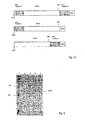
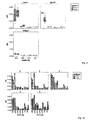
- Fig. 1A shows a schematic overview of the vector p89VP1.
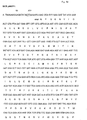
- Fig. 1B shows the DNA sequence of the multiple cloning site of the pET-17b vector and the 89VP1 encoding gene.
- Fig. 1C shows the schematic representation of three possibilities for creating nucleic acid fusions.
- Fig. 2 shows a Coomassie blue stained 12% SDS-PAGE gel containing purified 89VP1 his-tagged protein (Lane 1: 5 ⁇ g molecular marker; Lane 2-5: 10 ⁇ l 89VP1 elution samples).
- Fig. 3 shows IgG recognition of 14VP1: Immunoblotting of 14VP1 and controls. Dots are visualized by autoradiography (Lane 1-6: Incubation with 1:500-1:16000 diluted rabbit anti-14VP1 antiserum; Lane 7-12: Incubation with 1:500-1:16000 diluted preimmune serum).
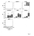
- Fig. 4 shows 89VP1-specific IgG1 response in mice.
- Groups of mice were immunized with different antigens as indicated on top of each box.
- 89VP1-specific IgG1 titers were measured by ELISA and are expressed as OD values on the y-axis. The results are shown as box plots, where 50% of the values are within the boxes and non-outliers between the bars. The line within the boxes indicates the median values.
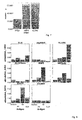
- Fig. 5 shows Phl p 1-specific IgG1 response in mice.
- Groups of mice were immunized with different antigens as indicated on top of each box.
- rPhl p 1-specific IgG1 titers were measured by ELISA and are expressed as optical value (OD 405nm) on the y-axis. The optical value corresponds to the level of IgG1 antibody in the mouse sera.
- the results are shown as box plots were 50% of the values are within the boxes and non-outliers between the bars. The line within the boxes indicates the median values.
- Fig. 6 shows Timothy grass pollen extract-specific IgG1 response in immunized mice.
- Groups of mice were immunized with different antigens as indicated on top of each box.
- Timothy grass pollen extract-specific IgG1 titers were measured by ELISA and are expressed as optical value (OD 405nm) on the y-axis. The optical value corresponds to the level of IgG1 antibody in the mouse sera.
- the results are shown as box plots, where 50% of the values are within the boxes and non-outliers between the bars. The line within the boxes indicates the median values.
- Fig. 7 shows the mean of %inhibition of patient's IgE-binding to rPhl p 1 by preincubation with antisera against rPhl p 1, r89P5 and KLHP5 of all 19 patients.
- the %inhibition is shown on the y-axis. The results are shown as bars.
- Fig. 8 shows the proliferation of spleen cells of immunized mice.
- T-cells of immunized mice with different antigens as indicated on top of each box were stimulated with peptide 5, 89VP1(89) and KLH. Medium was used as a reference. At the y-axis the stimulation index is shown. The results are displayed in bars.
- Fig. 9 shows IgG1,IgG2,IgG4 and IgA response to 14VP1, 89VP1 and rPhl p 1 detected in human sera by ELISA measurement.
- 10 patient's sera were tested for four antibodies specific for 89VP1, 14VP1and rPhl p 1 as indicated on top of each box.
- Sera taken in autumn and winter are shown on the left hand and right hand of each "bar pair", respectively.
- Titers were measured by ELISA and are expressed as optical value (OD 405nm) on the y-axis. The optical value corresponds to the level of antibody in the human sera.
- the results are shown as box plots, where 50% of the values are within the boxes and non-outliers between the bars. The line within the boxes indicates the median values.
- Fig. 10 shows the detection of anti-89VP1 and anti-rPhl p 1 antibodies in sera of allergic patients.
- 5 Phl p 1 allergic patient's sera were tested for seven antibodies specific for 89VP1 (left bar of each pair) and rPhl p 1 (right bar of each pair).
- Titers were measured by ELISA and are expressed as optical value (OD 405nm) on the y-axis. The optical value corresponds to the level of antibody in the human sera.
- the results are shown as box plots, where 50% of the values are within the boxes and non-outliers between the bars. The line within the boxes indicates the median values.
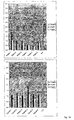
- Fig. 11 shows anti-14VP1 IgG binding to HRV14 protein extract and purified 14VP1 (Lane 1 and 4: 5 ⁇ g Marker; lane 2 and 4: Virus extract; lane 2 and 5: 5 ⁇ g 14VP1). Blot A und B was incubated with anti-14VP1 immune serum and preimmune serum, respectively. Bound IgG was detected by Western blotting and visualized by autoradiography.
- Fig. 12 shows the neutralization of HRV14 (Lane A (cell control): Cells after preincubation of HRV14 with a dilution of anti-14VP1 immune serum 1:10 2 -1:10 8 (row A1-A6); Lane B: Cells after preincubation of HRV14 with a dilution of preimmune serum 1:10 2 -1:10 8 (row B1-B6); Lane C: cells after Incubation with HR-V14 10 TCD50-10 6 TCD50 (row C1- C6); D5: cells after incubation with preimmune serum; D6: cells after incubation with immune serum). Cells were plated out in all wells and colored with crystal violet after three days.
- Fig. 13 shows IgG reactivity of anti-peptide antisera with the complete Phl p 5 allergen.
- IgG reactivities (OD values) to Phl p 5 are shown for 3 serum samples (bleeding 1: preimmune serum; bleedings 2-3: serum samples collected in monthly intervals) obtained from 6 rabbits, each of which was immunized with one of the KLH-conjugated peptides.
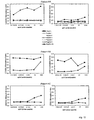
- Fig. 14 shows allergenic activity of rPhl p 5, and the peptide mix as detected by CD203c expression.
- Heparinized blood samples from three patients allergic to Phl p 5 were incubated with serial dilutions from 10 -4 to 10 ⁇ g/mL of recombinant allergen, an equimolar mix of Phl p 5 derived peptides, anti-IgE or control buffer (co, x-axis).
- Cells were then stained with CD203c mAb and analyzed for CD203c expression on a FACScan.
- the stimulation index (SI) is displayed on the y-axis.
- Fig. 15 shows identification of Phl p 5-derived peptides which induce low lymphoproliferative responses.
- PBMCs from timothy pollen allergic patients were stimulated with different concentrations of peptides and, for control purposes, with interleukin-2 (x-axis).
- Stimulation indices (SI) are indicated on the y-axis.
- Fig. 16 shows Fel d 1-derived synthetic peptides which induce Fel d 1 specific IgG immune responses in rabbits.
- Six rabbits were immunized with the KLH-conjugated Fel d 1-derived synthetic peptides or unconjugated rFel d 1 and 3-4 bleedings were drawn in monthly intervals.
- IgG reactivities of the preimmune sera and the antisera to ELISA plate-bound rFel d 1 are shown as optical densities (O.D. values, y-axis).
- Fig. 17 shows the low allergenic activity of Fel d 1-derived synthetic peptides as determined by CD63 and CD203c expression on basophils of allergic patients.
- PBMCs from 5 cat allergic patients were incubated with serial dilutions of Fel d 1 (closed boxes) or a mixture of Fel d 1-derived synthetic peptides (open boxes) (x-axis).
- PBMCs were also probed with serial dilutions of Fel d 1-derived synthetic peptides as single components.
- Induction of expression of surface markers CD203c and CD63 was measured as mean fluorescence intensities, and calculated stimulation indices are shown on the y-axis.
- Fig. 18 shows that a treatment with KLH-coupled Bet v 1-derived peptides reduces lymphoproliferative responses to rBet v 1 in sensitized mice.
- T-cell proliferation was measured in spleen cell cultures after in vitro stimulation with the recombinant birch pollen allergen Bet v 1 (white bars), KLH (black bars), or the peptide mix (grey bars). The bars represent the mean stimulation indices (SI ⁇ SD) for the different groups of mice.
- Fig. 19 shows the prophylactic vaccination of naive mice with KLH-coupled Bet v 1-derived peptides which reduces lymphoproliferative responses to rBet v 1 after sensitization.
- Fig. 20 shows that the prophylactic vaccination of naive mice with KLH-coupled Bet v 1-derived peptides induces a Bet v 1-specific IgG response and primes for the induction of allergen-specific IgG responses by the complete allergen.
- IgG responses (OD values: y-axis) to Bet v 1 were measured in the four treatment groups at different points of time (x-axis).
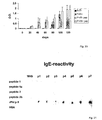
- Fig. 21 shows a comparison of IgE-reactivity: IgE binding capacity of Phl p 5 derived peptides (1, 2) and variants (1a, 2b) was determined in dot blot assays applying 0.2 ⁇ g/dot using sera from 7 grass-pollen allergic patients (p1-p7) and the serum from a non-atopic individual (NHS). rPhl p 5 was used as positive control and HSA as negative control. Bound IgE was detected with 125 I- labelled anti-human IgE.
- Fig. 22 shows a lymphoproliferative responses of Phl p 5 derived peptides (1, 2) and variants (1a, 2b).
- PBMCs from grass pollen allergic patients were stimulated with different concentrations of peptides and, for control purposes, with equimolar concentrations of rPhl p 5.
- Stimulation indices (SI) are indicated on the y-axis.
- Fig. 23 shows the cross protection of anti VP1 antibodies.
- Virus stock samples were prepared for RT-PCR by addition of 1ul of RNase inhibitor (Boehringer GmbH, Germany) to a final concentration of 0.01 U/ ⁇ l after RNA extraction from cell culture supernatants by QIAamp viral RNA kit (Qiagen, Germany).
- Plasmid p89VP1 ( Fig. 1A ) was constructed by replacing the NdeI/EcoRI fragment of the multiple cloning site of pET-17b with the cDNA sequence encoding for the VP1 protein of human rhinovirus strain 89 (89VP1).
- the NdeI and EcoRI restriction sites (in pET-17b) were used for insertion of AseI/EcoRI linked 89VP1 with ATG at the 5' end, six histidin followed by the stopcodon TGA at the 3' end ( Fig.1B ).
- NdeI/AseI fusion instead of the NdeI site CATAAT was created and could not be cut with any available enzyme. Therefore, the site was mutated to CTTAAG, the restriction site of Afl II.
- CTTAAG the restriction site of Afl II.
- ACCGTT sequence at the 3' end was mutated to ACCGGT, the restriction site of AgeI.
- the amino acid sequences are displayed below the nucleotide sequences of 89VP1.
- the restriction sites are shown underlined in Fig. 1B .
- cDNAs for gene fragments can thus be inserted for easy purification into the remaining restrictions sites either at the 5' end (using Afl II) or at the 3' end (using AgeI) of 89VP1 or into both sites as indicated in Figure 1C .
- Recombinant allergen fragments will thus be expressed at the N-terminus and/or the C-terminus of the 89VP1.
- Table I Cloning and mutagenesis primers (5' to 3') 89VP1 cloning SEQ ID No.
- 89VP1forward 1 89VP1 reverse 2 Mutagenesis Afl II forward 3 Afl II reverse 4 AgeI forward CCTGATGTTTTTACCGGTACAAACGTCCACCAC 5 AgeI reverse GTGGTGGACGTTTGTACCGGTAAAAACATCAGG 6
- Example 2 Cloning of a construct expressing a 89VP1-allergen fragment fusion protein
- Phl p 1 allergen fragment namely peptide 5
- the peptide 5 DNA sequence was amplified by PCR using the Phl p 1 cDNA (GenBank: X78813) as a template (Primers: Phl p 1 forward 5'-CGCGCTTAAGATGGTCCGCTACACCACCGAGGGC-3' (SEQ ID No. 7); Phl p 1 reverse 5'-CGCGCTTAAGCTTGGACTCGTAGGCGGTGTCGGC-3' (SEQ ID No. 8)).
- the PCR product was inserted into the Afl II restriction site of the p89VP1 vector, and the resulting construct is referred to as vector p89P5 and gene product r89P5.
- Example 3 Expression and purification of 89VP1 peptide 5-fusion protein and 89VP1
- plasmids were transformed into E. coli BL21 (DE3).
- Transformed E. coli cells were grown in 250 ml LB medium containing 100 mg/l ampicillin at 37°C to an optical density (600nm) of 0.4, and protein expression was induced by addition of isopropyl-beta-D-thiogalactosidase (IPTG) to a final concentration of 1 mM.
- IPTG isopropyl-beta-D-thiogalactosidase
- E. coli cells were harvested after 4 hours by centrifugation at 3500 rpm at 4°C for 10 min. Purification was performed with the Qiagen protocol using Qiagen Ni-NTA and columns.
- the cell pellet was resuspended under denaturing conditions in 5 ml 6 M guanidinium hydrochloride for 1 hour. After centrifugation (20 min, 10000 x g) the supernatant was incubated with 1 ml Ni-NTA for an additional hour. The suspension was then loaded on a column, washed twice with 5 ml wash buffer (8 M urea pH 6.3) and then eluted with 4 ml elution buffer (8 M urea pH 3.5). Renaturation was achieved after dialysis with decreasing molarity of urea.
- Protein bands correlated with the calculated protein size of 33.6 kD. Integrity of the proteins was also confirmed by Western blot analysis, using anti-histidine tag antibody.
- Example 5 89VP1-specific antibody response in immunized mice determined by ELISA
- mice Groups of 4 mice were immunized with 89VP1, r89P5 and 89P5edc and 2 mice were immunized with peptide 5 only (5 ⁇ g each and mixed 1:2 with aluminium hydroxide). The mice were immunized subcutaneously in approximately three-week intervals and bled from the tail veins. The 89VP1-specific IgG1 antibody level was determined by ELISA.
- ELISA plates (Nunc) were coated with 5 ⁇ g/ml 89VP1.
- the mice anti-89VP1, anti- r89P5 and peptide 5 sera were diluted 1:500.
- Bound IgG1 was detected with rat anti-mouse IgG1 (BD Pharmingen) 1:1000 and then with goat anti rat IgG POX-coupled (Amersham Bioscience) 1:2000.
- the optical value (OD 405nm) is displayed on the y-axis and corresponds to the level of IgG1 antibody in the mouse sera ( Fig.4 ).
- KLHP5, KLHP5edc, KLH and peptide 5 were used as controls.
- IgG1 antibodies were detected with an increasing titer during immunization in mice injected with 89VP1 (89VP1, 89P5edc and r89P5). Rabbits immunized with 89VP1, r89P5 and KLHP5 show the same result.
- Example 6 rPhl p 1-specific antibody response in immunized mice determined by ELISA
- Example 7 ELISA detection of timothy grass pollen extract-specific IgG1 antibodies
- mice and ELISA analysis were performed as described in section 5.
- Whole timothy grass pollen extract was coated (100 ⁇ g/ml) on ELISA plates and the IgG1 antibody titer was determined ( Fig.6 ).
- Example 8 Rabbit anti-r89P5 antibodies block patient's IgE-binding to rPhl p 1
- ELISA plates were coated with 1 ⁇ g/ml rPhl p 1, washed and blocked. The plates were preincubated with 1:100-diluted rabbit anti- peptide (89P5, KLHP5), a rabbit anti rPhl p 1 and, for control purposes, with the corresponding preimmune sera. After washing, plates were incubated with human sera from Phl p 1-allergic patients (1:3 diluted) and bound IgE was detected with mouse anti-human IgE (Pharmingen 1:1000) and then with sheep anti-mouse IgG POX-coupled (Amersham Bioscience) 1:2000. The percentage of inhibition of IgE-binding achieved by preincubation with the anti- peptide antisera was calculated as follows: 100-OD i /OD P x 100.
- ODi and OD P represent the extinctions after preincubation with the rabbit immune and preimmune serum, respectively.
- Table 2 shows the capacity of anti-Phl p 1 peptide antibodies to inhibit the binding of 19 allergic patients' IgE to complete rPhl p 1.
- Figure 7 displays the mean inhibition (in %) of all anti- rPhl p 1, anti-r89P5 and anti-KLHP5 immune sera. Anti-peptide sera blocked the IgE-binding much better then rPhl p 1. The ability for inhibition is with 89P5 and KLHP5 almost alike.
- Table 2 shows the inhibition (in %) of all 19 patients.
- Table 2 %inhibition of 19 patients' IgE-binding to rPhl p 1 after incubation with rabbit anti-rPhl p 1, r89P5 and anti-KLHP5 antisera % inhibition patient rPh1 p 1 r89P5 KLHP5 1 32.343 68.291 68.213 2 29.373 64.915 61.509 3 10.367 59.469 66.270 4 28.087 73.906 71.330 5 13.808 49.358 45.372 6 22.597 66.259 67.331 7 5.375 26.667 18.902 8 22.478 42.612 47.979 9 5.019 39.822 56.837 10 13.756 53.878 63.047 11 26.444 58.430 57.944 12 25.795 67.243 62.458 13 41.330 75.694 79.517 14 35.543 85.714 87.012 15 45.796 84.255 75.185 16 32.641 76.508
- Example 9 T-cell proliferation of mouse spleen cells after antigen stimulation
- mice Groups of three mice were immunized with KLH, KLHP5 and KLH-P5edc. Groups of 4 mice were immunized four times with 89VP1, r89P5 and 89P5edc, and 2 mice were immunized with peptide 5 only (5 ⁇ g each).
- Spleen cells were taken 10 days after the last immunization and single cell cultures were stimulated in triplicates in 96 well plates with peptide 5 (P5), 89VP1, KLH, Con A and a medium as a positive and negative control, respectively. After four days radioactive [ 3 H]thymidine 0.5 ⁇ Ci was added to each well. Cells were then harvested with a Packard Cell Harvester onto unifilter plates after 15 hours.
- Example 10 Detection of 14VP1-, 89VP1- and rPhl p 1-specific antibodies in human sera obtained in autumn and winter by ELISA
- the IgG1, IgG2, IgG4 and IgA antibody level against 14VP1, 89VP1 and rPhl p 1 was determined by ELISA as described in example 5.
- Human IgG1, IgG2, IgG4 and IgA were detected (BD Pharmingen) 1:1000 with sheep anti mouse IgG POX-coupled (Amersham Bioscience) 1:2000.
- a high anti-14VP1 and 89VP1 IgG1 titer of sera taken in autumn and winter could be detected ( Fig. 9 ).
- the anti-rPh1 p 1 IgG1 antibody titer was much lower.
- IgG2, IgG4 and IgA antibodies could be detected in all cases at a very low level.
- the VP1 proteins of the different HRV strains are closely related and cross-reactivity has been shown in other studies.
- Example 11 Anti- 89VP1 and anti-rPhl p 1 antibodies of Phl p 1 allergic patients
- Example 12 Detection of anti-14VP1 antibody binding to the whole virus by Western blot analysis
- the IgG antibody binding of sera of the 14VP1 injected rabbit to the whole virus was confirmed by using the whole HRV14 virus (lane 2 and 5) and 5 ⁇ g purified 14VP1 (lane 3 and 6) as control.
- the virus extract was separated by 12% SDS-Page and blotted onto nitrocellulose membrane.
- Rabbit anti-14VP1 antiserum (lane 1-3) 1:500 and preimmune serum (lane 4-6) 1:500 were tested for binding to HRV14 and 14VP1.
- Bound IgG was detected with 125I-labelled donkey anti-rabbit antibody and visualized by autoradiography ( Fig.11 ).
- the binding of 14VP1-antiserum could be detected at the same seize (33, 6 kD) as 14VP1.
- TCD50 tissue culture dosis 50
- the cytotoxic effect of the virus was visualized with crystal violet after three days and the TCD50 (the dilution where 50% of the cells are dead) was calculated.
- Serum dilutions and virus (100TCD50) in row A and B were incubated at 34 °C. After 2 hours cells were spread out in all wells. D5 and D6 are serum controls.
- the experimental schema is shown in Fig. 12 . The neutralization effect of the antibodies was detected after three days with crystal violet ( Fig. 12 ).
- peptides have isoelectric points between 4.32 and 8.98 and three of them (peptide 3, 4 and 6) may contain human T-cell epitopes.
- Table 4 Serological characterization of 29 grass pollen allergic patients and a non-allergenic control. Sex, age, total serum IgE levels (kU/L), timothy extract-specific IgE (kUA/L), IgE antibodies specific for rPhl p 5 and the 6 isolated (P1-P6) and KLH-coupled (KLH-P1-KLH-P6) peptides were measured by ELISA and ODs (optical densities) are shown. Dashes indicate the lack of IgE reactivity to the isolated as well as to the KLH-coupled peptides.
- ELISA plates (Nunc Maxisorp, Denmark) were coated with Phl p 5-derived peptides (5 ⁇ g/ml) or rPhl p 5 as control (5 ⁇ g/ml), washed and blocked. Subsequently, plates were incubated with 1:3 diluted sera from 29 grass pollen allergic patients and from a non-atopic individual overnight at 4°C. Grass pollen allergic patients suffering from allergic rhinoconjunctivitis and/or asthma were selected according to case history indicative for seasonal grass pollinosis and characterized by skin prick testing with timothy pollen extract and serological CAP-FEIA (Pharmacia Diagnostics, Uppsala, Sweden) testing.
- IgE levels in the sera were determined by CAP measurements (Pharmacia). IgE antibodies specific for rPhl p 5 were determined by ELISA. Sera from 29 grass pollen allergic patients and a non-atopic individual were used for IgE competition studies. The grass pollen allergic patients group consisted of 13 males and 16 females with a mean age of 35 years (ranging from 25-51 years) (Table 4).
- Bound IgE antibodies were detected with a 1:1000 diluted alkaline-phosphatase-coupled mouse monoclonal anti-human IgE antibody (Pharmingen, CA).
- Total IgE levels and grass pollen extract-specific IgE ranged from 24.9-2000 kU/L (mean: 262.7) and 2.2-100 kUA/L (mean: 41.5), respectively. All patients contained rPhl p 5-specific IgE antibodies ranging between 0.157-2.530 OD units (mean: 1.082 OD units), but none of the 29 patients showed IgE reactivity to any of the peptides (P1-P6) or to an equimolar mix of the peptides (OD ⁇ 0.08). This result demonstrates that no serum contained detectable IgE antibodies with specificity for any of the six Phl p 5 derived peptides.
- CD 203 c The upregulation of CD 203 c has been described as a surrogate marker for allergen-induced basophil activation and degranulation ( Hauswirth et al., J Allergy Clin Immunol 2002; 110:102 ). Therefore, the allergenic activity of complete rPhl p 5 allergen and an equimolar mix of peptides by measuring CD 203 c upregulation on basophils of grass pollen allergic patients was compared.
- RT room temperature
- erythrocytes were lysed with 2 mL FACS TM Lysing Solution. Cells were then washed, resuspended in PBS, and analyzed by two-color flow cytometry on a FACScan (Becton Dickinson), using Paint-a-Gate Software. Allergen-induced upregulation of CD203c was calculated from mean fluorescence intensities (MFIs) obtained with stimulated (MFIstim) and unstimulated (MFIcontrol) cells, and expressed as stimulation index (MFIstim : MFIcontrol). An SI of ⁇ 2.0 ( ⁇ 2-fold upregulation) was considered indicative of a specific response.
- MFIs mean fluorescence intensities
- Example 16 Immunization with Phl p 5 derived peptides induces IgG antibodies reactive with rPhl p.5 and natural allergens from different grass species
- HPLC-purified peptides were coupled to KLH (keyhole limpet hemocyanin, MW 4.5x10³- 1.3x10⁷, Pierce, USA) according to the manufacturers advice and purified using a Conjugation Kit (Sigma, USA).
- KLH keyhole limpet hemocyanin, MW 4.5x10³- 1.3x10⁷, Pierce, USA
- Rabbits were immunized with each of the KLH conjugated-peptides (200 ⁇ g/injection) and, for control purposes, with complete rPhl p 5 using Freunds complete and incomplete adjuvant (Charles River). Serum samples were obtained in four week intervals.
- ELISA detection plates (Nunc Maxisorp, Denmark) were coated with pollen allergen extracts (100 ⁇ g/ml: Phleum pratense, Lolium perenne, Poa pratensis Dactylis glomerata, Secale cereale, Triticum aestivum, Avena sativa, Hordeum vulgare, Anthoxanthum odoratum) or purified recombinant allergen (5 ⁇ g/ml: rPhl p 5).
- ELISA plates were washed and blocked and subsequently incubated with rabbit anti-peptide antisera and corresponding pre-immunsera diluted 1:2500. Bound rabbit IgG was detected with a HRP-coupled goat anti-rabbit Ig antiserum (Jackson Immunresearch, Pennsylvania). Results represent means of duplicate determination with an error of ⁇ 5% ( Figure 13 , Table 5).
- Table 5 Crossreactivity of anti-Phl p 5 peptide antisera with rPhl p 5 and natural group 5 allergens from Phleum pratense, Lolium perenne, Poa pratensis, Dactylis glomerata, Secale cereale, Triticum aestivum, Avena sativa, Hordeum vulgare, Anthoxanthum odoratum .
- IgG reactivities (OD values) of peptide antisera (anti-P1 to anti -P6) to Phl p 5 and pollen extracts from grass pollen are displayed for 6 rabbits which were immunized with KLH-conjugated Phl p 5-derived peptides (P1-P6).
- Example 17 Immunization with PhI p 5-derived peptides induces IgG antibodies which inhibit the binding of grass pollen allergic patients IgE to Phl p 5
- ELISA plates were coated with rPhl p 5 (1 ⁇ g/ml) and preincubated either with a 1:250 dilution of each of the anti-peptide antisera (anti-P1-anti-P6), a mixture of the anti-peptide antisera, an anti-rPhl p 5 antiserum or for control purposes, with the corresponding preimmune sera or a mixture of the preimmune sera. After washing, the plates were incubated with 1:3 diluted sera from 29 grass pollen allergic patients and bound IgE antibodies were detected with the alkaline phosphatase-coupled mouse monoclonal anti-human IgE antibody (Pharmingen).
- Table 6 Rabbit anti-Phl p 5 peptide antisera inhibit serum IgE-binding of timothy pollen allergic patients to Phl p 5.
- the percentage inhibition of IgE-binding to Phl p 5 is displayed for each patient after preincubation of Phl p 5 with anti-peptide antisera (anti-P1 - anti-P6), with a mix of the six anti-peptide antisera (anti-P1-P6) or with an anti-rPhl p 5 antiserum.
- the mean percentage inhibition is displayed in the bottom line. N.d.: not done.
- Example 18 Phl p 5-derived peptides induce low specific lymphoproliferative responses.
- PBMC Peripheral blood mononuclear cells
- PBMC peripheral blood mononuclear cells
- PBMCs from timothy pollen allergic patients were stimulated with different concentrations of synthetic peptides. Stimulation indices with peptides were significantly lower than with IL2. Phl p 5-derived peptides induced low specific lymphoproliferative responses. The lowest response was seen with peptide 5 followed by peptide 4.
- peptides suitable for cat allergy vaccination five peptides which are 30 to 36 amino acids long and cover the whole molecule were designed according to the known amino acid sequence of Fel d 1.
- Peptides were synthesized using a Fmoc (9-fluorenylmethoxycarbonyl)-strategy with HBTU (2-(1H-benzotriazol-1-yl) 1,1,3,3 tetramethyluronium hexafluorophosphate)-activation (0.1 mmol small-scale cycles) on the Applied Biosystems peptide synthesizer Model 433A (USA).
- Preloaded PEG-PS polyethylenglycol polystyrene
- resins (0.15-0.2 mmol/g loading
- PerSeptive Biosystems, UK were used as solid phase to build up the peptides. Chemicals were purchased from Applied Biosystems.
- Table 7 Molecular characteristics of Fel d 1-derived synthetic peptides. Position within the native Fel d 1 molecule, amino acid sequence, number of amino acids, calculated molecular weight (MW) and theoretical isoelectric point (pI) of the Fel d 1-derived synthetic peptides are shown. All peptides are soluble in water. Position Amino acid sequence aa length MW pI Pept. 1 SEQ ID No. 15 chain 1, aa 1-34 EICPAVKRDVDLFLTGTP-DEYVEQVAQYKALPVVC 35 3911 4.30 Pept. 2 SEQ ID No. 16 chain 1, aa 35-70 LENARILKNCVDAKMTEEDKEN-ALSLLDKIYTSPLC 36 4083 4.72 Pept.
- the five Fel d 1-derived synthetic peptides have molecular weights in the range of 3246 to 4083 Dalton and have calculated isoelectric points of from 4.30 to 4.93. All five peptides are watersoluble and Peptides 1, 2 and 3 may contain human T-cell epitopes (Table 7). Table 8.
- Example 20 Fel d 1-derived synthetic peptides have reduced IgE reactivity compared to rFel d 1 and Fel d 1-derived synthetic peptides lack allergenic activity
- Serum IgE reactivity to the Fel d 1-derived synthetic peptides in order to identify hypoallergenic peptides suited for vaccination was investigated.
- IgE-mediated cat allergy was based on anamnesis, skin prick testing (Allergopharma, Reinbek, Germany) and measurement of total serum IgE as well as of cat dander-specific serum IgE (CAP-FEIA, Pharmacia Diagnostics, Sweden). Non-allergic persons were included for control purposes.
- the IgE-binding capacity of the five Fel d 1-derived synthetic peptides was compared with that of the complete rFel d 1 allergen using sera from 14 cat allergic patients.
- ELISA plates Nunc Maxisorb, Denmark
- Fel d 1-derived synthetic peptides or rFel d 1 as control 0.5 ⁇ g/well
- Plates were then incubated overnight at 4°C with 1:5 diluted sera from cat allergic patients and from a non-atopic individual.
- Bound IgE antibodies were detected with a 1:2500 diluted horse-raddish-peroxidase labeled anti-human IgE antibody (KPL, USA).
- IgE-binding was observed for 6/14 sera to Peptide 1, for 3/14 sera to Peptide 2, for 1/14 sera to Peptide 3 and for 10/14 sera to Peptide 5.
- Measured OD units were between 0.051 and 1.998 for Peptide 1, between 0.060 and 0.123 for Peptide 2, 0.186 for Peptide 3 and between 0.056 and 0.677 for Peptide 5. In summary, all measured OD units were considerably lower than the respective values measured for the whole Fel d 1 allergen.
- Fel d 1-derived synthetic peptides have a reduced IgE reactivity compared to the whole Fel d 1 allergen. Fel d 1-derived synthetic peptides can therefore be considered hypoallergenic, providing the advantage of reduced IgE-mediated side-effects, when used in SIT.
- Heparinized blood samples were taken from 5 cat-allergic patients after informed consent had been given. Blood aliquots (100 ⁇ l) were incubated with serial dilutions of rFel d 1, Fel d 1-derived synthetic peptides as single components or as equimolar mixture, anti-IgE antibody or buffer (PBS) for 15 minutes at 37°C. After incubation, cells were washed in PBS containing 20 mM EDTA. Cells were then incubated with 10 ⁇ l of PE-conjugated CD203c mAb 97A6 and 20 ⁇ l of FITC-conjugated CD63 mAb CLB-gran12 for 15 minutes at room temperature.
- CD203c stimulation indices ranged from 1.1 (SM) to 3.2 (RR) for the lowest tested concentration of 0.001 ⁇ g rFel d 1/ml and from 3.6 (KC) to 6.2 (RR) for the highest tested concentration of 10 ⁇ g rFel d 1/ml.
- CD63 stimulation indices determined in the same assay ranged from 1.1 (RR) to 2.0 (MG) for the lowest tested rFel d 1 concentration of 0.001 ⁇ g/ml and from 3.9 (RR) to 7.3 (MG) for the highest tested rFel d 1 concentration of 10 ⁇ g/ml.
- RR 1.1
- MG 2.0
- MG 2.3
- MG 2.3
- MG 2.3
- MG 2.3
- Basophils from all five cat-allergic patients were probed with five increasing concentrations (0.005, 0.05, 0.5, 5 and 50 ⁇ g/ml) of an equimolar mix of the five Fel d 1-derived synthetic peptides.
- Basophils from patient RR were additionally probed with five increasing concentrations of the five single Fel d 1-derived synthetic peptides (0.001, 0.01, 0.1, 1 and 10 ⁇ g/ml).
- Peptides were found to be deficient in upregulating the basophil surface markers CD203c and CD63. Peptides were unable to induce any increased expression of CD203c and CD63 on cells of patient RR, KC and SM.
- Fel d 1-derived synthetic peptides provide the advantage of holding a lower allergenic activity than the whole Fel d 1 allergen. This is relevant for a decreased risk of IgE-mediated side-effects when Fel d 1-derived synthetic peptides are used in SIT.
- Example 21 Immunization with Fel d 1-derived synthetic peptides induces IgG antibodies reactive with the whole rFel d 1 allergen
- Fel d 1-derived synthetic peptides were shown to be deficient in IgE-binding.
- candidate molecules for vaccination which aims at the induction of allergen-specific IgG antibodies, peptides must retain specific allergen structures and must still be able to induce an IgG immune response specific for the whole allergen.
- immunization experiments in rabbits were performed.
- Rabbits (New Zealand White rabbits) were immunized with the immunogens (200 ⁇ g/injection) using CFA (first immunization) and IFA (first booster injection after 4 weeks; a second booster injection with incomplete adjuvant was given after 7 weeks) (Charles River Breeding Laboratories, Germany). Rabbits were bled 8 weeks after the first immunization and in four-week intervals thereafter.
- ELISA assays The induction of peptide- and rFel d 1-specific antibodies was monitored in ELISA assays.
- ELISA plates Nunc Maxisorb, Denmark
- rFel d 1 0.5 ⁇ g/well
- Plate-bound rFel d 1 was then probed in duplicates with 1:1000 diluted rabbit antisera and the corresponding rabbit. preimmune sera, and bound IgG was detected with an 1:2000 diluted hoseraddish-peroxidase labelled goat anti-rabbit antiserum (Jackson ImmunoResearch Inc., USA). Means of duplicates were calculated and showed errors of less than 5%.
- Anti-Peptide 1, anti-Peptide 2, anti-Peptide 4 and anti-Peptide 5 rabbit antisera showed IgG reactivities to Fel d 1 at about the same magnitude than the anti-Fel d 1 rabbit antiserum. Also the anti-Peptide 3 rabbit antiserum showed a distinct but a somewhat lower IgG reactivity to Fel d 1.
- Example 22 Fel d 1-derived synthetic peptides induce weaker lymphoproliferative responses than Fel d 1
- Desired candidate molecules for improved SIT do not only offer the advantage of reduced IgE-mediated side effects but also of reduced T-cell mediated side effects.
- lymphoproliferative assays were performed.
- PBMCs were isolated from 7 cat-allergic patients by Ficoll (Amersham Pharmacia Biotech, UK) densitiy gradient centrifugation.
- PBMC (2 x 10 5 ) were cultured in triplicates in 96-well plates (Nunclone, Nalgene Nunc International, Denmark) in 200 ⁇ l serum-free Ultra Culture medium (Cambrex, Belgium) supplemented with 2 mM L-glutamine (Sigma, USA), 50 ⁇ M ⁇ -mercaptoethanol (Sigma) and 0.1 mg gentamicin per ml (Sigma) at 37°C using 5% CO 2 in a humidified atmosphere.
- rFel d 1 and Fe d 1-derived sythetic peptides were stimulated with different concentrations (5, 2.5, 1.25 and 0.6 ⁇ g/well) of rFel d 1 and Fe d 1-derived sythetic peptides as single components or as equimolar mixture and, for control purposes, with 4U interleukin-2 or with medium alone.
- 0.5 ⁇ Ci per well 3 H-thymidine (Amersham Pharmacia Biotech) was added and 16 h thereafter, incorporated radioactivity was measured by liquid scintillation counting using a microbeta scintillation counter (Wallac ADL, Germany), and mean cpm were calculated from the triplicates.
- the stimulation index (SI) was calculated as the quotient of the cpm obtained by antigen or interleukin-2 stimulation and the unstimulated medium control.
- IL-2 stimulated proliferation of PBMC from all 7 tested cat-allergic patients, resulting in stimulation indices of 9.8 for RR, 5.2 for EB, 3.2 for KC, 6.7 for MG, 6.3 for SM, 15.7 for RA and of 13.9 for AR.
- Fel d 1-derived synthetic peptides which on an equimolar basis induce weaker lymphoproliferative responses than Fel d 1 can be identified.
- PBMCs from 7 cat-allergic patients were stimulated with serial dilutions of rFel d 1 or Fel d 1-derived synthetic peptides as single components. Specific lymphoproliferative responses are shown as stimulation indices.
- Example 23 IgG antibodies induced by immunization with Fel d 1-derived synthetic peptides inhibit binding of cat-allergic patients IgE to the whole Fel d 1 allergen
- ELISA competition assays The ability of peptide-induced rabbit Ig to inhibit the binding of allergic patients' IgE antibodies to complete rFel d 1 was examined in ELISA competition assays.
- ELISA plates Nunc Maxisorb, Denmark
- rFel d 1 0.05 ⁇ g/well
- Plate-bound rFel d 1 was then preincubated with 1:100 diluted rabbit anti-peptide antisera (single anti-peptide antisera as well as a mixture of anti-peptide antisera were used), rabbit anti-rfel d 1 antiserum, and for control purposes also with the respective rabbit preimmune. sera. After the plates had been washed, they were incubated with 1:5 diluted human sera from cat-allergic patients.
- Bound IgE antibodies were detected with a 1:2500 diluted horse-raddish-peroxidase labeled anti-human IgE antibody (KPL, USA).
- Preincubation of ELISA-plate bound Fel d 1 with the 5 anti-peptide rabbit antisera resulted in inhibition patterns that varied between the 14 different tested sera from cat-allergic patients.
- Anti-Peptide 1 rabbit antiserum blocked patients' IgE-binding to Fel d 1 for 13/14 tested patients' sera, anti-Peptide 2 rabbit antiserum for 8/14, anti-Peptide 3 rabbit antiserum for 13/14, anti-Peptide 4 rabbit antiserum for 9/14 and anti-Peptide 5 rabbit antiserum for 5/14.
- anti-Peptide 1 rabbit antiserum showed best inhibition rates with inhibitions from 0-55% (average 29%).
- anti-Peptide 2 rabbit antiserum inhibition rates of 0-18% (average 5%) could be achieved, with anti-Peptide 3 rabbit antiserum of 0-29% (average 11%), with anti-Peptide 4 rabbit antiserum of 0-24% (average 8%) and with anti-Peptide 5 rabbit antiserum of 0-18% (average 4%).
- Table 10 Rabbit antisera raised against Fel d 1-derived synthetic peptides inhibit binding of human IgE to Fel d 1. The percentage inhibition of IgE-binding to Fel d 1 achieved by preincubation of Fel d 1 with rabbit antisera are shown for 14 patients and as means. Preincubations were performed with 5 rabbit antisera raised against the 5 Fel d 1-derived synthetic peptides (anti-Peptide 1-5), a mix of the 5 anti-peptide antisera (Mix) and an antiserum raised against Fel d 1 (anti-rFel d 1).
- Patient anti-Peptide 1 anti-Peptide 2 anti-Peptide 3 anti-Peptide 4 anti-Peptide 5 Mix of the 5 anti-peptide antisera anti-rFel d 1 1 48 18 29 20 18 78 64 2 24 0 8 0 0 67 43 3 55 11 5 17 8 84 74 4 38 7 11 24 8 66 49 5 10 5 5 12 0 54 48 6 33 0 12 5 0 68 46 7 6 1 10 5 0 58 45 8 44 3 17 10 0 60 53 9 26 17 12 15 16 53 43 10 0 0 10 0 0 31 26 11 38 0 0 0 0 52 56 12 47 0 22 0 7 75 51 13 27 2 8 0 0 56 41 14 16 0 6 5 0 25 25 mean 29 5 11 8 4 59 47
- Example 24 Bet v 1-derived peptides induce Bet v 1-specific IgG responses receiving T-cell help from carrier-derived T-cell epitopes and reduce Bet v 1-specific T-cell proliferation.
- Bet v 1-derived, surface-exposed B cell peptides have already been shown to induce a protective Bet v 1-specific IgG response in a mouse model of therapeutic and prophylactic vaccination ( Focke M et al. Clin Exp Allergy (2004) 34:1525-1533 ).
- 6 Bet v 1-derived peptides were coupled to the carrier molecule KLH before immunization of mice.
- the Bet v 1-specific IgG response induced with these peptides (Table 1) is driven by the help of T-cell epitopes derived from the carrier but not from the Bet v 1 allergen.
- allergen-derived peptides lacking any IgE reactivity but containing allergen-derived T-cell epitopes induced side effects due to T-cell activation.
- Allergen-derived peptides lacking IgE and T-cell reactivity as exemplified for the Bet v 1 peptides induce neither IgE nor T-cell mediated side-effects during therapeutic vaccination.
- the peptides When used for prophylactic vaccination, the peptides will induce a Bet v 1-specific protective IgG response without priming of Bet v 1-specific T-cells. This should minimize the risk of pre-priming an allergic immune response through the vaccine which might pave the road for a subsequent allergic sensitization.
- Allergen-, peptide-, and carrier-specific lymphoproliferation was analyzed in a T-cell proliferation assay. Spleens were removed under sterile conditions and homogenized. After lysis of erythrocytes, cells were washed and resuspended in complete medium (RPMI, 10% fetal calf serum, 0.1 mg/ml gentamycin, 2 mM glutamine).
- Single cell suspensions were plated into 96-well round-bottom plates at a concentration of 2 x 10 5 cells/well and stimulated with concavalin A (0.5 ⁇ g/well) as a positive control, rBet v 1 (2 pg/well), KLH (2 ⁇ g/well), the peptide mix (0.34 ⁇ g of each peptide/well) or the medium alone for 4 days.
- the cultures were pulsed with 0.5 ⁇ Ci/well tritiated thymidine for 16 h and harvested.
- the proliferation responses were measured by scintillation counting.
- the ratio of the mean proliferation after antigen stimulation and medium control values, i.e. the stimulation index (SI) was calculated.
- Prophylactic treatment with Bet v 1-derived peptides induced Bet v 1-specific IgG responses without help by Bet v 1-specific T-cells. Furthermore, prophylactic treatment increased Bet v 1-specific IgG responses induced by the Bet v 1 allergen already 20 and 40 days after first sensitization ( Figure 20 ).
- Example 25 Der p 2-derived peptides showing reduced IgE binding capacity
- the IgE binding capacity of Der p 2-derived peptides was determined as described in examples 15.1 and 20.1 employing the peptides according to table 13 and using sera of individuals suffering from house dust mite allergy.
- Table 13 Der p 2-derived peptides Peptide Position Sequence SEQ ID No.
- T-cell reactivity and immunogenicity variants of Phl p 5 derived peptides were designed by increasing the length of peptide 1 (P1) and decreasing the length of peptide 2 (P2) by a few amino-acids (Table 15).
- Table 15 Position, sequence, length in number of aminoacids and molecular weight of synthetic Phl p 5 derived peptides (1, 2) and variants thereof (1a, 2b) Table 15: Variants of PHL p 5 - derived synthetic peptides Position aa Sequence Number of aa Molecular Weight (MW) Peptide 1 98-128 CGAASNKAFAEGLSGEPKGAAESSSKAALTSK 32 3026 1a 93-128 CFVATFGAASNKAFAEGLSGEPKGAAESSSKAALTSK 37 3592 Peptide 2 26-58 ADLGYGPATPAAPAAGYTPATPAAPAEAAPAGKC 34 3068 2b 26-53 ADLGYGPATPAAPAAGYTPATPAAPAEAC 29 2644
- PBMCs from 2 grass pollen allergic patients were stimulated with different concentrations of Phl p 5 derived peptides 1, 2, their variants 1a, 2b and for control purposes with rPhl p 5. Stimulation indices obtained with the peptides were significantly lower than those obtained with rPhl p 5 ( Fig. 22 ).
- Table 16 Cross-reactivity of anti-Phl p 5 peptide antisera raised in rabbits by immunization with KLH-conjugated peptides. IgG reactivities of peptide antisera to peptides (1, 2) and variants (1a, 2b) are displayed. No reactivity was observed with preimmune-sera (pre P1, pre P1a, pre P2, pre P2b)
- Table 17 Inhibition of patients' IgE binding to rPhl p 5 by anti peptide antisera.
- Anti peptide 2 and anti peptide 2b antisera inhibit patients' IgE binding to rPhl p 5 to the same extent.
- ELISA plate bound rPhl p 5 was preincubated with anti P2, anti P2b and, for control purposes, with anti Phl p 5 antisera. Plates were then exposed to sera from 12 grass pollen allergic patients. The percentage of inhibition of IgE-binding to rPhl p 5 is displayed.
- Table 17 % Inhibition of IgE-binding patient anti P2 anti P2b anti Phl p 5 1 33.38 24.40 84.77 2 52.20 57.40 87.00 3 52.70 54.85 90.81 4 51.44 59.76 78.26 5 43.19 49.15 77.93 6 47.04 52.02 83.68 7 62.67 58.00 76.62 8 52.36 50.27 74.44 9 57.63 50.91 88.13 10 35.10 37.99 75.03 11 44.44 41.24 68.39 12 47.56 45.41 77.34
- Human rhinoviruses comprise over hundred different strains. In this neutralization test it is shown that the rhinovirus infection of one strain can also be inhibited by VP1 specific antibodies of another strain. HeLa cells were seeded out in the wells at equal density. 100TCD 50 HRV14 was preincubated with dilutions of anti-14VP1- and anti-89VP1 antibodies (undiluted; 1:2-1:32 wells 1-6) and added to the wells in lane A and D, respectively. In lanes B and C 100TCD 50 HRV 89 preincubated with dilutions of anti-14VP1- and anti-89VP1 antibodies, respectively, were added to the cells. After 3 days live cells were stained violet. Anti-89VP1 antibodies and anti-14VP1 antibodies block the infection of HRV14 in a comparable manner. The antibodies raised against 14VP1 and 89VP1 also inhibit the infection of HRV89 up to the same concentration.
Landscapes
- Health & Medical Sciences (AREA)
- Chemical & Material Sciences (AREA)
- Life Sciences & Earth Sciences (AREA)
- Organic Chemistry (AREA)
- Medicinal Chemistry (AREA)
- General Health & Medical Sciences (AREA)
- Biochemistry (AREA)
- Veterinary Medicine (AREA)
- Pharmacology & Pharmacy (AREA)
- Animal Behavior & Ethology (AREA)
- Public Health (AREA)
- Immunology (AREA)
- Biophysics (AREA)
- Proteomics, Peptides & Aminoacids (AREA)
- Molecular Biology (AREA)
- Genetics & Genomics (AREA)
- Epidemiology (AREA)
- Mycology (AREA)
- Microbiology (AREA)
- Botany (AREA)
- Gastroenterology & Hepatology (AREA)
- Chemical Kinetics & Catalysis (AREA)
- Nuclear Medicine, Radiotherapy & Molecular Imaging (AREA)
- General Chemical & Material Sciences (AREA)
- Oncology (AREA)
- Virology (AREA)
- Engineering & Computer Science (AREA)
- Bioinformatics & Cheminformatics (AREA)
- Communicable Diseases (AREA)
- Pulmonology (AREA)
- Peptides Or Proteins (AREA)
- Medicines Containing Antibodies Or Antigens For Use As Internal Diagnostic Agents (AREA)
- Micro-Organisms Or Cultivation Processes Thereof (AREA)
- Preparation Of Compounds By Using Micro-Organisms (AREA)
- Medicines That Contain Protein Lipid Enzymes And Other Medicines (AREA)
Description
- The present invention relates to novel hypoallergenic molecules and uses thereof.
- Type I allergy is an IgE-mediated hypersensitivity disease affecting almost 25% of the population. It is based on the recognition of harmless airborne, insect, venom, food allergen and contact allergen antigens derived from per se harmless antigen sources such as pollen, insects, mold and animal proteins by specific immunoglobulin E. The crosslinking of effector cell-bound IgE antibodies leads to a release of inflammatory mediators (e.g., histamine, leucotrienes) and thus to the immediate symptoms of allergy (e.g., rhinoconjunctivitis, asthma, dermatitis, anaphylaxis). T-cell activation via IgE-dependent as well as IgE-independent mechanisms contributes to chronic allergic inflammation.
- The probably only causative forms of allergy treatment is allergen-specific immunotherapy, which is based on the repeated administration of increasing amounts of allergen extracts for most sources. Numerous clinical studies have documented the clinical efficacy of injection immunotherapy and there is evidence for several immunological mechanisms underlying this treatment. Due to the difficulty to prepare high quality allergen extracts for certain allergen sources and the fact that the administration of allergens to patients can cause severe side effects, allergen-specific immunotherapy can only be recommended for certain patients groups and disease manifestations. It is especially difficult to treat patients with co-sensitizations to several different allergen sources and patients suffering from severe disease manifestations such as allergic asthma. Allergic asthma is one of the most vigorous manifestations of allergy, because it severely affects the quality of daily life, causes a high rate of hospitality admissions and can manifest itself in serious, life-threatening forms requiring intensive care of the patient.
- Allergen extracts prepared from natural allergen-sources are crude in nature, and it is impossible to influence the quality and amounts of individual allergens in such preparations by technical means. They also contain numerous undefined non-allergenic components, and several recent studies indicate the poor quality of such extracts and document their great heterogeneity.
- In the last decade great progress has been made in the field of molecular allergen characterization using recombinant DNA technology. A large number of the most important disease-eliciting allergens has been characterized down to the molecular level, and recombinant allergens mimicking the epitope complexity of natural allergen extracts have been produced. Moreover, several research groups have used the knowledge regarding allergen structures to develop defined new allergy vaccines. Genetic engineering, synthetic peptide chemistry and conjugation of allergens with immunostimulatory DNA sequences have been used to reduce the allergenic activity of the new vaccines and thus the rate of therapy-induced side effects. First promising clinical studies were conducted with such allergen derivatives. Interestingly, it turned out that although IgE-reactivity of genetically engineered recombinant allergens and allergen-derived synthetic T-cell epitope-containing peptides could be strongly reduced or even abolished, these derivatives still could induce systemic side effects appearing several hours after injection. For example, it was reported that T-cell epitope peptides of the major cat allergen,
Fel d 1, induced asthma and bronchial hyper reactivity several hours after intracutaneous injection, and there is strong evidence that this effect is T-cell mediated and MHC-restricted. - These results indicate that the removal of IgE-reactivity diminishes IgE-mediated side effects since no immediate reactions were recorded in the course of these immunotherapy studies. However, the allergen-specific T-cell epitopes which have been preserved in the recombinant allergen derivatives as well as in the peptide mixtures are responsible for the late side effects (e.g. very problematic or atopic dermatitis, chronic T cell-mediated allergic skin manifestation). The side effects caused in the case of recombinant allergen-derivatives were relatively mild and in the case of the T-cell peptide vaccines may be overcome by adequate dosing. Both of the two new approaches therefore seem very promising for immunotherapy of allergic rhinoconjunctivitis but may have limitations when it comes to the treatment of severe forms of allergic asthma, where the induction of late side effects in the lung may be very problematic.
- In order to administer and consequently to provoke an efficient immune response against peptides, polypeptides and proteins, adjuvants and/or carriers are regularly used. Complete Freund's adjuvant, for instance, is one of the most potent adjuvants available. However, because of its side effects, its use is not approved for humans. Therefore, there exists a need for vaccine compositions able to induce strong immune responses against peptides and polypeptides derived from allergens and of course of other antigens avoiding the use of complete Freund's adjuvant. Further, while BSA has been used successfully as a carrier in animal models it may not be appropriate for use in human vaccine compositions because of the risk of adverse reactions such as the risk of transmitting prion disease (variant Creutzfeldt-Jakob disease). A further challenge to the development of an effective vaccine against allergens is the need for an immune response able to rapidly decrease allergens in an individual or animal. Therefore, high concentrations of allergen-specific antibodies in the blood, which are mainly of the IgG subtype, are needed. In mucosal surfaces IgA antibodies are the primary subtype.
- Cholera toxin, a known carrier protein in the art, is also used regularly as an adjuvant, eliminating the need for complete Freund's adjuvant in a vaccine composition. However, cholera toxin increases total and specific IgE antibody levels and leads to IgE-associated inflammatory reactions.
- Due to the side effects provoked by most carrier proteins used for vaccination, there exists a need for carrier systems which are able to stimulate immune responses against allergens or other antigens, without using toxic adjuvants, without using poorly tolerated carrier proteins and, in certain situations, without stimulation of potentially pathologic immune responses. Novel carrier systems meeting these specifications can be used towards the formation of novel conjugates and compositions suitable for the treatment or prevention of diseases like allergic diseases.
- In Bohle B. et al. (J. Immunol. 172 (11) (2004): 6642-6648) a recombinant fusion protein comprising an S-layer protein moiety and Bet v 1 moiety is described. This molecule comprises the native hyperallergenic Bet v 1 protein.
-
WO 2004/004761 relates to virus like particles which are fused to an immunogen and which may be used for immunisation. - In
WO 2004/003143 the use of fusion proteins comprising a virus like particle and a hyperallergenic molecule as immunogen for vaccination is disclosed. -
WO 03/072601 A -
WO 02/40676 A -
WO 2006/058359 A relates to a method for producing hypoallergenic proteins by splitting up a wild-type protein into two parts and rejoining the resulting fragments in inverse orientation. For protein purification the hypoallergenic molecule according to,this document is fused to a his-tag. - Breitweiser et al., Protein Eng.; 15(3) (2002): 243-249, describe a recombinant bacterial cell surface (S-Layer)-major birch pollen allergen fusion protein.
- In Elfman et al., Int. Arch. Allergy Immunol., 117(3) (1998):167-173, fusion proteins are described which comprise a GST moiety fused to fragments of the allergen Lep d2 which exhibit reduced IgE binding affinity.
- Bauer et al., J. Allergy and Clin. Immunol., 118 (2006):269-276, relate to the generation of hypoallergenic DNA vaccines encoding the major birch pollen allergen Bet v1 stably linked to ubiquitin which is a well known marker in an eukaryotic cell and gives the signal to degrade the marked polypeptide.
-
WO 2004/092210 A relates to fusion proteins of the Fve polypeptide (comprising immunomodulatory activity) and an allergen as fusion partner. This document discloses hypoallergenic peptides derived from major grass pollenallergen Phl p 1, which are coupled to the non-allergenic carrier protein KLH. - In
WO 02/12503 A -
WO 2006/008018 A discloses his-tag fusion proteins which comprise hypoallergenic molecules. - In Gonzalez et al., Mol. Immunol, 43(6) (2006) :570-578, GST fusion proteins are disclosed which consist of a GST moiety and peptides derived from the allergen Ole e1.
- In
WO 2005/085278 A fusion proteins are disclosed which comprise hypoallergenic allergens fused to another allergen. - In
US 2006/088549 immunogenic compositions comprising a chimeric human rhinovirus are disclosed, which consist of a nucleic acid molecule encoding for a human rhinovirus capsid protein fused to a heterologous nucleic acid sequence encoding for an antigen. - In
WO 02/04007 -
EP 0 358 485 - It is an object of the present invention to provide medicaments and carriers which overcome the aforementioned drawbacks and allow an allergen vaccination with reduced side effects.
- Therefore, the present invention relates to a hypoallergenic protein for use in the treatment or prevention of an allergy in a human or animal consisting of at least one hypoallergenic molecule derived from an allergen, which is fused to at least one second non-allergenic protein, wherein the at least one hypoallergenic molecule derived from an allergen exhibits an at least 50% reduced IgE-binding capacity and an at least 30% reduced T-cell reactivity compared to the wild-type allergen and wherein the at least one second protein is a viral protein.
- In order to provoke an enhanced immune response against a molecule, in particular of a hypoallergenic molecule according to the present invention, said molecule is fused (by genetic engineering) to a carrier.
- The second protein (the "carrier" or "carrier protein") to be fused to a hypoallergenic molecule of the invention is not derived from an allergen ("non-allergenic"). However, the carrier protein used in the present invention may exhibit T cell reactivity and/or provoke an immune response against itself and the hypoallergenic molecule fused or conjugated to it when administered to an animal or human body. Consequently, if the carrier protein is derived from a pathogen (e.g. virus, bacteria etc.), (protecting) antibodies directed to said carrier and pathogens are produced.
- As used herein, "hypoallergenic protein" means a fusion protein/polypeptide of a carrier of a non-allergenic source with a hypoallergenic molecule. Furthermore, a "hypoallergenic protein" is also intended to be a conjugation product (e.g. chemical coupling, adsorption) of a carrier with a hypoallergenic molecule.
- "Hypoallergenic" as used herein, refers to molecules with reduced allergenic potential. Such molecules have a decreased capacity to provoke allergic reactions in an individual compared to the wild-type protein from which these molecules are derived.
- The at least one hypoallergenic molecule derived from an allergen and fused to a second protein is preferably C- and/or N-terminally truncated. "C- and/or N-terminal truncation", as used herein, means that amino acid residues either from the N-terminus or from the C-terminus or both from the N- and C-terminus of the wild-type allergen are removed by deletion of at least 1, 2, 3, 4, 5, 7, 10, 15, 20, 30 amino acid residues.
- The hypoallergenic molecules, i.e. peptides/polypeptides, comprise preferably 10 to 50 amino acids, more preferably 15 to 40 amino acids, in particular 20-30 amino acids and exhibit reduced IgE reactivity. These molecules are designed to exclude T-cell epitopes which may cause T-cell-mediated side effects. T-cell epitopes and molecules exhibiting reduced T-cell response may be determined and identified by methods known by the person skilled in the art (e.g., Bercovici N. et al. Clin Diagn Lab Immunol. (2000) 7:859-864).
- It was found that it is possible to design peptide vaccines derived from allergens like the major grass pollen allergens, e.g.,
Phl p 1, and for the major birch pollen allergen,Bet v 1, using surface exposed peptides. The data obtained show that such peptide vaccines can be produced for any allergen whose primary structure is known according to IgE epitope mapping, three-dimensional structure data or computer-aided prediction of surface-exposed domains. However, the selection of suitable peptides which may be used for vaccination remains crucial, because not all peptides identified with these methods can be employed in vaccination. The peptides suitably used for vaccination purposes exhibit reduced IgE-binding capacity and - in order to reduce or avoid late side effects - exhibit reduced T-cell reactivity. - The term "derived from an allergen", as used herein, means that the hypoallergenic molecules according to the present invention are obtained directly from an allergen by fragmentation or truncation. The amino acid sequence of the hypoallergenic molecules of the present invention are preferably at least 80% identical, more preferably at least 90% identical, most preferably at least 95% identical, in particular 100% identical, to the amino sequence stretch of the wild-type allergen, from which the hypoallergenic molecule is derived. However, the molecules which are not 100% identical to the wild-type allergen fragments should be able to bind with at least 60%, preferably at least 70%, more preferably at least 80%, most preferably at least 90%, strength to an antibody or to antibodies, preferably to IgG antibodies, which are directed to said wild-type allergen fragments.
- The degree of identity of a first amino acid sequence to a second amino acid can be determined by a direct comparison between both amino acid sequences using certain algorithms. Such algorithms are, for instance, incorporated in various computer programs (e.g. "
BLAST 2 SEQUENCES (blastp)" (Tatusova et al. (1999) FEMS Microbiol. Lett. 174:247-25; Corpet F, Nucl. Acids Res. (1988) 16:10881-10890). - The truncated molecules according to the present invention can be defined as being parts of the complete allergen that induce less activation of allergen-specific T cells than the complete wild-type allergen ( at least a 30%, preferably at least a 50%, most preferably at least a 70%, reduction), exhibit a more than 50% reduced (preferably more than 70%) allergenic activity as evaluated by IgE binding assays and ability to induce IgE-mediated cell activation and when coupled to a carrier as described induce IgG antibodies which inhibit the binding of polyclonal IgE from allergic patients to the complete wild-type allergen.
- The peptides should contain sequences from the allergens to avoid overlaps with the mimotopes. Mimotopes, however, which are small peptide mimics (less than 15 amino acids) of antigen pieces and are obtained from random peptide libraries do not represent original, allergen-derived molecules as defined herein. They can not be used according to the invention because they are too small to induce a robust blocking IgG response.
- The hypoallergenic molecules according to the present invention may be obtained by recombinant methods or chemical synthesis. Alternatively, it is, of course, also possible to obtain the molecules by enzymatic or chemical cleavage of the wild-type allergen or a polypeptide/protein harbouring the molecule of interest.
- The hypoallergenic molecule may comprise preferably at least two truncated allergen molecules derived from at least one allergen, wherein the order of the truncated allergen fragments differs from the order of the fragments in the wild-type allergen if the at least two molecules are derived from the same allergen.
- The hypoallergenic molecule according to the present invention may comprise one or more (preferably at least 2, more preferably at least 3) hypoallergenic molecules as defined herein, thus, resulting in a fusion protein. The single hypoallergenic molecules of the fusion protein, which, of course, also lacks IgE-binding capacity and lacks T-cell epitopes, may be derived from allergens of the same and/or of different origin. If the molecules are derived from the same allergen, the order in the hypoallergenic fusion protein should not be identical to the order in the wild-type allergen (this prevents the reconstitution and formation of IgE-binding sites) (see, e.g.,
WO2004/065414 , Linhart B and Valenta R (Int Arch Allergy Immunol. (2004) 134:324-31)). - According to a preferred embodiment of the present invention the at least one hypoallergenic molecule is fused to the N-terminus and/or C-terminus of said at least one second protein or fragment thereof.
- The allergen or fragments thereof may be conjugated chemically, e.g., or by recombinant methods to each other. If the allergen or fragment thereof is conjugated chemically to a carrier, said allergen or fragment should be provided with a terminal cysteine residue (resulting in a free sulfhydryl group). To said terminal (N- or C-terminal) cysteine residue any maleimide-activated carrier protein may be conjugated, thus creating an immunogen/carrier complex. If the allergen or fragment thereof does not have a sulfhydryl group at a terminus, EDC (1-ethyl-3-(3-dimethylaminopropyl) carbodiimide hydrochloride) chemistry in order to couple amines (lysine) or carboxylic acids (glutamic, aspartic acid or 5'-phosphate) to the carrier protein may be employed.
- If the hypoallergenic molecule fused to the N- or C-terminus of the carrier, recombinant methods are employed.
- The at least one second protein is a viral, in particular RNA or DNA viral, protein.
- The at least one second protein ("carrier") may be of any of the above-mentioned origin. It is, however, in particular preferred to use proteins which provoke an immune response against the protein itself and the hypoallergenic molecule fused or conjugated thereto. Due to the induction of formation of (protective) antibodies directed also to the at least one second protein, the hypoallergenic protein according to the present invention may also be employed as vaccine for said second protein and its originating source (e.g. virus).
- The viral protein according to the present invention is preferably a capsid protein.
- Viral capsid proteins are especially suited because they induce antiviral activity, provoke the formation of antibodies which block adhesion of viruses, e.g. rhinoviruses, to epithelial cells, exhibit an immuno-modulatory activity towards a Thl response, increase the immunogenicity of the peptide (i.e., higher anti-peptide and hence higher levels of protective IgG antibodies), are suited and proved for prophylactic vaccination (virus vaccination) and are safe, when capsid proteins are used to whose humans are continuously exposed (e.g. rhinoviruses).
- According to another preferred embodiment of the present invention the at least one viral capsid protein is derived from a human pathogenic virus, preferably a virus of the family of picornaviridae.
- The virus of the family of picornaviridae is preferably of the genus of rhinoviruses, preferably of the species of human rhinoviruses, in particular
human rhinovirus 89 and 14. The capsid protein may be VP1, VP2, VP3 and/or VP4. - The allergen to be fused to a viral capsid protein is preferably selected from the group consisting of major birch pollen allergens, in
particular Bet v 1 andBet v 4, major timothy grass pollen allergens, inparticular Phl p 1,Phl p 2,Phl p 5,Phl p 6 andPhl p 7, major house dust mite allergens, inparticular Der p 1 andDer p 2, major catallergen Fel d 1, major bee allergens, major wasp allergens, profilins, especiallyPhl p 12, and storage mite allergens, especiallyLep d 2. - Other suited allergens to be used according to the present invention can be derived from the following table.
-
Species Name Allergen Name Biochem.ID or Obsolete name MW cDNA (C) or protein (P) Reference, Acc.No. Ambrosia artemisiifolia short ragweed Amb a 1 antigen E 8 C 8, 20 Amb a 2 antigen K 38 C 8, 21 Am b a 3 Ra3 11 C 22 Amb a 5 Ra5 5 C 11, 23 Amb a 6 Ra6 10 C 24, 25 Amb a 7 Ra7 12 P 26 Ambrosia trifida giant ragweed Amb t 5 Ra5G 4.4 C 9, 10, 27 Artemisia vulgaris mugwort Art v 1 27-29 C 28 Art v 2 35 P 28A Art v 3 lipid transfer protein 12 P 53 Art v 4 profilin 14 C 29 Helianthus annuus sunflower Hel a 1 34 29A Hel a 2 profilin 15.7 C Y15210 Mercurialis annua Mer a 1 profilin 14-15 C Y13271 Caryophyllales Chenopodium album lamb's-quarters, pigweed, Che a 1 17 C AY049012, 29B white goosefoot Che a 2 profilin 14 C AY082337 Che a 3 polcalcin 10 C AY082338 Salsola kali Russian-thistle Sal k 1 43 P 29C Rosales Humulus japonicus Japanese hop Hum j 4w C AY335187 Parietaria judaica Par j 1 lipid transfer protein 1 15 C see list of isoallergens Par j 2 lipid transfer protein 2 C see list of isoallergens Par j 3 profilin C see list of isoaller-gens Parietaria officinalis Par o 1 lipid transfer protein 15 29D B. Grasses Poales Cynodon dactylon Bermuda grass Cyn d 1 32 C 30, S83343 Cyn d 7 C 31, X91256 Cyn d 12 profilin 14 C 31a, Y08390 Cyn d 15 9 C AF517686 Cyn d 22w enolase data pending Cyn d 23 Cyn d 14 9 C AF517685 Cyn d 24 Pathogenesis- related p. 21 P pending Dactylis glomerata orchard grass Dac g 1 AgDg1 32 P 32 Dac g 2 11 C 33, S45354 Dac g 3 C 33A, U25343 Dac g 5 31 P 34 Festuca pratensis meadow fescue Fes p 4w 60 - Holcus lanatus velvet grass Hol l 1 C Z27084 Lolium perenne rye grass Lol p 1 group I 27 C 35, 36 Lol p 2 group II 11 P 37, 37A, X73363 Lol p 3 group III 11 P 38 Lol p 5 Lol p IX, Lol p Ib 31/35 C 34, 39 Lol p 11 hom: trypsin inhibitor 16 39A Phalaris aquatica canary grass Pha a 1 C 40, S80654 Phleum pratense timothy Phl p 1 27 C X78813 Phl p 2 C X75925, 41 Phl p 4 P 41A Phl p 5 Ag25 32 C 42 Phl p 6 C Z27082, 43 Phl p 11 trypsin inhibitor hom. 20 C AF521563, 43A Phl p 12 profilin C X77583, 44 Phl p 13 polygalacturonase 55-60 C AJ238848 Poa pratensis Kentucky blue grass Poa p 1 group I 33 P 46 Poa p 5 31/34 C 34, 47 Sorghum halepense Johnson grass Sor h 1 C 48 C. Trees Arecales Phoenix dactylifera date palm Pho d 2 profilin 14.3 C Asturias p.c. Fagales Alnus glutinosa alder Aln g 117 C S50892 Betula verrucosa birch Bet v 1 17 C see list of isoaller- gens Bet v 2 profilin 15 C M65179 Bet v 3 C X79267 Bet v 4 8 C X87153, S54819 Bet v 6 h: isoflavone reductase 33.5 C see list of isoaller- gens Bet v 7 cyclophilin 18 P P81531 Carpinus betulus hornbeam Car b 1 17 C see list of isoaller-gens Castanea sativa chestnut Cas s 1 22 p 52 Cas s 5 chitinase Cas s 8 lipid transfer protein 9.7 P 53 Corylus avellana hazel Cor a 1 17 C see list of isoaller-gens Cor a 2 profilin 14 C Cor a 8 lipid transfer protein 9 C Cor a 9 11S globulin- like protein 40/? C Beyer p.c. Cor a 10 luminal binding prot. 70 C AJ295617 Cor a 11 7S vicilin-like prot. 48 C AF441864 Quercus alba White oak Que a 1 17 P 54 Lamiales Oleaceae Fraxinus excelsior ash Fra e 1 20 P 58A, AF526295 Ligustrum vulgare privet Lig v 120 P 58A Olea europea olive Ole e 1 16 C 59, 60 Ole e 2profilin 15-18 C 60A Ole e 3 9.2 60B Ole e 4 32 P P80741 Ole e 5 superoxide dismutase 16 P P80740 Ole e 6 10 C 60C, U86342 Ole e 7 ? P 60D, P81430 Ole e 8 Ca2+-binding protein 21 C 60E, AF078679 Ole e 9 beta-1,3-glucanase 46 C AF249675 Ole e 10 glycosyl hydrolase hom. 11 C 60F, AY082335 Syringa vulgaris lilac Syr v 120 P 58A Plantaginaceae Plantago lanceolata English plantain Pla l 1 18 P P842242 Pinales Cryptomeria sugi japonica Cry j 141-45 C 55, 56 Cry j 2 C 57, D29772 Cupressus cypress arisonica Cup a 1 43 C A1243570 Cupressus sempervirens common cypress Cup s 1 43 C see list of isoaller-gens Cup s 3w 34 C ref pending Juniperus ashei mountain cedar Jun a 1 43 P P81294 Jun a 2 C 57A, AJ404653 Jun a 3 30 P 57B, P81295 Juniperus oxycedrus prickly juniper Jun o 4 hom: calmodulin 29 C 57C, AF031471 Juniperus mountain cedar sabinoides Jun s 1 50 P 58 Juniperus virginiana eastern red cedar Jun v 1 43 P P81825, 58B Platanaceae Platanus acerifolia London plane tree Pla a 1 18 P P82817 Pla a 2 43 P P82967 Pla a 3 lipid transfer protein 10 P Iris p.c. D. Mites Acarus siro mite Aca s 13 arthropod fatty acid binding prot. 14* C AJ006774 Blomia tropicalis mite Blo t 1cysteine protease 39 C AF277840 Blo t 3 trypsin 24* C Cheong p.c. Blo t 4alpha amylase 56 C Cheong p.c. Blo t 5 C U59102 Blo t 6 chymotrypsin 25 C Cheong p.c. Blo t 10tropomyosin 33 C 61 Blo t 11paramyosin 110 C AF525465, 61A Blo t 12 Bt11a C U27479 Blo t 13 Bt6, fatty acid bind prot. C U58106 Blo t 19 anti-microbial pep. hom. 7.2 C Cheong p.c. Dermatophagoides farinae American house dust mite Der f 1 cysteine protease 25 C 69 Der f 214 C 70, 70A, see list of isoallergens Der f 3trypsin 30 C 63 Der f 724-31 C SW:Q26456, 71 Der f 10tropomyosin C 72 Der f 11paramyosin 98 C 72A Der f 14 mag3, apolipophorin C D17686 Der f 15 98k chitinase 98 C AF178772 Der f 16 gelsolin/villin 53 C 71A Der f 17 Ca binding EF protein 53 C 71A Der f 18w 60k chitinase 60 C Weber p.c. Dermatophagoides house dust mite microceras Der m 1 cysteine protease 25 P 68 Dermatophagoides pteronyssinus European house dust mite Der p 1 antigen P1, cysteine protease 25 C 62, see list of isoallergens Der p 214 C 62A-C, see list of isoallergens Der p 3trypsin 2&/30 C 63 Der p 4amylase 60 P 64 Der p 514 C 65 Der p 6chymotrypsin 25 P 66 Der p 722/28 C 67 Der p 8glutathione transferase C 67A Der p 9 collagenolytic serine pro. P 67B Der p 10 tropomyosin 36 C Y14906 Der p 14 apolipophorin like prot. C Epton p.c. Euroglyphus maynei mite Eur m 2 C see list of isoaller-gens Eur m 14 apolipophorin 177 C AF149827 Glycyphagus storage mite domesticus Gly d 2 C 72B, see isoallergen list Lepidoglyphus storage mite destructor Lep d 2 Lep d 1 15 C 73, 74, 74A, see isoallergen list Lep-d 5 C 75, AJ250278 Lep d 7 C 75, AJ271058 Lep d 10 tropomyosin C 75A, AJ250096 Lep d 13 C 75, AJ250279 Tyrophagus storage mite putrescentiae Tyr p 2 C 75B, Y12690 E. Animals Bos domesticus domestic cattle Bos d 2 Ag3, lipocalin 20 C 76, see isoallergen list (see also foods) Bos d 3 Ca-binding S100 hom. 11 C L39834 Bos d 4 alpha-lactalbumin 14.2 C M18780 Bos d 5 beta-lactoglobulin 18.3 C X14712 Bos d 6 serum albumin 67 C M73993 Bos d 7 immunoglobulin 160 77 Bos d 8 caseins 20-30 77 Canis familiaris (Canis domesticus) Can f 1 25 C 78, 79 dog Can f 2 27 C 78, 79 Can f 3 albumin C S72946 Can f 4 18 P A59491 Equus caballus domestic horse Equ c 1 lipocalin 25 C U70823 Equ c 2 lipocalin 18.5 P 79A, 79B Equ c 3 Ag3 - albumin 67 C 79C, X74045 Equ C 4 17 P 79D Equ c 5 AgX 17 P Goubran Botros p.c. Felis domesticus cat (saliva) Fel d 1cat-1 38 C 15 Fel d 2albumin C 79E, X84842 Fel d 3 cystatin 11 C 79F, AF238996 Fel d 4 lipocalin 22 C AY497902 Fel d 5w immunoglobulin A 400 Adedoyin p.c. Fel d 6w immunoglobulin M 800-1000 Adedoyin p.c. Fel d 7w immunoglobulin G 150 Adedoyin p.c. Cavia porcellus guinea pig Cav p 1 lipocalin homologue 20 P SW:P83507, 80 Cav p 217 P SW:P83508 Mus musculus mouse (urine) Mus m 1MUP 19 C 81, 81A Rattus norvegius rat (urine) Rat n 117 C 82, 83 F. Fungi (moulds) 1. Ascomycota 1.1 Dothideales Alternaria alternata Alt a 1 28 C U82633 Alt a 2 25 C 83A, U62442 Alt a 3 heat shock prot. 70 C U87807, U87808 Alt a 4 prot. disulfideisomerase 57 C X84217 Alt a 6 acid ribosomal prot. P2 11 C X78222, U87806 Alt a 7 YCP4 protein 22 C X78225 Alt a 10 aldehyde dehydrogenase 53 C X78227, P42041 Alt a 11 enolase 45 C U82437 Alt a 12 acid ribosomal prot. P1 11 C X84216 Cladosporium herbarum Cla h 1 13 838, 83C Cla h 2 23 83B, 83C Cla h 3 aldehyde dehydrogenase 53 C X78228 Cla h 4 acid ribosomal prot. P2 11 C X78223 Cla h 5 YCP4 protein 22 C X78224 Cla h 6 enolase 46 C X78226 Cla h 12 acid ribosomal prot. P1 11 C X85180 1.2 Eurotiales Aspergillus flavus Asp f1 13 alkaline serine protease 34 84 Aspergillus fumigatus Asp f 118 C M83781, S39330 Asp f 2 37 C U56938 Asp f 3 peroxisomal protein 19 C U20722 Asp f 4 30 C AJ001732 Asp f 5 metalloprotease 40 C Z30424 Asp f 6 Mn superoxide dismut. 26.5 C U53561 Asp f 7 12 C AJ223315 Asp f 8 ribosomal prot. P2 11 C AJ224333 Asp f 9 34 C AJ223327 Asp f 10 aspartic protease 34 C X85092 Asp f 11 peptidyl-prolyl isomeras 24 84A Asp f 12 heat shock prot. P90 90 C 85 Asp f 13 alkaline serine protease 34 84B Asp f 15 16 C AJ002026 Asp f 16 43 C g3643813 Asp f 17 C AJ224865 Asp f 18 vacuolar serine protease 34 84C Asp f 22w enolase 46 C AF284645 Asp f 23 L3 ribosomal protein 44 C 85A, AF464911 Aspergillus niger Asp n 14 beta-xylosidase 105 C AF108944 Asp n 18 vacuolar serine protease 34 C 84B Asp n 25 3-phytase B 66-100 C 85B, P34754 Asp n ? 85 C Z84377 Aspergillus oryzae Asp o 13 alkaline serine protease 34 C X17561 Asp o 21 TAKA-amylase A 53 C D00434, M33218 Penicillium brevicompactum Pen b 13 alkaline serine protease 33 86A Penicillium chrysogenum (formerly P. notatum) Pen ch 13 alkaline serine protease 34 87 Pen ch 18 vacuolar serine protease 32 87 Pen ch 20 N-acetyl glucosaminidas 68 87A Penicillium citrinum Pen c 3 peroxisomal mem. prot. 18 86B Pen c 13 alkaline serine protease 33 86A Pen c 19 heat shock prot. P70 70 C U64207 Pen c 22w enolase 46 C AF254643 Pen c 24 elongation factor 1 betaC AY363911 Penicillium oxalicum Pen o 18 vacuolar serine protease 34 87B 1.3 Hypocreales Fusarium culmorum Fus c 1 ribosomal prot. P2 11* C AY077706 Fus c 2 thioredoxin-like prot. 13* C AY077707 1.4 Onygenales Trichophyton rubrum Tri r 2 C 88 Tri r 4serine protease C 88 Trichophyton tonsurans Tri t 130 P 88A Tri t 4 serine protease 83 C 88 1.5 Saccharomycetales Candida albicans Cand a 1 40 C 89 Cand a 3 peroxisomal protein 29 C AY136739 Candida boidinii Cand b 2 20 C J04984, J04985 2. Basidiomycotina 2.1 Hymenomycetes Psilocybe cubensis Psi c 1 Psi c 2cyclophilin 16 89A Coprinus comatus shaggy cap Cop c 1 leucine zipper protein 11 C AJ132235 Cop c 2 AJ242791 Cop c 3 AJ242792 Cop c 5 AJ242793 Cop c 7 AJ242794 2.2 Urediniomycetes Rhodotorula mucilaginosa Rho m 1 enolase 47 C 89B Rho m 2 vacuolar serine protease 31 C AY547285 2.3 Ustilaginomycetes Malassezia furfur Mala f 2 MF1, peroxisomal membrane protein 21 C AB011804, 90 Mala f 3 MF2, peroxisomal membrane protein 20 C AB011805, 90 Mala f 4 mitochondrial malate dehydrogenase 35 C AF084828, 90A Malassezia sympodialis Mala s 1 C X96486, 91 Mala s 5 18* C AJ011955 Mala s 6 17* C AJ011956 Mala s 7 C AJ011957, 91A Mala s 8 19* C AJ011958, 91A Mala s 9 37* C AJ011959, 91A Mala s 10 heat shock prot. 70 86 C AJ428052 Mala s 11 Mn superoxide dismut. 23 C AJ548421 3. Deuteromycotina 3.1 Tuberculariales Epicoccum purpurascens (formerly E. nigrum) Epi p 1serine protease 30 P SW:P83340, 91B G. Insects Aedes aegyptii mosquito Aed a 1 apyrase 68 C L12389 Aed a 2 37 C M33157 Apis mellifera honey bee Api m 1 phospholipase A2 16 C 92 Api m 2hyaluronidase 44 C 93 Api m 4melittin 3 C 94 Api m 67-8 P Kettner p.c. Api m 7 CUB serine protease 39 C AY127579 Bombus pennsylvanicus bumble bee Bom p 1 phospholipase 16 P 95 Bom p 4 protease P 95 Blattella germanica German cockroach Bla g 1 Bd90k C Bla g 2 aspartic protease 36 C 96 Bla g 4 calycin 21 C 97 Bla g 5 glutathione transferase 22 C 98 Bla g 6 troponin C 27 C 98 Periplaneta americana American cockroach Per a 1 Cr-PII C Per a 3 Cr-PI 72-78 C 98A Per a 7 tropomyosin 37 C Y14854 Chironomus kiiensis midge Chi k 10 tropomyosin 32.5* C AJ012184 Chironomus thummi thummi midge Chi t 1-9 hemoglobin 16 C 99 Chi t 1.01 component III 16 C P02229 Chi t 1.02 component IV 16 C P02230 Chi t 2.0101 component I 16 C P02221 Chi t 2.0102 component IA 16 C P02221 Chi t 3 component II-beta 16 C P02222 Chi t 4 component IIIA 16 C P02231 Chi t 5 component VI 16 C P02224 Chi t 6.01 component VIIA 16 C P02226 Chi t 6.02 component IX 16 C P02223 Chi t 7 component VIIB 16 C P02225 Chi t 8 component VIII 16 C P02227 Chi t 9 component X 16 C P02228 Ctenocephalides cat flea felis felis Cte f 1 Cte f 2 M1b 27 C AF231352 Cte f 3 25 C Thaumetopoea pityocampa pine processionary moth Tha p 1 15 P PIR:A59396, 99A Lepisma saccharina silverfish Lep s 1 tropomyosin 36 C AJ309202 Dolichovespula maculata white face hornet Dol m 1 phospholipase A1 35 C 100 Dol m 2 hyaluronidase 44 C 101 Dol m 5 antigen 5 23 C 102, 103 Dolichovespula arenaria yellow hornet Dol a 5 antigen 5 23 C 104 Polistes annularies wasp Pol a 1 phospholipase A1 35 P 105 Pol a 2 hyaluronidase 44 P 105 Pol a 5 antigen 5 23 C 104 Polistes dominulus Mediterranean paper wasp Pol d 1 Hoffman p.c. Pol d 4serine protease 32-34 C Hoffman p.c. Pol d 5P81656 Polistes exclamans wasp Pol e 1 phospholipase A1 34 P 107 Pol e 5antigen 523 C 104 Polistes fuscatus wasp Pol f 5 antigen 523 C 106 Polistes gallicus wasp Pol g 5 antigen 524 C P83377 Polistes metricus wasp Pol m 5 antigen 523 C 106 Vespa crabo European hornet Vesp c 1 phospholipase 34 P 107 Vesp c 5antigen 523 C 106 Vespa mandarina giant asian hornet Vesp m 1 Hoffman p.c. Vesp m 5P81657 Vespula flavopilosa yellowjacket Ves f 5antigen 5 23C 106 Vespula germanica yellowjacket Ves g 5antigen 5 23C 106 Vespula yellowjacketmaculifrons Ves m 1 phospholipase A1 33.5 C 108 Ves m 2hyaluronidase 44 P 109 Ves m 5antigen 523 C 104 Vespula pennsylvanica yellowjacket Ves p 5antigen 523 C 106 Vespula squamosa yellowjacket Ves s 5 antigen 523 C 106 Vespula vidua wasp Ves vi 5 antigen 523 C 106 Vespula vulgaris yellowjacket Ves v 1phospholipase A1 35 C 105A Ves v 2 hyaluronidase 44 P 105A Ves v 5antigen 523 C 104 Myrmecia pilosula Australian jumper ant Myr p 1 C X70256 Myr p 2 C S81785 Solenopsis tropical fire geminata ant Sol g 2 Hoffman p.c. Sol g 4Hoffman p.c. Solenopsis invicta fire ant Sol i 2 13 C 110, 111 Sol i 324 C 110 Sol i 413 C 110 Solenopsis saevissima Brazilian fire ant Sol s 2 Hoffman p.c. Triatoma protracta California kissing bug Tria p 1 Procalin 20 C AF179004, 111A. H. Foods Gadus callarias cod Gad c 1 allergen M 12 C 112, 113 Salmo salar Atlantic salmon Sal s 1 parvalbumin 12 C X97824 Bos domesticus domestic cattle Bos d 4 alpha-lactalbumin 14.2 C M18780 (milk) Bos d 5 beta-lactoglobulin 18.3 C X14712 see also animals Bos d 6 serum albumin 67 C M73993 Bos d 7 immunoglobulin 160 77 Bos d 8 caseins 20-30 77 Cyprinus carpio (Common carp) Cyp c 1 parvalbumin 12 C 129 Gallus domesticus chicken Gal d 1 ovomucoid 28 C 114, 115 Gal d 2 ovalbumin 44 C 114, 115 Gal d 3 Ag22, conalbumin 78 C 114, 115 Gal d 4 lysozyme 14 C 114, 115 Gal d 5 serum albumin 69 C X60688 Metapenaeus ensis shrimp Met e 1 tropomyosin C U08008 Penaeus aztecus shrimp Pen a 1 tropomyosin 36 P 116 Penaeus indicus shrimp Pen i 1 tropomyosin 34 C 116A Penaeus monodon black tiger shrimp Pen m 1 tropomyosin 38 C Pen m 2 arginine kinase 40 C AF479772, 117 Todarodes pacificus squid Tod p 1 tropomyosin 38 P 117A Helix aspersa brown garden snail Hel as 1 tropomyosin 36 C Y14855, 117B Haliotis midae abalone Hal m 1 49 117C Rana esculenta edible frog Ran e 1 parvalbumin alpha 11.9* C AJ315959 Ran e 2 parvalbumin beta 11.7* C AJ414730 Brassica juncea oriental mustard Bra j 1 2S albumin 14 C 118 Brassica napus rapeseed Bra n 1 2S albumin 15 P 118A, P80208 Brassica rapa turnip Bra r 2 hom: prohevein 25 P81729 Hordeum vulgare barley Hor v 15 BMAI-1 15 C 119 Hor v 16 alpha-amylase Hor v 17 beta-amylase Hor v 21 gamma-3 hordein 34 C 119A, SW:P80198 Secale cereale rye Sec c 20 secalin see isoall. list Triticum aestivum wheat Tri a 18 agglutinin Tri a 19 omega-5 gliadin 65 P PTR:A59156 Zea mays maise, corn Zea m 14 lipid transfer prot. 9 P P19656 Oryza sativa rice Ory s 1 C 119B, U31771 Apium graveolens celery Api g 1 hom: Bet v 116* C Z48967 Api g 4 profilin AF129423 Api g 5 55/58 P P81943 Daucus carota carrot Dau c 1 hom: Bet v 116 C 117D, see isoallergen list Dau c 4 profilin C AF456482 Corylus avellana hazelnut Cor a 1.04 hom: Bet v 117 C see list of isoaller-gens Cor a 2 profilin 14 C AF327622 Cor a 8 lipid transfer protein 9 C AF329829 Malus domestica apple Mal d 1 hom: Bet v 1 C see list of isoaller- gens Mal d 2 hom: thaumatin C AJ243427 Mal d 3 lipid transfer protein 9 C Pastorello p.c. Mal d 4 profilin14.4* C see list of isoaller-gens Pyrus communis pear Pyr c 1 hom: Bet v 118 C AF05730 Pyr c 4 profilin 14 C AF129424 Pyr c 5 hom: isoflavone reductas 33.5 C AF071477 Persea americana avocado Pers a 1 endochitinase 32 C Z78202 Prunus armeniaca apricot Pru ar 1 hom: Bet v 1 C U93165 Pru ar 3 lipid transfer protein 9 P Prunus avium sweet cherry Pru av 1 hom: Bet v 1 C U66076 Pru av 2 hom: thaumatin C U32440 Pru av 3 lipid transfer protein 10 C AF221501 Pru av 4 profilin 15 C AF129425 Prunus domestica European plum Pru d 3 lipid transfer protein 9 P 119C Prunus persica peach Pru p 3 lipid transfer protein 10 P P81402 Pru p 4 profilin 14 C see isoallergen list Asparagus Asparagus officinalis Aspa o 1 lipid transfer protein 9 P 119D Crocus satins saffron crocus Cro s 1 21 Varasteh A-R p.c. Lactuca sativa lettuce Lac s 1 lipid transfer protein 9 Vieths p.c. Vitis vinifera grape vit v 1lipid transfer protein 9 P P80274 Musa x paradisiaca banana Mus xp 1 profilin 15 C AF377948 Ananas comosus pineapple Ana c 1 profilin 15 C AF377949 Ana c 2 bromelain 22.8* C 119E-G, D14059 Citrus limon lemon Cit 1 3 lipid transfer protein 9 P Torrejon p.c. Citrus sinensis sweet orange Cit s 1 germin-like protein 23 P Torrejon p.c. Cit s 2 profilin 14 P Torrejon p.c. Cit s 3 lipid transfer protein 9 P Torrejon p.c. Litchi chinensis litchi Lit c 1 profilin 15 C AY049013 Sinapis alba yellow mustard Sin a 1 2S albumin 14 C 120 Glycine max soybean Gly m 1 HPS 7 P 120A Gly m 2 8 P A57106 Gly m 3 profilin 14 C see list of isoaller-gens Gly m 4 (SAM22) PR-10 prot. 17 C X60043, 120B Vigna radiata mung bean Vig r 1 PR-10 protein 15 C AY792956 Arachis hypogaea peanut Ara h 1 vicilin 63.5 C L34402 Ara h 2 conglutin 17 C L77197 Ara h 3 glycinin 60 C AF093541 Ara h 4 glycinin 37 C AF086821 Ara h 5 profilin 15 C AF059616 Ara h 6 hom: conglutin 15 C AF092846 Ara h 7 hom: conglutin 15 C AF091737 Ara h 8 PR-10 protein 17 C AY328088 Lens culinaris lentil Len c 1 vicilin 47 C see list of isoaller-gens Len c 2 seed biotinylated prot. 66 P 120C Pisum savitum pea Pis s 1 vicilin 44 C see list of isoaller-gens Pis s 2 convicilin 63 C pending Actinidia kiwi chinensis Act c 1 cysteine protease 30 P P00785 Act c 2 thaumatin-like protein 24 P SW:P81370, 121 Capsicum annuum bell pepper Cap a 1w osmotin-like protein 23 C AJ297410 Cap a 2 profilin 14 C AJ417552 Lycopersicon esculentum tomato Lyc e 1 profilin 14 C AJ417553 Lyc e 2 b-fructofuranosidase 50 C see isoallergen list Lyc e 3 lipid transfer prot. 6 C U81996 Solanum tuberosum potato Sola t 1 patatin 43 P P15476 Sola t 2 cathepsin D inhibitor 21 P P16348 Sola t 3 cysteine protease inhibitor 21 P P20347 Sola t 4 aspartic protease inhibitor 16+4 P P30941 Bertholletia excelsa Brazil nut Ber e 1 2S albumin 9 C P04403, M17146 Ber e 2 11S globulin seed storage protein 29 C AY221641 Juglans nigra black walnut Jug n 1 2S albumin 19* C AY102930 Jug n 2 vicilin-like prot. 56* C AY102931 Juglans regia English walnut Jug r 1 2S albumin C U66866 Jug r 2 vicilin 44 C AF066055 Jug r 3 lipid transfer protein 9 P Pastorello Anacardium occidentale Cashew Ana o 1 vicilin-like protein 50 C see isoallergen list Ana o 2 legumin-like protein 55 C AF453947 Ana o 3 2S albumin 14 C AY081853 Ricinus communis Castor bean Ric c 1 2S albumin C P01089 Sesamum indicum sesame Ses i 1 2S albumin 9 C 121A, AF240005 Ses i 2 2S albumin 7 C AF091841 Ses i 3 7S vicilin-like globulin 45 C AF240006 Ses i 4 oleosin 17 C AAG23840 Ses i 5 oleosin 15 C AAD42942 Cucumis melo muskmelon Cuc m 1 serine protease 66 C D32206 Cuc m 2 profilin 14 C AY271295 Cuc m 3 pathogenesis-rel p. PR-1 16* P P83834 I. Others Anisakis simplex nematode Ani s 1 24 P 121B, A59069 Ani s 2 paramyosin 97 C AF173004 Ani s 3 tropomyosin 41 C 121C, Y19221 Ani s 4 9 P P83885 Argas reflexus pigeon tick Arg r 1 17 C AJ697694 Ascaris suum worm Asc s 1 10 P 122 Carica papaya papaya Car p 3w papain 23.4* C 122A, M15203 Dendronephthya soft coral nipponica Den n 1 53 P 122B Hevea brasiliensis rubber (latex) Hev b 1 elongation factor 58 P 123, 124 Hev b 2 1,3-glucanase 34/36 C 125 Hev b 3 24 P 126, 127 Hev b 4 component of microhelix,complex 100-115 P 128 Hev b 5 16 C U42640 Hev b 6.01 hevein precursor 20 C M36986, p02877 Hev b 6.02 hevein 5 C M36986, p02877 Hev b 6.03 C-terminal fragment 14 C M36986, p02877 Hev b 7.01 hom: patatin from B-serum 42 C U80598 Hev b 7.02 hom: patatin from C-serum 44 C AJ223038 Hev b 8 profilin 14 C see list of isoaller-gens Hev b 9 enolase 51 C AJ132580 Hev b 10 Mn superoxide dismut. 26 C see list of isoaller-gens Hev b 11 class 1 chitinase C see list of isoaller-gens Hev b 12 lipid transfer protein 9.3 C AY057860 Hev b 13 esterase 42 P P83269 Homo sapiens human autoallergens Hom s 1 73* C Y14314 Hom s 2 10.3* C X80909 Hom s 3 20.1* C X89985 Hom s 4 36* C Y17711 Hom s 5 42.6* C P02538 Triplochiton scleroxylon obeche Trip s 1 class 1 chitinase 38.5 P Kespohl p.c. -
- 1 Marsh, D.G., and L.R. Freidhoff. 1992. ALBE, an allergen database. IUIS, Baltimore, MD, Edition 1.0.
- 2 Marsh, D. G. et al. 1986. Allergen nomenclature. Bull WHO 64:767-770.
- 3 King, T.P. et al. 1964. Biochemistry 3:458-468.
- 4 Lowenstein, H. 1980. Allergy 35:188-191.
- 5 Aukrust, L. 1980. Allergy 35:206-207.
- 6 Demerec, M. et al. 1966. Genetics 54:61-75.
- 7 Bodmer, J. G.et al. 1991. Immunogenetics 33:301-309.
- 8 Griffith, I.J. et al. 1991. Int. Arch. Allergy Appl. Immunol. 96:296-304.
- 9 Roebber, M. et al. 1985. J. Immunol. 134:3062-3069.
- 10 Metzler, W. J. et al. 1992. Biochemistry 31:5117-5127.
- 11 Metzler, W. J. et al. 1992. Biochemistry 31:8697-8705.
- 12 Goodfriend, L. et al. 1979. Fed. Proc. 38:1415.
- 13 Ekramoddoullah, A. K. M. et al. 1982. Mol. Immunol. 19:1527-1534.
- 14 Ansari, A. A. et al. 1987. J. Allergy Clin. Immunol. 80:229-235.
- 15 Morgenstern, J.P. et al. 1991. Proc. Natl. Acad. Sci. USA 88:9690-9694.
- 16 Griffith, I.J. et al. 1992. Gene 113:263-268.
- 17 Weber, A. et al. 1986. Biochem. Physiol. 83B:321-324.
- 18 Weber, A. et al. 1987. Allergy 42:464-470.
- 19 Stanworth, D. R. et al. 1990. Bulletin WHO 68:109-111.
- 20 Rafnar, T. et al. 1991. J. Biol. Chem. 266: 1229-1236.
- 21 Rogers, B.L. et al. 1991. J. Immunol. 147:2547-2552.
- 22 Klapper, D.G. et al. 1980. Biochemistry 19:5729-5734.
- 23 Ghosh, B. et al. 1993. J. Immunol. 150:5391-5399.
- 24 Roebber, M. et al. 1983. J. Immunol. 131:706-711.
- 25 Lubahn, B., and D.G. Klapper. 1993. J. Allergy Clin. Immunol. 91:338.
- 26 Roebber, M., and D.G. Marsh. 1991. J. Allergy Clin. Immunol. 87:324.
- 27 Goodfriend L. et al. Mol Immunol 22: 899-906, 1985.
- 28 Himly M. et al. FASEB J 17: 106-108, 2003.
- 28A Nilsen, B. M. et al. 1991. J. Biol. Chem. 266:2660-2668.
- 29 Wopfner N. et al. Biol Chem 383: 1779-1789, 2002.
- 29A Jimenez A. et al. 1994. Int Arch Allergy Immunol 105:297-307.
- 29B Barderas R. et al. Int Arch Allergy Immunol 127: 47-54, 2002.
- 29C Carnés J. et al. Allergy 56, Supplement 68: 274, 2001.
- 29D Giuliani A. et al. Allergy 42: 434-440, 1987.
- 30 Smith,P.M. et al. 1996. J. Allergy Clin. Immunol. 98:331-343.
- 31 Suphioglu,C. et al. 1997. FEBS Lett. 402:167-172.
- 31a Asturias J.A. et al. 1997. Clin Exp Allergy 27:1307-1313.
- 32 Mecheri, S. et al. 1985. Allergy Appl. Immunol. 78:283-289.
- 33 Roberts, A.M. et al. 1993. Allergy 48:615-623.
- 33a Guerin-Marchand,C. et al. 1996. Mol. Immunol. 33:797-806.
- 34 Klysner, S. et al. 1992. Clin. Exp. Allergy 22: 491-497.
- 35 Perez, M. et al. 1990. J. Biol. Chem. 265:16210-16215.
- 36 Griffith, I. J. et al. 1991. FEBS Letters 279:210-215.
- 37 Ansari, A. A. et al. 1989. J. Biol. Chem. 264:11181-11185. 37a Sidoli,A. et al. 1993. J. Biol. Chem. 268:21819-21825.
- 38 Ansari, A. A. et al. 1989. Biochemistry 28:8665-8670.
- 39 Singh, M. B. et al. 1991. Proc. Natl. Acad. Sci. 88:1384-1388.
- 39a van Ree R. et al. 1995. J Allergy Clin Immunol 95:970-978.
- 40 Suphioglu,C. and Singh,M.B. 1995. Clin. Exp. Allergy 25:853-865.
- 41 Dolecek,C. et al. 1993. FEBS Lett. 335:299-304.
- 41A Fischer S. et al. 1996. J Allergy Clin Immunol 98:189-198.
- 42 Matthiesen, F., and H. Lowenstein. 1991. Clin. Exp. Allergy 21:297-307.
- 43 Petersen,A. et al. 1995. Int. Arch. Allergy Immunol. 108:55-59.
- 43A Marknell DeWitt A. et al. Clin Exp Allergy 32: 1329-1340, 2002.
- 44 Valenta,R. et al. 1994. Biochem. Biophys. Res. Commun. 199:106-118.
- 46 Esch, R. E., and D. G. Klapper. 1989. Mol. Immunol. 26:557-561.
- 47 Olsen, E. et al. 1991. J. Immunol. 147:205-211.
- 48 Avjioglu, A. et al. 1993. J. Allergy Clin. Immunol. 91:340.
- 52 Kos T. et al. 1993. Biochem Biophys Res Commun 196:1086-92.
- 53 Diaz-Perales A. et al. 2000. Clin Exp Allergy 30:1403-1410.
- 54 Ipsen, H., and O.C. Hansen. 1991. Mol. Immunol. 28: 1279-1288.
- 55 Taniai, M. et al. 1988. FEBS Lett. 239:329-332.
- 56 Griffith, I.J. et al. 1993. J. Allergy Clin. Immunol. 91:339.
- 57 Sakaguchi, M. et al. Allergy 45: 309-312, 1990.
- 57A Yokoyama M. et al. Biochem Biophys Res Commun 275: 195-202, 2000.
- 57B Midoro-Horiuti T. et al. J Immunol 164: 2188-2192, 2000.
- 57C Tinghino R. et al. J. Allergy Clin. Immunol. 101: 772-777, 1998.
- 58 Gross GN et al. Scand J Immunol 8: 437-441, 1978.
- 58A Obispo TM et al. Clin Exp Allergy 23: 311-316, 1993.
- 58B Midoro-Horiuti T. et al. Clin Exp Allergy 31: 771-778, 2001.
- 59 Lombardero M. et al. Clin. Exp. Allergy 24: 765-770, 1994.
- 60 Villalba, M. et al. Eur. J. Biochem. 216: 863-869, 1993.
- 60A Asturias JA et al. J Allergy Clin Immunol 100: 365-372, 1997.
- 60B Batanero E. et al. Eur J Biochem 241: 772-778, 1996.
- 60C Batanero E. et al. FEBS Lett. 410: 293-296, 1997.
- 60D Tejera ML et al. J Allergy Clin Immunol 104: 797-802, 1999.
- 60E Ledesma A. et al. FEBS Lett 466: 192-196, 2000.
- 60F Barral P. et al. J Immunol 172: 3644-3651, 2004.
- 61 Yi FC et al. Clin Exp Allergy 32: 1203-1210, 2002.
- 61A Ramos JD et al. Int Arch Allergy Immunol 126: 286-293, 2001.
- 62 Chua, K. Y. et al. J. Exp. Med. 167: 175-182, 1988.
- 62A Chua, K. Y. et al. Int. Arch. Allergy Appl. Immunol. 91: 118-123, 1990.
- 62B Smith AM et al. Int Arch Allergy Immunol 124: 61-63, 2001.
- 62C Smith AM et al. J Allergy Clin Immunol 107: 977-984, 2001.
- 63 Smith WA, Thomas WR. Int Arch Allergy Immunol 109: 133-140, 1996.
- 64 Lake, F.R. et al. J. Allergy Clin. Immunol. 87: 1035-1042, 1991.
- 65 Tovey, E. R. et al. J. Exp. Med. 170: 1457-1462, 1989.
- 66 Yasueda, H., T. Shida, T. Ando, S. Sugiyama, and H. Yamakawa. 1991. Allergenic and proteolytic properties of fourth allergens from Dermatophagoides mites. In: "Dust Mite Allergens and Asthma. Report of the 2nd international workshop" A. Todt, Ed., UCB Institute of Allergy, Brussels, Belgium, pp. 63-64.
- 67 Shen, H.-D. et al. Clin. Exp. Allergy 23: 934-940, 1993.
- 67A O'Neil GM et al. Biochim Biophys Acta,1219: 521-528, 1994.
- 67B King C. et al. J Allergy Clin Immunol 98: 739-747, 1996.
- 68 Lind P. et al. J. Immunol. 140: 4256-4262, 1988.
- 69 Dilworth, R. J. et al. Clin. Exp. Allergy 21: 25-32, 1991.
- 70 Nishiyama, C. et al. Int. Arch. Allergy Immunol. 101: 159-166, 1993.
- 70A Trudinger, M. et al. Clin. Exp. Allergy 21: 33-38, 1991.
- 71 Shen HD et al. Clin Exp Allergy 25: 1000-1006, 1995.
- 71A Tategaki A. et al. ACI International suppl. 1: 74-76, 2000.
- 72 Aki T. et al. J Allergy Clin Immunol 96: 74-83, 1995.
- 72A Tsai L. et al. Clin Exp Allergy 29: 1606-1613, 1999.
- 72B Gafvelin G. et al. J Allergy Clin Immunol 107: 511-518, 2001.
- 73 van Hage-Hamsten. et al. J. Allergy Clin. Immunol. 91:353, 1993.
- 74 Varela J. et al. Eur J Biochem 225: 93-98, 1994.
- 74A Schmidt M. et al. FEBS Lett 370: 11-14, 1995.
- 75 Eriksson TLJ et al. Eur. J. Biochem. 268: 287-294, 2001.
- 75A Saarne T. et al. Int Arch Allergy Immunol 130: 258-265, 2003.
- 75B Eriksson TL et al. Eur. J. Biochem. 251 (1-2), 443-447, 1998.
- 76 Rautiainen J, Rytkonen M, Pelkonen J, Pentikainen J, Perola O, Virtanen T, Zeiler T, Mantyjarvi R. BDA20, a major bovine dander allergen characterised at the sequence level is .
- 77 Gjesing B, Lowenstein H. Ann Allergy 53:602, 1984.
- 78 de Groot, H. et al. J. Allergy Clin. Immunol. 87:1056-1065, 1991.
- 79 Konieczny, A. Personal communication; Immunologic Pharmaceutical Corp.
- 79A Bulone, V. Eur J Biochem 253: 202-211, 1998.
- 79B Swiss-Prot acc. P81216, P81217.
- 79C Dandeu J. P. et al. (1993). J. Chromatogr. 621:23-31.
- 79D Goubran Botros H. et al. 1998. J. Chromatogr. B 710:57-65.
- 79E Hilger C. et al. Allergy 52: 179-187; and Hilger C. et al. Gene 169:295-296, 1996.
- 79F Ichikawa K. et al. Clin Exp Allergy, In Press 2001.
- 80 Fahlbusch B. et al. Allergy 57: 417-422, 2002.
- 81 McDonald, B. et al. 1988. J. Allergy Clin. Immunol. 83:251.
- 81A Clarke, A. J. et al. 1984. EMBO J 3:1045-1052.
- 82 Longbottom, J. L. 1983. Characterisation of allergens from the urines of experimental animals. McMillan Press, London, pp. 525-529.
- 83 Laperche, Y. et al. 1983. Cell 32:453-460.
- 83A Bush RK et al. 1999. J Allergy Clin Immunol 104:665-671.
- 83B Aukrust L, Borch SM. 1979. Int Arch Allergy Appl Immunol 60:68-79.
- 83C Sward-Nordmo M. et al. 1988. Int Arch Allergy Appl Immunol 85:288-294.
- 84 Shen, et al. J. Allergy Clin. Immunol. 103:S157, 1999.
- 84A Crameri R. Epidemiology and molecular basis of the involvement of Aspergillus fumigatus in allergic diseases. Contrib. Microbiol. Vol. 2, Karger, Basel (in press).
- 84B Shen, et al. (manuscript submitted), 1999
- 84C Shen HD et al. Vacuolar serine proteinase: A major allergen of Aspergillus fumigatus. 10th International Congress of Immunology, Abstract, 1998.
- 85 Kumar A. et al. 1993. J. Allergy Clin. Immunol. 91:1024-1030.
- 85A Saxena S. et al. 2003. Clin Exp Immunol 134:86-91.
- 85B Baur X. et al. Allergy 57: 943-945, 2002.
- 86A Shen HD et al. 1996. Clin Exp Allergy 26:444-451.
- 86B Shen, et al. Abstract; The XVIII Congress of the European Academy of Allergology and Clinical Immunology, Brussels, Belgium, 3-7 July 1999.
- 87 Shen HD et al. Clin Exp Allergy 29: 642-651, 1999.
- 87A Shen HD et al. Clin Exp Allergy 25: 350-356, 1995.
- 87B Shen HD et al. J Lab Clin Med 137: 115-124, 2001.
- 88 Woodfolk JA et al. 1998. J Biol Chem 273:29489-96.
- 88A Deuell, B. et al. 1991. J. Immunol. 147:96-101.
- 89 Shen, H.D. et al. 1991. Clin. Exp. Allergy 21:675-681.
- 89A Horner WE et al. 1995. Int Arch Allergy Immunol 107:298-300.
- 89B Chang CY et al. J Biomed Sci 9: 645-655, 2002.
- 90 Yasueda H. et al. Biochem Biophys Res Commun 248: 240-244, 1998. NB:strain TIMM2782 (Teikyo University Institute for Medical Mycology) equal to strain CBS1878 (Central Bureau von Schimmelkulturen).
- 90A Onishi Y. et al. Eur J Biochem 261: 148-154, 1999. NB: strain TIMM2782 (Teikyo University Institute for Medical Mycology) equal to strain CBS1878 (Central Bureau von Schimmelkulturen).
- 91 Schmidt M. et al. Eur J Biochem 246:181-185, 1997. NB: strain ATCC no. 42132 (American Type Culture Collection).
- 91A Rasool O. et al. Eur J Biochem 267: 4355-4361, 2000. NB: strain ATCC no. 42132 (American Type Culture Collection).
- 91B NB: ; strain 4625 (Indian Agricultural Research Institute, PUSA; New Delhi , India).
- 92 Kuchler, K. et al. 1989. Eur. J. Biochem. 184:249-254.
- 93 Gmachl, M., and G. Kreil. 1993. Proc. Natl. Acad. Sci. USA 90:3569-3573.
- 93A Hoffman DR. 1977. J Allergy Clin. Immunol. 59:364-366.
- 94 Habermann, E. 1972. Science 177:314-322.
- 95 Hoffman DR, Jacobson RS. 1996. J. Allergy Clin. Immunol. 97:812-821.
- 95A Hoffman DR, El-Choufani AE, Smith MM, de Groot H. 2001. Occupational allergy,to bumblebee venom: Allergens of Bombus terrestris. J Allergy Clin Immunol In press.
- 95B Helm R. et al. 1996. J Allerg Clin Immunol 98:172-180.
- 95C Pomes A. et al. 1998. J Biol Chem 273:30801-30807.
- 96 Arruda LK et al. J Biol Chem 270:19563-19568, 1995.
- 97 Arruda LK et al. J Biol Chem 270:31196-31201, 1995.
- 98 Arruda LK et al. Int Arch Allergy Immunol 107:295-297, 1995.
- 98A Wu CH et al. 1998. J Allergy Clin Immunol 101:832-840.
- 98B Melen E. et al. 1999. J Allergy Clin Immunol 103:859-64.
- 98C Wu CH et al. J Biol Chem 271:17937-17943, 1996.
- 98D Wu CH et al. Molecular Immunol 34:1-8, 1997.
- 98E Santos ABR et al. 1999. J Allergy Clin Immunol 104:329-337.
- 98F Asturias JA et al. 1999. J Immunol 162:4342-4348.
- 99 Mazur, G. et al. 1990. Monog. Allergy 28:121-137.
- 99A Moneo I. et al. Allergy 58: 34-37, 2003.
- 100 Soldatova, L. et al. 1993. FEBS Letters 320:145-149.
- 101 Lu, G. et al. 1994. J. Allergy Clin. Immunol. 93:224.
- 102 Fang, K. S. F. et al. 1988. Proc. Natl. Acad. Sci., USA 85:895-899.
- 103 King, T. P. et al. 1990. Prot. Seq. Data Anal. 3:263-266.
- 104 Lu, G. et al. 1993. J. Immunol. 150: 2823-2830.
- 105 King, T. P. and Lu, G. 1997. Unpublished data.
- 105A King TP et al. 1996. J. Allergy Clin. Immunol. 98:588-600.
- 106 Hoffman, D.R. 1993. J. Allergy Clin. Immunol. 92:707-716. 107 Hoffman DR. 1992. Unpublished data.
- 108 Hoffman DR. J. Allergy Clin. Immunol. 91:187, 1993.
- 109 Jacobson RS et al. J. Allergy Clin. Immunol. 89:292, 1992.
- 110 Hoffman DR. J. Allergy Clin. Immunol 91: 71-78, 1993.
- 111 Schmidt M. et al. FEBS Letters 319: 138-140, 1993.
- 111A Paddock CD et al. J Immunol 167: 2694-2699, 2001.
- 112 Elsayed S, Bennich H. Scand J Immunol 3: 683-686, 1974.
- 113 Elsayed S. et al. Immunochemistry 9: 647-661, 1972.
- 114 Hoffman, D. R. 1983. J. Allergy Clin. Immunol. 71: 481-486.
- 115 Langeland, T. 1983. Allergy 38:493-500.
- 116 Daul CB, Slattery M, Morgan JE, Lehrer SB. 1993. Common crustacea allergens: identification of B cell epitopes with the shrimp specific monoclonal antibodies. In: "Molecular Biology and Immunology of Allergens" (D. Kraft and A. Sehon, eds.). CRC Press, Boca Raton. pp. 291-293.
- 116A Shanti KN et al. J. Immunol. 151: 5354-5363, 1993.
- 117 Yu CJ et al. J Immunol 170: 445-453, 2003.
- 117A Miyazawa M et al. J. Allergy Clin. Immunol. 98: 948-953, 1996.
- 117B Asturias JA et al. Int Arch Allergy Immunol 128: 90-96, 2002.
- 117C Lopata AL et al. J. Allergy Clin. Immunol. 100: 642-648, 1997.
- 117D Hoffmann-Sommergruber K. et al. Clin. Exp. Allergy 29: 840-847, 1999.
- 118 Monsalve RI et al. Biochem. J. 293: 625-632 1993.
- 118A. Monsalve RI et al. 1997. Clin Exp Allergy 27:833-841.
- 119 Mena, M. et al. Plant Molec. Biol. 20: 451-458, 1992.
- 119A Palosuo K. et al. J. Allergy Clin. Immunol. 108: 634-638, 2001.
- 119B Xu H. et al. Gene 164: 255-259, 1995.
- 119C Pastorello EA et al. J. Allergy Clin. Immunol. 94: 699-707, 1994.
- 119D Diaz-Perales A. et al. J Allergy Clin Immunol 110: 790-796, 2002.
- 119E Galleguillos F, Rodriguez JC. Clin Allergy 8: 21-24, 1978.
- 119F Baur X. Clin Allergy 9: 451-457, 1979.
- 119G Gailhofer G. et al. Clin Allergy 18: 445-450, 1988.
- 120 Menendez-Arias, L. et al. 1988. Eur. J. Biochem. 177:159-166.
- 120A Gonzalez R. et al. Lancet 346:48-49, 1995.
- 120B Kleine-Tebbe J. et al. J Allergy Clin Immunol 110: 797-804, 2002.
- 120C Sanchez-Monge R. et al. J. Allergy Clin. Immunol. 106: 955-961, 2000.
- 121 Gavrovic-Jankulovic M. et al. J Allergy Clin Immun ol 110: 805-810, 2002.
- 121A Pastorello EA et al. J. Chromatogr. B Biomed. Sci. Appl. 756: 85-93, 2001.
- 121B Moneo I. et al. J. Allergy Clin. Immunol. 106: 177-182, 2000.
- 121C Asturias JA et al. 2000. Allergy 55:898-890.
- 122 Christie, J. F. et al. 1990. Immunology 69:596-602.
- 122A Baur X. et al. Clin Allergy 12: 9-17, 1982.
- 122B Onisuka R. et al. Int Arch Allergy Immunol 125: 135-143, 2001.
- 123 Czuppon AB et al. J Allergy Clin Immunol 92:690-697, 1993.
- 124 Attanayaka DPSTG et al. 1991. Plant Mol Biol 16:1079-1081.
- 125 Chye ML, Cheung KY. 1995. Plant Mol Biol 26:397-402.
- 126 Alenius H. et al. 1993. Int Arch Allergy Immunol 102:61-66.
- 127 Yeang HY, Cheong KF, Sunderasan E, Hamzah S, Chew NP, Hamid S, Hamilton RG, Cardosa MJ. 1996. The 14.6 kD (REF, Hev b 1) and 24 kD (Hev b 3) rubber particle proteins are recognised by IgE from Spina Bifida patients with Latex allergy. J Allerg Clin Immunol in press.
- 128 Sunderasan E. et al. 1995. J nat Rubb Res 10:82-99.
- 129 Swoboda I. et al. 2002. J Immunol. 168:4576-84.
- The hypoallergenic molecule of the present invention exhibits a reduced IgE-binding capacity.
- The hypoallergenic molecule of the present invention exhibits a reduced T-cell reactivity.
- However, also allergen fragments comprising at least one T-cell epitope may be used in the hypoallergenic protein according to the present invention.
- "Exhibiting reduced IgE-binding capacity", as used herein, means that the molecules according to the present invention show significantly reduced IgE-binding capacity or activity (at least 50% less, preferably at least 70% less, more preferably at least 80% less, even more preferably at least 90% less, most preferably at least 95% less, binding capacity compared to the wild-type allergen) or even lack them at all.
- IgE-binding activity/capacity of molecules like peptides and proteins can be determined by, for example, an enzyme linked immunosorbent assay (ELISA) using, for example, sera obtained from a subject, (i.e., an allergic subject) that has been previously exposed to the wild-type allergen. Briefly, a peptide to be tested is coated onto wells of a microtiter plate. After washing and blocking the wells, an antibody solution consisting of the plasma of an allergic subject, who has been exposed to the peptide being tested or the protein from which it was derived, is incubated in the wells. A labelled secondary antibody is added to the wells and incubated. The amount of IgE-binding is then quantified and compared to the amount of IgE bound by a purified wild-type allergen.
- Alternatively, the binding activity of a peptide can be determined by Western blot analysis. For example, a peptide to be tested is run on a polyacrylamide gel using SDS-PAGE. The peptide is then transferred to nitrocellulose and subsequently incubated with serum from an allergic subject. After incubation with the labelled secondary antibody, the amount of IgE bound is determined and quantified.
- Another assay which can be used to determine IgE-binding activity of a peptide is a competition ELISA assay. Briefly, an IgE-antibody pool is generated by combining plasma from allergic subjects who have been shown by direct ELISA to be IgE-reactive with wild-type allergen. This pool is used in ELISA competition assays to compare IgE-binding to wild-type allergen to the peptide tested. IgE-binding for the wild-type allergen and the peptide being tested is determined and quantified.
- A "T-cell epitope" means a protein (e.g., allergen) or fragment thereof, for which a T-cell has an antigen specific binding site, the result of binding to said binding site activates the T-cell. The term "exhibiting reduced T-cell reactivity", as used herein, refers to molecules which exhibit a T-cell reactivity which is significantly reduced compared to the stimulation induced by the wild-type allergen from which the hypoallergenic molecule is derivedusing equimolar amounts in standard assays known in the art (reduced T-cell reactivity means at least 30%, preferably at least 50%, more preferably at least 70%, most preferably at least 90%, less stimulation of hypoallergenic molecules compared to the wildtype allergen at equimolar amounts). In a particular preferred embodiment of this invention, the molecules may "lack" T-cell epitopes and thus molecule shows reduced T-cell reactivity in the individual(s) to be treated (i.e., who is to receive an epitope-presenting valency platform molecule). It is likely that, for example, an allergen-derived molecule may lack a T-cell epitope(s) with respect to an individual, or a group of individuals, while possessing a T-cell epitope(s) with respect to other individual(s). Methods for detecting the presence of a T-cell epitope are known in the art and include assays which detect T-cell proliferation (such as thymidine incorporation). Immunogens that fail to induce statistically significant incorporation of thymidine above background (i.e., generally p less than 0.05 using standard statistically methods) are generally considered to lack T-cell epitopes, although it will be appreciated that the quantitative amount of thymidine incorporation may vary, depending on the immunogen being tested (see, e.g., Zhen L. et al. (Infect Immun. (2003) 71:3920-3926)). Generally, a stimulation index below about 2-3, more preferably less than about 1, indicates lack of T-cell reactivity and epitopes. The presence of T-cell epitopes can also be determined by measuring secretion of T-cell-derived lymphokines according to standard methods. The stimulation index (SI) may be calculated by dividing the proliferation rate (Thymidine uptake) of stimulated cells through the proliferation rate of unstimulated cells in medium alone. SI=1 means no stimulation, 8I<1 indicates toxic effects and SI>1 indicates stimulation of cells. Location and content of T-cell epitopes, if present, can be determined empirically.
- The cytokine secretion may be determined in addition to the stimulation of T cells. For example, IFN-gamma has been recognized as a harmful cytokine. Other examples may be TNF-alpha, IL-5, IL-4, IL-8 etc.
- The allergen fragment is preferably composed of amino acids 151 to 177, 87 to 117, 1 to 30, 43 to 70 or 212 to 241 of Phl p 1, amino acids 93 to 128, 98 to 128, 26 to 53, 26 to 58, 132 to 162, 217 to 246, 252 to 283 or 176 to 212 of Phl p 5, amino acids 1 to 34 or 35 to 70 of chain 1 of Fel d 1, amino acids 1 to 34, 35 to 63 or 64 to 92 of chain 2 of Fel d 1, amino acids 30 to 59, 50 to 79 or 75 to 104 of Bet v 1, amino acids 1 to 33, 21 to 51, 42 to 73, 62 to 103 or 98 to 129 of Der p 2, amino acids 1 to 30, 20 to 50, 50 to 80, 90 to 125, 125 to 155 or 165 to 198 of Der p 7, amino acids 1-35, 36-70, 71-110, 111-145, 140-170, 175-205, 210-250 or 250-284 of Der p 10, amino acids 1 to 35, 35 to 72, 70 to 100 or 90 to 122 of Der p 21, amino acids 1 to 32, 15 to 48 or 32 to 70 of Clone 30, amino acids 19 to 58, 59 to 95, 91 to 120 or 121 to 157 of Alt a 1, amino acids 31 to 60, 45 to 80, 60 to 96 or 97 to 133 of Par j 2, amino acids 1 to 40, 36 to 66, 63 to 99, 86 to 120 or 107 to 145 of Ole e 1, amino acids 25 to 58, 99 to 133, 154 to 183, 277 to 307, 334 to 363, 373 to 402, 544 to 573, 579 to 608, 58 to 99, 125 to 165, 183 to 224, 224 to 261, 252 to 289, 303 to 340, 416 to 457, 460 to 500 or 501 to 542 of Fel d 2, amino acids 19 to 58, 52 to 91, 82 to 119, 106 to 144 or 139 to 180 of Can f 2, amino acids 19 to 56, 51 to 90, 78 to 118, 106 to 145 or 135-174 of Can f 1, amino acids 27 to 70, 70 to 100 or 92 to 132 of Art v 1, amino acids 31 to 70, 80 to 120, 125 to 155, 160 to 200, 225 to 263, 264 to 300 305 to 350 or 356 to 396 of Amb a 1, amino acids 1 to 34, 35 to 74, 74 to 115, 125 to 165, 174 to 213, 241 to 280, 294 to 333, 361 to 400 or 401 to 438 of Alt a 6, amino acids 1 to 40, 41 to 80, 81 to 120, 121 to 160 of Alt a 2 or fragments or sequence variations thereof.
- The specific amino acid sequences of the above identified allergen-derived molecules are:
Peptide Position Sequence SEQ ID No. Pep Alt a 1.1 19-58 
23 Pep Alt a 1.2 59-95 GFNIKATNGGTLDFTCSAQADKLEDHKWYSCGENSFM 24 Pep Alt a 1.3 91-120 ENSFMDFSFDSDRSGLLLKQKVSDDITYVA 25 Pep Alt a 1.4 121-157 TATLPNYCRAGGNGPKDFVCQGVADAYITLVTLPKSS 26 Pep Alt a 2.1 1-40 
27 Pep Alt a 2.2 41-80 
28 Pep Alt a 2.3 81-120 
29 Pep Alt a 2.4 121-160 
30 Pep Alt a 2.5 161-190 CLKKIEASFELIEENGDPKITSEIQLLKAS 31 Pep Alt a 6.1 1-34 MTITKIHARSVYDSRGNPTVEVDIVTETGLHRAI 32 Pep Alt a 6.2 35-74 
33 Pep Alt a 6.3 74-115 
34 Pep Alt a 6.4 125-165 
35 Pep Alt a 6.5 174-213 
36 Pep Alt a 6.6 241-280 
37 Pep Alt a 6.7 294-333 
38 Pep Alt a 6.8 361-400 
39 Pep Alt a 6.9 401-438 
40 Pep Amb a 1.1 31-70 
41 Pep Amb a 1.2 80-120 
42 Pep Amb a 1.3 125-155 IRLDKEMVVNSDKTIDGRGAKVEIINAGFTL 43 Pep Amb a 1.4 160-200 
44 Pep Amb a 1.5 225-263 
45 Pep Amb a 1.6 264-300 NTFTDNVDQRMPRCRHGFFQVVNNNYDKWGSYAIGGS 46 Pep Amb a 1.7 305-350 
47 Pep Amb a 1.8 356-396 
48 Pep Art v 1.1 27-70 
49 Pep Art v 1.2 70-100 SCFCYFDCSKSPPGATPAPPGAAPPPAAGGS 50 Pep Art v 1.3 92-132 
51 Can f 1 Pep 1 19-56 
52 Can f 1 Pep 2 51-90 
53 Can f 1 Pep 3 78-118 
54 Can f 1 Pep 4 106-145 
55 Can f 1 Pep 5 135-174 
56 Can f 2 Pep 1 19-58 
57 Can f 2 Pep 2 52-91 
58 Can f 2 Pep 3 82-119 
59 Can f 2 Pep 4 106-144 
60 Can f 2 Pep 5 139-180 
61 Fel d 2 Pep 1 25-58 EAHQSEIAHRFNDLGEEHFRGLVLVAFSQYLQQC 62 Fel d 2 Pep 2 99-133 CTVASLRDKYGEMADCCEKKEPERNECFLQHKDDN 63 Fel d 2 Pep 3 154-183 NEQRFLGKYLYEIARRHPYFYAPELLYYAE 64 Fel d 2 Pep 4 277-307 CADDRADLAKYICENQDSISTKLKECCGKPV 65 Fel d 2 Pep 5 334-363 VEDKEVCKNYQEAKDVFLGTFLYEYSRRHP 66 Fel d 2 Pep 6 373-402 LAKEYEATLEKCCATDDPPACYAHVFDEFK 67 Fel d 2 Pep 7 544-573 EKQIKKQSALVELLKHKPKATEEQLKTVMG 68 Fel d 2 Pep 8 579-608 VDKCCAAEDKEACFAEEGPKLVAAAQAALA 69 Fel d 2 Pep 9 58-99 
70 Fel d 2 Pep 10 125-165 
71 Fel d 2 Pep 11 183-224 
72 Fel d 2 Pep 12 224-261 
73 Fel d 2 Pep 13 252-289 
74 Fel d 2 Pep 14 303-340 
75 Fel d 2 Pep 15 416-457 
76 Fel d 2 Pep 16 460-500 
77 Fel d 2 Pep 17 501-542 
78 Pep Ole e 1.1 1-40 
79 Pep Ole e 1.2 36-66 GASLRLQCKDKENGDVTFTEVGYTRAEGLYS 80 Pep Ole e 1.3 63-99 GLYSMLVERDHKNEFCEITLISSGRKDCNEIPTEGWA 81 Pep Ole e 1.4 86-120 GRKDCNEIPTEGWAKPSLKFKLNTVNGTTRTVNPL 82 Pep Ole e 1.5 107-145 
83 Pep Par j 2.1 31-60 GEEACGKVVQDIMPCLHFVKGEEKEPSKEC 84 Pep Par j 2.2 45-80 CLHFVKGEEKEPSKECCSGTKKLSEEVKTTEQKREA 85 Pep Par j 2.3 60-96 CCSGTKKLSEEVKTTEQKREACKCIVRATKGISGIKN 86 Pep Par j 2.4 97-133 ELVAEVPKKCDIKTTLPPITADFDCSKIQSTIFRGYY 87 Der p 1 Pep 1 1-30 TNACSINGNAPAEIDLRQMRTVTPIRMQGG 88 Der p 1 Pep 2 52-84 NQSLDLAEQELVDCASQHGCHGDTIPRGIEYIQ 89 Der p 1 Pep 3 85-115 HNGVVQESYYRYVAREQSCRRPNAQRFGISN 90 Der p 1 Pep 4 99-135 REQSCRRPNAQRFGISNYCQIYPPNVNKIREALAQTH 91 Der p 1 Pep 5 145-175 KDLDAFRHYDGRTIIQRDNGYQPNYHAVNIV 92 Der p 1 Pep 6 155-187 GRTIIQRDNGYQPNYHAVNIVGYSNAQGVDYWI 93 Der p 1 Pep 7 175-208 VGYSNAQGVDYWIVRNSWDTNWGDNGYGYFAANI 94 Der p 1 Pep 8 188-222 VRNSWDTNWGDNGYGYFAANIDLMMIEEYPYVVIL 95 Der p 2 Pep 1 1-33 DQVDVKDCANHEIKKVLVPGCHGSEPCIIHRGK 96 Der p 2 Pep 2 21-51 CHGSEPCIIHRGKPFQLEAVFEANQNSKTAK 97 Der p 2 Pep 3 42-73 EANQNSKTAKIEIKASIEGLEVDVPGIDPNAC 98 Der p 2 Pep 4 62-103 EVDVPGIDPNACHYMKCPLVKGQQYDIKYTWIVP-KIAPKSEN 99 Der p 2 Pep 5 98-129 APKSENVVVTVKVMGDNGVLACAIATHAKIRD 100 Der p 5 Pep 1 1-35 MEDKKHDYQNEFDFLLMERIHEQIKKGELALFYLQ 101 Der p 5 Pep 2 25-60 KKGELALFYLQEQINHFEEKPTKEMKDKIVAEMDTI 102 Der p 5 Pep 3 65-95 DGVRGVLDRLMQRKDLDIFEQYNLEMAKKSG 103 Der p 5 Pep 4 78-114 DLDIFEQYNLEMAKKSGDILERDLKKEEARVKKIEV 104 Der p 7 Pep 1 1-30 DPIHYDKITEEINKAVDEAVAAIEKSETFD 105 Der p 7 Pep 2 20-50 VAAIEKSETFDPMKVPDHSDKFERHIGIIDL 106 Der p 7 Pep 3 50-80 LKGELDMRNIQVRGLKQMKRVGDANVKSEDG 107 Der p 7 Pep 4 90-125 VHDDVVSMEYDLAYKLGDLHPNTHVISDIQDFVVEL 108 Der p 7 Pep 5 125-155 LSLEVSEEGNMTLTSFEVRQFANVVNHIGGL 109 Der p 7 Pep 6 165-198 LSDVLTAIFQDTVRAEMTKVLAPAFKKELERNNQ 110 Der p 10 Pep 1 1-35 MEAIKKKMQAMKLEKDNAIDRAEIAEQKARDANLR 111 Der p 10 Pep 2 36-70 AEKSEEEVRALQKKIQQIENELDQVQEQLSAANTK 112 Der p 10 Pep 3 71-110 
113 Der p 10 Pep 4 111-145 AKLEEASQSADESERMRKMLEHRSITDEERMEGLE 114 Der p 10 Pep 5 140-170 RMEGLENQLKEARMMAEDADRKYDEVARKLA 115 Der p 10 Pep 6 175-205 DLERAEERAETGESKIVELEEELRVVGNNLK 116 Der p 10 Pep 7 210-250 
117 Der p 10 Pep 8 250-284 QKEVDRLEDELVHEKEKYKSISDELDQTFAELTGY 118 Der p 21 Pep 1 1-35 MFIVGDKKEDEWRMAFDRLMMEELETKIDQVEKGL 119 Der p 21 Pep 2 35-72 LHLSEQYKELEKTKSKELKEQILRELTIGENFMKGAL 120 Der p 21 Pep 3 70-100 GALKFFEMEAKRTDLNMFERYNYEFALESIK 121 Der p 21 Pep 4 90-122 YNYEFALESIKLLIKKLDELAKKVKAVNPDEYY 122 Clone 30 Pep 1 1-32 MANDNDDDPTTTVHPTTTEQPDDKFECPSRFG 123 Clone 30 Pep 2 15-48 PTTTEQPDDKFECPSRFGYFADPKDPHKFYICSN 124 Clone 30 Pep 3 32-70 
125 Bet v 1 Pep 1 30-59 LFPKVAPQAISSVENIEGNGGPGTIKKISF 126 Bet v 1 Pep 2 50-79 GPGTIKKISFPEGFPFKYVKDRVDEVDHTN 127 Bet v 1 Pep 3 75-104 VDHTNFKYNYSVIEGGPIGDTLEKISNEIK 128 Fel d 1 chain 1 Pep 1 1-34 EICPAVKRDVDLFLTGTPDEYVEQVAQYKALPVVC 129 Fel d 1 chain 1 Pep 2 35-70 LENARILKNCVDAKMTEEDKENALSLLDKIYTSPLC 130 Fel d 1 chain 2 Pep 1 1-34 VKMAITCPIFYDVFFAVANGNELLLDLSLTKVNAC 131 Fel d 1 chain 2 Pep 2 35-63 TEPERTAMKKIQDCYVENGLISRVLDGLVC 132 Fel d 1 chain 2 Pep 3 64-92 CMTTISSSKDCMGEAVQNTVEDLKLNTLGR 133 Phl p 5 Pep 1 98-128 CGAASNKAFAEGLSGEPKGAAESSSKAALTSK 134 Phl p 5 Pep 2 26-58 ADLGYGPATPAAPAAGYTPATPAAPAEAAPAGKC 135 Phl p 5 Pep 3 132-162 AYKLAYKTAEGATPEAKYDAYVATLSEALRIC 136 Phl p 5 Pep 4 217-246 CEAAFNDAIKASTGGAYESYKFIPALEAAVK 137 Phl p 5 Pep 5 252-283 TVATAPEVKYTVFETALKKAITAMSEAQKAAKC 138 Phl p 5 Pep 6 176-212 
139 Phl p 5 Pep 1a 93-128 CFVATFGAASNKAFAEGLSGEPKGAAESSSKAALTSK 141 Phl p 5 Pep 2b 26-53 ADLGYGPATPAAPAAGYTPATPAAPAEAC 142 - The terms "fragments thereof" and "sequence variations thereof" refer to peptides which are deduced from the allergen-derived molecules disclosed herein and show biochemical properties (e.g. the capacity to prevent IgE binding to the allergen from which those molecules are derived from) which are comparable or identical to said allergen-derived molecules. The fragments of the present invention comprise at least 5, preferably at least 7, more preferably at least 10, successive and/or a maximum of 95%, preferably a maximum of 90%, more preferably a maximum of 80% amino acid residues of the allergen-derived molecule. The term "sequence variation" includes modifications of the peptides such as fragmentation (see above), amino acid substitutions (e.g. with other natural or non-natural amino acids or amino acid derivatives), deletions or additions. "Sequence variation" refers also to said allergen-derived molecules of the above table, wherein at least 1, preferably at least 2, more preferably at least 3, even more preferably at least 4 (5, 6, 7, 8, 9, 10, 15, 20) amino acid residues are added to the C- and/or N-terminus.
- It is noted that the
clone 30 allergen is an allergen derived from the house dust mite Dermatophagoides pteronyssinus and consists of the following sequence: MANDNDDDPTTTVHPTTTEQP-DDKFECPSRFGYFADPKDPHKFYICSNWEAVHKDCPGNTRWNEDEETCT (SEQ ID No. 140; see also AT A 733/2006). - According to the present invention also peptides are encompassed which are at least 80% identical, preferably 90% identical, to the amino sequences disclosed above.
- The fused hypoallergenic protein according to the present invention may be encoded by a nucleic acid molecule.
- The nucleic acid molecule may be comprised in a vector.
- Said vector is preferably an expression vector.
- The vector harbouring the nucleic acid molecule may be used for cloning purposes or for the production of expression vectors. Said vector can be a plasmid, cosmid, virus, bacteriophage or any other vector commonly used in genetic engineering, and can include, in addition to the nucleic acid molecule of the invention, eukaryotic or prokaryotic elements for the control of the expression, such as regulatory sequences for the initiation and the termination of the transcription and/or translation, enhancers, promoters, signal sequences and the like.
- The vector may be a bacterial, fungal, insect, viral or mammalian vector.
- The vector may preferably be employed for cloning and expression purposes in various hosts. Therefore, said vector comprises besides a nucleic acid encoding for a hypoallergenic molecule or fusion protein according to the present invention host specific regulatory sequences.
- The nucleic acid molecule or the vector may be comprised in a host.
- The nucleic acid molecule and the vector may be introduced into a suitable host. Said molecule may be incorporated into the genome of the host. The vector may exist extrachromosomally in the cytoplasm or incorporated into the chromosome of the host.
- The hypoallergenic protein according to the present invention may be comprised in a vaccine formulation.
- The vaccine formulation may be formulated as known in the art and necessarily adapted to the way of administration of said vaccine formulation.
- Preferred ways of administration of the vaccine formulation include all standard administration regimes described and suggested for vaccination in general and allergy immunotherapy specifically (orally, transdermally, intraveneously, intranasally, via mucosa, rectally, etc). However, it is particularly preferred to administer the molecules and proteins according to the present invention subcutaneously or intramusculary.
- The vaccine formulation may only comprise a viral capsid protein or fragments thereof of a member of the genus of rhinvovirus
- Said formulation preferably further comprises at least one adjuvant, pharmaceutical acceptable excipient and/or preservative.
- In order to increase the immunogenicity of the hypoallergenic molecules according to the present invention, adjuvants, for instance, may be used in a medicament. An adjuvant is an auxiliary agent which, when administered together or in parallel with an antigen, increases its immunogenicity and/or influences the quality of the immune response. Hence, the adjuvant can, e.g., considerably influence the extent of the humoral or cellular immune response. Customary adjuvants are, e.g., aluminum compounds, lipid-containing compounds or inactivated mycobacteria.
- Generally, adjuvants can be of different forms, provided that they are suitable for administration to human beings. Further examples of such adjuvants are oil emulsions of mineral or vegetal origin, mineral compounds such as aluminium phosphate or hydroxide, or calcium phosphate, bacterial products and derivatives, such as P40 (derived from the cell wall of Corynebacterium granulosum), monophosphoryl lipid A (MPL, derivative of LPS) and muramyl peptide derivatives and conjugates thereof (derivatives from mycobacterium components), alum, incomplete Freund's adjuvant, liposyn, saponin, squalene, etc. (see, e.g., Gupta R. K. et al. (Vaccine 11:293-306 (1993)) and Johnson A. G. (Clin. Microbiol. Rev. 7:277-289).
- The formulation may comprise 10 ng to 1 g, preferably 100 ng to 10 mg, especially 0.5 µg to 200 µg of said hypoallergenic molecule.
- Another aspect of the present invention relates to the use of a hypoallergenic protein according to the present invention for manufacturing a medicament for the treatment or prevention of an allergy in a human or animal.
- Said medicament preferably further comprises at least one adjuvant, pharmaceutical acceptable excipient and/or preservative.
- The medicament may be used for active (administration of the hypoallergenic protein and/or molecules of the invention) as well as for passive immunization (antibodies directed to the hypoallergenic protein and/or molecules of the invention).
- Said medicament may comprise 10 ng to 1 g, preferably 100 ng to 10 mg, especially 0.5 µg to 200 µg of said hypoallergenic molecule, nucleic acid molecule, vector, host or antibody.
- The medicament is preferably administered to an individual in amount of 0.01 mg/kg body weight to 5 mg/kg body weight, preferably 0.1 mg/kg body weight to 2 mg/kg body weight.
- The particular dosage regimen, i.e., dose, timing and repetition, will depend on the particular individual and that individual's medical history. Empirical considerations, such as the half life, will generally contribute to determination of the dosage. Frequency of administration may be determined and adjusted over the course of therapy.
- The individual to whom the medicament according to the present invention is administered is preferably an individual or animal which is at risk to become an allergy.
- Subjects having or at risk of having an allergic condition, disorder or disease include subjects with an existing allergic condition or a known or a suspected predisposition towards developing a symptom associated with or caused by an allergic condition. Thus, the subject can have an active chronic allergic condition, disorder or disease, an acute allergic episode, or a latent allergic condition, disorder or disease. Certain allergic conditions are associated with seasonal or geographical environmental factors. Thus, at risk subjects include those at risk from suffering from a condition based upon a prior personal or family history, and the season or physical location, but which the condition or a symptom associated with the condition may not presently manifest itself in the subject.
- The administration of the medicament according to the present invention, which comprises at least one hypoallergenic molecule as described herein, to an individual may prevent sensitization of said individual or may induce an appropriate immune response to allergens. If the medicament of the present invention is used to prevent sensitization, it should be administered to an individual prior to the first contact with said allergen. Therefore, it is preferred to administer the medicament according to the present invention to neonates and children. It turned out that also the administration of the medicament according to the present invention to pregnant individuals will induce the formation of antibodies directed against allergens in the unborn child. It is especially beneficiary to use hypoallergenic molecules according to the present invention for such therapies, because due to the lack of T-cell epitopes side effects occurring in the course of allergen immunotherapy can significantly be reduced or even be completely avoided.
- The hypoallergenic protein according to the present invention may be used for the diagnosis of an allergy and/or a viral infection in an individual.
- A viral capsid protein from a virus of the family of picornaviridae may be used as a carrier in medicaments or vaccines or for diagnosing a viral infection, in particular common cold.
- As a valuable alternative to the widely spread KLH carrier protein viral capsid proteins of viruses of the family of picornaviridae may be used. The carrier may be conjugated chemically or fused with recombinant techniques to peptides, proteins and polypeptides or other antigens. Furthermore, the viral capsid protein may be used to detect, e.g., antibodies directed to said capsid protein in the serum of an individual.
- One of the advantages of such a carrier is that not only the antigen fused or conjugated thereon may be exposed to the immune system, but also an immune response against the capsid protein of a rhinovirus is induced. Consequently, such a vaccination leads to the prevention and/or treatment of diseases caused by rhinoviruses. The virus is preferably of the species of human rhinoviruses, in particular
human rhinovirus 89 and 14. - A hypoallergenic molecule derived from Phl p 5 (Genbank Nr. X7435) may have a C- and/or N-terminal truncation and lacking substantially IgE-binding capacity.
- Grass pollen is one of most potent outdoor seasonal sources of airborne allergens responsible for hay fever and allergic asthma.
- More than 40% of allergic individuals display IgE-reactivity with grass pollen allergens, which are divided into more than 11 groups. More than 80% of the grass pollen allergic patients react with
group 5 allergens. -
Group 5 allergens are non-glycosylated, highly homologous proteins with a molecular mass range from 25-33kD.Several group 5 allergens have been cloned and/or immunologically characterized. - The trial to reduce the allergenic activity by introducing pointmutations, mutations of several amino acids in row or deletions showed no effect (Schramm G, et al. J Immunol 1999; 162: 2406-1435). IgE-binding regions of Phl p 5 (Flicker S, et al. ) have already been described and the three-dimensional structure has been solved (Maglio O, et al. 2002. Protein Eng. 15:635-642).
- It turned out that in particular the
Phl p 5 peptides according to the present invention, which are C- and/or N-terminally truncated and lack IgE-binding capacity, may be employed for the active vaccination of individuals. - According to a preferred embodiment of the present invention the truncated molecule lacks T-cell epitopes.
- As already outlined above, late side effects of allergen immunotherapy can be significantly reduced or even be avoided if the hypoallergenic molecules substantially lack T-cell epitopes.
- Truncated
Phl p 5 molecules lacking T-cell epitopes are composed of amino acids 93 to 128, 98 to 128, 26 to 53, 26 to 58 or 252 to 283 ofPhl p 5 or fragments or sequence variations thereof. - In particular these truncated molecules substantially show no T-cell epitopes and are, nevertheless, able to provoke an appropriate immune response directed against the wild-type allergen.
- The hypoallergenic truncated
Phl p 5 may be composed of amino acids 132 to 162, 217 to 246 or 176 to 212 ofPhl p 5 or sequence variations thereof. - These hypoallergenic molecules comprise one or more T-cell epitopes but lack IgE-binding capacity.
- A hypoallergenic molecule derived from Fel d 1 (Genbank Nr. X62477 and X62478) may have a C- and/or N-terminal truncation and lacking IgE-binding capacity.
- Allergies to animals affect up to 40% of allergic patients. In the domestic environment, allergies to the most popular pets, cats and dogs, are particularly prevalent and connected with perennial symptoms. Animal allergens are present in dander, epithelium, saliva, serum or urine. Exposure to the allergens can occur either by direct skin contact or by inhalation of particles carrying the allergens. The major cat and dog allergens were shown to be present widespread and could even be detected in non-pet owning households and in public places, e.g., schools. This can be attributed to the high and increasing number of households keeping pets in industrialized countries (about 50%) and the high stability of the allergens, which are carried off and distributed.
-
Fel d 1 was identified as the major cat allergen, which is recognized by more than 90% of cat allergic patients.Fel d 1 represents a 38 kDa acidic glycoprotein of unknown biological function. It consists of two identical non-covalently linked heterodimers, which, again, are composed of two polypeptide chains antiparallely linked by three disulfide bonds.Chain 1 andchain 2 are encoded on different genes, each consisting of 3 exons. Recombinant Fel d 1 (rFel d 1), expressed as a chain 2 - tochain 1 fusion protein, has been generated in E. coli. Thisrecombinant Fel d 1 is able to completely mimick the immunological properties of the wild-type allergen. - Peptides derived from the major cat
allergen Fel d 1, and lacking IgE-binding capacity are suitable, e.g., for immunotherapy and prophylactic allergy vaccination. These peptides may be comprised in a larger polypeptide or be coupled to a suitable carrier protein such as keyhole limpet hemocyanin (KLH). The Fel d 1-derived synthetic peptides - like thePhl p 5 and allergen-derived peptides disclosed herein - are capable of inducing an IgG response, i.e., the production of so called "blocking antibodies" or "protective antibodies". These antibodies prevent IgE-binding to theallergen Fel d 1. A significant reduction in allergic symptoms may thus be achieved. - According to a preferred embodiment of the present invention the truncated molecule exhibits reduced T-cell reactivity.
- In order to avoid or to significantly reduce late side effects the
Fel d 1 derived hypoallergenic molecule exhibits reduced T-cell reactivity as defined in the present invention. - The
truncated Fel d 1 is preferably composed ofamino acids 1 to 34 or 35 to 70 ofchain 1 ofFel d 1,amino acids 1 to 34, 35 to 63 or 64 to 92 ofchain 2 ofFel d 1 or sequence variations thereof. - Hypoallergenic molecules may be composed of or comprising amino acids 1 to 33, 21 to 51, 42 to 73, 62 to 103 or 98 to 129 of Der p 2, amino acids 1 to 30, 20 to 50, 50 to 80, 90 to 125, 125 to 155 or 165 to 198 of Der p 7, amino acids 1 to 35, 35 to 72, 70 to 100 or 90 to 122 of Der p 21, amino acids 1 to 32, 15 to 48 or 32 to 70 of Clone 30, amino acids 19 to 58, 59 to 95, 91 to 120 or 121 to 157 of Alt a 1, amino acids 31 to 60, 45 to 80, 60 to 96 or 97 to 133 of Par j 2, amino acids 1 to 40, 36 to 66, 63 to 99, 86 to 120 or 107 to 145 of Ole e 1, amino acids 25 to 58, 99 to 133, 154 to 183, 277 to 307, 334 to 363, 373 to 402, 544 to 573, 579 to 608, 58 to 99, 125 to 165, 183 to 224, 224 to 261, 252 to 289, 303 to 340, 416 to 457, 460 to 500 or 501 to 542 of Fel d 2, amino acids 19 to 58, 52 to 91, 82 to 119, 106 to 144 or 139 to 180 of Can f 2, amino acids 19 to 56, 51 to 90, 78 to 118, 106 to 145 or 135-174 of Can f 1, amino acids 27 to 70, 70 to 100 or 92 to 132 of Art v 1, amino acids 31 to 70, 80 to 120, 125 to 155, 160 to 200, 225 to 263, 264 to 300 305 to 350 or 356 to 396 of Amb a 1, amino acids 1 to 34, 35 to 74, 74 to 115, 125 to 165, 174 to 213, 241 to 280, 294 to 333, 361 to 400 or 401 to 438 of Alt a 6, amino acids 1 to 40, 41 to 80, 81 to 120, 121 to 160 of Alt a 2 or fragments or sequence variations thereof.
- The hypoallergenic fusion protein may comprise at least two hypoallergenic molecules according to the present invention exhibiting reduced IgE-binding capacity and exhibiting optionally reduced T-cell reactivity.
- The hypoallergenic molecules of the present invention which are derived from an allergen and lack IgE-binding capacity may be fused recombinantly or conjugated chemically to each other. As single components (allergen fragments) of the fusion protein/polypeptide also said fusion protein/polypeptide lacks IgE-binding capacity.
- The fusion protein according to the present invention may comprise at least two, preferably at least three, more preferably at least four, even more preferably at least five, hypoallergenic molecules according to the present invention. It is, of course, also possible to fuse the hypoallergenic molecules to other peptides, polypeptides and proteins not derived from allergens. These peptides, polypeptides and proteins may when administered to an individual also induce an immunologic reaction or may act as a carrier or exhibit enzymatic activities. The hypoallergenic molecules in the fusion protein according to the present invention may be coupled directly to each other or via a linker which is preferably composed of amino acid residues.
- Methods for the production of fusion proteins are well known in the art and can be found in standard molecular biology references such as Sambrook et al. (Molecular Cloning, 2nd ed., Cold Spring Harbor Laboratory Press, 1989) and Ausubel et al. (Short Protocols in Molecular Biology, 3rd ed; Wiley and Sons, 1995). In general, a fusion protein is produced by first constructing a fusion gene which is inserted into a suitable expression vector, which is, in turn, used to transfect a suitable hosT-cell. In general, recombinant fusion constructs are produced by a series of restriction enzyme digestions and ligation reactions which result in the desired sequences being incorporated into a plasmid. If suitable restriction sites are not available, synthetic oligonucleotide adapters or linkers can be used as is known by those skilled in the art and described in the references cited above. The polynucleotide sequences encoding allergens and native proteins can be assembled prior to insertion into a suitable vector or the sequence encoding the allergen can be inserted adjacent to a sequence encoding a native sequence already present in a vector. Insertion of the sequence within the vector should be in frame so that the sequence can be transcribed into a protein. It will be apparent to those of ordinary skill in the art that the precise restriction enzymes, linkers and/or adaptors required as well as the precise reaction conditions will vary with the sequences and cloning vectors used. The assembly of DNA constructs, however, is routine in the art and can be readily accomplished by a person skilled in the art.
- The molecules may be fused to each other in an order differing from the order of the fragments in the wild-type allergen if the at least two molecules are derived from the same allergen.
- The fusion protein according to the present invention may comprise at least two hypoallergenic molecules which are derived from the same wild-type allergen. In such a case the single molecules (allergen fragments) are fused to each other in an order differing from the order in the wild-type allergen. Such an approach prevents the re-formation of potential IgE-binding sites/epitopes in the hypoallergenic fusion protein.
- The hypoallergenic molecule and fusion protein according to the present invention may be used for vaccination of an individual. This vaccination may reduce or prevent the allergic response caused by a wild-type allergen.
- According to a preferred embodiment of the present invention said medicament further comprises at least one adjuvant, pharmaceutical acceptable excipient and/or preservative.
- The medicament according to the present invention comprises preferably 10 ng to 1 g, preferably 100 ng to 10 mg, especially 0.5 µg to 200 µg of said immunogenic molecule, nucleic acid molecule, vector, host or antibody.
- According to another preferred embodiment of the present invention the medicament is administered to an individual in the amount of 0.01 mg/kg body weight to 5 mg/kg body weight, preferably 0.1 mg/kg body weight to 2 mg/kg body weight.
- According to a preferred embodiment of the present invention said individual is at risk to get an allergy.
- The hypoallergenic molecule or fusion protein according to the present invention may be used for diagnosing an allergy or monitoring the progress of an allergy therapy in an individual.
- The hypoallergenic molecule or fusion protein according to the present invention may not only be used in medicaments but can also be suitably employed for various diagnostic purposes. For instance, these molecules and fusion proteins may be used for diagnosing an allergy by exposing, e.g., a sample of an individual comprising histamine releasing cells to said polypeptide (see, e.g., Purohit et al., Clin.Exp.Allergy 35 (2005): 186-192). Furthermore, these molecules, fusion proteins and antibodies may be immobilized on a surface in order to form a polypeptide array/chip. Such arrays may be used, e.g., in high throughput screening in order to diagnose an allergy in a number of samples taken from a number of individuals.
- The present invention is further illustrated by the following figures and examples, however, without being restricted thereto.
-
Fig. 1A shows a schematic overview of the vector p89VP1. -
Fig. 1B shows the DNA sequence of the multiple cloning site of the pET-17b vector and the 89VP1 encoding gene. -
Fig. 1C shows the schematic representation of three possibilities for creating nucleic acid fusions. -
Fig. 2 shows a Coomassie blue stained 12% SDS-PAGE gel containing purified 89VP1 his-tagged protein (Lane 1: 5 µg molecular marker; Lane 2-5: 10 µl 89VP1 elution samples). -
Fig. 3 shows IgG recognition of 14VP1: Immunoblotting of 14VP1 and controls. Dots are visualized by autoradiography (Lane 1-6: Incubation with 1:500-1:16000 diluted rabbit anti-14VP1 antiserum; Lane 7-12: Incubation with 1:500-1:16000 diluted preimmune serum). -
Fig. 4 shows 89VP1-specific IgG1 response in mice. Groups of mice were immunized with different antigens as indicated on top of each box. 89VP1-specific IgG1 titers were measured by ELISA and are expressed as OD values on the y-axis. The results are shown as box plots, where 50% of the values are within the boxes and non-outliers between the bars. The line within the boxes indicates the median values. -
Fig. 5 shows Phl p 1-specific IgG1 response in mice. Groups of mice were immunized with different antigens as indicated on top of each box. rPhl p 1-specific IgG1 titers were measured by ELISA and are expressed as optical value (OD 405nm) on the y-axis. The optical value corresponds to the level of IgG1 antibody in the mouse sera. The results are shown as box plots were 50% of the values are within the boxes and non-outliers between the bars. The line within the boxes indicates the median values. -
Fig. 6 shows Timothy grass pollen extract-specific IgG1 response in immunized mice. Groups of mice were immunized with different antigens as indicated on top of each box. Timothy grass pollen extract-specific IgG1 titers were measured by ELISA and are expressed as optical value (OD 405nm) on the y-axis. The optical value corresponds to the level of IgG1 antibody in the mouse sera. The results are shown as box plots, where 50% of the values are within the boxes and non-outliers between the bars. The line within the boxes indicates the median values. -
Fig. 7 shows the mean of %inhibition of patient's IgE-binding torPhl p 1 by preincubation with antisera againstrPhl p 1, r89P5 and KLHP5 of all 19 patients. The %inhibition is shown on the y-axis. The results are shown as bars. -
Fig. 8 shows the proliferation of spleen cells of immunized mice. T-cells of immunized mice with different antigens as indicated on top of each box were stimulated withpeptide 5, 89VP1(89) and KLH. Medium was used as a reference. At the y-axis the stimulation index is shown. The results are displayed in bars. -
Fig. 9 shows IgG1,IgG2,IgG4 and IgA response to 14VP1, 89VP1 andrPhl p 1 detected in human sera by ELISA measurement. 10 patient's sera were tested for four antibodies specific for 89VP1,14VP1and rPhl p 1 as indicated on top of each box. Sera taken in autumn and winter are shown on the left hand and right hand of each "bar pair", respectively. Titers were measured by ELISA and are expressed as optical value (OD 405nm) on the y-axis. The optical value corresponds to the level of antibody in the human sera. The results are shown as box plots, where 50% of the values are within the boxes and non-outliers between the bars. The line within the boxes indicates the median values. -
Fig. 10 shows the detection of anti-89VP1 andanti-rPhl p 1 antibodies in sera of allergic patients. 5Phl p 1 allergic patient's sera were tested for seven antibodies specific for 89VP1 (left bar of each pair) and rPhl p 1 (right bar of each pair). Titers were measured by ELISA and are expressed as optical value (OD 405nm) on the y-axis. The optical value corresponds to the level of antibody in the human sera. The results are shown as box plots, where 50% of the values are within the boxes and non-outliers between the bars. The line within the boxes indicates the median values. -
Fig. 11 shows anti-14VP1 IgG binding to HRV14 protein extract and purified 14VP1 (Lane 1 and 4: 5µg Marker;lane 2 and 4: Virus extract;lane 2 and 5: 5µg 14VP1). Blot A und B was incubated with anti-14VP1 immune serum and preimmune serum, respectively. Bound IgG was detected by Western blotting and visualized by autoradiography. -
Fig. 12 shows the neutralization of HRV14 (Lane A (cell control): Cells after preincubation of HRV14 with a dilution of anti-14VP1 immune serum 1:102-1:108 (row A1-A6); Lane B: Cells after preincubation of HRV14 with a dilution of preimmune serum 1:102-1:108 (row B1-B6); Lane C: cells after Incubation with HR-V14 10 TCD50-106 TCD50 (row C1- C6); D5: cells after incubation with preimmune serum; D6: cells after incubation with immune serum). Cells were plated out in all wells and colored with crystal violet after three days. -
Fig. 13 shows IgG reactivity of anti-peptide antisera with thecomplete Phl p 5 allergen. IgG reactivities (OD values) toPhl p 5 are shown for 3 serum samples (bleeding 1: preimmune serum; bleedings 2-3: serum samples collected in monthly intervals) obtained from 6 rabbits, each of which was immunized with one of the KLH-conjugated peptides. -
Fig. 14 shows allergenic activity ofrPhl p 5, and the peptide mix as detected by CD203c expression. Heparinized blood samples from three patients allergic toPhl p 5 were incubated with serial dilutions from 10-4 to 10 µg/mL of recombinant allergen, an equimolar mix ofPhl p 5 derived peptides, anti-IgE or control buffer (co, x-axis). Cells were then stained with CD203c mAb and analyzed for CD203c expression on a FACScan. The stimulation index (SI) is displayed on the y-axis. -
Fig. 15 shows identification of Phl p 5-derived peptides which induce low lymphoproliferative responses. PBMCs from timothy pollen allergic patients were stimulated with different concentrations of peptides and, for control purposes, with interleukin-2 (x-axis). Stimulation indices (SI) are indicated on the y-axis. -
Fig. 16 shows Fel d 1-derived synthetic peptides which induceFel d 1 specific IgG immune responses in rabbits. Six rabbits were immunized with the KLH-conjugated Fel d 1-derived synthetic peptides orunconjugated rFel d 1 and 3-4 bleedings were drawn in monthly intervals. IgG reactivities of the preimmune sera and the antisera to ELISA plate-boundrFel d 1 are shown as optical densities (O.D. values, y-axis). -
Fig. 17 shows the low allergenic activity of Fel d 1-derived synthetic peptides as determined by CD63 and CD203c expression on basophils of allergic patients. PBMCs from 5 cat allergic patients were incubated with serial dilutions of Fel d 1 (closed boxes) or a mixture of Fel d 1-derived synthetic peptides (open boxes) (x-axis). For patient RR PBMCs were also probed with serial dilutions of Fel d 1-derived synthetic peptides as single components. Induction of expression of surface markers CD203c and CD63 was measured as mean fluorescence intensities, and calculated stimulation indices are shown on the y-axis. -
Fig. 18 shows that a treatment with KLH-coupled Bet v 1-derived peptides reduces lymphoproliferative responses torBet v 1 in sensitized mice. T-cell proliferation was measured in spleen cell cultures after in vitro stimulation with the recombinant birch pollen allergen Bet v 1 (white bars), KLH (black bars), or the peptide mix (grey bars). The bars represent the mean stimulation indices (SI±SD) for the different groups of mice. -
Fig. 19 shows the prophylactic vaccination of naive mice with KLH-coupled Bet v 1-derived peptides which reduces lymphoproliferative responses torBet v 1 after sensitization. -
Fig. 20 shows that the prophylactic vaccination of naive mice with KLH-coupled Bet v 1-derived peptides induces a Bet v 1-specific IgG response and primes for the induction of allergen-specific IgG responses by the complete allergen. IgG responses (OD values: y-axis) to Betv 1 were measured in the four treatment groups at different points of time (x-axis). -
Fig. 21 shows a comparison of IgE-reactivity: IgE binding capacity ofPhl p 5 derived peptides (1, 2) and variants (1a, 2b) was determined in dot blot assays applying 0.2□g/dot using sera from 7 grass-pollen allergic patients (p1-p7) and the serum from a non-atopic individual (NHS).rPhl p 5 was used as positive control and HSA as negative control. Bound IgE was detected with 125 I- labelled anti-human IgE. -
Fig. 22 shows a lymphoproliferative responses ofPhl p 5 derived peptides (1, 2) and variants (1a, 2b). PBMCs from grass pollen allergic patients were stimulated with different concentrations of peptides and, for control purposes, with equimolar concentrations ofrPhl p 5. Stimulation indices (SI) are indicated on the y-axis. -
Fig. 23 shows the cross protection of anti VP1 antibodies. - Virus stock samples were prepared for RT-PCR by addition of 1ul of RNase inhibitor (Boehringer GmbH, Germany) to a final concentration of 0.01 U/µl after RNA extraction from cell culture supernatants by QIAamp viral RNA kit (Qiagen, Germany).
- Plasmid p89VP1 (
Fig. 1A ) was constructed by replacing the NdeI/EcoRI fragment of the multiple cloning site of pET-17b with the cDNA sequence encoding for the VP1 protein of human rhinovirus strain 89 (89VP1). The NdeI and EcoRI restriction sites (in pET-17b) were used for insertion of AseI/EcoRI linked 89VP1 with ATG at the 5' end, six histidin followed by the stopcodon TGA at the 3' end (Fig.1B ). - The insertion of 89VP1 in pET-17b was confirmed by nucleotide sequencing.
- After NdeI/AseI fusion instead of the NdeI site CATAAT was created and could not be cut with any available enzyme. Therefore, the site was mutated to CTTAAG, the restriction site of Afl II. To insert a further allergen fragment, the ACCGTT sequence at the 3' end was mutated to ACCGGT, the restriction site of AgeI. The amino acid sequences are displayed below the nucleotide sequences of 89VP1. The restriction sites are shown underlined in
Fig. 1B . - Said Afl II and AgeI restriction sites were created with the Quick change site mutagenesis Kit (Stratagene).
- cDNAs for gene fragments can thus be inserted for easy purification into the remaining restrictions sites either at the 5' end (using Afl II) or at the 3' end (using AgeI) of 89VP1 or into both sites as indicated in
Figure 1C . Recombinant allergen fragments will thus be expressed at the N-terminus and/or the C-terminus of the 89VP1.Table I: Cloning and mutagenesis primers (5' to 3') 89VP1 cloning SEQ ID No. 89VP1forward 
1 89VP1 reverse 
2 Mutagenesis Afl II forward 
3 Afl II reverse 
4 AgeI forward CCTGATGTTTTTACCGGTACAAACGTCCACCAC 5 AgeI reverse GTGGTGGACGTTTGTACCGGTAAAAACATCAGG 6 - The approach described above was exemplified for a C-
terminal Phl p 1 allergen fragment, namely peptide 5 (CVRYTTEGGTK-TEAEDVIPEGWKADTAYESK; M. Focke et al. FASEB J (2001) 15:2042-4). Thepeptide 5 DNA sequence was amplified by PCR using thePhl p 1 cDNA (GenBank: X78813) as a template (Primers:Phl p 1 forward 5'-CGCGCTTAAGATGGTCCGCTACACCACCGAGGGC-3' (SEQ ID No. 7);Phl p 1 reverse 5'-CGCGCTTAAGCTTGGACTCGTAGGCGGTGTCGGC-3' (SEQ ID No. 8)). The PCR product was inserted into the Afl II restriction site of the p89VP1 vector, and the resulting construct is referred to as vector p89P5 and gene product r89P5. - In order to achieve expression of 89VP1 or r89P5 (recombinant 89VP1-
peptide 5 fusion protein), plasmids were transformed into E. coli BL21 (DE3). Transformed E. coli cells were grown in 250 ml LB medium containing 100 mg/l ampicillin at 37°C to an optical density (600nm) of 0.4, and protein expression was induced by addition of isopropyl-beta-D-thiogalactosidase (IPTG) to a final concentration of 1 mM. E. coli cells were harvested after 4 hours by centrifugation at 3500 rpm at 4°C for 10 min. Purification was performed with the Qiagen protocol using Qiagen Ni-NTA and columns. The cell pellet was resuspended under denaturing conditions in 5 ml 6 M guanidinium hydrochloride for 1 hour. After centrifugation (20 min, 10000 x g) the supernatant was incubated with 1 ml Ni-NTA for an additional hour. The suspension was then loaded on a column, washed twice with 5 ml wash buffer (8 M urea pH 6.3) and then eluted with 4 ml elution buffer (8 M urea pH 3.5). Renaturation was achieved after dialysis with decreasing molarity of urea. - Purity and size of the purified protein were analyzed by SDS-PAGE as shown in
Figure 2 . Protein bands correlated with the calculated protein size of 33.6 kD. Integrity of the proteins was also confirmed by Western blot analysis, using anti-histidine tag antibody. - 5 µg 14VP1 (VP1 protein of the human rhinovirus strain 14) and controls (
Bet v 1,Phl p 5, BSA) were dotted onto nitrocellulose membrane strips. These strips were exposed to a dilution of rabbit anti-14VP1 antiserum (lane 1-6) and preimmune serum (lane 8-12). Bound rabbit IgG antibodies were detected with 125I-labeled donkey anti-rabbit IgG 1:1000 and visualized by autoradiography (Fig.3 ). - Dot blot analysis of rabbit anti-14 VP1 serum shows that 14VP1 is strongly immunogenic. IgG antibodies were still detected with a 1:16000 dilution of the antiserum in contrast to the preimmune serum and the
Bet v 1,Phl p 5 and BSA controls. - In order to determine the immunogenicity of 89VP1 and its ability to act as a carrier for
peptide 5, groups of six week old female balb/c mice (Charles River) were immunized with the following antigens: KLH, KLH maleimide coupled peptide 5 (KLHP5) and KLH EDC coupled peptide 5 (KLHP5edc). The chemical coupling was made with the Imject maleimide activated mcKLH and Imject Immunogen EDC Kit (Pierce). The maleimide group reacts with SH groups and the EDC reacts with carboxyl and amino groups to form stable bonds. Groups of 4 mice were immunized with 89VP1, r89P5 and 89P5edc and 2 mice were immunized withpeptide 5 only (5 µg each and mixed 1:2 with aluminium hydroxide). The mice were immunized subcutaneously in approximately three-week intervals and bled from the tail veins. The 89VP1-specific IgG1 antibody level was determined by ELISA. - ELISA plates (Nunc) were coated with 5 µg/ml 89VP1. The mice anti-89VP1, anti- r89P5 and
peptide 5 sera were diluted 1:500. Bound IgG1 was detected with rat anti-mouse IgG1 (BD Pharmingen) 1:1000 and then with goat anti rat IgG POX-coupled (Amersham Bioscience) 1:2000. The optical value (OD 405nm) is displayed on the y-axis and corresponds to the level of IgG1 antibody in the mouse sera (Fig.4 ). - KLHP5, KLHP5edc, KLH and
peptide 5 were used as controls. IgG1 antibodies were detected with an increasing titer during immunization in mice injected with 89VP1 (89VP1, 89P5edc and r89P5). Rabbits immunized with 89VP1, r89P5 and KLHP5 show the same result. - To evaluate whether immunization with r89P5 will induce IgG antibodies that react with
complete Phl p 1, the same method and the same mice sera were used as described in example 5. ELISA plates were coated with 5 µg/ml rPhl p 1 and the IgG1 antibody titer was determined (Fig.5 ). - All
Phl p 1 derived peptides either coupled to KLH or 89VP1 inducedrPhl p 1 specific IgG1 antibodies with increasing responses during immunizations. Rabbits immunized with r89P5 and KLHP5 show the same result. - Immunization of mice and ELISA analysis was performed as described in
section 5. Whole timothy grass pollen extract was coated (100 µg/ml) on ELISA plates and the IgG1 antibody titer was determined (Fig.6 ). - After three immunizations extract-specific IgG1 antibodies could be detected in mice immunized with
peptide 5. - To determine the ability of peptide-induced rabbit Ig to inhibit the binding of allergic patients' IgE antibodies to
rPhl p 1, ELISA plates were coated with 1µg/ml rPhl p 1, washed and blocked. The plates were preincubated with 1:100-diluted rabbit anti- peptide (89P5, KLHP5), a rabbitanti rPhl p 1 and, for control purposes, with the corresponding preimmune sera. After washing, plates were incubated with human sera from Phl p 1-allergic patients (1:3 diluted) and bound IgE was detected with mouse anti-human IgE (Pharmingen 1:1000) and then with sheep anti-mouse IgG POX-coupled (Amersham Bioscience) 1:2000. The percentage of inhibition of IgE-binding achieved by preincubation with the anti- peptide antisera was calculated as follows: 100-ODi/ODP x 100. - ODi and ODP represent the extinctions after preincubation with the rabbit immune and preimmune serum, respectively. Table 2 shows the capacity of
anti-Phl p 1 peptide antibodies to inhibit the binding of 19 allergic patients' IgE to completerPhl p 1.Figure 7 displays the mean inhibition (in %) of all anti-rPhl p 1, anti-r89P5 and anti-KLHP5 immune sera. Anti-peptide sera blocked the IgE-binding much better thenrPhl p 1. The ability for inhibition is with 89P5 and KLHP5 almost alike. Table 2 shows the inhibition (in %) of all 19 patients.Table 2: %inhibition of 19 patients' IgE-binding to rPhl p 1 after incubation withrabbit anti-rPhl p 1, r89P5 and anti-KLHP5 antisera%inhibition patient rPh1 p 1 r89P5 KLHP5 1 32.343 68.291 68.213 2 29.373 64.915 61.509 3 10.367 59.469 66.270 4 28.087 73.906 71.330 5 13.808 49.358 45.372 6 22.597 66.259 67.331 7 5.375 26.667 18.902 8 22.478 42.612 47.979 9 5.019 39.822 56.837 10 13.756 53.878 63.047 11 26.444 58.430 57.944 12 25.795 67.243 62.458 13 41.330 75.694 79.517 14 35.543 85.714 87.012 15 45.796 84.255 75.185 16 32.641 76.508 77.412 17 26.483 63.171 47.735 18 19.229 85.750 86.642 19 31.142 62.428 71.086 - Groups of three mice were immunized with KLH, KLHP5 and KLH-P5edc. Groups of 4 mice were immunized four times with 89VP1, r89P5 and 89P5edc, and 2 mice were immunized with
peptide 5 only (5 µg each). Spleen cells were taken 10 days after the last immunization and single cell cultures were stimulated in triplicates in 96 well plates with peptide 5 (P5), 89VP1, KLH, Con A and a medium as a positive and negative control, respectively. After four days radioactive [3H]thymidine 0.5 µCi was added to each well. Cells were then harvested with a Packard Cell Harvester onto unifilter plates after 15 hours. Cell-associated radioactivity was measured with a beta-counter. Stimulation indices where calculated and are shown at the y-axis. The antigen which was used for stimulation is shown on the x-axis. Each box represents the data of the antigen which was used for immunization of the mice (Fig. 8 ). All values above two count as positive. The KLH and KLHP5 immunized mice are only positive when stimulated with KLH and thepeptide 5 mice are completely negative. The KLHP5edc group is also negative which corresponds to the ELISA results. Cells from r89P5, 89P5edc and 89VP1 immunized mice proliferated only after stimulation with 89VP1. The naive control mouse shows no proliferation in all cases. These results show that T-cell epitopes are provided by the carrier 89VP1 and not by thepeptide 5. - Five human sera of randomly chosen persons were taken in autumn and five in winter. The IgG1, IgG2, IgG4 and IgA antibody level against 14VP1, 89VP1 and
rPhl p 1 was determined by ELISA as described in example 5. Human IgG1, IgG2, IgG4 and IgA were detected (BD Pharmingen) 1:1000 with sheep anti mouse IgG POX-coupled (Amersham Bioscience) 1:2000. A high anti-14VP1 and 89VP1 IgG1 titer of sera taken in autumn and winter could be detected (Fig. 9 ). Theanti-rPh1 p 1 IgG1 antibody titer was much lower. IgG2, IgG4 and IgA antibodies could be detected in all cases at a very low level. The VP1 proteins of the different HRV strains are closely related and cross-reactivity has been shown in other studies. - Sera of five
Phl p 1 allergic patients were taken and an ELISA experiment was performed as described in example 5. ELISA plates were coated withrPhl p 1 and 89VP1 and the specific IgM, IgG1, IgG2, IgG3, IgG4, IgA and IgE antibody titer were determined (Fig. 10 ). More anti-89VP1 IgG1 antibodies thananti-rPhl p 1 IgG1 antibodies could be detected. - The IgG antibody binding of sera of the 14VP1 injected rabbit to the whole virus was confirmed by using the whole HRV14 virus (
lane 2 and 5) and 5 µg purified 14VP1 (lane 3 and 6) as control. The virus extract was separated by 12% SDS-Page and blotted onto nitrocellulose membrane. Rabbit anti-14VP1 antiserum (lane 1-3) 1:500 and preimmune serum (lane 4-6) 1:500 were tested for binding to HRV14 and 14VP1. Bound IgG was detected with 125I-labelled donkey anti-rabbit antibody and visualized by autoradiography (Fig.11 ). - The binding of 14VP1-antiserum could be detected at the same seize (33, 6 kD) as 14VP1.
- The tissue culture dosis 50 (TCD50) of HRV14 was determined. Therefore, a virus dilution from 1:102-1:108 in MEM-
Eagle 1% FCS and 40mM MgCl2 was performed and incubated in 24 well plates at 34°C in a humidified 5% CO2 atmosphere together with HeLa Ohio cells for three days. A control without the virus was also spread out. - The cytotoxic effect of the virus was visualized with crystal violet after three days and the TCD50 (the dilution where 50% of the cells are dead) was calculated.
- Serum dilutions and virus (100TCD50) in row A and B were incubated at 34 °C. After 2 hours cells were spread out in all wells. D5 and D6 are serum controls. The experimental schema is shown in
Fig. 12 . The neutralization effect of the antibodies was detected after three days with crystal violet (Fig. 12 ). - Peptides were synthesized using Fmoc-strategy with HBTU-activation (0.1 mmol small-scale cycles) on the Applied Biosystems peptide synthesizer Model 433A as described. (Focke et al. Faseb J (2001) 15:2042). After in-depth analysis of the
Phl p 5 allergen six Phl p 5-derived peptides ranging from 31 (P1: 3026 Dalton) to 38 (P6: 3853 Dalton) amino acids in length which are rich in solvent-exposed amino acids were prepared (Table 3). - These peptides have isoelectric points between 4.32 and 8.98 and three of them (
peptide - Table 3. Characteristics of non-allergenic Phl p 5-derived synthetic peptides. Position (in relation to the
Phl p 5 molecule), sequence, length, molecular weight (MW), isoelectric point (pI) and presence of T-cell epitopes of the Phl p 5-derived peptides are displayed. The cysteine residue added to facilitate the coupling is marked in bold and underlined.aa position Sequence aa length MW pI T-cell epitope Pept. 1 (SEQ ID No. 9) 98-128 C GAASNKAFAEGLSGEP-KGAAESSSKAALTSK 32 3026 8,16 - Pept. 2 (SEQ ID No. 10) 26-58 ADLGYGPATPAAPAAGYT-PATPAAPAEAAPAGK C 34 3068 4,37 - Pept. 3 (SEQ ID No. 11) 132-162 AYKLAYKTAEGATPEAKY-DAYVATLSEALRI C 32 3482 6,29 + Pept. 4 (SEQ ID No. 12) 217-246 C EAAFNDAIKASTG-GAYESYKFIPALEAAVK 31 3236 4,87 + Pept. 5 (SEQ ID No. 13) 252-283 TVATA-PEVKYTVFETALKKAITAM-SEAQKAAK C 33 3501 8,98 - Pept. 6 (SEQ ID No. 14) 176-212 C AEEVKVIPAGELQVIEK-VDAAFKVAATAANAAPANDK 38 3853 4, 66 + - To analyze the IgE reactivity of the six Phl p 5-derived peptides, the isolated as well as KLH-coupled Phl p 5-derived peptides were compared with
complete rPhl p 5 regarding IgE-binding capacity by ELISA using sera from 29 grass pollen allergic patients (Table 4). - Table 4: Serological characterization of 29 grass pollen allergic patients and a non-allergenic control. Sex, age, total serum IgE levels (kU/L), timothy extract-specific IgE (kUA/L), IgE antibodies specific for
rPhl p 5 and the 6 isolated (P1-P6) and KLH-coupled (KLH-P1-KLH-P6) peptides were measured by ELISA and ODs (optical densities) are shown. Dashes indicate the lack of IgE reactivity to the isolated as well as to the KLH-coupled peptides.Patient sex age total IgE IgE timothy IgE (OD) (kU/L) kUA/L) rPhl p 5 P1 P2 P3 P4 P5 P6 1 m 29 140.0 25.90 1.437 - - - - - - 2 m 39 76.2 10.50 0.456 - - - - - - 3 f 29 100.0 33.50 0.699 - - - - - - 4 f 31 261.0 28.10 0.930 - - - - - - 5 m 33 380.0 32.00 0.545 - - - - - - 6 f 31 278.0 37.00 1.720 - - - - - - 7 m 43 128.0 20.70 1.118 - - - - - - 8 f 29 200.0 18.40 0.489 - - - - - - 9 f 34 76.6 18.70 0.571 - - - - - - 10 m 35 144.0 39.30 0.157 - - - - - - 11 f 33 79.2 29.60 0.574 - - - - - - 12 f 30 30.3 10.70 0.350 - - - - - - 13 f 34 106.0 20.80 0.395 - - - - - - 14 f 52 448.0 43.00 1.320 - - - - - - 15 f 25 294.0 95.50 1.638 - - - - - - 16 m 30 471.0 82.60 0.752 - - - - - - 17 m 44 2000.0 100.00 2.500 - - - - - - 18 f 30 168.0 66.60 0.806 - - - - - - 19 m 42 512.0 50.30 1.175 - - - - - - 20 f 28 253.0 54.00 1.954 - - - - - - 21 m 30 315.0 100.00 1.054 - - - - - - 22 f 42 401.0 89.50 2.297 - - - - - - 23 f 28 100.0 82.10 1.802 - - - - - - 24 m 42 52.5 3.52 0.885 - - - - - - 25 m 34 136.0 6.11 2.036 - - - - - - 26 m 30 31.2 9.17 1.909 - - - - - - 27 m 36 24.9 4.34 0.233 - - - - - - 28 f 41 41.5 2.19 0.281 - - - - - - 29 f 51 370.0 90.10 1.296 - - - - - - NHS m 39 0.0 0.00 0.065 - - - - - - - ELISA plates (Nunc Maxisorp, Denmark) were coated with Phl p 5-derived peptides (5 µg/ml) or
rPhl p 5 as control (5 µg/ml), washed and blocked. Subsequently, plates were incubated with 1:3 diluted sera from 29 grass pollen allergic patients and from a non-atopic individual overnight at 4°C. Grass pollen allergic patients suffering from allergic rhinoconjunctivitis and/or asthma were selected according to case history indicative for seasonal grass pollinosis and characterized by skin prick testing with timothy pollen extract and serological CAP-FEIA (Pharmacia Diagnostics, Uppsala, Sweden) testing. Total IgE levels in the sera were determined by CAP measurements (Pharmacia). IgE antibodies specific forrPhl p 5 were determined by ELISA. Sera from 29 grass pollen allergic patients and a non-atopic individual were used for IgE competition studies. The grass pollen allergic patients group consisted of 13 males and 16 females with a mean age of 35 years (ranging from 25-51 years) (Table 4). - Bound IgE antibodies were detected with a 1:1000 diluted alkaline-phosphatase-coupled mouse monoclonal anti-human IgE antibody (Pharmingen, CA).
- Total IgE levels and grass pollen extract-specific IgE ranged from 24.9-2000 kU/L (mean: 262.7) and 2.2-100 kUA/L (mean: 41.5), respectively. All patients contained rPhl p 5-specific IgE antibodies ranging between 0.157-2.530 OD units (mean: 1.082 OD units), but none of the 29 patients showed IgE reactivity to any of the peptides (P1-P6) or to an equimolar mix of the peptides (OD ≤ 0.08). This result demonstrates that no serum contained detectable IgE antibodies with specificity for any of the six
Phl p 5 derived peptides. - The upregulation of CD 203 c has been described as a surrogate marker for allergen-induced basophil activation and degranulation (Hauswirth et al., J Allergy Clin Immunol 2002; 110:102). Therefore, the allergenic activity of
complete rPhl p 5 allergen and an equimolar mix of peptides by measuring CD 203 c upregulation on basophils of grass pollen allergic patients was compared. - Peripheral blood cells were obtained from 3 allergic donors after informed consent had been given. Heparinized blood aliquots (100 µl) were incubated with serial dilutions of recombinant allergens (10-4 to 10 pg/ml), anti-IgE antibody (1 µg/ml) or control buffer (phosphate-buffered saline = PBS) for 15 minutes at 37°C. After incubation, cells were washed in PBS containing 20 mM EDTA. Cells were then incubated with 10 µl of PE-conjugated CD203c mAb 97A6 for 15 minutes at room temperature (RT). Thereafter, erythrocytes were lysed with 2 mL FACS™ Lysing Solution. Cells were then washed, resuspended in PBS, and analyzed by two-color flow cytometry on a FACScan (Becton Dickinson), using Paint-a-Gate Software. Allergen-induced upregulation of CD203c was calculated from mean fluorescence intensities (MFIs) obtained with stimulated (MFIstim) and unstimulated (MFIcontrol) cells, and expressed as stimulation index (MFIstim : MFIcontrol). An SI of ≥ 2.0 (≥ 2-fold upregulation) was considered indicative of a specific response.
- As shown in
Figure 14 it was found thatcomplete rPhl p 5 shows a dose-dependent (10-4 to 10 µg/mL) increase in expression of CD203c on peripheral blood basophils in a sensitized individual, whereas an equimolar mix of the peptides shows no effect. - Determination of CD 203c expression on basophils from grass-pollen allergic patients indicates that no allergenic activity can be observed with the
Phl p 5 derived peptides. - Purified
recombinant Phl p 5 were expressed in E. coli as described (Vrtala et al. J of Immunol (1993) 151:4773-4781). - Grass pollen from Phleum pratense, Lolium perenne, Poa pratensis, Dactylis glomerata, Secale cereale, Triticum aestivum, Avena sativa, Hordeum vulgare, Anthoxanthum odoratum were obtained from Allergon Pharmacia (Sweden), and an aqueous pollen extract was prepared as described (Vrtala et al., Int Arch Allergy Immunol (1993) 102:160-9.).
- HPLC-purified peptides were coupled to KLH (keyhole limpet hemocyanin, MW 4.5x10³- 1.3x10⁷, Pierce, USA) according to the manufacturers advice and purified using a Conjugation Kit (Sigma, USA).
- Rabbits were immunized with each of the KLH conjugated-peptides (200 µg/injection) and, for control purposes, with
complete rPhl p 5 using Freunds complete and incomplete adjuvant (Charles River). Serum samples were obtained in four week intervals. - In order to investigate whether antibodies induced after immunization with KLH-coupled peptides recognize the
rPhl p 5,natural Phl p 5 and Phl p 5-related grass pollen allergens from other grass pollen species, ELISA experiments were performed. For ELISA detection, plates (Nunc Maxisorp, Denmark) were coated with pollen allergen extracts (100 µg/ml: Phleum pratense, Lolium perenne, Poa pratensis Dactylis glomerata, Secale cereale, Triticum aestivum, Avena sativa, Hordeum vulgare, Anthoxanthum odoratum) or purified recombinant allergen (5 µg/ml: rPhl p 5). ELISA plates were washed and blocked and subsequently incubated with rabbit anti-peptide antisera and corresponding pre-immunsera diluted 1:2500. Bound rabbit IgG was detected with a HRP-coupled goat anti-rabbit Ig antiserum (Jackson Immunresearch, Pennsylvania). Results represent means of duplicate determination with an error of <5% (Figure 13 , Table 5). - Table 5: Crossreactivity of
anti-Phl p 5 peptide antisera withrPhl p 5 andnatural group 5 allergens from Phleum pratense, Lolium perenne, Poa pratensis, Dactylis glomerata, Secale cereale, Triticum aestivum, Avena sativa, Hordeum vulgare, Anthoxanthum odoratum. IgG reactivities (OD values) of peptide antisera (anti-P1 to anti -P6) toPhl p 5 and pollen extracts from grass pollen are displayed for 6 rabbits which were immunized with KLH-conjugated Phl p 5-derived peptides (P1-P6).Crossreactivity of anti-peptide antisera with rPhl p 5 and grass pollen extractsanti-P1 anti-P2 anti-P3 anti-P4 anti-P5 anti-P6 rPhl p5a 1.115 2.418 1.336 1.600 1.540 2.142 Phleum pratense 0.227 1.155 0.955 0.703 1.138 1.000 Lolium perenne 0.056 1.320 0.834 0.238 0.163 2.500 Poa pratensis 0.070 1.491 1.045 1.880 2.200 2.500 Dactylis glomerata 0.060 0.390 0.728 0.689 0.154 0.657 Secale cereale 0.090 0.292 0.777 0.676 0.162 0.843 Triticum aestivum 0.116 1.076 0.734 0.404 0.570 0.703 Avena sativa 0.150 0.790 1.029 0.551 0.224 1.494 Anthoxanthum 0.114 1.209 1.531 0.827 1.114 1.115 Hordeum vulgare 0.080 1.972 1.150 1.184 0.602 1.513 - Immunization with each of the peptides induced a robust Phl p 5-specific IgG response which became detectable four weeks after the first immunization and increased after the second immunization (
Figure 13 ). Immunization withpeptide 2 induced thehighest Phl p 5 specific IgG response followed bypeptides anti-peptide 1 antibodies which lacked reactivity withgroup 5 allergens in Lolium perenne, Poa pratensis, Dactylis glomerata, Secale cereale and Hordeum vulgare, the other peptide antisera cross-reacted with each of the grass pollen extracts tested (Table 5). - Information regarding the capacity of the peptides to induce blocking antibodies is important since blocking antibodies were shown to play a major role in immunotherapy.
- In order to examine the ability of peptide-induced rabbit Ig to inhibit the binding of allergic patients' IgE to complete
rPhl p 5 ELISA, competition experiments were performed using sera from 29 grass allergic patients. - ELISA plates were coated with rPhl p 5 (1 µg/ml) and preincubated either with a 1:250 dilution of each of the anti-peptide antisera (anti-P1-anti-P6), a mixture of the anti-peptide antisera, an
anti-rPhl p 5 antiserum or for control purposes, with the corresponding preimmune sera or a mixture of the preimmune sera. After washing, the plates were incubated with 1:3 diluted sera from 29 grass pollen allergic patients and bound IgE antibodies were detected with the alkaline phosphatase-coupled mouse monoclonal anti-human IgE antibody (Pharmingen). The percentage inhibition of IgE-binding achieved by preincubation with the anti-peptide antisera was calculated as follows: %inhibition of IgE-binding = 100-ODI/ODpx100. ODI and ODP represent the extinctions after preincubation with the rabbit's immune and preimmune serum, respectively. Preincubation ofPhl p 5 with peptide-induced rabbit IgG inhibited allergic patients IgE reactivity to a varying degree. The average degree of inhibition of IgE binding ranged from 19.3% foranti-peptide 6 IgG to 28.5% withanti-peptide 1 IgG. Rabbit antibodies raised againscomplete Phl p 5 induced a mean inhibition of IgE binding of 43.6%. - Table 6:
Rabbit anti-Phl p 5 peptide antisera inhibit serum IgE-binding of timothy pollen allergic patients toPhl p 5. The percentage inhibition of IgE-binding toPhl p 5 is displayed for each patient after preincubation ofPhl p 5 with anti-peptide antisera (anti-P1 - anti-P6), with a mix of the six anti-peptide antisera (anti-P1-P6) or with ananti-rPhl p 5 antiserum. The mean percentage inhibition is displayed in the bottom line. N.d.: not done.% Inhibition of IgE-binding to Phi p 5 with antisera specific for Patient P1 P2 P3 P4 P5 P6 P1-P6 Phl p 5 1 0 5 4 0 0 0 0 nd 2 1 10 4 0 0 0 0 nd 3 28 35 28 39 37 38 46 50 4 33 40 33 42 35 45 54 20 5 0 0 3 8 8 0 9 nd 6 46 34 39 47 47 34 21 56 7 41 48 46 49 50 45 49 60 8 41 8 34 18 39 0 0 8 9 6 0 0 0 0 0 0 nd 10 34 46 30 35 39 42 48 38 11 28 0 32 0 28 9 0 nd 12 33 0 27 4 33 0 0 nd 13 30 31 29 0 29 11 15 0 14 46 14 49 17 47 2 8 15 15 48 44 55 22 46 25 23 72 16 41 0 44 24 41 19 8 28 17 52 71 50 57 49 59 73 82 18 43 17 42 0 32 0 10 0 19 5 17 19 16 7 0 4 nd 20 42 54 43 38 38 41 48 65 21 39 51 46 43 43 43 40 39 22 44 49 44 46 44 40 50 70 23 38 54 40 42 48 40 50 66 24 23 0 15 0 0 0 0 nd 25 0 35 0 8 4 28 43 nd 26 51 26 31 21 24 0 19 nd 27 14 15 3 0 9 11 25 nd 28 9 0 17 0 9 0 0 nd 29 10 44 11 31 21 28 24 73 mean 28.5 25.8 28.2 20.9 27.8 19.3 23.0 43.6 - In order to identify peptides with the lowest possible T-cell reactivity to minimize therapy-related side effects, the T~ cell reactivity was examined by lymphoproliferation assays. Peripheral blood mononuclear cells (PBMC) were isolated from 2 grass pollen allergic patients by Ficoll (Amersham Pharmacia Biotech, UK) density gradient centrifugation. PBMC (2x10⁵) were cultured in triplicates in 96-well plates (Nunclone; Nalge Nunc International, Denmark) in 200 µl serum-free Ultra Culture medium (BioWhittaker, Rockland, ME) supplemented with 2 mM L-glutamine (SIGMA, USA), 50 µM beta-mercaptoethanol (SIGMA) and 0.1 mg gentamicin per ml (SIGMA) at 37°C and 5% CO2 in a humidified atmosphere. Cells were stimulated with different concentrations of synthetic peptides (1.25, 0.6 and 0.3 µg per well) and, for comparison, with 4 U Interleukin-2 per well (Boehringer Mannheim, Germany) and with medium alone. After 6 d culture 0.5 µCi per well [3H]thymidine (Amersham Pharmacia Biotech) was added and 16 h thereafter incorporated radioactivity was measured by liquid scintillation counting using a microbeta scintilllation counter (Wallac ADL, Germany). Mean cpm were calculated from the triplicates, and stimulation indices (SI) were calculated as the quotient of the cpm obtained by antigen or interleukin-2 stimulation and the unstimulated control.
- PBMCs from timothy pollen allergic patients were stimulated with different concentrations of synthetic peptides. Stimulation indices with peptides were significantly lower than with IL2. Phl p 5-derived peptides induced low specific lymphoproliferative responses. The lowest response was seen with
peptide 5 followed bypeptide 4. - In order to obtain peptides suitable for cat allergy vaccination, five peptides which are 30 to 36 amino acids long and cover the whole molecule were designed according to the known amino acid sequence of
Fel d 1. - Peptides were synthesized using a Fmoc (9-fluorenylmethoxycarbonyl)-strategy with HBTU (2-(1H-benzotriazol-1-yl) 1,1,3,3 tetramethyluronium hexafluorophosphate)-activation (0.1 mmol small-scale cycles) on the Applied Biosystems peptide synthesizer Model 433A (USA). Preloaded PEG-PS (polyethylenglycol polystyrene) resins (0.15-0.2 mmol/g loading) (PerSeptive Biosystems, UK) were used as solid phase to build up the peptides. Chemicals were purchased from Applied Biosystems. Coupling of amino acids was confirmed by conductivity monitoring in a feedback control system. One cystein residue was added to
peptides - Table 7: Molecular characteristics of Fel d 1-derived synthetic peptides. Position within the
native Fel d 1 molecule, amino acid sequence, number of amino acids, calculated molecular weight (MW) and theoretical isoelectric point (pI) of the Fel d 1-derived synthetic peptides are shown. All peptides are soluble in water.Position Amino acid sequence aa length MW pI Pept. 1 SEQ ID No. 15 chain 1, aa 1-34EICPAVKRDVDLFLTGTP- DEYVEQVAQYKALPVVC 35 3911 4.30 Pept. 2 SEQ ID No. 16 chain 1, aa 35-70LENARILKNCVDAKMTEEDKEN-ALSLLDKIYTSPLC 36 4083 4.72 Pept. 3 SEQ ID No. 17 chain 2, aa 1-34VKMAITCPIFYDVFFAVANG- NELLLDLSLTKVNAC 35 3835 4.56 Pept. 4 SEQ ID No. 18 chain 2, aa 35-63TEPERTAMKKIQDCYVENG- LISRVLDGLVC 30 3382 4.93 Pept. 5 SEQ ID No. 19 chain 2, aa 64-92CMTTISSSKD- CMGEAVQNTVEDLKLNTLGR 30 3246 4.78 - The five Fel d 1-derived synthetic peptides have molecular weights in the range of 3246 to 4083 Dalton and have calculated isoelectric points of from 4.30 to 4.93. All five peptides are watersoluble and
Peptides Table 8. Reduced IgE reactivity of Fel d 1-derived synthetic peptides compared to rFel d 1Patient sex age total IgE (kU/1) cat dander specific IgE (kUA/1) rFel d 1 (O.D.) Peptide 1 (O.D.) Peptide 2 (O.D.) Peptide 3 (O.D.) Peptide 4 (O.D.) Peptide 5 (O.D.) 1 f 36 >2000 48.2 2.314 0.112 - - - 0.056 2 m 27 798 62.2 2.255 0.069 0.123 - - 0.140 3 m 33 153 9.48 1.394 - - - - - 4 m 25 122 13.2 1.194 1.998 0.113 0.186 - 0.073 5 f 42 267 42.1 1.793 0.074 - - - 0.677 6 f 35 494 37.0 2.007 - - - - 0.204 7 m 27 129 31.3 2.259 - - - - 0.149 8 m 36 1150 13.5 1.384 - - - - 0.130 9 f 32 580 17.3 0.569 - - - - - 10 f 22 189 4.65 0.553 0.051 - - - 0.057 11 f 53 >2000 >100 2.838 0.504 - - - 0.644 12 f 75 4567 47,3 2.519 - 0.060 - - 0.161 13 m 34 >2000 40.0 1.244 - - - - - 14 m n.d. n.d. 1.99 0.178 - - - - - NHS f 27 <2 <0.35 - - - - - - - Serum IgE reactivity to the Fel d 1-derived synthetic peptides in order to identify hypoallergenic peptides suited for vaccination was investigated.
- The diagnosis of IgE-mediated cat allergy was based on anamnesis, skin prick testing (Allergopharma, Reinbek, Germany) and measurement of total serum IgE as well as of cat dander-specific serum IgE (CAP-FEIA, Pharmacia Diagnostics, Sweden). Non-allergic persons were included for control purposes.
- The IgE-binding capacity of the five Fel d 1-derived synthetic peptides was compared with that of the
complete rFel d 1 allergen using sera from 14 cat allergic patients. ELISA plates (Nunc Maxisorb, Denmark) were coated with Fel d 1-derived synthetic peptides orrFel d 1 as control (0.5 µg/well), washed and blocked. Plates were then incubated overnight at 4°C with 1:5 diluted sera from cat allergic patients and from a non-atopic individual. Bound IgE antibodies were detected with a 1:2500 diluted horse-raddish-peroxidase labeled anti-human IgE antibody (KPL, USA). - Sera from 7 female and 7 male cat allergic patients at the age of 22 to 75 years were subjected to CAP-FEIA determinations. Measured total IgE levels ranged from 122 to >4000 kU/l and cat dander specific IgE levels from 1.99 to >100 kUA/l (Table 7). In ELISA assays the IgE reactivity of all 14 tested sera to the major cat
allergen Fel d 1 was confirmed. Results were obtained as optical densities (OD) and ranged from 0.178 to 2.838 OD units. IgE reactivity of the 14 sera to Fel d 1-derived synthetic peptides was measured in the same ELISA assay. It was found that IgE-binding was retained forPeptides Peptide 1, for 3/14 sera toPeptide 2, for 1/14 sera toPeptide 3 and for 10/14 sera toPeptide 5. Measured OD units were between 0.051 and 1.998 forPeptide 1, between 0.060 and 0.123 forPeptide 2, 0.186 forPeptide 3 and between 0.056 and 0.677 forPeptide 5. In summary, all measured OD units were considerably lower than the respective values measured for thewhole Fel d 1 allergen. - This demonstrates that Fel d 1-derived synthetic peptides have a reduced IgE reactivity compared to the
whole Fel d 1 allergen. Fel d 1-derived synthetic peptides can therefore be considered hypoallergenic, providing the advantage of reduced IgE-mediated side-effects, when used in SIT. - Since IgE-binding is a prerequisite but not ample for induction of
type 1 allergic reactions that also require cross-link of effector cell bound specific IgE, the actual allergenic activity of Fel d 1-derived synthetic peptides was investigated in basophil activation assays. These assays detect an allergen-specific upregulation of surface markers CD203c and CD63, both recognized as markers for basopil activation (Hauswirth et al. J Allergy Clin Immunol. (2002) 110:102-109). - Heparinized blood samples were taken from 5 cat-allergic patients after informed consent had been given. Blood aliquots (100 µl) were incubated with serial dilutions of
rFel d 1, Fel d 1-derived synthetic peptides as single components or as equimolar mixture, anti-IgE antibody or buffer (PBS) for 15 minutes at 37°C. After incubation, cells were washed in PBS containing 20 mM EDTA. Cells were then incubated with 10 µl of PE-conjugated CD203c mAb 97A6 and 20 µl of FITC-conjugated CD63 mAb CLB-gran12 for 15 minutes at room temperature. Thereafter, the samples were subjected to erythrocyte lysis with 2 ml FACS™ Lysing Solution. Cells were then washed, resuspended in PBS, and analyzed by two-color flow cytometry on a FACScan (Becton Dickinson, USA), using Paint-a-Gate Software. Allergen-induced upregulation of CD203c and CD63 was calculated from mean fluorescence intensities (MFIs) obtained with stimulated (MFIstim) and unstimulated (MFIcontrol) cells, and expressed as stimulation index (MFIstim : MFIcontrol). An SI of more than 2.0 (i.e. more than 2-fold upregulation) was considered indicative of a specific (diagnostic) response. - On basophils of all five studied cat-allergic patients (RR, EB, KC, MG and SM) stimulation with
rFel d 1 induced an allergen-specific upregulation of surface markers CD203c and CD63. The upregulation of CD203c and CD63 was observed to be dose-dependent for 4/5 patients (RR, KC, MG and SM). For these patients CD203c stimulation indices ranged from 1.1 (SM) to 3.2 (RR) for the lowest tested concentration of 0.001µg rFel d 1/ml and from 3.6 (KC) to 6.2 (RR) for the highest tested concentration of 10µg rFel d 1/ml. CD63 stimulation indices determined in the same assay ranged from 1.1 (RR) to 2.0 (MG) for the lowest testedrFel d 1 concentration of 0.001 µg/ml and from 3.9 (RR) to 7.3 (MG) for the highest testedrFel d 1 concentration of 10 µg/ml. For Patient EB 0.001 µg/ml Fel d 1 were already sufficient to induce a high-level upregulation of surface markers CD203c and CD63 preventing an observation of dose-dependency of the surface marker upregulation. - Basophils from all five cat-allergic patients were probed with five increasing concentrations (0.005, 0.05, 0.5, 5 and 50 µg/ml) of an equimolar mix of the five Fel d 1-derived synthetic peptides. Basophils from patient RR were additionally probed with five increasing concentrations of the five single Fel d 1-derived synthetic peptides (0.001, 0.01, 0.1, 1 and 10 µg/ml). Peptides were found to be deficient in upregulating the basophil surface markers CD203c and CD63. Peptides were unable to induce any increased expression of CD203c and CD63 on cells of patient RR, KC and SM. A slight upregulation of CD203c (SI=2.3) and of CD63 (SI=2.5) could be detected for patient MG but only for the highest tested concentration of 50 µg peptide mixture/ml, while the lower concentrations applied had also no stimulating effect. A more pronounced upregulation of CD203c (SI=4.2) and CD63 (SI=4.3) was observed for patient EB but, again, only for the highest tested peptide mixture concentration. In both cases, patient MG and EB, the rate of upregulation after stimulation with peptides was considerably lower than the corresponding values for stimulation with the
whole Fel d 1 allergen. - This demonstrates that Fel d 1-derived synthetic peptides provide the advantage of holding a lower allergenic activity than the
whole Fel d 1 allergen. This is relevant for a decreased risk of IgE-mediated side-effects when Fel d 1-derived synthetic peptides are used in SIT. - Fel d 1-derived synthetic peptides were shown to be deficient in IgE-binding. As candidate molecules for vaccination, which aims at the induction of allergen-specific IgG antibodies, peptides must retain specific allergen structures and must still be able to induce an IgG immune response specific for the whole allergen. In order to find out whether Fel d 1-derived synthetic peptides fulfill these requirements, immunization experiments in rabbits were performed.
- Rabbits were immunized with uncoupled
rFel d 1 and KLH-coupled Fel d 1-derived synthetic peptides. HPLC-purified peptides were coupled to KLH via their cysteine residues, using an Imject Maleimide Activated Immunogen Conjugation Kit (Pierce, USA). - Rabbits (New Zealand White rabbits) were immunized with the immunogens (200 µg/injection) using CFA (first immunization) and IFA (first booster injection after 4 weeks; a second booster injection with incomplete adjuvant was given after 7 weeks) (Charles River Breeding Laboratories, Germany). Rabbits were bled 8 weeks after the first immunization and in four-week intervals thereafter.
- The induction of peptide- and rFel d 1-specific antibodies was monitored in ELISA assays. ELISA plates (Nunc Maxisorb, Denmark) were coated with rFel d 1 (0.5 µg/well), washed and blocked. Plate-bound
rFel d 1 was then probed in duplicates with 1:1000 diluted rabbit antisera and the corresponding rabbit. preimmune sera, and bound IgG was detected with an 1:2000 diluted hoseraddish-peroxidase labelled goat anti-rabbit antiserum (Jackson ImmunoResearch Inc., USA). Means of duplicates were calculated and showed errors of less than 5%. - Immunization with Fel d 1-derived synthetic peptides induces
Fel d 1 reactive IgG antibodies. Eight weeks after the first immunization with each of the five Fel d 1-derived synthetic peptides, IgG antibodies reactive to thewhole Fel d 1 allergen could be detected in each of the five rabbit antisera. IgG antibody levels remained at comparable levels in the subsequent bleedings (Fig. 16 ). - Anti-Peptide 1,
anti-Peptide 2, anti-Peptide 4 and anti-Peptide 5 rabbit antisera showed IgG reactivities toFel d 1 at about the same magnitude than theanti-Fel d 1 rabbit antiserum. Also the anti-Peptide 3 rabbit antiserum showed a distinct but a somewhat lower IgG reactivity toFel d 1. - This indicates that all 5 Fel d 1-derived synthetic peptides are candidate molecules to induce an
Fel d 1 specific IgG antibody response. - Desired candidate molecules for improved SIT do not only offer the advantage of reduced IgE-mediated side effects but also of reduced T-cell mediated side effects. In order to investigate the T-cell activating properties of Fel d 1-derived synthetic peptides, lymphoproliferative assays were performed.
- PBMCs were isolated from 7 cat-allergic patients by Ficoll (Amersham Pharmacia Biotech, UK) densitiy gradient centrifugation. PBMC (2 x 10⁵) were cultured in triplicates in 96-well plates (Nunclone, Nalgene Nunc International, Denmark) in 200 µl serum-free Ultra Culture medium (Cambrex, Belgium) supplemented with 2 mM L-glutamine (Sigma, USA), 50 µM β-mercaptoethanol (Sigma) and 0.1 mg gentamicin per ml (Sigma) at 37°C using 5% CO2 in a humidified atmosphere. Cells were stimulated with different concentrations (5, 2.5, 1.25 and 0.6 µg/well) of
rFel d 1 and Fe d 1-derived sythetic peptides as single components or as equimolar mixture and, for control purposes, with 4U interleukin-2 or with medium alone. After 6 days of culture, 0.5 µCi per well 3H-thymidine (Amersham Pharmacia Biotech) was added and 16 h thereafter, incorporated radioactivity was measured by liquid scintillation counting using a microbeta scintillation counter (Wallac ADL, Germany), and mean cpm were calculated from the triplicates. The stimulation index (SI) was calculated as the quotient of the cpm obtained by antigen or interleukin-2 stimulation and the unstimulated medium control. - IL-2 stimulated proliferation of PBMC from all 7 tested cat-allergic patients, resulting in stimulation indices of 9.8 for RR, 5.2 for EB, 3.2 for KC, 6.7 for MG, 6.3 for SM, 15.7 for RA and of 13.9 for AR.
- Fel d 1-derived synthetic peptides induced lower stimulation indices (Table 9).
- Table 9: Fel d 1-derived synthetic peptides which on an equimolar basis induce weaker lymphoproliferative responses than
Fel d 1 can be identified. PBMCs from 7 cat-allergic patients were stimulated with serial dilutions ofrFel d 1 or Fel d 1-derived synthetic peptides as single components. Specific lymphoproliferative responses are shown as stimulation indices.5 µg/w 2.5 µg/w 1.25 µg/w 0.6 µg/w Patient RR rFel d 1 2.6 1.8 1.5 1.9 Peptide 1 1.9 0.6 1.3 1.5 Peptide 2 2.1 1.3 2.0 1.6 Peptide 3 3.5 2.8 2.0 3.0 Peptide 4 2.5 2.4 1.5 0.8 Peptide 5 1.7 0.9 2.3 0.7 Patient EB rFel d 1 8.2 2.9 1.6 1.5 Peptide 1 1.3 0.9 1.0 1.2 Peptide 2 2.4 1.7 1.8 1.6 Peptide 3 1.1 1.2 1.4 1.7 Peptide 4 3.6 3.6 3.2 2.3 Peptide 5 2.2 2.1 1.4 2.1 Patient KC rFel d 1 0.8 1.2 1.3 5.2 Peptide 1 0.7 1.0 1.1 1.1 Peptide 2 1.2 1.5 1.0 1.1 Peptide 3 0.6 0.5 0.5 0.6 Peptide 4 1.6 1.4 1.3 1.1 Peptide 5 1.3 1.4 0.9 1.4 Patient MG rFel d 1 2.9 2.3 2.3 2.2 Peptide 1 1.8 1.4 1.4 1.1 Peptide 2 1.2 1.3 1.4 0.9 Peptide 3 1.1 0.5 0.6 0.7 Peptide 4 1.1 1.5 1.8 1.0 Peptide 5 1.5 1.2 1.6 0.8 Patient SM rFel d 1 2.3 1.6 1.8 1.1 Peptide 1 1.1 1.0 0.8 1.0 Peptide 2 1.8 1.1 1.3 1.2 Peptide 3 2.6 2.1 2.1 1.5 Peptide 4 1.9 1.6 1.7 1.1 Peptide 5 2.3 1.3 1.4 1.0 Patient RA rFel d 1 3.2 1.2 2.4 1.2 Peptide 1 0.8 0.7 1.3 1.1 Peptide 2 1.2 0.5 1.7 1.6 Peptide 3 2.0 2.3 1.6 0.9 Peptide 4 3.0 1.3 1.1 0.6 Peptide 5 0.4 0.6 0.9 0.9 Patient AR rFel d 1 1.4 0.6 0.9 1.0 Peptide 1 1.0 0.5 1.7 0.7 Peptide 2 0.7 0.6 0.9 0.6 Peptide 3 1.6 1.6 2.1 1.0 Peptide 4 1.0 0.7 0.7 0.6 Peptide 5 0.8 0.5 0.3 0.5 - The ability of peptide-induced rabbit Ig to inhibit the binding of allergic patients' IgE antibodies to complete
rFel d 1 was examined in ELISA competition assays. ELISA plates (Nunc Maxisorb, Denmark) were coated with rFel d 1 (0.05 µg/well), washed and blocked. Plate-boundrFel d 1 was then preincubated with 1:100 diluted rabbit anti-peptide antisera (single anti-peptide antisera as well as a mixture of anti-peptide antisera were used),rabbit anti-rfel d 1 antiserum, and for control purposes also with the respective rabbit preimmune. sera. After the plates had been washed, they were incubated with 1:5 diluted human sera from cat-allergic patients. Bound IgE antibodies were detected with a 1:2500 diluted horse-raddish-peroxidase labeled anti-human IgE antibody (KPL, USA). The percentage inhibition of IgE-binding achieved by preincubation with the anti-peptide antisera was calculated as follows: %inhibition of IgE-binding = 100 - O.D.I /O.D.p x 100, with O.D.I being the measured optical densitiy after preincubation with rabbit immune sera and O.D.p with rabbit preimmune sera. - Preincubation of ELISA-plate bound
Fel d 1 with the 5 anti-peptide rabbit antisera resulted in inhibition patterns that varied between the 14 different tested sera from cat-allergic patients. Anti-Peptide 1 rabbit antiserum blocked patients' IgE-binding toFel d 1 for 13/14 tested patients' sera,anti-Peptide 2 rabbit antiserum for 8/14, anti-Peptide 3 rabbit antiserum for 13/14, anti-Peptide 4 rabbit antiserum for 9/14 and anti-Peptide 5 rabbit antiserum for 5/14. - Also the range of inhibition showed variations between the different antisera. Among the single tested anti-peptide rabbit antisera,
anti-Peptide 1 rabbit antiserum showed best inhibition rates with inhibitions from 0-55% (average 29%). Withanti-Peptide 2 rabbit antiserum inhibition rates of 0-18% (average 5%) could be achieved, withanti-Peptide 3 rabbit antiserum of 0-29% (average 11%), withanti-Peptide 4 rabbit antiserum of 0-24% (average 8%) and withanti-Peptide 5 rabbit antiserum of 0-18% (average 4%). - A mix of all 5 anti-peptide rabbit antisera inhibited patients' IgE-binding to
Fel d 1 most efficiently with inhibitions achieved for all patients' sera and inhibition rates of 25-84% (average 59%). These inhibitions were even more pronounced than that achieved by preincubation with theanti-Fel d 1 rabbit antiserum (Table 10). - Table 10: Rabbit antisera raised against Fel d 1-derived synthetic peptides inhibit binding of human IgE to
Fel d 1. The percentage inhibition of IgE-binding toFel d 1 achieved by preincubation ofFel d 1 with rabbit antisera are shown for 14 patients and as means. Preincubations were performed with 5 rabbit antisera raised against the 5 Fel d 1-derived synthetic peptides (anti-Peptide 1-5), a mix of the 5 anti-peptide antisera (Mix) and an antiserum raised against Fel d 1 (anti-rFel d 1).Patient anti-Peptide 1 anti-Peptide 2anti-Peptide 3anti-Peptide 4anti-Peptide 5 Mix of the 5 anti-peptide antisera anti-rFel d 1 1 48 18 29 20 18 78 64 2 24 0 8 0 0 67 43 3 55 11 5 17 8 84 74 4 38 7 11 24 8 66 49 5 10 5 5 12 0 54 48 6 33 0 12 5 0 68 46 7 6 1 10 5 0 58 45 8 44 3 17 10 0 60 53 9 26 17 12 15 16 53 43 10 0 0 10 0 0 31 26 11 38 0 0 0 0 52 56 12 47 0 22 0 7 75 51 13 27 2 8 0 0 56 41 14 16 0 6 5 0 25 25 mean 29 5 11 8 4 59 47 - When the anti-Peptide 1 rabbit antiserum was combined with each of the other anti-peptide antisera, the inhibition of allergic patients IgE binding was substantially increased reaching almost values (e.g. anti-peptide 1+4:41%, anti-peptide 1+5:42%) obtained with
anti-rFel d 1 antibodies (67%) (Table 11).Table 11: Patient anti-Peptide 1 + anti Peptide 2anti-Peptide 1 + anti Peptide 3anti-Peptide 1 + anti Peptide 4anti-Peptide 1 + anti Peptide 5anti-rFel d 11 61 49 61 63 75 2 24 17 28 28 74 3 60 52 62 57 86 4 43 33 43 40 68 5 m 9 27 30 67 6 37 24 42 46 73 7 26 21 36 34 74 8 51 46 53 55 72 9 40 28 46 43 61 10 16 11 30 34 40 11 45 35 47 45 78 12 52 40 56 59 76 13 29 17 29 32 62 14 7 10 14 16 28 mean 36 28 41 42 67 - Bet v 1-derived, surface-exposed B cell peptides have already been shown to induce a protective Bet v 1-specific IgG response in a mouse model of therapeutic and prophylactic vaccination (Focke M et al. Clin Exp Allergy (2004) 34:1525-1533). In Focke M et al. (2004), 6 Bet v 1-derived peptides were coupled to the carrier molecule KLH before immunization of mice. In the present example it is shown that the Bet v 1-specific IgG response induced with these peptides (Table 1) is driven by the help of T-cell epitopes derived from the carrier but not from the
Bet v 1 allergen. It was surprisingly found that none of the Bet v 1-derived peptides having the sequences LFPKVAPQAISS-VENIEGNGGPGTIKKISF (SEQ ID No. 20), GPGTIKKISFPEGFPFKYVKDRVDEVD-HTN (SEQ ID No. 21) and VDHTNFKYNYSVIEGGPIGDTLEKISNEIK (SEQ ID No. 22) induced relevant T-cell responses, and it could be demonstrated that the majority of T-cell responses was directed against the carrier molecule, KLH (Figs. 18 and 19 ). This finding is of great importance for the reduction of side effects during therapeutic vaccination and for reducing the risk of a potential sensitization during prophylactic vaccination. It has been demonstrated in the past that allergen-derived peptides lacking any IgE reactivity but containing allergen-derived T-cell epitopes induced side effects due to T-cell activation. Allergen-derived peptides lacking IgE and T-cell reactivity as exemplified for theBet v 1 peptides, induce neither IgE nor T-cell mediated side-effects during therapeutic vaccination. When used for prophylactic vaccination, the peptides will induce a Bet v 1-specific protective IgG response without priming of Bet v 1-specific T-cells. This should minimize the risk of pre-priming an allergic immune response through the vaccine which might pave the road for a subsequent allergic sensitization. - In this example the allergen- and carrier-specific T-cell responses in a mouse model of therapeutic and prophylactic allergen vaccination were dissected. Bet v 1-derived
peptides peptides Bet v 1 and the peptide mix were adsorbed to aluminium hydroxide (Alu-Gel-S, Serva, Germany) in a total volume of 150 µl/mouse.Table 12: Sensitization and treatment protocol. groups (n=5) Sensitization (rBet v 1) Therapy (peptides KLH) no Sensitization / no Therapy (S-/T-) - - Sensitization / no Therapy (S+/T-) day - no Sensitization / Therapy (S-/T+) - day Sensitization / Therapy (S+/T+) day day Prophylaxis (peptides KLH) Sensitization (rBet v 1) no Prophylaxis / Sensitization (P-/S+) - day Prophylaxis / no Sensitization (P+/S-) day - Prophylaxis / Sensitization (P+/S+) day day - Allergen-, peptide-, and carrier-specific lymphoproliferation was analyzed in a T-cell proliferation assay. Spleens were removed under sterile conditions and homogenized. After lysis of erythrocytes, cells were washed and resuspended in complete medium (RPMI, 10% fetal calf serum, 0.1 mg/ml gentamycin, 2 mM glutamine). Single cell suspensions were plated into 96-well round-bottom plates at a concentration of 2 x 10⁵ cells/well and stimulated with concavalin A (0.5 µg/well) as a positive control, rBet v 1 (2 pg/well), KLH (2 µg/well), the peptide mix (0.34 µg of each peptide/well) or the medium alone for 4 days. The cultures were pulsed with 0.5 µCi/well tritiated thymidine for 16 h and harvested. The proliferation responses were measured by scintillation counting. The ratio of the mean proliferation after antigen stimulation and medium control values, i.e. the stimulation index (SI), was calculated.
- Interestingly, it could be shown that therapeutic vaccination with Bet v 1-derived peptides could reduce Bet v 1-specific proliferation in
Bet v 1 sensitized mice (group S+/T+) compared to the sensitized but untreated group S+/T-. In the sensitized and treated group no peptide-specific proliferation could be measured, but according to the carrier effect, a KLH-specific proliferation was observed. The peptide vaccine alone (group S-/T+) induced mainly KLH-specific T-cells, but almost no Bet v 1-specific T-cell response (Figure 18 ). - Prophylactic vaccination with the peptides induced almost no Bet v 1-specific proliferation (group P+/S-) compared to the Bet v 1-sensitized group P-/S+ but KLH-specific proliferation. In prophylactically vaccinated and subsequently sensitized mice (group P+/S+)
Bet v 1 specific proliferation was remarkably reduced, furthermore, no peptide-specific response could be observed in any mouse group (Figure 19 ). - Thus, it could be shown in an allergy mouse model that prophylactic vaccination with carrier-bound allergen-derived B cell peptides did not prime peptide-specific T-cells, almost no allergen-specific but carrier-specific T-cells. Prophylactic vaccination preceding allergic sensitization but also therapeutic vaccination of sensitized mice reduces allergen-specific T-cell proliferation.
- Prophylactic treatment with Bet v 1-derived peptides induced Bet v 1-specific IgG responses without help by Bet v 1-specific T-cells. Furthermore, prophylactic treatment increased Bet v 1-specific IgG responses induced by the
Bet v 1 allergen already 20 and 40 days after first sensitization (Figure 20 ). - These results demonstrate that the peptide vaccine induces a Bet v 1-specific IgG response which can be boosted by allergen exposure.
- The IgE binding capacity of Der p 2-derived peptides was determined as described in examples 15.1 and 20.1 employing the peptides according to table 13 and using sera of individuals suffering from house dust mite allergy.
Table 13: Der p 2-derived peptides Peptide Position Sequence SEQ ID No. Der p 2Pep 11-33 DQVDVKDCANHEIKKVLVPGCHGSEPCIIHRGK 96 Der p 2Pep 221-51 CHGSEPCIIHRGKPFQLEAVFEANQNSKTAK 97 Der p 2Pep 342-73 EANQNSKTAKIEIKASIEGLEVDVPGIDPNAC 98 Der p 2Pep 462-103 
99 Der p 2Pep 598-129 APKSENVVVTVKVMGDNGVLACAIATHAKIRD 100 - The results clearly show that the
Der p 2 derived peptides of the present invention exhibit significantly reduced IgE binding capacity.Table 14: Results rDer p 2 peptide 1peptide 2peptide 3peptide 4peptide 5means (n=50) 1,080 0,010 0,015 0,004 0,031 0,006 - To study the effect of variation in peptides length on IgE-binding capacity, T-cell reactivity and immunogenicity variants of
Phl p 5 derived peptides were designed by increasing the length of peptide 1 (P1) and decreasing the length of peptide 2 (P2) by a few amino-acids (Table 15). - Table 15: Position, sequence, length in number of aminoacids and molecular weight of
synthetic Phl p 5 derived peptides (1, 2) and variants thereof (1a, 2b)Table 15: Variants of PHL p 5 - derived synthetic peptides Position aa Sequence Number of aa Molecular Weight (MW) Peptide 198-128 CGAASNKAFAEGLSGEPKGAAESSSKAALTSK 32 3026 1a 93-128 CFVATFGAASNKAFAEGLSGEPKGAAESSSKAALTSK 37 3592 Peptide 226-58 ADLGYGPATPAAPAAGYTPATPAAPAEAAPAGKC 34 3068 2b 26-53 ADLGYGPATPAAPAAGYTPATPAAPAEAC 29 2644 - To analyse the IgE-reactivity of
Phl p 5 derivedpeptides variants rPhl p 5 was used as positive control and HSA as negative control. Patients react withrPhl p 5 but not with the peptides and peptide variants (Fig. 21 ) . - PBMCs from 2 grass pollen allergic patients were stimulated with different concentrations of
Phl p 5 derivedpeptides variants rPhl p 5. Stimulation indices obtained with the peptides were significantly lower than those obtained with rPhl p 5 (Fig. 22 ). - Rabbits were immunized with KLH-coupled
Phl p 5 derived peptides and variants. ELISA experiments were used to measure IgG reactivity of the obtained rabbit antisera to peptides and their variants (Table 16). Immunization with peptides and their variants induced cross-reactive IgG antibodies recognizing the peptide and the corresponding variant. - Table 16: Cross-reactivity of
anti-Phl p 5 peptide antisera raised in rabbits by immunization with KLH-conjugated peptides. IgG reactivities of peptide antisera to peptides (1, 2) and variants (1a, 2b) are displayed. No reactivity was observed with preimmune-sera (pre P1, pre P1a, pre P2, pre P2b) - a.
Anti peptide 1 antiserum (anti P1) cross-reacts withpeptide 1 variant (1a) andanti peptide 1a antiserum (anti P1a) cross-reacts withpeptide 1. - b.
Anti peptide 2 antiserum (anti P2) cross-reacts withpeptide 2 variant (2b) andanti peptide 2b antiserum (anti P2b) cross-reacts withpeptide 2. - The ability of
rabbit anti-peptide rPhl p 5 was studied in competition ELISAs. ELISA plate boundrPhl p 5 was preincubated with anti P2, anti P2b and, for control purposes, withanti Phl p 5 antisera. Plates were then exposed to sera from 12 grass pollen allergic patients. The percentage of inhibition of IgE-binding torPhl p 5 is displayed in Table 17.Anti peptide 2 andanti peptide 2b antisera inhibit patients' IgE binding torPhl p 5 to the same extent. - Competition ELISAs were also performed with rabbit
anti peptide Phl p 5 derived peptides induces IgG antibodies which inhibit the binding of grass pollen allergic patients IgE to Phl p 5) the anti peptide 1 (P1) antibodies inhibited patients IgE binding toPhl p 5 with a mean inhibition rate of 28.5%. Similar results were obtained withanti peptide 1a antiserum which gave an inhibition rate of 23.7%. - Table 17: Inhibition of patients' IgE binding to
rPhl p 5 by anti peptide antisera.Anti peptide 2 andanti peptide 2b antisera inhibit patients' IgE binding torPhl p 5 to the same extent. ELISA plate boundrPhl p 5 was preincubated with anti P2, anti P2b and, for control purposes, withanti Phl p 5 antisera. Plates were then exposed to sera from 12 grass pollen allergic patients. The percentage of inhibition of IgE-binding torPhl p 5 is displayed.Table 17: % Inhibition of IgE-binding patient anti P2 anti P2b anti Phl p 51 33.38 24.40 84.77 2 52.20 57.40 87.00 3 52.70 54.85 90.81 4 51.44 59.76 78.26 5 43.19 49.15 77.93 6 47.04 52.02 83.68 7 62.67 58.00 76.62 8 52.36 50.27 74.44 9 57.63 50.91 88.13 10 35.10 37.99 75.03 11 44.44 41.24 68.39 12 47.56 45.41 77.34 - Human rhinoviruses comprise over hundred different strains. In this neutralization test it is shown that the rhinovirus infection of one strain can also be inhibited by VP1 specific antibodies of another strain. HeLa cells were seeded out in the wells at equal density. 100TCD50 HRV14 was preincubated with dilutions of anti-14VP1- and anti-89VP1 antibodies (undiluted; 1:2-1:32 wells 1-6) and added to the wells in lane A and D, respectively. In lanes B and C 100TCD50 HRV 89 preincubated with dilutions of anti-14VP1- and anti-89VP1 antibodies, respectively, were added to the cells. After 3 days live cells were stained violet. Anti-89VP1 antibodies and anti-14VP1 antibodies block the infection of HRV14 in a comparable manner. The antibodies raised against 14VP1 and 89VP1 also inhibit the infection of HRV89 up to the same concentration.
-
- <110> Biomay AG
- <120> vaccine carrier
- <130> R 49994
- <150>
AT A 994/2006
<151> 2006-06-09 - <160> 142
- <170> PatentIn version 3.4
- <210> 1
<211> 50
<212> DNA
<213> Artificial - <220>
<223> Primer - <400> 1
cggaattcat taatatgaac ccagttgaaa attatataga tagtgtatta 50 - <210> 2
<211> 46
<212> DNA
<213> Artificial - <220>
<223> Primer - <400> 2
cgattaattc agtggtggtg gtggtggtgg acgtttgtaa cggtaa 46 - <210> 3
<211> 37
<212> DNA
<213> Artificial - <220>
<223> Primer - <400> 3
ctttaagaag gagatatact taagatgaac ccagttg 37 - <210> 4
<211> 37
<212> DNA
<213> Artificial - <220>
<223> Primer - <400> 4
caactgggtt catcttaagt atatctcctt cttaaag 37 - <210> 5
<211> 33
<212> DNA
<213> Artificial - <220>
<223> Primer - <400> 5
cctgatgttt ttaccggtac aaacgtccac cac 33 - <210> 6
<211> 33
<212> DNA
<213> Artificial - <220>
<223> Primer - <400> 6
gtggtggacg tttgtaccgg taaaaacatc agg 33 - <210> 7
<211> 34
<212> DNA
<213> Artificial - <220>
<223> Primer - <400> 7
cgcgcttaag atggtccgct acaccaccga gggc 34 - <210> 8
<211> 34
<212> DNA
<213> Artificial - <220>
<223> Primer - <400> 8
cgcgcttaag cttggactcg taggcggtgt cggc 34 - <210> 9
<211> 32
<212> PRT
<213> Artificial - <220>
<223>Phl p 5 derived hyperallergenic molecule - <400> 9

- <210> 10
<211> 34
<212> PRT
<213> Artificial - <220>
<223>Phl p 5 derived hyperallergenic molecule - <400> 10


- <210> 11
<211> 32
<212> PRT
<213> Artificial - <220>
<223>Phl p 5 derived hyperallergenic molecule - <400> 11

- <210> 12
<211> 31
<212> PRT
<213> Artificial - <220>
<223>Phl p 5 derived hyperallergenic molecule - <400> 12

- <210> 13
<211> 33
<212> PRT
<213> Artificial - <220>
<223>Phl p 5 derived hyperallergenic molecule - <400> 13

- <210> 14
<211> 38
<212> PRT
<213> Artificial - <220>
<223>Phl p 5 derived hyperallergenic molecule - <400> 14

- <210> 15
<211> 35
<212> PRT
<213> Artificial - <220>
<223>Fel d 1 derived hyperallergenic molecule - <400> 15

- <210> 16
<211> 36
<212> PRT
<213> Artificial - <220>
<223>Fel d 1 derived hyperallergenic molecule - <400> 16

- <210> 17
<211> 35
<212> PRT
<213> Artificial - <220>
<223>Fel d 1 derived hyperallergenic molecule - <400> 17

- <210> 18
<211> 30
<212> PRT
<213> Artificial - <220>
<223>Fel d 1 derived hyperallergenic molecule - <400> 18

- <210> 19
<211> 30
<212> PRT
<213> Artificial - <220>
<223>Fel d 1 derived hyperallergenic molecule - <400> 19

- <210> 20
<211> 30
<212> PRT
<213> Artificial - <220>
<223>Bet v 1 derived hyperallergenic molecule - <400> 20

- <210> 21
<211> 30
<212> PRT
<213> Artificial - <220>
<223>Bet v 1 derived hyperallergenic molecule - <400> 21

- <210> 22
<211> 30
<212> PRT
<213> Artificial - <220>
<223>Bet v 1 derived hyperallergenic molecule - <400> 22

- <210> 23
<211> 40
<212> PRT
<213> Artificial - <220>
<223> Alt a 1 derived hyperallergenic molecule - <400> 23

- <210> 24
<211> 37
<212> PRT
<213> Artificial - <220>
<223> Alt a 1 derived hyperallergenic molecule - <400> 24


- <210> 25
<211> 30
<212> PRT
<213> Artificial - <220>
<223> Alt a 1 derived hyperallergenic molecule - <400> 25

- <210> 26
<211> 37
<212> PRT
<213> Artificial - <220>
<223> Alt a 1 derived hyperallergenic molecule - <400> 26

- <210> 27
<211> 40
<212> PRT
<213> Artificial - <220>
<223> Alt a 2 derived hyperallergenic molecule - <400> 27

- <210> 28
<211> 40
<212> PRT
<213> Artificial - <220>
<223> Alt a 2 derived hyperallergenic molecule - <400> 28

- <210> 29
<211> 40
<212> PRT
<213> Artificial - <220>
<223> Alt a 2 derived hyperallergenic molecule - <400> 29

- <210> 30
<211> 40
<212> PRT
<213> Artificial - <220>
<223> Alt a 2 derived hyperallergenic molecule - <400> 30

- <210> 31
<211> 30
<212> PRT
<213> Artificial - <220>
<223> Alt a 2 derived hyperallergenic molecule - <400> 31

- <210> 32
<211> 34
<212> PRT
<213> Artificial - <220>
<223> Alt a 6 derived hyperallergenic molecule - <400> 32

- <210> 33
<211> 40
<212> PRT
<213> Artificial - <220>
<223> Alt a 6 derived hyperallergenic molecule - <400> 33

- <210> 34
<211> 42
<212> PRT
<213> Artificial - <220>
<223> Alt a 6 derived hyperallergenic molecule - <400> 34


- <210> 35
<211> 41
<212> PRT
<213> Artificial - <220>
<223> Alt a 6 derived hyperallergenic molecule - <400> 35

- <210> 36
<211> 40
<212> PRT
<213> Artificial - <220>
<223> Alt a 6 derived hyperallergenic molecule - <400> 36

- <210> 37
<211> 40
<212> PRT
<213> Artificial - <220>
<223> Alt a 6 derived hyperallergenic molecule - <400> 37


- <210> 38
<211> 40
<212> PRT
<213> Artificial - <220>
<223> Alt a 6 derived hyperallergenic molecule - <400> 38

- <210> 39
<211> 40
<212> PRT
<213> Artificial - <220>
<223> Alt a 6 derived hyperallergenic molecule - <400> 39

- <210> 40
<211> 38
<212> PRT
<213> Artificial - <220>
<223> Alt a 6 derived hyperallergenic molecule - <400> 40
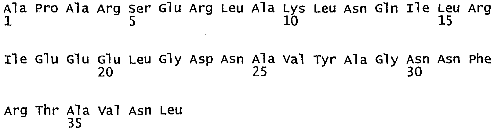
- <210> 41
<211> 40
<212> PRT
<213> Artificial - <220>
<223> Amb a 1 derived hyperallergenic molecule - <400> 41

- <210> 42
<211> 41
<212> PRT
<213> Artificial - <220>
<223> Amb a 1 derived hyperallergenic molecule - <400> 42

- <210> 43
<211> 31
<212> PRT
<213> Artificial - <220>
<223> Amb a 1 derived hyperallergenic molecule - <400> 43

- <210> 44
<211> 41
<212> PRT
<213> Artificial - <220>
<223> Amb a 1 derived hyperallergenic molecule - <400> 44

- <210> 45
<211> 39
<212> PRT
<213> Artificial - <220>
<223> Amb a 1 derived hyperallergenic molecule - <400> 45

- <210> 46
<211> 37
<212> PRT
<213> Artificial - <220>
<223> Amb a 1 derived hyperallergenic molecule - <400> 46
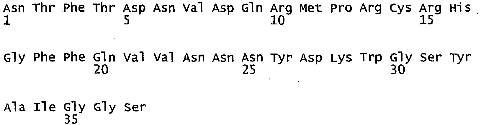
- <210> 47
<211> 46
<212> PRT
<213> Artificial - <220>
<223> Amb a 1 derived hyperallergenic molecule - <400> 47


- <210> 48
<211> 41
<212> PRT
<213> Artificial - <220>
<223> Amb a 1 derived hyperallergenic molecule - <400> 48

- <210> 49
<211> 44
<212> PRT
<213> Artificial - <220>
<223>Art v 1 derived hyperallergenic molecule - <400> 49

- <210> 50
<211> 31
<212> PRT
<213> Artificial - <220>
<223>Art v 1 derived hyperallergenic molecule - <400> 50

- <210> 51
<211> 41
<212> PRT
<213> Artificial - <220>
<223>Art v 1 derived hyperallergenic molecule - <400> 51

- <210> 52
<211> 38
<212> PRT
<213> Artificial - <220>
<223> canf 1 derived hyperallergenic molecule - <400> 52

- <210> 53
<211> 40
<212> PRT
<213> Artificial - <220>
<223> Canf 1 derived hyperallergenic molecule - <400> 53

- <210> 54
<211> 41
<212> PRT
<213> Artificial - <220>
<223> Canf 1 derived hyperallergenic molecule - <400> 54

- <210> 55
<211> 40
<212> PRT
<213> Artificial - <220>
<223> Canf 1 derived hyperallergenic molecule - <400> 55

- <210> 56
<211> 40
<212> PRT
<213> Artificial - <220>
<223> Canf 1 derived hyperallergenic molecule - <400> 56

- <210> 57
<211> 40
<212> PRT
<213> Artificial - <220>
<223> canf 2 derived hyperallergenic molecule - <400> 57

- <210> 58
<211> 40
<212> PRT
<213> Artificial - <220>
<223> canf 2 derived hyperallergenic molecule - <400> 58

- <210> 59
<211> 38
<212> PRT
<213> Artificial - <220>
<223> canf 2 derived hyperallergenic molecule - <400> 59

- <210> 60
<211> 39
<212> PRT
<213> Artificial - <220>
<223> canf 2 derived hyperallergenic molecule - <400> 60

- <210> 61
<211> 42
<212> PRT
<213> Artificial - <220>
<223> canf 2 derived hyperallergenic molecule - <400> 61

- <210> 62
<211> 34
<212> PRT
<213> Artificial - <220>
<223>Fel d 2 derived hyperallergenic molecule - <400> 62

- <210> 63
<211> 35
<212> PRT
<213> Artificial - <220>
<223>Fel d 2 derived hyperallergenic molecule - <400> 63


- <210> 64
<211> 30
<212> PRT
<213> Artificial - <220>
<223>Fel d 2 derived hyperallergenic molecule - <400> 64

- <210> 65
<211> 31
<212> PRT
<213> Artificial - <220>
<223>Fel d 2 derived hyperallergenic molecule - <400> 65

- <210> 66
<211> 30
<212> PRT
<213> Artificial - <220>
<223>Fel d 2 derived hyperallergenic molecule - <400> 66

- <210> 67
<211> 30
<212> PRT
<213> Artificial - <220>
<223>Fel d 2 derived hyperallergenic molecule - <400> 67

- <210> 68
<211> 30
<212> PRT
<213> Artificial - <220>
<223>Fel d 2 derived hyperallergenic molecule - <400> 68

- <210> 69
<211> 30
<212> PRT
<213> Artificial - <220>
<223>Fel d 2 derived hyperallergenic molecule - <400> 69

- <210> 70
<211> 42
<212> PRT
<213> Artificial - <220>
<223>Fel d 2 derived hyperallergenic molecule - <400> 70

- <210> 71
<211> 41
<212> PRT
<213> Artificial - <220>
<223>Fel d 2 derived hyperallergenic molecule - <400> 71

- <210> 72
<211> 42
<212> PRT
<213> Artificial - <220>
<223>Fel d 2 derived hyperallergenic molecule - <400> 72

- <210> 73
<211> 38
<212> PRT
<213> Artificial - <220>
<223>Fel d 2 derived hyperallergenic molecule - <400> 73

- <210> 74
<211> 38
<212> PRT
<213> Artificial - <220>
<223>Fel d 2 derived hyperallergenic molecule - <400> 74

- <210> 75
<211> 38
<212> PRT
<213> Artificial - <220>
<223>Fel d 2 derived hyperallergenic molecule - <400> 75

- <210> 76
<211> 42
<212> PRT
<213> Artificial - <220>
<223>Fel d 2 derived hyperallergenic molecule - <400> 76

- <210> 77
<211> 39
<212> PRT
<213> Artificial - <220>
<223>Fel d 2 derived hyperallergenic molecule - <400> 77

- <210> 78
<211> 42
<212> PRT
<213> Artificial - <220>
<223>Fel d 2 derived hyperallergenic molecule - <400> 78
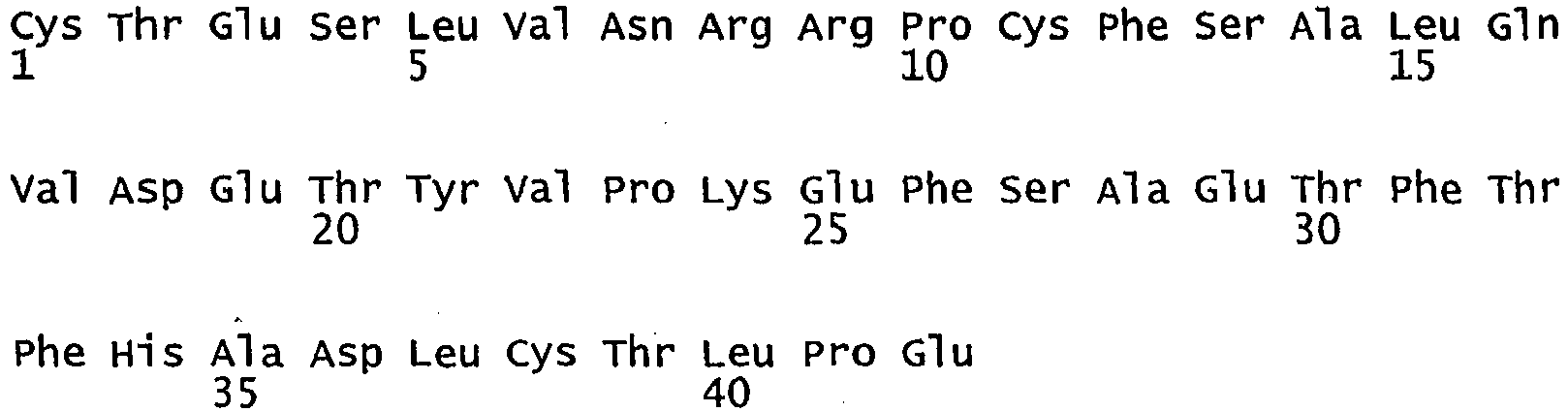
- <210> 79
<211> 40
<212> PRT
<213> Artificial - <220>
<223>ole e 1 derived hyperallergenic molecule - <400> 79

- <210> 80
<211> 31
<212> PRT
<213> Artificial - <220>
<223>Ole e 1 derived hyperallergenic molecule - <400> 80


- <210> 81
<211> 37
<212> PRT
<213> Artificial - <220>
<223>ole e 1 derived hyperallergenic molecule - <400> 81

- <210> 82
<211> 35
<212> PRT
<213> Artificial - <220>
<223>ole e 1 derived hyperallergenic molecule - <400> 82

- <210> 83
<211> 39
<212> PRT
<213> Artificial - <220>
<223>ole e 1 derived hyperallergenic molecule - <400> 83

- <210> 84
<211> 30
<212> PRT
<213> Artificial - <220>
<223>Par j 2 derived hyperallergenic molecule - <400> 84

- <210> 85
<211> 36
<212> PRT
<213> Artificial - <220>
<223>Par j 2 derived hyperallergenic molecule - <400> 85

- <210> 86
<211> 37
<212> PRT
<213> Artificial - <220>
<223>Par j 2 derived hyperallergenic molecule - <400> 86

- <210> 87
<211> 37
<212> PRT
<213> Artificial - <220>
<223>Par j 2 derived hyperallergenic molecule - <400> 87

- <210> 88
<211> 30
<212> PRT
<213> Artificial - <220>
<223>Der p 1 derived hyperallergenic molecule - <400> 88

- <210> 89
<211> 33
<212> PRT
<213> Artificial - <220>
<223>Der p 1 derived hyperallergenic molecule - <400> 89

- <210> 90
<211> 31
<212> PRT
<213> Artificial - <220>
<223>Der p 1 derived hyperallergenic molecule - <400> 90

- <210> 91
<211> 37
<212> PRT
<213> Artificial - <220>
<223>Der p 1 derived hyperallergenic molecule - <400> 91

- <210> 92
<211> 31
<212> PRT
<213> Artificial - <220>
<223>Der p 1 derived hyperallergenic molecule - <400> 92

- <210> 93
<211> 33
<212> PRT
<213> Artificial - <220>
<223>Der p 1 derived hyperallergenic molecule - <400> 93

- <210> 94
<211> 34
<212> PRT
<213> Artificial - <220>
<223>Der p 1 derived hyperallergenic molecule - <400> 94

- <210> 95
<211> 35
<212> PRT
<213> Artificial - <220>
<223>Der p 1 derived hyperallergenic molecule - <400> 95
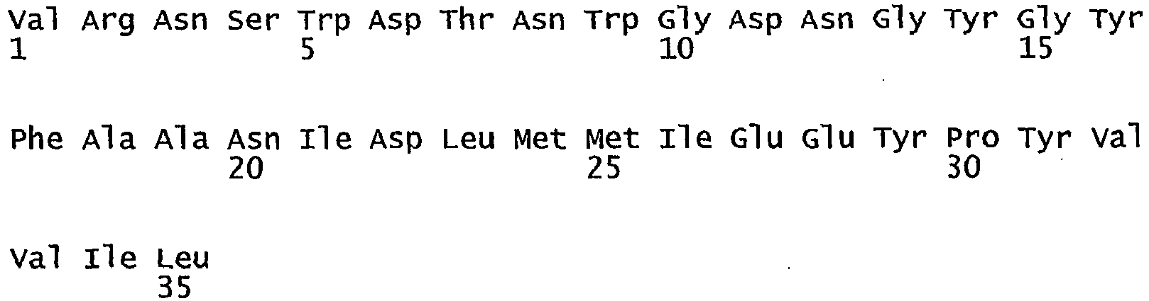
- <210> 96
<211> 33
<212> PRT
<213> Artificial - <220>
<223>Der p 2 derived hyperallergenic molecule - <400> 96

- <210> 97
<211> 31
<212> PRT
<213> Artificial - <220>
<223>Der p 2 derived hyperallergenic molecule - <400> 97

- <210> 98
<211> 32
<212> PRT
<213> Artificial - <220>
<223>Der p 2 derived hyperallergenic molecule - <400> 98

- <210> 99
<211> 42
<212> PRT
<213> Artificial - <220>
<223>Der p 2 derived hyperallergenic molecule - <400> 99

- <210> 100
<211> 32
<212> PRT
<213> Artificial - <220>
<223>Der p 2 derived hyperallergenic molecule - <400> 100

- <210> 101
<211> 35
<212> PRT
<213> Artificial - <220>
<223>Der p 5 derived hyperallergenic molecule - <400> 101

- <210> 102
<211> 36
<212> PRT
<213> Artificial - <220>
<223>Der p 5 derived hyperallergenic molecule - <400> 102

- <210> 103
<211> 31
<212> PRT
<213> Artificial - <220>
<223>Der p 5 derived hyperallergenic molecule - <400> 103

- <210> 104
<211> 36
<212> PRT
<213> Artificial - <220>
<223>Der p 5 derived hyperallergenic molecule - <400> 104


- <210> 105
<211> 30
<212> PRT
<213> Artificial - <220>
<223>Der p 7 derived hyperallergenic molecule - <400> 105

- <210> 106
<211> 31
<212> PRT
<213> Artificial - <220>
<223>Der p 7 derived hyperallergenic molecule - <400> 106

- <210> 107
<211> 31
<212> PRT
<213> Artificial - <220>
<223>Der p 7 derived hyperallergenic molecule - <400> 107

- <210> 108
<211> 36
<212> PRT
<213> Artificial - <220>
<223>Der p 7 derived hyperallergenic molecule - <400> 108

- <210> 109
<211> 31
<212> PRT
<213> Artificial - <220>
<223>Der p 7 derived hyperallergenic molecule - <400> 109

- <210> 110
<211> 34
<212> PRT
<213> Artificial - <220>
<223>Der p 7 derived hyperallergenic molecule - <400> 110

- <210> 111
<211> 35
<212> PRT
<213> Artificial - <220>
<223>Der p 10 derived hyperallergenic molecule - <400> 111


- <210> 112
<211> 35
<212> PRT
<213> Artificial - <220>
<223>Der p 10 derived hyperallergenic molecule - <400> 112
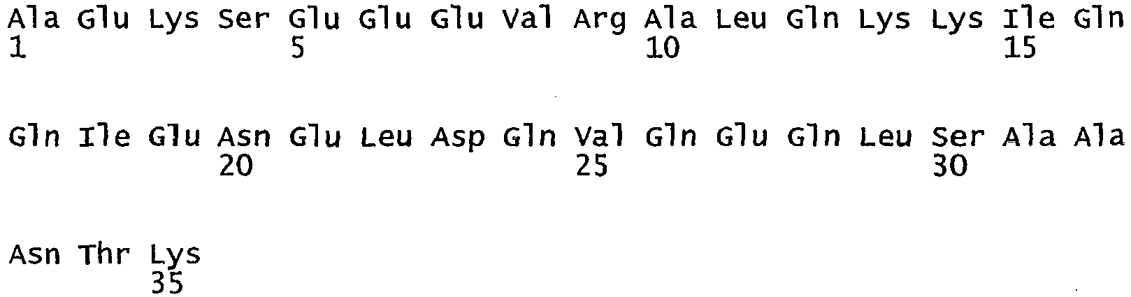
- <210> 113
<211> 40
<212> PRT
<213> Artificial - <220>
<223>Der p 10 derived hyperallergenic molecule - <400> 113

- <210> 114
<211> 35
<212> PRT
<213> Artificial - <220>
<223>Der p 10 derived hyperallergenic molecule - <400> 114


- <210> 115
<211> 31
<212> PRT
<213> Artificial - <220>
<223>Der p 10 derived hyperallergenic molecule - <400> 115

- <210> 116
<211> 31
<212> PRT
<213> Artificial - <220>
<223>Der p 10 derived hyperallergenic molecule - <400> 116

- <210> 117
<211> 41
<212> PRT
<213> Artificial - <220>
<223>Der p 10 derived hyperallergenic molecule - <400> 117

- <210> 118
<211> 35
<212> PRT
<213> Artificial - <220>
<223>Der p 10 derived hyperallergenic molecule - <400> 118

- <210> 119
<211> 35
<212> PRT
<213> Artificial - <220>
<223> Der p 21 derived hyperallergenic molecule - <400> 119

- <210> 120
<211> 37
<212> PRT
<213> Artificial - <220>
<223> Der p 21 derived hyperallergenic molecule - <400> 120

- <210> 121
<211> 31
<212> PRT
<213> Artificial - <220>
<223> Der p 21 derived hyperallergenic molecule - <400> 121


- <210> 122
<211> 33
<212> PRT
<213> Artificial - <220>
<223> Der p 21 derived hyperallergenic molecule - <400> 122

- <210> 123
<211> 32
<212> PRT
<213> Artificial - <220>
<223>Clone 30 derived hyperallergenic molecule - <400> 123

- <210> 124
<211> 34
<212> PRT
<213> Artificial - <220>
<223>clone 30 derived hyperallergenic molecule - <400> 124

- <210> 125
<211> 39
<212> PRT
<213> Artificial - <220>
<223>clone 30 derived hyperallergenic molecule - <400> 125

- <210> 126
<211> 30
<212> PRT
<213> Artificial - <220>
<223>Bet v 1 derived hyperallergenic molecule - <400> 126

- <210> 127
<211> 30
<212> PRT
<213> Artificial - <220>
<223>Bet v 1 derived hyperallergenic molecule - <400> 127

- <210> 128
<211> 30
<212> PRT
<213> Artificial - <220>
<223>Bet v 1 derived hyperallergenic molecule - <400> 128


- <210> 129
<211> 35
<212> PRT
<213> Artificial - <220>
<223>Fel d 1chain 1 derived hyperallergenic molecule - <400> 129

- <210> 130
<211> 36
<212> PRT
<213> Artificial - <220>
<223>Fel d 1chain 1 derived hyperallergenic molecule - <400> 130

- <210> 131
<211> 35
<212> PRT
<213> Artificial - <220>
<223>Fel d 1chain 2 derived hyperallergenic molecule - <400> 131

- <210> 132
<211> 30
<212> PRT
<213> Artificial - <220>
<223>Fel d 1chain 2 derived hyperallergenic molecule - <400> 132

- <210> 133
<211> 30
<212> PRT
<213> Artificial - <220>
<223>Fel d 1chain 2 derived hyperallergenic molecule - <400> 133

- <210> 134
<211> 32
<212> PRT
<213> Artificial - <220>
<223>Phl p 5 derived hyperallergenic molecule - <400> 134

- <210> 135
<211> 34
<212> PRT
<213> Artificial - <220>
<223>Phl p 5 derived hyperallergenic molecule - <400> 135


- <210> 136
<211> 32
<212> PRT
<213> Artificial - <220>
<223>Phl p 5 derived hyperallergenic molecule - <400> 136

- <210> 137
<211> 31
<212> PRT
<213> Artificial - <220>
<223>Phl p 5 derived hyperallergenic molecule - <400> 137

- <210> 138
<211> 33
<212> PRT
<213> Artificial - <220>
<223>Phl p 5 derived hyperallergenic molecule - <400> 138

- <210> 139
<211> 38
<212> PRT
<213> Artificial - <220>
<223>Phl p 5 derived hyperallergenic molecule - <400> 139

- <210> 140
<211> 70
<212> PRT
<213> Artificial - <220>
<223>Clone 30 Dermatophagoides pteronyssinus allergen - <400> 140

- <210> 141
<211> 37
<212> PRT
<213> Artificial - <220>
<223>Phl p 5 derived peptide - <400> 141

- <210> 142
<211> 29
<212> PRT
<213> Artificial - <220>
<223>Phl p 5 derived peptide - <400> 142

| a) | ||||
| pre P1 | anti P1 | pre P1a | anti P1a | |
| P1 | 0,041 | 0,880 | 0,052 | 0,947 |
| P1a | 0,038 | 0,705 | 0,048 | 0,859 |
| b) | ||||
| pre P2 | anti P2 | pre P2b | anti P2b | |
| P2 | 0,089 | 1,168 | 0,042 | 1,175 |
| P2b | 0,075 | 0,954 | 0,053 | 1,122 |
Claims (7)
- A hypoallergenic protein for use in the treatment or prevention of an allergy in a human or animal consisting of at least one hypoallergenic molecule derived from an allergen, which is fused to at least one second non-allergenic protein, wherein the at least one hypoallergenic molecule derived from an allergen exhibits an at least 50% reduced IgE-binding capacity and an at least 30% reduced T-cell reactivity compared to the wild-type allergen and wherein the at least one second protein is a viral protein.
- Hypoallergenic protein for the use according to claim 1, characterized in that the viral protein is a capsid protein.
- Hypoallergenic protein for the use according to claim 1 or 2, characterized in that the at least one viral protein is derived from a human pathogenic virus, preferably a virus of the family of picornaviridae.
- Hypoallergenic protein for the use according to claim 3, characterized in that the virus of the family of picornaviridae is of the genus of rhinoviruses, preferably of the species of human rhinoviruses, in particular human rhinovirus 89 or 14.
- Hypoallergenic protein for the use according to any one of claims 1 to 4, characterized in that the allergen is selected from the group consisting of major birch pollen allergens, in particular Bet v 1 and Bet v 4, major timothy grass pollen allergens, in particular Phl p 1, Phl p 2, Phl p 5, Phl p 6 and Phl p 7, major house dust mite allergens, in particular Der p 1, Der p 2, Der p 5, Der p 7, Der p 10, Der p 21 and clone 30, major cat allergen Fel d 1 and Fel d 2, major bee allergens, major wasp allergens, profilins, especially Phl p 12, olive allergens, especially Ole e 1, Parietaria judaica allergens, especially Par j 2, Ragweed allergens, especially Amb a 1, mugwort pollen allergens, especially Art v 1, and storage mite allergens, especially Lep d 2, Alt a 1, Alt a 2, Alt a 6, Can f 1, Can f 2.
- Hypoallergenic protein for the use according to any one of claims 1 to 5, characterized in that the at least one hypoallergenic molecule derived from an allergen is an allergen fragment composed of amino acids 151 to 177, 87 to 117, 1 to 30, 43 to 70 or 212 to 241 of Phl p 1, amino acids 93 to 128, 98 to 128, 26 to 53, 26 to 58, 132 to 162, 217 to 246, 252 to 283 or 176 to 212 of Phl p 5, amino acids 1 to 34 or 35 to 70 of chain 1 of Fel d 1, amino acids 1 to 34, 35 to 63 or 64 to 92 of chain 2 of Fel d 1, amino acids 30 to 59, 50 to 79 or 75 to 104 of Bet v 1, amino acids 1 to 33, 21 to 51, 42 to 73, 62 to 103 or 98 to 129 of Der p 2, amino acids 1 to 30, 20 to 50, 50 to 80, 90 to 125, 125 to 155 or 165 to 198 of Der p 7, amino acids 1-35, 36-70, 71-110, 111-145, 140-170, 175-205, 210-250 or 250-284 of Der p 10, amino acids 1 to 35, 35 to 72, 70 to 100 or 90 to 122 of Der p 21, amino acids 1 to 32, 15 to 48 or 32 to 70 of Clone 30, amino acids 19 to 58, 59 to 95, 91 to 120 or 121 to 157 of Alt a 1, amino acids 31 to 60, 45 to 80, 60 to 96 or 97 to 133 of Par j 2, amino acids 1 to 40, 36 to 66, 63 to 99, 86 to 120 or 107 to 145 of Ole e 1, amino acids 25 to 58, 99 to 133, 154 to 183, 277 to 307, 334 to 363, 373 to 402, 544 to 573, 579 to 608, 58 to 99, 125 to 165, 183 to 224, 224 to 261, 252 to 289, 303 to 340, 416 to 457, 460 to 500 or 501 to 542 of Fel d 2, amino acids 19 to 58, 52 to 91, 82 to 119, 106 to 144 or 139 to 180 of Can f 2, amino acids 19 to 56, 51 to 90, 78 to 118, 106 to 145 or 135-174 of Can f 1, amino acids 27 to 70, 70 to 100 or 92 to 132 of Art v 1, amino acids 31 to 70, 80 to 120, 125 to 155, 160 to 200, 225 to 263, 264 to 300 305 to 350 or 356 to 396 of Amb a 1, amino acids 1 to 34, 35 to 74, 74 to 115, 125 to 165, 174 to 213, 241 to 280, 294 to 333, 361 to 400 or 401 to 438 of Alt a 6, amino acids 1 to 40, 41 to 80, 81 to 120, or 121 to 160 of Alt a 2.
- Hypoallergenic protein for the use according to any one of claims 1 to 6, characterized in that the hypoallergenic protein is administered to an individual in the amount of 0,01 mg/kg body weight to 5 mg/kg body weight, preferably 0,1 mg/kg body weight to 2 mg/kg body weight.
Priority Applications (3)
| Application Number | Priority Date | Filing Date | Title |
|---|---|---|---|
| PL07718492T PL2035457T3 (en) | 2006-06-09 | 2007-06-11 | Vaccine carrier |
| EP15161237.1A EP2924047B1 (en) | 2006-06-09 | 2007-06-11 | Vaccine carrier |
| DK15161237.1T DK2924047T3 (en) | 2006-06-09 | 2007-06-11 | vaccine carrier |
Applications Claiming Priority (2)
| Application Number | Priority Date | Filing Date | Title |
|---|---|---|---|
| AT0099406A AT503690A1 (en) | 2006-06-09 | 2006-06-09 | HYPOALLERGENIC MOLECULES |
| PCT/AT2007/000281 WO2007140505A2 (en) | 2006-06-09 | 2007-06-11 | Vaccine carrier |
Related Child Applications (1)
| Application Number | Title | Priority Date | Filing Date |
|---|---|---|---|
| EP15161237.1A Division EP2924047B1 (en) | 2006-06-09 | 2007-06-11 | Vaccine carrier |
Publications (2)
| Publication Number | Publication Date |
|---|---|
| EP2035457A2 EP2035457A2 (en) | 2009-03-18 |
| EP2035457B1 true EP2035457B1 (en) | 2015-04-01 |
Family
ID=38650124
Family Applications (2)
| Application Number | Title | Priority Date | Filing Date |
|---|---|---|---|
| EP07718492.7A Active EP2035457B1 (en) | 2006-06-09 | 2007-06-11 | Vaccine carrier |
| EP15161237.1A Active EP2924047B1 (en) | 2006-06-09 | 2007-06-11 | Vaccine carrier |
Family Applications After (1)
| Application Number | Title | Priority Date | Filing Date |
|---|---|---|---|
| EP15161237.1A Active EP2924047B1 (en) | 2006-06-09 | 2007-06-11 | Vaccine carrier |
Country Status (14)
| Country | Link |
|---|---|
| US (2) | US9296828B2 (en) |
| EP (2) | EP2035457B1 (en) |
| JP (3) | JP5399895B2 (en) |
| CN (3) | CN101466738A (en) |
| AT (1) | AT503690A1 (en) |
| AU (1) | AU2007257308B2 (en) |
| BR (1) | BR122019004393B1 (en) |
| CA (1) | CA2657971C (en) |
| DK (2) | DK2924047T3 (en) |
| ES (2) | ES2541311T3 (en) |
| PL (1) | PL2035457T3 (en) |
| PT (1) | PT2035457E (en) |
| RU (1) | RU2486206C2 (en) |
| WO (1) | WO2007140505A2 (en) |
Families Citing this family (20)
| Publication number | Priority date | Publication date | Assignee | Title |
|---|---|---|---|---|
| EA023303B1 (en) | 2007-08-15 | 2016-05-31 | Сиркейшиа Лимитед | Peptide composition for tolerization to house dust mites and use thereof |
| ES2378870T5 (en) * | 2008-08-15 | 2016-02-03 | Circassia Limited | Vaccine comprising Amb to 1 peptides for use in the treatment of ragweed allergy |
| CN102307894B (en) * | 2008-12-30 | 2014-07-30 | 蔡考圆 | A variant of mite allergen and uses thereof |
| CO6260019A1 (en) | 2009-01-30 | 2011-03-22 | Fundacion Universidad Del Norte | IMMUNOCHEMICAL METHOD AND COMPOSITION FOR THE DETECTION OF ALLERGENS FROM ACAROS USING IGY POLYCLONAL ANTIBODIES |
| AU2013202362B2 (en) * | 2009-02-05 | 2015-09-17 | Circassia Limited | Grass Peptides for Vaccine |
| ES2532759T3 (en) * | 2009-02-05 | 2015-03-31 | Circassia Limited | Herbaceous plant peptides for vaccines |
| EP2316481A1 (en) * | 2009-10-30 | 2011-05-04 | Biomay Ag | Pharmaceutical composition for the treatment and prevention of a rhinovirus infection |
| WO2012168487A1 (en) * | 2011-06-09 | 2012-12-13 | Biomay Ag | Peptide carrier fusion proteins as allergy vaccines |
| WO2013001362A2 (en) * | 2011-06-27 | 2013-01-03 | Anergis S.A. | Contiguous overlapping peptides for treatment of ragweed pollen allergy |
| EP2543725A1 (en) | 2011-07-08 | 2013-01-09 | Biomay Ag | Polysaccharide lyases |
| JO3820B1 (en) * | 2012-05-03 | 2021-01-31 | Regeneron Pharma | Human antibodies to FEL D1 and methods for their use |
| GB201209868D0 (en) * | 2012-06-01 | 2012-07-18 | Circassia Ltd | Alternaria peptides |
| ES2601220T3 (en) | 2012-09-20 | 2017-02-14 | Universidad De Cartagena | Fusion proteins with representation of different allergens: vaccine proposal for mite allergy |
| JP2014231500A (en) * | 2013-05-29 | 2014-12-11 | 元晴 植原 | Peptide mixture used for immunotherapy of betula pollinosis |
| US10935554B2 (en) | 2013-08-23 | 2021-03-02 | Regeneron Pharmaceuticals, Inc. | Diagnostic tests and methods for assessing safety, efficacy or outcome of allergen-specific immunotherapy (SIT) |
| US11013781B2 (en) | 2015-07-01 | 2021-05-25 | Alk-Abelló As | Peptide combinations and uses thereof for treating grass allergy |
| US11352417B2 (en) | 2016-12-22 | 2022-06-07 | Regeneron Pharmaceuticals, Inc. | Method of treating an allergy with allergen-specific monoclonal antibodies |
| US11408895B1 (en) | 2018-04-09 | 2022-08-09 | Hob Biotech Group Corp., Ltd. | Allergen characterization, potent allergen product formulation, comprehensive allergen reagent, and allergy assay |
| EP3569612A1 (en) * | 2018-05-18 | 2019-11-20 | Biomay Ag | Treatment and prevention of house dust mite allergies |
| RU2761431C9 (en) | 2020-10-26 | 2022-04-18 | Федеральное государственное бюджетное учреждение "Государственный научный центр "Институт иммунологии" Федерального медико-биологического агентства России (ФГБУ "ГНЦ Институт иммунологии" ФМБА России) | Recombinant polypeptide based on birch pollen allergen and apple allergen as an allergy vaccine |
Family Cites Families (42)
| Publication number | Priority date | Publication date | Assignee | Title |
|---|---|---|---|---|
| GB8308235D0 (en) | 1983-03-25 | 1983-05-05 | Celltech Ltd | Polypeptides |
| US4816567A (en) | 1983-04-08 | 1989-03-28 | Genentech, Inc. | Recombinant immunoglobin preparations |
| US5807715A (en) | 1984-08-27 | 1998-09-15 | The Board Of Trustees Of The Leland Stanford Junior University | Methods and transformed mammalian lymphocyte cells for producing functional antigen-binding protein including chimeric immunoglobulin |
| GB8607679D0 (en) | 1986-03-27 | 1986-04-30 | Winter G P | Recombinant dna product |
| US5225539A (en) | 1986-03-27 | 1993-07-06 | Medical Research Council | Recombinant altered antibodies and methods of making altered antibodies |
| GB8821077D0 (en) * | 1988-09-08 | 1988-10-05 | Wellcome Found | Peptides |
| US5530101A (en) | 1988-12-28 | 1996-06-25 | Protein Design Labs, Inc. | Humanized immunoglobulins |
| US6019972A (en) * | 1989-11-03 | 2000-02-01 | Immulogic Pharmaceutical Corporation | Peptides of human T cell reactive feline protein (TRFP) |
| GB8928874D0 (en) | 1989-12-21 | 1990-02-28 | Celltech Ltd | Humanised antibodies |
| DE69233482T2 (en) | 1991-05-17 | 2006-01-12 | Merck & Co., Inc. | Method for reducing the immunogenicity of antibody variable domains |
| US5565332A (en) | 1991-09-23 | 1996-10-15 | Medical Research Council | Production of chimeric antibodies - a combinatorial approach |
| JP4246794B2 (en) * | 1991-10-16 | 2009-04-02 | メルク パテント ゲーエムベーハー | Recombitope peptide |
| IL105153A (en) * | 1992-03-25 | 1999-12-22 | Immulogic Pharma Corp | Therapeutic compositions comprising peptides derived from human t cell reactive feline protein |
| US5639641A (en) | 1992-09-09 | 1997-06-17 | Immunogen Inc. | Resurfacing of rodent antibodies |
| AT401937B (en) * | 1993-04-01 | 1996-12-27 | Biomay Prod & Handel | RECOMBINANT LYING GRASS POLLEN ALLERGEN PHL P II |
| DE69333979T2 (en) * | 1993-04-14 | 2006-12-21 | Heska Corp.(n.d.Ges, d.Staates Delaware), Loveland | T CELL EPITOPES OF THE MAIN ALLERGIES OF DERMATOPHAGOIDES (HOUSE DUST MILK) |
| JPH09512014A (en) * | 1994-04-14 | 1997-12-02 | イミユロジク・フアーマシユーチカル・コーポレーシヨン | Pharmaceutical peptide composition for treating mite allergy |
| SE9402089D0 (en) * | 1994-06-14 | 1994-06-14 | Rudolf Valenta | Recombinant allergen, fragments thereof, corresponding recombinant DNA molecules, vectors and hosts containing the DNA molecules, diagnostic and therapeutic uses of said allergens and fragments |
| US6572859B1 (en) * | 1999-10-29 | 2003-06-03 | Pharmacia Diagnostics Ab | Non-anaphylactic forms of grass pollen Phl p 6 allergen and their use |
| US20030166518A1 (en) * | 2000-01-13 | 2003-09-04 | Beardslee Thomas A. | Method for allergen characterization |
| ATE418997T1 (en) * | 2000-07-06 | 2009-01-15 | Univ Georgetown | STABLE (FIXED) FORMS OF VIRAL L1 CAPSID PROTEINS, THEIR FUSION PROTEINS, AND THEIR USES |
| ES2209563B1 (en) * | 2000-07-28 | 2005-10-01 | Universidad Complutense De Madrid | PRODUCTION IN THE LEVADURAPIPCHIA PASTORIS AND POMIFICATION SYSTEM OF THE OLEA EUROPEAN OLEA RECOMBINING ALLERGEN FOR USE IN DIAGNOSIS AND ALLERGY TREATMENT. |
| KR100919914B1 (en) * | 2000-11-16 | 2009-10-06 | 알크-아벨로 에이/에스 | Novel mutant allergens |
| EP1221317A1 (en) * | 2000-12-28 | 2002-07-10 | SHAN-Beteiligungsgesellschaft m.b.H. | Vaccines containing hybrid polypeptides consisting of at least two different allergenic proteins |
| US7320793B2 (en) * | 2001-01-19 | 2008-01-22 | Cytos Biotechnology Ag | Molecular antigen array |
| PL206709B1 (en) * | 2002-02-27 | 2010-09-30 | Merck Patent Gmbh | Method for producing hypoallergenic major birch pollen allergens rbet v 1 |
| WO2004003143A2 (en) | 2002-06-26 | 2004-01-08 | Advanced Bionutrition Corporation | Viruses and virus-like particles for multiple antigen and target display |
| MXPA05000287A (en) | 2002-07-05 | 2005-09-08 | Leclerc Denis | Adjuvant viral particle. |
| ATE307141T1 (en) | 2003-01-21 | 2005-11-15 | Biomay Prod & Handel | METHOD FOR PREPARING HYPOALLERGENIC MOSAIC PROTEINS |
| GB0308988D0 (en) * | 2003-04-17 | 2003-05-28 | Univ Singapore | Molecule |
| PT2287180E (en) * | 2003-06-04 | 2013-10-31 | Merck Patent Gmbh | Lol p 5-derivatives with reduced allergenicity and conserved t-cell reactivity |
| US20050053615A1 (en) * | 2003-07-16 | 2005-03-10 | Best Elaine A. | Variants of mite group 1 allergens for the treatment of house dust mite allergy |
| DE10351471A1 (en) * | 2003-11-04 | 2005-06-09 | Ursula Prof. Dr. Wiedermann-Schmidt | Polyvalent allergy vaccine |
| EP1758925A4 (en) * | 2003-12-01 | 2009-04-08 | Dow Global Technologies Inc | Recombinant icosahedral virus like particle production in pseudomonads |
| ITRM20040103A1 (en) * | 2004-02-27 | 2004-05-27 | Consiglio Nazionale Ricerche | MELTING PROTEINS INCLUDING ALLERGENS OF THE NS-LTPS FAMILY, THEIR USES AND PHARMACEUTICAL COMPOSITIONS THAT INCLUDE THEM. |
| CN1926238A (en) * | 2004-02-27 | 2007-03-07 | 陶氏化学公司 | High efficiency peptide production in plant cells |
| US20060088549A1 (en) * | 2004-07-08 | 2006-04-27 | Arnold Gail F | Chimeric virus vaccine |
| DE102004035337A1 (en) * | 2004-07-21 | 2006-03-16 | Merck Patent Gmbh | Variations of the group 1 allergens from Poaceae with reduced allergenicity and preserved T cell reactivity |
| MX2007003171A (en) * | 2004-09-21 | 2007-05-23 | Cytos Biotechnology Ag | Virus-like particles comprising a fusion protein of the coat protein of ap205 and an antigenic polypeptide. |
| AT501101B1 (en) * | 2004-12-02 | 2008-01-15 | Biomay Ag | PROTEIN ALLERGEN DERIVATIVE |
| PL1868642T3 (en) * | 2005-03-18 | 2013-10-31 | Cytos Biotechnology Ag | Cat allergen fusion proteins and uses thereof |
| AU2006291835B2 (en) * | 2005-09-15 | 2012-04-05 | Alk-Abello A/S | A method for quantification of allergens |
-
2006
- 2006-06-09 AT AT0099406A patent/AT503690A1/en not_active Application Discontinuation
-
2007
- 2007-06-11 WO PCT/AT2007/000281 patent/WO2007140505A2/en not_active Ceased
- 2007-06-11 BR BR122019004393-0A patent/BR122019004393B1/en active IP Right Grant
- 2007-06-11 ES ES07718492.7T patent/ES2541311T3/en active Active
- 2007-06-11 ES ES15161237T patent/ES2698525T3/en active Active
- 2007-06-11 CA CA2657971A patent/CA2657971C/en active Active
- 2007-06-11 EP EP07718492.7A patent/EP2035457B1/en active Active
- 2007-06-11 EP EP15161237.1A patent/EP2924047B1/en active Active
- 2007-06-11 CN CNA200780021374XA patent/CN101466738A/en active Pending
- 2007-06-11 DK DK15161237.1T patent/DK2924047T3/en active
- 2007-06-11 AU AU2007257308A patent/AU2007257308B2/en not_active Ceased
- 2007-06-11 JP JP2009513514A patent/JP5399895B2/en active Active
- 2007-06-11 CN CN2013102084632A patent/CN103319601A/en active Pending
- 2007-06-11 CN CN201310209988.8A patent/CN103319602B/en active Active
- 2007-06-11 RU RU2008152045/10A patent/RU2486206C2/en active
- 2007-06-11 PL PL07718492T patent/PL2035457T3/en unknown
- 2007-06-11 PT PT77184927T patent/PT2035457E/en unknown
- 2007-06-11 DK DK07718492.7T patent/DK2035457T3/en active
- 2007-06-11 US US12/304,267 patent/US9296828B2/en active Active
-
2013
- 2013-10-24 JP JP2013221630A patent/JP2014064573A/en active Pending
- 2013-10-24 JP JP2013221629A patent/JP2014087341A/en active Pending
-
2015
- 2015-12-17 US US14/972,685 patent/US20160257722A1/en not_active Abandoned
Non-Patent Citations (2)
| Title |
|---|
| FOCKE M ET AL: "Developments in allergen-specific immunotherapy: from allergen extracts to allergy vaccines bypassing allergen-specific immunoglobulin E and T cell reactivity", CLINICAL AND EXPERIMENTAL ALLERGY, vol. 40, no. 3, 1 March 2010 (2010-03-01), pages 385 - 397 * |
| FOCKE M ET AL: "Nonanaphylactic synthetic peptides derived from B cell epitopes for the major grass pollen allergen, Phl p 1, for allergy vaccination", FASEB JOURNAL, vol. 15, 1 September 2001 (2001-09-01), pages 2042 - 2044 * |
Also Published As
| Publication number | Publication date |
|---|---|
| RU2008152045A (en) | 2010-07-20 |
| JP2014064573A (en) | 2014-04-17 |
| CN103319602A (en) | 2013-09-25 |
| ES2541311T3 (en) | 2015-07-17 |
| JP5399895B2 (en) | 2014-01-29 |
| EP2924047A1 (en) | 2015-09-30 |
| BRPI0712421A2 (en) | 2012-06-19 |
| CA2657971C (en) | 2018-01-23 |
| AT503690A1 (en) | 2007-12-15 |
| ES2698525T3 (en) | 2019-02-05 |
| JP2009539354A (en) | 2009-11-19 |
| RU2486206C2 (en) | 2013-06-27 |
| US20160257722A1 (en) | 2016-09-08 |
| DK2924047T3 (en) | 2018-12-03 |
| BR122019004393B1 (en) | 2023-10-17 |
| WO2007140505A3 (en) | 2008-03-20 |
| AU2007257308A1 (en) | 2007-12-13 |
| PT2035457E (en) | 2015-07-28 |
| PL2035457T3 (en) | 2015-10-30 |
| AU2007257308B2 (en) | 2013-06-06 |
| CN103319601A (en) | 2013-09-25 |
| US20090324501A1 (en) | 2009-12-31 |
| CN103319602B (en) | 2016-09-07 |
| JP2014087341A (en) | 2014-05-15 |
| US9296828B2 (en) | 2016-03-29 |
| WO2007140505A2 (en) | 2007-12-13 |
| CA2657971A1 (en) | 2007-12-13 |
| EP2924047B1 (en) | 2018-08-22 |
| EP2035457A2 (en) | 2009-03-18 |
| DK2035457T3 (en) | 2015-06-15 |
| CN101466738A (en) | 2009-06-24 |
Similar Documents
| Publication | Publication Date | Title |
|---|---|---|
| US9296828B2 (en) | Vaccine carrier | |
| EP2717910B1 (en) | Peptide carrier fusion proteins as allergy vaccines | |
| US8246945B2 (en) | Methods and reagents for decreasing clinical reaction to allergy | |
| US20050063994A1 (en) | Methods and reagents for decreasing clinical reaction to allergy | |
| AU2005312324B2 (en) | Protein allergen derivatives | |
| AU2013222052B2 (en) | Vaccine carrier | |
| US8709435B2 (en) | Hypallergenic mosaic antigens and methods of making same |
Legal Events
| Date | Code | Title | Description |
|---|---|---|---|
| PUAI | Public reference made under article 153(3) epc to a published international application that has entered the european phase |
Free format text: ORIGINAL CODE: 0009012 |
|
| 17P | Request for examination filed |
Effective date: 20090109 |
|
| AK | Designated contracting states |
Kind code of ref document: A2 Designated state(s): AT BE BG CH CY CZ DE DK EE ES FI FR GB GR HU IE IS IT LI LT LU LV MC MT NL PL PT RO SE SI SK TR |
|
| AX | Request for extension of the european patent |
Extension state: AL BA HR MK RS |
|
| 17Q | First examination report despatched |
Effective date: 20090417 |
|
| RIN1 | Information on inventor provided before grant (corrected) |
Inventor name: VAN HAGE, MARIANNE Inventor name: GROENLUND, HANS Inventor name: SWOBODA, INES Inventor name: VALENTA, RUDOLF Inventor name: VALENT, PETER Inventor name: REININGER, RENATE Inventor name: SPITZAUER, SUSANNE Inventor name: FOCKE-TEJKL, MARGARETE Inventor name: POPOW-KRAUPP, THERESIA Inventor name: WESTRITSCHNIG, KERSTIN Inventor name: VRTALA, SUSANNE Inventor name: LINHART, BIRGIT Inventor name: TINHOFER, JOHANNA |
|
| RAX | Requested extension states of the european patent have changed |
Extension state: HR Payment date: 20090109 Extension state: RS Payment date: 20090109 |
|
| REG | Reference to a national code |
Ref country code: DE Ref legal event code: R079 Ref document number: 602007040867 Country of ref document: DE Free format text: PREVIOUS MAIN CLASS: C07K0019000000 Ipc: A61K0039350000 |
|
| RIC1 | Information provided on ipc code assigned before grant |
Ipc: A61K 39/35 20060101AFI20140722BHEP Ipc: A61K 39/36 20060101ALI20140722BHEP Ipc: C07K 19/00 20060101ALI20140722BHEP |
|
| GRAP | Despatch of communication of intention to grant a patent |
Free format text: ORIGINAL CODE: EPIDOSNIGR1 |
|
| INTG | Intention to grant announced |
Effective date: 20141021 |
|
| GRAS | Grant fee paid |
Free format text: ORIGINAL CODE: EPIDOSNIGR3 |
|
| GRAA | (expected) grant |
Free format text: ORIGINAL CODE: 0009210 |
|
| AK | Designated contracting states |
Kind code of ref document: B1 Designated state(s): AT BE BG CH CY CZ DE DK EE ES FI FR GB GR HU IE IS IT LI LT LU LV MC MT NL PL PT RO SE SI SK TR |
|
| AX | Request for extension of the european patent |
Extension state: HR RS |
|
| REG | Reference to a national code |
Ref country code: GB Ref legal event code: FG4D |
|
| REG | Reference to a national code |
Ref country code: CH Ref legal event code: EP |
|
| REG | Reference to a national code |
Ref country code: IE Ref legal event code: FG4D |
|
| REG | Reference to a national code |
Ref country code: AT Ref legal event code: REF Ref document number: 718695 Country of ref document: AT Kind code of ref document: T Effective date: 20150515 |
|
| REG | Reference to a national code |
Ref country code: DE Ref legal event code: R096 Ref document number: 602007040867 Country of ref document: DE Effective date: 20150521 |
|
| REG | Reference to a national code |
Ref country code: DK Ref legal event code: T3 Effective date: 20150609 |
|
| REG | Reference to a national code |
Ref country code: NL Ref legal event code: T3 |
|
| REG | Reference to a national code |
Ref country code: CH Ref legal event code: NV Representative=s name: KELLER AND PARTNER PATENTANWAELTE AG, CH |
|
| REG | Reference to a national code |
Ref country code: ES Ref legal event code: FG2A Ref document number: 2541311 Country of ref document: ES Kind code of ref document: T3 Effective date: 20150717 |
|
| REG | Reference to a national code |
Ref country code: SE Ref legal event code: TRGR |
|
| REG | Reference to a national code |
Ref country code: PT Ref legal event code: SC4A Free format text: AVAILABILITY OF NATIONAL TRANSLATION Effective date: 20150618 |
|
| REG | Reference to a national code |
Ref country code: LT Ref legal event code: MG4D |
|
| PG25 | Lapsed in a contracting state [announced via postgrant information from national office to epo] |
Ref country code: CZ Free format text: LAPSE BECAUSE OF FAILURE TO SUBMIT A TRANSLATION OF THE DESCRIPTION OR TO PAY THE FEE WITHIN THE PRESCRIBED TIME-LIMIT Effective date: 20150401 Ref country code: FI Free format text: LAPSE BECAUSE OF FAILURE TO SUBMIT A TRANSLATION OF THE DESCRIPTION OR TO PAY THE FEE WITHIN THE PRESCRIBED TIME-LIMIT Effective date: 20150401 Ref country code: LT Free format text: LAPSE BECAUSE OF FAILURE TO SUBMIT A TRANSLATION OF THE DESCRIPTION OR TO PAY THE FEE WITHIN THE PRESCRIBED TIME-LIMIT Effective date: 20150401 |
|
| REG | Reference to a national code |
Ref country code: PL Ref legal event code: T3 |
|
| PG25 | Lapsed in a contracting state [announced via postgrant information from national office to epo] |
Ref country code: IS Free format text: LAPSE BECAUSE OF FAILURE TO SUBMIT A TRANSLATION OF THE DESCRIPTION OR TO PAY THE FEE WITHIN THE PRESCRIBED TIME-LIMIT Effective date: 20150801 Ref country code: LV Free format text: LAPSE BECAUSE OF FAILURE TO SUBMIT A TRANSLATION OF THE DESCRIPTION OR TO PAY THE FEE WITHIN THE PRESCRIBED TIME-LIMIT Effective date: 20150401 Ref country code: GR Free format text: LAPSE BECAUSE OF FAILURE TO SUBMIT A TRANSLATION OF THE DESCRIPTION OR TO PAY THE FEE WITHIN THE PRESCRIBED TIME-LIMIT Effective date: 20150702 |
|
| REG | Reference to a national code |
Ref country code: DE Ref legal event code: R097 Ref document number: 602007040867 Country of ref document: DE |
|
| PG25 | Lapsed in a contracting state [announced via postgrant information from national office to epo] |
Ref country code: EE Free format text: LAPSE BECAUSE OF FAILURE TO SUBMIT A TRANSLATION OF THE DESCRIPTION OR TO PAY THE FEE WITHIN THE PRESCRIBED TIME-LIMIT Effective date: 20150401 Ref country code: MC Free format text: LAPSE BECAUSE OF FAILURE TO SUBMIT A TRANSLATION OF THE DESCRIPTION OR TO PAY THE FEE WITHIN THE PRESCRIBED TIME-LIMIT Effective date: 20150401 |
|
| PLBE | No opposition filed within time limit |
Free format text: ORIGINAL CODE: 0009261 |
|
| STAA | Information on the status of an ep patent application or granted ep patent |
Free format text: STATUS: NO OPPOSITION FILED WITHIN TIME LIMIT |
|
| PG25 | Lapsed in a contracting state [announced via postgrant information from national office to epo] |
Ref country code: RO Free format text: LAPSE BECAUSE OF NON-PAYMENT OF DUE FEES Effective date: 20150401 Ref country code: SK Free format text: LAPSE BECAUSE OF FAILURE TO SUBMIT A TRANSLATION OF THE DESCRIPTION OR TO PAY THE FEE WITHIN THE PRESCRIBED TIME-LIMIT Effective date: 20150401 Ref country code: LU Free format text: LAPSE BECAUSE OF FAILURE TO SUBMIT A TRANSLATION OF THE DESCRIPTION OR TO PAY THE FEE WITHIN THE PRESCRIBED TIME-LIMIT Effective date: 20150611 |
|
| 26N | No opposition filed |
Effective date: 20160105 |
|
| REG | Reference to a national code |
Ref country code: AT Ref legal event code: UEP Ref document number: 718695 Country of ref document: AT Kind code of ref document: T Effective date: 20150401 |
|
| PG25 | Lapsed in a contracting state [announced via postgrant information from national office to epo] |
Ref country code: SI Free format text: LAPSE BECAUSE OF FAILURE TO SUBMIT A TRANSLATION OF THE DESCRIPTION OR TO PAY THE FEE WITHIN THE PRESCRIBED TIME-LIMIT Effective date: 20150401 |
|
| REG | Reference to a national code |
Ref country code: FR Ref legal event code: PLFP Year of fee payment: 10 |
|
| PG25 | Lapsed in a contracting state [announced via postgrant information from national office to epo] |
Ref country code: MT Free format text: LAPSE BECAUSE OF FAILURE TO SUBMIT A TRANSLATION OF THE DESCRIPTION OR TO PAY THE FEE WITHIN THE PRESCRIBED TIME-LIMIT Effective date: 20150401 |
|
| PG25 | Lapsed in a contracting state [announced via postgrant information from national office to epo] |
Ref country code: HU Free format text: LAPSE BECAUSE OF FAILURE TO SUBMIT A TRANSLATION OF THE DESCRIPTION OR TO PAY THE FEE WITHIN THE PRESCRIBED TIME-LIMIT; INVALID AB INITIO Effective date: 20070611 Ref country code: BG Free format text: LAPSE BECAUSE OF FAILURE TO SUBMIT A TRANSLATION OF THE DESCRIPTION OR TO PAY THE FEE WITHIN THE PRESCRIBED TIME-LIMIT Effective date: 20150401 |
|
| REG | Reference to a national code |
Ref country code: FR Ref legal event code: PLFP Year of fee payment: 11 |
|
| PG25 | Lapsed in a contracting state [announced via postgrant information from national office to epo] |
Ref country code: CY Free format text: LAPSE BECAUSE OF FAILURE TO SUBMIT A TRANSLATION OF THE DESCRIPTION OR TO PAY THE FEE WITHIN THE PRESCRIBED TIME-LIMIT Effective date: 20150401 |
|
| REG | Reference to a national code |
Ref country code: FR Ref legal event code: PLFP Year of fee payment: 12 |
|
| REG | Reference to a national code |
Ref country code: CH Ref legal event code: PFA Owner name: BIOMAY AG, AT Free format text: FORMER OWNER: BIOMAY AG, AT |
|
| REG | Reference to a national code |
Ref country code: CH Ref legal event code: NV Representative=s name: SCHMAUDER AND PARTNER AG PATENT- UND MARKENANW, CH Ref country code: CH Ref legal event code: PUE Owner name: WORG PHARMACEUTICALS (HANGZHOU) CO., LTD., CN Free format text: FORMER OWNER: BIOMAY AG, AT |
|
| REG | Reference to a national code |
Ref country code: GB Ref legal event code: 732E Free format text: REGISTERED BETWEEN 20201029 AND 20201104 |
|
| REG | Reference to a national code |
Ref country code: DE Ref legal event code: R081 Ref document number: 602007040867 Country of ref document: DE Owner name: WORG PHARMACEUTICALS (ZHEJIANG) CO., LTD., HUZ, CN Free format text: FORMER OWNER: BIOMAY AG, WIEN, AT Ref country code: DE Ref legal event code: R081 Ref document number: 602007040867 Country of ref document: DE Owner name: WORG PHARMACEUTICALS (HANGZHOU) CO., LTD., HAN, CN Free format text: FORMER OWNER: BIOMAY AG, WIEN, AT |
|
| REG | Reference to a national code |
Ref country code: BE Ref legal event code: PD Owner name: WORG PHARMACEUTICALS (HANGZHOU) CO., LTD.; CN Free format text: DETAILS ASSIGNMENT: CHANGE OF OWNER(S), CESSION; FORMER OWNER NAME: BIOMAY AG Effective date: 20201218 |
|
| REG | Reference to a national code |
Ref country code: AT Ref legal event code: PC Ref document number: 718695 Country of ref document: AT Kind code of ref document: T Owner name: WORG PHARMACEUTICALS (HANGZHOU) CO., LTD., CN Effective date: 20201218 |
|
| REG | Reference to a national code |
Ref country code: NL Ref legal event code: PD Owner name: WORG PHARMACEUTICALS (HANGZHOU) CO., LTD.; CN Free format text: DETAILS ASSIGNMENT: CHANGE OF OWNER(S), ASSIGNMENT; FORMER OWNER NAME: BIOMAY AG Effective date: 20210205 |
|
| REG | Reference to a national code |
Ref country code: ES Ref legal event code: PC2A Owner name: WORG PHARMACEUTICALS (HANGZHOU) CO., LTD. Effective date: 20210330 |
|
| REG | Reference to a national code |
Ref country code: DE Ref legal event code: R081 Ref document number: 602007040867 Country of ref document: DE Owner name: WORG PHARMACEUTICALS (ZHEJIANG) CO., LTD., HUZ, CN Free format text: FORMER OWNER: WORG PHARMACEUTICALS (HANGZHOU) CO., LTD., HANGZHOU, ZHEJIANG, CN |
|
| REG | Reference to a national code |
Ref country code: NL Ref legal event code: HC Owner name: WORG PHARMACEUTICALS (ZHEJIANG) CO., LTD.; CN Free format text: DETAILS ASSIGNMENT: CHANGE OF OWNER(S), CHANGE OF OWNER(S) NAME; FORMER OWNER NAME: WORG PHARMACEUTICALS (HANGZHOU) CO., LTD. Effective date: 20230614 |
|
| REG | Reference to a national code |
Ref country code: BE Ref legal event code: HC Owner name: WORG PHARMACEUTICALS (ZHEJIANG) CO., LTD.; CN Free format text: DETAILS ASSIGNMENT: CHANGE OF OWNER(S), CHANGE OF OWNER(S) NAME; FORMER OWNER NAME: WORG PHARMACEUTICALS (HANGZHOU) CO., LTD. Effective date: 20230626 |
|
| REG | Reference to a national code |
Ref country code: AT Ref legal event code: HC Ref document number: 718695 Country of ref document: AT Kind code of ref document: T Owner name: WORG PHARMACEUTICALS (ZHEJIANG) CO., LTD., CN Effective date: 20231116 |
|
| REG | Reference to a national code |
Ref country code: ES Ref legal event code: PC2A Owner name: WORG PHARMACEUTICALS (ZHEJIANG) CO., LTD Effective date: 20240402 |
|
| PGFP | Annual fee paid to national office [announced via postgrant information from national office to epo] |
Ref country code: NL Payment date: 20241030 Year of fee payment: 18 |
|
| PGFP | Annual fee paid to national office [announced via postgrant information from national office to epo] |
Ref country code: PT Payment date: 20241031 Year of fee payment: 18 |
|
| PGFP | Annual fee paid to national office [announced via postgrant information from national office to epo] |
Ref country code: DE Payment date: 20241030 Year of fee payment: 18 |
|
| PGFP | Annual fee paid to national office [announced via postgrant information from national office to epo] |
Ref country code: DK Payment date: 20241030 Year of fee payment: 18 |
|
| PGFP | Annual fee paid to national office [announced via postgrant information from national office to epo] |
Ref country code: BE Payment date: 20241030 Year of fee payment: 18 Ref country code: PL Payment date: 20241104 Year of fee payment: 18 |
|
| PGFP | Annual fee paid to national office [announced via postgrant information from national office to epo] |
Ref country code: GB Payment date: 20241030 Year of fee payment: 18 |
|
| PGFP | Annual fee paid to national office [announced via postgrant information from national office to epo] |
Ref country code: FR Payment date: 20241030 Year of fee payment: 18 Ref country code: AT Payment date: 20241030 Year of fee payment: 18 |
|
| PGFP | Annual fee paid to national office [announced via postgrant information from national office to epo] |
Ref country code: IE Payment date: 20241024 Year of fee payment: 18 |
|
| PGFP | Annual fee paid to national office [announced via postgrant information from national office to epo] |
Ref country code: IT Payment date: 20241030 Year of fee payment: 18 Ref country code: ES Payment date: 20241025 Year of fee payment: 18 |
|
| PGFP | Annual fee paid to national office [announced via postgrant information from national office to epo] |
Ref country code: SE Payment date: 20241030 Year of fee payment: 18 |
|
| PGFP | Annual fee paid to national office [announced via postgrant information from national office to epo] |
Ref country code: CH Payment date: 20241106 Year of fee payment: 18 |
|
| PGFP | Annual fee paid to national office [announced via postgrant information from national office to epo] |
Ref country code: TR Payment date: 20241101 Year of fee payment: 18 |
|
| REG | Reference to a national code |
Ref country code: DE Ref legal event code: R119 Ref document number: 602007040867 Country of ref document: DE |
|
| PG25 | Lapsed in a contracting state [announced via postgrant information from national office to epo] |
Ref country code: PT Free format text: LAPSE BECAUSE OF NON-PAYMENT OF DUE FEES Effective date: 20251211 |
|
| REG | Reference to a national code |
Ref country code: CH Ref legal event code: H13 Free format text: ST27 STATUS EVENT CODE: U-0-0-H10-H13 (AS PROVIDED BY THE NATIONAL OFFICE) Effective date: 20260127 |
|
| REG | Reference to a national code |
Ref country code: DK Ref legal event code: EBP Effective date: 20250630 |
|
| REG | Reference to a national code |
Ref country code: SE Ref legal event code: EUG |
|
| REG | Reference to a national code |
Ref country code: NL Ref legal event code: MM Effective date: 20250701 |
|
| REG | Reference to a national code |
Ref country code: AT Ref legal event code: MM01 Ref document number: 718695 Country of ref document: AT Kind code of ref document: T Effective date: 20250611 |
|
| GBPC | Gb: european patent ceased through non-payment of renewal fee |
Effective date: 20250611 |
|
| REG | Reference to a national code |
Ref country code: BE Ref legal event code: MM Effective date: 20250630 |
|
| PG25 | Lapsed in a contracting state [announced via postgrant information from national office to epo] |
Ref country code: NL Free format text: LAPSE BECAUSE OF NON-PAYMENT OF DUE FEES Effective date: 20250701 |
|
| PG25 | Lapsed in a contracting state [announced via postgrant information from national office to epo] |
Ref country code: GB Free format text: LAPSE BECAUSE OF NON-PAYMENT OF DUE FEES Effective date: 20250611 |
|
| PG25 | Lapsed in a contracting state [announced via postgrant information from national office to epo] |
Ref country code: IE Free format text: LAPSE BECAUSE OF NON-PAYMENT OF DUE FEES Effective date: 20250611 Ref country code: DE Free format text: LAPSE BECAUSE OF NON-PAYMENT OF DUE FEES Effective date: 20260101 |
|
| PG25 | Lapsed in a contracting state [announced via postgrant information from national office to epo] |
Ref country code: AT Free format text: LAPSE BECAUSE OF NON-PAYMENT OF DUE FEES Effective date: 20250611 |
|
| PG25 | Lapsed in a contracting state [announced via postgrant information from national office to epo] |
Ref country code: BE Free format text: LAPSE BECAUSE OF NON-PAYMENT OF DUE FEES Effective date: 20250630 Ref country code: IT Free format text: LAPSE BECAUSE OF NON-PAYMENT OF DUE FEES Effective date: 20250611 |
|
| PG25 | Lapsed in a contracting state [announced via postgrant information from national office to epo] |
Ref country code: FR Free format text: LAPSE BECAUSE OF NON-PAYMENT OF DUE FEES Effective date: 20250630 |
|
| PG25 | Lapsed in a contracting state [announced via postgrant information from national office to epo] |
Ref country code: CH Free format text: LAPSE BECAUSE OF NON-PAYMENT OF DUE FEES Effective date: 20250630 |