EP2010273B1 - Transvenöses zufuhrsystem für medizinische vorrichtung - Google Patents
Transvenöses zufuhrsystem für medizinische vorrichtung Download PDFInfo
- Publication number
- EP2010273B1 EP2010273B1 EP07760809A EP07760809A EP2010273B1 EP 2010273 B1 EP2010273 B1 EP 2010273B1 EP 07760809 A EP07760809 A EP 07760809A EP 07760809 A EP07760809 A EP 07760809A EP 2010273 B1 EP2010273 B1 EP 2010273B1
- Authority
- EP
- European Patent Office
- Prior art keywords
- catheter
- inner catheter
- braided
- wires
- forming
- Prior art date
- Legal status (The legal status is an assumption and is not a legal conclusion. Google has not performed a legal analysis and makes no representation as to the accuracy of the status listed.)
- Active
Links
Images



Classifications
-
- A—HUMAN NECESSITIES
- A61—MEDICAL OR VETERINARY SCIENCE; HYGIENE
- A61N—ELECTROTHERAPY; MAGNETOTHERAPY; RADIATION THERAPY; ULTRASOUND THERAPY
- A61N1/00—Electrotherapy; Circuits therefor
- A61N1/02—Details
- A61N1/04—Electrodes
- A61N1/05—Electrodes for implantation or insertion into the body, e.g. heart electrode
- A61N1/056—Transvascular endocardial electrode systems
-
- A—HUMAN NECESSITIES
- A61—MEDICAL OR VETERINARY SCIENCE; HYGIENE
- A61M—DEVICES FOR INTRODUCING MEDIA INTO, OR ONTO, THE BODY; DEVICES FOR TRANSDUCING BODY MEDIA OR FOR TAKING MEDIA FROM THE BODY; DEVICES FOR PRODUCING OR ENDING SLEEP OR STUPOR
- A61M25/00—Catheters; Hollow probes
- A61M25/0009—Making of catheters or other medical or surgical tubes
- A61M25/0012—Making of catheters or other medical or surgical tubes with embedded structures, e.g. coils, braids, meshes, strands or radiopaque coils
-
- A—HUMAN NECESSITIES
- A61—MEDICAL OR VETERINARY SCIENCE; HYGIENE
- A61M—DEVICES FOR INTRODUCING MEDIA INTO, OR ONTO, THE BODY; DEVICES FOR TRANSDUCING BODY MEDIA OR FOR TAKING MEDIA FROM THE BODY; DEVICES FOR PRODUCING OR ENDING SLEEP OR STUPOR
- A61M25/00—Catheters; Hollow probes
- A61M25/0043—Catheters; Hollow probes characterised by structural features
- A61M25/005—Catheters; Hollow probes characterised by structural features with embedded materials for reinforcement, e.g. wires, coils, braids
-
- A—HUMAN NECESSITIES
- A61—MEDICAL OR VETERINARY SCIENCE; HYGIENE
- A61M—DEVICES FOR INTRODUCING MEDIA INTO, OR ONTO, THE BODY; DEVICES FOR TRANSDUCING BODY MEDIA OR FOR TAKING MEDIA FROM THE BODY; DEVICES FOR PRODUCING OR ENDING SLEEP OR STUPOR
- A61M25/00—Catheters; Hollow probes
- A61M25/0043—Catheters; Hollow probes characterised by structural features
- A61M25/005—Catheters; Hollow probes characterised by structural features with embedded materials for reinforcement, e.g. wires, coils, braids
- A61M25/0053—Catheters; Hollow probes characterised by structural features with embedded materials for reinforcement, e.g. wires, coils, braids having a variable stiffness along the longitudinal axis, e.g. by varying the pitch of the coil or braid
Definitions
- the invention relates generally to implantable medical devices and, in particular, to a transvenous delivery system for elongated medical devices.
- Transvenous endocardial leads may be placed inside a chamber of a patient's heart by passing the lead through a venous entry site, such as the subclavian vein or the cephalic vein, or a tributary thereof, along a venous pathway into the superior vena cava and into the right cardiac chambers.
- Cardiac vein leads may be advanced further, from the right atrium through the coronary sinus ostium into the coronary sinus and ultimately into one of the various cardiac veins for stimulation and/or sensing of the left heart chambers.
- Cardiac lead placement is important in achieving accurate sensing of cardiac signals and proper cardiac stimulation pulse delivery for providing optimal therapeutic benefit from cardiac stimulation therapies such as cardiac resynchronization therapy (CRT).
- CRT cardiac resynchronization therapy
- Cardiac vein leads generally need to be small in diameter to allow advancement through the cardiac veins and highly flexible in order to withstand flexing motion caused by the beating heart without fracturing.
- the small diameter and flexibility of the lead makes advancement of the lead along a tortuous venous pathway challenging.
- Cardiac vein leads are generally implanted with the aid of a relatively stiff guide catheter and/or guidewire or stylet. Considerable skill and time are required to achieve proper placement of a transvenous lead along a cardiac vein site.
- a subselection catheter is a catheter that is relatively smaller in diameter and more flexible than the guide catheter and is used for selecting a cardiac vein branch in which the lead witt ultimately be implanted.
- the guide catheter is typically advanced to the os of the coronary sinus.
- the subselection catheter is advanced through the guide catheter into the coronary sinus and further into a selected cardiac vein branch with the use of a guidewire.
- the subselection catheter is removed, and the cardiac vein lead is advanced over the guidewire to the targeted implant site.
- the guidewire being very flexible and having a small diameter, sometimes prolapses out of the selected vein branch, back into a larger vessel before the lead is successfully positioned at the targeted implant site.
- the guidewire then needs to be repositioned, with the use of the subselection catheter. Such occurrences increase the time and difficulty of the implant procedure.
- Improved transvenous lead delivery systems are needed for facilitating implantation of cardiac leads, particularly for implantation in cardiac vein locations.
- the invention provides a medical device delivery system as defined in claim 1 and a method of manufacture as defined in claim 11.
- the invention is directed to a medical device delivery system used for implanting transvenous cardiac leads in the cardiac venous system, referred to generally as "cardiac vein leads.”
- a medical device delivery system used in accordance with the present invention may be used to deliver other transvenous medical devices, including, for example, fluid delivery devices, diagnostic devices, or sensors.
- Fig. 1 is a side sectional view of a distal portion of a medical device delivery system according to one embodiment of the invention.
- the system includes an outer catheter 12, a first inner catheter 14, and a second inner catheter 16.
- the first inner catheter 14 is provided as a subselection catheter and a medical device delivery catheter.
- first inner catheter 14 is provided with an inner diameter 20 adapted to receive an elongated medical device extending there through.
- the first inner catheter 14 is relatively more flexible than the outer catheter 12 to allow first inner catheter 14 to flexibly track the second inner catheter 16 for subselecting a targeted cardiac vein.
- the first inner catheter 14 is provided with a reinforcing layer to provide efficient torque transfer from a proximal end that remains outside the patient's body to the distal end 22 being advanced to a targeted implant or therapy site.
- first inner catheter 14 In forming first inner catheter 14 with a lumen large enough to pass an elongated medical device, first inner catheter 14 has a lumen too large too closely track a guidewire. As such, second inner catheter 16 is provided to closely track guidewire 18 and provide support to first inner catheter 14 during advancement and rotation.
- Second inner catheter 16 is adapted for receiving a guidewire 18 extending there through Second inner catheter 16 is provided as a flexible, relatively thin-walled sheath for easily tracking guidewire 18.
- Second inner catheter 16 is arranged telescopically with first inner catheter 14 and outer catheter 12.
- Second inner catheter 16 is maneuvered in a telescopic manner with first inner catheter 14 until first inner catheter 14 is positioned at a targeted implant or therapy delivery site.
- outer catheter 12 is advanced to the os of the coronary sinus.
- the first and second inner catheters 14 and 16 are advanced into the coronary sinus and, with the use of guidewire 18, advanced in a telescoping manner to sub-select a desired cardiac vein branch.
- second inner catheter 16 may be provided as a solid member for supporting first inner catheter 14 and advance in a telescoping manner with first inner catheter 14 to subselect a desired cardiac vein.
- Second inner catheter 14 may be provided with a variable durometer having a relatively stiff portion extending from the proximal end, a soft distal portion, and a radio-opaque distal tip. Second inner catheter 14 is typically provided without a reinforcing layer.
- Fig. 2 is an end sectional view of the delivery system shown in Fig. 1 .
- Outer catheter 12 serves as a guide catheter for first inner catheter 14 and is generally provided as a relatively thicker-walled and/or stiffer catheter than first inner catheter 14.
- Outer catheter 12 may include a reinforcing layer and provides a lubricious lumen through which first inner catheter 14 is passed.
- First inner catheter 14 is used to deliver a medical) device to an implant site.
- First inner catheter 14 is provided with an inner diameter 26 large enough to accommodate an elongated medical device. However, the outer diameter 28 of inner catheter 14 is provided small enough to pass through cardiac veins and serve as a sub-selection catheter for selecting a targeted cardiac vein branch.
- outer catheter 12 is provided with an inner diameter of approximately 7 to 8 French; first inner catheter 14 is provided with an inner diameter of approximately 5 to 6 French, and second inner catheter 16 is provided with an inner diameter of approximately 3 French to accommodate a guide wire. It is to be understood that other catheter dimensions may be used as appropriate for a particular application.
- first inner catheter 14 for serving as both a sub-selection catheter and a medical device delivery catheter require first inner catheter 14 to be provided with a relatively thin wall.
- a thin wall also allows first inner catheter 14 to possess lateral or bending flexibility needed to follow a tortuous venous pathway to a targeted cardiac vein branch.
- first inner catheter 14 is required to possess adequate torsional rigidity to transfer rotational motion between the proximal catheter end and the distal catheter end being advanced to an implant site.
- First inner catheter 14 includes a reinforcing layer 24, on or in the first inner catheter wall 25, in order to provide an outer diameter small enough to pass through cardiac vein branches, an inner diameter large enough to allow passage of an elongated medical device, and adequate torsional rigidity to allow rotation of the distal catheter end 22 in response to rotation of the proximal catheter end.
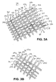
- Fig. 3a is an illustration of a braided reinforcing layer according to one embodiment of the invention.
- Reinforcing layer 24a includes multiple wires braided in pairs or "in tow".
- a first wire 30a is braided in a first braiding direction 38 with a second wire 32a braided in tow with the first wire 30a in the same first braiding direction 38.
- a third wire 34a is braided in a second braiding direction 35a with a forth wire 36a braided in tow with the third wire 34a in the same second braiding direction 35a.
- Each wire 30a, 32a, 34a, and 36a may be provided as a single filar or a multifilar wire.
- eight pairs of wires 31a through 31h are braided in the first braiding direction 38 with eight pairs of wires 33a through 33h braided in the second braiding direction to provide a braided reinforcing layer 24a composed of thirty two wires.
- Any number of pairs of wires may be braided to from braided reinforcement layer 24 using a braiding machine, such as those commercially available from Steeger, Germany; Wardwel, Mass, or other commercial suppliers.
- a braiding machine such as those commercially available from Steeger, Germany; Wardwel, Mass, or other commercial suppliers.
- three or more wires may be braided in tow in triplets, quadruplets, etc.
- first inner catheter 14 By providing multiple wires braided in the "in-tow” manner as shown, the torsional rigidy of first inner catheter 14 is improved, allowing first inner catheter 14 to be manufactured with a thin wall, maneuverable through small cardiac veins and allowing passage of an elongated medical device through first inner catheter 14.
- Wires 30a, 32a, 34a, and 36a are typically formed from stainless steel, a shape memory alloy such as Nitinol®, a polymeric fiber such as polyethylene napthalate, or other material that can be formed into a small cross-sectional wire having sufficient tensile strength to undergo the braiding process.
- the wires 30a, 32a, 34a, and 36a may be provided with a generally round or flat cross-sectional geometry.
- First inner catheter 14 is provided as slittable catheter for removing catheter 14 over an implanted medical device
- reinforcing layer 24 using a braided polymer wire such as polyethylene napthalate, forces required to slit the catheter body are reduced, making it easier to remove catheter 14 from the implanted medical device without dislodging the medical device from the implant site.
- Figure 3A illustrates a "one-over-one" tow braiding pattern wherein each wire pair 31a through 31h is braided over one pair of 33a through 33h, then under the next pair of 33a through 33h, and so on.
- Other tow braiding patterns are possible.
- a '"two-over-two" tow braiding pattern is illustrated in Figure 3B .
- Each wire pair 37a through 37f is braided over two pairs of 39a through 39d, then under the next two pairs of 39a through 39d, and so on.
- reinforcing layer 24 may be provided with a variable pic rate, imparting variable pushability and flexibility along the length of the first inner catheter.
- Fig. 4 is a cut-away view of a first inner catheter having a braided reinforcing layer.
- First inner catheter 14 includes an elongated shaft 44 typically constructed using an inner tubular member 40 defining the inner lumen of catheter 14 through which the second inner catheter extends and an elongatedtherm device is passed.
- Inner tubular member 40 may be formed from a fluoropolymer such as polytetrafluoroethylene (PTFE), a polyamide such as nylon, a polyolefin, a polyimide, or other materials which provide a generally lubricious inner lumpen for passing the second inner catheter and through which an elongated medical device.
- PTFE polytetrafluoroethylene
- Braided reinforcing layer 24 is formed over the outer surface of the inner tubular member 40.
- the braid density or "pic count” may be constant or variable and will be selected to provide the desired lateral flexibility, torsional rigidity, kink resistance, and pushability needed for a particular application.
- the pic count will typically range from 20 to 150 pics/inch and will usually fall in the range of approximately 45-60 pics/inch.
- An outer tubular member 42 is formed over braided reinforcing layer 24.
- Outer tubular member 42 is typically formed of a soft thermoplastic material such as a polyamide polyether block polymer, polyurethanes, silicone rubbers, nylon, polyethylene, or fluoronated hydrocarbon polymers.
- First inner catheter 14 may be provided having variable mechanical properties along the length of the catheter shaft 44.
- catheter shaft 44 may be provided with three distinct regions as generally disclosed in U.S. Pat. No. 5,676,659 (McGurk ).
- a first region extending from the proximal end of first inner catheter 14 includes outer tubular member 42, reinforcing layer 24 and inner tubular member 40.
- a second region extends from the termination of reinforcing layer 24 and includes inner and outer tubular members 40 and 42. The second region serves as a transition region between the first region and a third region including only outer tubular member 42.
- Outer tubular member 42 is formed to provide a continuous inner diameter with the inner tubular member 40 to thereby provide a constant diameter medical device delivery lumen.
- First inner catheter 14 may be provided with a distal end formed of a radio-opaque material.
- Fig. 5 is a plan view of one embodiment of first inner catheter 14 formed with a braided layer having a variable pic count.
- the reinforcing layer may be braided with a variable pic count.
- Shaft44 is shown having three regions 52, 54 and 56. Proximal region 52 extends distally from proximal end 21. Distal region 56 extends proximally from distal end 22. Region 54 is a transition region extending between region 52 and 54.
- the reinforcing layer formed along proximal region 52 is provided with a low pic count, which results in a relatively more axial orientation of the wires forming the reinforcing layer giving proximal region 52 enhanced pushability.
- the pic count is gradually transitioned from a lower pic count to a higher pic count along transition region 54.
- Distal region 56 is provided with a higher pic count than proximal region 52, giving distal region 56 enhanced flexibility. It is recognized that multiple regions may be formed having varying pic counts to provide shaft 44 with desired variation in mechanical properties.
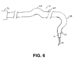
- Fig. 6 is a distal plan view of a delivery system according to another embodiment of the invention.
- First inner catheter 14 may be provided with a pre-shaped distal portion 48 including one or more curves, bends or angles arranged proximate distal end 22 to facilitate subselection of a targeted cardiac vein.
- first inner catheter 14 is provided with a pre-shaped distal end corresponding to an Amplatz curve.
- Second inner catheter 16 is formed as a thin sheath that tracks guidewire 18 and provides mechanical support to first, inner catheter 14 during advancement and rotation of first inner catheter 14 Second inner catheter 16 may be retracted into first inner catheter 14 to allow pre-shaped distal end 22 to conform to its natural bias for vein subselection.
- first inner catheter 14 may additionally include a second pre-shaped portion 50 along first inner catheter shaft 44, proximal to the distal pre-shaped portion 48.
- Proximal pre-shaped portion 50 may be provided as a generally "C-shaped" curve or other pre-shaped bend, curve, or angle.
- Proximal pre-shaped portion 50 is formed for providing cardiac vein wall support when tracking into a cardiac vein branch.
- Fig. 7 sis a flow chart summarizing a method for manufacturing a medical device delivery system.
- the outer catheter is formed.
- the outer catheter may be formed with or without a reinforcing layer and with or without a pre-curved or other pre-shaped distal end.
- the first inner catheter is formed as a thin-waned catheter having a reinforcing layer and is sized to pass through the outer catheter and allow passage of an elongated medical device through the first inner catheter lumen.
- the reinforcing layer is formed by applying a plurality of mulytifilar or single filar wires in an "in tow" braiding pattern on or incorporated in the first inner catheter wall.
- the first inner catheter may be formed with a pre-curved or other pre-shaped distal portion and/or proximal portion of the catheter shaft.
- the second inner catheter is formed at block 115 and is sized to pass through the first inner catheter.
Landscapes
- Health & Medical Sciences (AREA)
- Life Sciences & Earth Sciences (AREA)
- Heart & Thoracic Surgery (AREA)
- Engineering & Computer Science (AREA)
- Animal Behavior & Ethology (AREA)
- Biomedical Technology (AREA)
- Veterinary Medicine (AREA)
- Public Health (AREA)
- General Health & Medical Sciences (AREA)
- Biophysics (AREA)
- Pulmonology (AREA)
- Anesthesiology (AREA)
- Hematology (AREA)
- Vascular Medicine (AREA)
- Radiology & Medical Imaging (AREA)
- Nuclear Medicine, Radiotherapy & Molecular Imaging (AREA)
- Cardiology (AREA)
- Media Introduction/Drainage Providing Device (AREA)
- External Artificial Organs (AREA)
Claims (15)
- System zum Zuführen einer medizinischen Vorrichtung, das enthält:einen äußeren Katheter (12); undeinen ersten inneren Katheter (14), der durch den äußeren Katheter einführbar ist, wobei der erste innere Katheter dafür ausgelegt ist, eine lang gestreckte medizinische Vorrichtung, die durch ihn verläuft, aufzunehmen;gekennzeichnet durcheine geflochtene Verstärkungsschicht (24), die in dem ersten inneren Katheter positioniert ist und mehrere Drähte (30a, 32a, 34a, 36a) enthält, wobei ein erster Draht der mehreren Drähte im Schlepp mit einem zweiten Draht der mehreren Drähte in einer ersten Flechtrichtung (38) geflochten ist und ein dritter Draht der mehreren Drähte im Schlepp mit einem vierten Draht der mehreren Drähte in einer zweiten Flechtrichtung (35a) geflochten ist; undeinen zweiten inneren Katheter (16), der durch den ersten inneren Katheter einführbar ist und dafür ausgelegt ist, einen Führungsdraht (18), der durch ihn verläuft, aufzunehmen.
- System nach Anspruch 1, wobei der erste innere Katheter ein inneres rohrförmiges Element (40) und ein äußeres rohrförmiges Element (42) enthält und die geflochtene Verstärkungsschicht (24) zwischen dem inneren und dem äußeren rohrförmigen Element angeordnet ist.
- System nach Anspruch 1, wobei der erste, der zweite, der dritte und der vierte Draht jeweils Mehrfädendrähte sind.
- System nach Anspruch 1, wobei die geflochtene Verstärkungsschicht Drähte enthält, die aus Edelstahl- und Polyethylen-Naphthalat gebildet sind.
- System nach Anspruch 1, wobei die geflochtene Verstärkungsschicht eine veränderliche PIC-Rate enthält.
- System nach Anspruch 1, wobei der erste und der zweite Draht mit dem dritten und dem vierten Draht in einem Eins-über-Eins-Muster oder in einem Zwei-über-Zwei-Muster geflochten sind.
- System nach Anspruch 1, wobei die mehreren Drähte acht Paare von Drähten, die in der ersten Richtung geflochten sind, und acht Paare von Drähten, die in der zweiten Richtung geflochten sind, enthalten.
- System nach Anspruch 1, wobei der zweite innere Katheter (14) einen Katheterkörper mit veränderlichem Durometer enthält.
- System nach Anspruch 1, wobei der erste innere Katheter (14) ein distales Katheterende und einen vorgeformten Abschnitt (48), der von dem distalen Katheterende proximal beabstandet ist, enthält.
- System nach Anspruch 1, wobei der erste innere Katheter (14) mit einem Innendurchmesser von 5,8 French und einem Außendurchmesser von 7 French versehen ist.
- Verfahren zum Herstellen eines Systems zum Zuführen einer medizinischen Vorrichtung, das umfasst:Bilden eines äußeren Katheters (12);Bilden eines ersten inneren Katheters (14), der durch den äußeren Katheter einführbar ist und dafür ausgelegt ist, eine lang gestreckte medizinische Vorrichtung, die durch ihn verläuft, aufzunehmen;gekennzeichnet durchdie Tatsache, dass das Bilden des ersten inneren Katheters das Bilden einer geflochtenen Verstärkungsschicht (24), die einen ersten Draht, der im Schlepp mit einem zweiten Draht in einer ersten Flechtrichtung (38) geflochten ist, und einen dritten Draht, der im Schlepp mit einem vierten Draht in einer zweiten Flechtrichtung (35a) geflochten ist, enthält, umfasst und durch das Bilden eines zweiten inneren Katheters (16), der durch den ersten inneren Katheter einführbar ist und dafür ausgelegt ist, einen Führungsdraht (18), der durch ihn verläuft, aufzunehmen.
- Verfahren nach Anspruch 11, wobei das Bilden des ersten inneren Katheters das Bilden eines inneren rohrförmigen Elements (40), das Anordnen der geflochtenen Verstärkungsschicht (24) auf einer äußeren Oberfläche des inneren rohrförmigen Elements und das Bilden eines äußeren rohrförmigen Elements (42) auf der geflochtenen Verstärkungsschicht umfasst.
- Verfahren nach Anspruch 11, wobei das Bilden des ersten inneren Katheters das Bilden eines ersten vorgeformten Abschnitts (48) in der Nähe eines distalen Katheterendes umfasst.
- Verfahren nach Anspruch 11, wobei das Bilden des ersten inneren Katheters (14) das Bilden eines zweiten vorgeformten Abschnitts, der von dem ersten vorgeformten Abschnitt (48) proximal beabstandet ist, umfasst.
- Verfahren nach Anspruch 11, wobei der erste innere Katheter (14) mit einem Innendurchmesser von 5,8 French und einem Außendurchmesser von 7 French gebildet ist.
Applications Claiming Priority (2)
| Application Number | Priority Date | Filing Date | Title |
|---|---|---|---|
| US11/380,454 US7901396B2 (en) | 2006-04-27 | 2006-04-27 | Transvenous medical device delivery system |
| PCT/US2007/066834 WO2007127649A2 (en) | 2006-04-27 | 2007-04-18 | Transvenous medical device delivery system |
Publications (2)
| Publication Number | Publication Date |
|---|---|
| EP2010273A2 EP2010273A2 (de) | 2009-01-07 |
| EP2010273B1 true EP2010273B1 (de) | 2013-02-27 |
Family
ID=38649247
Family Applications (1)
| Application Number | Title | Priority Date | Filing Date |
|---|---|---|---|
| EP07760809A Active EP2010273B1 (de) | 2006-04-27 | 2007-04-18 | Transvenöses zufuhrsystem für medizinische vorrichtung |
Country Status (3)
| Country | Link |
|---|---|
| US (1) | US7901396B2 (de) |
| EP (1) | EP2010273B1 (de) |
| WO (1) | WO2007127649A2 (de) |
Families Citing this family (37)
| Publication number | Priority date | Publication date | Assignee | Title |
|---|---|---|---|---|
| US9339278B2 (en) | 2006-02-27 | 2016-05-17 | Biomet Manufacturing, Llc | Patient-specific acetabular guides and associated instruments |
| US20150335438A1 (en) | 2006-02-27 | 2015-11-26 | Biomet Manufacturing, Llc. | Patient-specific augments |
| US9918740B2 (en) | 2006-02-27 | 2018-03-20 | Biomet Manufacturing, Llc | Backup surgical instrument system and method |
| US9345548B2 (en) | 2006-02-27 | 2016-05-24 | Biomet Manufacturing, Llc | Patient-specific pre-operative planning |
| US8603180B2 (en) | 2006-02-27 | 2013-12-10 | Biomet Manufacturing, Llc | Patient-specific acetabular alignment guides |
| US8591516B2 (en) | 2006-02-27 | 2013-11-26 | Biomet Manufacturing, Llc | Patient-specific orthopedic instruments |
| US9173661B2 (en) | 2006-02-27 | 2015-11-03 | Biomet Manufacturing, Llc | Patient specific alignment guide with cutting surface and laser indicator |
| US8407067B2 (en) | 2007-04-17 | 2013-03-26 | Biomet Manufacturing Corp. | Method and apparatus for manufacturing an implant |
| US9907659B2 (en) | 2007-04-17 | 2018-03-06 | Biomet Manufacturing, Llc | Method and apparatus for manufacturing an implant |
| US9289253B2 (en) | 2006-02-27 | 2016-03-22 | Biomet Manufacturing, Llc | Patient-specific shoulder guide |
| US9795399B2 (en) | 2006-06-09 | 2017-10-24 | Biomet Manufacturing, Llc | Patient-specific knee alignment guide and associated method |
| US7914515B2 (en) * | 2007-07-18 | 2011-03-29 | St. Jude Medical, Atrial Fibrillation Division, Inc. | Catheter and introducer catheter having torque transfer layer and method of manufacture |
| US9968376B2 (en) | 2010-11-29 | 2018-05-15 | Biomet Manufacturing, Llc | Patient-specific orthopedic instruments |
| US9072624B2 (en) | 2012-02-23 | 2015-07-07 | Covidien Lp | Luminal stenting |
| US20140336572A1 (en) | 2013-05-07 | 2014-11-13 | St. Jude Medical, Atrial Fibrillation Division, Inc. | Guiding Medical Devices and Associated Methods of Manufacturing |
| US9782186B2 (en) | 2013-08-27 | 2017-10-10 | Covidien Lp | Vascular intervention system |
| US10045867B2 (en) | 2013-08-27 | 2018-08-14 | Covidien Lp | Delivery of medical devices |
| US9764139B2 (en) | 2014-01-24 | 2017-09-19 | Medtronic, Inc. | Pre-implant detection |
| US10926077B2 (en) | 2016-05-11 | 2021-02-23 | II Nunzio P. Pagano | Interchangeable lockable catheter |
| US10376396B2 (en) | 2017-01-19 | 2019-08-13 | Covidien Lp | Coupling units for medical device delivery systems |
| US10722310B2 (en) | 2017-03-13 | 2020-07-28 | Zimmer Biomet CMF and Thoracic, LLC | Virtual surgery planning system and method |
| US11071637B2 (en) | 2018-04-12 | 2021-07-27 | Covidien Lp | Medical device delivery |
| US11413176B2 (en) | 2018-04-12 | 2022-08-16 | Covidien Lp | Medical device delivery |
| US10786377B2 (en) | 2018-04-12 | 2020-09-29 | Covidien Lp | Medical device delivery |
| US11123209B2 (en) | 2018-04-12 | 2021-09-21 | Covidien Lp | Medical device delivery |
| US11413174B2 (en) | 2019-06-26 | 2022-08-16 | Covidien Lp | Core assembly for medical device delivery systems |
| US12115380B2 (en) * | 2019-10-11 | 2024-10-15 | Medtronic, Inc. | Medical device with braided tubular body |
| CN115768509A (zh) * | 2020-03-23 | 2023-03-07 | 波士顿科学国际有限公司 | 具有加强构件的引导导管 |
| EP4192328A4 (de) * | 2020-08-05 | 2024-07-17 | Shawn P. Fojtik | Medizinische aspirationsvorrichtungen und verfahren |
| US11786698B2 (en) | 2020-12-08 | 2023-10-17 | DePuy Synthes Products, Inc. | Catheter with textured surface |
| US11826520B2 (en) | 2020-12-08 | 2023-11-28 | DePuy Synthes Products, Inc. | Catheter designs for enhanced column strength |
| US12458518B2 (en) | 2021-02-17 | 2025-11-04 | Covidien Lp | Medical device delivery devices, systems, and methods |
| US12042413B2 (en) | 2021-04-07 | 2024-07-23 | Covidien Lp | Delivery of medical devices |
| US12109137B2 (en) | 2021-07-30 | 2024-10-08 | Covidien Lp | Medical device delivery |
| US11944558B2 (en) | 2021-08-05 | 2024-04-02 | Covidien Lp | Medical device delivery devices, systems, and methods |
| CN116020038A (zh) * | 2022-12-30 | 2023-04-28 | 辽宁垠艺生物科技股份有限公司 | 一种医用复合加强丝导管 |
| US20240285902A1 (en) * | 2023-02-23 | 2024-08-29 | DePuy Synthes Products, Inc. | Multiple wire catheter braid with wire count gradient |
Family Cites Families (24)
| Publication number | Priority date | Publication date | Assignee | Title |
|---|---|---|---|---|
| US3485234A (en) * | 1966-04-13 | 1969-12-23 | Cordis Corp | Tubular products and method of making same |
| US4243050A (en) * | 1977-12-13 | 1981-01-06 | Littleford Philip O | Method for inserting pacemaker electrodes and the like |
| US5120323A (en) * | 1990-01-12 | 1992-06-09 | Schneider (Usa) Inc. | Telescoping guide catheter system |
| US5954651A (en) * | 1993-08-18 | 1999-09-21 | Scimed Life Systems, Inc. | Catheter having a high tensile strength braid wire constraint |
| ES2194896T3 (es) | 1993-11-12 | 2003-12-01 | Micro Interventional Syst | Cateter de pequeño diametro con par elevado. |
| US6059779A (en) * | 1995-04-28 | 2000-05-09 | Target Therapeutics, Inc. | Delivery catheter for electrolytically detachable implant |
| US6022336A (en) * | 1996-05-20 | 2000-02-08 | Percusurge, Inc. | Catheter system for emboli containment |
| US5964795A (en) * | 1998-03-13 | 1999-10-12 | Medtronic, Inc. | Medical electrical lead |
| US6709429B1 (en) * | 2000-01-19 | 2004-03-23 | Scimed Life Systems, Inc. | Intravascular catheter with multiple axial fibers |
| US6836687B2 (en) * | 2000-03-31 | 2004-12-28 | Medtronic, Inc. | Method and system for delivery of a medical electrical lead within a venous system |
| US6733500B2 (en) * | 2000-03-31 | 2004-05-11 | Medtronic, Inc. | Method and system for delivering a medical electrical lead within a venous system |
| US6540733B2 (en) * | 2000-12-29 | 2003-04-01 | Corazon Technologies, Inc. | Proton generating catheters and methods for their use in enhancing fluid flow through a vascular site occupied by a calcified vascular occlusion |
| US6755812B2 (en) * | 2001-12-11 | 2004-06-29 | Cardiac Pacemakers, Inc. | Deflectable telescoping guide catheter |
| US7493156B2 (en) * | 2002-01-07 | 2009-02-17 | Cardiac Pacemakers, Inc. | Steerable guide catheter with pre-shaped rotatable shaft |
| US7717899B2 (en) * | 2002-01-28 | 2010-05-18 | Cardiac Pacemakers, Inc. | Inner and outer telescoping catheter delivery system |
| US7462184B2 (en) * | 2002-05-06 | 2008-12-09 | Pressure Products Medical Supplies Inc. | Introducer for accessing the coronary sinus of a heart |
| US7993351B2 (en) * | 2002-07-24 | 2011-08-09 | Pressure Products Medical Supplies, Inc. | Telescopic introducer with a compound curvature for inducing alignment and method of using the same |
| US6871085B2 (en) * | 2002-09-30 | 2005-03-22 | Medtronic, Inc. | Cardiac vein lead and guide catheter |
| JP2006501969A (ja) * | 2002-10-10 | 2006-01-19 | マイクロ セラピューティックス インコーポレイテッド | ワイヤ組紐補強マイクロカテーテル |
| US20050020914A1 (en) * | 2002-11-12 | 2005-01-27 | David Amundson | Coronary sinus access catheter with forward-imaging |
| AU2003300295A1 (en) * | 2002-12-20 | 2004-07-22 | Cardiac Inventions Unlimited, Inc. | Left ventricular pacing lead and implantation method |
| US7438712B2 (en) * | 2003-03-05 | 2008-10-21 | Scimed Life Systems, Inc. | Multi-braid exterior tube |
| US20050004553A1 (en) | 2003-07-02 | 2005-01-06 | Medtronic Ave, Inc. | Sheath catheter having variable over-the-wire length and methods of use |
| HRP20030588A2 (en) | 2003-07-18 | 2005-10-31 | Breyer Branko | Ultrasonically marked delivery system for left heart pacing lead |
-
2006
- 2006-04-27 US US11/380,454 patent/US7901396B2/en active Active
-
2007
- 2007-04-18 EP EP07760809A patent/EP2010273B1/de active Active
- 2007-04-18 WO PCT/US2007/066834 patent/WO2007127649A2/en not_active Ceased
Also Published As
| Publication number | Publication date |
|---|---|
| WO2007127649A3 (en) | 2008-01-17 |
| US7901396B2 (en) | 2011-03-08 |
| US20070255255A1 (en) | 2007-11-01 |
| EP2010273A2 (de) | 2009-01-07 |
| WO2007127649A2 (en) | 2007-11-08 |
Similar Documents
| Publication | Publication Date | Title |
|---|---|---|
| US7901396B2 (en) | Transvenous medical device delivery system | |
| US6408214B1 (en) | Deflectable tip catheter for CS pacing | |
| US7037290B2 (en) | Multi-lumen steerable catheter | |
| EP1465697B1 (de) | Steuerbarer katheter oder dergleichen | |
| US6871085B2 (en) | Cardiac vein lead and guide catheter | |
| US20120232563A1 (en) | Implant catheters for physiological pacing | |
| EP1804882B1 (de) | Katheter mit vorgeformter distaler spitze | |
| US6544270B1 (en) | Multi-lumen cardiac catheter and system | |
| US8090451B2 (en) | Transvenous active fixation lead system | |
| AU2013207563B2 (en) | Guidewire with highly flexible tip | |
| US7354428B1 (en) | Guiding catheter shaft with improved radiopacity on the wire braid | |
| US5358517A (en) | Electrical medical lead with textured stylet guide | |
| US9333335B2 (en) | Microcatheter implantable in venous, arterial or lymphatic networks | |
| EP4054691B1 (de) | Katheterschlauch für einen steuerbaren katheter zum implantieren einer implantierbaren medizinischen vorrichtung | |
| EP3232970A1 (de) | Verbesserter katheter und verfahren zur herstellung davon | |
| JP2009531084A (ja) | 左心室リードの形状 | |
| US20140276605A1 (en) | Catheter for vascular interventions | |
| WO2004047900A1 (en) | Multiple bend catheter for delivering a lead to a heart | |
| CN115768509A (zh) | 具有加强构件的引导导管 | |
| US20250195833A1 (en) | Variable radius braid for a medical device | |
| US9248271B2 (en) | Detection/stimulation microlead with enhanced positioning | |
| WO2010060889A1 (en) | Microcatheter |
Legal Events
| Date | Code | Title | Description |
|---|---|---|---|
| PUAI | Public reference made under article 153(3) epc to a published international application that has entered the european phase |
Free format text: ORIGINAL CODE: 0009012 |
|
| 17P | Request for examination filed |
Effective date: 20080730 |
|
| AK | Designated contracting states |
Kind code of ref document: A2 Designated state(s): DE FR |
|
| 17Q | First examination report despatched |
Effective date: 20090206 |
|
| DAX | Request for extension of the european patent (deleted) | ||
| RBV | Designated contracting states (corrected) |
Designated state(s): DE FR |
|
| RIC1 | Information provided on ipc code assigned before grant |
Ipc: A61N 1/05 20060101AFI20111111BHEP Ipc: A61M 25/00 20060101ALI20111111BHEP |
|
| GRAP | Despatch of communication of intention to grant a patent |
Free format text: ORIGINAL CODE: EPIDOSNIGR1 |
|
| RAP1 | Party data changed (applicant data changed or rights of an application transferred) |
Owner name: MEDTRONIC, INC. |
|
| GRAS | Grant fee paid |
Free format text: ORIGINAL CODE: EPIDOSNIGR3 |
|
| GRAA | (expected) grant |
Free format text: ORIGINAL CODE: 0009210 |
|
| AK | Designated contracting states |
Kind code of ref document: B1 Designated state(s): DE FR |
|
| REG | Reference to a national code |
Ref country code: DE Ref legal event code: R096 Ref document number: 602007028729 Country of ref document: DE Effective date: 20130425 |
|
| PLBE | No opposition filed within time limit |
Free format text: ORIGINAL CODE: 0009261 |
|
| STAA | Information on the status of an ep patent application or granted ep patent |
Free format text: STATUS: NO OPPOSITION FILED WITHIN TIME LIMIT |
|
| 26N | No opposition filed |
Effective date: 20131128 |
|
| REG | Reference to a national code |
Ref country code: DE Ref legal event code: R097 Ref document number: 602007028729 Country of ref document: DE Effective date: 20131128 |
|
| REG | Reference to a national code |
Ref country code: FR Ref legal event code: PLFP Year of fee payment: 10 |
|
| REG | Reference to a national code |
Ref country code: FR Ref legal event code: PLFP Year of fee payment: 11 |
|
| REG | Reference to a national code |
Ref country code: FR Ref legal event code: PLFP Year of fee payment: 12 |
|
| PGFP | Annual fee paid to national office [announced via postgrant information from national office to epo] |
Ref country code: FR Payment date: 20250319 Year of fee payment: 19 |
|
| PGFP | Annual fee paid to national office [announced via postgrant information from national office to epo] |
Ref country code: DE Payment date: 20250319 Year of fee payment: 19 |