EP2010181B1 - Methods of treating cell proliferative disorders by using pyrimidinediamine compounds - Google Patents
Methods of treating cell proliferative disorders by using pyrimidinediamine compounds Download PDFInfo
- Publication number
- EP2010181B1 EP2010181B1 EP07759007A EP07759007A EP2010181B1 EP 2010181 B1 EP2010181 B1 EP 2010181B1 EP 07759007 A EP07759007 A EP 07759007A EP 07759007 A EP07759007 A EP 07759007A EP 2010181 B1 EP2010181 B1 EP 2010181B1
- Authority
- EP
- European Patent Office
- Prior art keywords
- syk
- cell
- cells
- compound
- kinase
- Prior art date
- Legal status (The legal status is an assumption and is not a legal conclusion. Google has not performed a legal analysis and makes no representation as to the accuracy of the status listed.)
- Active
Links
- 230000002062 proliferating effect Effects 0.000 title claims abstract description 57
- 238000000034 method Methods 0.000 title abstract description 46
- YAAWASYJIRZXSZ-UHFFFAOYSA-N pyrimidine-2,4-diamine Chemical class NC1=CC=NC(N)=N1 YAAWASYJIRZXSZ-UHFFFAOYSA-N 0.000 title description 9
- 150000001875 compounds Chemical class 0.000 claims abstract description 163
- 206010028980 Neoplasm Diseases 0.000 claims abstract description 156
- 101150110875 Syk gene Proteins 0.000 claims abstract description 122
- 102000000551 Syk Kinase Human genes 0.000 claims abstract description 80
- 108010016672 Syk Kinase Proteins 0.000 claims abstract description 80
- 208000037265 diseases, disorders, signs and symptoms Diseases 0.000 claims abstract description 80
- 238000011282 treatment Methods 0.000 claims abstract description 38
- 229940043355 kinase inhibitor Drugs 0.000 claims abstract description 37
- 239000003757 phosphotransferase inhibitor Substances 0.000 claims abstract description 37
- 206010027476 Metastases Diseases 0.000 claims abstract description 28
- 230000009401 metastasis Effects 0.000 claims abstract description 28
- 208000002250 Hematologic Neoplasms Diseases 0.000 claims abstract description 24
- 210000004027 cell Anatomy 0.000 claims description 254
- 230000000694 effects Effects 0.000 claims description 96
- 239000003112 inhibitor Substances 0.000 claims description 68
- 230000002401 inhibitory effect Effects 0.000 claims description 48
- 102000006495 integrins Human genes 0.000 claims description 42
- 108010044426 integrins Proteins 0.000 claims description 42
- 108090000623 proteins and genes Proteins 0.000 claims description 37
- 208000031261 Acute myeloid leukaemia Diseases 0.000 claims description 34
- 239000000203 mixture Substances 0.000 claims description 26
- 241000700605 Viruses Species 0.000 claims description 25
- 230000001404 mediated effect Effects 0.000 claims description 25
- 102000004169 proteins and genes Human genes 0.000 claims description 25
- 201000010099 disease Diseases 0.000 claims description 17
- 150000003839 salts Chemical class 0.000 claims description 17
- 210000001744 T-lymphocyte Anatomy 0.000 claims description 15
- 208000010833 Chronic myeloid leukaemia Diseases 0.000 claims description 14
- 241000701044 Human gammaherpesvirus 4 Species 0.000 claims description 13
- 201000000050 myeloid neoplasm Diseases 0.000 claims description 12
- 230000011664 signaling Effects 0.000 claims description 12
- 206010006187 Breast cancer Diseases 0.000 claims description 11
- 201000003793 Myelodysplastic syndrome Diseases 0.000 claims description 11
- 208000019420 lymphoid neoplasm Diseases 0.000 claims description 11
- 239000002243 precursor Substances 0.000 claims description 11
- 208000012526 B-cell neoplasm Diseases 0.000 claims description 9
- 150000001204 N-oxides Chemical class 0.000 claims description 9
- 239000012453 solvate Substances 0.000 claims description 9
- 208000025317 T-cell and NK-cell neoplasm Diseases 0.000 claims description 8
- 125000002496 methyl group Chemical group [H]C([H])([H])* 0.000 claims description 8
- 208000024893 Acute lymphoblastic leukemia Diseases 0.000 claims description 7
- 208000014697 Acute lymphocytic leukaemia Diseases 0.000 claims description 7
- 208000011691 Burkitt lymphomas Diseases 0.000 claims description 7
- 206010061187 Haematopoietic neoplasm Diseases 0.000 claims description 7
- 201000005787 hematologic cancer Diseases 0.000 claims description 7
- 208000026310 Breast neoplasm Diseases 0.000 claims description 6
- UFHFLCQGNIYNRP-UHFFFAOYSA-N Hydrogen Chemical compound [H][H] UFHFLCQGNIYNRP-UHFFFAOYSA-N 0.000 claims description 6
- 208000008839 Kidney Neoplasms Diseases 0.000 claims description 6
- 206010038389 Renal cancer Diseases 0.000 claims description 6
- 201000010982 kidney cancer Diseases 0.000 claims description 6
- JDNJOMWHEGYGLU-UHFFFAOYSA-N 4-[[4-[3-chloro-4-(cyanomethoxy)anilino]-5-fluoropyrimidin-2-yl]amino]benzamide Chemical compound C1=CC(C(=O)N)=CC=C1NC1=NC=C(F)C(NC=2C=C(Cl)C(OCC#N)=CC=2)=N1 JDNJOMWHEGYGLU-UHFFFAOYSA-N 0.000 claims description 5
- 108020001507 fusion proteins Proteins 0.000 claims description 5
- 102000037865 fusion proteins Human genes 0.000 claims description 5
- 208000014767 Myeloproliferative disease Diseases 0.000 claims description 4
- 206010061902 Pancreatic neoplasm Diseases 0.000 claims description 4
- 238000003745 diagnosis Methods 0.000 claims description 4
- 210000004072 lung Anatomy 0.000 claims description 4
- 208000015486 malignant pancreatic neoplasm Diseases 0.000 claims description 4
- 201000002528 pancreatic cancer Diseases 0.000 claims description 4
- 208000008443 pancreatic carcinoma Diseases 0.000 claims description 4
- 206010005003 Bladder cancer Diseases 0.000 claims description 3
- 206010017993 Gastrointestinal neoplasms Diseases 0.000 claims description 3
- 206010033128 Ovarian cancer Diseases 0.000 claims description 3
- 206010061535 Ovarian neoplasm Diseases 0.000 claims description 3
- 208000007097 Urinary Bladder Neoplasms Diseases 0.000 claims description 3
- 208000009956 adenocarcinoma Diseases 0.000 claims description 3
- 208000003747 lymphoid leukemia Diseases 0.000 claims description 3
- XGXNTJHZPBRBHJ-UHFFFAOYSA-N n-phenylpyrimidin-2-amine Chemical group N=1C=CC=NC=1NC1=CC=CC=C1 XGXNTJHZPBRBHJ-UHFFFAOYSA-N 0.000 claims description 3
- 206010041823 squamous cell carcinoma Diseases 0.000 claims description 3
- 201000005112 urinary bladder cancer Diseases 0.000 claims description 3
- 201000010902 chronic myelomonocytic leukemia Diseases 0.000 claims description 2
- 241001502974 Human gammaherpesvirus 8 Species 0.000 claims 2
- 241000714260 Human T-lymphotropic virus 1 Species 0.000 claims 1
- 101100335081 Mus musculus Flt3 gene Proteins 0.000 abstract description 108
- 230000001225 therapeutic effect Effects 0.000 abstract description 13
- 230000005764 inhibitory process Effects 0.000 abstract description 12
- 230000000069 prophylactic effect Effects 0.000 abstract description 7
- 241000699670 Mus sp. Species 0.000 description 87
- 239000003981 vehicle Substances 0.000 description 69
- 210000004881 tumor cell Anatomy 0.000 description 63
- 241001465754 Metazoa Species 0.000 description 58
- -1 2,4-diamino-pyrimidine derivative compounds Chemical class 0.000 description 57
- 208000035475 disorder Diseases 0.000 description 57
- 241000282414 Homo sapiens Species 0.000 description 38
- 210000001185 bone marrow Anatomy 0.000 description 37
- 230000004083 survival effect Effects 0.000 description 31
- 208000033776 Myeloid Acute Leukemia Diseases 0.000 description 29
- 108091000080 Phosphotransferase Proteins 0.000 description 26
- 230000001594 aberrant effect Effects 0.000 description 26
- 208000032839 leukemia Diseases 0.000 description 26
- 210000005259 peripheral blood Anatomy 0.000 description 26
- 239000011886 peripheral blood Substances 0.000 description 26
- 102000020233 phosphotransferase Human genes 0.000 description 26
- 230000004913 activation Effects 0.000 description 23
- 230000035755 proliferation Effects 0.000 description 23
- 235000018102 proteins Nutrition 0.000 description 23
- 210000004369 blood Anatomy 0.000 description 22
- 239000008280 blood Substances 0.000 description 22
- 230000014509 gene expression Effects 0.000 description 22
- 125000001424 substituent group Chemical group 0.000 description 22
- 238000011887 Necropsy Methods 0.000 description 21
- 210000003719 b-lymphocyte Anatomy 0.000 description 21
- 210000000130 stem cell Anatomy 0.000 description 20
- 210000003958 hematopoietic stem cell Anatomy 0.000 description 19
- 210000001519 tissue Anatomy 0.000 description 19
- 230000004663 cell proliferation Effects 0.000 description 18
- 230000006870 function Effects 0.000 description 18
- 238000011789 NOD SCID mouse Methods 0.000 description 17
- 125000003118 aryl group Chemical group 0.000 description 17
- 238000002347 injection Methods 0.000 description 17
- 239000007924 injection Substances 0.000 description 17
- 229940090044 injection Drugs 0.000 description 17
- 230000001413 cellular effect Effects 0.000 description 15
- 230000034994 death Effects 0.000 description 15
- 231100000517 death Toxicity 0.000 description 15
- 230000002829 reductive effect Effects 0.000 description 15
- 238000009472 formulation Methods 0.000 description 14
- 238000010186 staining Methods 0.000 description 14
- 239000000758 substrate Substances 0.000 description 14
- 101000934338 Homo sapiens Myeloid cell surface antigen CD33 Proteins 0.000 description 13
- 241000699666 Mus <mouse, genus> Species 0.000 description 13
- 108700020796 Oncogene Proteins 0.000 description 13
- 201000011510 cancer Diseases 0.000 description 13
- 230000001394 metastastic effect Effects 0.000 description 13
- 230000037361 pathway Effects 0.000 description 13
- 208000017604 Hodgkin disease Diseases 0.000 description 12
- 238000003556 assay Methods 0.000 description 12
- 206010061289 metastatic neoplasm Diseases 0.000 description 12
- 210000001948 pro-b lymphocyte Anatomy 0.000 description 12
- 208000032791 BCR-ABL1 positive chronic myelogenous leukemia Diseases 0.000 description 11
- 230000002159 abnormal effect Effects 0.000 description 11
- 239000002246 antineoplastic agent Substances 0.000 description 11
- 239000003814 drug Substances 0.000 description 11
- 238000001727 in vivo Methods 0.000 description 11
- IAZDPXIOMUYVGZ-UHFFFAOYSA-N Dimethylsulphoxide Chemical compound CS(C)=O IAZDPXIOMUYVGZ-UHFFFAOYSA-N 0.000 description 10
- 102100025243 Myeloid cell surface antigen CD33 Human genes 0.000 description 10
- 125000000217 alkyl group Chemical group 0.000 description 10
- 238000004458 analytical method Methods 0.000 description 10
- 229940127089 cytotoxic agent Drugs 0.000 description 10
- 230000026731 phosphorylation Effects 0.000 description 10
- 238000006366 phosphorylation reaction Methods 0.000 description 10
- 238000002360 preparation method Methods 0.000 description 10
- 208000016261 weight loss Diseases 0.000 description 10
- SIKJAQJRHWYJAI-UHFFFAOYSA-N Indole Chemical compound C1=CC=C2NC=CC2=C1 SIKJAQJRHWYJAI-UHFFFAOYSA-N 0.000 description 9
- 208000033761 Myelogenous Chronic BCR-ABL Positive Leukemia Diseases 0.000 description 9
- 150000001413 amino acids Chemical group 0.000 description 9
- 125000003710 aryl alkyl group Chemical group 0.000 description 9
- 230000037396 body weight Effects 0.000 description 9
- 238000011161 development Methods 0.000 description 9
- 230000018109 developmental process Effects 0.000 description 9
- 229940079593 drug Drugs 0.000 description 9
- 238000002474 experimental method Methods 0.000 description 9
- 125000001072 heteroaryl group Chemical group 0.000 description 9
- 125000004435 hydrogen atom Chemical group [H]* 0.000 description 9
- 230000003993 interaction Effects 0.000 description 9
- 239000000047 product Substances 0.000 description 9
- 230000019491 signal transduction Effects 0.000 description 9
- CMSMOCZEIVJLDB-UHFFFAOYSA-N Cyclophosphamide Chemical compound ClCCN(CCCl)P1(=O)NCCCO1 CMSMOCZEIVJLDB-UHFFFAOYSA-N 0.000 description 8
- 108020004414 DNA Proteins 0.000 description 8
- 102000004190 Enzymes Human genes 0.000 description 8
- 108090000790 Enzymes Proteins 0.000 description 8
- 208000010747 Hodgkins lymphoma Diseases 0.000 description 8
- 241001529936 Murinae Species 0.000 description 8
- FIWILGQIZHDAQG-UHFFFAOYSA-N NC1=C(C(=O)NCC2=CC=C(C=C2)OCC(F)(F)F)C=C(C(=N1)N)N1N=C(N=C1)C1(CC1)C(F)(F)F Chemical compound NC1=C(C(=O)NCC2=CC=C(C=C2)OCC(F)(F)F)C=C(C(=N1)N)N1N=C(N=C1)C1(CC1)C(F)(F)F FIWILGQIZHDAQG-UHFFFAOYSA-N 0.000 description 8
- 102000004022 Protein-Tyrosine Kinases Human genes 0.000 description 8
- 108090000412 Protein-Tyrosine Kinases Proteins 0.000 description 8
- 108700020978 Proto-Oncogene Proteins 0.000 description 8
- 102000052575 Proto-Oncogene Human genes 0.000 description 8
- 239000006180 TBST buffer Substances 0.000 description 8
- 102000001742 Tumor Suppressor Proteins Human genes 0.000 description 8
- 108010040002 Tumor Suppressor Proteins Proteins 0.000 description 8
- 230000027455 binding Effects 0.000 description 8
- 150000001721 carbon Chemical group 0.000 description 8
- 229910052799 carbon Inorganic materials 0.000 description 8
- 230000032823 cell division Effects 0.000 description 8
- 239000003795 chemical substances by application Substances 0.000 description 8
- 229960004397 cyclophosphamide Drugs 0.000 description 8
- 230000002354 daily effect Effects 0.000 description 8
- 229940088598 enzyme Drugs 0.000 description 8
- 208000015181 infectious disease Diseases 0.000 description 8
- 125000002950 monocyclic group Chemical group 0.000 description 8
- 238000012360 testing method Methods 0.000 description 8
- 230000003612 virological effect Effects 0.000 description 8
- OUYCCCASQSFEME-QMMMGPOBSA-N L-tyrosine Chemical compound OC(=O)[C@@H](N)CC1=CC=C(O)C=C1 OUYCCCASQSFEME-QMMMGPOBSA-N 0.000 description 7
- 125000003342 alkenyl group Chemical group 0.000 description 7
- 125000000304 alkynyl group Chemical group 0.000 description 7
- 230000008901 benefit Effects 0.000 description 7
- 159000000007 calcium salts Chemical class 0.000 description 7
- 230000010261 cell growth Effects 0.000 description 7
- 230000012010 growth Effects 0.000 description 7
- 230000035772 mutation Effects 0.000 description 7
- 231100000590 oncogenic Toxicity 0.000 description 7
- 230000002246 oncogenic effect Effects 0.000 description 7
- 239000000243 solution Substances 0.000 description 7
- 230000009466 transformation Effects 0.000 description 7
- 231100000588 tumorigenic Toxicity 0.000 description 7
- 230000000381 tumorigenic effect Effects 0.000 description 7
- OUYCCCASQSFEME-UHFFFAOYSA-N tyrosine Natural products OC(=O)C(N)CC1=CC=C(O)C=C1 OUYCCCASQSFEME-UHFFFAOYSA-N 0.000 description 7
- YBYIRNPNPLQARY-UHFFFAOYSA-N 1H-indene Chemical compound C1=CC=C2CC=CC2=C1 YBYIRNPNPLQARY-UHFFFAOYSA-N 0.000 description 6
- UHOVQNZJYSORNB-UHFFFAOYSA-N Benzene Chemical group C1=CC=CC=C1 UHOVQNZJYSORNB-UHFFFAOYSA-N 0.000 description 6
- 208000031404 Chromosome Aberrations Diseases 0.000 description 6
- WSFSSNUMVMOOMR-UHFFFAOYSA-N Formaldehyde Chemical compound O=C WSFSSNUMVMOOMR-UHFFFAOYSA-N 0.000 description 6
- 208000021519 Hodgkin lymphoma Diseases 0.000 description 6
- 101000738771 Homo sapiens Receptor-type tyrosine-protein phosphatase C Proteins 0.000 description 6
- 102000043276 Oncogene Human genes 0.000 description 6
- 108010029485 Protein Isoforms Proteins 0.000 description 6
- 102000001708 Protein Isoforms Human genes 0.000 description 6
- 102100037422 Receptor-type tyrosine-protein phosphatase C Human genes 0.000 description 6
- 230000006907 apoptotic process Effects 0.000 description 6
- XSCHRSMBECNVNS-UHFFFAOYSA-N benzopyrazine Natural products N1=CC=NC2=CC=CC=C21 XSCHRSMBECNVNS-UHFFFAOYSA-N 0.000 description 6
- 239000011203 carbon fibre reinforced carbon Substances 0.000 description 6
- 150000002148 esters Chemical class 0.000 description 6
- GVEPBJHOBDJJJI-UHFFFAOYSA-N fluoranthene Chemical group C1=CC(C2=CC=CC=C22)=C3C2=CC=CC3=C1 GVEPBJHOBDJJJI-UHFFFAOYSA-N 0.000 description 6
- 125000004446 heteroarylalkyl group Chemical group 0.000 description 6
- RAXXELZNTBOGNW-UHFFFAOYSA-N imidazole Natural products C1=CNC=N1 RAXXELZNTBOGNW-UHFFFAOYSA-N 0.000 description 6
- 238000000338 in vitro Methods 0.000 description 6
- PQNFLJBBNBOBRQ-UHFFFAOYSA-N indane Chemical compound C1=CC=C2CCCC2=C1 PQNFLJBBNBOBRQ-UHFFFAOYSA-N 0.000 description 6
- 238000001990 intravenous administration Methods 0.000 description 6
- 210000004698 lymphocyte Anatomy 0.000 description 6
- 239000006166 lysate Substances 0.000 description 6
- 210000004379 membrane Anatomy 0.000 description 6
- 239000012528 membrane Substances 0.000 description 6
- 239000008194 pharmaceutical composition Substances 0.000 description 6
- 229940002612 prodrug Drugs 0.000 description 6
- 239000000651 prodrug Substances 0.000 description 6
- 102000005962 receptors Human genes 0.000 description 6
- 108020003175 receptors Proteins 0.000 description 6
- 230000005945 translocation Effects 0.000 description 6
- 230000009385 viral infection Effects 0.000 description 6
- XLYOFNOQVPJJNP-UHFFFAOYSA-N water Substances O XLYOFNOQVPJJNP-UHFFFAOYSA-N 0.000 description 6
- 208000016683 Adult T-cell leukemia/lymphoma Diseases 0.000 description 5
- IJGRMHOSHXDMSA-UHFFFAOYSA-N Atomic nitrogen Chemical compound N#N IJGRMHOSHXDMSA-UHFFFAOYSA-N 0.000 description 5
- 206010011906 Death Diseases 0.000 description 5
- 102100033636 Histone H3.2 Human genes 0.000 description 5
- 108010033040 Histones Proteins 0.000 description 5
- 101000935040 Homo sapiens Integrin beta-2 Proteins 0.000 description 5
- 102100025390 Integrin beta-2 Human genes 0.000 description 5
- 108010002586 Interleukin-7 Proteins 0.000 description 5
- 206010039491 Sarcoma Diseases 0.000 description 5
- 208000036142 Viral infection Diseases 0.000 description 5
- 201000006966 adult T-cell leukemia Diseases 0.000 description 5
- 238000004364 calculation method Methods 0.000 description 5
- 230000036952 cancer formation Effects 0.000 description 5
- 125000004432 carbon atom Chemical group C* 0.000 description 5
- 230000010307 cell transformation Effects 0.000 description 5
- 230000008859 change Effects 0.000 description 5
- 230000002596 correlated effect Effects 0.000 description 5
- 210000003714 granulocyte Anatomy 0.000 description 5
- 230000003394 haemopoietic effect Effects 0.000 description 5
- 230000011132 hemopoiesis Effects 0.000 description 5
- YLMAHDNUQAMNNX-UHFFFAOYSA-N imatinib methanesulfonate Chemical compound CS(O)(=O)=O.C1CN(C)CCN1CC1=CC=C(C(=O)NC=2C=C(NC=3N=C(C=CN=3)C=3C=NC=CC=3)C(C)=CC=2)C=C1 YLMAHDNUQAMNNX-UHFFFAOYSA-N 0.000 description 5
- 238000001114 immunoprecipitation Methods 0.000 description 5
- 238000000099 in vitro assay Methods 0.000 description 5
- 239000003446 ligand Substances 0.000 description 5
- 125000005647 linker group Chemical group 0.000 description 5
- 210000001165 lymph node Anatomy 0.000 description 5
- 239000003550 marker Substances 0.000 description 5
- 210000001616 monocyte Anatomy 0.000 description 5
- 108090000765 processed proteins & peptides Proteins 0.000 description 5
- QELSKZZBTMNZEB-UHFFFAOYSA-N propylparaben Chemical class CCCOC(=O)C1=CC=C(O)C=C1 QELSKZZBTMNZEB-UHFFFAOYSA-N 0.000 description 5
- 230000009467 reduction Effects 0.000 description 5
- 229920006395 saturated elastomer Polymers 0.000 description 5
- 125000003903 2-propenyl group Chemical group [H]C([*])([H])C([H])=C([H])[H] 0.000 description 4
- KDCGOANMDULRCW-UHFFFAOYSA-N 7H-purine Chemical compound N1=CNC2=NC=NC2=C1 KDCGOANMDULRCW-UHFFFAOYSA-N 0.000 description 4
- UJOBWOGCFQCDNV-UHFFFAOYSA-N 9H-carbazole Chemical compound C1=CC=C2C3=CC=CC=C3NC2=C1 UJOBWOGCFQCDNV-UHFFFAOYSA-N 0.000 description 4
- 108700028369 Alleles Proteins 0.000 description 4
- CURLTUGMZLYLDI-UHFFFAOYSA-N Carbon dioxide Chemical compound O=C=O CURLTUGMZLYLDI-UHFFFAOYSA-N 0.000 description 4
- 208000005623 Carcinogenesis Diseases 0.000 description 4
- 206010061818 Disease progression Diseases 0.000 description 4
- YLQBMQCUIZJEEH-UHFFFAOYSA-N Furan Chemical compound C=1C=COC=1 YLQBMQCUIZJEEH-UHFFFAOYSA-N 0.000 description 4
- UFWIBTONFRDIAS-UHFFFAOYSA-N Naphthalene Chemical group C1=CC=CC2=CC=CC=C21 UFWIBTONFRDIAS-UHFFFAOYSA-N 0.000 description 4
- PCNDJXKNXGMECE-UHFFFAOYSA-N Phenazine Natural products C1=CC=CC2=NC3=CC=CC=C3N=C21 PCNDJXKNXGMECE-UHFFFAOYSA-N 0.000 description 4
- KYQCOXFCLRTKLS-UHFFFAOYSA-N Pyrazine Chemical compound C1=CN=CC=N1 KYQCOXFCLRTKLS-UHFFFAOYSA-N 0.000 description 4
- JUJWROOIHBZHMG-UHFFFAOYSA-N Pyridine Chemical compound C1=CC=NC=C1 JUJWROOIHBZHMG-UHFFFAOYSA-N 0.000 description 4
- KAESVJOAVNADME-UHFFFAOYSA-N Pyrrole Chemical compound C=1C=CNC=1 KAESVJOAVNADME-UHFFFAOYSA-N 0.000 description 4
- SMWDFEZZVXVKRB-UHFFFAOYSA-N Quinoline Chemical compound N1=CC=CC2=CC=CC=C21 SMWDFEZZVXVKRB-UHFFFAOYSA-N 0.000 description 4
- YTPLMLYBLZKORZ-UHFFFAOYSA-N Thiophene Chemical compound C=1C=CSC=1 YTPLMLYBLZKORZ-UHFFFAOYSA-N 0.000 description 4
- 235000001014 amino acid Nutrition 0.000 description 4
- 238000010171 animal model Methods 0.000 description 4
- MWPLVEDNUUSJAV-UHFFFAOYSA-N anthracene Chemical group C1=CC=CC2=CC3=CC=CC=C3C=C21 MWPLVEDNUUSJAV-UHFFFAOYSA-N 0.000 description 4
- 230000000259 anti-tumor effect Effects 0.000 description 4
- CUFNKYGDVFVPHO-UHFFFAOYSA-N azulene Chemical group C1=CC=CC2=CC=CC2=C1 CUFNKYGDVFVPHO-UHFFFAOYSA-N 0.000 description 4
- 230000015572 biosynthetic process Effects 0.000 description 4
- 231100000504 carcinogenesis Toxicity 0.000 description 4
- 238000000423 cell based assay Methods 0.000 description 4
- 230000033077 cellular process Effects 0.000 description 4
- 231100000005 chromosome aberration Toxicity 0.000 description 4
- WDECIBYCCFPHNR-UHFFFAOYSA-N chrysene Chemical group C1=CC=CC2=CC=C3C4=CC=CC=C4C=CC3=C21 WDECIBYCCFPHNR-UHFFFAOYSA-N 0.000 description 4
- VPUGDVKSAQVFFS-UHFFFAOYSA-N coronene Chemical group C1=C(C2=C34)C=CC3=CC=C(C=C3)C4=C4C3=CC=C(C=C3)C4=C2C3=C1 VPUGDVKSAQVFFS-UHFFFAOYSA-N 0.000 description 4
- 230000000875 corresponding effect Effects 0.000 description 4
- 125000004122 cyclic group Chemical group 0.000 description 4
- 230000001086 cytosolic effect Effects 0.000 description 4
- 238000001514 detection method Methods 0.000 description 4
- 230000005750 disease progression Effects 0.000 description 4
- 239000012458 free base Substances 0.000 description 4
- 125000000524 functional group Chemical group 0.000 description 4
- 239000003102 growth factor Substances 0.000 description 4
- 229940088597 hormone Drugs 0.000 description 4
- 239000005556 hormone Substances 0.000 description 4
- 238000011532 immunohistochemical staining Methods 0.000 description 4
- 238000011081 inoculation Methods 0.000 description 4
- AWJUIBRHMBBTKR-UHFFFAOYSA-N isoquinoline Chemical compound C1=NC=CC2=CC=CC=C21 AWJUIBRHMBBTKR-UHFFFAOYSA-N 0.000 description 4
- 230000003211 malignant effect Effects 0.000 description 4
- 235000013336 milk Nutrition 0.000 description 4
- 239000008267 milk Substances 0.000 description 4
- 210000004080 milk Anatomy 0.000 description 4
- 210000000440 neutrophil Anatomy 0.000 description 4
- 210000003819 peripheral blood mononuclear cell Anatomy 0.000 description 4
- 230000002093 peripheral effect Effects 0.000 description 4
- YNPNZTXNASCQKK-UHFFFAOYSA-N phenanthrene Chemical group C1=CC=C2C3=CC=CC=C3C=CC2=C1 YNPNZTXNASCQKK-UHFFFAOYSA-N 0.000 description 4
- RDOWQLZANAYVLL-UHFFFAOYSA-N phenanthridine Chemical compound C1=CC=C2C3=CC=CC=C3C=NC2=C1 RDOWQLZANAYVLL-UHFFFAOYSA-N 0.000 description 4
- GBROPGWFBFCKAG-UHFFFAOYSA-N picene Chemical group C1=CC2=C3C=CC=CC3=CC=C2C2=C1C1=CC=CC=C1C=C2 GBROPGWFBFCKAG-UHFFFAOYSA-N 0.000 description 4
- 230000036470 plasma concentration Effects 0.000 description 4
- 125000002924 primary amino group Chemical group [H]N([H])* 0.000 description 4
- BBEAQIROQSPTKN-UHFFFAOYSA-N pyrene Chemical group C1=CC=C2C=CC3=CC=CC4=CC=C1C2=C43 BBEAQIROQSPTKN-UHFFFAOYSA-N 0.000 description 4
- 108091008598 receptor tyrosine kinases Proteins 0.000 description 4
- 102000027426 receptor tyrosine kinases Human genes 0.000 description 4
- 230000009885 systemic effect Effects 0.000 description 4
- 230000008685 targeting Effects 0.000 description 4
- ZIIUUSVHCHPIQD-UHFFFAOYSA-N 2,4,6-trimethyl-N-[3-(trifluoromethyl)phenyl]benzenesulfonamide Chemical compound CC1=CC(C)=CC(C)=C1S(=O)(=O)NC1=CC=CC(C(F)(F)F)=C1 ZIIUUSVHCHPIQD-UHFFFAOYSA-N 0.000 description 3
- 125000004974 2-butenyl group Chemical group C(C=CC)* 0.000 description 3
- GJCOSYZMQJWQCA-UHFFFAOYSA-N 9H-xanthene Chemical compound C1=CC=C2CC3=CC=CC=C3OC2=C1 GJCOSYZMQJWQCA-UHFFFAOYSA-N 0.000 description 3
- 208000009746 Adult T-Cell Leukemia-Lymphoma Diseases 0.000 description 3
- 108090000672 Annexin A5 Proteins 0.000 description 3
- 102000004121 Annexin A5 Human genes 0.000 description 3
- 102100026008 Breakpoint cluster region protein Human genes 0.000 description 3
- 108010037362 Extracellular Matrix Proteins Proteins 0.000 description 3
- 102000010834 Extracellular Matrix Proteins Human genes 0.000 description 3
- 206010061598 Immunodeficiency Diseases 0.000 description 3
- 108010064785 Phospholipases Proteins 0.000 description 3
- 102000015439 Phospholipases Human genes 0.000 description 3
- 208000006664 Precursor Cell Lymphoblastic Leukemia-Lymphoma Diseases 0.000 description 3
- 102000001253 Protein Kinase Human genes 0.000 description 3
- CZPWVGJYEJSRLH-UHFFFAOYSA-N Pyrimidine Chemical compound C1=CN=CN=C1 CZPWVGJYEJSRLH-UHFFFAOYSA-N 0.000 description 3
- 102100020718 Receptor-type tyrosine-protein kinase FLT3 Human genes 0.000 description 3
- 241000283984 Rodentia Species 0.000 description 3
- 208000029052 T-cell acute lymphoblastic leukemia Diseases 0.000 description 3
- 241000863480 Vinca Species 0.000 description 3
- 108700005077 Viral Genes Proteins 0.000 description 3
- 102000007624 ZAP-70 Protein-Tyrosine Kinase Human genes 0.000 description 3
- 108010046882 ZAP-70 Protein-Tyrosine Kinase Proteins 0.000 description 3
- 230000001154 acute effect Effects 0.000 description 3
- 125000002015 acyclic group Chemical group 0.000 description 3
- 239000000853 adhesive Substances 0.000 description 3
- 230000001070 adhesive effect Effects 0.000 description 3
- 229940100198 alkylating agent Drugs 0.000 description 3
- 239000002168 alkylating agent Substances 0.000 description 3
- 230000004075 alteration Effects 0.000 description 3
- 150000001408 amides Chemical class 0.000 description 3
- 239000003242 anti bacterial agent Substances 0.000 description 3
- 230000000340 anti-metabolite Effects 0.000 description 3
- 229940088710 antibiotic agent Drugs 0.000 description 3
- 229940100197 antimetabolite Drugs 0.000 description 3
- 239000002256 antimetabolite Substances 0.000 description 3
- BVUSIQTYUVWOSX-UHFFFAOYSA-N arsindole Chemical compound C1=CC=C2[As]C=CC2=C1 BVUSIQTYUVWOSX-UHFFFAOYSA-N 0.000 description 3
- 230000035578 autophosphorylation Effects 0.000 description 3
- 239000002585 base Substances 0.000 description 3
- 239000000872 buffer Substances 0.000 description 3
- 208000035269 cancer or benign tumor Diseases 0.000 description 3
- 230000005754 cellular signaling Effects 0.000 description 3
- QZHPTGXQGDFGEN-UHFFFAOYSA-N chromene Chemical compound C1=CC=C2C=C[CH]OC2=C1 QZHPTGXQGDFGEN-UHFFFAOYSA-N 0.000 description 3
- 125000006165 cyclic alkyl group Chemical group 0.000 description 3
- 230000007547 defect Effects 0.000 description 3
- 239000003534 dna topoisomerase inhibitor Substances 0.000 description 3
- 231100000673 dose–response relationship Toxicity 0.000 description 3
- 239000000839 emulsion Substances 0.000 description 3
- 210000002919 epithelial cell Anatomy 0.000 description 3
- 230000000925 erythroid effect Effects 0.000 description 3
- 125000001495 ethyl group Chemical group [H]C([H])([H])C([H])([H])* 0.000 description 3
- 125000002534 ethynyl group Chemical group [H]C#C* 0.000 description 3
- 210000002744 extracellular matrix Anatomy 0.000 description 3
- 238000000684 flow cytometry Methods 0.000 description 3
- RMBPEFMHABBEKP-UHFFFAOYSA-N fluorene Chemical compound C1=CC=C2C3=C[CH]C=CC3=CC2=C1 RMBPEFMHABBEKP-UHFFFAOYSA-N 0.000 description 3
- 108010003374 fms-Like Tyrosine Kinase 3 Proteins 0.000 description 3
- 239000012737 fresh medium Substances 0.000 description 3
- 210000002216 heart Anatomy 0.000 description 3
- 208000010726 hind limb paralysis Diseases 0.000 description 3
- 102000056982 human CD33 Human genes 0.000 description 3
- 150000004677 hydrates Chemical class 0.000 description 3
- 125000002887 hydroxy group Chemical group [H]O* 0.000 description 3
- 229960002411 imatinib Drugs 0.000 description 3
- 230000036039 immunity Effects 0.000 description 3
- 238000002513 implantation Methods 0.000 description 3
- PZOUSPYUWWUPPK-UHFFFAOYSA-N indole Natural products CC1=CC=CC2=C1C=CN2 PZOUSPYUWWUPPK-UHFFFAOYSA-N 0.000 description 3
- RKJUIXBNRJVNHR-UHFFFAOYSA-N indolenine Natural products C1=CC=C2CC=NC2=C1 RKJUIXBNRJVNHR-UHFFFAOYSA-N 0.000 description 3
- 230000001939 inductive effect Effects 0.000 description 3
- 201000006747 infectious mononucleosis Diseases 0.000 description 3
- 230000003834 intracellular effect Effects 0.000 description 3
- 125000001449 isopropyl group Chemical group [H]C([H])([H])C([H])(*)C([H])([H])[H] 0.000 description 3
- QDLAGTHXVHQKRE-UHFFFAOYSA-N lichenxanthone Natural products COC1=CC(O)=C2C(=O)C3=C(C)C=C(OC)C=C3OC2=C1 QDLAGTHXVHQKRE-UHFFFAOYSA-N 0.000 description 3
- 230000000670 limiting effect Effects 0.000 description 3
- 239000007788 liquid Substances 0.000 description 3
- 239000012139 lysis buffer Substances 0.000 description 3
- 238000005259 measurement Methods 0.000 description 3
- 229910052751 metal Inorganic materials 0.000 description 3
- 239000002184 metal Substances 0.000 description 3
- 208000017869 myelodysplastic/myeloproliferative disease Diseases 0.000 description 3
- 229910052757 nitrogen Inorganic materials 0.000 description 3
- NIHNNTQXNPWCJQ-UHFFFAOYSA-N o-biphenylenemethane Natural products C1=CC=C2CC3=CC=CC=C3C2=C1 NIHNNTQXNPWCJQ-UHFFFAOYSA-N 0.000 description 3
- XDJOIMJURHQYDW-UHFFFAOYSA-N phenalene Chemical compound C1=CC(CC=C2)=C3C2=CC=CC3=C1 XDJOIMJURHQYDW-UHFFFAOYSA-N 0.000 description 3
- 239000000843 powder Substances 0.000 description 3
- 230000008569 process Effects 0.000 description 3
- 102000004196 processed proteins & peptides Human genes 0.000 description 3
- 230000002035 prolonged effect Effects 0.000 description 3
- 125000001436 propyl group Chemical group [H]C([*])([H])C([H])([H])C([H])([H])[H] 0.000 description 3
- 235000010232 propyl p-hydroxybenzoate Nutrition 0.000 description 3
- 108060006633 protein kinase Proteins 0.000 description 3
- 210000003079 salivary gland Anatomy 0.000 description 3
- 208000011581 secondary neoplasm Diseases 0.000 description 3
- 210000002966 serum Anatomy 0.000 description 3
- 108091006024 signal transducing proteins Proteins 0.000 description 3
- 102000034285 signal transducing proteins Human genes 0.000 description 3
- 210000000952 spleen Anatomy 0.000 description 3
- 102000009076 src-Family Kinases Human genes 0.000 description 3
- 108010087686 src-Family Kinases Proteins 0.000 description 3
- 238000007920 subcutaneous administration Methods 0.000 description 3
- 239000000725 suspension Substances 0.000 description 3
- 208000024891 symptom Diseases 0.000 description 3
- 239000003826 tablet Substances 0.000 description 3
- 238000002560 therapeutic procedure Methods 0.000 description 3
- 208000008732 thymoma Diseases 0.000 description 3
- 229940044693 topoisomerase inhibitor Drugs 0.000 description 3
- 231100000419 toxicity Toxicity 0.000 description 3
- 230000001988 toxicity Effects 0.000 description 3
- 238000013518 transcription Methods 0.000 description 3
- 230000035897 transcription Effects 0.000 description 3
- 238000002054 transplantation Methods 0.000 description 3
- 230000005740 tumor formation Effects 0.000 description 3
- 241001529453 unidentified herpesvirus Species 0.000 description 3
- 210000003462 vein Anatomy 0.000 description 3
- 230000004580 weight loss Effects 0.000 description 3
- 238000001262 western blot Methods 0.000 description 3
- AIFRHYZBTHREPW-UHFFFAOYSA-N β-carboline Chemical compound N1=CC=C2C3=CC=CC=C3NC2=C1 AIFRHYZBTHREPW-UHFFFAOYSA-N 0.000 description 3
- 125000006708 (C5-C14) heteroaryl group Chemical group 0.000 description 2
- IAKHMKGGTNLKSZ-INIZCTEOSA-N (S)-colchicine Chemical compound C1([C@@H](NC(C)=O)CC2)=CC(=O)C(OC)=CC=C1C1=C2C=C(OC)C(OC)=C1OC IAKHMKGGTNLKSZ-INIZCTEOSA-N 0.000 description 2
- FLBAYUMRQUHISI-UHFFFAOYSA-N 1,8-naphthyridine Chemical compound N1=CC=CC2=CC=CN=C21 FLBAYUMRQUHISI-UHFFFAOYSA-N 0.000 description 2
- 125000004973 1-butenyl group Chemical group C(=CCC)* 0.000 description 2
- 125000004972 1-butynyl group Chemical group [H]C([H])([H])C([H])([H])C#C* 0.000 description 2
- BAXOFTOLAUCFNW-UHFFFAOYSA-N 1H-indazole Chemical compound C1=CC=C2C=NNC2=C1 BAXOFTOLAUCFNW-UHFFFAOYSA-N 0.000 description 2
- MFJCPDOGFAYSTF-UHFFFAOYSA-N 1H-isochromene Chemical compound C1=CC=C2COC=CC2=C1 MFJCPDOGFAYSTF-UHFFFAOYSA-N 0.000 description 2
- AAQTWLBJPNLKHT-UHFFFAOYSA-N 1H-perimidine Chemical compound N1C=NC2=CC=CC3=CC=CC1=C32 AAQTWLBJPNLKHT-UHFFFAOYSA-N 0.000 description 2
- ODMMNALOCMNQJZ-UHFFFAOYSA-N 1H-pyrrolizine Chemical compound C1=CC=C2CC=CN21 ODMMNALOCMNQJZ-UHFFFAOYSA-N 0.000 description 2
- VEPOHXYIFQMVHW-XOZOLZJESA-N 2,3-dihydroxybutanedioic acid (2S,3S)-3,4-dimethyl-2-phenylmorpholine Chemical compound OC(C(O)C(O)=O)C(O)=O.C[C@H]1[C@@H](OCCN1C)c1ccccc1 VEPOHXYIFQMVHW-XOZOLZJESA-N 0.000 description 2
- UXGVMFHEKMGWMA-UHFFFAOYSA-N 2-benzofuran Chemical compound C1=CC=CC2=COC=C21 UXGVMFHEKMGWMA-UHFFFAOYSA-N 0.000 description 2
- 125000001494 2-propynyl group Chemical group [H]C#CC([H])([H])* 0.000 description 2
- VHMICKWLTGFITH-UHFFFAOYSA-N 2H-isoindole Chemical compound C1=CC=CC2=CNC=C21 VHMICKWLTGFITH-UHFFFAOYSA-N 0.000 description 2
- MGADZUXDNSDTHW-UHFFFAOYSA-N 2H-pyran Chemical compound C1OC=CC=C1 MGADZUXDNSDTHW-UHFFFAOYSA-N 0.000 description 2
- 125000000474 3-butynyl group Chemical group [H]C#CC([H])([H])C([H])([H])* 0.000 description 2
- GDRVFDDBLLKWRI-UHFFFAOYSA-N 4H-quinolizine Chemical compound C1=CC=CN2CC=CC=C21 GDRVFDDBLLKWRI-UHFFFAOYSA-N 0.000 description 2
- ZKHQWZAMYRWXGA-KQYNXXCUSA-J ATP(4-) Chemical compound C1=NC=2C(N)=NC=NC=2N1[C@@H]1O[C@H](COP([O-])(=O)OP([O-])(=O)OP([O-])([O-])=O)[C@@H](O)[C@H]1O ZKHQWZAMYRWXGA-KQYNXXCUSA-J 0.000 description 2
- 208000033496 Acute myeloid leukaemia with myelodysplasia-related features Diseases 0.000 description 2
- ZKHQWZAMYRWXGA-UHFFFAOYSA-N Adenosine triphosphate Natural products C1=NC=2C(N)=NC=NC=2N1C1OC(COP(O)(=O)OP(O)(=O)OP(O)(O)=O)C(O)C1O ZKHQWZAMYRWXGA-UHFFFAOYSA-N 0.000 description 2
- 206010073478 Anaplastic large-cell lymphoma Diseases 0.000 description 2
- 108091008875 B cell receptors Proteins 0.000 description 2
- 238000000846 Bartlett's test Methods 0.000 description 2
- 241000283690 Bos taurus Species 0.000 description 2
- 125000001313 C5-C10 heteroaryl group Chemical group 0.000 description 2
- 0 C=C(Cc1ccc(C(C=O)N)cc1)C(NC1Nc(cc2O)ccc2OCC#N)=*C=C1F Chemical compound C=C(Cc1ccc(C(C=O)N)cc1)C(NC1Nc(cc2O)ccc2OCC#N)=*C=C1F 0.000 description 2
- GKDRMWXFWHEQQT-UHFFFAOYSA-N CC(C)(C(N1COP(O)(O)=O)=O)Oc(cc2)c1nc2Nc1nc(Nc(cc2OC)cc(OC)c2OC)ncc1F Chemical compound CC(C)(C(N1COP(O)(O)=O)=O)Oc(cc2)c1nc2Nc1nc(Nc(cc2OC)cc(OC)c2OC)ncc1F GKDRMWXFWHEQQT-UHFFFAOYSA-N 0.000 description 2
- OKTJSMMVPCPJKN-UHFFFAOYSA-N Carbon Chemical group [C] OKTJSMMVPCPJKN-UHFFFAOYSA-N 0.000 description 2
- 239000004215 Carbon black (E152) Substances 0.000 description 2
- FKLJPTJMIBLJAV-UHFFFAOYSA-N Compound IV Chemical compound O1N=C(C)C=C1CCCCCCCOC1=CC=C(C=2OCCN=2)C=C1 FKLJPTJMIBLJAV-UHFFFAOYSA-N 0.000 description 2
- 108090000695 Cytokines Proteins 0.000 description 2
- 102000004127 Cytokines Human genes 0.000 description 2
- LFQSCWFLJHTTHZ-UHFFFAOYSA-N Ethanol Chemical compound CCO LFQSCWFLJHTTHZ-UHFFFAOYSA-N 0.000 description 2
- 206010064571 Gene mutation Diseases 0.000 description 2
- NTYJJOPFIAHURM-UHFFFAOYSA-N Histamine Chemical compound NCCC1=CN=CN1 NTYJJOPFIAHURM-UHFFFAOYSA-N 0.000 description 2
- 241000725303 Human immunodeficiency virus Species 0.000 description 2
- 108060003951 Immunoglobulin Proteins 0.000 description 2
- 102000008607 Integrin beta3 Human genes 0.000 description 2
- 108010020950 Integrin beta3 Proteins 0.000 description 2
- GUBGYTABKSRVRQ-QKKXKWKRSA-N Lactose Natural products OC[C@H]1O[C@@H](O[C@H]2[C@H](O)[C@@H](O)C(O)O[C@@H]2CO)[C@H](O)[C@@H](O)[C@H]1O GUBGYTABKSRVRQ-QKKXKWKRSA-N 0.000 description 2
- 208000032004 Large-Cell Anaplastic Lymphoma Diseases 0.000 description 2
- 206010025323 Lymphomas Diseases 0.000 description 2
- 102000043136 MAP kinase family Human genes 0.000 description 2
- 108091054455 MAP kinase family Proteins 0.000 description 2
- 102100024193 Mitogen-activated protein kinase 1 Human genes 0.000 description 2
- ZCQWOFVYLHDMMC-UHFFFAOYSA-N Oxazole Chemical compound C1=COC=N1 ZCQWOFVYLHDMMC-UHFFFAOYSA-N 0.000 description 2
- 108091008606 PDGF receptors Proteins 0.000 description 2
- 108091005804 Peptidases Proteins 0.000 description 2
- 102000035195 Peptidases Human genes 0.000 description 2
- 206010035226 Plasma cell myeloma Diseases 0.000 description 2
- 102000011653 Platelet-Derived Growth Factor Receptors Human genes 0.000 description 2
- 102000003923 Protein Kinase C Human genes 0.000 description 2
- 108090000315 Protein Kinase C Proteins 0.000 description 2
- WTKZEGDFNFYCGP-UHFFFAOYSA-N Pyrazole Chemical compound C=1C=NNC=1 WTKZEGDFNFYCGP-UHFFFAOYSA-N 0.000 description 2
- 108091005682 Receptor kinases Proteins 0.000 description 2
- 208000009527 Refractory anemia Diseases 0.000 description 2
- 206010072684 Refractory cytopenia with unilineage dysplasia Diseases 0.000 description 2
- 102000014400 SH2 domains Human genes 0.000 description 2
- 108050003452 SH2 domains Proteins 0.000 description 2
- VYPSYNLAJGMNEJ-UHFFFAOYSA-N Silicium dioxide Chemical compound O=[Si]=O VYPSYNLAJGMNEJ-UHFFFAOYSA-N 0.000 description 2
- DBMJMQXJHONAFJ-UHFFFAOYSA-M Sodium laurylsulphate Chemical compound [Na+].CCCCCCCCCCCCOS([O-])(=O)=O DBMJMQXJHONAFJ-UHFFFAOYSA-M 0.000 description 2
- 206010041660 Splenomegaly Diseases 0.000 description 2
- 238000000692 Student's t-test Methods 0.000 description 2
- 108091008874 T cell receptors Proteins 0.000 description 2
- 102000016266 T-Cell Antigen Receptors Human genes 0.000 description 2
- 206010042971 T-cell lymphoma Diseases 0.000 description 2
- NKANXQFJJICGDU-QPLCGJKRSA-N Tamoxifen Chemical compound C=1C=CC=CC=1C(/CC)=C(C=1C=CC(OCCN(C)C)=CC=1)/C1=CC=CC=C1 NKANXQFJJICGDU-QPLCGJKRSA-N 0.000 description 2
- FZWLAAWBMGSTSO-UHFFFAOYSA-N Thiazole Chemical compound C1=CSC=N1 FZWLAAWBMGSTSO-UHFFFAOYSA-N 0.000 description 2
- SLGBZMMZGDRARJ-UHFFFAOYSA-N Triphenylene Chemical group C1=CC=C2C3=CC=CC=C3C3=CC=CC=C3C2=C1 SLGBZMMZGDRARJ-UHFFFAOYSA-N 0.000 description 2
- XSQUKJJJFZCRTK-UHFFFAOYSA-N Urea Chemical compound NC(N)=O XSQUKJJJFZCRTK-UHFFFAOYSA-N 0.000 description 2
- QVXFGVVYTKZLJN-KHPPLWFESA-N [(z)-hexadec-7-enyl] acetate Chemical group CCCCCCCC\C=C/CCCCCCOC(C)=O QVXFGVVYTKZLJN-KHPPLWFESA-N 0.000 description 2
- DGEZNRSVGBDHLK-UHFFFAOYSA-N [1,10]phenanthroline Chemical compound C1=CN=C2C3=NC=CC=C3C=CC2=C1 DGEZNRSVGBDHLK-UHFFFAOYSA-N 0.000 description 2
- 230000005856 abnormality Effects 0.000 description 2
- JDPAVWAQGBGGHD-UHFFFAOYSA-N aceanthrylene Chemical group C1=CC=C2C(C=CC3=CC=C4)=C3C4=CC2=C1 JDPAVWAQGBGGHD-UHFFFAOYSA-N 0.000 description 2
- 125000004054 acenaphthylenyl group Chemical group C1(=CC2=CC=CC3=CC=CC1=C23)* 0.000 description 2
- SQFPKRNUGBRTAR-UHFFFAOYSA-N acephenanthrylene Chemical group C1=CC(C=C2)=C3C2=CC2=CC=CC=C2C3=C1 SQFPKRNUGBRTAR-UHFFFAOYSA-N 0.000 description 2
- HXGDTGSAIMULJN-UHFFFAOYSA-N acetnaphthylene Chemical group C1=CC(C=C2)=C3C2=CC=CC3=C1 HXGDTGSAIMULJN-UHFFFAOYSA-N 0.000 description 2
- 239000002253 acid Substances 0.000 description 2
- DZBUGLKDJFMEHC-UHFFFAOYSA-N acridine Chemical compound C1=CC=CC2=CC3=CC=CC=C3N=C21 DZBUGLKDJFMEHC-UHFFFAOYSA-N 0.000 description 2
- RJURFGZVJUQBHK-UHFFFAOYSA-N actinomycin D Natural products CC1OC(=O)C(C(C)C)N(C)C(=O)CN(C)C(=O)C2CCCN2C(=O)C(C(C)C)NC(=O)C1NC(=O)C1=C(N)C(=O)C(C)=C2OC(C(C)=CC=C3C(=O)NC4C(=O)NC(C(N5CCCC5C(=O)N(C)CC(=O)N(C)C(C(C)C)C(=O)OC4C)=O)C(C)C)=C3N=C21 RJURFGZVJUQBHK-UHFFFAOYSA-N 0.000 description 2
- 230000003213 activating effect Effects 0.000 description 2
- 208000016544 acute myeloid leukemia with multilineage dysplasia Diseases 0.000 description 2
- 239000000443 aerosol Substances 0.000 description 2
- 150000001336 alkenes Chemical class 0.000 description 2
- 150000001345 alkine derivatives Chemical class 0.000 description 2
- HSFWRNGVRCDJHI-UHFFFAOYSA-N alpha-acetylene Natural products C#C HSFWRNGVRCDJHI-UHFFFAOYSA-N 0.000 description 2
- XCPGHVQEEXUHNC-UHFFFAOYSA-N amsacrine Chemical compound COC1=CC(NS(C)(=O)=O)=CC=C1NC1=C(C=CC=C2)C2=NC2=CC=CC=C12 XCPGHVQEEXUHNC-UHFFFAOYSA-N 0.000 description 2
- 230000030741 antigen processing and presentation Effects 0.000 description 2
- KNNXFYIMEYKHBZ-UHFFFAOYSA-N as-indacene Chemical group C1=CC2=CC=CC2=C2C=CC=C21 KNNXFYIMEYKHBZ-UHFFFAOYSA-N 0.000 description 2
- 125000004429 atom Chemical group 0.000 description 2
- 210000002469 basement membrane Anatomy 0.000 description 2
- 210000003651 basophil Anatomy 0.000 description 2
- BNBQRQQYDMDJAH-UHFFFAOYSA-N benzodioxan Chemical compound C1=CC=C2OCCOC2=C1 BNBQRQQYDMDJAH-UHFFFAOYSA-N 0.000 description 2
- 238000010256 biochemical assay Methods 0.000 description 2
- 210000003969 blast cell Anatomy 0.000 description 2
- 210000004556 brain Anatomy 0.000 description 2
- 125000005510 but-1-en-2-yl group Chemical group 0.000 description 2
- 125000005514 but-1-yn-3-yl group Chemical group 0.000 description 2
- 239000011575 calcium Substances 0.000 description 2
- 239000002775 capsule Substances 0.000 description 2
- 229910002092 carbon dioxide Inorganic materials 0.000 description 2
- 125000003178 carboxy group Chemical group [H]OC(*)=O 0.000 description 2
- 230000003197 catalytic effect Effects 0.000 description 2
- 230000004956 cell adhesive effect Effects 0.000 description 2
- 230000003915 cell function Effects 0.000 description 2
- 239000002771 cell marker Substances 0.000 description 2
- VZWXIQHBIQLMPN-UHFFFAOYSA-N chromane Chemical compound C1=CC=C2CCCOC2=C1 VZWXIQHBIQLMPN-UHFFFAOYSA-N 0.000 description 2
- OTAFHZMPRISVEM-UHFFFAOYSA-N chromone Chemical compound C1=CC=C2C(=O)C=COC2=C1 OTAFHZMPRISVEM-UHFFFAOYSA-N 0.000 description 2
- WCZVZNOTHYJIEI-UHFFFAOYSA-N cinnoline Chemical compound N1=NC=CC2=CC=CC=C21 WCZVZNOTHYJIEI-UHFFFAOYSA-N 0.000 description 2
- 238000003776 cleavage reaction Methods 0.000 description 2
- 230000001276 controlling effect Effects 0.000 description 2
- 238000012937 correction Methods 0.000 description 2
- 230000002559 cytogenic effect Effects 0.000 description 2
- 231100000433 cytotoxic Toxicity 0.000 description 2
- 230000001472 cytotoxic effect Effects 0.000 description 2
- 230000007423 decrease Effects 0.000 description 2
- 230000003247 decreasing effect Effects 0.000 description 2
- 238000012217 deletion Methods 0.000 description 2
- 230000037430 deletion Effects 0.000 description 2
- 210000004443 dendritic cell Anatomy 0.000 description 2
- 238000013461 design Methods 0.000 description 2
- 238000010790 dilution Methods 0.000 description 2
- 239000012895 dilution Substances 0.000 description 2
- 231100000676 disease causative agent Toxicity 0.000 description 2
- 239000002552 dosage form Substances 0.000 description 2
- 230000009977 dual effect Effects 0.000 description 2
- 230000005584 early death Effects 0.000 description 2
- 108700004025 env Genes Proteins 0.000 description 2
- 101150030339 env gene Proteins 0.000 description 2
- 210000003979 eosinophil Anatomy 0.000 description 2
- 230000008029 eradication Effects 0.000 description 2
- 210000003743 erythrocyte Anatomy 0.000 description 2
- 235000019441 ethanol Nutrition 0.000 description 2
- 238000011156 evaluation Methods 0.000 description 2
- 230000001747 exhibiting effect Effects 0.000 description 2
- 239000013604 expression vector Substances 0.000 description 2
- 210000000416 exudates and transudate Anatomy 0.000 description 2
- 108700014844 flt3 ligand Proteins 0.000 description 2
- 238000001943 fluorescence-activated cell sorting Methods 0.000 description 2
- 239000012634 fragment Substances 0.000 description 2
- 239000007789 gas Substances 0.000 description 2
- 239000000499 gel Substances 0.000 description 2
- 239000008187 granular material Substances 0.000 description 2
- 230000002489 hematologic effect Effects 0.000 description 2
- 125000005842 heteroatom Chemical group 0.000 description 2
- QSQIGGCOCHABAP-UHFFFAOYSA-N hexacene Chemical group C1=CC=CC2=CC3=CC4=CC5=CC6=CC=CC=C6C=C5C=C4C=C3C=C21 QSQIGGCOCHABAP-UHFFFAOYSA-N 0.000 description 2
- PKIFBGYEEVFWTJ-UHFFFAOYSA-N hexaphene Chemical group C1=CC=C2C=C3C4=CC5=CC6=CC=CC=C6C=C5C=C4C=CC3=CC2=C1 PKIFBGYEEVFWTJ-UHFFFAOYSA-N 0.000 description 2
- 229930195733 hydrocarbon Natural products 0.000 description 2
- 150000002466 imines Chemical class 0.000 description 2
- 102000018358 immunoglobulin Human genes 0.000 description 2
- 238000003364 immunohistochemistry Methods 0.000 description 2
- 230000006872 improvement Effects 0.000 description 2
- 238000005462 in vivo assay Methods 0.000 description 2
- HOBCFUWDNJPFHB-UHFFFAOYSA-N indolizine Chemical compound C1=CC=CN2C=CC=C21 HOBCFUWDNJPFHB-UHFFFAOYSA-N 0.000 description 2
- 239000007928 intraperitoneal injection Substances 0.000 description 2
- 125000000959 isobutyl group Chemical group [H]C([H])([H])C([H])(C([H])([H])[H])C([H])([H])* 0.000 description 2
- GWVMLCQWXVFZCN-UHFFFAOYSA-N isoindoline Chemical compound C1=CC=C2CNCC2=C1 GWVMLCQWXVFZCN-UHFFFAOYSA-N 0.000 description 2
- 125000000555 isopropenyl group Chemical group [H]\C([H])=C(\*)C([H])([H])[H] 0.000 description 2
- ZLTPDFXIESTBQG-UHFFFAOYSA-N isothiazole Chemical compound C=1C=NSC=1 ZLTPDFXIESTBQG-UHFFFAOYSA-N 0.000 description 2
- CTAPFRYPJLPFDF-UHFFFAOYSA-N isoxazole Chemical compound C=1C=NOC=1 CTAPFRYPJLPFDF-UHFFFAOYSA-N 0.000 description 2
- 238000000021 kinase assay Methods 0.000 description 2
- 239000008101 lactose Substances 0.000 description 2
- 231100000225 lethality Toxicity 0.000 description 2
- 210000002540 macrophage Anatomy 0.000 description 2
- HQKMJHAJHXVSDF-UHFFFAOYSA-L magnesium stearate Chemical compound [Mg+2].CCCCCCCCCCCCCCCCCC([O-])=O.CCCCCCCCCCCCCCCCCC([O-])=O HQKMJHAJHXVSDF-UHFFFAOYSA-L 0.000 description 2
- 238000012423 maintenance Methods 0.000 description 2
- 230000000873 masking effect Effects 0.000 description 2
- 230000005012 migration Effects 0.000 description 2
- 238000013508 migration Methods 0.000 description 2
- 210000002894 multi-fate stem cell Anatomy 0.000 description 2
- 125000004108 n-butyl group Chemical group [H]C([H])([H])C([H])([H])C([H])([H])C([H])([H])* 0.000 description 2
- 210000000822 natural killer cell Anatomy 0.000 description 2
- 125000000449 nitro group Chemical group [O-][N+](*)=O 0.000 description 2
- 125000004433 nitrogen atom Chemical group N* 0.000 description 2
- 102000037979 non-receptor tyrosine kinases Human genes 0.000 description 2
- 108091008046 non-receptor tyrosine kinases Proteins 0.000 description 2
- 150000007523 nucleic acids Chemical group 0.000 description 2
- 210000004287 null lymphocyte Anatomy 0.000 description 2
- PFTXKXWAXWAZBP-UHFFFAOYSA-N octacene Chemical group C1=CC=CC2=CC3=CC4=CC5=CC6=CC7=CC8=CC=CC=C8C=C7C=C6C=C5C=C4C=C3C=C21 PFTXKXWAXWAZBP-UHFFFAOYSA-N 0.000 description 2
- OVPVGJFDFSJUIG-UHFFFAOYSA-N octalene Chemical group C1=CC=CC=C2C=CC=CC=CC2=C1 OVPVGJFDFSJUIG-UHFFFAOYSA-N 0.000 description 2
- WTFQBTLMPISHTA-UHFFFAOYSA-N octaphene Chemical group C1=CC=C2C=C(C=C3C4=CC5=CC6=CC7=CC=CC=C7C=C6C=C5C=C4C=CC3=C3)C3=CC2=C1 WTFQBTLMPISHTA-UHFFFAOYSA-N 0.000 description 2
- 239000002674 ointment Substances 0.000 description 2
- 210000000056 organ Anatomy 0.000 description 2
- LSQODMMMSXHVCN-UHFFFAOYSA-N ovalene Chemical group C1=C(C2=C34)C=CC3=CC=C(C=C3C5=C6C(C=C3)=CC=C3C6=C6C(C=C3)=C3)C4=C5C6=C2C3=C1 LSQODMMMSXHVCN-UHFFFAOYSA-N 0.000 description 2
- WCPAKWJPBJAGKN-UHFFFAOYSA-N oxadiazole Chemical compound C1=CON=N1 WCPAKWJPBJAGKN-UHFFFAOYSA-N 0.000 description 2
- 239000008188 pellet Substances 0.000 description 2
- PMJHHCWVYXUKFD-UHFFFAOYSA-N penta-1,3-diene Chemical group CC=CC=C PMJHHCWVYXUKFD-UHFFFAOYSA-N 0.000 description 2
- SLIUAWYAILUBJU-UHFFFAOYSA-N pentacene Chemical group C1=CC=CC2=CC3=CC4=CC5=CC=CC=C5C=C4C=C3C=C21 SLIUAWYAILUBJU-UHFFFAOYSA-N 0.000 description 2
- GUVXZFRDPCKWEM-UHFFFAOYSA-N pentalene Chemical group C1=CC2=CC=CC2=C1 GUVXZFRDPCKWEM-UHFFFAOYSA-N 0.000 description 2
- JQQSUOJIMKJQHS-UHFFFAOYSA-N pentaphene Chemical group C1=CC=C2C=C3C4=CC5=CC=CC=C5C=C4C=CC3=CC2=C1 JQQSUOJIMKJQHS-UHFFFAOYSA-N 0.000 description 2
- 230000002085 persistent effect Effects 0.000 description 2
- 125000002080 perylenyl group Chemical group C1(=CC=C2C=CC=C3C4=CC=CC5=CC=CC(C1=C23)=C45)* 0.000 description 2
- CSHWQDPOILHKBI-UHFFFAOYSA-N peryrene Chemical group C1=CC(C2=CC=CC=3C2=C2C=CC=3)=C3C2=CC=CC3=C1 CSHWQDPOILHKBI-UHFFFAOYSA-N 0.000 description 2
- 239000000546 pharmaceutical excipient Substances 0.000 description 2
- 210000004214 philadelphia chromosome Anatomy 0.000 description 2
- 150000003905 phosphatidylinositols Chemical class 0.000 description 2
- LFSXCDWNBUNEEM-UHFFFAOYSA-N phthalazine Chemical compound C1=NN=CC2=CC=CC=C21 LFSXCDWNBUNEEM-UHFFFAOYSA-N 0.000 description 2
- DIJNSQQKNIVDPV-UHFFFAOYSA-N pleiadene Chemical group C1=C2[CH]C=CC=C2C=C2C=CC=C3[C]2C1=CC=C3 DIJNSQQKNIVDPV-UHFFFAOYSA-N 0.000 description 2
- 239000003755 preservative agent Substances 0.000 description 2
- 125000006238 prop-1-en-1-yl group Chemical group [H]\C(*)=C(/[H])C([H])([H])[H] 0.000 description 2
- 239000004405 propyl p-hydroxybenzoate Substances 0.000 description 2
- 229960003415 propylparaben Drugs 0.000 description 2
- 235000019833 protease Nutrition 0.000 description 2
- CPNGPNLZQNNVQM-UHFFFAOYSA-N pteridine Chemical compound N1=CN=CC2=NC=CN=C21 CPNGPNLZQNNVQM-UHFFFAOYSA-N 0.000 description 2
- LNKHTYQPVMAJSF-UHFFFAOYSA-N pyranthrene Chemical group C1=C2C3=CC=CC=C3C=C(C=C3)C2=C2C3=CC3=C(C=CC=C4)C4=CC4=CC=C1C2=C34 LNKHTYQPVMAJSF-UHFFFAOYSA-N 0.000 description 2
- PBMFSQRYOILNGV-UHFFFAOYSA-N pyridazine Chemical compound C1=CC=NN=C1 PBMFSQRYOILNGV-UHFFFAOYSA-N 0.000 description 2
- UMJSCPRVCHMLSP-UHFFFAOYSA-N pyridine Natural products COC1=CC=CN=C1 UMJSCPRVCHMLSP-UHFFFAOYSA-N 0.000 description 2
- JWVCLYRUEFBMGU-UHFFFAOYSA-N quinazoline Chemical compound N1=CN=CC2=CC=CC=C21 JWVCLYRUEFBMGU-UHFFFAOYSA-N 0.000 description 2
- 230000002285 radioactive effect Effects 0.000 description 2
- 230000000306 recurrent effect Effects 0.000 description 2
- 230000001105 regulatory effect Effects 0.000 description 2
- 230000004044 response Effects 0.000 description 2
- 125000006413 ring segment Chemical group 0.000 description 2
- FMKFBRKHHLWKDB-UHFFFAOYSA-N rubicene Chemical group C12=CC=CC=C2C2=CC=CC3=C2C1=C1C=CC=C2C4=CC=CC=C4C3=C21 FMKFBRKHHLWKDB-UHFFFAOYSA-N 0.000 description 2
- WEMQMWWWCBYPOV-UHFFFAOYSA-N s-indacene Chemical group C=1C2=CC=CC2=CC2=CC=CC2=1 WEMQMWWWCBYPOV-UHFFFAOYSA-N 0.000 description 2
- 230000007017 scission Effects 0.000 description 2
- 150000003335 secondary amines Chemical class 0.000 description 2
- 235000019333 sodium laurylsulphate Nutrition 0.000 description 2
- 241000894007 species Species 0.000 description 2
- 238000001228 spectrum Methods 0.000 description 2
- 230000002269 spontaneous effect Effects 0.000 description 2
- 230000000638 stimulation Effects 0.000 description 2
- UCSJYZPVAKXKNQ-HZYVHMACSA-N streptomycin Chemical compound CN[C@H]1[C@H](O)[C@@H](O)[C@H](CO)O[C@H]1O[C@@H]1[C@](C=O)(O)[C@H](C)O[C@H]1O[C@@H]1[C@@H](NC(N)=N)[C@H](O)[C@@H](NC(N)=N)[C@H](O)[C@H]1O UCSJYZPVAKXKNQ-HZYVHMACSA-N 0.000 description 2
- 239000000375 suspending agent Substances 0.000 description 2
- 150000003536 tetrazoles Chemical class 0.000 description 2
- VLLMWSRANPNYQX-UHFFFAOYSA-N thiadiazole Chemical compound C1=CSN=N1.C1=CSN=N1 VLLMWSRANPNYQX-UHFFFAOYSA-N 0.000 description 2
- 150000003568 thioethers Chemical class 0.000 description 2
- 229930192474 thiophene Natural products 0.000 description 2
- 210000001541 thymus gland Anatomy 0.000 description 2
- 238000011200 topical administration Methods 0.000 description 2
- 238000012546 transfer Methods 0.000 description 2
- 102000035160 transmembrane proteins Human genes 0.000 description 2
- 108091005703 transmembrane proteins Proteins 0.000 description 2
- 150000003852 triazoles Chemical class 0.000 description 2
- 125000005580 triphenylene group Chemical group 0.000 description 2
- 230000004614 tumor growth Effects 0.000 description 2
- 241001430294 unidentified retrovirus Species 0.000 description 2
- 125000000391 vinyl group Chemical group [H]C([*])=C([H])[H] 0.000 description 2
- 125000004169 (C1-C6) alkyl group Chemical group 0.000 description 1
- 108091032973 (ribonucleotides)n+m Proteins 0.000 description 1
- DDMOUSALMHHKOS-UHFFFAOYSA-N 1,2-dichloro-1,1,2,2-tetrafluoroethane Chemical compound FC(F)(Cl)C(F)(F)Cl DDMOUSALMHHKOS-UHFFFAOYSA-N 0.000 description 1
- YJZSUCFGHXQWDM-UHFFFAOYSA-N 1-adamantyl 4-[(2,5-dihydroxyphenyl)methylamino]benzoate Chemical compound OC1=CC=C(O)C(CNC=2C=CC(=CC=2)C(=O)OC23CC4CC(CC(C4)C2)C3)=C1 YJZSUCFGHXQWDM-UHFFFAOYSA-N 0.000 description 1
- 102100025573 1-alkyl-2-acetylglycerophosphocholine esterase Human genes 0.000 description 1
- IIZPXYDJLKNOIY-JXPKJXOSSA-N 1-palmitoyl-2-arachidonoyl-sn-glycero-3-phosphocholine Chemical compound CCCCCCCCCCCCCCCC(=O)OC[C@H](COP([O-])(=O)OCC[N+](C)(C)C)OC(=O)CCC\C=C/C\C=C/C\C=C/C\C=C/CCCCC IIZPXYDJLKNOIY-JXPKJXOSSA-N 0.000 description 1
- LOVNYFVWYTXDRE-WMSSUOLPSA-N 16-Hydroxyprogesterone Chemical compound C1CC2=CC(=O)CC[C@]2(C)[C@@H]2[C@@H]1[C@@H]1CC(O)[C@H](C(=O)C)[C@@]1(C)CC2 LOVNYFVWYTXDRE-WMSSUOLPSA-N 0.000 description 1
- VOXBZHOHGGBLCQ-UHFFFAOYSA-N 2-amino-3,7-dihydropurine-6-thione;hydrate Chemical compound O.N1C(N)=NC(=S)C2=C1N=CN2.N1C(N)=NC(=S)C2=C1N=CN2 VOXBZHOHGGBLCQ-UHFFFAOYSA-N 0.000 description 1
- CTRPRMNBTVRDFH-UHFFFAOYSA-N 2-n-methyl-1,3,5-triazine-2,4,6-triamine Chemical class CNC1=NC(N)=NC(N)=N1 CTRPRMNBTVRDFH-UHFFFAOYSA-N 0.000 description 1
- KRTGJZMJJVEKRX-UHFFFAOYSA-N 2-phenylethan-1-yl Chemical group [CH2]CC1=CC=CC=C1 KRTGJZMJJVEKRX-UHFFFAOYSA-N 0.000 description 1
- 102100037263 3-phosphoinositide-dependent protein kinase 1 Human genes 0.000 description 1
- RLWYFUZOXYXDCV-UHFFFAOYSA-N 6-(1,3-oxazol-2-yl)-5-phenylpyrimidine-2,4-diamine Chemical class N=1C=COC=1C1=NC(N)=NC(N)=C1C1=CC=CC=C1 RLWYFUZOXYXDCV-UHFFFAOYSA-N 0.000 description 1
- WYWHKKSPHMUBEB-UHFFFAOYSA-N 6-Mercaptoguanine Natural products N1C(N)=NC(=S)C2=C1N=CN2 WYWHKKSPHMUBEB-UHFFFAOYSA-N 0.000 description 1
- STQGQHZAVUOBTE-UHFFFAOYSA-N 7-Cyan-hept-2t-en-4,6-diinsaeure Natural products C1=2C(O)=C3C(=O)C=4C(OC)=CC=CC=4C(=O)C3=C(O)C=2CC(O)(C(C)=O)CC1OC1CC(N)C(O)C(C)O1 STQGQHZAVUOBTE-UHFFFAOYSA-N 0.000 description 1
- RZVAJINKPMORJF-UHFFFAOYSA-N Acetaminophen Chemical group CC(=O)NC1=CC=C(O)C=C1 RZVAJINKPMORJF-UHFFFAOYSA-N 0.000 description 1
- 208000016557 Acute basophilic leukemia Diseases 0.000 description 1
- 206010000871 Acute monocytic leukaemia Diseases 0.000 description 1
- 206010000890 Acute myelomonocytic leukaemia Diseases 0.000 description 1
- 208000016585 Acute panmyelosis with myelofibrosis Diseases 0.000 description 1
- 208000036762 Acute promyelocytic leukaemia Diseases 0.000 description 1
- 206010001413 Adult T-cell lymphoma/leukaemia Diseases 0.000 description 1
- 229920001817 Agar Polymers 0.000 description 1
- 229920000936 Agarose Polymers 0.000 description 1
- 235000019489 Almond oil Nutrition 0.000 description 1
- 101000719121 Arabidopsis thaliana Protein MEI2-like 1 Proteins 0.000 description 1
- 108010024976 Asparaginase Proteins 0.000 description 1
- 102000004000 Aurora Kinase A Human genes 0.000 description 1
- 108090000461 Aurora Kinase A Proteins 0.000 description 1
- 102000003989 Aurora kinases Human genes 0.000 description 1
- 108090000433 Aurora kinases Proteins 0.000 description 1
- 208000025324 B-cell acute lymphoblastic leukemia Diseases 0.000 description 1
- 208000010839 B-cell chronic lymphocytic leukemia Diseases 0.000 description 1
- 102100035634 B-cell linker protein Human genes 0.000 description 1
- 208000032568 B-cell prolymphocytic leukaemia Diseases 0.000 description 1
- 206010003908 B-cell small lymphocytic lymphoma Diseases 0.000 description 1
- 208000032800 BCR-ABL1 positive blast phase chronic myelogenous leukemia Diseases 0.000 description 1
- 208000033775 Basophilic Acute Leukemia Diseases 0.000 description 1
- 208000004860 Blast Crisis Diseases 0.000 description 1
- 108010006654 Bleomycin Proteins 0.000 description 1
- 108091003079 Bovine Serum Albumin Proteins 0.000 description 1
- 206010055113 Breast cancer metastatic Diseases 0.000 description 1
- COVZYZSDYWQREU-UHFFFAOYSA-N Busulfan Chemical compound CS(=O)(=O)OCCCCOS(C)(=O)=O COVZYZSDYWQREU-UHFFFAOYSA-N 0.000 description 1
- WKWIVVRSKOGUQO-UHFFFAOYSA-N CC1(C)Sc(ccc(NC2=NC(Nc3ccc4nc(C)[nH]c4c3)=C[IH]C=C2F)c2)c2NC1=O Chemical compound CC1(C)Sc(ccc(NC2=NC(Nc3ccc4nc(C)[nH]c4c3)=C[IH]C=C2F)c2)c2NC1=O WKWIVVRSKOGUQO-UHFFFAOYSA-N 0.000 description 1
- 108010059108 CD18 Antigens Proteins 0.000 description 1
- 229940045513 CTLA4 antagonist Drugs 0.000 description 1
- 206010006895 Cachexia Diseases 0.000 description 1
- 102000000905 Cadherin Human genes 0.000 description 1
- 108050007957 Cadherin Proteins 0.000 description 1
- OYPRJOBELJOOCE-UHFFFAOYSA-N Calcium Chemical compound [Ca] OYPRJOBELJOOCE-UHFFFAOYSA-N 0.000 description 1
- KLWPJMFMVPTNCC-UHFFFAOYSA-N Camptothecin Natural products CCC1(O)C(=O)OCC2=C1C=C3C4Nc5ccccc5C=C4CN3C2=O KLWPJMFMVPTNCC-UHFFFAOYSA-N 0.000 description 1
- KXDHJXZQYSOELW-UHFFFAOYSA-M Carbamate Chemical compound NC([O-])=O KXDHJXZQYSOELW-UHFFFAOYSA-M 0.000 description 1
- BVKZGUZCCUSVTD-UHFFFAOYSA-L Carbonate Chemical compound [O-]C([O-])=O BVKZGUZCCUSVTD-UHFFFAOYSA-L 0.000 description 1
- DLGOEMSEDOSKAD-UHFFFAOYSA-N Carmustine Chemical compound ClCCNC(=O)N(N=O)CCCl DLGOEMSEDOSKAD-UHFFFAOYSA-N 0.000 description 1
- 108010067225 Cell Adhesion Molecules Proteins 0.000 description 1
- 102000016289 Cell Adhesion Molecules Human genes 0.000 description 1
- 102000000844 Cell Surface Receptors Human genes 0.000 description 1
- 108010001857 Cell Surface Receptors Proteins 0.000 description 1
- 102100040428 Chitobiosyldiphosphodolichol beta-mannosyltransferase Human genes 0.000 description 1
- 206010008805 Chromosomal abnormalities Diseases 0.000 description 1
- 229920002261 Corn starch Polymers 0.000 description 1
- 102000010958 Cortactin Human genes 0.000 description 1
- 108010037663 Cortactin Proteins 0.000 description 1
- 108010024986 Cyclin-Dependent Kinase 2 Proteins 0.000 description 1
- 108010025454 Cyclin-Dependent Kinase 5 Proteins 0.000 description 1
- 108010025468 Cyclin-Dependent Kinase 6 Proteins 0.000 description 1
- 102100032857 Cyclin-dependent kinase 1 Human genes 0.000 description 1
- 101710106279 Cyclin-dependent kinase 1 Proteins 0.000 description 1
- 102100036239 Cyclin-dependent kinase 2 Human genes 0.000 description 1
- 102100036329 Cyclin-dependent kinase 3 Human genes 0.000 description 1
- 102100026804 Cyclin-dependent kinase 6 Human genes 0.000 description 1
- 102100026810 Cyclin-dependent kinase 7 Human genes 0.000 description 1
- 102100026805 Cyclin-dependent-like kinase 5 Human genes 0.000 description 1
- 241000514744 Cyclina Species 0.000 description 1
- UHDGCWIWMRVCDJ-CCXZUQQUSA-N Cytarabine Chemical compound O=C1N=C(N)C=CN1[C@H]1[C@@H](O)[C@H](O)[C@@H](CO)O1 UHDGCWIWMRVCDJ-CCXZUQQUSA-N 0.000 description 1
- 206010067477 Cytogenetic abnormality Diseases 0.000 description 1
- 241000450599 DNA viruses Species 0.000 description 1
- 102000052510 DNA-Binding Proteins Human genes 0.000 description 1
- 108700020911 DNA-Binding Proteins Proteins 0.000 description 1
- 108010092160 Dactinomycin Proteins 0.000 description 1
- 239000004338 Dichlorodifluoromethane Substances 0.000 description 1
- 101100015729 Drosophila melanogaster drk gene Proteins 0.000 description 1
- 102100031480 Dual specificity mitogen-activated protein kinase kinase 1 Human genes 0.000 description 1
- 101710146526 Dual specificity mitogen-activated protein kinase kinase 1 Proteins 0.000 description 1
- 206010058314 Dysplasia Diseases 0.000 description 1
- 102100035273 E3 ubiquitin-protein ligase CBL-B Human genes 0.000 description 1
- 208000006586 Ectromelia Diseases 0.000 description 1
- 241000792859 Enema Species 0.000 description 1
- 208000002460 Enteropathy-Associated T-Cell Lymphoma Diseases 0.000 description 1
- 206010015108 Epstein-Barr virus infection Diseases 0.000 description 1
- 102000016955 Erythrocyte Anion Exchange Protein 1 Human genes 0.000 description 1
- 108010014384 Erythrocyte Anion Exchange Protein 1 Proteins 0.000 description 1
- 208000036566 Erythroleukaemia Diseases 0.000 description 1
- 208000032027 Essential Thrombocythemia Diseases 0.000 description 1
- 108090000371 Esterases Proteins 0.000 description 1
- 108010007457 Extracellular Signal-Regulated MAP Kinases Proteins 0.000 description 1
- 208000033371 Extranodal NK/T-cell lymphoma, nasal type Diseases 0.000 description 1
- 108010087819 Fc receptors Proteins 0.000 description 1
- 102000009109 Fc receptors Human genes 0.000 description 1
- 102100027842 Fibroblast growth factor receptor 3 Human genes 0.000 description 1
- 101710182396 Fibroblast growth factor receptor 3 Proteins 0.000 description 1
- 238000000729 Fisher's exact test Methods 0.000 description 1
- GHASVSINZRGABV-UHFFFAOYSA-N Fluorouracil Chemical compound FC1=CNC(=O)NC1=O GHASVSINZRGABV-UHFFFAOYSA-N 0.000 description 1
- 108010010803 Gelatin Proteins 0.000 description 1
- 208000034951 Genetic Translocation Diseases 0.000 description 1
- 208000031448 Genomic Instability Diseases 0.000 description 1
- WQZGKKKJIJFFOK-GASJEMHNSA-N Glucose Chemical compound OC[C@H]1OC(O)[C@H](O)[C@@H](O)[C@@H]1O WQZGKKKJIJFFOK-GASJEMHNSA-N 0.000 description 1
- 102000019058 Glycogen Synthase Kinase 3 beta Human genes 0.000 description 1
- 108010051975 Glycogen Synthase Kinase 3 beta Proteins 0.000 description 1
- 108010017213 Granulocyte-Macrophage Colony-Stimulating Factor Proteins 0.000 description 1
- 102100039620 Granulocyte-macrophage colony-stimulating factor Human genes 0.000 description 1
- 102000009465 Growth Factor Receptors Human genes 0.000 description 1
- 108010009202 Growth Factor Receptors Proteins 0.000 description 1
- 102100033067 Growth factor receptor-bound protein 2 Human genes 0.000 description 1
- 101150113453 Gsk3a gene Proteins 0.000 description 1
- 241000700586 Herpesviridae Species 0.000 description 1
- 102000002268 Hexosaminidases Human genes 0.000 description 1
- 108010000540 Hexosaminidases Proteins 0.000 description 1
- 241000282412 Homo Species 0.000 description 1
- 101000600756 Homo sapiens 3-phosphoinositide-dependent protein kinase 1 Proteins 0.000 description 1
- 101000803266 Homo sapiens B-cell linker protein Proteins 0.000 description 1
- 101000891557 Homo sapiens Chitobiosyldiphosphodolichol beta-mannosyltransferase Proteins 0.000 description 1
- 101000715946 Homo sapiens Cyclin-dependent kinase 3 Proteins 0.000 description 1
- 101000911952 Homo sapiens Cyclin-dependent kinase 7 Proteins 0.000 description 1
- 101000737265 Homo sapiens E3 ubiquitin-protein ligase CBL-B Proteins 0.000 description 1
- 101000871017 Homo sapiens Growth factor receptor-bound protein 2 Proteins 0.000 description 1
- 101001046686 Homo sapiens Integrin alpha-M Proteins 0.000 description 1
- 101000578774 Homo sapiens MAP kinase-activated protein kinase 5 Proteins 0.000 description 1
- 101001052493 Homo sapiens Mitogen-activated protein kinase 1 Proteins 0.000 description 1
- 101001074439 Homo sapiens Polycystin-2 Proteins 0.000 description 1
- 101000857677 Homo sapiens Runt-related transcription factor 1 Proteins 0.000 description 1
- 101000777293 Homo sapiens Serine/threonine-protein kinase Chk1 Proteins 0.000 description 1
- 101000777277 Homo sapiens Serine/threonine-protein kinase Chk2 Proteins 0.000 description 1
- 101001026882 Homo sapiens Serine/threonine-protein kinase D2 Proteins 0.000 description 1
- 101000691459 Homo sapiens Serine/threonine-protein kinase N2 Proteins 0.000 description 1
- 101000987310 Homo sapiens Serine/threonine-protein kinase PAK 2 Proteins 0.000 description 1
- 101000604583 Homo sapiens Tyrosine-protein kinase SYK Proteins 0.000 description 1
- 101001117146 Homo sapiens [Pyruvate dehydrogenase (acetyl-transferring)] kinase isozyme 1, mitochondrial Proteins 0.000 description 1
- 241000598436 Human T-cell lymphotropic virus Species 0.000 description 1
- 241000713772 Human immunodeficiency virus 1 Species 0.000 description 1
- 102000009438 IgE Receptors Human genes 0.000 description 1
- 108010073816 IgE Receptors Proteins 0.000 description 1
- 206010062016 Immunosuppression Diseases 0.000 description 1
- 102100022338 Integrin alpha-M Human genes 0.000 description 1
- 102100022297 Integrin alpha-X Human genes 0.000 description 1
- 108010047852 Integrin alphaVbeta3 Proteins 0.000 description 1
- 102000012355 Integrin beta1 Human genes 0.000 description 1
- 108010022222 Integrin beta1 Proteins 0.000 description 1
- 102000005755 Intercellular Signaling Peptides and Proteins Human genes 0.000 description 1
- 108010070716 Intercellular Signaling Peptides and Proteins Proteins 0.000 description 1
- 102000006992 Interferon-alpha Human genes 0.000 description 1
- 108010047761 Interferon-alpha Proteins 0.000 description 1
- 102000008070 Interferon-gamma Human genes 0.000 description 1
- 108010074328 Interferon-gamma Proteins 0.000 description 1
- 108010002350 Interleukin-2 Proteins 0.000 description 1
- 102000000588 Interleukin-2 Human genes 0.000 description 1
- 239000007760 Iscove's Modified Dulbecco's Medium Substances 0.000 description 1
- 108010055717 JNK Mitogen-Activated Protein Kinases Proteins 0.000 description 1
- 102000019145 JUN kinase activity proteins Human genes 0.000 description 1
- ZDXPYRJPNDTMRX-VKHMYHEASA-N L-glutamine Chemical compound OC(=O)[C@@H](N)CCC(N)=O ZDXPYRJPNDTMRX-VKHMYHEASA-N 0.000 description 1
- 229930182816 L-glutamine Natural products 0.000 description 1
- 239000005517 L01XE01 - Imatinib Substances 0.000 description 1
- 208000031671 Large B-Cell Diffuse Lymphoma Diseases 0.000 description 1
- 208000006404 Large Granular Lymphocytic Leukemia Diseases 0.000 description 1
- 206010024264 Lethargy Diseases 0.000 description 1
- 235000010643 Leucaena leucocephala Nutrition 0.000 description 1
- 240000007472 Leucaena leucocephala Species 0.000 description 1
- 206010024503 Limb reduction defect Diseases 0.000 description 1
- 208000007433 Lymphatic Metastasis Diseases 0.000 description 1
- 208000031422 Lymphocytic Chronic B-Cell Leukemia Diseases 0.000 description 1
- 241000712899 Lymphocytic choriomeningitis mammarenavirus Species 0.000 description 1
- 208000028018 Lymphocytic leukaemia Diseases 0.000 description 1
- 208000030289 Lymphoproliferative disease Diseases 0.000 description 1
- 201000003791 MALT lymphoma Diseases 0.000 description 1
- 102000019149 MAP kinase activity proteins Human genes 0.000 description 1
- 108040008097 MAP kinase activity proteins Proteins 0.000 description 1
- 102100034069 MAP kinase-activated protein kinase 2 Human genes 0.000 description 1
- 101710141394 MAP kinase-activated protein kinase 2 Proteins 0.000 description 1
- 102100028396 MAP kinase-activated protein kinase 5 Human genes 0.000 description 1
- 229940124647 MEK inhibitor Drugs 0.000 description 1
- 235000019759 Maize starch Nutrition 0.000 description 1
- 206010064912 Malignant transformation Diseases 0.000 description 1
- 208000025205 Mantle-Cell Lymphoma Diseases 0.000 description 1
- 208000035490 Megakaryoblastic Acute Leukemia Diseases 0.000 description 1
- 229920000168 Microcrystalline cellulose Polymers 0.000 description 1
- 208000035489 Monocytic Acute Leukemia Diseases 0.000 description 1
- 208000034578 Multiple myelomas Diseases 0.000 description 1
- 241000699660 Mus musculus Species 0.000 description 1
- 101100351501 Mus musculus Cbfb gene Proteins 0.000 description 1
- 101100268066 Mus musculus Zap70 gene Proteins 0.000 description 1
- 241000202944 Mycoplasma sp. Species 0.000 description 1
- 208000033495 Myelodysplastic syndrome associated with isolated del(5q) chromosome abnormality Diseases 0.000 description 1
- 208000033835 Myelomonocytic Acute Leukemia Diseases 0.000 description 1
- 208000033833 Myelomonocytic Chronic Leukemia Diseases 0.000 description 1
- 208000037538 Myelomonocytic Juvenile Leukemia Diseases 0.000 description 1
- HSHXDCVZWHOWCS-UHFFFAOYSA-N N'-hexadecylthiophene-2-carbohydrazide Chemical compound CCCCCCCCCCCCCCCCNNC(=O)c1cccs1 HSHXDCVZWHOWCS-UHFFFAOYSA-N 0.000 description 1
- GXCLVBGFBYZDAG-UHFFFAOYSA-N N-[2-(1H-indol-3-yl)ethyl]-N-methylprop-2-en-1-amine Chemical compound CN(CCC1=CNC2=C1C=CC=C2)CC=C GXCLVBGFBYZDAG-UHFFFAOYSA-N 0.000 description 1
- 208000033755 Neutrophilic Chronic Leukemia Diseases 0.000 description 1
- 206010029461 Nodal marginal zone B-cell lymphomas Diseases 0.000 description 1
- 208000019569 Nodular lymphocyte predominant Hodgkin lymphoma Diseases 0.000 description 1
- 102000007999 Nuclear Proteins Human genes 0.000 description 1
- 108010089610 Nuclear Proteins Proteins 0.000 description 1
- 239000002033 PVDF binder Substances 0.000 description 1
- 229930012538 Paclitaxel Natural products 0.000 description 1
- 241000282577 Pan troglodytes Species 0.000 description 1
- 206010033661 Pancytopenia Diseases 0.000 description 1
- 229930182555 Penicillin Natural products 0.000 description 1
- JGSARLDLIJGVTE-MBNYWOFBSA-N Penicillin G Chemical compound N([C@H]1[C@H]2SC([C@@H](N2C1=O)C(O)=O)(C)C)C(=O)CC1=CC=CC=C1 JGSARLDLIJGVTE-MBNYWOFBSA-N 0.000 description 1
- 208000027190 Peripheral T-cell lymphomas Diseases 0.000 description 1
- 208000037581 Persistent Infection Diseases 0.000 description 1
- 102000003993 Phosphatidylinositol 3-kinases Human genes 0.000 description 1
- 108090000430 Phosphatidylinositol 3-kinases Proteins 0.000 description 1
- 108700019535 Phosphoprotein Phosphatases Proteins 0.000 description 1
- 102000045595 Phosphoprotein Phosphatases Human genes 0.000 description 1
- 208000007452 Plasmacytoma Diseases 0.000 description 1
- 102000001393 Platelet-Derived Growth Factor alpha Receptor Human genes 0.000 description 1
- 108010068588 Platelet-Derived Growth Factor alpha Receptor Proteins 0.000 description 1
- 102100036142 Polycystin-2 Human genes 0.000 description 1
- 229920001213 Polysorbate 20 Polymers 0.000 description 1
- 102000000434 Pre-B Cell Receptors Human genes 0.000 description 1
- 108010016231 Pre-B Cell Receptors Proteins 0.000 description 1
- 208000017414 Precursor T-cell acute lymphoblastic leukemia Diseases 0.000 description 1
- 208000032758 Precursor T-lymphoblastic lymphoma/leukaemia Diseases 0.000 description 1
- 206010065857 Primary Effusion Lymphoma Diseases 0.000 description 1
- 206010036711 Primary mediastinal large B-cell lymphomas Diseases 0.000 description 1
- RJKFOVLPORLFTN-LEKSSAKUSA-N Progesterone Chemical class C1CC2=CC(=O)CC[C@]2(C)[C@@H]2[C@@H]1[C@@H]1CC[C@H](C(=O)C)[C@@]1(C)CC2 RJKFOVLPORLFTN-LEKSSAKUSA-N 0.000 description 1
- 208000035416 Prolymphocytic B-Cell Leukemia Diseases 0.000 description 1
- 208000033759 Prolymphocytic T-Cell Leukemia Diseases 0.000 description 1
- 208000033826 Promyelocytic Acute Leukemia Diseases 0.000 description 1
- 229940124158 Protease/peptidase inhibitor Drugs 0.000 description 1
- 239000012083 RIPA buffer Substances 0.000 description 1
- 101150071831 RPS6KA1 gene Proteins 0.000 description 1
- 102000004278 Receptor Protein-Tyrosine Kinases Human genes 0.000 description 1
- 108090000873 Receptor Protein-Tyrosine Kinases Proteins 0.000 description 1
- 102100023606 Retinoic acid receptor alpha Human genes 0.000 description 1
- 101710088493 Rho-associated protein kinase 2 Proteins 0.000 description 1
- 102100039314 Rho-associated protein kinase 2 Human genes 0.000 description 1
- 102100025373 Runt-related transcription factor 1 Human genes 0.000 description 1
- 239000011542 SDS running buffer Substances 0.000 description 1
- 108010029477 STAT5 Transcription Factor Proteins 0.000 description 1
- 101100545004 Saccharomyces cerevisiae (strain ATCC 204508 / S288c) YSP2 gene Proteins 0.000 description 1
- 102100031081 Serine/threonine-protein kinase Chk1 Human genes 0.000 description 1
- 102100031075 Serine/threonine-protein kinase Chk2 Human genes 0.000 description 1
- 102100026180 Serine/threonine-protein kinase N2 Human genes 0.000 description 1
- 102100027939 Serine/threonine-protein kinase PAK 2 Human genes 0.000 description 1
- 102100024481 Signal transducer and activator of transcription 5A Human genes 0.000 description 1
- UIIMBOGNXHQVGW-DEQYMQKBSA-M Sodium bicarbonate-14C Chemical compound [Na+].O[14C]([O-])=O UIIMBOGNXHQVGW-DEQYMQKBSA-M 0.000 description 1
- FAPWRFPIFSIZLT-UHFFFAOYSA-M Sodium chloride Chemical compound [Na+].[Cl-] FAPWRFPIFSIZLT-UHFFFAOYSA-M 0.000 description 1
- 229920002472 Starch Polymers 0.000 description 1
- 208000010502 Subcutaneous panniculitis-like T-cell lymphoma Diseases 0.000 description 1
- 230000024932 T cell mediated immunity Effects 0.000 description 1
- 208000031672 T-Cell Peripheral Lymphoma Diseases 0.000 description 1
- 208000027585 T-cell non-Hodgkin lymphoma Diseases 0.000 description 1
- 208000026651 T-cell prolymphocytic leukemia Diseases 0.000 description 1
- 241000710209 Theiler's encephalomyelitis virus Species 0.000 description 1
- 241000906446 Theraps Species 0.000 description 1
- 102000001400 Tryptase Human genes 0.000 description 1
- 108060005989 Tryptase Proteins 0.000 description 1
- 102000004243 Tubulin Human genes 0.000 description 1
- 108090000704 Tubulin Proteins 0.000 description 1
- 102000044209 Tumor Suppressor Genes Human genes 0.000 description 1
- 108700025716 Tumor Suppressor Genes Proteins 0.000 description 1
- 206010067584 Type 1 diabetes mellitus Diseases 0.000 description 1
- 102100033019 Tyrosine-protein phosphatase non-receptor type 11 Human genes 0.000 description 1
- 101710116241 Tyrosine-protein phosphatase non-receptor type 11 Proteins 0.000 description 1
- 108010019530 Vascular Endothelial Growth Factors Proteins 0.000 description 1
- 102000005789 Vascular Endothelial Growth Factors Human genes 0.000 description 1
- JXLYSJRDGCGARV-WWYNWVTFSA-N Vinblastine Natural products O=C(O[C@H]1[C@](O)(C(=O)OC)[C@@H]2N(C)c3c(cc(c(OC)c3)[C@]3(C(=O)OC)c4[nH]c5c(c4CCN4C[C@](O)(CC)C[C@H](C3)C4)cccc5)[C@@]32[C@H]2[C@@]1(CC)C=CCN2CC3)C JXLYSJRDGCGARV-WWYNWVTFSA-N 0.000 description 1
- 108010067390 Viral Proteins Proteins 0.000 description 1
- 238000010521 absorption reaction Methods 0.000 description 1
- 150000001241 acetals Chemical class 0.000 description 1
- 230000002378 acidificating effect Effects 0.000 description 1
- 150000007513 acids Chemical class 0.000 description 1
- 208000017733 acquired polycythemia vera Diseases 0.000 description 1
- RJURFGZVJUQBHK-IIXSONLDSA-N actinomycin D Chemical compound C[C@H]1OC(=O)[C@H](C(C)C)N(C)C(=O)CN(C)C(=O)[C@@H]2CCCN2C(=O)[C@@H](C(C)C)NC(=O)[C@H]1NC(=O)C1=C(N)C(=O)C(C)=C2OC(C(C)=CC=C3C(=O)N[C@@H]4C(=O)N[C@@H](C(N5CCC[C@H]5C(=O)N(C)CC(=O)N(C)[C@@H](C(C)C)C(=O)O[C@@H]4C)=O)C(C)C)=C3N=C21 RJURFGZVJUQBHK-IIXSONLDSA-N 0.000 description 1
- 239000004480 active ingredient Substances 0.000 description 1
- 208000021841 acute erythroid leukemia Diseases 0.000 description 1
- 208000013593 acute megakaryoblastic leukemia Diseases 0.000 description 1
- 208000020700 acute megakaryocytic leukemia Diseases 0.000 description 1
- 208000026784 acute myeloblastic leukemia with maturation Diseases 0.000 description 1
- 208000010816 acute myeloblastic leukemia without maturation Diseases 0.000 description 1
- 208000011912 acute myelomonocytic leukemia M4 Diseases 0.000 description 1
- 230000004721 adaptive immunity Effects 0.000 description 1
- 239000000654 additive Substances 0.000 description 1
- 210000002867 adherens junction Anatomy 0.000 description 1
- 102000019997 adhesion receptor Human genes 0.000 description 1
- 108010013985 adhesion receptor Proteins 0.000 description 1
- 239000008272 agar Substances 0.000 description 1
- 208000015230 aggressive NK-cell leukemia Diseases 0.000 description 1
- 150000001298 alcohols Chemical class 0.000 description 1
- 150000001299 aldehydes Chemical class 0.000 description 1
- 150000001335 aliphatic alkanes Chemical class 0.000 description 1
- 229940045714 alkyl sulfonate alkylating agent Drugs 0.000 description 1
- 150000008052 alkyl sulfonates Chemical class 0.000 description 1
- 239000008168 almond oil Substances 0.000 description 1
- 150000001409 amidines Chemical class 0.000 description 1
- 150000001412 amines Chemical class 0.000 description 1
- 125000003277 amino group Chemical group 0.000 description 1
- 229960003437 aminoglutethimide Drugs 0.000 description 1
- ROBVIMPUHSLWNV-UHFFFAOYSA-N aminoglutethimide Chemical compound C=1C=C(N)C=CC=1C1(CC)CCC(=O)NC1=O ROBVIMPUHSLWNV-UHFFFAOYSA-N 0.000 description 1
- 230000003321 amplification Effects 0.000 description 1
- 239000003708 ampul Substances 0.000 description 1
- 229960001220 amsacrine Drugs 0.000 description 1
- 230000003698 anagen phase Effects 0.000 description 1
- 229960002932 anastrozole Drugs 0.000 description 1
- YBBLVLTVTVSKRW-UHFFFAOYSA-N anastrozole Chemical compound N#CC(C)(C)C1=CC(C(C)(C#N)C)=CC(CN2N=CN=C2)=C1 YBBLVLTVTVSKRW-UHFFFAOYSA-N 0.000 description 1
- 230000033115 angiogenesis Effects 0.000 description 1
- 206010002449 angioimmunoblastic T-cell lymphoma Diseases 0.000 description 1
- 230000001093 anti-cancer Effects 0.000 description 1
- 229940046836 anti-estrogen Drugs 0.000 description 1
- 230000001833 anti-estrogenic effect Effects 0.000 description 1
- 230000001028 anti-proliverative effect Effects 0.000 description 1
- 239000003080 antimitotic agent Substances 0.000 description 1
- 229940034982 antineoplastic agent Drugs 0.000 description 1
- 229940045713 antineoplastic alkylating drug ethylene imines Drugs 0.000 description 1
- 230000001640 apoptogenic effect Effects 0.000 description 1
- 238000013459 approach Methods 0.000 description 1
- 239000007864 aqueous solution Substances 0.000 description 1
- 239000003886 aromatase inhibitor Substances 0.000 description 1
- 229940046844 aromatase inhibitors Drugs 0.000 description 1
- 125000002029 aromatic hydrocarbon group Chemical group 0.000 description 1
- 125000005018 aryl alkenyl group Chemical group 0.000 description 1
- 125000005015 aryl alkynyl group Chemical group 0.000 description 1
- 238000003149 assay kit Methods 0.000 description 1
- VSRXQHXAPYXROS-UHFFFAOYSA-N azanide;cyclobutane-1,1-dicarboxylic acid;platinum(2+) Chemical compound [NH2-].[NH2-].[Pt+2].OC(=O)C1(C(O)=O)CCC1 VSRXQHXAPYXROS-UHFFFAOYSA-N 0.000 description 1
- 229960002170 azathioprine Drugs 0.000 description 1
- LMEKQMALGUDUQG-UHFFFAOYSA-N azathioprine Chemical compound CN1C=NC([N+]([O-])=O)=C1SC1=NC=NC2=C1NC=N2 LMEKQMALGUDUQG-UHFFFAOYSA-N 0.000 description 1
- 150000001540 azides Chemical class 0.000 description 1
- 230000004888 barrier function Effects 0.000 description 1
- 239000011324 bead Substances 0.000 description 1
- RFRXIWQYSOIBDI-UHFFFAOYSA-N benzarone Chemical compound CCC=1OC2=CC=CC=C2C=1C(=O)C1=CC=C(O)C=C1 RFRXIWQYSOIBDI-UHFFFAOYSA-N 0.000 description 1
- 125000001797 benzyl group Chemical group [H]C1=C([H])C([H])=C(C([H])=C1[H])C([H])([H])* 0.000 description 1
- 102000000072 beta-Arrestins Human genes 0.000 description 1
- 108010080367 beta-Arrestins Proteins 0.000 description 1
- 239000011230 binding agent Substances 0.000 description 1
- 230000033228 biological regulation Effects 0.000 description 1
- 229960001561 bleomycin Drugs 0.000 description 1
- OYVAGSVQBOHSSS-UAPAGMARSA-O bleomycin A2 Chemical compound N([C@H](C(=O)N[C@H](C)[C@@H](O)[C@H](C)C(=O)N[C@@H]([C@H](O)C)C(=O)NCCC=1SC=C(N=1)C=1SC=C(N=1)C(=O)NCCC[S+](C)C)[C@@H](O[C@H]1[C@H]([C@@H](O)[C@H](O)[C@H](CO)O1)O[C@@H]1[C@H]([C@@H](OC(N)=O)[C@H](O)[C@@H](CO)O1)O)C=1N=CNC=1)C(=O)C1=NC([C@H](CC(N)=O)NC[C@H](N)C(N)=O)=NC(N)=C1C OYVAGSVQBOHSSS-UAPAGMARSA-O 0.000 description 1
- 230000000903 blocking effect Effects 0.000 description 1
- 210000001772 blood platelet Anatomy 0.000 description 1
- 210000000988 bone and bone Anatomy 0.000 description 1
- 210000000481 breast Anatomy 0.000 description 1
- 210000000069 breast epithelial cell Anatomy 0.000 description 1
- FMWLUWPQPKEARP-UHFFFAOYSA-N bromodichloromethane Chemical compound ClC(Cl)Br FMWLUWPQPKEARP-UHFFFAOYSA-N 0.000 description 1
- 239000000337 buffer salt Substances 0.000 description 1
- 229960002092 busulfan Drugs 0.000 description 1
- 125000004369 butenyl group Chemical group C(=CCC)* 0.000 description 1
- 125000000484 butyl group Chemical group [H]C([*])([H])C([H])([H])C([H])([H])C([H])([H])[H] 0.000 description 1
- 125000000480 butynyl group Chemical group [*]C#CC([H])([H])C([H])([H])[H] 0.000 description 1
- 230000001275 ca(2+)-mobilization Effects 0.000 description 1
- 229910052791 calcium Inorganic materials 0.000 description 1
- FUFJGUQYACFECW-UHFFFAOYSA-L calcium hydrogenphosphate Chemical compound [Ca+2].OP([O-])([O-])=O FUFJGUQYACFECW-UHFFFAOYSA-L 0.000 description 1
- VSJKWCGYPAHWDS-FQEVSTJZSA-N camptothecin Chemical compound C1=CC=C2C=C(CN3C4=CC5=C(C3=O)COC(=O)[C@]5(O)CC)C4=NC2=C1 VSJKWCGYPAHWDS-FQEVSTJZSA-N 0.000 description 1
- 229940127093 camptothecin Drugs 0.000 description 1
- 150000004657 carbamic acid derivatives Chemical class 0.000 description 1
- 239000004202 carbamide Substances 0.000 description 1
- 239000001569 carbon dioxide Substances 0.000 description 1
- 229960004424 carbon dioxide Drugs 0.000 description 1
- 229960004562 carboplatin Drugs 0.000 description 1
- 150000001735 carboxylic acids Chemical class 0.000 description 1
- 238000012754 cardiac puncture Methods 0.000 description 1
- 229960005243 carmustine Drugs 0.000 description 1
- 239000000969 carrier Substances 0.000 description 1
- 230000015556 catabolic process Effects 0.000 description 1
- 230000021164 cell adhesion Effects 0.000 description 1
- 238000004113 cell culture Methods 0.000 description 1
- 230000006369 cell cycle progression Effects 0.000 description 1
- 230000024245 cell differentiation Effects 0.000 description 1
- 239000013592 cell lysate Substances 0.000 description 1
- 239000002458 cell surface marker Substances 0.000 description 1
- 230000036755 cellular response Effects 0.000 description 1
- 230000004715 cellular signal transduction Effects 0.000 description 1
- 239000001913 cellulose Substances 0.000 description 1
- 235000010980 cellulose Nutrition 0.000 description 1
- 229920002678 cellulose Polymers 0.000 description 1
- 239000002738 chelating agent Substances 0.000 description 1
- 239000003638 chemical reducing agent Substances 0.000 description 1
- 238000002512 chemotherapy Methods 0.000 description 1
- 210000000038 chest Anatomy 0.000 description 1
- 229960004630 chlorambucil Drugs 0.000 description 1
- JCKYGMPEJWAADB-UHFFFAOYSA-N chlorambucil Chemical compound OC(=O)CCCC1=CC=C(N(CCCl)CCCl)C=C1 JCKYGMPEJWAADB-UHFFFAOYSA-N 0.000 description 1
- ZCDOYSPFYFSLEW-UHFFFAOYSA-N chromate(2-) Chemical compound [O-][Cr]([O-])(=O)=O ZCDOYSPFYFSLEW-UHFFFAOYSA-N 0.000 description 1
- 230000002759 chromosomal effect Effects 0.000 description 1
- 210000000349 chromosome Anatomy 0.000 description 1
- 208000003053 chromosome 5q deletion syndrome Diseases 0.000 description 1
- 208000021668 chronic eosinophilic leukemia Diseases 0.000 description 1
- 208000032852 chronic lymphocytic leukemia Diseases 0.000 description 1
- 201000010903 chronic neutrophilic leukemia Diseases 0.000 description 1
- DQLATGHUWYMOKM-UHFFFAOYSA-L cisplatin Chemical compound N[Pt](N)(Cl)Cl DQLATGHUWYMOKM-UHFFFAOYSA-L 0.000 description 1
- 229960004316 cisplatin Drugs 0.000 description 1
- 229940110456 cocoa butter Drugs 0.000 description 1
- 235000019868 cocoa butter Nutrition 0.000 description 1
- 229960001338 colchicine Drugs 0.000 description 1
- 230000005757 colony formation Effects 0.000 description 1
- 239000003086 colorant Substances 0.000 description 1
- 238000004040 coloring Methods 0.000 description 1
- 238000011284 combination treatment Methods 0.000 description 1
- 230000002860 competitive effect Effects 0.000 description 1
- 238000012790 confirmation Methods 0.000 description 1
- 108091036078 conserved sequence Proteins 0.000 description 1
- 230000006552 constitutive activation Effects 0.000 description 1
- 230000037011 constitutive activity Effects 0.000 description 1
- 238000013270 controlled release Methods 0.000 description 1
- 108010010201 core binding factor alpha Proteins 0.000 description 1
- 239000006071 cream Substances 0.000 description 1
- 150000001913 cyanates Chemical class 0.000 description 1
- 125000000058 cyclopentadienyl group Chemical group C1(=CC=CC1)* 0.000 description 1
- 229940108605 cyclophosphamide injection Drugs 0.000 description 1
- XUJNEKJLAYXESH-UHFFFAOYSA-N cysteine Natural products SCC(N)C(O)=O XUJNEKJLAYXESH-UHFFFAOYSA-N 0.000 description 1
- 235000018417 cysteine Nutrition 0.000 description 1
- 208000024389 cytopenia Diseases 0.000 description 1
- 210000005220 cytoplasmic tail Anatomy 0.000 description 1
- 239000002254 cytotoxic agent Substances 0.000 description 1
- 231100000599 cytotoxic agent Toxicity 0.000 description 1
- 229960000640 dactinomycin Drugs 0.000 description 1
- STQGQHZAVUOBTE-VGBVRHCVSA-N daunorubicin Chemical compound O([C@H]1C[C@@](O)(CC=2C(O)=C3C(=O)C=4C=CC=C(C=4C(=O)C3=C(O)C=21)OC)C(C)=O)[C@H]1C[C@H](N)[C@H](O)[C@H](C)O1 STQGQHZAVUOBTE-VGBVRHCVSA-N 0.000 description 1
- 229960000975 daunorubicin Drugs 0.000 description 1
- 238000006731 degradation reaction Methods 0.000 description 1
- 239000003405 delayed action preparation Substances 0.000 description 1
- 230000001419 dependent effect Effects 0.000 description 1
- UREBDLICKHMUKA-CXSFZGCWSA-N dexamethasone Chemical compound C1CC2=CC(=O)C=C[C@]2(C)[C@]2(F)[C@@H]1[C@@H]1C[C@@H](C)[C@@](C(=O)CO)(O)[C@@]1(C)C[C@@H]2O UREBDLICKHMUKA-CXSFZGCWSA-N 0.000 description 1
- 229960003957 dexamethasone Drugs 0.000 description 1
- 206010012601 diabetes mellitus Diseases 0.000 description 1
- 125000000664 diazo group Chemical group [N-]=[N+]=[*] 0.000 description 1
- 235000019700 dicalcium phosphate Nutrition 0.000 description 1
- PXBRQCKWGAHEHS-UHFFFAOYSA-N dichlorodifluoromethane Chemical compound FC(F)(Cl)Cl PXBRQCKWGAHEHS-UHFFFAOYSA-N 0.000 description 1
- 235000019404 dichlorodifluoromethane Nutrition 0.000 description 1
- 229940042935 dichlorodifluoromethane Drugs 0.000 description 1
- 229940087091 dichlorotetrafluoroethane Drugs 0.000 description 1
- 230000004069 differentiation Effects 0.000 description 1
- 206010012818 diffuse large B-cell lymphoma Diseases 0.000 description 1
- 239000003085 diluting agent Substances 0.000 description 1
- 239000007884 disintegrant Substances 0.000 description 1
- 239000002270 dispersing agent Substances 0.000 description 1
- VSJKWCGYPAHWDS-UHFFFAOYSA-N dl-camptothecin Natural products C1=CC=C2C=C(CN3C4=CC5=C(C3=O)COC(=O)C5(O)CC)C4=NC2=C1 VSJKWCGYPAHWDS-UHFFFAOYSA-N 0.000 description 1
- 230000003828 downregulation Effects 0.000 description 1
- 230000007783 downstream signaling Effects 0.000 description 1
- 239000003651 drinking water Substances 0.000 description 1
- 235000020188 drinking water Nutrition 0.000 description 1
- 241001493065 dsRNA viruses Species 0.000 description 1
- 230000008482 dysregulation Effects 0.000 description 1
- 230000002900 effect on cell Effects 0.000 description 1
- 239000012636 effector Substances 0.000 description 1
- 239000003995 emulsifying agent Substances 0.000 description 1
- 230000001804 emulsifying effect Effects 0.000 description 1
- ZSWFCLXCOIISFI-UHFFFAOYSA-N endo-cyclopentadiene Natural products C1C=CC=C1 ZSWFCLXCOIISFI-UHFFFAOYSA-N 0.000 description 1
- 239000007920 enema Substances 0.000 description 1
- 229940079360 enema for constipation Drugs 0.000 description 1
- 238000005516 engineering process Methods 0.000 description 1
- 239000003623 enhancer Substances 0.000 description 1
- 230000002708 enhancing effect Effects 0.000 description 1
- 239000002702 enteric coating Substances 0.000 description 1
- 238000009505 enteric coating Methods 0.000 description 1
- 230000029578 entry into host Effects 0.000 description 1
- 230000007613 environmental effect Effects 0.000 description 1
- 239000002532 enzyme inhibitor Substances 0.000 description 1
- 102000052116 epidermal growth factor receptor activity proteins Human genes 0.000 description 1
- 108700015053 epidermal growth factor receptor activity proteins Proteins 0.000 description 1
- 239000000328 estrogen antagonist Substances 0.000 description 1
- BEFDCLMNVWHSGT-UHFFFAOYSA-N ethenylcyclopentane Chemical compound C=CC1CCCC1 BEFDCLMNVWHSGT-UHFFFAOYSA-N 0.000 description 1
- 150000002170 ethers Chemical class 0.000 description 1
- 125000000816 ethylene group Chemical group [H]C([H])([*:1])C([H])([H])[*:2] 0.000 description 1
- 230000003203 everyday effect Effects 0.000 description 1
- 239000003925 fat Substances 0.000 description 1
- 235000013861 fat-free Nutrition 0.000 description 1
- 235000019197 fats Nutrition 0.000 description 1
- 239000012091 fetal bovine serum Substances 0.000 description 1
- 239000000945 filler Substances 0.000 description 1
- 239000000796 flavoring agent Substances 0.000 description 1
- 238000002866 fluorescence resonance energy transfer Methods 0.000 description 1
- 229960002949 fluorouracil Drugs 0.000 description 1
- 239000011888 foil Substances 0.000 description 1
- 201000003444 follicular lymphoma Diseases 0.000 description 1
- 235000013305 food Nutrition 0.000 description 1
- 235000003599 food sweetener Nutrition 0.000 description 1
- 229960004421 formestane Drugs 0.000 description 1
- OSVMTWJCGUFAOD-KZQROQTASA-N formestane Chemical compound O=C1CC[C@]2(C)[C@H]3CC[C@](C)(C(CC4)=O)[C@@H]4[C@@H]3CCC2=C1O OSVMTWJCGUFAOD-KZQROQTASA-N 0.000 description 1
- 238000004108 freeze drying Methods 0.000 description 1
- 230000004927 fusion Effects 0.000 description 1
- 210000004475 gamma-delta t lymphocyte Anatomy 0.000 description 1
- 239000008273 gelatin Substances 0.000 description 1
- 229920000159 gelatin Polymers 0.000 description 1
- 235000019322 gelatine Nutrition 0.000 description 1
- 235000011852 gelatine desserts Nutrition 0.000 description 1
- 229940080856 gleevec Drugs 0.000 description 1
- 229930182478 glucoside Natural products 0.000 description 1
- 150000008131 glucosides Chemical class 0.000 description 1
- 125000005456 glyceride group Chemical group 0.000 description 1
- 210000002149 gonad Anatomy 0.000 description 1
- 101150098203 grb2 gene Proteins 0.000 description 1
- 201000009277 hairy cell leukemia Diseases 0.000 description 1
- 229910052736 halogen Inorganic materials 0.000 description 1
- 125000005843 halogen group Chemical group 0.000 description 1
- 150000002367 halogens Chemical class 0.000 description 1
- 238000003306 harvesting Methods 0.000 description 1
- 230000036541 health Effects 0.000 description 1
- 230000005802 health problem Effects 0.000 description 1
- 230000009033 hematopoietic malignancy Effects 0.000 description 1
- 210000000777 hematopoietic system Anatomy 0.000 description 1
- 210000003494 hepatocyte Anatomy 0.000 description 1
- 125000004447 heteroarylalkenyl group Chemical group 0.000 description 1
- FUZZWVXGSFPDMH-UHFFFAOYSA-M hexanoate Chemical compound CCCCCC([O-])=O FUZZWVXGSFPDMH-UHFFFAOYSA-M 0.000 description 1
- 229960001340 histamine Drugs 0.000 description 1
- 210000003630 histaminocyte Anatomy 0.000 description 1
- 102000045613 human SYK Human genes 0.000 description 1
- 210000005260 human cell Anatomy 0.000 description 1
- 150000002430 hydrocarbons Chemical group 0.000 description 1
- 229910052739 hydrogen Inorganic materials 0.000 description 1
- 239000001257 hydrogen Substances 0.000 description 1
- 238000006460 hydrolysis reaction Methods 0.000 description 1
- 230000002209 hydrophobic effect Effects 0.000 description 1
- 239000001866 hydroxypropyl methyl cellulose Substances 0.000 description 1
- 235000010979 hydroxypropyl methyl cellulose Nutrition 0.000 description 1
- 229920003088 hydroxypropyl methyl cellulose Polymers 0.000 description 1
- 230000003463 hyperproliferative effect Effects 0.000 description 1
- 229960001101 ifosfamide Drugs 0.000 description 1
- HOMGKSMUEGBAAB-UHFFFAOYSA-N ifosfamide Chemical compound ClCCNP1(=O)OCCCN1CCCl HOMGKSMUEGBAAB-UHFFFAOYSA-N 0.000 description 1
- 229960003685 imatinib mesylate Drugs 0.000 description 1
- 210000002865 immune cell Anatomy 0.000 description 1
- 210000000987 immune system Anatomy 0.000 description 1
- 229940072221 immunoglobulins Drugs 0.000 description 1
- 230000001506 immunosuppresive effect Effects 0.000 description 1
- 238000011534 incubation Methods 0.000 description 1
- 239000000411 inducer Substances 0.000 description 1
- 230000006698 induction Effects 0.000 description 1
- 239000007972 injectable composition Substances 0.000 description 1
- 230000015788 innate immune response Effects 0.000 description 1
- 102000030582 inositol polyphosphate 5-phosphatase Human genes 0.000 description 1
- 108060004006 inositol polyphosphate 5-phosphatase Proteins 0.000 description 1
- 238000003780 insertion Methods 0.000 description 1
- 230000037431 insertion Effects 0.000 description 1
- 229960003130 interferon gamma Drugs 0.000 description 1
- 238000007918 intramuscular administration Methods 0.000 description 1
- 238000010255 intramuscular injection Methods 0.000 description 1
- 239000007927 intramuscular injection Substances 0.000 description 1
- 238000007913 intrathecal administration Methods 0.000 description 1
- 238000010253 intravenous injection Methods 0.000 description 1
- 230000009545 invasion Effects 0.000 description 1
- 208000030776 invasive breast carcinoma Diseases 0.000 description 1
- 239000003456 ion exchange resin Substances 0.000 description 1
- 229920003303 ion-exchange polymer Polymers 0.000 description 1
- UWKQSNNFCGGAFS-XIFFEERXSA-N irinotecan Chemical compound C1=C2C(CC)=C3CN(C(C4=C([C@@](C(=O)OC4)(O)CC)C=4)=O)C=4C3=NC2=CC=C1OC(=O)N(CC1)CCC1N1CCCCC1 UWKQSNNFCGGAFS-XIFFEERXSA-N 0.000 description 1
- 229960004768 irinotecan Drugs 0.000 description 1
- 238000002955 isolation Methods 0.000 description 1
- 201000005992 juvenile myelomonocytic leukemia Diseases 0.000 description 1
- 150000002576 ketones Chemical class 0.000 description 1
- 210000003734 kidney Anatomy 0.000 description 1
- 150000003951 lactams Chemical class 0.000 description 1
- 150000002596 lactones Chemical class 0.000 description 1
- 235000010445 lecithin Nutrition 0.000 description 1
- 239000000787 lecithin Substances 0.000 description 1
- 229940067606 lecithin Drugs 0.000 description 1
- 210000002414 leg Anatomy 0.000 description 1
- GWNVDXQDILPJIG-NXOLIXFESA-N leukotriene C4 Chemical compound CCCCC\C=C/C\C=C/C=C/C=C/[C@H]([C@@H](O)CCCC(O)=O)SC[C@@H](C(=O)NCC(O)=O)NC(=O)CC[C@H](N)C(O)=O GWNVDXQDILPJIG-NXOLIXFESA-N 0.000 description 1
- 150000002617 leukotrienes Chemical class 0.000 description 1
- 239000002502 liposome Substances 0.000 description 1
- 239000012160 loading buffer Substances 0.000 description 1
- 239000007937 lozenge Substances 0.000 description 1
- 239000000314 lubricant Substances 0.000 description 1
- 210000005265 lung cell Anatomy 0.000 description 1
- 210000002751 lymph Anatomy 0.000 description 1
- 210000004324 lymphatic system Anatomy 0.000 description 1
- 208000037652 lymphocytic-histiocytic predominance Hodgkin lymphoma Diseases 0.000 description 1
- 201000007919 lymphoplasmacytic lymphoma Diseases 0.000 description 1
- 238000012792 lyophilization process Methods 0.000 description 1
- 235000019359 magnesium stearate Nutrition 0.000 description 1
- 230000014759 maintenance of location Effects 0.000 description 1
- 230000036212 malign transformation Effects 0.000 description 1
- 230000036210 malignancy Effects 0.000 description 1
- 239000000463 material Substances 0.000 description 1
- 208000020968 mature T-cell and NK-cell non-Hodgkin lymphoma Diseases 0.000 description 1
- 230000007246 mechanism Effects 0.000 description 1
- FRQMUZJSZHZSGN-HBNHAYAOSA-N medroxyprogesterone Chemical compound C([C@@]12C)CC(=O)C=C1[C@@H](C)C[C@@H]1[C@@H]2CC[C@]2(C)[C@@](O)(C(C)=O)CC[C@H]21 FRQMUZJSZHZSGN-HBNHAYAOSA-N 0.000 description 1
- 229960004616 medroxyprogesterone Drugs 0.000 description 1
- 210000003593 megakaryocyte Anatomy 0.000 description 1
- 229960001924 melphalan Drugs 0.000 description 1
- SGDBTWWWUNNDEQ-LBPRGKRZSA-N melphalan Chemical compound OC(=O)[C@@H](N)CC1=CC=C(N(CCCl)CCCl)C=C1 SGDBTWWWUNNDEQ-LBPRGKRZSA-N 0.000 description 1
- 108020004999 messenger RNA Proteins 0.000 description 1
- 230000006510 metastatic growth Effects 0.000 description 1
- 229960000485 methotrexate Drugs 0.000 description 1
- 235000010270 methyl p-hydroxybenzoate Nutrition 0.000 description 1
- 125000001570 methylene group Chemical group [H]C([H])([*:1])[*:2] 0.000 description 1
- LXCFILQKKLGQFO-UHFFFAOYSA-N methylparaben Chemical compound COC(=O)C1=CC=C(O)C=C1 LXCFILQKKLGQFO-UHFFFAOYSA-N 0.000 description 1
- 229940016286 microcrystalline cellulose Drugs 0.000 description 1
- 235000019813 microcrystalline cellulose Nutrition 0.000 description 1
- 239000008108 microcrystalline cellulose Substances 0.000 description 1
- 150000007522 mineralic acids Chemical class 0.000 description 1
- KKZJGLLVHKMTCM-UHFFFAOYSA-N mitoxantrone Chemical compound O=C1C2=C(O)C=CC(O)=C2C(=O)C2=C1C(NCCNCCO)=CC=C2NCCNCCO KKZJGLLVHKMTCM-UHFFFAOYSA-N 0.000 description 1
- 229960001156 mitoxantrone Drugs 0.000 description 1
- 208000037524 mixed cellularity Hodgkin lymphoma Diseases 0.000 description 1
- 238000002156 mixing Methods 0.000 description 1
- 210000005087 mononuclear cell Anatomy 0.000 description 1
- 230000004899 motility Effects 0.000 description 1
- 201000005962 mycosis fungoides Diseases 0.000 description 1
- 210000000066 myeloid cell Anatomy 0.000 description 1
- 210000003643 myeloid progenitor cell Anatomy 0.000 description 1
- YOHYSYJDKVYCJI-UHFFFAOYSA-N n-[3-[[6-[3-(trifluoromethyl)anilino]pyrimidin-4-yl]amino]phenyl]cyclopropanecarboxamide Chemical compound FC(F)(F)C1=CC=CC(NC=2N=CN=C(NC=3C=C(NC(=O)C4CC4)C=CC=3)C=2)=C1 YOHYSYJDKVYCJI-UHFFFAOYSA-N 0.000 description 1
- 125000001624 naphthyl group Chemical group 0.000 description 1
- 125000004923 naphthylmethyl group Chemical group C1(=CC=CC2=CC=CC=C12)C* 0.000 description 1
- 239000006225 natural substrate Substances 0.000 description 1
- 239000006199 nebulizer Substances 0.000 description 1
- 150000002825 nitriles Chemical class 0.000 description 1
- 125000000018 nitroso group Chemical group N(=O)* 0.000 description 1
- 208000025275 nodular sclerosis classical Hodgkin lymphoma Diseases 0.000 description 1
- 210000004967 non-hematopoietic stem cell Anatomy 0.000 description 1
- 239000002687 nonaqueous vehicle Substances 0.000 description 1
- 230000036963 noncompetitive effect Effects 0.000 description 1
- 230000000683 nonmetastatic effect Effects 0.000 description 1
- 238000010606 normalization Methods 0.000 description 1
- 238000003199 nucleic acid amplification method Methods 0.000 description 1
- 102000039446 nucleic acids Human genes 0.000 description 1
- 108020004707 nucleic acids Proteins 0.000 description 1
- 239000003921 oil Substances 0.000 description 1
- 235000019198 oils Nutrition 0.000 description 1
- 230000008266 oncogenic mechanism Effects 0.000 description 1
- 238000001543 one-way ANOVA Methods 0.000 description 1
- 150000007524 organic acids Chemical class 0.000 description 1
- 235000005985 organic acids Nutrition 0.000 description 1
- 239000003960 organic solvent Substances 0.000 description 1
- 230000008520 organization Effects 0.000 description 1
- 125000001181 organosilyl group Chemical group [SiH3]* 0.000 description 1
- 210000003300 oropharynx Anatomy 0.000 description 1
- 230000002188 osteogenic effect Effects 0.000 description 1
- 210000001672 ovary Anatomy 0.000 description 1
- 230000002018 overexpression Effects 0.000 description 1
- SOWBFZRMHSNYGE-UHFFFAOYSA-N oxamic acid Chemical class NC(=O)C(O)=O SOWBFZRMHSNYGE-UHFFFAOYSA-N 0.000 description 1
- 150000002923 oximes Chemical class 0.000 description 1
- 229910052760 oxygen Inorganic materials 0.000 description 1
- 229960001592 paclitaxel Drugs 0.000 description 1
- 230000035515 penetration Effects 0.000 description 1
- 229940049954 penicillin Drugs 0.000 description 1
- 239000000137 peptide hydrolase inhibitor Substances 0.000 description 1
- 239000000816 peptidomimetic Substances 0.000 description 1
- 230000002688 persistence Effects 0.000 description 1
- 230000000144 pharmacologic effect Effects 0.000 description 1
- JTJMJGYZQZDUJJ-UHFFFAOYSA-N phencyclidine Chemical compound C1CCCCN1C1(C=2C=CC=CC=2)CCCCC1 JTJMJGYZQZDUJJ-UHFFFAOYSA-N 0.000 description 1
- 125000001997 phenyl group Chemical group [H]C1=C([H])C([H])=C(*)C([H])=C1[H] 0.000 description 1
- 229910052698 phosphorus Inorganic materials 0.000 description 1
- LFGREXWGYUGZLY-UHFFFAOYSA-N phosphoryl Chemical group [P]=O LFGREXWGYUGZLY-UHFFFAOYSA-N 0.000 description 1
- DCWXELXMIBXGTH-UHFFFAOYSA-N phosphotyrosine Chemical compound OC(=O)C(N)CC1=CC=C(OP(O)(O)=O)C=C1 DCWXELXMIBXGTH-UHFFFAOYSA-N 0.000 description 1
- 230000035790 physiological processes and functions Effects 0.000 description 1
- 210000002826 placenta Anatomy 0.000 description 1
- 208000031223 plasma cell leukemia Diseases 0.000 description 1
- 239000004033 plastic Substances 0.000 description 1
- 229920003023 plastic Polymers 0.000 description 1
- 230000010118 platelet activation Effects 0.000 description 1
- 210000001778 pluripotent stem cell Anatomy 0.000 description 1
- 208000037244 polycythemia vera Diseases 0.000 description 1
- 239000000256 polyoxyethylene sorbitan monolaurate Substances 0.000 description 1
- 235000010486 polyoxyethylene sorbitan monolaurate Nutrition 0.000 description 1
- 229920001184 polypeptide Polymers 0.000 description 1
- 229920002981 polyvinylidene fluoride Polymers 0.000 description 1
- 235000013855 polyvinylpyrrolidone Nutrition 0.000 description 1
- 239000001267 polyvinylpyrrolidone Substances 0.000 description 1
- 229920000036 polyvinylpyrrolidone Polymers 0.000 description 1
- 238000010837 poor prognosis Methods 0.000 description 1
- 229920001592 potato starch Polymers 0.000 description 1
- 239000002244 precipitate Substances 0.000 description 1
- 208000017426 precursor B-cell acute lymphoblastic leukemia Diseases 0.000 description 1
- XOFYZVNMUHMLCC-ZPOLXVRWSA-N prednisone Chemical compound O=C1C=C[C@]2(C)[C@H]3C(=O)C[C@](C)([C@@](CC4)(O)C(=O)CO)[C@@H]4[C@@H]3CCC2=C1 XOFYZVNMUHMLCC-ZPOLXVRWSA-N 0.000 description 1
- 229960004618 prednisone Drugs 0.000 description 1
- 208000003476 primary myelofibrosis Diseases 0.000 description 1
- 238000012545 processing Methods 0.000 description 1
- 125000001500 prolyl group Chemical group [H]N1C([H])(C(=O)[*])C([H])([H])C([H])([H])C1([H])[H] 0.000 description 1
- 239000003380 propellant Substances 0.000 description 1
- 125000004368 propenyl group Chemical group C(=CC)* 0.000 description 1
- XJMOSONTPMZWPB-UHFFFAOYSA-M propidium iodide Chemical compound [I-].[I-].C12=CC(N)=CC=C2C2=CC=C(N)C=C2[N+](CCC[N+](C)(CC)CC)=C1C1=CC=CC=C1 XJMOSONTPMZWPB-UHFFFAOYSA-M 0.000 description 1
- 125000002568 propynyl group Chemical group [*]C#CC([H])([H])[H] 0.000 description 1
- 230000006337 proteolytic cleavage Effects 0.000 description 1
- 230000002685 pulmonary effect Effects 0.000 description 1
- 150000003212 purines Chemical class 0.000 description 1
- 150000003230 pyrimidines Chemical class 0.000 description 1
- 239000002510 pyrogen Substances 0.000 description 1
- 125000002112 pyrrolidino group Chemical group [*]N1C([H])([H])C([H])([H])C([H])([H])C1([H])[H] 0.000 description 1
- 230000003134 recirculating effect Effects 0.000 description 1
- 230000008844 regulatory mechanism Effects 0.000 description 1
- 230000010076 replication Effects 0.000 description 1
- 238000011160 research Methods 0.000 description 1
- 230000000717 retained effect Effects 0.000 description 1
- 108091008726 retinoic acid receptors α Proteins 0.000 description 1
- 230000002441 reversible effect Effects 0.000 description 1
- 238000012552 review Methods 0.000 description 1
- 229960004641 rituximab Drugs 0.000 description 1
- 102220005659 rs121913486 Human genes 0.000 description 1
- 102220198098 rs121913490 Human genes 0.000 description 1
- 238000005070 sampling Methods 0.000 description 1
- 238000012216 screening Methods 0.000 description 1
- 125000002914 sec-butyl group Chemical group [H]C([H])([H])C([H])([H])C([H])(*)C([H])([H])[H] 0.000 description 1
- 238000000926 separation method Methods 0.000 description 1
- 229910052710 silicon Inorganic materials 0.000 description 1
- 239000000377 silicon dioxide Substances 0.000 description 1
- 239000011780 sodium chloride Substances 0.000 description 1
- 229940079832 sodium starch glycolate Drugs 0.000 description 1
- 239000008109 sodium starch glycolate Substances 0.000 description 1
- 229920003109 sodium starch glycolate Polymers 0.000 description 1
- 235000010199 sorbic acid Nutrition 0.000 description 1
- 239000004334 sorbic acid Substances 0.000 description 1
- 229940075582 sorbic acid Drugs 0.000 description 1
- 239000000600 sorbitol Substances 0.000 description 1
- 235000010356 sorbitol Nutrition 0.000 description 1
- 210000000278 spinal cord Anatomy 0.000 description 1
- 206010062113 splenic marginal zone lymphoma Diseases 0.000 description 1
- 239000007921 spray Substances 0.000 description 1
- 239000003381 stabilizer Substances 0.000 description 1
- 230000000087 stabilizing effect Effects 0.000 description 1
- 239000008107 starch Substances 0.000 description 1
- 229940032147 starch Drugs 0.000 description 1
- 235000019698 starch Nutrition 0.000 description 1
- 238000007619 statistical method Methods 0.000 description 1
- 210000001562 sternum Anatomy 0.000 description 1
- 210000002784 stomach Anatomy 0.000 description 1
- 229960005322 streptomycin Drugs 0.000 description 1
- 239000000126 substance Substances 0.000 description 1
- 125000000547 substituted alkyl group Chemical group 0.000 description 1
- 235000000346 sugar Nutrition 0.000 description 1
- 150000008163 sugars Chemical class 0.000 description 1
- NVBFHJWHLNUMCV-UHFFFAOYSA-N sulfamide Chemical class NS(N)(=O)=O NVBFHJWHLNUMCV-UHFFFAOYSA-N 0.000 description 1
- 125000004646 sulfenyl group Chemical group S(*)* 0.000 description 1
- 229940124530 sulfonamide Drugs 0.000 description 1
- 150000003456 sulfonamides Chemical class 0.000 description 1
- BDHFUVZGWQCTTF-UHFFFAOYSA-M sulfonate Chemical compound [O-]S(=O)=O BDHFUVZGWQCTTF-UHFFFAOYSA-M 0.000 description 1
- 150000003457 sulfones Chemical class 0.000 description 1
- 150000003460 sulfonic acids Chemical class 0.000 description 1
- 125000000472 sulfonyl group Chemical group *S(*)(=O)=O 0.000 description 1
- 150000003462 sulfoxides Chemical class 0.000 description 1
- 229910052717 sulfur Inorganic materials 0.000 description 1
- 239000006228 supernatant Substances 0.000 description 1
- 239000000829 suppository Substances 0.000 description 1
- 239000002511 suppository base Substances 0.000 description 1
- 238000001356 surgical procedure Methods 0.000 description 1
- 239000003765 sweetening agent Substances 0.000 description 1
- 208000011580 syndromic disease Diseases 0.000 description 1
- 239000006188 syrup Substances 0.000 description 1
- 235000020357 syrup Nutrition 0.000 description 1
- 239000000454 talc Substances 0.000 description 1
- 229910052623 talc Inorganic materials 0.000 description 1
- 229960001603 tamoxifen Drugs 0.000 description 1
- RCINICONZNJXQF-MZXODVADSA-N taxol Chemical compound O([C@@H]1[C@@]2(C[C@@H](C(C)=C(C2(C)C)[C@H](C([C@]2(C)[C@@H](O)C[C@H]3OC[C@]3([C@H]21)OC(C)=O)=O)OC(=O)C)OC(=O)[C@H](O)[C@@H](NC(=O)C=1C=CC=CC=1)C=1C=CC=CC=1)O)C(=O)C1=CC=CC=C1 RCINICONZNJXQF-MZXODVADSA-N 0.000 description 1
- 230000002123 temporal effect Effects 0.000 description 1
- 125000000999 tert-butyl group Chemical group [H]C([H])([H])C(*)(C([H])([H])[H])C([H])([H])[H] 0.000 description 1
- 125000000383 tetramethylene group Chemical group [H]C([H])([*:1])C([H])([H])C([H])([H])C([H])([H])[*:2] 0.000 description 1
- 231100001274 therapeutic index Toxicity 0.000 description 1
- 150000003557 thiazoles Chemical class 0.000 description 1
- 150000007970 thio esters Chemical class 0.000 description 1
- 150000003567 thiocyanates Chemical class 0.000 description 1
- 125000003396 thiol group Chemical group [H]S* 0.000 description 1
- 150000003573 thiols Chemical class 0.000 description 1
- 125000000464 thioxo group Chemical group S=* 0.000 description 1
- 210000000779 thoracic wall Anatomy 0.000 description 1
- 201000002341 thymus lymphoma Diseases 0.000 description 1
- 210000002303 tibia Anatomy 0.000 description 1
- 229960003087 tioguanine Drugs 0.000 description 1
- 238000004448 titration Methods 0.000 description 1
- 230000000699 topical effect Effects 0.000 description 1
- UCFGDBYHRUNTLO-QHCPKHFHSA-N topotecan Chemical compound C1=C(O)C(CN(C)C)=C2C=C(CN3C4=CC5=C(C3=O)COC(=O)[C@]5(O)CC)C4=NC2=C1 UCFGDBYHRUNTLO-QHCPKHFHSA-N 0.000 description 1
- 229960000303 topotecan Drugs 0.000 description 1
- 231100000331 toxic Toxicity 0.000 description 1
- 230000002588 toxic effect Effects 0.000 description 1
- 210000003437 trachea Anatomy 0.000 description 1
- 230000037317 transdermal delivery Effects 0.000 description 1
- 230000002463 transducing effect Effects 0.000 description 1
- 230000001131 transforming effect Effects 0.000 description 1
- 238000011830 transgenic mouse model Methods 0.000 description 1
- 230000001052 transient effect Effects 0.000 description 1
- 238000011269 treatment regimen Methods 0.000 description 1
- CYRMSUTZVYGINF-UHFFFAOYSA-N trichlorofluoromethane Chemical compound FC(Cl)(Cl)Cl CYRMSUTZVYGINF-UHFFFAOYSA-N 0.000 description 1
- 229940029284 trichlorofluoromethane Drugs 0.000 description 1
- 125000000876 trifluoromethoxy group Chemical group FC(F)(F)O* 0.000 description 1
- 125000003258 trimethylene group Chemical group [H]C([H])([*:2])C([H])([H])C([H])([H])[*:1] 0.000 description 1
- 238000003211 trypan blue cell staining Methods 0.000 description 1
- 238000013414 tumor xenograft model Methods 0.000 description 1
- 229940121358 tyrosine kinase inhibitor Drugs 0.000 description 1
- 125000001493 tyrosinyl group Chemical group [H]OC1=C([H])C([H])=C(C([H])=C1[H])C([H])([H])C([H])(N([H])[H])C(*)=O 0.000 description 1
- 230000036967 uncompetitive effect Effects 0.000 description 1
- 210000000689 upper leg Anatomy 0.000 description 1
- 230000003827 upregulation Effects 0.000 description 1
- VBEQCZHXXJYVRD-GACYYNSASA-N uroanthelone Chemical compound C([C@@H](C(=O)N[C@H](C(=O)N[C@@H](CS)C(=O)N[C@@H](CC(N)=O)C(=O)N[C@@H](CS)C(=O)N[C@H](C(=O)N[C@@H]([C@@H](C)CC)C(=O)NCC(=O)N[C@@H](CC=1C=CC(O)=CC=1)C(=O)N[C@@H](CO)C(=O)NCC(=O)N[C@@H](CC(O)=O)C(=O)N[C@@H](CCCNC(N)=N)C(=O)N[C@@H](CS)C(=O)N[C@@H](CCC(N)=O)C(=O)N[C@@H]([C@@H](C)O)C(=O)N[C@@H](CCCNC(N)=N)C(=O)N[C@@H](CC(O)=O)C(=O)N[C@@H](CC(C)C)C(=O)N[C@@H](CCCNC(N)=N)C(=O)N[C@@H](CC=1C2=CC=CC=C2NC=1)C(=O)N[C@@H](CC=1C2=CC=CC=C2NC=1)C(=O)N[C@@H](CCC(O)=O)C(=O)N[C@@H](CC(C)C)C(=O)N[C@@H](CCCNC(N)=N)C(O)=O)C(C)C)[C@@H](C)O)NC(=O)[C@H](CO)NC(=O)[C@H](CC(O)=O)NC(=O)[C@H](CC(C)C)NC(=O)[C@H](CO)NC(=O)[C@H](CCC(O)=O)NC(=O)[C@@H](NC(=O)[C@H](CC=1NC=NC=1)NC(=O)[C@H](CCSC)NC(=O)[C@H](CS)NC(=O)[C@@H](NC(=O)CNC(=O)CNC(=O)[C@H](CC(N)=O)NC(=O)[C@H](CC(C)C)NC(=O)[C@H](CS)NC(=O)[C@H](CC=1C=CC(O)=CC=1)NC(=O)CNC(=O)[C@H](CC(O)=O)NC(=O)[C@H](CC=1C=CC(O)=CC=1)NC(=O)[C@H](CO)NC(=O)[C@H](CO)NC(=O)[C@H]1N(CCC1)C(=O)[C@H](CS)NC(=O)CNC(=O)[C@H]1N(CCC1)C(=O)[C@H](CC=1C=CC(O)=CC=1)NC(=O)[C@H](CO)NC(=O)[C@@H](N)CC(N)=O)C(C)C)[C@@H](C)CC)C1=CC=C(O)C=C1 VBEQCZHXXJYVRD-GACYYNSASA-N 0.000 description 1
- 239000013598 vector Substances 0.000 description 1
- 235000015112 vegetable and seed oil Nutrition 0.000 description 1
- 239000008158 vegetable oil Substances 0.000 description 1
- 210000001631 vena cava inferior Anatomy 0.000 description 1
- 229960003048 vinblastine Drugs 0.000 description 1
- JXLYSJRDGCGARV-XQKSVPLYSA-N vincaleukoblastine Chemical compound C([C@@H](C[C@]1(C(=O)OC)C=2C(=CC3=C([C@]45[C@H]([C@@]([C@H](OC(C)=O)[C@]6(CC)C=CCN([C@H]56)CC4)(O)C(=O)OC)N3C)C=2)OC)C[C@@](C2)(O)CC)N2CCC2=C1NC1=CC=CC=C21 JXLYSJRDGCGARV-XQKSVPLYSA-N 0.000 description 1
- OGWKCGZFUXNPDA-XQKSVPLYSA-N vincristine Chemical compound C([N@]1C[C@@H](C[C@]2(C(=O)OC)C=3C(=CC4=C([C@]56[C@H]([C@@]([C@H](OC(C)=O)[C@]7(CC)C=CCN([C@H]67)CC5)(O)C(=O)OC)N4C=O)C=3)OC)C[C@@](C1)(O)CC)CC1=C2NC2=CC=CC=C12 OGWKCGZFUXNPDA-XQKSVPLYSA-N 0.000 description 1
- 229960004528 vincristine Drugs 0.000 description 1
- OGWKCGZFUXNPDA-UHFFFAOYSA-N vincristine Natural products C1C(CC)(O)CC(CC2(C(=O)OC)C=3C(=CC4=C(C56C(C(C(OC(C)=O)C7(CC)C=CCN(C67)CC5)(O)C(=O)OC)N4C=O)C=3)OC)CN1CCC1=C2NC2=CC=CC=C12 OGWKCGZFUXNPDA-UHFFFAOYSA-N 0.000 description 1
- 210000000605 viral structure Anatomy 0.000 description 1
- 230000003442 weekly effect Effects 0.000 description 1
- 239000000080 wetting agent Substances 0.000 description 1
- 239000012224 working solution Substances 0.000 description 1
Images


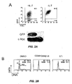

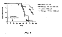
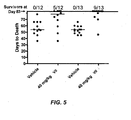
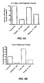
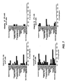
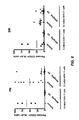
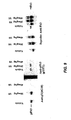

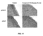

Classifications
-
- A—HUMAN NECESSITIES
- A61—MEDICAL OR VETERINARY SCIENCE; HYGIENE
- A61K—PREPARATIONS FOR MEDICAL, DENTAL OR TOILETRY PURPOSES
- A61K31/00—Medicinal preparations containing organic active ingredients
- A61K31/33—Heterocyclic compounds
- A61K31/395—Heterocyclic compounds having nitrogen as a ring hetero atom, e.g. guanethidine or rifamycins
- A61K31/495—Heterocyclic compounds having nitrogen as a ring hetero atom, e.g. guanethidine or rifamycins having six-membered rings with two or more nitrogen atoms as the only ring heteroatoms, e.g. piperazine or tetrazines
- A61K31/505—Pyrimidines; Hydrogenated pyrimidines, e.g. trimethoprim
-
- A—HUMAN NECESSITIES
- A61—MEDICAL OR VETERINARY SCIENCE; HYGIENE
- A61K—PREPARATIONS FOR MEDICAL, DENTAL OR TOILETRY PURPOSES
- A61K31/00—Medicinal preparations containing organic active ingredients
- A61K31/33—Heterocyclic compounds
- A61K31/395—Heterocyclic compounds having nitrogen as a ring hetero atom, e.g. guanethidine or rifamycins
- A61K31/535—Heterocyclic compounds having nitrogen as a ring hetero atom, e.g. guanethidine or rifamycins having six-membered rings with at least one nitrogen and one oxygen as the ring hetero atoms, e.g. 1,2-oxazines
- A61K31/5375—1,4-Oxazines, e.g. morpholine
- A61K31/5383—1,4-Oxazines, e.g. morpholine ortho- or peri-condensed with heterocyclic ring systems
-
- A—HUMAN NECESSITIES
- A61—MEDICAL OR VETERINARY SCIENCE; HYGIENE
- A61K—PREPARATIONS FOR MEDICAL, DENTAL OR TOILETRY PURPOSES
- A61K31/00—Medicinal preparations containing organic active ingredients
- A61K31/33—Heterocyclic compounds
- A61K31/395—Heterocyclic compounds having nitrogen as a ring hetero atom, e.g. guanethidine or rifamycins
- A61K31/54—Heterocyclic compounds having nitrogen as a ring hetero atom, e.g. guanethidine or rifamycins having six-membered rings with at least one nitrogen and one sulfur as the ring hetero atoms, e.g. sulthiame
- A61K31/542—Heterocyclic compounds having nitrogen as a ring hetero atom, e.g. guanethidine or rifamycins having six-membered rings with at least one nitrogen and one sulfur as the ring hetero atoms, e.g. sulthiame ortho- or peri-condensed with heterocyclic ring systems
-
- A—HUMAN NECESSITIES
- A61—MEDICAL OR VETERINARY SCIENCE; HYGIENE
- A61P—SPECIFIC THERAPEUTIC ACTIVITY OF CHEMICAL COMPOUNDS OR MEDICINAL PREPARATIONS
- A61P31/00—Antiinfectives, i.e. antibiotics, antiseptics, chemotherapeutics
- A61P31/12—Antivirals
-
- A—HUMAN NECESSITIES
- A61—MEDICAL OR VETERINARY SCIENCE; HYGIENE
- A61P—SPECIFIC THERAPEUTIC ACTIVITY OF CHEMICAL COMPOUNDS OR MEDICINAL PREPARATIONS
- A61P31/00—Antiinfectives, i.e. antibiotics, antiseptics, chemotherapeutics
- A61P31/12—Antivirals
- A61P31/20—Antivirals for DNA viruses
- A61P31/22—Antivirals for DNA viruses for herpes viruses
-
- A—HUMAN NECESSITIES
- A61—MEDICAL OR VETERINARY SCIENCE; HYGIENE
- A61P—SPECIFIC THERAPEUTIC ACTIVITY OF CHEMICAL COMPOUNDS OR MEDICINAL PREPARATIONS
- A61P35/00—Antineoplastic agents
-
- A—HUMAN NECESSITIES
- A61—MEDICAL OR VETERINARY SCIENCE; HYGIENE
- A61P—SPECIFIC THERAPEUTIC ACTIVITY OF CHEMICAL COMPOUNDS OR MEDICINAL PREPARATIONS
- A61P35/00—Antineoplastic agents
- A61P35/02—Antineoplastic agents specific for leukemia
-
- A—HUMAN NECESSITIES
- A61—MEDICAL OR VETERINARY SCIENCE; HYGIENE
- A61P—SPECIFIC THERAPEUTIC ACTIVITY OF CHEMICAL COMPOUNDS OR MEDICINAL PREPARATIONS
- A61P35/00—Antineoplastic agents
- A61P35/04—Antineoplastic agents specific for metastasis
-
- A—HUMAN NECESSITIES
- A61—MEDICAL OR VETERINARY SCIENCE; HYGIENE
- A61P—SPECIFIC THERAPEUTIC ACTIVITY OF CHEMICAL COMPOUNDS OR MEDICINAL PREPARATIONS
- A61P43/00—Drugs for specific purposes, not provided for in groups A61P1/00-A61P41/00
-
- A—HUMAN NECESSITIES
- A61—MEDICAL OR VETERINARY SCIENCE; HYGIENE
- A61P—SPECIFIC THERAPEUTIC ACTIVITY OF CHEMICAL COMPOUNDS OR MEDICINAL PREPARATIONS
- A61P7/00—Drugs for disorders of the blood or the extracellular fluid
-
- A—HUMAN NECESSITIES
- A61—MEDICAL OR VETERINARY SCIENCE; HYGIENE
- A61P—SPECIFIC THERAPEUTIC ACTIVITY OF CHEMICAL COMPOUNDS OR MEDICINAL PREPARATIONS
- A61P7/00—Drugs for disorders of the blood or the extracellular fluid
- A61P7/06—Antianaemics
Definitions
- compositions for treating cell proliferative disorders where the compositions comprise inhibitors that target kinase activities affecting the proliferative potential of cells.
- Unregulated cell growth is the hallmark of tumors and cancers and other cell proliferative disorders.
- the cellular processes controlling cell division and cell proliferation are complex, involving an intricate interplay between gene products that promote cell ' division and growth and those that hold such processes in check.
- Positive regulators of growth and proliferation are generally described as proto-oncogenes, which are the normal counterparts of altered genes and their gene products known to promote tumor and cancer formation.
- Proto-oncogenes promote cell division and negatively control cell apoptosis. Uncoupling the activity of these gene products from their normal regulated state converts the proto-oncogenes to oncogenes.
- Normal function of proto-oncogenes includes growth factors, growth factor receptors, cellular signal transduction molecules, and nuclear factors. Activation of the proto-oncogenes into oncogenic forms can occur in a variety of ways, including gene mutation, amplification, gene translocation, and viral activation.
- Tumor suppressors as opposed to the proto-oncogenes, generally exert a negative effect on cell growth, promote apoptosis of cells, inhibit cell cycle progression, and affect invasive and metastatic potential. In some instances, tumor suppressors can counter the activity of oncogenes even in their altered forms. Upon loss or inhibition of tumor suppressor function, the unregulated activity of proto-oncogenes or their corresponding oncogenic forms leads to cell transformation and carcinogenesis. Gene mutation or deletion, suppressed transcription, increased degradation, or abnormalities of associated proteins that work in concert with the tumor suppressors may compromise tumor suppressor activity. Tumor suppressor genes act as recessive alleles such that a cell with a normal allele along with a mutant allele still behaves normally.
- loss of the normal allele also called loss of heterozygozity (LOH) characterizes some types of abnormal cell growth and proliferation.
- LOH loss of heterozygozity
- Treatment of cell proliferative disorders can target the oncogenes and/or the tumor suppressors affected in the transformed cells.
- a disorder arising from a loss-of-function such as a tumor suppressor
- a gain-of-function change such as activation of an oncogene.
- Altering cellular processes to provide the lost cellular function is not practicable in many cases.
- therapy is typically directed at the dysregulated molecules (e.g ., proto-oncogenes) that act as a consequence of the lost tumor suppressor function.
- WO-A-03/040141 describes oxazolyl-phenyl-2,4-diamino-pyrimidine compounds and methods for treating hyperproliferative disorders.
- WO-A-2005/027848 describes methods for screening osteogenic compounds targeting Syk kinase and/or Vav3 and uses of Syk modulators and/or Vav modulators.
- WO-A-03/055489 describes 2,4-diamino-pyrimidine derivative compounds as inhibitors of prolyl peptidase, inducers of apoptosis and cancer treatment agents.
- WO-A-2004/087698 describes thiazoles useful as inhibitors of protein kinases.
- the present invention provides a compound which is a Syk kinase inhibitory compound for use in the treatment of a cell proliferative disorder, wherein the compound is
- the present invention also provides a compound which is a Syk kinase inhibitory compound for use in inhibiting tumor metastasis, wherein the compound is
- the Syk kinase inhibitor is selective for Syk kinase, thereby specifically targeting the aberrant Syk kinase activity present in the proliferative disorder.
- Any cell proliferative disorder in which Syk plays a role in some aspect of abnormal cell division or cell growth can be treated with the inhibitor compounds.
- the cell proliferative disorders treatable with the inhibitor compounds are hematopoietic neoplasms, which are abnormal growth involving cells of the hematopoietic lineage.
- Hematopoietic neoplasms treatable with the Syk inhibitory compounds include, among others, various myeloid and lymphoid neoplasms, such as chronic myelogenous leukaemia., Burkitt's lymphoma, and acute myelogenous leukaemia.
- the Syk kinase inhibitory compound comprises a Syk/Flt-3 kinase inhibitory compound, capable of inhibiting the activity of both Syk kinase and Flt-3 kinase.
- These compounds can be used to treat cell proliferative disorders that are associated with aberrant Flt-3 kinase activity.
- Various hematopoietic neoplasms in which Flt-3 activity is abnormal include, among others, acute myelogenous leukaemia, B-precursor cell acute lymphoblastic leukaemia, T-cell acute lymphoblastic leukaemia, myelodysplastic syndrom, and chronic myelogenous leukaemia.
- These and other such disorders involving Flt-3 can be treated with the Syk/Flt-3 inhibitory compounds, such as members of the 2,4-pyrimidinediamine compounds described herein.
- the inhibitor compounds are for use in a method to treat tumors mediated by viral genes that transform a normal cell into a tumour cell.
- tumors in which the activity of a viral oncogene disrupts Syk kinase function as part of its transformation mechanism can be targeted for treatment.
- virally mediated tumors are associated with infection by viruses carrying genes encoding proteins with immunoreceptor tyrosine-based activation motifs (ITAM). Normally, these conserved sequences modulate Syk kinase activity during development and function of immune system cells (e.g ., B cells, T cells, neutrophils, etc.).
- ITAM immunoreceptor tyrosine-based activation motifs
- the virally mediated tumors can be associated with Karposi's sarcoma-associated herpes virus, Epstein Barr virus, human T-cell lymphotrophic virus (HTLV-1), or mammary tumor virus (MTV). Presence of these viruses are correlated with a number of cell proliferative disorders, including, Karposi's sarcoma, Burkitt's lymphoma, Hodgkin's lymphoma, adult T cell leukemia, and certain forms of breast cancer.
- the compounds are for use in a method of inhibiting tumor metastasis by administration to a subject an amount of a Syk inhibitory compound effective to inhibit tumor metastasis.
- a Syk inhibitory compound effective to inhibit tumor metastasis.
- Any tumor with metastatic potential can be treated with the inhibitor compounds.
- the tumor metastasis is associated with the activity of integrins, which can act through Syk kinase in modulating cell adhesive properties, such as that observed in metastasis of tumor cells.
- Syk kinase inhibitors can be used to inhibit tumor metastasis by affecting cell signaling events mediated by integrins, such as ⁇ 1, ⁇ 2, and/or ⁇ 3 integrins.
- the inhibitor compounds are for use in a method in combination with other cancer treatments.
- Syk inhibitory compounds are for use in a method in combination with other chemotherapeutic agents, including, among others, antimetabolites, alkylating agents, coordination compounds, transcription inhibitors, topoisomerase inhibitors, DNA minor-groove binding compounds, vinca alkyloids, antitumor antibiotics, hormones, and antitumor enzymes.
- FIG. 1A, FIG. 1B , and FIG. 1C show the ability of Syk inhibitor compound VI to block proliferation and induce differentiation of TEL-Syk-transformed cells.
- DNA content of TEL-Syk- or BCRAbl-transformed cells treated for 36 hours with either DMSO, the Syk inhibitor compound VI (2 ⁇ M) or Abl kinase inhibitor STI-571 (2 ⁇ M) are shown in FIG. 1A .
- the ability of TEL-Syk to blocks pre-B cell differentiation is shown in FIG. 1B .
- Cells were cultured for 3 days in the absence of IL-7 and analyzed for the expression of kappa LC by FACS. Inhibition of TEL-Syk activity with compound VI is shown in FIG. 1C .
- FIG. 2A and FIG. 2B show the ability of Syk inhibitor VI to block the proliferation of Myc-transformed pre-B cells.
- FIG. 2A is a FACS-protile of pre-B cells that were transduced with Myc expression vectors and cultured in the absence of IL-7 for 1 week.
- Lower panel of FIG. 2B shows spleens of RAG/ ⁇ C- +/- mice 5 weeks after injection with the indicated cells, illustrating the ability of the Myc transformed cells to cause splenomegaly and leukemia.
- FIG. 2B shows the DNA content of Myc-transformed cells that were treated for 36 hours with either DMSO, VI (2 ⁇ M) or STI-571 (2 ⁇ M).
- FIG. 3A and FIG. 3B show the ability of Syk inhibitor VI to block the proliferation of tumorigenic SLP-65 -/- pre-B cell lines.
- FIG. 3A shows that SLP-65 -/- pre-B cell lines can cause splenomegaly and leukemia, as evidenced by the state of spleens of RAG/ ⁇ C -/- mice 5 weeks after injection with the indicated cells.
- FIG. 3B shows the DNA content of tumorigenic SLP-65 -/- pre-B cell lines that were treated for 36 hours with either DMSO, VI (2 ⁇ M) or STI-571 (2 ⁇ M).
- FIG. 4 shows survival curves for NOD-SCID mice inoculated intravenously (i.v.) with MV4-11 human acute myeloid leukemia cells and treated with compound VII at 40 mg/kg twice daily, PO, from day 17 post tumor cell inoculation for the duration of the study. Calculations for % ILS for 40mg/kg VII group (10 6 cells) assumes median day of death of 83 days, due to greater than 50% survival in this group at study termination (Day 83).
- FIG. 5 shows days to death for individual animals.
- the survival data is for NOD-SCID mice injected intravenously with MV4-11 tumor cells. Mice were treated with vehicle or varying doses of VII twice daily, PO, from day 17 post-tumor cell injection until the end of the study (Day 83). Days to death for sacrificed animals are shown in the graph with median day of death denoted by blue line. The survivors in each group at study termination are shown at the top of the graph denoted as # survivors/total # mice.
- FIG. 6A and FIG. 6B show frequency of tumors in MV4-11 tumor bearing NOD-SCID mice at necropsy.
- the severity of disease was evaluated by quantitating the frequency of mice having palpable tumors upon necropsy at termination, irrespective of reason for sacrifice ( FIG. 6A ).
- the total number of tumors per group is shown in FIG. 6B .
- Mice were inoculated i.v. with 5 or 10 million MV4-11 human AML cells (denoted as 5E6 or 10E6, respectively).
- mice were treated with vehicle or 40mg/kg VII (PO, bid) until sacrifice or for the duration of the study, up to Day 83 for surviving animals.
- Mice were sacrificed and a comprehensive necropsy performed.
- the total number of necropsies performed per group ranged from 8-13 animals.
- FIG. 7 shows the tumor distribution profile of experimental animals. Percent of total tumors found in various anatomical locations were noted upon necropsy at sacrifice. Mice were injected i.v. with 5 or 10 million MV4-11 cells and dosed orally with either vehicle or 40 mg/kg of VII twice daily for the duration of the study. Animals showing enlarged salivary glands are included in the graph, although no palpable tumor was detected. The total number of necropsies performed per group ranged from 8-13 animals.
- FIG. 8 shows the engraftment profile of MV4-11 tumor cells in bone marrow (BM) and peripheral blood (PB) of NOD-SCID mice.
- the data is from engraftment of MV4-11 human tumor cells in NOD-SCID mice treated with vehicle or 40mg/kg VII twice daily, PO, from day 17 post i.v. tumor cell injection until the end of the study.
- Bone marrow (BM) and peripheral blood (PB) tumor cell engraftment were detected by flow cytometric analysis using CD33 and HLA cell surface staining for detection of MV4-11 tumor cells. Data were normalized, and percent human tumor cells out of total cells were calculated. Total cells were defined as the number of human CD33 + HLA + positive events and murine CD45 positive staining events.
- Graph includes all samples.
- FIG. 9 shows detection of Flt-3 in MV4-11 xenografts by immunoprecipitation and Western Blot analysis.
- Panel A is detection of phosphorylated Flt-3 in MV4-11 tumor lysates from mice approximately two hours after the final dose of VII or vehicle using anti-phosphotyrosine antibody on the left and anti-phospho-Flt-3 specific antibody on the right side of the blot. Additionally, a reprobe of the blot for the total Flt-3 levels are shown in Panel B.
- FIG. 10 shows phosphorylated histone H3 analysis of MV4-11 tumor xenografts. Proliferation was assessed ex vivo in formalin fixed tumor sections from three randomly selected mice from this study using immunohistochemical staining of human-specific phosphorylated histone H3 (phH3) as a marker for tumor cell proliferation. Human phH3 expression was reduced in tumor sections in a dose-dependent manner following treatment with VII .
- FIG. 11 shows a representative data illustrating immunohistochemical staining for pErk1/2 and pStat5 in tumor sections from MV4-11 tumor bearing mice treated orally twice per day for 26 days. Tumors were harvested from mice -2 hours following final dose of vehicle or 40mg/kg VII .
- FIG. 12 shows the effect of treating AML cells or 32D transfectants with 1 uM VI in inducing apoptosis as determined by Annexin V and PI staining.
- Syk kinases is one of the two known members of the Syk family (Syk and ZAP-70) non-receptor tyrosine kinases. Syk is activated upon the binding of its src homology 2 (SH2) domains to immunoreceptor tyrosine-based activation motifs (ITAM). Syk kinase plays an essential role in lymphocyte development and activation of immune cells and is best characterized for its role in B cell receptor signaling and Fc receptor mediated release of mast cell granules.
- SH2 src homology 2
- ITAM immunoreceptor tyrosine-based activation motifs
- Syk Although expressed ubiquitously in hematopoietic cells, Syk is also expressed in other tissues, such as breast epithelial cells and hepatocytes. It is believed by some in the field that Syk is a tumor suppressor and acts as a negative regulator of metastatic potential. Loss of Syk activity is suggested as being associated with formation of invasive breast cancer, and chromosomal loss of the Syk gene is indicated in certain types of lymph node metastasis of primary breast cancer. Thus, use of Syk inhibitors to treat cell proliferative disorders or as treatment to reduce the metastatic potential of tumor cells would be contraindicated if Syk functions as a tumor suppressor.
- Flt-3 is also a tyrosine kinase, but unlike Syk, it belongs to the family of tyrosine kinase receptor proteins.
- Flt-3 is a member of the class III receptor tyrosine kinases, which are related by amino acid sequence and structural characteristics.
- Flt-3 is activated by binding to its cognate ligand, Flt-3 L.
- Flt-3 kinase is expressed in early hematopoietic stem cells of normal bone marrow, and appears to function in the development of multipotent stem cells and B cells by controlling the activity of various downstream cellular targets that include, among others, phospholipase C- (PLC), the p85 subunit of phosphatidylinositol 3'-kinase (PI3K), Shc, Shp-2, Ship, Grb2, Vav, Fyn kinase, Src kinase, Stat5 signal transducing protein, and Erk.
- PLC phospholipase C-
- PI3K the p85 subunit of phosphatidylinositol 3'-kinase
- Shc Shp-2
- Ship Grb2
- Vav Vav
- Src kinase Src kinase
- Stat5 signal transducing protein and Erk.
- Flt-3 kinase is normally expressed in progenitor cells in the bone marrow, high levels of expression are also observed in a spectrum of hematologic cell proliferative disorders, such as acute myelogenous leukemia (AML), B-precursor cell acute lymphoblastic leukemia, T-cell acute lymphoblastic leukemia, and chronic myelogenous leukemia (CML) in lymphoid blast crisis (see. e.g ., Rosnet et al., 1996, Leukemia 10:238-248 ; Carow et al., 1996, Blood 87:1089-1096 ).
- AML acute myelogenous leukemia
- B-precursor cell acute lymphoblastic leukemia T-cell acute lymphoblastic leukemia
- CML chronic myelogenous leukemia
- Syk may act as a tumor suppressor
- the present disclosure is based on indications that Syk functions contrary to that posited role. For instance, forced expression of Syk kinase in tumor cells does not appear to reverse the transformed phenotype of tumor cells. To the contrary, it is suggested herein that Syk acts in an oncogenic capacity to promote and/or maintain cell proliferation.
- the disclosure provides compounds for use in a method of treating cell proliferative disorders by administering to a subject an amount of a Syk kinase inhibitory compound effective to treat a cell proliferative disorder.
- Syk inhibitory compounds such as members of the 2,4-pyrimidinediamine compounds described in further detail below, can also act as inhibitors of the Flt-3 kinase. Consequently, in some embodiments, Syk inhibitory compounds with this dual- or multi-acting inhibitory profile of inhibiting Flt-3 kinase as well as Syk kinase ( i.e ., Syk/Flt-3 kinase inhibitory activities) find applications for treatment of cell proliferative disorders associated with aberrant Flt-3 receptor kinase activity.
- Syk kinase and Flt-3 kinase The association of both Syk kinase and Flt-3 kinase with the development of hematopoietic cells supports the use of such Syk/Flt-3 kinase inhibitory compounds for treatment of hematopoietic neoplasms.
- Syk or " Syk kinase” refers to the 72kDa non-receptor (cytoplasmic) spleen protein tyrosine kinase expressed in B-cells and other hematopoetic cells. Syk kinase is characterized by two consensus Src-homology 2 (SH2) domains in tandem that bind to phosphorylated immunoreceptor tyrosine-based activation motifs ("ITAMs”), a "linker” domain and a catalytic domain (for a review, see Sada et al., 2001, J. Biochem.
- SH2 consensus Src-homology 2
- Syk kinase is also critical for tyrosine phosphorylation of multiple proteins which regulate important pathways leading from immunoreceptors, such as Ca 2+ mobilization and mitogen-activated protein kinase (MAPK) cascades and degranulation. Syk kinase also plays a critical role in integrin signaling in neutrophils (see, e.g ., Mocsai et al. 2002, Immunity 16:547-558 ).
- Syk kinase includes kinases from any species of animal, including but not limited to, homo sapiens, simian, bovine, porcine, rodent, etc., recognized as belonging to the Syk family. Specifically included are isoforms, splice variants, allelic variants, mutants, both naturally occurring and man-made. The amino acid sequences of such Syk kinases are available from GENBANK. Specific examples of mRNAs encoding different isoforms of human Syk kinase are available at GENBANK accession no.
- Flt-3 or "Flt-3 receptor tyrosine kinase” refers to a receptor tyrosine kinase that binds the flt-3 ligand (Flt-3 L or FL).
- Flt-3 is also knowns as Fms-like tyrosine kinase 3, FLK-2 (fetal liver kinase-2) and STK-1 (human stem cell kinase-11) (see, e.g ., Mathews et al., 1991, Cell. 65:1143-1152 ; Rosnet et al., 1991, Oncogene. 6:1641-1650 ).
- Flt-3 has sequence similarity to members of the class III receptor tyrosine kinase (RTKIII) family, a subset of which includes, among others, FMS, platelet-derived growth factor receptor (PDGFR), and Kit ( Rosnet et al., 1993, Crit. Rev. Oncog. 4:595-613 .
- the Flt-3 polypeptide in mouse and human is a 1000- and 993-amino acid protein, respectively, and is known to be expressed in immature hematopoietic cells, placenta, gonads, and brain.
- Flt-3 has a postulated role in the development of multipotent stem cells and B cells.
- Flt-3 is characterized by 5 immunoglobulin-like repeats in the extracellular domain, a transmembrane (TM) domain, a juxtamembrane (JM) domain, two intracellular tyrosine kinase (TK1 and TK2) domains separated by a kinase insert (KI) domain, and a C-terminus domain ( Agnes et al., 1994, Gene 145: 283-288 ; 2004, Griffith et al., Molecular Cell 13:169-118 ).
- TM transmembrane
- JM juxtamembrane
- TK1 and TK2 two intracellular tyrosine kinase domains separated by a kinase insert (KI) domain
- C-terminus domain Agnes et al., 1994, Gene 145: 283-288 ; 2004, Griffith et al., Molecular Cell 13:169-118 .
- Flt-3 kinase includes kinases from any species of animal, including but not limited to, homo sapiens, simian, bovine, porcine, rodent, etc., recognized as belonging to the Flt-3 receptor family. Specifically included are isoforms, splice variants, allelic variants, mutants, both naturally occurring and man-made. The amino acid sequences of such Syk kinases are available from GENBANK. Specific examples of nucleic acid sequences and corresponding amino acid sequences of different Flt-3 kinases include, among others, human (Accession Nos. NM_004119.1), chimpanzee (Accession Nos.
- Flt-3 ITD refers to a variant of Flt-3 having internal tandem duplications (ITDs) in the juxtamembrane (JM) domain ( Nakao et al., 1996, Leukemia 10:1911-1918 ; Griffith et al., supra ).
- JM juxtamembrane
- the number of JM domains duplicated in Flt-3 ITD varies from subject to subject but are in-frame and generally results in protein with an aberrant (i.e., increased) tyrosine kinase activity.
- Variants of the Flt-3 TTD type are often associated with acute myelogenous leukemia and myelodysplastic syndrome.
- Flt-3 activation loop mutations refer to variants having an amino sequence change (as compared to wild-type) in the activation loop of the second tyrosine kinase (TK2) domain.
- the activation loop appears to function in blocking access of adenosine triphosphate (ATP) and substrate to the kinase domain, and thereby exerts an inhibitory effect on the kinase.
- ATP adenosine triphosphate
- activation loop mutations can give rise to Flt-3 kinase forms with constitutive activity.
- An exemplary activation loop variant is D835A, which is observed in acute myelogenous leukemia, myelodysplatic syndrome, and acute lymphoblastic leukemia ( Yamamoto et al., 2001, Blood 97:2434-2439 ; Griffin, J.D., 2001, Blood 97:2193a ).
- Other exemplary activation loop variations associated with cell proliferative disorders in humans included, among others, D835Y, D835A, D835E, D835H, D835N, D835V, D835del, and I836del
- Cell proliferative disorder refers to a disorder characterized by abnormal proliferation of cells.
- a proliferative disorder does not imply any limitation with respect to the rate of cell growth, but merely indicates loss of normal controls that affect growth and cell division. Thus, in some embodiments, cells of a proliferative disorder can have the same cell division rates as normal cells but do not respond to signals that limit such growth.
- neoplasm or tumor which is an abnormal growth of tissue. Cancer refers to any of various malignant neoplasms characterized by the proliferation of cells that have the capability to invade surrounding tissue and/or metastasize to new colonization sites.
- Hematopoietic neoplasm refers to a cell proliferative disorder arising from cells of the hematopoietic lineage.
- hematopoiesis is the physiological process whereby undifferentiated cells or stem cells develop into various cells found in the peripheral blood.
- hematopoietic stem cells typically found in the bone marrow, undergo a series of cell divisions to form multipotent progenitor cells that commit to two main developmental pathways: the lymphoid lineage and the myeloid lineage.
- the committed progenitor cells of the myeloid lineage differentiate into three major sub-branches comprised of the erythroid, megakaryocyte, and granulocyte/monocyte developmental pathways.
- Committed progenitor cells of the lymphoid lineage develop into the B cell pathway, T cell pathway, or the non-T/B cell pathway. Similar to the myeloid lineage, an additional lymphoid pathway appears to give rise to dendritic cells involved in antigen presentation.
- the B cell progenitor cell develops into a precursor B cell (pre-B), which differentiates into B cells responsible for producing immunoglobulins.
- Progenitor cells of the T cell lineage differentiate into precursor T cells (pre-T) that, based on the influence of certain cytokines, develop into cytotoxic or helper/suppressor T cells involved in cell mediated immunity.
- Non-T/B cell pathway leads to generation of natural killer (NK) cells.
- Neoplasms of hematopoietic cells can involve cells of any phase of hematopoiesis, including hematopoietic stem cells, multipotent progenitor cells, oligopotent committed progenitor cells, precursor cells, and mature differentiated cells.
- the categories of hematopoietic neoplasms can generally follow the descriptions and diagnostic criteria employed by those of skill in the art (see, e.g ., International Classification of Disease and Related Health Problems (ICD 10), World Health Organization (2003 )).
- Hematopoietic neoplasms can also be characterized based on the molecular features, such as cell surface markers and gene expression profiles, cell phenotype exhibited by the aberrant cells, and/or chromosomal aberrations ( e.g ., deletions, translocations, insertions, etc.) characteristic of certain hematopoietic neoplasms, such as the Philadelphia chromosome found in chronic myelogenous leukemia. Other classifications include National Cancer Institute Working Formulation ( Cancer, 1982, 49:2112-2135 ) and Revised European-American Lymphoma Classification (REAL).
- MERS Revised European-American Lymphoma Classification
- Lymphoid neoplasm refers a proliferative disorder involving cells of the lymphoid lineage of hematopoiesis. Lymphoid neoplasms can arise from hematopoietic stem cells as well as lymphoid committed progenitor cells, precursor cells, and terminally differentiated cells. These neoplasms can be subdivided based on the phenotypic attributes of the aberrant cells or the differentiated state from which the abnormal cells arise. Subdivisions include, among others, B cell neoplasms, T cell neoplasms, NK cell neoplasms, and Hodgkin's lymphoma.
- Myeloid neoplasm refers to proliferative disorder of cells of the myeloid lineage of hematopoiesis. Neoplasms can arise from hematopoietic stem cells, myeloid committed progenitor cells, precursor cells, and terminally differentiated cells. Myeloid neoplasms can be subdivided based on the phenotypic attributes of the aberrant cells or the differentiated state from which the abnormal cells arise. Subdivisions include, among others, myeloproliferative diseases, myelodysplastic/myeloproliferative diseases, myelodysplastic syndromes, acute myeloid leukemia, and acute biphcnotypic leukemia.
- Virally mediated tumor refers to a neoplasm or tumor associated with viral infection or an activity of a virally encoded product.
- the neoplasm can arise from presence of a latent virus integrated into the cellular genome or arise from activity of a virally associated gene product. Infection with the virus need not be tightly correlated in time with tumor formation in that incubation periods can extend from months to years before development of a tumor phenotype. Because the treatments herein are directed to use of Syk inhibitors, the applicable virally associated tumors are those in which viral modulation of Syk activity is correlated with aberrant cell proliferation. Any virus, including RNA and DNA viruses and viruses that reside episomally or integrate into the cellular genome, in which activation of Syk is a consequence of virus infection can be targeted using the methods herein.
- Tumor metastasis refers to the capability of tumor cells to migrate from the original tumor site and colonize in other tissues. Tumors formed from cells that have spread are referred to as “secondary tumors” and contain cells that are similar to those in the original "primary” tumor. Metastatic tumors typically form by migration of tumor cells from the original tumor site through the blood and lymph system to other tissues.
- Syk mediated integrin signaling refers to signal transduction of cell surface integrins that occur via interaction with Syk kinase. Integrins comprise an extended family of cell surface adhesion receptors that bind extracellular matrix and cell surface ligands. Structurally, integrins are heterodimeric proteins composed of an ⁇ and ⁇ chain, where each subunit has an extracellular domain, a single transmembrane domain, and a cytoplasmic domain. The ⁇ subunit generally composed of about seven tandem repeats, where a subset of the repeats contain putative metal binding sequences of the general structure DxDxDGxxD, where x is any amino acid.
- Two groups of integrins can be characterize by the ⁇ subunits: those that contain an "A" domain and those having a proteolytic cleavage site.
- the ⁇ subunit comprises a conserved region of about 200 amino acids in the extracellular domain, which is characterized by a region having structural similarity to the "A" domain of the ⁇ subunit and another region with epidermal growth factor (EGF) like repeats, similar to those found in laminin (see, e.g., Xiong et al., 2003, Blood, 102(4):1155-1159 ).
- Integrin activity may modulate intracellular.Syk, or conversely, the integrin function can be modulated via the activity of Syk.
- Integrins require activation within the cell to bind its cognate ligands (inside-out activation).
- Integrins that either modulate or is modulated by Syk include, among others, ⁇ 1-integrins ( Lin et al., J Biol. Chem. 1995; 270(27):16189-97 ) such as ⁇ 2 b 1 ( Keely et al., 1996, J Biol Chem. 271(43):26668-76 ), ⁇ 2 integrins, and ⁇ 3 integrins ( Woodside et al., 2001, Curr Biol.
- cell proliferative disorders treatable with the compounds disclosed herein relate to any disorder characterized by aberrant cell proliferation. These include various tumors and cancers, benign or malignant, metastatic or non-metastatic. Specific properties of cancers, such as tissue invasiveness or metastasis, can be targeted using the methods described herein.
- Cell proliferative disorders include a variety of cancers, including, among others, breast cancer, ovarian cancer, renal cancer, gastrointestinal cancer, kidney cancer, bladder cancer, pancreatic cancer, lung squamous carcinoma, and adenocarcinoma.
- the cell proliferative disorder treated is a hematopoietic neoplasm, which is aberrant growth of cells of the hematopoietic system.
- Hematopoietic malignancies can have its origins in pluripotent stem cells, multipotent progenitor cells, oligopotent committed progenitor cells, precursor cells, and terminally differentiated cells involved in hematopoiesis. Some hematological malignancies are believed to arise from hematopoietic stem cells, which have the ability for self renewal.
- cells capable of developing specific subtypes of acute myeloid leukemia (AML) upon transplantation display the cell surface markers of hematopoietic stem cells, implicating hematopoietic stem cells as the source of leukemic cells.
- Blast cells that do not have a cell marker characteristic of hematopoietic stem cells appear to be incapable of establishing tumors upon transplantation ( Blaire et al., 1997, Blood 89:3104-3112 ).
- the stem cell origin of certain hematological malignancies also finds support in the observation that specific chromosomal abnormalities associated with particular types of leukemia can be found in normal cells of Hematopoietic lineage as well as leukemic blast cells.
- the reciprocal translocation t(9q34;22q11) associated with approximately 95% of chronic myelogenous leukemia appears to be present in cells of the myeloid, erythroid, and lymphoid lineage, suggesting that the chromosomal aberration originates in hematopoietic stem cells.
- a subgroup of cells in certain types of CML displays the cell marker phenotype of hematopoietic stem cells.
- hematopoietic neoplasms often originate from stem cells, committed progenitor cells or more terminally differentiated cells of a developmental lineage can also be the source of some leukemias.
- forced expression of the fusion protein Bcr/Abl associated with chronic myelogenous leukemia
- common myeloid progenitor or granulocyte/macrophage progenitor cells produces a leukemic-like condition.
- chromosomal aberrations associated with subtypes of leukemia are not found in the cell population with a marker phenotype of hematopoietic stem cells, but are found in a cell population displaying markers of a more differentiated state of the hematopoietic pathway ( Turhan et al., 1995, Blood 85:2154-2161 ).
- committed progenitor cells and other differentiated cells may have only a limited potential for cell division
- leukemic cells may have acquired the ability to grow unregulated, in some instances mimicking the self-renewal characteristics of hematopoietic stem cells ( Passegue et al., Proc. Natl. Acad. Sci. USA, 2003, 100:11842-9 ).
- the hematopoietic neoplasm treated is a lymphoid neoplasm, where the abnormal cells are derived from and/or display the characteristic phenotype of cells of the lymphoid lineage.
- Lymphoid neoplasms can be subdivided into B-cell neoplasms, T and NK -cell neoplasms, and Hodgkin's lymphoma.
- B-cell neoplasms can be further subdivided into precursor B-cell neoplasm and mature/peripheral B-cell neoplasm.
- Exemplary B-cell neoplasms are precursor B-lymphoblastic leukemia/lymphoma (precursor B-cell acute lymphoblastic leukemia) while exemplary mature/peripheral B-cell neoplasms, are B-cell chronic lymphocytic leukemia/small lymphocytic lymphoma, B-cell prolymphocytic leukemia, lymphoplasmacytic lymphoma, splenic marginal zone B-cell lymphoma, hairy cell leukemia, plasma cell myeloma/plasmacytoma, extranodal marginal zone B-cell lymphoma of MALT type, nodal marginal zone B-cell lymphoma, follicular lymphoma, mantle-cell lymphoma, diffuse large B-cell lymphoma, mediastinal large B-cell lymphoma, primary effusion lymphoma, and Burkitt's lymphoma/Burkitt cell leukemia.
- T-cell and Nk-cell neoplasms are further subdivided into precursor T-cell neoplasm and mature (peripheral) T-cell neoplasms.
- Exemplary precursor T-cell neoplasm is precursor T-lymphoblastic lymphoma/leukemia (precursor T-cell acute lymphoblastic leukemia) while exemplary mature (peripheral) T-cell neoplasms are T-cell prolymphocytic leukemia T-cell granular lymphocytic leukemia, aggressive NK-cell leukemia, adult T-cell lymphoma/leukemia (HTLV-1), extranodal NK/T-cell lymphoma, nasal type, enteropathy-type T-cell lymphoma, hepatosplenic gamma-delta T-cell lymphoma, subcutaneous panniculitis-like T-cell lymphoma, Mycosis fungoides/Sezary syndrome, Anaplastic large-cell
- the third member of lymphoid neoplasms is Hodgkin's lymphoma, also referred to as Hodgkin's disease.
- Exemplary diagnosis of this class that can be treated with the compounds include, among others, nodular lymphocyte-predominant Hodgkin's lymphoma, and various classical forms of Hodgkin's disease, exemplary members of which are Nodular sclerosis Hodgkin's lymphoma (grades 1 and 2), Lymphocyte-rich classical Hodgkin's lymphoma, Mixed cellularity Hodgkin's lymphoma, and Lymphocyte depletion Hodgkin's lymphoma.
- any of the lymphoid neoplasms that are associated with aberrant Syk activity can be treated with the Syk inhibitory compounds.
- the hematopoietic neoplasm treated is a myeloid neoplasm.
- This group comprises a large class of cell proliferative disorders involving or displaying the characteristic phenotype of the cells of the myeloid lineage.
- Myeloid neoplasms can be subdivided into myeloproliferative diseases, myelodysplastic/myeloproliferative diseases, myelodysplastic syndromes, and acute myeloid leukemias.
- Exemplary myeloproliferative diseases are chronic myelogenous leukemia (e.g ., Philadelphia chromosome positive (t(9;22)(qq34;q11)), chronic neutrophilic leukemia, chronic eosinophilic leukemia/hypereosinophilic syndrome, chronic idiopathic myelofibrosis, polycythemia vera, and essential thrombocythemia.
- Exemplary myelodysplastic/myeloproliferative diseases are chronic myelomonocytic leukemia, atypical chronic myelogenous leukemia, and juvenile myelomonocytic leukemia.
- Exemplary myelodysplastic syndromes are refractory anemia, with ringed sideroblasts and without ringed sideroblasts, refractory cytopenia (myelodysplastic syndrome) with multilineage dysplasia, refractory anemia (myelodysplastic syndrome) with excess blasts, 5q- syndrome, and myelodysplastic syndrome with t(9;12)(q22;p12) (TEL-Syk fusion; see, e.g., Kuno et al., 2001, Blood 97:1050).
- any of the myeloid neoplasms that are associated with aberrant Syk activity can be treated with the Syk inhibitory compounds.
- the Syk inhibitory compounds can be for use in a method to treat Acute myeloid leukemias (AML), which represent a large class of myeloid neoplasms having its own subdivision of disorders. These subdivisions include, among others, AMLs with recurrent cytogenetic translocations, AML with multilineage dysplasia, and other AML not otherwise categorized.
- AML Acute myeloid leukemias
- Exemplary AMLs with recurrent cytogenetic translocations include, among others, AML with t(8;21)(q22;q22), AML1(CBF-alpha)/ETO, Acute promyelocytic leukemia (AML with t(15;17)(q22;q11-12) and variants, PML/RAR-alpha), AML with abnormal bone marrow eosinophils (inv(16)(p13q22) or t(16;16)(p13;q11), CBFb/MYH11X), and AML with 11q23 (MLL) abnormalities.
- Exemplary AML with multilineage dysplasia are those that are associated with or without prior myelodysplastic syndrome.
- AML minimally differentiated AML without maturation, AML with maturation, Acute myelomonocytic leukemia, Acute monocytic leukemia, Acute erythroid leukemia, Acute megakaryocytic leukemia, Acute basophilic leukemia, and Acute panmyelosis with myelofibrosis.
- cell proliferative disorders that can be targeted with the Syk kinase inhibitors comprise virally mediated tumors. These can arise from infection of cells by an oncogenic virus that has the capability of transforming a normal cell into a tumor cell. Because rates of viral infection far exceed the number of actual incidence of cell transformation, viral mediated transformation generally act together with other cellular factors to generate a transformed tumor cell. Thus, a virally mediated tumor does not require the virus to be the sole causative agent of the cell proliferative disorder, but rather that the viral infection or persistent presence of virus is associated with the generation of the tumor.
- tumors where the causative agent is a virus typically has continual expression of a limited number of viral genes and that viral these oncogenes, expressed as part of the viral infection or through persistence of the virus, disrupts the normal cellular gene expression and signal transduction pathways.
- viral oncogenes involved in cell transformation appear to disrupt four main cellular processes: cell surface receptors that interact with growth factors and extracellular matrix, transmembrane signaling networks, cytosolic elements such as soluble proteins and second messengers, and nuclear proteins including DNA binding proteins and factors which function directly and indirectly in gene regulation and replication. Because certain virally encoded proteins target specific cellular components as part of the transformation process, application of Syk inhibitors can be appropriate where the viral component targets the activity of Syk kinase.
- the virally mediated tumor treatable with the compounds disclosed herein is associated with any virus that encodes an immunoreceptor tyrosine-based activation motif (ITAM) capable of modulating Syk activity.
- ITAM immunoreceptor tyrosine-based activation motif
- This motif refers to a conserved amino acid sequence motif that functions by interacting with and activating nonreceptor tyrosine kinases.
- ITAM motifs are found in, among others, then and ⁇ chains of Fc ⁇ RI, the ⁇ subunit of the T cell receptor, and imnunoglobulin ⁇ (Ig ⁇ ) and Ig ⁇ of the B cell receptor.
- the canonical sequence motif is typically Yxx(L/I)x 6-8 Yxx(L/I), where x represents any amino acid.
- the tyrosine residues in the motif are involved in ITAM signaling and are substrates for phosphorylation by Src family of kinases.
- the phosphorylated form of ITAMs function as interaction sites for SH2 (src homology domain) containing signaling proteins, such as Syk/ZAP-70 kinases.
- SH2 serosine protein
- Syk/ZAP-70 kinases signaling proteins
- the ITAM sequences have been identified in virally encoded proteins.
- tumors associated with viruses carrying genes encoding proteins with ITAM sequences can be treated with Syk inhibitor compounds.
- the virally mediated tumor treatable with the inhibitor compounds is associated with Karposi's sarcoma (KS) associated herpes virus, a lymphotropic virus implicated in Karposi's sarcoma, a rare malignancy found at higher incidence among HIV infected population.
- the KS associated herpes virus encodes a transmembrane protein termed KI having an immunoreceptor tyrosine-based activation motif (ITAM)-like sequence.
- ITAM immunoreceptor tyrosine-based activation motif
- the KI gene product is thought to act in a constitutive manner through its cysteine-rich ectodomain to activate Syk and its related kinase Zap-70 ( Lagunoff, M. et al., 1999, Proc. Natl. Acad.
- transgenic mice bearing the KI gene appears to increase the incidence of certain sarcomas and lymphomas in an infected animal, indicating a role for KI activity in oncogenesis ( Prakash et al., 2002, J. Natl. Cancer Inst. 94:926-35 ).
- the virally mediated tumor is associated with Epstein Barr Virus (EBV).
- Epstein Barr Virus is a member of the Herpesviridae family that, following primary infection, replicates in the epithelial cells of the oropharynx and infect recirculating B lymphocytes. Infection can lead to acute infectious mononucleosis, also known as glandular fever. Infectious mononucleosis is a benign lymphoproliferative disease characterized by transient immunosuppression and an expansion of atypical lymphocytes, the majority of which are CD8 + T cells. In these T cells, EBV establishes a latent but persistent infection during which a select set of viral genes are expressed. The entire genome can persist in the proliferating lymphocytes as episomal DNA. EBV infection is associated with Burkitt's lymphoma, Hodgkin's lymphoma, and adult T cell leukemia.
- the LMP2A protein encoded by the EBV genome is a transmembrane protein thought to play a role in maintaining the latency of the EBV virus following infection. It consists of an extended amino terminal tail, 12 membrane spanning domains, and a cytoplasmic domain. The amino terminal region contains the ITAM motif, which allows interaction of LMP2A with Syk kinase ( Fruehling et al., 1997, Virology, 235:241-251 ). LMP2A appears to regulate Syk kinase in lymphoid cells to promote B-cell survival and maintain latency.
- Syk plays a critical role in the signal transduction pathways that regulate other signaling pathways, such as PI-3K, BLNK, and phospholipase ⁇ 2 and is involved in enhancing lymphoid cell survival
- improper Syk activation through LMP2A protein or other virally mediated effectors may play a role in inducing aberrant lymphoproliferation (Caldwell et al., 2000, J Virol 74(19):9115; Caldwell et al., 1998, Immunity 9:405)).
- inhibition of Syk activity can provide a therapeutic benefit for cell proliferative disorders associated with EBV viral infection.
- the virally mediated tumor to be treated with the Syk inhibitor is associated with Human T-cell Lymphotropic Virus (HTLV-1 virus), a retrovirus in the same class of virus as the AIDS virus, HIV-1.
- the virus is tropic for CD4 + T-cells although CD8 + T-cells can also serve as a viral reservoir.
- HTLV-1 infection is associated with, among others, adult T-cell Leukemia/lymphoma (ATLL) and a number of other lymphocyte disorders.
- ATLL adult T-cell Leukemia/lymphoma
- Syk is expressed in infected cells while expression of the Syk related kinase, ZAP-70, is absent ( Weil et al., 1999, J. Virol. 73(5):3709-17 ).
- Dysregulation of a number of kinases, including Syk is implicated in HTLV-1 mediated induction of adult T-cell leukemia.
- the virally mediated tumor is associated with mammary tumor virus (MTV).
- MTV mammary tumor virus
- ITAM sequences are found within the Env gene of murine mammary tumor virus (MMTV), a B type retrovirus identified as an etiological agent for breast cancer in mice.
- MMTV murine mammary tumor virus
- Mouse mammary epithelial cells transfected with MMTV Env gene display characteristics of a transformer phenotype, such as colony formation in soft agar and invasiveness in basement membrane preparations ( Katz et al., 2005, J Exp Med. 201 (3):431-9 ).
- Murine mammary tumor virus-like sequences are also present in human cancers, such as breast cancer and T cell lymphomas ( Wang et al., 2000, Clinical Cancer Res. 6:1273-1278 ), and correlated with tumorigenesis as these sequences are not observed in the majority of normal breast tissue.
- tumors associated with MTV can be treated with the Syk kinas
- Syk inhibitor compounds for treating virally mediated tumors is not limited to tumors associated with the viruses specified above.
- any tumors associated with an oncogenic virus in which Syk is activated as part of its oncogenic mechanism, whether or not it involves ITAM sequences, can be targeted using the Syk inhibitor compounds.
- the cell proliferative disorder that can be targeted with the inhibitors comprise cell proliferative disorders associated with aberrant Flt-3 kinase activity.
- aberrant Flt-3 kinase activity refers to activity that is abnormal from what the normal level of Flt-3 kinase activity would be for a wild type Flt-3 kinase or in a normal Flt-3 expressing cell, tissue, organ, or organism.
- Aberrant Fit-3 kinase activity can arise from mislocalization of the protein (spatial expression), increases or decrease in activity of the enzyme (directly or indirectly), or changes in temporal expression ( i.e ., developmental expression).
- the aberrant kinase activity is associated with variants of Flt-3. In some embodiments, these variants are characterized by increased Flt-3 receptor kinase activity.
- increased kinase activity refers to kinase activity that is higher than observed for a wild type Flt-3 kinase or in a normal Flt-3 expressing cell, tissue, organ, or organism. Exemplary increased kinase activity are found in cells with certain Flt-3 variants, such as for example, Flt-3 ITD and Flt-3 activation loop mutations.
- the Syk/Flt-3 inhibitory compounds can be for use in a method to treat hematopoietic neoplasms characterized by the presence of an aberrant Flt-3 kinase.
- the hematological neoplasms treatable with the Syk/Flt-3 inhibitory compound can include, among others, acute myelogenous leukemia, B-precursor cell acute lymphoblastic leukemia, T-cell acute lymphoblastic leukemia, and chronic myelogenous leukemia (CML) in which the presence of an aberrant Flt-3 has been diagnosed or is suspected in the subject.
- CML chronic myelogenous leukemia
- the skilled artisan can apply the treatments without such information and that the inhibitory compounds can be given prophylactically to increase the probability of survival.
- the skilled artisan can apply the Syk/Flt-3 inhibitory compounds herein to other cell proliferative disorders in which an aberrant Flt-3 kinase activity has been detected or is suspected.
- the present disclosure is directed to the treatment of tumor metastasis by use of the Syk kinase inhibitors.
- Metastasis is a characteristic of malignant tumor cells whereby tumor cells detach from its site of origin and then spread to colonize at other sites. These secondary tumors can form in tissues unrelated to the cells from which the tumor cells originate. It is the formation of these secondary tumors by metastasis that appears to be the primary cause of mortality from malignant forms of cancer. Metastasis begins when malignant cells break off from the primary tumor and enter the blood or lymphatic system, and then migrate to other colonization sites. Generally, normal cells do not detach and invade other tissues because of various signals that inhibit dissimilar cells from adhering to each other, as well as signals between cells that inhibit cell growth.
- CAMs cell adhesion molecule
- CAMs cell adhesion molecule
- integrins Another class of adhesion proteins implicated in tissue invasiveness and metastasis are integrins. As noted above, integrins form a diverse class of cell surface molecules. Permutations in the spectrum of the more than 22 integrin subtypes are able to create a flexible system for changing interaction of cells in response to different cell signaling states or changing external environments. Integrins interact dynamically with extracellular ligands depending on activation state of the integrin, which is controlled by intracellular signals (i.e. , inside out signaling) that modifies integrin affinity and avidity. Conversely, interaction of integrin with extracellular ligands can trigger signal transduction cascades that affect cell adhesive properties and cellular responses to changing extracellular environments (i.e.
- ⁇ v subunit containing integrin activity while inhibiting ⁇ v subunit containing integrin activity, such as by use of RGD peptidomimetic agents that compete for binding of the integrin with its natural substrate, can reduce the metastatic potential of tumors without affecting cell proliferation properties ( Kristen et al., 2004, Clin. Exp. Metastasis 21 (2): 129-38 ; Harms et al., 2004, Clin. Exp. Metastasis 21(2):119-28 ). Similarly, overexpression of ⁇ 1 integrins can disrupt adherens junctions that function to keep cells attached to the basement membrane.
- Syk kinase activity is associated with various integrins expressed on cells of the hematopoietic lineage, but also in non-hematopoietic cells. Syk kinase is implicated in ⁇ 1 integrin signaling of lung epithelial cells ( Ulanova et al., 2004, Am. J. Physiol. Lung Cell Mol. Physiol. 288:L497-L507 ) and monocytes ( Lin et al., 1995, J. Biol. Chem. 270(27): 16189-97 ).
- ⁇ 2 integrin signaling in granulocytes/neutrophils Miura et al., 2000, Blood 96(5):1733-9 ; Kusumoto et al 2001, Microbiol. Immunol. 45(3):241-8
- ⁇ 3 integrin signaling in platelet activation and cell adhesion Gao et al., 1997, EMBO J. 16(21):6414-25 ).
- inhibitors of Syk kinase can be for use in a method to modulate metastatic properties of tumors mediated via integrin activity.
- the Syk kinase inhibitors can be for use in a method to attenuate tumor cell tissue invasiveness and metastatic potential affected by ⁇ 1 integrins ( Lin et al., 1995, J. Biol. Chem. 270:16189-16197 ; Kusumoto et al., Microbiol Immunol., 2001, 45(3):241-8 ; Ortiz-Stern et al., 2005, J Leukoc Biol. (Epub )).
- An Exemplary integrin of this type is integrin ⁇ 2 b 1 .
- the Syk kinase inhibitors can be for use in a method to attenuate tumor cell tissue invasiveness and metastatic potential affected by the activity of ⁇ 2 integrins (CD18) ( Willeke et al., 2003, J Leukoc. Biol. 74(2):260-9 ). These include, among others, CD11a/CD18, CD11b/CD18, CD11c/CD18 and CD11ld/CD18.
- the Syk kinase inhibitors can be for use in a method to attenuate tumor cell tissue invasiveness and metastatic potential affected by the activity of ⁇ 3 integrins. Exemplary integrins of this type are ⁇ IIb ⁇ 3 and ⁇ v ⁇ 3.
- tumors include, by way of example and not limitation, breast cancer, ovarian cancer, renal cancer, gastrointestinal cancer, kidney cancer, bladder cancer, pancreatic cancer, lung squamous carcinoma, and adenocarcinoma (see, e.g., Felding-Habermann et al., 2001, Proc Natl Acad Sci USA 98(4):1853-8 ).
- Therapeutic treatment to attenuate the metastasis of established tumors can follow a diagnosis of metastasis. If no diagnosis of metastasis has been made, the inhibitor compound can be administered prophylactically to reduce the probability of metastasis.
- the Syk inhibitor compounds can be used independently of any other treatment, or used in combination with other cancer treatment regimens, including surgery, radiology, or other chemotherapies. Accordingly, in some embodiments, the Syk kinase inhibitors can be for use in a method in combination with other chemotherapeutic agents. Combination treatments with Syk inhibitors can target different cellular components by appropriate choice of the second chemotherapeutic agent. For instance, Syk inhibitors in some embodiments can be for use in a method to limit the metastatic potential of tumor cells while another chemotherapeutic agent can be for use to eliminate or kill aberrant cells.
- chemotherapeutic agents can be used in combination with Syk kinase inhibitors to treat cell proliferative disorders. These chemotherapeutic agents can be general cytotoxic agents or target a specific cellular molecule.
- chemotherapeutic agents include, among others, antimetabolites, agents that react with DNA (e.g ., alkylating agents, coordination compounds, etc.), inhibitors of transcription enzymes, topoisomerase inhibitors, DNA minor-groove binding compounds, antimitotic agents (e.g ., vinca allcyloids), antitumor antibiotics, hormones, and enzymes.
- Exemplary alkylating agents include, by way of example and not limitation, mechlorothamine, cyclophosphamide, ifosfamide, melphalan, chlorambucil, ethyleneimines, methylmelamines, alkyl sulfonates ( e.g ., busulfan), and carmustine.
- Exemplary antimetabolites include, by way of example and not limitation, folic acid analog methotrexate; pyrimidine analogs fluorouracil, cytosine arabinoside; and purine analogs mecaptopurine, thioguanine, and azathioprine.
- Exemplary vinca alkyloids include, by way of example and not limitation, vinblastine, vincristine, paclitaxel, and colchicine.
- Exemplary antitumor antibiotics include, by way of example and not limitation, actinomycin D, daunorubicin, and bleomycin.
- An exemplary enzyme effective as anti-neoplastic agent is L-asparaginase.
- Exemplary coordination compounds include, by way of example and not limitation, cisplatin and carboplatin.
- Exemplary hormones and hormone related compounds include, by way of example and not limitation, adrenocorticosteroids prednisone, and dexamethasone; aromatase inhibitors amino glutethimide, formestane, and anastrozole; progestin compounds hydroxyprogesteron caproate, medroxyprogesterone; and anti-estrogen compound tamoxifen.
- Exemplary topoisomerase inhibitors include, by way of example and not limitation, amsacrine (m-AMSA); mitoxantrone, topotecan, irinotecan, and camptothecin.
- anti-proliferative compounds useful in combination with the Syk inhibitor compounds include, by way of example and not limitation, antibodies directed against growth factor receptors (e.g ., anti-Her2); cytokines such as interferon- ⁇ and interferon- ⁇ , interleukin-2, and GM-CSF; and antibodies for cell surface markers (e.g ., anti-CTLA-4, anti-CD20 (rituximab); anti-CD33).
- growth factor receptors e.g ., anti-Her2
- cytokines such as interferon- ⁇ and interferon- ⁇ , interleukin-2, and GM-CSF
- antibodies for cell surface markers e.g ., anti-CTLA-4, anti-CD20 (rituximab); anti-CD33.
- a chemotherapeutic agent can be conjugated to it for specific targeting to the tumor cell.
- Suitable conjugates include radioactive compounds (e.g ., radioactive metal bound to an antibody conjugated chelator), cytotoxic compounds, and drug activating enzymes (e.g ., allinase, peptidases, esterases, catalytic antibodies, etc.) (see, e.g., Arditti et al., 2005, Mol. Cancer Therap. 4(2):325-331 ; U.S. Patent No. 6,258,360 ;
- radioactive compounds e.g ., radioactive metal bound to an antibody conjugated chelator
- cytotoxic compounds e.g cytotoxic compounds
- drug activating enzymes e.g ., allinase, peptidases, esterases, catalytic antibodies, etc.
- the Syk inhibitors can be for use in a method with a second kinase inhibitor that targets an oncogenic kinase different from Syk or Syk and Flt-3.
- a second kinase inhibitor that targets an oncogenic kinase different from Syk or Syk and Flt-3.
- Syk inhibitors are disclosed herein for the treatment of hematopoietic neoplasms, other compatible kinase inhibitors used for treating hematopoietic neoplasms can also be used.
- the second kinase inhibitor is an inhibitor of Abl kinase.
- Chronic myelogenous leukemia is a Myeloid neoplasm characterized by malignant proliferation of leukemic stem cells in the bone marrow.
- the majority of chronic myelogenous leukemia are associated with a cytogenetic abnormality defined by a reciprocal translocation t(9;22)(q34;q11). This chromosomal aberration results in generation of a BCR/ABL fusion protein with activated kinase activity.
- Inhibitors of the fusion protein kinase activity can be effective in treating chronic myelogenous leukemia although resistant forms can develop upon continued treatment.
- Use of Syk kinase inhibitor in combination of Abl kinase inhibitors can lessen the chances of resistant cells by targeting a different cellular process than targeted by the second kinase inhibitor.
- An exemplary Abl kinase inhibitor is 2-phenylaminopyrimidine, also known as imatinib mesylate and Gleevec®.
- the Syk kinase inhibitors can be for use in a method in combination with Abl kinase inhibitor 2-phenylaminopyrimidine and its derivatives.
- the second kinase inhibitor can be pyridol[2-3-d]pyrimidine and its derivatives, which was originally identified as inhibitors of Src kinase.
- the second kinase inhibitor can be tyrphostins and its derivatives (e.g ., adaphostin), which can affect the association of the kinase with its substrates.
- adaphostin e.g., adaphostin
- Other kinase inhibitor compounds will be apparent to the skilled artisan.
- the administration of other chemotherapeutic agents can be done in the form of a composition, or administered adjunetively in combination with the Syk inhibitor.
- the chemotherapeutic agents can be administered simultaneously with or sequentially with administration of the Syk inhibitor.
- Alkyl by itself or as part of another substituent refers to a saturated or unsaturated, branched, straight-chain or cyclic monovalent hydrocarbon group having the stated number of carbon atoms (i.e. , C 1 -C 6 means from one to six carbon atoms) derived by the removal of one hydrogen atom from a single carbon atom of a parent alkane, alkene or alkyne.
- Typical alkyl groups include, but are not limited to, methyl; ethyls such as ethanyl, ethenyl, ethynyl; propyls such as propan-1-yl, propan-2-yl, cyclopropan-1-yl, prop-1-en-1-yl, prop-1-en-2-yl, prop-2-en-1-yl (allyl), cycloprop-1-en-1-yl; cycloprop-2-en-1-yl, prop-1-yn-1-yl, prop-2-yn-1-yl, etc.; butyls such as butan-1-yl, butan-2-yl, 2-methyl-propan-1-yl, 2-methyl-propan-2-yl, cyclobutan-1-yl, but-1-en-1-yl, but-1-en-2-yl, 2-methyl-prop-1-en-1-yl, but-2-en-2-yl, buta-1,
- alkyl is specifically intended to include groups having any degree or level of saturation, i.e. , groups having exclusively single carbon-carbon bonds, groups having one or more double carbon-carbon bonds, groups having one or more triple carbon-carbon bonds and groups having mixtures of single, double and triple carbon-carbon bonds. Where a specific level of saturation is intended, the expressions “alkanyl,” “alkenyl,” and “alkynyl” are used. The expression “lower alkyl” refers to alkyl, groups composed of from 1 to 6 carbon atoms.
- Alkanyl by itself or as part of another substituent refers to a saturated branched, straight-chain or cyclic alkyl group.
- Typical alkanyl groups include, but are not limited to, methanyl; ethanyl; propanyls such as propan-1-yl, propan-2-yl (isopropyl), cyclopropan-1-yl, etc.; butyanyls such as butan-1-yl, butan-2-yl (sec-butyl), 3-methyl-propan-1-yl (isobutyl), 2-methyl-propan-2-yl (t-butyl), cyclobutan-1-yl, etc..
- Alkenyl by itself or as part of another substituent refers to an unsaturated branched, straight-chain or cyclic alkyl group having at least one carbon-carbon double bond derived by the removal of one hydrogen atom from a single carbon atom of a parent alkene.
- the group can be in either the cis or trans conformation about the double bond(s).
- Typical alkenyl groups include, but are not limited to, ethenyl; propenyls such as prop-1-en-1-yl , prop-1-en-2-yl, prop-2-en-1-yl (allyl), prop-2-en-2-yl, cycloprop-1-en-1-yl; cycloprop-2-en-1-yl ; butenyls such as but-1-en-1-yl, but-1-en-2-yl, 2-methyl-prop-1-en-1-yl, but-2-en-1-yl, but-2-en-1-yl, but-2-en-2yl, buta-1,3-dien-1-yl, buta-1,3-dien-2-yl, cyclobut-1-en-1-yl, cyclobut-1-en-3-yl, cyclobuta-1,3-dien-1-yl, etc.
- propenyls such as prop-1-en-1-yl , prop-1-en-2-yl, prop
- Alkynyl by itself or as part of another substituent refers to an unsaturated branched, straight-chain or cyclic alkyl group having at least one carbon-carbon triple bond derived by the removal of one hydrogen atom from a single carbon atom of a parent alkyne.
- Typical alkynyl groups include, but are not limited to, ethynyl; propynyls such as prop-1-yn-1-yl, prop-2-yn-1-yl, etc.; butynyls such as but-1-yn-1-yl, but-1-yn-3-yl, but-3-yn-1-yl, etc..
- Parent Aromatic Ring System refers to an unsaturated cyclic or polycyclic ring system having a conjugated .pi. electron system. Specifically included within the definition of "parent aromatic ring system” are fused ring systems in which one or more of the rings are aromatic and one or more of the rings are saturated or unsaturated, such as, for example, fluorene, indane, indene, phenalene, etc.
- Typical parent aromatic ring systems include, but are not limited to, aceanthrylene, acenaphthylene, acephenanthrylene, anthracene, azulene, benzene, chrysene, coronene, fluoranthene, fluorene, hexacene, hexaphene, hexalene, as-indacene, s-indacene, indane, indene, naphthalene, octacene, octaphene, octalene, ovalene, penta-2,4-diene, pentacene, pentalene, pentaphene, perylene, phenalene, phenanthrene, picene, pleiadene, pyrene, pyranthrene, rubicene, triphenylene, trinaphthalene, and the like.
- Aryl by itself or as part of another substituent refers to a monovalent aromatic hydrocarbon group having the stated number of carbon ring atoms (i.e., C 5 -C 14 means from 5 to 14 carbon ring atoms) derived by the removal of one hydrogen atom from a single carbon atom of a parent aromatic ring system.
- Typical aryl groups include, but are not limited to, groups derived from aceanthrylene, acenaphthylene, acephenanthrylene, anthracene, azulene, benzene, chrysene, coronene, fluoranthene, fluorene, hexacene, hexaphene, hexalene, as-indacene, s-indacene, indane, indene, naphthalene, octacene, octaphene, octalene, ovalene, penta-2,4-diene, pentacene, pentalene, pentaphene, perylene, phenalene, phenanthrene, picene, pleiadene, pyrene, pyranthrene, rubicene, triphenylene, trinaphthalene, and the like.
- the aryl group is (C 5 -C 14 ) aryl, with (C 5 -C 10 ) being even more preferred.
- Particularly preferred aryls are cyclopentadienyl, phenyl and naphthyl.
- Arylalkyl by itself or as part of another substituent refers to an acyclic alkyl group in which one of the hydrogen atoms bonded to a carbon atom, typically a terminal or sp 3 carbon atom, is replaced with an aryl group.
- Typical arylalkyl groups include, but are not limited to, benzyl, 2-phenylethan-1-yl, 2-phenylethen-1-yl, naphthylmethyl, 2-naphthylethan-1-yl, 2-naphthylethen-1-yl, naphthobenzyl, 2-naphthophenylethan-1-yl and the like.
- arylalkanyl arylalkenyl and/or arylalkynyl
- the arylalkyl group is (C 6 -C 16) arylalkyl, e.g ., the alkanyl, alkenyl or alkynyl moiety of the arylalkyl group is (C 1 -C 6 ) and the aryl moiety is (C 5 -C 10 ).
- the arylalkyl group is (C 6 -C 13 ), e.g., the alkanyl, alkenyl or alkynyl moiety of the arylalkyl group is (C 1 -C 3 ) and the aryl moiety is (C 5 -C 10 ).
- Parent Heteroaromatic Ring System refers to a parent aromatic ring system in which one or more carbon atoms are each independently replaced with the same or different heteroatoms or heteroatomic groups. Typical heteroatoms or heteroatomic groups to replace the carbon atoms include, but are not limited to, N, NH, P, O, S, Si, etc. Specifically included within the definition of "parent heteroaromatic ring systems” are fused ring systems in which one or more of the rings are aromatic and one or more of the rings are saturated or unsaturated, such as, for example, arsindole, benzodioxan, benzofuran, chromane, chromene, indole, indoline, xanthene, etc.
- parent heteroaromatic ring system those recognized rings that include substituents, such as benzopyrone.
- Typical parent heteroaromatic ring systems include, hut are not limited to, arsindole, benzodioxan, benzofuiran, benzopyrone, carbazole, ⁇ -carboline, chromate, chromene, cinnoline, furan, imidazole, indazole, indole, indoline, indolizine, isobenzofuran, isochromene, isoindole, isoindoline, isoquinoline, isothiazole, isoxazole, naphthyridine, oxadiazole, oxazole, perimidine, phenanthridine, phenanthroline, phenazine, phthalazine, pteridine, purine, pyran, pyrazine, pyrazole, pyrida
- Heteroaryl by itself or as part of another substituent refers to a monovalent heteroaromatic group having the stated number of ring atoms (i.e. , "5-14 membered” means from 5 to 14 ring atoms) derived by the removal of one hydrogen atom from a single atom of a parent heteroaromatic ring system.
- Typical heteroaryl groups include, but are not limited to, groups derived from acridine, arsindole, carbazole, .beta.-carboline, chromane, chromene, cinnoline, furan, imidazole, indazole, indole, indoline, indolizine, isobenzofuran, isochromene, isoindole, isoindoline, isoquinoline, isothiazole, isoxazole, naphthyridine, oxadiazole, oxazole, perimidine, phenanthridine, phenanthroline, phenazine, phthalazine, pteridine, purine, pyran, pyrazine, pyrazole, pyridazine, pyridine, pyrimidine, pyrrole, pyrrolizine, quinazoline, quinoline, quinolizine, quinoxaline
- Heteroarylalkyl by itself or as part of another substituent refers to an acyclic alkyl group in which one of the hydrogen atoms bonded to a carbon atom, typically a terminal or sp 3 carbon atom, is replaced with a heteroaryl group. Where specific alkyl moieties are intended, the nomenclature heteroarylalkanyl, heteroarylalkenyl and/or heterorylalkynyl is used.
- the heteroarylalkyl group is a 6-20 membered heteroarylalkyl, e.g ., the alkanyl, alkenyl or alkynyl moiety of the heteroarylalkyl is 1-6 membered and the heteroaryl moiety is a 5-14-membered heteroaryl.
- the heteroarylalkyl is a 6-13 membered heteroarylalkyl, e.g., the alkanyl, alkenyl or alkynyl moiety is 1-3 membered and the heteroaryl moiety is a 5-10 membered heteroaryl.
- Substituted Alkyl, Aryl, Arylalkyl, Heteroaryl or Heteroarylakyl refers to an alkyl, aryl, arylalkyl, heteroaryl or heteroarylakyl group in which one or more hydrogen atoms is replaced with another substituent group.
- substituent groups include, but are not limited to, -OR', -SR', -NR'R', -NO 2 , -NO, -CN, -CF 3 , halogen (e.g ., -F, -Cl , -Br and -I), - C(O)R', -C(O)OR', -C(O)NR', -S(O) 2 R', -S(O) 2 NR'R', where each R' is independently selected from the group consisting of hydrogen and (C 1 -C 6 ) alkyl.
- Prodrug refers to a derivative of an active compound (drug) that requires a transformation under the conditions of use, such as within the body, to release the active drug. Prodrugs are frequently, but not necessarily, pharmacologically inactive until converted into the active drug. Prodrugs are typically obtained by masking a functional group in the drug believed to be in part required for activity with a progroup (defined below) to form a promoiety which undergoes a transformation, such as cleavage, under the specified conditions of use to release the functional group, and hence the active drug.
- the cleavage of the promoiety can proceed spontaneously, such as by way of a hydrolysis reaction, or it can be catalyzed or induced by another agent, such as by an enzyme, by light, by acid, or by a change of or exposure to a physical or environmental parameter, such as a change of temperature.
- the agent can be endogenous to the conditions of use, such as an enzyme present in the cells to which the prodrug is administered or the acidic conditions of the stomach, or it can be supplied exogenously.
- progroups as well as the resultant promoieties, suitable for masking functional groups in the active drugs to yield prodrugs are well-known in the art.
- a hydroxyl functional group can be masked as a sulfonate, ester or carbonate promoiety, which can be hydrolyzed in vivo to provide the hydroxyl group.
- An amino functional group can be masked as an amide, carbamate, imine, urea, phosphenyl, phosphoryl or sulfenyl promoiety, which can be hydrolyzed in vivo to provide the amino group.
- a carboxyl group can be masked as an ester (including silyl esters and thioesters), amide or hydrazide promoiety, which can be hydrolyzed in vivo to provide the carboxyl group.
- ester including silyl esters and thioesters
- amide or hydrazide promoiety which can be hydrolyzed in vivo to provide the carboxyl group.
- suitable progroups and their respective promoieties will be apparent to those of skill in the art.
- Syk inhibitors or “Syk kinase inhibitory compound” refers to any compound that directly inhibits the activity of Syk kinase itself or inhibits Syk interaction with other cellular targets needed for proper Syk function in the IC 50 range described herein.
- Inhibitors as used herein include the classical description of enzyme inhibitors, such as competitive, noncompetitive and uncompetitive inhibitors.
- Compounds that are Syk inhibitors are generally those that display an IC 50 with respect to a Syk kinase activity, such as the ability of Syk kinase to phosphorylate a synthetic or endogenous substrate, in an in vitro or cellular assay, in the range of about 5uM or lower, about 1uM or lower, about 500nm or lower, about 100nM or lower, about 50nM or lower, about 10nM or lower, or about 1nM or lower.
- exemplary Syk inhibitor compounds are disclosed in U.S. application Serial No. 10/631,029 and PCT publication WO 2004/014382 . Skilled artisans will appreciate that compounds exhibiting lower IC 50 s, such as in the range of about 100nM, 10nM, 1nM, or even lower, are useful for the methods herein.
- the inhibitor compound can be selective for Syk kinase.
- a "Syk kinase selective inhibitory compound” refers to a compound displaying selectivity for Syk, which is defined as the ratio of an IC 50 for a reference kinase over an IC 50 for Syk kinase in a defined set of assays.
- the Syk kinase selective inhibitory compound can have a selectivity for Syk kinase that is greater than about 10, greater than about 50, greater than about 100, greater than about 1000, or higher.
- the reference kinase can be any kinase activity associated with cell proliferative disorders, including kinases such as, by way of example and not limitation, Aurora-A, AKT, CDK1/cyclinB, CDK2/cyclinA, CDK3/cyclinE, CDK5/p35, CDK6/cyclinD3, CDK7/cyclinH/MAT1, CHK1, CHK2, EGFR, c-RAF, RAS, cSRC, Yes, Fyn, Lck, Fes, Lyn, Bmx, FGFR3, GSK3 ⁇ ., GSK3 ⁇ , P13, IGF-1R, MAPK2, MAPKAP-K2, JNK, MEK1, p70S6K, PAK2, PDGFR ⁇ , PDGFR ⁇ , PDK1, PKA, PKC ⁇ , PKC, PKD2, VEGF, PRAK, PRK2, ROCK-II, Rsk1, Rsk2, Rsk3, SGK.
- kinases such as, by way of example
- Aurora kinase activities can use natural or synthetic substrates (e.g ., fluorescent peptides, Histone H3) in in vitro assays, or measurement of phosphorylated products in cells ( Walter et al., 2000, Oncogene 19(42):4906-16 ).
- substrates e.g ., fluorescent peptides, Histone H3
- measurement of phosphorylated products in cells Walter et al., 2000, Oncogene 19(42):4906-16 .
- Kinase activities can be detected using various approaches, including, by way of example and not limitation, immunoprecipitation (e.g ., Cyclex Aurora A kinase Assay; MBL Corp, Woburn, MA, USA) mobility shift (e.g ., Caliper Technologies, Mountain View, CA, USA), autofluorescent fusion protein substrates ( e.g., U.S. Patent No. 6,248,550 ), and FRET based assays (Z-LYTE®; Invitrogen, CA, USA).
- immunoprecipitation e.g ., Cyclex Aurora A kinase Assay; MBL Corp, Woburn, MA, USA
- mobility shift e.g ., Caliper Technologies, Mountain View, CA, USA
- autofluorescent fusion protein substrates e.g., U.S. Patent No. 6,248,550
- FRET based assays Z-LYTE®; Invitrogen, CA, USA
- the inhibitor compound for use in a method for treating the cell proliferative disorder comprises a Syk/Flt-3 kinase inhibitory compound.
- a "Syk/Flt-3 kinase inhibitory compound” or “Syk/Flt-3 kinases inhibitor” refers to a Syk inhibitory compound that can also inhibit Flt-3 kinase, such as by directly inhibiting the activity of Flt-3 kinases itself or by inhibiting the interaction with other cellular targets needed for proper Flt-3 function in the IC 50 range described herein.
- Compounds that are Syk/Flt-3 inhibitors are generally those that display an IC 50 with respect to a Flt-3 kinase activity, such as the ability of Flt-3 kinase to phosphorylate a synthetic or endogenous substrate, in an in vitro or cellular assay, in the range of about 5uM or lower, about 1uM or lower, about 500nm or lower, about 100nM or lower, about 50nM or lower, about 10nM or lower, or about 1nM or lower.
- exemplary Syk/Flt-3 inhibitor compounds can be found in the genus of compounds disclosed in U.S. application Serial No. 10/631,029 and PCT publication WO 2004/014382 . Skilled artisans will appreciate that compounds exhibiting lower IC 50 s, such as in the range of about 100nM, 10nM, 1nM, or even lower, are useful for the methods herein.
- kinase inhibitors can be used in the methods herein, and is meant to include, where applicable, the salts, hydrates, solvates, and N-oxides of the corresponding inhibitor compounds.
- U.S. application Serial No. 10/631,029 and published PCT application No. WO 2004/014382 describe 2,4-pyrimidinediamine compounds and its various derivatives. These compounds generally comprise a 2,4-pyrimidinediamine "core" having the following structure and numbering convention:
- the compounds are substituted at the C2 nitrogen (N2) to form a secondary amine and are optionally further substituted at one or more of the following positions: the C4 nitrogen (N4), the C5 position and/or the C6 position.
- the substituent forms a secondary amine.
- the substituent at N2, as well as the optional substituents at the other positions, can range broadly in character and physico-chemical properties.
- the substituent(s) can be a branched, straight-chained or cyclic alkyl, a branched, straight-chained or cyclic heteroalkyl, a mono- or polycyclic aryl a mono- or polycyclic heteroaryl or combinations of these groups.
- These substituent groups can be further substituted, as is described in U.S. application Serial No. 10/631,029 and PCT publication WO 2004/014382 .
- the N2 and/or N4 substituents can be attached directly to their respective nitrogen atoms, or they can be spaced away from their respective nitrogen atoms via linkers, which can be the same or different.
- the nature of the linkers can vary widely, and can include virtually any combination of atoms or groups useful for spacing one molecular moiety from another.
- the linker can be an acyclic hydrocarbon bridge ( e.g .
- a saturated or unsaturated alkyleno such as methano, ethano, etheno, propano, prop[1]eno, butano, hut[1]eno, but[2]eno, buta[1,3]dieno, and the like
- a monocyclic or polycyclic hydrocarbon bridge e.g ., [1,2]benzeno, [2,3]naphthaleno, and the like
- substituents at the N2, N4, C5 and/or C6 positions, as well as the optional linkers, can be further substituted with one or more of the same or different substituent groups.
- the nature of these substituent groups can vary broadly.
- suitable substituent groups include branched, straight-chain or cyclic alkyls, mono- or polycyclic aryls, branched, straight-chain or cyclic heteroalkyls, mono- or polycyclic heteroaryls, halos, branched, straight-chain or cyclic haloalkyls, hydroxyls, oxos, thioxos, branched, straight-chain or cyclic alkoxys, branched, straight-chain or cyclic haloalkoxys, trifluoromethoxys, mono- or polycyclic aryloxys, mono- or polycyclic heteroaryloxys, ethers, alcohols, sulfides, thioethers, sulfanyls (
- Syk kinase inhibitory compounds are also described in U.S. Application Serial No. 10/631,029, filed July 29,2003 ; U.S. Application Serial No. 10/903,263, filed July 30, 2004 ( U.S. application Publication No. 2005/0234049 ); U.S. Application Serial No. 10/903,870, filed July 30, 2004 ( U.S. application Publication No. 2005/0209224 ); U.S. Application Serial No. 60/630,808 ; and PCT publication WO 2004/014382 .
- Prodrugs forms of the 2,4-pyrimidinediamine compounds are described in U.S. Application Serial No. 11/337,049, filed January 19, 2006 .
- the Syk inhibitor compounds do not include specific embodiments described in U.S. Application No. 60/494,008, filed August 7, 2003 , U.S. Application 60/572,534, filed May 18, 2004 ; and U.S. application Publication No. 2005/0113398 (Ser. No. 10/913,270 ). These compounds are also disclosed in Appendixes 1, 2, and 3 of U.S. Provisional Application No. 60/672,648, filed April 18, 2005 .
- Exemplary embodiments of Syk or Syk/Flt-3 kinase inhibitors based on 2,4-pyrimidinediamines for the treatment of the conditions and disorders described herein include, among others, compounds having the following structures: and and various salts, hydrates, solvates, and N-oxides thereof.
- Syk or Syk/Flt-3 kinase inhibitors based on 2,4-pyrimidinediamines include, among others, N4-(2,2-Dimethyl-3-oxo-4H-5-pyid[1,4]oxazin-6-yl)-5-fluoro-N2-(3,4,5,-trimethoxyphenyl)-2,4-pyrimidinediamine (Compound VI ); N4-(2,2-dimethyl-4-[(dihydrogen phosphonoxy)methyl]-3-oxo-5-pyrido[1,4]oxazin-6-yl)-5-fluoro-N2-(3,4,5-trimethoxyphenyl)-2,4-pyrimidinediamine (Compound VII ); and various the salts (e.g ., calcium salts, etc.), hydrates, solvates, and N-oxides thereof.
- salts e.g ., calcium salts, etc.
- Syk kinase phosphorylates LAT and PLC- ⁇ l, which leads to, among other things, degranulation in mast and/or basophil cells. Syk kinase activity is also observed in response to T-cell receptor stimulation. It is to be understood that any of these activities can be used to confirm the activity of the Syk inhibitor compounds.
- the Syk kinase assay is a degranulation assay based on measurement of granule content release following stimulation with anti-IgE.
- these assays include, for example, measurement of tryptase, histamine, leukotriene LTC4, or hexosaminidase release.
- the activity is determined by contacting an isolated Syk kinase, or an active fragment thereof with an inhibitor compound in the presence of a Syk kinase substrate (e.g., a synthetic peptide or a protein that is known to be phophorylated by Syk in a signaling cascade) and assessing whether the Syk kinase phosphorylate the substrate.
- a Syk kinase substrate e.g., a synthetic peptide or a protein that is known to be phophorylated by Syk in a signaling cascade
- the assay can be carried out with cells that express a Syk kinase.
- the cells can express the Syk kinase endogenously or they can be engineered to express a recombinant Syk kinase.
- the cells can optionally also express the Syk kinase substrate.
- Cells suitable for performing such confirmation assays, as well as methods of engineering suitable cells will be apparent to those of skill in the art.
- Suitable Syk kinase substrate include, by way of example and not limitation, human band 3 protein ( Wang et al., 1999, J Biol Chem. 274(45), 32159-32166 ); protein kinase C ( Kawakami et al., 2003, Proc Natl Acad Sci USA, 100(16):9470-5 ), tubulin ( Peters et al., 1996, J.
- Flt-3 kinase leads to autophosphorylation as well as phosphorylation of a number of cellular substrates, including, among others, of Src homology 2 (SH2)-containing inositol-5-phosphatase (SHIP) and a 100-kD protein in monocytic THP-1 cells; phosphorylation of Shc and Cb1 in myeloid cells; ⁇ -arrestin; SH2-containing tyrosine phosphatase, and Cbl-b in pro-B cells (see, e.g., Rottapel et al., 1994, Oncogene 9:1755-1765 ; Zhang et al., 1999, J. Leukoc. Biol.
- SH2 Src homology 2
- SHIP inositol-5-phosphatase
- the activity of Flt-3 kinase can be determined by used of antibodies to the phosphorylated form of Flt-3 kinase (i.e. , autophosphorylation assay; Kiyoi et al. 1998, Leukemia 12:1333-1337 ). In some embodiments, the activity can be determined by contacting an isolated Flt-3 kinase, or an active fragment thereof, with an inhibitor compound in the presence of an Flt-3 kinase substrate (e.g ., a synthetic peptide or a protein that is known to be phophorylated by Flt-3 in a signaling cascade).
- an Flt-3 kinase substrate e.g ., a synthetic peptide or a protein that is known to be phophorylated by Flt-3 in a signaling cascade.
- the assay can be carried out with cells that express an Flt-3 kinase, either endogenously or they being engineered to express a recombinant Flt-3 kinase ( e.g ., Yamamoto et al., 2001, Blood 97(8):2434-2439 ).
- the cells can optionally also express the Flt-3 kinase substrate.
- the Flt-3 expressing cells can be examined for activation of various downstream targets described above, such as, for example, phospholipase C- (PLC), the p85 subunit of phosphatidylinositol 3'-kinase (PI3K), SHC, SHP-2, SHIP, GRB2, VAV, Fyn kinase, Src kinase, Stat5 signal transducing protein, and ERK.
- PLC phospholipase C-
- PI3K the p85 subunit of phosphatidylinositol 3'-kinase
- SHC phosphatidylinositol 3'-kinase
- SHP-2 p85 subunit of phosphatidylinositol 3'-kinase
- SHIP phosphatidylinositol 3'-kinase
- GRB2 phospholipase C-
- VAV VAV
- Determining the effect of the inhibitor compounds on cell proliferation can use any number of in vitro and in vivo assays.
- proliferating cells can be suitably cultured in vitro and treated with the compounds of interest.
- Proliferative capacity in the cell populations can be determined use dye staining (e.g ., trypan blue dye-exclusion; 3-4,5-dimethylthiazol-3,5-diphenyltetrazolium (MTT); and annexin V), or cell sorting techniques (e.g ., fluorescence activated cell sorting with propidium iodide).
- In vivo assays for cell proliferation can be based on transplantation of tumor cells into experimental animals followed by administration of the inhibitor compounds. These and other methods of assessing cell proliferation will be apparent to the skilled artisan.
- the active compound(s), or compositions thereof can be used in an amount effective to treat or prevent the particular disease being treated.
- the compound(s) can be administered therapeutically to achieve therapeutic benefit or prophylactically to achieve prophylactic benefit.
- therapeutic benefit is meant eradication or amelioration of the underlying cell proliferative disorder being treated, e.g ., lymphoid neoplasm, myeloid neoplasm, viral associated tumors, and/or eradication or amelioration of one or more of the symptoms associated with the underlying disorder such that the patient reports an improvement in condition, notwithstanding that the patient can still be afflicted with the underlying disorder.
- Therapeutic benefit also includes halting or slowing the progression of the disease, regardless of whether improvement is realized.
- the active compound can be administered to a patient at risk of developing a disorder characterized by, caused by or associated with aberrant cell proliferation, such as the various disorders previously described above. For instance, if a patient is diagnosed with a tumor but there is no indication of metastasis, the inhibitor compounds can be administered prophylactically to inhibit tumor metastasis.
- inhibitor compound(s) administered will depend upon a variety of factors, including, for example, the particular indication being treated, the mode of administration, whether the desired benefit is prophylactic or therapeutic, the severity of the indication being treated and the age and weight of the patient, the bioavailability of the particular active compound, etc. Determination of an effective dosage is well within the capabilities of those skilled in the art.
- Initial dosages can be estimated initially from in vitro assays.
- an initial dosage for use in animals can be formulated to achieve a circulating blood or serum concentration of compound that inhibits Syk or Flt-3 sufficient to reduce the cell proliferation or invasiveness of the tumor cells.
- an initial dosage for use in animals can be formulated to achieve a circulating blood or serum concentration of active compound that is equal to or greater than the IC 50 as measured in Syk kinase or Flt-3 inhibition assay. Calculating dosages to achieve such circulating blood or serum concentrations taking into account the bioavailability of the particular inhibitor compound is well within the capabilities of skilled artisans. For guidance, the reader is referred to Fingl and Woodbury, "General Principles," In: The Pharmaceutical Basis of Therapeutics, Chapter 1, pp.
- Initial dosages can also be estimated from in vivo data, such as animal models. Animal models useful for testing the efficacy of compounds to treat or prevent diseases characterized by, caused by or associated with Syk kinase or Flt-3 kinase activity are described herein.
- Dosage amounts will typically be in the range of from about 1 mg/kg/day to about 100 mg/kg/day, 200 mg/kg/day, 300 mg/kg/day, 400 mg/kg/day or 500 mg/kg/day, but can be higher or lower, depending upon, among other factors, the activity of the inhibitory compound, its bioavailability, the mode of administration and various factors discussed above. Dosage amount and interval can be adjusted individually to provide plasma levels of the active compound(s) which are sufficient to maintain therapeutic or prophylactic effect. In cases of local administration or selective uptake, such as local topical administration, the effective local concentration of active compound(s) may not be related to plasma concentration. Skilled artisans will be able to optimize effective dosages without undue experimentation.
- the compound(s) can be administered once per day, a few or several times per day, or even multiple times per day, depending upon, among other things, the indication being treated and the judgment of the prescribing physician.
- the active compound(s) will provide therapeutic or prophylactic benefit without causing substantial toxicity.
- Toxicity of the active compound(s) can be determined using standard pharmaceutical procedures.
- the dose ratio between toxic and therapeutic (or prophylactic) effect is the therapeutic index.
- Active compound(s) that exhibit high therapeutic indices are preferred.
- the Syk or Syl/Flt-3 inhibitor compounds can be administered singly, as mixtures of one or more active compounds or in mixture or combination with other agents useful for treating such diseases and/or symptoms associated with such diseases.
- the active compounds can be administered per se or as pharmaceutical compositions.
- compositions comprising the active compounds of the invention can be manufactured by means of conventional mixing, dissolving, granulating, dragee-making levigating, emulsifying, encapsulating, entrapping or lyophilization processes.
- the compositions can be formulated in conventional manner using one or more physiologically acceptable carriers, diluents, excipients or auxiliaries which facilitate processing of the active compounds into preparations which can be used pharmaceutically.
- the actual pharmaceutical composition administered will depend upon the mode of administration. Virtually any mode of administration can be used, including, for example topical, oral, systemic, inhalation, injection, transdermal, etc.
- the active compound can be formulated in the pharmaceutical compositions per se, or in the form of a pharmaceutically acceptable salt.
- pharmaceutically acceptable salt means those salts which retain substantially the biological effectiveness and properties of the active compound and which is not biologically or otherwise undesirable.
- Such salts can be prepared from inorganic and organic acids and bases, as is well-known in the art. Typically, such salts are more soluble in aqueous solutions than the corresponding free acids and bases.
- the active compound(s) can be formulated as solutions, gels, ointments, creams, suspensions, etc. as are well-known in the art.
- Systemic formulations include those designed for administration by injection, e.g., subcutaneous, intravenous, intramuscular, intrathecal or intraperitoneal injection, as well as those designed for transdermal, transmucosal oral or pulmonary administration.
- Useful injectable preparations include sterile suspensions, solutions or emulsions of the active compound(s) in aqueous or oily vehicles.
- the compositions can also contain formulating agents, such as suspending, stabilizing and/or dispersing agent.
- the formulations for injection can be presented in unit dosage form, e.g. , in ampules or in multidose containers, and can contain added preservatives.
- the injectable formulation can be provided in powder form for reconstitution with a suitable vehicle, including but not limited to sterile pyrogen free water, buffer, dextrose solution, etc., before use.
- a suitable vehicle including but not limited to sterile pyrogen free water, buffer, dextrose solution, etc.
- the active compound(s) can be dried by any art-known technique, such as lyophilization, and reconstituted prior to use.
- penetrants appropriate to the barrier to be permeated are used in the formulation.
- penetrants are known in the art.
- the pharmaceutical compositions can take the form of, for example, tablets or capsules prepared by conventional means with pharmaceutically acceptable excipients such as binding agents (e.g. , pregelatinised maize starch, polyvinylpyrrolidone or hydroxypropyl methylcellulose); fillers ( e.g. , lactose, microcrystalline cellulose or calcium hydrogen phosphate); lubricants (e.g. , magnesium stearate, talc or silica); disintegrants ( e.g. , potato starch or sodium starch glycolate); or wetting agents (e.g. , sodium lauryl sulfate).
- binding agents e.g. , pregelatinised maize starch, polyvinylpyrrolidone or hydroxypropyl methylcellulose
- fillers e.g. , lactose, microcrystalline cellulose or calcium hydrogen phosphate
- lubricants e.g. , magnesium stearate, talc or silica
- Liquid preparations for oral administration can take the form of, for example, elixirs, solutions, syrups or suspensions, or they can be presented as a dry product for constitution with water or other suitable vehicle before use.
- Such liquid preparations can be prepared by conventional means with pharmaceutically acceptable additives such as suspending agents (e.g. , sorbitol syrup, cellulose derivatives or hydrogenated edible fats); emulsifying agents (e.g. , lecithin or acacia); non-aqueous vehicles (e.g. , almond oil, oily esters, ethyl alcohol or fractionated vegetable oils); and preservatives ( e.g. , methyl or propyl-p-hydroxybenzoates or sorbic acid).
- the preparations can also contain buffer salts, flavoring, coloring and sweetening agents as appropriate.
- Preparations for oral administration can be suitably formulated to give controlled release of the active compound.
- compositions can take the form of tablets or lozenges formulated in conventional manner.
- the active compound(s) can be formulated as solutions (for retention enemas) suppositories or ointments containing conventional suppository bases such as cocoa butter or other glycerides.
- the active compound(s) can be conveniently delivered in the form of an aerosol spray from pressurized packs or a nebulizer, with the use of a suitable propellant, e.g. , dichlorodifluoromethane, trichlorofluoromethane, dichlorotetrafluoroethane, carbon dioxide or other suitable gas.
- a suitable propellant e.g. , dichlorodifluoromethane, trichlorofluoromethane, dichlorotetrafluoroethane, carbon dioxide or other suitable gas.
- a suitable propellant e.g. , dichlorodifluoromethane, trichlorofluoromethane, dichlorotetrafluoroethane, carbon dioxide or other suitable gas.
- a suitable propellant e.g. , dichlorodifluoromethane, trichlorofluoromethane, dichlorotetrafluoroethane, carbon dioxide
- the active compound(s) can be formulated as a depot preparation, for administration by implantation; e.g., subcutaneous, intradermal, or intramuscular injection.
- the active ingredient can be formulated with suitable polymeric or hydrophobic materials (e.g. , as an emulsion in an acceptable oil) or ion exchange resins, or as sparingly soluble derivatives; e.g. , as a sparingly soluble salt.
- transdermal delivery systems manufactured as an adhesive disc or patch which slowly releases the active compound(s) for percutaneous absorption can be used.
- permeation enhancers can be used to facilitate transdermal penetration of the active compound(s).
- Suitable transdermal patches are described in for example, U.S. Pat. No. 5,407,713 .; U.S. Pat. No. 5,352,456 ; U.S. Pat. No. 5,332,213 ; U.S. Pat. No. 5,336,168 ; U.S. Pat. No. 5,290,561 ; U.S. Pat. No. 5,254,346 ; U.S. Pat. No. 5,164,189 ; U.S. Pat. No.
- Liposomes and emulsions are well-known examples of delivery vehicles that can be used to deliver active compounds(s).
- Certain organic solvents such as dimethylsulfoxide (DMSO) can also be employed, although usually at the cost of greater toxicity.
- DMSO dimethylsulfoxide
- compositions can, if desired, be presented in a pack or dispenser device which can contain one or more unit dosage forms containing the active compound(s).
- the pack can, for example, comprise metal or plastic foil, such as a blister pack.
- the pack or dispenser device can be accompanied by instructions for administration.
- Tel-Syk Aberrant Syk variants resulting from chromosomal translocation Tel-Syk were tested for its abilty to transform pre-B cells by retrovirally transducing freshly isolated bone marrow pre-B cells with IRES-GFP vectors expressing Tel-Syk. Withdrawal of IL-7 from these cell cultures led to an enrichment of Tel-Syk-expressing cells and the generation of stable IL-7 independent cell lines, indicating that TEL-Syk expression can promote proliferation and transformation of pre-B cells. Transformation of the pre B cells by Tel-Syk activity was confirmed by injecting the TEL-SYK transduced cells into RAG/yC-/- mice, which led to animals presenting symptoms of myelodysplastic disease. The data show that Tel-Syk-expressing cells can proliferate rapidly in vivo, thereby inducing aggressive leukemia.
- Syk kinase inhibitor 2,4-pyrimidinediamine compound IV was tested in vitro by examining the DNA content of treated cells.
- Compound IV effectively blocked Syk-induced pre-B cell proliferation and permitted the pre B cells to differentiate, as indicated by expression of kappa light chain in these cells.
- the data show that Syk expression may be required for proliferation of leukemic cells and that block of Syk kinase activity can lead to inhibition of leukemic cell proliferation.
- VI ability of VI to block proliferation was also examined using tumorigenic SLP-65 - l - pre-B cell line. Cells were injected into RAG/yC- +/- mice to examine their ability to proliferate in vivo. The ability of VI to block proliferation of the tumorigenic SLP-65 -/- pre-B cell line was tested in vitro ( FIG. 3A and FIG. 3B ).
- B-cell lymphonia cell line JM1 (ATCC No. CRL-10423), Hodgkin's lymphoma cell line RPMI-6666 (ATCC No. CCL-113), promyelocytic leukaemia cell line HL-60 clone 15 (ATCC No. CRL-1964), Burkitt's lymphoma cell line GL-10 (ATCC No. CRL-2392), acute myelogenous leukemia cell line BDCM (ATCC No. CRL-2740), myelomonocytic leukemia (EBV) cell line CESS (ATCC No. TIB-190), EBV transformed plasmacytom/myeloma cell line MC-CAR (ATCC No. CRL-8083), and EBV transformed plasma cell leukemia cell line ARH-77 (ATCC No. CRL-1621).
- B-cell lymphonia cell line JM1 ATCC No. CRL-10423
- the Flt-3 ITD mutation has been associated with poor prognosis and reduced remission rates in affected AML patients, making it an attractive target for therapeutic intervention.
- This study was carried out to demonstrate the efficacy of administering VII in inhibiting tumor growth through twice daily oral treatment with 40 mg/kg VII or vehicle on disease progression, severity, and survival resulting from i.v. injection of either 5 or 10 million MV4-11 leukemia cells into NOD-SCID mice pretreated with cyclophosphamide.
- Leukemia Cell Lines MV4-11 human acute myelogenous leukemia (AML) (Supplier: American Type Culture Collection (ATCC) were maintained and harvested by the Oncology group at Rigel. Cells were IMPACT tested on 11-30-04 and were negative for Ectromelia, EDIM, Hantaan, K virus, LCMV, LDEV, MAD, mCMV, MHV, MMV, MPV, MTV, Mycoplasma sp., Polyoma, PVM, REO3, Sendai, TMEV and GDVII.
- AML acute myelogenous leukemia
- MV4-11 cells will be maintained in Iscove's modified Dulbecco's medium (ATCC® Number: 30-2005) with 4 mM L-glutamine adjusted to contain 1.5 g/L sodium bicarbonate, 80%; fetal bovine serum, 20% (GIBCO BRL, Carlsbad, CA), 1% Penicillin/Streptomycin (10,000 IU/ml and 10,000 ug/ml) (CellGro/MediaTech, Cat# 30-002-Cl). Cells were harvested in logarithmic growth phase, washed, and injected in fresh medium. MV4-11 cells were inoculated at 5X10 6 or 10X10 6 cells per animal.
- Iscove's modified Dulbecco's medium ATCC® Number: 30-2005
- 4 mM L-glutamine adjusted to contain 1.5 g/L sodium bicarbonate, 80%; fetal bovine serum, 20% (GIBCO BRL, Carlsbad, CA), 1% Penicillin/Streptomycin (10,000 IU/
- Test article and vehicle formulations were made by the Pharmaceutics department before the start of dosing (Vehicle (Lot: 1024-15-01); 8 mg/mL VII calcium salt (Lot: 1024-15-03)). Calcium salt form VII was prepared at equivalent concentration of VI. Formulations were stored at 4°C, light protected, and were vortexed prior to use. Vivo Evaluation of the kinetics of engraftment of MV411 leukemia (AML) cells administered i.v. in NOD-S immunocompromised mice and effect of Compound VII on level of engraftment. Test Article Id. Number Formulation Details Conc.
- mice were given cyclophosphamide and used for the in-life portion of this study, while the additional mice were used for non-tumored controls for the assessment of background staining for flow cytometric analysis of engraftment levels. Slightly older mice (>19grams) were used (as compared to V050144) to try to avoid early deaths due to excessive weight loss following cyclophosphamide injection.
- mice were housed five per cage in Micro isolator cages in ventilated, HEPA-filtered cage racks (Alternative Design, Siloam Springs, AZ). Upon arrival, they were acclimated for at least four working days before use. Temperature was maintained at 72 ⁇ 5°F, relative humidity at 35-70%, and a 12-h light/dark cycle was used. The mice were fed certified rodent chow (Deans Feed, San Carlos, CA) ad libitum. Autoclaved R.O. drinking water was available ad libitum. Cages and food were autoclaved prior to use. All in-life procedures were approved by the Rigel IACUC.
- NOD-SCID mice are both insulitis- and diabetes-free throughout life despite the NOD autoimmune diabetes-prone background, but there is a high incidence of thymic lymphomas in this congenic stock limiting the mean lifespan to only 8.5 months under specific pathogen-free conditions.
- the Emv-30 provirus on chromosome 11 results in high frequency of thymomas beginning at 5-6 months of age in the NOD-SCID (3, 4), (5), limiting the duration of experiments performed using NOD-SCID mice as recipients. In this experiment, all surviving mice were terminated when mice reached approximately 5 months of age (day 83 of the study) in order to limit the affects of spontaneous thymomas on outcome variables in this study.
- mice were pretreated with sterile filtered (using a 0.22 ⁇ M syringe filter) Cyclophosphamide prepared in 0.9% sterile saline (Sigma, St Louis, MO) by intraperitoneal injection of 150 mg/kg/day for 2 days followed by a rest period of 72 hours prior to intravenous (i.v.) injection of leukemia cells via the lateral tail vein, as described below.
- sterile filtered using a 0.22 ⁇ M syringe filter
- Cyclophosphamide prepared in 0.9% sterile saline (Sigma, St Louis, MO) by intraperitoneal injection of 150 mg/kg/day for 2 days followed by a rest period of 72 hours prior to intravenous (i.v.) injection of leukemia cells via the lateral tail vein, as described below.
- mice On day 0 of the study, mice were inoculated once by intravenous tail vein injection of 5 or 10 million human MV4-11 leukemia cells resuspended in 100 microliters of fresh media as outlined in Table 1.
- mice were inoculated with human MV4-11 cells (day 0), dosing began on day 17 of the study. Mice were dosed orally (PO) twice daily (bid) for the duration of the study with either Vehicle or 40mg/kg VII at 5mL/kg dose volume with a minimum separation of 8 hours between doses
- Tumor Model This study was designed to assess the effects of various doses of VII on tumor engraftment, disease progression, severity, and overall survival in a murine intravenous MV4-11 leukemia model using NOD-SCID immunocompromised mice as a host with cyclophosphamide pretreatment to enhance tumor engraftment.
- Historical data show variable tumor cell engraftment in bone marrow (BM) and peripheral blood mononuclear cells (PBMC), despite uniform lethality of tumor cell injection as determined by >20% weight loss, moribund status, severe loss of motility due to hindlimb paralysis or unexpected death as endpoints (Studies VO50123 and V050144).
- mice were terminated when mice reached approximately 5 months of age (day 83 of the study) in order to limit the affects of the spontaneous thymoma development, which can be seen in NOD-SCID mice beginning at approximately 5-6 months of age.
- mice were i.v. injected with human MV4-11 cells in fresh medium at 5 or 10 million cells per mouse via the lateral tail vein.
- day 3 post MV4-11 cell injection (on 8/15/05), one mouse was excluded due to early death from excessive body weight loss prior to randomization or treatment, potentially due to cyclophosphamide treatment.
- Remaining animals were randomized into treatment groups using body weights and dosing began on day 17 post cell injection (8/29/05).
- Mean body weights of groups 1-4 were 20.66 ⁇ 0.43, 20.23 ⁇ 0.41, 20.18 ⁇ 0.54, and 20.88 ⁇ 0.59, respectively.
- Treatment groups are outlined in Table 1: Table 1: Treatment Groups.
- mice were obtained for nontumored control tissues, whole blood, or bone marrow 60*
- Tumor Cell Engraftment To assess tumor burden, the distribution of MV4-11 tumor cells in the bone marrow and peripheral blood was determined in some of the mice at sacrifice via flow cytometric analysis using human and mouse surface marker staining.
- BM bone marrow
- IHC immunohistochemistry
- BM and PBMC samples were stained with antibodies specific for mouse CD45, human CD45, human HLA, and human CD33 cell surface markers. Flow cytometric staining and analysis was then performed using this multiple marker strategy to identify and quantitate the percent of human MV4-11 leukemia cells present in randomly selected samples from VII treated and vehicle treated animals (Appendices 5 and 6). The percent of human MV4-11 cells was defined as the number of CD33+ HLA+ events out of total events (defined as the sum of human CD33+ HLA+ and murine CD45+ events).
- additional survival curve analyses including the Log Rank (Mantel-Haenszel) test (two-tailed p value) for curve comparison, hazard ratio for kinetics of survival, and the overall median survival were also calculated using Prism 4.0 ® . All surviving animals at study termination (day 83) were declared censored for statistical purposes in the Prism ® analysis.
- the hazard statistic defined as the slope of the survival curve, is a measure of how rapidly subjects are expiring; therefore, the reported hazard ratio is a comparison of the death rate between two groups. For example, if the hazard ratio is 2.0, then the rate of deaths in one group is twice that of the other group.
- Prism 4.0 ® uses the standard calculations as detailed in D.G. Altman, Practical Statistics for Medical Research, 1991, Chapman and Hall . All results are expressed as mean ⁇ SEM, unless otherwise indicated.
- %ILS percent increase in life-span
- mice treated with 40mg/kg VI I had an undefined median survival time, since inadequate numbers of mice ( ⁇ 50%) had expired by study termination; therefore, calculations of %ILS assumed the maximum median survival of 80 days for this group and denoted as greater than the calculated value.
- p values reflect the results of a comparison of treated versus vehicle groups using a two-tailed Fisher's Exact test with a 95% confidence interval also performed with GraphPad Prism 4.0 ® software package.
- mice treated with VII demonstrated prolonged survival and decreased numbers of palpable tumor masses on examination at necropsy.
- Vehicle treated mice inoculated with 5 million MV4-11cells had a median survival time of 54 days compared to 79 days in 40mg/kg VII treated mice.
- Vehicle treated animals inoculated with 10 million tumor cells had a median survival time of 54 days with median day of death undefined for 40mg/kg VII treated mice (>83 days).
- Mice treated with 40mg/kg VII in the 10 million MV4-11 cell group had an undefined median survival time, since inadequate numbers of mice ( ⁇ 50%) had expired by study termination; therefore, calculations of %ILS assumed the maximum median survival of 83 days for this group and denoted as greater than the calculated value.
- Table 2 Median Day Of Death (DOD) And Percent Increase In Life-Span (% ILS) Of MV4-11 Tumor Bearing NOD-SCID Mice.
- mice from the VII treated group injected with 5 and 10 million MV4-11 cells had 5 and 9 surviving mice, respectively. Over 50% of the mice survived to Day 83 in the 10 million cell group treated with VII at 40 mg/kg, as compared to 0% survival of vehicle controls.
- Vehicle treated animals manifested hind limb paralysis as early as 42 days post MV4-11 injection in the 5 million cell group and 48 days in the 10 million cell group. Upon necropsy, palpable tumors in vehicle treated animals were observed in similar locations as in previous studies. In total, 8 of 8 mice inoculated with 5 million tumor cells had palpable tumor masses on necropsy, with many mice having multiple anatomical sites identified as having tumors. Five mice from the vehicle treated group were found dead and 4 of these mice did not have a necropsy report due to advanced decay resulting in only 8 total necropsies in this group.
- mice had visible tumor masses with many having multiple sites identified in the vehicle treated mice inoculated with 10 million cells. A total of 1 vehicle treated animal was found dead and 1 had no necropsy performed due to advanced decay resulting in a total of 11 necropsies in this group.
- 13 out of 13 vehicle treated animals had been terminated with 1 of the animals being found dead, 3 of them were moribund upon examination, 7 had >20% body weight loss, and 2 were sacrificed for HLP associated with severe wasting.
- mice treated with 40mg/kg VII demonstrated very few tumors with only 3 out of 11 total animals necropsied in the 5 million cell group and 7 out of 13 animals in the 10 million cell group showing some visible or palpable tumors. Most of these animals in the treated groups demonstrated much smaller tumors than vehicle treated animals. Tumors were located in similar anatonic sites and by study termination, 7 of 12 animals had been terminated in the 5 million cell 40mg/kg VII treated group with 2 of the animals being found dead, 1 animal was moribund upon examination, 4 had >20% body weight loss, and none were sacrificed for HLP.
- Table 3 Mean % MV4-11 Human Tumor Cells In Bone Marrow And Peripheral Blood of NOD-SCID Mice at Sacrifice.
- PK data was obtained using remaining dosing solutions after study termination.
- % CV percent Coefficient of Variation
- Table 5A and 5B PO administration of VII Calcium Salt: Mean Concentration of Compound VI in Plasma (ng/mL).
- Table 5A Dose (mg/kg) 20 40 80 20 40 Group 1 2 3 4 5 Time (hrs) Female Female Female Male Male 0.25 3717 5140 3847 1872 3587 0.5 2240 3597 7433 3377 2860 1 1737 5497 7267 3280 5250 2 1438 2823 8193 2130 6370 4 55.0 201 2529 1209 1730 6 13.0 105 465 454 1909
- Table 5B Dose (mg/kg) 20 40 80 20 40 Group 1 2 3 4 5 Parameter Female Female Female Male Male Dose, mg/kg 20 40 80 20 40 C -max 3717 5497 8193 3377 6370 T -max 0.25 1.00 2.00 0.50 2.00 AUC last ,ng*hr/mL 5351 11498 27013 10261 20831 AUC inf , ng*hr/mL 5362 11625 27662
- Intravenous administration of MV4-11 tumor cells at 10 million cells per mouse resulted in greater dissemination of tumors to multiple anatomical sites in comparison to 5 million cells. Moreover, mice inoculated with 10 million cells more consistently had high levels of engraftment in the bone marrow and peripheral blood.
- Administration of 40 mg/kg VII to mice bearing disseminated MV4-11 tumors demonstrated marked efficacy when compared to vehicle treated mice with respect to overall survival and disease-related sequelae regardless of cell titer. Additionally, VII was well tolerated by NOD-SCID mice for greater than 60 days in this study and formulations used appeared adequate to achieve similar systemic exposures as in previous PK study.
- VII treated mice reproducibly demonstrated a drug-related reduction in the number of total tumors identified, the total number of tumored animals, and in disease severity as measured by body weight loss, condition at necropsy, and other in-life clinical observations.
- Data from this study was similar to a previous efficacy study, showing repeated prolongation of survival of MV4-11 bearing mice with VII treatment.
- mice treated with VII demonstrated fewer detectable MV4-11 tumor cells in the bone marrow and peripheral blood when compared to vehicle controls.
- BCA assay kit was obtained from Pierce Prod#23227
- Anti-Phosphotyrosine clone 4G10 (mouse monoclonal, Upstate, #05-321).
- AML cells or 32D transfectants were treated for 48 h (AML) or 24 h (32D) with 1 uM VI, and the increase in % of apoptotic cells was determined using Annexin V and PI staining.
- the data show that AML cells harboring Flt-3 ITD are uniquely sensitive to VI -induced apoptosis.
- 32D transfected with ITD or point mutated Flt-3 TDK undergo more apoptosis at 24 h than 32D cells transfected with wt Flt-3.
Abstract
Description
- The present disclosure relates to methods and compositions for treating cell proliferative disorders, where the compositions comprise inhibitors that target kinase activities affecting the proliferative potential of cells.
- Unregulated cell growth is the hallmark of tumors and cancers and other cell proliferative disorders. The cellular processes controlling cell division and cell proliferation are complex, involving an intricate interplay between gene products that promote cell ' division and growth and those that hold such processes in check. Positive regulators of growth and proliferation are generally described as proto-oncogenes, which are the normal counterparts of altered genes and their gene products known to promote tumor and cancer formation. Proto-oncogenes promote cell division and negatively control cell apoptosis. Uncoupling the activity of these gene products from their normal regulated state converts the proto-oncogenes to oncogenes. Normal function of proto-oncogenes includes growth factors, growth factor receptors, cellular signal transduction molecules, and nuclear factors. Activation of the proto-oncogenes into oncogenic forms can occur in a variety of ways, including gene mutation, amplification, gene translocation, and viral activation.
- Tumor suppressors, as opposed to the proto-oncogenes, generally exert a negative effect on cell growth, promote apoptosis of cells, inhibit cell cycle progression, and affect invasive and metastatic potential. In some instances, tumor suppressors can counter the activity of oncogenes even in their altered forms. Upon loss or inhibition of tumor suppressor function, the unregulated activity of proto-oncogenes or their corresponding oncogenic forms leads to cell transformation and carcinogenesis. Gene mutation or deletion, suppressed transcription, increased degradation, or abnormalities of associated proteins that work in concert with the tumor suppressors may compromise tumor suppressor activity. Tumor suppressor genes act as recessive alleles such that a cell with a normal allele along with a mutant allele still behaves normally. Thus, loss of the normal allele, also called loss of heterozygozity (LOH), characterizes some types of abnormal cell growth and proliferation. Genomic instability arising as a consequence of oncogene activity and disruption of normal cell division controls can increase the probability of LOH and thus the occurrence of the transformed phenotype by oncogenes.
- Treatment of cell proliferative disorders can target the oncogenes and/or the tumor suppressors affected in the transformed cells. However, a disorder arising from a loss-of-function, such as a tumor suppressor, is typically more problematic when attempting to treat the underlying molecular defect than treating the underlying molecular defect in a disorder arising from a gain-of-function change, such as activation of an oncogene. Altering cellular processes to provide the lost cellular function is not practicable in many cases. Thus, even for cell proliferative disorders arising from loss of tumor suppressor activity, therapy is typically directed at the dysregulated molecules (e.g., proto-oncogenes) that act as a consequence of the lost tumor suppressor function. Although many molecular targets have been identified, such as non-receptor and receptor based protein kinases, the complex nature of the cellular regulatory mechanisms at play in cell proliferation and growth would indicate that other molecules that could be targets of therapy remain to be identified. Some of these will be unknown while others may be known but not linked to cell proliferative disorders.
- Thus it is desirable to identify other cellular molecules that act in an oncogenic manner in cell proliferative disorders, either as a consequence of alteration of its own activity or as a result of loss of a cellular function that act to regulate its activity. Upon identification of such molecules, compounds specifically directed to that cellular molecule can be identified and used, either independently or in combination with other known therapies, to treat the cell proliferative disorder.
-
WO-A-03/040141 -
WO-A-2005/027848 describes methods for screening osteogenic compounds targeting Syk kinase and/or Vav3 and uses of Syk modulators and/or Vav modulators. -
WO-A-03/055489 -
WO-A-2004/087698 describes thiazoles useful as inhibitors of protein kinases. - The present invention provides a compound which is a Syk kinase inhibitory compound for use in the treatment of a cell proliferative disorder, wherein the compound is
- (a) N2-(2-methyl-1-benzo[d]imidazol-6-yl)-N4-(2,2-dimethyl-3(4H)-oxo-2H-benzo[b] [1,4]thiazin-3(4H)-one-6-yl)-5-fluoro-2,4-pyrimidinediamine

- (b) N2-(4-carbamoylphenyl)-N4-[3-chloro-4-cyanomethoxy phenyl]-5-fluoro-2,4-pyrimidinediamine

- (c) N2-(3-chloro-4-methoxyphenyl)-5-fluoro-N4-(3-oxo-4H-5-pyrido[1,4]oxazin-6-yl)-2,4-pyrimidinediamine

- (d) N4-(2,2-Dimethyl-3-oxo-4H-5-pyrido[1,4]oxazin-6-yl)-5-fluoro-N2-(3,4,5,-trimethoxyphenyl)-2,4-pyrimidinediamine

- (e) (N4-(2,2-dimethyl-4-[(dihydrogen phosphonoxy)methyl]-3-oxo-5-pyrido[1,4]oxazin-6-yl)-5-fluoro-N2-(3,4,5-trimethoxyphenyl)-2,4-pyrimidinediamine)

- The present invention also provides a compound which is a Syk kinase inhibitory compound for use in inhibiting tumor metastasis, wherein the compound is
- (a) N2-(2-methyl-1H-benzo[d]imidazol-6-yl)-N4-(2,2-dimethyl-3(4H)-oxo-2H-benzo[b] [1,4]thiazin-6-yl)-5-fluoro-2,4-pyrimidinediamine

- (b) N2-(4-carbamoyl phenyl)-N4-[3-chloro-4-cyanomethoxy phenyl]-5-fluoro-2,4-pyrimidinediamine

- (c) N2-(3-chloro-4-methoxyphenyl)-5-fluoro-N4-(3-oxo-4H-5-pyrido[1,4]oxazin-6-yl)-2,4-pyrimidinediamine

- (d) N4-(2,2-Dimethyl-3-oxo-4H-5-pyrido[1,4]oxazin-6-yl)-5-fluoro-N2-(3,4,5,-trimethoxyphenyl)-2,4-pyrimidinediamine

- (e) (N4-(2,2-dimethyl-4-[(dihydrogen phosphonoxy)methyl]-3-oxo-5-pyrido[1,4]oxazin-6-yl)-5-fluoro-N2-(3,4,5-trimethoxyphenyl)-2,4-pyrimidinediamine)

- In some embodiments, the Syk kinase inhibitor is selective for Syk kinase, thereby specifically targeting the aberrant Syk kinase activity present in the proliferative disorder. Any cell proliferative disorder in which Syk plays a role in some aspect of abnormal cell division or cell growth can be treated with the inhibitor compounds. In some embodiments, the cell proliferative disorders treatable with the inhibitor compounds are hematopoietic neoplasms, which are abnormal growth involving cells of the hematopoietic lineage. Hematopoietic neoplasms treatable with the Syk inhibitory compounds include, among others, various myeloid and lymphoid neoplasms, such as chronic myelogenous leukaemia., Burkitt's lymphoma, and acute myelogenous leukaemia.
- In some embodiments, the Syk kinase inhibitory compound comprises a Syk/Flt-3 kinase inhibitory compound, capable of inhibiting the activity of both Syk kinase and Flt-3 kinase. These compounds can be used to treat cell proliferative disorders that are associated with aberrant Flt-3 kinase activity. Various hematopoietic neoplasms in which Flt-3 activity is abnormal include, among others, acute myelogenous leukaemia, B-precursor cell acute lymphoblastic leukaemia, T-cell acute lymphoblastic leukaemia, myelodysplastic syndrom, and chronic myelogenous leukaemia. These and other such disorders involving Flt-3 can be treated with the Syk/Flt-3 inhibitory compounds, such as members of the 2,4-pyrimidinediamine compounds described herein.
- In other aspects, the inhibitor compounds are for use in a method to treat tumors mediated by viral genes that transform a normal cell into a tumour cell. In these embodiments, tumors in which the activity of a viral oncogene disrupts Syk kinase function as part of its transformation mechanism can be targeted for treatment. Thus, in some embodiments, virally mediated tumors are associated with infection by viruses carrying genes encoding proteins with immunoreceptor tyrosine-based activation motifs (ITAM). Normally, these conserved sequences modulate Syk kinase activity during development and function of immune system cells (e.g., B cells, T cells, neutrophils, etc.). However, persistent expression of viral proteins with ITAM sequences can lead to aberrant Syk kinase activity and consequent tumor formation and/or maintenance. In various embodiments, the virally mediated tumors can be associated with Karposi's sarcoma-associated herpes virus, Epstein Barr virus, human T-cell lymphotrophic virus (HTLV-1), or mammary tumor virus (MTV). Presence of these viruses are correlated with a number of cell proliferative disorders, including, Karposi's sarcoma, Burkitt's lymphoma, Hodgkin's lymphoma, adult T cell leukemia, and certain forms of breast cancer.
- In some aspects, the compounds are for use in a method of inhibiting tumor metastasis by administration to a subject an amount of a Syk inhibitory compound effective to inhibit tumor metastasis. Any tumor with metastatic potential can be treated with the inhibitor compounds. In some embodiments, the tumor metastasis is associated with the activity of integrins, which can act through Syk kinase in modulating cell adhesive properties, such as that observed in metastasis of tumor cells. Syk kinase inhibitors can be used to inhibit tumor metastasis by affecting cell signaling events mediated by integrins, such as β1, β2, and/or β3 integrins.
- In some aspects, the inhibitor compounds are for use in a method in combination with other cancer treatments. In some embodiments, Syk inhibitory compounds are for use in a method in combination with other chemotherapeutic agents, including, among others, antimetabolites, alkylating agents, coordination compounds, transcription inhibitors, topoisomerase inhibitors, DNA minor-groove binding compounds, vinca alkyloids, antitumor antibiotics, hormones, and antitumor enzymes.
-
FIG. 1A, FIG. 1B , andFIG. 1C show the ability of Syk inhibitor compound VI to block proliferation and induce differentiation of TEL-Syk-transformed cells. DNA content of TEL-Syk- or BCRAbl-transformed cells treated for 36 hours with either DMSO, the Syk inhibitor compound VI (2 µM) or Abl kinase inhibitor STI-571 (2 µM) are shown inFIG. 1A . The ability of TEL-Syk to blocks pre-B cell differentiation is shown inFIG. 1B . Cells were cultured for 3 days in the absence of IL-7 and analyzed for the expression of kappa LC by FACS. Inhibition of TEL-Syk activity with compound VI is shown inFIG. 1C . -
FIG. 2A and FIG. 2B show the ability of Syk inhibitor VI to block the proliferation of Myc-transformed pre-B cells.FIG. 2A is a FACS-protile of pre-B cells that were transduced with Myc expression vectors and cultured in the absence of IL-7 for 1 week. Lower panel ofFIG. 2B shows spleens of RAG/γC- +/-mice 5 weeks after injection with the indicated cells, illustrating the ability of the Myc transformed cells to cause splenomegaly and leukemia.FIG. 2B shows the DNA content of Myc-transformed cells that were treated for 36 hours with either DMSO, VI (2 µM) or STI-571 (2 µM). -
FIG. 3A and FIG. 3B show the ability of Syk inhibitor VI to block the proliferation of tumorigenic SLP-65-/- pre-B cell lines.FIG. 3A shows that SLP-65-/- pre-B cell lines can cause splenomegaly and leukemia, as evidenced by the state of spleens of RAG/γC-/- mice 5 weeks after injection with the indicated cells.FIG. 3B shows the DNA content of tumorigenic SLP-65-/- pre-B cell lines that were treated for 36 hours with either DMSO, VI (2 µM) or STI-571 (2 µM). -
FIG. 4 shows survival curves for NOD-SCID mice inoculated intravenously (i.v.) with MV4-11 human acute myeloid leukemia cells and treated with compound VII at 40 mg/kg twice daily, PO, from day 17 post tumor cell inoculation for the duration of the study. Calculations for % ILS for 40mg/kg VII group (106 cells) assumes median day of death of 83 days, due to greater than 50% survival in this group at study termination (Day 83). -
FIG. 5 shows days to death for individual animals. The survival data is for NOD-SCID mice injected intravenously with MV4-11 tumor cells. Mice were treated with vehicle or varying doses of VII twice daily, PO, from day 17 post-tumor cell injection until the end of the study (Day 83). Days to death for sacrificed animals are shown in the graph with median day of death denoted by blue line. The survivors in each group at study termination are shown at the top of the graph denoted as # survivors/total # mice. -
FIG. 6A and FIG. 6B show frequency of tumors in MV4-11 tumor bearing NOD-SCID mice at necropsy. The severity of disease was evaluated by quantitating the frequency of mice having palpable tumors upon necropsy at termination, irrespective of reason for sacrifice (FIG. 6A ). The total number of tumors per group is shown inFIG. 6B . Mice were inoculated i.v. with 5 or 10 million MV4-11 human AML cells (denoted as 5E6 or 10E6, respectively). At Day 17 post cell injection, mice were treated with vehicle or 40mg/kg VII (PO, bid) until sacrifice or for the duration of the study, up toDay 83 for surviving animals. Mice were sacrificed and a comprehensive necropsy performed. The total number of necropsies performed per group ranged from 8-13 animals. -
FIG. 7 shows the tumor distribution profile of experimental animals. Percent of total tumors found in various anatomical locations were noted upon necropsy at sacrifice. Mice were injected i.v. with 5 or 10 million MV4-11 cells and dosed orally with either vehicle or 40 mg/kg of VII twice daily for the duration of the study. Animals showing enlarged salivary glands are included in the graph, although no palpable tumor was detected. The total number of necropsies performed per group ranged from 8-13 animals. -
FIG. 8 shows the engraftment profile of MV4-11 tumor cells in bone marrow (BM) and peripheral blood (PB) of NOD-SCID mice. The data is from engraftment of MV4-11 human tumor cells in NOD-SCID mice treated with vehicle or 40mg/kg VII twice daily, PO, from day 17 post i.v. tumor cell injection until the end of the study. Bone marrow (BM) and peripheral blood (PB) tumor cell engraftment were detected by flow cytometric analysis using CD33 and HLA cell surface staining for detection of MV4-11 tumor cells. Data were normalized, and percent human tumor cells out of total cells were calculated. Total cells were defined as the number of human CD33 + HLA + positive events and murine CD45 positive staining events. Graph includes all samples. -
FIG. 9 shows detection of Flt-3 in MV4-11 xenografts by immunoprecipitation and Western Blot analysis. Panel A is detection of phosphorylated Flt-3 in MV4-11 tumor lysates from mice approximately two hours after the final dose of VII or vehicle using anti-phosphotyrosine antibody on the left and anti-phospho-Flt-3 specific antibody on the right side of the blot. Additionally, a reprobe of the blot for the total Flt-3 levels are shown in Panel B. -
FIG. 10 shows phosphorylated histone H3 analysis of MV4-11 tumor xenografts. Proliferation was assessed ex vivo in formalin fixed tumor sections from three randomly selected mice from this study using immunohistochemical staining of human-specific phosphorylated histone H3 (phH3) as a marker for tumor cell proliferation. Human phH3 expression was reduced in tumor sections in a dose-dependent manner following treatment with VII. When compared to MV4-11 tumor xenografts from vehicle treated mice, treatment with 20 and 40mg/kg VII resulted in a 53% and 71% inhibiton of phH3 staining, respectively, These data indicate that VII mediated inhibition reduced the proliferative capacity of MV4-11 tumors in vivo, correlating with the reduced tumor volumes observed during the in-life portion of the study. The reduced proliferation is likely to be due to reduced constitutive Flt-3 phosphorylation, as this activity has been shown to be required for survival of MV4-11 cells in vitro. -
FIG. 11 shows a representative data illustrating immunohistochemical staining for pErk1/2 and pStat5 in tumor sections from MV4-11 tumor bearing mice treated orally twice per day for 26 days. Tumors were harvested from mice -2 hours following final dose of vehicle or 40mg/kg VII. -
FIG. 12 shows the effect of treating AML cells or 32D transfectants with 1 uM VI in inducing apoptosis as determined by Annexin V and PI staining. - The present disclosure describes a method of treating cell proliferative disorders by administration of Syk kinase or Syk/FIt-3 kinase inhibitory compounds. Syk kinases is one of the two known members of the Syk family (Syk and ZAP-70) non-receptor tyrosine kinases. Syk is activated upon the binding of its src homology 2 (SH2) domains to immunoreceptor tyrosine-based activation motifs (ITAM). Syk kinase plays an essential role in lymphocyte development and activation of immune cells and is best characterized for its role in B cell receptor signaling and Fc receptor mediated release of mast cell granules. Although expressed ubiquitously in hematopoietic cells, Syk is also expressed in other tissues, such as breast epithelial cells and hepatocytes. It is believed by some in the field that Syk is a tumor suppressor and acts as a negative regulator of metastatic potential. Loss of Syk activity is suggested as being associated with formation of invasive breast cancer, and chromosomal loss of the Syk gene is indicated in certain types of lymph node metastasis of primary breast cancer. Thus, use of Syk inhibitors to treat cell proliferative disorders or as treatment to reduce the metastatic potential of tumor cells would be contraindicated if Syk functions as a tumor suppressor.
- Flt-3 is also a tyrosine kinase, but unlike Syk, it belongs to the family of tyrosine kinase receptor proteins. Flt-3 is a member of the class III receptor tyrosine kinases, which are related by amino acid sequence and structural characteristics. Flt-3 is activated by binding to its cognate ligand, Flt-3 L. Flt-3 kinase is expressed in early hematopoietic stem cells of normal bone marrow, and appears to function in the development of multipotent stem cells and B cells by controlling the activity of various downstream cellular targets that include, among others, phospholipase C- (PLC), the p85 subunit of phosphatidylinositol 3'-kinase (PI3K), Shc, Shp-2, Ship, Grb2, Vav, Fyn kinase, Src kinase, Stat5 signal transducing protein, and Erk. Although Flt-3 kinase is normally expressed in progenitor cells in the bone marrow, high levels of expression are also observed in a spectrum of hematologic cell proliferative disorders, such as acute myelogenous leukemia (AML), B-precursor cell acute lymphoblastic leukemia, T-cell acute lymphoblastic leukemia, and chronic myelogenous leukemia (CML) in lymphoid blast crisis (see. e.g., Rosnet et al., 1996, Leukemia 10:238-248; Carow et al., 1996, Blood 87:1089-1096). Presence of Flt-3 mutations in hematological cell proliferative disorders correlates with decreased remission rates, increases in relapse rates, and generally lower overall survival rates (Rombouts et al., 2000, Leukemia 14:675-683; Thiede et al., 2002, Blood 99:4326-4335; Frohling et al., 2002, Blood 100:4372-4380).
- Although the art suggests that Syk may act as a tumor suppressor, the present disclosure is based on indications that Syk functions contrary to that posited role. For instance, forced expression of Syk kinase in tumor cells does not appear to reverse the transformed phenotype of tumor cells. To the contrary, it is suggested herein that Syk acts in an oncogenic capacity to promote and/or maintain cell proliferation. With this perspective on the role of Syk, the disclosure provides compounds for use in a method of treating cell proliferative disorders by administering to a subject an amount of a Syk kinase inhibitory compound effective to treat a cell proliferative disorder.
- Moreover, it is shown herein that some Syk inhibitory compounds, such as members of the 2,4-pyrimidinediamine compounds described in further detail below, can also act as inhibitors of the Flt-3 kinase. Consequently, in some embodiments, Syk inhibitory compounds with this dual- or multi-acting inhibitory profile of inhibiting Flt-3 kinase as well as Syk kinase (i.e., Syk/Flt-3 kinase inhibitory activities) find applications for treatment of cell proliferative disorders associated with aberrant Flt-3 receptor kinase activity. The association of both Syk kinase and Flt-3 kinase with the development of hematopoietic cells supports the use of such Syk/Flt-3 kinase inhibitory compounds for treatment of hematopoietic neoplasms.
- In the descriptions of the methods herein, the terms used will have their ordinary and common meaning, unless specifically defined otherwise herein.
- "Syk" or "Syk kinase" refers to the 72kDa non-receptor (cytoplasmic) spleen protein tyrosine kinase expressed in B-cells and other hematopoetic cells. Syk kinase is characterized by two consensus Src-homology 2 (SH2) domains in tandem that bind to phosphorylated immunoreceptor tyrosine-based activation motifs ("ITAMs"), a "linker" domain and a catalytic domain (for a review, see Sada et al., 2001, J. Biochem. (Tokyo) 130:177-186 and also Turner et al., 2000, Immunology Today 21:148-154 and Wong et al., 2004, Expert Opin Investig Drugs 13(7):743-62.). Syk kinase is also critical for tyrosine phosphorylation of multiple proteins which regulate important pathways leading from immunoreceptors, such as Ca2+ mobilization and mitogen-activated protein kinase (MAPK) cascades and degranulation. Syk kinase also plays a critical role in integrin signaling in neutrophils (see, e.g., Mocsai et al. 2002, Immunity 16:547-558). Syk kinase includes kinases from any species of animal, including but not limited to, homo sapiens, simian, bovine, porcine, rodent, etc., recognized as belonging to the Syk family. Specifically included are isoforms, splice variants, allelic variants, mutants, both naturally occurring and man-made. The amino acid sequences of such Syk kinases are available from GENBANK. Specific examples of mRNAs encoding different isoforms of human Syk kinase are available at GENBANK accession no. gi|21361552|ref|NM_003177.2, gi|496899|emb|Z29630.1|HSSYKPTK[496899] and gi|1503058|gb|BC011399.1|BC011399[15030258], which are incorporated herein by reference.
- "Flt-3" or "Flt-3 receptor tyrosine kinase" refers to a receptor tyrosine kinase that binds the flt-3 ligand (Flt-3 L or FL). Flt-3 is also knowns as Fms-
like tyrosine kinase 3, FLK-2 (fetal liver kinase-2) and STK-1 (human stem cell kinase-11) (see, e.g., Mathews et al., 1991, Cell. 65:1143-1152; Rosnet et al., 1991, Oncogene. 6:1641-1650). Flt-3 has sequence similarity to members of the class III receptor tyrosine kinase (RTKIII) family, a subset of which includes, among others, FMS, platelet-derived growth factor receptor (PDGFR), and Kit (Rosnet et al., 1993, Crit. Rev. Oncog. 4:595-613. The Flt-3 polypeptide in mouse and human is a 1000- and 993-amino acid protein, respectively, and is known to be expressed in immature hematopoietic cells, placenta, gonads, and brain. As noted above, Flt-3 has a postulated role in the development of multipotent stem cells and B cells. Similar to other members of the class III receptor tyrosine kinases, Flt-3 is characterized by 5 immunoglobulin-like repeats in the extracellular domain, a transmembrane (TM) domain, a juxtamembrane (JM) domain, two intracellular tyrosine kinase (TK1 and TK2) domains separated by a kinase insert (KI) domain, and a C-terminus domain (Agnes et al., 1994, Gene 145: 283-288; 2004, Griffith et al., Molecular Cell 13:169-118). Flt-3 kinase includes kinases from any species of animal, including but not limited to, homo sapiens, simian, bovine, porcine, rodent, etc., recognized as belonging to the Flt-3 receptor family. Specifically included are isoforms, splice variants, allelic variants, mutants, both naturally occurring and man-made. The amino acid sequences of such Syk kinases are available from GENBANK. Specific examples of nucleic acid sequences and corresponding amino acid sequences of different Flt-3 kinases include, among others, human (Accession Nos. NM_004119.1), chimpanzee (Accession Nos. 452508; XM_509601.1; XP_509601.1), dog (NM_001020811.1; NP_001018647.1), and mouse (Accession Nos. 142551; NM_010229.11; NP_034359.11; AK0458654; AK1492924; AK1636404; BC1090034; BC1090044; L361634; M646894; X593984), which are incorporated herein by reference. - Some embodiments of Flt-3 variants are associated with certain hematopoietic cell proliferative disorders. "Flt-3 ITD" refers to a variant of Flt-3 having internal tandem duplications (ITDs) in the juxtamembrane (JM) domain (Nakao et al., 1996, Leukemia 10:1911-1918; Griffith et al., supra). The number of JM domains duplicated in Flt-3 ITD varies from subject to subject but are in-frame and generally results in protein with an aberrant (i.e., increased) tyrosine kinase activity. Variants of the Flt-3 TTD type are often associated with acute myelogenous leukemia and myelodysplastic syndrome. Another type of variant is "Flt-3 activation loop mutations," which refer to variants having an amino sequence change (as compared to wild-type) in the activation loop of the second tyrosine kinase (TK2) domain. Without being bound by theory, the activation loop appears to function in blocking access of adenosine triphosphate (ATP) and substrate to the kinase domain, and thereby exerts an inhibitory effect on the kinase. As a consequences, activation loop mutations can give rise to Flt-3 kinase forms with constitutive activity. An exemplary activation loop variant is D835A, which is observed in acute myelogenous leukemia, myelodysplatic syndrome, and acute lymphoblastic leukemia (Yamamoto et al., 2001, Blood 97:2434-2439; Griffin, J.D., 2001, Blood 97:2193a). Other exemplary activation loop variations associated with cell proliferative disorders in humans included, among others, D835Y, D835A, D835E, D835H, D835N, D835V, D835del, and I836del
- "Cell proliferative disorder" refers to a disorder characterized by abnormal proliferation of cells. A proliferative disorder does not imply any limitation with respect to the rate of cell growth, but merely indicates loss of normal controls that affect growth and cell division. Thus, in some embodiments, cells of a proliferative disorder can have the same cell division rates as normal cells but do not respond to signals that limit such growth. Within the ambit of "cell proliferative disorder" is neoplasm or tumor, which is an abnormal growth of tissue. Cancer refers to any of various malignant neoplasms characterized by the proliferation of cells that have the capability to invade surrounding tissue and/or metastasize to new colonization sites.
- "Hematopoietic neoplasm" refers to a cell proliferative disorder arising from cells of the hematopoietic lineage. Generally, hematopoiesis is the physiological process whereby undifferentiated cells or stem cells develop into various cells found in the peripheral blood. In the initial phase of development, hematopoietic stem cells, typically found in the bone marrow, undergo a series of cell divisions to form multipotent progenitor cells that commit to two main developmental pathways: the lymphoid lineage and the myeloid lineage. The committed progenitor cells of the myeloid lineage differentiate into three major sub-branches comprised of the erythroid, megakaryocyte, and granulocyte/monocyte developmental pathways. An additional pathway leads to formation of dendritic cells, which are involved in antigen presentation. The erythroid lineage gives rise to red blood cells while the megakaryocytic lineage gives rise to blood platelets. Committed cells of the granulocyte/monocyte lineage split into granulocyte or monocyte developmental pathways, the former pathway leading to formation of neutrophils, eosinophils, and basophils and the latter pathway giving rise to blood monocytes and macrophages.
- Committed progenitor cells of the lymphoid lineage develop into the B cell pathway, T cell pathway, or the non-T/B cell pathway. Similar to the myeloid lineage, an additional lymphoid pathway appears to give rise to dendritic cells involved in antigen presentation. The B cell progenitor cell develops into a precursor B cell (pre-B), which differentiates into B cells responsible for producing immunoglobulins. Progenitor cells of the T cell lineage differentiate into precursor T cells (pre-T) that, based on the influence of certain cytokines, develop into cytotoxic or helper/suppressor T cells involved in cell mediated immunity. Non-T/B cell pathway leads to generation of natural killer (NK) cells. Neoplasms of hematopoietic cells can involve cells of any phase of hematopoiesis, including hematopoietic stem cells, multipotent progenitor cells, oligopotent committed progenitor cells, precursor cells, and mature differentiated cells. The categories of hematopoietic neoplasms can generally follow the descriptions and diagnostic criteria employed by those of skill in the art (see, e.g., International Classification of Disease and Related Health Problems (ICD 10), World Health Organization (2003)). Hematopoietic neoplasms can also be characterized based on the molecular features, such as cell surface markers and gene expression profiles, cell phenotype exhibited by the aberrant cells, and/or chromosomal aberrations (e.g., deletions, translocations, insertions, etc.) characteristic of certain hematopoietic neoplasms, such as the Philadelphia chromosome found in chronic myelogenous leukemia. Other classifications include National Cancer Institute Working Formulation (Cancer, 1982, 49:2112-2135) and Revised European-American Lymphoma Classification (REAL).
- "Lymphoid neoplasm" refers a proliferative disorder involving cells of the lymphoid lineage of hematopoiesis. Lymphoid neoplasms can arise from hematopoietic stem cells as well as lymphoid committed progenitor cells, precursor cells, and terminally differentiated cells. These neoplasms can be subdivided based on the phenotypic attributes of the aberrant cells or the differentiated state from which the abnormal cells arise. Subdivisions include, among others, B cell neoplasms, T cell neoplasms, NK cell neoplasms, and Hodgkin's lymphoma.
- "Myeloid neoplasm" refers to proliferative disorder of cells of the myeloid lineage of hematopoiesis. Neoplasms can arise from hematopoietic stem cells, myeloid committed progenitor cells, precursor cells, and terminally differentiated cells. Myeloid neoplasms can be subdivided based on the phenotypic attributes of the aberrant cells or the differentiated state from which the abnormal cells arise. Subdivisions include, among others, myeloproliferative diseases, myelodysplastic/myeloproliferative diseases, myelodysplastic syndromes, acute myeloid leukemia, and acute biphcnotypic leukemia.
- "Virally mediated tumor" refers to a neoplasm or tumor associated with viral infection or an activity of a virally encoded product. The neoplasm can arise from presence of a latent virus integrated into the cellular genome or arise from activity of a virally associated gene product. Infection with the virus need not be tightly correlated in time with tumor formation in that incubation periods can extend from months to years before development of a tumor phenotype. Because the treatments herein are directed to use of Syk inhibitors, the applicable virally associated tumors are those in which viral modulation of Syk activity is correlated with aberrant cell proliferation. Any virus, including RNA and DNA viruses and viruses that reside episomally or integrate into the cellular genome, in which activation of Syk is a consequence of virus infection can be targeted using the methods herein.
- "Tumor metastasis" refers to the capability of tumor cells to migrate from the original tumor site and colonize in other tissues. Tumors formed from cells that have spread are referred to as "secondary tumors" and contain cells that are similar to those in the original "primary" tumor. Metastatic tumors typically form by migration of tumor cells from the original tumor site through the blood and lymph system to other tissues.
- "Syk mediated integrin signaling" refers to signal transduction of cell surface integrins that occur via interaction with Syk kinase. Integrins comprise an extended family of cell surface adhesion receptors that bind extracellular matrix and cell surface ligands. Structurally, integrins are heterodimeric proteins composed of an α and β chain, where each subunit has an extracellular domain, a single transmembrane domain, and a cytoplasmic domain. The α subunit generally composed of about seven tandem repeats, where a subset of the repeats contain putative metal binding sequences of the general structure DxDxDGxxD, where x is any amino acid. Two groups of integrins can be characterize by the α subunits: those that contain an "A" domain and those having a proteolytic cleavage site. The β subunit comprises a conserved region of about 200 amino acids in the extracellular domain, which is characterized by a region having structural similarity to the "A" domain of the α subunit and another region with epidermal growth factor (EGF) like repeats, similar to those found in laminin (see, e.g., Xiong et al., 2003, Blood, 102(4):1155-1159). Integrin activity may modulate intracellular.Syk, or conversely, the integrin function can be modulated via the activity of Syk. It is generally understood that in some instances, integrins require activation within the cell to bind its cognate ligands (inside-out activation). Integrins that either modulate or is modulated by Syk include, among others, β1-integrins (Lin et al., J Biol. Chem. 1995; 270(27):16189-97) such as α2b1 (Keely et al., 1996, J Biol Chem. 271(43):26668-76), β2 integrins, and β3 integrins (Woodside et al., 2001, Curr Biol. 11(22):1799-804) such as αIIbβ3 (Clark et al., J. Biol. Chem. 1994, 269(46):28859-64). For instance, it is believed that Syk binds directly to the integrin β3 cytoplasmic tail through the SH2 domains. However, unlike Syk binding to ITAMs, the interaction with β3 integrin appears independent of the phosphotyrosine binding function of the tandem SH2 domains.
- Generally, cell proliferative disorders treatable with the compounds disclosed herein relate to any disorder characterized by aberrant cell proliferation. These include various tumors and cancers, benign or malignant, metastatic or non-metastatic. Specific properties of cancers, such as tissue invasiveness or metastasis, can be targeted using the methods described herein. Cell proliferative disorders include a variety of cancers, including, among others, breast cancer, ovarian cancer, renal cancer, gastrointestinal cancer, kidney cancer, bladder cancer, pancreatic cancer, lung squamous carcinoma, and adenocarcinoma.
- In some embodiments, the cell proliferative disorder treated is a hematopoietic neoplasm, which is aberrant growth of cells of the hematopoietic system. Hematopoietic malignancies can have its origins in pluripotent stem cells, multipotent progenitor cells, oligopotent committed progenitor cells, precursor cells, and terminally differentiated cells involved in hematopoiesis. Some hematological malignancies are believed to arise from hematopoietic stem cells, which have the ability for self renewal. For instance, cells capable of developing specific subtypes of acute myeloid leukemia (AML) upon transplantation display the cell surface markers of hematopoietic stem cells, implicating hematopoietic stem cells as the source of leukemic cells. Blast cells that do not have a cell marker characteristic of hematopoietic stem cells appear to be incapable of establishing tumors upon transplantation (Blaire et al., 1997, Blood 89:3104-3112). The stem cell origin of certain hematological malignancies also finds support in the observation that specific chromosomal abnormalities associated with particular types of leukemia can be found in normal cells of Hematopoietic lineage as well as leukemic blast cells. For instance, the reciprocal translocation t(9q34;22q11) associated with approximately 95% of chronic myelogenous leukemia appears to be present in cells of the myeloid, erythroid, and lymphoid lineage, suggesting that the chromosomal aberration originates in hematopoietic stem cells. A subgroup of cells in certain types of CML displays the cell marker phenotype of hematopoietic stem cells.
- Although hematopoietic neoplasms often originate from stem cells, committed progenitor cells or more terminally differentiated cells of a developmental lineage can also be the source of some leukemias. For example, forced expression of the fusion protein Bcr/Abl (associated with chronic myelogenous leukemia) in common myeloid progenitor or granulocyte/macrophage progenitor cells produces a leukemic-like condition. Moreover, some chromosomal aberrations associated with subtypes of leukemia are not found in the cell population with a marker phenotype of hematopoietic stem cells, but are found in a cell population displaying markers of a more differentiated state of the hematopoietic pathway (Turhan et al., 1995, Blood 85:2154-2161). Thus, while committed progenitor cells and other differentiated cells may have only a limited potential for cell division, leukemic cells may have acquired the ability to grow unregulated, in some instances mimicking the self-renewal characteristics of hematopoietic stem cells (Passegue et al., Proc. Natl. Acad. Sci. USA, 2003, 100:11842-9).
- In some embodiments, the hematopoietic neoplasm treated is a lymphoid neoplasm, where the abnormal cells are derived from and/or display the characteristic phenotype of cells of the lymphoid lineage. Lymphoid neoplasms can be subdivided into B-cell neoplasms, T and NK -cell neoplasms, and Hodgkin's lymphoma. B-cell neoplasms can be further subdivided into precursor B-cell neoplasm and mature/peripheral B-cell neoplasm. Exemplary B-cell neoplasms are precursor B-lymphoblastic leukemia/lymphoma (precursor B-cell acute lymphoblastic leukemia) while exemplary mature/peripheral B-cell neoplasms, are B-cell chronic lymphocytic leukemia/small lymphocytic lymphoma, B-cell prolymphocytic leukemia, lymphoplasmacytic lymphoma, splenic marginal zone B-cell lymphoma, hairy cell leukemia, plasma cell myeloma/plasmacytoma, extranodal marginal zone B-cell lymphoma of MALT type, nodal marginal zone B-cell lymphoma, follicular lymphoma, mantle-cell lymphoma, diffuse large B-cell lymphoma, mediastinal large B-cell lymphoma, primary effusion lymphoma, and Burkitt's lymphoma/Burkitt cell leukemia. T-cell and Nk-cell neoplasms are further subdivided into precursor T-cell neoplasm and mature (peripheral) T-cell neoplasms. Exemplary precursor T-cell neoplasm is precursor T-lymphoblastic lymphoma/leukemia (precursor T-cell acute lymphoblastic leukemia) while exemplary mature (peripheral) T-cell neoplasms are T-cell prolymphocytic leukemia T-cell granular lymphocytic leukemia, aggressive NK-cell leukemia, adult T-cell lymphoma/leukemia (HTLV-1), extranodal NK/T-cell lymphoma, nasal type, enteropathy-type T-cell lymphoma, hepatosplenic gamma-delta T-cell lymphoma, subcutaneous panniculitis-like T-cell lymphoma, Mycosis fungoides/Sezary syndrome, Anaplastic large-cell lymphoma, T/null cell, primary cutaneous type, Peripheral T-cell lymphoma, not otherwise characterized, Angioimmunoblastic T-cell lymphoma, Anaplastic large-cell lymphoma, T/null cell, primary systemic type. The third member of lymphoid neoplasms is Hodgkin's lymphoma, also referred to as Hodgkin's disease. Exemplary diagnosis of this class that can be treated with the compounds include, among others, nodular lymphocyte-predominant Hodgkin's lymphoma, and various classical forms of Hodgkin's disease, exemplary members of which are Nodular sclerosis Hodgkin's lymphoma (
grades 1 and 2), Lymphocyte-rich classical Hodgkin's lymphoma, Mixed cellularity Hodgkin's lymphoma, and Lymphocyte depletion Hodgkin's lymphoma. In various embodiments, any of the lymphoid neoplasms that are associated with aberrant Syk activity can be treated with the Syk inhibitory compounds. - In some embodiments, the hematopoietic neoplasm treated is a myeloid neoplasm. This group comprises a large class of cell proliferative disorders involving or displaying the characteristic phenotype of the cells of the myeloid lineage. Myeloid neoplasms can be subdivided into myeloproliferative diseases, myelodysplastic/myeloproliferative diseases, myelodysplastic syndromes, and acute myeloid leukemias. Exemplary myeloproliferative diseases are chronic myelogenous leukemia (e.g., Philadelphia chromosome positive (t(9;22)(qq34;q11)), chronic neutrophilic leukemia, chronic eosinophilic leukemia/hypereosinophilic syndrome, chronic idiopathic myelofibrosis, polycythemia vera, and essential thrombocythemia. Exemplary myelodysplastic/myeloproliferative diseases are chronic myelomonocytic leukemia, atypical chronic myelogenous leukemia, and juvenile myelomonocytic leukemia. Exemplary myelodysplastic syndromes are refractory anemia, with ringed sideroblasts and without ringed sideroblasts, refractory cytopenia (myelodysplastic syndrome) with multilineage dysplasia, refractory anemia (myelodysplastic syndrome) with excess blasts, 5q- syndrome, and myelodysplastic syndrome with t(9;12)(q22;p12) (TEL-Syk fusion; see, e.g., Kuno et al., 2001, Blood 97:1050). In various embodiments, any of the myeloid neoplasms that are associated with aberrant Syk activity can be treated with the Syk inhibitory compounds.
- In some embodiments, the Syk inhibitory compounds can be for use in a method to treat Acute myeloid leukemias (AML), which represent a large class of myeloid neoplasms having its own subdivision of disorders. These subdivisions include, among others, AMLs with recurrent cytogenetic translocations, AML with multilineage dysplasia, and other AML not otherwise categorized. Exemplary AMLs with recurrent cytogenetic translocations include, among others, AML with t(8;21)(q22;q22), AML1(CBF-alpha)/ETO, Acute promyelocytic leukemia (AML with t(15;17)(q22;q11-12) and variants, PML/RAR-alpha), AML with abnormal bone marrow eosinophils (inv(16)(p13q22) or t(16;16)(p13;q11), CBFb/MYH11X), and AML with 11q23 (MLL) abnormalities. Exemplary AML with multilineage dysplasia are those that are associated with or without prior myelodysplastic syndrome. Other acute Myeloid leukemias not classified within any definable group include, AML minimally differentiated, AML without maturation, AML with maturation, Acute myelomonocytic leukemia, Acute monocytic leukemia, Acute erythroid leukemia, Acute megakaryocytic leukemia, Acute basophilic leukemia, and Acute panmyelosis with myelofibrosis.
- In other aspects, cell proliferative disorders that can be targeted with the Syk kinase inhibitors comprise virally mediated tumors. These can arise from infection of cells by an oncogenic virus that has the capability of transforming a normal cell into a tumor cell. Because rates of viral infection far exceed the number of actual incidence of cell transformation, viral mediated transformation generally act together with other cellular factors to generate a transformed tumor cell. Thus, a virally mediated tumor does not require the virus to be the sole causative agent of the cell proliferative disorder, but rather that the viral infection or persistent presence of virus is associated with the generation of the tumor. Generally, tumors where the causative agent is a virus typically has continual expression of a limited number of viral genes and that viral these oncogenes, expressed as part of the viral infection or through persistence of the virus, disrupts the normal cellular gene expression and signal transduction pathways. Without being bound by theory, viral oncogenes involved in cell transformation appear to disrupt four main cellular processes: cell surface receptors that interact with growth factors and extracellular matrix, transmembrane signaling networks, cytosolic elements such as soluble proteins and second messengers, and nuclear proteins including DNA binding proteins and factors which function directly and indirectly in gene regulation and replication. Because certain virally encoded proteins target specific cellular components as part of the transformation process, application of Syk inhibitors can be appropriate where the viral component targets the activity of Syk kinase.
- In some embodiments, the virally mediated tumor treatable with the compounds disclosed herein is associated with any virus that encodes an immunoreceptor tyrosine-based activation motif (ITAM) capable of modulating Syk activity. This motif, as noted above, refers to a conserved amino acid sequence motif that functions by interacting with and activating nonreceptor tyrosine kinases. ITAM motifs are found in, among others, then and γ chains of FcεRI, the ε subunit of the T cell receptor, and imnunoglobulin β (Igβ) and Igα of the B cell receptor. The canonical sequence motif is typically Yxx(L/I)x6-8 Yxx(L/I), where x represents any amino acid. Generally, the tyrosine residues in the motif are involved in ITAM signaling and are substrates for phosphorylation by Src family of kinases. The phosphorylated form of ITAMs function as interaction sites for SH2 (src homology domain) containing signaling proteins, such as Syk/ZAP-70 kinases. In addition to its presence in a variety of cellular cell surface molecules, the ITAM sequences have been identified in virally encoded proteins. In view of the descriptions herein indicating function of Syk kinase as an oncogene, tumors associated with viruses carrying genes encoding proteins with ITAM sequences can be treated with Syk inhibitor compounds.
- Accordingly, in some embodiments, the virally mediated tumor treatable with the inhibitor compounds is associated with Karposi's sarcoma (KS) associated herpes virus, a lymphotropic virus implicated in Karposi's sarcoma, a rare malignancy found at higher incidence among HIV infected population. The KS associated herpes virus encodes a transmembrane protein termed KI having an immunoreceptor tyrosine-based activation motif (ITAM)-like sequence. The KI gene product is thought to act in a constitutive manner through its cysteine-rich ectodomain to activate Syk and its related kinase Zap-70 (Lagunoff, M. et al., 1999, Proc. Natl. Acad. Sci. USA 96(10):5704-5709). In further support of the methods herein, transgenic mice bearing the KI gene appears to increase the incidence of certain sarcomas and lymphomas in an infected animal, indicating a role for KI activity in oncogenesis (Prakash et al., 2002, J. Natl. Cancer Inst. 94:926-35).
- In some embodiments, the virally mediated tumor is associated with Epstein Barr Virus (EBV). Epstein Barr Virus is a member of the Herpesviridae family that, following primary infection, replicates in the epithelial cells of the oropharynx and infect recirculating B lymphocytes. Infection can lead to acute infectious mononucleosis, also known as glandular fever. Infectious mononucleosis is a benign lymphoproliferative disease characterized by transient immunosuppression and an expansion of atypical lymphocytes, the majority of which are CD8+ T cells. In these T cells, EBV establishes a latent but persistent infection during which a select set of viral genes are expressed. The entire genome can persist in the proliferating lymphocytes as episomal DNA. EBV infection is associated with Burkitt's lymphoma, Hodgkin's lymphoma, and adult T cell leukemia.
- The LMP2A protein encoded by the EBV genome is a transmembrane protein thought to play a role in maintaining the latency of the EBV virus following infection. It consists of an extended amino terminal tail, 12 membrane spanning domains, and a cytoplasmic domain. The amino terminal region contains the ITAM motif, which allows interaction of LMP2A with Syk kinase (Fruehling et al., 1997, Virology, 235:241-251). LMP2A appears to regulate Syk kinase in lymphoid cells to promote B-cell survival and maintain latency. Because Syk plays a critical role in the signal transduction pathways that regulate other signaling pathways, such as PI-3K, BLNK, and phospholipase γ2 and is involved in enhancing lymphoid cell survival, improper Syk activation through LMP2A protein or other virally mediated effectors may play a role in inducing aberrant lymphoproliferation (Caldwell et al., 2000, J Virol 74(19):9115; Caldwell et al., 1998, Immunity 9:405)). Thus, inhibition of Syk activity can provide a therapeutic benefit for cell proliferative disorders associated with EBV viral infection.
- In some embodiments, the virally mediated tumor to be treated with the Syk inhibitor is associated with Human T-cell Lymphotropic Virus (HTLV-1 virus), a retrovirus in the same class of virus as the AIDS virus, HIV-1. The virus is tropic for CD4+ T-cells although CD8+ T-cells can also serve as a viral reservoir. HTLV-1 infection is associated with, among others, adult T-cell Leukemia/lymphoma (ATLL) and a number of other lymphocyte disorders. During HTLV-1 infection, Syk is expressed in infected cells while expression of the Syk related kinase, ZAP-70, is absent (Weil et al., 1999, J. Virol. 73(5):3709-17). Dysregulation of a number of kinases, including Syk, is implicated in HTLV-1 mediated induction of adult T-cell leukemia.
- In some embodiments, the virally mediated tumor is associated with mammary tumor virus (MTV). ITAM sequences are found within the Env gene of murine mammary tumor virus (MMTV), a B type retrovirus identified as an etiological agent for breast cancer in mice. Mouse mammary epithelial cells transfected with MMTV Env gene display characteristics of a transformer phenotype, such as colony formation in soft agar and invasiveness in basement membrane preparations (Katz et al., 2005, J Exp Med. 201 (3):431-9). Murine mammary tumor virus-like sequences are also present in human cancers, such as breast cancer and T cell lymphomas (Wang et al., 2000, Clinical Cancer Res. 6:1273-1278), and correlated with tumorigenesis as these sequences are not observed in the majority of normal breast tissue. Thus, tumors associated with MTV can be treated with the Syk kinase inhibitors.
- It is to be understood that use of Syk inhibitor compounds for treating virally mediated tumors is not limited to tumors associated with the viruses specified above. As noted, any tumors associated with an oncogenic virus in which Syk is activated as part of its oncogenic mechanism, whether or not it involves ITAM sequences, can be targeted using the Syk inhibitor compounds.
- In some embodiments, the cell proliferative disorder that can be targeted with the inhibitors comprise cell proliferative disorders associated with aberrant Flt-3 kinase activity. The term "aberrant Flt-3 kinase activity" refers to activity that is abnormal from what the normal level of Flt-3 kinase activity would be for a wild type Flt-3 kinase or in a normal Flt-3 expressing cell, tissue, organ, or organism. Aberrant Fit-3 kinase activity can arise from mislocalization of the protein (spatial expression), increases or decrease in activity of the enzyme (directly or indirectly), or changes in temporal expression (i.e., developmental expression).
- In some embodiments, the aberrant kinase activity is associated with variants of Flt-3. In some embodiments, these variants are characterized by increased Flt-3 receptor kinase activity. As used herein, "increased kinase activity" refers to kinase activity that is higher than observed for a wild type Flt-3 kinase or in a normal Flt-3 expressing cell, tissue, organ, or organism. Exemplary increased kinase activity are found in cells with certain Flt-3 variants, such as for example, Flt-3 ITD and Flt-3 activation loop mutations.
- Because aberrant Flt-3 kinase activity is observed in many different types of hematological neoplasms, in some embodiments, the Syk/Flt-3 inhibitory compounds can be for use in a method to treat hematopoietic neoplasms characterized by the presence of an aberrant Flt-3 kinase. Thus, in some embodiments, the hematological neoplasms treatable with the Syk/Flt-3 inhibitory compound can include, among others, acute myelogenous leukemia, B-precursor cell acute lymphoblastic leukemia, T-cell acute lymphoblastic leukemia, and chronic myelogenous leukemia (CML) in which the presence of an aberrant Flt-3 has been diagnosed or is suspected in the subject. However, it is to be understood that the skilled artisan can apply the treatments without such information and that the inhibitory compounds can be given prophylactically to increase the probability of survival. Moreover, the skilled artisan can apply the Syk/Flt-3 inhibitory compounds herein to other cell proliferative disorders in which an aberrant Flt-3 kinase activity has been detected or is suspected.
- In other aspects, the present disclosure is directed to the treatment of tumor metastasis by use of the Syk kinase inhibitors. Metastasis is a characteristic of malignant tumor cells whereby tumor cells detach from its site of origin and then spread to colonize at other sites. These secondary tumors can form in tissues unrelated to the cells from which the tumor cells originate. It is the formation of these secondary tumors by metastasis that appears to be the primary cause of mortality from malignant forms of cancer. Metastasis begins when malignant cells break off from the primary tumor and enter the blood or lymphatic system, and then migrate to other colonization sites. Generally, normal cells do not detach and invade other tissues because of various signals that inhibit dissimilar cells from adhering to each other, as well as signals between cells that inhibit cell growth. Cell transformation, however, alters these normal regulatory programs such that tumor cells interact with local tissue cells to modify the local extracellular matrix, stimulate migration, and promote proliferation and survival. Alterations of cell adhesion molecule (CAMs), such as those members of the immunoglobulin and calcium-dependent cadherin families and integrins, appear to play critical role in invasion and metastasis. For instance, alteration of N-CAM from a highly adhesive isoform to a poorly adhesive form, which along with its down regulation, may lead to invasive pancreatic cancer.
- Another class of adhesion proteins implicated in tissue invasiveness and metastasis are integrins. As noted above, integrins form a diverse class of cell surface molecules. Permutations in the spectrum of the more than 22 integrin subtypes are able to create a flexible system for changing interaction of cells in response to different cell signaling states or changing external environments. Integrins interact dynamically with extracellular ligands depending on activation state of the integrin, which is controlled by intracellular signals (i.e., inside out signaling) that modifies integrin affinity and avidity. Conversely, interaction of integrin with extracellular ligands can trigger signal transduction cascades that affect cell adhesive properties and cellular responses to changing extracellular environments (i.e., outside-in signaling). Changes in expression of integrin subunits induce or inhibit invasive and metastatic growth, implicating these proteins as critical determinant of these processes (Guo et al., 2004, Nat. Rev. Mol. Cell Biol. 5(10):816-26; Jin et al., 2004, Br. J. Cancer, 90:561-565). For instance, integrin αvβ3 is required for angiogenesis, and its upregulation is correlated with tumor invasiveness and metastatic potential (Liapis et al., 1996, Diag. Mol. Pathol. 23:127-135). Cultured metastatic breast cancer cells show constitutive expression of αvβ3 (Pecheur et al., 2002, FASEB J. 16:1266-1268) while inhibiting αv subunit containing integrin activity, such as by use of RGD peptidomimetic agents that compete for binding of the integrin with its natural substrate, can reduce the metastatic potential of tumors without affecting cell proliferation properties (Kristen et al., 2004, Clin. Exp. Metastasis 21 (2): 129-38; Harms et al., 2004, Clin. Exp. Metastasis 21(2):119-28). Similarly, overexpression of β1 integrins can disrupt adherens junctions that function to keep cells attached to the basement membrane.
- Syk kinase activity is associated with various integrins expressed on cells of the hematopoietic lineage, but also in non-hematopoietic cells. Syk kinase is implicated in β1 integrin signaling of lung epithelial cells (Ulanova et al., 2004, Am. J. Physiol. Lung Cell Mol. Physiol. 288:L497-L507) and monocytes (Lin et al., 1995, J. Biol. Chem. 270(27): 16189-97). β2 integrin signaling in granulocytes/neutrophils (Miura et al., 2000, Blood 96(5):1733-9; Kusumoto et al 2001, Microbiol. Immunol. 45(3):241-8), and β3 integrin signaling in platelet activation and cell adhesion (Gao et al., 1997, EMBO J. 16(21):6414-25). Given the connection provided herein between Syk kinase activity and tumorigenesis, the use of Syk kinase inhibitors in attenuating the invasiveness and metastatic properties of tumors is indicated through the link between Syk kinase activity and certain integrins (Mocsai et al., 2002, Immunity 16(4):547-58). Thus, in some embodiments. inhibitors of Syk kinase can be for use in a method to modulate metastatic properties of tumors mediated via integrin activity. In some embodiments, the Syk kinase inhibitors can be for use in a method to attenuate tumor cell tissue invasiveness and metastatic potential affected by β1 integrins (Lin et al., 1995, J. Biol. Chem. 270:16189-16197; Kusumoto et al., Microbiol Immunol., 2001, 45(3):241-8; Ortiz-Stern et al., 2005, J Leukoc Biol. (Epub)). An Exemplary integrin of this type is integrin α2b1.
- In some embodiments, the Syk kinase inhibitors can be for use in a method to attenuate tumor cell tissue invasiveness and metastatic potential affected by the activity of β2 integrins (CD18) (Willeke et al., 2003, J Leukoc. Biol. 74(2):260-9). These include, among others, CD11a/CD18, CD11b/CD18, CD11c/CD18 and CD11ld/CD18. In further embodiments, the Syk kinase inhibitors can be for use in a method to attenuate tumor cell tissue invasiveness and metastatic potential affected by the activity of β3 integrins. Exemplary integrins of this type are αIIbβ3 and αvβ3.
- Various tumor types capable of metastasis can be treated with the Syk inhibitor compounds. Such tumors include, by way of example and not limitation, breast cancer, ovarian cancer, renal cancer, gastrointestinal cancer, kidney cancer, bladder cancer, pancreatic cancer, lung squamous carcinoma, and adenocarcinoma (see, e.g., Felding-Habermann et al., 2001, Proc Natl Acad Sci USA 98(4):1853-8). Therapeutic treatment to attenuate the metastasis of established tumors can follow a diagnosis of metastasis. If no diagnosis of metastasis has been made, the inhibitor compound can be administered prophylactically to reduce the probability of metastasis.
- It is to be understood that the Syk inhibitor compounds can be used independently of any other treatment, or used in combination with other cancer treatment regimens, including surgery, radiology, or other chemotherapies. Accordingly, in some embodiments, the Syk kinase inhibitors can be for use in a method in combination with other chemotherapeutic agents. Combination treatments with Syk inhibitors can target different cellular components by appropriate choice of the second chemotherapeutic agent. For instance, Syk inhibitors
in some embodiments can be for use in a method to limit the metastatic potential of tumor cells while another chemotherapeutic agent can be for use to eliminate or kill aberrant cells. - Various chemotherapeutic agents can be used in combination with Syk kinase inhibitors to treat cell proliferative disorders. These chemotherapeutic agents can be general cytotoxic agents or target a specific cellular molecule. Various classes of cancer chemotherapeutic agents include, among others, antimetabolites, agents that react with DNA (e.g., alkylating agents, coordination compounds, etc.), inhibitors of transcription enzymes, topoisomerase inhibitors, DNA minor-groove binding compounds, antimitotic agents (e.g., vinca allcyloids), antitumor antibiotics, hormones, and enzymes. Exemplary alkylating agents include, by way of example and not limitation, mechlorothamine, cyclophosphamide, ifosfamide, melphalan, chlorambucil, ethyleneimines, methylmelamines, alkyl sulfonates (e.g., busulfan), and carmustine. Exemplary antimetabolites include, by way of example and not limitation, folic acid analog methotrexate; pyrimidine analogs fluorouracil, cytosine arabinoside; and purine analogs mecaptopurine, thioguanine, and azathioprine. Exemplary vinca alkyloids include, by way of example and not limitation, vinblastine, vincristine, paclitaxel, and colchicine. Exemplary antitumor antibiotics include, by way of example and not limitation, actinomycin D, daunorubicin, and bleomycin. An exemplary enzyme effective as anti-neoplastic agent is L-asparaginase. Exemplary coordination compounds include, by way of example and not limitation, cisplatin and carboplatin. Exemplary hormones and hormone related compounds include, by way of example and not limitation, adrenocorticosteroids prednisone, and dexamethasone; aromatase inhibitors amino glutethimide, formestane, and anastrozole; progestin compounds hydroxyprogesteron caproate, medroxyprogesterone; and anti-estrogen compound tamoxifen. Exemplary topoisomerase inhibitors include, by way of example and not limitation, amsacrine (m-AMSA); mitoxantrone, topotecan, irinotecan, and camptothecin.
- These and other useful anti-cancer compounds are described in Merck Index, 13th Ed. (O'Neil, M.J. et al., ed) Merck Publishing Group (2001) and Goodman and Gilmans The Pharmacological Basis of Therapeutics, 10th Edition, Hardman, J.G. and Limbird, L.E. eds., pg. 1381-1287, McGraw Hill, (1996).
- Other anti-proliferative compounds useful in combination with the Syk inhibitor compounds include, by way of example and not limitation, antibodies directed against growth factor receptors (e.g., anti-Her2); cytokines such as interferon-α and interferon-γ, interleukin-2, and GM-CSF; and antibodies for cell surface markers (e.g., anti-CTLA-4, anti-CD20 (rituximab); anti-CD33). When antibodies against cell surface markers are used, a chemotherapeutic agent can be conjugated to it for specific targeting to the tumor cell. Suitable conjugates include radioactive compounds (e.g., radioactive metal bound to an antibody conjugated chelator), cytotoxic compounds, and drug activating enzymes (e.g., allinase, peptidases, esterases, catalytic antibodies, etc.) (see, e.g., Arditti et al., 2005, Mol. Cancer Therap. 4(2):325-331;
U.S. Patent No. 6,258,360 ; - In some embodiments, the Syk inhibitors can be for use in a method with a second kinase inhibitor that targets an oncogenic kinase different from Syk or Syk and Flt-3. Given that Syk inhibitors are disclosed herein for the treatment of hematopoietic neoplasms, other compatible kinase inhibitors used for treating hematopoietic neoplasms can also be used. In some embodiments, the second kinase inhibitor is an inhibitor of Abl kinase. Chronic myelogenous leukemia is a Myeloid neoplasm characterized by malignant proliferation of leukemic stem cells in the bone marrow. The majority of chronic myelogenous leukemia are associated with a cytogenetic abnormality defined by a reciprocal translocation t(9;22)(q34;q11). This chromosomal aberration results in generation of a BCR/ABL fusion protein with activated kinase activity. Inhibitors of the fusion protein kinase activity can be effective in treating chronic myelogenous leukemia although resistant forms can develop upon continued treatment. Use of Syk kinase inhibitor in combination of Abl kinase inhibitors can lessen the chances of resistant cells by targeting a different cellular process than targeted by the second kinase inhibitor. An exemplary Abl kinase inhibitor is 2-phenylaminopyrimidine, also known as imatinib mesylate and Gleevec®. Thus, in some embodiments, the Syk kinase inhibitors can be for use in a method in combination with Abl kinase inhibitor 2-phenylaminopyrimidine and its derivatives. In other embodiments, the second kinase inhibitor can be pyridol[2-3-d]pyrimidine and its derivatives, which was originally identified as inhibitors of Src kinase. In still other embodiments, the second kinase inhibitor can be tyrphostins and its derivatives (e.g., adaphostin), which can affect the association of the kinase with its substrates. Other kinase inhibitor compounds will be apparent to the skilled artisan.
- As further described herein, the administration of other chemotherapeutic agents can be done in the form of a composition, or administered adjunetively in combination with the Syk inhibitor. When provided adjunctively, the chemotherapeutic agents can be administered simultaneously with or sequentially with administration of the Syk inhibitor.
- In reference to various inhibitors, the terms used to describe the compounds will have their ordinary and common meaning as used by those in the art unless a different definition is provided herein or is provided in the references describing the specific inhibitor compounds.
- "Alkyl" by itself or as part of another substituent refers to a saturated or unsaturated, branched, straight-chain or cyclic monovalent hydrocarbon group having the stated number of carbon atoms (i.e., C1-C6 means from one to six carbon atoms) derived by the removal of one hydrogen atom from a single carbon atom of a parent alkane, alkene or alkyne. Typical alkyl groups include, but are not limited to, methyl; ethyls such as ethanyl, ethenyl, ethynyl; propyls such as propan-1-yl, propan-2-yl, cyclopropan-1-yl, prop-1-en-1-yl, prop-1-en-2-yl, prop-2-en-1-yl (allyl), cycloprop-1-en-1-yl; cycloprop-2-en-1-yl, prop-1-yn-1-yl, prop-2-yn-1-yl, etc.; butyls such as butan-1-yl, butan-2-yl, 2-methyl-propan-1-yl, 2-methyl-propan-2-yl, cyclobutan-1-yl, but-1-en-1-yl, but-1-en-2-yl, 2-methyl-prop-1-en-1-yl, but-2-en-1-yl, but-2-en-2-yl, buta-1,3-dien-1-yl, buta-1,3-dien-3-yl, cyclobut-1-en-1-yl, cyclobut-1-en-3-yl, cyclobuta-1,3-dien-1-yl, but-1-yn-1-yl, but-1-yn-3-yl, but-3-yn-1-yl, etc.; and the like. The term "alkyl" is specifically intended to include groups having any degree or level of saturation, i.e., groups having exclusively single carbon-carbon bonds, groups having one or more double carbon-carbon bonds, groups having one or more triple carbon-carbon bonds and groups having mixtures of single, double and triple carbon-carbon bonds. Where a specific level of saturation is intended, the expressions "alkanyl," "alkenyl," and "alkynyl" are used. The expression "lower alkyl" refers to alkyl, groups composed of from 1 to 6 carbon atoms.
- "Alkanyl" by itself or as part of another substituent refers to a saturated branched, straight-chain or cyclic alkyl group. Typical alkanyl groups include, but are not limited to, methanyl; ethanyl; propanyls such as propan-1-yl, propan-2-yl (isopropyl), cyclopropan-1-yl, etc.; butyanyls such as butan-1-yl, butan-2-yl (sec-butyl), 3-methyl-propan-1-yl (isobutyl), 2-methyl-propan-2-yl (t-butyl), cyclobutan-1-yl, etc..
- "Alkenyl" by itself or as part of another substituent refers to an unsaturated branched, straight-chain or cyclic alkyl group having at least one carbon-carbon double bond derived by the removal of one hydrogen atom from a single carbon atom of a parent alkene. The group can be in either the cis or trans conformation about the double bond(s). Typical alkenyl groups include, but are not limited to, ethenyl; propenyls such as prop-1-en-1-yl , prop-1-en-2-yl, prop-2-en-1-yl (allyl), prop-2-en-2-yl, cycloprop-1-en-1-yl; cycloprop-2-en-1-yl ; butenyls such as but-1-en-1-yl, but-1-en-2-yl, 2-methyl-prop-1-en-1-yl, but-2-en-1-yl, but-2-en-1-yl, but-2-en-2yl, buta-1,3-dien-1-yl, buta-1,3-dien-2-yl, cyclobut-1-en-1-yl, cyclobut-1-en-3-yl, cyclobuta-1,3-dien-1-yl, etc.
- "Alkynyl" by itself or as part of another substituent refers to an unsaturated branched, straight-chain or cyclic alkyl group having at least one carbon-carbon triple bond derived by the removal of one hydrogen atom from a single carbon atom of a parent alkyne. Typical alkynyl groups include, but are not limited to, ethynyl; propynyls such as prop-1-yn-1-yl, prop-2-yn-1-yl, etc.; butynyls such as but-1-yn-1-yl, but-1-yn-3-yl, but-3-yn-1-yl, etc..
- "Parent Aromatic Ring System" refers to an unsaturated cyclic or polycyclic ring system having a conjugated .pi. electron system. Specifically included within the definition of "parent aromatic ring system" are fused ring systems in which one or more of the rings are aromatic and one or more of the rings are saturated or unsaturated, such as, for example, fluorene, indane, indene, phenalene, etc. Typical parent aromatic ring systems include, but are not limited to, aceanthrylene, acenaphthylene, acephenanthrylene, anthracene, azulene, benzene, chrysene, coronene, fluoranthene, fluorene, hexacene, hexaphene, hexalene, as-indacene, s-indacene, indane, indene, naphthalene, octacene, octaphene, octalene, ovalene, penta-2,4-diene, pentacene, pentalene, pentaphene, perylene, phenalene, phenanthrene, picene, pleiadene, pyrene, pyranthrene, rubicene, triphenylene, trinaphthalene, and the like.
- "Aryl" by itself or as part of another substituent refers to a monovalent aromatic hydrocarbon group having the stated number of carbon ring atoms (i.e., C5-C14 means from 5 to 14 carbon ring atoms) derived by the removal of one hydrogen atom from a single carbon atom of a parent aromatic ring system. Typical aryl groups include, but are not limited to, groups derived from aceanthrylene, acenaphthylene, acephenanthrylene, anthracene, azulene, benzene, chrysene, coronene, fluoranthene, fluorene, hexacene, hexaphene, hexalene, as-indacene, s-indacene, indane, indene, naphthalene, octacene, octaphene, octalene, ovalene, penta-2,4-diene, pentacene, pentalene, pentaphene, perylene, phenalene, phenanthrene, picene, pleiadene, pyrene, pyranthrene, rubicene, triphenylene, trinaphthalene, and the like. In preferred embodiments, the aryl group is (C5-C14) aryl, with (C5-C10) being even more preferred. Particularly preferred aryls are cyclopentadienyl, phenyl and naphthyl.
- "Arylalkyl" by itself or as part of another substituent refers to an acyclic alkyl group in which one of the hydrogen atoms bonded to a carbon atom, typically a terminal or sp3 carbon atom, is replaced with an aryl group. Typical arylalkyl groups include, but are not limited to, benzyl, 2-phenylethan-1-yl, 2-phenylethen-1-yl, naphthylmethyl, 2-naphthylethan-1-yl, 2-naphthylethen-1-yl, naphthobenzyl, 2-naphthophenylethan-1-yl and the like. Where specific alkyl moieties are intended, the nomenclature arylalkanyl, arylalkenyl and/or arylalkynyl is used. In preferred embodiments, the arylalkyl group is (C6-C16) arylalkyl, e.g., the alkanyl, alkenyl or alkynyl moiety of the arylalkyl group is (C1-C6) and the aryl moiety is (C5-C10). In particularly preferred embodiments the arylalkyl group is (C6-C13), e.g., the alkanyl, alkenyl or alkynyl moiety of the arylalkyl group is (C1-C3) and the aryl moiety is (C5-C10).
- "Parent Heteroaromatic Ring System" refers to a parent aromatic ring system in which one or more carbon atoms are each independently replaced with the same or different heteroatoms or heteroatomic groups. Typical heteroatoms or heteroatomic groups to replace the carbon atoms include, but are not limited to, N, NH, P, O, S, Si, etc. Specifically included within the definition of "parent heteroaromatic ring systems" are fused ring systems in which one or more of the rings are aromatic and one or more of the rings are saturated or unsaturated, such as, for example, arsindole, benzodioxan, benzofuran, chromane, chromene, indole, indoline, xanthene, etc. Also included in the definition of "parent heteroaromatic ring system" are those recognized rings that include substituents, such as benzopyrone. Typical parent heteroaromatic ring systems include, hut are not limited to, arsindole, benzodioxan, benzofuiran, benzopyrone, carbazole, β-carboline, chromate, chromene, cinnoline, furan, imidazole, indazole, indole, indoline, indolizine, isobenzofuran, isochromene, isoindole, isoindoline, isoquinoline, isothiazole, isoxazole, naphthyridine, oxadiazole, oxazole, perimidine, phenanthridine, phenanthroline, phenazine, phthalazine, pteridine, purine, pyran, pyrazine, pyrazole, pyridazine, pyridine, pyrimidine, pyrrole, pyrrolizine, quinazoline, quinoline, quinolizine, quinoxaline, tetrazole, thiadiazole, thiazole, thiophene, triazole, xanthene, and the like.
- "Heteroaryl" by itself or as part of another substituent refers to a monovalent heteroaromatic group having the stated number of ring atoms (i.e., "5-14 membered" means from 5 to 14 ring atoms) derived by the removal of one hydrogen atom from a single atom of a parent heteroaromatic ring system. Typical heteroaryl groups include, but are not limited to, groups derived from acridine, arsindole, carbazole, .beta.-carboline, chromane, chromene, cinnoline, furan, imidazole, indazole, indole, indoline, indolizine, isobenzofuran, isochromene, isoindole, isoindoline, isoquinoline, isothiazole, isoxazole, naphthyridine, oxadiazole, oxazole, perimidine, phenanthridine, phenanthroline, phenazine, phthalazine, pteridine, purine, pyran, pyrazine, pyrazole, pyridazine, pyridine, pyrimidine, pyrrole, pyrrolizine, quinazoline, quinoline, quinolizine, quinoxaline, tetrazole, thiadiazole, thiazole, thiophene, triazole, xanthene, and the like. In some embodiments, the heteroaryl group is a 5-14 membered heteroaryl or a 5-10 membered heteroaryl.
- "Heteroarylalkyl" by itself or as part of another substituent refers to an acyclic alkyl group in which one of the hydrogen atoms bonded to a carbon atom, typically a terminal or sp3 carbon atom, is replaced with a heteroaryl group. Where specific alkyl moieties are intended, the nomenclature heteroarylalkanyl, heteroarylalkenyl and/or heterorylalkynyl is used. In some embodiments, the heteroarylalkyl group is a 6-20 membered heteroarylalkyl, e.g., the alkanyl, alkenyl or alkynyl moiety of the heteroarylalkyl is 1-6 membered and the heteroaryl moiety is a 5-14-membered heteroaryl. In particularly preferred embodiments, the heteroarylalkyl is a 6-13 membered heteroarylalkyl, e.g., the alkanyl, alkenyl or alkynyl moiety is 1-3 membered and the heteroaryl moiety is a 5-10 membered heteroaryl.
- "Substituted Alkyl, Aryl, Arylalkyl, Heteroaryl or Heteroarylakyl" refers to an alkyl, aryl, arylalkyl, heteroaryl or heteroarylakyl group in which one or more hydrogen atoms is replaced with another substituent group. Exemplary substituent groups include, but are not limited to, -OR', -SR', -NR'R', -NO2, -NO, -CN, -CF3, halogen (e.g., -F, -Cl , -Br and -I), - C(O)R', -C(O)OR', -C(O)NR', -S(O)2R', -S(O)2NR'R', where each R' is independently selected from the group consisting of hydrogen and (C1-C6) alkyl.
- "Prodrug" refers to a derivative of an active compound (drug) that requires a transformation under the conditions of use, such as within the body, to release the active drug. Prodrugs are frequently, but not necessarily, pharmacologically inactive until converted into the active drug. Prodrugs are typically obtained by masking a functional group in the drug believed to be in part required for activity with a progroup (defined below) to form a promoiety which undergoes a transformation, such as cleavage, under the specified conditions of use to release the functional group, and hence the active drug. The cleavage of the promoiety can proceed spontaneously, such as by way of a hydrolysis reaction, or it can be catalyzed or induced by another agent, such as by an enzyme, by light, by acid, or by a change of or exposure to a physical or environmental parameter, such as a change of temperature. The agent can be endogenous to the conditions of use, such as an enzyme present in the cells to which the prodrug is administered or the acidic conditions of the stomach, or it can be supplied exogenously.
- A wide variety of progroups, as well as the resultant promoieties, suitable for masking functional groups in the active drugs to yield prodrugs are well-known in the art. For example, a hydroxyl functional group can be masked as a sulfonate, ester or carbonate promoiety, which can be hydrolyzed in vivo to provide the hydroxyl group. An amino functional group can be masked as an amide, carbamate, imine, urea, phosphenyl, phosphoryl or sulfenyl promoiety, which can be hydrolyzed in vivo to provide the amino group. A carboxyl group can be masked as an ester (including silyl esters and thioesters), amide or hydrazide promoiety, which can be hydrolyzed in vivo to provide the carboxyl group. Other specific examples of suitable progroups and their respective promoieties will be apparent to those of skill in the art.
- Various compounds that inhibit Syk kinase or Syk/Flt-3 kinase activity can be used in the methods described herein. These include, among others, small organic molecules, peptides or proteins, or nucleic acids. As used herein, a "Syk inhibitor" or "Syk kinase inhibitory compound" refers to any compound that directly inhibits the activity of Syk kinase itself or inhibits Syk interaction with other cellular targets needed for proper Syk function in the IC50 range described herein. Inhibitors as used herein include the classical description of enzyme inhibitors, such as competitive, noncompetitive and uncompetitive inhibitors. Compounds that are Syk inhibitors are generally those that display an IC50 with respect to a Syk kinase activity, such as the ability of Syk kinase to phosphorylate a synthetic or endogenous substrate, in an in vitro or cellular assay, in the range of about 5uM or lower, about 1uM or lower, about 500nm or lower, about 100nM or lower, about 50nM or lower, about 10nM or lower, or about 1nM or lower. For instance, exemplary Syk inhibitor compounds are disclosed in
U.S. application Serial No. 10/631,029 andPCT publication WO 2004/014382 . Skilled artisans will appreciate that compounds exhibiting lower IC50s, such as in the range of about 100nM, 10nM, 1nM, or even lower, are useful for the methods herein. - In some embodiments, the inhibitor compound can be selective for Syk kinase. A "Syk kinase selective inhibitory compound" refers to a compound displaying selectivity for Syk, which is defined as the ratio of an IC50 for a reference kinase over an IC50 for Syk kinase in a defined set of assays. Generally the Syk kinase selective inhibitory compound can have a selectivity for Syk kinase that is greater than about 10, greater than about 50, greater than about 100, greater than about 1000, or higher. The reference kinase can be any kinase activity associated with cell proliferative disorders, including kinases such as, by way of example and not limitation, Aurora-A, AKT, CDK1/cyclinB, CDK2/cyclinA, CDK3/cyclinE, CDK5/p35, CDK6/cyclinD3, CDK7/cyclinH/MAT1, CHK1, CHK2, EGFR, c-RAF, RAS, cSRC, Yes, Fyn, Lck, Fes, Lyn, Bmx, FGFR3, GSK3α., GSK3β, P13, IGF-1R, MAPK2, MAPKAP-K2, JNK, MEK1, p70S6K, PAK2, PDGFRα, PDGFRβ, PDK1, PKA, PKCε, PKC, PKD2, VEGF, PRAK, PRK2, ROCK-II, Rsk1, Rsk2, Rsk3, SGK. Various assays for each of the kinases will be apparent to the skilled artisan. For example, Aurora kinase activities can use natural or synthetic substrates (e.g., fluorescent peptides, Histone H3) in in vitro assays, or measurement of phosphorylated products in cells (Walter et al., 2000, Oncogene 19(42):4906-16). Kinase activities can be detected using various approaches, including, by way of example and not limitation, immunoprecipitation (e.g., Cyclex Aurora A kinase Assay; MBL Corp, Woburn, MA, USA) mobility shift (e.g., Caliper Technologies, Mountain View, CA, USA), autofluorescent fusion protein substrates (e.g.,
U.S. Patent No. 6,248,550 ), and FRET based assays (Z-LYTE®; Invitrogen, CA, USA). As will be appreciated by the skilled artisan, other active kinases involved in aberrant cell proliferation can be used to determined the selectivity of a kinase inhibitor for Syk. - In some embodiments, the inhibitor compound for use in a method for treating the cell proliferative disorder comprises a Syk/Flt-3 kinase inhibitory compound. A "Syk/Flt-3 kinase inhibitory compound" or "Syk/Flt-3 kinases inhibitor" refers to a Syk inhibitory compound that can also inhibit Flt-3 kinase, such as by directly inhibiting the activity of Flt-3 kinases itself or by inhibiting the interaction with other cellular targets needed for proper Flt-3 function in the IC50 range described herein. Compounds that are Syk/Flt-3 inhibitors are generally those that display an IC50 with respect to a Flt-3 kinase activity, such as the ability of Flt-3 kinase to phosphorylate a synthetic or endogenous substrate, in an in vitro or cellular assay, in the range of about 5uM or lower, about 1uM or lower, about 500nm or lower, about 100nM or lower, about 50nM or lower, about 10nM or lower, or about 1nM or lower. For instance, exemplary Syk/Flt-3 inhibitor compounds can be found in the genus of compounds disclosed in
U.S. application Serial No. 10/631,029 andPCT publication WO 2004/014382 . Skilled artisans will appreciate that compounds exhibiting lower IC50s, such as in the range of about 100nM, 10nM, 1nM, or even lower, are useful for the methods herein. - Various kinase inhibitors can be used in the methods herein, and is meant to include, where applicable, the salts, hydrates, solvates, and N-oxides of the corresponding inhibitor compounds.
U.S. application Serial No. 10/631,029 and publishedPCT application No. WO 2004/014382 describe 2,4-pyrimidinediamine compounds and its various derivatives. These compounds generally comprise a 2,4-pyrimidinediamine "core" having the following structure and numbering convention:
- The compounds are substituted at the C2 nitrogen (N2) to form a secondary amine and are optionally further substituted at one or more of the following positions: the C4 nitrogen (N4), the C5 position and/or the C6 position. When substituted at N4, the substituent forms a secondary amine. The substituent at N2, as well as the optional substituents at the other positions, can range broadly in character and physico-chemical properties. For example, the substituent(s) can be a branched, straight-chained or cyclic alkyl, a branched, straight-chained or cyclic heteroalkyl, a mono- or polycyclic aryl a mono- or polycyclic heteroaryl or combinations of these groups. These substituent groups can be further substituted, as is described in
U.S. application Serial No. 10/631,029 andPCT publication WO 2004/014382 . - The N2 and/or N4 substituents can be attached directly to their respective nitrogen atoms, or they can be spaced away from their respective nitrogen atoms via linkers, which can be the same or different. The nature of the linkers can vary widely, and can include virtually any combination of atoms or groups useful for spacing one molecular moiety from another. For example, the linker can be an acyclic hydrocarbon bridge (e.g. a saturated or unsaturated alkyleno such as methano, ethano, etheno, propano, prop[1]eno, butano, hut[1]eno, but[2]eno, buta[1,3]dieno, and the like), a monocyclic or polycyclic hydrocarbon bridge (e.g., [1,2]benzeno, [2,3]naphthaleno, and the like), a simple acyclic heteroatomic or heteroalkyldiyl bridge (e.g., -O-, -S-, -S-O-, -NH-, -PH-, -C(O)-, -C(O)NH-, -S(O)-, -S(O)2-, - S(O)NH-, -S(O)2NH-, -O-CH2-, -CH2-O-CH2-, -O-CH=CH-CH2-, and the like), a monocyclic or polycyclic heteroaryl bridge (e.g., [3,4]furano, pyridino, thiopheno, piperidino, piperazino, pyrazidino, pyrrolidino, and the like) or combinations of such bridges.
- The substituents at the N2, N4, C5 and/or C6 positions, as well as the optional linkers, can be further substituted with one or more of the same or different substituent groups. The nature of these substituent groups can vary broadly. Non-limiting examples of suitable substituent groups include branched, straight-chain or cyclic alkyls, mono- or polycyclic aryls, branched, straight-chain or cyclic heteroalkyls, mono- or polycyclic heteroaryls, halos, branched, straight-chain or cyclic haloalkyls, hydroxyls, oxos, thioxos, branched, straight-chain or cyclic alkoxys, branched, straight-chain or cyclic haloalkoxys, trifluoromethoxys, mono- or polycyclic aryloxys, mono- or polycyclic heteroaryloxys, ethers, alcohols, sulfides, thioethers, sulfanyls (thiols), imines, azos, azides, amines (primary, secondary and tertiary), nitriles (any isomer), cyanates (any isomer), thiocyanates (any isomer), nitrosos, nitros, diazos, sulfoxides, sulfonyls, sulfonic acids, sulfamides, sulfonamides, sulfamic esters, aldehydes, ketones, carboxylic acids, esters, amides, amidines, formadines, amino acids, acetylenes, carbamates, lactones, lactams, glucosides, gluconurides, sulfones, ketals, acetals, thioketals, oximes, oxamic acids, oxamic esters, etc., and combinations of these groups. Substituent groups bearing reactive functionalities can be protected or unprotected, as is well-known in the art.
- Specific embodiments of Syk kinase inhibitory compounds are also described in
U.S. Application Serial No. 10/631,029, filed July 29,2003 U.S. Application Serial No. 10/903,263, filed July 30, 2004 U.S. application Publication No. 2005/0234049 );U.S. Application Serial No. 10/903,870, filed July 30, 2004 U.S. application Publication No. 2005/0209224 );U.S. Application Serial No. 60/630,808 ; andPCT publication WO 2004/014382 . Prodrugs forms of the 2,4-pyrimidinediamine compounds are described inU.S. Application Serial No. 11/337,049, filed January 19, 2006 - These compounds are also disclosed in Appendixes A, B, C and D of
U.S. Provisional Application Serial No. 60/672,648, filed April 18, 2005 - In some embodiments, the Syk inhibitor compounds do not include specific embodiments described in
U.S. Application No. 60/494,008, filed August 7, 2003 U.S. Application 60/572,534, filed May 18, 2004U.S. application Publication No. 2005/0113398 (Ser. No.10/913,270
These compounds are also disclosed inAppendixes U.S. Provisional Application No. 60/672,648, filed April 18, 2005 - Exemplary embodiments of Syk or Syk/Flt-3 kinase inhibitors based on 2,4-pyrimidinediamines for the treatment of the conditions and disorders described herein include, among others, compounds having the following structures:


and various salts, hydrates, solvates, and N-oxides thereof. - Other exemplary embodiments of Syk or Syk/Flt-3 kinase inhibitors based on 2,4-pyrimidinediamines include, among others, N4-(2,2-Dimethyl-3-oxo-4H-5-pyid[1,4]oxazin-6-yl)-5-fluoro-N2-(3,4,5,-trimethoxyphenyl)-2,4-pyrimidinediamine (Compound VI); N4-(2,2-dimethyl-4-[(dihydrogen phosphonoxy)methyl]-3-oxo-5-pyrido[1,4]oxazin-6-yl)-5-fluoro-N2-(3,4,5-trimethoxyphenyl)-2,4-pyrimidinediamine (Compound VII); and various the salts (e.g., calcium salts, etc.), hydrates, solvates, and N-oxides thereof.
- Compounds can be tested in various biochemical and cellular assays for their inhibitory effect on Syk and/or Flt-3 kinase. Syk kinase phosphorylates LAT and PLC-γl, which leads to, among other things, degranulation in mast and/or basophil cells. Syk kinase activity is also observed in response to T-cell receptor stimulation. It is to be understood that any of these activities can be used to confirm the activity of the Syk inhibitor compounds. In some embodiments, the Syk kinase assay is a degranulation assay based on measurement of granule content release following stimulation with anti-IgE. These assays include, for example, measurement of tryptase, histamine, leukotriene LTC4, or hexosaminidase release. In other embodiments, the activity is determined by contacting an isolated Syk kinase, or an active fragment thereof with an inhibitor compound in the presence of a Syk kinase substrate (e.g., a synthetic peptide or a protein that is known to be phophorylated by Syk in a signaling cascade) and assessing whether the Syk kinase phosphorylate the substrate. Alternatively, the assay can be carried out with cells that express a Syk kinase. The cells can express the Syk kinase endogenously or they can be engineered to express a recombinant Syk kinase. The cells can optionally also express the Syk kinase substrate. Cells suitable for performing such confirmation assays, as well as methods of engineering suitable cells will be apparent to those of skill in the art. Suitable Syk kinase substrate include, by way of example and not limitation,
human band 3 protein (Wang et al., 1999, J Biol Chem. 274(45), 32159-32166); protein kinase C (Kawakami et al., 2003, Proc Natl Acad Sci USA, 100(16):9470-5), tubulin (Peters et al., 1996, J. Biol. Chem. 271:4755), cortactin (Maruyama et al., 1996, J. Biol. Chem. 271:6631), and p50/HS1 (Ruzzene et al., 1996, Biochemistry 35:1527). Specific examples of biochemical and cellular assays suitable for confirming the activity of the Syk inhibitor compounds are described in Fox et al., 1998, Protein Science, 7:2249,U.S. application Serial No. 10/631,029 ,WO 2004/014382 , and references cited therein. - Activation of Flt-3 kinase leads to autophosphorylation as well as phosphorylation of a number of cellular substrates, including, among others, of Src homology 2 (SH2)-containing inositol-5-phosphatase (SHIP) and a 100-kD protein in monocytic THP-1 cells; phosphorylation of Shc and Cb1 in myeloid cells; β-arrestin; SH2-containing tyrosine phosphatase, and Cbl-b in pro-B cells (see, e.g., Rottapel et al., 1994, Oncogene 9:1755-1765; Zhang et al., 1999, J. Leukoc. Biol. 65:372-380). In some embodiments, the activity of Flt-3 kinase can be determined by used of antibodies to the phosphorylated form of Flt-3 kinase (i.e., autophosphorylation assay; Kiyoi et al. 1998, Leukemia 12:1333-1337). In some embodiments, the activity can be determined by contacting an isolated Flt-3 kinase, or an active fragment thereof, with an inhibitor compound in the presence of an Flt-3 kinase substrate (e.g., a synthetic peptide or a protein that is known to be phophorylated by Flt-3 in a signaling cascade). Alternatively, the assay can be carried out with cells that express an Flt-3 kinase, either endogenously or they being engineered to express a recombinant Flt-3 kinase (e.g., Yamamoto et al., 2001, Blood 97(8):2434-2439). The cells can optionally also express the Flt-3 kinase substrate. In some embodiments, the Flt-3 expressing cells can be examined for activation of various downstream targets described above, such as, for example, phospholipase C- (PLC), the p85 subunit of phosphatidylinositol 3'-kinase (PI3K), SHC, SHP-2, SHIP, GRB2, VAV, Fyn kinase, Src kinase, Stat5 signal transducing protein, and ERK.
- Determining the effect of the inhibitor compounds on cell proliferation can use any number of in vitro and in vivo assays. For example, proliferating cells can be suitably cultured in vitro and treated with the compounds of interest. Proliferative capacity in the cell populations can be determined use dye staining (e.g., trypan blue dye-exclusion; 3-4,5-dimethylthiazol-3,5-diphenyltetrazolium (MTT); and annexin V), or cell sorting techniques (e.g., fluorescence activated cell sorting with propidium iodide). In vivo assays for cell proliferation can be based on transplantation of tumor cells into experimental animals followed by administration of the inhibitor compounds. These and other methods of assessing cell proliferation will be apparent to the skilled artisan.
- The active compound(s), or compositions thereof, can be used in an amount effective to treat or prevent the particular disease being treated. The compound(s) can be administered therapeutically to achieve therapeutic benefit or prophylactically to achieve prophylactic benefit. By therapeutic benefit is meant eradication or amelioration of the underlying cell proliferative disorder being treated, e.g., lymphoid neoplasm, myeloid neoplasm, viral associated tumors, and/or eradication or amelioration of one or more of the symptoms associated with the underlying disorder such that the patient reports an improvement in condition, notwithstanding that the patient can still be afflicted with the underlying disorder. Therapeutic benefit also includes halting or slowing the progression of the disease, regardless of whether improvement is realized.
- For prophylactic administration, the active compound can be administered to a patient at risk of developing a disorder characterized by, caused by or associated with aberrant cell proliferation, such as the various disorders previously described above. For instance, if a patient is diagnosed with a tumor but there is no indication of metastasis, the inhibitor compounds can be administered prophylactically to inhibit tumor metastasis.
- The amount of inhibitor compound(s) administered will depend upon a variety of factors, including, for example, the particular indication being treated, the mode of administration, whether the desired benefit is prophylactic or therapeutic, the severity of the indication being treated and the age and weight of the patient, the bioavailability of the particular active compound, etc. Determination of an effective dosage is well within the capabilities of those skilled in the art.
- Initial dosages can be estimated initially from in vitro assays. For example, an initial dosage for use in animals can be formulated to achieve a circulating blood or serum concentration of compound that inhibits Syk or Flt-3 sufficient to reduce the cell proliferation or invasiveness of the tumor cells. Alternatively, an initial dosage for use in animals can be formulated to achieve a circulating blood or serum concentration of active compound that is equal to or greater than the IC50 as measured in Syk kinase or Flt-3 inhibition assay. Calculating dosages to achieve such circulating blood or serum concentrations taking into account the bioavailability of the particular inhibitor compound is well within the capabilities of skilled artisans. For guidance, the reader is referred to Fingl and Woodbury, "General Principles," In: The Pharmaceutical Basis of Therapeutics, , and the references cited therein. Initial dosages can also be estimated from in vivo data, such as animal models. Animal models useful for testing the efficacy of compounds to treat or prevent diseases characterized by, caused by or associated with Syk kinase or Flt-3 kinase activity are described herein.
- Dosage amounts will typically be in the range of from about 1 mg/kg/day to about 100 mg/kg/day, 200 mg/kg/day, 300 mg/kg/day, 400 mg/kg/day or 500 mg/kg/day, but can be higher or lower, depending upon, among other factors, the activity of the inhibitory compound, its bioavailability, the mode of administration and various factors discussed above. Dosage amount and interval can be adjusted individually to provide plasma levels of the active compound(s) which are sufficient to maintain therapeutic or prophylactic effect. In cases of local administration or selective uptake, such as local topical administration, the effective local concentration of active compound(s) may not be related to plasma concentration. Skilled artisans will be able to optimize effective dosages without undue experimentation.
- The compound(s) can be administered once per day, a few or several times per day, or even multiple times per day, depending upon, among other things, the indication being treated and the judgment of the prescribing physician.
- Preferably, the active compound(s) will provide therapeutic or prophylactic benefit without causing substantial toxicity. Toxicity of the active compound(s) can be determined using standard pharmaceutical procedures. The dose ratio between toxic and therapeutic (or prophylactic) effect is the therapeutic index. Active compound(s) that exhibit high therapeutic indices are preferred.
- When used to treat or prevent cell proliferative disorders, the Syk or Syl/Flt-3 inhibitor compounds can be administered singly, as mixtures of one or more active compounds or in mixture or combination with other agents useful for treating such diseases and/or symptoms associated with such diseases. The active compounds can be administered per se or as pharmaceutical compositions.
- Pharmaceutical compositions comprising the active compounds of the invention can be manufactured by means of conventional mixing, dissolving, granulating, dragee-making levigating, emulsifying, encapsulating, entrapping or lyophilization processes. The compositions can be formulated in conventional manner using one or more physiologically acceptable carriers, diluents, excipients or auxiliaries which facilitate processing of the active compounds into preparations which can be used pharmaceutically. The actual pharmaceutical composition administered will depend upon the mode of administration. Virtually any mode of administration can be used, including, for example topical, oral, systemic, inhalation, injection, transdermal, etc.
- The active compound can be formulated in the pharmaceutical compositions per se, or in the form of a pharmaceutically acceptable salt. As used herein, the expression "pharmaceutically acceptable salt" means those salts which retain substantially the biological effectiveness and properties of the active compound and which is not biologically or otherwise undesirable. Such salts can be prepared from inorganic and organic acids and bases, as is well-known in the art. Typically, such salts are more soluble in aqueous solutions than the corresponding free acids and bases.
- For topical administration, the active compound(s) can be formulated as solutions, gels, ointments, creams, suspensions, etc. as are well-known in the art.
- Systemic formulations include those designed for administration by injection, e.g., subcutaneous, intravenous, intramuscular, intrathecal or intraperitoneal injection, as well as those designed for transdermal, transmucosal oral or pulmonary administration.
- Useful injectable preparations include sterile suspensions, solutions or emulsions of the active compound(s) in aqueous or oily vehicles. The compositions can also contain formulating agents, such as suspending, stabilizing and/or dispersing agent. The formulations for injection can be presented in unit dosage form, e.g., in ampules or in multidose containers, and can contain added preservatives.
- Alternatively, the injectable formulation can be provided in powder form for reconstitution with a suitable vehicle, including but not limited to sterile pyrogen free water, buffer, dextrose solution, etc., before use. To this end, the active compound(s) can be dried by any art-known technique, such as lyophilization, and reconstituted prior to use.
- For transmucosal administration, penetrants appropriate to the barrier to be permeated are used in the formulation. Such penetrants are known in the art.
- For oral administration, the pharmaceutical compositions can take the form of, for example, tablets or capsules prepared by conventional means with pharmaceutically acceptable excipients such as binding agents (e.g., pregelatinised maize starch, polyvinylpyrrolidone or hydroxypropyl methylcellulose); fillers (e.g., lactose, microcrystalline cellulose or calcium hydrogen phosphate); lubricants (e.g., magnesium stearate, talc or silica); disintegrants (e.g., potato starch or sodium starch glycolate); or wetting agents (e.g., sodium lauryl sulfate). The tablets can be coated by methods well known in the art with, for example, sugars or enteric coatings.
- Liquid preparations for oral administration can take the form of, for example, elixirs, solutions, syrups or suspensions, or they can be presented as a dry product for constitution with water or other suitable vehicle before use. Such liquid preparations can be prepared by conventional means with pharmaceutically acceptable additives such as suspending agents (e.g., sorbitol syrup, cellulose derivatives or hydrogenated edible fats); emulsifying agents (e.g., lecithin or acacia); non-aqueous vehicles (e.g., almond oil, oily esters, ethyl alcohol or fractionated vegetable oils); and preservatives (e.g., methyl or propyl-p-hydroxybenzoates or sorbic acid). The preparations can also contain buffer salts, flavoring, coloring and sweetening agents as appropriate. Preparations for oral administration can be suitably formulated to give controlled release of the active compound.
- For buccal administration, the compositions can take the form of tablets or lozenges formulated in conventional manner.
- For rectal and vaginal routes of administration, the active compound(s) can be formulated as solutions (for retention enemas) suppositories or ointments containing conventional suppository bases such as cocoa butter or other glycerides.
- For administration by inhalation, the active compound(s) can be conveniently delivered in the form of an aerosol spray from pressurized packs or a nebulizer, with the use of a suitable propellant, e.g., dichlorodifluoromethane, trichlorofluoromethane, dichlorotetrafluoroethane, carbon dioxide or other suitable gas. In the case of a pressurized aerosol the dosage unit can be determined by providing a valve to deliver a metered amount. Capsules and cartridges of e.g. gelatin for use in an inhaler or insufflator can be formulated containing a powder mix of the compound and a suitable powder base such as lactose or starch.
- For prolonged delivery, the active compound(s) can be formulated as a depot preparation, for administration by implantation; e.g., subcutaneous, intradermal, or intramuscular injection. Thus, for example, the active ingredient can be formulated with suitable polymeric or hydrophobic materials (e.g., as an emulsion in an acceptable oil) or ion exchange resins, or as sparingly soluble derivatives; e.g., as a sparingly soluble salt.
- Alternatively, transdermal delivery systems manufactured as an adhesive disc or patch which slowly releases the active compound(s) for percutaneous absorption can be used. To this end, permeation enhancers can be used to facilitate transdermal penetration of the active compound(s). Suitable transdermal patches are described in for example,
U.S. Pat. No. 5,407,713 .;U.S. Pat. No. 5,352,456 ;U.S. Pat. No. 5,332,213 ;U.S. Pat. No. 5,336,168 ;U.S. Pat. No. 5,290,561 ;U.S. Pat. No. 5,254,346 ;U.S. Pat. No. 5,164,189 ;U.S. Pat. No. 5,163,899 ;U.S. Pat. No. 5,088,977 ;U.S. Pat. No. 5,087,240 ;U.S. Pat. No. 5,008,110 ; andU.S. Pat. No. 4,921,475 . - Alternatively, other pharmaceutical delivery systems can be employed. Liposomes and emulsions are well-known examples of delivery vehicles that can be used to deliver active compounds(s). Certain organic solvents such as dimethylsulfoxide (DMSO) can also be employed, although usually at the cost of greater toxicity.
- The pharmaceutical compositions can, if desired, be presented in a pack or dispenser device which can contain one or more unit dosage forms containing the active compound(s). The pack can, for example, comprise metal or plastic foil, such as a blister pack. The pack or dispenser device can be accompanied by instructions for administration.
- Experiments showing the inhibiton of proliferation of Syk-transformed cells by a Syk inhibitory compound are described in Wossning, T., Herzog, S., Köhler, F., Meixlsperger, S., Kulathu, Y., Mittler, G., Abe, A., and Jumaa, H., "The protein tyrosine kinase Syk is involved in malignant transformation and leukemia development," (manuscript in preparation). The experiments described in the manuscript are briefly summarized below.
- Aberrant Syk variants resulting from chromosomal translocation Tel-Syk were tested for its abilty to transform pre-B cells by retrovirally transducing freshly isolated bone marrow pre-B cells with IRES-GFP vectors expressing Tel-Syk. Withdrawal of IL-7 from these cell cultures led to an enrichment of Tel-Syk-expressing cells and the generation of stable IL-7 independent cell lines, indicating that TEL-Syk expression can promote proliferation and transformation of pre-B cells. Transformation of the pre B cells by Tel-Syk activity was confirmed by injecting the TEL-SYK transduced cells into RAG/yC-/- mice, which led to animals presenting symptoms of myelodysplastic disease. The data show that Tel-Syk-expressing cells can proliferate rapidly in vivo, thereby inducing aggressive leukemia.
- To test whether inhibition of Syk activity in the cells transformed with Tel-Syk block cell proliferation, the effect of
Syk kinase inhibitor 2,4-pyrimidinediamine compound IV was tested in vitro by examining the DNA content of treated cells. Compound IV effectively blocked Syk-induced pre-B cell proliferation and permitted the pre B cells to differentiate, as indicated by expression of kappa light chain in these cells. The data show that Syk expression may be required for proliferation of leukemic cells and that block of Syk kinase activity can lead to inhibition of leukemic cell proliferation. - To determined the effect of Syk inhibitor on proliferation of leukemic cells, several tumorigenic pre-B cell lines that induce leukemia after injection into mice were used. Because tumorigenic cells need the pre-B cell receptor (pre-BCR) for proliferation, and Syk is a critical protein for pre-BCR signaling cascade, it is expected that Syk inhibitors will inhibit proliferation of these tumorigenic pre-B cells. In one experiment, the ability of compound VI to block proliferation was examined using pre-B cells transduced with Myc expression vectors and cultured in the absence of IL-7. Cells were injected into RAG/yC- +/mice to examine their ability to proliferate in vivo. The ability of VI to block proliferation was tested in vitro using the Myc transduced cell lines (
FIG. 2A and FIG. 2B ). - The ability of VI to block proliferation was also examined using tumorigenic SLP-65-l- pre-B cell line. Cells were injected into RAG/yC- +/- mice to examine their ability to proliferate in vivo. The ability of VI to block proliferation of the tumorigenic SLP-65-/- pre-B cell line was tested in vitro (
FIG. 3A and FIG. 3B ). - Other leukemic cell lines useful for examining the effect of Syk inhibitor compounds include, by way of example and not limitation, B-cell lymphonia cell line JM1 (ATCC No. CRL-10423), Hodgkin's lymphoma cell line RPMI-6666 (ATCC No. CCL-113), promyelocytic leukaemia cell line HL-60 clone 15 (ATCC No. CRL-1964), Burkitt's lymphoma cell line GL-10 (ATCC No. CRL-2392), acute myelogenous leukemia cell line BDCM (ATCC No. CRL-2740), myelomonocytic leukemia (EBV) cell line CESS (ATCC No. TIB-190), EBV transformed plasmacytom/myeloma cell line MC-CAR (ATCC No. CRL-8083), and EBV transformed plasma cell leukemia cell line ARH-77 (ATCC No. CRL-1621).
- The Flt-3 ITD mutation has been associated with poor prognosis and reduced remission rates in affected AML patients, making it an attractive target for therapeutic intervention. Studies have shown that mutant Flt-3 ITD receptors dimerize in a Flt-3 ligand-independent manner and auto-phosphorylation of the receptor results in constitutive activation, increased proliferation, and growth factor independence of the mutant cell. Initial studies showed that the calcium salt form VII reduced subcutaneous tumor growth and prolonged survival and reduced the tumor burden of mice intravenously (i.v.) inoculated with 5 million MV4-11 human acute myeloid leukemia cells having the Fit-3 ITD mutation. This study was carried out to demonstrate the efficacy of administering VII in inhibiting tumor growth through twice daily oral treatment with 40 mg/kg VII or vehicle on disease progression, severity, and survival resulting from i.v. injection of either 5 or 10 million MV4-11 leukemia cells into NOD-SCID mice pretreated with cyclophosphamide.
- Leukemia Cell Lines: MV4-11 human acute myelogenous leukemia (AML) (Supplier: American Type Culture Collection (ATCC) were maintained and harvested by the Oncology group at Rigel. Cells were IMPACT tested on 11-30-04 and were negative for Ectromelia, EDIM, Hantaan, K virus, LCMV, LDEV, MAD, mCMV, MHV, MMV, MPV, MTV, Mycoplasma sp., Polyoma, PVM, REO3, Sendai, TMEV and GDVII.
- Cell Line Maintenance: MV4-11 cells will be maintained in Iscove's modified Dulbecco's medium (ATCC® Number: 30-2005) with 4 mM L-glutamine adjusted to contain 1.5 g/L sodium bicarbonate, 80%; fetal bovine serum, 20% (GIBCO BRL, Carlsbad, CA), 1% Penicillin/Streptomycin (10,000 IU/ml and 10,000 ug/ml) (CellGro/MediaTech, Cat# 30-002-Cl). Cells were harvested in logarithmic growth phase, washed, and injected in fresh medium. MV4-11 cells were inoculated at 5X10⁶ or 10X106cells per animal.
- The specifications of the test articles and test formulations used in this study are listed on the formulations sheet below. Test article and vehicle formulations were made by the Pharmaceutics department before the start of dosing (Vehicle (Lot: 1024-15-01); 8 mg/mL VII calcium salt (Lot: 1024-15-03)). Calcium salt form VII was prepared at equivalent concentration of VI. Formulations were stored at 4°C, light protected, and were vortexed prior to use.
Vivo Evaluation of the kinetics of engraftment of MV411 leukemia (AML) cells administered i.v. in NOD-S immunocompromised mice and effect of Compound VII on level of engraftment. Test Article Id. Number Formulation Details Conc. (mg/mL) Calculations Based on Free Base or Salt? Formulation Required* (mL) Free Base Equivalent Volume (mg) Dose (mL/kg) Dilution Factor* Dose (mg/kg) Vehicle (0.1%CMC Na - 0.1%
Methylparaben-.02%Propylparab en-H2O)Vehicle (0.1%CMC Na -0.1%
Methylparaben- 0.02%Propylparaben -H2O)0.00 406 Free Base 198.4 0.00 5.0 1 0 Ca salt of VII Vehicle (0.1%CMC Na - 0.1%
Methylparaben-0.02%Propylparaben -H2O)16.00 406 Free Base 198.4 3,174.00 5.0 1 80 - Approximately 10 week old, female NOD.CB17-Prkdc scid/J (NOD/SCID) mice (n=60; DOB 05/30/05; rec'd 07/19/05 from Jackson Laboratories, Bar Harbor, ME) were used for the study. NOD-SCID mice (stock # 001303) are exclusively available from The Jackson Laboratory and the multiple defects in innate and adaptive immunity unique to the NOD-SCID mouse provide an excellent in vivo environment for reconstitution with human hematopoietic cells. Approximately 50 mice were given cyclophosphamide and used for the in-life portion of this study, while the additional mice were used for non-tumored controls for the assessment of background staining for flow cytometric analysis of engraftment levels. Slightly older mice (>19grams) were used (as compared to V050144) to try to avoid early deaths due to excessive weight loss following cyclophosphamide injection.
- Animals were housed five per cage in Micro isolator cages in ventilated, HEPA-filtered cage racks (Alternative Design, Siloam Springs, AZ). Upon arrival, they were acclimated for at least four working days before use. Temperature was maintained at 72 ± 5°F, relative humidity at 35-70%, and a 12-h light/dark cycle was used. The mice were fed certified rodent chow (Deans Feed, San Carlos, CA) ad libitum. Autoclaved R.O. drinking water was available ad libitum. Cages and food were autoclaved prior to use. All in-life procedures were approved by the Rigel IACUC.
- NOD-SCID mice are both insulitis- and diabetes-free throughout life despite the NOD autoimmune diabetes-prone background, but there is a high incidence of thymic lymphomas in this congenic stock limiting the mean lifespan to only 8.5 months under specific pathogen-free conditions. The Emv-30 provirus on chromosome 11 results in high frequency of thymomas beginning at 5-6 months of age in the NOD-SCID (3, 4), (5), limiting the duration of experiments performed using NOD-SCID mice as recipients. In this experiment, all surviving mice were terminated when mice reached approximately 5 months of age (
day 83 of the study) in order to limit the affects of spontaneous thymomas on outcome variables in this study. - Bone Marrow Depletion for Enhanced Engraftment. At the end of the acclimatization period for the animals (>3 weeks in this study), mice were pretreated with sterile filtered (using a 0.22µM syringe filter) Cyclophosphamide prepared in 0.9% sterile saline (Sigma, St Louis, MO) by intraperitoneal injection of 150 mg/kg/day for 2 days followed by a rest period of 72 hours prior to intravenous (i.v.) injection of leukemia cells via the lateral tail vein, as described below.
- Tumor Inoculations: On
day 0 of the study, mice were inoculated once by intravenous tail vein injection of 5 or 10 million human MV4-11 leukemia cells resuspended in 100 microliters of fresh media as outlined in Table 1. - Dosing: After mice were inoculated with human MV4-11 cells (day 0), dosing began on day 17 of the study. Mice were dosed orally (PO) twice daily (bid) for the duration of the study with either Vehicle or 40mg/kg VII at 5mL/kg dose volume with a minimum separation of 8 hours between doses
- Tumor Model: This study was designed to assess the effects of various doses of VII on tumor engraftment, disease progression, severity, and overall survival in a murine intravenous MV4-11 leukemia model using NOD-SCID immunocompromised mice as a host with cyclophosphamide pretreatment to enhance tumor engraftment. Historical data show variable tumor cell engraftment in bone marrow (BM) and peripheral blood mononuclear cells (PBMC), despite uniform lethality of tumor cell injection as determined by >20% weight loss, moribund status, severe loss of motility due to hindlimb paralysis or unexpected death as endpoints (Studies VO50123 and V050144). In addition, cyclophosphamide was shown to be required for lethality in this model. Published studies showing median survival time of 51 days in vehicle control mice also demonstrate variable BM engraftment at sacrifice with a range of 2-19% human cells detected in the BM compartment.
- This study was conducted in an effort to confirm a previous efficacy data from study, but also to determine if 5 or 10 million cell inoculations would yield similar inconsistencies in tumor cell engraftment in the bone marrow and peripheral blood or alter the resultant mortality in these mice. Since a detectable level of engraftment in some animals was observed as early as 4 weeks post tumor cell injection in pilot studies and historical efficacy data had been determined with this regimen , animals were treated orally (PO) with vehicle or R945788 calcium salt at 40mg/kg on a twice per day (bid) schedule, every day beginning approximately 2 weeks after tumor cell injection. Animals were continued on this regimen until euthanized or until the end of the study. In this experiment, all surviving mice were terminated when mice reached approximately 5 months of age (
day 83 of the study) in order to limit the affects of the spontaneous thymoma development, which can be seen in NOD-SCID mice beginning at approximately 5-6 months of age. - Study Design: On day 0 (8/12/05), mice were i.v. injected with human MV4-11 cells in fresh medium at 5 or 10 million cells per mouse via the lateral tail vein. By
day 3 post MV4-11 cell injection (on 8/15/05), one mouse was excluded due to early death from excessive body weight loss prior to randomization or treatment, potentially due to cyclophosphamide treatment. Remaining animals were randomized into treatment groups using body weights and dosing began on day 17 post cell injection (8/29/05). Mean body weights of groups 1-4 were 20.66 ± 0.43, 20.23 ± 0.41, 20.18 ± 0.54, and 20.88 ± 0.59, respectively. Treatment groups are outlined in Table 1:Table 1: Treatment Groups. Group Cell Line Cells per inoculation Treatment N= 1 MV4-11 5.0X10⁶ Vehicle 12 2 MV4-11 5.0X10⁶ 40mg/kg VII 12 3 MV4-11 10.0X10⁶ Vehicle 13 4 MV4-11 10.0X10⁶ 40mg/ kg VII 13 *Approximately n=10 mice were obtained for nontumored control tissues, whole blood, or bone marrow 60* - Disease Progression and Disease Severity: Overall disease severity was determined from a combination of body weight loss, clinical observations, necropsy findings, and quantitation of tumor cell engraftment in selected tissues. Mice were weighed 2-3 times weekly and percent weight change calculated. At the first indication of morbidity, hind limb paralysis with apparent wasting, severe cachexia or weight loss greater than 20%, involving lethargy, ruffled fur etc, as outlined in IACUC Protocol Rigel 6-2002 "Efficacy of Novel Chemotherapeutics in Human/Mouse Tumor Xenograft Models", animals were removed from the study and euthanized. At sacrifice, animals were anesthetized with CO2 gas and blood was be collected by cardiocentesis. Blood from animal was transferred to K2EDTA coated Microtainer (lavendar top) tubes. Whole blood was placed indirectly on ice and processed for flow cytometric staining, as described in section below. To further assess disease severity, the location and description of palpable or suspected tumors on gross examination was carefully noted upon necropsy.
- Tumor Cell Engraftment: To assess tumor burden, the distribution of MV4-11 tumor cells in the bone marrow and peripheral blood was determined in some of the mice at sacrifice via flow cytometric analysis using human and mouse surface marker staining.
- The extent of tumor engraftment was evaluated in some mice by isolation of femurs and/or tibias at sacrifice. The bone marrow (BM) was aspirated into clean ependorff tube with 1 ml of cold PBS and retained on ice prior to flow staining. All remaining tissues or tumors were fixed in 10% buffered formalin for histopathology and immunohistochemistry (IHC). Peripheral blood was also obtained via cardiac puncture and put into K2EDTA coated Microtainer (purple) tubes. Red blood cells were lysed and samples washed, leaving only mononuclear cells (PBMC) for flow cytometric staining and analysis to determine tumor cell engraftment level.
- two identify human MV4-11 tumor cells, BM and PBMC samples were stained with antibodies specific for mouse CD45, human CD45, human HLA, and human CD33 cell surface markers. Flow cytometric staining and analysis was then performed using this multiple marker strategy to identify and quantitate the percent of human MV4-11 leukemia cells present in randomly selected samples from VII treated and vehicle treated animals (
Appendices 5 and 6). The percent of human MV4-11 cells was defined as the number of CD33+ HLA+ events out of total events (defined as the sum of human CD33+ HLA+ and murine CD45+ events). Every effort was made to collect and analyze all animals in the study; however, due to the abrupt nature of death in vehicle treated mice in this model fewer samples were available for analysis and the number of animals was variable among the groups. For details of the cell preparation and flow staining and analysis procedure, seeAppendices 5 and 6. - Statistical Analyses. Survival of mice was evaluated by Kaplan-Meier product limit method as a determination of the relative disease severity (n=18-20 animals/group) using the GraphPad Prism 4.0® software package. When possible, additional survival curve analyses including the Log Rank (Mantel-Haenszel) test (two-tailed p value) for curve comparison, hazard ratio for kinetics of survival, and the overall median survival were also calculated using Prism 4.0®. All surviving animals at study termination (day 83) were declared censored for statistical purposes in the Prism® analysis. The hazard statistic, defined as the slope of the survival curve, is a measure of how rapidly subjects are expiring; therefore, the reported hazard ratio is a comparison of the death rate between two groups. For example, if the hazard ratio is 2.0, then the rate of deaths in one group is twice that of the other group. In general, Prism 4.0® uses the standard calculations as detailed in D.G. Altman, Practical Statistics for Medical Research, 1991, Chapman and Hall. All results are expressed as mean ± SEM, unless otherwise indicated.
- Additionally, the percent increase in life-span (%ILS) was determined using the calculated median day of death for each group compared to the vehicle control group using the following equation, where DOD is defined as day of death:

- Mice treated with 40mg/kg VII had an undefined median survival time, since inadequate numbers of mice (<50%) had expired by study termination; therefore, calculations of %ILS assumed the maximum median survival of 80 days for this group and denoted as greater than the calculated value.
- Engraftment data was analyzed using a One-way ANOVA, unless unequal variances were detected by the Bartlett's test. In this case, an Unpaired Two-tailed student's t test was performed using a Welch correction to account for unequal variances. Normalization of the percent CD33+ HLA+ cells per total number of cells was performed. The mean percent tumor cells were determined using the following equation for each sample and then determining mean of these values. The total number of mouse cells was determined using a single cell surface marker, murine CD45 and the total number of human events was determined using dual human surface markers CD33 and HLA.

- Due to the abrupt nature of death in vehicle treated mice in this model, few samples were available for analysis limiting our ability to achieve statistical significance. Sufficient sample size and random sampling was achieved with VII treated mice; therefore, comparisons were made between 20, 40, and 80mg/kg VII treated mice and statistics performed as described above.
- For categorical variables such as the total number of tumored animals per group, p values reflect the results of a comparison of treated versus vehicle groups using a two-tailed Fisher's Exact test with a 95% confidence interval also performed with GraphPad Prism 4.0® software package.
- Effect of Compound VII Treatment on Survival: Overall, animals treated with VII demonstrated prolonged survival and decreased numbers of palpable tumor masses on examination at necropsy. Vehicle treated mice inoculated with 5 million MV4-11cells had a median survival time of 54 days compared to 79 days in 40mg/kg VII treated mice. Vehicle treated animals inoculated with 10 million tumor cells had a median survival time of 54 days with median day of death undefined for 40mg/kg VII treated mice (>83 days). Mice treated with 40mg/kg VII in the 10 million MV4-11 cell group had an undefined median survival time, since inadequate numbers of mice (<50%) had expired by study termination; therefore, calculations of %ILS assumed the maximum median survival of 83 days for this group and denoted as greater than the calculated value. Significant differences were seen in overall survival between VH treated and vehicle treated animals ((LogRank p<0.0022 and %ILS =45 % for 40mg/kg VII with 5 million cells, LogRank p<0.0001 and % ILS >54% for 40mg/kg VII with 10 million cells (assuming median DOD=83 for this group)).
-
Table 2: Median Day Of Death (DOD) And Percent Increase In Life-Span (% ILS) Of MV4-11 Tumor Bearing NOD-SCID Mice. MV4-11 Cell Titer Treatment Median DOD % ILS* Hazard ratio Log Rank P value 5E6 Vehicle 54 NA NA NA 5E6 40mg/kg VII 79 45 3.5 0.0022 10E6 Vehicle 54 NA NA NA 10E6 40mg/kg VII Undefined (>83) >54%** 7.6 <0.0001 NA= not applicable
*as compared to vehicle control (median DOD=54 days for both)
**assumes median day of death = 83 - At
day 83, there were no survivors in the vehicle treated groups. In contrast, mice from the VII treated group injected with 5 and 10 million MV4-11 cells had 5 and 9 surviving mice, respectively. Over 50% of the mice survived toDay 83 in the 10 million cell group treated with VII at 40 mg/kg, as compared to 0% survival of vehicle controls. - Effect of Compound VII on Disease Severity and Tumor Frequency and Distribution. Significant phenotypic differences were apparent when comparing the disease progression of vehicle and VII treated animals. Upon necropsy, palpable tumors in vehicle treated animals were observed in various lymph nodes (LN), on the spinal cord in lower region near inferior vena cava (possible LN), in the chest wall and surrounding the ribcage and sternum, near the trachea, jaw and throat area (presumably salivary gland and LN related). Tumors were also found on the kidney, ovary and heart. Additionally, very large tumors were found surrounding the bone in many cases in the shoulder, arms and legs. Some mice were noted to have soft and smooth brains or obvious tumor and one mouse had a severely enlarged eye that appeared to be tumored on necropsy, similar to several mice from MOLM13 leukemia model in an intial study.
- Vehicle treated animals manifested hind limb paralysis as early as 42 days post MV4-11 injection in the 5 million cell group and 48 days in the 10 million cell group. Upon necropsy, palpable tumors in vehicle treated animals were observed in similar locations as in previous studies. In total, 8 of 8 mice inoculated with 5 million tumor cells had palpable tumor masses on necropsy, with many mice having multiple anatomical sites identified as having tumors. Five mice from the vehicle treated group were found dead and 4 of these mice did not have a necropsy report due to advanced decay resulting in only 8 total necropsies in this group. By the end of the study (
day 83 post MV4-11 implantation), 12 out of 12 vehicle treated animals had been terminated with 5 of the animals being found dead, 3 of them were moribund upon examination, 2 were sacrificed >20% body weight loss, and 2 were sacrificed for HLP associated with severe wasting. - Similarly, 11 of 12 mice had visible tumor masses with many having multiple sites identified in the vehicle treated mice inoculated with 10 million cells. A total of 1 vehicle treated animal was found dead and 1 had no necropsy performed due to advanced decay resulting in a total of 11 necropsies in this group. At study termination (
day 83 post MV4-11 implantation), 13 out of 13 vehicle treated animals had been terminated with 1 of the animals being found dead, 3 of them were moribund upon examination, 7 had >20% body weight loss, and 2 were sacrificed for HLP associated with severe wasting. - In marked contrast, mice treated with 40mg/kg VII demonstrated very few tumors with only 3 out of 11 total animals necropsied in the 5 million cell group and 7 out of 13 animals in the 10 million cell group showing some visible or palpable tumors. Most of these animals in the treated groups demonstrated much smaller tumors than vehicle treated animals. Tumors were located in similar anatonic sites and by study termination, 7 of 12 animals had been terminated in the 5 million cell 40mg/kg VII treated group with 2 of the animals being found dead, 1 animal was moribund upon examination, 4 had >20% body weight loss, and none were sacrificed for HLP. Similarly, 40mg/kg VII treated animals inoculated with 10 million MV4-11 cells demonstrated very few tumors with some mice having enlarged salivary glands or slightly enlarged LN. At study termination, only 4 out of 13 of the 40mg/kg VII treated mice had been terminated to date, with 3 mice having >20% body weight loss, and 1 animal found moribund upon examination. Overall, VII treated mice showed a significant reduction in total number of tumors, relative size of tumors observed at necropsy, and overall severity of disease in this study, with 40mg/kg VII treatment showing similar efficacy seen in an initial study.
- Effect of Compound VII on Engraftment of MV4-11 Tumor Cells in Bone Marrow (BM) and Peripheral Blood (PB): To correlate with a previous study, the level of MV4-11 engraftment in the bone marrow or peripheral blood was also examined in some animals at termination. Despite our best efforts, due to the abrupt nature of death in vehicle treated mice in this experiment some samples were unavailable for analysis. Unlike the intial study, a statistically significant decrease in mean percent MV4-11 tumor cells in the bone marrow and peripheral blood of VII treated animals at time of sacrifice was observed when compared to vehicle treated animals, regardless of the titer of cells inoculated (Table 3).
- Vehicle treated mice inoculated with 5 million cells had 25% and 61% CD33+/HLA+ human tumor cells in BM (n=6) and PB (n=4), respectively. In contrast, VII treated mice inoculated with 5 million cells had 1% and 0.6% CD33+/HLA+ human tumor cells in BM (n=8) and PB (n=8), respectively (for BM p= 0.13 and p=0.02 for PB for 40mg/kg vs. vehicle control). The 10 million cell vehicle treated group had 13% and 45% CD33+/HLA+ human tumor cells in BM (n=8) and PB (n=8), respectively. In contrast, the 10 million cell VII treated group had 5% and 3% CD33+/HLA+ human tumor cells in BM (n=13) and PB (n=11), respectively (for BM p= 0.05 and p=0.007 for PB for 40mg/kg vs. vehicle control).
-
Table 3: Mean % MV4-11 Human Tumor Cells In Bone Marrow And Peripheral Blood of NOD-SCID Mice at Sacrifice. A. Bone Marrow: Mean % Tumor Cells Naïve* No cells) 5 million MV4-11 Cells 10 million MV4-11 Cells Vehicle 40mg/kg VII Vehicle 40mg/kg VII Number of values (n=) 6 6 8 9 13 Mean % Tumor Cells* 0-85 24.51 1.03 12.60 5.21 Std. Deviation 0.65 31.64 1.52 8.44 7.70 Std. Error 0.27 12.92 0.54 2.81 2.14 Statistical Significance
(versus Vehicle controls)***-- -- p=0.13 -- p=0.05 B. Peripheral Blood: Mean % Tumor Cells Naïve** no cells injected 5 million MV4-11 Cells 10 million MV4-11 Cells Vehicle Vehicle 40mg/kg VII Vehicle 40mg/kg VII Number of values (n=) 6 4 8 9
(8)12
(11)Mean%
Tumor Cells*0.05 61.12 0.59 (44.68)
50.57(2.80)
10.32Std. Deviation 0.08 26.78 0.76 (30.99)
33.95(4.76)
26.46Std. Error 0.03 13.39 0.27 (10.96)
11.32(1.44)
7.64Statistical Significance
(versus Vehicle control)***-- -- p=0.02 -- (p=0.007)
p=0.01 - Numbers in ( ) reflect values adjusted to exclude two suspected contaminated PB samples (n=1 in each group). These samples were noted to have milky white exudates while attempting blood collection by cardiocenthesis and later noted to have tumors near heart or thymus on necropsy. The two suspected values showed 98 and 93% tumor cells in vehicle and 40 mg/kg VII groups (10 million cells), respectively.
- *Mean % Tumor cells are defined as # of CD33+ HLA+ events / total events; where total events = # of murine CD45+ cells + # of CD33+ HLA+ events.
- **Naïve samples were stained on 3 separate days among 3 separate studies. Values are reflective of the detection limit of assay, since mice were not injected with MV4-11 tumor cells.
- ***Statistical significance was determined using an unpaired two-tailed Student's t test with Welch's correction to account for unequal variances in the data, even in the event that the sample size was too small for the Bartlett's test for equal variance.
- These data demonstrate 96% and 59% reductions (5 and 10 million cell injected groups, respectively) in tumor burden in BM with VII treatment when compared to vehicle group and 99% and 94% reductions in tumor cell engraftment with VII treatment in the PB (5 and 10 million cell, respectively) when compared to vehicle group (Table 4).
Table 4: Engraftment Data Summary Group ID % Reduction from Vehicle Control Bone Marrow Peripheral blood 5 million cells, VII 40 mg/kg PO, bid daily96 99 10 million cells, VII 40 mg/kg PO, bid daily59 80 (94) - Numbers in ( ) reflect values adjusted to exclude suspected contaminated samples (n=1 in each 10million cell PB group). These samples were noted to have milky white exudates while attempting blood collection by cardiocenthesis and subsequently noted to have tumors near heart or thymus on necropsy. The two suspected values showed 98 and 93% tumor cells in vehicle and 40 mg/kg VII groups (10 million cells), respectively.
- Pharmacokinetics of Compound VII in NOD-SCID Mice: Initial PK data in NOD-SCID mice was generated in an intial study prior to dosing in this study. Results indicate that dose-proportional increases in plasma levels of VI with 20, 40 or 80 mg/kg doses and exposures were limited to 2-4 hours post dose with plasma levels of VI virtually undetectable by 6 hours post-dose in all female mice. Males showed higher exposures with equivalent doses and longer duration of VI levels in the plasma when compared to females.
- Due to precipitates observed in on-study formulations prepared in the initial study and other studies, additional PK data was obtained using remaining dosing solutions after study termination. NOD-SCID female mice (n=4) were dosed with 40 mg/kg dose solution of VII and plasma obtained at 1 hour post dose. As shown in Table 5, PK data using remaining formulations was moderately variable between the four animals with percent Coefficient of Variation (% CV) value of 25%. Plasma VI concentrations obtained for 1 hour exposures were similar to the values obtained in a previous PK study, indicating that these formulations were comparable.
- Table 5A and 5B: PO administration of VII Calcium Salt: Mean Concentration of Compound VI in Plasma (ng/mL).
Table 5A Dose (mg/kg) 20 40 80 20 40 Group 1 2 3 4 5 Time (hrs) Female Female Female Male Male 0.25 3717 5140 3847 1872 3587 0.5 2240 3597 7433 3377 2860 1 1737 5497 7267 3280 5250 2 1438 2823 8193 2130 6370 4 55.0 201 2529 1209 1730 6 13.0 105 465 454 1909 Table 5B Dose (mg/kg) 20 40 80 20 40 Group 1 2 3 4 5 Parameter Female Female Female Male Male Dose, mg/ kg 20 40 80 20 40 C-max 3717 5497 8193 3377 6370 T-max 0.25 1.00 2.00 0.50 2.00 AUClast,ng*hr/mL 5351 11498 27013 10261 20831 AUCinf, ng*hr/mL 5362 11625 27662 11434 27170 T1/2, hrs 0.59 0.84 0.97 1.79 2.30 - Table 6: PK Data in NOD-SCID Mice Using Remaining Dosing Solutions.
N=3 animals per group Dose => 40 mg/kg VII Average Conc. (ng/mL VI) 4210 Standard Deviation 1035 Coefficient of Variation (%) 25 - Intravenous administration of MV4-11 tumor cells at 10 million cells per mouse resulted in greater dissemination of tumors to multiple anatomical sites in comparison to 5 million cells. Moreover, mice inoculated with 10 million cells more consistently had high levels of engraftment in the bone marrow and peripheral blood. Administration of 40 mg/kg VII to mice bearing disseminated MV4-11 tumors demonstrated marked efficacy when compared to vehicle treated mice with respect to overall survival and disease-related sequelae regardless of cell titer. Additionally, VII was well tolerated by NOD-SCID mice for greater than 60 days in this study and formulations used appeared adequate to achieve similar systemic exposures as in previous PK study.
- In summary, VII treated mice reproducibly demonstrated a drug-related reduction in the number of total tumors identified, the total number of tumored animals, and in disease severity as measured by body weight loss, condition at necropsy, and other in-life clinical observations. Data from this study was similar to a previous efficacy study, showing repeated prolongation of survival of MV4-11 bearing mice with VII treatment. Additionally, mice treated with VII demonstrated fewer detectable MV4-11 tumor cells in the bone marrow and peripheral blood when compared to vehicle controls. These data suggest that Syk/Flt-3 kinase inhibitors may be a valuable therapeutic for the treatment of leukemia patients bearing FLT-3-ITD or other leukemias associated with Flt-3 mutations.
- Initial studies had shown that treatment of MV4-11 human AML cells resulted in the inhibition of constitutive phosphorylation of Flt-3. Given these data, the phosphorylation of Flt-3 versus total Flt-3 was assessed ex vivo in tumor cell lysates from randomly selected mice from this study using immunoprecipitation (IP) of human Flt-3 and subsequent. Western blot analysis of phosphorylated Flt-3 was carried out as follows.
- Preparing Tumor Lysates.
- 1. Harvest the tumor as soon as possible.
- 2. Pulverize frozen tumor in liquid nitrogen.
- 3. Weigh and normalize in 250mg/1ml ice-cold RIPA buffer (Santa Cruz Biotechnology, sc-24948) containing protease inhibitor and phosphotase inhibitor cocktail(Sigma, P5726, 1ml/100ml lysis buffer).
- 4. Homogenize on ice.
- Microcentrifuge and collect the clear supernatant which is the tumor lysate.
- BCA Assay to measure the protein concentration, BCA assay kit was obtained from Pierce Prod#23227)
- 1. Preparation of BSA standard.: 2000ug/ml→1500ug/ml→100ug/ml→750ug/ml→500ug/ml→250ug/ml→125 ug/ml→25ug/ml
- 2. Pre-dilute test sample with cold PBS (tumor lysate 1:20 dilution)
- 3. 200ul BCA working solution + 25ul standard/sample, incubate at 37°C for 30 min.
- 4. Cool plate and read OD 570nm.
-
- 1. Take 1mg tumor lysate and add primary antibody 2ug (Anti-Flt-3 S-18, Santa Cruz, #sc-480) adjust volume to 500ul with lysis buffer. Incubate with gentle rocking for 1h at 4°C.
- 2. Add protein A/G plus-agarose beads (Santa Cruz, SC-2003) 40ul. Incubate with gentle rocking for 1h at RT.
- 3. Microcentrifuge for 30 second at 4°C. Wash pellet three times with 500ul of 1X lysis buffer. Keep on ice during washes.
- 4. Resuspend the pellet with 30ul 2X Tris-Glysine SDS loading buffer + 5ul reducing agent 10X (Invitrogen).
- 5. Heat the sample to 95-100°C for 5 minutes.
- 6. Store the sample at -80°C
-
- 1. Load 20ul of the sample on 8% Tris-Glysine gel.
- 2. Run in 1X Tris-glysine SDS running buffer.
- 3. Transfer to PVDF membrane in 1X Tris-Glysine transfer buffer.
- 4. Block in 1% BSA/TBST (TBS + 0.1% Tween-20) at RT for 1h.
- 5. Add primary antibody 1:5000 overnight at 4°C.
- Anti-Phosphotyrosine clone 4G10 (mouse monoclonal, Upstate, #05-321).
- Anti-Phospho-Flt-3 (Tyr591) (mouse mAb, Cell Signaling, #3466S)
- 6. Wash 2h in TBST.
- 7. 2°Ab anti-mouse IgG-HRP (Amersham, NA931V) 1:5000 in 5% milk/ TBST (Blotting grade Blocker, non-fat dry milk, Bio-Rad, #170-6404) for 1h. ECL-plus (Amersham, RPN2132)short exposure.
-
- 1. Submerge the membrane in stripping buffer (Pierce, #21062) and incubate 37°C for 1h.
- 2. Wash the membrane in TBST for 15 mins, twice.
- 3. Block the membrane with 1% BSA/ TBST for 1h.
- 4. Add anti-Flt-3 1:5000 (Santa Cruz, #sc-480) in 5% milk/TBST, incubate overnight at 4°C.
- 5. Wash 2h in TBST.
- 6. 2°Ab anti-rabbit IgG-HRP (Amersham, NA934V) 1:5000 in 5% milk/ TBST at RT for 1h.
- 7. ECL-plus short exposure.
- In addition, the blot was reprobed for total Flt-3 levels. Constitutive phosphorylation of Flt-3 was reduced in tumors from 40mg/kg VII treated animals in a dose-dependent manner when compared to vehicle treated controls..
- Proliferation was assessed ex vivo in formalin fixed tumor sections from three randomly selected mice from this study using immunohistochemical staining of human-specific phosphorylated histone H3 (phH3) as a marker for tumor cell proliferation. Human phH3 expression was reduced in tumor sections in a dose-dependent manner following treatment with VII. When compared to MV4-11 tumor xenografts from vehicle treated mice, treatment with 20 and 40mg/kg VII resulted in a 53% and 71% inhibiton of phH3 staining, respectively. These data indicate that VII inhibition reduced the proliferative capacity of MV4-11 tumors in vivo, correlating with the reduced tumor volumes observed during the in-life portion of the study. The reduced proliferation is likely to be due to reduced constitutive Flt-3 phosphorylation, as this activity has been shown to be required for survival of MV4-11 cells in vitro.
- Additional immunohistochemical staining of tumor sections from mice treated for 26 days with 40mg/kg VII demonstrated reduced STAT5 and ERK1/2 phosphorylation, which are downstream molecules in the Flt-3:ITD signaling pathway in MV4-11 cells (
FIG. 11 ). - AML cells or 32D transfectants were treated for 48 h (AML) or 24 h (32D) with 1 uM VI, and the increase in % of apoptotic cells was determined using Annexin V and PI staining. The data show that AML cells harboring Flt-3 ITD are uniquely sensitive to VI-induced apoptosis. Similarly, 32D transfected with ITD or point mutated Flt-3 TDK undergo more apoptosis at 24 h than 32D cells transfected with wt Flt-3.
- The foregoing descriptions of specific embodiments of the present invention have been presented for purposes of illustration and description.
Claims (15)
- A compound which is a Syk kinase inhibitory compound for use in the treatment of a cell proliferative disorder, wherein the compound is(a) N2-(2-methyl-1H-benzo[d]imidazol-6-yl)-N4-(2,2-dimethyl-3(4H)-oxo-2H-benzo[b][1,4]thiazin-6-yl)-5-fluoro-2,4-pyrimidinediamine
 (b) N2-(4-carbamoyl phenyl)-N4-[3-chloro-4-(cyanomethoxy)phenyl]-5-fluoro-2,4-pyrimidinediamine
 (c) N2-(3-chloro-4-methoxyphenyl)-5-fluoro-N4-(3-oxo-4H-5-pyrido[1,4]oxazin-6-yl)-2,4-pyrimidinediamine
 (d) N4-(2,2-Dimethyl-3-oxo-4H-5-pyrido[1,4]oxazin-6-yl)-5-fluoro-N2-(3,4,5,-trimethoxyphenyl)-2,4-pyrimidinediamine
 (e) (N4-(2,2-dimethyl-4-[(dihydrogen phosphonoxy)methyl]-3-oxo-5-pyrido[1,4]oxazin-6-yl)-5-fluoro-N2-(3,4,5-trimethoxyphenyl)-2,4-pyrimidinediamine)
- The compound of claim 1 in which the cell proliferative disorder is a hematopoietic neoplasm.
- The compound of claim 2 in which the hematopoietic neoplasm is a lymphoid neoplasm, optionally(i) in which the lymphoid neoplasm is a T cell neoplasm, optionally in which the T cell neoplasm is T lymphoblastic leukaemia; or(ii) in which the lymphoid neoplasm is a B cell neoplasm, optionally in which the B cell neoplasm is B-lymphoblastic leukaemia or Burkitt's lymphoma.
- The compound of claim 1 in which the cell proliferative disorder is a myeloid neoplasm.
- The compound of claim 4 in which the myeloid neoplasm is(i) a myeloproliferative disease, optionally chronic myelogenous leukaemia (CML);(ii) myelodysplastic disease, optionally chronic myelomonocytic leukaemia;(iii) myelodysplastic syndrome; or(iv) acute myeloid leukaemia, optionally associated with activity of a TEL/Syk fusion protein.
- The compound of claim 2 in which the hematopoietic neoplasm is selected from acute myelogenous leukaemia, B-precursor cell acute lymphoblastic leukaemia, T-cell acute lymphoblastic leukaemia, myelodysplastic syndrome, and chronic myelogenous leukaemia.
- The compound of claim 1 in which the proliferative disorder is a virally mediated tumor arising from modulation of Syk kinase activity.
- The compound of claim 7 in which(i) the virally mediated tumor is associated with a virus encoding an ITAM motif; or(ii) the virally mediated tumor is associated with Kaposi's sarcoma associated herpes virus, optionally associated with the activity of K1 protein of Kaposi's sarcoma associated herpes virus; or(iii) the virally mediated tumor is associated with Epstein Barr Virus, optionally associated with the activity of LMP2A protein of the Epstein Barr Virus; or(iv) the virally mediated tumor is associated with HTLV-1 virus.
- The compound of claim 1 in which the Syk inhibitory compound comprises a mixture of Syk inhibitory compounds.
- The compound of claim 1 in which the Syk inhibitory compound is administered adjunctively with an Ab1 kinase inhibitor, optionally(i) in which the Ab1 kinase inhibitor is 2-phenylaminopyrimidine; or(ii) in which the Syk and Ab1 kinase inhibitors are administered sequentially; or(iii) in which the Syk and Ab1 kinase inhibitors are administered simultaneously.
- A compound which is a Syk kinase inhibitory compound for use in inhibiting tumor metastasis, wherein the compound is(a) N2-(2-methyl-1H-benzo[d]imidazol-6-yl)-N4-(2,2-dimethyl-3(4H)-oxo-2H-benzo[b][1,4]thiazin-6-yl)-5-fluoro-2,4-pyrimidinediamine
 (b) N2-(4-carbamoyl phenyl)-N4-[3-chloro-4-(cyanomethoxy)phenyl]-5-fluoro-2,4-pyrimidinediamine
 (c) N2-(3-chloro-4-methoxyphenyl)-5-fluoro-N4-(3-oxo-4H-5-pyrido[1,4]oxazin-6-yl)-2,4-pyrimidinediamine
 (d) N4-(2,2-Dimethyl-3-oxo-4H-5-pyrido[1,4]oxazin-6-yl)-5-fluoro-N2-(3,4,5,-trimethoxyphenyl)-2,4-pyrimidinediamine
 (e) (N4-(2,2-dimethyl-4-[(dihydrogen phosphonoxy)methyl]-3-oxo-5-pyrido[1,4]oxazin-6-yl)-5-fluoro-N2-(3,4,5-trimethoxyphenyl)-2,4-pyrimidinediamine)
- The compound of claim 11 in which the tumor is selected from breast cancer, ovarian cancer, renal cancer, gastrointestinal cancer, kidney cancer, bladder cancer, pancreatic cancer, lung squamous carcinoma, and adenocarcinoma, optionally in which the inhibitor is administered prophylactically prior to diagnosis of tumor metastasis.
- The compound of claim 12 in which the metastasis is caused by integrin signalling through Syk, optionally in which the metastasis is caused by integrin signalling by β1 integrins, by β2 integrins or by β3 integrins.
- The compound of any one of the preceding claims which is
N4-(2,2-dimethyl-4-[(dihydrogen phosphonoxy)methyl]-3-oxo-5-pyrido[1,4]oxazin-6-yl)-5-fluoro-N2-(3,4,5-trimethoxyphenyl)-2,4-pyrimidinediamine;
or a salt, hydrate, solvate, or N-oxide thereof. - The compound of any one of claims 1 to 13 which is
N4-(2,2-Dimethyl-3-oxo-4H-5-pyrido[1,4]oxazin-6-yl)-5-fluoro-N2-(3,4,5,-trimethoxyphenyl)-2,4-pyrimidinediamine;
or a salt, hydrate, solvate, or N-oxide thereof.
Priority Applications (1)
| Application Number | Priority Date | Filing Date | Title |
|---|---|---|---|
| SI200730591T SI2010181T1 (en) | 2006-04-18 | 2007-03-21 | Methods of treating cell proliferative disorders by using pyrimidinediamine compounds |
Applications Claiming Priority (2)
| Application Number | Priority Date | Filing Date | Title |
|---|---|---|---|
| US11/407,233 US8227455B2 (en) | 2005-04-18 | 2006-04-18 | Methods of treating cell proliferative disorders |
| PCT/US2007/064511 WO2007124221A1 (en) | 2006-04-18 | 2007-03-21 | Methods of treating cell proliferative disorders by using pyrimidinediamine compounds |
Publications (2)
| Publication Number | Publication Date |
|---|---|
| EP2010181A1 EP2010181A1 (en) | 2009-01-07 |
| EP2010181B1 true EP2010181B1 (en) | 2011-02-23 |
Family
ID=38477280
Family Applications (1)
| Application Number | Title | Priority Date | Filing Date |
|---|---|---|---|
| EP07759007A Active EP2010181B1 (en) | 2006-04-18 | 2007-03-21 | Methods of treating cell proliferative disorders by using pyrimidinediamine compounds |
Country Status (10)
| Country | Link |
|---|---|
| US (2) | US8227455B2 (en) |
| EP (1) | EP2010181B1 (en) |
| JP (1) | JP5204761B2 (en) |
| AT (1) | ATE499102T1 (en) |
| CA (1) | CA2649549C (en) |
| DE (1) | DE602007012677D1 (en) |
| DK (1) | DK2010181T3 (en) |
| ES (1) | ES2364657T3 (en) |
| SI (1) | SI2010181T1 (en) |
| WO (1) | WO2007124221A1 (en) |
Families Citing this family (31)
| Publication number | Priority date | Publication date | Assignee | Title |
|---|---|---|---|---|
| US20050113398A1 (en) | 2003-08-07 | 2005-05-26 | Ankush Argade | 2,4-pyrimidinediamine compounds and uses as anti-proliferative agents |
| US8227455B2 (en) | 2005-04-18 | 2012-07-24 | Rigel Pharmaceuticals, Inc. | Methods of treating cell proliferative disorders |
| US20090048214A1 (en) * | 2006-11-15 | 2009-02-19 | Rigel Pharmaceuticals, Inc | Methods for Treating Renal Tumors Using 2, 4-Pyrimidinediamine Drug and Prodrug Compounds |
| WO2009003136A1 (en) * | 2007-06-26 | 2008-12-31 | Rigel Pharmaceuticals, Inc. | Substituted pyrimidine-2, 4 -diamines for treating cell proliferative disorders |
| WO2009029682A1 (en) * | 2007-08-28 | 2009-03-05 | Rigel Pharmaceuticals, Inc. | Combination therapy with syk kinase inhibitor |
| AU2008323938B2 (en) * | 2007-11-07 | 2014-04-10 | Rigel Pharmaceuticals, Inc. | Wet granulation using a water sequestering agent |
| AR071891A1 (en) | 2008-05-30 | 2010-07-21 | Imclone Llc | ANTI-FLT3 HUMAN ANTIBODIES (THIROSINE KINASE 3 RECEPTOR HUMAN FMS TYPE) |
| CN102089004B (en) | 2008-07-10 | 2018-06-01 | 东丽株式会社 | The treatment of cancer and prophylactic compositions |
| EP2387572B1 (en) * | 2009-01-15 | 2015-09-16 | Rigel Pharmaceuticals, Inc. | Protein kinase c inhibitors and uses thereof |
| ES2524127T3 (en) * | 2009-11-20 | 2014-12-04 | Rigel Pharmaceuticals, Inc. | 2,4-Pyrimidinodiamine compounds and their prodrugs and their uses |
| JP6067550B2 (en) | 2010-04-13 | 2017-01-25 | ライジェル ファーマシューティカルズ, インコーポレイテッド | 2,4-pyrimidinediamine compounds and prodrugs thereof and uses thereof |
| CA2798578C (en) | 2010-05-21 | 2015-12-29 | Chemilia Ab | Novel pyrimidine derivatives |
| CA2803842C (en) | 2010-06-30 | 2020-01-07 | Fujifilm Corporation | Nicotinamide derivative or salt thereof as syk-inhibitors |
| EP2688883B1 (en) | 2011-03-24 | 2016-05-18 | Noviga Research AB | Pyrimidine derivatives |
| WO2012167423A1 (en) | 2011-06-08 | 2012-12-13 | Hutchison Medipharma Limited | Substituted pyridopyrazines as novel syk inhibitors |
| DK2736487T3 (en) | 2011-07-28 | 2019-03-04 | Rigel Pharmaceuticals Inc | NEW (TRIMETHOXYPHENYLAMINO) -PYRIMIDINYL FORMULATIONS |
| KR101744607B1 (en) | 2011-12-28 | 2017-06-08 | 후지필름 가부시키가이샤 | Novel nicotinamide derivative or salt thereof |
| US20130310387A1 (en) | 2012-05-16 | 2013-11-21 | Novartis Ag | Monocyclic Heteroaryl Cycloalkyldiamine Derivatives |
| CN103958526B (en) * | 2012-06-20 | 2015-09-09 | 上海恒瑞医药有限公司 | Pyrimidinediamine analog derivative, its preparation method and in application pharmaceutically |
| CN103804382A (en) * | 2012-11-05 | 2014-05-21 | 韩文毅 | Compound for treating eczema and application thereof |
| CN103102412B (en) * | 2013-01-29 | 2014-03-26 | 陈仁杰 | Humanized anti-nasopharyngeal-cancer LMP2A extracellular region antibody Fab and application thereof |
| BR112017015833A2 (en) | 2015-01-23 | 2018-03-27 | Erasmus University Medical Center Rotterdam | anti-senescence compounds and their uses |
| EP3334432B1 (en) * | 2015-08-12 | 2022-02-23 | Portola Pharmaceuticals, Inc. | Cerdulatinib for treating myeloma |
| MX2018005528A (en) * | 2015-11-02 | 2018-11-09 | Blueprint Medicines Corp | Inhibitors of ret. |
| RU2621187C1 (en) | 2016-05-13 | 2017-06-01 | Общество с ограниченной ответственностью "Молекулярные Технологии" | New crystalline salt form of 2,2-dimethyl-6-((4-((3,4,5-trimethoxyphenyl)amino)-1,3,5-triazin-2-yl)amino)-2h-pyrido[3,2-b][1,4]oxazin-3(4h)-one for medical use |
| TW201822764A (en) | 2016-09-14 | 2018-07-01 | 美商基利科學股份有限公司 | Syk inhibitors |
| WO2018053190A1 (en) | 2016-09-14 | 2018-03-22 | Gilead Sciences, Inc. | Syk inhibitors |
| WO2018195471A1 (en) | 2017-04-21 | 2018-10-25 | Gilead Sciences, Inc. | Syk inhibitors in combination with hypomethylating agents |
| US20210100795A1 (en) | 2018-04-03 | 2021-04-08 | Blueprint Medicines Corporation | Ret inhibitor for use in treating cancer having a ret alteration |
| WO2023182460A1 (en) * | 2022-03-24 | 2023-09-28 | 学校法人 聖マリアンナ医科大学 | DETECTION METHOD FOR CHRONIC ACTIVE Epstein-Barr VIRUS (CAEBV) |
| WO2024037910A1 (en) * | 2022-08-17 | 2024-02-22 | Institut National de la Santé et de la Recherche Médicale | Syk inhibitors for use in the treatment of cancer |
Family Cites Families (20)
| Publication number | Priority date | Publication date | Assignee | Title |
|---|---|---|---|---|
| GB9523675D0 (en) | 1995-11-20 | 1996-01-24 | Celltech Therapeutics Ltd | Chemical compounds |
| KR20010033811A (en) * | 1997-12-31 | 2001-04-25 | 토마스 안 빅토리아 | Water soluble prodrugs of secondary and tertiary amine containing drugs and methods of making thereof |
| GB9828511D0 (en) | 1998-12-24 | 1999-02-17 | Zeneca Ltd | Chemical compounds |
| SI2311825T1 (en) | 2000-12-21 | 2016-02-29 | Novartis Ag | Pyrimidineamines as angiogenesis modulators |
| WO2003030909A1 (en) | 2001-09-25 | 2003-04-17 | Bayer Pharmaceuticals Corporation | 2- and 4-aminopyrimidines n-substtituded by a bicyclic ring for use as kinase inhibitors in the treatment of cancer |
| WO2003026665A1 (en) | 2001-09-26 | 2003-04-03 | Bayer Pharmaceuticals Corporation | 2-phenylamino-4-(5-pyrazolylamino)-pyrimidine derivatives as kinase inhibitors, in particular, src kinase inhibitors |
| WO2003040141A1 (en) | 2001-09-28 | 2003-05-15 | Bayer Pharmaceuticals Corporation | Oxazolyl-phenyl-2,4-diamino-pyrimidine compounds and methods for treating hyperproliferative disorders |
| AU2002367172A1 (en) | 2001-12-21 | 2003-07-15 | Bayer Pharmaceuticals Corporation | 2,4-diamino-pyrimidine derivative compounds as inhibitors of prolylpeptidase, inducers of apoptosis and cancer treatment agents |
| US6930100B2 (en) * | 2002-03-20 | 2005-08-16 | Bristol-Myers Squibb Company | Phosphate prodrugs of fluoroxindoles |
| ATE451104T1 (en) | 2002-07-29 | 2009-12-15 | Rigel Pharmaceuticals Inc | METHOD FOR TREATING OR PREVENTING AUTOIMMUNE DISEASES USING 2,4-PYRIMIDINEDIAMINE COMPOUNDS |
| WO2004014832A2 (en) | 2002-08-02 | 2004-02-19 | Vanetta S.P.A. | Redox process particularly for the production of menadione and use of polyoxometalates |
| JP2006524688A (en) | 2003-03-25 | 2006-11-02 | バーテックス ファーマシューティカルズ インコーポレイテッド | Thiazoles useful as protein kinase inhibitors |
| WO2005016893A2 (en) * | 2003-07-30 | 2005-02-24 | Rigel Pharmaceuticals, Inc. | 2,4-pyrimidinediamine compounds for use in the treatment or prevention of autoimmune diseases |
| US20050113398A1 (en) | 2003-08-07 | 2005-05-26 | Ankush Argade | 2,4-pyrimidinediamine compounds and uses as anti-proliferative agents |
| WO2005027848A2 (en) | 2003-09-19 | 2005-03-31 | Barnes-Jewish Hospital | Methods for screening osteogenic compounds targeting syk kinase and/or vav3 and uses of syk modulators and/or vav modulators |
| US20050197371A1 (en) * | 2003-11-13 | 2005-09-08 | Ambit Biosciences Corporation | Urea derivatives as PDGFR modulators |
| ATE540035T1 (en) | 2004-11-24 | 2012-01-15 | Rigel Pharmaceuticals Inc | SPIRO-2,4-PYRIMIDINEDIAMINE COMPOUNDS AND USES THEREOF |
| RU2416616C2 (en) | 2005-01-19 | 2011-04-20 | Райджел Фармасьютикалз, Инк. | Prodrugs of 2,4-pyrimidine diamine compounds and use thereof |
| US8227455B2 (en) | 2005-04-18 | 2012-07-24 | Rigel Pharmaceuticals, Inc. | Methods of treating cell proliferative disorders |
| US20090048214A1 (en) | 2006-11-15 | 2009-02-19 | Rigel Pharmaceuticals, Inc | Methods for Treating Renal Tumors Using 2, 4-Pyrimidinediamine Drug and Prodrug Compounds |
-
2006
- 2006-04-18 US US11/407,233 patent/US8227455B2/en active Active
-
2007
- 2007-03-21 ES ES07759007T patent/ES2364657T3/en active Active
- 2007-03-21 DK DK07759007.3T patent/DK2010181T3/en active
- 2007-03-21 SI SI200730591T patent/SI2010181T1/en unknown
- 2007-03-21 AT AT07759007T patent/ATE499102T1/en not_active IP Right Cessation
- 2007-03-21 EP EP07759007A patent/EP2010181B1/en active Active
- 2007-03-21 WO PCT/US2007/064511 patent/WO2007124221A1/en active Application Filing
- 2007-03-21 CA CA2649549A patent/CA2649549C/en active Active
- 2007-03-21 DE DE602007012677T patent/DE602007012677D1/en active Active
- 2007-03-21 JP JP2009506662A patent/JP5204761B2/en active Active
-
2012
- 2012-06-04 US US13/487,979 patent/US8481521B2/en active Active
Also Published As
| Publication number | Publication date |
|---|---|
| US8481521B2 (en) | 2013-07-09 |
| ATE499102T1 (en) | 2011-03-15 |
| WO2007124221A1 (en) | 2007-11-01 |
| EP2010181A1 (en) | 2009-01-07 |
| CA2649549A1 (en) | 2007-11-01 |
| DK2010181T3 (en) | 2011-06-14 |
| US20120245127A1 (en) | 2012-09-27 |
| JP2009534397A (en) | 2009-09-24 |
| CA2649549C (en) | 2011-10-18 |
| JP5204761B2 (en) | 2013-06-05 |
| ES2364657T3 (en) | 2011-09-08 |
| SI2010181T1 (en) | 2011-06-30 |
| DE602007012677D1 (en) | 2011-04-07 |
| US20060276459A1 (en) | 2006-12-07 |
| US8227455B2 (en) | 2012-07-24 |
Similar Documents
| Publication | Publication Date | Title |
|---|---|---|
| EP2010181B1 (en) | Methods of treating cell proliferative disorders by using pyrimidinediamine compounds | |
| KR102473113B1 (en) | Combination therapy for treating cancer | |
| Mills et al. | Recent advances of cell-cycle inhibitor therapies for pediatric cancer | |
| US20090012045A1 (en) | Methods of Treating Cell Proliferative Disorders | |
| CN101222850B (en) | Methods for treating drug resistant cancer | |
| Howe et al. | Focal adhesion kinase inhibitors in combination with erlotinib demonstrate enhanced anti-tumor activity in non-small cell lung cancer | |
| WO2009061345A2 (en) | Targeting cdk4 and cdk6 in cancer therapy | |
| KR20080004495A (en) | Combinations, methods and compositions for treating cancer | |
| CN102834098A (en) | Use of benzo-heterocycle derivatives for preventing and treating cancer or for inhibiting cancer metastasis | |
| US20200297725A1 (en) | Allosteric bcr-abl proteolysis targeting chimeric compounds | |
| US9180118B2 (en) | Thiadiazolidinone derivatives | |
| Fei et al. | Treatment of human pre-B acute lymphoblastic leukemia with the Aurora kinase inhibitor PHA-739358 (Danusertib) | |
| US20220062291A1 (en) | Compositions and methods of treating cancers by administering a phenothiazine-related drug that activates protein phosphatase 2a (pp2a) with reduced inhibitory activity targeted to the dopamine d2 receptor and accompanying toxicity | |
| Robak et al. | A targeted therapy for protein and lipid kinases in chronic lymphocytic leukemia | |
| Hu et al. | Discovery and evaluation of ZT55, a novel highly-selective tyrosine kinase inhibitor of JAK2 V617F against myeloproliferative neoplasms | |
| Arita et al. | Signaling pathways in lymphoma: pathogenesis and therapeutic targets | |
| CA2890086C (en) | Combination of an anti-cancer agent such as a tyrosinekinase inhibitor and a stat5 antagonist preferably a thiazolid nedione, for eliminating hematologic cancer stem cells in vivo and for preventing hematologic cancer relapse | |
| WO2023172640A1 (en) | Treatments for single-mutant waldenström's macroglobulinemia | |
| Kasper et al. | The kinase inhibitor LS104 induces apoptosis, enhances cytotoxic effects of chemotherapeutic drugs and is targeting the receptor tyrosine kinase FLT3 in acute myeloid leukemia | |
| McIntyre et al. | Dasatinib | |
| WO2023018688A1 (en) | Combination therapy with an iodva1 compound for the treatment of vav3 cancer | |
| BRPI0613863A2 (en) | compositions for treating systemic mastocytosis | |
| WO2021086912A1 (en) | Combined pikfyve and p38 map kinase inhibition for treating cancer | |
| WO2023240258A1 (en) | Combination therapies for treating hyperproliferative disorders | |
| CA3224123A1 (en) | Small molecule inhibition of deubiquitinating enzyme josephin domain containing 1 (josd1) as a targeted therapy for leukemias with mutant janus kinase 2 (jak2) |
Legal Events
| Date | Code | Title | Description |
|---|---|---|---|
| PUAI | Public reference made under article 153(3) epc to a published international application that has entered the european phase |
Free format text: ORIGINAL CODE: 0009012 |
|
| 17P | Request for examination filed |
Effective date: 20081020 |
|
| AK | Designated contracting states |
Kind code of ref document: A1 Designated state(s): AT BE BG CH CY CZ DE DK EE ES FI FR GB GR HU IE IS IT LI LT LU LV MC MT NL PL PT RO SE SI SK TR |
|
| AX | Request for extension of the european patent |
Extension state: AL BA HR MK RS |
|
| 17Q | First examination report despatched |
Effective date: 20090518 |
|
| GRAP | Despatch of communication of intention to grant a patent |
Free format text: ORIGINAL CODE: EPIDOSNIGR1 |
|
| DAX | Request for extension of the european patent (deleted) | ||
| GRAS | Grant fee paid |
Free format text: ORIGINAL CODE: EPIDOSNIGR3 |
|
| GRAA | (expected) grant |
Free format text: ORIGINAL CODE: 0009210 |
|
| AK | Designated contracting states |
Kind code of ref document: B1 Designated state(s): AT BE BG CH CY CZ DE DK EE ES FI FR GB GR HU IE IS IT LI LT LU LV MC MT NL PL PT RO SE SI SK TR |
|
| REG | Reference to a national code |
Ref country code: GB Ref legal event code: FG4D |
|
| REG | Reference to a national code |
Ref country code: CH Ref legal event code: EP |
|
| REG | Reference to a national code |
Ref country code: IE Ref legal event code: FG4D |
|
| REF | Corresponds to: |
Ref document number: 602007012677 Country of ref document: DE Date of ref document: 20110407 Kind code of ref document: P |
|
| REG | Reference to a national code |
Ref country code: DE Ref legal event code: R096 Ref document number: 602007012677 Country of ref document: DE Effective date: 20110407 |
|
| REG | Reference to a national code |
Ref country code: NL Ref legal event code: T3 |
|
| REG | Reference to a national code |
Ref country code: SE Ref legal event code: TRGR |
|
| REG | Reference to a national code |
Ref country code: DK Ref legal event code: T3 |
|
| PG25 | Lapsed in a contracting state [announced via postgrant information from national office to epo] |
Ref country code: PT Free format text: LAPSE BECAUSE OF FAILURE TO SUBMIT A TRANSLATION OF THE DESCRIPTION OR TO PAY THE FEE WITHIN THE PRESCRIBED TIME-LIMIT Effective date: 20110623 Ref country code: GR Free format text: LAPSE BECAUSE OF FAILURE TO SUBMIT A TRANSLATION OF THE DESCRIPTION OR TO PAY THE FEE WITHIN THE PRESCRIBED TIME-LIMIT Effective date: 20110524 |
|
| PG25 | Lapsed in a contracting state [announced via postgrant information from national office to epo] |
Ref country code: CY Free format text: LAPSE BECAUSE OF FAILURE TO SUBMIT A TRANSLATION OF THE DESCRIPTION OR TO PAY THE FEE WITHIN THE PRESCRIBED TIME-LIMIT Effective date: 20110223 Ref country code: BG Free format text: LAPSE BECAUSE OF FAILURE TO SUBMIT A TRANSLATION OF THE DESCRIPTION OR TO PAY THE FEE WITHIN THE PRESCRIBED TIME-LIMIT Effective date: 20110523 Ref country code: AT Free format text: LAPSE BECAUSE OF FAILURE TO SUBMIT A TRANSLATION OF THE DESCRIPTION OR TO PAY THE FEE WITHIN THE PRESCRIBED TIME-LIMIT Effective date: 20110223 Ref country code: BE Free format text: LAPSE BECAUSE OF FAILURE TO SUBMIT A TRANSLATION OF THE DESCRIPTION OR TO PAY THE FEE WITHIN THE PRESCRIBED TIME-LIMIT Effective date: 20110223 Ref country code: FI Free format text: LAPSE BECAUSE OF FAILURE TO SUBMIT A TRANSLATION OF THE DESCRIPTION OR TO PAY THE FEE WITHIN THE PRESCRIBED TIME-LIMIT Effective date: 20110223 |
|
| REG | Reference to a national code |
Ref country code: ES Ref legal event code: FG2A Ref document number: 2364657 Country of ref document: ES Kind code of ref document: T3 Effective date: 20110908 |
|
| PG25 | Lapsed in a contracting state [announced via postgrant information from national office to epo] |
Ref country code: EE Free format text: LAPSE BECAUSE OF FAILURE TO SUBMIT A TRANSLATION OF THE DESCRIPTION OR TO PAY THE FEE WITHIN THE PRESCRIBED TIME-LIMIT Effective date: 20110223 |
|
| PG25 | Lapsed in a contracting state [announced via postgrant information from national office to epo] |
Ref country code: CZ Free format text: LAPSE BECAUSE OF FAILURE TO SUBMIT A TRANSLATION OF THE DESCRIPTION OR TO PAY THE FEE WITHIN THE PRESCRIBED TIME-LIMIT Effective date: 20110223 Ref country code: SK Free format text: LAPSE BECAUSE OF FAILURE TO SUBMIT A TRANSLATION OF THE DESCRIPTION OR TO PAY THE FEE WITHIN THE PRESCRIBED TIME-LIMIT Effective date: 20110223 Ref country code: RO Free format text: LAPSE BECAUSE OF FAILURE TO SUBMIT A TRANSLATION OF THE DESCRIPTION OR TO PAY THE FEE WITHIN THE PRESCRIBED TIME-LIMIT Effective date: 20110223 |
|
| PG25 | Lapsed in a contracting state [announced via postgrant information from national office to epo] |
Ref country code: MT Free format text: LAPSE BECAUSE OF FAILURE TO SUBMIT A TRANSLATION OF THE DESCRIPTION OR TO PAY THE FEE WITHIN THE PRESCRIBED TIME-LIMIT Effective date: 20110223 |
|
| PLBE | No opposition filed within time limit |
Free format text: ORIGINAL CODE: 0009261 |
|
| STAA | Information on the status of an ep patent application or granted ep patent |
Free format text: STATUS: NO OPPOSITION FILED WITHIN TIME LIMIT |
|
| 26N | No opposition filed |
Effective date: 20111124 |
|
| PG25 | Lapsed in a contracting state [announced via postgrant information from national office to epo] |
Ref country code: PL Free format text: LAPSE BECAUSE OF FAILURE TO SUBMIT A TRANSLATION OF THE DESCRIPTION OR TO PAY THE FEE WITHIN THE PRESCRIBED TIME-LIMIT Effective date: 20110223 |
|
| REG | Reference to a national code |
Ref country code: DE Ref legal event code: R097 Ref document number: 602007012677 Country of ref document: DE Effective date: 20111124 |
|
| REG | Reference to a national code |
Ref country code: DE Ref legal event code: R088 Ref document number: 602007012677 Country of ref document: DE |
|
| REG | Reference to a national code |
Ref country code: GB Ref legal event code: 732E Free format text: REGISTERED BETWEEN 20120621 AND 20120627 |
|
| REG | Reference to a national code |
Ref country code: NL Ref legal event code: UD Name of requester: ASTRAZENECA AS, SE Effective date: 20130124 |
|
| REG | Reference to a national code |
Ref country code: ES Ref legal event code: GD2A Effective date: 20130702 |
|
| PG25 | Lapsed in a contracting state [announced via postgrant information from national office to epo] |
Ref country code: HU Free format text: LAPSE BECAUSE OF FAILURE TO SUBMIT A TRANSLATION OF THE DESCRIPTION OR TO PAY THE FEE WITHIN THE PRESCRIBED TIME-LIMIT Effective date: 20110223 |
|
| REG | Reference to a national code |
Ref country code: DE Ref legal event code: R089 Ref document number: 602007012677 Country of ref document: DE |
|
| REG | Reference to a national code |
Ref country code: DE Ref legal event code: R089 Ref document number: 602007012677 Country of ref document: DE |
|
| REG | Reference to a national code |
Ref country code: NL Ref legal event code: UDE Effective date: 20131205 |
|
| REG | Reference to a national code |
Ref country code: FR Ref legal event code: RL Name of requester: ASTRAZENECA AB, SE Effective date: 20131217 |
|
| REG | Reference to a national code |
Ref country code: FR Ref legal event code: PLFP Year of fee payment: 10 |
|
| REG | Reference to a national code |
Ref country code: FR Ref legal event code: PLFP Year of fee payment: 11 |
|
| REG | Reference to a national code |
Ref country code: FR Ref legal event code: PLFP Year of fee payment: 12 |
|
| PGFP | Annual fee paid to national office [announced via postgrant information from national office to epo] |
Ref country code: MC Payment date: 20230306 Year of fee payment: 17 Ref country code: LT Payment date: 20230302 Year of fee payment: 17 Ref country code: IE Payment date: 20230327 Year of fee payment: 17 Ref country code: FR Payment date: 20230327 Year of fee payment: 17 Ref country code: DK Payment date: 20230329 Year of fee payment: 17 |
|
| PGFP | Annual fee paid to national office [announced via postgrant information from national office to epo] |
Ref country code: TR Payment date: 20230309 Year of fee payment: 17 Ref country code: SI Payment date: 20230303 Year of fee payment: 17 Ref country code: SE Payment date: 20230315 Year of fee payment: 17 Ref country code: LV Payment date: 20230315 Year of fee payment: 17 Ref country code: IT Payment date: 20230321 Year of fee payment: 17 Ref country code: IS Payment date: 20230309 Year of fee payment: 17 Ref country code: GB Payment date: 20230327 Year of fee payment: 17 Ref country code: DE Payment date: 20230329 Year of fee payment: 17 |
|
| P01 | Opt-out of the competence of the unified patent court (upc) registered |
Effective date: 20230502 |
|
| PGFP | Annual fee paid to national office [announced via postgrant information from national office to epo] |
Ref country code: NL Payment date: 20230326 Year of fee payment: 17 Ref country code: LU Payment date: 20230427 Year of fee payment: 17 |
|
| PGFP | Annual fee paid to national office [announced via postgrant information from national office to epo] |
Ref country code: ES Payment date: 20230403 Year of fee payment: 17 Ref country code: CH Payment date: 20230402 Year of fee payment: 17 |
|
| PGFP | Annual fee paid to national office [announced via postgrant information from national office to epo] |
Ref country code: LT Payment date: 20240304 Year of fee payment: 18 |
|
| PGFP | Annual fee paid to national office [announced via postgrant information from national office to epo] |
Ref country code: LU Payment date: 20240327 Year of fee payment: 18 Ref country code: NL Payment date: 20240326 Year of fee payment: 18 Ref country code: IE Payment date: 20240327 Year of fee payment: 18 |