EP2008653A1 - Serotonerge Verbindungen zur Behandlung neuronaler Krankheiten - Google Patents
Serotonerge Verbindungen zur Behandlung neuronaler Krankheiten Download PDFInfo
- Publication number
- EP2008653A1 EP2008653A1 EP08011779A EP08011779A EP2008653A1 EP 2008653 A1 EP2008653 A1 EP 2008653A1 EP 08011779 A EP08011779 A EP 08011779A EP 08011779 A EP08011779 A EP 08011779A EP 2008653 A1 EP2008653 A1 EP 2008653A1
- Authority
- EP
- European Patent Office
- Prior art keywords
- treatment
- animals
- neuronal
- serotonergic
- stroke
- Prior art date
- Legal status (The legal status is an assumption and is not a legal conclusion. Google has not performed a legal analysis and makes no representation as to the accuracy of the status listed.)
- Withdrawn
Links
- 150000001875 compounds Chemical class 0.000 title claims abstract description 33
- 230000000862 serotonergic effect Effects 0.000 title claims abstract description 19
- 230000001537 neural effect Effects 0.000 title claims abstract description 18
- 238000011282 treatment Methods 0.000 claims abstract description 35
- 229940124834 selective serotonin reuptake inhibitor Drugs 0.000 claims description 24
- 239000012896 selective serotonin reuptake inhibitor Substances 0.000 claims description 22
- 208000037265 diseases, disorders, signs and symptoms Diseases 0.000 claims description 17
- 201000010099 disease Diseases 0.000 claims description 15
- 208000028867 ischemia Diseases 0.000 claims description 15
- WSEQXVZVJXJVFP-HXUWFJFHSA-N (R)-citalopram Chemical compound C1([C@@]2(C3=CC=C(C=C3CO2)C#N)CCCN(C)C)=CC=C(F)C=C1 WSEQXVZVJXJVFP-HXUWFJFHSA-N 0.000 claims description 14
- 229960001653 citalopram Drugs 0.000 claims description 14
- 230000001684 chronic effect Effects 0.000 claims description 11
- 229960001785 mirtazapine Drugs 0.000 claims description 11
- RONZAEMNMFQXRA-UHFFFAOYSA-N mirtazapine Chemical compound C1C2=CC=CN=C2N2CCN(C)CC2C2=CC=CC=C21 RONZAEMNMFQXRA-UHFFFAOYSA-N 0.000 claims description 11
- 230000001154 acute effect Effects 0.000 claims description 10
- 230000035771 neuroregeneration Effects 0.000 claims description 5
- 206010008120 Cerebral ischaemia Diseases 0.000 claims description 4
- 201000006474 Brain Ischemia Diseases 0.000 claims description 3
- 206010008118 cerebral infarction Diseases 0.000 claims description 3
- 239000004090 neuroprotective agent Substances 0.000 claims description 3
- GDLIGKIOYRNHDA-UHFFFAOYSA-N Clomipramine Chemical compound C1CC2=CC=C(Cl)C=C2N(CCCN(C)C)C2=CC=CC=C21 GDLIGKIOYRNHDA-UHFFFAOYSA-N 0.000 claims description 2
- HCYAFALTSJYZDH-UHFFFAOYSA-N Desimpramine Chemical compound C1CC2=CC=CC=C2N(CCCNC)C2=CC=CC=C21 HCYAFALTSJYZDH-UHFFFAOYSA-N 0.000 claims description 2
- UEQUQVLFIPOEMF-UHFFFAOYSA-N Mianserin Chemical compound C1C2=CC=CC=C2N2CCN(C)CC2C2=CC=CC=C21 UEQUQVLFIPOEMF-UHFFFAOYSA-N 0.000 claims description 2
- 229960000836 amitriptyline Drugs 0.000 claims description 2
- KRMDCWKBEZIMAB-UHFFFAOYSA-N amitriptyline Chemical compound C1CC2=CC=CC=C2C(=CCCN(C)C)C2=CC=CC=C21 KRMDCWKBEZIMAB-UHFFFAOYSA-N 0.000 claims description 2
- 229960004606 clomipramine Drugs 0.000 claims description 2
- 229960003914 desipramine Drugs 0.000 claims description 2
- 229960005426 doxepin Drugs 0.000 claims description 2
- ODQWQRRAPPTVAG-GZTJUZNOSA-N doxepin Chemical compound C1OC2=CC=CC=C2C(=C/CCN(C)C)/C2=CC=CC=C21 ODQWQRRAPPTVAG-GZTJUZNOSA-N 0.000 claims description 2
- 229960004801 imipramine Drugs 0.000 claims description 2
- BCGWQEUPMDMJNV-UHFFFAOYSA-N imipramine Chemical compound C1CC2=CC=CC=C2N(CCCN(C)C)C2=CC=CC=C21 BCGWQEUPMDMJNV-UHFFFAOYSA-N 0.000 claims description 2
- 229960004090 maprotiline Drugs 0.000 claims description 2
- QSLMDECMDJKHMQ-GSXCWMCISA-N maprotiline Chemical compound C12=CC=CC=C2[C@@]2(CCCNC)C3=CC=CC=C3[C@@H]1CC2 QSLMDECMDJKHMQ-GSXCWMCISA-N 0.000 claims description 2
- 229960003955 mianserin Drugs 0.000 claims description 2
- 208000026106 cerebrovascular disease Diseases 0.000 abstract description 4
- 208000015122 neurodegenerative disease Diseases 0.000 abstract description 3
- 230000000626 neurodegenerative effect Effects 0.000 abstract description 3
- 241001465754 Metazoa Species 0.000 description 40
- QZAYGJVTTNCVMB-UHFFFAOYSA-N serotonin Chemical compound C1=C(O)C=C2C(CCN)=CNC2=C1 QZAYGJVTTNCVMB-UHFFFAOYSA-N 0.000 description 24
- 238000012360 testing method Methods 0.000 description 24
- 208000006011 Stroke Diseases 0.000 description 23
- 230000000302 ischemic effect Effects 0.000 description 15
- 241000699670 Mus sp. Species 0.000 description 13
- 210000004556 brain Anatomy 0.000 description 13
- 230000006378 damage Effects 0.000 description 12
- VYFYYTLLBUKUHU-UHFFFAOYSA-N dopamine Chemical compound NCCC1=CC=C(O)C(O)=C1 VYFYYTLLBUKUHU-UHFFFAOYSA-N 0.000 description 12
- 230000000694 effects Effects 0.000 description 10
- 238000002474 experimental method Methods 0.000 description 9
- 108020003175 receptors Proteins 0.000 description 9
- 102000005962 receptors Human genes 0.000 description 9
- 230000016273 neuron death Effects 0.000 description 8
- XLYOFNOQVPJJNP-UHFFFAOYSA-N water Substances O XLYOFNOQVPJJNP-UHFFFAOYSA-N 0.000 description 8
- 230000006399 behavior Effects 0.000 description 7
- 229930195712 glutamate Natural products 0.000 description 7
- 238000000034 method Methods 0.000 description 7
- 229940076279 serotonin Drugs 0.000 description 7
- WHUUTDBJXJRKMK-VKHMYHEASA-N L-glutamic acid Chemical compound OC(=O)[C@@H](N)CCC(O)=O WHUUTDBJXJRKMK-VKHMYHEASA-N 0.000 description 6
- 238000010171 animal model Methods 0.000 description 6
- 229960003638 dopamine Drugs 0.000 description 6
- 230000033001 locomotion Effects 0.000 description 6
- 210000002569 neuron Anatomy 0.000 description 6
- AHOUBRCZNHFOSL-YOEHRIQHSA-N (+)-Casbol Chemical compound C1=CC(F)=CC=C1[C@H]1[C@H](COC=2C=C3OCOC3=CC=2)CNCC1 AHOUBRCZNHFOSL-YOEHRIQHSA-N 0.000 description 5
- 241000699666 Mus <mouse, genus> Species 0.000 description 5
- 239000000935 antidepressant agent Substances 0.000 description 5
- 229940005513 antidepressants Drugs 0.000 description 5
- 238000009227 behaviour therapy Methods 0.000 description 5
- 239000003814 drug Substances 0.000 description 5
- 230000003902 lesion Effects 0.000 description 5
- 230000004112 neuroprotection Effects 0.000 description 5
- 230000010410 reperfusion Effects 0.000 description 5
- 239000000126 substance Substances 0.000 description 5
- CFFZDZCDUFSOFZ-UHFFFAOYSA-N 3,4-Dihydroxy-phenylacetic acid Chemical compound OC(=O)CC1=CC=C(O)C(O)=C1 CFFZDZCDUFSOFZ-UHFFFAOYSA-N 0.000 description 4
- DUUGKQCEGZLZNO-UHFFFAOYSA-N 5-hydroxyindoleacetic acid Chemical compound C1=C(O)C=C2C(CC(=O)O)=CNC2=C1 DUUGKQCEGZLZNO-UHFFFAOYSA-N 0.000 description 4
- 108010058699 Choline O-acetyltransferase Proteins 0.000 description 4
- 102100023460 Choline O-acetyltransferase Human genes 0.000 description 4
- 102000018899 Glutamate Receptors Human genes 0.000 description 4
- 108010027915 Glutamate Receptors Proteins 0.000 description 4
- 229930006000 Sucrose Natural products 0.000 description 4
- CZMRCDWAGMRECN-UGDNZRGBSA-N Sucrose Chemical compound O[C@H]1[C@H](O)[C@@H](CO)O[C@@]1(CO)O[C@@H]1[C@H](O)[C@@H](O)[C@H](O)[C@@H](CO)O1 CZMRCDWAGMRECN-UGDNZRGBSA-N 0.000 description 4
- 230000036770 blood supply Effects 0.000 description 4
- 210000003169 central nervous system Anatomy 0.000 description 4
- 230000001419 dependent effect Effects 0.000 description 4
- 230000006870 function Effects 0.000 description 4
- BTCSSZJGUNDROE-UHFFFAOYSA-N gamma-aminobutyric acid Chemical compound NCCCC(O)=O BTCSSZJGUNDROE-UHFFFAOYSA-N 0.000 description 4
- 230000006872 improvement Effects 0.000 description 4
- 239000003112 inhibitor Substances 0.000 description 4
- 208000014674 injury Diseases 0.000 description 4
- 230000006742 locomotor activity Effects 0.000 description 4
- 210000001577 neostriatum Anatomy 0.000 description 4
- 230000003018 neuroregenerative effect Effects 0.000 description 4
- 230000001242 postsynaptic effect Effects 0.000 description 4
- 230000001681 protective effect Effects 0.000 description 4
- 230000002269 spontaneous effect Effects 0.000 description 4
- 239000005720 sucrose Substances 0.000 description 4
- 206010002091 Anaesthesia Diseases 0.000 description 3
- 208000019901 Anxiety disease Diseases 0.000 description 3
- 206010012289 Dementia Diseases 0.000 description 3
- OKKJLVBELUTLKV-UHFFFAOYSA-N Methanol Chemical compound OC OKKJLVBELUTLKV-UHFFFAOYSA-N 0.000 description 3
- 108010025020 Nerve Growth Factor Proteins 0.000 description 3
- 102000019208 Serotonin Plasma Membrane Transport Proteins Human genes 0.000 description 3
- 108010012996 Serotonin Plasma Membrane Transport Proteins Proteins 0.000 description 3
- 150000001413 amino acids Chemical class 0.000 description 3
- 230000037005 anaesthesia Effects 0.000 description 3
- 230000036506 anxiety Effects 0.000 description 3
- 238000013459 approach Methods 0.000 description 3
- 230000037396 body weight Effects 0.000 description 3
- 239000011575 calcium Substances 0.000 description 3
- 230000030833 cell death Effects 0.000 description 3
- 210000001627 cerebral artery Anatomy 0.000 description 3
- 230000008859 change Effects 0.000 description 3
- 229940079593 drug Drugs 0.000 description 3
- 238000011156 evaluation Methods 0.000 description 3
- 235000013305 food Nutrition 0.000 description 3
- 238000005259 measurement Methods 0.000 description 3
- 230000007246 mechanism Effects 0.000 description 3
- 230000004770 neurodegeneration Effects 0.000 description 3
- 239000002858 neurotransmitter agent Substances 0.000 description 3
- 229960002296 paroxetine Drugs 0.000 description 3
- 230000002265 prevention Effects 0.000 description 3
- 238000011160 research Methods 0.000 description 3
- 230000006641 stabilisation Effects 0.000 description 3
- 238000011105 stabilization Methods 0.000 description 3
- 230000009182 swimming Effects 0.000 description 3
- 230000001960 triggered effect Effects 0.000 description 3
- OGNSCSPNOLGXSM-UHFFFAOYSA-N (+/-)-DABA Natural products NCCC(N)C(O)=O OGNSCSPNOLGXSM-UHFFFAOYSA-N 0.000 description 2
- SFLSHLFXELFNJZ-QMMMGPOBSA-N (-)-norepinephrine Chemical compound NC[C@H](O)C1=CC=C(O)C(O)=C1 SFLSHLFXELFNJZ-QMMMGPOBSA-N 0.000 description 2
- WIHMBLDNRMIGDW-UHFFFAOYSA-N 1-[3-(dimethylamino)propyl]-1-(4-fluorophenyl)-3h-2-benzofuran-5-carbonitrile;hydron;bromide Chemical compound [Br-].O1CC2=CC(C#N)=CC=C2C1(CCC[NH+](C)C)C1=CC=C(F)C=C1 WIHMBLDNRMIGDW-UHFFFAOYSA-N 0.000 description 2
- 208000024827 Alzheimer disease Diseases 0.000 description 2
- IJGRMHOSHXDMSA-UHFFFAOYSA-N Atomic nitrogen Chemical compound N#N IJGRMHOSHXDMSA-UHFFFAOYSA-N 0.000 description 2
- 102000004219 Brain-derived neurotrophic factor Human genes 0.000 description 2
- 108090000715 Brain-derived neurotrophic factor Proteins 0.000 description 2
- 108091005462 Cation channels Proteins 0.000 description 2
- 238000002965 ELISA Methods 0.000 description 2
- RTHCYVBBDHJXIQ-UHFFFAOYSA-N N-methyl-3-phenyl-3-[4-(trifluoromethyl)phenoxy]propan-1-amine Chemical compound C=1C=CC=CC=1C(CCNC)OC1=CC=C(C(F)(F)F)C=C1 RTHCYVBBDHJXIQ-UHFFFAOYSA-N 0.000 description 2
- 208000018737 Parkinson disease Diseases 0.000 description 2
- AHOUBRCZNHFOSL-UHFFFAOYSA-N Paroxetine hydrochloride Natural products C1=CC(F)=CC=C1C1C(COC=2C=C3OCOC3=CC=2)CNCC1 AHOUBRCZNHFOSL-UHFFFAOYSA-N 0.000 description 2
- -1 SSRI Chemical class 0.000 description 2
- PXIPVTKHYLBLMZ-UHFFFAOYSA-N Sodium azide Chemical compound [Na+].[N-]=[N+]=[N-] PXIPVTKHYLBLMZ-UHFFFAOYSA-N 0.000 description 2
- FAPWRFPIFSIZLT-UHFFFAOYSA-M Sodium chloride Chemical compound [Na+].[Cl-] FAPWRFPIFSIZLT-UHFFFAOYSA-M 0.000 description 2
- 208000027418 Wounds and injury Diseases 0.000 description 2
- 230000001430 anti-depressive effect Effects 0.000 description 2
- QVGXLLKOCUKJST-UHFFFAOYSA-N atomic oxygen Chemical compound [O] QVGXLLKOCUKJST-UHFFFAOYSA-N 0.000 description 2
- 230000003542 behavioural effect Effects 0.000 description 2
- 210000004204 blood vessel Anatomy 0.000 description 2
- 230000001364 causal effect Effects 0.000 description 2
- 210000004027 cell Anatomy 0.000 description 2
- 230000001413 cellular effect Effects 0.000 description 2
- 230000002490 cerebral effect Effects 0.000 description 2
- 230000009194 climbing Effects 0.000 description 2
- 238000002648 combination therapy Methods 0.000 description 2
- 230000009850 completed effect Effects 0.000 description 2
- 208000035475 disorder Diseases 0.000 description 2
- 230000037149 energy metabolism Effects 0.000 description 2
- WSEQXVZVJXJVFP-FQEVSTJZSA-N escitalopram Chemical compound C1([C@]2(C3=CC=C(C=C3CO2)C#N)CCCN(C)C)=CC=C(F)C=C1 WSEQXVZVJXJVFP-FQEVSTJZSA-N 0.000 description 2
- 239000003257 excitatory amino acid Substances 0.000 description 2
- 230000002461 excitatory amino acid Effects 0.000 description 2
- 230000003492 excitotoxic effect Effects 0.000 description 2
- 231100000063 excitotoxicity Toxicity 0.000 description 2
- 239000012530 fluid Substances 0.000 description 2
- 238000012048 forced swim test Methods 0.000 description 2
- 229960003692 gamma aminobutyric acid Drugs 0.000 description 2
- 210000001320 hippocampus Anatomy 0.000 description 2
- QRMZSPFSDQBLIX-UHFFFAOYSA-N homovanillic acid Chemical compound COC1=CC(CC(O)=O)=CC=C1O QRMZSPFSDQBLIX-UHFFFAOYSA-N 0.000 description 2
- 238000003364 immunohistochemistry Methods 0.000 description 2
- 230000006698 induction Effects 0.000 description 2
- 230000002401 inhibitory effect Effects 0.000 description 2
- 230000005764 inhibitory process Effects 0.000 description 2
- 230000003447 ipsilateral effect Effects 0.000 description 2
- 238000002595 magnetic resonance imaging Methods 0.000 description 2
- 230000001722 neurochemical effect Effects 0.000 description 2
- 230000007971 neurological deficit Effects 0.000 description 2
- 230000003961 neuronal insult Effects 0.000 description 2
- 230000000324 neuroprotective effect Effects 0.000 description 2
- 230000007935 neutral effect Effects 0.000 description 2
- 229910052757 nitrogen Inorganic materials 0.000 description 2
- 229960002748 norepinephrine Drugs 0.000 description 2
- SFLSHLFXELFNJZ-UHFFFAOYSA-N norepinephrine Natural products NCC(O)C1=CC=C(O)C(O)=C1 SFLSHLFXELFNJZ-UHFFFAOYSA-N 0.000 description 2
- 210000000056 organ Anatomy 0.000 description 2
- 239000001301 oxygen Substances 0.000 description 2
- 229910052760 oxygen Inorganic materials 0.000 description 2
- VLTRZXGMWDSKGL-UHFFFAOYSA-N perchloric acid Chemical compound OCl(=O)(=O)=O VLTRZXGMWDSKGL-UHFFFAOYSA-N 0.000 description 2
- 230000008569 process Effects 0.000 description 2
- 230000000750 progressive effect Effects 0.000 description 2
- 102000004169 proteins and genes Human genes 0.000 description 2
- 108090000623 proteins and genes Proteins 0.000 description 2
- 238000011084 recovery Methods 0.000 description 2
- VGKDLMBJGBXTGI-SJCJKPOMSA-N sertraline Chemical compound C1([C@@H]2CC[C@@H](C3=CC=CC=C32)NC)=CC=C(Cl)C(Cl)=C1 VGKDLMBJGBXTGI-SJCJKPOMSA-N 0.000 description 2
- 208000024891 symptom Diseases 0.000 description 2
- 230000000946 synaptic effect Effects 0.000 description 2
- XOAAWQZATWQOTB-UHFFFAOYSA-N taurine Chemical compound NCCS(O)(=O)=O XOAAWQZATWQOTB-UHFFFAOYSA-N 0.000 description 2
- 230000002123 temporal effect Effects 0.000 description 2
- 230000001225 therapeutic effect Effects 0.000 description 2
- 230000002537 thrombolytic effect Effects 0.000 description 2
- 230000001052 transient effect Effects 0.000 description 2
- 230000008733 trauma Effects 0.000 description 2
- RTHCYVBBDHJXIQ-MRXNPFEDSA-N (R)-fluoxetine Chemical compound O([C@H](CCNC)C=1C=CC=CC=1)C1=CC=C(C(F)(F)F)C=C1 RTHCYVBBDHJXIQ-MRXNPFEDSA-N 0.000 description 1
- KWTSXDURSIMDCE-QMMMGPOBSA-N (S)-amphetamine Chemical compound C[C@H](N)CC1=CC=CC=C1 KWTSXDURSIMDCE-QMMMGPOBSA-N 0.000 description 1
- QKNYBSVHEMOAJP-UHFFFAOYSA-N 2-amino-2-(hydroxymethyl)propane-1,3-diol;hydron;chloride Chemical compound Cl.OCC(N)(CO)CO QKNYBSVHEMOAJP-UHFFFAOYSA-N 0.000 description 1
- VGKDLMBJGBXTGI-UHFFFAOYSA-N 4-(3,4-dichlorophenyl)-n-methyl-1,2,3,4-tetrahydronaphthalen-1-amine Chemical compound C12=CC=CC=C2C(NC)CCC1C1=CC=C(Cl)C(Cl)=C1 VGKDLMBJGBXTGI-UHFFFAOYSA-N 0.000 description 1
- 125000001255 4-fluorophenyl group Chemical group [H]C1=C([H])C(*)=C([H])C([H])=C1F 0.000 description 1
- WKJFILILEWPMQF-UHFFFAOYSA-N 5-(2,3,5-trichlorophenyl)pyrimidine-2,4-diamine Chemical compound NC1=NC(N)=NC=C1C1=CC(Cl)=CC(Cl)=C1Cl WKJFILILEWPMQF-UHFFFAOYSA-N 0.000 description 1
- BVWVBAFKSRBVDR-UHFFFAOYSA-N 5-(2-chlorophenyl)pyrimidine-2,4-diamine Chemical compound NC1=NC(N)=NC=C1C1=CC=CC=C1Cl BVWVBAFKSRBVDR-UHFFFAOYSA-N 0.000 description 1
- RVWZUOPFHTYIEO-UHFFFAOYSA-N 5-hydroxyindoleacetic acid Natural products C1=C(O)C=C2C(C(=O)O)=CNC2=C1 RVWZUOPFHTYIEO-UHFFFAOYSA-N 0.000 description 1
- 239000003310 5-hydroxyindoleacetic acid Substances 0.000 description 1
- 208000000044 Amnesia Diseases 0.000 description 1
- 208000031091 Amnestic disease Diseases 0.000 description 1
- BSYNRYMUTXBXSQ-UHFFFAOYSA-N Aspirin Chemical compound CC(=O)OC1=CC=CC=C1C(O)=O BSYNRYMUTXBXSQ-UHFFFAOYSA-N 0.000 description 1
- 206010003694 Atrophy Diseases 0.000 description 1
- 208000014644 Brain disease Diseases 0.000 description 1
- 208000024172 Cardiovascular disease Diseases 0.000 description 1
- 229940127328 Cholesterol Synthesis Inhibitors Drugs 0.000 description 1
- 208000020401 Depressive disease Diseases 0.000 description 1
- 206010013647 Drowning Diseases 0.000 description 1
- 208000030814 Eating disease Diseases 0.000 description 1
- 102000004190 Enzymes Human genes 0.000 description 1
- 108090000790 Enzymes Proteins 0.000 description 1
- 208000019454 Feeding and Eating disease Diseases 0.000 description 1
- LFMYNZPAVPMEGP-PIDGMYBPSA-N Fluvoxamine maleate Chemical compound OC(=O)\C=C/C(O)=O.COCCCC\C(=N/OCCN)C1=CC=C(C(F)(F)F)C=C1 LFMYNZPAVPMEGP-PIDGMYBPSA-N 0.000 description 1
- 208000011688 Generalised anxiety disease Diseases 0.000 description 1
- 229940122459 Glutamate antagonist Drugs 0.000 description 1
- 208000016621 Hearing disease Diseases 0.000 description 1
- 208000032843 Hemorrhage Diseases 0.000 description 1
- 208000016988 Hemorrhagic Stroke Diseases 0.000 description 1
- 101001092197 Homo sapiens RNA binding protein fox-1 homolog 3 Proteins 0.000 description 1
- 208000023105 Huntington disease Diseases 0.000 description 1
- 206010021143 Hypoxia Diseases 0.000 description 1
- 206010061218 Inflammation Diseases 0.000 description 1
- 206010060840 Ischaemic cerebral infarction Diseases 0.000 description 1
- 208000032382 Ischaemic stroke Diseases 0.000 description 1
- PIWKPBJCKXDKJR-UHFFFAOYSA-N Isoflurane Chemical compound FC(F)OC(Cl)C(F)(F)F PIWKPBJCKXDKJR-UHFFFAOYSA-N 0.000 description 1
- VVQNEPGJFQJSBK-UHFFFAOYSA-N Methyl methacrylate Chemical compound COC(=O)C(C)=C VVQNEPGJFQJSBK-UHFFFAOYSA-N 0.000 description 1
- 208000016285 Movement disease Diseases 0.000 description 1
- HOKKHZGPKSLGJE-GSVOUGTGSA-N N-Methyl-D-aspartic acid Chemical compound CN[C@@H](C(O)=O)CC(O)=O HOKKHZGPKSLGJE-GSVOUGTGSA-N 0.000 description 1
- 102000007072 Nerve Growth Factors Human genes 0.000 description 1
- 206010060860 Neurological symptom Diseases 0.000 description 1
- 206010029350 Neurotoxicity Diseases 0.000 description 1
- 208000008589 Obesity Diseases 0.000 description 1
- 208000021384 Obsessive-Compulsive disease Diseases 0.000 description 1
- 229930040373 Paraformaldehyde Natural products 0.000 description 1
- 229940123333 Phosphodiesterase 5 inhibitor Drugs 0.000 description 1
- GMZVRMREEHBGGF-UHFFFAOYSA-N Piracetam Chemical compound NC(=O)CN1CCCC1=O GMZVRMREEHBGGF-UHFFFAOYSA-N 0.000 description 1
- 229920005372 Plexiglas® Polymers 0.000 description 1
- 102100035530 RNA binding protein fox-1 homolog 3 Human genes 0.000 description 1
- 241000283984 Rodentia Species 0.000 description 1
- 206010041250 Social phobia Diseases 0.000 description 1
- 208000003028 Stuttering Diseases 0.000 description 1
- 102000003978 Tissue Plasminogen Activator Human genes 0.000 description 1
- 108090000373 Tissue Plasminogen Activator Proteins 0.000 description 1
- 206010044221 Toxic encephalopathy Diseases 0.000 description 1
- 108091000117 Tyrosine 3-Monooxygenase Proteins 0.000 description 1
- 102000048218 Tyrosine 3-monooxygenases Human genes 0.000 description 1
- 206010053648 Vascular occlusion Diseases 0.000 description 1
- SXEHKFHPFVVDIR-UHFFFAOYSA-N [4-(4-hydrazinylphenyl)phenyl]hydrazine Chemical compound C1=CC(NN)=CC=C1C1=CC=C(NN)C=C1 SXEHKFHPFVVDIR-UHFFFAOYSA-N 0.000 description 1
- OIPILFWXSMYKGL-UHFFFAOYSA-N acetylcholine Chemical compound CC(=O)OCC[N+](C)(C)C OIPILFWXSMYKGL-UHFFFAOYSA-N 0.000 description 1
- 229960004373 acetylcholine Drugs 0.000 description 1
- 229960001138 acetylsalicylic acid Drugs 0.000 description 1
- 230000009471 action Effects 0.000 description 1
- 230000004913 activation Effects 0.000 description 1
- 239000013543 active substance Substances 0.000 description 1
- 230000006978 adaptation Effects 0.000 description 1
- 239000002390 adhesive tape Substances 0.000 description 1
- 239000003194 amino acid receptor blocking agent Substances 0.000 description 1
- 230000006986 amnesia Effects 0.000 description 1
- 229940025084 amphetamine Drugs 0.000 description 1
- 230000003444 anaesthetic effect Effects 0.000 description 1
- 239000003146 anticoagulant agent Substances 0.000 description 1
- 229940127219 anticoagulant drug Drugs 0.000 description 1
- 229940127218 antiplatelet drug Drugs 0.000 description 1
- 230000006907 apoptotic process Effects 0.000 description 1
- 230000037444 atrophy Effects 0.000 description 1
- 210000004227 basal ganglia Anatomy 0.000 description 1
- 230000008901 benefit Effects 0.000 description 1
- 230000002146 bilateral effect Effects 0.000 description 1
- 230000015572 biosynthetic process Effects 0.000 description 1
- 208000034158 bleeding Diseases 0.000 description 1
- 230000000740 bleeding effect Effects 0.000 description 1
- 239000008280 blood Substances 0.000 description 1
- 210000004369 blood Anatomy 0.000 description 1
- 230000017531 blood circulation Effects 0.000 description 1
- 230000036772 blood pressure Effects 0.000 description 1
- 230000036760 body temperature Effects 0.000 description 1
- 230000009460 calcium influx Effects 0.000 description 1
- 210000001715 carotid artery Anatomy 0.000 description 1
- 210000004004 carotid artery internal Anatomy 0.000 description 1
- 230000001925 catabolic effect Effects 0.000 description 1
- 230000019522 cellular metabolic process Effects 0.000 description 1
- 210000001638 cerebellum Anatomy 0.000 description 1
- 230000003727 cerebral blood flow Effects 0.000 description 1
- 210000003710 cerebral cortex Anatomy 0.000 description 1
- 238000006243 chemical reaction Methods 0.000 description 1
- 208000012696 congenital leptin deficiency Diseases 0.000 description 1
- 230000034994 death Effects 0.000 description 1
- 230000007850 degeneration Effects 0.000 description 1
- 239000007857 degradation product Substances 0.000 description 1
- 239000008367 deionised water Substances 0.000 description 1
- 229910021641 deionized water Inorganic materials 0.000 description 1
- 230000003111 delayed effect Effects 0.000 description 1
- 230000003001 depressive effect Effects 0.000 description 1
- 238000001514 detection method Methods 0.000 description 1
- 230000004069 differentiation Effects 0.000 description 1
- 125000002147 dimethylamino group Chemical group [H]C([H])([H])N(*)C([H])([H])[H] 0.000 description 1
- 229940042399 direct acting antivirals protease inhibitors Drugs 0.000 description 1
- 235000014632 disordered eating Nutrition 0.000 description 1
- 238000007876 drug discovery Methods 0.000 description 1
- 230000008482 dysregulation Effects 0.000 description 1
- 230000002526 effect on cardiovascular system Effects 0.000 description 1
- 238000000835 electrochemical detection Methods 0.000 description 1
- 206010015037 epilepsy Diseases 0.000 description 1
- 229960004341 escitalopram Drugs 0.000 description 1
- 239000004744 fabric Substances 0.000 description 1
- 229960002464 fluoxetine Drugs 0.000 description 1
- 229960004038 fluvoxamine Drugs 0.000 description 1
- CJOFXWAVKWHTFT-XSFVSMFZSA-N fluvoxamine Chemical compound COCCCC\C(=N/OCCN)C1=CC=C(C(F)(F)F)C=C1 CJOFXWAVKWHTFT-XSFVSMFZSA-N 0.000 description 1
- 230000037406 food intake Effects 0.000 description 1
- 235000012631 food intake Nutrition 0.000 description 1
- 208000029364 generalized anxiety disease Diseases 0.000 description 1
- 239000011521 glass Substances 0.000 description 1
- 150000002306 glutamic acid derivatives Chemical class 0.000 description 1
- 208000035474 group of disease Diseases 0.000 description 1
- 208000031169 hemorrhagic disease Diseases 0.000 description 1
- 238000004128 high performance liquid chromatography Methods 0.000 description 1
- 230000007954 hypoxia Effects 0.000 description 1
- 238000003384 imaging method Methods 0.000 description 1
- 238000003119 immunoblot Methods 0.000 description 1
- 238000011532 immunohistochemical staining Methods 0.000 description 1
- 238000007901 in situ hybridization Methods 0.000 description 1
- 230000004054 inflammatory process Effects 0.000 description 1
- 208000020658 intracerebral hemorrhage Diseases 0.000 description 1
- 238000007912 intraperitoneal administration Methods 0.000 description 1
- 238000011835 investigation Methods 0.000 description 1
- 230000007654 ischemic lesion Effects 0.000 description 1
- 229960002725 isoflurane Drugs 0.000 description 1
- 201000010901 lateral sclerosis Diseases 0.000 description 1
- 231100000225 lethality Toxicity 0.000 description 1
- 239000007788 liquid Substances 0.000 description 1
- 239000012139 lysis buffer Substances 0.000 description 1
- 238000007726 management method Methods 0.000 description 1
- 238000004519 manufacturing process Methods 0.000 description 1
- 239000003550 marker Substances 0.000 description 1
- 239000000463 material Substances 0.000 description 1
- 239000011159 matrix material Substances 0.000 description 1
- 230000010534 mechanism of action Effects 0.000 description 1
- 230000001404 mediated effect Effects 0.000 description 1
- 239000012528 membrane Substances 0.000 description 1
- 210000001259 mesencephalon Anatomy 0.000 description 1
- 230000004060 metabolic process Effects 0.000 description 1
- 239000002207 metabolite Substances 0.000 description 1
- 229910052751 metal Inorganic materials 0.000 description 1
- 239000002184 metal Substances 0.000 description 1
- 238000000386 microscopy Methods 0.000 description 1
- 238000012986 modification Methods 0.000 description 1
- 230000004048 modification Effects 0.000 description 1
- 238000012544 monitoring process Methods 0.000 description 1
- 208000001022 morbid obesity Diseases 0.000 description 1
- 230000000877 morphologic effect Effects 0.000 description 1
- 230000037023 motor activity Effects 0.000 description 1
- 230000004973 motor coordination Effects 0.000 description 1
- 208000005264 motor neuron disease Diseases 0.000 description 1
- 208000010125 myocardial infarction Diseases 0.000 description 1
- 210000000653 nervous system Anatomy 0.000 description 1
- 230000000926 neurological effect Effects 0.000 description 1
- 230000007996 neuronal plasticity Effects 0.000 description 1
- 230000007135 neurotoxicity Effects 0.000 description 1
- 231100000228 neurotoxicity Toxicity 0.000 description 1
- 230000002474 noradrenergic effect Effects 0.000 description 1
- 235000015097 nutrients Nutrition 0.000 description 1
- 238000001584 occupational therapy Methods 0.000 description 1
- 210000000956 olfactory bulb Anatomy 0.000 description 1
- 208000019906 panic disease Diseases 0.000 description 1
- 229920002866 paraformaldehyde Polymers 0.000 description 1
- 238000005192 partition Methods 0.000 description 1
- 231100000915 pathological change Toxicity 0.000 description 1
- 230000036285 pathological change Effects 0.000 description 1
- 230000001575 pathological effect Effects 0.000 description 1
- 239000008188 pellet Substances 0.000 description 1
- 239000000137 peptide hydrolase inhibitor Substances 0.000 description 1
- 230000010412 perfusion Effects 0.000 description 1
- 230000002085 persistent effect Effects 0.000 description 1
- 239000008363 phosphate buffer Substances 0.000 description 1
- 239000002590 phosphodiesterase V inhibitor Substances 0.000 description 1
- 238000000554 physical therapy Methods 0.000 description 1
- 229960004526 piracetam Drugs 0.000 description 1
- 239000000106 platelet aggregation inhibitor Substances 0.000 description 1
- 230000008092 positive effect Effects 0.000 description 1
- 238000002360 preparation method Methods 0.000 description 1
- 230000003518 presynaptic effect Effects 0.000 description 1
- 238000012545 processing Methods 0.000 description 1
- 230000001737 promoting effect Effects 0.000 description 1
- 125000001436 propyl group Chemical group [H]C([*])([H])C([H])([H])C([H])([H])[H] 0.000 description 1
- 229940035613 prozac Drugs 0.000 description 1
- 208000020016 psychiatric disease Diseases 0.000 description 1
- 230000001003 psychopharmacologic effect Effects 0.000 description 1
- 238000001671 psychotherapy Methods 0.000 description 1
- 230000005180 public health Effects 0.000 description 1
- 210000002637 putamen Anatomy 0.000 description 1
- 230000009467 reduction Effects 0.000 description 1
- 230000008929 regeneration Effects 0.000 description 1
- 238000011069 regeneration method Methods 0.000 description 1
- 230000001105 regulatory effect Effects 0.000 description 1
- 238000010319 rehabilitative therapy Methods 0.000 description 1
- 230000008521 reorganization Effects 0.000 description 1
- 230000000241 respiratory effect Effects 0.000 description 1
- 230000029058 respiratory gaseous exchange Effects 0.000 description 1
- 238000003757 reverse transcription PCR Methods 0.000 description 1
- 230000033764 rhythmic process Effects 0.000 description 1
- 238000012216 screening Methods 0.000 description 1
- 230000009863 secondary prevention Effects 0.000 description 1
- 229960002073 sertraline Drugs 0.000 description 1
- BLFQGGGGFNSJKA-XHXSRVRCSA-N sertraline hydrochloride Chemical compound Cl.C1([C@@H]2CC[C@@H](C3=CC=CC=C32)NC)=CC=C(Cl)C(Cl)=C1 BLFQGGGGFNSJKA-XHXSRVRCSA-N 0.000 description 1
- 238000004088 simulation Methods 0.000 description 1
- 238000009097 single-agent therapy Methods 0.000 description 1
- 239000011780 sodium chloride Substances 0.000 description 1
- 238000000527 sonication Methods 0.000 description 1
- 238000002630 speech therapy Methods 0.000 description 1
- 238000000264 spin echo pulse sequence Methods 0.000 description 1
- 239000000021 stimulant Substances 0.000 description 1
- 210000003523 substantia nigra Anatomy 0.000 description 1
- 239000006228 supernatant Substances 0.000 description 1
- 230000001629 suppression Effects 0.000 description 1
- 239000000725 suspension Substances 0.000 description 1
- 230000005062 synaptic transmission Effects 0.000 description 1
- 239000008399 tap water Substances 0.000 description 1
- 235000020679 tap water Nutrition 0.000 description 1
- 229960003080 taurine Drugs 0.000 description 1
- 238000002560 therapeutic procedure Methods 0.000 description 1
- 229960000187 tissue plasminogen activator Drugs 0.000 description 1
- 238000002604 ultrasonography Methods 0.000 description 1
- 239000006200 vaporizer Substances 0.000 description 1
- 230000006441 vascular event Effects 0.000 description 1
- 208000021331 vascular occlusion disease Diseases 0.000 description 1
- PJVWKTKQMONHTI-UHFFFAOYSA-N warfarin Chemical compound OC=1C2=CC=CC=C2OC(=O)C=1C(CC(=O)C)C1=CC=CC=C1 PJVWKTKQMONHTI-UHFFFAOYSA-N 0.000 description 1
- 229960005080 warfarin Drugs 0.000 description 1
- 238000010626 work up procedure Methods 0.000 description 1
- 229940020965 zoloft Drugs 0.000 description 1
- UBQNRHZMVUUOMG-UHFFFAOYSA-N zonisamide Chemical compound C1=CC=C2C(CS(=O)(=O)N)=NOC2=C1 UBQNRHZMVUUOMG-UHFFFAOYSA-N 0.000 description 1
- 229960002911 zonisamide Drugs 0.000 description 1
Images



Classifications
-
- A—HUMAN NECESSITIES
- A61—MEDICAL OR VETERINARY SCIENCE; HYGIENE
- A61K—PREPARATIONS FOR MEDICAL, DENTAL OR TOILETRY PURPOSES
- A61K31/00—Medicinal preparations containing organic active ingredients
- A61K31/33—Heterocyclic compounds
- A61K31/335—Heterocyclic compounds having oxygen as the only ring hetero atom, e.g. fungichromin
- A61K31/34—Heterocyclic compounds having oxygen as the only ring hetero atom, e.g. fungichromin having five-membered rings with one oxygen as the only ring hetero atom, e.g. isosorbide
- A61K31/343—Heterocyclic compounds having oxygen as the only ring hetero atom, e.g. fungichromin having five-membered rings with one oxygen as the only ring hetero atom, e.g. isosorbide condensed with a carbocyclic ring, e.g. coumaran, bufuralol, befunolol, clobenfurol, amiodarone
-
- A—HUMAN NECESSITIES
- A61—MEDICAL OR VETERINARY SCIENCE; HYGIENE
- A61K—PREPARATIONS FOR MEDICAL, DENTAL OR TOILETRY PURPOSES
- A61K31/00—Medicinal preparations containing organic active ingredients
- A61K31/33—Heterocyclic compounds
- A61K31/395—Heterocyclic compounds having nitrogen as a ring hetero atom, e.g. guanethidine or rifamycins
- A61K31/55—Heterocyclic compounds having nitrogen as a ring hetero atom, e.g. guanethidine or rifamycins having seven-membered rings, e.g. azelastine, pentylenetetrazole
-
- A—HUMAN NECESSITIES
- A61—MEDICAL OR VETERINARY SCIENCE; HYGIENE
- A61P—SPECIFIC THERAPEUTIC ACTIVITY OF CHEMICAL COMPOUNDS OR MEDICINAL PREPARATIONS
- A61P25/00—Drugs for disorders of the nervous system
- A61P25/28—Drugs for disorders of the nervous system for treating neurodegenerative disorders of the central nervous system, e.g. nootropic agents, cognition enhancers, drugs for treating Alzheimer's disease or other forms of dementia
Definitions
- the invention relates to the provision of a medicament for the treatment of neuronal diseases, in particular stroke.
- Stroke is a common disease with high morbidity and lethality. In Germany alone, 200,000 new cases occur each year, making stroke the most common cause of disability and the third leading cause of death in modern civil society. The total cost of the disease is estimated at about 10 billion euros in Germany alone.
- ischemic cerebral infarction which is caused by an acute vascular occlusion in the brain.
- rt-PA tissue plasminogen activator
- Neuroprotection therefore primarily affects the Treatment or prevention of secondary symptoms of an ischemic event that follow the event, especially during the following hours and days.
- Neuronal cell death can, according to a recent understanding, be triggered by a complex cascade of a large number of individual reaction steps (including excitotoxicity, inflammation, apoptosis and other damage mechanisms).
- the initial excitotoxicity is triggered by the fact that the ischemic neurons suffering from hypoxia rapidly lose ATP and depolarize. This leads to increased postsynaptic delivery of the neurotransmitter glutamate, which in turn activates membrane-bound glutamate receptors, which in turn regulate cation channels.
- the glutamate receptors are overactivated.
- the voltage-dependent cation channels regulated by the glutamate receptors are opened by the binding of glutamate to the receptor.
- 2,4-diamino-5- (2,3,5-trichlorophenyl) pyrimidine or 2,4-diamino-5- (2-chlorophenyl) pyrimidine are known ( EP 0 459 830 A1 ).
- the attending physician endeavors to initiate a rehabilitation of the patient as early as possible.
- This early rehabilitation treatment usually begins a few days after the stroke event; and is thus typical for the post-acute or chronic phase of treatment. However, at best, it may begin the same day after stabilization of the patient. A clear temporal differentiation between the individual treatment phases is therefore not readily possible.
- Rehabilitation is defined in this context as a measure that helps the brain to adapt to changed conditions through reorganization or possibly new formation, and thus to recover or replace initially lost or weakened functions or structures.
- Neuronal plasticity which is the functional and / or morphological adaptation of mature neuronal cells to altered internal or external influences, in particular after a neuronal loss or damage such as after a stroke.
- the positive effect of a measure on the rehabilitation or plasticity of neuronal structures is defined in the present context as a "neuroregenerative” or “neuroplastic” effect or as a “neuroregeneration”.
- the current rehabilitation measures include, for example, occupational therapy, speech therapy or physiotherapy.
- a drug rehabilitative therapy is not established.
- stimulants such as piracetam or amphetamine were used.
- secondary prevention begins to prevent further vascular events (stroke, heart attack, etc.).
- antiplatelet drugs eg aspirin
- anticoagulants eg warfarin
- cholesterol synthesis inhibitors or blood pressure medicines.
- a "serotonergic compound” is defined here as any compound which leads to an increase in the action of serotonin in the central nervous system. These are preferably compounds which increase the serotonin concentration in the synaptic gap compared to the normal starting position.
- selective serotonin reuptake inhibitors SSRIs
- serotonergic antidepressants e.g. the mirtazapine can be used.
- serotonergic compounds in the context of the invention, the respective underlying mechanism of action of the compound is not relevant. Rather, it depends on the physiological result of increased serotonin activity in the CNS. This may accordingly be e.g. via a reuptake inhibition or via an increased release of serotonin. Other mechanisms are conceivable. Preferred are SRRI or mirtazapine.
- the improvement to be observed according to the invention applies in particular to damages which are due to an ischemic event. It is particularly surprising that this improvement occurs even if administration of the serotonergic compound is outside the acute phase of the ischemic event, e.g. postacute or chronic.
- the invention thus relates to the treatment of neuronal diseases with serotonergic compounds for purposes of neuroprotection or neuroregeneration.
- Both processes play, inter alia, in the treatment of stroke one crucial role, so that the invention in particular comprises the use of serotonergic compounds as Neuroprotektivum or as a measure for rehabilitation or neuroregeneration in stroke patients.
- the treatment is also post-acute or even chronically possible.
- the treatment of neurodegenerative conditions by the administration of the neuroregenerative serotonergic compound is the subject of the invention. For this purpose, a chronic administration of the compounds is advantageous.
- serotonergic compounds in particular of SSRI or mirtazapine, is particularly suitable for the treatment of patients suffering from focal cerebral ischaemia.
- the preferably usable SSRIs are a group of substances which, in agreement with the serotonin transporter (5-HT transporter), display a preferential inhibitory effect but do not necessarily belong to a uniform chemical class. Possible further effects of these compounds on other receptors are clearly less important than the effect on the 5-HT transporter. Because they inhibit the reuptake of the neurotransmitter serotonin into the presynapse, they increase the serotonin concentration in the synaptic cleft. However, some of the SSRIs also weakly inhibit postsynaptic receptors. However, the level of postsynaptic inhibition is not clinically relevant in general.
- SSRIs There are a variety of SSRIs known, some of which are already used in clinical practice for the treatment of depression. This is, for example, the fluvoxamine (Fevarin ®), fluoxetine (N-methyl-3-phenyl-3- [4- (trifluoromethyl) phenoxy] propan-1-amine; Prozac ®)), paroxetine (3S-trans ) -3 - ((1,3-benzodioxol-5-yloxy) methyl) -4- (4-fluorophenyl) -piperidine, paroxetine; Paxil ®, Seroxat ®, sertraline, (1S, 4S) -4- (3,4-dichlorophenyl) - 1,2,3,4-tetrahydro-N-methyl-1-naphthylamine, Zoloft ®, Gladem ®)) Citalopram , 1- [3- (dimethylamino) propyl] - 1- (4
- mirtazapine can be used according to the invention. It is also the same mirtazapine is used in the invention. These are the (RS) - ( ⁇ ) -2-methyl-1,2,3,4,10,14b-hexahydropyrazino [2,1-a] pyrido [2,3-c] [2] benzazepine .
- the antidepressants known to the person skilled in the art may also be amitriptyline, clomipramine, Desipramine, doxepin, imipramine, maprotiline, mianserin or Nortriptylen invention be used according to. These compounds all show a receptor affinity profile that is basically mirtazapine. The receptor affinity profile of mirtazapine is given below.
- neuronal diseases or “neuronal diseases” are all those states that represent a change in the functionality or morphology of neuronal tissues, organs or structures to a healthy person from a medical point of view.
- the causes of the damage may be of any origin, e.g. genetically caused or even by trauma (eg spiral or cerebral trauma) to be triggered.
- This definition therefore also includes neurodegenerative or cerebrovascular diseases. Primarily psychiatric illnesses such as depression are not covered by the present definition.
- Neurodegenerative diseases are understood as meaning a group of usually slowly progressive, inherited or sporadically occurring disorders of the nervous system.
- the main feature is the progressive loss of nerve cells, which leads to various neurological symptoms - often including dementia and movement disorders.
- Diseases can occur at different ages, may be diffuse or generalized, and cause characteristic histological damage patterns. Examples of these diseases are Alzheimer's disease, Parkinson's disease, Huntington's disease or amytrophic lateral sclerosis.
- cerebrovascular diseases are pathological states which are based on a change or damage of the blood vessels of the central nervous system, in particular of the supplying vessels to the brain.
- Stroke belongs to this group of disorders and is defined in this context as a sudden or short-term brain disease that results in a persistent or at least temporary failure of central nervous system functions and critical disorders of the blood supply to the brain is caused.
- the underlying bleeding disorder may be a sudden onset of under-circulation (e.g., ischemia) or bleeding (e.g., hemorrhagic stroke).
- the term "focal ischemia” or “focal cerebral ischemia” refers to damage to the neuronal tissue by occlusion or narrowing of a cerebral artery, resulting in a localized shortage of oxygen and nutrients. It differs in this respect from so-called global ischemia or global cerebral ischemia, in which a whole organ is dependent on the supply of oxygen the blood is at least temporarily isolated
- serotonergic compounds according to the invention for example SSRIs
- SSRIs are already used as antidepressants.
- SSRIs can also be used as an adjunct to an overall psychotherapy-based approach, such as the treatment of obsessive-compulsive disorder of anxiety (social phobia, panic disorder, generalized anxiety disorder) or eating disorders.
- Another possible area of application is the reduction of stuttering.
- combination therapies are known in which SSRIs are used in conjunction with other therapeutically active substances, such as the zonisamide approved for epilepsy treatment for the treatment of hearing disorders (see US 2007/021352 A1 ) or in combination with a phosphodiesterase 5 inhibitor for the treatment of cardiovascular diseases.
- the last-mentioned combination therapy is inter alia the subject of the US patent application US2006 / 0106039 A1 , However, this document does not disclose either the treatment of stroke with SSRI as monotherapy nor the neuroprotective or neuroregenerative effect of SSRIs now found according to the invention.
- the serotonergic compounds are administered after the end of the acute phase of the ischemic event, ie in the post-acute or chronic phase of the disease. If the administration of the compound is especially for purposes of neurogeneration, it can - as stated above - also be chronic. This applies, for example, in the treatment according to the invention of Alzheimer's disease or Parkinson's disease.
- the term "outside the acute" phase is understood as meaning the period of time following the receipt of the essential vital functions and the stabilization of a patient after the ischemic event.
- This phase is mainly characterized by the fact that the attending physician devotes himself to the treatment of the consequences of the ischemic event and the extensive regeneration of the damaged functions and structures.
- the post-acute and chronic phase are mainly characterized by the fact that the attending physician devotes himself to the treatment of the consequences of the ischemic event and the extensive regeneration of the damaged functions and structures.
- the post-acute and chronic phase is the post-acute and chronic phase.
- the "post-acute" phase begins when the primary damage events and damage cascades in the affected tissue have been completed. As a rule, it can be assumed that the acute phase will be completed at the earliest after about 6 to 12 hours and then the post-acute phase will begin.
- the administration of the serotonergic compound is therefore at the earliest 6, 12 or 24 hours after the start of the ischemic event, or only a few days after the start of the event. This is especially true for the treatment of stroke. They were able to show the inventors that even with a dose of SSRI starting on the 7th day after the stroke event, a significant improvement could be achieved.
- the serotonergic compounds according to the invention can also be administered chronically.
- Chronic administration means that the compound is administered over a period of a few days to weeks and months. Particularly advantageous is the daily administration of the compound over a continuous period of several days or weeks, for example over 2 days to 60 days, advantageously 2 to 30 or 5 to 20 days. However, it can also be administered over a period of several months to years.
- the neuroprotective and neuro-regenerative / neuroplastic effects of SSRIs according to the invention on neuronal diseases was investigated using a recognized animal model for focal ischemia in mice.
- the SSRI citalopram was continuously administered to the experimental animals in a daily dose of 13 mg / kg body weight until the end of the experiment, the administration starting on the 7th day after the induction of ischemia.
- mice Male 12936 / SvEv mice (18-20 g, Federal Office for Public Health Berlin) were housed in groups of 4-5 mice per cage in standard laboratory cages at an ambient temperature of 20-21 ° C with a 12 h light-dark rhythm (8.00 g Clock / 20.00 clock) with free access to food and water.
- the anesthesia of the mice was carried out with 1% isoflurane in 69% N 2 0 and 30% 0 2 with an anesthetic vaporizer.
- the animals were subjected to fluid cerebral artery cerebral artery fluid (MCAo) for 30 min, measuring body temperature and regional cerebral blood flow as described (Endres et al., 1998).
- MCAo fluid cerebral artery cerebral artery fluid
- the filament was inserted into the left or right internal carotid artery only for 30 minutes, but not further advanced. 10 experimental animals were tested per experimental group. An experimental animal from the IMCAo (left-sided MCAo) group died after the operation.
- the MRI examinations were performed with a 7 T Bruker scanner (Pharmascan 70/16 AS, Bruker Biospin, Ettlingen, Germany) with a 16 cm horizontal borehole magnet and a 9 cm shielded gradient (inner diameter) with a maximum field strength of 300 mT / m carried out.
- the mouse was placed under inhalation anesthesia on a mouse bed in the middle of a 38 mm RF coil. Respiration was monitored using the Model 1025 (SA Instruments, Inc.) monitoring unit.
- the ischemia area was measured with a T2-weighted, fat-suppressed 2D Turbo Spin echo sequence (TR 2937 ms, TE 36.5 ms, 12 x averaged).
- the lesion volume was measured using the analysis software Analyze 5.0. (AnalyzeDirect, Inc., Lenexa, USA). The hyperintense ischemic areas on the T2-weighted images were determined manually and then the lesion volume was calculated automatically.
- mice were subjected to a series of behavioral tests: spontaneous locomotor activity, elevated plus maze, modified Porsolt swim test, light / dark box, latency to feed, and sugar water intake.
- the behavioral tests were conducted during the dark period (i.e., during the activity phase of the animals). Before the start of the study, the animals each had at least a quarter of an hour to get used to the examination room.
- the order of the behavioral studies corresponded to earlier recommendations to do so in a graduation according to the extent of the stress load starting with the least stressful tests (van Gaalen and Steckler, 2000, McIlwain et al., 2001). The investigator was blinded to the experimental groups during the experiments.
- the animals were sacrificed 2 days after the completion of the behavioral test and the brains were preserved for further processing (determination of monoamines, amino acids, neurotrophin concentrations). Separate experimental groups were set up for immunohistochemistry, determination of cerebral lesion sizes, immunoblots and RT-PCR (see below).
- the animals were placed in individual boxes (30 x 20 x 15 cm). Time, speed and distance for spontaneous locomotor activity were recorded and evaluated automatically over a period of 8 hours overnight using VideoMot. The test took place under dimmed indirect lighting in a neutral soundproof environment.
- the so-called "elevated plus maze” is widely established as a test for the determination of anxiety in rodents.
- the study was performed according to published protocols (Lister et al., 1987; Rodgers et al., 2002).
- the "Maze” is made of black Plexiglass and consists of two opposing open arms (30 x 5 x 0.25 cm) and two opposing closed arms (30 x 5 x 15 cm). The arms extend from a common platform (5 x 5 cm), which is mounted on a stand 50 cm above the ground.
- the Plus Maze is placed in a soundproof and neutral environment with indirect lighting for the experiment.
- the animals are placed in the center of the "Maze” at the beginning of the test, with the head positioned in the direction of a closed arm.
- the (i) total time the animal spends in open arms and (ii) the number of open-arm visits were recorded over a 5-minute period. Time in the open arms is established as a meaningful index of anxiety.
- test animal is attached about 1 cm from the tail end with adhesive tape (3M Durapore) to a metal bar 80 cm above the ground.
- adhesive tape 3M Durapore
- the time the animal was immobile was recorded by a blinded observer. Immobility was defined as the absence of movements, ignoring respiratory movements. When a mouse climbed up its tail, it was gently pulled down and testing continued. Mice that continued to climb up their tail were excluded from the evaluation.
- the modified Porsolt swim test represents a screening method for depression and for the efficacy of antidepressants. Testing was performed essentially as described (Cryan et al., 2002; Lucki et al., 1997). The animals were individually placed for 5 min in a glass jar (15 x 21 cm) filled with 22 ° C water (8.5 cm depth). Over this period of 300 sec then the behaviors "swimming", “climbing” and “floating” monitored. "Swimming” was defined as movement (usually horizontal) through the vessel. Immobility was defined as immobility, ignoring such movements that prevent the animal from drowning. "Climbing” consisted of upward movements of the animal's front paws at the edge of the vessel. Time and latency of the three behaviors were compared and evaluated as published (Cryan et al., 2002, Bale and Vale, 2003, Montkowski et al., 1997, Lucki et al., 1997).
- the MCAo animals show in the so-called “swim test” (forced swim test) after Porsolt shorter latencies to the first Latency to float compared to sham animals. This "depressive" phenotype is completely prevented by treatment with the SSRI citalopram.
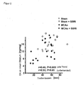
- MCAo animals, but not Citalopam-treated MCAo animals show significantly lower levels of dopamine in the ischemic striatum. The dopamine content correlates with the behavior in the forced swim test (Pearson Correlation) ( FIG. 2 ).
- the light / dark box consisted of a Plexiglassapparate (TSE shuttle box system) with the dimensions 30 cm x 17 cm x 17 cm (length x width x height) and was divided into two compartments (darkened or full with a 20W bulb lit compartment). The animals were tested in the LD box for 5 minutes. A small opening within the partition wall between the two rooms (4 x 4 cm) allowed the animals a free change back and forth between the two compartments. The animals were examined in the LD box for a period of 5 minutes. The animals were placed in the darkened room at the beginning of the investigation.
- TSE shuttle box system Plexiglassapparate
- the experiments were conducted in a soundproof, brightly lit (1500 lx) observation room between 11:00 and 12:00 in a wooden white open field (120 cm x 120 cm x 40 cm). Beginning 48 hours before testing, the animals were deprived of food. The animals were brought to the observation room just one hour before the test. A Petri dish (diameter 8 cm) was filled with standard mouse pellets and placed in the middle of the "open field”. Each animal was individually placed in a corner of the unknown "open field" facing the food. The behavior of the animals was recorded and evaluated with regard to the duration until the beginning of food intake. Animals that did not move or which ate nothing during the study period of 300 s were excluded from the evaluation.
- mice prefer a 1% sucrose solution to tap water using a well-established procedure (Strekalova et al., 2004). For a period of 24 hours, the animals within their usual cage were given free choice between a sugar solution and water using two otherwise identical water bottles. Here, the bottles were weighed before and at the end of the test protocol. From this it is easy to calculate the sucrose intake in mg / g body weight.
- tissue samples from both hemispheres were rapidly taken from cortex (fronto-parietal), striatum (Nucl. Caudatus / putamen), septum, and hippocampus (a total of eight tissue samples per brain), snap frozen in liquid nitrogen, and until stored at -80 ° C for further study.
- the frozen tissue samples were weighed and homogenized with ultrasound in 10-20 vol deionized water at 4 ° C.
- monoamines an aliquot of the homogenate was immediately added to the same volume of 0.2 N perchloric acid and centrifuged at 25000 g for 10 min at 4 ° C.
- the supernatant was used for the measurement of monoamines and their degradation products.
- 30 ⁇ l of aqueous homogenate was added to 120 ⁇ l of methanol and centrifuged at 25000 g for 10 minutes at a temperature of 4 ° C to remove precipitated protein.
- the residual aqueous homogenate was used to determine choline acetyltransferase (ChAT) activity.
- the MCAo animals show a significantly lowered tissue level of dopamine in the ischemic stratum.
- the dopamine content correlates with the behavior in the Porsolt's swimming test ( FIG. 2 ).
- Tissue samples were homogenized by sonication in 10-20 ⁇ l lysis buffer (0.1M Tris-HCl, pH 7.0, 0.4M NaCl, 0.1% NaN3, and a variety of protease inhibitors).
- concentration of BDNF and NGF at the protein level was determined by means of a commercial ELISA (Promega). The detection limit of the test was 1pg / ml or 0.25pg / ml.
- NGF and BDNF concentrations were reported by a fluorescence-based ELISA as published (Hellweg et al., 2003).
- Coronal 40 ⁇ m sections were obtained on a carriage microtome (Leica, Bensheim, Germany). The sections were dyed floating. The sections were analyzed by light microscopy according to the ABC method (Vectastain Elite Kit, Vector Laboratories) stained and developed with diaminobenzidine as the chromogen. ( FIG. 3, FIG. 4 and FIG. 5 ).
- mice 129 / SV wild-type mice were exposed to a 30 min MCAo with subsequent reperfusion.
- Mice receiving chronic treatment with the SSRI citalopram had significantly smaller lesions at 3 months (as determined by immunohistochemical staining with the neuronal marker NeuN).
- animals treated with citalopram showed significantly less loss of volume (less atrophy) of the ipsilateral striatum and significantly lower loss of midbrain neurons (in the substantia nigra and other core areas) than MCAo mice treated with the control substance.
- the daily intraperitoneal treatment with citalopram (13 mg / kg body weight) was started 7 days after MCAo / reperfusion and continued until the end of the experiment.
- mirtazapine is an antidepressant that blocks presynaptic alpha2 receptors, thereby promoting the release of serotonin and norepinephrine. Since postsynaptic 5-HT 2 and 5-HT 3 receptors are blocked at the same time, the increased serotonergic neurotransmission occurs primarily via 5-HT 1 receptors (noradrenergic and specifically serotonergic antidepressant).
- FIGS. 6 to 10 are identical to FIGS. 6 to 10:
Landscapes
- Health & Medical Sciences (AREA)
- Veterinary Medicine (AREA)
- Chemical & Material Sciences (AREA)
- Medicinal Chemistry (AREA)
- Pharmacology & Pharmacy (AREA)
- Life Sciences & Earth Sciences (AREA)
- Animal Behavior & Ethology (AREA)
- General Health & Medical Sciences (AREA)
- Public Health (AREA)
- Engineering & Computer Science (AREA)
- Epidemiology (AREA)
- Bioinformatics & Cheminformatics (AREA)
- Biomedical Technology (AREA)
- Neurology (AREA)
- Neurosurgery (AREA)
- Hospice & Palliative Care (AREA)
- Psychiatry (AREA)
- Chemical Kinetics & Catalysis (AREA)
- General Chemical & Material Sciences (AREA)
- Nuclear Medicine, Radiotherapy & Molecular Imaging (AREA)
- Organic Chemistry (AREA)
- Pharmaceuticals Containing Other Organic And Inorganic Compounds (AREA)
Abstract
Description
- Die Erfindung betrifft die Bereitstellung eines Arzneimittels zur Behandlung neuronaler Erkrankungen, insbesondere des Schlaganfalls.
- Schlaganfall ist eine Volkskrankheit mit hoher Morbidität und Letalität. Allein in Deutschland treten jährlich 200.000 Neuerkrankungen auf, was den Schlaganfall zur häufigsten Ursache für Behinderungen und zur dritthäufigsten Todesursache in modernen Zivilgesellschaften macht. Die Gesamtkosten der Erkrankung werden allein in Deutschland auf etwa 10 Milliarden Euro geschätzt.
- Ursache des Schlaganfalls ist zu etwa 90% ein sog. ischämischer Hirninfarkt, der durch einen akuten Gefäßverschluss im Gehirn verursacht wird. Zur kausalen Behandlung des Schlaganfalls ist derzeit lediglich die Thrombolyse mit rekombinantem Gewebe Plasminogen Aktivator (rt-PA) zugelassen. Diese Therapie ist jedoch auf ein therapeutisches Fenster von drei Stunden nach Beginn des Einsetzens der Schlaganfallsymptome beschränkt. Deutschlandweit kommen daher nur weit weniger als 5% aller Schlaganfallpatienten in den Genuss dieser Behandlung. Andere kausale Behandlungsmöglichkeiten, insbesondere Substanzen, die entweder akut oder chronisch gegeben werden, um die neurologischen Schäden zu verringern, sind derzeit trotz umfangreicher jahrzehntelanger Forschungsaktivitäten bislang in der Klinik nicht verfügbar.
- Als Folge eines ischämischen Ereignisses kommt es zu einem Zelluntergang von Neuronen und anderen Zelltypen im _Gehirn.. Dies gilt selbst dann, wenn es sich nur um eine transiente Ischämie handelt und/oder die vollständige Erholung der lokalen Blutversorgung einen normalen Energiestoffwechsel und eine normale neuronale Aktivität wieder erlauben würde. Die zum Zelltod führenden pathologischen Veränderungen von Nervenzellen, die insbesondere in vulnerablen Regionen auftreten, wie z.B. dem Hippocampus, Basalganglien, oder der Großhirnrinde, manifestieren sich teilweise erst einige Tage nach der Ischämie und werden deshalb auch verzögerter neuronaler Zelltod genannt. Der neuronale Zelltod kann auch Hirnregionen betreffen, in denen der Blutstrom nicht vollständig unterbrochen war.
- Die Wissenschaft und Forschung setzt bei der Behandlung von Schlaganfall daher einerseits an einer möglichst schnellen und effektiven Thrombolyse zur Wiederherstellung der Blutzufuhr, anderseits aber auch an der Verhinderung des neuronalen Zelltods an. Als Begriff für die Verhinderung des neuronalen Zelltods hat sich der Terminus "Neuroprotektion", durchgesetzt. Die Neuroprotektion betrifft somit in erster Linie die Behandlung oder Vermeidung von Sekundärerscheinungen eines ischämischen Ereignisses, die sich dem Ereignis - vor allem im Laufe der darauf folgenden Stunden und Tage - anschließen.
- Der neuronale Zelltod kann nach neuerem Verständnis durch eine komplexe Kaskade einer Vielzahl einzelner Reaktionsschritte ausgelöst werden (u.a. Exzitotoxizität, Inflammation, _Apoptose sowie weitere Schadensmechanismen). Die initiale Exzitotoxizität wird dadurch ausgelöst, dass die unter Sauerstoffmangel leidenden ischämischen Neuronen schnell ATP verlieren und depolarisieren. Dies führt zu einer verstärkten postsynaptischen Abgabe des Neurotransmitters Glutamat, der seinerseits membrangebundene Glutamat-Rezeptoren aktiviert, die ihrerseits Kationen-Kanäle regulieren. Als Folge der überhöhten Glutamatausschüttung werden die Glutamat-Rezeptoren jedoch überaktiviert. Die von den Glutamat-Rezeptoren regulierten spannungsabhängigen Kationen-Kanäle werden durch die Bindung von Glutamat an den Rezeptor geöffnet. Damit setzt ein Na+ und ein Ca2+ Strom in die Zelle ein, der zu einer massiven Störung des Ca2+ abhängigen zellulären Stoffwechsels - einschließlich des Energiestoffwechsels - führt. Für den nachfolgend eintretenden Zelltod könnte dabei insbesondere die Aktivierung Ca2+ abhängiger kataboler Enzyme verantwortlich sein.
- Selbst wenn der Mechanismus dieser durch Glutamat vermittelten Neurotoxizität derzeit noch nicht im Detail abschließend verstanden ist, scheint dennoch in der Fachwelt Einigkeit darüber zu bestehen, dass diese Phänomen in einem erheblichen Umfang zu dem frühen neuronalen Zelltod nach einer Ischämie beiträgt.
- In den letzten Jahrzehnten wurde eine Vielzahl verschiedener Strategien entwickelt, um dem neuronalen Zelltod klinisch zu begegnen. Frühe Ansätze stellten auf die Unterdrückung des neuronalen Metabolismus und die Blockade des Calciumeinstroms ab (siehe beispielsweise
EP 0 781 766 B1 ). Ein Großteil neuerer Strategien setzt dagegen an der Verwendung von Inhibitoren exzitatorischer Aminosäuren an. Dieser Ansatz basiert auf der Vorstellung, den ischämischen Zelltod durch die Hemmung der oben geschilderten Überanregung zu verhindern. Als Inhibitoren exzitatorischer Aminosäuren sind vor allem Glutämat-Antagonisten bekannt; die spezifisch die Rezeptoren dieser Aminosäure blockieren, sowie Verbindungen, die die Glutamatfreisetzung hemmen. Als Inhibitor des NMDA Glutamat-Rezeptors ist beispielsweise das MK-801 bekannt. Als Inhibitoren der Glutamatfreisetzung sind 2,4-Diamino-5-(2,3,5-trichlorphenyl)pyrimidin oder 2,4-Diamino-5-(2-chlorphenyl)pyrimidin bekannt (EP 0 459 830 A1 ). - In der Vergangenheit wurden insgesamt mehr als 50 mögliche neuroprotektive Substanzen in klinischen Versuchen zum akuten ischämischen Schlaganfall untersucht. Bislang hat sich jedoch keine der zunächst positiv in präklinischen Untersuchungen getesteten Substanzen in der klinischen Praxis als wirksam erwiesen. Demnach fehlt es derzeit immer noch an wirksamen Neuroprotektiva, die eine erfolgreiche Verhinderung oder zumindest Linderung des neuronalen Zelltods nach einem ischämischen Ereignis ermöglichen.
- Nach der Reperfusion (Rekanalisation) der Blutgefäße und der möglichen Neuroprotektion - sowie der begleitenden Stabilisation des Allgemeinzustands des Patienten - noch während der Akutphase oder frühen postakuten Phase des Schlaganfallereignisses ist der behandelnde Arzt bestrebt, möglichst frühzeitig eine Rehabilitation des Patienten einzuleiten. Diese frühe Rehabilitationsbehandlung setzt üblicherweise einige Tage nach dem Schlaganfallereignis ein; und ist somit für die postakute oder chronische Phase der Behandlung typisch. Sie kann aber bestenfalls auch noch am selben Tag nach der Stabilisierung des Patienten beginnen. Eine klare zeitliche Differenzierung zwischen den einzelnen Behandlungsphasen ist daher nicht ohne weiteres möglich.
- Die Rehabilitation ist in diesem Kontext definiert als eine Maßnahme, die das Gehirn unterstützt, sich durch Umorganisation oder eventuell auch Neubildung veränderten Bedingungen anzupassen, und so zunächst verlorene oder geschwächte Funktionen oder Strukturen wiederzuerlangen oder zu ersetzen. Auf der zellulären Ebene versteht der Fachmann darunter vor allem die sog. neuronale Plastizität, bei der es sich um die funktionale und/oder morphologische Anpassung reifer neuronaler Zellen auf veränderte interne oder externe Einflüsse handelt, insbesondere nach einem neuronalen Verlust oder einer Schädigung wie beispielsweise nach einem Schlaganfall. Die positive Wirkung einer Maßnahme auf die Rehabilitation oder Plastizität neuronaler Strukturen wird im vorliegenden Zusammenhang als "neuroregenerative" oder "neuroplastische" Wirkung oder als "Neuroregeneration" definiert.
- Die derzeit gängigen Maßnahmen zur Rehabilitation umfassen beispielsweise ergotherapeutische, logopädische oder physiotherapeutische Behandlungen. Eine medikamentöse rehabilitative Therapie ist nicht etabliert. Letztlich versuchweise wurden stimulierende Medikamente wie Piracetam oder Amphetamine eingesetzt. Bereits unmittelbar nach der Akutphase setzt jedoch die Sekundärprävention zur Vermeidung weiterer vaskulärer Ereignisse (Schlaganfallung, Herzinfarkt etc ein). Dies geschieht z.B. mit Thrombozytenfunktionshemmern (z.B. Aspirin), Antikoagulantien (z.B. Warfarin), Cholesterinsynthesehemmern oder Blutdruckmedikamenten. Diese werden dem Patienten meist chronisch verabreicht, d.h. über Monate und Jahre.
- Trotz des großen Bedarf an Maßnahmen zur Behandlung neuronaler Erkrankungen wie beispielsweise des Schlaganfalls sowohl in der akuten als auch in der postakuten oder chronischen Phase des Krankheits- oder Schadensverlaufs, haben die bisherigen Anstrengungen der Arzneimittelforschung noch nicht zu ausreichend befriedigenden Ergebnissen geführt. Der Bedarf an weiteren oder alternativen Medikamenten zur Behandlung neuronaler Schäden besteht somit trotz jahrzehntelangen Bemühungen der Wissenschaft und Forschung fort. Dies gilt insbesondere für den Bereich der postakuten und chronischen Behandlung des Schlaganfalls.
- Es ist daher Aufgabe der vorliegenden Erfindung, ein Arzneimittel bereitzustellen, das hier Abhilfe schafft und die Möglichkeit eröffnet, die Behandlung neuronaler Erkrankungen, insbesondere des Schlaganfalls, zu verbessern.
- Es hat sich nun überraschend gezeigt, dass die Behandlung geschädigten neuronalen Gewebes mit einer serotonergen Verbindung zu einer deutlichen Verbesserung oder Erholung der betroffenen Bereiche der Schädigung führt.
- Nach der Erfindung ist dabei als "serotonerge Verbindung" jede Verbindung definiert, die zu einer Erhöhung der Wirkung von Serotonin im Zentralen Nervensystem führt. Bevorzugt handelt es sich dabei um Verbindungen, die die Serotonin-Konzentration im synaptischen Spalt gegenüber der normalen Ausgangslage erhöhen. In einer besonderen Ausführungsform der Erfindung können selektive Serotonin-Wiederaufnahme-Inhibitoren (SSRI) verwendet werden. Außerdem sind auch serotonerge Antidepressiva wie z.B. das Mirtazapin einsetzbar.
- Bei der Definition der serotonergen Verbindungen im Sinne der Erfindung ist der der Verbindung jeweils zugrunde liegende Wirkmechanismus nicht relevant. Vielmehr kommt es auf das physiologische Ergebnis der erhöhten Serotonin-Wirkung im CNS an. Das kann demgemäß z.B. über eine Wiederaufnahmehemmung oder aber über eine erhöhte Ausschüttung von Serotonin erfolgen. Auch andere Mechanismen sind denkbar. Bevorzugt sind SRRI oder Mirtazapin.
- Die erfindungsgemäß zu beobachtende Verbesserung gilt insbesondere für Schäden, die auf ein ischämisches Ereignis zurückgehen. Es ist dabei besonders überraschend, dass diese Verbesserung selbst dann eintritt, wenn die Gabe der serotonergen Verbindung außerhalb der akuten Phase des ischämischen Ereignisses, nämlich z.B. postakut oder chronisch erfolgt.
- Die Erfindung betrifft somit die Behandlung neuronaler Erkrankungen mit serotonergen Verbindungen zu Zwecken der Neuroprotektion oder der Neuroregeneration. Beide Vorgänge spielen unter anderem bei der Behandlung des Schlaganfalls eine entscheidende Rolle, so dass die Erfindung insbesondere die Verwendung von serotonergen Verbindungen als Neuroprotektivum oder als Maßnahme zur Rehabilitation oder Neuroregeneration bei Schlaganfallpatienten umfaßt. Die Behandlung ist dabei auch postakut oder sogar chronisch möglich. Ebenso ist die Behandlung neurodegenerativer Zustände durch die Verabreichung der neuroregenerativ wirkenden serotonergen Verbindung Gegenstand der Erfindung. Dazu ist eine chronische Gabe der Verbindungen vorteilhaft.
- Die erfindungsgemäße Verwendung von serotonergen Verbindungen, insbesondere von SSRI oder Mirtazapin, ist vor allem zur Behandlung von Patienten geeignet, die unter einer fokalen cerebralen Ischämie leiden.
- Bei den bevorzugt einsetzbaren SSRI handelt es sich um eine Gruppe von Substanzen, die übereinstimmend am Serotonin-Transporter (5-HT Transporter) eine präferentielle Hemmwirkung entfalten, jedoch nicht notwendigerweise zu einer einheitlichen chemischen Klasse gehören. Mögliche weitere Wirkungen dieser Verbindungen an anderen Rezeptoren treten gegenüber der Wirkung am 5-HT Transporter deutlich in den Hintergrund. Da sie die Wiederaufnahme des Neurotransmitters Serotonin in die Präsynapse hemmen, erhöhen sie die Serotonin-Konzentration im synaptischen Spalt. Einige der SSRI hemmen jedoch auch schwach die postsynaptischen Rezeptoren. Das Maß der postsynaptischen Hemmung ist jedoch klinisch in der Regel nicht relevant.
- Es ist eine Vielzahl von SSRI bekannt, die zum Teil auch bereits in der klinischen Praxis zur Behandlung von Depressionen eingesetzt werden. Dabei handelt es sich beispielsweise um das Fluvoxamin (Fevarin®), Fluoxetin (N-Methyl-3-phenyl-3-[4-(trifluoromethyl)phenoxy]-propan-1-amin; Fluctin®)), Paroxetin (3S-trans)-3-((1,3-Benzodioxol-5-yloxy)methyl)-4-(4-fluorophenyl)-piperidin, Paroxetin; Paxil®, Seroxat®, Sertralin, (1S,4S)-4-(3,4-Dichlorphenyl)- 1,2,3,4-tetrahydro- N-methyl-1-naphthylamin, Zoloft®, Gladem®)) Citalopram, 1-[3-(Dimethylamino)propyl]- 1-(4-fluorphenyl)- 1,3-dihydroisobenzofuran- 5-carbonitril] (Cipramil®, Sepram®), sowie dessen S-Enantiomer (Cipralex®). Diese Substanzen sind erfindungsgemäß einsetzbar. Citalopram und Escitalopram sind besonders bevorzugt. Die Herstellung dieser Verbindungen ist dem zuständigen Fachmann geläufig.
- Ebenso ist Mirtazapin erfindungsgemäß einsetzbar. Dabei handelt es sich um das Ebenso ist Mirtazapin erfindungsgemäß einsetzbar. Dabei handelt es sich um das (RS)-(±)-2-Methyl- 1,2,3,4,10,14b-hexahydropyrazino [2,1-a]pyrido[2,3-c][2]benzazepin. Auch können die dem Fachmann bekannten Antidepressiva Amitriptylin, Clomipramin, Desipramin, Doxepin, Imipramin, Maprotilin, Mianserin oder Nortriptylen erfindunsgemäß einsetzbar sein. Diese Verbindungen zeigen alle ein Rezeptoraffinitätsprofil, dass dem Grunde nach Mirtazapin entspricht. Das Rezeptoraffinitätsprofil von Mirtazapin ist unten angeführt.
- Zu Zwecken der vorliegenden Erfindung werden unter "neuronalen Erkrankungen" bzw. "neuronalen Krankheiten" alle diejenigen Zustände zusammengefasst, die bezüglich der Funktionalität oder Morphologie neuronaler Gewebe, Organe oder Strukturen gegenüber einem gesunden Menschen aus medizinischer Sicht eine Veränderung darstellen. Die Ursachen der Schädigung können dabei beliebigen Ursprung sein, so z.B. genetisch bedingt oder aber auch durch ein Trauma (z. B. spirale oder cerebrale Traumata) ausgelöst sein. Diese Definition umfasst demnach auch neurodegenerative oder cerebrovaskuläre Erkrankungen. Vornehmlich psychiatrische Erkrankungen wie Depressionen sind von der vorliegenden Definition allerdings nicht erfaßt.
- Als neurodegenerative Erkrankungen wird eine Gruppe von meist langsam fortschreitenden, erblichen oder sporadisch auftretenden Erkrankungen des Nervensystems verstanden. Hauptmerkmal ist der fortschreitende Verlust von Nervenzellen, der zu verschiedenen neurologischen Symptomen - darunter häufig zu Demenz und Bewegungsstörungen - führt. Die Erkrankungen können in unterschiedlichen Lebensaltern auftreten, verlaufen diffus oder generalisiert und rufen charakteristische histologische Schädigungsmuster hervor. Beispiele für diese Erkrankungen sind Morbus Alzheimer, Morbus Parkinson, Morbus Huntington oder die Amytrophische Lateralsklerose.
- Als cerebrovaskuläre Erkrankungen werden im Kontext der vorliegenden Erfindung krankhafte Zustände bezeichnet, die auf einer Veränderung oder Schädigung der Blutgefäße des Zentralnervensystems beruhen, insbesondere der zuführenden Gefäße zum Gehirn. Der Schlaganfall gehört zu dieser Gruppe von Erkrankungen und wird in diesem Zusammenhang näher definiert als eine plötzlich oder innerhalb kurzer Zeit auftretende Erkrankung des Gehirns, die zu einem anhaltenden oder aber zumindest vorübergehenden Ausfall von Funktionen des Zentralen Nervensystems führt und durch kritische Störungen der Blutversorgung des Gehirns verursacht wird. Bei der ursächlichen Durchiblutungsstörung kann es sich um eine plötzlich eintretende Minderdurchblutung (z.B. Ischämie) oder aber auch um eine Blutung (z.B. hämorrhagischer Schlaganfall) handeln.
- Im Kontext der vorliegenden Erfindung bezeichnet der Begriff "fokale Ischämie" bzw. "fokale cerebrale Ischämie" eine Schädigung des neuronalen Gewebes, die durch einen Verschluß oder eine Verengung einer zerebralen Arterie bedingt ist und somit zu einer lokal begrenzten Minderversorgung mit Sauerstoff und Nährstoffen führt.. Sie grenzt sich insoweit von der sog. globalen Ischämie oder der globalen cerebralen Ischämie ab, bei der ein ganzes Organ von der Sauerstoffzufuhr durch das Blut zumindest vorübergehend getrennt ist
- Wie bereits ausgeführt, werden erfindungsgemäße serotonerge Verbindungen, beispielsweise SSRI, bereits als Antidepressiva eingesetzt. Mittlerweile ist aber darüber hinaus bekannt, dass SSRI ebenso als Ergänzung eines auf einer Psychotherapie basierenden Gesamtkonzepts, beispielsweise zur Behandlung der Zwangsstörung von Angstzuständen (soziale Phobie, Panikstörung, generalisierte Angststörung) oder von Essstörungen eingesetzt werden können. Ein möglicher weiterer Anwendungsbereich ist die Verminderung von Stottern. Ebenso liegen Erfahrungen im Bereich der therapeutischen Behandlung bei krankhaftem Übergewicht vor.
- In der
EP 0474 580 B1 wird die protektive Wirkung von SSRI auf Bewusstseinsstörungen oder Amnesie beansprucht, die im Zusammenhang mit Demenz oder cerebrovaskulären Störungen auftreten können. Um diese Schutzwirkung zu erfassen, wurde den Versuchstieren zunächst der SSRI Citalopram verabreicht, und anschließend durch den beidseitigen Verschluß der Carotiden der Tiere eine vorübergehende Unterbrechung der Blutversorgung im Gehirn erzeugt. Danach wurde unter anderem das durch die vorübergehende Ischämie erzeugte Läsionsvolumen im Gehirn oder das Verhalten der Tiere in einem Standard-Verhaltenstest untersucht. Aus den Ergebnissen aus diesem Tiermodell zur Simulation von durch einen Herzkreislaufstillstand erzeugten Schäden im Gehirn sowie an entsprechenden positiven klinischen Ergebnissen an Depressionskranken mit begleitenden Demenzerscheinungen wird die oben genannte Schutzwirkung der SSRI im Hinblick auf Bewusstseinsstörungen abgeleitet. - Darüber hinaus sind verschiedene Kombinationstherapien bekannt, in denen SSRI gemeinsam mit anderen therapeutisch aktiven Substanzen eingesetzt werden, so z.B. mit dem für die Epilepsie Behandlung zugelassenem Zonisamid zur Behandlung von Hörstörungen (siehe
US 2007/021352 A1 ) oder in Kombination mit einem Phosphodiesterase 5 Inhibitor zur Behandlung cardiovaskulärer Erkrankungen. Die letzt genannte Kombinationstherapie ist u.a. Gegenstand der US PatentanmeldungUS2006/0106039 A1 . Allerdings offenbart diese Druckschrift weder die Behandlung des Schlaganfalls mit SSRI als Monotherapie, noch die nun erfindungsgemäß aufgefundene neuroprotektive oder neuroregenerative Wirkung von SSRI. - Keine dieser Druckschriften gibt demnach aus der Sicht des Fachmanns einen überzeugenden Hinweis darauf, dass - wie es Gegendstand der vorliegenden Erfindung ist - serötonerge Verbindungen, u.a. SSRI, klinisch zur Neuroprotektion nach einem fokalen Schlaganfall oder aber zur Neuroregeneration bei neuronalen Krankheiten erfolgreich einsetzbar sind.
- In einer besonders vorteilhaften Ausführungsform der vorliegenden Erfindung werden die serotonergen Verbindungen nach Ablauf der akuten Phase des ischämischen Ereignisses, also in der postakuten bzw. chronischen Phase der Erkrankung verabreicht. Erfolgt die Gabe der Verbindung insbesondere auch zu Zwecken der Neurogeneration kann sie - wie bereits oben ausgeführt - auch chronisch erfolgen. Dies gilt beispielsweise bei der erfindungsgemäßen Behandlung des Morbus Alzheimer oder des Morbus Parkinson.
- Dabei wird vorliegend unter der Begrifflichkeit "außerhalb der akuten" Phase derjenige Zeitraum verstanden, der sich dem Erhalt der wesentlichen Lebensfunktionen und der Stabilisierung eines Patienten nach dem ischämischen Ereignis anschließt. Diese Phase ist vor allem dadurch charakterisiert, dass sich der behandelnde Arzt der Behandlung der Folgen des ischämischen Ereignisses und der weitgehenden Regeneration der geschädigten Funktionen und Strukturen widmet. Außerhalb der akuten Phase liegt die postakute sowie die chronische Phase. Die "postakute" Phase beginnt im Sinne der vorliegenden Erfindung dann, wenn die primären Schadensereignisse und Schadenskaskaden im betroffenen Gewebe abgeschlossen sind. In der Regel ist davon auszugehen, dass die akute Phase frühestens nach etwa nach 6 bis 12 Stunden abgeschlossen ist und dann die postakute Phase beginnt.
- In einer bevorzugten Ausführungsform der Erfindung erfolgt die Gabe der serotonergen Verbindung demnach frühestens 6, 12 oder 24 h nach Beginn des ischämischen Ereignisses, oder aber erst sogar einige Tagen nach Beginn des Ereignisses. Dies gilt insbesondere für die Behandlung des Schlaganfalls. Sie konnten die Erfinder zeigen, dass sogar bei einer Gabe eines SSRI beginnend am 7. Tag nach dem Schlaganfallereignis eine signifikante Verbesserung erreicht werden konnte.
- Die serotonergen Verbindungen können erfindungsgemäß auch chronisch verabreicht werden. Dabei bedeutet die chronische Gabe, dass die Verbindung über einen Zeitraum von einigen Tagen bis hin zu Wochen und Monaten verabreicht werden. Besonders vorteilhaft ist die tägliche Verabreichung der Verbindung über einen kontinuierlichen Zeitraum von mehreren Tagen oder Wochen, beispielsweise über 2 Tage bis 60 Tage, vorteilhafterweise 2 bis 30 oder 5 bis 20 Tage. Sie kann jedoch auch über einen Zeitraum von mehreren Monaten bis Jahren verabreicht werden.
- Die erfindungsgemäßen neuroprotektiven und neuroregenerativen/neuroplastischen Effekte von SSRI auf neuronale Krankheiten wurde anhand eines anerkannten Tiermodells zur fokalen Ischämie in Mäusen untersucht. Dazu wurde der SSRI Citalopram den Versuchstieren in einer täglichen Dosis von 13 mg/kg Körpergewicht kontinuierlich bis zum Versuchsende verabreicht, wobei die Verabreichung am 7. Tag nach der Induktion der Ischämie einsetzte.
- Anschließend wurden die SRRI Effekte auf das Volumen der ischämischen Läsionen ausgewertet. Ferner wurden die Tiere Standard-Verhaltenstests unterzogen. Zudem wurden die Konzentrationen ausgewählter Neurotransmitter erfaßt.
- Die Tierversuche wurden nach den Maßgaben des Tierschutzgesetzes in Übereinstimmung mit institutionellen und internationalen Richtlinien durchgeführt. Männliche 12936/SvEv-Mäuse (18-20 g, Bundesamt für Gesundheitsschutz Berlin) wurden in Gruppen von 4-5 Mäusen pro Käfig in Standardlaborkäfigen bei einer Umgebungstemperatur von 20-21°C bei einem 12 h-Licht-Dunkel-Rhythmus (8.00 Uhr/20.00 Uhr) mit freiem Zugang zu Futter und Wasser gehalten. Die Narkose der Mäuse erfolgte mit 1% Isofluran in 69% N20 und 30% 02 mit einem Narkoseverdampfer. Die Tiere wurden einer 30 min Fliamentokklusion der Arteria cerebri media (MCAo) unterworfen, wobei die Körpertemperatur und der regionale cerebrale Blutfluss wie beschrieben gemessen wurden (Endres et al., 1998). Im Rahmen einer "sham"-Operation (Scheinoperation) wurde das Filament lediglich für 30 Minuten in die linke oder rechte Arteria carotis interna eingebracht, jedoch nicht weiter vorgeschoben. Pro Versuchsgruppe wurden jeweils 10 Versuchstiere untersucht. Ein Versuchstier aus der IMCAo-(linksseitige MCAo) Gruppe verstarb nach der Operation.
- Die MRT-Untersuchungen wurden mit einem 7 T Bruker Scanner (Pharmascan 70/16 AS, Bruker Biospin, Ettlingen, Germany) mit einem 16 cm horizontalen Bohrlochmagnet und einem 9 cm abgeschirmten Gradienten (innerer Durchmesser) mit einer maximalen Feldstärke von 300 mT/m durchgeführt. Während der Bildgebung wurde die Maus unter Inhalationsnarkose auf einem Mausbett in die Mitte einer 38 mm RF-Spule gelegt. Die Respiration wurde mit Hilfe der Überwachungseinheit Model 1025 (SA Instruments, Inc.) überwacht. Das Ischämieareal wurde mit einer T2-gewichteten, fettsupprimierten 2D Turbo Spin-Echosequenz (TR 2937 ms; TE 36.5 ms, 12 x gemittelt) erfasst. Zwanzig aufeinander folgende axiale Schnitte mit einer Schnittdicke von 0.4 mm wurden so durch das Gehirn gelegt, dass Bulbus olfactorius sowie Kleinhirn nicht erfasst wurden. Das sogenannte "field of view" betrug 2 x 2 cm, die Matrix war 128 x 128, woraus sich eine Auflösung innerhalb einer Ebene von 156 µm ergab. Das Läsionsvolumen wurde mit der Auswertungssoftware Analyze 5.0. (AnalyzeDirect, Inc., Lenexa, USA) bestimmt. Dabei wurden die hyperintensen Ischämieareale auf den T2-gewichteten Bildern manuell bestimmt und dann das Läsionsvolumen automatisch berechnet.
- Unmittelbar nach Induktion der Ischämie und zusätzlich 1 Tag vor Tötung wurden die Tiere entsprechend des Bederson Scores hinsichtlich des neurologischen Defizits "geratet"(Bederson et al., 1986).
- Ab zwölf Wochen nach MCAo/Reperfusion wurden die Tiere in Folge einer Reihe von Verhaltenstests unterworfen: spontane lokomotorische Aktivität, "elevated plus maze", modifizierter Porsolt Schwimmtest, "light/dark box", "latency to feed" sowie Konsum von Zuckerwasser. Die Verhaltenstests wurden während der Dunkelperiode (d.h. während der Aktivitätsphase der Tiere) durchgeführt. Vor Untersuchungsbeginn hatten die Tiere jeweils mindestens eine Viertelstunde Zeit, sich an den Untersuchungsraum zu gewöhnen. Die Reihenfolge der Verhaltensuntersuchungen entsprach dabei früheren Empfehlungen, diese in einer Graduierung nach dem Ausmaß der Stressbelastung mit Beginn mit den am wenigsten belastenden Tests durchzuführen (van Gaalen und Steckler, 2000; McIlwain et al., 2001). Der Untersucher war während der Durchführung der Experimente hinsichtlich der Versuchsgruppen verblindet. Die Tiere wurden 2 Tage nach dem Abschluss der Verhaltenstestung geopfert und die Gehirne für die weitere Aufarbeitung asserviert (Bestimmung von Monoaminen, Aminosäuren, Neurotrophinkonzentrationen). Separate Versuchsgruppen wurden für die Immunhistochemie, Bestimmung der cerebralen Läsionsgrößen, Immunoblots und RT-PCR angesetzt (siehe unten).
- Bei allen Tests wurde ein automatisches Videotracking eingesetzt (TSE VideoMot 2 System, Technical & Scientifical Equipment GmBH, Bad Homburg, Deutschland) mit einer räumlichen Auflösung von 640 x 480 Pixel und einer zeitlichen Auflösung von 1 Hz (spontane lokomotorische Aktivität) bzw. 12.5 Hz ("elevated plus maze").
- Die Tiere wurden in Einzelkästen gesetzt (30 x 20 x 15 cm). Zeit, Geschwindigkeit und Distanz für spontane lokomotorische Aktivität wurde über eine Zeitspanne von 8 Stunden über Nacht automatisch mittels VideoMot aufgezeichnet und ausgewertet. Der Test fand unter gedimmter indirekter Beleuchtung in einer neutralen schallgeschützten Umgebung statt.
- Das sogenannte "elevated plus maze" ist weithin als Test zur Bestimmung von Ängstlichkeit bei Nagern etabliert. Die Untersuchung wurde nach publizierten Protokollen durchgeführt (Lister et al., 1987; Rodgers et al., 2002). Das "Maze" ist aus schwarzem Plexiglass gefertig und besteht aus zwei sich gegenüberstehenden offenen Armen (30 x 5 x 0.25 cm) sowie zwei sich gegenüberstehenden geschlossenen Armen (30 x 5 x 15 cm). Die Arme gehen von einer gemeinsamen Plattform aus (5 x 5 cm), die auf einem Ständer 50 cm oberhalb des Bodens befestigt ist. Das Plus Maze wird für das Experiment in eine schallgeschützte und neutrale Umgebung mit indirekter Beleuchtung gebracht. Die Tiere werden zu Beginn des Test in das Zentrum des "Maze" gesetzt, wobei der Kopf in die Richtung eines geschlossenen Arms positioniert wird. Die (i) Gesamtzeit, die das Tier in den offenen Armen verbringt, sowie (ii) die Anzahl der Besuche in den offenen Armen ("open arm visits") wurden über einen Zeitraum von 5 min aufgezeichnet. Die Zeit in den offenen Armen ist als aussagekräftiger Index für Ängstlichkeit etabliert.
- Das Versuchstier wird etwa 1 cm vom Schwanzende mit Klebestreifen (3M Durapore) an einer Metallstange 80 cm über dem Boden befestigt. Die Zeitdauer, in der das Tier immobil war, wurde durch einen verblindeten Beobachter aufgezeichnet. Immobilität war dabei definiert als Abwesenheit von Bewegungen unter Nichtberücksichtigung der Atembewegungen. Wenn eine Maus an ihrem Schwanz nach oben kletterte, wurde sie sanft wieder nach unten gezogen und die Testung anschließend fortgesetzt. Mäuse, die auch weiterhin an ihrem Schwanz nach oben kletterten, wurden von der Auswertung ausgeschlossen.
- Der modifizierte Porsolt Schwimmtest stellt ein Screeningverfahren für Depressivität und für die Wirksamkeit von Antidepressiva dar. Die Testung wurde im wesentlichen wie beschrieben durchgeführt (Cryan et al., 2002; Lucki et al., 1997). Die Tiere wurden einzeln für 5 min in ein gläsernes Gefäß gegeben (15 x 21 cm), das mit 22°C warmen Wasser gefüllt war (8.5 cm Tiefe). Über diesen Zeitraum von 300 sec wurden dann die Verhaltensweisen "Schwimmen", "Climbing" (Klettern) sowie "Floating" (Treiben) gemonitort. "Schwimmen" war definiert als Bewegung (gewöhnlich horizontal) durch das Gefäß. Immobilität war definiert als Bewegungslosigkeit unter Nichtberücksichtigung solcher Bewegungen, die das Tier daran hindern unterzugehen. "Climbing" (Klettern), bestand in nach oben gerichteten Bewegungen der Vorderpfoten des Tieres am Gefäßrand. Es wurden Zeit und Latenz der drei Verhaltensweisen verglichen und wie publiziert ausgewertet (Cryan et al., 2002; Bale und Vale, 2003; Montkowski et al., 1997; Lucki et al., 1997).
- Die MCAo Tiere zeigen im sog. "Schwimmtest" (forced swim test) nach Porsolt kürzere Latenzen bis zum ersten Treibenlassen (latency to float) im Vergleich zu Sham Tieren. Dieser "depressive", Phänotyp wird durch die Behandlung mit dem SSRI Citalopram gänzlich verhindert. Darüberhinaus zeigen MCAo Tiere, nicht aber Citalopambehandelte MCAo Tiere, signifikant niedrigere Gewebsspiegel von Dopamin im ischämischen Striatum. Hierbei korreliert der Dopamingehalt mit dem Verhalten im "Forced swim test" (Pearson Correlation) (
Figur 2 ). - Die Light/Dark-Box (LD Box) bestand aus einem Plexiglassapparat (TSE shuttle box System) mit den Maßen 30 cm x 17 cm x 17 cm (Länge x Breite x Höhe) und war in zwei Kompartimente eingeteilt (abgedunkeltes bzw. voll mit einer 20W Glühbirne ausgeleuchtetes Kompartiment). Die Tiere wurden über 5 Minuten in der LD Box getestet. Eine kleine Öffnung innerhalb der Trennwand zwischen den beiden Räumen (4 x 4 cm) erlaubte den Tieren einen freien Wechsel hin und her zwischen den beiden Kompartimenten. Die Tiere wurden für einen Zeitraum von 5 Minuten in den LD Box untersucht. Die Tiere wurden zu Untersuchungsbeginn in den abgedunkelten Raum gesetzt. Danach wurden gemessen (i) Anzahl von Wechseln in das ausgeleuchtete Kompartiment, (ii) Gesamtzeit, welche die Tiere im abgedunkelten Kompartiment verbrachten, (iii) Gesamtzahl der Wechsel zwischen den beiden Kompartimenten, (iv) Lokomotion. Alle Maße wurden durch die Lichtsensoren des Verhaltensapparates erfasst und von der Software des Systems ausgewertet (System Shuttle ver.02.10)
- Die Experimente würden in einem schallgeschützten, hell ausgeleuchteten (1500 lx) Beobachtungsraum zwischen 11.00 und 12.00 Uhr in einem hölzernen weißen "open field" (120 cm x 120 cm x 40 cm) durchgeführt. Beginnend 48 Stunden vor Testung wurde den Tieren Futter vorenthalten. Bereits eine Stunde vor der Testung wurden die Tiere in den Beobachtungsraum gebracht. Eine Petrischale (Durchmesser 8 cm) wurde mit Standardmäusepellets gefüllt und in die Mitte des "open field" gestellt. Jedes Tier wurde mit Blick auf das Futter einzeln in eine Ecke des unbekannten "open field" gesetzt. Das Verhalten der Tiere wurde aufgezeichnet und hinsichtlich der Dauer bis zum Beginn der Nahrungsaufnahme ausgewertet. Tiere, die sich nicht bewegten bzw. die während des Untersuchungszeitraums von 300 s nichts fraßen wurden von der Auswertung ausgeschlossen.
- Hierbei handelt es sich auch um einen sehr einfachen, nichtinvasiven Test, bei dem nach einem etablierten Verfahren getestet wird, ob Mäuse eine 1 %ige Sucrose-Lösung Leitungswasser vorziehen (Strekalova et al., 2004). Für einen Zeitraum von 24 h wurde den Tieren innerhalb ihres gewohnten Käfigs die freie Auswahl zwischen einer Zuckerlösung und Wasser mittels zweier sonst identischer Wasserflaschen gegeben. Hierbei wurden die Flaschen jeweils vor und am Ende des Versuchsprotokolls gewogen. Hieraus lässt sich dann leicht die Sucrosezufuhr in mg/g Körpergewicht errechnen.
- Viele Wochen nach einem 30 min Verschluß der linken A. cerebri media (MCAo) mit nachfolgender Perfusion nehmen 129/SV Wildtyp Mäuse im "Sucrose consumption test" weniger gesüßtes Wasser im Vergleich zu Schein (sham)-operierten Mäusen zu sich. Dieser anhedone Phänotyp wird durch die Behandlung mit dem SSRI Citalopram vollständig verhindert (
Figur 1 ). - Die motorische Koordination wurde mit Hilfe eines sich beschleunigenden Tretrades (Rota Rod Treadmill for mice; Durchmesser der sich beschleunigenden Stange 3 cm; TSE Systems, Bad Homburg, Deutschland) einen Tag nach Durchführung der Morris-Wasserlabyrinthexperimente getestet. Nachdem die Tiere sich zunächst an einem Tag vorab bei einer konstanten Geschwindigkeit von 4 Umdrehungen pro Minute an das "rota rod" gewöhnen konnten (3 Durchgänge von jeweils 120 s mit einer zwischenzeitlichen Pause von 2 h) wurde am nächsten Tag der eigentliche Test durchgeführt. Die Mäuse wurden bei einer Anfangsgeschwindigkeit von 4 Umdrehungen pro Minute auf die rotierende Stange gesetzt. Die Höchstgeschwindigkeit von 40 Umdrehungen pro Minute wurde nach 245 s erreicht, die maximale Untersuchungszeit betrug 300s. Gemessen wurde die Zeit, bis die Maus sich nicht mehr auf der rotierenden Stange halten konnte und herunterfiel.
- Bei Versuchsende wurden die Tiere dekapitiert und die Hirne rasch entnommen. Gewebeproben aus beiden Hemisphären wurden schnell aus Cortex (fronto-parietal), Striatum (Nucl. Caudatus/ Putamen), Septum und Hippokampus (insgesamt acht Gewebeproben je Gehirn) genommen, in flüssigem Stickstoff schockgefroren und bis zur weiteren Untersuchung bei -80°C aufbewahrt. Für die neurochemischen Analysen wurden die gefrorenen Gewebeproben gewogen und mit Ultraschall in 10-20 vol deionisiertem Wasser bei 4°C homogenisiert. Für die Bestimmung von Monoaminen wurde ein Aliquot des Homogenats sofort zu demselben Volumen 0.2 N Perchlorsäure gegeben und bei 25000g für 10 min bei 4°C zentrifugiert. Der Überstand wurde für die Messung von Monoaminen und ihren Abbauprodukten verwendet. Für die Bestimmung von Aminosäuren wurden 30 µl wässrigen Homogenats zu 120 µl Methanol gegeben und für 10 Minuten bei einer Temperatur von 4°C mit 25000g zentrifugiert, um präzipitiertes Protein zu entfernen. Das zurückbleibende wässrige Homogenat wurde verwendet zur Bestimmung der Cholinacetyltransferase (ChAT)-aktivität.
- Die Konzentrationen von Monoaminen (Noradrenalin, NA; Dopamin, DA; 5-Hydroxytryptamin, 5-HT und ihrer Metaboliten 3,4-Dihydroxyphenylessigsäure, DOPAC; Homovanillinsäure, HVA; 5-Hydroxyindolessigsäure, 5-HIAA) wurden entsprechend früher beschriebenen Methoden mittels HPLC mit elektrochemischer Detektion bestimmt (Felice et al., 1978; Sperk et al, 1981, 1982). Glutamat, Gammaaminobuttersäure (GABA) und Taurin wurden wie publiziert gemessen (Piepponen und Skujins, 2001). Die Aktivität der ChAT wurde entsprechend der Arbeit von Fonnum (1975) mit unwesentlichen Modifikationen bestimmt. Die Aktivität der ChAT wird angegeben als Mikroeinheiten (µU; Picomol Acetylcholinbildung pro Minute) je Milligramm Gewebe (Feuchtgewicht).
- Die MCAo Tiere zeigen einen signifikant erniedrigten Gewebespiegel von Dopamin im ischämischen Striätum. Dabei korreliert der Dopamingehalt mit dem Verhalten im Schwimmtest nach Porsolt (
Figur 2 ). - Gewebeproben wurden durch Ultraschallbehandlung in 10-20 vol Lysispuffer (0.1M Tris-HCl, pH 7.0, 0.4M NaCl, 0.1% NaN3, und einer Reihe von Proteaseinhibitoren) homogenisiert. Die Konzentration von BDNF und NGF auf Proteinebene wurde mittels eines kommerziellen ELISAs bestimmt (Promega). Die Nachweisgrenze des Tests lag bei 1pg/ml bzw. 0.25pg/ml. NGF und BDNF-Konzentrationen wurden mittels eines fluoreszenzbasierten ELISA wie publiziert beschrieben (Hellweg et al., 2003).
- Nach tiefer Narkose wurden die Tiere transkardial mit 4% Paraformaldehyd in 0.1M Phosphatpuffer getötet. Koronare 40 µm Schnitte wurden an einem Schlittenmikrotom (Leica, Bensheim, Deutschland) gewonnen. Die Schnitte wurden frei flottierend gefärbt. Die Schnitte wurden für die lichtmikroskopische Auswertung nach der ABC-Methode (Vectastain Elite Kit, Vector Laboratories) gefärbt und mit Diaminobenzidin als Chromogen entwickelt. (
Figur 3, Figur 4 undFigur 5 ). - 129/SV Wildtypmäuse wurden einer 30 min MCAo mit nachfolgender Reperfusion ausgesetzt. Mäuse, die eine chronische Behandlung mit dem SSRI Citalopram (Dosis und Protokoll siehe Methoden) erhalten hatten, wiesen nach 3 Monaten signifikant kleinere Läsionen auf (bestimmt mittels immunhistochemischer Färbung mit dem neuronalen Marker NeuN). Weiterhin zeigten die mit Citalopram behandelten Tiere einen signifikant geringeren Verlust des Volumens (geringere Atrophie) des ipsilateralen Striatum und einen signifikant geringeren Verlust von Mittelhirnneuronen (in der Substantia nigra sowie weiteren Kerngebieten) als MCAo Mäuse, die mit der Kontrollsubstanz behandelt wurden.
- Die tägliche intraperitoneale Behandlung mit Citalopram (13 mg/kg KG) wurde 7 Tage nach MCAo/Reperfusion begonnen und bis Versuchsende fortgeführt.
- Als weitere Substanz wurde das Antidepressivum Mirtazapin getestet, welches ebenfalls intraperitoneal (20 mg/kg KG) beginnend 7 Tage nach MCAo/Reperfusion gegeben wurde. Bei Mirtazapin handelt es sich um ein Antidepressivum, welches präsynaptische alpha2-Rezeptoren blockiert und dadurch die Ausschüttung von Serotonin und Noradrenalin fördert. Da postsynaptisch gleichzeitig 5-HT2 und 5-HT3 Rezeptoren blockiert werden, erfolgt die gesteigerte serotonerge Neurotransmission in erster Linie über 5-HT1 Rezeptoren (noradrenerges und spezifisch serotonerges Antidepressivum).
- Die Ergebnisse entsprachen denjenigen, die mit Citalopram erreicht wurden.
-
- Fig. 6:
- Korrelation zwischen Dopaminspiegeln im ipsilateralen (ischämischen) Striatum und Verhalten - protektive Wirkung von Citalopram.
- Fig. 7
- Typisches Aussehen des stroke im MR (früher Zeitpunkt) bzw. in der Histologie (MAP2).
- Fig. 8:
- Sekundäre Neurodegeneration im MR.
- Fig. 9 und 10:
- Tyrosinhydroxylase in situ Hybridisierungen in vehicle - bzw. Citalopram behandeltem Tier
Claims (6)
- Verwendung einer serotonergen Verbindung als Neuroprotektivum zur Behandlung einer fokalen cerebralen Ischämie nach Ablauf der akuten Phase der Ischämie.
- Verwendung nach Anspuch 1 dadurch gekennzeichnet, dass die serotonerge Verbindung ausgewählt ist aus der Gruppe der Verbindungen bestehen aus: SSRI (z.B- Citalopram), Mirtazapin, Amitriptylin, Clomipramin, Desipramin, Doxepin, Imipramin, Maprotilin, Mianserin und Nortriptylen.
- Verwendung nach Anspruch 1 oder 2, dadurch gekennzeichnet, dass die Verbindung nach Ablauf von mindestens etwa 6, vorzugsweise etwa 12 oder etwa 24 Stunden nach Beginn der Ischämie verabreicht wird.
- Verwendung nach einer der vorherigen Ansprüche, dadurch gekennzeichnet, dass die Verbindung zum Ablauf von bis zu 2 bis 60, vorzugsweise 2 bis 30 oder 5 bis 20 Tagen nach Beginn der Ischämie verabreicht wird.
- Verwendung einer serotonergen Verbindung zur Neuroregeneration bei der Behandlung neuronaler Erkrankungen.
- Verwendung nach einem der obigen Ansprüche, dadurch gekennzeichnet, dass die Verabreichung der serotonergen Verbindung chronisch erfolgt.
Applications Claiming Priority (2)
| Application Number | Priority Date | Filing Date | Title |
|---|---|---|---|
| DE102007030100 | 2007-06-28 | ||
| DE200710051901 DE102007051901B3 (de) | 2007-10-29 | 2007-10-29 | Ölspender |
Publications (1)
| Publication Number | Publication Date |
|---|---|
| EP2008653A1 true EP2008653A1 (de) | 2008-12-31 |
Family
ID=39765283
Family Applications (1)
| Application Number | Title | Priority Date | Filing Date |
|---|---|---|---|
| EP08011779A Withdrawn EP2008653A1 (de) | 2007-06-28 | 2008-06-30 | Serotonerge Verbindungen zur Behandlung neuronaler Krankheiten |
Country Status (1)
| Country | Link |
|---|---|
| EP (1) | EP2008653A1 (de) |
Cited By (1)
| Publication number | Priority date | Publication date | Assignee | Title |
|---|---|---|---|---|
| WO2023116671A1 (en) * | 2021-12-20 | 2023-06-29 | 4B Technologies (Beijing) Co., Limited | Method and medicament for treating amyotrophic lateral sclerosis |
Citations (10)
| Publication number | Priority date | Publication date | Assignee | Title |
|---|---|---|---|---|
| EP0459830A1 (de) | 1990-06-01 | 1991-12-04 | The Wellcome Foundation Limited | Pharmakologisch aktive ZNS-Verbindungen |
| EP0474580A2 (de) * | 1990-09-06 | 1992-03-11 | H. Lundbeck A/S | Verwendung von 1-[3-(Dimethylamino)propyl]-1-phenylphthalenen zur Behandlung zerebrovaskulärer Störungen |
| WO2000069429A2 (en) * | 1999-05-17 | 2000-11-23 | Heartdrug Research, L.L.C. | Methods of inhibiting platelet activation with selective serotonin reuptake inhibitors |
| WO2003075914A1 (en) * | 2002-03-14 | 2003-09-18 | EGIS Gyógyszergyár Rt. | Citalopram for the treatment of elevated blood pressure |
| EP0781766B1 (de) | 1994-09-09 | 2004-03-31 | Nippon Shinyaku Company, Limited | Heterocyclische verbindungen und arzneimittel |
| US20040171664A1 (en) * | 2002-12-20 | 2004-09-02 | Pharmacia Corporation | Compositions of cyclooxygenase-2 selective inhibitors and selective serotonin reuptake inhibitors for the treatment or prevention of a vaso-occlusive event |
| US20050059656A1 (en) * | 2003-04-07 | 2005-03-17 | Cornell Research Foundation, Inc. | Compositions and methods for protecting against mitochondria component-mediated pathology |
| US20050080084A1 (en) * | 2003-07-11 | 2005-04-14 | Pharmacia Corporation | Compositions of a cyclooxygenase-2 selective inhibitor and a serotonin-modulating agent for the treatment of central nervous system damage |
| US20060106039A1 (en) | 2001-09-27 | 2006-05-18 | Cohen David S | Combinations |
| US20070021352A1 (en) | 2005-07-20 | 2007-01-25 | Cypress Bioscience, Inc. | Prevention and treatment of hearing disorders |
-
2008
- 2008-06-30 EP EP08011779A patent/EP2008653A1/de not_active Withdrawn
Patent Citations (11)
| Publication number | Priority date | Publication date | Assignee | Title |
|---|---|---|---|---|
| EP0459830A1 (de) | 1990-06-01 | 1991-12-04 | The Wellcome Foundation Limited | Pharmakologisch aktive ZNS-Verbindungen |
| EP0474580A2 (de) * | 1990-09-06 | 1992-03-11 | H. Lundbeck A/S | Verwendung von 1-[3-(Dimethylamino)propyl]-1-phenylphthalenen zur Behandlung zerebrovaskulärer Störungen |
| EP0474580B1 (de) | 1990-09-06 | 1994-09-28 | H. Lundbeck A/S | Verwendung von 1-[3-(Dimethylamino)propyl]-1-phenylphthalenen zur Behandlung zerebrovaskulärer Störungen |
| EP0781766B1 (de) | 1994-09-09 | 2004-03-31 | Nippon Shinyaku Company, Limited | Heterocyclische verbindungen und arzneimittel |
| WO2000069429A2 (en) * | 1999-05-17 | 2000-11-23 | Heartdrug Research, L.L.C. | Methods of inhibiting platelet activation with selective serotonin reuptake inhibitors |
| US20060106039A1 (en) | 2001-09-27 | 2006-05-18 | Cohen David S | Combinations |
| WO2003075914A1 (en) * | 2002-03-14 | 2003-09-18 | EGIS Gyógyszergyár Rt. | Citalopram for the treatment of elevated blood pressure |
| US20040171664A1 (en) * | 2002-12-20 | 2004-09-02 | Pharmacia Corporation | Compositions of cyclooxygenase-2 selective inhibitors and selective serotonin reuptake inhibitors for the treatment or prevention of a vaso-occlusive event |
| US20050059656A1 (en) * | 2003-04-07 | 2005-03-17 | Cornell Research Foundation, Inc. | Compositions and methods for protecting against mitochondria component-mediated pathology |
| US20050080084A1 (en) * | 2003-07-11 | 2005-04-14 | Pharmacia Corporation | Compositions of a cyclooxygenase-2 selective inhibitor and a serotonin-modulating agent for the treatment of central nervous system damage |
| US20070021352A1 (en) | 2005-07-20 | 2007-01-25 | Cypress Bioscience, Inc. | Prevention and treatment of hearing disorders |
Non-Patent Citations (4)
| Title |
|---|
| DEOGAONKAR ET AL: "Pathophysiological basis of drug-induced dyskinesias in Parkinson's disease", 1 December 2005, BRAIN RESEARCH REVIEWS, ELSEVIER, PAGE(S) 156 - 168, ISSN: 0165-0173, XP005161758 * |
| JI B -S ET AL: "Protective effects of doxepin on focal cerebral ischemia-reperfusion injured rats", CHINESE PHARMACOLOGICAL BULLETIN 20030801 CN, vol. 19, no. 8, 1 August 2003 (2003-08-01), pages 896 - 898, XP009106397, ISSN: 1001-1978 * |
| NAGAO TETSUHIKO ET AL: "CITALOPRAM A SEROTONIN REUPTAKE INHIBITOR AND BRAIN ISCHEMIA IN SHR", 1 January 1995, BRAIN RESEARCH BULLETIN, ELSEVIER SCIENCE LTD, OXFORD, GB, PAGE(S) 49 - 52, ISSN: 0361-9230, XP002248135 * |
| NAKATA N ET AL: "PROTECTIVE EFFECTS OF SEROTONIN REUPTAKE INHIBITORS CITALOPRAM AND CLOMIPRAMINE AGAINST HIPPOCAMPAL CA1 NEURONAL DAMAGE FOLLOWING TRANSIENT ISCHEMIA IN THE GERBIL", BRAIN RESEARCH, vol. 590, no. 1-2, 1992, pages 48 - 52, XP002498049, ISSN: 0006-8993 * |
Cited By (1)
| Publication number | Priority date | Publication date | Assignee | Title |
|---|---|---|---|---|
| WO2023116671A1 (en) * | 2021-12-20 | 2023-06-29 | 4B Technologies (Beijing) Co., Limited | Method and medicament for treating amyotrophic lateral sclerosis |
Similar Documents
| Publication | Publication Date | Title |
|---|---|---|
| Cai et al. | Salidroside ameliorates Alzheimer's disease by targeting NLRP3 inflammasome-mediated pyroptosis | |
| Tohda et al. | Aβ (25–35)-induced memory impairment, axonal atrophy, and synaptic loss are ameliorated by M1, A metabolite of protopanaxadiol-type saponins | |
| Joniec | Pharmacological and biochemical effects of Ginkgo biloba extract on learning, memory consolidation and motor activity in old rats | |
| Yan et al. | CSB6B prevents β-amyloid-associated neuroinflammation and cognitive impairments via inhibiting NF-κB and NLRP3 in microglia cells | |
| Liu et al. | Silibinin attenuates motor dysfunction in a mouse model of Parkinson's disease by suppression of oxidative stress and neuroinflammation along with promotion of mitophagy | |
| Tang et al. | A novel drug candidate for Alzheimer's disease treatment: gx-50 derived from Zanthoxylum bungeanum | |
| JP2016094430A (ja) | Nmdanr2b−サブタイプ選択的アンタゴニストを使用する障害の処置方法 | |
| JP2002541194A (ja) | ハイドロキシシンナム酸誘導体又はこれを含むトウキ抽出物を含有する痴呆予防及び治療用の組成物 | |
| Gao et al. | Necrostatin-1 relieves learning and memory deficits in a zebrafish model of Alzheimer’s disease induced by aluminum | |
| Katnik et al. | Treatment with afobazole at delayed time points following ischemic stroke improves long-term functional and histological outcomes | |
| Schuler et al. | Bryophyllum pinnatum inhibits detrusor contractility in porcine bladder strips—a pharmacological study towards a new treatment option of overactive bladder | |
| US20040247703A1 (en) | Composition for inhibiting glutamate-mediated neurotoxicity comprising ginsenoside Rg3 or ginsenoside Rh2 | |
| US20210154156A1 (en) | Application of compound in inhibiting ab accumulation and treating alzheimer's disease | |
| EP2862572B1 (de) | Propyhlaktikum und/oder therapeutikum für leichte kognitive beeinträchtigung | |
| Marriott et al. | Long-term estrogen therapy worsens the behavioral and neuropathological consequences of chronic brain inflammation. | |
| CA3237579A1 (en) | Therapy selection and treatment of neurodegenerative disorders | |
| Koprich et al. | Neonatal 3, 4-methylenedioxymethamphetamine (ecstasy) alters dopamine and serotonin neurochemistry and increases brain-derived neurotrophic factor in the forebrain and brainstem of the rat | |
| Wang et al. | Catalpol regulates function of hypothalamic-pituitary-adrenocortical-axis in an Alzheimer's disease rat model | |
| Huang et al. | L-3-n-Butylphthalide improves synaptic and dendritic spine plasticity and ameliorates neurite pathology in Alzheimer’s disease mouse model and cultured hippocampal neurons | |
| Seidler et al. | Cocaine inhibits central noradrenergic and dopaminergic activity during the critical developmental period in which catecholamines influence cell development | |
| Sui et al. | Scorpion venom heat-resistant synthesized peptide ameliorates epileptic seizures and imparts neuroprotection in rats mediated by NMDA receptors | |
| Jia et al. | Metformin ameliorates postoperative cognitive dysfunction through regulation of the AMPK/SIRT1 pathway | |
| CN108904488A (zh) | 一种化合物在降低蛋白聚积治疗老年痴呆方面的应用 | |
| DE102004025726B4 (de) | Verwendung eines spezifischen K252a-Derivats zur Verhinderung oder Behandlung der Alzheimerschen Krankheit | |
| Zhang et al. | Activation of GPER1 alleviates white matter injury by promoting microglia M2 polarization through EGFR/Stat3 pathway in intracerebral hemorrhage mice |
Legal Events
| Date | Code | Title | Description |
|---|---|---|---|
| PUAI | Public reference made under article 153(3) epc to a published international application that has entered the european phase |
Free format text: ORIGINAL CODE: 0009012 |
|
| AK | Designated contracting states |
Kind code of ref document: A1 Designated state(s): AT BE BG CH CY CZ DE DK EE ES FI FR GB GR HR HU IE IS IT LI LT LU LV MC MT NL NO PL PT RO SE SI SK TR |
|
| AX | Request for extension of the european patent |
Extension state: AL BA MK RS |
|
| RIN1 | Information on inventor provided before grant (corrected) |
Inventor name: BALKAYA, MUSTAFA Inventor name: GERTZ, KAREN Inventor name: KRONENBERG, GOLO Inventor name: HOERTNAGL, HEIDE Inventor name: ENDRES, MATTHIAS |
|
| 17P | Request for examination filed |
Effective date: 20090630 |
|
| AKX | Designation fees paid |
Designated state(s): AT BE BG CH CY CZ DE DK EE ES FI FR GB GR HR HU IE IS IT LI LT LU LV MC MT NL NO PL PT RO SE SI SK TR |
|
| 17Q | First examination report despatched |
Effective date: 20090911 |
|
| STAA | Information on the status of an ep patent application or granted ep patent |
Free format text: STATUS: THE APPLICATION IS DEEMED TO BE WITHDRAWN |
|
| 18D | Application deemed to be withdrawn |
Effective date: 20100323 |