EP2007357B1 - Compositions of less immunogenic and long-circulating protein-lipid complexes - Google Patents
Compositions of less immunogenic and long-circulating protein-lipid complexes Download PDFInfo
- Publication number
- EP2007357B1 EP2007357B1 EP07754777.6A EP07754777A EP2007357B1 EP 2007357 B1 EP2007357 B1 EP 2007357B1 EP 07754777 A EP07754777 A EP 07754777A EP 2007357 B1 EP2007357 B1 EP 2007357B1
- Authority
- EP
- European Patent Office
- Prior art keywords
- particles
- protein
- lipidic
- factor
- therapeutic agent
- Prior art date
- Legal status (The legal status is an assumption and is not a legal conclusion. Google has not performed a legal analysis and makes no representation as to the accuracy of the status listed.)
- Not-in-force
Links
- 239000000203 mixture Substances 0.000 title claims description 54
- 230000002163 immunogen Effects 0.000 title description 5
- 108090000623 proteins and genes Proteins 0.000 claims description 117
- 102000004169 proteins and genes Human genes 0.000 claims description 117
- 239000002245 particle Substances 0.000 claims description 85
- 150000003905 phosphatidylinositols Chemical class 0.000 claims description 65
- 108010054218 Factor VIII Proteins 0.000 claims description 49
- 102000001690 Factor VIII Human genes 0.000 claims description 48
- 229960000301 factor viii Drugs 0.000 claims description 48
- HVYWMOMLDIMFJA-DPAQBDIFSA-N cholesterol Chemical compound C1C=C2C[C@@H](O)CC[C@]2(C)[C@@H]2[C@@H]1[C@@H]1CC[C@H]([C@H](C)CCCC(C)C)[C@@]1(C)CC2 HVYWMOMLDIMFJA-DPAQBDIFSA-N 0.000 claims description 36
- WTJKGGKOPKCXLL-RRHRGVEJSA-N phosphatidylcholine Chemical compound CCCCCCCCCCCCCCCC(=O)OC[C@H](COP([O-])(=O)OCC[N+](C)(C)C)OC(=O)CCCCCCCC=CCCCCCCCC WTJKGGKOPKCXLL-RRHRGVEJSA-N 0.000 claims description 33
- 239000003814 drug Substances 0.000 claims description 26
- 229940124597 therapeutic agent Drugs 0.000 claims description 21
- 235000012000 cholesterol Nutrition 0.000 claims description 18
- 238000000034 method Methods 0.000 claims description 16
- 108090000765 processed proteins & peptides Proteins 0.000 claims description 14
- 230000002829 reductive effect Effects 0.000 claims description 14
- 230000005847 immunogenicity Effects 0.000 claims description 11
- 102000004196 processed proteins & peptides Human genes 0.000 claims description 11
- 102100023804 Coagulation factor VII Human genes 0.000 claims description 8
- 108090000394 Erythropoietin Proteins 0.000 claims description 8
- 102000003951 Erythropoietin Human genes 0.000 claims description 8
- 108010023321 Factor VII Proteins 0.000 claims description 8
- 125000002252 acyl group Chemical group 0.000 claims description 8
- 229940012413 factor vii Drugs 0.000 claims description 8
- 238000004513 sizing Methods 0.000 claims description 8
- 102000016943 Muramidase Human genes 0.000 claims description 7
- 108010014251 Muramidase Proteins 0.000 claims description 7
- 108010062010 N-Acetylmuramoyl-L-alanine Amidase Proteins 0.000 claims description 7
- 229940105423 erythropoietin Drugs 0.000 claims description 7
- 229960000274 lysozyme Drugs 0.000 claims description 7
- 239000004325 lysozyme Substances 0.000 claims description 7
- 235000010335 lysozyme Nutrition 0.000 claims description 7
- 229920001184 polypeptide Polymers 0.000 claims description 7
- OXCMYAYHXIHQOA-UHFFFAOYSA-N potassium;[2-butyl-5-chloro-3-[[4-[2-(1,2,4-triaza-3-azanidacyclopenta-1,4-dien-5-yl)phenyl]phenyl]methyl]imidazol-4-yl]methanol Chemical compound [K+].CCCCC1=NC(Cl)=C(CO)N1CC1=CC=C(C=2C(=CC=CC=2)C2=N[N-]N=N2)C=C1 OXCMYAYHXIHQOA-UHFFFAOYSA-N 0.000 claims description 7
- 102100022641 Coagulation factor IX Human genes 0.000 claims description 6
- NOESYZHRGYRDHS-UHFFFAOYSA-N insulin Chemical compound N1C(=O)C(NC(=O)C(CCC(N)=O)NC(=O)C(CCC(O)=O)NC(=O)C(C(C)C)NC(=O)C(NC(=O)CN)C(C)CC)CSSCC(C(NC(CO)C(=O)NC(CC(C)C)C(=O)NC(CC=2C=CC(O)=CC=2)C(=O)NC(CCC(N)=O)C(=O)NC(CC(C)C)C(=O)NC(CCC(O)=O)C(=O)NC(CC(N)=O)C(=O)NC(CC=2C=CC(O)=CC=2)C(=O)NC(CSSCC(NC(=O)C(C(C)C)NC(=O)C(CC(C)C)NC(=O)C(CC=2C=CC(O)=CC=2)NC(=O)C(CC(C)C)NC(=O)C(C)NC(=O)C(CCC(O)=O)NC(=O)C(C(C)C)NC(=O)C(CC(C)C)NC(=O)C(CC=2NC=NC=2)NC(=O)C(CO)NC(=O)CNC2=O)C(=O)NCC(=O)NC(CCC(O)=O)C(=O)NC(CCCNC(N)=N)C(=O)NCC(=O)NC(CC=3C=CC=CC=3)C(=O)NC(CC=3C=CC=CC=3)C(=O)NC(CC=3C=CC(O)=CC=3)C(=O)NC(C(C)O)C(=O)N3C(CCC3)C(=O)NC(CCCCN)C(=O)NC(C)C(O)=O)C(=O)NC(CC(N)=O)C(O)=O)=O)NC(=O)C(C(C)CC)NC(=O)C(CO)NC(=O)C(C(C)O)NC(=O)C1CSSCC2NC(=O)C(CC(C)C)NC(=O)C(NC(=O)C(CCC(N)=O)NC(=O)C(CC(N)=O)NC(=O)C(NC(=O)C(N)CC=1C=CC=CC=1)C(C)C)CC1=CN=CN1 NOESYZHRGYRDHS-UHFFFAOYSA-N 0.000 claims description 6
- 108010076282 Factor IX Proteins 0.000 claims description 5
- 230000023555 blood coagulation Effects 0.000 claims description 5
- 229960004222 factor ix Drugs 0.000 claims description 5
- 229920006395 saturated elastomer Polymers 0.000 claims description 5
- 108010014172 Factor V Proteins 0.000 claims description 4
- 102000018997 Growth Hormone Human genes 0.000 claims description 3
- 108010051696 Growth Hormone Proteins 0.000 claims description 3
- 102000004877 Insulin Human genes 0.000 claims description 3
- 108090001061 Insulin Proteins 0.000 claims description 3
- 125000004432 carbon atom Chemical group C* 0.000 claims description 3
- 239000000122 growth hormone Substances 0.000 claims description 3
- 229940125396 insulin Drugs 0.000 claims description 3
- 102000036693 Thrombopoietin Human genes 0.000 claims description 2
- 108010041111 Thrombopoietin Proteins 0.000 claims description 2
- 102000003978 Tissue Plasminogen Activator Human genes 0.000 claims description 2
- 108090000373 Tissue Plasminogen Activator Proteins 0.000 claims description 2
- 238000002156 mixing Methods 0.000 claims description 2
- 229960000187 tissue plasminogen activator Drugs 0.000 claims description 2
- 238000004519 manufacturing process Methods 0.000 claims 1
- 239000002502 liposome Substances 0.000 description 64
- 150000002632 lipids Chemical class 0.000 description 52
- CITHEXJVPOWHKC-UUWRZZSWSA-N 1,2-di-O-myristoyl-sn-glycero-3-phosphocholine Chemical compound CCCCCCCCCCCCCC(=O)OC[C@H](COP([O-])(=O)OCC[N+](C)(C)C)OC(=O)CCCCCCCCCCCCC CITHEXJVPOWHKC-UUWRZZSWSA-N 0.000 description 44
- 229960003724 dimyristoylphosphatidylcholine Drugs 0.000 description 43
- 239000007983 Tris buffer Substances 0.000 description 32
- LENZDBCJOHFCAS-UHFFFAOYSA-N tris Chemical compound OCC(N)(CO)CO LENZDBCJOHFCAS-UHFFFAOYSA-N 0.000 description 32
- 230000000694 effects Effects 0.000 description 28
- ZWZWYGMENQVNFU-UHFFFAOYSA-N Glycerophosphorylserin Natural products OC(=O)C(N)COP(O)(=O)OCC(O)CO ZWZWYGMENQVNFU-UHFFFAOYSA-N 0.000 description 26
- FAPWRFPIFSIZLT-UHFFFAOYSA-M Sodium chloride Chemical compound [Na+].[Cl-] FAPWRFPIFSIZLT-UHFFFAOYSA-M 0.000 description 26
- 229920002307 Dextran Polymers 0.000 description 25
- TZCPCKNHXULUIY-RGULYWFUSA-N 1,2-distearoyl-sn-glycero-3-phosphoserine Chemical group CCCCCCCCCCCCCCCCCC(=O)OC[C@H](COP(O)(=O)OC[C@H](N)C(O)=O)OC(=O)CCCCCCCCCCCCCCCCC TZCPCKNHXULUIY-RGULYWFUSA-N 0.000 description 23
- 239000000872 buffer Substances 0.000 description 20
- HEDRZPFGACZZDS-UHFFFAOYSA-N Chloroform Chemical compound ClC(Cl)Cl HEDRZPFGACZZDS-UHFFFAOYSA-N 0.000 description 14
- OYPRJOBELJOOCE-UHFFFAOYSA-N Calcium Chemical compound [Ca] OYPRJOBELJOOCE-UHFFFAOYSA-N 0.000 description 13
- 230000027455 binding Effects 0.000 description 13
- 239000011575 calcium Substances 0.000 description 13
- 229910052791 calcium Inorganic materials 0.000 description 13
- 238000002360 preparation method Methods 0.000 description 13
- 239000011780 sodium chloride Substances 0.000 description 13
- 241000699670 Mus sp. Species 0.000 description 12
- 238000009472 formulation Methods 0.000 description 11
- 238000002347 injection Methods 0.000 description 11
- 239000007924 injection Substances 0.000 description 11
- 208000009292 Hemophilia A Diseases 0.000 description 9
- 241001465754 Metazoa Species 0.000 description 9
- -1 anionic phospholipids Chemical class 0.000 description 9
- 238000003556 assay Methods 0.000 description 9
- 230000002401 inhibitory effect Effects 0.000 description 9
- XLYOFNOQVPJJNP-UHFFFAOYSA-N water Substances O XLYOFNOQVPJJNP-UHFFFAOYSA-N 0.000 description 9
- 108010054265 Factor VIIa Proteins 0.000 description 8
- MWRBNPKJOOWZPW-CLFAGFIQSA-N dioleoyl phosphatidylethanolamine Chemical compound CCCCCCCC\C=C/CCCCCCCC(=O)OCC(COP(O)(=O)OCCN)OC(=O)CCCCCCC\C=C/CCCCCCCC MWRBNPKJOOWZPW-CLFAGFIQSA-N 0.000 description 8
- 229940012414 factor viia Drugs 0.000 description 8
- 238000005119 centrifugation Methods 0.000 description 7
- 238000001990 intravenous administration Methods 0.000 description 7
- 239000012528 membrane Substances 0.000 description 7
- BZQFBWGGLXLEPQ-REOHCLBHSA-N phosphoserine Chemical compound OC(=O)[C@@H](N)COP(O)(O)=O BZQFBWGGLXLEPQ-REOHCLBHSA-N 0.000 description 7
- 239000002904 solvent Substances 0.000 description 7
- 102100026735 Coagulation factor VIII Human genes 0.000 description 6
- 201000003542 Factor VIII deficiency Diseases 0.000 description 6
- 235000010469 Glycine max Nutrition 0.000 description 6
- 101000911390 Homo sapiens Coagulation factor VIII Proteins 0.000 description 6
- 239000004743 Polypropylene Substances 0.000 description 6
- PXIPVTKHYLBLMZ-UHFFFAOYSA-N Sodium azide Chemical compound [Na+].[N-]=[N+]=[N-] PXIPVTKHYLBLMZ-UHFFFAOYSA-N 0.000 description 6
- 238000004458 analytical method Methods 0.000 description 6
- 239000008280 blood Substances 0.000 description 6
- 210000004369 blood Anatomy 0.000 description 6
- 229920000515 polycarbonate Polymers 0.000 description 6
- 239000004417 polycarbonate Substances 0.000 description 6
- 229920001155 polypropylene Polymers 0.000 description 6
- IJRKANNOPXMZSG-SSPAHAAFSA-N 2-hydroxypropane-1,2,3-tricarboxylic acid;(2r,3s,4r,5r)-2,3,4,5,6-pentahydroxyhexanal Chemical compound OC[C@@H](O)[C@@H](O)[C@H](O)[C@@H](O)C=O.OC(=O)CC(O)(C(O)=O)CC(O)=O IJRKANNOPXMZSG-SSPAHAAFSA-N 0.000 description 5
- JZNWSCPGTDBMEW-UHFFFAOYSA-N Glycerophosphorylethanolamin Natural products NCCOP(O)(=O)OCC(O)CO JZNWSCPGTDBMEW-UHFFFAOYSA-N 0.000 description 5
- 108010000499 Thromboplastin Proteins 0.000 description 5
- 102000002262 Thromboplastin Human genes 0.000 description 5
- 150000001413 amino acids Chemical group 0.000 description 5
- 238000009826 distribution Methods 0.000 description 5
- 239000011521 glass Substances 0.000 description 5
- 230000008520 organization Effects 0.000 description 5
- 150000008104 phosphatidylethanolamines Chemical class 0.000 description 5
- RYMZZMVNJRMUDD-HGQWONQESA-N simvastatin Chemical compound C([C@H]1[C@@H](C)C=CC2=C[C@H](C)C[C@@H]([C@H]12)OC(=O)C(C)(C)CC)C[C@@H]1C[C@@H](O)CC(=O)O1 RYMZZMVNJRMUDD-HGQWONQESA-N 0.000 description 5
- 230000001225 therapeutic effect Effects 0.000 description 5
- 238000011282 treatment Methods 0.000 description 5
- 238000005199 ultracentrifugation Methods 0.000 description 5
- 102000002110 C2 domains Human genes 0.000 description 4
- 108050009459 C2 domains Proteins 0.000 description 4
- UXVMQQNJUSDDNG-UHFFFAOYSA-L Calcium chloride Chemical compound [Cl-].[Cl-].[Ca+2] UXVMQQNJUSDDNG-UHFFFAOYSA-L 0.000 description 4
- 239000001110 calcium chloride Substances 0.000 description 4
- 229910001628 calcium chloride Inorganic materials 0.000 description 4
- 230000008859 change Effects 0.000 description 4
- 230000015271 coagulation Effects 0.000 description 4
- 238000005345 coagulation Methods 0.000 description 4
- 238000002189 fluorescence spectrum Methods 0.000 description 4
- JHDGGIDITFLRJY-UHFFFAOYSA-N laurdan Chemical compound C1=C(N(C)C)C=CC2=CC(C(=O)CCCCCCCCCCC)=CC=C21 JHDGGIDITFLRJY-UHFFFAOYSA-N 0.000 description 4
- 239000007788 liquid Substances 0.000 description 4
- 125000000956 methoxy group Chemical group [H]C([H])([H])O* 0.000 description 4
- 210000000865 mononuclear phagocyte system Anatomy 0.000 description 4
- 230000006320 pegylation Effects 0.000 description 4
- 238000003118 sandwich ELISA Methods 0.000 description 4
- 238000007811 spectroscopic assay Methods 0.000 description 4
- PGOHTUIFYSHAQG-LJSDBVFPSA-N (2S)-6-amino-2-[[(2S)-5-amino-2-[[(2S)-2-[[(2S)-2-[[(2S)-2-[[(2S)-4-amino-2-[[(2S)-2-[[(2S)-2-[[(2S)-2-[[(2S)-2-[[(2S)-5-amino-2-[[(2S)-5-amino-2-[[(2S)-2-[[(2S)-2-[[(2S)-2-[[(2S,3R)-2-[[(2S)-5-amino-2-[[(2S)-2-[[(2S)-2-[[(2S,3R)-2-[[(2S)-2-[[(2S)-2-[[(2S)-2-[[(2S)-2-[[(2S)-5-amino-2-[[(2S)-1-[(2S,3R)-2-[[(2S)-2-[[(2S)-2-[[(2R)-2-[[(2S)-2-[[(2S)-2-[[2-[[(2S)-2-[[(2S)-2-[[(2S)-2-[[(2S)-1-[(2S)-2-[[(2S)-2-[[(2S)-2-[[(2S)-2-amino-4-methylsulfanylbutanoyl]amino]-3-(1H-indol-3-yl)propanoyl]amino]-5-carbamimidamidopentanoyl]amino]propanoyl]pyrrolidine-2-carbonyl]amino]-3-methylbutanoyl]amino]-4-methylpentanoyl]amino]-4-methylpentanoyl]amino]acetyl]amino]-3-hydroxypropanoyl]amino]-4-methylpentanoyl]amino]-3-sulfanylpropanoyl]amino]-4-methylsulfanylbutanoyl]amino]-5-carbamimidamidopentanoyl]amino]-3-hydroxybutanoyl]pyrrolidine-2-carbonyl]amino]-5-oxopentanoyl]amino]-3-hydroxypropanoyl]amino]-3-hydroxypropanoyl]amino]-3-(1H-imidazol-5-yl)propanoyl]amino]-4-methylpentanoyl]amino]-3-hydroxybutanoyl]amino]-3-(1H-indol-3-yl)propanoyl]amino]-5-carbamimidamidopentanoyl]amino]-5-oxopentanoyl]amino]-3-hydroxybutanoyl]amino]-3-hydroxypropanoyl]amino]-3-carboxypropanoyl]amino]-3-hydroxypropanoyl]amino]-5-oxopentanoyl]amino]-5-oxopentanoyl]amino]-3-phenylpropanoyl]amino]-5-carbamimidamidopentanoyl]amino]-3-methylbutanoyl]amino]-4-methylpentanoyl]amino]-4-oxobutanoyl]amino]-5-carbamimidamidopentanoyl]amino]-3-(1H-indol-3-yl)propanoyl]amino]-4-carboxybutanoyl]amino]-5-oxopentanoyl]amino]hexanoic acid Chemical compound CSCC[C@H](N)C(=O)N[C@@H](Cc1c[nH]c2ccccc12)C(=O)N[C@@H](CCCNC(N)=N)C(=O)N[C@@H](C)C(=O)N1CCC[C@H]1C(=O)N[C@@H](C(C)C)C(=O)N[C@@H](CC(C)C)C(=O)N[C@@H](CC(C)C)C(=O)NCC(=O)N[C@@H](CO)C(=O)N[C@@H](CC(C)C)C(=O)N[C@@H](CS)C(=O)N[C@@H](CCSC)C(=O)N[C@@H](CCCNC(N)=N)C(=O)N[C@@H]([C@@H](C)O)C(=O)N1CCC[C@H]1C(=O)N[C@@H](CCC(N)=O)C(=O)N[C@@H](CO)C(=O)N[C@@H](CO)C(=O)N[C@@H](Cc1cnc[nH]1)C(=O)N[C@@H](CC(C)C)C(=O)N[C@@H]([C@@H](C)O)C(=O)N[C@@H](Cc1c[nH]c2ccccc12)C(=O)N[C@@H](CCCNC(N)=N)C(=O)N[C@@H](CCC(N)=O)C(=O)N[C@@H]([C@@H](C)O)C(=O)N[C@@H](CO)C(=O)N[C@@H](CC(O)=O)C(=O)N[C@@H](CO)C(=O)N[C@@H](CCC(N)=O)C(=O)N[C@@H](CCC(N)=O)C(=O)N[C@@H](Cc1ccccc1)C(=O)N[C@@H](CCCNC(N)=N)C(=O)N[C@@H](C(C)C)C(=O)N[C@@H](CC(C)C)C(=O)N[C@@H](CC(N)=O)C(=O)N[C@@H](CCCNC(N)=N)C(=O)N[C@@H](Cc1c[nH]c2ccccc12)C(=O)N[C@@H](CCC(O)=O)C(=O)N[C@@H](CCC(N)=O)C(=O)N[C@@H](CCCCN)C(O)=O PGOHTUIFYSHAQG-LJSDBVFPSA-N 0.000 description 3
- JKMHFZQWWAIEOD-UHFFFAOYSA-N 2-[4-(2-hydroxyethyl)piperazin-1-yl]ethanesulfonic acid Chemical compound OCC[NH+]1CCN(CCS([O-])(=O)=O)CC1 JKMHFZQWWAIEOD-UHFFFAOYSA-N 0.000 description 3
- 108091003079 Bovine Serum Albumin Proteins 0.000 description 3
- 208000031220 Hemophilia Diseases 0.000 description 3
- HEMHJVSKTPXQMS-UHFFFAOYSA-M Sodium hydroxide Chemical compound [OH-].[Na+] HEMHJVSKTPXQMS-UHFFFAOYSA-M 0.000 description 3
- 238000003917 TEM image Methods 0.000 description 3
- 229940098773 bovine serum albumin Drugs 0.000 description 3
- 238000012754 cardiac puncture Methods 0.000 description 3
- 238000012512 characterization method Methods 0.000 description 3
- 238000001142 circular dichroism spectrum Methods 0.000 description 3
- KRKNYBCHXYNGOX-UHFFFAOYSA-N citric acid Chemical compound OC(=O)CC(O)(C(O)=O)CC(O)=O KRKNYBCHXYNGOX-UHFFFAOYSA-N 0.000 description 3
- 238000012217 deletion Methods 0.000 description 3
- 230000037430 deletion Effects 0.000 description 3
- 238000000432 density-gradient centrifugation Methods 0.000 description 3
- 230000001419 dependent effect Effects 0.000 description 3
- ZBCBWPMODOFKDW-UHFFFAOYSA-N diethanolamine Chemical compound OCCNCCO ZBCBWPMODOFKDW-UHFFFAOYSA-N 0.000 description 3
- 238000010790 dilution Methods 0.000 description 3
- 239000012895 dilution Substances 0.000 description 3
- 230000008030 elimination Effects 0.000 description 3
- 238000003379 elimination reaction Methods 0.000 description 3
- 230000028993 immune response Effects 0.000 description 3
- 230000003993 interaction Effects 0.000 description 3
- 230000004576 lipid-binding Effects 0.000 description 3
- 230000036961 partial effect Effects 0.000 description 3
- 210000005164 penile vein Anatomy 0.000 description 3
- YHHSONZFOIEMCP-UHFFFAOYSA-O phosphocholine Chemical compound C[N+](C)(C)CCOP(O)(O)=O YHHSONZFOIEMCP-UHFFFAOYSA-O 0.000 description 3
- 150000003904 phospholipids Chemical class 0.000 description 3
- 230000008569 process Effects 0.000 description 3
- 239000000523 sample Substances 0.000 description 3
- 238000000926 separation method Methods 0.000 description 3
- 238000001228 spectrum Methods 0.000 description 3
- 238000007920 subcutaneous administration Methods 0.000 description 3
- 230000003442 weekly effect Effects 0.000 description 3
- 102000015081 Blood Coagulation Factors Human genes 0.000 description 2
- 108010039209 Blood Coagulation Factors Proteins 0.000 description 2
- 241000283707 Capra Species 0.000 description 2
- 208000032843 Hemorrhage Diseases 0.000 description 2
- TWRXJAOTZQYOKJ-UHFFFAOYSA-L Magnesium chloride Chemical compound [Mg+2].[Cl-].[Cl-] TWRXJAOTZQYOKJ-UHFFFAOYSA-L 0.000 description 2
- 230000003213 activating effect Effects 0.000 description 2
- 238000000540 analysis of variance Methods 0.000 description 2
- 230000005875 antibody response Effects 0.000 description 2
- 230000009286 beneficial effect Effects 0.000 description 2
- 208000034158 bleeding Diseases 0.000 description 2
- 230000000740 bleeding effect Effects 0.000 description 2
- 230000000903 blocking effect Effects 0.000 description 2
- 239000003114 blood coagulation factor Substances 0.000 description 2
- 210000004899 c-terminal region Anatomy 0.000 description 2
- 238000006243 chemical reaction Methods 0.000 description 2
- 230000035602 clotting Effects 0.000 description 2
- 230000003247 decreasing effect Effects 0.000 description 2
- 239000006185 dispersion Substances 0.000 description 2
- 238000010494 dissociation reaction Methods 0.000 description 2
- 230000005593 dissociations Effects 0.000 description 2
- 235000012489 doughnuts Nutrition 0.000 description 2
- 238000002296 dynamic light scattering Methods 0.000 description 2
- 238000002474 experimental method Methods 0.000 description 2
- 230000036571 hydration Effects 0.000 description 2
- 238000006703 hydration reaction Methods 0.000 description 2
- 230000002209 hydrophobic effect Effects 0.000 description 2
- 230000001900 immune effect Effects 0.000 description 2
- 210000000987 immune system Anatomy 0.000 description 2
- 238000001727 in vivo Methods 0.000 description 2
- 238000011534 incubation Methods 0.000 description 2
- OYHQOLUKZRVURQ-IXWMQOLASA-N linoleic acid Natural products CCCCC\C=C/C\C=C\CCCCCCCC(O)=O OYHQOLUKZRVURQ-IXWMQOLASA-N 0.000 description 2
- 238000001000 micrograph Methods 0.000 description 2
- 230000036470 plasma concentration Effects 0.000 description 2
- 239000000843 powder Substances 0.000 description 2
- 230000004952 protein activity Effects 0.000 description 2
- 238000010791 quenching Methods 0.000 description 2
- 230000000171 quenching effect Effects 0.000 description 2
- 230000009467 reduction Effects 0.000 description 2
- 239000000243 solution Substances 0.000 description 2
- 230000009897 systematic effect Effects 0.000 description 2
- 238000012546 transfer Methods 0.000 description 2
- 108010047303 von Willebrand Factor Proteins 0.000 description 2
- 102100036537 von Willebrand factor Human genes 0.000 description 2
- 229960001134 von willebrand factor Drugs 0.000 description 2
- XZKIHKMTEMTJQX-UHFFFAOYSA-N 4-Nitrophenyl Phosphate Chemical compound OP(O)(=O)OC1=CC=C([N+]([O-])=O)C=C1 XZKIHKMTEMTJQX-UHFFFAOYSA-N 0.000 description 1
- HRPVXLWXLXDGHG-UHFFFAOYSA-N Acrylamide Chemical compound NC(=O)C=C HRPVXLWXLXDGHG-UHFFFAOYSA-N 0.000 description 1
- 241001479434 Agfa Species 0.000 description 1
- 108010088751 Albumins Proteins 0.000 description 1
- 102000009027 Albumins Human genes 0.000 description 1
- 102000002260 Alkaline Phosphatase Human genes 0.000 description 1
- 108020004774 Alkaline Phosphatase Proteins 0.000 description 1
- 108010089996 B-domain-deleted factor VIII Proteins 0.000 description 1
- BVKZGUZCCUSVTD-UHFFFAOYSA-L Carbonate Chemical compound [O-]C([O-])=O BVKZGUZCCUSVTD-UHFFFAOYSA-L 0.000 description 1
- 238000002965 ELISA Methods 0.000 description 1
- WQZGKKKJIJFFOK-GASJEMHNSA-N Glucose Natural products OC[C@H]1OC(O)[C@H](O)[C@@H](O)[C@@H]1O WQZGKKKJIJFFOK-GASJEMHNSA-N 0.000 description 1
- 244000068988 Glycine max Species 0.000 description 1
- 102000003886 Glycoproteins Human genes 0.000 description 1
- 108090000288 Glycoproteins Proteins 0.000 description 1
- 101001112229 Homo sapiens Neutrophil cytosol factor 1 Proteins 0.000 description 1
- 101001112224 Homo sapiens Neutrophil cytosol factor 2 Proteins 0.000 description 1
- 239000007836 KH2PO4 Substances 0.000 description 1
- 239000000232 Lipid Bilayer Substances 0.000 description 1
- 108010036176 Melitten Proteins 0.000 description 1
- 241001123862 Mico Species 0.000 description 1
- 108010002998 NADPH Oxidases Proteins 0.000 description 1
- 102000004722 NADPH Oxidases Human genes 0.000 description 1
- 102100023620 Neutrophil cytosol factor 1 Human genes 0.000 description 1
- 102100023618 Neutrophil cytosol factor 2 Human genes 0.000 description 1
- 229910019142 PO4 Inorganic materials 0.000 description 1
- 108091005804 Peptidases Proteins 0.000 description 1
- 102000004160 Phosphoric Monoester Hydrolases Human genes 0.000 description 1
- 108090000608 Phosphoric Monoester Hydrolases Proteins 0.000 description 1
- 229920001213 Polysorbate 20 Polymers 0.000 description 1
- 239000004365 Protease Substances 0.000 description 1
- 102100037486 Reverse transcriptase/ribonuclease H Human genes 0.000 description 1
- 238000000692 Student's t-test Methods 0.000 description 1
- 108090000190 Thrombin Proteins 0.000 description 1
- COQLPRJCUIATTQ-UHFFFAOYSA-N Uranyl acetate Chemical compound O.O.O=[U]=O.CC(O)=O.CC(O)=O COQLPRJCUIATTQ-UHFFFAOYSA-N 0.000 description 1
- 208000027418 Wounds and injury Diseases 0.000 description 1
- MIOPJNTWMNEORI-ZDFGOMNRSA-N [2,2,3,3,4,5,5-heptadeuterio-7-methyl-6-oxo-7-(trideuteriomethyl)-1-bicyclo[2.2.1]heptanyl]methanesulfonic acid Chemical compound C12(C(=O)C(C(C(C1([2H])[2H])([2H])[2H])(C2(C([2H])([2H])[2H])C)[2H])([2H])[2H])CS(=O)(=O)O MIOPJNTWMNEORI-ZDFGOMNRSA-N 0.000 description 1
- 230000004913 activation Effects 0.000 description 1
- 239000013543 active substance Substances 0.000 description 1
- 229940031675 advate Drugs 0.000 description 1
- 230000002776 aggregation Effects 0.000 description 1
- 238000004220 aggregation Methods 0.000 description 1
- 238000007605 air drying Methods 0.000 description 1
- 230000003281 allosteric effect Effects 0.000 description 1
- 239000000427 antigen Substances 0.000 description 1
- 230000000890 antigenic effect Effects 0.000 description 1
- 102000036639 antigens Human genes 0.000 description 1
- 108091007433 antigens Proteins 0.000 description 1
- 238000000149 argon plasma sintering Methods 0.000 description 1
- 230000008901 benefit Effects 0.000 description 1
- 230000005540 biological transmission Effects 0.000 description 1
- 230000015572 biosynthetic process Effects 0.000 description 1
- 208000015294 blood coagulation disease Diseases 0.000 description 1
- 210000004204 blood vessel Anatomy 0.000 description 1
- 210000004556 brain Anatomy 0.000 description 1
- 238000009395 breeding Methods 0.000 description 1
- 230000001488 breeding effect Effects 0.000 description 1
- 229910052799 carbon Inorganic materials 0.000 description 1
- 230000015556 catabolic process Effects 0.000 description 1
- 239000003054 catalyst Substances 0.000 description 1
- 230000003197 catalytic effect Effects 0.000 description 1
- 230000004700 cellular uptake Effects 0.000 description 1
- 239000003795 chemical substances by application Substances 0.000 description 1
- 238000003776 cleavage reaction Methods 0.000 description 1
- 239000000084 colloidal system Substances 0.000 description 1
- 230000000052 comparative effect Effects 0.000 description 1
- 238000012866 crystallographic experiment Methods 0.000 description 1
- 230000006378 damage Effects 0.000 description 1
- 230000007812 deficiency Effects 0.000 description 1
- 230000002950 deficient Effects 0.000 description 1
- 238000006731 degradation reaction Methods 0.000 description 1
- 230000036425 denaturation Effects 0.000 description 1
- 238000004925 denaturation Methods 0.000 description 1
- 238000001514 detection method Methods 0.000 description 1
- VIYFPAMJCJLZKD-UHFFFAOYSA-L disodium;(4-nitrophenyl) phosphate Chemical compound [Na+].[Na+].[O-][N+](=O)C1=CC=C(OP([O-])([O-])=O)C=C1 VIYFPAMJCJLZKD-UHFFFAOYSA-L 0.000 description 1
- AGDANEVFLMAYGL-UHFFFAOYSA-N docosanoic acid Chemical compound CCCCCCCCCCCCCCCCCCCCCC(O)=O.CCCCCCCCCCCCCCCCCCCCCC(O)=O AGDANEVFLMAYGL-UHFFFAOYSA-N 0.000 description 1
- UKMSUNONTOPOIO-UHFFFAOYSA-N docosanoic acid Chemical compound CCCCCCCCCCCCCCCCCCCCCC(O)=O UKMSUNONTOPOIO-UHFFFAOYSA-N 0.000 description 1
- WLGSIWNFEGRXDF-UHFFFAOYSA-N dodecanoic acid Chemical compound CCCCCCCCCCCC(O)=O.CCCCCCCCCCCC(O)=O WLGSIWNFEGRXDF-UHFFFAOYSA-N 0.000 description 1
- POULHZVOKOAJMA-UHFFFAOYSA-N dodecanoic acid Chemical compound CCCCCCCCCCCC(O)=O POULHZVOKOAJMA-UHFFFAOYSA-N 0.000 description 1
- 229940079593 drug Drugs 0.000 description 1
- 230000004064 dysfunction Effects 0.000 description 1
- 238000000635 electron micrograph Methods 0.000 description 1
- 238000000295 emission spectrum Methods 0.000 description 1
- 230000005284 excitation Effects 0.000 description 1
- 238000012921 fluorescence analysis Methods 0.000 description 1
- 238000000198 fluorescence anisotropy Methods 0.000 description 1
- 238000001506 fluorescence spectroscopy Methods 0.000 description 1
- 210000004907 gland Anatomy 0.000 description 1
- PEDCQBHIVMGVHV-UHFFFAOYSA-N glycerol group Chemical group OCC(O)CO PEDCQBHIVMGVHV-UHFFFAOYSA-N 0.000 description 1
- 230000036541 health Effects 0.000 description 1
- 238000010438 heat treatment Methods 0.000 description 1
- XLYOFNOQVPJJNP-ZSJDYOACSA-N heavy water Substances [2H]O[2H] XLYOFNOQVPJJNP-ZSJDYOACSA-N 0.000 description 1
- 208000009429 hemophilia B Diseases 0.000 description 1
- 208000031169 hemorrhagic disease Diseases 0.000 description 1
- 239000000833 heterodimer Substances 0.000 description 1
- KYYWBEYKBLQSFW-UHFFFAOYSA-N hexadecanoic acid Chemical compound CCCCCCCCCCCCCCCC(O)=O.CCCCCCCCCCCCCCCC(O)=O KYYWBEYKBLQSFW-UHFFFAOYSA-N 0.000 description 1
- IPCSVZSSVZVIGE-UHFFFAOYSA-N hexadecanoic acid Chemical compound CCCCCCCCCCCCCCCC(O)=O IPCSVZSSVZVIGE-UHFFFAOYSA-N 0.000 description 1
- 229940088597 hormone Drugs 0.000 description 1
- 239000005556 hormone Substances 0.000 description 1
- NHXTZGXYQYMODD-UHFFFAOYSA-N icosanoic acid Chemical compound CCCCCCCCCCCCCCCCCCCC(O)=O.CCCCCCCCCCCCCCCCCCCC(O)=O NHXTZGXYQYMODD-UHFFFAOYSA-N 0.000 description 1
- VKOBVWXKNCXXDE-UHFFFAOYSA-N icosanoic acid Chemical compound CCCCCCCCCCCCCCCCCCCC(O)=O VKOBVWXKNCXXDE-UHFFFAOYSA-N 0.000 description 1
- 208000014674 injury Diseases 0.000 description 1
- 238000007918 intramuscular administration Methods 0.000 description 1
- 238000010253 intravenous injection Methods 0.000 description 1
- 239000005367 kimax Substances 0.000 description 1
- 238000012417 linear regression Methods 0.000 description 1
- 235000020778 linoleic acid Nutrition 0.000 description 1
- 229910001629 magnesium chloride Inorganic materials 0.000 description 1
- 239000000463 material Substances 0.000 description 1
- 238000005259 measurement Methods 0.000 description 1
- VDXZNPDIRNWWCW-JFTDCZMZSA-N melittin Chemical compound NCC(=O)N[C@@H]([C@@H](C)CC)C(=O)NCC(=O)N[C@@H](C)C(=O)N[C@@H](C(C)C)C(=O)N[C@@H](CC(C)C)C(=O)N[C@@H](CCCCN)C(=O)N[C@@H](C(C)C)C(=O)N[C@@H](CC(C)C)C(=O)N[C@@H]([C@@H](C)O)C(=O)N[C@@H]([C@@H](C)O)C(=O)NCC(=O)N[C@@H](CC(C)C)C(=O)N1CCC[C@H]1C(=O)N[C@@H](C)C(=O)N[C@@H](CC(C)C)C(=O)N[C@@H]([C@@H](C)CC)C(=O)N[C@@H](CO)C(=O)N[C@H](C(=O)N[C@@H]([C@@H](C)CC)C(=O)N[C@@H](CCCCN)C(=O)N[C@@H](CCCNC(N)=N)C(=O)N[C@@H](CCCCN)C(=O)N[C@@H](CCCNC(N)=N)C(=O)N[C@@H](CCC(N)=O)C(=O)N[C@@H](CCC(N)=O)C(N)=O)CC1=CNC2=CC=CC=C12 VDXZNPDIRNWWCW-JFTDCZMZSA-N 0.000 description 1
- 238000002844 melting Methods 0.000 description 1
- 230000008018 melting Effects 0.000 description 1
- 238000007431 microscopic evaluation Methods 0.000 description 1
- 238000012544 monitoring process Methods 0.000 description 1
- 229910000402 monopotassium phosphate Inorganic materials 0.000 description 1
- 238000010172 mouse model Methods 0.000 description 1
- 239000002105 nanoparticle Substances 0.000 description 1
- 239000013642 negative control Substances 0.000 description 1
- 230000007935 neutral effect Effects 0.000 description 1
- RQFLGKYCYMMRMC-UHFFFAOYSA-N octadecanoic acid Chemical compound CCCCCCCCCCCCCCCCCC(O)=O.CCCCCCCCCCCCCCCCCC(O)=O RQFLGKYCYMMRMC-UHFFFAOYSA-N 0.000 description 1
- QIQXTHQIDYTFRH-UHFFFAOYSA-N octadecanoic acid Chemical compound CCCCCCCCCCCCCCCCCC(O)=O QIQXTHQIDYTFRH-UHFFFAOYSA-N 0.000 description 1
- 238000001543 one-way ANOVA Methods 0.000 description 1
- 230000003287 optical effect Effects 0.000 description 1
- 238000005192 partition Methods 0.000 description 1
- 230000037361 pathway Effects 0.000 description 1
- 230000000144 pharmacologic effect Effects 0.000 description 1
- 239000010452 phosphate Substances 0.000 description 1
- NBIIXXVUZAFLBC-UHFFFAOYSA-K phosphate Chemical compound [O-]P([O-])([O-])=O NBIIXXVUZAFLBC-UHFFFAOYSA-K 0.000 description 1
- 239000000256 polyoxyethylene sorbitan monolaurate Substances 0.000 description 1
- 235000010486 polyoxyethylene sorbitan monolaurate Nutrition 0.000 description 1
- 239000011148 porous material Substances 0.000 description 1
- 239000013641 positive control Substances 0.000 description 1
- GNSKLFRGEWLPPA-UHFFFAOYSA-M potassium dihydrogen phosphate Chemical compound [K+].OP(O)([O-])=O GNSKLFRGEWLPPA-UHFFFAOYSA-M 0.000 description 1
- 238000012545 processing Methods 0.000 description 1
- 230000002035 prolonged effect Effects 0.000 description 1
- 230000002797 proteolythic effect Effects 0.000 description 1
- 230000006337 proteolytic cleavage Effects 0.000 description 1
- 239000010453 quartz Substances 0.000 description 1
- 238000009256 replacement therapy Methods 0.000 description 1
- 230000007017 scission Effects 0.000 description 1
- 230000028327 secretion Effects 0.000 description 1
- VYPSYNLAJGMNEJ-UHFFFAOYSA-N silicon dioxide Inorganic materials O=[Si]=O VYPSYNLAJGMNEJ-UHFFFAOYSA-N 0.000 description 1
- 239000011734 sodium Substances 0.000 description 1
- 239000001509 sodium citrate Substances 0.000 description 1
- NLJMYIDDQXHKNR-UHFFFAOYSA-K sodium citrate Chemical compound O.O.[Na+].[Na+].[Na+].[O-]C(=O)CC(O)(CC([O-])=O)C([O-])=O NLJMYIDDQXHKNR-UHFFFAOYSA-K 0.000 description 1
- 230000009870 specific binding Effects 0.000 description 1
- 238000010186 staining Methods 0.000 description 1
- 238000012916 structural analysis Methods 0.000 description 1
- 239000007929 subcutaneous injection Substances 0.000 description 1
- 238000010254 subcutaneous injection Methods 0.000 description 1
- 230000009885 systemic effect Effects 0.000 description 1
- 238000012353 t test Methods 0.000 description 1
- 238000012360 testing method Methods 0.000 description 1
- ZTUXEFFFLOVXQE-UHFFFAOYSA-N tetradecanoic acid Chemical compound CCCCCCCCCCCCCC(O)=O.CCCCCCCCCCCCCC(O)=O ZTUXEFFFLOVXQE-UHFFFAOYSA-N 0.000 description 1
- TUNFSRHWOTWDNC-UHFFFAOYSA-N tetradecanoic acid Chemical compound CCCCCCCCCCCCCC(O)=O TUNFSRHWOTWDNC-UHFFFAOYSA-N 0.000 description 1
- 238000002560 therapeutic procedure Methods 0.000 description 1
- 229960004072 thrombin Drugs 0.000 description 1
- 230000007704 transition Effects 0.000 description 1
- 238000011269 treatment regimen Methods 0.000 description 1
- 108010036927 trypsin-like serine protease Proteins 0.000 description 1
Images

Classifications
-
- A—HUMAN NECESSITIES
- A61—MEDICAL OR VETERINARY SCIENCE; HYGIENE
- A61K—PREPARATIONS FOR MEDICAL, DENTAL OR TOILETRY PURPOSES
- A61K9/00—Medicinal preparations characterised by special physical form
- A61K9/10—Dispersions; Emulsions
- A61K9/127—Liposomes
- A61K9/1271—Non-conventional liposomes, e.g. PEGylated liposomes, liposomes coated with polymers
-
- A—HUMAN NECESSITIES
- A61—MEDICAL OR VETERINARY SCIENCE; HYGIENE
- A61K—PREPARATIONS FOR MEDICAL, DENTAL OR TOILETRY PURPOSES
- A61K38/00—Medicinal preparations containing peptides
- A61K38/16—Peptides having more than 20 amino acids; Gastrins; Somatostatins; Melanotropins; Derivatives thereof
- A61K38/17—Peptides having more than 20 amino acids; Gastrins; Somatostatins; Melanotropins; Derivatives thereof from animals; from humans
- A61K38/18—Growth factors; Growth regulators
- A61K38/1816—Erythropoietin [EPO]
-
- A—HUMAN NECESSITIES
- A61—MEDICAL OR VETERINARY SCIENCE; HYGIENE
- A61K—PREPARATIONS FOR MEDICAL, DENTAL OR TOILETRY PURPOSES
- A61K38/00—Medicinal preparations containing peptides
- A61K38/16—Peptides having more than 20 amino acids; Gastrins; Somatostatins; Melanotropins; Derivatives thereof
- A61K38/17—Peptides having more than 20 amino acids; Gastrins; Somatostatins; Melanotropins; Derivatives thereof from animals; from humans
- A61K38/36—Blood coagulation or fibrinolysis factors
- A61K38/37—Factors VIII
-
- A—HUMAN NECESSITIES
- A61—MEDICAL OR VETERINARY SCIENCE; HYGIENE
- A61K—PREPARATIONS FOR MEDICAL, DENTAL OR TOILETRY PURPOSES
- A61K38/00—Medicinal preparations containing peptides
- A61K38/16—Peptides having more than 20 amino acids; Gastrins; Somatostatins; Melanotropins; Derivatives thereof
- A61K38/43—Enzymes; Proenzymes; Derivatives thereof
- A61K38/46—Hydrolases (3)
- A61K38/47—Hydrolases (3) acting on glycosyl compounds (3.2), e.g. cellulases, lactases
-
- A—HUMAN NECESSITIES
- A61—MEDICAL OR VETERINARY SCIENCE; HYGIENE
- A61K—PREPARATIONS FOR MEDICAL, DENTAL OR TOILETRY PURPOSES
- A61K38/00—Medicinal preparations containing peptides
- A61K38/16—Peptides having more than 20 amino acids; Gastrins; Somatostatins; Melanotropins; Derivatives thereof
- A61K38/43—Enzymes; Proenzymes; Derivatives thereof
- A61K38/46—Hydrolases (3)
- A61K38/48—Hydrolases (3) acting on peptide bonds (3.4)
- A61K38/482—Serine endopeptidases (3.4.21)
- A61K38/4846—Factor VII (3.4.21.21); Factor IX (3.4.21.22); Factor Xa (3.4.21.6); Factor XI (3.4.21.27); Factor XII (3.4.21.38)
-
- A—HUMAN NECESSITIES
- A61—MEDICAL OR VETERINARY SCIENCE; HYGIENE
- A61P—SPECIFIC THERAPEUTIC ACTIVITY OF CHEMICAL COMPOUNDS OR MEDICINAL PREPARATIONS
- A61P37/00—Drugs for immunological or allergic disorders
- A61P37/02—Immunomodulators
-
- A—HUMAN NECESSITIES
- A61—MEDICAL OR VETERINARY SCIENCE; HYGIENE
- A61P—SPECIFIC THERAPEUTIC ACTIVITY OF CHEMICAL COMPOUNDS OR MEDICINAL PREPARATIONS
- A61P43/00—Drugs for specific purposes, not provided for in groups A61P1/00-A61P41/00
-
- A—HUMAN NECESSITIES
- A61—MEDICAL OR VETERINARY SCIENCE; HYGIENE
- A61P—SPECIFIC THERAPEUTIC ACTIVITY OF CHEMICAL COMPOUNDS OR MEDICINAL PREPARATIONS
- A61P7/00—Drugs for disorders of the blood or the extracellular fluid
-
- A—HUMAN NECESSITIES
- A61—MEDICAL OR VETERINARY SCIENCE; HYGIENE
- A61P—SPECIFIC THERAPEUTIC ACTIVITY OF CHEMICAL COMPOUNDS OR MEDICINAL PREPARATIONS
- A61P7/00—Drugs for disorders of the blood or the extracellular fluid
- A61P7/02—Antithrombotic agents; Anticoagulants; Platelet aggregation inhibitors
-
- A—HUMAN NECESSITIES
- A61—MEDICAL OR VETERINARY SCIENCE; HYGIENE
- A61P—SPECIFIC THERAPEUTIC ACTIVITY OF CHEMICAL COMPOUNDS OR MEDICINAL PREPARATIONS
- A61P7/00—Drugs for disorders of the blood or the extracellular fluid
- A61P7/04—Antihaemorrhagics; Procoagulants; Haemostatic agents; Antifibrinolytic agents
Definitions
- FVIII Factor VIII
- rFVIII recombinant FVIII
- pdFVIII plasma-derived FVIII
- FVIII Prior to secretion into plasma, FVIII is subjected to proteolytic cleavage, leading to the generation of a heterodimer with molecular weights ranging from ⁇ 170 to ⁇ 300 KDa.
- the presence of the multiple proteolytic sites at the B domain level is responsible for the high heterogeneity of the FVIII preparations.
- the B domain lacks any essential function for the cofactor coagulation activity. Deletion of the B domain leads to a less heterogenic, genetically engineered rFVIII that corresponds to the shortest form of pdFVIII (e.g. ⁇ 170KDa).
- B domain deleted rFVIII (BDDrFVIII) is characterized by a higher specific activity than rFVIII and can also be used for treatment of hemophilia.
- FVIIa Factor VIIa
- FVIIa Factor VIIa
- TF tissue factor
- Factor VIIa has been approved by the Food and Drug Administration in the United States for uncontrollable bleeding in hemophilia A and B patients who have developed inhibitory antibodies against replacement coagulation factors, factor VIII and factor IX.
- Intravenous administration of recombinant human Factor FVIIa has been introduced because of fewer side effects than other alternative treatment strategies and to circumvent difficulty in preparing plasma-derived FVIIa.
- rHu-FVIIa recombinant human Factor FVIIa
- the short circulation half-life of FVIIa requiring repeated bolus injections to achieve desired efficacy can be problematic.
- proteins are used as therapeutics. These include erythropoietin, VEG-F, other blood coagulation proteins, hormones (such as insulin and growth hormone) and the like. Strategies that can inhibit processing by immune system and also prolong circulation time (reduce frequency of administration) would improve efficacy of proteins. Therefore there is a need in the area of therapeutics to develop formulations that make the proteins less immunogenic, without significantly affecting the circulating time or the efficacy.
- EP0728476A1 describes liposome composition effective in administering a physiologically active substance to Peyer's glands.
- Ebisu et al. J. Biol. Chem. (2001) 276(27):24498-24505 ) describes that fused p47phox and p67phox truncations efficiently reconstitute NADPH oxidase with higher activity and stability than the individual components.
- Stromstedt et al. J. Colloid and Interface Sci. (2007) 311(1):59-69 ) describes the effect of lipid headgroup composition on the interaction between melittin and lipid bilayers.
- EP0680763A2 describes a stable preparation for the treatment of blood coagulation disorders comprising an activated coagulation factor and lipid vesicles.
- WO2007/002886 describes compositions and methods for less immunogenic protein-lipid complexes.
- US2005/227913 and WO2004/071420 describe compositions and methods for less immunogenic protein formulations.
- US4920016 describes liposomes with enhanced circulation time.
- compositions comprising therapeutic agents such that the immunogenicity of the agents is reduced and their circulating time is increased.
- the compositions comprise lipidic particles (also referred to herein as lipidic structures) comprising phosphatidylcholine, phosphatidylinositol and cholesterol.
- Therapeutic agents such as peptides, polypeptides and/or proteins can be associated with the lipidic particles to form delivery compositions.
- the therapeutic agent displays reduced immunogenicity and longer circulating time.
- lipidic particles having associated therewith proteins such as Factor VIII, B domain deleted Factor VIII, Factor VII, lysozyme and Erythropoietin are disclosed.
- therapeutic agent associated with the lipidic particles comprising PI is sometimes referred to as therapeutic agent-PI.
- the present disclosure provides methods and compositions for less immunogenic and long circulating lipidic formulations for delivering a therapeutic agent.
- the formulations comprise a therapeutic agent associated with lipidic structures comprising phosphatidyl choline (PC), and phosphatidyl inositol (PI) and cholesterol.
- the therapeutic agent may be a peptide (generally 50 amino acids or less) a polypeptide(generally 100 amino acids or less) or proteins (larger than 100 amino acids).
- the lower imunigenicity and/or the longer circulation time is at least in part due to the lipidic particles having a unique structure.
- the lipidic particles of the present disclosure do not appear to have donut like structures typical of liposomal lamellarity.
- Substantial number of the lipidic particles displayed disc like structures (see Example 2) which is attributable to reduced water volume thereby providing reduced contrast in the electron micrographs. Therefore the morphology appears different from that of liposomes, possibly due to altered lipid structure and organization, and reduced internal water volume.
- the probe partitions into the interfacial region and the emission is sensitive to the presence and dynamics of water molecules and lamellar structures of liposomes.
- the fluorescence emission spectra were acquired for Laurdan labeled lipid particles of the present disclosure and also for liposomes, the latter serving as control.
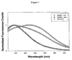
- For liposomes that undergo transition from gel to liquid crystalline phase a red shift in the emission maxima, from 440 nm to 490 nm is observed ( Figure. 1 ). Based on the composition one would expect an emission spectrum corresponding to liquid crystalline phase.
- laurdan labeled lipidic particles of the present disclosure showed a spectrum that is neither gel like nor liquid crystalline like.
- the data indicates that lamellar organization in this particle is different from that of liposomes - possibly due to the water concentration and dynamics being altered in this particle.
- Centrifugation studies carried out in discontinous dextran gradient indicated the particle floated more readily than liposomes.
- the lipidic structures of the present disclosure appear to have altered lipid organization and dynamics, internal water volume, water concentration and/or dynamics near the head group compared to typical liposomes.
- the particle may be lighter than the lamellar liposomes.
- association efficiency of the proteins in the lipidic particles as well as the reduction in the immunogenicity of proteins associated with the lipidic particles comprising PI was greater than for similar compositions in which PI was replaced with PS, PA or PG. Since PI, PS, PA and PG are all anionic phospholipids, the advantage obtained by using PI was surprising. Further because one of the proteins tested, FVIII is known to bind more avidly to PS than to PI, it was surprising that the association efficiency of FVIII for PI containing lipidic structures was higher than that for PS containing liposomes.
- the present disclosure also provides a method for preparing the lipidic structures.
- the lipidic structures can be prepared by thin lipid film hydration using the appropriate molar ratios of PC, PI and cholesterol in a suitable buffer.
- the lipids are dissolved in chloroform and the solvent is dried.
- the resulting multilamellar vesicles (MLVs) are extruded through the desired size filters (sizing device) under high pressure to obtain lipidic structures of the present disclosure.
- the size of the lipidic particles is less than 140 nm (as calculated from micrographs and dynamic light scattering measurements) so that the particles are not filtered out in the Reticulo Endothelial System (RES) so as to become available for the immune system reaction.
- RES Reticulo Endothelial System
- the protein in a suitable buffer is added to the lipidic structures.
- the free protein is then separated from the the lipidic structures by routine centrifugation methods such as density gradient centrifugations.
- the association efficiency of the protein with the lipidic particles is at least 30, 40, 50, 55, 60, 65, 70, 75, 80, 85, 90 and 95%. It desired, the lipidic particles with the associated therapeutic agent can be lyophilized for future use.
- the lipidic structures of the present disclosure prior to association with the protein can be lyophilized and stored.
- the lipidic structures can be reconstituted and then used for combination with protein to effect association of the protein with the lipidic structures prior to use.
- the present disclosure can be used for association of therapeutic agents such as proteins, polypeptides or peptides with the lipidic structures.
- the protein and peptides with wide biochemical properties can be loaded in the particles.
- the proteins may be neutral or charged (negatively or positively).
- proteins include proteins involved in the blood coagulation cascade including Factor VIII (FVIII), Factor VII (FVII), Factor IX (FIX), Factor V (FV), and von Willebrand Factor (vWF), von Heldebrant Factor, tissue plasminogen activator, insulin, growth hormone, erythropoietin alpha, VEG-F, Thrombopoietin, lysozyme and the like.
- Factor VIII Factor VIII
- FVII Factor VII
- FIX Factor IX
- FV Factor V
- vWF von Willebrand Factor
- the ratio of PC to PI is between 40:60: to 60:40 and in another instance 45:55 to 55:45. In another instance, it is 50:50.
- the cholesterol (as a percentage of PC and PI together) is between 5 and 15% as structures formed at higher cholesterol ratio than 15% lack stability.
- the association of the protein with the lipidic structures can be such that the molar ratio between the protein to lipid is between 1:200 (protein:lipid) to 1:30,000 (protein:lipid). In one instance it is about 1:10,000 (protein:lipid). In other instances, the ratio is about 1:2,000 or 1:4,000.
- the phospholipids PC and PI have two acyl chains.
- the length of the acyl chains attached to the glycerol backbone varies in length from 12 to 22 carbon atoms.
- the acyl chains may be saturated or unsaturated and may be same or different lengths.
- the acyl chains attached to PC are preferably 12 to 22. These can be saturated or unsaturated and can be same or different length.
- the acyl chains attached to PI can be from 12 to 22 and can be saturated or unsaturated.
- the chains of the PC and the PI can be same or are different in length.
- the PC and PI can be obtained from various sources both natural and synthetic. For example, soy PI and egg PC are available commercially. Additionally, synthetic PC and PI are also available commercially.
- compositions can be delivered by any standard route such as intravenous, intramuscular, intraperitonial, mucosal, subcutaneous, transdermal, intradermal, oral or the like.
- This example describes the preparation of the lipidic particles.
- Example 1 This example describes characterization of the lipid structures prepared in Example 1.
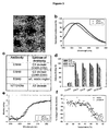
- the lipid dispersion samples for microscopic analysis were prepared by air-drying on formvar-coated grids and negatively staining them with 2% uranyl acetate for approximately 1 min. The samples were photographed using a Hitachi H500 TEM operating at 75 kV. Negatives were scanned at 300 dpi with an Agfa Duoscan T1200 scanner. The morphology of the particles determined using Transmission electron microscopic studies indicated the following. The particle size was found to be around 100 nm, consistent with dynamic light scattering studies (data not shown).
- This example describes fluorescence analysis of rFVIII and rFVIII-PI.
- the effect of PI on the tertiary structure of rFVIII was determined by exciting the samples either at 280 nm or at 265 nm and the emission was monitored in the wavelength range of 300-400 nm.
- the spectra were acquired on a PTI-Quantamaster fluorescence spectrophotometer (Photon Technology International, Lawrenceville, NJ). Protein concentration was 5 u g/ml and slit width was set at 4 nm.
- the fluorescence emission spectra of FVIII loaded in this particle showed blue shifted emission maxima compared to free protein suggesting that FVIII is located in hydrophobic environment ( Figure 2b ).
- FVIII-PI particulates it is likely that most of the molecular surface of FVIII is buried in hydrophobic acyl chain region of the lipidic particle and/or the protein is located at the lipid-water interface where water concentration at lipid interface may be less for PI particles.
- This example describes Sandwich ELISA and detection of rFVIII epitopes involved in rFVIII-PI association.
- sandwich ELISAs were performed. Briefly, Nunc-Maxisorb 96-well plates were coated overnight at 4°C with appropriate concentrations of capture monoclonal antibodies in carbonate buffer (0.2 M, pH 9.6).
- Plates were washed and then incubated with 100 u l of a 1:500 dilution of rat polyclonal antibody containing a 1:1,000 dilution of goat antirat-Ig-alkaline phosphatase conjugate in blocking buffer at room temperature for 1 h. After the last wash, 200 u l of a 1 mg/ml p-nitrophenyl phosphate solution in diethanolamine buffer (1 M diethanolamine, 0.5 mM MgCl 2 ) was added and incubated for 30 min at room temperature. 100 u l of 3 N NaOH were added to stop the reaction. A plate reader was used to measure the optical density at 405 nm.
- sandwich ELISA studies were carried out ( Figure 2c and 2d ).
- the rationale for this experiment is that domains associated with lipidic particle are shielded and hence will not be available for monoclonal antibody binding. Therefore, sandwich ELISA is an indirect, qualitative method to provide insight into protein surface accessible to bulk aqueous compartment.
- the binding of FVIII in the absence of PI was normalized to 100% to account for differences in binding affinity of various antibodies and decrease in antibody binding in the presence of PI was interpreted as domains of FVIII involved in PI binding.
- C2 and A2 domains are spatially well separated and only lipid-binding region in C2 domain may be shielded from antibody binding due to liposome association (Purohit et al. 2003; Stoilova-McPhie et al., 2002).
- Monoclonal antibodies directed against C2 and A2 domains were chosen based on this molecular model of FVIII bound to PS containing liposomes. The results indicated that the molecular surface of FVIII that is in contact with PI is different from that observed for PS.
- This example describes CD analysis of rFVIII and rFVIII-PI.
- CD spectra were acquired on a JASCO-715 spectropolarimeter calibrated with d-10 camphor sulfonic acid. The protein to lipid ratio was 1 to 2,500 where the protein concentration used was 20 u g/ml (98.6 IU/ml). Spectra were obtained over the range of 255 to 208 nm for secondary structural analysis using a 10 mm quartz cuvette. Thermal denaturation of the rFVIII and rFVIII-PI was determined by monitoring the ellipticity at 215 nm from 20 to 80 °C using a heating rate of 60 °C/h. The light scattering effect due to the presence of lipidic particles was corrected as described previously ( Balasubramanian et al., 2000, Pharm Res 17, 344-350 ).
- the association of the protein was carried out in several buffer systems as shown in Tables 2A-2C. In all the cases the protein to lipid ratio was 1:10,000. The free protein was then separated from the the lipidic structures by density gradient centrifugations and the protein associated with each fraction was measured using activity and spectroscopic assay.
- Table 2A shows the percentage of rFVIII associated with 50% DMPC:50% SPI:5% Chol (100nm) with different buffer compositions at 37°C.
- Table 2B shows the percentage of rFVIII associated with 50% DMPC:50% SPI (100nm) with different buffer compositions at 37°C.
- Table 2C Percentage of rFVIII associated with 50% DMPC:50% SPI:15% Chol (100nm) with different buffer compositions at 37°C.
- the relative immunogenicity of free rFVIII and rFVIII-PI were determined in hemophilia A mice. This mouse model is suitable for investigating immunogenicity of FVIII as the antibody response patterns against FVIII are very similar to those observed in hemophilic patients. 8 male and 10 female mice received 4 weekly intravenous injections (via penile vein) and subcutaneous injections of 10 IU of FVIII (400 IU/kg), respectively. 2 weeks after the last injection, blood samples were collected in acid citrate dextrose (ACD) buffer (85mM sodium citrate, 110mM D-glucose and 71mM citric acid) at a 10:1 (v/v) ratio by cardiac puncture.
- ACD acid citrate dextrose
- rFVIII or rFVIII-PI (10 IU/25g) was administered to male hemophilia A mice as a single intravenous bolus injection via penile vein.
- Plasma was collected immediately by centrifugation (5,000 rpm, 5 min, 4 °C) and stored at -70 °C until analysis. Chromogenic assay was used to measure the activity of rFVIII in plasma samples.
- the average values of rFVIII activities at each time point were used to compute basic pharmacokinetic parameters (half-life, MRT and area under the plasma activity curve) using a noncompartmental analysis (NCA) 20 (WinNonlin Pharsight Corporation, Mountainview, CA).
- NCA noncompartmental analysis
- the areas under the plasma activity (AUC) versus time curves from 0 to the last measurable activity time point were measured by log-linear trapezoidal method.
- the elimination rate constant (lambda z) was estimated by log-linear regression of the terminal phase concentration.
- the elimination half-life ( t 1/2 ) was calculated as In 2/lambda z and MRT was calculated from AUMC/AUC where AUMC is the area under the curve plot of the product of concentration and time versus time.
- the MRT and AUC is found to be higher for FVIII-PI compared to FVIII and also showed prolonged terminal elimination phase.
- the circulation half-life of FVIII associated with PI lipidic particle (7.6 hrs) is higher than that observed for free FVIII (2.3 hrs).
- Substantial protein activity was detected after 48 hrs of injection for animals that are given FVIII-PI particles; in contrast no detectable FVIII activity was observed at 48 hrs in animals that received FVIII alone ( Figure 4 ). Further, the protein activity was detectable for only 24 hrs following administration of the PS containing liposomes (data not shown) and is due to the rapid uptake of PS liposomes by the RES. However, in the presence of PI it is possible that the cellular uptake is reduced and is consistent with the stealth like properties of PI.
- DMPC:SPI:Cholesterol (50:50:5) formulation showed highest association efficiency than other formulations for both rFVIII and BDDrFVIII.
- Cholesterol is preferably included in the formulation to increase liposome stability in plasma. Size of liposome and association temperature, and lipid concentration all play important roles in association.
- DMPC:SPI:Cholesterol (50:50:5) formulation surprisingly has higher association for rFVIII than BDDrFVIII even though BDDrFVIII has lower Molecular weight and size. Conformational changes as a result of the B domain deletion could be responsible for a decrease in the binding affinity of BDDrFVIII towards PI containing lipidic particles.
- mice Male hemophilic mice (20-24g, 22-25 weeks old) were given 10IU/25g of rBDDFVIII associated with the lipidic structures (referred to as PI-BDDrFVIII) as a single i.v. bolus injection via the penile vein.
- the Area Under the Curve AUC (hr*IU/mL) or Free BDDrVIII was 955.1 and for BDDFVIII associated with lipidic structures was 1058.7.
- BDDrFVIII was mixed with solution containing different concentration of O-phospho -L- serine (OPLS) or phosphocholine in such a way that the final protein concentration is maintained constant at 3ug/mL.
- O-phospho -L- serine O-phospho -L- serine
- the OPLS or phosphocholine concentration vared between 0 and 100uM.
- Each mixture is incubated for 5 minutes before subjecting to further analysis.
- the intrinsic fluorescent of BDDrFVIII in the presence of increasing concentration of phospholipids head group was measured with a Quanta Master PTI instrument. The excitation was set to 285 nm and the emission was recorded at peak maximum (e.g. 330nm).
- the normalized fluorescence (F/F 0 ) data was plotted vs. [lipid] and used for for the determination of the dissociation constant for lipid head group- BDDrFVIII interaction.
- the sample fluorescence anisotropy was measured as a function of temperature using the a Quanta Master PTI spectrofluorometer equipped with motorized polarizer prisms. The data was plotted as anisotropy vs. temperature and fitted to a sigmoidal curve. The inflection point of each curve was obtained.
- the dissociation constant (Kd (uM)) for OPLS was 70.2 and that for phophocholine was 24.2.
- the inflection point for free BDDrFVIII was 71.6, for BDDrFVIII in OPLS liposomes was 79.4 and for BDDrFVIII in phosphocholine liposomes was 72.2.
- DMPC dimyristoylphosphatidylcholine
- BPS brain phosphatidylserine
- the liposomes were extruded eight times through double stacked 100nm or 200 nm polycarbonate membranes using a high pressure extruder (Lipex Biomembranes, Inc.) at a pressure of ⁇ 200 psi.
- the size distribution of the liposomes was monitored using a Nicomp model CW380 size analyzer (Particle Sizing System).
- the association of the protein with the preformed liposomes was achieved by incubating the protein in the presence of the liposomes at 37°C for 30 minutes with occasional gentle swirling.
- the protein to molar ratio was maintained the same for all preparation (1:10,000).
- PEGylation of the preformed liposomes was achieved by addition of the liposomal preparations to a dry powder of 1,2 dimyristoyl-sn-glycero-3-phosphoethanolamine-N-[methoxy (polyethylenglycol) 2000] (DMPC-PEG 2000) or 1,2 distearoyl-sn-glycero-3-phosphoethanolamine-N-[methoxy (polyethylenglycol) 2000] (DSPE-PEG 2000). The incubation was performed for 45 minutes at room temperature. Care was taken to maintain the DMPC-PEG 2000 concentration below the critical micellar concentration in order to facilitate the transfer of DMPC-PEG 2000 to the preformed lipidic bilayer.
- liposome-associated protein free protein was separated from liposome-associated protein by floatation on a discontinuous dextran density gradient. Briefly, 0.5ml of the liposome-protein mixture was mixed with 1ml of 20% (w/v) dextran (in calcium free Tris buffer) in a 5ml polypropylene centrifuge tube and 3ml of 10% (w/v) dextran and 0.5ml calcium free Tris buffer were overlaid on the liposome-containing band. The gradient was subjected to ultracentrifugation at 45,000rpm for 30min in a Beckman SW50.1 rotor.
- BDDrFVIII retains all critical structural characteristics of the parent molecule, including the binding properties towards phosphatidylserine (PS) containing lipidic membranes as well as its activity.
- PS phosphatidylserine
- the association efficiency of BDDrFVIII was higher than that observed for full length rFVIII.
- BDDrFVIII- PS containing liposomes were also tested. Eight to 12 week old hemophilic mice received 4 weekly injections containing 10 IU of BDDrFVIII - PS containing liposomes (prepared as described in example 3). Two weeks following the last injection, blood samples were collected and analyzed for the presence of inhibitory antibodies using a modified Bethesda assay.
- This example describes the association efficiency for BDDrFVIII associated with PS and phosphatidylethanolamine (PE) containing liposomes which were used for comparison purposes.
- the liposomes were extruded eight times through double stacked 100nm polycarbonate membranes using a high pressure extruder (Lipex Biomembranes, Inc.) at a pressure of ⁇ 200 psi.
- the size distribution of the particles was monitored using a Nicomp model CW380 size analyzer (Particle Sizing System).
- the association of the protein with the preformed liposomes was achieved by incubating the protein in the presence of the liposomes at 37°C for 30 minutes with occasional gentle swirling.
- the protein to molar ratio was maintained the same for all preparation (1:10,000).
- PEGylation of the preformed liposomes was achieved by addition of the liposomal preparations to a dry powder of 1,2 dimyristoyl-sn-glycero-3-phosphoethanolamine-N-[methoxy (polyethylenglycol) 2000] (DMPC-PEG 2000) or 1,2 distearoyl-sn-glycero-3-phosphoethanolamine-N-[methoxy (polyethylenglycol) 2000] (DSPE-PEG 2000). The incubation was performed for 45 minutes at room temperature. Care was taken to maintain the DMPC-PEG 2000 concentration below the critical micellar concentration in order to facilitate the transfer of DMPC-PEG 2000 to the preformed lipidic bilayer.
- liposome-associated protein free protein was separated from liposome-associated protein by floatation on a discontinuous dextran density gradient. Briefly, 0.5ml of the liposome-protein mixture was mixed with 1ml of 20% (w/v) dextran (in calcium free Tris buffer) in a 5ml polypropylene centrifuge tube and 3ml of 10% (w/v) dextran and 0.5ml calcium free Tris buffer were overlaid on the liposome-containing band. The gradient was subjected to ultracentrifugation at 45,000rpm for 30min in a Beckman SW50.1 rotor.
- PS containing liposomes are rapidly cleared from circulation by the reticuloendothelium system (RES).
- Phopsphatidylethanolamine (PE) increases the affinity of FVIII towards PS containing lipids.
- PE is added to the composition at the expense of PS.
- the association efficiency is found to increase with increasing concentration of PE (compare Exp # I and II).
- the association efficiency is found to be higher for DOPE containing particles than in the absence of DOPE (Exp # II and III). Decreasing the content of PS in the formulation while achieving a higher association efficiency is more beneficial from the perspective of pharmacological properties of BDDrFVIII.
- lipidic structures comprising FVII were prepared.
- the required amounts of DMPC, SPI and Chol Dimyristoylphosphatidylcholine (DMPC): soy phosphatidylinositol (SPI): Cholesterol (Chol) (molar ratio 50:50:5) were dissolved in chloroform.
- DMPC dimyristoylphosphatidylcholine
- SPI soy phosphatidylinositol
- Cholesterol (Chol) molar ratio 50:50:5
- the LP were extruded twenty times through double stacked 80 nm polycarbonate membranes using a high pressure extruder (Lipex Biomembranes, Inc.) at a pressure of ⁇ 200 psi.
- the size distribution of the particles was monitored using a Nicomp model CW380 size analyzer (Particle Sizing System).
- the association of the protein with the LP was achieved by incubating the protein in the presence of the LP at 37°C for 30 minutes.
- the protein to lipid molar ratio was maintained for the first two trials of preparation (1:10,000). Additional, one trial using protein:lipid ratios of 1:2000 were also investigated.
- DMPC dimyristoylphosphatidylcholine
- SPI soy phosphatidylinositol
- Cholesterol Cholesterol (Chol) (molar ratio 50:50:5)
- DMPC dimyristoylphosphatidylcholine
- SPI soy phosphatidylinositol
- Cholesterol Cholesterol (Chol) (molar ratio 50:50:5)
- the LP were extruded twenty times through double stacked 80 nm polycarbonate membranes using a high pressure extruder (Lipex Biomembranes, Inc.) at a pressure of ⁇ 200 psi.
- the size distribution of the particles was monitored using a Nicomp model CW380 size analyzer (Particle Sizing System).
- the association of the protein with the LP was achieved by incubating lysozyme in the presence of the LP at 37°C for 30 minutes.
- the protein to lipid molar ratio maintained at 1:2000 was investigated.
- LP-associated protein free protein was separated from LP-associated protein by floatation on a discontinuous dextran density gradient. Briefly, 0.5ml of the LP-protein mixture was mixed with 1ml of 20% (w/v) dextran (in calcium free Tris buffer) in a 5ml polypropylene centrifuge tube and 3ml of 10% (w/v) dextran and 0.5ml calcium free Tris buffer were overlaid on the LP-containing band. The gradient was subjected to ultracentrifugation at 45,000rpm for 30min in a Beckman SW50.1 rotor. The LP and their associated protein floated to the interface of the buffer/10% dextran bands, and the unassociated protein remained at the bottom.
- the concentration of the protein associated with LP was determined using spectroscopic assay.
- the particle thus prepared packaged the protein inside the particle and shields from the surrounding milieu. This is supported by acrylamide quenching data as shown in Figure 6 . This is likely to provide in vivo stability as it may shield the protein from protease degradation.
- DMPC erythropoietin
- SPI soy phosphatidylinositol
- Cholesterol Cholesterol (Chol) (molar ratio 50:50:5)
- the LP were extruded twenty times through double stacked 80 nm polycarbonate membranes using a high pressure extruder (Lipex Biomembranes, Inc.) at a pressure of ⁇ 200 psi.
- the size distribution of the particles was monitored using a Nicomp model CW380 size analyzer (Particle Sizing System).
- the association of the protein erythropoietin with the LP was achieved by incubating EPO in the presence of the LP at 37°C for 30 minutes.
- the protein to lipid molar ratio was maintained as 1:10,000 (3 trials) and 1:2000 (1 trial).
- LP-associated protein free protein was separated from LP-associated protein by floatation on a discontinuous dextran density gradient. Briefly, 0.5ml of the LP-protein mixture was mixed with 1ml of 20% (w/v) dextran (in calcium free Tris buffer) in a 5ml polypropylene centrifuge tube and 3ml of 10% (w/v) dextran and 0.5ml calcium free Tris buffer were overlaid on the LP-containing band. The gradient was subjected to ultracentrifugation at 45,000rpm for 30min in a Beckman SW50.1 rotor. The LP and their associated protein floated to the interface of the buffer/10% dextran bands, and the unassociated protein remained at the bottom. The concentration of the protein associated with LP was determined using spectroscopic assay.
- association efficiency as determined by fluorescence spectroscopy was 74.90%, 68.60% and 68.50% for a protein:lipid ratio of 1:10,000 and 51% for a protein;lipid ratio of 1:2,000.
Description
- In the treatment of diseased conditions, therapeutic interventions are often undertaken which involve administration of foreign molecules having therapeutically beneficial effects. However, such administrations can often result in unwanted side effects resulting from activation of the body's immune response. Formation of antibodies following administration of therapeutics poses a serious clinical challenge. The antibodies can abrogate activity and/or alter pharmaco-kinetics of the therapeutic molecules.
- This is particularly relevant when administering strong antigenic molecules such as peptides, polypeptides or proteins. Many such polypeptides are routinely used as therapeutic molecules. For example, Factor VIII (FVIII) is an essential cofactor in the intrinsic coagulation pathway. Any deficiency or dysfunction of FVIII results in a bleeding disorder, characterized as hemophilia A. Replacement therapy with recombinant FVIII (rFVIII) or plasma-derived FVIII (pdFVIII) is the common therapy for controlling bleeding episodes. FVIII is a multidomain glycoprotein comprising of six domains (A1-A2-B-A3-C1-C2). Prior to secretion into plasma, FVIII is subjected to proteolytic cleavage, leading to the generation of a heterodimer with molecular weights ranging from ∼ 170 to ∼300 KDa. The presence of the multiple proteolytic sites at the B domain level is responsible for the high heterogeneity of the FVIII preparations. In spite of being FVIII's largest domain (908 amino acids residues or ∼ 40% of the total number of amino acids residues), the B domain lacks any essential function for the cofactor coagulation activity. Deletion of the B domain leads to a less heterogenic, genetically engineered rFVIII that corresponds to the shortest form of pdFVIII (e.g. ∼170KDa). B domain deleted rFVIII (BDDrFVIII) is characterized by a higher specific activity than rFVIII and can also be used for treatment of hemophilia.
- Another therapeutic molecule is Factor VIIa (FVIIa). This is a trypsin-like serine protease which plays an important role in activating the extrinsic coagulation cascade. FVIIa is a poorly catalytic form of factor VII after the activating cleavage between Arg152 and Ile153. Upon injury, circulating FVIIa becomes an efficient catalyst when forming a complex with tissue factor (TF), its allosteric regulator that is found on the outside of blood vessel. FVIIa-TF complex induces generation of small amounts of thrombin which further triggers blood clotting. Factor VIIa has been approved by the Food and Drug Administration in the United States for uncontrollable bleeding in hemophilia A and B patients who have developed inhibitory antibodies against replacement coagulation factors, factor VIII and factor IX. Intravenous administration of recombinant human Factor FVIIa (rHu-FVIIa) has been introduced because of fewer side effects than other alternative treatment strategies and to circumvent difficulty in preparing plasma-derived FVIIa. However, the short circulation half-life of FVIIa requiring repeated bolus injections to achieve desired efficacy can be problematic.
- Additionally, many other proteins are used as therapeutics. These include erythropoietin, VEG-F, other blood coagulation proteins, hormones (such as insulin and growth hormone) and the like. Strategies that can inhibit processing by immune system and also prolong circulation time (reduce frequency of administration) would improve efficacy of proteins. Therefore there is a need in the area of therapeutics to develop formulations that make the proteins less immunogenic, without significantly affecting the circulating time or the efficacy.
-
EP0728476A1 describes liposome composition effective in administering a physiologically active substance to Peyer's glands. Ebisu et al. (J. Biol. Chem. (2001) 276(27):24498-24505) describes that fused p47phox and p67phox truncations efficiently reconstitute NADPH oxidase with higher activity and stability than the individual components. Stromstedt et al. (J. Colloid and Interface Sci. (2007) 311(1):59-69) describes the effect of lipid headgroup composition on the interaction between melittin and lipid bilayers.EP0680763A2 describes a stable preparation for the treatment of blood coagulation disorders comprising an activated coagulation factor and lipid vesicles.WO2007/002886 describes compositions and methods for less immunogenic protein-lipid complexes.US2005/227913 andWO2004/071420 describe compositions and methods for less immunogenic protein formulations.US4920016 describes liposomes with enhanced circulation time. - The invention is defined by the claims. Those aspects/instances of the present disclosure which constitute the invention are defined by the claims.
- The present invention provides compositions comprising therapeutic agents such that the immunogenicity of the agents is reduced and their circulating time is increased. The compositions comprise lipidic particles (also referred to herein as lipidic structures) comprising phosphatidylcholine, phosphatidylinositol and cholesterol. Therapeutic agents such as peptides, polypeptides and/or proteins can be associated with the lipidic particles to form delivery compositions.
- In these compositions, the therapeutic agent displays reduced immunogenicity and longer circulating time.
- In various instances, lipidic particles having associated therewith proteins such as Factor VIII, B domain deleted Factor VIII, Factor VII, lysozyme and Erythropoietin are disclosed.
- In the description, the therapeutic agent associated with the lipidic particles comprising PI is sometimes referred to as therapeutic agent-PI.
-
-
Figure 1 is a representation of biophysical and biochemical characterization of Laurdan study of PC containing liposomes alone as a liquid or gel or PI containing lipidic particles with associated rFVIII. -
Figure 2 . Biophysical and Biochemical characterization of rFVIII-PI (a): Transmission Electron Micrograph (TEM) of rFVIII-PI; (b); normalized fluorescence emission spectra of free rFVIII and rFVIII-PI (1:10,000); (c): a list of capture monoclonal antibodies utilized in this study that target specific epitopes in rFVIII molecules; (d): the binding of monoclonal antibodies to rFVIII-PI at various lipid concentrations. Control is protein free liposomes. (e): far-UV CD spectra of rFVIII in the presence (1:2,500) and in the absence of PI acquired at 20 °C; and (f): percent change in ellipticity of rFVIII as a function of temperature in the presence and in the absence of PI. -
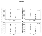
Figure 3 . Effect of phophatidylinositol on the Immunogenicity of rFVIII. (a, c) show the mean of total antibody titers (horizontal bars) and individual (open circles) antibody titers were determined following s.c. and i.v. administrations, respectively. (b, d) show the mean of inhibitory titers (horizontal bars) and individual (open circles) inhibitory titers were determined following s.c. and i.v. administrations, respectively. -
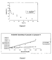
Figure 4 . Influence of phosphatidylinositol on pharmacokinetics of rFVIII. The mean plasma concentration of rFVIII clotting activity after i.v. administration of free rFVIII and rFVIII-PI. -
Figure 5 . Influence of phosphatidylinositol on pharmacokinetics of BDDrFVIII. The mean plasma concentration of BDDrFVIII clotting activity after i.v. administration of free BDDrFVIII and BDDrFVIII-PI. -
Figure 6 . Acryamide quenching for free lysozyme and lysozyme associated with PI containing lipidic particles. - The present disclosure provides methods and compositions for less immunogenic and long circulating lipidic formulations for delivering a therapeutic agent. The formulations comprise a therapeutic agent associated with lipidic structures comprising phosphatidyl choline (PC), and phosphatidyl inositol (PI) and cholesterol. The therapeutic agent may be a peptide (generally 50 amino acids or less) a polypeptide(generally 100 amino acids or less) or proteins (larger than 100 amino acids).
- Although not intending to be bound by any particular theory, it is considered that the lower imunigenicity and/or the longer circulation time is at least in part due to the lipidic particles having a unique structure. As seen under high magnification, the lipidic particles of the present disclosure do not appear to have donut like structures typical of liposomal lamellarity. Substantial number of the lipidic particles displayed disc like structures (see Example 2) which is attributable to reduced water volume thereby providing reduced contrast in the electron micrographs. Therefore the morphology appears different from that of liposomes, possibly due to altered lipid structure and organization, and reduced internal water volume. To further investigate the lipid structure and organization we carried out fluorescence studies using Laurdan as probe. The probe partitions into the interfacial region and the emission is sensitive to the presence and dynamics of water molecules and lamellar structures of liposomes. The fluorescence emission spectra were acquired for Laurdan labeled lipid particles of the present disclosure and also for liposomes, the latter serving as control. For liposomes that undergo transition from gel to liquid crystalline phase a red shift in the emission maxima, from 440 nm to 490 nm is observed (
Figure. 1 ). Based on the composition one would expect an emission spectrum corresponding to liquid crystalline phase. However, laurdan labeled lipidic particles of the present disclosure showed a spectrum that is neither gel like nor liquid crystalline like. Thus, the data indicates that lamellar organization in this particle is different from that of liposomes - possibly due to the water concentration and dynamics being altered in this particle. Centrifugation studies carried out in discontinous dextran gradient indicated the particle floated more readily than liposomes. Thus, the lipidic structures of the present disclosure appear to have altered lipid organization and dynamics, internal water volume, water concentration and/or dynamics near the head group compared to typical liposomes. In addition, the particle may be lighter than the lamellar liposomes. - The association efficiency of the proteins in the lipidic particles as well as the reduction in the immunogenicity of proteins associated with the lipidic particles comprising PI was greater than for similar compositions in which PI was replaced with PS, PA or PG. Since PI, PS, PA and PG are all anionic phospholipids, the advantage obtained by using PI was surprising. Further because one of the proteins tested, FVIII is known to bind more avidly to PS than to PI, it was surprising that the association efficiency of FVIII for PI containing lipidic structures was higher than that for PS containing liposomes.
- The present disclosure also provides a method for preparing the lipidic structures. The lipidic structures can be prepared by thin lipid film hydration using the appropriate molar ratios of PC, PI and cholesterol in a suitable buffer. The lipids are dissolved in chloroform and the solvent is dried. The resulting multilamellar vesicles (MLVs) are extruded through the desired size filters (sizing device) under high pressure to obtain lipidic structures of the present disclosure. The size of the lipidic particles is less than 140 nm (as calculated from micrographs and dynamic light scattering measurements) so that the particles are not filtered out in the Reticulo Endothelial System (RES) so as to become available for the immune system reaction. Thus at least 50% of the particles have a size of less than 140 nm More preferably, the particles should be less than 120 nm and still more preferably between 40 and 100 nm. In various instances, 50, 60, 70, 80,and 90% of the particles are less than 140nm and more preferably between 40 and 100 nm.
- To effect association of the protein with the lipidic structures, the protein in a suitable buffer is added to the lipidic structures. The free protein is then separated from the the lipidic structures by routine centrifugation methods such as density gradient centrifugations. In various instances, the association efficiency of the protein with the lipidic particles is at least 30, 40, 50, 55, 60, 65, 70, 75, 80, 85, 90 and 95%. It desired, the lipidic particles with the associated therapeutic agent can be lyophilized for future use.
- In one instance, the lipidic structures of the present disclosure prior to association with the protein can be lyophilized and stored. When needed, the lipidic structures can be reconstituted and then used for combination with protein to effect association of the protein with the lipidic structures prior to use.
- The present disclosure can be used for association of therapeutic agents such as proteins, polypeptides or peptides with the lipidic structures. The protein and peptides with wide biochemical properties can be loaded in the particles. The proteins may be neutral or charged (negatively or positively). Such proteins include proteins involved in the blood coagulation cascade including Factor VIII (FVIII), Factor VII (FVII), Factor IX (FIX), Factor V (FV), and von Willebrand Factor (vWF), von Heldebrant Factor, tissue plasminogen activator, insulin, growth hormone, erythropoietin alpha, VEG-F, Thrombopoietin, lysozyme and the like.
- The ratio of PC to PI is between 40:60: to 60:40 and in another instance 45:55 to 55:45. In another instance, it is 50:50. The cholesterol (as a percentage of PC and PI together) is between 5 and 15% as structures formed at higher cholesterol ratio than 15% lack stability.
- The association of the protein with the lipidic structures can be such that the molar ratio between the protein to lipid is between 1:200 (protein:lipid) to 1:30,000 (protein:lipid). In one instance it is about 1:10,000 (protein:lipid). In other instances, the ratio is about 1:2,000 or 1:4,000.
- The phospholipids PC and PI have two acyl chains. The length of the acyl chains attached to the glycerol backbone varies in length from 12 to 22 carbon atoms. The acyl chains may be saturated or unsaturated and may be same or different lengths. Some nonlimiting examples of 12-22 carbon atom saturated and unsaturated acyl chains are shown in Tables 1A and 1B:
Table 1A Symbol Common Name Systematic name Structure 12:0 Lauric acid dodecanoic acid CH3(CH2)10COOH 14:0 Myristic acid tetradecanoic acid CH3(CH2)12COOH 16:0 Palmitic acid hexadecanoic acid CH3(CH2)14COOH 18:0 Stearic acid octadecanoic acid CH3(CH2)16COOH 20:0 Arachidic acid eicosanoic acid CH3(CH2)18COOH 22:0 Behenic acid docosanoic acid CH3(CH2)20COOH Table 1B Symbol Common Name Systematic name Structure 18:1 Oleic acid 9-Octadecenoic acid CH3(CH2)7CH=CH(CH2)7COOH 16:1 Palmitoleic acid 9-Hexadecenoic acid CH3(CH2)5CH=CH(CH2)7COOH 18:2 Linoleic acid 9,12-Octadecadienoic acid CH3(CH2)4(CH=CHCH2)2(CH2)6COOH 20:4 Arachidonic acid 5,8,11,14-Eicosatetraenoic acid CH3(CH2)4(CH=CHCH2)4(CH2)2COOH - The acyl chains attached to PC are preferably 12 to 22. These can be saturated or unsaturated and can be same or different length. The acyl chains attached to PI can be from 12 to 22 and can be saturated or unsaturated. The chains of the PC and the PI can be same or are different in length.
- The PC and PI can be obtained from various sources both natural and synthetic. For example, soy PI and egg PC are available commercially. Additionally, synthetic PC and PI are also available commercially.
- The compositions can be delivered by any standard route such as intravenous, intramuscular, intraperitonial, mucosal, subcutaneous, transdermal, intradermal, oral or the like.
- The invention is described by the following examples, which are intended to be illustrative and not restrictive in any way. Particles falling outside the scope of the claims are included in the following examples for comparative purposes only and do not constitute the invention.
- This example describes the preparation of the lipidic particles.
- Materials: Albumin free full-length rFVIII (Baxter Health Care Glendale, CA) was used as antigen. Advate was provided as a gift from Western New York Hemophilia foundation. Dimyristoyl phosphatidylcholine (DMPC) and soybean phosphatidylinositol (Soy PI) were purchased from Avanti Polar Lipids (Alabaster, AL). Cholesterol, IgG-free bovine serum albumin (BSA), and diethanolamine were purchased from Sigma (St. Louis, MO). Goat antimouse-Ig and antirat-Ig, alkaline phosphatase conjugates were obtained from Southern Biotechnology Associates, Inc. (Birmingham, AL). p-Nitrophenyl phosphate disodium salt was purchased from Pierce (Rockford, IL). Monoclonal antibodies ESH4, ESH5, and ESH8 were purchased from American Diagnostica Inc. (Greenwich, CT). Monoclonal antibody N77210M was purchased from Biodesign International (Saco, ME). Normal coagulation control plasma and FVIII-deficient plasma were purchased from Trinity Biotech (County Wicklow, Ireland). The Coamatic Factor VIII kit from DiaPharma Group (West Chester, OH) was used to determine the rFVIII activity in plasma samples.
- Preparation of rFVIII-PI lipidic particles: The particles were prepared by thin lipid film hydration of appropriate molar ratios of DMPC, soy PI, and cholesterol (50:50:5) with Tris buffer (TB) (25mM Tris, and 300mM NaCl, pH=7.0). The required amount of lipids was dissolved in chloroform in a kimax tube and the solvent was dried using Buchi-R200 rotaevaporator (Fisher Scientific, NJ). Multilamellar vesicles (MLV) were formed by vortex, mixing the lipid dispersions at 37 °C for 20 min. The resulting MLV were extruded through double polycarbonate membranes of 80nm pore size (GE Osmonics Labstore, Minnetonka, MN) in a high-pressure extruder (Mico, Inc., Middleton, WI) at a pressure of ∼250 psi and then sterile-filtered through a 0.22 um MillexTM-GP filter unit (Millipore Corporation, Bedford, MA). Phosphate assay was used to estimate phospholipids concentrations. Particle size was monitored using a
Nicomp Model CW 380 particle size analyzer (Particle Sizing Systems, Santa Barbara, CA). The protein was added to the lipid particles at 37°C and during this process Ca2+ ion concentration was decreased from 5mM CaCl2 to 0.2mM CaCl2 in TB to ensure optimal lipid-Ca2+ interaction and possible lipid phase change. The protein to lipid ratio was maintained at 1:10,000 for all experiments, unless stated otherwise. - Separation of Free rFVIII from rFVIII-PI: Discontinuous dextran density gradient centrifugation technique was used to separate the free protein from the lipidic particles. Briefly, 0.5 ml of incubated rFVIII-PI mixture was mixed with 1.0 ml of 20% (w/v) dextran (in Ca+2 free TB) in a 5 ml polypropylene centrifuge tube. 3 ml of 10% (w/v) dextran was then carefully added on top of the mixture followed by 0.5 ml of Ca+2 free TB. After centrifugation at 45,000 rpm at 4 °C for 30min in a Beckman SW50.1 rotor, the bound protein and free lipidic particles would float to the top of the dextran band unbound rFVIII would stay at the bottom of the gradient. One-stage activated partial thromboplastin time (APTT) assay was used to estimate the protein association efficiency of rFVIII-PI. This procedure yielded an association efficiency of 72 ± 9% and is much higher than that observed with Phosphatidyl Serine (PS) containing liposomes (45 ± 16.8%) (Purohit, et al. Biochim Biophys Acta 1617, 31-38 (2003). Kemball-Cook, et al., Thromb Res 67, 57-71 (1992)).
- This example describes characterization of the lipid structures prepared in Example 1. The lipid dispersion samples for microscopic analysis were prepared by air-drying on formvar-coated grids and negatively staining them with 2% uranyl acetate for approximately 1 min. The samples were photographed using a Hitachi H500 TEM operating at 75 kV. Negatives were scanned at 300 dpi with an Agfa Duoscan T1200 scanner. The morphology of the particles determined using Transmission electron microscopic studies indicated the following. The particle size was found to be around 100 nm, consistent with dynamic light scattering studies (data not shown). The analysis of the micrograph showed that the donut like structures typical of liposomal lamellarity was not observed instead particles displayed disc like structures (
Figure 2a ) and it is possible that unique lipid organization distinct from liposomes are formed that can accommodate higher mol% of FVIII. - This example describes fluorescence analysis of rFVIII and rFVIII-PI. The effect of PI on the tertiary structure of rFVIII was determined by exciting the samples either at 280 nm or at 265 nm and the emission was monitored in the wavelength range of 300-400 nm. The spectra were acquired on a PTI-Quantamaster fluorescence spectrophotometer (Photon Technology International, Lawrenceville, NJ). Protein concentration was 5 ug/ml and slit width was set at 4 nm. The fluorescence emission spectra of FVIII loaded in this particle showed blue shifted emission maxima compared to free protein suggesting that FVIII is located in hydrophobic environment (
Figure 2b ). This observation is in contrast to fluorescence spectrum observed for FVIII associated with PS containing liposomes where no change in fluorescence properties was observed (Purohit et al., 2003) and is consistent with molecular model proposed based on crystallographic and biophysical studies. FVIII was associated with PS containing liposomes only via the C-terminal region (2303-2332) of C2 domain and the rest of the molecule is accessible to bulk water (Purohit et al., 2003). However, in FVIII-PI particulates, it is likely that most of the molecular surface of FVIII is buried in hydrophobic acyl chain region of the lipidic particle and/or the protein is located at the lipid-water interface where water concentration at lipid interface may be less for PI particles. - This example describes Sandwich ELISA and detection of rFVIII epitopes involved in rFVIII-PI association. In order to determine the rFVIII epitopes that were associated with PI, sandwich ELISAs were performed. Briefly, Nunc-Maxisorb 96-well plates were coated overnight at 4°C with appropriate concentrations of capture monoclonal antibodies in carbonate buffer (0.2 M, pH 9.6). Plates were then washed with Tween-PBS (2.7 mM KCl, 140 mM NaCl, 1.8 mM KH2PO4, 10 mM Na2HPO4.2H2O, 0.05% w/
v Tween 20, pH 7.4) and then blocked with 1% BSA (prepared in PBS) for 2 h at room temperature. 100 ul of 0.5 ug/ml of various dilutions of rFVIII-PI (1:0, 1:5,000, 10,000, and 50,000) or PI-containing liposomes in blocking buffer were incubated at 37 °C for 1 h. Plates were washed and then incubated with 100 ul of a 1:500 dilution of rat polyclonal antibody containing a 1:1,000 dilution of goat antirat-Ig-alkaline phosphatase conjugate in blocking buffer at room temperature for 1 h. After the last wash, 200 ul of a 1 mg/ml p-nitrophenyl phosphate solution in diethanolamine buffer (1 M diethanolamine, 0.5 mM MgCl2) was added and incubated for 30 min at room temperature. 100 ul of 3 N NaOH were added to stop the reaction. A plate reader was used to measure the optical density at 405 nm. - In order to investigate the molecular surface area associated with PI, sandwich ELISA studies were carried out (
Figure 2c and 2d ). The rationale for this experiment is that domains associated with lipidic particle are shielded and hence will not be available for monoclonal antibody binding. Therefore, sandwich ELISA is an indirect, qualitative method to provide insight into protein surface accessible to bulk aqueous compartment. The binding of FVIII in the absence of PI was normalized to 100% to account for differences in binding affinity of various antibodies and decrease in antibody binding in the presence of PI was interpreted as domains of FVIII involved in PI binding. PhosphatidylCholine (PC) vesicles were used as negative control as the association efficiency of FVIII in PC vesicles is around 10±4% (Purohit et al., 2003. Biochim Biophys Acta, 1617:31-38). PS liposomes were used as positive control for binding of C2 domain antibodies based on crystallographic and biophysical/biochemical studies. It has been shown that C-terminal region of the C2 domain involving 2303-2332 is involved in lipid binding and the A2 domain is further apart from the liposome surface (Stoilova-McPhie et al., 2002, Blood, 99:1215-1223). Based on this molecular topology, C2 and A2 domains are spatially well separated and only lipid-binding region in C2 domain may be shielded from antibody binding due to liposome association (Purohit et al. 2003; Stoilova-McPhie et al., 2002). Monoclonal antibodies directed against C2 and A2 domains were chosen based on this molecular model of FVIII bound to PS containing liposomes. The results indicated that the molecular surface of FVIII that is in contact with PI is different from that observed for PS. In PC liposomes no lipid concentration dependent change in antibody binding was observed indicating that no specific binding between FVIII and PC, whereas for PS vesicles, lipid concentration dependent changes observed only for antibody directed against the lipid binding domain (ESH 4), consistent with the model proposed based on crystallographic studies. However, for FVIII-PI, all the monoclonal antibodies used in this study showed reduced binding and was dependent on PI concentration (Figure 2d ). The results indicated that both C2 and A2 domains are somewhat inaccessible for antibody binding possibly due to steric hindrance and/or substantial surface area of the FVIII molecule is buried in the PI particle. - This example describes CD analysis of rFVIII and rFVIII-PI. CD spectra were acquired on a JASCO-715 spectropolarimeter calibrated with d-10 camphor sulfonic acid. The protein to lipid ratio was 1 to 2,500 where the protein concentration used was 20 ug/ml (98.6 IU/ml). Spectra were obtained over the range of 255 to 208 nm for secondary structural analysis using a 10 mm quartz cuvette. Thermal denaturation of the rFVIII and rFVIII-PI was determined by monitoring the ellipticity at 215 nm from 20 to 80 °C using a heating rate of 60 °C/h. The light scattering effect due to the presence of lipidic particles was corrected as described previously (Balasubramanian et al., 2000, Pharm Res 17, 344-350).
- The CD studies showed that such molecular topology did not alter the secondary structure of the protein, as the CD spectrum of the protein was not changed by PI association (
Figure 2e ). Thermal unfolding is often used to investigate the intrinsic stability. FVIII associated with PI nano particle displayed a shallower melting with Tm slightly higher than that observed for FVIII indicating that the association of FVIII with PI improved the intrinsic stability of FVIII (Figure 2f ). - The association of the protein was carried out in several buffer systems as shown in Tables 2A-2C. In all the cases the protein to lipid ratio was 1:10,000. The free protein was then separated from the the lipidic structures by density gradient centrifugations and the protein associated with each fraction was measured using activity and spectroscopic assay.
- Table 2A shows the percentage of rFVIII associated with 50% DMPC:50% SPI:5% Chol (100nm) with different buffer compositions at 37°C. Table 2B shows the percentage of rFVIII associated with 50% DMPC:50% SPI (100nm) with different buffer compositions at 37°C. Table 2C: Percentage of rFVIII associated with 50% DMPC:50% SPI:15% Chol (100nm) with different buffer compositions at 37°C.
Table 2A Buffer Composition Percent Association 300mM NaCl, 25mM Tris, pH 7.4 70∼80 PBS, pH 7.0 95 50mM Tris, 150mM NaCl, 3mM NaN3, pH 7.4 96 25mM Tris, pH 7.4 42 10mM Hepes, pH 7.4 23 Table 2B Buffer Composition Percent Association 300mM NaCl, 25mM Tris, pH 7.4 67 PBS, pH 7.0 88 50mM Tris, 150mM NaCl, 3mM NaN3, pH 7.4 58 10mM Hepes, pH 7.4 33 Table 2C Buffer Composition Percent Association 300mM NaCl, 25mM Tris, pH 7.4 66 PBS, pH 7.0 93 50mM Tris, 150mM NaCl, 3mM NaN3, pH 7.4 51 10mM Hepes, pH 7.4 32 - This example describes immunogenicity studies: Breeding pairs of hemophilia A mice (C57BL/6J) with a target deletion in exon 16 of the FVIII gene were used. A colony of hemophilia A mice was established and animals aged from 8-12 weeks were used for the in vivo studies. Since the sex of the mice has no impact on the immune response, both male and female mice were used for the studies.
- The relative immunogenicity of free rFVIII and rFVIII-PI were determined in hemophilia A mice. This mouse model is suitable for investigating immunogenicity of FVIII as the antibody response patterns against FVIII are very similar to those observed in hemophilic patients. 8 male and 10 female mice received 4 weekly intravenous injections (via penile vein) and subcutaneous injections of 10 IU of FVIII (400 IU/kg), respectively. 2 weeks after the last injection, blood samples were collected in acid citrate dextrose (ACD) buffer (85mM sodium citrate, 110mM D-glucose and 71mM citric acid) at a 10:1 (v/v) ratio by cardiac puncture. Plasma was separated by centrifugation at 5,000 rpm at 4°C for 5min. Samples were stored at -80°C immediately after centrifugation. The total titers were determined by ELISA studies and the inhibitory titer was determined using a modified Bethesda assay as described previously (verbruggen et al., Thromb Haemost, 1995, 73:2470251).
- The results showed that PI reduced antibody response in Hemophilia mice (
Figure 3 ). Animals treated with rFVIII-PI displayed significantly lower total antibody titers (Figure 3a and 3c ) compared to animals treated with rFVIII alone. Titers were 2379±556 (±S.E.M; n=10) for FVIII-PI given by sc route, compared to 13,167 ± 2042 (n=15) for animals treated with rFVIII alone. These differences were significant at P<0.05. Animals treated with rFVIII-PI by i.v. also showed lower mean antibody titers; for FVIII-PI antibody titers were found to be of 3321±874 (n=8) and those treated with rFVIII had titers of 4569±1021(n=8) and this difference was not significant. However, the inhibitory titers that abrogate the activity of the protein reduced significantly for FVIII-PI given by both sc and iv route (Figure 3b and 3d ). For sc administration, the inhibitory titers reduced by more than 70%. For animals given FVIII alone by iv route, the inhibitory titers were 675±71 and it reduced to 385±84 for FVIII-PI and this reduction is statistically significant at p<0.05. Together these results indicate that PI containing lipidic particles not only reduced overall anti-rFVIII antibody titers, but also lowered the titer of antibodies that abrogate the activity of the protein. - This example describes Pharmacokinetics Studies. rFVIII or rFVIII-PI (10 IU/25g) was administered to male hemophilia A mice as a single intravenous bolus injection via penile vein. Blood samples were collected in syringes containing ACD buffer (10:1 v/v) at 0.08, 0.5, 1, 2, 4, 8, 16, 24, 36, and 48 h after the injections by cardiac puncture (n=3 mice/time point). Plasma was collected immediately by centrifugation (5,000 rpm, 5 min, 4 °C) and stored at -70 °C until analysis. Chromogenic assay was used to measure the activity of rFVIII in plasma samples. The average values of rFVIII activities at each time point were used to compute basic pharmacokinetic parameters (half-life, MRT and area under the plasma activity curve) using a noncompartmental analysis (NCA)20 (WinNonlin Pharsight Corporation, Mountainview, CA). The areas under the plasma activity (AUC) versus time curves from 0 to the last measurable activity time point were measured by log-linear trapezoidal method. The elimination rate constant (lambda z) was estimated by log-linear regression of the terminal phase concentration. The elimination half-life (t 1/2) was calculated as In 2/lambda z and MRT was calculated from AUMC/AUC where AUMC is the area under the curve plot of the product of concentration and time versus time.
- Data was analyzed by ANOVA using Minitab (Minitab Inc., State College, PA). Statistical difference (p<0.05) was detected by the Student independent t-test, and one-way ANOVA followed by Dunnette's post-hoc multiple comparison test. For PK studies, repeated-measures ANOVA was used to compare the profiles generated by the two treatments. Bailer-Satterthwaite method was used to compare differences in systemic exposure between the two treatments.
- The MRT and AUC is found to be higher for FVIII-PI compared to FVIII and also showed prolonged terminal elimination phase. The circulation half-life of FVIII associated with PI lipidic particle (7.6 hrs) is higher than that observed for free FVIII (2.3 hrs). Substantial protein activity was detected after 48 hrs of injection for animals that are given FVIII-PI particles; in contrast no detectable FVIII activity was observed at 48 hrs in animals that received FVIII alone (
Figure 4 ). Further, the protein activity was detectable for only 24 hrs following administration of the PS containing liposomes (data not shown) and is due to the rapid uptake of PS liposomes by the RES. However, in the presence of PI it is possible that the cellular uptake is reduced and is consistent with the stealth like properties of PI. - This examples describes the Association efficiency for rFVIII and and a truncated version of FVIII. BDDrFVIII. The lipidic structures having FVIII or BDDrFVIII were prepared by a process as described above. The percent association for these proteins is shown below in Table 3.
Table 3 Percentage of rFVIII associated with lipidic particles of various compositions. Protein (protein:lipid) Composition (molar ratio) Lipidic particle Size (nm) Temperature (°C) % Association rFVIII (1:10,000) DMPC:SPI (70:30) 200 37 30.7 rFVIII (1:10,000) DMPC:SPI (70:30) 100 37 43.9 rFVIII (1:10,000) DMPC:SPI (50:50) 100 37 74.6 rFVIII (1:10,000) DMPC:SPI:Chol (70:30:5) 100 37 30.4 rFVIII (1:10,000) DMPC:SPI:Chol (50:50:5) 100 37 81.2 rFVIII (1:10,000) DMPC:SPI:Chol (50:50:5) 100 20 61.8 BDDrFVIII (1:10,000) DMPC:SPI:Chol (50:50:5) 100 37 59.0 BDDrFVIII (1:10,000) DMPC:SPI:Chol (50:50:5) 100 20 44.7 BDDrFVIII (1:20,000) DMPC:SPI:Chol (50:50:5) 100 37 65.0 BDDrFVIII (1:20,000) DMPC:SPI:Chol (50:50:5) 100 20 61.3 - As seen above, DMPC:SPI:Cholesterol (50:50:5) formulation showed highest association efficiency than other formulations for both rFVIII and BDDrFVIII. Cholesterol is preferably included in the formulation to increase liposome stability in plasma. Size of liposome and association temperature, and lipid concentration all play important roles in association. DMPC:SPI:Cholesterol (50:50:5) formulation surprisingly has higher association for rFVIII than BDDrFVIII even though BDDrFVIII has lower Molecular weight and size. Conformational changes as a result of the B domain deletion could be responsible for a decrease in the binding affinity of BDDrFVIII towards PI containing lipidic particles.
- Male hemophilic mice (20-24g, 22-25 weeks old) were given 10IU/25g of rBDDFVIII associated with the lipidic structures (referred to as PI-BDDrFVIII) as a single i.v. bolus injection via the penile vein. Blood samples were collected 0.08, 0.5, 1, 2, 4, 8, 16, 24, 36, and 48 hr post dose by cardiac puncture (n=1/time point) and added to acid citrate dextrose (ACD) at a 10:1 (v/v) ratio. Plasma was separated by centrifugation at 5000 g for 5 min at 4°C and stored at -70°C until analysis. Plasma samples were analyzed for the activity of the protein by the chromogenic assay. The activities of BDDrFVIII determined at each time point were then utilized to estimate basic PK parameters (half-life, t1/2 and area under the plasma activity curve/exposure, AUAC)
- The PK Normalized PK Profile for Free BDDrFVIII and PI-BDDrFVIII Formulation (n=1) is shown in
Figure 5 . The Area Under the Curve AUC (hr*IU/mL) or Free BDDrVIII was 955.1 and for BDDFVIII associated with lipidic structures was 1058.7. - This examples describes the preparation of PS containing liposomes that were used for comparison with the PI containing lipidic structures in other Examples. BDDrFVIII was mixed with solution containing different concentration of O-phospho -L- serine (OPLS) or phosphocholine in such a way that the final protein concentration is maintained constant at 3ug/mL. The OPLS or phosphocholine concentration vared between 0 and 100uM. Each mixture is incubated for 5 minutes before subjecting to further analysis. The intrinsic fluorescent of BDDrFVIII in the presence of increasing concentration of phospholipids head group was measured with a Quanta Master PTI instrument. The excitation was set to 285 nm and the emission was recorded at peak maximum (e.g. 330nm). The normalized fluorescence (F/F0) data was plotted vs. [lipid] and used for for the determination of the dissociation constant for lipid head group- BDDrFVIII interaction.
- To monitor the aggregation process of BDDrFVIII in the presence and absence of OPLS and PC headgroup, the sample fluorescence anisotropy was measured as a function of temperature using the a Quanta Master PTI spectrofluorometer equipped with motorized polarizer prisms. The data was plotted as anisotropy vs. temperature and fitted to a sigmoidal curve. The inflection point of each curve was obtained.
- The dissociation constant (Kd (uM)) for OPLS was 70.2 and that for phophocholine was 24.2. The inflection point for free BDDrFVIII was 71.6, for BDDrFVIII in OPLS liposomes was 79.4 and for BDDrFVIII in phosphocholine liposomes was 72.2.
- To study the immunological properties of BDDrFVIII- OPLS complex, 8 to 12 week old hemophilic mice received 4 weekly injection containing different BDDrFVIII formulations (free BDDrFVIII anf BDDrFVIII- OPLS complex -- prepared as described above.; [OPLS] = 10mM). The dose for each injection is of 10 IU / animal. Two weeks following the last injection, blood samples were collected and analyzed for the presence of inhibitory antibodies using a modified Bethesda assay. Results indicated a statistical significant decrease in the immune response for the BDDrFVIII - OPLS complex compared to free BDDrFVIII (P<0.05).
- The association efficiency for rFVIII and BDDrFVIII - phosphatidylserine - containing liposomes was determined. Dimyristoylphosphatidylcholine (DMPC): brain phosphatidylserine (BPS) liposomes (molar ratio 70:30). The required amounts of DMPC and BPS were dissolved in chloroform. A thin lipid film was formed on the walls of a glass tube, by removing the solvent in a Buchi-R200 rotoevaporator (Fisher Scientific). The liposomes were prepared by rehydration of the lipid film with Tris buffer (TB 25mm Tris, 300 mM NaCl, 5mM CaCl2 pH =7.4) at 37°C. The liposomes were extruded eight times through double stacked 100nm or 200 nm polycarbonate membranes using a high pressure extruder (Lipex Biomembranes, Inc.) at a pressure of ∼200 psi. The size distribution of the liposomes was monitored using a Nicomp model CW380 size analyzer (Particle Sizing System).
- The association of the protein with the preformed liposomes was achieved by incubating the protein in the presence of the liposomes at 37°C for 30 minutes with occasional gentle swirling. The protein to molar ratio was maintained the same for all preparation (1:10,000).
- PEGylation of the preformed liposomes was achieved by addition of the liposomal preparations to a dry powder of 1,2 dimyristoyl-sn-glycero-3-phosphoethanolamine-N-[methoxy (polyethylenglycol) 2000] (DMPC-PEG 2000) or 1,2 distearoyl-sn-glycero-3-phosphoethanolamine-N-[methoxy (polyethylenglycol) 2000] (DSPE-PEG 2000). The incubation was performed for 45 minutes at room temperature. Care was taken to maintain the DMPC-
PEG 2000 concentration below the critical micellar concentration in order to facilitate the transfer of DMPC-PEG 2000 to the preformed lipidic bilayer. - To estimate the amount of protein associated with liposomes, free protein was separated from liposome-associated protein by floatation on a discontinuous dextran density gradient. Briefly, 0.5ml of the liposome-protein mixture was mixed with 1ml of 20% (w/v) dextran (in calcium free Tris buffer) in a 5ml polypropylene centrifuge tube and 3ml of 10% (w/v) dextran and 0.5ml calcium free Tris buffer were overlaid on the liposome-containing band. The gradient was subjected to ultracentrifugation at 45,000rpm for 30min in a Beckman SW50.1 rotor. The liposomes and their associated protein floated to the interface of the buffer/10% dextran bands, and the unassociated protein remained at the bottom. The activity of the protein associated with liposomes was determined using the one-stage activated partial thromboplastin time (APTT) assay. Results are shown in Table 4A and 4B.
Table 4A Association efficiency % Non- PEGylated liposomes BDDrFVIII 200nm DMPC:BPS (70:30) 41.5 BDDrFVIII 100nm DMPC:BPS (70:30) 44.4 Table 4B PEGylated liposomes rFVIII (Control) 200nm DMPC:BPS (70:30) 28.0 BDDrFVIII 200nm DMPC:BPS (70:30) 38.0 BDDrFVIII 100nm DMPC:BPS (70:30) 48.3 - BDDrFVIII retains all critical structural characteristics of the parent molecule, including the binding properties towards phosphatidylserine (PS) containing lipidic membranes as well as its activity. The association efficiency of BDDrFVIII was higher than that observed for full length rFVIII.
- Immunological properties of BDDrFVIII- PS containing liposomes were also tested. Eight to 12 week old hemophilic mice received 4 weekly injections containing 10 IU of BDDrFVIII - PS containing liposomes (prepared as described in example 3). Two weeks following the last injection, blood samples were collected and analyzed for the presence of inhibitory antibodies using a modified Bethesda assay.
- This example describes the association efficiency for BDDrFVIII associated with PS and phosphatidylethanolamine (PE) containing liposomes which were used for comparison purposes.
- DMPC: BPS: dioleoylphosphatidylethanolamine (DOPE) (molar ratio 70:10:20) were prepared as described bellow: The required amounts of DMPC, BPS and DOPE were dissolved in chloroform. A thin lipid film was formed on the walls of a glass tube, by removing the solvent in a Buchi-R200 rotoevaporator (Fisher Scientific). The liposomes were prepared by rehydration of the lipid film with Tris buffer (TB 25mm Tris, 300 mM NaCl, 5mM CaCl2 pH =7.4) at 37°C. The liposomes were extruded eight times through double stacked 100nm polycarbonate membranes using a high pressure extruder (Lipex Biomembranes, Inc.) at a pressure of ~200 psi. The size distribution of the particles was monitored using a Nicomp model CW380 size analyzer (Particle Sizing System).
- The association of the protein with the preformed liposomes was achieved by incubating the protein in the presence of the liposomes at 37°C for 30 minutes with occasional gentle swirling. The protein to molar ratio was maintained the same for all preparation (1:10,000).
- PEGylation of the preformed liposomes was achieved by addition of the liposomal preparations to a dry powder of 1,2 dimyristoyl-sn-glycero-3-phosphoethanolamine-N-[methoxy (polyethylenglycol) 2000] (DMPC-PEG 2000) or 1,2 distearoyl-sn-glycero-3-phosphoethanolamine-N-[methoxy (polyethylenglycol) 2000] (DSPE-PEG 2000). The incubation was performed for 45 minutes at room temperature. Care was taken to maintain the DMPC-
PEG 2000 concentration below the critical micellar concentration in order to facilitate the transfer of DMPC-PEG 2000 to the preformed lipidic bilayer. - To estimate the amount of protein associated with liposomes, free protein was separated from liposome-associated protein by floatation on a discontinuous dextran density gradient. Briefly, 0.5ml of the liposome-protein mixture was mixed with 1ml of 20% (w/v) dextran (in calcium free Tris buffer) in a 5ml polypropylene centrifuge tube and 3ml of 10% (w/v) dextran and 0.5ml calcium free Tris buffer were overlaid on the liposome-containing band. The gradient was subjected to ultracentrifugation at 45,000rpm for 30min in a Beckman SW50.1 rotor. The liposomes and their associated protein floated to the interface of the buffer/10% dextran bands, and the unassociated protein remained at the bottom. The activity of the protein associated with liposomes was determined using the one-stage activated partial thromboplastin time (APTT) assay. Results are shown in Table 5.
Table 5 Exp # size composition Percent association I Control 100nm. DMPC:BPS:DOPE (90:10:00) 18.0 II 100mn DMPC:BPS:DOPE (70:10:20) 57.4 III 100nm DMPC:BPS (70:30) 44.4 IV 100nm DMPC:BPS:DOPE (70:10:20) +3% PEG 45.6 - PS containing liposomes are rapidly cleared from circulation by the reticuloendothelium system (RES). Phopsphatidylethanolamine (PE) increases the affinity of FVIII towards PS containing lipids. In the present example, PE is added to the composition at the expense of PS. The association efficiency is found to increase with increasing concentration of PE (compare Exp # I and II). In the absence of PEG, the association efficiency is found to be higher for DOPE containing particles than in the absence of DOPE (Exp # II and III). Decreasing the content of PS in the formulation while achieving a higher association efficiency is more beneficial from the perspective of pharmacological properties of BDDrFVIII.
- This example describes the application of this method to another protein, Factor VII. In this example, lipidic structures comprising FVII were prepared. The required amounts of DMPC, SPI and Chol (Dimyristoylphosphatidylcholine (DMPC): soy phosphatidylinositol (SPI): Cholesterol (Chol) (molar ratio 50:50:5) were dissolved in chloroform. A thin lipid film was formed on the walls of a glass tube, by removing the solvent in a Buchi-R200 rotoevaporator (Fisher Scientific). The lipidic particles (LP) were prepared by rehydration of the lipid film with 25 mM tris buffer (300 mM NaCl, pH =7.0; calcium free) at 37°C. The LP were extruded twenty times through double stacked 80 nm polycarbonate membranes using a high pressure extruder (Lipex Biomembranes, Inc.) at a pressure of ∼200 psi. The size distribution of the particles was monitored using a Nicomp model CW380 size analyzer (Particle Sizing System).
- The association of the protein with the LP was achieved by incubating the protein in the presence of the LP at 37°C for 30 minutes. The protein to lipid molar ratio was maintained for the first two trials of preparation (1:10,000). Additional, one trial using protein:lipid ratios of 1:2000 were also investigated.
- To estimate the amount of protein associated with LP, free protein was separated from LP-associated protein by floatation on a discontinuous dextran density gradient. Briefly, 0.5ml of the LP-protein mixture was mixed with 1ml of 20% (w/v) dextran (in calcium free Tris buffer) in a 5ml polypropylene centrifuge tube and 3ml of 10% (w/v) dextran and 0.5ml calcium free Tris buffer were overlaid on the LP-containing band. The gradient was subjected to ultracentrifugation at 45,000rpm for 30min in a Beckman SW50.1 rotor. The LP and their associated protein floated to the interface of the buffer/10% dextran bands, and the unassociated protein remained at the bottom. The concentration of the protein associated with LP was determined using spectroscopic assay. The percent association for protein:lipid of 1:10,000 was 63.9±9.6% (n=3) and the percent association for protein:lipid of 1:2,000 was 40.1±1.1% (n=3).
- This example describes the application of this method to another protein, lysozyme. The required amounts of DMPC, SPI and Chol (Dimyristoylphosphatidylcholine (DMPC): soy phosphatidylinositol (SPI): Cholesterol (Chol) (molar ratio 50:50:5) were dissolved in chloroform. A thin lipid film was formed on the walls of a glass tube, by removing the solvent in a Buchi-R200 rotoevaporator (Fisher Scientific). The lipidic particles (LP) were prepared by rehydration of the lipid film with 25 mM tris buffer (300 mM NaCl, pH =7.0; calcium free) at 37°C. The LP were extruded twenty times through double stacked 80 nm polycarbonate membranes using a high pressure extruder (Lipex Biomembranes, Inc.) at a pressure of ∼200 psi. The size distribution of the particles was monitored using a Nicomp model CW380 size analyzer (Particle Sizing System).
- The association of the protein with the LP was achieved by incubating lysozyme in the presence of the LP at 37°C for 30 minutes. The protein to lipid molar ratio maintained at 1:2000 was investigated.
- To estimate the amount of protein associated with LP, free protein was separated from LP-associated protein by floatation on a discontinuous dextran density gradient. Briefly, 0.5ml of the LP-protein mixture was mixed with 1ml of 20% (w/v) dextran (in calcium free Tris buffer) in a 5ml polypropylene centrifuge tube and 3ml of 10% (w/v) dextran and 0.5ml calcium free Tris buffer were overlaid on the LP-containing band. The gradient was subjected to ultracentrifugation at 45,000rpm for 30min in a Beckman SW50.1 rotor. The LP and their associated protein floated to the interface of the buffer/10% dextran bands, and the unassociated protein remained at the bottom. The concentration of the protein associated with LP was determined using spectroscopic assay.
Protein: Lipid Composition (molar ratio) % Association 1:2,000 DMPC (100) 47.5 1:2,000 DMPC:SPI:Chol (50:50:5) 81.9 - The particle thus prepared packaged the protein inside the particle and shields from the surrounding milieu. This is supported by acrylamide quenching data as shown in
Figure 6 . This is likely to provide in vivo stability as it may shield the protein from protease degradation. - This example describes the association of another protein, erythropoietin (EPO) with the lipidic particles of the present invention. DMPC, SPI and Chol (Dimyristoylphosphatidylcholine (DMPC): soy phosphatidylinositol (SPI): Cholesterol (Chol) (molar ratio 50:50:5) were dissolved in chloroform. A thin lipid film was formed on the walls of a glass tube, by removing the solvent in a Buchi-R200 rotoevaporator (Fisher Scientific). The lipidic particles (LP) were prepared by rehydration of the lipid film with 25mM tris buffer (300mM NaCl, pH = 7.0, calcium free) at 37°C. The LP were extruded twenty times through double stacked 80 nm polycarbonate membranes using a high pressure extruder (Lipex Biomembranes, Inc.) at a pressure of ~200 psi. The size distribution of the particles was monitored using a Nicomp model CW380 size analyzer (Particle Sizing System).
- The association of the protein erythropoietin with the LP was achieved by incubating EPO in the presence of the LP at 37°C for 30 minutes. The protein to lipid molar ratio was maintained as 1:10,000 (3 trials) and 1:2000 (1 trial).
- To estimate the amount of protein associated with LP, free protein was separated from LP-associated protein by floatation on a discontinuous dextran density gradient. Briefly, 0.5ml of the LP-protein mixture was mixed with 1ml of 20% (w/v) dextran (in calcium free Tris buffer) in a 5ml polypropylene centrifuge tube and 3ml of 10% (w/v) dextran and 0.5ml calcium free Tris buffer were overlaid on the LP-containing band. The gradient was subjected to ultracentrifugation at 45,000rpm for 30min in a Beckman SW50.1 rotor. The LP and their associated protein floated to the interface of the buffer/10% dextran bands, and the unassociated protein remained at the bottom. The concentration of the protein associated with LP was determined using spectroscopic assay.
- The association efficiency as determined by fluorescence spectroscopy was 74.90%, 68.60% and 68.50% for a protein:lipid ratio of 1:10,000 and 51% for a protein;lipid ratio of 1:2,000.
Claims (15)
- Lipidic particles comprising phosphatidylcholine (PC), phosphatidylinositol (PI) and cholesterol, wherein the molar ratio of PC to PI is between 60:40 to 40:60 and cholesterol is present between 5 to 15mol% of PC and PI together and at least 50% of the particles have a size of less than 140nm.
- The particles of claim 1, wherein the PC to PI ratio is between 55:45 to 45:55.
- The particles of claim 2, wherein the PC to PI ratio is 50:50.
- The particles of claim 1, wherein each acyl chain of PC and PI independently has between 12 and 22 carbon atoms, and is saturated or unsaturated.
- The particles of claim 1, wherein the particles are present in a lyophilized form.
- The particles of claim 1, wherein the PI is soy PI and PC is egg PC.
- A composition comprising the lipidic particles of claim 1, and further comprising one or more therapeutic agents associated therewith such that the immunogenicity of the therapeutic agent is reduced, wherein the therapeutic agent is a peptide, polypeptide or a protein.
- The composition of claim 7, wherein the therapeutic agent is a protein involved in the blood coagulation cascade.
- The composition of claim 8, wherein the therapeutic agent is selected from the group consisting of Factor VIII, Factor VII, Factor IX, Factor V, Willebrand Factor (vWF) and von Heldebrant Factor (vHF).
- The composition of claim 7 wherein the therapeutic agent is selected from the group consisting of tissue plasminogen activator, insulin, growth hormone, erythropoietin alpha, VEG-F, thrombopoietin and lysozyme.
- The composition of claim 7, wherein at least 60%, 70%, 80% or 90% of the lipidic particles have a size between 40 to 100 nm.
- The composition of claim 11, wherein the size of the lipidic particles is between 80 and 100 nm.
- The lipidic particles according to claim 1, for use in a method of reducing the immunogenicity of a therapeutic agent selected from the group consisting of a peptide, polypeptide or a protein, wherein the method comprises the steps of:a) preparing the lipidic particles of claim 1 by extruding multilamellar vesicles comprising PC, PI and cholesterol through a sizing device to form lipidic particles of less than 140 nm; andb) mixing the therapeutic agent with the lipidic particles prepared in step a) wherein the immunogenicity of the therapeutic agent is reduced.
- Use of a composition in the manufacture of a medicament for reducing the immunogenicity of a therapeutic agent, said composition comprising lipidic particles and, said particles having one or more therapeutic agents associated therewith, wherein the lipidic particles comprise phosphatidylcholine (PC), phosphatidylinositol (PI) and cholesterol, wherein the molar ratio of PC to PI is between 60:40 to 40:60 and cholesterol is present between 5 to 15mol% of PC and PI together and at least 50% of the particles have a size of less than 140 nm.
- Use of the composition of claim 14, wherein the therapeutic agent is a protein involved in the blood coagulation cascade and is selected from the group consisting of Factor VIII, Factor VII, Factor IX, Factor V, Willebrand Factor (vWF) and von Heldebrant Factor (vH F).
Applications Claiming Priority (5)
| Application Number | Priority Date | Filing Date | Title |
|---|---|---|---|
| US78741106P | 2006-03-30 | 2006-03-30 | |
| US78758606P | 2006-03-30 | 2006-03-30 | |
| US86506206P | 2006-11-09 | 2006-11-09 | |
| US87017706P | 2006-12-15 | 2006-12-15 | |
| PCT/US2007/008311 WO2007117469A2 (en) | 2006-03-30 | 2007-03-30 | Compositions of less immunogenic and long-circulating protein-lipid complexes |
Publications (3)
| Publication Number | Publication Date |
|---|---|
| EP2007357A2 EP2007357A2 (en) | 2008-12-31 |
| EP2007357A4 EP2007357A4 (en) | 2013-07-17 |
| EP2007357B1 true EP2007357B1 (en) | 2016-11-09 |
Family
ID=38581584
Family Applications (1)
| Application Number | Title | Priority Date | Filing Date |
|---|---|---|---|
| EP07754777.6A Not-in-force EP2007357B1 (en) | 2006-03-30 | 2007-03-30 | Compositions of less immunogenic and long-circulating protein-lipid complexes |
Country Status (7)
| Country | Link |
|---|---|
| US (2) | US7875289B2 (en) |
| EP (1) | EP2007357B1 (en) |
| JP (1) | JP5160534B2 (en) |
| AU (1) | AU2007235463B2 (en) |
| CA (1) | CA2660310C (en) |
| NZ (1) | NZ572308A (en) |
| WO (1) | WO2007117469A2 (en) |
Families Citing this family (11)
| Publication number | Priority date | Publication date | Assignee | Title |
|---|---|---|---|---|
| WO2007117469A2 (en) * | 2006-03-30 | 2007-10-18 | The Research Foundation Of State University Of New York | Compositions of less immunogenic and long-circulating protein-lipid complexes |
| CN106310293A (en) | 2007-09-27 | 2017-01-11 | 免疫疫苗技术有限公司 | Use of liposomes in a carrier comprising a continuous hydrophobic phase for delivery of polynucleotides in vivo |
| EP2282723A2 (en) * | 2008-03-26 | 2011-02-16 | University of Oxford | Endoplasmic reticulum targeting liposomes |
| AU2009253780B2 (en) | 2008-06-05 | 2014-08-14 | Immunovaccine Technologies Inc. | Compositions comprising liposomes, an antigen, a polynucleotide and a carrier comprising a continuous phase of a hydrophobic substance |
| EP2451485B1 (en) * | 2009-07-07 | 2016-03-02 | The Research Foundation Of State University Of New York | Lipidic compositions for induction of immune tolerance |
| US10617640B2 (en) | 2009-07-07 | 2020-04-14 | The Research Foundation For The State University Of New York | Phosphoserine containing compositions for immune tolerance induction |
| EP2763659B1 (en) | 2011-10-06 | 2017-07-19 | The Research Foundation Of State University Of New York | Compositions and methods for immune tolerance induction |
| SG11201401177WA (en) | 2011-10-06 | 2014-04-28 | Immunovaccine Technologies Inc | Liposome compositions comprising an adjuvant that activates or increases the activity of tlr2 and uses thereof |
| SG11201407349UA (en) | 2012-05-09 | 2014-12-30 | Univ Western Health Sciences | Proliposomal testosterone formulations |
| CA2942478A1 (en) * | 2013-03-13 | 2014-10-02 | Stemetrix, Inc. | Skin compositions and uses |
| JP6783652B2 (en) * | 2013-09-24 | 2020-11-11 | ザ リサーチ ファウンデイション フォー ザ ステイト ユニバーシティー オブ ニューヨーク | Compositions and Methods for Reducing Antigen-Specific Immunogenicity |
Family Cites Families (24)
| Publication number | Priority date | Publication date | Assignee | Title |
|---|---|---|---|---|
| US4127502A (en) | 1977-06-10 | 1978-11-28 | Eastman Kodak Company | Stabilizers for reconstituted, lyophilized samples |
| US4837028A (en) * | 1986-12-24 | 1989-06-06 | Liposome Technology, Inc. | Liposomes with enhanced circulation time |
| US4920016A (en) * | 1986-12-24 | 1990-04-24 | Linear Technology, Inc. | Liposomes with enhanced circulation time |
| US4795806A (en) | 1987-07-16 | 1989-01-03 | Miles Laboratories, Inc. | Phospholipid affinity purification of Factor VIII:C |
| US5997864A (en) | 1995-06-07 | 1999-12-07 | Novo Nordisk A/S | Modified factor VII |
| EP0728476B1 (en) | 1992-04-10 | 2002-09-04 | Hisamitsu Pharmaceutical Co., Inc. | Liposome composition |
| DE4416180C2 (en) * | 1994-05-06 | 1997-08-14 | Immuno Ag | Stable preparation for the treatment of blood coagulation disorders and method for producing this preparation |
| DE4416166C2 (en) * | 1994-05-06 | 1997-11-20 | Immuno Ag | Stable preparation for the treatment of blood clotting disorders |
| US5580856A (en) | 1994-07-15 | 1996-12-03 | Prestrelski; Steven J. | Formulation of a reconstituted protein, and method and kit for the production thereof |
| CA2181390C (en) | 1995-07-18 | 2001-04-24 | Pankaj Modi | Phospholipid formulations |
| AT403764B (en) | 1996-03-15 | 1998-05-25 | Immuno Ag | STABLE FACTOR VIII / VWF COMPLEX |
| WO1998049198A1 (en) | 1997-04-30 | 1998-11-05 | Enzon, Inc. | Single-chain antigen-binding proteins capable of glycosylation, production and uses thereof |
| CA2329768C (en) | 1998-04-27 | 2008-06-10 | Opperbas Holding B.V. | Pharmaceutical composition comprising factor viii and neutral liposomes |
| US20020132982A1 (en) | 2000-11-30 | 2002-09-19 | Balasubramanian Sathyamangalam V. | AHF associated dispersion system and method for preparation |
| ZA200305980B (en) | 2001-02-12 | 2007-01-31 | Res Dev Foundation | Modified proteins, designer toxins, and methods of making thereof |
| WO2003013245A1 (en) | 2001-08-07 | 2003-02-20 | Wisconsin Alumni Research Foundation | Polyamines and analogs for protecting cells during cancer chemotherapy and radiotherapy |
| PT1443900E (en) * | 2001-11-13 | 2012-08-01 | Celator Pharmaceuticals Inc | Lipid carrier compositions with enhanced blood stability |
| US7351688B2 (en) * | 2003-02-05 | 2008-04-01 | The Research Foundation Of State University Of New York | Compositions and methods for less immunogenic protein formulations |
| US20040229793A1 (en) | 2003-02-05 | 2004-11-18 | Balasubramanian Sathyamangalam V. | Compositions and methods for less immunogenic protein formulations |
| AU2004212944B2 (en) * | 2003-02-14 | 2008-08-21 | Children's Hospital & Research Center At Oakland | Lipophilic drug delivery vehicle and methods of use thereof |
| EP1811963A4 (en) * | 2004-11-08 | 2010-01-06 | Transave Inc | Methods of treating cancer with lipid-based platinum compound formulations administered intraperitoneally |
| AU2006263566A1 (en) | 2005-06-29 | 2007-01-04 | The Research Foundation Of State Of University Of New York At Buffalo | Compositions and methods for less immunogenic protein-lip1d complexes |
| WO2007117469A2 (en) * | 2006-03-30 | 2007-10-18 | The Research Foundation Of State University Of New York | Compositions of less immunogenic and long-circulating protein-lipid complexes |
| US7875288B2 (en) * | 2006-03-30 | 2011-01-25 | The Research Foundation Of State University Of New York | Method for treating blood coagulation disorders |
-
2007
- 2007-03-30 WO PCT/US2007/008311 patent/WO2007117469A2/en active Application Filing
- 2007-03-30 NZ NZ572308A patent/NZ572308A/en not_active IP Right Cessation
- 2007-03-30 CA CA2660310A patent/CA2660310C/en not_active Expired - Fee Related
- 2007-03-30 AU AU2007235463A patent/AU2007235463B2/en not_active Ceased
- 2007-03-30 JP JP2009503092A patent/JP5160534B2/en not_active Expired - Fee Related
- 2007-03-30 US US11/731,648 patent/US7875289B2/en not_active Expired - Fee Related
- 2007-03-30 EP EP07754777.6A patent/EP2007357B1/en not_active Not-in-force
-
2010
- 2010-12-28 US US12/979,621 patent/US20110123625A1/en not_active Abandoned
Also Published As
| Publication number | Publication date |
|---|---|
| US20110123625A1 (en) | 2011-05-26 |
| EP2007357A4 (en) | 2013-07-17 |
| CA2660310C (en) | 2015-04-21 |
| AU2007235463B2 (en) | 2012-11-22 |
| NZ572308A (en) | 2011-09-30 |
| WO2007117469A2 (en) | 2007-10-18 |
| WO2007117469A3 (en) | 2008-07-03 |
| US20070274980A1 (en) | 2007-11-29 |
| US7875289B2 (en) | 2011-01-25 |
| JP2009532371A (en) | 2009-09-10 |
| JP5160534B2 (en) | 2013-03-13 |
| AU2007235463A1 (en) | 2007-10-18 |
| CA2660310A1 (en) | 2007-10-18 |
| EP2007357A2 (en) | 2008-12-31 |
Similar Documents
| Publication | Publication Date | Title |
|---|---|---|
| EP2007357B1 (en) | Compositions of less immunogenic and long-circulating protein-lipid complexes | |
| EP2451485B1 (en) | Lipidic compositions for induction of immune tolerance | |
| US7875288B2 (en) | Method for treating blood coagulation disorders | |
| US20070141135A1 (en) | Compositions and methods for less immunogenic protein-lipid complexes | |
| US8110218B2 (en) | Compositions and methods for less immunogenic protein-lipid complexes | |
| US11484499B2 (en) | Pharmaceutical formulations of PEGylated liposomes and blood coagulation factors | |
| JP2018535952A (en) | Pharmaceutical colloidal particles | |
| US10064922B2 (en) | Compositions and methods for immune tolerance induction | |
| WO2007123691A2 (en) | Method for treating blood coagulation disorders | |
| US20070167359A1 (en) | Pharmaceutical composition comprising proteins and/or polypeptides and colloidal particles | |
| AU2022331799A1 (en) | Modified colloidal particles for use in the treatment of haemophilia a | |
| CA3227156A1 (en) | Modified colloidal particles for use in the treatment of haemophilia a | |
| WO2023021109A1 (en) | Modified colloidal particles for use in the treatment of haemophilia a | |
| Ramani et al. | Preclinical Evaluation of PEGylated Liposomal-Recombinant Human FVIII in a Murine Model of Hemophilia A | |
| MX2008000223A (en) | Compositions and methods for less immunogenic pr0tein-lip1d complexes |
Legal Events
| Date | Code | Title | Description |
|---|---|---|---|
| PUAI | Public reference made under article 153(3) epc to a published international application that has entered the european phase |
Free format text: ORIGINAL CODE: 0009012 |
|
| 17P | Request for examination filed |
Effective date: 20081029 |
|
| AK | Designated contracting states |
Kind code of ref document: A2 Designated state(s): AT BE BG CH CY CZ DE DK EE ES FI FR GB GR HU IE IS IT LI LT LU LV MC MT NL PL PT RO SE SI SK TR |
|
| AX | Request for extension of the european patent |
Extension state: AL BA HR MK RS |
|
| RIN1 | Information on inventor provided before grant (corrected) |
Inventor name: MICLEA, RAZVAN Inventor name: PENG, AARON Inventor name: STRAUBINGER, ROBERT, M. Inventor name: BALU-IYER, SATHY, V. |
|
| DAX | Request for extension of the european patent (deleted) | ||
| RIC1 | Information provided on ipc code assigned before grant |
Ipc: A61K 9/127 20060101AFI20130605BHEP |
|
| A4 | Supplementary search report drawn up and despatched |
Effective date: 20130614 |
|
| 17Q | First examination report despatched |
Effective date: 20150831 |
|
| GRAP | Despatch of communication of intention to grant a patent |
Free format text: ORIGINAL CODE: EPIDOSNIGR1 |
|
| RIC1 | Information provided on ipc code assigned before grant |
Ipc: A61K 9/127 20060101AFI20160512BHEP Ipc: A61K 38/37 20060101ALI20160512BHEP Ipc: A61K 38/47 20060101ALI20160512BHEP Ipc: A61K 38/18 20060101ALI20160512BHEP Ipc: A61K 38/48 20060101ALI20160512BHEP |
|
| INTG | Intention to grant announced |
Effective date: 20160603 |
|
| RAP1 | Party data changed (applicant data changed or rights of an application transferred) |
Owner name: THE RESEARCH FOUNDATION OF STATE UNIVERSITY OF NEW |
|
| RIN1 | Information on inventor provided before grant (corrected) |
Inventor name: STRAUBINGER, ROBERT, M. Inventor name: MICLEA, RAZVAN Inventor name: PENG, AARON Inventor name: BALU-IYER, SATHY, V. |
|
| GRAS | Grant fee paid |
Free format text: ORIGINAL CODE: EPIDOSNIGR3 |
|
| GRAA | (expected) grant |
Free format text: ORIGINAL CODE: 0009210 |
|
| AK | Designated contracting states |
Kind code of ref document: B1 Designated state(s): AT BE BG CH CY CZ DE DK EE ES FI FR GB GR HU IE IS IT LI LT LU LV MC MT NL PL PT RO SE SI SK TR |
|
| REG | Reference to a national code |
Ref country code: GB Ref legal event code: FG4D |
|
| REG | Reference to a national code |
Ref country code: AT Ref legal event code: REF Ref document number: 843237 Country of ref document: AT Kind code of ref document: T Effective date: 20161115 Ref country code: CH Ref legal event code: EP |
|
| REG | Reference to a national code |
Ref country code: IE Ref legal event code: FG4D |
|
| REG | Reference to a national code |
Ref country code: DE Ref legal event code: R096 Ref document number: 602007048657 Country of ref document: DE |
|
| PG25 | Lapsed in a contracting state [announced via postgrant information from national office to epo] |
Ref country code: LV Free format text: LAPSE BECAUSE OF FAILURE TO SUBMIT A TRANSLATION OF THE DESCRIPTION OR TO PAY THE FEE WITHIN THE PRESCRIBED TIME-LIMIT Effective date: 20161109 |
|
| REG | Reference to a national code |
Ref country code: LT Ref legal event code: MG4D |
|
| REG | Reference to a national code |
Ref country code: NL Ref legal event code: MP Effective date: 20161109 |
|
| REG | Reference to a national code |
Ref country code: FR Ref legal event code: PLFP Year of fee payment: 11 |
|
| REG | Reference to a national code |
Ref country code: AT Ref legal event code: MK05 Ref document number: 843237 Country of ref document: AT Kind code of ref document: T Effective date: 20161109 |
|
| PG25 | Lapsed in a contracting state [announced via postgrant information from national office to epo] |
Ref country code: NL Free format text: LAPSE BECAUSE OF FAILURE TO SUBMIT A TRANSLATION OF THE DESCRIPTION OR TO PAY THE FEE WITHIN THE PRESCRIBED TIME-LIMIT Effective date: 20161109 Ref country code: GR Free format text: LAPSE BECAUSE OF FAILURE TO SUBMIT A TRANSLATION OF THE DESCRIPTION OR TO PAY THE FEE WITHIN THE PRESCRIBED TIME-LIMIT Effective date: 20170210 Ref country code: LT Free format text: LAPSE BECAUSE OF FAILURE TO SUBMIT A TRANSLATION OF THE DESCRIPTION OR TO PAY THE FEE WITHIN THE PRESCRIBED TIME-LIMIT Effective date: 20161109 Ref country code: SE Free format text: LAPSE BECAUSE OF FAILURE TO SUBMIT A TRANSLATION OF THE DESCRIPTION OR TO PAY THE FEE WITHIN THE PRESCRIBED TIME-LIMIT Effective date: 20161109 |
|
| PG25 | Lapsed in a contracting state [announced via postgrant information from national office to epo] |
Ref country code: PT Free format text: LAPSE BECAUSE OF FAILURE TO SUBMIT A TRANSLATION OF THE DESCRIPTION OR TO PAY THE FEE WITHIN THE PRESCRIBED TIME-LIMIT Effective date: 20170309 Ref country code: AT Free format text: LAPSE BECAUSE OF FAILURE TO SUBMIT A TRANSLATION OF THE DESCRIPTION OR TO PAY THE FEE WITHIN THE PRESCRIBED TIME-LIMIT Effective date: 20161109 Ref country code: FI Free format text: LAPSE BECAUSE OF FAILURE TO SUBMIT A TRANSLATION OF THE DESCRIPTION OR TO PAY THE FEE WITHIN THE PRESCRIBED TIME-LIMIT Effective date: 20161109 Ref country code: ES Free format text: LAPSE BECAUSE OF FAILURE TO SUBMIT A TRANSLATION OF THE DESCRIPTION OR TO PAY THE FEE WITHIN THE PRESCRIBED TIME-LIMIT Effective date: 20161109 Ref country code: PL Free format text: LAPSE BECAUSE OF FAILURE TO SUBMIT A TRANSLATION OF THE DESCRIPTION OR TO PAY THE FEE WITHIN THE PRESCRIBED TIME-LIMIT Effective date: 20161109 Ref country code: IS Free format text: LAPSE BECAUSE OF FAILURE TO SUBMIT A TRANSLATION OF THE DESCRIPTION OR TO PAY THE FEE WITHIN THE PRESCRIBED TIME-LIMIT Effective date: 20170309 |
|
| PG25 | Lapsed in a contracting state [announced via postgrant information from national office to epo] |
Ref country code: RO Free format text: LAPSE BECAUSE OF FAILURE TO SUBMIT A TRANSLATION OF THE DESCRIPTION OR TO PAY THE FEE WITHIN THE PRESCRIBED TIME-LIMIT Effective date: 20161109 Ref country code: CZ Free format text: LAPSE BECAUSE OF FAILURE TO SUBMIT A TRANSLATION OF THE DESCRIPTION OR TO PAY THE FEE WITHIN THE PRESCRIBED TIME-LIMIT Effective date: 20161109 Ref country code: EE Free format text: LAPSE BECAUSE OF FAILURE TO SUBMIT A TRANSLATION OF THE DESCRIPTION OR TO PAY THE FEE WITHIN THE PRESCRIBED TIME-LIMIT Effective date: 20161109 Ref country code: DK Free format text: LAPSE BECAUSE OF FAILURE TO SUBMIT A TRANSLATION OF THE DESCRIPTION OR TO PAY THE FEE WITHIN THE PRESCRIBED TIME-LIMIT Effective date: 20161109 Ref country code: SK Free format text: LAPSE BECAUSE OF FAILURE TO SUBMIT A TRANSLATION OF THE DESCRIPTION OR TO PAY THE FEE WITHIN THE PRESCRIBED TIME-LIMIT Effective date: 20161109 |
|
| REG | Reference to a national code |
Ref country code: DE Ref legal event code: R097 Ref document number: 602007048657 Country of ref document: DE |
|
| PG25 | Lapsed in a contracting state [announced via postgrant information from national office to epo] |
Ref country code: BE Free format text: LAPSE BECAUSE OF FAILURE TO SUBMIT A TRANSLATION OF THE DESCRIPTION OR TO PAY THE FEE WITHIN THE PRESCRIBED TIME-LIMIT Effective date: 20161109 Ref country code: IT Free format text: LAPSE BECAUSE OF FAILURE TO SUBMIT A TRANSLATION OF THE DESCRIPTION OR TO PAY THE FEE WITHIN THE PRESCRIBED TIME-LIMIT Effective date: 20161109 Ref country code: BG Free format text: LAPSE BECAUSE OF FAILURE TO SUBMIT A TRANSLATION OF THE DESCRIPTION OR TO PAY THE FEE WITHIN THE PRESCRIBED TIME-LIMIT Effective date: 20170209 |
|
| PLBE | No opposition filed within time limit |
Free format text: ORIGINAL CODE: 0009261 |
|
| STAA | Information on the status of an ep patent application or granted ep patent |
Free format text: STATUS: NO OPPOSITION FILED WITHIN TIME LIMIT |
|
| 26N | No opposition filed |
Effective date: 20170810 |
|
| REG | Reference to a national code |
Ref country code: CH Ref legal event code: PL |
|
| PG25 | Lapsed in a contracting state [announced via postgrant information from national office to epo] |
Ref country code: MC Free format text: LAPSE BECAUSE OF FAILURE TO SUBMIT A TRANSLATION OF THE DESCRIPTION OR TO PAY THE FEE WITHIN THE PRESCRIBED TIME-LIMIT Effective date: 20161109 Ref country code: SI Free format text: LAPSE BECAUSE OF FAILURE TO SUBMIT A TRANSLATION OF THE DESCRIPTION OR TO PAY THE FEE WITHIN THE PRESCRIBED TIME-LIMIT Effective date: 20161109 |
|
| REG | Reference to a national code |
Ref country code: IE Ref legal event code: MM4A |
|
| PG25 | Lapsed in a contracting state [announced via postgrant information from national office to epo] |
Ref country code: LU Free format text: LAPSE BECAUSE OF NON-PAYMENT OF DUE FEES Effective date: 20170330 |
|
| PG25 | Lapsed in a contracting state [announced via postgrant information from national office to epo] |
Ref country code: IE Free format text: LAPSE BECAUSE OF NON-PAYMENT OF DUE FEES Effective date: 20170330 Ref country code: CH Free format text: LAPSE BECAUSE OF NON-PAYMENT OF DUE FEES Effective date: 20170331 Ref country code: LI Free format text: LAPSE BECAUSE OF NON-PAYMENT OF DUE FEES Effective date: 20170331 |
|
| REG | Reference to a national code |
Ref country code: FR Ref legal event code: PLFP Year of fee payment: 12 |
|
| PG25 | Lapsed in a contracting state [announced via postgrant information from national office to epo] |
Ref country code: MT Free format text: LAPSE BECAUSE OF NON-PAYMENT OF DUE FEES Effective date: 20170330 |
|
| PG25 | Lapsed in a contracting state [announced via postgrant information from national office to epo] |
Ref country code: HU Free format text: LAPSE BECAUSE OF FAILURE TO SUBMIT A TRANSLATION OF THE DESCRIPTION OR TO PAY THE FEE WITHIN THE PRESCRIBED TIME-LIMIT; INVALID AB INITIO Effective date: 20070330 |
|
| PG25 | Lapsed in a contracting state [announced via postgrant information from national office to epo] |
Ref country code: CY Free format text: LAPSE BECAUSE OF NON-PAYMENT OF DUE FEES Effective date: 20161109 |
|
| PG25 | Lapsed in a contracting state [announced via postgrant information from national office to epo] |
Ref country code: TR Free format text: LAPSE BECAUSE OF FAILURE TO SUBMIT A TRANSLATION OF THE DESCRIPTION OR TO PAY THE FEE WITHIN THE PRESCRIBED TIME-LIMIT Effective date: 20161109 |
|
| PGFP | Annual fee paid to national office [announced via postgrant information from national office to epo] |
Ref country code: FR Payment date: 20210329 Year of fee payment: 15 |
|
| PGFP | Annual fee paid to national office [announced via postgrant information from national office to epo] |
Ref country code: DE Payment date: 20210317 Year of fee payment: 15 Ref country code: GB Payment date: 20210326 Year of fee payment: 15 |
|
| REG | Reference to a national code |
Ref country code: DE Ref legal event code: R082 Ref document number: 602007048657 Country of ref document: DE Representative=s name: ZACCO LEGAL RECHTSANWALTSGESELLSCHAFT MBH, DE |
|
| REG | Reference to a national code |
Ref country code: DE Ref legal event code: R119 Ref document number: 602007048657 Country of ref document: DE |
|
| GBPC | Gb: european patent ceased through non-payment of renewal fee |
Effective date: 20220330 |
|
| PG25 | Lapsed in a contracting state [announced via postgrant information from national office to epo] |
Ref country code: GB Free format text: LAPSE BECAUSE OF NON-PAYMENT OF DUE FEES Effective date: 20220330 Ref country code: FR Free format text: LAPSE BECAUSE OF NON-PAYMENT OF DUE FEES Effective date: 20220331 Ref country code: DE Free format text: LAPSE BECAUSE OF NON-PAYMENT OF DUE FEES Effective date: 20221001 |