EP1996939B1 - Methods for predicting cardiac toxicity before and upon treatment with tyrosine kinase inhibitors - Google Patents
Methods for predicting cardiac toxicity before and upon treatment with tyrosine kinase inhibitors Download PDFInfo
- Publication number
- EP1996939B1 EP1996939B1 EP07757542.1A EP07757542A EP1996939B1 EP 1996939 B1 EP1996939 B1 EP 1996939B1 EP 07757542 A EP07757542 A EP 07757542A EP 1996939 B1 EP1996939 B1 EP 1996939B1
- Authority
- EP
- European Patent Office
- Prior art keywords
- cells
- fatty acid
- acid oxidation
- tyrosine kinase
- treatment
- Prior art date
- Legal status (The legal status is an assumption and is not a legal conclusion. Google has not performed a legal analysis and makes no representation as to the accuracy of the status listed.)
- Not-in-force
Links
Images


Classifications
-
- C—CHEMISTRY; METALLURGY
- C12—BIOCHEMISTRY; BEER; SPIRITS; WINE; VINEGAR; MICROBIOLOGY; ENZYMOLOGY; MUTATION OR GENETIC ENGINEERING
- C12N—MICROORGANISMS OR ENZYMES; COMPOSITIONS THEREOF; PROPAGATING, PRESERVING, OR MAINTAINING MICROORGANISMS; MUTATION OR GENETIC ENGINEERING; CULTURE MEDIA
- C12N15/00—Mutation or genetic engineering; DNA or RNA concerning genetic engineering, vectors, e.g. plasmids, or their isolation, preparation or purification; Use of hosts therefor
- C12N15/09—Recombinant DNA-technology
-
- A—HUMAN NECESSITIES
- A61—MEDICAL OR VETERINARY SCIENCE; HYGIENE
- A61P—SPECIFIC THERAPEUTIC ACTIVITY OF CHEMICAL COMPOUNDS OR MEDICINAL PREPARATIONS
- A61P3/00—Drugs for disorders of the metabolism
- A61P3/02—Nutrients, e.g. vitamins, minerals
-
- G—PHYSICS
- G01—MEASURING; TESTING
- G01N—INVESTIGATING OR ANALYSING MATERIALS BY DETERMINING THEIR CHEMICAL OR PHYSICAL PROPERTIES
- G01N33/00—Investigating or analysing materials by specific methods not covered by groups G01N1/00 - G01N31/00
- G01N33/48—Biological material, e.g. blood, urine; Haemocytometers
- G01N33/50—Chemical analysis of biological material, e.g. blood, urine; Testing involving biospecific ligand binding methods; Immunological testing
- G01N33/5005—Chemical analysis of biological material, e.g. blood, urine; Testing involving biospecific ligand binding methods; Immunological testing involving human or animal cells
- G01N33/5008—Chemical analysis of biological material, e.g. blood, urine; Testing involving biospecific ligand binding methods; Immunological testing involving human or animal cells for testing or evaluating the effect of chemical or biological compounds, e.g. drugs, cosmetics
- G01N33/5044—Chemical analysis of biological material, e.g. blood, urine; Testing involving biospecific ligand binding methods; Immunological testing involving human or animal cells for testing or evaluating the effect of chemical or biological compounds, e.g. drugs, cosmetics involving specific cell types
- G01N33/5061—Muscle cells
-
- C—CHEMISTRY; METALLURGY
- C12—BIOCHEMISTRY; BEER; SPIRITS; WINE; VINEGAR; MICROBIOLOGY; ENZYMOLOGY; MUTATION OR GENETIC ENGINEERING
- C12P—FERMENTATION OR ENZYME-USING PROCESSES TO SYNTHESISE A DESIRED CHEMICAL COMPOUND OR COMPOSITION OR TO SEPARATE OPTICAL ISOMERS FROM A RACEMIC MIXTURE
- C12P13/00—Preparation of nitrogen-containing organic compounds
- C12P13/04—Alpha- or beta- amino acids
- C12P13/22—Tryptophan; Tyrosine; Phenylalanine; 3,4-Dihydroxyphenylalanine
-
- G—PHYSICS
- G01—MEASURING; TESTING
- G01N—INVESTIGATING OR ANALYSING MATERIALS BY DETERMINING THEIR CHEMICAL OR PHYSICAL PROPERTIES
- G01N33/00—Investigating or analysing materials by specific methods not covered by groups G01N1/00 - G01N31/00
- G01N33/48—Biological material, e.g. blood, urine; Haemocytometers
- G01N33/50—Chemical analysis of biological material, e.g. blood, urine; Testing involving biospecific ligand binding methods; Immunological testing
- G01N33/5005—Chemical analysis of biological material, e.g. blood, urine; Testing involving biospecific ligand binding methods; Immunological testing involving human or animal cells
- G01N33/5091—Chemical analysis of biological material, e.g. blood, urine; Testing involving biospecific ligand binding methods; Immunological testing involving human or animal cells for testing the pathological state of an organism
-
- G—PHYSICS
- G01—MEASURING; TESTING
- G01N—INVESTIGATING OR ANALYSING MATERIALS BY DETERMINING THEIR CHEMICAL OR PHYSICAL PROPERTIES
- G01N33/00—Investigating or analysing materials by specific methods not covered by groups G01N1/00 - G01N31/00
- G01N33/48—Biological material, e.g. blood, urine; Haemocytometers
- G01N33/50—Chemical analysis of biological material, e.g. blood, urine; Testing involving biospecific ligand binding methods; Immunological testing
- G01N33/53—Immunoassay; Biospecific binding assay; Materials therefor
-
- G—PHYSICS
- G01—MEASURING; TESTING
- G01N—INVESTIGATING OR ANALYSING MATERIALS BY DETERMINING THEIR CHEMICAL OR PHYSICAL PROPERTIES
- G01N2333/00—Assays involving biological materials from specific organisms or of a specific nature
- G01N2333/435—Assays involving biological materials from specific organisms or of a specific nature from animals; from humans
- G01N2333/46—Assays involving biological materials from specific organisms or of a specific nature from animals; from humans from vertebrates
- G01N2333/47—Assays involving proteins of known structure or function as defined in the subgroups
- G01N2333/4701—Details
- G01N2333/4703—Regulators; Modulating activity
- G01N2333/4706—Regulators; Modulating activity stimulating, promoting or activating activity
-
- G—PHYSICS
- G01—MEASURING; TESTING
- G01N—INVESTIGATING OR ANALYSING MATERIALS BY DETERMINING THEIR CHEMICAL OR PHYSICAL PROPERTIES
- G01N2333/00—Assays involving biological materials from specific organisms or of a specific nature
- G01N2333/81—Protease inhibitors
-
- G—PHYSICS
- G01—MEASURING; TESTING
- G01N—INVESTIGATING OR ANALYSING MATERIALS BY DETERMINING THEIR CHEMICAL OR PHYSICAL PROPERTIES
- G01N2800/00—Detection or diagnosis of diseases
- G01N2800/52—Predicting or monitoring the response to treatment, e.g. for selection of therapy based on assay results in personalised medicine; Prognosis
Definitions
- the heart has a tremendous capacity for ATP generation which allows it to function as an efficient pump throughout the life of the organism.
- the adult myocardium uses either fatty acid (FA) and/or glucose oxidation as its main energy sources. Under normal conditions, the adult heart derives most of its energy through oxidation of fatty acids in mitochondria.
- FA fatty acid
- glucose oxidation oxidation of fatty acids in mitochondria.
- Cells of the myocardium have the ability to switch between carbohydrate glycolysis and the Krebs cycle and to fat fuel sources so that ATP production is maintained at a constant rate under diverse physiological and dietary conditions. This metabolic and fuel selection flexibility is important for normal cardiac function. Although cardiac energy conversion capacity and metabolic flux is modulated at many levels, one important mechanism of regulation occurs at the level of gene expression. The expression of genes involved in multiple energy transduction pathways is dynamically regulated in response to developmental, physiological, and pathophysiological cues.
- the genes involved in these key energy metabolic pathways are transcriptionally regulated by members of the nuclear receptor superfamily, specifically the fatty acid-activated peroxisome proliferator-activated receptors (PPARs) and the nuclear receptor coactivator, PPAR ⁇ coactivator-1 ⁇ (PGC-1 ⁇ ), as well as the estrogen receptor-related protein ERR ⁇ , ERRß and ERR ⁇ and their activators PGR-1 and PERC.
- PPARs fatty acid-activated peroxisome proliferator-activated receptors
- PSC-1 ⁇ nuclear receptor coactivator-1 ⁇
- PGC-1 ⁇ is a PPAR ⁇ coactivator, linked to adaptive thermogenesis in brown adipose.
- Two structurally related proteins, PGC-1ß and PARC, have been cloned and appear to be involved in regulating energy metabolic pathways.
- the tissue-specific and inducible nature of PGC-1 ⁇ expression suggests its involvement in the dynamic regulation of cellular energy yielding metabolic processes, including mitochondrial biogenesis and oxidation, hepatic gluconeogenesis, and skeletal muscle glucose uptake.
- PGC-1 ⁇ is selectively expressed in highly oxidative tissues such as heart, skeletal muscle, brown adipose, and liver. In the heart PGC-1 ⁇ expression increases sharply at birth. This coincides with a perinatal shift from glucose metabolism to fat oxidation.
- PGC-1 ⁇ activity and expression levels are also known to be induced by cold exposure, fasting, and exercise; stimuli known to promote oxidative metabolism. Forced expression of PGC-1 in cardiac myocytes in culture induces expression of nuclear and mitochondrial genes involved in multiple mitochondrial energy-transduction/energy-production pathways, increases cellular mitochondrial number, and stimulates coupled respiration. Signaling pathways associated with these stimuli, including p38 MAP kinase, ß-adrenergic/cAMP, nitric oxide, AMP kinase, and Ca 2 -calmodulin kinase, activate PGC-1 ⁇ and its downstream target genes either by increasing PGC-1 ⁇ expression or its transactivation function.
- ERR estrogen-related receptor
- ERRß ERRß
- ERR ⁇ ERR ⁇
- ERR ⁇ and ERR ⁇ expression is elevated in adult tissues that rely primarily on mitochondrial oxidative metabolism for ATP production, such as heart and slow twitch skeletal muscle.
- ERR ⁇ expression dramatically increases in heart after birth, in parallel with the global upregulation of enzymes involved in cellular fatty acid uptake and mitochondrial oxidation.
- ERR ⁇ and ERR ⁇ were identified as novel partners for the PGC-1 family of coactivators. This functional relationship between ERR isoforms and PGC-1 ⁇ have stimulated interest in the role of ERRs in energy metabolism.
- ERR ⁇ Deletion of the ERR ⁇ gene reveals a tissue-specific role for ERR ⁇ in constitutive regulation of lipid metabolism.
- White adipose mass is decreased in ERR ⁇ __/_ mice coincident with decreased adipocyte size and lipid synthesis rates.
- ERR ⁇ likely plays a role in lipid catabolism in heart, consistent with its functional interaction with PGC-1 ⁇ .
- ERR ⁇ __/_ mice which do not display an overt cardiac phenotype, exhibit a compensatory increase in cardiac PGC-1 ⁇ and ERR ⁇ expression.
- ERR ⁇ activates genes involved in energy production pathways, including cellular fatty acid uptake (LPL, CD36/FAT, H-FABP, FACS-1), ß-oxidation (MCAD, VLCAD, LCHAD), and mitochondrial electron transport/oxidative phosphorylation (cytochrome c, COXIV, COXVIII, NADH ubiquinone dehydrogenase, flavoprotein-ubiquinone oxidoreductase, ATP synthase ß). ERR ⁇ also increases palmitate oxidation rates in cardiac myocytes.
- ERR ⁇ Activation of ß-oxidation enzymes genes by ERR ⁇ involves the PPAR ⁇ signaling pathway.
- ERR ⁇ directly activates PPAR ⁇ gene expression, and ERR ⁇ -mediated regulation of MCAD and M-CPT I is abolished in cells derived from PPAR ⁇ __/_ mice.
- ERR ⁇ is also now known to be involved in the PGC-1 ⁇ regulation of mitochondrial biogenesis. It is known to mediate PGC-1 ⁇ activation of the NRF pathway through regulation of the Gapba gene, which encodes a subunit of the NRF-2 complex and directly activates genes involved in mitochondrial oxidative metabolism at the level of transcription.
- ERR ⁇ with its coactivator PGC-1 ⁇ activates the MCAD, cytochrome c, and ATP synthase ß gene promoters.
- the nuclear receptor ERR ⁇ (estrogen related receptor gamma) is highly expressed in heart, skeletal muscle, kidney, and brain, as well as in the developing nervous system.
- the expression of the coactivators PGC-1 ⁇ and PGC-1 ⁇ in mammalian cells potently augmented transcriptional activation by ERR ⁇ .
- the constitutive activation function 2 (AF-2) of the orphan receptor is important for the synergistic enhancement.
- Functional receptor truncation analysis has been used to identify an additional amino-terminal activation function, specific for the ERR ⁇ 2 isoform and PGC-1 ⁇ . In vitro experiments showed a direct interaction of ERR ⁇ with both coactivators.
- PGC-1 Cardiac-specific overexpression of PGC-1 in transgenic mice results in uncontrolled mitochondrial proliferation in cardiac myocytes leading to loss of sarcomeric structure and a dilated cardiomyopathy.
- PGC-1 is an important regulatory molecule in the control of cardiac mitochondrial number and function in response to energy demands.
- Methods are disclosed for diagnosing whether toxicity, especially cardiotoxicity, is likely to occur in a patient selected for treatment with a variety of drugs, such as tyrosine kinase inhibitors or erbB inhibitors. Methods are also disclosed for evaluating whether a candidate drug is likely to have a toxic or cardiotoxic affect.
- lipids such as triglycerides and cholesterol
- enzymes responsible for the observed fatty acid oxidation such as MCAD, can be determined.
- AMP-activated protein kinase activation can lead to a characteristic reduction in the level of lipids and a corresponding increase in glycolytic and shorter carbon chain intermediates, for example of C 2 to C 6 carbon intermediates.
- Any statistically significant deviation from the characteristic lipid reduction in normal cells can be considered, for purposes of this disclosure, a fatty acid oxidation disorder.
- any statistically significant change, relative to normal cells, in the amount of activity or levels of these enzymes as measured by Western, Northern, PCR or other techniques can be considered, for purposes of this disclosure, a fatty acid oxidation disorder.
- the diagnosis of a fatty acid oxidation disorder can be used to predict an increased risk of toxicity and possibly as a contra-indicator for the use of the drug.
- the methods can be used to indicate the need to closely follow cardiac function in the patient.
- glucose uptake can be measured by known methods, such as by positron emission tomography. In situations where glucose uptake is not diminished or is not diminished to the same extent as in normal noncancerous cells upon administration of a tyrosine kinase inhibitor drug, then the drug treatment is likely to be toxic to the noncancerous cells. Alternatively, if ATP levels decrease more than in normal noncancerous cells upon exposure to a tyrosine kinase inhibitor, then the tyrosine kinase inhibitor is predicted to be toxic.
- Another method for predicting whether cardiotoxicity in a patient selected for treatment with a drug is to assess the TNF ⁇ levels in the patient, either in the tumor or blood or both.
- the level of TNF ⁇ can be used to predict whether a patient is likely to have an adverse event related to cardiotoxicity resulting from drug, particularly Herceptin, therapy.
- the present disclosure is based on the discovery that drugs, such as tyrosine kinase inhibitors, like Herceptin and lapatinib (Tykerb), affect the expression of genes associated with lipid metabolic pathways and dramatically affect the amount of lipid within the cells.
- drugs such as tyrosine kinase inhibitors, like Herceptin and lapatinib (Tykerb)
- tyrosine kinase inhibitors like Herceptin and lapatinib (Tykerb)
- Treatment of otherwise normal cells or cells having normal protein tyrosine kinase regulation with the kinase inhibitors of the invention affects fatty acid metabolism by increasing or decreasing the capacity of such cells to oxidize fatty acids.
- kinase inhibitors such as GW2974, GW572016, the lipid stored within those cells rapidly disappears. This observation has also been made in cardiac cells.
- Cardiotoxicity as a result of impairment of a general cellular mechanism such as fatty acid oxidation is not indicated in this article.
- the present disclosure is based on the surprising discovery that cardiotoxicity can be associated with defects in fatty acid metabolism.
- patients with certain dysfunctions in fatty acid metabolism or that have high levels of TNF ⁇ in blood, and that are undergoing treatment with kinase inhibitors are more likely to suffer from cardiac malfunction such as cardiomyopathy upon treatment with kinase inhibitors such as erbB tyrosine kinase inhibitors.
- kinase inhibitors such as erbB tyrosine kinase inhibitors.
- NF- ⁇ B tyrosine kinase inhibitors.
- patients having high levels of TNF ⁇ , or its downstream survival factor NF- ⁇ B, in tumor tissue or serum generally have a better response to Herceptin. This discovery has led to the development of new methods for predicting whether patients will suffer from cardiotoxicity upon treatment with drugs, including kinase inhibitor
- the results from such analysis can then be used to predict when cardiotoxicity could result from kinase inhibitor treatment and to provide an early indication that cardiac function should be closely monitored in patients undergoing treatment with drugs, such as kinase inhibitors, including Herceptin, GW572016 or other erbB inhibitors.
- 5-'AMP-activated protein kinase which has been shown to phosphorylate and inactivate acetyl-CoA carboxylase in other tissues, has been discovered to be significantly increased at the end of ischemia, and remains elevated throughout reperfusion. Accumulation of 5'-AMP during ischemia results in an activation of AMP-activated protein kinase, which phosphorylates and inactivates acetyl-CoA carboxylase during reperfusion. The subsequent decrease in malonyl-CoA levels can result in accelerated fatty acid oxidation rates during reperfusion of ischemic hearts.
- fatty acids can be fed to an individual and their metabolism followed.
- enzyme levels can be determined as in Western blots or mRNA levels for certain gene products can be analyzed, for example. Any detectable decrease provides an indication that a fatty acid oxidation disorder exists and that treatment with a tyrosine kinase inhibitor may be toxic to normal cells and organs.
- patients who are candidates for treatment with kinase inhibitors can be screened for these diseases to determine whether they are likely to suffer myocardiocyte toxicity.
- the biological macromolecules can be determined in myocardiocytes grown in culture to determine how the levels of these macromolecules are affected by administration of the candidate drug.
- human myocardiocytes can be grown in culture and the level of phosphorylated AMP-activated protein kinase can be monitored in the presence of the candidate drug. This can be determined by a Western blot that detects the phosphorylated AMP activated kinase.
- AMP-activated protein kinase AMP-activated protein kinase
- ACC acetyl-CoA carboxylase
- HMG-CoA reductase HMG-CoA reductase
- AMPK upstream AMPK kinases
- CAMKK ⁇ calmodulin-dependent kinase kinase ⁇
- LKB1 a serine/threonine kinase encoded by the Peutz-Jegher syndrome tumor suppressor gene.
- AMPK acetyl-CoA carboxylase
- ACC acetyl-CoA carboxylase
- CPT 1 carnitine palmitoyltransferase 1
- De-repression of CPT 1 is thought to cause the concomitant increase in ⁇ -oxidation of fatty acid, which is thought to lead to increased mitochondrial production of ATP.
- Stress-induced activation of AMPK is also thought to inhibit protein synthesis by inhibiting mTOR and directly modulating eEF2, a translation elongation factor known to be associated with cardiac protection.
- GW2974 a potent small molecule HER2/EGFR tyrosine kinase inhibitor with a similar activity profile to lapatinib, that can activate AMPK and its downstream substrates stimulate fatty acid oxidation, which in turn increases ATP production in HER2-expressing human cardiomyocytes, protecting against apoptosis induced by TNF ⁇ , a known cytokine detected in cardiac failure.
- trastuzumab that do not activate AMPK result in enhanced cardiomyocyte cell death in response to TNF ⁇ .
- HER2-targeted therapies on AMPK and consequently energy production may predict for the risk associated cardiomyopathy and provide a novel HER2-directed therapeutic strategy to protect myocardium from the killing effects of TNF ⁇ or other pro-apoptotic stimuli, following acute ischemic injury.
- tyrosine kinase inhibitors can be used to reduce fat in cells, particularly cells that are otherwise normal or that lack protein tyrosine kinase activity mediated disease.
- Table II below shows the reduction in lipid content obtained by treatment with GW2974.
- Au565 cells were grown under normal conditions known in the art and treated for 2 days with GW2974 (25 ⁇ M). The cells were collected, washed and sonicated in water (2,000,000 cells in 200 ⁇ L of water). The cells were spun down and were tested for acylcarnitines (byproducts of mitochondrial fatty acid oxidation) by MS/MS for intraceullar metabolites.
- acylcarnitines byproducts of mitochondrial fatty acid oxidation
- the following example demonstrates the identification of genes that are affected by treatment of Herceptin in an in vitro cell culture of Au565 cells.
- Au565 cells were grown under normal conditions and treated with Herceptin or left untreated. Cells were pelleted, snap frozen in liquid nitrogen and analyzed in a microarray using standard conditions. Cy3 and Cy5 labeled cDNA was prepared from RNA isolated from the cell pellets. Genes involved in lipid metabolism are shown in Table III. Genes involved in other pathways that were either upregulated or downregulated are also shown in Figure 1 .
- FL1 Potassium voltage-gated channel (Shal-related subfamily, member 1), predicted to generate A-type transient outward K+ currents that are important for the control of excitability of neurons and cardiac cells [647-aa form] -4.16E-01 SCN2A2 Sodium channel voltage gated type II alpha 2, displays voltage-dependent and sodium-selective current, can play a role in the rising phase of action potential in excitable cells, sensitive to tetrodotoxin 4.54E-01 SCN1A Sodium channel voltage-gated type I (alpha subunit), a voltage-sensitive sodium channel; mutations are associated with severe myoclonic epilepsy of infancy and generalized epilepsy with febrile seizures plus 4.14E-01 SCN11A Sodium channel voltage-gated type XI alpha polypeptide, a putative voltage-sensitive sodium channel that can produce tetrodotoxin-resistant sodium currents in peripheral sensory neurons, can play a role in pain transmission and neuropathic pain in ...
- FIG. 1 shows that adipocytes lose lipid when treated with a small molecule tyrosine kinase inhibitor, GW2974.
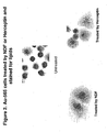
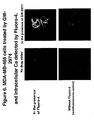
- Figure 2 shows that Au565 cells treated with either an ErbB stimulatory ligand, NDF, or the monoclonal antibody Herceptin, both result in the production of lipids. This is shown by the staining of lipids with oil red (lipids are represented by red dots) against the background counterstaining of the cells (hematoxylin).
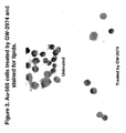
- Figure 3 shows that lipids are present in untreated Au565 cells but are reduced in cells treated with the dual EGFR and ErbB2 inhibitor, GW2974.
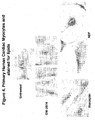
- Figure 4 shows cardiomyocyte cells treated with either GW2974, Herceptin or NDF. Lipids increase in cells treated with Herceptin and NDF (compared with untreated cells) but not decrease in cells treated with GW2974.
- Figure 5 shows a quantitative measure of lipids in control, Herceptin and GW2974 treated cells.
- FIG. 6 Treatment of cells with GW2974 causes a redistribution of intracellular calcium ( Figure 6 ). This can be seen in MDA-MB-468 breast cancer cells where calcium is detected by fluorescently by Fluoro-4. This redistribution of calcium results in the activation and phosphorylation of AMPK.
- Activated AMPK represses translation by phosphorylation of the translation factor eEF-2 ( Figure 7 ), which inactivates eEF-2 and represses protein synthesis, a known effect of TKIs.
- Figure 7A shows a western blot of Au565 cells treated with either a stimulatory ligand (EGF) or GW2974 and probed for p-eEF-2. p-eEF-2 is dramatically increased after GW2974 treatment.
- Figure 7B shows expression of p-eEF-2 by IHC. C225 and Herceptin do not increase p-eEF-2, however TKIs like Iressa, GW2974 and rapamycin do.
- ERR ⁇ plays a role in lipid metabolism in cardiac cells
- MCAD is an enzyme that breaks down lipids and fatty acids. Mutations in MCAD is a common genetic disorder, especially in those of northern European descent.
- Figure 8 shows that in Herceptin treated cells, the level of ERR ⁇ diminished slightly. MCAD is expressed in Herceptin treated cells but is completely absent from GW2974 treated cells.
- the following example demonstrates the change in mRNA expression profile of cells treated with GW2974.
- Au565 cells were grown under normal conditions and were untreated or treated with GW2974 (25 ⁇ M). Cells were pelleted, snap frozen in liquid nitrogen and subjected to microarray analysis. RNA was isolated using the Agilent Total RNA Isolation Kit. Cy3 and Cy5 labeled cRNA was prepared using the Agilent Low RNA Input Fluorescent Linear Amplification Kit. Labeled cRNAs were hybridized to a G4110A Human 1A(V2) microarray consisting of 60-mer oligonucleotides representing over 18K well-characterized, full length, human genes. Table IV provides the results in Table form.
- GW2974 change compared to control FLJ12476 Protein containing an IQ calmodulin-binding domain 5.0 x CAMK4 Calcium/calmodulin-dependent protein kinase IV, a protein kinase involved in Ca(2+)-regulated gene expression, including CREBBP -dependent gene expression 4.5 x AVIL Protein with high similarity to villin 1 (human VIL1), which is a calcium-regulated actin-binding protein that caps, severs, and bundles actin filaments, member of the gelsolin family and contains a villin headpiece domain 4.2 x SCN1A Sodium channel voltage-gated type I (alpha subunit), a voltage-sensitive sodium channel; mutations are associated with severe myoclonic epilepsy of infancy and generalized epilepsy with febrile seizures plus 4.1 x CLSP Calmodulin-like skin protein, a member of the calmodulin family of calcium-binding proteins, may play a role in
Description
- The heart has a tremendous capacity for ATP generation which allows it to function as an efficient pump throughout the life of the organism. The adult myocardium uses either fatty acid (FA) and/or glucose oxidation as its main energy sources. Under normal conditions, the adult heart derives most of its energy through oxidation of fatty acids in mitochondria.
- Cells of the myocardium have the ability to switch between carbohydrate glycolysis and the Krebs cycle and to fat fuel sources so that ATP production is maintained at a constant rate under diverse physiological and dietary conditions. This metabolic and fuel selection flexibility is important for normal cardiac function. Although cardiac energy conversion capacity and metabolic flux is modulated at many levels, one important mechanism of regulation occurs at the level of gene expression. The expression of genes involved in multiple energy transduction pathways is dynamically regulated in response to developmental, physiological, and pathophysiological cues.
- The genes involved in these key energy metabolic pathways are transcriptionally regulated by members of the nuclear receptor superfamily, specifically the fatty acid-activated peroxisome proliferator-activated receptors (PPARs) and the nuclear receptor coactivator, PPARγ coactivator-1α (PGC-1α), as well as the estrogen receptor-related protein ERRα, ERRß and ERRγ and their activators PGR-1 and PERC. The dynamic regulation of the cardiac PPAR/PGC-1 complex in accordance with physiological and pathophysiological states is described in more detail below.
- PGC-1α is a PPARγ coactivator, linked to adaptive thermogenesis in brown adipose. Two structurally related proteins, PGC-1ß and PARC, have been cloned and appear to be involved in regulating energy metabolic pathways. The tissue-specific and inducible nature of PGC-1α expression suggests its involvement in the dynamic regulation of cellular energy yielding metabolic processes, including mitochondrial biogenesis and oxidation, hepatic gluconeogenesis, and skeletal muscle glucose uptake. PGC-1α is selectively expressed in highly oxidative tissues such as heart, skeletal muscle, brown adipose, and liver. In the heart PGC-1α expression increases sharply at birth. This coincides with a perinatal shift from glucose metabolism to fat oxidation. PGC-1α activity and expression levels are also known to be induced by cold exposure, fasting, and exercise; stimuli known to promote oxidative metabolism. Forced expression of PGC-1 in cardiac myocytes in culture induces expression of nuclear and mitochondrial genes involved in multiple mitochondrial energy-transduction/energy-production pathways, increases cellular mitochondrial number, and stimulates coupled respiration. Signaling pathways associated with these stimuli, including p38 MAP kinase, ß-adrenergic/cAMP, nitric oxide, AMP kinase, and Ca2-calmodulin kinase, activate PGC-1α and its downstream target genes either by increasing PGC-1α expression or its transactivation function.
- These metabolic and structural changes can result in dilated cardiomyopathy and diastolic dysfunction in the heart. Interestingly, mitochondrial proliferation is reversible and the cardiomyopathy can be rescued upon reduction in transgene expression. This suggests that, in addition to serving as an activator of cellular fatty acid metabolism through PPARs, PGC-1α is linked to the mitochondrial biogenesis. Therefore, PGC-1α appears to serve as a master modulator of oxidative energy metabolism and responds to changes in the cellular energy status.
- Evidence is emerging that the estrogen-related receptor (ERR) family of orphan Nuclear Receptors function as PGC-1-activated regulators of cardiac and skeletal muscle energy metabolism. There are three members of the ERR family: ERRα, ERRß, and ERRγ. ERRα and ERRγ expression is elevated in adult tissues that rely primarily on mitochondrial oxidative metabolism for ATP production, such as heart and slow twitch skeletal muscle. ERRα expression dramatically increases in heart after birth, in parallel with the global upregulation of enzymes involved in cellular fatty acid uptake and mitochondrial oxidation. Recently, ERRα and ERRγ were identified as novel partners for the PGC-1 family of coactivators. This functional relationship between ERR isoforms and PGC-1α have stimulated interest in the role of ERRs in energy metabolism.
- Deletion of the ERRα gene reveals a tissue-specific role for ERRα in constitutive regulation of lipid metabolism. White adipose mass is decreased in ERRα__/_ mice coincident with decreased adipocyte size and lipid synthesis rates. In contrast, ERRα likely plays a role in lipid catabolism in heart, consistent with its functional interaction with PGC-1α. ERRα__/_ mice, which do not display an overt cardiac phenotype, exhibit a compensatory increase in cardiac PGC-1α and ERRγ expression. These results suggest that ERR isoforms contribute to constitutive expression of fatty acid metabolic genes in heart. However, the metabolic effects of changes in gene expression remain unknown.
- Gene expression profiling in cardiac myocytes that overexpress ERRα are being used to identify cardiac ERRα target genes. ERRα activates genes involved in energy production pathways, including cellular fatty acid uptake (LPL, CD36/FAT, H-FABP, FACS-1), ß-oxidation (MCAD, VLCAD, LCHAD), and mitochondrial electron transport/oxidative phosphorylation (cytochrome c, COXIV, COXVIII, NADH ubiquinone dehydrogenase, flavoprotein-ubiquinone oxidoreductase, ATP synthase ß). ERRα also increases palmitate oxidation rates in cardiac myocytes. Activation of ß-oxidation enzymes genes by ERRα involves the PPARα signaling pathway. ERRα directly activates PPARα gene expression, and ERRα-mediated regulation of MCAD and M-CPT I is abolished in cells derived from PPARα__/_ mice. ERRα is also now known to be involved in the PGC-1α regulation of mitochondrial biogenesis. It is known to mediate PGC-1α activation of the NRF pathway through regulation of the Gapba gene, which encodes a subunit of the NRF-2 complex and directly activates genes involved in mitochondrial oxidative metabolism at the level of transcription. ERRα with its coactivator PGC-1α activates the MCAD, cytochrome c, and ATP synthase ß gene promoters. Collectively, these results identify ERRα as a regulator of cardiac oxidative energy metabolism through its involvement in the PGC-1 regulatory circuit. However, the precise biological roles of ERRs in heart have not been identified.
- The nuclear receptor ERRγ (estrogen related receptor gamma) is highly expressed in heart, skeletal muscle, kidney, and brain, as well as in the developing nervous system. The expression of the coactivators PGC-1α and PGC-1β in mammalian cells potently augmented transcriptional activation by ERRγ. The constitutive activation function 2 (AF-2) of the orphan receptor is important for the synergistic enhancement. Functional receptor truncation analysis has been used to identify an additional amino-terminal activation function, specific for the ERRγ2 isoform and PGC-1α. In vitro experiments showed a direct interaction of ERRγ with both coactivators. These findings are consistent with the hypothesis that distinct regulatory functions for PGC-1α and PGC-1β as tissue-specific coactivators for ERRγ. Nevertheless, more studies are needed to further define these functions.
- Cardiac-specific overexpression of PGC-1 in transgenic mice results in uncontrolled mitochondrial proliferation in cardiac myocytes leading to loss of sarcomeric structure and a dilated cardiomyopathy. Thus, PGC-1 is an important regulatory molecule in the control of cardiac mitochondrial number and function in response to energy demands.
- Most, if not all of these regulatory pathways involve phosphorylation of intermediates in a signaling pathway. Inhibition of phosphorylation, such as by the action of various kinase inhibitors, affects these signaling pathways causing alterations in fatty acid metabolism which can cause organ toxicity, including cardiotoxicity. Many new anti-cancer drugs are kinase inhibitors and are accompanied by toxicity. Thus, methods are needed for identifying whether drugs may be accompanied by toxic effects and whether the toxic effects are likely to occur in a patient. Methods are also needed for avoiding toxic effects of these inhibitors while maintaining their potency against the phosphorylated receptor targets.
- Methods are disclosed for diagnosing whether toxicity, especially cardiotoxicity, is likely to occur in a patient selected for treatment with a variety of drugs, such as tyrosine kinase inhibitors or erbB inhibitors. Methods are also disclosed for evaluating whether a candidate drug is likely to have a toxic or cardiotoxic affect. In one method lipids, such as triglycerides and cholesterol, can be analyzed to determine whether a fatty acid oxidation disorder is present. In another method enzymes responsible for the observed fatty acid oxidation, such as MCAD, can be determined. With respect to lipid levels it is thought that in normal cells AMP-activated protein kinase activation can lead to a characteristic reduction in the level of lipids and a corresponding increase in glycolytic and shorter carbon chain intermediates, for example of C2 to C6 carbon intermediates. Any statistically significant deviation from the characteristic lipid reduction in normal cells can be considered, for purposes of this disclosure, a fatty acid oxidation disorder. Similarly, with respect to the enzymes involved in these metabolic pathways, any statistically significant change, relative to normal cells, in the amount of activity or levels of these enzymes as measured by Western, Northern, PCR or other techniques, can be considered, for purposes of this disclosure, a fatty acid oxidation disorder. The diagnosis of a fatty acid oxidation disorder can be used to predict an increased risk of toxicity and possibly as a contra-indicator for the use of the drug. Alternatively, in the event a drug is used in a patient having a fatty acid oxidation disorder the methods can be used to indicate the need to closely follow cardiac function in the patient. Alternatively glucose uptake can be measured by known methods, such as by positron emission tomography. In situations where glucose uptake is not diminished or is not diminished to the same extent as in normal noncancerous cells upon administration of a tyrosine kinase inhibitor drug, then the drug treatment is likely to be toxic to the noncancerous cells. Alternatively, if ATP levels decrease more than in normal noncancerous cells upon exposure to a tyrosine kinase inhibitor, then the tyrosine kinase inhibitor is predicted to be toxic.
- Another method for predicting whether cardiotoxicity in a patient selected for treatment with a drug, such as tyrosine kinase inhibitor, especially an erbB inhibitor, is to assess the TNFα levels in the patient, either in the tumor or blood or both. The level of TNFα can be used to predict whether a patient is likely to have an adverse event related to cardiotoxicity resulting from drug, particularly Herceptin, therapy.
- Additional features and advantages are described herein, and will be apparent from, the following Detailed Description and the figures.
-
-
Figure 1 is a listing of genes regulated by Herceptin treatment in Au565 cells. -
Figure 2 are photographs of Au-565 cells treated by NDF or Herceptin and stained for lipids. -
Figure 3 are photographs of Au-565 cells treated by GW-2974 and stained for lipids. -
Figure 4 are photographs of primary human cardiac myocytes grown under various conditions and stained for lipids. -
Figure 5 is a bar graph illustrating the percentage of human cardiomyocytes testing positive for lipids under various conditions. -
Figure 6 are photographs of MDA-MB-468 cells treated by GW-2974 and intracellular Ca detected by Fluoro-4. -
Figure 7A a photograph of a Western Blot showing the affect of certain tyrosine kinase inhibitors on expression p-eEF2 and p-AMPKa. -
Figure 7B is a photograph of stained cells showing the expression of p-eEF2 in Au565 cells in the presence of various compounds. -
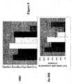
Figure 8 is a photograph of ERRα and MCAD in cardiomyocytes cells with and without treatment by various kinase inhibitors. -
Figure 9 is a bar graph illustrating the growth inhibition of HMCs treated with combinations of different types of erbB inhibitors and TNFα. -
Figure 10 is a western blot of HMCs probed for NF-κB after treatment with either TNFα, GW2974 or Herceptin (or combinations). - In one aspect, the present disclosure is based on the discovery that drugs, such as tyrosine kinase inhibitors, like Herceptin and lapatinib (Tykerb), affect the expression of genes associated with lipid metabolic pathways and dramatically affect the amount of lipid within the cells. Treatment of otherwise normal cells or cells having normal protein tyrosine kinase regulation with the kinase inhibitors of the invention affects fatty acid metabolism by increasing or decreasing the capacity of such cells to oxidize fatty acids. When normal fat cells grown in culture are exposed to kinase inhibitors such as GW2974, GW572016, the lipid stored within those cells rapidly disappears. This observation has also been made in cardiac cells. Such studies can be conducted using Oil red 0 staining for lipids. Thus, treatment with lapatinib (tykerb) and other Her1/Her2 tyrosine kinase inhibitors cause a loss of fat from such cells that is consistent with reduced lipid synthetic rates and/or increased lipid oxidation rates. With other drugs, such as Herceptin, NDF lipid content appears to increase.
- Many kinase inhibitors are also known to be useful as chemotherapeutic agents. In some patients these drugs produce cardiotoxicity. In the article entitled "Trastuzumab cardiotoxicity: Speculations regarding pathophysiology and targets for further study" (Seminars in Oncology, vol. 29, pages 22-28) the authors Jay W. Schneider et al. disclose that toxic cardiovascular side effects occur in the context of treatment with trastuzumab. Various factors like germline mutation, upregulation of cardiac HER2 expression and changes in the stoichiometry of available erbB receptors are discussed to elucidate potential molecular mechanisms underlying the observed toxicity. Cardiotoxicity as a result of impairment of a general cellular mechanism such as fatty acid oxidation is not indicated in this article. The present disclosure is based on the surprising discovery that cardiotoxicity can be associated with defects in fatty acid metabolism. Thus, patients with certain dysfunctions in fatty acid metabolism or that have high levels of TNFα in blood, and that are undergoing treatment with kinase inhibitors are more likely to suffer from cardiac malfunction such as cardiomyopathy upon treatment with kinase inhibitors such as erbB tyrosine kinase inhibitors. In addition, it has been discovered that patients having high levels of TNFα, or its downstream survival factor NF-κB, in tumor tissue or serum generally have a better response to Herceptin. This discovery has led to the development of new methods for predicting whether patients will suffer from cardiotoxicity upon treatment with drugs, including kinase inhibitors either alone or in combination with other active agents, that affect phosphorylation states of certain cellular proteins.
- A method is disclosed for analyzing a patient's lipids including triglycerides and cholesterol and/or lipid metabolic enzymes such as, MCAD, among others. The results from such analysis can then be used to predict when cardiotoxicity could result from kinase inhibitor treatment and to provide an early indication that cardiac function should be closely monitored in patients undergoing treatment with drugs, such as kinase inhibitors, including Herceptin, GW572016 or other erbB inhibitors.
- The activity of 5-'AMP-activated protein kinase, which has been shown to phosphorylate and inactivate acetyl-CoA carboxylase in other tissues, has been discovered to be significantly increased at the end of ischemia, and remains elevated throughout reperfusion. Accumulation of 5'-AMP during ischemia results in an activation of AMP-activated protein kinase, which phosphorylates and inactivates acetyl-CoA carboxylase during reperfusion. The subsequent decrease in malonyl-CoA levels can result in accelerated fatty acid oxidation rates during reperfusion of ischemic hearts.
- With respect to cardiac toxicity, a variety of fatty acid oxidation disorders are known and are listed below in Table I. If such a disorder is detected in a patient it can provide an indication that kinase inhibitors could be toxic to the heart.
TABLE I Acyl-CoA dehydrogenase deficiencies Acyl-CoA dehydrogenase, short-chain (SCAD) Acyl-CoA dehydrogenase, medium-chain (MCAD) Acyl-CoA dehydrogenase, long-chain (LCAD) Acyl-CoA dehydrogenase, very long-chain (VLCAD) 2-Enoyl-CoA hydratase deficiency L-3-Hydroxyacyl-CoA dehydrogenase deficiencies L-3-Hydroxyacyl-CoA dehydrogenase, short chain (SCHAD) Trifunctional protein: Long-chain FA (LCHAD) Alpha subunit (HADHA) Beta subunit (HADHB) 3-Ketoacyl-CoA thiolase deficiency 3-Ketoacyl-CoA thiolase, Medium chain (MCKAT) Trifunctional protein α-Methylacyl-CoA racemase (AMACR) deficiency Carnitine-acylcarnitine translocase deficiency: 3p21 2,4-Dienoyl-CoA reductase deficiency: 8q21 Electron transfer flavoprotein (ETF) deficiency: 15q23 Ichthyosiform erythroderma (NCIE2): CGI58 gene; 3p21 Trifunctional protein deficiencies: Subunits A & B Tyrosinemia 1° Disorders of Carnitine metabolism Fatty acid & Carnitine transport pathways Fatty acid oxidation pathways Lipid disorders Mitochondria: Biochemical abnormalities Peroxisomal disorders - In a method, patients who are candidates for treatment with kinase inhibitors can be screened for these diseases to determine whether they are likely to suffer myocardiocyte toxicity. For example, the biological macromolecules can be determined in myocardiocytes grown in culture to determine how the levels of these macromolecules are affected by administration of the candidate drug. In a method human myocardiocytes can be grown in culture and the level of phosphorylated AMP-activated protein kinase can be monitored in the presence of the candidate drug. This can be determined by a Western blot that detects the phosphorylated AMP activated kinase.
- Without limiting the invention, it is believed that under stress conditions such as hypoxia, ischemia, glucose deprivation, and starvation, an increase in the intracellular AMP:ATP ratio allosterically activates AMP-activated protein kinase (AMPK), a response designed to maintain cellular energy balance. AMP-activated protein kinase was initially discovered to inhibit preparations of acetyl-CoA carboxylase (ACC) and 3-hydroxy-3-methylglutaryl-CoA reductase (HMG-CoA reductase, HMGR). Activation of AMPK is thought to initiate a series of downstream phosphorylation events that switch cells from active ATP consumption (e.g., fatty acid, cholesterol and protein biosynthesis) to ATP production (e.g., fatty acid and glucose oxidation). Stress-induced activation of AMPK is thought to occur following its phosphorylation at threonine 172 on the α subunit by one or more upstream AMPK kinases (AMPKKs), including calmodulin-dependent kinase kinase β (CAMKKβ), a calcium-activated protein kinase, and LKB1, a serine/threonine kinase encoded by the Peutz-Jegher syndrome tumor suppressor gene. Activation of AMPK in skeletal muscle and heart is believed to lead to the phosphorylation and inhibition of acetyl-CoA carboxylase (ACC), which in turn is thought to reduce the level of malonyl-CoA, itself an inhibitor of carnitine palmitoyltransferase 1 (CPT 1). De-repression of
CPT 1 is thought to cause the concomitant increase in β-oxidation of fatty acid, which is thought to lead to increased mitochondrial production of ATP. Stress-induced activation of AMPK is also thought to inhibit protein synthesis by inhibiting mTOR and directly modulating eEF2, a translation elongation factor known to be associated with cardiac protection. Importantly, alteration in mitochondrial function is thought to lead to cardiomyocyte death by imatinib. Moreover, inhibition of cap-dependent translation via AMPK-mediated TSC2 phosphorylation is thought to be extremely important for cell survival in response to ATP depletion. Increased biosynthesis of, rather than consumption of ATP following AMPK activation may also protect cardiomyocytes against ischemic injury. - It has been discovered that molecules such as GW2974, a potent small molecule HER2/EGFR tyrosine kinase inhibitor with a similar activity profile to lapatinib, that can activate AMPK and its downstream substrates stimulate fatty acid oxidation, which in turn increases ATP production in HER2-expressing human cardiomyocytes, protecting against apoptosis induced by TNFα, a known cytokine detected in cardiac failure. Conversely, molecules such as trastuzumab that do not activate AMPK result in enhanced cardiomyocyte cell death in response to TNFα. The effects of specific HER2-targeted therapies on AMPK and consequently energy production may predict for the risk associated cardiomyopathy and provide a novel HER2-directed therapeutic strategy to protect myocardium from the killing effects of TNFα or other pro-apoptotic stimuli, following acute ischemic injury.
- In addition, tyrosine kinase inhibitors can be used to reduce fat in cells, particularly cells that are otherwise normal or that lack protein tyrosine kinase activity mediated disease. Table II below shows the reduction in lipid content obtained by treatment with GW2974. Au565 cells were grown under normal conditions known in the art and treated for 2 days with GW2974 (25 µM). The cells were collected, washed and sonicated in water (2,000,000 cells in 200 µL of water). The cells were spun down and were tested for acylcarnitines (byproducts of mitochondrial fatty acid oxidation) by MS/MS for intraceullar metabolites.
TABLE II Acylcarnitines (pmoles of protein) C18:1 C16 C2 Control (Cell pellet) 8.56 4.09 148.54 GW2974 (Cell pellet) 4.1 0.83 258.88 - The following example demonstrates the identification of genes that are affected by treatment of Herceptin in an in vitro cell culture of Au565 cells. Au565 cells were grown under normal conditions and treated with Herceptin or left untreated. Cells were pelleted, snap frozen in liquid nitrogen and analyzed in a microarray using standard conditions. Cy3 and Cy5 labeled cDNA was prepared from RNA isolated from the cell pellets. Genes involved in lipid metabolism are shown in Table III. Genes involved in other pathways that were either upregulated or downregulated are also shown in
Figure 1 .TABLE III Changes in Metabolic Genes by Microarray Analysis of Au565 Cells That Were Untreated or Treated with Herceptin Gene Description Relative change in Herceptin treated cells compared with untreated cells NKX2-5 Cardiac specific homeobox, a transcription factor involved in heart development and possibly in apoptosis; mutations in the corresponding gene are associated with congenital heart disease, septal and conduction defects, and tetralogy of Fallot 4.71 x ESRR6 Estrogen-related receptor gamma, binds to estrogen response elements and activates transcription in a ligand-independent manner, can have roles in tissue differentiation and maintenance 4.18 x FABP1 Fatty acid binding protein 1 liver, positive regulator of peroxisome proliferators activated receptor alpha (PPARA), plays a role in fatty acid transport, cell proliferation, and apoptosis, increased expression is associated with prostate cancer -6.29 x NRG1 Neuregulin 1, a secreted protein, activates ERBB2 and other members of the EGF receptor family of tyrosine kinase receptors, induces cell migration, cell proliferation and neurogenesis; gene amplification is associated with some breast tumors -5.07 x PERC PGC-1 related estrogen receptor alpha coactivator (PPAR gamma coactivator 1 beta), a transcriptional coactivator that binds and activates nuclear hormone receptors, can play a role in gluconeogenesis or fatty acid oxidation -5.20 x ERBB4 Avian erythroblastosis oncogene B4, a receptor tyrosine kinase of the EGF receptor family, activated by neuregulin ligands, plays a role in cell migration, proliferation, and differentiation, involved in the pathogenesis of multiple malignant neoplasias 4.48 x Gene Description Log Ratio BBOX1 Butyrobetaine (gamma) 2-oxoglutarate dioxygenase (gamma-butyrobetaine hydroxylase) 1, catalyzes the conversion of gamma butyrobetaine to L-carnitine in carnitine biosynthesis 5.13E-01 GLS Kidney-type glutaminase, catalyzes the hydrolysis of glutamine to glutamate and ammonia, provides TCA cycle intermediates, helps maintain acid-base balance, produces neurotransmitters, and initiates glutamine catabolism 5.12E-01 IQGAP2 IQ motif containing GTPase activating protein 2, inhibits GTPase activity of CDC42 and RAC1, can bind actin and play a role in Rho-family GTPase regulation of cell shape 4.85E-01 TRPM4 Transient receptor potential cation channel subfamily M member 4, a Ca2+-activated channel permeable to monovalent cations, responsive to G protein-coupled receptor-mediated Ca2+ elevation, inhibits Ca2+ influx through membrane potential depolarization 4.43E-01 SAT Spermidine/spermine N1-acetyltransferase, catalyzes the rate limiting step of polyamine catabolism, promotes polyamine homeostasis, involved in oxidative stress and heat shock responses, modulates tumorigenicity and sensitivity to some anticancer drugs 4.04E-01 I_1152020 Protein containing three collagen triple helix repeats, which are found in some extracellular proteins, and a C-terminal C1q domain, has moderate similarity to mouse Acrp30, which controls energy balance, insulin sensitivity, and adipocyte 4.00E-01 CLSP Calmodulin-like skin protein, a member of the calmodulin family of calcium-binding proteins, can play a role in keratinocyte differentiation, shows altered expression in sun-damaged skin -4.03E-01 ME1 Malic enzyme 1, catalyzes the oxidative decarboxylation of malate to form pyruvate and can play a role lipogenesis; variant can be associated with breast cancer -4.04E-01 ACAT2 Homo sapiens acetyl-Coenzyme A acetyltransferase 2 (acetoacetyl Coenzvme A thiolase) (ACAT2), mRNA -4.33E-01 ACAT2 Acetyl-Coenzyme A acetyltransferase 2 (cytosolic acetoacetyl Coenzyme A thiolase), a liver enzyme that functions in acvl-CoA metabolism -5.30E-01 ACAT2 Homo sapiens acetyl-Coenzyme A acetyltransferase 2 (acetoacetyl Coenzyme A thiolase) (ACAT2), mRNA -4.33E-01 ACAT2 Acetyl-Coenzyme A acetyltransferase 2 (cytosolic acetoacetyl Coenzyme A thiolase), a liver enzyme that functions in acyl-CoA metabolism -5.30E-01 ALDOA Aldolase A (fructose-bisphosphate aldolase), catalyzes cleavage or condensation of fructose-1,6-bisphosphate into dihydroxyacetonephosphate and glyceraldehyde-3-phosphate in glycolysis, deficiency manifests as hemolytic anemia and metabolic myopathy -4.54E-01 NFκBIL2 NF-kappaB inhibitor-like 2, member of IkappaB family, inhibits DNA binding of NFKB1-RELA NF-kappaB heterodimers and NFKB 1 homodimers, NF-kappaB-mediated transcription from Igkappa enhancer, and can regulate NF-kappaB function in epithelial cells -4.90E-01 ENO1 Enolase 1 (alpha enolase), converts 2-phospho-D-glycerate to phosphoenolpyruvate in glycolysis, an autoantigen in multiple autoimmune diseases, shorter alternative form c-myc promoter binding protein (MPB1) is a transcriptional repressor -5.01E-01 GSTT2 Glutathione S-transferase theta 2, theta class glutathione transferase and peroxidase, involved in xenobiotic metabolism, can be involved in detoxification of fatty acid hydroperoxides and play a role in cancer prevention bv inactivating carcinogens -5.33E-01 APOL1 Apolipoprotein L, a component of large, apoA-I(APOA1)-containing, high density lipoproteins, can be involved in lipid transport and metabolism; elevated expression in prefrontal cortex is associated with schizophrenia -6.43E-01 AKR1C2 Aldo keto reductase family 1 member C2 (dihydrodiol dehydrogenase), functions in bile transport, steroid metabolism, and xenobiotic metabolism, can play a role in behavior modification mediated by selective serotonin reuptake inhibitors -7.44E-01 AKR1C2 Aldo keto reductase family 1 member C2 (dihydrodiol dehydrogenase), functions in bile transport, steroid metabolism, and xenobiotic metabolism, can play a role in behavior modification mediated by selective serotonin reuptake inhibitors -8.23E-01 CAMK4 Calcium/calmodulin-dependent protein kinase IV, a protein kinase involved in Ca(2+)-regulated gene expression, including CREBBP -dependent gene expression 4.46E-01 FKSG 14 Protein with high similarity to SoxLZ-Sox6 leucine zipper binding protein in testis (mouse Solt), which binds SoxLZ/Sox6 and enhances SoxLZ/Sox6-mediated transcription activation along with calcium/calmodulin-dependent protein kinase IV (mouse Camk4) -4.57E-01 SOAT1 Acyl-Coenzyme A:cholesterol acyltransferase, synthesizes cholesterol esters from cholesterol and long-chain fatty acyl-coenzyme A, acts in lipoprotein metabolism, cholesterol homeostasis, and monocyte differentiation; associated with atherosclerosis -4.05E-01 I_962304. FL1 Potassium voltage-gated channel (Shal-related subfamily, member 1), predicted to generate A-type transient outward K+ currents that are important for the control of excitability of neurons and cardiac cells [647-aa form] -4.16E-01 SCN2A2 Sodium channel voltage gated type II alpha 2, displays voltage-dependent and sodium-selective current, can play a role in the rising phase of action potential in excitable cells, sensitive to tetrodotoxin 4.54E-01 SCN1A Sodium channel voltage-gated type I (alpha subunit), a voltage-sensitive sodium channel; mutations are associated with severe myoclonic epilepsy of infancy and generalized epilepsy with febrile seizures plus 4.14E-01 SCN11A Sodium channel voltage-gated type XI alpha polypeptide, a putative voltage-sensitive sodium channel that can produce tetrodotoxin-resistant sodium currents in peripheral sensory neurons, can play a role in pain transmission and neuropathic pain in ... 4.02E-01 FASN Fatty acid synthase, multifunctional enzyme that synthesizes fatty acids from dietary proteins and carbohydrates, increased expression is associated with various cancers and inhibition can be therapeutic for breast and prostate cancer -4.29E-01 ELOVL2 Homo sapiens elongation of very long chain fatty acids (FEN1/Elo2, SUR4/Elo3, yeast)-like 2 (ELOVL2), mRNA -4.36E-01 HPCAL1 Hippocalcin-like 1, a putative calcium-sensing protein, member of the neural visinin-like (NVP) family of calcium-binding proteins, localized to axons and dendrites, can play a role in neuronal signaling in the central nervous system -4.53E-01 KCNG2 Potassium voltage channel subfamily gamma 2, a member of the Kv6 family of ion channels, functions as a votage-gated potassium channel upon interaction with Kv2.1 alpha subunit, can contribute to cardiac action potentiation repolarization -5.53E-01 CCL14 Small inducible cytokine subfamily A member 14, a chemoattractant that enhances proliferation of myeloid progenitor cells and can affect replication of the HIV 1 virus, can play a role in AIDS pathogenesis and chemokine receptor CCR1 associated diseases -4.49E-01 CLCA1 Calcium-activated chloride channel 1, a chloride channel which plays a role in mucous production in mucoepidermal cells and can function as a tumor suppressor; dysregulation can contribute to asthma and the progression of colorectal cancer -6.70E-01 FABP1 Fatty acid binding protein 1 liver, positive regulator of peroxisome proliferator activated receptor alpha (PPARA), plays a role in fatty acid transport, cell proliferation, and apoptosis, increased expression is associated with prostate cancer -7.22E-01 BBOX1 Butyrobetaine (gamma) 2-oxoglutarate dioxygenase (gamma-butyrobetaine hydroxylase) 1, catalyzes the conversion of gamma butyrobetaine to L-carnitine in carnitine biosynthesis 5.13E-01 GLS Kidney-type glutaminase, catalyzes the hydrolysis of glutamine to glutamate and ammonia, provides TCA cycle intermediates, helps maintain acid-base balance, produces neurotransmitters, and initiates glutamine catabolism 5.12E-01 IQGAP2 IQ motif containing GTPase activating protein 2, inhibits GTPase activity of CDC42 and RAC1, can bind actin and play a role in Rho-family GTPase regulation of cell shape 4.85E-01 TRPM4 Transient receptor potential cation channel subfamily M member 4, a Ca2+-activated channel permeable to monovalent cations, responsive to G protein-coupled receptor-mediated Ca2+ elevation, inhibits Ca2+ influx through membrane potential depolarization 4.43E-01 SAT Spermidine/spermine N1-acetyltransferase, catalyzes the rate limiting step of polyamine catabolism, promotes polyamine homeostasis, involved in oxidative stress and heat shock responses, modulates tumorigenicity and sensitivity to some anticancer drugs 4.04E-01 I_1152020 Protein containing three collagen triple helix repeats, which are found in some extracellular proteins, and a C-terminal C1q domain, has moderate similarity to mouse Acrp30, which controls energy balance, insulin sensitivity, and adipocyte 4.00E-01 - This example demonstrates that adipocytes lose lipid when treated with a small molecule tyrosine kinase inhibitor, GW2974.
Figure 2 shows that Au565 cells treated with either an ErbB stimulatory ligand, NDF, or the monoclonal antibody Herceptin, both result in the production of lipids. This is shown by the staining of lipids with oil red (lipids are represented by red dots) against the background counterstaining of the cells (hematoxylin).Figure 3 shows that lipids are present in untreated Au565 cells but are reduced in cells treated with the dual EGFR and ErbB2 inhibitor, GW2974.Figure 4 shows cardiomyocyte cells treated with either GW2974, Herceptin or NDF. Lipids increase in cells treated with Herceptin and NDF (compared with untreated cells) but not decrease in cells treated with GW2974.Figure 5 shows a quantitative measure of lipids in control, Herceptin and GW2974 treated cells. - Treatment of cells with GW2974 causes a redistribution of intracellular calcium (
Figure 6 ). This can be seen in MDA-MB-468 breast cancer cells where calcium is detected by fluorescently by Fluoro-4. This redistribution of calcium results in the activation and phosphorylation of AMPK. Activated AMPK represses translation by phosphorylation of the translation factor eEF-2 (Figure 7 ), which inactivates eEF-2 and represses protein synthesis, a known effect of TKIs.Figure 7A shows a western blot of Au565 cells treated with either a stimulatory ligand (EGF) or GW2974 and probed for p-eEF-2. p-eEF-2 is dramatically increased after GW2974 treatment.Figure 7B shows expression of p-eEF-2 by IHC. C225 and Herceptin do not increase p-eEF-2, however TKIs like Iressa, GW2974 and rapamycin do. - ERRα plays a role in lipid metabolism in cardiac cells, and MCAD is an enzyme that breaks down lipids and fatty acids. Mutations in MCAD is a common genetic disorder, especially in those of northern European descent.
Figure 8 shows that in Herceptin treated cells, the level of ERRα diminished slightly. MCAD is expressed in Herceptin treated cells but is completely absent from GW2974 treated cells. - The following example demonstrates the change in mRNA expression profile of cells treated with GW2974.
- Au565 cells were grown under normal conditions and were untreated or treated with GW2974 (25 µM). Cells were pelleted, snap frozen in liquid nitrogen and subjected to microarray analysis. RNA was isolated using the Agilent Total RNA Isolation Kit. Cy3 and Cy5 labeled cRNA was prepared using the Agilent Low RNA Input Fluorescent Linear Amplification Kit. Labeled cRNAs were hybridized to a G4110A Human 1A(V2) microarray consisting of 60-mer oligonucleotides representing over 18K well-characterized, full length, human genes. Table IV provides the results in Table form.
-
TABLE IV A - Ion Channel Gene Name Description GW2974 change compared to control FLJ12476 Protein containing an IQ calmodulin-binding domain 5.0 x CAMK4 Calcium/calmodulin-dependent protein kinase IV, a protein kinase involved in Ca(2+)-regulated gene expression, including CREBBP -dependent gene expression 4.5 x AVIL Protein with high similarity to villin 1 (human VIL1), which is a calcium-regulated actin-binding protein that caps, severs, and bundles actin filaments, member of the gelsolin family and contains a villin headpiece domain 4.2 x SCN1A Sodium channel voltage-gated type I (alpha subunit), a voltage-sensitive sodium channel; mutations are associated with severe myoclonic epilepsy of infancy and generalized epilepsy with febrile seizures plus 4.1 x CLSP Calmodulin-like skin protein, a member of the calmodulin family of calcium-binding proteins, may play a role in keratinocyte differentiation, shows altered expression in sun-damaged skin - 4.0 x GNB5 Guanine nucleotide binding protein (G protein) beta 5, a component of heterotrimeric G protein complexes that transduce signals from G protein-coupled receptors to downstream effector proteins, may regulate calcium channel activity - 4.1 x KCNK6 Potassium channel subfamily K member 6 (TWIK-2), a pH-sensitive outward and mild inward rectifying member of the tandem pore domain K+ channel family, may play a role in setting the cellular resting membrane potential and in cardiac cell excitability - 4.1 x CASK Calcium/calmodulin-dependent serine protein kinase, member of the MAGUK family, involved in recruiting multiprotein complexes at the plasma membrane, may link the extracellular matrix to the actin cytoskeleton, may regulate synaptic vesicle exocytosis - 4.2 x I-962304.FL1 Potassium voltage-gated channel (Shal-related subfamily, member 1), predicted to generate A-type transient outward K+ currents that are important for the control of excitability of neurons and cardiac cells [647-aa form] - 4.2 x CD38 CD38 antigen, has both cyclic ADP-ribose-forming and -hydrolyzing activities, regulates intracellular calcium mobilization, may play a role in superantigen-induced T cell proliferation, autoantibodies may contribute to noninsulin dependent diabetes - 4.4 x HPCAL1 Hippocalcin-like 1, a putative calcium-sensing protein, member of the neural visinin-like (NVP) family of calcium-binding proteins, localized to axons and dendrites, may play a role in neuronal signaling in the central nervous system - 4.5 x FKSG 14 Protein with high similarity to SoxLZ-Sox6 leucine zipper binding protein in testis (mouse Solt), which binds SoxLZ/Sox6 and enhances SoxLZ/Sox6-mediated transcription activation along with calcium/calmodulin-dependent protein kinase IV (mouse Camk4) - 4.6 x CCR2 CC chemokine receptor 2, a G protein-coupled receptor that binds CC subfamily chemokines and mediates chemotaxis and intracellular calcium flux; variants of CCR2 may confer increased survival after human immunodeficiency virus infection - 5.0 x FREQ Frequenin homolog (Drosophila), a calcium-binding protein and putative kinase inhibitor, binds and modulates the activity of KV4 K+ channels in a Ca2+-dependent manner, may have a regulatory role in secretion - 5.3 x STK33 Serine-threonine protein kinase 33, a putative serine-threonine kinase that may be a member of the calcium/calmodulin-dependent protein kinase family - 5.3 x S100A9 S100 calcium-binding protein A9 (calgranulin B), part of a complex (27e10 antigen) with S100A8 that activates beta2 integrin (ITGB2) ligand binding, thereby mediating neutrophil adhesion during inflammation, binds and transports fatty acids 5.5 x KCNG2 Potassium voltage channel subfamily gamma 2, a member of the Kv6 family of ion channels, functions as a votage-gated potassium channel upon interaction with Kv2.1 alpha subunit, may contribute to cardiac action potentiation repolarization - 5.5 x CLCA1 Calcium-activated chloride channel 1, a chloride channel which plays a role in mucous production in mucoepidermal cells and may function as a tumor suppressor; dysregulation mav contribute to asthma and the progression of colorectal cancer - 6.7 x AKAP5 A kinase anchor protein 5, anchors cAMP-dependent protein kinase to postsynaptic densities by binding the type 2 regulatory subunits, PRKAR2A and PRKAR2B, and by this may regulate postsynaptic events; also binds calmodulin and protein kinase C - 7.3 x Table IV B - Cardiac regulation Gene Name Description GW2974 change compared to control PRKG1 cGMP-dependent protein kinase type 1, relaxes vascular smooth muscle and inhibits platelet aggregation, may be involved cardiac contractility, may be associated with hypertension and atherosclerosis; mouse Prkg1 is associated with erectile dysfunction 6.51 x TGFβ1 Transforming growth factor beta induced 68 kDa (kerato-epithelin), extracellular adhesion protein induced by transforming growth factor beta (TGFB1), may play roles in osteogenesis and lung structure/function; gene alteration causes corneal dystrophies 5.31 x NRXN3 Neurexin 3, member of the neurexin family of synaptic cell surface proteins and a putative integral membrane protein which may have a role in axon guidance, cardiac isoform may form a complex with dystroglycan and mediate intercellular connections 4.58 x KCNK6 Potassium channel subfamily K member 6 (TWIK-2), a pH-sensitive outward and mild inward rectifying member of the tandem pore domain K+ channel family, may play a role in setting the cellular resting membrane potential and in cardiac cell excitability - 4.08 x I_962304.FL1 Potassium voltage-gated channel (Shal-related subfamily, member 1), predicted to generate A-type transient outward K+ currents that are important for the control of excitability of neurons and cardiac cells [647-aa form] - 4.16 x KCNG2 Potassium voltage channel subfamily gamma 2, a member of the Kv6 family of ion channels, functions as a votage-gated potassium channel upon interaction with Kv2.1 alpha subunit, may contribute to cardiac action potentiation repolarization - 5.53 x Table IV C - Fatty acid and amino acid metabolism Gene Name Description GW2974 Change compared to control PRKG1 cGMP-dependent protein kinase type 1, relaxes vascular smooth muscle and inhibits platelet aggregation, may be involved cardiac contractility, may be associated with hypertension and atherosclerosis; mouse Prkg1 is associated with erectile dysfunction 6.5 x BBOX1 Butyrobetaine (gamma) 2-oxoglutarate dioxygenase (gamma-butyrobetaine hydroxylase) 1, catalyzes the conversion of gamma butyrobetaine to L-carnitine in carnitine biosynthesis 5.1 GLS Kidney-type glutaminase, catalyzes the hydrolysis of glutamine to glutamate and ammonia, provides TCA cycle intermediates, helps maintain acid-base balance, produces neurotransmitters, and initiates glutamine catabolism 5.1 NRXN3 Neurexin 3, member of the neurexin family of synaptic cell surface proteins and a putative integral membrane protein which may have a role in axon guidance, cardiac isoform may form a complex with dystroglycan and mediate intercellular connections 4.6 x CYP2C8 Cytochrome P450 subfamily IIC (mephenytoin 4-hydroxylase) polypeptide 8, a member of hemebinding monooxygenase superfamily that metabolizes steroids, fatty acids, and xenobiotics; hepatic expression is upregulated by rifampin treatment 4.5 x SAT Spermidine/spermine N1-acetyltransferase, catalyzes the rate limiting step of polyamine catabolism, promotes polyamine homeostasis, involved in oxidative stress and heat shock responses, modulates tumorigenicity and sensitivity to some anticancer drugs 4.4 SOAT1 Acyl-Coenzyme A:cholesterol acyltransferase, synthesizes cholesterol esters from cholesterol and long-chain fatty acyl-coenzyme A, acts in lipoprotein metabolism, cholesterol homeostasis, and monocyte differentiation; associated with atherosclerosis -4.0 x KCNK6 Potassium channel subfamily K member 6 (TWIK-2), a pH-sensitive outward and mild inward rectifying member of the tandem pore domain K+ channel family, may play a role in setting the cellular resting membrane potential and in cardiac cell excitability - 4.1 x I_962304.FL1 Potassium voltage-gated channel (Shal-related subfamily, member 1), predicted to generate A-type transient outward K+ currents that are important for the control of excitability of neurons and cardiac cells [647-aa form] - 4.2 x HSPA8 Heat shock 70kD protein 8, a constitutively expressed member of the heat shock HSP70 family of molecular chaperones; expression is elevated in the hearts of patients with hvpertrophic cardiomyopathy - 4.2 x FASN Fatty acid synthase, multifunctional enzyme that synthesizes fatty acids from dietary proteins and carbohydrates, increased expression is associated with various cancers and inhibition may be therapeutic for breast and prostate cancer - 4.3 x ELOVL2 Homo sapiens elongation of very long chain fatty acids (FEN1/Elo2, SUR4/Elo3, yeast)-like 2 (ELOVL2), mRNA -4.4 x PFKL Liver phosphofructokinase, catalyses the phosphorylation of fructose-6-phosphate to fructose-1,6-bisphosphate in blycolysis, deficiency is linked to glycogenosis type VII while overexpression may lead to the cognitive diabilities of Downs syndrome -4.7 LDLR Low density lipoprotein receptor, mediates uptake of low density lipoproteins, involved in lipid metabolism; gene variations are associated with familial hypercholesterolemia, hypertension, atherosclerosis, and coronary artery disease -5.2 x GSTT2 Glutathione S-transferase theta 2, theta class glutathione transferase and peroxidase, involved in xenobiotic metabolism, may be involved in detoxification of fatty acid hydroperoxides and play a role in cancer prevention by inactivating carcinogens -5.3 ACAT2 Acetyl-Coenzyme A acetyltransferase 2 (cytosolic acetoacetyl Coenzyme A thiolase), a liver enzyme that functions in acyl-CoA metabolism -5.3 S100A9 S100 calcium-binding protein A9 (calgranulin B), part of a complex (27e10 antigen) with S100A8 that activates beta2 integrin (ITGB2) ligand binding, thereby mediating neutrophil adhesion during inflammation, binds and transports fatty acids -5.4 KCNG2 Potassium voltage channel subfamily gamma 2, a member of the Kv6 family of ion channels, functions as a votage-gated potassium channel upon interaction with Kv2.1 alpha subunit, may contribute to cardiac action potentiation repolarization -5.5 x FABP1 Fatty acid binding protein 1 liver, positive regulator of peroxisome proliferator activated receptor alpha (PPARA), plays a role in fatty acid transport, cell proliferation, and apoptosis, increased expression is associated with prostate cancer -7.2
Claims (10)
- An in vitro method for predicting cardiac toxicity in a patient selected for treatment with a tyrosine kinase inhibitor, said method comprising:- treating a target cell population selected from normal fat cells, cardiac cells or myocardiocytes with the tyrosine kinase inhibitor, and- determining whether a fatty acid oxidation disorder exists in the treated target cell population,
whereby the presence of a fatty acid oxidation disorder in the treated target cell population predicts that treatment of the target cell population with the tyrosine kinase inhibitor is likely to be toxic. - The method of Claim 1, wherein the tyrosine kinase inhibitor is an erbB inhibitor.
- The method of Claim 1, wherein the tyrosine kinase inhibitor is Herceptin.
- The method of Claim 1, wherein the fatty acid oxidation disorder is determined by measuring a reduction in lipid content of the cells upon treatment with a tyrosine kinase inhibitor as compared to the reduction in lipid content in cells that lack a fatty acid oxidation disorder upon treatment with a tyrosine kinase inhibitor.
- The method of Claim 1, wherein the fatty acid oxidation disorder is determined by measuring a reduced amount of activity of at least one enzyme in a fatty acid oxidation metabolic pathway in the cells as compared to cells that lack a fatty acid oxidation disorder.
- The method of Claim 1, wherein the fatty acid oxidation disorder is determined by measuring a reduced amount of mRNA coding for at least one enzyme in a fatty acid oxidation metabolic pathway in cells as compared to cells that lack a fatty acid oxidation disorder.
- The method of Claim 1, wherein the fatty acid oxidation disorder is determined by measuring an amount of at least one enzyme in a fatty acid oxidation metabolic pathway in the cells upon treatment of normal cells with a tyrosine kinase inhibitor.
- The method of Claim 1, wherein the fatty acid oxidation disorder is determined by measuring a reduction in the amount of ATP upon treatment of normal cells with a tyrosine kinase inhibitor.
- The method of Claim 1, wherein the target cell population are myocardiocytes grown in culture and biological macromolecules which are selected from phosphorylated AMP activated kinase, a cytokine, TNFα, or pNFκB, are determined in said myocardiocytes.
- The method of Claim 9, wherein the method of determining biological macromolecules in the patient comprises analyzing an extract obtained from the tissue of the patient on a microarray analyzer.
Applications Claiming Priority (6)
| Application Number | Priority Date | Filing Date | Title |
|---|---|---|---|
| US77709606P | 2006-02-27 | 2006-02-27 | |
| US82123006P | 2006-08-02 | 2006-08-02 | |
| US82737206P | 2006-09-28 | 2006-09-28 | |
| US82834506P | 2006-10-05 | 2006-10-05 | |
| US86773606P | 2006-11-29 | 2006-11-29 | |
| PCT/US2007/062871 WO2007101191A2 (en) | 2006-02-27 | 2007-02-27 | Compositions and methods for reducing cellular fat and for predicting cardiac toxicity and upon treatment with tyrosine kinase inhibitors |
Publications (3)
| Publication Number | Publication Date |
|---|---|
| EP1996939A2 EP1996939A2 (en) | 2008-12-03 |
| EP1996939A4 EP1996939A4 (en) | 2009-07-15 |
| EP1996939B1 true EP1996939B1 (en) | 2014-03-26 |
Family
ID=38459790
Family Applications (1)
| Application Number | Title | Priority Date | Filing Date |
|---|---|---|---|
| EP07757542.1A Not-in-force EP1996939B1 (en) | 2006-02-27 | 2007-02-27 | Methods for predicting cardiac toxicity before and upon treatment with tyrosine kinase inhibitors |
Country Status (11)
| Country | Link |
|---|---|
| US (2) | US8709738B2 (en) |
| EP (1) | EP1996939B1 (en) |
| JP (2) | JP5539653B2 (en) |
| KR (1) | KR101390625B1 (en) |
| CN (2) | CN103217520A (en) |
| AU (1) | AU2007220094B2 (en) |
| CA (1) | CA2643846A1 (en) |
| ES (1) | ES2475162T3 (en) |
| IL (1) | IL193715A (en) |
| NZ (2) | NZ594178A (en) |
| WO (1) | WO2007101191A2 (en) |
Families Citing this family (8)
| Publication number | Priority date | Publication date | Assignee | Title |
|---|---|---|---|---|
| WO2010036910A1 (en) * | 2008-09-26 | 2010-04-01 | Yoshikazu Ohta | Heart protection by administering an amp-activated protein kinase activator |
| WO2015027171A1 (en) * | 2013-08-23 | 2015-02-26 | Quintiles Transnational Corporation | Methods for predicting toxicity in response to treatment with a drug by assessing activation of the sterol regulatory binding protein (srebp) pathway |
| US10920199B2 (en) | 2015-02-27 | 2021-02-16 | Salk Institute For Biological Studies | Reprogramming progenitor compositions and methods of use therefore |
| JP7178264B2 (en) | 2016-05-25 | 2022-11-25 | ソーク インスティチュート フォー バイオロジカル スタディーズ | Compositions and methods for organoid production and disease modeling |
| JP7032723B2 (en) * | 2017-07-21 | 2022-03-09 | 公立大学法人福島県立医科大学 | Drug cardiotoxicity evaluation method and reagents or kits for that purpose |
| EP3710001A4 (en) * | 2017-10-27 | 2021-07-14 | University Of Virginia Patent Foundation | Compounds and methods for regulating, limiting, or inhibiting avil expression |
| TWI711458B (en) * | 2019-07-08 | 2020-12-01 | 大江生醫股份有限公司 | Plant ferment, preparation method thereof and use thereof for stomach health care |
| KR102493664B1 (en) | 2020-01-29 | 2023-02-01 | 한국화학연구원 | System and method for modelling prediction of herg induced cardiotoxicity |
Family Cites Families (6)
| Publication number | Priority date | Publication date | Assignee | Title |
|---|---|---|---|---|
| GB9618420D0 (en) * | 1996-09-04 | 1996-10-16 | Scotia Holdings Plc | Fatty acid treatment |
| DE19932555A1 (en) * | 1999-07-13 | 2001-01-18 | Solvay Pharm Gmbh | Medicines with a protective effect against oxidative-toxic and especially cardiotoxic substances |
| US20020028826A1 (en) * | 2000-06-15 | 2002-03-07 | Robl Jeffrey A. | HMG-CoA reductase inhibitors and method |
| DK1438420T3 (en) * | 2001-09-24 | 2010-01-18 | Lipomics Technologies Inc | Methods for using quantitative lipid metaboma data |
| SG152922A1 (en) * | 2002-11-04 | 2009-06-29 | Univ California | Deuterated glucose or fat tolerance tests for high-throughput measurement of the metabolism of sugars or fatty acids in the body |
| DE602004015626D1 (en) * | 2003-08-28 | 2008-09-18 | Ipsogen | IN BREAST CANCER |
-
2007
- 2007-02-27 NZ NZ594178A patent/NZ594178A/en not_active IP Right Cessation
- 2007-02-27 WO PCT/US2007/062871 patent/WO2007101191A2/en active Application Filing
- 2007-02-27 AU AU2007220094A patent/AU2007220094B2/en not_active Ceased
- 2007-02-27 CA CA002643846A patent/CA2643846A1/en not_active Abandoned
- 2007-02-27 CN CN2013101368247A patent/CN103217520A/en active Pending
- 2007-02-27 CN CN2007800136974A patent/CN101438155B/en not_active Expired - Fee Related
- 2007-02-27 ES ES07757542.1T patent/ES2475162T3/en active Active
- 2007-02-27 US US12/280,893 patent/US8709738B2/en not_active Expired - Fee Related
- 2007-02-27 JP JP2008556587A patent/JP5539653B2/en not_active Expired - Fee Related
- 2007-02-27 NZ NZ571465A patent/NZ571465A/en not_active IP Right Cessation
- 2007-02-27 KR KR1020087023650A patent/KR101390625B1/en not_active IP Right Cessation
- 2007-02-27 EP EP07757542.1A patent/EP1996939B1/en not_active Not-in-force
-
2008
- 2008-08-26 IL IL193715A patent/IL193715A/en not_active IP Right Cessation
-
2012
- 2012-12-26 JP JP2012282502A patent/JP2013063090A/en active Pending
-
2013
- 2013-04-05 US US13/857,591 patent/US20130288285A1/en not_active Abandoned
Also Published As
| Publication number | Publication date |
|---|---|
| AU2007220094A1 (en) | 2007-09-07 |
| JP2009533017A (en) | 2009-09-17 |
| IL193715A0 (en) | 2009-05-04 |
| US8709738B2 (en) | 2014-04-29 |
| CA2643846A1 (en) | 2007-09-07 |
| US20090186910A1 (en) | 2009-07-23 |
| WO2007101191A3 (en) | 2007-12-13 |
| JP2013063090A (en) | 2013-04-11 |
| CN101438155B (en) | 2013-04-24 |
| CN101438155A (en) | 2009-05-20 |
| CN103217520A (en) | 2013-07-24 |
| AU2007220094B2 (en) | 2013-12-05 |
| US20130288285A1 (en) | 2013-10-31 |
| EP1996939A2 (en) | 2008-12-03 |
| NZ571465A (en) | 2011-09-30 |
| IL193715A (en) | 2014-06-30 |
| KR101390625B1 (en) | 2014-04-29 |
| KR20090008194A (en) | 2009-01-21 |
| NZ594178A (en) | 2013-02-22 |
| EP1996939A4 (en) | 2009-07-15 |
| ES2475162T3 (en) | 2014-07-10 |
| WO2007101191A2 (en) | 2007-09-07 |
| JP5539653B2 (en) | 2014-07-02 |
Similar Documents
| Publication | Publication Date | Title |
|---|---|---|
| EP1996939B1 (en) | Methods for predicting cardiac toxicity before and upon treatment with tyrosine kinase inhibitors | |
| Klemm et al. | Insulin stimulates cAMP-response element binding protein activity in HepG2 and 3T3-L1 cell lines | |
| Ishibashi et al. | Chronic endothelin-1 treatment leads to heterologous desensitization of insulin signaling in 3T3-L1 adipocytes | |
| Aliaga et al. | Requirement of the MAP kinase cascade for cell cycle progression and differentiation of human intestinal cells | |
| Mirshamsi et al. | Leptin and insulin stimulation of signalling pathways in arcuate nucleus neurones: PI3K dependent actin reorganization and K ATP channel activation | |
| Li et al. | Transcriptional induction of MKP-1 in response to stress is associated with histone H3 phosphorylation-acetylation | |
| Hörbelt et al. | The novel adipokine WISP1 associates with insulin resistance and impairs insulin action in human myotubes and mouse hepatocytes | |
| Kitamura et al. | Proinsulin C-peptide activates cAMP response element-binding proteins through the p38 mitogen-activated protein kinase pathway in mouse lung capillary endothelial cells | |
| Duyndam et al. | Evidence for a role of p38 kinase in hypoxia-inducible factor 1-independent induction of vascular endothelial growth factor expression by sodium arsenite | |
| Wang et al. | Adiponectin exerts its negative effect on bone metabolism via OPG/RANKL pathway: an in vivo study | |
| Su et al. | Glucose enhances leptin signaling through modulation of AMPK activity | |
| WO2008016730A2 (en) | Compositions and methods for reducing cellular fat | |
| Teshigawara et al. | Role of Krüppel-like factor 15 in PEPCK gene expression in the liver | |
| Ares-Carrasco et al. | Proteome changes in the myocardium of experimental chronic diabetes and hypertension: role of PPARα in the associated hypertrophy | |
| Choi et al. | Sustained signaling by phospholipase C-γ mediates nerve growth factor-triggered gene expression | |
| Gemmink et al. | Dissociation of intramyocellular lipid storage and insulin resistance in trained athletes and type 2 diabetes patients; involvement of perilipin 5? | |
| Juel | Training-induced changes in membrane transport proteins of human skeletal muscle | |
| Kim et al. | Adiponectin deficiency contributes to sensitivity in human skin | |
| Yamada et al. | AMPK activation, eEF2 inactivation, and reduced protein synthesis in the cerebral cortex of hibernating chipmunks | |
| Cornier et al. | Nutritional upregulation of p85α expression is an early molecular manifestation of insulin resistance | |
| Monemdjou et al. | Increased mitochondrial proton leak in skeletal muscle mitochondria of UCP1-deficient mice | |
| Seo et al. | A single extra copy of Down syndrome critical region 1–4 results in impaired hepatic glucose homeostasis | |
| Singh et al. | Proteomic identification of 14-3-3ζ as an adapter for IGF-1 and Akt/GSK-3β signaling and survival of renal mesangial cells | |
| Wolf et al. | cAMP activates magnesium efflux via the Na/Mg antiporter in ascites cells | |
| Calogero et al. | Sensitivity to cisplatin in primary cell lines derived from human glioma correlates with levels of EGR-1 expression |
Legal Events
| Date | Code | Title | Description |
|---|---|---|---|
| PUAI | Public reference made under article 153(3) epc to a published international application that has entered the european phase |
Free format text: ORIGINAL CODE: 0009012 |
|
| 17P | Request for examination filed |
Effective date: 20080924 |
|
| AK | Designated contracting states |
Kind code of ref document: A2 Designated state(s): AT BE BG CH CY CZ DE DK EE ES FI FR GB GR HU IE IS IT LI LT LU LV MC NL PL PT RO SE SI SK TR |
|
| A4 | Supplementary search report drawn up and despatched |
Effective date: 20090612 |
|
| 17Q | First examination report despatched |
Effective date: 20091007 |
|
| DAX | Request for extension of the european patent (deleted) | ||
| GRAP | Despatch of communication of intention to grant a patent |
Free format text: ORIGINAL CODE: EPIDOSNIGR1 |
|
| INTG | Intention to grant announced |
Effective date: 20130925 |
|
| RIN1 | Information on inventor provided before grant (corrected) |
Inventor name: BACUS, SARAH S. |
|
| GRAS | Grant fee paid |
Free format text: ORIGINAL CODE: EPIDOSNIGR3 |
|
| GRAA | (expected) grant |
Free format text: ORIGINAL CODE: 0009210 |
|
| AK | Designated contracting states |
Kind code of ref document: B1 Designated state(s): AT BE BG CH CY CZ DE DK EE ES FI FR GB GR HU IE IS IT LI LT LU LV MC NL PL PT RO SE SI SK TR |
|
| REG | Reference to a national code |
Ref country code: GB Ref legal event code: FG4D |
|
| REG | Reference to a national code |
Ref country code: CH Ref legal event code: EP |
|
| REG | Reference to a national code |
Ref country code: AT Ref legal event code: REF Ref document number: 659242 Country of ref document: AT Kind code of ref document: T Effective date: 20140415 |
|
| REG | Reference to a national code |
Ref country code: IE Ref legal event code: FG4D |
|
| REG | Reference to a national code |
Ref country code: DE Ref legal event code: R096 Ref document number: 602007035775 Country of ref document: DE Effective date: 20140508 |
|
| REG | Reference to a national code |
Ref country code: ES Ref legal event code: FG2A Ref document number: 2475162 Country of ref document: ES Kind code of ref document: T3 Effective date: 20140710 |
|
| PG25 | Lapsed in a contracting state [announced via postgrant information from national office to epo] |
Ref country code: LT Free format text: LAPSE BECAUSE OF FAILURE TO SUBMIT A TRANSLATION OF THE DESCRIPTION OR TO PAY THE FEE WITHIN THE PRESCRIBED TIME-LIMIT Effective date: 20140326 |
|
| REG | Reference to a national code |
Ref country code: AT Ref legal event code: MK05 Ref document number: 659242 Country of ref document: AT Kind code of ref document: T Effective date: 20140326 |
|
| REG | Reference to a national code |
Ref country code: NL Ref legal event code: VDEP Effective date: 20140326 |
|
| REG | Reference to a national code |
Ref country code: LT Ref legal event code: MG4D |
|
| PG25 | Lapsed in a contracting state [announced via postgrant information from national office to epo] |
Ref country code: FI Free format text: LAPSE BECAUSE OF FAILURE TO SUBMIT A TRANSLATION OF THE DESCRIPTION OR TO PAY THE FEE WITHIN THE PRESCRIBED TIME-LIMIT Effective date: 20140326 Ref country code: SE Free format text: LAPSE BECAUSE OF FAILURE TO SUBMIT A TRANSLATION OF THE DESCRIPTION OR TO PAY THE FEE WITHIN THE PRESCRIBED TIME-LIMIT Effective date: 20140326 |
|
| PG25 | Lapsed in a contracting state [announced via postgrant information from national office to epo] |
Ref country code: LV Free format text: LAPSE BECAUSE OF FAILURE TO SUBMIT A TRANSLATION OF THE DESCRIPTION OR TO PAY THE FEE WITHIN THE PRESCRIBED TIME-LIMIT Effective date: 20140326 |
|
| PG25 | Lapsed in a contracting state [announced via postgrant information from national office to epo] |
Ref country code: CZ Free format text: LAPSE BECAUSE OF FAILURE TO SUBMIT A TRANSLATION OF THE DESCRIPTION OR TO PAY THE FEE WITHIN THE PRESCRIBED TIME-LIMIT Effective date: 20140326 Ref country code: EE Free format text: LAPSE BECAUSE OF FAILURE TO SUBMIT A TRANSLATION OF THE DESCRIPTION OR TO PAY THE FEE WITHIN THE PRESCRIBED TIME-LIMIT Effective date: 20140326 Ref country code: RO Free format text: LAPSE BECAUSE OF FAILURE TO SUBMIT A TRANSLATION OF THE DESCRIPTION OR TO PAY THE FEE WITHIN THE PRESCRIBED TIME-LIMIT Effective date: 20140326 Ref country code: IS Free format text: LAPSE BECAUSE OF FAILURE TO SUBMIT A TRANSLATION OF THE DESCRIPTION OR TO PAY THE FEE WITHIN THE PRESCRIBED TIME-LIMIT Effective date: 20140726 Ref country code: CY Free format text: LAPSE BECAUSE OF FAILURE TO SUBMIT A TRANSLATION OF THE DESCRIPTION OR TO PAY THE FEE WITHIN THE PRESCRIBED TIME-LIMIT Effective date: 20140326 Ref country code: BG Free format text: LAPSE BECAUSE OF FAILURE TO SUBMIT A TRANSLATION OF THE DESCRIPTION OR TO PAY THE FEE WITHIN THE PRESCRIBED TIME-LIMIT Effective date: 20140626 Ref country code: BE Free format text: LAPSE BECAUSE OF FAILURE TO SUBMIT A TRANSLATION OF THE DESCRIPTION OR TO PAY THE FEE WITHIN THE PRESCRIBED TIME-LIMIT Effective date: 20140326 Ref country code: NL Free format text: LAPSE BECAUSE OF FAILURE TO SUBMIT A TRANSLATION OF THE DESCRIPTION OR TO PAY THE FEE WITHIN THE PRESCRIBED TIME-LIMIT Effective date: 20140326 |
|
| PG25 | Lapsed in a contracting state [announced via postgrant information from national office to epo] |
Ref country code: SK Free format text: LAPSE BECAUSE OF FAILURE TO SUBMIT A TRANSLATION OF THE DESCRIPTION OR TO PAY THE FEE WITHIN THE PRESCRIBED TIME-LIMIT Effective date: 20140326 Ref country code: AT Free format text: LAPSE BECAUSE OF FAILURE TO SUBMIT A TRANSLATION OF THE DESCRIPTION OR TO PAY THE FEE WITHIN THE PRESCRIBED TIME-LIMIT Effective date: 20140326 Ref country code: PL Free format text: LAPSE BECAUSE OF FAILURE TO SUBMIT A TRANSLATION OF THE DESCRIPTION OR TO PAY THE FEE WITHIN THE PRESCRIBED TIME-LIMIT Effective date: 20140326 |
|
| PG25 | Lapsed in a contracting state [announced via postgrant information from national office to epo] |
Ref country code: PT Free format text: LAPSE BECAUSE OF FAILURE TO SUBMIT A TRANSLATION OF THE DESCRIPTION OR TO PAY THE FEE WITHIN THE PRESCRIBED TIME-LIMIT Effective date: 20140728 |
|
| REG | Reference to a national code |
Ref country code: DE Ref legal event code: R097 Ref document number: 602007035775 Country of ref document: DE |
|
| PG25 | Lapsed in a contracting state [announced via postgrant information from national office to epo] |
Ref country code: DK Free format text: LAPSE BECAUSE OF FAILURE TO SUBMIT A TRANSLATION OF THE DESCRIPTION OR TO PAY THE FEE WITHIN THE PRESCRIBED TIME-LIMIT Effective date: 20140326 |
|
| PLBE | No opposition filed within time limit |
Free format text: ORIGINAL CODE: 0009261 |
|
| STAA | Information on the status of an ep patent application or granted ep patent |
Free format text: STATUS: NO OPPOSITION FILED WITHIN TIME LIMIT |
|
| REG | Reference to a national code |
Ref country code: FR Ref legal event code: PLFP Year of fee payment: 9 |
|
| 26N | No opposition filed |
Effective date: 20150106 |
|
| REG | Reference to a national code |
Ref country code: DE Ref legal event code: R097 Ref document number: 602007035775 Country of ref document: DE Effective date: 20150106 |
|
| PGFP | Annual fee paid to national office [announced via postgrant information from national office to epo] |
Ref country code: ES Payment date: 20150212 Year of fee payment: 9 Ref country code: IT Payment date: 20150211 Year of fee payment: 9 Ref country code: DE Payment date: 20150224 Year of fee payment: 9 |
|
| PGFP | Annual fee paid to national office [announced via postgrant information from national office to epo] |
Ref country code: GB Payment date: 20150225 Year of fee payment: 9 Ref country code: FR Payment date: 20150210 Year of fee payment: 9 |
|
| PG25 | Lapsed in a contracting state [announced via postgrant information from national office to epo] |
Ref country code: SI Free format text: LAPSE BECAUSE OF FAILURE TO SUBMIT A TRANSLATION OF THE DESCRIPTION OR TO PAY THE FEE WITHIN THE PRESCRIBED TIME-LIMIT Effective date: 20140326 |
|
| PG25 | Lapsed in a contracting state [announced via postgrant information from national office to epo] |
Ref country code: LU Free format text: LAPSE BECAUSE OF FAILURE TO SUBMIT A TRANSLATION OF THE DESCRIPTION OR TO PAY THE FEE WITHIN THE PRESCRIBED TIME-LIMIT Effective date: 20150227 |
|
| REG | Reference to a national code |
Ref country code: CH Ref legal event code: PL |
|
| PG25 | Lapsed in a contracting state [announced via postgrant information from national office to epo] |
Ref country code: CH Free format text: LAPSE BECAUSE OF NON-PAYMENT OF DUE FEES Effective date: 20150228 Ref country code: LI Free format text: LAPSE BECAUSE OF NON-PAYMENT OF DUE FEES Effective date: 20150228 Ref country code: MC Free format text: LAPSE BECAUSE OF FAILURE TO SUBMIT A TRANSLATION OF THE DESCRIPTION OR TO PAY THE FEE WITHIN THE PRESCRIBED TIME-LIMIT Effective date: 20140326 |
|
| REG | Reference to a national code |
Ref country code: IE Ref legal event code: MM4A |
|
| PG25 | Lapsed in a contracting state [announced via postgrant information from national office to epo] |
Ref country code: IE Free format text: LAPSE BECAUSE OF NON-PAYMENT OF DUE FEES Effective date: 20150227 |
|
| PG25 | Lapsed in a contracting state [announced via postgrant information from national office to epo] |
Ref country code: GR Free format text: LAPSE BECAUSE OF FAILURE TO SUBMIT A TRANSLATION OF THE DESCRIPTION OR TO PAY THE FEE WITHIN THE PRESCRIBED TIME-LIMIT Effective date: 20140627 |
|
| REG | Reference to a national code |
Ref country code: DE Ref legal event code: R119 Ref document number: 602007035775 Country of ref document: DE |
|
| GBPC | Gb: european patent ceased through non-payment of renewal fee |
Effective date: 20160227 |
|
| REG | Reference to a national code |
Ref country code: FR Ref legal event code: ST Effective date: 20161028 |
|
| PG25 | Lapsed in a contracting state [announced via postgrant information from national office to epo] |
Ref country code: IT Free format text: LAPSE BECAUSE OF NON-PAYMENT OF DUE FEES Effective date: 20160227 |
|
| PG25 | Lapsed in a contracting state [announced via postgrant information from national office to epo] |
Ref country code: FR Free format text: LAPSE BECAUSE OF NON-PAYMENT OF DUE FEES Effective date: 20160229 Ref country code: DE Free format text: LAPSE BECAUSE OF NON-PAYMENT OF DUE FEES Effective date: 20160901 Ref country code: GB Free format text: LAPSE BECAUSE OF NON-PAYMENT OF DUE FEES Effective date: 20160227 |
|
| PG25 | Lapsed in a contracting state [announced via postgrant information from national office to epo] |
Ref country code: HU Free format text: LAPSE BECAUSE OF FAILURE TO SUBMIT A TRANSLATION OF THE DESCRIPTION OR TO PAY THE FEE WITHIN THE PRESCRIBED TIME-LIMIT; INVALID AB INITIO Effective date: 20070227 |
|
| PG25 | Lapsed in a contracting state [announced via postgrant information from national office to epo] |
Ref country code: TR Free format text: LAPSE BECAUSE OF FAILURE TO SUBMIT A TRANSLATION OF THE DESCRIPTION OR TO PAY THE FEE WITHIN THE PRESCRIBED TIME-LIMIT Effective date: 20140326 |
|
| PG25 | Lapsed in a contracting state [announced via postgrant information from national office to epo] |
Ref country code: ES Free format text: LAPSE BECAUSE OF NON-PAYMENT OF DUE FEES Effective date: 20160228 |
|
| REG | Reference to a national code |
Ref country code: ES Ref legal event code: FD2A Effective date: 20181207 |