EP1992359A1 - Neurotropic virus transport - Google Patents
Neurotropic virus transport Download PDFInfo
- Publication number
- EP1992359A1 EP1992359A1 EP07116331A EP07116331A EP1992359A1 EP 1992359 A1 EP1992359 A1 EP 1992359A1 EP 07116331 A EP07116331 A EP 07116331A EP 07116331 A EP07116331 A EP 07116331A EP 1992359 A1 EP1992359 A1 EP 1992359A1
- Authority
- EP
- European Patent Office
- Prior art keywords
- virus
- protein
- tegument
- structural
- kinesin
- Prior art date
- Legal status (The legal status is an assumption and is not a legal conclusion. Google has not performed a legal analysis and makes no representation as to the accuracy of the status listed.)
- Withdrawn
Links
- 241000700605 Viruses Species 0.000 title claims abstract description 77
- 230000002276 neurotropic effect Effects 0.000 title claims abstract description 22
- 108090000623 proteins and genes Proteins 0.000 claims abstract description 58
- 102000004169 proteins and genes Human genes 0.000 claims abstract description 57
- 210000002569 neuron Anatomy 0.000 claims abstract description 53
- 101710186352 Probable membrane antigen 3 Proteins 0.000 claims abstract description 44
- 101710181078 Probable membrane antigen 75 Proteins 0.000 claims abstract description 44
- 101710178472 Tegument protein Proteins 0.000 claims abstract description 44
- 230000003993 interaction Effects 0.000 claims abstract description 25
- 238000000034 method Methods 0.000 claims abstract description 19
- 241000700584 Simplexvirus Species 0.000 claims description 42
- 102000010638 Kinesin Human genes 0.000 claims description 38
- 108010063296 Kinesin Proteins 0.000 claims description 38
- 210000004027 cell Anatomy 0.000 claims description 32
- 230000000840 anti-viral effect Effects 0.000 claims description 15
- 230000001413 cellular effect Effects 0.000 claims description 15
- 230000003612 virological effect Effects 0.000 claims description 15
- 150000001875 compounds Chemical class 0.000 claims description 14
- 230000003278 mimic effect Effects 0.000 claims description 14
- 239000000203 mixture Substances 0.000 claims description 12
- 241000701085 Human alphaherpesvirus 3 Species 0.000 claims description 8
- 241000711798 Rabies lyssavirus Species 0.000 claims description 5
- 101150085955 US11 gene Proteins 0.000 description 74
- 230000032258 transport Effects 0.000 description 35
- 210000003050 axon Anatomy 0.000 description 33
- 241000700588 Human alphaherpesvirus 1 Species 0.000 description 31
- 108010068250 Herpes Simplex Virus Protein Vmw65 Proteins 0.000 description 23
- 102100023422 Kinesin-1 heavy chain Human genes 0.000 description 20
- 101710174459 Kinesin-1 heavy chain Proteins 0.000 description 20
- 230000027455 binding Effects 0.000 description 13
- 239000012634 fragment Substances 0.000 description 13
- PXHVJJICTQNCMI-UHFFFAOYSA-N Nickel Chemical group [Ni] PXHVJJICTQNCMI-UHFFFAOYSA-N 0.000 description 12
- RAXXELZNTBOGNW-UHFFFAOYSA-N imidazole Natural products C1=CNC=N1 RAXXELZNTBOGNW-UHFFFAOYSA-N 0.000 description 9
- 102000036639 antigens Human genes 0.000 description 8
- 108091007433 antigens Proteins 0.000 description 8
- 210000005056 cell body Anatomy 0.000 description 8
- 238000012217 deletion Methods 0.000 description 8
- 230000037430 deletion Effects 0.000 description 8
- 108090000288 Glycoproteins Proteins 0.000 description 7
- 102000003886 Glycoproteins Human genes 0.000 description 7
- 108090001074 Nucleocapsid Proteins Proteins 0.000 description 7
- 239000000427 antigen Substances 0.000 description 7
- 230000003376 axonal effect Effects 0.000 description 7
- 230000007246 mechanism Effects 0.000 description 7
- FAPWRFPIFSIZLT-UHFFFAOYSA-M Sodium chloride Chemical compound [Na+].[Cl-] FAPWRFPIFSIZLT-UHFFFAOYSA-M 0.000 description 6
- 108010067390 Viral Proteins Proteins 0.000 description 6
- 239000011324 bead Substances 0.000 description 6
- 238000003119 immunoblot Methods 0.000 description 6
- 238000002372 labelling Methods 0.000 description 6
- 229910052759 nickel Inorganic materials 0.000 description 6
- 239000002245 particle Substances 0.000 description 6
- 102000029749 Microtubule Human genes 0.000 description 5
- 108091022875 Microtubule Proteins 0.000 description 5
- 210000000805 cytoplasm Anatomy 0.000 description 5
- 210000004688 microtubule Anatomy 0.000 description 5
- 241000287828 Gallus gallus Species 0.000 description 4
- 102000007474 Multiprotein Complexes Human genes 0.000 description 4
- 108010085220 Multiprotein Complexes Proteins 0.000 description 4
- 150000001413 amino acids Chemical class 0.000 description 4
- 230000026925 anterograde axon cargo transport Effects 0.000 description 4
- 230000002238 attenuated effect Effects 0.000 description 4
- 230000015572 biosynthetic process Effects 0.000 description 4
- 235000013330 chicken meat Nutrition 0.000 description 4
- 230000014509 gene expression Effects 0.000 description 4
- PCHJSUWPFVWCPO-UHFFFAOYSA-N gold Chemical compound [Au] PCHJSUWPFVWCPO-UHFFFAOYSA-N 0.000 description 4
- 239000010931 gold Substances 0.000 description 4
- 229910052737 gold Inorganic materials 0.000 description 4
- 239000006166 lysate Substances 0.000 description 4
- 239000013612 plasmid Substances 0.000 description 4
- 241000283973 Oryctolagus cuniculus Species 0.000 description 3
- 230000008045 co-localization Effects 0.000 description 3
- 210000004940 nucleus Anatomy 0.000 description 3
- 239000011780 sodium chloride Substances 0.000 description 3
- QKNYBSVHEMOAJP-UHFFFAOYSA-N 2-amino-2-(hydroxymethyl)propane-1,3-diol;hydron;chloride Chemical compound Cl.OCC(N)(CO)CO QKNYBSVHEMOAJP-UHFFFAOYSA-N 0.000 description 2
- 241000894006 Bacteria Species 0.000 description 2
- 108090000565 Capsid Proteins Proteins 0.000 description 2
- 102100023321 Ceruloplasmin Human genes 0.000 description 2
- 108020004414 DNA Proteins 0.000 description 2
- 101710169680 Major capsid protein VP5 Proteins 0.000 description 2
- KYRVNWMVYQXFEU-UHFFFAOYSA-N Nocodazole Chemical compound C1=C2NC(NC(=O)OC)=NC2=CC=C1C(=O)C1=CC=CS1 KYRVNWMVYQXFEU-UHFFFAOYSA-N 0.000 description 2
- 230000004570 RNA-binding Effects 0.000 description 2
- 206010037742 Rabies Diseases 0.000 description 2
- 208000035999 Recurrence Diseases 0.000 description 2
- 108020004440 Thymidine kinase Proteins 0.000 description 2
- 238000004458 analytical method Methods 0.000 description 2
- 230000008335 axon cargo transport Effects 0.000 description 2
- 230000001580 bacterial effect Effects 0.000 description 2
- 230000005540 biological transmission Effects 0.000 description 2
- KQNZDYYTLMIZCT-KQPMLPITSA-N brefeldin A Chemical compound O[C@@H]1\C=C\C(=O)O[C@@H](C)CCC\C=C\[C@@H]2C[C@H](O)C[C@H]21 KQNZDYYTLMIZCT-KQPMLPITSA-N 0.000 description 2
- JUMGSHROWPPKFX-UHFFFAOYSA-N brefeldin-A Natural products CC1CCCC=CC2(C)CC(O)CC2(C)C(O)C=CC(=O)O1 JUMGSHROWPPKFX-UHFFFAOYSA-N 0.000 description 2
- 238000005119 centrifugation Methods 0.000 description 2
- 238000004624 confocal microscopy Methods 0.000 description 2
- 230000001419 dependent effect Effects 0.000 description 2
- 238000011161 development Methods 0.000 description 2
- 230000029087 digestion Effects 0.000 description 2
- 201000010099 disease Diseases 0.000 description 2
- 208000037265 diseases, disorders, signs and symptoms Diseases 0.000 description 2
- 230000029117 egress of virus within host cell Effects 0.000 description 2
- 238000002474 experimental method Methods 0.000 description 2
- 210000001723 extracellular space Anatomy 0.000 description 2
- 230000001605 fetal effect Effects 0.000 description 2
- 108020001507 fusion proteins Proteins 0.000 description 2
- 102000037865 fusion proteins Human genes 0.000 description 2
- 210000000609 ganglia Anatomy 0.000 description 2
- 230000000574 ganglionic effect Effects 0.000 description 2
- 238000010820 immunofluorescence microscopy Methods 0.000 description 2
- 238000010348 incorporation Methods 0.000 description 2
- 238000011534 incubation Methods 0.000 description 2
- 208000015181 infectious disease Diseases 0.000 description 2
- 230000005764 inhibitory process Effects 0.000 description 2
- 230000003902 lesion Effects 0.000 description 2
- 230000001537 neural effect Effects 0.000 description 2
- 229950006344 nocodazole Drugs 0.000 description 2
- 239000002773 nucleotide Substances 0.000 description 2
- 125000003729 nucleotide group Chemical group 0.000 description 2
- 230000037361 pathway Effects 0.000 description 2
- 239000000816 peptidomimetic Substances 0.000 description 2
- 102000004196 processed proteins & peptides Human genes 0.000 description 2
- 108090000765 processed proteins & peptides Proteins 0.000 description 2
- 238000000159 protein binding assay Methods 0.000 description 2
- 239000012146 running buffer Substances 0.000 description 2
- 210000000273 spinal nerve root Anatomy 0.000 description 2
- 238000006467 substitution reaction Methods 0.000 description 2
- 208000024891 symptom Diseases 0.000 description 2
- 229940125575 vaccine candidate Drugs 0.000 description 2
- 210000002845 virion Anatomy 0.000 description 2
- 108091026890 Coding region Proteins 0.000 description 1
- 108020004705 Codon Proteins 0.000 description 1
- 108020004635 Complementary DNA Proteins 0.000 description 1
- 101100510209 Dictyostelium discoideum kif3 gene Proteins 0.000 description 1
- 241000257465 Echinoidea Species 0.000 description 1
- 101710121417 Envelope glycoprotein Proteins 0.000 description 1
- 241000588724 Escherichia coli Species 0.000 description 1
- 101001123943 Human cytomegalovirus (strain AD169) Large structural phosphoprotein Proteins 0.000 description 1
- 101150078489 KIF3A gene Proteins 0.000 description 1
- 101710185699 Kinesin light chain Proteins 0.000 description 1
- 102100023425 Kinesin-like protein KIF3A Human genes 0.000 description 1
- 102000009664 Microtubule-Associated Proteins Human genes 0.000 description 1
- 108010020004 Microtubule-Associated Proteins Proteins 0.000 description 1
- 208000035415 Reinfection Diseases 0.000 description 1
- 240000004808 Saccharomyces cerevisiae Species 0.000 description 1
- 235000013290 Sagittaria latifolia Nutrition 0.000 description 1
- 108091013841 Spermatogenesis-associated protein 6 Proteins 0.000 description 1
- 102100021696 Syncytin-1 Human genes 0.000 description 1
- 101710192321 Tegument protein VP16 Proteins 0.000 description 1
- 101710192266 Tegument protein VP22 Proteins 0.000 description 1
- 108020005202 Viral DNA Proteins 0.000 description 1
- 208000036142 Viral infection Diseases 0.000 description 1
- 230000002159 abnormal effect Effects 0.000 description 1
- 239000000654 additive Substances 0.000 description 1
- 125000000539 amino acid group Chemical group 0.000 description 1
- 230000008901 benefit Effects 0.000 description 1
- 239000012148 binding buffer Substances 0.000 description 1
- 210000004899 c-terminal region Anatomy 0.000 description 1
- 210000000234 capsid Anatomy 0.000 description 1
- 239000013592 cell lysate Substances 0.000 description 1
- 239000013553 cell monolayer Substances 0.000 description 1
- 235000015246 common arrowhead Nutrition 0.000 description 1
- 230000006957 competitive inhibition Effects 0.000 description 1
- 230000001086 cytosolic effect Effects 0.000 description 1
- 239000003085 diluting agent Substances 0.000 description 1
- 230000009977 dual effect Effects 0.000 description 1
- 230000000694 effects Effects 0.000 description 1
- 238000000635 electron micrograph Methods 0.000 description 1
- 238000001493 electron microscopy Methods 0.000 description 1
- 238000010828 elution Methods 0.000 description 1
- 210000003002 eukaryotic large ribosome subunit Anatomy 0.000 description 1
- 230000007717 exclusion Effects 0.000 description 1
- 230000006870 function Effects 0.000 description 1
- 238000012224 gene deletion Methods 0.000 description 1
- 210000002816 gill Anatomy 0.000 description 1
- 230000036541 health Effects 0.000 description 1
- 210000004201 immune sera Anatomy 0.000 description 1
- 229940042743 immune sera Drugs 0.000 description 1
- 238000000338 in vitro Methods 0.000 description 1
- 238000001727 in vivo Methods 0.000 description 1
- 239000003112 inhibitor Substances 0.000 description 1
- 238000003780 insertion Methods 0.000 description 1
- 230000037431 insertion Effects 0.000 description 1
- 230000001404 mediated effect Effects 0.000 description 1
- 239000012528 membrane Substances 0.000 description 1
- 210000004779 membrane envelope Anatomy 0.000 description 1
- 108020004999 messenger RNA Proteins 0.000 description 1
- 230000029115 microtubule polymerization Effects 0.000 description 1
- 229940126619 mouse monoclonal antibody Drugs 0.000 description 1
- 230000035772 mutation Effects 0.000 description 1
- 210000000633 nuclear envelope Anatomy 0.000 description 1
- 210000003463 organelle Anatomy 0.000 description 1
- 230000036961 partial effect Effects 0.000 description 1
- 239000008188 pellet Substances 0.000 description 1
- 239000000546 pharmaceutical excipient Substances 0.000 description 1
- 230000023603 positive regulation of transcription initiation, DNA-dependent Effects 0.000 description 1
- 230000007859 posttranscriptional regulation of gene expression Effects 0.000 description 1
- 238000011533 pre-incubation Methods 0.000 description 1
- 125000002924 primary amino group Chemical class [H]N([H])* 0.000 description 1
- 230000008569 process Effects 0.000 description 1
- 238000002731 protein assay Methods 0.000 description 1
- 230000009257 reactivity Effects 0.000 description 1
- 230000009467 reduction Effects 0.000 description 1
- 230000001105 regulatory effect Effects 0.000 description 1
- 230000009844 retrograde axon cargo transport Effects 0.000 description 1
- 150000003839 salts Chemical class 0.000 description 1
- 210000002966 serum Anatomy 0.000 description 1
- FQENQNTWSFEDLI-UHFFFAOYSA-J sodium diphosphate Chemical compound [Na+].[Na+].[Na+].[Na+].[O-]P([O-])(=O)OP([O-])([O-])=O FQENQNTWSFEDLI-UHFFFAOYSA-J 0.000 description 1
- 238000002415 sodium dodecyl sulfate polyacrylamide gel electrophoresis Methods 0.000 description 1
- 238000000527 sonication Methods 0.000 description 1
- 230000003335 steric effect Effects 0.000 description 1
- 238000012360 testing method Methods 0.000 description 1
- 238000002560 therapeutic procedure Methods 0.000 description 1
- 210000001519 tissue Anatomy 0.000 description 1
- 238000004448 titration Methods 0.000 description 1
- 238000013519 translation Methods 0.000 description 1
- 238000004627 transmission electron microscopy Methods 0.000 description 1
- 210000003956 transport vesicle Anatomy 0.000 description 1
- 241001529453 unidentified herpesvirus Species 0.000 description 1
- 230000009385 viral infection Effects 0.000 description 1
- 210000000605 viral structure Anatomy 0.000 description 1
- 239000011534 wash buffer Substances 0.000 description 1
Images



Classifications
-
- C—CHEMISTRY; METALLURGY
- C12—BIOCHEMISTRY; BEER; SPIRITS; WINE; VINEGAR; MICROBIOLOGY; ENZYMOLOGY; MUTATION OR GENETIC ENGINEERING
- C12N—MICROORGANISMS OR ENZYMES; COMPOSITIONS THEREOF; PROPAGATING, PRESERVING, OR MAINTAINING MICROORGANISMS; MUTATION OR GENETIC ENGINEERING; CULTURE MEDIA
- C12N15/00—Mutation or genetic engineering; DNA or RNA concerning genetic engineering, vectors, e.g. plasmids, or their isolation, preparation or purification; Use of hosts therefor
- C12N15/09—Recombinant DNA-technology
- C12N15/63—Introduction of foreign genetic material using vectors; Vectors; Use of hosts therefor; Regulation of expression
- C12N15/79—Vectors or expression systems specially adapted for eukaryotic hosts
- C12N15/85—Vectors or expression systems specially adapted for eukaryotic hosts for animal cells
- C12N15/86—Viral vectors
-
- A—HUMAN NECESSITIES
- A61—MEDICAL OR VETERINARY SCIENCE; HYGIENE
- A61K—PREPARATIONS FOR MEDICAL, DENTAL OR TOILETRY PURPOSES
- A61K38/00—Medicinal preparations containing peptides
- A61K38/16—Peptides having more than 20 amino acids; Gastrins; Somatostatins; Melanotropins; Derivatives thereof
- A61K38/43—Enzymes; Proenzymes; Derivatives thereof
- A61K38/46—Hydrolases (3)
-
- A—HUMAN NECESSITIES
- A61—MEDICAL OR VETERINARY SCIENCE; HYGIENE
- A61P—SPECIFIC THERAPEUTIC ACTIVITY OF CHEMICAL COMPOUNDS OR MEDICINAL PREPARATIONS
- A61P25/00—Drugs for disorders of the nervous system
-
- A—HUMAN NECESSITIES
- A61—MEDICAL OR VETERINARY SCIENCE; HYGIENE
- A61P—SPECIFIC THERAPEUTIC ACTIVITY OF CHEMICAL COMPOUNDS OR MEDICINAL PREPARATIONS
- A61P25/00—Drugs for disorders of the nervous system
- A61P25/02—Drugs for disorders of the nervous system for peripheral neuropathies
-
- A—HUMAN NECESSITIES
- A61—MEDICAL OR VETERINARY SCIENCE; HYGIENE
- A61P—SPECIFIC THERAPEUTIC ACTIVITY OF CHEMICAL COMPOUNDS OR MEDICINAL PREPARATIONS
- A61P31/00—Antiinfectives, i.e. antibiotics, antiseptics, chemotherapeutics
- A61P31/12—Antivirals
-
- A—HUMAN NECESSITIES
- A61—MEDICAL OR VETERINARY SCIENCE; HYGIENE
- A61P—SPECIFIC THERAPEUTIC ACTIVITY OF CHEMICAL COMPOUNDS OR MEDICINAL PREPARATIONS
- A61P31/00—Antiinfectives, i.e. antibiotics, antiseptics, chemotherapeutics
- A61P31/12—Antivirals
- A61P31/20—Antivirals for DNA viruses
- A61P31/22—Antivirals for DNA viruses for herpes viruses
-
- C—CHEMISTRY; METALLURGY
- C12—BIOCHEMISTRY; BEER; SPIRITS; WINE; VINEGAR; MICROBIOLOGY; ENZYMOLOGY; MUTATION OR GENETIC ENGINEERING
- C12Y—ENZYMES
- C12Y306/00—Hydrolases acting on acid anhydrides (3.6)
- C12Y306/04—Hydrolases acting on acid anhydrides (3.6) acting on acid anhydrides; involved in cellular and subcellular movement (3.6.4)
- C12Y306/04004—Plus-end-directed kinesin ATPase (3.6.4.4)
-
- C—CHEMISTRY; METALLURGY
- C12—BIOCHEMISTRY; BEER; SPIRITS; WINE; VINEGAR; MICROBIOLOGY; ENZYMOLOGY; MUTATION OR GENETIC ENGINEERING
- C12Y—ENZYMES
- C12Y306/00—Hydrolases acting on acid anhydrides (3.6)
- C12Y306/04—Hydrolases acting on acid anhydrides (3.6) acting on acid anhydrides; involved in cellular and subcellular movement (3.6.4)
- C12Y306/04005—Minus-end-directed kinesin ATPase (3.6.4.5)
-
- A—HUMAN NECESSITIES
- A61—MEDICAL OR VETERINARY SCIENCE; HYGIENE
- A61K—PREPARATIONS FOR MEDICAL, DENTAL OR TOILETRY PURPOSES
- A61K39/00—Medicinal preparations containing antigens or antibodies
-
- C—CHEMISTRY; METALLURGY
- C12—BIOCHEMISTRY; BEER; SPIRITS; WINE; VINEGAR; MICROBIOLOGY; ENZYMOLOGY; MUTATION OR GENETIC ENGINEERING
- C12N—MICROORGANISMS OR ENZYMES; COMPOSITIONS THEREOF; PROPAGATING, PRESERVING, OR MAINTAINING MICROORGANISMS; MUTATION OR GENETIC ENGINEERING; CULTURE MEDIA
- C12N2710/00—MICROORGANISMS OR ENZYMES; COMPOSITIONS THEREOF; PROPAGATING, PRESERVING, OR MAINTAINING MICROORGANISMS; MUTATION OR GENETIC ENGINEERING; CULTURE MEDIA dsDNA viruses
- C12N2710/00011—Details
- C12N2710/16011—Herpesviridae
- C12N2710/16611—Simplexvirus, e.g. human herpesvirus 1, 2
- C12N2710/16641—Use of virus, viral particle or viral elements as a vector
- C12N2710/16643—Use of virus, viral particle or viral elements as a vector viral genome or elements thereof as genetic vector
Definitions
- the present invention relates to viral infections, particularly cellular and tissue interactions during infection.
- HSV Herpes simplex virus
- DRG dorsal root human ganglionic
- HSV consists of an outer lipid envelope containing glycoproteins which surrounds the tegument.
- the inner capsid encloses the viral DNA to form the nucleocapsid.
- microtubules K. Kristensson, E. Lycke, J. Sjostrand. Acta Neuropathol, 17, 44, 1971 .
- the present inventors have used a two chamber system to examine the mechanism of anterograde transport of HSV from infected dorsal root ganglionic (DRG) neurones in the central chamber along axonal fascicles to epidermal explants in the outer chamber.
- DRG dorsal root ganglionic
- the velocity of viral transport (0.6 ⁇ m/s) indicated a mechanism based on fast anterograde transport ( M. E. Penfold, P. Armati. A. L. Cunningham. Proc. Natl. Acad. Sci. USA 91, 6529. 1994 ).
- TIEM freeze-substitution transmission immuno-electron microscopy
- the virus was shown to be transported as unenveloped nucleocapsids, coated with tegument, separately from glycoproteins which are transported in vesicles ( D. J. Holland. M. Miranda-Saksena. R. A. Boadle, P. J. Armati, A. L. Cunningham. J. Virol. 73, 8476, 1999 ).
- the present inventors have now obtained useful evidence for the direct interaction between a viral structural tegument protein and an ubiquitous cellular protein. This new information has been used to develop methods and compositions to alter viral transport and movement in cells.
- the present invention provides a method of preventing or reducing transport of a neurotropic virus within a neuron or cell, the method comprising providing to said neuron or cell a compound preventing binding between a structural tegument protein of the virus and a motor protein in the neuron or cell such that virus transport in the neuron is prevented or reduced.
- the neurotropic virus is Herpes simplex virus (HSV), varicella-zoster virus, or rabies virus, More preferably, the virus is HSV, the structural tegument protein is US11, and the motor protein is kinesin.
- HSV Herpes simplex virus
- the structural tegument protein is US11
- the motor protein is kinesin.
- the method according to the invention can be carried out by providing a compound to a neuron or cell capable of preventing binding between a structural tegument protein of a neurotropic virus and a motor protein in a neuron or cell.
- the compound is a motor protein-like molecule which binds the structural tegument protein thereby preventing the normal interaction of the virus with cellular motor protein.
- the motor protein-like molecule can be any suitable molecule and would include mimics of the motor protein or a parts of the motor protein to which the structural tegument protein binds.
- the kinesin-like molecule can be any suitable molecule and would include mimics of kinesin or a parts of kinesin to which the structural tegument protein US11 of HSV binds.
- the compound is a structural tegument-like molecule which binds to a motor protein of a neuron or cell thereby preventing the normal interaction between the neuron or cell with the virus.
- the structural tegument-like molecule can be a mimic of a viral tegument protein or a part of the tegument protein to which a cellular motor protein binds
- the virus is Herpes simplex virus
- the structural tegument protein is US11
- motor protein is kinesin
- the structural tegument-Like molecule is a mimic of US11 or a part of US11 to which the motor protein kinesin binds.
- the present invention provides a modified neurotropic virus which has lost or has reduced ability to be transported in neurons, the virus comprising a mutation in a tegument protein such that the mutated tegument protein has abnormal interaction with a cellular motor protein.
- the present invention provides use of a compound to prevent or reduce the interaction between a neurotropic viral tegument protein with a cellular motor protein to prevent or reduce viral transport in a neuron.
- the neurotropic virus is Herpes simplex virus (HSV), varicella-zoster virus, or rabies virus. More preferably, the virus is HSV, the structural tegument protein is US11, and the motor protein is kinesin.
- the present invention provides an antiviral composition
- an antiviral composition comprising a compound capable of preventing binding between a structural tegument protein of a neurotropic virus and a motor protein in a neuron.
- the neurotropic virus is selected from the group consisting of Herpes simplex virus, varicella-zoster virus, and rabies virus.
- the virus is Herpes simplex virus
- the structural tegument protein is US11
- the motor protein is kinesin.
- the compound is a motor protein-like molecule which binds to the structural tegument protein of the virus thereby preventing normal interaction of the virus with cellular motor protein of a neuron.
- the motor protein-like molecule comprises a mimic of a cellular motor protein or a part of the motor protein to which a structural tegument protein of a virus binds.
- the virus is Herpes simplex virus
- the motor protein is kinesin
- the motor protein-like molecule is a mimic of kinesin or a part of kinesin to which the structural tegument protein US11 of Herpes simplex virus binds.
- the compound is a structural tegument-like molecule which binds to a motor protein of a neuron or cell thereby preventing the normal interaction between the neuron or cell with the virus.
- the structural tegument-like molecule can be a mimic of a viral tegument protein or a part of the tegument protein to which a cellular motor protein binds.
- the virus is Herpes simplex virus
- the structural tegument protein is US11
- motor protein is kinesin
- the structural tegument-like molecule is a mimic of US11 or a part of US11 to which the motor protein kinesin binds.
- composition can further include additives, diluents, excipients and the like which can be used in clinical and health situations.
- HisKHC555-772 a oligohistidine (His)-tagged fragment of human uKHC ( R. J. Diefenbach. J. P. Mackay. P. J. Armati, A. L. Cunningham. Biochemistry 37, 16663, 1998 ), was used to generate chicken polyclonal anti-uKHC.
- the cell pellet from a 1 litre bacterial culture expressing HisKHC555-772 was resuspended in 25 ml of column running buffer (20 mM sodium diphosphate.
- Pre- and post-immune serum from a single immunised chicken was subsequently used in this study.
- the antibody was shown to be specific in immunoblots for human uKHC by comparing reactivity with the immunising antigen against human neuronal (n) KHC (expressed as a His-tagged fragment spanning amino acids 409-1032),.cDNAs for uKHC ( F. Navone, et al., J. Cell Biol. 117, 1263, 1992 ) and nKHC ( J. Niclas. F. Navone, N. Hom-Booher. R. D. Vale. Neuron 12, 1059, 1994 )) were provided by R. Vale.
- the HisKHC proteins were expressed, harvested, bound to, and eluted from nickel-activated beads as previously described ( R. J. Diefenbach, J. P. Mackay. P. J. Armati. A. L. Cunningham, Biochemistry 37, 16663, 1998 ).
- Generation of virus stocks and titrations of wt HSV1 (CW1) and modified viruses were performed in Hep-2.
- For binding assays Hep-2 cells were infected at a multiplicity of 5 pfu/cell and incubated for 24 h. Cell monolayers were washed twice with PBS.
- Protein complexes were separated by SDS-PAGE and identified by immunoblotting as previously described ( R. J. Diefenbach, J. P. Mackay. P. J. Armati. A. L. Cunningham, Biochemistry 37, 16663,* 1998 ).
- Antibodies used included mouse monoclonal anti-US11 ( R. J. Roller, B. Roizman. J. Virol. 66, 3624, 1992 ) (provided by B. Roizman), mouse monoclonal anti-VP16 ( C. McLean. et al., J. Gen. Virol. 63, 297, (1982 ) (LP1; provided by T. Minson) and rabbit polyclonal anti-HSV1 from Dako.
- HSV1 capsid protein VP5 Rabbit polyclonal antibody to HSV1 capsid protein VP5 was provided by G. Cohen and R. Eisenberg ( G. H. Cohen, et al., J. Virol. 34, 521, 1980 ). Mouse monoclonal antibody to HSV1 gC and KLC (L2) was obtained from Chemicon International.
- Plasmid pRB4766 H. Tsiang, E. Lycke. P. E. Ceccaldi, A. Ermine, X. Hirardot. J. Gen. Virol. 70, 2075. 1989 ) containing HSV1 US11 genomic DNA in pGEX-KG was provided by B. Roizman.
- An untagged US11 construct was generated by digestion of pRB4766 with Nco I and insertion into the Nco I site of pET-28a. This resulted in an additional five amino acids (MGRLE) at the N-terminus of US11.
- His-tagged US11 was constructed by inserting an Eco RI/ Fsp I US11-containing fragment from pRB4766 into Eco RI/ Hind III (Klenow filled-in) digested pET-28c. Between the oligohistidine tag and US11 sequence are inserted amino acids LDSMGRLE. Genomic DNA containing HSV1 VP16 was provided by P. O'Hare ( T. A. Hughes. S. La Boissiere. P. O'Hare. J. Biol. Chem. 274, 16437, 1999 : D. O'Reilly. O. Hanscombe. P. O'Hare. EMBO J. 16, 2420, 1997 ).
- the plasmid pPO54 consisted of the gene for VP16 inserted into the Bam HI site of pBS.
- An untagged VP16 construct was generated by firstly digesting pP054 with Bam HI and inserting into the Bam HI site of pET-28c.
- VP16 was subsequently released from pET-28c by digestion with Bam HI/ Xho I and inserted into the yeast vector pACT2 also cut with Bam HI/ Xho I.
- VP16 was then released with an Nco I/ Xho I digest and reinserted into the Nco I lXho I site of pET-28a to allow expression of untagged VP16.
- the resulting fusion protein had an additional nineteen amino acids (MEAPGIRDPRSSFPYQPHP) at the N-terminus of VP16.
- Bacteria expressing untagged US11, VP16 or KLC R. J. Diefenbach, J. P. Mackay, P. J. Armati, A. L. Cunningham, Biochemistry 37, 16663, 1998 ) were lysed in normal salt binding buffer (150 mM NaCl, 5 mM imidazole, 20 mM Tris-HCl, pH 7.9) for use in binding assays as described above.
- R6604 is a partial US11 gene deletion mutant which does not express the US11 protein and R6606 is US11 rescuant of R6604.
- US11- a plasmid carrying a deletion in the US11 coding sequence was constructed (pRB5173) by deleting the sequences present between Bsp EI and Xho I in the Bam HI Z fragment of HSV1(F) genome.
- the 132 nucleotides deleted from the fragment removed all three ATG codons of the US11 ORF without affecting the adjacent US12 ORF.
- the plasmid was cotransfected with the R3630 recombinant virus DNA containing the thymidine kinase gene inserted into US12.
- the selected recombinant progeny contained a US11 gene lacking the 132 nucleotides but an intact US12.
- the thymidine kinase gene at its natural locus was then restored to yield R6604.
- the genotype and phenotype of all recombinants was verified by Southern analysis and immunoreactivity of its proteins.
- the candidate motor proteins for viral transport should be those involved in microtubule-dependent fast axonal anterograde transport of organelles, most likely the conventional kinesins (ubiquitous and neuronal) or possibly the kinesin-related protein KIF3.
- the colocalization of kinesin and HSV1 nucleocapsids was examined in fetal human neurons using the DRG neuron-epidermal explant two chamber model system as previously described ( D. J. Holland. M. Miranda-Saksena. R. A. Boadle. P. J. Armati, A. L. Cunningham. J. Virol. 73, 8476, 1999 ).
- Anterograde axonal transport of HSV1 ( CW strain: D. J. Holland.
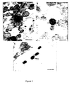
- Anti-uKHC showed diffuse labelling throughout axons with clusters of immunolabel for uKHC observed around electron dense axonal vesicles ( Figure 1A) . Only sparse background label was found extra-axonally. With pre-immune sera only sparse background immunolabel was present on sections and there was no labelling of axonal vesicles. Immunolabel for uKHC and VP5 was found in and around unenveloped HSV1 nucleocapsids, respectively, within axons ( Figure 1B ). The anti-VP5 antibody was specific with no labelling of axons in mock-infected cultures.
- kinesin is a heterotetramer consisting of two identical heavy and two identical light chains.
- the heavy chain has a three domain structure consisting of a N-terminal motor domain, highly conserved among the kinesin superfamily, a central stalk domain containing heptad repeats and a C-terminal tail domain.
- the present inventors used fragments of human uKHC tagged at the N-terminus with an oligohistidine (His) sequence, which were generated and characterised previously, as bait to identify kinesin-binding HSV1 proteins. In this work, the fragments were designated His-KHCstalk (amino acid residues 355-772) and His-KHCstalk/tail (residues 771-963).
- HisKHC fragments attached to nickel-activated beads were incubated with a lysate of mock or HSV1-infected Hep-2 cells.
- Analysis of eluted complexes by immunoblotting with anti-HSV1 identified predominantly a 65 and 20 kDa viral protein preferentially bound to KHCstalk/tail ( Figure 2A ).
- These viral proteins were subsequently identified as the tegument proteins VP16 ( Figure 2B ) and US11 ( Figure 2C ).
- VP16 does not bind directly to uKHC ( Figure 3B ).
- Figure 3C The presence of VP16 co-eluting with KHC after incubation with viral lysates can be explained by the observation that VP16 binds directly to US11 ( Figure 3C). This binding was observed with both native and recombinant VP16 (in the absence of other viral proteins). Therefore uKHC binds US11 which in turn binds VP16.
- This VP16/US11 interaction may represent an important structural interaction for US11 in the tegument of the virus or may play a regulatory role in infected cells.
- VP16 has also been shown to bind to other tegument proteins including vhs ( C. A. Smibert, B.
- US11 appears to be a multi-functional protein. It is a basic phosphoprotein which has RNA-binding activity, stably associates with 60S ribosomal subunits has a role in post-transcriptional regulation of gene expression and localizes to the nucleoli.
- the RNA-binding properties of US11 are dependent on the 20-24 RXP-repeats in the carboxy-half of the protein while the amino-half of US11 is required for transactivation of gene expression. It has been suggested that the N-terminal domain may be involved in transport and translation of mRNA from the nucleus to the cytoplasm. This amino domain may well also bind to kinesin.
- the US11 protein is well conserved between HSV1 and 2 (63% homology) with the greater variation at the N-terminus. There are no US11 homologues in varicella-zoster virus or rabies suggesting this virus may use different proteins for anterograde axonal transport. However, there are precedents though within the herpesviruses for similar functions on different proteins (eg entry into cells via gD in HSV ( J. Rajcani. A. Vojvodova. Acta Virol. 42, 103. 1998 ) and via gE/I and gB in varicella zoster virus C. Grose. Rev. Infect. Dis. Suppl. 11. S960. 1991 ).
Landscapes
- Health & Medical Sciences (AREA)
- Life Sciences & Earth Sciences (AREA)
- Chemical & Material Sciences (AREA)
- Organic Chemistry (AREA)
- Engineering & Computer Science (AREA)
- General Health & Medical Sciences (AREA)
- Genetics & Genomics (AREA)
- Bioinformatics & Cheminformatics (AREA)
- Zoology (AREA)
- Wood Science & Technology (AREA)
- General Engineering & Computer Science (AREA)
- Biomedical Technology (AREA)
- Pharmacology & Pharmacy (AREA)
- Public Health (AREA)
- Veterinary Medicine (AREA)
- Medicinal Chemistry (AREA)
- Virology (AREA)
- Animal Behavior & Ethology (AREA)
- Biochemistry (AREA)
- Biotechnology (AREA)
- Chemical Kinetics & Catalysis (AREA)
- Nuclear Medicine, Radiotherapy & Molecular Imaging (AREA)
- General Chemical & Material Sciences (AREA)
- Molecular Biology (AREA)
- Communicable Diseases (AREA)
- Immunology (AREA)
- Neurosurgery (AREA)
- Neurology (AREA)
- Physics & Mathematics (AREA)
- Biophysics (AREA)
- Plant Pathology (AREA)
- Microbiology (AREA)
- Gastroenterology & Hepatology (AREA)
- Oncology (AREA)
- Proteomics, Peptides & Aminoacids (AREA)
- Epidemiology (AREA)
- Medicines That Contain Protein Lipid Enzymes And Other Medicines (AREA)
- Peptides Or Proteins (AREA)
- Medicines Containing Antibodies Or Antigens For Use As Internal Diagnostic Agents (AREA)
- Medicines Containing Material From Animals Or Micro-Organisms (AREA)
Abstract
Description
- The present invention relates to viral infections, particularly cellular and tissue interactions during infection.
- Little is known about the mechanisms of transport of neurotropic viruses, such as Herpes simplex virus (HSV), varicella-zoster virus and rabies, within neurones. For these viruses, which replicate in the nucleus, anterograde transport from the cell body of dorsal root human ganglionic (DRG) neurons to the axon terminus occurs over long distances. In the case of HSV. there is increasing evidence for separate, fast anterograde axonal transport of viral nucleocapsids coated with tegument along microtubules (MTs) and of glycoproteins in axonal transport vesicles. An understanding of the mechanisms of transport of these viruses would be of benefit. Once this information is obtained, means to interrupt or prevent this transport would be a good candidate for antiviral therapy.
- HSV consists of an outer lipid envelope containing glycoproteins which surrounds the tegument. The inner capsid encloses the viral DNA to form the nucleocapsid. Early work has shown that HSV retrograde axonal transport in rat neurons utilizes microtubules (K. Kristensson, E. Lycke, J. Sjostrand. Acta Neuropathol, 17, 44, 1971). Previously, the present inventors have used a two chamber system to examine the mechanism of anterograde transport of HSV from infected dorsal root ganglionic (DRG) neurones in the central chamber along axonal fascicles to epidermal explants in the outer chamber. The velocity of viral transport (0.6 µm/s) indicated a mechanism based on fast anterograde transport (M. E. Penfold, P. Armati. A. L. Cunningham. Proc. Natl. Acad. Sci. USA 91, 6529. 1994). Using freeze-substitution transmission immuno-electron microscopy (TIEM) the virus was shown to be transported as unenveloped nucleocapsids, coated with tegument, separately from glycoproteins which are transported in vesicles (D. J. Holland. M. Miranda-Saksena. R. A. Boadle, P. J. Armati, A. L. Cunningham. J. Virol. 73, 8476, 1999). These findings were supported by experiments with nocodazole, an inhibitor of microtubule polymerization, and brefeldin A which inhibits transport through the Golgi. The transport of all three viral components, nucleocapsid, tegument and glycoproteins was inhibited by nocodazole, indicating their dependence on microtubule-associated transport Brefeldin A, however, inhibited glycoprotein transport but not nucleocapsids confirming that the two are transported along separate pathways and indicating the close association of HSV glycoproteins with Golgi/intermediate compartment membranes.
- The present inventors have now obtained useful evidence for the direct interaction between a viral structural tegument protein and an ubiquitous cellular protein. This new information has been used to develop methods and compositions to alter viral transport and movement in cells.
- In a first aspect, the present invention provides a method of preventing or reducing transport of a neurotropic virus within a neuron or cell, the method comprising providing to said neuron or cell a compound preventing binding between a structural tegument protein of the virus and a motor protein in the neuron or cell such that virus transport in the neuron is prevented or reduced.
- In a preferred embodiment, the neurotropic virus is Herpes simplex virus (HSV), varicella-zoster virus, or rabies virus, More preferably, the virus is HSV, the structural tegument protein is US11, and the motor protein is kinesin.
- The method according to the invention can be carried out by providing a compound to a neuron or cell capable of preventing binding between a structural tegument protein of a neurotropic virus and a motor protein in a neuron or cell.
- In one preferred form, the compound is a motor protein-like molecule which binds the structural tegument protein thereby preventing the normal interaction of the virus with cellular motor protein.
- The motor protein-like molecule can be any suitable molecule and would include mimics of the motor protein or a parts of the motor protein to which the structural tegument protein binds.
- When the motor protein is kinesin in the case of HSV, the kinesin-like molecule can be any suitable molecule and would include mimics of kinesin or a parts of kinesin to which the structural tegument protein US11 of HSV binds.
- In another preferred form, the compound is a structural tegument-like molecule which binds to a motor protein of a neuron or cell thereby preventing the normal interaction between the neuron or cell with the virus. The structural tegument-like molecule can be a mimic of a viral tegument protein or a part of the tegument protein to which a cellular motor protein binds, Preferably, the virus is Herpes simplex virus, the structural tegument protein is US11, motor protein is kinesin and the structural tegument-Like molecule is a mimic of US11 or a part of US11 to which the motor protein kinesin binds.
- Knowing how these types of viruses move in neurons to infect neurons other cells and ultimately form lesions is an important advance and is useful for the development of antiviral strategies. Usually, during the latent state these viruses do not,cause any real problems to an individual. It is during the active phase of the virus where clinical manifestations occur and suffering is caused. Preventing the actual transport of the virus down infected neurons should at least combat or prevent the clinical symptoms of re-infectian. Similarly, preventing movement up the neuron may prevent the formation of latent state of the virus in ganglia or at least reduce the incidence of re-occurrence of manifestations of the disease in infected individuals.
- In a second aspect, the present invention provides a modified neurotropic virus which has lost or has reduced ability to be transported in neurons, the virus comprising a mutation in a tegument protein such that the mutated tegument protein has abnormal interaction with a cellular motor protein.
- In a third aspect, the present invention provides use of a compound to prevent or reduce the interaction between a neurotropic viral tegument protein with a cellular motor protein to prevent or reduce viral transport in a neuron.
- In a preferred embodiment of the second and third aspects of the present invention, the neurotropic virus is Herpes simplex virus (HSV), varicella-zoster virus, or rabies virus. More preferably, the virus is HSV, the structural tegument protein is US11, and the motor protein is kinesin.
- In a fourth aspect, the present invention provides an antiviral composition comprising a compound capable of preventing binding between a structural tegument protein of a neurotropic virus and a motor protein in a neuron.
- Preferably, the neurotropic virus is selected from the group consisting of Herpes simplex virus, varicella-zoster virus, and rabies virus.
- Preferably, the virus is Herpes simplex virus, the structural tegument protein is US11, and the motor protein is kinesin.
- In one preferred form, the compound is a motor protein-like molecule which binds to the structural tegument protein of the virus thereby preventing normal interaction of the virus with cellular motor protein of a neuron. Preferably, the motor protein-like molecule comprises a mimic of a cellular motor protein or a part of the motor protein to which a structural tegument protein of a virus binds.
- In a further preferred form, the virus is Herpes simplex virus, the motor protein is kinesin and the motor protein-like molecule is a mimic of kinesin or a part of kinesin to which the structural tegument protein US11 of Herpes simplex virus binds.
- In another preferred form, the compound is a structural tegument-like molecule which binds to a motor protein of a neuron or cell thereby preventing the normal interaction between the neuron or cell with the virus. The structural tegument-like molecule can be a mimic of a viral tegument protein or a part of the tegument protein to which a cellular motor protein binds. Preferably, the virus is Herpes simplex virus, the structural tegument protein is US11, motor protein is kinesin and the structural tegument-like molecule is a mimic of US11 or a part of US11 to which the motor protein kinesin binds.
- The composition can further include additives, diluents, excipients and the like which can be used in clinical and health situations.
- This study raises many questions about the mechanisms of tegument formation, location of US11 in the tegument of HSV, the potential role of motor proteins other than kinesin(s) and homologous interactions mediating the transport of other neurotropic viruses. Studies to map minimal binding regions of US11 and uKHC and identification of other cellular factors which facilitate or contribute to the interaction will be useful. Inhibition of this interaction by peptides and/or peptidomimetic analogues could provide a new strategy for antiviral treatment for this and other neurotropic viruses. Incorporation of deletions into specifically attenuated live HSV (or other attenuated live neurotropic viruses) vaccine candidates could also be useful to prevent clinical recrudescence.
- Throughout this specification, unless the context requires otherwise, the word "comprise", or variations such as "comprises" or "comprising", will be understood to imply the inclusion of a stated element, integer or step, or group of elements, integers or steps, but not the exclusion of any other element, integer or step, or group of elements, integers or steps.
- In order that the present invention may be more clearly understood, preferred forms will be described with reference to the following examples and drawings.
-
-
Figure 1 . Transmission immune-electron micrographs of human fetal axons labelled with 5 and 10 nm immunogold particles recognising HSV major capsid protein VP5 and the stalk of human uKHC respectively. Arrows = 5 nm gold particles. Arrowheads = 10 nm gold particles. Bars = 200 nm (A and C) or 100 nm (B).- A Clustering of immunogold label for uKHC around axonal vesicles and diffuse labelling of other axons. Labelling with preimmune sera produced only sparse background label.
- B Colocalization of the two immunogold labels against uKHC and VP5 over a dense unenveloped viral particle in an axon. Arrows = VP5 (5 nm gold particle).
- C Extracellullar virus labelled only with anti-VP5. In the upper right corner is an axon with diffuse 10 nm immunolabel against uKHC. There is no immunolabel for uKHC over extracellular virus with only sparse background label outside axons.
-
Figure 2 . Identification of kinesin-binding HSV1 proteins. Mock or virus-infected Hep-2 cell lysates were incubated, as indicated (+), with His-KHC fragments bound to nickel-activated beads. Eluted kinesin-viral protein complexes were analysed by immunoblotting with appropriate antibodies.- A Polyclonal antibody to HSV1 identified major viral bands at 65 and 20 kDa bound preferentially to His-KHCstalk/tail.
- B-C The viral proteins were identified with monoclonal antibodies as VP16 (B) and US11 (C), respectively. In each immunoblot, the first two lanes are controls for antibody specificity.
-
Figure 3 . Direct binding of US11 to uKHC and VP16. Shown are immunoblots of protein complexes eluted from nickel-activated beads.- A Lysates of bacteria expressing untagged KLC or US11 were incubated, as indicated (+), with His-KHC fragments. US11 bound only to His-KHCstalk/tail in the absence of other HSV1 proteins (
lane 2, bottom panel). Overnight preincubation of the His-KHC fragments with KLC, which binds only to His-KHCstalk/tail (lane 4, top panel), prior to addition of US11, also showed binding of US11 but this was slightly reduced (lane 4, bottom panel). - B In a similar experiment, recombinant untagged VP16 did not bind to either His-KHC fragment (
lanes 2 and 3). Expression of VP16 in bacterial lysates was confirmed in lane 1. Native VP16 from HSV1-infected Hep-2 cells (lanes 4 and 5) or recombinant untagged VP16 (lanes 6 and 7) was incubated with His-US11 or His-KHCstalk. Both forms of VP16 interacted with His-US11.
- A Lysates of bacteria expressing untagged KLC or US11 were incubated, as indicated (+), with His-KHC fragments. US11 bound only to His-KHCstalk/tail in the absence of other HSV1 proteins (
-
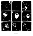
Figure 4 . Transport of structural HSV antigens into axons of infected dissociated rat neonatal neurons in vitro. Dissociated DRG neurons were infected with either wt HSV1 (CW strain), a US11 deletion mutant of HSV1 (R6604) or a US11 rescuant of R6604 (R6606) at 20 pfu/neuron. The appearance and transport into the principle axon of HSV1 tegument (US11), nucleocapsid (VP5) and envelope glycoprotein (gC) antigens was followed by serial fixation over 6 to 36 h. and then immunofluorescence and confocal microscopy. (A-I) HSV1 antigens in the axon at 24 h post infection (hpi). Bars = 10 µm (A and C) or 25 µm (B and D-I). US11 was present in axons infected with wt HSV1 (A) but was completely absent from neurons infected with R6604 (B). US11 was expressed in neurons and present in axons infected with R6606 (C). VP5 was present in axons infected with wt HSV1 (D) but was not detected in axons infected with R6604 (E). VP5 was present in axons infected with R6606 (F). gC was detected in axons whether infected with wt HSV1 (G). R6604 (H) or R6606 (I). -
Figure 5 . Ultrastructural distribution of enveloped and unenveloped nucleocapsids from an HSV1 US11 deletion mutant (R6604) in dissociated rat DRG neurons (26 hpi).- A Unenveloped (arrow) and enveloped (arrow head) nucleocapsids adjacent to the Golgi (G) within the cytoplasm of the cell body.
- B Enveloped virions (arrow) in the extracellular space adjacent to the cell body and longitudinal section of an axonal process (arrowhead) containing microtubules.
- HisKHC555-772. a oligohistidine (His)-tagged fragment of human uKHC (R. J. Diefenbach. J. P. Mackay. P. J. Armati, A. L. Cunningham. Biochemistry 37, 16663, 1998), was used to generate chicken polyclonal anti-uKHC. The cell pellet from a 1 litre bacterial culture expressing HisKHC555-772 was resuspended in 25 ml of column running buffer (20 mM sodium diphosphate. pH 7.4, 0.5 M NaCl), lysed by sonication, clarified by centrifugation, and applied to a
POROS 20 MC column (Perseptive Biosystems: 4.6 x 10 mm) which had been charged with nickel according to the manufacturer's instructions. The His-tagged fusion protein was eluted at 5 ml/min with 15 column volumes of a gradient from 0 to 0.5 M imidazole in running buffer. Pooled fractions containing purified HisKHC555-772 were then dialysed against PBS. Protein concentrations were determined using a Bio-Rad protein assay. A total of 2 mg of protein was then used to raise a polyclonal antibody in chickens (a service provided by the Institute of Medical and Veterinary Science. Veterinary Services Division. Gilles Plains, South Australia). Pre- and post-immune serum from a single immunised chicken was subsequently used in this study. The antibody was shown to be specific in immunoblots for human uKHC by comparing reactivity with the immunising antigen against human neuronal (n) KHC (expressed as a His-tagged fragment spanning amino acids 409-1032),.cDNAs for uKHC (F. Navone, et al., J. Cell Biol. 117, 1263, 1992) and nKHC (J. Niclas. F. Navone, N. Hom-Booher. R. D. Vale. Neuron 12, 1059, 1994)) were provided by R. Vale. - The HisKHC proteins were expressed, harvested, bound to, and eluted from nickel-activated beads as previously described (R. J. Diefenbach, J. P. Mackay. P. J. Armati. A. L. Cunningham, Biochemistry 37, 16663, 1998). Generation of virus stocks and titrations of wt HSV1 (CW1) and modified viruses were performed in Hep-2. For binding assays. Hep-2 cells were infected at a multiplicity of 5 pfu/cell and incubated for 24 h. Cell monolayers were washed twice with PBS. resuspended in PBS (1 x 10⁶ cells/ml), freeze-thawed 3 times, sonicated for 15 s, before addition of 1% (v/v) NP-40 and incubation for 1 h at 4°C. The soluble fraction was harvested by centrifugation at 4°C (10000g for 15 min) before addition (1 ml) to HisKHC fragments on nickel-activated beads. Beads were incubated overnight with rocking at 4°C, washed with 3 x 20 volumes of wash buffer (150 mM NaCl. 120 mM imidazole. 20 mM Tris-HCl, pH 7.9), prior to elution of bound protein complexes. Protein complexes were separated by SDS-PAGE and identified by immunoblotting as previously described (R. J. Diefenbach, J. P. Mackay. P. J. Armati. A. L. Cunningham, Biochemistry 37, 16663,* 1998). Antibodies used included mouse monoclonal anti-US11 (R. J. Roller, B. Roizman. J. Virol. 66, 3624, 1992) (provided by B. Roizman), mouse monoclonal anti-VP16 (C. McLean. et al., J. Gen. Virol. 63, 297, (1982) (LP1; provided by T. Minson) and rabbit polyclonal anti-HSV1 from Dako. Rabbit polyclonal antibody to HSV1 capsid protein VP5 was provided by G. Cohen and R. Eisenberg (G. H. Cohen, et al., J. Virol. 34, 521, 1980). Mouse monoclonal antibody to HSV1 gC and KLC (L2) was obtained from Chemicon International.
- Plasmid pRB4766 (H. Tsiang, E. Lycke. P. E. Ceccaldi, A. Ermine, X. Hirardot. J. Gen. Virol. 70, 2075. 1989) containing HSV1 US11 genomic DNA in pGEX-KG was provided by B. Roizman. An untagged US11 construct was generated by digestion of pRB4766 with NcoI and insertion into the NcoI site of pET-28a. This resulted in an additional five amino acids (MGRLE) at the N-terminus of US11. His-tagged US11 was constructed by inserting an EcoRI/FspI US11-containing fragment from pRB4766 into EcoRI/HindIII (Klenow filled-in) digested pET-28c. Between the oligohistidine tag and US11 sequence are inserted amino acids LDSMGRLE. Genomic DNA containing HSV1 VP16 was provided by P. O'Hare (T. A. Hughes. S. La Boissiere. P. O'Hare. J. Biol. Chem. 274, 16437, 1999: D. O'Reilly. O. Hanscombe. P. O'Hare. EMBO J. 16, 2420, 1997). The plasmid pPO54 consisted of the gene for VP16 inserted into the BamHI site of pBS. An untagged VP16 construct was generated by firstly digesting pP054 with BamHI and inserting into the BamHI site of pET-28c. VP16 was subsequently released from pET-28c by digestion with BamHI/XhoI and inserted into the yeast vector pACT2 also cut with BamHI/XhoI. VP16 was then released with an NcoI/XhoI digest and reinserted into the NcoIlXhoI site of pET-28a to allow expression of untagged VP16. The resulting fusion protein had an additional nineteen amino acids (MEAPGIRDPRSSFPYQPHP) at the N-terminus of VP16. Bacteria expressing untagged US11, VP16 or KLC (R. J. Diefenbach, J. P. Mackay, P. J. Armati, A. L. Cunningham, Biochemistry 37, 16663, 1998) were lysed in normal salt binding buffer (150 mM NaCl, 5 mM imidazole, 20 mM Tris-HCl, pH 7.9) for use in binding assays as described above.
- R6604 is a partial US11 gene deletion mutant which does not express the US11 protein and R6606 is US11 rescuant of R6604. To generate the HSV1 mutant R6604 (US11-), a plasmid carrying a deletion in the US11 coding sequence was constructed (pRB5173) by deleting the sequences present between BspEI and XhoI in the BamHI Z fragment of HSV1(F) genome. The 132 nucleotides deleted from the fragment removed all three ATG codons of the US11 ORF without affecting the adjacent US12 ORF. The plasmid was cotransfected with the R3630 recombinant virus DNA containing the thymidine kinase gene inserted into US12. The selected recombinant progeny (R6603) contained a US11 gene lacking the 132 nucleotides but an intact US12. The thymidine kinase gene at its natural locus was then restored to yield R6604. The genotype and phenotype of all recombinants was verified by Southern analysis and immunoreactivity of its proteins.
- The candidate motor proteins for viral transport should be those involved in microtubule-dependent fast axonal anterograde transport of organelles, most likely the conventional kinesins (ubiquitous and neuronal) or possibly the kinesin-related protein KIF3. The colocalization of kinesin and HSV1 nucleocapsids was examined in fetal human neurons using the DRG neuron-epidermal explant two chamber model system as previously described (D. J. Holland. M. Miranda-Saksena. R. A. Boadle. P. J. Armati, A. L. Cunningham. J. Virol. 73, 8476, 1999). Anterograde axonal transport of HSV1 (CW strain: D. J. Holland. M. Miranda-Saksena, R. A. Boadle. P. J. Armati. A. L. Cunningham, J. Virol. 73, 8476, 1999) was visualized using freeze-substitution TIEM. Dual immunogold labelling (5 and 10 nm gold particles) of axons was performed with chicken polyclonal antibody to ubiquitous kinesin heavy chain (uKHC) and rabbit polyclonal antibody to HSV1 major capsid protein VP5 (G. H. Cohen, et al., J. Virol. 34, 521, 1980).
- Anti-uKHC showed diffuse labelling throughout axons with clusters of immunolabel for uKHC observed around electron dense axonal vesicles (
Figure 1A) . Only sparse background label was found extra-axonally. With pre-immune sera only sparse background immunolabel was present on sections and there was no labelling of axonal vesicles. Immunolabel for uKHC and VP5 was found in and around unenveloped HSV1 nucleocapsids, respectively, within axons (Figure 1B ). The anti-VP5 antibody was specific with no labelling of axons in mock-infected cultures. Only VP5 and not kinesin immunolabel was present on extracellular virus (Figure 1C ). The immunolabelling for uKHC over intra-axonal virus (but not extra-axonal virus) was as intense as that around axonal vesicles and provides strong direct evidence that kinesin is involved in the anterograde axonal transport of HSV. The tegument protein VP16 has been demonstrated to colocalize with VP5-labelled nucleocapsids during anterograde transport (D. J. Holland, M. Miranda-Saksena, R. A. Boadle. P. J. Armati, A. L. Cunningham, J. Virol. 73, 8476, 1999). Therefore, tegument or outer capsid proteins are candidates for binding to conventional ubiquitous kinesin. - Conventional kinesin is a heterotetramer consisting of two identical heavy and two identical light chains. The heavy chain has a three domain structure consisting of a N-terminal motor domain, highly conserved among the kinesin superfamily, a central stalk domain containing heptad repeats and a C-terminal tail domain. The present inventors used fragments of human uKHC tagged at the N-terminus with an oligohistidine (His) sequence, which were generated and characterised previously, as bait to identify kinesin-binding HSV1 proteins. In this work, the fragments were designated His-KHCstalk (amino acid residues 355-772) and His-KHCstalk/tail (residues 771-963).
- HisKHC fragments attached to nickel-activated beads were incubated with a lysate of mock or HSV1-infected Hep-2 cells. Analysis of eluted complexes by immunoblotting with anti-HSV1 identified predominantly a 65 and 20 kDa viral protein preferentially bound to KHCstalk/tail (
Figure 2A ). These viral proteins were subsequently identified as the tegument proteins VP16 (Figure 2B ) and US11 (Figure 2C ). - To establish whether US11 and/or VP16 bound directly to uKHC. untagged forms of both were expressed in Escherichia coli. Addition of untagged US11 to HisKHC showed that US11 binds specifically to KHCstalk/tail (
Figure 3A . bottom panel). This also showed that the binding of US11 to uKHC can occur in the absence of other viral proteins although it may well be modulated in vivo by viral or cellular factors. A complex of uKHC and KLC was generated by preincubating HisKHCstalk/tail with untagged KLC (Figure 3A , top panel). This kinesin heavy/light chain complex also binds US11, though at a slightly reduced level when compared with heavy chain alone (Figure 3A ,bottom panel lanes 2 and 4). The minor reduction in US11 binding is probably due to steric effects and not competitive inhibition since KLC and US11 bind to different regions of His-KHCstalk/tail. Therefore, KLC appears not to be directly involved in binding US11 as there was no increase in the level of US11 bound to a heavy/light chain complex compared with heavy chain alone. A similar observation has been documented using a KHCstalk/tail fragment from sea urchin which can bind to vesicles in the absence of KLC. - In contrast, VP16 does not bind directly to uKHC (
Figure 3B ). The presence of VP16 co-eluting with KHC after incubation with viral lysates can be explained by the observation that VP16 binds directly to US11 (Figure 3C). This binding was observed with both native and recombinant VP16 (in the absence of other viral proteins). Therefore uKHC binds US11 which in turn binds VP16. This VP16/US11 interaction may represent an important structural interaction for US11 in the tegument of the virus or may play a regulatory role in infected cells. VP16 has also been shown to bind to other tegument proteins including vhs (C. A. Smibert, B. Popova, P. Xiao, J. P. Capone. J. R. Smiley. J. Virol. 68, 2339, 1994) and VP22 (G. Elliott, G. Mouzakitis. P. O'Hare. J. Virol. 69, 7932, 1995), as well as to glycoprotein (gD; Q. Zhu. R. J. Courtney, Virology 204, 590, 1994). Therefore US11, a structural viral protein proposed to be part of the tegument, may also interact with one or both of these other tegument proteins. - US11 appears to be a multi-functional protein. It is a basic phosphoprotein which has RNA-binding activity, stably associates with 60S ribosomal subunits has a role in post-transcriptional regulation of gene expression and localizes to the nucleoli. The RNA-binding properties of US11 are dependent on the 20-24 RXP-repeats in the carboxy-half of the protein while the amino-half of US11 is required for transactivation of gene expression. It has been suggested that the N-terminal domain may be involved in transport and translation of mRNA from the nucleus to the cytoplasm. This amino domain may well also bind to kinesin. The US11 protein is well conserved between HSV1 and 2 (63% homology) with the greater variation at the N-terminus. There are no US11 homologues in varicella-zoster virus or rabies suggesting this virus may use different proteins for anterograde axonal transport. However, there are precedents though within the herpesviruses for similar functions on different proteins (eg entry into cells via gD in HSV (J. Rajcani. A. Vojvodova. Acta Virol. 42, 103. 1998) and via gE/I and gB in varicella zoster virus C. Grose. Rev. Infect. Dis. Suppl. 11. S960. 1991).
- To test the hypothesis that the US11-uKHC interaction mediated anterograde transport of HSV into the axons of rat neonatal DRG neurons, the axonal transport of wild-type (wt) HSV1, a US11 deletion mutant of HSV1 and its rescuant were examined. The appearance and distribution of structural HSV protein antigens from the tegument (US11), nucleocapsid (VP5) and envelope (gC) and their anterograde transport into axons of dissociated rat neonatal DRG was followed by fixation at serial time points over 24 hours and immunofluorescence/confocal microscopy.
- Firstly. US11 antigen from wt HSV1 (CW clinical strain), was demonstrated to move down the axon (a criterion for its proposed role in anterograde axonal transport:
Figure 4A ). The protein was not expressed in the US11 deletion mutant of HSV1. R6604. but was present in the US11 rescuant of R6604. R6606 (Figure 4B and C ). The kinetics of transport of US11 into axons was similar for wt and R6606 (Figure 4A and C ). In neurons infected with wt HSV1. VP5 antigen was diffusely distributed in the cytoplasm of the cell body and transported throughout the axon by 24 hours (Figure 4D ). The kinetics of transport of US11 and VP5 from wt HSV1 into axons was similar, supporting colocalization of US11 with nucleocapsids (Figure 4A and D ). In contrast, deletion of US11 in R6604 abrogated nucleocapsid transport into axons since no VP5 antigen could be detected in the axons of infected neurons (Figure 4E ), although there was a similar diffuse cytoplasmic distribution of VP5 to wt HSV1 (Figure 4D and E ). Rescue of US11 expression in R6606 restored HSV1 nucleocapsid transport into and along axons at a rate similar to wt (Figure 4F ). Anterograde transport of gC from either wt HSV1, R6604 or R6606 into axons was unaffected (Figure 4G-I ). For R6604. transmission electron microscopy at late time points (26 hours) showed large numbers of enveloped virions in the extracellular space and in the cytoplasm of the cell body of neurons, concentrated around the Golgi (Figure 5A and B ). Unenveloped nucleocapsids were in variable abundance in the nuclei and sparsely scattered in the perinuclear cytoplasm, often adjacent to the Golgi (Figure 5A ). Thus the absence of US11 does not prevent transport across the nuclear membrane, normal envelopment and viral egress in the cell body. Thus, there is no observable functional disruption of the tegument. - These findings suggest that there are two separate mechanisms of viral egress, one via the cell body and the other via axon termini. The absence of US11 appears to disrupt the latter pathway between the cell body and the axon. The presence of US11 in the tegument, the similar kinetics of anterograde transport of US11 and nucleocapsid VP5, and the inability of a HSV mutant deleted in US11 to be transported into axons is consistent with the hypothesis that the observed US11-uKHC interaction has a major role in mediating anterograde transport of nucleocapsids into and along axons.
- Knowing how these types of viruses move in neurons to infect neurons, other cells and ultimately form lesions is an important advance and is useful for the development of antiviral strategies. Usually, during the latent state these viruses do not cause any real problems to an individual. It is during the active phase of the virus where clinical manifestations occur and suffering is caused. Preventing the actual transport of the virus down infected neurons should at least combat or prevent the clinical symptoms of re-infection. Similarly, preventing movement up the neuron may prevent the formation of latent state of the virus in ganglia or at least reduce the incidence of re-occurrence of manifestations of the disease in infected individuals.
- This work raises many possibilities about the mechanisms of tegument formation, location of US11 in the tegument of HSV. the potential role of motor proteins other than kinesin(s) and homologous interactions mediating the transport of other neurotropic viruses. Inhibition of this interaction by peptides and/or peptidomimetic analogues could provide a new strategy for antiviral treatment for this and other neurotropic viruses. Incorporation of deletions into specifically attenuated live HSV (or other attenuated live neurotropic viruses) vaccine candidates could also be useful to prevent clinical recrudescence.
Claims (19)
- A method of preventing or reducing transport of a neurotropic virus within a neuron or cell, the method comprising altering or preventing interaction between a structural tegument protein of the virus and a motor protein in the neuron or cell such that virus transport in the neuron or cell is prevented or reduced.
- The method according to claim 1 wherein the neurotropic virus is selected from the group consisting of Herpes simplex virus, varicella-zoster virus, and rabies virus.
- The method according to claim 2 wherein the virus is Herpes simplex virus, the structural tegument protein is US11, and the motor protein is kinesin.
- The method according to any one of claims 1 to 3 comprising providing to a neuron or cell a compound capable of altering or preventing interaction between a structural tegument protein of a neurotropic virus and a motor protein in the neuron or cell.
- The method according to claim 4 wherein the compound is a motor protein-like molecule which binds to a structural tegument protein of the virus thereby preventing the normal interaction of the virus and neuron or cell.
- The method according to claim 5 wherein the motor protein-like molecule comprises a mimic of a cellular motor protein or a part of the motor protein to which the structural tegument protein binds or specifically interacts.
- The method according to claim 6 wherein the virus is Herpes simplex virus, the motor protein is kinesin and the motor protein-like molecule is a mimic of kinesin or a part of kinesin to which the structural tegument protein US11 of Herpes simplex virus binds or specifically interacts.
- The method according to claim 4 wherein the compound is a structural tegument-like molecule which binds to a motor protein of a neuron or cell thereby preventing the normal interaction between the neuron or cell with the virus.
- The method according to claim 8 wherein the structural tegument-like molecule comprises a mimic of a viral tegument protein or a part of the tegument protein to which a cellular motor protein binds or specifically interacts.
- The method according to claim 9 wherein the virus is Herpes simplex virus, the structural tegument protein is US11. motor protein is kinesin and the structural tegument-like molecule is a mimic of US11 or a part of US11 to which the motor protein kinesin binds or specifically interacts.
- An antiviral composition comprising a compound capable of altering or preventing interaction between a structural tegument protein of a neurotropic virus and a motor protein in a neuron or cell.
- The antiviral composition according to claim 11 wherein the neurotropic virus is selected from the group consisting of Herpes simplex virus, varicella-zoster virus, and rabies virus.
- The antiviral composition according to claim 12 wherein the virus is Herpes simplex virus, the structural tegument protein is US11, and the motor protein is kinesin.
- The antiviral composition according to any one of claims 11 to 13 wherein the compound a motor protein-like molecule which binds to a structural tegument protein of the virus thereby preventing the normal interaction of the virus and neuron or cell.
- The antiviral composition according to claim 14 wherein the motor protein-like molecule comprises a mimic of a cellular motor protein or a part of the motor protein to which a structural tegument protein of a virus binds or specifically interacts.
- The antiviral composition according to claim 15 wherein the virus is Herpes simplex virus, the motor protein is kinesin and the motor protein-like molecule is a mimic of kinesin or a part of kinesin to which the structural tegument protein US11 of Herpes simplex virus binds or specifically interacts.
- The antiviral composition according to any one of claims 11 to 13 wherein the compound is a structural tegument-like molecule which binds to a motor protein of a neuron thereby preventing the normal interaction between the neuron or cell with the virus.
- The antiviral composition according to claim 17 wherein the structural tegument-like molecule comprises a mimic of a viral tegument protein or a part of the tegument protein to which a cellular motor protein binds or specifically interacts.
- The antiviral composition according to claim 18 wherein the virus is Herpes simplex virus, the structural tegument protein is US11, motor protein is kinesin and the structural tegument-like molecule is a mimic of US11 or a part of US11 to which the motor protein kinesin binds or specifically interacts.
Applications Claiming Priority (2)
| Application Number | Priority Date | Filing Date | Title |
|---|---|---|---|
| AUPQ1719A AUPQ171999A0 (en) | 1999-07-20 | 1999-07-20 | Neurotropic virus transport |
| EP00945443A EP1202741A4 (en) | 1999-07-20 | 2000-07-20 | TRANSPORT NEUROTROPER VIRUSES |
Related Parent Applications (1)
| Application Number | Title | Priority Date | Filing Date |
|---|---|---|---|
| EP00945443A Division EP1202741A4 (en) | 1999-07-20 | 2000-07-20 | TRANSPORT NEUROTROPER VIRUSES |
Publications (1)
| Publication Number | Publication Date |
|---|---|
| EP1992359A1 true EP1992359A1 (en) | 2008-11-19 |
Family
ID=3815900
Family Applications (2)
| Application Number | Title | Priority Date | Filing Date |
|---|---|---|---|
| EP07116331A Withdrawn EP1992359A1 (en) | 1999-07-20 | 2000-07-20 | Neurotropic virus transport |
| EP00945443A Ceased EP1202741A4 (en) | 1999-07-20 | 2000-07-20 | TRANSPORT NEUROTROPER VIRUSES |
Family Applications After (1)
| Application Number | Title | Priority Date | Filing Date |
|---|---|---|---|
| EP00945443A Ceased EP1202741A4 (en) | 1999-07-20 | 2000-07-20 | TRANSPORT NEUROTROPER VIRUSES |
Country Status (5)
| Country | Link |
|---|---|
| US (1) | US6953661B1 (en) |
| EP (2) | EP1992359A1 (en) |
| JP (1) | JP2003504407A (en) |
| AU (1) | AUPQ171999A0 (en) |
| WO (1) | WO2001005410A1 (en) |
Families Citing this family (6)
| Publication number | Priority date | Publication date | Assignee | Title |
|---|---|---|---|---|
| US6900214B2 (en) | 2001-03-29 | 2005-05-31 | Bristol-Myers Squibb Company | Cyano-substituted dihydropyrimidine compounds and their use to treat diseases |
| US6809102B2 (en) | 2001-03-29 | 2004-10-26 | Bristol-Myers Squibb Company | Cyano-substituted dihydropyrimidine compounds and their use to treat diseases |
| WO2010000012A1 (en) * | 2008-07-03 | 2010-01-07 | The University Of Sydney | Method for preventing transport of a neurotropic virus |
| WO2011119920A2 (en) | 2010-03-25 | 2011-09-29 | Oregon Health & Science University | Cmv glycoproteins and recombinant vectors |
| CN109843321A (en) | 2016-06-22 | 2019-06-04 | 国际艾滋病疫苗行动组织公司 | Recombinant cytomegalovirus vector as a tuberculosis vaccine |
| US11638749B2 (en) | 2017-10-17 | 2023-05-02 | International Aids Vaccine Initiative, Inc. | Tuberculosis antigen cassettes |
Citations (1)
| Publication number | Priority date | Publication date | Assignee | Title |
|---|---|---|---|---|
| WO2000018919A1 (en) * | 1998-09-28 | 2000-04-06 | Incyte Pharmaceuticals, Inc. | Human kinesin-like motor protein |
Family Cites Families (3)
| Publication number | Priority date | Publication date | Assignee | Title |
|---|---|---|---|---|
| US6326402B1 (en) * | 1998-08-12 | 2001-12-04 | Octamer, Inc. | Methods for treating viral infections using a compound capable of inhibiting microtubules |
| US6346410B1 (en) * | 2000-08-11 | 2002-02-12 | Cytokinetics, Inc. | Motor proteins and methods for their use |
| US6455293B1 (en) * | 2000-11-22 | 2002-09-24 | Cytokinetics, Inc. | Human kinesin-like protein HSKIF21B |
-
1999
- 1999-07-20 AU AUPQ1719A patent/AUPQ171999A0/en not_active Abandoned
-
2000
- 2000-07-20 US US10/031,492 patent/US6953661B1/en not_active Expired - Fee Related
- 2000-07-20 EP EP07116331A patent/EP1992359A1/en not_active Withdrawn
- 2000-07-20 WO PCT/AU2000/000873 patent/WO2001005410A1/en not_active Ceased
- 2000-07-20 EP EP00945443A patent/EP1202741A4/en not_active Ceased
- 2000-07-20 JP JP2001510465A patent/JP2003504407A/en active Pending
Patent Citations (1)
| Publication number | Priority date | Publication date | Assignee | Title |
|---|---|---|---|---|
| WO2000018919A1 (en) * | 1998-09-28 | 2000-04-06 | Incyte Pharmaceuticals, Inc. | Human kinesin-like motor protein |
Non-Patent Citations (1)
| Title |
|---|
| TOPP ET AL: "Centripetal transport of herpes simplex virus in human retinal pigment epithelial cells in vitro", NEUROSCIENCE, NEW YORK, NY, US, vol. 71, no. 4, 1 April 1996 (1996-04-01), pages 1133 - 1144, XP002953334, ISSN: 0306-4522 * |
Also Published As
| Publication number | Publication date |
|---|---|
| EP1202741A4 (en) | 2004-01-14 |
| WO2001005410A1 (en) | 2001-01-25 |
| AUPQ171999A0 (en) | 1999-08-12 |
| EP1202741A1 (en) | 2002-05-08 |
| JP2003504407A (en) | 2003-02-04 |
| US6953661B1 (en) | 2005-10-11 |
Similar Documents
| Publication | Publication Date | Title |
|---|---|---|
| Diefenbach et al. | Herpes simplex virus tegument protein US11 interacts with conventional kinesin heavy chain | |
| Brideau et al. | The Us9 gene product of pseudorabies virus, an alphaherpesvirus, is a phosphorylated, tail-anchored type II membrane protein | |
| Rodríguez et al. | African swine fever virus structural protein p54 is essential for the recruitment of envelope precursors to assembly sites | |
| JP3254128B2 (en) | Method for producing antibody against glycoprotein of HCMV | |
| Soliman et al. | Shuttling of the herpes simplex virus type 1 regulatory protein ICP27 between the nucleus and cytoplasm mediates the expression of late proteins | |
| Mears et al. | Identification of nuclear and nucleolar localization signals in the herpes simplex virus regulatory protein ICP27 | |
| Wright et al. | Four phosphoproteins with common amino termini are encoded by human cytomegalovirus AD169 | |
| Schang et al. | The latency-related gene of bovine herpesvirus 1 encodes a product which inhibits cell cycle progression | |
| Del Rio et al. | The pseudorabies virus VP22 homologue (UL49) is dispensable for virus growth in vitro and has no effect on virulence and neuronal spread in rodents | |
| Lynch et al. | Physical and functional interaction between the varicella zoster virus IE63 and IE62 proteins | |
| Majerciak et al. | Structural and functional analyses of Kaposi sarcoma-associated herpesvirus ORF57 nuclear localization signals in living cells | |
| Dubin et al. | Expression of herpes simplex virus type 1 glycoprotein L (gL) in transfected mammalian cells: evidence that gL is not independently anchored to cell membranes | |
| Preston et al. | The UL25 gene product of herpes simplex virus type 1 is involved in uncoating of the viral genome | |
| CA2177699A1 (en) | Vector, viral protein, nucleotide sequence coding therefor and method for inhibiting immune recognition | |
| US6013265A (en) | Vaccine composition for herpes simplex virus and methods of using | |
| JP2002509113A (en) | Vaccine compositions for herpes simplex virus and methods of use | |
| Chow et al. | Mutations that specifically impair the DNA binding activity of the herpes simplex virus protein UL42 | |
| US6953661B1 (en) | Method of preventing transport of a neurotropic virus and identifying agents for achieving same | |
| Tirabassi et al. | Molecular mechanisms of neurotropic herpesvirus invasion and spread in the CNS | |
| McMahon et al. | Interactions between human cytomegalovirus helicase–primase proteins | |
| Holden et al. | Identification and characterization of the ICP22 protein of equine herpesvirus 1 | |
| Malik et al. | Intracellular localization of the herpes simplex virus type-1 origin binding protein, UL9 | |
| Derbigny et al. | The EICP22 protein of equine herpesvirus 1 physically interacts with the immediate-early protein and with itself to form dimers and higher-order complexes | |
| Peden et al. | Effects of mutations within the SV40 large T antigen ATPase/p53 binding domain on viral replication and transformation | |
| Spaderna et al. | Deletion of gpUL132, a structural component of human cytomegalovirus, results in impaired virus replication in fibroblasts |
Legal Events
| Date | Code | Title | Description |
|---|---|---|---|
| PUAI | Public reference made under article 153(3) epc to a published international application that has entered the european phase |
Free format text: ORIGINAL CODE: 0009012 |
|
| AC | Divisional application: reference to earlier application |
Ref document number: 1202741 Country of ref document: EP Kind code of ref document: P |
|
| AK | Designated contracting states |
Kind code of ref document: A1 Designated state(s): AT BE CH CY DE DK ES FI FR GB GR IE IT LI LU MC NL PT SE |
|
| RIN1 | Information on inventor provided before grant (corrected) |
Inventor name: DIEFENBACH, EVE, MARGARET Inventor name: ARMATI, PATRICIA, JOAN Inventor name: MIRANDA-SAKSENA, MONICA Inventor name: HOLLAND, DAVID, JAMES Inventor name: DIEFENBACH, RUSSELL JOHN Inventor name: CUNNINGHAM, ANTHONY, LAWRENCE Inventor name: PENFOLD, MARK |
|
| AKX | Designation fees paid | ||
| REG | Reference to a national code |
Ref country code: DE Ref legal event code: 8566 |
|
| STAA | Information on the status of an ep patent application or granted ep patent |
Free format text: STATUS: THE APPLICATION IS DEEMED TO BE WITHDRAWN |
|
| 18D | Application deemed to be withdrawn |
Effective date: 20090520 |