EP1966243B1 - Tgf-beta binding antibodies - Google Patents
Tgf-beta binding antibodies Download PDFInfo
- Publication number
- EP1966243B1 EP1966243B1 EP06840328A EP06840328A EP1966243B1 EP 1966243 B1 EP1966243 B1 EP 1966243B1 EP 06840328 A EP06840328 A EP 06840328A EP 06840328 A EP06840328 A EP 06840328A EP 1966243 B1 EP1966243 B1 EP 1966243B1
- Authority
- EP
- European Patent Office
- Prior art keywords
- tgf
- seq
- amino acid
- acid sequence
- antibody
- Prior art date
- Legal status (The legal status is an assumption and is not a legal conclusion. Google has not performed a legal analysis and makes no representation as to the accuracy of the status listed.)
- Not-in-force
Links
Images
Classifications
-
- C—CHEMISTRY; METALLURGY
- C07—ORGANIC CHEMISTRY
- C07K—PEPTIDES
- C07K16/00—Immunoglobulins [IGs], e.g. monoclonal or polyclonal antibodies
- C07K16/18—Immunoglobulins [IGs], e.g. monoclonal or polyclonal antibodies against material from animals or humans
- C07K16/22—Immunoglobulins [IGs], e.g. monoclonal or polyclonal antibodies against material from animals or humans against growth factors ; against growth regulators
-
- A—HUMAN NECESSITIES
- A61—MEDICAL OR VETERINARY SCIENCE; HYGIENE
- A61P—SPECIFIC THERAPEUTIC ACTIVITY OF CHEMICAL COMPOUNDS OR MEDICINAL PREPARATIONS
- A61P29/00—Non-central analgesic, antipyretic or antiinflammatory agents, e.g. antirheumatic agents; Non-steroidal antiinflammatory drugs [NSAID]
-
- A—HUMAN NECESSITIES
- A61—MEDICAL OR VETERINARY SCIENCE; HYGIENE
- A61P—SPECIFIC THERAPEUTIC ACTIVITY OF CHEMICAL COMPOUNDS OR MEDICINAL PREPARATIONS
- A61P31/00—Antiinfectives, i.e. antibiotics, antiseptics, chemotherapeutics
-
- A—HUMAN NECESSITIES
- A61—MEDICAL OR VETERINARY SCIENCE; HYGIENE
- A61P—SPECIFIC THERAPEUTIC ACTIVITY OF CHEMICAL COMPOUNDS OR MEDICINAL PREPARATIONS
- A61P35/00—Antineoplastic agents
-
- A—HUMAN NECESSITIES
- A61—MEDICAL OR VETERINARY SCIENCE; HYGIENE
- A61P—SPECIFIC THERAPEUTIC ACTIVITY OF CHEMICAL COMPOUNDS OR MEDICINAL PREPARATIONS
- A61P35/00—Antineoplastic agents
- A61P35/04—Antineoplastic agents specific for metastasis
-
- A—HUMAN NECESSITIES
- A61—MEDICAL OR VETERINARY SCIENCE; HYGIENE
- A61P—SPECIFIC THERAPEUTIC ACTIVITY OF CHEMICAL COMPOUNDS OR MEDICINAL PREPARATIONS
- A61P9/00—Drugs for disorders of the cardiovascular system
-
- A—HUMAN NECESSITIES
- A61—MEDICAL OR VETERINARY SCIENCE; HYGIENE
- A61K—PREPARATIONS FOR MEDICAL, DENTAL OR TOILETRY PURPOSES
- A61K39/00—Medicinal preparations containing antigens or antibodies
- A61K2039/505—Medicinal preparations containing antigens or antibodies comprising antibodies
-
- C—CHEMISTRY; METALLURGY
- C07—ORGANIC CHEMISTRY
- C07K—PEPTIDES
- C07K2317/00—Immunoglobulins specific features
- C07K2317/20—Immunoglobulins specific features characterized by taxonomic origin
- C07K2317/24—Immunoglobulins specific features characterized by taxonomic origin containing regions, domains or residues from different species, e.g. chimeric, humanized or veneered
-
- C—CHEMISTRY; METALLURGY
- C07—ORGANIC CHEMISTRY
- C07K—PEPTIDES
- C07K2317/00—Immunoglobulins specific features
- C07K2317/50—Immunoglobulins specific features characterized by immunoglobulin fragments
- C07K2317/52—Constant or Fc region; Isotype
-
- C—CHEMISTRY; METALLURGY
- C07—ORGANIC CHEMISTRY
- C07K—PEPTIDES
- C07K2317/00—Immunoglobulins specific features
- C07K2317/50—Immunoglobulins specific features characterized by immunoglobulin fragments
- C07K2317/55—Fab or Fab'
-
- C—CHEMISTRY; METALLURGY
- C07—ORGANIC CHEMISTRY
- C07K—PEPTIDES
- C07K2317/00—Immunoglobulins specific features
- C07K2317/70—Immunoglobulins specific features characterized by effect upon binding to a cell or to an antigen
- C07K2317/74—Inducing cell proliferation
-
- C—CHEMISTRY; METALLURGY
- C07—ORGANIC CHEMISTRY
- C07K—PEPTIDES
- C07K2317/00—Immunoglobulins specific features
- C07K2317/70—Immunoglobulins specific features characterized by effect upon binding to a cell or to an antigen
- C07K2317/76—Antagonist effect on antigen, e.g. neutralization or inhibition of binding
-
- C—CHEMISTRY; METALLURGY
- C07—ORGANIC CHEMISTRY
- C07K—PEPTIDES
- C07K2317/00—Immunoglobulins specific features
- C07K2317/90—Immunoglobulins specific features characterized by (pharmaco)kinetic aspects or by stability of the immunoglobulin
- C07K2317/92—Affinity (KD), association rate (Ka), dissociation rate (Kd) or EC50 value
-
- C—CHEMISTRY; METALLURGY
- C07—ORGANIC CHEMISTRY
- C07K—PEPTIDES
- C07K2319/00—Fusion polypeptide
Definitions
- This invention provides a method of treating a patient with HER2-overexpressing metastatic breast cancer, comprising administering to the patent an effective amount of an antibody of the invention and an antibody that blocks signaling through the HER-2 receptor, i.e. trastuzumab.
- the K d of an invention Fab for mature human TGF- ⁇ 1 is in the range of about 0.009 pM to about 75.0 pM, is more preferably in the range of about 0.018 pM to about 50 pM, and is most preferably in the range of about 0.02 pM to about 25 pM.
- the K d of an invention Fab for mature human TGF- ⁇ 2 is in the range of about 0.0045 pM to about 60 pM, is more preferably in the range of about 0.002 pM to about 45 pM, and is most preferably in the range of about 0.02 pM to about 35 pM.
- An antibody is said to "neutralize” its antigen if antibody binding to the antigen results in complete or partial, inhibition or reduction, of a biological function of the antigen.
- Neutralization of a TGF- ⁇ isoform's biological activity is assessed by measuring the complete or partial, inhibition or reduction, of one or more in vitro or in vivo indicators of TGF- ⁇ activity such as, receptor binding, an inhibitory effect on cell growth; chemotaxis, apoptosis, intracellular protein phosphorylation, or signal transduction. Most preferably, the ability to neutralize TGF- ⁇ activity is assessed, as described herein, by an HT-2 cell proliferation assay or by measuring the inhibition of Smad2 phosphorylation.
- variable domain framework sequences are those that do not significantly affect the biological properties of an anti-TGF- ⁇ 1, - ⁇ 2, and - ⁇ 3 antibody embodiment - that is, the ability to bind with high affinity and neutralize mature human TGF- ⁇ 1, - ⁇ 2, and - ⁇ 3.
- frameworks additionally do not elicit significant immunogenic reactions when administered to a human.
- Framework sequences can be sequences of naturally occurring human antibodies or consensus sequences of several human antibodies.
- polynucleotide sequences encoding invention antibodies are also encompassed.
- identification of residues within antibody frameworks likely to influence antigen binding donor and acceptor sequences may be determined by aligning to sequence templates derived from antibody repertoires. "Invariant residues” (Kabat et al., 1991) and “key residues” (Chothia et al., 1989) may be identified, and canonical-class assignments of the donor antigen binding loops L1-L3, H1 and H2, respectively, may be determined by screening a proposed sequence against sequence templates (see, e.g., Martin & Thornton, 1996 Mol. Biol. 263:800-15 ).
- the plate is washed (2X) in wash buffer before adding blocking solution (100uL per well of 10 mg/ml BSA in wash buffer). After incubation ( ⁇ 1hr-22°C), the plate is washed (2X) with wash buffer then 100uL of either sample (diluted in buffer) or control (diluted in PBS) is added per well and incubated (1.5h-22°C). After incubation, the plate is washed (6X) with wash buffer then either anti-mouse kappa-peroxidase conjugate (diluted to 1:2000 in Blocking solution) or SA-HRP (diluted 1:10,000 in blocking solution) added (100uL/well). Test samples are left to incubate (1h-22°C) before adding 100uL of OPD substrate/well. After color development ( ⁇ 10min), the 96-well plate is measured at an absorbance of 490nm.
Abstract
Description
- The invention relates to treatment of cell proliferative diseases, disorders, and conditions associated with TGF-β. In particular, the invention provides antibodies and antigen-binding fragments thereof that neutralize mature, human TGF-β1, β2, and β3.
- The TGF-β protein family consists of three distinct isoforms (TGF-β1, β2, and β3) whose pathways activate and regulate multiple gene responses that influence disease states such as, e.g., cell proliferative, inflammatory, and cardiovascular conditions. TGF-β isoform expression in cancer is complex and variable with different combinations of TGF-β isoforms having different roles in particular cancers. For example, TGF-β1 and -β3 play a greater role in ovarian cancer and its progression than TGF-β2; while TGF-β1 and -β2 expression is greater in higher grade chondrosarcoma tumors than -β3. In human breast cancer, TGF-β1 and -β3 are highly expressed, with -β3 expression correlating with overall survival - patients with node metastasis and positive TGF-β3 expression have poor prognostic outcomes. However, in colon cancer, TGF-β1 and -β2 are more highly expressed than -β3 and are present at greater circulating levels than in cancer-free individuals. In glioma cancer, TGF-β2 is pivotal for cell migration. Consequently, there is a need to modulate multiple TGF-β isoform expression in cell proliferation conditions such as cancer.
-
US 5,571,714 discloses the use of anti-TGF antibodies in treating malignancies and metastatic cancer, and in particular, discloses a murine antibody designated 1D11.16 (ATCC No. HB9849) that is said to bind both TGF-β1 and -β2. This document states that antibody 1D11.16 binds TGF-β2 with a Ka of merely 3.4 x 10⁸ L/mol (Kd = I/Ka). -
WO 2005/097832 discloses humanized anti-TGF-beta antibodies for the treatment of cancer. - The treatment of cell proliferative disorders, such as malignancies and cancers, may be improved by use of antibodies that neutralize mature, human, TGF-β1, -β2, and -β3 with improved binding kinetics and affinity. Substantial physical and chemical stability, adequate pharmacokinetics, and good solubility are also desirable for a pharmaceutical product. Consequently, an unmet need remains for antibodies having characteristics suitable for the pharmaceutical treatment of cell proliferative disorders.
- The present invention provides monoclonal antibody compositions that neutralize mature human TGF-β1, -β2 and -β3 with a Kd less than 4 pM for mature human TGF-β1, and a Kd less than 8 pM for mature human TGF-β2, and a Kd less than 4 pM for mature human TGF-β3. The invention also provides antibodies having the following combinations of light chain-(LCVR) and heavy chain-variable region (HCVR) sequences: SEQ ID NO: 27 and 51, 27 and 59, 27 and 60, 27 and 61, 27 and 62, 27 and 63, 27 and 64, 28 and 52, 29 and 51, 30 and 53, 31 and 54, 32 and 55, 33 and 56, 34 and 51, 34 and 57, 35 and 58, 36 and 51, 36 and 69, 36 and 75, 37 and 51, 38 and 51, 39 and 51, 40 and 51, 41 and 64, 41 and 67, 42 and 66, 43 and 68, 44 and 66, 45 and 51, 45 and 69, 46 and 70, 46 and 71, 47 and 71, 48 and 72, 49 and 73, 50 and 65, or 50 and 74. The invention also includes antigen-binding fragments of such antibodies, as well as antibodies and fragments having human or humanized frameworks and constant regions. The antibodies and fragments of the invention are useful to treat cell proliferative diseases, disorders, and conditions.
- According to the first aspect of the present invention, there is provided a monoclonal anti-TGF-β antibody or antigen-binding fragment thereof, that neutralizes mature human TGF-β1, TGF-β2 and TGF-β3, comprising a heavy chain variable region (HCVR) and a light chain variable region (LCVR), wherein said HCVR and said LCVR consists of:
- a) a HCVR having the amino acid sequence shown in SEQ ID NO: 64 and
a LCVR having the amino acid sequence shown in SEQ ID NO:41; or - b) a HCVR having the amino acid sequence shown in SEQ ID NO: 68 and
a LCVR having the amino acid sequence shown in SEQ ID NO: 43; or - c) a HCVR having the amino acid sequence shown in SEQ ID NO: 69 and
a LCVR having the amino acid sequence shown in SEQ ID NO: 45; or - d) a HCVR having the amino acid sequence shown in SEQ IDNO: 70 and
a LCVR having the amino acid sequence shown in SEQ ID NO: 46; or - e) a HCVR having the amino acid sequence shown in SEQ ID No: 74 and
a LCVR having the amino acid sequence shown in SEQ ID No: 50. - Preferably, a monoclonal antibody or antigen-binding fragment thereof according to claim 1, comprises a HCVR having the amino acid sequence shown in SEQ ID NO: 69 and a LCVR having the amino acid sequence shown in SEQ ID NO: 45.
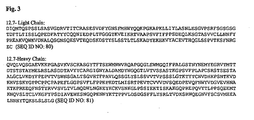
- A preferred monoclonal antibody of the present invention comprises a heavy chain having the amino acid sequence shown in SEQ ID NO: 81 and a light chain having the amino acid sequence shown in SEQ ID NO: 80.
- According to a second aspect of the present invention, there is provided a pharmaceutical composition, comprising a monoclonal antibody or antigen- binding fragment thereof according to the present invention, and a pharmaceutically acceptable carrier, diluent, or excipient.
- According to a third aspect of the present invention, there is provided a monoclonal antibody or antigen-binding fragment thereof according to the present invention for use as a medicament.
- Preferably, the monoclonal antibody of the present invention is used in the treatment of a cell proliferative disorder in a subject in need thereof. More preferably, the monoclonal antibody of the present invention is used in the treatment of a cell proliferative disorder selected from myelodysplastic syndrome (MDS)/myeloproliferative disorder (MPD), breast cancer, prostate cancer, ovarian cancer, hepatocellular carcinoma, pancreatic cancer, multiple myeloma, colorectal cancer, hairy cell leukemia, chronic myelogenous leukemia, and acute myelogenous leukemia.
- Antibodies of the present invention neutralize mature human TGF-β1, mature human TGF-β2, and mature human TGF-β3, and have an IC50 of less than or equal to about 100 pM for mature human TGF-β1, and an IC50 of less than or equal to about 400 pM for mature human TGF-β2, and an IC50 of less than or equal to about 200 pM for mature human TGF-β3 in an in vitro HT-2 Cell neutralization assay.
- Antibodies of the present invention comprise a heavy chain variable region (HCVR) and a light chain variable region (LCVR), wherein said HCVR comprises a peptide at CDRH1 with a sequence as shown in SEQ ID NO: 96 or SEQ ID NO: 100, a peptide at CDRH2 with a sequence as shown in SEQ ID NO: 97, SEQ ID NO: 99, SEQ ID NO: 101, or SEQ ID NO: 102, and a peptide at CDRH3 with a sequence as shown in SEQ ID NO: 98, and wherein said LCVR comprises a peptide at CDRL1 with a sequence as shown in SEQ ID NO: 88, SEQ ID NO: 91, SEQ ID NO: 92, or SEQ ID NO: 93, a peptide at CDRL2 with a sequence as shown in SEQ ID NO: 89 or SEQ ID NO: 94, and a peptide at CDRL3 with a sequence as shown in SEQ ID NO: 90, or SEQ ID NO: 95.
- Antibodies of the present invention further comprise a human framework region.
- The invention provides a method of treating cell proliferative disorders in a mammal, preferably a primate, and more preferably a human, comprising administering to a mammal in need of such treatment an effective amount of an antibody of the invention or a fragment thereof.
- The invention provides a method of treating a disease or condition in which fibrogenesis and/or angiogenesis mediated by TGF-β are implicated in a mammal, preferably a primate, and more preferably a human, including myelodysplastic syndrome (MDS)/myeloproliferative disorder (MPD), breast cancer, prostate cancer, ovarian cancer, hepatocellular carcinoma, pancreatic cancer, multiple myeloma, colorectal cancer, other hematological malignancies (hairy cell leukemia, CML, AML,etc), comprising administering to a mammal in need of such treatment an effective amount of an antibody of the invention or a fragment thereof. This may further comprise administering to the mammal an effective amount of a therapeutic agent other than anti-TGF-β antibodies, such as a chemotherapeutic agent, anti-angiogenic agent, or cytotoxic chemotherapy.
- This invention provides a method of treating a patient with HER2-overexpressing metastatic breast cancer, comprising administering to the patent an effective amount of an antibody of the invention and an antibody that blocks signaling through the HER-2 receptor, i.e. trastuzumab.
- This invention also provides the use of an antibody of the invention for the manufacture of a medicament for treating cell proliferatvie disorders in mammals. Additionally, this invention provides a pharmaceutical composition adapted for treating cell proliferatvie disorders comprising an antibody of the invention in combination with one or more pharmaceutically acceptable excipients, carriers, or diluents thereof. The invention also provides the use of an antibody of the invention for the manufacture of a medicament for treating a disease or condition capable of being improved or prevented by neutralizing TGF-β activities.
- This invention also provides the use of an antibody of the invention for the manufacture of a medicament for the treatment of a disease or condition in which fibrogenesis and/or angiogenesis mediated by TGF-β are implicated in a mammal, preferably a primate, and more preferably a human, including MDS/MPD, breast cancer, prostate cancer, ovarian cancer, hepatocellular carcinoma, pancreatic cancer, multiple myeloma, colorectal cancer, other hematological malignancies (hairy cell leukemia, CML, AML,etc), comprising administering to a mammal in need of such treatment an effective amount of an antibody of the invention or a fragment thereof. This may further comprise administering to the mammal an effective amount of a therapeutic agent other than anti-TGF-β antibodies, such as a chemotherapeutic, anti-angiogenic, or cytotoxic agent, or a cytokine. Furthermore, this invention provides a pharmaceutical composition adapted for the treatment of conditions in which neutralization or reduction of TGF-β activities would be beneficial in a mammal, including MDS/MPD, breast cancer, prostate cancer, ovarian cancer, hepatocellular carcinoma, pancreatic cancer, multiple myeloma, colorectal cancer, other hematological malignancies (hairy cell leukemia, CML, AML,etc), comprising an antibody of the invention in combination with one or more pharmaceutically acceptable excipients, carriers, or diluents. The invention also provides the use of an antibody of the invention for the manufacture of a medicament for treating a disease or condition capable of being improved or prevented by neutralizing or reducing TGF-β activities.
-
-
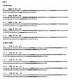
FIGS. 1A and B show the amino acid sequences of the heavy chain and light chain variable regions of preferred antibodies of the invention respectively. The CDR domains are in bold print. -
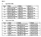
FIGs. 2A and B shows the alignment of the amino acid sequence of the CDR domains of preferred antibodies of the present invention. The variations are in bold print and underlined. - With respect to the binding and neutralization capabilities of antibodies of the invention, the TGF-β referred to herein is the mature, human, biologically active form of TGF-β1, TGF-β2, and TGF-β3 (SEQ ID NO: 1, 2, and 3). See also, e.g., NCBI accession No: P01137, NP_003229, and NP_003230 describing DNA and amino acid sequences of, respectively, human TGF-β1, -β2, and -β3 (including their precursor domains and mature portions). A mature disulfide-linked homodimeric human TGF-β1, -β2, or -β3 contains two 112 amino acid residue polypeptides and has a predicted molecular mass of approximately 25 kDa. Antibodies of the invention bind and neutralize such mature TGF-β isoforms but do not show significant binding of latent TGF-β1, -β2, and -β3 isoforms under similar conditions.
- Affinity and binding kinetic improvements solve antibody problems, for example, by helping to increase pharmacokinetic and safety profiles; by reducing dosing, toxicity, and cost of therapy; and by improving biological efficacy. The dissociation constant (Kd) of an antibody may be found using art methods, for example, Kinexa® (see, e.g., Darling, et al., 2004 ASSAY and Drug Development Technologies2:647-57) or BIAcore® AB (Upsala, Sweden), or adapting Karlsson et al., 1991 J. Immunol. Methods 145, 299-340. The term "kon" herein refers to the association or on-rate constant, or specific reaction rate, of the forward, or antibody:antigen complex-forming reaction, M-1sec-1. The term "koff" herein, refers to the dissociation or "off-rate" constant, or specific reaction rate, for dissociation of an antibody from an antibody:antigen complex, (sec-1). The term "Kd" herein, refers to the dissociation constant of a particular antibody:antigen complex - calculated as Kd = koff/kon. Ka is the inverse of Kd.
- The Kd of an invention antibody for mature human TGF-β1 is in the range of about 0.0001 pM to about 5.0 pM. More preferably, it is in the range of about 0.001 pM to about 3.0 pM. Most preferably, it is in the range of about 0.01 pM to about 0.5 pM. The Kd of an invention antibody for mature human TGF-β2 is in the range of about 0.01 pM to about 5.0 pM. More preferably, in the range of about 0.05 pM to about 2.0 pM, and most preferably, it is in the range of about 0.1 pM to about 1.5 pM. The Kd of an invention antibody for mature human TGF-β3 is in the range of about 0.005 pM to about 3 pM. More preferably, it is in the range of about 0.05 pM to about 2.8 pM. Most preferably, it is in the range of about 0.2 pM to about 2 pM. Preferably the antibodies of the invention are characterized by a Kd of 4.0x10⁻¹²M or less for mature human TGF-β1, a Kd of 8.0x10⁻¹²M or less for mature human TGF-β2, and a Kd of 4.0x10⁻¹²M or less for mature human TGF-β3.
- The Kd of an invention antibody for TGF-β1 is at least 2 fold greater than the Kd of 1D11.16 for TGF-β1, more preferably is at least 25 fold greater, and most preferably is at least 50 fold greater. To be clear, greater affinity would mean a smaller Kd value. The Kd of an invention antibody for TGF-β2 is at least 1.5 fold greater than the Kd of 1D11.16 for TGF-β2, more preferably is at least 10 fold greater, and most preferably is at least 20 fold greater. The Kd of an invention antibody for TGF-β3 is at least 4 fold greater than the Kd of 1D11.16 for TGF-β3, more preferably is at least 6 fold greater, and most preferably is at least 10 fold greater.
- The Kd of an invention Fab for mature human TGF-β1 is in the range of about 0.009 pM to about 75.0 pM, is more preferably in the range of about 0.018 pM to about 50 pM, and is most preferably in the range of about 0.02 pM to about 25 pM. The Kd of an invention Fab for mature human TGF-β2 is in the range of about 0.0045 pM to about 60 pM, is more preferably in the range of about 0.002 pM to about 45 pM, and is most preferably in the range of about 0.02 pM to about 35 pM. The Kd of an invention Fab for TGF-β1 is at least 30 fold greater than the Kd of 1D11.16 for TGF-β1, more preferably is at least 300 fold greater, and most preferably is at least 1500 fold greater. The Kd of an invention Fab for TGF-β2 is at least 10 fold greater than the Kd of 1D11.16 for TGF-β2, more preferably is at least 100 fold greater, and most preferably is at least 500 fold greater.
- Invention antibodies exhibit koff for mature human TGF-β1 preferably in the range of about 110 to about 1 (all values for koff are x 10-6 s-1), more preferably in the range of about 50 to about 6, and even more preferably in the range of about 15 to about 4; for mature human TGF-β2 preferably in the range of about 240 to about 1, more preferably in the range of about 50 to about 2, and even more preferably in the range of about 20 to about 4; and for mature human TGF-β3 preferably in the range of about 90 to about 1, more preferably in the range of about 50 to about 4, and even more preferably in the range of about 35 to about 6. Invention antibodies exhibit koff improvement over 1D11.16 for TGF-β1, -β2, and -β3 in the range of about 2 to about 50 fold, more preferably in the range of about 2.5 to about 25 fold, and even more preferably in the range of about 3 to about 15 fold.
- In another embodiment, an invention antibody has a kon greater than 5 for mature human TGF-β1, greater than 2.8 for mature human TGF-β2, and greater than 1.56 for mature human TGF-β3 (all values for kon are x 107 M-1 s-1). The kon for mature human TGF-β1 is preferably in the range of about 1.5 to about 60, more preferably in the range of about 2 to about 45, and even more preferably in the range of about 3 to about 30; for mature human TGF-β2 is preferably in the range of about 1 to about 40, more preferably in the range of about 1 to about 25, and even more preferably in the range of about 1 to about 15; and for mature human TGF-β3 is preferably in the range of about 1.2 to about 20, more preferably in the range of about 1.2 to about 10, and even more preferably in the range of about 1.2 to about 5. Invention antibodies exhibit an average kon improvement over 1D11.16 for TGF-β1, -β2, and -β3 preferably in the range of about 1.5 to about 40 fold, more preferably in the range of about 2 to about 15 fold, and even more preferably in the range of about 2 to about 10 fold. In a preferred embodiment, an invention antibody binds TGF-β1 over TGF-β2 and β3.
- An antibody is said to "neutralize" its antigen if antibody binding to the antigen results in complete or partial, inhibition or reduction, of a biological function of the antigen. Neutralization of a TGF-β isoform's biological activity is assessed by measuring the complete or partial, inhibition or reduction, of one or more in vitro or in vivo indicators of TGF-β activity such as, receptor binding, an inhibitory effect on cell growth; chemotaxis, apoptosis, intracellular protein phosphorylation, or signal transduction. Most preferably, the ability to neutralize TGF-β activity is assessed, as described herein, by an HT-2 cell proliferation assay or by measuring the inhibition of Smad2 phosphorylation.
- Antibodies of the invention neutralize mature human TGF-β1, mature human TGF-β2, and mature human TGF-β3, and have an IC50 of less than or equal to about 100 pM 75 pM, 50 pM, 25 pM, 17.5 pM, 10 pM, 4 pM, or 3pM for mature human TGF-β1, an IC50 of less than or equal to about 400 pM, 345 pM, 200 pM, 100 pM, 77.5 pM, 50 pM, 40 pM, or 23 pM for mature human TGF-β2, and an IC50 of less than or equal to about 200 pM, 115 pM, 105 pM, 75 pM, 50 pM, 45 pM, or 35 pM for mature human TGF-β3 in an in vitro HT-2 Cell neutralization assay.
- Improved neutralizing activity increases pharmacokinetic and safety profiles; reduces dosing, toxicity, and cost of therapy; and improves biological efficacy. In a preferred embodiment, the IC50 of an invention antibody in the HT-2 cell proliferation/neutralization assay as described herein is less than about 20 pM for TGF-β1, less than about 325 pM for TGF-β2, and less than about 125 pM for TGF-β3. Against TGF-β1, an invention antibody preferably has an IC50 in the range of about 0.1 to about 50 pM, more preferably in the range of about 0.1 to about 30 pM, and most preferably in the range of about 0.1 to about 20 pM. Against TGF-β2, an invention antibody preferably has an IC50 in the range of about 1 to about 400 pM, more preferably in the range of about 10 to about 350 pM, and most preferably in the range of about 15 to about 325 pM. Against TGF-β3, an invention antibody preferably has an IC50 in the range of about 0.1 to about 200 pM, more preferably in the range of about 1 to about 150 pM, and most preferably in the range of about 10 to about 125 pM.
- In another embodiment, invention antibodies exhibit on average an improvement of IC50 over 1D11.16 in an HT-2 cell proliferation assay for TGF-β1 in the range of about 100 to about 600 fold, more preferably in the range of about 200 to about 500 fold, and even more preferably in the range of about 300 to about 400 fold; for TGF-β2 in the range of about 10 to about 400 fold, more preferably in the range of about 25 to about 300 fold, and even more preferably in the range of about 50 to about 200; and for TGF-β3 in the range of about 2 to about 200 fold, more preferably in the range of about 4 to about 50 fold, and even more preferably in the range of about 6 to about 25.
- The concentration of an antibody required to neutralize a TGF-β activity is dependent on various parameters, such as, e.g., cytokine concentration, cell type, growth conditions, and type of activity studied. The neutralization character of an invention antibody is assessed by measuring the degree of inhibition of the phosphorylation of Smad2 protein in an U87MG human tumor xenograft model assay, as described herein. In another preferred embodiment, an invention antibody has a TED50 (therapeutic effective dose) in such an assay in the range of about 100 to about 200 mg/kg, more preferably in the range of about 50 to about 80 mg/kg; and even more preferably in the range of about 5 to about 25 mg/kg.
- Certain antibodies and fragments of the invention have specific CDRs: VHCDR1 X1X2WMN [SEQ ID NO:10, where X1 is either T or S; and X2 is either E, or Y]; VHCDR2 QIFPX1X2GSTNYX3EM4EG [SEQ ID NO:11, where X1 is either A, or F; X2 is either S, T, or L; X3 is either N, G, S, D, or A; X4 is either F, or Y]; VHCDR3 GX1GNYALDAMDY [SEQ ID NO:12, where X1 is either D, I, M, Y, L, V, Q, or F]; VLCDR1 RASESVDX1X2GNSFMH [SEQ ID NO:13, where X1 is either S, Y, F, or L; X2 is either Y or W]; VLCDR2 X1ASNLES [SEQ ID NO:14, where X1 is either L, or Y]; and VLCDR3 X1QX2X3EDPLT [SEQ ID NO:15, where X1 is either Q, T, or C; X2 is either N, or H; X3 is either N, I, M, D, T, or A].
- Further encompassed are antibodies created with CDRs from such formulae after engineering the CDRs (in appropriate orientation) in human or humanized antibody framework sequences to produce invention compositions that neutralize mature, human TGF-β1, -β2, and -β3. Any art-known method may be used to incorporate specific CDRs within framework sequences. As described, variable human or humanized framework sequences can be derived from any germline or rearranged human variable domain, or, e.g., synthetic variable domain based on consensus sequences of known human variable domains. Preferred variable domain framework sequences are those that do not significantly affect the biological properties of an anti-TGF-β1, -β2, and -β3 antibody embodiment - that is, the ability to bind with high affinity and neutralize mature human TGF-β1, -β2, and -β3. Preferably, such frameworks additionally do not elicit significant immunogenic reactions when administered to a human. Framework sequences can be sequences of naturally occurring human antibodies or consensus sequences of several human antibodies.
- Non-limiting examples of framework sequences for the heavy chain variable region of antibody embodiments of the invention include the VH segment DP-5 (Tomlinson, et al. 1992 J. Mol. Biol. 227:776-98) and the J segment JH4, JH1 or JH5 (Ravetch, et al. 1981 Cell 27:583-91). The Vk segment L1 (Cox, et al. 1994 Eur. J. Immunol. 24:827-36) and the J segment Jk4 (Hieter, et al. 1982 J. Biol. Chem. 10:1516-22) are non-limiting example framework sequences for the light chain variable region.
- In a preferred embodiment, the HCVR FR1 framework comprises [SEQ ID NO: 16]; the HCVR FR2 framework comprises [SEQ ID NO:17]; the HCVR FR3 framework comprises [SEQ ID NO:18]; and the HCVR FR4 framework comprises [SEQ ID NO:19]. In another preferred embodiment, the LCVR FR1 framework comprises [SEQ ID NO:20]; the LCVR FR2 framework comprises [SEQ ID NO:21], the LCVR FR3 framework comprises [SEQ ID NO:22]; and the LCVR FR4 framework comprises [SEQ ID NO:23]. In another preferred embodiment, a HCVR framework comprises [SEQ ID NO: 86] wherein X1 is either S, Y, F, or L; X2 is either Y or W; X3 is either A, or F; X4 is either S, T, or L; X5 is either N, G, S, D, or A; X6 is either F, or Y; and X7 is either D, I, M, Y, L, V, Q, or F. In another preferred embodiment, a LCVR framework comprises [SEQ ID NO: 87] wherein X1 is either T or S; X2 is either E, or Y; X3 is either is either L, or Y; X4 is either Q, T, or C; X5 is either N, or H; and X6 is either N, I, M, D, T, or A.
- In another preferred embodiment, framework and constant regions may contain alterations, deletions, additions, substitutions, or any combination thereof as compared with human sequences. Frameworks and constant regions in which 1, 2, 3, 4, 5, 6, 7, 8, 9, or 10 amino acids are substituted, deleted, or added in any combination are preferred. In another embodiment, the framework has 85-99% sequence identity to a disclosed framework herein.
- In one embodiment, a preferred heavy chain constant region for use with an antibody binding composition of the invention is an IgG constant region. In a more preferred embodiment, the IgG constant region is an IgG1 constant region or an IgG4 constant region (even more preferably are constant regions of [SEQ ID NO:24]; or [SEQ ID NO:25]). A preferred light chain constant region sequence of the invention is [SEQ ID NO:26]. In another preferred embodiment, antibody, binding compositions contain the IgG1 Heavy chain constant region or the IgG4 Heavy chain constant region and the kappa Light chain constant region.
- Also encompassed are polynucleotide sequences encoding invention antibodies. When engineering antibodies using invention CDRs, identification of residues within antibody frameworks likely to influence antigen binding donor and acceptor sequences may be determined by aligning to sequence templates derived from antibody repertoires. "Invariant residues" (Kabat et al., 1991) and "key residues" (Chothia et al., 1989) may be identified, and canonical-class assignments of the donor antigen binding loops L1-L3, H1 and H2, respectively, may be determined by screening a proposed sequence against sequence templates (see, e.g., Martin & Thornton, 1996 Mol. Biol. 263:800-15). Residues at the VH/VL interface (Chothia et al., 1985) and residues known to be structurally conserved at core sites (Chothia et al., 1998) are compared with corresponding donor and acceptor residues. Non-matching donor and acceptor framework residues at these sites are analyzed based on information from other antibodies of known structure (Berman, et al., 2000 Nucl. Acids Res. 28(1):235-42). Selection of human frameworks as templates for humanization of non-human V regions can define subsequent decisions regarding which residues to humanize. Choosing homologous templates from antibodies with known crystal structure, from germline, non-germline, or consensus sequences derived from available data bases can be employed (see, e.g., Routledge et al. (Routledge, et al., 1993 in Protein Engineering of Antibody Molecules for Prophylactic and Therapeutic Applications in Man (Clark, M., ed) pp14-44, Academic Titles, Nottingham, UK; and B. Lo, 2004 Antibody Engineering: Methods and Protocols, Humana Press)). Another way to engineer antibodies is to choose the closest human germline sequence as the framework to receive donor CDRs (Tomlinson et al., 1992) using a best-fit strategy to search germline sequences in databases. The germline framework method is useful because human germline sequence does not present somatic hypermutations, which are potentially immunogenic. Nucleic acid molecules encoding variant invention antibodies of a combination of framework regions and invention CDRs embedded into modified human frameworks can be engineered using a consensus human germline strategy where a human subgroup is used as the framework (see, e.g., Presta et al., 1993 J. Immunol 151:2623-32; Couto et al., 1994 Hybridoma, 13:215-9; Couto et al., 1995 Cancer Res. (Suppl.), 55, 5973s-7s; Werther et al., 1996 J. Immunol., 157:4986-95; O'Connor et al., 1998 Protein Engng 11:321-8).
- Antibody fragments (as described), or part of a sequence or SEQ ID NO herein, are also encompassed. Such protein and/or polypeptide fragments can be "free-standing," or comprise part of a larger polypeptide or protein, of which the fragment forms a portion or region, e.g., a single continuous region of a SEQ ID NO: herein such as, e.g., connected in a fusion protein. Polynucleotides encoding such fragments are also encompassed.
- Invention antibodies are engineered by any art-known method, such as, e.g., chemical synthesis; or recombinant, genetic, or molecular engineering and are not restricted by method of creation. Typically, nucleic acids encoding invention antibodies include an expression control polynucleotide sequence operably-linked to the encoding sequences, including naturally-associated or heterologous promoter regions. Preferably, expression control sequences are eukaryotic promoter systems in vectors that transform or transfect eukaryotic host cells, but control sequences for prokaryotic hosts can also be used. Once the vector is incorporated into an appropriate host cell, it is propagated under conditions suitable for expressing sequences, and, as desired, for the collection and purification of the light chains, heavy chains, light/heavy chain dimers, or intact antibodies, binding fragments or other immunoglobulin forms. Human constant region DNA sequences are isolated by art-known methods from a variety of human cells, but preferably from immortalized B-cells. Suitable source cells for the polynucleotide sequences and host cells for immunoglobulin expression and secretion are obtained from art-known sources.
- The invention encompasses functional polypeptide sequences with substantial sequence similarity or identity to a sequence herein (e.g., at least: 87%, 88%, 89%, 90%, 91%, 92%, 93%, 94%, 95%, 96%, 97%, 98%, or 99% identical to an invention sequence (or fragment)) using art-known methods such as, e.g., optimizing residue matches. Such sequences include any with a functional characteristic of an invention antibody (e.g., binding and neutralizing mature human TGF-β1, -β2, and -β3 in a described assay herein). Such functionally related embodiments include additions, substitutions, and/or deletions of amino acid residues of an invention sequence in a CDR or a constant region. Amino acid substitutions and/or additions may be based on similarity in polarity, charge, solubility, hydrophobicity, hydrophilicity, and/or the amphiphatic nature of the residues involved. Also, non-classical amino acids or chemical amino acid analogs may be substituted or added into the polypeptide sequence. All such variants are in the scope of those skilled in the art of molecular biology given the teachings here specifying unique formulae or polypeptide sequences and the functional limitations of the invention.
- Preferred embodiments of the invention composition have one or more of the following characteristics: Kd less than 2 x 10-13 M for TGF-β1, less than 5 x 10-13 M for TGF-β2, less than 8 x 10-13 M for TGF-β3; koff less than 8 x 10⁻⁶ s-1 for TGF-β1, less than 11 x 10-6 s-1 for TGF-β2, less than 13 x 10-6 s-1 for TGF-β3; kon greater than 5 x 10⁷ M-1 s-1 for TGF-β1, greater than 2 x 10⁷ M-1 s-1 for TGF-β2, and greater than 1.5 x 10⁷ M-1 s-1 for TGF-β3; Kd improvement of a Fab composition for TGF-β1 with respect to a 1D11.16 Fab for TGF-β1 of at least 6000-fold; Kd improvement of a antibody composition with respect to 1D11.16 greater than about 34-fold for TGF-β1, greater than about 13-fold for TGF-β2, and greater than about 9-fold for TGF-β3; IC50 greater than about 300-fold higher than of an IC50 value of a 1D11.16 in an HT-2 assay; a ED50 value in the range of about 11 to about 17 mg/kg in a U87MG human tumor xenograft model for the inhibition of Smad2 phosphorylation of (as described herein); less than 5.0% aggregation of an invention composition as determined by size exclusion chromatography after storage for 30 days at a concentration of 1mg/mL, 40°C, in standard buffer (such as, e.g., Citrate (20mM); Phosphate (10mM); or PBS (10mM phosphate, containing 150mM NaCl)), and at pH (5.0, 6.5, or 7.4); less than 1% degradation products or no acidic forms -as determined by cation-exchange chromatography (CEX)- after storage for 30 days at 1 mg/mL, less than 35°C, in standard buffer (such as, e.g., Citrate (20mM); Phosphate (10mM) or PBS (10mM phosphate, containing 150mM NaCl)), and <pH7.0; and greater than 80% protein recovery of an invention composition using art-known methods after storage at a concentration of 2 mg/mL at 4°C for two weeks, and dialysis recovery at pH 6.0.
- Invention compositions are useful as therapeutics to modulate, treat, inhibit, ameliorate, or prevent a disease, disorder, state, syndrome, or condition associated with one or more of TGF-β1, and -β2, and -β3. Antibodies can be administered using any art-known method and may be combined for use with conventional pharmaceutically acceptable carriers, diluents, stabilizers, and excipients. These combinations can be placed into dosage forms such as by lyophilization in sealed dosage vials or under storage in stabilized aqueous preparations. Pharmaceutically acceptable carriers, diluents, stabilizers, and excipients are art-known or described, e.g., in the Merck Index, Merck & Co., Rahway, NJ.
- In particular, antibodies (or fragments) of the invention are useful for treating cell proliferative disorders, including any disease, syndrome, disorder, condition, or state, affecting any cell, tissue, any site or any combination of organs, tissues, or body parts that is characterized by a single or multiple local abnormal proliferation of cells, groups of cells, or tissues, whether benign or malignant. As defined herein, a cell proliferative disorder encompasses, e.g., hematological malignancies (such as, e.g., MDS or MPD, such as, e.g., with megakaryocyte involvement (see, e.g., Sakamaki, et al., 1999 Blood 94(6):1961-70)), hairy cell leukemia and other hematological malignancies, such as, e.g., chronic myelogenous leukemia (CML), chronic lymphocytic leukemia (CLL), acute lymphoblastic leukemia (ALL), acute myelogenous leukemia (AML), etc.)); non-small cell lung cancer; breast cancer; prostate cancer (including hormone refractory); ovarian cancer; hepatocellular cancer; pancreatic cancer; multiple myeloma; colorectal cancer; a neoplasm of the colon, abdomen, bone, breast, digestive system, liver, pancreas, peritoneum, endocrine system (e.g., an adrenal gland, a parathyroid gland, the pituitary, the testicles, the ovary, the thymus, or the thyroid), eye, head, neck, nervous system (central or peripheral), the lymphatic system, pelvis, skin, spleen, thorax, and urogenital system. Additionally, an invention antibody is useful for skeletal muscle disease - such as, e.g., treatment of muscle wasting (e.g., cachexia); promotion of muscle growth (e.g., after disease, trauma, reconstruction, replacement).
- A "chemotherapeutic agent" is a chemical compound useful in the treatment of cancer. Examples of chemotherapeutic agents include alkylating agents, such as cyclophosphamide and fluorouracil; antimetabolites, such as fluorouracil and gemcitabine; antibiotics, such as adriamycin; and antimiotic agents, such as vincristine and vinorelbine.
- An "anti-angiogenic agent" refers to a compound that blocks, or interferes with, to some degree, the development of blood vessels. The anti-angiogenic agent may be, e.g., a small molecule or antibody that binds a growth factor or growth factor receptor involved in promoting angiogenesis.
- The term "cytotoxic chemotherapy" as used herein refers to a substance that inhibits or prevents the function of cells and/or causes destruction of cells, including radioactive isotopes (e.g. At.211, I131, I125, Y90, Re186, Re188, Sm153, Bi212, P32 and radioactive isotopes of Lu), and toxins such as small-molecule toxins or enzymatically active toxins of bacterial, fungal, plant or animal origin, including fragments and/or variants thereof.
- A polynucleotide is "operably linked" when it is placed into a functional relationship with another polynucleotide. For example, a promoter or enhancer is operably linked to a coding sequence if it affects the transcription of the sequence. A peptide is "operably linked" to another peptide when the polynucleotides encoding them are operably linked, preferably they are in the same open reading frame.
- The term "vector" includes a nucleic acid molecule capable of transporting another nucleic acid to which it has been linked including, but not limited to, plasmids and viral vectors. Certain vectors are capable of autonomous replication in a host cell into which they are introduced while other vectors can be integrated into the genome of a host cell upon introduction into the host cell, and thereby, are replicated along with the host genome. Moreover, certain vectors are capable of directing the expression of genes to which they are operably linked. Such vectors are referred to herein as "recombinant expression vectors" (or simply "expression vectors") and exemplary vectors are well known in the art.
- As used herein, the expressions "cell," "host cell," "cell line," and "cell culture" are used interchangeably and include an individual cell or cell culture that is a recipient of any isolated polynucleotide of the invention or any recombinant vector(s) comprising a sequence encoding a HCVR, LCVR or monoclonal antibody of the invention. Host cells include progeny of a single host cell, and the progeny may not necessarily be completely identical (in morphology or in total DNA complement) to the original parent cell due to natural, accidental, or deliberate mutation and/or change. A host cell includes cells transformed, transduced or infected in vivo or in vitro with one or more a recombinant vectors or a polynucleotide expressing a monoclonal antibody of the invention or a light chain or heavy chain thereof. A host cell which comprises a recombinant vector of the invention (either stably incorporated into the host chromosome or not) may also be referred to as a "recombinant host cell". Preferred host cells for use in the invention are CHO cells (e.g., ATCC CRL-9096), NS0 cells, SP2/0 cells and COS cells (ATCC e.g., CRL-1650, CRL-1651), HeLa (ATCC CCL-2). Additional host cells for use in the invention include plant cells, yeast cells, other mammalian cells and prokaryotic cells.
- To express an antibody of the invention, a DNA encoding a partial or full-length light and/or heavy chain are inserted into an expression vector such that the gene is operably linked to transcriptional and translational control sequences. The expression vector and expression control sequences are chosen to be compatible with the expression host cell used. The antibody light chain gene and the antibody heavy chain gene can be inserted into separate vectors or, more typically, both genes are inserted into the same expression vector. The antibody genes are inserted into the expression vector by standard methods. Additionally, the recombinant expression vector can encode a signal peptide that facilitates secretion of the anti-TGF-β monoclonal antibody light and/or heavy chain from a host cell. The anti-TGF-β monoclonal antibody light and/or heavy chain gene can be cloned into the vector such that the signal peptide is operably linked in-frame to the amino terminus of the antibody chain gene. The signal peptide can be an immunoglobulin signal peptide or a heterologous signal peptide.
- In addition to the antibody heavy and/or light chain gene(s), a recombinant expression vector of the invention carries regulatory sequences that control the expression of the antibody chain gene(s) in a host cell. The term "regulatory sequence" is intended to include promoters, enhancers and other expression control elements (e.g., polyadenylation signals), as needed, that control the transcription or translation of the antibody chain gene(s). The design of the expression vector, including the selection of regulatory sequences may depend on such factors as the choice of the host cell to be transformed, the level of expression of protein desired. Preferred regulatory sequences for mammalian host cell expression include viral elements that direct high levels of protein expression in mammalian cells, such as promoters and/or enhancers derived from cytomegalovirus (CMV), Simian Virus 40 (SV40), adenovirus, (e.g., the adenovirus major late promoter (AdMLP)) and polyoma virus.
- In addition to the antibody heavy and/or light chain genes and regulatory sequences, the recombinant expression vectors of the invention may carry additional sequences, such as sequences that regulate replication of the vector in host cells (e.g., origins of replication) and one or more selectable marker genes. The selectable marker gene facilitates selection of host cells into which the vector has been introduced. For example, typically the selectable marker gene confers resistance to drugs, such as G418, hygromycin; or methotrexate, on a host cell into which the vector has been introduced. Preferred selectable marker genes include the dihydrofolate reductase (DHFR) gene (for use in DHFR-minus host cells with methotrexate selection/amplification), the neo gene (for G418 selection), and glutamine synthetase (GS) in a GS-negative cell line (such as NS0) for selection/amplification.
- For expression of the light and/or heavy chains, the expression vector(s) encoding the heavy and/or light chains is introduced into a host cell by standard techniques e.g., electroporation, calcium phosphate precipitation, DEAE-dextran transfection, transduction, infection and the like. Although it is theoretically possible to express the antibodies of the invention in either prokaryotic or eukaryotic host cells, eukaryotic cells are preferred, and most preferably mammalian host cells, because such cells, are more likely to assemble and secrete a properly folded and immunologically active antibody. Preferred mammalian host cells for expressing the recombinant antibodies of the invention include Chinese Hamster Ovary (CHO) cells (including DHFR-CHO cells, described in Urlaub and Chasin, Proc. Natl. Acad. Sci. USA 77:4216-20, 1980, used with a DHFR selectable marker, e.g., as described in Kaufman and Sharp, J. Mol. Biol. 159:601-21, 1982, and GS-CHO cells, described in Enosawa, et al., Cell Transplantation 6: 537-540, 1997, used with a glutamine synthetase (GS) selectable marker), NS0 myeloma cells, COS cells, and SP2/0 cells. When recombinant expression vectors encoding antibody genes are introduced into mammalian host cells, the antibodies are produced by culturing the host cells for a period of time sufficient to allow for expression of the antibody in the host cells or, more preferably, secretion of the antibody into the culture medium in which the host cells are grown. Antibodies can be recovered from the host cell and/or the culture medium using standard purification methods.
- Host cells can also be used to produce portions, or fragments, of intact antibodies, e.g., Fab fragments or scFv molecules by techniques that are conventional. It will be understood by a skilled artisan that variations on the above procedure are within the scope of the present invention. For example, it may be desirable to transfect a host cell with DNA encoding either the light chain or the heavy chain of an antibody of this invention. Recombinant DNA technology may also be used to remove some or all the DNA encoding either or both of the light and heavy chains that is not necessary for binding to TGF-β. The molecules expressed from such truncated DNA molecules are also encompassed by the antibodies of the invention.
- In a preferred system for recombinant expression of an antibody of the invention, a recombinant expression vector encoding both the antibody heavy chain and the antibody light chain is introduced into GS-CHO cells by electroporation. Within the recombinant expression vector, the antibody heavy and light chain genes are each operably linked to enhancer/promoter regulatory elements (e.g., derived from SV40, CMV, adenovirus and the like, such as a CMV enhancer/AdMLP promoter regulatory element or an SV40 enhancer/AdMLP promoter regulatory element) to drive high levels of transcription of the genes. The recombinant expression vector also carries a DHFR gene, which allows for selection of CHO cells that have been transfected with the vector using methotrexate selection/amplification. The selected transformant host cells are cultured to allow for expression of the antibody heavy and light chains and intact antibody is recovered from the culture medium. Standard molecular biology techniques are used to prepare the recombinant expression vector, transfect the host cells, select for transformants, culture the host cells and recover the antibody from the culture medium. Antibodies, or antigen-binding portions thereof, of the invention can be expressed in an animal (e.g., a mouse) that is transgenic for human immunoglobulin genes (see, e.g., Taylor, et al., Nucleic Acids Res. 20:6287-95, 1992).
- Once expressed, the intact antibodies, their dimers, individual light and heavy chains, or other immunoglobulin forms of the present invention can be purified according to standard procedures of the art, including ammonium sulfate precipitation, ion exchange, affinity, reverse phase, hydrophobic interaction column chromatography, gel electrophoresis and the like. Substantially pure immunoglobulins of at least about 90%, 92%, 94% or 96% homogeneity are preferred, and 98 to 99% or more homogeneity most preferred, for pharmaceutical uses. Once purified, partially or to homogeneity as desired, the peptides may then be used therapeutically or prophylactically, as directed herein.
- An antibody of the invention can be incorporated into pharmaceutical compositions suitable for administration to a subject. The compounds of the invention may be administered alone or in combination with a pharmaceutically acceptable carrier, diluent, and/or excipients, in single or multiple doses. The compositions for administration are designed to be appropriate for the selected mode of administration, and pharmaceutically acceptable diluents, carrier, and/or excipients such as dispersing agents, buffers, surfactants, preservatives, solubilizing agents, isotonicity agents, stabilizing agents and the like are used as appropriate. Said compositions are designed in accordance with conventional techniques as in e.g., Remington, The Science and Practice of Pharmacy, 19th Edition, Gennaro, Ed., Mack Publishing Co., Easton, PA 1995 which provides a compendium of formulation techniques as are generally known to practitioners.
- A composition comprising an antibody of the invention may be administered to a subject exhibiting pathologies or disorders as described herein using standard administration techniques including intravenous, intraperitoneal, subcutaneous, pulmonary, transdermal, intramuscular, intranasal, buccal, sublingual, or suppository administration.
- The route of administration of an antibody of the invention may be parenteral. Preferably, antibodies of the invention can be incorporated into a pharmaceutical composition suitable for parenteral administration. The term parenteral as used herein includes intravenous, intramuscular, subcutaneous, rectal, vaginal, or intraperitoneal administration. Peripheral systemic delivery by intravenous or intraperitoneal or subcutaneous injection is preferred.
- The composition typically must be sterile and stable under the conditions of manufacture and storage in the container provided, including e.g., a sealed vial or syringe. Therefore, compositions may be sterile filtered after making the formulation, or otherwise made microbiologically acceptable. A typical composition for intravenous infusion could have a volume as much as 250-1000 ml of fluid, such as sterile Ringer's solution, physiological saline, dextrose solution and Hank's solution and a therapeutically effective dose, (e.g., 1 to 100 mg/mL, or more) of antibody concentration. Dose may vary depending on the type and severity of the disease. As is well known in the medical arts, dosages for any one subject depends upon many factors, including the patient's size, body surface area, age, the particular compound to be administered, sex, time and route of administration, general health, and other drugs being administered concurrently. A typical dose can be, for example, in the range of 0.001 to 1000 µg; however, doses below or above this exemplary range are envisioned, especially considering the aforementioned factors. The daily parenteral dosage regimen can be about 0.1 µg/kg to about 100 mg/kg of total body weight, preferably from about 10 µg/kg to about 5 mg/kg and more preferably from about 10 µg/kg to 3 mg/kg body weight per day. Progress may be monitored by periodic assessment. For repeated administrations over several days or longer, depending on the condition, the treatment is repeated until a desired suppression of disease symptoms occurs. However, other dosage regimens may be useful and are not excluded herefrom. The desired dosage can be delivered by a single bolus administration, by multiple bolus administrations, or by continuous infusion administration of antibody, depending on the pattern of pharmacokinetic decay that the practitioner wishes to achieve.
- These suggested amounts of antibody are subject to a great deal of therapeutic discretion. The key factor in selecting an appropriate dose and scheduling is the result obtained. Factors for consideration in this context include the particular disorder being treated, the particular mammal being treated, the clinical condition of the individual patient, the cause of the disorder, the site of delivery of the antibody, the particular type of antibody, the method of administration, the scheduling of administration, and other factors known to medical practitioners.
- Therapeutic agents of the invention may be frozen or lyophilized for storage and reconstituted in a suitable sterile carrier prior to use. Lyophilization and reconstitution can lead to varying degrees of antibody activity loss. Dosages may have to be adjusted to compensate. Generally, pH between 6 and 8 is preferred.
- In another embodiment of the invention, an article of manufacture containing materials useful for the treatment of the disorders or conditions described above is provided. The article of manufacture comprises a container and a label. Suitable containers include, for example, bottles, vials, syringes, and test tubes. The containers may be formed from a variety of materials such as glass or plastic. The container holds a composition of the invention which is effective for treating the disorder or condition and may have a sterile access port (for example the container may be an intravenous solution bag or a vial having a stopper pierceable by a hypodermic injection needle). The active agent in the composition is an anti-TGF-β antibody of the invention. The label on, or associated with, the container indicates that the composition is used for treating the condition of choice. The article of manufacture may further comprise a second container comprising a pharmaceutically-acceptable buffer, such as phosphate-buffered saline, Ringer's solution and dextrose solution. It may further include other materials desirable from a commercial and user standpoint, including other buffers, diluents, filters, needles, syringes, and package inserts with instructions for use.
- For the simultaneous expression of the light chain and heavy chain of antibodies of the invention, both the genes encoding the heavy chain and light chain are cloned to form a double gene vector with each gene under the control of a separate hCMV-MIE promoter. The correct coding sequences are confirmed by DNA sequencing of both the light and heavy chain coding sequences.
- The expression plasmid for the simultaneous expression of the light chain and heavy chain of an antibody of the invention is linearized by the single-cutting enzyme Sal I, precipitated with sodium acetate and ethanol, washed with ice-cold 70% ethanol, and air-dried in a sterile biosafety cabinet. The DNA pellet is then re-dissolved with the transfection medium and used to transfect CHO cells. Transfection is performed by electroporating the cell-DNA mixture using the GenePulsar (BioRad, Hercules, CA) set at 300 V and 1120 uFd. Clones of expressing an antibody of the invention are then generated by limiting dilution and further expanded for antibody production and purification. Antibodies of the invention are purified according to standard procedures of the art, including ammonium sulfate precipitation, ion exchange, affinity, reverse phase, hydrophobic interaction column chromatography, gel electrophoresis and the like.
- ELISA is run using Costar 3366 coated microtiter plates (overnight 4°C with 0.4 ug/ml TGF-β1, TGF-β2, or TGF-β3). The plate is then washed (2X) before adding (100µL) blocking solution (10mg/ml BSA in wash buffer) per well. Fab dilutions are incubated in coated wells (1.5hr, 22°C). After washing, anti-human kappa-alkaline phosphatase conjugate is added and incubated (1hr, 22°C). A colometric substrate is added after extensive washing and absorbance measured (A560).
- In another example, binding compositions are tested in a competitive ELISA assay. Typically, a solution phase assay is performed in which a compound that might compete with an antigen for binding to an antibody, such as an antibody, is combined first with the antibody in solution phase, then the degree of binding of the antibody with the antigen is subsequently measured. Materials: Carbonate coating buffer (50 mM sodium carbonate pH 9.6). Antigens: TGF-β1 (R&D Systems, Cat # 240-B/CF, 239 ug/ml), TGF-β2 (RDI, Cat #RDI-1035, 50 ug/ml), and TGF-β3 (RDI, Cat # RDI-1036/CF, 50 ug/ml) diluted to 0.4 ug/mL in coating buffer. Wash buffer (0.02 M Tris pH 7.4, 0.15 M NaCl, 0.1% Tween 20 and blocking solution of 10 mg/ml BSA (Sigma A-4503) dissolved in wash buffer). Proteins used as positive controls are mouse-anti-human TGF-β1, -β2, or -β3 (R&D Systems, cat# ID11), mouse-anti-human TGF-β2 (R&D Systems, cat# BAF302) and mouse-anti-human TGF-β3 (R&D Systems, cat# BAF243), which are diluted to 1ug/ml in block buffer. The detection antibody conjugate is anti-mouse kappa - peroxidase conjugate (Southern Biotech, cat# 1050-05), at a working concentration of 1:2000 in blocking solution. The color reaction substrate is O-phenylenediamine (OPD) tablets (Sigma cat# P-6912) dissolved in substrate buffer: 0.1 M Na2HPO4, pH to 5.0 with 0.05 M citric Acid. The OPD working solution (i.e., the volume for one 196-well-plate) is freshly made prior to each plate development by dissolving 1x5mg OPD tablet in 12.5 mL of substrate buffer then by 5ul of 30% H2O2. Protocol: A single 96 well plate is coated with antigen (TGF-β1, -β2, or -β3 at 0.4 ug/ml and dispensed 50uL per well) tape-sealed and stored (16-20h, 4°C). The plate is washed (2X) in wash buffer before adding blocking solution (100uL per well of 10 mg/ml BSA in wash buffer). After incubation (∼1hr-22°C), the plate is washed (2X) with wash buffer then 100uL of either sample (diluted in buffer) or control (diluted in PBS) is added per well and incubated (1.5h-22°C). After incubation, the plate is washed (6X) with wash buffer then either anti-mouse kappa-peroxidase conjugate (diluted to 1:2000 in Blocking solution) or SA-HRP (diluted 1:10,000 in blocking solution) added (100uL/well). Test samples are left to incubate (1h-22°C) before adding 100uL of OPD substrate/well. After color development (∼10min), the 96-well plate is measured at an absorbance of 490nm.
- A KinExA 3000 instrument (Sapidyne Inst. Inc.) measures binding kinetics. Briefly, antigen is covalently-coupled to azlactone beads and the binding of an invention free Fab to the beads is detected on the instrument. To measure Kd, individual tubes containing 20 pM of Fab (200 pM for antibody) with decreasing serially diluted antigen (0-250nM), is incubated (1-6d-25°C in PBS containing 1% BSA, 0.02% azide and 0.01% Tween20). After incubation, free Fab in each equilibrated sample is determined on KinExA 3000 per manufacturer instructions. Kd values are determined using KinExA 3000 software. To measure kon , individual Fabs at 2nM are mixed with 0-240nM of antigen using the injection method according to manufacturer's instructions, and the unbound Fab is detected. The resulting data is used to calculate the kon with KinExA 3000 software. The koff is calculated using the formula Kd = koff /kon . Affinity data obtained under KinExA © conditions for Fab embodiments binding TGF Beta 1 are shown below in Table 1.
Table 1 Fabs kon (M-1 s-1) (x 106) koff (sec-1) calc, (× 10-6) Kd (pM) Fab 3.A 21.0 310 15 Fab 4.17 22.0 6.0 0.28 Fab 12.4 34.0 52 1.55 Fab 12.7 3.7 0.34 0.09 Fab 12.8 26.0 5.5 0.22 - Similarly, Fab data for affinity binding of TGF-β2 under KinExA © conditions showed Kd <35 pM.
- Alternate methods of measuring kinetic constants are known, for example: affinity of an invention antibody for TGF-β1 (R&D Systems, Cat # 240-B/CF), TGF-β2 (RDI, Cat #RDI-1035), and TGF-β3 (RDI, Cat # RDI-1036/CF) is measured by BIAcore® 2000. Binding affinity measurements for full-length monoclonal antibodies of the invention are determined using Biacore. Except as noted, all reagents and materials are purchased from BIAcore® AB (Upsala, Sweden). All measurements made at room temperature. Samples are dissolved in HBS-EP buffer (150mM sodium chloride, 3mM EDTA, 0.01% (w/v) surfactant P-20, and 10mM HEPES, pH7.4). Recombinant Protein A is immobilized on all four flow cells of a CM4 sensor chip at a level of 400-450 response units (RUs) using an amine coupling kit. Binding is evaluated using multiple analytical cycles. Each cycle is performed at a flow rate of 50µL/min consisting of the steps: injection of 12µL of antibody at 0.5 µg/mL, injection of 250µL of TGF-β1 (starting at 5nM and using two-fold serial dilutions to 0.13nM for each cycle, with two injections for each concentration) followed by either a short (5min) or long (120min) delay for dissociation, and regeneration using two injections of 50 µL of 10mM glycine hydrochloride, pH 1.5. Association and dissociation rates per cycle are made by fitting the biosensor data from a simple association model using ClampXP (Center for Biomolecular Interaction Analysis, Univ. of Utah) to extract the kon and koff rate constants; the equilibrium binding constant Kd is calculated from Kd = koff/kon.
- Full-length monoclonal antibodies of the invention were constructed by operably linked Fabs to an IgG4 Fc region using standard technique:
mAb 3 A comprising LC of SEQ ID NO:76 & HC of SEQ ID NO: 77; mAb 4.17 LC of SEQ ID NO:84 & HC of SEQ ID NO: 85; mAb 12.4 LC of SEQ ID NO:78 & HC of SEQ ID NO: 79; mAb 12.7 LC SEQ ID NO: 80 & HC SEQ ID NO:81; and mAb 12.8 LC SEQ ID NO:82 & HC SEQ ID NO: 83. When mAbs are measured using the described assay, the results are as stated in Table 2 below.Table 2 Average values Mabs Isoform kon (M-1 s-1) koff (s-1) KD (M) Mab 12.4 TGF-b1 > 4 E+07 6.71E-06 <2.0 E-13 TGF-b2 2.42E+07 6.00E-06 2.65E-13 TGF-b3 1.76E+07 15.3E-06 8.51E-13 Mab 12.7 TGF-b1 > 5E+07 7.93E-06 <2.0 E-13 TGF-b2 2.80E+07 11.1E-06 4.73E-13 TGF-b3 1.56E+07 12.8E-06 8.20E-13 Mab 12.8 TGF-b1 >5 E+07 7.92E-06 <2.0 E-13 TGF-b2 2.29E+07 1.22E-05 1.00E-12 TGF-b3 1.85E+07 1.12E-05 5.40E-13 Mab 3.A TGF-b1 >5 E+07 1.07E-04 <2.0 E-12 TGF-b2 >5 E+07 2.18E-04 <4 E-12 TGF-b3 2.90E+07 2.38E-05 9.26E-13 Mab 4.17 TGF-b1 >5 E+07 9.71E-06 <2.0 E-13 TGF-b2 2.37E+07 9.86E-06 5.29E-13 TGF-b3 2.17E+07 2.84E-05 1.53E-12 - BIAcore is used to assess specificity of antibodies for entities, such as, e.g., the latent form of TGF-β1, -β2 or -β3. All measurements are performed at room temperature. Samples are dissolved in HBS-EP buffer (150mM sodium chloride, 3mM EDTA, 0.01% (w/v) surfactant P-20, and 10mM HEPES, pH7.4). Recombinant Protein A is immobilized on all four flow cells of a CM4 sensor chip at a level of 400-450 response units (RUs) using an amine coupling kit. Binding is evaluated using multiple analytical cycles. Each cycle is performed at a flow rate of 100µL/minute consisting of the following steps: injection of 15µL of an antibody binding composition at 1 µg/mL, injection of 250µL of either 5 nM TGF-β1, 5 nM latent TGF-β2, or 5 nM TGF-β3 followed by a short delay (5 min) for dissociation, and regeneration using two injections of 50µL of 10mM glycine hydrochloride, pH1.5. The amount of signal after capturing the antibody then ligand is determined using instrument control software. As the signal is proportional to the mass of protein captured, the stoichiometry of the captured ligand is readily calculable. Under such conditions, data for antibodies of the invention do not show significant specific binding of latent TGF isoforms.
- To test ability of an antibody to neutralize TGF-β bioactivity, one can adapt the HT-2 cell proliferation assay of Tsang, et al., (1995 Cytokine 7:389-97). The HT-2 assay assesses the neutralization characteristics of an antibody on the bioactivity of TGF-β by inhibiting and/or significantly diminishing the cell proliferation of the IL4-dependent HT2 cell line. Briefly, HT-2 cells proliferate in a dose dependent manner by IL-4 but undergo apoptosis by TGF-β. The TGF-β inhibition of proliferation is blocked by adding an anti-TGF-β antibody. Human HT-2 cells proliferate in response to IL-4 but TGF-β1, -β2, or -β3 inhibit IL-4-induced-proliferation. Consequently, an antibody is neutralizing if it prevents the normal inhibitory effect of TGF-β on IL-4-induced HT-2 cells. Accordingly, IL-4-induced cell proliferation should proceed unconstrained if sufficient amount of a TGF-β1, -β2, and -β3 specific binding composition is added to a mixture of HT-2 cells containing a cell proliferation inhibitory amount of TGF-β1, -β2, or -β3. The dose response neutralizing capability is made using the HT-2 assay in the presence of particular TGF-β isoforms and the IL-4 proliferation signal.
- The degree of cell proliferation is determined using a commercial cell proliferation assay (e.g.,
CellTiter 96® AQueous One Solution Cell Proliferation Assay from Promega). HT-2 cells are maintained in RPMI 1640 supplemented with 10% FBS, penicillin/streptomycin (100 U/ml and 100 µg/ml respectively), 50uM beta-mercaptoethanol and 10ng/ml hIL-2 (R&D Systems). Cells are centrifuged at 1000 RPM in a Jouvan CR422 centrifuge and re-suspended in PBS. After washing (2X) with PBS, cells are finally re-suspended (0.15x10⁶ cells/ml in Assay Media (phenol red-free RPMI 1640 supplemented with 2% FBS, penicillin/streptomycin (100U/ml and 100ug/ml respectively) and 50uM beta-mercaptoethanol). To each well of a 96 well plate is added 50ul of cells in Assay Media. Varying concentrations of an invention antibody are pre-incubated with recombinant TGF-β1, -β2, or -β3 (300pg/ml in Assay Media). Following a 30min pre-incubation, 50ul of the TGF-β/antibody mixture is added to the HT-2 cells, followed immediately by 50ul of Assay media containing 6.0ng/ml murine IL-4 (2.0ng/ml final). After incubation with the assay media (20-48hr, 37°C in a humidified, 5%CO2 atmosphere), 35ul ofCellTiter 96 Aqueous solution (Promega Corp) is added. After further incubation (2-3hr, as above), the assay is quantitated by analysis on an ELISA plate reader at 490nM using theCellTiter 96® colorimetric assay (the quantity of formazan product -measured by amount of 490nm absorbance- is directly proportional to the number of living cells). Compared to 1D11.16; ATCC-HB9849, antibodies of the invention exhibit improved neutralization of TGF-β1-, -β2-, and -β3-induced cell death and neutralization potency (e.g., IC50 < 0.1mg/mL or <125 pM) as shown in Table 3 below.Table 3 mAb 3.A mAb 4.17 mAb 12.4 mAb 12.7 Mean ng/ml Mean ng/ml SEM Mean ng/ml SEM Mean ng/ml SEM Mean ng/ml SEM TGF-β1 214.84 2.62 0.96 0.42 0.08 0.44 0.16 0.59 0.16 TGF-β2 574.40 51.43 11.63 3.65 0.84 6.14 1.37 3.34 0.89 TGF-β3 99.98 16.91 5.61 15.79 3.68 5.22 0.87 6.78 0.41 - On binding TGF-βRI and TGF-βRII receptors, TGF-β ligands activate a signaling cascade, in which Smad-2 proteins are phosphorylated to produce downstream biologic effects such as, e.g., in cancer. Inhibition and/or significant diminishment of Smad2 phosphorylation evidences neutralization of TGF-β biological activities via transcriptional activation (see, e.g., Li, et al., 2005 World J Surg 29(3):306-11). To assess in vivo neutralization efficacy of an antibody, Phospho-Smad2 levels in a xenograft model and/or in multiple organs or tissue are made after exposure to an invention antibody to provide evidence of its neutralization efficacy in cell proliferative conditions such as, e.g., cancer. Testing in vivo efficacy is assessed by measuring the degree of phospho-Smad2 inhibition using a highly vascularized U87MG human tumor xenograft model (see, e.g., Plowman, et al., 1997 "Human tumor xenograft models" in Anticancer Drug Development Guide: Preclinical Screening, Clinical Trials, and Approval; Teicher B (ed) pp 101-25. Humana Press: Totowa NJ). Female athymic nu/nu nude mice (Charles River, ∼22-24 g) are quarantined and maintained (7d ad libitum food & water) before experimental manipulation. Testing starts by flank injections(s.c.) of subconfluent human U87MG glioblastoma cells (∼5x10⁶/per animal in 0.2ml culture medium mixed with Matrigel (BD Biosciences, 1:1 v/v)) to promote tumor growth. Xenografts are then monitored until tumor volume reaches ∼300 mm3 then animals are randomly divided into treatment groups (10/group) with dosing initiated. Post-tumor implantation therapy starts when mAb 12.7 is administered (i.p.) at varying dosages (e.g., 1, 10, 100 ug/animal) in a saline vehicle twice a week (q4d) for a dosing duration of two weeks. Saline and human IgG4 controls (100ug) are dosed in parallel. 48H after last dose, animals are sacrificed with tumor and lung samples collected and snap frozen in liquid nitrogen. Samples are subsequently ground and lysed for Smad2 phosphorylation analysis by ELISA using antibodies against phosphorylated or total Smad2. Blood is also collected into EDTA-treated tubes via cardiac puncture. Blood samples are centrifuged to obtain plasma samples (800rpm, 4°C, 30min; then 3000rpm, 4°C, 10min), which are stored (-80°C) until analysis. Statistical comparison of Smad2 phosphorylation is performed using JMP5.1 (SAS Institute). A one-way ANOVA and Dunnett's test with a control are also used. The phospho-Smad2 levels are assayed to evaluate target inhibition induced by treatment with invention compositions. The phospho-Smad2 level is normalized to total Smad (or total protein) to minimize variation introduced by tissue size and process handling. Data from this U87MG xenograft model show a dose dependent inhibition of Smad2 phosphorylation, thus demonstrating in vivo neutralization efficacy in modulating TGF-βeffects on cell proliferation. Data show that a 10µg dose decreases Smad2 phosphorylation by 60% (p=0.012), with 72% inhibition achievable at doses of 100µg (p<0.0001). Further, a 75% decrease in Smad2 phosphorylation is seen in lung tissue at 100µg dose of an invention composition (p<0.001). There is also a dose-dependent decrease in Phospho-Smad2 levels (relative to total-Smad (TSmad)) and decreased Phospho-Smad2 levels in lung tissue (at the 100ug dose). Similar data obtain when phospho-Smad levels are normalized either to total Smad levels or to the square root of total Smad (tSmad) levels, thus, further indicating that percentage inhibition of tumor growth is correlated with increasing dosage delivery of an invention binding composition.
-
- SEQ ID NO: 1 is the human mature chain of TGF-β1 amino acid sequence.
- SEQ ID NO: 2 is the human mature chain of TGF-β2 amino acid sequence.
- SEQ ID NO: 3 is the human mature chain of TGF-β3 amino acid sequence.
- SEQ ID NO: 4 is a VHCDR1 amino acid sequence of an invention antibody.
- SEQ ID NO: 5 is a VHCDR2 amino acid sequence of an invention antibody.
- SEQ ID NO: 6 is a VHCDR3 amino acid sequence of an invention antibody.
- SEQ ID NO: 7 is a VLCDR1 amino acid sequence of an invention antibody.
- SEQ ID NO: 8 is a VLCDR2 amino acid sequence of an invention antihody.
- SEQ ID NO: 9 is a VLCDR3 amino acid sequence of an invention antibody.
- SEQ ID NO: 10 is the VHCDR1 amino acid sequence of invention antibodies.
- SEQ ID NO: 11 is the VHCDR2 amino acid sequence of invention antibodies.
- SEQ ID NO: 11 is the VHCDR23 amino acid sequence of invention antibodies.
- SEQ ID NO: 13 is the VLCDR1 amino acid sequence of invention antibodies.
- SEQ ID NO: 14 is the VLCDR2 amino acid sequence of invention antibodies.
- SEQ ID NO: 15 is the VLCDR3 amino acid sequence of invention antibodies.
- SEQ ID NO: 16 is a human HCVR FR1 framework amino acid sequence.
- SEQ ID NO: 17 is a human HCVR FR2 framework amino acid sequence.
- SEQ ID NO: 18 is a human HCVR FR3 framework amino acid sequence.
- SEQ ID NO: 19 is a human HCVR FR4 framework amino acid sequence.
- SEQ ID NO: 20 is a human LCVR FR1 framework amino acid sequence.
- SEQ ID NO: 21 is a human HCVR FR2 framework amino acid sequence.
- SEQ ID NO: 22 is a human LCVR FR3 framework amino acid sequence.
- SEQ ID NO: 23 is a human LCVR FR4 framework amino acid sequence.
- SEQ ID NO: 24 is a human heavy chain constant region amino acid sequence.
- SEQ ID NO: 25 is another human heavy chain constant region amino acid sequence.
- SEQ ID NO: 26 is a human light chain constant region amino acid sequence.
- SEQ ID NO: 27-50 are humanized light chain variable region (LCVR) amino acid sequences for particular antibodies of the present invention.
- SEQ ID NO: 51-75 are humanized heavy chain variable region (HCVR) amino acid sequences for particular antibodies of the present invention.
- SEQ ID NO: 76-85 are humanized heavy and light chain (HC & LC) amino acid sequences for particular antibodies of the present invention.
- SEQ ID NO: 86-87 are humanized heavy and light chain (HC & LC) amino acid sequences for particular antibodies of the present invention,
- SEQ ID NO: 88.102 are the amino acid sequences of heavy and light chain CDRs for preferred antibodies of the present invention.
- SEQ ID NO: 103-112 are the DNA sequences encoding preferred LCVRs and HCVRs of the present invention.
Claims (8)
- A monoclonal anti-TGF-β antibody or antigen-binding fragment thereof, that neutralizes mature human TGF-β1, TGF-β2 and TGF-β3, comprising a heavy chain variable region (HCVR) and a light chain variable region (LCVR), wherein said HCVR and said LCVR consists of:a) a HCVR having the amino acid sequence shown in SEQ ID NO: 64 and
a LCVR having the amino acid sequence shown in SEQ ID NO:41; orb) a HCVR having the amino acid sequence shown in SEQ ID NO: 68 and
a LCVR having the amino acid sequence shown in SEQ ID NO: 43; orc) a HCVR having the amino acid sequence shown in SEQ ID NO: 69 and
a LCVR having the amino acid sequence shown in SEQ ID NO: 45; ord) a HCVR having the amino acid sequence shown in SEQ IDNO: 70 and
a LCVR having the amino acid sequence shown in SEQ ID NO: 46; ore) a HCVR having the amino acid sequence shown in SEQ ID No: 74 and
a LCVR having the amino acid sequence shown in SEQ ID No: 50. - A monoclonal antibody or antigen-binding fragment thereof according to claim 1, comprising a HCVR having the amino acid sequence shown in SEQ ID NO: 69 and a LCVR having the amino acid sequence shown in SEQ ID NO: 45.
- A monoclonal antigen-binding fragment according to claim 1 or claim 2 wherein the fragment is a Fab fragment.
- A monoclonal antibody according to claim 3, comprising a heavy chain having the amino acid sequence shown in SEQ ID NO: 81 and a light chain having the amino acid sequence shown in SEQ ID NO: 80.
- A pharmaceutical composition, comprising said monoclonal antibody or antigen- binding fragment thereof according to any one of claims 1 to 4, and a pharmaceutically acceptable carrier, diluent, or excipient.
- A monoclonal antibody or antigen-binding fragment thereof according to any one of claims 1 to 4 for use as a medicament.
- A monoclonal antibody according to any one of claims 1 to 4 for use in treating a cell proliferative disorder in a subject in need thereof.
- A monoclonal antibody according to any one of claims 1 to 4 for use in treating a cell proliferative disorder selected from myelodysplastic syndrome (MDS)/myeloproliferative disorder (MPD), breast cancer, prostate cancer, ovarian cancer, hepatocellular carcinoma, pancreatic cancer, multiple myeloma, colorectal cancer, hairy cell leukemia, chronic myelogenous leukemia, and acute myelogenous leukemia.
Applications Claiming Priority (2)
| Application Number | Priority Date | Filing Date | Title |
|---|---|---|---|
| US75395605P | 2005-12-23 | 2005-12-23 | |
| PCT/US2006/062397 WO2007076391A1 (en) | 2005-12-23 | 2006-12-20 | Tgf-beta binding compositions |
Publications (2)
| Publication Number | Publication Date |
|---|---|
| EP1966243A1 EP1966243A1 (en) | 2008-09-10 |
| EP1966243B1 true EP1966243B1 (en) | 2012-02-15 |
Family
ID=37898733
Family Applications (1)
| Application Number | Title | Priority Date | Filing Date |
|---|---|---|---|
| EP06840328A Not-in-force EP1966243B1 (en) | 2005-12-23 | 2006-12-20 | Tgf-beta binding antibodies |
Country Status (10)
| Country | Link |
|---|---|
| US (3) | US7494651B2 (en) |
| EP (1) | EP1966243B1 (en) |
| JP (1) | JP2009521496A (en) |
| CN (1) | CN101346394B (en) |
| AT (1) | ATE545657T1 (en) |
| AU (1) | AU2006330542B2 (en) |
| BR (1) | BRPI0620240A2 (en) |
| CA (1) | CA2632799A1 (en) |
| ES (1) | ES2379194T3 (en) |
| WO (1) | WO2007076391A1 (en) |
Families Citing this family (31)
| Publication number | Priority date | Publication date | Assignee | Title |
|---|---|---|---|---|
| RU2386638C2 (en) * | 2004-03-31 | 2010-04-20 | Дженентек, Инк. | Humanised anti-tgf-beta-antibody |
| CN101346394B (en) * | 2005-12-23 | 2012-07-25 | 伊莱利利公司 | TGF-beta binding compositions |
| US8350006B2 (en) * | 2008-10-24 | 2013-01-08 | Mead Johnson Nutrition Company | Methods for determining the bioactivity of TGF-β in a composition |
| JP5818688B2 (en) * | 2009-01-08 | 2015-11-18 | バイオ−ラッド ラボラトリーズ インコーポレーティッド | Methods and compositions for improving the efficiency of nucleic acid amplification reactions |
| EP2421559B1 (en) * | 2009-04-24 | 2016-07-20 | Vanderbilt University | Anti-tgf-beta induction of bone growth |
| MY192843A (en) | 2011-06-03 | 2022-09-12 | Xoma Technology Ltd | Antibodies specific for tgf-beta |
| US9518112B2 (en) | 2012-03-08 | 2016-12-13 | Ludwig Institute For Cancer Research Ltd | TGF-β1 specific antibodies and methods and uses thereof |
| EP3312195B1 (en) * | 2013-03-20 | 2019-10-09 | Genzyme Corporation | Methods for treating osteogenesis imperfecta |
| WO2016024628A1 (en) * | 2014-08-14 | 2016-02-18 | 学校法人東京医科大学 | Method for enhancing anticancer immunostimulating capacity of dendritic cells and use of same |
| JP7152156B2 (en) | 2015-01-14 | 2022-10-12 | ザ・ブリガーム・アンド・ウーメンズ・ホスピタル・インコーポレーテッド | Treatment of cancer with anti-LAP monoclonal antibodies |
| TWI733661B (en) | 2015-03-04 | 2021-07-21 | 美商健臻公司 | MODIFIED-IgG ANTIBODIES THAT BIND TRANSFORMING GROWTH FACTOR-β1 WITH HIGH AFFINITY, AVIDITY AND SPECIFICITY |
| TWI726870B (en) | 2015-03-04 | 2021-05-11 | 美商健臻公司 | scFv-Fc DIMERS THAT BIND TRANSFORMING GROWTH FACTOR-β1 WITH HIGH AFFINITY, AVIDITY AND SPECIFICITY |
| ES2820768T3 (en) | 2015-04-03 | 2021-04-22 | Xoma Technology Ltd | Cancer treatment using TGF-beta and PD-1 inhibitors |
| US10723793B2 (en) | 2015-06-12 | 2020-07-28 | Ludwig Institute For Cancer Research, Ltd. | TGF-β3 specific antibodies and methods and uses thereof |
| CA3024465A1 (en) | 2016-05-17 | 2017-11-23 | Genentech, Inc. | Stromal gene signatures for diagnosis and use in immunotherapy |
| GB201610044D0 (en) | 2016-06-08 | 2016-07-20 | Ucb Biopharma Sprl | Antibodies |
| CN111213059B (en) | 2017-11-06 | 2024-01-09 | 豪夫迈·罗氏有限公司 | Diagnostic and therapeutic methods for cancer |
| WO2020006511A1 (en) | 2018-06-29 | 2020-01-02 | Gensun Biopharma, Inc. | Trispecific antagonists |
| EP3877055A1 (en) | 2018-11-05 | 2021-09-15 | Ludwig Institute for Cancer Research Ltd | Humanized and variant tgf-beta1 specific antibodies and methods and uses thereof |
| CN113613726A (en) | 2018-11-05 | 2021-11-05 | 路德维格癌症研究所有限公司 | Humanized and variant TGF-beta3 specific antibodies and methods and uses thereof |
| EP3990116A1 (en) | 2019-06-28 | 2022-05-04 | Gensun Biopharma Inc. | ANTITUMOR ANTAGONIST CONSISTING OF A MUTATED TGFß1 - RII EXTRACELLULAR DOMAIN AND AN IMMUNOGLOBULIN SCAFFOLD |
| CA3152371A1 (en) | 2019-08-01 | 2021-02-04 | Vaccinex, Inc. | Combined inhibition of semaphorin-4d and tgf.beta. and compositions therefor |
| WO2021167703A1 (en) | 2020-02-19 | 2021-08-26 | Nammi Therapeutics, Inc. | Formulated and/or co-formulated liposome compositions containing tgfb antagonist prodrugs useful in the treatment of cancer and methods thereof |
| CN113563473A (en) | 2020-04-29 | 2021-10-29 | 三生国健药业(上海)股份有限公司 | Tetravalent bispecific antibody, preparation method and application thereof |
| WO2022109443A1 (en) * | 2020-11-23 | 2022-05-27 | The Regents Of The University Of Michigan | Single-chain antibody against flavivirus ns1 protein |
| AU2021388155A1 (en) | 2020-11-25 | 2023-06-15 | Catamaran Bio, Inc. | Cellular therapeutics engineered with signal modulators and methods of use thereof |
| CN112940119B (en) * | 2021-04-26 | 2022-05-20 | 北京芳渟阳生生物科技有限公司 | Monoclonal antibody and combined application thereof and mesenchymal stem cell exosome |
| CN115707716A (en) * | 2021-08-18 | 2023-02-21 | 丹生医药技术(上海)有限公司 | TGF-beta/VEGF difunctional antibody fusion protein |
| WO2023080900A1 (en) | 2021-11-05 | 2023-05-11 | Genentech, Inc. | Methods and compositions for classifying and treating kidney cancer |
| WO2024077166A1 (en) | 2022-10-05 | 2024-04-11 | Genentech, Inc. | Methods and compositions for classifying and treating lung cancer |
| WO2024077095A1 (en) | 2022-10-05 | 2024-04-11 | Genentech, Inc. | Methods and compositions for classifying and treating bladder cancer |
Family Cites Families (4)
| Publication number | Priority date | Publication date | Assignee | Title |
|---|---|---|---|---|
| US5571714A (en) | 1988-12-22 | 1996-11-05 | Celtrix Pharmaceuticals, Inc. | Monoclonal antibodies which bind both transforming growth factors β1 and β2 and methods of use |
| WO2005010049A2 (en) * | 2003-07-09 | 2005-02-03 | Eli Lilly And Company | Tgf-beta1 ligands |
| RU2386638C2 (en) * | 2004-03-31 | 2010-04-20 | Дженентек, Инк. | Humanised anti-tgf-beta-antibody |
| CN101346394B (en) * | 2005-12-23 | 2012-07-25 | 伊莱利利公司 | TGF-beta binding compositions |
-
2006
- 2006-12-20 CN CN2006800485519A patent/CN101346394B/en not_active Expired - Fee Related
- 2006-12-20 BR BRPI0620240-3A patent/BRPI0620240A2/en not_active IP Right Cessation
- 2006-12-20 JP JP2008547744A patent/JP2009521496A/en not_active Ceased
- 2006-12-20 WO PCT/US2006/062397 patent/WO2007076391A1/en active Application Filing
- 2006-12-20 ES ES06840328T patent/ES2379194T3/en active Active
- 2006-12-20 EP EP06840328A patent/EP1966243B1/en not_active Not-in-force
- 2006-12-20 CA CA002632799A patent/CA2632799A1/en not_active Abandoned
- 2006-12-20 US US12/096,383 patent/US7494651B2/en not_active Expired - Fee Related
- 2006-12-20 AT AT06840328T patent/ATE545657T1/en active
- 2006-12-20 AU AU2006330542A patent/AU2006330542B2/en not_active Ceased
-
2009
- 2009-02-06 US US12/366,664 patent/US20090155285A1/en not_active Abandoned
-
2010
- 2010-01-26 US US12/693,722 patent/US7927593B2/en not_active Expired - Fee Related
Also Published As
| Publication number | Publication date |
|---|---|
| AU2006330542B2 (en) | 2012-05-24 |
| ES2379194T3 (en) | 2012-04-23 |
| US20100136021A1 (en) | 2010-06-03 |
| CA2632799A1 (en) | 2007-07-05 |
| CN101346394B (en) | 2012-07-25 |
| WO2007076391A9 (en) | 2008-08-28 |
| BRPI0620240A2 (en) | 2011-11-08 |
| US20080292638A1 (en) | 2008-11-27 |
| US7927593B2 (en) | 2011-04-19 |
| AU2006330542A1 (en) | 2007-07-05 |
| US20090155285A1 (en) | 2009-06-18 |
| ATE545657T1 (en) | 2012-03-15 |
| JP2009521496A (en) | 2009-06-04 |
| WO2007076391A1 (en) | 2007-07-05 |
| CN101346394A (en) | 2009-01-14 |
| EP1966243A1 (en) | 2008-09-10 |
| US7494651B2 (en) | 2009-02-24 |
Similar Documents
| Publication | Publication Date | Title |
|---|---|---|
| EP1966243B1 (en) | Tgf-beta binding antibodies | |
| CA2645358C (en) | Humanized monoclonal antibodies to hepatocyte growth factor | |
| US7947811B2 (en) | Antibodies that bind specifically to human RON protein | |
| US20090246205A1 (en) | Inhibition of macrophage-stimulating protein receptor (ron) | |
| EP4180457A1 (en) | Anti-cldn-18.2 antibody and use thereof | |
| MX2008008137A (en) | Tgf-beta binding compositions |
Legal Events
| Date | Code | Title | Description |
|---|---|---|---|
| PUAI | Public reference made under article 153(3) epc to a published international application that has entered the european phase |
Free format text: ORIGINAL CODE: 0009012 |
|
| 17P | Request for examination filed |
Effective date: 20080723 |
|
| AK | Designated contracting states |
Kind code of ref document: A1 Designated state(s): AT BE BG CH CY CZ DE DK EE ES FI FR GB GR HU IE IS IT LI LT LU LV MC NL PL PT RO SE SI SK TR |
|
| 17Q | First examination report despatched |
Effective date: 20090519 |
|
| GRAP | Despatch of communication of intention to grant a patent |
Free format text: ORIGINAL CODE: EPIDOSNIGR1 |
|
| RTI1 | Title (correction) |
Free format text: TGF-BETA BINDING ANTIBODIES |
|
| DAX | Request for extension of the european patent (deleted) | ||
| GRAS | Grant fee paid |
Free format text: ORIGINAL CODE: EPIDOSNIGR3 |
|
| GRAA | (expected) grant |
Free format text: ORIGINAL CODE: 0009210 |
|
| AK | Designated contracting states |
Kind code of ref document: B1 Designated state(s): AT BE BG CH CY CZ DE DK EE ES FI FR GB GR HU IE IS IT LI LT LU LV MC NL PL PT RO SE SI SK TR |
|
| REG | Reference to a national code |
Ref country code: CH Ref legal event code: EP Ref country code: GB Ref legal event code: FG4D |
|
| REG | Reference to a national code |
Ref country code: IE Ref legal event code: FG4D |
|
| REG | Reference to a national code |
Ref country code: AT Ref legal event code: REF Ref document number: 545657 Country of ref document: AT Kind code of ref document: T Effective date: 20120315 |
|
| REG | Reference to a national code |
Ref country code: DE Ref legal event code: R096 Ref document number: 602006027689 Country of ref document: DE Effective date: 20120419 |
|
| REG | Reference to a national code |
Ref country code: ES Ref legal event code: FG2A Ref document number: 2379194 Country of ref document: ES Kind code of ref document: T3 Effective date: 20120423 |
|
| REG | Reference to a national code |
Ref country code: NL Ref legal event code: VDEP Effective date: 20120215 |
|
| LTIE | Lt: invalidation of european patent or patent extension |
Effective date: 20120215 |
|
| PG25 | Lapsed in a contracting state [announced via postgrant information from national office to epo] |
Ref country code: IS Free format text: LAPSE BECAUSE OF FAILURE TO SUBMIT A TRANSLATION OF THE DESCRIPTION OR TO PAY THE FEE WITHIN THE PRESCRIBED TIME-LIMIT Effective date: 20120615 Ref country code: LT Free format text: LAPSE BECAUSE OF FAILURE TO SUBMIT A TRANSLATION OF THE DESCRIPTION OR TO PAY THE FEE WITHIN THE PRESCRIBED TIME-LIMIT Effective date: 20120215 Ref country code: NL Free format text: LAPSE BECAUSE OF FAILURE TO SUBMIT A TRANSLATION OF THE DESCRIPTION OR TO PAY THE FEE WITHIN THE PRESCRIBED TIME-LIMIT Effective date: 20120215 |
|
| PG25 | Lapsed in a contracting state [announced via postgrant information from national office to epo] |
Ref country code: LV Free format text: LAPSE BECAUSE OF FAILURE TO SUBMIT A TRANSLATION OF THE DESCRIPTION OR TO PAY THE FEE WITHIN THE PRESCRIBED TIME-LIMIT Effective date: 20120215 Ref country code: GR Free format text: LAPSE BECAUSE OF FAILURE TO SUBMIT A TRANSLATION OF THE DESCRIPTION OR TO PAY THE FEE WITHIN THE PRESCRIBED TIME-LIMIT Effective date: 20120516 Ref country code: BE Free format text: LAPSE BECAUSE OF FAILURE TO SUBMIT A TRANSLATION OF THE DESCRIPTION OR TO PAY THE FEE WITHIN THE PRESCRIBED TIME-LIMIT Effective date: 20120215 Ref country code: FI Free format text: LAPSE BECAUSE OF FAILURE TO SUBMIT A TRANSLATION OF THE DESCRIPTION OR TO PAY THE FEE WITHIN THE PRESCRIBED TIME-LIMIT Effective date: 20120215 Ref country code: PT Free format text: LAPSE BECAUSE OF FAILURE TO SUBMIT A TRANSLATION OF THE DESCRIPTION OR TO PAY THE FEE WITHIN THE PRESCRIBED TIME-LIMIT Effective date: 20120615 Ref country code: PL Free format text: LAPSE BECAUSE OF FAILURE TO SUBMIT A TRANSLATION OF THE DESCRIPTION OR TO PAY THE FEE WITHIN THE PRESCRIBED TIME-LIMIT Effective date: 20120215 |
|
| REG | Reference to a national code |
Ref country code: AT Ref legal event code: MK05 Ref document number: 545657 Country of ref document: AT Kind code of ref document: T Effective date: 20120215 |
|
| PG25 | Lapsed in a contracting state [announced via postgrant information from national office to epo] |
Ref country code: CY Free format text: LAPSE BECAUSE OF FAILURE TO SUBMIT A TRANSLATION OF THE DESCRIPTION OR TO PAY THE FEE WITHIN THE PRESCRIBED TIME-LIMIT Effective date: 20120215 |
|
| PG25 | Lapsed in a contracting state [announced via postgrant information from national office to epo] |
Ref country code: CZ Free format text: LAPSE BECAUSE OF FAILURE TO SUBMIT A TRANSLATION OF THE DESCRIPTION OR TO PAY THE FEE WITHIN THE PRESCRIBED TIME-LIMIT Effective date: 20120215 Ref country code: EE Free format text: LAPSE BECAUSE OF FAILURE TO SUBMIT A TRANSLATION OF THE DESCRIPTION OR TO PAY THE FEE WITHIN THE PRESCRIBED TIME-LIMIT Effective date: 20120215 Ref country code: RO Free format text: LAPSE BECAUSE OF FAILURE TO SUBMIT A TRANSLATION OF THE DESCRIPTION OR TO PAY THE FEE WITHIN THE PRESCRIBED TIME-LIMIT Effective date: 20120215 Ref country code: SI Free format text: LAPSE BECAUSE OF FAILURE TO SUBMIT A TRANSLATION OF THE DESCRIPTION OR TO PAY THE FEE WITHIN THE PRESCRIBED TIME-LIMIT Effective date: 20120215 Ref country code: SE Free format text: LAPSE BECAUSE OF FAILURE TO SUBMIT A TRANSLATION OF THE DESCRIPTION OR TO PAY THE FEE WITHIN THE PRESCRIBED TIME-LIMIT Effective date: 20120215 Ref country code: DK Free format text: LAPSE BECAUSE OF FAILURE TO SUBMIT A TRANSLATION OF THE DESCRIPTION OR TO PAY THE FEE WITHIN THE PRESCRIBED TIME-LIMIT Effective date: 20120215 |
|
| PG25 | Lapsed in a contracting state [announced via postgrant information from national office to epo] |
Ref country code: SK Free format text: LAPSE BECAUSE OF FAILURE TO SUBMIT A TRANSLATION OF THE DESCRIPTION OR TO PAY THE FEE WITHIN THE PRESCRIBED TIME-LIMIT Effective date: 20120215 |
|
| PLBE | No opposition filed within time limit |
Free format text: ORIGINAL CODE: 0009261 |
|
| STAA | Information on the status of an ep patent application or granted ep patent |
Free format text: STATUS: NO OPPOSITION FILED WITHIN TIME LIMIT |
|
| 26N | No opposition filed |
Effective date: 20121116 |
|
| PG25 | Lapsed in a contracting state [announced via postgrant information from national office to epo] |
Ref country code: AT Free format text: LAPSE BECAUSE OF FAILURE TO SUBMIT A TRANSLATION OF THE DESCRIPTION OR TO PAY THE FEE WITHIN THE PRESCRIBED TIME-LIMIT Effective date: 20120215 |
|
| REG | Reference to a national code |
Ref country code: DE Ref legal event code: R097 Ref document number: 602006027689 Country of ref document: DE Effective date: 20121116 |
|
| PG25 | Lapsed in a contracting state [announced via postgrant information from national office to epo] |
Ref country code: BG Free format text: LAPSE BECAUSE OF FAILURE TO SUBMIT A TRANSLATION OF THE DESCRIPTION OR TO PAY THE FEE WITHIN THE PRESCRIBED TIME-LIMIT Effective date: 20120515 Ref country code: MC Free format text: LAPSE BECAUSE OF NON-PAYMENT OF DUE FEES Effective date: 20121231 |
|
| REG | Reference to a national code |
Ref country code: CH Ref legal event code: PL |
|
| PG25 | Lapsed in a contracting state [announced via postgrant information from national office to epo] |
Ref country code: LI Free format text: LAPSE BECAUSE OF NON-PAYMENT OF DUE FEES Effective date: 20121231 Ref country code: CH Free format text: LAPSE BECAUSE OF NON-PAYMENT OF DUE FEES Effective date: 20121231 |
|
| PGFP | Annual fee paid to national office [announced via postgrant information from national office to epo] |
Ref country code: IE Payment date: 20131127 Year of fee payment: 8 Ref country code: GB Payment date: 20131126 Year of fee payment: 8 |
|
| PGFP | Annual fee paid to national office [announced via postgrant information from national office to epo] |
Ref country code: FR Payment date: 20131126 Year of fee payment: 8 Ref country code: ES Payment date: 20131217 Year of fee payment: 8 Ref country code: IT Payment date: 20131218 Year of fee payment: 8 |
|
| PG25 | Lapsed in a contracting state [announced via postgrant information from national office to epo] |
Ref country code: TR Free format text: LAPSE BECAUSE OF FAILURE TO SUBMIT A TRANSLATION OF THE DESCRIPTION OR TO PAY THE FEE WITHIN THE PRESCRIBED TIME-LIMIT Effective date: 20120215 |
|
| PGFP | Annual fee paid to national office [announced via postgrant information from national office to epo] |
Ref country code: DE Payment date: 20131230 Year of fee payment: 8 |
|
| PG25 | Lapsed in a contracting state [announced via postgrant information from national office to epo] |
Ref country code: LU Free format text: LAPSE BECAUSE OF NON-PAYMENT OF DUE FEES Effective date: 20121220 |
|
| PG25 | Lapsed in a contracting state [announced via postgrant information from national office to epo] |
Ref country code: HU Free format text: LAPSE BECAUSE OF FAILURE TO SUBMIT A TRANSLATION OF THE DESCRIPTION OR TO PAY THE FEE WITHIN THE PRESCRIBED TIME-LIMIT Effective date: 20061220 |
|
| REG | Reference to a national code |
Ref country code: DE Ref legal event code: R119 Ref document number: 602006027689 Country of ref document: DE |
|
| GBPC | Gb: european patent ceased through non-payment of renewal fee |
Effective date: 20141220 |
|
| REG | Reference to a national code |
Ref country code: IE Ref legal event code: MM4A |
|
| REG | Reference to a national code |
Ref country code: FR Ref legal event code: ST Effective date: 20150831 |
|
| PG25 | Lapsed in a contracting state [announced via postgrant information from national office to epo] |
Ref country code: DE Free format text: LAPSE BECAUSE OF NON-PAYMENT OF DUE FEES Effective date: 20150701 Ref country code: GB Free format text: LAPSE BECAUSE OF NON-PAYMENT OF DUE FEES Effective date: 20141220 Ref country code: IE Free format text: LAPSE BECAUSE OF NON-PAYMENT OF DUE FEES Effective date: 20141220 |
|
| PG25 | Lapsed in a contracting state [announced via postgrant information from national office to epo] |
Ref country code: FR Free format text: LAPSE BECAUSE OF NON-PAYMENT OF DUE FEES Effective date: 20141231 |
|
| PG25 | Lapsed in a contracting state [announced via postgrant information from national office to epo] |
Ref country code: IT Free format text: LAPSE BECAUSE OF NON-PAYMENT OF DUE FEES Effective date: 20141220 |
|
| REG | Reference to a national code |
Ref country code: ES Ref legal event code: FD2A Effective date: 20160128 |
|
| PG25 | Lapsed in a contracting state [announced via postgrant information from national office to epo] |
Ref country code: ES Free format text: LAPSE BECAUSE OF NON-PAYMENT OF DUE FEES Effective date: 20141221 |