EP1963864B1 - Screening compounds for activity in modulating chloride ion transport - Google Patents
Screening compounds for activity in modulating chloride ion transport Download PDFInfo
- Publication number
- EP1963864B1 EP1963864B1 EP06841423A EP06841423A EP1963864B1 EP 1963864 B1 EP1963864 B1 EP 1963864B1 EP 06841423 A EP06841423 A EP 06841423A EP 06841423 A EP06841423 A EP 06841423A EP 1963864 B1 EP1963864 B1 EP 1963864B1
- Authority
- EP
- European Patent Office
- Prior art keywords
- clc
- ostml
- fragment
- ostm1
- protein
- Prior art date
- Legal status (The legal status is an assumption and is not a legal conclusion. Google has not performed a legal analysis and makes no representation as to the accuracy of the status listed.)
- Not-in-force
Links
- 150000001875 compounds Chemical class 0.000 title claims abstract description 60
- 230000000694 effects Effects 0.000 title claims abstract description 16
- 238000012216 screening Methods 0.000 title claims abstract description 15
- VEXZGXHMUGYJMC-UHFFFAOYSA-M Chloride anion Chemical compound [Cl-] VEXZGXHMUGYJMC-UHFFFAOYSA-M 0.000 title claims description 15
- 230000037427 ion transport Effects 0.000 title description 4
- 102100028685 H(+)/Cl(-) exchange transporter 7 Human genes 0.000 claims abstract description 155
- 101710135663 H(+)/Cl(-) exchange transporter 7 Proteins 0.000 claims abstract description 155
- 230000027455 binding Effects 0.000 claims abstract description 46
- 238000000034 method Methods 0.000 claims abstract description 45
- 238000012360 testing method Methods 0.000 claims abstract description 38
- 230000004960 subcellular localization Effects 0.000 claims abstract description 5
- 239000012634 fragment Substances 0.000 claims description 67
- 210000004027 cell Anatomy 0.000 claims description 41
- 241000282414 Homo sapiens Species 0.000 claims description 8
- 102000004190 Enzymes Human genes 0.000 claims description 4
- 108090000790 Enzymes Proteins 0.000 claims description 4
- 238000011065 in-situ storage Methods 0.000 claims description 4
- 239000007787 solid Substances 0.000 claims description 4
- 230000001413 cellular effect Effects 0.000 claims description 2
- 210000005061 intracellular organelle Anatomy 0.000 claims description 2
- 241001529936 Murinae Species 0.000 claims 2
- 101000766971 Homo sapiens H(+)/Cl(-) exchange transporter 7 Proteins 0.000 claims 1
- 102000048001 human CLCN7 Human genes 0.000 claims 1
- 210000002997 osteoclast Anatomy 0.000 abstract description 18
- 208000001132 Osteoporosis Diseases 0.000 abstract description 5
- 208000006386 Bone Resorption Diseases 0.000 abstract description 4
- 230000024279 bone resorption Effects 0.000 abstract description 4
- 102000018421 Chloride channel ClC-7 Human genes 0.000 abstract 1
- 108050007526 Chloride channel ClC-7 Proteins 0.000 abstract 1
- 235000018102 proteins Nutrition 0.000 description 80
- 102000004169 proteins and genes Human genes 0.000 description 80
- 108090000623 proteins and genes Proteins 0.000 description 80
- 230000003993 interaction Effects 0.000 description 47
- 239000011324 bead Substances 0.000 description 26
- 238000001262 western blot Methods 0.000 description 23
- 241000699670 Mus sp. Species 0.000 description 19
- 108010062745 Chloride Channels Proteins 0.000 description 15
- 102000011045 Chloride Channels Human genes 0.000 description 15
- 210000004556 brain Anatomy 0.000 description 15
- 210000003712 lysosome Anatomy 0.000 description 15
- 230000001868 lysosomic effect Effects 0.000 description 15
- 208000002865 osteopetrosis Diseases 0.000 description 15
- 101150092724 CLCN7 gene Proteins 0.000 description 14
- 101000613806 Homo sapiens Osteopetrosis-associated transmembrane protein 1 Proteins 0.000 description 14
- 230000035772 mutation Effects 0.000 description 14
- 102100040559 Osteopetrosis-associated transmembrane protein 1 Human genes 0.000 description 13
- 230000002132 lysosomal effect Effects 0.000 description 13
- 101150048357 Lamp1 gene Proteins 0.000 description 11
- 238000003556 assay Methods 0.000 description 11
- 102100034473 H(+)/Cl(-) exchange transporter 6 Human genes 0.000 description 10
- 101710135664 H(+)/Cl(-) exchange transporter 6 Proteins 0.000 description 10
- 241000699666 Mus <mouse, genus> Species 0.000 description 10
- 239000000370 acceptor Substances 0.000 description 10
- 210000000988 bone and bone Anatomy 0.000 description 10
- 210000002472 endoplasmic reticulum Anatomy 0.000 description 10
- 102100034477 H(+)/Cl(-) exchange transporter 3 Human genes 0.000 description 9
- 101710135680 H(+)/Cl(-) exchange transporter 3 Proteins 0.000 description 9
- 210000001163 endosome Anatomy 0.000 description 9
- 210000002950 fibroblast Anatomy 0.000 description 9
- 230000006870 function Effects 0.000 description 8
- 230000004807 localization Effects 0.000 description 8
- 230000020477 pH reduction Effects 0.000 description 8
- 241000894007 species Species 0.000 description 8
- 241000701447 unidentified baculovirus Species 0.000 description 8
- 108010078791 Carrier Proteins Proteins 0.000 description 7
- 238000001514 detection method Methods 0.000 description 7
- 239000000284 extract Substances 0.000 description 7
- 230000002829 reductive effect Effects 0.000 description 7
- 238000010186 staining Methods 0.000 description 7
- 238000005406 washing Methods 0.000 description 7
- DHMQDGOQFOQNFH-UHFFFAOYSA-N Glycine Chemical compound NCC(O)=O DHMQDGOQFOQNFH-UHFFFAOYSA-N 0.000 description 6
- 238000000749 co-immunoprecipitation Methods 0.000 description 6
- 230000007423 decrease Effects 0.000 description 6
- 201000010099 disease Diseases 0.000 description 6
- 208000037265 diseases, disorders, signs and symptoms Diseases 0.000 description 6
- 238000013537 high throughput screening Methods 0.000 description 6
- 238000011534 incubation Methods 0.000 description 6
- 238000001556 precipitation Methods 0.000 description 6
- 239000006228 supernatant Substances 0.000 description 6
- 230000032258 transport Effects 0.000 description 6
- 239000013598 vector Substances 0.000 description 6
- 108091006146 Channels Proteins 0.000 description 5
- 210000004899 c-terminal region Anatomy 0.000 description 5
- 239000000463 material Substances 0.000 description 5
- 239000012528 membrane Substances 0.000 description 5
- 239000000137 peptide hydrolase inhibitor Substances 0.000 description 5
- 239000002244 precipitate Substances 0.000 description 5
- 230000002265 prevention Effects 0.000 description 5
- 230000002285 radioactive effect Effects 0.000 description 5
- 210000001519 tissue Anatomy 0.000 description 5
- 238000011282 treatment Methods 0.000 description 5
- XUJNEKJLAYXESH-REOHCLBHSA-N L-Cysteine Chemical compound SC[C@H](N)C(O)=O XUJNEKJLAYXESH-REOHCLBHSA-N 0.000 description 4
- TWRXJAOTZQYOKJ-UHFFFAOYSA-L Magnesium chloride Chemical compound [Mg+2].[Cl-].[Cl-] TWRXJAOTZQYOKJ-UHFFFAOYSA-L 0.000 description 4
- 102000003992 Peroxidases Human genes 0.000 description 4
- 208000025213 autosomal recessive osteopetrosis Diseases 0.000 description 4
- 210000000170 cell membrane Anatomy 0.000 description 4
- 235000018417 cysteine Nutrition 0.000 description 4
- 230000022811 deglycosylation Effects 0.000 description 4
- 230000014509 gene expression Effects 0.000 description 4
- 230000002209 hydrophobic effect Effects 0.000 description 4
- 230000003834 intracellular effect Effects 0.000 description 4
- 210000002569 neuron Anatomy 0.000 description 4
- 230000001599 osteoclastic effect Effects 0.000 description 4
- 108040007629 peroxidase activity proteins Proteins 0.000 description 4
- 238000002415 sodium dodecyl sulfate polyacrylamide gel electrophoresis Methods 0.000 description 4
- 108091005957 yellow fluorescent proteins Proteins 0.000 description 4
- 239000004471 Glycine Substances 0.000 description 3
- 102100034472 H(+)/Cl(-) exchange transporter 4 Human genes 0.000 description 3
- 101710135668 H(+)/Cl(-) exchange transporter 4 Proteins 0.000 description 3
- 102100034471 H(+)/Cl(-) exchange transporter 5 Human genes 0.000 description 3
- 101710135667 H(+)/Cl(-) exchange transporter 5 Proteins 0.000 description 3
- VEXZGXHMUGYJMC-UHFFFAOYSA-N Hydrochloric acid Chemical group Cl VEXZGXHMUGYJMC-UHFFFAOYSA-N 0.000 description 3
- 101150117895 LAMP2 gene Proteins 0.000 description 3
- 241001484259 Lacuna Species 0.000 description 3
- 208000015439 Lysosomal storage disease Diseases 0.000 description 3
- 230000004988 N-glycosylation Effects 0.000 description 3
- 241000283973 Oryctolagus cuniculus Species 0.000 description 3
- 108010076504 Protein Sorting Signals Proteins 0.000 description 3
- 229920002684 Sepharose Polymers 0.000 description 3
- 230000009471 action Effects 0.000 description 3
- 239000000872 buffer Substances 0.000 description 3
- 238000005119 centrifugation Methods 0.000 description 3
- 230000008045 co-localization Effects 0.000 description 3
- XUJNEKJLAYXESH-UHFFFAOYSA-N cysteine Natural products SCC(N)C(O)=O XUJNEKJLAYXESH-UHFFFAOYSA-N 0.000 description 3
- 230000003247 decreasing effect Effects 0.000 description 3
- 229940042399 direct acting antivirals protease inhibitors Drugs 0.000 description 3
- 239000003814 drug Substances 0.000 description 3
- 230000007831 electrophysiology Effects 0.000 description 3
- 238000002001 electrophysiology Methods 0.000 description 3
- 108010048367 enhanced green fluorescent protein Proteins 0.000 description 3
- 229940088598 enzyme Drugs 0.000 description 3
- 230000013595 glycosylation Effects 0.000 description 3
- 238000006206 glycosylation reaction Methods 0.000 description 3
- 238000001114 immunoprecipitation Methods 0.000 description 3
- 239000003112 inhibitor Substances 0.000 description 3
- 239000006166 lysate Substances 0.000 description 3
- 239000012139 lysis buffer Substances 0.000 description 3
- 238000005259 measurement Methods 0.000 description 3
- 230000001404 mediated effect Effects 0.000 description 3
- 230000004770 neurodegeneration Effects 0.000 description 3
- 230000002018 overexpression Effects 0.000 description 3
- 238000002360 preparation method Methods 0.000 description 3
- 238000000746 purification Methods 0.000 description 3
- 239000011347 resin Substances 0.000 description 3
- 229920005989 resin Polymers 0.000 description 3
- 239000000523 sample Substances 0.000 description 3
- 239000000243 solution Substances 0.000 description 3
- 230000008685 targeting Effects 0.000 description 3
- QKNYBSVHEMOAJP-UHFFFAOYSA-N 2-amino-2-(hydroxymethyl)propane-1,3-diol;hydron;chloride Chemical compound Cl.OCC(N)(CO)CO QKNYBSVHEMOAJP-UHFFFAOYSA-N 0.000 description 2
- UMCMPZBLKLEWAF-BCTGSCMUSA-N 3-[(3-cholamidopropyl)dimethylammonio]propane-1-sulfonate Chemical compound C([C@H]1C[C@H]2O)[C@H](O)CC[C@]1(C)[C@@H]1[C@@H]2[C@@H]2CC[C@H]([C@@H](CCC(=O)NCCC[N+](C)(C)CCCS([O-])(=O)=O)C)[C@@]2(C)[C@@H](O)C1 UMCMPZBLKLEWAF-BCTGSCMUSA-N 0.000 description 2
- 208000020084 Bone disease Diseases 0.000 description 2
- UXVMQQNJUSDDNG-UHFFFAOYSA-L Calcium chloride Chemical compound [Cl-].[Cl-].[Ca+2] UXVMQQNJUSDDNG-UHFFFAOYSA-L 0.000 description 2
- 108010079245 Cystic Fibrosis Transmembrane Conductance Regulator Proteins 0.000 description 2
- 102000012605 Cystic Fibrosis Transmembrane Conductance Regulator Human genes 0.000 description 2
- 238000002965 ELISA Methods 0.000 description 2
- HVLSXIKZNLPZJJ-TXZCQADKSA-N HA peptide Chemical compound C([C@@H](C(=O)N[C@@H](CC(O)=O)C(=O)N[C@@H](C(C)C)C(=O)N1[C@@H](CCC1)C(=O)N[C@@H](CC(O)=O)C(=O)N[C@@H](CC=1C=CC(O)=CC=1)C(=O)N[C@@H](C)C(O)=O)NC(=O)[C@H]1N(CCC1)C(=O)[C@@H](N)CC=1C=CC(O)=CC=1)C1=CC=C(O)C=C1 HVLSXIKZNLPZJJ-TXZCQADKSA-N 0.000 description 2
- 101100518492 Homo sapiens OSTM1 gene Proteins 0.000 description 2
- FFEARJCKVFRZRR-BYPYZUCNSA-N L-methionine Chemical compound CSCC[C@H](N)C(O)=O FFEARJCKVFRZRR-BYPYZUCNSA-N 0.000 description 2
- 102000014944 Lysosome-Associated Membrane Glycoproteins Human genes 0.000 description 2
- 108010064171 Lysosome-Associated Membrane Glycoproteins Proteins 0.000 description 2
- PXHVJJICTQNCMI-UHFFFAOYSA-N Nickel Chemical compound [Ni] PXHVJJICTQNCMI-UHFFFAOYSA-N 0.000 description 2
- 101150046588 OSTM1 gene Proteins 0.000 description 2
- 229940124158 Protease/peptidase inhibitor Drugs 0.000 description 2
- 108010083204 Proton Pumps Proteins 0.000 description 2
- 102000006270 Proton Pumps Human genes 0.000 description 2
- 239000012722 SDS sample buffer Substances 0.000 description 2
- FAPWRFPIFSIZLT-UHFFFAOYSA-M Sodium chloride Chemical compound [Na+].[Cl-] FAPWRFPIFSIZLT-UHFFFAOYSA-M 0.000 description 2
- 229920004890 Triton X-100 Polymers 0.000 description 2
- 239000013504 Triton X-100 Substances 0.000 description 2
- 108010083111 Ubiquitin-Protein Ligases Proteins 0.000 description 2
- 102000006275 Ubiquitin-Protein Ligases Human genes 0.000 description 2
- 230000009056 active transport Effects 0.000 description 2
- 238000004458 analytical method Methods 0.000 description 2
- 239000001110 calcium chloride Substances 0.000 description 2
- 229910001628 calcium chloride Inorganic materials 0.000 description 2
- 238000007707 calorimetry Methods 0.000 description 2
- 238000003776 cleavage reaction Methods 0.000 description 2
- 238000010367 cloning Methods 0.000 description 2
- 238000012761 co-transfection Methods 0.000 description 2
- 239000002299 complementary DNA Substances 0.000 description 2
- 238000004624 confocal microscopy Methods 0.000 description 2
- 230000001086 cytosolic effect Effects 0.000 description 2
- 230000002950 deficient Effects 0.000 description 2
- 230000007850 degeneration Effects 0.000 description 2
- 239000013578 denaturing buffer Substances 0.000 description 2
- LRPQMNYCTSPGCX-UHFFFAOYSA-N dimethyl pimelimidate Chemical compound COC(=N)CCCCCC(=N)OC LRPQMNYCTSPGCX-UHFFFAOYSA-N 0.000 description 2
- 230000005284 excitation Effects 0.000 description 2
- 239000013604 expression vector Substances 0.000 description 2
- 238000001914 filtration Methods 0.000 description 2
- GNBHRKFJIUUOQI-UHFFFAOYSA-N fluorescein Chemical compound O1C(=O)C2=CC=CC=C2C21C1=CC=C(O)C=C1OC1=CC(O)=CC=C21 GNBHRKFJIUUOQI-UHFFFAOYSA-N 0.000 description 2
- 230000002401 inhibitory effect Effects 0.000 description 2
- 230000005764 inhibitory process Effects 0.000 description 2
- 210000003734 kidney Anatomy 0.000 description 2
- 238000011813 knockout mouse model Methods 0.000 description 2
- 239000003446 ligand Substances 0.000 description 2
- 210000004185 liver Anatomy 0.000 description 2
- 108010045758 lysosomal proteins Proteins 0.000 description 2
- 229910001629 magnesium chloride Inorganic materials 0.000 description 2
- 208000030159 metabolic disease Diseases 0.000 description 2
- 229930182817 methionine Natural products 0.000 description 2
- 230000003287 optical effect Effects 0.000 description 2
- QWYZFXLSWMXLDM-UHFFFAOYSA-M pinacyanol iodide Chemical compound [I-].C1=CC2=CC=CC=C2N(CC)C1=CC=CC1=CC=C(C=CC=C2)C2=[N+]1CC QWYZFXLSWMXLDM-UHFFFAOYSA-M 0.000 description 2
- 108090000765 processed proteins & peptides Proteins 0.000 description 2
- 239000000047 product Substances 0.000 description 2
- 230000006916 protein interaction Effects 0.000 description 2
- 108020003175 receptors Proteins 0.000 description 2
- 102000005962 receptors Human genes 0.000 description 2
- 238000003259 recombinant expression Methods 0.000 description 2
- 230000002207 retinal effect Effects 0.000 description 2
- 238000003757 reverse transcription PCR Methods 0.000 description 2
- 238000002821 scintillation proximity assay Methods 0.000 description 2
- 230000007017 scission Effects 0.000 description 2
- 238000007423 screening assay Methods 0.000 description 2
- 230000028327 secretion Effects 0.000 description 2
- 238000003860 storage Methods 0.000 description 2
- 230000014616 translation Effects 0.000 description 2
- 108091005703 transmembrane proteins Proteins 0.000 description 2
- 102000035160 transmembrane proteins Human genes 0.000 description 2
- 108091032973 (ribonucleotides)n+m Proteins 0.000 description 1
- UAIUNKRWKOVEES-UHFFFAOYSA-N 3,3',5,5'-tetramethylbenzidine Chemical compound CC1=C(N)C(C)=CC(C=2C=C(C)C(N)=C(C)C=2)=C1 UAIUNKRWKOVEES-UHFFFAOYSA-N 0.000 description 1
- WCKQPPQRFNHPRJ-UHFFFAOYSA-N 4-[[4-(dimethylamino)phenyl]diazenyl]benzoic acid Chemical compound C1=CC(N(C)C)=CC=C1N=NC1=CC=C(C(O)=O)C=C1 WCKQPPQRFNHPRJ-UHFFFAOYSA-N 0.000 description 1
- SJQRQOKXQKVJGJ-UHFFFAOYSA-N 5-(2-aminoethylamino)naphthalene-1-sulfonic acid Chemical compound C1=CC=C2C(NCCN)=CC=CC2=C1S(O)(=O)=O SJQRQOKXQKVJGJ-UHFFFAOYSA-N 0.000 description 1
- 108091006112 ATPases Proteins 0.000 description 1
- 102000057290 Adenosine Triphosphatases Human genes 0.000 description 1
- 102000006822 Agouti Signaling Protein Human genes 0.000 description 1
- 108010072151 Agouti Signaling Protein Proteins 0.000 description 1
- 108091006515 Anion channels Proteins 0.000 description 1
- 102000037829 Anion channels Human genes 0.000 description 1
- 108010032595 Antibody Binding Sites Proteins 0.000 description 1
- 102000003669 Antiporters Human genes 0.000 description 1
- 108090000084 Antiporters Proteins 0.000 description 1
- 102100025359 Barttin Human genes 0.000 description 1
- 101710144840 Barttin Proteins 0.000 description 1
- BTBUEUYNUDRHOZ-UHFFFAOYSA-N Borate Chemical compound [O-]B([O-])[O-] BTBUEUYNUDRHOZ-UHFFFAOYSA-N 0.000 description 1
- 102000000584 Calmodulin Human genes 0.000 description 1
- 108010041952 Calmodulin Proteins 0.000 description 1
- 108090000712 Cathepsin B Proteins 0.000 description 1
- 102000004225 Cathepsin B Human genes 0.000 description 1
- 108090000624 Cathepsin L Proteins 0.000 description 1
- 102000004172 Cathepsin L Human genes 0.000 description 1
- 108090000613 Cathepsin S Proteins 0.000 description 1
- 102100035654 Cathepsin S Human genes 0.000 description 1
- 241000700198 Cavia Species 0.000 description 1
- 241000700199 Cavia porcellus Species 0.000 description 1
- 101710128223 Chloride channel protein Proteins 0.000 description 1
- 101710091665 Chloride channel protein 1 Proteins 0.000 description 1
- 102100023457 Chloride channel protein 1 Human genes 0.000 description 1
- 101710091667 Chloride channel protein 2 Proteins 0.000 description 1
- 102100023509 Chloride channel protein 2 Human genes 0.000 description 1
- 108091064523 Clc family Proteins 0.000 description 1
- 108091026890 Coding region Proteins 0.000 description 1
- 241000484025 Cuniculus Species 0.000 description 1
- 238000012286 ELISA Assay Methods 0.000 description 1
- 101000882900 Escherichia coli (strain K12) H(+)/Cl(-) exchange transporter ClcA Proteins 0.000 description 1
- 206010017076 Fracture Diseases 0.000 description 1
- 102000005915 GABA Receptors Human genes 0.000 description 1
- 108010005551 GABA Receptors Proteins 0.000 description 1
- 108010076533 Glycine Receptors Proteins 0.000 description 1
- 102000011714 Glycine Receptors Human genes 0.000 description 1
- 108091006054 His-tagged proteins Proteins 0.000 description 1
- -1 His6 Chemical class 0.000 description 1
- 241000282412 Homo Species 0.000 description 1
- 108090000144 Human Proteins Proteins 0.000 description 1
- 102000003839 Human Proteins Human genes 0.000 description 1
- 208000026350 Inborn Genetic disease Diseases 0.000 description 1
- GDBQQVLCIARPGH-UHFFFAOYSA-N Leupeptin Natural products CC(C)CC(NC(C)=O)C(=O)NC(CC(C)C)C(=O)NC(C=O)CCCN=C(N)N GDBQQVLCIARPGH-UHFFFAOYSA-N 0.000 description 1
- 108010009254 Lysosomal-Associated Membrane Protein 1 Proteins 0.000 description 1
- 241000124008 Mammalia Species 0.000 description 1
- 108010052285 Membrane Proteins Proteins 0.000 description 1
- 206010027476 Metastases Diseases 0.000 description 1
- 206010028980 Neoplasm Diseases 0.000 description 1
- 208000010191 Osteitis Deformans Diseases 0.000 description 1
- 101710142381 Osteopetrosis-associated transmembrane protein 1 Proteins 0.000 description 1
- 208000001164 Osteoporotic Fractures Diseases 0.000 description 1
- 208000027868 Paget disease Diseases 0.000 description 1
- 108091005804 Peptidases Proteins 0.000 description 1
- 102000035195 Peptidases Human genes 0.000 description 1
- 239000004365 Protease Substances 0.000 description 1
- 201000007737 Retinal degeneration Diseases 0.000 description 1
- 241000283984 Rodentia Species 0.000 description 1
- 239000012721 SDS lysis buffer Substances 0.000 description 1
- 101001075014 Saccharomyces cerevisiae (strain ATCC 204508 / S288c) Anion/proton exchange transporter GEF1 Proteins 0.000 description 1
- 229940124639 Selective inhibitor Drugs 0.000 description 1
- 108010090804 Streptavidin Proteins 0.000 description 1
- MZZINWWGSYUHGU-UHFFFAOYSA-J ToTo-1 Chemical compound [I-].[I-].[I-].[I-].C12=CC=CC=C2C(C=C2N(C3=CC=CC=C3S2)C)=CC=[N+]1CCC[N+](C)(C)CCC[N+](C)(C)CCC[N+](C1=CC=CC=C11)=CC=C1C=C1N(C)C2=CC=CC=C2S1 MZZINWWGSYUHGU-UHFFFAOYSA-J 0.000 description 1
- 108091005906 Type I transmembrane proteins Proteins 0.000 description 1
- 241000700605 Viruses Species 0.000 description 1
- 230000009102 absorption Effects 0.000 description 1
- 238000010521 absorption reaction Methods 0.000 description 1
- 239000002253 acid Substances 0.000 description 1
- 238000013019 agitation Methods 0.000 description 1
- 235000001014 amino acid Nutrition 0.000 description 1
- 150000001413 amino acids Chemical class 0.000 description 1
- 239000005557 antagonist Substances 0.000 description 1
- 238000013459 approach Methods 0.000 description 1
- 238000000376 autoradiography Methods 0.000 description 1
- 208000030757 autosomal dominant osteopetrosis Diseases 0.000 description 1
- 230000015572 biosynthetic process Effects 0.000 description 1
- 230000000903 blocking effect Effects 0.000 description 1
- 230000037182 bone density Effects 0.000 description 1
- 210000002805 bone matrix Anatomy 0.000 description 1
- 230000010072 bone remodeling Effects 0.000 description 1
- 239000007975 buffered saline Substances 0.000 description 1
- 239000001506 calcium phosphate Substances 0.000 description 1
- 229910000389 calcium phosphate Inorganic materials 0.000 description 1
- 235000011010 calcium phosphates Nutrition 0.000 description 1
- 201000011510 cancer Diseases 0.000 description 1
- 230000015556 catabolic process Effects 0.000 description 1
- 239000013592 cell lysate Substances 0.000 description 1
- 230000029215 cell volume homeostasis Effects 0.000 description 1
- 210000003169 central nervous system Anatomy 0.000 description 1
- 230000002490 cerebral effect Effects 0.000 description 1
- 230000008859 change Effects 0.000 description 1
- 239000013522 chelant Substances 0.000 description 1
- 238000006243 chemical reaction Methods 0.000 description 1
- 239000003795 chemical substances by application Substances 0.000 description 1
- 150000001805 chlorine compounds Chemical class 0.000 description 1
- 230000004186 co-expression Effects 0.000 description 1
- 238000012875 competitive assay Methods 0.000 description 1
- 238000000942 confocal micrograph Methods 0.000 description 1
- 238000004132 cross linking Methods 0.000 description 1
- 108010082025 cyan fluorescent protein Proteins 0.000 description 1
- 150000001945 cysteines Chemical class 0.000 description 1
- 230000009089 cytolysis Effects 0.000 description 1
- 238000006731 degradation reaction Methods 0.000 description 1
- 230000000593 degrading effect Effects 0.000 description 1
- 238000012217 deletion Methods 0.000 description 1
- 230000037430 deletion Effects 0.000 description 1
- 230000001419 dependent effect Effects 0.000 description 1
- 238000013461 design Methods 0.000 description 1
- 230000006866 deterioration Effects 0.000 description 1
- 230000008034 disappearance Effects 0.000 description 1
- BFMYDTVEBKDAKJ-UHFFFAOYSA-L disodium;(2',7'-dibromo-3',6'-dioxido-3-oxospiro[2-benzofuran-1,9'-xanthene]-4'-yl)mercury;hydrate Chemical compound O.[Na+].[Na+].O1C(=O)C2=CC=CC=C2C21C1=CC(Br)=C([O-])C([Hg])=C1OC1=C2C=C(Br)C([O-])=C1 BFMYDTVEBKDAKJ-UHFFFAOYSA-L 0.000 description 1
- 238000006073 displacement reaction Methods 0.000 description 1
- 238000004090 dissolution Methods 0.000 description 1
- 238000009826 distribution Methods 0.000 description 1
- 230000003828 downregulation Effects 0.000 description 1
- 229940079593 drug Drugs 0.000 description 1
- 238000009509 drug development Methods 0.000 description 1
- 238000010828 elution Methods 0.000 description 1
- 230000028023 exocytosis Effects 0.000 description 1
- 238000002474 experimental method Methods 0.000 description 1
- 238000002060 fluorescence correlation spectroscopy Methods 0.000 description 1
- 238000002875 fluorescence polarization Methods 0.000 description 1
- 238000002866 fluorescence resonance energy transfer Methods 0.000 description 1
- 108091006047 fluorescent proteins Proteins 0.000 description 1
- 102000034287 fluorescent proteins Human genes 0.000 description 1
- 238000005194 fractionation Methods 0.000 description 1
- 230000037433 frameshift Effects 0.000 description 1
- 230000005714 functional activity Effects 0.000 description 1
- 108020001507 fusion proteins Proteins 0.000 description 1
- 102000037865 fusion proteins Human genes 0.000 description 1
- 208000016361 genetic disease Diseases 0.000 description 1
- 102000035122 glycosylated proteins Human genes 0.000 description 1
- 108091005608 glycosylated proteins Proteins 0.000 description 1
- PCHJSUWPFVWCPO-UHFFFAOYSA-N gold Chemical compound [Au] PCHJSUWPFVWCPO-UHFFFAOYSA-N 0.000 description 1
- 239000010931 gold Substances 0.000 description 1
- 229910052737 gold Inorganic materials 0.000 description 1
- 210000002288 golgi apparatus Anatomy 0.000 description 1
- 230000000971 hippocampal effect Effects 0.000 description 1
- 210000001320 hippocampus Anatomy 0.000 description 1
- 102000047848 human OSTM1 Human genes 0.000 description 1
- 230000002102 hyperpolarization Effects 0.000 description 1
- 238000010166 immunofluorescence Methods 0.000 description 1
- 238000003364 immunohistochemistry Methods 0.000 description 1
- 238000000338 in vitro Methods 0.000 description 1
- 238000012750 in vivo screening Methods 0.000 description 1
- 238000003780 insertion Methods 0.000 description 1
- 230000037431 insertion Effects 0.000 description 1
- 238000002372 labelling Methods 0.000 description 1
- 239000010410 layer Substances 0.000 description 1
- GDBQQVLCIARPGH-ULQDDVLXSA-N leupeptin Chemical compound CC(C)C[C@H](NC(C)=O)C(=O)N[C@@H](CC(C)C)C(=O)N[C@H](C=O)CCCN=C(N)N GDBQQVLCIARPGH-ULQDDVLXSA-N 0.000 description 1
- 108010052968 leupeptin Proteins 0.000 description 1
- 230000003211 malignant effect Effects 0.000 description 1
- 210000004962 mammalian cell Anatomy 0.000 description 1
- 208000027202 mammary Paget disease Diseases 0.000 description 1
- 239000003550 marker Substances 0.000 description 1
- 230000007246 mechanism Effects 0.000 description 1
- 210000002752 melanocyte Anatomy 0.000 description 1
- 230000009245 menopause Effects 0.000 description 1
- 229910052751 metal Inorganic materials 0.000 description 1
- 239000002184 metal Substances 0.000 description 1
- 230000009401 metastasis Effects 0.000 description 1
- CXKWCBBOMKCUKX-UHFFFAOYSA-M methylene blue Chemical compound [Cl-].C1=CC(N(C)C)=CC2=[S+]C3=CC(N(C)C)=CC=C3N=C21 CXKWCBBOMKCUKX-UHFFFAOYSA-M 0.000 description 1
- 229960000907 methylthioninium chloride Drugs 0.000 description 1
- 238000000386 microscopy Methods 0.000 description 1
- 229910052759 nickel Inorganic materials 0.000 description 1
- 108020004707 nucleic acids Proteins 0.000 description 1
- 102000039446 nucleic acids Human genes 0.000 description 1
- 150000007523 nucleic acids Chemical class 0.000 description 1
- 210000003463 organelle Anatomy 0.000 description 1
- 210000000963 osteoblast Anatomy 0.000 description 1
- 230000009057 passive transport Effects 0.000 description 1
- 230000001717 pathogenic effect Effects 0.000 description 1
- 230000037361 pathway Effects 0.000 description 1
- 230000007030 peptide scission Effects 0.000 description 1
- 229920001184 polypeptide Polymers 0.000 description 1
- 230000008569 process Effects 0.000 description 1
- 102000004196 processed proteins & peptides Human genes 0.000 description 1
- 230000004850 protein–protein interaction Effects 0.000 description 1
- 230000006337 proteolytic cleavage Effects 0.000 description 1
- 210000000449 purkinje cell Anatomy 0.000 description 1
- 238000011002 quantification Methods 0.000 description 1
- 230000009467 reduction Effects 0.000 description 1
- 230000012481 regulation of membrane potential Effects 0.000 description 1
- 230000001105 regulatory effect Effects 0.000 description 1
- 238000011160 research Methods 0.000 description 1
- 238000002165 resonance energy transfer Methods 0.000 description 1
- 230000004258 retinal degeneration Effects 0.000 description 1
- 150000003839 salts Chemical class 0.000 description 1
- 230000003248 secreting effect Effects 0.000 description 1
- 239000011780 sodium chloride Substances 0.000 description 1
- 230000002269 spontaneous effect Effects 0.000 description 1
- 150000003431 steroids Chemical class 0.000 description 1
- 239000011232 storage material Substances 0.000 description 1
- 239000000758 substrate Substances 0.000 description 1
- 239000002344 surface layer Substances 0.000 description 1
- 238000002198 surface plasmon resonance spectroscopy Methods 0.000 description 1
- 208000011580 syndromic disease Diseases 0.000 description 1
- 229940124597 therapeutic agent Drugs 0.000 description 1
- 238000012546 transfer Methods 0.000 description 1
- QORWJWZARLRLPR-UHFFFAOYSA-H tricalcium bis(phosphate) Chemical compound [Ca+2].[Ca+2].[Ca+2].[O-]P([O-])([O-])=O.[O-]P([O-])([O-])=O QORWJWZARLRLPR-UHFFFAOYSA-H 0.000 description 1
- 238000001086 yeast two-hybrid system Methods 0.000 description 1
Images


Classifications
-
- G—PHYSICS
- G01—MEASURING; TESTING
- G01N—INVESTIGATING OR ANALYSING MATERIALS BY DETERMINING THEIR CHEMICAL OR PHYSICAL PROPERTIES
- G01N33/00—Investigating or analysing materials by specific methods not covered by groups G01N1/00 - G01N31/00
- G01N33/48—Biological material, e.g. blood, urine; Haemocytometers
- G01N33/50—Chemical analysis of biological material, e.g. blood, urine; Testing involving biospecific ligand binding methods; Immunological testing
- G01N33/5005—Chemical analysis of biological material, e.g. blood, urine; Testing involving biospecific ligand binding methods; Immunological testing involving human or animal cells
- G01N33/5008—Chemical analysis of biological material, e.g. blood, urine; Testing involving biospecific ligand binding methods; Immunological testing involving human or animal cells for testing or evaluating the effect of chemical or biological compounds, e.g. drugs, cosmetics
-
- A—HUMAN NECESSITIES
- A61—MEDICAL OR VETERINARY SCIENCE; HYGIENE
- A61P—SPECIFIC THERAPEUTIC ACTIVITY OF CHEMICAL COMPOUNDS OR MEDICINAL PREPARATIONS
- A61P19/00—Drugs for skeletal disorders
- A61P19/08—Drugs for skeletal disorders for bone diseases, e.g. rachitism, Paget's disease
-
- A—HUMAN NECESSITIES
- A61—MEDICAL OR VETERINARY SCIENCE; HYGIENE
- A61P—SPECIFIC THERAPEUTIC ACTIVITY OF CHEMICAL COMPOUNDS OR MEDICINAL PREPARATIONS
- A61P19/00—Drugs for skeletal disorders
- A61P19/08—Drugs for skeletal disorders for bone diseases, e.g. rachitism, Paget's disease
- A61P19/10—Drugs for skeletal disorders for bone diseases, e.g. rachitism, Paget's disease for osteoporosis
-
- G—PHYSICS
- G01—MEASURING; TESTING
- G01N—INVESTIGATING OR ANALYSING MATERIALS BY DETERMINING THEIR CHEMICAL OR PHYSICAL PROPERTIES
- G01N33/00—Investigating or analysing materials by specific methods not covered by groups G01N1/00 - G01N31/00
- G01N33/48—Biological material, e.g. blood, urine; Haemocytometers
- G01N33/50—Chemical analysis of biological material, e.g. blood, urine; Testing involving biospecific ligand binding methods; Immunological testing
- G01N33/5005—Chemical analysis of biological material, e.g. blood, urine; Testing involving biospecific ligand binding methods; Immunological testing involving human or animal cells
- G01N33/5008—Chemical analysis of biological material, e.g. blood, urine; Testing involving biospecific ligand binding methods; Immunological testing involving human or animal cells for testing or evaluating the effect of chemical or biological compounds, e.g. drugs, cosmetics
- G01N33/5044—Chemical analysis of biological material, e.g. blood, urine; Testing involving biospecific ligand binding methods; Immunological testing involving human or animal cells for testing or evaluating the effect of chemical or biological compounds, e.g. drugs, cosmetics involving specific cell types
-
- G—PHYSICS
- G01—MEASURING; TESTING
- G01N—INVESTIGATING OR ANALYSING MATERIALS BY DETERMINING THEIR CHEMICAL OR PHYSICAL PROPERTIES
- G01N33/00—Investigating or analysing materials by specific methods not covered by groups G01N1/00 - G01N31/00
- G01N33/48—Biological material, e.g. blood, urine; Haemocytometers
- G01N33/50—Chemical analysis of biological material, e.g. blood, urine; Testing involving biospecific ligand binding methods; Immunological testing
- G01N33/68—Chemical analysis of biological material, e.g. blood, urine; Testing involving biospecific ligand binding methods; Immunological testing involving proteins, peptides or amino acids
- G01N33/6872—Intracellular protein regulatory factors and their receptors, e.g. including ion channels
-
- G—PHYSICS
- G01—MEASURING; TESTING
- G01N—INVESTIGATING OR ANALYSING MATERIALS BY DETERMINING THEIR CHEMICAL OR PHYSICAL PROPERTIES
- G01N33/00—Investigating or analysing materials by specific methods not covered by groups G01N1/00 - G01N31/00
- G01N33/48—Biological material, e.g. blood, urine; Haemocytometers
- G01N33/50—Chemical analysis of biological material, e.g. blood, urine; Testing involving biospecific ligand binding methods; Immunological testing
- G01N33/68—Chemical analysis of biological material, e.g. blood, urine; Testing involving biospecific ligand binding methods; Immunological testing involving proteins, peptides or amino acids
- G01N33/6893—Chemical analysis of biological material, e.g. blood, urine; Testing involving biospecific ligand binding methods; Immunological testing involving proteins, peptides or amino acids related to diseases not provided for elsewhere
-
- G—PHYSICS
- G01—MEASURING; TESTING
- G01N—INVESTIGATING OR ANALYSING MATERIALS BY DETERMINING THEIR CHEMICAL OR PHYSICAL PROPERTIES
- G01N2500/00—Screening for compounds of potential therapeutic value
- G01N2500/02—Screening involving studying the effect of compounds C on the interaction between interacting molecules A and B (e.g. A = enzyme and B = substrate for A, or A = receptor and B = ligand for the receptor)
-
- G—PHYSICS
- G01—MEASURING; TESTING
- G01N—INVESTIGATING OR ANALYSING MATERIALS BY DETERMINING THEIR CHEMICAL OR PHYSICAL PROPERTIES
- G01N2800/00—Detection or diagnosis of diseases
- G01N2800/10—Musculoskeletal or connective tissue disorders
Definitions
- the present invention relates to methods for screening compounds for activity in modulating chloride ion transport via ClC-7 or modulating subcellular localisation of Ostml.
- Compounds that successfully modulate such chloride ion transport will be candidates for use in the study or treatment of osteoclast related diseases such as osteoporosis and other osteoclastic metabolic diseases such as Paget's disease, steroid induced osteoporosis and cancer subtypes such a bone metastasis.
- Osteoporosis is defined as skeletal fragility characterized by reduced bone mass and micro-architectural deterioration, which leads to increased fractures. The majority of osteoporotic fractures occur in older women after menopause (1).
- the osteoclasts attach to the bone matrix and form the bone-resorbing compartment and the ruffled border by exocytosis of lysosomes (4). Dissolution of bone by acidification of the resorption compartment is mediated by an active transport of protons through the osteoclastic V-type H + ATPase (5-8). At the same time, a passive transport of chloride through chloride conductances (channels and/or electrogenic coupled transporters) preserves the electroneutrality (9).
- Chloride channels and transporters comprise a large group of transmembrane proteins positioned in the plasma membrane and in intracellular vesicles. Their roles vary from the control of secretion and absorption of salt, regulation of membrane potentials, organellar acidification and cell volume homeostasis (12, 13).
- Three different structural families of chloride channels have been characterized: the CLC voltage-gated chloride channels and chloride proton exchangers, the ligand-gated chloride channels containing the glycine receptor and the ⁇ -aminobutyric acid receptor, and the CFTR (cystic fibrosis transmembrane conductance regulator).
- VRAC volume regulated anion channel
- the CLIC, CLCA and VRAC functions are not very well understood yet, whereas the CLC channels and transporters have been characterized through analysis of mutations leading to genetic disorders and generation of knock-out mice (13).
- Mutations in the CLCN7 gene are responsible for various forms of osteopetrosis: the Autosomal Dominant Osteopetrosis II (ADOII), cases of Autosomal Recessive Osteopetrosis (ARO) and of an intermediate form of ARO (IARO) (18-22).
- ADOII Autosomal Dominant Osteopetrosis II
- ARO Autosomal Recessive Osteopetrosis
- IARO intermediate form of ARO
- ClC-0, ClC-1, ClC-2 and ClC-Ks have been shown to be proper chloride channels (23-25), whereas ClC-4 and ClC-5 have been shown to be chloride proton exchangers (23, 26) where it is proposed that in the case of the exchanger, the ClCs transport one proton for two chloride (27). This was shown by electrophysiology and acidification measurements.
- CIC-7 the expression product of the CLCN7 gene
- ClC-6 have not been characterized by electrophysiology, thus it has not been clarified whether they are chlorides channels or chloride proton exchangers. In both cases, the role of ClC-7 remains the same (acidification of the resorption lacunae) and its inhibition leads to increased bone density.
- the present invention relates to a new approach in which a newly discovered dependency of ClC-7 on another protein, (Ostm1 - Osteopetrosis-associated transmembrane protein 1, the product of the OSTM1 gene), for functional transport of chloride is exploited.
- This dependency is mediated by an interaction (direct or indirect) between ClC-7 and Ostm1.
- additional molecules modulate ClC-7 function, among others the protein Ostml, which is therefore also required for normal osteoclast function.
- Ostm1 inhibitors e.g. compounds that inhibit Ostml expression
- compounds disrupting the interaction between Ostml and ClC-7 may be relevant pharmaceutical compounds for the treatment or prevention of osteoclast related diseases.
- Ostml is a transmembrane protein of previously unknown function. Mutations in the OSTM1 gene cause a severe osteopetrosis syndrome, as was shown for the spontaneous mouse mutant called 'grey lethal' (28) and human patients (28, 29).
- the present invention now provides a method for screening a test compound for activity in modulating chloride transport via ClC-7, which comprises determining whether or the extent to which the test compound prevents or inhibits the binding of Ostml to ClC-7.
- the present invention provides a method for screening a test compound for activity in modulating the subcellular localization of Ostml which comprises determining whether or the extent to which the test compound prevents the binding of Ostml to ClC-7.
- a method for screening a test compound for activity in modulating the subcellular localization of Ostml which comprises determining whether or the extent to which the test compound prevents the binding of Ostml to ClC-7.
- the invention provides a method for screening a test compound for activity in modulating interaction between ClC-7 and Ostml, which method comprises determining whether or the extent to which the test compound prevents binding of Ostm1 to ClC-7.
- the invention may be practised to screen numerous compounds for the desired property.
- Such a method may be practised by exposing ClC-7, or a fragment thereof capable of binding Ostml, to Ostml or to a fragment thereof capable of binding ClC-7, said exposure being in the presence of said test compound, and determining whether or the extent to which binding between the ClC-7 or fragment thereof and the Ostml or fragment thereof is prevented or disrupted.
- Ostml and ClC-7 are expressed and functional in very many organisms.
- the Ostml used and the ClC-7 used should come from the same organism, but ones of different origin that bind one another are satisfactory.
- the use of Ostml and ClC-7 which are human is preferred.
- non-human Ostml and ClC-7 those that are more closely homologous to the human proteins are preferred.
- one of said ClC-7 or binding fragment thereof and Ostml or binding fragment thereof may be immobilised to a solid support and the other thereof and said test compound may be in solution.
- solid supports may be multi-well plates, test strips, beads, or other supports known or conventional in the art or equivalent thereto.
- the ClC-7 or binding fragment thereof or the Ostml or binding fragment thereof which is in solution may bear a detectable label such as a radio-label, an enzyme label, a label which is able to be stimulated to emit light, e.g. as fluorescence.
- a detectable label such as a radio-label, an enzyme label, a label which is able to be stimulated to emit light, e.g. as fluorescence.
- the screening method may also be performed such that all of the ClC-7 or binding fragment thereof, the Ostm1 or binding fragment thereof, and the test compound are in solution in said exposure step.
- said ClC-7 or binding fragment thereof and said Ostml or binding fragment thereof each bear a label, and the proximity of the said labels may be detectable.
- ClC-7 and Ostml are present in situ in a cell or intracellular organelles which can be lysosomes, endosomes, the endoplasmic reticulum, the Golgi apparatus, the osteoclastic plasma membrane facing the resorption lacuna or the plasma membrane in other cell types.
- each of the ClC-7 and the Ostml bear a label, and the proximity of the said labels may be detectable.
- the concentration of the test compound may suitably be from 10'pM to 10 ⁇ M, e.g. from 100 pM to 10 nM, e.g. about 1 nM.
- Ostml is important for ClC-7 function. Therefore pharmaceutical modalities which disrupt this interaction and thereby inhibit ClC-7 are of interest as potential treatment or prevention agents in connection with diseases including osteoclast related bone diseases, such as osteoporosis and other osteoclastic related diseases.
- the described screening methods may be used in drug development or in confirming the mode of action of a known therapeutic agent.
- the invention relates to the use of a compound identified as a blocker of the interaction between ClC-7 and Ostml by a said screening method in the treatment, prevention or alleviation of an osteoclast related bone disease in a subject.
- a protein pair such as ClC-K and barttin (a chloride channel ⁇ -subunit not related to Ostml) (61).
- the assay can consist of a binding test between ClC-7 and Ostml in the presence of a test compound that potentially can inhibit this interaction.
- it can comprise a preliminary screening assay that identifies compounds that bind to one of the proteins (ClC-7 or Ostml) present either in full length form or as only a part of such a protein involved in the interaction (a binding fragment), followed by further testing for the disruption of the interaction with the second partner.
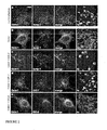
- it can comprise an in vivo screening assay that identifies compounds that inhibit targeting of Ostml to late endosomes/lysosomes and co-localization with ClC-7 and lamp-1 (lysosomal-associated membrane protein), based on the finding that Ostml requires Clc-7 for lysosomal localisation as demonstrated in Figure 2 below.
- This may be practised on wild type cells (looking at endogenous Ostml or in ClC-7 -/- deficient fibroblasts (for over-expression of epitope tagged ClC-7 and Ostml).
- Suitable methods can be applied using full-length proteins or using fragments of the proteins that are involved in the interaction or using one protein in full length form and the other as a binding fragment.
- immunoprecipitation and His-tag purification can be used in order to test the interaction of the proteins or suitable fragments in the presence of suitable test compounds.
- ClC-7 and Ostm1 independently as whole protein or as a binding fragment
- Co-immunoprecipitation may be detected in various ways.
- the precipitate may be loaded on a gel and run and detected by Western blotting with a labelled antibody to the other of said proteins.
- the precipitate may be washed and then stained with a labelled antibody to the other of the proteins.
- the interaction partner can be radioactively labelled and detected by SDS-page or radioactive counting.
- His-tag precipitation may be carried out as follows. ClC-7, Ostml or a binding fragment of one of them is recombinantly expressed in a modified form in which it is extended by a sequence of His amino acids, e.g. His 6 , suitably at the C-terminal or the N-terminal. This enables the His-tagged protein to be captured to beads such as Ni-NTA beads. Then, exposure of the loaded beads to the other protein or binding fragment and the test compound will result (if the test compound is ineffective) in binding of both proteins to the beads.
- the presence of both proteins on the beads after a suitable washing step can be determined by straining the beads directly with a labelled antibody or by removing the bound proteins from the beads and then conducting a Western blot to detect the presence of the said other protein or fragment (or radioactive detection, as above).
- a high throughput screening (HTS) compatible format for a screen can be provided as an ELISA system as described in ref. 62 (Zhao et al).
- One of the proteins (or binding fragments thereof) is fixed to a plate or equivalent solid support either directly, as by using a purified protein and a Maxiprop TM plate, or indirectly, as by using an antibody against Ostml or ClC-7 (that can be bound to a streptavidin plate if biotinylated).
- An extract with the interaction partner ClC-7 or Ostml respectively and selected test compound is incubated on each well of the plate. After washing, a secondary antibody is used to detect the interaction partner using an enzyme labelled (e.g.
- the presence of the labelled antibody is detected, e.g. a POD antibody is detected with a 3,3',5,5'-tetramethylbenzidine (TMB) substrate.
- TMB 3,3',5,5'-tetramethylbenzidine
- the coloration indicates the presence of the interaction partner. If the compound is able to disrupt this interaction, the coloration is lost.
- the optical density can be detected in an ELISA reader in 96 well plates and thus this assay can be optimised for HTS.
- Flashplates and radioactive labels 31).
- the Flashplates TM Perkin Elmer
- the Flashplates TM are 96 well microplates permanently coated with a thin layer of scintillant. The principle is similar to the ELISA assay described above.
- One of the proteins (or a binding fragment thereof) is fixed to the wells.
- a specific WGA Flashplate that binds to glycosylated proteins can be used to fix Ostm-1.
- the second protein needs to be radioactively labelled. This can be achieved by cultivating cells that over express the protein in a medium containing S 35 Methionine and S 35 Cysteine. Alternatively, it may be bound with a radiolabelled antibody. After incubation with the compounds and washing, the radioactivity is measured using a system such as a Wallac counter. If the compound disrupts the interaction, the radioactive protein is too far from the scintillant to activate it.
- SPA system 32, 33
- scintillation proximity assay Beads with anti mouse or anti rabbit antibodies are available as well as Nickel chelate beads that can fix His tag proteins. These beads are used to fix one protein or a binding fragment thereof. For instance, one may bind the protein to an antibody for that protein generated in a mouse and bind the complex to the beads by the anti mouse antibody. The second protein or a binding fragment thereof is radiolabelled as described above and is incubated with the beads and the compounds to test. As in the Flashplates assay the scintillant contained in the beads is only activated upon binding of the radiolabelled protein.
- Biacore.(surface plasmon resonance) system Only the Biacore.(surface plasmon resonance) system will be described here in detail, but the other methodologies can be applied as well.
- One of the proteins or a binding fragment thereof is fixed to a metal surface, preferably gold on a prism.
- the bound protein is exposed to the test compound and the other protein or a binding fragment thereof.
- the critical angle of reflection from behind the surface is recorded. The angle of this reflected light is dependent on the mass of the molecules at the surface layer, so that the method can detect the binding of a second protein and the displacement or prevention of this binding by compounds.
- the parts of the respective proteins involved in the interaction between the full length proteins have yet to be determined. However, this information can be gained by performing any of the above described techniques to detect the interaction between fragments of ClC-7 and Ostml or between ClC-7 and fragments of Ostml.
- the interaction tests described above may be performed on these fragments in order to identify which fragments are necessary to maintain the interaction.
- One may also detect interacting domains by testing fragments of ClC-7 and Ostml in the yeast-two-hybrid technique.
- a fluorescence polarisation assay may be performed between a full-length protein (Ostml or ClC-7) and a suitably small fragment of the interaction partner (ClC-7 or Ostml respectively) that is fused to a fluorophore (for example fluorescein).
- a fluorophore for example fluorescein
- the small fragment binds to the full length interaction partner, the light will remain polarized.
- the light emitted needs to be detected through a polarisation filter.
- HTS compatible readers are available at Tecan, BMG LabTechnologies, LJL Biosystems and Jolley Consulting and Research Inc.
- a filtration assay may be used as generally described in Ref. (31).
- a filtration assay may consist also of an interaction assay between full-length protein (Ostml or ClC-7) and a binding fragment of the interaction partner (ClC-7 or Ostml respectively) that is fused to a fluorophore (for example fluorescein) or is radiolabelled.
- a fluorophore for example fluorescein
- the preparation is centrifuged on a membrane that allows the small fragment to go through but not the full length protein. If the fragment interacts with the full-length protein, it will not go through the membrane either and the supernatant will remain fluorescent or radioactive.
- Fluorescence (or Förster) Resonance Energy Transfer is based on non-radiative energy transfer between a donor and an acceptor (37, 38, 39, 40, 41).
- the donor is fixed to one of the proteins (or a binding fragment) close to its site of interaction. It can be a recombinantly expressed ClC-7 or a fragment retaining the site of interaction with Ostml fused to a first fluorophore (e.g. cyan fluorescent protein - CFP).
- the second protein is fused to an acceptor close to its site of interaction or only a fragment that interacts with the ,protein is used. It can be the domain of Ostml that interacts with ClC-7.
- the Ostml or binding fragment thereof may be fused to an acceptor such as yellow fluorescent protein - YFP.
- acceptor such as yellow fluorescent protein - YFP.
- the acceptor can be fluorescent or not and the FRET is detected by the appearance of sensitized fluorescence of the acceptor, by the intensity ratio change of donor/acceptor, by the fluorescence decrease of the donor, or by changes in the fluorescence lifetime of the donor.
- Both fusion-proteins are recombinantly expressed, extracts are prepared and they are incubated together with or without (control) the compounds to test.
- the assay may be performed in live or fixed cells. Less preferably, non-protein chromophores may be chemically linked to the proteins for practising in vitro methods.
- CFP When CFP is excited at 452nm, it emits a fluorescence wavelength at 505nm that is an excitation wavelength for YFP. YFP will in turn emit at 527nm if it is in close proximity to CFP, thus only when ClC-7 and Ostml fragments interact. If a compound disrupts the interaction, it will also disrupt the YPF excitation and emission at 527nm (35).
- Non fluorescent acceptors can found in other pairs of donor-acceptors such as EDANS/Dabcyl or pinacyanol/EGFP (42). In this case, upon interaction leading to a close proximity of pinacyanol to EGFP (both fused to one interaction partner), the EGFP fluorescence at 510nm will decrease accordingly.
- Changes in the fluorescence of the donor or the acceptor may be measured as an intensity measurement or in a time resolved manner.
- the recordings can be done in a spectrofluorometer (Molecular Devices) and can be set-up as an HTS format.
- the above technique can be applied on living cells using Fluorescence Correlation Spectroscopy (Evotec) (43).
- This method uses confocal microscopy and can detect if a labelled ligand can bind to its receptor in cells.
- This can be adapted also for in situ detection of protein-protein interaction using the FRET method described above.
- Endogenous or over expressed ClC-7 and Ostml protein can be monitored indirectly by incubation of live or fixed cells with anti-ClC-7 and anti-Ostml antibodies followed by incubation with matching secondary antibodies coupled to a fluorochrome or enzyme.
- fluorescent proteins fused to ClC-7 or Ostml can be monitored directly after over expression in cells.
- the potential of a given compound to inhibit proper targeting of Ostml to late endosomes / lysosomes which is shown herein to depend on binding to ClC-7, can be measured as the amount of co-localization of Ostm1 with lysosomal proteins such as lamp1.
- the data acquisition can be performed using microscopy or confocal microscopy of live or fixed cells in microtiter plates.
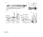
- Figure 1c shows a Western blot of brain membrane proteins revealing an Ostml band at ⁇ 80 kD (filled arrowhead) and a doublet at ⁇ 35-45 kD (open arrowhead) that were absent in grey-lethal (gl) mice, an osteopetrotic mouse mutant(45) carrying a deletion of the Ostm1 promoter and exon 1 (28).
- Antibodies against Ostm1 have been raised in guinea pigs and rabbits and tested for specificity on KO tissues.
- FIG. 1g there is shown a Western blot of cells (lanes 1+2) and supernatants (lanes 3+4) of HEK293 cells expressing Ostml (lanes 1+3) or a truncated form (lanes 2+4) mimicking a human mutation(44). Both proteins carried a C-terminal HA-epitope for detection.
- the first and second hydrophobic domains serve as cleavable signal peptide and transmembrane domain, respectively, in agreement with the type I transmembrane protein model of Vacher and colleagues (28) ( Fig. 1b ).
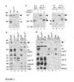
- ClC-7 could be efficiently co-immunoprecipitated from brain with Ostml, and vice versa ( Fig. 3d ). This interaction was specific, since it was neither observed with the related endosomal ClC-3 and ClC-6 proteins, nor with lamp-2. As expected from the lysosomal localisation of ClC-7 (30), antibodies against ClC-7 almost exclusively precipitated the cleaved, lysosomal Ostml fragment from brain. Co-immunoprecipitation performed with transfected cells, in which only the large form could be detected, showed that this putative ER form also interacted with ClC-7 ( Fig. 6 ). Surprisingly, Fig. 3d also revealed that ClC-7 levels were drastically reduced in extracts of gl brain (lane 3).
- ClC-7 may support bone resorption by electrically shunting the H + -ATPase that acidifies the osteoclast resorption lacuna (22), or by similarly facilitating the insertion of proton-pump containing vesicles into the ruffled border, which is underdeveloped in both Clcn7 -/- and gl osteoclasts (22, 45).
- This mechanism is also feasible if ClC-7 were not a Cl - -channel, as believed so far (22, 48), but an electrogenic Cl - /H + -exchanger like ClC-ec1(27), ClC-4 and ClC-5 (23, 26)(23). As the intracellular localisation of ClC-7 precluded biophysical analysis, these alternatives remain untested.
- Clcn7 -/- mice Like Clcn7 -/- mice (30), they displayed electron-dense storage material in neurons and renal proximal tubular cells, possibly pointing to an altered acidification during lysosome formation, or to a role of lysosomal chloride(23, 28). Taken together, the ClC-7/Ostm1 complex is also important for the function of melanocytes and lysosomes. Patients with OSTM1 mutations may develop lysosomal storage disease in addition to osteopetrosis.
- Ostml as a hitherto unknown ancillary ⁇ -subunit of ClC-7. Ostml needs ClC-7 to travel to lysosomes, whereas ClC-7 can reach its destination without Ostml.
- the stability of ClC-7 depends on its association with Ostml. As pronounced glycosylation is thought to protect lysosomal membrane proteins from degradation (57, 58), one may speculate that the highly glycosylated Ostml protein shields ClC-7, the sole mammalian CLC lacking N-linked glycosylation sites, from lysosomal proteases. The osteopetrosis, lysosomal storage and neurodegeneration observed with a loss of Ostml may be entirely explained by a drastic reduction of ClC-7 protein levels.
- ClC-7 and Ostml can be co-immunoprecipitated using endogenous material ( FIG. 3d ) or overexpressed proteins (Fig. 6 ).
- ClC-7 was efficiently co-immunoprecipitated from brain with Ostml, and vice versa
- Co-immunoprecipitation reveals a ClC-7/Ostm1 complex.
- Solubilized brain membranes from the genotypes indicated were directly loaded on the gel (input, lanes 1-3), or first immunoprecipitated with ClC-7 (lanes 4,5) or Ostml antibodies (lanes 6,7).
- Ostml or ClC-7 antibodies were crosslinked to protein A sepharose by dimethylpimelimidate. Brain membranes were pelleted and solubilized in lysis buffer (MES buffered saline pH 6.5, 1% Triton X-100, 2.5 mM CaCl 2 , 2.5 mM MgCl 2 ). Non-solubilized material was removed by centrifugation at 70,000xg. Samples were incubated with protein A sepharose-antibody complexes for 2h at 4°C and washed 4x with lysis buffer. After elution with 100 ⁇ l 0.1 M glycine/Cl, pH 2.8, samples were neutralized and denatured using SDS sample buffer. Equivalent amounts of lysate and precipitate were separated by SDS-PAGE.
- Ostml was transiently co-expressed with the indicated ClC proteins in HEK293 cells using calcium phosphate precipitation. After 24 to 48 hours, the cells were harvested in HBS pH7.4, pelleted at 1000xg and lysed in HBS containing 1% Triton X-100, 2.5 mM of each MgCl 2 and CaCl 2 and complete (Roche) protease inhibitors. The supernatant of a 20000xg spin was used for co-immunoprecipitation. Antibodies against Ostml were incubated with protein A sepharose in PBS for one hour at room temperature under agitation.
- the PVL1393-ClC-7-myc-His construct was used to construct a recombinant baculovirus as described in the book: Baculovirus Expression vectors, A manual, O Reilly et al, Oxford University Press 1994 .
- Western-blot using both anti-ClC-7 antibodies (22, 59)and anti-His antibody (Invitrogen) were used to confirm the expression of the protein.
- Ostm1 Recombinant expression of Ostm1 is established in a similar way in baculovirus.
- the full length Ostm-1 (GI:56699489) was cloned at NB by RT-PCR into a TOPO (Invitrogen) and subsequently into PVL1393 (Stratagene).
- Figure 5 illustrates how the recombinant ClC-7 expressed in Sf9 cells can be trapped on Ni-NTA beads.
- 20 ⁇ 10 e Sf9 cells were infected with 1 ml baculovirus for 72 h. Cells were lysed in 5 ml buffer containing Tris HCl 50mM pH7.5, 150mM NaCl, protease inhibitor cocktail (Amersham) and 0,1 % (A) or 1% (B and C) SDS.
- Ni-NTA purification (Invitrogen Manual 021202) is performed using Ni-NTA beads. After preparation of the beads as recommended by the manufacturer, 1 ml of ClC-7 Sf9 cell extract is incubated per 200 ⁇ l of Ni-NTA beads overnight at 4 °C. The supernatant is removed and 1ml of the Ostm-1 extract is added for 2h at 4 °C with the various compounds to test at a concentration of 1nM. Unbound material is washed by 3 washing with 2ml of PBS 0,2mM NaCl-2% CHAPS.
- SDS sample denaturing buffer is added to the beads, which are boiled and the samples are run on a gel.
- the PVL1393-ClC-7-myc-His construct was used to construct a recombinant baculovirus as described in the book Baculovirus Expression vectors, A manual, O Reilly et al, Oxford University Press 1994 .
- Western-blot using both ClC-7 antibodies (22, 59) and his antibody (Invitrogen) was used to confirm the expression of the protein.
- Ostml Recombinant expression of Ostml is established in a similar way in baculovirus.
- the full length Ostm-1 (GI:56699489) was cloned at NB by RT-PCR into a TOPO (Invitrogen) and subsequently into PVL1393 (Stratagene).
- Sf9 cells are infected with ClC-7 and Ostm1 baculovirus for 48 or 72h with 1 ml of virus for 20.10 6 cells in a 175cm 3 flask. 4h before lysis, a medium without Methionine and Cysteine is added with 20mCi/ml of S 35 Met and S 35 Cys.
- the cells are lysed in 1 ml of Tris HCl 50mM pH7.5, 150M, SDS 0.1%, protease inhibitor cocktail (Amersham) and the extract is diluted to 1mg/ml. Cell debris is removed by a centrifugation at 20000 rpm.
- Ni-NTA purification (Invitrogen Manual 021202) is performed using Ni-NTA beads. After preparation of the beads as recommended by the manufacturer, 1 ml Sf9 cells extract is incubated per 200 ⁇ l of Ni-NTA beads overnight at 4 degrees with the various compounds to test at a concentration of 1nM. Unbound material is washed by 3 washing with 2ml of PBS 0.2mM NaCl-2% CHAPS.
- SDS sample denaturing buffer is added to the beads, boiled and the samples are analysed by SDS-page and visualized by autoradiography. This method has been used earlier for other proteins (60).
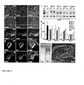
- Ostm1 was transiently co-expressed with the indicated CLC proteins in HEK293 cells. Upon such overexpression, only the ⁇ 80 kD (arrowhead), but not the 35-45 kD Ostml species was detected in Western blots.
- ClC-7 was co-precipitated. Thus, ClC-7 also interacts with the uncleaved Ostm1 species. By contrast, ClC-6 or ClC-3 could not be co-precipitated, demonstrating the specificity of the ClC-7/Ostm1 interaction.
- the word 'or' is used in the sense of an operator that returns a true value when either or both of the stated conditions is met, as opposed to the operator 'exclusive or' which requires that only one of the conditions is met.
- the word 'comprising' is used in the sense of 'including' rather than in to mean 'consisting of'.
Landscapes
- Health & Medical Sciences (AREA)
- Life Sciences & Earth Sciences (AREA)
- Engineering & Computer Science (AREA)
- Biomedical Technology (AREA)
- Immunology (AREA)
- Molecular Biology (AREA)
- Chemical & Material Sciences (AREA)
- Hematology (AREA)
- Urology & Nephrology (AREA)
- Cell Biology (AREA)
- General Health & Medical Sciences (AREA)
- Medicinal Chemistry (AREA)
- Analytical Chemistry (AREA)
- General Physics & Mathematics (AREA)
- Biotechnology (AREA)
- Food Science & Technology (AREA)
- Microbiology (AREA)
- Physics & Mathematics (AREA)
- Pathology (AREA)
- Biochemistry (AREA)
- Bioinformatics & Cheminformatics (AREA)
- Physical Education & Sports Medicine (AREA)
- Toxicology (AREA)
- Proteomics, Peptides & Aminoacids (AREA)
- Tropical Medicine & Parasitology (AREA)
- Orthopedic Medicine & Surgery (AREA)
- Rheumatology (AREA)
- Organic Chemistry (AREA)
- General Chemical & Material Sciences (AREA)
- Nuclear Medicine, Radiotherapy & Molecular Imaging (AREA)
- Chemical Kinetics & Catalysis (AREA)
- Pharmacology & Pharmacy (AREA)
- Animal Behavior & Ethology (AREA)
- Public Health (AREA)
- Veterinary Medicine (AREA)
- Investigating Or Analysing Biological Materials (AREA)
- Medicines That Contain Protein Lipid Enzymes And Other Medicines (AREA)
- Measuring Or Testing Involving Enzymes Or Micro-Organisms (AREA)
- Pharmaceuticals Containing Other Organic And Inorganic Compounds (AREA)
Abstract
Description
- The present invention relates to methods for screening compounds for activity in modulating chloride ion transport via ClC-7 or modulating subcellular localisation of Ostml. Compounds that successfully modulate such chloride ion transport will be candidates for use in the study or treatment of osteoclast related diseases such as osteoporosis and other osteoclastic metabolic diseases such as Paget's disease, steroid induced osteoporosis and cancer subtypes such a bone metastasis. Osteoporosis is defined as skeletal fragility characterized by reduced bone mass and micro-architectural deterioration, which leads to increased fractures. The majority of osteoporotic fractures occur in older women after menopause (1).
- Two cell types are responsible for the bone remodelling processes: the bone forming osteoblast and the bone degrading osteoclast (2, 3). The osteoclasts attach to the bone matrix and form the bone-resorbing compartment and the ruffled border by exocytosis of lysosomes (4). Dissolution of bone by acidification of the resorption compartment is mediated by an active transport of protons through the osteoclastic V-type H+ ATPase (5-8). At the same time, a passive transport of chloride through chloride conductances (channels and/or electrogenic coupled transporters) preserves the electroneutrality (9). Prevention of chloride transport will lead to a rapid hyperpolarization of the membrane, preventing further secretion of protons, thus resulting in an inhibition of further bone resorption (10, 11). Drugs inhibiting the acidification employ a different mode of action than existing treatments for bone metabolic diseases.
- Chloride channels and transporters comprise a large group of transmembrane proteins positioned in the plasma membrane and in intracellular vesicles. Their roles vary from the control of secretion and absorption of salt, regulation of membrane potentials, organellar acidification and cell volume homeostasis (12, 13). Three different structural families of chloride channels have been characterized: the CLC voltage-gated chloride channels and chloride proton exchangers, the ligand-gated chloride channels containing the glycine receptor and the γ-aminobutyric acid receptor, and the CFTR (cystic fibrosis transmembrane conductance regulator). Moreover, two additional families of putative chloride channels have been identified: the CLIC (chloride intracellular channels) and the CLCA calcium activated chloride channels. Another chloride channel, called volume regulated anion channel (VRAC) and the molecular identity of which has not been established yet, has been identified by electrophysiology in most mammalian cells (14) including the osteoclasts (15-17).
- The CLIC, CLCA and VRAC functions are not very well understood yet, whereas the CLC channels and transporters have been characterized through analysis of mutations leading to genetic disorders and generation of knock-out mice (13). Mutations in the CLCN7 gene are responsible for various forms of osteopetrosis: the Autosomal Dominant Osteopetrosis II (ADOII), cases of Autosomal Recessive Osteopetrosis (ARO) and of an intermediate form of ARO (IARO) (18-22). Furthermore the disruption of Clcn7 leads to severe osteopetrosis in mice, with approximately 700% more bone (22). Among the ClCs, ClC-0, ClC-1, ClC-2 and ClC-Ks have been shown to be proper chloride channels (23-25), whereas ClC-4 and ClC-5 have been shown to be chloride proton exchangers (23, 26) where it is proposed that in the case of the exchanger, the ClCs transport one proton for two chloride (27). This was shown by electrophysiology and acidification measurements.
- CIC-7 (the expression product of the CLCN7 gene) as well as ClC-6 have not been characterized by electrophysiology, thus it has not been clarified whether they are chlorides channels or chloride proton exchangers. In both cases, the role of ClC-7 remains the same (acidification of the resorption lacunae) and its inhibition leads to increased bone density.
- Methods for screening compounds for the ability to modulate the transport of chloride ions by ClC-7 have been proposed in
WO02/059612 WO02/059356 - The present invention relates to a new approach in which a newly discovered dependency of ClC-7 on another protein, (Ostm1 - Osteopetrosis-associated
transmembrane protein 1, the product of the OSTM1 gene), for functional transport of chloride is exploited. This dependency is mediated by an interaction (direct or indirect) between ClC-7 and Ostm1. Thus, it has now been discovered that additional molecules modulate ClC-7 function, among others the protein Ostml, which is therefore also required for normal osteoclast function. - Accordingly Ostm1 inhibitors (e.g. compounds that inhibit Ostml expression), or compounds disrupting the interaction between Ostml and ClC-7 may be relevant pharmaceutical compounds for the treatment or prevention of osteoclast related diseases.
- Ostml is a transmembrane protein of previously unknown function. Mutations in the OSTM1 gene cause a severe osteopetrosis syndrome, as was shown for the spontaneous mouse mutant called 'grey lethal' (28) and human patients (28, 29).
- Similarly, mutations in ClC-7, a late endosomal/lysosomal member of the ClC family of chloride channels and transporters (12, 25), cause osteopetrosis (22) and lysosomal storage disease (30) in man and mice.
- We have now found that both proteins co-localise in late endosomes and lysosomes of various tissues, as well as in the ruffled border of bone-resorbing osteoclasts. Co-immunoprecipitations reveal that ClC-7 and Ostm1 form a molecular complex and suggest Ostml as a novel β-subunit of ClC-7. Ostml needs ClC-7 to reach lysosomes, where its highly glycosylated luminal domain is cleaved. The protein, but not RNA, levels of ClC-7 are greatly reduced in grey-lethal mice lacking Ostml and vice versa, suggesting that their interaction is important for protein stability. As the level of ClC-7 protein is decreased below 10% in Ostm1-deficient tissues and cells including osteoclasts, Ostm1 mutations can now be seen to cause osteopetrosis by impairing the acidification of the osteoclast resorption lacuna that depends on ClC-7 (22). The finding that grey-lethal mice, just like ClC-7 knock-out mice (30), display lysosomal storage and neurodegeneration in addition to osteopetrosis implies a more general importance of ClC-7/Ostml complexes.
- The present invention now provides a method for screening a test compound for activity in modulating chloride transport via ClC-7, which comprises determining whether or the extent to which the test compound prevents or inhibits the binding of Ostml to ClC-7.
- In an alternative realization, the present invention provides a method for screening a test compound for activity in modulating the subcellular localization of Ostml which comprises determining whether or the extent to which the test compound prevents the binding of Ostml to ClC-7. As the subcellular localisation of Ostml is influenced by its interaction with ClC-7 and modulates ClC-7 stability, interference in the mutual binding of ClC-7 and Ostml is likely to result in a localization behaviour that is changed by the test compound.
- In a further aspect, the invention provides a method for screening a test compound for activity in modulating interaction between ClC-7 and Ostml, which method comprises determining whether or the extent to which the test compound prevents binding of Ostm1 to ClC-7.
- In either aspect, the invention may be practised to screen numerous compounds for the desired property.
- Because other chloride channels are not known to interact with Ostml (and we have demonstrated a lack of interaction between Ostml and ClC-3 and ClC-6, respectively), a blocker of the interaction between Ostml and ClC-7 is more likely to provide selectivity in decreasing ClC-7 functional activity with respect to other chloride channels and transporters than a compound selected simply for its direct effect on ClC-7 alone. It is thought that the interaction between ClC-7 and Ostm1 is direct rather than through some intermediate protein or other molecule, and evidence for that is presented below. However, cell based methods are described herein that should be effective even if the interaction is indirect.
- Such a method may be practised by exposing ClC-7, or a fragment thereof capable of binding Ostml, to Ostml or to a fragment thereof capable of binding ClC-7, said exposure being in the presence of said test compound, and determining whether or the extent to which binding between the ClC-7 or fragment thereof and the Ostml or fragment thereof is prevented or disrupted.
- Both Ostml and ClC-7 are expressed and functional in very many organisms. In practising the invention, one may use Ostml and ClC-7 selected from those of any organism in which both are expressed, particularly any mammal, e.g. rodent (especially mouse) or human. Preferably, the Ostml used and the ClC-7 used should come from the same organism, but ones of different origin that bind one another are satisfactory. The use of Ostml and ClC-7 which are human is preferred. Amongst non-human Ostml and ClC-7, those that are more closely homologous to the human proteins are preferred.
- In what follows, we refer to fragments of ClC-7 which are capable of binding Ostml and to fragments of Ostml which are capable of binding ClC-7 as 'binding fragments'.
- In said exposure step one of said ClC-7 or binding fragment thereof and Ostml or binding fragment thereof may be immobilised to a solid support and the other thereof and said test compound may be in solution. Such solid supports may be multi-well plates, test strips, beads, or other supports known or conventional in the art or equivalent thereto.
- The ClC-7 or binding fragment thereof or the Ostml or binding fragment thereof which is in solution may bear a detectable label such as a radio-label, an enzyme label, a label which is able to be stimulated to emit light, e.g. as fluorescence.
- The screening method may also be performed such that all of the ClC-7 or binding fragment thereof, the Ostm1 or binding fragment thereof, and the test compound are in solution in said exposure step.
- Optionally, in homogeneous or non-homogeneous systems, said ClC-7 or binding fragment thereof and said Ostml or binding fragment thereof each bear a label, and the proximity of the said labels may be detectable.
- Alternatively, ClC-7 and Ostml are present in situ in a cell or intracellular organelles which can be lysosomes, endosomes, the endoplasmic reticulum, the Golgi apparatus, the osteoclastic plasma membrane facing the resorption lacuna or the plasma membrane in other cell types. Again, each of the ClC-7 and the Ostml bear a label, and the proximity of the said labels may be detectable.
- In such screening methods, the concentration of the test compound may suitably be from 10'pM to 10µM, e.g. from 100 pM to 10 nM, e.g. about 1 nM.
- According the invention it has now been discovered that Ostml is important for ClC-7 function. Therefore pharmaceutical modalities which disrupt this interaction and thereby inhibit ClC-7 are of interest as potential treatment or prevention agents in connection with diseases including osteoclast related bone diseases, such as osteoporosis and other osteoclastic related diseases. The described screening methods may be used in drug development or in confirming the mode of action of a known therapeutic agent.
- In a further aspect, the invention relates to the use of a compound identified as a blocker of the interaction between ClC-7 and Ostml by a said screening method in the treatment, prevention or alleviation of an osteoclast related bone disease in a subject.
- Other objects and uses of the invention will be apparent to the person skilled in the art from the following detailed description and example.
- Optionally; one may further screen to select compounds that do not block interaction between Ostml and some other chosen molecule or do not block interaction between ClC-7 and some other chosen molecule.
- Optionally, one may further screen to select compounds that do not block interaction between proteins in general, for instance by screening for lack of blocking effect on a protein pair such as ClC-K and barttin (a chloride channel β-subunit not related to Ostml) (61).
- Numerous screening methods may be used in order to identify compounds that inhibit ClC-7/Ostm-1 interaction, thus inhibiting bone resorption by down-regulation of ClC-7 levels or activity. The assay can consist of a binding test between ClC-7 and Ostml in the presence of a test compound that potentially can inhibit this interaction.
- Alternatively, it can comprise a preliminary screening assay that identifies compounds that bind to one of the proteins (ClC-7 or Ostml) present either in full length form or as only a part of such a protein involved in the interaction (a binding fragment), followed by further testing for the disruption of the interaction with the second partner.
- Alternatively, it can comprise an in vivo screening assay that identifies compounds that inhibit targeting of Ostml to late endosomes/lysosomes and co-localization with ClC-7 and lamp-1 (lysosomal-associated membrane protein), based on the finding that Ostml requires Clc-7 for lysosomal localisation as demonstrated in
Figure 2 below. This may be practised on wild type cells (looking at endogenous Ostml or in ClC-7-/- deficient fibroblasts (for over-expression of epitope tagged ClC-7 and Ostml). - Suitable methods can be applied using full-length proteins or using fragments of the proteins that are involved in the interaction or using one protein in full length form and the other as a binding fragment.
- As exemplified below (see Examples 1-3), immunoprecipitation and His-tag purification can be used in order to test the interaction of the proteins or suitable fragments in the presence of suitable test compounds. Thus, in an immunoprecipitation screening assay, ClC-7 and Ostm1 (independently as whole protein or as a binding fragment) may be incubated with a test compound and may then be immunoprecipitated using an antibody specific for either ClC-7 or Ostml. Co-immunoprecipitation may be detected in various ways. The precipitate may be loaded on a gel and run and detected by Western blotting with a labelled antibody to the other of said proteins. The precipitate may be washed and then stained with a labelled antibody to the other of the proteins. Alternatively, the interaction partner can be radioactively labelled and detected by SDS-page or radioactive counting.
- His-tag precipitation may be carried out as follows. ClC-7, Ostml or a binding fragment of one of them is recombinantly expressed in a modified form in which it is extended by a sequence of His amino acids, e.g. His6, suitably at the C-terminal or the N-terminal. This enables the His-tagged protein to be captured to beads such as Ni-NTA beads. Then, exposure of the loaded beads to the other protein or binding fragment and the test compound will result (if the test compound is ineffective) in binding of both proteins to the beads. The presence of both proteins on the beads after a suitable washing step can be determined by straining the beads directly with a labelled antibody or by removing the bound proteins from the beads and then conducting a Western blot to detect the presence of the said other protein or fragment (or radioactive detection, as above).
- A high throughput screening (HTS) compatible format for a screen can be provided as an ELISA system as described in ref. 62 (Zhao et al). One of the proteins (or binding fragments thereof) is fixed to a plate or equivalent solid support either directly, as by using a purified protein and a Maxiprop™ plate, or indirectly, as by using an antibody against Ostml or ClC-7 (that can be bound to a streptavidin plate if biotinylated). An extract with the interaction partner ClC-7 or Ostml respectively and selected test compound is incubated on each well of the plate. After washing, a secondary antibody is used to detect the interaction partner using an enzyme labelled (e.g. peroxidase (POD) labelled) specific antibody or a specific antibody recognized by an anti-species, e.g. anti rabbit or anti mouse POD antibody. The presence of the labelled antibody is detected, e.g. a POD antibody is detected with a 3,3',5,5'-tetramethylbenzidine (TMB) substrate. The coloration indicates the presence of the interaction partner. If the compound is able to disrupt this interaction, the coloration is lost. The optical density can be detected in an ELISA reader in 96 well plates and thus this assay can be optimised for HTS.
- A similar assay can be developed using Flashplates and radioactive labels (31). The Flashplates™ (Perkin Elmer) are 96 well microplates permanently coated with a thin layer of scintillant. The principle is similar to the ELISA assay described above. One of the proteins (or a binding fragment thereof) is fixed to the wells. A specific WGA Flashplate that binds to glycosylated proteins can be used to fix Ostm-1. The second protein needs to be radioactively labelled. This can be achieved by cultivating cells that over express the protein in a medium containing S35 Methionine and S35 Cysteine. Alternatively, it may be bound with a radiolabelled antibody. After incubation with the compounds and washing, the radioactivity is measured using a system such as a Wallac counter. If the compound disrupts the interaction, the radioactive protein is too far from the scintillant to activate it.
- Another suitable methodology using radioactivity and proximity is the SPA system (32, 33): scintillation proximity assay. Beads with anti mouse or anti rabbit antibodies are available as well as Nickel chelate beads that can fix His tag proteins. These beads are used to fix one protein or a binding fragment thereof. For instance, one may bind the protein to an antibody for that protein generated in a mouse and bind the complex to the beads by the anti mouse antibody. The second protein or a binding fragment thereof is radiolabelled as described above and is incubated with the beads and the compounds to test. As in the Flashplates assay the scintillant contained in the beads is only activated upon binding of the radiolabelled protein.
- Other methods of detection of protein/protein interactions are available and can be used to detect if compounds are able to disrupt the interaction. They include a range of label-free interaction screening methodologies (34). These use optical biosensor (Affinity Sensors, Biacore, HTS biosystems, IBIS, SRU Biosystems), acoustic biosensor (Akubio) or micro-calorimetry (MicoCal, Thermometric, Calorimetry sciences Corp., 3D Pharmaceuticals, Vivactiss) based methods.
- Only the Biacore.(surface plasmon resonance) system will be described here in detail, but the other methodologies can be applied as well. One of the proteins or a binding fragment thereof is fixed to a metal surface, preferably gold on a prism. The bound protein is exposed to the test compound and the other protein or a binding fragment thereof. By directing polarised light onto the prism, the critical angle of reflection from behind the surface is recorded. The angle of this reflected light is dependent on the mass of the molecules at the surface layer, so that the method can detect the binding of a second protein and the displacement or prevention of this binding by compounds.
- The parts of the respective proteins involved in the interaction between the full length proteins have yet to be determined. However, this information can be gained by performing any of the above described techniques to detect the interaction between fragments of ClC-7 and Ostml or between ClC-7 and fragments of Ostml. One may recombinantly express only parts of the coding sequence of one or both proteins (for example only the intracellular domain or the extracellular domain or the various transmembrane domains alone or in association). The interaction tests described above may be performed on these fragments in order to identify which fragments are necessary to maintain the interaction. One may also detect interacting domains by testing fragments of ClC-7 and Ostml in the yeast-two-hybrid technique.
- A fluorescence polarisation assay (35, 36) may be performed between a full-length protein (Ostml or ClC-7) and a suitably small fragment of the interaction partner (ClC-7 or Ostml respectively) that is fused to a fluorophore (for example fluorescein). When the small fragment is not bound and is excited with a polarized light, it will depolarise the light due to the rotation of the fluorophore. When the small fragment binds to the full length interaction partner, the light will remain polarized. Thus compounds that disrupt the interaction can be screened in this assay. The light emitted needs to be detected through a polarisation filter. HTS compatible readers are available at Tecan, BMG LabTechnologies, LJL Biosystems and Jolley Consulting and Research Inc.
- A filtration assay may be used as generally described in Ref. (31).
- A filtration assay may consist also of an interaction assay between full-length protein (Ostml or ClC-7) and a binding fragment of the interaction partner (ClC-7 or Ostml respectively) that is fused to a fluorophore (for example fluorescein) or is radiolabelled. After incubation of the interaction partners in a buffer with the compound to test, the preparation is centrifuged on a membrane that allows the small fragment to go through but not the full length protein. If the fragment interacts with the full-length protein, it will not go through the membrane either and the supernatant will remain fluorescent or radioactive.
- Fluorescence (or Förster) Resonance Energy Transfer (FRET) is based on non-radiative energy transfer between a donor and an acceptor (37, 38, 39, 40, 41). The donor is fixed to one of the proteins (or a binding fragment) close to its site of interaction. It can be a recombinantly expressed ClC-7 or a fragment retaining the site of interaction with Ostml fused to a first fluorophore (e.g. cyan fluorescent protein - CFP). The second protein is fused to an acceptor close to its site of interaction or only a fragment that interacts with the ,protein is used. It can be the domain of Ostml that interacts with ClC-7. The Ostml or binding fragment thereof may be fused to an acceptor such as yellow fluorescent protein - YFP. A lot of different acceptor and donor pairs are widely used and all can be used in this assay. The acceptor can be fluorescent or not and the FRET is detected by the appearance of sensitized fluorescence of the acceptor, by the intensity ratio change of donor/acceptor, by the fluorescence decrease of the donor, or by changes in the fluorescence lifetime of the donor. Both fusion-proteins are recombinantly expressed, extracts are prepared and they are incubated together with or without (control) the compounds to test. Alternatively, the assay may be performed in live or fixed cells. Less preferably, non-protein chromophores may be chemically linked to the proteins for practising in vitro methods.
- When CFP is excited at 452nm, it emits a fluorescence wavelength at 505nm that is an excitation wavelength for YFP. YFP will in turn emit at 527nm if it is in close proximity to CFP, thus only when ClC-7 and Ostml fragments interact. If a compound disrupts the interaction, it will also disrupt the YPF excitation and emission at 527nm (35).
- Non fluorescent acceptors can found in other pairs of donor-acceptors such as EDANS/Dabcyl or pinacyanol/EGFP (42). In this case, upon interaction leading to a close proximity of pinacyanol to EGFP (both fused to one interaction partner), the EGFP fluorescence at 510nm will decrease accordingly.
- Changes in the fluorescence of the donor or the acceptor may be measured as an intensity measurement or in a time resolved manner. The recordings can be done in a spectrofluorometer (Molecular Devices) and can be set-up as an HTS format.
- The above technique can be applied on living cells using Fluorescence Correlation Spectroscopy (Evotec) (43). This method uses confocal microscopy and can detect if a labelled ligand can bind to its receptor in cells. This can be adapted also for in situ detection of protein-protein interaction using the FRET method described above. To identify compounds that inhibit targeting of Ostml to late endosomes/lysosomes and co-localization with ClC-7 and lamp1 therein the following as well as other methods can be applied. Endogenous or over expressed ClC-7 and Ostml protein can be monitored indirectly by incubation of live or fixed cells with anti-ClC-7 and anti-Ostml antibodies followed by incubation with matching secondary antibodies coupled to a fluorochrome or enzyme. Alternatively fluorescent proteins fused to ClC-7 or Ostml can be monitored directly after over expression in cells. The potential of a given compound to inhibit proper targeting of Ostml to late endosomes / lysosomes, which is shown herein to depend on binding to ClC-7, can be measured as the amount of co-localization of Ostm1 with lysosomal proteins such as lamp1. The data acquisition can be performed using microscopy or confocal microscopy of live or fixed cells in microtiter plates.
- The invention will be further described and illustrated with reference to the accompanying drawings, in which:
- Figure la shows an annotated schematic of the mouse Ostml protein in which the symbols represent the following: Black box, hydrophobic stretch; Y, consensus site for N-linked glycosylation; *, cysteine; Arrow, predicted signal peptide cleavage site; Dashed, proposed RING-finger domain (44); AB, antibody binding site; Lines, truncated proteins predicted from human OSTM1 mutations (28, 29);
-
Figure 1b shows a topology model of the Ostml protein in which the arrow points to an approximate cleavage site in lysosomal Ostml; -
Figures 1c-f show Western blots obtained as described below. -
Figure 2 shows confocal microscopy images from staining fibroblasts for Ostml, ClC- and lamp-1 proteins. The rows are as follows:- Row (a): Fibroblast stained for endogenous Ostm1 and lamp-1. The third panel shows the overlay, with higher magnification in the fourth panel.
- Row (b) Ostml-transfected fibroblasts showed a reticular and perinuclear Ostml staining that co-localised poorly with lamp-1.
- Rows (c) and (d): In fibroblasts co-transfected with Ostml and ClC-7. Ostml co-localised with lamp-1 (c) and ClC-7 (d).
- Row (e): Co-transfecting Ostml with ClC-6 did not lead to a lysosomal localisation of Ostml, nor to a significant co-localisation. In (b-e), Clcn7 -/ - fibroblasts were used to avoid effects of endogenous ClC-7, but similar results were seen in HeLa cells. Scale bar indicates 8.5 µm for (a), 10 µm for (b-e), and 1.7 µm for enlargements (right).
-
Figure 3 shows Western blots as follows:- (a) Western blot for Ostml in WT, Clcn7 -/ - and grey-lethal (gl) brain.
- (b) Subcellular distribution of Ostm1 in WT and Clcn7 -/ - mice.
- (c) Deglycosylation of WT lysosomal and Clcn7 -/- ER/endosomal fractions.
- (d) Co-immunoprecipitation of a ClC-7/Ostm1 complex.
-
Figure 4 shows immunofluorescence images of cerebellar Purkinje cells in panels (a) and (b) and of osteoclasts in situ in panels (c) - (e). In (a-e), overlays (right) are shown with TOTO staining (blue) for nucleic acids. Scale bar in (a), 10 µm for (a-e). Panel (f) shows Western blots which are quantified in panel (g)- Age, P11-P33. Error bars, SEM. n=3-9, except for ClC-3 in bone (n=2). 'h' and 'i' are methylene blue-stained histological sections showing retinal degeneration in gl (h) but not in WT (i) mice at age P31. A section of hippocampus showing neurodegeneration in the CA3 region (arrows) of a P47 gl mouse revealed by anti-NeuN staining (Scale bar, 100 µm) is seen in panel (j). -
Figure 5 shows the result of a Western-blot against ClC-7 performed in Example 2, showing that the recombinant ClC-7 expressed in Sf9 cells can be bound to Ni-NTA beads via a His tag. -
Figure 6 is a Western blot demonstrating that an ~80 kDa Ostm1 species interacts with ClC-7 in transfected HEK cells, whereas ClC-3 and ClC-6 do not interact with Ostml. -
Figure 1c shows a Western blot of brain membrane proteins revealing an Ostml band at ~80 kD (filled arrowhead) and a doublet at ~35-45 kD (open arrowhead) that were absent in grey-lethal (gl) mice, an osteopetrotic mouse mutant(45) carrying a deletion of the Ostm1 promoter and exon 1 (28). - Antibodies against Ostm1 (peptide C-LKSSTSFANIQENAT) have been raised in guinea pigs and rabbits and tested for specificity on KO tissues.
- In
Figure 1d , incubation of fibroblasts with leupeptin increased the proportion of the large Ostml species. Similar results were obtained with E64, an inhibitor of cathepsin B, L and S (data not shown). Thus, incubating cells with protease inhibitors increased the proportion of the large band at the expense of the smaller ones (Fig. 1d ), indicating that the small form was produced by proteolytic cleavage of the ~80 kD protein. The apparent sizes of Ostm1 species suggest a cleavage roughly in the middle of the protein (Fig. 1b ). - In Figure le, Western blots of brain proteins separated by non-reducing SDS-PAGE showed a single large Ostml-specific band, in contrast to the large and small forms observed under reducing conditions. Thus, the cleaved fragments might be linked by disulfide bonds between some of the cysteines that abound in the luminal domain of Ostml (
Fig. 1a ). - Recent work proposed an E3 ubiquitin ligase function for Ostml (cloned independently as GIPN) (44). The stretch between the amino- and carboxy-terminal hydrophobic regions of Ostml (
Fig. 1a ) displayed weak homology to RING-finger proteins and was suggested to be cytosolic (44). However, this stretch (and no other part of the protein) contains several consensus sites for N-linked glycosylation. In the Western blot ofFigure 1f , deglycosylation with PNGaseF reduced the sizes of all Ostml-species detected under reducing conditions, demonstrating that several or all of these sites are used. The observed glycosylation placed the hypothetical RING-finger domain (44) into the lumen of endoplasmic reticulum (ER), a localisation difficult to reconcile with the cytosolic/nucleoplasmic activity of ubiquitin ligases (46). The disappearance in transfected cells of an HA-epitope added to the amino-terminus indicated the presence of a cleavable signal peptide (data not shown).Fig. 1f also revealed that the apparent molecular weight of the largest deglycosylated band roughly agreed with the prediction from the Ostm1 reading frame (~37 kDa). As deglycosylation of the small species yielded a single band, the doublet is due to non-uniform glycosylation. - We also investigated an Ostml mutant truncated before the second hydrophobic stretch, predicted to serve as a transmembrane domain. In
Figure 1g there is shown a Western blot of cells (lanes 1+2) and supernatants (lanes 3+4) of HEK293 cells expressing Ostml (lanes 1+3) or a truncated form (lanes 2+4) mimicking a human mutation(44). Both proteins carried a C-terminal HA-epitope for detection. This mutant, but not WT Ostml, was secreted into the supernatant of transfected cells (Fig. 1g ). Hence the first and second hydrophobic domains serve as cleavable signal peptide and transmembrane domain, respectively, in agreement with the type I transmembrane protein model of Vacher and colleagues (28) (Fig. 1b ). - Turning to the results shown in
Figure 2 , it is seen that in cultured primary fibroblasts, Ostml co-localised with lamp-1, a marker for late endosomes and lysosomes (Fig. 2a ). This localisation resembled that of ClC-7, loss of which also causes osteopetrosis (22). However, when fibroblasts were transiently transfected with Ostml (Fig. 2b ), no significant co-localisation with lamp-1 was observed and Ostml staining was rather suggestive of the ER. Co-transfection with ClC-7 restored a punctate Ostml pattern that largely co-stained for lamp-1 (Fig. 2c ) and ClC-7 (Fig. 2d ). The effect of ClC-7 was specific, as neither co-transfection with ClC-3 (not shown) nor with ClC-6 (Fig. 2e ), both expressed in the endosomal/lysosomal pathway (12, 47), resulted in such changes. - As ClC-7 changed Ostm1 localisation, we studied Ostml in ClC-7 knockout (Clcn7 -/-) mice. Western blots surprisingly indicated an exclusive loss of the 35-45 kD Ostml doublet in Clcn7 -/- brain (
Fig. 3a ). Subcellular fractionation of WT brain revealed that the small Ostml form was co-enriched with lysosomal markers (bottom fractions, 1-2), whereas the 80 kDa form was only detectable in fractions 9-12 containing markers for endosomes and the ER (Fig. 3b ). Such experiments provided samples only containing the small (WT fractions containing lysosomes) or large (Clcn7 -/- fractions containing endosomes/ER) Ostml forms which were used for deglycosylation. PNGaseF reduced the size of both species (Fig. 3c ,lanes 2 and 4), while only the 35-45 kD form was partially resistant to EndoH (Fig. 3c , comparelanes 2 and 6). This indicated that the small, but not the large species had left the ER. Our results thus show that ClC-7 is needed to traffic Ostml from the ER to lysosomes, and that the luminal domain of Ostml is cleaved on its way to, or in, this final compartment. - ClC-7 could be efficiently co-immunoprecipitated from brain with Ostml, and vice versa (
Fig. 3d ). This interaction was specific, since it was neither observed with the related endosomal ClC-3 and ClC-6 proteins, nor with lamp-2. As expected from the lysosomal localisation of ClC-7 (30), antibodies against ClC-7 almost exclusively precipitated the cleaved, lysosomal Ostml fragment from brain. Co-immunoprecipitation performed with transfected cells, in which only the large form could be detected, showed that this putative ER form also interacted with ClC-7 (Fig. 6 ). Surprisingly,Fig. 3d also revealed that ClC-7 levels were drastically reduced in extracts of gl brain (lane 3). - Both ClC-7 and Ostm1 are expressed in many tissues, including brain, liver, kidney, and osteoclasts (22, 28, 30, 44, 48). Immunohistochemistry of brain sections revealed that both proteins co-localised in neuronal cell bodies in structures that most likely represent late endosomes/lysosomes (30) (
Fig. 4a ). In grey-lethal (gl) neurons (Fig 4b ), both proteins were undetectable. Both proteins also co-localised in osteoclasts in a pattern that represents the 'ruffled border' (Fig. 4c ). This acid-secreting plasma membrane domain was identified by co-staining for the a3 proton pump subunit (Fig. 4d ) (49), mutations in which also underlie osteopetrosis (50-54). - In line with our Western blot analyses
(Fig. 3a,b,d ), Ostml staining was very weak in Clcn7 -/ - mice while ClC-7 labelling was drastically reduced in grey-lethal cells. These cells included neurons (Fig. 4b ) and osteoclasts (Fig. 4e ), in which the remaining portion of ClC-7 still localised to the ruffled border. Western blots revealed a strong decrease of ClC-7, but not of ClC-3, ClC-6, lamp-1 and lamp-2 in gl brain (Fig. 4f ). Quantification of Western blots from brain, kidney, liver and bone showed a decrease in gl mice of ClC-7, but not of other late endosomal/lysosomal proteins tested (ClC-3, ClC-6, lamp-1/-2, H+-ATPase a3,Fig. 4g ). The moderate decrease of control proteins in bone may be explained by the osteopetrosis of gl mice. This Western blot analysis revealed that ClC-7 was reduced to less than 10% in gl mice (Fig. 4f,g ). Neither the transcript level of ClC-7 in gl tissues nor that of Ostml in Clcn7-/- mice was changed (not shown). - ClC-7 may support bone resorption by electrically shunting the H+-ATPase that acidifies the osteoclast resorption lacuna (22), or by similarly facilitating the insertion of proton-pump containing vesicles into the ruffled border, which is underdeveloped in both Clcn7 -/- and gl osteoclasts (22, 45). This mechanism is also feasible if ClC-7 were not a Cl--channel, as believed so far (22, 48), but an electrogenic Cl-/H+-exchanger like ClC-ec1(27), ClC-4 and ClC-5 (23, 26)(23). As the intracellular localisation of ClC-7 precluded biophysical analysis, these alternatives remain untested. Our work suggests that loss of Ostml causes osteopetrosis (28, 29, 55) by decreasing the amount of ClC-7 to pathogenic levels. Already a 75% decrease of ClC-7 function by dominant negative mutations causes mild osteopetrosis in humans (20, 53). The even lower levels of ClC-7 in gl mice may suffice to cause the severe osteopetrosis observed with OSTM1 mutations (28, 29, 55).
- Known disease-causing mutations in the human OSTM1 gene (28, 29) introduce frame-shifts that replace the Ostml polypeptide by short unrelated sequences 143 or 21 residues (28, 29) before the transmembrane domain. When an epitope-tagged construct modelled after the latter mutation (28) (
Fig. 1a ) was transfected into cells, co-expression with ClC-7 failed to direct the truncated Ostml to lysosomes (data not shown) and the truncated protein.was secreted into the supernatant (Fig. 1g ). Thus, this mutant may lead to disease because it no longer interacts with ClC-7. - The phenotypes of Clcn7 -/- and gl mice are strikingly similar. On an agouti background, the fur of ClC-7 KO mice is grey (data not shown), just like the coat colour of grey-lethal mice (28, 56). The disruption of ClC-7 not only entails osteopetrosis (22), but also retinal (22, 30) and CNS (22, 30) degeneration that is related to lysosomal storage disease (22, 30). We detected a similar retinal and hippocampal degeneration in grey-lethal mice
(Fig. 4h-j) . Like Clcn7 -/- mice (30), they displayed electron-dense storage material in neurons and renal proximal tubular cells, possibly pointing to an altered acidification during lysosome formation, or to a role of lysosomal chloride(23, 28). Taken together, the ClC-7/Ostm1 complex is also important for the function of melanocytes and lysosomes. Patients with OSTM1 mutations may develop lysosomal storage disease in addition to osteopetrosis. - Our work has identified Ostml as a hitherto unknown ancillary β-subunit of ClC-7. Ostml needs ClC-7 to travel to lysosomes, whereas ClC-7 can reach its destination without Ostml. The stability of ClC-7 depends on its association with Ostml. As pronounced glycosylation is thought to protect lysosomal membrane proteins from degradation (57, 58), one may speculate that the highly glycosylated Ostml protein shields ClC-7, the sole mammalian CLC lacking N-linked glycosylation sites, from lysosomal proteases. The osteopetrosis, lysosomal storage and neurodegeneration observed with a loss of Ostml may be entirely explained by a drastic reduction of ClC-7 protein levels.
- The invention will be further described and illustrated by the following examples.
- ClC-7 and Ostml can be co-immunoprecipitated using endogenous material (
Fig. 3d ) or overexpressed proteins (Fig. 6 ). As seen inFig. 3d , ClC-7 was efficiently co-immunoprecipitated from brain with Ostml, and vice versa Co-immunoprecipitation reveals a ClC-7/Ostm1 complex. Solubilized brain membranes from the genotypes indicated were directly loaded on the gel (input, lanes 1-3), or first immunoprecipitated with ClC-7 (lanes 4,5) or Ostml antibodies (lanes 6,7). - Ostml or ClC-7 antibodies were crosslinked to protein A sepharose by dimethylpimelimidate. Brain membranes were pelleted and solubilized in lysis buffer (MES buffered saline pH 6.5, 1% Triton X-100, 2.5 mM CaCl2, 2.5 mM MgCl2). Non-solubilized material was removed by centrifugation at 70,000xg. Samples were incubated with protein A sepharose-antibody complexes for 2h at 4°C and washed 4x with lysis buffer. After elution with 100 µl 0.1 M glycine/Cl, pH 2.8, samples were neutralized and denatured using SDS sample buffer. Equivalent amounts of lysate and precipitate were separated by SDS-PAGE.
- Resulting Western blots were probed for the proteins indicated at right. Arrowheads, specific Ostm1 bands; *, nonspecific bands. Equivalent amounts of lysates and precipitates were loaded.
- Repetition of this in the presence of a test compound would screen such a compound for the ability to disrupt ClC-7 to Ostml binding.
- With reference to
Fig. 6 , Ostml was transiently co-expressed with the indicated ClC proteins in HEK293 cells using calcium phosphate precipitation. After 24 to 48 hours, the cells were harvested in HBS pH7.4, pelleted at 1000xg and lysed in HBS containing 1% Triton X-100, 2.5 mM of each MgCl2 and CaCl2 and complete (Roche) protease inhibitors. The supernatant of a 20000xg spin was used for co-immunoprecipitation. Antibodies against Ostml were incubated with protein A sepharose in PBS for one hour at room temperature under agitation. They were washed twice with 0.2 M Na-borate pH9 followed by crosslinking the antibodies to the protein A sepharose with 5.2 mg/ml dimethylpimelimidate (Pierce) in borate buffer for 30 min. The reaction was stopped by washing with 0,2 M glycine/Cl PH2.5. Protein samples were incubated with crosslinked antibodies for 2 hours at 4 degrees C and unbound material was removed by washing 4 times with 1 ml lysis buffer. Bound proteins were eluted with 100 ml of 0,1 M glycine/Cl pH 2.8, neutralized and denatured using SDS sample buffer. The samples were analysed by Western-blotting against Ostml and the various ClCs. - When Ostml was immunoprecipitated, ClC-7 was co-precipitated, whereas ClC-3 and ClC-6 did not co-precipitate with Ostml.
- Repetition of this in the presence of a test compound would screen such a compound for the ability to disrupt ClC-7 to Ostm1 binding.
- The full length ClC-7 cDNA (cloned at NB by RT-PCR-sequenced verified and compared to gi | 39725672) was cloned into a TOPO (Invitrogen) vector by PCR to remove the ATG and Stop. A myc and a his tag were fused at its C-terminal end by EcoRI cloning into a picZαA vector (Invitrogen).
- A new PCR reintroduced the ATG and the PCR fragment was cloned into a TOPOblunt vector (Invitrogen). The EcoRV-SpeI fragment from this clone containing the ClC-7-myc-His construct was cloned into PVL1393 (Stratagene) SmaI-XbaI sites and the sequence of all the ORF was verified (ATG-ClC-7-myc-His-Stop).
- The PVL1393-ClC-7-myc-His construct was used to construct a recombinant baculovirus as described in the book: Baculovirus Expression vectors, A manual, O Reilly et al, Oxford University Press 1994. Western-blot using both anti-ClC-7 antibodies (22, 59)and anti-His antibody (Invitrogen) were used to confirm the expression of the protein.
- Recombinant expression of Ostm1 is established in a similar way in baculovirus. The full length Ostm-1 (GI:56699489) was cloned at NB by RT-PCR into a TOPO (Invitrogen) and subsequently into PVL1393 (Stratagene).
-
Figure 5 illustrates how the recombinant ClC-7 expressed in Sf9 cells can be trapped on Ni-NTA beads. 20·10e Sf9 cells were infected with 1 ml baculovirus for 72 h. Cells were lysed in 5 ml buffer containing Tris HCl 50mM pH7.5, 150mM NaCl, protease inhibitor cocktail (Amersham) and 0,1 % (A) or 1% (B and C) SDS. 1 ml of lysate was incubated with 200 µl of Ni-NTA beads (Amersham) (A, B and 1xbeads C) or 500 µl of Ni-NTA beads (5Xbeads C) for 1 h at room temperature (RT) (A and B when indicated) or over night at 4°C (A, B when indicated and C). Cell debris is removed by centrifugation at 20000 rpm. The highest amount of ClC-7 bound to the resin was obtained when incubation was performed over night at 4°C and when 1% SDS lysis buffer was used. (A, B). Moreover, more ClC-7 was bound to the resin when more resin was used (C). - Ni-NTA purification (Invitrogen Manual 021202) is performed using Ni-NTA beads. After preparation of the beads as recommended by the manufacturer, 1 ml of ClC-7 Sf9 cell extract is incubated per 200 µl of Ni-NTA beads overnight at 4 °C. The supernatant is removed and 1ml of the Ostm-1 extract is added for 2h at 4 °C with the various compounds to test at a concentration of 1nM. Unbound material is washed by 3 washing with 2ml of
PBS 0,2mM NaCl-2% CHAPS. - SDS sample denaturing buffer is added to the beads, which are boiled and the samples are run on a gel.
- Western-blot against Ostm-1 is performed. If the compound tested is able to disrupt the interaction between ClC-7 and Ostm-1, there will be no signal.
- The full length ClC-7 cDNA (cloned at NB by RT-PCR-sequenced verified and compared to gil|39725672) was cloned into a TOPO (Invitrogen) vector by PCR to remove the ATG and Stop. A myc and a his tag were fused at its C-terminal end by EcoRI cloning into a picZαA vector (Invitrogen).
- A new PCR reintroduced the ATG and the PCR fragment was cloned into a TOPOblunt vector (Invitrogen). The EcoRV-SpeI fragment from this clone containing the ClC-7-myc-His construct was cloned into PVL1393 (Stratagene) SmaI-XbaI sites and the sequence of all the ORF was verified (ATG-ClC-7-myc-His-Stop).
- The PVL1393-ClC-7-myc-His construct was used to construct a recombinant baculovirus as described in the book Baculovirus Expression vectors, A manual, O Reilly et al, Oxford University Press 1994. Western-blot using both ClC-7 antibodies (22, 59) and his antibody (Invitrogen) was used to confirm the expression of the protein.
- Recombinant expression of Ostml is established in a similar way in baculovirus. The full length Ostm-1 (GI:56699489) was cloned at NB by RT-PCR into a TOPO (Invitrogen) and subsequently into PVL1393 (Stratagene).
- Sf9 cells are infected with ClC-7 and Ostm1 baculovirus for 48 or 72h with 1 ml of virus for 20.106 cells in a 175cm3 flask. 4h before lysis, a medium without Methionine and Cysteine is added with 20mCi/ml of S35 Met and S35 Cys.
- The cells are lysed in 1 ml of Tris HCl 50mM pH7.5, 150M, SDS 0.1%, protease inhibitor cocktail (Amersham) and the extract is diluted to 1mg/ml. Cell debris is removed by a centrifugation at 20000 rpm.
- Ni-NTA purification (Invitrogen Manual 021202) is performed using Ni-NTA beads. After preparation of the beads as recommended by the manufacturer, 1 ml Sf9 cells extract is incubated per 200 µl of Ni-NTA beads overnight at 4 degrees with the various compounds to test at a concentration of 1nM. Unbound material is washed by 3 washing with 2ml of PBS 0.2mM NaCl-2% CHAPS.
- SDS sample denaturing buffer is added to the beads, boiled and the samples are analysed by SDS-page and visualized by autoradiography. This method has been used earlier for other proteins (60).
- Referring again to
Figure 6 , Ostm1 was transiently co-expressed with the indicated CLC proteins in HEK293 cells. Upon such overexpression, only the ∼80 kD (arrowhead), but not the 35-45 kD Ostml species was detected in Western blots. When Ostml was immunoprecipitated from transfected cell lysates using the guinea-pig antibody against the carboxyterminus of Ostm1, ClC-7 was co-precipitated. Thus, ClC-7 also interacts with the uncleaved Ostm1 species. By contrast, ClC-6 or ClC-3 could not be co-precipitated, demonstrating the specificity of the ClC-7/Ostm1 interaction. - In this specification, unless expressly otherwise indicated, the word 'or' is used in the sense of an operator that returns a true value when either or both of the stated conditions is met, as opposed to the operator 'exclusive or' which requires that only one of the conditions is met. The word 'comprising' is used in the sense of 'including' rather than in to mean 'consisting of'.
-
- 1. Rodan,G.A. and Martin,T.J. 2000. Therapeutic approaches to bone diseases. Science. 289:1508-1514.
- 2. Teitelbaum,S.L. and Ross,F.P. 2003. Genetic regulation of osteoclast development and function. Nat, Rev.Genet. 4:638-649.
- 3. Teitelbaum,S.L. 2000. Bone resorption by osteoclasts. Science. 289:1504-1508.
- 4. Stenbeck,G. 2002. Formation and function of the ruffled border in osteoclasts. Semin.Cell Dev.Biol. 13:285-292.
- 5. Blair,H.C., Teitelbaum,S.L., Tan,H.L., Koziol,C.M., and Schlesinger,P.H. 1991. Passive chloride permeability charge coupled to H(+)-ATPase of avian osteoclast ruffled membrane. Am J Physiol. 260:C1315-C1324.
- 6. Blair,H.C., Teitelbaum,S.L., Grosso,L.E., Lacey,D.L., Tan,H.L., McCourt,D.W., and Jeffrey,J.J. 1993. Extracellular-matrix degradation at acid pH. Avian osteoclast acid collagenase isolation and characterization. Biochem J. 290 (Pt 3):873-884.
- 7. Blair,H.C., Teitelbaum,S.L., Ghiselli,R., and Gluck,S. 1989. Osteoclastic bone resorption by a polarized vacuolar proton pump. Science. 245:855-857.
- 8. Blair,H.C., Schlesinger,P.H., Ross,F.P., and Teitelbaum,S.L. 1993. Recent advances toward understanding osteoclast physiology. Clin Orthop.7-22.
- 9. al Awqati,Q. 1995. Chloride channels of intracellular organelles. Curr Opin Cell Biol. 7:504-508.
- 10. Laitala,T. and Vaananen,H.K. 1994. Inhibition of bone resorption in vitro by antisense RNA and DNA molecules targeted against carbonic anhydrase II or two subunits of vacuolar H(+)-ATPase. J Clin Invest. 93:2311-2318.
- 11. Kiviranta,R., Morko,J., Alatalo,S.L., NicAmhlaoibh,R., Risteli,J., Laitala-Leinonen,T., and Vuorio,E. 2005. Impaired bone resorption in cathepsin K-deficient mice is partially compensated for by enhanced osteoclastogenesis and increased expression of other proteases via an increased RANKL/OPG ratio. Bone. 36:159-172.
- 12. Jentsch,T.J., Poet,M., Fuhrmann,J.C., and Zdebik,A.A. 2005. Physiological functions of CLC Cl- channels gleaned from human genetic disease and mouse models. Annu.Rev.Physiol. 67:779-807.
- 13. Jentsch,T.J., Stein,V., Weinreich, F., and Zdebik,A.A. 2002. Molecular structure and physiological function of chloride channels. Physiol.Rev. 82:503-568.
- 14. Nilius,B., Eggermont,J., Voets,T., and Droogmans,G. 1996. Volume-activated Cl- channels. Gen.Pharmacol. 27:1131-1140.
- 15. kelly,M.E., Dixon,S.J., and Sims,S.M. 1994. Outwardly rectifying chloride current in rabbit osteoclasts is activated by hyposmotic stimulation. J Physiol. 475:377-389.
- 16. Sakai,H., Nakamura,F., and Kuno,M. 1999. Synergetic activation of outwardly rectifying Cl- currents by hypotonic stress and external Ca2+ in murine osteoclasts. J Physiol. 515 (Pt 1):157-168.
- 17. Weidema,A.F., Dixon,S.J., and Sims,S.M. 2000. Electrophysiological characterization of ion channels in osteoclasts isolated from human deciduous teeth. Bone. 27:5-11.
- 18. Campos-Xavier,A.B., Saraiva,J.M., Ribeiro,L.M., Munnich,A., and Cormier-Daire,V. 2003. Chloride channel 7 (CLCN7) gene mutations in intermediate autosomal recessive osteopetrosis. Hum.Genet. 112:186-189.
- 19. Benichou,O., Cleiren,E., Gram,J., Bollerslev,J., de Vernejoul,M.C., and Van Hul,W. 2001. Mapping of autosomal dominant osteopetrosis type II (Albers-Schonberg disease) to chromosome 16p13.3. Am J Hum.Genet. 69:647-654.
- 20. Cleiren,E., Benichou,O., Van Hul,E., Gram,J., Bollerslev,J., Singer,F.R., Beaverson,K., Aledo,A., Whyte,M.P., Yoneyama,T. et al. 2001. Albers-Schonberg disease (autosomal dominant osteopetrosis, type II) results from mutations in the ClCN7 chloride channel gene. Hum.Mol Genet. 10:2861-2867.
- 21. Daniels,R.J., Peden,J.F., Lloyd,C., Horsley.S.W., Clark,K., Tufarelli,C., Kearney,L., Buckle,V.J., Doggett,N.A., Flint,J. et al. 2001. Sequence, structure and pathology of the fully annotated terminal 2 Mb of the short arm of .
- 22. Kornak,U., kasper,D., Bosl,M.R., Kaiser,E., Schweizer,M., Schulz,A., Friedrich,W., Delling,G., and Jentsch,T.J. 2001. Loss of the ClC-7 chloride channel leads to osteopetrosis in mice and man. cell. 104:205-215.
- 23. Scheel,O., Zdebik,A.A., Lourdel,S., and Jentsch,T.J. 2005. Voltage-dependent electrogenic chloride/proton exchange by endosomal CLC proteins. Nature. 436:424-427.
- 24. Jentsch,T.J., Poet,M., Fuhrmann,J.C., and Zdebik,A.A. 2005. Physiological functions of CLC Cl- channels gleaned from human genetic disease and mouse models. Annu.Rev.Physiol. 67:779-807.
- 25. Jentsch,T.J., Neagoe,I., and Scheel,O. 2005. CLC chloride channels and transporters. Curr Opin Neurobiol. 15:319-325.
- 26. Picollo,A. and Pusch,M. 2005. Chloride/proton antiporter activity of mammalian CLC proteins ClC-4 and ClC-5. Nature. 436:420-423.
- 27. Accardi,A. and Miller,C. 2004. Secondary active transport mediated by a prokaryotic homologue of ClC Cl- channels. Nature. 427:803-807.
- 28. Chalhoub,N., Benachenhou,N., Rajapurohitam,V., Pata,M., Ferron,M., Frattini,A., Villa,A., and Vacher,J. 2003. Grey-lethal mutation induces severe malignant autosomal recessive osteopetrosis in mouse and human. Nat Med. 9:399-406.
- 29. Ramirez,A., Faupel,J., Goebel,I., Stiller,A., Beyer,S., Stockle,C., Hasan,C., Bode,U., Kornak,U., and Kubisch,C. 2004. Identification of a novel mutation in the coding region of the grey-lethal gene OSTM1 in human malignant infantile osteopetrosis. Hum.Mutat. 23:471-476.
- 30. Kasper,D., Planells-Cases,R., Fuhrmann,J.C., Scheel,O., Zeitz,O., Ruether,K., Schmitt,A., Poet, M., Steinfeld,R., Schweizer,M. et al. 2005. Loss of the chloride channel ClC-7 leads to lysosomal storage disease and neurodegeneration. EMBO J. 24:1079-1091.
- 31. Van Der Hee,R.M., Deurholt,T., Gerhardt,C.C., and De Groene,E.M. 2005. Comparison of 3 AT1 receptor binding assays: filtration assay, ScreenReady Target, and WGA Flashplate. J Biomol.Screen. 10:118-126.
- 32. Crane,K. and Shih,D.T. 2004. Development of a homogeneous binding assay for histamine receptors. Anal Biochem. 335:42-49.
- 33. Sorensen,P., Kussmann,M., Rosen,A., Bennett,K.L., Thrige,D.G., Uvebrant,K., Walse,B., Roepstorff,P., and Bjork,P. 2004. Identification of protein-protein interfaces implicated in CD80-CD28 costimulatory signaling. J Immunol. 172:6803-6809.
- 34. Cooper,M.A. 2003. Label-free screening of bio-molecular interactions. Anal Bioanal.Chem. 377:834-842.
- 35. Parker,G.J., Law,T.L., Lenoch,F.J., and Bolger,R.E. 2000. Development of high throughput screening assays using fluorescence polarization: nuclear receptor-ligand-binding and kinase/phosphatase assays. J Biomol.Screen. 5:77-88.
- 36. Seethala,R. 2000. Fluorescence polarization competition immunoassay for tyrosine kinases. Methods. 22:61-70.
- 37. Rigler,R., Pramanik,A., Jonasson,P., Kratz,G., Jansson,O.T., Nygren,P., Stahl,S., Ekberg,K., Johansson,B., Uhlen,S. et al. 1999. Specific binding of proinsulin C-peptide to human cell membranes. Proc Natl.Acad Sci U S.A. 96:13318-13323.
- 38. Liu,Y.C., Huang,V., Chao,T.C., Hsiao,C.D., Lin,A., Chang,M.F., and Chow,L.P. 2005. Screening of drugs by FRET analysis identifies inhibitors of SARS-CoV 3CL protease. Biochem Biophys Res Commun. 333:194-199.
- 39. Klostermeier,D., Sears,P., Wong.C.H., Millar,D.P., and Williamson,J.R. 2004. A three-fluorophore FRET assay for high-throughput screening of small-molecule inhibitors of ribosome assembly. Nucleic Acids Res. 32:2707-2715.
- 40. Sharma,B., Deo,S.K., Bachas,L.G., and Daunert,S. 2005. Competitive binding assay using fluorescence resonance energy transfer for the identification of calmodulin antagonists. Bioconjug.Chem . 16:1257-1263.
- 41. Zhang,B. 2004. Design of FRET-based GFP probes for detection of protease inhibitors. Biochem Biophys Res Commun. 323:674-678.
- 42. Tahtaoui,C., Guillier,F., Klotz,P., Galzi,J.L., Hibert,M., and Ilien,B. 2005. On the Use of Nonfluorescent Dye Labeled Ligands in FRET-Based Receptor Binding Studies. J Med Chem. 48:7847-7859.
- 43. Pramanik,A. and Rigler,R. 2001. Ligand-receptor interactions in the membrane of cultured cells monitored by fluorescence correlation spectroscopy. Biol Chem. 382:371-378.
- 44. Fischer,T., De Vries,L., Meerloo,T., and Farquhar,M.G. 2003. Promotion of G alpha i3 subunit down-regulation by GIPN, a putative E3 ubiquitin ligase that interacts with RGS-GAIP. Proc Natl.Acad Sci U S.A. 100:8270-8275.
- 45. Rajapurohitam,V., Chalhoub,N., Benachenhou,N., Neff,L., Baron,R., and Vacher,J. 2001. The mouse osteopetrotic grey-lethal mutation induces a defect in osteoclast maturation/function. Bone. 28:513-523.
- 46. Pickart,C.M. 2001. Mechanisms underlying ubiquitination. Annu.Rev.Biochem. 70:503-533.
- 47. Stobrawa,S.M., Breiderhoff,T., Takamori,S., Engel,D., Schweizer,M., Zdebik,A.A., Bosl,M.R., Ruether,K., Jahn,H., Draguhn,A. et al. 2001. Disruption of ClC-3, a chloride channel expressed on synaptic vesicles, leads to a loss of the hippocampus. Neuron. 29:185-196.
- 48. Brandt,S. and Jentsch,T.J. 1995. ClC-6 and ClC-7 are two novel broadly expressed members of the CLC chloride channel family. FEBS.Lett. 377:15-20.
- 49. Nishi,T. and Forgac,M. 2000. Molecular cloning and expression of three isoforms of the 100-kDa a subunit of the mouse vacuolar proton-translocating ATPase. J Biol Chem. 275:6824-6830.
- 50. Scimeca,J.C., Franchi,A., Trojani,C., Parrinello,H., Grosgeorge,J., Robert,C., Jaillon,O., Poirier,C., Gaudray,P., and Carle,G.F. 2000. The gene encoding the mouse homologue of the human osteoclast-specific 116-kDa V-ATPase subunit bears a deletion in osteosclerotic (oc/oc) mutants. Bone. 26:207-213.
- 51. Li,Y.P., Chen,W., Liang,Y., Li,E., and Stashenko,P. 1999. Atp6i-deficient mice exhibit severe osteopetrosis due to loss of osteoclast-mediated extracellular acidification. Nat Genet. 23:447-451.
- 52. Frattini,A., Pangrazio,A., Susani,L., Sobacchi,C., Mirolo,M., Abinun,M., Andolina,M., Flanagan,A., Horwitz,E.M., Mihci,E. et al. 2003. Chloride channel ClCN7 mutations are responsible for severe recessive, dominant, and intermediate osteopetrosis. J Bone Miner Res. 18:1790-1797.
- 53. Frattini,A., Orchard,P.J., Sobacchi,C., Giliani,S., Abinun,M., Mattsson,J.P., Keeling,D.J., Andersson,A.K., Wallbrandt,P., Zecca,L. et al. 2000. Defects in TCIRG1 subunit of the vacuolar proton pump are responsible for a subset of human autosomal recessive osteopetrosis. Nat Genet. 25:343-346.
- 54. Kornak,U., Schulz,A., Friedrich,W., Uhlhaas,S., Kremens,B., Voit,T., Hasan,C., Bode,U., Jentsch,T.J., and Kubisch,C. 2000. Mutations in the a3 subunit of the vacuolar H(+)-ATPase cause infantile malignant osteopetrosis. Hum.Mol Genet. 9:2059-2063.
- 55. Quarello,P., Forni,M., Barberis,L., Defilippi,C., Campagnoli,M.F., Silvestro,L., Frattini,A., Chalhoub,N., Vacher,J., and Ramenghi,U. 2004. Severe malignant osteopetrosis caused by a GL gene mutation. J Bone Miner.Res. 19:1194-1199.
- 56. Boyce,B.F. 2003. Bad bones, grey hair, one mutation. Nat Med. 9:395-396.
- 57. Fukuda,M. 1991. Lysosomal membrane glycoproteins. Structure, biosynthesis, and intracellular trafficking.. J Biol Chem. 266:21327-21330.
- 58. Eskelinen,E.L., Tanaka,Y., and Saftig,P. 2003. At the acidic edge: emerging functions for lysosomal membrane proteins. Trends Cell Biol. 13:137-145.
- 59. Schaller,S., Henriksen,K., Sveigaard,C., Heegaard,A.M., Helix,N., Stahlhut,M., Ovejero,M.C., Johansen,J.V., Solberg,H., Andersen,T.L. et al. 2004. The chloride channel inhibitor n53736 prevents bone resorption in ovariectomized rats without changing bone formation. J Bone Miner Res. 19:1194-1153.
- 60. Schaller,S., Grandemange,S., Shpakovski,G.V., Golemis,E.A., Kedinger,C., and Vigneron,M. 1999. Interactions between the full complement of human RNA polymerase II subunits. FEBS.Lett. 461:253-257.
- 61. Estevez R, Boettger T, Stein V, Birkenhager R, Otto E, Hildebrandt F, and Jentsch TJ. 2002 Barttin is a Cl- channel beta-subunit crucial for renal Cl- reabsorption and inner ear K+ secretion. Nature 414:558-61
- 62. Zhao,Q., He,Y., Alespeiti,G., and Debnath,A.K. 2004. A novel assay to identify entry inhibitors that block binding of HIV-1 gp120 to CCR5. Virology. 326:299-309.
Claims (13)
- A method for screening a test compound for activity in modulating chloride transport via ClC-7 or for activity in modulating subcellular localization of Ostml, which method comprises determining whether or the extent to which the test compound prevents binding of Ostml to ClC-7.
- A method as claimed in claim 1, comprising exposing ClC-7 or a fragment thereof capable of binding to Ostm1 to Ostml or a fragment thereof capable of binding to ClC-7, said exposure being in the presence of said test compound, and determining whether or the extent to which binding between the ClC-7 or fragment thereof and the Ostml or fragment thereof is prevented.
- A method as claimed in claim 2, wherein in said exposure step one of said ClC-7 or fragment thereof and Ostml or fragment thereof is immobilised to a solid support and the other and said test compound are in solution.
- A method as claimed in claim 3, wherein the ClC-7 or fragment thereof or the Ostml or fragment thereof which is in solution bears a detectable label.
- A method as claimed in claim 4, wherein said detectable label is a radio-label, an enzyme label, a label which is able to be stimulated to emit light.
- A method as claimed in claim 2, wherein all of the ClC-7 or fragment thereof, the Ostm1 or fragment thereof, and the test compound are in solution in said exposure step.
- A method as claimed in claim 6, wherein said ClC-7 or fragment thereof and said Ostm1 or fragment thereof each bear a label, and the proximity of the said labels is detectable.
- A method as claimed in claim 2, wherein ClC-7 and Ostm1 are present in situ in a cell or intracellular organelle.
- A method as claimed in claim 8, wherein the ClC-7 and the Ostm1 each bear a label, and the proximity of the said labels is detectable.
- A method as claimed in any one of claims 2 to 9, wherein the Ostm1 is mammalian Ostm1 or fragment thereof.
- A method as claimed in claim 10, wherein the Ostm1 is murine Ostm1 or human Ostm1 or fragment thereof.
- A method as claimed in any one of claims 2 to 9, wherein the ClC-7 is mammalian ClC-7 or fragment thereof.
- A method as claimed in claim 10, wherein the ClC-7 is murine ClC-7 or human ClC-7 or fragment thereof.
Applications Claiming Priority (2)
| Application Number | Priority Date | Filing Date | Title |
|---|---|---|---|
| GBGB0525898.3A GB0525898D0 (en) | 2005-12-20 | 2005-12-20 | Screening compounds for activity in modulating chloride ion transport |
| PCT/EP2006/069829 WO2007071645A1 (en) | 2005-12-20 | 2006-12-18 | Screening compounds for activity in modulating chloride ion transport |
Publications (2)
| Publication Number | Publication Date |
|---|---|
| EP1963864A1 EP1963864A1 (en) | 2008-09-03 |
| EP1963864B1 true EP1963864B1 (en) | 2009-12-09 |
Family
ID=35840774
Family Applications (1)
| Application Number | Title | Priority Date | Filing Date |
|---|---|---|---|
| EP06841423A Not-in-force EP1963864B1 (en) | 2005-12-20 | 2006-12-18 | Screening compounds for activity in modulating chloride ion transport |
Country Status (7)
| Country | Link |
|---|---|
| US (1) | US7851165B2 (en) |
| EP (1) | EP1963864B1 (en) |
| JP (1) | JP4791551B2 (en) |
| AT (1) | ATE451620T1 (en) |
| DE (1) | DE602006011054D1 (en) |
| GB (1) | GB0525898D0 (en) |
| WO (1) | WO2007071645A1 (en) |
Families Citing this family (5)
| Publication number | Priority date | Publication date | Assignee | Title |
|---|---|---|---|---|
| EA015145B1 (en) | 2003-01-14 | 2011-06-30 | Джилид Сайэнс, Инк. | Pharmaceutical compositions and oral pharmaceutical dosage form (variants), exhibiting activity relative to a hiv infection, a patient pack and a tablet |
| TWI375560B (en) | 2005-06-13 | 2012-11-01 | Gilead Sciences Inc | Composition comprising dry granulated emtricitabine and tenofovir df and method for making the same |
| TWI471145B (en) | 2005-06-13 | 2015-02-01 | Bristol Myers Squibb & Gilead Sciences Llc | Unitary pharmaceutical dosage form |
| EP2420830B1 (en) | 2010-08-16 | 2016-11-16 | Forschungsverbund Berlin e.V. | Chloride transporter ClC-7 and cell-based screening method |
| CN105853998B (en) * | 2016-04-08 | 2019-09-24 | 武汉大学 | Osteopetrosis correlation transmembrane protein is treating or preventing the application in EV71 infection medicine |
Family Cites Families (5)
| Publication number | Priority date | Publication date | Assignee | Title |
|---|---|---|---|---|
| DK1360504T3 (en) * | 2001-01-23 | 2007-01-29 | Pharmos Bioscience As | Method of screening compounds for activity in the treatment of an osteoclast-related bone disease |
| WO2002079414A2 (en) * | 2001-03-29 | 2002-10-10 | Deltagen, Inc. | Transgenic mice containing clcn7 chloride channel gene disruptions |
| WO2003062821A1 (en) * | 2002-01-23 | 2003-07-31 | Bristol-Myers Squibb Company | Modulators of the clc-7 chloride channel and methods for their identification and use in the treatment and prevention of osteoporosis and related disease states |
| US20040009915A1 (en) * | 2002-03-06 | 2004-01-15 | Han Chang | Polynucleotides encoding a novel intracellular chloride channel-related polypeptide |
| US20040219511A1 (en) * | 2003-04-30 | 2004-11-04 | Liang Dong Cuan | High-throughput screening assay for chloride channel activity using atomic absorption spectroscopy |
-
2005
- 2005-12-20 GB GBGB0525898.3A patent/GB0525898D0/en not_active Ceased
-
2006
- 2006-12-18 AT AT06841423T patent/ATE451620T1/en not_active IP Right Cessation
- 2006-12-18 WO PCT/EP2006/069829 patent/WO2007071645A1/en not_active Ceased
- 2006-12-18 JP JP2008546411A patent/JP4791551B2/en not_active Expired - Fee Related
- 2006-12-18 DE DE602006011054T patent/DE602006011054D1/en active Active
- 2006-12-18 US US12/086,726 patent/US7851165B2/en not_active Expired - Fee Related
- 2006-12-18 EP EP06841423A patent/EP1963864B1/en not_active Not-in-force
Also Published As
| Publication number | Publication date |
|---|---|
| US7851165B2 (en) | 2010-12-14 |
| GB0525898D0 (en) | 2006-02-01 |
| JP4791551B2 (en) | 2011-10-12 |
| JP2009520196A (en) | 2009-05-21 |
| DE602006011054D1 (en) | 2010-01-21 |
| WO2007071645A1 (en) | 2007-06-28 |
| US20090170127A1 (en) | 2009-07-02 |
| EP1963864A1 (en) | 2008-09-03 |
| ATE451620T1 (en) | 2009-12-15 |
Similar Documents
| Publication | Publication Date | Title |
|---|---|---|
| Laura et al. | The Erbin PDZ domain binds with high affinity and specificity to the carboxyl termini of δ-catenin and ARVCF | |
| Hebert et al. | Coilin forms the bridge between Cajal bodies and SMN, the spinal muscular atrophy protein | |
| Horgan et al. | Rab11-FIP3 localises to a Rab11-positive pericentrosomal compartment during interphase and to the cleavage furrow during cytokinesis | |
| Foley et al. | Endocytosis, recycling, and regulated exocytosis of glucose transporter 4 | |
| Lin et al. | APPL1 associates with TrkA and GIPC1 and is required for nerve growth factor-mediated signal transduction | |
| Phillips et al. | CLN3, the protein associated with batten disease: structure, function and localization | |
| Renigunta et al. | The retention factor p11 confers an endoplasmic reticulum‐localization signal to the potassium channel TASK‐1 | |
| JP5102255B2 (en) | Single cell biosensor for measuring GPCR ligands in test samples | |
| Riefler et al. | Binding of neuronal nitric-oxide synthase (nNOS) to carboxyl-terminal-binding protein (CtBP) changes the localization of CtBP from the nucleus to the cytosol: a novel function for targeting by the PDZ domain of nNOS | |
| George et al. | Ryanodine receptor regulation by intramolecular interaction between cytoplasmic and transmembrane domains | |
| EP1802978B1 (en) | Cop1 molecules and uses thereof | |
| Meng et al. | MEK1 binds directly to βarrestin1, influencing both its phosphorylation by ERK and the timing of its isoprenaline-stimulated internalization | |
| RU2604805C2 (en) | Extracellular domain of tyrosine kinase receptor, allosteric binding inhibitor | |
| Kountz et al. | Endogenous N-terminal domain cleavage modulates α1D-adrenergic receptor pharmacodynamics | |
| Ong et al. | Kinectin anchors the translation elongation factor-1δ to the endoplasmic reticulum | |
| Simon et al. | Promiscuity mapping of the S100 protein family using a high-throughput holdup assay | |
| Lin et al. | The carboxyl terminus of the prolactin-releasing peptide receptor interacts with PDZ domain proteins involved in α-amino-3-hydroxy-5-methylisoxazole-4-propionic acid receptor clustering | |
| Blazejczyk et al. | Biochemical characterization and expression analysis of a novel EF-hand Ca2+ binding protein calmyrin2 (Cib2) in brain indicates its function in NMDA receptor mediated Ca2+ signaling | |
| US7851165B2 (en) | Screening compounds for activity in modulating chloride ion transport | |
| Kwon et al. | Dab1 binds to Fe65 and diminishes the effect of Fe65 or LRP1 on APP processing | |
| Park et al. | Leucine-rich glioma inactivated 3 associates with syntaxin 1 | |
| Matsumoto et al. | The ECS (SPSB) E3 ubiquitin ligase is the master regulator of the lifetime of inducible nitric-oxide synthase | |
| CA2456010A1 (en) | Phosphorylated histone h2b as an apoptosis marker | |
| WO2003059943A2 (en) | Conformation-specific, protein kinase binding peptides and related methods and products | |
| Safi et al. | Drosophila Nedd4-long reduces Amphiphysin levels in muscles and leads to impaired T-tubule formation |
Legal Events
| Date | Code | Title | Description |
|---|---|---|---|
| PUAI | Public reference made under article 153(3) epc to a published international application that has entered the european phase |
Free format text: ORIGINAL CODE: 0009012 |
|
| 17P | Request for examination filed |
Effective date: 20080620 |
|
| AK | Designated contracting states |
Kind code of ref document: A1 Designated state(s): AT BE BG CH CY CZ DE DK EE ES FI FR GB GR HU IE IS IT LI LT LU LV MC NL PL PT RO SE SI SK TR |
|
| GRAP | Despatch of communication of intention to grant a patent |
Free format text: ORIGINAL CODE: EPIDOSNIGR1 |
|
| GRAS | Grant fee paid |
Free format text: ORIGINAL CODE: EPIDOSNIGR3 |
|
| GRAA | (expected) grant |
Free format text: ORIGINAL CODE: 0009210 |
|
| AK | Designated contracting states |
Kind code of ref document: B1 Designated state(s): AT BE BG CH CY CZ DE DK EE ES FI FR GB GR HU IE IS IT LI LT LU LV MC NL PL PT RO SE SI SK TR |
|
| REG | Reference to a national code |
Ref country code: GB Ref legal event code: FG4D |
|
| REG | Reference to a national code |
Ref country code: CH Ref legal event code: EP |
|
| REG | Reference to a national code |
Ref country code: IE Ref legal event code: FG4D |
|
| REF | Corresponds to: |
Ref document number: 602006011054 Country of ref document: DE Date of ref document: 20100121 Kind code of ref document: P |
|
| REG | Reference to a national code |
Ref country code: NL Ref legal event code: VDEP Effective date: 20091209 |
|
| PG25 | Lapsed in a contracting state [announced via postgrant information from national office to epo] |
Ref country code: LT Free format text: LAPSE BECAUSE OF FAILURE TO SUBMIT A TRANSLATION OF THE DESCRIPTION OR TO PAY THE FEE WITHIN THE PRESCRIBED TIME-LIMIT Effective date: 20091209 Ref country code: SE Free format text: LAPSE BECAUSE OF FAILURE TO SUBMIT A TRANSLATION OF THE DESCRIPTION OR TO PAY THE FEE WITHIN THE PRESCRIBED TIME-LIMIT Effective date: 20091209 Ref country code: FI Free format text: LAPSE BECAUSE OF FAILURE TO SUBMIT A TRANSLATION OF THE DESCRIPTION OR TO PAY THE FEE WITHIN THE PRESCRIBED TIME-LIMIT Effective date: 20091209 |
|
| LTIE | Lt: invalidation of european patent or patent extension |
Effective date: 20091209 |
|
| PG25 | Lapsed in a contracting state [announced via postgrant information from national office to epo] |
Ref country code: LV Free format text: LAPSE BECAUSE OF FAILURE TO SUBMIT A TRANSLATION OF THE DESCRIPTION OR TO PAY THE FEE WITHIN THE PRESCRIBED TIME-LIMIT Effective date: 20091209 Ref country code: PL Free format text: LAPSE BECAUSE OF FAILURE TO SUBMIT A TRANSLATION OF THE DESCRIPTION OR TO PAY THE FEE WITHIN THE PRESCRIBED TIME-LIMIT Effective date: 20091209 Ref country code: SI Free format text: LAPSE BECAUSE OF FAILURE TO SUBMIT A TRANSLATION OF THE DESCRIPTION OR TO PAY THE FEE WITHIN THE PRESCRIBED TIME-LIMIT Effective date: 20091209 |
|
| PG25 | Lapsed in a contracting state [announced via postgrant information from national office to epo] |
Ref country code: AT Free format text: LAPSE BECAUSE OF FAILURE TO SUBMIT A TRANSLATION OF THE DESCRIPTION OR TO PAY THE FEE WITHIN THE PRESCRIBED TIME-LIMIT Effective date: 20091209 |
|
| PG25 | Lapsed in a contracting state [announced via postgrant information from national office to epo] |
Ref country code: NL Free format text: LAPSE BECAUSE OF FAILURE TO SUBMIT A TRANSLATION OF THE DESCRIPTION OR TO PAY THE FEE WITHIN THE PRESCRIBED TIME-LIMIT Effective date: 20091209 Ref country code: EE Free format text: LAPSE BECAUSE OF FAILURE TO SUBMIT A TRANSLATION OF THE DESCRIPTION OR TO PAY THE FEE WITHIN THE PRESCRIBED TIME-LIMIT Effective date: 20091209 Ref country code: BG Free format text: LAPSE BECAUSE OF FAILURE TO SUBMIT A TRANSLATION OF THE DESCRIPTION OR TO PAY THE FEE WITHIN THE PRESCRIBED TIME-LIMIT Effective date: 20100309 Ref country code: PT Free format text: LAPSE BECAUSE OF FAILURE TO SUBMIT A TRANSLATION OF THE DESCRIPTION OR TO PAY THE FEE WITHIN THE PRESCRIBED TIME-LIMIT Effective date: 20100409 Ref country code: RO Free format text: LAPSE BECAUSE OF FAILURE TO SUBMIT A TRANSLATION OF THE DESCRIPTION OR TO PAY THE FEE WITHIN THE PRESCRIBED TIME-LIMIT Effective date: 20091209 Ref country code: IS Free format text: LAPSE BECAUSE OF FAILURE TO SUBMIT A TRANSLATION OF THE DESCRIPTION OR TO PAY THE FEE WITHIN THE PRESCRIBED TIME-LIMIT Effective date: 20100409 Ref country code: MC Free format text: LAPSE BECAUSE OF NON-PAYMENT OF DUE FEES Effective date: 20100701 Ref country code: ES Free format text: LAPSE BECAUSE OF FAILURE TO SUBMIT A TRANSLATION OF THE DESCRIPTION OR TO PAY THE FEE WITHIN THE PRESCRIBED TIME-LIMIT Effective date: 20100320 |
|
| PG25 | Lapsed in a contracting state [announced via postgrant information from national office to epo] |
Ref country code: SK Free format text: LAPSE BECAUSE OF FAILURE TO SUBMIT A TRANSLATION OF THE DESCRIPTION OR TO PAY THE FEE WITHIN THE PRESCRIBED TIME-LIMIT Effective date: 20091209 Ref country code: BE Free format text: LAPSE BECAUSE OF FAILURE TO SUBMIT A TRANSLATION OF THE DESCRIPTION OR TO PAY THE FEE WITHIN THE PRESCRIBED TIME-LIMIT Effective date: 20091209 Ref country code: CZ Free format text: LAPSE BECAUSE OF FAILURE TO SUBMIT A TRANSLATION OF THE DESCRIPTION OR TO PAY THE FEE WITHIN THE PRESCRIBED TIME-LIMIT Effective date: 20091209 |
|
| PLBE | No opposition filed within time limit |
Free format text: ORIGINAL CODE: 0009261 |
|
| STAA | Information on the status of an ep patent application or granted ep patent |
Free format text: STATUS: NO OPPOSITION FILED WITHIN TIME LIMIT |
|
| PG25 | Lapsed in a contracting state [announced via postgrant information from national office to epo] |
Ref country code: GR Free format text: LAPSE BECAUSE OF FAILURE TO SUBMIT A TRANSLATION OF THE DESCRIPTION OR TO PAY THE FEE WITHIN THE PRESCRIBED TIME-LIMIT Effective date: 20100310 Ref country code: IE Free format text: LAPSE BECAUSE OF NON-PAYMENT OF DUE FEES Effective date: 20091218 Ref country code: CY Free format text: LAPSE BECAUSE OF FAILURE TO SUBMIT A TRANSLATION OF THE DESCRIPTION OR TO PAY THE FEE WITHIN THE PRESCRIBED TIME-LIMIT Effective date: 20091209 |
|
| 26N | No opposition filed |
Effective date: 20100910 |
|
| PG25 | Lapsed in a contracting state [announced via postgrant information from national office to epo] |
Ref country code: DK Free format text: LAPSE BECAUSE OF FAILURE TO SUBMIT A TRANSLATION OF THE DESCRIPTION OR TO PAY THE FEE WITHIN THE PRESCRIBED TIME-LIMIT Effective date: 20091209 |
|
| REG | Reference to a national code |
Ref country code: FR Ref legal event code: ST Effective date: 20110131 |
|
| PG25 | Lapsed in a contracting state [announced via postgrant information from national office to epo] |
Ref country code: IT Free format text: LAPSE BECAUSE OF FAILURE TO SUBMIT A TRANSLATION OF THE DESCRIPTION OR TO PAY THE FEE WITHIN THE PRESCRIBED TIME-LIMIT Effective date: 20091209 |
|
| REG | Reference to a national code |
Ref country code: FR Ref legal event code: RN |
|
| PG25 | Lapsed in a contracting state [announced via postgrant information from national office to epo] |
Ref country code: LU Free format text: LAPSE BECAUSE OF NON-PAYMENT OF DUE FEES Effective date: 20091218 |
|
| PG25 | Lapsed in a contracting state [announced via postgrant information from national office to epo] |
Ref country code: FR Free format text: LAPSE BECAUSE OF NON-PAYMENT OF DUE FEES Effective date: 20100209 |
|
| REG | Reference to a national code |
Ref country code: FR Ref legal event code: FC |
|
| PG25 | Lapsed in a contracting state [announced via postgrant information from national office to epo] |
Ref country code: HU Free format text: LAPSE BECAUSE OF FAILURE TO SUBMIT A TRANSLATION OF THE DESCRIPTION OR TO PAY THE FEE WITHIN THE PRESCRIBED TIME-LIMIT Effective date: 20100610 |
|
| PGRI | Patent reinstated in contracting state [announced from national office to epo] |
Ref country code: FR Effective date: 20110527 |
|
| REG | Reference to a national code |
Ref country code: CH Ref legal event code: PL |
|
| PG25 | Lapsed in a contracting state [announced via postgrant information from national office to epo] |
Ref country code: TR Free format text: LAPSE BECAUSE OF FAILURE TO SUBMIT A TRANSLATION OF THE DESCRIPTION OR TO PAY THE FEE WITHIN THE PRESCRIBED TIME-LIMIT Effective date: 20091209 |
|
| PGRI | Patent reinstated in contracting state [announced from national office to epo] |
Ref country code: FR Effective date: 20110527 |
|
| PG25 | Lapsed in a contracting state [announced via postgrant information from national office to epo] |
Ref country code: CH Free format text: LAPSE BECAUSE OF NON-PAYMENT OF DUE FEES Effective date: 20101231 Ref country code: LI Free format text: LAPSE BECAUSE OF NON-PAYMENT OF DUE FEES Effective date: 20101231 |
|
| PGFP | Annual fee paid to national office [announced via postgrant information from national office to epo] |
Ref country code: DE Payment date: 20141209 Year of fee payment: 9 Ref country code: GB Payment date: 20141002 Year of fee payment: 9 |
|
| PGFP | Annual fee paid to national office [announced via postgrant information from national office to epo] |
Ref country code: FR Payment date: 20141208 Year of fee payment: 9 |
|
| REG | Reference to a national code |
Ref country code: DE Ref legal event code: R119 Ref document number: 602006011054 Country of ref document: DE |
|
| GBPC | Gb: european patent ceased through non-payment of renewal fee |
Effective date: 20151218 |
|
| REG | Reference to a national code |
Ref country code: FR Ref legal event code: ST Effective date: 20160831 |
|
| PG25 | Lapsed in a contracting state [announced via postgrant information from national office to epo] |
Ref country code: GB Free format text: LAPSE BECAUSE OF NON-PAYMENT OF DUE FEES Effective date: 20151218 Ref country code: DE Free format text: LAPSE BECAUSE OF NON-PAYMENT OF DUE FEES Effective date: 20160701 |
|
| PG25 | Lapsed in a contracting state [announced via postgrant information from national office to epo] |
Ref country code: FR Free format text: LAPSE BECAUSE OF NON-PAYMENT OF DUE FEES Effective date: 20151231 |