EP1960002B1 - Aqueous dispersions of superparamagnetic single domain particles production and use thereof for diagnosis and therapy - Google Patents
Aqueous dispersions of superparamagnetic single domain particles production and use thereof for diagnosis and therapy Download PDFInfo
- Publication number
- EP1960002B1 EP1960002B1 EP06830457A EP06830457A EP1960002B1 EP 1960002 B1 EP1960002 B1 EP 1960002B1 EP 06830457 A EP06830457 A EP 06830457A EP 06830457 A EP06830457 A EP 06830457A EP 1960002 B1 EP1960002 B1 EP 1960002B1
- Authority
- EP
- European Patent Office
- Prior art keywords
- iron
- acid
- dispersion
- particles
- mixtures
- Prior art date
- Legal status (The legal status is an assumption and is not a legal conclusion. Google has not performed a legal analysis and makes no representation as to the accuracy of the status listed.)
- Not-in-force
Links
Images

Classifications
-
- G—PHYSICS
- G01—MEASURING; TESTING
- G01N—INVESTIGATING OR ANALYSING MATERIALS BY DETERMINING THEIR CHEMICAL OR PHYSICAL PROPERTIES
- G01N33/00—Investigating or analysing materials by specific methods not covered by groups G01N1/00 - G01N31/00
- G01N33/48—Biological material, e.g. blood, urine; Haemocytometers
- G01N33/50—Chemical analysis of biological material, e.g. blood, urine; Testing involving biospecific ligand binding methods; Immunological testing
- G01N33/53—Immunoassay; Biospecific binding assay; Materials therefor
- G01N33/543—Immunoassay; Biospecific binding assay; Materials therefor with an insoluble carrier for immobilising immunochemicals
- G01N33/54313—Immunoassay; Biospecific binding assay; Materials therefor with an insoluble carrier for immobilising immunochemicals the carrier being characterised by its particulate form
- G01N33/54326—Magnetic particles
-
- A—HUMAN NECESSITIES
- A61—MEDICAL OR VETERINARY SCIENCE; HYGIENE
- A61K—PREPARATIONS FOR MEDICAL, DENTAL OR TOILETRY PURPOSES
- A61K41/00—Medicinal preparations obtained by treating materials with wave energy or particle radiation ; Therapies using these preparations
- A61K41/0052—Thermotherapy; Hyperthermia; Magnetic induction; Induction heating therapy
-
- A—HUMAN NECESSITIES
- A61—MEDICAL OR VETERINARY SCIENCE; HYGIENE
- A61K—PREPARATIONS FOR MEDICAL, DENTAL OR TOILETRY PURPOSES
- A61K47/00—Medicinal preparations characterised by the non-active ingredients used, e.g. carriers or inert additives; Targeting or modifying agents chemically bound to the active ingredient
- A61K47/50—Medicinal preparations characterised by the non-active ingredients used, e.g. carriers or inert additives; Targeting or modifying agents chemically bound to the active ingredient the non-active ingredient being chemically bound to the active ingredient, e.g. polymer-drug conjugates
- A61K47/69—Medicinal preparations characterised by the non-active ingredients used, e.g. carriers or inert additives; Targeting or modifying agents chemically bound to the active ingredient the non-active ingredient being chemically bound to the active ingredient, e.g. polymer-drug conjugates the conjugate being characterised by physical or galenical forms, e.g. emulsion, particle, inclusion complex, stent or kit
- A61K47/6921—Medicinal preparations characterised by the non-active ingredients used, e.g. carriers or inert additives; Targeting or modifying agents chemically bound to the active ingredient the non-active ingredient being chemically bound to the active ingredient, e.g. polymer-drug conjugates the conjugate being characterised by physical or galenical forms, e.g. emulsion, particle, inclusion complex, stent or kit the form being a particulate, a powder, an adsorbate, a bead or a sphere
- A61K47/6923—Medicinal preparations characterised by the non-active ingredients used, e.g. carriers or inert additives; Targeting or modifying agents chemically bound to the active ingredient the non-active ingredient being chemically bound to the active ingredient, e.g. polymer-drug conjugates the conjugate being characterised by physical or galenical forms, e.g. emulsion, particle, inclusion complex, stent or kit the form being a particulate, a powder, an adsorbate, a bead or a sphere the form being an inorganic particle, e.g. ceramic particles, silica particles, ferrite or synsorb
-
- A—HUMAN NECESSITIES
- A61—MEDICAL OR VETERINARY SCIENCE; HYGIENE
- A61K—PREPARATIONS FOR MEDICAL, DENTAL OR TOILETRY PURPOSES
- A61K47/00—Medicinal preparations characterised by the non-active ingredients used, e.g. carriers or inert additives; Targeting or modifying agents chemically bound to the active ingredient
- A61K47/50—Medicinal preparations characterised by the non-active ingredients used, e.g. carriers or inert additives; Targeting or modifying agents chemically bound to the active ingredient the non-active ingredient being chemically bound to the active ingredient, e.g. polymer-drug conjugates
- A61K47/69—Medicinal preparations characterised by the non-active ingredients used, e.g. carriers or inert additives; Targeting or modifying agents chemically bound to the active ingredient the non-active ingredient being chemically bound to the active ingredient, e.g. polymer-drug conjugates the conjugate being characterised by physical or galenical forms, e.g. emulsion, particle, inclusion complex, stent or kit
- A61K47/6921—Medicinal preparations characterised by the non-active ingredients used, e.g. carriers or inert additives; Targeting or modifying agents chemically bound to the active ingredient the non-active ingredient being chemically bound to the active ingredient, e.g. polymer-drug conjugates the conjugate being characterised by physical or galenical forms, e.g. emulsion, particle, inclusion complex, stent or kit the form being a particulate, a powder, an adsorbate, a bead or a sphere
- A61K47/6927—Medicinal preparations characterised by the non-active ingredients used, e.g. carriers or inert additives; Targeting or modifying agents chemically bound to the active ingredient the non-active ingredient being chemically bound to the active ingredient, e.g. polymer-drug conjugates the conjugate being characterised by physical or galenical forms, e.g. emulsion, particle, inclusion complex, stent or kit the form being a particulate, a powder, an adsorbate, a bead or a sphere the form being a solid microparticle having no hollow or gas-filled cores
- A61K47/6929—Medicinal preparations characterised by the non-active ingredients used, e.g. carriers or inert additives; Targeting or modifying agents chemically bound to the active ingredient the non-active ingredient being chemically bound to the active ingredient, e.g. polymer-drug conjugates the conjugate being characterised by physical or galenical forms, e.g. emulsion, particle, inclusion complex, stent or kit the form being a particulate, a powder, an adsorbate, a bead or a sphere the form being a solid microparticle having no hollow or gas-filled cores the form being a nanoparticle, e.g. an immuno-nanoparticle
-
- A—HUMAN NECESSITIES
- A61—MEDICAL OR VETERINARY SCIENCE; HYGIENE
- A61K—PREPARATIONS FOR MEDICAL, DENTAL OR TOILETRY PURPOSES
- A61K49/00—Preparations for testing in vivo
- A61K49/0002—General or multifunctional contrast agents, e.g. chelated agents
-
- A—HUMAN NECESSITIES
- A61—MEDICAL OR VETERINARY SCIENCE; HYGIENE
- A61K—PREPARATIONS FOR MEDICAL, DENTAL OR TOILETRY PURPOSES
- A61K49/00—Preparations for testing in vivo
- A61K49/06—Nuclear magnetic resonance [NMR] contrast preparations; Magnetic resonance imaging [MRI] contrast preparations
- A61K49/18—Nuclear magnetic resonance [NMR] contrast preparations; Magnetic resonance imaging [MRI] contrast preparations characterised by a special physical form, e.g. emulsions, microcapsules, liposomes
- A61K49/1818—Nuclear magnetic resonance [NMR] contrast preparations; Magnetic resonance imaging [MRI] contrast preparations characterised by a special physical form, e.g. emulsions, microcapsules, liposomes particles, e.g. uncoated or non-functionalised microparticles or nanoparticles
- A61K49/1821—Nuclear magnetic resonance [NMR] contrast preparations; Magnetic resonance imaging [MRI] contrast preparations characterised by a special physical form, e.g. emulsions, microcapsules, liposomes particles, e.g. uncoated or non-functionalised microparticles or nanoparticles coated or functionalised microparticles or nanoparticles
- A61K49/1824—Nuclear magnetic resonance [NMR] contrast preparations; Magnetic resonance imaging [MRI] contrast preparations characterised by a special physical form, e.g. emulsions, microcapsules, liposomes particles, e.g. uncoated or non-functionalised microparticles or nanoparticles coated or functionalised microparticles or nanoparticles coated or functionalised nanoparticles
- A61K49/1827—Nuclear magnetic resonance [NMR] contrast preparations; Magnetic resonance imaging [MRI] contrast preparations characterised by a special physical form, e.g. emulsions, microcapsules, liposomes particles, e.g. uncoated or non-functionalised microparticles or nanoparticles coated or functionalised microparticles or nanoparticles coated or functionalised nanoparticles having a (super)(para)magnetic core, being a solid MRI-active material, e.g. magnetite, or composed of a plurality of MRI-active, organic agents, e.g. Gd-chelates, or nuclei, e.g. Eu3+, encapsulated or entrapped in the core of the coated or functionalised nanoparticle
- A61K49/1833—Nuclear magnetic resonance [NMR] contrast preparations; Magnetic resonance imaging [MRI] contrast preparations characterised by a special physical form, e.g. emulsions, microcapsules, liposomes particles, e.g. uncoated or non-functionalised microparticles or nanoparticles coated or functionalised microparticles or nanoparticles coated or functionalised nanoparticles having a (super)(para)magnetic core, being a solid MRI-active material, e.g. magnetite, or composed of a plurality of MRI-active, organic agents, e.g. Gd-chelates, or nuclei, e.g. Eu3+, encapsulated or entrapped in the core of the coated or functionalised nanoparticle having a (super)(para)magnetic core coated or functionalised with a small organic molecule
- A61K49/1836—Nuclear magnetic resonance [NMR] contrast preparations; Magnetic resonance imaging [MRI] contrast preparations characterised by a special physical form, e.g. emulsions, microcapsules, liposomes particles, e.g. uncoated or non-functionalised microparticles or nanoparticles coated or functionalised microparticles or nanoparticles coated or functionalised nanoparticles having a (super)(para)magnetic core, being a solid MRI-active material, e.g. magnetite, or composed of a plurality of MRI-active, organic agents, e.g. Gd-chelates, or nuclei, e.g. Eu3+, encapsulated or entrapped in the core of the coated or functionalised nanoparticle having a (super)(para)magnetic core coated or functionalised with a small organic molecule the small organic molecule being a carboxylic acid having less than 8 carbon atoms in the main chain
-
- A—HUMAN NECESSITIES
- A61—MEDICAL OR VETERINARY SCIENCE; HYGIENE
- A61K—PREPARATIONS FOR MEDICAL, DENTAL OR TOILETRY PURPOSES
- A61K49/00—Preparations for testing in vivo
- A61K49/06—Nuclear magnetic resonance [NMR] contrast preparations; Magnetic resonance imaging [MRI] contrast preparations
- A61K49/18—Nuclear magnetic resonance [NMR] contrast preparations; Magnetic resonance imaging [MRI] contrast preparations characterised by a special physical form, e.g. emulsions, microcapsules, liposomes
- A61K49/1818—Nuclear magnetic resonance [NMR] contrast preparations; Magnetic resonance imaging [MRI] contrast preparations characterised by a special physical form, e.g. emulsions, microcapsules, liposomes particles, e.g. uncoated or non-functionalised microparticles or nanoparticles
- A61K49/1821—Nuclear magnetic resonance [NMR] contrast preparations; Magnetic resonance imaging [MRI] contrast preparations characterised by a special physical form, e.g. emulsions, microcapsules, liposomes particles, e.g. uncoated or non-functionalised microparticles or nanoparticles coated or functionalised microparticles or nanoparticles
- A61K49/1824—Nuclear magnetic resonance [NMR] contrast preparations; Magnetic resonance imaging [MRI] contrast preparations characterised by a special physical form, e.g. emulsions, microcapsules, liposomes particles, e.g. uncoated or non-functionalised microparticles or nanoparticles coated or functionalised microparticles or nanoparticles coated or functionalised nanoparticles
- A61K49/1827—Nuclear magnetic resonance [NMR] contrast preparations; Magnetic resonance imaging [MRI] contrast preparations characterised by a special physical form, e.g. emulsions, microcapsules, liposomes particles, e.g. uncoated or non-functionalised microparticles or nanoparticles coated or functionalised microparticles or nanoparticles coated or functionalised nanoparticles having a (super)(para)magnetic core, being a solid MRI-active material, e.g. magnetite, or composed of a plurality of MRI-active, organic agents, e.g. Gd-chelates, or nuclei, e.g. Eu3+, encapsulated or entrapped in the core of the coated or functionalised nanoparticle
- A61K49/1851—Nuclear magnetic resonance [NMR] contrast preparations; Magnetic resonance imaging [MRI] contrast preparations characterised by a special physical form, e.g. emulsions, microcapsules, liposomes particles, e.g. uncoated or non-functionalised microparticles or nanoparticles coated or functionalised microparticles or nanoparticles coated or functionalised nanoparticles having a (super)(para)magnetic core, being a solid MRI-active material, e.g. magnetite, or composed of a plurality of MRI-active, organic agents, e.g. Gd-chelates, or nuclei, e.g. Eu3+, encapsulated or entrapped in the core of the coated or functionalised nanoparticle having a (super)(para)magnetic core coated or functionalised with an organic macromolecular compound, i.e. oligomeric, polymeric, dendrimeric organic molecule
- A61K49/1857—Nuclear magnetic resonance [NMR] contrast preparations; Magnetic resonance imaging [MRI] contrast preparations characterised by a special physical form, e.g. emulsions, microcapsules, liposomes particles, e.g. uncoated or non-functionalised microparticles or nanoparticles coated or functionalised microparticles or nanoparticles coated or functionalised nanoparticles having a (super)(para)magnetic core, being a solid MRI-active material, e.g. magnetite, or composed of a plurality of MRI-active, organic agents, e.g. Gd-chelates, or nuclei, e.g. Eu3+, encapsulated or entrapped in the core of the coated or functionalised nanoparticle having a (super)(para)magnetic core coated or functionalised with an organic macromolecular compound, i.e. oligomeric, polymeric, dendrimeric organic molecule the organic macromolecular compound being obtained otherwise than by reactions only involving carbon-to-carbon unsaturated bonds, e.g. PLGA
-
- A—HUMAN NECESSITIES
- A61—MEDICAL OR VETERINARY SCIENCE; HYGIENE
- A61K—PREPARATIONS FOR MEDICAL, DENTAL OR TOILETRY PURPOSES
- A61K49/00—Preparations for testing in vivo
- A61K49/06—Nuclear magnetic resonance [NMR] contrast preparations; Magnetic resonance imaging [MRI] contrast preparations
- A61K49/18—Nuclear magnetic resonance [NMR] contrast preparations; Magnetic resonance imaging [MRI] contrast preparations characterised by a special physical form, e.g. emulsions, microcapsules, liposomes
- A61K49/1818—Nuclear magnetic resonance [NMR] contrast preparations; Magnetic resonance imaging [MRI] contrast preparations characterised by a special physical form, e.g. emulsions, microcapsules, liposomes particles, e.g. uncoated or non-functionalised microparticles or nanoparticles
- A61K49/1821—Nuclear magnetic resonance [NMR] contrast preparations; Magnetic resonance imaging [MRI] contrast preparations characterised by a special physical form, e.g. emulsions, microcapsules, liposomes particles, e.g. uncoated or non-functionalised microparticles or nanoparticles coated or functionalised microparticles or nanoparticles
- A61K49/1824—Nuclear magnetic resonance [NMR] contrast preparations; Magnetic resonance imaging [MRI] contrast preparations characterised by a special physical form, e.g. emulsions, microcapsules, liposomes particles, e.g. uncoated or non-functionalised microparticles or nanoparticles coated or functionalised microparticles or nanoparticles coated or functionalised nanoparticles
- A61K49/1827—Nuclear magnetic resonance [NMR] contrast preparations; Magnetic resonance imaging [MRI] contrast preparations characterised by a special physical form, e.g. emulsions, microcapsules, liposomes particles, e.g. uncoated or non-functionalised microparticles or nanoparticles coated or functionalised microparticles or nanoparticles coated or functionalised nanoparticles having a (super)(para)magnetic core, being a solid MRI-active material, e.g. magnetite, or composed of a plurality of MRI-active, organic agents, e.g. Gd-chelates, or nuclei, e.g. Eu3+, encapsulated or entrapped in the core of the coated or functionalised nanoparticle
- A61K49/1851—Nuclear magnetic resonance [NMR] contrast preparations; Magnetic resonance imaging [MRI] contrast preparations characterised by a special physical form, e.g. emulsions, microcapsules, liposomes particles, e.g. uncoated or non-functionalised microparticles or nanoparticles coated or functionalised microparticles or nanoparticles coated or functionalised nanoparticles having a (super)(para)magnetic core, being a solid MRI-active material, e.g. magnetite, or composed of a plurality of MRI-active, organic agents, e.g. Gd-chelates, or nuclei, e.g. Eu3+, encapsulated or entrapped in the core of the coated or functionalised nanoparticle having a (super)(para)magnetic core coated or functionalised with an organic macromolecular compound, i.e. oligomeric, polymeric, dendrimeric organic molecule
- A61K49/1863—Nuclear magnetic resonance [NMR] contrast preparations; Magnetic resonance imaging [MRI] contrast preparations characterised by a special physical form, e.g. emulsions, microcapsules, liposomes particles, e.g. uncoated or non-functionalised microparticles or nanoparticles coated or functionalised microparticles or nanoparticles coated or functionalised nanoparticles having a (super)(para)magnetic core, being a solid MRI-active material, e.g. magnetite, or composed of a plurality of MRI-active, organic agents, e.g. Gd-chelates, or nuclei, e.g. Eu3+, encapsulated or entrapped in the core of the coated or functionalised nanoparticle having a (super)(para)magnetic core coated or functionalised with an organic macromolecular compound, i.e. oligomeric, polymeric, dendrimeric organic molecule the organic macromolecular compound being a polysaccharide or derivative thereof, e.g. chitosan, chitin, cellulose, pectin, starch
-
- A—HUMAN NECESSITIES
- A61—MEDICAL OR VETERINARY SCIENCE; HYGIENE
- A61K—PREPARATIONS FOR MEDICAL, DENTAL OR TOILETRY PURPOSES
- A61K49/00—Preparations for testing in vivo
- A61K49/06—Nuclear magnetic resonance [NMR] contrast preparations; Magnetic resonance imaging [MRI] contrast preparations
- A61K49/18—Nuclear magnetic resonance [NMR] contrast preparations; Magnetic resonance imaging [MRI] contrast preparations characterised by a special physical form, e.g. emulsions, microcapsules, liposomes
- A61K49/1818—Nuclear magnetic resonance [NMR] contrast preparations; Magnetic resonance imaging [MRI] contrast preparations characterised by a special physical form, e.g. emulsions, microcapsules, liposomes particles, e.g. uncoated or non-functionalised microparticles or nanoparticles
- A61K49/1821—Nuclear magnetic resonance [NMR] contrast preparations; Magnetic resonance imaging [MRI] contrast preparations characterised by a special physical form, e.g. emulsions, microcapsules, liposomes particles, e.g. uncoated or non-functionalised microparticles or nanoparticles coated or functionalised microparticles or nanoparticles
- A61K49/1824—Nuclear magnetic resonance [NMR] contrast preparations; Magnetic resonance imaging [MRI] contrast preparations characterised by a special physical form, e.g. emulsions, microcapsules, liposomes particles, e.g. uncoated or non-functionalised microparticles or nanoparticles coated or functionalised microparticles or nanoparticles coated or functionalised nanoparticles
- A61K49/1827—Nuclear magnetic resonance [NMR] contrast preparations; Magnetic resonance imaging [MRI] contrast preparations characterised by a special physical form, e.g. emulsions, microcapsules, liposomes particles, e.g. uncoated or non-functionalised microparticles or nanoparticles coated or functionalised microparticles or nanoparticles coated or functionalised nanoparticles having a (super)(para)magnetic core, being a solid MRI-active material, e.g. magnetite, or composed of a plurality of MRI-active, organic agents, e.g. Gd-chelates, or nuclei, e.g. Eu3+, encapsulated or entrapped in the core of the coated or functionalised nanoparticle
- A61K49/1866—Nuclear magnetic resonance [NMR] contrast preparations; Magnetic resonance imaging [MRI] contrast preparations characterised by a special physical form, e.g. emulsions, microcapsules, liposomes particles, e.g. uncoated or non-functionalised microparticles or nanoparticles coated or functionalised microparticles or nanoparticles coated or functionalised nanoparticles having a (super)(para)magnetic core, being a solid MRI-active material, e.g. magnetite, or composed of a plurality of MRI-active, organic agents, e.g. Gd-chelates, or nuclei, e.g. Eu3+, encapsulated or entrapped in the core of the coated or functionalised nanoparticle the nanoparticle having a (super)(para)magnetic core coated or functionalised with a peptide, e.g. protein, polyamino acid
- A61K49/1872—Nuclear magnetic resonance [NMR] contrast preparations; Magnetic resonance imaging [MRI] contrast preparations characterised by a special physical form, e.g. emulsions, microcapsules, liposomes particles, e.g. uncoated or non-functionalised microparticles or nanoparticles coated or functionalised microparticles or nanoparticles coated or functionalised nanoparticles having a (super)(para)magnetic core, being a solid MRI-active material, e.g. magnetite, or composed of a plurality of MRI-active, organic agents, e.g. Gd-chelates, or nuclei, e.g. Eu3+, encapsulated or entrapped in the core of the coated or functionalised nanoparticle the nanoparticle having a (super)(para)magnetic core coated or functionalised with a peptide, e.g. protein, polyamino acid coated or functionalised with a polyamino acid, e.g. polylysine, polyglutamic acid
-
- A—HUMAN NECESSITIES
- A61—MEDICAL OR VETERINARY SCIENCE; HYGIENE
- A61K—PREPARATIONS FOR MEDICAL, DENTAL OR TOILETRY PURPOSES
- A61K51/00—Preparations containing radioactive substances for use in therapy or testing in vivo
- A61K51/12—Preparations containing radioactive substances for use in therapy or testing in vivo characterised by a special physical form, e.g. emulsion, microcapsules, liposomes, characterized by a special physical form, e.g. emulsions, dispersions, microcapsules
- A61K51/1241—Preparations containing radioactive substances for use in therapy or testing in vivo characterised by a special physical form, e.g. emulsion, microcapsules, liposomes, characterized by a special physical form, e.g. emulsions, dispersions, microcapsules particles, powders, lyophilizates, adsorbates, e.g. polymers or resins for adsorption or ion-exchange resins
- A61K51/1244—Preparations containing radioactive substances for use in therapy or testing in vivo characterised by a special physical form, e.g. emulsion, microcapsules, liposomes, characterized by a special physical form, e.g. emulsions, dispersions, microcapsules particles, powders, lyophilizates, adsorbates, e.g. polymers or resins for adsorption or ion-exchange resins microparticles or nanoparticles, e.g. polymeric nanoparticles
-
- A—HUMAN NECESSITIES
- A61—MEDICAL OR VETERINARY SCIENCE; HYGIENE
- A61P—SPECIFIC THERAPEUTIC ACTIVITY OF CHEMICAL COMPOUNDS OR MEDICINAL PREPARATIONS
- A61P35/00—Antineoplastic agents
-
- B—PERFORMING OPERATIONS; TRANSPORTING
- B82—NANOTECHNOLOGY
- B82Y—SPECIFIC USES OR APPLICATIONS OF NANOSTRUCTURES; MEASUREMENT OR ANALYSIS OF NANOSTRUCTURES; MANUFACTURE OR TREATMENT OF NANOSTRUCTURES
- B82Y5/00—Nanobiotechnology or nanomedicine, e.g. protein engineering or drug delivery
Definitions
- the invention relates to an aqueous dispersion of superparamagnetic iron-containing particles which carry on their surface as stabilizer substances .alpha.-hydroxycarboxylic acids, the dispersion comprising N-methyl-D-glucamine (meglumine) and / or 2-amino-2- (hydroxymethyl) -1, 3-propanediol (trometamol) and the free iron ion content is less than 1 mg / l iron.
- the dispersion according to the invention may additionally contain a complexing agent for iron.
- the dispersion contains positively charged metal ions and / or polyamino-containing compounds which may be bound to therapeutically or diagnostically active substances.
- the invention also relates to the preparation process of this dispersion, its use as an MRI contrast agent and its use as a therapeutic, also with the possibility of monitoring the therapy by an imaging method.
- Magnetic Resonance Imaging has established itself as an important pillar of clinical radiological diagnostics due to its superior soft-tissue contrast compared to other imaging techniques combined with high anatomical resolution.
- MRI Magnetic Resonance Imaging
- T2 and, in part, T1 relaxivity make effective markers available for 'molecular imaging'.
- WO-A-96/03653 WO 97/35200 and WO-A-2004/034411 are very small superparamagnetic iron oxide particles called VSOP (Very Small Iron Oxide Particles), which are well suited for molecular imaging and drug targeting.
- VSOP Very Small Iron Oxide Particles
- VSOP are significantly smaller compared to the previously known polymer-coated (eg with dextran) superparamagnetic iron oxide particles (SPIO, USPIO).
- SPIO polymer-coated superparamagnetic iron oxide particles
- citrate-coated VSOP have a hydrodynamic diameter of ⁇ 7 nm, while the smallest polymer-coated USPIO have a diameter of about 15-20 nm.
- Trivalent and particularly divalent iron ions are highly toxic to biological tissue as well as to mammals and humans. Thus, it has been found that the toxicity of citric acid-stabilized manganese iron ferrites is very high ( Lacava et. al, Biological effects of magnetic fluids: toxicity studies, J of Magnetism a. Magnetic Materials, 201 (1999) 431-434 ).
- iron complexes for parenteral iron ore therapy in iron deficiency anemia.
- intravenous injection of an iron-sucrose complex as an active ingredient in the finished approved drug Venofer ® leads to transient kidney damage caused by oxidative stress by free iron ions ( Agarwal et. al, Kidney International, 2004 Vol. 65: 2279-2289 ).
- free iron ions have a toxic effect on red blood cells (danger of hemolysis).
- the non-heat stabilizable Endorem ® (AMI 227) from Laboratoire Guerbet (France) was developed on the basis of superparamagnetic iron oxide nanoparticles, but is stabilized with dextran and therefore very incompatible. Because of the incompatibility it may only be used by infusion with a glucose solution and in a low concentration of 20 .mu.mol Fe / kg n iron. It is approved for the detection of liver tumors by MRI.
- liver-specific superparamagnetic iron oxide particles is Resovist ® from Schering AG (Germany). These are relatively large, dextran-coated superparamagnetic iron oxide particles, which are absorbed immediately after administration by the macrophages of the liver. These particles circulate in the blood only for a very short time. Despite the low dosage of 20 ⁇ mol Fe / kg incompatibilities can occur.
- the object of the invention was therefore to provide an aqueous dispersion of very small superparamagnetic iron-containing particles, which provides a high contrast effect, but is less toxic, so that a parenteral application without side effects is possible.
- the dispersion should be heat sterilizable without the free iron ion concentration increases substantially and without the effectiveness of the iron-containing particles is lost and a deterioration of the contrast occurs.
- the iron-containing particles should also have a longer residence time in the blood.
- the dispersion can be prepared by precipitation of the iron-containing particles from aqueous iron salt solutions with alkali metal hydroxide or ammonium hydroxide, subsequent treatment with said di- and / or tricarboxylic acids or mixtures thereof and purification of the thus stabilized particles by dialysis with distilled water until the dialysate has an electrical conductivity of less than 10 ⁇ S / cm.
- the dispersion thus obtained is referred to as prepurified dispersion.
- the dispersion is treated with an aqueous solution of said free di- and / or tricarboxylic acids or mixtures thereof and dialyzed with distilled water until the dialysate has an electrical conductivity of less than 10 ⁇ S / cm and a free iron ion content of less than 1 mg / l and N- Methyl D-glucamine (meglumine) and / or 2-amino-2- (hydroxymethyl) -1,3-propanediol (trometamol).
- citric acid is used as aliphatic tricarboxylic acid.
- Tri-sodium citrate and di-sodium hydrogen citrate are preferred as citrates.
- citric acid stabilized superparamagnetic single domain particles form in aqueous dispersion a stable magnetic fluid which is prepared by dialysis against distilled water from the water soluble reaction products used in the Preparation of the superparamagnetic Eindomänenteilchen are formed, pre-cleaned.
- This procedure is in WO 97/35200 and the particles thus obtained are referred to as stabilized and prepurified particles in the present specification.
- the pre-cleaned and stabilized superparamagnetic single-domain particles are then treated with aqueous solutions of tri-, di- or mono-salts of citric acid to reduce the free iron ion content and then dialyzed with distilled water, then treated with an aqueous solution of free citric acid and subsequently dialyzed again with distilled water until the free iron ion content is less than 0.005% of the total amount of iron.
- the residence time of the superparamagnetic iron-containing particles according to the invention in the blood is prolonged by this treatment with solutions of salts of di- or tricarboxylic acids and of the free acids and in each case subsequent dialysis with distilled water.
- mono-amino-containing compounds are bound to the thus-stabilized iron-containing particles, selected from D (-) - N-methylglucamine (meglumine) or 2-amino-2- (hydroxymethyl) -1,3-propanediol (trometamol) or a mixture thereof.
- D (-) - N-methylglucamine meglumine
- 2-amino-2- (hydroxymethyl) -1,3-propanediol trometamol
- the complete or partial replacement of the cations such as ammonium, sodium or hydronium ions of the free carboxyl groups of z.
- citric acid stabilized particles by N-methyl-D-glucamine, and / or 2-amino-2- (hydroxymethyl) -1,3-propanediol leads to a reduction in the toxicity of the iron-containing particles according to the invention.
- polyethyleneimines PEI
- PVAm polyvinylamines
- PEI and PVAm copolymers polylysine, spermine, spermidine, protamine, Protamine sulfate, oligopeptides, polypeptides, protein and protein denaturation products such as gelatin, casein hydrolysates, gluteline; nitrogen-containing polysaccharides, such as Mucopolysaccharides, glycoproteins, chitins, and mixtures thereof, preferably polyethyleneimines (PEI) or polyvinylamines (PVAm).
- PEI polyethyleneimines
- PVAm polyvinylamines
- PVAm copolymers polylysine, spermine, spermidine
- protamine Protamine sulfate
- oligopeptides polypeptides
- protein and protein denaturation products such as gelatin, casein hydrolysates, gluteline
- nitrogen-containing polysaccharides such as Mucopolysacc
- fluorescent dyes for the wavelength range of 200 to 1200 nanometers for the combination of MRI with optical diagnostic methods are bound.
- fluorescent dyes z. B. fluorescein, Rhodamine green, Texas red. and mixtures thereof in question.
- radiopharmaceuticals for the combination of MRI with positron emission tomography (PET) are bound.
- PET positron emission tomography
- [ 11 C] thymidine, [ 18 F] fluoro-L-DOPA, [ 68 Ga] anti-CD66 are used.
- polyamino matter compounds can be used as cell- or tissue-specific binding substances z.
- compounds can be used as pharmacologically active substances such.
- polyamino matter compounds can be used as pharmacologically active cells z.
- organelles viruses, microbes, algae, fungi, especially erythrocytes, platelets, granulocytes, monocytes, lymphocytes, Langerhans'sche be bound.
- the non-covalent coupling can be done by electrostatic interactions.
- polyamines electrostatically bind to citrate-coated iron-containing particles.
- the inventive dispersion contains positively charged metal ions of the chemical elements such.
- metal ions of the chemical elements such as copper, silver, gold, iron, gallium thallium, bismuth, palladium, rhenium, ruthenium, platinum, technetium, indium, iridium, radium, selenium, yttrium, zirconium and rare earths and mixtures thereof, wherein the metal ions and the radioactive isotopes the same chemical elements may be such.
- These particles may additionally contain the mono- and / or polyamino-containing compounds mentioned as well as diagnostically active substances, cell- and tissue-specific binding substances, pharmacologically active substances, pharmacologically active cells or cell fusion-mediating substances.
- the toxicity of the aqueous dispersion of the iron-containing particles according to the invention is further reduced by the addition of a physiologically compatible complexing agent for iron to the galenic formulation and, surprisingly, no dissolution of the particles occurs.
- a physiologically compatible complexing agent for iron to the galenic formulation and, surprisingly, no dissolution of the particles occurs.
- Preferred complexing agents are z.
- Glycerophosphoric acid ethylenediaminetetraacetic acid (EDTA), N-hydroxyethylethylenediaminetriacetic acid (HEDTA), diethylenetriaminepentaacetic acid (DTPA), ⁇ -mercaptopropionylglycine (tiopronine), 2,3-mercapto-1-propanesulfonic acid, 30-amino-3,14,25- trihydroxy-3,9,14,20,25-pentaazatriacontane-2,10,13,21,24-pentone-methanesulfonic acid (deferoxamine mesylate). Due to the low toxicity of these particles / dispersions are therefore suitable for multiple applications in humans, such. For parenteral iron replacement therapy.
- the accumulation of these particles in organs of the blood-forming system leads to a depot effect and thus represents an advantageous therapy for patients with iron deficiency diseases.
- the concentration of the physiologically acceptable complexing agent for iron in the dispersion is in the range of 1 to 20 wt. % based on the content of iron.
- the complexing agent used is preferably glycerophosphoric acid or a salt thereof, more preferably sodium glycerol phosphate.
- iron-containing particles according to the invention with bound positively charged metal ions and a complexing agent for iron z. B. carry a glycerol phosphate, so it is important that initially the positively charged metal ions are bound, d. H. be added to the dispersion and the complexing agent is added only in the preparation of the galenic formulation.
- a contrast agent is formed which can be combined to form a novel combination of MRI imaging with nuclear medical imaging.
- This new imaging technique combines the great resolving power of MRI with the high sensitivity of nuclear medicine imaging techniques such as SINTINTAPPHY or SPECT (Single-Photon Emission Computed Tomography).
- MR contrast agents for parenteral use can be prepared that allow a combination of MRI and optical imaging or a combination of MRI and nuclear medicine imaging, such as PET.
- the very small superparamagnetic single-domain particles of the invention may consist of the following substances: iron hydroxide, iron oxide hydrate, Fe 2 O 3 , Fe 3 O 4 , of the iron mixed oxides of the general formula m MO.n Fe 2 O 3 , where M is the divalent metal ions Fe, Co , Ni, Mn, Be, Mg, Ca, Ba, Sr, Cu, Zn, Pt or mixtures thereof, or from the mixed oxides of the general formula m Fe 2 O 3 .n Me 2 O 3 , where Me the trivalent metal ions Al , Cr, Bi, rare earth metals or mixtures thereof, or iron, where m and n in the respective formulas are integers from 1 to 6 are.
- their magnetic susceptibility can be varied within wide limits and a ratio of relaxivities R2 / R1 of less than 5 can be set.
- the invention also provides the process for producing an aqueous dispersion of superparamagnetic single-domain particles of iron hydroxide, iron oxide hydrate, iron oxide, iron mixed oxide or iron having a particle size of 2 to 10 nm, the aliphatic di- and / or tricarboxylic acids selected from citric acid on their surface as stabilizer substances , Malic acid, tartaric acid or mixtures thereof, by precipitation of the superparamagnetic iron-containing particles from aqueous iron salt solutions with alkali or ammonium hydroxide, followed by treatment with aliphatic di- and / or tricarboxylic acids selected from citric acid, malic acid and tartaric acid or mixtures thereof and purification of the particles thus stabilized Dialysis with distilled water until the dialysate has an electrical conductivity of less than 10 ⁇ S / cm, which is characterized in that the dispersion is subsequently treated with an aqueous salt solution of alipha tables of di- and / or tricarboxylic acids
- the very small superparamagnetic Eindomänen Marieteilchen be first in a known manner by precipitation from aqueous iron salt solutions with alkali or ammonia water and subsequent treatment with 20 to 50 wt .-% stabilizing acid selected from malic acid, tartaric acid, citric acid or mixtures thereof, which aggregation and sedimentation in Prevent gravity field, prepared and subsequently pre-cleaned by dialysis with distilled water to an electrical conductivity of the dialysate less than 10 ⁇ S / cm.
- the treatment according to the invention of the thus prepurified super-paramagnetic iron oxide particles with preferably solutions of tri-, di- and mono-salts of citric acid and dialysis with distilled water to a free iron ion content of less than 1 mg / l and subsequent treatment with an aqueous solution of the free citric acid and dialysis with distilled water up to a free iron ion content of less than 1 mg / l, the proportion of free iron ions is reduced to less than 0.005% of the total amount of iron.
- the addition of N-methyl-D-glucamine (meglumine) and / or 2-amino-2- (hydroxymethyl) -1,3-propanediol (trometamol) causes a further reduction in toxicity.
- the ratio of the relaxivities R2 / R1 can thus be reduced to values between 1 and 3, preferably 1 to 2.
- average particle diameter d 50 is meant that at least 50% of the particles are in the specified diameter range.
- the particle size is determined with Zetasizer from Malvern and with the electron microscope.
- the mean particle diameter refers to the particles with (Zetasizer) and without Hydrathülle (electron microscope).
- the blood half-life of the very small superparamagnetic single-domain particles according to the invention is substantially prolonged compared to the previous particles and the possible fields of use, eg. B. for T1-weighted MRI tomography in angiography, lymphography, thrombi and tumor diagnostics are expanding.
- polyamino group-containing compounds are added to the resulting aqueous dispersion in order to create possibilities for the binding of biologically active substances.
- a complexing agent for iron may also be added to the dispersion, preferably a glycerophosphoric acid or a salt thereof.
- the invention also provides a pharmaceutical composition
- a pharmaceutical composition comprising the above-defined, inventive dispersion of stabilized and purified iron-containing particles, which optionally contains a complexing agent for iron and / or optionally positively charged metal ions and / or optionally polyamino-containing compounds.
- the pharmaceutical composition can pharmaceutically acceptable excipients such.
- sugars preferably mannitol, sorbitol, glucose or xylitol.
- the sugars are contained in such an amount as to ensure physiological conditions such as osmolality in the range of 200 to 2000 mOs / kg, preferably about 300 mOs / kg.
- the pharmaceutical composition contains z. B. about 6% mannitol.
- the invention also provides the use of the aqueous dispersion according to the invention as claimed in claim 14.
- the main fields of application of the dispersion according to the invention are in the fields of MRI contrast agents for angiography, lymphography, thrombus and tumor diagnostics, tumor damage, thrombus resolution, immune enhancement, cell fusion mediation or gene transfer the efficacy of tumor damage, thrombus resolution, cell fusion and gene transfer can be assessed by MRI diagnostics.
- the dispersion according to the invention which contains very small stabilized superparamagnetic Eindomänen surelyteilchen containing polyaminoluigue compounds such.
- B. Pentaetylenhexamin are coated can be used for tumor diagnosis, since in their injection into the bloodstream an accumulation in some tumor types is observed.
- their concentration at the site of action can be increased, especially in the case of cytostatic agents such.
- doxorubicin or paclitaxel bound to a polyamine, such as. Pentaetylenhexamin stabilized very small superparamagnetic Einomänenteilchen or when using tumor-specific antibodies. This circumstance has significance for cancer therapy, since the substances used for the chemotherapy of tumors exert very strong side effects on the entire organism and, when accumulated at the site of action, the rest of the body is less heavily burdened with cytostatic agents.
- the dispersion according to the invention still provides a good negative contrast for the liver, spleen, bone marrow and lymph nodes.
- the amounts of the very small superparamagnetic single-domain particles according to the invention are about 0.1 to 100 ⁇ mol Fe / kg body weight when used as parenteral contrast agent for MRI and about 1 to 50 ⁇ mol Fe when used as an oral contrast medium for MRI. kg body weight.
- the dose of bound radioactive metal ions such as. B. Technetium for myocardial perfusion is between 150 and 300 MBq per patient and for gallium-67 citrate for scintigraphy in inflammatory diseases between 100 and 220 MBq.
- the particles according to the invention and the aqueous dispersion according to the invention are outstandingly suitable for vascular diagnosis as a positive and negative MR contrast agent for the magnetic resonance tomography assessment of the lumen, the wall and the morphological characterization of constrictions or occlusions of vessels (arteries, veins) of the body trunk, the extremities, the head and neck region, including intracranial vessels, of cardiac vessels and coronary arteries, for the assessment of microcirculation including angiogenesis in inflammatory processes, infections or tumors, for the diagnosis of inflammatory arterial walls including various stages of arteriosclerosis, for the morphological assessment of thrombi or emboli ,
- the dispersion of the invention can be used advantageously as shown in the embodiments below.
- the dispersion according to the invention can also be used for parenteral iron replacement therapy.
- the patient z. B. 20 .mu.mol iron per week and kg body weight i.v. administered.
- the particles accumulate in the liver and organs of the hematopoietic system (bone marrow, spleen) and are released into the blood over a period of days or weeks, depending on particle size, only slowly (sustained release), so that a depot effect is achieved.
- the very good compatibility and the long circulation time of the purified and formulated iron oxide particles according to the invention contained in the dispersion make it possible to use for tumor therapy after intravenous, intraarterial and intratumoral injection in combination with magnetic fields (magnetic field hyperthermia), embolisates and chemotherapeutics.
- magnetic fields magnetic field hyperthermia
- embolisates and chemotherapeutics.
- increased accumulation in the target tissue by binding so-called target-specific ligands on the iron oxide particles be achieved.
- Fig. 3.1 is the enhancement of the intratumoral accumulation of the particles by the binding of polyamine on the surface and thus the targeting of angiogenic endothelium.
- the resulting superparamagnetic iron-containing particles of iron hydroxide, iron oxide, iron oxide, iron mixed oxide or iron having a particle size of 2 to 10 nm, on their surface as stabilizer substances aliphatic di- and / or tricarboxylic acids selected from citric acid, malic acid, tartaric acid or mixtures thereof are characterized in that they have a free iron ion content of less than 0.005% of the total amount of iron and can be prepared by precipitation of the iron-containing particles from aqueous iron salt solutions with alkali or ammonium hydroxide, subsequent treatment with the di- and / or tricarboxylic acids or mixtures thereof, purification of the thus stabilized Particles by dialysis with distilled water until the dialysate has an electrical conductivity less than 10 ⁇ S / cm, subsequent treatment of the dialysate with aqueous salt solutions of aliphatic di- and / or tricarboxylic acids selected from Zitronensä acid, malic acid and tartaric
- Iron (III) chloride (270 g) and iron (II) chloride (119 g) are distilled in 1 liter. Dissolved water with stirring and heated to 80 ° C with exclusion of oxygen. By adding ammonia water, the pH of the solution is adjusted to 10 with stirring. Thereafter, the dispersion is cooled to about 60 ° C, adjusted to pH 7.0 with citric acid and distilled with dist. Water is dialyzed until the dialysate has an electrical conductivity of ⁇ 10 ⁇ S / cm. To remove larger or weakly aggregated superparamagnetic particles, the dispersion is centrifuged for 10 minutes at 10,000 rpm.
- the centrifugate of the dispersion is separated, filled in an ultrafiltration apparatus with a 5 kD filter and with dist. Water is dialyzed until the dialysate has an electrical conductivity of less than 10 ⁇ S / cm. The conductivity was determined with the conductivity meter. From Knick.
- the prepurified dialysate can be used as the starting dispersion to produce a positive i.v. Contrast medium for MRI diagnostics.
- Iron (III) chloride (270 g) and iron (II) chloride (119 g) are distilled in 1 liter. Dissolved water with stirring and heated to 80 ° C with exclusion of oxygen. By adding ammonia water, the pH of the solution is adjusted to 10 with stirring. Thereafter, the dispersion is cooled to about 60 ° C, adjusted to pH 7.0 with citric acid and the dispersion adjusted to a conductivity of 150 mS / cm with distilled water. Subsequently, the dispersion is placed on a magnet with a magnetic flux density of 0.1 T for 5 hours. The supernatant of the dispersion is separated and distilled with dist. Water is dialyzed until the dialysate has an electrical conductivity of ⁇ 10 ⁇ S / cm.
- the prepurified dispersion is washed with 50 ml of a 20 wt.% Solution of trisodium citrate in dist. Water added, with dist. Water made up to 1 liter and dialyzed. This process is repeated until the content of free iron ions is less than 1 mg iron / l.
- a prepurified dispersion is prepared as in Example 2, which has an electrical conductivity of ⁇ 10 ⁇ S / cm.
- the dispersion is washed with 50 ml of a 20 wt.% Solution of di-sodium hydrogen citrate in dist. Water added, with dist. Water made up to 1 liter and dialyzed. This process is repeated until the content of free iron ions is less than 1 mg iron / l.
- the dispersion is adjusted with a 20 wt.% Solution of citric acid to a pH of 5 and so long with dist. Water is dialyzed until the content of free iron ions is less than 1 mg iron / l.
- Example 4 In a 100 ml volumetric flask, an amount of dispersion according to Example 4, which contains 2.79 g of iron. For this purpose, 6 g of mannitol and 0.304 g of sodium glycerophosphate, dissolved in 50 ml of dist. Water, added and made up to 100 ml. The resulting galenic formulation is sterile filtered through a 0.2 micron filter into a 100 ml ampoule and the ampoule is heat sterilized at 121 ° C. The cooled dispersion can be used as MRI contrast agent for angiography, lymphography, thrombi and tumor diagnostics.
- Iron (III) chloride (270 g) and iron (II) chloride (119 g) are distilled in 1 liter. Dissolved water with stirring and heated to 85 ° C with exclusion of oxygen. Under the dropwise addition of 25% ammonium hydroxide solution, a pH of 10.5 is set. Immediately after the precipitation, the dispersion is adjusted to pH 7.0 with a solution of 25 g of citric acid and 25 g of tartaric acid in 500 ml of water, and 20 Stirred at 85 ° C min. Thereafter, the dispersion is cooled to about 20 ° C, adjusted to pH 7.0 with hydrochloric acid, mixed with 20 ml of 30% hydrogen peroxide and stirred until no gas evolution occurs.
- the dispersion is dispersed for 20 min with ultrasound of 300 W power and then dialyzed until the dialysate has an electrical conductivity of less than 10 ⁇ S / cm. To remove larger or weakly aggregated superparamagnetic particles, the dispersion is centrifuged for 10 minutes at 10,000 rpm.
- Example 8 In a 100 ml volumetric flask, an amount of dispersion of Example 8 is added, which contains 2.79 g of iron. For this purpose, 6 g of mannitol and 0.304 g of sodium glycerophosphate, dissolved in 50 ml of dist. Water, added and made up to 100 ml. The resulting galenic formulation is sterile filtered through a 0.2 micron filter into a 100 ml ampoule and the ampoule is heat sterilized at 121 ° C. The cooled dispersion can be used as MRI contrast agent for angiography, lymphography, thrombus and Tumor diagnostics, particularly advantageous for the diagnosis of prostate tumors.
- a solution of 65 mg of fluorescein isothiocyanate in 10 ml of DMF is mixed with a solution of 950 mg of pentaethylenehexamine in 10 ml of DMF. There is an orange precipitate. After one hour, the precipitate is dissolved in water and added to 100 ml of the dispersion of the very small superparamagnetic single-domain particles of Example 7, with an iron content of about 100 g iron / l. Subsequently, the dispersion with a 1 molar solution of trometamol (2-amino-2- (hydroxymethyl) -1,3-propanediol) in dist. Water adjusted to pH 7.5.
- the mixture is filled into an ultrafiltration apparatus with a 50 kD filter and distilled with dist. Water is dialyzed until the dialysate has an electrical conductivity of less than 10 ⁇ S / cm.
- the purified dispersion can be used for the preparation of an MRI contrast agent for angiography, thrombi and tumor diagnostics, particularly advantageous for the diagnosis of sentinel lymph nodes in mammary carcinomas, as well as for the labeling of living cells.
- Typical analysis data of the very small superparamagnetic single-domain particles are: Particle diameter d50 3.8 nm Overall diameter with stabilizer : 9 nm T1 relaxivity : 20 l / mmol s T2 relaxivity : 38 l / mmol s Ratio of relaxivities R2 / R1 : 1.9
- Reduction in toxicity is achieved by treatment of the aqueous dispersion of citric acid stabilized and prepurified superparamagnetic single domain particles with solutions of tri-, di- and mono-salts of citric acid followed by dialysis with distilled water (see Example 2).
- a further improvement of the compatibility is achieved by the inventive purification with citric acid salts according to Example 2 and setting a pH of about 5 with free citric acid and subsequent dialysis with distilled water (see Example 3). By neutralizing the resulting dispersion with a solution of D (-) - N-methylglucamine in distilled water, a further reduction in toxicity is achieved (see Example 4).
- Example 1 Influence of cleaning and galenic formulation on the tolerability of the samples after intravenous injection into rats ⁇ / b> example 1
- Example 2 Example 4
- Example 5 precleared purified Na citrate purified according to Example 3 methylglucamine purified according to Example 4 with Na glycerophosphate unformulated unformulated unformulated unformulated formulated heat-sterilized heat-sterilized heat-sterilized heat-sterilized Non-Toxic-dose level > 0.1 mmol Fe / kg urine can not be recovered because toxic dose is very high to 1 mmol Fe / kg to 2 mmol Fe / kg to 3 mmol Felkg hemoglobinuria low from 0.5 mmol Fe / kg from 1.5 mmol Fe / kg from 2 mmol Fe / kg No urine can be obtained because of the toxic dose proteinuria low from 0.5 mmol Fe / kg from 1.5 mmol Felkq from 2 mmol Fe / kg
- Non-Toxic Dose Level Dose at which no rat of an experimental group died within two weeks of injection
- rats show that the purification steps and the galenic additives a biologically applicable product is achieved, that after intravenous injection even at very high dosages, which are up to about 100 times a clinically necessary dose, no toxic effects occur.
- the circulation time of blood samples of Examples 1, 2 and 5 was determined by measuring the magnetic activity.
- the samples were injected intravenously at a dose of 0.05 mmol Fe / kg.
- the relaxation time (longitudinal and transverse relaxation time) in the blood was measured by relaxometry at 0.94 T (Bruker Minispec, Bruker, Düsseldorf). On the basis of the time (after injection) and the relaxation times in the blood an effect time course was determined.
- the results show that the residence time of the particles in the blood was prolonged by the purification according to the invention and the formulation with sodium glycerophosphate. This is in contrast to the tolerability, as it is more likely to suspect that particles that stay in the blood for a long time and are not filtered out so quickly, can develop a toxic effect.
- a long half-life of blood is beneficial, as it allows a higher concentration to be achieved in the target tissue before the particles are cleared from the blood.
- the formulations according to the invention can advantageously be used in medical and diagnostic and therapy.
- the particles can be used as positive (whitening) and negative (darkening) contrast agents for magnetic resonance imaging assessment of the lumen, wall and morphological characterization of constrictions or occlusions of vessels (arteries, veins) of the body trunk, extremities, head and neck region including intracranial Vessels, of vessels close to the heart and coronary vessels. This is possible after intravenous and intraarterial bolus injection.
- the good compatibility of the particles also allows a bolus injection in humans.
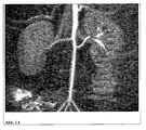
- the long circulation time allows a high-resolution representation of the coronary arteries in humans ( Figure 1.2 ).
- the iron oxide nanoparticles of the invention allow assessment of microcirculation, including angiogenesis, in the context of inflammatory processes, infections or tumors.
- microcirculation including angiogenesis
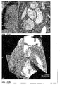
- the particles of the invention By enriching the particles of the invention in inflammatory arterial walls (various stages of arteriosclerosis), it is possible to detect a threat to an infarction very early, regardless of the extent of vascular constriction. This is in picture example Figure 1.4 explained. Furthermore, the particles can be used for the morphological evaluation of thrombi or emboli (arterial or venous).
- Example of Use 2 Diagnosis of tumors and their metastases including metastasis pathways of the lymphatic system
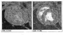
- the well-tolerated purified and formulated iron oxide particles of Example 5 are used for MRI diagnosis of primary tumors and their metastases. This is with T1-weighted imaging (whitening effect, Figure 2.1 ) and T2-weighted (darkening effect Figure 2.2 ) possible. In picture example Figure 2.1 the application is shown for the improved imaging of a liver tumor in the rat.
- the magnetic properties and good compatibility of the purified and formulated particles of the invention allow injection into the lymphatic system, or into regions (body tissues, organs tumors) from which tissue water (lymph) is transported to a lymph node. Used in tumor diagnosis this for finding the Sentinellymphknoten or sentinel lymph node. This is the crucial lymph node that receives metastases as a possible first lymph node through a primary tumor. A possible metastatic involvement of the sentinel lymph node determines the therapy planning and the prognosis for tumor diseases.
- the lymphatic vessel system can be assessed with T1-weighted imaging (lightening magnetic activity of the iron oxide nanoparticles).
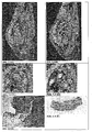
- the lymph node and any metastases can be assessed (darkening magnetic effect of the iron oxide nanoparticles). This is shown in the image examples 2.3.
- the evaluation of the lymphatic system can be combined by additional binding of dyes (visual, fluorescent) or by the binding of radioactive substances (technetium, indium) to an MRI-optical imaging or to an MRI-nuclear medical imaging.
- the very good compatibility and the long circulation time of the purified and formulated iron oxide particles of the invention make it possible to use them for tumor therapy after intravenous, intraarterial and intratumoral injection in combination with magnetic fields (magnetic field hyperthermia), embolisates and chemotherapeutics.
- increased accumulation in the target tissue can be achieved by binding so-called target-specific ligands on the iron oxide particles.
- Fig. 3.1 is the enhancement of the intratumoral accumulation of the particles by the binding of polyamines on the surface and thus the target direction of angiogenetic endothelium.
- the unmodified particles allow the long intravasal residence time to record the circulatory circulation of tumors and thus possible therapeutic effects in the context of tumor therapy (therapy monitoring).
- iron oxide particles cells stem cells, endothelial cells, dendritic cells, organ cells, immune cells
- stem cells endothelial cells, dendritic cells, organ cells, immune cells
- z. incubated with a dispersion of Example 3 for 30 to 60 min and washed. After injection of these labeled cells into the body (intravenously, intraarterially, lymphoid, in tissues, organs or pathological processes) it is possible to insert these cells within the body
- the display of the labeling of neuronal stem cells shown on a rat Parkinson's model ( Fig. 4.1 and 4.2 ).
- MRI and optical imaging By binding dyes or radioactive markers, a combination of MRI and optical imaging, or a combination of MRI and nuclear medical imaging, such as SINTINTAPRAPHY, SPECT, or PET, is possible to assess the morphology, function, and biochemistry of superparamagnetic particle-labeled cells in the living organism to investigate.
- nuclear medical imaging such as SINTINTAPRAPHY, SPECT, or PET
- the accumulation of the magnetic particles from the sample of Example 5 in the arteriosclerotic plaque leads to a T2 relaxation time-reducing effect with a loss of signal in the vessel wall.
- the arteriosclerotic plaque is shown in dark contrast. Enrichment of the magnetic particles of this sample demonstrates the presence of inflammatory cells in and macrophages in the arteriosclerotic plaque.
- MR contrast agents for parenteral use can be prepared that allow a combination of MRI and optical imaging, or a combination of MRI and nuclear medical imaging, such as SINTINTAPRAPHY, SPECT, or PET.
- apoptosis imaging can provide real-time information about the spatial distribution of apoptosis and thus allow a revealing characterization of pathological processes in a wide variety of disease states.
- acute thrombi can be detected by MRI, as shown by studies in rats and rabbits.
- the accumulation of the magnetic particles from the sample of Example 5 in the arteriosclerotic plaque leads to a T2 relaxation time-reducing effect with a loss of signal in the vessel wall.
- the arteriosclerotic plaque is shown in dark contrast.
- anti-inflammatory substances such as paclitaxel or matrix metalloproteinase inhibitors (MMP), such as.
- MMP matrix metalloproteinase inhibitors
- As marimastat, neovastat, sirolimus or tacrolimus leads to a Enrichment of these anti-inflammatory substances in the inflammatory plaques and thus to an anti-inflammatory.
- particles according to the invention with polyamine-coated surfaces according to example 8 are suitable as transfection agents for DNA and RNA in vitro on cell cultures of colon carcinomas.
Abstract
Description
Die Erfindung betrifft eine wässrige Dispersion von superparamagnetischen eisenhaltigen Teilchen, die auf ihrer Oberfläche als Stabilisatorsubstanzen α-Hydroxycarbonsäuren tragen, wobei die Dispersion N-Methyl-D-glucamin (Meglumin) und/oder 2-Amino-2-(hydroxymethyl)-1,3-propandiol (Trometamol) umfasst und der freie Eisenionengehalt kleiner 1 mg/l Eisen ist. Die erfindungsgemäße Dispersion kann in einer bevorzugten Ausführungsform zusätzlich einen Komplexbildner für Eisen enthalten. In einer weiteren bevorzugten Ausführungsform beinhaltet die Dispersion positiv geladene Metallionen und/oder polyaminogruppenhaltige Verbindungen, die an therapeutisch oder diagnostisch wirksame Substanzen gebunden sein können.The invention relates to an aqueous dispersion of superparamagnetic iron-containing particles which carry on their surface as stabilizer substances .alpha.-hydroxycarboxylic acids, the dispersion comprising N-methyl-D-glucamine (meglumine) and / or 2-amino-2- (hydroxymethyl) -1, 3-propanediol (trometamol) and the free iron ion content is less than 1 mg / l iron. In a preferred embodiment, the dispersion according to the invention may additionally contain a complexing agent for iron. In a further preferred embodiment, the dispersion contains positively charged metal ions and / or polyamino-containing compounds which may be bound to therapeutically or diagnostically active substances.
Gegenstand der Erfindung ist auch das Herstellungsverfahren dieser Dispersion, deren Verwendung als MRT-Kontrastmittel sowie deren Verwendung als Therapeutikum, auch mit der Möglichkeit der Verlaufskontrolle der Therapie durch ein Bildgebungsverfahren.The invention also relates to the preparation process of this dispersion, its use as an MRI contrast agent and its use as a therapeutic, also with the possibility of monitoring the therapy by an imaging method.
In den letzten Jahren gewinnt die ,Molekulare Bildgebung', also die in vivo-Charakterisierung und Darstellung von biologischen Prozessen auf dem zellulären und molekularen Niveau, bei der Erforschung von Krankheiten und zunehmend auch bei der klinischen Anwendung immer mehr an Bedeutung. Grundlage dafür ist die Entwicklung von molekularen Markern, die mit den zur Verfügung stehenden oder zu entwickelnden Bildgebungstechniken die gewünschten molekularen Targets ausreichend sensitiv detektieren können.In recent years, molecular imaging, ie the in vivo characterization and presentation of biological processes at the cellular and molecular level, in the study of diseases and increasingly also in clinical application is becoming increasingly important. The basis for this is the development of molecular markers that can detect the desired molecular targets sufficiently sensitively with the imaging techniques available or to be developed.
Die Magnetresonanztomographie (MRT) hat sich auf Grund ihres im Vergleich zu anderen Bildgebungstechniken hervorragenden Weichteilkontrastes bei gleichzeitig hoher anatomischer Auflösung als wichtige Säule der klinisch-radiologischen Diagnostik etabliert. Mit der Einführung superparamagnetischer Eisenoxid-Nanopartikel stehen aufgrund ihrer hohen T2- und teilweise T1-Relaxivität wirksame Marker für die ,Molekulare Bildgebung' zur Verfügung.Magnetic Resonance Imaging (MRI) has established itself as an important pillar of clinical radiological diagnostics due to its superior soft-tissue contrast compared to other imaging techniques combined with high anatomical resolution. With the introduction of superparamagnetic iron oxide nanoparticles, their high T2 and, in part, T1 relaxivity make effective markers available for 'molecular imaging'.
In den Patentanmeldungen
VSOP sind im Vergleich zu den vorher bekannten polymerbeschichteten (z. B. mit Dextran) superparamagnetischen Eisenoxidpartikel (SPIO, USPIO) deutlich kleiner. Beispielsweise weisen Citrat-beschichtete VSOP einen hydrodynamischen Durchmesser von ~7 nm auf, während die kleinsten polymerummantelten USPIO einen Durchmesser von etwa 15 - 20 nm haben.VSOP are significantly smaller compared to the previously known polymer-coated (eg with dextran) superparamagnetic iron oxide particles (SPIO, USPIO). For example, citrate-coated VSOP have a hydrodynamic diameter of ~ 7 nm, while the smallest polymer-coated USPIO have a diameter of about 15-20 nm.
Allerdings weisen auch diese bisher bekannten kleinen superparamagnetischen Eisenpartikel keine optimale Verträglichkeit bei parenteraler oder enteraler Anwendung im tierischen oder menschlichen Körper auf.However, these hitherto known small superparamagnetic iron particles also have no optimal compatibility with parenteral or enteral application in the animal or human body.
Dreiwertige und insbesondere zweiwertige Eisenionen sind hochtoxisch für biologisches Gewebe sowie für Säugetiere und Menschen. So wurde gefunden, dass die Toxizität von mit Citronensäure stabilisierten Mangan- Eisenferriten sehr hoch ist (
Dies ist auch aus der Anwendung von Eisenkomplexen für die parenterale Eisenerzsatz-Therapie bei Eisenmangelanämie bekannt. So führt die intravenöse Injektion von einem Eisen-Sucrose-Komplex als Wirkstoff in dem fertigen zugelassenen Arzneimittel Venofer® zu einer vorübergehenden Nierenschädigung, welche durch oxidativen Stress durch freie Eisenionen ausgelöst wird (
Neben der direkt zellschädigenden Wirkung freier Eisenionen gibt es bei den bekannten klinisch zugelassenen Präparaten zur Eisenersatztherapie und auch bei den klinisch zugelassen Eisenoxidpartikeln für die MR-Diagnostik das Nebenwirkungsspektrum der anaphylaktischen Reaktion, hervorgerufen durch polymere Stabilisatorsubstanzen, wie z. B. das Dextran.In addition to the direct cell-damaging effect of free iron ions, there are in the known clinically approved preparations for iron replacement therapy and also in the clinically approved iron oxide particles for MR diagnosis, the side effect spectrum of the anaphylactic reaction, caused by polymeric stabilizer substances such. B. the dextran.
Das nicht hitzestabilisierbare Endorem® (AMI 227) von Laboratoire Guerbet (Frankreich) wurde zwar auf der Basis von superparamagnetischen Eisenoxid-Nanopartikeln entwickelt, ist aber mit Dextran stabilisiert und daher sehr unverträglich. Wegen der Unverträglichkeit darf es nur durch Infusion mit einer Glucoselösung und in einer geringen Konzentration à 20 µmol Fe/kg n Eisen eingesetzt werden. Es ist zugelassen für den Nachweis von Lebertumoren durch MRT.The non-heat stabilizable Endorem ® (AMI 227) from Laboratoire Guerbet (France) was developed on the basis of superparamagnetic iron oxide nanoparticles, but is stabilized with dextran and therefore very incompatible. Because of the incompatibility it may only be used by infusion with a glucose solution and in a low concentration of 20 .mu.mol Fe / kg n iron. It is approved for the detection of liver tumors by MRI.
Ein weiteres zugelassenes leberspezifisches superparamagnetisches Eisenoxidpartikel ist Resovist® von der Schering AG (Deutschland). Dabei handelt es sich um relativ große, dextranummantelte superparamagnetische Eisenoxidpartikel, die nach Applikation sofort von den Makrophagen der Leber aufgenommen werden. Diese Partikel zirkulieren also nur sehr kurze Zeit im Blut. Trotz der niedrigen Dosierung von 20 µmol Fe/kg kann es zu Unverträglichkeiten kommen.Another approved liver-specific superparamagnetic iron oxide particles is Resovist ® from Schering AG (Germany). These are relatively large, dextran-coated superparamagnetic iron oxide particles, which are absorbed immediately after administration by the macrophages of the liver. These particles circulate in the blood only for a very short time. Despite the low dosage of 20 μmol Fe / kg incompatibilities can occur.
Aufgabe der Erfindung war es deshalb, eine wässrige Dispersion von sehr kleinen superparamagnetischen eisenhaltigen Teilchen zur Verfügung zu stellen, die einen hohen Kontrasteffekt liefert, aber weniger toxisch ist, so dass auch eine parenterale Anwendung ohne Nebenwirkungen möglich ist. Auch soll die Dispersion hitzesterilisierbar sein, ohne dass die freie Eisenionenkonzentration wesentlich ansteigt und ohne dass die Wirksamkeit der eisenhaltigen Teilchen verloren geht und eine Verschlechterung des Kontrastes eintritt. Daneben sollen die eisenhaltigen Teilchen auch eine längere Verweildauer im Blut aufweisen.The object of the invention was therefore to provide an aqueous dispersion of very small superparamagnetic iron-containing particles, which provides a high contrast effect, but is less toxic, so that a parenteral application without side effects is possible. Also, the dispersion should be heat sterilizable without the free iron ion concentration increases substantially and without the effectiveness of the iron-containing particles is lost and a deterioration of the contrast occurs. In addition, the iron-containing particles should also have a longer residence time in the blood.
Die Aufgabe der Erfindung wird gemäß den Patentansprüchen gelöst. Die Unteransprüche stellen bevorzugte Ausführungsformen der zugehörigen unabhängigen Ansprüche dar. Demgemäß wird eine wässrige Dispersion von Eindomänenteilchen aus Eisenhydroxid, Eisenoxidhydrat, Eisenoxid, Eisenmischoxid oder Eisen mit einer Teilchengröße von 2 bis 10 nm, die auf ihrer Oberfläche als Stabilisatorsubstanzen aliphatische Di- und/oder Tricarbonsäuren ausgewählt aus Zitronensäure, Äpfelsäure, Weinsäure oder deren Gemischen tragen, zur Verfügung gestellt, die dadurch gekennzeichnet ist, dass sie N-Methyl-D-glucamin (Meglumin) und/oder 2-Amino-2-(hydroxymethyl)-1,3-propandiol (Trometamol) umfasst und der freie Eisenionengehalt kleiner 1 mg/l Eisen ist. Die Dispersion ist herstellbar durch Fällung der eisenhaltigen Teilchen aus wässrigen Eisensalzlösungen mit Alkalilauge oder Ammoniumhydroxid, anschließender Behandlung mit den genannten Di- und/oder Tricarbonsäuren oder deren Gemischen und Reinigung der so stabilisierten Teilchen durch Dialyse mit destilliertem Wasser bis das Dialysat eine elektrische Leitfähigkeit kleiner 10 µS/cm besitzt. Die so erhaltene Dispersion wird als vorgereinigte Dispersion bezeichnet. Danach erfolgt die erfindungsgemäße Behandlung der vorgereinigten Dispersion mit wässrigen Lösungen von Salzen aliphatischer Di- und/oder Tricarbonsäuren, ausgewählt aus Zitronensäure, Äpfelsäure, Weinsäure oder deren Gemischen und Dialyse mit destilliertem Wasser bis das Dialysat eine elektrische Leitfähigkeit kleiner 10 µS/cm und einen freien Eisenionengehalt kleiner 1 mg/l besitzt. Anschließend wird die Dispersion mit einer wässrigen Lösung der genannten freien Di- und/oder Tricarbonsäuren oder deren Gemischen behandelt und mit destilliertem Wasser dialysiert bis das Dialysat eine elektrische Leitfähigkeit kleiner 10 µS/cm und einen freien Eisenionengehalt kleiner 1 mg/l besitzt und N-Methyl-D-glucamin (Meglumin) und/oder 2-Amino-2-(hydroxymethyl)-1,3-propandiol (Trometamol) zugesetzt.The object of the invention is achieved according to the patent claims. Accordingly, an aqueous dispersion of single domain particles of iron hydroxide, iron oxide hydrate, iron oxide, iron mixed oxide or iron having a particle size of 2 to 10 nm, on their surface as stabilizer substances aliphatic di- and / or tricarboxylic acids selected from citric acid, malic acid, tartaric acid or mixtures thereof, characterized in that they contain N-methyl-D-glucamine (meglumine) and / or 2-amino-2- (hydroxymethyl) -1,3- propandiol (trometamol) and the free iron ion content is less than 1 mg / l iron. The dispersion can be prepared by precipitation of the iron-containing particles from aqueous iron salt solutions with alkali metal hydroxide or ammonium hydroxide, subsequent treatment with said di- and / or tricarboxylic acids or mixtures thereof and purification of the thus stabilized particles by dialysis with distilled water until the dialysate has an electrical conductivity of less than 10 μS / cm. The dispersion thus obtained is referred to as prepurified dispersion. Thereafter, the inventive treatment of the prepurified dispersion with aqueous solutions of salts of aliphatic di- and / or tricarboxylic acids, selected from citric acid, malic acid, tartaric acid or mixtures thereof and dialysis with distilled water until the dialysate is an electrical conductivity less than 10 μS / cm and a free Iron ion content less than 1 mg / l has. Subsequently, the dispersion is treated with an aqueous solution of said free di- and / or tricarboxylic acids or mixtures thereof and dialyzed with distilled water until the dialysate has an electrical conductivity of less than 10 μS / cm and a free iron ion content of less than 1 mg / l and N- Methyl D-glucamine (meglumine) and / or 2-amino-2- (hydroxymethyl) -1,3-propanediol (trometamol).
Überraschend wurde gefunden, dass die Toxizität der wässrigen Dispersion von super-paramagnetischen Eindomänenteilchen durch eine Behandlung der vorgereinigten und stabilisierten Teilchen (wie sie beispielsweise in
In einer bevorzugten Ausführungsform der Erfindung wird als aliphatische Tricarbonsäure Citronensäure eingesetzt. Als Citrate finden Tri-Natrium-Citrat und Di-Natrium-Hydrogencitrat bevorzugte Anwendung.In a preferred embodiment of the invention, citric acid is used as aliphatic tricarboxylic acid. Tri-sodium citrate and di-sodium hydrogen citrate are preferred as citrates.
Die z. B. mit Citronensäure stabilisierten superparamagnetischen Eindomänenteilchen bilden in wässriger Dispersion eine stabile magnetische Flüssigkeit, die durch Dialyse gegen destilliertes Wasser von den wasserlöslichen Reaktionsprodukten, die bei der Herstellung der superparamagnetischen Eindomänenteilchen entstanden sind, vorgereinigt werden. Diese Vorgehensweise ist in
Erfindungsgemäß bevorzugt werden nun die vorgereinigten und stabilisierten superparamagnetischen Eindomänenteilchen zur Verringerung des freien Eisenionengehalts mit wässrigen Lösungen von Tri-, Di-, oder Mono-Salzen der Citronensäure behandelt und anschließend mit destilliertem Wasser dialysiert, danach mit einer wässrigen Lösung von freier Citronensäure behandelt und anschließend erneut mit destilliertem Wasser so lange dialysiert, bis der freie Eisenionengehalt kleiner als 0,005% der Gesamtmenge an Eisen ist.According to the invention, the pre-cleaned and stabilized superparamagnetic single-domain particles are then treated with aqueous solutions of tri-, di- or mono-salts of citric acid to reduce the free iron ion content and then dialyzed with distilled water, then treated with an aqueous solution of free citric acid and subsequently dialyzed again with distilled water until the free iron ion content is less than 0.005% of the total amount of iron.
Weiterhin wurde überraschend gefunden, dass die Verweildauer der erfindungsgemäßen superparamagnetischen eisenhaltigen Teilchen im Blut durch diese Behandlung mit Lösungen von Salzen der Di- oder Tricarbonsäuren und der freien Säuren und jeweils anschließender Dialyse mit destilliertem Wasser verlängert wird.Furthermore, it has surprisingly been found that the residence time of the superparamagnetic iron-containing particles according to the invention in the blood is prolonged by this treatment with solutions of salts of di- or tricarboxylic acids and of the free acids and in each case subsequent dialysis with distilled water.
Erfindungsgemäß werden an die so stabilisierten eisenhaltigen Teilchen monoaminogruppenhaltige Verbindungen gebunden, ausgewählt aus D(-)-N-Methylglucamin (Meglumin) oder 2-Amino-2-(hydroxymethyl)-1,3-propandiol (Trometamol) oder deren Gemisch. Hierbei hat sich überraschenderweise gezeigt, dass der vollständige oder teilweise Ersatz der Kationen, wie Ammonium-, Natrium- oder Hydroniumionen der freien Carboxylgruppen der z. B. mit Citronensäure stabilisierten Teilchen, durch N- Methyl-D-glucamin, und/oder 2-Amino-2-(hydroxymethyl)-1,3-propandiol zu einer Verringerung der Toxizität der erfindungsgemäßen eisenhaltigen Teilchen führt.According to the invention, mono-amino-containing compounds are bound to the thus-stabilized iron-containing particles, selected from D (-) - N-methylglucamine (meglumine) or 2-amino-2- (hydroxymethyl) -1,3-propanediol (trometamol) or a mixture thereof. It has surprisingly been found that the complete or partial replacement of the cations, such as ammonium, sodium or hydronium ions of the free carboxyl groups of z. As with citric acid stabilized particles, by N-methyl-D-glucamine, and / or 2-amino-2- (hydroxymethyl) -1,3-propanediol leads to a reduction in the toxicity of the iron-containing particles according to the invention.
In einer weiteren erfindungsgemäßen Ausführungsform können der Dispersion physiologisch verträgliche polyaminogruppenhaltige Verbindungen zugesetzt werden, die aus der Gruppe ausgewählt sind, die Polyethylenimine (PEI), Polyvinylamine (PVAm), PEI- und PVAm- Co-Polymere, Polylysin, Spermin, Spermidin, Protamin, Protaminsulfat, Oligopeptide, Polypeptide, Denaturierungsprodukte von Proteinen und Proteiden, wie Gelatine, Kasein-Hydrolysate, Gluteline; stickstoffhaltigen Polysaccharide, wie Mucopolysaccharide, Glykoproteide, Chitine und Gemische davon, umfasst, vorzugsweise Polyethylenimine (PEI) oder Poly-vinylamine (PVAm).In a further embodiment according to the invention, it is possible to add to the dispersion physiologically compatible polyamino group-containing compounds selected from the group consisting of polyethyleneimines (PEI), polyvinylamines (PVAm), PEI and PVAm copolymers, polylysine, spermine, spermidine, protamine, Protamine sulfate, oligopeptides, polypeptides, protein and protein denaturation products such as gelatin, casein hydrolysates, gluteline; nitrogen-containing polysaccharides, such as Mucopolysaccharides, glycoproteins, chitins, and mixtures thereof, preferably polyethyleneimines (PEI) or polyvinylamines (PVAm).
Durch den teilweisen Ersatz der Kationen, wie z. B. Ammonium-, Natrium- oder Hydroniumionen der freien Carboxylgruppen der z. B. mit Citronensäure stabilisierten eisenhaltigen Teilchen, durch polyaminogruppenhaltige Verbindungen, können diagnostisch wirksame Substanzen, zell- und gewebespezifische Bindungssubstanzen, pharmakologisch wirksame Substanzen, pharmakologisch wirksame Zellen oder zellfusionsvermittelnde Substanzen an die polyaminogruppenhaltigen Verbindungen nach bekannten Kopplungsverfahren chemisch gebunden werden. Dabei können zunächst die biologisch wirksamen Substanzen an die Polyamine gebunden und aufgereinigt werden und diese Reaktionsprodukte dann an die erfindungsgemäßen eisenhaltigen Teilchen gekoppelt werden.Due to the partial replacement of cations, such. For example, ammonium, sodium or hydronium ions of the free carboxyl groups of z. As with citric acid-stabilized iron-containing particles, by polyaminogruppenhaltige compounds, diagnostically active substances, cell and tissue-specific binding substances, pharmacologically active substances, pharmacologically active cells or cell fusion mediating substances to the polyaminogruppenhaltigen compounds can be chemically bound by known coupling methods. In this case, first the biologically active substances can be bound to the polyamines and purified, and these reaction products can then be coupled to the iron-containing particles according to the invention.
Als diagnostisch wirksame Substanzen können an die Polyamine z. B. Fluoreszenzfarbstoffe für den Wellenlängenbereich von 200 bis 1200 Nanometer für die Kombination der MRT mit optischen Diagnoseverfahren gebunden werden. Als Fluoreszenzfarbstoffe kommen z. B. Fluoreszein, Rhodamin green, Texas red. sowie Gemische davon in Frage.As diagnostically active substances can be attached to the polyamines z. B. fluorescent dyes for the wavelength range of 200 to 1200 nanometers for the combination of MRI with optical diagnostic methods are bound. As fluorescent dyes z. B. fluorescein, Rhodamine green, Texas red. and mixtures thereof in question.
An die polyaminogruppenhaltigen Verbindungen können auch diagnostisch wirksame Substanzen, wie perfluorhaltige Moleküle für die Ultraschalldiagnostik gebunden werden. Als perfluorhaltige Moleküle kommen z. B. Perfluoralkylphosphat, Perfluoralkoxypolyethylenglykolphosphat , Hexanfluorphospat sowie Gemische davon in Frage.To the polyaminogruppenhaltigen compounds also diagnostically active substances such as perfluorhaltige molecules for ultrasound diagnosis can be bound. As perfluorhaltige molecules come z. As perfluoroalkyl phosphate, Perfluoralkoxypolyethylenglykolphosphat, Hexanfluorphospat and mixtures thereof in question.
Als diagnostisch wirksame Substanzen können an die Polyamine z. B. kurzlebige Radiopharmaka für die Kombination der MRT mit der Positronen-Emissionen-Tomographie (PET) gebunden werden. Als Radiopharmaka kommen organische Substanzen , die 11C, 13N, 15O, 18F, 68Ga, 75Br, 123J, enthalten, wie z. B. [11C] Thymidin, [18F]Fluor-L-DOPA, [68Ga]-anti- CD66, zur Anwendung..As diagnostically active substances can be attached to the polyamines z. B. short-lived radiopharmaceuticals for the combination of MRI with positron emission tomography (PET) are bound. As radiopharmaceuticals, organic substances containing 11 C, 13 N, 15 O, 18 F, 68 Ga, 75 Br, 123 J, such as. For example, [ 11 C] thymidine, [ 18 F] fluoro-L-DOPA, [ 68 Ga] anti-CD66, are used.
An die polyaminogruppenhaltigen Verbindungen können als zell- oder gewebespezifische Bindungssubstanzen z. B. Antigene, Antikörper, Ribonucleinsäuren, Desoxyribo-nucleinsäuren, Ribonucleinsäuresequenzen, Desoxyribonucleinsäuresequenzen, Haptene, Avidin, Streptavidin, Protein A, Protein G, Anexin, Endotoxinbindende Proteine, Lectine, Selectine, Integrine, Oberflächenproteine von Organellen, Viren, Mikroben, Algen, Pilze sowie Gemische davon gebunden werden.To the polyaminogruppenhaltigen compounds can be used as cell- or tissue-specific binding substances z. Antigens, antibodies, ribonucleic acids, deoxyribonucleic acids, ribonucleic acid sequences, deoxyribonucleic acid sequences, haptens, avidin, streptavidin, protein A, protein G, anexin, endotoxin binders Proteins, lectins, selectins, integrins, surface proteins of organelles, viruses, microbes, algae, fungi and mixtures thereof are bound.
An die polyaminogruppenhaltigen Verbindungen können als pharmakologisch wirksame Substanzen z. B. Antitumorproteine, Enzyme, Antitumorenzyme, Antibiotika, Pflanzenalkaloide, Alkylierungsreagenzien, Antimetaboliten, Hormone und Hormonantagonisten, Interleukine, Interferone, Wachstumsfaktoren, Tumornekrosefaktoren, Endotoxine, Lymphotoxine, Integrine, Urokinase, Streptokinase, Plasminogen-Streptokinase-Aktivator-Komplex, Gewebe-Plasminogen-Aktivatoren, Desmodus-Plasminogen-Aktivatoren, Makrophagen-Aktivierungs-Körper, Antisera, Blut- und Zellbestandteile und deren Abbauprodukte und Derivate, Zellwandbestandteile von Organellen, Viren, Mikroben, Algen, Pilze und deren Abbauprodukte und Derivate, Proteaseninhibitoren, Alkylphosphocholine; radioaktive Isotope enthaltende Substanzen, Tenside, kardiovaskuläre Pharmazeutika, Chemotherapeutika, gastrointestinale Pharmazeutika, Neuropharma-zeutika sowie Gemische davon gebunden werden.At the polyaminogruppenhaltigen compounds can be used as pharmacologically active substances such. Antitumor proteins, enzymes, antitumor enzymes, antibiotics, plant alkaloids, alkylating reagents, antimetabolites, hormones and hormone antagonists, interleukins, interferons, growth factors, tumor necrosis factors, endotoxins, lymphotoxins, integrins, urokinase, streptokinase, plasminogen streptokinase activator complex, tissue plasminogen Activators, desmode plasminogen activators, macrophage activating bodies, antisera, blood and cell components and their degradation products and derivatives, cell wall components of organelles, viruses, microbes, algae, fungi and their degradation products and derivatives, protease inhibitors, alkylphosphocholines; radioactive isotope-containing substances, surfactants, cardiovascular pharmaceuticals, chemotherapeutic agents, gastrointestinal pharmaceuticals, neuropharmaceuticals and mixtures thereof.
An die polyaminogruppenhaltigen Verbindungen können als pharmakologisch wirksame Zellen z. B. Organellen, Viren, Mikroben, Algen, Pilze, insbesondere Erythrozyten, Thrombozyten, Granulozyten, Monozyten, Lymphozyten, Langerhans'sche Inseln gebunden werden.To the polyaminogruppenhaltigen compounds can be used as pharmacologically active cells z. As organelles, viruses, microbes, algae, fungi, especially erythrocytes, platelets, granulocytes, monocytes, lymphocytes, Langerhans'sche be bound.
Die Bindung dieser Substanzen an die polyaminogruppenhaltigen Verbindungen ist dem Fachmann bekannt. So kann die kovalente Bindung der polyaminogruppenhaltigen Verbindungen oder deren Reaktionsprodukte mit den genannten Substanzen mit den z. B. durch Citronensäure stabilisierten erfindungsgemäßen Eindomänenteilchen beispielsweise mit Substanzen aus der Gruppe der Carbodiimide, wie z. B. 1-Ethyl-3-(3-Dimethylaminopropyl)carbodiimide (EDC), 1-Cyclohexyl-3-(2-morpholinoethyl)carbodiimide (CMC) oder Dicyclohexyl carbodiimide (DCC) geknüpft werden. So lassen sich stabile Verbindungen zwischen den Carboxylgruppen der Stabilisatormoleküle auf der Oberfläche der erfindungsgemäßen Eindomänenteilchen und den Aminogruppen der genannten Substanzen erzeugen.The binding of these substances to the polyaminogruppenhaltigen compounds is known in the art. Thus, the covalent bonding of polyaminogruppenhaltigen compounds or their reaction products with the substances mentioned with the z. B. stabilized by citric acid Einomänenteilchen invention, for example, with substances from the group of carbodiimides, such as. For example, 1-ethyl-3- (3-dimethylaminopropyl) carbodiimides (EDC), 1-cyclohexyl-3- (2-morpholinoethyl) carbodiimides (CMC) or dicyclohexyl carbodiimide (DCC) are linked. Thus, stable compounds can be generated between the carboxyl groups of the stabilizer molecules on the surface of the single-domain particles according to the invention and the amino groups of the substances mentioned.
Die nicht-kovalente Kopplung kann durch elektrostatische Wechselwirkungen erfolgen. Beispielsweise binden Polyamine elektrostatisch an Zitrat-beschichtete eisenhaltige Teilchen.The non-covalent coupling can be done by electrostatic interactions. For example, polyamines electrostatically bind to citrate-coated iron-containing particles.
In einer weiteren erfindungsgemäßen Ausführungsform der Erfindung enthält die erfindungsgemäße Dispersion positiv geladene Metallionen der chemischen Elemente wie z. B. Kupfer, Silber, Gold, Eisen, Gallium Thallium, Bismut, Palladium, Rhenium, Ruthenium, Platin, Technetium, Indium, Iridium, Radium, Selen, Ytrium, Zirkon und seltenen Erden sowie Gemische davon, wobei die Metallionen auch die radioaktiven Isotope derselben chemischen Elemente sein können wie z. B. 52Fe, 67Ga, 99mTc, 113In, 188Rh, 192Ir, 198Au, 201Tl, 223Ra sowie Gemische davon. Auch an diese Teilchen können zusätzlich noch die genannten mono- und/oder polyaminogruppenhaltigen Verbindungen sowie diagnostisch wirksamen Substanzen, zell- und gewebe-spezifische Bindungssubstanzen, pharmakologisch wirksame Substanzen, pharmakologisch wirksame Zellen oder zellfusionsvermittelnden Substanzen gebunden sein.In a further embodiment of the invention, the inventive dispersion contains positively charged metal ions of the chemical elements such. As copper, silver, gold, iron, gallium thallium, bismuth, palladium, rhenium, ruthenium, platinum, technetium, indium, iridium, radium, selenium, yttrium, zirconium and rare earths and mixtures thereof, wherein the metal ions and the radioactive isotopes the same chemical elements may be such. 52 Fe, 67 Ga, 99m Tc, 113 In, 188 Rh, 192 Ir, 198 Au, 201 Tl, 223 Ra and mixtures thereof. These particles may additionally contain the mono- and / or polyamino-containing compounds mentioned as well as diagnostically active substances, cell- and tissue-specific binding substances, pharmacologically active substances, pharmacologically active cells or cell fusion-mediating substances.
In einer weiteren erfindungsgemäßen Ausführungsform der Erfindung wird die Toxizität der erfindungsgemäßen wässrigen Dispersion der eisenhaltigen Teilchen durch Zugabe eines physiologisch verträglichen Komplexbildners für Eisen zur galenischen Formulierung noch weiter verringert und es kommt überraschenderweise zu keiner Auflösung der Teilchen. Bevozugte Komplexbildner sind z. B. Glycerolphosphorsäure, Ethylendiamintetraessigsäure (EDTA), N-Hydroxyethyl-ethylendiamintriessigsäure (HEDTA), Diethylentriaminpentaessigsäure (DTPA), α-Mercaptopropionylglycin (Tiopronin), 2,3-Mercapto-1-propansulfonsäure, 30-Amino-3,14,25-trihydroxy-3,9,14,20,25-pentaazatriacontan-2,10,13,21, 24-penton-methansulfonsäure (Deferoxaminmesilat). Aufgrund der geringen Toxizität sind diese Teilchen/Dispersionen damit auch für Mehrfachanwendungen am Menschen geeignet, so z. B. zur parenteralen Eisenersatztherapie. Die Anreicherung dieser Teilchen in Organen des blutbildenden Systems (Knochenmark, Milz) führt zu einer Depotwirkung und stellt somit für Patienten mit Eisenmangelerkrankungen eine vorteilhafte Therapie dar. Die Konzentration des physiologisch verträglichen Komplexbildners für Eisen in der Dispersion liegt im Bereich von 1 bis 20 Gew.% bezogen auf den Gehalt an Eisen.In a further embodiment of the invention, the toxicity of the aqueous dispersion of the iron-containing particles according to the invention is further reduced by the addition of a physiologically compatible complexing agent for iron to the galenic formulation and, surprisingly, no dissolution of the particles occurs. Preferred complexing agents are z. Glycerophosphoric acid, ethylenediaminetetraacetic acid (EDTA), N-hydroxyethylethylenediaminetriacetic acid (HEDTA), diethylenetriaminepentaacetic acid (DTPA), α-mercaptopropionylglycine (tiopronine), 2,3-mercapto-1-propanesulfonic acid, 30-amino-3,14,25- trihydroxy-3,9,14,20,25-pentaazatriacontane-2,10,13,21,24-pentone-methanesulfonic acid (deferoxamine mesylate). Due to the low toxicity of these particles / dispersions are therefore suitable for multiple applications in humans, such. For parenteral iron replacement therapy. The accumulation of these particles in organs of the blood-forming system (bone marrow, spleen) leads to a depot effect and thus represents an advantageous therapy for patients with iron deficiency diseases. The concentration of the physiologically acceptable complexing agent for iron in the dispersion is in the range of 1 to 20 wt. % based on the content of iron.
Als Kationen der säuregruppenhaltigen Komplexbildner für Eisen können Natrium, Kalium, Calcium, Magnesium, D(-)-N- Methylglucamin (Meglumin) oder 2-Amino-2-(hydroxymethyl)-1,3-propandiol (Trometamol) sowie Gemische davon eingesetzt werden. Als Komplexbildner.wird vorzugsweise Glycerolphosphorsäure oder ein Salz davon eingesetzt, besonders bevorzugt Natrium-Glycerolphosphat.Sodium, potassium, calcium, magnesium, D (-) - N-methylglucamine (meglumine) or 2-amino-2- (hydroxymethyl) -1,3-propanediol (trometamol) and mixtures thereof can be used as cations of the acid group-containing complexing agents for iron become. The complexing agent used is preferably glycerophosphoric acid or a salt thereof, more preferably sodium glycerol phosphate.
Sollen die erfindungsgemäßen eisenhaltigen Teilchen mit gebundenen positiv geladenen Metallionen auch einen Komplexbildner für Eisen, z. B. ein Glycerolphosphat tragen, so ist es wichtig, dass zunächst die positiv geladenen Metallionen gebunden werden, d. h. der Dispersion zugesetzt werden und erst bei der Herstellung der galenischen Formulierung der Komplexbildner zugesetzt wird.If the iron-containing particles according to the invention with bound positively charged metal ions and a complexing agent for iron, z. B. carry a glycerol phosphate, so it is important that initially the positively charged metal ions are bound, d. H. be added to the dispersion and the complexing agent is added only in the preparation of the galenic formulation.
Durch die Bindung von der Dispersion zugegebenen radioaktiven Metallionen, wie z. B. Technetium 99m oder Gallium 67, auf der Oberfläche der erfindungsgemäßen superparamagnetischen Eindomänenteilchen entsteht ein Kontrastmittel, das zu einer neuartigen Kombination von MRT Bildgebung mit einer Nuklearmedizinischen Bildgebung zusammengeführt werden kann. Dieses neue Bildgebungsverfahren kombiniert das große Auflösungs-vermögen der MR-Tomographie mit der hohen Empfindlichkeit der nuklearmedizinischen Bildgebungsverfahren, wie SZINTIGRAPHIE oder SPECT (Single-Photon Emission Computed Tomography).By the binding of the dispersion added radioactive metal ions, such as. Technetium 99m or gallium 67, on the surface of the superparamagnetic single domain particles of the present invention, a contrast agent is formed which can be combined to form a novel combination of MRI imaging with nuclear medical imaging. This new imaging technique combines the great resolving power of MRI with the high sensitivity of nuclear medicine imaging techniques such as SINTINTAPPHY or SPECT (Single-Photon Emission Computed Tomography).
Durch die Bindung von Farbstoffen oder kurzlebigen radioaktiven Markern können MR-Kontrastmittel für die parenterale Anwendung hergestellt werden, die eine Kombination von MRT und optischer Bildgebung oder eine Kombination von MRT und nuklearmedizinischer Bildgebung, wie PET, ermöglichen.By binding dyes or transient radioactive markers, MR contrast agents for parenteral use can be prepared that allow a combination of MRI and optical imaging or a combination of MRI and nuclear medicine imaging, such as PET.
Es besteht auch die Möglichkeit, die zwei getrennt aufgenommenen Bilddatensätze der MRT und der optischen oder nuklearmedizinischen Bildgebung zu einem Bild zusammenzufügen und für die verbesserte Diagnose von Erkrankungen einzusetzen.It is also possible to combine the two separately recorded image data sets of MRI and optical or nuclear medicine imaging into one image and use them for the improved diagnosis of diseases.
Die sehr kleinen superparamagnetischen Eindomänenteilchen der Erfindung können aus folgenden Substanzen bestehen: Eisenhydroxid, Eisenoxidhydrat, Fe2O3, Fe3O4, aus den Eisenmischoxiden der allgemeinen Formel m MO.n Fe2O3, worin M die zweiwertigen Metallionen Fe, Co, Ni, Mn, Be, Mg, Ca, Ba, Sr, Cu, Zn, Pt oder Gemische davon bedeutet, oder aus den Mischoxiden der allgemeinen Formel m Fe2O3.n Me2O3, worin Me die dreiwertigen Metallionen Al, Cr, Bi, seltene Erdmetalle oder Gemische davon bedeutet oder Eisen, wobei m und n in den jeweiligen Formeln ganze Zahlen von 1 bis 6 sind. So lässt sich durch die Zusammensetzung und durch die Struktur der Eindomänenteilchen deren magnetische Suszeptibilität in weiten Grenzen variieren und ein Verhältnis der Relaxivitäten R2/R1 kleiner 5 einstellen.The very small superparamagnetic single-domain particles of the invention may consist of the following substances: iron hydroxide, iron oxide hydrate, Fe 2 O 3 , Fe 3 O 4 , of the iron mixed oxides of the general formula m MO.n Fe 2 O 3 , where M is the divalent metal ions Fe, Co , Ni, Mn, Be, Mg, Ca, Ba, Sr, Cu, Zn, Pt or mixtures thereof, or from the mixed oxides of the general formula m Fe 2 O 3 .n Me 2 O 3 , where Me the trivalent metal ions Al , Cr, Bi, rare earth metals or mixtures thereof, or iron, where m and n in the respective formulas are integers from 1 to 6 are. Thus, due to the composition and the structure of the single-domain particles, their magnetic susceptibility can be varied within wide limits and a ratio of relaxivities R2 / R1 of less than 5 can be set.
Gegenstand der Erfindung ist auch das Verfahren zur Herstellung einer wässrigen Dispersion von superparamagnetischen Eindomänenteilchen aus Eisenhydroxid, Eisenoxidhydrat, Eisenoxid, Eisenmischoxid oder Eisen mit einer Teilchengröße von 2 bis 10 nm, die auf ihrer Oberfläche als Stabilisatorsubstanzen aliphatische Di- und/oder Tricarbonsäuren ausgewählt aus Zitronensäure, Äpfelsäure, Weinsäure oder deren Gemischen tragen, durch Fällung der superparamagnetischen eisenhaltigen Teilchen aus wässrigen Eisensalzlösungen mit Alkalilauge oder Ammoniumhydroxid, anschließender Behandlung mit aliphatischen Di- und/oder Tricarbonsäuren ausgewählt aus Zitronensäure, Äpfelsäure und Weinsäure oder deren Gemischen und Reinigung der so stabilisierten Teilchen durch Dialyse mit destilliertem Wasser bis das Dialysat eine elektrische Leitfähigkeit von kleiner 10 µS/cm besitzt, das dadurch gekennzeichnet ist, dass die Dispersion nachfolgend mit einer wässrigen Salzlösung von aliphatischen Di- und/oder Tricarbonsäuren, ausgewählt aus Zitronensäure, Äpfelsäure und Weinsäure oder deren Gemischen behandelt und mit destilliertem Wasser dialysiert wird bis die Leitfähigkeit des Dialysats kleiner 10 µS/cm ist und der freie Eisenionengehalt kleiner 1 mg/l ist, dann das Dialysat mit einer wässrigen Lösung der freien Di- und/oder Tricarbonsäuren, ausgewählt aus Zitronensäure, Äpfelsäure, Weinsäure oder deren Gemischen behandelt und mit destilliertem Wasser wiederum bis zu einer Leitfähigkeit des Dialysats von kleiner 10 µS/cm und bis zu einem freien Eisenionengehalt kleiner 1 mg/l dialysiert wird und N-Methyl-D-glucamin (Meglumin) und/oder 2-Amino-2-(hydroxymethyl)-1,3-propandiol (Trometamol) zugesetzt wird.The invention also provides the process for producing an aqueous dispersion of superparamagnetic single-domain particles of iron hydroxide, iron oxide hydrate, iron oxide, iron mixed oxide or iron having a particle size of 2 to 10 nm, the aliphatic di- and / or tricarboxylic acids selected from citric acid on their surface as stabilizer substances , Malic acid, tartaric acid or mixtures thereof, by precipitation of the superparamagnetic iron-containing particles from aqueous iron salt solutions with alkali or ammonium hydroxide, followed by treatment with aliphatic di- and / or tricarboxylic acids selected from citric acid, malic acid and tartaric acid or mixtures thereof and purification of the particles thus stabilized Dialysis with distilled water until the dialysate has an electrical conductivity of less than 10 μS / cm, which is characterized in that the dispersion is subsequently treated with an aqueous salt solution of alipha tables of di- and / or tricarboxylic acids, selected from citric acid, malic acid and tartaric acid or mixtures thereof and dialyzed with distilled water until the conductivity of the dialysate is less than 10 μS / cm and the free iron ion content is less than 1 mg / l, then the dialysate treated with an aqueous solution of the free di- and / or tricarboxylic acids selected from citric acid, malic acid, tartaric acid or mixtures thereof and in turn with distilled water to a conductivity of the dialysate of less than 10 μS / cm and up to a free iron ion content of less than 1 mg is dialyzed and N-methyl-D-glucamine (meglumine) and / or 2-amino-2- (hydroxymethyl) -1,3-propanediol (trometamol) is added.
Die sehr kleinen superparamagnetischen Eindomänenteilchen werden zunächst in bekannter Weise durch Fällung aus wässrigen Eisensalzlösungen mit Alkalilauge oder Ammoniakwasser und anschließender Behandlung mit 20 bis 50 Gew.-% stabilisierender Säure, ausgewählt aus Äpfelsäure, Weinsäure, Zitronensäure oder deren Gemischen , die eine Aggregation und Sedimentation im Schwerefeld verhindern, hergestellt und nachfolgend durch Dialyse mit destilliertem Wasser bis zu einer elektrischen Leitfähigkeit des Dialysates kleiner als 10 µS/cm vorgereinigt. Durch die erfindungsgemäße Behandlung der so vorgereinigten super-paramagnetischen Eisenoxidpartikel mit vorzugsweise Lösungen von Tri-, Di-, und Mono-Salzen der Citronensäure und Dialyse mit destilliertem Wasser bis zu einem freien Eisenionengehalt kleiner 1 mg/l und anschließender Behandlung mit einer wässrigen Lösung der freien Citronensäure und Dialyse mit destilliertem Wasser bis zu einem freien Eisenionengehalt kleiner 1 mg/l wird der Anteil an freien Eisenionen auf kleiner als 0,005% der Gesamtmenge an Eisen verringert. Der Zusatz von N-Methyl-D-glucamin (Meglumin) und/oder 2-Amino-2-(hydroxymethyl)-1,3-propandiol (Trometamol) bewirkt eine weitere Verringerung der Toxizität.The very small superparamagnetic Eindomänenteilenteilchen be first in a known manner by precipitation from aqueous iron salt solutions with alkali or ammonia water and subsequent treatment with 20 to 50 wt .-% stabilizing acid selected from malic acid, tartaric acid, citric acid or mixtures thereof, which aggregation and sedimentation in Prevent gravity field, prepared and subsequently pre-cleaned by dialysis with distilled water to an electrical conductivity of the dialysate less than 10 μS / cm. By the treatment according to the invention of the thus prepurified super-paramagnetic iron oxide particles with preferably solutions of tri-, di- and mono-salts of citric acid and dialysis with distilled water to a free iron ion content of less than 1 mg / l and subsequent treatment with an aqueous solution of the free citric acid and dialysis with distilled water up to a free iron ion content of less than 1 mg / l, the proportion of free iron ions is reduced to less than 0.005% of the total amount of iron. The addition of N-methyl-D-glucamine (meglumine) and / or 2-amino-2- (hydroxymethyl) -1,3-propanediol (trometamol) causes a further reduction in toxicity.
So lassen sich stabilisierte superparamagnetische Eindomänenteilchen mit einem Bereich des mittleren Teilchendurchmessers d50 von 2 bis 4 nm, vorzugsweise unter 3,5 nm herstellen, die also einen mittleren hydrodynamischen Teilchendurchmessers von 5 bis 8 nm aufweisen. Das Verhältnis der Relaxivitäten R2/R1 kann so auf Werte zwischen 1 bis 3 verringert werden, vorzugsweise auf 1 bis 2. Unter "mittlerer Teilchendurchmesser d50" wird verstanden, dass wenigstens 50 % der Teilchen in dem angegebenen Durchmesserbereich liegen. Die Teilchengröße wird mit Zetasizer der Fa. Malvern und mit dem Elektronenmikroskop bestimmt. Der mittlere Teilchendurchmesser bezieht sich auf die Teilchen mit (Zetasizer) und ohne Hydrathülle (Elektronenmikroskop).Thus, it is possible to produce stabilized superparamagnetic single-domain particles having a range of the mean particle diameter d50 of 2 to 4 nm, preferably less than 3.5 nm, that is to say having a mean hydrodynamic particle diameter of 5 to 8 nm. The ratio of the relaxivities R2 / R1 can thus be reduced to values between 1 and 3, preferably 1 to 2. By "average particle diameter d 50 " is meant that at least 50% of the particles are in the specified diameter range. The particle size is determined with Zetasizer from Malvern and with the electron microscope. The mean particle diameter refers to the particles with (Zetasizer) and without Hydrathülle (electron microscope).
Überraschend wurde gefunden, dass sehr kleine superparamagnetische Eindomänenteilchen mit einem Teilchendurchmesser kleiner 3,5 Nanometer nierengängig sind, also auch für eine MR-Diagnostik der harnableitenden Wege und vor allen Dingen für die molekulare Bildgebung geeignet sind.Surprisingly, it has been found that very small superparamagnetic single-domain particles with a particle diameter of less than 3.5 nanometers are kidney-like, thus also suitable for MR diagnosis of the urinary diversion pathways and, above all, for molecular imaging.
Damit verlängert sich die Blut-Halbwertszeit der erfindungsgemäßen sehr kleinen superparamagnetischen Eindomänenteilchen gegenüber den bisherigen Teilchen wesentlich und die möglichen Einsatzbereiche, z. B. für die T1-gewichtete MRT-Tomographie in der Angiographie, Lymphographie, Thromben- und TumorDiagnostik, erweitern sich.Thus, the blood half-life of the very small superparamagnetic single-domain particles according to the invention is substantially prolonged compared to the previous particles and the possible fields of use, eg. B. for T1-weighted MRI tomography in angiography, lymphography, thrombi and tumor diagnostics are expanding.
In einer bevorzugten Ausführungsform des erfindungsgemäßen Herstellungsverfahrens werden der erhaltenen wässrigen Dispersion polyaminogruppenhaltige Verbindungen zugesetzt, um Möglichkeiten für die Anbindung biologisch wirksamer Substanzen zu schaffen.In a preferred embodiment of the preparation process according to the invention, polyamino group-containing compounds are added to the resulting aqueous dispersion in order to create possibilities for the binding of biologically active substances.
Die Bindung der polyaminogruppenhaltigen Verbindungen oder der gereinigten Reaktionsprodukte von polyaminogruppenhaltigen Verbindungen mit diagnostisch wirksamen Substanzen, zell- und gewebespezifischen Bindungssubstanzen, pharmakologisch wirksamen Substanzen, pharmakologisch wirksamen Zellen oder zellfusioniermittelnden Substanzen auf der Oberfläche der mit Säuren gegen Aggregation stabilisierten superparamagnetischen Eindomänenteilchen kann durch elektrostatische Wechselwirkungen oder über eine kovalente chemische Bindung erfolgen wie oben beschrieben.The binding of the polyaminogruppenhaltigen compounds or the purified reaction products of polyaminogruppenhaltigen compounds with diagnostically active substances, cell and tissue-specific binding substances, pharmacologically active substances, pharmacologically active cells or Zellfusioniermittelnden substances on the surface of acid-aggregation stabilized superparamagnetic Einomänenteilchenchen by electrostatic interactions or a covalent chemical bond is carried out as described above.
Erfindungsgemäß kann der Dispersion auch ein Komplexbildner für Eisen zugesetzt werden, vorzugsweise eine Glycerolphosphorsäure oder ein Salz davon.According to the invention, a complexing agent for iron may also be added to the dispersion, preferably a glycerophosphoric acid or a salt thereof.
Gegenstand der Erfindung ist auch eine pharmazeutische Zusammensetzung umfassend die vorstehend definierte, erfindungsgemäße Dispersion von stabilisierten und gereinigten eisenhaltigen Teilchen, die gegebenenfalls einen Komplexbildner für Eisen und/oder gegebenenfalls positiv geladene Metallionen und/oder gegebenenfalls polyaminogruppenhaltige Verbindungen enthält. Die pharmazeutische Zusammensetzung kann pharmazeutisch annehmbare Hilfsstoffe wie z. B. Zucker, vorzugsweise Mannitol, Sorbitol, Glucose oder Xylitol, enthalten. Die Zucker sind in einer solchen Menge enthalten, dass physiologische Bedingungen, wie eine Osmolalität im Bereich von 200 bis 2000 mOs/kg, vorzugsweise von ca. 300 mOs/kg, gewährleistet sind. So enthält die pharmazeutische Zusammensetzung z. B. ca. 6% Mannitol.The invention also provides a pharmaceutical composition comprising the above-defined, inventive dispersion of stabilized and purified iron-containing particles, which optionally contains a complexing agent for iron and / or optionally positively charged metal ions and / or optionally polyamino-containing compounds. The pharmaceutical composition can pharmaceutically acceptable excipients such. For example, sugars, preferably mannitol, sorbitol, glucose or xylitol. The sugars are contained in such an amount as to ensure physiological conditions such as osmolality in the range of 200 to 2000 mOs / kg, preferably about 300 mOs / kg. Thus, the pharmaceutical composition contains z. B. about 6% mannitol.
Gegenstand der Erfindung ist auch die Verwendung der erfindungsgemäßen wässrigen Dispersion gemäß Anspruch 14.The invention also provides the use of the aqueous dispersion according to the invention as claimed in claim 14.
Die Hauptanwendungsgebiete der erfindungsgemäßen Dispersion, die sehr kleine superparamagnetische Eindomänenteilchen enthält, liegen auf den Gebieten der MRT-Kontrastmittel für die Angiographie, Lymphographie, Thromben- und Tumordiagnostik, der Tumorschädigung, der Thrombenauflösung, der Immunsteigerung, der Zellfusionsvermittlung oder des Gentransfers, wobei auch hier die Wirksamkeit der Tumorschädigung, der Thrombenauflösung, der Zellfusion und des Gentransfers mit der MRT-Diagnostik untersucht werden kann.The main fields of application of the dispersion according to the invention, which contains very small superparamagnetic single-domain particles, are in the fields of MRI contrast agents for angiography, lymphography, thrombus and tumor diagnostics, tumor damage, thrombus resolution, immune enhancement, cell fusion mediation or gene transfer the efficacy of tumor damage, thrombus resolution, cell fusion and gene transfer can be assessed by MRI diagnostics.
Die erfindungsgemäße Dispersion, die sehr kleine stabilisierte superparamagnetische Eindomänenteilchen enthält, die mit polyaminogruppenhaltigen Verbindungen wie z. B. Pentaetylenhexamin beschichtet sind, kann für die Tumordiagnostik eingesetzt werden, da bei ihrer Injektion in den Blutkreislauf eine Anreicherung in einigen Tumorarten zu beobachten ist. Bei Kopplung von pharmakologisch wirksamen Substanzen an die sehr kleinen stabilisierten superparamagnetischen Eindomänenteilchen kann deren Konzentration am Wirkungsort erhöht werden, insbesondere bei den mit Zytostatika, wie z. B. Doxorubicin oder Paclitaxel, gebunden an ein Polyamin, wie z. B. Pentaetylenhexamin, stabilisierten sehr kleinen superparamagnetischen Eindomänenteilchen oder beim Einsatz tumorspezifischer Antikörper. Dieser Umstand hat für die Krebstherapie Bedeutung, da die zur Chemotherapie von Tumoren eingesetzten Substanzen sehr starke Nebenwirkungen auf den gesamten Organismus ausüben und bei einer Anreicherung am Wirkungsort der übrige Körper weniger stark mit Zytostatika belastet wird.The dispersion according to the invention, which contains very small stabilized superparamagnetic Eindomänenteilenteilchen containing polyaminogruppenhaltigen compounds such. B. Pentaetylenhexamin are coated can be used for tumor diagnosis, since in their injection into the bloodstream an accumulation in some tumor types is observed. When coupling pharmacologically active substances to the very small stabilized superparamagnetic single-domain particles, their concentration at the site of action can be increased, especially in the case of cytostatic agents such. As doxorubicin or paclitaxel, bound to a polyamine, such as. Pentaetylenhexamin stabilized very small superparamagnetic Einomänenteilchen or when using tumor-specific antibodies. This circumstance has significance for cancer therapy, since the substances used for the chemotherapy of tumors exert very strong side effects on the entire organism and, when accumulated at the site of action, the rest of the body is less heavily burdened with cytostatic agents.
In Tierversuchen konnten für die erfindungsgemäße Dispersion gute Effekte als parenterales Positivkontrastmittel bei T1-gewichteter MR-Tomographie, wie z. B. für den Blutkreislauf, für die Thromben- und Tumordiagnostik, für die Abbildung des Magen-Darm-Traktes, sowie als antikörperspezifische Kontrastmittel, gefunden werden. Hier wirkt sich die große Bluthalbwertszeit positiv aus, da das retikuloendotheliale System die Teilchen nur langsam aufnimmt und die Teilchen, besonders wenn sie mit Antikörpern gekoppelt sind, über einen längeren Zeitraum im Blutkreislauf beweglich sind und sich so verstärkt am Bindungsort aufkonzentrieren lassen.In animal experiments could for the dispersion according to the invention good effects as parenteral positive contrast agent in T1-weighted MR tomography, such. B. for blood circulation, for thrombi and tumor diagnosis, for the imaging of the gastrointestinal tract, as well as an antibody-specific contrast agent can be found. Here, the large blood half-life has a positive effect, since the reticuloendothelial system absorbs the particles only slowly and the particles, especially if they are coupled with antibodies, are mobile over a longer period of time in the bloodstream and can be concentrated at the binding site.
Bei T2-gewichteter MR-Tomographie liefert die erfindungsgemäße Dispersion noch einen guten Negativkontrast für Leber, Milz, Knochenmark und Lymphknoten.In T2-weighted MR tomography, the dispersion according to the invention still provides a good negative contrast for the liver, spleen, bone marrow and lymph nodes.
Die Mengen der erfindungsgemäßen sehr kleinen superparamagnetischen Eindomänenteilchen liegen bei der Anwendung als parenterales Kontrastmittel für die MRT bei ca. 0,1 bis 100 µmol Fe/kg Körpergewicht und bei der Anwendung als orales Kontrastmittel für die MRI bei ca.1 bis 50 µmol Fe/kg Körpergewicht.The amounts of the very small superparamagnetic single-domain particles according to the invention are about 0.1 to 100 μmol Fe / kg body weight when used as parenteral contrast agent for MRI and about 1 to 50 μmol Fe when used as an oral contrast medium for MRI. kg body weight.
Die Mengen der erfindungsgemäßen sehr kleinen superparamagnetischen Eindomänenteilchen mit gebundenen radioaktiven Metallionen liegen bei derThe amounts of the very small superparamagnetic Einomänenteilchen invention bound with radioactive metal ions according to the invention are in the
Anwendung als parenterales Kontrastmittel für die MRT in Kombination mit SZINTIGRAPHIE, SPECT oder PET bei ca. 0,1 bis 60µmol Fe/kg Körpergewicht). Die Dosis von gebundenen radio-aktiven Metallionen, wie z. B. Technetium liegt bei der Myokardperfusion zwischen 150 und 300 MBq pro Patient und bei Gallium-67-Zitrat für die Szintigraphie bei entzündlichen Erkrankungen zwischen 100 und 220 MBq.Use as parenteral contrast agent for MRI in combination with SZINTIGRAPHIE, SPECT or PET at approx. 0.1 to 60 μmol Fe / kg body weight). The dose of bound radioactive metal ions, such as. B. Technetium for myocardial perfusion is between 150 and 300 MBq per patient and for gallium-67 citrate for scintigraphy in inflammatory diseases between 100 and 220 MBq.
Die erfindungsgemäßen Teilchen und die erfindungsgemäße wässrige Dispersion sind hervorragend geeignet zur Gefäßdiagnostik als positives und negatives MR-Kontrastmittel für die magnetresonanztomographische Beurteilung des Lumens, der Wand und der morphologischen Charakterisierung von Einengungen oder Verschlüssen von Gefäßen (Arterien, Venen) des Körperstammes, der Extremitäten, der Kopf-Halsregion einschließlich intrakranieller Gefäße, von herznahen Gefäßen und Herzkranzgefäßen, zur Beurteilung der Mikrozirkulation einschließlich der Angiogenese im Rahmen von Entzündungsprozessen, Infekten oder Tumoren, zur Diagnostik entzündlich veränderter Arterienwände einschließlich verschiedener Stadien der Arteriosklerose, zur morpholo-gischen Beurteilung von Thromben oder Emboli.The particles according to the invention and the aqueous dispersion according to the invention are outstandingly suitable for vascular diagnosis as a positive and negative MR contrast agent for the magnetic resonance tomography assessment of the lumen, the wall and the morphological characterization of constrictions or occlusions of vessels (arteries, veins) of the body trunk, the extremities, the head and neck region, including intracranial vessels, of cardiac vessels and coronary arteries, for the assessment of microcirculation including angiogenesis in inflammatory processes, infections or tumors, for the diagnosis of inflammatory arterial walls including various stages of arteriosclerosis, for the morphological assessment of thrombi or emboli ,
Auch zur Diagnostik von Primärtumoren und deren Metastasen und zur Diagnostik des lymphatischen Systems, einschließlich der Auffindung des Sentinellymphknotens, kann die erfindungsgemäße Dispersion vorteilhaft eingesetzt werden wie in den Ausführungsbeispielen nachfolgend dargestellt ist.Also for the diagnosis of primary tumors and their metastases and for the diagnosis of the lymphatic system, including the discovery of Sentinellymphknotens, the dispersion of the invention can be used advantageously as shown in the embodiments below.
Es hat sich gezeigt, dass die erfindungsgemäße Dispersion auch zur parenteralen Eisenersatztherapie eingesetzt werden kann. Dazu werden dem Patienten z. B. 20 µmol Eisen pro Woche und kg Körpergewicht i.v. verabreicht. Die Teilchen reichern sich in der Leber und den Organen des blutbildenden Systems (Knochenmark, Milz) an und werden nur langsam (sustained release) über Tage oder Wochen, je nach Teilchengröße, ins Blut abgegeben, so dass eine Depotwirkung erreicht wird.It has been found that the dispersion according to the invention can also be used for parenteral iron replacement therapy. For this purpose, the patient z. B. 20 .mu.mol iron per week and kg body weight i.v. administered. The particles accumulate in the liver and organs of the hematopoietic system (bone marrow, spleen) and are released into the blood over a period of days or weeks, depending on particle size, only slowly (sustained release), so that a depot effect is achieved.
Die sehr gute Verträglichkeit und die lange Zirkulationsdauer der in der Dispersion enthaltenen gereinigten und formulierten erfindungsgemäßen Eisenoxid-Partikel ermöglicht eine Anwendung für die Tumortherapie nach intravenöser, intraarterieller und intratumoraler Injektion in Kombi-nation mit Magnetfeldern (Magnetfeldhyperthermie), Embolisaten und Chemotherapeutika. Dadurch kann eine erhöhte Anreicherung im Zielgewebe durch Bindung sogenannter targetspezifischer Liganden auf den Eisenoxid-Partikeln erreicht werden. In
Die erhaltenen superparamagnetischen eisenhaltigen Teilchen aus Eisenhydroxid, Eisenoxidhydrat, Eisenoxid, Eisenmischoxid oder Eisen mit einer Teilchengröße von 2 bis 10 nm, die auf ihrer Oberfläche als Stabilisatorsubstanzen aliphatische Di- und/oder Tricarbonsäuren ausgewählt aus Zitronensäure, Äpfelsäure, Weinsäure oder deren Gemischen tragen, sind dadurch gekennzeichnet, dass sie einen freien Eisenionengehalt kleiner 0,005 % der Gesamtmenge an Eisen aufweisen und herstellbar sind durch Fällung der eisenhaltigen Teilchen aus wässrigen Eisensalzlösungen mit Alkalilauge oder Ammoniumhydroxid, anschließender Behandlung mit den Di- und/oder Tricarbonsäuren oder deren Gemischen, Reinigung der so stabilisierten Teilchen durch Dialyse mit destilliertem Wasser bis das Dialysat eine elektrische Leitfähigkeit kleiner 10 µS/cm besitzt, nachfolgender Behandlung des Dialysats mit wässrigen Salzlösungen von aliphatischen Di- und/oder Tricarbonsäuren, ausgewählt aus Zitronensäure, Äpfelsäure und Weinsäure, Dialyse mit destilliertem Wasser bis das Dialysat eine elektrische Leitfähigkeit kleiner 10 µS/cm und nachfol-gender Behandlung des Dialysats mit wässrigen Lösungen der freien Di- und/oder Tricarbonsäuren, ausgewählt aus Zitronensäure, Äpfelsäure und Weinsäure und Dialyse mit destilliertem Wasser bis das Dialysat eine elektrische Leitfähigkeit kleiner 10 µS/cm besitzt und der freie Eisenionengehalt des Dialysats kleiner 1 mg/l ist, Zusatz von N-Methyl-D-glucamin (Meglumin) und/oder 2-Amino-2-(hydroxymethyl)-1,3-propandiol (Trometamol) zur Dispersion sowie Gewinnung der eisenhaltigen Teilchen aus der hergestellten Dispersion.The resulting superparamagnetic iron-containing particles of iron hydroxide, iron oxide, iron oxide, iron mixed oxide or iron having a particle size of 2 to 10 nm, on their surface as stabilizer substances aliphatic di- and / or tricarboxylic acids selected from citric acid, malic acid, tartaric acid or mixtures thereof are characterized in that they have a free iron ion content of less than 0.005% of the total amount of iron and can be prepared by precipitation of the iron-containing particles from aqueous iron salt solutions with alkali or ammonium hydroxide, subsequent treatment with the di- and / or tricarboxylic acids or mixtures thereof, purification of the thus stabilized Particles by dialysis with distilled water until the dialysate has an electrical conductivity less than 10 μS / cm, subsequent treatment of the dialysate with aqueous salt solutions of aliphatic di- and / or tricarboxylic acids selected from Zitronensä acid, malic acid and tartaric acid, dialysis with distilled water until the dialysate has an electrical conductivity of less than 10 μS / cm and subsequent treatment of the dialysate with aqueous solutions of the free di- and / or tricarboxylic acids selected from citric acid, malic acid and tartaric acid and dialysis with distilled water until the dialysate has an electrical conductivity of less than 10 μS / cm and the free iron ion content of the dialysate is less than 1 mg / l, addition of N-methyl-D-glucamine (meglumine) and / or 2-amino-2- (hydroxymethyl ) -1,3-propanediol (trometamol) for dispersion and recovery of the iron-containing particles from the prepared dispersion.
Eisen(III)-chlorid (270 g) und Eisen(II)-chlorid(119 g) werden in 1 l dest. Wasser unter Rühren gelöst und unter Sauerstoffausschluss auf 80°C erwärmt. Durch Zugabe von Ammoniakwasser wird unter Rühren der pH-Wert der Lösung auf 10 eingestellt. Danach wird die Dispersion auf ca.60°C abgekühlt, mit Zitronensäure auf den pH 7,0 eingestellt und mit dest. Wasser dialysiert, bis das Dialysat eine elektrische Leitfähigkeit von < 10 µS/cm besitzt. Zur Entfernung größerer oder schwach aggregierter superparamagnetischer Teilchen wird die Dispersion 10 min bei 10.000 U/min zentrifugiert. Das Zentrifugat der Dispersion wird abgetrennt, in eine Ultrafiltrationsapparatur mit einem 5 kD Filter gefüllt und mit dest. Wasser so lange dialysiert, bis das Dialysat eine elektrische Leitfähigkeit von kleiner 10 µS/cm besitzt. Die Leitfähigkeit wurde mit dem Leitfähigkeitsmessgerät.der Fa. Knick bestimmt.Iron (III) chloride (270 g) and iron (II) chloride (119 g) are distilled in 1 liter. Dissolved water with stirring and heated to 80 ° C with exclusion of oxygen. By adding ammonia water, the pH of the solution is adjusted to 10 with stirring. Thereafter, the dispersion is cooled to about 60 ° C, adjusted to pH 7.0 with citric acid and distilled with dist. Water is dialyzed until the dialysate has an electrical conductivity of <10 μS / cm. To remove larger or weakly aggregated superparamagnetic particles, the dispersion is centrifuged for 10 minutes at 10,000 rpm. The centrifugate of the dispersion is separated, filled in an ultrafiltration apparatus with a 5 kD filter and with dist. Water is dialyzed until the dialysate has an electrical conductivity of less than 10 μS / cm. The conductivity was determined with the conductivity meter. From Knick.
Das vorgereinigte Dialysat kann als Ausgangsdispersion zur Herstellung eines positiven i.v. Kontrastmittels für die MRT-Diagnostik dienen.The prepurified dialysate can be used as the starting dispersion to produce a positive i.v. Contrast medium for MRI diagnostics.
Eisen(III)-chlorid (270 g) und Eisen(II)-chlorid(119 g) werden in 1 l dest. Wasser unter Rühren gelöst und unter Sauerstoffausschluss auf 80°C erwärmt. Durch Zugabe von Ammoniakwasser wird unter Rühren der pH-Wert der Lösung auf 10 eingestellt. Danach wird die Dispersion auf ca.60°C abgekühlt, mit Zitronensäure auf den pH 7,0 eingestellt und die Dispersion auf eine Leitfähigkeit von 150 mS/cm mit destilliertem Wasser eingestellt. Anschließend wird die Dispersion auf einen Magneten mit einer magnetischen Flussdichte von 0,1 T für 5 Stunden gestellt. Der Überstand der Dispersion wird abgetrennt und mit dest. Wasser dialysiert, bis das Dialysat eine elektrische Leitfähigkeit von < 10 µS/cm besitzt.Iron (III) chloride (270 g) and iron (II) chloride (119 g) are distilled in 1 liter. Dissolved water with stirring and heated to 80 ° C with exclusion of oxygen. By adding ammonia water, the pH of the solution is adjusted to 10 with stirring. Thereafter, the dispersion is cooled to about 60 ° C, adjusted to pH 7.0 with citric acid and the dispersion adjusted to a conductivity of 150 mS / cm with distilled water. Subsequently, the dispersion is placed on a magnet with a magnetic flux density of 0.1 T for 5 hours. The supernatant of the dispersion is separated and distilled with dist. Water is dialyzed until the dialysate has an electrical conductivity of <10 μS / cm.
Die vorgereinigte Dispersion wird mit 50 ml einer 20 Gew. % Lösung von TriNatriumcitrat in dest. Wasser versetzt, mit dest. Wasser auf 1 I aufgefüllt und dialysiert. Dieser Vorgang wird so oft wiederholt, bis der Gehalt an freien Eisenionen kleiner als 1 mg Eisen/l ist.The prepurified dispersion is washed with 50 ml of a 20 wt.% Solution of trisodium citrate in dist. Water added, with dist. Water made up to 1 liter and dialyzed. This process is repeated until the content of free iron ions is less than 1 mg iron / l.
Es wird eine vorgereinigte Dispersion wie in Beispiel 2 hergestellt, die eine elektrische Leitfähigkeit von < 10 µS/cm besitzt.A prepurified dispersion is prepared as in Example 2, which has an electrical conductivity of <10 μS / cm.
Die Dispersion wird mit 50 ml einer 20 Gew. % Lösung von Di-Natrium Hydrogencitrat in dest. Wasser versetzt, mit dest. Wasser auf 1 I aufgefüllt und dialysiert. Dieser Vorgang wird so oft wiederholt, bis der Gehalt an freien Eisenionen kleiner als 1 mg Eisen/l ist.The dispersion is washed with 50 ml of a 20 wt.% Solution of di-sodium hydrogen citrate in dist. Water added, with dist. Water made up to 1 liter and dialyzed. This process is repeated until the content of free iron ions is less than 1 mg iron / l.
Anschließend wird die Dispersion mit einer 20 Gew. % Lösung von Citronensäure auf einen pH-Wert von 5 eingestellt und so lange mit dest. Wasser dialysiert, bis der Gehalt an freien Eisenionen kleiner als 1 mg Eisen/l ist.Subsequently, the dispersion is adjusted with a 20 wt.% Solution of citric acid to a pH of 5 and so long with dist. Water is dialyzed until the content of free iron ions is less than 1 mg iron / l.
100 ml der Dispersion der sehr kleinen superparamagnetischen Eindomänenteilchen von Beispiel 3, mit einem Eisengehalt von ca. 200 g Eisen/l werden mit einer Lösung von 1 molarer D(-)-N- Methylglucamin in dest. Wasser auf den pH 7,5 eingestellt. Diese Dispersion dient zu Herstellung einer galenischen Formulierung eines MR-Kontrastmittels.100 ml of the dispersion of the very small superparamagnetic single-domain particles of Example 3, with an iron content of about 200 g of iron / l are mixed with a solution of 1 molar D (-) - N-methylglucamine in dist. Water adjusted to pH 7.5. This dispersion serves to prepare a galenic formulation of an MR contrast agent.
In einen 100 ml Maßkolben wird eine Menge an Dispersion nach Beispiel 4 gegeben, die 2,79 g Eisen enthält. Dazu werden 6 g Mannitol und 0,304 g Natrium Glycerophosphat, gelöst in 50 ml dest. Wasser, gegeben und auf 100 ml aufgefüllt. Die entstehende galenische Formulierung wird durch einen 0,2 µm Filter in eine 100 ml Ampulle steril filtriert und die Ampulle bei 121 °C hitzesterilisiert. Die abgekühlte Dispersion kann als MRT-Kontrastmittel für die Angiographie, Lymphographie, Thromben- und Tumordiagnostik eingesetzt werden.In a 100 ml volumetric flask, an amount of dispersion according to Example 4, which contains 2.79 g of iron. For this purpose, 6 g of mannitol and 0.304 g of sodium glycerophosphate, dissolved in 50 ml of dist. Water, added and made up to 100 ml. The resulting galenic formulation is sterile filtered through a 0.2 micron filter into a 100 ml ampoule and the ampoule is heat sterilized at 121 ° C. The cooled dispersion can be used as MRI contrast agent for angiography, lymphography, thrombi and tumor diagnostics.
Eisen(III)-chlorid (270 g) und Eisen(II)-chlorid(119 g) werden in 1 l dest. Wasser unter Rühren gelöst und unter Sauerstoffausschluss auf 85°C erwärmt. Unter Zutropfen von 25%-iger Ammoniumhydroxydlösung wird ein pH-Wert von 10,5 eingestellt. Sofort nach der Fällung wird die Dispersion mit einer Lösung aus 25g Citronensäure und 25 g Weinsäure in 500 ml Wasser der pH-Wert der Dispersion auf pH 7,o eingestellt, und 20 min bei 85°C gerührt. Danach wird die Dispersion auf ca. 20°C abgekühlt, mit Salzsäure auf den pH-Wert von 7,0 eingestellt, mit 20 ml 30%-igem Wasserstoffperoxid versetzt und so lange gerührt, bis keine Gasentwicklung mehr auftritt. Die Dispersion wird 20 min mit Ultraschall von 300 W Leistung dispergiert und anschließend dialysiert, bis das Dialysat eine elektrische Leitfähigkeit von kleiner 10 µS/cm besitzt. Zur Entfernung größerer oder schwach aggregierter superparamagnetischer Teilchen wird die Dispersion 10 min bei 10.000 U/min zentrifugiert.Iron (III) chloride (270 g) and iron (II) chloride (119 g) are distilled in 1 liter. Dissolved water with stirring and heated to 85 ° C with exclusion of oxygen. Under the dropwise addition of 25% ammonium hydroxide solution, a pH of 10.5 is set. Immediately after the precipitation, the dispersion is adjusted to pH 7.0 with a solution of 25 g of citric acid and 25 g of tartaric acid in 500 ml of water, and 20 Stirred at 85 ° C min. Thereafter, the dispersion is cooled to about 20 ° C, adjusted to pH 7.0 with hydrochloric acid, mixed with 20 ml of 30% hydrogen peroxide and stirred until no gas evolution occurs. The dispersion is dispersed for 20 min with ultrasound of 300 W power and then dialyzed until the dialysate has an electrical conductivity of less than 10 μS / cm. To remove larger or weakly aggregated superparamagnetic particles, the dispersion is centrifuged for 10 minutes at 10,000 rpm.
100 ml der vorgereinigten Dispersion der sehr kleinen superparamagnetischen Eindomänenteilchen von Beispiel 6, mit einem Eisengehalt von ca. 100 g Eisen/l, wird mit 50 ml einer 20 Gew. % Lösung von Natrium Di-Hydrogencitrat in dest. Wasser versetzt, mit dest. Wasser auf 1 I aufgefüllt und dialysiert. Dieser Vorgang wird so oft wiederholt, bis der Gehalt an freien Eisenionen kleiner als 1 mg Eisen/l ist. Anschließend wird die Dispersion mit einer 20 Gew. % Lösung von Citronensäure auf einen pH-Wert von 5 eingestellt und so lange mit dest. Wasser dialysiert, bis der Gehalt an freien Eisenionen kleiner als 1 mg Eisen/l ist.100 ml of the prepurified dispersion of the very small superparamagnetic single-domain particles of example 6, with an iron content of about 100 g of iron / l, is mixed with 50 ml of a 20% by weight solution of sodium di-hydrogen citrate in dist. Water added, with dist. Water made up to 1 liter and dialyzed. This process is repeated until the content of free iron ions is less than 1 mg iron / l. Subsequently, the dispersion is adjusted with a 20 wt.% Solution of citric acid to a pH of 5 and so long with dist. Water is dialyzed until the content of free iron ions is less than 1 mg iron / l.
100 ml der gereinigten Dispersion der sehr kleinen superparamagnetischen Eindomänenteilchen von Beispiel 7, mit einem Eisengehalt von ca. 100 g Eisen/l, werden mit einer Lösung von 0,1 molarer Pentaethylenhexamin in dest. Wasser auf den pH 6,0 und anschließend mit einer 1 molaren Lösung von D(-)-N- Methylglucamin in dest. Wasser auf den pH 7,5 eingestellt. Diese Dispersion dient zu Kopplung von diagnostisch wirksamen Substanzen, zell- und gewebespezifischen Bindungssubstanzen, pharmakologisch wirksamen Substanzen, pharmakologisch wirksamen Zellen oder zellfusions-vermittelnden Substanzen.100 ml of the purified dispersion of the very small superparamagnetic single-domain particles of Example 7, with an iron content of about 100 g of iron / l, are mixed with a solution of 0.1 molar pentaethylenehexamine in dist. Water to pH 6.0 and then with a 1 molar solution of D (-) - N-methylglucamine in dist. Water adjusted to pH 7.5. This dispersion is used for coupling of diagnostically active substances, cell- and tissue-specific binding substances, pharmacologically active substances, pharmacologically active cells or cell fusion-mediating substances.
In einen 100 ml Maßkolben wird eine Menge an Dispersion nach Beispiel 8 gegeben, die 2,79 g Eisen enthält. Dazu werden 6 g Mannitol und 0,304 g Natrium Glycerophosphat, gelöst in 50 ml dest. Wasser, gegeben und auf 100 ml aufgefüllt. Die entstehende galenische Formulierung wird durch einen 0,2 µm Filter in eine 100ml Ampulle steril filtriert und die Ampulle bei 121 °C hitzesterilisiert. Die abgekühlte Dispersion kann als MRT-Kontrastmittel für die Angiographie, Lymphographie, Thromben- und Tumordiagnostik, besonders vorteilhaft für die Diagnostik von Prostata-Tumoren eingesetzt werden.In a 100 ml volumetric flask, an amount of dispersion of Example 8 is added, which contains 2.79 g of iron. For this purpose, 6 g of mannitol and 0.304 g of sodium glycerophosphate, dissolved in 50 ml of dist. Water, added and made up to 100 ml. The resulting galenic formulation is sterile filtered through a 0.2 micron filter into a 100 ml ampoule and the ampoule is heat sterilized at 121 ° C. The cooled dispersion can be used as MRI contrast agent for angiography, lymphography, thrombus and Tumor diagnostics, particularly advantageous for the diagnosis of prostate tumors.
Eine Lösung von 65 mg Fluoresczeinisothiocyanat in 10 ml DMF wird mit einer Lösung vom 950 mg Pentaethylenhexamin in 10 ml DMF gemischt. Es fällt ein orangefarbiger Niederschlag aus. Nach einer Stunde wird der Niederschlag in Wasser gelöst und zu 100 ml der Dispersion der sehr kleinen superparamagnetischen Eindomänenteilchen von Beispiel 7, mit einem Eisengehalt von ca. 100 g Eisen/l, gegeben. Anschließend wird die Dispersion mit einer 1 molaren Lösung von Trometamol (2-Amino-2-(hydroxymethyl)-1,3-propandiol) in dest. Wasser auf den pH 7,5 eingestellt. Die Mischung wird in eine Ultrafiltrationsapparatur mit einem 50 kD Filter gefüllt und mit dest. Wasser so lange dialysiert, bis das Dialysat eine elektrische Leitfähigkeit von kleiner 10 µS/cm besitzt. Die gereinigte Dispersion kann zur Herstellung eines MRT-Kontrastmittel für die Angiographie, Thromben- und Tumordiagnostik, besonders vorteilhaft für die Diagnostik der Sentinel-Lymphknoten bei Mamacarzinomen, sowie zum Labeling von lebenden Zellen, eingesetzt werden.A solution of 65 mg of fluorescein isothiocyanate in 10 ml of DMF is mixed with a solution of 950 mg of pentaethylenehexamine in 10 ml of DMF. There is an orange precipitate. After one hour, the precipitate is dissolved in water and added to 100 ml of the dispersion of the very small superparamagnetic single-domain particles of Example 7, with an iron content of about 100 g iron / l. Subsequently, the dispersion with a 1 molar solution of trometamol (2-amino-2- (hydroxymethyl) -1,3-propanediol) in dist. Water adjusted to pH 7.5. The mixture is filled into an ultrafiltration apparatus with a 50 kD filter and distilled with dist. Water is dialyzed until the dialysate has an electrical conductivity of less than 10 μS / cm. The purified dispersion can be used for the preparation of an MRI contrast agent for angiography, thrombi and tumor diagnostics, particularly advantageous for the diagnosis of sentinel lymph nodes in mammary carcinomas, as well as for the labeling of living cells.
Typische Analysendaten der sehr kleinen superparamagnetischen Eindomänenteilchen sind:
Die Verträglichkeit wurde an Ratten untersucht (männlich, Wistar, 200-250g). Als Parameter diente das Non-Toxic-Dose-Level. Das ist die Dosis bei der keine Ratte einer Versuchsgruppe (n=3 Tiere je Dosisgruppe) innerhalb von zwei Wochen nach intravenöser Injektion einer Probe gestorben ist.Tolerability was studied in rats (male, Wistar, 200-250g). The parameter used was the non-toxic-can level. That's the dose at which no rat one Experimental group (n = 3 animals per dose group) died within two weeks after intravenous injection of a sample.
In einem weiteren Versuch wurde der Einfluss der intravenösen Injektion einer Probe auf die Proteinausscheidung und die Hämoglobinausscheidung (Hämolyse) untersucht. Hierzu wurden die Ratten nach intravenöser Injektion der Probe in eine mit dest. Wasser gereinigte Kunstoffbox gesetzt und der Abgang von Spontanurin beobachtet. Es wurde stündlich bis vier Stunden nach Injektion der Spontanurin untersucht. Der Spontanurin wurde innerhalb weniger Sekunden nach Abgang gesammelt und auf einen Urin-Teststreifen (CombistixR, Bayer AG) aufgebracht. Das Proteinfeld und das Hämolysefeld wurden innerhalb der vom Hersteller vorgegebenen Zeit abgelesen.In a further experiment, the influence of intravenous injection of a sample on protein secretion and hemoglobin excretion (hemolysis) was investigated. For this purpose, the rats were after intravenous injection of the sample in a dest. Water purified plastic box set and observed the departure of spontaneous urine. It was examined every hour to four hours after injection of spontaneous urine. The spontaneous urine was collected within a few seconds of departure and applied to a urine test strip (Combistix R , Bayer AG). The protein field and hemolysis field were read within the time specified by the manufacturer.
Die Ergebnisse der Untersuchungen sind in Tabelle1 dargestellt. Es zeigt sich, dass die nach Beispiel 1 (
Eine Verringerung der Toxizität wird durch die Behandlung der wässrigen Dispersion von mit Zitronensäure stabilisierten und vorgereinigten superparamagnetischen Eindomänenteilchen mit Lösungen von Tri-, Di-, und Mono-Salzen der Zitronensäure und anschließender Dialyse mit destilliertem Wasser erreicht (vgl. Beispiel 2).Reduction in toxicity is achieved by treatment of the aqueous dispersion of citric acid stabilized and prepurified superparamagnetic single domain particles with solutions of tri-, di- and mono-salts of citric acid followed by dialysis with distilled water (see Example 2).
Eine weitere Verbesserung der Verträglichkeit erreicht man durch die erfindungsgemäße Reinigung mit Zitronensäuresalzen gemäß Beispiel 2 und Einstellen eines pH-Wertes von ca. 5 mit freier Zitronensäure und anschließender Dialyse mit destilliertem Wasser (vgl. Beispiel 3). Durch Neutralisation der so entstandenen Dispersion mit einer Lösung von D(-)-N- Methylglucamin in destilliertem Wasser wird eine weitere Verringerung der Toxizität erreicht (vgl. Beispiel 4).A further improvement of the compatibility is achieved by the inventive purification with citric acid salts according to Example 2 and setting a pH of about 5 with free citric acid and subsequent dialysis with distilled water (see Example 3). By neutralizing the resulting dispersion with a solution of D (-) - N-methylglucamine in distilled water, a further reduction in toxicity is achieved (see Example 4).
Durch die Formulierung der galenischen Zubereitung mit dem Komplexbildner Natrium-Glycerophosphat (vgl. Beispiel 5) werden Dosierungen bis 3 mmol Fe/kg ohne Nebenwirkungen (Proteinurie, Hämolyse) sehr gut als Bolusinjektion von den Ratten vertragen.
Non-Toxic-Dose-Level: Dosis, bei der keine Ratte einer Versuchsgruppe innerhalb von zwei Wochen nach Injektion gestorben istNon-Toxic Dose Level: Dose at which no rat of an experimental group died within two weeks of injection
Die Ergebnisse an Ratten zeigen, dass durch die Reinigungsschritte und die galenischen Zusätze ein biologisch anwendbares Produkt erreicht wird, dass nach intravenöser Injektion auch bei sehr hohen Dosierungen, die bis ca. 100-fach über einer klinisch notwendigen Dosis liegen, keine toxischen Effekte auftreten.The results in rats show that the purification steps and the galenic additives a biologically applicable product is achieved, that after intravenous injection even at very high dosages, which are up to about 100 times a clinically necessary dose, no toxic effects occur.
An männlichen Wistar-Ratten (200-250 g) wurde die Zirkulationsdauer von Proben der Beispiele 1, 2 und 5 im Blut mittels der Messung der magnetischen Wirkung bestimmt. Hierzu wurden die Proben in einer Dosis von 0.05 mmol Fe/kg intravenös injiziert. Es erfolgten Blutabnahmen vor und 1, 2, 5, 10, 15, 20, 30, 60, 90, 180 und 240 min nach Injektion der Proben. Es wurde die Relaxationszeit (longitudinale und transversale Relaxationszeit) im Blut gemessen mittels Relaxometrie bei 0.94 T (Bruker Minispec, Bruker, Karlsruhe). Anhand der Zeit (nach Injektion) und der Relaxationszeiten im Blut wurde ein Wirkungs-Zeit-Verlauf ermittelt.In male Wistar rats (200-250 g), the circulation time of blood samples of Examples 1, 2 and 5 was determined by measuring the magnetic activity. For this purpose, the samples were injected intravenously at a dose of 0.05 mmol Fe / kg. There were blood draws before and 1, 2, 5, 10, 15, 20, 30, 60, 90, 180, and 240 minutes after injection of the samples. The relaxation time (longitudinal and transverse relaxation time) in the blood was measured by relaxometry at 0.94 T (Bruker Minispec, Bruker, Karlsruhe). On the basis of the time (after injection) and the relaxation times in the blood an effect time course was determined.
Durch die Anpassung dieser Daten an eine einfache exponentielle Funktion wurde die Bluthalbwertszeit berechnet. Die Bluthalbwertszeit ist ein pharmakokinetischer Parameter, der die Klärung des Wirkstoffes aus dem Blut beschreibt. Je länger der Wirkstoff im Blut zirkuliert, um so größer ist die Bluthalbwertszeit.
Überraschenderweise zeigen die Ergebnisse, dass durch die erfindungsgemäße Reinigung und die Formulierung mit Natrium-Glycerophosphat auch die Verweildauer der Partikel im Blut verlängert wurde. Dies steht im Gegensatz zur Verträglichkeit, da man eher vermutet, dass Partikel, welche lange im Blut verweilen und nicht so schnell herausgefiltert werden, eine toxische Wirkung entfalten können.Surprisingly, the results show that the residence time of the particles in the blood was prolonged by the purification according to the invention and the formulation with sodium glycerophosphate. This is in contrast to the tolerability, as it is more likely to suspect that particles that stay in the blood for a long time and are not filtered out so quickly, can develop a toxic effect.
Für die Anwendung als Diagnostikum oder für die Therapie ist eine lange Bluthalbwertszeit von Vorteil, da auf diese Weise eine höhere Konzentration im Zielgewebe erreicht werden kann, bevor die Partikel aus dem Blut geklärt sind.For use as a diagnostic or therapy, a long half-life of blood is beneficial, as it allows a higher concentration to be achieved in the target tissue before the particles are cleared from the blood.
Durch die ausreichende Verträglichkeit der Partikel und die lange Zirkulation im Blut können die erfindungsgemäßen Formulierungen vorteilhaft in der medizinischen und Diagnostik und Therapie eingesetzt werden.Due to the sufficient compatibility of the particles and the long circulation in the blood, the formulations according to the invention can advantageously be used in medical and diagnostic and therapy.
Die Partikel können als positives (hellmachend) und negatives (dunkelmachend) Kontrastmittel für die magnetresonanztomographische Beurteilung des Lumens, der Wand und der morphologischen Charakterisierung von Einengungen oder Verschlüssen von Gefäßen (Arterien, Venen) des Körperstammes, der Extremitäten, der Kopf-Halsregion einschließlich intrakranieller Gefäße, von herznahen Gefäßen und Herzkranzgefäßen Anwendung finden. Dies ist nach intravenöser und intraarterieller Bolusinjektion möglich.The particles can be used as positive (whitening) and negative (darkening) contrast agents for magnetic resonance imaging assessment of the lumen, wall and morphological characterization of constrictions or occlusions of vessels (arteries, veins) of the body trunk, extremities, head and neck region including intracranial Vessels, of vessels close to the heart and coronary vessels. This is possible after intravenous and intraarterial bolus injection.
Anhand der Bildbeispiele ist die Bolusangiographie am Schwein (
Die gute Verträglichkeit der Partikel ermöglicht auch eine Bolusinjektion am Menschen. Die lange Zirkulationsdauer ermöglicht eine hochaufgelöste Darstellung der Herzkranzgefäße am Menschen (
Neben der Beurteilung der großen Gefäße ermöglichen die Eisenoxid-Nanopartikel der Erfindung eine Beurteilung der Mikrozirkulation einschließlich der Angiogenes im Rahmen von Entzündungsprozessen, Infekten oder Tumoren. Als Beispiel ist in
Durch die Anreicherung der erfindungsgemäßen Partikel in entzündlich veränderten Arterienwänden (verschiedene Stadien der Arteriosklerose) ist es möglich eine Gefährdung für einen Infarkt unabhängig von dem Ausmaß einer Gefäßeinengung sehr früh zu erkennen. Dies ist in Bildbeispiel
Die gut verträglichen gereinigten und formulierten Eisenoxid-Partikel des Beispiels 5 werden zur MRT-Diagnostik von Primärtumoren und deren Metastasen verwendet. Dies ist mit T1-gewichteter Bildgebung (hellmachende Wirkung,
Die magnetischen Eigenschaften und die gute Verträglichkeit der gereinigten und formulierten Partikel der Erfindung ermöglichen eine Injektion in das lymphatische System, oder in Regionen (Körpergewebe, Organe Tumoren) von denen Gewebswasser (Lymphe) zu einem Lymphknoten hintransportiert wird. Bei der Tumordiagnostik dient dies zur Auffindung des Sentinellymphknotens oder auch Wächterlymphknotens. Dies ist der entscheidende Lymphknoten, der als möglicher erster Lymphknoten Metastasen durch einen Primärtumor erhält. Ein möglicher metastatischer Befall des Sentinellymphknotens entscheidet über die Therapieplanung und die Prognose bei Tumorerkrankungen. Durch Injektion der Eisenoxid-Nanoteilchen des Beispiels 5 in die Primärtumorregion kann mit T1-gewichteter Bildgebung das Lymphgefäßsystem beurteilt werden (hellmachende magnetische Wirkung der Eisenoxid-Nanoteilchen). Mit T2-gewichteter MRT-Bildgebung kann der Lymphknoten und eventuelle Metastasen beurteilt werden (dunkelmachende magnetische Wirkung der Eisenoxid-Nanoteilchen). Dies ist in den Bildbeispielen 2.3 dargestellt. Die Beurteilung des Lymphsystems kann durch zusätzliche Bindung von Farbstoffen (visuell, fluoreszenztechnisch) oder durch die Bindung von radioaktiven Stoffen (Technetium, Indium) zu einer MRT-optischen Bildgebung oder zu einer MRT-Nuklearmedizinischen Bildgebung kombiniert werden.The magnetic properties and good compatibility of the purified and formulated particles of the invention allow injection into the lymphatic system, or into regions (body tissues, organs tumors) from which tissue water (lymph) is transported to a lymph node. Used in tumor diagnosis this for finding the Sentinellymphknoten or sentinel lymph node. This is the crucial lymph node that receives metastases as a possible first lymph node through a primary tumor. A possible metastatic involvement of the sentinel lymph node determines the therapy planning and the prognosis for tumor diseases. By injecting the iron oxide nanoparticles of Example 5 into the primary tumor region, the lymphatic vessel system can be assessed with T1-weighted imaging (lightening magnetic activity of the iron oxide nanoparticles). With T2-weighted MRI imaging, the lymph node and any metastases can be assessed (darkening magnetic effect of the iron oxide nanoparticles). This is shown in the image examples 2.3. The evaluation of the lymphatic system can be combined by additional binding of dyes (visual, fluorescent) or by the binding of radioactive substances (technetium, indium) to an MRI-optical imaging or to an MRI-nuclear medical imaging.
Die sehr gute Verträglichkeit und die lange Zirkulationsdauer der gereinigten und formulierten Eisenoxid-Partikel der Erfindung ermöglicht eine Anwendung für die Tumortherapie nach intravenöser, intraarterieller und intratumoraler Injektion in Kombination mit Magnetfeldern (Magnetfeldhyperthermie), Embolisaten und Chemotherapeutika. Dadurch kann eine erhöhte Anreicherung im Zielgewebe durch Bindung sogenannter targetspezifischer Liganden auf den Eisenoxid-Partikeln erreicht werden. In Beispiel
Mit den gut verträglichen gereinigten und formulierten Eisenoxid-Partikeln können Zellen (Stammzellen, Endothelzellen, dendritische Zellen, Organzellen, Immunzellen) außerhalb des Körpers markiert werden, indem die Zellen mit z. B. mit einer Dispersion nach Beispiel 3 für 30 bis 60 min inkubiert und gewaschen werden. Nach Injektion dieser markierten Zellen in den Körper (intravenös, intraarteriell, lymphatisch, in Gewebe, Organe oder pathologische Prozesse) ist es möglich, diese Zellen innerhalb desWith the well-tolerated purified and formulated iron oxide particles cells (stem cells, endothelial cells, dendritic cells, organ cells, immune cells) can be labeled outside the body by the cells with z. B. incubated with a dispersion of Example 3 for 30 to 60 min and washed. After injection of these labeled cells into the body (intravenously, intraarterially, lymphoid, in tissues, organs or pathological processes) it is possible to insert these cells within the body
lebenden Körpers zu verfolgen. Als Beispiel ist die Darstellung der Markierung von neuronalen Stammzellen aufgezeigt an einem Parkinson-Modell an der Ratte (
Durch die Bindung von Farbstoffen oder radioaktiven Markern ist eine Kombination von MRT und optischer Bildgebung oder eine Kombination von MRT und Nuklearmedizinischer Bildgebung, wie SZINTIGRAPHIE, SPECT oder PET, möglich, um die Morphologie, Funktion und Biochemie von mit superparamagnetischen Teilchen markierten Zellen im lebenden Organismus zu untersuchen.By binding dyes or radioactive markers, a combination of MRI and optical imaging, or a combination of MRI and nuclear medical imaging, such as SINTINTAPRAPHY, SPECT, or PET, is possible to assess the morphology, function, and biochemistry of superparamagnetic particle-labeled cells in the living organism to investigate.
Bei entzündlichen Prozessen im menschlichen Körper findet eine Anhäufung von körpereigenen Immunzellen, wie z. B. Makrophagen, statt. Diese Makrophagen nehmen die superparamagnetische Nanopartikel auf und es kommt zu einer Anreicherung der erfindungsgemäßen Partikel des Beispiels 5 in den Entzündungsregionen. Diese "magnetisierbaren Makrophagen" können im MR-Tomographen bei T2-gewichteten Aufnahmen sichtbar gemacht werden. Anwendungsgebiete sind z. B. der Rheumatismus.In inflammatory processes in the human body, an accumulation of endogenous immune cells, such. As macrophages instead. These macrophages absorb the superparamagnetic nanoparticles and there is an accumulation of the particles according to the invention of Example 5 in the inflammatory regions. These "magnetizable macrophages" can be visualized in the MR tomograph on T2-weighted images. Application areas are z. B. rheumatism.
Die Anreicherung der magnetischen Partikel aus der Probe des Beispiels 5 in dem arteriosklerotischen Plaque führt zu einer T2-Relaxationszeit verkürzenden Wirkung mit einem Signalverlust in der Gefäßwand. Die arteriosklerotischen Plaque werden dunkel kontrastiert dargestellt. Die Anreicherung der magnetischen Partikel dieser Probe zeigt das Vorliegen von Entzündungszellen in und Makrophagen in dem arteriosklerotischen Plaque.The accumulation of the magnetic particles from the sample of Example 5 in the arteriosclerotic plaque leads to a T2 relaxation time-reducing effect with a loss of signal in the vessel wall. The arteriosclerotic plaque is shown in dark contrast. Enrichment of the magnetic particles of this sample demonstrates the presence of inflammatory cells in and macrophages in the arteriosclerotic plaque.
Bei vielen neurodegenerativen Erkrankungen wie Morbus Alzheimer oder Multipler Sklerose spielt eine erhöhte Apoptose eine herausragende Bedeutung. Erste Versuche mit sehr kleinen Teilchen des Beispiels 4 (Teilchendurchmesser 3,5 nm) zeigen bei Mäusen mit passiver experimenteller autoimmuner Enzephalomyelitis (EAE) eine Anreicherung der Teilchen im durch Multiple Sklerose (MS) veränderten Bereich der Großhirnrind.In many neurodegenerative diseases such as Alzheimer's disease or multiple sclerosis, increased apoptosis plays an outstanding role. First experiments with very small particles of Example 4 (particle diameter 3.5 nm) in mice with passive experimental autoimmune encephalomyelitis (EAE) show an accumulation of the particles in the area of the cerebral cerebellum, which has been altered by multiple sclerosis (MS).
Durch die Bindung von Farbstoffen oder radioaktiven Markern können MR-Kontrastmittel für die parenterale Anwendung hergestellt werden, die eine Kombination von MRT und optischer Bildgebung oder eine Kombination von MRT und Nuklearmedizinischer Bildgebung, wie SZINTIGRAPHIE, SPECT oder PET, ermöglichen.By binding dyes or radioactive markers, MR contrast agents for parenteral use can be prepared that allow a combination of MRI and optical imaging, or a combination of MRI and nuclear medical imaging, such as SINTINTAPRAPHY, SPECT, or PET.
Überwachung therapiebedingter Apoptose z. B. bei der Tumortherapie von Annexin V gekoppelten erfindungsgemäßen TeilchenMonitoring therapy-related apoptosis z. B. in the tumor therapy of Annexin V coupled particles of the invention
Bisher wurde der Erfolg einer Tumortherapie vorwiegend anhand morphologischer Kriterien eingeschätzt, die jedoch in der Regel erst nach Wochen oder gar Monaten zu erfassen sind. Die in vivo Bildgebung von Apoptose kann dagegen eine frühe bzw. simultane Überwachung des Therapieerfolgs ermöglichen, da eine der resultierenden Tumorregression vorausgehende Apoptose-Induktion schon innerhalb von Stunden bzw. weniger Tage eingeleitet wird. Die in vivo Bildgebung dieser frühen Veränderungen kann die notwendige Zeit zur Einschätzung eines Therapieerfolges stark verkürzen, was es letztendlich ermöglicht, bei Bedarf Therapiekonzepte wesentlich früher zu ändern. Dadurch ist es möglich, zur Erhöhung der Behandlungschancen wichtige Zeit zu gewinnen und unnötige Nebenwirkungen, aber auch Kosten einer unwirksamen Therapie zu reduzieren. Idealer Weise kann die Apoptose-Bildgebung Echtzeit-Informationen über die räumliche Verteilung von Apoptose liefern und damit eine aufschlussreiche Charakterisierung pathologischer Prozesse unterschiedlichster Krankheitszustände erlauben.So far, the success of a tumor therapy was assessed mainly on the basis of morphological criteria, which, however, are usually recorded only after weeks or even months. On the other hand, in vivo imaging of apoptosis can enable early or simultaneous monitoring of therapeutic success, since an apoptosis induction preceding the tumor regression is initiated within hours or less days. In vivo imaging of these early changes can greatly reduce the time it takes to assess therapeutic success, ultimately allowing therapy to change much earlier if needed. This makes it possible to gain important time to increase the chances of treatment and to reduce unnecessary side effects, but also costs of ineffective therapy. Ideally, apoptosis imaging can provide real-time information about the spatial distribution of apoptosis and thus allow a revealing characterization of pathological processes in a wide variety of disease states.
Mit den erfindungsgemäßen Teilchen können akute Thromben, wie durch Untersuchungen an Ratten und Kaninchen gezeigt werden konnte, durch MRT nachgewiesen werden.With the particles according to the invention, acute thrombi can be detected by MRI, as shown by studies in rats and rabbits.
Die Anreicherung der magnetischen Partikel aus der Probe des Beispiels 5 in dem arteriosklerotischen Plaque führt zu einer T2-Relaxationszeit verkürzenden Wirkung mit einem Signalverlust in der Gefäßwand. Die arteriosklerotischen Plaque werden dunkel kontrastiert dargestellt. Die Kopplung von entzündungshemmenden Substanzen an die erfindungsgemäßen Teilchen, wie Paclitaxel oder Matrix-Metallo-Proteinasehemmern (MMP), wie z. B. Marimastat, Neovastat, Sirolimus oder Tacrolimus, führt zu einer Anreicherung dieser entzündungshemmenden Substanzen in den entzündlichen Plaques und damit zu einer Entzündungshemmung.The accumulation of the magnetic particles from the sample of Example 5 in the arteriosclerotic plaque leads to a T2 relaxation time-reducing effect with a loss of signal in the vessel wall. The arteriosclerotic plaque is shown in dark contrast. The coupling of anti-inflammatory substances to the particles according to the invention, such as paclitaxel or matrix metalloproteinase inhibitors (MMP), such as. As marimastat, neovastat, sirolimus or tacrolimus leads to a Enrichment of these anti-inflammatory substances in the inflammatory plaques and thus to an anti-inflammatory.
Experimentelle wurde gefunden, dass erfindungsgemäße Teilchen mit polyaminbeschichteten Oberflächen nach Beispiel 8 als Transfektionsmittel für DNA und RNA in vitro an Zellkulturen von Colonkarzinomen geeignet sind. Die Anreicherung der DNA und RNA , gebunden an die magnetischen Partikel, führt in den Zellen zu einer T2-Relaxa-tionszeit verkürzenden Wirkung mit einem Signalverlust in den Zellen und kann so als Maß des Transfektionserfolges dienen.It has been found experimentally that particles according to the invention with polyamine-coated surfaces according to example 8 are suitable as transfection agents for DNA and RNA in vitro on cell cultures of colon carcinomas. The accumulation of DNA and RNA, bound to the magnetic particles, leads to a T2 relaxation time-reducing effect in the cells with a loss of signal in the cells and can thus serve as a measure of transfection success.
Therapieversuche mit Konjugaten der erfindungsgemäßen Teilchen nach Beispiel 8 mit Zellwandbestandteilen von Tumorzellen wurden im Tierversuch an implantierten Prostata- und Colonkarzinomen durchgeführt. Eine Immunreaktion wurde beobachtet, die zur Nekrose des Tumors führte.Therapy experiments with conjugates of the particles according to the invention according to Example 8 with cell wall constituents of tumor cells were carried out in animal experiments on implanted prostate and colon carcinomas. An immune reaction was observed that led to necrosis of the tumor.
Es zeigen die Abbildungen:
- Abbildung 1.1:
- Magnetresonanztomographische Darstellung der Nierenarterien am Schwein während Bolusinjektion der Probe des Herstellungsbeispiels 5 (arterielle MRT-Bolusangiographie). MRT-Bolusangiographie am Schwein der Nierenarterien und der Aorta bei 1.5 Tesla mit einer T1-gewichteten Gradienten-Echo-Technik Repetitionszeit 6 ms Echozeit 1.7 ms Anregungswinkel 25°. Es wurde eine Dosis von 0.025 mmol Fe/kg der Probe des Herstellungsbeispiels 5 intravenös als schneller Bolus injiziert. Durch die Anflutung des Kontrastmittels in den arteriellen Gefäßen erhält man eine angiographische Abbildung der Gefäße ohne die Darstellung von Venen. Die Bolusinjektion wird durch die gute Verträglichkeit der Probe des Beispiels 5 erst möglich. Die hohe magnetische Wirksamkeit führt zu einer starken Verkürzung der T1-Relaxivität des Blutes dies führt zu einer hellen Darstellung der Gefäße.
- Abbildung 1.2:
- Magnetresonanztomographische Darstellung der Herzkranzgefäße (MRT-Koronarangiographie) am Menschen nach Injektion der Probe des Herstellungsbeispiels 5. Durch die Injektion der Probe konnte am Menschen die Darstellung der Herzkranzgefäße verbessert werden. Untersucht wurde eine gesunder Proband bei 1.5 Tesla mit einer Gradienten-Echo-Technik mit einer Repetitionszeit von 4.5 ms, einer Echozeit von 1.7 ms und einen Anregungswinkel von 25° vor und nach Injektion der Probe in einer Dosis von 0.045 mmol Fe/kg. Ein Abschnitt der rechten Herzkranzarterie an einem gesunden Probanden ist mit einem Pfeil markiert. Vor Kontrastmittelgabe ((A)linke Bildseite) ist der dargestellte Abschnitt der rechten Herzkranzarterie unscharf und unterbrochen dargestellt. Mit der erfindungsgemäßen Kontrastmittel-probe ist der Gefäßabschnitt scharf und kontrastreich zu sehen. Ebenso stellen sich die Herzkammern sehr hell dar, was eine gute Abgrenzung zur Herzmuskulatur erlaubt. Der Kontrast bleibt nach Injektion über einen Zeitraum von bis zu 50 Minuten erhalten. Dies erlaubt die hoch aufgelöste Darstellung des gesamten Gefäßsystems des Herzen. In
Abbildung 1.2 (B) wurde aus den gemessenen Einzelschichten eine dreidimensionale Rekonstruktion der Herzkranzgefäße durchgeführt. Dies ist nur durch die hohe Wirksamkeit und die lange Verweildauer im Blut der erfindungsgemäßen Probe möglich. - Abbildung 1.3:
- Mikrozirkulation im gesunden Herzgewebe im Vergleich zum Infarkt am Schwein in der Equilibriumphase. Darstellung der Mikrozirkulation am Beispiel eines künstlich erzeugten Infarktes am Schwein. Die MRT-Untersuchung erfolgte bei 1.5 Tesla mit einer elektrokardiographisch getriggerten Gradienten-Echo Technik mit einer Repetitionszeit von 5 ms, einer Echozeit von 2 ms und einem Anregungswinkel von 25 °. Es ist eine Untersuchung während der Equilibriumsphase. Da die Probe des Herstellungsbeispiels 5 lange im Blut verweilt und dort die magnetische Kontrastmittelwirkung entfaltet ist das gut durchblutete Herzmuskelgewebe hell dargestellt, im Vergleich zu dem dunkel schlecht durchbluteten Infarktareal (B) im unteren Rand des kreisförmig dargestellten linken Herzmuskels. Eine sichere Abgrenzung des Infarktes ist ohne Kontrastmittel nicht möglich (A).
- Abbildung 1.4:
- Darstellung der Gefäßwandmorphologie mit Entzündung an einem Model für Arteriosklerose am Watanabe-Kaninchen mit erblicher Hyperlipidämie (Doppelkontrast-MRT-Angiographie am Kaninchen zur Darstellung arteriosklerotischer Plaque). Untersucht wurden Kaninchen an einem klinischen MR-Tomographen bei 1.5 Tesla mit einer mäßig T1-gewichteten Gradienten-Echo Technik mit einer Repetitionszeit von 100 ms, einer Echozeit von 3.2 ms und einem Anregungswinkel von 25°. Vor intravenöser Injektion der Probe des Herstellungsbeispiels 5 sind die zentralen Halsgefäße dunkel dargestellt, wobei die Gefäßwand heller erscheint (A und vergrößerter Ausschnitt C). Nach Injektion der Probe wird das Lumen der Gefäße heller, also signalreicher, dargestellt durch die T1-Relaxationszeit verkürzende Wirkung der Probe (B und vergrößerter Ausschnitt D). Die hellen weißen Pfeilköpfe in D zeigen die Halsgefäße. Die Anreicherung der magnetischen Partikel aus der Probe in dem arteriosklerotischen Plaque führt zu einer T2-Relaxationszeit verkürzenden Wirkung mit einem Signalverlust in der Gefäßwand. Die arteriosklerotischen Plaques werden dunkel kontrastiert dargestellt. Die Anreicherung der magnetischen Partikel der Probe zeigt das Vorliegen von Entzündungszellen und Makrophagen in dem arteriosklerotischen Plaque. Durch die magnetischen Eigenschaften der Partikel von der Probe ist die Untersuchung von Gefäßen mit einem doppelten Kontrast möglich. Das gesunde Gefäßlumen wird durch die frei zirkulierenden Partikel hell dargestellt und die gefährlichen Gefäßwandveränderungen werden dunkel bzw. geschwärzt dargestellt. Makroskopisch im Sektionspräparat (F) und im histologischen Schnitt (E) der Aorta von diesem Kaninchen kann die Anreicherung der eisenhaltigen magnetischen Partikel durch die Berliner-Blau-Eisenreaktion (angereichertes Eisen ist blau) dargestellt werden. Histologisch erkennt man die Anreicherung der magnetischen Partikel der Probe in den Makrophagen des arteriosklerotischen Plaque (E). Die Makrophagen stehen für entzündliche und damit gefährliche Veränderungen in der Gefäßwand, welche zu einem plötzlichen Herzinfarkt führen können, da an dieser Stelle die Gefäßwand reißen kann, was zur Bildung eines Thrombus führt.
- Abbildung 2.1:
- Magnetresonanztomographische Darstellung eines Lebertumors (implantiertes Colon-Carcinom CC531) an der Ratte in T1-gewichteter Bildgebung mit positivem Kontrast. Untersuchung in frontaler Schicht-orientierung bei 1.5 Tesla mit einer T1-gewichteten Gradienten-Echo Sequenz mit einer Repetitionszeit von 6.8 ms, einer Echozeit von 2.3 ms und einem Anregungswinkel von 25°. Vor Kontrastmittelinjektion (A) ist der Tumor in der Leber nur schwer abgrenzbar. Nach Injektion der Probe des Herstellungsbeispiels 5 in einer Dosis von 0,03 mmol Fe/kg KGW (B) ist die Leber sehr hell und signalreich dargestellt. Der dunkle Tumor am unteren Rand grenzt sich deutlich ab. Man erkennt sehr genau den oberen großen Anteil des Tumors und den unteren kleinen Teil.
- Abbildung 2.2:
- Magnetresonanztomographische Darstellung eines Lebertumors (implantiertes Colon-Carcinom CC531) an der Ratte in T2-gewichteter Bildgebung mit negativem Kontrast. Untersuchung in axialer Schichtorientierung bei 1.5 Tesla mit einer T2-gewichteten Gradienten-Echo Sequenz mit einer Repetitionszeit von 200 ms, einer Echozeit von 12 ms und einem Anregungswinkel von 12°. Vor Kontrastmittelinjektion (A) ist die Leber sehr hell und der Tumor in der Leber ist nicht abgrenzbar. Der mit Nahrung und Luft gefüllte Magen (rechts in Bild A) ist dunkel dargestellt Nach Injektion der Probe des Herstellungsbeispiels 5 in einer Dosis von 0,03 mmol Fe/kg KGW (B) wird die Leber durch die T2-Relaxationszeit verkürzende Wirkung der magnetischen Teilchen schwarz dargestellt. Nun erkennt man den signalreichen (hellen) Tumor am oberen Rand der Leber sehr gut.
- Abbildung 2.3:
- Magnetresonanztomographische Lymphographie und Lymph-angiographie an der Ratte in T1 und T2-gewichteter Bildgebung. Die Untersuchungen erfolgten bei 1.5 Tesla in frontaler Schichtorientierung. Es wurden 0.02 ml einer Lösung mit 0.02 mmol Eisen pro ml der Probe des Beispiels 5 in die rechte hintere Pfote der Ratte injiziert. In der T1-gewichteteten Messtechnik (A) mit einer Gradienten-Echo Technik mit einer Repetitionszeit von 50 ms, einer Echozeit von 5 ms und einem Anregungswinkel von 25° erkennt man die Lymphgefäße, welche die Lymphe vom Injektionsort (Pfote) zu dem Sentinellymphknoten (hell dargestellt) transportiert (kleine Pfeilköpfe). Der kleine Pfeil deutet auf den Lymphknoten. Hier ist die helle Lymphe in den Randsinus des Lymphknotens dargestellt. Die Untersuchung erfolgte ca. 5 min nach Injektion. In der T2-gewichteteten Gradienten-Echo-Messung (B) mit einer Repetitionszeit von 100 ms, einer Echozeit von 11 ms und einem Anregungswinkel von 15° sieht man den eigentlichen Lymphknoten, in dem sich die Eisenpartikel der Probe angereichert haben. Die Anreicherung führt in dieser Messtechnik zu einer Signalauslöschung (Pfeil).
- Abbildung 3.1:
- Magnetresonanztomographische Darstellung des Targeting der Angiogenese mit den mit Polyamin modifizierten Eindomänenteilchen der Probe des Herstellungsbeispiels 8 an dem Prostata-Carcinom Dunning Tumor G-Zell-Linie der Ratte. Die Untersuchung der Ratten erfolgte bei 1.5 Tesla in axialer Schichtorientierung mit einer T2-gewichteten Gradienten-Echo Technik (Repetitionszeit 200 ms, Echozeit 15 ms, Anregungswinkel 15 °). (A) und (C) sind Untersuchungen vor Injektion der Proben. Der Tumor ist im Vergleich zum umliegenden Gewebe heller dargestellt. Nach intravenöser Injektion der Probe des Herstellungsbeispiels 5 (B) in einer Dosis von 0.045 mmol Fe/kg Körpergewicht, erkennt man nach Injektion, dass Tumorareale inhomogen heller und auch dunkler werden, im Vergleich zu der Aufnahme vor intravenöser Injektion der Probe (A). Dies zeigt Areale mit einer hohen Gefäßdichte (hellere Areale) und Areale mit einer Anreicherung im Tumorgewebe (dunklere Areale).
Abbildung 3.1 (D) zeigt nach intravenöser Injektion der Probe des Beispiels 8 in einer Dosis von 0.045 mmol Fe/kg eine starke Anreicherung dieser Partikel im Tumor (dunkel). Das angiogenetische Gefäßendothel hat eine hohe Dichte an Rezeptoren für positive Ladungen. Durch die Modifikation mit Amin wird die Oberfläche der Partikel positiv. Dadurch zeigt sich im Vergleich zu der negativen Probe des Beispiels 5 eine sehr starke Anreicherung der Partikel im Tumorgewebe, die zu einer dramatischen Signalreduktion (Schwärzung, (D)) führt, im Vergleich zum Leerbild (C). - Abbildung 4.1:
- Magnetresonanztomographisches Monitoring von zuvor mit Eisenoxidpartikeln gelabelten und anschließend implantierten neuronalen Stammzellen im Gehirn von Ratten. Die Untersuchungen wurden bei 7 Tesla durchgeführt mit einer T2-gewichteten Gradienten-Echo-Technik (Repetitionszeit 490 ms, Echozeit 5.4 und Anregungswinkel 15°). MR-tomographische Darstellung einer Ratte 16 Wochen nach Implantation von 100.000 neuronalen Stammzellen, die vor der Implantation mit der Probe des Beispiels 5 inkubiert wurden. Im Gehirn erkennt man einen Bereich mit Signalauslöschung (schwarz) durch die Zellen, welche mit der Probe markiert wurden. Die Zellen können noch nach 16 Wochen an der Implantationsstelle MR-tomographisch dargestellt werden
- Abbildung 4.2:
- Magnetresonanztomographisches Monitoring von implantierten neuronalen Stammzellen im Gehirn von Ratten im Vergleich zu Fluoreszenzmarkierung (A) und Eisenfärbung (B). Von dem Gehirn der Ratte wurden nach Ende der MRT-Untersuchung histologische Schnitte angefertigt in einer Orientierung entlang des Stichkanales (A, weißer Strich). Die neuronalen Stammzellen wurden vor der Implantation und der Markierung mit der Probe des Beispiels 5 mit einem Gen für die Produktion eines grün-fluoreszierenden Proteins transfiziert. In der Histologie sieht man, dass die implantierten Zellen auch 16 Wochen nach Implantation noch leben und das grün fluoreszierende Protein produzieren (A, Pfeil). Dies ist in der Fluoreszenz-Mikroskopischen Untersuchung des Implantationskanals sichtbar. Eine Berliner-Blau-Eisenfärbung zeigt das Eisen (B, blaue Zellen) der erfindungsgemäßen Probe, welches vor Implantation von den Zellen aufgenommen wurde. In der Lokalisation der blauen Eisenfärbung und der grünen Fluoreszenz zeigt sich eine sehr gute Übereinstimmung.
- Figure 1.1:
- Magnetic resonance imaging of the renal arteries in the pig during bolus injection of the sample of Preparation Example 5 (arterial MR bolus angiography). MRI bolus angiography on the renal arteries and aorta at 1.5 Tesla using a T1-weighted gradient echo technique. Repetition time 6 ms Echo time 1.7 ms Excitation angle 25 °. A dose of 0.025 mmol Fe / kg of the sample of Preparation Example 5 was injected intravenously as a rapid bolus. The influx of the contrast agent in the arterial vessels gives an angiographic image of the vessels without the depiction of veins. The bolus injection is made possible by the good compatibility of the sample of Example 5 only. The high magnetic activity leads to a strong reduction of the T1-relaxivity of the blood this leads to a bright representation of the vessels.
- Figure 1.2:
- Magnetic resonance imaging of the coronary arteries (MRI coronary angiography) in humans after injection of the sample of Preparation Example 5 By injecting the sample, the representation of the coronary arteries in humans could be improved. A healthy subject at 1.5 Tesla was examined with a gradient echo technique with a repetition time of 4.5 ms, an echo time of 1.7 ms and an excitation angle of 25 ° before and after injection of the sample at a dose of 0.045 mmol Fe / kg. A section of the right coronary artery on a healthy subject is marked with an arrow. Before contrast agent administration ((A) left image side), the illustrated section of the right coronary artery is shown out of focus and interrupted. With the contrast sample of the invention, the vessel section is sharp and rich in contrast to see. Likewise, the heart chambers are very bright, which allows a good demarcation to the heart muscles. The contrast is maintained after injection for a period of up to 50 minutes. This allows the high-resolution representation of the entire vascular system of the heart. In
Figure 1.2 (B) A three-dimensional reconstruction of the coronary arteries was performed from the measured monolayers. This is only possible by the high activity and the long residence time in the blood of the sample according to the invention. - Figure 1.3:
- Microcirculation in healthy heart tissue compared to infarction in pigs in the equilibrium phase. Representation of the microcirculation on the example of an artificially created infarct in the pig. The MRI examination was performed at 1.5 Tesla with an electrocardiographically triggered gradient-echo technique with a repetition time of 5 ms, an echo time of 2 ms and an excitation angle of 25 °. It is an investigation during the equilibrium phase. Since the sample of Preparation Example 5 lingers long in the blood and there unfolds the magnetic contrast agent effect, the well-perfused myocardial tissue is shown in light, compared to the dark badly perfused infarcted area (B) in the lower edge of the circular left heart muscle. A safe demarcation of the infarction is not possible without contrast medium (A).
- Figure 1.4:
- Representation of vascular wall morphology with inflammation on a model for arteriosclerosis on the Watanabe rabbit with hereditary hyperlipidemia (double contrast MRI angiography in rabbits to visualize arteriosclerotic plaque). Rabbits were examined on a clinical MR tomograph at 1.5 Tesla with a moderately T1-weighted gradient-echo technique with a repetition time of 100 ms, an echo time of 3.2 ms and an excitation angle of 25 °. Before intravenous injection of the sample of Preparation Example 5, the central cervical vessels are shown dark, with the vessel wall appearing lighter (A and enlarged section C). After injection of the sample, the lumen of the vessels becomes lighter, that is more signal-rich, represented by the T1-relaxation time shortening effect of the sample (B and enlarged detail D). The bright white arrow heads in D show the neck vessels. The accumulation of the magnetic particles from the sample in the arteriosclerotic plaque results in a T2 relaxation time-shortening effect with a loss of signal in the vessel wall. The arteriosclerotic plaques are shown in dark contrast. The accumulation of the magnetic particles of the sample indicates the presence of inflammatory cells and macrophages in the arteriosclerotic plaque. Due to the magnetic properties of the particles from the sample, the examination of vessels with a double contrast is possible. The healthy vascular lumen is displayed brightly by the freely circulating particles and the dangerous vascular wall changes are shown dark or blackened. Macroscopically in the section preparation (F) and in the histological section (E) of the aorta of this rabbit, the enrichment of the iron-containing magnetic particles by the Berlin blue iron reaction (enriched iron is blue) can be shown. Histologically one recognizes the enrichment of the magnetic particles of the sample in the macrophages of the atherosclerotic plaque (E). The macrophages stand for inflammatory and thus dangerous changes in the vessel wall, which can lead to a sudden heart attack, because at this point the vessel wall can tear, resulting in the formation of a thrombus.
- Figure 2.1:
- Magnetic resonance imaging of a liver tumor (implanted colon carcinoma CC531) in the rat in T1-weighted positive contrast imaging. Investigation in frontal slice orientation at 1.5 Tesla with a T1-weighted gradient echo sequence with a repetition time of 6.8 ms, an echo time of 2.3 ms and an excitation angle of 25 °. Before contrast medium injection (A), the tumor is difficult to distinguish in the liver. After injection of the sample of preparation 5 at a dose of 0.03 mmol Fe / kg body weight (B), the liver is shown very bright and rich in signals. The dark tumor at the bottom is clearly different. One recognizes very accurately the upper large portion of the tumor and the lower small portion.
- Figure 2.2:
- Magnetic resonance imaging of a liver tumor (implanted colon carcinoma CC531) in the rat in T2-weighted negative contrast imaging. Examination in axial slice orientation at 1.5 Tesla with a T2-weighted gradient echo sequence with a repetition time of 200 ms, an echo time of 12 ms and an excitation angle of 12 °. Before contrast medium injection (A), the liver is very bright and the tumor in the liver is indistinguishable. The stomach filled with food and air (on the right in Figure A) is darkened. After injecting the sample of preparation 5 at a dose of 0.03 mmol Fe / kg body weight (B), the liver becomes shortened by the T2 relaxation time Particles shown in black. Now you can see the high-signal (bright) tumor at the top of the liver very well.
- Figure 2.3:
- Magnetic Resonance Imaging Lymphography and Lymphangiography in the Rat in T1 and T2-weighted Imaging. The investigations were carried out at 1.5 Tesla in frontal slice orientation. 0.02 ml of a solution containing 0.02 mmol iron per ml of the sample of Example 5 was injected into the right hind paw of the rat. In the T1-weighted measurement technique (A) with a gradient-echo technique with a repetition time of 50 ms, an echo time of 5 ms and a Excitation angle of 25 ° can be seen the lymphatic vessels, which transported the lymph from the injection site (paw) to the sentinel lymph node (shown in light) (small arrowheads). The small arrow points to the lymph node. Here the bright lymph is shown in the marginal sinus of the lymph node. The examination took place about 5 minutes after injection. In the T2-weighted gradient echo measurement (B) with a repetition time of 100 ms, an echo time of 11 ms and an excitation angle of 15 °, one sees the actual lymph node in which the iron particles of the sample have accumulated. The enrichment leads to a signal extinction (arrow) in this measurement technique.
- Figure 3.1:
- Magnetic resonance imaging of angiogenesis targeting with the polyamine-modified single-domain particles of the sample of Preparation 8 on the rat prostate carcinoma Dunning Tumor G-cell line. The rats were examined at 1.5 Tesla in axial slice orientation with a T2-weighted gradient-echo technique (repetition time 200 ms, echo time 15 ms, excitation angle 15 °). (A) and (C) are examinations before injection of the samples. The tumor is brighter compared to the surrounding tissue. After intravenous injection of the sample of Preparation 5 (B) at a dose of 0.045 mmol Fe / kg body weight, it can be seen after injection that tumor areas become inhomogeneously brighter and also darker, compared to the admission before intravenous injection of the sample (A). This shows areas with a high vascular density (lighter areas) and areas with an accumulation in the tumor tissue (darker areas).
Figure 3.1 (D) shows after intravenous injection of the sample of Example 8 in a dose of 0.045 mmol Fe / kg, a strong accumulation of these particles in the tumor (dark). The angiogenic vascular endothelium has a high density of receptors for positive charges. By modification with amine, the surface of the particles becomes positive. This results in a very strong accumulation of the particles in the tumor tissue compared to the negative sample of Example 5, which leads to a dramatic signal reduction (blackening, (D)), compared to the blank image (C). - Figure 4.1:
- Magnetic resonance tomographic monitoring of previously stained with iron oxide particles and subsequently implanted neuronal stem cells in the brain of rats. The investigations were carried out at 7 Tesla using a T2-weighted gradient-echo technique (repetition time 490 ms, echo time 5.4 and excitation angle 15 °). MR tomography of a rat 16 weeks after implantation of 100,000 neuronal stem cells incubated with the sample of Example 5 prior to implantation. In the brain one recognizes an area with signal extinction (black) by the cells, which were marked with the sample. The cells can still be visualized MR-tomographically after 16 weeks at the implantation site
- Figure 4.2:
- Magnetic resonance tomographic monitoring of implanted neuronal stem cells in the brain of rats compared to fluorescence labeling (A) and iron staining (B). From the brain of the rat, histological sections were taken at the end of the MRI scan in an orientation along the puncture channel (A, white line). The neural stem cells were transfected before implantation and labeling with the sample of Example 5 with a gene for the production of a green fluorescent protein. In histology, it can be seen that the implanted cells still live 16 weeks after implantation and produce the green fluorescent protein (A, arrow). This is visible in the fluorescence microscopic examination of the implantation channel. A Berlin blue iron stain shows the iron (B, blue cells) of the sample according to the invention, which was taken up by the cells before implantation. The localization of blue iron staining and green fluorescence shows very good agreement.
Claims (14)
- An aqueous dispersion of superparamagnetic single-domain particles of iron hydroxide, iron oxide hydrate, iron oxide, iron mixed oxide or iron with a particle size of from 2 to 10 nm, which particles bear aliphatic di- and/or tricarboxylic acids selected from citric acid, malic acid, tartaric acid or mixtures thereof as stabilizer substances on their surface, characterized in that the dispersion comprises N-methyl-D-glucamine (meglumine) and/or 2-amino-2-(hydroxymethyl)-1,3-propanediol (trometamol) and that the content of free iron ions is less than 1 mg/l.
- The dispersion according to claim 1,
characterized in that
a physiologically tolerable complexing agent for iron ions is included, preferably glycerolphosphoric acid, ethylenediaminetetraacetic acid (EDTA), N-hydroxyethylethylenediaminetriacetic acid (HEDTA), diethylenetriaminepentaacetic acid (DTPA), α-mercaptopropionylglycine (thiopronine), 2,3-mercapto-1-propanesulfonic acid, 30-amino-3,14,25-trihydroxy-3,9,14,20,25-pentaazatriacontane-2,10,13,21,24-pentaone-methanesulfonic acid (deferoxamine mesylate), a mixture or salt thereof, the cations of the salts preferably being sodium, potassium, calcium, magnesium, D-(-)-N-methylglucamine (meglumine), 2-amino-2-(hydroxymethyl)-1,3-propandiol (trometamol) or mixtures thereof. - The dispersion according to claim 2,
characterized in that
the complexing agent is glycerolphosphoric acid or a salt thereof. - The dispersion according to any of claims 1 to 3,
characterized in that
it includes physiologically tolerable compounds containing polyamino groups, selected from the group of polyethyleneimines (PEI), polyvinylamines (PVAm), PEI and PVAm copolymers, polylysine, spermine, spermidine, protamin, protamin sulfate, oligopeptides, polypeptides, denaturation products of proteins and proteids, such as gelatin, casein hydrolyzates, glutelins; nitrogen-containing polysaccharides such as mucopolysaccharides, glycoproteids, chitins and mixtures thereof, preferably polyethyleneimines (PEI) or polyvinylamines (PVAm). - The dispersion according to claim 4,
characterized in that
the compounds containing polyamino groups are bound to diagnostically or pharmaceutically effective substances, cell- or tissue-specific substances, cells or cell fusion-mediating substances or gene transfer-mediating substances or to short-lived radiopharmaceutical agents containing 11C, 13N, 15O, 18F, 68Ga, 75Br, 123I, preferably [11C]-thymidine, [18F]-fluoro-L-DOPA, [68Ga]-anti-CD66. - The dispersion according to any of claims 1 to 5,
characterized in that
it contains positively charged metal ions selected from positively charged metal ions of the chemical elements copper, silver, gold, iron, gallium, thallium, bismuth, palladium, rhenium, ruthenium, platinum, technetium, indium, iridium, radium, selenium, yttrium, zirconium and rare earths, as well as mixtures thereof, and of the radioactive isotopes 52Fe, 67Ga, 99mTc, 113In, 188Rh, 192Ir, 198Au, 201Tl , 223Ra, as well as mixtures thereof. - A method for the production of an aqueous dispersion of superparamagnetic single-domain particles of iron hydroxide, iron oxide hydrate, iron oxide, iron mixed oxide or iron with a particle size of from 2 to 10 nm, which particles bear aliphatic di- and/or tricarboxylic acids selected from citric acid, malic acid, tartaric acid or mixtures thereof as stabilizer substances on their surface, by precipitation of the superparamagnetic iron-containing particles from aqueous iron salt solutions using an alkali solution or ammonium hydroxide, subsequent treatment with aliphatic di- and/or tricarboxylic acids selected from citric acid, malic acid and tartaric acid or mixtures thereof, and purification of the particles thus stabilized using dialysis with distilled water until the dialyzate has an electric conductivity of less than 10 µS/cm,
characterized in that
the dialyzate is subsequently treated with an aqueous salt solution of aliphatic di- and/or tricarboxylic acids selected from citric acid, malic acid, tartaric acid or mixtures thereof and dialyzed with distilled water until the dialyzate has an electric conductivity of less than 10 µS/cm and a content of free iron ions of less than 1 mg/l, subsequently treated with an aqueous solution of the above-mentioned free di- and/or tricarboxylic acids or mixtures thereof and dialyzed with distilled water until the dialyzate has an electric conductivity of less than 10 µS/cm and a content of free iron ions of less than 1 mg/l, and N-methyl-D-glucamine (meglumine) and/or 2-amino-2-(hydroxymethyl)-1,3-propanediol (trometamol) are added. - The method according to claim7,
characterized in that
the resulting aqueous dispersion is added with a physiologically tolerable iron ions complexing agent, preferably glycerolphosphoric acid, ethylenediaminetetraacetic acid (EDTA), N-hydroxyethylethylenediaminetriacetic acid (HEDTA), diethylenetriaminepentaacetic acid (DTPA), α-mercaptopropionylglycine (thiopronine), 2,3-mercapto-1-propanesulfonic acid, 30-amino-3,14,25-trihydroxy-3,9,14,20,25-pentaazatriacontane-2,10,13,21,24-pentaone-methanesulfonic acid (deferoxamine mesylate) or a mixture or salt thereof, more preferably glycerolphosphoric acid or a salt thereof. - The method according to any of claims 7 or 8,
characterized in that
physiologically tolerable compounds containing polyamino groups, selected from the group of polyethyleneimines (PEI), polyvinylamines (PVAm), PEI and PVAm copolymers, polylysine, spermine, spermidine, protamin, protamin sulfate, oligopeptides, polypeptides, denaturation products of proteins and proteids, such as gelatin, casein hydrolyzates, glutelins; nitrogen-containing polysaccharides such as mucopolysaccharides, glycoproteids, chitins and mixtures thereof, are added, preferably polyethyleneimines (PEI) or polyvinylamines (PVAm). - The method according to claim 9,
characterized in that
diagnostically or pharmaceutically effective substances, cell- or tissue-specific binding substances, cells or cell fusion-mediating substances or gene transfer-mediating substances or short-lived radiopharmaceutical agents containing 11C, 13N, 15O, 18F, 68Ga, 75Br, 123I, preferably [11C]-thymidine, [18F]-fluoro-L-DOPA, [68Ga]-anti-CD66 are bound to the compounds containing polyamino groups. - The method according to any of claims 7 to 10,
characterized in that
the resulting aqueous dispersion is added with positively charged metal ions selected from positively charged metal ions of the chemical elements copper, silver, gold, iron, gallium, thallium, bismuth, palladium, rhenium, ruthenium, platinum, technetium, indium, iridium, radium, selenium, yttrium, zirconium and rare earths, as well as mixtures thereof, and of the radioactive isotopes 52Fe, 67Ga, 99mTc, 113In, 188Rh, 192Ir, 196Au, 201Tl, 223Ra, as well as mixtures thereof. - A pharmaceutical composition comprising an aqueous dispersion of superparamagnetic single-domain particles of iron hydroxide, iron oxide hydrate, iron oxide, iron mixed oxide or iron in accordance with claims 1 to 6 and preferably pharmaceutically acceptable adjuvants and/or vehicles.
- The pharmaceutical composition according to claim 12,
characterized in that
the adjuvants and/or vehicles are sugars, preferably mannitol, sorbitol, glucose or xylitol. - Use of the aqueous dispersion of superparamagnetic iron single-domain particles according to claims 1 to 6 in the production of a pharmaceutical composition for vascular diagnostics, especially for the diagnostics of vulnerable plaques, for the diagnostics of primary tumors and metastases, and for the diagnostics of the lymphatic system, particularly for the detection of the sentinel lymph node,
in the production of a pharmaceutical composition for parenteral iron replacement therapy, tumor therapy, gene therapy and in vivo cell monitoring. in the production of a pharmaceutical composition for use as parenteral contrast medium for MRT in combination with nuclear-medical imaging methods such as scintigraphy, SPECT or PET,
in cell labeling to observe the labeled cells in the body using MRT in combination with optical imaging or nuclear-medical imaging, preferably scintigraphy, SPECT or PET.
Applications Claiming Priority (2)
| Application Number | Priority Date | Filing Date | Title |
|---|---|---|---|
| DE102005059751A DE102005059751A1 (en) | 2005-12-09 | 2005-12-09 | Aqueous dispersion of superparamagnetic single-domain particles, their preparation and use for diagnosis and therapy |
| PCT/EP2006/069453 WO2007065935A1 (en) | 2005-12-09 | 2006-12-07 | Aqueous dispersions of superparamagnetic single domain particles production and use thereof for diagnosis and therapy |
Publications (2)
| Publication Number | Publication Date |
|---|---|
| EP1960002A1 EP1960002A1 (en) | 2008-08-27 |
| EP1960002B1 true EP1960002B1 (en) | 2011-11-02 |
Family
ID=37951792
Family Applications (1)
| Application Number | Title | Priority Date | Filing Date |
|---|---|---|---|
| EP06830457A Not-in-force EP1960002B1 (en) | 2005-12-09 | 2006-12-07 | Aqueous dispersions of superparamagnetic single domain particles production and use thereof for diagnosis and therapy |
Country Status (5)
| Country | Link |
|---|---|
| US (1) | US20090317327A1 (en) |
| EP (1) | EP1960002B1 (en) |
| AT (1) | ATE531393T1 (en) |
| DE (1) | DE102005059751A1 (en) |
| WO (1) | WO2007065935A1 (en) |
Families Citing this family (14)
| Publication number | Priority date | Publication date | Assignee | Title |
|---|---|---|---|---|
| GB0900993D0 (en) | 2009-01-21 | 2009-03-04 | Ucl Business Plc | Apparatus for driving a resonant circuit |
| US9205155B2 (en) | 2009-10-30 | 2015-12-08 | General Electric Company | Treating water insoluble nanoparticles with hydrophilic alpha-hydroxyphosphonic acid conjugates, the so modified nanoparticles and their use as contrast agents |
| US9492480B2 (en) | 2010-04-12 | 2016-11-15 | Ramot At Tel Aviv University Ltd. | Iron oxide nanoparticles for use in treating non-infectious inflammatory disorders |
| DE102010042506B4 (en) * | 2010-10-15 | 2012-08-30 | Siemens Aktiengesellschaft | Method for a nuclear medicine examination of a patient by means of a magnetic resonance recording and a nuclear medicine recording |
| KR101325524B1 (en) * | 2011-07-29 | 2013-11-07 | 한국과학기술연구원 | Recyclable superparamagnetic nanoparticles for water treatment and method for producing the same |
| JP5844902B2 (en) | 2011-08-10 | 2016-01-20 | マグフォース アーゲー | Aggregation of magnetic nanoparticles coated with alkoxysilanes |
| CN104471684B (en) * | 2012-05-30 | 2018-01-23 | 可乐丽股份有限公司 | Slurry and cmp method for chemically mechanical polishing |
| WO2016004061A1 (en) * | 2014-06-30 | 2016-01-07 | University Of Washington | Mri signal supression agents, compositions, and methods |
| TWI689310B (en) | 2014-07-11 | 2020-04-01 | 巨生生醫股份有限公司 | Method of treating iron deficiency |
| DE102014012020A1 (en) * | 2014-08-13 | 2016-02-18 | Clariant International Ltd. | Low-VOC amines as a surface-active ingredient in dispersions |
| DE102019134726A1 (en) * | 2019-12-17 | 2021-06-17 | Chiracon Gmbh | Process for the production of superparamagnetic tiny particles from iron oxide as a contrast medium for MRT tomography |
| EP3875185A1 (en) * | 2020-03-05 | 2021-09-08 | Evonik Operations GmbH | Selective superparamagnetic sintering and corresponding ink |
| US20220241722A1 (en) * | 2021-02-04 | 2022-08-04 | Jiang Wan | Hydrogen Sulfide Removal in Liquid and Gas Streams |
| DE102021115439A1 (en) | 2021-06-15 | 2022-12-15 | Chiracon Gmbh | Process for the production of superparamagnetic microparticles from iron oxide as a contrast medium for MRI tomography |
Family Cites Families (7)
| Publication number | Priority date | Publication date | Assignee | Title |
|---|---|---|---|---|
| GB8413772D0 (en) * | 1984-05-30 | 1984-07-04 | Nyegaard & Co As | Chemical compounds |
| DE3709851A1 (en) * | 1987-03-24 | 1988-10-06 | Silica Gel Gmbh Adsorptions Te | NMR DIAGNOSTIC LIQUID COMPOSITIONS |
| IL98744A0 (en) * | 1990-07-06 | 1992-07-15 | Gen Hospital Corp | Method of studying biological tissue using monocrystalline particles |
| DE19612001A1 (en) * | 1996-03-18 | 1997-09-25 | Silica Gel Gmbh Adsorptions Te | Superparamagnetic particles with increased R¶1¶ relaxivity, process for their production and their use |
| EP1105162A1 (en) * | 1998-08-10 | 2001-06-13 | Bracco Research S.A. | Combination of a positive mri contrast agent with a negative mri contrast agent |
| AU2002320058A1 (en) * | 2001-06-06 | 2002-12-16 | The General Hspital Corporation | Magnetic-nanoparticle conjugates and methods of use |
| DE50213399D1 (en) * | 2002-10-09 | 2009-05-07 | Pilgrimm Helga | STABILIZED SUPERPARAMAGNETIC PARTICLES |
-
2005
- 2005-12-09 DE DE102005059751A patent/DE102005059751A1/en not_active Withdrawn
-
2006
- 2006-12-07 AT AT06830457T patent/ATE531393T1/en active
- 2006-12-07 EP EP06830457A patent/EP1960002B1/en not_active Not-in-force
- 2006-12-07 WO PCT/EP2006/069453 patent/WO2007065935A1/en active Application Filing
- 2006-12-07 US US12/096,692 patent/US20090317327A1/en not_active Abandoned
Also Published As
| Publication number | Publication date |
|---|---|
| DE102005059751A1 (en) | 2007-06-14 |
| EP1960002A1 (en) | 2008-08-27 |
| US20090317327A1 (en) | 2009-12-24 |
| WO2007065935A1 (en) | 2007-06-14 |
| ATE531393T1 (en) | 2011-11-15 |
Similar Documents
| Publication | Publication Date | Title |
|---|---|---|
| EP1960002B1 (en) | Aqueous dispersions of superparamagnetic single domain particles production and use thereof for diagnosis and therapy | |
| DE4428851C2 (en) | Nanoparticles containing iron, their production and application in diagnostics and therapy | |
| DE69332952T2 (en) | Biocompatible polymers containing diagnostic and therapeutic units | |
| US4770183A (en) | Biologically degradable superparamagnetic particles for use as nuclear magnetic resonance imaging agents | |
| Marasini et al. | Integration of gadolinium in nanostructure for contrast enhanced‐magnetic resonance imaging | |
| DE3650572T3 (en) | CONTRASTANT FOR THE ILLUSTRATION OF THE LIVER GALLING SYSTEM BY NMR CONTRASTING AGENT | |
| EP0186616A1 (en) | Magnetic particles for diagnostic purposes | |
| US8580230B2 (en) | Materials and methods for MRI contrast agents and drug delivery | |
| JPH06506468A (en) | Melanin image enhancer | |
| DE3443251C2 (en) | Iron oxide complexes for NMR diagnosis, diagnostic compounds containing these compounds, their use and process for their preparation | |
| CN103041408A (en) | Core-shell type nano-contrast agent, preparation method and application thereof | |
| EP1554734B1 (en) | Stabilized superparamagnetic particles | |
| DE10040858C2 (en) | Perfluoroalkyl-containing complexes with polar residues, process for their preparation and their use | |
| KR102008041B1 (en) | Superparamagnetic nanoparticles with peg substituted alpha-hydroxy phosponate shells | |
| KR20000005194A (en) | Method of t1 weighted magnetic resonance imaging of res organs | |
| WO1997005904A2 (en) | Use of metal clusters as a contrast or radiotherapy agent | |
| DE60026743T2 (en) | Use of contrast agents for the preparation of a diagnostic agent for intestinal lumen imaging | |
| KR101386715B1 (en) | Advanced biocompatible, non-polymeric surface modified, iron oxide nanoparticles with application of optimal amount of gluconic acid and a composition for diagnosing and treating cancer comprising the same | |
| Zheng et al. | PEGylated liposome co-encapsulating iohexol and gadoteridol for multimodal CT and MR imaging | |
| EP1037672A1 (en) | Dendritic polymer-saccharide conjugate, pharmaceuticals containing said conjugate, method for its production and use thereof | |
| DE202016006847U1 (en) | Iron preparations for medical imaging | |
| US20230022136A1 (en) | Diffusivity contrast agents for medical imaging | |
| Yen et al. | Biocompatible and bioactivable terpolymer-lipid-MnO2 Nanoparticle-based MRI contrast agent for improving tumor detection and delineation | |
| Akhtar et al. | Noninvasive/Minimally Invasive Nanodiagnostics | |
| KR20240042634A (en) | Novel biocompatible imaging particles, their synthesis and use in imaging technology |
Legal Events
| Date | Code | Title | Description |
|---|---|---|---|
| PUAI | Public reference made under article 153(3) epc to a published international application that has entered the european phase |
Free format text: ORIGINAL CODE: 0009012 |
|
| 17P | Request for examination filed |
Effective date: 20080709 |
|
| AK | Designated contracting states |
Kind code of ref document: A1 Designated state(s): AT BE BG CH CY CZ DE DK EE ES FI FR GB GR HU IE IS IT LI LT LU LV MC NL PL PT RO SE SI SK TR |
|
| 17Q | First examination report despatched |
Effective date: 20090818 |
|
| GRAP | Despatch of communication of intention to grant a patent |
Free format text: ORIGINAL CODE: EPIDOSNIGR1 |
|
| DAX | Request for extension of the european patent (deleted) | ||
| GRAS | Grant fee paid |
Free format text: ORIGINAL CODE: EPIDOSNIGR3 |
|
| GRAA | (expected) grant |
Free format text: ORIGINAL CODE: 0009210 |
|
| RAP1 | Party data changed (applicant data changed or rights of an application transferred) |
Owner name: PILGRIMM, HELGA |
|
| AK | Designated contracting states |
Kind code of ref document: B1 Designated state(s): AT BE BG CH CY CZ DE DK EE ES FI FR GB GR HU IE IS IT LI LT LU LV MC NL PL PT RO SE SI SK TR |
|
| REG | Reference to a national code |
Ref country code: GB Ref legal event code: FG4D Free format text: NOT ENGLISH |
|
| REG | Reference to a national code |
Ref country code: CH Ref legal event code: EP |
|
| REG | Reference to a national code |
Ref country code: IE Ref legal event code: FG4D |
|
| REG | Reference to a national code |
Ref country code: DE Ref legal event code: R096 Ref document number: 502006010548 Country of ref document: DE Effective date: 20120105 |
|
| REG | Reference to a national code |
Ref country code: NL Ref legal event code: VDEP Effective date: 20111102 |
|
| LTIE | Lt: invalidation of european patent or patent extension |
Effective date: 20111102 |
|
| PG25 | Lapsed in a contracting state [announced via postgrant information from national office to epo] |
Ref country code: IS Free format text: LAPSE BECAUSE OF FAILURE TO SUBMIT A TRANSLATION OF THE DESCRIPTION OR TO PAY THE FEE WITHIN THE PRESCRIBED TIME-LIMIT Effective date: 20120302 Ref country code: LT Free format text: LAPSE BECAUSE OF FAILURE TO SUBMIT A TRANSLATION OF THE DESCRIPTION OR TO PAY THE FEE WITHIN THE PRESCRIBED TIME-LIMIT Effective date: 20111102 |
|
| PG25 | Lapsed in a contracting state [announced via postgrant information from national office to epo] |
Ref country code: SE Free format text: LAPSE BECAUSE OF FAILURE TO SUBMIT A TRANSLATION OF THE DESCRIPTION OR TO PAY THE FEE WITHIN THE PRESCRIBED TIME-LIMIT Effective date: 20111102 Ref country code: PT Free format text: LAPSE BECAUSE OF FAILURE TO SUBMIT A TRANSLATION OF THE DESCRIPTION OR TO PAY THE FEE WITHIN THE PRESCRIBED TIME-LIMIT Effective date: 20120302 Ref country code: PL Free format text: LAPSE BECAUSE OF FAILURE TO SUBMIT A TRANSLATION OF THE DESCRIPTION OR TO PAY THE FEE WITHIN THE PRESCRIBED TIME-LIMIT Effective date: 20111102 Ref country code: GR Free format text: LAPSE BECAUSE OF FAILURE TO SUBMIT A TRANSLATION OF THE DESCRIPTION OR TO PAY THE FEE WITHIN THE PRESCRIBED TIME-LIMIT Effective date: 20120203 Ref country code: SI Free format text: LAPSE BECAUSE OF FAILURE TO SUBMIT A TRANSLATION OF THE DESCRIPTION OR TO PAY THE FEE WITHIN THE PRESCRIBED TIME-LIMIT Effective date: 20111102 Ref country code: LV Free format text: LAPSE BECAUSE OF FAILURE TO SUBMIT A TRANSLATION OF THE DESCRIPTION OR TO PAY THE FEE WITHIN THE PRESCRIBED TIME-LIMIT Effective date: 20111102 Ref country code: NL Free format text: LAPSE BECAUSE OF FAILURE TO SUBMIT A TRANSLATION OF THE DESCRIPTION OR TO PAY THE FEE WITHIN THE PRESCRIBED TIME-LIMIT Effective date: 20111102 |
|
| REG | Reference to a national code |
Ref country code: IE Ref legal event code: FD4D |
|
| PG25 | Lapsed in a contracting state [announced via postgrant information from national office to epo] |
Ref country code: CY Free format text: LAPSE BECAUSE OF FAILURE TO SUBMIT A TRANSLATION OF THE DESCRIPTION OR TO PAY THE FEE WITHIN THE PRESCRIBED TIME-LIMIT Effective date: 20111102 |
|
| BERE | Be: lapsed |
Owner name: PILGRIMM, HELGA Effective date: 20111231 |
|
| PG25 | Lapsed in a contracting state [announced via postgrant information from national office to epo] |
Ref country code: DK Free format text: LAPSE BECAUSE OF FAILURE TO SUBMIT A TRANSLATION OF THE DESCRIPTION OR TO PAY THE FEE WITHIN THE PRESCRIBED TIME-LIMIT Effective date: 20111102 Ref country code: MC Free format text: LAPSE BECAUSE OF NON-PAYMENT OF DUE FEES Effective date: 20111231 Ref country code: CZ Free format text: LAPSE BECAUSE OF FAILURE TO SUBMIT A TRANSLATION OF THE DESCRIPTION OR TO PAY THE FEE WITHIN THE PRESCRIBED TIME-LIMIT Effective date: 20111102 Ref country code: SK Free format text: LAPSE BECAUSE OF FAILURE TO SUBMIT A TRANSLATION OF THE DESCRIPTION OR TO PAY THE FEE WITHIN THE PRESCRIBED TIME-LIMIT Effective date: 20111102 Ref country code: IE Free format text: LAPSE BECAUSE OF FAILURE TO SUBMIT A TRANSLATION OF THE DESCRIPTION OR TO PAY THE FEE WITHIN THE PRESCRIBED TIME-LIMIT Effective date: 20111102 Ref country code: BG Free format text: LAPSE BECAUSE OF FAILURE TO SUBMIT A TRANSLATION OF THE DESCRIPTION OR TO PAY THE FEE WITHIN THE PRESCRIBED TIME-LIMIT Effective date: 20120202 Ref country code: EE Free format text: LAPSE BECAUSE OF FAILURE TO SUBMIT A TRANSLATION OF THE DESCRIPTION OR TO PAY THE FEE WITHIN THE PRESCRIBED TIME-LIMIT Effective date: 20111102 |
|
| REG | Reference to a national code |
Ref country code: CH Ref legal event code: PL |
|
| PG25 | Lapsed in a contracting state [announced via postgrant information from national office to epo] |
Ref country code: IT Free format text: LAPSE BECAUSE OF FAILURE TO SUBMIT A TRANSLATION OF THE DESCRIPTION OR TO PAY THE FEE WITHIN THE PRESCRIBED TIME-LIMIT Effective date: 20111102 Ref country code: RO Free format text: LAPSE BECAUSE OF FAILURE TO SUBMIT A TRANSLATION OF THE DESCRIPTION OR TO PAY THE FEE WITHIN THE PRESCRIBED TIME-LIMIT Effective date: 20111102 |
|
| PLBE | No opposition filed within time limit |
Free format text: ORIGINAL CODE: 0009261 |
|
| STAA | Information on the status of an ep patent application or granted ep patent |
Free format text: STATUS: NO OPPOSITION FILED WITHIN TIME LIMIT |
|
| 26N | No opposition filed |
Effective date: 20120803 |
|
| PG25 | Lapsed in a contracting state [announced via postgrant information from national office to epo] |
Ref country code: LI Free format text: LAPSE BECAUSE OF NON-PAYMENT OF DUE FEES Effective date: 20111231 Ref country code: BE Free format text: LAPSE BECAUSE OF NON-PAYMENT OF DUE FEES Effective date: 20111231 Ref country code: CH Free format text: LAPSE BECAUSE OF NON-PAYMENT OF DUE FEES Effective date: 20111231 |
|
| REG | Reference to a national code |
Ref country code: DE Ref legal event code: R097 Ref document number: 502006010548 Country of ref document: DE Effective date: 20120803 |
|
| REG | Reference to a national code |
Ref country code: AT Ref legal event code: MM01 Ref document number: 531393 Country of ref document: AT Kind code of ref document: T Effective date: 20111207 |
|
| PG25 | Lapsed in a contracting state [announced via postgrant information from national office to epo] |
Ref country code: ES Free format text: LAPSE BECAUSE OF FAILURE TO SUBMIT A TRANSLATION OF THE DESCRIPTION OR TO PAY THE FEE WITHIN THE PRESCRIBED TIME-LIMIT Effective date: 20120213 |
|
| PGFP | Annual fee paid to national office [announced via postgrant information from national office to epo] |
Ref country code: DE Payment date: 20121218 Year of fee payment: 7 |
|
| PG25 | Lapsed in a contracting state [announced via postgrant information from national office to epo] |
Ref country code: LU Free format text: LAPSE BECAUSE OF NON-PAYMENT OF DUE FEES Effective date: 20111207 |
|
| PG25 | Lapsed in a contracting state [announced via postgrant information from national office to epo] |
Ref country code: FI Free format text: LAPSE BECAUSE OF FAILURE TO SUBMIT A TRANSLATION OF THE DESCRIPTION OR TO PAY THE FEE WITHIN THE PRESCRIBED TIME-LIMIT Effective date: 20111102 Ref country code: AT Free format text: LAPSE BECAUSE OF NON-PAYMENT OF DUE FEES Effective date: 20111207 |
|
| PG25 | Lapsed in a contracting state [announced via postgrant information from national office to epo] |
Ref country code: TR Free format text: LAPSE BECAUSE OF FAILURE TO SUBMIT A TRANSLATION OF THE DESCRIPTION OR TO PAY THE FEE WITHIN THE PRESCRIBED TIME-LIMIT Effective date: 20111102 |
|
| PG25 | Lapsed in a contracting state [announced via postgrant information from national office to epo] |
Ref country code: HU Free format text: LAPSE BECAUSE OF FAILURE TO SUBMIT A TRANSLATION OF THE DESCRIPTION OR TO PAY THE FEE WITHIN THE PRESCRIBED TIME-LIMIT Effective date: 20111102 |
|
| PGFP | Annual fee paid to national office [announced via postgrant information from national office to epo] |
Ref country code: GB Payment date: 20131217 Year of fee payment: 8 |
|
| PGFP | Annual fee paid to national office [announced via postgrant information from national office to epo] |
Ref country code: FR Payment date: 20131213 Year of fee payment: 8 |
|
| REG | Reference to a national code |
Ref country code: DE Ref legal event code: R119 Ref document number: 502006010548 Country of ref document: DE |
|
| REG | Reference to a national code |
Ref country code: DE Ref legal event code: R119 Ref document number: 502006010548 Country of ref document: DE Effective date: 20140701 |
|
| PG25 | Lapsed in a contracting state [announced via postgrant information from national office to epo] |
Ref country code: DE Free format text: LAPSE BECAUSE OF NON-PAYMENT OF DUE FEES Effective date: 20140701 |
|
| GBPC | Gb: european patent ceased through non-payment of renewal fee |
Effective date: 20141207 |
|
| REG | Reference to a national code |
Ref country code: FR Ref legal event code: ST Effective date: 20150831 |
|
| PG25 | Lapsed in a contracting state [announced via postgrant information from national office to epo] |
Ref country code: GB Free format text: LAPSE BECAUSE OF NON-PAYMENT OF DUE FEES Effective date: 20141207 |
|
| PG25 | Lapsed in a contracting state [announced via postgrant information from national office to epo] |
Ref country code: FR Free format text: LAPSE BECAUSE OF NON-PAYMENT OF DUE FEES Effective date: 20141231 |