EP1918883A2 - Bilderzeugungsvorrichtung für medizinische Behandlungen und Verfahren dazu - Google Patents
Bilderzeugungsvorrichtung für medizinische Behandlungen und Verfahren dazu Download PDFInfo
- Publication number
- EP1918883A2 EP1918883A2 EP07016305A EP07016305A EP1918883A2 EP 1918883 A2 EP1918883 A2 EP 1918883A2 EP 07016305 A EP07016305 A EP 07016305A EP 07016305 A EP07016305 A EP 07016305A EP 1918883 A2 EP1918883 A2 EP 1918883A2
- Authority
- EP
- European Patent Office
- Prior art keywords
- shape
- functional information
- polyhedron
- image
- subject
- Prior art date
- Legal status (The legal status is an assumption and is not a legal conclusion. Google has not performed a legal analysis and makes no representation as to the accuracy of the status listed.)
- Withdrawn
Links
Images


Classifications
-
- G—PHYSICS
- G06—COMPUTING OR CALCULATING; COUNTING
- G06T—IMAGE DATA PROCESSING OR GENERATION, IN GENERAL
- G06T19/00—Manipulating three-dimensional [3D] models or images for computer graphics
-
- G—PHYSICS
- G06—COMPUTING OR CALCULATING; COUNTING
- G06T—IMAGE DATA PROCESSING OR GENERATION, IN GENERAL
- G06T2219/00—Indexing scheme for manipulating 3D models or images for computer graphics
- G06T2219/004—Annotating, labelling
-
- G—PHYSICS
- G06—COMPUTING OR CALCULATING; COUNTING
- G06T—IMAGE DATA PROCESSING OR GENERATION, IN GENERAL
- G06T2219/00—Indexing scheme for manipulating 3D models or images for computer graphics
- G06T2219/20—Indexing scheme for editing of 3D models
- G06T2219/2012—Colour editing, changing, or manipulating; Use of colour codes
Definitions
- the present invention relates to an image generator for a medical treatment displaying a three-dimensional shape of a subject such as internal organs, and its method.
- Japanese Patent Application Kokai No. 11-128191 shows an example in which the functional value is displayed on a three-dimensional shape model surface, a sectional image, a developing view, and a projecting view.
- a mode and the functional value are displayed by different images, it is necessary to compare plural images.
- the functional value is displayed in a model view such as a developing view and a projecting view, this view does not reflect an accurate shape of the subject. Therefore, a problem exists in that an observer must judge correspondence setting of a position in the model view and a position in the actual subject.
- the present invention is made to solve the above problem points, and provides an image generator for a medical treatment and its method able to observe the mode and the functional value of the subject at one time without forcing a burden of the correspondence setting of positions upon the observer, and intuitively display the functional values of plural kinds at one time.
- an embodiment mode is an image generator for medical use, comprises:
- the subject of the three-dimensional shape can be grasped as an envelope surface of an aggregate of plural polyhedrons, and a clearance is arranged between the polyhedrons. Therefore, a hidden portion is small, and an entire aspect can be understood. Thus, the mode and the functional value of the subject can be observed at one time without forcing a burden of correspondence setting of a position onto an observer.
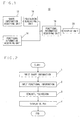
- Fig. 1 is a block diagram showing the image generator 10 for a medical treatment.
- the image generator 10 for a medical treatment has a shape information acquiring unit 12 configured to acquire shape information of a subject, a functional information acquiring unit 14 configured to acquire a functional value of the subject, a polyhedron generating unit 16 configured to generate a polyhedron within a virtual display space on the basis of the acquired shape information, a functional information mapping unit 18 configured to allocate functional information onto a face of the polyhedron, and a display unit 20 configured to display and output the virtual display space.
- the image generator 10 for a medical treatment can be also realized by using a general purpose computer device.
- the shape information acquiring unit 12, the functional information acquiring unit 14, the polyhedron generating unit 16 and the functional information mapping unit 18 can be realized by executing a program by a processor arranged in the computer.
- the display unit 20 can be also realized by utilizing a monitor screen as an output device of the computer.
- shape information is inputted by the shape information acquiring unit 12.
- the "shape information” is data showing the shape of a subject such as internal organs.
- the “shape information” is polygon data showing an internal organ boundary face of internal organs such as the heart and a blood vessel, i.e., a set of coordinate data of a wire frame, etc.
- time series data of this shape may be also set to an input.
- the boundary face of the subject may be also automatically extracted by using an image processing technique of edge detection, etc. from image data for a medical treatment (two-dimensional image data, three-dimensional voxel data), etc., and may be also manually inputted.
- Image data are given to the shape information acquiring unit 12, and the above shape information extracting means using an automatic operation or a manual operation may be also constructed so as to be arranged in the shape information acquiring unit 12.
- the "functional information" is information showing a function or state of the subject such as information of a movement of the subject, the amount of a blood flow, a contrast amount using a contrast agent, and a strain value of a muscle.
- the functional information is a value detected by a separate sensor, etc., a value calculated from a brightness value of image data, and a value calculated by image-analyzing the state of the subject within the image data in accordance with kinds of this functional information.
- the functional information acquiring unit 14 merely inputs the functional information obtained in the exterior as data. Otherwise, when the functional information is a kind calculated from the image data, it may be also set to a mode for calculating the functional information by setting the image data to an input and processing the image data. (2-3) Polyhedron generating unit 16
- step 3 the polyhedron generating unit 16 generates the subject of a three-dimensional shape as a set of plural polyhedrons using the shape information of the subject. These polyhedrons are generated within a space of display data finally displayed. An aggregate of these plural polyhedrons is arranged so as to grasp the three-dimensional shape of the subject and have a gap.
- a first polyhedral arranging method is an example in which plural polyhedrons of a disk shape, a columnar shape, a cylindrical shape or a donut shape are spaced and arranged.
- Fig. 3 shows an example using the first polyhedral arranging method when the subject is an internal organ of a saclike shape such as the heart.
- the outer circumferential face and the inner circumferential face of a disk are arranged so as to be conformed to an outside boundary face and an inside boundary face of a wall of the heart with reference to the shape information acquired in the shape information acquiring unit 12.
- the polyhedron when the shape information of the subject is acquired as data of a vertex coordinate of a polygon, the polyhedron is arranged such that the vertex coordinate constituting one face (called face A) of the polyhedron is conformed to the vertex coordinate of the outside boundary face, and the vertex coordinate of face B opposed to face A is conformed to the vertex coordinate of the inside boundary face.
- the polyhedron is arranged by using a coordinate calculated on the basis of the vertex coordinate. For example, this is effective when fine irregularities of the subject are neglected and a whole shape is concerned, etc.
- the polyhedron is arranged by smoothing original shape information so that the whole shape can be shown.
- the polyhedron of the disk shape is spaced and arranged. All vertex data of the shape information are not used but only one portion is used at a polyhedron generating time so that the polyhedron can be spaced and arranged.
- the spacing is preferably determined from viewpoints of complicatedness of the shape and easiness of observation.
- the shape of the entire heart can be understood and the shape of a wall corresponding to the rear side seen from a visual point can be also grasped by such construction and arrangement of the polyhedron.
- the polyhedron is not limited to a planar construction, but may be also constructed by plural curved surfaces as illustrated here. Further, in this arrangement example, the polyhedron is arranged so as to abut on both the outside boundary face and the inside boundary face of the subject. However, the polyhedron may be also arranged so as to abut on only one boundary face in accordance with an observation object, and the other face may be also arranged such that, e.g., the thickness of the polyhedron becomes a prescribed value.
- the second polyhedral arranging method is an example for spacing and arranging plural hexahedrons.
- Fig. 4 shows an example of an arrangement in an internal organ of a sleeve shape such as a blood vessel.
- faces constituting the front and rear of a hexahedron are arranged so as to be conformed to the outside boundary and the inside boundary of a blood vessel wall with reference to the shape information acquired in the shape information acquiring unit 12.
- the rear side of the blood vessel unable to be formerly observed from a visual point of one place can be also observed by spacing and arranging the polyhedron in this way.
- the number of polyhedrons is one, but the polyhedron itself is constructed so as to have a hole.
- the rear side of the subject unable to be formerly observed from a visual point of one place can be also observed by constructing and arranging the polyhedron in this way.
- the polyhedron of the disk shape shown in the first polyhedral arranging method, and the hexahedron shown in the second polyhedral arranging method may be also mixed and arranged.
- the hexahedron is arranged in a pointed end portion of the heart and the polyhedron of the disk shape is arranged in a portion except for this pointed end portion, the shape of the pointed end portion is more easily grasped without obstructing the observation of a functional value.
- the arrangement of the polyhedron may be also changed in accordance with a visual point for observing the subject as another modified example of the polyhedral arranging method. For example, when one portion of the subject is concerned and becomes a visual point hidden by other portions, display obstructing no observation of the concerned portion can be performed by adaptively widening an arranging interval of the polyhedron of this side, and changing the kind of the arranged polyhedron.
- the functional information mapping unit 18 refers to the functional information acquired in the functional information acquiring unit 14, and allocates the functional information to the generated polyhedron.
- the functional information is allocated to a face of the polyhedron by a method of a brightness map in a gray scale, a color map using a color table, etc., or conversion into a text character, etc. in accordance with its value.
- a spatial average value of the functional value in a position of the polyhedron is calculated, etc. , and a typical value may be allocated to the polyhedron.
- the allocation may be also performed in a shape expressing a distribution of the functional value of a locating range of the polyhedron.
- Fig. 5 shows an example in which the functional value is converted into a color, which is allocated to the polyhedron arranged by the first polyhedral arranging method.
- no color can be expressed so that the color is shown by light and shade.
- a different color is allocated by the magnitude of the functional value. While the actual shape of the entire subject is grasped, a difference of the functional value every position of the subject can be intuitively understood.
- a strain every direction such as a strain of the thickness direction of a wall, and a strain of a direction along the wall can be defined with respect to a strain (distortion) value of a heart wall.
- a strain of the thickness direction of the wall is allocated to a face (face A of Fig. 6) of the thickness direction of the wall of the polyhedron, and the strain of the direction along the wall is allocated to a side face (face B of Fig. 6) of the polyhedron, display conformed to the definition of the functional value can be performed, and display intuitively easily understood can be performed.
- the distribution of the functional value for example, when the functional value is different on the inside boundary face and the outside boundary face of the subject, a value therebetween is interpolated and a color can be also displayed in a gradation shape on a face of the polyhedron. Otherwise, when the functional value can be closely acquired in an area between the inside boundary face and the outside boundary face of the subject, the distribution of this functional value may be also displayed by the color display, a contour line, etc. of a gradation shape on the face of the polyhedron. The distribution of the functional value can be observed in larger portions in comparison with the former case by showing the shape and the distribution of the functional value by using the polyhedron in this way.
- step 5 the polyhedron provided by mapping the functional information is displayed by the display unit 20.
- the polyhedron is displayed in a display device.
- No display device is limited.
- the present invention is not limited to each of the above embodiment modes, but can be variously modified as long as these modifications are not departed from its spirit.
- Shape display in the wire frame may be also performed together simultaneously with the display of the polyhedron.
- the shape and the functional value of the subject can be grasped from a visual point of one place.
- a means for changing the visual point may be also additionally arranged by a pointing device, etc.
- the shape of the subject can be grasped as an envelope surface of plural polyhedrons, and a clearance is arranged between the polyhedrons. Therefore, a hidden portion is small, and an entire aspect can be understood.
- the mode and the functional value of the subject can be observed at one time without forcing a burden of correspondence setting of a position onto an observer.
- the functional values of plural kinds can be intuitively displayed at one time by allocating the functional value of a different kind to each face of the polyhedron.
Landscapes
- Engineering & Computer Science (AREA)
- Computer Graphics (AREA)
- Computer Hardware Design (AREA)
- General Engineering & Computer Science (AREA)
- Software Systems (AREA)
- Physics & Mathematics (AREA)
- General Physics & Mathematics (AREA)
- Theoretical Computer Science (AREA)
- Image Generation (AREA)
- Apparatus For Radiation Diagnosis (AREA)
- Measuring And Recording Apparatus For Diagnosis (AREA)
Applications Claiming Priority (1)
| Application Number | Priority Date | Filing Date | Title |
|---|---|---|---|
| JP2006296607A JP4810395B2 (ja) | 2006-10-31 | 2006-10-31 | 医用画像生成装置及びその方法 |
Publications (2)
| Publication Number | Publication Date |
|---|---|
| EP1918883A2 true EP1918883A2 (de) | 2008-05-07 |
| EP1918883A3 EP1918883A3 (de) | 2011-06-22 |
Family
ID=39135167
Family Applications (1)
| Application Number | Title | Priority Date | Filing Date |
|---|---|---|---|
| EP07016305A Withdrawn EP1918883A3 (de) | 2006-10-31 | 2007-08-20 | Bilderzeugungsvorrichtung für medizinische Behandlungen und Verfahren dazu |
Country Status (4)
| Country | Link |
|---|---|
| US (1) | US8049749B2 (de) |
| EP (1) | EP1918883A3 (de) |
| JP (1) | JP4810395B2 (de) |
| CN (1) | CN101174329B (de) |
Families Citing this family (3)
| Publication number | Priority date | Publication date | Assignee | Title |
|---|---|---|---|---|
| JP4810395B2 (ja) * | 2006-10-31 | 2011-11-09 | 株式会社東芝 | 医用画像生成装置及びその方法 |
| KR101260576B1 (ko) * | 2010-10-13 | 2013-05-06 | 주식회사 팬택 | Ar 서비스를 제공하기 위한 사용자 단말기 및 그 방법 |
| JP6635695B2 (ja) * | 2014-07-16 | 2020-01-29 | キヤノンメディカルシステムズ株式会社 | 画像処理装置、医用画像診断装置、及び画像処理方法 |
Citations (2)
| Publication number | Priority date | Publication date | Assignee | Title |
|---|---|---|---|---|
| JP2000139917A (ja) | 1998-11-12 | 2000-05-23 | Toshiba Corp | 超音波診断装置 |
| US20030048269A1 (en) * | 2001-09-12 | 2003-03-13 | Powell Donald T. | System and method for generating finite element models |
Family Cites Families (9)
| Publication number | Priority date | Publication date | Assignee | Title |
|---|---|---|---|---|
| JPH01108690A (ja) * | 1987-10-22 | 1989-04-25 | Hitachi Medical Corp | 3次元手術シミュレーション装置 |
| US5594849A (en) * | 1991-08-09 | 1997-01-14 | Yale University | Biomedical magnetism imaging apparatus and method |
| GB9122843D0 (en) * | 1991-10-28 | 1991-12-11 | Imperial College | Method and apparatus for image processing |
| JP3191829B2 (ja) * | 1992-07-31 | 2001-07-23 | 株式会社島津製作所 | 立体モデル作成法 |
| JP4208275B2 (ja) | 1997-10-30 | 2009-01-14 | 株式会社東芝 | 心臓内電気現象の診断装置およびその現象の表示方法 |
| US6600487B1 (en) | 1998-07-22 | 2003-07-29 | Silicon Graphics, Inc. | Method and apparatus for representing, manipulating and rendering solid shapes using volumetric primitives |
| JP4027867B2 (ja) * | 2003-09-10 | 2007-12-26 | 株式会社日立ハイテクノロジーズ | 生体磁場計測装置 |
| US7801346B2 (en) * | 2004-09-24 | 2010-09-21 | Hitachi Medical Corporation | Medical image display device, method, and program |
| JP4810395B2 (ja) * | 2006-10-31 | 2011-11-09 | 株式会社東芝 | 医用画像生成装置及びその方法 |
-
2006
- 2006-10-31 JP JP2006296607A patent/JP4810395B2/ja not_active Expired - Fee Related
-
2007
- 2007-08-17 US US11/840,380 patent/US8049749B2/en active Active
- 2007-08-20 EP EP07016305A patent/EP1918883A3/de not_active Withdrawn
- 2007-10-23 CN CN2007101678619A patent/CN101174329B/zh active Active
Patent Citations (2)
| Publication number | Priority date | Publication date | Assignee | Title |
|---|---|---|---|---|
| JP2000139917A (ja) | 1998-11-12 | 2000-05-23 | Toshiba Corp | 超音波診断装置 |
| US20030048269A1 (en) * | 2001-09-12 | 2003-03-13 | Powell Donald T. | System and method for generating finite element models |
Non-Patent Citations (1)
| Title |
|---|
| MCGUFFIN M J ET AL: "Using deformations for browsing volumetric data", VIS 2003. IEEE VISUALIZATION 2003. PROCEEDINGS. SEATTLE, WA, OCT. 19 - 24, 2003; [ANNUAL IEEE CONFERENCE ON VISUALIZATION], NEW YORK, NY : IEEE, US, 1 January 2003 (2003-01-01), pages 401 - 408, XP031173525, ISBN: 978-0-7803-8120-9, DOI: 10.1109/VISUAL.2003.1250400 * |
Also Published As
| Publication number | Publication date |
|---|---|
| JP4810395B2 (ja) | 2011-11-09 |
| EP1918883A3 (de) | 2011-06-22 |
| CN101174329A (zh) | 2008-05-07 |
| US8049749B2 (en) | 2011-11-01 |
| US20080101671A1 (en) | 2008-05-01 |
| CN101174329B (zh) | 2011-05-18 |
| JP2008110177A (ja) | 2008-05-15 |
Similar Documents
| Publication | Publication Date | Title |
|---|---|---|
| US7912264B2 (en) | Multi-volume rendering of single mode data in medical diagnostic imaging | |
| JP6670595B2 (ja) | 医用画像処理装置 | |
| EP2203894B1 (de) | Visualisierung von voxel-daten | |
| EP2124197A1 (de) | Bildverarbeitungsvorrichtung und Computerprogrammprodukt | |
| US10275930B2 (en) | Combined intensity projection | |
| JP2006055213A (ja) | 画像処理装置、及びプログラム | |
| JP2008259699A (ja) | 画像処理方法および装置並びにプログラム | |
| JP2012196437A (ja) | 医用画像診断装置、医用画像表示装置、医用画像処理装置、及び医用画像処理プログラム | |
| US9846973B2 (en) | Method and system for volume rendering color mapping on polygonal objects | |
| JP4885042B2 (ja) | 画像処理方法および装置ならびにプログラム | |
| JP2015217120A (ja) | 画像診断支援装置、その処理方法及びプログラム | |
| JP7205189B2 (ja) | レンダリング装置、レンダリング方法、及びプログラム | |
| CN103654851B (zh) | 用于显示与超声截面有关的立体信息的方法和设备 | |
| JP6533687B2 (ja) | 医用画像処理装置、医用画像処理方法、及び医用画像処理プログラム | |
| US8049749B2 (en) | Image generator for medical treatment and its method | |
| CN108876783B (zh) | 图像融合方法及系统、医疗设备和图像融合终端 | |
| JP2006000127A (ja) | 画像処理方法および装置並びにプログラム | |
| JP6564075B2 (ja) | 医用画像を表示するための伝達関数の選択 | |
| JP2009247490A (ja) | 画像処理装置および方法並びにプログラム | |
| JP2014524810A (ja) | レンダリングを行う方法及びシステム | |
| KR20230159696A (ko) | 매질의 다중-양식 및/또는 다중-소스 데이터를 처리하는 방법 및 시스템 | |
| JP2005525863A (ja) | 医療用データの統合された視覚化用の医療用視検システム及び画像処理 | |
| JP2000105838A (ja) | 画像表示方法及び画像処理装置 | |
| JP2008259698A (ja) | 画像処理方法および装置並びにプログラム | |
| CN106296707A (zh) | 医学图像处理方法及装置 |
Legal Events
| Date | Code | Title | Description |
|---|---|---|---|
| PUAI | Public reference made under article 153(3) epc to a published international application that has entered the european phase |
Free format text: ORIGINAL CODE: 0009012 |
|
| 17P | Request for examination filed |
Effective date: 20070820 |
|
| AK | Designated contracting states |
Kind code of ref document: A2 Designated state(s): AT BE BG CH CY CZ DE DK EE ES FI FR GB GR HU IE IS IT LI LT LU LV MC MT NL PL PT RO SE SI SK TR |
|
| AX | Request for extension of the european patent |
Extension state: AL BA HR MK RS |
|
| PUAL | Search report despatched |
Free format text: ORIGINAL CODE: 0009013 |
|
| AK | Designated contracting states |
Kind code of ref document: A3 Designated state(s): AT BE BG CH CY CZ DE DK EE ES FI FR GB GR HU IE IS IT LI LT LU LV MC MT NL PL PT RO SE SI SK TR |
|
| AX | Request for extension of the european patent |
Extension state: AL BA HR MK RS |
|
| AKX | Designation fees paid |
Designated state(s): DE FR GB |
|
| 17Q | First examination report despatched |
Effective date: 20120216 |
|
| STAA | Information on the status of an ep patent application or granted ep patent |
Free format text: STATUS: THE APPLICATION HAS BEEN WITHDRAWN |
|
| 18W | Application withdrawn |
Effective date: 20160210 |