EP1872136B2 - Methods and products for evaluating an immune response to a therapeutic protein - Google Patents
Methods and products for evaluating an immune response to a therapeutic protein Download PDFInfo
- Publication number
- EP1872136B2 EP1872136B2 EP06749243.9A EP06749243A EP1872136B2 EP 1872136 B2 EP1872136 B2 EP 1872136B2 EP 06749243 A EP06749243 A EP 06749243A EP 1872136 B2 EP1872136 B2 EP 1872136B2
- Authority
- EP
- European Patent Office
- Prior art keywords
- natalizumab
- antibody
- subject
- immune response
- binding
- Prior art date
- Legal status (The legal status is an assumption and is not a legal conclusion. Google has not performed a legal analysis and makes no representation as to the accuracy of the status listed.)
- Active
Links
Images


Classifications
-
- G—PHYSICS
- G01—MEASURING; TESTING
- G01N—INVESTIGATING OR ANALYSING MATERIALS BY DETERMINING THEIR CHEMICAL OR PHYSICAL PROPERTIES
- G01N33/00—Investigating or analysing materials by specific methods not covered by groups G01N1/00 - G01N31/00
- G01N33/48—Biological material, e.g. blood, urine; Haemocytometers
- G01N33/50—Chemical analysis of biological material, e.g. blood, urine; Testing involving biospecific ligand binding methods; Immunological testing
- G01N33/68—Chemical analysis of biological material, e.g. blood, urine; Testing involving biospecific ligand binding methods; Immunological testing involving proteins, peptides or amino acids
- G01N33/6854—Immunoglobulins
- G01N33/686—Anti-idiotype
-
- G—PHYSICS
- G01—MEASURING; TESTING
- G01N—INVESTIGATING OR ANALYSING MATERIALS BY DETERMINING THEIR CHEMICAL OR PHYSICAL PROPERTIES
- G01N33/00—Investigating or analysing materials by specific methods not covered by groups G01N1/00 - G01N31/00
- G01N33/48—Biological material, e.g. blood, urine; Haemocytometers
- G01N33/50—Chemical analysis of biological material, e.g. blood, urine; Testing involving biospecific ligand binding methods; Immunological testing
- G01N33/68—Chemical analysis of biological material, e.g. blood, urine; Testing involving biospecific ligand binding methods; Immunological testing involving proteins, peptides or amino acids
- G01N33/6854—Immunoglobulins
-
- A—HUMAN NECESSITIES
- A61—MEDICAL OR VETERINARY SCIENCE; HYGIENE
- A61P—SPECIFIC THERAPEUTIC ACTIVITY OF CHEMICAL COMPOUNDS OR MEDICINAL PREPARATIONS
- A61P1/00—Drugs for disorders of the alimentary tract or the digestive system
-
- A—HUMAN NECESSITIES
- A61—MEDICAL OR VETERINARY SCIENCE; HYGIENE
- A61P—SPECIFIC THERAPEUTIC ACTIVITY OF CHEMICAL COMPOUNDS OR MEDICINAL PREPARATIONS
- A61P13/00—Drugs for disorders of the urinary system
- A61P13/12—Drugs for disorders of the urinary system of the kidneys
-
- A—HUMAN NECESSITIES
- A61—MEDICAL OR VETERINARY SCIENCE; HYGIENE
- A61P—SPECIFIC THERAPEUTIC ACTIVITY OF CHEMICAL COMPOUNDS OR MEDICINAL PREPARATIONS
- A61P17/00—Drugs for dermatological disorders
-
- A—HUMAN NECESSITIES
- A61—MEDICAL OR VETERINARY SCIENCE; HYGIENE
- A61P—SPECIFIC THERAPEUTIC ACTIVITY OF CHEMICAL COMPOUNDS OR MEDICINAL PREPARATIONS
- A61P19/00—Drugs for skeletal disorders
- A61P19/02—Drugs for skeletal disorders for joint disorders, e.g. arthritis, arthrosis
-
- A—HUMAN NECESSITIES
- A61—MEDICAL OR VETERINARY SCIENCE; HYGIENE
- A61P—SPECIFIC THERAPEUTIC ACTIVITY OF CHEMICAL COMPOUNDS OR MEDICINAL PREPARATIONS
- A61P25/00—Drugs for disorders of the nervous system
-
- A—HUMAN NECESSITIES
- A61—MEDICAL OR VETERINARY SCIENCE; HYGIENE
- A61P—SPECIFIC THERAPEUTIC ACTIVITY OF CHEMICAL COMPOUNDS OR MEDICINAL PREPARATIONS
- A61P29/00—Non-central analgesic, antipyretic or antiinflammatory agents, e.g. antirheumatic agents; Non-steroidal antiinflammatory drugs [NSAID]
-
- A—HUMAN NECESSITIES
- A61—MEDICAL OR VETERINARY SCIENCE; HYGIENE
- A61P—SPECIFIC THERAPEUTIC ACTIVITY OF CHEMICAL COMPOUNDS OR MEDICINAL PREPARATIONS
- A61P35/00—Antineoplastic agents
-
- A—HUMAN NECESSITIES
- A61—MEDICAL OR VETERINARY SCIENCE; HYGIENE
- A61P—SPECIFIC THERAPEUTIC ACTIVITY OF CHEMICAL COMPOUNDS OR MEDICINAL PREPARATIONS
- A61P35/00—Antineoplastic agents
- A61P35/02—Antineoplastic agents specific for leukemia
-
- A—HUMAN NECESSITIES
- A61—MEDICAL OR VETERINARY SCIENCE; HYGIENE
- A61P—SPECIFIC THERAPEUTIC ACTIVITY OF CHEMICAL COMPOUNDS OR MEDICINAL PREPARATIONS
- A61P37/00—Drugs for immunological or allergic disorders
-
- A—HUMAN NECESSITIES
- A61—MEDICAL OR VETERINARY SCIENCE; HYGIENE
- A61P—SPECIFIC THERAPEUTIC ACTIVITY OF CHEMICAL COMPOUNDS OR MEDICINAL PREPARATIONS
- A61P37/00—Drugs for immunological or allergic disorders
- A61P37/02—Immunomodulators
-
- A—HUMAN NECESSITIES
- A61—MEDICAL OR VETERINARY SCIENCE; HYGIENE
- A61P—SPECIFIC THERAPEUTIC ACTIVITY OF CHEMICAL COMPOUNDS OR MEDICINAL PREPARATIONS
- A61P37/00—Drugs for immunological or allergic disorders
- A61P37/02—Immunomodulators
- A61P37/06—Immunosuppressants, e.g. drugs for graft rejection
-
- C—CHEMISTRY; METALLURGY
- C07—ORGANIC CHEMISTRY
- C07K—PEPTIDES
- C07K16/00—Immunoglobulins [IG], e.g. monoclonal or polyclonal antibodies
- C07K16/18—Immunoglobulins [IG], e.g. monoclonal or polyclonal antibodies against material from animals or humans
- C07K16/28—Immunoglobulins [IG], e.g. monoclonal or polyclonal antibodies against material from animals or humans against receptors, cell surface antigens or cell surface determinants
- C07K16/2839—Immunoglobulins [IG], e.g. monoclonal or polyclonal antibodies against material from animals or humans against receptors, cell surface antigens or cell surface determinants against the integrin superfamily
-
- G—PHYSICS
- G01—MEASURING; TESTING
- G01N—INVESTIGATING OR ANALYSING MATERIALS BY DETERMINING THEIR CHEMICAL OR PHYSICAL PROPERTIES
- G01N2333/00—Assays involving biological materials from specific organisms or of a specific nature
- G01N2333/435—Assays involving biological materials from specific organisms or of a specific nature from animals; from humans
- G01N2333/705—Assays involving receptors, cell surface antigens or cell surface determinants
- G01N2333/70546—Integrin superfamily, e.g. VLAs, leuCAM, GPIIb/GPIIIa, LPAM
-
- G—PHYSICS
- G01—MEASURING; TESTING
- G01N—INVESTIGATING OR ANALYSING MATERIALS BY DETERMINING THEIR CHEMICAL OR PHYSICAL PROPERTIES
- G01N2800/00—Detection or diagnosis of diseases
- G01N2800/06—Gastro-intestinal diseases
- G01N2800/065—Bowel diseases, e.g. Crohn, ulcerative colitis, IBS
-
- G—PHYSICS
- G01—MEASURING; TESTING
- G01N—INVESTIGATING OR ANALYSING MATERIALS BY DETERMINING THEIR CHEMICAL OR PHYSICAL PROPERTIES
- G01N2800/00—Detection or diagnosis of diseases
- G01N2800/10—Musculoskeletal or connective tissue disorders
- G01N2800/101—Diffuse connective tissue disease, e.g. Sjögren, Wegener's granulomatosis
- G01N2800/102—Arthritis; Rheumatoid arthritis, i.e. inflammation of peripheral joints
-
- G—PHYSICS
- G01—MEASURING; TESTING
- G01N—INVESTIGATING OR ANALYSING MATERIALS BY DETERMINING THEIR CHEMICAL OR PHYSICAL PROPERTIES
- G01N2800/00—Detection or diagnosis of diseases
- G01N2800/28—Neurological disorders
- G01N2800/285—Demyelinating diseases; Multipel sclerosis
-
- G—PHYSICS
- G01—MEASURING; TESTING
- G01N—INVESTIGATING OR ANALYSING MATERIALS BY DETERMINING THEIR CHEMICAL OR PHYSICAL PROPERTIES
- G01N2800/00—Detection or diagnosis of diseases
- G01N2800/52—Predicting or monitoring the response to treatment, e.g. for selection of therapy based on assay results in personalised medicine; Prognosis
Definitions
- the invention relates to evaluating patients for an immune response to a therapeutic agent, and particularly to a therapeutic protein, natalizumab.
- Biologic therapeutics are currently available for treating diseases and disorders such as transplant rejection, leukemia, breast cancer, arthritis, multiple sclerosis, and Crohn's disease; and numerous additional protein-based therapies are in development.
- Available biologics therapeutics include AMEVIVE ® (alefacept), ZEVALIN ® (ibritumomab tiuxetan), ORTHOCLONE ® (muromonab-CD3), ENBREL ® (etanercept), REOPRO ® (abciximab), RITUXAN ® (rituximab), SIMULECT ® (basiliximab), REMICADE ® (infliximab), SYNAGIS ® (palivizumab), HERCEPTIN ® (trastuzumab), ZENAPAX ® (daclizum-ab), CAMPATH ® (alemtuzumab), MYLOTARG ® (gem-tuzumab ozogamici
- Natalizumab is a humanized monoclonal antibody against ⁇ 4 ⁇ 1 integrin (VLA-4). Natalizumab binds to the a4 subunit of ⁇ 4 ⁇ 1 and ⁇ 4 ⁇ 7 integrins. Natalizumab is useful to treat certain inflammatory diseases and conditions including multiple sclerosis, Crohn's disease, and rheumatoid arthritis.
- the invention relates to the embodiments as defined in the claims.
- the invention relates to a method of detecting a clinically significant immune response to natalizumab in a subject, the method comprising determining whether at least two biological samples taken at different time points from a subject that has been administered natalizumab contain at least a clinically significant threshold level of about 500 ng/ml in a serum sample of a soluble antibody that binds to natalizumab, wherein the presence of at least the threshold level of the soluble antibody in said at least two samples is indicative of a clinically significant immune response to natalizumab wherein the time points are separated by at least one month, and wherein the clinically significant immune response indicates a diminution of efficacy or lack of efficacy of natalizumab.
- the disclosure provides methods and compositions for identifying, monitoring, and/or evaluating an immune response to a therapeutic agent, e.g., a therapeutic protein, e.g., a therapeutic antibody.
- a therapeutic agent e.g., a therapeutic protein, e.g., a therapeutic antibody.
- the fact that a patient develops any antibodies to a therapeutic agent may or may not correlate with a clinical response to the therapeutic agent.
- aspects of the disclosure are based, in part, on the discovery of an unexpected level of antibody response that can be used as a threshold for detecting a clinically significant response to the therapeutic agent.
- the threshold level is higher than would have been predicted using a statistical analysis of patients that have not received the therapeutic agent
- the clinically significant threshold is generally higher than the lowest detectable level of immune response in a patient.
- the clinically significant threshold level is generally at least 2 standard deviations above a negative control level, e.g., above a mean pre-treatment level of an untreated patient population.
- the higher threshold levels used in methods of the invention result in fewer false positives than would be identified if the threshold level were based on a 5% cutoff(e.g., 1.645 standard deviations above the mean) for immune responses observed in patients that had not received the therapeutic agent.
- the presence of a detectable immune response in a patient sample is not clinically significant unless the immune response reaches at least a predetermined threshold level.
- the disclosure provides, inter alia, methods of identifying a clinically significant threshold level of antibody response to a therapeutic agent (e.g., a therapeutic protein, e.g., a therapeutic antibody), and methods of identifying patients who have a clinically significant antibody response to a therapeutic agent.
- a therapeutic agent e.g., a therapeutic protein, e.g., a therapeutic antibody
- the disclosure also provides a threshold level with which to identify clinically meaningful antibodies in a subject.
- an immune response to a therapeutic agent e.g., natalizumab
- may not be clinically significant e.g., may not show a significant association with reduced clinical efficacy
- the magnitude of the immune response reaches a threshold level that can be predetermined (e.g., based on immune responses obtained for different patient groups).
- methods of the disclosure relate to detecting at least a threshold level of an immune response to a therapeutic agent, where the threshold level may be higher than the lowest detectable level of immune response, and wherein the positive results from the assay are clinically meaningful, in part, because the assay avoids false positives that have no associated clinical significance.
- a clinically significant immune response to a therapeutic agent is an antibody response that may affect one or more clinical parameters in a patient, and/or the pharmacokinetics and/or efficacy of the therapeutic agent.
- a clinically significant antibody response indicates a diminution of efficacy or lack of efficacy of the therapeutic agent, or an adverse reaction to the therapeutic agent.
- a clinically significant antibody response to a therapeutic protein includes one or more of: (a) lack of efficacy or at least 10%, 20%, 30%, 40%, 50%, 60% or more diminution in efficacy of the therapeutic agent to reduce the number, severity or rate of relapse in the patient; (b) lack of efficacy or at least 10%, 20%, 30%, 40%, 50%, 60% or more diminution in efficacy of the therapeutic agent to slow progression of disability in the Expanded Disability Status Scale (EDSS) scale or Multiple Sclerosis Functional Composite (MSFC) scale; (c) lack of efficacy or at least 10%, 20%, 30%, 40%, 50%, 60% or more diminution in efficacy in reducing the number or volume of new or newly enlarging T2 hyperintense lesions or attenuating the increase in T2 hyperintense lesion volume on brain MRI, (d) lack of efficacy or at least 10%, 20%, 30%, 40%, 50%, 60% or more diminution in efficacy in reducing the number or volume
- the disclosure provides methods of identifying a clinically significant threshold level of antibody response to a therapeutic agent (e.g., a therapeutic protein, e.g., a therapeutic antibody).
- a therapeutic agent e.g., a therapeutic protein, e.g., a therapeutic antibody.
- the method includes (a) evaluating the level of anti-agent antibodies in a control population of patients who have a disorder (e.g., determining the mean or median level of anti-agent antibodies in a population of at least 2, 3, 5, 10, 20, 30, 50, 100 or more patients who have a disorder and who have not been treated with a subject therapeutic agent for at least 3 months, 6 months or longer); and (b) selecting a threshold level of at least 2 (e.g., 2.5, 3, 4, 5, or 6) standard deviations above the level of anti-agent antibodies in the control population.
- a threshold level of at least 2 e.g., 2.5, 3, 4, 5, or 6
- the presence of at least the threshold level of anti-agent antibodies in a patient who has been administered the therapeutic agent correlates with a clinically significant response in the treated patient
- the same detection reagent e.g., labeled anti-agent antibody
- the therapeutic agent is natalizumab.
- the disorder is multiple sclerosis.
- the disorder is an inflammation of the central nervous system (e.g., meningitis, neuromyelitis optica, neurosarcoidosis, CNS vasculitis, encephalitis, or transverse myelitis, in addition to or instead of multiple sclerosis,), a tissue or organ graft rejection or a graft-versus-host disease, an acute CNS injury (e.g., stroke or spinal cord injury); chronic renal disease; allergy (e.g., allergic asthma); type 1 diabetes; an inflammatory bowel disorders (e.g., Crohn's disease, or ulcerative colitis); myasthenia gravis; fibromyalgia; an arthritic disorder (e.g., rheumatoid arthritis or psoriatic arthritis); an inflammatory/immune skin disorder (e.g., psoriasis, vitiligo, dermatitis, or lichen planus); systemic lupus erythematosus; Sjogren's
- the disclosure provides methods of identifying a patient who has a clinically significant antibody response to a therapeutic protein, e.g., a therapeutic antibody.
- the method includes identifying, in a biological sample obtained from a subject who has a disorder and who has been administered the therapeutic protein, the presence of a threshold level of one or more antibodies that specifically bind to the therapeutic protein, wherein the threshold level is at least 2 (e.g., 2.5, 3, 4, 5, or 6) standard deviations above the level of antibodies that specifically bind to the therapeutic protein in a control population (e.g., a population of patients who have the disorder but have not been administered the therapeutic protein within the last 3 months, 6 months or more).
- the therapeutic protein is natalizumab.
- the disorder is multiple sclerosis.
- the disorder is rheumatoid arthritis. In certain embodiments, the disorder is Crohn's disease. In one embodiment, the method further includes modifying the treatment regimen of a patient who is thus identified as having a clinically significant antibody response to a therapeutic protein.
- the disclosure provides methods and compositions for identifying in a biological sample obtained from a subject the presence of a clinically significant level of one or more antibodies that specifically bind to natalizumab that was administered to the subject. Aspects of the disclosure include the use of ELISA assays for the detection of levels of induced antibodies that are indicative of a clinically significant immune response in a subject to the administration of natalizumab. In one embodiment, the disclosure provides methods and kits for identifying clinically significant levels of anti-natalizumab antibodies that are indicative of an immune response to natalizumab in a subject that has received at least one dose of natalizumab
- the disclosure provides methods for evaluating and/or modifying a therapeutic regimen based on a subject's immune response to a natalizumab.
- methods of detecting a clinically significant immune response to natalizumab in a subject include determining whether a biological sample from a subject that has been administered natalizumab contains a clinically significant threshold level of a soluble antibody that binds to natalizumab, wherein the presence of at least the threshold level of the soluble antibody is indicative of a clinically significant immune response to natalizumab.
- a clinically significant immune response to natalizumab is indicated by the presence of at least the threshold level of soluble antibody to natalizumab in at least two biological samples taken from the subject at different time points. In certain embodiments, the time points are separated by at least one month.
- At least the threshold level of soluble antibody that binds to natalizumab is present in two biological samples taken from the subject at two consecutive time points.
- a level of soluble antibody that binds to natalizumab is determined by: determining a level of soluble binding activity to natalizumab in a first aliquot of the biological sample; and determining whether the soluble binding activity is specific for natalizumab.
- the specificity of the soluble binding activity is determined in a second aliquot of the biological sample.
- a level of soluble antibody that binds to natalizumab in the biological sample is determined by comparing levels of binding activity to a labeled natalizumab measured in the presence of two or more different amounts of unlabeled natalizumab (e.g., levels measured in the presence of no unlabeled natalizumab may be compared to levels measured in the presence of a competing amount of unlabeled natalizumab ).
- a level of soluble antibody that binds to natalizumab in the biological sample is determined by comparing levels of binding activity to an immobilized natalizumab measured in the presence of two or more different amounts of soluble natalizumab (e.g., levels measured in the presence of no soluble natalizumab may be compared to levels measured in the presence of a competing amount of soluble natalizumab ).
- a first level of binding activity to a labeled natalizumab measured in the presence of a first amount of unlabeled VLA-4 is compared to a second level of binding activity to a labeled natalizumab measured in the presence of a second amount of unlabeled natalizumab.
- the first and second levels of binding activity are determined in first and second aliquots of the biological sample.
- the amount of soluble antibody to the natalizumab is determined using a bridging ELISA assay.
- a first level of binding activity to the natalizumab is determined in a first immunoassay for a first aliquot of the biological sample
- a second level of binding activity to the natalizumab is determined in a second immunoassay for a second aliquot of the biological sample
- the second immunoassay is spiked with a greater amount of unlabeled soluble natalizumab than the first immunoassay
- the presence in the biological sample of at least a threshold level of soluble antibody to the natalizumab is indicated if the first level of binding activity is greater than a reference level and the second level of binding activity is less than a predetermined percentage of the first level of binding activity.
- the reference level is a level of binding activity measured for a reference amount of soluble antibody to the natalizumab.
- the reference amount is about 500 ng/ml (e.g., in a serum sample) of a soluble antibody to natalizumab.
- the reference amount may be between about 400 ng/ml and about 600 ng/ml (e.g., about400 ng/ml, about425 ng/ml, about450 ng/ml, about 475 ng/ml, about 500 ng/ml, about 525 ng/ml, about 550 ng/ml, about 575 ng/ml, or about 600 ng/ml).
- the reference level of binding activity that corresponds to the reference amount may be measured in a diluted sample (for example, a sample that corresponds to a 10 fold dilution and contains from about 40 ng/ml to about 60 ng/ml, e.g., about 50 ng/ml, of a soluble antibody that to natalizumab ).
- a reference level of binding activity in an assay may be provided by any predetermined amount of soluble antibody to the natalizumab corresponding to an appropriate dilution of the reference amount.
- the VLA-4 binding antibody is AN100226 (natalizumab).
- the soluble antibody to natalizumab is a reference antibody that binds to natalizumab with high affinity. In embodiments, the reference antibody blocks the interaction between natalizumab and VLA-4.
- the first and second immunoassays are bridging ELISA assays. In some embodiments, the first and second assays comprise an immobilized unlabeled natalizumab and a soluble labeled natalizumab, wherein the soluble labeled natalizumab is labeled with an enzyme, a fluorescent marker, a biotin marker (e.g., the natalizumab may be biotinylated), or a radioactive marker.
- the first and second immunoassays are conducted in parallel reaction volumes on a single reaction substrate.
- the biological sample is a serum sample.
- the subject is a human patient.
- the patient has multiple sclerosis.
- the patient has rheumatoid arthritis.
- the patient has Crohn's disease.
- the time points of at least two or more biological samples obtained the subject are separated by at least 15 days, 30 days, 45 days, 60 days, 90 days, or more.
- the method also includes selecting a therapeutic regimen for the subject if a clinically significant threshold level of a soluble antibody that binds to the natalizumab is detected in at least two biological samples obtained from the subject.
- selecting a therapeutic regimen includes evaluating a current therapy of the subject, determining a new therapy for the subject, modifying a current therapy of the subject, or stopping a current therapy of the subject.
- a current therapy includes administering the natalizumab to the subject.
- methods of selecting a therapeutic regimen for a subject include assaying a subject who has been administered natalizumab for the presence of a positive immune response to natalizumab at first and second time points, selecting a therapeutic regimen for the subject based on the assay results at the first and second time points, wherein the presence of a positive immune response at a point in time is indicated by the presence of at least a clinically significant threshold amount of binding activity in a biological sample obtained from the subject at the point in time.
- the first and second time points are separated by a clinically significant time period.
- the clinically significant time period is at least 30 days.
- natalizumab therapy is continued if a negative immune response is detected at the second time point. In some embodiments, a therapy other than natalizumab therapy is selected if a positive immune response is detected at both the first and second time points.
- the subject has multiple sclerosis. In certain embodiments, the subject has Crohn's disease. In some embodiments, the subject has rheumatoid arthritis.
- methods of selecting a therapeutic regimen for a subject include detecting the presence of a clinically significant immune response to natalizumab in at least two biological samples obtained from a subject wherein the subject has been administered natalizumab and the at least two biological samples are obtained from the subject at times separated by at least a clinically significant time interval, and selecting a therapeutic regimen based on the detection of a clinically significant immune response to natalizumab in the subject at the times when the at least two biological samples are obtained from the subject.
- the clinically significant interval separating the times at which the samples are obtained from the subject is at least 15 days, 30 days, 45 days, 60 days, 90 days, or longer.
- selecting a therapeutic regimen includes evaluating a current therapy of the subject, determining a new therapy for the subject, modifying a current therapy of the subject, or stopping a current therapy of the subject.
- a current therapy includes administering natalizumab to the subject.
- detecting the presence of a clinically significant immune response to natalizumab comprises, determining whether a biological sample obtained from a subject that has been administered natalizumab contains a threshold level of a soluble antibody that binds to natalizumab, wherein the presence of at least the threshold level of the soluble antibody is indicative of a clinically significant immune response to natalizumab.
- At least the threshold level of soluble antibody that binds to natalizumab is present in two biological samples taken from the subject at two consecutive time points.
- a level of soluble antibody that binds to natalizumab is determined by: determining a level of soluble binding activity to natalizumab in a first aliquot of the biological sample and determining whether the soluble binding activity is specific for natalizumab.
- the specificity of the soluble binding activity is determined in a second aliquot of the same biological sample.
- a level of soluble antibody that binds to natalizumab in the biological sample is determined by comparing levels of binding activity to a labeled natalizumab measured in the presence of two or more different amounts of unlabeled natalizumab (e.g., levels measured in the presence of no unlabeled natalizumab may be compared to levels measured in the presence of a competing amount of unlabeled natalizumab ).
- a level of soluble antibody that binds to the natalizumab in the biological sample is determined by comparing levels of binding activity to an immobilized natalizumab measured in the presence of two or more different amounts of soluble natalizumab (e.g., levels measured in the presence of no soluble natalizumab may be compared to levels measured in the presence of a competing amount of soluble natalizumab ).
- a first level of binding activity to a labeled natalizumab measured in the presence of a first amount of unlabeled VLA-4 is compared to a second level of binding activity to a labeled natalizumab measured in the presence of a second amount of unlabeled natalizumab .
- the first and second levels of binding activity are determined in first and second aliquots of the same biological sample.
- the amount of soluble antibody to natalizumab is determined using a bridging ELISA assay.
- a first level of binding activity to the natalizumab is determined in a first immunoassay for a first aliquot of the biological sample
- a second level of binding activity to natalizumab is determined in a second immunoassay for a second aliquot of the biological sample
- the second immunoassay is spiked with a greater amount of unlabeled soluble natalizumab than the first immunoassay
- the presence in the biological sample of at leas a threshold level of soluble antibody to the natalizumab is indicated if the first level of binding activity is greater than a reference level and the second level of binding activity is less than a predetermined percentage of the first level of binding activity.
- the reference level is a level of binding activity measured for a reference amount of soluble antibody to natalizumab. In certain embodiments, the reference amount is about 500 ng.
- the first and second immunoassays are bridging ELISA assays. In some embodiments, the first and second assays comprise an immobilized unlabeled natalizumab and a soluble labeled natalizumab, wherein the soluble labeled is labeled with an enzyme, a fluorescent marker, a biotin marker (e.g., natalizumab may be biotinylated), or a radioactive marker.
- the first and second immunoassays are conducted in parallel reaction volumes on a single reaction substrate (e.g., in separate wells of a multi-well plate).
- the biological sample is a serum sample.
- the subject is a human patient.
- the patient has multiple sclerosis.
- the patient has rheumatoid arthritis.
- the patient has Crohn's disease.
- methods of identifying a clinically significant threshold level of antibody response to a protein therapeutic agent for a patient who has a disorder includes (a) evaluating the level of anti-agent antibodies in a control population of patients who have the disorder and who have not been treated with the agent and (b) selecting a level at least 2 standard deviations above the level of anti-agent antibodies in the control population as a clinically significant threshold level of antibody response for patients who have the disorder and are treated with the agent
- the protein therapeutic agent is a therapeutic antibody or antigen-binding fragment thereof.
- methods of identifying a patient who has a clinically significant antibody response to a therapeutic protein include assaying, in a biological sample obtained from a patient who has a disorder and who has been administered the therapeutic protein, for the presence of a threshold level of antibodies that specifically bind to the therapeutic protein, wherein the threshold level is at least 2 standard deviations above the level of antibodies that specifically bind to the therapeutic protein in a control untreated population of patients who have the disorder.
- the therapeutic protein is a therapeutic antibody or antigen-binding fragment thereof.
- aspects of the disclosure provide methods of detecting a clinically significant immune responses to natalizumab in a subject, by determining whether a biological sample from a subject that has been administered natalizumab contains a threshold level of a soluble antibody that binds to natalizumab, wherein the presence of at least the threshold level of the soluble antibody is indicative of a clinically significant immune response to natalizumab.
- the disclosure relates, in part, to methods, compositions, and kits for detecting and monitoring an immune response to a therapeutic protein (e.g., a therapeutic antibody) that is administered to a subject.
- a therapeutic protein e.g., a therapeutic antibody
- the disclosure provides methods for identifying patients with a clinically significant immune response to a therapeutic antibody.
- the presence of a detectable immune response is not clinically significant unless the immune response reaches a clinically significant threshold level.
- a clinically significant threshold level of immune response was surprisingly more than 1.645 standard deviations (e.g., at more than about 2 standard deviations) above a control level of immune response observed for subjects that have not received the therapeutic agent.
- Certain aspects disclosed herein relate to methods for detecting a clinically significant immune response against natalizumab that is administered to a subject
- aspects of the disclosure are particularly useful for detecting and monitoring immune responses in a subject who has received at least one dose (e.g., one therapeutic dose) of natalizumab.
- aspects of the disclosure include identifying and/or monitoring a subject with a clinically significant immune response to natalizumab, evaluating the immune response, and/or determining an appropriate clinical treatment (e.g., a particular therapeutic regimen) based on the nature and/or extent of the immune response.
- Information about a subject's response to the administration of natalizumab may be used to adjust, design, and/or optimize a therapeutic regimen for the subject Accordingly, one aspect of the disclosure relates to identifying a subject who has a clinically significant immune response to natalizumab. Another aspect of the disclosure relates to monitoring a subject's immune response to natalizumab. A further aspect of the disclosure relates to determining appropriate therapeutic strategies to treat certain diseases (e.g., multiple sclerosis, Crohn's disease, or rheumatoid arthritis, etc.) based on a subject's immune response to natalizumab.
- diseases e.g., multiple sclerosis, Crohn's disease, or rheumatoid arthritis, etc.
- VLA-4 binding antibodies may be used to treat a number of diseases and disorders associated with inflammation.
- disorders include, e.g., inflammation of the central nervous system (e.g., in addition to multiple sclerosis, meningitis, neuromyelitis optica, neurosarcoidosis, CNS vasculitis, encephalitis, and transverse myelitis), tissue or organ graft rejection or graft-versus-host disease, acute CNS injury, e.g., stroke or spinal cord injury; chronic renal disease; allergy, e.g., allergic asthma; type 1 diabetes; inflammatory bowel disorders, e.g., Crohn's disease, ulcerative colitis; myasthenia gravis; fibromyalgia; arthritic disorders, e.g., rheumatoid arthritis, psoriatic arthritis; inflammatory/immune skin disorders, e.g., psoriasis, vitiligo, dermatitis, lichen planus; systemic l
- aspects of the disclosure relate to evaluating a subject's response to natalizumab and determining appropriate treatments for multiple sclerosis and other inflammatory conditions or diseases that can be treated with natalizumab.
- the invention relates in part to identifying an immune response to natalizumab natalizumab, in a subject, and determining whether the response is clinically significant.
- identifying a subject with an immune response means detecting or diagnosing the presence of an immune response in a subject. Accordingly, identifying a subject with a clinically significant immune response means detecting or diagnosing the presence of a clinically significant immune response in a subject.
- a "clinically significant threshold" for an antibody response to a therapeutic protein is at least 2 standard deviations above a control reference level.
- the threshold level for a clinically significant immune response to a therapeutic protein may be between 3 and 6 (e.g., about 4 or 5) standard deviations above a control level.
- the control level may be a mean or median level of binding activity that is present in a patient population (e.g., a population of subjects with a disease or condition such as multiple sclerosis, Crohn's disease, or rheumatoid arthritis) before exposure to the therapeutic protein.
- a clinically significant threshold for anti-natalizumab antibodies is 500 ng/ml of patient sera (e.g., a 50 ng/ml threshold in an assay of 10-fold diluted serum).
- an immune response is an immunogenic response to a therapeutic protein characterized by increased levels in the subject of one or more antibodies that bind the protein.
- an immune response may be characterized by the induction of increased levels of soluble antibodies that recognize (e.g., specifically recognize) and bind to natalizumab.
- a typical immune response is polyclonal and may include antibodies with different affinities (and therefore different degrees of specificity) for the therapeutic protein.
- methods of the disclosure may involve detecting the presence in a subject of one or more induced antibodies that bind to a therapeutic protein natalizumab that was administered to the subject.
- the induced antibodies may be detected as soluble antibodies that are present in a biological sample (e.g., a serum sample).
- binding activity refers to the detected amount of binding to a therapeutic protein in a biological sample.
- the presence of binding activity in a biological sample may reflect a polyclonal response to the administration of a therapeutic protein. Accordingly, the amount of binding may reflect an aggregate of binding by different antibodies with different affinities for the protein.
- the binding activity is further analyzed to determine with greater confidence whether the level of binding is due to the presence of specific antibodies against the therapeutic protein or due to other factors such as rheumatoid factors.
- the specificity of a binding activity may be evaluated in competition assays as described herein.
- a subject is identified as a positive (i.e., as having a clinically significant immune response to a therapeutic protein) only if one or more samples obtained from the subject test positive in an assay disclosed herein.
- a positive test result is determined when a sample obtained from a subject contains at least a clinically significant threshold level of binding activity for the therapeutic protein natalizumab.
- the presence of any detectable immune response to a therapeutic antibody is not clinically significant.
- methods based on screening patients to detect any level of immune response to a therapeutic antibody identify many false positive patients, resulting in unnecessary additional clinical monitoring and potential anxiety for patients who do not have a clinically significant immune response.
- an excessive number of false positives are detected when patients are identified as positive based on an immune response to a therapeutic antibody that is greater than 1.645 standard deviations above a mean level of binding activity present in subjects that have not received the therapeutic antibody.
- the theoretical 5% false positive rate using a 1.645 standard deviation cut-off is an underestimate of the number of false positives, because the 5% represents the rate of false-detection of any immune response and not the rate of false-positives for a clinically significant immune response.
- the threshold should be set at a level that results in acceptable detection rates of patients with a clinically significant immune response. Therefore, even though the clinically significant threshold should be set at more than 1.645 standard deviations above a pre-immune reference level, the threshold should not be set so high as to reduce the detection efficiency of actual positives.
- a subject's immune response may be classified as negative if samples obtained from the subject do not test positive in an assay of the invention, e.g., they do not reach the clinically significant threshold level of antibody response.
- a subject if a subject is identified as positive based on a positive level (a level at or above a clinically significant threshold level) of binding activity in a single assay, the patient may be either a "transient” or a "persistent" positive.
- a transient positive is a patient who has a positive immune response to the therapeutic antibody for a specified period of time after which the patient becomes negative.
- a persistent positive is a patient who is positive for clinically significant levels of immune response for greater than a specified period of time.
- transient and persistent are relative terms. Accordingly, a patient may be classified initially as persistent if the patient tests positive for an immune response at two or more time points (e.g., at 3, 4, 5, 6, 7, 8, 9, 10 or more time points) separated by clinically significant time intervals. However, the patient subsequently may be reclassified as transient if the patient tests negative for an immune response in a subsequent assay. Clinically significant time intervals may be at least one week, one month, one year, or longer. For example, the threshold time interval may be between 30 and 180 days, about 60 days, about 42 days, etc. The presence of a transient immune response may be indicative of a transient reduction in therapeutic efficacy.
- the presence of a persistent immune response may be indicative of a persistently reduced therapeutic efficacy. Accordingly, the presence of a transient or persistent immune response may be clinically relevant and may affect the nature of a therapeutic regimen in a subject that is identified as transiently positive or persistently positive. A persistent immune response may necessitate a modification of the subject's therapeutic regimen.
- therapeutic regimen means a course of treatment for a subject.
- a therapeutic regimen may include administration of pharmaceutical agent(s) and/or application of a therapy.
- the selection of a regimen may include selection of dose amount, dose timing, dose frequency, duration of treatment, combination therapies with one or more pharmaceutical agents or therapies, and any other aspects of treatment decision making that are used by those of skill in the medical and therapeutic arts.
- a therapeutic regimen also may include the use of therapies such as procedures or devices that are administered to or used on a subject for the prevention or treatment of a disease or disorder. Examples of therapeutic procedures, although not intended to be limiting, include the use of medical devices or surgery.
- determining or altering a natalizumab therapeutic regimen may involve determining or altering the dose amount of natalizumab that is administered to a subject, the frequency of administration, the route of administration, the duration of the treatment (e.g., the number of doses that are administered), whether or not to combine natalizumab treatment with one or more additional treatments, whether to discontinue natalizumab, whether to use a different VLA-4 binding antibody, and/or whether to use a combination of VLA-4 binding antibodies, etc.
- the invention may involve determining whether to use a therapeutic alternative to natalizumab e.g., whether to use beta interferon.
- a subject may be a human subject
- a subject may be a human patient that has an inflammatory disease or condition.
- a subject may be a patient that has received at least one dose of natalizumab natalizumab.
- a subject may be a patient that is being (or was) treated chronically with natalizumab natalizumab.
- a subject may be a patient that is being (or was) treated repeatedly with natalizumab.
- a chronic treatment may involve administering natalizumab over an extended period of time (e.g., to control or manage symptoms of an inflammatory disease or condition during the time period).
- a repeated treatment may involve repeating a course of treatment (e.g., a period of administration) with natalizumab when necessary (e.g., to treat symptoms of an inflammatory disease when they worsen or "flare up").
- a patient is considered to be undergoing a repeated treatment when the subject is re-treated with natalizumab for the first time.
- a subject may also undergo or have undergone treatments with therapies or procedures in combination with or separate from treatment with natalizumab.
- aspects disclosed herein are not limited to human subjects. Accordingly, a subject may be a non-human primate, cow, horse, pig, sheep, goat, dog, cat, rodent, or other non-human subject.
- the disclosure involves identifying and/or monitoring an immune response to natalizumab in a subject
- the identification and/or monitoring is performed by assaying a biological sample obtained from the subject, preferably blood, for the presence of induced antibodies that bind to the administered natalizumab as described herein.
- a qualitative assay is performed on a biological sample obtained from a subject, and the presence of an immune response is identified if the biological sample contains antibodies against natalizumab in an amount greater than a threshold amount
- a threshold amount is an amount above which an immune response is identified as being clinically relevant, e.g., the threshold level is determined as described herein.
- a clinically relevant immune response may have clinical implications, e.g., it indicates that the subject should be evaluated to determine whether the dosage of the administered natalizumab should be modified, to determine whether other physiological parameters of the patient should be monitored, to determine whether a further assay for an immune response should be performed, or to determine whether any alternative or additional steps should be taken to treat or monitor the subject, etc.
- a clinically relevant immune response may be evaluated along with one or more other factors. It should be appreciated that the identification of a clinically relevant immune response does not, by itself, require that a change be made to the subject's therapy or treatment regimen.
- a quantitative assay may be performed on a biological sample to quantify the amount of antibodies (e.g., the antibody titer) against natalizumab that was administered to a subject Quantitative results also may be analyzed to determine whether an immune response is above a clinically significant threshold level.
- antibodies e.g., the antibody titer
- an immune response against natalizumab may be assessed in a subject over time by performing assays on samples obtained at different time points from the subject.
- the multiple-assessment strategy permits monitoring of a subject's immune response to natalizumab and may allow the natalizumab regimen to be individually tailored to the subject's therapeutic needs. For example, a sample may be obtained from a subject, tested for an immune response to natalizumab that has been administered to the subject, and at a second, subsequent time, another sample may be obtained from the subject and similarly tested.
- Detection and confirmation of the presence of an antibody response in a subject's samples over time by sequential determinations at predetermined time intervals permits monitoring of an immune response to natalizumab treatment.
- the detection and monitoring of an immune response to an administered natalizumab also allows adjustment in the overall treatment of the subject, for example by adjusting (e.g., modifying or suspending) natalizumab treatment and/or by adjusting additional therapies (e.g., therapies that modulate the immune response of the subject).
- the selection or adjustment of a therapeutic regimen may be based on a determination of a clinically significant immune response to natalizumab in at least two biological samples obtained at different times from a subject who has been administered natalizumab.
- the determination of a subject's clinically significant immune response to natalizumab may indicate that initiating, continuing, adjusting, or stopping administration of a specific pharmaceutical agent and/or therapy to the subject would be beneficial.
- the determination of a clinically significant immune response to natalizumab in at least two biological samples obtained from a subject may be the basis for altering the dose of a pharmaceutical agent that is administered to the subject as part of a current therapeutic regimen.
- the treatment may be changed to include additional pharmaceutical agents or therapies or to lower or raise the dose of a currently administered agent or therapy.
- the identification of an immune response to natalizumab in a subject may suggest initiating or continuing a treatment with an immunosuppressive pharmaceutical agent, etc.
- an initial therapeutic regimen may be selected based on the determination of an initial immune response to natalizumab in a single biological sample obtained from a subject who has been treated with natalizumab. Following the selection and administration of a selected therapeutic regimen, a subsequent determination of an immune response to natalizumab in one or more subsequent biological samples obtained from the subject may be made and may provide a basis for adjusting the therapeutic regimen.
- the determination of a clinically significant immune response to natalizumab in two or more biological samples obtained from a subject at different time points can be compared to evaluate or measure the onset, progression, or regression of an immune response in the subject to natalizumab therapy.
- Onset of an immune response to natalizumab in a subject may be characterized by increased level(s) of at least one antibody that binds to natalizumab, and may be accompanied by the onset of one or more physiological changes or symptoms in the subject.
- Progression of an immune response to natalizumab may be characterized by a further increase in the level of the at least one antibody that binds to natalizumab.
- the progression of an immune response may involve an increase in the level(s) of at least one additional antibody, and/or a decrease in the level of at least one of the antibodies that increased with the onset of the immune response.
- an initial immune response may be predominantly an IgM response.
- the predominant antibodies may switch from IgM to IgG antibodies.
- Progression of an immune response also may be accompanied by a progression (e.g., an increase, decrease, or modification) of one or more of the initial physiological changes or symptoms or the onset of one or more additional physiological changes or symptoms.
- Regression of an immune response in a subject to natalizumab may be characterized by a decrease in the level(s) of one or more antibodies that bind to natalizumab.
- the regression of an immune response also may be accompanied by a decrease of certain physiological changes or symptoms.
- onset, progression, and/or regression of an immune response to natalizumab may be clinically asymptomatic, other than the detectable changes in antibody levels.
- Progression and regression of a clinically significant immune response to natalizumab may generally be indicated by the increase or decrease, respectively, of the level of an antibody that binds natalizumab in a subject's samples over time. For example, if no antibody, or a subclinically significant level of an antibody, that specifically binds natalizumab is determined to be present in a first sample from a subject and a clinically significant threshold of antibodies that specifically bind natalizumab is determined to be present in a second or subsequent sample from the subject, it may indicate the onset of an immune response to natalizumab in the subject.
- Progression of an immune response to a natalizumab in a subject may be indicated by the presence of a higher level of an antibody that specifically binds natalizumab in a second or subsequent sample from a subject compared to the level present in the initial or previous sample from the subject.
- Regression of an immune response to natalizumab may be indicated by the presence of a lower level of an antibody that specifically binds natalizumab in a second or subsequent sample from a subject compared to the level present in the initial or previous sample from the subject.
- an immune response may be categorized as either positive or negative based on whether a level of antibodies against natalizumab is above a predetermined clinically significant threshold.
- a clinically significant threshold is more than 1.645 (e.g. more than 2, 3, 4, 5, or 6) standard deviations above a mean level of binding activity measured in pre-immune subjects (i.e., subjects who have not received any dose of natalizumab.
- the subjects are healthy subjects and in certain embodiments, the subjects are diseased patients (e.g., patients with multiple sclerosis, Crohn's disease, or rheumatoid arthritis).
- the threshold level is > 0.5 micrograms/ml serum. In some embodiments, the threshold level is equal to about 0.5 micrograms/ml serum.
- subjects who have been administered natalizumab may be categorized as falling into one of at least three categories.
- One category, referred to herein as "negative" includes subjects in which binding activities are at or below clinically significant threshold levels (e.g., a subject in which antibodies to natalizumab are not detected at a concentration of at least about 500ng/ml in a biological sample (e.g., serum) obtained from the subject).
- a second category referred to herein as "transient positive” includes subjects in which binding activities are detected above a threshold level only a limited number of times, e.g., at one, two, three, four, five points in time (e.g., antibodies to natalizumab are not detected at a subsequent time point separated by at least 30 days from the last time point).
- a third category, referred to herein as "persistent positive” includes subjects in which binding activities are detected above a threshold level a predetermined number of times, e.g., at two, three, four, five, six, seven or more time points separated by a minimum threshold time interval (e.g., a subject in which antibodies to natalizumab are detected at a concentration of at least about 500ng/ml in two three, four, five, six, seven or more biological samples obtained from the subject at time points separated by at least a threshold interval).
- the threshold interval is at least about 10, 20, 30, 40, 50, 60, 70, 80, 90, or more days.
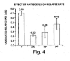
- Patients who are "persistent positive” may have a loss of efficacy from natalizumab therapy, while “transient positive” patients have full efficacy restored after only a temporary diminution in efficacy.
- a negative subject may become a transient positive. And either one may become a persistent positive if a positive immune response develops and persists for a specified number of time points, e.g., at least two time points.
- a therapy may be changed based on a determination of a single positive result.
- a therapy may be changed based upon a determination that a subject is a transient positive subject.
- a therapy may be changed based upon a determination that a subject is a persistent positive subject.
- transient and persistent are relative terms, and that a patient that seems to be persistently positive may become negative at a later time. Accordingly, patients with positive responses should be monitored regularly to evaluate the persistence of the positive response, the effectiveness of the therapy, and/or the presence of other clinical manifestations of a positive immune response.
- the risk of a reduction in therapeutic efficacy increases with the length of time that a positive immune response persists. Accordingly, in one aspect of the disclosure, the number of times that a patient tests, positive is less important than the length of time over which the patient remains positive.
- a patient may be identified as being at risk of a reduction in therapeutic efficacy (e.g., at risk of a relapse) if a positive result is detected within 3 months of the first administration of natalizumab.
- this risk increases if the positive immune response persists for 3-6 months, and further increases if the positive immune response persists for 6-9 months, and yet further increases with persistence for 9-12 months after the first administration of natalizumab. It should be appreciated that persistence for more than one year even further increases the probability of a relapse. Accordingly, different therapeutic regimens may be appropriate for a patient with a persistently positive immune response. However, it should be appreciated that even in the presence of a persistently positive immune response, a therapeutic antibody therapy need not be discontinued unless it becomes ineffective (e.g., a loss of substantially all efficacy) or causes other negative clinical manifestations.
- treatment with a therapeutic natalizumab may be discontinued if the treatment is ineffective or is losing is effectiveness in a patient that has a below-threshold level of immune response.
- a lack of efficacy (or a reduction in efficacy) in the absence of a clinically significant immune response may indicate that the ineffectiveness of the treatment is due to one or more factors other than a patient immune response to the therapeutic agent.
- a patient will not be a transient positive if no positive response is detected. Accordingly, alternative treatment may be considered.
- binding activities or antibody levels may be compared to pre-immune activities or levels (i.e., measured before the administration of the first dose of natalizumab).
- pre-immune activities or levels i.e., measured before the administration of the first dose of natalizumab.
- a comparison to a pre-immune amount is not necessary as discussed herein, because a positive immune response may be identified when a clinically significant threshold (or above threshold) amount of binding activity or antibody levels are present in a patient sample.
- a threshold amount of an antibody response is assayed for.
- a qualitative assay may be performed.
- a quantitative assay may be performed and in one embodiment, the quantitative data may be translated into a qualitative output (e.g., whether the amount of antibody is greater than a threshold amount).
- Detection assays may include any known immunodetection methods for detecting, confirming, binding, purifying, removing, quantifying and/or otherwise generally detecting antibodies that specifically bind to a specified therapeutic protein, e.g., to natalizumab, or fragments thereof.
- immunodetection techniques may include, but are not limited to, enzyme linked immunosorbent assays (ELISA) (including, but not limited to, a standard sandwich ELISA or a bridging ELISA), radioimmunoassays (RIA), immunoradiometric assays, fluoroimmunoassays, chemiluminescent assays, bioluminescent assays, radioimmunoprecipitation assays (RIPA), and Western blots.
- ELISA enzyme linked immunosorbent assays
- RIA radioimmunoassays
- immunoradiometric assays immunoradiometric assays
- fluoroimmunoassays fluoroimmunoassays
- chemiluminescent assays chemiluminescent assays
- bioluminescent assays bioluminescent assays
- radioimmunoprecipitation assays RIPA
- Western blots may include Opti
- IRMA immunoradiometric assays
- TRF time-resolved fluorometry
- ECL electrochemiluminescence
- an assay is performed to detect a presence of a binding activity for natalizumab in a biological sample.
- the specificity of the binding activity may be evaluated by determining whether the observed binding activity is specific for natalizumab or whether it is due to an interfering factor that may be present in the biological sample such as a rheumatoid factor or other binding factor.
- aspects of the disclosure may include an assay that involves contacting a biological sample with an immobilization moiety to immobilize any binding activity that is present in the biological sample.
- Immobilized binding activity may be detected using a detection moiety.
- Immobilization and detection moieties may be, respectively, immobilized unlabeled and non-immobilized labeled natalizumab as described herein.
- an immobilization moiety may be bound to a solid substrate or surface (e.g., in a well of a multi-well plate, on the surface of an ELISA plate, etc.).
- an immobilization moiety may be attached to a bead (e.g., a magnetic bead) via a covalent or other linkage (e.g., the immobilization moiety may be conjugated to a biotin molecule and attached to a bead coated with streptavidin via a biotin-streptavidin interaction).
- the bead may be attached to a surface or a matrix.
- a magnetic bead may be immobilized on a magnetic surface.
- a charged bead may be immobilized on a charged surface (e.g., an electrode).
- a positive result may be determined if the detected amount of binding activity (e.g., the amount of binding activity that is captured by the immobilization moiety) is above a predetermined threshold.
- the specificity of the detected binding activity may be evaluated by including a competition moiety in the assay.
- the competition moiety may be non-immobilized unlabeled natalizumab that may be included to compete with the immobilization and/or detection steps of the assay. If the presence of the competition moiety reduces the binding activity by at least a predetermined percentage or cut-off, the binding activity is determined to be specific and the positive result is confirmed. If the presence of the competition moiety fails to reduce the binding activity by at least a predetermined percentage or cut-off, the binding activity is determined to be non-specific and the initial positive result is now determined to be a negative result for an immune response.
- an initial threshold level of binding activity may be established using a sample that contains a predetermined amount of an antibody that is known to bind to natalizumab.
- a threshold level may be established using between 10 ng and 1,000 ng (e.g., about 50 ng, or about 500 ng) of a control antibody per ml of assay.
- the amount of antibody used to determine the threshold level will determine the sensitivity of the assay.
- the sensitivity of the assay may be considered to be similar to the amount of antibody that is used to determine the initial threshold.
- the amount of binding in the control may serve as a reference that is used to determine the threshold (e.g., the threshold amount may be a multiple or a fraction of the signal obtained in the control).
- the signal obtained in the control assay is used as the threshold amount
- the assay sensitivity may be affected by a number of factors including the affinity and specificity of the control antibody.
- the specificity of the binding activity may be evaluated by spiking the assay with an amount of competition moiety that is similar to the amount of control antibody that was used to establish the threshold level of binding.
- an assay may be spiked with between about 10 ng and 1,000 ng (e.g., about 50 ng, or about 500. ng) of unlabeled soluble natalizumab per ml of assay.
- other amounts of competition moiety may be used.
- a first level of binding activity, in a biological sample, to natalizumab is determined in a first immunoassay for a first aliquot of the biological sample
- a second level of binding activity to natalizumab is determined in a second immunoassay for a second aliquot of the biological sample
- the second immunoassay is spiked with a greater amount of unlabeled soluble natalizumab than the first immunoassay, and the presence of at least a threshold level of soluble antibody to the natalizumab is indicated if the first level of binding activity is greater than a reference level and the second level of binding activity is less than a predetermined percentage of the first level of binding activity.
- the assessment and/or monitoring may be performed by determining whether the amount of an antibody that specifically binds to natalizumab using a single-level "cut-off".
- the cut-off level of binding is the level at or above which increased detection will be scored as significant and/or positive and a confirmatory determination that the detection of a level of soluble binding activity in a biological sample reflects the level of an antibody that specifically binds to natalizumab in the sample.
- the identification of an immune response to natalizumab may be performed quantitatively to determine a titer of an antibody that specifically binds to natalizumab in a biological sample from a subject
- an immunodetection assay may be an ELISA.
- ELISA encompasses a number of protocols for immunodetection.
- ELISA methods include sandwich ELISAs, bridging ELISAs, etc.
- the ELISA immunoassay is a manual assay. However, in some embodiments all or part of the ELISA may be performed robotically.
- an ELISA assay includes using natalizumab as an immobilized target moiety with which an ELISA plate is coated. The coated ELISA plate may then be contacted with a biological sample for determination of the level of a subject antibody that specifically binds natalizumab.
- a first aliquot of a biological sample is assayed using an ELISA assay to determine the presence or absence of a threshold amount of binding to the immobilized target natalizumab and a second aliquot of the same biological sample is also assayed using an ELISA to confirm whether or not a threshold (or above threshold) level of binding to the immobilized target natalizumab is indicative of specific antibody.
- the threshold level of soluble binding activity in an aliquot is about equal to the level of binding activity present in a control or reference sample comprising at least about 50 ng, 100 ng, 200 ng, 300 ng, 400 ng, 500 ng, 600 ng, 700 ng, 800 ng, 900 ng, or 1,000 ng per ml of a reference antibody that binds to natalizumab.
- the threshold level is determined as about equal to the level of binding activity in a control or reference sample containing about 500 ng/ml of a reference antibody that binds to natalizumab.
- the reference antibody may be polyclonal or monoclonal.
- the reference antibody may be a murine anti-natalizumab antibody (e.g., 12C4 described in Sheremata et al., 1999, Neurology 52, pages 1072-1074 , incorporated herein by reference): As described herein if the level of binding to the immobilized target natalizumab that is at least at the threshold level, and the soluble binding activity is determined to be the binding activity of an antibody that specifically binds to natalizumab then it identifies an immune response to natalizumab in the subject.
- a murine anti-natalizumab antibody e.g., 12C4 described in Sheremata et al., 1999, Neurology 52, pages 1072-1074 , incorporated herein by reference
- ELISA methods useful in methods of the disclosure may include obtaining a biological sample from a subject who has been administered a therapeutic antibody such as natalizumab, and contacting an aliquot of the sample with an immobilization antibody.
- the immobilization antibody may be natalizumab.
- the immobilization antibody captures molecules or compounds in the sample that bind to the antibody, and the sample is contacted with a second detection moiety that is capable of selectively binding to or detecting the molecule or compound that is captured, (e.g., a labeled second antibody).
- moieties capable of selectively binding or detecting the complex include, but are not limited to antibodies or other ligands that can be labeled using a variety of markers (e.g., biotin/avidin ligand binding arrangement, as is known in the art).
- markers e.g., biotin/avidin ligand binding arrangement, as is known in the art.
- One skilled in the art may also use a labeled third antibody.
- the second moiety is a labeled form of the immobilization antibody.
- an ELISA assay includes using the natalizumab as an immobilized target moiety with which an ELISA plate is coated.
- the coated ELISA plate may then be contacted with a biological sample for determination of the level of a subject antibody that specifically binds the therapeutic natalizumab.
- a first aliquot of a biological sample is assayed using an ELISA assay to determine the presence or absence of a threshold amount of binding activity for the immobilized target natalizumab, and a second aliquot of the same biological sample is assayed using an ELISA to confirm whether or not a threshold (or above threshold) level of binding to the immobilized target natalizumab is indicative of natalizumab specific antibody.
- the threshold level of soluble binding activity in an aliquot is about equal to the level of binding activity present in a control or reference sample comprising at least about 50 ng, 100 ng, 200 ng, 300 ng, 400 ng, 500 ng, 600 ng, 700 ng, 800 ng, 900 ng, or 1,000 ng per ml of a reference antibody that binds to natalizumab.
- the threshold level is determined as about equal to the level of binding activity in a control or reference sample containing about 500 ng/ml a reference antibody that binds to natalizumab.
- the reference antibody may be polyclonal or monoclonal.
- the reference antibody may be a murine anti-natalizumab antibody (e.g., 12C4 described in Sheremata et al., 1999, Neurology 52, page 1072 ).
- a reference antibody that binds to natalizumab may be a reference antibody that binds to natalizumab (for example, an antibody that binds to natalizumab with high affinity, e.g., with nanomolar affinity).
- a reference antibody that binds to natalizumab may block natalizumab binding to VLA-4 (e.g., it may inhibit binding of natalizumab to VLA-4 by at least 50%, at least 60%, at least 70%, at least 80%, at least 90%, or more).
- the reference antibody may be a murine monoclonal antibody.
- the reference antibody may be an anti-idiotypic antibody specific for natalizumab.
- the reference antibody is the 12C4 antibody (available from Maine Biotechnology Services, Inc., Portland ME; see, e.g., Sheremata et al., 1999, Neurology 52, page 1072 ).
- 12C4 is a blocking antibody that blocks natalizumab binding to VLA-4.
- the reference antibody competes with 12C4 for binding to natalizumab.
- Antibody binding competition may be demonstrated using standard methods of assessing an antibody's ability to competitively inhibit the 12C4 antibody's ability to block binding of natalizumab to VLA-4.
- the presence of an antibody that specifically binds to natalizumab is determined using a bridging ELISA.
- antibodies that specifically bind to natalizumab act as a bridge between natalizumab coated on an ELISA plate and detectably labeled natalizumab in solution (e.g., non-immobilized).
- natalizumab e.g., from a biological sample
- detectably labeled natalizumab in solution e.g., non-immobilized.
- an ELISA signal after standard processing indicates that the detectable label has been linked to the solid phase and that a soluble binding activity is present in the biological sample.
- an aliquot of the biological sample with the immobilized antibody under effective conditions and for a period of time sufficient to allow the formation of immune complexes is generally a matter of adding the aliquot of the biological sample to the immobilized antibody (e.g., natalizumab immobilized on an ELISA plate) and incubating the mixture for a period of time long enough for the immobilized antibody to form an immune complex with (i.e., to bind to) a molecule or compound with soluble binding activity that is present in the aliquot of the biological sample.
- the immobilized antibody e.g., natalizumab immobilized on an ELISA plate
- the molecule or compound with soluble binding activity may be an induced antibody that specifically binds to natalizumab or may be a non-induced endogenous antibody or receptor that binds to natalizumab (e.g., a rheumatoid factor [RF] or an anti-Fab antibody).
- the sample-antibody mixture e.g., the ELISA plate, dot blot, or western blot
- an additional step may involve confirming whether or not the binding activity is indicative of an induced antibody that specifically binds to natalizumab.
- a second aliquot of the biological sample may be prepared and assayed as described for the first aliquot, except that a predetermined amount of non-immobilized unlabeled competition natalizumab also is added to the assay (e.g., the ELISA assay).
- the predetermined amount of competition antibody may be an unlabeled amount that reduces a specific signal by about 50% or more in a control assay.
- the threshold (or above threshold) binding activity is judged as a positive indicator for the presence of an antibody that specifically binds to natalizumab. In contrast, if the presence of the unlabeled reduces the signal by less than an expected percentage amount then the threshold (or above threshold) binding activity is judged as negative for an antibody that specifically binds to natalizumab. It should be appreciated that a non-specific signal may be due serum factors other than an antibody that binds to natalizumab. As used herein the terms “spike” or “spiked” refers to the addition of an unlabeled (or differently labeled) soluble competition natalizumab to a sample or assay.
- the "percentage reduction” is the percentage of the level of binding determined in the first aliquot.
- the amount of signal by more than 40-90% (e.g., by about 50% or more, by about 55% or more, by about 60% or more, by about 65% or more, by about 70% or more, by about 75% or more, by about 80% or more, by about 85% or more, by about 90% or more)
- the binding activity in the biological sample is considered indicative of the presence of an induced antibody that specifically binds to natalizumab.
- the competition antibody may be soluble unlabeled natalizumab.
- the soluble unlabeled natalizumab may be used at a final concentration of about 100 ( ⁇ g/ml.
- any concentration of freeunlabeled natalizumab may be used if it results in a predetermined decrease (e.g., about 40%, about 50%, or more) in the signal obtained for a control sample containing a control amount of reference antibody.
- a control sample may contain about 500 ng/ml, about 3 ⁇ g/ml or any other suitable amount of reference antibody (e.g., 12C4).
- the presence in a biological sample from a patient of an antibody that specifically binds to natalizumab indicates that the subject has a clinically significant immune response to natalizumab.
- a therapeutic natalizumab may be used as the target antibody and may be immobilized onto a selected surface exhibiting protein affinity, such as a well in a polystyrene microtiter plate. Then, a sample from a subject who has had at least one administration of a therapeutic natalizumab, is added to the wells. After binding and/or washing to remove non-bound materials, binding molecules or compounds that are bound to the target antibody may be detected. Detection may be achieved by the addition of a second antibody that is linked to a detectable label. In addition, the identity of the binding molecule or compound as an antibody that specifically binds to natalizumab may be confirmed as described above herein.
- ELISAs have certain features in common, such as coating, incubating and binding, washing to remove non-specifically bound species, and detecting the bound immune complexes.
- a coating buffer may be a sodium phosphateBSA coating buffer or another suitable art-known coating buffer.
- the wells of the plate will then be washed to remove incompletely adsorbed material. Any remaining available surfaces of the wells are then "coated" with a nonspecific protein that is antigenically neutral with regard to the test sample.
- This protein may be bovine serum albumin (BSA), casein or solutions of milk powder, etc.
- BSA bovine serum albumin
- the coating allows for blocking of non-specific adsorption sites on the immobilizing surface and thus reduces the background caused by nonspecific binding of antisera onto the surface.
- a secondary or tertiary detection means may be used or a direct detection means may be used.
- a secondary or tertiary detection methods after binding of a protein or antibody to the well, coating with a non-reactive material to reduce background, (e.g. with blocking buffer such as Tris-sucrose blocking buffer or other art-recognized blocking buffer), and washing to remove unbound material, the immobilizing surface is contacted with the biological sample to be tested under conditions effective to allow immune complex (antigen/antibody) formation.
- a non-reactive material to reduce background e.g. with blocking buffer such as Tris-sucrose blocking buffer or other art-recognized blocking buffer
- Detection of the immune complex then requires a labeled secondary binding ligand or antibody, and a secondary binding ligand or antibody in conjunction with a labeled tertiary antibody or a third binding ligand.
- the second binding ligand is natalizumab (e.g. as used for the target antibody).
- the term "under conditions effective to allow immune complex formation” means that the conditions preferably include diluting the antigens and/or antibodies with solutions such as BSA, bovine gamma globulin (BGG), phosphate buffered saline (PBS)/Tween, PBS with casein and Tween 20, or PBS/BSA buffer with Tween 20.
- solutions such as BSA, bovine gamma globulin (BGG), phosphate buffered saline (PBS)/Tween, PBS with casein and Tween 20, or PBS/BSA buffer with Tween 20.
- BGG bovine gamma globulin
- PBS phosphate buffered saline
- PBS phosphate buffered saline
- PBS/BSA buffer with Tween 20 PBS/BSA buffer with Tween 20.
- the "suitable" conditions also mean that the incubation is at a temperature or for a period of time sufficient to allow effective binding. Incubation steps are typically from about 1 to 2 to 4 hours or so, at temperatures preferably on the order of 25oC. to 27oC, or may be overnight at about 4oC. Various art-known assay temperature and timing parameters can be used in the methods of the invention.
- wash buffer formulations can be used in the methods disclosed herein.
- the second or third antibody will have an associated detectable label.
- the detectable label is an enzyme that will generate color development upon incubating with an appropriate chromogenic substrate.
- a urease-, glucose oxidase-, alkaline phosphatase-, hydrogen peroxidase-conjugated antibody, or other conjugated antibody for a period of time and under conditions that favor the development of further immune complex formation (e.g., incubation for 2 hours at room temperature in a PBS-containing solution such as PBS-Tween).
- a positive quality control sample may be a normal serum sample that contains a predetermined amount of an antibody that is known to bind to natalizumab. Quality control samples may be reacted in parallel with and under the same conditions as the biological and control samples of the assay and provide a measure of the function of the assay.
- a negative quality control sample may be a serum sample known not to include an antibody that is known to bind to natalizumab.
- a positive control may include a known amount of an antibody that specifically binds natalizumab so when treated under the same conditions as the test samples (e.g., the biological sample) it indicates that the assay works within expected parameters.
- An example of a negative control may be a sample that is known to not include an antibody that specifically binds to natalizumab. Such a negative control, when treated under the same conditions as the test sample (e.g., the biological sample), will demonstrate that the binding detected in a biological sample arises from the biological sample and is not due to contamination of assay components or other factor not associated with the biological sample.
- a non-limiting example of an assay encompassed by the methods of the disclosure may involve the following procedures.
- the ELISA reagents described herein maybe packaged in a kit that may be produced commercially to detect the presence of and/or measure an antibody that specifically binds a natalizumab in a biological sample as described herein.
- controls for use in the invention may include, in addition to predetermined values (such as clinically significant threshold values identified as described herein for a particular therapeutic protein), samples of materials tested in parallel with the experimental materials.
- predetermined values such as clinically significant threshold values identified as described herein for a particular therapeutic protein
- examples include negative control samples (e.g., generated through manufacture) to be tested in parallel with the experimental samples.
- a biological sample may be, but is not limited to, any of the following: a body fluid of a subject including, blood, serum, plasma, urine, saliva, pleural effusions, stool, synovial fluid, cerebral spinal fluid, mucus, and tissue infiltrations. Preferred body fluids include blood, plasma, and serum.
- biological samples may be obtained using methods well known to those of ordinary skill in the related medical arts.
- a biological sample may be obtained directly from a subject or may be obtained from cell, tissue, or other culture.
- a biological sample may be fresh or may have been stored under suitable conditions (e.g., frozen, chilled, etc.) prior to use in methods of the invention.
- a biological sample may be obtained from a subject at different time intervals after the administration of a therapeutic agent.
- a therapeutic agent may have a characteristic in vivo half-life, and the amount of free therapeutic agent that is present in a biological sample typically will decrease as a function of time after administration of the agent to the subject.
- the presence of free therapeutic agent (unlabeled and non-immobilized) in a biological sample may interfere with binding and detection reactions of the invention. Accordingly, biological samples should be obtained as long as possible after a therapeutic administration, for example at a "trough" in the treatment cycle. A "trough" represents the lowest amounts of free therapeutic agent that are present in a subject during a treatment cycle.
- a trough may occur, for example, long after one therapeutic administration and soon before a subsequent therapeutic administration. It should be appreciated that the timing of the trough may be influenced by many factors, including the amount of agent that is administered, the half-life of the agent, the frequency of administration, etc. For example, a monthly administration of natalizumab (e.g., 300 mg of natalizumab) results in a trough at about 30 days after one administration and immediately before a subsequent administration.
- assays of the invention may be performed on samples taken at different times after treatment administration provided that the assays are relatively insensitive to, or account for, the presence of free therapeutic agent in the biological sample.
- biological samples may be diluted before being assayed (e.g., 2 fold, 5 fold, 10 fold, 50 fold, 100 fold, and including higher or lower fold values or any fold value in between).
- a reference sample containing a clinically significant threshold amount of reference antibody may be diluted by the same amount as the biological sample being tested so that the signal obtained for the biological sample can be compared directly to the signal obtained for the reference sample.
- one or more aliquots from a biological sample are used.
- the term "aliquot” means a portion or part of the biological sample.
- two or more aliquots may be taken from a biological sample obtained from a subject and the aliquots can be tested using methods of the invention to determine the presence of an immune response to natalizumab in the subject.
- two substantially equivalent aliquots can be taken from a biological sample obtained from a subject to whom natalizumab natalizumab has been administered, and a level of soluble binding activity can be detected in one aliquot (e.g. a "first" aliquot) can be determined.
- the other aliquot e.g.
- the "second" aliquot) may be assessed using the methods of the invention to determine whether the soluble binding activity detected in the first aliquot, which would also be present in the second aliquot by virtue of their common sample origin, is an antibody that specifically binds natalizumab. In some embodiments, if at least a threshold level of binding is present in the first aliquot and the soluble binding activity is determined to be the activity of an antibody that specifically binds natalizumab, it identifies that an immune response to natalizumab is present in the subject.
- a detection assay involves both immobilization and detection moieties.
- a detection assay also may include a competition moiety.
- an immobilization moiety may be used to immobilize an induced antibody (e.g., a serum antibody) that binds to natalizumab.
- a detection moiety may be used to detect the immobilized antibody.
- a competition moiety may be used to compete with the either the immobilization and/or detection moiety for binding to the induced antibody in order to determine the specificity of binding to the induced antibody.
- the immobilization and detection moieties may bind independently to one or more antibodies that are characteristic of an immune response to natalizumab. Accordingly, the immobilization and detection moieties may be the same that was administered to the subject (e.g., the immobilization moiety may be an immobilized form of natalizumab that was administered to the subject, and the detection moiety may be a labeled form of natalizumab that was administered to the subject).
- each of the immobilization and detection moieties independently may be variants of natalizumab that was administered to the subject, provided that the immobilization and detection moieties bind with suitable affinity to detect one or more induced antibodies (e.g., serum antibodies) against natalizumab.
- the competition moiety is typically a soluble unlabeled (or differentially labeled) form of the immobilization or detection moiety.
- the competition moiety may be a variant of the immobilization or detection moiety, provided that it competes sufficiently for binding to natalizumab to determine the specificity of the binding activity detected in the assay.
- natalizumab may be used therapeutically. Accordingly, may be used as immobilization, detection, and/or competition moieties in a detection assay of the disclosure.
- Natalizumab and related VLA-4 binding antibodies are described, e.g., in US 5,840,299 .
- mAb 21.6 and HP1/2 are exemplary murine monoclonal antibodies that bind VLA-4.
- Natalizumab is a humanized version of murine mAb 21.6 (see, e.g., US Patent No. 5,840,299 ).
- a humanized version of HP1/2 has also been described (see, e.g., US Patent No. 6,602,503 ).
- VLA-4 binding monoclonal antibodies such as HP2/1, HP2/4, L25 and P4C2 are described (e.g., in US Patent No. 6,602,503 ; Sanchez-Madrid et al., 1986 Eur. J.
- VLA-4 binding antibodies interact with VLA-4 on cells, e.g., lymphocytes, but do not cause cell aggregation. However, other anti-VLA-4 binding antibodies have been observed to cause such aggregation. HP1/2 does not cause cell aggregation.
- the HP1/2 MAb ( Sanchez-Madrid et al., 1986 Eur. J. Immunol., 16:1343-1349 ) has an extremely high potency, blocks VLA-4 interaction with both VCAM1 and fibronectin, and has the specificity for epitope B on VLA-4.
- This antibody and other B epitope-specific antibodies (such as B1 or B2 epitope binding antibodies; Pulido et al., 1991 J. Biol. Chem., 266(16):10241-10245 ) represent one class of useful VLA-4 binding antibodies.
- sequence identity is calculated as follows.
- the sequences are aligned for optimal comparison purposes (e.g., gaps can be introduced in one or both of a first and a second amino acid or nucleic acid sequence for optimal alignment and non-homologous sequences can be disregarded for comparison purposes).
- the optimal alignment is determined as the best score using the GAP program in the GCG software package with a Blossum 62 scoring matrix with a gap penalty of 12, a gap extend penalty of 4, and a frameshift gap penalty of 5.
- the amino acid residues or nucleotides at corresponding amino acid positions or nucleotide positions are then compared.
- amino acid or nucleic acid “identity” is equivalent to amino acid or nucleic acid “homology”).
- the percent identity between the two sequences is a function of the number of identical positions shared by the sequences.
- conservative amino acid substitutions may be made in VLA-4 binding antibodies to provide functionally equivalent variants, of the antibodies, i.e, the variants retain the functional capabilities of the VLA-4 polypeptides.
- a "conservative amino acid substitution” refers to an amino acid substitution that does not alter the relative charge or size characteristics of the protein in which the amino acid substitution is made.
- Variants can be prepared according to methods for altering polypeptide sequence known to one of ordinary skill in the art such as are found in references that compile such methods, e.g. Molecular Cloning: A Laboratory Manual, J.
- VLA-4 binding antibodies include conservative amino acid substitutions of in the amino acid sequences of proteins disclosed herein.
- Conservative substitutions of amino acids include substitutions made amongst amino acids within the following groups: (a) M, I, L, V; (b) F, Y, W; (c) K, R, H; (d) A, G; (e) S, T; (f) Q, N; and (g) E, D.
- hybridizes under high stringency conditions describes conditions for hybridization and washing.
- Guidance for performing hybridization reactions can be found in Current Protocols in Molecular Biology, John Wiley & Sons, N.Y. (1989), 6.3.1-6.3.6 . Aqueous and nonaqueous methods are described in that reference and either can be used.
- High stringency hybridization conditions include hybridization in 6XSSC atabout45°C, followed by one or more washes in 0.2X SSC, 0.1% SDS at 65°C, or substantially similar conditions.
- Antibodies can be tested for a functional property, e.g., VLA-4 binding, e.g., as described in US Patent No. 6,602,503 .
- Antibodies that bind to VLA-4 can be generated by immunization, e.g., using an animal. All or part of VLA-4 can be used as an immunogen. For example, the extracellular region of the alpha-4 subunit can be used as immunogen.
- the immunized animal contains immunoglobulin producing cells with natural, human, or partially human immunoglobulin loci.
- the non-human animal includes at least a part of a human immunoglobulin gene. For example, it is possible to engineer mouse strains deficient in mouse antibody production with large fragments of the human Ig loci. Using the hybridoma technology, antigen-specific monoclonal antibodies derived from the genes with the desired specificity may be produced and selected.
- Non-human antibodies to VLA-4 can also be produced, e.g., in a rodent.
- the non-human antibody can be humanized, e.g., as described in US Patent No. 6,602,503 , EP 239 400 , US Patent No. 5,693,761 , and US Patent No. 6,407,213 .
- EP 239 400 (Winter et al. ) describes altering antibodies by substitution (within a given variable region) of their complementarity determining regions (CDRs) for one species with those from another.
- CDR-substituted antibodies are predicted to be less likely to elicit an immune response in humans compared to true chimeric antibodies because the CDR-substituted antibodies contain considerably less non-human components. ( Riechmann et al., 1988, Nature 332, 323-327 ; Verhoeyen et al., 1988, Science 239, 1534-1536 ).
- CDRs of a murine antibody substituted into the corresponding regions in a human antibody by using recombinant nucleic acid technology to produce sequences encoding the desired substituted antibody.
- Human constant region gene segments of the desired isotype usually gamma I for CH and kappa for CL
- the humanized heavy and light chain genes are co-expressed in mammalian cells to produce soluble humanized antibody.
- Tempest et al. 1991, Biotechnology 9,266-271
- An advantage of using the Tempest et al., approach to construct NEWM and REI based humanized antibodies is that the three-dimensional structures of NEWM and REI variable regions are known from x-ray crystallography and thus specific interactions between CDRs and V region framework residues can be modeled.
- Non-human antibodies can be modified to include substitutions that insert human immunoglobulin sequences, e.g., consensus human amino acid residues at particular positions, e.g., at one or more of the following positions (preferably at least five, ten, twelve, or all): (in the FR of the variable domain of the light chain) 4L, 35L, 36L, 38L, 43L, 44L, 58L, 46L, 62L, 63L, 64L, 65L, 66L, 67L, 68L, 69L, 70L, 71L, 73L, 85L, 87L, 98L, and/or (in the FR of the variable domain of the heavy chain) 2H, 4H, 24H, 36H, 37H, 39H, 43H, 45H, 49H, 58H, 64H, 67H, 68H, 69H, 70H, 73H, 74H, 75H, 78H, 91H, 92H, 93H, and/or
- the present disclosure also provides for F(ab')2, Fab, Fv and Fd fragments; chimeric antibodies in which the Fc and/or FR and/or CDR1 and/or CDR2 and/or light chain CDR3 regions have been replaced by homologous human or non-human sequences; chimeric F(ab')2 fragment antibodies in which the FR and/or CDR1 and/or CDR2 and/or light chain CDR3 regions have been replaced by homologous human or non-human sequences; chimeric Fab fragment antibodies in which the FR and/or CDR1 and/or CDR2 and/or light chain CDR3 regions have been replaced by homologous human or non-human sequences; and chimeric Fd fragment antibodies in which the FR and/or CDR1 and/or CDR2 regions have been replaced by homologous human or non-human sequences.
- Nanobodies TM are the smallest functional fragments of antibodies and are derived from naturally occurring single-chain antibodies (see Ablynx, Belgium; ablynx.com ). Nanobody TM technology was developed following the discovery that camelidae (camels and llamas) possess a unique repertoire of fully functional antibodies that lack light chains. Nanobody TM structure consists of a single variable domain (VHH), a hinge region, and two constant domains (CH2 and CH3). The cloned and isolated VHH domain is a stable polypeptide harboring the full antigen-binding capacity of the original heavy chain. Nanobodies TM combine the features of conventional antibodies with features of small molecule drugs. Nanobodies TM show high target specificity and low inherent toxicity. Additionally, Nanobodies TM are very stable, can be administered by means other than injection, and are easy to manufacture.
- Fully human monoclonal antibodies that bind to VLA-4 can be produced, e.g., using in vitro-primed human splenocytes, as described by Boerner et al., 1991, J. Immunol., 147, 86-95 . They may be prepared by repertoire cloning as described by Persson et al., 1991, Proc. Nat. Acad. Sci. USA, 88: 2432-2436 or by Huang and Stollar, 1991, J. Immunol. Methods 141,227-236 . US 5,798,230 .
- Antibodies can be produced in prokaryotic and eukaryotic cells.
- the antibodies e.g., scFvs
- the antibodies are expressed in a yeast cell such as Pichia (see, e.g., Powers et al. (2001) J Immunol Methods. 251:123-35 ), Hanseula, or Saccharomyces.
- Antibodies are produced in mammalian cells.
- mammalian host cells for recombinant expression include Chinese Hamster Ovary (CHO cells) (including dhfr- CHO cells, described in Urlaub and Chasin (1980) Proc. Natl. Acad. Sci. USA 77:4216-4220 , used with a DHFR selectable marker, e.g., as described in Kaufman and Sharp (1982) Mol. Biol.
- lymphocytic cell lines e.g., NS0 myeloma cells and SP2 cells, COS cells, K562, and a cell from a transgenic animal, e.g., a transgenic mammal.
- the cell is a mammary epithelial cell.
- the recombinant expression vectors may carry additional sequences, such as sequences that regulate replication of the vector in host cells (e.g., origins of replication) and selectable marker genes.
- the selectable marker gene facilitates selection of host cells into which the vector has been introduced (see e.g., US Patent Nos. 4,399,216 , 4,634,665 and 5,179,017 ).
- Exemplary selectable marker genes include the dihydrofolate reductase (DHFR) gene (for use in dhfr- host cells with methotrexate selection/amplification) and the neo gene (for G418 selection).
- DHFR dihydrofolate reductase
- a recombinant expression vector encoding both the antibody heavy chain and the antibody light chain is introduced into dhfr-CHO cells by calcium phosphate-mediated transfection.
- the antibody heavy and light chain genes are each operatively linked to enhancer/promoter regulatory elements (e.g., derived from SV40, CMV, adenovirus and the like, such as a CMV enhancer/AdMLP promoter regulatory element or an SV40 enhancer/AdMLP promoter regulatory element) to drive high levels of transcription of the genes.
- the recombinant expression vector also carries a DHFR gene, which allows for selection of CHO cells that have been transfected with the vector using methotrexate selection/amplification.
- the selected transformant host cells are cultured to allow for expression of the antibody heavy and light chains and intact antibody is recovered from the culture medium.
- Standard molecular biology techniques are used to prepare the recombinant expression vector, transfect the host cells, select for transformants, culture the host cells, and recover the antibody from the culture medium. For example, some antibodies can be isolated by affinity chromatography with a Protein A or Protein G.
- US 6,602,503 also describes exemplary methods for expressing and purifying natalizumab.
- Antibodies may also include modifications, e.g., modifications that alter Fc function, e.g., to decrease or remove interaction with an Fc receptor or with C1q, or both.
- modifications e.g., modifications that alter Fc function, e.g., to decrease or remove interaction with an Fc receptor or with C1q, or both.
- the human IgG1 constant region can be mutated at one or more residues, e.g., one or more of residues 234 and 237, e.g., according to the numbering in US Patent No. 5,648,260 .
- Other exemplary modifications include those described in US Patent No. 5,648,260 .
- the antibody production system may be designed to synthe size antibodies in which the Fc region is glycosylated.
- the Fc domain of IgG molecules is glycosylated at asparagine 297 in the CH2 domain.
- This asparagine is the site for modification with biantennary-type oligosaccharides. This glycosylation participates in effector functions mediated by Fey receptors and complement C1q ( Burton and Woof (1992) Adv. Immunol. 51:1-84 ; Jefferis et al. (1998) Immunol. Rev. 163:59-76 ).
- the Fc domain can be produced in a mammalian expression system that appropriately glycosylates the residue corresponding to asparagine 297.
- the Fc domain can also include other eukaryotic post-translational modifications.
- Antibodies can also be produced by a transgenic animal.
- US Patent No. 5,849,992 describes a method for expressing an antibody in the mammary gland of a transgenic mammal.
- a transgene is constructed that includes a milk-specific promoter and nucleic acids encoding the antibody of interest and a signal sequence for secretion.
- the milk produced by females of such transgenic mammals includes, secreted-therein, the antibody of interest.
- the antibody can be purified from the milk, or for some applications, used directly.
- non-immobilized, anti-therapeutic protein antibodies e.g., natalizumab as detection moieties to assess the presence and/or level of soluble binding activity that is bound to an immobilized antibody against a therapeutic protein (e.g., natalizumab).
- Methods to evaluate the presence and/or level of soluble binding activity may include the use of one or more labeled detection moieties (e.g., natalizumab containing or attached to a detectable label).
- a detectable label is defined as any moiety that can be detected using an assay.
- the antibodies and functional antibody fragments disclosed herein can be coupled to specific labeling agents for detecting binding according to standard coupling procedures.
- detectable labels can be used, such as those that provide direct detection (e.g., a radioactive label, a fluorophore, [e.g. Green Fluorescent Protein (GFP), Red Fluorescent Protein (RFP), etc.], a chromophore, an optical or electron dense label, etc.) or indirect detection (e.g., an enzyme tag such as horseradish peroxidase, etc.).
- detectable labels that have been attached to or incorporated into antibodies include: enzymes, radiolabels, fluorescent labels, phosphorescent molecules, chemiluminescent molecules, chromophores, luminescent molecules, photoaffnity molecules, and colored particles or ligands such as biotin, etc.
- detection methods disclosed herein may include electrochemiluminescence methods (ECL).
- Labels may be directly detected through optical or electron density, radioactive emissions, non-radiative energy transfers, etc. or indirectly detected with antibody conjugates, streptavidin-biotin conjugates, etc. Many additional detectable labels are known in the art, as are methods for their attachment to antibodies.
- Labeled antibodies of the disclosure may be antibodies that are used in vitro, e.g., in an immunoassay such as an ELISA.
- detectably labeled antibodies may be antibodies that have a detectable label incorporated into the antibody or may be antibodies that are linked to a secondary binding ligand and/or to an enzyme (an enzyme tag) that will generate a colored product upon contact with a chromogenic substrate.
- suitable enzymes include urease, alkaline phosphatase, (horseradish) hydrogen peroxidase or glucose oxidase.
- Preferred secondary binding ligands are biotin and/or avidin and streptavidin compounds.
- An attachment method may include the use of a metal chelate complex employing, for example, an organic chelating agent such a diethylenetriaminepentaacetic acid anhydride (DTPA); ethylenetriaminetetraacetic acid; N-chloro-p-toluenesulfonamide; and/or tetrachloro-3 .alpha.-6.alpha.-diphenylglycouril-3 attached to the antibody ( U.S. Pat. Nos. 4,472,509 and 4,938,948 ).
- DTPA diethylenetriaminepentaacetic acid anhydride
- ethylenetriaminetetraacetic acid N-chloro-p-toluenesulfonamide
- tetrachloro-3 .alpha.-6.alpha.-diphenylglycouril-3 attached to the antibody
- Monoclonal antibodies may also be reacted with an enzyme in the presence of a coupling agent such as glutaraldehyde or periodate.
- Antibodies may be labeled with fluorescein markers in the presence of these coupling agents or by reaction with an isothiocyanate.
- antibodies may be labeled by derivatization, for example, by selectively introducing sulfhydryl groups in the Fc region of the antibody, using reaction conditions that do not alter the antibody recognition site.
- the detection of the detectable label in an assay disclosed herein is also referred to herein as detecting the "signal".
- Methods for detecting the signal in an immunoassay are well known in the art.
- the assay signal can be detected using with a multi-well plate reader (e.g. microplate reader) to assess the amount and/or location of a signal.
- Signal detection can be optical detection or other detection means suitable for detecting a detectable label utilized in the disclosure. Additional methods for detecting labels are well known in the art and can be used in methods disclosed herein.
- Methods of the disclosure include ELISAs that have a sensitivity for detecting an antibody that specifically binds to natalizumab, wherein the sensitivity is at least about 1000ng, 500ng, or 50ng. In preferred embodiments, the ELISA sensitivity for detecting an antibody that specifically binds to natalizumab is at least about 500ng.
- the detection of immunocomplex formation may be achieved through the application of numerous approaches. These methods are generally based upon the detection of a label or marker, such as any of those radioactive, fluorescent, biological and enzymatic tags.
- U.S. Patents concerning the use of such labels include U.S. Pat. Nos. 3,817,837 ; 3,850,752 ; 3,939,350 ; 3,996,345 ; 4,277,437 ; 4,275,149 and 4,366,241 .
- a secondary binding ligand such as a second antibody and/or a biotin/avidin ligand binding arrangement, as is known in the art.
- natatizumab also may be used to obtain and produce a reference antibody that binds to natalizumab with high affinity, (for example, a high affinity natalizumab binding antibody (e.g., 12C4).
- a high affinity natalizumab binding antibody e.g., 12C4
- kits for_assaying the presence of an immune response to natalizumab in a sample may include Natalizumab.
- a kit may include detectably labeled and/or unlabeled antibodies and may include solutions and compounds for detectably labeling an antibody.
- a kit of the disclosure may also include one or more of the following components: plates, pipettes, vials, detectable label, solutions (e.g. blocking buffer, wash buffer, binding solutions, diluent solutions, etc), positive and/or negative control samples and solutions.
- a kit of the disclosure may also include written instructions for the use of the kit for the identification of an immune response to natalizumab in a biological sample. Kits of the disclosure may also include additional components useful in the performance of positive and/or negative ELISA control assays.
- a kit of the disclosure may also include equipment such as plate readers and/or robotic instrumentation for use in the methods of the invention.
- the assay format and design was based upon a bridging enzyme-linked Immunosorbent assay (ELISA).
- ELISA bridging enzyme-linked Immunosorbent assay
- Standard reagents and procedures suitable for bridging assays were used.
- natalizumab was adsorbed to the surface of microtiter plates followed by a blocking step to minimize non-specific binding of serum antibodies. Controls and samples were diluted and added to the wells.
- detection of bound anti-natalizumab antibodies was accomplished by incubating the plates with biotin-natalizumab followed by streptavidin-Alkaline Phosphatase (SA-AP). The color development was proportional to the amount of anti-natalizumab antibody bound.
- SA-AP streptavidin-Alkaline Phosphatase
- Natalizumab was diluted to a specific concentration in plate coating buffer was incubated in 96-well microtiter ELISA plates overnight at ambient temperatures. The recommended volume was 100 ⁇ l/well. The plates were washed with washing buffer then incubated with blocking buffer at ambient temperatures for >1hr. The volume of blocking buffer was 200 ⁇ l/well. The plates were washed with washing buffer and then incubated with quality control and subject (patient) samples diluted 1:10 in assay diluent with or without added natalizumab (100 ⁇ g/ml final concentration) at ambient temperatures for 2 hr ⁇ 15 min. The assay diluent containing added natalizumab represented a confirmatory step.
- the volume for the assay diluent was 100 ⁇ l/well.
- the plates were then washed in washing buffer, and then incubated with SA-AP at ambient temperatures for 30-35 min.
- the volume for the incubation with SA-AP was 100 ⁇ l/well.
- the plates were then washed in washing buffer, and then incubated with AP substrate, p-nitrophenyl phosphate (PNPP) at ambient temperatures for 45-50 min.
- the volume for the incubation in the AP substrate was 100 ⁇ l/well.
- Stop solution e.g. H2SO4, etc.
- the positive and negative controls had to provide results within a pre-defined value and precision.
- the samples were judged as negative if their OD value fell below the defined negative cut-off quality control sample.
- Samples were also judged negative if their OD value fell above the defined negative cut-off quality control sample and the inhibition of the subject (patient) sample signal by added natalizumab in the confirmatory step was less than a predefined percent.
- Samples were judged positive if their OD value fell above the defined negative cut-off quality control sample and the inhibition of the subject (patient) sample signal by added natalizumab in the confirmatory step was greater than or equal to a predefined percent.
- Example 2 Screening Assay for an Immune Response to natalizumab
- a screening assay (as described above) was performed on samples from subjects who had been administered natalizumab. The presence of an immune response to natalizumab was examined in patients who had undergone natalizumab administration.
- the high affinity mAb detected binding antibodies (e.g. antibodies that bound natalizumab) that were present in biological samples from subjects.
- the negative cut-off level was determined to the lowest concentration that returned acceptable accuracy/precision (25%/25%); 50 ng/mL in 10% serum.
- the sensitivity of the mAb was 500 ng/mL in neat (undiluted) serum. With respect to sensitivity, the mAb was determined to detect anti- natalizumab Abs but to be insensitive to irrelevant human monoclonal antibodies.
- the interference with the mAb by other molecules or components of the samples was determined to include interference by free drug at an mAb:drug ratio of greater than 1:2.
- Fig. 1 illustrates the effects of free natalizumab interference with the immunoassay for anti-natalizumab antibodies. Rheumatoid factor was also found to interfere with the assay.
- characterization assays to assess binding in the screening assays were also preformed.
- Biological samples from subjects who had been administered natalizumab were tested for the presence of a soluble antibody that specifically binds natalizumab using a flow cytometry blocking assay.
- a high affinity mAb was used as a positive control.
- the assay detected blocking antibodies.
- the negative cut-off level for the mAb used was determined at 3-4 standard deviations above the mean of measured levels in sera obtained from non-dosed patients. There was a 5% false-positive rate and the sensitivity of the assay was 500ng/ml of neat serum.
- the specificity of the mAb was tested and the mAb was determined to detect anti- natalizumab antibodies but was found to be insensitive to irrelevant human monoclonal antibodies. Free-drug interference with the assay was also examined. The assay showed the presence of free-drug interference. Results from an assessment of blocking assay interference by free drug are shown in Fig. 2 .
- the screening assay provides an accurate measure of a subject's immune response to natalizumab.
- a screening assay (as described in Examples 1 and 2) was performed on samples from 625 subjects who had been administered natalizumab. The presence of an immune response to natalizumab was examined in patients who had undergone natalizumab administration. Incidence of antibodies to natalizumab in the patients examined was: 91% (569 patients) antibody negative and 9% (56 patients) "binding antibody” positive at any time point with 3% (19 patients) "transiently” positive and 6% (37 patients) "persistently” positive. The transient positive patients had detectable antibodies (at a concentration of > 0.5 ⁇ g/ml) at a single time point, but negative for antibodies at all other time points. The persistent positive patients had detectable antibodies at two or more time points that were at least 42 days apart, or at a single time point with no follow-up samples tested.
- Fig. 3 shows the time of first positive results in patients who developed any antibodies. Six percent of patients developed "persistent" antibodies to natalizumab. Over 90% of persistent-positive patients first had detectable antibodies at week 12. No subject became positive for persistent antibodies after week 36. Transient-positive patients had detectable antibodies at week 12, but were subsequently antibody negative.
- Fig. 4 illustrates the overall effect of antibodies on relapse rate.
- the relapse rate was also assessed for subjects with respect to the time elapsed from the administration of the natalizumab.
- Fig. 5A-D depict the rate of relapse at 0-3 months, 3-6 months, 6-9 months, and 9-12 months respectively.
Landscapes
- Health & Medical Sciences (AREA)
- Life Sciences & Earth Sciences (AREA)
- Chemical & Material Sciences (AREA)
- Immunology (AREA)
- Engineering & Computer Science (AREA)
- General Health & Medical Sciences (AREA)
- Medicinal Chemistry (AREA)
- Organic Chemistry (AREA)
- Pharmacology & Pharmacy (AREA)
- Nuclear Medicine, Radiotherapy & Molecular Imaging (AREA)
- General Chemical & Material Sciences (AREA)
- Animal Behavior & Ethology (AREA)
- Chemical Kinetics & Catalysis (AREA)
- Public Health (AREA)
- Veterinary Medicine (AREA)
- Bioinformatics & Cheminformatics (AREA)
- Molecular Biology (AREA)
- Biomedical Technology (AREA)
- Hematology (AREA)
- Urology & Nephrology (AREA)
- Biochemistry (AREA)
- Proteomics, Peptides & Aminoacids (AREA)
- Biotechnology (AREA)
- Cell Biology (AREA)
- Microbiology (AREA)
- Pathology (AREA)
- General Physics & Mathematics (AREA)
- Food Science & Technology (AREA)
- Physics & Mathematics (AREA)
- Analytical Chemistry (AREA)
- Rheumatology (AREA)
- Genetics & Genomics (AREA)
- Biophysics (AREA)
- Transplantation (AREA)
- Oncology (AREA)
- Pain & Pain Management (AREA)
- Neurosurgery (AREA)
- Neurology (AREA)
- Dermatology (AREA)
- Orthopedic Medicine & Surgery (AREA)
Description
- This application claims priority under 35 U.S.C. §119(e) from
U.S. provisional application serial number 60/668,404, filed April 4, 2005 - The invention relates to evaluating patients for an immune response to a therapeutic agent, and particularly to a therapeutic protein, natalizumab.
- Biologic therapeutics are currently available for treating diseases and disorders such as transplant rejection, leukemia, breast cancer, arthritis, multiple sclerosis, and Crohn's disease; and numerous additional protein-based therapies are in development. Available biologics therapeutics include AMEVIVE® (alefacept), ZEVALIN® (ibritumomab tiuxetan), ORTHOCLONE® (muromonab-CD3), ENBREL® (etanercept), REOPRO® (abciximab), RITUXAN® (rituximab), SIMULECT® (basiliximab), REMICADE® (infliximab), SYNAGIS® (palivizumab), HERCEPTIN® (trastuzumab), ZENAPAX® (daclizum-ab), CAMPATH® (alemtuzumab), MYLOTARG® (gem-tuzumab ozogamicin), HUMIRA® (adalimumab), AVON-EX® (Interferon beta-1a), and TYSABRT® (natalizumab). Natalizumab is a humanized monoclonal antibody against α4β1 integrin (VLA-4). Natalizumab binds to the a4 subunit of α4β1 and α4β7 integrins. Natalizumab is useful to treat certain inflammatory diseases and conditions including multiple sclerosis, Crohn's disease, and rheumatoid arthritis.
- Because immune responses to biologic therapeutic agents may have clinical consequences, immunogenicity assay development and validation is of great importance in the field of biologic therapeutic agents.
- The invention relates to the embodiments as defined in the claims.
- In particular, the invention relates to a method of detecting a clinically significant immune response to natalizumab in a subject, the method comprising determining whether at least two biological samples taken at different time points from a subject that has been administered natalizumab contain at least a clinically significant threshold level of about 500 ng/ml in a serum sample of a soluble antibody that binds to natalizumab, wherein the presence of at least the threshold level of the soluble antibody in said at least two samples is indicative of a clinically significant immune response to natalizumab wherein the time points are separated by at least one month, and wherein the clinically significant immune response indicates a diminution of efficacy or lack of efficacy of natalizumab.
- The disclosure provides methods and compositions for identifying, monitoring, and/or evaluating an immune response to a therapeutic agent, e.g., a therapeutic protein, e.g., a therapeutic antibody. The fact that a patient develops any antibodies to a therapeutic agent (such as a therapeutic protein or therapeutic antibody) may or may not correlate with a clinical response to the therapeutic agent. Aspects of the disclosure are based, in part, on the discovery of an unexpected level of antibody response that can be used as a threshold for detecting a clinically significant response to the therapeutic agent. In some embodiments, the threshold level is higher than would have been predicted using a statistical analysis of patients that have not received the therapeutic agent The clinically significant threshold is generally higher than the lowest detectable level of immune response in a patient. For example, the clinically significant threshold level is generally at least 2 standard deviations above a negative control level, e.g., above a mean pre-treatment level of an untreated patient population. In some embodiments, the higher threshold levels used in methods of the invention result in fewer false positives than would be identified if the threshold level were based on a 5% cutoff(e.g., 1.645 standard deviations above the mean) for immune responses observed in patients that had not received the therapeutic agent. According to aspects of the disclosure, the presence of a detectable immune response in a patient sample is not clinically significant unless the immune response reaches at least a predetermined threshold level. The disclosure provides, inter alia, methods of identifying a clinically significant threshold level of antibody response to a therapeutic agent (e.g., a therapeutic protein, e.g., a therapeutic antibody), and methods of identifying patients who have a clinically significant antibody response to a therapeutic agent. The disclosure, in part, also provides a threshold level with which to identify clinically meaningful antibodies in a subject. According to aspects disclosed herein, an immune response to a therapeutic agent (e.g., natalizumab) may not be clinically significant (e.g., may not show a significant association with reduced clinical efficacy) unless the magnitude of the immune response reaches a threshold level that can be predetermined (e.g., based on immune responses obtained for different patient groups). Surprisingly, the methods described herein do not rely on comparing samples obtained from each patient before and after treatment, nor do they rely on identifying the mere presence of a detectable immune response to the therapeutic agent. In contrast, methods of the disclosure, relate to detecting at least a threshold level of an immune response to a therapeutic agent, where the threshold level may be higher than the lowest detectable level of immune response, and wherein the positive results from the assay are clinically meaningful, in part, because the assay avoids false positives that have no associated clinical significance.
- Currently, there is no generally applicable technique or standard for detecting a clinically significant antibody response to a therapeutic protein. Different therapeutic proteins may induce different types of antibodies, and the presence of such antibodies may or may not affect the safety, pharmacokinetics, and/or efficacy of a therapeutic protein. Current methods of monitoring a patient's response to a therapeutic antibody typically involve comparing levels of serum antibodies before and after treatment for each patient identifying the presence of any detectable immune response, and evaluating the patient to determine whether the detectable immune response is correlated with any safety, pharmacokinetic, and/or efficacy issues. In contrast, methods disclosed herein are useful to identify those patients with clinically significant immune responses by providing screening assays for detecting clinically significant threshold levels of response.
- According to the disclosure, a clinically significant immune response to a therapeutic agent is an antibody response that may affect one or more clinical parameters in a patient, and/or the pharmacokinetics and/or efficacy of the therapeutic agent. Generally, a clinically significant antibody response indicates a diminution of efficacy or lack of efficacy of the therapeutic agent, or an adverse reaction to the therapeutic agent. For example, for multiple sclerosis, a clinically significant antibody response to a therapeutic protein includes one or more of: (a) lack of efficacy or at least 10%, 20%, 30%, 40%, 50%, 60% or more diminution in efficacy of the therapeutic agent to reduce the number, severity or rate of relapse in the patient; (b) lack of efficacy or at least 10%, 20%, 30%, 40%, 50%, 60% or more diminution in efficacy of the therapeutic agent to slow progression of disability in the Expanded Disability Status Scale (EDSS) scale or Multiple Sclerosis Functional Composite (MSFC) scale; (c) lack of efficacy or at least 10%, 20%, 30%, 40%, 50%, 60% or more diminution in efficacy in reducing the number or volume of new or newly enlarging T2 hyperintense lesions or attenuating the increase in T2 hyperintense lesion volume on brain MRI, (d) lack of efficacy or at least 10%, 20%, 30%, 40%, 50%, 60% or more diminution in efficacy in reducing the number or volume of gadolinium-enhancing lesions on brain MRI; (e) lack of efficacy or at least 10%, 20%, 30%, 40%, 50% 60% or more diminution in efficacy in improving visual function; (f) presence of a serious adverse event (e.g., hypersensitivity reaction, e.g., anaphylaxis). With the exception of (f), such responses are evaluated within a specified period of time after administration of the agent, e.g., within 3 months, 6 months, 9 months, or at least one year.
- In one aspect, the disclosure provides methods of identifying a clinically significant threshold level of antibody response to a therapeutic agent (e.g., a therapeutic protein, e.g., a therapeutic antibody). The method includes (a) evaluating the level of anti-agent antibodies in a control population of patients who have a disorder (e.g., determining the mean or median level of anti-agent antibodies in a population of at least 2, 3, 5, 10, 20, 30, 50, 100 or more patients who have a disorder and who have not been treated with a subject therapeutic agent for at least 3 months, 6 months or longer); and (b) selecting a threshold level of at least 2 (e.g., 2.5, 3, 4, 5, or 6) standard deviations above the level of anti-agent antibodies in the control population. The presence of at least the threshold level of anti-agent antibodies in a patient who has been administered the therapeutic agent (the treated patient) correlates with a clinically significant response in the treated patient Preferably, the same detection reagent (e.g., labeled anti-agent antibody) is used to evaluate the treated patient as is used to identify the level of anti-agent antibodies in the control population. In one embodiment, the therapeutic agent is natalizumab. In one embodiment, the disorder is multiple sclerosis. In some embodiments, the disorder is an inflammation of the central nervous system (e.g., meningitis, neuromyelitis optica, neurosarcoidosis, CNS vasculitis, encephalitis, or transverse myelitis, in addition to or instead of multiple sclerosis,), a tissue or organ graft rejection or a graft-versus-host disease, an acute CNS injury (e.g., stroke or spinal cord injury); chronic renal disease; allergy (e.g., allergic asthma);
type 1 diabetes; an inflammatory bowel disorders (e.g., Crohn's disease, or ulcerative colitis); myasthenia gravis; fibromyalgia; an arthritic disorder (e.g., rheumatoid arthritis or psoriatic arthritis); an inflammatory/immune skin disorder (e.g., psoriasis, vitiligo, dermatitis, or lichen planus); systemic lupus erythematosus; Sjogren's Syndrome; a hematological cancer (e.g., multiple myeloma, leukemia, or lymphoma); a solid cancer such as a sarcoma or a carcinoma (e.g., of the lung, breast, prostate, or brain); or a fibrotic disorder (e.g., pulmonary fibrosis, myelofibrosis, liver cirrhosis, mesangial proliferative glomerulonephritis, crescentic glomerulonephritis, diabetic nephropathy, or renal interstitial fibrosis). In some embodiments, the disorder is a disease that involves modulation of an α4β1 and/or α4β7 subunit. - In another aspect, the disclosure provides methods of identifying a patient who has a clinically significant antibody response to a therapeutic protein, e.g., a therapeutic antibody. The method includes identifying, in a biological sample obtained from a subject who has a disorder and who has been administered the therapeutic protein, the presence of a threshold level of one or more antibodies that specifically bind to the therapeutic protein, wherein the threshold level is at least 2 (e.g., 2.5, 3, 4, 5, or 6) standard deviations above the level of antibodies that specifically bind to the therapeutic protein in a control population (e.g., a population of patients who have the disorder but have not been administered the therapeutic protein within the last 3 months, 6 months or more). The therapeutic protein is natalizumab. In one embodiment, the disorder is multiple sclerosis. In some embodiments, the disorder is rheumatoid arthritis. In certain embodiments, the disorder is Crohn's disease. In one embodiment, the method further includes modifying the treatment regimen of a patient who is thus identified as having a clinically significant antibody response to a therapeutic protein.
- In one aspect, the disclosure provides methods and compositions for identifying in a biological sample obtained from a subject the presence of a clinically significant level of one or more antibodies that specifically bind to natalizumab that was administered to the subject. Aspects of the disclosure include the use of ELISA assays for the detection of levels of induced antibodies that are indicative of a clinically significant immune response in a subject to the administration of natalizumab. In one embodiment, the disclosure provides methods and kits for identifying clinically significant levels of anti-natalizumab antibodies that are indicative of an immune response to natalizumab in a subject that has received at least one dose of natalizumab
- In one aspect, the disclosure provides methods for evaluating and/or modifying a therapeutic regimen based on a subject's immune response to a natalizumab.
- According to one aspect of the disclosure, methods of detecting a clinically significant immune response to natalizumab in a subject are provided. The methods include determining whether a biological sample from a subject that has been administered natalizumab contains a clinically significant threshold level of a soluble antibody that binds to natalizumab, wherein the presence of at least the threshold level of the soluble antibody is indicative of a clinically significant immune response to natalizumab. In some embodiments, a clinically significant immune response to natalizumab is indicated by the presence of at least the threshold level of soluble antibody to natalizumab in at least two biological samples taken from the subject at different time points. In certain embodiments, the time points are separated by at least one month. In some embodiments, at least the threshold level of soluble antibody that binds to natalizumab is present in two biological samples taken from the subject at two consecutive time points. In some embodiments a level of soluble antibody that binds to natalizumab is determined by: determining a level of soluble binding activity to natalizumab in a first aliquot of the biological sample; and determining whether the soluble binding activity is specific for natalizumab. In certain embodiments, the specificity of the soluble binding activity is determined in a second aliquot of the biological sample. In some embodiments, a level of soluble antibody that binds to natalizumab in the biological sample is determined by comparing levels of binding activity to a labeled natalizumab measured in the presence of two or more different amounts of unlabeled natalizumab (e.g., levels measured in the presence of no unlabeled natalizumab may be compared to levels measured in the presence of a competing amount of unlabeled natalizumab ). In certain embodiments, a level of soluble antibody that binds to natalizumab in the biological sample is determined by comparing levels of binding activity to an immobilized natalizumab measured in the presence of two or more different amounts of soluble natalizumab (e.g., levels measured in the presence of no soluble natalizumab may be compared to levels measured in the presence of a competing amount of soluble natalizumab ). In some embodiments, a first level of binding activity to a labeled natalizumab measured in the presence of a first amount of unlabeled VLA-4 is compared to a second level of binding activity to a labeled natalizumab measured in the presence of a second amount of unlabeled natalizumab. In some embodiments, the first and second levels of binding activity are determined in first and second aliquots of the biological sample. In certain embodiments, the amount of soluble antibody to the natalizumab is determined using a bridging ELISA assay. In some embodiments, a first level of binding activity to the natalizumab is determined in a first immunoassay for a first aliquot of the biological sample, and a second level of binding activity to the natalizumab is determined in a second immunoassay for a second aliquot of the biological sample, wherein the second immunoassay is spiked with a greater amount of unlabeled soluble natalizumab than the first immunoassay, and wherein the presence in the biological sample of at least a threshold level of soluble antibody to the natalizumab is indicated if the first level of binding activity is greater than a reference level and the second level of binding activity is less than a predetermined percentage of the first level of binding activity. In certain embodiments, the reference level is a level of binding activity measured for a reference amount of soluble antibody to the natalizumab. In some embodiments, the reference amount is about 500 ng/ml (e.g., in a serum sample) of a soluble antibody to natalizumab. For example, the reference amount may be between about 400 ng/ml and about 600 ng/ml (e.g., about400 ng/ml, about425 ng/ml, about450 ng/ml, about 475 ng/ml, about 500 ng/ml, about 525 ng/ml, about 550 ng/ml, about 575 ng/ml, or about 600 ng/ml). It should be appreciated that the reference level of binding activity that corresponds to the reference amount may be measured in a diluted sample (for example, a sample that corresponds to a 10 fold dilution and contains from about 40 ng/ml to about 60 ng/ml, e.g., about 50 ng/ml, of a soluble antibody that to natalizumab ). A reference level of binding activity in an assay may be provided by any predetermined amount of soluble antibody to the natalizumab corresponding to an appropriate dilution of the reference amount. The VLA-4 binding antibody is AN100226 (natalizumab). In some embodiments, the soluble antibody to natalizumab is a reference antibody that binds to natalizumab with high affinity. In embodiments, the reference antibody blocks the interaction between natalizumab and VLA-4. In certain embodiments, the first and second immunoassays are bridging ELISA assays. In some embodiments, the first and second assays comprise an immobilized unlabeled natalizumab and a soluble labeled natalizumab, wherein the soluble labeled natalizumab is labeled with an enzyme, a fluorescent marker, a biotin marker (e.g., the natalizumab may be biotinylated), or a radioactive marker. In some embodiments, the first and second immunoassays are conducted in parallel reaction volumes on a single reaction substrate. In some embodiments, the biological sample is a serum sample. In certain embodiments, the subject is a human patient. In some embodiments, the patient has multiple sclerosis. In some embodiments, the patient has rheumatoid arthritis. In certain embodiments, the patient has Crohn's disease. In some embodiments, the time points of at least two or more biological samples obtained the subject are separated by at least 15 days, 30 days, 45 days, 60 days, 90 days, or more. In certain embodiments, the method also includes selecting a therapeutic regimen for the subject if a clinically significant threshold level of a soluble antibody that binds to the natalizumab is detected in at least two biological samples obtained from the subject In some embodiments, selecting a therapeutic regimen includes evaluating a current therapy of the subject, determining a new therapy for the subject, modifying a current therapy of the subject, or stopping a current therapy of the subject. In some embodiments, a current therapy includes administering the natalizumab to the subject.
- According to another aspect of the disclosure, methods of selecting a therapeutic regimen for a subject are provided. The methods include assaying a subject who has been administered natalizumab for the presence of a positive immune response to natalizumab at first and second time points, selecting a therapeutic regimen for the subject based on the assay results at the first and second time points, wherein the presence of a positive immune response at a point in time is indicated by the presence of at least a clinically significant threshold amount of binding activity in a biological sample obtained from the subject at the point in time. In some embodiments, the first and second time points are separated by a clinically significant time period. In certain embodiments, the clinically significant time period is at least 30 days. In some embodiments, natalizumab therapy is continued if a negative immune response is detected at the second time point. In some embodiments, a therapy other than natalizumab therapy is selected if a positive immune response is detected at both the first and second time points. In some embodiments, the subject has multiple sclerosis. In certain embodiments, the subject has Crohn's disease. In some embodiments, the subject has rheumatoid arthritis.
- According to yet another aspect of the disclosure, methods of selecting a therapeutic regimen for a subject are provided. The methods include detecting the presence of a clinically significant immune response to natalizumab in at least two biological samples obtained from a subject wherein the subject has been administered natalizumab and the at least two biological samples are obtained from the subject at times separated by at least a clinically significant time interval, and selecting a therapeutic regimen based on the detection of a clinically significant immune response to natalizumab in the subject at the times when the at least two biological samples are obtained from the subject. In some embodiments, the clinically significant interval separating the times at which the samples are obtained from the subject is at least 15 days, 30 days, 45 days, 60 days, 90 days, or longer. In certain embodiments, selecting a therapeutic regimen includes evaluating a current therapy of the subject, determining a new therapy for the subject, modifying a current therapy of the subject, or stopping a current therapy of the subject. In some embodiments, a current therapy includes administering natalizumab to the subject. In some embodiments, detecting the presence of a clinically significant immune response to natalizumab comprises, determining whether a biological sample obtained from a subject that has been administered natalizumab contains a threshold level of a soluble antibody that binds to natalizumab, wherein the presence of at least the threshold level of the soluble antibody is indicative of a clinically significant immune response to natalizumab. In certain embodiments, at least the threshold level of soluble antibody that binds to natalizumab is present in two biological samples taken from the subject at two consecutive time points. In some embodiments, a level of soluble antibody that binds to natalizumab is determined by: determining a level of soluble binding activity to natalizumab in a first aliquot of the biological sample and determining whether the soluble binding activity is specific for natalizumab. In some embodiments, the specificity of the soluble binding activity is determined in a second aliquot of the same biological sample. In certain embodiments, a level of soluble antibody that binds to natalizumab in the biological sample is determined by comparing levels of binding activity to a labeled natalizumab measured in the presence of two or more different amounts of unlabeled natalizumab (e.g., levels measured in the presence of no unlabeled natalizumab may be compared to levels measured in the presence of a competing amount of unlabeled natalizumab ). In some embodiments, a level of soluble antibody that binds to the natalizumab in the biological sample is determined by comparing levels of binding activity to an immobilized natalizumab measured in the presence of two or more different amounts of soluble natalizumab (e.g., levels measured in the presence of no soluble natalizumab may be compared to levels measured in the presence of a competing amount of soluble natalizumab ). In some embodiments, a first level of binding activity to a labeled natalizumab measured in the presence of a first amount of unlabeled VLA-4 is compared to a second level of binding activity to a labeled natalizumab measured in the presence of a second amount of unlabeled natalizumab . In certain embodiments, the first and second levels of binding activity are determined in first and second aliquots of the same biological sample. In some embodiments, the amount of soluble antibody to natalizumab is determined using a bridging ELISA assay. In some embodiments, a first level of binding activity to the natalizumab is determined in a first immunoassay for a first aliquot of the biological sample, and a second level of binding activity to natalizumab is determined in a second immunoassay for a second aliquot of the biological sample, wherein the second immunoassay is spiked with a greater amount of unlabeled soluble natalizumab than the first immunoassay, and wherein the presence in the biological sample of at leas a threshold level of soluble antibody to the natalizumab is indicated if the first level of binding activity is greater than a reference level and the second level of binding activity is less than a predetermined percentage of the first level of binding activity. In some embodiments, the reference level is a level of binding activity measured for a reference amount of soluble antibody to natalizumab. In certain embodiments, the reference amount is about 500 ng. In certain embodiments, the first and second immunoassays are bridging ELISA assays. In some embodiments, the first and second assays comprise an immobilized unlabeled natalizumab and a soluble labeled natalizumab, wherein the soluble labeled is labeled with an enzyme, a fluorescent marker, a biotin marker (e.g., natalizumab may be biotinylated), or a radioactive marker. In some embodiments, the first and second immunoassays are conducted in parallel reaction volumes on a single reaction substrate (e.g., in separate wells of a multi-well plate). In certain embodiments, the biological sample is a serum sample. In some embodiments, the subject is a human patient. In some embodiments, the patient has multiple sclerosis. In some embodiments, the patient has rheumatoid arthritis. In certain embodiments, the patient has Crohn's disease.
- According to another aspect of the disclosure, methods of identifying a clinically significant threshold level of antibody response to a protein therapeutic agent for a patient who has a disorder are provided. The methods includes (a) evaluating the level of anti-agent antibodies in a control population of patients who have the disorder and who have not been treated with the agent and (b) selecting a level at least 2 standard deviations above the level of anti-agent antibodies in the control population as a clinically significant threshold level of antibody response for patients who have the disorder and are treated with the agent In some embodiments, the protein therapeutic agent is a therapeutic antibody or antigen-binding fragment thereof.
- According to yet another aspect of the disclosure, methods of identifying a patient who has a clinically significant antibody response to a therapeutic protein are provided. The methods include assaying, in a biological sample obtained from a patient who has a disorder and who has been administered the therapeutic protein, for the presence of a threshold level of antibodies that specifically bind to the therapeutic protein, wherein the threshold level is at least 2 standard deviations above the level of antibodies that specifically bind to the therapeutic protein in a control untreated population of patients who have the disorder. In some embodiments, the therapeutic protein is a therapeutic antibody or antigen-binding fragment thereof. Accordingly aspects of the disclosure provide methods of detecting a clinically significant immune responses to natalizumab in a subject, by determining whether a biological sample from a subject that has been administered natalizumab contains a threshold level of a soluble antibody that binds to natalizumab, wherein the presence of at least the threshold level of the soluble antibody is indicative of a clinically significant immune response to natalizumab.
- Each of the limitations disclosed herein can encompass various embodiments of the disclosure. It is, therefore anticipated that each of the limitations disclosed herein involving any one element or combinations of elements can be included in each aspect of the disclosure.
-
-
Figure 1 illustrates the interference of free natalizumab in an immunoassay. -
Figure 2 illustrates the interference of free natalizumab in a blocking assay. -
Figure 3 shows a probability of a clinically significant immune response as a function of time after an initial administration of natalizumab. -
Figure 4 illustrates an overall effect of positive immune responses on relapse rates. P=Placebo, n=315. AN = Antibody Negative, n=569. TP = Transient Positive, n =19. PP = Persistent Positive, n=37. -
Figure 5 illustrates the effect of positive immune responses on relapse rates over particular time intervals after an initial administration of natalizumab (A: 0-3 months, B: 3-6 months, C: 6-9 months, and D: 9-12 months). P=Placebo, n=315. AN = Antibody Negative, n=569. TP = Transient Positive, n =19. PP = Persistent Positive, n=37. - The disclosure relates, in part, to methods, compositions, and kits for detecting and monitoring an immune response to a therapeutic protein (e.g., a therapeutic antibody) that is administered to a subject. In one aspect, the disclosure provides methods for identifying patients with a clinically significant immune response to a therapeutic antibody. According to the disclosure, the presence of a detectable immune response is not clinically significant unless the immune response reaches a clinically significant threshold level. For example, in clinical studies of natalizumab, a clinically significant threshold level of immune response was surprisingly more than 1.645 standard deviations (e.g., at more than about 2 standard deviations) above a control level of immune response observed for subjects that have not received the therapeutic agent.
- Certain aspects disclosed herein relate to methods for detecting a clinically significant immune response against natalizumab that is administered to a subject Aspects of the disclosure are particularly useful for detecting and monitoring immune responses in a subject who has received at least one dose (e.g., one therapeutic dose) of natalizumab. Aspects of the disclosure include identifying and/or monitoring a subject with a clinically significant immune response to natalizumab, evaluating the immune response, and/or determining an appropriate clinical treatment (e.g., a particular therapeutic regimen) based on the nature and/or extent of the immune response. Information about a subject's response to the administration of natalizumab may be used to adjust, design, and/or optimize a therapeutic regimen for the subject Accordingly, one aspect of the disclosure relates to identifying a subject who has a clinically significant immune response to natalizumab. Another aspect of the disclosure relates to monitoring a subject's immune response to natalizumab. A further aspect of the disclosure relates to determining appropriate therapeutic strategies to treat certain diseases (e.g., multiple sclerosis, Crohn's disease, or rheumatoid arthritis, etc.) based on a subject's immune response to natalizumab.
- VLA-4 binding antibodies may be used to treat a number of diseases and disorders associated with inflammation. Such disorders include, e.g., inflammation of the central nervous system (e.g., in addition to multiple sclerosis, meningitis, neuromyelitis optica, neurosarcoidosis, CNS vasculitis, encephalitis, and transverse myelitis), tissue or organ graft rejection or graft-versus-host disease, acute CNS injury, e.g., stroke or spinal cord injury; chronic renal disease; allergy, e.g., allergic asthma;
type 1 diabetes; inflammatory bowel disorders, e.g., Crohn's disease, ulcerative colitis; myasthenia gravis; fibromyalgia; arthritic disorders, e.g., rheumatoid arthritis, psoriatic arthritis; inflammatory/immune skin disorders, e.g., psoriasis, vitiligo, dermatitis, lichen planus; systemic lupus erythematosus; Sjogren's Syndrome; hematological cancers, e.g., multiple myeloma, leukemia, lymphoma; solid cancers, e.g., sarcomas or carcinomas, e.g., of the lung, breast, prostate, brain; and fibrotic disorders, e.g., pulmonary fibrosis, myelofibrosis, liver cirrhosis, mesangial proliferative glomerulonephritis, crescentic glomerulonephritis, diabetic nephropathy, and renal interstitial fibrosis. In some embodiments, the disorder is a disease effected by modulation of an α4β1 or/an α4β7 subunit. - Accordingly, aspects of the disclosure relate to evaluating a subject's response to natalizumab and determining appropriate treatments for multiple sclerosis and other inflammatory conditions or diseases that can be treated with natalizumab.
- The invention relates in part to identifying an immune response to natalizumab natalizumab, in a subject, and determining whether the response is clinically significant.
- As used herein, "identifying" a subject with an immune response means detecting or diagnosing the presence of an immune response in a subject. Accordingly, identifying a subject with a clinically significant immune response means detecting or diagnosing the presence of a clinically significant immune response in a subject.
- As used herein, a "clinically significant threshold" for an antibody response to a therapeutic protein is at least 2 standard deviations above a control reference level. In one embodiment, the threshold level for a clinically significant immune response to a therapeutic protein may be between 3 and 6 (e.g., about 4 or 5) standard deviations above a control level. The control level may be a mean or median level of binding activity that is present in a patient population (e.g., a population of subjects with a disease or condition such as multiple sclerosis, Crohn's disease, or rheumatoid arthritis) before exposure to the therapeutic protein. In one embodiment, a clinically significant threshold for anti-natalizumab antibodies is 500 ng/ml of patient sera (e.g., a 50 ng/ml threshold in an assay of 10-fold diluted serum).
- As used herein, an immune response is an immunogenic response to a therapeutic protein characterized by increased levels in the subject of one or more antibodies that bind the protein. Thus, an immune response may be characterized by the induction of increased levels of soluble antibodies that recognize (e.g., specifically recognize) and bind to natalizumab. A typical immune response is polyclonal and may include antibodies with different affinities (and therefore different degrees of specificity) for the therapeutic protein. Accordingly, methods of the disclosure may involve detecting the presence in a subject of one or more induced antibodies that bind to a therapeutic protein natalizumab that was administered to the subject. In some embodiments, the induced antibodies may be detected as soluble antibodies that are present in a biological sample (e.g., a serum sample).
- Aspects disclosed herein relate to assays for detecting a clinically significant threshold level of binding activity in a biological sample obtained from a patient. The threshold level represents a level below which any detectable binding activity is considered not to be clinically significant. As used herein, binding activity refers to the detected amount of binding to a therapeutic protein in a biological sample. As described herein, the presence of binding activity in a biological sample may reflect a polyclonal response to the administration of a therapeutic protein. Accordingly, the amount of binding may reflect an aggregate of binding by different antibodies with different affinities for the protein. In certain embodiments, the binding activity is further analyzed to determine with greater confidence whether the level of binding is due to the presence of specific antibodies against the therapeutic protein or due to other factors such as rheumatoid factors. The specificity of a binding activity may be evaluated in competition assays as described herein.
- In one aspect of the disclosure, a subject is identified as a positive (i.e., as having a clinically significant immune response to a therapeutic protein) only if one or more samples obtained from the subject test positive in an assay disclosed herein. A positive test result is determined when a sample obtained from a subject contains at least a clinically significant threshold level of binding activity for the therapeutic protein natalizumab. Surprisingly, the presence of any detectable immune response to a therapeutic antibody is not clinically significant. According to the disclosure, methods based on screening patients to detect any level of immune response to a therapeutic antibody identify many false positive patients, resulting in unnecessary additional clinical monitoring and potential anxiety for patients who do not have a clinically significant immune response. For example, an excessive number of false positives are detected when patients are identified as positive based on an immune response to a therapeutic antibody that is greater than 1.645 standard deviations above a mean level of binding activity present in subjects that have not received the therapeutic antibody. According to the disclosure, the theoretical 5% false positive rate using a 1.645 standard deviation cut-off is an underestimate of the number of false positives, because the 5% represents the rate of false-detection of any immune response and not the rate of false-positives for a clinically significant immune response. Surprisingly, by raising the cut-off level (the level below which a response is considered to be negative) to higher than 1.645 standard deviations above a control reference level, the number of false positives is reduced without affecting the identification of subjects with clinically significant immune responses. It should be appreciated that the threshold should be set at a level that results in acceptable detection rates of patients with a clinically significant immune response. Therefore, even though the clinically significant threshold should be set at more than 1.645 standard deviations above a pre-immune reference level, the threshold should not be set so high as to reduce the detection efficiency of actual positives.
- In one aspect of the disclosure, a subject's immune response may be classified as negative if samples obtained from the subject do not test positive in an assay of the invention, e.g., they do not reach the clinically significant threshold level of antibody response. In contrast, if a subject is identified as positive based on a positive level (a level at or above a clinically significant threshold level) of binding activity in a single assay, the patient may be either a "transient" or a "persistent" positive. A transient positive is a patient who has a positive immune response to the therapeutic antibody for a specified period of time after which the patient becomes negative. In contrast, a persistent positive is a patient who is positive for clinically significant levels of immune response for greater than a specified period of time. It should be appreciated that transient and persistent are relative terms. Accordingly, a patient may be classified initially as persistent if the patient tests positive for an immune response at two or more time points (e.g., at 3, 4, 5, 6, 7, 8, 9, 10 or more time points) separated by clinically significant time intervals. However, the patient subsequently may be reclassified as transient if the patient tests negative for an immune response in a subsequent assay. Clinically significant time intervals may be at least one week, one month, one year, or longer. For example, the threshold time interval may be between 30 and 180 days, about 60 days, about 42 days, etc. The presence of a transient immune response may be indicative of a transient reduction in therapeutic efficacy. The presence of a persistent immune response may be indicative of a persistently reduced therapeutic efficacy. Accordingly, the presence of a transient or persistent immune response may be clinically relevant and may affect the nature of a therapeutic regimen in a subject that is identified as transiently positive or persistently positive. A persistent immune response may necessitate a modification of the subject's therapeutic regimen.
- As used herein, the term "therapeutic regimen" means a course of treatment for a subject. A therapeutic regimen may include administration of pharmaceutical agent(s) and/or application of a therapy. The selection of a regimen may include selection of dose amount, dose timing, dose frequency, duration of treatment, combination therapies with one or more pharmaceutical agents or therapies, and any other aspects of treatment decision making that are used by those of skill in the medical and therapeutic arts. A therapeutic regimen also may include the use of therapies such as procedures or devices that are administered to or used on a subject for the prevention or treatment of a disease or disorder. Examples of therapeutic procedures, although not intended to be limiting, include the use of medical devices or surgery. Accordingly, determining or altering a natalizumab therapeutic regimen may involve determining or altering the dose amount of natalizumab that is administered to a subject, the frequency of administration, the route of administration, the duration of the treatment (e.g., the number of doses that are administered), whether or not to combine natalizumab treatment with one or more additional treatments, whether to discontinue natalizumab, whether to use a different VLA-4 binding antibody, and/or whether to use a combination of VLA-4 binding antibodies, etc. In one embodiment, the invention may involve determining whether to use a therapeutic alternative to natalizumab e.g., whether to use beta interferon.
- Aspects of the disclosure relate to detecting and/or monitoring an immune response to natalizumab in a human (e.g., a human patient). Accordingly, as used herein, a subject may be a human subject A subject may be a human patient that has an inflammatory disease or condition. A subject may be a patient that has received at least one dose of natalizumab natalizumab. A subject may be a patient that is being (or was) treated chronically with natalizumab natalizumab. A subject may be a patient that is being (or was) treated repeatedly with natalizumab. As used herein, a chronic treatment may involve administering natalizumab over an extended period of time (e.g., to control or manage symptoms of an inflammatory disease or condition during the time period). In contrast, a repeated treatment may involve repeating a course of treatment (e.g., a period of administration) with natalizumab when necessary (e.g., to treat symptoms of an inflammatory disease when they worsen or "flare up"). In one embodiment, a patient is considered to be undergoing a repeated treatment when the subject is re-treated with natalizumab for the first time. It will be understood by those of ordinary skill in the art that a subject may also undergo or have undergone treatments with therapies or procedures in combination with or separate from treatment with natalizumab. It should be appreciated that aspects disclosed herein are not limited to human subjects. Accordingly, a subject may be a non-human primate, cow, horse, pig, sheep, goat, dog, cat, rodent, or other non-human subject.
- In one aspect, the disclosure involves identifying and/or monitoring an immune response to natalizumab in a subject In certain embodiments, the identification and/or monitoring is performed by assaying a biological sample obtained from the subject, preferably blood, for the presence of induced antibodies that bind to the administered natalizumab as described herein.
- In one aspect disclosed herein, a qualitative assay is performed on a biological sample obtained from a subject, and the presence of an immune response is identified if the biological sample contains antibodies against natalizumab in an amount greater than a threshold amount In one embodiment, a threshold amount is an amount above which an immune response is identified as being clinically relevant, e.g., the threshold level is determined as described herein. A clinically relevant immune response may have clinical implications, e.g., it indicates that the subject should be evaluated to determine whether the dosage of the administered natalizumab should be modified, to determine whether other physiological parameters of the patient should be monitored, to determine whether a further assay for an immune response should be performed, or to determine whether any alternative or additional steps should be taken to treat or monitor the subject, etc. A clinically relevant immune response may be evaluated along with one or more other factors. It should be appreciated that the identification of a clinically relevant immune response does not, by itself, require that a change be made to the subject's therapy or treatment regimen.
- In another aspect disclosed herein, a quantitative assay may be performed on a biological sample to quantify the amount of antibodies (e.g., the antibody titer) against natalizumab that was administered to a subject Quantitative results also may be analyzed to determine whether an immune response is above a clinically significant threshold level.
- According to the disclosure, an immune response against natalizumab (e.g., natalizumab may be assessed in a subject over time by performing assays on samples obtained at different time points from the subject. The multiple-assessment strategy permits monitoring of a subject's immune response to natalizumab and may allow the natalizumab regimen to be individually tailored to the subject's therapeutic needs. For example, a sample may be obtained from a subject, tested for an immune response to natalizumab that has been administered to the subject, and at a second, subsequent time, another sample may be obtained from the subject and similarly tested. Detection and confirmation of the presence of an antibody response in a subject's samples over time by sequential determinations at predetermined time intervals permits monitoring of an immune response to natalizumab treatment. The detection and monitoring of an immune response to an administered natalizumab also allows adjustment in the overall treatment of the subject, for example by adjusting (e.g., modifying or suspending) natalizumab treatment and/or by adjusting additional therapies (e.g., therapies that modulate the immune response of the subject).
- The selection or adjustment of a therapeutic regimen may be based on a determination of a clinically significant immune response to natalizumab in at least two biological samples obtained at different times from a subject who has been administered natalizumab. The determination of a subject's clinically significant immune response to natalizumab may indicate that initiating, continuing, adjusting, or stopping administration of a specific pharmaceutical agent and/or therapy to the subject would be beneficial. For example, the determination of a clinically significant immune response to natalizumab in at least two biological samples obtained from a subject may be the basis for altering the dose of a pharmaceutical agent that is administered to the subject as part of a current therapeutic regimen. The treatment may be changed to include additional pharmaceutical agents or therapies or to lower or raise the dose of a currently administered agent or therapy. For example, the identification of an immune response to natalizumab in a subject may suggest initiating or continuing a treatment with an immunosuppressive pharmaceutical agent, etc. In some embodiments, an initial therapeutic regimen may be selected based on the determination of an initial immune response to natalizumab in a single biological sample obtained from a subject who has been treated with natalizumab. Following the selection and administration of a selected therapeutic regimen, a subsequent determination of an immune response to natalizumab in one or more subsequent biological samples obtained from the subject may be made and may provide a basis for adjusting the therapeutic regimen.
- The determination of a clinically significant immune response to natalizumab in two or more biological samples obtained from a subject at different time points can be compared to evaluate or measure the onset, progression, or regression of an immune response in the subject to natalizumab therapy. Onset of an immune response to natalizumab in a subject may be characterized by increased level(s) of at least one antibody that binds to natalizumab, and may be accompanied by the onset of one or more physiological changes or symptoms in the subject. Progression of an immune response to natalizumab may be characterized by a further increase in the level of the at least one antibody that binds to natalizumab. However, the progression of an immune response may involve an increase in the level(s) of at least one additional antibody, and/or a decrease in the level of at least one of the antibodies that increased with the onset of the immune response. For example, an initial immune response may be predominantly an IgM response. As the immune response progresses, the predominant antibodies may switch from IgM to IgG antibodies. Progression of an immune response also may be accompanied by a progression (e.g., an increase, decrease, or modification) of one or more of the initial physiological changes or symptoms or the onset of one or more additional physiological changes or symptoms. Regression of an immune response in a subject to natalizumab may be characterized by a decrease in the level(s) of one or more antibodies that bind to natalizumab. The regression of an immune response also may be accompanied by a decrease of certain physiological changes or symptoms. However, it should be appreciated that onset, progression, and/or regression of an immune response to natalizumab may be clinically asymptomatic, other than the detectable changes in antibody levels.
- Progression and regression of a clinically significant immune response to natalizumab may generally be indicated by the increase or decrease, respectively, of the level of an antibody that binds natalizumab in a subject's samples over time. For example, if no antibody, or a subclinically significant level of an antibody, that specifically binds natalizumab is determined to be present in a first sample from a subject and a clinically significant threshold of antibodies that specifically bind natalizumab is determined to be present in a second or subsequent sample from the subject, it may indicate the onset of an immune response to natalizumab in the subject. Progression of an immune response to a natalizumab in a subject may be indicated by the presence of a higher level of an antibody that specifically binds natalizumab in a second or subsequent sample from a subject compared to the level present in the initial or previous sample from the subject. Regression of an immune response to natalizumab may be indicated by the presence of a lower level of an antibody that specifically binds natalizumab in a second or subsequent sample from a subject compared to the level present in the initial or previous sample from the subject.
- In one aspect of the disclosure, an immune response may be categorized as either positive or negative based on whether a level of antibodies against natalizumab is above a predetermined clinically significant threshold. In some embodiments, a clinically significant threshold is more than 1.645 (e.g. more than 2, 3, 4, 5, or 6) standard deviations above a mean level of binding activity measured in pre-immune subjects (i.e., subjects who have not received any dose of natalizumab. In some embodiments, the subjects are healthy subjects and in certain embodiments, the subjects are diseased patients (e.g., patients with multiple sclerosis, Crohn's disease, or rheumatoid arthritis). In one embodiment, the threshold level is > 0.5 micrograms/ml serum. In some embodiments, the threshold level is equal to about 0.5 micrograms/ml serum.
- In one embodiment, subjects who have been administered natalizumab may be categorized as falling into one of at least three categories. One category, referred to herein as "negative", includes subjects in which binding activities are at or below clinically significant threshold levels (e.g., a subject in which antibodies to natalizumab are not detected at a concentration of at least about 500ng/ml in a biological sample (e.g., serum) obtained from the subject). A second category, referred to herein as "transient positive" includes subjects in which binding activities are detected above a threshold level only a limited number of times, e.g., at one, two, three, four, five points in time (e.g., antibodies to natalizumab are not detected at a subsequent time point separated by at least 30 days from the last time point). A third category, referred to herein as "persistent positive" includes subjects in which binding activities are detected above a threshold level a predetermined number of times, e.g., at two, three, four, five, six, seven or more time points separated by a minimum threshold time interval (e.g., a subject in which antibodies to natalizumab are detected at a concentration of at least about 500ng/ml in two three, four, five, six, seven or more biological samples obtained from the subject at time points separated by at least a threshold interval). In some embodiments, the threshold interval is at least about 10, 20, 30, 40, 50, 60, 70, 80, 90, or more days. Patients who are "persistent positive" may have a loss of efficacy from natalizumab therapy, while "transient positive" patients have full efficacy restored after only a temporary diminution in efficacy.
- It should be appreciated that a negative subject may become a transient positive. And either one may become a persistent positive if a positive immune response develops and persists for a specified number of time points, e.g., at least two time points.
- In one embodiment, a therapy may be changed based on a determination of a single positive result. In another embodiment, a therapy may be changed based upon a determination that a subject is a transient positive subject. In yet another embodiment, a therapy may be changed based upon a determination that a subject is a persistent positive subject.
- As discussed above, it should be appreciated that the terms transient and persistent are relative terms, and that a patient that seems to be persistently positive may become negative at a later time. Accordingly, patients with positive responses should be monitored regularly to evaluate the persistence of the positive response, the effectiveness of the therapy, and/or the presence of other clinical manifestations of a positive immune response.
- According to the disclosure, the risk of a reduction in therapeutic efficacy (e.g., the risk of a relapse) increases with the length of time that a positive immune response persists. Accordingly, in one aspect of the disclosure, the number of times that a patient tests, positive is less important than the length of time over which the patient remains positive. In one embodiment, a patient may be identified as being at risk of a reduction in therapeutic efficacy (e.g., at risk of a relapse) if a positive result is detected within 3 months of the first administration of natalizumab. In one embodiment, this risk increases if the positive immune response persists for 3-6 months, and further increases if the positive immune response persists for 6-9 months, and yet further increases with persistence for 9-12 months after the first administration of natalizumab. It should be appreciated that persistence for more than one year even further increases the probability of a relapse. Accordingly, different therapeutic regimens may be appropriate for a patient with a persistently positive immune response. However, it should be appreciated that even in the presence of a persistently positive immune response, a therapeutic antibody therapy need not be discontinued unless it becomes ineffective (e.g., a loss of substantially all efficacy) or causes other negative clinical manifestations.
- In some embodiments, treatment with a therapeutic natalizumab may be discontinued if the treatment is ineffective or is losing is effectiveness in a patient that has a below-threshold level of immune response. A lack of efficacy (or a reduction in efficacy) in the absence of a clinically significant immune response may indicate that the ineffectiveness of the treatment is due to one or more factors other than a patient immune response to the therapeutic agent. A patient will not be a transient positive if no positive response is detected. Accordingly, alternative treatment may be considered.
- It should be appreciated that binding activities or antibody levels may be compared to pre-immune activities or levels (i.e., measured before the administration of the first dose of natalizumab). However, a comparison to a pre-immune amount is not necessary as discussed herein, because a positive immune response may be identified when a clinically significant threshold (or above threshold) amount of binding activity or antibody levels are present in a patient sample.
- According to aspects disclosed herein, a threshold amount of an antibody response is assayed for. As discussed herein, a qualitative assay may be performed. Alternatively, a quantitative assay may be performed and in one embodiment, the quantitative data may be translated into a qualitative output (e.g., whether the amount of antibody is greater than a threshold amount).
- Any suitable method for detecting an amount of antibody or binding activity may be used to determine whether it is at least a threshold amount or activity. Detection assays may include any known immunodetection methods for detecting, confirming, binding, purifying, removing, quantifying and/or otherwise generally detecting antibodies that specifically bind to a specified therapeutic protein, e.g., to natalizumab, or fragments thereof. For example, immunodetection techniques may include, but are not limited to, enzyme linked immunosorbent assays (ELISA) (including, but not limited to, a standard sandwich ELISA or a bridging ELISA), radioimmunoassays (RIA), immunoradiometric assays, fluoroimmunoassays, chemiluminescent assays, bioluminescent assays, radioimmunoprecipitation assays (RIPA), and Western blots. In addition, immunodetection techniques may include Optical Sensor-based methods, such as surface plasmon resonance (SPR), or guided mode resonance filter (BIND). Although certain examples provided herein relate to assays utilizing immobilized attached to a surface (e.g. an ELISA), one of ordinary skill in the art will recognize that the disclosure in some aspects may include assays without surface attachment of natalizumab (e.g. flow cytometric assays, etc.). Other immunodetection techniques may include immunoradiometric assays (IRMA), time-resolved fluorometry (TRF), or electrochemiluminescence (ECL). A number of useful immunodetection methods have been described in the scientific literature, such as, e.g., Doolittle M H and Ben-
Zeev 0, 1999; Gulbis B and Galand P, 1993; De Jager R et al., 1993; and Nakamura et al., 1987. - In one aspect, an assay is performed to detect a presence of a binding activity for natalizumab in a biological sample. In one embodiment, the specificity of the binding activity may be evaluated by determining whether the observed binding activity is specific for natalizumab or whether it is due to an interfering factor that may be present in the biological sample such as a rheumatoid factor or other binding factor.
- Aspects of the disclosure may include an assay that involves contacting a biological sample with an immobilization moiety to immobilize any binding activity that is present in the biological sample. Immobilized binding activity may be detected using a detection moiety. Immobilization and detection moieties may be, respectively, immobilized unlabeled and non-immobilized labeled natalizumab as described herein. In one embodiment, an immobilization moiety may be bound to a solid substrate or surface (e.g., in a well of a multi-well plate, on the surface of an ELISA plate, etc.). In another embodiment, an immobilization moiety may be attached to a bead (e.g., a magnetic bead) via a covalent or other linkage (e.g., the immobilization moiety may be conjugated to a biotin molecule and attached to a bead coated with streptavidin via a biotin-streptavidin interaction). In some embodiments, the bead may be attached to a surface or a matrix. For example, a magnetic bead may be immobilized on a magnetic surface. Similarly, a charged bead may be immobilized on a charged surface (e.g., an electrode).
- A positive result may be determined if the detected amount of binding activity (e.g., the amount of binding activity that is captured by the immobilization moiety) is above a predetermined threshold. The specificity of the detected binding activity may be evaluated by including a competition moiety in the assay. The competition moiety may be non-immobilized unlabeled natalizumab that may be included to compete with the immobilization and/or detection steps of the assay. If the presence of the competition moiety reduces the binding activity by at least a predetermined percentage or cut-off, the binding activity is determined to be specific and the positive result is confirmed. If the presence of the competition moiety fails to reduce the binding activity by at least a predetermined percentage or cut-off, the binding activity is determined to be non-specific and the initial positive result is now determined to be a negative result for an immune response.
- According to aspects of the disclosure, predetermined amounts of immobilization, detection and/or competition moieties may be used. Similarly, an initial threshold level of binding activity may be established using a sample that contains a predetermined amount of an antibody that is known to bind to natalizumab. For example, a threshold level may be established using between 10 ng and 1,000 ng (e.g., about 50 ng, or about 500 ng) of a control antibody per ml of assay. The amount of antibody used to determine the threshold level will determine the sensitivity of the assay. In general, the sensitivity of the assay may be considered to be similar to the amount of antibody that is used to determine the initial threshold. It should be appreciated that the amount of binding in the control may serve as a reference that is used to determine the threshold (e.g., the threshold amount may be a multiple or a fraction of the signal obtained in the control). However, in one embodiment, the signal obtained in the control assay is used as the threshold amount It also should be appreciated that the assay sensitivity may be affected by a number of factors including the affinity and specificity of the control antibody.
- In one embodiment, the specificity of the binding activity may be evaluated by spiking the assay with an amount of competition moiety that is similar to the amount of control antibody that was used to establish the threshold level of binding. For example, an assay may be spiked with between about 10 ng and 1,000 ng (e.g., about 50 ng, or about 500. ng) of unlabeled soluble natalizumab per ml of assay. However, other amounts of competition moiety may be used.
- Accordingly, in some embodiments disclosed herein, a first level of binding activity, in a biological sample, to natalizumab is determined in a first immunoassay for a first aliquot of the biological sample, and a second level of binding activity to natalizumab is determined in a second immunoassay for a second aliquot of the biological sample, wherein the second immunoassay is spiked with a greater amount of unlabeled soluble natalizumab than the first immunoassay, and the presence of at least a threshold level of soluble antibody to the natalizumab is indicated if the first level of binding activity is greater than a reference level and the second level of binding activity is less than a predetermined percentage of the first level of binding activity.
- One of ordinary skill in the art will recognize that different methods can be used for assessing and/or monitoring an immune response in a subject who has been treated with a therapeutic antibody such as natalizumab. For example, as described above, the assessment and/or monitoring may be performed by determining whether the amount of an antibody that specifically binds to natalizumab using a single-level "cut-off". As used herein the cut-off level of binding is the level at or above which increased detection will be scored as significant and/or positive and a confirmatory determination that the detection of a level of soluble binding activity in a biological sample reflects the level of an antibody that specifically binds to natalizumab in the sample. In other embodiments, the identification of an immune response to natalizumab may be performed quantitatively to determine a titer of an antibody that specifically binds to natalizumab in a biological sample from a subject
- In one aspect disclosed herein, an immunodetection assay may be an ELISA. As will be understood by those of ordinary skill in the art, the term "ELISA" encompasses a number of protocols for immunodetection. For example, ELISA methods include sandwich ELISAs, bridging ELISAs, etc. In some embodiments, the ELISA immunoassay is a manual assay. However, in some embodiments all or part of the ELISA may be performed robotically.
- In some embodiments disclosed herein, an ELISA assay includes using natalizumab as an immobilized target moiety with which an ELISA plate is coated. The coated ELISA plate may then be contacted with a biological sample for determination of the level of a subject antibody that specifically binds natalizumab In some embodiments, a first aliquot of a biological sample is assayed using an ELISA assay to determine the presence or absence of a threshold amount of binding to the immobilized target natalizumab and a second aliquot of the same biological sample is also assayed using an ELISA to confirm whether or not a threshold (or above threshold) level of binding to the immobilized target natalizumab is indicative of specific antibody. In one aspect disclosed herein, the threshold level of soluble binding activity in an aliquot is about equal to the level of binding activity present in a control or reference sample comprising at least about 50 ng, 100 ng, 200 ng, 300 ng, 400 ng, 500 ng, 600 ng, 700 ng, 800 ng, 900 ng, or 1,000 ng per ml of a reference antibody that binds to natalizumab. In one embodiment, the threshold level is determined as about equal to the level of binding activity in a control or reference sample containing about 500 ng/ml of a reference antibody that binds to natalizumab. The reference antibody may be polyclonal or monoclonal. The reference antibody may be a murine anti-natalizumab antibody (e.g., 12C4 described in Sheremata et al., 1999, Neurology 52, pages 1072-1074, incorporated herein by reference): As described herein if the level of binding to the immobilized target natalizumab that is at least at the threshold level, and the soluble binding activity is determined to be the binding activity of an antibody that specifically binds to natalizumab then it identifies an immune response to natalizumab in the subject.
- In general, ELISA methods useful in methods of the disclosure may include obtaining a biological sample from a subject who has been administered a therapeutic antibody such as natalizumab, and contacting an aliquot of the sample with an immobilization antibody. In some embodiments, the immobilization antibody may be natalizumab. The immobilization antibody captures molecules or compounds in the sample that bind to the antibody, and the sample is contacted with a second detection moiety that is capable of selectively binding to or detecting the molecule or compound that is captured, (e.g., a labeled second antibody). Examples of moieties capable of selectively binding or detecting the complex include, but are not limited to antibodies or other ligands that can be labeled using a variety of markers (e.g., biotin/avidin ligand binding arrangement, as is known in the art). One skilled in the art may also use a labeled third antibody. In preferred embodiments, the second moiety is a labeled form of the immobilization antibody.
- In some embodiments disclosed herein, an ELISA assay includes using the natalizumab as an immobilized target moiety with which an ELISA plate is coated. The coated ELISA plate may then be contacted with a biological sample for determination of the level of a subject antibody that specifically binds the therapeutic natalizumab. In some embodiments, a first aliquot of a biological sample is assayed using an ELISA assay to determine the presence or absence of a threshold amount of binding activity for the immobilized target natalizumab, and a second aliquot of the same biological sample is assayed using an ELISA to confirm whether or not a threshold (or above threshold) level of binding to the immobilized target natalizumab is indicative of natalizumab specific antibody. In one aspect disclosed herein, the threshold level of soluble binding activity in an aliquot is about equal to the level of binding activity present in a control or reference sample comprising at least about 50 ng, 100 ng, 200 ng, 300 ng, 400 ng, 500 ng, 600 ng, 700 ng, 800 ng, 900 ng, or 1,000 ng per ml of a reference antibody that binds to natalizumab. In one embodiment, the threshold level is determined as about equal to the level of binding activity in a control or reference sample containing about 500 ng/ml a reference antibody that binds to natalizumab. The reference antibody may be polyclonal or monoclonal. The reference antibody may be a murine anti-natalizumab antibody (e.g., 12C4 described in Sheremata et al., 1999, Neurology 52, page 1072).
- A reference antibody that binds to natalizumab may be a reference antibody that binds to natalizumab (for example, an antibody that binds to natalizumab with high affinity, e.g., with nanomolar affinity). In some embodiments, a reference antibody that binds to natalizumab may block natalizumab binding to VLA-4 (e.g., it may inhibit binding of natalizumab to VLA-4 by at least 50%, at least 60%, at least 70%, at least 80%, at least 90%, or more). The reference antibody may be a murine monoclonal antibody. The reference antibody may be an anti-idiotypic antibody specific for natalizumab. In some embodiments, the reference antibody is the 12C4 antibody (available from Maine Biotechnology Services, Inc., Portland ME; see, e.g., Sheremata et al., 1999, Neurology 52, page 1072). 12C4 is a blocking antibody that blocks natalizumab binding to VLA-4. In some embodiments, the reference antibody competes with 12C4 for binding to natalizumab. Antibody binding competition may be demonstrated using standard methods of assessing an antibody's ability to competitively inhibit the 12C4 antibody's ability to block binding of natalizumab to VLA-4. In some embodiments, the presence of an antibody that specifically binds to natalizumab is determined using a bridging ELISA. In a bridging ELISA, antibodies that specifically bind to natalizumab (e.g., from a biological sample) act as a bridge between natalizumab coated on an ELISA plate and detectably labeled natalizumab in solution (e.g., non-immobilized). Thus, an ELISA signal after standard processing indicates that the detectable label has been linked to the solid phase and that a soluble binding activity is present in the biological sample.
- Contacting an aliquot of the biological sample with the immobilized antibody under effective conditions and for a period of time sufficient to allow the formation of immune complexes (primary immune complexes) is generally a matter of adding the aliquot of the biological sample to the immobilized antibody (e.g., natalizumab immobilized on an ELISA plate) and incubating the mixture for a period of time long enough for the immobilized antibody to form an immune complex with (i.e., to bind to) a molecule or compound with soluble binding activity that is present in the aliquot of the biological sample. The molecule or compound with soluble binding activity may be an induced antibody that specifically binds to natalizumab or may be a non-induced endogenous antibody or receptor that binds to natalizumab (e.g., a rheumatoid factor [RF] or an anti-Fab antibody). After this time, the sample-antibody mixture (e.g., the ELISA plate, dot blot, or western blot) will generally be washed to remove unbound antibody species and/or materials from the assay mixture.
- If a threshold level of binding activity is detected, an additional step may involve confirming whether or not the binding activity is indicative of an induced antibody that specifically binds to natalizumab. For this confirmation step, a second aliquot of the biological sample may be prepared and assayed as described for the first aliquot, except that a predetermined amount of non-immobilized unlabeled competition natalizumab also is added to the assay (e.g., the ELISA assay). For example, the predetermined amount of competition antibody may be an unlabeled amount that reduces a specific signal by about 50% or more in a control assay. If the presence of the unlabeled natalizumab reduces the signal by more than an expected percentage amount, then the threshold (or above threshold) binding activity is judged as a positive indicator for the presence of an antibody that specifically binds to natalizumab. In contrast, if the presence of the unlabeled reduces the signal by less than an expected percentage amount then the threshold (or above threshold) binding activity is judged as negative for an antibody that specifically binds to natalizumab. It should be appreciated that a non-specific signal may be due serum factors other than an antibody that binds to natalizumab. As used herein the terms "spike" or "spiked" refers to the addition of an unlabeled (or differently labeled) soluble competition natalizumab to a sample or assay.
- As used herein, the "percentage reduction" is the percentage of the level of binding determined in the first aliquot. Thus, for example, if there is an indication of about 500 ng/ml equivalent of a molecule or compound with binding activity in the first aliquot and the inclusion of the unlabeled natalizumab in the second aliquot reduces the amount of signal by more than 40-90% (e.g., by about 50% or more, by about 55% or more, by about 60% or more, by about 65% or more, by about 70% or more, by about 75% or more, by about 80% or more, by about 85% or more, by about 90% or more), then the binding activity in the biological sample is considered indicative of the presence of an induced antibody that specifically binds to natalizumab. But if the inclusion of the unlabeled natalizumab in the second aliquot reduces the amount of signal by less than 20-40% , then the binding activity in the biological sample is not considered to be indicative of the presence of an induced antibody that specifically binds to natalizumab. In some embodiments, the competition antibody may be soluble unlabeled natalizumab. In some embodiments, the soluble unlabeled natalizumab may be used at a final concentration of about 100 (µg/ml. However, any concentration of freeunlabeled natalizumab may be used if it results in a predetermined decrease (e.g., about 40%, about 50%, or more) in the signal obtained for a control sample containing a control amount of reference antibody. For example, a control sample may contain about 500 ng/ml, about 3 µg/ml or any other suitable amount of reference antibody (e.g., 12C4). As indicated herein, the presence in a biological sample from a patient of an antibody that specifically binds to natalizumab indicates that the subject has a clinically significant immune response to natalizumab.
- In an exemplary "sandwich" ELISA, a therapeutic natalizumab may be used as the target antibody and may be immobilized onto a selected surface exhibiting protein affinity, such as a well in a polystyrene microtiter plate. Then, a sample from a subject who has had at least one administration of a therapeutic natalizumab, is added to the wells. After binding and/or washing to remove non-bound materials, binding molecules or compounds that are bound to the target antibody may be detected. Detection may be achieved by the addition of a second antibody that is linked to a detectable label. In addition, the identity of the binding molecule or compound as an antibody that specifically binds to natalizumab may be confirmed as described above herein.
- As will be understood by those of ordinary skill in the art, notwithstanding individual features (e.g. the confirmatory steps described herein), in general, ELISAs have certain features in common, such as coating, incubating and binding, washing to remove non-specifically bound species, and detecting the bound immune complexes.
- In coating a plate with either antigen or antibody, the wells of the plate will generally be incubated with a solution of the target antibody, either overnight or for a specified period of hours. A coating buffer may be a sodium phosphateBSA coating buffer or another suitable art-known coating buffer. The wells of the plate will then be washed to remove incompletely adsorbed material. Any remaining available surfaces of the wells are then "coated" with a nonspecific protein that is antigenically neutral with regard to the test sample. This protein may be bovine serum albumin (BSA), casein or solutions of milk powder, etc. The coating allows for blocking of non-specific adsorption sites on the immobilizing surface and thus reduces the background caused by nonspecific binding of antisera onto the surface.
- In an ELISA, a secondary or tertiary detection means may be used or a direct detection means may be used. When using a secondary or tertiary detection methods, after binding of a protein or antibody to the well, coating with a non-reactive material to reduce background, (e.g. with blocking buffer such as Tris-sucrose blocking buffer or other art-recognized blocking buffer), and washing to remove unbound material, the immobilizing surface is contacted with the biological sample to be tested under conditions effective to allow immune complex (antigen/antibody) formation. Detection of the immune complex then requires a labeled secondary binding ligand or antibody, and a secondary binding ligand or antibody in conjunction with a labeled tertiary antibody or a third binding ligand. In preferred embodiments, the second binding ligand is natalizumab (e.g. as used for the target antibody).
- As used herein, the term "under conditions effective to allow immune complex formation" means that the conditions preferably include diluting the antigens and/or antibodies with solutions such as BSA, bovine gamma globulin (BGG), phosphate buffered saline (PBS)/Tween, PBS with casein and
Tween 20, or PBS/BSA buffer withTween 20. Various other art-known assay diluents can be used in the methods of the invention. These added agents also tend to assist in the reduction of nonspecific background and may include up to 0.5M NaCl. - As used herein, the "suitable" conditions also mean that the incubation is at a temperature or for a period of time sufficient to allow effective binding. Incubation steps are typically from about 1 to 2 to 4 hours or so, at temperatures preferably on the order of 25oC. to 27oC, or may be overnight at about 4oC. Various art-known assay temperature and timing parameters can be used in the methods of the invention.
- After the incubation steps in an ELISA, the contacted surface is washed to remove non-bound material. A preferred washing procedure may include washing with a solution such as PBS/Tween, TBS/Tween, or borate buffer, which may also include up to 0.5M NaCl. Following the formation of specific immune complexes between the test sample and the target antibody, and subsequent washing, the presence of even minute amounts of immune complexes may be determined. It will be understood that additional art-known wash buffer formulations can be used in the methods disclosed herein.
- To provide a detecting means, the second or third antibody will have an associated detectable label. In certain embodiments, the detectable label is an enzyme that will generate color development upon incubating with an appropriate chromogenic substrate. Thus, for example, one may contact or incubate the first and second immune complex with a urease-, glucose oxidase-, alkaline phosphatase-, hydrogen peroxidase-conjugated antibody, or other conjugated antibody for a period of time and under conditions that favor the development of further immune complex formation (e.g., incubation for 2 hours at room temperature in a PBS-containing solution such as PBS-Tween).
- It also will be understood by those of skill in the art that one or more positive and negative quality controls may be utilized in the methods disclosed herein. A positive quality control sample may be a normal serum sample that contains a predetermined amount of an antibody that is known to bind to natalizumab. Quality control samples may be reacted in parallel with and under the same conditions as the biological and control samples of the assay and provide a measure of the function of the assay. A negative quality control sample may be a serum sample known not to include an antibody that is known to bind to natalizumab. One of ordinary skill will understand how to utilize positive and negative control reactions and samples in an ELISA to ascertain and validate the functionality of the solutions and/or substrates and/or protocol used in the assay. For example, a positive control may include a known amount of an antibody that specifically binds natalizumab so when treated under the same conditions as the test samples (e.g., the biological sample) it indicates that the assay works within expected parameters. An example of a negative control may be a sample that is known to not include an antibody that specifically binds to natalizumab. Such a negative control, when treated under the same conditions as the test sample (e.g., the biological sample), will demonstrate that the binding detected in a biological sample arises from the biological sample and is not due to contamination of assay components or other factor not associated with the biological sample. A non-limiting example of an assay encompassed by the methods of the disclosure may involve the following procedures. Coating the wells of an ELISA plate with a solution of about 0.25 µg/mL natalizumab reference standard in a buffer and incubating the coated plate overnight at ambient temperature; washing the plate wells a least once with a wash buffer and incubating the plate wells with a blocking buffer for a minimum of 1 hour at ambient temperature; diluting control samples and screening samples by about 1:10 in an assay diluent; diluting competition samples and natalizumab (at about 1mg/mL) together in the assay diluent to a final concentration of natalizumab of about 100µg/mL and about a 1:10 final dilution of the competition samples; incubating control samples, screening samples, and competition samples about 1 hour at ambient temperature and washing the plate wells at least once with wash buffer; incubating the samples in the plate wells between about 60 and 150 minutes at ambient temperature and washing the plate wells at least three times with wash buffer; adding about 100 µL/plate well of biotinylated-natalizumab diluted to about 1:1000 in the assay diluent, incubating the plate for about 60-90 min at ambient temperature, and washing the plate wells at least three times with wash buffer; adding streptavidin-horseradish peroxidase diluted about 1:5000 in assay diluent to the samples in plate wells, incubating the plate about 60-90 minutes at ambient temperature, and washing the plate at least three times with wash buffer; adding a sufficient amount of color-producing substrate to the plate wells to visualize antibody binding, developing the plate for several minutes at ambient temperature, stopping the development by adding 1N H2SO4 to the plate wells; and reading the plate wells thereby obtaining a result.
- It also is contemplated that the ELISA reagents described herein maybe packaged in a kit that may be produced commercially to detect the presence of and/or measure an antibody that specifically binds a natalizumab in a biological sample as described herein.
- It will also be understood that the controls for use in the invention may include, in addition to predetermined values (such as clinically significant threshold values identified as described herein for a particular therapeutic protein), samples of materials tested in parallel with the experimental materials. Examples include negative control samples (e.g., generated through manufacture) to be tested in parallel with the experimental samples.
- As used herein, a biological sample may be, but is not limited to, any of the following: a body fluid of a subject including, blood, serum, plasma, urine, saliva, pleural effusions, stool, synovial fluid, cerebral spinal fluid, mucus, and tissue infiltrations. Preferred body fluids include blood, plasma, and serum. As used herein, biological samples may be obtained using methods well known to those of ordinary skill in the related medical arts. A biological sample may be obtained directly from a subject or may be obtained from cell, tissue, or other culture. A biological sample may be fresh or may have been stored under suitable conditions (e.g., frozen, chilled, etc.) prior to use in methods of the invention.
- It should be appreciated that a biological sample may be obtained from a subject at different time intervals after the administration of a therapeutic agent. However, a therapeutic agent may have a characteristic in vivo half-life, and the amount of free therapeutic agent that is present in a biological sample typically will decrease as a function of time after administration of the agent to the subject. The presence of free therapeutic agent (unlabeled and non-immobilized) in a biological sample may interfere with binding and detection reactions of the invention. Accordingly, biological samples should be obtained as long as possible after a therapeutic administration, for example at a "trough" in the treatment cycle. A "trough" represents the lowest amounts of free therapeutic agent that are present in a subject during a treatment cycle. A trough may occur, for example, long after one therapeutic administration and soon before a subsequent therapeutic administration. It should be appreciated that the timing of the trough may be influenced by many factors, including the amount of agent that is administered, the half-life of the agent, the frequency of administration, etc. For example, a monthly administration of natalizumab (e.g., 300 mg of natalizumab) results in a trough at about 30 days after one administration and immediately before a subsequent administration. However, it should be appreciated that assays of the invention may be performed on samples taken at different times after treatment administration provided that the assays are relatively insensitive to, or account for, the presence of free therapeutic agent in the biological sample. When levels of binding activity are compared for different samples taken at different time points, it may be particularly important to obtain each sample from a similar stage in the treatment cycle (e.g., a similar length of time after a therapeutic administration) so that the results can be interpreted without needing to correct for differences in levels of free therapeutic agent in the biological samples.
- It should be appreciated that biological samples may be diluted before being assayed (e.g., 2 fold, 5 fold, 10 fold, 50 fold, 100 fold, and including higher or lower fold values or any fold value in between). In one embodiment, a reference sample containing a clinically significant threshold amount of reference antibody may be diluted by the same amount as the biological sample being tested so that the signal obtained for the biological sample can be compared directly to the signal obtained for the reference sample.
- In some embodiments disclosed herein, one or more aliquots from a biological sample are used. As used herein, the term "aliquot" means a portion or part of the biological sample. In some embodiments, two or more aliquots may be taken from a biological sample obtained from a subject and the aliquots can be tested using methods of the invention to determine the presence of an immune response to natalizumab in the subject. For example, two substantially equivalent aliquots can be taken from a biological sample obtained from a subject to whom natalizumab natalizumab has been administered, and a level of soluble binding activity can be detected in one aliquot (e.g. a "first" aliquot) can be determined. Additionally, the other aliquot (e.g. the "second" aliquot) may be assessed using the methods of the invention to determine whether the soluble binding activity detected in the first aliquot, which would also be present in the second aliquot by virtue of their common sample origin, is an antibody that specifically binds natalizumab. In some embodiments, if at least a threshold level of binding is present in the first aliquot and the soluble binding activity is determined to be the activity of an antibody that specifically binds natalizumab, it identifies that an immune response to natalizumab is present in the subject.
- Aspects of the disclosure relate to detection assay(s) for identifying and/or monitoring an immune response to natalizumab that are administered to a subject. In certain aspects disclosed herein, a detection assay involves both immobilization and detection moieties. In some embodiments, a detection assay also may include a competition moiety. As described herein, an immobilization moiety may be used to immobilize an induced antibody (e.g., a serum antibody) that binds to natalizumab. A detection moiety may be used to detect the immobilized antibody. A competition moiety may be used to compete with the either the immobilization and/or detection moiety for binding to the induced antibody in order to determine the specificity of binding to the induced antibody. The immobilization and detection moieties may bind independently to one or more antibodies that are characteristic of an immune response to natalizumab. Accordingly, the immobilization and detection moieties may be the same that was administered to the subject (e.g., the immobilization moiety may be an immobilized form of natalizumab that was administered to the subject, and the detection moiety may be a labeled form of natalizumab that was administered to the subject). However, in other embodiments, each of the immobilization and detection moieties independently may be variants of natalizumab that was administered to the subject, provided that the immobilization and detection moieties bind with suitable affinity to detect one or more induced antibodies (e.g., serum antibodies) against natalizumab. The competition moiety is typically a soluble unlabeled (or differentially labeled) form of the immobilization or detection moiety. However, the competition moiety may be a variant of the immobilization or detection moiety, provided that it competes sufficiently for binding to natalizumab to determine the specificity of the binding activity detected in the assay.
- According to aspects disclosed herein, natalizumab may be used therapeutically. Accordingly, may be used as immobilization, detection, and/or competition moieties in a detection assay of the disclosure.
- Natalizumab and related VLA-4 binding antibodies are described, e.g., in
US 5,840,299 . mAb 21.6 and HP1/2 are exemplary murine monoclonal antibodies that bind VLA-4. Natalizumab is a humanized version of murine mAb 21.6 (see, e.g.,US Patent No. 5,840,299 ). A humanized version of HP1/2 has also been described (see, e.g.,US Patent No. 6,602,503 ). Several additional VLA-4 binding monoclonal antibodies, such as HP2/1, HP2/4, L25 and P4C2, are described (e.g., inUS Patent No. 6,602,503 ; Sanchez-Madrid et al., 1986 Eur. J. Immunol., 16:1343-1349; Hemler et al., 1987 J. Biol. Chem. 2:11478-11485; Issekutz and Wykretowicz, 1991, J. Immunol., 147:109 (TA-2 mab); Pulido et al., 1991 J. Biol. Chem., 266(16):10241-10245; andUS Patent No. 5,888,507 ). Many useful VLA-4 binding antibodies interact with VLA-4 on cells, e.g., lymphocytes, but do not cause cell aggregation. However, other anti-VLA-4 binding antibodies have been observed to cause such aggregation. HP1/2 does not cause cell aggregation. The HP1/2 MAb (Sanchez-Madrid et al., 1986 Eur. J. Immunol., 16:1343-1349) has an extremely high potency, blocks VLA-4 interaction with both VCAM1 and fibronectin, and has the specificity for epitope B on VLA-4. This antibody and other B epitope-specific antibodies (such as B1 or B2 epitope binding antibodies; Pulido et al., 1991 J. Biol. Chem., 266(16):10241-10245) represent one class of useful VLA-4 binding antibodies. - Calculations of "homology" or "sequence identity" between two sequences (the terms are used interchangeably herein) are performed as follows. The sequences are aligned for optimal comparison purposes (e.g., gaps can be introduced in one or both of a first and a second amino acid or nucleic acid sequence for optimal alignment and non-homologous sequences can be disregarded for comparison purposes). The optimal alignment is determined as the best score using the GAP program in the GCG software package with a Blossum 62 scoring matrix with a gap penalty of 12, a gap extend penalty of 4, and a frameshift gap penalty of 5. The amino acid residues or nucleotides at corresponding amino acid positions or nucleotide positions are then compared. When a position in the first sequence is occupied by the same amino acid residue or nucleotide as the corresponding position in the second sequence, then the molecules are identical at that position (as used herein amino acid or nucleic acid "identity" is equivalent to amino acid or nucleic acid "homology"). The percent identity between the two sequences is a function of the number of identical positions shared by the sequences.
- The skilled artisan will realize that conservative amino acid substitutions may be made in VLA-4 binding antibodies to provide functionally equivalent variants, of the antibodies, i.e, the variants retain the functional capabilities of the VLA-4 polypeptides. As used herein, a "conservative amino acid substitution" refers to an amino acid substitution that does not alter the relative charge or size characteristics of the protein in which the amino acid substitution is made. Variants can be prepared according to methods for altering polypeptide sequence known to one of ordinary skill in the art such as are found in references that compile such methods, e.g. Molecular Cloning: A Laboratory Manual, J. Sambrook, et al., eds., Second Edition, Cold Spring Harbor Laboratory Press, Cold Spring Harbor, New York, 1989, or Current Protocols in Molecular Biology, F.M. Ausubel, et al., eds., John Wiley & Sons, Inc., New York. Exemplary functionally equivalent variants of VLA-4 binding antibodies include conservative amino acid substitutions of in the amino acid sequences of proteins disclosed herein. Conservative substitutions of amino acids include substitutions made amongst amino acids within the following groups: (a) M, I, L, V; (b) F, Y, W; (c) K, R, H; (d) A, G; (e) S, T; (f) Q, N; and (g) E, D.
- As used herein, the term "hybridizes under high stringency conditions" describes conditions for hybridization and washing. Guidance for performing hybridization reactions can be found in Current Protocols in Molecular Biology, John Wiley & Sons, N.Y. (1989), 6.3.1-6.3.6. Aqueous and nonaqueous methods are described in that reference and either can be used. High stringency hybridization conditions include hybridization in 6XSSC atabout45°C, followed by one or more washes in 0.2X SSC, 0.1% SDS at 65°C, or substantially similar conditions.
- Antibodies can be tested for a functional property, e.g., VLA-4 binding, e.g., as described in
US Patent No. 6,602,503 . - Antibodies that bind to VLA-4 can be generated by immunization, e.g., using an animal. All or part of VLA-4 can be used as an immunogen. For example, the extracellular region of the alpha-4 subunit can be used as immunogen. In one embodiment, the immunized animal contains immunoglobulin producing cells with natural, human, or partially human immunoglobulin loci. In one embodiment, the non-human animal includes at least a part of a human immunoglobulin gene. For example, it is possible to engineer mouse strains deficient in mouse antibody production with large fragments of the human Ig loci. Using the hybridoma technology, antigen-specific monoclonal antibodies derived from the genes with the desired specificity may be produced and selected. See, e.g., XenoMouse™, Green et al. Nature Genetics 7:13-21 (1994),
US Published Patent Application No. 2003-0070185 ,US Patent No. 5,789,650 , andWO 96/34096 US Patent No. 6,602,503 ,EP 239 400 US Patent No. 5,693,761 , andUS Patent No. 6,407,213 . -
EP 239 400 (Winter et al. - Queen et al., 1989 Proc Natl Acad Sci USA. Dec;86(24):10029-33 and
WO 90/07861 US Patent Nos. 5,693,762 ;5,693,761 ;5,585,089 ; and5,530,101 . Tempest et al., 1991, Biotechnology 9,266-271) utilize, as standard, the V region frameworks derived from NEWM and REI heavy and light chains respectively for CDR-grafting without radical introduction of mouse residues. An advantage of using the Tempest et al., approach to construct NEWM and REI based humanized antibodies is that the three-dimensional structures of NEWM and REI variable regions are known from x-ray crystallography and thus specific interactions between CDRs and V region framework residues can be modeled. - Non-human antibodies can be modified to include substitutions that insert human immunoglobulin sequences, e.g., consensus human amino acid residues at particular positions, e.g., at one or more of the following positions (preferably at least five, ten, twelve, or all): (in the FR of the variable domain of the light chain) 4L, 35L, 36L, 38L, 43L, 44L, 58L, 46L, 62L, 63L, 64L, 65L, 66L, 67L, 68L, 69L, 70L, 71L, 73L, 85L, 87L, 98L, and/or (in the FR of the variable domain of the heavy chain) 2H, 4H, 24H, 36H, 37H, 39H, 43H, 45H, 49H, 58H, 64H, 67H, 68H, 69H, 70H, 73H, 74H, 75H, 78H, 91H, 92H, 93H, and/or 103H (according to the Kabat numbering). See, e.g.,
US Patent No. 6,407,213 . - As will be apparent to one of ordinary skill in the art, the present disclosure also provides for F(ab')2, Fab, Fv and Fd fragments; chimeric antibodies in which the Fc and/or FR and/or CDR1 and/or CDR2 and/or light chain CDR3 regions have been replaced by homologous human or non-human sequences; chimeric F(ab')2 fragment antibodies in which the FR and/or CDR1 and/or CDR2 and/or light chain CDR3 regions have been replaced by homologous human or non-human sequences; chimeric Fab fragment antibodies in which the FR and/or CDR1 and/or CDR2 and/or light chain CDR3 regions have been replaced by homologous human or non-human sequences; and chimeric Fd fragment antibodies in which the FR and/or CDR1 and/or CDR2 regions have been replaced by homologous human or non-human sequences.
- Characteristics of each of these antibody types and methods for their use are well known in the art. Nanobodies™ are the smallest functional fragments of antibodies and are derived from naturally occurring single-chain antibodies (see Ablynx, Belgium; ablynx.com). Nanobody™ technology was developed following the discovery that camelidae (camels and llamas) possess a unique repertoire of fully functional antibodies that lack light chains. Nanobody™ structure consists of a single variable domain (VHH), a hinge region, and two constant domains (CH2 and CH3). The cloned and isolated VHH domain is a stable polypeptide harboring the full antigen-binding capacity of the original heavy chain. Nanobodies™ combine the features of conventional antibodies with features of small molecule drugs. Nanobodies™ show high target specificity and low inherent toxicity. Additionally, Nanobodies™ are very stable, can be administered by means other than injection, and are easy to manufacture.
- Fully human monoclonal antibodies that bind to VLA-4 can be produced, e.g., using in vitro-primed human splenocytes, as described by Boerner et al., 1991, J. Immunol., 147, 86-95. They may be prepared by repertoire cloning as described by Persson et al., 1991, Proc. Nat. Acad. Sci. USA, 88: 2432-2436 or by Huang and Stollar, 1991, J. Immunol. Methods 141,227-236.
US 5,798,230 . Large nonimmunized human phage display libraries may also be used to isolate high affinity antibodies that can be developed as human therapeutics using standard phage technology (see, e.g., Vaughan et al, 1996 Nat Biotechnol. Mar;14(3):309-14; Hoogenboom et al. (1998) Immunotechnology 4:1-20; and Hoogenboom et al. (2000) Immunol Today 2:371-8;US Published Patent Application No. 2003-0232333 ). Antibodies can be produced in prokaryotic and eukaryotic cells. In one embodiment, the antibodies (e.g., scFvs) are expressed in a yeast cell such as Pichia (see, e.g., Powers et al. (2001) J Immunol Methods. 251:123-35), Hanseula, or Saccharomyces. - Antibodies, particularly full length antibodies, e.g., IgGs, are produced in mammalian cells. Exemplary mammalian host cells for recombinant expression include Chinese Hamster Ovary (CHO cells) (including dhfr- CHO cells, described in Urlaub and Chasin (1980) Proc. Natl. Acad. Sci. USA 77:4216-4220, used with a DHFR selectable marker, e.g., as described in Kaufman and Sharp (1982) Mol. Biol. 159:601-621), lymphocytic cell lines, e.g., NS0 myeloma cells and SP2 cells, COS cells, K562, and a cell from a transgenic animal, e.g., a transgenic mammal. For example, the cell is a mammary epithelial cell.
- In addition to the nucleic acid sequence encoding the immunoglobulin domain, the recombinant expression vectors may carry additional sequences, such as sequences that regulate replication of the vector in host cells (e.g., origins of replication) and selectable marker genes. The selectable marker gene facilitates selection of host cells into which the vector has been introduced (see e.g.,
US Patent Nos. 4,399,216 ,4,634,665 and5,179,017 ). Exemplary selectable marker genes include the dihydrofolate reductase (DHFR) gene (for use in dhfr- host cells with methotrexate selection/amplification) and the neo gene (for G418 selection). - In an exemplary system for recombinant expression of an antibody (e.g., a full length antibody or an antigen-binding portion thereof), a recombinant expression vector encoding both the antibody heavy chain and the antibody light chain is introduced into dhfr-CHO cells by calcium phosphate-mediated transfection. Within the recombinant expression vector, the antibody heavy and light chain genes are each operatively linked to enhancer/promoter regulatory elements (e.g., derived from SV40, CMV, adenovirus and the like, such as a CMV enhancer/AdMLP promoter regulatory element or an SV40 enhancer/AdMLP promoter regulatory element) to drive high levels of transcription of the genes. The recombinant expression vector also carries a DHFR gene, which allows for selection of CHO cells that have been transfected with the vector using methotrexate selection/amplification. The selected transformant host cells are cultured to allow for expression of the antibody heavy and light chains and intact antibody is recovered from the culture medium. Standard molecular biology techniques are used to prepare the recombinant expression vector, transfect the host cells, select for transformants, culture the host cells, and recover the antibody from the culture medium. For example, some antibodies can be isolated by affinity chromatography with a Protein A or Protein G.
US 6,602,503 also describes exemplary methods for expressing and purifying natalizumab. - Antibodies may also include modifications, e.g., modifications that alter Fc function, e.g., to decrease or remove interaction with an Fc receptor or with C1q, or both. For example, the human IgG1 constant region can be mutated at one or more residues, e.g., one or more of residues 234 and 237, e.g., according to the numbering in
US Patent No. 5,648,260 . Other exemplary modifications include those described inUS Patent No. 5,648,260 . - For some antibodies that include an Fc domain, the antibody production system may be designed to synthe size antibodies in which the Fc region is glycosylated. For example, the Fc domain of IgG molecules is glycosylated at asparagine 297 in the CH2 domain. This asparagine is the site for modification with biantennary-type oligosaccharides. This glycosylation participates in effector functions mediated by Fey receptors and complement C1q (Burton and Woof (1992) Adv. Immunol. 51:1-84; Jefferis et al. (1998) Immunol. Rev. 163:59-76). The Fc domain can be produced in a mammalian expression system that appropriately glycosylates the residue corresponding to asparagine 297. The Fc domain can also include other eukaryotic post-translational modifications.
- Antibodies can also be produced by a transgenic animal. For example,
US Patent No. 5,849,992 describes a method for expressing an antibody in the mammary gland of a transgenic mammal. A transgene is constructed that includes a milk-specific promoter and nucleic acids encoding the antibody of interest and a signal sequence for secretion. The milk produced by females of such transgenic mammals includes, secreted-therein, the antibody of interest. The antibody can be purified from the milk, or for some applications, used directly. - Aspects of the disclosure include using non-immobilized, anti-therapeutic protein antibodies (e.g., natalizumab as detection moieties to assess the presence and/or level of soluble binding activity that is bound to an immobilized antibody against a therapeutic protein (e.g., natalizumab). Methods to evaluate the presence and/or level of soluble binding activity may include the use of one or more labeled detection moieties (e.g., natalizumab containing or attached to a detectable label). A detectable label is defined as any moiety that can be detected using an assay. The antibodies and functional antibody fragments disclosed herein can be coupled to specific labeling agents for detecting binding according to standard coupling procedures. A wide variety of detectable labels can be used, such as those that provide direct detection (e.g., a radioactive label, a fluorophore, [e.g. Green Fluorescent Protein (GFP), Red Fluorescent Protein (RFP), etc.], a chromophore, an optical or electron dense label, etc.) or indirect detection (e.g., an enzyme tag such as horseradish peroxidase, etc.). Non-limiting examples of detectable labels that have been attached to or incorporated into antibodies include: enzymes, radiolabels, fluorescent labels, phosphorescent molecules, chemiluminescent molecules, chromophores, luminescent molecules, photoaffnity molecules, and colored particles or ligands such as biotin, etc. In addition, detection methods disclosed herein may include electrochemiluminescence methods (ECL).
- A variety of methods may be used to detect a label, depending on the nature of the label and other assay components. Labels may be directly detected through optical or electron density, radioactive emissions, non-radiative energy transfers, etc. or indirectly detected with antibody conjugates, streptavidin-biotin conjugates, etc. Many additional detectable labels are known in the art, as are methods for their attachment to antibodies.
- Labeled antibodies of the disclosure may be antibodies that are used in vitro, e.g., in an immunoassay such as an ELISA. Such detectably labeled antibodies may be antibodies that have a detectable label incorporated into the antibody or may be antibodies that are linked to a secondary binding ligand and/or to an enzyme (an enzyme tag) that will generate a colored product upon contact with a chromogenic substrate. Examples of suitable enzymes include urease, alkaline phosphatase, (horseradish) hydrogen peroxidase or glucose oxidase. Preferred secondary binding ligands are biotin and/or avidin and streptavidin compounds. The use of such labels is well known to those of skill in the art and are described, for example, in
U.S. Pat. Nos. 3,817,837 ;3,850,752 ;3,939,350 ;3,996,345 ;4,277,437 ;4,275,149 and4,366,241 . - Numerous methods for the attachment or conjugation of an antibody to its detectable label are known in the art. An attachment method may include the use of a metal chelate complex employing, for example, an organic chelating agent such a diethylenetriaminepentaacetic acid anhydride (DTPA); ethylenetriaminetetraacetic acid; N-chloro-p-toluenesulfonamide; and/or tetrachloro-3 .alpha.-6.alpha.-diphenylglycouril-3 attached to the antibody (
U.S. Pat. Nos. 4,472,509 and4,938,948 ). - Monoclonal antibodies may also be reacted with an enzyme in the presence of a coupling agent such as glutaraldehyde or periodate. Antibodies may be labeled with fluorescein markers in the presence of these coupling agents or by reaction with an isothiocyanate. In other embodiments, antibodies may be labeled by derivatization, for example, by selectively introducing sulfhydryl groups in the Fc region of the antibody, using reaction conditions that do not alter the antibody recognition site.
- The detection of the detectable label in an assay disclosed herein is also referred to herein as detecting the "signal". Methods for detecting the signal in an immunoassay are well known in the art. In some important embodiments of the invention, the assay signal can be detected using with a multi-well plate reader (e.g. microplate reader) to assess the amount and/or location of a signal. Signal detection can be optical detection or other detection means suitable for detecting a detectable label utilized in the disclosure. Additional methods for detecting labels are well known in the art and can be used in methods disclosed herein. Methods of the disclosure include ELISAs that have a sensitivity for detecting an antibody that specifically binds to natalizumab, wherein the sensitivity is at least about 1000ng, 500ng, or 50ng. In preferred embodiments, the ELISA sensitivity for detecting an antibody that specifically binds to natalizumab is at least about 500ng.
- In general, the detection of immunocomplex formation may be achieved through the application of numerous approaches. These methods are generally based upon the detection of a label or marker, such as any of those radioactive, fluorescent, biological and enzymatic tags. U.S. Patents concerning the use of such labels include
U.S. Pat. Nos. 3,817,837 ;3,850,752 ;3,939,350 ;3,996,345 ;4,277,437 ;4,275,149 and4,366,241 . Of course, one may find additional advantages through the use of a secondary binding ligand such as a second antibody and/or a biotin/avidin ligand binding arrangement, as is known in the art. - It should be appreciated that techniques described herein to obtain and produce natatizumab also may be used to obtain and produce a reference antibody that binds to natalizumab with high affinity, (for example, a high affinity natalizumab binding antibody (e.g., 12C4).
- The disclosure also relates, in part, to kits for_assaying the presence of an immune response to natalizumab in a sample. An example of such a kit may include Natalizumab. A kit may include detectably labeled and/or unlabeled antibodies and may include solutions and compounds for detectably labeling an antibody. A kit of the disclosure may also include one or more of the following components: plates, pipettes, vials, detectable label, solutions (e.g. blocking buffer, wash buffer, binding solutions, diluent solutions, etc), positive and/or negative control samples and solutions. A kit of the disclosure may also include written instructions for the use of the kit for the identification of an immune response to natalizumab in a biological sample. Kits of the disclosure may also include additional components useful in the performance of positive and/or negative ELISA control assays. A kit of the disclosure may also include equipment such as plate readers and/or robotic instrumentation for use in the methods of the invention.
- The assay format and design was based upon a bridging enzyme-linked Immunosorbent assay (ELISA). Standard reagents and procedures suitable for bridging assays were used. In brief, using standard procedures natalizumab was adsorbed to the surface of microtiter plates followed by a blocking step to minimize non-specific binding of serum antibodies. Controls and samples were diluted and added to the wells. Using standard procedures, detection of bound anti-natalizumab antibodies was accomplished by incubating the plates with biotin-natalizumab followed by streptavidin-Alkaline Phosphatase (SA-AP). The color development was proportional to the amount of anti-natalizumab antibody bound.
- Natalizumab was diluted to a specific concentration in plate coating buffer was incubated in 96-well microtiter ELISA plates overnight at ambient temperatures. The recommended volume was 100 µl/well. The plates were washed with washing buffer then incubated with blocking buffer at ambient temperatures for >1hr. The volume of blocking buffer was 200 µl/well. The plates were washed with washing buffer and then incubated with quality control and subject (patient) samples diluted 1:10 in assay diluent with or without added natalizumab (100 µg/ml final concentration) at ambient temperatures for 2 hr ± 15 min. The assay diluent containing added natalizumab represented a confirmatory step. The volume for the assay diluent was 100 µl/well. The plates were then washed in washing buffer, and then incubated with SA-AP at ambient temperatures for 30-35 min. The volume for the incubation with SA-AP was 100 µl/well. The plates were then washed in washing buffer, and then incubated with AP substrate, p-nitrophenyl phosphate (PNPP) at ambient temperatures for 45-50 min. The volume for the incubation in the AP substrate was 100 µl/well. Stop solution (e.g. H2SO4, etc.) was then added directly to the substrate reaction mixture and the optical density (OC) was read at 405 nm.
- For assay acceptance the positive and negative controls had to provide results within a pre-defined value and precision. For the assessment of subject (patient) samples results, the samples were judged as negative if their OD value fell below the defined negative cut-off quality control sample. Samples were also judged negative if their OD value fell above the defined negative cut-off quality control sample and the inhibition of the subject (patient) sample signal by added natalizumab in the confirmatory step was less than a predefined percent. Samples were judged positive if their OD value fell above the defined negative cut-off quality control sample and the inhibition of the subject (patient) sample signal by added natalizumab in the confirmatory step was greater than or equal to a predefined percent.
- A screening assay (as described above) was performed on samples from subjects who had been administered natalizumab. The presence of an immune response to natalizumab was examined in patients who had undergone natalizumab administration.
- Biological samples from subjects who had been administered natalizumab were tested for the presence of a soluble antibody that specifically binds natalizumab using the methods described in Example 1.
- The tests included the use of a high affinity mAb as a positive control. The high affinity mAb detected binding antibodies (e.g. antibodies that bound natalizumab) that were present in biological samples from subjects. The negative cut-off level was determined to the lowest concentration that returned acceptable accuracy/precision (25%/25%); 50 ng/mL in 10% serum. The sensitivity of the mAb was 500 ng/mL in neat (undiluted) serum. With respect to sensitivity, the mAb was determined to detect anti- natalizumab Abs but to be insensitive to irrelevant human monoclonal antibodies. The interference with the mAb by other molecules or components of the samples was determined to include interference by free drug at an mAb:drug ratio of greater than 1:2.
Fig. 1 illustrates the effects of free natalizumab interference with the immunoassay for anti-natalizumab antibodies. Rheumatoid factor was also found to interfere with the assay. - In addition to the screening assay described in Examples 1 and 2, characterization assays to assess binding in the screening assays were also preformed. Biological samples from subjects who had been administered natalizumab were tested for the presence of a soluble antibody that specifically binds natalizumab using a flow cytometry blocking assay. A high affinity mAb was used as a positive control. The assay detected blocking antibodies. The negative cut-off level for the mAb used was determined at 3-4 standard deviations above the mean of measured levels in sera obtained from non-dosed patients. There was a 5% false-positive rate and the sensitivity of the assay was 500ng/ml of neat serum. The specificity of the mAb was tested and the mAb was determined to detect anti- natalizumab antibodies but was found to be insensitive to irrelevant human monoclonal antibodies. Free-drug interference with the assay was also examined. The assay showed the presence of free-drug interference. Results from an assessment of blocking assay interference by free drug are shown in
Fig. 2 . - The positive results of the screening and blocking assays were compared. There were 217 Blocking assay positives and 222 Screening assay positives, thus there was a difference of 5% and a percent concordance of 98%. Accordingly, the screening assay provides an accurate measure of a subject's immune response to natalizumab.
- A screening assay (as described in Examples 1 and 2) was performed on samples from 625 subjects who had been administered natalizumab. The presence of an immune response to natalizumab was examined in patients who had undergone natalizumab administration. Incidence of antibodies to natalizumab in the patients examined was: 91% (569 patients) antibody negative and 9% (56 patients) "binding antibody" positive at any time point with 3% (19 patients) "transiently" positive and 6% (37 patients) "persistently" positive. The transient positive patients had detectable antibodies (at a concentration of > 0.5 µg/ml) at a single time point, but negative for antibodies at all other time points. The persistent positive patients had detectable antibodies at two or more time points that were at least 42 days apart, or at a single time point with no follow-up samples tested.
-
Fig. 3 shows the time of first positive results in patients who developed any antibodies. Six percent of patients developed "persistent" antibodies to natalizumab. Over 90% of persistent-positive patients first had detectable antibodies atweek 12. No subject became positive for persistent antibodies afterweek 36. Transient-positive patients had detectable antibodies atweek 12, but were subsequently antibody negative. - The results were analyzed to determine the presence of an effect of antibodies on the rate of relapse of the original disorder in the patients treated.
Fig. 4 illustrates the overall effect of antibodies on relapse rate. The relapse rate was also assessed for subjects with respect to the time elapsed from the administration of the natalizumab.Fig. 5A-D depict the rate of relapse at 0-3 months, 3-6 months, 6-9 months, and 9-12 months respectively. - The results indicated that there is no apparent effect of antibodies during the first three months of treatment. From three to six months the "transient" antibody-positive patients showed diminution in efficacy of the natalizumab treatment. "Persistent" antibody-positive patients showed lost of efficiency of natalizumab treatment. From six to twelve months, full efficacy was restored in "transient" antibody-positive patients, but not in "persistent" antibody-positive patients. Accordingly it is important to identify transient antibody-positive patients as a target population for continued natalizumab therapy.
- The terms and expressions which have been employed are used as terms of description and not of limitation, and there is no intention in the use of such terms and expressions of excluding any equivalents of the features shown and described or portions thereof.
Claims (10)
- A method of detecting a clinically significant immune response to a VLA-4 binding antibody in a subject, the method comprising determining whether at least two biological samples taken at different time points from a subject that has been administered a VLA-4 binding antibody contain at least a clinically significant threshold level of about 500 ng/ml in a serum sample of a soluble antibody that binds to the VLA-4 binding antibody, wherein the presence of at least said threshold level of the soluble antibody in said at least two samples is indicative of a clinically significant immune response to the VLA-4 binding antibody, wherein said time points are separated by at least one month, and wherein said clinically significant immune response indicates a diminution of efficacy or lack of efficacy of the VLA-4 binding antibody, wherein the VLA-4 binding antibody is natalizumab.
- The method of claim 1, wherein said time points are two consecutive time points.
- The method of claim 1 or 2, wherein a level of soluble antibody that binds to the VLA-4 binding antibody is determined by:determining a level of soluble binding activity to the VLA-4 binding antibody in a first aliquot of the biological sample; anddetermining whether the soluble binding activity is specific for the VLA-4 binding antibody.
- The method of claim 3, wherein the specificity of the soluble binding activity is determined in a second aliquot of the biological sample.
- The method of any one of claims 1 to 4, wherein the amount of soluble antibody that binds to the VLA-4 binding antibody is determined using a bridging ELISA assay.
- The method of any one of claims 1 to 5, wherein the subject is a human patient.
- The method of claim 6, wherein the patient has multiple sclerosis, rheumatoid arthritis, or Crohn's disease.
- The method of any one of claims 1 to 7, further comprising selecting a therapeutic regimen for the subject if a threshold level of a soluble antibody that binds to the VLA-4 binding antibody is determined in the at least two biological samples from the subject.
- The method of claim 8, wherein selecting a therapeutic regimen comprises, evaluating a current therapy of the subject, determining a new therapy for the subject, modifying a current therapy of the subject, or stopping a current therapy of the subject.
- The method of claim 9, wherein a current therapy comprises administering natalizumab to the subject.
Priority Applications (7)
| Application Number | Priority Date | Filing Date | Title |
|---|---|---|---|
| EP13158951.7A EP2645106B2 (en) | 2005-04-04 | 2006-04-04 | Methods for evaluating an immune response to a therapeutic agent |
| EP20191086.6A EP3796003A1 (en) | 2005-04-04 | 2006-04-04 | Methods for evaluating an immune response to a therapeutic agent |
| PL17174144T PL3264094T3 (en) | 2005-04-04 | 2006-04-04 | Methods for evaluating an immune response to a therapeutic agent |
| EP17174144.0A EP3264094B1 (en) | 2005-04-04 | 2006-04-04 | Methods for evaluating an immune response to a therapeutic agent |
| DK13158951.7T DK2645106T4 (en) | 2005-04-04 | 2006-04-04 | METHODS FOR EVALUATING AN IMMUNE RESPONSE TO A THERAPEUTIC AGENT |
| CY20171100973T CY1119412T1 (en) | 2005-04-04 | 2017-09-14 | METHODS FOR EVALUATING AN IMMUNE RESPONSE TO A THERAPEUTIC FACTOR |
| CY20201101094T CY1123750T1 (en) | 2005-04-04 | 2020-11-18 | METHODS FOR ASSESSING AN IMMUNE RESPONSE TO A THERAPEUTIC AGENT |
Applications Claiming Priority (2)
| Application Number | Priority Date | Filing Date | Title |
|---|---|---|---|
| US66840405P | 2005-04-04 | 2005-04-04 | |
| PCT/US2006/012493 WO2006107962A2 (en) | 2005-04-04 | 2006-04-04 | Methods and products for evaluating an immune response to a therapeutic protein |
Related Child Applications (6)
| Application Number | Title | Priority Date | Filing Date |
|---|---|---|---|
| EP17174144.0A Division EP3264094B1 (en) | 2005-04-04 | 2006-04-04 | Methods for evaluating an immune response to a therapeutic agent |
| EP17174144.0A Division-Into EP3264094B1 (en) | 2005-04-04 | 2006-04-04 | Methods for evaluating an immune response to a therapeutic agent |
| EP20191086.6A Division-Into EP3796003A1 (en) | 2005-04-04 | 2006-04-04 | Methods for evaluating an immune response to a therapeutic agent |
| EP20191086.6A Division EP3796003A1 (en) | 2005-04-04 | 2006-04-04 | Methods for evaluating an immune response to a therapeutic agent |
| EP13158951.7A Division-Into EP2645106B2 (en) | 2005-04-04 | 2006-04-04 | Methods for evaluating an immune response to a therapeutic agent |
| EP13158951.7A Division EP2645106B2 (en) | 2005-04-04 | 2006-04-04 | Methods for evaluating an immune response to a therapeutic agent |
Publications (4)
| Publication Number | Publication Date |
|---|---|
| EP1872136A2 EP1872136A2 (en) | 2008-01-02 |
| EP1872136B1 EP1872136B1 (en) | 2015-02-18 |
| EP1872136B2 true EP1872136B2 (en) | 2022-11-16 |
| EP1872136B9 EP1872136B9 (en) | 2023-02-08 |
Family
ID=37023063
Family Applications (4)
| Application Number | Title | Priority Date | Filing Date |
|---|---|---|---|
| EP20191086.6A Pending EP3796003A1 (en) | 2005-04-04 | 2006-04-04 | Methods for evaluating an immune response to a therapeutic agent |
| EP17174144.0A Revoked EP3264094B1 (en) | 2005-04-04 | 2006-04-04 | Methods for evaluating an immune response to a therapeutic agent |
| EP06749243.9A Active EP1872136B9 (en) | 2005-04-04 | 2006-04-04 | Methods and products for evaluating an immune response to a therapeutic protein |
| EP13158951.7A Active EP2645106B2 (en) | 2005-04-04 | 2006-04-04 | Methods for evaluating an immune response to a therapeutic agent |
Family Applications Before (2)
| Application Number | Title | Priority Date | Filing Date |
|---|---|---|---|
| EP20191086.6A Pending EP3796003A1 (en) | 2005-04-04 | 2006-04-04 | Methods for evaluating an immune response to a therapeutic agent |
| EP17174144.0A Revoked EP3264094B1 (en) | 2005-04-04 | 2006-04-04 | Methods for evaluating an immune response to a therapeutic agent |
Family Applications After (1)
| Application Number | Title | Priority Date | Filing Date |
|---|---|---|---|
| EP13158951.7A Active EP2645106B2 (en) | 2005-04-04 | 2006-04-04 | Methods for evaluating an immune response to a therapeutic agent |
Country Status (20)
| Country | Link |
|---|---|
| US (6) | US8124350B2 (en) |
| EP (4) | EP3796003A1 (en) |
| JP (3) | JP2008537595A (en) |
| CN (1) | CN101351710B (en) |
| AU (1) | AU2006231511B2 (en) |
| CA (2) | CA3031792A1 (en) |
| CY (2) | CY1119412T1 (en) |
| DK (2) | DK2645106T4 (en) |
| ES (3) | ES2640295T5 (en) |
| FI (1) | FI2645106T4 (en) |
| HR (2) | HRP20171310T4 (en) |
| HU (2) | HUE052412T2 (en) |
| IL (1) | IL186431A (en) |
| LT (2) | LT2645106T (en) |
| NZ (1) | NZ562784A (en) |
| PL (2) | PL2645106T5 (en) |
| PT (2) | PT2645106T (en) |
| RS (2) | RS61229B1 (en) |
| SI (2) | SI2645106T2 (en) |
| WO (1) | WO2006107962A2 (en) |
Families Citing this family (19)
| Publication number | Priority date | Publication date | Assignee | Title |
|---|---|---|---|---|
| AR038605A1 (en) * | 2002-02-25 | 2005-01-19 | Elan Pharm Inc | USE OF AGENTS FOR THE MANUFACTURE OF A MEDICINAL PRODUCT FOR THE TREATMENT OF INFLAMMATION AND COMPOSITIONS FOR SUCH TREATMENT |
| SI2645106T2 (en) | 2005-04-04 | 2025-05-30 | Biogen Ma Inc. | Methods for evaluating an immune response to a therapeutic agent |
| LT2676967T (en) | 2006-02-28 | 2019-09-10 | Biogen Ma Inc. | Methods of treating inflammatory and autoimmune diseases with natalizumab |
| AU2007224228B2 (en) | 2006-03-03 | 2013-09-19 | Biogen Ma Inc. | Methods of treating inflammatory and autoimmune diseases with natalizumab |
| ES2407603T3 (en) | 2007-12-15 | 2013-06-13 | F. Hoffmann-La Roche Ag | Distinction test |
| JP2011510291A (en) * | 2008-01-15 | 2011-03-31 | スティヒティンク サンクイン ブロイドフォーツィーニンク | Methods and kits for detecting antibodies against therapeutic antibodies |
| EP2485762B1 (en) | 2009-10-11 | 2017-12-13 | Biogen MA Inc. | Anti-vla-4 related assays |
| US11287423B2 (en) | 2010-01-11 | 2022-03-29 | Biogen Ma Inc. | Assay for JC virus antibodies |
| SMT202500065T1 (en) | 2010-01-11 | 2025-03-12 | Biogen Ma Inc | Assay for jc virus antibodies |
| EP2606362B1 (en) | 2010-08-19 | 2015-09-30 | Roche Diagniostics GmbH | An assay for measurement of antibodies binding to a therapeutic monoclonal antibody |
| ES2997080T3 (en) * | 2010-10-25 | 2025-02-14 | Biogen Ma Inc | Methods for determining differences in alpha-4 integrin activity by correlating differences in svcam and/or smadcam levels |
| KR102292563B1 (en) | 2011-05-31 | 2021-08-24 | 바이오젠 엠에이 인코포레이티드 | Method of assessing risk of pml |
| WO2012175201A1 (en) * | 2011-06-20 | 2012-12-27 | Cellzome Ag | Methods for the characterization of antibodies |
| WO2014193804A1 (en) | 2013-05-28 | 2014-12-04 | Biogen Idec Ma Inc. | Method of assessing risk of pml |
| CN103439515B (en) * | 2013-08-14 | 2015-07-15 | 南方医科大学 | Method for detecting valence of antibody |
| US20170059561A1 (en) * | 2015-08-28 | 2017-03-02 | The Florida International University Board Of Trustees | Thermally Stable Electrochemical Sensor With Long Shelf-Life |
| US20200323772A1 (en) * | 2017-08-15 | 2020-10-15 | Progenity, Inc. | Treatment of inflammatory disease using ingestible device to release immune modulator |
| CN112730846B (en) * | 2020-12-18 | 2023-12-15 | 安渡生物医药(杭州)有限公司 | A method for detecting immune complexes in mouse blood samples |
| CN114360650A (en) * | 2022-01-05 | 2022-04-15 | 科临达康医药生物科技(北京)有限公司 | Method for generating therapeutic protein and peptide clinical immunogenicity evaluation report |
Family Cites Families (106)
| Publication number | Priority date | Publication date | Assignee | Title |
|---|---|---|---|---|
| US3639350A (en) | 1970-04-15 | 1972-02-01 | Allied Chem | Esterification of terephthalic acid with an alkylene glycol in the presence of guanidine or an alkyl guanidine |
| NL154598B (en) | 1970-11-10 | 1977-09-15 | Organon Nv | PROCEDURE FOR DETERMINING AND DETERMINING LOW MOLECULAR COMPOUNDS AND PROTEINS THAT CAN SPECIFICALLY BIND THESE COMPOUNDS AND TEST PACKAGING. |
| US3817837A (en) | 1971-05-14 | 1974-06-18 | Syva Corp | Enzyme amplification assay |
| US3939350A (en) | 1974-04-29 | 1976-02-17 | Board Of Trustees Of The Leland Stanford Junior University | Fluorescent immunoassay employing total reflection for activation |
| US3996345A (en) | 1974-08-12 | 1976-12-07 | Syva Company | Fluorescence quenching with immunological pairs in immunoassays |
| US4425112A (en) | 1976-02-25 | 1984-01-10 | The United States Of America As Represented By The Department Of Health And Human Services | Flow-through centrifuge |
| US4275149A (en) | 1978-11-24 | 1981-06-23 | Syva Company | Macromolecular environment control in specific receptor assays |
| US4277437A (en) | 1978-04-05 | 1981-07-07 | Syva Company | Kit for carrying out chemically induced fluorescence immunoassay |
| US4235601A (en) | 1979-01-12 | 1980-11-25 | Thyroid Diagnostics, Inc. | Test device and method for its use |
| US4391904A (en) | 1979-12-26 | 1983-07-05 | Syva Company | Test strip kits in immunoassays and compositions therein |
| US4634665A (en) | 1980-02-25 | 1987-01-06 | The Trustees Of Columbia University In The City Of New York | Processes for inserting DNA into eucaryotic cells and for producing proteinaceous materials |
| US4399216A (en) | 1980-02-25 | 1983-08-16 | The Trustees Of Columbia University | Processes for inserting DNA into eucaryotic cells and for producing proteinaceous materials |
| US5179017A (en) | 1980-02-25 | 1993-01-12 | The Trustees Of Columbia University In The City Of New York | Processes for inserting DNA into eucaryotic cells and for producing proteinaceous materials |
| US4376110A (en) | 1980-08-04 | 1983-03-08 | Hybritech, Incorporated | Immunometric assays using monoclonal antibodies |
| US4366241A (en) | 1980-08-07 | 1982-12-28 | Syva Company | Concentrating zone method in heterogeneous immunoassays |
| US4517288A (en) | 1981-01-23 | 1985-05-14 | American Hospital Supply Corp. | Solid phase system for ligand assay |
| US4472509A (en) | 1982-06-07 | 1984-09-18 | Gansow Otto A | Metal chelate conjugated monoclonal antibodies |
| US4703017C1 (en) | 1984-02-14 | 2001-12-04 | Becton Dickinson Co | Solid phase assay with visual readout |
| US4938948A (en) | 1985-10-07 | 1990-07-03 | Cetus Corporation | Method for imaging breast tumors using labeled monoclonal anti-human breast cancer antibodies |
| CA1291031C (en) | 1985-12-23 | 1991-10-22 | Nikolaas C.J. De Jaeger | Method for the detection of specific binding agents and their correspondingbindable substances |
| GB8607679D0 (en) | 1986-03-27 | 1986-04-30 | Winter G P | Recombinant dna product |
| US5763262A (en) | 1986-09-18 | 1998-06-09 | Quidel Corporation | Immunodiagnostic device |
| AU600575B2 (en) | 1987-03-18 | 1990-08-16 | Sb2, Inc. | Altered antibodies |
| ATE195022T1 (en) | 1987-04-27 | 2000-08-15 | Unilever Nv | SPECIFIC BINDING TESTING METHODS |
| US4943522A (en) | 1987-06-01 | 1990-07-24 | Quidel | Lateral flow, non-bibulous membrane assay protocols |
| US5120643A (en) | 1987-07-13 | 1992-06-09 | Abbott Laboratories | Process for immunochromatography with colloidal particles |
| US4818677A (en) | 1987-12-03 | 1989-04-04 | Monoclonal Antibodies, Inc. | Membrane assay using focused sample application |
| AU2684488A (en) | 1988-06-27 | 1990-01-04 | Carter-Wallace, Inc. | Test device and method for colored particle immunoassay |
| US5221616A (en) | 1988-07-15 | 1993-06-22 | Quidel Corporation | Prevention of spontaneous complement activation in mammalian biological fluids |
| US5118630A (en) | 1988-11-04 | 1992-06-02 | Quidel Corporation | Method for determining periodic infertility in females |
| IL162181A (en) | 1988-12-28 | 2006-04-10 | Pdl Biopharma Inc | A method of producing humanized immunoglubulin, and polynucleotides encoding the same |
| US5530101A (en) | 1988-12-28 | 1996-06-25 | Protein Design Labs, Inc. | Humanized immunoglobulins |
| US6352862B1 (en) | 1989-02-17 | 2002-03-05 | Unilever Patent Holdings B.V. | Analytical test device for imuno assays and methods of using same |
| US5084828A (en) | 1989-09-29 | 1992-01-28 | Healthtech Services Corp. | Interactive medication delivery system |
| US5252496A (en) | 1989-12-18 | 1993-10-12 | Princeton Biomeditech Corporation | Carbon black immunochemical label |
| US5096837A (en) | 1990-02-08 | 1992-03-17 | Pacific Biotech, Inc. | Immunochromatographic assay and method of using same |
| US5223220A (en) | 1990-03-27 | 1993-06-29 | Pacific Biotech, Inc. | Solid phase immunoassay device and method of making same |
| US5789650A (en) | 1990-08-29 | 1998-08-04 | Genpharm International, Inc. | Transgenic non-human animals for producing heterologous antibodies |
| US5118428A (en) | 1990-11-13 | 1992-06-02 | Quidel | Method to remove red blood cells from whole blood samples |
| DE4139840B4 (en) | 1990-12-04 | 2005-06-02 | Quidel Corp., San Diego | Antigen preparation for the detection of H. pylori |
| WO1992012428A1 (en) | 1991-01-11 | 1992-07-23 | Quidel Corporation | A one-step lateral flow nonbibulous assay |
| US5225328A (en) | 1991-05-30 | 1993-07-06 | Quidel Corporation | Stable alkaline phosphatase compositions with color enhancement and their use in assays |
| EP0940468A1 (en) | 1991-06-14 | 1999-09-08 | Genentech, Inc. | Humanized antibody variable domain |
| US5686315A (en) | 1991-06-14 | 1997-11-11 | Quidel Corporation | Assay device for one step detection of analyte |
| JPH07503543A (en) | 1992-02-04 | 1995-04-13 | クイデル コーポレイション | A simplified extraction method for bacterial antigens using dry reagents |
| US5541069A (en) | 1992-02-28 | 1996-07-30 | Quidel Corporation | Assay having improved dose response curve |
| DE69329377T2 (en) | 1992-03-10 | 2001-04-05 | Quidel Corp., San Diego | RELEASE AGENT FOR RED BLOOD CELLS IN EXAMINATIONS WITH SPECIFIC BINDING |
| CA2153692C (en) | 1993-01-12 | 2011-11-08 | Roy R. Lobb | Recombinant anti-vla4 antibody molecules |
| ES2114183T5 (en) | 1993-02-09 | 2006-06-16 | Biogen Idec Ma, Inc. | ANTIBODY FOR THE TREATMENT OF INSULIN DEPENDENT DIABETES. |
| US5415994A (en) | 1993-08-02 | 1995-05-16 | Quidel Corporation | Lateral flow medical diagnostic assay device with sample extraction means |
| US5827690A (en) | 1993-12-20 | 1998-10-27 | Genzyme Transgenics Corporatiion | Transgenic production of antibodies in milk |
| US5840299A (en) | 1994-01-25 | 1998-11-24 | Athena Neurosciences, Inc. | Humanized antibodies against leukocyte adhesion molecule VLA-4 |
| US5434057A (en) | 1994-02-02 | 1995-07-18 | Quidel Corporation | Sperm motility assay and devices |
| US5521102A (en) | 1994-08-08 | 1996-05-28 | Quidel Corporation | Controlled sensitivity immunochromatographic assay |
| US5845255A (en) | 1994-10-28 | 1998-12-01 | Advanced Health Med-E-Systems Corporation | Prescription management system |
| US5712172A (en) | 1995-05-18 | 1998-01-27 | Wyntek Diagnostics, Inc. | One step immunochromatographic device and method of use |
| US5804452A (en) | 1995-04-27 | 1998-09-08 | Quidel Corporation | One step urine creatinine assays |
| CA2219486A1 (en) | 1995-04-28 | 1996-10-31 | Abgenix, Inc. | Human antibodies derived from immunized xenomice |
| US5786220A (en) | 1995-04-28 | 1998-07-28 | Quidel Corporation | Assays and devices for distinguishing between normal and abnormal pregnancy |
| US20010051350A1 (en) | 1995-05-02 | 2001-12-13 | Albert Nazareth | Diagnostic detection device and method |
| US5773234A (en) | 1995-08-07 | 1998-06-30 | Quidel Corporation | Method and device for chlamydia detection |
| DE19541844C1 (en) | 1995-11-09 | 1997-07-24 | Gsf Forschungszentrum Umwelt | Process for the production of human antibodies and their use |
| DE19543553B4 (en) | 1995-11-22 | 2009-04-09 | Deutsches Primatenzentrum Gmbh | VP antigens of the JC virus |
| EP2305027B1 (en) | 1996-12-03 | 2014-07-02 | Amgen Fremont Inc. | Transgenic mammals having human Ig loci including plural VH and Vkappa regions and antibodies produced therefrom |
| US6305377B1 (en) | 1996-12-12 | 2001-10-23 | Michael T. Portwood | System and method for improving compliance of a medical regimen |
| US6306642B1 (en) | 1997-11-24 | 2001-10-23 | Quidel Corporation | Enzyme substrate delivery and product registration in one step enzyme immunoassays |
| JPH11190734A (en) * | 1997-12-26 | 1999-07-13 | Wakamoto Pharmaceut Co Ltd | Test method and test kit for Crohn's disease |
| US6623981B2 (en) | 1998-01-27 | 2003-09-23 | Bristol-Myers Squibb Company | Detection of patients at risk for developing integrin antagonist/agonist mediated disease states |
| US6014631A (en) | 1998-04-02 | 2000-01-11 | Merck-Medco Managed Care, Llc | Computer implemented patient medication review system and process for the managed care, health care and/or pharmacy industry |
| US6045501A (en) | 1998-08-28 | 2000-04-04 | Celgene Corporation | Methods for delivering a drug to a patient while preventing the exposure of a foetus or other contraindicated individual to the drug |
| US7171371B2 (en) | 1999-09-03 | 2007-01-30 | Smg Trust | Method and system for providing pre and post operative support and care |
| DE50001665D1 (en) | 1999-10-12 | 2003-05-08 | Connex Ges Zur Optimierung Von | IMPROVED METHOD FOR DETECTING ACID-RESISTANT BACTERIES OF THE GENE HELICOBACTER IN THE CHAIR |
| US6612032B1 (en) | 2000-01-31 | 2003-09-02 | Lexmark International, Inc. | Manufacturing method for ink jet pen |
| WO2001075449A1 (en) | 2000-03-30 | 2001-10-11 | Nippon Kayaku Kabushiki Kaisha | Method of examining cancer by assaying autoantibody against mdm2 and reagent therefor |
| US8288322B2 (en) | 2000-04-17 | 2012-10-16 | Dyax Corp. | Methods of constructing libraries comprising displayed and/or expressed members of a diverse family of peptides, polypeptides or proteins and the novel libraries |
| US7018847B2 (en) | 2000-05-05 | 2006-03-28 | Pharmacia Diagnostics Ab | Assay device with timer function |
| US6620626B1 (en) | 2000-08-09 | 2003-09-16 | Mission Research Corp. | Antigen detection device and method |
| JP2002071694A (en) * | 2000-08-25 | 2002-03-12 | Wakamoto Pharmaceut Co Ltd | Test method and test kit for inflammatory bowel disease |
| DK1340086T3 (en) | 2000-10-17 | 2008-12-01 | Besst Test Aps | Assay for direct detection of an RS virus-related biological cell in a body fluid sample |
| US6315720B1 (en) | 2000-10-23 | 2001-11-13 | Celgene Corporation | Methods for delivering a drug to a patient while avoiding the occurrence of an adverse side effect known or suspected of being caused by the drug |
| US6485460B2 (en) | 2001-01-12 | 2002-11-26 | Bracco Diagnostics, Inc. | Tamper evident syringe barrel |
| US7291477B2 (en) | 2001-07-03 | 2007-11-06 | Xenotope Diagnostics, Inc. | Method and device for trichomonas detection |
| WO2003016909A1 (en) | 2001-08-16 | 2003-02-27 | Ludwig Institute For Cancer Research | Method for determining protein component in a biological sample |
| EP1419387B1 (en) | 2001-08-20 | 2012-01-04 | Proteome Systems Ltd. | Diagnostic testing process |
| AR038605A1 (en) | 2002-02-25 | 2005-01-19 | Elan Pharm Inc | USE OF AGENTS FOR THE MANUFACTURE OF A MEDICINAL PRODUCT FOR THE TREATMENT OF INFLAMMATION AND COMPOSITIONS FOR SUCH TREATMENT |
| DE60336848D1 (en) * | 2002-09-12 | 2011-06-01 | Univ California | IMMUNOGENOUS AND CORRESPONDING ANTIBODIES SPECIFIC FOR COMMON HIGH-MOLECULAR AGGREGATION INTERMEDIATE PRODUCTS OF AMYLOIDS FROM PROTEINS OF DIFFERENT SEQUENCE |
| EP1558932A2 (en) * | 2002-11-08 | 2005-08-03 | Barnes-Jewish Hospital | Uncoupled collagen synthesis and degradation assays |
| RU2370775C2 (en) * | 2003-07-29 | 2009-10-20 | Дженентек, Инк. | Analysis of neutralising antibodies and its application |
| US7238538B2 (en) | 2003-09-19 | 2007-07-03 | Freitag Helmut E | Chromatographic assay device and methods |
| JP4933258B2 (en) | 2003-09-22 | 2012-05-16 | クイデル コーポレーション | Device for detecting multiple analytes in a sample |
| JP4672263B2 (en) | 2004-01-27 | 2011-04-20 | デンカ生研株式会社 | Simple detection method, detection device, detection kit and production method thereof |
| US7419666B1 (en) | 2004-02-23 | 2008-09-02 | Massachusetts Eye And Ear Infirmary | Treatment of ocular disorders |
| US20050283385A1 (en) | 2004-06-21 | 2005-12-22 | The Permanente Medical Group, Inc. | Individualized healthcare management system |
| US8445293B2 (en) | 2005-02-09 | 2013-05-21 | Rapid Pathogen Screening, Inc. | Method to increase specificity and/or accuracy of lateral flow immunoassays |
| WO2006112951A2 (en) | 2005-03-03 | 2006-10-26 | Seedlings Life Science Ventures, Llc | Method of risk management for patients undergoing natalizumab treatment |
| SI2645106T2 (en) * | 2005-04-04 | 2025-05-30 | Biogen Ma Inc. | Methods for evaluating an immune response to a therapeutic agent |
| US20060246513A1 (en) | 2005-05-02 | 2006-11-02 | Bohannon Robert C | Method and device to detect the presence of analytes in a sample |
| US8410115B2 (en) | 2006-02-28 | 2013-04-02 | Elan Pharmaceuticals, Inc. | Methods of treating inflammatory and autoimmune diseases with alpha-4 inhibitory compounds |
| LT2676967T (en) | 2006-02-28 | 2019-09-10 | Biogen Ma Inc. | Methods of treating inflammatory and autoimmune diseases with natalizumab |
| AU2007224228B2 (en) | 2006-03-03 | 2013-09-19 | Biogen Ma Inc. | Methods of treating inflammatory and autoimmune diseases with natalizumab |
| DE212007000054U1 (en) | 2006-07-26 | 2009-03-19 | Abon Biopharm Hangzhou | Test device for detecting an analyte in a liquid sample |
| US7704702B2 (en) | 2006-08-10 | 2010-04-27 | Inverness Medical Switzerland Gmbh | Test strip for lateral flow assays |
| EP2089515A4 (en) | 2006-11-16 | 2011-02-23 | Novavax Inc | Respiratory syncytial virus-virus like particle (vlps) |
| EP1933140B1 (en) | 2006-12-11 | 2011-08-17 | AraGen Biotechnology Co. Ltd. | Antibody detection method involving an oligonucleotide enhanced collodial gold signal |
| CA2702118A1 (en) | 2007-08-30 | 2009-03-12 | President And Fellows Of Harvard College | Compliant surface multi-well culture plate |
| SMT202500065T1 (en) | 2010-01-11 | 2025-03-12 | Biogen Ma Inc | Assay for jc virus antibodies |
-
2006
- 2006-04-04 SI SI200632202T patent/SI2645106T2/en unknown
- 2006-04-04 PT PT131589517T patent/PT2645106T/en unknown
- 2006-04-04 EP EP20191086.6A patent/EP3796003A1/en active Pending
- 2006-04-04 ES ES13158951T patent/ES2640295T5/en active Active
- 2006-04-04 CA CA3031792A patent/CA3031792A1/en active Pending
- 2006-04-04 ES ES06749243T patent/ES2536915T5/en active Active
- 2006-04-04 PT PT171741440T patent/PT3264094T/en unknown
- 2006-04-04 RS RS20201391A patent/RS61229B1/en unknown
- 2006-04-04 PL PL13158951.7T patent/PL2645106T5/en unknown
- 2006-04-04 WO PCT/US2006/012493 patent/WO2006107962A2/en not_active Ceased
- 2006-04-04 SI SI200632394T patent/SI3264094T1/en unknown
- 2006-04-04 US US11/887,782 patent/US8124350B2/en active Active
- 2006-04-04 AU AU2006231511A patent/AU2006231511B2/en active Active
- 2006-04-04 DK DK13158951.7T patent/DK2645106T4/en active
- 2006-04-04 PL PL17174144T patent/PL3264094T3/en unknown
- 2006-04-04 LT LTEP13158951.7T patent/LT2645106T/en unknown
- 2006-04-04 EP EP17174144.0A patent/EP3264094B1/en not_active Revoked
- 2006-04-04 EP EP06749243.9A patent/EP1872136B9/en active Active
- 2006-04-04 NZ NZ562784A patent/NZ562784A/en unknown
- 2006-04-04 JP JP2008505451A patent/JP2008537595A/en not_active Withdrawn
- 2006-04-04 CA CA2603481A patent/CA2603481C/en active Active
- 2006-04-04 EP EP13158951.7A patent/EP2645106B2/en active Active
- 2006-04-04 DK DK17174144.0T patent/DK3264094T3/en active
- 2006-04-04 ES ES17174144T patent/ES2833948T3/en active Active
- 2006-04-04 HR HRP20171310TT patent/HRP20171310T4/en unknown
- 2006-04-04 RS RS20170894A patent/RS56418B2/en unknown
- 2006-04-04 LT LTEP17174144.0T patent/LT3264094T/en unknown
- 2006-04-04 HU HUE17174144A patent/HUE052412T2/en unknown
- 2006-04-04 FI FIEP13158951.7T patent/FI2645106T4/en active
- 2006-04-04 HU HUE13158951A patent/HUE034318T2/en unknown
- 2006-04-04 CN CN2006800194227A patent/CN101351710B/en active Active
-
2007
- 2007-10-07 IL IL186431A patent/IL186431A/en active IP Right Grant
-
2011
- 2011-09-23 US US13/242,505 patent/US8871449B2/en active Active
- 2011-09-28 JP JP2011213503A patent/JP2012008146A/en not_active Withdrawn
-
2014
- 2014-07-14 US US14/330,619 patent/US9709575B2/en active Active
- 2014-07-16 JP JP2014145701A patent/JP6044904B2/en active Active
-
2017
- 2017-06-07 US US15/616,669 patent/US10705095B2/en active Active
- 2017-09-14 CY CY20171100973T patent/CY1119412T1/en unknown
-
2020
- 2020-07-07 US US16/923,043 patent/US11686734B2/en active Active
- 2020-11-18 CY CY20201101094T patent/CY1123750T1/en unknown
- 2020-11-19 HR HRP20201840TT patent/HRP20201840T1/en unknown
-
2024
- 2024-01-29 US US18/426,232 patent/US20250020660A1/en active Pending
Also Published As
Similar Documents
| Publication | Publication Date | Title |
|---|---|---|
| US20250020660A1 (en) | Methods and products for evaluating an immune response to a therapeutic protein | |
| HK40048831A (en) | Methods for evaluating an immune response to a therapeutic agent | |
| AU2012202823B2 (en) | Methods and Products for Evaluating an Immune Response to a Therapeutic Protein | |
| HK1245884B (en) | Methods for evaluating an immune response to a therapeutic agent | |
| HK1190190B (en) | Methods for evaluating an immune response to a therapeutic agent | |
| HK1190190A (en) | Methods for evaluating an immune response to a therapeutic agent | |
| HK1112059B (en) | Methods and products for evaluating an immune response to a therapeutic protein |
Legal Events
| Date | Code | Title | Description |
|---|---|---|---|
| PUAI | Public reference made under article 153(3) epc to a published international application that has entered the european phase |
Free format text: ORIGINAL CODE: 0009012 |
|
| 17P | Request for examination filed |
Effective date: 20071031 |
|
| AK | Designated contracting states |
Kind code of ref document: A2 Designated state(s): AT BE BG CH CY CZ DE DK EE ES FI FR GB GR HU IE IS IT LI LT LU LV MC NL PL PT RO SE SI SK TR |
|
| AX | Request for extension of the european patent |
Extension state: AL BA HR MK YU |
|
| R17D | Deferred search report published (corrected) |
Effective date: 20080124 |
|
| REG | Reference to a national code |
Ref country code: HK Ref legal event code: DE Ref document number: 1112059 Country of ref document: HK |
|
| 17Q | First examination report despatched |
Effective date: 20110303 |
|
| GRAP | Despatch of communication of intention to grant a patent |
Free format text: ORIGINAL CODE: EPIDOSNIGR1 |
|
| INTG | Intention to grant announced |
Effective date: 20140306 |
|
| GRAS | Grant fee paid |
Free format text: ORIGINAL CODE: EPIDOSNIGR3 |
|
| RAP1 | Party data changed (applicant data changed or rights of an application transferred) |
Owner name: BIOGEN IDEC MA INC. |
|
| GRAP | Despatch of communication of intention to grant a patent |
Free format text: ORIGINAL CODE: EPIDOSNIGR1 |
|
| INTG | Intention to grant announced |
Effective date: 20140818 |
|
| GRAA | (expected) grant |
Free format text: ORIGINAL CODE: 0009210 |
|
| AK | Designated contracting states |
Kind code of ref document: B1 Designated state(s): AT BE BG CH CY CZ DE DK EE ES FI FR GB GR HU IE IS IT LI LT LU LV MC NL PL PT RO SE SI SK TR |
|
| AX | Request for extension of the european patent |
Extension state: AL BA HR MK YU |
|
| REG | Reference to a national code |
Ref country code: GB Ref legal event code: FG4D |
|
| REG | Reference to a national code |
Ref country code: CH Ref legal event code: EP |
|
| REG | Reference to a national code |
Ref country code: AT Ref legal event code: REF Ref document number: 710861 Country of ref document: AT Kind code of ref document: T Effective date: 20150315 |
|
| REG | Reference to a national code |
Ref country code: IE Ref legal event code: FG4D |
|
| REG | Reference to a national code |
Ref country code: DE Ref legal event code: R096 Ref document number: 602006044491 Country of ref document: DE Effective date: 20150402 |
|
| REG | Reference to a national code |
Ref country code: FR Ref legal event code: PLFP Year of fee payment: 10 |
|
| REG | Reference to a national code |
Ref country code: ES Ref legal event code: FG2A Ref document number: 2536915 Country of ref document: ES Kind code of ref document: T3 Effective date: 20150529 |
|
| REG | Reference to a national code |
Ref country code: NL Ref legal event code: VDEP Effective date: 20150218 |
|
| RAP2 | Party data changed (patent owner data changed or rights of a patent transferred) |
Owner name: BIOGEN MA INC. |
|
| REG | Reference to a national code |
Ref country code: AT Ref legal event code: MK05 Ref document number: 710861 Country of ref document: AT Kind code of ref document: T Effective date: 20150218 |
|
| REG | Reference to a national code |
Ref country code: LT Ref legal event code: MG4D |
|
| PG25 | Lapsed in a contracting state [announced via postgrant information from national office to epo] |
Ref country code: LT Free format text: LAPSE BECAUSE OF FAILURE TO SUBMIT A TRANSLATION OF THE DESCRIPTION OR TO PAY THE FEE WITHIN THE PRESCRIBED TIME-LIMIT Effective date: 20150218 Ref country code: FI Free format text: LAPSE BECAUSE OF FAILURE TO SUBMIT A TRANSLATION OF THE DESCRIPTION OR TO PAY THE FEE WITHIN THE PRESCRIBED TIME-LIMIT Effective date: 20150218 Ref country code: SE Free format text: LAPSE BECAUSE OF FAILURE TO SUBMIT A TRANSLATION OF THE DESCRIPTION OR TO PAY THE FEE WITHIN THE PRESCRIBED TIME-LIMIT Effective date: 20150218 |
|
| REG | Reference to a national code |
Ref country code: HK Ref legal event code: GR Ref document number: 1112059 Country of ref document: HK |
|
| PG25 | Lapsed in a contracting state [announced via postgrant information from national office to epo] |
Ref country code: LV Free format text: LAPSE BECAUSE OF FAILURE TO SUBMIT A TRANSLATION OF THE DESCRIPTION OR TO PAY THE FEE WITHIN THE PRESCRIBED TIME-LIMIT Effective date: 20150218 Ref country code: NL Free format text: LAPSE BECAUSE OF FAILURE TO SUBMIT A TRANSLATION OF THE DESCRIPTION OR TO PAY THE FEE WITHIN THE PRESCRIBED TIME-LIMIT Effective date: 20150218 Ref country code: IS Free format text: LAPSE BECAUSE OF FAILURE TO SUBMIT A TRANSLATION OF THE DESCRIPTION OR TO PAY THE FEE WITHIN THE PRESCRIBED TIME-LIMIT Effective date: 20150618 Ref country code: AT Free format text: LAPSE BECAUSE OF FAILURE TO SUBMIT A TRANSLATION OF THE DESCRIPTION OR TO PAY THE FEE WITHIN THE PRESCRIBED TIME-LIMIT Effective date: 20150218 Ref country code: GR Free format text: LAPSE BECAUSE OF FAILURE TO SUBMIT A TRANSLATION OF THE DESCRIPTION OR TO PAY THE FEE WITHIN THE PRESCRIBED TIME-LIMIT Effective date: 20150519 |
|
| PG25 | Lapsed in a contracting state [announced via postgrant information from national office to epo] |
Ref country code: EE Free format text: LAPSE BECAUSE OF FAILURE TO SUBMIT A TRANSLATION OF THE DESCRIPTION OR TO PAY THE FEE WITHIN THE PRESCRIBED TIME-LIMIT Effective date: 20150218 Ref country code: RO Free format text: LAPSE BECAUSE OF FAILURE TO SUBMIT A TRANSLATION OF THE DESCRIPTION OR TO PAY THE FEE WITHIN THE PRESCRIBED TIME-LIMIT Effective date: 20150218 Ref country code: SK Free format text: LAPSE BECAUSE OF FAILURE TO SUBMIT A TRANSLATION OF THE DESCRIPTION OR TO PAY THE FEE WITHIN THE PRESCRIBED TIME-LIMIT Effective date: 20150218 Ref country code: CZ Free format text: LAPSE BECAUSE OF FAILURE TO SUBMIT A TRANSLATION OF THE DESCRIPTION OR TO PAY THE FEE WITHIN THE PRESCRIBED TIME-LIMIT Effective date: 20150218 Ref country code: DK Free format text: LAPSE BECAUSE OF FAILURE TO SUBMIT A TRANSLATION OF THE DESCRIPTION OR TO PAY THE FEE WITHIN THE PRESCRIBED TIME-LIMIT Effective date: 20150218 |
|
| REG | Reference to a national code |
Ref country code: DE Ref legal event code: R026 Ref document number: 602006044491 Country of ref document: DE |
|
| PLBI | Opposition filed |
Free format text: ORIGINAL CODE: 0009260 |
|
| PLAB | Opposition data, opponent's data or that of the opponent's representative modified |
Free format text: ORIGINAL CODE: 0009299OPPO |
|
| PG25 | Lapsed in a contracting state [announced via postgrant information from national office to epo] |
Ref country code: PL Free format text: LAPSE BECAUSE OF FAILURE TO SUBMIT A TRANSLATION OF THE DESCRIPTION OR TO PAY THE FEE WITHIN THE PRESCRIBED TIME-LIMIT Effective date: 20150218 Ref country code: LU Free format text: LAPSE BECAUSE OF FAILURE TO SUBMIT A TRANSLATION OF THE DESCRIPTION OR TO PAY THE FEE WITHIN THE PRESCRIBED TIME-LIMIT Effective date: 20150404 Ref country code: MC Free format text: LAPSE BECAUSE OF FAILURE TO SUBMIT A TRANSLATION OF THE DESCRIPTION OR TO PAY THE FEE WITHIN THE PRESCRIBED TIME-LIMIT Effective date: 20150218 |
|
| REG | Reference to a national code |
Ref country code: CH Ref legal event code: PL |
|
| PLAX | Notice of opposition and request to file observation + time limit sent |
Free format text: ORIGINAL CODE: EPIDOSNOBS2 |
|
| 26 | Opposition filed |
Opponent name: PHARMACEUTICAL WORKS POLPHARMA Effective date: 20151117 Opponent name: URQUHART-DYKES & LORD LLP Effective date: 20151118 |
|
| R26 | Opposition filed (corrected) |
Opponent name: PHARMACEUTICAL WORKS POLPHARMA SA Effective date: 20151117 |
|
| REG | Reference to a national code |
Ref country code: IE Ref legal event code: MM4A |
|
| PG25 | Lapsed in a contracting state [announced via postgrant information from national office to epo] |
Ref country code: CH Free format text: LAPSE BECAUSE OF NON-PAYMENT OF DUE FEES Effective date: 20150430 Ref country code: LI Free format text: LAPSE BECAUSE OF NON-PAYMENT OF DUE FEES Effective date: 20150430 |
|
| PG25 | Lapsed in a contracting state [announced via postgrant information from national office to epo] |
Ref country code: SI Free format text: LAPSE BECAUSE OF FAILURE TO SUBMIT A TRANSLATION OF THE DESCRIPTION OR TO PAY THE FEE WITHIN THE PRESCRIBED TIME-LIMIT Effective date: 20150218 |
|
| PLAF | Information modified related to communication of a notice of opposition and request to file observations + time limit |
Free format text: ORIGINAL CODE: EPIDOSCOBS2 |
|
| REG | Reference to a national code |
Ref country code: FR Ref legal event code: PLFP Year of fee payment: 11 |
|
| PG25 | Lapsed in a contracting state [announced via postgrant information from national office to epo] |
Ref country code: IE Free format text: LAPSE BECAUSE OF NON-PAYMENT OF DUE FEES Effective date: 20150404 |
|
| PG25 | Lapsed in a contracting state [announced via postgrant information from national office to epo] |
Ref country code: BE Free format text: LAPSE BECAUSE OF FAILURE TO SUBMIT A TRANSLATION OF THE DESCRIPTION OR TO PAY THE FEE WITHIN THE PRESCRIBED TIME-LIMIT Effective date: 20150218 |
|
| PLBB | Reply of patent proprietor to notice(s) of opposition received |
Free format text: ORIGINAL CODE: EPIDOSNOBS3 |
|
| PLAB | Opposition data, opponent's data or that of the opponent's representative modified |
Free format text: ORIGINAL CODE: 0009299OPPO |
|
| REG | Reference to a national code |
Ref country code: FR Ref legal event code: PLFP Year of fee payment: 12 |
|
| R26 | Opposition filed (corrected) |
Opponent name: URQUHART-DYKES & LORD LLP Effective date: 20151118 |
|
| PG25 | Lapsed in a contracting state [announced via postgrant information from national office to epo] |
Ref country code: BG Free format text: LAPSE BECAUSE OF FAILURE TO SUBMIT A TRANSLATION OF THE DESCRIPTION OR TO PAY THE FEE WITHIN THE PRESCRIBED TIME-LIMIT Effective date: 20150218 Ref country code: HU Free format text: LAPSE BECAUSE OF FAILURE TO SUBMIT A TRANSLATION OF THE DESCRIPTION OR TO PAY THE FEE WITHIN THE PRESCRIBED TIME-LIMIT; INVALID AB INITIO Effective date: 20060404 |
|
| PG25 | Lapsed in a contracting state [announced via postgrant information from national office to epo] |
Ref country code: CY Free format text: LAPSE BECAUSE OF FAILURE TO SUBMIT A TRANSLATION OF THE DESCRIPTION OR TO PAY THE FEE WITHIN THE PRESCRIBED TIME-LIMIT Effective date: 20150218 |
|
| PG25 | Lapsed in a contracting state [announced via postgrant information from national office to epo] |
Ref country code: PT Free format text: LAPSE BECAUSE OF FAILURE TO SUBMIT A TRANSLATION OF THE DESCRIPTION OR TO PAY THE FEE WITHIN THE PRESCRIBED TIME-LIMIT Effective date: 20150618 |
|
| APBM | Appeal reference recorded |
Free format text: ORIGINAL CODE: EPIDOSNREFNO |
|
| APBP | Date of receipt of notice of appeal recorded |
Free format text: ORIGINAL CODE: EPIDOSNNOA2O |
|
| APAH | Appeal reference modified |
Free format text: ORIGINAL CODE: EPIDOSCREFNO |
|
| PG25 | Lapsed in a contracting state [announced via postgrant information from national office to epo] |
Ref country code: TR Free format text: LAPSE BECAUSE OF FAILURE TO SUBMIT A TRANSLATION OF THE DESCRIPTION OR TO PAY THE FEE WITHIN THE PRESCRIBED TIME-LIMIT Effective date: 20150218 |
|
| RAP2 | Party data changed (patent owner data changed or rights of a patent transferred) |
Owner name: BIOGEN MA INC. |
|
| APBQ | Date of receipt of statement of grounds of appeal recorded |
Free format text: ORIGINAL CODE: EPIDOSNNOA3O |
|
| REG | Reference to a national code |
Ref country code: FR Ref legal event code: PLFP Year of fee payment: 13 |
|
| APBU | Appeal procedure closed |
Free format text: ORIGINAL CODE: EPIDOSNNOA9O |
|
| PUAH | Patent maintained in amended form |
Free format text: ORIGINAL CODE: 0009272 |
|
| STAA | Information on the status of an ep patent application or granted ep patent |
Free format text: STATUS: PATENT MAINTAINED AS AMENDED |
|
| 27A | Patent maintained in amended form |
Effective date: 20221116 |
|
| AK | Designated contracting states |
Kind code of ref document: B2 Designated state(s): AT BE BG CH CY CZ DE DK EE ES FI FR GB GR HU IE IS IT LI LT LU LV MC NL PL PT RO SE SI SK TR |
|
| REG | Reference to a national code |
Ref country code: DE Ref legal event code: R102 Ref document number: 602006044491 Country of ref document: DE |
|
| REG | Reference to a national code |
Ref country code: ES Ref legal event code: DC2A Ref document number: 2536915 Country of ref document: ES Kind code of ref document: T5 Effective date: 20230301 |
|
| P01 | Opt-out of the competence of the unified patent court (upc) registered |
Effective date: 20230506 |
|
| PGFP | Annual fee paid to national office [announced via postgrant information from national office to epo] |
Ref country code: FR Payment date: 20250319 Year of fee payment: 20 |
|
| PGFP | Annual fee paid to national office [announced via postgrant information from national office to epo] |
Ref country code: IT Payment date: 20250319 Year of fee payment: 20 Ref country code: GB Payment date: 20250319 Year of fee payment: 20 |
|
| PGFP | Annual fee paid to national office [announced via postgrant information from national office to epo] |
Ref country code: DE Payment date: 20250319 Year of fee payment: 20 |
|
| PGFP | Annual fee paid to national office [announced via postgrant information from national office to epo] |
Ref country code: ES Payment date: 20250502 Year of fee payment: 20 |