EP1863905B1 - Method for activating natural killer cells by tumour cell preparations in vitro - Google Patents
Method for activating natural killer cells by tumour cell preparations in vitro Download PDFInfo
- Publication number
- EP1863905B1 EP1863905B1 EP06726410.1A EP06726410A EP1863905B1 EP 1863905 B1 EP1863905 B1 EP 1863905B1 EP 06726410 A EP06726410 A EP 06726410A EP 1863905 B1 EP1863905 B1 EP 1863905B1
- Authority
- EP
- European Patent Office
- Prior art keywords
- cells
- cell
- tumour
- lysis
- hla
- Prior art date
- Legal status (The legal status is an assumption and is not a legal conclusion. Google has not performed a legal analysis and makes no representation as to the accuracy of the status listed.)
- Not-in-force
Links
- 210000000822 natural killer cell Anatomy 0.000 title claims description 168
- 210000004881 tumor cell Anatomy 0.000 title claims description 73
- 238000002360 preparation method Methods 0.000 title claims description 43
- 238000000034 method Methods 0.000 title claims description 40
- 230000003213 activating effect Effects 0.000 title claims description 26
- 238000000338 in vitro Methods 0.000 title claims description 10
- 210000004027 cell Anatomy 0.000 claims description 221
- 102100025137 Early activation antigen CD69 Human genes 0.000 claims description 41
- 101000934374 Homo sapiens Early activation antigen CD69 Proteins 0.000 claims description 40
- 230000004913 activation Effects 0.000 claims description 23
- 239000012528 membrane Substances 0.000 claims description 15
- 208000025113 myeloid leukemia Diseases 0.000 claims description 5
- 230000009089 cytolysis Effects 0.000 description 53
- 206010028980 Neoplasm Diseases 0.000 description 40
- 108010043610 KIR Receptors Proteins 0.000 description 35
- 102100033627 Killer cell immunoglobulin-like receptor 3DL1 Human genes 0.000 description 22
- 230000000735 allogeneic effect Effects 0.000 description 22
- 230000000694 effects Effects 0.000 description 22
- 208000031261 Acute myeloid leukaemia Diseases 0.000 description 21
- 239000003446 ligand Substances 0.000 description 17
- 238000000684 flow cytometry Methods 0.000 description 16
- 108700028369 Alleles Proteins 0.000 description 15
- 101000581981 Homo sapiens Neural cell adhesion molecule 1 Proteins 0.000 description 15
- 102000000588 Interleukin-2 Human genes 0.000 description 15
- 108010002350 Interleukin-2 Proteins 0.000 description 15
- 102000002698 KIR Receptors Human genes 0.000 description 15
- 102100027347 Neural cell adhesion molecule 1 Human genes 0.000 description 15
- 201000011510 cancer Diseases 0.000 description 15
- 230000001404 mediated effect Effects 0.000 description 15
- 102100028971 HLA class I histocompatibility antigen, C alpha chain Human genes 0.000 description 13
- 238000011533 pre-incubation Methods 0.000 description 13
- 108010052199 HLA-C Antigens Proteins 0.000 description 12
- FAPWRFPIFSIZLT-UHFFFAOYSA-M Sodium chloride Chemical compound [Na+].[Cl-] FAPWRFPIFSIZLT-UHFFFAOYSA-M 0.000 description 12
- 210000001744 T-lymphocyte Anatomy 0.000 description 12
- RAXXELZNTBOGNW-UHFFFAOYSA-N imidazole Natural products C1=CNC=N1 RAXXELZNTBOGNW-UHFFFAOYSA-N 0.000 description 12
- 210000003819 peripheral blood mononuclear cell Anatomy 0.000 description 12
- 238000002372 labelling Methods 0.000 description 11
- 239000002105 nanoparticle Substances 0.000 description 11
- 239000000047 product Substances 0.000 description 11
- 238000011282 treatment Methods 0.000 description 11
- 206010006187 Breast cancer Diseases 0.000 description 10
- 208000026310 Breast neoplasm Diseases 0.000 description 10
- 210000003719 b-lymphocyte Anatomy 0.000 description 10
- 238000011534 incubation Methods 0.000 description 10
- 238000004128 high performance liquid chromatography Methods 0.000 description 9
- 238000001802 infusion Methods 0.000 description 9
- 239000000203 mixture Substances 0.000 description 9
- 210000005087 mononuclear cell Anatomy 0.000 description 9
- 108090000623 proteins and genes Proteins 0.000 description 9
- 206010035226 Plasma cell myeloma Diseases 0.000 description 8
- 239000000872 buffer Substances 0.000 description 8
- 230000006037 cell lysis Effects 0.000 description 8
- 230000002401 inhibitory effect Effects 0.000 description 8
- 208000032839 leukemia Diseases 0.000 description 8
- 102000004169 proteins and genes Human genes 0.000 description 8
- 230000004044 response Effects 0.000 description 8
- QKNYBSVHEMOAJP-UHFFFAOYSA-N 2-amino-2-(hydroxymethyl)propane-1,3-diol;hydron;chloride Chemical compound Cl.OCC(N)(CO)CO QKNYBSVHEMOAJP-UHFFFAOYSA-N 0.000 description 7
- 238000002617 apheresis Methods 0.000 description 7
- 210000000170 cell membrane Anatomy 0.000 description 7
- 239000012636 effector Substances 0.000 description 7
- 238000002474 experimental method Methods 0.000 description 7
- 201000000050 myeloid neoplasm Diseases 0.000 description 7
- 238000000926 separation method Methods 0.000 description 7
- 239000011780 sodium chloride Substances 0.000 description 7
- 238000010561 standard procedure Methods 0.000 description 7
- 230000004936 stimulating effect Effects 0.000 description 7
- 230000000638 stimulation Effects 0.000 description 7
- 206010025323 Lymphomas Diseases 0.000 description 6
- 210000004369 blood Anatomy 0.000 description 6
- 239000008280 blood Substances 0.000 description 6
- 238000005119 centrifugation Methods 0.000 description 6
- 238000002512 chemotherapy Methods 0.000 description 6
- 238000001727 in vivo Methods 0.000 description 6
- 230000002101 lytic effect Effects 0.000 description 6
- 238000004519 manufacturing process Methods 0.000 description 6
- 230000000284 resting effect Effects 0.000 description 6
- 208000010839 B-cell chronic lymphocytic leukemia Diseases 0.000 description 5
- 108010092574 CD69 antigen Proteins 0.000 description 5
- 101001027081 Homo sapiens Killer cell immunoglobulin-like receptor 2DL1 Proteins 0.000 description 5
- 102100037363 Killer cell immunoglobulin-like receptor 2DL1 Human genes 0.000 description 5
- 239000012980 RPMI-1640 medium Substances 0.000 description 5
- 238000003556 assay Methods 0.000 description 5
- 239000011324 bead Substances 0.000 description 5
- 238000004624 confocal microscopy Methods 0.000 description 5
- 238000011109 contamination Methods 0.000 description 5
- 238000002784 cytotoxicity assay Methods 0.000 description 5
- 231100000263 cytotoxicity test Toxicity 0.000 description 5
- 201000010099 disease Diseases 0.000 description 5
- 208000037265 diseases, disorders, signs and symptoms Diseases 0.000 description 5
- 239000008188 pellet Substances 0.000 description 5
- 239000012981 Hank's balanced salt solution Substances 0.000 description 4
- 238000004458 analytical method Methods 0.000 description 4
- 230000008901 benefit Effects 0.000 description 4
- 230000015572 biosynthetic process Effects 0.000 description 4
- 210000001185 bone marrow Anatomy 0.000 description 4
- 230000003013 cytotoxicity Effects 0.000 description 4
- 231100000135 cytotoxicity Toxicity 0.000 description 4
- MHMNJMPURVTYEJ-UHFFFAOYSA-N fluorescein-5-isothiocyanate Chemical compound O1C(=O)C2=CC(N=C=S)=CC=C2C21C1=CC=C(O)C=C1OC1=CC(O)=CC=C21 MHMNJMPURVTYEJ-UHFFFAOYSA-N 0.000 description 4
- 210000004524 haematopoietic cell Anatomy 0.000 description 4
- 230000003394 haemopoietic effect Effects 0.000 description 4
- 238000009169 immunotherapy Methods 0.000 description 4
- 230000005764 inhibitory process Effects 0.000 description 4
- 210000005259 peripheral blood Anatomy 0.000 description 4
- 239000011886 peripheral blood Substances 0.000 description 4
- 210000001519 tissue Anatomy 0.000 description 4
- 230000003827 upregulation Effects 0.000 description 4
- 238000005406 washing Methods 0.000 description 4
- CMSMOCZEIVJLDB-UHFFFAOYSA-N Cyclophosphamide Chemical compound ClCCN(CCCl)P1(=O)NCCCO1 CMSMOCZEIVJLDB-UHFFFAOYSA-N 0.000 description 3
- 208000009329 Graft vs Host Disease Diseases 0.000 description 3
- 101000945371 Homo sapiens Killer cell immunoglobulin-like receptor 2DL2 Proteins 0.000 description 3
- 101000917858 Homo sapiens Low affinity immunoglobulin gamma Fc region receptor III-A Proteins 0.000 description 3
- 101000917839 Homo sapiens Low affinity immunoglobulin gamma Fc region receptor III-B Proteins 0.000 description 3
- 101000971513 Homo sapiens Natural killer cells antigen CD94 Proteins 0.000 description 3
- 102100033599 Killer cell immunoglobulin-like receptor 2DL2 Human genes 0.000 description 3
- 102100029185 Low affinity immunoglobulin gamma Fc region receptor III-B Human genes 0.000 description 3
- 230000006051 NK cell activation Effects 0.000 description 3
- 102100022682 NKG2-A/NKG2-B type II integral membrane protein Human genes 0.000 description 3
- 102100021462 Natural killer cells antigen CD94 Human genes 0.000 description 3
- 206010033128 Ovarian cancer Diseases 0.000 description 3
- 206010061535 Ovarian neoplasm Diseases 0.000 description 3
- XSQUKJJJFZCRTK-UHFFFAOYSA-N Urea Chemical compound NC(N)=O XSQUKJJJFZCRTK-UHFFFAOYSA-N 0.000 description 3
- 208000026784 acute myeloblastic leukemia with maturation Diseases 0.000 description 3
- 230000010056 antibody-dependent cellular cytotoxicity Effects 0.000 description 3
- KQNZDYYTLMIZCT-KQPMLPITSA-N brefeldin A Chemical compound O[C@@H]1\C=C\C(=O)O[C@@H](C)CCC\C=C\[C@@H]2C[C@H](O)C[C@H]21 KQNZDYYTLMIZCT-KQPMLPITSA-N 0.000 description 3
- JUMGSHROWPPKFX-UHFFFAOYSA-N brefeldin-A Natural products CC1CCCC=CC2(C)CC(O)CC2(C)C(O)C=CC(=O)O1 JUMGSHROWPPKFX-UHFFFAOYSA-N 0.000 description 3
- 239000004202 carbamide Substances 0.000 description 3
- 229960004397 cyclophosphamide Drugs 0.000 description 3
- 230000001461 cytolytic effect Effects 0.000 description 3
- 238000003745 diagnosis Methods 0.000 description 3
- BFMYDTVEBKDAKJ-UHFFFAOYSA-L disodium;(2',7'-dibromo-3',6'-dioxido-3-oxospiro[2-benzofuran-1,9'-xanthene]-4'-yl)mercury;hydrate Chemical compound O.[Na+].[Na+].O1C(=O)C2=CC=CC=C2C21C1=CC(Br)=C([O-])C([Hg])=C1OC1=C2C=C(Br)C([O-])=C1 BFMYDTVEBKDAKJ-UHFFFAOYSA-L 0.000 description 3
- 208000024908 graft versus host disease Diseases 0.000 description 3
- 238000011134 hematopoietic stem cell transplantation Methods 0.000 description 3
- 230000003993 interaction Effects 0.000 description 3
- 238000011835 investigation Methods 0.000 description 3
- 230000002147 killing effect Effects 0.000 description 3
- 230000002934 lysing effect Effects 0.000 description 3
- 239000002609 medium Substances 0.000 description 3
- 239000011325 microbead Substances 0.000 description 3
- 238000010837 poor prognosis Methods 0.000 description 3
- 230000008569 process Effects 0.000 description 3
- 238000000746 purification Methods 0.000 description 3
- 108020003175 receptors Proteins 0.000 description 3
- 102000005962 receptors Human genes 0.000 description 3
- 230000002829 reductive effect Effects 0.000 description 3
- 210000000130 stem cell Anatomy 0.000 description 3
- 239000006228 supernatant Substances 0.000 description 3
- 210000000225 synapse Anatomy 0.000 description 3
- 238000012360 testing method Methods 0.000 description 3
- YBJHBAHKTGYVGT-ZKWXMUAHSA-N (+)-Biotin Chemical compound N1C(=O)N[C@@H]2[C@H](CCCCC(=O)O)SC[C@@H]21 YBJHBAHKTGYVGT-ZKWXMUAHSA-N 0.000 description 2
- KQNZDYYTLMIZCT-KFKPYADVSA-N (2e,7s,10e,12r,13r,15s)-12,15-dihydroxy-7-methyl-8-oxabicyclo[11.3.0]hexadeca-2,10-dien-9-one Chemical compound O[C@@H]1\C=C\C(=O)O[C@@H](C)CCC\C=C\C2C[C@H](O)C[C@H]21 KQNZDYYTLMIZCT-KFKPYADVSA-N 0.000 description 2
- 206010000890 Acute myelomonocytic leukaemia Diseases 0.000 description 2
- 208000011691 Burkitt lymphomas Diseases 0.000 description 2
- 102100028972 HLA class I histocompatibility antigen, A alpha chain Human genes 0.000 description 2
- 108010075704 HLA-A Antigens Proteins 0.000 description 2
- 206010066476 Haematological malignancy Diseases 0.000 description 2
- 102000008949 Histocompatibility Antigens Class I Human genes 0.000 description 2
- 208000017604 Hodgkin disease Diseases 0.000 description 2
- 208000010747 Hodgkins lymphoma Diseases 0.000 description 2
- 101000945333 Homo sapiens Killer cell immunoglobulin-like receptor 2DL3 Proteins 0.000 description 2
- 101000945351 Homo sapiens Killer cell immunoglobulin-like receptor 3DL1 Proteins 0.000 description 2
- 101001109508 Homo sapiens NKG2-A/NKG2-B type II integral membrane protein Proteins 0.000 description 2
- 102100033634 Killer cell immunoglobulin-like receptor 2DL3 Human genes 0.000 description 2
- 208000031422 Lymphocytic Chronic B-Cell Leukemia Diseases 0.000 description 2
- 108091054437 MHC class I family Proteins 0.000 description 2
- 241001529936 Murinae Species 0.000 description 2
- PXHVJJICTQNCMI-UHFFFAOYSA-N Nickel Chemical compound [Ni] PXHVJJICTQNCMI-UHFFFAOYSA-N 0.000 description 2
- 108091028043 Nucleic acid sequence Proteins 0.000 description 2
- 230000001154 acute effect Effects 0.000 description 2
- 208000021841 acute erythroid leukemia Diseases 0.000 description 2
- 208000011912 acute myelomonocytic leukemia M4 Diseases 0.000 description 2
- 238000013459 approach Methods 0.000 description 2
- 230000001580 bacterial effect Effects 0.000 description 2
- 230000000903 blocking effect Effects 0.000 description 2
- 230000030833 cell death Effects 0.000 description 2
- 239000013592 cell lysate Substances 0.000 description 2
- 230000005859 cell recognition Effects 0.000 description 2
- 239000006285 cell suspension Substances 0.000 description 2
- 239000003153 chemical reaction reagent Substances 0.000 description 2
- 230000001684 chronic effect Effects 0.000 description 2
- 208000032852 chronic lymphocytic leukemia Diseases 0.000 description 2
- 238000005138 cryopreservation Methods 0.000 description 2
- 230000001472 cytotoxic effect Effects 0.000 description 2
- 239000003085 diluting agent Substances 0.000 description 2
- 239000000539 dimer Substances 0.000 description 2
- 239000003814 drug Substances 0.000 description 2
- 229960000390 fludarabine Drugs 0.000 description 2
- GIUYCYHIANZCFB-FJFJXFQQSA-N fludarabine phosphate Chemical compound C1=NC=2C(N)=NC(F)=NC=2N1[C@@H]1O[C@H](COP(O)(O)=O)[C@@H](O)[C@@H]1O GIUYCYHIANZCFB-FJFJXFQQSA-N 0.000 description 2
- XMBWDFGMSWQBCA-UHFFFAOYSA-N hydrogen iodide Chemical compound I XMBWDFGMSWQBCA-UHFFFAOYSA-N 0.000 description 2
- 230000036039 immunity Effects 0.000 description 2
- 238000000099 in vitro assay Methods 0.000 description 2
- 230000006698 induction Effects 0.000 description 2
- 208000015181 infectious disease Diseases 0.000 description 2
- 108091008042 inhibitory receptors Proteins 0.000 description 2
- BPHPUYQFMNQIOC-NXRLNHOXSA-N isopropyl beta-D-thiogalactopyranoside Chemical compound CC(C)S[C@@H]1O[C@H](CO)[C@H](O)[C@H](O)[C@H]1O BPHPUYQFMNQIOC-NXRLNHOXSA-N 0.000 description 2
- 239000002523 lectin Substances 0.000 description 2
- 210000004698 lymphocyte Anatomy 0.000 description 2
- 230000036210 malignancy Effects 0.000 description 2
- 201000001441 melanoma Diseases 0.000 description 2
- 239000000178 monomer Substances 0.000 description 2
- 210000005105 peripheral blood lymphocyte Anatomy 0.000 description 2
- 208000017805 post-transplant lymphoproliferative disease Diseases 0.000 description 2
- 238000000275 quality assurance Methods 0.000 description 2
- 239000000523 sample Substances 0.000 description 2
- 230000011664 signaling Effects 0.000 description 2
- YEENEYXBHNNNGV-XEHWZWQGSA-M sodium;3-acetamido-5-[acetyl(methyl)amino]-2,4,6-triiodobenzoate;(2r,3r,4s,5s,6r)-2-[(2r,3s,4s,5r)-3,4-dihydroxy-2,5-bis(hydroxymethyl)oxolan-2-yl]oxy-6-(hydroxymethyl)oxane-3,4,5-triol Chemical compound [Na+].CC(=O)N(C)C1=C(I)C(NC(C)=O)=C(I)C(C([O-])=O)=C1I.O[C@H]1[C@H](O)[C@@H](CO)O[C@]1(CO)O[C@@H]1[C@H](O)[C@@H](O)[C@H](O)[C@@H](CO)O1 YEENEYXBHNNNGV-XEHWZWQGSA-M 0.000 description 2
- 230000002269 spontaneous effect Effects 0.000 description 2
- 238000011476 stem cell transplantation Methods 0.000 description 2
- UCSJYZPVAKXKNQ-HZYVHMACSA-N streptomycin Chemical compound CN[C@H]1[C@H](O)[C@@H](O)[C@H](CO)O[C@H]1O[C@@H]1[C@](C=O)(O)[C@H](C)O[C@H]1O[C@@H]1[C@@H](NC(N)=N)[C@H](O)[C@@H](NC(N)=N)[C@H](O)[C@H]1O UCSJYZPVAKXKNQ-HZYVHMACSA-N 0.000 description 2
- 238000002560 therapeutic procedure Methods 0.000 description 2
- UEJJHQNACJXSKW-UHFFFAOYSA-N 2-(2,6-dioxopiperidin-3-yl)-1H-isoindole-1,3(2H)-dione Chemical class O=C1C2=CC=CC=C2C(=O)N1C1CCC(=O)NC1=O UEJJHQNACJXSKW-UHFFFAOYSA-N 0.000 description 1
- SNBCLPGEMZEWLU-QXFUBDJGSA-N 2-chloro-n-[[(2r,3s,5r)-3-hydroxy-5-(5-methyl-2,4-dioxopyrimidin-1-yl)oxolan-2-yl]methyl]acetamide Chemical compound O=C1NC(=O)C(C)=CN1[C@@H]1O[C@H](CNC(=O)CCl)[C@@H](O)C1 SNBCLPGEMZEWLU-QXFUBDJGSA-N 0.000 description 1
- FWBHETKCLVMNFS-UHFFFAOYSA-N 4',6-Diamino-2-phenylindol Chemical compound C1=CC(C(=N)N)=CC=C1C1=CC2=CC=C(C(N)=N)C=C2N1 FWBHETKCLVMNFS-UHFFFAOYSA-N 0.000 description 1
- 102100031585 ADP-ribosyl cyclase/cyclic ADP-ribose hydrolase 1 Human genes 0.000 description 1
- 206010000830 Acute leukaemia Diseases 0.000 description 1
- 208000010581 Acute myeloid leukemia with minimal differentiation Diseases 0.000 description 1
- 208000036762 Acute promyelocytic leukaemia Diseases 0.000 description 1
- 206010002961 Aplasia Diseases 0.000 description 1
- 108090001008 Avidin Proteins 0.000 description 1
- 108050003866 Bifunctional ligase/repressor BirA Proteins 0.000 description 1
- 102100033743 Biotin-[acetyl-CoA-carboxylase] ligase Human genes 0.000 description 1
- 206010005003 Bladder cancer Diseases 0.000 description 1
- 206010005949 Bone cancer Diseases 0.000 description 1
- 208000018084 Bone neoplasm Diseases 0.000 description 1
- 108091003079 Bovine Serum Albumin Proteins 0.000 description 1
- 208000003174 Brain Neoplasms Diseases 0.000 description 1
- 101150013553 CD40 gene Proteins 0.000 description 1
- 201000009030 Carcinoma Diseases 0.000 description 1
- 231100000023 Cell-mediated cytotoxicity Toxicity 0.000 description 1
- 206010057250 Cell-mediated cytotoxicity Diseases 0.000 description 1
- 206010008342 Cervix carcinoma Diseases 0.000 description 1
- 206010068051 Chimerism Diseases 0.000 description 1
- 108020004705 Codon Proteins 0.000 description 1
- 206010009944 Colon cancer Diseases 0.000 description 1
- 208000001333 Colorectal Neoplasms Diseases 0.000 description 1
- 208000035473 Communicable disease Diseases 0.000 description 1
- 102000004127 Cytokines Human genes 0.000 description 1
- 108090000695 Cytokines Proteins 0.000 description 1
- 108020004414 DNA Proteins 0.000 description 1
- 238000002965 ELISA Methods 0.000 description 1
- 206010014733 Endometrial cancer Diseases 0.000 description 1
- 206010014759 Endometrial neoplasm Diseases 0.000 description 1
- 108090000790 Enzymes Proteins 0.000 description 1
- 102000004190 Enzymes Human genes 0.000 description 1
- 208000031637 Erythroblastic Acute Leukemia Diseases 0.000 description 1
- 208000036566 Erythroleukaemia Diseases 0.000 description 1
- 241000588724 Escherichia coli Species 0.000 description 1
- 108090000288 Glycoproteins Proteins 0.000 description 1
- 102000003886 Glycoproteins Human genes 0.000 description 1
- 102100028970 HLA class I histocompatibility antigen, alpha chain E Human genes 0.000 description 1
- 108010002634 HLA-Bw4 antigen Proteins 0.000 description 1
- 108010088652 Histocompatibility Antigens Class I Proteins 0.000 description 1
- 208000021519 Hodgkin lymphoma Diseases 0.000 description 1
- 101000777636 Homo sapiens ADP-ribosyl cyclase/cyclic ADP-ribose hydrolase 1 Proteins 0.000 description 1
- 101000986084 Homo sapiens HLA class I histocompatibility antigen, C alpha chain Proteins 0.000 description 1
- 101000986085 Homo sapiens HLA class I histocompatibility antigen, alpha chain E Proteins 0.000 description 1
- 101000945340 Homo sapiens Killer cell immunoglobulin-like receptor 2DS1 Proteins 0.000 description 1
- 101000945339 Homo sapiens Killer cell immunoglobulin-like receptor 2DS2 Proteins 0.000 description 1
- 101000945342 Homo sapiens Killer cell immunoglobulin-like receptor 2DS4 Proteins 0.000 description 1
- 101000589305 Homo sapiens Natural cytotoxicity triggering receptor 2 Proteins 0.000 description 1
- 101000934346 Homo sapiens T-cell surface antigen CD2 Proteins 0.000 description 1
- 101000809875 Homo sapiens TYRO protein tyrosine kinase-binding protein Proteins 0.000 description 1
- 101150069255 KLRC1 gene Proteins 0.000 description 1
- 208000008839 Kidney Neoplasms Diseases 0.000 description 1
- 102100033631 Killer cell immunoglobulin-like receptor 2DS1 Human genes 0.000 description 1
- 102100033630 Killer cell immunoglobulin-like receptor 2DS2 Human genes 0.000 description 1
- 102100033624 Killer cell immunoglobulin-like receptor 2DS4 Human genes 0.000 description 1
- 108090001090 Lectins Proteins 0.000 description 1
- 102000004856 Lectins Human genes 0.000 description 1
- 206010058467 Lung neoplasm malignant Diseases 0.000 description 1
- 102000043129 MHC class I family Human genes 0.000 description 1
- 101100404845 Macaca mulatta NKG2A gene Proteins 0.000 description 1
- 208000032271 Malignant tumor of penis Diseases 0.000 description 1
- 206010027406 Mesothelioma Diseases 0.000 description 1
- 206010027480 Metastatic malignant melanoma Diseases 0.000 description 1
- 206010050513 Metastatic renal cell carcinoma Diseases 0.000 description 1
- 208000034578 Multiple myelomas Diseases 0.000 description 1
- 241000699666 Mus <mouse, genus> Species 0.000 description 1
- 241000699670 Mus sp. Species 0.000 description 1
- 108091008043 NK cell inhibitory receptors Proteins 0.000 description 1
- -1 NKRP-1 Proteins 0.000 description 1
- 108010004217 Natural Cytotoxicity Triggering Receptor 1 Proteins 0.000 description 1
- 108010004222 Natural Cytotoxicity Triggering Receptor 3 Proteins 0.000 description 1
- 102100032870 Natural cytotoxicity triggering receptor 1 Human genes 0.000 description 1
- 102100032851 Natural cytotoxicity triggering receptor 2 Human genes 0.000 description 1
- 102100032852 Natural cytotoxicity triggering receptor 3 Human genes 0.000 description 1
- 208000015914 Non-Hodgkin lymphomas Diseases 0.000 description 1
- 206010030155 Oesophageal carcinoma Diseases 0.000 description 1
- 101710160107 Outer membrane protein A Proteins 0.000 description 1
- 206010061902 Pancreatic neoplasm Diseases 0.000 description 1
- 229930040373 Paraformaldehyde Natural products 0.000 description 1
- 229930182555 Penicillin Natural products 0.000 description 1
- JGSARLDLIJGVTE-MBNYWOFBSA-N Penicillin G Chemical compound N([C@H]1[C@H]2SC([C@@H](N2C1=O)C(O)=O)(C)C)C(=O)CC1=CC=CC=C1 JGSARLDLIJGVTE-MBNYWOFBSA-N 0.000 description 1
- 208000002471 Penile Neoplasms Diseases 0.000 description 1
- 206010034299 Penile cancer Diseases 0.000 description 1
- 108091000054 Prion Proteins 0.000 description 1
- 102000029797 Prion Human genes 0.000 description 1
- 208000033826 Promyelocytic Acute Leukemia Diseases 0.000 description 1
- 206010060862 Prostate cancer Diseases 0.000 description 1
- 208000000236 Prostatic Neoplasms Diseases 0.000 description 1
- 206010038389 Renal cancer Diseases 0.000 description 1
- 208000000453 Skin Neoplasms Diseases 0.000 description 1
- 208000005718 Stomach Neoplasms Diseases 0.000 description 1
- 108091008874 T cell receptors Proteins 0.000 description 1
- 102000016266 T-Cell Antigen Receptors Human genes 0.000 description 1
- 206010042971 T-cell lymphoma Diseases 0.000 description 1
- 208000027585 T-cell non-Hodgkin lymphoma Diseases 0.000 description 1
- 102100025237 T-cell surface antigen CD2 Human genes 0.000 description 1
- 102100038717 TYRO protein tyrosine kinase-binding protein Human genes 0.000 description 1
- 208000024313 Testicular Neoplasms Diseases 0.000 description 1
- 206010057644 Testis cancer Diseases 0.000 description 1
- 208000024770 Thyroid neoplasm Diseases 0.000 description 1
- QHNORJFCVHUPNH-UHFFFAOYSA-L To-Pro-3 Chemical compound [I-].[I-].S1C2=CC=CC=C2[N+](C)=C1C=CC=C1C2=CC=CC=C2N(CCC[N+](C)(C)C)C=C1 QHNORJFCVHUPNH-UHFFFAOYSA-L 0.000 description 1
- 102100040245 Tumor necrosis factor receptor superfamily member 5 Human genes 0.000 description 1
- 208000007097 Urinary Bladder Neoplasms Diseases 0.000 description 1
- 208000006105 Uterine Cervical Neoplasms Diseases 0.000 description 1
- 208000013593 acute megakaryoblastic leukemia Diseases 0.000 description 1
- 230000002411 adverse Effects 0.000 description 1
- 230000002776 aggregation Effects 0.000 description 1
- 238000004220 aggregation Methods 0.000 description 1
- 150000001413 amino acids Chemical class 0.000 description 1
- 229960000723 ampicillin Drugs 0.000 description 1
- AVKUERGKIZMTKX-NJBDSQKTSA-N ampicillin Chemical compound C1([C@@H](N)C(=O)N[C@H]2[C@H]3SC([C@@H](N3C2=O)C(O)=O)(C)C)=CC=CC=C1 AVKUERGKIZMTKX-NJBDSQKTSA-N 0.000 description 1
- 230000000719 anti-leukaemic effect Effects 0.000 description 1
- 230000000259 anti-tumor effect Effects 0.000 description 1
- 239000003146 anticoagulant agent Substances 0.000 description 1
- 229940127219 anticoagulant drug Drugs 0.000 description 1
- 239000000427 antigen Substances 0.000 description 1
- 108091007433 antigens Proteins 0.000 description 1
- 102000036639 antigens Human genes 0.000 description 1
- 210000002960 bfu-e Anatomy 0.000 description 1
- 230000033228 biological regulation Effects 0.000 description 1
- 229960002685 biotin Drugs 0.000 description 1
- 235000020958 biotin Nutrition 0.000 description 1
- 239000011616 biotin Substances 0.000 description 1
- 210000000481 breast Anatomy 0.000 description 1
- 244000309466 calf Species 0.000 description 1
- 238000004113 cell culture Methods 0.000 description 1
- 230000022534 cell killing Effects 0.000 description 1
- 238000002659 cell therapy Methods 0.000 description 1
- 230000005890 cell-mediated cytotoxicity Effects 0.000 description 1
- 230000001413 cellular effect Effects 0.000 description 1
- 201000010881 cervical cancer Diseases 0.000 description 1
- 229960005091 chloramphenicol Drugs 0.000 description 1
- WIIZWVCIJKGZOK-RKDXNWHRSA-N chloramphenicol Chemical compound ClC(Cl)C(=O)N[C@H](CO)[C@H](O)C1=CC=C([N+]([O-])=O)C=C1 WIIZWVCIJKGZOK-RKDXNWHRSA-N 0.000 description 1
- 208000024207 chronic leukemia Diseases 0.000 description 1
- 238000003501 co-culture Methods 0.000 description 1
- 230000001332 colony forming effect Effects 0.000 description 1
- 239000002299 complementary DNA Substances 0.000 description 1
- 230000021615 conjugation Effects 0.000 description 1
- 238000001816 cooling Methods 0.000 description 1
- 230000002596 correlated effect Effects 0.000 description 1
- 230000000875 corresponding effect Effects 0.000 description 1
- 238000012258 culturing Methods 0.000 description 1
- 231100000433 cytotoxic Toxicity 0.000 description 1
- 210000004443 dendritic cell Anatomy 0.000 description 1
- 230000001419 dependent effect Effects 0.000 description 1
- 238000010586 diagram Methods 0.000 description 1
- 238000009826 distribution Methods 0.000 description 1
- 108010067396 dornase alfa Proteins 0.000 description 1
- 230000003828 downregulation Effects 0.000 description 1
- 229940079593 drug Drugs 0.000 description 1
- 239000012149 elution buffer Substances 0.000 description 1
- 238000005516 engineering process Methods 0.000 description 1
- 229940088598 enzyme Drugs 0.000 description 1
- 239000012894 fetal calf serum Substances 0.000 description 1
- 238000001943 fluorescence-activated cell sorting Methods 0.000 description 1
- 238000005194 fractionation Methods 0.000 description 1
- 206010017758 gastric cancer Diseases 0.000 description 1
- 230000012010 growth Effects 0.000 description 1
- PJJJBBJSCAKJQF-UHFFFAOYSA-N guanidinium chloride Chemical compound [Cl-].NC(N)=[NH2+] PJJJBBJSCAKJQF-UHFFFAOYSA-N 0.000 description 1
- 238000003306 harvesting Methods 0.000 description 1
- 230000002489 hematologic effect Effects 0.000 description 1
- 210000003958 hematopoietic stem cell Anatomy 0.000 description 1
- 210000000428 immunological synapse Anatomy 0.000 description 1
- 238000013394 immunophenotyping Methods 0.000 description 1
- 230000036512 infertility Effects 0.000 description 1
- 238000002347 injection Methods 0.000 description 1
- 239000007924 injection Substances 0.000 description 1
- 230000003834 intracellular effect Effects 0.000 description 1
- 238000001990 intravenous administration Methods 0.000 description 1
- 238000002955 isolation Methods 0.000 description 1
- 210000003125 jurkat cell Anatomy 0.000 description 1
- 201000010982 kidney cancer Diseases 0.000 description 1
- 201000007270 liver cancer Diseases 0.000 description 1
- 208000014018 liver neoplasm Diseases 0.000 description 1
- 201000005202 lung cancer Diseases 0.000 description 1
- 208000020816 lung neoplasm Diseases 0.000 description 1
- 210000003810 lymphokine-activated killer cell Anatomy 0.000 description 1
- 230000003211 malignant effect Effects 0.000 description 1
- 208000015486 malignant pancreatic neoplasm Diseases 0.000 description 1
- 238000013411 master cell bank Methods 0.000 description 1
- 239000000463 material Substances 0.000 description 1
- 238000005259 measurement Methods 0.000 description 1
- 230000007246 mechanism Effects 0.000 description 1
- 230000001394 metastastic effect Effects 0.000 description 1
- 208000021039 metastatic melanoma Diseases 0.000 description 1
- 206010061289 metastatic neoplasm Diseases 0.000 description 1
- 239000003068 molecular probe Substances 0.000 description 1
- 210000003643 myeloid progenitor cell Anatomy 0.000 description 1
- 210000000581 natural killer T-cell Anatomy 0.000 description 1
- 239000013642 negative control Substances 0.000 description 1
- 229910052759 nickel Inorganic materials 0.000 description 1
- 210000000056 organ Anatomy 0.000 description 1
- 201000008968 osteosarcoma Diseases 0.000 description 1
- 201000002528 pancreatic cancer Diseases 0.000 description 1
- 208000008443 pancreatic carcinoma Diseases 0.000 description 1
- 229920002866 paraformaldehyde Polymers 0.000 description 1
- 244000052769 pathogen Species 0.000 description 1
- 230000037361 pathway Effects 0.000 description 1
- 210000002568 pbsc Anatomy 0.000 description 1
- 229940049954 penicillin Drugs 0.000 description 1
- 230000002688 persistence Effects 0.000 description 1
- 239000013612 plasmid Substances 0.000 description 1
- 210000004180 plasmocyte Anatomy 0.000 description 1
- 238000003752 polymerase chain reaction Methods 0.000 description 1
- 238000002203 pretreatment Methods 0.000 description 1
- 230000037452 priming Effects 0.000 description 1
- 229940107568 pulmozyme Drugs 0.000 description 1
- 208000016691 refractory malignant neoplasm Diseases 0.000 description 1
- 230000000717 retained effect Effects 0.000 description 1
- 230000028327 secretion Effects 0.000 description 1
- 238000012163 sequencing technique Methods 0.000 description 1
- 210000002966 serum Anatomy 0.000 description 1
- 201000000849 skin cancer Diseases 0.000 description 1
- 210000004872 soft tissue Anatomy 0.000 description 1
- 239000007787 solid Substances 0.000 description 1
- 239000011537 solubilization buffer Substances 0.000 description 1
- 239000000243 solution Substances 0.000 description 1
- 201000011549 stomach cancer Diseases 0.000 description 1
- 229960005322 streptomycin Drugs 0.000 description 1
- 239000000758 substrate Substances 0.000 description 1
- 230000001629 suppression Effects 0.000 description 1
- 238000004114 suspension culture Methods 0.000 description 1
- 230000002459 sustained effect Effects 0.000 description 1
- 201000003120 testicular cancer Diseases 0.000 description 1
- 238000010257 thawing Methods 0.000 description 1
- 201000002510 thyroid cancer Diseases 0.000 description 1
- 238000002054 transplantation Methods 0.000 description 1
- 201000005112 urinary bladder cancer Diseases 0.000 description 1
- 239000011534 wash buffer Substances 0.000 description 1
- 230000003442 weekly effect Effects 0.000 description 1
- 238000001262 western blot Methods 0.000 description 1
Images


Classifications
-
- A—HUMAN NECESSITIES
- A61—MEDICAL OR VETERINARY SCIENCE; HYGIENE
- A61K—PREPARATIONS FOR MEDICAL, DENTAL OR TOILETRY PURPOSES
- A61K35/00—Medicinal preparations containing materials or reaction products thereof with undetermined constitution
- A61K35/12—Materials from mammals; Compositions comprising non-specified tissues or cells; Compositions comprising non-embryonic stem cells; Genetically modified cells
- A61K35/14—Blood; Artificial blood
- A61K35/17—Lymphocytes; B-cells; T-cells; Natural killer cells; Interferon-activated or cytokine-activated lymphocytes
-
- A—HUMAN NECESSITIES
- A61—MEDICAL OR VETERINARY SCIENCE; HYGIENE
- A61K—PREPARATIONS FOR MEDICAL, DENTAL OR TOILETRY PURPOSES
- A61K39/00—Medicinal preparations containing antigens or antibodies
- A61K39/0005—Vertebrate antigens
- A61K39/0011—Cancer antigens
- A61K39/001102—Receptors, cell surface antigens or cell surface determinants
-
- A—HUMAN NECESSITIES
- A61—MEDICAL OR VETERINARY SCIENCE; HYGIENE
- A61K—PREPARATIONS FOR MEDICAL, DENTAL OR TOILETRY PURPOSES
- A61K39/00—Medicinal preparations containing antigens or antibodies
- A61K39/46—Cellular immunotherapy
- A61K39/461—Cellular immunotherapy characterised by the cell type used
- A61K39/4613—Natural-killer cells [NK or NK-T]
-
- A—HUMAN NECESSITIES
- A61—MEDICAL OR VETERINARY SCIENCE; HYGIENE
- A61K—PREPARATIONS FOR MEDICAL, DENTAL OR TOILETRY PURPOSES
- A61K39/00—Medicinal preparations containing antigens or antibodies
- A61K39/46—Cellular immunotherapy
- A61K39/464—Cellular immunotherapy characterised by the antigen targeted or presented
- A61K39/4643—Vertebrate antigens
- A61K39/4644—Cancer antigens
-
- A—HUMAN NECESSITIES
- A61—MEDICAL OR VETERINARY SCIENCE; HYGIENE
- A61P—SPECIFIC THERAPEUTIC ACTIVITY OF CHEMICAL COMPOUNDS OR MEDICINAL PREPARATIONS
- A61P35/00—Antineoplastic agents
-
- A—HUMAN NECESSITIES
- A61—MEDICAL OR VETERINARY SCIENCE; HYGIENE
- A61P—SPECIFIC THERAPEUTIC ACTIVITY OF CHEMICAL COMPOUNDS OR MEDICINAL PREPARATIONS
- A61P35/00—Antineoplastic agents
- A61P35/02—Antineoplastic agents specific for leukemia
-
- A—HUMAN NECESSITIES
- A61—MEDICAL OR VETERINARY SCIENCE; HYGIENE
- A61P—SPECIFIC THERAPEUTIC ACTIVITY OF CHEMICAL COMPOUNDS OR MEDICINAL PREPARATIONS
- A61P37/00—Drugs for immunological or allergic disorders
- A61P37/02—Immunomodulators
-
- C—CHEMISTRY; METALLURGY
- C12—BIOCHEMISTRY; BEER; SPIRITS; WINE; VINEGAR; MICROBIOLOGY; ENZYMOLOGY; MUTATION OR GENETIC ENGINEERING
- C12N—MICROORGANISMS OR ENZYMES; COMPOSITIONS THEREOF; PROPAGATING, PRESERVING, OR MAINTAINING MICROORGANISMS; MUTATION OR GENETIC ENGINEERING; CULTURE MEDIA
- C12N5/00—Undifferentiated human, animal or plant cells, e.g. cell lines; Tissues; Cultivation or maintenance thereof; Culture media therefor
- C12N5/06—Animal cells or tissues; Human cells or tissues
- C12N5/0602—Vertebrate cells
- C12N5/0634—Cells from the blood or the immune system
- C12N5/0646—Natural killers cells [NK], NKT cells
-
- A—HUMAN NECESSITIES
- A61—MEDICAL OR VETERINARY SCIENCE; HYGIENE
- A61K—PREPARATIONS FOR MEDICAL, DENTAL OR TOILETRY PURPOSES
- A61K39/00—Medicinal preparations containing antigens or antibodies
- A61K2039/51—Medicinal preparations containing antigens or antibodies comprising whole cells, viruses or DNA/RNA
- A61K2039/515—Animal cells
- A61K2039/5158—Antigen-pulsed cells, e.g. T-cells
-
- A—HUMAN NECESSITIES
- A61—MEDICAL OR VETERINARY SCIENCE; HYGIENE
- A61K—PREPARATIONS FOR MEDICAL, DENTAL OR TOILETRY PURPOSES
- A61K2239/00—Indexing codes associated with cellular immunotherapy of group A61K39/46
- A61K2239/26—Universal/off- the- shelf cellular immunotherapy; Allogenic cells or means to avoid rejection
-
- A—HUMAN NECESSITIES
- A61—MEDICAL OR VETERINARY SCIENCE; HYGIENE
- A61K—PREPARATIONS FOR MEDICAL, DENTAL OR TOILETRY PURPOSES
- A61K2239/00—Indexing codes associated with cellular immunotherapy of group A61K39/46
- A61K2239/46—Indexing codes associated with cellular immunotherapy of group A61K39/46 characterised by the cancer treated
- A61K2239/48—Blood cells, e.g. leukemia or lymphoma
-
- A—HUMAN NECESSITIES
- A61—MEDICAL OR VETERINARY SCIENCE; HYGIENE
- A61K—PREPARATIONS FOR MEDICAL, DENTAL OR TOILETRY PURPOSES
- A61K2239/00—Indexing codes associated with cellular immunotherapy of group A61K39/46
- A61K2239/46—Indexing codes associated with cellular immunotherapy of group A61K39/46 characterised by the cancer treated
- A61K2239/49—Breast
-
- C—CHEMISTRY; METALLURGY
- C12—BIOCHEMISTRY; BEER; SPIRITS; WINE; VINEGAR; MICROBIOLOGY; ENZYMOLOGY; MUTATION OR GENETIC ENGINEERING
- C12N—MICROORGANISMS OR ENZYMES; COMPOSITIONS THEREOF; PROPAGATING, PRESERVING, OR MAINTAINING MICROORGANISMS; MUTATION OR GENETIC ENGINEERING; CULTURE MEDIA
- C12N2501/00—Active agents used in cell culture processes, e.g. differentation
- C12N2501/50—Cell markers; Cell surface determinants
- C12N2501/599—Cell markers; Cell surface determinants with CD designations not provided for elsewhere
Definitions
- the present invention relates to a method for activating a Natural Killer (NK) cell.
- NK Natural Killer
- it relates to a method for activating an NK cell such that it has the capacity to lyse an NK-resistant cancer cell.
- HSCT hematopoietic stem cell transplantation
- NK cells Natural Killer (NK) cells are a subset of peripheral blood lymphocytes which can spontaneously lyse certain tumour cells.
- the use of NK cells in adoptive tumour immunotherapy has been proposed, and there has been interest in the in vitro or ex vivo stimulation of NK cells to increase their capacity to lyse tumour cells.
- NK cells are now known to be controlled by both positive and negative cytolytic signals.
- a number of molecules which mediate NK cell inhibition have been cloned over the past ten years and their ligands are almost exclusively Class I MHC molecules.
- Some of these receptors (“KIRs") are specific for determinants shared by certain class I alleles, and each KIR is expressed by a subset of NK cells. Therefore, in the NK repertoire, some NK cells recognise, and are blocked by, specific class I alleles.
- NK cells have a limited view of class I polymorphism but cells can be responsible for alloreactions when the mismatched target cells do not express the class I alleles which block every NK cell in the repertoire (the "missing - self" hypothesis).
- allogeneic target cells which lack at least one of the class I allele groups expressed by the donor cells will not find the inhibitory class I ligand on a subset of donor NK cells and their lytic pathway will be activated.
- US 6,849,452 relates to a process for activating NK cells by bringing them into contact with dendritic cells.
- Non-specifically activated NK cells may therefore have an application against a subset of tumours, but the donor cells must be allogeneic and much more likely to be effective if they are HLA mismatched.
- a disadvantage associated with using mismatched NK cells target normal hematopoeitic cells is that they may target and reject normal (e.g.host) hematopoeitic cells ( Yu et al (1996) Immunity 4:67-76 ).
- NK cells are stimulated by the target cell which they ultimately lyse and that "NK-resistant" tumours are not lysed because they fail to provide this stimulus.
- tumour cells can be temporally separated from the "lytic event”. They have also shown that some tumour cells, or membrane preparations thereof, are capable of stimulating NK cells such that they can then go on to lyse a target cell which is resistant to lysis by an equivalent unstimulated NK cell.
- the present invention provides a method for activating a human NK cell, which comprises the step of contacting the NK cell in vitro with an activating tumour cell preparation (ATCP), which comprises intact human tumour cells, or a human tumour cell membrane preparation.
- ATCP activating tumour cell preparation
- the NK cell is activated to lyse a target cell.
- the NK cell may be activated such that it is capable of lysing a cell previously resistant to NK-cell lysis.
- the method of the present invention therefore may be used to provide NK cells useful in the treatment of a number of "NK-resistant" malignancies, many of which are incurable at present (such as myeloma and Chronic lymphocytic leukemia (CLL)).
- the "stimulation event” can be separated from the “lytic event” has the advantage that the NK cell can be stimulated in vitro, but once stimulated retains the capacity to lyse a target cell until it encounters the target cell when introduced or returned to the subject.
- NK cell capable of lysing multiple tumour types which are regarded as resistant to NK cell killing.
- activated NK cells are effective irrespective of the degree of HLA matching between the NK and tumour cells.
- the use of autologous or HLA-matched cells has the advantage that it is less likely to result in rejection of donor or host (eg host normal haematopoietic) cells.
- the method of the invention does not rely on IL-2 mediated activation, it avoids the adverse effects in vivo associated with the IL-2 response (Miller et al (2005) as above).
- the ATCP may be a preparation of, or comprise intact human tumour cells.
- the cells may be irradiated or fixed.
- the ATCP may be or comprise a human tumour cell membrane preparation.
- Use of cell membranes is advantageous as it bypasses many safety concerns associated with the use of tumour cells.
- the ATCP may comprise tumour cells or preparations thereof with NK activating ability, such as CTV-1 myeloid leukemia cells and/or a membrane preparation thereof.
- Activated NK cells produced by the method of the invention may be used to treat cancer.
- the approach is particularly suitable in cases where the subject is unsuited to intensive cancer treatment.
- the cancer may, for example be: Acute myeloid leukaemia (AML); Chronic lymphocytic leukemia (CLL); Lymphoma; or Breast cancer.
- AML Acute myeloid leukaemia
- CLL Chronic lymphocytic leukemia
- Lymphoma Lymphoma
- the present invention relates to a method for activating a NK cell.
- NK cells are a subset of peripheral blood lymphocytes defined by the expression of CD56 or CD16 and the absence of the T cell receptor (CD3). The recognise and kill transformed cell lines without priming in an MHC-unrestricted fashion.
- NK cells represent the predominant lymphoid cell in the peripheral blood for many months after allogeneic or autologous stem cell transplant and they have a primary role in immunity to pathogens during this period ( Reittie et al (1989) Blood 73: 1351-1358 ; Lowdell et al (1998) Bone Marrow Transplant 21: 679-686 ).
- the role of NK cells in engraftment, graft-versus-host disease, anti-leukemia actiovity and post-transplant infection is reviewed in Lowdell (2003) Transfusion Medicine 13:399-404 .
- Human NK cells mediate the lysis of tumour cells and virus-infected cells via natural cytotoxicity and antibody-dependent cellular cytotoxicity (ADCC).
- ADCC antibody-dependent cellular cytotoxicity
- NK lysis Human NK are controlled by positive and negative cytolytic signals. Negative (inhibitory) signals are transduced by C-lectin domain containing receptors CD94/NKG2A and by some Killer Immunoglobulin-like Receptors (KIRs).
- KIRs Killer Immunoglobulin-like Receptors
- the regulation of NK lysis by inhibitory signals is known as the "missing self" hypothesis in which specific HLA-class I alleles expressed on the target cell surface ligate inhibitory receptors on NK cells.
- the down-regulation of HLA molecules on tumor cells and some virally infected cells (e.g. CMV) lowers this inhibition below a target threshold and the target cells becomes susceptible to NK cell-mediated lysis.
- KIRs Killer Immunoglobulin-like Receptors
- NKG2 the lectin family
- KIRs have up to 4 intracellular domains which contain ITIMs and the best characterized are KIR2DL1, KIR2DL2 and KIR2DL3 which are known to bind HLA-C molecules.
- KIR2DL2 and KIR2DL3 bind the group 1 HLA-C alleles whilst KIR2DL1 binds to group 2 alleles.
- Certain leukemia/lymphoma cells express both group 1 and 2 HLA-C alleles and are known to be resistant to NK-mediated cell lysis
- ADCC is thought to be mediated via CD16, the and a number triggering receptors responsible for natural cytotoxicity have been identified, including CD2, CD38, CD69, NKRP-1, CD40, B7-2, NK-TR, NKp46, NKp30 and NKp44.
- KIR2DS1, KIR2DS2 and KIR2DS4 are known to bind to HLA-C; their extracellular domains being identical to their related inhibitory KIRs.
- the activatory KIRs lack the ITIMs and instead associate with DAP12 leading to NK cell activation. The mechanism of control of expression of inhibitory versus activatory KIRs remains unknown.
- the NK cells made by the method of the present invention may be autologous or allogeneic NK cell.
- NK cells are cells derived from the patient.
- Allogeneic NK cells are derived from another individual, having non-identical gene at one or more loci. If the NK cells are derived from an identical twin, they may be termed "syngeneic".
- Donor NK cells may be HLA-KIR matched or mismatched.
- the present inventors have shown that the degree of matching between the NK cells and target tumour cells is of no significance.
- activating is used synonymously with the term “stimulating” in this section, and throughout the document.
- tumour cells have the capacity to stimulate NK cells to increase their capacity to lyse tumour cells.
- Stimulated NK cells have been shown to be capable of lysing "NK-resistant" tumour cell (i.e. tumour cells resistant to lysis with unstimulated NK cells).
- Tumour cells capable of activating NK cells in this manner include CTV-1 cells.
- This cell line is commercially available, for example from the American Typed Cell Collection (ATCC).
- tumour cell preparation is an activating tumour cell preparation
- tumour cell has the capacity to act as an activating tumour cell preparation and to screen known tumour cells for this activity.
- the present inventors have shown that pre-incubation of NK cells with an ATCP (such as CTV-1 AML blasts) causes rapid upregulation of CD69 on the NK cells. They have also shown (using labelled CD69) that tumour cells which are lysable by activated NK cells express CD69 ligand (CD69L), but this expression is absent from cells which are not lysed (such as B cells). The presence of recombinant CD69 inhibits the capacity of activated NK cells to lyse tumour cells, presumably because it blocks interaction with CD69L on the tumour cells.
- an ATCP such as CTV-1 AML blasts
- CD69 on stimulated NK cells is the predominant trigger molecule for their cytotoxic activity.
- the ATCP used in the method of the present invention causes upregulation of expression of CD69 on the NK cell.
- CD69 ligand(s) Although the nature of CD69 ligand(s) is, at present, unknown, it is possible to determine its expression on a candidate tumour target cells by standard techniques. For example, using CD69 labelled with a fluorochrome, it is possible to determine expression of CD69L by techniques such as flow cytometry or confocal microscopy.
- tumour cell preparation is an activating tumour cell preparation
- the method may involve determining whether the TCP comprises or expresses CD69L.
- the ATCP may consist of or comprise a population of intact human tumour cells.
- the activating tumour cell preparation may be a tumour cell line.
- the ATCP may consist of or comprise a human tumour cell membrane preparation.
- a cell membrane preparation may be made by standard fixation techniques (such as using paraformaldehyde). Fixation has the advantage that the preparation is stabilised, has a much longer "shelf-life" and is easier to store.
- a suitable cell membrane preparation may also be made by repeated cycles of freeze-thawing, in combination with DNAse treatment. Such a preparation may be considered to have increased safety as it reduces the likelihood of contamination associated with prions etc.
- the stimulator cells may be irradiated prior to use, by standard techniques.
- Membrane preparations have the advantage over preparations comprising intact tumour cells as they avoid the risk of transferring potentially malignant tumour cells to the patient.
- the ATCP and the NK cell preparation may be brought together by, for example, co-culturing (where intact tumour cells are used).
- the "activation time” will depend on the nature of the cell preparations and the contact conditions, but may commonly be 12-24 hours, perhaps 20 hours.
- the NK cells activated by the method of the invention may be autologous and/or allogeneic NK cells.
- Allogeneic NK cells may be HLA mismatched.
- Allogenic NK may be obtained from peripheral blood from a donor individual. Allogeneic peripheral blood mononuclear cells may be collected by standard techniques (e.g. conventional apheresis). To minimize the possibility of graft versus host disease and immune mediated aplasia, allogeneic cells may be depleted of T cells. For example, the cell preparation may be depleted of CD3+ T-cells using microbeads conjugated with monoclonal mouse anti-human CD3 antibody and a cell selection device (such as the Miltenyi Biotec CliniMACS® cell selection device). However, NK cells produced by such "negative selection" procedures alone do not have a high degree of purity and may be contaminated with T and B cells.
- the product may be depleted for CD3+ cells (for example using CD3 FITC and anti-FITC beads).
- the NK cell preparation Prior to activation by the activating tumour cell preparation, the NK cell preparation may comprise at least 80%, at least 90%, at least 95% or at least 98% CD56+ cells.
- the NK cell preparation Prior to activation by the activating tumour cell preparation, the NK cell preparation may comprise less than 15%, less than 10%, less than 5% or less than 3% CD3+ cells.
- a composition comprising NK cells activated by the method of the invention may also comprise all or a portion of the activating tumour cell preparation (i.e. activating tumour cells and/or a membrane preparation thereof) or a product thereof.
- the ATCP-mediated activation may be the only activation the NK cells receive, or there may be further activation steps.
- the NK cells may or may not also be non-specifically activated by IL-2 (for example by incubation of the cells in medium supplemented with IL-2).
- the cells may be activated in the absence of IL-2, but IL-2 may be used for the ex vivo expansion of stimulated cells.
- a composition comprising NK cells activated by the method of the invention may be used to treat or prevent cancer in a subject.
- composition may be administered to the subject by any suitable method known in the art, for example, intravenous infusion.
- the composition may be used to treat a subject in need of same.
- the procedure is low-risk and particularly suitable for cancer patients for whom intensive cancer treatments are precluded (for example, elderly patients). It also provides an alternative for patients (with, for example, lymphoma, myeloma or AML) who lack a suitable donor for allogeneic stem cell transplantation.
- the patient may receive some pre-treatment, for example, to de-bulk the tumour and /or immunosuppress the patient. This may be achieved, for example, by chemotherapy.
- a composition comprising NK cells activated by the method of the present invention may be used to treat or prevent a disease or medical condition.
- the disease may be a cancer.
- Some more common cancers include leukaumia (acute and chronic), bladder cancer, bone cancer (osteosarcoma), Bowel (colorectal cancer), brain cancer, breast cancer, cervical cancer, oesophageal cancer, Hodgkin's lymphoma, kidney cancer, liver cancer, lung cancer, mesothelioma, multiple myeloma, non-Hodgkin's lymphoma, ovarian cancer, pancreatic cancer, penile cancer, prostate cancer, skin cancer (melanoma and non-melanoma) soft tissue carcinoma, gastric cancer, testicular cancer, thyroid cancer and endometrial cancer.
- composition may be useful to treat any cancer which is accessible to NK cells.
- the cancer may be a haematological malignancy, such as leukaemia (AML); Chronic lymphocytic leukemia (CLL); Lymphoma.
- AML leukaemia
- CLL Chronic lymphocytic leukemia
- Lymphoma Lymphoma
- Myeloma is an incurable and fatal malignancy. NK activity against myeloma plasma cells is documented in vitro and enhanced NK activity against autologous myeloma cells has been shown to correlate with response to treatment with Thalidomide derivatives. Myeloma patients are generally young and fit enough to undergo autologous haematopoietic stem cell transplantation and could readily undergo a less invasive procedure.
- PTLD Post transplant lymphoproliferative disease
- composition may be used to treat solid tumours such as breast cancer.
- NK-resistant tumours Normal, non ATCP-stimulated NK cells can spontaeously lyse some human tumours, but many other tumours are NK-resistant. "NK-resistant” as used herein, therefore, indicates tumour cells resistant to lysis by normal, non ATCP-stimulated NK cells.
- NK-mediated lysis is controlled by expression of specific MHC class I molecules on the target cell surface, particularly HLA-C.
- MHC class I molecules on the target cell surface
- HLA-C specific MHC class I molecules on the target cell surface
- NK-C specific MHC class I molecules on the target cell surface
- Some leukemia/lymphoma-derived cell lines, such as Raji and Daudi express both types of HLA-C allele, making them useful models for NK-resistant tumour cells in vivo.
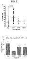
- Example 1 Pre-incubation of NK cells with certain tumour cell lines significantly increases the degree of lysis of NK-resistant cell.
- CTV-1 cells are "activating tumour cells".
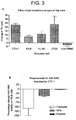
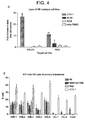
- Pre-incubation with HL-60 ( Figure 2A , 3A and 4A ) or Raji cells ( Figures 3A and 4A ) are less effective or ineffective in activating the NK cells to lyse Raji cells.
- Pre-incubation with allogeneic HLA-KIR mismatched normal PBMC does not induce NK activation ( Figure 4A ).
- the tumour cells express normal levels of MHC class I antigens as do the Daudi and Raji cell lines. Daudi and Raji cells both express HLA-C molecules which ligate both class 1 and class 2 KIRs.
- Pre-incubation with CTV-1 causes an increase in the degree of lysis of various tumour cell-lines, such as Raji, Daudi, JOSK and HL-60 ( Figure 2B ).
- NK activation by CTV-1 cells The effect of fixation and Brefeldin A (BFA) on NK activation by CTV-1 cells is investigated using multiple normal donors.
- induction of NK activation requires contact with the tumour cell line although does not require secretion of a cytokine since fixation of the tumour cells does not abrogate the response.
- the NK cells do need to synthesise a protein in response to the tumour cell ligation as addition of Brefeldin A during the pre-incubation prevents induction of the activated state.
- HLA-KIR matched and mismatched stimulating tumour cell lines is compared on the stimulation of NK cells to lyse Raji cells.
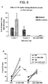
- the stimulating tumour cell lines need not be HLA-KIR mismatched to the donor NK cells although it appears that the threshold for NK activation by the tumour cell line may be lower in the absence of KIR ligation ( Figure 6A ).
- NK cells from normal donors are selected on the basis of their HLA-A, -B and -C type as KIR-ligand matched or mis-matched with CTV-1 cells.
- CTV-1 cells are HLA-C type 2 homozygous and express HLA-Bw4 alleles. They thus ligate KIR2DL1 and KIR3DL1 on NK cells.
- NK:CTV-1 co-cultures are established with NK cells expressing KIR2DL1, expressing KIR2DL1 and KIR3DL1 and with cells expressing only KIR2DL2/3, the ligand for which is missing from CTV-1 stimulator cells. It is thus possible to evaluate the contribution of "missing self" to the NK activation step.
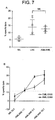
- AMLANK (AML-activated NK cells) are generated by CTV-1 from both HLA-/ KIR matched and mismatched donors and there is no significant difference in the degree of specific lysis although the AMLANK from matched donors show greater heterogeneity ( Fig 6B ). The degree of lysis is equivalent to that obtained by non-specific activation with IL-2 ( Fig 7A ).
- NK cells peripheral blood NK cells
- NK cells from normal donors are co-incubated overnight with CTV-1 and phenotyped for expression of KIR and of CD69. It is readily apparent that CTV-1 induced NK activation is not restricted to KIR mismatched NK cells since cells expressing CD158a and CD158e1 show equivalent levels of activation as NK cells from the same donors which lack CD158a or CD158e1 but express CD158b, the ligand for which is absent from CTV-1 ( fig 7c ).
- NK cells are phenotyped and selected on their KIR compatibility to the HLA of the CTV-1 stimulator cells by flow cytometric sorting.
- Flow sorted NK cell subsets are either incubated directly with CTV-1 cells or are incubated overnight so that the anti-KIR antibody is shed from the NK cell and cannot block KIR:HLA interaction.
- the NK cells expressing CD158a and CD158e1 show equivalent lysis of Raji cells compared to CD158a/e1-ve NK from the same donors ( fig 7d ).
- CTV-1 activated NK-cells have a greatly increased capacity to lyse primary leukemia cells, when compared to NK cells pre-incubated with HLA-KIR matched AML cells or Raji cells ( Figure 4B and C ).
- AMLANK cells from allogeneic donors are capable of lysis of primary AML cells of all FAB types ( Fig 4C ). These cells also lyse primary CLL cells at an effector:target cell ratio of 1:1 although the level of killing is low. It was notable that the relatively NK-resistant breast cancer cell line, MCF-7, is extremely susceptible to AMLANK cells (Example 5) as are pimary tumor cells isolated from ressected tissue from patients with breast cancer and ovarian cancer ( Fig 4D ).
- AMLANK cells from the HLA-identical sibling donor for Patient 0100 effectively lyse cryopreserved CML blasts obtained from the patient at disease presentation. This lysis was apparent at an E:T ratio of 1:1 and was increased at increasing E:T ratios. In contrast, NK cells from the same donor were unable to lyse the CML blasts even at the highest E:T ratio of 10:1. The same was observed using AMLANK cells from an HLA-identical sibling donor for patient 0359 who presented with AML M2 and from whom presentation blasts had been cryopreserved.
- the breast cancer cell line MCF-7 was extremely susceptible to AMLANK lysis at an E:T ratio of 5:1 after a four hour incubation period ( Figure 4C ).
- NK cells are isolated from normal donors and either activated with CTV-1 cells overnight or maintained in media. These AMLANK cells are then compared with matched NK cells with respect to lysis of normal autologous and allogeneic PBMC. Neither NK nor AMLANK cells lyse autologous PBMC nor do they lyse PBMC from HLA-C mismatched normal donors ( Figs 5B ).
- hematopoietic colony forming assays are established with bone marrow from 5 normal donors and added AMLANK from HLA-C mismatched donors at increasing ratios. CFU-GM, BFU-E and CFU-GEMM are not affected by co-incubation with HLA-mismatched AMLANK ( Fig 5C ).
- AMLANK cells are capable of detectable lysis of the presentation leukaemic blasts even at a 1:1 ratio ( Figure 7B ).
- the dashed line (in Figure 7B ) represents the degree of specific lysis of AML blasts which we have previously reported as being associated with continued remission in AML patients after chemotherapy ( Lowdell et al (2002) Br. J. Haematol. 117:821-7 ).
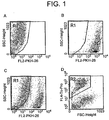
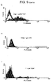
- NK cells Co-incubation of normal donor NK cells with equal numbers of irradiated CTV-1 cells induces rapid and sustained expression of CD69 on the NK cells ( Figure 8A ).
- Purified NK cells are mixed with an equivalent number of irradiated CTV-1 cells which have been labelled with PKH-26. Aliquots are removed at the time points indicated and labelled with anti-CD56 FITC and anti-CD69 APC, washed and analysed by flow cytometry.
- CTV-1 cells are excluded from the analysis on the basis of forward angle light scatter (FSC) and PKH-26 expression and NK cells are positively included on the basis of FSC and CD56 expression.
- Results are presented from 10 normal donors and expressed as the proportion of CD69+ve cells within the CD56+ NK fraction.
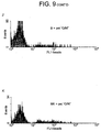
- AMLANK cells co-incubated with Raji cells at a 1:1 E:T ratio show conjugate formation at 60min by confocal microscopy and capping of CD69 at the immune synapse.
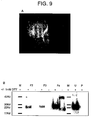
- Fig 9A shows a single AMLANK cell from a normal donor conjugated to a single Raji cell. The conjugate is labelled with anti-CD69 FITC and Dapi as the nuclear stain.
- Recombinant human CD69 is generated as described and the refolded protein supernatant fractionated by HPLC. Fractions F2 and F3 contain monomeric rCD69 when assessed by Western blot under reducing conditions.
- Fraction 4 contains considerably higher concentrations of rCD69 which is detectable as a monomer in the presence of DTT and as both a monomer and a dimer in nonreducing conditions ( Fig 9B ).
- rCD69 is absent from the parental bacterial strain (lane P) and from all other HPLC fractions tested (data not shown).
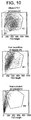
- Flow cytometric analysis of labelled Raji cells shows positive binding only with nano-particles coated with HPLC fractions containing rCD69 (shaded histograms in Figure 9C ). This binding is confirmed by confocal microscopy (D) and flow cytometry (G). In contrast to the malignant B cell line, normal B cells do not bind the beads (E).
- Table 1 Tissue distribution of CD69L expression Cell Line Cell type CD69L expression Normal T cells Negative Normal B cells Negative Normal NK cells Negative RAJI Burkitt's Lymphoma Positive Daudi Burkitt's Lymphoma Positive K562 Erythroleukemia Negative ARH77 Myeloma Positive Jurkat T cell lymphoma Positive MCF-7 Metastatic breast tumor Positive
- CD69 ligand CD69L
- AMLANK-sensitive tumour cells CD69L
- CD69 is critical for tumor-restricted killing by T-ANKs
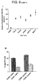
- the CD69+ T-ANK cells are sorted after CTV-1 stimulation from the CD69-ve cells prior to a RAJI lysis assay.
- the CD69+ve fraction mediates 83.7% of the activity of unfractionated T-ANK cells whereas the CD69-ve NK cells show 5.5% ( Fig 9M ).
- the critical role of CD69 in T-ANK triggering is confirmed by the inhibition of RAJI cell lysis in the presence of rCD69.
- Pre-incubation of RAJI cells with rCD69 significantly reduces the degree of RAJI cell lysis almost to the level of lysis by resting NK cells. This effect is not observed when RAJI cells were pre-incubated with BSA or heat denatured rCD69 ( Fig 9N ).
- rCD69 does not block lysis of K562 either by resting NK cells nor by T-ANK cells ( Fig 9O ).
- CD69 is a homodimeric glycoprotein expressed on many haematopoietic cells upon activation.
- human NK cells On human NK cells it has been shown to initiate tumour cell lysis when ligated ( Demanet et al (2004) Blood 103:3122-3130 ) although murine data imply that CD69 ligation is inhibitory to NK-mediated lysis since CD69 KO mice show enhanced anti-tumor activity ( Esplugues et al (2003) J. Exp. Med. 197:1093-1106 ) and monoclonal antibody blockade of CD69 on murine NK cells increases their lytic activity (Esplugues et al (2005) 105:4399-4406).
- tumour cells express the ligand for CD69 which is absent from normal haematopoietic cells.
- blocking experiments with CD69L confirm that CD69 on activated NK cell is the predominant trigger molecule for AMLANK cytotoxicity. This is supported by the evidence that AMLANK:Raji cell conjugation leads to Syk activation within the AMLANK cells (data not shown), a phenomenon known to be associated with CD69-mediated signalling (Pisegna et al (2002) 169:68-74).
- PBMCs peripheral blood mononuclear cells
- Fresh heparinised peripheral blood samples are obtained after informed consent from normal healthy donors, patients with acute and chronic leukemias at diagnosis (Table 1) and from two HLA-identical PBSC sibling donors of patients selected for allogeneic stem cell transplant and who had donated bone marrow samples at time of their disease presentation; the leukemic blasts from which had been cryopreserved in multiple aliquots.
- PBMCs Mononuclear cells
- PBMCs Mononuclear cells
- PBMCs Mononuclear cells
- CD56+ CD3- cells are purified from PBMCs by direct immunomagnetic separation with CD56 Multisort kit (Miltenyi Biotec, Germany) and subsequent depletion with CD3 FITC and anti-FITC beads. All selected cells are confirmed as >98% CD56+ and ⁇ 3% CD3+ and resuspended in CM.
- NK cells Freshly isolated NK cells are suspended in CM at a concentration of 10 6 /ml and incubated with an equal number of irradiated (30Gy) tumor cells for 20 hours at 37°C/5%CO 2 .
- Stimulator tumor cells are restricted to the well characterised myeloid leukemia cell lines, U937, HL-60 and CTV-1 which are obtained from the DTMZ repository.
- Target cells in cytotoxicity assays include the NK-resistant RAJI cell line (obtained from the DTMZ cell bank), the breast cancer cell line MCF-7 (obtained from ATCC) and primary leukaemia cells from patients attending the Royal Free Hospital.
- Each myeloid leukemia line and the target cells are subjected to HLA typing as described above.
- Target cells are recovered from culture or cryopreservation and washed in HBSS before resuspension in 1.0 ml of PHK-26 labelling diluent at a concentration of 4 x 10 6 /ml.
- a 4 ⁇ l aliquot of PKH-26 is added to 1.0 ml of labelling diluent and then added to the cell suspension for 2 min at room temperature.
- the labelling reaction is stopped by the addition of 1.0 ml neat fetal calf serum for 1 min.
- the labelled cells are washed twice in CM and resuspended in CM at 10 6 /ml.
- 50 000 PKH-26 labelled target cells in 100 ⁇ L RPMI 1640 (10% FCS) are added to 400 ⁇ L of effector cells and pelleted at 200g for 1 min.
- Cytotoxicity is measured in triplicate samples using a 4-hr cytotoxicity assay at 37 °C. After the incubation period the cells are resuspended in a solution of To-Pro-3 iodide (Molecular Probes, Oregon, USA) in PBS (1 ⁇ M) and analysed by flow cytometry. At least 10 000 target cells are acquired with 1024 channel resolution after electronic gating on red fluorescence and the mean proportion of To-Pro iodide positive cells from the triplicate samples determined. Background target cell death is determined from cells incubated in the absence of effector cells. Cell-mediated cytotoxicity is reported as percentage killing over background cell death averaged from the three samples: Mean % cell lysis in test - % spontaneous lysis
- PKH-labelled RAJI and K562 target cells are pre-incubated with rCD69 or control reagent (6 ⁇ g per 10 5 cells) at 4°C for 30 minutes prior to set-up of the T-ANK cytotoxicity assay described above.
- the extracellular domain of CD69 (residues 65-199) is amplified from cDNA by polymerase chain reaction using primers introducing Xhol and HindIII restriction sites and a stop codon (CD69 For 5' GCG CCT CGA GCA ATA CAA TTG TCC AGG CCA AT 3'; CD69Rev 5' CGC GAA GCT TAT TAT TTG TAA GGT TTG TTA CA 3').
- the PCR product is subcloned into XhoI-HindIII restriction sites of pET-19b plasmid (Novagen) using standard techniques, to construct pET-19b/69.
- DNA sequence that encodes the amino acid acceptor sequence for the E.coli BirA biotin protein ligase is added between the Ndel and XhoI sites of pET-19b/69 with the following primers 5' CAT ATG CAT GCG GGC GGC CTG AAT GAA ATT CTG GAT GGC ATG AAA ATG CTG TAT CAT GAA CTC GAG 3' and 5' CTC GAG TTC ATG ATA CAG CAT TTT CAT GCC ATC CAG AAT TTC ATT CAG GCC GCC CGC ATG CAT ATG 3'. DNA sequence is confirmed by automated sequencing using an ABI Prism 377 DNA sequencer.
- Recombinant His-tagged human CD69 is expressed in BL21 (DE3)pLysS (Novagen) at 37°C. Cultures are grown in 1 liter batches in 2xTY medium containing 100 ⁇ g/ml ampicillin and 34 ⁇ gml chloramphenicol. CD69 expression is induced by addition of 1mM isopropyl-D-thiogalactopyranoside (IPTG) after the culture had reached an OD 600 ⁇ 0.6. Cells are allowed to grow for a further 4-5 hours and then harvested by centrifugation at 5000g for 20 minutes at 4°C. Cell pellets are stored at -80°C.
- IPTG isopropyl-D-thiogalactopyranoside
- Cell pellets from 250 ml culture are resuspended in 15ml ice cold Resuspension Buffer (20mM Tris-HCl pH 8.0). Cells are disrupted by multiple passages through a 16 gauge needle before centrifugation at 12000g for 15 minutes at 4°C. The pellet is washed in Isolation buffer (20mM Tris-HCl pH8.0, 500mM NaCl, 2% Trition-X100, 2 M Urea) before being centrifuged again. This process is repeated once more. Pellets are finally washed in Resuspension buffer before being stored at -80°C.
- pellets Prior to purification and refolding, pellets are resuspended in Solubilization buffer (6M guanidinium hydrochloride, 20mM Tris-HCl pH8.0, 500mM NaCl, 10mM imidazole) and passed through a 0.45 ⁇ m filter and then loaded onto a 5ml Nickel loaded HiTrap Chelating column (GE Life science, Amersham UK) pre-equilibrated with Refolding buffer (20mM Tris-HCl pH8.0, 500mM NaCl, 6 M Urea, 10mM imidazole).
- Solubilization buffer 6M guanidinium hydrochloride, 20mM Tris-HCl pH8.0, 500mM NaCl, 10mM imidazole
- the protein is refolded by gradual removal of the urea through a linear gradient expanding from 100% Refolding buffer to 100% Wash buffer (20mM Tris-HCl pH8.0, 500mM NaCl, 10mM imidazole). This is achieved with 250ml buffer at 5ml/minute using a HPLC system (Varian Technologies). After refolding, the protein is eluted with Elution buffer (20mM Tris-HCl pH8.0, 500mM NaCl, 500mM imidazole).
- Fractions are buffer exchanged into 10mM Tris-HCl pH 8.0 using PD10 columns (GE Life science, Amersham, UK) and incubated with 2.5 ⁇ g BirA enzyme (Avidity Denver, USA) per 10nmol substrate at 30°C overnight following the manufacturers instructions. Excess biotin is removed and protein concentrated by washing with 50mls HBSS in 10000 dalton MW cut-off centrifuge tubes (Vivascience UK) and assessed for rCD69 content by ELISA.
- Biotinylated rCD69 is conjugated to avidin coated yellow fluorescent beads (Spherotech Inc.) by rotating incubation at 4°C for 40 minutes as previously described ( Brown et al (1988) J. Exp. Med. 188: 2083-2090 ). Protein bead conjugates are briefly sonicated to prevent aggregation and incubated with 10 5 target cells on ice for 60 minutes. Bound cells are washed with HBSS. Flow cytometric acquisition is performed at a maximum of 40 events per second in order to prevent acquisition of coincident events. Binding of 5 ⁇ g heat denatured rCD69 is used as a negative control for each experiment.
- Example 9 Use of activated NK cells to treat poor prognosis AML patients.
- NK products infused in the Miller study were not pure (approximately 40% NK) and were contaminated with T and B cells.
- NK cells are selected from haploidentical related normal donors by direct immunomagnetic separation (CliniMACS), greater than 95% pure CD56+ cells is achieved.
- the degree of contaminating NKT cells is donor-dependent but is unlikely to exceed the dose of T cells infused in the Miller study. In the eventuality of a high NKT contamination, the NK dose infused may be reduced to ensure the T cell dose does not exceed that given by Miller.
- Alloreactive NK cells can be identified by CD69 expression in a MLR and such cells are phenotyped for KIR expression pre-and post culture prior to infusion.
- the present inventors have also established a skin explant model for graft-versus-host disease prediction. This in vitro assay may be used for quality assurance of NK cell infusions.
- Leukaemic blasts are cryopreserved as viable cells from AML patients.
- donor NK products are tested in vitro for lytic activity against patient AML blasts and the results correlated with clinical response to treatment.
- NK cells are isolated by immunomagnetic selection using anti-CD56 microbeads (Miltenyi Biotec) and the CliniMACS device and incubated overnight with equal numbers of irradiated CTV-1 myeloid leukaemia cells to provide a tumour-specific stimulus.
- NK/kg are prepared for the first 5 patients, 1x10 7 /kg for the next five patients and 2x10 7 /kg are prepared for the final group of five patients. Aliquots of donor NK cells are retained for testing as described above. Patients receive their donor NK cells as a single i.v infusion on day 6. They are monitored for clinical GvHD and cell specific chimerism studies are performed daily for the first 7 days; weekly for the next three months and monthly thereafter until 12 months.
- T-aNK Production schedule for Clinical Tumour-activated NK cells
- Consenting donors undergo a single two-hour apheresis to harvest 25x10 9 mononuclear cells into ACD anti-coagulant.
- the apheresis collection bag is labelled with the donor name, donor hospital number, donor date of birth, recipient name, recipient hospital number, date and time of apheresis and volume of product.
- the apheresate is collected from the unit by a member of the LCT staff and transported directly in an approved container to the LCT.
- the apheresate On acceptance by the LCT, the apheresate is booked-in to the LCT product database and assigned a unique product number.
- the database reproduces all of the details on the product bag and additionally records the recipient date of birth, recipient body mass and the unique trial number assigned to the patient upon trial entry and consent.
- apheresate is reduced to a pure mononuclear cell fraction by density gradient separation.
- a 1ml sample is removed to obtain a mononuclear cell count and a CD56+ cell enumeration by flow cytometry.
- the volume of mononuclear cell fraction required to provide 2x10 7 NK/kg patient body mass is recovered into a 250ml sterile bag, washed by centrifugation and resuspended at 5x10 6 /ml in RPMI 1640 media, supplemented with 10% foetal calf serum (batches approved for pharmaceutical use) - all media supplied by Gibco Ltd, Paisely).
- CTV-1 cells (supplied direct from the DSMZ tissue bank, Braunschweig, Germany - master cell bank record attached) are maintained in continuous exponential growth at a conc. of 0.5-1x10 6 /ml in RPMI 1640 medium / 10% FCS, in a closed culture system (Lifecell bags, Baxter Healthcare) in the Paul O'Gorman Laboratory of Cellular Therapeutics, RFUCMS (MHRA Accredited Tissue Bank 0029/00/00/0-03). Production records are maintained for all batches which include the batch numbers of all reagents and disposables used and the initials of all staff who performed individual procedures and the dates of those procedures. The serial numbers of all equipment used in the production process are also recorded.
- the membrane preparations are resuspended in 4mls sterile saline (infusion grade) and autoclaved to 121°C for 5mins. After cooling to 21°C the bags are placed in a sonicaton bath for 25 seconds to disrupt aggregates which can form during autoclaving.
- Formation of cell membranes is monitored by taking samples at various stages of the procedure and comparing them with forward angle light scatter (FSC) and 90° light scatter (SSC) of whole CTV-1 cells by flow cytometry.
- FSC forward angle light scatter
- SSC 90° light scatter
- the membrane preps are batch-tested for sterility following routine SOP and stored at a total protein concentration of 5mg/ml in sterile saline (for injection) at -80°C in a controlled freezer labelled: "CTV-1 Membrane Preparation - Clinical Grade. Store below -40°C" together with the batch number, date of production and expiry date (6 months from date of manufacture).
- Lifecell culture bags containing selected donor mononuclear cells at 5x10 6 /ml are supplemented with thawed CTV-1 membrane preparation to a final concentration of 5mg per 10 7 donor NK cells.
- Sufficient mononuclear cells are cultured to provide a maximum cell dose of 10 7 NK/kg patient body mass. Cell cultures are maintained for a minimum of 16 hours and a maximum of 26 hours at 37°C/ 5% CO 2 in a Hepa filtered monitored incubator within the LCT.
- CD56+ NK cells is isolated by immunomagnetic selection by CliniMACS (Miltenyi Biotec GmbH) to remove CD56-ve cells and residual CTV1 membrane preparation.
- CD56+ cells are recovered and suspended in RPMI1640 at 10 8 /ml. Cells are cryopreserved following routine SOP in a single aliquot at the dose required. Aliquots are removed for quality assurance testing prior to cryopreservation:
- CD3+/CD56- T cell contamination (below 10 5 /kg patient body mass)
- Anaerobic/aerobic bacterial culture ("negative" prior to release of product)
- Detectable TaNK activity against NK-resistant cell line Raji in a four hour cytotoxicity assay (determined by >25% increase in Raji lysis compared to matched NK cells from same donor).
Landscapes
- Health & Medical Sciences (AREA)
- Life Sciences & Earth Sciences (AREA)
- Chemical & Material Sciences (AREA)
- Engineering & Computer Science (AREA)
- Immunology (AREA)
- General Health & Medical Sciences (AREA)
- Cell Biology (AREA)
- Biomedical Technology (AREA)
- Medicinal Chemistry (AREA)
- Animal Behavior & Ethology (AREA)
- Public Health (AREA)
- Veterinary Medicine (AREA)
- Pharmacology & Pharmacy (AREA)
- Organic Chemistry (AREA)
- Biotechnology (AREA)
- Zoology (AREA)
- Bioinformatics & Cheminformatics (AREA)
- Microbiology (AREA)
- Epidemiology (AREA)
- Hematology (AREA)
- Wood Science & Technology (AREA)
- Genetics & Genomics (AREA)
- Oncology (AREA)
- Mycology (AREA)
- General Chemical & Material Sciences (AREA)
- Nuclear Medicine, Radiotherapy & Molecular Imaging (AREA)
- Chemical Kinetics & Catalysis (AREA)
- Biochemistry (AREA)
- General Engineering & Computer Science (AREA)
- Virology (AREA)
- Developmental Biology & Embryology (AREA)
- Medicines Containing Material From Animals Or Micro-Organisms (AREA)
- Measuring Or Testing Involving Enzymes Or Micro-Organisms (AREA)
- Micro-Organisms Or Cultivation Processes Thereof (AREA)
- Investigating Or Analysing Biological Materials (AREA)
Description
- The present invention relates to a method for activating a Natural Killer (NK) cell. In particular, it relates to a method for activating an NK cell such that it has the capacity to lyse an NK-resistant cancer cell.
- A number of cancers are, at present, incurable. For others, chemotherapy is only partially effective and a significant proportion of patients relapse following treatment Some haematological malignancies are treatable by hematopoietic stem cell transplantation (HSCT), but fewer than 30% of patients requiring HSCT have a suitable donor and are the requisite age.
- Natural Killer (NK) cells are a subset of peripheral blood lymphocytes which can spontaneously lyse certain tumour cells. The use of NK cells in adoptive tumour immunotherapy has been proposed, and there has been interest in the in vitro or ex vivo stimulation of NK cells to increase their capacity to lyse tumour cells.
- The discovery of interleukin-2 (IL-2) and its role in NK-cell activation in the 1980's led to considerable interest in the use of lymphokine-activated killer (LAK) cells in tumour immunotherapy. The results of these trials were, however, largely disappointing. In a study investigating the effect of administering autologous LAK cells to patients along with IL-2, less than 20% of patients responded (Rosenburg et al (1987) N. Engl. J. Med. 316: 889-897). In studies using daily IL-2 administrations to cancer patients along with chemotherapy and autologous HCT, it was shown that, although IL-2 significantly expanded the number of circulating MK cells in vivo, the cells are not maximally cytotoxic according to an in vitro assay (Miller et al (1997) Biol. Blood Marrow Transplant. 3: 34-44).
- NK cells are now known to be controlled by both positive and negative cytolytic signals. A number of molecules which mediate NK cell inhibition have been cloned over the past ten years and their ligands are almost exclusively Class I MHC molecules. Some of these receptors ("KIRs") are specific for determinants shared by certain class I alleles, and each KIR is expressed by a subset of NK cells. Therefore, in the NK repertoire, some NK cells recognise, and are blocked by, specific class I alleles. NK cells have a limited view of class I polymorphism but cells can be responsible for alloreactions when the mismatched target cells do not express the class I alleles which block every NK cell in the repertoire (the "missing - self" hypothesis). Thus allogeneic target cells which lack at least one of the class I allele groups expressed by the donor cells will not find the inhibitory class I ligand on a subset of donor NK cells and their lytic pathway will be activated.
- It has thus been suggested that autologous NK cells may be suppressed by the physiologic response resulting from NK cell recognition of "self" MHC molecules. It has also been suggested that the greater the degree of KIR mismatch with tumour MHC (i.e. KIR ligand) the greater tumour kill (Ruggeri et al (2002) Science 295:2097-2100). In view of the shortcomings of autologous NK cell therapy (thought to be due to a lack of NK cell inhibitory receptor mismatching with autologous tumour cells) the use of allogeneic NK cell infusions has been suggested as an alternative (Miller et al (2005) Blood in press, but pre-published on-line on January 4 2005).
- Miller et al (2005, as above) administered IL-2 activated allogeneic haploidentical NK cells to patients with metastatic melanoma, metastatic renal cell carcinoma, refractory Hodgkin's disease or poor prognosis AML. Importantly, their results demonstrate that the NK cells can persist and expand in vivo. The cells induced complete hematologic remission in five of the 19 poor prognosis AML patients, but no activity against the other tumours. In the group which achieved remission, patients are stratified into those with predicted graft versus host alloreactivity using the KIR ligand mismatch strategy. The results showed remission was much more likely in those patients which are KIR ligand mismatched.
-
US 6,849,452 relates to a process for activating NK cells by bringing them into contact with dendritic cells. - Non-specifically activated NK cells may therefore have an application against a subset of tumours, but the donor cells must be allogeneic and much more likely to be effective if they are HLA mismatched. A disadvantage associated with using mismatched NK cells target normal hematopoeitic cells is that they may target and reject normal (e.g.host) hematopoeitic cells (Yu et al (1996) Immunity 4:67-76).
- There is thus a need for alternative immunotherapy for cancer which is effective against "NK resistant" tumours but which spares normal hematopoietic cells.
- The current dogma is that NK cells are stimulated by the target cell which they ultimately lyse and that "NK-resistant" tumours are not lysed because they fail to provide this stimulus.
- Contrary to current thinking, the present inventors have shown that the "stimulation event" can be temporally separated from the "lytic event". They have also shown that some tumour cells, or membrane preparations thereof, are capable of stimulating NK cells such that they can then go on to lyse a target cell which is resistant to lysis by an equivalent unstimulated NK cell.
- The present invention provides a method for activating a human NK cell, which comprises the step of contacting the NK cell in vitro with an activating tumour cell preparation (ATCP), which comprises intact human tumour cells, or a human tumour cell membrane preparation.
- The NK cell is activated to lyse a target cell. For example, the NK cell may be activated such that it is capable of lysing a cell previously resistant to NK-cell lysis. The method of the present invention therefore may be used to provide NK cells useful in the treatment of a number of "NK-resistant" malignancies, many of which are incurable at present (such as myeloma and Chronic lymphocytic leukemia (CLL)).
- The fact that the "stimulation event" can be separated from the "lytic event" has the advantage that the NK cell can be stimulated in vitro, but once stimulated retains the capacity to lyse a target cell until it encounters the target cell when introduced or returned to the subject.
- This form of activation renders the NK cell capable of lysing multiple tumour types which are regarded as resistant to NK cell killing. Moreover the activated NK cells are effective irrespective of the degree of HLA matching between the NK and tumour cells. This opens up the possibility of using autologous or HLA-matched allogeneic donor NK cells. The use of autologous or HLA-matched cells has the advantage that it is less likely to result in rejection of donor or host (eg host normal haematopoietic) cells.
- Moreover, since the method of the invention does not rely on IL-2 mediated activation, it avoids the adverse effects in vivo associated with the IL-2 response (Miller et al (2005) as above).
- In the method of the invention, the ATCP may be a preparation of, or comprise intact human tumour cells. The cells may be irradiated or fixed.
- Alternatively the ATCP may be or comprise a human tumour cell membrane preparation. Use of cell membranes is advantageous as it bypasses many safety concerns associated with the use of tumour cells.
- The ATCP may comprise tumour cells or preparations thereof with NK activating ability, such as CTV-1 myeloid leukemia cells and/or a membrane preparation thereof.
- Activated NK cells produced by the method of the invention may be used to treat cancer.
- The approach is particularly suitable in cases where the subject is unsuited to intensive cancer treatment.
- The cancer may, for example be: Acute myeloid leukaemia (AML); Chronic lymphocytic leukemia (CLL); Lymphoma; or Breast cancer.
-
-
FIGURE 1 is a flow cytometric dot-plot showing the analysis strategy for measurement of target cell lysis.
Analysis Region 1 (R1) is established to include target cells (PKH-26-ve) (A - target cells alone) which excludes effector NK cells and stimulator AML cells pre-labeled with PKH-26 (B - effector + stimulator cells alone). This gating strategy effectively discriminates the target and effector cell populations in the admixture (C). Gating on the target cells within the admixture (R1) allows enumeration of the FSClow/ To-Pro+ "dead" targets (R2) from the To-Pro-ve live target cells (D). -
Figure 2A is a graph showing % lysis of Raji cells when various different cell types are used as stimulator cells for NK cells. -
Figure 2B is a chart showing the effect of pre-incubation of NK cells with CTV-1 cell on % lysis various different cell types. -
Figure 3A is a chart showing % lysis of Raji cells when various different cell types are used as stimulator cells for NK cells. -
Figure 3B is a chart showing the effect of fixation and Brefeldin A (BFA) treatment on the capacity of CTV-1 cells to activate NK cells. -
Figure 4A is a chart showing % lysis of Raji cells and Daudi cells when various different cell types are used as stimulator cells for NK cells. -
Figure 4B is a chart showing % lysis of primary leukemia cells when various different cell types are used as stimulator cells for NK cells. -
Figure 4C is a graph showing % lysis of primary leukaemic blasts (AML and CLL) and a breast cancer cell line (MCF-7) by activated NK cells. -
Figure 4D is a graph showing % lysis of primary tumor cells from patients with breast or ovarian cancer by T-aNK cells from allogeneic normal donors. -
Figure 5A is a chart showing the effect of stimulated NK cells on KIR-matched (autologous) or KIR mismatched (haplo 1 or haplo 2) normal PBMC. -
Figure 5B is a graph to show lysis by activated NK cells from normal donors on normal PBMC from the same donor (autologous) and KIR-mismatched normal donors (allogeneic). -
Figure 5C is a chart showing the effect of activated NK cells on in vitro haemotopoiesis of normal donor BMMC -
Figure 6A is a chart comparing the effect of using HLA-KIR mismatched versus HLA-KIR matched activating tumour cell lines -
Figure 6B is a graph showing the % lysis of Raji targets by NK cells activated by either KIR-ligand matched and mismatched CTV-1 cells. -
Figure 7A is a comparing lysis by ATCP-activated and IL-2 stimulated NK cells. -
Figure 7B is a graph showing % lysis of leukaemic blasts at different E:T ratios. -
Figures 7C and 7D are graphs showing the effect of KIR mismatching on CTV-1 induced NK activation (C) CD56+/CD3- NK cells are incubated overnight in medium alone (open bars), with irradiated CTV-1 cells (shaded bars), or with CTV-1 cell lysate (black bars) and phenotyped for expression of KIRs and CD69 (bars show mean +/- sd). (D) Resting NK cells are flow sorted from 3 normal donors into two populations of those co-expressing CD158a and CD158e1 and those lacking both molecules. These are stimulated overnight with CTV-1 cell lysates and their cytolytic activity tested against RAJI cells in a 4-hour assay (bars show mean +/- sd). -
Figure 8A is a graph showing the proportion of CD69+ve cells within the CD56+ NK fraction of CTV-1-activated NK cells. -
Figure 8B is a chart showing the effect of incubation with CTV-1 on the expression of a panel of ligands by NK cells. -
Figure 9A is a confocal microsopy photograph showing conjugate formation between activated NK cells and Raji cells and capping of CD69 at the immune synapse. -
Figure 9B is a gel photograph showing HPLC fractionation of recombinant human CD69 re-folded protein supernatant. -
Figure 9C is a panel of histograms showing flow cytometric analysis of labelled Raji cells after contact with nano-particles coated with HPLC fractions from rCD69 supernatant. -
Figure 9D is a confocal microscopy photograph of labelled Raji cells after contact with nano-particles coated with an rCD69-positive HPLC fraction. -
Figure 9E is a confocal microscopy photograph of labelled normal B cells after contact with nano-particles coated with an rCD69-positive HPLC fraction. -
Figure 9F is a graph showing % lysis of Raji cells following pre-incubation with 260µg-1 mg of rCD69 HPLC fractions (in the absence of nano-particles). -
Figure 9G is a graph to show flow cytometric analysis of Raji cells after labelling with nanoparticles coated with rCD69 (shaded histogram) or denatured rCD69 (open histogram). -
Figure 9H is a graph to show flow cytometric analysis of K562 cells after labelling with nanoparticles coated with rCD69 (shaded histogram) or denatured rCD69 (open histogram). -
Figure 9I is a graph to show flow cytometric analysis of normal T cells cells after labelling with nanoparticles coated with rCD69 (shaded histogram) or denatured rCD69 (open histogram). -
Figure 9J is a graph to show flow cytometric analysis of normal B cells after labelling with nanoparticles coated with rCD69 (shaded histogram) or denatured rCD69 (open histogram). -
Figure 9K is a graph to show flow cytometric analysis of normal NK cells after labelling with nanoparticles coated with rCD69 (shaded histogram) or denatured rCD69 (open histogram). -
Figure 9L is a graph to show the relative fluorescence intensity observed with different cell lines. -
Figure 9M is a chart to show T-aNK mediated lysis of RAJI cells in the presence of rCD69 at two concentrations -
Figure 9N is a chart to compare T-aNK mediated RAJI cell lysis in the presence of rCD69, denatured rCD69 or BSA. -
Figure 9O is a chart to show the effect of rCD69 on lysis of K562 by resting NK cells or T-aNK cells -
Figure 10 is a scatter diagram showing an example of formation of cell membranes from CTV-1 cell line - The present invention relates to a method for activating a NK cell.
- NK cells are a subset of peripheral blood lymphocytes defined by the expression of CD56 or CD16 and the absence of the T cell receptor (CD3). The recognise and kill transformed cell lines without priming in an MHC-unrestricted fashion.
- NK cells represent the predominant lymphoid cell in the peripheral blood for many months after allogeneic or autologous stem cell transplant and they have a primary role in immunity to pathogens during this period (Reittie et al (1989) Blood 73: 1351-1358; Lowdell et al (1998) Bone Marrow Transplant 21: 679-686). The role of NK cells in engraftment, graft-versus-host disease, anti-leukemia actiovity and post-transplant infection is reviewed in Lowdell (2003) Transfusion Medicine 13:399-404.
- Human NK cells mediate the lysis of tumour cells and virus-infected cells via natural cytotoxicity and antibody-dependent cellular cytotoxicity (ADCC).
- Human NK are controlled by positive and negative cytolytic signals. Negative (inhibitory) signals are transduced by C-lectin domain containing receptors CD94/NKG2A and by some Killer Immunoglobulin-like Receptors (KIRs). The regulation of NK lysis by inhibitory signals is known as the "missing self" hypothesis in which specific HLA-class I alleles expressed on the target cell surface ligate inhibitory receptors on NK cells. The down-regulation of HLA molecules on tumor cells and some virally infected cells (e.g. CMV) lowers this inhibition below a target threshold and the target cells becomes susceptible to NK cell-mediated lysis.
- Inhibitory receptors fall into two groups, those of the Ig-superfamily called Killer Immunoglobulin-like Receptors (KIRs) and those of the lectin family, the NKG2, which form dimers with CD94 at the cell surface. KIRs have a 2- or 3-domain extracellular structure and bind to HLA-A, -B or -C. The NKG2/CD94 complexes ligate HLA-E.
- Inhibitory KIRs have up to 4 intracellular domains which contain ITIMs and the best characterized are KIR2DL1, KIR2DL2 and KIR2DL3 which are known to bind HLA-C molecules. KIR2DL2 and KIR2DL3 bind the
group 1 HLA-C alleles whilst KIR2DL1 binds togroup 2 alleles. Certain leukemia/lymphoma cells express bothgroup - As regards positive activating signals, ADCC is thought to be mediated via CD16, the and a number triggering receptors responsible for natural cytotoxicity have been identified, including CD2, CD38, CD69, NKRP-1, CD40, B7-2, NK-TR, NKp46, NKp30 and NKp44. In addition, several KIR molecules with short intracytoplasmic tails are also stimulatory. These KIRs (KIR2DS1, KIR2DS2 and KIR2DS4) are known to bind to HLA-C; their extracellular domains being identical to their related inhibitory KIRs. The activatory KIRs lack the ITIMs and instead associate with DAP12 leading to NK cell activation. The mechanism of control of expression of inhibitory versus activatory KIRs remains unknown.
- The NK cells made by the method of the present invention may be autologous or allogeneic NK cell.
- "Autologous" NK cells are cells derived from the patient. "Allogeneic" NK cells are derived from another individual, having non-identical gene at one or more loci. If the NK cells are derived from an identical twin, they may be termed "syngeneic".
- Donor NK cells may be HLA-KIR matched or mismatched. The present inventors have shown that the degree of matching between the NK cells and target tumour cells is of no significance.
- The term "activating" is used synonymously with the term "stimulating" in this section, and throughout the document.
- The present inventors have found that certain tumour cells have the capacity to stimulate NK cells to increase their capacity to lyse tumour cells. Stimulated NK cells have been shown to be capable of lysing "NK-resistant" tumour cell (i.e. tumour cells resistant to lysis with unstimulated NK cells).
- Tumour cells capable of activating NK cells in this manner include CTV-1 cells. This cell line is commercially available, for example from the American Typed Cell Collection (ATCC).
- It is expected that other tumour cells will also have the capacity to activate NK cells. In order to determine whether a tumour cell preparation is an activating tumour cell preparation, one can use a method having the following steps:
- (i) contacting the tumour cell preparation with a NK cell;
- (ii) contacting the NK cell from step (i) with a target cell resistant to lysis by non-activated NK cells;
- (iii) determining whether the target cell is lysed by the NK cell from step (i).
- It is thus possible for a skilled person to establish whether a given tumour cell has the capacity to act as an activating tumour cell preparation and to screen known tumour cells for this activity.
- The present inventors have shown that pre-incubation of NK cells with an ATCP (such as CTV-1 AML blasts) causes rapid upregulation of CD69 on the NK cells. They have also shown (using labelled CD69) that tumour cells which are lysable by activated NK cells express CD69 ligand (CD69L), but this expression is absent from cells which are not lysed (such as B cells). The presence of recombinant CD69 inhibits the capacity of activated NK cells to lyse tumour cells, presumably because it blocks interaction with CD69L on the tumour cells.
- Without wishing to be bound by theory, the present inventors believe that CD69 on stimulated NK cells is the predominant trigger molecule for their cytotoxic activity.
- In a preferred embodiment the ATCP used in the method of the present invention causes upregulation of expression of CD69 on the NK cell.
- Although the nature of CD69 ligand(s) is, at present, unknown, it is possible to determine its expression on a candidate tumour target cells by standard techniques. For example, using CD69 labelled with a fluorochrome, it is possible to determine expression of CD69L by techniques such as flow cytometry or confocal microscopy.
- It is possible to determine whether a tumour cell preparation is an activating tumour cell preparation, by using a method having the following steps:
- (i) contacting the tumour cell preparation with a NK cell;
- (ii) determining whether the TCP causes upregulation of CD69 on the NK cell.
- The method may involve determining whether the TCP comprises or expresses CD69L.
- The ATCP may consist of or comprise a population of intact human tumour cells. For example, the activating tumour cell preparation may be a tumour cell line.
- The ATCP may consist of or comprise a human tumour cell membrane preparation. For example, a cell membrane preparation may be made by standard fixation techniques (such as using paraformaldehyde). Fixation has the advantage that the preparation is stabilised, has a much longer "shelf-life" and is easier to store. A suitable cell membrane preparation may also be made by repeated cycles of freeze-thawing, in combination with DNAse treatment. Such a preparation may be considered to have increased safety as it reduces the likelihood of contamination associated with prions etc.
- The stimulator cells may be irradiated prior to use, by standard techniques.
- Membrane preparations have the advantage over preparations comprising intact tumour cells as they avoid the risk of transferring potentially malignant tumour cells to the patient.
- The ATCP and the NK cell preparation may be brought together by, for example, co-culturing (where intact tumour cells are used). The "activation time" will depend on the nature of the cell preparations and the contact conditions, but may commonly be 12-24 hours, perhaps 20 hours.
- The NK cells activated by the method of the invention may be autologous and/or allogeneic NK cells.
- Allogeneic NK cells may be HLA mismatched.
- Allogenic NK may be obtained from peripheral blood from a donor individual. Allogeneic peripheral blood mononuclear cells may be collected by standard techniques (e.g. conventional apheresis). To minimize the possibility of graft versus host disease and immune mediated aplasia, allogeneic cells may be depleted of T cells. For example, the cell preparation may be depleted of CD3+ T-cells using microbeads conjugated with monoclonal mouse anti-human CD3 antibody and a cell selection device (such as the Miltenyi Biotec CliniMACS® cell selection device). However, NK cells produced by such "negative selection" procedures alone do not have a high degree of purity and may be contaminated with T and B cells.
- In order to reduce contamination, it is possible to obtain an NK cell preparation by direct immunomagnetic separation, for example on the basis of CD56 expression. To further reduce T cell contamination, the product may be depleted for CD3+ cells (for example using CD3 FITC and anti-FITC beads).
- Prior to activation by the activating tumour cell preparation, the NK cell preparation may comprise at least 80%, at least 90%, at least 95% or at least 98% CD56+ cells.
- Prior to activation by the activating tumour cell preparation, the NK cell preparation may comprise less than 15%, less than 10%, less than 5% or less than 3% CD3+ cells.
- A composition comprising NK cells activated by the method of the invention may also comprise all or a portion of the activating tumour cell preparation (i.e. activating tumour cells and/or a membrane preparation thereof) or a product thereof.
- The ATCP-mediated activation may be the only activation the NK cells receive, or there may be further activation steps. The NK cells may or may not also be non-specifically activated by IL-2 (for example by incubation of the cells in medium supplemented with IL-2). Alternatively, the cells may be activated in the absence of IL-2, but IL-2 may be used for the ex vivo expansion of stimulated cells.
- A composition comprising NK cells activated by the method of the invention may be used to treat or prevent cancer in a subject.
- The composition may be administered to the subject by any suitable method known in the art, for example, intravenous infusion.
- The composition may be used to treat a subject in need of same. The procedure is low-risk and particularly suitable for cancer patients for whom intensive cancer treatments are precluded (for example, elderly patients). It also provides an alternative for patients (with, for example, lymphoma, myeloma or AML) who lack a suitable donor for allogeneic stem cell transplantation.
- Prior to treatment with the composition, the patient may receive some pre-treatment, for example, to de-bulk the tumour and /or immunosuppress the patient. This may be achieved, for example, by chemotherapy.
- It is possible to obtain primary tumour cells from patients at time of diagnosis and to cryopreserve these as viable single cell suspensions. It is thus possible for a composition comprising activated NK cells to be tested in vitro against patient blasts. This could be done before embarking on a treatment regime, to gauge the suitability of the approach. The correlation of the results of the in vitro study and the corresponding clinical response to treatment may also be investigated.
- A composition comprising NK cells activated by the method of the present invention may be used to treat or prevent a disease or medical condition.
- The disease may be a cancer. There are about 200 different types of cancer. List of types of cancer are available (for example, see http://www.acor.org/types.html or http://www.cancerresearch uk.org).
- Some more common cancers include leukaumia (acute and chronic), bladder cancer, bone cancer (osteosarcoma), Bowel (colorectal cancer), brain cancer, breast cancer, cervical cancer, oesophageal cancer, Hodgkin's lymphoma, kidney cancer, liver cancer, lung cancer, mesothelioma, multiple myeloma, non-Hodgkin's lymphoma, ovarian cancer, pancreatic cancer, penile cancer, prostate cancer, skin cancer (melanoma and non-melanoma) soft tissue carcinoma, gastric cancer, testicular cancer, thyroid cancer and endometrial cancer.
- The composition may be useful to treat any cancer which is accessible to NK cells.
- In particular the cancer may be a haematological malignancy, such as leukaemia (AML); Chronic lymphocytic leukemia (CLL); Lymphoma.
- Myeloma is an incurable and fatal malignancy. NK activity against myeloma plasma cells is documented in vitro and enhanced NK activity against autologous myeloma cells has been shown to correlate with response to treatment with Thalidomide derivatives. Myeloma patients are generally young and fit enough to undergo autologous haematopoietic stem cell transplantation and could readily undergo a less invasive procedure.
- Post transplant lymphoproliferative disease (PTLD) is a serious and relatively common complication after solid organ transplantation and T cell immunotherapy is currently under trial but with little success. Therapy using NK cells activated according to the present invention therapy would be easy and safe in this group of patients.
- In addition the composition may be used to treat solid tumours such as breast cancer.
- The procedure is particularly suitable to treat "NK-resistant" tumours. Normal, non ATCP-stimulated NK cells can spontaeously lyse some human tumours, but many other tumours are NK-resistant. "NK-resistant" as used herein, therefore, indicates tumour cells resistant to lysis by normal, non ATCP-stimulated NK cells.
- As explained above, inhibition of NK-mediated lysis is controlled by expression of specific MHC class I molecules on the target cell surface, particularly HLA-C. There are two distinct groups of HLA-C alleles with regard to NK cell recognition. Some tumours express both types of HLA-C allele, which is thought to make them resistant to NK-mediated lysis. "NK resistant" cells may, therefore express both groups of class I allele. Some leukemia/lymphoma-derived cell lines, such as Raji and Daudi express both types of HLA-C allele, making them useful models for NK-resistant tumour cells in vivo.
- The invention will now be further described by way of Examples, which are meant to serve to assist one of ordinary skill in the art in carrying out the invention and are not intended in any way to limit the scope of the invention.
- Pre-incubation of normal donor NK cells with CTV-1 cells very significantly increases (p<0.001) the % lysis of Raji cells (
Figures 2A ,3A and4A ). CTV-1 cells are "activating tumour cells". - Pre-incubation with HL-60 (
Figure 2A ,3A and4A ) or Raji cells (Figures 3A and4A ) are less effective or ineffective in activating the NK cells to lyse Raji cells. Pre-incubation with allogeneic HLA-KIR mismatched normal PBMC does not induce NK activation (Figure 4A ). In these experiments, the tumour cells express normal levels of MHC class I antigens as do the Daudi and Raji cell lines. Daudi and Raji cells both express HLA-C molecules which ligate bothclass 1 andclass 2 KIRs. - Pre-incubation with CTV-1 causes an increase in the degree of lysis of various tumour cell-lines, such as Raji, Daudi, JOSK and HL-60 (
Figure 2B ). - The effect of fixation and Brefeldin A (BFA) on NK activation by CTV-1 cells is investigated using multiple normal donors. As shown in
Figure 3B , induction of NK activation requires contact with the tumour cell line although does not require secretion of a cytokine since fixation of the tumour cells does not abrogate the response. The NK cells do need to synthesise a protein in response to the tumour cell ligation as addition of Brefeldin A during the pre-incubation prevents induction of the activated state. - In order to investigate the effect of KIR ligation during the stimulation phase, the use of HLA-KIR matched and mismatched stimulating tumour cell lines is compared on the stimulation of NK cells to lyse Raji cells. The stimulating tumour cell lines need not be HLA-KIR mismatched to the donor NK cells although it appears that the threshold for NK activation by the tumour cell line may be lower in the absence of KIR ligation (
Figure 6A ). - In another experiment, purified NK cells from normal donors are selected on the basis of their HLA-A, -B and -C type as KIR-ligand matched or mis-matched with CTV-1 cells. CTV-1 cells are HLA-
C type 2 homozygous and express HLA-Bw4 alleles. They thus ligate KIR2DL1 and KIR3DL1 on NK cells. NK:CTV-1 co-cultures are established with NK cells expressing KIR2DL1, expressing KIR2DL1 and KIR3DL1 and with cells expressing only KIR2DL2/3, the ligand for which is missing from CTV-1 stimulator cells. It is thus possible to evaluate the contribution of "missing self" to the NK activation step. AMLANK (AML-activated NK cells) are generated by CTV-1 from both HLA-/ KIR matched and mismatched donors and there is no significant difference in the degree of specific lysis although the AMLANK from matched donors show greater heterogeneity (Fig 6B ). The degree of lysis is equivalent to that obtained by non-specific activation with IL-2 (Fig 7A ). - The KIR phenotype of peripheral blood NK cells is not completely restricted by the HLA of the individual and it is common for NK cells to lack appropriate KIRs for self MHC and even to express KIRs specific for HLA alleles absent from the individual. In another experiment, NK cells from normal donors are co-incubated overnight with CTV-1 and phenotyped for expression of KIR and of CD69. It is readily apparent that CTV-1 induced NK activation is not restricted to KIR mismatched NK cells since cells expressing CD158a and CD158e1 show equivalent levels of activation as NK cells from the same donors which lack CD158a or CD158e1 but express CD158b, the ligand for which is absent from CTV-1 (
fig 7c ). To more precisely investigate the role of KIR in the CTV-1 mediated activation and in the lysis of Raji, NK cells are phenotyped and selected on their KIR compatibility to the HLA of the CTV-1 stimulator cells by flow cytometric sorting. Flow sorted NK cell subsets are either incubated directly with CTV-1 cells or are incubated overnight so that the anti-KIR antibody is shed from the NK cell and cannot block KIR:HLA interaction. In both cases the NK cells expressing CD158a and CD158e1 show equivalent lysis of Raji cells compared to CD158a/e1-ve NK from the same donors (fig 7d ). - In addition to their capacity to lyse NK-reistant tumour cell lines, it is also shown that CTV-1 activated NK-cells have a greatly increased capacity to lyse primary leukemia cells, when compared to NK cells pre-incubated with HLA-KIR matched AML cells or Raji cells (
Figure 4B andC ). - AMLANK cells from allogeneic donors are capable of lysis of primary AML cells of all FAB types (
Fig 4C ). These cells also lyse primary CLL cells at an effector:target cell ratio of 1:1 although the level of killing is low. It was notable that the relatively NK-resistant breast cancer cell line, MCF-7, is extremely susceptible to AMLANK cells (Example 5) as are pimary tumor cells isolated from ressected tissue from patients with breast cancer and ovarian cancer (Fig 4D ). - The lack of requirement for HLA mismatch is confirmed in a study of two HLA-identical donors and their respective siblings with leukaemia. AMLANK cells from the HLA-identical sibling donor for
Patient 0100 effectively lyse cryopreserved CML blasts obtained from the patient at disease presentation. This lysis was apparent at an E:T ratio of 1:1 and was increased at increasing E:T ratios. In contrast, NK cells from the same donor were unable to lyse the CML blasts even at the highest E:T ratio of 10:1. The same was observed using AMLANK cells from an HLA-identical sibling donor forpatient 0359 who presented with AML M2 and from whom presentation blasts had been cryopreserved. - The breast cancer cell line MCF-7 was extremely susceptible to AMLANK lysis at an E:T ratio of 5:1 after a four hour incubation period (
Figure 4C ). - Stimulation of haplo-mismatched normal donor NK cells with the two tumour cell lines does not initiate lytic responses to KIR-matched (autologous) or KIR mismatched (
haplo 1 or haplo 2) normal PBMC (Figure 5A ). - To investigate the tumour-specificity of allogeneic AMLANK cells, NK cells are isolated from normal donors and either activated with CTV-1 cells overnight or maintained in media. These AMLANK cells are then compared with matched NK cells with respect to lysis of normal autologous and allogeneic PBMC. Neither NK nor AMLANK cells lyse autologous PBMC nor do they lyse PBMC from HLA-C mismatched normal donors (
Figs 5B ). To determine the likelihood of bone marrow suppression by AMLANK cells hematopoietic colony forming assays are established with bone marrow from 5 normal donors and added AMLANK from HLA-C mismatched donors at increasing ratios. CFU-GM, BFU-E and CFU-GEMM are not affected by co-incubation with HLA-mismatched AMLANK (Fig 5C ). - Comparison of AMLANK cells with resting NK and with IL-2 stimulated NK cells (lymphokine activated killer - LAK) from the same donors shows equivalent lysis of Raji (
Figure 7A ). - In contrast to resting NK cells at a high E:T ratio (10:1), AMLANK cells are capable of detectable lysis of the presentation leukaemic blasts even at a 1:1 ratio (
Figure 7B ). The dashed line (inFigure 7B ) represents the degree of specific lysis of AML blasts which we have previously reported as being associated with continued remission in AML patients after chemotherapy (Lowdell et al (2002) Br. J. Haematol. 117:821-7). - Co-incubation of normal donor NK cells with equal numbers of irradiated CTV-1 cells induces rapid and sustained expression of CD69 on the NK cells (
Figure 8A ). Purified NK cells are mixed with an equivalent number of irradiated CTV-1 cells which have been labelled with PKH-26. Aliquots are removed at the time points indicated and labelled with anti-CD56 FITC and anti-CD69 APC, washed and analysed by flow cytometry. CTV-1 cells are excluded from the analysis on the basis of forward angle light scatter (FSC) and PKH-26 expression and NK cells are positively included on the basis of FSC and CD56 expression. Results are presented from 10 normal donors and expressed as the proportion of CD69+ve cells within the CD56+ NK fraction. - When normal donor NK cells (n=10) are incubated with or without irradiated CTV-1 overnight, matched pair comparisons of the expression of a range of candidate activating and inhibitory ligands by flow cytometry shows there is a significant increase in CD69 expression (p<0.001) but no increase in any of the other stimulatory ligands studied (
Figure 8B ). Expression of CD16 is reduced. CD69 upregulation is blocked in the presence of Brefeldin A (data not shown). - AMLANK cells co-incubated with Raji cells at a 1:1 E:T ratio show conjugate formation at 60min by confocal microscopy and capping of CD69 at the immune synapse.
Fig 9A shows a single AMLANK cell from a normal donor conjugated to a single Raji cell. The conjugate is labelled with anti-CD69 FITC and Dapi as the nuclear stain. Recombinant human CD69 is generated as described and the refolded protein supernatant fractionated by HPLC. Fractions F2 and F3 contain monomeric rCD69 when assessed by Western blot under reducing conditions.Fraction 4 contains considerably higher concentrations of rCD69 which is detectable as a monomer in the presence of DTT and as both a monomer and a dimer in nonreducing conditions (Fig 9B ). rCD69 is absent from the parental bacterial strain (lane P) and from all other HPLC fractions tested (data not shown). Flow cytometric analysis of labelled Raji cells shows positive binding only with nano-particles coated with HPLC fractions containing rCD69 (shaded histograms inFigure 9C ). This binding is confirmed by confocal microscopy (D) and flow cytometry (G). In contrast to the malignant B cell line, normal B cells do not bind the beads (E). CD69L expression is also absent from NK sensitive K562 cells (Fig 9H ), normal T cells (Fig 9I ), normal B cells (Fig 9J ) and normal NK cells (Fig 9K ) from all healthy donors (n=3). CD69L expression was also detected on other cell lines susceptible to T-ANK mediated lysis including Daudi cells, Jurkat cells, MCF-7 cells and ARH77 cells (table 1).Table 1 - Tissue distribution of CD69L expression Cell Line Cell type CD69L expression Normal T cells Negative Normal B cells Negative Normal NK cells Negative RAJI Burkitt's Lymphoma Positive Daudi Burkitt's Lymphoma Positive K562 Erythroleukemia Negative ARH77 Myeloma Positive Jurkat T cell lymphoma Positive MCF-7 Metastatic breast tumor Positive - Pre-incubation of Raji cells with 260µg-1mg of
fractions 2 or 4 (in the absence of nano-particles) significantly inhibits AMLANK lysis of Raji cells in contrast to pre-incubation withfraction 1 which contained no rCD69 (F). - To summarise, the present inventors have previously shown that CD69 expressed on the activated NK cell caps at the immunological synapse with an autologous AML cell (Lowdell et al (2002) as above) and have now confirmed this at the synapse between AMLANK and Raji cell (
Fig 9A ). - Without wishing to be bound by theory, these findings imply that CD69 ligand (CD69L) is expressed on AMLANK-sensitive tumour cells. CD69L is currently unknown.
- To establish the role of the CD69:CD69L interaction in T-ANK activity the CD69+ T-ANK cells are sorted after CTV-1 stimulation from the CD69-ve cells prior to a RAJI lysis assay. The CD69+ve fraction mediates 83.7% of the activity of unfractionated T-ANK cells whereas the CD69-ve NK cells show 5.5% (
Fig 9M ). The critical role of CD69 in T-ANK triggering is confirmed by the inhibition of RAJI cell lysis in the presence of rCD69. Pre-incubation of RAJI cells with rCD69 significantly reduces the degree of RAJI cell lysis almost to the level of lysis by resting NK cells. This effect is not observed when RAJI cells were pre-incubated with BSA or heat denatured rCD69 (Fig 9N ). - As expected, rCD69 does not block lysis of K562 either by resting NK cells nor by T-ANK cells (
Fig 9O ). - The resistance of normal haematopoietic cells to lysis by AMLANK cells, even in the absence of relevant KIR-ligating HLA, implied that the tumour cells express a tumour-restricted ligand which is responsible for NK lysis. The lack of AMLANK generation in the presence of Brefeldin A confirmed that the signalling molecule for AMLANK-mediated lysis was newly synthesised upon co-incubation with the stimulatory tumour cells. Of the known NK triggering molecules, only CD69 is upregulated during the pre-incubation.
- CD69 is a homodimeric glycoprotein expressed on many haematopoietic cells upon activation. On human NK cells it has been shown to initiate tumour cell lysis when ligated (Demanet et al (2004) Blood 103:3122-3130) although murine data imply that CD69 ligation is inhibitory to NK-mediated lysis since CD69 KO mice show enhanced anti-tumor activity (Esplugues et al (2003) J. Exp. Med. 197:1093-1106) and monoclonal antibody blockade of CD69 on murine NK cells increases their lytic activity (Esplugues et al (2005) 105:4399-4406). By producing a recombinant dimeric human CD69 molecule the inventors have shown that tumour cells express the ligand for CD69 which is absent from normal haematopoietic cells. Furthermore, blocking experiments with CD69L confirm that CD69 on activated NK cell is the predominant trigger molecule for AMLANK cytotoxicity. This is supported by the evidence that AMLANK:Raji cell conjugation leads to Syk activation within the AMLANK cells (data not shown), a phenomenon known to be associated with CD69-mediated signalling (Pisegna et al (2002) 169:68-74).
- All cell lines are obtained from the American Typed Cell Collection (ATCC) and maintained in continuous suspension culture in "Complete Media" (CM) consisting of RPMI 1640 supplemented with 10% FCS, 100i.u. penicillin and 100i.u.streptomycin (all supplied by Gibco, Paisley, Scotland). Fresh peripheral blood mononuclear cells (PBMCs) are isolated from heparinized venous blood from normal healthy donors by discontinuous density gradient separation (Lymphoprep, Nycomed, UK) and used within four hours of venesection.
- To analyze cell surface antigen expression, 105 cells in100 µL HBSS are incubated with fluorochrome conjugated MAbs at the manufacturers' recommended concentration for 15 mins at room temperature. After washing the cells are analysed by flow cytometry (FACS Calibur with CellQuest software, Becton Dickinson, UK). Forward and side light scatter characteristics are used to gate on the viable lymphocyte population before acquisition of at least 10 000 cells from each sample. All fluorochrome conjugated mAbs are purchased from BD (Cowley, UK) or Beckman Coulter (High Wycombe, UK).
- Fresh heparinised peripheral blood samples are obtained after informed consent from normal healthy donors, patients with acute and chronic leukemias at diagnosis (Table 1) and from two HLA-identical PBSC sibling donors of patients selected for allogeneic stem cell transplant and who had donated bone marrow samples at time of their disease presentation; the leukemic blasts from which had been cryopreserved in multiple aliquots.
- Mononuclear cells (PBMCs) are isolated from venous blood by discontinuous density gradient separation (Lymphoprep, Nycomed, UK) and typed for HLA class I A and B alleles by low resolution techniques and HLA-Cw to high resolution. CD56+ CD3- cells are purified from PBMCs by direct immunomagnetic separation with CD56 Multisort kit (Miltenyi Biotec, Germany) and subsequent depletion with CD3 FITC and anti-FITC beads. All selected cells are confirmed as >98% CD56+ and <3% CD3+ and resuspended in CM.
Table 2 - Patient characteristics Identifier Diagnosis Age Gender AML0191 AML M0 31 M AML0231 AML M2 42 M AML0258 AML M4 35 M AML0273 AML M2 22 M AML0302 AML M4eo 28 F AML0306 AML M6 48 M AML0314 AML M3 40 M AML0317 AML M7 24 M AML0359 AML M4 19 M CLL727 CLL728 CLL729 CLL730 CML0100 Chronic phase 52 F - Freshly isolated NK cells are suspended in CM at a concentration of 106/ml and incubated with an equal number of irradiated (30Gy) tumor cells for 20 hours at 37°C/5%CO2. Stimulator tumor cells are restricted to the well characterised myeloid leukemia cell lines, U937, HL-60 and CTV-1 which are obtained from the DTMZ repository. Target cells in cytotoxicity assays include the NK-resistant RAJI cell line (obtained from the DTMZ cell bank), the breast cancer cell line MCF-7 (obtained from ATCC) and primary leukaemia cells from patients attending the Royal Free Hospital. Each myeloid leukemia line and the target cells are subjected to HLA typing as described above.
- Target cells are recovered from culture or cryopreservation and washed in HBSS before resuspension in 1.0 ml of PHK-26 labelling diluent at a concentration of 4 x 10⁶/ml. A 4 µl aliquot of PKH-26 is added to 1.0 ml of labelling diluent and then added to the cell suspension for 2 min at room temperature. The labelling reaction is stopped by the addition of 1.0 ml neat fetal calf serum for 1 min. Finally the labelled cells are washed twice in CM and resuspended in CM at 106/ml. 50 000 PKH-26 labelled target cells in 100 µL RPMI 1640 (10% FCS) are added to 400 µL of effector cells and pelleted at 200g for 1 min.
- Cytotoxicity is measured in triplicate samples using a 4-hr cytotoxicity assay at 37 °C. After the incubation period the cells are resuspended in a solution of To-Pro-3 iodide (Molecular Probes, Oregon, USA) in PBS (1µM) and analysed by flow cytometry. At least 10 000 target cells are acquired with 1024 channel resolution after electronic gating on red fluorescence and the mean proportion of To-Pro iodide positive cells from the triplicate samples determined. Background target cell death is determined from cells incubated in the absence of effector cells. Cell-mediated cytotoxicity is reported as percentage killing over background cell death averaged from the three samples:

- Less than 5 % spontaneous lysis of target cells is observed in these experiments. In some experiments the labelling strategy is reversed, with the effector cells being labelled with PKH-26 and analysis of cell lysis being restricted to the PKH-ve fraction. This reversal confirmed that our initial findings are not due to an artefact of cell labelling.
- PKH-labelled RAJI and K562 target cells are pre-incubated with rCD69 or control reagent (6µg per 105 cells) at 4°C for 30 minutes prior to set-up of the T-ANK cytotoxicity assay described above.
- The extracellular domain of CD69 (residues 65-199) is amplified from cDNA by polymerase chain reaction using primers introducing Xhol and HindIII restriction sites and a stop codon (CD69 For 5' GCG CCT CGA GCA ATA CAA TTG TCC AGG CCA AT 3'; CD69Rev 5' CGC GAA GCT TAT TAT TTG TAA GGT TTG TTA CA 3'). The PCR product is subcloned into XhoI-HindIII restriction sites of pET-19b plasmid (Novagen) using standard techniques, to construct pET-19b/69. The DNA sequence that encodes the amino acid acceptor sequence for the E.coli BirA biotin protein ligase is added between the Ndel and XhoI sites of pET-19b/69 with the following primers 5' CAT ATG CAT GCG GGC GGC CTG AAT GAA ATT CTG GAT GGC ATG AAA ATG CTG TAT CAT GAA CTC GAG 3' and 5' CTC GAG TTC ATG ATA CAG CAT TTT CAT GCC ATC CAG AAT TTC ATT CAG GCC GCC CGC ATG CAT ATG 3'. DNA sequence is confirmed by automated sequencing using an ABI Prism 377 DNA sequencer.
- Recombinant His-tagged human CD69 is expressed in BL21 (DE3)pLysS (Novagen) at 37°C. Cultures are grown in 1 liter batches in 2xTY medium containing 100µg/ml ampicillin and 34µgml chloramphenicol. CD69 expression is induced by addition of 1mM isopropyl-D-thiogalactopyranoside (IPTG) after the culture had reached an OD600 ∼0.6. Cells are allowed to grow for a further 4-5 hours and then harvested by centrifugation at 5000g for 20 minutes at 4°C. Cell pellets are stored at -80°C.
- Cell pellets from 250 ml culture are resuspended in 15ml ice cold Resuspension Buffer (20mM Tris-HCl pH 8.0). Cells are disrupted by multiple passages through a 16 gauge needle before centrifugation at 12000g for 15 minutes at 4°C. The pellet is washed in Isolation buffer (20mM Tris-HCl pH8.0, 500mM NaCl, 2% Trition-X100, 2 M Urea) before being centrifuged again. This process is repeated once more. Pellets are finally washed in Resuspension buffer before being stored at -80°C.
- Prior to purification and refolding, pellets are resuspended in Solubilization buffer (6M guanidinium hydrochloride, 20mM Tris-HCl pH8.0, 500mM NaCl, 10mM imidazole) and passed through a 0.45µm filter and then loaded onto a 5ml Nickel loaded HiTrap Chelating column (GE Life science, Amersham UK) pre-equilibrated with Refolding buffer (20mM Tris-HCl pH8.0, 500mM NaCl, 6 M Urea, 10mM imidazole). The protein is refolded by gradual removal of the urea through a linear gradient expanding from 100% Refolding buffer to 100% Wash buffer (20mM Tris-HCl pH8.0, 500mM NaCl, 10mM imidazole). This is achieved with 250ml buffer at 5ml/minute using a HPLC system (Varian Technologies). After refolding, the protein is eluted with Elution buffer (20mM Tris-HCl pH8.0, 500mM NaCl, 500mM imidazole).
- Fractions are buffer exchanged into 10mM Tris-HCl pH 8.0 using PD10 columns (GE Life science, Amersham, UK) and incubated with 2.5µg BirA enzyme (Avidity Denver, USA) per 10nmol substrate at 30°C overnight following the manufacturers instructions. Excess biotin is removed and protein concentrated by washing with 50mls HBSS in 10000 dalton MW cut-off centrifuge tubes (Vivascience UK) and assessed for rCD69 content by ELISA.
- In order to assay CD69L expression by flow cytometry, 5µg biotinylated rCD69 is conjugated to avidin coated yellow fluorescent beads (Spherotech Inc.) by rotating incubation at 4°C for 40 minutes as previously described (Brown et al (1988) J. Exp. Med. 188: 2083-2090). Protein bead conjugates are briefly sonicated to prevent aggregation and incubated with 105 target cells on ice for 60 minutes. Bound cells are washed with HBSS. Flow cytometric acquisition is performed at a maximum of 40 events per second in order to prevent acquisition of coincident events. Binding of 5µg heat denatured rCD69 is used as a negative control for each experiment.
- Miller et al (2005) as above, recently described method for giving NK cells from HLA haploidentical healthy donors to patients with relapsed AML after cyclophosphamide and Fludarabine chemotherapy. These patients showed NK cell engraftment, expansion and persistence in vivo.
- The method described by Miller is adapted for use with the present invention by making the following changes:
- (i) the allogeneic NK cells are pre-activated prior to infusion in accordance with the invention and using a process described below; and
- (ii) NK cells will be selected from haploidentical related normal donors by direct immunomagnetic separation (CliniMACS).
- The NK products infused in the Miller study were not pure (approximately 40% NK) and were contaminated with T and B cells. In one case, a patient died from EBV lymphoma derived from a B cell clone within the donor product which transformed in vivo during the lymphopaenic period post infusion.
- When NK cells are selected from haploidentical related normal donors by direct immunomagnetic separation (CliniMACS), greater than 95% pure CD56+ cells is achieved. The degree of contaminating NKT cells is donor-dependent but is unlikely to exceed the dose of T cells infused in the Miller study. In the eventuality of a high NKT contamination, the NK dose infused may be reduced to ensure the T cell dose does not exceed that given by Miller. Alloreactive NK cells can be identified by CD69 expression in a MLR and such cells are phenotyped for KIR expression pre-and post culture prior to infusion. The present inventors have also established a skin explant model for graft-versus-host disease prediction. This in vitro assay may be used for quality assurance of NK cell infusions.
- Leukaemic blasts are cryopreserved as viable cells from AML patients. Thus, donor NK products are tested in vitro for lytic activity against patient AML blasts and the results correlated with clinical response to treatment.
- Patients are selected on the following criteria:
- Adult capable of giving informed consent and having an HLA haploidentical donor Acute myeloid leukaemia beyond CR1
- Ineligible for allogeneic HSCT from HLA-matched sibling or non-related donor Suitable for high dose chemotherapy with cyclophosphamide and Fluarabine
- Patients receive 60mg/m2 Cyclophosphamide and 25mg/m2 Fludarabine by daily i.v infusion for 5 consecutive days. On day five, the consenting donor undergo a single apheresis to obtain 2-3x10¹⁰ mononuclear cells. NK cells are isolated by immunomagnetic selection using anti-CD56 microbeads (Miltenyi Biotec) and the CliniMACS device and incubated overnight with equal numbers of irradiated CTV-1 myeloid leukaemia cells to provide a tumour-specific stimulus.
- After overnight incubation the cells are washed by centrifugation and viable NK cells enumerated. Doses of 5x10⁶ NK/kg are prepared for the first 5 patients, 1x10⁷/kg for the next five patients and 2x10⁷/kg are prepared for the final group of five patients. Aliquots of donor NK cells are retained for testing as described above. Patients receive their donor NK cells as a single i.v infusion on
day 6. They are monitored for clinical GvHD and cell specific chimerism studies are performed daily for the first 7 days; weekly for the next three months and monthly thereafter until 12 months. - Normal, healthy, related donors are selected according to the same criteria as haematopoietic stem cell donors. Subject to informed consent, donors are screened for infectious disease markers according to JACIE standards and medically assessed for suitability to undergo apheresis. Each donor is additionally independently assessed by the apheresis sister.
- Consenting donors undergo a single two-hour apheresis to harvest 25x109 mononuclear cells into ACD anti-coagulant. The apheresis collection bag is labelled with the donor name, donor hospital number, donor date of birth, recipient name, recipient hospital number, date and time of apheresis and volume of product. The apheresate is collected from the unit by a member of the LCT staff and transported directly in an approved container to the LCT.
- On acceptance by the LCT, the apheresate is booked-in to the LCT product database and assigned a unique product number. The database reproduces all of the details on the product bag and additionally records the recipient date of birth, recipient body mass and the unique trial number assigned to the patient upon trial entry and consent.
- Using routine SOP the apheresate is reduced to a pure mononuclear cell fraction by density gradient separation. A 1ml sample is removed to obtain a mononuclear cell count and a CD56+ cell enumeration by flow cytometry. The volume of mononuclear cell fraction required to provide 2x10⁷ NK/kg patient body mass is recovered into a 250ml sterile bag, washed by centrifugation and resuspended at 5x10⁶/ml in RPMI 1640 media, supplemented with 10% foetal calf serum (batches approved for pharmaceutical use) - all media supplied by Gibco Ltd, Paisely).
- CTV-1 cells (supplied direct from the DSMZ tissue bank, Braunschweig, Germany - master cell bank record attached) are maintained in continuous exponential growth at a conc. of 0.5-1x10⁶/ml in RPMI 1640 medium / 10% FCS, in a closed culture system (Lifecell bags, Baxter Healthcare) in the Paul O'Gorman Laboratory of Cellular Therapeutics, RFUCMS (MHRA Accredited Tissue Bank 0029/00/00/0-03). Production records are maintained for all batches which include the batch numbers of all reagents and disposables used and the initials of all staff who performed individual procedures and the dates of those procedures. The serial numbers of all equipment used in the production process are also recorded.
- To prepare the cell membranes, 8ml aliquots are transferred by closed procedure into sterile 10ml Cryocyte bags (Baxter Healthcare) and placed in a -80 freezer for 15mins. Cells are quickly thawed in a 37°C waterbath and then returned to the -80 freezer for a further 15mins. Cells are again quickly thawed in the waterbath. 40µl Pulmozyme (1,000U/ml stock) is added to each culture bag which are then incubated at 37°C for 30 minutes.
- After washing by centrifugation at 2,500xg for 10minutes to remove DNAse, the membrane preparations are resuspended in 4mls sterile saline (infusion grade) and autoclaved to 121°C for 5mins. After cooling to 21°C the bags are placed in a sonicaton bath for 25 seconds to disrupt aggregates which can form during autoclaving.
- Formation of cell membranes is monitored by taking samples at various stages of the procedure and comparing them with forward angle light scatter (FSC) and 90° light scatter (SSC) of whole CTV-1 cells by flow cytometry. An example is shown in
Figure 10 . - The membrane preps are batch-tested for sterility following routine SOP and stored at a total protein concentration of 5mg/ml in sterile saline (for injection) at -80°C in a controlled freezer labelled: "CTV-1 Membrane Preparation - Clinical Grade. Store below -40°C" together with the batch number, date of production and expiry date (6 months from date of manufacture).
- Lifecell culture bags containing selected donor mononuclear cells at 5x10⁶/ml are supplemented with thawed CTV-1 membrane preparation to a final concentration of 5mg per 107 donor NK cells. Sufficient mononuclear cells are cultured to provide a maximum cell dose of 107 NK/kg patient body mass. Cell cultures are maintained for a minimum of 16 hours and a maximum of 26 hours at 37°C/ 5% CO2 in a Hepa filtered monitored incubator within the LCT.
- After the overnight incubation, mononuclear cells are recovered by centrifugation, resuspended in labelling buffer (details) and incubated with clinical-grade anti-CD56 microbeads (Miltenyi Biotec GmbH) for 45 mins at 21°C. CD56+ NK cells is isolated by immunomagnetic selection by CliniMACS (Miltenyi Biotec GmbH). The NK+ fraction is recovered from the column after extensive washing (Cell Enrichment Procedure v3.02, Miltenyi Biotec, GmbH) to remove CD56-ve cells and residual CTV1 membrane preparation. CD56+ cells are recovered and suspended in RPMI1640 at 108/ml. Cells are cryopreserved following routine SOP in a single aliquot at the dose required. Aliquots are removed for quality assurance testing prior to cryopreservation:
- CD3+/CD56- T cell contamination (below 105/kg patient body mass) Anaerobic/aerobic bacterial culture ("negative" prior to release of product) Detectable TaNK activity against NK-resistant cell line Raji in a four hour cytotoxicity assay (determined by >25% increase in Raji lysis compared to matched NK cells from same donor).
Claims (3)
- A method for activating a human Natural Killer (NK) cell, which comprises the step of contacting the NK cell in vitro with an activating tumour cell preparation (ATCP), which comprises intact human tumour cells, or a human tumour cell membrane preparation.
- A method according to claim 1, wherein the ATCP comprises CTV-1 myeloid leukaemia cells or a membrane preparation thereof.
- A method according to any preceding claim, wherein, during activation, expression of CD69 is upregulated on the NK cell.
Priority Applications (2)
| Application Number | Priority Date | Filing Date | Title |
|---|---|---|---|
| EP11002652.3A EP2399594B1 (en) | 2005-03-17 | 2006-03-16 | Tumour cell-activated Natural Killer cells |
| PL06726410T PL1863905T3 (en) | 2005-03-17 | 2006-03-16 | Method for activating natural killer cells by tumour cell preparations in vitro |
Applications Claiming Priority (3)
| Application Number | Priority Date | Filing Date | Title |
|---|---|---|---|
| GB0505508A GB0505508D0 (en) | 2005-03-17 | 2005-03-17 | Method |
| GB0514288A GB0514288D0 (en) | 2005-07-12 | 2005-07-12 | Method |
| PCT/GB2006/000960 WO2006097743A2 (en) | 2005-03-17 | 2006-03-16 | Method for actvating natural killer cells by tumor cell preparation in vitro |
Related Child Applications (2)
| Application Number | Title | Priority Date | Filing Date |
|---|---|---|---|
| EP11002652.3A Division-Into EP2399594B1 (en) | 2005-03-17 | 2006-03-16 | Tumour cell-activated Natural Killer cells |
| EP11002652.3A Division EP2399594B1 (en) | 2005-03-17 | 2006-03-16 | Tumour cell-activated Natural Killer cells |
Publications (2)
| Publication Number | Publication Date |
|---|---|
| EP1863905A2 EP1863905A2 (en) | 2007-12-12 |
| EP1863905B1 true EP1863905B1 (en) | 2016-01-20 |
Family
ID=36992099
Family Applications (2)
| Application Number | Title | Priority Date | Filing Date |
|---|---|---|---|
| EP11002652.3A Active EP2399594B1 (en) | 2005-03-17 | 2006-03-16 | Tumour cell-activated Natural Killer cells |
| EP06726410.1A Not-in-force EP1863905B1 (en) | 2005-03-17 | 2006-03-16 | Method for activating natural killer cells by tumour cell preparations in vitro |
Family Applications Before (1)
| Application Number | Title | Priority Date | Filing Date |
|---|---|---|---|
| EP11002652.3A Active EP2399594B1 (en) | 2005-03-17 | 2006-03-16 | Tumour cell-activated Natural Killer cells |
Country Status (10)
| Country | Link |
|---|---|
| US (3) | US8257970B2 (en) |
| EP (2) | EP2399594B1 (en) |
| JP (3) | JP5663135B2 (en) |
| AU (1) | AU2006224313B2 (en) |
| CA (1) | CA2601197C (en) |
| DK (1) | DK1863905T3 (en) |
| ES (1) | ES2566335T3 (en) |
| HK (1) | HK1112483A1 (en) |
| PL (1) | PL1863905T3 (en) |
| WO (1) | WO2006097743A2 (en) |
Families Citing this family (14)
| Publication number | Priority date | Publication date | Assignee | Title |
|---|---|---|---|---|
| EP2426197A1 (en) | 2006-04-14 | 2012-03-07 | Advanced Cell Technology, Inc. | Hemangio-colony forming cells |
| US20110117578A1 (en) * | 2008-05-29 | 2011-05-19 | Bruce Acres | Biomarker for selecting patients and related methods |
| WO2011004201A1 (en) * | 2009-07-10 | 2011-01-13 | Mark Lowdell | Preserved compositions of activated nk cells and methods of using the same |
| WO2013132256A1 (en) | 2012-03-07 | 2013-09-12 | Ucl Business Plc | Compositions and methods for treating viral-mediated infection and disease in immunocompromised individuals |
| WO2013168978A1 (en) * | 2012-05-07 | 2013-11-14 | 고려대학교 산학협력단 | Method for inducing and proliferating natural killer cells derived from peripheral blood mononuclear cells |
| CN104321425B (en) * | 2012-05-07 | 2018-08-10 | 株式会社Nkmax | Method for inducing and expanding the natural killer cells from peripheral blood mononuclear cells |
| EP3973967A1 (en) | 2012-12-21 | 2022-03-30 | Astellas Institute for Regenerative Medicine | Methods for production of platelets from pluripotent stem cells and compositions thereof |
| BR112017004270B1 (en) * | 2014-09-04 | 2023-03-07 | Stemcell Technologies Inc | METHOD OF ACTIVATION OF T CELLS OR NK CELLS |
| US10758567B2 (en) | 2015-09-16 | 2020-09-01 | Immune Ventures LLC | In vivo priming of natural killer cells |
| DK3349769T3 (en) * | 2015-12-07 | 2021-03-08 | Immune Ventures LLC | IN VIVO PRIMING OF NATURAL KILLER CELLS |
| BR112019019241A2 (en) * | 2017-03-15 | 2020-04-14 | Immune Ventures LLC | preparation of initiation tumor cell |
| CA3089853A1 (en) | 2018-02-01 | 2019-08-08 | Nkmax Co., Ltd. | Method of producing natural killer cells and composition for treating cancer |
| CN113015744A (en) | 2018-08-30 | 2021-06-22 | Hcw生物科技公司 | Single-chain chimeric polypeptide and multi-chain chimeric polypeptide and application thereof |
| JP6838750B2 (en) * | 2019-01-21 | 2021-03-03 | 株式会社ガイアバイオメディシン | Method for producing a cell population containing NK cells |
Family Cites Families (4)
| Publication number | Priority date | Publication date | Assignee | Title |
|---|---|---|---|---|
| DE69834257T2 (en) * | 1997-04-30 | 2007-01-04 | Klingemann, Hans | NATURAL KILLER CELL LINES AND METHOD FOR THEIR USE |
| US6849452B1 (en) * | 1998-03-03 | 2005-02-01 | Institut Gustave Roussy | Methods for activating natural killer (NK) cells and means for carrying out said methods |
| CA2516455C (en) | 2003-02-20 | 2012-05-01 | Seattle Genetics, Inc. | Anti-cd70 antibody-drug conjugates and their use for the treatment of cancer and immune disorders |
| US7435596B2 (en) * | 2004-11-04 | 2008-10-14 | St. Jude Children's Research Hospital, Inc. | Modified cell line and method for expansion of NK cell |
-
2006
- 2006-03-16 EP EP11002652.3A patent/EP2399594B1/en active Active
- 2006-03-16 ES ES06726410.1T patent/ES2566335T3/en active Active
- 2006-03-16 JP JP2008501411A patent/JP5663135B2/en not_active Expired - Fee Related
- 2006-03-16 WO PCT/GB2006/000960 patent/WO2006097743A2/en not_active Application Discontinuation
- 2006-03-16 CA CA2601197A patent/CA2601197C/en not_active Expired - Fee Related
- 2006-03-16 EP EP06726410.1A patent/EP1863905B1/en not_active Not-in-force
- 2006-03-16 AU AU2006224313A patent/AU2006224313B2/en not_active Ceased
- 2006-03-16 DK DK06726410.1T patent/DK1863905T3/en active
- 2006-03-16 PL PL06726410T patent/PL1863905T3/en unknown
-
2007
- 2007-09-17 US US11/856,466 patent/US8257970B2/en active Active
-
2008
- 2008-06-06 HK HK08106312.0A patent/HK1112483A1/en not_active IP Right Cessation
-
2012
- 2012-08-31 US US13/601,153 patent/US8637308B2/en not_active Expired - Fee Related
- 2012-10-09 JP JP2012224042A patent/JP5906168B2/en active Active
-
2013
- 2013-11-21 US US14/086,768 patent/US9655925B2/en not_active Expired - Fee Related
-
2014
- 2014-10-17 JP JP2014212302A patent/JP2015012876A/en not_active Withdrawn
Also Published As
| Publication number | Publication date |
|---|---|
| PL1863905T3 (en) | 2016-07-29 |
| DK1863905T3 (en) | 2016-04-04 |
| US20080166326A1 (en) | 2008-07-10 |
| JP5906168B2 (en) | 2016-04-20 |
| ES2566335T3 (en) | 2016-04-12 |
| JP5663135B2 (en) | 2015-02-04 |
| AU2006224313B2 (en) | 2011-08-11 |
| JP2015012876A (en) | 2015-01-22 |
| US8257970B2 (en) | 2012-09-04 |
| US20140079678A1 (en) | 2014-03-20 |
| WO2006097743A2 (en) | 2006-09-21 |
| EP2399594A1 (en) | 2011-12-28 |
| JP2008536487A (en) | 2008-09-11 |
| HK1112483A1 (en) | 2008-09-05 |
| WO2006097743A3 (en) | 2006-12-14 |
| AU2006224313A1 (en) | 2006-09-21 |
| US9655925B2 (en) | 2017-05-23 |
| US20120328587A1 (en) | 2012-12-27 |
| EP1863905A2 (en) | 2007-12-12 |
| JP2013009686A (en) | 2013-01-17 |
| EP2399594B1 (en) | 2019-08-14 |
| CA2601197C (en) | 2017-06-06 |
| CA2601197A1 (en) | 2006-09-21 |
| US8637308B2 (en) | 2014-01-28 |
Similar Documents
| Publication | Publication Date | Title |
|---|---|---|
| EP1863905B1 (en) | Method for activating natural killer cells by tumour cell preparations in vitro | |
| US11766456B2 (en) | Method for culturing natural killer cells using T cells | |
| JP7057669B2 (en) | Methods and Compositions for Dosing in Adoptive Cell Therapy | |
| Takahashi et al. | In vitro and in vivo evidence of PNH cell sensitivity to immune attack after nonmyeloablative allogeneic hematopoietic cell transplantation | |
| EP3786287A1 (en) | Cd3-negative cell population expressing chemokine receptor and cell adhesion molecule, use thereof, and method for producing same | |
| EP3834849A1 (en) | Method for treating tumor using immune effector cell | |
| US20220249555A1 (en) | Expanded nk cell fractions for transplantation in combination therapy | |
| WO2019069184A2 (en) | Expansion and use of expanded nk cell fractions | |
| JP4256431B2 (en) | Use of cytokines, cells and mitogens to inhibit graft-versus-host disease | |
| JP2011522835A (en) | Anti-CD8 antibodies block priming of cytotoxic effectors and lead to the generation of regulatory CD8 + T cells | |
| AU2018311096A1 (en) | Treating and inhibiting leukemia with NK-92 cells | |
| JP2024508906A (en) | Highly potent M-CENK cells and methods | |
| CN117120596A (en) | High-efficiency M-CENK cells and methods | |
| Hoogendoorn et al. | Minor Histocompatibility Antigen (mHag)-specific And B-cell Chronic Lymphocytic Leukemia (CLL)-reactive T Cells Can Be Derived From Matched Sibling Donors Using Transformed CLL Cells As Stimulator Cells. |
Legal Events
| Date | Code | Title | Description |
|---|---|---|---|
| PUAI | Public reference made under article 153(3) epc to a published international application that has entered the european phase |
Free format text: ORIGINAL CODE: 0009012 |
|
| 17P | Request for examination filed |
Effective date: 20070927 |
|
| AK | Designated contracting states |
Kind code of ref document: A2 Designated state(s): AT BE BG CH CY CZ DE DK EE ES FI FR GB GR HU IE IS IT LI LT LU LV MC NL PL PT RO SE SI SK TR |
|
| DAX | Request for extension of the european patent (deleted) | ||
| REG | Reference to a national code |
Ref country code: HK Ref legal event code: DE Ref document number: 1112483 Country of ref document: HK |
|
| 17Q | First examination report despatched |
Effective date: 20090401 |
|
| RAP1 | Party data changed (applicant data changed or rights of an application transferred) |
Owner name: UCL BUSINESS PLC |
|
| REG | Reference to a national code |
Ref country code: DE Ref legal event code: R079 Ref document number: 602006047785 Country of ref document: DE Free format text: PREVIOUS MAIN CLASS: C12N0005080000 Ipc: C12N0005078300 |
|
| GRAP | Despatch of communication of intention to grant a patent |
Free format text: ORIGINAL CODE: EPIDOSNIGR1 |
|
| RIC1 | Information provided on ipc code assigned before grant |
Ipc: C12N 5/0783 20100101AFI20150625BHEP |
|
| INTG | Intention to grant announced |
Effective date: 20150729 |
|
| GRAS | Grant fee paid |
Free format text: ORIGINAL CODE: EPIDOSNIGR3 |
|
| GRAA | (expected) grant |
Free format text: ORIGINAL CODE: 0009210 |
|
| AK | Designated contracting states |
Kind code of ref document: B1 Designated state(s): AT BE BG CH CY CZ DE DK EE ES FI FR GB GR HU IE IS IT LI LT LU LV MC NL PL PT RO SE SI SK TR |
|
| REG | Reference to a national code |
Ref country code: GB Ref legal event code: FG4D |
|
| REG | Reference to a national code |
Ref country code: CH Ref legal event code: EP |
|
| REG | Reference to a national code |
Ref country code: IE Ref legal event code: FG4D |
|
| REG | Reference to a national code |
Ref country code: AT Ref legal event code: REF Ref document number: 771744 Country of ref document: AT Kind code of ref document: T Effective date: 20160215 |
|
| REG | Reference to a national code |
Ref country code: DE Ref legal event code: R096 Ref document number: 602006047785 Country of ref document: DE |
|
| REG | Reference to a national code |
Ref country code: FR Ref legal event code: PLFP Year of fee payment: 11 |
|
| REG | Reference to a national code |
Ref country code: DK Ref legal event code: T3 Effective date: 20160331 |
|
| REG | Reference to a national code |
Ref country code: ES Ref legal event code: FG2A Ref document number: 2566335 Country of ref document: ES Kind code of ref document: T3 Effective date: 20160412 |
|
| REG | Reference to a national code |
Ref country code: SE Ref legal event code: TRGR |
|
| REG | Reference to a national code |
Ref country code: PT Ref legal event code: SC4A Free format text: AVAILABILITY OF NATIONAL TRANSLATION Effective date: 20160414 Ref country code: NL Ref legal event code: FP |
|
| REG | Reference to a national code |
Ref country code: LT Ref legal event code: MG4D |
|
| REG | Reference to a national code |
Ref country code: HK Ref legal event code: GR Ref document number: 1112483 Country of ref document: HK |
|
| PG25 | Lapsed in a contracting state [announced via postgrant information from national office to epo] |
Ref country code: FI Free format text: LAPSE BECAUSE OF FAILURE TO SUBMIT A TRANSLATION OF THE DESCRIPTION OR TO PAY THE FEE WITHIN THE PRESCRIBED TIME-LIMIT Effective date: 20160120 |
|
| PG25 | Lapsed in a contracting state [announced via postgrant information from national office to epo] |
Ref country code: IS Free format text: LAPSE BECAUSE OF FAILURE TO SUBMIT A TRANSLATION OF THE DESCRIPTION OR TO PAY THE FEE WITHIN THE PRESCRIBED TIME-LIMIT Effective date: 20160520 Ref country code: LV Free format text: LAPSE BECAUSE OF FAILURE TO SUBMIT A TRANSLATION OF THE DESCRIPTION OR TO PAY THE FEE WITHIN THE PRESCRIBED TIME-LIMIT Effective date: 20160120 Ref country code: LT Free format text: LAPSE BECAUSE OF FAILURE TO SUBMIT A TRANSLATION OF THE DESCRIPTION OR TO PAY THE FEE WITHIN THE PRESCRIBED TIME-LIMIT Effective date: 20160120 |
|
| REG | Reference to a national code |
Ref country code: GR Ref legal event code: EP Ref document number: 20160400756 Country of ref document: GR Effective date: 20160601 |
|
| REG | Reference to a national code |
Ref country code: DE Ref legal event code: R097 Ref document number: 602006047785 Country of ref document: DE |
|
| PG25 | Lapsed in a contracting state [announced via postgrant information from national office to epo] |
Ref country code: MC Free format text: LAPSE BECAUSE OF FAILURE TO SUBMIT A TRANSLATION OF THE DESCRIPTION OR TO PAY THE FEE WITHIN THE PRESCRIBED TIME-LIMIT Effective date: 20160120 Ref country code: LU Free format text: LAPSE BECAUSE OF FAILURE TO SUBMIT A TRANSLATION OF THE DESCRIPTION OR TO PAY THE FEE WITHIN THE PRESCRIBED TIME-LIMIT Effective date: 20160316 Ref country code: EE Free format text: LAPSE BECAUSE OF FAILURE TO SUBMIT A TRANSLATION OF THE DESCRIPTION OR TO PAY THE FEE WITHIN THE PRESCRIBED TIME-LIMIT Effective date: 20160120 |
|
| PLBE | No opposition filed within time limit |
Free format text: ORIGINAL CODE: 0009261 |
|
| STAA | Information on the status of an ep patent application or granted ep patent |
Free format text: STATUS: NO OPPOSITION FILED WITHIN TIME LIMIT |
|
| PG25 | Lapsed in a contracting state [announced via postgrant information from national office to epo] |
Ref country code: RO Free format text: LAPSE BECAUSE OF FAILURE TO SUBMIT A TRANSLATION OF THE DESCRIPTION OR TO PAY THE FEE WITHIN THE PRESCRIBED TIME-LIMIT Effective date: 20160120 Ref country code: SK Free format text: LAPSE BECAUSE OF FAILURE TO SUBMIT A TRANSLATION OF THE DESCRIPTION OR TO PAY THE FEE WITHIN THE PRESCRIBED TIME-LIMIT Effective date: 20160120 Ref country code: CZ Free format text: LAPSE BECAUSE OF FAILURE TO SUBMIT A TRANSLATION OF THE DESCRIPTION OR TO PAY THE FEE WITHIN THE PRESCRIBED TIME-LIMIT Effective date: 20160120 |
|
| 26N | No opposition filed |
Effective date: 20161021 |
|
| PG25 | Lapsed in a contracting state [announced via postgrant information from national office to epo] |
Ref country code: SI Free format text: LAPSE BECAUSE OF FAILURE TO SUBMIT A TRANSLATION OF THE DESCRIPTION OR TO PAY THE FEE WITHIN THE PRESCRIBED TIME-LIMIT Effective date: 20160120 Ref country code: BG Free format text: LAPSE BECAUSE OF FAILURE TO SUBMIT A TRANSLATION OF THE DESCRIPTION OR TO PAY THE FEE WITHIN THE PRESCRIBED TIME-LIMIT Effective date: 20160420 |
|
| REG | Reference to a national code |
Ref country code: FR Ref legal event code: PLFP Year of fee payment: 12 |
|
| REG | Reference to a national code |
Ref country code: FR Ref legal event code: PLFP Year of fee payment: 13 |
|
| PG25 | Lapsed in a contracting state [announced via postgrant information from national office to epo] |
Ref country code: HU Free format text: LAPSE BECAUSE OF FAILURE TO SUBMIT A TRANSLATION OF THE DESCRIPTION OR TO PAY THE FEE WITHIN THE PRESCRIBED TIME-LIMIT; INVALID AB INITIO Effective date: 20060316 Ref country code: CY Free format text: LAPSE BECAUSE OF FAILURE TO SUBMIT A TRANSLATION OF THE DESCRIPTION OR TO PAY THE FEE WITHIN THE PRESCRIBED TIME-LIMIT Effective date: 20160120 |
|
| REG | Reference to a national code |
Ref country code: AT Ref legal event code: UEP Ref document number: 771744 Country of ref document: AT Kind code of ref document: T Effective date: 20160120 |
|
| PGFP | Annual fee paid to national office [announced via postgrant information from national office to epo] |
Ref country code: GB Payment date: 20190130 Year of fee payment: 15 Ref country code: CH Payment date: 20190329 Year of fee payment: 14 Ref country code: IT Payment date: 20190321 Year of fee payment: 14 |
|
| PGFP | Annual fee paid to national office [announced via postgrant information from national office to epo] |
Ref country code: BE Payment date: 20190314 Year of fee payment: 14 Ref country code: TR Payment date: 20190313 Year of fee payment: 14 Ref country code: AT Payment date: 20190319 Year of fee payment: 14 Ref country code: DK Payment date: 20190325 Year of fee payment: 14 Ref country code: GR Payment date: 20190319 Year of fee payment: 14 Ref country code: SE Payment date: 20190318 Year of fee payment: 14 Ref country code: NL Payment date: 20190314 Year of fee payment: 14 |
|
| PGFP | Annual fee paid to national office [announced via postgrant information from national office to epo] |
Ref country code: PT Payment date: 20190311 Year of fee payment: 14 Ref country code: ES Payment date: 20190424 Year of fee payment: 14 |
|
| PGFP | Annual fee paid to national office [announced via postgrant information from national office to epo] |
Ref country code: DE Payment date: 20200324 Year of fee payment: 15 Ref country code: GB Payment date: 20200318 Year of fee payment: 15 |
|
| PGFP | Annual fee paid to national office [announced via postgrant information from national office to epo] |
Ref country code: FR Payment date: 20200324 Year of fee payment: 15 |
|
| REG | Reference to a national code |
Ref country code: DE Ref legal event code: R082 Ref document number: 602006047785 Country of ref document: DE Representative=s name: D YOUNG & CO LLP, DE |
|
| PG25 | Lapsed in a contracting state [announced via postgrant information from national office to epo] |
Ref country code: PT Free format text: LAPSE BECAUSE OF NON-PAYMENT OF DUE FEES Effective date: 20200916 |
|
| REG | Reference to a national code |
Ref country code: CH Ref legal event code: PL |
|
| REG | Reference to a national code |
Ref country code: DK Ref legal event code: EBP Effective date: 20200331 |
|
| REG | Reference to a national code |
Ref country code: NL Ref legal event code: MM Effective date: 20200401 |
|
| REG | Reference to a national code |
Ref country code: AT Ref legal event code: MM01 Ref document number: 771744 Country of ref document: AT Kind code of ref document: T Effective date: 20200316 |
|
| REG | Reference to a national code |
Ref country code: BE Ref legal event code: MM Effective date: 20200331 |
|
| PG25 | Lapsed in a contracting state [announced via postgrant information from national office to epo] |
Ref country code: NL Free format text: LAPSE BECAUSE OF NON-PAYMENT OF DUE FEES Effective date: 20200401 |
|
| PG25 | Lapsed in a contracting state [announced via postgrant information from national office to epo] |
Ref country code: CH Free format text: LAPSE BECAUSE OF NON-PAYMENT OF DUE FEES Effective date: 20200331 Ref country code: LI Free format text: LAPSE BECAUSE OF NON-PAYMENT OF DUE FEES Effective date: 20200331 Ref country code: AT Free format text: LAPSE BECAUSE OF NON-PAYMENT OF DUE FEES Effective date: 20200316 Ref country code: SE Free format text: LAPSE BECAUSE OF NON-PAYMENT OF DUE FEES Effective date: 20200317 Ref country code: IE Free format text: LAPSE BECAUSE OF NON-PAYMENT OF DUE FEES Effective date: 20200316 Ref country code: GR Free format text: LAPSE BECAUSE OF NON-PAYMENT OF DUE FEES Effective date: 20201008 |
|
| PG25 | Lapsed in a contracting state [announced via postgrant information from national office to epo] |
Ref country code: BE Free format text: LAPSE BECAUSE OF NON-PAYMENT OF DUE FEES Effective date: 20200331 |
|
| PG25 | Lapsed in a contracting state [announced via postgrant information from national office to epo] |
Ref country code: DK Free format text: LAPSE BECAUSE OF NON-PAYMENT OF DUE FEES Effective date: 20200331 |
|
| REG | Reference to a national code |
Ref country code: ES Ref legal event code: FD2A Effective date: 20210730 |
|
| REG | Reference to a national code |
Ref country code: DE Ref legal event code: R119 Ref document number: 602006047785 Country of ref document: DE |
|
| PG25 | Lapsed in a contracting state [announced via postgrant information from national office to epo] |
Ref country code: IT Free format text: LAPSE BECAUSE OF NON-PAYMENT OF DUE FEES Effective date: 20200316 |
|
| GBPC | Gb: european patent ceased through non-payment of renewal fee |
Effective date: 20210316 |
|
| PG25 | Lapsed in a contracting state [announced via postgrant information from national office to epo] |
Ref country code: ES Free format text: LAPSE BECAUSE OF NON-PAYMENT OF DUE FEES Effective date: 20200317 |
|
| PG25 | Lapsed in a contracting state [announced via postgrant information from national office to epo] |
Ref country code: DE Free format text: LAPSE BECAUSE OF NON-PAYMENT OF DUE FEES Effective date: 20211001 Ref country code: GB Free format text: LAPSE BECAUSE OF NON-PAYMENT OF DUE FEES Effective date: 20210316 Ref country code: FR Free format text: LAPSE BECAUSE OF NON-PAYMENT OF DUE FEES Effective date: 20210331 |
|
| PG25 | Lapsed in a contracting state [announced via postgrant information from national office to epo] |
Ref country code: TR Free format text: LAPSE BECAUSE OF NON-PAYMENT OF DUE FEES Effective date: 20200316 |
|
| PG25 | Lapsed in a contracting state [announced via postgrant information from national office to epo] |
Ref country code: PL Free format text: LAPSE BECAUSE OF NON-PAYMENT OF DUE FEES Effective date: 20200316 |