EP1860427A2 - Imaging of ion distribution in tissue - Google Patents
Imaging of ion distribution in tissue Download PDFInfo
- Publication number
- EP1860427A2 EP1860427A2 EP07017105A EP07017105A EP1860427A2 EP 1860427 A2 EP1860427 A2 EP 1860427A2 EP 07017105 A EP07017105 A EP 07017105A EP 07017105 A EP07017105 A EP 07017105A EP 1860427 A2 EP1860427 A2 EP 1860427A2
- Authority
- EP
- European Patent Office
- Prior art keywords
- membrane
- tissue sample
- ion
- thin
- calcium
- Prior art date
- Legal status (The legal status is an assumption and is not a legal conclusion. Google has not performed a legal analysis and makes no representation as to the accuracy of the status listed.)
- Withdrawn
Links
Images

Classifications
-
- G—PHYSICS
- G01—MEASURING; TESTING
- G01N—INVESTIGATING OR ANALYSING MATERIALS BY DETERMINING THEIR CHEMICAL OR PHYSICAL PROPERTIES
- G01N1/00—Sampling; Preparing specimens for investigation
- G01N1/28—Preparing specimens for investigation including physical details of (bio-)chemical methods covered elsewhere, e.g. G01N33/50, C12Q
- G01N1/42—Low-temperature sample treatment, e.g. cryofixation
-
- G—PHYSICS
- G01—MEASURING; TESTING
- G01N—INVESTIGATING OR ANALYSING MATERIALS BY DETERMINING THEIR CHEMICAL OR PHYSICAL PROPERTIES
- G01N21/00—Investigating or analysing materials by the use of optical means, i.e. using sub-millimetre waves, infrared, visible or ultraviolet light
- G01N21/62—Systems in which the material investigated is excited whereby it emits light or causes a change in wavelength of the incident light
- G01N21/63—Systems in which the material investigated is excited whereby it emits light or causes a change in wavelength of the incident light optically excited
- G01N21/64—Fluorescence; Phosphorescence
-
- G—PHYSICS
- G01—MEASURING; TESTING
- G01N—INVESTIGATING OR ANALYSING MATERIALS BY DETERMINING THEIR CHEMICAL OR PHYSICAL PROPERTIES
- G01N21/00—Investigating or analysing materials by the use of optical means, i.e. using sub-millimetre waves, infrared, visible or ultraviolet light
- G01N21/75—Systems in which material is subjected to a chemical reaction, the progress or the result of the reaction being investigated
- G01N21/77—Systems in which material is subjected to a chemical reaction, the progress or the result of the reaction being investigated by observing the effect on a chemical indicator
- G01N21/78—Systems in which material is subjected to a chemical reaction, the progress or the result of the reaction being investigated by observing the effect on a chemical indicator producing a change of colour
-
- G—PHYSICS
- G01—MEASURING; TESTING
- G01N—INVESTIGATING OR ANALYSING MATERIALS BY DETERMINING THEIR CHEMICAL OR PHYSICAL PROPERTIES
- G01N33/00—Investigating or analysing materials by specific methods not covered by groups G01N1/00 - G01N31/00
- G01N33/48—Biological material, e.g. blood, urine; Haemocytometers
- G01N33/483—Physical analysis of biological material
-
- Y—GENERAL TAGGING OF NEW TECHNOLOGICAL DEVELOPMENTS; GENERAL TAGGING OF CROSS-SECTIONAL TECHNOLOGIES SPANNING OVER SEVERAL SECTIONS OF THE IPC; TECHNICAL SUBJECTS COVERED BY FORMER USPC CROSS-REFERENCE ART COLLECTIONS [XRACs] AND DIGESTS
- Y10—TECHNICAL SUBJECTS COVERED BY FORMER USPC
- Y10T—TECHNICAL SUBJECTS COVERED BY FORMER US CLASSIFICATION
- Y10T436/00—Chemistry: analytical and immunological testing
- Y10T436/13—Tracers or tags
-
- Y—GENERAL TAGGING OF NEW TECHNOLOGICAL DEVELOPMENTS; GENERAL TAGGING OF CROSS-SECTIONAL TECHNOLOGIES SPANNING OVER SEVERAL SECTIONS OF THE IPC; TECHNICAL SUBJECTS COVERED BY FORMER USPC CROSS-REFERENCE ART COLLECTIONS [XRACs] AND DIGESTS
- Y10—TECHNICAL SUBJECTS COVERED BY FORMER USPC
- Y10T—TECHNICAL SUBJECTS COVERED BY FORMER US CLASSIFICATION
- Y10T436/00—Chemistry: analytical and immunological testing
- Y10T436/19—Halogen containing
Definitions
- the present invention relates in general to visual imaging of the ion distribution in tissue and, more particularly, to a method of producing visual images of ion distribution in human epidermis.
- the method of this invention can provide an extremely inexpensive and simple method to detect ion distribution compared with the prior art methods.
- SIMS Secondary ion MS
- frozen hydrated and frozen freeze-dried sample preparations do provide optimal samples for SIMS analysis, neither approach preserves the living-state cell matrix.
- frozen hydrated samples the physical form of the cellular water is completely altered, and, in freeze-drying, the water is removed from the specimen.
- the choice between frozen hydrated and frozen freeze-dried sample preparations may depend on the type of SIMS analysis desired.
- the frozen hydrated analysis is preferred for static time-of-flight molecular imaging. Static SIMS experiments have also imaged molecules in tissue sections after air drying.
- frozen freeze-dried sample preparations are preferred in dynamic SIMS. Analysis of frozen hydrated cells under a dynamic primary beam preferentially removes water along the z-direction. This effect also enhances other analyte signals and causes false image contrasts in ion images. Additionally, frozen hydrated biological samples offer poor electrical conductivity, whereas conductivity is enhanced upon freeze-drying.
- freeze-drying may cause cell shrinkage and often damages some of the cellular morphology.
- sample freezing and freeze drying at low temperatures have been used.
- morphological evaluations of fractured cells and membrane pieces prepared with a "sandwich fracture" method using electron and laser scanning confocal microscopic techniques revealed well-preserved membrane particles, mitochondria, lysosomes, Golgi apparatus, and cell cytoskeleton. This is not surprising, because the method quick freezes cell monolayers of ⁇ 10 ⁇ m thickness and freeze-dries the sample at temperatures of -80°C or lower.
- a method is provided according to the present invention of producing a visual image of ions in tissue samples which comprises placing frozen thin tissue sample in contact with a thin membrane formed from a water soluble polymer gel or plastic film (hydrophilic or hydrophobic film) having incorporated in or placed thereon a fluorescent or color ion indicator which fluoresces or changes color under light of a particular wavelength.
- the thin frozen tissue sample is then subjected to light of a wavelength to cause the ion indicator to fluoresce or change color.
- the tissue sample to be analyzed is a frozen tissue sample of human skin.
- the human skin sample can be any portion of the skin, but it is preferred that it is the epidermal layer and dermal layer, etc.
- the sample size is not limited and suitable sample size can be easily determined in any particular test.
- the sample size is approximately 1mm x 1mm cross-section tissue sample.
- the size of the tissue sample may be dictated by the type of equipment used in photographing and irradiating the tissue sample.
- a cross-section of the skin can be analyzed by cutting the skin sample with a cryostat to a thickness of about 10 ⁇ m.
- the frozen thin tissue sample is cut to a thickness of from about 5 to 10 ⁇ m for use in a fluorescence microscope.
- the frozen thin tissue sample is brought into contact with a thin membrane of a water soluble polymer gel or hydrophobic plastic film.
- the water soluble polymer used to form the plastic film is a polymer selected from the group consisting of polyacrylamide, polyvinyl alcohol, or polysaccharide which is gelated or solidified into sheet form by cross bridging or drying, or crosslinking or polymerization of a monomer.
- the membrane comprises a water soluble polymer gel such as polysaccharide
- the membrane preferably comprises from about 2 wt% of said polymer and about 98% of water.
- the content of said polymer and water depends on the particular polymer being used to form the membrane.
- the membrane upon which the tissue sample is placed forms a three-dimensional structure which prevents any substantial water flow inside the structure so as to prevent any substantial movement of an ion indicator in the membrane.
- a fluorescent or color ion probe which fluoresces or changes color under light of a particular wavelength.
- concentration of the ion probe is not limited but is preferred to use about 10 ⁇ g/ml based on the membrane being used.
- any of the conventional ion probes can be used for the detection of any ions.
- calcium ion probes can be used as described in a Japanese non-examined patent publication Tokkai HEI 2-28542 .
- Other suitable ion probes for calcium available from Molecular Probe of Eugene, Oregon USA include Fura-2 (5-Oxazolecarboxylic acid, 2-(6-(bis(carboxymethyl)amino)-5-(2-(2-(bis(carboxymethyl)amino)-5-methylpheno xy)ethoxy)-2-benzofuranyl)-, pentapotassium salt) Fluo-4.
- Preferred magnesium ion probes include Mag-Fura-2(0xazolecarboxylic acid, 2-[6-[bis(carboxymethyl)amino]-5-(carboxymethoxy)-2-benzofuranyl]-, tetrapotassium salt).
- Preferred potassium ion probes include PBFI(1,3-Benzenedicarboxylic acid, 4.4'-[1,4,10,13-tetraoxa-7,16-diazacyclooctadecane-7, 16-diylbis (5-methoxy-6, 2-benzofurandiyl)] bis-, tetraammonium salt).
- sodium ion probes include SBF1(1,3-Benzenedicarboxylic acid, 4,4'-[1,4,10-trioxa-7,13-diazacyclopentadecane-7,13-diylbis(5-methoxy-6,2-benz ofurandiyl)]bis-, tetraammonium salt).
- zinc ion probes for example, include TSQ(N-(6-methoxy-8-quinolyl)-p-toluenesulfonamide).
- Suitable chloride ion probes for example, include MEQ(6-methoxy-N-ethylquinolinium iodide).
- Suitable ion probes for reactive oxygen include, for example, carboxy-H2DCFDA (5-(and-6)-carboxy-2',7'-dichlorodihydrofluorescein diacetate), and suitable lipid ion probes include, for example, Dil (1,1'-dioctadecyl-3,3, 3',3'-tetramethylindocarbocyanine perchiorate).
- Among preferred probes are Calcium Green-1; AM ester (C-3011); Magnesium Green, AM ester (M-3735); PBFI, AM ester (P-1266); Sodium Green (S-6900); Newport Green (N-7990); Potassium probe PBFI; Chloride Probes 6-Methoxy-N-(3-sulfopropyl)quinolinium (SPQ, M-440), N-(Ethoxycarbonylmethyl)-6-methoxyquinolinium bromide (MQAE, E-3101), 6-Methoxy-N-ethylquinolinium iodide (MEQ, M-6886), and Lucigenin (L-6868); Membrane potential, JC-1 (T-3168), JC-9 (D-22421), and Dil, Di0, DiD,-General purpose fluorescent membrane stains, all of which can be obtained from Molecular Probes, Inc. of Eugene, Oregon, USA.
- Preferred molecular probes for singlet oxygen include trans-1-(2' -methoxyvinyl) pyrene available from Molecular Probes of Eugene, Oregon.
- zinc probes for detecting Zn 2+ include Newport Green, dipotassium salt, having the molecular formula C 30 H 24 Cl 2 K 2 N 4 O 6 .
- Suitable molecular probes for Fe 2+ , Cu 2+ are Phen Green FL, dipotassium salt having the formula C 33 H 18 K 2 N 4 O 5 S; suitable indicators for probes for Mg 2+ include Magnesium Green, pentapotassium salt, molecular formula C 33 H 17 Cl 2 K 5 N 2 O 13 ; a preferred CA 2+ probe is Calcium Green-1, hexapotassium salt having a molecular formula of C 43 H 27 Cl 2 K 8 N 3 O 16 and a preferred Na + molecular probe is Sodium Green, tetra(tetramethylammonium)salt having a molecular formula C 84 H 100 Cl 7 N 8 O 10 , all of which are obtainable from Molecular Probes, Inc. of Eugene, Oregon.
- the membrane for use in producing a visual image of ions in a tissue sample can be formed by first preparing an aqueous solution containing from about 5 to 20 wt% of polyacrylamide or polyvinyl alcohol or from about 0.05-2 wt% of polysaccharide together with an ion probe for the particular ion to be detected. A few drops of this aqueous solution can then be dripped on a glass slide which is preferably heated to a temperature of from about 40-50°C to vaporize a portion of the water. Alternatively, an agarose gel can be formed from an aqueous solution dripped onto a glass slide and then dried to form a thin gel membrane.
- the polysaccharide is gelated or solidified into sheet form by cross- bridging or drying, or crosslinking or polymerization of a monomer on a flat surface such as a glass slide so as to form a very thin membrane.
- the ion indicator is incorporated into the material forming the membrane before it is cast into a thin film.
- the ion probe can also be applied as a surface coating on the gel membrane.
- the sample is then subjected to light of a wavelength to cause the ion probe to fluoresce or change color and the color change or fluorescence of the ion probes is observed by means of an optical microscope, fluorescent microscope, confocal microscope, and/or multiphoton laser scanning microscope.
- Skin samples were obtained from the inner forearm of healthy males who gave their informed consent by cutting with a Diener Spezial, CHR Diener (dissecting scissors). The samples were biopsied from untreated skin 30 minutes after tape stripping. To check the reproducability of results and avoid error, at least three samples were taken from one region. Tape stripping was carried out 20 times. At that time, most of the stratum corneum was removed. Samples were immediately frozen in an isopentane- filled metal jar which was kept in liquid nitrogen to prevent artifactual redistributions. These frozen samples were kept at -80°C until sectioning.
- Agarose powder (Type I-B; Shigma) is dissolved in about 90°C water in a beaker and mixed on a hot plate. Thereafter, each of the ion probes described below was mixed in the solution while heating on a hot plate at about 50°C to form a final Agarose gel with a concentration of 2%. Thereafter, a small portion of each of the mixtures containing different ion probes was spread to a depth of about 50 ⁇ m on slide glass to form a thin membrane on the glass. For the calcium observation, a final concentration of 10 ⁇ g/ml of calcium green 1 ion probe was mixed with the agarose gel before the formation of the membrane.
- a final concentration of 10 ⁇ g/ml of Magnesium Green (produced by Molecular Probe, Inc.) and 0.2mM final concentration of ethylene glycol bis(2-amino-ethyl ether)-N,N,N' N' -tetraacetic acid (EGTA) were mixed together.
- the final concentration of 10 ⁇ g/ml of PBFI, referred to above was mixed with the agarose gel.
- the final concentration of 10 ⁇ g/ml of sodium green was mixed with the agarose gel.
- An Olympus microscope system (AH3-RFC, Olympus, Tokyo, Japan) was used for observation and photographing the tissue samples.
- the wavelength of the excitation light for the ion probe was 546 nm.
- the wavelength of the excitation light was 334, 365 nm.
- a polymer gel such as agarose or polyacrylamide, forms a three-dimensional structure and prevents any substantial water flow inside the structure. Therefore, this gel structure can be used for electrophoresis or in situ zymography. According to the present invention the diffusion of the chemical indicators may be prevented, and the agarose membrane shows the images of the ion distribution.
- Ionized calcium is the most common signal transaction element.
- the calcium concentration is higher in the upper epidermis, i. e. , granular layer and lower in the deeper epidermis, i. e. , basal layer.
- the peak of calcium concentration in the epidermal granular layer became broad.
- the mechanism of the quick movement of calcium, magnesium and potassium after the barrier insult remains unknown. There is an electric potential between the surface (negative) and bottom (positive) of the skin. This potential is erased immediately by barrier disruption and with slow recovery.
- the skin surface potential may be induced by the distribution of the ions.
- sodium did not show a gradation in the epidermis (See figure 4).
- the barrier insults did not affect the sodium ion distribution. This result suggests that sodium is not directly related to the cutaneous barrier homeostasis.
- the ion profile has been reported to be altered in various skin diseases. For example, abnormal calcium distribution was observed in psoriatic epidermis and atopic dermatitis. The distribution of zinc and iron was also altered in atopic skin. Ions may play an important role in the pathology of the skin. During the wound healing process, the distribution of magnesium and calcium, in the wound fluid is altered, and may activate the cell migratory response. The process of the present invention should play an important role in elucidating these disease mechanisms, especially since this is the simplest and most inexpensive method for visual imaging of ion distribution in tissue.
Landscapes
- Physics & Mathematics (AREA)
- Health & Medical Sciences (AREA)
- Chemical & Material Sciences (AREA)
- Life Sciences & Earth Sciences (AREA)
- Analytical Chemistry (AREA)
- Biochemistry (AREA)
- General Health & Medical Sciences (AREA)
- General Physics & Mathematics (AREA)
- Immunology (AREA)
- Pathology (AREA)
- Engineering & Computer Science (AREA)
- Plasma & Fusion (AREA)
- Chemical Kinetics & Catalysis (AREA)
- Biomedical Technology (AREA)
- Food Science & Technology (AREA)
- Hematology (AREA)
- Molecular Biology (AREA)
- Urology & Nephrology (AREA)
- Biophysics (AREA)
- Medicinal Chemistry (AREA)
- Nuclear Medicine, Radiotherapy & Molecular Imaging (AREA)
- Investigating Or Analysing Biological Materials (AREA)
- Investigating Or Analysing Materials By The Use Of Chemical Reactions (AREA)
- Investigating Or Analyzing Non-Biological Materials By The Use Of Chemical Means (AREA)
- Sampling And Sample Adjustment (AREA)
Abstract
Description
- The present invention relates in general to visual imaging of the ion distribution in tissue and, more particularly, to a method of producing visual images of ion distribution in human epidermis. The method of this invention can provide an extremely inexpensive and simple method to detect ion distribution compared with the prior art methods.
- Studies suggest that ionic signals such as calcium and potassium play an important role in the homeostatic mechanism of the epidermal barrier function. Observation of these ions would provide important information to understand epidermal homeostasis. However, image analysis of ions in the skin is technically difficult. Previously, the distribution of calcium and potassium has been studied by electron microscopic analysis after calcium precipitation or by Particle Induced X=Ray Emission (PIXE) analysis of the skin. Although the results of these procedures provides important quantitative information, some other important elements such as hydrogen or magnesium, could not be observed by these methods because of their low atomic weight. Moreover, since these methods require dehydration or fixation of the sample without destroying the native chemical composition, they require complicated processes.
- Secondary ion MS (SIMS)-based imaging technique also can be used for observation of intercellular elements. But this method also requires freeze-dry processing of the sample to be analyzed.
- Although frozen hydrated and frozen freeze-dried sample preparations do provide optimal samples for SIMS analysis, neither approach preserves the living-state cell matrix. In frozen hydrated samples, the physical form of the cellular water is completely altered, and, in freeze-drying, the water is removed from the specimen. The choice between frozen hydrated and frozen freeze-dried sample preparations may depend on the type of SIMS analysis desired. The frozen hydrated analysis is preferred for static time-of-flight molecular imaging. Static SIMS experiments have also imaged molecules in tissue sections after air drying. On the other hand, frozen freeze-dried sample preparations are preferred in dynamic SIMS. Analysis of frozen hydrated cells under a dynamic primary beam preferentially removes water along the z-direction. This effect also enhances other analyte signals and causes false image contrasts in ion images. Additionally, frozen hydrated biological samples offer poor electrical conductivity, whereas conductivity is enhanced upon freeze-drying.
- Further, freeze-drying may cause cell shrinkage and often damages some of the cellular morphology. To minimize this damage, sample freezing and freeze drying at low temperatures have been used. For example, morphological evaluations of fractured cells and membrane pieces prepared with a "sandwich fracture" method using electron and laser scanning confocal microscopic techniques, revealed well-preserved membrane particles, mitochondria, lysosomes, Golgi apparatus, and cell cytoskeleton. This is not surprising, because the method quick freezes cell monolayers of ≦ 10 µm thickness and freeze-dries the sample at temperatures of -80°C or lower.
- The presence of external cell growth medium complicates the direct analysis of cultured cells by ion microscopy. Washing out the nutrient medium can expose cells to a foreign solution that may perturb them and affect their ionic composition. These obstacles are overcome by using a sandwich freeze fracture method developed in our laboratory.
- It is therefore an object of the present invention to provide a method of imaging ions in tissue samples without any substantial alteration of the native chemical composition of the cell by an inexpensive and simple procedure..
- A method is provided according to the present invention of producing a visual image of ions in tissue samples which comprises placing frozen thin tissue sample in contact with a thin membrane formed from a water soluble polymer gel or plastic film (hydrophilic or hydrophobic film) having incorporated in or placed thereon a fluorescent or color ion indicator which fluoresces or changes color under light of a particular wavelength. The thin frozen tissue sample is then subjected to light of a wavelength to cause the ion indicator to fluoresce or change color.
- In a preferred method of the present invention, the tissue sample to be analyzed is a frozen tissue sample of human skin. The human skin sample can be any portion of the skin, but it is preferred that it is the epidermal layer and dermal layer, etc. The sample size is not limited and suitable sample size can be easily determined in any particular test. In a preferred embodiment the sample size is approximately 1mm x 1mm cross-section tissue sample. However, the size of the tissue sample may be dictated by the type of equipment used in photographing and irradiating the tissue sample. In one embodiment of the invention, a cross-section of the skin can be analyzed by cutting the skin sample with a cryostat to a thickness of about 10 µm. Preferably, the frozen thin tissue sample is cut to a thickness of from about 5 to 10 µm for use in a fluorescence microscope.
- According to a preferred method of the present invention, the frozen thin tissue sample is brought into contact with a thin membrane of a water soluble polymer gel or hydrophobic plastic film. Preferably, the water soluble polymer used to form the plastic film is a polymer selected from the group consisting of polyacrylamide, polyvinyl alcohol, or polysaccharide which is gelated or solidified into sheet form by cross bridging or drying, or crosslinking or polymerization of a monomer.
- When the membrane comprises a water soluble polymer gel such as polysaccharide, the membrane preferably comprises from about 2 wt% of said polymer and about 98% of water. The content of said polymer and water depends on the particular polymer being used to form the membrane. According to the present invention, the membrane upon which the tissue sample is placed forms a three-dimensional structure which prevents any substantial water flow inside the structure so as to prevent any substantial movement of an ion indicator in the membrane.
- Incorporated in or placed in the thin membrane is a fluorescent or color ion probe which fluoresces or changes color under light of a particular wavelength. The concentration of the ion probe is not limited but is preferred to use about 10 µg/ml based on the membrane being used.
- In the process of the present invention for producing visual images of ions in tissue samples, any of the conventional ion probes can be used for the detection of any ions. For example, calcium ion probes can be used as described in a
Japanese non-examined patent publication Tokkai HEI 2-28542
2-(6-(bis(carboxymethyl)amino)-5-(2-(2-(bis(carboxymethyl)amino)-5-methylpheno xy)ethoxy)-2-benzofuranyl)-, pentapotassium salt) Fluo-4. Preferred magnesium ion probes include Mag-Fura-2(0xazolecarboxylic acid,
2-[6-[bis(carboxymethyl)amino]-5-(carboxymethoxy)-2-benzofuranyl]-, tetrapotassium salt). - Preferred potassium ion probes include PBFI(1,3-Benzenedicarboxylic acid, 4.4'-[1,4,10,13-tetraoxa-7,16-diazacyclooctadecane-7, 16-diylbis (5-methoxy-6, 2-benzofurandiyl)] bis-, tetraammonium salt). For example, sodium ion probes include SBF1(1,3-Benzenedicarboxylic acid,
4,4'-[1,4,10-trioxa-7,13-diazacyclopentadecane-7,13-diylbis(5-methoxy-6,2-benz ofurandiyl)]bis-, tetraammonium salt). Also, zinc ion probes, for example, include TSQ(N-(6-methoxy-8-quinolyl)-p-toluenesulfonamide). Suitable chloride ion probes, for example, include MEQ(6-methoxy-N-ethylquinolinium iodide). Suitable ion probes for reactive oxygen include, for example, carboxy-H2DCFDA (5-(and-6)-carboxy-2',7'-dichlorodihydrofluorescein diacetate), and suitable lipid ion probes include, for example, Dil
(1,1'-dioctadecyl-3,3, 3',3'-tetramethylindocarbocyanine perchiorate). - Among preferred probes are Calcium Green-1; AM ester (C-3011); Magnesium Green, AM ester (M-3735); PBFI, AM ester (P-1266); Sodium Green (S-6900); Newport Green (N-7990); Potassium probe PBFI; Chloride Probes
6-Methoxy-N-(3-sulfopropyl)quinolinium (SPQ, M-440),
N-(Ethoxycarbonylmethyl)-6-methoxyquinolinium bromide (MQAE, E-3101),
6-Methoxy-N-ethylquinolinium iodide (MEQ, M-6886), and Lucigenin (L-6868);
Membrane potential, JC-1 (T-3168), JC-9 (D-22421), and Dil, Di0, DiD,-General purpose fluorescent membrane stains, all of which can be obtained from Molecular Probes, Inc. of Eugene, Oregon, USA. - Preferred molecular probes for singlet oxygen include trans-1-(2' -methoxyvinyl) pyrene available from Molecular Probes of Eugene, Oregon. In addition, zinc probes for detecting Zn2+ include Newport Green, dipotassium salt, having the molecular formula C30H24Cl2K2N4O6. Suitable molecular probes for Fe2+, Cu2+ are Phen Green FL, dipotassium salt having the formula C33H18K2N4O5S; suitable indicators for probes for Mg2+ include Magnesium Green, pentapotassium salt, molecular formula C33H17Cl2K5N2O13; a preferred CA2+ probe is Calcium Green-1, hexapotassium salt having a molecular formula of C43H27Cl2K8N3O16 and a preferred Na+ molecular probe is Sodium Green, tetra(tetramethylammonium)salt having a molecular formula C84H100Cl7N8O10, all of which are obtainable from Molecular Probes, Inc. of Eugene, Oregon.
- The membrane for use in producing a visual image of ions in a tissue sample can be formed by first preparing an aqueous solution containing from about 5 to 20 wt% of polyacrylamide or polyvinyl alcohol or from about 0.05-2 wt% of polysaccharide together with an ion probe for the particular ion to be detected. A few drops of this aqueous solution can then be dripped on a glass slide which is preferably heated to a temperature of from about 40-50°C to vaporize a portion of the water. Alternatively, an agarose gel can be formed from an aqueous solution dripped onto a glass slide and then dried to form a thin gel membrane.
- In a preferred embodiment the polysaccharide is gelated or solidified into sheet form by cross- bridging or drying, or crosslinking or polymerization of a monomer on a flat surface such as a glass slide so as to form a very thin membrane. According to a preferred method, the ion indicator is incorporated into the material forming the membrane before it is cast into a thin film. However, the ion probe can also be applied as a surface coating on the gel membrane.
- After the thin frozen tissue sample is placed in contact with the thin membrane, the sample is then subjected to light of a wavelength to cause the ion probe to fluoresce or change color and the color change or fluorescence of the ion probes is observed by means of an optical microscope, fluorescent microscope, confocal microscope, and/or multiphoton laser scanning microscope.
-
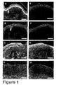
- Fig. 1A is a photomicrograph of a normal skin epidermal granular layer showing the localization of calcium (white arrow);
- Fig. 1B is a photomicrograph of a normal skin epidermal granular layer showing the localization of magnesium before tape stripping (white arrow);
- Fig. 1C is a photomicrograph of a normal skin epidermal granular layer showing the localization of potassium, and illustrating particularly that the concentration of potassium is highest in the spinous layer (black arrow) and the lowest in the granular layer before tape stripping,
- Fig. ID is a photomicrograph of a normal skin epidermal granular layer showing the homogeneous distribution of sodium around the whole epidermis;
- Fig. IE is a photomicrograph showing the skin in Fig. 1A, taken thirty minutes after tape stripping, illustrating the disappearance of the calcium gradation;
- Fig. 1F is a photomicrograph showing the skin in Fig. 1B, taken thirty minutes after tape stripping, illustrating particularly the disappearance of the magnesium gradation;
- Fig. 1G is a photomicrograph of the skin in Fig. 1C, taken thirty minutes after tape stripping, illustrating the disappearance of the potassium gradient and the homogeneous distribution of the sodium around the whole epidermis by tape stripping;
- Fig. 1H is a photomicrograph of a normal skin epidermal granular layer in Fig. 1D, taken thirty minutes after tape stripping, illustrating the homogeneous distribution of sodium ion around the whole epidermis.
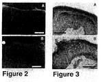
- Fig. 2A is a photomicrograph of normal skin epidermal granular layer using calcium indicators, illustrating that EDTA absorbed the fluorescence of calcium indicator;
- Fig. 2B is a photomicrograph of normal skin epidermal granular layer using magnesium indicator, illustrating that EDTA absorbed the fluorescence of magnesium indicator;
- Fig. 3 is a photomicrograph of normal skin epidermal layer showing the gradation of pH in the skin before and after tape stripping;
- Fig. 4A is a photomicrograph at high magnification after tape stripping of the spinous layer of the epidermis, showing the calcium distribution;
- Fig. 4B is a photomicrograph at high magnification after tape stripping of the spinous layer of the epidermis showing the magnesium concentration was slightly higher in the nucleus;
- Fig. 4C is a photomicrograph at high magnification after tape stripping of the spinous layer of the epidermis, showing the potassium ions did not perform a clear distribution in the skin; and
- Fig. 4D is a photomicrograph at high magnification after tape stripping of the spinous layer of the epidermis, showing the sodium distribution.
- The following example is exemplary only of the present invention. All percentages are in weight percent.
- Skin samples were obtained from the inner forearm of healthy males who gave their informed consent by cutting with a Diener Spezial, CHR Diener (dissecting scissors). The samples were biopsied from untreated skin 30 minutes after tape stripping. To check the reproducability of results and avoid error, at least three samples were taken from one region. Tape stripping was carried out 20 times. At that time, most of the stratum corneum was removed. Samples were immediately frozen in an isopentane- filled metal jar which was kept in liquid nitrogen to prevent artifactual redistributions. These frozen samples were kept at -80°C until sectioning.
- Agarose powder (Type I-B; Shigma) is dissolved in about 90°C water in a beaker and mixed on a hot plate. Thereafter, each of the ion probes described below was mixed in the solution while heating on a hot plate at about 50°C to form a final Agarose gel with a concentration of 2%. Thereafter, a small portion of each of the mixtures containing different ion probes was spread to a depth of about 50 µm on slide glass to form a thin membrane on the glass. For the calcium observation, a final concentration of 10 µg/ml of calcium green 1 ion probe was mixed with the agarose gel before the formation of the membrane. For the magnesium observation, a final concentration of 10 µg/ml of Magnesium Green (produced by Molecular Probe, Inc.) and 0.2mM final concentration of ethylene glycol bis(2-amino-ethyl ether)-N,N,N' N' -tetraacetic acid (EGTA) were mixed together. For the potassium observation, the final concentration of 10 µg/ml of PBFI, referred to above, was mixed with the agarose gel. For the sodium observation, the final concentration of 10 µg/ml of sodium green was mixed with the agarose gel.
- In the case of hydrogen ion (pH) observation, first an agarose gel membrane was formed and then 20ml of 0.01% Bromocresol Green ethanol solution was spread over the gel membrane.
- A frozen section of skin 10 µm in thickness prepared by Cryostat as described above, was then placed in contact with the gel membrane and within a couple of hours, the whole picture was taken. It is necessary to promptly photograph the tissue sample because within 12 hours after the preparation, the clear images disappeared. An Olympus microscope system (AH3-RFC, Olympus, Tokyo, Japan) was used for observation and photographing the tissue samples.
- For Calcium Green 1, Magnesium Green, and Sodium Green (all molecular probes produced by Molecular Probe, Inc.), the wavelength of the excitation light for the ion probe was 546 nm. For the potassium indicator, PBFI, the wavelength of the excitation light was 334, 365 nm. For each of these observations, at least five sections were observed to find common features.
- The images of calcium, magnesium, potassium and sodium in the skin are shown in figure 1. These are the representatives of each observation. In normal skin, calcium was localized in the epidermal granular layer (fig. 1A). Thirty minutes after tape stripping, this gradation disappeared (fig. 1E). Magnesium also showed the same tendency (fig. 1B, F).
- The concentration of potassium was the highest in the spinous layer and the lowest in the granular layer (fig.C). This gradation also disappeared after tape stripping (fig. 1G). Sodium showed a homogeneous distribution around the whole epidermis (fig. 1G), which was not affected by tape stripping. To confirm these results, experiments were carried out with calcium or magnesium indicators using a 50mM EDTA solution (fig.2A,B). EDTA absorbed most of the fluorescence in the epidermis. This indicates that the gradation in figure 1 showed the distribution of each ion.
- The gradation of pH in the skin before and after tape stripping are shown in figure 3. In both cases, the stratum corneum and upper epidermis were more acidic (4.0-4.5 indicated by yellow-green). A deeper part of the epidermis and dermis were less acidic (higher than 5.6 indicated by blue-green). No obvious difference in pH gradation was observed after tape stripping.
- Photographs of the spinous layer of the epidermis at a high magnification after tape stripping are shown in figure 4. Calcium (fig.4A) and sodium (fig.4D) distribution showed an obvious pattern. In both cases, these ions were absent in the nucleus. On the other hand, after tape stripping, the magnesium concentration was slightly higher in the nucleus (fig.4B). Potassium did not show a clear distribution pattern (fig.4C).
- A polymer gel, such as agarose or polyacrylamide, forms a three-dimensional structure and prevents any substantial water flow inside the structure. Therefore, this gel structure can be used for electrophoresis or in situ zymography. According to the present invention the diffusion of the chemical indicators may be prevented, and the agarose membrane shows the images of the ion distribution.
- Ionized calcium is the most common signal transaction element.
- In the normal epidermis, it has been found by the method of the present invention that the calcium concentration is higher in the upper epidermis, i. e. , granular layer and lower in the deeper epidermis, i. e. , basal layer. In the human study herein, the peak of calcium concentration in the epidermal granular layer became broad. These results suggest that calcium plays an important role in barrier homeostasis and/or signaling of barrier insults.
- It has been found using the method of the present invention that there is a gradation in calcium in the epidermis (See figure 2b). Although the role of magnesium in the epidermis remains largely unknown, it is known that magnesium is required for the activity of Rab-geranylgeranyl transferase which modifies Rab. It has been previously demonstrated that application of magnesium after tape stripping accelerates barrier recovery. After the modification, Rab plays an important role on exocytosis and endocytosis. For the barrier formation, exocytosis of the lamellar body is an important process. Previous studies have indicated that Rab is modified by Rab-geranylgeranyl transferase during the terminal differentiation of the epidermis. It is believed that magnesium may be required in the differentiation of the keratinocyte or the barrier homeostasis.
- Previously taken images of the skin suggest a close relation between potassium and calcium. Both ions play an important role in the terminal differentiation of keratinocyte or stratum corneum barrier function. Using the method of the present invention, potassium and calcium were localized comparably before and after barrier disruption. The disappearance of the potassium distribution in the present examples is similar to a previous study of hairless mice. The relative localization of these ions may be important for epidermal homeostasis.
- The mechanism of the quick movement of calcium, magnesium and potassium after the barrier insult (barrier disruption by tape stripping) remains unknown. There is an electric potential between the surface (negative) and bottom (positive) of the skin. This potential is erased immediately by barrier disruption and with slow recovery. The skin surface potential may be induced by the distribution of the ions.
- Using the method of the present invention, sodium did not show a gradation in the epidermis (See figure 4). The barrier insults did not affect the sodium ion distribution. This result suggests that sodium is not directly related to the cutaneous barrier homeostasis.
- It can be seen from the results herein that localization in each cell was different between calcium and magnesium after barrier disruption. The concentration of calcium was low in the nucleus and relatively high in the cytosol. The concentrations of sodium in the cytosol was also higher than in the nucleus. On other hand, the magnesium concentration after tape stripping is slightly higher in the nucleus than in the cytosol. Magnesium ion has an ability to decrease the entropy of the water structure. Thus, magnesium plays a role to stabilize the nucleus protein structure. Magnesium may be important against environmental insults.
- It is known that a lower pH in the upper epidermis would be important for barrier formation because lipid processing enzymes require it. Previously, application of basic buffer solution has been shown to delay the barrier recovery. The epidermis has a mechanism to keep the pH gradient for the barrier homeostasis. However, this gradation was not altered by barrier insults. The mechanism might be different from that of other divalent ions.
- The ion profile has been reported to be altered in various skin diseases. For example, abnormal calcium distribution was observed in psoriatic epidermis and atopic dermatitis. The distribution of zinc and iron was also altered in atopic skin. Ions may play an important role in the pathology of the skin. During the wound healing process, the distribution of magnesium and calcium, in the wound fluid is altered, and may activate the cell migratory response. The process of the present invention should play an important role in elucidating these disease mechanisms, especially since this is the simplest and most inexpensive method for visual imaging of ion distribution in tissue.
Claims (5)
- A method of producing a visual image of hydrogen ions in a tissue sample comprising the steps of:(a) preparing a thin frozen tissue sample;(b) placing the thin frozen tissue sample in contact with a thin membrane upon which a hydrogen ion indicator has been incorporated; and(c) subjecting the thin frozen tissue sample on the thin membrane from step (b) to light having a visible wavelength to cause the hydrogen ion indicator to change color.
- The method of claim 1, wherein the thin membrane comprises water, a hydrogen ion indicator and a water soluble polymer selected from the group consisting of polyacrylamide, polyvinyl alcohol, polysaccharide which is gelated or solidified into sheet form by cross-bridging or drying, or crosslinking or polymerization of a monomer.
- The method of claim 2, wherein the hydrogen ion indicator is ethanol which has been applied to the surface of the membrane.
- The method of claim 2, wherein the thin membrane is in the form of a gel and comprises about 2% of a water soluble polymer.
- A membrane for use in producing a visual image of hydrogen ions in a tissue sample, said membrane comprising from about 98 to 99.5 wt% of water; from about .05 to 2 wt% of polysaccharide which are gelated or solidified into sheet form by cross-bridging or drying or crosslinking agent or polymerization of a monomer; or from about 80-95 wt% of water and from about 5 to 20 wt% of polyacrylamide or polyvinyl alcohol which are gelated or solidified into sheet form by cross-bridging or drying or crosslinking agent or polymerization of a monomer; and a hydrogen ion indicator which can change color depending upon pH.
Applications Claiming Priority (2)
| Application Number | Priority Date | Filing Date | Title |
|---|---|---|---|
| US09/570,447 US6544794B1 (en) | 2000-05-12 | 2000-05-12 | Method for visual imaging of ion distribution in tissue |
| EP00127696A EP1154261B1 (en) | 2000-05-12 | 2000-12-18 | Imaging of ion distribution in tissue |
Related Parent Applications (1)
| Application Number | Title | Priority Date | Filing Date |
|---|---|---|---|
| EP00127696A Division EP1154261B1 (en) | 2000-05-12 | 2000-12-18 | Imaging of ion distribution in tissue |
Publications (2)
| Publication Number | Publication Date |
|---|---|
| EP1860427A2 true EP1860427A2 (en) | 2007-11-28 |
| EP1860427A3 EP1860427A3 (en) | 2008-01-09 |
Family
ID=24279679
Family Applications (2)
| Application Number | Title | Priority Date | Filing Date |
|---|---|---|---|
| EP20070017105 Withdrawn EP1860427A3 (en) | 2000-05-12 | 2000-12-18 | Imaging of ion distribution in tissue |
| EP00127696A Expired - Lifetime EP1154261B1 (en) | 2000-05-12 | 2000-12-18 | Imaging of ion distribution in tissue |
Family Applications After (1)
| Application Number | Title | Priority Date | Filing Date |
|---|---|---|---|
| EP00127696A Expired - Lifetime EP1154261B1 (en) | 2000-05-12 | 2000-12-18 | Imaging of ion distribution in tissue |
Country Status (8)
| Country | Link |
|---|---|
| US (1) | US6544794B1 (en) |
| EP (2) | EP1860427A3 (en) |
| JP (1) | JP4117870B2 (en) |
| KR (1) | KR100543667B1 (en) |
| AU (1) | AU771375B2 (en) |
| CA (1) | CA2329691C (en) |
| DE (1) | DE60038366T2 (en) |
| TW (1) | TWI253501B (en) |
Families Citing this family (6)
| Publication number | Priority date | Publication date | Assignee | Title |
|---|---|---|---|---|
| US6841387B2 (en) * | 2002-08-21 | 2005-01-11 | Advanced Energy Technology Inc. | Ex-situ PEM fuel cell testing: towards visualizing gas diffusion |
| JP4781972B2 (en) * | 2006-11-06 | 2011-09-28 | 株式会社 資生堂 | Test method and kit for stratum corneum oxidized protein |
| CN101710077B (en) * | 2009-12-09 | 2011-05-25 | 宁波工程学院 | Judging the Solubility of Cationic Polyacrylamide in Water by Visual Color Difference Method |
| CN101769871B (en) * | 2009-12-31 | 2015-01-21 | 宁波工程学院 | Method for judging dissolvability of non-ionic polyacrylamide in water through visual inspection of color difference |
| CN101799402B (en) * | 2010-03-12 | 2014-12-31 | 宁波工程学院 | Method for judging solubility of amphoteric ionic polyacrylamide in water by visual chromatic aberration method |
| DE102017118419A1 (en) * | 2017-08-11 | 2019-02-14 | Lts Lohmann Therapie-Systeme Ag | Microneedle array having a color change indicator |
Family Cites Families (8)
| Publication number | Priority date | Publication date | Assignee | Title |
|---|---|---|---|---|
| JP2749069B2 (en) * | 1988-04-26 | 1998-05-13 | オリンパス光学工業株式会社 | Fluorescence microscope equipment |
| JP2799191B2 (en) * | 1989-08-24 | 1998-09-17 | オリンパス光学工業株式会社 | Method for forming two-dimensional concentration distribution image of intracellular ions |
| JPH0614921B2 (en) * | 1989-09-29 | 1994-03-02 | 浜松ホトニクス株式会社 | Fluorescence observation device for biological tissue |
| WO1993007483A1 (en) * | 1991-09-30 | 1993-04-15 | Merck Patent Gmbh | SENSOR MEMBRANE FOR DISPLAYING THE pH-VALUE OF A SAMPLE, METHOD OF MANUFACTURING THE MEMBRANE, AND ITS USE |
| EP0561297B1 (en) * | 1992-03-19 | 1998-10-14 | Hoechst Marion Roussel, Ltd. | Tissue antigen and diagnostic method for human osteoporosis using the same |
| US5474909A (en) * | 1992-08-07 | 1995-12-12 | Anticancer, Inc. | Noncolorimetric histoculture method for predicting drug response of tumors |
| DE4324991A1 (en) * | 1993-07-26 | 1995-02-02 | Merck Patent Gmbh | Process for the preparation of polyvinyl alcohol films containing dyes and their use |
| JP2001128927A (en) * | 1999-11-05 | 2001-05-15 | Fuji Photo Film Co Ltd | Fluorescence image acquisition method and apparatus |
-
2000
- 2000-05-12 US US09/570,447 patent/US6544794B1/en not_active Expired - Lifetime
- 2000-09-14 TW TW089118805A patent/TWI253501B/en not_active IP Right Cessation
- 2000-09-29 AU AU61375/00A patent/AU771375B2/en not_active Expired
- 2000-10-16 JP JP2000314991A patent/JP4117870B2/en not_active Expired - Fee Related
- 2000-11-22 KR KR1020000069504A patent/KR100543667B1/en not_active Expired - Lifetime
- 2000-12-18 DE DE60038366T patent/DE60038366T2/en not_active Expired - Lifetime
- 2000-12-18 EP EP20070017105 patent/EP1860427A3/en not_active Withdrawn
- 2000-12-18 EP EP00127696A patent/EP1154261B1/en not_active Expired - Lifetime
- 2000-12-28 CA CA002329691A patent/CA2329691C/en not_active Expired - Lifetime
Also Published As
| Publication number | Publication date |
|---|---|
| EP1154261A2 (en) | 2001-11-14 |
| KR100543667B1 (en) | 2006-01-20 |
| EP1860427A3 (en) | 2008-01-09 |
| EP1154261B1 (en) | 2008-03-19 |
| TWI253501B (en) | 2006-04-21 |
| JP4117870B2 (en) | 2008-07-16 |
| AU771375B2 (en) | 2004-03-18 |
| DE60038366D1 (en) | 2008-04-30 |
| CA2329691A1 (en) | 2001-11-12 |
| CA2329691C (en) | 2008-12-09 |
| DE60038366T2 (en) | 2009-11-26 |
| KR20010106081A (en) | 2001-11-29 |
| US6544794B1 (en) | 2003-04-08 |
| EP1154261A3 (en) | 2004-03-17 |
| AU6137500A (en) | 2001-11-15 |
| JP2001324502A (en) | 2001-11-22 |
Similar Documents
| Publication | Publication Date | Title |
|---|---|---|
| Dubochet et al. | Electron microscopy of frozen-hydrated bacteria | |
| Denda et al. | Visual imaging of ion distribution in human epidermis | |
| Eady et al. | Mast cell population density, blood vessel density and histamine content in normal human skin | |
| Culling | Handbook of histopathological and histochemical techniques: including museum techniques | |
| Krause et al. | Fluorescent erythrosin B is preferable to trypan blue as a vital exclusion dye for mammalian cells in monolayer culture. | |
| Mundkur | Electron microscopical studies of frozen-dried yeast: I. Localization of polysaccharides | |
| Fullerton et al. | Permeation of nickel through human skin in vitro—effect of vehicles | |
| Domeradzka-Gajda et al. | A study on the in vitro percutaneous absorption of silver nanoparticles in combination with aluminum chloride, methyl paraben or di-n-butyl phthalate | |
| Mukherjee et al. | Sorbitan Monolaurate 20 as a Potential Skin Permeation Enhancer in Transdermal Patches. | |
| EP1154261B1 (en) | Imaging of ion distribution in tissue | |
| Lee et al. | Visualization of iontophoretic transport paths in cultured and animal skin models | |
| Brown | Polyvinyl alcohol coating: An improvement of the freeze‐fracture technique | |
| Hagler et al. | Effect of specimen preparation and section transfer techniques on the preservation of ultrastructure, lipids and elements in cryosections | |
| Von Schack et al. | The study of the cell nucleus using cryofixation and cryosubstitution | |
| Weavers | Combined transmission electron microscopy and X‐ray micro‐analysis of ultrathin frozen dried sections—an investigation to determine the normal elemental composition of mammalian tissue | |
| Ingram et al. | Procedures for the study of biological soft tissue with the electron microprobe | |
| Lawton et al. | Fixation of senescing plant tissues: sclerenchymatous fibre cells from the flowering stem of a grass | |
| de Haan et al. | Visualizing drug transport across stratum corneum: cryotechniques, vapour fixation, autoradiography | |
| Wergin et al. | Techniques for obtaining and observing complementary images with a low‐temperature field emission SEM and subsequent comparison of the identical cells in freeze‐etch replicas viewed with a TEM | |
| Squier et al. | Localization of lanthanum tracer in oral epithelium using transmission electron microscopy and the electron microprobe | |
| Cartman et al. | Electrolyte homeostasis in urothelial neoplasia: the effects of amiloride | |
| Wijdeveld et al. | Visualization and electron diffraction on cryosections of stratum corneum: a utopia? | |
| Steere | Freeze‐fracture: A personal history | |
| Pezzati et al. | The frog neuromuscular junction revisited after quick-freezing-freeze-drying: ultrastructure, immunogold labelling and high resolution calcium mapping | |
| Veilleux | Acridine orange fluorescence of mast cells as influenced by fixation |
Legal Events
| Date | Code | Title | Description |
|---|---|---|---|
| PUAI | Public reference made under article 153(3) epc to a published international application that has entered the european phase |
Free format text: ORIGINAL CODE: 0009012 |
|
| AC | Divisional application: reference to earlier application |
Ref document number: 1154261 Country of ref document: EP Kind code of ref document: P |
|
| AK | Designated contracting states |
Kind code of ref document: A2 Designated state(s): DE FR GB IT |
|
| PUAL | Search report despatched |
Free format text: ORIGINAL CODE: 0009013 |
|
| AK | Designated contracting states |
Kind code of ref document: A3 Designated state(s): DE FR GB IT |
|
| 17P | Request for examination filed |
Effective date: 20080207 |
|
| 17Q | First examination report despatched |
Effective date: 20080307 |
|
| AKX | Designation fees paid |
Designated state(s): DE FR GB IT |
|
| STAA | Information on the status of an ep patent application or granted ep patent |
Free format text: STATUS: THE APPLICATION IS DEEMED TO BE WITHDRAWN |
|
| 18D | Application deemed to be withdrawn |
Effective date: 20150715 |