EP1855758B1 - Implantable cardiac device with dyspnea measurement - Google Patents
Implantable cardiac device with dyspnea measurement Download PDFInfo
- Publication number
- EP1855758B1 EP1855758B1 EP06735992.7A EP06735992A EP1855758B1 EP 1855758 B1 EP1855758 B1 EP 1855758B1 EP 06735992 A EP06735992 A EP 06735992A EP 1855758 B1 EP1855758 B1 EP 1855758B1
- Authority
- EP
- European Patent Office
- Prior art keywords
- patient
- pulmonary function
- controller
- dyspnea
- function index
- Prior art date
- Legal status (The legal status is an assumption and is not a legal conclusion. Google has not performed a legal analysis and makes no representation as to the accuracy of the status listed.)
- Not-in-force
Links
- 206010013975 Dyspnoeas Diseases 0.000 title claims description 94
- 208000000059 Dyspnea Diseases 0.000 title claims description 93
- 230000000747 cardiac effect Effects 0.000 title description 55
- 238000005259 measurement Methods 0.000 title description 13
- 230000000694 effects Effects 0.000 claims description 55
- 238000002560 therapeutic procedure Methods 0.000 claims description 51
- 230000009325 pulmonary function Effects 0.000 claims description 47
- 230000006854 communication Effects 0.000 claims description 22
- 238000004891 communication Methods 0.000 claims description 22
- 230000029058 respiratory gaseous exchange Effects 0.000 claims description 21
- 230000006870 function Effects 0.000 claims description 18
- 229940030602 cardiac therapy drug Drugs 0.000 claims description 12
- 230000004044 response Effects 0.000 claims description 9
- 230000003247 decreasing effect Effects 0.000 claims description 6
- 230000002685 pulmonary effect Effects 0.000 claims description 6
- 230000008859 change Effects 0.000 claims description 5
- 238000000034 method Methods 0.000 description 35
- 230000000638 stimulation Effects 0.000 description 21
- 238000012544 monitoring process Methods 0.000 description 20
- 238000001514 detection method Methods 0.000 description 19
- 238000013194 cardioversion Methods 0.000 description 11
- 238000010586 diagram Methods 0.000 description 10
- 230000036387 respiratory rate Effects 0.000 description 10
- 230000033001 locomotion Effects 0.000 description 9
- 230000009471 action Effects 0.000 description 8
- 230000002612 cardiopulmonary effect Effects 0.000 description 8
- 238000003745 diagnosis Methods 0.000 description 8
- 206010019280 Heart failures Diseases 0.000 description 7
- 206010003119 arrhythmia Diseases 0.000 description 7
- 230000008901 benefit Effects 0.000 description 7
- 230000009467 reduction Effects 0.000 description 7
- 230000000241 respiratory effect Effects 0.000 description 7
- 206010007559 Cardiac failure congestive Diseases 0.000 description 6
- 208000006545 Chronic Obstructive Pulmonary Disease Diseases 0.000 description 6
- BQJCRHHNABKAKU-KBQPJGBKSA-N morphine Chemical compound O([C@H]1[C@H](C=C[C@H]23)O)C4=C5[C@@]12CCN(C)[C@@H]3CC5=CC=C4O BQJCRHHNABKAKU-KBQPJGBKSA-N 0.000 description 6
- 238000007920 subcutaneous administration Methods 0.000 description 6
- 208000001871 Tachycardia Diseases 0.000 description 5
- 208000006218 bradycardia Diseases 0.000 description 5
- 230000036471 bradycardia Effects 0.000 description 5
- 210000000038 chest Anatomy 0.000 description 5
- 238000007726 management method Methods 0.000 description 5
- 229940005483 opioid analgesics Drugs 0.000 description 5
- 208000013220 shortness of breath Diseases 0.000 description 5
- 230000006793 arrhythmia Effects 0.000 description 4
- 238000002633 shock therapy Methods 0.000 description 4
- 230000001225 therapeutic effect Effects 0.000 description 4
- 238000009423 ventilation Methods 0.000 description 4
- 208000019901 Anxiety disease Diseases 0.000 description 3
- 208000010496 Heart Arrest Diseases 0.000 description 3
- 230000036506 anxiety Effects 0.000 description 3
- QVGXLLKOCUKJST-UHFFFAOYSA-N atomic oxygen Chemical compound [O] QVGXLLKOCUKJST-UHFFFAOYSA-N 0.000 description 3
- 238000011156 evaluation Methods 0.000 description 3
- 239000007789 gas Substances 0.000 description 3
- 208000000122 hyperventilation Diseases 0.000 description 3
- 229960005181 morphine Drugs 0.000 description 3
- 239000001301 oxygen Substances 0.000 description 3
- 229910052760 oxygen Inorganic materials 0.000 description 3
- 238000012545 processing Methods 0.000 description 3
- 210000001519 tissue Anatomy 0.000 description 3
- 238000012546 transfer Methods 0.000 description 3
- 208000019693 Lung disease Diseases 0.000 description 2
- 102000003840 Opioid Receptors Human genes 0.000 description 2
- 108090000137 Opioid Receptors Proteins 0.000 description 2
- 206010049447 Tachyarrhythmia Diseases 0.000 description 2
- 230000003044 adaptive effect Effects 0.000 description 2
- 230000002411 adverse Effects 0.000 description 2
- 238000013459 approach Methods 0.000 description 2
- 230000002763 arrhythmic effect Effects 0.000 description 2
- 230000001746 atrial effect Effects 0.000 description 2
- 230000006399 behavior Effects 0.000 description 2
- 230000007175 bidirectional communication Effects 0.000 description 2
- 210000005242 cardiac chamber Anatomy 0.000 description 2
- 238000009125 cardiac resynchronization therapy Methods 0.000 description 2
- 230000001684 chronic effect Effects 0.000 description 2
- 230000010339 dilation Effects 0.000 description 2
- 230000007613 environmental effect Effects 0.000 description 2
- 230000007717 exclusion Effects 0.000 description 2
- 208000019622 heart disease Diseases 0.000 description 2
- 230000004217 heart function Effects 0.000 description 2
- 230000003993 interaction Effects 0.000 description 2
- 230000000670 limiting effect Effects 0.000 description 2
- 239000000463 material Substances 0.000 description 2
- 210000000412 mechanoreceptor Anatomy 0.000 description 2
- 108091008704 mechanoreceptors Proteins 0.000 description 2
- 210000003205 muscle Anatomy 0.000 description 2
- 230000008569 process Effects 0.000 description 2
- 210000003019 respiratory muscle Anatomy 0.000 description 2
- 230000033764 rhythmic process Effects 0.000 description 2
- 230000035939 shock Effects 0.000 description 2
- 229920002379 silicone rubber Polymers 0.000 description 2
- 239000004945 silicone rubber Substances 0.000 description 2
- 230000009885 systemic effect Effects 0.000 description 2
- 208000008203 tachypnea Diseases 0.000 description 2
- 206010043089 tachypnoea Diseases 0.000 description 2
- 210000005166 vasculature Anatomy 0.000 description 2
- RTAPDZBZLSXHQQ-UHFFFAOYSA-N 8-methyl-3,7-dihydropurine-2,6-dione Chemical class N1C(=O)NC(=O)C2=C1N=C(C)N2 RTAPDZBZLSXHQQ-UHFFFAOYSA-N 0.000 description 1
- 206010001029 Acute pulmonary oedema Diseases 0.000 description 1
- 208000020446 Cardiac disease Diseases 0.000 description 1
- 102000008186 Collagen Human genes 0.000 description 1
- 108010035532 Collagen Proteins 0.000 description 1
- 206010011224 Cough Diseases 0.000 description 1
- LTMHDMANZUZIPE-AMTYYWEZSA-N Digoxin Natural products O([C@H]1[C@H](C)O[C@H](O[C@@H]2C[C@@H]3[C@@](C)([C@@H]4[C@H]([C@]5(O)[C@](C)([C@H](O)C4)[C@H](C4=CC(=O)OC4)CC5)CC3)CC2)C[C@@H]1O)[C@H]1O[C@H](C)[C@@H](O[C@H]2O[C@@H](C)[C@H](O)[C@@H](O)C2)[C@@H](O)C1 LTMHDMANZUZIPE-AMTYYWEZSA-N 0.000 description 1
- 206010013974 Dyspnoea paroxysmal nocturnal Diseases 0.000 description 1
- 208000027534 Emotional disease Diseases 0.000 description 1
- 206010014561 Emphysema Diseases 0.000 description 1
- 241001539473 Euphoria Species 0.000 description 1
- 206010015535 Euphoric mood Diseases 0.000 description 1
- 241000288140 Gruiformes Species 0.000 description 1
- 206010020591 Hypercapnia Diseases 0.000 description 1
- 206010021079 Hypopnoea Diseases 0.000 description 1
- 206010021143 Hypoxia Diseases 0.000 description 1
- 206010049816 Muscle tightness Diseases 0.000 description 1
- 206010028980 Neoplasm Diseases 0.000 description 1
- 208000004327 Paroxysmal Dyspnea Diseases 0.000 description 1
- 208000001431 Psychomotor Agitation Diseases 0.000 description 1
- 208000010378 Pulmonary Embolism Diseases 0.000 description 1
- 208000037656 Respiratory Sounds Diseases 0.000 description 1
- 206010038743 Restlessness Diseases 0.000 description 1
- 206010041235 Snoring Diseases 0.000 description 1
- 210000001015 abdomen Anatomy 0.000 description 1
- 230000003187 abdominal effect Effects 0.000 description 1
- 230000004913 activation Effects 0.000 description 1
- 208000012871 acute dyspnea Diseases 0.000 description 1
- 230000001154 acute effect Effects 0.000 description 1
- 230000006978 adaptation Effects 0.000 description 1
- 238000011374 additional therapy Methods 0.000 description 1
- 239000000048 adrenergic agonist Substances 0.000 description 1
- 230000000172 allergic effect Effects 0.000 description 1
- 230000003321 amplification Effects 0.000 description 1
- 238000004458 analytical method Methods 0.000 description 1
- 229940065524 anticholinergics inhalants for obstructive airway diseases Drugs 0.000 description 1
- 239000002249 anxiolytic agent Substances 0.000 description 1
- 230000000949 anxiolytic effect Effects 0.000 description 1
- 229940005530 anxiolytics Drugs 0.000 description 1
- 238000003491 array Methods 0.000 description 1
- 208000010668 atopic eczema Diseases 0.000 description 1
- 206010003668 atrial tachycardia Diseases 0.000 description 1
- 229940049706 benzodiazepine Drugs 0.000 description 1
- 150000001557 benzodiazepines Chemical class 0.000 description 1
- 239000008280 blood Substances 0.000 description 1
- 210000004369 blood Anatomy 0.000 description 1
- 229940124630 bronchodilator Drugs 0.000 description 1
- 239000000168 bronchodilator agent Substances 0.000 description 1
- 238000004364 calculation method Methods 0.000 description 1
- 201000011510 cancer Diseases 0.000 description 1
- 206010061592 cardiac fibrillation Diseases 0.000 description 1
- 210000004903 cardiac system Anatomy 0.000 description 1
- 108091008690 chemoreceptors Proteins 0.000 description 1
- 239000000812 cholinergic antagonist Substances 0.000 description 1
- 238000000576 coating method Methods 0.000 description 1
- 229920001436 collagen Polymers 0.000 description 1
- 230000001447 compensatory effect Effects 0.000 description 1
- 230000008602 contraction Effects 0.000 description 1
- 238000007796 conventional method Methods 0.000 description 1
- 238000013480 data collection Methods 0.000 description 1
- 230000000881 depressing effect Effects 0.000 description 1
- 230000000994 depressogenic effect Effects 0.000 description 1
- 238000013461 design Methods 0.000 description 1
- LTMHDMANZUZIPE-PUGKRICDSA-N digoxin Chemical compound C1[C@H](O)[C@H](O)[C@@H](C)O[C@H]1O[C@@H]1[C@@H](C)O[C@@H](O[C@@H]2[C@H](O[C@@H](O[C@@H]3C[C@@H]4[C@]([C@@H]5[C@H]([C@]6(CC[C@@H]([C@@]6(C)[C@H](O)C5)C=5COC(=O)C=5)O)CC4)(C)CC3)C[C@@H]2O)C)C[C@@H]1O LTMHDMANZUZIPE-PUGKRICDSA-N 0.000 description 1
- 229960005156 digoxin Drugs 0.000 description 1
- LTMHDMANZUZIPE-UHFFFAOYSA-N digoxine Natural products C1C(O)C(O)C(C)OC1OC1C(C)OC(OC2C(OC(OC3CC4C(C5C(C6(CCC(C6(C)C(O)C5)C=5COC(=O)C=5)O)CC4)(C)CC3)CC2O)C)CC1O LTMHDMANZUZIPE-UHFFFAOYSA-N 0.000 description 1
- 201000010099 disease Diseases 0.000 description 1
- 208000037265 diseases, disorders, signs and symptoms Diseases 0.000 description 1
- 230000009429 distress Effects 0.000 description 1
- 239000002934 diuretic Substances 0.000 description 1
- 229940030606 diuretics Drugs 0.000 description 1
- 229940079593 drug Drugs 0.000 description 1
- 239000003814 drug Substances 0.000 description 1
- 230000006397 emotional response Effects 0.000 description 1
- 238000005516 engineering process Methods 0.000 description 1
- 230000002600 fibrillogenic effect Effects 0.000 description 1
- 238000001914 filtration Methods 0.000 description 1
- 230000000870 hyperventilation Effects 0.000 description 1
- 230000001146 hypoxic effect Effects 0.000 description 1
- 239000007943 implant Substances 0.000 description 1
- 230000000977 initiatory effect Effects 0.000 description 1
- 230000003434 inspiratory effect Effects 0.000 description 1
- 230000003601 intercostal effect Effects 0.000 description 1
- 210000000876 intercostal muscle Anatomy 0.000 description 1
- 238000001990 intravenous administration Methods 0.000 description 1
- 210000005246 left atrium Anatomy 0.000 description 1
- 210000005240 left ventricle Anatomy 0.000 description 1
- 210000003715 limbic system Anatomy 0.000 description 1
- 210000004072 lung Anatomy 0.000 description 1
- 239000011159 matrix material Substances 0.000 description 1
- 230000007246 mechanism Effects 0.000 description 1
- 230000002503 metabolic effect Effects 0.000 description 1
- 238000012986 modification Methods 0.000 description 1
- 230000004048 modification Effects 0.000 description 1
- 230000000877 morphologic effect Effects 0.000 description 1
- 208000010125 myocardial infarction Diseases 0.000 description 1
- 208000018360 neuromuscular disease Diseases 0.000 description 1
- 239000012811 non-conductive material Substances 0.000 description 1
- 238000003199 nucleic acid amplification method Methods 0.000 description 1
- 238000005457 optimization Methods 0.000 description 1
- 230000036284 oxygen consumption Effects 0.000 description 1
- 238000002640 oxygen therapy Methods 0.000 description 1
- 230000008447 perception Effects 0.000 description 1
- 230000002093 peripheral effect Effects 0.000 description 1
- 230000002085 persistent effect Effects 0.000 description 1
- 230000000144 pharmacologic effect Effects 0.000 description 1
- 150000002990 phenothiazines Chemical class 0.000 description 1
- 230000037081 physical activity Effects 0.000 description 1
- 230000006461 physiological response Effects 0.000 description 1
- 229920000052 poly(p-xylylene) Polymers 0.000 description 1
- 229920002635 polyurethane Polymers 0.000 description 1
- 239000004814 polyurethane Substances 0.000 description 1
- 230000002265 prevention Effects 0.000 description 1
- 208000020016 psychiatric disease Diseases 0.000 description 1
- 206010037833 rales Diseases 0.000 description 1
- 230000002829 reductive effect Effects 0.000 description 1
- 230000004202 respiratory function Effects 0.000 description 1
- 210000002345 respiratory system Anatomy 0.000 description 1
- 230000000284 resting effect Effects 0.000 description 1
- 210000005245 right atrium Anatomy 0.000 description 1
- 210000005241 right ventricle Anatomy 0.000 description 1
- 210000002460 smooth muscle Anatomy 0.000 description 1
- 238000007619 statistical method Methods 0.000 description 1
- 230000035882 stress Effects 0.000 description 1
- 230000006794 tachycardia Effects 0.000 description 1
- 210000000115 thoracic cavity Anatomy 0.000 description 1
- 210000000779 thoracic wall Anatomy 0.000 description 1
- 238000011282 treatment Methods 0.000 description 1
- 230000002861 ventricular Effects 0.000 description 1
- 206010047302 ventricular tachycardia Diseases 0.000 description 1
- 230000000007 visual effect Effects 0.000 description 1
Images


Classifications
-
- A—HUMAN NECESSITIES
- A61—MEDICAL OR VETERINARY SCIENCE; HYGIENE
- A61B—DIAGNOSIS; SURGERY; IDENTIFICATION
- A61B5/00—Measuring for diagnostic purposes; Identification of persons
- A61B5/68—Arrangements of detecting, measuring or recording means, e.g. sensors, in relation to patient
- A61B5/6846—Arrangements of detecting, measuring or recording means, e.g. sensors, in relation to patient specially adapted to be brought in contact with an internal body part, i.e. invasive
- A61B5/6847—Arrangements of detecting, measuring or recording means, e.g. sensors, in relation to patient specially adapted to be brought in contact with an internal body part, i.e. invasive mounted on an invasive device
- A61B5/686—Permanently implanted devices, e.g. pacemakers, other stimulators, biochips
-
- A—HUMAN NECESSITIES
- A61—MEDICAL OR VETERINARY SCIENCE; HYGIENE
- A61B—DIAGNOSIS; SURGERY; IDENTIFICATION
- A61B5/00—Measuring for diagnostic purposes; Identification of persons
- A61B5/02—Detecting, measuring or recording pulse, heart rate, blood pressure or blood flow; Combined pulse/heart-rate/blood pressure determination; Evaluating a cardiovascular condition not otherwise provided for, e.g. using combinations of techniques provided for in this group with electrocardiography or electroauscultation; Heart catheters for measuring blood pressure
- A61B5/02028—Determining haemodynamic parameters not otherwise provided for, e.g. cardiac contractility or left ventricular ejection fraction
-
- A—HUMAN NECESSITIES
- A61—MEDICAL OR VETERINARY SCIENCE; HYGIENE
- A61B—DIAGNOSIS; SURGERY; IDENTIFICATION
- A61B5/00—Measuring for diagnostic purposes; Identification of persons
- A61B5/02—Detecting, measuring or recording pulse, heart rate, blood pressure or blood flow; Combined pulse/heart-rate/blood pressure determination; Evaluating a cardiovascular condition not otherwise provided for, e.g. using combinations of techniques provided for in this group with electrocardiography or electroauscultation; Heart catheters for measuring blood pressure
- A61B5/0205—Simultaneously evaluating both cardiovascular conditions and different types of body conditions, e.g. heart and respiratory condition
-
- A—HUMAN NECESSITIES
- A61—MEDICAL OR VETERINARY SCIENCE; HYGIENE
- A61B—DIAGNOSIS; SURGERY; IDENTIFICATION
- A61B5/00—Measuring for diagnostic purposes; Identification of persons
- A61B5/07—Endoradiosondes
- A61B5/076—Permanent implantations
-
- A—HUMAN NECESSITIES
- A61—MEDICAL OR VETERINARY SCIENCE; HYGIENE
- A61B—DIAGNOSIS; SURGERY; IDENTIFICATION
- A61B5/00—Measuring for diagnostic purposes; Identification of persons
- A61B5/08—Detecting, measuring or recording devices for evaluating the respiratory organs
- A61B5/0816—Measuring devices for examining respiratory frequency
-
- A—HUMAN NECESSITIES
- A61—MEDICAL OR VETERINARY SCIENCE; HYGIENE
- A61B—DIAGNOSIS; SURGERY; IDENTIFICATION
- A61B5/00—Measuring for diagnostic purposes; Identification of persons
- A61B5/08—Detecting, measuring or recording devices for evaluating the respiratory organs
- A61B5/085—Measuring impedance of respiratory organs or lung elasticity
-
- A—HUMAN NECESSITIES
- A61—MEDICAL OR VETERINARY SCIENCE; HYGIENE
- A61B—DIAGNOSIS; SURGERY; IDENTIFICATION
- A61B5/00—Measuring for diagnostic purposes; Identification of persons
- A61B5/08—Detecting, measuring or recording devices for evaluating the respiratory organs
- A61B5/091—Measuring volume of inspired or expired gases, e.g. to determine lung capacity
-
- A—HUMAN NECESSITIES
- A61—MEDICAL OR VETERINARY SCIENCE; HYGIENE
- A61B—DIAGNOSIS; SURGERY; IDENTIFICATION
- A61B5/00—Measuring for diagnostic purposes; Identification of persons
- A61B5/103—Detecting, measuring or recording devices for testing the shape, pattern, colour, size or movement of the body or parts thereof, for diagnostic purposes
- A61B5/11—Measuring movement of the entire body or parts thereof, e.g. head or hand tremor, mobility of a limb
- A61B5/1118—Determining activity level
-
- A—HUMAN NECESSITIES
- A61—MEDICAL OR VETERINARY SCIENCE; HYGIENE
- A61B—DIAGNOSIS; SURGERY; IDENTIFICATION
- A61B5/00—Measuring for diagnostic purposes; Identification of persons
- A61B5/24—Detecting, measuring or recording bioelectric or biomagnetic signals of the body or parts thereof
- A61B5/316—Modalities, i.e. specific diagnostic methods
- A61B5/318—Heart-related electrical modalities, e.g. electrocardiography [ECG]
- A61B5/346—Analysis of electrocardiograms
-
- A—HUMAN NECESSITIES
- A61—MEDICAL OR VETERINARY SCIENCE; HYGIENE
- A61B—DIAGNOSIS; SURGERY; IDENTIFICATION
- A61B5/00—Measuring for diagnostic purposes; Identification of persons
- A61B5/48—Other medical applications
- A61B5/4836—Diagnosis combined with treatment in closed-loop systems or methods
-
- A—HUMAN NECESSITIES
- A61—MEDICAL OR VETERINARY SCIENCE; HYGIENE
- A61B—DIAGNOSIS; SURGERY; IDENTIFICATION
- A61B5/00—Measuring for diagnostic purposes; Identification of persons
- A61B5/72—Signal processing specially adapted for physiological signals or for diagnostic purposes
- A61B5/7271—Specific aspects of physiological measurement analysis
- A61B5/7275—Determining trends in physiological measurement data; Predicting development of a medical condition based on physiological measurements, e.g. determining a risk factor
-
- A—HUMAN NECESSITIES
- A61—MEDICAL OR VETERINARY SCIENCE; HYGIENE
- A61B—DIAGNOSIS; SURGERY; IDENTIFICATION
- A61B5/00—Measuring for diagnostic purposes; Identification of persons
- A61B5/72—Signal processing specially adapted for physiological signals or for diagnostic purposes
- A61B5/7271—Specific aspects of physiological measurement analysis
- A61B5/7282—Event detection, e.g. detecting unique waveforms indicative of a medical condition
-
- A—HUMAN NECESSITIES
- A61—MEDICAL OR VETERINARY SCIENCE; HYGIENE
- A61B—DIAGNOSIS; SURGERY; IDENTIFICATION
- A61B5/00—Measuring for diagnostic purposes; Identification of persons
- A61B5/74—Details of notification to user or communication with user or patient ; user input means
- A61B5/746—Alarms related to a physiological condition, e.g. details of setting alarm thresholds or avoiding false alarms
-
- A—HUMAN NECESSITIES
- A61—MEDICAL OR VETERINARY SCIENCE; HYGIENE
- A61B—DIAGNOSIS; SURGERY; IDENTIFICATION
- A61B5/00—Measuring for diagnostic purposes; Identification of persons
- A61B5/74—Details of notification to user or communication with user or patient ; user input means
- A61B5/7465—Arrangements for interactive communication between patient and care services, e.g. by using a telephone network
- A61B5/747—Arrangements for interactive communication between patient and care services, e.g. by using a telephone network in case of emergency, i.e. alerting emergency services
-
- A—HUMAN NECESSITIES
- A61—MEDICAL OR VETERINARY SCIENCE; HYGIENE
- A61N—ELECTROTHERAPY; MAGNETOTHERAPY; RADIATION THERAPY; ULTRASOUND THERAPY
- A61N1/00—Electrotherapy; Circuits therefor
- A61N1/18—Applying electric currents by contact electrodes
- A61N1/32—Applying electric currents by contact electrodes alternating or intermittent currents
- A61N1/36—Applying electric currents by contact electrodes alternating or intermittent currents for stimulation
- A61N1/362—Heart stimulators
- A61N1/3627—Heart stimulators for treating a mechanical deficiency of the heart, e.g. congestive heart failure or cardiomyopathy
-
- A—HUMAN NECESSITIES
- A61—MEDICAL OR VETERINARY SCIENCE; HYGIENE
- A61N—ELECTROTHERAPY; MAGNETOTHERAPY; RADIATION THERAPY; ULTRASOUND THERAPY
- A61N1/00—Electrotherapy; Circuits therefor
- A61N1/18—Applying electric currents by contact electrodes
- A61N1/32—Applying electric currents by contact electrodes alternating or intermittent currents
- A61N1/36—Applying electric currents by contact electrodes alternating or intermittent currents for stimulation
- A61N1/362—Heart stimulators
- A61N1/365—Heart stimulators controlled by a physiological parameter, e.g. heart potential
- A61N1/36585—Heart stimulators controlled by a physiological parameter, e.g. heart potential controlled by two or more physical parameters
-
- A—HUMAN NECESSITIES
- A61—MEDICAL OR VETERINARY SCIENCE; HYGIENE
- A61N—ELECTROTHERAPY; MAGNETOTHERAPY; RADIATION THERAPY; ULTRASOUND THERAPY
- A61N1/00—Electrotherapy; Circuits therefor
- A61N1/18—Applying electric currents by contact electrodes
- A61N1/32—Applying electric currents by contact electrodes alternating or intermittent currents
- A61N1/36—Applying electric currents by contact electrodes alternating or intermittent currents for stimulation
- A61N1/362—Heart stimulators
- A61N1/37—Monitoring; Protecting
- A61N1/3702—Physiological parameters
-
- A—HUMAN NECESSITIES
- A61—MEDICAL OR VETERINARY SCIENCE; HYGIENE
- A61N—ELECTROTHERAPY; MAGNETOTHERAPY; RADIATION THERAPY; ULTRASOUND THERAPY
- A61N1/00—Electrotherapy; Circuits therefor
- A61N1/18—Applying electric currents by contact electrodes
- A61N1/32—Applying electric currents by contact electrodes alternating or intermittent currents
- A61N1/38—Applying electric currents by contact electrodes alternating or intermittent currents for producing shock effects
- A61N1/39—Heart defibrillators
- A61N1/3987—Heart defibrillators characterised by the timing or triggering of the shock
-
- G—PHYSICS
- G16—INFORMATION AND COMMUNICATION TECHNOLOGY [ICT] SPECIALLY ADAPTED FOR SPECIFIC APPLICATION FIELDS
- G16H—HEALTHCARE INFORMATICS, i.e. INFORMATION AND COMMUNICATION TECHNOLOGY [ICT] SPECIALLY ADAPTED FOR THE HANDLING OR PROCESSING OF MEDICAL OR HEALTHCARE DATA
- G16H50/00—ICT specially adapted for medical diagnosis, medical simulation or medical data mining; ICT specially adapted for detecting, monitoring or modelling epidemics or pandemics
- G16H50/20—ICT specially adapted for medical diagnosis, medical simulation or medical data mining; ICT specially adapted for detecting, monitoring or modelling epidemics or pandemics for computer-aided diagnosis, e.g. based on medical expert systems
-
- G—PHYSICS
- G16—INFORMATION AND COMMUNICATION TECHNOLOGY [ICT] SPECIALLY ADAPTED FOR SPECIFIC APPLICATION FIELDS
- G16H—HEALTHCARE INFORMATICS, i.e. INFORMATION AND COMMUNICATION TECHNOLOGY [ICT] SPECIALLY ADAPTED FOR THE HANDLING OR PROCESSING OF MEDICAL OR HEALTHCARE DATA
- G16H50/00—ICT specially adapted for medical diagnosis, medical simulation or medical data mining; ICT specially adapted for detecting, monitoring or modelling epidemics or pandemics
- G16H50/30—ICT specially adapted for medical diagnosis, medical simulation or medical data mining; ICT specially adapted for detecting, monitoring or modelling epidemics or pandemics for calculating health indices; for individual health risk assessment
-
- A—HUMAN NECESSITIES
- A61—MEDICAL OR VETERINARY SCIENCE; HYGIENE
- A61B—DIAGNOSIS; SURGERY; IDENTIFICATION
- A61B5/00—Measuring for diagnostic purposes; Identification of persons
- A61B5/0002—Remote monitoring of patients using telemetry, e.g. transmission of vital signals via a communication network
- A61B5/0015—Remote monitoring of patients using telemetry, e.g. transmission of vital signals via a communication network characterised by features of the telemetry system
- A61B5/0022—Monitoring a patient using a global network, e.g. telephone networks, internet
-
- A—HUMAN NECESSITIES
- A61—MEDICAL OR VETERINARY SCIENCE; HYGIENE
- A61B—DIAGNOSIS; SURGERY; IDENTIFICATION
- A61B5/00—Measuring for diagnostic purposes; Identification of persons
- A61B5/0002—Remote monitoring of patients using telemetry, e.g. transmission of vital signals via a communication network
- A61B5/0015—Remote monitoring of patients using telemetry, e.g. transmission of vital signals via a communication network characterised by features of the telemetry system
- A61B5/0024—Remote monitoring of patients using telemetry, e.g. transmission of vital signals via a communication network characterised by features of the telemetry system for multiple sensor units attached to the patient, e.g. using a body or personal area network
-
- A—HUMAN NECESSITIES
- A61—MEDICAL OR VETERINARY SCIENCE; HYGIENE
- A61B—DIAGNOSIS; SURGERY; IDENTIFICATION
- A61B5/00—Measuring for diagnostic purposes; Identification of persons
- A61B5/08—Detecting, measuring or recording devices for evaluating the respiratory organs
- A61B5/0809—Detecting, measuring or recording devices for evaluating the respiratory organs by impedance pneumography
-
- A—HUMAN NECESSITIES
- A61—MEDICAL OR VETERINARY SCIENCE; HYGIENE
- A61B—DIAGNOSIS; SURGERY; IDENTIFICATION
- A61B5/00—Measuring for diagnostic purposes; Identification of persons
- A61B5/08—Detecting, measuring or recording devices for evaluating the respiratory organs
- A61B5/0826—Detecting or evaluating apnoea events
Definitions
- the present invention relates generally to implantable cardiac devices and, more particularly, to cardiac sensing and/or stimulation devices employing dyspnea measurement.
- Dyspnea is defined as a shortness of breath or difficult breathing (the subjective feeling of being out of breath) caused by heart or lung disorders, strenuous activity, high anxiety or stress. Dyspnea derives from interactions among multiple physiological, psychological, social, and environmental factors, and may induce secondary physiological and behavioral responses. Dyspnea is different from tachypnea (rapid breathing) and hyperpnea (deep breathing). Tachypnea and hyperpnea can occur with hyperventilation, or over breathing beyond what is required to maintain arterial blood gases within normal limits. Fear or anxiety may create even more distress in dyspneic patients.
- a dyspnea index has been developed as a monitoring tool for dyspnea.
- One conventional method of determining a dyspnea index value has a subject take a deep breath in and count out loud from 1 to 15 (should take approximately 8 seconds). When stressed or running out of air, the subject stops counting, takes another deep breath and then continues counting from where they left off. This is repeated as needed until the subject finishes counting to 15. The number of breaths needed to take in addition to the very first one is the dyspnea index.
- a value of 0-1 is considered normal.
- a dyspnea value of 2-3 is acceptable, depending on what activity is being performed.
- the subject should slow down, and think about stopping.
- the subject should definitely stop and rest.
- Dyspnea may be classified as chronic, acute, or terminal.
- Chronic dyspnea has a variable intensity and persistent shortness of breath. This is most often seen in patients with chronic obstructive pulmonary disease (COPD).
- COPD chronic obstructive pulmonary disease
- Acute dyspnea causes episodes of shortness of breath with high intensity. It may be seen in patients who have suffered a myocardial infarction or pulmonary embolism.
- Terminal dyspnea occurs in patients with end-stage diseases, and these patients may be in a hospital, at home, or in a hospice. This type of dyspnea is a common complaint in patients with cancer.
- Dyspnea can be caused by a variety of conditions, including metabolic, allergic, psychiatric, and neuromuscular disorders, and by pain. However, cardiac and pulmonary disorders are the most common causes.
- nonpharmacological and pharmacological interventions may be performed. For example, treating patients in cardiac failure with digoxin and diuretics may resolve the problem. Stimulation of mechanoreceptors in the respiratory musculature or over the face has reduced dyspnea in some patients. Vibration of the intercostal muscles, in phase with inspiration so that contracting respiratory muscles are vibrated, has relieved dyspnea in some COPD patients. This intervention has worked best with severe dyspnea at rest. Movement of cool air across the face by a fan or an open window can stimulate mechanoreceptors in the face, minimizing mild dyspnea.
- Oxygen administered by mask or nasal cannula or transtracheally, improves dyspnea.
- most authorities recommend oxygen therapy for raising PaO2 levels to at least 55 mmHg to 60 mmHg or oxygen saturation to 88% to 90%.
- opioids despite their possible respiratory depressant effect, are useful in managing dyspnea. While the action of opioids to relieve dyspnea is not fully understood, the drugs may act by blunting the emotional reaction to dyspnea by interaction with opioid receptors in the limbic system. Because opioids cause euphoria, they reduce fear, anxiety, and the associated restlessness and muscle tension that decrease oxygen consumption. Opioids may also relieve dyspnea by action on the chemoreceptors, thus reducing respiratory drive.
- Nebulized morphine may relieve dyspnea by direct local action on peripheral opioid receptors in the airways so that it does not reach the systemic concentration to the extent that oral, subcutaneous, or intravenous morphine does. Therefore, some patients experience relief of dyspnea with fewer adverse effects.
- Anxiolytics frequently used to relieve dyspnea include benzodiazepines and phenothiazines. These act by depressing the hypoxic, hypercapnic response to dyspnea and the emotional response to dyspnea. Depending on the cause of dyspnea, patients may benefit from bronchodilators. Since methylxanthines cause smooth muscle dilation of the airways and improve the contraction of the diaphragm, they may be useful in patients with COPD. Similarly, inhaled beta-2 adrenergic agonists and anticholinergics cause smooth, muscle dilation of airways, thus improving lung mechanics and possibly relieving dyspnea.
- US 2003/0055461 describes systems, devices and methods for predicting how the status of a patient's congestive heart failure condition will progress in the future, for example by measuring acute pulmonary edema by sensing transthoracic impedance or by measuring paroxysmal nocturnal dyspnea by sensing shortness of breath using a respiration sensor and using a sleep detector / activity sensor.
- US 6459929 describes a method and apparatus for providing congestive heart failure therapy status using a CRM device capable of measuring transthoracic impedance and sensing physical activity level.
- US 2005/0039745 describes methods and apparatus for adaptive therapy for disordered breathing and US 2005/0043644 . describes methods and apparatus for predicting disordered breathing.
- the present invention provides an implantable device as defined in claim 1. Described herein are cardiac monitoring and/or stimulation methods and systems that provide monitoring, defibrillation therapies, pacing therapies, or a combination of these capabilities. Embodiments of the present invention relate generally to implantable medical devices employing dyspnea measurement and/or detection capability.
- Embodiments of methods described herein involve providing an implantable cardiac device configured to sense transthoracic impedance and determine a patient activity level.
- An index indicative of pulmonary function is implantably computed using the sensed transthoracic impedance.
- An episode of dyspnea may be detected based on a change in a computed pulmonary function index exceeding a threshold at a determined patient activity level.
- Further embodiments are directed to methods that trend one or more pulmonary function index values to determine a patient's pulmonary function index profile.
- the threshold for detecting the dyspnea episode may be based on the patient's pulmonary function index profile.
- Other embodiments may involve trending one or more pulmonary function index values and their associated patient activity levels to determine a patient's pulmonary function index versus activity level profile.
- the threshold for detecting the dyspnea episode may be based on the patient's pulmonary function index versus activity level profile by computing the pulmonary function Index as a ratio of a respiratory rate value and a tidal volume value for a patient.
- Methods may further involve adapting a cardiac therapy for the patient based on a pulmonary function index value.
- An adapted cardiac therapy may be delivered to the patient after comparing the pulmonary function index value to a threshold and determining the pulmonary function index value is beyond the threshold.
- Adapting the cardiac therapy may also involve comparing the pulmonary function index value to a predetermined range and modifying the cardiac therapy if the pulmonary function index value is beyond the predetermined range.
- Therapy adaptations include increasing or decreasing a rate at which pacing pulses are delivered to the patient's heart, for example increasing or decreasing pacing pulses within a range of about 5 to about 10 beats per minute.
- Methods may further involve determining a patient's sleep-state at least in part using the pulmonary function index and/or a trend of pulmonary function index values.
- a physician may be automatically alerted in response to a pulmonary function index value and/or a trend of the patient's pulmonary index being beyond a threshold.
- Computed pulmonary function index values and their associated patient's activity levels may be stored periodically in a memory and/or transmitted to a patient-external device.
- Devices include a sensor configured to sense transthoracic impedance.
- a controller is coupled to the sensor and configured to compute an index indicative of pulmonary function using the sensed transthoracic impedance.
- An activity sensor is also coupled to the controller and configured to sense patient activity.
- Therapy circuitry is coupled to the controller and configured to provide a therapy at least partly based on a computed pulmonary function index value and a sensed patient activity level.
- An electrode may be coupled to the cardiac therapy circuitry and configured to deliver the cardiac therapy.
- the pulmonary function index may be a dyspnea index, computed as a ratio of a respiration rate to a tidal volume.
- Memory may be coupled to the controller and configured to store periodically computed pulmonary function index values and their associated patient activity levels.
- Communications circuitry may also be coupled to the controller and configured to communicate pulmonary function index values and/or their associated patient activity levels to a patient-external device. The device may further provide alerts to the patient and/or physician, such as by using a patient-external device or an advanced patient management system.
- An implanted device may include one or more of the features, structures, methods, or combinations thereof described hereinbelow.
- a cardiac monitor or a cardiac stimulator may be implemented to include one or more of the advantageous features and/or processes described below. It is intended that such a monitor, stimulator, or other implanted or partially implanted device need not include all of the features described herein, but may be implemented to include selected features that provide for unique structures and/or functionality. Such a device may be implemented to provide a variety of therapeutic or diagnostic functions.
- a wide variety of implantable cardiac sensing and/or stimulation devices may be configured to implement a dyspnea measurement methodology.
- cardiac monitors pacemakers, cardiovertors, defibrillators, resynchronizers, and other cardiac sensing and therapy delivery devices.
- These devices may be configured with a variety of electrode arrangements, including transvenous, endocardial, and epicardial electrodes (i.e., intrathoracic electrodes), and/or subcutaneous, non-intrathoracic electrodes, including can, header, and indifferent electrodes, and subcutaneous array or lead electrodes (i.e., non-intrathoracic electrodes).
- Embodiments may be implemented in the context of a wide variety of cardiac devices, such as those listed above, and are referred to herein generally as a patient implantable medical device (PIMD) for convenience.
- PIMD patient implantable medical device
- a PIMD may incorporate one or more of the electrode types identified above and/or combinations thereof.
- dyspnea shortness of breath
- the pulmonary characteristics of a patient that a physician typically evaluates may be described using a dyspnea scale.
- the dyspnea scale relates the pulmonary parameters of respiratory rate (RR), in breaths per minute, to tidal volume (Vt), in milliliters per minute or liters per minute.
- a pulmonary index such as may be obtained by dividing the respiratory rate by the tidal volume, (RR/Vt) for example, gives the physician an evaluation tool for dyspnea measurement.
- the pulmonary index may be used to detect a dyspnea episode in which the patient has an increased RR confirmed by a decreased Vt.
- a PIMD having dyspnea measurement capabilities reduces the response time needed to correct the patient's dyspnea problem and/or to introduce additional therapy.
- a PIMD having dyspnea measurement capabilities may also provide trending of the patient's dyspnea over time.
- a PIMD that incorporates dyspnea measurement may be implanted in a patient.
- the PIMD may sense, for example, transthoracic impedance and patient activity.
- Parameters such as respiration rate, minute ventilation, and tidal volume, may be detected along with activity levels of the patient associated with the measured parameters.
- Patient profiles may be established, to determine what levels of a particular parameter occur for a given activity level, time of day, or the like. Indexes may be established, such as an RR/Vt index. As a patient is resting, performing moderate exercise, or performing maximum exertion, baselines for patient parameters and/or index values may be determined and recorded in the PIMD.
- Changes in parameters and index values may be used to trigger alerts, modify therapies, or otherwise control the PIMD.
- Parameters and index values may be displayed to the patient on a patient-external monitor, so that the patient may evaluate and/or modify their behavior. If a dyspnea index value is too high, the PIMD may increase the heart rate a modest amount, for example 5 to 10 beats per minute, to adapt the heart rate to the patient's need for oxygen.
- PIMDs that incorporate adaptive dyspnea control may provide improved patient performance and less physician intervention than other PIMDs.
- a PIMD with dyspnea detection capability may develop a model of the cardiopulmonary response of an individual patient to track changes in a patient's condition. Patients whose cardiopulmonary status deteriorates over time can be easily evaluated without the use of external gas exchange equipment. The physician may be alerted to changes in the patient's status, thereby reducing the length and intensity of dyspnea that a patient experiences before intervention.
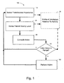
- FIG. 1 is a block diagram of embodiments of a method 100 for controlling patient implanted medical devices incorporating dyspnea measurements.
- the method 100 includes a block 110 providing for the sensing of a patient's breathing, such as by using transthoracic impedance to determine respiration rate and tidal volume.
- a block 120 provides for the sensing of the patient's activity level, such as by using an accelerometer, an EMG sensor, an EEG sensor, or other patient activity level sensing methodology.
- Measured parameters such as respiration rate, minute ventilation, activity level, and/or statistical analyses of measured parameters may be used at a block 140 to compute one or more index indicative of the patient's cardiopulmonary status.
- the index value from block 140 is compared at a decision block 150 to determine if some action is necessary.
- an index value may be compared to an acceptable range, to determine if the value lies within an expected or acceptable range.
- the rate of change of the index value may be compared to an acceptable rate of change, to determine if the value lies within an expected or acceptable range. If the index value is acceptable, the method 100 returns to the block 110 for subsequent sensing and determinations. If the index value is not acceptable, an action 160 may be performed to attempt to bring the patient to an acceptable cardiopulmonary function, alert the patient or physician, or perform other appropriate actions before returning to the block 110.
- a profile 130 may be used to determine the cardiopulmonary condition of the patient.
- the blocks 110,120 may provide sensing information to the profile 130.
- the profile 130 may use, for example, look-up tables of index values for ranges of patient activity, which may be useful for determining if the patient requires some form of action 160, or if the sensed parameters are expected for the patient.
- Profile information may be used exclusively in the determination 150, or combined with information from the index computed at block 140.
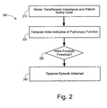
- FIG. 2 is a block diagram of a dyspnea determination method 200.
- Transthoracic impedance and patient activity level are sensed at a block 210.
- Transthoracic impedance and patient activity level measurements from the block 210 are used to compute one or more index vale indicative of pulmonary function at a block 220.
- the index value from the block 220 is compared, at a decision 230, to determine if the index value is beyond an acceptable threshold. If the index value is beyond the threshold, a dyspnea episode is detected at block 240.
- a dyspnea index may be computed at block 220 by dividing the patient's respiratory rate by the patient's tidal volume (RR/Vt).
- the patient's PIMD may be adapted by increasing the patient's heart rate 5-10 beats per minute.
- the patient's PIMD may further increase the patient's heart rate, and/or may alert the patient to a potential problem.
- the patient's PIMD may alert emergency response services to respond to the patient's needs.
- the index values above represent potential values based on calculation coefficients for the particular index, and that ranges of index values may be established clinically and/or individually for each patient and/or each index. It is also understood that the actions described above associated with the index values are for purposes of illustration only, and are not intended as limiting descriptions.
- ICD implantable cardioverter/defibrillator
- Examples of ICD circuitry, structures and functionality, aspects of which may be incorporated in a PIMD of a type that may benefit from dyspnea measuring methods and implementations are disclosed in U.S. Patent Nos. 5,133,353 ; 5,179,945 ; 5,314,459 ; 5,318,597 ; 5,620,466 ; and 5,662,688 .
- systems and methods may perform functions traditionally performed by pacemakers, such as providing various pacing therapies as are known in the art, in addition to cardioversion/defibrillation therapies.
- pacemaker circuitry, structures and functionality, aspects of which may be incorporated in a PIMD of a type that may benefit from dyspnea measuring methods and implementations are disclosed in U.S. Patent Nos. 4,562,841 ; 5,284,136 ; 5,376,106 ; 5,036,849 ; 5,540,727 ; 5,836,987 ; 6,044,298 ; and 6,055,454 .
- PIMD configurations may provide for non-physiologic pacing support in addition to, or to the exclusion of, bradycardia and/or anti-tachycardia pacing therapies.
- a PIMD may implement diagnostic and/or monitoring functions as well as provide cardiac stimulation therapy. Examples of cardiac monitoring circuitry, structures and functionality, aspects of which may be incorporated in a PIMD of a type that may benefit from dyspnea measuring methods and implementations are disclosed in U.S. Patent Nos. 5,313,953 ; 5,388,578 ; and 5,411,031 .
- a PIMD may incorporate CHF features involving dual-chamber or bi-ventricular pacing/therapy, cardiac resynchronization therapy, cardiac function optimization, or other CHF related methodologies.
- a PIMD may incorporate features of one or more of the following references: US Patent Publication No. 2003/0130702 ; and US Patent Nos. 6,411,848 ; 6,285,907 ; 4,928,688 ; 6,459,929 ; 5,334,222 ; 6,026,320 ; 6,371,922 ; 6,597,951 ; 6,424,865 ; and 6,542,775 .
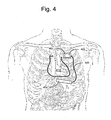
- the implantable device illustrated in Figure 3 is an embodiment of a PIMD configured to determine dyspnea.
- the implantable device includes a cardiac rhythm management device (CRM) 300 including an implantable pulse generator 305 electrically and physically coupled to an intracardiac lead system 310.
- CRM cardiac rhythm management device
- the intracardiac lead system 310 includes one or more electrodes configured to sense electrical cardiac activity of the heart, deliver electrical stimulation to the heart, sense the patient's transthoracic impedance, and/or sense other physiological parameters, e.g., cardiac chamber pressure or temperature. Portions of the housing 301 of the pulse generator 305 may optionally serve as a can electrode.
- Communications circuitry is disposed within the housing 301 for facilitating communication between the pulse generator 305 and an external communication device, such as a portable or bed-side communication station, patient-carried/worn communication station, or external programmer, for example.
- the communications circuitry may also facilitate unidirectional or bidirectional communication with one or more implanted, external, cutaneous, or subcutaneous physiologic or non-physiologic sensors, patient-input devices and/or information systems.
- the pulse generator 305 may optionally incorporate a motion detector 320 that may be used to sense patient activity as well as various respiration and cardiac related conditions.
- the motion detector 320 may be optionally configured to sense snoring, activity level, and/or chest wall movements associated with respiratory effort, for example.
- the motion detector 320 may be implemented as an accelerometer positioned in or on the housing 301 of the pulse generator 305. If the motion sensor is implemented as an accelerometer, the motion sensor may also provide respiratory, e.g. rales, coughing, and cardiac, e.g. S1-S4 heart sounds, murmurs, and other acoustic information.
- the lead system 310 and pulse generator 305 of the CRM 300 may incorporate one or more transthoracic impedance sensors that may be used to acquire the patient's respiration waveform, or other respiration-related information.
- the transthoracic impedance sensor may include, for example, one or more intracardiac electrodes 341, 342, 351-355, 363 positioned in one or more chambers of the heart 390.
- the intracardiac electrodes 341, 342, 351-355, 363 may be coupled to impedance drive/sense circuitry 330 positioned within the housing of the pulse generator 305.
- impedance drive/sense circuitry 330 generates a current that flows through the tissue between an impedance drive electrode 351 and a can electrode on the housing 301 of the pulse generator 305.
- the voltage at an impedance sense electrode 352 relative to the can electrode changes as the patient's transthoracic impedance changes.
- the voltage signal developed between the impedance sense electrode 352 and the can electrode is detected by the impedance sense circuitry 330.
- Other locations and/or combinations of impedance sense and drive electrodes are also possible.
- the lead system 310 may include one or more cardiac pace/sense electrodes 351-355 positioned in, on, or about one or more heart chambers for sensing electrical signals from the patient's heart 390 and/or delivering pacing pulses to the heart 390.
- the intracardiac sense/pace electrodes 351-355 such as those illustrated in Figure 3 , may be used to sense and/or pace one or more chambers of the heart, including the left ventricle, the right ventricle, the left atrium and/or the right atrium.
- the lead system 310 may include one or more defibrillation electrodes 341, 342 for delivering defibrillation/cardioversion shocks to the heart.
- the pulse generator 305 may include circuitry for detecting cardiac arrhythmias and/or for controlling pacing or defibrillation therapy in the form of electrical stimulation pulses or shocks delivered to the heart through the lead system 310.
- the pulse generator 305 may also incorporate circuitry, structures and functionality of the implantable medical devices disclosed in U.S. Patent Nos. 5,203,348 ; 5,230,337 ; 5,360,442 ; 5,366,496 ; 5,397,342 ; 5,391,200 ; 5,545,202 ; 5,603,732 ; and 5,916,243 ; 6,360,127 ; 6,597,951 ; and US Patent Publication No. 2002/0143264 .
- PIMD's that may be implanted under the skin in the chest region of a patient.
- a PIMD may, for example, be implanted subcutaneously such that all or selected elements of the device are positioned on the patient's front, back, side, or other body locations suitable for sensing cardiac activity and/or delivering cardiac stimulation therapy. It is understood that elements of the PIMD may be located at several different body locations, such as in the chest, abdominal, or subclavian region with electrode elements respectively positioned at different regions near, around, in, or on the heart.
- the primary housing (e.g., the active or non-active can) of the PIMD may be configured for positioning outside of the rib cage at an intercostal or subcostal location, within the abdomen, or in the upper chest region (e.g., subclavian location, such as above the third rib).
- one or more leads incorporating electrodes may be located in direct contact with the heart, great vessel or coronary vasculature, such as via one or more leads implanted by use of conventional transvenous delivery approaches.
- one or more electrodes may be located on the primary housing and/or at other locations about, but not in direct contact with the heart, great vessel or coronary vasculature.
- one or more electrode subsystems or electrode arrays may be used to sense cardiac activity and deliver cardiac stimulation energy in a PIMD configuration employing an active can or a configuration employing a non-active can. Electrodes may be situated at anterior and/or posterior locations relative to the heart. Examples of useful electrode locations and features that may be incorporated in various embodiments are described in US Patent Publication Nos. 2004/0230230 ; and 2004/0230243 .
- electrode subsystems of a PIMD system are arranged about a patient's heart 410.
- the PIMD system includes a first electrode subsystem, comprising a can electrode 402, and a second electrode subsystem 404 that includes at least two electrodes or at least one multi-element electrode.
- the second electrode subsystem 404 may include a number of electrodes used for sensing and/or electrical stimulation.
- the second electrode subsystem 404 may include a combination of electrodes.
- the combination of electrodes of the second electrode subsystem 404 may include coil electrodes, tip electrodes, ring electrodes, multi-element coils, spiral coils, spiral coils mounted on non-conductive backing, screen patch electrodes, and other electrode configurations as will be described below.
- a suitable non-conductive backing material is silicone rubber, for example.
- the can electrode 402 is positioned on the housing 401 that encloses the PIMD electronics.
- the can electrode 402 includes the entirety of the external surface of housing 401.
- various portions of the housing 401 may be electrically isolated from the can electrode 402 or from tissue.
- the active area of the can electrode 402 may include all or a portion of either the anterior or posterior surface of the housing 401 to direct current flow in a manner advantageous for cardiac sensing and/or stimulation.
- the housing 401 may resemble that of a conventional implantable PIMD, is approximately 20-100 cc in volume, with a thickness of 0.4 to 2 cm and with a surface area on each face of approximately 30 to 100 cm 2 .
- portions of the housing may be electrically isolated from tissue to optimally direct current flow.
- portions of the housing 401 may be covered with a non-conductive, or otherwise electrically resistive, material to direct current flow.
- Suitable non-conductive material coatings include those formed from silicone rubber, polyurethane, or parylene, for example.
- Figure 5 illustrates a block diagram of a PIMD 602, which includes a dyspnea processor 603 which may be incorporated into and/or work in cooperation with a microprocessor 606.
- a detection circuit 602 which may be coupled to the dyspnea processor 603 and/or the microprocessor 606, may be configured to incorporate, or communicate with, specialized circuitry for processing sensed cardiac signals in manners particularly useful in a cardiac sensing and/or stimulation device. As is shown by way of example in Figure 5 , the detection circuitry 602 may receive information from multiple physiologic and non-physiologic sensors.
- the detection circuitry 602 receives information from one or more sensor(s) 605 that monitor transthoracic impedance.
- transthoracic impedance sensor(s) 605 may be the same as or different from one or more cardiac electrodes 607 used for cardiac sensing and/or stimulation.
- the dyspnea processor 603 is coupled to the sensor(s) 605 and configured to compute an index indicative of pulmonary function using the sensed transthoracic impedance.
- An activity sensor 610 is coupled to the dyspnea processor 603 and configured to sense patient activity.
- the activity sensor 610 may be, for example, an accelerometer in, on, or coupled to the PIMD 602.
- Therapy circuitry 620 is coupled to the microprocessor 606 and configured to provide a therapy at least partly based on a computed pulmonary function index value and a sensed patient activity level determined by the dyspnea processor 603.
- Therapy circuitry 620 is coupled to one or more of the cardiac electrodes 607 and configured to deliver a cardiac therapy.
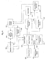
- FIG. 6 is a block diagram depicting various components of a PIMD in accordance with one configuration.
- the PIMD incorporates a processor-based control system 505 that includes a microprocessor 506 coupled to appropriate memory (volatile and non-volatile) 509, it being understood that any logic-based control architecture may be used.
- the control system 505 is coupled to circuitry and components to sense, detect, and analyze electrical signals produced by the heart and deliver electrical stimulation energy to the heart under predetermined conditions to treat cardiac arrhythmias.
- the control system 505 and associated components also provide pacing therapy to the heart.
- the electrical energy delivered by the PIMD may be in the form of low energy pacing pulses or high-energy pulses for cardioversion or defibrillation.
- Cardiac signals are sensed using the electrode(s) 514 and the can or indifferent electrode 507 provided on the PIMD housing. Cardiac signals may also be sensed using only the electrode(s) 514, such as in a non-active can configuration. As such, unipolar, bipolar, or combined unipolar/bipolar electrode configurations as well as multi-element electrodes and combinations of noise canceling and standard electrodes may be employed.
- the sensed cardiac signals are received by sensing circuitry 504, which includes sense amplification circuitry and may also include filtering circuitry and an analog-to-digital (A/D) converter.
- the sensed cardiac signals processed by the sensing circuitry 504 may be received by noise reduction circuitry 503, which may further reduce noise before signals are sent to the detection circuitry 502.
- Noise reduction circuitry 503 may also be incorporated after sensing circuitry 502 in cases where high power or computationally intensive noise reduction algorithms are required.
- the noise reduction circuitry 503, by way of amplifiers used to perform operations with the electrode signals, may also perform the function of the sensing circuitry 504. Combining the functions of sensing circuitry 504 and noise reduction circuitry 503 may be useful to minimize the necessary componentry and lower the power requirements of the system.
- Patient activity may be sensed by a patient activity sensor 515, coupled to the microprocessor 506, to provide patient activity information.
- the patient activity information may be used by the microprocessor 506 to determine a pulmonary function index value as described above.
- the detection circuitry 502 is coupled to, or otherwise incorporates, noise reduction circuitry 503.
- the noise reduction circuitry 503 operates to improve the SNR of sensed cardiac signals by removing noise content of the sensed cardiac signals introduced from various sources.
- Detection circuitry 502 includes a signal processor that coordinates analysis of the sensed cardiac signals, patient activity information, and transthoracic impedance signals to detect dyspnea.
- Rate based and/or morphological discrimination algorithms may be implemented by the signal processor of the detection circuitry 502 to detect and verify the presence and severity of an arrhythmic episode.
- Examples of arrhythmia detection and discrimination circuitry, structures, and techniques, aspects of which may be implemented by a PIMD of a type that may benefit from dyspnea measuring methods and implementations are disclosed in U.S. Patent Nos. 5,301,677 , 6,438,410 , and 6,708,058 .
- Arrhythmia detection methodologies particularly well suited for implementation in cardiac monitoring and/or stimulation systems are described hereinbelow.
- the detection circuitry 502 communicates cardiac signal information to the control system 505.
- Memory circuitry 509 of the control system 505 contains parameters for operating in various sensing, defibrillation, and, if applicable, pacing modes, and stores data indicative of cardiac signals received by the detection circuitry 502.
- the memory circuitry 509 may also be configured to store historical ECG and therapy data, patient activity data, pulmonary function index data, and/or dyspnea information, which may be used for various purposes and transmitted to an external receiving device as needed or desired.
- the PIMD may include diagnostics circuitry 510.
- the diagnostics circuitry 510 typically receives input signals from the detection circuitry 502 and the sensing circuitry 504.
- the diagnostics circuitry 510 provides diagnostics data to the control system 505, it being understood that the control system 505 may incorporate all or part of the diagnostics circuitry 510 or its functionality.
- the control system 505 may store and use information provided by the diagnostics circuitry 510 for a variety of diagnostics purposes. This diagnostic information may be stored, for example, subsequent to a triggering event or at predetermined intervals, and may include system diagnostics, such as power source status, therapy delivery history, and/or patient diagnostics.
- the diagnostic information may take the form of electrical signals or other sensor data acquired immediately prior to therapy delivery.
- the control system 505 processes cardiac signal data received from the detection circuitry 502 and initiates appropriate tachyarrhythmia therapies to terminate cardiac arrhythmic episodes and return the heart to normal sinus rhythm.
- the control system 505 is coupled to shock therapy circuitry 516.
- the shock therapy circuitry 516 is coupled to the electrode(s) 514 and the can or indifferent electrode 507 of the PIMD housing. Upon command, the shock therapy circuitry 516 delivers cardioversion and defibrillation stimulation energy to the heart in accordance with a selected cardioversion or defibrillation therapy.
- the shock therapy circuitry 516 is controlled to deliver defibrillation therapies, in contrast to a configuration that provides for delivery of both cardioversion and defibrillation therapies.
- a PIMD may incorporate a cardiac pacing capability in addition to, or to the exclusion of, cardioversion and/or defibrillation capabilities.
- the PIMD includes pacing therapy circuitry 530 that is coupled to the control system 505 and the electrode(s) 514 and can/indifferent electrodes 507.
- the pacing therapy circuitry 530 delivers pacing pulses to the heart in accordance with a selected pacing therapy.
- Control signals developed in accordance with a pacing regimen by pacemaker circuitry within the control system 505, are initiated and transmitted to the pacing therapy circuitry 530 where pacing pulses are generated.
- a pacing regimen such as those discussed and incorporated herein, may be modified by the control system 505.
- the PIMD shown in Figure 6 may be configured to receive signals from one or more physiologic and/or non-physiologic sensors. Depending on the type of sensor employed, signals generated by the sensors may be communicated to transducer circuitry coupled directly to the detection circuitry 502 or indirectly via the sensing circuitry 504. It is noted that certain sensors may transmit sense data to the control system 505 without processing by the detection circuitry 502.
- Communications circuitry 518 is coupled to the microprocessor 506 of the control system 505.
- the communications circuitry 518 allows the PIMD to communicate with one or more receiving devices or systems situated external to the PIMD.
- the PIMD may communicate with a patient-worn, portable or bedside communication system via the communications circuitry 518.
- one or more physiologic or non-physiologic sensors may be equipped with a short-range wireless communication interface, such as an interface conforming to a known communications standard, such as Bluetooth or IEEE 802 standards. Data acquired by such sensors may be communicated to the PIMD via the communications circuitry 518.
- physiologic or non-physiologic sensors equipped with wireless transmitters or transceivers may communicate with a receiving system external of the patient.
- the communications circuitry 518 preferably allows the PIMD to communicate with an external programmer.
- the communications circuitry 518 and the programmer unit use a wire loop antenna and a radio frequency telemetric link, as is known in the art, to receive and transmit signals and data between the programmer unit and communications circuitry 518.
- programming commands and data are transferred between the PIMD and the programmer unit during and after implant.
- a physician is able to set or modify various parameters used by the PIMD. For example, a physician may set or modify parameters affecting sensing, detection, pacing, and defibrillation functions of the PIMD, including pacing and cardioversion/defibrillation therapy modes.
- the PIMD is encased and hermetically sealed in a housing suitable for implanting in a human body as is known in the art.
- Power to the PIMD is supplied by an electrochemical power source 520 housed within the PIMD.
- the power source 520 includes a rechargeable battery.
- charging circuitry is coupled to the power source 520 to facilitate repeated non-invasive charging of the power source 520.
- the communications circuitry 518, or separate receiver circuitry, is configured to receive RF energy transmitted by an external RF energy transmitter.
- the PIMD may, in addition to a rechargeable power source, include a non-rechargeable battery. It is understood that a rechargeable power source need not be used, in which case a long-life non-rechargeable battery is employed.
- PIMDs implantable cardiac monitoring and/or stimulation device configurations
- PIMDs implantable cardiac monitoring and/or stimulation device configurations
- particular PIMD or cardiac monitoring and/or stimulation device configurations may include particular features as described herein, while other such device configurations may exclude particular features described herein.
- a PIMD may be H A implemented to include an electrode system that provides for one or both of cardiac sensing and arrhythmia therapy delivery.
- a PIMD may be implemented as a chronically implantable system that performs monitoring, diagnostic and/or therapeutic functions.
- the PIMD may automatically detect and treat cardiac arrhythmias.
- the PIMD includes a pulse generator and three or more electrodes that are implanted subcutaneously in the chest region of the body, such as in the anterior thoracic region of the body.
- the PIMD may be used to provide atrial and ventricular therapy for bradycardia and tachycardia arrhythmias.
- Tachyarrhythmia therapy may include cardioversion, defibrillation and anti-tachycardia pacing (ATP), for example, to treat atrial or ventricular tachycardia or fibrillation.
- Bradycardia therapy may include temporary post-shock pacing for bradycardia or asystole. Methods and systems for implementing post-shock pacing for bradycardia or asystole are described in U.S. Patent Publication No. Application entitled "Subcutaneous Cardiac Stimulator Employing Post-Shock Asystole Prevention Pacing, Serial Number 2004/0972066.
- the PIMD may detect a variety of physiological signals that may be used in connection with various diagnostic, therapeutic or monitoring implementations.
- the PIMD may include sensors or circuitry for detecting respiratory system signals, cardiac system signals, and signals related to patient activity.
- the PIMD senses intrathoracic impedance, from which various respiratory parameters may be derived, including, for example, respiratory tidal volume and minute ventilation.
- Sensors and associated circuitry may be incorporated in connection with a PIMD for detecting one or more body movement or body posture or position related signals.
- accelerometers and GPS devices may be employed to detect patient activity, patient location, body orientation, or torso position.
- a PIMD may be used within the structure of an advanced patient management (APM) system 700.
- the advanced patient management system 700 allows physicians to remotely and automatically monitor cardiac and respiratory functions, as well as other patient conditions.
- a PIMD implemented as a cardiac pacemaker, defibrillator, or resynchronization device may be equipped with various telecommunications and information technologies that enable real-time data collection, diagnosis, and treatment of the patient.
- Various PIMD embodiments described herein may be used in connection with advanced patient management. Methods, structures, and/or techniques described herein, which may be adapted to provide for remote patient/device monitoring, diagnosis, therapy, or other APM related methodologies, may incorporate features of one or more of the following references: US Patent Nos.
- the medical system 700 may be used to implement coordinated patient measuring and/or monitoring, diagnosis, and/or therapy.
- the medical system 700 may include, for example, one or more patient-internal medical devices 710, such as a PIMD, and one or more patient-external medical devices 720, such as a monitor or signal display device.
- Each of the patient-internal 710 and patient-external 720 medical devices may include one or more of a patient monitoring unit 712, 722, a diagnostics unit 714, 724, and/or a therapy unit 716, 726.
- the patient-external medical device 720 performs monitoring, and/or diagnosis and/or therapy functions external to the patient (i.e., not invasively implanted within the patient's body).
- the patient-external medical device 720 may be positioned on the patient, near the patient, or in any location external to the patient. I
- the patient-internal and patient-external medical devices 710, 720 may be coupled to one or more sensors 741, 742, 745, 746, patient input / trigger devices 743, 747 and/or other information acquisition devices 744, 748.
- the sensors 741, 742, 745, 746, patient input / trigger devices 743, 747, and/or other information acquisition devices 744, 748 may be employed to detect conditions relevant to the monitoring, diagnostic, and/or therapeutic functions of the patient-internal and patient-external medical devices 710, 720.
- the medical devices 710, 720 may each be coupled to one or more patient-internal sensors 741, 745 that are fully or partially implantable within the patient.
- the medical devices 710, 720 may also be coupled to patient-external sensors positioned on, near, or in a remote location with respect to the patient.
- the patient-internal and patient-external sensors are used to sense conditions, such as physiological or environmental conditions, that affect the patient.
- the patient-internal sensors 741 may be coupled to the patient-internal medical device 710 through one or more internal leads 753. Still referring to Figure 7 , one or more patient-internal sensors 741 may be equipped with transceiver circuitry to support wireless communications between the one or more patient-internal sensors 741 and the patient-internal medical device 710 and/or the patient-external medical device 720.
- the patient-external sensors 742 may be coupled to the patient-internal medical device 710 and/or the patient-external medical device 720 through one or more internal leads 755 or through wireless connections. Patient-external sensors 742 may communicate with the patient-internal medical device 710 wirelessly. Patient-external sensors 742 may be coupled to the patient-external medical device 720 through one or more internal leads 757 or through a wireless link.
- the patient-external medical device 720 includes a visual display configured to concurrently display non-electrophysiological signals and ECG signals. For example, the display may present the information visually.
- the patient-external medical device 720 may also, or alternately, provide signals to other components of the medical system 700 for presentation to a clinician, whether local to the patient or remote to the patient.
- the medical devices 710, 720 may be connected to one or more information acquisition devices 744, 748, such as a database that stores information useful in connection with the monitoring, diagnostic, or therapy functions of the medical devices 710, 720.
- one or more of the medical devices 710, 720 may be coupled through a network to a patient information server 730.
- the input/trigger devices 743, 747 are used to allow the physician, clinician, and/or patient to manually trigger and/or transfer information to the medical devices 710, 720.
- the input/trigger devices 743, 747 may be particularly useful for inputting information concerning patient perceptions, such as a perceived cardiac event, how well the patient feels, and other information not automatically sensed or detected by the medical devices 710, 720.
- the patient may trigger the input/trigger device 743 upon perceiving a cardiac event.
- the trigger may then initiate the recording of cardiac signals and/or other sensor signals in the patient-internal device 710.
- a clinician may trigger the input/trigger device 747, initiating the transfer of the recorded cardiac and/or other signals from the patient-internal device 710 to the patient-external device 720 for display and diagnosis.
- the input/trigger device 747 may also be used by the patient, clinician, and/or physician as an activation stimulus to the PIMD to update and/or select a vector.
- the patient-internal medical device 710 and the patient-external medical device 720 may communicate through a wireless link between the medical devices 710, 720.
- the patient-internal and patient-external devices 710, 720 may be coupled through a short-range radio link, such as Bluetooth, IEEE 802.11, and/or a proprietary wireless protocol.
- the communications link may facilitate uni-directional or bi-directional communication between the patient-internal 710 and patient-external 720 medical devices.
- Data and/or control signals may be transmitted between the patient-internal 710 and patient-external 720 medical devices to coordinate the functions of the medical devices 710, 720.
- patient data may be downloaded from one or more of the medical devices periodically or on command, and stored at the patient information server 730.
- the physician and/or the patient may communicate with the medical devices and the patient information server 730, for example, to acquire patient data or to initiate, terminate or modify recording and/or therapy.
- the data stored on the patient information server 730 may be accessible by the patient and the patient's physician through one or more terminals 750, e.g., remote computers located in the patient's home or the physician's office.
- the patient information server 730 may be used to communicate to one or more of the atient-internal and patient-external medical devices 710, 720 to provide remote control of the monitoring, diagnosis, and/or therapy functions of the medical devices 710, 720.
- the patient's physician may access patient data transmitted from the medical devices 710, 720 to the patient information server 730. After evaluation of the patient data, the patient's physician may communicate with one or more of the patient-internal or patient-external devices 710, 720 through an APM system 740 to initiate, terminate, or modify the monitoring, diagnostic, and/or therapy functions of the patient-internal and/or patient-external medical systems 710, 720.
- the patient-internal and patient-external medical devices 710, 720 may not communicate directly, but may communicate indirectly through the APM system 740.
- the APM system 740 may operate as an intermediary between two or more of the medical devices 710, 720. For example, data and/or control information may be transferred from one of the medical devices 710, 720 to the APM system 740. The APM system 740 may transfer the data and/or control information to another of the medical devices 710, 720.
- the APM system 740 may communicate directly with the patient-internal and/or patient-external medical devices 710, 720. In another embodiment, the APM system 740 may communicate with the patient-internal and/or patient-external medical devices 710, 720 through medical device programmers 760, 770 respectively associated with each medical device 710, 720. As was stated previously, the patient-internal medical device 710 may take the form of an implantable PIMD.
Landscapes
- Health & Medical Sciences (AREA)
- Life Sciences & Earth Sciences (AREA)
- Engineering & Computer Science (AREA)
- Public Health (AREA)
- Biomedical Technology (AREA)
- General Health & Medical Sciences (AREA)
- Heart & Thoracic Surgery (AREA)
- Medical Informatics (AREA)
- Animal Behavior & Ethology (AREA)
- Veterinary Medicine (AREA)
- Pathology (AREA)
- Biophysics (AREA)
- Physics & Mathematics (AREA)
- Molecular Biology (AREA)
- Surgery (AREA)
- Physiology (AREA)
- Cardiology (AREA)
- Pulmonology (AREA)
- Radiology & Medical Imaging (AREA)
- Nuclear Medicine, Radiotherapy & Molecular Imaging (AREA)
- Signal Processing (AREA)
- Artificial Intelligence (AREA)
- Psychiatry (AREA)
- Computer Vision & Pattern Recognition (AREA)
- Databases & Information Systems (AREA)
- Epidemiology (AREA)
- Primary Health Care (AREA)
- Data Mining & Analysis (AREA)
- Dentistry (AREA)
- Oral & Maxillofacial Surgery (AREA)
- Business, Economics & Management (AREA)
- Critical Care (AREA)
- Emergency Medicine (AREA)
- Emergency Management (AREA)
- Nursing (AREA)
- Hospice & Palliative Care (AREA)
- Electrotherapy Devices (AREA)
- Measurement Of The Respiration, Hearing Ability, Form, And Blood Characteristics Of Living Organisms (AREA)
- Measuring And Recording Apparatus For Diagnosis (AREA)
Description
- The present invention relates generally to implantable cardiac devices and, more particularly, to cardiac sensing and/or stimulation devices employing dyspnea measurement.
- Dyspnea is defined as a shortness of breath or difficult breathing (the subjective feeling of being out of breath) caused by heart or lung disorders, strenuous activity, high anxiety or stress. Dyspnea derives from interactions among multiple physiological, psychological, social, and environmental factors, and may induce secondary physiological and behavioral responses. Dyspnea is different from tachypnea (rapid breathing) and hyperpnea (deep breathing). Tachypnea and hyperpnea can occur with hyperventilation, or over breathing beyond what is required to maintain arterial blood gases within normal limits. Fear or anxiety may create even more distress in dyspneic patients.
- Clinically, a dyspnea index has been developed as a monitoring tool for dyspnea. One conventional method of determining a dyspnea index value has a subject take a deep breath in and count out loud from 1 to 15 (should take approximately 8 seconds). When stressed or running out of air, the subject stops counting, takes another deep breath and then continues counting from where they left off. This is repeated as needed until the subject finishes counting to 15. The number of breaths needed to take in addition to the very first one is the dyspnea index.
- Using the subject's dyspnea index measurement, a value of 0-1 is considered normal. A dyspnea value of 2-3 is acceptable, depending on what activity is being performed. At a dyspnea value of 4, the subject should slow down, and think about stopping. At a dyspnea value of 5, the subject should definitely stop and rest.
- Dyspnea may be classified as chronic, acute, or terminal. Chronic dyspnea has a variable intensity and persistent shortness of breath. This is most often seen in patients with chronic obstructive pulmonary disease (COPD). Acute dyspnea causes episodes of shortness of breath with high intensity. It may be seen in patients who have suffered a myocardial infarction or pulmonary embolism. Terminal dyspnea occurs in patients with end-stage diseases, and these patients may be in a hospital, at home, or in a hospice. This type of dyspnea is a common complaint in patients with cancer. Dyspnea can be caused by a variety of conditions, including metabolic, allergic, psychiatric, and neuromuscular disorders, and by pain. However, cardiac and pulmonary disorders are the most common causes.
- To manage dyspnea, nonpharmacological and pharmacological interventions may be performed. For example, treating patients in cardiac failure with digoxin and diuretics may resolve the problem. Stimulation of mechanoreceptors in the respiratory musculature or over the face has reduced dyspnea in some patients. Vibration of the intercostal muscles, in phase with inspiration so that contracting respiratory muscles are vibrated, has relieved dyspnea in some COPD patients. This intervention has worked best with severe dyspnea at rest. Movement of cool air across the face by a fan or an open window can stimulate mechanoreceptors in the face, minimizing mild dyspnea.
- Patients with COPD often obtain relief from dyspnea while sitting and leaning forward with their arms supported on a table. Unsupported arm and shoulder movement alters the efficient use of respiratory muscles. This position may also improve overall inspiratory muscle strength and enhance the efficiency of diaphragmatic breathing. Techniques like pursed-lip and diaphragmatic breathing are widely used to reduce dyspnea in COPD patients, though their effect is variable. These breathing techniques allow for slower and deeper breathing, thus raising tidal volume and decreasing respiratory rates. Pursed-lip breathing is especially useful for patients with emphysema because it creates positive end-expiratory pressure in the alveoli, thus splinting airways that have lost their collagen matrix. However, patients often revert to their fast and shallow breathing patterns, which may be compensatory for the mechanism causing dyspnea.
- Oxygen, administered by mask or nasal cannula or transtracheally, improves dyspnea. In patients with COPD, most authorities recommend oxygen therapy for raising PaO2 levels to at least 55 mmHg to 60 mmHg or oxygen saturation to 88% to 90%.
- Evidence suggests that opioids, despite their possible respiratory depressant effect, are useful in managing dyspnea. While the action of opioids to relieve dyspnea is not fully understood, the drugs may act by blunting the emotional reaction to dyspnea by interaction with opioid receptors in the limbic system. Because opioids cause euphoria, they reduce fear, anxiety, and the associated restlessness and muscle tension that decrease oxygen consumption. Opioids may also relieve dyspnea by action on the chemoreceptors, thus reducing respiratory drive.
- When the dyspnea becomes intolerable and increased doses of systemic opioids are contraindicated because of unacceptable adverse effects, nebulized morphine may be started. Nebulized morphine may relieve dyspnea by direct local action on peripheral opioid receptors in the airways so that it does not reach the systemic concentration to the extent that oral, subcutaneous, or intravenous morphine does. Therefore, some patients experience relief of dyspnea with fewer adverse effects.
- Anxiolytics frequently used to relieve dyspnea include benzodiazepines and phenothiazines. These act by depressing the hypoxic, hypercapnic response to dyspnea and the emotional response to dyspnea. Depending on the cause of dyspnea, patients may benefit from bronchodilators. Since methylxanthines cause smooth muscle dilation of the airways and improve the contraction of the diaphragm, they may be useful in patients with COPD. Similarly, inhaled beta-2 adrenergic agonists and anticholinergics cause smooth, muscle dilation of airways, thus improving lung mechanics and possibly relieving dyspnea.
-
US 2003/0055461 describes systems, devices and methods for predicting how the status of a patient's congestive heart failure condition will progress in the future, for example by measuring acute pulmonary edema by sensing transthoracic impedance or by measuring paroxysmal nocturnal dyspnea by sensing shortness of breath using a respiration sensor and using a sleep detector / activity sensor. -
US 6459929 describes a method and apparatus for providing congestive heart failure therapy status using a CRM device capable of measuring transthoracic impedance and sensing physical activity level.US 2005/0039745 describes methods and apparatus for adaptive therapy for disordered breathing andUS 2005/0043644 . describes methods and apparatus for predicting disordered breathing. - The present invention provides an implantable device as defined in claim 1. Described herein are cardiac monitoring and/or stimulation methods and systems that provide monitoring, defibrillation therapies, pacing therapies, or a combination of these capabilities. Embodiments of the present invention relate generally to implantable medical devices employing dyspnea measurement and/or detection capability.
- Embodiments of methods described herein involve providing an implantable cardiac device configured to sense transthoracic impedance and determine a patient activity level. An index indicative of pulmonary function is implantably computed using the sensed transthoracic impedance. An episode of dyspnea may be detected based on a change in a computed pulmonary function index exceeding a threshold at a determined patient activity level.
- Further embodiments are directed to methods that trend one or more pulmonary function index values to determine a patient's pulmonary function index profile. The threshold for detecting the dyspnea episode may be based on the patient's pulmonary function index profile. Other embodiments may involve trending one or more pulmonary function index values and their associated patient activity levels to determine a patient's pulmonary function index versus activity level profile.
- The threshold for detecting the dyspnea episode may be based on the patient's pulmonary function index versus activity level profile by computing the pulmonary function Index as a ratio of a respiratory rate value and a tidal volume value for a patient. Methods may further involve adapting a cardiac therapy for the patient based on a pulmonary function index value. An adapted cardiac therapy may be delivered to the patient after comparing the pulmonary function index value to a threshold and determining the pulmonary function index value is beyond the threshold. Adapting the cardiac therapy may also involve comparing the pulmonary function index value to a predetermined range and modifying the cardiac therapy if the pulmonary function index value is beyond the predetermined range. Therapy adaptations include increasing or decreasing a rate at which pacing pulses are delivered to the patient's heart, for example increasing or decreasing pacing pulses within a range of about 5 to about 10 beats per minute.
- Methods may further involve determining a patient's sleep-state at least in part using the pulmonary function index and/or a trend of pulmonary function index values. A physician may be automatically alerted in response to a pulmonary function index value and/or a trend of the patient's pulmonary index being beyond a threshold. Computed pulmonary function index values and their associated patient's activity levels may be stored periodically in a memory and/or transmitted to a patient-external device.
- Devices include a sensor configured to sense transthoracic impedance. A controller is coupled to the sensor and configured to compute an index indicative of pulmonary function using the sensed transthoracic impedance. An activity sensor is also coupled to the controller and configured to sense patient activity. Therapy circuitry is coupled to the controller and configured to provide a therapy at least partly based on a computed pulmonary function index value and a sensed patient activity level. An electrode may be coupled to the cardiac therapy circuitry and configured to deliver the cardiac therapy.
- The pulmonary function index may be a dyspnea index, computed as a ratio of a respiration rate to a tidal volume. Memory may be coupled to the controller and configured to store periodically computed pulmonary function index values and their associated patient activity levels. Communications circuitry may also be coupled to the controller and configured to communicate pulmonary function index values and/or their associated patient activity levels to a patient-external device. The device may further provide alerts to the patient and/or physician, such as by using a patient-external device or an advanced patient management system.
- The above summary of the present invention is not intended to describe each embodiment or every implementation of the present invention. Advantages and attainments, together with a more complete understanding of the invention, will become apparent and appreciated by referring to the following detailed description and claims taken in conjunction with the accompanying drawings.
-
-
Figure 1 is a block diagram of embodiments of methods for controlling patient implanted medical devices incorporating dyspnea measurements; -
Figure 2 is a block diagram of a dyspnea determination method; -
Figure 3 is an illustration of an implantable cardiac device including a lead assembly shown implanted in a sectional view of a heart; -
Figure 4 is a diagram illustrating components of a cardiac sensing and/or stimulation device including an electrode array; -
Figure 5 is a block diagram illustrating various processing and detection components of a cardiac sensing and/or stimulation device; -
Figure 6 is a block diagram illustrating various components of a cardiac sensing and/or stimulation device; and -
Figure 7 is a block diagram of a medical system that may be used to implement system updating, coordinated patient monitoring, diagnosis, and/or therapy. - It is to be understood that the intention is not to limit the invention to the particular embodiments described. On the contrary, the invention is intended to cover all modifications, equivalents, and alternatives falling within the scope of the invention as defined by the appended claims.
- In the following description of the illustrated embodiments, references are made to the accompanying drawings. It is to be understood that other embodiments may be utilized, and structural and functional changes may be made without departing from the scope of the present invention as defined by the claims.
- An implanted device may include one or more of the features, structures, methods, or combinations thereof described hereinbelow. For example, a cardiac monitor or a cardiac stimulator may be implemented to include one or more of the advantageous features and/or processes described below. It is intended that such a monitor, stimulator, or other implanted or partially implanted device need not include all of the features described herein, but may be implemented to include selected features that provide for unique structures and/or functionality. Such a device may be implemented to provide a variety of therapeutic or diagnostic functions.
- A wide variety of implantable cardiac sensing and/or stimulation devices may be configured to implement a dyspnea measurement methodology. A non-limiting, representative list of such devices includes cardiac monitors, pacemakers, cardiovertors, defibrillators, resynchronizers, and other cardiac sensing and therapy delivery devices. These devices may be configured with a variety of electrode arrangements, including transvenous, endocardial, and epicardial electrodes (i.e., intrathoracic electrodes), and/or subcutaneous, non-intrathoracic electrodes, including can, header, and indifferent electrodes, and subcutaneous array or lead electrodes (i.e., non-intrathoracic electrodes).
- Embodiments may be implemented in the context of a wide variety of cardiac devices, such as those listed above, and are referred to herein generally as a patient implantable medical device (PIMD) for convenience. A PIMD may incorporate one or more of the electrode types identified above and/or combinations thereof.
- Many patients implanted with a pacemaker/defibrillator/CRT device have minute ventilation changes that reflect a change in the patient's cardiopulmonary function. A decrease in cardiopulmonary function often manifests itself in a form of dyspnea (shortness of breath). Typically, dyspnea can be seen both at rest and during exercise.
- The pulmonary characteristics of a patient that a physician typically evaluates may be described using a dyspnea scale. The dyspnea scale relates the pulmonary parameters of respiratory rate (RR), in breaths per minute, to tidal volume (Vt), in milliliters per minute or liters per minute. A pulmonary index, such as may be obtained by dividing the respiratory rate by the tidal volume, (RR/Vt) for example, gives the physician an evaluation tool for dyspnea measurement. The pulmonary index may be used to detect a dyspnea episode in which the patient has an increased RR confirmed by a decreased Vt.
- Typically, changes in a patient's cardiopulmonary response after a pacemaker has been implanted are not addressed until the patient is symptomatic, and has gone through a formal clinical evaluation with the use of external gas exchange equipment. A PIMD having dyspnea measurement capabilities reduces the response time needed to correct the patient's dyspnea problem and/or to introduce additional therapy. A PIMD having dyspnea measurement capabilities may also provide trending of the patient's dyspnea over time.
- A PIMD that incorporates dyspnea measurement may be implanted in a patient. The PIMD may sense, for example, transthoracic impedance and patient activity. Parameters, such as respiration rate, minute ventilation, and tidal volume, may be detected along with activity levels of the patient associated with the measured parameters.
- Patient profiles may be established, to determine what levels of a particular parameter occur for a given activity level, time of day, or the like. Indexes may be established, such as an RR/Vt index. As a patient is resting, performing moderate exercise, or performing maximum exertion, baselines for patient parameters and/or index values may be determined and recorded in the PIMD.
- Changes in parameters and index values may be used to trigger alerts, modify therapies, or otherwise control the PIMD. Parameters and index values may be displayed to the patient on a patient-external monitor, so that the patient may evaluate and/or modify their behavior. If a dyspnea index value is too high, the PIMD may increase the heart rate a modest amount, for example 5 to 10 beats per minute, to adapt the heart rate to the patient's need for oxygen. PIMDs that incorporate adaptive dyspnea control may provide improved patient performance and less physician intervention than other PIMDs.
- In another embodiment, a PIMD with dyspnea detection capability may develop a model of the cardiopulmonary response of an individual patient to track changes in a patient's condition. Patients whose cardiopulmonary status deteriorates over time can be easily evaluated without the use of external gas exchange equipment. The physician may be alerted to changes in the patient's status, thereby reducing the length and intensity of dyspnea that a patient experiences before intervention.
-
Figure 1 is a block diagram of embodiments of amethod 100 for controlling patient implanted medical devices incorporating dyspnea measurements. Themethod 100 includes ablock 110 providing for the sensing of a patient's breathing, such as by using transthoracic impedance to determine respiration rate and tidal volume. Ablock 120 provides for the sensing of the patient's activity level, such as by using an accelerometer, an EMG sensor, an EEG sensor, or other patient activity level sensing methodology. Measured parameters, such as respiration rate, minute ventilation, activity level, and/or statistical analyses of measured parameters may be used at ablock 140 to compute one or more index indicative of the patient's cardiopulmonary status. The index value fromblock 140 is compared at adecision block 150 to determine if some action is necessary. For example, an index value may be compared to an acceptable range, to determine if the value lies within an expected or acceptable range. In another example, the rate of change of the index value may be compared to an acceptable rate of change, to determine if the value lies within an expected or acceptable range. If the index value is acceptable, themethod 100 returns to theblock 110 for subsequent sensing and determinations. If the index value is not acceptable, anaction 160 may be performed to attempt to bring the patient to an acceptable cardiopulmonary function, alert the patient or physician, or perform other appropriate actions before returning to theblock 110. - In a further embodiment of the
method 100, optionally or additionally, aprofile 130 may be used to determine the cardiopulmonary condition of the patient. The blocks 110,120 may provide sensing information to theprofile 130. Theprofile 130 may use, for example, look-up tables of index values for ranges of patient activity, which may be useful for determining if the patient requires some form ofaction 160, or if the sensed parameters are expected for the patient. Profile information may be used exclusively in thedetermination 150, or combined with information from the index computed atblock 140. -
Figure 2 is a block diagram of adyspnea determination method 200. Transthoracic impedance and patient activity level are sensed at ablock 210. Transthoracic impedance and patient activity level measurements from theblock 210 are used to compute one or more index vale indicative of pulmonary function at ablock 220. - The index value from the
block 220 is compared, at adecision 230, to determine if the index value is beyond an acceptable threshold. If the index value is beyond the threshold, a dyspnea episode is detected atblock 240. For example, a dyspnea index may be computed atblock 220 by dividing the patient's respiratory rate by the patient's tidal volume (RR/Vt). - At a dyspnea index value above 3, the patient's PIMD may be adapted by increasing the patient's heart rate 5-10 beats per minute. At a dyspnea index value above 4, the patient's PIMD may further increase the patient's heart rate, and/or may alert the patient to a potential problem. At a dyspnea index value above 5, the patient's PIMD may alert emergency response services to respond to the patient's needs. It is understood that the index values above represent potential values based on calculation coefficients for the particular index, and that ranges of index values may be established clinically and/or individually for each patient and/or each index. It is also understood that the actions described above associated with the index values are for purposes of illustration only, and are not intended as limiting descriptions.
- Certain configurations illustrated herein are generally described as capable of implementing various functions traditionally performed by an implantable cardioverter/defibrillator (ICD), and may operate in numerous cardioversion/defibrillation modes as are known in the art. Examples of ICD circuitry, structures and functionality, aspects of which may be incorporated in a PIMD of a type that may benefit from dyspnea measuring methods and implementations are disclosed in
U.S. Patent Nos. 5,133,353 ;5,179,945 ;5,314,459 ;5,318,597 ;5,620,466 ; and5,662,688 . - In particular configurations, systems and methods may perform functions traditionally performed by pacemakers, such as providing various pacing therapies as are known in the art, in addition to cardioversion/defibrillation therapies. Examples of pacemaker circuitry, structures and functionality, aspects of which may be incorporated in a PIMD of a type that may benefit from dyspnea measuring methods and implementations are disclosed in
U.S. Patent Nos. 4,562,841 ;5,284,136 ;5,376,106 ;5,036,849 ;5,540,727 ;5,836,987 ;6,044,298 ; and6,055,454 . It is understood that PIMD configurations may provide for non-physiologic pacing support in addition to, or to the exclusion of, bradycardia and/or anti-tachycardia pacing therapies. - A PIMD may implement diagnostic and/or monitoring functions as well as provide cardiac stimulation therapy. Examples of cardiac monitoring circuitry, structures and functionality, aspects of which may be incorporated in a PIMD of a type that may benefit from dyspnea measuring methods and implementations are disclosed in
U.S. Patent Nos. 5,313,953 ;5,388,578 ; and5,411,031 . - Various embodiments described herein may be used in connection with congestive heart failure (CHF) monitoring, diagnosis, and/or therapy. A PIMD may incorporate CHF features involving dual-chamber or bi-ventricular pacing/therapy, cardiac resynchronization therapy, cardiac function optimization, or other CHF related methodologies. For example, a PIMD may incorporate features of one or more of the following references:
US Patent Publication No. 2003/0130702 ; andUS Patent Nos. 6,411,848 ;6,285,907 ;4,928,688 ;6,459,929 ;5,334,222 ;6,026,320 ;6,371,922 ;6,597,951 ;6,424,865 ; and6,542,775 . - Referring now to
Figure 3 , the implantable device illustrated inFigure 3 is an embodiment of a PIMD configured to determine dyspnea. In this example, the implantable device includes a cardiac rhythm management device (CRM) 300 including animplantable pulse generator 305 electrically and physically coupled to anintracardiac lead system 310. - Portions of the
intracardiac lead system 310 are inserted into the patient'sheart 390. Theintracardiac lead system 310 includes one or more electrodes configured to sense electrical cardiac activity of the heart, deliver electrical stimulation to the heart, sense the patient's transthoracic impedance, and/or sense other physiological parameters, e.g., cardiac chamber pressure or temperature. Portions of thehousing 301 of thepulse generator 305 may optionally serve as a can electrode. - Communications circuitry is disposed within the
housing 301 for facilitating communication between thepulse generator 305 and an external communication device, such as a portable or bed-side communication station, patient-carried/worn communication station, or external programmer, for example. The communications circuitry may also facilitate unidirectional or bidirectional communication with one or more implanted, external, cutaneous, or subcutaneous physiologic or non-physiologic sensors, patient-input devices and/or information systems. - The
pulse generator 305 may optionally incorporate amotion detector 320 that may be used to sense patient activity as well as various respiration and cardiac related conditions. For example, themotion detector 320 may be optionally configured to sense snoring, activity level, and/or chest wall movements associated with respiratory effort, for example. Themotion detector 320 may be implemented as an accelerometer positioned in or on thehousing 301 of thepulse generator 305. If the motion sensor is implemented as an accelerometer, the motion sensor may also provide respiratory, e.g. rales, coughing, and cardiac, e.g. S1-S4 heart sounds, murmurs, and other acoustic information. - The
lead system 310 andpulse generator 305 of theCRM 300 may incorporate one or more transthoracic impedance sensors that may be used to acquire the patient's respiration waveform, or other respiration-related information. The transthoracic impedance sensor may include, for example, one or moreintracardiac electrodes heart 390. Theintracardiac electrodes sense circuitry 330 positioned within the housing of thepulse generator 305. - In one implementation, impedance drive/
sense circuitry 330 generates a current that flows through the tissue between animpedance drive electrode 351 and a can electrode on thehousing 301 of thepulse generator 305. The voltage at animpedance sense electrode 352 relative to the can electrode changes as the patient's transthoracic impedance changes. The voltage signal developed between theimpedance sense electrode 352 and the can electrode is detected by theimpedance sense circuitry 330. Other locations and/or combinations of impedance sense and drive electrodes are also possible. - The
lead system 310 may include one or more cardiac pace/sense electrodes 351-355 positioned in, on, or about one or more heart chambers for sensing electrical signals from the patient'sheart 390 and/or delivering pacing pulses to theheart 390. The intracardiac sense/pace electrodes 351-355, such as those illustrated inFigure 3 , may be used to sense and/or pace one or more chambers of the heart, including the left ventricle, the right ventricle, the left atrium and/or the right atrium. Thelead system 310 may include one ormore defibrillation electrodes - The
pulse generator 305 may include circuitry for detecting cardiac arrhythmias and/or for controlling pacing or defibrillation therapy in the form of electrical stimulation pulses or shocks delivered to the heart through thelead system 310. Thepulse generator 305 may also incorporate circuitry, structures and functionality of the implantable medical devices disclosed inU.S. Patent Nos. 5,203,348 ;5,230,337 ;5,360,442 ;5,366,496 ;5,397,342 ;5,391,200 ;5,545,202 ;5,603,732 ; and5,916,243 ;6,360,127 ;6,597,951 ; andUS Patent Publication No. 2002/0143264 .
For purposes of illustration, and not of limitation, various embodiments of devices that may use dyspnea measurement are described herein in the context of PIMD's that may be implanted under the skin in the chest region of a patient. A PIMD may, for example, be implanted subcutaneously such that all or selected elements of the device are positioned on the patient's front, back, side, or other body locations suitable for sensing cardiac activity and/or delivering cardiac stimulation therapy. It is understood that elements of the PIMD may be located at several different body locations, such as in the chest, abdominal, or subclavian region with electrode elements respectively positioned at different regions near, around, in, or on the heart. - The primary housing (e.g., the active or non-active can) of the PIMD, for example, may be configured for positioning outside of the rib cage at an intercostal or subcostal location, within the abdomen, or in the upper chest region (e.g., subclavian location, such as above the third rib). In one implementation, one or more leads incorporating electrodes may be located in direct contact with the heart, great vessel or coronary vasculature, such as via one or more leads implanted by use of conventional transvenous delivery approaches. In another implementation, one or more electrodes may be located on the primary housing and/or at other locations about, but not in direct contact with the heart, great vessel or coronary vasculature.
- In a further implementation, for example, one or more electrode subsystems or electrode arrays may be used to sense cardiac activity and deliver cardiac stimulation energy in a PIMD configuration employing an active can or a configuration employing a non-active can. Electrodes may be situated at anterior and/or posterior locations relative to the heart. Examples of useful electrode locations and features that may be incorporated in various embodiments are described in
US Patent Publication Nos. 2004/0230230 ; and2004/0230243 . - In one configuration, as is illustrated in
Figure 4 , electrode subsystems of a PIMD system are arranged about a patient'sheart 410. The PIMD system includes a first electrode subsystem, comprising acan electrode 402, and a second electrode subsystem 404 that includes at least two electrodes or at least one multi-element electrode. The second electrode subsystem 404 may include a number of electrodes used for sensing and/or electrical stimulation. - In various configurations, the second electrode subsystem 404 may include a combination of electrodes. The combination of electrodes of the second electrode subsystem 404 may include coil electrodes, tip electrodes, ring electrodes, multi-element coils, spiral coils, spiral coils mounted on non-conductive backing, screen patch electrodes, and other electrode configurations as will be described below. A suitable non-conductive backing material is silicone rubber, for example.
- The
can electrode 402 is positioned on thehousing 401 that encloses the PIMD electronics. In one embodiment, thecan electrode 402 includes the entirety of the external surface ofhousing 401. In other embodiments, various portions of thehousing 401 may be electrically isolated from the can electrode 402 or from tissue. For example, the active area of thecan electrode 402 may include all or a portion of either the anterior or posterior surface of thehousing 401 to direct current flow in a manner advantageous for cardiac sensing and/or stimulation. - In accordance with one embodiment, the
housing 401 may resemble that of a conventional implantable PIMD, is approximately 20-100 cc in volume, with a thickness of 0.4 to 2 cm and with a surface area on each face of approximately 30 to 100 cm2. As previously discussed, portions of the housing may be electrically isolated from tissue to optimally direct current flow. For example, portions of thehousing 401 may be covered with a non-conductive, or otherwise electrically resistive, material to direct current flow. Suitable non-conductive material coatings include those formed from silicone rubber, polyurethane, or parylene, for example. -
Figure 5 illustrates a block diagram of aPIMD 602, which includes adyspnea processor 603 which may be incorporated into and/or work in cooperation with amicroprocessor 606. Adetection circuit 602, which may be coupled to thedyspnea processor 603 and/or themicroprocessor 606, may be configured to incorporate, or communicate with, specialized circuitry for processing sensed cardiac signals in manners particularly useful in a cardiac sensing and/or stimulation device. As is shown by way of example inFigure 5 , thedetection circuitry 602 may receive information from multiple physiologic and non-physiologic sensors. - The
detection circuitry 602 receives information from one or more sensor(s) 605 that monitor transthoracic impedance. As is known in the art, transthoracic impedance sensor(s) 605 may be the same as or different from one or morecardiac electrodes 607 used for cardiac sensing and/or stimulation. - The
dyspnea processor 603 is coupled to the sensor(s) 605 and configured to compute an index indicative of pulmonary function using the sensed transthoracic impedance. Anactivity sensor 610 is coupled to thedyspnea processor 603 and configured to sense patient activity. Theactivity sensor 610 may be, for example, an accelerometer in, on, or coupled to thePIMD 602.Therapy circuitry 620 is coupled to themicroprocessor 606 and configured to provide a therapy at least partly based on a computed pulmonary function index value and a sensed patient activity level determined by thedyspnea processor 603.Therapy circuitry 620 is coupled to one or more of thecardiac electrodes 607 and configured to deliver a cardiac therapy. -
Figure 6 is a block diagram depicting various components of a PIMD in accordance with one configuration. According to this configuration, the PIMD incorporates a processor-basedcontrol system 505 that includes amicroprocessor 506 coupled to appropriate memory (volatile and non-volatile) 509, it being understood that any logic-based control architecture may be used. Thecontrol system 505 is coupled to circuitry and components to sense, detect, and analyze electrical signals produced by the heart and deliver electrical stimulation energy to the heart under predetermined conditions to treat cardiac arrhythmias. Thecontrol system 505 and associated components also provide pacing therapy to the heart. The electrical energy delivered by the PIMD may be in the form of low energy pacing pulses or high-energy pulses for cardioversion or defibrillation. - Cardiac signals are sensed using the electrode(s) 514 and the can or
indifferent electrode 507 provided on the PIMD housing. Cardiac signals may also be sensed using only the electrode(s) 514, such as in a non-active can configuration. As such, unipolar, bipolar, or combined unipolar/bipolar electrode configurations as well as multi-element electrodes and combinations of noise canceling and standard electrodes may be employed. The sensed cardiac signals are received by sensingcircuitry 504, which includes sense amplification circuitry and may also include filtering circuitry and an analog-to-digital (A/D) converter. The sensed cardiac signals processed by thesensing circuitry 504 may be received bynoise reduction circuitry 503, which may further reduce noise before signals are sent to thedetection circuitry 502. -
Noise reduction circuitry 503 may also be incorporated after sensingcircuitry 502 in cases where high power or computationally intensive noise reduction algorithms are required. Thenoise reduction circuitry 503, by way of amplifiers used to perform operations with the electrode signals, may also perform the function of thesensing circuitry 504. Combining the functions ofsensing circuitry 504 andnoise reduction circuitry 503 may be useful to minimize the necessary componentry and lower the power requirements of the system. - Patient activity may be sensed by a
patient activity sensor 515, coupled to themicroprocessor 506, to provide patient activity information. The patient activity information may be used by themicroprocessor 506 to determine a pulmonary function index value as described above. - In the illustrative configuration shown in
Figure 6 , thedetection circuitry 502 is coupled to, or otherwise incorporates,noise reduction circuitry 503. Thenoise reduction circuitry 503 operates to improve the SNR of sensed cardiac signals by removing noise content of the sensed cardiac signals introduced from various sources.Detection circuitry 502 includes a signal processor that coordinates analysis of the sensed cardiac signals, patient activity information, and transthoracic impedance signals to detect dyspnea. - Rate based and/or morphological discrimination algorithms may be implemented by the signal processor of the
detection circuitry 502 to detect and verify the presence and severity of an arrhythmic episode. Examples of arrhythmia detection and discrimination circuitry, structures, and techniques, aspects of which may be implemented by a PIMD of a type that may benefit from dyspnea measuring methods and implementations are disclosed inU.S. Patent Nos. 5,301,677 ,6,438,410 , and6,708,058 . Arrhythmia detection methodologies particularly well suited for implementation in cardiac monitoring and/or stimulation systems are described hereinbelow. - The
detection circuitry 502 communicates cardiac signal information to thecontrol system 505.Memory circuitry 509 of thecontrol system 505 contains parameters for operating in various sensing, defibrillation, and, if applicable, pacing modes, and stores data indicative of cardiac signals received by thedetection circuitry 502. Thememory circuitry 509 may also be configured to store historical ECG and therapy data, patient activity data, pulmonary function index data, and/or dyspnea information, which may be used for various purposes and transmitted to an external receiving device as needed or desired. - In certain configurations, the PIMD may include
diagnostics circuitry 510. Thediagnostics circuitry 510 typically receives input signals from thedetection circuitry 502 and thesensing circuitry 504. Thediagnostics circuitry 510 provides diagnostics data to thecontrol system 505, it being understood that thecontrol system 505 may incorporate all or part of thediagnostics circuitry 510 or its functionality. Thecontrol system 505 may store and use information provided by thediagnostics circuitry 510 for a variety of diagnostics purposes. This diagnostic information may be stored, for example, subsequent to a triggering event or at predetermined intervals, and may include system diagnostics, such as power source status, therapy delivery history, and/or patient diagnostics. The diagnostic information may take the form of electrical signals or other sensor data acquired immediately prior to therapy delivery. - According to a configuration that provides cardioversion and defibrillation therapies, the
control system 505 processes cardiac signal data received from thedetection circuitry 502 and initiates appropriate tachyarrhythmia therapies to terminate cardiac arrhythmic episodes and return the heart to normal sinus rhythm. Thecontrol system 505 is coupled toshock therapy circuitry 516. Theshock therapy circuitry 516 is coupled to the electrode(s) 514 and the can orindifferent electrode 507 of the PIMD housing. Upon command, theshock therapy circuitry 516 delivers cardioversion and defibrillation stimulation energy to the heart in accordance with a selected cardioversion or defibrillation therapy. In a less sophisticated configuration, theshock therapy circuitry 516 is controlled to deliver defibrillation therapies, in contrast to a configuration that provides for delivery of both cardioversion and defibrillation therapies. Examples of PIMD high energy delivery circuitry, structures and functionality, aspects of which may be incorporated in a PIMD of a type that may benefit from aspects disclosed inU.S. Patent Nos. 5,372,606 ;5,411,525 ;5,468,254 ; and5,634,938 . - In accordance with another configuration, a PIMD may incorporate a cardiac pacing capability in addition to, or to the exclusion of, cardioversion and/or defibrillation capabilities. As is shown in
Figure 6 , the PIMD includes pacingtherapy circuitry 530 that is coupled to thecontrol system 505 and the electrode(s) 514 and can/indifferent electrodes 507. Upon command, thepacing therapy circuitry 530 delivers pacing pulses to the heart in accordance with a selected pacing therapy. Control signals, developed in accordance with a pacing regimen by pacemaker circuitry within thecontrol system 505, are initiated and transmitted to thepacing therapy circuitry 530 where pacing pulses are generated. A pacing regimen, such as those discussed and incorporated herein, may be modified by thecontrol system 505. - The PIMD shown in
Figure 6 may be configured to receive signals from one or more physiologic and/or non-physiologic sensors. Depending on the type of sensor employed, signals generated by the sensors may be communicated to transducer circuitry coupled directly to thedetection circuitry 502 or indirectly via thesensing circuitry 504. It is noted that certain sensors may transmit sense data to thecontrol system 505 without processing by thedetection circuitry 502. -
Communications circuitry 518 is coupled to themicroprocessor 506 of thecontrol system 505. Thecommunications circuitry 518 allows the PIMD to communicate with one or more receiving devices or systems situated external to the PIMD. By way of example, the PIMD may communicate with a patient-worn, portable or bedside communication system via thecommunications circuitry 518. In one configuration, one or more physiologic or non-physiologic sensors (subcutaneous, cutaneous, or external of patient) may be equipped with a short-range wireless communication interface, such as an interface conforming to a known communications standard, such as Bluetooth or IEEE 802 standards. Data acquired by such sensors may be communicated to the PIMD via thecommunications circuitry 518. It is noted that physiologic or non-physiologic sensors equipped with wireless transmitters or transceivers may communicate with a receiving system external of the patient. - The
communications circuitry 518 preferably allows the PIMD to communicate with an external programmer. In one configuration, thecommunications circuitry 518 and the programmer unit (not shown) use a wire loop antenna and a radio frequency telemetric link, as is known in the art, to receive and transmit signals and data between the programmer unit andcommunications circuitry 518. In this manner, programming commands and data are transferred between the PIMD and the programmer unit during and after implant. Using a programmer, a physician is able to set or modify various parameters used by the PIMD. For example, a physician may set or modify parameters affecting sensing, detection, pacing, and defibrillation functions of the PIMD, including pacing and cardioversion/defibrillation therapy modes. - Typically, the PIMD is encased and hermetically sealed in a housing suitable for implanting in a human body as is known in the art. Power to the PIMD is supplied by an
electrochemical power source 520 housed within the PIMD. In one configuration, thepower source 520 includes a rechargeable battery. According to this configuration, charging circuitry is coupled to thepower source 520 to facilitate repeated non-invasive charging of thepower source 520. Thecommunications circuitry 518, or separate receiver circuitry, is configured to receive RF energy transmitted by an external RF energy transmitter. The PIMD may, in addition to a rechargeable power source, include a non-rechargeable battery. It is understood that a rechargeable power source need not be used, in which case a long-life non-rechargeable battery is employed. - The components, functionality, and structural configurations depicted herein are intended to provide an understanding of various features and combination of features that may be incorporated in a PIMD. It is understood that a wide variety of PIMDs and other implantable cardiac monitoring and/or stimulation device configurations are contemplated, ranging from relatively sophisticated to relatively simple designs. As such, particular PIMD or cardiac monitoring and/or stimulation device configurations may include particular features as described herein, while other such device configurations may exclude particular features described herein.
- A PIMD may be H A implemented to include an electrode system that provides for one or both of cardiac sensing and arrhythmia therapy delivery. According to one approach, a PIMD may be implemented as a chronically implantable system that performs monitoring, diagnostic and/or therapeutic functions. The PIMD may automatically detect and treat cardiac arrhythmias. In one configuration, the PIMD includes a pulse generator and three or more electrodes that are implanted subcutaneously in the chest region of the body, such as in the anterior thoracic region of the body. The PIMD may be used to provide atrial and ventricular therapy for bradycardia and tachycardia arrhythmias. Tachyarrhythmia therapy may include cardioversion, defibrillation and anti-tachycardia pacing (ATP), for example, to treat atrial or ventricular tachycardia or fibrillation. Bradycardia therapy may include temporary post-shock pacing for bradycardia or asystole. Methods and systems for implementing post-shock pacing for bradycardia or asystole are described in U.S. Patent Publication No. Application entitled "Subcutaneous Cardiac Stimulator Employing Post-Shock Asystole Prevention Pacing, Serial Number 2004/0972066.
- The PIMD may detect a variety of physiological signals that may be used in connection with various diagnostic, therapeutic or monitoring implementations. For example, the PIMD may include sensors or circuitry for detecting respiratory system signals, cardiac system signals, and signals related to patient activity. In one embodiment, the PIMD senses intrathoracic impedance, from which various respiratory parameters may be derived, including, for example, respiratory tidal volume and minute ventilation. Sensors and associated circuitry may be incorporated in connection with a PIMD for detecting one or more body movement or body posture or position related signals. For example, accelerometers and GPS devices may be employed to detect patient activity, patient location, body orientation, or torso position.
- Referring now to
Figure 7 , a PIMD may be used within the structure of an advanced patient management (APM)system 700. The advancedpatient management system 700 allows physicians to remotely and automatically monitor cardiac and respiratory functions, as well as other patient conditions. In one example, a PIMD implemented as a cardiac pacemaker, defibrillator, or resynchronization device may be equipped with various telecommunications and information technologies that enable real-time data collection, diagnosis, and treatment of the patient. Various PIMD embodiments described herein may be used in connection with advanced patient management. Methods, structures, and/or techniques described herein, which may be adapted to provide for remote patient/device monitoring, diagnosis, therapy, or other APM related methodologies, may incorporate features of one or more of the following references:US Patent Nos. 6,221,011 ;6,270,457 ;6,277,072 ;6,280,380 ;6,312,378 ;6,336,903 ;6,358,203 ;6,368,284 ;6,398,728 ; and6,440,066 . - As is illustrated in
Figure 7 , themedical system 700 may be used to implement coordinated patient measuring and/or monitoring, diagnosis, and/or therapy. Themedical system 700 may include, for example, one or more patient-internalmedical devices 710, such as a PIMD, and one or more patient-externalmedical devices 720, such as a monitor or signal display device. Each of the patient-internal 710 and patient-external 720 medical devices may include one or more of apatient monitoring unit diagnostics unit therapy unit - The patient-external
medical device 720 performs monitoring, and/or diagnosis and/or therapy functions external to the patient (i.e., not invasively implanted within the patient's body). The patient-externalmedical device 720 may be positioned on the patient, near the patient, or in any location external to the patient. I - The patient-internal and patient-external
medical devices more sensors trigger devices information acquisition devices sensors trigger devices information acquisition devices medical devices - The
medical devices internal sensors medical devices - The patient-
internal sensors 741 may be coupled to the patient-internalmedical device 710 through one or more internal leads 753. Still referring toFigure 7 , one or more patient-internal sensors 741 may be equipped with transceiver circuitry to support wireless communications between the one or more patient-internal sensors 741 and the patient-internalmedical device 710 and/or the patient-externalmedical device 720. - The patient-
external sensors 742 may be coupled to the patient-internalmedical device 710 and/or the patient-externalmedical device 720 through one or moreinternal leads 755 or through wireless connections. Patient-external sensors 742 may communicate with the patient-internalmedical device 710 wirelessly. Patient-external sensors 742 may be coupled to the patient-externalmedical device 720 through one or moreinternal leads 757 or through a wireless link. - The patient-external
medical device 720 includes a visual display configured to concurrently display non-electrophysiological signals and ECG signals. For example, the display may present the information visually. The patient-externalmedical device 720 may also, or alternately, provide signals to other components of themedical system 700 for presentation to a clinician, whether local to the patient or remote to the patient. - Referring still to
Figure 7 , themedical devices information acquisition devices medical devices medical devices patient information server 730. - The input/
trigger devices medical devices trigger devices medical devices trigger device 743 upon perceiving a cardiac event. The trigger may then initiate the recording of cardiac signals and/or other sensor signals in the patient-internal device 710. Later, a clinician may trigger the input/trigger device 747, initiating the transfer of the recorded cardiac and/or other signals from the patient-internal device 710 to the patient-external device 720 for display and diagnosis. The input/trigger device 747 may also be used by the patient, clinician, and/or physician as an activation stimulus to the PIMD to update and/or select a vector. - In one embodiment, the patient-internal
medical device 710 and the patient-externalmedical device 720 may communicate through a wireless link between themedical devices external devices medical devices - In another embodiment, patient data may be downloaded from one or more of the medical devices periodically or on command, and stored at the
patient information server 730. The physician and/or the patient may communicate with the medical devices and thepatient information server 730, for example, to acquire patient data or to initiate, terminate or modify recording and/or therapy. - The data stored on the
patient information server 730 may be accessible by the patient and the patient's physician through one ormore terminals 750, e.g., remote computers located in the patient's home or the physician's office. Thepatient information server 730 may be used to communicate to one or more of the atient-internal and patient-externalmedical devices medical devices - In one embodiment, the patient's physician may access patient data transmitted from the
medical devices patient information server 730. After evaluation of the patient data, the patient's physician may communicate with one or more of the patient-internal or patient-external devices APM system 740 to initiate, terminate, or modify the monitoring, diagnostic, and/or therapy functions of the patient-internal and/or patient-externalmedical systems - In another embodiment, the patient-internal and patient-external
medical devices APM system 740. In this embodiment, theAPM system 740 may operate as an intermediary between two or more of themedical devices medical devices APM system 740. TheAPM system 740 may transfer the data and/or control information to another of themedical devices - In one embodiment, the
APM system 740 may communicate directly with the patient-internal and/or patient-externalmedical devices APM system 740 may communicate with the patient-internal and/or patient-externalmedical devices medical device programmers medical device medical device 710 may take the form of an implantable PIMD. - The scope of the present invention should not be limited by the particular embodiments described above, but should be defined only by the claims set forth below.
Claims (17)
- An implantable device (300, 602, 710), comprising:a sensor (605) configured to sense transthoracic impedance;an activity sensor (610) configured to sense patient activity; anda controller (505, 606) coupled to the sensor (605) and the activity sensor (610), characterized in that the controller is configured to compute (140, 220) an index indicative of pulmonary function using the sensed transthoracic impedance, and to detect an episode of dyspnea based on the computed pulmonary function index and sensed patient activity level,wherein the controller is configured to detect (150, 230) the episode of dyspnea based on a change in the computed pulmonary function index exceeding a threshold at the sensed patient activity level, andwherein the controller is configured to trend a plurality of pulmonary function index values to determine a patient's pulmonary function index profile, wherein the threshold for detecting the dyspnea episode is based on the patient's pulmonary function index profile.
- The device of claim 1, wherein the pulmonary function index is a dyspnea index, and the controller (505, 606) is configured to compute the dyspnea index by computing a ratio of a respiration rate to a tidal volume.
- The device of claim 1. comprising a memory (509) coupled to the controller (505, 606), the memory configured to store penodicaffy computed, pulmonary, function index values.
- The device of claim 1, comprising a memory (509) coupled to the controller (505, 606), the memory configured to store periodically computed pulmonary function index values and their associated patient activity levels.
- The device of claim 1, comprising communications circuitry (518) coupled to the controller (505, 606) and configured to communicate pulmonary function index values to a patient-external device (720).
- The device of claim 1, comprising communications circuitry (518) coupled to the controller (505, 606) and configured to communicate pulmonary function index values and their associated patient activity levels to a patient-external device (720).
- The device of claim 1, wherein the controller (505, 606) is configured to trend the plurality of pulmonary function index values and their associated patient activity levels to determine the patient's pulmonary function index versus activity level profile.
- The device of claim 7, wherein the threshold for detecting the dyspnea episode is based on the patient's pulmonary function index versus activity level profile.
- The device of claim 1, wherein the controller (505, 606) is configured to determine a patient's sleep-state at least in part using the pulmonary function index.
- The device of claim 1, wherein the controller (505, 606) is configured to alert a physician in response to a pulmonary function index value being beyond a threshold.
- The device of claim 1, wherein the controller (505, 606) is configured to alert the patient or a patient-external system (720) in response to a pulmonary function index value being beyond a threshold.
- The device of claim 1, wherein the controller (505, 606) is configured to alert a physician in response to a trend of the patient's pulmonary function index value.
- The device of claim 1, further comprising:therapy circuitry (516, 530, 620) coupled to the controller (505, 606); andelectrodes (507, 514, 607) coupled to the therapy circuitry and configured to deliver a cardiac therapy.
- The device of claim .13, wherein the therapy circuitry (516, 530, 620) is configured to provide a therapy at least partly based on a computed pulmonary function index value and the sensed patient activity level.
- The device of claim 13, wherein the controller (505, 606) is configured to:adapt a cardiac therapy for the patient based on a pulmonary function index value of the patient; anddeliver the adapted cardiac therapy to the patient.
- The device of claim 15, wherein the controller (505, 606) is configured to adapt the cardiac therapy by increasing or decreasing a rate at which pacing pulses are delivered to the patient's heart.
- The device of claim 15, wherein the controller (505, 606) is configured to adapt the cardiac therapy by increasing or decreasing a rate at which pacing pulses are delivered to the patient's heart within a range of about 5 to about 10 beats per minute.
Applications Claiming Priority (2)
| Application Number | Priority Date | Filing Date | Title |
|---|---|---|---|
| US11/067,964 US7680534B2 (en) | 2005-02-28 | 2005-02-28 | Implantable cardiac device with dyspnea measurement |
| PCT/US2006/006558 WO2006093822A1 (en) | 2005-02-28 | 2006-02-24 | Implantable cardiac device with dyspnea measurement |
Publications (2)
| Publication Number | Publication Date |
|---|---|
| EP1855758A1 EP1855758A1 (en) | 2007-11-21 |
| EP1855758B1 true EP1855758B1 (en) | 2015-09-09 |
Family
ID=36481322
Family Applications (1)
| Application Number | Title | Priority Date | Filing Date |
|---|---|---|---|
| EP06735992.7A Not-in-force EP1855758B1 (en) | 2005-02-28 | 2006-02-24 | Implantable cardiac device with dyspnea measurement |
Country Status (4)
| Country | Link |
|---|---|
| US (5) | US7680534B2 (en) |
| EP (1) | EP1855758B1 (en) |
| JP (1) | JP5459961B2 (en) |
| WO (1) | WO2006093822A1 (en) |
Families Citing this family (68)
| Publication number | Priority date | Publication date | Assignee | Title |
|---|---|---|---|---|
| US8932227B2 (en) | 2000-07-28 | 2015-01-13 | Lawrence A. Lynn | System and method for CO2 and oximetry integration |
| US9468378B2 (en) | 1997-01-27 | 2016-10-18 | Lawrence A. Lynn | Airway instability detection system and method |
| US9042952B2 (en) | 1997-01-27 | 2015-05-26 | Lawrence A. Lynn | System and method for automatic detection of a plurality of SPO2 time series pattern types |
| US9521971B2 (en) | 1997-07-14 | 2016-12-20 | Lawrence A. Lynn | System and method for automatic detection of a plurality of SPO2 time series pattern types |
| US20070191697A1 (en) | 2006-02-10 | 2007-08-16 | Lynn Lawrence A | System and method for SPO2 instability detection and quantification |
| US20060195041A1 (en) | 2002-05-17 | 2006-08-31 | Lynn Lawrence A | Centralized hospital monitoring system for automatically detecting upper airway instability and for preventing and aborting adverse drug reactions |
| US9053222B2 (en) | 2002-05-17 | 2015-06-09 | Lawrence A. Lynn | Patient safety processor |
| US8391989B2 (en) * | 2002-12-18 | 2013-03-05 | Cardiac Pacemakers, Inc. | Advanced patient management for defining, identifying and using predetermined health-related events |
| US20040122294A1 (en) | 2002-12-18 | 2004-06-24 | John Hatlestad | Advanced patient management with environmental data |
| US20040122487A1 (en) * | 2002-12-18 | 2004-06-24 | John Hatlestad | Advanced patient management with composite parameter indices |
| US7680534B2 (en) | 2005-02-28 | 2010-03-16 | Cardiac Pacemakers, Inc. | Implantable cardiac device with dyspnea measurement |
| US7775983B2 (en) * | 2005-09-16 | 2010-08-17 | Cardiac Pacemakers, Inc. | Rapid shallow breathing detection for use in congestive heart failure status determination |
| US7662105B2 (en) | 2005-12-14 | 2010-02-16 | Cardiac Pacemakers, Inc. | Systems and methods for determining respiration metrics |
| US7668579B2 (en) | 2006-02-10 | 2010-02-23 | Lynn Lawrence A | System and method for the detection of physiologic response to stimulation |
| US20080091255A1 (en) * | 2006-10-11 | 2008-04-17 | Cardiac Pacemakers | Implantable neurostimulator for modulating cardiovascular function |
| US9913982B2 (en) | 2011-01-28 | 2018-03-13 | Cyberonics, Inc. | Obstructive sleep apnea treatment devices, systems and methods |
| US9205262B2 (en) | 2011-05-12 | 2015-12-08 | Cyberonics, Inc. | Devices and methods for sleep apnea treatment |
| US7809442B2 (en) | 2006-10-13 | 2010-10-05 | Apnex Medical, Inc. | Obstructive sleep apnea treatment devices, systems and methods |
| US9744354B2 (en) * | 2008-12-31 | 2017-08-29 | Cyberonics, Inc. | Obstructive sleep apnea treatment devices, systems and methods |
| US9186511B2 (en) | 2006-10-13 | 2015-11-17 | Cyberonics, Inc. | Obstructive sleep apnea treatment devices, systems and methods |
| US8855771B2 (en) | 2011-01-28 | 2014-10-07 | Cyberonics, Inc. | Screening devices and methods for obstructive sleep apnea therapy |
| WO2009063443A2 (en) * | 2007-11-13 | 2009-05-22 | Oridion Medical (1987) Ltd. | Medical system, apparatus and method |
| US8052611B2 (en) | 2007-03-14 | 2011-11-08 | Cardiac Pacemakers, Inc. | Method and apparatus for management of heart failure hospitalization |
| US20100160992A1 (en) * | 2007-05-28 | 2010-06-24 | St. Jude Medical Ab | Implantable medical device, system and method |
| US8801636B2 (en) * | 2007-07-19 | 2014-08-12 | Cardiac Pacemakers, Inc. | Method and apparatus for determining wellness based on decubitus posture |
| EP2229641B1 (en) * | 2007-11-13 | 2018-09-26 | Oridion Medical 1987 Ltd. | Medical system, apparatus and method |
| US8180442B2 (en) * | 2007-12-14 | 2012-05-15 | Greatbatch Ltd. | Deriving patient activity information from sensed body electrical information |
| US9079033B2 (en) * | 2008-01-22 | 2015-07-14 | Cardiac Pacemakers, Inc. | Respiration as a trigger for therapy optimization |
| US20140241601A1 (en) | 2013-02-28 | 2014-08-28 | Lawrence A. Lynn | System for Analysis and Imaging Using Perturbation Feature Quanta |
| EP3708219B1 (en) | 2008-05-15 | 2022-08-03 | Inspire Medical Systems, Inc. | Apparatus for sensing respiratory pressure in an implantable stimulation system |
| US9706965B2 (en) | 2008-05-28 | 2017-07-18 | Oridion Medical 1987 Ltd. | Methods, apparatus and systems for monitoring CO2 |
| US8323218B2 (en) * | 2008-07-11 | 2012-12-04 | Medtronic, Inc. | Generation of proportional posture information over multiple time intervals |
| WO2010019481A1 (en) | 2008-08-11 | 2010-02-18 | Conceptx Medical, Inc. | Systems and methods for treating dyspnea, including via electrical afferent signal blocking |
| WO2010039853A1 (en) | 2008-10-01 | 2010-04-08 | Inspire Medical Systems, Inc. | Transvenous method of treating sleep apnea |
| WO2010059839A2 (en) | 2008-11-19 | 2010-05-27 | Inspire Medical Systems, Inc. | Method of treating sleep disordered breathing |
| JP2012521864A (en) | 2009-03-31 | 2012-09-20 | インスパイア・メディカル・システムズ・インコーポレイテッド | Percutaneous access method in a system for treating sleep-related abnormal breathing |
| US20110009760A1 (en) * | 2009-07-10 | 2011-01-13 | Yi Zhang | Hospital Readmission Alert for Heart Failure Patients |
| US20130102855A1 (en) * | 2009-12-21 | 2013-04-25 | Koninklijke Philips Electronics N.V. | Bode index measurement |
| WO2011110963A1 (en) * | 2010-03-08 | 2011-09-15 | Koninklijke Philips Electronics N.V. | System and method for obtaining an objective measure of dyspnea |
| US9861817B2 (en) | 2011-07-28 | 2018-01-09 | Medtronic, Inc. | Medical device to provide breathing therapy |
| US8509902B2 (en) | 2011-07-28 | 2013-08-13 | Medtronic, Inc. | Medical device to provide breathing therapy |
| EP2741813B1 (en) | 2011-08-11 | 2022-03-09 | Inspire Medical Systems, Inc. | System for selecting a stimulation protocol based on sensed respiratory effort |
| US8934992B2 (en) | 2011-09-01 | 2015-01-13 | Inspire Medical Systems, Inc. | Nerve cuff |
| US9151731B2 (en) | 2012-01-19 | 2015-10-06 | Idexx Laboratories Inc. | Fluid pressure control device for an analyzer |
| WO2013163322A1 (en) | 2012-04-24 | 2013-10-31 | Cibiem, Inc. | Endovascular catheters and methods for carotid body ablation |
| WO2013181660A1 (en) | 2012-06-01 | 2013-12-05 | Cibiem, Inc. | Methods and devices for cryogenic carotid body ablation |
| US9398930B2 (en) | 2012-06-01 | 2016-07-26 | Cibiem, Inc. | Percutaneous methods and devices for carotid body ablation |
| WO2014005155A1 (en) | 2012-06-30 | 2014-01-03 | Cibiem, Inc. | Carotid body ablation via directed energy |
| US9953453B2 (en) | 2012-11-14 | 2018-04-24 | Lawrence A. Lynn | System for converting biologic particle density data into dynamic images |
| US10354429B2 (en) | 2012-11-14 | 2019-07-16 | Lawrence A. Lynn | Patient storm tracker and visualization processor |
| CN105451648A (en) | 2013-08-05 | 2016-03-30 | 心脏起搏器股份公司 | System and method for detecting worsening of heart failure based on rapid shallow breathing index |
| EP3104768B1 (en) | 2014-02-11 | 2023-07-26 | Cyberonics, Inc. | Systems for detecting and treating obstructive sleep apnea |
| EP3116408B1 (en) | 2014-03-12 | 2018-12-19 | Cibiem, Inc. | Ultrasound ablation catheter |
| NL2013551B1 (en) | 2014-10-01 | 2016-10-03 | Medwear B V | Device and method for assessing respiratory data in a monitored subject. |
| WO2016083240A1 (en) * | 2014-11-27 | 2016-06-02 | Koninklijke Philips N.V. | A wearable pain monitor using accelerometry |
| CN107864617B (en) | 2015-03-19 | 2021-08-20 | 启迪医疗仪器公司 | Stimulation for treating sleep disordered breathing |
| US10542961B2 (en) | 2015-06-15 | 2020-01-28 | The Research Foundation For The State University Of New York | System and method for infrasonic cardiac monitoring |
| KR101765423B1 (en) * | 2016-11-18 | 2017-08-07 | 경희대학교 산학협력단 | Method and apparatus for pulmonary function test |
| US11832970B2 (en) | 2017-07-26 | 2023-12-05 | Cardiac Pacemakers, Inc. | Worsening heart failure stratification |
| WO2019032890A1 (en) | 2017-08-11 | 2019-02-14 | Inspire Medical Systems, Inc. | Cuff electrode |
| EP3684463A4 (en) | 2017-09-19 | 2021-06-23 | Neuroenhancement Lab, LLC | Method and apparatus for neuroenhancement |
| US11717686B2 (en) | 2017-12-04 | 2023-08-08 | Neuroenhancement Lab, LLC | Method and apparatus for neuroenhancement to facilitate learning and performance |
| US11318277B2 (en) | 2017-12-31 | 2022-05-03 | Neuroenhancement Lab, LLC | Method and apparatus for neuroenhancement to enhance emotional response |
| US11364361B2 (en) | 2018-04-20 | 2022-06-21 | Neuroenhancement Lab, LLC | System and method for inducing sleep by transplanting mental states |
| US10918869B2 (en) | 2018-06-15 | 2021-02-16 | Pacesetter, Inc. | Methods and systems that monitor and respond to changes in physiological status based on measurements of respiration characteristics and pulmonary arterial pressure obtained from implantable sensors |
| WO2020056418A1 (en) | 2018-09-14 | 2020-03-19 | Neuroenhancement Lab, LLC | System and method of improving sleep |
| US11786694B2 (en) | 2019-05-24 | 2023-10-17 | NeuroLight, Inc. | Device, method, and app for facilitating sleep |
| WO2022115493A1 (en) * | 2020-11-24 | 2022-06-02 | CereVu Medical, Inc. | Quantitative measurement of dyspnea |
Family Cites Families (356)
| Publication number | Priority date | Publication date | Assignee | Title |
|---|---|---|---|---|
| US4958632A (en) | 1978-07-20 | 1990-09-25 | Medtronic, Inc. | Adaptable, digital computer controlled cardiac pacemaker |
| US4312734A (en) | 1980-02-19 | 1982-01-26 | The Curators Of The University Of Missouri | Ion-selective electrode |
| US4390405A (en) | 1980-02-20 | 1983-06-28 | Curators Of The University Of Missouri | Oxygen electrode and method for preparation thereof |
| US4365636A (en) | 1981-06-19 | 1982-12-28 | Medicon, Inc. | Method of monitoring patient respiration and predicting apnea therefrom |
| US4562841A (en) | 1982-08-05 | 1986-01-07 | Cardiac Pacemakers, Inc. | Programmable multi-mode cardiac pacemaker |
| US4506666A (en) | 1982-12-03 | 1985-03-26 | Kircaldie, Randall And Mcnab | Method and apparatus for rectifying obstructive apnea |
| JPS6211171A (en) * | 1985-07-09 | 1987-01-20 | Toshiba Corp | Electronic type wattmeter |
| US4702253A (en) | 1985-10-15 | 1987-10-27 | Telectronics N.V. | Metabolic-demand pacemaker and method of using the same to determine minute volume |
| DE3604986A1 (en) | 1986-02-17 | 1987-08-20 | Hellige Gmbh | DEVICE FOR PREVENTING OXYGEN LACK DAMAGE |
| US4827935A (en) | 1986-04-24 | 1989-05-09 | Purdue Research Foundation | Demand electroventilator |
| US4777962A (en) | 1986-05-09 | 1988-10-18 | Respitrace Corporation | Method and apparatus for distinguishing central obstructive and mixed apneas by external monitoring devices which measure rib cage and abdominal compartmental excursions during respiration |
| US4784162A (en) | 1986-09-23 | 1988-11-15 | Advanced Medical Technologies | Portable, multi-channel, physiological data monitoring system |
| US4827943A (en) | 1986-09-23 | 1989-05-09 | Advanced Medical Technologies, Inc. | Portable, multi-channel, physiological data monitoring system |
| US4953551A (en) | 1987-01-14 | 1990-09-04 | Medtronic, Inc. | Method of defibrillating a heart |
| US6375621B1 (en) | 1987-03-06 | 2002-04-23 | Ocean Laboratories, Inc. | Passive apnea monitor |
| US4830008A (en) | 1987-04-24 | 1989-05-16 | Meer Jeffrey A | Method and system for treatment of sleep apnea |
| US5199424A (en) | 1987-06-26 | 1993-04-06 | Sullivan Colin E | Device for monitoring breathing during sleep and control of CPAP treatment that is patient controlled |
| US5047930A (en) | 1987-06-26 | 1991-09-10 | Nicolet Instrument Corporation | Method and system for analysis of long term physiological polygraphic recordings |
| US5522382A (en) | 1987-06-26 | 1996-06-04 | Rescare Limited | Device and method for treating obstructed breathing having a delay/ramp feature |
| US4836219A (en) | 1987-07-08 | 1989-06-06 | President & Fellows Of Harvard College | Electronic sleep monitor headgear |
| DE3724336A1 (en) | 1987-07-23 | 1989-02-02 | Draegerwerk Ag | PROTECTIVE MASK WITH BUILT-IN SENSOR FOR MONITORING LIFE FUNCTIONS |
| US4802485A (en) | 1987-09-02 | 1989-02-07 | Sentel Technologies, Inc. | Sleep apnea monitor |
| US5063927A (en) | 1988-02-17 | 1991-11-12 | Webb Stuart C | Rate-responsive pacemaker |
| US5010888A (en) | 1988-03-25 | 1991-04-30 | Arzco Medical Electronics, Inc. | Method and apparatus for detection of posterior ischemia |
| US4972842A (en) | 1988-06-09 | 1990-11-27 | Vital Signals, Inc. | Method and apparatus for precision monitoring of infants on assisted ventilation |
| US4961423A (en) | 1988-07-15 | 1990-10-09 | Medtronic, Inc. | Rate adaptive myoelectric pacer |
| US4856524A (en) | 1988-08-29 | 1989-08-15 | Intermedics, Inc. | A-V responsive rate adaptive pacemaker |
| US4982738A (en) | 1988-11-30 | 1991-01-08 | Dr. Madaus Gmbh | Diagnostic apnea monitor system |
| US4928688A (en) | 1989-01-23 | 1990-05-29 | Mieczyslaw Mirowski | Method and apparatus for treating hemodynamic disfunction |
| US5105354A (en) | 1989-01-23 | 1992-04-14 | Nippon Kayaku Kabushiki Kaisha | Method and apparatus for correlating respiration and heartbeat variability |
| US5259373A (en) | 1989-05-19 | 1993-11-09 | Puritan-Bennett Corporation | Inspiratory airway pressure system controlled by the detection and analysis of patient airway sounds |
| JP2794196B2 (en) | 1989-06-20 | 1998-09-03 | チェスト株式会社 | Apnea prevention stimulator |
| US5239995A (en) | 1989-09-22 | 1993-08-31 | Respironics, Inc. | Sleep apnea treatment apparatus |
| US5111812A (en) | 1990-01-23 | 1992-05-12 | Cardiac Pacemakers, Inc. | Defilbrillation electrode having smooth current distribution |
| US5024222A (en) | 1990-02-21 | 1991-06-18 | Siemens-Pacesetter, Inc. | Hemodynamically rate responsive pacemaker and method of automatically adjusting the escape and A-V intervals |
| JP2798291B2 (en) * | 1990-03-13 | 1998-09-17 | 株式会社メトラン | Monitoring equipment in high-frequency ventilators |
| US5036849A (en) | 1990-04-04 | 1991-08-06 | Cardiac Pacemakers, Inc. | Variable rate cardiac pacer |
| US5284136A (en) | 1990-04-04 | 1994-02-08 | Cardiac Pacemakers, Inc. | Dual indifferent electrode pacemaker |
| US5187657A (en) | 1990-04-05 | 1993-02-16 | Hewlett-Packard Company | Cardiac analyzer with rem sleep detection |
| US5133353A (en) | 1990-04-25 | 1992-07-28 | Cardiac Pacemakers, Inc. | Implantable intravenous cardiac stimulation system with pulse generator housing serving as optional additional electrode |
| US5713926A (en) | 1990-04-25 | 1998-02-03 | Cardiac Pacemakers, Inc. | Implantable intravenous cardiac stimulation system with pulse generator housing serving as optional additional electrode |
| US5203348A (en) | 1990-06-06 | 1993-04-20 | Cardiac Pacemakers, Inc. | Subcutaneous defibrillation electrodes |
| US5230337A (en) | 1990-06-06 | 1993-07-27 | Cardiac Pacemakers, Inc. | Process for implanting subcutaneous defibrillation electrodes |
| JP3261482B2 (en) | 1990-06-08 | 2002-03-04 | 東洋アルミニウム株式会社 | Electrode of aluminum electrolytic capacitor and method of manufacturing the same |
| US5123425A (en) | 1990-09-06 | 1992-06-23 | Edentec | Obstructive sleep apnea collar |
| US5170784A (en) | 1990-11-27 | 1992-12-15 | Ceon Ramon | Leadless magnetic cardiac pacemaker |
| US5211173A (en) | 1991-01-09 | 1993-05-18 | Medtronic, Inc. | Servo muscle control |
| US5179945A (en) | 1991-01-17 | 1993-01-19 | Cardiac Pacemakers, Inc. | Defibrillation/cardioversion system with multiple evaluation of heart condition prior to shock delivery |
| US5156157A (en) | 1991-03-08 | 1992-10-20 | Telectronics Pacing Systems, Inc. | Catheter-mounted doppler ultrasound transducer and signal processor |
| US5188106A (en) | 1991-03-08 | 1993-02-23 | Telectronics Pacing Systems, Inc. | Method and apparatus for chronically monitoring the hemodynamic state of a patient using doppler ultrasound |
| US5405363A (en) | 1991-03-15 | 1995-04-11 | Angelon Corporation | Implantable cardioverter defibrillator having a smaller displacement volume |
| US5146918A (en) | 1991-03-19 | 1992-09-15 | Medtronic, Inc. | Demand apnea control of central and obstructive sleep apnea |
| DE4138702A1 (en) | 1991-03-22 | 1992-09-24 | Madaus Medizin Elektronik | METHOD AND DEVICE FOR THE DIAGNOSIS AND QUANTITATIVE ANALYSIS OF APNOE AND FOR THE SIMULTANEOUS DETERMINATION OF OTHER DISEASES |
| US5215082A (en) | 1991-04-02 | 1993-06-01 | Medtronic, Inc. | Implantable apnea generator with ramp on generator |
| CA2106378A1 (en) | 1991-04-05 | 1992-10-06 | Tom D. Bennett | Subcutaneous multi-electrode sensing system |
| US5335657A (en) | 1991-05-03 | 1994-08-09 | Cyberonics, Inc. | Therapeutic treatment of sleep disorder by nerve stimulation |
| WO1992020402A1 (en) | 1991-05-17 | 1992-11-26 | Noel Desmond Gray | A pacemaker for a heart |
| US5209229A (en) | 1991-05-20 | 1993-05-11 | Telectronics Pacing Systems, Inc. | Apparatus and method employing plural electrode configurations for cardioversion of atrial fibrillation in an arrhythmia control system |
| US5174287A (en) | 1991-05-28 | 1992-12-29 | Medtronic, Inc. | Airway feedback measurement system responsive to detected inspiration and obstructive apnea event |
| US5300106A (en) | 1991-06-07 | 1994-04-05 | Cardiac Pacemakers, Inc. | Insertion and tunneling tool for a subcutaneous wire patch electrode |
| US5233983A (en) | 1991-09-03 | 1993-08-10 | Medtronic, Inc. | Method and apparatus for apnea patient screening |
| US5183038A (en) | 1991-11-04 | 1993-02-02 | The Trustees Of The University Of Pennsylvania | Gated programmable ventilator |
| IL100080A (en) | 1991-11-19 | 1994-12-29 | Sleep Disorders Diagnostic And | Monitor system for determining the sleep stages of a person |
| US5313953A (en) | 1992-01-14 | 1994-05-24 | Incontrol, Inc. | Implantable cardiac patient monitor |
| US5981011A (en) | 1992-01-22 | 1999-11-09 | A*Ware Technologies, L.C. | Coated sheet material |
| DE69326850T2 (en) | 1992-01-30 | 2000-04-20 | Cardiac Pacemakers Inc | Defibrillator wave generator for generating waves of long duration |
| US5411525A (en) | 1992-01-30 | 1995-05-02 | Cardiac Pacemakers, Inc. | Dual capacitor biphasic defibrillator waveform generator employing selective connection of capacitors for each phase |
| US5301677A (en) | 1992-02-06 | 1994-04-12 | Cardiac Pacemakers, Inc. | Arrhythmia detector using delta modulated turning point morphology of the ECG wave |
| US5261400A (en) | 1992-02-12 | 1993-11-16 | Medtronic, Inc. | Defibrillator employing transvenous and subcutaneous electrodes and method of use |
| IT1259358B (en) | 1992-03-26 | 1996-03-12 | Sorin Biomedica Spa | IMPLANTABLE DEVICE FOR DETECTION AND CONTROL OF THE SYMPATHIC-VAGAL TONE |
| US5439482A (en) | 1992-04-07 | 1995-08-08 | Angeion Corporation | Prophylactic implantable cardioverter-defibrillator |
| US5330505A (en) | 1992-05-08 | 1994-07-19 | Leonard Bloom | System for and method of treating a malfunctioning heart |
| US5292338A (en) | 1992-07-30 | 1994-03-08 | Medtronic, Inc. | Atrial defibrillator employing transvenous and subcutaneous electrodes and method of use |
| US7081095B2 (en) | 2001-05-17 | 2006-07-25 | Lynn Lawrence A | Centralized hospital monitoring system for automatically detecting upper airway instability and for preventing and aborting adverse drug reactions |
| DE69331951T2 (en) | 1992-08-19 | 2003-01-09 | Lawrence A Lynn | DEVICE FOR DISPLAYING APNOE WHILE SLEEPING |
| US5353788A (en) | 1992-09-21 | 1994-10-11 | Miles Laughton E | Cardio-respiratory control and monitoring system for determining CPAP pressure for apnea treatment |
| ATE176404T1 (en) | 1992-09-30 | 1999-02-15 | Cardiac Pacemakers Inc | HINGED CUSHION ELECTRODE FOR CARDIAC REACH WITH A LADDER-FREE AREA THAT SERVES AS A HINGE |
| EP0669839B2 (en) | 1992-10-01 | 2001-12-19 | Cardiac Pacemakers, Inc. | Stent-type defibrillation electrode structures |
| US5441518A (en) | 1993-07-22 | 1995-08-15 | Angeion Corporation | Implantable cardioverter defibrillator system having independently controllable electrode discharge pathway |
| US5334222A (en) | 1992-11-03 | 1994-08-02 | Cardiac Pacemakers, Inc. | Cardiac stimulating apparatus and method for heart failure therapy |
| US5496362A (en) | 1992-11-24 | 1996-03-05 | Cardiac Pacemakers, Inc. | Implantable conformal coil patch electrode with multiple conductive elements for cardioversion and defibrillation |
| US5590648A (en) | 1992-11-30 | 1997-01-07 | Tremont Medical | Personal health care system |
| US5517983A (en) | 1992-12-09 | 1996-05-21 | Puritan Bennett Corporation | Compliance meter for respiratory therapy |
| US5697953A (en) | 1993-03-13 | 1997-12-16 | Angeion Corporation | Implantable cardioverter defibrillator having a smaller displacement volume |
| US5318597A (en) * | 1993-03-15 | 1994-06-07 | Cardiac Pacemakers, Inc. | Rate adaptive cardiac rhythm management device control algorithm using trans-thoracic ventilation |
| US5366496A (en) | 1993-04-01 | 1994-11-22 | Cardiac Pacemakers, Inc. | Subcutaneous shunted coil electrode |
| US5397342A (en) | 1993-06-07 | 1995-03-14 | Cardiac Pacemakers, Inc. | Resilient structurally coupled and electrically independent electrodes |
| US5520176A (en) | 1993-06-23 | 1996-05-28 | Aequitron Medical, Inc. | Iterative sleep evaluation |
| US5314430A (en) | 1993-06-24 | 1994-05-24 | Medtronic, Inc. | Atrial defibrillator employing transvenous and subcutaneous electrodes and method of use |
| US5468254A (en) | 1993-07-26 | 1995-11-21 | Cardiac Pacemakers, Inc. | Method and apparatus for defibrillation using a multiphasic truncated exponential waveform |
| US5376476A (en) | 1993-08-02 | 1994-12-27 | Eylon; Dan | Battery orientation-indifferent battery receptor |
| US5411539A (en) | 1993-08-31 | 1995-05-02 | Medtronic, Inc. | Active can emulator and method of use |
| US5372606A (en) | 1993-10-07 | 1994-12-13 | Cardiac Pacemakers, Inc. | Method and apparatus for generating adaptive n-phasic defibrillation waveforms |
| US5376106A (en) | 1993-10-18 | 1994-12-27 | Cardiac Pacemakers, Inc. | Multi-sensor blending in a rate responsive cardiac pacemaker |
| US5411031A (en) | 1993-11-24 | 1995-05-02 | Incontrol, Inc. | Implantable cardiac patient monitor |
| US5641326A (en) | 1993-12-13 | 1997-06-24 | Angeion Corporation | Method and apparatus for independent atrial and ventricular defibrillation |
| US5560355A (en) | 1993-12-17 | 1996-10-01 | Nellcor Puritan Bennett Incorporated | Medical sensor with amplitude independent output |
| US5800470A (en) | 1994-01-07 | 1998-09-01 | Medtronic, Inc. | Respiratory muscle electromyographic rate responsive pacemaker |
| US5713367A (en) | 1994-01-26 | 1998-02-03 | Cambridge Heart, Inc. | Measuring and assessing cardiac electrical stability |
| US5738102A (en) | 1994-03-31 | 1998-04-14 | Lemelson; Jerome H. | Patient monitoring system |
| US5622178A (en) | 1994-05-04 | 1997-04-22 | Spacelabs Medical, Inc. | System and method for dynamically displaying cardiac interval data using scatter-plots |
| US6351670B1 (en) | 1994-05-31 | 2002-02-26 | Galvani, Ltd. | Electrical cardiac assist for an implantable syncope monitor |
| US6105575A (en) | 1994-06-03 | 2000-08-22 | Respironics, Inc. | Method and apparatus for providing positive airway pressure to a patient |
| US5957956A (en) | 1994-06-21 | 1999-09-28 | Angeion Corp | Implantable cardioverter defibrillator having a smaller mass |
| US5540732A (en) | 1994-09-21 | 1996-07-30 | Medtronic, Inc. | Method and apparatus for impedance detecting and treating obstructive airway disorders |
| US5483969A (en) | 1994-09-21 | 1996-01-16 | Medtronic, Inc. | Method and apparatus for providing a respiratory effort waveform for the treatment of obstructive sleep apnea |
| US5522862A (en) | 1994-09-21 | 1996-06-04 | Medtronic, Inc. | Method and apparatus for treating obstructive sleep apnea |
| US5549655A (en) | 1994-09-21 | 1996-08-27 | Medtronic, Inc. | Method and apparatus for synchronized treatment of obstructive sleep apnea |
| US5485851A (en) | 1994-09-21 | 1996-01-23 | Medtronic, Inc. | Method and apparatus for arousal detection |
| DE4434168B4 (en) | 1994-09-24 | 2004-12-30 | Byk-Gardner Gmbh | Device and method for measuring and evaluating spectral radiation and in particular for measuring and evaluating color properties |
| US5540734A (en) | 1994-09-28 | 1996-07-30 | Zabara; Jacob | Cranial nerve stimulation treatments using neurocybernetic prosthesis |
| US5919141A (en) | 1994-11-15 | 1999-07-06 | Life Sensing Instrument Company, Inc. | Vital sign remote monitoring device |
| US5540727A (en) | 1994-11-15 | 1996-07-30 | Cardiac Pacemakers, Inc. | Method and apparatus to automatically optimize the pacing mode and pacing cycle parameters of a dual chamber pacemaker |
| US5466245A (en) | 1994-11-15 | 1995-11-14 | Cardiac Pacemakers, Inc. | Method and apparatus to continuously optimize the A-V delay in a dual chamber pacemaker |
| US5713933A (en) | 1994-11-30 | 1998-02-03 | Medtronic, Inc. | Method and apparatus for automatic pacing threshold determination |
| US5562711A (en) * | 1994-11-30 | 1996-10-08 | Medtronic, Inc. | Method and apparatus for rate-responsive cardiac pacing |
| US5724984A (en) | 1995-01-26 | 1998-03-10 | Cambridge Heart, Inc. | Multi-segment ECG electrode and system |
| US5545186A (en) | 1995-03-30 | 1996-08-13 | Medtronic, Inc. | Prioritized rule based method and apparatus for diagnosis and treatment of arrhythmias |
| US5606969A (en) | 1995-03-31 | 1997-03-04 | Brigham & Women'hospital | Methods for measuring lung function using diffused light |
| AUPN236595A0 (en) | 1995-04-11 | 1995-05-11 | Rescare Limited | Monitoring of apneic arousals |
| US5678535A (en) | 1995-04-21 | 1997-10-21 | Dimarco; Anthony Fortunato | Method and apparatus for electrical stimulation of the respiratory muscles to achieve artificial ventilation in a patient |
| EP0743814A3 (en) | 1995-05-16 | 1998-01-21 | Hewlett-Packard Company | Integrated chassis and resonance component in an electronic device |
| US5591216A (en) | 1995-05-19 | 1997-01-07 | Medtronic, Inc. | Method for treatment of sleep apnea by electrical stimulation |
| FR2735985B1 (en) | 1995-06-30 | 1997-12-19 | Ela Medical Sa | ACTIVE IMPLANTABLE MEDICAL DEVICE, IN PARTICULAR A CARDIAC STIMULATOR, SERVED WITH AT LEAST ONE PHYSIOLOGICAL PARAMETER |
| US5662688A (en) | 1995-08-14 | 1997-09-02 | Cardiac Pacemakers, Inc. | Slow gain control |
| US5620466A (en) | 1995-08-14 | 1997-04-15 | Cardiac Pacemakers, Inc. | Digital AGC using separate gain control and threshold templating |
| US5961446A (en) | 1995-10-06 | 1999-10-05 | Tevital Incorporated | Patient terminal for home health care system |
| FR2739782B1 (en) | 1995-10-13 | 1997-12-19 | Ela Medical Sa | ACTIVE IMPLANTABLE MEDICAL DEVICE, IN PARTICULAR HEART STIMULATOR, WITH CONTROLLED OPERATION AND REDUCED CONSUMPTION |
| DE19538473A1 (en) | 1995-10-16 | 1997-04-17 | Map Gmbh | Device and method for the quantitative analysis of sleep disorders |
| AUPN627395A0 (en) | 1995-10-31 | 1995-11-23 | Compumedics Sleep Pty Ltd | Integrated diagnostic and therapeutic device for gas delivery to patient |
| US5826579A (en) | 1995-11-01 | 1998-10-27 | University Technologies International, Inc. | Remote-controlled mandibular positioning device and method of using the device |
| US5701894A (en) | 1995-11-09 | 1997-12-30 | Del Mar Avionics | Modular physiological computer-recorder |
| US5836987A (en) | 1995-11-15 | 1998-11-17 | Cardiac Pacemakers, Inc. | Apparatus and method for optimizing cardiac performance by determining the optimal timing interval from an accelerometer signal |
| US6253103B1 (en) * | 1996-04-16 | 2001-06-26 | Kimberly-Clark Worldwide, Inc. | Method and apparatus for high current electrode, transthoracic and transmyocardial impedance estimation |
| US5895414A (en) | 1996-04-19 | 1999-04-20 | Sanchez-Zambrano; Sergio | Pacemaker housing |
| US5697951A (en) | 1996-04-25 | 1997-12-16 | Medtronic, Inc. | Implantable stimulation and drug infusion techniques |
| US6006134A (en) | 1998-04-30 | 1999-12-21 | Medtronic, Inc. | Method and device for electronically controlling the beating of a heart using venous electrical stimulation of nerve fibers |
| US5693000A (en) | 1996-06-06 | 1997-12-02 | Pacesetter, Inc. | Cardiomyoplasty simulator with feedback control |
| US6050940A (en) | 1996-06-17 | 2000-04-18 | Cybernet Systems Corporation | General-purpose medical instrumentation |
| US5944680A (en) | 1996-06-26 | 1999-08-31 | Medtronic, Inc. | Respiratory effort detection method and apparatus |
| US6132384A (en) | 1996-06-26 | 2000-10-17 | Medtronic, Inc. | Sensor, method of sensor implant and system for treatment of respiratory disorders |
| US6099479A (en) | 1996-06-26 | 2000-08-08 | Medtronic, Inc. | Method and apparatus for operating therapy system |
| US6026350A (en) | 1996-08-30 | 2000-02-15 | Hewlett Packard Company | Self-framing serial trigger for an oscilloscope or the like |
| SE9603569D0 (en) | 1996-09-30 | 1996-09-30 | Pacesetter Ab | Electrochemical sensor |
| US6168568B1 (en) | 1996-10-04 | 2001-01-02 | Karmel Medical Acoustic Technologies Ltd. | Phonopneumograph system |
| SE9604320D0 (en) | 1996-11-25 | 1996-11-25 | Pacesetter Ab | Medical device |
| US5814087A (en) | 1996-12-18 | 1998-09-29 | Medtronic, Inc. | Rate responsive pacemaker adapted to adjust lower rate limit according to monitored patient blood temperature |
| US5792188A (en) | 1997-01-23 | 1998-08-11 | Pacesetter, Inc. | Capacitor reformation and measurement in an implantable cardioverter/defibrillator (ICD) |
| US5876353A (en) | 1997-01-31 | 1999-03-02 | Medtronic, Inc. | Impedance monitor for discerning edema through evaluation of respiratory rate |
| US6607509B2 (en) | 1997-12-31 | 2003-08-19 | Medtronic Minimed, Inc. | Insertion device for an insertion set and method of using the same |
| US5861011A (en) | 1997-02-14 | 1999-01-19 | Vitatron Medical, B.V. | Pacemaker with automatic lower rate limit drop |
| US6155976A (en) | 1997-03-14 | 2000-12-05 | Nims, Inc. | Reciprocating movement platform for shifting subject to and fro in headwards-footwards direction |
| ATE383814T1 (en) | 1997-03-17 | 2008-02-15 | Vivometrics Inc | METHOD FOR RESPIRATORY WAVE FORM ANALYSIS IN RELATION TO ITS INFLUENCE ON NEUROMUSCULAR BREATHING |
| WO1998041279A1 (en) | 1997-03-17 | 1998-09-24 | Nims, Inc. | Physiologic signs feedback system |
| US5902250A (en) | 1997-03-31 | 1999-05-11 | President And Fellows Of Harvard College | Home-based system and method for monitoring sleep state and assessing cardiorespiratory risk |
| US5974340A (en) * | 1997-04-29 | 1999-10-26 | Cardiac Pacemakers, Inc. | Apparatus and method for monitoring respiratory function in heart failure patients to determine efficacy of therapy |
| IL132748A (en) | 1997-05-07 | 2003-11-23 | Compumedics Sleep Pty Ltd | Apparatus and method for controlling gas or drug delivery to a patient |
| WO1999004841A1 (en) | 1997-07-25 | 1999-02-04 | Minnesota Innovative Technologies & Instruments Corporation (Miti) | Control device for supplying supplemental respiratory oxygen |
| US6059725A (en) | 1997-08-05 | 2000-05-09 | American Sudden Infant Death Syndrome Institute | Prolonged apnea risk evaluation |
| US6144866A (en) | 1998-10-30 | 2000-11-07 | Medtronic, Inc. | Multiple sensor assembly for medical electric lead |
| US6141590A (en) | 1997-09-25 | 2000-10-31 | Medtronic, Inc. | System and method for respiration-modulated pacing |
| CA2306918C (en) | 1997-10-17 | 2008-04-15 | Respironics, Inc. | Muscle stimulating device and method for diagnosing and treating a breathing disorder |
| AUPP026997A0 (en) | 1997-11-07 | 1997-12-04 | Resmed Limited | Administration of cpap treatment pressure in presence of apnea |
| US6181961B1 (en) | 1997-12-16 | 2001-01-30 | Richard L. Prass | Method and apparatus for an automatic setup of a multi-channel nerve integrity monitoring system |
| IL122875A0 (en) | 1998-01-08 | 1998-08-16 | S L P Ltd | An integrated sleep apnea screening system |
| JPH11197248A (en) | 1998-01-19 | 1999-07-27 | Nippon Sanso Kk | Device and method for monitoring gas consumption for respiration |
| US6148230A (en) | 1998-01-30 | 2000-11-14 | Uab Research Foundation | Method for the monitoring and treatment of spontaneous cardiac arrhythmias |
| GB9802382D0 (en) | 1998-02-04 | 1998-04-01 | Medtronic Inc | Apparatus for management of sleep apnea |
| US6357444B1 (en) | 1998-02-18 | 2002-03-19 | Jonathan A. Parker | Motion limiting device |
| DK1066036T3 (en) | 1998-02-27 | 2006-08-21 | Univ Illinois | Agents with serotonin-related activity for the treatment of sleep apnea |
| US6076015A (en) | 1998-02-27 | 2000-06-13 | Cardiac Pacemakers, Inc. | Rate adaptive cardiac rhythm management device using transthoracic impedance |
| US5964778A (en) | 1998-03-17 | 1999-10-12 | Medtronic, Inc. | Balloon attachment at catheter tip |
| EP1067867A1 (en) | 1998-04-08 | 2001-01-17 | Karmel Medical Acoustic Technologies Ltd. | Determination of apnea type |
| US6251126B1 (en) | 1998-04-23 | 2001-06-26 | Medtronic Inc | Method and apparatus for synchronized treatment of obstructive sleep apnea |
| US6269269B1 (en) | 1998-04-23 | 2001-07-31 | Medtronic Inc. | Method and apparatus for synchronized treatment of obstructive sleep apnea |
| US6091986A (en) | 1998-04-27 | 2000-07-18 | Medtronic, Inc. | Method and apparatus for storage of physiologic signals |
| US6144880A (en) | 1998-05-08 | 2000-11-07 | Cardiac Pacemakers, Inc. | Cardiac pacing using adjustable atrio-ventricular delays |
| US6021351A (en) | 1998-05-11 | 2000-02-01 | Cardiac Pacemakers, Inc. | Method and apparatus for assessing patient well-being |
| US6045513A (en) | 1998-05-13 | 2000-04-04 | Medtronic, Inc. | Implantable medical device for tracking patient functional status |
| US6361494B1 (en) | 1998-05-18 | 2002-03-26 | Med-El. Elektromedizinische Gerate Ges.M.B.H. | Electrode and method for measuring muscle activity in the pharyngeal airways |
| US6026320A (en) | 1998-06-08 | 2000-02-15 | Cardiac Pacemakers, Inc. | Heart rate variability as an indicator of exercise capacity |
| US6128534A (en) | 1998-06-16 | 2000-10-03 | Pacesetter, Inc. | Implantable cardiac stimulation device and method for varying pacing parameters to mimic circadian cycles |
| FR2779932B1 (en) | 1998-06-18 | 2000-12-01 | Taema | DEVICE FOR DETERMINING RESPIRATORY PHASES OF THE SLEEP OF A USER |
| FR2780654B1 (en) | 1998-07-06 | 2000-12-01 | Ela Medical Sa | ACTIVE IMPLANTABLE MEDICAL DEVICE FOR ELECTROSTIMULATION TREATMENT OF SLEEP APNEA SYNDROME |
| US6055454A (en) | 1998-07-27 | 2000-04-25 | Cardiac Pacemakers, Inc. | Cardiac pacemaker with automatic response optimization of a physiologic sensor based on a second sensor |
| US6366813B1 (en) | 1998-08-05 | 2002-04-02 | Dilorenzo Daniel J. | Apparatus and method for closed-loop intracranical stimulation for optimal control of neurological disease |
| US6199550B1 (en) | 1998-08-14 | 2001-03-13 | Bioasyst, L.L.C. | Integrated physiologic sensor system |
| US6240316B1 (en) | 1998-08-14 | 2001-05-29 | Advanced Bionics Corporation | Implantable microstimulation system for treatment of sleep apnea |
| US6491647B1 (en) | 1998-09-23 | 2002-12-10 | Active Signal Technologies, Inc. | Physiological sensing device |
| US6306088B1 (en) | 1998-10-03 | 2001-10-23 | Individual Monitoring Systems, Inc. | Ambulatory distributed recorders system for diagnosing medical disorders |
| US6044298A (en) | 1998-10-13 | 2000-03-28 | Cardiac Pacemakers, Inc. | Optimization of pacing parameters based on measurement of integrated acoustic noise |
| SE521782C2 (en) | 1998-10-26 | 2003-12-09 | Volvo Ab | Methods of controlling the combustion process in an internal combustion engine and engine with means for controlling the valves of the engine |
| US6415174B1 (en) | 1998-11-09 | 2002-07-02 | Board Of Regents The University Of Texas System | ECG derived respiratory rhythms for improved diagnosis of sleep apnea |
| US6212435B1 (en) | 1998-11-13 | 2001-04-03 | Respironics, Inc. | Intraoral electromuscular stimulation device and method |
| US6260520B1 (en) | 1998-11-16 | 2001-07-17 | Ford Global Technologies | Homogeneous charge compression ignition internal combustion engine |
| US6266554B1 (en) | 1999-02-12 | 2001-07-24 | Cardiac Pacemakers, Inc. | System and method for classifying cardiac complexes |
| US6303270B1 (en) | 1999-03-01 | 2001-10-16 | The Curators Of The University Of Missouri | Highly plasma etch-resistant photoresist composition containing a photosensitive polymeric titania precursor |
| US6454708B1 (en) | 1999-04-15 | 2002-09-24 | Nexan Limited | Portable remote patient telemonitoring system using a memory card or smart card |
| US6200265B1 (en) | 1999-04-16 | 2001-03-13 | Medtronic, Inc. | Peripheral memory patch and access method for use with an implantable medical device |
| US6723055B2 (en) | 1999-04-23 | 2004-04-20 | Trustees Of Tufts College | System for measuring respiratory function |
| US6287264B1 (en) | 1999-04-23 | 2001-09-11 | The Trustees Of Tufts College | System for measuring respiratory function |
| US6190326B1 (en) | 1999-04-23 | 2001-02-20 | Medtrac Technologies, Inc. | Method and apparatus for obtaining patient respiratory data |
| US20030178031A1 (en) | 1999-05-07 | 2003-09-25 | Du Pen, Inc. | Method for cancer pain treatment |
| US6351669B1 (en) | 1999-05-21 | 2002-02-26 | Cardiac Pacemakers, Inc. | Cardiac rhythm management system promoting atrial pacing |
| US6285907B1 (en) | 1999-05-21 | 2001-09-04 | Cardiac Pacemakers, Inc. | System providing ventricular pacing and biventricular coordination |
| US6270457B1 (en) | 1999-06-03 | 2001-08-07 | Cardiac Intelligence Corp. | System and method for automated collection and analysis of regularly retrieved patient information for remote patient care |
| US6312378B1 (en) | 1999-06-03 | 2001-11-06 | Cardiac Intelligence Corporation | System and method for automated collection and analysis of patient information retrieved from an implantable medical device for remote patient care |
| US6449503B1 (en) | 1999-07-14 | 2002-09-10 | Cardiac Pacemakers, Inc. | Classification of supraventricular and ventricular cardiac rhythms using cross channel timing algorithm |
| US6221011B1 (en) | 1999-07-26 | 2001-04-24 | Cardiac Intelligence Corporation | System and method for determining a reference baseline of individual patient status for use in an automated collection and analysis patient care system |
| JP3820811B2 (en) | 1999-08-02 | 2006-09-13 | 株式会社デンソー | Respiratory system disease monitoring device |
| WO2001012721A1 (en) | 1999-08-12 | 2001-02-22 | Ciba Specialty Chemicals Holdings Inc. | Polymer blends with improved impact resistance |
| US6272377B1 (en) | 1999-10-01 | 2001-08-07 | Cardiac Pacemakers, Inc. | Cardiac rhythm management system with arrhythmia prediction and prevention |
| US7127290B2 (en) | 1999-10-01 | 2006-10-24 | Cardiac Pacemakers, Inc. | Cardiac rhythm management systems and methods predicting congestive heart failure status |
| US6678547B2 (en) * | 2001-03-08 | 2004-01-13 | Cardiac Pacemakers, Inc. | Cardiac rhythm management system using time-domain heart rate variability indicia |
| US6361522B1 (en) | 1999-10-21 | 2002-03-26 | Cardiac Pacemakers, Inc. | Drug delivery system for implantable cardiac device |
| US6275727B1 (en) * | 1999-11-04 | 2001-08-14 | Cardiac Pacemakers, Inc. | Implantable cardiac rhythm management device for assessing status of CHF patients |
| US6459929B1 (en) * | 1999-11-04 | 2002-10-01 | Cardiac Pacemakers, Inc. | Implantable cardiac rhythm management device for assessing status of CHF patients |
| US6491639B1 (en) | 1999-11-10 | 2002-12-10 | Pacesetter, Inc. | Extravascular hemodynamic sensor |
| US6600949B1 (en) | 1999-11-10 | 2003-07-29 | Pacesetter, Inc. | Method for monitoring heart failure via respiratory patterns |
| US6409675B1 (en) | 1999-11-10 | 2002-06-25 | Pacesetter, Inc. | Extravascular hemodynamic monitor |
| US6480733B1 (en) | 1999-11-10 | 2002-11-12 | Pacesetter, Inc. | Method for monitoring heart failure |
| US6440066B1 (en) | 1999-11-16 | 2002-08-27 | Cardiac Intelligence Corporation | Automated collection and analysis patient care system and method for ordering and prioritizing multiple health disorders to identify an index disorder |
| US6336903B1 (en) | 1999-11-16 | 2002-01-08 | Cardiac Intelligence Corp. | Automated collection and analysis patient care system and method for diagnosing and monitoring congestive heart failure and outcomes thereof |
| US6411840B1 (en) | 1999-11-16 | 2002-06-25 | Cardiac Intelligence Corporation | Automated collection and analysis patient care system and method for diagnosing and monitoring the outcomes of atrial fibrillation |
| US6398728B1 (en) | 1999-11-16 | 2002-06-04 | Cardiac Intelligence Corporation | Automated collection and analysis patient care system and method for diagnosing and monitoring respiratory insufficiency and outcomes thereof |
| US6368284B1 (en) | 1999-11-16 | 2002-04-09 | Cardiac Intelligence Corporation | Automated collection and analysis patient care system and method for diagnosing and monitoring myocardial ischemia and outcomes thereof |
| US6752765B1 (en) | 1999-12-01 | 2004-06-22 | Medtronic, Inc. | Method and apparatus for monitoring heart rate and abnormal respiration |
| US6415183B1 (en) | 1999-12-09 | 2002-07-02 | Cardiac Pacemakers, Inc. | Method and apparatus for diaphragmatic pacing |
| US6497658B2 (en) | 1999-12-19 | 2002-12-24 | Michael F. Roizen | Alarm upon detection of impending sleep state |
| US6345200B1 (en) | 2000-02-08 | 2002-02-05 | Pacesetter, Inc. | Implant guiding programmer for implantable cardioverter defibrillator |
| US6438407B1 (en) | 2000-03-20 | 2002-08-20 | Medtronic, Inc. | Method and apparatus for monitoring physiologic parameters conjunction with a treatment |
| US6371922B1 (en) | 2000-04-07 | 2002-04-16 | Cardiac Pacemakers, Inc. | Method for measuring baroreflex sensitivity and therapy optimization in heart failure patients |
| US6468219B1 (en) | 2000-04-24 | 2002-10-22 | Philip Chidi Njemanze | Implantable telemetric transcranial doppler device |
| US6589188B1 (en) | 2000-05-05 | 2003-07-08 | Pacesetter, Inc. | Method for monitoring heart failure via respiratory patterns |
| US6442413B1 (en) | 2000-05-15 | 2002-08-27 | James H. Silver | Implantable sensor |
| IT1317807B1 (en) | 2000-06-06 | 2003-07-15 | Cavalli S R L | PROCEDURE FOR THE REALIZATION OF VEHICLE STEERING WHEELS, IN PARTICULAR WOODEN STEERING WHEELS COVERED IN PRECIOUS MATERIAL, AND |
| DE60018556T2 (en) | 2000-07-11 | 2006-03-02 | Sorin Biomedica Crm S.R.L., Saluggia | Implantable pacemaker with automatic mode switching controlled by sympatho-vagal matching |
| US6424865B1 (en) | 2000-07-13 | 2002-07-23 | Cardiac Pacemakers, Inc. | Ventricular conduction delay trending system and method |
| US6390054B1 (en) | 2000-08-26 | 2002-05-21 | Ford Global Technologies, Inc. | Engine control strategy for a hybrid HCCI engine |
| GB2370509A (en) | 2000-08-29 | 2002-07-03 | Don Edward Casey | Subcutaneously implanted photovoltaic power supply |
| US6988003B2 (en) | 2000-09-18 | 2006-01-17 | Cameron Health, Inc. | Implantable cardioverter-defibrillator having two spaced apart shocking electrodes on housing |
| US7065407B2 (en) | 2000-09-18 | 2006-06-20 | Cameron Health, Inc. | Duckbill-shaped implantable cardioverter-defibrillator canister and method of use |
| US6778860B2 (en) | 2001-11-05 | 2004-08-17 | Cameron Health, Inc. | Switched capacitor defibrillation circuit |
| US7076296B2 (en) | 2000-09-18 | 2006-07-11 | Cameron Health, Inc. | Method of supplying energy to subcutaneous cardioverter-defibrillator and pacer |
| US6866044B2 (en) | 2000-09-18 | 2005-03-15 | Cameron Health, Inc. | Method of insertion and implantation of implantable cardioverter-defibrillator canisters |
| US20020035377A1 (en) | 2000-09-18 | 2002-03-21 | Cameron Health, Inc. | Subcutaneous electrode for transthoracic conduction with insertion tool |
| US7069080B2 (en) | 2000-09-18 | 2006-06-27 | Cameron Health, Inc. | Active housing and subcutaneous electrode cardioversion/defibrillating system |
| US20020035379A1 (en) | 2000-09-18 | 2002-03-21 | Bardy Gust H. | Subcutaneous electrode for transthoracic conduction with improved installation characteristics |
| US6954670B2 (en) | 2001-11-05 | 2005-10-11 | Cameron Health, Inc. | Simplified defibrillator output circuit |
| US6865417B2 (en) | 2001-11-05 | 2005-03-08 | Cameron Health, Inc. | H-bridge with sensing circuit |
| US7120495B2 (en) | 2000-09-18 | 2006-10-10 | Cameron Health, Inc. | Flexible subcutaneous implantable cardioverter-defibrillator |
| US6856835B2 (en) | 2000-09-18 | 2005-02-15 | Cameron Health, Inc. | Biphasic waveform for anti-tachycardia pacing for a subcutaneous implantable cardioverter-defibrillator |
| US20020035381A1 (en) | 2000-09-18 | 2002-03-21 | Cameron Health, Inc. | Subcutaneous electrode with improved contact shape for transthoracic conduction |
| US7092754B2 (en) | 2000-09-18 | 2006-08-15 | Cameron Health, Inc. | Monophasic waveform for anti-bradycardia pacing for a subcutaneous implantable cardioverter-defibrillator |
| US6952610B2 (en) | 2000-09-18 | 2005-10-04 | Cameron Health, Inc. | Current waveforms for anti-tachycardia pacing for a subcutaneous implantable cardioverter- defibrillator |
| US7146212B2 (en) | 2000-09-18 | 2006-12-05 | Cameron Health, Inc. | Anti-bradycardia pacing for a subcutaneous implantable cardioverter-defibrillator |
| US20020035378A1 (en) | 2000-09-18 | 2002-03-21 | Cameron Health, Inc. | Subcutaneous electrode for transthoracic conduction with highly maneuverable insertion tool |
| US6950705B2 (en) | 2000-09-18 | 2005-09-27 | Cameron Health, Inc. | Canister designs for implantable cardioverter-defibrillators |
| US6952608B2 (en) | 2001-11-05 | 2005-10-04 | Cameron Health, Inc. | Defibrillation pacing circuitry |
| US6788974B2 (en) | 2000-09-18 | 2004-09-07 | Cameron Health, Inc. | Radian curve shaped implantable cardioverter-defibrillator canister |
| US7090682B2 (en) | 2000-09-18 | 2006-08-15 | Cameron Health, Inc. | Method and apparatus for extraction of a subcutaneous electrode |
| US6834204B2 (en) | 2001-11-05 | 2004-12-21 | Cameron Health, Inc. | Method and apparatus for inducing defibrillation in a patient using a T-shock waveform |
| US6754528B2 (en) | 2001-11-21 | 2004-06-22 | Cameraon Health, Inc. | Apparatus and method of arrhythmia detection in a subcutaneous implantable cardioverter/defibrillator |
| US7039465B2 (en) | 2000-09-18 | 2006-05-02 | Cameron Health, Inc. | Ceramics and/or other material insulated shell for active and non-active S-ICD can |
| US7194309B2 (en) | 2000-09-18 | 2007-03-20 | Cameron Health, Inc. | Packaging technology for non-transvenous cardioverter/defibrillator devices |
| US6927721B2 (en) | 2001-11-05 | 2005-08-09 | Cameron Health, Inc. | Low power A/D converter |
| US6937907B2 (en) | 2000-09-18 | 2005-08-30 | Cameron Health, Inc. | Subcutaneous electrode for transthoracic conduction with low-profile installation appendage and method of doing same |
| US7043299B2 (en) | 2000-09-18 | 2006-05-09 | Cameron Health, Inc. | Subcutaneous implantable cardioverter-defibrillator employing a telescoping lead |
| US7149575B2 (en) | 2000-09-18 | 2006-12-12 | Cameron Health, Inc. | Subcutaneous cardiac stimulator device having an anteriorly positioned electrode |
| US20020107544A1 (en) | 2000-09-18 | 2002-08-08 | Cameron Health, Inc. | Current waveform for anti-bradycardia pacing for a subcutaneous implantable cardioverter-defibrillator |
| US7194302B2 (en) | 2000-09-18 | 2007-03-20 | Cameron Health, Inc. | Subcutaneous cardiac stimulator with small contact surface electrodes |
| US6850801B2 (en) | 2001-09-26 | 2005-02-01 | Cvrx, Inc. | Mapping methods for cardiovascular reflex control devices |
| US6522915B1 (en) | 2000-10-26 | 2003-02-18 | Medtronic, Inc. | Surround shroud connector and electrode housings for a subcutaneous electrode array and leadless ECGS |
| US6512940B1 (en) | 2000-10-31 | 2003-01-28 | Medtronic, Inc. | Subcutaneous spiral electrode for sensing electrical signals of the heart |
| EP1337296B1 (en) | 2000-11-17 | 2007-07-25 | Medtronic, Inc. | Apparatus for monitoring heart rate and abnormal respiration |
| EP1584351A1 (en) | 2000-11-22 | 2005-10-12 | Medtronic, Inc. | Apparatus for detecting and treating ventricular arrhythmia |
| US6580944B1 (en) | 2000-11-28 | 2003-06-17 | The United States Of America As Represented By The Secretary Of The Navy | Method and apparatus for diagnosing sleep breathing disorders while a patient in awake |
| US6741885B1 (en) | 2000-12-07 | 2004-05-25 | Pacesetter, Inc. | Implantable cardiac device for managing the progression of heart disease and method |
| US6564106B2 (en) | 2000-12-13 | 2003-05-13 | Medtronic, Inc. | Thin film electrodes for sensing cardiac depolarization signals |
| US6638231B2 (en) * | 2000-12-18 | 2003-10-28 | Biosense, Inc. | Implantable telemetric medical sensor and method |
| US6430439B1 (en) | 2000-12-26 | 2002-08-06 | Cardiac Pacemakers, Inc. | Method for collection of biventricular histograms |
| DE10103973A1 (en) | 2001-01-30 | 2002-08-01 | Peter L Kowallik | Method and device for monitoring sleep |
| US6644275B2 (en) | 2001-03-12 | 2003-11-11 | Denso Corporation | Apparatus for controlling engine |
| US6597951B2 (en) | 2001-03-16 | 2003-07-22 | Cardiac Pacemakers, Inc. | Automatic selection from multiple cardiac optimization protocols |
| US8195738B2 (en) | 2001-03-20 | 2012-06-05 | Verizon Business Global Llc | Systems and methods for communicating from an integration platform to a profile management server |
| US6993389B2 (en) | 2001-03-30 | 2006-01-31 | Cardiac Pacemakers, Inc. | Identifying heart failure patients suitable for resynchronization therapy using QRS complex width from an intracardiac electrogram |
| US6615083B2 (en) | 2001-04-27 | 2003-09-02 | Medtronic, Inc. | Implantable medical device system with sensor for hemodynamic stability and method of use |
| AU2002305313A1 (en) | 2001-04-30 | 2002-11-11 | Medtronic, Inc. | Implantable medical device and patch system |
| US6708058B2 (en) | 2001-04-30 | 2004-03-16 | Cardiac Pacemakers, Inc. | Normal cardiac rhythm template generation system and method |
| US6641542B2 (en) | 2001-04-30 | 2003-11-04 | Medtronic, Inc. | Method and apparatus to detect and treat sleep respiratory events |
| US6622046B2 (en) | 2001-05-07 | 2003-09-16 | Medtronic, Inc. | Subcutaneous sensing feedthrough/electrode assembly |
| US20030023175A1 (en) | 2001-05-22 | 2003-01-30 | Arzbaecher Robert C. | Implantable cardiac arrest monitor and alarm system |
| US7206635B2 (en) | 2001-06-07 | 2007-04-17 | Medtronic, Inc. | Method and apparatus for modifying delivery of a therapy in response to onset of sleep |
| US6731984B2 (en) | 2001-06-07 | 2004-05-04 | Medtronic, Inc. | Method for providing a therapy to a patient involving modifying the therapy after detecting an onset of sleep in the patient, and implantable medical device embodying same |
| US7428468B2 (en) | 2001-06-15 | 2008-09-23 | Sumitomo Osaka Cement Co., Ltd. | Monitoring apparatus |
| US7262885B2 (en) | 2001-06-25 | 2007-08-28 | Xerox Corporation | Stochastic halftone screening method |
| US7050851B2 (en) | 2001-06-29 | 2006-05-23 | Medtronic, Inc. | Implantable cardioverter/defibrillator with housing electrode and lead detection and switching circuitry |
| US6595927B2 (en) | 2001-07-23 | 2003-07-22 | Medtronic, Inc. | Method and system for diagnosing and administering therapy of pulmonary congestion |
| US7025729B2 (en) | 2001-09-14 | 2006-04-11 | Biancamed Limited | Apparatus for detecting sleep apnea using electrocardiogram signals |
| FR2829917B1 (en) | 2001-09-24 | 2004-06-11 | Ela Medical Sa | ACTIVE MEDICAL DEVICE INCLUDING MEANS FOR DIAGNOSING THE RESPIRATORY PROFILE |
| US7027861B2 (en) | 2001-10-09 | 2006-04-11 | Medtronic, Inc. | Method and apparatus for affecting atrial defibrillation with bi-atrial pacing |
| US6959214B2 (en) | 2001-11-28 | 2005-10-25 | Medtronic, Inc. | Implantable medical device for measuring mechanical heart function |
| US6810287B2 (en) | 2001-12-03 | 2004-10-26 | Cardiac Pacemakers, Inc. | Implantable cardiac disease management device with trigger-stored polysomnogram and phonocardiogram |
| FR2833177B1 (en) | 2001-12-07 | 2004-06-04 | Ela Medical Sa | ACTIVE MEDICAL DEVICE INCLUDING ADVANCED MEANS OF DISCRIMINATION IN THE WAKING AND SLEEPING PHASES |
| FR2833496B1 (en) | 2001-12-14 | 2004-02-13 | Ela Medical Sa | ACTIVE MEDICAL DEVICE COMPRISING IMPROVED MEANS OF DIAGNOSING SLEEP APNEA SYNDROME |
| US6892095B2 (en) | 2001-12-31 | 2005-05-10 | Cardiac Pacemakers, Inc. | Method and apparatus for monitoring left ventricular work or power |
| US20030199945A1 (en) | 2002-02-11 | 2003-10-23 | James Ciulla | Device and method for treating disordered breathing |
| US6928324B2 (en) | 2002-02-14 | 2005-08-09 | Pacesetter, Inc. | Stimulation device for sleep apnea prevention, detection and treatment |
| US6999817B2 (en) | 2002-02-14 | 2006-02-14 | Packsetter, Inc. | Cardiac stimulation device including sleep apnea prevention and treatment |
| US6904320B2 (en) | 2002-02-14 | 2005-06-07 | Pacesetter, Inc. | Sleep apnea therapy device using dynamic overdrive pacing |
| JP3872371B2 (en) | 2002-03-29 | 2007-01-24 | セイコーインスツル株式会社 | Portable biological information collecting apparatus, biological information collecting system, and biological information collecting method |
| US6704590B2 (en) | 2002-04-05 | 2004-03-09 | Cardiac Pacemakers, Inc. | Doppler guiding catheter using sensed blood turbulence levels |
| US20030195571A1 (en) | 2002-04-12 | 2003-10-16 | Burnes John E. | Method and apparatus for the treatment of central sleep apnea using biventricular pacing |
| US20030204213A1 (en) | 2002-04-30 | 2003-10-30 | Jensen Donald N. | Method and apparatus to detect and monitor the frequency of obstructive sleep apnea |
| US20030216789A1 (en) | 2002-05-14 | 2003-11-20 | The Foundry, Inc. | Method and system for treating sleep apnea |
| US6881192B1 (en) | 2002-06-12 | 2005-04-19 | Pacesetter, Inc. | Measurement of sleep apnea duration and evaluation of response therapies using duration metrics |
| US7117036B2 (en) | 2002-06-27 | 2006-10-03 | Pacesetter, Inc. | Using activity-based rest disturbance as a metric of sleep apnea |
| JP2004036595A (en) | 2002-07-08 | 2004-02-05 | Honda Motor Co Ltd | Control device for compression ignition type internal combustion engine |
| US7400928B2 (en) | 2002-10-11 | 2008-07-15 | Cardiac Pacemakers, Inc. | Methods and devices for detection of context when addressing a medical condition of a patient |
| US7027871B2 (en) | 2002-10-31 | 2006-04-11 | Medtronic, Inc. | Aggregation of data from external data sources within an implantable medical device |
| US7308311B2 (en) | 2002-11-22 | 2007-12-11 | Pacesetter, Inc. | Physician programmer system with telemetered sensor waveform |
| US7252640B2 (en) * | 2002-12-04 | 2007-08-07 | Cardiac Pacemakers, Inc. | Detection of disordered breathing |
| US7189204B2 (en) | 2002-12-04 | 2007-03-13 | Cardiac Pacemakers, Inc. | Sleep detection using an adjustable threshold |
| US7065409B2 (en) | 2002-12-13 | 2006-06-20 | Cardiac Pacemakers, Inc. | Device communications of an implantable medical device and an external system |
| US7127300B2 (en) | 2002-12-23 | 2006-10-24 | Cardiac Pacemakers, Inc. | Method and apparatus for enabling data communication between an implantable medical device and a patient management system |
| US20040128161A1 (en) | 2002-12-27 | 2004-07-01 | Mazar Scott T. | System and method for ad hoc communications with an implantable medical device |
| US20040133079A1 (en) | 2003-01-02 | 2004-07-08 | Mazar Scott Thomas | System and method for predicting patient health within a patient management system |
| US7025730B2 (en) | 2003-01-10 | 2006-04-11 | Medtronic, Inc. | System and method for automatically monitoring and delivering therapy for sleep-related disordered breathing |
| US7438686B2 (en) | 2003-01-10 | 2008-10-21 | Medtronic, Inc. | Apparatus and method for monitoring for disordered breathing |
| US7092755B2 (en) | 2003-03-18 | 2006-08-15 | Pacesetter, Inc. | System and method of cardiac pacing during sleep apnea |
| US20040230229A1 (en) | 2003-04-11 | 2004-11-18 | Lovett Eric G. | Hybrid transthoracic/intrathoracic cardiac stimulation devices and methods |
| US20040215240A1 (en) | 2003-04-11 | 2004-10-28 | Lovett Eric G. | Reconfigurable subcutaneous cardiac device |
| US20050004615A1 (en) | 2003-04-11 | 2005-01-06 | Sanders Richard S. | Reconfigurable implantable cardiac monitoring and therapy delivery device |
| US20040230230A1 (en) | 2003-04-11 | 2004-11-18 | Lindstrom Curtis Charles | Methods and systems involving subcutaneous electrode positioning relative to a heart |
| US7155278B2 (en) | 2003-04-21 | 2006-12-26 | Medtronic, Inc. | Neurostimulation to treat effects of sleep apnea |
| IL155955A0 (en) | 2003-05-15 | 2003-12-23 | Widemed Ltd | Adaptive prediction of changes of physiological/pathological states using processing of biomedical signal |
| US7149574B2 (en) | 2003-06-09 | 2006-12-12 | Palo Alto Investors | Treatment of conditions through electrical modulation of the autonomic nervous system |
| ATE529156T1 (en) * | 2003-08-18 | 2011-11-15 | Cardiac Pacemakers Inc | SYSTEM AND METHOD FOR DEALING WITH RESPIRATORY DISORDERS |
| EP1656181B1 (en) | 2003-08-18 | 2008-06-04 | Cardiac Pacemakers, Inc. | Disordered breathing management system |
| US7469697B2 (en) | 2003-09-18 | 2008-12-30 | Cardiac Pacemakers, Inc. | Feedback system and method for sleep disordered breathing therapy |
| US8192376B2 (en) | 2003-08-18 | 2012-06-05 | Cardiac Pacemakers, Inc. | Sleep state classification |
| US7662101B2 (en) | 2003-09-18 | 2010-02-16 | Cardiac Pacemakers, Inc. | Therapy control based on cardiopulmonary status |
| US7720541B2 (en) | 2003-08-18 | 2010-05-18 | Cardiac Pacemakers, Inc. | Adaptive therapy for disordered breathing |
| US8002553B2 (en) | 2003-08-18 | 2011-08-23 | Cardiac Pacemakers, Inc. | Sleep quality data collection and evaluation |
| US7396333B2 (en) | 2003-08-18 | 2008-07-08 | Cardiac Pacemakers, Inc. | Prediction of disordered breathing |
| US7680537B2 (en) | 2003-08-18 | 2010-03-16 | Cardiac Pacemakers, Inc. | Therapy triggered by prediction of disordered breathing |
| US7664546B2 (en) | 2003-09-18 | 2010-02-16 | Cardiac Pacemakers, Inc. | Posture detection system and method |
| US8467876B2 (en) | 2003-10-15 | 2013-06-18 | Rmx, Llc | Breathing disorder detection and therapy delivery device and method |
| US7130687B2 (en) | 2003-10-24 | 2006-10-31 | Medtronic, Inc | Implantable medical device and method for delivering therapy for sleep-disordered breathing |
| US6964641B2 (en) | 2003-12-24 | 2005-11-15 | Medtronic, Inc. | Implantable medical device with sleep disordered breathing monitoring |
| US7706884B2 (en) | 2003-12-24 | 2010-04-27 | Cardiac Pacemakers, Inc. | Baroreflex stimulation synchronized to circadian rhythm |
| US7747323B2 (en) | 2004-06-08 | 2010-06-29 | Cardiac Pacemakers, Inc. | Adaptive baroreflex stimulation therapy for disordered breathing |
| US7515969B2 (en) | 2004-08-31 | 2009-04-07 | Cardiac Pacemakers, Inc. | Sensor guided epicardial lead |
| US7680534B2 (en) | 2005-02-28 | 2010-03-16 | Cardiac Pacemakers, Inc. | Implantable cardiac device with dyspnea measurement |
| CN101287897A (en) | 2005-03-03 | 2008-10-15 | 通用汽车环球科技运作公司 | Load transient control methods for direct-injection engines with controlled auto-ignition combustion |
| DE102006034806A1 (en) | 2006-07-27 | 2008-01-31 | Robert Bosch Gmbh | Internal combustion engine i.e. petrol engine, operating method, involves compressing ignitable gas mixture in chamber in compression stroke, and changing remaining gas amount and/or injecting time for working cycle to intermediate value |
-
2005
- 2005-02-28 US US11/067,964 patent/US7680534B2/en not_active Expired - Fee Related
-
2006
- 2006-02-24 JP JP2007558079A patent/JP5459961B2/en not_active Expired - Fee Related
- 2006-02-24 EP EP06735992.7A patent/EP1855758B1/en not_active Not-in-force
- 2006-02-24 WO PCT/US2006/006558 patent/WO2006093822A1/en active Application Filing
-
2010
- 2010-01-28 US US12/695,895 patent/US8515535B2/en active Active
-
2013
- 2013-08-15 US US13/967,864 patent/US8750992B2/en active Active
-
2014
- 2014-04-16 US US14/254,464 patent/US8954146B2/en active Active
-
2015
- 2015-02-09 US US14/617,492 patent/US9277885B2/en active Active
Also Published As
| Publication number | Publication date |
|---|---|
| JP5459961B2 (en) | 2014-04-02 |
| US8954146B2 (en) | 2015-02-10 |
| US20060195149A1 (en) | 2006-08-31 |
| WO2006093822A1 (en) | 2006-09-08 |
| EP1855758A1 (en) | 2007-11-21 |
| US7680534B2 (en) | 2010-03-16 |
| US20130331902A1 (en) | 2013-12-12 |
| US9277885B2 (en) | 2016-03-08 |
| US20100137931A1 (en) | 2010-06-03 |
| US20150164421A1 (en) | 2015-06-18 |
| US20140221853A1 (en) | 2014-08-07 |
| US8750992B2 (en) | 2014-06-10 |
| JP2008535537A (en) | 2008-09-04 |
| US8515535B2 (en) | 2013-08-20 |
Similar Documents
| Publication | Publication Date | Title |
|---|---|---|
| US9277885B2 (en) | Implantable cardiac device with dyspnea measurement | |
| US7775983B2 (en) | Rapid shallow breathing detection for use in congestive heart failure status determination | |
| US10537281B2 (en) | Decongestive therapy titration for heart failure patients using implantable sensor | |
| US11350848B2 (en) | System and method for generating a trend parameter based on respiration rate distribution | |
| US20050107838A1 (en) | Subcutaneous cardiac rhythm management with disordered breathing detection and treatment | |
| US9931074B2 (en) | Cardiac resynchronization therapy for improved hemodynamics based on disordered breathing detection | |
| WO2011053723A1 (en) | Detecting worsening heart failure | |
| US20230117950A1 (en) | Methods and systems for reducing false declarations of arrythmias | |
| CA2737053C (en) | Pacemaker with neurocardiogenic syncope detection and therapy utilizing minute ventilation input | |
| US10945670B2 (en) | Minute volume sensor optimization using quadripolar leads |
Legal Events
| Date | Code | Title | Description |
|---|---|---|---|
| PUAI | Public reference made under article 153(3) epc to a published international application that has entered the european phase |
Free format text: ORIGINAL CODE: 0009012 |
|
| 17P | Request for examination filed |
Effective date: 20070928 |
|
| AK | Designated contracting states |
Kind code of ref document: A1 Designated state(s): AT BE BG CH CY CZ DE DK EE ES FI FR GB GR HU IE IS IT LI LT LU LV MC NL PL PT RO SE SI SK TR |
|
| 17Q | First examination report despatched |
Effective date: 20080123 |
|
| DAX | Request for extension of the european patent (deleted) | ||
| REG | Reference to a national code |
Ref country code: DE Ref legal event code: R079 Ref document number: 602006046576 Country of ref document: DE Free format text: PREVIOUS MAIN CLASS: A61N0001362000 Ipc: A61B0005110000 |
|
| RIC1 | Information provided on ipc code assigned before grant |
Ipc: A61B 5/085 20060101ALI20150203BHEP Ipc: A61B 5/091 20060101ALI20150203BHEP Ipc: A61N 1/37 20060101ALI20150203BHEP Ipc: A61B 5/11 20060101AFI20150203BHEP |
|
| GRAP | Despatch of communication of intention to grant a patent |
Free format text: ORIGINAL CODE: EPIDOSNIGR1 |
|
| INTG | Intention to grant announced |
Effective date: 20150409 |
|
| GRAS | Grant fee paid |
Free format text: ORIGINAL CODE: EPIDOSNIGR3 |
|
| GRAA | (expected) grant |
Free format text: ORIGINAL CODE: 0009210 |
|
| AK | Designated contracting states |
Kind code of ref document: B1 Designated state(s): AT BE BG CH CY CZ DE DK EE ES FI FR GB GR HU IE IS IT LI LT LU LV MC NL PL PT RO SE SI SK TR |
|
| REG | Reference to a national code |
Ref country code: GB Ref legal event code: FG4D |
|
| REG | Reference to a national code |
Ref country code: AT Ref legal event code: REF Ref document number: 747429 Country of ref document: AT Kind code of ref document: T Effective date: 20150915 Ref country code: CH Ref legal event code: EP |
|
| REG | Reference to a national code |
Ref country code: IE Ref legal event code: FG4D |
|
| REG | Reference to a national code |
Ref country code: DE Ref legal event code: R096 Ref document number: 602006046576 Country of ref document: DE |
|
| REG | Reference to a national code |
Ref country code: DE Ref legal event code: R082 Ref document number: 602006046576 Country of ref document: DE Representative=s name: PFENNING, MEINIG & PARTNER MBB PATENTANWAELTE, DE Ref country code: NL Ref legal event code: MP Effective date: 20150909 |
|
| PG25 | Lapsed in a contracting state [announced via postgrant information from national office to epo] |
Ref country code: LV Free format text: LAPSE BECAUSE OF FAILURE TO SUBMIT A TRANSLATION OF THE DESCRIPTION OR TO PAY THE FEE WITHIN THE PRESCRIBED TIME-LIMIT Effective date: 20150909 Ref country code: GR Free format text: LAPSE BECAUSE OF FAILURE TO SUBMIT A TRANSLATION OF THE DESCRIPTION OR TO PAY THE FEE WITHIN THE PRESCRIBED TIME-LIMIT Effective date: 20151210 Ref country code: FI Free format text: LAPSE BECAUSE OF FAILURE TO SUBMIT A TRANSLATION OF THE DESCRIPTION OR TO PAY THE FEE WITHIN THE PRESCRIBED TIME-LIMIT Effective date: 20150909 Ref country code: LT Free format text: LAPSE BECAUSE OF FAILURE TO SUBMIT A TRANSLATION OF THE DESCRIPTION OR TO PAY THE FEE WITHIN THE PRESCRIBED TIME-LIMIT Effective date: 20150909 |
|
| REG | Reference to a national code |
Ref country code: LT Ref legal event code: MG4D |
|
| REG | Reference to a national code |
Ref country code: AT Ref legal event code: MK05 Ref document number: 747429 Country of ref document: AT Kind code of ref document: T Effective date: 20150909 |
|
| PG25 | Lapsed in a contracting state [announced via postgrant information from national office to epo] |
Ref country code: ES Free format text: LAPSE BECAUSE OF FAILURE TO SUBMIT A TRANSLATION OF THE DESCRIPTION OR TO PAY THE FEE WITHIN THE PRESCRIBED TIME-LIMIT Effective date: 20150909 Ref country code: SE Free format text: LAPSE BECAUSE OF FAILURE TO SUBMIT A TRANSLATION OF THE DESCRIPTION OR TO PAY THE FEE WITHIN THE PRESCRIBED TIME-LIMIT Effective date: 20150909 |
|
| PG25 | Lapsed in a contracting state [announced via postgrant information from national office to epo] |
Ref country code: NL Free format text: LAPSE BECAUSE OF FAILURE TO SUBMIT A TRANSLATION OF THE DESCRIPTION OR TO PAY THE FEE WITHIN THE PRESCRIBED TIME-LIMIT Effective date: 20150909 |
|
| PG25 | Lapsed in a contracting state [announced via postgrant information from national office to epo] |
Ref country code: CZ Free format text: LAPSE BECAUSE OF FAILURE TO SUBMIT A TRANSLATION OF THE DESCRIPTION OR TO PAY THE FEE WITHIN THE PRESCRIBED TIME-LIMIT Effective date: 20150909 Ref country code: IS Free format text: LAPSE BECAUSE OF FAILURE TO SUBMIT A TRANSLATION OF THE DESCRIPTION OR TO PAY THE FEE WITHIN THE PRESCRIBED TIME-LIMIT Effective date: 20160109 Ref country code: EE Free format text: LAPSE BECAUSE OF FAILURE TO SUBMIT A TRANSLATION OF THE DESCRIPTION OR TO PAY THE FEE WITHIN THE PRESCRIBED TIME-LIMIT Effective date: 20150909 Ref country code: SK Free format text: LAPSE BECAUSE OF FAILURE TO SUBMIT A TRANSLATION OF THE DESCRIPTION OR TO PAY THE FEE WITHIN THE PRESCRIBED TIME-LIMIT Effective date: 20150909 |
|
| PG25 | Lapsed in a contracting state [announced via postgrant information from national office to epo] |
Ref country code: AT Free format text: LAPSE BECAUSE OF FAILURE TO SUBMIT A TRANSLATION OF THE DESCRIPTION OR TO PAY THE FEE WITHIN THE PRESCRIBED TIME-LIMIT Effective date: 20150909 Ref country code: PL Free format text: LAPSE BECAUSE OF FAILURE TO SUBMIT A TRANSLATION OF THE DESCRIPTION OR TO PAY THE FEE WITHIN THE PRESCRIBED TIME-LIMIT Effective date: 20150909 Ref country code: BE Free format text: LAPSE BECAUSE OF NON-PAYMENT OF DUE FEES Effective date: 20160229 Ref country code: PT Free format text: LAPSE BECAUSE OF FAILURE TO SUBMIT A TRANSLATION OF THE DESCRIPTION OR TO PAY THE FEE WITHIN THE PRESCRIBED TIME-LIMIT Effective date: 20160111 Ref country code: RO Free format text: LAPSE BECAUSE OF FAILURE TO SUBMIT A TRANSLATION OF THE DESCRIPTION OR TO PAY THE FEE WITHIN THE PRESCRIBED TIME-LIMIT Effective date: 20150909 |
|
| REG | Reference to a national code |
Ref country code: DE Ref legal event code: R097 Ref document number: 602006046576 Country of ref document: DE |
|
| PLBE | No opposition filed within time limit |
Free format text: ORIGINAL CODE: 0009261 |
|
| STAA | Information on the status of an ep patent application or granted ep patent |
Free format text: STATUS: NO OPPOSITION FILED WITHIN TIME LIMIT |
|
| 26N | No opposition filed |
Effective date: 20160610 |
|
| PG25 | Lapsed in a contracting state [announced via postgrant information from national office to epo] |
Ref country code: DK Free format text: LAPSE BECAUSE OF FAILURE TO SUBMIT A TRANSLATION OF THE DESCRIPTION OR TO PAY THE FEE WITHIN THE PRESCRIBED TIME-LIMIT Effective date: 20150909 Ref country code: SI Free format text: LAPSE BECAUSE OF FAILURE TO SUBMIT A TRANSLATION OF THE DESCRIPTION OR TO PAY THE FEE WITHIN THE PRESCRIBED TIME-LIMIT Effective date: 20150909 |
|
| PG25 | Lapsed in a contracting state [announced via postgrant information from national office to epo] |
Ref country code: LU Free format text: LAPSE BECAUSE OF FAILURE TO SUBMIT A TRANSLATION OF THE DESCRIPTION OR TO PAY THE FEE WITHIN THE PRESCRIBED TIME-LIMIT Effective date: 20160224 Ref country code: MC Free format text: LAPSE BECAUSE OF FAILURE TO SUBMIT A TRANSLATION OF THE DESCRIPTION OR TO PAY THE FEE WITHIN THE PRESCRIBED TIME-LIMIT Effective date: 20150909 |
|
| REG | Reference to a national code |
Ref country code: CH Ref legal event code: PL |
|
| GBPC | Gb: european patent ceased through non-payment of renewal fee |
Effective date: 20160224 |
|
| PG25 | Lapsed in a contracting state [announced via postgrant information from national office to epo] |
Ref country code: LI Free format text: LAPSE BECAUSE OF NON-PAYMENT OF DUE FEES Effective date: 20160229 Ref country code: CH Free format text: LAPSE BECAUSE OF NON-PAYMENT OF DUE FEES Effective date: 20160229 |
|
| REG | Reference to a national code |
Ref country code: FR Ref legal event code: ST Effective date: 20161028 |
|
| REG | Reference to a national code |
Ref country code: IE Ref legal event code: MM4A |
|
| PG25 | Lapsed in a contracting state [announced via postgrant information from national office to epo] |
Ref country code: BE Free format text: LAPSE BECAUSE OF FAILURE TO SUBMIT A TRANSLATION OF THE DESCRIPTION OR TO PAY THE FEE WITHIN THE PRESCRIBED TIME-LIMIT Effective date: 20150909 |
|
| PG25 | Lapsed in a contracting state [announced via postgrant information from national office to epo] |
Ref country code: GB Free format text: LAPSE BECAUSE OF NON-PAYMENT OF DUE FEES Effective date: 20160224 Ref country code: FR Free format text: LAPSE BECAUSE OF NON-PAYMENT OF DUE FEES Effective date: 20160229 Ref country code: IE Free format text: LAPSE BECAUSE OF NON-PAYMENT OF DUE FEES Effective date: 20160224 |
|
| PG25 | Lapsed in a contracting state [announced via postgrant information from national office to epo] |
Ref country code: HU Free format text: LAPSE BECAUSE OF FAILURE TO SUBMIT A TRANSLATION OF THE DESCRIPTION OR TO PAY THE FEE WITHIN THE PRESCRIBED TIME-LIMIT; INVALID AB INITIO Effective date: 20060224 Ref country code: CY Free format text: LAPSE BECAUSE OF FAILURE TO SUBMIT A TRANSLATION OF THE DESCRIPTION OR TO PAY THE FEE WITHIN THE PRESCRIBED TIME-LIMIT Effective date: 20150909 |
|
| PG25 | Lapsed in a contracting state [announced via postgrant information from national office to epo] |
Ref country code: TR Free format text: LAPSE BECAUSE OF FAILURE TO SUBMIT A TRANSLATION OF THE DESCRIPTION OR TO PAY THE FEE WITHIN THE PRESCRIBED TIME-LIMIT Effective date: 20150909 |
|
| PG25 | Lapsed in a contracting state [announced via postgrant information from national office to epo] |
Ref country code: BG Free format text: LAPSE BECAUSE OF FAILURE TO SUBMIT A TRANSLATION OF THE DESCRIPTION OR TO PAY THE FEE WITHIN THE PRESCRIBED TIME-LIMIT Effective date: 20150909 |
|
| PGFP | Annual fee paid to national office [announced via postgrant information from national office to epo] |
Ref country code: IT Payment date: 20210112 Year of fee payment: 16 |
|
| PGFP | Annual fee paid to national office [announced via postgrant information from national office to epo] |
Ref country code: DE Payment date: 20211230 Year of fee payment: 17 |
|
| PG25 | Lapsed in a contracting state [announced via postgrant information from national office to epo] |
Ref country code: IT Free format text: LAPSE BECAUSE OF NON-PAYMENT OF DUE FEES Effective date: 20220224 |
|
| REG | Reference to a national code |
Ref country code: DE Ref legal event code: R119 Ref document number: 602006046576 Country of ref document: DE |
|
| PG25 | Lapsed in a contracting state [announced via postgrant information from national office to epo] |
Ref country code: DE Free format text: LAPSE BECAUSE OF NON-PAYMENT OF DUE FEES Effective date: 20230901 |