EP1854488A1 - Méthodes de traitement de maladies hyperprolifératives, au moyen de la protéine humaine MDA-7 - Google Patents
Méthodes de traitement de maladies hyperprolifératives, au moyen de la protéine humaine MDA-7 Download PDFInfo
- Publication number
- EP1854488A1 EP1854488A1 EP06022661A EP06022661A EP1854488A1 EP 1854488 A1 EP1854488 A1 EP 1854488A1 EP 06022661 A EP06022661 A EP 06022661A EP 06022661 A EP06022661 A EP 06022661A EP 1854488 A1 EP1854488 A1 EP 1854488A1
- Authority
- EP
- European Patent Office
- Prior art keywords
- cells
- mda
- cell
- tumor
- cancer
- Prior art date
- Legal status (The legal status is an assumption and is not a legal conclusion. Google has not performed a legal analysis and makes no representation as to the accuracy of the status listed.)
- Withdrawn
Links
Images


Classifications
-
- C—CHEMISTRY; METALLURGY
- C07—ORGANIC CHEMISTRY
- C07K—PEPTIDES
- C07K14/00—Peptides having more than 20 amino acids; Gastrins; Somatostatins; Melanotropins; Derivatives thereof
- C07K14/435—Peptides having more than 20 amino acids; Gastrins; Somatostatins; Melanotropins; Derivatives thereof from animals; from humans
- C07K14/46—Peptides having more than 20 amino acids; Gastrins; Somatostatins; Melanotropins; Derivatives thereof from animals; from humans from vertebrates
- C07K14/47—Peptides having more than 20 amino acids; Gastrins; Somatostatins; Melanotropins; Derivatives thereof from animals; from humans from vertebrates from mammals
-
- A—HUMAN NECESSITIES
- A61—MEDICAL OR VETERINARY SCIENCE; HYGIENE
- A61K—PREPARATIONS FOR MEDICAL, DENTAL OR TOILETRY PURPOSES
- A61K48/00—Medicinal preparations containing genetic material which is inserted into cells of the living body to treat genetic diseases; Gene therapy
-
- C—CHEMISTRY; METALLURGY
- C12—BIOCHEMISTRY; BEER; SPIRITS; WINE; VINEGAR; MICROBIOLOGY; ENZYMOLOGY; MUTATION OR GENETIC ENGINEERING
- C12N—MICROORGANISMS OR ENZYMES; COMPOSITIONS THEREOF; PROPAGATING, PRESERVING, OR MAINTAINING MICROORGANISMS; MUTATION OR GENETIC ENGINEERING; CULTURE MEDIA
- C12N2799/00—Uses of viruses
- C12N2799/02—Uses of viruses as vector
- C12N2799/021—Uses of viruses as vector for the expression of a heterologous nucleic acid
- C12N2799/022—Uses of viruses as vector for the expression of a heterologous nucleic acid where the vector is derived from an adenovirus
Definitions
- the present invention relates generally to the field of gene therapy. More particularly, it concerns a method of administering a therapeutic nucleic acid for the treatment of hyperproliferative diseases.
- the invention relates to the expression of a nucleic acid encoding a truncated form of the human MDA-7 (mda7 TF ) protein for the treatment of hyperproliferative diseases, while in other embodiments the invention involves the full-length form of the human MDA-7 polypeptide for the treatment of hyperproliferative diseases.
- Gene therapy is an emerging field in biomedical research with a focus on the treatment of disease by the introduction of therapeutic recombinant nucleic acids into somatic cells of patients.
- Various clinical trials using gene therapies have been initiated and include the treatment of various cancers, AIDS, cystic fibrosis, adenosine deaminase deficiency, cardiovascular disease, Gaucher's disease, rheumatoid arthritis, and others.
- adenovirus is the preferred vehicle for the delivery of gene therapy agents.
- Advantages in using adenovirus as a gene therapy agent are high transduction efficiency, infection of non-dividing cells, easy manipulation of its genome, and low probability of non-homologous recombination with the host genome.
- the present invention describes a novel nucleic acid, encoding a truncated form of human MDA-7 (mda7 TF ), for the treatment of hyperproliferative disease in humans. Furthermore, the present invention also describes a nucleic acid that encodes a soluble form of the MDA-7 protein and uses thereof.
- the cDNA encoding the MDA-7 protein has been described by Jiang et al., 1995 ( WO 9511986 ).
- the protein encoded by the mda-7 cDNA was recognized as a potential regulator of melanoma progression.
- Jiang et al. used a subtractive hybridization technique (Jiang et al., 1995) to identify genes involved in the regulation of growth and differentiation in human melanoma cells.
- a cDNA library prepared by subtraction hybridization of cDNAs prepared form actively proliferating human HO-1 melanoma cells against cDNAs prepared from interferon-beta (IFN- ⁇ ) and mezerin-differentiated human HO-1 melanoma cells was used to identify several melanoma differentiation associated (mda) cDNAs, including mda-7.
- the expression of mda-7 mRNA is inversely correlated with melanoma progression as demonstrated by increased mRNA levels in normal melanocytes as compared to primary and metastatic melanomas as well as decreased mda-7 mRNA expression in early vertical growth phase melanoma cells selected for enhanced tumor formation in nude mice.
- the mda-7 cDNA encodes a novel, evolutionarily conserved protein of 206 amino acids with a predicted size of 23.8 kDa.
- the deduced amino acid sequence contains a hydrophobic stretch from about amino acid 26 to 45, which has characteristics of a signal sequence.
- the protein sequence shows no significant amino-acid sequence homology to known proteins or protein motifs with the exception of a 42 amino acid stretch that is 54% identical to interleukin 10 (IL-10).
- Structural analysis performed by Bazan et al. has determined that mda-7 (IL-BKW or IL-20) displays the structural characteristics of the cytokine family ( WO 9828425 ). The structural characteristics and limited identity across a small stretch of amino acids implies an extracellular function for MDA-7.
- a colony inhibition assay was used to demonstrate that elevated expression of mda-7 enhanced growth inhibition in human cervical carcinoma (HeLa), human breast carcinoma (MCF-7 and T47D), colon carcinoma (LS174T and SW480), nasopharyngeal carcinoma (HONE-1), prostate carcinoma (DU-145), melanoma (HO-1 and C8161), glioblastome multiforme (GBM-18 and T98G), and osteosarcoma (Saos-2).
- MDA-7 overexpression in normal cells HMECs, HBL-100, and CREF-Trans6
- growth inhibition by elevated expression of MDA-7 is more effective in vitro in cancer cells than in normal cells.
- the bax gene plays an important role in inducing apoptosis. Increases in bax transcription may be in part responsible for the p53-regulated pathway of apoptosis-induction (Miyashita et al., 1995). Overexpression of BAX and an increase in the Bax/Bcl-2 protein ratio results in dissipation of mitochondrial membrane potential and release of cytochrome c (Rosse, 1998).
- the BAX protein binds directly to the mitochondrial porin channel (called voltage dependent anion channel, VDAC) and allows cytochrome c to pass through VDAC (Shimizu et al., 1999).
- the caspase cascade is activated from this initiator caspase.
- the caspase and Bcl-2 protein families play a key role in the regulation and execution of apoptosis.
- Normal tissue homeostasis is a highly regulated process of cell proliferation and cell death.
- An imbalance of either cell proliferation or cell death can develop into a cancerous state (Solyanik et al., 1995; Stokke et al., 1997; Mumby and Walter, 1991; Natoli et al., 1998; Magi-Galluzzi et al., 1998).
- cervical, kidney, lung, pancreatic, colorectal and brain cancer are just a few examples of the many cancers that can result (Erlandsson, 1998; Kolmel, 1998; Mangray and King, 1998; Gertig and Hunter, 1997; Mougin et al., 1998).
- the occurrence of cancer is so high, that over 500,000 deaths per year are attributed to cancer in the United States alone.
- a proto-oncogene can encode proteins that induce cellular proliferation (e.g., sis, erbB, src, ras and myc ), proteins that inhibit cellular proliferation (e.g., Rb, p16, p19, p21, p53, NF1 and WT1 ) or proteins that regulate programmed cell death (e.g., bcl-2 ) (Ochi et al., 1998; Johnson and Hamdy, 1998; Liebermann et al., 1998).
- proteins that induce cellular proliferation e.g., sis, erbB, src, ras and myc
- proteins that inhibit cellular proliferation e.g., Rb, p16, p19, p21, p53, NF1 and WT1
- proteins that regulate programmed cell death e.g., bcl-2
- Radiation therapy involves a precise aiming of high energy radiation to destroy cancer cells and much like surgery, is mainly effective in the treatment of non-metastasized, localized cancer cells.
- Side effects of radiation therapy include skin irritation, difficulty swallowing, dry mouth, nausea, diarrhea, hair loss and loss of energy (Curran, 1998; Brizel, 1998).
- Chemotherapy the treatment of cancer with anti-cancer drugs, is another mode of cancer therapy.
- the effectiveness of a given anti-cancer drug therapy often is limited by the difficulty of achieving drug delivery throughout solid tumors (el-Kareh and Secomb, 1997).
- Chemotherapeutic strategies are based on tumor tissue growth, wherein the anti-cancer drug is targeted to the rapidly dividing cancer cells.
- Most chemotherapy approaches include the combination of more than one anti-cancer drug, which has proven to increase the response rate of a wide variety of cancers ( U.S. Patent 5,824,348 ; U.S. Patent 5,633,016 and U.S. Patent 5,798,339 ).
- a major side effect of chemotherapy drugs is that they also affect normal tissue cells, with the cells most likely to be affected being those that divide rapidly (e.g. , bone marrow, gastrointestinal tract, reproductive system and hair follicles).
- Other toxic side effects of chemotherapy drugs are sores in the mouth, difficulty swallowing, dry mouth, nausea, diarrhea, vomiting, fatigue, bleeding, hair loss and infection.
- Immunotherapy a rapidly evolving area in cancer research, is yet another option for the treatment of certain types of cancers.
- the immune system identifies tumor cells as being foreign and thus they are targeted for destruction by the immune system.
- the response typically is not sufficient to prevent most tumor growths.
- immunotherapies currently under investigation or in use are immune adjuvants (e.g., Mycobacterium bovis, Plasmodium falciparum, dinitrochlorobenzene and aromatic compounds) ( U.S. Patent 5,801,005 ; U.S.
- Patent 5,739,169 Hui and Hashimoto, 1998; Christodoulides et al., 1998), cytokine therapy (e.g., interferons ⁇ , ⁇ and ⁇ ; IL-1, GM-CSF and TNF) (Bukowski et al., 1998; Davidson et al., 1998; Hellstrand et al., 1998) gene therapy ( e.g., TNF, IL-1, IL-2, p53) (Qin et al., 1998; Austin-Ward and Villaseca, 1998; U.S. Patent 5,830,880 and U.S. Patent 5,846,945 ) and monoclonal antibodies ( e.g. , anti-ganglioside GM2, anti-HER-2, anti-p185) (Pietras et al., 1998; Hanibuchi et al., 1998; U.S. Patent 5,824,311 ).
- cytokine therapy e.g., interferon
- tumor suppressors play an important role in cancer biology.
- the p53 tumor suppressor proto-oncogene is essential for the maintenance of the non-tumorogenic phenotype of cells (reviewed by Soddu and Sacchi, 1998).
- Approximately 50% of all cancers have been found to be associated with mutations of the p53 gene, which result in the loss of p53 tumor suppressor properties (Levine et al., 1991; Vogelstein and Kinzler, 1992; Hartmann et al., 1996a; Hartmann et al., 1996b). Mutations in the p53 gene also result in the stabilization of the p53 protein in cells with concomitant overexpression of p53 protein.
- p53 protein In normal cells, p53 protein is generally undetectable due to its high turnover rate.
- the high incidence of cancer related mutations in the p53 gene has prompted many research groups to investigate p53 as a route of cancer treatment via gene replacement.
- Ad-mda7 has been shown to suppress the growth of cancer cells that are p53 wildtype, p53 null and p53 mutant.
- the upregulation of the apoptosis-related bax gene indicates that MDA-7 is capable of using p53 independent mechanisms to induce the destruction of cancer cells.
- an objective of the present invention to provide methods for treating a patient with a hyperproliferative disease comprising administering or giving a therapeutic nucleic acid, such as DNA, encoding either a full-length or truncated human MDA-7 protein or polypeptide under the control of a promoter operable in eukaryotic cells.
- the therapeutic nucleic acid may be comprised in an expression cassette or construct, which is a nucleic acid molecule capable of allowing the expression of at least a portion of the nucleic acid sequence.
- a hyperproliferative disease includes diseases and conditions that are associated with any sort of abnormal cell growth or abnormal growth regulation.
- the patient is a human.
- the sequence of a full-length MDA-7 polypeptide is provided in SEQ ID. NO:2.
- a truncated version of MDA-7 would comprise a portion or portions of contiguous amino acid regions of the full-length sequence, but would not contain the entire sequence.
- the truncated version may be truncated by any number of contiguous amino acids at any site in the polypeptide.
- the methods for treating a patient with a hyperproliferative disease in the present invention comprise the transfer of a nucleic acid encoding either a full-length or truncated form of the human MDA-7 protein or polypeptide.
- the nucleic acid under control of a promoter active in eukaryotic cells, is expressed by hyperproliferative cells thereby stimulating growth arrest or apoptosis of those cells.
- the nucleic acid encoding all or part of an MDA-7 protein may be expressed in normal, i.e.
- cells that are not hyperproliferative (non-hyperproliferative cells) such as normal or noncancerous cells (i.e., cells that do not exhibit characteristics of unregulated cancerous cell growth)
- Non-transduced or non-transfected cells are cells that have not internalized the exogenous expression cassette, and thereby, such cells do not express the polypeptide encoded by it.
- Non-transfected cells could include hyperproliferative or non-normal cells, which may uptake a secreted form of the MDA-7 polypeptide or protein such that the hyperproliferative or non-normal cells are induced to undergo apoptosis or growth inhibited. These non-normal cells may be tumor cells. It is envisioned that non-transduced or non-transfected cells affected by transduced or transfected cells would be proximate to, adjacent to (next to), or near each other. Essentially, the non-tranduced or non-transfected cell is close enough to the transduced or transfected cell so that the MDA-7 protein secreted by the transfected cell reaches the non-transfected cell.
- the hyperproliferative disease is further defined as cancer.
- the cancer is melanoma, non-small cell lung, small-cell lung, lung, hepatocarcinoma, retinoblastoma, astrocytoma, glioblastoma, gum, tongue, leukemia, neuroblastoma, head, neck, breast, pancreatic, prostate, renal, bone, testicular, ovarian, mesothelioma, cervical, gastrointestinal, lymphoma, brain, colon, sarcoma or bladder.
- the cancer may include a tumor comprised of tumor cells.
- the hyperproliferative disease is rheumatoid arthritis, inflammatory bowel disease, osteoarthritis, leiomyomas, adenomas, lipomas, hemangiomas, fibromas, vascular occlusion, restenosis, atherosclerosis, pre-neoplastic lesions (such as adenomatous hyperplasia and prostatic intraepithelial neoplasia), carcinoma in situ, oral hairy leukoplakia, or psoriasis.
- the nucleic acid molecule encodes amino acids from about 49 to about 206; about 75 to about 206; about 100 to about 206; about 125 to about 206; about 150 to about 206; about 175 to about 206; or about 182 to about 206 of SEQ ID NO:2.
- the expression cassette or vector encodes a truncated MDA-7 polypeptide.
- the nucleic acid sequence encoding a truncated MDA-7 polypeptide comprises fewer contiguous nucleotides than are in SEQ ID NO:1, i.e., the nucleic acid segment is also less than full-length or truncated.
- the expression vector or cassette may lack coding sequences corresponding to amino acid 1 to about amino acid 49, amino acid 1 to about amino acid 75, amino acid 1 to about amino acid 100, amino acid 1 to about amino acid 125, amino acid 1 to about amino acid 150, amino acid 1 to about amino acid 175, or amino acid 1 to about amino acid 182, of SEQ ID NO:2.
- Nucleic acid molecules of the present invention may contain sequences encoding a full-length, human mda-7 gene, as disclosed in SEQ ID NO:1.
- a nucleic acid molecule may encode fewer nucleotides than is depicted in SEQ ID NO:1, such that the molecule contains fewer than 700 contigous nucleotides from SEQ ID NO:1.
- a nucleic acid molecule may contain about 50, 100, 200, 300, 400, 500, 600, or 700 contigous nucleotides from SEQ ID NO:1.
- the molecule may encode a nucleic acid molecule that encodes a MDA-7 polypeptide missing the first 49 amino acids of SEQ ID NO:2 because the nucleic acid sequence corresponding to the first 49 amino acids is absent.
- the nucleic acid further comprises nucleotides encoding a heterologous secretory signal sequence in which the heterologous sequence is derived from a non-MDA-7 nucleic acid sequence or polypeptide.
- a "signal sequence" refers to a sequence, typically short, that directs a newly translated secretory or membrane polypeptide to and through the endoplasmic reticulum or across a membrane. The signal sequence allows a protein to be secreted from a cell.
- the nucleic acid further comprises a heterologous secretory signal sequence defined as a positively charged N-terminal region in combination with a hydrophobic core.
- the promoter is CMV IE, dectin-1, dectin-2, human CD11c, F4/80, SM22, RSV, SV40, Ad MLP, beta-actin, MHC class I or MHC class II promoter, however any other promoter that is useful to drive expression of the mda-7 gene of the present invention, such as those set forth hereinbelow, is believed to be applicable to the practice of the present invention.
- a polyadenylation signal is operatively linked to a MDA-7 coding region.
- the nucleic acid is a viral vector, wherein the viral vector dose is from about 10 3 , 10 4 , 10 5 , 10 6 , 10 7 , 10 8 , 10 9 , 10 10 , 10 11 , 10 12 , 10 13 pfu and higher.
- dosage may be expressed in units of viral particles (vp); thus, the numbers listed above in "pfu” units may be expressed in units of "vp" units or "viral particles.” It is contemplated that about 10 3 to about 10 15 , about 10 5 to about 10 12 , or 10 7 to about 10 10 viral particles may be administered to a patient.
- the viral vector is an adenoviral vector, a retroviral vector, a vaccinia viral vector, an adeno-associated viral vector, a polyoma viral vector, alpha viral vector or a herpesviral vector.
- the viral vector is an adenoviral vector. It is contemplated that the viral vector may be replication-deficient or -defective. While in other embodiments, an adenovirus vector that contains Ad-5 sequences may be employed; in some aspects, an adenovirus vector construct lacks E1-coding regions, which may comprise a deletion of both E1A and E1B sequences.
- the methods of the present invention include dispersing expression constructs, vectors, and cassettes in pharmacologically acceptable solution for administration to a patient.
- the pharmacologically acceptable solution comprises a lipid.
- a nucleic acid molecule encoding a full-length or truncated MDA-7 polypeptide is administered in a lipoplex (i.e., as a lipid-nucleic acid complex), which may contain, as described in some embodiments, DOTAP and at least one cholesterol, cholesterol derivative, or cholesterol mixture.
- nucleic acid molecules may be administered to the patient intravenously, intraperitoneally, intratracheally, intratumorally, intramuscularly, endoscopically, intralesionally, percutaneously, subcutaneously, regionally, or by direct injection or perfusion. It is further contemplated that treatment methods may involve multiple administrations.
- the nucleic acid of the present invention may be administered by injection.
- Other embodiments include the administering of the nucleic acid by multiple injections.
- the injection is performed local, regional or distal to a disease or tumor site.
- the administering of nucleic acid is via continuous infusion, intratumoral injection, or intravenous injection.
- the nucleic acid is administered to the tumor bed prior to or after; or both prior to and after resection of the tumor.
- nucleic acid sequence encoding a full-length or truncated MDA-7 polypeptide in combination with a second therapy to treat a hyperproliferative disease.
- the nucleic acid molecule may be administered to the patient before, during, or after chemotherapy, biotherapy such as gene therapy with a second therapeutic polynucleotide other than a polynucleotide encoding an MDA-7 polypeptide), immunotherapy, surgery or radiotherapy.
- chemotherapy involving at least one DNA damaging agent is implemented in combination with administration of an MDA-7 encoding nucleic acid molecule.
- the DNA damaging agent may be gamma-irradiation, X-rays, UV-irradiation, microwaves, electronic emissions, adriamycin, 5-fluorouracil (5FU), etoposide (VP-16), camptothecin, actinomycin-D, mitomycin C, cisplatin (CDDP), or hydrogen peroxide.
- the DNA damaging agent is adriamycin.
- the chemotherapy comprises a cisplatin (CDDP), carboplatin, procarbazine, mechlorethamine, cyclophosphamide, camptothecin, ifosfamide, melphalan, chlorambucil, bisulfan, nitrosurea, dactinomycin, daunorubicin, doxorubicin, bleomycin, plicomycin, mitomycin, etoposide (VP16), tamoxifen, taxotere, taxol, transplatinum, 5-fluorouracil, vincristin, vinblastin, or methotrexate or any analog or derivative variant thereof.
- the chemotherapy comprises tamoxifen, while in another aspect is it comprises adriamycin. Further embodiments involve immunotherapy, such as Herceptin.
- a combination treatment may involve administration of a nucleic acid molecule encoding a full-length or truncated MDA-7 polypeptide and tumor resection, which may occur before, after, or during the mda-7 gene therapy administration. If mda-7 treatment occurs after tumor resection, the expression construct or vector encoding MDA-7 may be administered to the tumor bed.
- Other methods of the invention include treating a patient with a hyperproliferative disease in a process involving at least the following step: administering to the patient an adenovirus composition that contains an adenovirus construct with a human mda-7 gene under the control of a promoter in an amount effective to confer a therapeutic benefit on the patient.
- Another aspect includes methods of inducing apoptosis in a cancer cell by administering to a cancer cell in a subject an expression cassette containing a nucleic acid sequence encoding a human MDA-7 protein under the control of a promoter operable in eukaryotic cells.
- the invention includes methods of inducing apoptosis in a cancer cell by administering to a noncancerous cell in a subject an expression cassette that contains a nucleic acid sequence encoding a human MDA-7 polypeptide under the control of a promoter operable in eukaryotic cells, wherein the MDA-7 polypeptide is expressed and secreted.
- a transfected noncancerous cell may be adjacent or close enough to a cancer cell such that the noncancerous cell secretes an MDA-7 polypeptide that induces growth arrest or apoptosis in the untransfected cancer cell.
- adenovirus composition can include an adenovirus vector construct that contains a mda-7 gene under the control of a promoter.
- the methods of the present invention also encompass methods of treating a tumor by inducing apoptosis in transfected and untransfected tumor cells comprising administering to the tumor an adenovirus composition comprising an adenovirus vector construct comprising a human mda-7 gene under the control of a promoter, such that transfected cells express and secrete a truncated MDA-7 polypeptide.
- Still other embodiments include methods of treating cancer by administering to a subject with cancer an adenovirus composition that contains an adenovirus vector construct with a human mda-7 gene under the control of a promoter to a cell that does not have mutated p53, Rb, ras, or p16 genes, in an amount effective to induce apoptosis in a cell that does have a mutated p53, Rb, ras, or p16 gene.
- Other methods of the invention include treating a subject with a tumor by administering to the subject a nucleic acid molecule comprising a human mda-7 gene under the control of a promoter in an amount effective to inhibit angiogenesis around the tumor. Such methods may also include steps to evaluate the level of angiogenesis inhibition. It is contemplated that other embodiments of treatment described herein may be implemented with these methods.
- compositions of the present invention include an expression vector encoding a mda-7 coding region under the control of a promoter operable in an eukaryotic cell, such that the coding region contains a deletion corresponding to N-terminal sequences.
- the expression vector compositions may include any expression cassette described with respect to the methods of the present invention. Similarly, any compositions described herein may be utilized in the practice of any of the methods disclosed herein.
- the present invention contemplates the treatment of hyperproliferative diseases by identifying patients with such diseases and expressing either a full-length or truncated form of human MDA-7 polypeptide in these hyperproliferative cells or normal cells neighboring hyperproliferative cells by means of nucleic acid transfer.
- the treatment of such a hyperproliferative disease in one embodiment involves the intratumoral administration of a full-length or truncated human MDA-7 expression construct to hyperproliferative cells.
- the hyperproliferative cells or normal cells then express a full-length or truncated form of human MDA-7, resulting in the growth inhibition or death of the hyperproliferative cells.
- neighboring hyperproliferative cells that have not taken up the MDA-7 expression construct may also be growth inhibited and/ or killed by the soluble form of MDA-7.
- hyperproliferative diseases can be treated according to the methods of the present invention.
- Some of the hyperproliferative diseases contemplated for treatment in the present invention are psoriasis, rheumatoid arthritis (RA), inflammatory bowel disease (IBD), osteoarthritis (OA) and pre-neoplastic lesions in the mouth, prostate, breast, lung etc..
- the present invention has important ramifications particularly with respect to one hyperproliferative disease: cancer.
- Cancer has become one of the leading causes of death in the western world, second only behind heart disease. Current estimates project that one person in three in the U.S. will develop cancer, and that one person in five will die from cancer. Cancers can be viewed as altered cells that have lost the normal growth-regulating mechanisms.
- oncogenes There currently are three major categories of oncogenes, reflecting their different activities.
- One category of oncogenes encodes proteins that induce cellular proliferation.
- a second category of oncogenes called tumor-suppressors genes or anti-oncogenes, function to inhibit excessive cellular proliferation.
- the third category of oncogenes either block or induce apoptosis by encoding proteins that regulate programmed cell death.
- the cDNA encoding the MDA-7 protein has been described by J i ⁇ ang et al., 1995 ( WO 9511986 ).
- the protein encoded by the mda-7 cDNA was recognized as a potential regulator of melanoma progression.
- Jiang et al. used a subtractive hybridization technique (Jiang et al., 1995) to identify genes involved in the regulation of growth and differentiation in human melanoma cells.
- the cDNA for MDA-7 was identified as having elevated expression levels in the differentiated melanoma cells.
- Treatment of cells with a mda-7 expression vector results in the secretion of a soluble form of the MDA-7 protein.
- This soluble protein possesses anti-tumor activity. Therefore, the combination of direct induction of apoptosis and release of soluble mediator with anti-tumor properties will provide enhanced activity against hyperproliferative diseases.
- the cancer cell-specific anti-proliferative effects of elevated MDA-7 expression make this molecule an ideal gene therapy treatment for hyperproliferative disease, especially cancer.
- the treatment of a wide variety of cancerous states or tissue/organ types is within the scope of the invention, for example, melanoma, non-small cell lung, small-cell lung, lung, hepatocarcinoma, retinoblastoma, astrocytoma, glioblastoma, leukemia, blood, brain, skin, eye, tongue, gum, neuroblastoma, head, neck, breast, pancreatic, prostate, renal, bone, testicular, ovarian, mesothelioma, cervical, gastrointestinal, lymphoma, brain, colon or bladder.
- melanoma non-small cell lung, small-cell lung, lung, hepatocarcinoma, retinoblastoma, astrocytoma, glioblastoma, leukemia, blood, brain, skin, eye, tongue, gum, neuroblastoma, head, neck, breast, pancreatic, prostate, renal, bone, testicular, ovarian, mesotheli
- the hyperproliferative disease being treated according to the present invention is rheumatoid arthritis, inflammatory bowel disease, osteoarthritis, leiomyomas, adenomas, lipomas, hemangiomas, fibromas, vascular occlusion, restenosis, atherosclerosis, pre-neoplastic lesions, carcinoma in situ, oral hairy leukoplakia or psoriasis.
- the present invention is directed at the use of at least a part of an MDA-7 protein to treat patients with hyperproliferative diseases such that these patients are conferred a therapeutic benefit as a result of the treatment.
- therapeutic benefit used throughout this application refers to anything that promotes or enhances the well-being of the patient with respect to the medical treatment of his hyperproliferative disease.
- a list of nonexhaustive examples of this includes extension of the patient's life by any period of time; decrease or delay in the neoplastic development of the disease; decrease in hyperproliferation; reduction in tumor growth; delay of metastases; reduction in the proliferation rate of a cancer cell, tumor cell, or any other hyperproliferative cell; induction of apoptosis in any treated cell or in any cell affected by a treated cell; and a decrease in pain to the patient that can be attributed to the patient's condition.
- the mda-7 cDNA encodes a novel, evolutionarily conserved protein of 206 amino acids with a predicted size of 23.8 kDa.
- the deduced amino acid sequence contains a hydrophobic stretch from about amino acid 26 to 45.
- the protein sequence shows no significant homology to known proteins or protein motifs with the exception of a 42 amino acid stretch that is 54% identical to interleukin 10 (IL-10).
- Structural analysis performed by Bazan et al. has determined that a shortened soluble form of MDA-7 (IL-BKW or IL-20) displays structural characteristics of the cytokine family ( WO 9828425 ) and antagonizes IL-10 function.
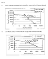
- Ad-mda7 which encodes the full length 206 amino-acid sequence, gives rise to an intracellular protein of approximately 23 kD. Furthermore, the Ad-mda7 vector also causes release of a soluble form of MDA-7 protein from treated cells.
- the soluble MDA-7 protein is approximately 40kD and is glycosylated. Treatment with glycosidases reduces the molecular mass of the soluble protein. Inhibitors of protein secretion, brefeldin A and tunicamycin, cause an intracellular accumulation of MDA-7 and inhibit release of this protein from cells. The MDA-7 soluble protein causes growth inhibition of tumor cells. Therefore, release of this soluble MDA-7 can give rise to a "bystander" effect wherein tumor cells that are not contacted by a mda-7 expression construct will be growth inhibited.
- a colony inhibition assay was used to demonstrate that elevated expression of mda-7 enhanced growth inhibition in human cervical carcinoma (HeLa), human breast carcinoma (MCF-7 and T47D), colon carcinoma (LS174T and SW480), nasopharyngeal carcinoma (HONE-1), prostate carcinoma (DU-145), melanoma (HO-1 and C8161), glioblastome multiforme (GBM-18 and T98G), and osteosarcoma (Saos-2).
- Mda-7 overexpressed in normal cells (HMECs, HBL-100, and CREF-Trans6) did not show significant effects.
- MDA-7 growth inhibition by elevated expression of MDA-7 is more effective in cancer cells than in normal cells.
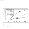
- Su et al. investigated the mechanism by which MDA-7 suppressed cancer cell growth. The studies reported that ectopic expression of MDA-7 in breast cancer cell lines MCF-7 and T47D induced apoptosis as detected by cell cycle analysis and TUNEL assay without an effect on the normal HBL-100 cells.
- Western blotting of lysates from cells infected with adenovirus mda-7 showed an upregulation of the apoptosis stimulating protein BAX.
- Ad-mda-7 infection elevated levels of BAX protein only in MCF-7 and T47D cells and not normal HBL-100 or HMEC cells.
- one embodiment of the present invention is the treatment of various hyperproliferative diseases with a mda-7 adenoviral construct encoding full-length or truncated MDA-7.
- WO 98/28425 describes a cytokine molecule allegedly related to IL-10.
- This molecule designated IL-BKW, appears to be derived from the same gene as MDA-7.
- the authors describe the coding region designation of MDA-7 as "mis-identified".
- the mature form of IL-BKW was to begin at about residue 47 or 49 of the mda-7 coding region, and continue some 158-160 residues, i.e., to residues 206 of the mda-7 sequence.
- a preferred molecule would preferably lack all or part of both the putative signal sequence (residues 1-25) and a putative membrane spanning hydrophobic domain (residues 26-45) of full length MDA-7.
- molecules beginning approximately at MDA-7 residues 46-49 are the largest molecules, further N-terminal truncations are within the scope of the invention.
- a full-length or a substantially full-length MDA-7 polypeptide is contemplated to be of use in the treatment of hyperproliferative diseases and conditions.
- the term “full-length” refers to a MDA-7 polypeptide that contains at least the 206 amino acids encoded by the human mda-7 cDNA.
- the term “substantially full-length” in the context of human MDA-7 refers to a MDA-7 polypeptide that contains at least 80% of the contiguous amino acids of the full-length human MDA-7 polypeptide (SEQ ID NO:2).
- MDA-7 polypeptides containing at least about 85%, 90%, and 95% of SEQ ID NO:2 are within the scope of the invention as “substantially full-length” MDA-7.
- a “truncated MDA-7 polypeptide” or “truncated MDA-7” refers to an MDA-7 polypeptide that is lacking contiguous amino acids from the full-length MDA-7 amino acid sequence.
- the missing contiguous amino acids could number inter alia about 3, 4, 5, 6, 7, 8, 9, 10, 11, 12, 13, 14, 15, 16, 17, 18, 19, 20, 21, 22, 23, 24, 25, 26, 27, 28, 29, 30, 31, 32, 33, 34, 35, 36, 37, 38, 39, 40, 41, 42, 43, 44, 45, 46, 47, 48, 49, 50, 51, 52, 53, 54, 55, 60, 65, 70, 75, 80, 85, 90, 95, 100, or more amino acids.
- secreted MDA-7 refers to an MDA-7 polypeptide that is secreted from a cell, i.e., a polypeptide that may or may not be encoded by a full-length mda-7 cDNA and whose N-terminus begins at about amino acid 46 of the full-length MDA-7 polypeptide.
- truncated MDA-7 and truncated MDA-7 polypeptide include a secreted MDA-7 polypeptide if, for example, the signal sequence is not missing or if a heterologous signal sequence is attached to the truncated polypeptide.
- biologically functional equivalent is well understood in the art and is further defined in detail herein. Accordingly, a sequence that has between about 70% and about 80%; or more preferably, between about 81 % and about 90%; or even more preferably, between about 91 % and about 99%; of amino acids that are identical or functionally equivalent to the amino acids of SEQ ID NO:2 will be a sequence that is "essentially as set forth in SEQ ID NO:2," provided the biological activity of the protein, polypeptide, or peptide is maintained.
- ''functionally equivalent codon is used herein to refer to codons that encode the same amino acid, such as the six codons for arginine and serine, and also refers to codons that encode biologically equivalent amino acids.
- nucleic acid sequences that have between about 70% and about 79%; or more preferably, between about 80% and about 89%; or even more particularly, between about 90% and about 99%; of nucleotides that are identical to the nucleotides of SEQ ID NO:1 will be nucleic acid sequences that are "essentially as set forth in SEQ ID NO:1.”
- this invention is not limited to the particular nucleic acid and amino acid sequences of SEQ ID NO:1 and SEQ ID NO:2, respectively.
- Recombinant vectors and isolated nucleic acid segments may therefore variously include these coding regions themselves, coding regions bearing selected alterations or modifications in the basic coding region, and they may encode larger polypeptides or peptides that nevertheless include such coding regions or may encode biologically functional equivalent proteins, polypeptide or peptides that have variant amino acids sequences.
- nucleic acids of the present invention encompass biologically functional equivalent MDA-7 proteins, polypeptides, or peptides. Such sequences may arise as a consequence of codon redundancy or functional equivalency that are known to occur naturally within nucleic acid sequences or the proteins, polypeptides or peptides thus encoded.
- functionally equivalent proteins, polypeptides or peptides may be created via the application of recombinant DNA technology, in which changes in the protein, polypeptide or peptide structure may be engineered, based on considerations of the properties of the amino acids being exchanged.
- Recombinant changes may be introduced, for example, through the application of site-directed mutagenesis techniques as discussed herein below, e.g., to introduce improvements or alterations to the antigenicity of the protein, polypeptide or peptide, or to test mutants in order to examine MDA-7 protein, polypeptide, or peptide activity at the molecular level.
- Fusion proteins, polypeptides or peptides may be prepared, e.g., where the mda-7 coding regions are aligned within the same expression unit with other proteins, polypeptides or peptides having desired functions.
- desired functions of expression sequences include purification or immunodetection purposes for the added expression sequences, e.g., proteinaceous compositions that may be purified by affinity chromatography or the enzyme labeling of coding regions, respectively.
- nucleic acid sequences encoding relatively small peptides or fusion peptides such as, for example, peptides of from about 3, about 4, about 5, about 6, about 7, about 8, about 9, about 10, about 11, about 12, about 13, about 14, about 15, about 16, about 17, about 18, about 19, about 20, about 21, about 22, about 23, about 24, about 25, about 26, about 27, about 28, about 29, about 30, about 31, about 32, about 33, about 34, about 35, about 35, about 36, about 37, about 38, about 39, about 40, about 41, about 42, about 43, about 44, about 45, about 46, about 47, about 48, about 49, about 50, about 51, about 52, about 53, about 54, about 55, about 56, about 57, about 58, about 59, about 60, about 61, about 62, about 63, about 64, about 65, about 66, about 67, about 68, about 69, about 70, about 71, about 72, about 73, about 74, about 75, about 76, about
- One embodiment of the present invention is to transfer the nucleic acids encoding the full-length, substantially full-length, or truncated form of human MDA-7 to induce the destruction, apoptosis or lysis of hyperproliferative cells.
- the expression of MDA-7 is inversely correlated with tumor progression as demonstrated by increased mRNA levels in normal melanocytes as compared to primary and metastatic tumors as well as decreased MDA-7 expression in early vertical growth phase tumor cells selected for enhanced tumor formation in nude mice.
- the treatment of hyperproliferative disease involves the administration of a therapeutic nucleic acid expression construct encoding a full-length, substantially full-length, or truncated form of MDA-7 to hyperproliferative cells. It is contemplated that the hyperproliferative cells take up the construct and express the therapeutic polypeptide encoded by nucleic acid, thereby restoring a growth control to or destroying the hyperproliferative cells. Furthermore, the soluble MDA-7 released from transfected or transduced cells will be available locally and provide a bystander effect on neighboring tumor cells.
- the therapeutic mda-7 expression construct may be delivered to normal cells and the released bystander effector (MDA-7, full-length or truncated) would cause anti-tumor effects, particularly with respect to hyperproliferative cells.
- MDA-7 released bystander effector
- the present invention concern at least one human mda-7 nucleic acid molecule.
- the mda-7 nucleic acid comprises a wild-type or mutant mda-7 nucleic acid.
- the mda-7 nucleic acid encodes for at least one transcribed nucleic acid.
- the mda-7 nucleic acid encodes at least one MDA-7 protein, polypeptide, or peptide, or biologically functional equivalent thereof.
- the human mda-7 nucleic acid comprises at least one nucleic acid segment of SEQ ID NO:1 or at least one biologically functional equivalent thereof.
- the present invention also concerns the isolation or creation of at least one recombinant construct or at least one recombinant host cell through the application of recombinant nucleic acid technology known to those of skill in the art or as described herein.
- the recombinant construct or host cell may comprise at least one mda-7 nucleic acid, and may express at least one MDA-7 protein, polypeptide, or peptide, or at least one biologically functional equivalent thereof.
- wild-type refers to the naturally occurring sequence of a nucleic acid at a genetic locus in the genome of an organism, and sequences transcribed or translated from such a nucleic acid. Thus, the term “wild-type” also may refer to the amino acid sequence encoded by the nucleic acid. As a genetic locus may have more than one sequence or alleles in a population of individuals, the term “wild-type” encompasses all such naturally occurring alleles. As used herein the term “polymorphic” means that variation exists (i.e., two or more alleles exist) at a genetic locus in the individuals of a population. As used herein, “mutant” refers to a change in the sequence of a nucleic acid or its encoded protein, polypeptide, or peptide that is the result of recombinant DNA technology.
- a nucleic acid may be made by any technique known to one of ordinary skill in the art.
- Non-limiting examples of synthetic nucleic acid, particularly a synthetic oligonucleotide include a nucleic acid made by in vitro chemical synthesis using phosphotriester, phosphite or phosphoramidite chemistry and solid phase techniques such as described in EP 266,032 , incorporated herein by reference, or via deoxynucleoside H-phosphonate intermediates as described by Froehler el al., 1986, and U.S. Patent Serial No. 5,705,629 , each incorporated herein by reference.
- a non-limiting example of enzymatically produced nucleic acid include one produced by enzymes in amplification reactions such as PCRTM (see for example, U.S. Patent 4,683,202 and U.S. Patent 4,682,195 , each incorporated herein by reference), or the synthesis of oligonucleotides described in U.S. Patent No. 5,645,897 , incorporated herein by reference.
- a non-limiting example of a biologically produced nucleic acid includes recombinant nucleic acid production in living cells, such as recombinant DNA vector production in bacteria (see for example, Sambrook et al. 1989, incorporated herein by reference).
- a nucleic acid may be purified on polyacrylamide gels, cesium chloride centrifugation gradients, or by any other means known to one of ordinary skill in the art (see for example, Sambrook et al. 1989, incorporated herein by reference).
- nucleic acid will generally refer to at least one molecule or strand of DNA, RNA or a derivative or mimic thereof, comprising at least one nucleobase, such as, for example, a naturally occurring purine or pyrimidine base found in DNA (e.g. , adenine "A,” guanine “G,” thymine “T,” and cytosine “C”) or RNA (e.g. A, G, uracil “U,” and C).
- nucleic acid encompasses the terms “oligonucleotide” and “polynucleotide.”
- oligonucleotide refers to at least one molecule of between about 3 and about 100 nucleobases in length.
- polynucleotide refers to at least one molecule of greater than about 100 nucleobases in length. These definitions generally refer to at least one single-stranded molecule, but in specific embodiments will also encompass at least one additional strand that is partially, substantially or fully complementary to the at least one single-stranded molecule. Thus, a nucleic acid may encompass at least one double-stranded molecule or at least one triple-stranded molecule that comprises one or more complementary strand(s) or "complement(s)" of a particular sequence comprising a strand of the molecule.
- a “gene” refers to a nucleic acid that is transcribed.
- a “gene segment” is a nucleic acid segment of a gene.
- the gene includes regulatory sequences involved in transcription, or message production or composition.
- the gene comprises transcribed sequences that encode for a protein, polypeptide or peptide.
- the gene comprises a mda-7 nucleic acid, and/or encodes a MDA-7 polypeptide or peptide-coding sequences.
- an "isolated gene'' may comprise transcribed nucleic acid(s), regulatory sequences, coding sequences, or the like, isolated substantially away from other such sequences, such as other naturally occurring genes, regulatory sequences, polypeptide or peptide encoding sequences, etc.
- the term “gene” is used for simplicity to refer to a nucleic acid comprising a nucleotide sequence that is transcribed, and the complement thereof.
- the transcribed nucleotide sequence comprises at least one functional protein, polypeptide and/or peptide encoding unit.
- this functional term "gene” includes both genomic sequences, RNA or cDNA sequences, or smaller engineered nucleic acid segments, including nucleic acid segments of a non-transcribed part of a gene, including but not limited to the non-transcribed promoter or enhancer regions of a gene. Smaller engineered gene nucleic acid segments may express, or may be adapted to express using nucleic acid manipulation technology, proteins, polypeptides, domains, peptides, fusion proteins, mutants and/or such like.
- a "truncated gene” refers to a nucleic acid sequence that is missing a stretch of contiguous nucleic acid residues that encode a portion of the full-length MDA-7 polypeptide.
- a truncated gene may not contain the nucleic acid sequence for the N-terminal region of the MDA-7 polypeptide, such as the first 46 amino acids. It is envisioned that the nucleic acid sequences of the present invention may contain fewer than 95% of the contiguous nucleic acid residues of SEQ ID NO:1. Alternatively, these sequences may encode fewer than 90%, 85%, 80%, 75%, or 70% of the contiguous nucleic acid residues of SEQ ID NO:1.
- isolated substantially away from other coding sequences means that the gene of interest, in this case the mda-7 gene, forms the significant part of the coding region of the nucleic acid, or that the nucleic acid does not contain large portions of naturally-occurring coding nucleic acids, such as large chromosomal fragments, other functional genes, RNA or cDNA coding regions. Of course, this refers to the nucleic acid as originally isolated, and does not exclude genes or coding regions later added to the nucleic acid by recombinant nucleic acid technology.
- the nucleic acid is a nucleic acid segment.
- nucleic acid segment are smaller fragments of a nucleic acid, such as for non-limiting example, those that encode only part of the MDA-7 peptide or polypeptide sequence.
- a “nucleic acid segment may comprise any part of the mda-7 gene sequence, of from about 2 nucleotides to the full-length of the MDA-7 peptide- or polypeptide-encoding region.
- the "nucleic acid segment” encompasses the full-length mda-7 gene sequence.
- the nucleic acid comprises any part of SEQ ID NO:1 of from about 2 nucleotides to the full-length of the sequence encoding SEQ ID NO:2.
- nucleic acid segments may be designed based on a particular nucleic acid sequence, and may be of any length.
- an algorithm defining all nucleic acid segments can be created: n to n + y where n is an integer from 1 to the last number of the sequence and y is the length of the nucleic acid segment minus one, where n + y does not exceed the last number of the sequence.
- the nucleic acid segments correspond to bases 1 to 10, 2 to 11, 3 to 12 ... and/or so on.
- nucleic acid segments correspond to bases 1 to 15, 2 to 16, 3 to 17 ... and/or so on.
- nucleic segments correspond to bases 1 to 20, 2 to 21, 3 to 22 ... and/or so on.
- the nucleic acid segment may be a probe or primer.
- nucleic acid(s) of the present invention may be combined with other nucleic acid sequences, including but not limited to, promoters, enhancers, polyadenylation signals, restriction enzyme sites, multiple cloning sites, coding segments, and the like, to create one or more nucleic acid construct(s).
- the overall length may vary considerably between nucleic acid constructs.
- a nucleic acid segment of almost any length may be employed, with the total length preferably being limited by the ease of preparation or use in the intended recombinant nucleic acid protocol.
- one or more nucleic acid constructs may be prepared that include a contiguous stretch of nucleotides identical to or complementary to SEQ ID NO:1.
- Such a stretch of nucleotides, or a nucleic acid construct may be about 3, about 4, about 5, about 6, about 7, about 8" about 9, about 10, about 11, about 12, about 13, about 14, about 15, about 16, about 17, about 18, about 19, about 20, about 21, about 22, about 23, about 24, about 25, about 26, about 27, about 28, about 29, about 30, about 31, about 32, about 33, about 34, about 35, about 36, about 37, about 38, about 39 about 40, about 45, about 50, about 55, about 60, about 65, about 70, about 75, about 80, about 85, about 90, about 95, about 100, about 105, about 110, about 115, about 120, about 125, about 130, about 135, about 140, about 145, about 150, about 155, about 160, about 165, about 170, about 175, about 180, about 185, about 190, about 195,
- intermediate lengths and “intermediate ranges,” as used herein, means any length or range including or between the quoted values ( i.e., all integers including and between such values).
- Intermediate lengths include about 11, about 12, about 13, about 16, about 17, about 18, about 19, etc.; about 21, about 22, about 23, etc.; about 31, about 32, etc.; about 51, about 52, about 53, etc.; about 101, about 102, about 103, etc.; about 151, about 152, about 153, etc.; about 1,001, about 1002, etc,; about 50,001, about 50,002, etc; about 750,001, about 750,002, etc.; about 1,000,001, about 1,000,002, etc.
- Non-limiting examples of intermediate ranges include about 3 to about 32, about 150 to about 500,001, about 3,032 to about 7,145, about 5,000 to about 15,000, about 20,007 to about 1,000,003, etc.
- nucleic acid sequence encoding all or a portion of an MDA-7 polypeptide may be comprised of contiguous complementary or identical nucleic acid sequences of any of the lengths described above and from SEQ ID NO:1.
- nucleic acid constructs of the present invention may encode a full-length MDA-7 or encode a truncated version of MDA-7, such that the transcript of the coding region represents the truncated version.
- the truncated transcript may then be translated into a truncated protein.
- a nucleic acid sequence may encode a full-length MDA-7 protein sequence, which is processed by the cellular machinery to produce a truncated MDA-7.
- the nucleic acid encoding a truncated transcript may contain a contiguous nucleic acid encoding a portion of mda-7 of the following lengths: about 10, 20, 30, 40, 50, 60, 70, 80, 90, 100, 110, 120, 130, 140, 150, 160, 170, 180, 190, 200, 210, 220, 230, 240, 250, 260, 270, 280, 290, 300, 310, 320, 330, 340, 350, 360, 370, 380, 390, 400, 410, 420, 430, 440, 450, 460, 470, 480, 490, 500, 510, 520, 530, 540, 550, 560, 570, 580, 590, 600, 610, or 620 nucleotides, nucleosides, or base pairs.
- nucleic acid molecules may contain contiguous nucleotides of the above-lengths from SEQ ID NO:1.
- sequences of the present invention may contain contiguous nucleic acids that are complementary or identical to SEQ ID NO: 1, yet be less than the entire sequence of SEQ ID NO:1.
- the sequence may contain fewer than 718 contiguous nucleic acids from SEQ ID NO:1; it may instead contain, less than 700, 690, 680, 670, 660, 650, 640, 630, 620, 610, 640, 590, 580, 570, 560, 550, 540, 530, 520, 510, 500, 490, 480, 470, 460, 450, 440, 430, 420, 410, 400, 390, 380, 370, 360, 350, 340, 330, 320, 310, 300, 290, 280, 270, 260, 250, 240, 230, 220, 210, 200, or fewer contiguous nucleotides or nucleosides from SEQ ID NO:1.
- sequence essentially as set forth in SEQ ID NO:1 or "a sequence essentially as set forth in SEQ ID NO:1” means that the sequence substantially corresponds to a portion of SEQ ID NO:land has relatively few amino acids that are not identical to, or biologically functionally equivalent to, the amino acids of SEQ ID NO:2.
- Vectors of the present invention are designed, primarily, to transform hyperproliferative cells with a therapeutic mda-7 gene under the control of regulated eukaryotic promoters (i.e., constitutive, inducible, repressable, tissue-specific). Also, the vectors may contain a selectable marker if, for no other reason, to facilitate their manipulation in vitro. However, selectable markers may play an important role in producing recombinant cells.
- regulated eukaryotic promoters i.e., constitutive, inducible, repressable, tissue-specific.
- selectable markers may play an important role in producing recombinant cells.
- Tables 1 and 2 list a variety of regulatory signals for use according to the present invention.
- Table 1 - Inducible Elements Element Inducer References MT II Phorbol Ester (TFA) Heavy metals Palmiter et al., 1982; Haslinger and Karin, 1985; Searle et al., 1985; Stuart et al., 1985; Imagawa et al., 1987; Karin ®, 1987; Angel et al., 1987b; McNeall et al., 1989 MMTV (mouse mammary tumor virus) Glucocorticoids Huang et al., 1981; Lee et al., 1981; Majors and Varmus, 1983; Chandler et al., 1983; Lee et al., 1984; Fonta et al., 1985; Sakai et al., 1986 ⁇ -Interferon poly(rI)X poly(rc) Tavernier et al., 1983 Adenovirus 5 E2 EIA Imperiale and Nevin
- the promoters and enhancers that control the transcription of protein encoding genes in eukaryotic cells are composed of multiple genetic elements.
- the cellular machinery is able to gather and integrate the regulatory information conveyed by each element, allowing different genes to evolve distinct, often complex patterns of transcriptional regulation.
- a promoter used in the context of the present invention includes constitutive, inducible, and tissue-specific promoters.
- promoter will be used here to refer to a group of transcriptional control modules that are clustered around the initiation site for RNA polymerase II. Much of the thinking about how promoters are organized derives from analyses of several viral promoters, including those for the HSV thymidine kinase (tk) and SV40 early transcription units. These studies, augmented by more recent work, have shown that promoters are composed of discrete functional modules, each consisting of approximately 7-20 bp of DNA, and containing one or more recognition sites for transcriptional activator proteins.
- At least one module in each promoter functions to position the start site for RNA synthesis.
- the best known example of this is the TATA box, but in some promoters lacking a TATA box, such as the promoter for the mammalian terminal deoxynucleotidyl transferase gene and the promoter for the SV40 late genes, a discrete element overlying the start site itself helps to fix the place of initiation.
- promoter elements regulate the frequency of transcriptional initiation. Typically, these are located in the region 30-110 bp upstream of the start site, although a number of promoters have recently been shown to contain functional elements downstream of the start site as well.
- the spacing between elements is flexible, so that promoter function is preserved when elements are inverted or moved relative to one another. In the tk promoter, the spacing between elements can be increased to 50 bp apart before activity begins to decline.
- individual elements can function either co-operatively or independently to activate transcription.
- Enhancers were originally detected as genetic elements that increased transcription from a promoter located at a distant position on the same molecule of DNA. This ability to act over a large distance had little precedent in classic studies of prokaryotic transcriptional regulation. Subsequent work showed that regions of DNA with enhancer activity are organized much like promoters. That is, they are composed of many individual elements, each of which binds to one or more transcriptional proteins. of many individual elements, each of which binds to one or more transcriptional proteins.
- enhancers and promoters are very similar entities.
- An enhancer region as a whole must be able to stimulate transcription at a distance; this need not be true of a promoter region or its component elements.
- a promoter must have one or more elements that direct initiation of RNA synthesis at a particular site and in a particular orientation, whereas enhancers lack these specificities.
- enhancers and promoters are very similar entities.
- Promoters and enhancers have the same general function of activating transcription in the cell. They are often overlapping and contiguous, often seeming to have a very similar modular organization. Taken together, these considerations suggest that enhancers and promoters are homologous entities and that the transcriptional activator proteins bound to these sequences may interact with the cellular transcriptional machinery in fundamentally the same way.
- CMV cytomegalovirus
- This promoter is commercially available from Invitrogen in the vector pcDNAIII, which is preferred for use in the present invention.
- dectin-1 and dectin-2 promoters are also contemplated as useful in the present invention. Additional viral promoters, cellular promoters/enhancers and inducible promoters/enhancers that could be used in combination with the present invention are listed in Tables 1 and 2.
- any promoter/enhancer combination (as per the Eukaryotic Promoter Data Base EPDB) could also be used to drive expression of structural genes encoding oligosaccharide processing enzymes, protein folding accessory proteins, selectable marker proteins or a heterologous protein of interest.
- a tissue-specific promoter for cancer gene therapy (Table 3) or the targeting of tumors (Table 4) may be employed with the nucleic acid molecules of the present invention.
- Table 3 Candidate tissue-specific promoters for cancer gene therapy Tissue-specific promoter Cancers in which promoter is active Normal cells in which promoter is active Carcinoembryonic antigen mucosa; Most colorectal carcinomas; 50% of Colonic mucosa; gastric (CEA)* lung carcinomas; 40-50% of gastric lung epithelia; eccrine sweat carcinomas; most pancreatic glands; cells in testes carcinomas; many breast carcinomas Prostate-specific antigen (PSA) .
- VIP Vasoactive intestinal peptide
- SP-A Surfactant protein A
- SP-A Surfactant protein A
- Most adenocarcinomas originating GFandular epithelial cells in from any tissue) and in respiratory, gastrointestinal, and genitourinary tracts
- Alpha-fetoprotein Most hepatocellular carcinomas; possibly many testicular cancers Hepatocytes (under certain conditions); testis Albumin Most hepatocellular carcinomas
- Hepatocytes Tyrosinase Most melanomas Melanocytes; astrocytes; Schwann cells; some neurons Tyrosine-binding protein Most melanomas Melanocytes; astmcytes
- IRES internal ribosome binding sites
- hGH polyadenylation signal
- BGH BGH
- SV40 polyadenylation signal
- IRES elements are used to create multigene, or polycistronic, messages. IRES elements are able to bypass the ribosome scanning model of 5'-methylated cap-dependent translation and begin translation at internal sites (Pelletier and Sonenberg, 1988). IRES elements from two members of the picornavirus family (polio and encephalomyocarditis) have been described (Pelletier and Sonenberg, 1988), as well as an IRES from a mammalian message (Macejak and Sarnow, 1991). IRES elements can be linked to heterologous open reading frames.
- each open reading frame can be transcribed together, each separated by an IRES, creating polycistronic messages.
- IRES element By virtue of the IRES element, each open reading frame is accessible to ribosomes for efficient translation. Multiple genes can be efficiently expressed using a single promoter/enhancer to transcribe a single message.
- promoters are DNA elements which when positioned functionally upstream of a gene leads to the expression of that gene.
- Most transgene constructs of the present invention are functionally positioned downstream of a promoter element.
- adenovirus expression vector is meant to include those constructs containing adenovirus sequences sufficient to (a) support packaging of the construct and (b) to ultimately express a recombinant gene construct that has been cloned therein.
- the vector comprises a genetically engineered form of adenovirus.
- retrovirus the adenoviral infection of host cells does not result in chromosomal integration because adenoviral DNA can replicate in an episomal manner without potential genotoxicity.
- adenoviruses are structurally stable, and no genome rearrangement has been detected after extensive amplification.
- Adenovirus is particularly suitable for use as a gene transfer vector because of its mid-sized genome, ease of manipulation, high titer, wide target-cell range and high infectivity. Both ends of the viral genome contain 100-200 base pair inverted repeats (ITRs), which are cis elements necessary for viral DNA replication and packaging.
- ITRs inverted repeats
- the early (E) and late (L) regions of the genome contain different transcription units that are divided by the onset of viral DNA replication.
- the E1 region (E1A and E1B) encodes proteins responsible for the regulation of transcription of the viral genome and a few cellular genes.
- the expression of the E2 region results in the synthesis of the proteins for viral DNA replication.
- MLP major late promoter
- TPL 5'-tripartite leader
- recombinant adenovirus is generated from homologous recombination between shuttle vector and provirus vector. Due to the possible recombination between two proviral vectors, wild-type adenovirus may be generated from this process. Therefore, it is critical to isolate a single clone of virus from an individual plaque and examine its genomic structure.
- adenovirus generation and propagation of the current adenovirus vectors, which are replication deficient, depend on a unique helper cell line, designated 293, which was transformed from human embryonic kidney cells by Ad5 DNA fragments and constitutively expresses E1 proteins (Graham et al., 1977). Since the E3 region is dispensable from the adenovirus genome (Jones and Shenk, 1978), the current adenovirus vectors, with the help of 293 cells, carry foreign DNA in either the E1, the E3, or both regions (Graham and Prevec, 1991). In nature, adenovirus can package approximately 105% of the wild-type genome (Ghosh-Choudhury et al., 1987), providing capacity for about 2 extra kb of DNA.
- the maximum capacity of the current adenovirus vector is under 7.5 kb, or about 15% of the total length of the vector. More than 80% of the adenovirus viral genome remains in the vector backbone.
- Helper cell lines may be derived from human cells such as human embryonic kidney cells, muscle cells, hematopoietic cells or other human embryonic mesenchymal or epithelial cells.
- the helper cells may be derived from the cells of other mammalian species that are permissive for human adenovirus. Such cells include, e.g. , Vero cells or other monkey embryonic mesenchymal or epithelial cells.
- the preferred helper cell line is 293.
- Racher et al. (1995) have disclosed improved methods for culturing 293 cells and propagating adenovirus.
- natural cell aggregates are grown by inoculating individual cells into 1 liter siliconized spinner flasks (Techne, Cambridge, UK) containing 100-200 ml of medium. Following stirring at 40 rpm, the cell viability is estimated with trypan blue.
- Fibra-Cel microcarriers (Bibby Sterlin, Stone, UK) (5 g/l) is employed as follows.
- the adenovirus vector may be replication defective, or at least conditionally defective, the nature of the adenovirus vector is not believed to be crucial to the successful practice of the invention.
- the adenovirus may be of any of the 42 different known serotypes or subgroups A-F.
- Adenovinis type 5 of subgroup C is the preferred starting material in order to obtain the conditional replication-defective adenovirus vector for use in the present invention. This is because Adenovirus type 5 is a human adenovirus about which a great deal of biochemical and genetic information is known, and it has historically been used for most constructions employing adenovirus as a vector.
- the typical vector according to the present invention is replication defective and will not have an adenovirus E1 region.
- the position of insertion of the construct within the adenovirus sequences is not critical to the invention.
- the polynucleotide encoding the gene of interest may also be inserted in lieu of the deleted E3 region in E3 replacement vectors as described by Karlsson et al. (1986) or in the E4 region where a helper cell line or helper virus complements the E4 defect.
- Adenovirus growth and manipulation is known to those of skill in the art, and exhibits broad host range in vitro and in vivo. This group of viruses can be obtained in high titers, e.g. , 10 9 -10 11 plaque-forming units per ml, and they are highly infective. The life cycle of adenovirus does not require integration into the host cell genome. The foreign genes delivered by adenovirus vectors are episomal and, therefore, have low genotoxicity to host cells. No side effects have been reported in studies of vaccination with wild-type adenovirus (Couch et al., 1963; Top et al., 1971), demonstrating their safety and therapeutic potential as in vivo gene transfer vectors.
- Adenovirus vectors have been used in eukaryotic gene expression (Levrero et al., 1991; Gomez-Foix et al., 1992) and vaccine development (Grunhaus and Horwitz, 1992; Graham and Prevec, 1992). Animal studies have suggested that recombinant adenovirus could be used for gene therapy (Stratford-Perricaudet and Perricaudet, 1991; Stratford-Perricaudet et al., 1990; Rich et al., 1993).
- the retroviruses are a group of single-stranded RNA viruses characterized by an ability to convert their RNA to double-stranded DNA in infected cells by a process of reverse-transcription (Coffin, 1990).
- the resulting DNA then stably integrates into cellular chromosomes as a provirus and directs synthesis of viral proteins.
- the integration results in the retention of the viral gene sequences in the recipient cell and its descendants.
- the retroviral genome contains three genes, gag, pol, and env that code for capsid proteins, polymerase enzyme, and envelope components, respectively.
- a sequence found upstream from the gag gene contains a signal for packaging of the genome into virions.
- Two long terminal repeat (LTR) sequences are present at the 5' and 3' ends of the viral genome. These contain strong promoter and enhancer sequences and are also required for integration in the host cell genome (Coffin, 1990).
- a nucleic acid encoding a gene of interest is inserted into the viral genome in the place of certain viral sequences to produce a virus that is replication-defective.
- a packaging cell line containing the gag, pol, and env genes but without the LTR and packaging components is constructed (Mann et al., 1983).
- Retroviral vectors are able to infect a broad variety of cell types. However, integration and stable expression require the division of host cells (Paskind et al., 1975).
- Adeno-associated virus is an attractive vector system for use in the present invention as it has a high frequency of integration and it can infect nondividing cells, thus making it useful for delivery of genes into mammalian cells in tissue culture (Muzyczka, 1992).
- AAV has a broad host range for infectivity (Tratschin, et al., 1984; Laughlin, et al., 1986; Lebkowski, et al., 1988; McLaughlin, et al., 1988), which means it is applicable for use with the present invention. Details concerning the generation and use of rAAV vectors are described in U.S. Patent No. 5,139,941 and U.S. Patent No. 4,797,368 , each incorporated herein by reference.
- AAV vectors have been used successfully for in vitro and in vivo transduction of marker genes (Kaplitt et al., 1994; Lebkowski el al., 1988; Samulski et al., 1989; Shelling and Smith, 1994; Yoder el al., 1994; Zhou et al., 1994; Hermonat and Muzyczka, 1984; Tratschin el al., 1985; McLaughlin et al., 1988) and genes involved in human diseases (Flotte et al..
- AAV is a dependent parvovirus in that it requires coinfection with another virus (either adenovirus or a member of the herpes virus family) to undergo a productive infection in cultured cells (Muzyczka, 1992).
- another virus either adenovirus or a member of the herpes virus family
- helper virus the wild-type AAV genome integrates through its ends into human chromosome 19 where it resides in a latent state as a provirus (Kotin et al., 1990; Samulski et al., 1991).
- rAAV is not restricted to chromosome 19 for integration unless the AAV Rep protein is also expressed (Shelling and Smith, 1994).
- recombinant AAV (rAAV) virus is made by cotransfecting a plasmid containing the gene of interest flanked by the two AAV terminal repeats (McLaughlin et al., 1988; Samulski et al., 1989; each incorporated herein by reference) and an expression plasmid containing the wild-type AAV coding sequences without the terminal repeats, for example pIM45 (McCarty et al., 1991; incorporated herein by reference).
- the cells are also infected or transfected with adenovirus or plasmids carrying the adenovirus genes required for AAV helper function.
- rAAV virus stocks made in such fashion are contaminated with adenovirus which must be physically separated from the rAAV particles (for example, by cesium chloride density centrifugation).
- adenovirus vectors containing the AAV coding regions or cell lines containing the AAV coding regions and some or all of the adenovirus helper genes could be used (Yang et al., 1994; Clark et al., 1995).
- Cell lines carrying the rAAV DNA as an integrated provirus can also be used (Flotte et al., 1995).
- viral vectors may be employed as constructs in the present invention.
- Vectors derived from viruses such as vaccinia virus (Ridgeway, 1988; Baichwal and Sugden, 1986; Coupar et al., 1988) and herpesviruses may be employed. They offer several attractive features for various mammalian cells (Friedmann, 1989; Ridgeway, 1988; Baichwal and Sugden, 1986; Coupar et al., 1988; Horwich et al., 1990).
- VEE virus A molecularly cloned strain of Venezuelan equine encephalitis (VEE) virus has been genetically refined as a replication competent vaccine vector for the expression of heterologous viral proteins (Davis et al., 1996). Studies have demonstrated that VEE infection stimulates potent CTL responses and has been sugested that VEE may be an extremely useful vector for immunizations (Caley et al., 1997). It is contemplated in the present invention, that VEE virus may be useful in targeting dendritic cells.
- Chang et al. recently introduced the chloramphenicol acetyltransferase (CAT) gene into duck hepatitis B virus genome in the place of the polymerase, surface, and pre-surface coding sequences. It was cotransfected with wild-type virus into an avian hepatoma cell line. Culture media containing high titers of the recombinant virus were used to infect primary duckling hepatocytes. Stable CAT gene expression was detected for at least 24 days after transfection (Chang et al., 1991).
- CAT chloramphenicol acetyltransferase
- the nucleic acid encoding human mda-7 is housed within an infective virus that has been engineered to express a specific binding ligand.
- the virus particle will thus bind specifically to the cognate receptors of the target cell and deliver the contents to the cell.
- a novel approach designed to allow specific targeting of retrovirus vectors was recently developed based on the chemical modification of a retrovirus by the chemical addition of lactose residues to the viral envelope. This modification can permit the specific infection of hepatocytes via sialoglycoprotein receptors.
- recombinant retroviruses targeting of recombinant retroviruses was designed in which biotinylated antibodies against a retroviral envelope protein and against a specific cell receptor were used.
- the antibodies were coupled via the biotin components by using streptavidin (Roux et al., 1989).
- streptavidin Using antibodies against major histocompatibility complex class I and class II antigens, they demonstrated the infection of a variety of human cells that bore those surface antigens with an ecotropic virus in vitro (Roux et al., 1989).
- the gene construct is introduced into target hyperproliferative cells via electroporation. Electroporation involves the exposure of cells (or tissues) and DNA (or a DNA complex) to a high-voltage electric discharge.
- electroporation conditions for hyperproliferative cells from different sources may be optimized.
- the execution of other routine adjustments will be known to those of skill in the art. See e.g., Hoffman, 1999; Heller et al., 1996.
- Another embodiment of the invention for transferring a naked DNA construct into cells involves particle bombardment. This method depends on the ability to accelerate DNA-coated microprojectiles to a high velocity allowing them to pierce cell membranes and enter cells without killing them (Klein et al., 1987).
- the microprojectiles used have consisted of biologically inert substances such as tungsten, platinum, or gold beads.
- DNA precipitation onto metal particles would not be necessary for DNA delivery to a recipient cell using particle bombardment. It is contemplated that particles may contain DNA rather than be coated with DNA. Hence it is proposed that DNA-coated particles may increase the level of DNA delivery via particle bombardment but are not, in and of themselves, necessary.
- a Biolistic Particle Delivery System which can be used to propel particles coated with DNA through a screen, such as stainless steel or Nytex screen, onto a filter surface covered with cells in suspension. The screen disperses the particles so that they are not delivered to the recipient cells in large aggregates. It is believed that a screen intervening between the projectile apparatus and the cells to be bombarded reduces the size of projectile aggregates and may contribute to a higher frequency of transformation by reducing the damage inflicted on the recipient cells by projectiles that are too large.
- cells in suspension are preferably concentrated on filters, or alternatively on solid culture medium.
- the cells to be bombarded are positioned at an appropriate distance below the macroprojectile stopping plate. If desired, one or more screens are also positioned between the acceleration device and the cells to be bombarded.
- bombardment transformation one may optimize the prebombardment culturing conditions and the bombardment parameters to yield the maximum numbers of stable transformants. Both the physical and biological parameters for bombardment are important in this technology. Physical factors are those that involve manipulating the DNA/microprojectile precipitate or those that affect the flight and velocity or either the macro- or microprojectiles. Biological factors include all steps involved in manipulation of cells before and immediately after bombardment, the osmotic adjustment of target cells to help alleviate the trauma associated with bombardment, and also the nature of the transforming DNA, such as linearized DNA or intact supercoiled plasmids. Recently, results from a clinical trial evaluating utility of this delivery system for vaccination was published.
- the study was designed to determine the safety and immunogenicity in volunteers of a DNA vaccine consisting of a plasmid encoding hepatitis B surface antigen delivered by the PowderJect XR1 gene delivery system into human skin (Tacket et al., 1999).
- the transgenic construct is introduced to the cells using calcium phosphate co-precipitation.
- Mouse primordial germ cells have been transfected with the SV40 large T antigen, with excellent results (Watanabe et al., 1997).
- Human KB cells have been transfected with adenovirus 5 DNA (Graham and Van Der Eb, 1973) using this technique.
- mouse L(A9), mouse C127, CHO, CV-1, BHK, NIH3T3 and HeLa cells were transfected with a neomycin marker gene (Chen and Okayama, 1987), and rat hepatocytes were transfected with a variety of marker genes (Rippe et al., 1990).
- the expression construct is delivered into the cell using DEAE-dextran followed by polyethylene glycol.
- reporter plasmids were introduced into mouse myeloma and erythroleukemia cells (Gopal, 1985).
- nucleic acid constructs include the introduction of the nucleic acid construct by direct microinjection or sonication loading.
- Direct microinjection has been used to introduce nucleic acid constructs into Xenopus oocytes (Harland and Weintraub, 1985), and LTK - fibroblasts have been transfected with the thymidine kinase gene by sonication loading (Fechheimer et al., 1987).
- the gene construct may be entrapped in a liposome or lipid formulation.
- Liposomes are vesicular structures characterized by a phospholipid bilayer membrane and an inner aqueous medium. Multilamellar liposomes have multiple lipid layers separated by aqueous medium. They form spontaneously when phospholipids are suspended in an excess of aqueous solution. The lipid components undergo self-rearrangement before the formation of closed structures and entrap water and dissolved solutes between the lipid bilayers (Ghosh and Bachhawat, 1991). Also contemplated is a gene construct complexed with Lipofectamine (Gibco BRL).
- Lipid-mediated nucleic acid delivery and expression of foreign DNA in vitro has been very successful (Nicolau and Sene, 1982; Fraley et al., 1979; Nicolau et al., 1987). Wong et al. (1980) demonstrated the feasibility of lipid-mediated delivery and expression of foreign DNA in cultured chick embryo, HeLa and hepatoma cells.
- Lipid based non-viral formulations provide an alternative to adenoviral gene therapies. Although many cell culture studies have documented lipid based non-viral gene transfer, systemic gene delivery via lipid based formulations has been limited. A major limitation of non-viral lipid based gene delivery is the toxicity of the cationic lipids that comprise the non-viral delivery vehicle. The in vivo toxicity of liposomes partially explains the discrepancy between in vitro and in vivo gene transfer results. Another factor contributing to this contradictory data is the difference in lipid vehicle stability in the presence and absence of serum proteins. The interaction between lipid vehicles and serum proteins has a dramatic impact on the stability characteristics of lipid vehicles (Yang and Huang, 1997).
- Cationic lipids attract and bind negatively charged serum proteins.
- Lipid vehicles associated with serum proteins are either dissolved or taken up by macrophages leading to their removal from circulation.
- Current in vivo lipid delivery methods use subcutaneous, intradermal, intratumoral, or intracranial injection to avoid the toxicity and stability problems associated with cationic lipids in the circulation.
- lipid formulations have improved the efficiency of gene transfer in vivo (Templeton et al. 1997; WO 98/07408 ).
- a novel lipid formulation composed of an equimolar ratio of 1,2-bis(oleoyloxy)-3-(trimethyl ammonio)propane (DOTAP) and cholesterol significantly enhances systemic in vivo gene transfer, approximately 150 fold.
- the DOTAP:cholesterol lipid formulation forms unique structure termed a "sandwich liposome". This formulation is reported to "sandwich" DNA between an invaginated bi-layer or 'vase' structure. Beneficial characteristics of these lipid structures include a positive ⁇ , colloidal stabilization by cholesterol, two dimensional DNA packing and increased serum stability.
- Patent Application Nos. 60/135,818 and 60/133,116 discuss formulations that may be used with the present invention and are incorporated herein by reference.
- lipid structures can be used to encapsulate compounds that are toxic (chemotherapeutics) or labile (nucleic acids) when in circulation. Lipid encapsulation has resulted in a lower toxicity and a longer serum half-life for such compounds (Gabizon et al., 1990). Numerous disease treatments are using lipid based gene transfer strategies to enhance conventional or establish novel therapies, in particular therapies for treating hyperproliferative diseases.
- the lipid vehicle may be complexed with a hemagglutinating virus (HVJ). This has been shown to facilitate fusion with the cell membrane and promote cell entry of lipid-encapsulated DNA (Kaneda et al., 1989).
- HVJ hemagglutinating virus
- the lipid vehicle may be complexed or employed in conjunction with nuclear non-histone chromosomal proteins (HMG-1) (Kato et al., 1991).
- HMG-1 nuclear non-histone chromosomal proteins
- the lipid vehicle may be complexed or employed in conjunction with both HVJ and HMG-1.
- a contemplated method for commercial scale preparation of a lipid composition is, generally speaking, mixing of the components, drying the mixture, rehydrating the mixture, dispersing the mixture, extruding the lipid composition through filters of decreasing pore size and mixing of the lipid composition with a therapeutic agent to form a lipoplex.
- the following provides additional information about manufacturing and formulating a lipoplex for use in the delivery of an mda-7 polynucleotide to a cell.
- Powdered components are weighed, mixed, and dissolved in an acceptable solvent, such as tertiary butanol, chloroform, methanol, ethylene chloride, ethanol, or mixtures of these solvents. It is contemplated that any two of the solvent are present in a ration of about 1:1000, 1:500,1:100, 1:50, 1:25, 1:10, 1:5 or 1:1. Solublization with tertiary butanol may be employed, due to the carcinogenic properties of chloroform. Although, chloroform can be used if steps are taken to limit the residual chloroform present in the lipid composition to acceptable levels. It is envisioned that other lipophilic solvents may be used for the solubilization of the lipid components.
- an acceptable solvent such as tertiary butanol, chloroform, methanol, ethylene chloride, ethanol, or mixtures of these solvents. It is contemplated that any two of the solvent are present in a ration of about 1:1000, 1:500,
- the lipid mixture may be solubilized in tert -butanol at a temperature of about 0°C, 2°C, 4°C, 6°C, 8°C, 10°C, 12°C, 14°C, 16°C, 18°C, 20°C, 22°C, 24°C, 26°C, 28°C, 30°C, 32°C, 34°C, 36°C, 38°C, 40°C, 42°C, 44°C, 46°C, 48°C, 50°C, 52°C, 54°C, 56°C, 58°C, 60°C, 62°C, 64°C, 66°C, 68°C, 70°C, 72°C, 74°C, 76°C, 78°C, 80°C, 82°C, 84°C, 86°C, 88°C, or 90°C.
- a range of temperatures such as 5°C to 80°C, 10°C to 70°C, 20°C to 60°C
- a lipid complex is formulated by combining the lipid composition and a therapeutic agent in a pharmaceutically acceptable carrier solution by rapid mixing. Rapid mixing can be done by using a T-mixing apparatus or ethanol injection of the therapeutic agent.
- a DOTAP:Cholesterol lipid composition is prepared by the methods described and combined with a polynucleotide that encodes for a therapeutic protein. The lipid composition is provided in an amount to encapsulate the polynucleotide and result in a colloidal suspension of the lipoplex.
- DOTAP cationic lipid
- DOTAP cationic lipid
- This mixture of powdered lipids is then dissolved with tert-butanol, the solution dried to a thin film and the film hydrated in water containing 5% dextrose (w/v) to give a final concentration of about 20 mM DOTAP and about 20 mM cholesterol.
- the hydrated lipid film is rotated in a heated water bath for about 45 minutes, then at about 35°C for an additional 10 minutes and left standing at room temperature overnight.
- the sonicated mixture is heated for 10 minutes at about 50°C.
- This mixture is sequentially extruded through filters of decreasing pore size (1 ⁇ m, 0.45 ⁇ m, 0.2 ⁇ m, 0.1 ⁇ m).
- This lipid composition is then mixed with DNA to produce a lipid complex.
- a lipid composition may comprise DOTAP and cholesterol, a cholesterol derivative or a cholesterol mixture and a polynucleotide encoding a therapeutic protein, such as MDA-7, antisense RNA or ribozyme for delivery into disease cells or into cells near the disease cells.
- the treatment of a disease involves the intravenous administration of a polynucleotide lipoplex to a patient, which subsequently express a therapeutic encoded by the polynucleotide.
- the lipoplex treatment of the patient delivers the polynucloetide to normal and hyperproliferative cells that express the therapeutic, resulting in the inhibition or destruction of the hyperproliferative cells.
- the initial lipid mixture will preferably be of powdered lipid components that can be weighed and mixed to appropriate molar ratios.
- the components can be anionic lipids, cationic lipids, neutral lipids, sterols, and/or other hydrophobic molecules in ratios necessary to produce the desired characteristics of the final lipid composition or complex.
- the actual composition of the lipid mixture will be determined by the properties required for efficient delivery of the agent(s) to the desired target cells by the desired means of administration, described in detail below.
- Components of the composition can be mixed to provide various molar ratios.
- the molar concentrations of any component of the lipid composition can be from about 0.5mM, 1mM, 2mM, 3mM, 4mM, 5mM, 6mM, 7mM, 8mM, 9mM, 10mM, 15mM, 20mM, 25mM, 30mM, 35mM, 40mM, 45mM, and 50mM.
- the molar ratio of any two of the components can be from about 1:100. 1:50, 1:25, 1:20:1:18, 1:16, 1:15, 1:14, 1:13, 1:12, 1:11, 1:10, 1:9, 1:8, 1:7, 1:6, 1:5, 1:4, 1:3, 1:2, 1:1, 1:0.5, 1:0.25, 1:0.1, 1:0.05, or 1:0.01.
- the lipid compositions are capable of carrying biologically active agents.
- the lipid composition can sequester toxic compounds to reduce free concentrations in the serum, protect compounds from degradative agents in the body, and/or mask antigenic components to reduce the immunogenicity of the agent.
- DOTP:Cholesterol lipid compositions complex with polynucleotides such that the polynucleotides are sequestered and resistant to degradation in the serum.
- the lipid composition may be complexed with a variety of therapeutic agents, including but not limited to polynucleotides, proteins, peptides, chemotherapeutics, small molecule, peptonucleotides, and carbohydrate therapeutics.
- a preferred lipid composition is DOTAP and cholesterol or a cholesterol derivative.
- the ratio of DOTAP to cholesterol, cholesterol derivative or cholesterol mixture may about 4:1 to 1:4, 3:1 to 1:3, more preferably 2:1 to 1:2, or 1:1.
- the DOTAP or cholesterol concentrations may be between about 1 to 8 mM, 2 to 7 mM, 3 to 6 mM, or 4 to 5 mM.
- Cholesterol derivatives may be readily substituted for the cholesterol or mixed with the cholesterol in the present invention. A number of cholesterol derivatives are known to the skilled artisan. Cholesterol acetate and cholesterol oleate may be used.
- Polynucleotides are preferably added to the liposomes at a concentration of 20 to 200 ⁇ g per 200 ⁇ L final volume.
- the lipoplex is prepared by diluting a given polynucleotide and lipid composition in 5% dextrose in water to obtain an appropriate concentration of nucleic acid and lipids. Equal volumes of nucleic acid and lipids, at a concentration to obtain 100 ⁇ g of nucleic acid / 5mM lipids / 100 ⁇ l, is mixed by adding the nucleic acid rapidly to the surface of the lipid composition by rapid mixing or with an impinging jet.
- a method of treatment for a hyperproliferative disease by the delivery of an expression construct encoding either a full length or truncated human mda-7 protein is contemplated.
- Hyperproliferative diseases that are most likely to be treated in the present invention are those that result from mutations in an oncogene and/ or the reduced expression of a wild-type protein in the hyperproliferative cells.
- hyperproliferative diseases contemplated for treatment include lung cancer, head and neck cancer, breast cancer, pancreatic cancer, prostate cancer, renal cancer, bone cancer, testicular cancer, cervical cancer, gastrointestinal cancer, lymphomas, pre-neoplastie lesions in the lung, colon cancer, melanoma, bladder cancer and any other hyperproliferative diseases that may be treated by administering a nucleic acid encoding either a full length or truncated human mda-7 protein.
- An effective amount of the pharmaceutical composition is defined as that amount sufficient to detectably and repeatedly to ameliorate, reduce, minimize or limit the extent of the disease or its symptoms. More rigorous definitions may apply, including elimination, eradication or cure of disease.
- patients will have adequate bone marrow function (defined as a peripheral absolute granulocyte count of > 2,000 / mm 3 and a platelet count of 100,000 / mm 3 ), adequate liver function (bilirubin ⁇ 1.5 mg / dl) and adequate renal function (creatinine ⁇ 1.5 mg / dl).
- adequate bone marrow function defined as a peripheral absolute granulocyte count of > 2,000 / mm 3 and a platelet count of 100,000 / mm 3
- adequate liver function bilirubin ⁇ 1.5 mg / dl
- adequate renal function creatinine ⁇ 1.5 mg / dl
- the routes of administration will vary, naturally, with the location and nature of the lesion, and include, e.g., intradermal, transdermal, parenteral, intravenous, intramuscular, intranasal, subcutaneous, percutaneous, intratracheal, intraperitoneal, intratumoral, perfusion, lavage, direct injection, and oral administration and formulation.
- Intratumoral injection, or injection into the tumor vasculature is specifically contemplated for discrete, solid, accessible tumors. Local, regional or systemic administration also may be appropriate.
- the volume to be administered will be about 4-10 ml (preferably 10 ml), while for tumors of ⁇ 4 cm, a volume of about 1-3 ml will be used (preferably 3 ml).
- Multiple injections delivered as single dose comprise about 0.1 to about 0.5 ml volumes.
- the viral particles may advantageously be contacted by administering multiple injections to the tumor, spaced at approximately 1 cm intervals.
- the present invention may be used preoperatively, to render an inoperable tumor subject to resection.
- the present invention may be used at the time of surgery, and/or thereafter, to treat residual or metastatic disease.
- a resected tumor bed may be injected or perfused with a formulation comprising mda-7 or an mda-7-encoding construct.
- the perfusion may be continued post-resection, for example, by leaving a catheter implanted at the site of the surgery. Periodic post-surgical treatment also is envisioned.
- Continuous administration also may be applied where appropriate, for example, where a tumor is excised and the tumor bed is treated to eliminate residual, microscopic disease. Delivery via syringe or catherization is preferred. Such continuous perfusion may take place for a period from about 1-2 hours, to about 2-6 hours, to about 6-12 hours, to about 12-24 hours, to about 1-2 days, to about 1-2 wk or longer following the initiation of treatment. Generally, the dose of the therapeutic composition via continuous perfusion will be equivalent to that given by a single or multiple injections, adjusted over a period of time during which the perfusion occurs. It is further contemplated that limb perfusion may be used to administer therapeutic compositions of the present invention, particularly in the treatment of melanomas and sarcomas.
- Treatment regimens may vary as well, and often depend on tumor type, tumor location, disease progression, and health and age of the patient. Obviously, certain types of tumor will require more aggressive treatment, while at the same time, certain patients cannot tolerate more taxing protocols. The clinician will be best suited to make such decisions based on the known efficacy and toxicity (if any) of the therapeutic formulations.
- the tumor being treated may not, at least initially, be resectable.
- Treatments with therapeutic viral constructs may increase the resectability of the tumor due to shrinkage at the margins or by elimination of certain particularly invasive portions. Following treatments, resection may be possible. Additional treatments subsequent to resection will serve to eliminate microscopic residual disease at the tumor site.
- a typical course of treatment, for a primary tumor or a post-excision tumor bed, will involve multiple doses.
- Typical primary tumor treatment involves a 6 dose application over a two-week period.
- the two-week regimen may be repeated one, two, three, four, five, six or more times.
- the need to complete the planned dosings may be re-evaluated.
- Unit dose is defined as containing a predetermined-quantity of the therapeutic composition.
- the quantity to be administered, and the particular route and formulation, are within the skill of those in the clinical arts.
- a unit dose need not be administered as a single injection but may comprise continuous infusion over a set period of time.
- Unit dose of the present invention may conveniently be described in terms of plaque forming units (pfu) for a viral construct. Unit doses range from 10 3 , 10 4 , 10 5 , 10 6 , 10 7 , 10 8 , 10 9 , 10 10 , 10 11 , 10 12 , 10 13 pfu and higher.
- infectious viral particles vp
- compositions disclosed herein may alternatively be administered parenterally, intravenously, intradermally, intramuscularly, transdermally or even intraperitoneally as described in U.S. Patent 5,543,158 ; U.S. Patent 5,641,515 and U.S. Patent 5,399,363 (each specifically incorporated herein by reference in its entirety).
- Injection of nucleic acid constructs may be delivered by syringe or any other method used for injection of a solution, as long as the expression construct can pass through the particular gauge of needle required for injection.
- a novel needleless injection system has recently been described ( U.S. Patent 5,846,233 ) having a nozzle defining an ampule chamber for holding the solution and an energy device for pushing the solution out of the nozzle to the site of delivery.
- a syringe system has also been described for use in gene therapy that permits multiple injections of predetermined quantities of a solution precisely at any depth ( U.S. Patent 5,846,225 ).
- Solutions of the active compounds as free base or pharmacologically acceptable salts may be prepared in water suitably mixed with a surfactant, such as hydroxypropylcellulose.
- Dispersions may also be prepared in glycerol, liquid polyethylene glycols, and mixtures thereof and in oils. Under ordinary conditions of storage and use, these preparations contain a preservative to prevent the growth of microorganisms.
- the pharmaceutical forms suitable for injectable use include sterile aqueous solutions or dispersions and sterile powders for the extemporaneous preparation of sterile injectable solutions or dispersions ( U.S. Patent 5,466,468 , specifically incorporated herein by reference in its entirety). In all cases the form must be sterile and must be fluid to the extent that easy syringability exists. It must be stable under the conditions of manufacture and storage and must be preserved against the contaminating action of microorganisms, such as bacteria and fungi.
- the carrier can be a solvent or dispersion medium containing, for example, water, ethanol, polyol (e.g. , glycerol, propylene glycol, and liquid polyethylene glycol, and the like), suitable mixtures thereof, and/or vegetable oils.
- polyol e.g. , glycerol, propylene glycol, and liquid polyethylene glycol, and the like
- suitable mixtures thereof e.g. , glycerol, propylene glycol, and liquid polyethylene glycol, and the like
- vegetable oils e.g. , glycerol, propylene glycol, and liquid polyethylene glycol, and the like
- Proper fluidity may be maintained, for example, by the use of a coating, such as lecithin, by the maintenance of the required particle size in the case of dispersion and by the use of surfactants.
- the prevention of the action of microorganisms can be brought about by various antibacterial and antifungal agents, for example, paraben
- isotonic agents for example, sugars or sodium chloride.
- Prolonged absorption of the injectable compositions can be brought about by the use in the compositions of agents delaying absorption, for example, aluminum monostearate and gelatin.
- aqueous solutions For parenteral administration in an aqueous solution, for example, the solution should be suitably buffered if necessary and the liquid diluent first rendered isotonic with sufficient saline or glucose.
- aqueous solutions are especially suitable for intravenous, intramuscular, subcutaneous, intratumoral and intraperitoneal administration.
- sterile aqueous media that can be employed will be known to those of skill in the art in light of the present disclosure.
- one dosage may be dissolved in 1 ml of isotonic NaCl solution and either added to 1000 ml of hypodermoclysis fluid or injected at the proposed site of infusion, (see for example, " Remington's Pharmaceutical Sciences” 15th Edition, pages 1035-1038 and 1570-1580 ).
- Some variation in dosage will necessarily occur depending on the condition of the subject being treated.
- the person responsible for administration will, in any event, determine the appropriate dose for the individual subject.
- preparations should meet sterility, pyrogenicity, general safety and purity standards as required by FDA Office of Biologics standards.
- Sterile injectable solutions are prepared by incorporating the active compounds in the required amount in the appropriate solvent with various of the other ingredients enumerated above, as required, followed by filtered sterilization.
- dispersions are prepared by incorporating the various sterilized active ingredients into a sterile vehicle which contains the basic dispersion medium and the required other ingredients from those enumerated above.
- the preferred methods of preparation are vaccuum-drying and freeze-drying techniques which yield a powder of the active ingredient plus any additional desired ingredient from a previously sterile-filtered solution thereof.
- compositions disclosed herein may be formulated in a neutral or salt form.
- Pharmaceutically-acceptable salts include the acid addition salts (formed with the free amino groups of the protein) and which are formed with inorganic acids such as, for example, hydrochloric or phosphoric acids, or such organic acids as acetic, oxalic, tartaric, mandelic, and the like. Salts formed with the free carboxyl groups can also be derived from inorganic bases such as, for example, sodium, potassium, ammonium, calcium, or ferric hydroxides, and such organic bases as isopropylamine, trimethylamine, histidine, procaine and the like.
- solutions Upon formulation, solutions will be administered in a manner compatible with the dosage formulation and in such amount as is therapeutically effective.
- the formulations are easily administered in a variety of dosage forms such as injectable solutions, drug release capsules and the like.
- carrier includes any and all solvents, dispersion media, vehicles, coatings, diluents, antibacterial and antifungal agents, isotonic and absorption delaying agents, buffers, carrier solutions, suspensions, colloids, and the like.
- carrier includes any and all solvents, dispersion media, vehicles, coatings, diluents, antibacterial and antifungal agents, isotonic and absorption delaying agents, buffers, carrier solutions, suspensions, colloids, and the like.
- the use of such media and agents for pharmaceutical active substances is well known in the art. Except insofar as any conventional media or agent is incompatible with the active ingredient, its use in the therapeutic compositions is contemplated. Supplementary active ingredients can also be incorporated into the compositions.
- phrases "pharmaceutically-acceptable” or “pharmacologically-acceptable” refers to molecular entities and compositions that do not produce an allergic or similar untoward reaction when administered to a human.
- the preparation of an aqueous composition that contains a protein as an active ingredient is well understood in the art.
- such compositions are prepared as injectables, either as liquid solutions or suspensions; solid forms suitable for solution in, or suspension in, liquid prior to injection can also be prepared.
- compositions In order to increase the effectiveness of a full-length, substantially full-length, or truncated mda-7 polypeptide, or expression construct coding therefor, it may be desirable to combine these compositions with other agents effective in the treatment of hyperproliferative disease, such as anti-cancer agents, or with surgery.
- an "anti-cancer” agent is capable of negatively affecting cancer in a subject, for example, by killing cancer cells, inducing apoptosis in cancer cells, reducing the growth rate of cancer cells, reducing the incidence or number of metastases, reducing tumor size, inhibiting tumor growth, reducing the blood supply to a tumor or cancer cells, promoting an immune response against cancer cells or a tumor, preventing or inhibiting the progression of cancer, or increasing the lifespan of a subject with cancer.
- Anti-cancer agents include biological agents (biotherapy), chemotherapy agents, and radiotherapy agents. More generally, these other compositions would be provided in a combined amount effective to kill or inhibit proliferation of the cell. This process may involve contacting the cells with the expression construct and the agent(s) or multiple factor(s) at the same time. This may be achieved by contacting the cell with a single composition or pharmacological formulation that includes both agents, or by contacting the cell with two distinct compositions or formulations, at the same time, wherein one composition includes the expression construct and the other includes the second agent
- HS-tK herpes simplex-thymidine kinase
- the gene therapy may precede or follow the other agent treatment by intervals ranging from minutes to weeks.
- the other agent and expression construct are applied separately to the cell, one would generally ensure that a significant period of time did not expire between the time of each delivery, such that the agent and expression construct would still be able to exert an advantageously combined effect on the cell.
- gene therapy is "A” and the secondary agent, such as radio- or chemotherapy, is "B":
- Administration of the therapeutic expression constructs of the present invention to a patient will follow general protocols for the administration of chemotherapeutics, taking into account the toxicity, if any, of the vector. It is expected that the treatment cycles would be repeated as necessary. It also is contemplated that various standard therapies, as well as surgical intervention, may be applied in combination with the described hyperproliferative cell therapy.
- Cancer therapies also include a variety of combination therapies with both chemical and radiation based treatments.
- Combination chemotherapies include, for example, cisplatin (CDDP), carboplatin, procarbazine, mechlorethamine, cyclophosphamide, camptothecin, ifosfamide, melphalan, chlorambucil, busulfan, nitrosurea, dactinomycin, daunorubicin, doxorubicin, bleomycin, plicomycin, mitomycin, etoposide (VP16), tamoxifen, raloxifene, estrogen receptor binding agents, taxol, gemcitabien, navelbine, farnesyl-protein transferase inhibitors, transplatinum, 5-fluorouracil, vincristine, vinblastine and methotrexate, Temazolomide (an aqueous form ofDTIC), or any analog or derivative variant of the foregoing.
- CDDP cisp
- Dosage ranges for X-rays range from daily doses of 50 to 200 roentgens for prolonged periods of time (3 to 4 wk), to single doses of 2000 to 6000 roentgens.
- Dosage ranges for radioisotopes vary widely, and depend on the half-life of the isotope, the strength and type of radiation emitted, and the uptake by the neoplastic cells.
- contacted and “exposed,” when applied to a cell are used herein to describe the process by which a therapeutic construct and a chemotherapeutic or radiotherapeutic agent are delivered to a target cell or are placed in direct juxtaposition with the target cell.
- both agents are delivered to a cell in a combined amount effective to kill the cell or prevent it from dividing.
- Immunotherapeutics generally, rely on the use of immune effector cells and molecules to target and destroy cancer cells.
- the immune effector may be, for example, an antibody specific for some marker on the surface of a tumor cell.
- the antibody alone may serve as an effector of therapy or it may recruit other cells to actually effect cell killing.
- the antibody also may be conjugated to a drug or toxin (chemotherapeutic, radionuclide, ricin A chain, cholera toxin, pertussis toxin, etc. ) and serve merely as a targeting agent.
- the effector may be a lymphocyte carrying a surface molecule that interacts, either directly or indirectly, with a tumor cell target.
- Various effector cells include cytotoxic T cells and NK cells.
- Mda-7 gene transfer to tumor cells causes tumor cell death and apoptosis.
- the apoptotic tumor cells are scavenged by reticuloendothelial cells including dendritic cells and macrophages and presented to the immune system to generate anti-tumor immunity (Rovere et al., 1999; Steinman et al., 1999).
- the soluble form of MDA-7 protein has cytokine-like structure and activities, such as activation of immune cells.
- the combination of therapeutic modalities, i.e., direct cytotoxic activity and immune activation by MDA-7 would provide therapeutic benefit in the treatment of cancer.
- Immunotherapy could also be used as part of a combined therapy, in conjunction with Ad-mda7 gene therapy.
- the general approach for combined therapy is discussed below.
- the tumor cell must bear some marker that is amenable to targeting, i.e., is not present on the majority of other cells.
- Common tumor markers include carcinoembryonic antigen, prostate specific antigen, urinary tumor associated antigen, fetal antigen, tyrosinase (p97), gp68, TAG-72, HMFG, Sialyl Lewis Antigen, MucA, MucB, PLAP, estrogen receptor, laminin receptor, erb B and p155.
- An alternative aspect of immunotherapy is to combine pro-apoptotic effect, mediated by Ad-mda7 treatment with immune stimulatory effects.
- the latter may be inherent in the soluble MDA-7 protein.
- alternate immune stimulating molecules also exist including: cytokines such as IL-2, IL-4, IL-12, GM-CSF, gamma-IFN, chemokines such as MIP-1, MCP-1, IL-8 and growth factors such as FLT3 ligand.
- cytokines such as IL-2, IL-4, IL-12, GM-CSF, gamma-IFN
- chemokines such as MIP-1, MCP-1, IL-8
- growth factors such as FLT3 ligand.
- immune adjuvants e.g., Mycobacterium bovis, Plasmodium falciparum, dinitrochlorobenzene and aromatic compounds
- U.S. Patent 5,801,005 U.S.
- Patent 5,739,169 Hui and Hashimoto, 1998; Christodoulides el al., 1998), cytokine therapy (e.g., interferons ⁇ , ⁇ and ⁇ ; IL-1, GM-CSF and TNF) (Bukowski et al., 1998; Davidson et al., 1998; Hellstrand et al., 1998) gene therapy (e.g., TNF, IL-1, IL-2, p53) (Qin et al., 1998; Austin-Ward and Villaseca, 1998; U.S. Patent 5,830,880 and U.S.
- cytokine therapy e.g., interferons ⁇ , ⁇ and ⁇ ; IL-1, GM-CSF and TNF
- gene therapy e.g., TNF, IL-1, IL-2, p53
- Patent 5,846,945 and monoclonal antibodies (e.g., anti-ganglioside GM2, anti-HER-2, anti-p185) (Pietras et al., 1998; Hanibuchi et al., 1998; U.S. Patent 5,824,311 ).
- Herceptin trastuzumab
- Herceptin is a chimeric (mouse-human) monoclonal antibody that blocks the HER2-neu receptor. It possesses anti-tumor activity and has been approved for use in the treatment of malignant tumors (Dillman, 1999). Combination therapy of cancer with herceptin and chemotherapy has been shown to be more effective than the individual therapies.
- one or more anti-cancer therapies may be employed with the Ad-mda7 therapy described herein.
- a number of different approaches for passive immunotherapy of cancer exist. They may be broadly categorized into the following: injection of antibodies alone; injection of antibodies coupled to toxins or chemotherapeutic agents; injection of antibodies coupled to radioactive isotopes; injection of anti-idiotype antibodies; and finally, purging of tumor cells in bone marrow.
- human monoclonal antibodies are employed in passive immunotherapy, as they produce few or no side effects in the patient.
- their application is somewhat limited by their scarcity and have so far only been administered intralesionally.
- Human monoclonal antibodies to ganglioside antigens have been administered intralesionally to patients suffering from cutaneous recurrent melanoma (Irie & Morton, 1986). Regression was observed in six out of ten patients, following, daily or weekly, intralesional injections. In another study, moderate success was achieved from intralesional injections of two human monoclonal antibodies (Irie et al., 1989).
- Treatment protocols may include administration of lymphokines or other immune enhancers as described by Bajorin et al. (1988). The development of human monoclonal antibodies is described in further detail elsewhere in the specification.
- an antigenic peptide, polypeptide or protein, or an autologous or allogenic tumor cell composition or "vaccine” is administered, generally with a distinct bacterial adjuvant (Ravindranath & Morton, 1991; Morton & Ravindranath, 1996; Morton et al., 1992; Mitchell et al., 1990; Mitchell et al., 1993).
- a distinct bacterial adjuvant Rostranath & Morton, 1991; Morton & Ravindranath, 1996; Morton et al., 1992; Mitchell et al., 1990; Mitchell et al., 1993.
- melanoma immunotherapy those patients who elicit high IgM response often survive better than those who elicit no or low IgM antibodies (Morton et al., 1992).
- IgM antibodies are often transient antibodies and the exception to the rule appears to be anti-ganglioside or anticarbohydrate antibodies.
- the patient's circulating lymphocytes, or tumor infiltrated lymphocytes are isolated in vitro, activated by lymphokines such as IL-2 or transduced with genes for tumor necrosis, and readministered (Rosenberg et al., 1988; 1989).
- lymphokines such as IL-2 or transduced with genes for tumor necrosis
- readministered Rosenberg et al., 1988; 1989.
- the activated lymphocytes will most preferably be the patient's own cells that were earlier isolated from a blood or tumor sample and activated (or "expanded") in vitro.
- This form of immunotherapy has produced several cases of regression of melanoma and renal carcinoma, but the percentage of responders were few compared to those who did not respond.
- the secondary treatment is a secondary gene therapy in which a second therapeutic polynucleotide is administered before, after, or at the same time a first therapeutic polynucleotide encoding all of part of an MDA-7 polypeptide.
- Delivery of a vector encoding either a full length or truncated MDA-7 in conjuction with a second vector encoding one of the following gene products will have a combined anti-hyperproliferative effect on target tissues.
- a single vector encoding both genes may be used.
- a variety of proteins are encompassed within the invention, some of which are described below. Table 6 lists various genes that may be targeted for gene therapy of some form in combination with the present invention.
- the proteins that induce cellular proliferation further fall into various categories dependent on function.
- the commonality of all of these proteins is their ability to regulate cellular proliferation.
- a form of PDGF the sis oncogene
- Oncogenes rarely arise from genes encoding growth factors, and at the present, sis is the only known naturally-occurring oncogenic growth factor.
- anti-sense mRNA directed to a particular inducer of cellular proliferation is used to prevent expression of the inducer of cellular proliferation.
- the proteins FMS, ErbA, ErbB and neu are growth factor receptors. Mutations to these receptors result in loss of regulatable function. For example, a point mutation affecting the transmembrane domain of the Neu receptor protein results in the neu oncogene.
- the erbA oncogene is derived from the intracellular receptor for thyroid hormone. The modified oncogenic ErbA receptor is believed to compete with the endogenous thyroid hormone receptor, causing uncontrolled growth.
- the largest class of oncogenes includes the signal transducing proteins (e.g. , Src, Abl and Ras).
- the protein Src is a cytoplasmic protein-tyrosine kinase, and its transformation from proto-oncogene to oncogene in some cases, results via mutations at tyrosine residue 527.
- transformation of GTPase protein ras from proto-oncogene to oncogene results from a valine to glycine mutation at amino acid 12 in the sequence, reducing ras GTPase activity.
- the proteins Jun, Fos and Myc are proteins that directly exert their effects on nuclear functions as transcription factors.
- the tumor suppressor oncogenes function to inhibit excessive cellular proliferation.
- the inactivation of these genes destroys their inhibitory activity, resulting in unregulated proliferation.
- the tumor suppressors p53, p16 and C-CAM are described below.
- mutant p53 has been found in many cells transformed by chemical carcinogenesis, ultraviolet radiation, and several viruses.
- the p53 gene is a frequent target of mutational inactivation in a wide variety of human tumors and is already documented to be the most frequently mutated gene in common human cancers. It is mutated in over 50% of human NSCLC (Hollstein et al., 1991) and in a wide spectrum of other tumors.
- the p53 gene encodes a 393-amino acid phosphoprotein that can form complexes with host proteins such as large-T antigen and E1B.
- the protein is found in normal tissues and cells, but at concentrations which are minute by comparison with transformed cells or tumor tissue
- Wild-type p53 is recognized as an important growth regulator in many cell types. Missense mutations are common for the p53 gene and are essential for the transforming ability of the oncogene. A single genetic change prompted by point mutations can create carcinogenic p53. Unlike other oncogenes, however, p53 point mutations are known to occur in at least 30 distinct codons, often creating dominant alleles that produce shifts in cell phenotype without a reduction to homozygosity. Additionally, many of these dominant negative alleles appear to be tolerated in the organism and passed on in the germ line. Various mutant alleles appear to range from minimally dysfunctional to strongly penetrant, dominant negative alleles (Weinberg, 1991).
- CDK cyclin-dependent kinases
- CDK4 cyclin-dependent kinase 4
- the activity of CDK4 is controlled by an activating subunit, D-type cyclin, and by an inhibitory subunit, the p16 INK4 has been biochemically characterized as a protein that specifically binds to and inhibits CDK4, and thus may regulate Rb phosphorylation (Serrano et al., 1993; Serrano et al., 1995).
- p16 INK4 protein is a CDK4 inhibitor (Serrano, 1993)
- deletion of this gene may increase the activity of CDK4, resulting in hyperphosphorylation of the Rb protein.
- p16 also is known to regulate the function of CDK6.
- p16 INK4 belongs to a newly described class of CDK-inhibitory proteins that also includes p16 B , p19, p21 WAF1 , and p27 KIP1 .
- the p16 INK4 gene maps to 9p21, a chromosome region frequently deleted in many tumor types. Homozygous deletions and mutations of the p16 INK4 gene are frequent in human tumor cell lines. This evidence suggests that the p16 INK4 gene is a tumor suppressor gene.
- genes that may be employed according to the present invention include Rb, APC, DCC, NF-1, NF-2, WT-1, MEN-I, MEN-II, zac1, p73, VHL, MMACl / PTEN, DBCCR-1, FCC, rsk-3, p27, p27/p16 fusions, p21/p27 fusions, antithrombotic genes (e.g. , COX-1, TFPI), PGS, Dp, E2F, ras, myc, neu, raf, erb, fins, trk, ret, gsp, hst, abl. E1A, p300, genes involved in angiogenesis (e.g. , VEGF, FGF, thrombospondin, BAI-1, GDAIF, or their receptors) and MCC.
- angiogenesis e.g. , VEGF, FGF, thrombospondin, BAI
- Apoptosis or programmed cell death, is an essential process for normal embryonic development, maintaining homeostasis in adult tissues, and suppressing carcinogenesis (Kerr et al., 1972).
- the Bcl-2 family of proteins and ICE-like proteases have been demonstrated to be important regulators and effectors of apoptosis in other systems.
- the Bcl-2 protein plays a prominent role in controlling apoptosis and enhancing cell survival in response to diverse apoptotic stimuli (Bakhshi et al., 1985; Cleary and Sklar, 1985; Cleary et al., 1986; Tsujimoto et al., 1985; Tsujimoto and Croce, 1986).
- the evolutionarily conserved Bcl-2 protein now is recognized to be a member of a family of related proteins, which can be categorized as death agonists or death antagonists.
- Bc1-2 acts to suppress cell death triggered by a variety of stimuli.
- Bcl-2 cell death regulatory proteins which share in common structural and sequence homologies. These different family members have been shown to either possess similar functions to Bcl-2 (e.g., Bcl XL , Bcl W , Bcl S , Mcl-1, A1, Bfl-1) or counteract Bcl-2 function and promote cell death (e.g. , Bax, Bak, Bik, Bim, Bid, Bad, Harakiri).
- Curative surgery is a cancer treatment that may be used in conjunction with other therapies, such as the treatment of the present invention, chemotherapy, radiotherapy, hormonal therapy, gene therapy, immunotherapy and/or alternative therapies.
- Curative surgery includes resection in which all or part of cancerous tissue is physically removed, excised, and/or destroyed.
- Tumor resection refers to physical removal of at least part of a tumor.
- treatment by surgery includes laser surgery, cryosurgery, electrosurgery, and miscopically controlled surgery (Mohs' surgery). It is further contemplated that the present invention may be used in conjunction with removal of superficial cancers, precancers, or incidental amounts of normal tissue.
- a cavity may be formed in the body.
- Treatment may be accomplished by perfusion, direct injection or local application of the area with an additional anti-cancer therapy.
- Such treatment may be repeated, for example, every 1, 2, 3, 4, 5, 6, or 7 days, or every 1, 2, 3, 4, and 5 weeks or every 1, 2, 3, 4, 5, 6, 7, 8, 9, 10, 11, or 12 months.
- These treatments may be of varying dosages as well.
- agents may be used in combination with the present invention to improve the therapeutic efficacy of treatment.
- additional agents include immunomodulatory agents, agents that affect the upregulation of cell surface receptors and GAP junctions, cytostatic and differentiation agents, inhibitors of cell adehesion, agents that increase the sensitivity of the hyperproliferative cells to apoptotic inducers, or other biological agents.
- Immunomodulatory agents include tumor necrosis factor; interferon alpha, beta, and gamma; IL-2 and other cytokines; F42K and other cytokine analogs; or MIP-1, MIP-1beta, MCP-1, RANTES, and other chemokines.
- cytostatic or differentiation agents can be used in combination with the present invention to improve the anti-hyerproliferative efficacy of the treatments.
- Inhibitors of cell adehesion are contemplated to improve the efficacy of the present invention.
- Examples of cell adhesion inhibitors are focal adhesion kinase (FAKs) inhibitors and Lovastatin. It is further contemplated that other agents that increase the sensitivity of a hyperproliferative cell to apoptosis, such as the antibody c225, could be used in combination with the present invention to improve the treatment efficacy.
- Apo2 ligand (Apo2L, also called TRAIL) is a member of the tumor necrosis factor (TNF) cytokine family. TRAIL activates rapid apoptosis in many types of cancer cells, yet is not toxic to normal cells. TRAIL mRNA occurs in a wide variety of tissues. Most normal cells appear to be resistant to TRAIL's cytotoxic action, suggesting the existence of mechanisms that can protect against apoptosis induction by TRAIL. The first receptor described for TRAIL, called death receptor 4 (DR4), contains a cytoplasmic "death domain"; DR4 transmits the apoptosis signal carried by TRAIL. Additional receptors have been identified that bind to TRAIL.
- DR4 death receptor 4
- DR5 One receptor, called DR5, contains a cytoplasmic death domain and signals apoptosis much like DR4.
- the DR4 and DR5 mRNAs are expressed in many normal tissues and tumor cell lines.
- decoy receptors such as DcR1 and DcR2 have been identified that prevent TRAIL from inducing apoptosis through DR4 and DR5.
- These decoy receptors thus represent a novel mechanism for regulating sensitivity to a pro-apoptotic cytokine directly at the cell's surface.
- the preferential expression of these inhibitory receptors in normal tissues suggests that TRAIL may be useful as an anticancer agent that induces apoptosis in cancer cells while sparing normal cells. (Marsters et al. 1999).
- TRAIL tumor cells that are resistant to TRAIL can be sensitized by subtoxic concentrations of drugs/cytokines and the sensitized tumor cells are significantly killed by TRAIL.
- Ad-mda7 treatment of cancer cells results in the up-regulation of mRNA for TRAIL and TRAIL receptors. Therefore, administration of the combination of Ad-mda7 with recombinant TRAIL can be used as a treatment to provide enhanced anti-tumor activity.
- chemotherapeutics such as CPT-11 or doxorubicin
- TRAIL also lead to enhanced anti-tumor activity and an increase in apoptosis.
- the combination of Ad-mda7 with chemotherapeutics and radiation therapy, including DNA damaging agents, will also provide enhanced anti-tumor effects. Some of these effects may be mediated via up-regulation of TRAIL or cognate receptors, whereas others may not.
- enhanced anti-tumor activity with the combinations of Ad-mda7 and tamoxifen or doxorubicin (adriamycin) has been observed. Neither tamoxifen nor adriamycin are known to up-regulate TRAIL or cognate receptors.
- hyperthermia is a procedure in which a patient's tissue is exposed to high temperatures (up to 106°F).
- External or internal heating devices may be involved in the application of local, regional, or whole-body hyperthermia.
- Local hyperthermia involves the application of heat to a small area, such as a tumor. Heat may be generated externally with high-frequency waves targeting a tumor from a device outside the body. Internal heat may involve a sterile probe , including thin, heated wires or hollow tubes filled with warm water, implanted microwave antennae, or radiofrequency electrodes.
- a patient's organ or a limb is heated for regional therapy, which is accomplished using devices that produce high energy, such as magnets.
- some of the patient's blood may be removed and heated before being perfused into an area that will be internally heated.
- Whole-body heating may also be implemented in cases where cancer has spread throughout the body. Warm-water blankets, hot wax, inductive coils, and thermal chambers may be used for this purpose.
- Hormonal therapy may also be used in conjunction with the present invention or in combination with any other cancer therapy previously described.
- the use of hormones may be employed in the treatment of certain cancers such as breast, prostate, ovarian, or cervical cancer to lower the level or block the effects of certain hormones such as testosterone or estrogen. This treatment is often used in combination with at least one other cancer therapy as a treatment option or to reduce the risk of metastases.
- MOS Maloney murine GVBD cystostatic SV factor
- MAP kinase kinase PIM-1 Promoter insertion Mouse RAF / MIL 3611 murine SV
- Signaling in RAS MH2 Pathway avian SV MISCELLANEOUS CELL SURFACE 1
- APC Tumor suppressor Colon cancer Interacts with catenins
- DCC Tumor suppressor Colon cancer CAM domains
- signals Drosophilia syndrome Gorline through Gli homology syndrome
- homogue Cl to antagonize hedgehog pathway TAN- 1 Notch homologue Translocation T-ALI.
- N-MYC Amplified Neuroblastom a L-MYC Lung cancer REL Avian NF- ⁇ B family Retriculoendotheliosis transcription factor Virus SKI Avian SKV770 Transcription factor Retrovirus VHL Heritable suppressor Von Hippel- Negative regulator or Landau elongin; syndrome transcriptional elongation complex WT-1 Wilm's tumor Transcription factor CELL CYCLE/DNA DAMAGE RESPONSE 10-21 ATM Hereditary disorder Ataxia- Protein/lipid kinase telangiectasia homology: DNA damage response upstream in P53 pathway BCL-2 Translocation Follicular Apoptosis lymphoma FACC Point mutation Fanconi's anemia group C (predisposition leukemia FHIT Fragile site 3p14.2 Lung Histidine triad-related carcinoma diadenosine 5',3""- P 1 .p 4 tetraphosphate asymmetric hydrolase hMLI / MutL HNP
- mice 3-6 wk-old female/male BALB/c nude mice were purchased from Harlan Inc. (Indianapolis, IN).
- Control adenovirus was prepared by deletion of E1 and partial E3 regions from adenovirus serotype 5.
- An adenovirus encoding human extracellular MDA-7 (Ad-mda7 EC ) will be constructed by Introgen Therapeutics Inc., Houston, TX. Extracellular MDA-7 refers to the secreted portion of the MDA-7 protein.
- cell lines are obtained from the American Type Culture Collection (ATTC, Rockville, MD). The cells are grown in DMEM medium (GIBCO/BRL, Life Technologies, Grand island, NY) with 100 IU/ml penicillin, 0.1 mg/mL streptomycin and 10% fetal calf serum, HyClone, Logan, UT), according to ATCC's recommendation. The cells will be tested and verified to be free of mycoplasma and used in the log phase of growth. Cells are routinely harvested with 0.125% Trypsin-1.3 mM EDTA (GIBCO).
- Cells are plated at a density of approximately 5 x 10 5 cells per 60 mm 2 in RPMI/10% FBS media and grown in 5% CO 2 at 37°C. Plating densities vary somewhat depending on cell growth rates etc. and are determined empirically.
- Ad5 Replication deficient human type 5 Adenovirus (Ad5) carrying the nucleic acid encoding extracellular human MDA-7 (or luciferase gene) linked to an internal CMV-IE promoter and followed by SV40 polyadenylation (pA) signal will be constructed.
- the adenovirus contruct encoding extracellular MDA-7 is designated Ad-mda7 TF , Ad-mda7 EC , and Ad-mda7 TR , which are used interchangeably to refer to an adenovirus construct encoding a portion of the full-length mda-7 gene sequence.
- Ad-mda7 TF will contain a nucleic acid sequence encoding a truncated transcript that encodes a truncated human MDA-7.
- An adenovirus (Ad5) construct encoding the full-length mda-7 gene sequence was made as described infra and used for some of these experiments.
- Ad-5 vectors harboring the gene cassettes will be co-transfected with plasmid pJM17 (Graham and Prevec 1992) in 293 cells to rescue recombinant viruses Ad-mda7 TF . Plaques will be picked, virus stocks will be grown and their genomes will be confirmed as correct by PCR/restriction analysis and sequencing. Viruses will be propagated in 293 cells and purified by chromatography. Control vectors including Ad-Luc (encoding the firefly luciferase gene), Ad-beta-gal (containing the bacterial beta-gal gene), Ad-GFP (containing the Green Fluorescent Protein) and Ad-CMVpA (containing the expression cassette, but no transgene) have been constructed and purified.
- Ad-Luc encoding the firefly luciferase gene
- Ad-beta-gal containing the bacterial beta-gal gene
- Ad-GFP containing the Green Fluorescent Protein
- Ad-CMVpA containing the expression cassette, but
- Cancer or normal cell lines used in this study will be infected with Ad-mda7 TR (with either AdCMVpA or AdLuc as controls) in increasing MOIs (multiplicities of infection) or viral particles/cell (0, 100, 250, 500, 1000, 2500, 5000, 10000 vp/cell increasing concentrations).
- Cells will be plated at 500-2000 cells/well in 96-well format for tritiated thymidine incorporation cell proliferation assay or plated at 10 5 -10 6 cells/well in a 6 well plate for protein expression or apoptosis assays or plated at 10 4 cells/well for alamar-blue assay.
- Ad-mda7 TR or AdLuc will be used at increasing MOIs (based on viral particles/cell; MOI ranged from 0-10,000 viral particles/cell).
- MOI based on viral particles/cell; MOI ranged from 0-10,000 viral particles/cell.
- tritiated thymidine/apoptosis and protein expression and alamar assays cells are analyzed 3 and 5 days post-infection
- H 3 -thymidine incorporation assay cells are plated at 200-5000 cell per well in a 96-well format and grown in DMEM/10% FBS in a 5% CO 2 incubator at 37°C overnight. The next day the media is aspirated and replaced with 50 ⁇ l DMEM/10% FBS containing the appropriate adenovirus at the appropriate MOI. Cells will be incubated with infecting media for 1 to 4 hrs and then diluted to 200 ⁇ l total volume and grown overnight. Media is replaced with DMEM/10% FBS/mCi H 3 -thymidine and grown for 16 to 18 hrs.
- H 3 -thymidine Stock solution of 100uCi/mL of H 3 -thymidine (Amersham) is prepared by dilution into high glucose DMEM (GIBCO). H 3 -Thymidine is added to each well at a final concentration of 1 ⁇ Ci/mL. The reaction is stopped 15 hours later by removal of the supernatant from recipient cells. The cells are harvested by addition of 100x Trypsin/EDTA (GIBCO) to each well for 15 minutes at 37°C. Cells are collected on a filter in the 96-well format using a Packard Filtermate Cell Harvester following the manufacturer's protocol and washed in deionized water and methanol. The filter are dried and analyzed in Matrix 9600 (Packard) and cell proliferation using Viral Particles/cell against Tritiated Thymidine uptake counts are plotted.
- Matrix 9600 Packard
- Cancer cells will be seeded in Lab-Tek chamber slides (Nunc) at density of 10 4 cells/chamber. Cells are transduced with desired concentration of Ad vectors. At different day points, post-infection, cells are analyzed according to manufacturer's instruction for apoptosis using the Chromogenic TUNEL-peroxidase assay ("In situ Death Detection Kit, POD", Boehringer Mannheim).
- Cancer cells will also be analyzed for Apoptosis, post-Ad-mda7 TR treatment, by ApoAlert Annexin V-FITC kit (CLONTECH).
- ApoAlert Annexin V-FITC kit CLONTECH
- PS phosphatidylserine
- annexin V binds PS with high affinity and FITC conjugated to Annexin help to pinpoint apoptotic cells both via flourescent microscopy and FACs analysis.
- Ad-mda7 TR -infected cancer cells will be prepared as a single cell suspension of 1-2 x 10 6 cells/mL of PBS. After the cells are fixed with cold 70% ethanol for 2 hours, the cells are centrifuged, and the fixative decanted, and washed 2x with PBS and then stained with propidium iodide working solution, which includes PI at 50 ⁇ g/mL and RNAse at 20 ⁇ g/mL in PBS. Treated cells are then analyzed by FACS.
- Tumor cells are plated at a density of approximately 20-40% confluency in 150 mm 2 dishes in RPMI/10% FBS media supplemented with penicillin, streptomycin and fungizone, and grown in 5% CO 2 at 37°C until approximately 80% confluent. Cells are washed twice in PBS, trypsinized, and counted. Cells are diluted to a concentration of 5 x 10 6 cells/100 ⁇ l in PBS. BALB/c nude mice will be injected subcutaneously with 5 x 10 6 tumor cells in 100 ⁇ l of PBS.
- MDA-7 TF refers specifically to a truncated form of the MDA-7 protein; it includes the secreted form of MDA-7.
- Cells are plated as described and grown in DMEM/10% FBS in a 5% CO 2 incubator at 37°C overnight. The next day media is removed by aspiration and replace with fresh media containing Ad-mda7 TF or Ad-control. Media is aspirated after a 3 hr incubation with adenovirus and replaced with fresh media. Cells are returned to the 5% CO incubator to be grown overnight at 37°C.
- MDA-7 Is Localized to Vesicles and the Cell Surface by Confocal Microscopy
- MDA-7 MDA-7 expression was also analyzed by FACS and Confocal microscopy.
- cancer cells H460. DLD-1, H1299
- Ad-mda-7 Ad-mda-7 at MOIs of 1000 and 5000 Vp/cell. 24 hours later the cells were washed with PBS and labeled with anti-mda-7 rabbit polyclonal antibody (1:5000 dilution of 1 mg/ml affinity purified antibody obtained from Corixa Inc.) for 1 hour at 4°C.
- the first treatment was followed by series of PBS washes and secondary anti-rabbit IgG-FITC (1:1000 dilution, Santa-Cruz Biotechnology), which was incubated with the cells for 1 hour on ice.
- the cells were washed and fixed with 4% HCHO-PBS and analyzed by FACS and Confocal Microscopy.
- MDA7 TR will inhibit growth and induce apoptosis in cancer cell lines but not in normal cell lines.
- Ad-mda7 TR will be transduced as previously described. Growth inhibition will be evaluated by 3 H-thymidine incorporation and alamar blue assays. Cancer cells treated with MDA7 TF will show a reduction in the rate of 3 H-thymidine incorporation as compared to normal cells transduced with mda7 TF and vector controls.
- Alamar blue assays will demonstrate a reduced optical density in MDA7 TF -expressing normal cells and transduced controls as compared to MDA7 TF -expressing cancer cells. These assays will be indicative of MDA7 TF expression specifically growth arresting cancer cells.
- apoptosis will be evaluated by using TUNEL assay, surface expression of Annexin V, and cell cycle analysis using propidium iodide staining.
- the TUNEL assay evaluates cellular nuclease activity. Activated nuclease activity, indicative of cells undergoing apoptosis, will produce an intense color reaction in comparison with the background staining associated with non-apoptotic cells. Cancer cell lines expressing MDA-7 TF will demonstrate a significant increase in the number TUNEL positive cells as compared to mda-7 TF -expressing normal cell lines and control vector transduced cells.
- annexin V is the surface expression of annexin V as detected by surface staining using an annexin V anti-body.
- Cancer cell lines expressing mda7 TF will demonstrate a significant increase in the number of Annexin V positive cells as compared to mda-7 TF -expressing normal cell lines and control vector transduced cells, indicating an increased rate of apoptosis in the treated cancer cell lines as compared with normal cells and control treated cancer cells.
- Ad-mda7 TF DNA will show a significant inhibition of tumor growth compared to no treatment and DNA alone.
- All the tumor cell lines utilized were obtained from American Type Culture Collection (ATCC, Rockville, MD). The cell lines evaluated were: breast (MCF-7, T47D, SKBr3, HBL-100, BT-20, MDA-MB-231, MDA-MB-468, MDA-MB-361), colorectal (DLD-1, SW-620, SW-480, HT-29, HCT-116, LS174T), lung (H1299, H460, A549, H322, H358) and osteosarcoma (SaosLM2) cancer lines.
- HUVEC human umbelical vein endothelial cells
- normal melanocytes normal melanocytes
- HMEC human mammary epithelial cells
- MJ90 primary fibroblasts obtained from Baylor College of Medicine
- the cells were grown in DMEM medium (GIBCO/BRL, Life Technologies, Grand Island, NY) and fetal bovine serum (at 5-10% final concentration, according to suppliers recommendation).
- the cells were free ot mycoplasma and were used in the log phase of growth. Cells were harvested with 0.125% Trypsin -1.3 mM EDTA (GIBCO).
- Ad-mda7 was initiated by using a construct obtained from Dr. Paul Fisher (Columbia University, NY) that has the mda-7 cDNA inserted into a TA cloning vector (Invitrogen Inc., San Diego CA).
- the mda-7 cDNA isolated as a Hind III-Not I fragment, was cloned into a shuttle vector (pIN147, obtained from the laboratory of J.A. Roth, M.D.Anderson Cancer Center. Houston, TX) using the Hind III-Not I restriction sites.
- pIN147 is a shuttle plasmid that is based on a pBR322 backbone and contains the CMV promoter and SV40 poly A elements substituted into the E1 region of Ad 5.
- pIN147 contains bases #1-456 of the left end of Ad5, the CMV expression cassette and then Ad5 bases #3333-5789.
- the pIN147-mda-7 cDNA expression construct was named pIN207.
- Ad-mda7 was constructed by cotransfecting 293 cells with pIN207 and pIN153 using the calcium phosphate kit and protocol provided by GibcoBRL (#123, pp44-46).
- pIN153 is identical to JM17 obtained from the laboratory of Graham, 1988. (A simple technique for the rescue of early region 1 mutations into infectious human adenovirus type 5, McGrory et al., 1988) and contains most of the Ad5 genome, but lacks the left end.
- the resulting cell lysate that contained the vector, Ad-mda7 was subjected to two sequential rounds of plaque purification using the following protocol. Briefly, the vector-containing lysate was serially diluted in media (DMEM) in five-fold increments and 400 ⁇ l of each dilution was used to infect a confluent layer of 293 cells plated on 6-well plates. The wells were overlayed with 1% agar in media and the resulting plaques were picked one week later. Each of four plaques was resuspended in 500 ⁇ l media, vortexed, centrifuged and the supernatant used to infect one well of confluent 293 cells plated on 6-well plates.
- DMEM media
- the cells were allowed to undergo lysis due to Ad vector replication and DNA from each expanded primary plaque was purified using the QIAmp DNA purification kit and protocol provided by Qiagen, Inc. The DNA was examined by PCR analysis, and one plaque was picked to undergo secondary plaque purification, which was performed as above. Plaques were isolated and used to infect 293 cells (as above) in one well of a 6-well plate; this lysate was then used to infect confluent 293 cells plated on a T-75 flask.
- Ad-mda7 (lot #B2119901) and Ad-luc (lot #00697004) were used in the preclinical studies below.
- Early research studies used Ad-mda7 vector obtained from Dr. Paul Fisher, Columbia University.
- the Fisher vector uses a similar first generation Ad vector with E1 and E3 deletions. This material was purified and amplified at Introgen Therapeutics Inc and shown to be bioequivalent to the Introgen Ad-mda7 used in all subsequent experiments.
- Ad-Luc and Ad-CMVp(A) were used as control viruses (FIG. 1).
- Ad5 vectors harboring the gene cassettes were co-transfected with plasmid pJM17 (Graham and Prevec 1992) in 293 cells to rescue recombinant Ad-mda7, Ad-Luc and Ad-CMVp(A) viruses. Plaques were picked, virus stocks were grown, and their genomes were confirmed as correct by PCR/Restriction analysis and DNA sequencing. Viruses were propagated in 293 cells and purified by HPLC.
- Recombinant MDA-7 protein was produced in E. coli and was purified on a nickel NTA agarose column. The material was bound to the nickel resin in a batch mode for 45 minutes and then poured into a column and the eluate was run through the column bed. The material was washed with 10 mM Tris pH 8.0 containing 0.5% chaps and finally eluted off of the column with 10 mM Tris pH 8.0 plus 400 mM imidazole. The eluted MDA-7 was dialyzed against 10 mM Tris pH 8.0. The final product was shown to be a single band with a molecular weight of approx. 23 kDa. The amino terminal protein sequence was shown to be correct and purity was estimated to be greater than 90%.
- This material was injected into rabbits using the following protocol: 400 ⁇ g MDA-7 protein with IFA and 100 ⁇ g of MDP was injected subcutaneously, 3 weeks later 200 ug MDA-7 protein with IFA was injected and 3 weeks after that another 100 ⁇ g of MDA-7 protein was injected intravenously.
- the titer of antiserum was shown to be greater than 1/100,000 based on an ELISA assay.
- the MDA-7 protein was coupled via sulfhydryl linkage to a solid support resin. The resin and bound protein was thoroughly washed. This washed material was used to make an MDA-7 column for antibody purification.
- the rabbit polyclonal sera was diluted 1:1 with 20 mM Tris buffer pH 8.0 and filtered through a 0.2-micron filter before being pumped onto the MDA-7 column. The column was then washed with the same 20 mM Tris buffer pH 8.0 until the absorbance returned to baseline.
- the antibody was eluted off the column with 0.1 M acetic acid. The eluent containing the antibody was immediately adjusted back to pH 8.0. This affinity-purified antibody was then dialyzed against 10 mM Tris pH 8.0 and concentrated.
- Recombinant MDA-7 protein was expressed in E. coli and purified using a nickel NTA agarose column. This recombinant MDA-7 protein was used to generate rabbit polyclonal antibodies, which were purified by affinity chromatography. This antibody was used in Western blot analysis at concentrations ranging from 1:1000-1:10,000 dilution (from stock of 1 mg/mL).
- Cell lysates (10 5 -10 6 cells were suspended in 500 ⁇ L of Laemmli buffer with 5% 2-mercaptoethanol[2ME]) or supernatants (1:1 mixing with Laemmli buffer +2ME), were obtained after cancer cells were treated with Ad-mda7 for desired length/s of time, followed by SDS polyacrylamide gel electrophoresis and western blot analysis using the Super-Signal substrate for horseradish peroxidase (Pierce Inc.). Other monoclonal antibodies used in the study specifically recognized Bax (Santa Cruz Biotechnology) and ⁇ -actin (Sigma).
- Cancer or normal cell lines used in this study were infected with Ad-mda7 (using Ad-CMVp(A) or Ad-Luc as controls) with increasing MOIs (0 100, 250, 500, 1000, 2500, 5000 and 10.000 viral particles (vp) /cell).
- Cells were either plated at 500-2000 cells/well in 96-well format for 3 H-thymidine incorporation-assay, or plated at 10 5 -10 6 cells/well in a 6-well plate format for protein expression or apoptosis assays, and plated at 10 4 cells/well (96-well format) for Alamar-blue assay.
- Ad-mda7 or Ad-Luc were used at increasing MOIs (based on viral particles/cell; MOI ranged from 0-10,000 vp/cell).
- MOI based on viral particles/cell; MOI ranged from 0-10,000 vp/cell.
- 3 H-thymidine-incorporation, apoptosis, protein expression and Alamar assays cells were analyzed 3 and 5 days post-infection
- the plates were then processed for optical density absorbance analysis using the Dynatech MRX plate reader at dual wavelengths of 575 and 600nm. Revelation 3.2 software program was used to analyze data.
- Cancer cells were seeded in Lab-Tek chamber slides (Nunc) at density of 10 3 -10 4 cells/chamber. Cells were transduced with desired concentration of Ad-vectors. At different time-points after infection, cells were analyzed according to manufacturer's instruction for apoptosis-induction using the Chromogenic TUNEL-POD (Boehringer Mannheim) assay. Cells were analyzed 2-5 days post-infection.
- the kit utilizes a deoryrthymidine transferase (TdT) enzyme to incorporate fluorescein bound-deoxythymidine molecules to fragmented DNA with free hydroxyl groups. After washing with PBS, horseradish peroxidase (POD)-tagged-anti-fluorescein antibody is used as the secondary agent, and samples are exposed to DAB to identify TUNEL positive cells (dark brown staining).
- TdT deoryrthymidine transferase
- PBS horseradish peroxidase
- Cancer cells were also analyzed for apoptosis, using the ApoAlert Annexin V-FITC kit (CLONTECH).
- Ad-vector-transduced cells (10 5 -10 6 cells total) were washed extensively in PBS and then incubated with Annexin V-FITC reagent for 30 minutes on ice. Cells are then washed and processed for FACS analysis and fluorescent microscopy.
- Cell-cycle staging was done by the evaluation of DNA content by PI staining.
- vector infected cancer cells were prepared as a single cell suspension of 1-2 x 10 6 cells/mL of PBS. After the cells were fixed with cold 70% ethanol for 2 hours, the cells were centrifuged, the fixative decanted, and cells washed 2x with PBS and then stained with propidium iodide (PI) at final concentration of 50 ⁇ g/mL with RNAse at 20 ⁇ g/mL in PBS. Treated cells were then analyzed by FACS analysis.
- PI propidium iodide
- Cancer cell lines were treated with increasing MOIs (multiplicities of infection) of Ad-mda7 or Ad-luc as control. Briefly, 1 x 10 6 cells (in 6-well plate) were infected with Ad vectors and 48 h later the cells were dislodged with 500 ⁇ L of Versene. The cells were washed 3x with PBS (3 mL per wash) and treated with 500 ⁇ L of 1:2000 diluted rabbit anti-mda7 affinity purified antibody (stock conc. 1mg/mL) at 4°C for 2h. After this treatment, the cells were washed with PBS (3x) and treated with 1:1000 diluted goat anti-rabbit IgG-FITC for 2h at 4°C. Cells were washed, fixed using 4% formaldehyde in PBS and analyzed by FACS analysis.
- MOIs multiplicities of infection
- the Fluorescent Microscopy and Imaging Core Facility at UT Health Science Center was used for the confocal work described in this report.
- the following center systems were used: Molecular Dynamics 2001 CSLM (Confocal Scanning Laser Microscope), Applied Precision Deltavision Deconvolution Microscope, Nikon 8000 RSCM (Real time scanning Confocal microscope) and Wallac/Olympus Concord system (real time fluorescence imaging system).
- MDA-7 protein staining cells were infected with Ad-mda7 (or Ad-luc ) at an MOI of 1000 vp/cell. After 48 hours, the cells were washed 3x with PBS (3 mL per wash) and treated with 500 ⁇ L of 1:2000 diluted rabbit anti-Mda-7 affinity purified antibody (stock conc. 1mg/mL) at 4°C for 2h. After the treatment, the cells were washed with PBS (3x) and treated with 1:1000 diluted goat anti-rabbit IgG-rhodamine. The cells were also stained with Annexin V and Hoechst stain. Cells were fixed post-staining using 4% formaldehyde in PBS.
- Ad-mda7 transduced cells were loaded for 15min in the dark at 37°C with the calcium probe FLUO3 at final concentration of 2 ⁇ M (Minta et al. 1989, Molecular Probes, Eugene, OR), then visualized with a Molecular Dynamics scanning laser confocal microscope at wavelength of 488 nm. Areas and volumes of the cells were determined using Image Space software (Molecular Dynamics, Sunnyvale, CA), following optical stack sectioning of the cells. Fast scan image captures were made to visualize calcium waves passing through the Ad-mda7 transduced cancer cells, and these images were then compiled and sequenced.
- Mitochondrial content was determined using similar protocols as above (Ca 2+ experiments), except the probe used was Mito Track (Molecular Probes). Loading parameters and probe concentration were the same as FLUO 3, with the scans performed at a wavelength setting of 595 nm (Marin et al. 1996).
- Supernatant was treated with the following three enzymes either individually or in different combinations.
- the enzymes used were sialidase (neuraminidase), endoglycosidase-H and endoglycosidase-F (all obtained from Sigma).
- the buffer conditions were 100 mM Tris, pH 8.2. For every microgram of total supernatant protein used, approximately 0.1 units of enzyme/s were used.
- the enzymes used were Sialidase (Neuraminidase), Endoglycosidase-H and N-glycosidase-F (all obtained from Sigma).
- the buffer conditions were 100 mM Tris, pH 8.2. The reaction was carried out at 37°C for one hour and then the samples were run on SDS-PAGE and analyzed by Western blot using the specific anti-MDA7 rabbit polyclonal antibody.
- a series of breast cancer cell lines (T47D, MCF-7, BT-20, MDA-MB-361, SKBr3, MDA-MB-231, MDA-MB-468) were transduced with Ad-mda7 (or Ad-CMVp(A) or Ad-luc as control vectors).
- the cell lines were strongly growth-inhibited by Ad-mda7 transduction.
- the two cell lines that demonstrated the highest sensitivity to Admda7 were T47D (p53 mutated) and MCF-7 (p53 wild-type) (FIG. 2A and 2B), as determined by 3 H-thymidine incorporation assay. Cancer cells were analyzed 3-6 days after Ad-mda7 transduction. See TABLE 7 below.
- FIG. 7 illustrates the high levels of apoptosis (as measured by Annexin V staining) induced in breast cancer cell lines by Ad-mda-7.
- Annexin V staining identifies cells in early and mid-stages of apoptosis, whereas the TUNEL assay detects DNA cleavage products, one of the final stages of apoptosis.
- TUNEL assays performed on MCF-7 cells infected with Ad-mda7 confirmed that these cells are killed via apoptotic pathways.
- Ad-CMVp(A) or Ad-luc control vectors were ineffective at inducing apoptosis.
- the two cell lines that demonstrated the highest sensitivity to Ad-mda7 were T47D (p53 mutant) and MCF-7 (p53 wild-type) (FIG. 2A and 2B).
- the Ad-mda7 concentration needed to inhibit growth by 50% (IC 50 ) of the T47D or MCF-7 cells averaged 500 and 1500 vp/cell, respectively (TABLE 8).
- FIG. 3 Panels A and B) are representative experiments using MDA-MB-361 and BT-20 cells. These two cell lines also showed marked sensitivity to Ad-mda7 infection.
- Table 8 summarizes the responsiveness of breast cancer cells to Ad-mda7 infection (as determined by a comparison of IC 50 values for Ad-mda7 and control Ad vector). Also included in Table 8 are the IC 50 values in normal cell lines. Table 8: Summary of IC 50 values of Ad-mda7 in Breast Cancer and Normal lines Cell Line Tumor type IC 50 range Admda-7 Control* Breast Cancer (1) T47D ductal carcinoma 150-500 >10,000 (2) MCF-7 carcinoma 1200-4000 >10,000 (3) MDA-MB-361 adenocarcinoma ⁇ 1500 >10,000 (4) MDA-MB-231 adenocarcinoma ⁇ 3000 >10,000 (5) MDA-MB-468 adenocarcinoma > 10,000 > 10,000 (6) SKBr-3 adenocarcinoma ⁇ 5000 >10,000 (7) BT-20 carcinoma ⁇ 2500 >10,000 Normal (8) MJ90 fibroblasts >10,000 >10,000 (9) HUVECs endothelium >10,000 >10,000 (10) HMECs ma
- H1299, H460, A549, H322, H358 and SaosLM2 Six lung cancer lines (H1299, H460, A549, H322, H358 and SaosLM2) were infected with Ad-mda7. All of these demonstrated effective killing by Ad-mda7 transduction.
- the H1299, and H322 cell lines were the most sensitive to Ad-mda7 killing (see FIG. 4A and B).
- the IC 50 in these lines ranged from 600 vp/cell to 2000 vp/cell as determined by 3 H-thymidine incorporation assay.
- the DLD-1 cell line was infected with Ad-mda7 at 1000 and 5000 vp/cell, using uninfected cells and Ad-Luc as controls. Forty-eight hours later the transduced cells were analyzed for apoptosis using Annexin V staining in conjunction with FACS analysis. Neither the uninfected or AdLuc-infected (5000 vp/cell) cells showed signs of apoptosis, whereas Ad-mda7 infected cells exhibited approximately 26% apoptotic cells at 1000 vp/cell and 58% apoptotic cells at 5000 vp/cell (FIG. 8).
- MJ90 fibroblasts Three normal human cell lines (MJ90 fibroblasts, HUVEC endothelial cells and human mammary epithelial cells) showed no growth inhibition when infected with Ad-mda7.
- the primary fibroblast cell line MJ90 showed overlapping growth curves when treated with Ad-mda7 or Ad-luc control vector (FIG. 6A).
- HUVEC and human mammary epithelium cells showed similar results (FIG. 6B).
- Annexin V staining identifies cells in early and mid-stages of apoptosis, whereas the TUNEL assay detects DNA cleavage products, one of the final stages of apoptosis.
- FIG. 7 illustrates the high levels of apoptosis (as measured by Annexin V staining) induced in breast cancer cell lines by Ad-mda7. TUNEL assays were performed on MCF-7 cells infected with Ad-mda7, thus confirming that these cells are killed via apoptotic pathways. Ad-CMVp(A) or Ad-luc control vectors were ineffective at inducing apoptosis.
- Ad-mda7-induced apoptosis are shown in Figures 10 and 11.
- the DLD-1 cell line was infected with Ad-mda7 at 1000 and 5000 vp/cell, using uninfected cells and Ad-Luc as controls. Forty-eight hours later the transduced cells were analyzed for apoptosis using Annexin V staining in conjunction with FACS analysis (FIG. 8). Neither the uninfected or Ad-Luc infected (5000 vp/cell) cells showed signs of apoptosis, whereas Ad-mda7 infected cells exhibited approximately 26% apoptotic cells at 1000 vp/cell and 58% apoptotic cells at 5000 vp/cell.
- Ad-mda7 caused rapid induction of apoptosis (FIG. 9). Two cell lines representing NSCLC and colorectal cancer are shown. Substantial levels of apoptosis were evident as soon as 12 hours post-infection with Ad-mda7, and increased over the next few days. The demonstration of apoptosis as soon as 12 hr post-infection is notable as immunoreactive MDA-7 protein is just detectable at 12 hr and, generally, does not peak until 24-48 hr post-infection.
- Ad-p53 can also cause rapid induction of apoptosis, however, other tumor suppressors, such as p16 or PTEN tend to cause apoptosis only after 2-3 days post infection with the Ad expression vector.
- AD-MDA7 INCREASES BAX PROTEIN LEVELS IN LUNG, BREAST AND COLORECTAL CANCER LINES
- Table 10 presents the status of different tumor suppressor/oncogene/cell cycle regulating genes and their response to Ad-mda7 infection in different cell lines used in this study.
- the growth-inhibitory action of MDA-7 was observed in a wide variety of cancer cell lines, independent of their p53, RB, p16, and Ras status.
- Bax expression is positively regulated by wild-type p53 (Han et, al., 1996)
- the ability of MDA-7 to induce BAX appears to be independent of p53 since BAX up-regulation is observed in p53-mutant (DLD-1, T47D) and p53-wild-type (H460).
- the H460 NSCLC cell line was treated with increasing MOIs of Ad-mda7 or Ad-luc as control, and 48 h later, the cells were stained with the polyclonal anti-MDA-7 antibody and analyzed by FACS analysis (FIG. 10). A high level of staining was observed in the Ad-mda7 treated cells only. The staining was dose-dependent and approximately 50% of cells were MDA-7 positive at 1000 vp/ cell. This result indicated that Ad-mda7 treatment of H460 cells resulted in high levels of protein production (verified by Western blot analysis) and that the protein appeared to on the cell surface.
- confocal microscopic analyses were performed on various cell lines (H460, H1299, T47D and DLD-1 cells) to determine sub-cellular distribution MDA-7 protein after Ad-mda7 treatment. Background staining in untreated or Ad-luc-treated cells was low and diffuse. The background is believed to be due to the anti-MDA-7 reagent being a polyclonal antiserum. However, highly specific staining was observed when cells were treated with Ad-mda7. At low MOIs, distinct membrane staining was observed with punctate staining in the cytoplasm. At higher MOIs, the punctate staining and membrane staining were reproduced and more intense. The pattern of staining was suggestive of a secreted protein, with the punctate staining representing protein trafficking and release at the plasma membrane. Similar observations were observed in the other cell lines
- Ad-mda7- or Ad-luc control-transduced cells were plated on laminin-coated cover-slips and treated with FLUO-3 (for Ca 2+ ) or with Mitotracker (for mitochondria).
- the control Ad-luc treated cells show a well distributed intracellular calcium content (green fluorescence) and displayed good mitochondrial integrity (red staining).
- intracellular Ca 2+ levels are disrupted and the mitochondrial integrity is disrupted.
- H1299 cells were infected with Ad-mda7 (MOI of 1000 vp/cell) for 6 hours, washed with fresh media and incubated at 37°C in fresh DMEM media. Twenty-four hours later, the cell lysate and the growth media were analyzed for MDA-7 protein expression using Western blot.
- Ad-mda7 transduced cells showed a specific 40 kD protein produced in growth media, which was absent in untransduced or Ad-luc transduced cells that only showed 19 kD and 23 kD bands. A dose-dependent increase in the intra-cellular MDA-7 and the extra-cellular MDA-7 protein was observed. As a control, the blot in Panel B was probed with an anti-actin antibody.
- the cell lysates showed an actin signal at approx. 40 kD, whereas the cell supernatants did not show any actin signal. This suggests that the MDA-7 protein signal observed in the supernatants is due to active release/ secretion of MDA-7 and is not due to release from dying cells.
- the supernatant from Ad-mda7 transduced H1299 cells was a good source of obtaining the secreted MDA-7 protein.
- the supernatant was further evaluated for protein glycosylation.
- Supernantant was treated with the following three enzymes either individually or in different combinations.
- the enzymes used were sialidase (neuraminidase), endoglycosidase-H and endoglycosidase-F (all obtained from Sigma).
- the samples were analyzed by Western blot using the specific anti-MDA-7 rabbit polyclonal antibody.
- Endoglycosidase treatment suggests that soluble MDA-7 protein is glycosylated.
- various glycosidases, especially Endo F a lower molecular weight band is also observed (which is approximately the same size as the MDA-7 protein band observed in cell lysate.
- N-linked glycosylation plays an important role in a protein's ultimate processing, whether it is sorted to a lysosomal pathway, or translocated to the cell surface or secreted.
- Tunicamycin the N-linked glycosylation process in the golgi apparatus can be inhibited, thus inhibiting protein secretion or other sugar-dependent sorting processes.
- Brefeldin A is a fungal metabolite (macrocyclic lactone) which exhibits a wide variety of antibiotic activities.
- Brefeldin A reversibly inhibits the intracellular translocation of proteins (during transport of protein to the cell surface for secretion or expression. Both Tunicamycin and Brefeldin A effectively inhibit the secretion of soluble MDA-7 protein. Therefore, intracellular processing and glycosylation appear to be required for MDA-7 secretion.
- MDA-7 The secreted form of MDA-7 (sMDA-7) was produced using various cell lines and evaluated for anti-tumor activity. A representative experiment is shown in FIG. 11. Soluble MDA-7 was analyzed for its anti-proliferative effects on H1299 cells. Briefly, H1299 cells were plated at cell density of 10 3 cells/chamber in Nunc chamber slides. 24 hours later, the cells were challenged with supernatants obtained from H1299 cells transduced with either Ad-mda-7 or Ad-luc (at 1000 vp/cell infection). Ad-mda7 and Ad-luc viruses were also used as additional controls.
- the soluble protein supernatants (500 uL total volume, different dilutions) were applied to na ⁇ ve H1299 cells and 24 hours later an additional 0.5mL of 10% FBS in DMEM was added. After 24 and 48 hours, the cells were microscopically examined for viability using the trypan blue exclusion staining.
- the soluble MDA-7 protein showed H1299 killing after 48 hours; however, Ad-luc supernatants had little effect (FIG. 11A).
- EXAMPLE 6 COMBINATION STUDIES OF AD-MDA7 IN BREAST CANCER LINES
- Ad-mda7 has been combined with tamoxifen and evaluated for anti-tumor effects in breast cancer cell lines (FIG. 12).
- the graphs demonstrate that combining these two agents provides superior anti-tumor activity compared to either agent alone.
- the effect of tamoxifen on T47D cells is shown (FIG. 12A) and on MCF-7 cells (FIG. 12B).
- Cells were plated and four days after treatment, a tritiated thymidine assay was performed to measure DNA replication. Cells were treated with 0/0 (no drug and no vector) or varying doses of tamoxifen or vectors (Ad-luc or Ad-mda7).
- tamoxifen or Ad-mda7 had minimal effect on DNA replication. However, when the tamoxifen and Ad-mda7 were combined, a supra-additive effect was observed. In MCF-7 cells, tamoxifen had little effect at lng/ml dose. Ad-mda7 reduced signal compared to Ad-luc. However when tamoxifen was combined with Ad-mda7, a supra-additive effect was observed, again demonstrating the enhanced effects of combining a chemotherapeutic agent with Ad-mda7.
- Ad-mda7 has been combined with adriamycin and evaluated for anti-tumor effects in breast cancer cell lines (FIG. 13).
- the graphs demonstrate that combining these two agents provides superior anti-tumor activity compared to either agent alone.
- Human non-small cell lung carcinoma cells A549, H460, H1299, human prostate cancer cells DU145, and human breast cancer cells MCF-7 were obtained from the American Type Culture Collection (ATCC, Bethesda, MD). All cells were maintained in DMEM medium containing 10% of Fetal Bovine Serum, antibiotics and L-glutamine. Normal human bronchial epithelium cells (NHBE cells) were obtained from Clonetics Inc (Clonetics Inc., Walkersville, MD) and maintained according to the manufacturer's instructions.
- the cells were verified to be free of mycoplasma and used in the log phase of growth. Cells were routinely harvested with 0.125% Trypsin -1.3 mM EDTA (GIBCO).
- Cancer or normal cell lines used in this study were plated in 12-well dishes with 2 x 10 4 cells in each well.
- Cells were infected with Ad-mda7, with Ad-Luc controls (5000 viral particles/cell), or with PBS as an additional control.
- Cells were harvested by trypsinization, diluted with trypan blue (GIBCO) and the numbers of viable cells were counted on a hemocytometer.
- GEBCO trypan blue
- inhibition of cell growth was assayed by XTT assay as per the manufacturer's guidelines (Cell Proliferation Detection Kit II, Roche) or by H 3 -thymidine assay.
- Fluorescence-activated cell sorter analysis was performed as follows: cells (5x10 5 /plate) were seeded on 10cm plates and infected with PBS, Ad-mda7 or Ad-Luc at 5000 vp/cell. Cells were harvested by trypsinization at designated times (24, 48, 72 hrs after infection) and washed twice with PBS. Cells were fixed with 70 % ethanol, washed with PBS twice and resusupended with 500 ⁇ l of PI solution (5 ⁇ g/ml PI and 10 ⁇ g/ml RNase). Cells were analyzed using a FASCscan analyzer.
- Tumor cells were seeded in chamber slides (Falcon) at a density of 1 x 10 5 cells/chamber. Cells were transduced with Ad-mda7 or Ad-Luc vectors. At different days post-infection, cells were analyzed for apoptosis by Hoechst 33342 staining (Boehringer Mannheim) and terminal deoxynucleotidyl transferase-mediated biotinylated UTP nick end labeling (TUNEL) staining with Terminal Transferase (Boehringer Mannheim).
- Hoechst 33342 staining
- TUNEL terminal deoxynucleotidyl transferase-mediated biotinylated UTP nick end labeling
- Immunohistochemical staining was carried out on virus infected cells to determine MDA-7 protein expression. Briefly, cells (H1299, A549, H460, and NHBE) were plated at a density of 1x10 5 in chamber slides (Falcon) and infected with Ad-mda7 or Ad-Luc (5000 viral particles/cell). 48 hrs later, cells were washed with PBS and fixed in 4% formalin solution for two minutes. After blocking of endogenous peroxidase activity with 0.3% H 2 O 2 in methanol for 30 minutes, cells were incubated with normal goat serum for 30 minutes at room temperature. Following incubation, slides were treated with rabbit polyclonal anti-MDA-7 antibody (1: 5000 dilution) for 60 minutes.
- Cells were harvested by trypsinization, washed with PBS and resuspended in 100 ⁇ l of lysis buffer (62.5mM Tris-Hcl, 2% SDS, 10% glycerol, 4M Urea). Cell extracts were homogenized with sonicator for 30 sec and after an hour incubation on ice, cell extracts were spun for 5min at 14000 rpm at 4 °C. Cell extracts were collected and stored in -70°C. Protein concentrations of all extracts were determined using the Bio-Rad protein determination kit (Bio-Rad).
- Membranes were incubated with primary antibody and then horse raddish peroxidase labeled secondary antibodies followed by application of Enhanced Chemiluminescence Western Blotting Detection System (Amersham) for 30 seconds. Proteins were visualized on Amersham Hyperfilm enhanced chemiluminescence film using exposure time varying 30 seconds to 30minutes.
- A549, H1299, H460, and NHBE cells were prepared in 12 well plates and treated with Ad-mda7, Ad-Luc, or PBS. The numbers of viable cells were counted from day 1 to day 5 after treatment. Infection with Ad-mda7 significantly suppressed cell proliferation in all the tumor cell lines as compare to PBS or Ad-Luc controls (Fig 23).
- morphological changes were observed in tumor cells. These changes, such as flattenning and enlargement were observed in all of infected cell lines. Apoptotic morphological changes were visualized using Hoechst 33342. 72 hours after infection of Ad-mda7 or Ad-Luc, nuclear condensation and fragmentation were observed in Ad-mda7 infected A549, H1299, and H460 cells, while apoptotic alterations were not seen in NHBE cells. TUNEL staining demonstrated many positive cells in Ad-mda7 infected A549 cells, while very few positive cells were seen in NHBE cells. TUNEL positive cells were also very rare in Ad-luc treated samples.
- BAX protein levels were demonstrated in A549 and H460 cells (p53 wild-type), while no change was observed in H1299 (p53-null) cells.
- the expression level of Bcl-2 was not changed in all of the three cell lines analyzed.
- human prostate cancer cell line DU 145 p53 expression levels were not changed and BAX was not detected.
- DU-145 cells were sensitive to Ad-mda7 infection and displayed growth arrest and apoptosis.
- p53 and bax are up-regulated by Ad-mda7 in p53 wild type tumor cells.
- caspases 3 and 9 and PARP are activated by Ad-mda7. Normal cells do not exhibit alterations in apoptotic mediators.
- Human non-small cell lung carcinoma cells A549 and H1299 were obtained from the American Type Culture Collection (ATCC, Bethesda, MD). All cells were maintained in RPMI1640 medium containing 10% of Fetal Bovine Serum, antibiotics and L-glutamine. Prior to start of the experiments, the cells were verified to be free of mycoplasma and used in the log phase of growth. Cells were routinely harvested with 0.125% Trypsin -1.3 mM EDTA (GIBCO).
- Ad5 Replication-deficient human type 5 Adenoviral vectors (Ad5) carrying the mda-7 or Luc genes linked to an internal CMV-IE promoter and followed by SV40 polyadenylation (pA) signal have been constructed and will be referred to as Ad-mda7 and Ad-luc, respectively.
- Ad-mda7 and Ad-luc SV40 polyadenylation
- Sections were stained for apoptotic cell death using the terminal deoxynucleotide transferase (Tdt) (Boehringer Mannheim) kit and counterstained with methylene blue or methlene green as described (Fujiwara et al., 1994).
- Tdt terminal deoxynucleotide transferase
- Proteins were transferred from gel to nitrocellulose membrane (Hybond-ECL membranes). Proteins were blocked in a blocking solution (5% dry milk and 0.3% Tween 20 in PBS) for 1 hour at room temperature. Then membranes were incubated with primary antibody. Horse raddish peroxidase labeled secondary antibodies were applied and Enhanced chemiluminescence Western Blotting detection system (Amersham) was applied for 30 second and proteins were then visualizen on Amersham Hyperfilm enhanced chemiluminescence film using exposure time varying 30sec to 30min.
- nu / nu mice Prior to the start of all experiments involving subcutaneous tumor growth and treatments, nu / nu mice were irradiated (3.5 Gy) using a cesium source to enhance tumor uptake.
- 5x10 6 tumor cells H1299, A549) suspended in 100 ⁇ l sterile phosphate buffered saline (PBS) were injected into the right dorsal flank.
- PBS sterile phosphate buffered saline
- anti-rabbit secondary antibody provided with ABC kit, Vector
- the slides will be counterstained with hematoxylin and then mounted with Aqua-mount (Lerner Labs., Pittsburgh, PA).
- Aqua-mount Lox Labs., Pittsburgh, PA.
- the number of tumor cells staining positive for MDA-7 were analyzed under bright field microscopy and quantitated in a blind fashion using image analysis and statpro software. A total of at least five fields per specimen were analyzed.
- Tissue sections obtained from subcutaneous tumors were stained for apoptotic cell death using the terminal deoxynucleotide transferase kit (Tdt) (Boehringer Mannheim). In all the staining procedures, appropriate negative controls were included. The number of tumor cells staining TUNEL, positive were analyzed under bright field microscopy and quantitated in a blind fashion using image analysis and statpro software. A total of at least five fields per specimen were analyzed.
- mice bearing each tumor cell type were divided into three groups, one receiving no treatment, one treatment with Ad-Luc, and one treatment with the Ad-mda-7 daily for a total of three doses (5 x 10 9 viral particles/dose).
- a significant growth inhibition of H1299 tumors and A549 tumors was observed in mice treated with the Ad-mda-7 compared with the tumor growth in the two control groups for each tumor type.
- mda-7 gene expression was obtained by removing subcutaneous tumors 48 hours after injection and analyzing them by immunohistochemistry. mda-7 gene expression was observed in tumor cells in animals receiving the Ad-mda7, as compared to no positive staining in control tumors that were either not treated or treated with Ad-Luc.
- MDA-7 gene expression in situ results in apoptotic cell death through caspase-3 and Apo2/TRAIL activation.
- mda-7 subcutaneous tumors harvested at 48 hours following the last treatment were analyzed for apoptotic tumor cell death by TUNEL staining. Tumors from control mice that were either untreated or treated with Ad-Luc showed minimal apoptotic cell death while tumors from animals treated with Ad-mda-7 demonstrated extensive apoptosis.
- caspase-3 a downstream caspase.
- Activated form of caspase-3 was observed in tissues treated with Ad-mda-7 while no caspase-3 activation was observed in the tissues from control mice.
- Apo2/TRA1L was observed in tumors expressing mda-7.
- TRAIL expression was not observed in tumors that were not treated or treated with Ad-luc.
- subcutaneous tumors were analyzed for CD31 expression, a marker frequently used to identify angiogenesis in tumors.
- Subcutaneous tumors treated with Ad-mda-7 demonstrated fewer numbers of blood vessels when compared to tumors treated with Ad-luc or no treatment groups.
- Ad-mda7 can inhibit metastatic spread of lung cancer tumors in vivo. Further experiments will be performed using melanoma cell lines to evaluate the ability of MDA-7 to prevent the metastatic spread of melanoma tumors. Techniques and protocols discussed previously will be employed.
- Human melanoma xenografts will be established by subcutaneous injection of human melanoma cells (1 x 10 6 cells) into the flanks of nude mice. TXM-1 or TXM-18 cells may be used. Once the tumor reaches 5 mm mean diameter, increasing doses of Ad-mda7 or control Ad-luc will be injected into the tumors. Doses of 3 x 10 7 to 3 x 10 9 pfu will be tried. Adenoviral vector will be delivered in three injections of approximately 33 ml, total 100 ml, intralesionally. Each injection will be orthogonally oriented to the preceding injection to ensure efficient tumor coverage.
- tumor xenografts will be treated with a single 100 ml dose or multiple fractional doses equaling 100 ml over a three day time period to assess the effectiveness of the described administration regimens. Following these studies, a comparison between single dose administration versus multiple dose administration will be performed, with a dose being defined as 100 ml injection of the previously optimized concentrations in pfus. Efficacy studies will consist of the treatment of tumor xenografts following the established adenoviral concentrations and treatment regimen for 3 to 5 days. Efficacy will be assessed by the reduction in tumor size. Tumor size will be determined by the direct measurement of tumor diameters.
- Ad-mda7 treated tumors will be evaluated for expression of MDA-7 protein and apoptosis induction. Immunohistochemical detection of MDA-7 and TUNEL assay detection of apoptosis will be used to evaluate the efficacy of Ad-mda7 treatment at the cellular level. An MDA-7 antibody that specifically recognizes MDA-7 protein will be employed for immunohistochemistry procedures. Endothelial cells in the melanoma xenografts will be detected with antibodies directed against mouse CD-31. Areas of the tumor sections with high numbers of capiallaries and small venules will be found by scanning the sections at low power (x40 and x100).
- EXAMPLE 10 MODULATION OF GROWTH FACTORS DURING ECTOPIC EXPRESSION OF MDA-7
- MDA-7 has an autocrine/paracrine activity
- Ad-mda7 has an autocrine/paracrine activity
- ELISA assays will be used to address the release of these soluble mediators, such as different types of TGF- ⁇ 1, IL-8, IL-10, and bFGF.
- Melanoma cells lines and normal cells will be treated with Ad-mda7, Ad-luc, or diluent control and then monitored for modulation of growth factor levels in culture supernatant after 24-48 hours. Immunoblotting on the lysates may also be performed at various times post-treatment.
- EXAMPLE 11 AD-MDA7 ENHANCES ACTIVITY OF HERCEPTIN
- the breast cancer SkBr3 (Her2+) and MCF-7 (Her2-) cell lines were both obtained from ATCC.
- Cells were plated at a density of 1000 cells/well in Nunc 2-chamber slides and propagated in DMEM medium with 10% FBS. The following day, the cells were left untreated or treated with Ad-mda7 at (increasing MOIs: 0, 500, 1000 and 2000 vp/cell) without (M series) or with Herceptin (M+H series) at a final concentration of 1 ⁇ g/mL.
- the cells were washed after 3 hours and growth media (with or without Herceptin, as indicated) was replaced.
- compositions and/or methods disclosed and claimed herein can be made and executed without undue experimentation in light of the present disclosure. While the compositions and methods of this invention have been described in terms of preferred embodiments, it will be apparent to those of skill in the art that variations may be applied to the compositions and methods and in the steps or in the sequence of steps of the method described herein without departing from the concept, spirit and scope of the invention. More specifically, it will be apparent that certain agents that are both chemically and physiologically related may be substituted for the agents described herein while the same or similar results would be achieved. All such similar substitutes and modifications apparent to those skilled in the art are deemed to be within the spirit, scope and concept of the invention as defined by the appended claims.
- EP 266,032 WO 9828425 WO 9807408 WO 9511986
- Nicolas and Rubenstein In: Vectors: A survey of molecular cloning vectors and their uses, Rodriguez and Denhardt (eds.), Stoneham: Butterworth, pp. 493-513, 1988 .
Applications Claiming Priority (3)
| Application Number | Priority Date | Filing Date | Title |
|---|---|---|---|
| US14435499P | 1999-07-15 | 1999-07-15 | |
| US20076800P | 2000-04-28 | 2000-04-28 | |
| EP00950382A EP1307234B1 (fr) | 1999-07-15 | 2000-07-13 | Methodes de traitement de maladies hyperproliferatives, au moyen de la proteine humaine mda-7 |
Related Parent Applications (1)
| Application Number | Title | Priority Date | Filing Date |
|---|---|---|---|
| EP00950382A Division EP1307234B1 (fr) | 1999-07-15 | 2000-07-13 | Methodes de traitement de maladies hyperproliferatives, au moyen de la proteine humaine mda-7 |
Publications (1)
| Publication Number | Publication Date |
|---|---|
| EP1854488A1 true EP1854488A1 (fr) | 2007-11-14 |
Family
ID=38573167
Family Applications (1)
| Application Number | Title | Priority Date | Filing Date |
|---|---|---|---|
| EP06022661A Withdrawn EP1854488A1 (fr) | 1999-07-15 | 2000-07-13 | Méthodes de traitement de maladies hyperprolifératives, au moyen de la protéine humaine MDA-7 |
Country Status (1)
| Country | Link |
|---|---|
| EP (1) | EP1854488A1 (fr) |
Citations (1)
| Publication number | Priority date | Publication date | Assignee | Title |
|---|---|---|---|---|
| WO2000071096A2 (fr) * | 1999-05-24 | 2000-11-30 | Introgen Therapeutics, Inc. | Methodes et compositions de thérapie génique non-virale pour le traitement des maladies hyperprolifératives |
-
2000
- 2000-07-13 EP EP06022661A patent/EP1854488A1/fr not_active Withdrawn
Patent Citations (1)
| Publication number | Priority date | Publication date | Assignee | Title |
|---|---|---|---|---|
| WO2000071096A2 (fr) * | 1999-05-24 | 2000-11-30 | Introgen Therapeutics, Inc. | Methodes et compositions de thérapie génique non-virale pour le traitement des maladies hyperprolifératives |
Non-Patent Citations (2)
| Title |
|---|
| JIANG HONGPING ET AL: "The melanoma differentiation associated gene mda-7 suppresses cancer cell growth.", PROCEEDINGS OF THE NATIONAL ACADEMY OF SCIENCES OF THE UNITED STATES, vol. 93, no. 17, 1996, 1996, pages 9160 - 9165, XP002151808, ISSN: 0027-8424 * |
| SU ZAO-ZHONG ET AL: "The cancer growth suppressor gene mda-7 selectively induces apoptosis in human breast cancer cells and inhibits tumor growth in nude mice.", PROCEEDINGS OF THE NATIONAL ACADEMY OF SCIENCES OF THE UNITED STATES, vol. 95, no. 24, 24 November 1998 (1998-11-24), Nov. 24, 1998, pages 14400 - 14405, XP002151807, ISSN: 0027-8424 * |
Similar Documents
| Publication | Publication Date | Title |
|---|---|---|
| AU694216B2 (en) | Compositions comprising DNA damaging agents and P53 | |
| EP0725791B1 (fr) | Procedes et compositions relatifs a l'adenovirus p53 recombine | |
| CA2518150C (fr) | Procedes et compositions impliquant mda-7 | |
| US20060024780A1 (en) | Wwox: a tumor suppressor gene mutated in multiple cancers | |
| EP1180016A2 (fr) | Methodes et compositions de th rapie g nique non-virale pour le traitement des maladies hyperprolif ratives | |
| US7247297B2 (en) | Use of DF3/MUC1 regulated expression in gene therapy | |
| CA2227193A1 (fr) | Produits de synthese a expression de p16 et leur application dans la therapie anticancereuse | |
| EP1307234B1 (fr) | Methodes de traitement de maladies hyperproliferatives, au moyen de la proteine humaine mda-7 | |
| CA2429769C (fr) | Procedes de traitement mettant en application mda-7 humain | |
| EP0918861A1 (fr) | Traitement du cancer chez l'homme par un vecteur adenoviral contenant un gene consensus d'interferon | |
| CA2277880A1 (fr) | Utilisation de pea3 en tant que suppresseur de tumeurs | |
| EP1854488A1 (fr) | Méthodes de traitement de maladies hyperprolifératives, au moyen de la protéine humaine MDA-7 | |
| WO1997016547A1 (fr) | VECTEURS D'EXPRESSION ADENOVIRAUX ANTISENS DU K-ras ET LEUR APPLICATION EN THERAPIE ANTICANCEREUSE | |
| WO1997016547A9 (fr) | VECTEURS D'EXPRESSION ADENOVIRAUX ANTISENS DU K-ras ET LEUR APPLICATION EN THERAPIE ANTICANCEREUSE | |
| US6331284B1 (en) | P202 is a tumor suppressor | |
| US20090233848A1 (en) | Pea15 as a Tumor Suppressor Gene | |
| US20020169126A1 (en) | Compositions and methods for inactivating the Akt oncogene and/or activating the p38 pro-apoptotic gene |
Legal Events
| Date | Code | Title | Description |
|---|---|---|---|
| PUAI | Public reference made under article 153(3) epc to a published international application that has entered the european phase |
Free format text: ORIGINAL CODE: 0009012 |
|
| 17P | Request for examination filed |
Effective date: 20061030 |
|
| AC | Divisional application: reference to earlier application |
Ref document number: 1307234 Country of ref document: EP Kind code of ref document: P |
|
| AK | Designated contracting states |
Kind code of ref document: A1 Designated state(s): AT BE CH CY DE DK ES FI FR GB GR IE IT LI LU MC NL PT SE |
|
| 17Q | First examination report despatched |
Effective date: 20080529 |
|
| AKX | Designation fees paid |
Designated state(s): AT BE CH CY DE DK ES FI FR GB GR IE IT LI LU MC NL PT SE |
|
| STAA | Information on the status of an ep patent application or granted ep patent |
Free format text: STATUS: THE APPLICATION IS DEEMED TO BE WITHDRAWN |
|
| 18D | Application deemed to be withdrawn |
Effective date: 20081009 |