EP1844725B1 - Évaluation des risques de trajectoires planifiés - Google Patents
Évaluation des risques de trajectoires planifiés Download PDFInfo
- Publication number
- EP1844725B1 EP1844725B1 EP06007425A EP06007425A EP1844725B1 EP 1844725 B1 EP1844725 B1 EP 1844725B1 EP 06007425 A EP06007425 A EP 06007425A EP 06007425 A EP06007425 A EP 06007425A EP 1844725 B1 EP1844725 B1 EP 1844725B1
- Authority
- EP
- European Patent Office
- Prior art keywords
- risk
- previous
- specific
- catheter
- trajectory
- Prior art date
- Legal status (The legal status is an assumption and is not a legal conclusion. Google has not performed a legal analysis and makes no representation as to the accuracy of the status listed.)
- Active
Links
- 238000012502 risk assessment Methods 0.000 title 1
- 238000001802 infusion Methods 0.000 claims description 69
- 238000000034 method Methods 0.000 claims description 56
- 238000011282 treatment Methods 0.000 claims description 16
- 238000002595 magnetic resonance imaging Methods 0.000 claims description 12
- 238000002591 computed tomography Methods 0.000 claims description 9
- 230000000694 effects Effects 0.000 claims description 9
- 238000013459 approach Methods 0.000 claims description 8
- 210000003484 anatomy Anatomy 0.000 claims description 7
- 238000003384 imaging method Methods 0.000 claims description 7
- 230000002411 adverse Effects 0.000 claims description 6
- 238000001574 biopsy Methods 0.000 claims description 6
- 238000004590 computer program Methods 0.000 claims description 6
- 238000009792 diffusion process Methods 0.000 claims description 6
- 210000005036 nerve Anatomy 0.000 claims description 5
- 230000010412 perfusion Effects 0.000 claims description 5
- 238000012636 positron electron tomography Methods 0.000 claims description 5
- 238000002604 ultrasonography Methods 0.000 claims description 5
- 230000011218 segmentation Effects 0.000 claims description 4
- 230000003287 optical effect Effects 0.000 claims description 3
- 238000002603 single-photon emission computed tomography Methods 0.000 claims description 3
- 238000004458 analytical method Methods 0.000 claims description 2
- 238000002599 functional magnetic resonance imaging Methods 0.000 claims description 2
- 238000002582 magnetoencephalography Methods 0.000 claims description 2
- 238000013507 mapping Methods 0.000 claims description 2
- 238000012831 peritoneal equilibrium test Methods 0.000 claims description 2
- 238000012877 positron emission topography Methods 0.000 claims description 2
- 238000002560 therapeutic procedure Methods 0.000 claims description 2
- 238000012285 ultrasound imaging Methods 0.000 claims description 2
- 238000012377 drug delivery Methods 0.000 claims 1
- 238000002091 elastography Methods 0.000 claims 1
- 239000007943 implant Substances 0.000 claims 1
- 238000004611 spectroscopical analysis Methods 0.000 claims 1
- 210000001519 tissue Anatomy 0.000 description 71
- 239000000126 substance Substances 0.000 description 39
- 238000009826 distribution Methods 0.000 description 22
- 206010028980 Neoplasm Diseases 0.000 description 12
- 238000002347 injection Methods 0.000 description 11
- 239000007924 injection Substances 0.000 description 11
- 238000012795 verification Methods 0.000 description 6
- 210000004556 brain Anatomy 0.000 description 4
- 239000003814 drug Substances 0.000 description 4
- 108090000623 proteins and genes Proteins 0.000 description 4
- 230000002792 vascular Effects 0.000 description 4
- 208000003174 Brain Neoplasms Diseases 0.000 description 3
- 230000009471 action Effects 0.000 description 3
- 230000008499 blood brain barrier function Effects 0.000 description 3
- 230000017531 blood circulation Effects 0.000 description 3
- 210000001218 blood-brain barrier Anatomy 0.000 description 3
- 230000008859 change Effects 0.000 description 3
- 238000010586 diagram Methods 0.000 description 3
- 238000007917 intracranial administration Methods 0.000 description 3
- 239000000463 material Substances 0.000 description 3
- 230000007246 mechanism Effects 0.000 description 3
- 230000004060 metabolic process Effects 0.000 description 3
- 230000035515 penetration Effects 0.000 description 3
- 230000008569 process Effects 0.000 description 3
- 238000004088 simulation Methods 0.000 description 3
- 108090000790 Enzymes Proteins 0.000 description 2
- 102000004190 Enzymes Human genes 0.000 description 2
- 241000700605 Viruses Species 0.000 description 2
- 239000013543 active substance Substances 0.000 description 2
- 230000003466 anti-cipated effect Effects 0.000 description 2
- 239000012530 fluid Substances 0.000 description 2
- 239000005556 hormone Substances 0.000 description 2
- 229940088597 hormone Drugs 0.000 description 2
- 230000003993 interaction Effects 0.000 description 2
- 239000002502 liposome Substances 0.000 description 2
- 102000004169 proteins and genes Human genes 0.000 description 2
- 238000009774 resonance method Methods 0.000 description 2
- 239000013603 viral vector Substances 0.000 description 2
- 208000018737 Parkinson disease Diseases 0.000 description 1
- 210000001367 artery Anatomy 0.000 description 1
- 230000004071 biological effect Effects 0.000 description 1
- 238000004364 calculation method Methods 0.000 description 1
- 230000001010 compromised effect Effects 0.000 description 1
- 239000002872 contrast media Substances 0.000 description 1
- 238000012937 correction Methods 0.000 description 1
- 238000010168 coupling process Methods 0.000 description 1
- 238000005859 coupling reaction Methods 0.000 description 1
- 238000013481 data capture Methods 0.000 description 1
- 238000002716 delivery method Methods 0.000 description 1
- 230000001419 dependent effect Effects 0.000 description 1
- 238000002405 diagnostic procedure Methods 0.000 description 1
- 201000010099 disease Diseases 0.000 description 1
- 208000037265 diseases, disorders, signs and symptoms Diseases 0.000 description 1
- 229940079593 drug Drugs 0.000 description 1
- 238000003780 insertion Methods 0.000 description 1
- 230000037431 insertion Effects 0.000 description 1
- 238000012977 invasive surgical procedure Methods 0.000 description 1
- 239000007788 liquid Substances 0.000 description 1
- 230000002503 metabolic effect Effects 0.000 description 1
- 238000006241 metabolic reaction Methods 0.000 description 1
- 239000000203 mixture Substances 0.000 description 1
- 210000003205 muscle Anatomy 0.000 description 1
- 206010033675 panniculitis Diseases 0.000 description 1
- 239000002245 particle Substances 0.000 description 1
- 238000012805 post-processing Methods 0.000 description 1
- 230000001105 regulatory effect Effects 0.000 description 1
- 239000000523 sample Substances 0.000 description 1
- 239000007787 solid Substances 0.000 description 1
- 239000000243 solution Substances 0.000 description 1
- 230000005236 sound signal Effects 0.000 description 1
- 230000007480 spreading Effects 0.000 description 1
- 210000004304 subcutaneous tissue Anatomy 0.000 description 1
- 238000001356 surgical procedure Methods 0.000 description 1
- 230000008961 swelling Effects 0.000 description 1
- 229940124597 therapeutic agent Drugs 0.000 description 1
- 231100000167 toxic agent Toxicity 0.000 description 1
- 239000003440 toxic substance Substances 0.000 description 1
- 238000009966 trimming Methods 0.000 description 1
- 210000003462 vein Anatomy 0.000 description 1
- 230000000007 visual effect Effects 0.000 description 1
Images


Classifications
-
- A—HUMAN NECESSITIES
- A61—MEDICAL OR VETERINARY SCIENCE; HYGIENE
- A61B—DIAGNOSIS; SURGERY; IDENTIFICATION
- A61B34/00—Computer-aided surgery; Manipulators or robots specially adapted for use in surgery
- A61B34/20—Surgical navigation systems; Devices for tracking or guiding surgical instruments, e.g. for frameless stereotaxis
-
- A—HUMAN NECESSITIES
- A61—MEDICAL OR VETERINARY SCIENCE; HYGIENE
- A61B—DIAGNOSIS; SURGERY; IDENTIFICATION
- A61B90/00—Instruments, implements or accessories specially adapted for surgery or diagnosis and not covered by any of the groups A61B1/00 - A61B50/00, e.g. for luxation treatment or for protecting wound edges
- A61B90/36—Image-producing devices or illumination devices not otherwise provided for
-
- G—PHYSICS
- G16—INFORMATION AND COMMUNICATION TECHNOLOGY [ICT] SPECIALLY ADAPTED FOR SPECIFIC APPLICATION FIELDS
- G16H—HEALTHCARE INFORMATICS, i.e. INFORMATION AND COMMUNICATION TECHNOLOGY [ICT] SPECIALLY ADAPTED FOR THE HANDLING OR PROCESSING OF MEDICAL OR HEALTHCARE DATA
- G16H50/00—ICT specially adapted for medical diagnosis, medical simulation or medical data mining; ICT specially adapted for detecting, monitoring or modelling epidemics or pandemics
- G16H50/50—ICT specially adapted for medical diagnosis, medical simulation or medical data mining; ICT specially adapted for detecting, monitoring or modelling epidemics or pandemics for simulation or modelling of medical disorders
-
- A—HUMAN NECESSITIES
- A61—MEDICAL OR VETERINARY SCIENCE; HYGIENE
- A61B—DIAGNOSIS; SURGERY; IDENTIFICATION
- A61B34/00—Computer-aided surgery; Manipulators or robots specially adapted for use in surgery
- A61B34/10—Computer-aided planning, simulation or modelling of surgical operations
- A61B2034/107—Visualisation of planned trajectories or target regions
-
- A—HUMAN NECESSITIES
- A61—MEDICAL OR VETERINARY SCIENCE; HYGIENE
- A61B—DIAGNOSIS; SURGERY; IDENTIFICATION
- A61B34/00—Computer-aided surgery; Manipulators or robots specially adapted for use in surgery
- A61B34/20—Surgical navigation systems; Devices for tracking or guiding surgical instruments, e.g. for frameless stereotaxis
- A61B2034/2046—Tracking techniques
- A61B2034/2055—Optical tracking systems
-
- A—HUMAN NECESSITIES
- A61—MEDICAL OR VETERINARY SCIENCE; HYGIENE
- A61B—DIAGNOSIS; SURGERY; IDENTIFICATION
- A61B90/00—Instruments, implements or accessories specially adapted for surgery or diagnosis and not covered by any of the groups A61B1/00 - A61B50/00, e.g. for luxation treatment or for protecting wound edges
- A61B90/36—Image-producing devices or illumination devices not otherwise provided for
- A61B90/37—Surgical systems with images on a monitor during operation
- A61B2090/374—NMR or MRI
-
- A—HUMAN NECESSITIES
- A61—MEDICAL OR VETERINARY SCIENCE; HYGIENE
- A61B—DIAGNOSIS; SURGERY; IDENTIFICATION
- A61B34/00—Computer-aided surgery; Manipulators or robots specially adapted for use in surgery
- A61B34/10—Computer-aided planning, simulation or modelling of surgical operations
-
- A—HUMAN NECESSITIES
- A61—MEDICAL OR VETERINARY SCIENCE; HYGIENE
- A61M—DEVICES FOR INTRODUCING MEDIA INTO, OR ONTO, THE BODY; DEVICES FOR TRANSDUCING BODY MEDIA OR FOR TAKING MEDIA FROM THE BODY; DEVICES FOR PRODUCING OR ENDING SLEEP OR STUPOR
- A61M5/00—Devices for bringing media into the body in a subcutaneous, intra-vascular or intramuscular way; Accessories therefor, e.g. filling or cleaning devices, arm-rests
- A61M5/14—Infusion devices, e.g. infusing by gravity; Blood infusion; Accessories therefor
- A61M5/142—Pressure infusion, e.g. using pumps
- A61M2005/14288—Infusion or injection simulation
- A61M2005/14292—Computer-based infusion planning or simulation of spatio-temporal infusate distribution
Definitions
- the present invention relates to methods, devices and computer programs which are used for preparing and planning the placement of catheters, especially intra-cranial catheters, in or into a body and according to a specific embodiment also for carrying out an infusion, drainage of a substance, such as fluid, into or out of tissue, or biopsies, preferably after having prepared and planned the placement of the device.
- the term "infusion" is to be understood in accordance with the invention as any administering of for example a liquid or solid substance and/or an infusing medium such as for example pharmaceutical substances, cells, genes, enzymes, proteins, liposomes, antibodies, hormones, viral vectors, viruses or the like, said substance being introduced directly into a body and/or into body tissues, in order for example to reach a pre-defined target area and/or to bypass the effect of the blood-brain barrier.
- the substance can be delivered within a relatively short period of time, for example through an injection, or over a longer period of time, for example at a continuous and possibly variable rate of delivery of the substance.
- Pharmaceutical substances have previously been administered by injecting a substance through the skin, directly into the vascular system, muscle tissue or subcutaneous tissue, or by positioning a catheter such that a substance could be introduced from directly into the targeted body tissues. This depends crucially on the experience of the person who for example positioned the syringe or catheter so as to be able to position the substance to be administered as precisely as possible in the desired area of tissue. Furthermore, the behaviour of said substance in the specific area of tissue must be known and taken into consideration to ensure that the resulting distribution of the substance matches the desired target area. Due to intrinsic and unavoidable inaccuracies related to the planning and placement procedure (e.g.
- the catheter is not placed as close as possible next to an area to be treated to avoid the risk of injuring a critical area.
- the substance distribution resulting from the infusion optimally matches the desired target area, which could necessitate, in case of suboptimal catheter placement, that, e.g. a larger amount of a toxic substance for treatment has to be infused than would have been necessary if the catheter could be placed in close proximity or directly into the area to be treated.
- trajectories for biopsies or intra-cranial catheters are planned prior to neurosurgery.
- the planning is based on target selection and on a selection of the entry point of the trajectory.
- the physician plans the trajectory in consideration of critical brain areas. These areas might consist of critical and anatomical structures, such as ventricles, or physiological, vascular or functional structures.
- US 6,026,316 describes a method for delivering medicines, using data obtained from magnetic resonance imaging (MRI) to determine the position of the delivery device and to monitor the spatial distribution of the delivered medicine.
- MRI magnetic resonance imaging
- US-2003-01-147051-A1 discloses a method for planning an infusion, wherein patient data is captured and the infusion to be carried out is planned using this patient data.
- US 2004/0015070 A1 discloses computer aided treatment planning and discloses a system that provides warnings to a user if a proposed intervention presents risk of damage to surrounding structures.
- the virtual intervention includes the placement of a virtual cylinder and a warning can be provided if the proximity of the virtual cylinder to anatomical structures of the ear is less than a predetermined threshold distance.
- a warning can also be provided if the proposed path of a biopsy needle will damage anatomical structures in the vicinity.
- a warning can further be provided if the generalized virtual cylinder placement will damage critical anatomical structures.
- the virtual cylinder may be a deformable model when the tool being modeled is a flexible catheter.
- the invention relates to a method for planning the placement of a device into a body or the movement of the device in the body, wherein at least a part of the internal structure or the whole internal structure of the body is analyzed to determine, if a critical region of the body such as a critical anatomical structure, as e.g. ventricles, or physiological or functional structure, which should preferably not be harmed, is in or near the planned trajectory of the device in the body or within a region of interest around the planned trajectory.
- the device can be any device used for medical examination or treatment and can be for example an intra-cranial catheter, which should be safely introduced and placed into the body and moved therein without harming the patient, e.g.
- the levels of risk e.g. resulting from a specifically planned trajectory or the difficulty in carrying out a surgical plan or medical examination associated with planned stereotactic trajectories can be assessed and related to a specific plan or trajectory of a device to be introduced into, placed in and/or moved in the body or patient.
- the proximity of a treatment approach e.g.
- a trajectory of the device, to risk structures can be determined and the riskiness of a particular treatment approach can be weighted, so that for example a plan can be drawn up that the catheter to be placed in the body has a predetermined minimum distance from certain critical structures and stays e.g. at least 5 mm away from sulci or 7 mm away from the optical nerve.
- the three dimensional structure of the body or tissue can be considered and inaccuracies of the planning and placement approach, which can probably result e.g. from the restricted image resolution, the non-perfect patient registration, the instability or flexibility of a catheter and so on, can be determined or anticipated for consideration, so that the risk of reaching or not reaching a specific structure can be rated.
- a catheter having a high flexibility may be deflected to cross a critical region, which may increase the determined level of risk.
- it can for example be easy to stay away from a critical area, such as a sulcus, by a predetermined distance of for example 5 mm at the point of entry, whereas further down the trajectory, i.e. in the body or patient, the predetermined distance of e.g. 5 mm is not sufficient if for example an instable catheter is used which is probably bent toward the risk structure.
- inaccuracy based on the navigational approach such as frame based or frameless, can be considered for planning the trajectory to guarantee that a specific structure is or is not reached by introducing the device.
- Image resolution and data accuracy can be taken into consideration to determine the accuracy of the method and thus the risk of reaching risk structures.
- the risk due to a particular setup or configuration of a surgical navigation system can be considered.
- the holding device used in surgery bears inherent inaccuracies, e.g. if a surgeon uses a pointer to pass a catheter along, the anticipated inaccuracies are higher than with a rigidly mounted holding device.
- the risk of inaccurate placement is elevated versus fixing the holding device close to the entry point, which inaccuracies can be determined or estimated according to the invention and can be used for planning the movement of a device into or in a body or patient.
- a catheter having a high flexibility may be deflected into or towards a structure of risk, which would increase the respective level of risk, whereas a stiff or hard catheter may decrease this level of risk, whereas a stiff catheter would rise the risk of e.g. harming vascular structures which is less likely for a more flexible catheter. Also an experienced or a good surgeon may decrease the calculated level of risk, whereas an inexperienced surgeon may increase it.
- the structures of the body or patient or a part of the body, such as the brain can be detected automatically to detect the anatomical, functional and/or physiological structures by using e.g. known segmentation techniques.
- segmentation techniques e.g. known segmentation techniques
- manually outlined structures which can be identified e.g. by an experienced surgeon. These structures can be manually or automatically related to different levels of risk.
- the analysis of the body or a part thereof, especially structural data can be performed on the basis of anatomical data obtained by e.g. MRI, CT, X-Ray, ultrasound and / or physiological data obtained by MR-DTI, MR-DCE, perfusion imaging from MR or CT, PET, SPECT, MEG and / or functional data, obtained by fMRI, PET, brain-mapping, EEG and/or angiographic data which is able to visualize veins and/or arteries.
- anatomical data obtained by e.g. MRI, CT, X-Ray, ultrasound and / or physiological data obtained by MR-DTI, MR-DCE, perfusion imaging from MR or CT, PET, SPECT, MEG and / or functional data, obtained by fMRI, PET, brain-mapping, EEG and/or angiographic data which is able to visualize veins and/or arteries.
- the method of the invention is able to either detect automatically anatomical, functional, angiographic and/or physiological structures of the body, especially the brain and/or manually outlined structures.
- the structures can be identified in the internal structure of the body or in images and / or data of the internal structure of the body and can be segmented e.g. automatically from said images. These structures can either manually or automatically be related to different levels of risk.
- the method of the present invention can preferably check for existence of critical structures in a selected area around the trajectory.
- the volume or region of interest can be defined as a default value or by a predetermined value around or close to the trajectory or can be defined manually by the user, or can be automatically or semiautomatically defined using information about patients and/or treatments and or the treating physicians experience.
- Critical regions or structures appearing within this volume can be automatically displayed and/or classified in terms of levels of risk.
- Different levels of risk can be defined in terms of proximity or likelihood of reaching this structures or can be determined based on proximity to the trajectory, anatomical characteristics, functional characteristics as well as physiological characteristics of the critical structure. Also close proximity between different trajectories can be included as a risk. In general, these characteristics can be used to assess potential risk levels for affecting functionality or any adverse effect due to the crossing of the trajectory in the respective region.
- Information of the identified critical structure can e.g. be displayed in terms of:
- the critical area can be obtained by patient-specific image data and/or can be outlined manually by the user.
- the segmented critical structure can be registered with an image of the part of the body and might then be overlaid on the co-registered subject image.
- Levels of risk can be displayed e.g. on a separate window and/or by colour classification of the segmented critical structure. Warnings about the potential adverse effect e.g. from crossing this structure can be also displayed.
- Quantitative information regarding to the proximity of the critical structure with respect to one or more trajectories can also be provided.
- An embodiment of the present invention is the use of the described information for navigation systems.
- Virtual "walls" or boarders can be created along or around the critical region that do not allow the user or warn the user if areas behind those walls or boarders are entered.
- suggestions for trajectories that do (do not) cross regions of predefined or predetermined risk levels are created or determined.
- the trajectory can be planned such that it only crosses areas or locations of a predetermined risk level but does not cross areas with a higher risk level.
- the distance from said areas which are not to be crossed by the trajectory can be related to or proportional to the height or extent of said risk level.
- Another embodiment is the use of information for creation of suggestions for trajectories that are placed in a predefined distance to areas of predefined risk levels.
- This distance can be the same constant distance for every critical structure with a risk level above a predetermined risk level or can be related to or proportional to the extent of each level of risk.
- Another embodiment is the provision of haptic feedback in a robotic or assisting arm system, whereby the feedback is based upon the proximity between a surgical tool moving in a known relationship to the robotic arm and regions of risk. For example, the distance of a surgical tool to critical structures can be determined repeatedly. Based on said distance and the risk level of each critical region, a strong or weak haptic feedback can be provided to make the robotic or assisting system change the direction or position of the surgical tool.
- Another embodiment is the directed delivery of electrical energy, e.g. by neurostimulation, based on the knowledge of the proximity of a delivery device to risk structures.
- the present invention allows the physician to focus on finding the optimal trajectory for a treatment or medical examination by providing all required information in terms of anatomical, functional and/or physiological information and/or based on warnings about the potential adverse effects when chosen trajectories cross a critical area or region as outlined. Transferring the information to a surgical navigation system increases safety for the patient by excluding areas associated to high-risk levels.
- the invention relates to a method for planning an infusion, i.e. for administering a substance or an active agent, in particular for injection into tissue, preferably into a predetermined tissue structure or tissue volume, wherein patient data or patient parameters obtained from them are captured.
- MRI magnetic resonance imaging methods
- CT computer tomography
- x-ray x-ray
- ultrasound ultrasound methods or other suitable methods, which enable the spatial structure of a body, in particular of a tissue structure, to be detected and displayed and/or functional data, such as for example patient-specific diffusion and perfusion properties, to be obtained, can be used in this respect.
- Infusion is planned in accordance with the invention as described above using the patient data determined, i.e.
- the catheter to be used is suitably selected, the catheter is positioned with respect to the insertion location and depth of penetration, the infusing medium is selected and if necessary modified, for example thinned, and the pressure gradient over time with which the infusing medium is to be delivered through the catheter is predetermined, by taking into account various selectable pre-set figures, such as for example patient data or also parameters of available substances to be administered, parameters of the catheters which may be used and possibly of a pump which may be used.
- the aim of this selecting and setting is to inject a defined quantity of the substance to be administered into a target tissue volume, in order to obtain a particular concentration there, wherein as little of the substance to be administered as possible is to be introduced into non-target tissue.
- the patient data captured may be used to position the infusion device(s), for example one or more catheters, wherein it is determined from these patient data where exactly in the patient's body a tissue volume to be treated, for example brain tumour, is situated.
- a suitable catheter can be selected, for example from an available data base, by an operator or fully automatically, and possibly modified by post-processing, for example trimming the length of the catheter, in accordance with application or patient specifications, for example with respect to the desired depth of penetration into the tissue or the exact planned position of the catheter.
- it can be determined where a suitable point for inserting the catheter is or how the catheter should be placed in the tissue, in order that the infusion debilitates as little healthy tissue and as much non healthy tissue as possible.
- markers which are attached to the catheter and detected by IR cameras, in order to attach the catheter at a desired position on the patient.
- markers may also be attached to the patient himself/herself, which serve as a reference and through which a patient coordinate system is determined in which the catheter is placed at a particular, determined point.
- Patient-specific parameters are preferably determined using the captured patient data for planning the infusion, for example the tissue or body structure in the area of the tissue to be treated by the infusion. It is particularly advantageous to determine the tissue density, the distribution of particular tissue structures, or the blood flow through a particular area of tissue, as patient parameters.
- Patient parameters may be obtained both directly from the captured patient data as well as from databases or from a combination of values stored in data bases together with the captured patient data.
- values which may be used as patient parameters for planning an infusion can be stored, for example values in data bases relating to usual blood flow through particular areas of tissue, the diffusion and perfusion behaviour of selected substances in the tissue under consideration, values relating to tissue behaviour after a known substance has been delivered, for example swelling of the tissue or metabolic reactions.
- parameters of the infusing medium which characterise the substance to be administered or an active agent and, for example, define the physical, chemical and/or biological properties.
- information relating to the molecular or particle size of the substance to be administered, the rate of diffusion of this substance in a particular type of tissue, the metabolism and/or interaction of the substance with tissue due to metabolic processes, a diffusion coefficient known for the substance for the type of tissue to be treated or advantageous injection pressure or pressure gradient, an advantageous concentration, quantity or rate of delivery, whose magnitude of size is usually in the range ⁇ l/min can be obtained from a data base.
- the parameters of the infusing medium listed by way of example can be used individually or in combination with other parameters for planning the infusion.
- Catheter variables are used, i.e. variables specific to a catheter for planning the infusion, wherein various types of catheter could be provided to choose from, for example in a data base.
- Catheter parameters relevant to the infusion can be for example, the inner diameter of the catheter, surface finish, the material, in particular the rigidity of the catheter, the shape, the number and arrangement of outlets on the catheter or a known suitability of a particular type of catheter for a particular substance to be administered or a particular type of tissue or diseased tissue to be treated.
- a number of catheters may also be used in accordance with the invention.
- a physiological fluid a pharmaceutical substance, a solution containing cells, viruses, viral vectors, genes, enzymes, proteins, liposomes, hormones, antibodies or a combination of those can be used as the infusing medium.
- an infusion to be performed can be planned in accordance with the invention, such that as large a proportion of a substance as possible is introduced into a target area of tissue by infusion, wherein as little of the substance as possible is released into non-target tissue.
- a substance to be introduced into a tissue by infusion can be introduced into an area of tissue to be treated in the patient using a particularly suitable and correctly positioned type of catheter and the correct injection pressure, at a suitable concentration and at a desired rate, taking into account metabolic and convective and/or diffusive and/or perfusive processes, in order to obtain a desired concentration of the substance to be introduced in said area of tissue, wherein - for example in the case of brain tumours - the effect of the blood-brain barrier may be bypassed by directly injecting the substance by infusion. Surrounding tissue is thus debilitated as little as possible.
- a simulation of the infusion to be performed can advantageously be calculated in accordance with the invention, for example by calculating the distribution of infusing medium in the tissue using the captured patient data and the various parameters mentioned above.
- the distribution of the infused medium can be determined both statically and dynamically as a function of time, and advantageously displayed graphically. In this way, it can be established even before performing an infusion whether a desired concentration distribution of the substance to be introduced in the target tissue can be obtained, or whether parameters of the infusing medium, catheter parameters or position parameters possibly have to be altered, to ensure a more successful infusion.
- retro- or inverse planning can also be performed in accordance with the invention, wherein for example treatment data defined by an operator are pre-set, such as for example the target volume to be treated, advantageously together with high-risk structures such as for example nerve tracts which should not be compromised by the infusion, levels of risk of regions of the tissue, and details of the type of tissue to be treated.
- treatment data defined by an operator are pre-set, such as for example the target volume to be treated, advantageously together with high-risk structures such as for example nerve tracts which should not be compromised by the infusion, levels of risk of regions of the tissue, and details of the type of tissue to be treated.
- one or more types of catheter can be selected together with suitable infusing media, the arrangement(s) of catheters can be determined with respect to position and/or depth of penetration and the parameters of the infusing medium can be set, in order to enable a optimal infusion treatment for the given target volume.
- the planning methods described above in particular the selecting of individual parameters and the segmentation of critical regions of the tissue and the determination of levels of risk in said region, can be performed: fully automatically, for example using values stored in data bases; semi-automatically, for example by selections - still to be made by an operator - from a displayed menu; or manually, for example through parameter values inputted by an operator.
- suitable computers are advantageously used, together with input and output elements, for example display elements displaying elements to be selected, tissue structures, calculated concentration distributions of the infusing medium and other information.
- the present invention relates to a computer program which, when loaded or running on a computer, performs the method described above or parts of it. Equally, the present invention relates to a storage medium for such a program or to a computer program product comprising the aforementioned program.
- a device in accordance with the invention for planning an infusion, comprises a planning system consisting of a computer system, preferably having input and output devices and corresponding software.
- a monitor is advantageously provided for displaying elements pre-set by the computer from data bases or values determined from calculations or spatial distributions.
- a navigation system is also advantageously provided, consisting for example of reflecting markers, LEDs or coils attached to elements to be positioned and IR cameras or magnetic field generators, with which a catheter on a body, for example, can be positioned using a suitable, known software and hardware.
- the device in accordance with the invention includes elements, devices and systems with which the steps of the method described above may be performed.
- verification is performed continuously or at particular intervals in time during the infusion, the distribution of the infusing medium in the tissue during or after the infusion process being determined using a suitable data capture or representation system.
- Magnetic resonance imaging, x-ray based methods, or ultrasound methods may be used in this respect, wherein it may be advantageous to add a contrast medium to the infusing medium in order to be able to establish or measure the distribution of the infusing medium in the body tissue clearly.
- deviations between the actual distribution of the infusing medium in the tissue determined in the verification process and the planning data determined before or during the infusion are determined and preferably displayed.
- the infusion parameters are corrected, i.e. the chemical and/or physical composition and/or properties of the infusing medium is/are changed and/or the delivery is changed, for example the injection pressure or quantity delivered is changed, in order to be able to correct the deviation, determined during verification, from the planned distribution.
- a catheter can also be repositioned or exchanged.
- the deviation is verified and determined and the correction made in real time, such that the infusion can be performed regulated via a back-coupling, to obtain the desired successful infusion, i.e. to deliver the infusing medium to the given target area as desired.
- a device can be used for carrying out an infusion method as described above, comprising a verification device for determining the spatial distribution of an infusing medium in a body, in particular in an area of tissue.
- the verification device can for example be a magnetic resonance or nuclear spin resonance, x-ray, or ultrasound system with which the infusing medium or its distribution and concentration in the tissue can be detected.
- a computer system is preferably provided with a display device, to evaluate the determined spatial distribution of the infusing medium in the tissue, establish a deviation from a previously established infusion plan and possibly to automatically alter the infusion parameters or propose such a change to an operator, in order to modify the infusion such that it can be carried out as planned.
- systems can for example be provided using which the concentration of the infusing medium can be changed and/or the injection pressure or injection quantity can for example be altered by means of a pump, to obtain a distribution of the infusing medium in the tissue as previously planned.
- the manner and magnitude of the change to the infusion parameters are advantageously determined using known action and function mechanisms. For example, the rate of delivery or the injection pressure is reduced when it is established that the infusing medium is spreading faster than predicted or is not being degraded by metabolic processes as quickly as expected.
- Figure 1a shows an image acquired using an embodiment of the method of the present invention for planning the movement and placement of a catheter 4 in tissue 1 of a patient having structures of risk 2, like damageable regions, and a target region 3 e.g. a region to be treated like a tumor.
- the catheter 4 For efficiently treating the tumor 3 the catheter 4 should be positioned as near as possible to the tumor 3 or directly into the tumor 3 so that the substance can be directly injected into the tumor.
- the rest of the tissue should not come into contact with the substance and the catheter 4 should not cross an damageable area like the structures of risks 2 or should not be placed to close to said structures of risk 2.
- an ultrasound imaging system images and displays the tissue 1, as shown in Figure 1a , so that the movement and placement of the catheter 4 can be planned and controlled.
- a catheter 4 is inserted into the tissue 1 at a position suspected or determined to be the best position for inserting the catheter 4 for treating the tumor 3 without harming or damaging the structures of risk 2.
- the catheter is bend towards a structure of risk 2.
- a warning device outputs a warning signal, when the catheter 4 comes closer to the structure of risk 2 then a predetermined or predefined safety range around the structure of risk 2.
- the warning device outputs a warning signal e.g. a sound signal or an image indicating danger or alarm like a yellow bar or a yellow sign.
- the warning signal When the warning signal is ignored, i.e. by a surgeon and the catheter comes even closer to the structure of risk 2 the warning signal is increased, e.g. the loudness of the signal is increased or a sign indicating higher danger is displayed like a read bar or a red sign. Considering this warning signal, the catheter movement can be corrected or the catheter 4 can be pulled out of the tissue 1.
- Figure 1b shows another attempt of positioning and moving the catheter such that the catheter 4 is directly inserted into the tumor 3 and the tumor can be ideally treated.
- the catheter 4 does not come to close to the structures of risk 2 and does not cross the safety region around the structures of risk 2, so that no warning signal is output by the warning device and the catheter can be perfectly inserted into the tumor 3.
- FIG. 2 shows a schematic flow diagram for preparing and carrying out an infusion in accordance with the invention.
- patient data are inputted, for example from a magnetic resonance or nuclear spin tomograph, which are used to determine a particular target area of tissue for the infusion and to plan the infusion dosage to be delivered.
- These data can be obtained for example through the magnetic resonance or nuclear spin resonance system 3 as shown schematically in Figure 4 , after a patient to be treated has been examined.
- parameters for the properties of the tissue structures like data of levels of risk of critical structures, and for various types of catheter, stored for example in data bases, one or more catheters suitable for the infusion can be selected, once the exact location of the tissue volume to be treated has been determined.
- the patient parameters obtained can be used together with the catheter parameters and the parameters of the infusing medium, also for example stored in data bases, to plan the infusion.
- the corresponding parameters can thus be optimised, on the ancillary condition that a large as possible proportion of the infusing medium is introduced into the target tissue at the desired concentration, wherein as little of the infusing medium as possible is to reach tissue lying outside the target tissue.
- as few catheters or needles should be placed as possible, said catheters or needles being fed through as few access ports as possible.
- This optimised planning of the infusion dosage is to be outputted via a display, such that for example a two-dimensional or three-dimensional representation can be outputted through representations of various incision planes, in order to display the results of infusion planning.
- the infusion plan produced in this way is communicated via an interface to a navigation system, such as for example the VectorVision® system shown schematically in Figure 4 , in order to position the selected catheter or catheters at the given points on the body, based on the planning data.
- a navigation system such as for example the VectorVision® system shown schematically in Figure 4
- the catheter(s) can be positioned automatically, for example using a robot, or by hand guided by the navigation system, a display device showing whether a catheter a correctly positioned or still has to be moved in a particular direction.
- the actual infusion is carried out using the parameters of the infusing medium set by the planning.
- patient data are again captured, to determine the actual distribution of infusing medium in the tissue.
- a comparison is made between the actual distribution of the infusing medium and the predicted, desired distribution of the infusing medium, and the parameters - for example the concentration of injecting medium, the quantity delivered or the injection pressure for carrying out the infusion - are altered as appropriate, preferably taking into account known action mechanisms in order to obtain the desired, planned result of infusion.
- the measured, actual distribution of the infusing medium concentration preferably together with possible deviations and correcting methods, can again be outputted via a display, in order for example to enable an operator to manually intercede in the injection method.
- Figure 3 schematically shows a simplified sequence of planning and carrying out an injection in accordance with the invention.
- patient data are captured using an imaging diagnostic method such as for example a magnetic resonance or nuclear spin resonance method, to obtain the current patient parameters such as for example tissue density, blood flow, and the location of a tissue to be treated.
- an imaging diagnostic method such as for example a magnetic resonance or nuclear spin resonance method
- the infusion is planned and/or simulated.
- the infusion plan is forwarded to a navigation platform, using which the catheter or catheters are to be positioned on the patient as provided for in the infusion plan.
- Infusion begins once the infusion device has been positioned and is carried out using the planned and possibly simulated parameters, wherein - as shown in Figures 2 and 3 - the infusion actually performed is optionally compared with the infusion plan, and in the event of deviations, the corresponding parameters are modified, preferably using known action mechanisms.
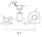
- Figure 4 schematically shows a device, which may be used when planning and carrying out an infusion in accordance with the invention.
- Patient data are obtained in a magnetic resonance or nuclear spin tomograph 3 and forwarded to a planning system 1 and to a navigation system 2.
- the catheter or catheters are positioned at a desired point on a body by the navigation system 2, using for example known reflectors or markers, attached to one or more catheters, positional data of the markers being captured by IR cameras 2a.
- the planning system 1 determines the suitable catheter parameters and parameters of the infusing medium for a pre-set infusion to be carried out, using patient parameters determined by the magnetic resonance or nuclear spin resonance system 3.
Landscapes
- Health & Medical Sciences (AREA)
- Engineering & Computer Science (AREA)
- Medical Informatics (AREA)
- Public Health (AREA)
- Surgery (AREA)
- Life Sciences & Earth Sciences (AREA)
- General Health & Medical Sciences (AREA)
- Biomedical Technology (AREA)
- Animal Behavior & Ethology (AREA)
- Molecular Biology (AREA)
- Heart & Thoracic Surgery (AREA)
- Nuclear Medicine, Radiotherapy & Molecular Imaging (AREA)
- Veterinary Medicine (AREA)
- Pathology (AREA)
- Robotics (AREA)
- Data Mining & Analysis (AREA)
- Databases & Information Systems (AREA)
- Epidemiology (AREA)
- Primary Health Care (AREA)
- Oral & Maxillofacial Surgery (AREA)
- Magnetic Resonance Imaging Apparatus (AREA)
Claims (24)
- Procédé pour préparer une perfusion et le placement d'un cathéter ayant une rigidité spécifique, choisi parmi plusieurs types de cathéters contenus dans une base de données, dans un corps, dans lequel des informations d'au moins une partie de la structure interne du corps d'un patient sont automatiquement analysées en utilisant des techniques de segmentation pour déterminer si au moins une région ou structure spécifique ou critique est contenue dans une région d'intérêt autour de la trajectoire prévue du dispositif dans le corps, dans lequel ladite région ou structure spécifique ou critique est automatiquement affichée et/ou classée et/ou évaluée en termes de niveaux de risque prédéfinis en utilisant des valeurs mémorisées dans des bases de données, dans lequel les niveaux de risque et la rigidité du cathéter sont déterminés sur la base du risque dû à la densité de tissu spécifique au patient d'une région ou structure spécifique du corps.
- Procédé selon la revendication précédente, dans lequel en tant qu'informations d'au moins une partie de la structure interne du corps, des structures anatomiques, fonctionnelles, angiographiques et/ou physiologiques du corps sont, en particulier automatiquement, détectées et/ou des structures tracées manuellement sont détectées.
- Procédé selon l'une quelconque des revendications précédentes, dans lequel les structures anatomiques sont obtenues en utilisant une imagerie par résonance magnétique (IRM), une imagerie par rayons X, une tomographie informatique (CT), une imagerie par ultrasons, les structures physiologiques sont déterminées en utilisant MR-DTI, MR-DCE, une imagerie par perfusion à partir de MR ou CT, PET, SPECT, MEG, et les structures fonctionnelles sont déterminées en utilisant fIRM, PET, le mappage du cerveau, EEG.
- Procédé selon l'une quelconque des revendications précédentes, dans lequel la région d'intérêt autour de la trajectoire peut être définie par au moins une valeur par défaut ou prédéterminée proche de la trajectoire ou autour de la trajectoire et/ou peut être sélectionnée par un utilisateur et/ou peut être semi-automatiquement ou automatiquement définie en utilisant des informations concernant le patient ou le traitement du patient.
- Procédé selon l'une quelconque des revendications précédentes, dans lequel la région ou structure spécifique ou critique dans la région d'intérêt est en outre automatiquement affichée et/ou classée et/ou évaluée en termes de position spatiale, d'effet indésirable potentiel et/ou de proximité par rapport à la trajectoire.
- Procédé selon l'une quelconque des revendications précédentes, dans lequel la région ou structure spécifique ou critique est identifiée dans les informations d'au moins une partie de la structure interne du corps et est segmentée à partir des informations de la structure interne du corps.
- Procédé selon l'une quelconque des revendications précédentes, dans lequel la région ou structure spécifique ou critique est obtenue par exemple par balayages IRM de diffusion, balayages IRM rehaussés par un agent de contraste, DTI, balayages IRM rehaussés par un agent de contraste dynamique, balayages IRM à densité de protons, balayages IRM spectroscopiques, balayages PET, balayages SPECT, imagerie moléculaire, CT, rayons X, ultrasons, élastographie, imagerie chronologique, biopsies et/ou analyse de tissu.
- Procédé selon l'une quelconque des revendications précédentes, dans lequel la région ou structure spécifique ou critique est enregistrée avec une image de la partie de la structure interne du corps et est superposée sur ladite image.
- Procédé selon la revendication précédente, dans lequel la région ou structure spécifique ou critique est affichée conjointement avec la trajectoire prévue sur l'image de la structure interne du corps.
- Procédé selon l'une quelconque des revendications précédentes, dans lequel les niveaux de risque sont déterminés sut la base :d'un risque dû aux caractéristiques anatomiques et/ou fonctionnelles et/ou physiologiques d'une région ou structure spécifique du corps, en particulier des risques de blessure du corps et/ou un risque d'échec de la thérapie, et/oud'un risque dû à un plan de traitement prédéfini, en particulier la proximité d'une approche de traitement, telle qu'une trajectoire, par rapport à une structure de risque, et/ou la proximité de deux ou plus de deux approches de traitement différentes, telles que deux ou plus de deux trajectoires, les unes par rapport aux autres et/ou la pondération des risques de l'approche de traitement prédéfinie, et/oud'un risque dû à un paramétrage ou à une configuration prédéfini d'un système de navigation chirurgical.
- Procédé selon la revendication précédente, dans lequel le risque de blessure du corps comporte le risque de traverser une trajectoire et un nerf optique et le risque d'échec du traitement comporte le risque de ne pas traverser la trajectoire via un sillon lors de l'administration de médicament.
- Procédé selon l'une quelconque des revendications précédentes, dans lequel la pondération des risques de l'approche de traitement prédéfinie comporte la pondération des risques d'un plan ou d'une trajectoire de cathéter qui est au moins éloigné d'une distance prédéterminée par exemple d'un sillon.
- Procédé selon l'une quelconque des trois revendications précédentes, dans lequel le risque dû à un paramétrage ou à une configuration prédéfini du système de navigation chirurgical comporte un risque d'utilisation d'un dispositif de maintien non monté, tel qu'une aiguille pour faire passer un cathéter le long de la trajectoire et/ou le risque d'une utilisation d'une perfusion flexible ou d'un dispositif de maintien et/ou le risque lorsque le dispositif de maintien est fixé ou monté loin d'un point d'entrée du cathéter et/ou le risque d'un chirurgien inexpérimenté.
- Procédé selon l'une quelconque des revendications précédentes, dans lequel des niveaux de risque sont affichés séparément et/ou surlignés, par exemple colorés, dans la région ou structure spécifique ou critique.
- Procédé selon l'une quelconque des revendications précédentes, dans lequel des avertissements concernant l'effet indésirable potentiel, tel que le croisement de la trajectoire et de la région ou structure spécifique ou critique, sont affichés.
- Procédé selon l'une quelconque des revendications précédentes, dans lequel des informations quantitatives concernant la proximité de la région ou structure spécifique ou critique par rapport à une ou plusieurs trajectoires sont fournies.
- Procédé selon l'une quelconque des revendications précédentes, dans lequel des bords sont affichés le long ou autour de la région ou structure spécifique ou critique, dans lequel lorsqu'il est prévu que la trajectoire traverse au moins un bord, un signal d'avertissement, en particulier un signal d'avertissement acoustique, est délivré en sortie ou on empêche la trajectoire de traverser le bord.
- Procédé selon l'une quelconque des revendications précédentes, dans lequel sont déterminées des trajectoires qui traversent ou ne traversent pas des régions ayant un niveau de risque prédéfini ou prédéterminé, en particulier qui sont situées à une distance prédéterminée de zones ayant un niveau de risque prédéfini ou prédéterminé.
- Procédé selon l'une quelconque des revendications précédentes, dans lequel la trajectoire prévue est utilisée pour des biopsies stéréotaxiques, un placement de cathéter, un placement de canule, un placement de stimulateur, un placement de dérivation, un placement d'implants chirurgicaux et/ou un placement d'autres dispositifs.
- Procédé selon l'une quelconque des revendications précédentes, dans lequel une réaction haptique est délivrée à un dispositif robotisé ou semi-robotisé, en particulier un bras robotisé ou un dispositif de guidage, la réaction haptique étant basée sur la proximité entre un instrument chirurgical se déplaçant selon une relation connue dans le patient et des régions de risque.
- Programme informatique lequel, lorsqu'il est exécuté sur un ordinateur ou chargé sur un ordinateur, amène l'ordinateur à mettre en oeuvre un procédé selon l'une quelconque des revendications précédentes.
- Produit de programme informatique mémorisé sur un support compatible avec un ordinateur ou un support de données et comportant le programme informatique tel que défini dans la revendication précédente.
- Dispositif pour préparer une perfusion et le placement d'un cathéter ayant une rigidité spécifique, choisi parmi plusieurs types de cathéters contenus dans une base de données, dans un corps, comportant :un dispositif d'acquisition pour acquérir des informations d'au moins une partie de la structure interne du corps d'un patientune base de données mémorisant des valeurs pour la détermination de niveaux de risqueun dispositif d'analyse pour analyser les informations acquises de la structure interne du corps en utilisant des techniques de segmentation pour déterminer si au moins une région ou structure spécifique ou critique est contenue dans une région d'intérêt autour de la trajectoire prévue du dispositif dans le corps et pour afficher et/ou classer et/ou évaluer ladite région ou structure spécifique ou critique en termes de niveaux de risque prédéfinis en utilisant les valeurs mémorisées dans la base de données, dans lequel les niveaux de risque et la rigidité du cathéter sont déterminés sur la base du risque dû à la densité de tissu spécifique au patient d'une région ou structure spécifique du corps.
- Dispositif selon la revendication 23, comportant en outreun dispositif d'affichage pour afficher les informations acquises de la structure interne du corps et des informations de la région ou structure spécifique ou critique etun dispositif de mémorisation pour mémoriser les informations acquises de la structure interne du corps, des informations de la région ou structure spécifique ou critique et des informations du dispositif.
Priority Applications (3)
| Application Number | Priority Date | Filing Date | Title |
|---|---|---|---|
| EP06007425A EP1844725B1 (fr) | 2006-04-07 | 2006-04-07 | Évaluation des risques de trajectoires planifiés |
| DE602006005899T DE602006005899D1 (de) | 2006-04-07 | 2006-04-07 | Risikobewertung von geplanten Trajektorien |
| US11/697,823 US8060181B2 (en) | 2006-04-07 | 2007-04-09 | Risk assessment for planned trajectories |
Applications Claiming Priority (1)
| Application Number | Priority Date | Filing Date | Title |
|---|---|---|---|
| EP06007425A EP1844725B1 (fr) | 2006-04-07 | 2006-04-07 | Évaluation des risques de trajectoires planifiés |
Publications (2)
| Publication Number | Publication Date |
|---|---|
| EP1844725A1 EP1844725A1 (fr) | 2007-10-17 |
| EP1844725B1 true EP1844725B1 (fr) | 2009-03-25 |
Family
ID=36888763
Family Applications (1)
| Application Number | Title | Priority Date | Filing Date |
|---|---|---|---|
| EP06007425A Active EP1844725B1 (fr) | 2006-04-07 | 2006-04-07 | Évaluation des risques de trajectoires planifiés |
Country Status (2)
| Country | Link |
|---|---|
| EP (1) | EP1844725B1 (fr) |
| DE (1) | DE602006005899D1 (fr) |
Families Citing this family (3)
| Publication number | Priority date | Publication date | Assignee | Title |
|---|---|---|---|---|
| JP2012513789A (ja) * | 2008-12-29 | 2012-06-21 | コーニンクレッカ フィリップス エレクトロニクス エヌ ヴィ | 低侵襲手術における組織への損傷を軽減するための経路計画方法及びシステム |
| JP5501290B2 (ja) * | 2011-05-23 | 2014-05-21 | 富士フイルム株式会社 | 画像処理装置、放射線画像撮影システム、及び画像処理プログラム |
| EP3066593B1 (fr) | 2013-11-05 | 2018-05-02 | Brainlab AG | Quantification de vulnérabilité de cerveau |
Family Cites Families (9)
| Publication number | Priority date | Publication date | Assignee | Title |
|---|---|---|---|---|
| US5626862A (en) | 1994-08-02 | 1997-05-06 | Massachusetts Institute Of Technology | Controlled local delivery of chemotherapeutic agents for treating solid tumors |
| US6026316A (en) | 1997-05-15 | 2000-02-15 | Regents Of The University Of Minnesota | Method and apparatus for use with MR imaging |
| US6112750A (en) * | 1998-03-24 | 2000-09-05 | International Business Machines Corporation | Method and system for assessing risks and prognoses of a given course of medical treatment |
| AU2001268217A1 (en) * | 2000-06-06 | 2001-12-17 | The Research Foundation Of State University Of New York | Computer aided visualization, fusion and treatment planning |
| US7630750B2 (en) * | 2001-02-05 | 2009-12-08 | The Research Foundation For The State University Of New York | Computer aided treatment planning |
| DE50107066D1 (de) * | 2001-11-30 | 2005-09-15 | Brainlab Ag | Vorrichtung zur Planung einer Infusion |
| CA2633137C (fr) * | 2002-08-13 | 2012-10-23 | The Governors Of The University Of Calgary | Systeme robotique utilise en microchirurgie |
| US20040049121A1 (en) * | 2002-09-06 | 2004-03-11 | Uri Yaron | Positioning system for neurological procedures in the brain |
| US20060052689A1 (en) * | 2004-08-14 | 2006-03-09 | Scouten Charles W | Digital stereotaxic manipulator with interfaces for use with computerized brain atlases |
-
2006
- 2006-04-07 EP EP06007425A patent/EP1844725B1/fr active Active
- 2006-04-07 DE DE602006005899T patent/DE602006005899D1/de active Active
Also Published As
| Publication number | Publication date |
|---|---|
| DE602006005899D1 (de) | 2009-05-07 |
| EP1844725A1 (fr) | 2007-10-17 |
Similar Documents
| Publication | Publication Date | Title |
|---|---|---|
| US8060181B2 (en) | Risk assessment for planned trajectories | |
| US7266227B2 (en) | Device and method for administering a substance | |
| US11793933B2 (en) | MRI-compatible surgical cannulae for transferring a substance to and/or from a patient | |
| US9008752B2 (en) | Method to determine distribution of a material by an infused magnetic resonance image contrast agent | |
| US20150011866A1 (en) | Probe for Surgical Navigation | |
| Larson et al. | An optimized system for interventional magnetic resonance imaging-guided stereotactic surgery: preliminary evaluation of targeting accuracy | |
| US20140303486A1 (en) | Surgical Navigation Planning System and Associated Methods | |
| CA2771175C (fr) | Placement optimise d'une canule pour administration d'agents therapeutiques au cerveau | |
| US9393361B2 (en) | Method to determine a material distribution | |
| US7740606B2 (en) | Method and apparatus for automated optimization of treatment plans | |
| US9924888B2 (en) | Targeted infusion of agents against parkinson's disease | |
| US9901413B2 (en) | Targeted infusion of agents for treatment of ALS | |
| US7734326B2 (en) | Method and device for preparing a drainage | |
| EP1844725B1 (fr) | Évaluation des risques de trajectoires planifiés | |
| US9907485B2 (en) | Targeted immunization and plaque destruction against Alzheimer's disease | |
| EP2681679B1 (fr) | Planification et simulation d'infusion assistées par ordinateur | |
| EP1825881A1 (fr) | Simulation de la distribution des agents thérapeutiques ou diagnostiques dans dans un patient | |
| US20070078338A1 (en) | Method and device for planning a direct infusion into hepatic tissue | |
| US9694170B2 (en) | Method and device for planning a direct infusion into hepatic tissue | |
| Oyama et al. | Targeted Delivery of Chemogenetic Adeno-Associated Viral Vectors to Cortical Sulcus Regions in Macaque Monkeys by Handheld Injections | |
| Campbell | Optimizing therapeutic agent delivery to malignant gliomas via convection-enhanced delivery | |
| CN113975659A (zh) | 放射线治疗辅助系统及方法 |
Legal Events
| Date | Code | Title | Description |
|---|---|---|---|
| PUAI | Public reference made under article 153(3) epc to a published international application that has entered the european phase |
Free format text: ORIGINAL CODE: 0009012 |
|
| 17P | Request for examination filed |
Effective date: 20060407 |
|
| AK | Designated contracting states |
Kind code of ref document: A1 Designated state(s): AT BE BG CH CY CZ DE DK EE ES FI FR GB GR HU IE IS IT LI LT LU LV MC NL PL PT RO SE SI SK TR |
|
| AX | Request for extension of the european patent |
Extension state: AL BA HR MK YU |
|
| AKX | Designation fees paid |
Designated state(s): DE FR GB IT |
|
| GRAP | Despatch of communication of intention to grant a patent |
Free format text: ORIGINAL CODE: EPIDOSNIGR1 |
|
| RAP1 | Party data changed (applicant data changed or rights of an application transferred) |
Owner name: BRAINLAB AG |
|
| GRAS | Grant fee paid |
Free format text: ORIGINAL CODE: EPIDOSNIGR3 |
|
| GRAA | (expected) grant |
Free format text: ORIGINAL CODE: 0009210 |
|
| AK | Designated contracting states |
Kind code of ref document: B1 Designated state(s): DE FR GB IT |
|
| REG | Reference to a national code |
Ref country code: GB Ref legal event code: FG4D |
|
| REF | Corresponds to: |
Ref document number: 602006005899 Country of ref document: DE Date of ref document: 20090507 Kind code of ref document: P |
|
| PLBE | No opposition filed within time limit |
Free format text: ORIGINAL CODE: 0009261 |
|
| STAA | Information on the status of an ep patent application or granted ep patent |
Free format text: STATUS: NO OPPOSITION FILED WITHIN TIME LIMIT |
|
| 26N | No opposition filed |
Effective date: 20091229 |
|
| PG25 | Lapsed in a contracting state [announced via postgrant information from national office to epo] |
Ref country code: IT Free format text: LAPSE BECAUSE OF FAILURE TO SUBMIT A TRANSLATION OF THE DESCRIPTION OR TO PAY THE FEE WITHIN THE PRESCRIBED TIME-LIMIT Effective date: 20090325 |
|
| REG | Reference to a national code |
Ref country code: DE Ref legal event code: R082 Ref document number: 602006005899 Country of ref document: DE Representative=s name: SCHWABE SANDMAIR MARX, DE |
|
| REG | Reference to a national code |
Ref country code: DE Ref legal event code: R082 Ref document number: 602006005899 Country of ref document: DE Representative=s name: SCHWABE SANDMAIR MARX, DE Effective date: 20131104 Ref country code: DE Ref legal event code: R081 Ref document number: 602006005899 Country of ref document: DE Owner name: BRAINLAB AG, DE Free format text: FORMER OWNER: BRAINLAB AG, 85622 FELDKIRCHEN, DE Effective date: 20131104 Ref country code: DE Ref legal event code: R082 Ref document number: 602006005899 Country of ref document: DE Representative=s name: SCHWABE SANDMAIR MARX PATENTANWAELTE RECHTSANW, DE Effective date: 20131104 |
|
| REG | Reference to a national code |
Ref country code: FR Ref legal event code: PLFP Year of fee payment: 11 |
|
| REG | Reference to a national code |
Ref country code: DE Ref legal event code: R082 Ref document number: 602006005899 Country of ref document: DE Representative=s name: SSM SANDMAIR PATENTANWAELTE RECHTSANWALT PARTN, DE Ref country code: DE Ref legal event code: R082 Ref document number: 602006005899 Country of ref document: DE Representative=s name: SCHWABE SANDMAIR MARX PATENTANWAELTE RECHTSANW, DE Ref country code: DE Ref legal event code: R081 Ref document number: 602006005899 Country of ref document: DE Owner name: BRAINLAB AG, DE Free format text: FORMER OWNER: BRAINLAB AG, 85622 FELDKIRCHEN, DE |
|
| REG | Reference to a national code |
Ref country code: FR Ref legal event code: PLFP Year of fee payment: 12 |
|
| REG | Reference to a national code |
Ref country code: FR Ref legal event code: CA Effective date: 20170706 |
|
| REG | Reference to a national code |
Ref country code: FR Ref legal event code: PLFP Year of fee payment: 13 |
|
| P01 | Opt-out of the competence of the unified patent court (upc) registered |
Effective date: 20230508 |
|
| PGFP | Annual fee paid to national office [announced via postgrant information from national office to epo] |
Ref country code: GB Payment date: 20240418 Year of fee payment: 19 |
|
| PGFP | Annual fee paid to national office [announced via postgrant information from national office to epo] |
Ref country code: DE Payment date: 20240418 Year of fee payment: 19 |
|
| PGFP | Annual fee paid to national office [announced via postgrant information from national office to epo] |
Ref country code: FR Payment date: 20240426 Year of fee payment: 19 |