EP1832271A2 - Verbessertes endodontisches Verfahren unter Verwendung von selbstklebenden Harzzementen oder selbstätzenden Klebstoffen und chemisch klebbaren Obturatoren - Google Patents
Verbessertes endodontisches Verfahren unter Verwendung von selbstklebenden Harzzementen oder selbstätzenden Klebstoffen und chemisch klebbaren Obturatoren Download PDFInfo
- Publication number
- EP1832271A2 EP1832271A2 EP07003966A EP07003966A EP1832271A2 EP 1832271 A2 EP1832271 A2 EP 1832271A2 EP 07003966 A EP07003966 A EP 07003966A EP 07003966 A EP07003966 A EP 07003966A EP 1832271 A2 EP1832271 A2 EP 1832271A2
- Authority
- EP
- European Patent Office
- Prior art keywords
- chemically
- self
- bondable
- adhesive
- root canal
- Prior art date
- Legal status (The legal status is an assumption and is not a legal conclusion. Google has not performed a legal analysis and makes no representation as to the accuracy of the status listed.)
- Withdrawn
Links
- 238000000034 method Methods 0.000 title claims abstract description 43
- 239000000853 adhesive Substances 0.000 title claims abstract description 40
- 230000001070 adhesive effect Effects 0.000 title claims abstract description 30
- 239000004568 cement Substances 0.000 title claims abstract description 22
- 239000004840 adhesive resin Substances 0.000 title claims abstract description 18
- 229920006223 adhesive resin Polymers 0.000 title claims abstract description 18
- 238000005530 etching Methods 0.000 title claims abstract description 10
- 210000004262 dental pulp cavity Anatomy 0.000 claims abstract description 62
- 239000000463 material Substances 0.000 claims abstract description 55
- 238000011049 filling Methods 0.000 claims abstract description 13
- 238000007789 sealing Methods 0.000 claims abstract description 9
- 239000000899 Gutta-Percha Substances 0.000 claims description 26
- 240000000342 Palaquium gutta Species 0.000 claims description 26
- 229920000588 gutta-percha Polymers 0.000 claims description 26
- 239000003178 glass ionomer cement Substances 0.000 claims description 7
- 239000002253 acid Substances 0.000 claims description 6
- 239000011152 fibreglass Substances 0.000 claims description 5
- 229920000728 polyester Polymers 0.000 claims description 5
- 239000002131 composite material Substances 0.000 claims description 4
- 239000000178 monomer Substances 0.000 claims description 4
- 239000012815 thermoplastic material Substances 0.000 claims 4
- 229920005989 resin Polymers 0.000 description 16
- 239000011347 resin Substances 0.000 description 16
- KCXVZYZYPLLWCC-UHFFFAOYSA-N EDTA Chemical compound OC(=O)CN(CC(O)=O)CCN(CC(O)=O)CC(O)=O KCXVZYZYPLLWCC-UHFFFAOYSA-N 0.000 description 9
- 210000004268 dentin Anatomy 0.000 description 9
- 229960001484 edetic acid Drugs 0.000 description 9
- 239000000565 sealant Substances 0.000 description 8
- 239000000835 fiber Substances 0.000 description 7
- 239000000047 product Substances 0.000 description 7
- 239000007787 solid Substances 0.000 description 7
- 238000001000 micrograph Methods 0.000 description 6
- 239000000805 composite resin Substances 0.000 description 5
- 230000009977 dual effect Effects 0.000 description 5
- 239000000945 filler Substances 0.000 description 5
- 239000003999 initiator Substances 0.000 description 5
- SUKJFIGYRHOWBL-UHFFFAOYSA-N sodium hypochlorite Chemical compound [Na+].Cl[O-] SUKJFIGYRHOWBL-UHFFFAOYSA-N 0.000 description 5
- 239000000126 substance Substances 0.000 description 5
- 229910019093 NaOCl Inorganic materials 0.000 description 4
- 230000008901 benefit Effects 0.000 description 4
- 238000009833 condensation Methods 0.000 description 4
- 230000005494 condensation Effects 0.000 description 4
- 239000000975 dye Substances 0.000 description 4
- 229920001169 thermoplastic Polymers 0.000 description 4
- 239000004416 thermosoftening plastic Substances 0.000 description 4
- 239000007767 bonding agent Substances 0.000 description 3
- 230000006872 improvement Effects 0.000 description 3
- 239000007788 liquid Substances 0.000 description 3
- 239000008188 pellet Substances 0.000 description 3
- 241000894006 Bacteria Species 0.000 description 2
- 241000183024 Populus tremula Species 0.000 description 2
- 208000002599 Smear Layer Diseases 0.000 description 2
- 241000364021 Tulsa Species 0.000 description 2
- XLOMVQKBTHCTTD-UHFFFAOYSA-N Zinc monoxide Chemical compound [Zn]=O XLOMVQKBTHCTTD-UHFFFAOYSA-N 0.000 description 2
- 230000001580 bacterial effect Effects 0.000 description 2
- 239000007844 bleaching agent Substances 0.000 description 2
- 239000003479 dental cement Substances 0.000 description 2
- 238000001962 electrophoresis Methods 0.000 description 2
- 238000005516 engineering process Methods 0.000 description 2
- RRAFCDWBNXTKKO-UHFFFAOYSA-N eugenol Chemical compound COC1=CC(CC=C)=CC=C1O RRAFCDWBNXTKKO-UHFFFAOYSA-N 0.000 description 2
- 238000001125 extrusion Methods 0.000 description 2
- 230000002262 irrigation Effects 0.000 description 2
- 238000003973 irrigation Methods 0.000 description 2
- 229910052751 metal Inorganic materials 0.000 description 2
- 239000002184 metal Substances 0.000 description 2
- 229920000642 polymer Polymers 0.000 description 2
- 229910001220 stainless steel Inorganic materials 0.000 description 2
- 239000010935 stainless steel Substances 0.000 description 2
- XLYOFNOQVPJJNP-UHFFFAOYSA-N water Substances O XLYOFNOQVPJJNP-UHFFFAOYSA-N 0.000 description 2
- NPBVQXIMTZKSBA-UHFFFAOYSA-N Chavibetol Natural products COC1=CC=C(CC=C)C=C1O NPBVQXIMTZKSBA-UHFFFAOYSA-N 0.000 description 1
- RYGMFSIKBFXOCR-UHFFFAOYSA-N Copper Chemical compound [Cu] RYGMFSIKBFXOCR-UHFFFAOYSA-N 0.000 description 1
- 102100026816 DNA-dependent metalloprotease SPRTN Human genes 0.000 description 1
- 101710175461 DNA-dependent metalloprotease SPRTN Proteins 0.000 description 1
- 239000005770 Eugenol Substances 0.000 description 1
- UVMRYBDEERADNV-UHFFFAOYSA-N Pseudoeugenol Natural products COC1=CC(C(C)=C)=CC=C1O UVMRYBDEERADNV-UHFFFAOYSA-N 0.000 description 1
- VYPSYNLAJGMNEJ-UHFFFAOYSA-N Silicium dioxide Chemical compound O=[Si]=O VYPSYNLAJGMNEJ-UHFFFAOYSA-N 0.000 description 1
- FAPWRFPIFSIZLT-UHFFFAOYSA-M Sodium chloride Chemical compound [Na+].[Cl-] FAPWRFPIFSIZLT-UHFFFAOYSA-M 0.000 description 1
- 239000005708 Sodium hypochlorite Substances 0.000 description 1
- 230000002378 acidificating effect Effects 0.000 description 1
- 238000004026 adhesive bonding Methods 0.000 description 1
- 229920003232 aliphatic polyester Polymers 0.000 description 1
- 239000001045 blue dye Substances 0.000 description 1
- CREMABGTGYGIQB-UHFFFAOYSA-N carbon carbon Chemical group C.C CREMABGTGYGIQB-UHFFFAOYSA-N 0.000 description 1
- 239000011203 carbon fibre reinforced carbon Substances 0.000 description 1
- 230000015556 catabolic process Effects 0.000 description 1
- 239000013043 chemical agent Substances 0.000 description 1
- 239000013065 commercial product Substances 0.000 description 1
- 150000001875 compounds Chemical class 0.000 description 1
- 238000004132 cross linking Methods 0.000 description 1
- 210000003298 dental enamel Anatomy 0.000 description 1
- 210000003074 dental pulp Anatomy 0.000 description 1
- 239000000645 desinfectant Substances 0.000 description 1
- 238000011161 development Methods 0.000 description 1
- 239000012153 distilled water Substances 0.000 description 1
- 229920001971 elastomer Polymers 0.000 description 1
- 229960002217 eugenol Drugs 0.000 description 1
- 239000002360 explosive Substances 0.000 description 1
- 239000000284 extract Substances 0.000 description 1
- 239000012530 fluid Substances 0.000 description 1
- 239000011521 glass Substances 0.000 description 1
- PCHJSUWPFVWCPO-UHFFFAOYSA-N gold Chemical compound [Au] PCHJSUWPFVWCPO-UHFFFAOYSA-N 0.000 description 1
- 238000007689 inspection Methods 0.000 description 1
- 229920000126 latex Polymers 0.000 description 1
- 238000011068 loading method Methods 0.000 description 1
- 150000002739 metals Chemical class 0.000 description 1
- CXKWCBBOMKCUKX-UHFFFAOYSA-M methylene blue Chemical compound [Cl-].C1=CC(N(C)C)=CC2=[S+]C3=CC(N(C)C)=CC=C3N=C21 CXKWCBBOMKCUKX-UHFFFAOYSA-M 0.000 description 1
- 229960000907 methylthioninium chloride Drugs 0.000 description 1
- 244000005700 microbiome Species 0.000 description 1
- 230000001338 necrotic effect Effects 0.000 description 1
- 238000001543 one-way ANOVA Methods 0.000 description 1
- 239000011368 organic material Substances 0.000 description 1
- 230000037361 pathway Effects 0.000 description 1
- XYJRXVWERLGGKC-UHFFFAOYSA-D pentacalcium;hydroxide;triphosphate Chemical class [OH-].[Ca+2].[Ca+2].[Ca+2].[Ca+2].[Ca+2].[O-]P([O-])([O-])=O.[O-]P([O-])([O-])=O.[O-]P([O-])([O-])=O XYJRXVWERLGGKC-UHFFFAOYSA-D 0.000 description 1
- 238000005325 percolation Methods 0.000 description 1
- 229920001610 polycaprolactone Polymers 0.000 description 1
- 239000004632 polycaprolactone Substances 0.000 description 1
- 239000004645 polyester resin Substances 0.000 description 1
- -1 porcelains Substances 0.000 description 1
- 238000002360 preparation method Methods 0.000 description 1
- 230000002265 prevention Effects 0.000 description 1
- 230000008569 process Effects 0.000 description 1
- 230000000750 progressive effect Effects 0.000 description 1
- 230000009467 reduction Effects 0.000 description 1
- 238000011160 research Methods 0.000 description 1
- 238000011268 retreatment Methods 0.000 description 1
- 210000003296 saliva Anatomy 0.000 description 1
- MKWYFZFMAMBPQK-UHFFFAOYSA-J sodium feredetate Chemical compound [Na+].[Fe+3].[O-]C(=O)CN(CC([O-])=O)CCN(CC([O-])=O)CC([O-])=O MKWYFZFMAMBPQK-UHFFFAOYSA-J 0.000 description 1
- 238000009987 spinning Methods 0.000 description 1
- 238000010561 standard procedure Methods 0.000 description 1
- 238000007619 statistical method Methods 0.000 description 1
- 238000004659 sterilization and disinfection Methods 0.000 description 1
- 239000000758 substrate Substances 0.000 description 1
- 238000012360 testing method Methods 0.000 description 1
- 238000002560 therapeutic procedure Methods 0.000 description 1
- 238000005382 thermal cycling Methods 0.000 description 1
- 229920005992 thermoplastic resin Polymers 0.000 description 1
- 239000002966 varnish Substances 0.000 description 1
- 239000011787 zinc oxide Substances 0.000 description 1
Images




Classifications
-
- A—HUMAN NECESSITIES
- A61—MEDICAL OR VETERINARY SCIENCE; HYGIENE
- A61C—DENTISTRY; APPARATUS OR METHODS FOR ORAL OR DENTAL HYGIENE
- A61C5/00—Filling or capping teeth
- A61C5/40—Implements for surgical treatment of the roots or nerves of the teeth; Nerve needles; Methods or instruments for medication of the roots
-
- A—HUMAN NECESSITIES
- A61—MEDICAL OR VETERINARY SCIENCE; HYGIENE
- A61K—PREPARATIONS FOR MEDICAL, DENTAL OR TOILETRY PURPOSES
- A61K6/00—Preparations for dentistry
- A61K6/70—Preparations for dentistry comprising inorganic additives
- A61K6/71—Fillers
- A61K6/77—Glass
-
- A—HUMAN NECESSITIES
- A61—MEDICAL OR VETERINARY SCIENCE; HYGIENE
- A61K—PREPARATIONS FOR MEDICAL, DENTAL OR TOILETRY PURPOSES
- A61K6/00—Preparations for dentistry
- A61K6/80—Preparations for artificial teeth, for filling teeth or for capping teeth
- A61K6/831—Preparations for artificial teeth, for filling teeth or for capping teeth comprising non-metallic elements or compounds thereof, e.g. carbon
- A61K6/833—Glass-ceramic composites
-
- A—HUMAN NECESSITIES
- A61—MEDICAL OR VETERINARY SCIENCE; HYGIENE
- A61K—PREPARATIONS FOR MEDICAL, DENTAL OR TOILETRY PURPOSES
- A61K6/00—Preparations for dentistry
- A61K6/80—Preparations for artificial teeth, for filling teeth or for capping teeth
- A61K6/884—Preparations for artificial teeth, for filling teeth or for capping teeth comprising natural or synthetic resins
Definitions
- the present invention relates to the field of endodontics.
- Root canal therapy has become well established as a viable clinical treatment to retain a tooth where the dental pulp has become painful, irreversibly inflamed, infected or necrotic (dead).
- the generally accepted method of performing a root canal is to drill an "access" opening into the root canal through the clinical crown of the tooth and then to remove the diseased or damaged pulpal tissues using small sharp files, rotary files, dental burs or drills.
- the root canal is treated with chemical agents to disinfect the canal, to remove any residual organic material and to remove what is commonly referred to as the debris "smear layer.”
- the cleaned root canal must then be "obturated,” which means that it is filled with an inert material that is capable of sealing the canal against the passage of fluids and microorganisms.
- U.S. Patent Application Publication No. 2005/0069836 describes a new type of polyester based, thermoplastic obturation material that is able to fulfill all of the same functions as gutta percha but which also takes advantage of a much stronger chemical adhesion/bonding strategy to improve the seal.
- a commercial product believed to be embraced by this patent application is RESILON TM (polycaprolactone-based thermoplastic aliphatic polyester resin obturation material).
- RESILON TM can be thought of as a soft thermoplastic resin that is chemically bondable. This differs from gutta percha, which is based upon a naturally occurring latex rubber and is not chemically bondable. So, instead of simply cementing the point into the root canal, it is now possible to adhesively bond the point in and achieve a superb water tight, insoluble seal throughout the length of the canal.
- RESILON TM so important is that it takes advantage of the important scientific discipline referred to as “Adhesion dentistry” or “bonding.” Bonding in dentistry has enjoyed explosive growth and improvement since its introduction. Through modem adhesion dentistry it is now possible to strongly link many different substrates to tooth structure, including resin composites, porcelains, and metals. However, RESILON TM marks the first real linking of endodontic obturation and adhesion bonding.
- the composite resin restorative materials are generally highly filled with glass or silica particles to convert them into pastes, improve their strength and durability whereas the bonding agents are largely unfilled or sparsely filled, allowing them to remain as liquids.
- EPIPHANY TM and REALSEAL TM utilize solid core obturators made entirely of RESILON TM .
- NaOCl sodium hypochlorite
- EDTA ethylene diamine tetraacetic acid
- the RESILON TM point is seated into the sealer and stabilized by light curing from the crown side.
- INNOENDO TM and SIMPLIFILL TM are carrier based obturators.
- SIMPLIFILL TM utilizes a metal rod carrier to push a small plug of RESILON TM deep into the canal. The carrier rod is then twisted and removed and the remaining space in the canal is backfilled with RESILON TM points.
- INNOENDO TM utilizes a fiberglass carrier which holds a small tip of RESILON TM on the end. As the INNOENDO TM is seated into the canal the RESILON TM is forced down deep into the canal all the way to the apex of the root. However, in INNOENDO TM , the carrier is left in the root canal and forms the seal in the upper coronal part of the canal.
- self adhesive resin based cements include UNICEM TM (3M/ ESPE), MAXCEM TM (Kerr Dental) and EMBRACE WETBOND TM (Pulpdent), the latter of which is believed embraced by U.S. Patent No. 6,797,767 .
- the present invention relates to a kit for use in filling and sealing a root canal during an endodontic procedure, wherein the kit comprises:
- Endodontic failure can be due to a number of factors, including apical and/or coronal leakage, root fractures, insufficient disinfection and anachoresis. Prevention of leakage is especially important because endodontic treatment results in the loss or reduction of one's ability to feel/sense bacterial breakdown coronally. Percolation at the restoration margins may go unnoticed by the patient for long periods of time, and, thus, may be brought to the attention of the dentist too late to resolve the problem. Indeed, studies indicate that significant bacteria leakage following exposure of sealed root canals to artificial and natural saliva may occur in as little as two days post-procedure, leading to complete bacterial leakage. Accordingly, much energy has been expended in the art making certain that root canal sealing is as effective as possible.
- Figure 1 Shown in Figure 1 is a micrograph of a portion of a root canal sealed according to a known technique.
- Figure 2 shows the same root canal, but at 10x magnification. It is seen in Figure 2 that upon closer inspection there are gaps between the dentin and the sealant. These gaps can provide channels of egress for bacteria into the dentin and the tissue beyond, resulting ultimately in failure of the endodontics.
- Figure 3 is a micrograph of a portion of a root canal sealed with a RESILON TM point and a self-adhesive sealant.
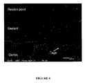
- Figure 4 shows the same root canal, but at 10x magnification. It is seen in Figure 4 that there are no gaps between either the sealant and the point or between the sealant and the dentin. In fact, the sealant bonds with the dentin to make an excellent adhesive seal, as evidenced by the projections, or "Tags,” extending from the sealant deep into the dentin.
- the idea is to achieve an excellent seal at every point where weakness might exist, for example, between the sealant and the obturator or between the sealant and the dentin, whether within the main root canal or within the accessory canals or irregularities.
- an obturator composed at least in part of a chemically-bondable material.
- the chemically-bondable material is any suitable such material.
- gutta percha which has been impregnated, coated or treated in any such a way as to render the gutta percha chemically-bondable; or a polymer-based, thermoplastic obturation material, such as RESILON TM , for example, as described in US 2005/0069836 , the entire contents of which are incorporated herein by reference; or a resin-modified glass ionomer.
- RESILON TM a polymer-based, thermoplastic obturation material
- RESI-LON TM material Especially preferred at this point is the RESI-LON TM material.
- the obturator is composed only in part of the chemically-bondable material.
- the obturator is composed in a first part of fiberglass and in a second part of the chemically-bondable material, such as, for example, the post and tapered fiberglass points available from INNOENDO TM (Heraeus Kulzer), which may be tipped with any of the foregoing chemically-bondable materials, especially the RESI-LON TM material.
- the obturator is composed in whole of the chemically-bondable material.
- the chemically-bondable material may, again, be any of the fore going chemically-bondable materials, especially the RESILON TM material.
- the self-adhesive resin-based cement or sealer is any suitable such material.
- materials containing acid containing monomers such as are described in U.S. Patent No. 6,797,767 , the entire contents of which are incorporated herein by reference. Due to the presence of the acid group, such materials can form excellent bonds to dentin themselves, without the need to use ethylene diamine tetraacetic acid (EDTA), and, thus, the customary irrigation step with EDTA can be dispensed with altogether if desired.
- Use of these materials also obviates the need to use the customary two-part adhesive primer, and, thus, this step can also be dispensed with altogether if desired.
- the self-adhesive cement or sealer will comprise a photoinitiator and/or a chemical initiator.
- a photoinitiator and/or a chemical initiator Most preferred is a "dual cure" system comprising both a photoinitiator and a chemical initiator.
- An access cavity is opened in the crown of a tooth to expose the root canal, and the pulp tissue is removed.
- An instrument is used to clean and shape the root canal to a predetermined working length.
- the root canal is irrigated with bleach to disinfect and dissolve residual pulp tissue.
- the root canal is irrigated with ethylene diamine tetraacetic acid (EDTA) to demineralize the surface of root dentin to remove the smear layer, although this step can be eliminated if desired.
- EDTA ethylene diamine tetraacetic acid
- the root canal is dried thoroughly with paper points. Selection is then made of an appropriate size obturator. A rubber stop is fitted on the distal end of the obturator at the working length. The obturator is inserted into the root canal, and the proper seating is verified by radiograph, and then the obturator is removed temporarily.
- a self-adhesive sealer is introduced into the root canal, for example, by spinning the sealer into the root canal with a lentulo filler instrument or a paper point.
- the selected obturator is placed into a disinfectant solution, and then seated into the root canal, and any excess sealer that extrudes from the root canal cleaned.
- the filling is light-cured with a standard curing light for 40 seconds.
- the portion of the obturator that protrudes from the crown is cut, and the coronal cavity is filled with, for example, INNOENDO TM dual cured or self cured core buildup material.
- Example 1 The protocol of Example 1 is repeated except that after the root canal is irrigated with bleach to disinfect and dissolve residual pulp tissue (again only optional), the upper portion of the root canal may be drilled to widen the root canal to accommodate the post.
- the root canal may be obturated with the use of obturator pellets which have been heated in a suitable device to allow the liquefaction and extrusion of the obturating material into the root canal, for example, RESILON TM pellets.
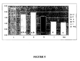
- the experimental protocol was follows: In each case, the root canal of an extracted tooth was sealed and obturated according to the manufacturer's directions or in the case of Group 6 as described herein. Each tooth was then stored in distilled water for 7 days at 37°C, and, thereafter, subjected to thermal cycling (1000 cycles, 5°C-55°C) in methylene blue dye. Each tooth was sectioned at 1 mm increments from the apical or coronal surface, and then evaluated for the presence of apical and coronal die leakage. The coronal leakage results are shown in Figure 5. The apical leakage results are shown in Figure 6.
- Group 6 i.e., the use of the INNOENDO TM obturator with the inventive protocol, provides the best results both in terms of coronal and apical leakage, even though the results are not statistically different from Group 5.
- the protocol associated with Group 5 involves the application of a two-part adhesive primer to the root canal prior to filling with the sealer. Accordingly, even compared to Group 5, Group 6 provides a benefit in terms of a more efficient protocol.
- Root fracture resistance was assessed of root canals sealed with various commercial products as follows:
- the experimental protocol was as follows: Single canal extracted teeth sectioned of their clinical crowns were obturated according to the manufacturer's instructions for each group or in the case of Group 7 as described herein. The obturated teeth were then stored in 100% humidity for 48 hours at 37°C to allow for complete setting of sealers. A vertical loading force was applied to the obturated roots with Model 4411 Instron at a crosshead speed of 1.0 mm/min. until the root fractured. The results are shown in Figure 7.
- Group 7 i.e., the inventive protocol (labeled "InnoEndo Exp.” in Figure 7,) exhibited the highest root fracture resistance of all of the groups tested, although again the results are not statistically different from Group 6.
- the protocol associated with Group 6 involves the application of a two-part adhesive primer to the root canal prior to filling with the sealer. Accordingly, even compared to Group 6, Group 7 provides a benefit in terms of a more efficient protocol.
- teeth obturated with INNOENDO TM fiber obturator and INNOENDO TM adhesive and resin sealer an INNOENDO TM fiber obturator with a self-adhesive resin sealer, an ENDOREZ TM cone with ENDOREZ TM resin sealer and a FIBREFILL TM fiber obturator with FIBREFILL TM adhesive and resin sealer.
- Teeth obturated with gutta percha with ENDOREZ TM resin sealer and those obturated with a RESILON TM cone with ENDOREZ TM resin sealer were intermediate in behavior.
Landscapes
- Health & Medical Sciences (AREA)
- Oral & Maxillofacial Surgery (AREA)
- Epidemiology (AREA)
- Veterinary Medicine (AREA)
- Public Health (AREA)
- General Health & Medical Sciences (AREA)
- Animal Behavior & Ethology (AREA)
- Life Sciences & Earth Sciences (AREA)
- Chemical & Material Sciences (AREA)
- Engineering & Computer Science (AREA)
- Plastic & Reconstructive Surgery (AREA)
- Dentistry (AREA)
- Surgery (AREA)
- Nuclear Medicine, Radiotherapy & Molecular Imaging (AREA)
- Neurosurgery (AREA)
- Neurology (AREA)
- Biomedical Technology (AREA)
- Ceramic Engineering (AREA)
- Inorganic Chemistry (AREA)
- Dental Preparations (AREA)
- Dental Tools And Instruments Or Auxiliary Dental Instruments (AREA)
Applications Claiming Priority (1)
| Application Number | Priority Date | Filing Date | Title |
|---|---|---|---|
| US77968206P | 2006-03-06 | 2006-03-06 |
Publications (2)
| Publication Number | Publication Date |
|---|---|
| EP1832271A2 true EP1832271A2 (de) | 2007-09-12 |
| EP1832271A3 EP1832271A3 (de) | 2009-11-11 |
Family
ID=38220601
Family Applications (1)
| Application Number | Title | Priority Date | Filing Date |
|---|---|---|---|
| EP07003966A Withdrawn EP1832271A3 (de) | 2006-03-06 | 2007-02-27 | Verbessertes endodontisches Verfahren unter Verwendung von selbstklebenden Harzzementen oder selbstätzenden Klebstoffen und chemisch klebbaren Obturatoren |
Country Status (3)
| Country | Link |
|---|---|
| US (2) | US8753120B2 (de) |
| EP (1) | EP1832271A3 (de) |
| NO (1) | NO20071235L (de) |
Families Citing this family (6)
| Publication number | Priority date | Publication date | Assignee | Title |
|---|---|---|---|---|
| EP1843714A1 (de) * | 2005-02-04 | 2007-10-17 | Dentsply DeTrey GmbH | Dentalvorrichtung zur verwendung bei der obturation eines wurzelkanals |
| WO2008140800A2 (en) * | 2007-05-11 | 2008-11-20 | Dentsply International Inc. | Dental compositions for coating restorations and tooth surfaces |
| WO2010052717A1 (en) * | 2008-11-05 | 2010-05-14 | Medicn.R.G. Ltd. | Device and method for dental cavity treatment |
| AU2010355508B2 (en) * | 2010-06-16 | 2013-09-26 | All India Institute Of Medical Sciences | SealBio: a novel non-obturation regenerative technique of endodontic treatment. |
| EP3178439B1 (de) | 2015-12-03 | 2021-10-13 | Ormco Corporation | Gerillte wurzelkanalfeile |
| USD842474S1 (en) | 2017-10-20 | 2019-03-05 | Ormco Corporation | Endodontic file |
Family Cites Families (19)
| Publication number | Priority date | Publication date | Assignee | Title |
|---|---|---|---|---|
| US4820306A (en) * | 1981-06-22 | 1989-04-11 | Sterling Drug Inc. | Method for augmentation of the alveolar ridge |
| US5883153A (en) * | 1993-04-15 | 1999-03-16 | Shofu Inc. | Fluoride ion sustained release preformed glass ionomer filler and dental compositions containing the same |
| US5658149A (en) * | 1993-05-26 | 1997-08-19 | Munce; C. John | Dedicated channel for root canal access |
| US6312261B1 (en) * | 2000-01-12 | 2001-11-06 | Ralph C. Mays | Endodontic obturator with removable carrier and method of use thereof |
| US20020019456A1 (en) * | 2000-05-11 | 2002-02-14 | Weitao Jia | Self-etching primer and method of use therefor |
| DE10124029B4 (de) * | 2001-05-16 | 2004-08-26 | 3M Espe Ag | Initiatorsystem für saure Dentalformulierungen |
| US7211136B2 (en) * | 2001-10-24 | 2007-05-01 | Pentron Clinical Technologies, Llc | Dental filling material |
| US7204875B2 (en) * | 2001-10-24 | 2007-04-17 | Pentron Clinical Technologies, Llc | Dental filling material |
| US7303817B2 (en) | 2001-10-24 | 2007-12-04 | Weitao Jia | Dental filling material |
| US20060234190A1 (en) * | 2002-12-27 | 2006-10-19 | Kenneth Koch | Glass ionomer sealed endodontic post |
| MXPA05007020A (es) * | 2002-12-27 | 2006-04-07 | Endodontic Educational Seminar | Tecnica de obturacion con nucleo/cono de gutapercha integral. |
| US6797767B2 (en) * | 2003-01-03 | 2004-09-28 | Pulpdent Corporation | Polymerizable composite material |
| US7331789B2 (en) * | 2003-02-06 | 2008-02-19 | Pentron Clinical Technologies, Llc | Method of manufacturing dental posts, obturators and restorations |
| US6986662B2 (en) * | 2003-04-09 | 2006-01-17 | Ultradent Products, Inc. | Adhesive endodontic cones and related methods |
| US20060154213A1 (en) * | 2003-12-22 | 2006-07-13 | Kenneth Koch | Integral gutta percha core/cone obturation technique |
| US20060154212A1 (en) * | 2003-12-22 | 2006-07-13 | Kenneth Koch | Integral gutta percha technique |
| CA2559061C (en) * | 2004-03-09 | 2013-04-30 | Dentsply International Inc. | Polymer-bioactive ceramic/cement hybrid composite |
| EP1741405B1 (de) * | 2005-07-01 | 2010-10-13 | 3M Innovative Properties Company | System zur Zuführung von dentalen Werkstoffen |
| US7828550B2 (en) * | 2005-09-21 | 2010-11-09 | Ultradent Products, Inc. | Activating endodontic points and dental tools for initiating polymerization of dental compositions |
-
2006
- 2006-04-25 US US11/380,030 patent/US8753120B2/en active Active
-
2007
- 2007-02-27 EP EP07003966A patent/EP1832271A3/de not_active Withdrawn
- 2007-03-06 NO NO20071235A patent/NO20071235L/no not_active Application Discontinuation
-
2014
- 2014-05-27 US US14/287,816 patent/US20140349247A1/en not_active Abandoned
Also Published As
| Publication number | Publication date |
|---|---|
| NO20071235L (no) | 2007-09-07 |
| US8753120B2 (en) | 2014-06-17 |
| US20140349247A1 (en) | 2014-11-27 |
| US20070207445A1 (en) | 2007-09-06 |
| EP1832271A3 (de) | 2009-11-11 |
Similar Documents
| Publication | Publication Date | Title |
|---|---|---|
| Sagsen et al. | Resistance to fracture of roots filled with three different techniques | |
| JP4954072B2 (ja) | 歯科用充填材 | |
| Schäfer et al. | Influence of resin-based adhesive root canal fillings on the resistance to fracture of endodontically treated roots: an in vitro preliminary study | |
| JP4657102B2 (ja) | 歯科充填材 | |
| US6500004B2 (en) | Endodontic sealing compositions and methods for using such compositions | |
| El-Mowafy et al. | Gingival microleakage of Class II resin composite restorations with fiber inserts | |
| US20140349247A1 (en) | Endodontic procedure using self-adhesive resin cements and sealers or self etching adhesives and chemically bondable obturators | |
| Hürmüzlü et al. | In vitro fracture resistance of root‐filled teeth using new‐generation dentine bonding adhesives | |
| Scotti et al. | Active application of liquid etching agent improves adhesion of fibre posts to intraradicular dentine | |
| US8113836B2 (en) | Dental material and methods of use | |
| US20070184405A1 (en) | Dental Filling Material | |
| Shindo et al. | The influence of orifice sealing with various filling materials on coronal leakage | |
| Aleisa et al. | Effect of cement types and timing of cementation on the retentive bond strength of fiber posts | |
| Reis et al. | Adhesion to root dentin: a challenging task | |
| Hammouda et al. | Post-Endodontic Restorations Using Direct Fiber-Reinforced Composite Resins: A Case Report | |
| Zaharia et al. | MicroCT Analisys of Dental Adhesives Loaded with Nanoparticles | |
| Rai et al. | Apical sealing ability of newer resin based pulp space sealers-An in vitro study | |
| Hamed et al. | Effect of Different Sealer Systems on the Resistance to Vertical-Root-Fracture of Endodontically Treated Tooth | |
| Sachdeva et al. | The science of strength: Enhancing post-core restorations with modern luting agents | |
| WO2026015751A1 (en) | Method of installing a dental fiber post and a dental fiber post system | |
| Sindhe | Comparison of Coronal Microleakage of MTA-HP, Cem and Biodentine as Intra-Orifice Barriers in Endodontically Treated Teeth-An UV Visible Spectrophotometric Study | |
| Jain et al. | Root canal filling | |
| Gangadharan | Comparative Assessment of Fracture Resistance of Glass Fiber Post with Composite Core Build Up and Prefabricated Resin Composite Post and Core Single Unit of Endodontically Treated Mandibular Premolar-an in Vitro Study | |
| Sneha | Evaluation of Fracture Resistance of Overly Flare Endodontically Treated Teeth With Extensive Coronal Tooth Structure Loss Using Different Reinforcement Techniques | |
| Chang et al. | Fracture resistance of crown-root fractured teeth repaired with dual-cured composite resin and horizontal posts |
Legal Events
| Date | Code | Title | Description |
|---|---|---|---|
| PUAI | Public reference made under article 153(3) epc to a published international application that has entered the european phase |
Free format text: ORIGINAL CODE: 0009012 |
|
| 17P | Request for examination filed |
Effective date: 20070323 |
|
| AK | Designated contracting states |
Kind code of ref document: A2 Designated state(s): AT BE BG CH CY CZ DE DK EE ES FI FR GB GR HU IE IS IT LI LT LU LV MC NL PL PT RO SE SI SK TR |
|
| AX | Request for extension of the european patent |
Extension state: AL BA HR MK YU |
|
| PUAL | Search report despatched |
Free format text: ORIGINAL CODE: 0009013 |
|
| AK | Designated contracting states |
Kind code of ref document: A3 Designated state(s): AT BE BG CH CY CZ DE DK EE ES FI FR GB GR HU IE IS IT LI LT LU LV MC NL PL PT RO SE SI SK TR |
|
| AX | Request for extension of the european patent |
Extension state: AL BA HR MK RS |
|
| AKX | Designation fees paid |
Designated state(s): AT BE BG CH CY CZ DE DK EE ES FI FR GB GR HU IE IS IT LI LT LU LV MC NL PL PT RO SE SI SK TR |
|
| STAA | Information on the status of an ep patent application or granted ep patent |
Free format text: STATUS: THE APPLICATION IS DEEMED TO BE WITHDRAWN |
|
| 18D | Application deemed to be withdrawn |
Effective date: 20100512 |