EP1815014B1 - Molecular indicators of breast cancer prognosis and prediction of treatment response - Google Patents
Molecular indicators of breast cancer prognosis and prediction of treatment response Download PDFInfo
- Publication number
- EP1815014B1 EP1815014B1 EP05821698A EP05821698A EP1815014B1 EP 1815014 B1 EP1815014 B1 EP 1815014B1 EP 05821698 A EP05821698 A EP 05821698A EP 05821698 A EP05821698 A EP 05821698A EP 1815014 B1 EP1815014 B1 EP 1815014B1
- Authority
- EP
- European Patent Office
- Prior art keywords
- esr1
- estrogen
- expression
- treatment
- patient
- Prior art date
- Legal status (The legal status is an assumption and is not a legal conclusion. Google has not performed a legal analysis and makes no representation as to the accuracy of the status listed.)
- Active
Links
- 206010006187 Breast cancer Diseases 0.000 title claims abstract description 60
- 208000026310 Breast neoplasm Diseases 0.000 title claims abstract description 51
- 230000004044 response Effects 0.000 title claims abstract description 33
- 238000011282 treatment Methods 0.000 title claims description 87
- 238000004393 prognosis Methods 0.000 title description 13
- NKANXQFJJICGDU-QPLCGJKRSA-N Tamoxifen Chemical compound C=1C=CC=CC=1C(/CC)=C(C=1C=CC(OCCN(C)C)=CC=1)/C1=CC=CC=C1 NKANXQFJJICGDU-QPLCGJKRSA-N 0.000 claims abstract description 134
- 230000014509 gene expression Effects 0.000 claims abstract description 117
- 108010038795 estrogen receptors Proteins 0.000 claims abstract description 82
- 229960001603 tamoxifen Drugs 0.000 claims abstract description 67
- 239000003814 drug Substances 0.000 claims abstract description 53
- 229940046836 anti-estrogen Drugs 0.000 claims abstract description 45
- 230000001833 anti-estrogenic effect Effects 0.000 claims abstract description 45
- 239000000328 estrogen antagonist Substances 0.000 claims abstract description 45
- 238000002512 chemotherapy Methods 0.000 claims abstract description 34
- 230000009286 beneficial effect Effects 0.000 claims abstract description 17
- 102000015694 estrogen receptors Human genes 0.000 claims abstract description 6
- 206010028980 Neoplasm Diseases 0.000 claims description 98
- 102100038595 Estrogen receptor Human genes 0.000 claims description 92
- 238000000034 method Methods 0.000 claims description 73
- 201000011510 cancer Diseases 0.000 claims description 51
- 229940079593 drug Drugs 0.000 claims description 47
- 102100021569 Apoptosis regulator Bcl-2 Human genes 0.000 claims description 11
- 101000631713 Homo sapiens Signal peptide, CUB and EGF-like domain-containing protein 2 Proteins 0.000 claims description 11
- 102100028932 Signal peptide, CUB and EGF-like domain-containing protein 2 Human genes 0.000 claims description 11
- 229940011871 estrogen Drugs 0.000 claims description 11
- 239000000262 estrogen Substances 0.000 claims description 11
- 102000003998 progesterone receptors Human genes 0.000 claims description 11
- 108090000468 progesterone receptors Proteins 0.000 claims description 11
- 108091012583 BCL2 Proteins 0.000 claims description 8
- YBBLVLTVTVSKRW-UHFFFAOYSA-N anastrozole Chemical compound N#CC(C)(C)C1=CC(C(C)(C#N)C)=CC(CN2N=CN=C2)=C1 YBBLVLTVTVSKRW-UHFFFAOYSA-N 0.000 claims description 8
- 229960002932 anastrozole Drugs 0.000 claims description 7
- 239000005557 antagonist Substances 0.000 claims description 5
- QTBSBXVTEAMEQO-UHFFFAOYSA-M Acetate Chemical compound CC([O-])=O QTBSBXVTEAMEQO-UHFFFAOYSA-M 0.000 claims description 3
- 229960005026 toremifene Drugs 0.000 claims description 3
- XFCLJVABOIYOMF-QPLCGJKRSA-N toremifene Chemical compound C1=CC(OCCN(C)C)=CC=C1C(\C=1C=CC=CC=1)=C(\CCCl)C1=CC=CC=C1 XFCLJVABOIYOMF-QPLCGJKRSA-N 0.000 claims description 3
- 108010007005 Estrogen Receptor alpha Proteins 0.000 claims 14
- 239000012472 biological sample Substances 0.000 claims 1
- 108090000623 proteins and genes Proteins 0.000 abstract description 127
- 238000005259 measurement Methods 0.000 abstract description 9
- 239000002671 adjuvant Substances 0.000 abstract description 6
- 210000002751 lymph Anatomy 0.000 abstract description 5
- 229940124597 therapeutic agent Drugs 0.000 abstract description 5
- 101150064205 ESR1 gene Proteins 0.000 abstract description 3
- 108091032973 (ribonucleotides)n+m Proteins 0.000 description 57
- 239000000523 sample Substances 0.000 description 49
- 108020004414 DNA Proteins 0.000 description 33
- 210000001519 tissue Anatomy 0.000 description 31
- 239000013615 primer Substances 0.000 description 24
- 108020004999 messenger RNA Proteins 0.000 description 23
- 230000002441 reversible effect Effects 0.000 description 23
- 238000003757 reverse transcription PCR Methods 0.000 description 22
- 210000004027 cell Anatomy 0.000 description 21
- 230000004083 survival effect Effects 0.000 description 20
- 230000000694 effects Effects 0.000 description 19
- 238000004458 analytical method Methods 0.000 description 18
- 230000035755 proliferation Effects 0.000 description 18
- 230000008901 benefit Effects 0.000 description 15
- 102100032340 G2/mitotic-specific cyclin-B1 Human genes 0.000 description 14
- 101000868643 Homo sapiens G2/mitotic-specific cyclin-B1 Proteins 0.000 description 14
- 102000006602 glyceraldehyde-3-phosphate dehydrogenase Human genes 0.000 description 14
- 108020004445 glyceraldehyde-3-phosphate dehydrogenase Proteins 0.000 description 14
- 229940068196 placebo Drugs 0.000 description 14
- 239000000902 placebo Substances 0.000 description 14
- 102000040430 polynucleotide Human genes 0.000 description 14
- 108091033319 polynucleotide Proteins 0.000 description 14
- 108700031843 GRB7 Adaptor Proteins 0.000 description 13
- 101150052409 GRB7 gene Proteins 0.000 description 13
- 102100033107 Growth factor receptor-bound protein 7 Human genes 0.000 description 13
- 239000002157 polynucleotide Substances 0.000 description 13
- 238000012360 testing method Methods 0.000 description 13
- 102100036534 Glutathione S-transferase Mu 1 Human genes 0.000 description 12
- 101001071694 Homo sapiens Glutathione S-transferase Mu 1 Proteins 0.000 description 12
- 101000593405 Homo sapiens Myb-related protein B Proteins 0.000 description 12
- 101000577877 Homo sapiens Stromelysin-3 Proteins 0.000 description 12
- 102100034670 Myb-related protein B Human genes 0.000 description 12
- 102100028847 Stromelysin-3 Human genes 0.000 description 12
- 108010002687 Survivin Proteins 0.000 description 12
- 102000000763 Survivin Human genes 0.000 description 12
- 238000003556 assay Methods 0.000 description 12
- 102100032311 Aurora kinase A Human genes 0.000 description 11
- 101000798300 Homo sapiens Aurora kinase A Proteins 0.000 description 11
- 230000003321 amplification Effects 0.000 description 11
- 238000013461 design Methods 0.000 description 11
- 230000009545 invasion Effects 0.000 description 11
- 238000003199 nucleic acid amplification method Methods 0.000 description 11
- 101000945496 Homo sapiens Proliferation marker protein Ki-67 Proteins 0.000 description 10
- 101001012157 Homo sapiens Receptor tyrosine-protein kinase erbB-2 Proteins 0.000 description 10
- 101000835093 Homo sapiens Transferrin receptor protein 1 Proteins 0.000 description 10
- 102100034836 Proliferation marker protein Ki-67 Human genes 0.000 description 10
- 102100030086 Receptor tyrosine-protein kinase erbB-2 Human genes 0.000 description 10
- 102100026144 Transferrin receptor protein 1 Human genes 0.000 description 10
- 210000000481 breast Anatomy 0.000 description 10
- 239000000047 product Substances 0.000 description 10
- 102100026540 Cathepsin L2 Human genes 0.000 description 9
- 101000983577 Homo sapiens Cathepsin L2 Proteins 0.000 description 9
- 101000934372 Homo sapiens Macrosialin Proteins 0.000 description 9
- 102100025136 Macrosialin Human genes 0.000 description 9
- 201000010099 disease Diseases 0.000 description 9
- 208000037265 diseases, disorders, signs and symptoms Diseases 0.000 description 9
- 238000010195 expression analysis Methods 0.000 description 9
- 238000002493 microarray Methods 0.000 description 9
- WVAKRQOMAINQPU-UHFFFAOYSA-N 2-[4-[2-[5-(2,2-dimethylbutyl)-1h-imidazol-2-yl]ethyl]phenyl]pyridine Chemical compound N1C(CC(C)(C)CC)=CN=C1CCC1=CC=C(C=2N=CC=CC=2)C=C1 WVAKRQOMAINQPU-UHFFFAOYSA-N 0.000 description 8
- 102100037152 BAG family molecular chaperone regulator 1 Human genes 0.000 description 8
- AOJJSUZBOXZQNB-TZSSRYMLSA-N Doxorubicin Chemical compound O([C@H]1C[C@@](O)(CC=2C(O)=C3C(=O)C=4C=CC=C(C=4C(=O)C3=C(O)C=21)OC)C(=O)CO)[C@H]1C[C@H](N)[C@H](O)[C@H](C)O1 AOJJSUZBOXZQNB-TZSSRYMLSA-N 0.000 description 8
- 101000740062 Homo sapiens BAG family molecular chaperone regulator 1 Proteins 0.000 description 8
- 108091034117 Oligonucleotide Proteins 0.000 description 8
- 238000004422 calculation algorithm Methods 0.000 description 8
- 230000006870 function Effects 0.000 description 8
- 239000002987 primer (paints) Substances 0.000 description 8
- 102000004169 proteins and genes Human genes 0.000 description 8
- 238000012163 sequencing technique Methods 0.000 description 8
- 238000003196 serial analysis of gene expression Methods 0.000 description 8
- 238000001356 surgical procedure Methods 0.000 description 8
- 239000002246 antineoplastic agent Substances 0.000 description 7
- 239000002299 complementary DNA Substances 0.000 description 7
- 238000006243 chemical reaction Methods 0.000 description 6
- 239000003153 chemical reaction reagent Substances 0.000 description 6
- 239000000975 dye Substances 0.000 description 6
- 239000003102 growth factor Substances 0.000 description 6
- 238000009396 hybridization Methods 0.000 description 6
- 238000002560 therapeutic procedure Methods 0.000 description 6
- 102000040650 (ribonucleotides)n+m Human genes 0.000 description 5
- 102100026031 Beta-glucuronidase Human genes 0.000 description 5
- 102000053602 DNA Human genes 0.000 description 5
- 230000004544 DNA amplification Effects 0.000 description 5
- 102100031780 Endonuclease Human genes 0.000 description 5
- 101000933465 Homo sapiens Beta-glucuronidase Proteins 0.000 description 5
- 229940123237 Taxane Drugs 0.000 description 5
- 238000013459 approach Methods 0.000 description 5
- 230000000875 corresponding effect Effects 0.000 description 5
- 230000034994 death Effects 0.000 description 5
- 231100000517 death Toxicity 0.000 description 5
- 230000003993 interaction Effects 0.000 description 5
- 210000001165 lymph node Anatomy 0.000 description 5
- 239000011325 microbead Substances 0.000 description 5
- 230000008569 process Effects 0.000 description 5
- 238000000746 purification Methods 0.000 description 5
- 210000004881 tumor cell Anatomy 0.000 description 5
- 230000007067 DNA methylation Effects 0.000 description 4
- 101150029707 ERBB2 gene Proteins 0.000 description 4
- GHASVSINZRGABV-UHFFFAOYSA-N Fluorouracil Chemical compound FC1=CNC(=O)NC1=O GHASVSINZRGABV-UHFFFAOYSA-N 0.000 description 4
- 108091092195 Intron Proteins 0.000 description 4
- 102000029749 Microtubule Human genes 0.000 description 4
- 108091022875 Microtubule Proteins 0.000 description 4
- 108010092799 RNA-directed DNA polymerase Proteins 0.000 description 4
- -1 RPLPO Proteins 0.000 description 4
- 108010006785 Taq Polymerase Proteins 0.000 description 4
- JLCPHMBAVCMARE-UHFFFAOYSA-N [3-[[3-[[3-[[3-[[3-[[3-[[3-[[3-[[3-[[3-[[3-[[5-(2-amino-6-oxo-1H-purin-9-yl)-3-[[3-[[3-[[3-[[3-[[3-[[5-(2-amino-6-oxo-1H-purin-9-yl)-3-[[5-(2-amino-6-oxo-1H-purin-9-yl)-3-hydroxyoxolan-2-yl]methoxy-hydroxyphosphoryl]oxyoxolan-2-yl]methoxy-hydroxyphosphoryl]oxy-5-(5-methyl-2,4-dioxopyrimidin-1-yl)oxolan-2-yl]methoxy-hydroxyphosphoryl]oxy-5-(6-aminopurin-9-yl)oxolan-2-yl]methoxy-hydroxyphosphoryl]oxy-5-(6-aminopurin-9-yl)oxolan-2-yl]methoxy-hydroxyphosphoryl]oxy-5-(6-aminopurin-9-yl)oxolan-2-yl]methoxy-hydroxyphosphoryl]oxy-5-(6-aminopurin-9-yl)oxolan-2-yl]methoxy-hydroxyphosphoryl]oxyoxolan-2-yl]methoxy-hydroxyphosphoryl]oxy-5-(5-methyl-2,4-dioxopyrimidin-1-yl)oxolan-2-yl]methoxy-hydroxyphosphoryl]oxy-5-(4-amino-2-oxopyrimidin-1-yl)oxolan-2-yl]methoxy-hydroxyphosphoryl]oxy-5-(5-methyl-2,4-dioxopyrimidin-1-yl)oxolan-2-yl]methoxy-hydroxyphosphoryl]oxy-5-(5-methyl-2,4-dioxopyrimidin-1-yl)oxolan-2-yl]methoxy-hydroxyphosphoryl]oxy-5-(6-aminopurin-9-yl)oxolan-2-yl]methoxy-hydroxyphosphoryl]oxy-5-(6-aminopurin-9-yl)oxolan-2-yl]methoxy-hydroxyphosphoryl]oxy-5-(4-amino-2-oxopyrimidin-1-yl)oxolan-2-yl]methoxy-hydroxyphosphoryl]oxy-5-(4-amino-2-oxopyrimidin-1-yl)oxolan-2-yl]methoxy-hydroxyphosphoryl]oxy-5-(4-amino-2-oxopyrimidin-1-yl)oxolan-2-yl]methoxy-hydroxyphosphoryl]oxy-5-(6-aminopurin-9-yl)oxolan-2-yl]methoxy-hydroxyphosphoryl]oxy-5-(4-amino-2-oxopyrimidin-1-yl)oxolan-2-yl]methyl [5-(6-aminopurin-9-yl)-2-(hydroxymethyl)oxolan-3-yl] hydrogen phosphate Polymers Cc1cn(C2CC(OP(O)(=O)OCC3OC(CC3OP(O)(=O)OCC3OC(CC3O)n3cnc4c3nc(N)[nH]c4=O)n3cnc4c3nc(N)[nH]c4=O)C(COP(O)(=O)OC3CC(OC3COP(O)(=O)OC3CC(OC3COP(O)(=O)OC3CC(OC3COP(O)(=O)OC3CC(OC3COP(O)(=O)OC3CC(OC3COP(O)(=O)OC3CC(OC3COP(O)(=O)OC3CC(OC3COP(O)(=O)OC3CC(OC3COP(O)(=O)OC3CC(OC3COP(O)(=O)OC3CC(OC3COP(O)(=O)OC3CC(OC3COP(O)(=O)OC3CC(OC3COP(O)(=O)OC3CC(OC3COP(O)(=O)OC3CC(OC3COP(O)(=O)OC3CC(OC3COP(O)(=O)OC3CC(OC3COP(O)(=O)OC3CC(OC3CO)n3cnc4c(N)ncnc34)n3ccc(N)nc3=O)n3cnc4c(N)ncnc34)n3ccc(N)nc3=O)n3ccc(N)nc3=O)n3ccc(N)nc3=O)n3cnc4c(N)ncnc34)n3cnc4c(N)ncnc34)n3cc(C)c(=O)[nH]c3=O)n3cc(C)c(=O)[nH]c3=O)n3ccc(N)nc3=O)n3cc(C)c(=O)[nH]c3=O)n3cnc4c3nc(N)[nH]c4=O)n3cnc4c(N)ncnc34)n3cnc4c(N)ncnc34)n3cnc4c(N)ncnc34)n3cnc4c(N)ncnc34)O2)c(=O)[nH]c1=O JLCPHMBAVCMARE-UHFFFAOYSA-N 0.000 description 4
- 229940127089 cytotoxic agent Drugs 0.000 description 4
- 238000001514 detection method Methods 0.000 description 4
- 238000011223 gene expression profiling Methods 0.000 description 4
- 238000010841 mRNA extraction Methods 0.000 description 4
- 239000000463 material Substances 0.000 description 4
- 206010061289 metastatic neoplasm Diseases 0.000 description 4
- 210000004688 microtubule Anatomy 0.000 description 4
- 238000009099 neoadjuvant therapy Methods 0.000 description 4
- 238000010606 normalization Methods 0.000 description 4
- 239000002773 nucleotide Substances 0.000 description 4
- 239000000758 substrate Substances 0.000 description 4
- 108091093088 Amplicon Proteins 0.000 description 3
- 101150017888 Bcl2 gene Proteins 0.000 description 3
- 108020004635 Complementary DNA Proteins 0.000 description 3
- CMSMOCZEIVJLDB-UHFFFAOYSA-N Cyclophosphamide Chemical compound ClCCN(CCCl)P1(=O)NCCCO1 CMSMOCZEIVJLDB-UHFFFAOYSA-N 0.000 description 3
- 108700039887 Essential Genes Proteins 0.000 description 3
- 108700024394 Exon Proteins 0.000 description 3
- FBOZXECLQNJBKD-ZDUSSCGKSA-N L-methotrexate Chemical compound C=1N=C2N=C(N)N=C(N)C2=NC=1CN(C)C1=CC=C(C(=O)N[C@@H](CCC(O)=O)C(O)=O)C=C1 FBOZXECLQNJBKD-ZDUSSCGKSA-N 0.000 description 3
- 108091027974 Mature messenger RNA Proteins 0.000 description 3
- ZDZOTLJHXYCWBA-VCVYQWHSSA-N N-debenzoyl-N-(tert-butoxycarbonyl)-10-deacetyltaxol Chemical compound O([C@H]1[C@H]2[C@@](C([C@H](O)C3=C(C)[C@@H](OC(=O)[C@H](O)[C@@H](NC(=O)OC(C)(C)C)C=4C=CC=CC=4)C[C@]1(O)C3(C)C)=O)(C)[C@@H](O)C[C@H]1OC[C@]12OC(=O)C)C(=O)C1=CC=CC=C1 ZDZOTLJHXYCWBA-VCVYQWHSSA-N 0.000 description 3
- 101710163270 Nuclease Proteins 0.000 description 3
- 230000002159 abnormal effect Effects 0.000 description 3
- 230000015572 biosynthetic process Effects 0.000 description 3
- 230000010261 cell growth Effects 0.000 description 3
- 229960004397 cyclophosphamide Drugs 0.000 description 3
- STQGQHZAVUOBTE-VGBVRHCVSA-N daunorubicin Chemical compound O([C@H]1C[C@@](O)(CC=2C(O)=C3C(=O)C=4C=CC=C(C=4C(=O)C3=C(O)C=21)OC)C(C)=O)[C@H]1C[C@H](N)[C@H](O)[C@H](C)O1 STQGQHZAVUOBTE-VGBVRHCVSA-N 0.000 description 3
- 230000007423 decrease Effects 0.000 description 3
- 238000003745 diagnosis Methods 0.000 description 3
- 229960002949 fluorouracil Drugs 0.000 description 3
- 239000012634 fragment Substances 0.000 description 3
- 238000000338 in vitro Methods 0.000 description 3
- 208000030776 invasive breast carcinoma Diseases 0.000 description 3
- 230000001394 metastastic effect Effects 0.000 description 3
- 238000011227 neoadjuvant chemotherapy Methods 0.000 description 3
- 230000001613 neoplastic effect Effects 0.000 description 3
- 125000003729 nucleotide group Chemical group 0.000 description 3
- 239000012188 paraffin wax Substances 0.000 description 3
- 230000003252 repetitive effect Effects 0.000 description 3
- 230000010076 replication Effects 0.000 description 3
- 238000011160 research Methods 0.000 description 3
- 238000010839 reverse transcription Methods 0.000 description 3
- 238000010200 validation analysis Methods 0.000 description 3
- 102100040881 60S acidic ribosomal protein P0 Human genes 0.000 description 2
- STQGQHZAVUOBTE-UHFFFAOYSA-N 7-Cyan-hept-2t-en-4,6-diinsaeure Natural products C1=2C(O)=C3C(=O)C=4C(OC)=CC=CC=4C(=O)C3=C(O)C=2CC(O)(C(C)=O)CC1OC1CC(N)C(O)C(C)O1 STQGQHZAVUOBTE-UHFFFAOYSA-N 0.000 description 2
- 102100022900 Actin, cytoplasmic 1 Human genes 0.000 description 2
- 108010085238 Actins Proteins 0.000 description 2
- 206010055113 Breast cancer metastatic Diseases 0.000 description 2
- 206010009944 Colon cancer Diseases 0.000 description 2
- 239000003298 DNA probe Substances 0.000 description 2
- 108010014303 DNA-directed DNA polymerase Proteins 0.000 description 2
- 102000016928 DNA-directed DNA polymerase Human genes 0.000 description 2
- 102000004163 DNA-directed RNA polymerases Human genes 0.000 description 2
- 108090000626 DNA-directed RNA polymerases Proteins 0.000 description 2
- 206010061819 Disease recurrence Diseases 0.000 description 2
- 206010014733 Endometrial cancer Diseases 0.000 description 2
- 206010014759 Endometrial neoplasm Diseases 0.000 description 2
- 102000004190 Enzymes Human genes 0.000 description 2
- 108090000790 Enzymes Proteins 0.000 description 2
- WSFSSNUMVMOOMR-UHFFFAOYSA-N Formaldehyde Chemical compound O=C WSFSSNUMVMOOMR-UHFFFAOYSA-N 0.000 description 2
- 101000673456 Homo sapiens 60S acidic ribosomal protein P0 Proteins 0.000 description 2
- VSNHCAURESNICA-UHFFFAOYSA-N Hydroxyurea Chemical compound NC(=O)NO VSNHCAURESNICA-UHFFFAOYSA-N 0.000 description 2
- 238000003657 Likelihood-ratio test Methods 0.000 description 2
- 102000048850 Neoplasm Genes Human genes 0.000 description 2
- 108700019961 Neoplasm Genes Proteins 0.000 description 2
- 108091028043 Nucleic acid sequence Proteins 0.000 description 2
- 102000043276 Oncogene Human genes 0.000 description 2
- 108700020796 Oncogene Proteins 0.000 description 2
- 238000002123 RNA extraction Methods 0.000 description 2
- 238000010802 RNA extraction kit Methods 0.000 description 2
- 238000011529 RT qPCR Methods 0.000 description 2
- 208000003837 Second Primary Neoplasms Diseases 0.000 description 2
- 108020004682 Single-Stranded DNA Proteins 0.000 description 2
- IWEQQRMGNVVKQW-OQKDUQJOSA-N Toremifene citrate Chemical compound OC(=O)CC(O)(C(O)=O)CC(O)=O.C1=CC(OCCN(C)C)=CC=C1C(\C=1C=CC=CC=1)=C(\CCCl)C1=CC=CC=C1 IWEQQRMGNVVKQW-OQKDUQJOSA-N 0.000 description 2
- 241000700605 Viruses Species 0.000 description 2
- 230000003213 activating effect Effects 0.000 description 2
- 238000009098 adjuvant therapy Methods 0.000 description 2
- 229940009456 adriamycin Drugs 0.000 description 2
- 230000004075 alteration Effects 0.000 description 2
- 229940045799 anthracyclines and related substance Drugs 0.000 description 2
- 239000003886 aromatase inhibitor Substances 0.000 description 2
- 239000008280 blood Substances 0.000 description 2
- 210000004369 blood Anatomy 0.000 description 2
- 239000000872 buffer Substances 0.000 description 2
- 229940127093 camptothecin Drugs 0.000 description 2
- 231100000504 carcinogenesis Toxicity 0.000 description 2
- 230000032823 cell division Effects 0.000 description 2
- 230000008859 change Effects 0.000 description 2
- 239000003795 chemical substances by application Substances 0.000 description 2
- 229940044683 chemotherapy drug Drugs 0.000 description 2
- 238000010367 cloning Methods 0.000 description 2
- 238000005520 cutting process Methods 0.000 description 2
- 229960000975 daunorubicin Drugs 0.000 description 2
- 230000003247 decreasing effect Effects 0.000 description 2
- 239000005547 deoxyribonucleotide Substances 0.000 description 2
- 125000002637 deoxyribonucleotide group Chemical group 0.000 description 2
- 230000001419 dependent effect Effects 0.000 description 2
- 238000011161 development Methods 0.000 description 2
- 238000002405 diagnostic procedure Methods 0.000 description 2
- 229960004679 doxorubicin Drugs 0.000 description 2
- 230000009977 dual effect Effects 0.000 description 2
- 238000005516 engineering process Methods 0.000 description 2
- VJJPUSNTGOMMGY-MRVIYFEKSA-N etoposide Chemical compound COC1=C(O)C(OC)=CC([C@@H]2C3=CC=4OCOC=4C=C3[C@@H](O[C@H]3[C@@H]([C@@H](O)[C@@H]4O[C@H](C)OC[C@H]4O3)O)[C@@H]3[C@@H]2C(OC3)=O)=C1 VJJPUSNTGOMMGY-MRVIYFEKSA-N 0.000 description 2
- 238000011156 evaluation Methods 0.000 description 2
- 239000007850 fluorescent dye Substances 0.000 description 2
- 230000036541 health Effects 0.000 description 2
- 238000003364 immunohistochemistry Methods 0.000 description 2
- 238000007901 in situ hybridization Methods 0.000 description 2
- 238000012482 interaction analysis Methods 0.000 description 2
- 230000003447 ipsilateral effect Effects 0.000 description 2
- 208000014018 liver neoplasm Diseases 0.000 description 2
- 210000004072 lung Anatomy 0.000 description 2
- 238000004519 manufacturing process Methods 0.000 description 2
- 239000003550 marker Substances 0.000 description 2
- GLVAUDGFNGKCSF-UHFFFAOYSA-N mercaptopurine Chemical compound S=C1NC=NC2=C1NC=N2 GLVAUDGFNGKCSF-UHFFFAOYSA-N 0.000 description 2
- 229960000485 methotrexate Drugs 0.000 description 2
- 238000012775 microarray technology Methods 0.000 description 2
- 230000009826 neoplastic cell growth Effects 0.000 description 2
- 230000002018 overexpression Effects 0.000 description 2
- 230000007170 pathology Effects 0.000 description 2
- 230000037361 pathway Effects 0.000 description 2
- 238000003752 polymerase chain reaction Methods 0.000 description 2
- 238000010837 poor prognosis Methods 0.000 description 2
- 210000002307 prostate Anatomy 0.000 description 2
- 238000004445 quantitative analysis Methods 0.000 description 2
- 230000000171 quenching effect Effects 0.000 description 2
- 238000001959 radiotherapy Methods 0.000 description 2
- 230000002829 reductive effect Effects 0.000 description 2
- 230000008439 repair process Effects 0.000 description 2
- 238000002271 resection Methods 0.000 description 2
- 230000035945 sensitivity Effects 0.000 description 2
- 238000007619 statistical method Methods 0.000 description 2
- 239000000126 substance Substances 0.000 description 2
- DKPFODGZWDEEBT-QFIAKTPHSA-N taxane Chemical class C([C@]1(C)CCC[C@@H](C)[C@H]1C1)C[C@H]2[C@H](C)CC[C@@H]1C2(C)C DKPFODGZWDEEBT-QFIAKTPHSA-N 0.000 description 2
- RCINICONZNJXQF-MZXODVADSA-N taxol Chemical compound O([C@@H]1[C@@]2(C[C@@H](C(C)=C(C2(C)C)[C@H](C([C@]2(C)[C@@H](O)C[C@H]3OC[C@]3([C@H]21)OC(C)=O)=O)OC(=O)C)OC(=O)[C@H](O)[C@@H](NC(=O)C=1C=CC=CC=1)C=1C=CC=CC=1)O)C(=O)C1=CC=CC=C1 RCINICONZNJXQF-MZXODVADSA-N 0.000 description 2
- RCINICONZNJXQF-XAZOAEDWSA-N taxol® Chemical compound O([C@@H]1[C@@]2(CC(C(C)=C(C2(C)C)[C@H](C([C@]2(C)[C@@H](O)C[C@H]3OC[C@]3(C21)OC(C)=O)=O)OC(=O)C)OC(=O)[C@H](O)[C@@H](NC(=O)C=1C=CC=CC=1)C=1C=CC=CC=1)O)C(=O)C1=CC=CC=C1 RCINICONZNJXQF-XAZOAEDWSA-N 0.000 description 2
- 229940063683 taxotere Drugs 0.000 description 2
- 229940126585 therapeutic drug Drugs 0.000 description 2
- 210000000779 thoracic wall Anatomy 0.000 description 2
- 231100000331 toxic Toxicity 0.000 description 2
- 230000002588 toxic effect Effects 0.000 description 2
- 231100000419 toxicity Toxicity 0.000 description 2
- 230000001988 toxicity Effects 0.000 description 2
- 238000011269 treatment regimen Methods 0.000 description 2
- JXLYSJRDGCGARV-CFWMRBGOSA-N vinblastine Chemical compound C([C@H](C[C@]1(C(=O)OC)C=2C(=CC3=C([C@]45[C@H]([C@@]([C@H](OC(C)=O)[C@]6(CC)C=CCN([C@H]56)CC4)(O)C(=O)OC)N3C)C=2)OC)C[C@@](C2)(O)CC)N2CCC2=C1NC1=CC=CC=C21 JXLYSJRDGCGARV-CFWMRBGOSA-N 0.000 description 2
- 229960004528 vincristine Drugs 0.000 description 2
- OGWKCGZFUXNPDA-XQKSVPLYSA-N vincristine Chemical compound C([N@]1C[C@@H](C[C@]2(C(=O)OC)C=3C(=CC4=C([C@]56[C@H]([C@@]([C@H](OC(C)=O)[C@]7(CC)C=CCN([C@H]67)CC5)(O)C(=O)OC)N4C=O)C=3)OC)C[C@@](C1)(O)CC)CC1=C2NC2=CC=CC=C12 OGWKCGZFUXNPDA-XQKSVPLYSA-N 0.000 description 2
- OGWKCGZFUXNPDA-UHFFFAOYSA-N vincristine Natural products C1C(CC)(O)CC(CC2(C(=O)OC)C=3C(=CC4=C(C56C(C(C(OC(C)=O)C7(CC)C=CCN(C67)CC5)(O)C(=O)OC)N4C=O)C=3)OC)CN1CCC1=C2NC2=CC=CC=C12 OGWKCGZFUXNPDA-UHFFFAOYSA-N 0.000 description 2
- MPPTYPZHFZZRQJ-RUELKSSGSA-N (7s,9s)-7-[(2r,4s,5s,6s)-4-amino-5-hydroxy-6-methyloxan-2-yl]oxy-6,9,11-trihydroxy-9-(2-hydroxyacetyl)-4-methoxy-8,10-dihydro-7h-tetracene-5,12-dione;n,n-bis(2-chloroethyl)-2-oxo-1,3,2$l^{5}-oxazaphosphinan-2-amine Chemical compound ClCCN(CCCl)P1(=O)NCCCO1.O([C@H]1C[C@@](O)(CC=2C(O)=C3C(=O)C=4C=CC=C(C=4C(=O)C3=C(O)C=21)OC)C(=O)CO)[C@H]1C[C@H](N)[C@H](O)[C@H](C)O1 MPPTYPZHFZZRQJ-RUELKSSGSA-N 0.000 description 1
- 101150032252 1.4 gene Proteins 0.000 description 1
- 101150028074 2 gene Proteins 0.000 description 1
- 101150042997 21 gene Proteins 0.000 description 1
- AOJJSUZBOXZQNB-VTZDEGQISA-N 4'-epidoxorubicin Chemical compound O([C@H]1C[C@@](O)(CC=2C(O)=C3C(=O)C=4C=CC=C(C=4C(=O)C3=C(O)C=21)OC)C(=O)CO)[C@H]1C[C@H](N)[C@@H](O)[C@H](C)O1 AOJJSUZBOXZQNB-VTZDEGQISA-N 0.000 description 1
- 101150057816 4.3 gene Proteins 0.000 description 1
- FUXVKZWTXQUGMW-FQEVSTJZSA-N 9-Aminocamptothecin Chemical compound C1=CC(N)=C2C=C(CN3C4=CC5=C(C3=O)COC(=O)[C@]5(O)CC)C4=NC2=C1 FUXVKZWTXQUGMW-FQEVSTJZSA-N 0.000 description 1
- 201000004384 Alopecia Diseases 0.000 description 1
- 229940122815 Aromatase inhibitor Drugs 0.000 description 1
- 108091007065 BIRCs Proteins 0.000 description 1
- 206010005003 Bladder cancer Diseases 0.000 description 1
- 208000003174 Brain Neoplasms Diseases 0.000 description 1
- KLWPJMFMVPTNCC-UHFFFAOYSA-N Camptothecin Natural products CCC1(O)C(=O)OCC2=C1C=C3C4Nc5ccccc5C=C4CN3C2=O KLWPJMFMVPTNCC-UHFFFAOYSA-N 0.000 description 1
- 208000005623 Carcinogenesis Diseases 0.000 description 1
- 201000009030 Carcinoma Diseases 0.000 description 1
- 208000002177 Cataract Diseases 0.000 description 1
- 206010008342 Cervix carcinoma Diseases 0.000 description 1
- 108091026890 Coding region Proteins 0.000 description 1
- 208000028698 Cognitive impairment Diseases 0.000 description 1
- 108091035707 Consensus sequence Proteins 0.000 description 1
- 102000004127 Cytokines Human genes 0.000 description 1
- 108090000695 Cytokines Proteins 0.000 description 1
- 239000003155 DNA primer Substances 0.000 description 1
- 206010051055 Deep vein thrombosis Diseases 0.000 description 1
- 102000001301 EGF receptor Human genes 0.000 description 1
- 108010042407 Endonucleases Proteins 0.000 description 1
- HTIJFSOGRVMCQR-UHFFFAOYSA-N Epirubicin Natural products COc1cccc2C(=O)c3c(O)c4CC(O)(CC(OC5CC(N)C(=O)C(C)O5)c4c(O)c3C(=O)c12)C(=O)CO HTIJFSOGRVMCQR-UHFFFAOYSA-N 0.000 description 1
- 101000933461 Escherichia coli (strain K12) Beta-glucuronidase Proteins 0.000 description 1
- 108700039691 Genetic Promoter Regions Proteins 0.000 description 1
- 102000009465 Growth Factor Receptors Human genes 0.000 description 1
- 108010009202 Growth Factor Receptors Proteins 0.000 description 1
- 101000851181 Homo sapiens Epidermal growth factor receptor Proteins 0.000 description 1
- 101000983583 Homo sapiens Procathepsin L Proteins 0.000 description 1
- 229930010555 Inosine Natural products 0.000 description 1
- UGQMRVRMYYASKQ-KQYNXXCUSA-N Inosine Chemical compound O[C@@H]1[C@H](O)[C@@H](CO)O[C@H]1N1C2=NC=NC(O)=C2N=C1 UGQMRVRMYYASKQ-KQYNXXCUSA-N 0.000 description 1
- 208000008839 Kidney Neoplasms Diseases 0.000 description 1
- 206010058467 Lung neoplasm malignant Diseases 0.000 description 1
- 241000124008 Mammalia Species 0.000 description 1
- 108010000684 Matrix Metalloproteinases Proteins 0.000 description 1
- 206010027476 Metastases Diseases 0.000 description 1
- 241000713869 Moloney murine leukemia virus Species 0.000 description 1
- 101000994636 Mus musculus Potassium voltage-gated channel subfamily A member 1 Proteins 0.000 description 1
- 206010028813 Nausea Diseases 0.000 description 1
- 238000000636 Northern blotting Methods 0.000 description 1
- 206010033128 Ovarian cancer Diseases 0.000 description 1
- 206010061535 Ovarian neoplasm Diseases 0.000 description 1
- 238000009004 PCR Kit Methods 0.000 description 1
- 229930012538 Paclitaxel Natural products 0.000 description 1
- 206010061902 Pancreatic neoplasm Diseases 0.000 description 1
- 108091005804 Peptidases Proteins 0.000 description 1
- 102100026534 Procathepsin L Human genes 0.000 description 1
- 206010060862 Prostate cancer Diseases 0.000 description 1
- 208000000236 Prostatic Neoplasms Diseases 0.000 description 1
- 239000004365 Protease Substances 0.000 description 1
- 208000010378 Pulmonary Embolism Diseases 0.000 description 1
- 230000006819 RNA synthesis Effects 0.000 description 1
- 238000010240 RT-PCR analysis Methods 0.000 description 1
- 238000001604 Rao's score test Methods 0.000 description 1
- 108020004511 Recombinant DNA Proteins 0.000 description 1
- 206010038389 Renal cancer Diseases 0.000 description 1
- 102100037486 Reverse transcriptase/ribonuclease H Human genes 0.000 description 1
- 108091028664 Ribonucleotide Proteins 0.000 description 1
- 240000004808 Saccharomyces cerevisiae Species 0.000 description 1
- 208000005718 Stomach Neoplasms Diseases 0.000 description 1
- 208000024770 Thyroid neoplasm Diseases 0.000 description 1
- 108010020713 Tth polymerase Proteins 0.000 description 1
- 208000007097 Urinary Bladder Neoplasms Diseases 0.000 description 1
- 208000006593 Urologic Neoplasms Diseases 0.000 description 1
- 208000006105 Uterine Cervical Neoplasms Diseases 0.000 description 1
- 208000002495 Uterine Neoplasms Diseases 0.000 description 1
- 206010047249 Venous thrombosis Diseases 0.000 description 1
- JXLYSJRDGCGARV-WWYNWVTFSA-N Vinblastine Natural products O=C(O[C@H]1[C@](O)(C(=O)OC)[C@@H]2N(C)c3c(cc(c(OC)c3)[C@]3(C(=O)OC)c4[nH]c5c(c4CCN4C[C@](O)(CC)C[C@H](C3)C4)cccc5)[C@@]32[C@H]2[C@@]1(CC)C=CCN2CC3)C JXLYSJRDGCGARV-WWYNWVTFSA-N 0.000 description 1
- 230000035508 accumulation Effects 0.000 description 1
- 238000009825 accumulation Methods 0.000 description 1
- 239000002253 acid Substances 0.000 description 1
- 150000007513 acids Chemical class 0.000 description 1
- 229930188522 aclacinomycin Natural products 0.000 description 1
- USZYSDMBJDPRIF-SVEJIMAYSA-N aclacinomycin A Chemical compound O([C@H]1[C@@H](O)C[C@@H](O[C@H]1C)O[C@H]1[C@H](C[C@@H](O[C@H]1C)O[C@H]1C[C@]([C@@H](C2=CC=3C(=O)C4=CC=CC(O)=C4C(=O)C=3C(O)=C21)C(=O)OC)(O)CC)N(C)C)[C@H]1CCC(=O)[C@H](C)O1 USZYSDMBJDPRIF-SVEJIMAYSA-N 0.000 description 1
- 229960004176 aclarubicin Drugs 0.000 description 1
- 229940064305 adrucil Drugs 0.000 description 1
- 239000012491 analyte Substances 0.000 description 1
- 210000004102 animal cell Anatomy 0.000 description 1
- 229940078010 arimidex Drugs 0.000 description 1
- 229940046844 aromatase inhibitors Drugs 0.000 description 1
- 238000003766 bioinformatics method Methods 0.000 description 1
- 230000031018 biological processes and functions Effects 0.000 description 1
- 238000001574 biopsy Methods 0.000 description 1
- 230000000903 blocking effect Effects 0.000 description 1
- 210000004556 brain Anatomy 0.000 description 1
- AIYUHDOJVYHVIT-UHFFFAOYSA-M caesium chloride Chemical compound [Cl-].[Cs+] AIYUHDOJVYHVIT-UHFFFAOYSA-M 0.000 description 1
- 238000004364 calculation method Methods 0.000 description 1
- VSJKWCGYPAHWDS-FQEVSTJZSA-N camptothecin Chemical compound C1=CC=C2C=C(CN3C4=CC5=C(C3=O)COC(=O)[C@]5(O)CC)C4=NC2=C1 VSJKWCGYPAHWDS-FQEVSTJZSA-N 0.000 description 1
- 230000036952 cancer formation Effects 0.000 description 1
- 230000015556 catabolic process Effects 0.000 description 1
- 238000004113 cell culture Methods 0.000 description 1
- 230000003915 cell function Effects 0.000 description 1
- 230000004663 cell proliferation Effects 0.000 description 1
- 230000001413 cellular effect Effects 0.000 description 1
- 201000010881 cervical cancer Diseases 0.000 description 1
- 229960004316 cisplatin Drugs 0.000 description 1
- DQLATGHUWYMOKM-UHFFFAOYSA-L cisplatin Chemical compound N[Pt](N)(Cl)Cl DQLATGHUWYMOKM-UHFFFAOYSA-L 0.000 description 1
- 208000010877 cognitive disease Diseases 0.000 description 1
- 210000001072 colon Anatomy 0.000 description 1
- 208000029742 colonic neoplasm Diseases 0.000 description 1
- 230000000052 comparative effect Effects 0.000 description 1
- 230000002860 competitive effect Effects 0.000 description 1
- 230000000295 complement effect Effects 0.000 description 1
- 238000007796 conventional method Methods 0.000 description 1
- 230000002596 correlated effect Effects 0.000 description 1
- 210000000805 cytoplasm Anatomy 0.000 description 1
- SUYVUBYJARFZHO-RRKCRQDMSA-N dATP Chemical compound C1=NC=2C(N)=NC=NC=2N1[C@H]1C[C@H](O)[C@@H](COP(O)(=O)OP(O)(=O)OP(O)(O)=O)O1 SUYVUBYJARFZHO-RRKCRQDMSA-N 0.000 description 1
- SUYVUBYJARFZHO-UHFFFAOYSA-N dATP Natural products C1=NC=2C(N)=NC=NC=2N1C1CC(O)C(COP(O)(=O)OP(O)(=O)OP(O)(O)=O)O1 SUYVUBYJARFZHO-UHFFFAOYSA-N 0.000 description 1
- RGWHQCVHVJXOKC-SHYZEUOFSA-J dCTP(4-) Chemical compound O=C1N=C(N)C=CN1[C@@H]1O[C@H](COP([O-])(=O)OP([O-])(=O)OP([O-])([O-])=O)[C@@H](O)C1 RGWHQCVHVJXOKC-SHYZEUOFSA-J 0.000 description 1
- HAAZLUGHYHWQIW-KVQBGUIXSA-N dGTP Chemical compound C1=NC=2C(=O)NC(N)=NC=2N1[C@H]1C[C@H](O)[C@@H](COP(O)(=O)OP(O)(=O)OP(O)(O)=O)O1 HAAZLUGHYHWQIW-KVQBGUIXSA-N 0.000 description 1
- NHVNXKFIZYSCEB-XLPZGREQSA-N dTTP Chemical compound O=C1NC(=O)C(C)=CN1[C@@H]1O[C@H](COP(O)(=O)OP(O)(=O)OP(O)(O)=O)[C@@H](O)C1 NHVNXKFIZYSCEB-XLPZGREQSA-N 0.000 description 1
- 230000006378 damage Effects 0.000 description 1
- 238000000432 density-gradient centrifugation Methods 0.000 description 1
- 230000009274 differential gene expression Effects 0.000 description 1
- 230000004069 differentiation Effects 0.000 description 1
- VSJKWCGYPAHWDS-UHFFFAOYSA-N dl-camptothecin Natural products C1=CC=C2C=C(CN3C4=CC5=C(C3=O)COC(=O)C5(O)CC)C4=NC2=C1 VSJKWCGYPAHWDS-UHFFFAOYSA-N 0.000 description 1
- 239000003534 dna topoisomerase inhibitor Substances 0.000 description 1
- 229960003668 docetaxel Drugs 0.000 description 1
- 229960001904 epirubicin Drugs 0.000 description 1
- 229960005420 etoposide Drugs 0.000 description 1
- 210000003527 eukaryotic cell Anatomy 0.000 description 1
- 230000003203 everyday effect Effects 0.000 description 1
- 230000007717 exclusion Effects 0.000 description 1
- 238000000605 extraction Methods 0.000 description 1
- 229940043168 fareston Drugs 0.000 description 1
- 206010017758 gastric cancer Diseases 0.000 description 1
- 230000002496 gastric effect Effects 0.000 description 1
- 230000004547 gene signature Effects 0.000 description 1
- 238000007429 general method Methods 0.000 description 1
- 230000012010 growth Effects 0.000 description 1
- 208000024963 hair loss Diseases 0.000 description 1
- 230000003676 hair loss Effects 0.000 description 1
- 206010073071 hepatocellular carcinoma Diseases 0.000 description 1
- 229940022353 herceptin Drugs 0.000 description 1
- 230000003054 hormonal effect Effects 0.000 description 1
- 229940096120 hydrea Drugs 0.000 description 1
- 229960001330 hydroxycarbamide Drugs 0.000 description 1
- 230000022141 hypomethylation of CpG island Effects 0.000 description 1
- 230000028993 immune response Effects 0.000 description 1
- 230000002055 immunohistochemical effect Effects 0.000 description 1
- 230000006872 improvement Effects 0.000 description 1
- 238000010348 incorporation Methods 0.000 description 1
- 230000028709 inflammatory response Effects 0.000 description 1
- 239000003112 inhibitor Substances 0.000 description 1
- 229960003786 inosine Drugs 0.000 description 1
- 229960004768 irinotecan Drugs 0.000 description 1
- UWKQSNNFCGGAFS-XIFFEERXSA-N irinotecan Chemical compound C1=C2C(CC)=C3CN(C(C4=C([C@@](C(=O)OC4)(O)CC)C=4)=O)C=4C3=NC2=CC=C1OC(=O)N(CC1)CCC1N1CCCCC1 UWKQSNNFCGGAFS-XIFFEERXSA-N 0.000 description 1
- 238000002955 isolation Methods 0.000 description 1
- 210000003734 kidney Anatomy 0.000 description 1
- 201000010982 kidney cancer Diseases 0.000 description 1
- 230000000670 limiting effect Effects 0.000 description 1
- 210000004185 liver Anatomy 0.000 description 1
- 201000007270 liver cancer Diseases 0.000 description 1
- 230000007774 longterm Effects 0.000 description 1
- 201000005202 lung cancer Diseases 0.000 description 1
- 208000020816 lung neoplasm Diseases 0.000 description 1
- RVFGKBWWUQOIOU-NDEPHWFRSA-N lurtotecan Chemical compound O=C([C@]1(O)CC)OCC(C(N2CC3=4)=O)=C1C=C2C3=NC1=CC=2OCCOC=2C=C1C=4CN1CCN(C)CC1 RVFGKBWWUQOIOU-NDEPHWFRSA-N 0.000 description 1
- 230000036210 malignancy Effects 0.000 description 1
- 230000003211 malignant effect Effects 0.000 description 1
- 208000015486 malignant pancreatic neoplasm Diseases 0.000 description 1
- 210000004962 mammalian cell Anatomy 0.000 description 1
- 230000010534 mechanism of action Effects 0.000 description 1
- 230000001404 mediated effect Effects 0.000 description 1
- RQZAXGRLVPAYTJ-GQFGMJRRSA-N megestrol acetate Chemical compound C1=C(C)C2=CC(=O)CC[C@]2(C)[C@@H]2[C@@H]1[C@@H]1CC[C@@](C(C)=O)(OC(=O)C)[C@@]1(C)CC2 RQZAXGRLVPAYTJ-GQFGMJRRSA-N 0.000 description 1
- 201000001441 melanoma Diseases 0.000 description 1
- 238000002844 melting Methods 0.000 description 1
- 230000008018 melting Effects 0.000 description 1
- 229960001428 mercaptopurine Drugs 0.000 description 1
- 230000009401 metastasis Effects 0.000 description 1
- 238000010208 microarray analysis Methods 0.000 description 1
- 238000000386 microscopy Methods 0.000 description 1
- 230000000394 mitotic effect Effects 0.000 description 1
- 230000004048 modification Effects 0.000 description 1
- 238000012986 modification Methods 0.000 description 1
- 238000010369 molecular cloning Methods 0.000 description 1
- 230000008693 nausea Effects 0.000 description 1
- 201000001119 neuropathy Diseases 0.000 description 1
- 230000007823 neuropathy Effects 0.000 description 1
- 208000002154 non-small cell lung carcinoma Diseases 0.000 description 1
- 238000002515 oligonucleotide synthesis Methods 0.000 description 1
- 238000011369 optimal treatment Methods 0.000 description 1
- 238000005457 optimization Methods 0.000 description 1
- 210000000056 organ Anatomy 0.000 description 1
- 210000001672 ovary Anatomy 0.000 description 1
- 229960001592 paclitaxel Drugs 0.000 description 1
- 210000000496 pancreas Anatomy 0.000 description 1
- 201000002528 pancreatic cancer Diseases 0.000 description 1
- 208000008443 pancreatic carcinoma Diseases 0.000 description 1
- 230000008447 perception Effects 0.000 description 1
- 208000033808 peripheral neuropathy Diseases 0.000 description 1
- 230000004962 physiological condition Effects 0.000 description 1
- 229940063179 platinol Drugs 0.000 description 1
- 238000011518 platinum-based chemotherapy Methods 0.000 description 1
- 238000002360 preparation method Methods 0.000 description 1
- 238000012545 processing Methods 0.000 description 1
- 230000002062 proliferating effect Effects 0.000 description 1
- 230000001915 proofreading effect Effects 0.000 description 1
- 230000004952 protein activity Effects 0.000 description 1
- 229940117820 purinethol Drugs 0.000 description 1
- 238000011002 quantification Methods 0.000 description 1
- 238000010791 quenching Methods 0.000 description 1
- XKMLYUALXHKNFT-UHFFFAOYSA-N rGTP Natural products C1=2NC(N)=NC(=O)C=2N=CN1C1OC(COP(O)(=O)OP(O)(=O)OP(O)(O)=O)C(O)C1O XKMLYUALXHKNFT-UHFFFAOYSA-N 0.000 description 1
- 230000005855 radiation Effects 0.000 description 1
- 229960004622 raloxifene Drugs 0.000 description 1
- GZUITABIAKMVPG-UHFFFAOYSA-N raloxifene Chemical compound C1=CC(O)=CC=C1C1=C(C(=O)C=2C=CC(OCCN3CCCCC3)=CC=2)C2=CC=C(O)C=C2S1 GZUITABIAKMVPG-UHFFFAOYSA-N 0.000 description 1
- 238000003753 real-time PCR Methods 0.000 description 1
- 102000005962 receptors Human genes 0.000 description 1
- 108020003175 receptors Proteins 0.000 description 1
- 238000010188 recombinant method Methods 0.000 description 1
- 230000000306 recurrent effect Effects 0.000 description 1
- 230000001105 regulatory effect Effects 0.000 description 1
- 230000004043 responsiveness Effects 0.000 description 1
- 238000012340 reverse transcriptase PCR Methods 0.000 description 1
- 239000002336 ribonucleotide Substances 0.000 description 1
- 125000002652 ribonucleotide group Chemical group 0.000 description 1
- 229950009213 rubitecan Drugs 0.000 description 1
- VHXNKPBCCMUMSW-FQEVSTJZSA-N rubitecan Chemical compound C1=CC([N+]([O-])=O)=C2C=C(CN3C4=CC5=C(C3=O)COC(=O)[C@]5(O)CC)C4=NC2=C1 VHXNKPBCCMUMSW-FQEVSTJZSA-N 0.000 description 1
- 230000003248 secreting effect Effects 0.000 description 1
- 238000000926 separation method Methods 0.000 description 1
- 230000011664 signaling Effects 0.000 description 1
- 241000894007 species Species 0.000 description 1
- 238000001228 spectrum Methods 0.000 description 1
- 210000000952 spleen Anatomy 0.000 description 1
- 238000011255 standard chemotherapy Methods 0.000 description 1
- 239000007858 starting material Substances 0.000 description 1
- 201000011549 stomach cancer Diseases 0.000 description 1
- 239000013589 supplement Substances 0.000 description 1
- 230000001629 suppression Effects 0.000 description 1
- 208000024891 symptom Diseases 0.000 description 1
- 238000003786 synthesis reaction Methods 0.000 description 1
- 210000001550 testis Anatomy 0.000 description 1
- 230000001225 therapeutic effect Effects 0.000 description 1
- 210000001541 thymus gland Anatomy 0.000 description 1
- 201000002510 thyroid cancer Diseases 0.000 description 1
- 229940044693 topoisomerase inhibitor Drugs 0.000 description 1
- 229960000303 topotecan Drugs 0.000 description 1
- UCFGDBYHRUNTLO-QHCPKHFHSA-N topotecan Chemical compound C1=C(O)C(CN(C)C)=C2C=C(CN3C4=CC5=C(C3=O)COC(=O)[C@]5(O)CC)C4=NC2=C1 UCFGDBYHRUNTLO-QHCPKHFHSA-N 0.000 description 1
- 229960004167 toremifene citrate Drugs 0.000 description 1
- 238000012546 transfer Methods 0.000 description 1
- 229960000575 trastuzumab Drugs 0.000 description 1
- 238000011277 treatment modality Methods 0.000 description 1
- 239000001226 triphosphate Substances 0.000 description 1
- 235000011178 triphosphate Nutrition 0.000 description 1
- 230000005740 tumor formation Effects 0.000 description 1
- 230000005751 tumor progression Effects 0.000 description 1
- 208000029729 tumor suppressor gene on chromosome 11 Diseases 0.000 description 1
- 238000007473 univariate analysis Methods 0.000 description 1
- 201000005112 urinary bladder cancer Diseases 0.000 description 1
- 206010046766 uterine cancer Diseases 0.000 description 1
- 210000004291 uterus Anatomy 0.000 description 1
- 239000013598 vector Substances 0.000 description 1
- 229960003048 vinblastine Drugs 0.000 description 1
- 238000005406 washing Methods 0.000 description 1
- 230000036642 wellbeing Effects 0.000 description 1
Images

Classifications
-
- G—PHYSICS
- G01—MEASURING; TESTING
- G01N—INVESTIGATING OR ANALYSING MATERIALS BY DETERMINING THEIR CHEMICAL OR PHYSICAL PROPERTIES
- G01N33/00—Investigating or analysing materials by specific methods not covered by groups G01N1/00 - G01N31/00
- G01N33/48—Biological material, e.g. blood, urine; Haemocytometers
- G01N33/50—Chemical analysis of biological material, e.g. blood, urine; Testing involving biospecific ligand binding methods; Immunological testing
- G01N33/53—Immunoassay; Biospecific binding assay; Materials therefor
- G01N33/574—Immunoassay; Biospecific binding assay; Materials therefor for cancer
- G01N33/57407—Specifically defined cancers
- G01N33/57415—Specifically defined cancers of breast
-
- A—HUMAN NECESSITIES
- A61—MEDICAL OR VETERINARY SCIENCE; HYGIENE
- A61P—SPECIFIC THERAPEUTIC ACTIVITY OF CHEMICAL COMPOUNDS OR MEDICINAL PREPARATIONS
- A61P35/00—Antineoplastic agents
-
- C—CHEMISTRY; METALLURGY
- C12—BIOCHEMISTRY; BEER; SPIRITS; WINE; VINEGAR; MICROBIOLOGY; ENZYMOLOGY; MUTATION OR GENETIC ENGINEERING
- C12Q—MEASURING OR TESTING PROCESSES INVOLVING ENZYMES, NUCLEIC ACIDS OR MICROORGANISMS; COMPOSITIONS OR TEST PAPERS THEREFOR; PROCESSES OF PREPARING SUCH COMPOSITIONS; CONDITION-RESPONSIVE CONTROL IN MICROBIOLOGICAL OR ENZYMOLOGICAL PROCESSES
- C12Q1/00—Measuring or testing processes involving enzymes, nucleic acids or microorganisms; Compositions therefor; Processes of preparing such compositions
- C12Q1/68—Measuring or testing processes involving enzymes, nucleic acids or microorganisms; Compositions therefor; Processes of preparing such compositions involving nucleic acids
- C12Q1/6876—Nucleic acid products used in the analysis of nucleic acids, e.g. primers or probes
- C12Q1/6883—Nucleic acid products used in the analysis of nucleic acids, e.g. primers or probes for diseases caused by alterations of genetic material
- C12Q1/6886—Nucleic acid products used in the analysis of nucleic acids, e.g. primers or probes for diseases caused by alterations of genetic material for cancer
-
- G—PHYSICS
- G16—INFORMATION AND COMMUNICATION TECHNOLOGY [ICT] SPECIALLY ADAPTED FOR SPECIFIC APPLICATION FIELDS
- G16B—BIOINFORMATICS, i.e. INFORMATION AND COMMUNICATION TECHNOLOGY [ICT] SPECIALLY ADAPTED FOR GENETIC OR PROTEIN-RELATED DATA PROCESSING IN COMPUTATIONAL MOLECULAR BIOLOGY
- G16B20/00—ICT specially adapted for functional genomics or proteomics, e.g. genotype-phenotype associations
- G16B20/20—Allele or variant detection, e.g. single nucleotide polymorphism [SNP] detection
-
- G—PHYSICS
- G16—INFORMATION AND COMMUNICATION TECHNOLOGY [ICT] SPECIALLY ADAPTED FOR SPECIFIC APPLICATION FIELDS
- G16B—BIOINFORMATICS, i.e. INFORMATION AND COMMUNICATION TECHNOLOGY [ICT] SPECIALLY ADAPTED FOR GENETIC OR PROTEIN-RELATED DATA PROCESSING IN COMPUTATIONAL MOLECULAR BIOLOGY
- G16B25/00—ICT specially adapted for hybridisation; ICT specially adapted for gene or protein expression
- G16B25/10—Gene or protein expression profiling; Expression-ratio estimation or normalisation
-
- C—CHEMISTRY; METALLURGY
- C12—BIOCHEMISTRY; BEER; SPIRITS; WINE; VINEGAR; MICROBIOLOGY; ENZYMOLOGY; MUTATION OR GENETIC ENGINEERING
- C12Q—MEASURING OR TESTING PROCESSES INVOLVING ENZYMES, NUCLEIC ACIDS OR MICROORGANISMS; COMPOSITIONS OR TEST PAPERS THEREFOR; PROCESSES OF PREPARING SUCH COMPOSITIONS; CONDITION-RESPONSIVE CONTROL IN MICROBIOLOGICAL OR ENZYMOLOGICAL PROCESSES
- C12Q2600/00—Oligonucleotides characterized by their use
- C12Q2600/106—Pharmacogenomics, i.e. genetic variability in individual responses to drugs and drug metabolism
-
- C—CHEMISTRY; METALLURGY
- C12—BIOCHEMISTRY; BEER; SPIRITS; WINE; VINEGAR; MICROBIOLOGY; ENZYMOLOGY; MUTATION OR GENETIC ENGINEERING
- C12Q—MEASURING OR TESTING PROCESSES INVOLVING ENZYMES, NUCLEIC ACIDS OR MICROORGANISMS; COMPOSITIONS OR TEST PAPERS THEREFOR; PROCESSES OF PREPARING SUCH COMPOSITIONS; CONDITION-RESPONSIVE CONTROL IN MICROBIOLOGICAL OR ENZYMOLOGICAL PROCESSES
- C12Q2600/00—Oligonucleotides characterized by their use
- C12Q2600/118—Prognosis of disease development
-
- G—PHYSICS
- G16—INFORMATION AND COMMUNICATION TECHNOLOGY [ICT] SPECIALLY ADAPTED FOR SPECIFIC APPLICATION FIELDS
- G16B—BIOINFORMATICS, i.e. INFORMATION AND COMMUNICATION TECHNOLOGY [ICT] SPECIALLY ADAPTED FOR GENETIC OR PROTEIN-RELATED DATA PROCESSING IN COMPUTATIONAL MOLECULAR BIOLOGY
- G16B25/00—ICT specially adapted for hybridisation; ICT specially adapted for gene or protein expression
Definitions
- the present invention relates to quantitative molecular indicators that can guide clinical decisions in breast cancer, such as estrogen receptor (ESR1)-positive, lymph node-negative breast cancer.
- ESR1 estrogen receptor
- the invention concerns certain genes, the varied expression of which indicates the likelihood of recurrence of surgically resected breast cancer in patients who are not treated with a therapeutic agent in the adjuvant setting.
- the invention concerns the use of quantitative measurement of the expression of certain genes, including the ESR1 gene, that measure as a continuous variable, to determine (a) the likelihood of a beneficial response to the anti-estrogen therapeutic agent, such as tamoxifen; and (b) the potential magnitude of beneficial response to chemotherapy.
- Oncologists have a number of treatment options available to them, including different combinations of chemotherapeutic drugs that are characterized as "standard of care,” and a number of drugs that do not carry a label claim for the treatment of a particular cancer, but for which there is evidence of efficacy in that cancer. Best likelihood of good treatment outcome requires that patients at highest risk of metastatic disease be identified and assigned to optimal available cancer treatment. In particular, it is important to determine the likelihood of patient response to "standard of care" therapeutic drugs, such as cyclophosphamide, methotrexate, 5-fluorouracil, anthracyclines, taxanes, and anti-estrogen drugs, such as tamoxifen, because these have limited efficacy and a spectrum of often severe side effects. The identification of patients who are most or least likely to need and respond to available drugs thus could increase the net benefit these drugs have to offer, and decrease net morbidity and toxicity, via more intelligent patient selection.
- standard of care therapeutic drugs
- RNA-based tests while potentially highly quantitative, have not been used because of the perception that RNA is destroyed in tumor specimens as routinely prepared, namely fixed in formalin and embedded in paraffin (FPE), and because it is inconvenient to obtain and store fresh tissue samples from patients for analysis.
- ESR1 and PGR progesterone receptor proteins
- TAM tamoxifen
- ErbB2 (Her2) immunochemistry or fluorescence in situ hybridization which measure protein and DNA, respectively
- trastuzumab Herceptin®; Genentech, Inc., South San Francisco, CA
- ESR1 positive patients are treated with an anti-estrogen drug, such as TAM, and patients whose tumors are found to be ESR1 negative are treated with chemotherapy.
- ESR1 positive patients are also prescribed chemotherapy in addition to anti-estrogen therapy, accepting the toxic side effects of chemotherapy in order to modestly decrease the risk of cancer recurrence.
- Toxicities include, neuropathy, nausea and other gastrointestinal symptoms, hair loss and cognitive impairment.
- Recurrence is to be feared because recurrent breast cancer is usually metastatic and poorly responsive to treatment.
- a need exists to identify those patients who are at substantial risk of recurrence (i.e., to provide prognostic information) and likely to respond to chemotherapy (i.e., to provide predictive information).
- a need exists to identify those patients who do not have a significant risk of recurrence, or who are unlikely to respond to chemotherapy, as these patients should be spared needless exposure to these toxic drugs.
- Prognostic factors differ from treatment predictive factors in breast cancer.
- Prognostic factors are those variables related to the natural history of breast cancer, which influence the recurrence rates and outcome of patients once they have developed breast cancer.
- Clinical parameters that have been associated with a worse prognosis include, for example, lymph node involvement, increasing tumor size, and high grade tumors.
- Prognostic factors are frequently used to categorize patients into subgroups with different baseline relapse risks.
- treatment predictive factors are variables related to the likelihood of an individual patient's beneficial response to a treatment, such as anti-estrogen or chemotherapy, independent of prognosis.
- WO 2004/065583 discloses gene sets, the expression of which is important in the diagnosis and/or prognosis of breast cancer.
- WO 03/078662 discloses methods to measure mRNA levels in biopsied tumour tissues, including archived paraffin-embedded biopsy material. It also discloses breast cancer gene sets important in the diagnosis and treatment of breast cancer, and methods for assigning the most optimal treatment options to a breast cancer patient based upon knowledge derived from gene expression studies.
- the anti-estrogen drug is TAM.
- the treatment predictive method of the present invention may comprising the step of preparing a report for the patient, including a treatment recommendation.
- anti-estrogen drug treatment without chemotherapy is recommended when ESR1 expression for the patient is higher than the expression value measured in the same test for percentage of ER-positive, node-negative breast cancer patients, and the patient is otherwise known to be in a low risk group.
- variable is the expression level of the RNA transcript of ESR1, or its expression product.
- the method includes the step of determining the Recurrence Score for the patient.
- a treatment comprising chemotherapy is recommended when ESR1 expression for the patient is non-zero but is lower than the expression value measured in the same test for a particular percentage of ER-positive, node-negative breast cancer patients and the patient is otherwise known to be in a group having a high risk of recurrence.
- the term "beneficial response” means an improvement in any measure of patient status, including those measures ordinarily used in the art, such as overall survival, progression free survival, recurrence-free survival, and distant recurrence-free survival.
- Recurrence-free survival RFS
- DFRS distant recurrence-free survival
- the calculation of these measures in practice may vary from study to study depending on the definition of events to be either censored or not considered.
- microarray refers to an ordered arrangement of hybridizable array elements, preferably polynucleotide probes, on a substrate.
- polynucleotide when used in singular or plural, generally refers to any polyribonucleotide or polydeoxribonucleotide, which may be unmodified RNA or DNA or modified RNA or DNA.
- polynucleotides as defmed herein include, without limitation, single- and double-stranded DNA, DNA including single- and double-stranded regions, single- and double-stranded RNA, and RNA including single- and double-stranded regions, hybrid molecules comprising DNA and RNA that may be single-stranded or, more typically, double-stranded or include single- and double-stranded regions.
- polynucleotide refers to triple-stranded regions comprising RNA or DNA or both RNA and DNA.
- the strands in such regions may be from the same molecule or from different molecules.
- the regions may include all of one or more of the molecules, but more typically involve only a region of some of the molecules.
- One of the molecules of a triple-helical region often is an oligonucleotide.
- polynucleotide specifically includes cDNAs.
- the term includes DNAs (including cDNAs) and RNAs that contain one or more modified bases.
- DNAs or RNAs with backbones modified for stability or for other reasons are "polynucleotides" as that term is intended herein.
- DNAs or RNAs comprising unusual bases, such as inosine, or modified bases, such as tritiated bases are included within the term “polynucleotides” as defined herein.
- polynucleotide embraces all chemically, enzymatically and/or metabolically modified forms of unmodified polynucleotides, as well as the chemical forms of DNA and RNA characteristic of viruses and cells, including simple and complex cells.
- oligonucleotide refers to a relatively short polynucleotide, including, without limitation, single-stranded deoxyribonucleotides, single- or double-stranded ribonucleotides, RNA:DNA hybrids and double-stranded DNAs. Oligonucleotides, such as single-stranded DNA probe oligonucleotides, are often synthesized by chemical methods, for example using automated oligonucleotide synthesizers that are commercially available. However, oligonucleotides can be made by a variety of other methods, including in vitro recombinant DNA-mediated techniques and by expression of DNAs in cells and organisms.
- Gene expression describes the conversion of the DNA gene sequence information into transcribed RNA (the initial unspliced RNA transcript or the mature mRNA) or the encoded protein product. Gene expression can be monitored by measuring the levels of either the entire RNA or protein products of the gene or subsequences.
- gene amplification refers to a process by which multiple copies of a gene or gene fragment are formed in a particular cell or cell line.
- the duplicated region (a stretch of amplified DNA) is often referred to as "amplicon.”
- amplicon a stretch of amplified DNA
- the amount of the messenger RNA (mRNA) produced i.e. , the level of gene expression, also increases in the proportion of the number of copies made of the particular gene expressed.
- Prognostic factors are those variables related to the natural history of breast cancer, which influence the recurrence rates and outcome of patients once they have developed breast cancer. Clinical parameters that have been associated with a worse prognosis include, for example, lymph node involvement, increasing tumor size, and high grade tumors. Prognostic factors are frequently used to categorize patients into subgroups with different baseline relapse risks. In contrast, treatment predictive factors are variables related to the likelihood of an individual patient's beneficial response to a treatment, such as anti-estrogen or chemotherapy, independent of prognosis.
- prognosis is used herein to refer to the likelihood of cancer-attributable death or cancer progression, including recurrence and metastatic spread of a neoplastic disease, such as breast cancer, during the natural history of the disease.
- Prognostic factors are those variables related to the natural history of a neoplastic diseases, such as breast cancer, which influence the recurrence rates and disease outcome once the patient developed the neoplastic disease, such as breast cancer.
- naturally outcome means outcome in the absence of further treatment.
- natural outcome means outcome following surgical resection of the tumor, in the absence of further treatment (such as, chemotherapy or radiation treatment).
- Prognostic factors are frequently used to categorize patients into subgroups with different baseline risks, such as baseline relapse risks.
- treatment predictive factors are those variables related to the response of an individual patient to a specific treatment, independent of prognosis.
- the predictive methods of the present invention can be used clinically to make treatment decisions by choosing the most appropriate treatment modalities for any particular patient.
- the predictive methods of the present invention are valuable tools in predicting if a patient is likely to respond favorably to a treatment regimen, such as anti-estrogen therapy, such as TAM treatment alone or in combination with chemotherapy and/or radiation therapy.
- long-term survival is used herein to refer to survival for at least 3 years, more preferably for at least 8 years, most preferably for at least 10 years following surgery or other treatment.
- tumor refers to all neoplastic cell growth and proliferation, whether malignant or benign, and all pre-cancerous and cancerous cells and tissues.
- cancer and “cancerous” refer to or describe the physiological condition in mammals that is typically characterized by unregulated cell growth.
- examples of cancer include, but are not limited to, breast cancer, ovarian cancer, colon cancer, lung cancer, prostate cancer, hepatocellular cancer, gastric cancer, pancreatic cancer, cervical cancer, liver cancer, bladder cancer, cancer of the urinary tract, thyroid cancer, renal cancer, carcinoma, melanoma, and brain cancer.
- the "pathology" of cancer includes all phenomena that compromise the well-being of the patient. This includes, without limitation, abnormal or uncontrollable cell growth, metastasis, interference with the normal functioning of neighboring cells, release of cytokines or other secretory products at abnormal levels, suppression or aggravation of inflammatory or immunological response, neoplasia, premalignancy, malignancy, invasion of surrounding or distant tissues or organs, such as lymph nodes, etc.
- references to "at least one,” “at least two,” “at least three,” “at least four,” “at least five,” etc. of the genes listed in any particular gene set means any one or any and all combinations of the genes listed.
- node negative cancer such as “node negative” breast cancer, is used herein to refer to cancer that has not spread to the lymph nodes.
- splicing and "RNA splicing” are used interchangeably and refer to RNA processing that removes introns and joins exons to produce mature mRNA with continuous coding sequence that moves into the cytoplasm of an eukaryotic cell.
- exon refers to any segment of an interrupted gene that is represented in the mature RNA product ( B. Lewin. Genes IV Cell Press, Cambridge Mass. 1990 ).
- intron refers to any segment of DNA that is transcribed but removed from within the transcript by splicing together the exons on either side of it.
- exon sequences occur in the mRNA sequence of a gene as defined by Ref. SEQ ID numbers.
- intron sequences are the intervening sequences within the genomic DNA of a gene, bracketed by exon sequences and having GT and AG splice consensus sequences at their 5' and 3' boundaries.
- RS Recurrence Score
- Pre-specified cut-off points of Recurrence Score classified patients into one of three categories: low risk, intermediate risk, and high risk of distant disease recurrence.
- the proportion of the 668 patients categorized as low, intermediate, and high risk by the RT-PCR assay were 51%, 23%, and 27%, respectively.
- the Kaplan-Meier estimates and 95% confidence intervals for the rates of distant recurrence at 10 years were 6.8% (4.0%, 9.6%), 14.3% (8.3%, 20.3%) 30.5% (23.6%, 37.4%), respectively, for the low, intermediate, and high risk groups; the rate for the low risk group was significantly lower than the rate for the high risk group (p ⁇ 0.001).
- Recurrence Score provides significant (p ⁇ 0.001) predictive power that goes beyond age and tumor size. This study validated the Recurrence Score as a powerful predictor of distant recurrence in patients without involved nodes who have tumors that are ESR1 positive and treated with TAM ( Paik et al. Breast Cancer Research and Treatment 82, Supplement 1: page S10, 2003 (Abstract 16).
- the invention disclosed herein derives, in part, from study of patients in the placebo arm of the NSABP B-14 clinical study (B-14) and, in part, from comparison of patients in the B-14 placebo arm to TAM-treated patients with patients in the randomized and registration arms of NSABP Study B-14.
- Breast cancer tissue derived from placebo-treated patients was quantitatively analyzed, using a RT-PCR assay to quantify the expression of sixteen cancer-related genes and five reference genes.
- the quantitative gene expression analysis resulted in the identification of molecular indicators of prognosis. Based on analysis of the relationship between gene expression and distant recurrence-free survival in the placebo arm of the NSABP B-14 trial, a set of genes has been identified, the expression levels of which are indicative of outcome if no further treatment is provided to the patient. Outcome may be manifest in various measurements including survival, recurrence-free survival and distant recurrence-free survival, all of which are within the scope of the invention.
- prognostic indicators include, specifically, the proliferation group (BIRC5 + MKI + MYBL2 + CCNB1 + STK6), the invasion group (CTSL2 + MMP11), and one or more of the individual genes: CCNB1, BIRC5, MYBL2, PGR, STK6, MKI, GSTM1, GAPD, RPLPO, and MMP11.
- the gene expression analysis disclosed herein resulted in the identification of molecular indicators of beneficial response to anti-estrogen drugs, such as TAM, based on analysis of the relationship between gene expression and distant recurrence-free survival in untreated patients from the placebo arm of B-14 as well as TAM treated patients from both registration and randomization arms of B-14.
- genes and gene groups Based on interaction analysis of the relationship between gene expression and distant recurrence-free survival in the combined placebo and treatment cohorts, a set of genes and gene groups has been identified, the expression levels of which are indicative of beneficial response to treatment TAM. These genes and gene groups are used in particular combinations to predict likelihood of beneficial response to treatment with TAM, or another anti-estrogen drug, for individual patients. Specifically, these genes/gene groups are: the ESR1 group (ESR1 + PGR + BCL2 + SCUBE2).
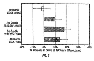
- a significant finding of the invention is that quantitative levels of ESR1 relate to likelihood of TAM benefit as a continuous variable across a 14 point expression scale. Thus, for an individual patient it is possible to provide a quantitative estimate of likelihood of benefit for this therapeutic agent, with higher ESR1 gene expression levels correlating with a greater chance of response.
- This information can be utilized in several ways. It provides a more refined assessment of the probability of a beneficial response to TAM treatment, and other anti-estrogen therapies, than has been available previously.
- TAM has significant side effects, including development of uterine cancer, deep vein thrombosis, pulmonary embolism, and cataracts (Physicians Desk Reference 2002).
- ESR1 is routinely used in clinical practice to determine whether a patient should be treated with TAM, based on "ESR1 positive” or “ESR1 negative” status
- the findings underlying the present invention relate to patients who are already defined as “ESR1 positive” by the conventional criteria. According to the present invention, it is possible to determine the likelihood of a beneficial response to TAM treatment, or treatment with other anti-estrogen drugs, among this group of patients.
- ESR1 levels can be used in conjunction with the Recurrence Score (discussed below) to determine whether individual patients should be prescribed TAM (or another anti-estrogen drug) alone, or TAM (or another anti-estrogen drug) plus chemotherapy.
- the invention additionally allows the design of a particular test, an example of which is given in the Example below, with precise ESR1 expression cut points that predict a high, intermediate of low level of benefit from TAM treatment, or treatment with other anti-estrogen drugs.
- various technological approaches are available for determination of expression levels of the disclosed genes, including, without limitation, RT-PCR, microarrays, serial analysis of gene expression (SAGE) and Gene Expression Analysis by Massively Parallel Signature Sequencing (MPSS), which will be discussed in detail below.
- the expression level of each gene may be determined in relation to various features of the expression products of the gene including exons, introns, protein epitopes and protein activity.
- methods of gene expression profiling can be divided into two large groups: methods based on hybridization analysis of polynucleotides, and methods based on sequencing of polynucleotides.
- the most commonly used methods known in the art for the quantification of mRNA expression in a sample include northern blotting and in situ hybridization ( Parker & Barnes, Methods in Molecular Biology 106:247-283 (1999 )); RNAse protection assays ( Hod, Biotechniques 13:852-854 (1992 )); and reverse transcription polymerase chain reaction (RT-PCR) ( Weis et al., Trends in Genetics 8:263-264 (1992 )).
- antibodies may be employed that can recognize specific duplexes, including DNA duplexes, RNA duplexes, and DNA-RNA hybrid duplexes or DNA-protein duplexes.
- Representative methods for sequencing-based gene expression analysis include Serial Analysis of Gene Expression (SAGE), and gene expression analysis by massively parallel signature sequencing (MPSS).
- DNA methylation has also been shown to be a common alteration in cancer leading to elevated or decreased expression of a broad spectrum of genes ( Jones, P.A. Cancer Res. 65:2463 (1996 )). In general, hypomethylation of CpG islands in the promoter regions and regulatory elements results in increased gene expression, including many oncogenes ( Hanada, M., et al., Blood 82:1820 (1993 ), Feinberg, A.P. and Vogelstein, B. Nature 301:89 (1983 )). Because DNA methylation correlates with the level of specific gene expression in many cancers, it serves as a useful surrogate to expression profiling of tumors ( Toyota, M. et al., Blood 97: 2823 (2001 ), Adorjan, P. et al. Nucl. Acids. Res. 10:e21 (2002 )).
- RT-PCR Reverse Transcriptase PCR
- RT-PCR which can be used to compare mRNA levels in different sample populations, in normal and tumor tissues, with or without drug treatment, to characterize patterns of gene expression, to discriminate between closely related mRNAs, and to analyze RNA structure.
- the first step is the isolation of mRNA from a target sample.
- the starting material is typically total RNA isolated from human tumors or tumor cell lines, and corresponding normal tissues or cell lines, respectively.
- RNA can be isolated from a variety of primary tumors, including breast, lung, colon, prostate, brain, liver, kidney, pancreas, spleen, thymus, testis, ovary, uterus, etc., tumor, or tumor cell lines, with pooled DNA from healthy donors.
- mRNA can be extracted, for example, from frozen or archived paraffin-embedded and fixed (e.g. formalin-fixed) tissue samples.
- RNA isolation can be performed using purification kit, buffer set and protease from commercial manufacturers, such as Qiagen, according to the manufacturer's instructions. For example, total RNA from cells in culture can be isolated using Qiagen RNeasy mini-columns.
- RNA isolation kits include MasterPureTM Complete DNA and RNA Purification Kit (EPICENTRE®, Madison, WI), and Paraffin Block RNA Isolation Kit (Ambion, Inc.). Total RNA from tissue samples can be isolated using RNA Stat-60 (Tel-Test). RNA prepared from tumor can be isolated, for example, by cesium chloride density gradient centrifugation.
- RNA cannot serve as a template for PCR
- the first step in gene expression profiling by RT-PCR is the reverse transcription of the RNA template into cDNA, followed by its exponential amplification in a PCR reaction.
- the two most commonly used reverse transcriptases are avilo myeloblastosis virus reverse transcriptase (AMV-RT) and Moloney murine leukemia virus reverse transcriptase (MMLV-RT).
- AMV-RT avilo myeloblastosis virus reverse transcriptase
- MMLV-RT Moloney murine leukemia virus reverse transcriptase
- the reverse transcription step is typically primed using specific primers, random hexamers, or oligo-dT primers, depending on the circumstances and the goal of expression profiling.
- extracted RNA can be reverse-transcribed using a GeneAmp RNA PCR kit (Perkin Elmer, CA, USA), following the manufacturer's instructions.
- the derived cDNA can then be used as a template in
- the PCR step can use a variety of thermostable DNA-dependent DNA polymerases, it typically employs the Taq DNA polymerase, which has a 5'-3' nuclease activity but lacks a 3'-5' proofreading endonuclease activity.
- TaqMan® PCR typically utilizes the 5'-nuclease activity of Taq or Tth polymerase to hydrolyze a hybridization probe bound to its target amplicon, but any enzyme with equivalent 5' nuclease activity can be used.
- Two oligonucleotide primers are used to generate an amplicon typical of a PCR reaction.
- a third oligonucleotide, or probe is designed to detect nucleotide sequence located between the two PCR primers.
- the probe is non-extendible by Taq DNA polymerase enzyme, and is labeled with a reporter fluorescent dye and a quencher fluorescent dye. Any laser-induced emission from the reporter dye is quenched by the quenching dye when the two dyes are located close together as they are on the probe.
- the Taq DNA polymerase enzyme cleaves the probe in a template-dependent manner.
- the resultant probe fragments disassociate in solution, and signal from the released reporter dye is free from the quenching effect of the second fluorophore.
- One molecule of reporter dye is liberated for each new molecule synthesized, and detection of the unquenched reporter dye provides the basis for quantitative interpretation of the data.
- TaqMan® RT-PCR can be performed using commercially available equipment, such as, for example, ABI PRISM 7700 TM Sequence Detection System TM (Perkin-Elmer-Applied Biosystems, Foster City, CA, USA), or Lightcycler (Roche Molecular Biochemicals, Mannheim, Germany).
- the 5' nuclease procedure is run on a real-time quantitative PCR device such as the ABI PRISM 7700 TM Sequence Detection System TM .
- the system consists of a thermocycler, laser, charge-coupled device (CCD), camera and computer.
- the system amplifies samples in a 96-well format on a thermocycler. During amplification, laser-induced fluorescent signal is detected at the CCD.
- the system includes software for running the instrument and for analyzing the data.
- 5'-Nuclease assay data are initially expressed as C T , or the threshold cycle.
- C T fluorescence values are recorded during every cycle and represent the amount of product amplified to that point in the amplification reaction. The point when the fluorescent signal is first recorded as statistically significant is the threshold cycle (C T ).
- RT-PCR is usually performed using one or more reference genes as internal standards.
- the ideal internal standard is expressed at a constant level among different tissues, and is unaffected by the experimental treatment.
- RNAs most frequently used to normalize patterns of gene expression are mRNAs for the housekeeping genes glyceraldehyde-3-phosphate-dehydrogenase (GAPD) and ⁇ -actin (ACTB).
- GPD glyceraldehyde-3-phosphate-dehydrogenase
- ACTB ⁇ -actin
- RT-PCR measures PCR product accumulation through a dual-labeled fluorigenic probe (i.e., TaqMan® probe).
- Real time PCR is compatible both with quantitative competitive PCR, where internal competitor for each target sequence is used for normalization, and with quantitative comparative PCR using a normalization gene contained within the sample, or a housekeeping gene for RT-PCR.
- quantitative competitive PCR where internal competitor for each target sequence is used for normalization
- quantitative comparative PCR using a normalization gene contained within the sample, or a housekeeping gene for RT-PCR.
- RNA isolation, purification, primer extension and amplification are given in various published journal articles ⁇ for example: T.E. Godfrey et al. J. Molec. Diagnostics 2: 84-91 (2000 ); K. Specht et al., Am. J. Pathol. 158: 419-29 (2001 ); Cronin et al., Am J Pathol 164:35-42 (2004 ) ⁇ .
- a representative process starts with cutting about 10 ⁇ m thick sections of paraffin-embedded tumor tissue samples. The RNA is then extracted, and protein and DNA are removed. After analysis of the RNA concentration, RNA repair and/or amplification steps may be included, if necessary, and RNA is reverse transcribed using gene specific promoters followed by RT-PCR.
- the expression profile of breast cancer-associated genes can be measured in either fresh or paraffin-embedded tumor tissue, using microarray technology.
- polynucleotide sequences of interest including cDNAs and oligonucleotides
- the arrayed sequences are then hybridized with specific DNA probes from cells or tissues of interest.
- the source of mRNA typically is total RNA isolated from human tumors or tumor cell lines, and corresponding normal tissues or cell lines.
- RNA can be isolated from a variety of primary tumors or tumor cell lines. If the source of mRNA is a primary tumor, mRNA can be extracted, for example, from frozen or archived paraffin-embedded and fixed (e.g. formalin-fixed) tissue samples, which are routinely prepared and preserved in everyday clinical practice.
- PCR amplified inserts of cDNA clones are applied to a substrate in a dense array.
- the microarrayed genes, immobilized on the microchip at 10,000 elements each, are suitable for hybridization under stringent conditions.
- Fluorescently labeled cDNA probes may be generated through incorporation of fluorescent nucleotides by reverse transcription of RNA extracted from tissues of interest. Labeled cDNA probes applied to the chip hybridize with specificity to each spot of DNA on the array. After stringent washing to remove non-specifically bound probes, the chip is scanned by confocal laser microscopy or by another detection method, such as a CCD camera.
- Quantitation of hybridization of each arrayed element allows for assessment of corresponding mRNA abundance.
- dual color fluorescence separately labeled cDNA probes generated from two sources of RNA are hybridized pairwise to the array. The relative abundance of the transcripts from the two sources corresponding to each specified gene is thus determined simultaneously.
- the miniaturized scale of the hybridization affords a convenient and rapid evaluation of the expression pattern for large numbers of genes.
- Such methods have been shown to have the sensitivity required to detect rare transcripts, which are expressed at a few copies per cell, and to reproducibly detect at least approximately two-fold differences in the expression levels ( Schena et al., Proc. Natl. Acad. Sci. USA 93(2):106-149 (1996 )).
- Microarray analysis can be performed by commercially available equipment, following manufacturer's protocols, such as by using the Affymetrix GenChip technology, or Incyte's microarray technology.
- microarray methods for large-scale analysis of gene expression makes it possible to search systematically for molecular markers of cancer classification and outcome prediction in a variety of tumor types.
- Serial analysis of gene expression is a method that allows the simultaneous and quantitative analysis of a large number of gene transcripts, without the need of providing an individual hybridization probe for each transcript.
- a short sequence tag (about 10-14 bp) is generated that contains sufficient information to uniquely identify a transcript, provided that the tag is obtained from a unique position within each transcript.
- many transcripts are linked together to form long serial molecules, that can be sequenced, revealing the identity of the multiple tags simultaneously.
- the expression pattern of any population of transcripts can be quantitatively evaluated by determining the abundance of individual tags, and identifying the gene corresponding to each tag. For more details see, e.g. Velculescu et al., Science 270:484-487 (1995 ); and Velculescu et al., Cell 88:243-51 (1997 ).
- This method is a sequencing approach that combines non-gel-based signature sequencing with in vitro cloning of millions of templates on separate 5 ⁇ m diameter microbeads.
- a microbead library of DNA templates is constructed by in vitro cloning. This is followed by the assembly of a planar array of the template-containing microbeads in a flow cell at a high density (typically greater than 3 x 10 6 microbeads/cm 2 ).
- the free ends of the cloned templates on each microbead are analyzed simultaneously, using a fluorescence-based signature sequencing method that does not require DNA fragment separation. This method has been shown to simultaneously and accurately provide, in a single operation, hundreds of thousands of gene signature sequences from a yeast cDNA library.
- RNA isolation, purification, primer extension and amplification are provided in various published journal articles (for example: T.E. Godfrey et al,. J. Molec. Diagnostics 2: 84-91 [2000 ]; K. Specht et al., Am. J. Pathol. 158: 419-29 [2001 ]).
- a representative process starts with cutting about 10 ⁇ m thick sections of paraffin-embedded tumor tissue samples. The RNA is then extracted, and protein and DNA are removed.
- RNA repair and/or amplification steps may be included, if necessary, and RNA is reverse transcribed using gene specific promoters followed by RT-PCR. Finally, the data are analyzed to identify the best treatment option(s) available to the patient on the basis of the characteristic gene expression pattern identified in the tumor sample examined, dependent on the predicted likelihood of cancer recurrence.
- An important aspect of the present invention is to use the measured expression of certain genes by breast cancer tissue to provide prognostic or predictive information. For this purpose it is necessary to correct for (normalize away) both differences in the amount of RNA assayed and variability in the quality of the RNA used. Therefore, the assay typically measures and incorporates the expression of certain normalizing genes, including well known housekeeping genes, such as ⁇ -actin, GAPD, GUS, RPLO, and TFRC, as shown in the Example below. Alternatively, normalization can be based on the mean or median signal (C T ) of all of the assayed genes or a large subset thereof (global normalization approach). Below, unless noted otherwise, gene expression means normalized expression.
- C T mean or median signal
- PCR primers and probes are designed based upon intron sequences present in the gene to be amplified. Accordingly, the first step in the primer/probe design is the delineation of intron sequences within the genes. This can be done by publicly available software, such as the DNA BLAT software developed by Kent, W.J., Genome Res. 12(4):656-64 (2002 ), or by the BLAST software including its variations. Subsequent steps follow well established methods of PCR primer and probe design.
- PCR primer design The most important factors considered in PCR primer design include primer length, melting temperature (Tm), and G/C content, specificity, complementary primer sequences, and 3'-end sequence.
- optimal PCR primers are generally 17-30 bases in length, and contain about 20-80%, such as, for example, about 50-60% G+C bases. Tm's between 50 and 80 °C, e.g. about 50 to 70 °C are typically preferred.
- the present invention takes advantage of certain algorithms and statistical methods, which are described in copending application Serial No. 10/883,303 .
- mRNA amounts are expressed in C T (threshold cycle) units ( Held et al., Genome Research 6:986-994 (1996 )).
- C T threshold cycle
- the averaged sum of reference mRNA C T s is set as a fixed number such as zero, and each measured test mRNA C T is given relative to this fixed point. For example, if, for a certain patient tumor specimen the average of C T s of the 5 reference genes found to be 31 and C T of the test gene X is found to be 35, the reported value for gene X is -4 (i.e. 31-35).
- genes identified in the tumor specimen and known to be associated with the molecular pathology of cancer are grouped into subsets.
- genes known to be associated with proliferation will constitute the “Proliferation Group” (axis, or subset).
- Genes known to be associated with invasion of cancer will constitute the “Invasion Group” (axis, or subset).
- Genes associated with key growth factor receptor pathway(s) will constitute the “Growth Factor Group” (axis, or subset), also referred to as GRB7 Group.
- subsets are, of course, not limiting.
- the subsets (axes) created will depend on the particular cancer, i.e. breast, prostate, pancreatic, lung, etc. cancer.
- genes the expression of which is known to correlate with each other, or which are known to be involved in the same pathway are grouped together.
- the measured tumor level of each mRNA in a subset is multiplied by a coefficient reflecting its relative intra-set contribution to the risk of cancer recurrence and this product is added to the other products between mRNA levels in the subset and their coefficients, to yield a term, e.g. a proliferation term, an invasion term, a growth factor term, etc.
- a term e.g. a proliferation term, an invasion term, a growth factor term, etc.
- the growth factor term is (0.45 to 1.35) x GRB7 + (0.05 to 0.15) x ErbB2, such as, for example 0.9 x GRB7 + 0.1 x ERBB2 (see Example below).
- each term contributes to the overall recurrence score by use of a coefficient.
- the coefficient of the growth factor term can be between 0.23 and 0.70.
- a further step is performed. If the relationship between the term and the risk of recurrence is non-linear, a non-linear functional transform of the term, such as a threshold is used Thus, in lymph node-negative invasive breast cancer, when the growth factor term is found at ⁇ 8 the value is fixed at 8.
- RS recurrence score
- the RS scale generated by the algorithm of the present invention can be adjusted in various ways.
- the range could be selected such that the scale run from 0 to 10, 0 to 50, or 0 to 100, for example.
- scaled recurrence score is calculated on a scale of 0 to 100.
- 10 C T units are added to each measured C T value, and unscaled RS is calculated as described before. Equations for calculating unscaled RS and scaled RS are provided in the following Example.
- any gene can be substituted by another gene that coexpresses in a set of at least 30 different patient specimens of that tumor type (such as breast cancer) with the first gene in the particular cancer tested with a Pearson's coefficient of ⁇ 0.5.
- any individual gene, or gene within a gene group (subset) included in the prognostic and predictive methods of the present invention can be substituted by another gene that coexpresses with the first gene in the particular cancer tested with a Pearson's coefficient of ⁇ 0.5.
- Estrogen is known to promote the growth of some cancers, such as breast cancers, especially those that express the estrogen receptor (ESR1).
- ESR1 estrogen receptor
- Several therapies have been developed to block the effect of estrogen, or to lower estrogen levels in such patient, especially in ESR1 positive breast cancer patients.
- Anti-estrogen drugs can be generally classified as either antagonists of estrogen binding to the estrogen receptor, or are inhibitors of estrogen biosynthesis, such as aromatase inhibitors.
- TAM anti-estrogen drug
- anti-estrogen drugs include raloxifene, which, like TAM, blocks the effect of estrogen on breast tissue and breast cancer; and toremifene citrate, which is closely related to TAM, and may be an option for post menopausal women with metastatic breast cancer.
- Anastrozole an aromatase inhibitor, acts by preventing estrogen from activating its receptor, blocking an enzyme needed for production of estrogen. Anastrozole is currently an option for women whose advanced breast cancer continues to grow during or after TAM treatment.
- Megesterol acetate is typically used for hormonal treatment of advanced breast cancer, usually for women whose cancers fail to respond to TAM.
- Chemotherapeutic agents used in cancer treatment can be divided into several groups, depending on their mechanism of action. Some chemotherapeutic agents directly damage DNA and RNA. By disrupting replication of the DNA such chemotherapeutics either completely halt replication, or result in the production of nonsense DNA or RNA. This category includes, for example, cisplatin (Platinol®), daunorubicin (Cerubidine®), doxorubicin (Adriamycin®), and etoposide (VePesid®). Another group of cancer chemotherapeutic agents interfere with the formation of nucleotides or deoxyribonucleotides, so that RNA synthesis and cell replication is blocked.
- drugs in this class include methotrexate (Abitrexate®), mercaptopurine (Purinethol®), fluorouracil (Adrucil®), and hydroxyurea (Hydrea®).
- a third class of chemotherapeutic agents effects the synthesis or breakdown of mitotic spindles, and, as a result, interrupt cell division.
- Examples of drugs in this class include Vinblastine (Velban®), Vincristine (Oncovin®) and taxenes, such as, Pacitaxel (Taxol®), and Tocetaxel (Taxotere®) Tocetaxel is currently approved in the United States to treat patients with locally advanced or metastatic breast cancer after failure of prior chemotherapy, and patients with locally advanced or metastatic non-small cell lung cancer after failure of prior platinum-based chemotherapy. The prediction of patient response to all of these, and other chemotherapeutic agents is specifically within the scope of the present invention.
- chemotherapy includes treatment with a taxane derivative.
- Taxanes include, without limitation,. paclitaxel (Taxol®) and docetaxel (Taxotere®), which are widely used in the treatment of cancer.
- Taxanes affect cell structures called microtubules, which play an important role in cell functions. In normal cell growth, microtubules are formed when a cell starts dividing. Once the cell stops dividing, the microtubules are broken down or destroyed. Taxanes stop the microtubules from breaking down; which blocks cancer cell division.
- chemotherapy includes treatment with an anthracycline derivative, such as, for example, doxorubicin, daunorubicin, and aclacinomycin.
- anthracycline derivative such as, for example, doxorubicin, daunorubicin, and aclacinomycin.
- chemotherapy includes treatment with a topoisomerase inhibitor, such as, for example, camptothecin, topotecan, irinotecan, 20-S-camptothecin, 9-nitro-camptothecin, 9-amino-camptothecin, or GI147211.
- a topoisomerase inhibitor such as, for example, camptothecin, topotecan, irinotecan, 20-S-camptothecin, 9-nitro-camptothecin, 9-amino-camptothecin, or GI147211.
- neoadjuvant therapy Most patients receive chemotherapy immediately following surgical removal of tumor. This approach is commonly referred to as adjuvant therapy.
- chemotherapy can be administered also before surgery, as so called neoadjuvant treatment.
- neo-adjuvant chemotherapy originates from the treatment of advanced and inoperable breast cancer, it has gained acceptance in the treatment of other types of cancers as well.
- the efficacy of neoadjuvant chemotherapy has been tested in several clinical trials. In the multi-center National Surgical Adjuvant Breast and Bowel Project B-18 (NSAB B-18) trial ( Fisher et al., J. Clin. Oncology 15:2002-2004 (1997 ); Fisher et al., J. Clin.
- neoadjuvant therapy was performed with a combination of adriamycin and cyclophosphamide ("AC regimen”).
- AC regimen a combination of adriamycin and cyclophosphamide
- FEC regimen 5-fluorouracil, epirubicin and cyclophosphamide
- Newer clinical trials have also used taxane-containing neoadjuvant treatment regiments. See, e.g. Holmes et al., J. Natl. Cancer Inst.
- kits thus comprise agents, which may include gene-specific or gene-selective probes and/or primers, for quantitating the expression of the disclosed genes for predicting prognostic outcome or response to treatment.
- agents may include gene-specific or gene-selective probes and/or primers, for quantitating the expression of the disclosed genes for predicting prognostic outcome or response to treatment.
- kits may optionally contain reagents for the extraction of RNA from tumor samples, in particular fixed paraffin-embedded tissue samples and/or reagents for RNA amplification.
- the kits may optionally comprise the reagent(s) with an identifying description or label or instructions relating to their use in the methods of the present invention.
- kits may comprise containers (including microtiter plates suitable for use in an automated implementation of the method), each with one or more of the various reagents (typically in concentrated form) utilized in the methods, including, for example, pre-fabricated microarrays, buffers, the appropriate nucleotide triphosphates (e.g., dATP, dCTP, dGTP and dTTP; or rATP, rCTP, rGTP and UTP), reverse transcriptase, DNA polymerase, RNA polymerase, and one or more probes and primers of the present invention (e.g., appropriate length poly(T) or random primers linked to a promoter reactive with the RNA polymerase).
- the appropriate nucleotide triphosphates e.g., dATP, dCTP, dGTP and dTTP; or rATP, rCTP, rGTP and UTP
- reverse transcriptase DNA polymerase
- RNA polymerase e.g
- the methods provided by the present invention may also be automated in whole or in part.
- RS ⁇ 0 if 20 ⁇ RS U - 6.7 ⁇ 0 20 ⁇ RS U - 6.7 if 0 ⁇ 20 ⁇ RS U - 6.7 ⁇ 100 100 if 20 ⁇ RS U - 6.7 > 100
- the correlation between cancer recurrence and Recurrence Score was evaluated as a continuous variable.
- the evaluation additionally included the Proliferation Group, GRB7 Group, ESR1 Group, Invasion Group, and each of the sixteen cancer-related genes as continuous variables.
- DRFS distant recurrence-free survival
- OS overall survival
- Table 2 reports the hazard ratio (H.R.) for recurrence versus variation in expression of genes and gene groups (axes), from analysis of the untreated NSABP B-14 trial patients (placebo arm). Table 2. Univariate Analysis B14 Placebo Arm Variable Estimate P-value (LR) H.R.
- the prognostic variables are: Proliferation Group; CCNB1; BIRC5; MYBL2; PGR; STK6; MKI67; GSTM1; GAPD; Invasion Group; RPLP0; Proliferation Threshold; MMP11.
- the Proliferation Group, Invasion Group, and Proliferation Threshold are defined components of the Recurrence Score algorithm.
- Increased expression of ten of these prognostic factors correlates with increased H.R.: Proliferation Group; CCNB1; BIRC5; MYBL2; STK6; MKI67; GAPD; Invasion Group; Proliferation threshold; MMP11. Seven of the ten markers for poor prognosis are are genes or gene sets that mark proliferating cells. Among these, the Proliferation Group (as defined in the Recurrence Score algorithm) is the top variable with respect to P-value. Increased expression three of the thirteen prognostic factors correlates with decreased H.R.: PGR, GSTM1, and RPLP0.
- Table 2 The results shown in Table 2 are significant not only statistically but also in their H.R. magnitudes. It should be noted that for any marker a two fold change in H.R. from 1.0 changes the H.R. by 50%, and that each HR value describes the impact of a two fold change in expression of the marker variable relative to the average expression in the patient population. Thus, for example, Table 2 shows that for every two fold increase in expression of Proliferation Group or CCNB1 H.R. increases by approximately 50% (95% confidence limits span ⁇ 20% to ⁇ 90%).
- Table 3 reports interaction analysis using results from both the placebo and TAM treatment arms of NSABP B14, carried out to identify variables that predict senstivity or resistance to TAM. Table 3. B14 Placebo and TAM Combined Analysis of Interaction (Int) Variable Estimate P-value H.R. 95% CI for H.R.
- ESR1 ESR1
- SCUBE2 ESR1 Group
- TFRC TFRC
- BCL2 BCL2
- ESR1 expression can be either represented in quartiles, tertiles or other divisions.
- ESR1 score cutpoints that categorize ESR1 expression by tertiles, using assay data from B-14 patients. ER neg 0-33%ile 33-67%ile 67-100%ile ESR1 ⁇ 6.5 [6.5,10.375) [10.375,11.41) ER ⁇ 11.41
- FIG. 2 shows the absolute benefit of TAM amongst these three groups of patients as a function of ESR1 Expression (Horizontal lines through bars represent 95% confidence limits.)
- TAM has substantial absolute benefit in the two-thirds of patients who express ESR1 at the highest levels, but has much less impact in patients in the lowest tertile of ESR1 expression.
- ESR1 expression and TAM benefit can be represented as a function of breaking ESR1 expression into quartiles.
- Figure 3 shows the absolute benefit of TAM treatment as a function of quartiles of ESR1 expression (horizontal lines through bars represent 95% confidence limits).
- the cutpoints for ESR1 expression are reference-normalized C T s derived from analysis of the NSABP B14 patient population. As shown, patients in the lowest quartile of ESR1 expression experience little benefit from TAM.
- ESR1 expression data can be used in conjunction with Recurrence Score to simultaneously determine whether a patient should be prescribed TAM or TAM plus chemotherapy.
- RS recurrence risk category
- Patients in the low recurrence risk category as defined by RS, who express ESR1 in the upper 75 th percentile are logical candidates to be treated with TAM alone.
- Patients who have low recurrence risk, but are in the lower 25 th percentile in ESR1 expression and for whom risk of TAM side effects is a particular concern may reasonably be considered patients for whom TAM treatment is less appropriate.
- Table 5 can be used to make decisions about treatment of a patient with anti-estrogen and/or chemotherapy.
Abstract
Description
- The present invention relates to quantitative molecular indicators that can guide clinical decisions in breast cancer, such as estrogen receptor (ESR1)-positive, lymph node-negative breast cancer. In particular, the invention concerns certain genes, the varied expression of which indicates the likelihood of recurrence of surgically resected breast cancer in patients who are not treated with a therapeutic agent in the adjuvant setting. In addition, the invention concerns the use of quantitative measurement of the expression of certain genes, including the ESR1 gene, that measure as a continuous variable, to determine (a) the likelihood of a beneficial response to the anti-estrogen therapeutic agent, such as tamoxifen; and (b) the potential magnitude of beneficial response to chemotherapy.
- Oncologists have a number of treatment options available to them, including different combinations of chemotherapeutic drugs that are characterized as "standard of care," and a number of drugs that do not carry a label claim for the treatment of a particular cancer, but for which there is evidence of efficacy in that cancer. Best likelihood of good treatment outcome requires that patients at highest risk of metastatic disease be identified and assigned to optimal available cancer treatment. In particular, it is important to determine the likelihood of patient response to "standard of care" therapeutic drugs, such as cyclophosphamide, methotrexate, 5-fluorouracil, anthracyclines, taxanes, and anti-estrogen drugs, such as tamoxifen, because these have limited efficacy and a spectrum of often severe side effects. The identification of patients who are most or least likely to need and respond to available drugs thus could increase the net benefit these drugs have to offer, and decrease net morbidity and toxicity, via more intelligent patient selection.
- Currently, diagnostic tests used in clinical practice are single analyte, and therefore do not capture the potential value of knowing relationships between dozens of different markers. Moreover, diagnostic tests are often based on immunohistochemistry, which is not quantitative. Immunohistochemistry often yields different results in different laboratories, in part because the reagents are not standardized, and in part because the interpretations are subjective. RNA-based tests, while potentially highly quantitative, have not been used because of the perception that RNA is destroyed in tumor specimens as routinely prepared, namely fixed in formalin and embedded in paraffin (FPE), and because it is inconvenient to obtain and store fresh tissue samples from patients for analysis.
- Over the last two decades molecular biology and biochemistry have revealed hundreds of genes whose activities influence the behavior of tumor cells, their state of differentiation, and their sensitivity or resistance to certain therapeutic drugs. However, with a few exceptions, the status of these genes has not been exploited for the purpose of routinely making clinical decisions about drug treatments. In the last few years, several groups have published studies concerning the classification of various cancer types by microarray gene expression analysis of thousands of genes (see, e.g. Golub et al., Science 286:531-537 (1999); Bhattacharjae et al., Proc. Natl. Acad. Sci. USA 98:13790-13795 (2001); Chen-Hsiang et al., Bioinformatics 17 (Suppl. 1):S316-S322 (2001); Ramaswamy et al., Proc. Natl. Acad. Sci. USA 98:15149-15154 (2001); Martin et al., Cancer Res. 60:2232-2238 (2000); West et al., Proc. Natl. Acad. Sci. USA 98:11462-114 (2001); Sorlie et al., Proc. Natl. Acad. Sci. USA 98:10869-10874 (2001); Yan et al., Cancer Res. 61:8375-8380 (2001)). However, these studies have not yet yielded tests routinely used in clinical practice, in large part because microarrays require fresh or frozen tissue RNA and such specimens are not present in sufficient quantity to permit clinical validation of identified molecular signatures.
- In the past three years, it has become possible to profile gene expression of hundreds of genes in formalin-fixed paraffin-embedded (FPE) tissue using RT-PCR technology. Methods have been described that are highly sensitive, precise, and reproducible (Cronin et al., Am. J. Pathol. 164:35-42 (2004)). Because thousands of archived FPE clinical tissue specimens exist with associated clinical records, such as survival, drug treatment history, etc., the ability to now quantitatively assay gene expression in this type of tissue enables rapid clinical studies relating expression of certain genes to patient prognosis and likelihood of response to treatments. Using data generated by past clinical studies allows for rapid results because the clinical events are historical. In contrast, for example, if one wished to carry out a survival study on newly recruited cancer patients one would generally need to wait for many years for statistically sufficient numbers of deaths to have occurred.
- Breast cancer is the most common type of cancer among women in the United States, and is the leading cause of cancer deaths among women ages 40 - 59.
- Currently only a few molecular tests are routinely used clinically in breast cancer. Immunohistochemical assays for estrogen receptor (ESR1) and progesterone receptor (PGR) proteins are used as a basis for selection of patients to treatment with anti-estrogen drugs, such as tamoxifen (TAM). In addition, ErbB2 (Her2) immunochemistry or fluorescence in situ hybridization (which measure protein and DNA, respectively) are used to select patients with the Her2 antagonist drugs, such as trastuzumab (Herceptin®; Genentech, Inc., South San Francisco, CA).
- Because current tests for prognosis and for prediction of response to chemotherapy are inadequate, breast cancer treatment strategies vary between oncologists (Schott and Hayes, J. Clin. Oncol. PMID 15505274 (2004); Hayes, Breast 12:543-9 (2003)). Generally, lymph node negative patients whose tumors are found to be ESR1 positive are treated with an anti-estrogen drug, such as TAM, and patients whose tumors are found to be ESR1 negative are treated with chemotherapy. Often, ESR1 positive patients are also prescribed chemotherapy in addition to anti-estrogen therapy, accepting the toxic side effects of chemotherapy in order to modestly decrease the risk of cancer recurrence. Toxicities include, neuropathy, nausea and other gastrointestinal symptoms, hair loss and cognitive impairment. Recurrence is to be feared because recurrent breast cancer is usually metastatic and poorly responsive to treatment. Clearly, a need exists to identify those patients who are at substantial risk of recurrence (i.e., to provide prognostic information) and likely to respond to chemotherapy (i.e., to provide predictive information). Likewise, a need exists to identify those patients who do not have a significant risk of recurrence, or who are unlikely to respond to chemotherapy, as these patients should be spared needless exposure to these toxic drugs.
- Prognostic factors differ from treatment predictive factors in breast cancer. Prognostic factors are those variables related to the natural history of breast cancer, which influence the recurrence rates and outcome of patients once they have developed breast cancer. Clinical parameters that have been associated with a worse prognosis include, for example, lymph node involvement, increasing tumor size, and high grade tumors. Prognostic factors are frequently used to categorize patients into subgroups with different baseline relapse risks. In contrast, treatment predictive factors are variables related to the likelihood of an individual patient's beneficial response to a treatment, such as anti-estrogen or chemotherapy, independent of prognosis.
- There is a great need for accurate, quantitative prognostic and predictive factors that can assist the practicing physician to make intelligent treatment choices, adapted to a particular patient's needs, based on well founded risk-benefit analysis.
-
WO 2004/065583 discloses gene sets, the expression of which is important in the diagnosis and/or prognosis of breast cancer. -
WO 03/078662 - According to an aspect of the present invention, there is provided a method for quantitative determination of the likelihood of a beneficial response of an ESR1 positive breast cancer patient to treatment with an anti-estrogen drug as specified in claim 1.
- In an embodiment, the anti-estrogen drug is TAM.
- The treatment predictive method of the present invention may comprising the step of preparing a report for the patient, including a treatment recommendation.
- In a further embodiment, anti-estrogen drug treatment without chemotherapy is recommended when ESR1 expression for the patient is higher than the expression value measured in the same test for percentage of ER-positive, node-negative breast cancer patients, and the patient is otherwise known to be in a low risk group.
- In another embodiment, the variable is the expression level of the RNA transcript of ESR1, or its expression product.
- In a further embodiment, the method includes the step of determining the Recurrence Score for the patient.
- In a particular embodiment, after determining the Recurrence Score, a treatment comprising chemotherapy is recommended when ESR1 expression for the patient is non-zero but is lower than the expression value measured in the same test for a particular percentage of ER-positive, node-negative breast cancer patients and the patient is otherwise known to be in a group having a high risk of recurrence.
-
-
Figure 1 shows the absolute increase in proportion of TAM treated patients who are distant disease-free at 10 years as a function of quantitative measurement of ESR1 -
Figure 2 shows the absolute benefit of tamoxifen (TAM) treatment as a function of ESR1 expression. The horizontal lines through bars represent 95% confidence limits. -
Figure 3 shows the absolute benefit of TAM treatment as a function of the quartile into which the expression of ESR1 falls. The horizontal lines through bars represent 95% confidence limits. The cutpoints defining the quartiles of ESR1 expression, are based on reference-normalized CT values derived from analysis of the NSABP B-14 patient population. - Unless defined otherwise, technical and scientific terms used herein have the same meaning as commonly understood by one of ordinary skill in the art to which this invention belongs. Singleton et al., Dictionary of Microbiology and Molecular Biology 2nd ed., J. Wiley & Sons (New York, NY 1994); and Webster's New World™ Medical Dictionary, 2nd Edition, Wiley Publishing Inc., 2003, provide one skilled in the art with a general guide to many of the terms used in the present application. For purposes of the present invention, the following terms are defined below.
- The term "beneficial response" means an improvement in any measure of patient status, including those measures ordinarily used in the art, such as overall survival, progression free survival, recurrence-free survival, and distant recurrence-free survival. Recurrence-free survival (RFS) refers to the time (in years) from surgery to the first local, regional, or distant recurrence. Distant recurrence-free survival (DFRS) refers to the time (in years) from surgery to the first anatomically distant recurrence. The calculation of these measures in practice may vary from study to study depending on the definition of events to be either censored or not considered.
- The term "microarray" refers to an ordered arrangement of hybridizable array elements, preferably polynucleotide probes, on a substrate.
- The term "polynucleotide," when used in singular or plural, generally refers to any polyribonucleotide or polydeoxribonucleotide, which may be unmodified RNA or DNA or modified RNA or DNA. Thus, for instance, polynucleotides as defmed herein include, without limitation, single- and double-stranded DNA, DNA including single- and double-stranded regions, single- and double-stranded RNA, and RNA including single- and double-stranded regions, hybrid molecules comprising DNA and RNA that may be single-stranded or, more typically, double-stranded or include single- and double-stranded regions. In addition, the term "polynucleotide" as used herein refers to triple-stranded regions comprising RNA or DNA or both RNA and DNA. The strands in such regions may be from the same molecule or from different molecules. The regions may include all of one or more of the molecules, but more typically involve only a region of some of the molecules. One of the molecules of a triple-helical region often is an oligonucleotide. The term "polynucleotide" specifically includes cDNAs. The term includes DNAs (including cDNAs) and RNAs that contain one or more modified bases. Thus, DNAs or RNAs with backbones modified for stability or for other reasons are "polynucleotides" as that term is intended herein. Moreover, DNAs or RNAs comprising unusual bases, such as inosine, or modified bases, such as tritiated bases, are included within the term "polynucleotides" as defined herein. In general, the term "polynucleotide" embraces all chemically, enzymatically and/or metabolically modified forms of unmodified polynucleotides, as well as the chemical forms of DNA and RNA characteristic of viruses and cells, including simple and complex cells.
- The term "oligonucleotide" refers to a relatively short polynucleotide, including, without limitation, single-stranded deoxyribonucleotides, single- or double-stranded ribonucleotides, RNA:DNA hybrids and double-stranded DNAs. Oligonucleotides, such as single-stranded DNA probe oligonucleotides, are often synthesized by chemical methods, for example using automated oligonucleotide synthesizers that are commercially available. However, oligonucleotides can be made by a variety of other methods, including in vitro recombinant DNA-mediated techniques and by expression of DNAs in cells and organisms.
- The term "gene expression" describes the conversion of the DNA gene sequence information into transcribed RNA (the initial unspliced RNA transcript or the mature mRNA) or the encoded protein product. Gene expression can be monitored by measuring the levels of either the entire RNA or protein products of the gene or subsequences.
- The phrase "gene amplification" refers to a process by which multiple copies of a gene or gene fragment are formed in a particular cell or cell line. The duplicated region (a stretch of amplified DNA) is often referred to as "amplicon." Often, the amount of the messenger RNA (mRNA) produced, i.e., the level of gene expression, also increases in the proportion of the number of copies made of the particular gene expressed.
- Prognostic factors are those variables related to the natural history of breast cancer, which influence the recurrence rates and outcome of patients once they have developed breast cancer. Clinical parameters that have been associated with a worse prognosis include, for example, lymph node involvement, increasing tumor size, and high grade tumors. Prognostic factors are frequently used to categorize patients into subgroups with different baseline relapse risks. In contrast, treatment predictive factors are variables related to the likelihood of an individual patient's beneficial response to a treatment, such as anti-estrogen or chemotherapy, independent of prognosis.
- The term "prognosis" is used herein to refer to the likelihood of cancer-attributable death or cancer progression, including recurrence and metastatic spread of a neoplastic disease, such as breast cancer, during the natural history of the disease. Prognostic factors are those variables related to the natural history of a neoplastic diseases, such as breast cancer, which influence the recurrence rates and disease outcome once the patient developed the neoplastic disease, such as breast cancer. In this context, "natural outcome" means outcome in the absence of further treatment. For example, in the case of breast cancer, "natural outcome" means outcome following surgical resection of the tumor, in the absence of further treatment (such as, chemotherapy or radiation treatment). Prognostic factors are frequently used to categorize patients into subgroups with different baseline risks, such as baseline relapse risks.
- The term "prediction" is used herein to refer to the likelihood that a patient will respond either favorably or unfavorably to a drug or set of drugs, and also the extent of those responses. Thus, treatment predictive factors are those variables related to the response of an individual patient to a specific treatment, independent of prognosis. The predictive methods of the present invention can be used clinically to make treatment decisions by choosing the most appropriate treatment modalities for any particular patient. The predictive methods of the present invention are valuable tools in predicting if a patient is likely to respond favorably to a treatment regimen, such as anti-estrogen therapy, such as TAM treatment alone or in combination with chemotherapy and/or radiation therapy.
- The term "long-term" survival is used herein to refer to survival for at least 3 years, more preferably for at least 8 years, most preferably for at least 10 years following surgery or other treatment.
- The term "tumor," as used herein, refers to all neoplastic cell growth and proliferation, whether malignant or benign, and all pre-cancerous and cancerous cells and tissues.
- The terms "cancer" and "cancerous" refer to or describe the physiological condition in mammals that is typically characterized by unregulated cell growth. Examples of cancer include, but are not limited to, breast cancer, ovarian cancer, colon cancer, lung cancer, prostate cancer, hepatocellular cancer, gastric cancer, pancreatic cancer, cervical cancer, liver cancer, bladder cancer, cancer of the urinary tract, thyroid cancer, renal cancer, carcinoma, melanoma, and brain cancer.
- The "pathology" of cancer includes all phenomena that compromise the well-being of the patient. This includes, without limitation, abnormal or uncontrollable cell growth, metastasis, interference with the normal functioning of neighboring cells, release of cytokines or other secretory products at abnormal levels, suppression or aggravation of inflammatory or immunological response, neoplasia, premalignancy, malignancy, invasion of surrounding or distant tissues or organs, such as lymph nodes, etc.
- In the context of the present invention, reference to "at least one," "at least two," "at least three," "at least four," "at least five," etc. of the genes listed in any particular gene set means any one or any and all combinations of the genes listed.
- The term "node negative" cancer, such as "node negative" breast cancer, is used herein to refer to cancer that has not spread to the lymph nodes.
- The terms "splicing" and "RNA splicing" are used interchangeably and refer to RNA processing that removes introns and joins exons to produce mature mRNA with continuous coding sequence that moves into the cytoplasm of an eukaryotic cell.
- In theory, the term "exon" refers to any segment of an interrupted gene that is represented in the mature RNA product (B. Lewin. Genes IV Cell Press, Cambridge Mass. 1990). In theory the term "intron" refers to any segment of DNA that is transcribed but removed from within the transcript by splicing together the exons on either side of it. Operationally, exon sequences occur in the mRNA sequence of a gene as defined by Ref. SEQ ID numbers. Operationally, intron sequences are the intervening sequences within the genomic DNA of a gene, bracketed by exon sequences and having GT and AG splice consensus sequences at their 5' and 3' boundaries.
- The practice of the present invention will employ, unless otherwise indicated, conventional techniques of molecular biology (including recombinant techniques), microbiology, cell biology, and biochemistry, which are within the skill of the art. Such techniques are explained fully in the literature, such as, "Molecular Cloning: A Laboratory Manual", 2nd edition (Sambrook et al., 1989); "Oligonucleotide Synthesis" (M.J. Gait, ed., 1984); "Animal Cell Culture" (R.I. Freshney, ed., 1987); "Methods in Enzymology" (Academic Press, Inc.); "Handbook of Experimental Immunology", 4th edition (D.M. Weir & C.C. Blackwell, eds., Blackwell Science Inc., 1987); "Gene Transfer Vectors for Mammalian Cells" (J.M. Miller & M.P. Calos, eds., 1987); "Current Protocols in Molecular Biology" (F.M. Ausubel et al., eds., 1987); and "PCR: The Polymerase Chain Reaction", (Mullis et al., eds., 1994).
- Over the past two years Genomic Health, Inc and collaborators (Esteban et al., Proc Am Soc Clin Oncol 22: page 850, 2003 (abstract 3416); Cobleigh et al. Soc Clin Oncol 22: page 850, 2003 (abstract 3415); Soule et al., Proc Am Soc Clin Oncol 22: page 862, 2003 (abstract 3466)) reported several exploratory clinical studies of gene expression in early breast cancer, aimed at finding a molecular signature for recurrence risk. These studies used quantitative RT-PCR to test 250 candidate gene markers in frozen, paraffin-embedded (FPE) tissue specimens having linked clinical records. Analysis across all three studies was performed in order to examine whether genes could be identified which were consistently related to the risk of recurrence across a diverse group of patients. Based on these univariate results, multi-gene models were designed and analyzed across the three studies. A single multi-gene assay, consisting of 16 cancer-related genes and 5 reference genes, was developed to be tested prospectively in clinical validation studies. An algorithm called Recurrence Score (RS) was generated, which utilizes the measurements of these 21 mRNA species and reads out recurrence risk on a 100 point scale.
- To test the clinical validity of this Recurrence Score test and algorithm, a blinded clinical trial with prospectively identified endpoints was carried out. This validation trial focused on patients treated with TAM alone in the randomized and registration arms of the National Surgical Adjuvant Breast and Bowel Project (NSABP) Study B-14 clinical trial population (Fisher B, Costantino JP, Redmond CK, et al: Endometrial cancer in TAM-treated breast cancer patients: Findings from the National Surgical Adjuvant Breast and Bowel Project (NSABP) B-14. J Natl Cancer Inst 86:527-537 (1994)). Genomic Health, Inc. and the NSABP carried out the 21 gene RT-PCR assay on 668 breast cancer tissue specimens derived from these patients and calculated a Recurrence Score for each patient.
- Pre-specified cut-off points of Recurrence Score classified patients into one of three categories: low risk, intermediate risk, and high risk of distant disease recurrence. The proportion of the 668 patients categorized as low, intermediate, and high risk by the RT-PCR assay were 51%, 23%, and 27%, respectively. The Kaplan-Meier estimates and 95% confidence intervals for the rates of distant recurrence at 10 years were 6.8% (4.0%, 9.6%), 14.3% (8.3%, 20.3%) 30.5% (23.6%, 37.4%), respectively, for the low, intermediate, and high risk groups; the rate for the low risk group was significantly lower than the rate for the high risk group (p<0.001). In a multivariate Cox model relating distant recurrence to Recurrence Score, age, and tumor size, Recurrence Score provides significant (p < 0.001) predictive power that goes beyond age and tumor size. This study validated the Recurrence Score as a powerful predictor of distant recurrence in patients without involved nodes who have tumors that are ESR1 positive and treated with TAM (Paik et al. Breast Cancer Research and Treatment 82, Supplement 1: page S10, 2003 (Abstract 16).
- The invention disclosed herein derives, in part, from study of patients in the placebo arm of the NSABP B-14 clinical study (B-14) and, in part, from comparison of patients in the B-14 placebo arm to TAM-treated patients with patients in the randomized and registration arms of NSABP Study B-14. Breast cancer tissue derived from placebo-treated patients was quantitatively analyzed, using a RT-PCR assay to quantify the expression of sixteen cancer-related genes and five reference genes.
- The quantitative gene expression analysis resulted in the identification of molecular indicators of prognosis. Based on analysis of the relationship between gene expression and distant recurrence-free survival in the placebo arm of the NSABP B-14 trial, a set of genes has been identified, the expression levels of which are indicative of outcome if no further treatment is provided to the patient. Outcome may be manifest in various measurements including survival, recurrence-free survival and distant recurrence-free survival, all of which are within the scope of the invention.
- The prognostic genes and gene groups identified may be used singly or in particular combinations to predict outcome likelihood for particular patients. Prognostic indicators include, specifically, the proliferation group (BIRC5 + MKI + MYBL2 + CCNB1 + STK6), the invasion group (CTSL2 + MMP11), and one or more of the individual genes: CCNB1, BIRC5, MYBL2, PGR, STK6, MKI, GSTM1, GAPD, RPLPO, and MMP11.
- In another aspect, the gene expression analysis disclosed herein resulted in the identification of molecular indicators of beneficial response to anti-estrogen drugs, such as TAM, based on analysis of the relationship between gene expression and distant recurrence-free survival in untreated patients from the placebo arm of B-14 as well as TAM treated patients from both registration and randomization arms of B-14.
- Based on interaction analysis of the relationship between gene expression and distant recurrence-free survival in the combined placebo and treatment cohorts, a set of genes and gene groups has been identified, the expression levels of which are indicative of beneficial response to treatment TAM. These genes and gene groups are used in particular combinations to predict likelihood of beneficial response to treatment with TAM, or another anti-estrogen drug, for individual patients. Specifically, these genes/gene groups are: the ESR1 group (ESR1 + PGR + BCL2 + SCUBE2).
- A significant finding of the invention is that quantitative levels of ESR1 relate to likelihood of TAM benefit as a continuous variable across a 14 point expression scale. Thus, for an individual patient it is possible to provide a quantitative estimate of likelihood of benefit for this therapeutic agent, with higher ESR1 gene expression levels correlating with a greater chance of response. This information can be utilized in several ways. It provides a more refined assessment of the probability of a beneficial response to TAM treatment, and other anti-estrogen therapies, than has been available previously. TAM has significant side effects, including development of uterine cancer, deep vein thrombosis, pulmonary embolism, and cataracts (Physicians Desk Reference 2002). Similarly, other anti-estrogen drugs, such as, toremifene (Fareston®, Orion, Corp.), anastrozole (Arimidex®, AstraZeneca Pharmaceuticals), and megasterol acetate, have serious side-effects. As a result of the present invention, patients and their oncologists can now use the ESR1 score to assess risk versus benefit when deciding whether TAM treatment, or other anti-estrogen therapy, is appropriate.
- While expression of ESR1 (principally as determined by immunochemistry measurement at the protein level) is routinely used in clinical practice to determine whether a patient should be treated with TAM, based on "ESR1 positive" or "ESR1 negative" status, the findings underlying the present invention relate to patients who are already defined as "ESR1 positive" by the conventional criteria. According to the present invention, it is possible to determine the likelihood of a beneficial response to TAM treatment, or treatment with other anti-estrogen drugs, among this group of patients.
- ESR1 levels can be used in conjunction with the Recurrence Score (discussed below) to determine whether individual patients should be prescribed TAM (or another anti-estrogen drug) alone, or TAM (or another anti-estrogen drug) plus chemotherapy.
- The invention additionally allows the design of a particular test, an example of which is given in the Example below, with precise ESR1 expression cut points that predict a high, intermediate of low level of benefit from TAM treatment, or treatment with other anti-estrogen drugs.
- In various embodiments of the inventions, various technological approaches are available for determination of expression levels of the disclosed genes, including, without limitation, RT-PCR, microarrays, serial analysis of gene expression (SAGE) and Gene Expression Analysis by Massively Parallel Signature Sequencing (MPSS), which will be discussed in detail below. In particular embodiments, the expression level of each gene may be determined in relation to various features of the expression products of the gene including exons, introns, protein epitopes and protein activity.
- In general, methods of gene expression profiling can be divided into two large groups: methods based on hybridization analysis of polynucleotides, and methods based on sequencing of polynucleotides. The most commonly used methods known in the art for the quantification of mRNA expression in a sample include northern blotting and in situ hybridization (Parker & Barnes, Methods in Molecular Biology 106:247-283 (1999)); RNAse protection assays (Hod, Biotechniques 13:852-854 (1992)); and reverse transcription polymerase chain reaction (RT-PCR) (Weis et al., Trends in Genetics 8:263-264 (1992)). Alternatively, antibodies may be employed that can recognize specific duplexes, including DNA duplexes, RNA duplexes, and DNA-RNA hybrid duplexes or DNA-protein duplexes. Representative methods for sequencing-based gene expression analysis include Serial Analysis of Gene Expression (SAGE), and gene expression analysis by massively parallel signature sequencing (MPSS).
- Two biological processes commonly involved in tumorigenesis include gene amplification and DNA methylation. Both processes result in the abnormal expression of genes important in tumor formation or progression. Methods that monitor gene amplification and DNA methylation can therefore be considered surrogate methods for gene expression profiling.
- Gene amplification is a common alteration in many cancers that can lead to elevated expression of cellular oncogenes (Meltzer, P. et al., Cancer Genet Cytogenet. 19:93 (1986). In breast cancer, there is good correlation between ERBB2 gene amplification and ERBB2 overexpression (Nagai, M.A. et al., Cancer Biother 8:29 (1993), Savinainen, K.J. et al., Am. J. Pathol. 160:339 (2002)). Amplification of the ERBB2 gene, leading to its overexpression, correlates with poor prognosis (Press, M.F. et al., J. Clin. Oncol. 15:2894 (1997), Slamon, D.J. et al., Science 244:707 (1989)) and is predictive for response to anti-HER2 therapy in combination with standard chemotherapy(Seidman, A.D. et al., J. Clin. Oncol. 19:1866 (2001)).
- DNA methylation has also been shown to be a common alteration in cancer leading to elevated or decreased expression of a broad spectrum of genes (Jones, P.A. Cancer Res. 65:2463 (1996)). In general, hypomethylation of CpG islands in the promoter regions and regulatory elements results in increased gene expression, including many oncogenes (Hanada, M., et al., Blood 82:1820 (1993), Feinberg, A.P. and Vogelstein, B. Nature 301:89 (1983)). Because DNA methylation correlates with the level of specific gene expression in many cancers, it serves as a useful surrogate to expression profiling of tumors (Toyota, M. et al., Blood 97: 2823 (2001), Adorjan, P. et al. Nucl. Acids. Res. 10:e21 (2002)).
- Of the techniques listed above, the most sensitive and most flexible quantitative method is RT-PCR, which can be used to compare mRNA levels in different sample populations, in normal and tumor tissues, with or without drug treatment, to characterize patterns of gene expression, to discriminate between closely related mRNAs, and to analyze RNA structure.
- The first step is the isolation of mRNA from a target sample. The starting material is typically total RNA isolated from human tumors or tumor cell lines, and corresponding normal tissues or cell lines, respectively. Thus RNA can be isolated from a variety of primary tumors, including breast, lung, colon, prostate, brain, liver, kidney, pancreas, spleen, thymus, testis, ovary, uterus, etc., tumor, or tumor cell lines, with pooled DNA from healthy donors. If the source of mRNA is a primary tumor, mRNA can be extracted, for example, from frozen or archived paraffin-embedded and fixed (e.g. formalin-fixed) tissue samples.
- General methods for mRNA extraction are well known in the art and are disclosed in standard textbooks of molecular biology, including Ausubel et al., Current Protocols of Molecular Biology, John Wiley and Sons (1997). Methods for RNA extraction from paraffin embedded tissues are disclosed, for example, in Rupp and Locker, Lab Invest. 56:A (1987), and De Andrés et al., BioTechniques 18:42044 (1995). In particular, RNA isolation can be performed using purification kit, buffer set and protease from commercial manufacturers, such as Qiagen, according to the manufacturer's instructions. For example, total RNA from cells in culture can be isolated using Qiagen RNeasy mini-columns. Other commercially available RNA isolation kits include MasterPure™ Complete DNA and RNA Purification Kit (EPICENTRE®, Madison, WI), and Paraffin Block RNA Isolation Kit (Ambion, Inc.). Total RNA from tissue samples can be isolated using RNA Stat-60 (Tel-Test). RNA prepared from tumor can be isolated, for example, by cesium chloride density gradient centrifugation.
- As RNA cannot serve as a template for PCR, the first step in gene expression profiling by RT-PCR is the reverse transcription of the RNA template into cDNA, followed by its exponential amplification in a PCR reaction. The two most commonly used reverse transcriptases are avilo myeloblastosis virus reverse transcriptase (AMV-RT) and Moloney murine leukemia virus reverse transcriptase (MMLV-RT). The reverse transcription step is typically primed using specific primers, random hexamers, or oligo-dT primers, depending on the circumstances and the goal of expression profiling. For example, extracted RNA can be reverse-transcribed using a GeneAmp RNA PCR kit (Perkin Elmer, CA, USA), following the manufacturer's instructions. The derived cDNA can then be used as a template in the subsequent PCR reaction.
- Although the PCR step can use a variety of thermostable DNA-dependent DNA polymerases, it typically employs the Taq DNA polymerase, which has a 5'-3' nuclease activity but lacks a 3'-5' proofreading endonuclease activity. Thus, TaqMan® PCR typically utilizes the 5'-nuclease activity of Taq or Tth polymerase to hydrolyze a hybridization probe bound to its target amplicon, but any enzyme with equivalent 5' nuclease activity can be used. Two oligonucleotide primers are used to generate an amplicon typical of a PCR reaction. A third oligonucleotide, or probe, is designed to detect nucleotide sequence located between the two PCR primers. The probe is non-extendible by Taq DNA polymerase enzyme, and is labeled with a reporter fluorescent dye and a quencher fluorescent dye. Any laser-induced emission from the reporter dye is quenched by the quenching dye when the two dyes are located close together as they are on the probe. During the amplification reaction, the Taq DNA polymerase enzyme cleaves the probe in a template-dependent manner. The resultant probe fragments disassociate in solution, and signal from the released reporter dye is free from the quenching effect of the second fluorophore. One molecule of reporter dye is liberated for each new molecule synthesized, and detection of the unquenched reporter dye provides the basis for quantitative interpretation of the data.
- TaqMan® RT-PCR can be performed using commercially available equipment, such as, for example, ABI PRISM 7700™ Sequence Detection System™ (Perkin-Elmer-Applied Biosystems, Foster City, CA, USA), or Lightcycler (Roche Molecular Biochemicals, Mannheim, Germany). In a preferred embodiment, the 5' nuclease procedure is run on a real-time quantitative PCR device such as the ABI PRISM 7700™ Sequence Detection System™. The system consists of a thermocycler, laser, charge-coupled device (CCD), camera and computer. The system amplifies samples in a 96-well format on a thermocycler. During amplification, laser-induced fluorescent signal is detected at the CCD. The system includes software for running the instrument and for analyzing the data.
- 5'-Nuclease assay data are initially expressed as CT, or the threshold cycle. As discussed above, fluorescence values are recorded during every cycle and represent the amount of product amplified to that point in the amplification reaction. The point when the fluorescent signal is first recorded as statistically significant is the threshold cycle (CT).
- To minimize errors and the effect of sample-to-sample variation, RT-PCR is usually performed using one or more reference genes as internal standards. The ideal internal standard is expressed at a constant level among different tissues, and is unaffected by the experimental treatment. RNAs most frequently used to normalize patterns of gene expression are mRNAs for the housekeeping genes glyceraldehyde-3-phosphate-dehydrogenase (GAPD) and β-actin (ACTB).
- A more recent variation of the RT-PCR technique is the real time quantitative PCR, which measures PCR product accumulation through a dual-labeled fluorigenic probe (i.e., TaqMan® probe). Real time PCR is compatible both with quantitative competitive PCR, where internal competitor for each target sequence is used for normalization, and with quantitative comparative PCR using a normalization gene contained within the sample, or a housekeeping gene for RT-PCR. For further details see, e.g. Held et al., Genome Research 6:986-994 (1996).
- The steps of a representative protocol for profiling gene expression using fixed, paraffin-embedded tissues as the RNA source, including mRNA isolation, purification, primer extension and amplification are given in various published journal articles {for example: T.E. Godfrey et al. J. Molec. Diagnostics 2: 84-91 (2000); K. Specht et al., Am. J. Pathol. 158: 419-29 (2001); Cronin et al., Am J Pathol 164:35-42 (2004)}. Briefly, a representative process starts with cutting about 10 µm thick sections of paraffin-embedded tumor tissue samples. The RNA is then extracted, and protein and DNA are removed. After analysis of the RNA concentration, RNA repair and/or amplification steps may be included, if necessary, and RNA is reverse transcribed using gene specific promoters followed by RT-PCR.
- Differential gene expression can also be identified, or confirmed using the microarray technique. Thus, the expression profile of breast cancer-associated genes can be measured in either fresh or paraffin-embedded tumor tissue, using microarray technology. In this method, polynucleotide sequences of interest (including cDNAs and oligonucleotides) are plated, or arrayed, on a microchip substrate. The arrayed sequences are then hybridized with specific DNA probes from cells or tissues of interest. Just as in the RT-PCR method, the source of mRNA typically is total RNA isolated from human tumors or tumor cell lines, and corresponding normal tissues or cell lines. Thus RNA can be isolated from a variety of primary tumors or tumor cell lines. If the source of mRNA is a primary tumor, mRNA can be extracted, for example, from frozen or archived paraffin-embedded and fixed (e.g. formalin-fixed) tissue samples, which are routinely prepared and preserved in everyday clinical practice.
- In a specific embodiment of the microarray technique, PCR amplified inserts of cDNA clones are applied to a substrate in a dense array. Preferably at least 10,000 nucleotide sequences are applied to the substrate. The microarrayed genes, immobilized on the microchip at 10,000 elements each, are suitable for hybridization under stringent conditions. Fluorescently labeled cDNA probes may be generated through incorporation of fluorescent nucleotides by reverse transcription of RNA extracted from tissues of interest. Labeled cDNA probes applied to the chip hybridize with specificity to each spot of DNA on the array. After stringent washing to remove non-specifically bound probes, the chip is scanned by confocal laser microscopy or by another detection method, such as a CCD camera. Quantitation of hybridization of each arrayed element allows for assessment of corresponding mRNA abundance. With dual color fluorescence, separately labeled cDNA probes generated from two sources of RNA are hybridized pairwise to the array. The relative abundance of the transcripts from the two sources corresponding to each specified gene is thus determined simultaneously. The miniaturized scale of the hybridization affords a convenient and rapid evaluation of the expression pattern for large numbers of genes. Such methods have been shown to have the sensitivity required to detect rare transcripts, which are expressed at a few copies per cell, and to reproducibly detect at least approximately two-fold differences in the expression levels (Schena et al., Proc. Natl. Acad. Sci. USA 93(2):106-149 (1996)). Microarray analysis can be performed by commercially available equipment, following manufacturer's protocols, such as by using the Affymetrix GenChip technology, or Incyte's microarray technology.
- The development of microarray methods for large-scale analysis of gene expression makes it possible to search systematically for molecular markers of cancer classification and outcome prediction in a variety of tumor types.
- Serial analysis of gene expression (SAGE) is a method that allows the simultaneous and quantitative analysis of a large number of gene transcripts, without the need of providing an individual hybridization probe for each transcript. First, a short sequence tag (about 10-14 bp) is generated that contains sufficient information to uniquely identify a transcript, provided that the tag is obtained from a unique position within each transcript. Then, many transcripts are linked together to form long serial molecules, that can be sequenced, revealing the identity of the multiple tags simultaneously. The expression pattern of any population of transcripts can be quantitatively evaluated by determining the abundance of individual tags, and identifying the gene corresponding to each tag. For more details see, e.g. Velculescu et al., Science 270:484-487 (1995); and Velculescu et al., Cell 88:243-51 (1997).
- This method, described by Brenner et al., Nature Biotechnology 18:630-634 (2000), is a sequencing approach that combines non-gel-based signature sequencing with in vitro cloning of millions of templates on separate 5 µm diameter microbeads. First, a microbead library of DNA templates is constructed by in vitro cloning. This is followed by the assembly of a planar array of the template-containing microbeads in a flow cell at a high density (typically greater than 3 x 10⁶ microbeads/cm2). The free ends of the cloned templates on each microbead are analyzed simultaneously, using a fluorescence-based signature sequencing method that does not require DNA fragment separation. This method has been shown to simultaneously and accurately provide, in a single operation, hundreds of thousands of gene signature sequences from a yeast cDNA library.
- The steps of a representative protocol for profiling gene expression using fixed, paraffin-embedded tissues as the RNA source, including mRNA isolation, purification, primer extension and amplification are provided in various published journal articles (for example: T.E. Godfrey et al,. J. Molec. Diagnostics 2: 84-91 [2000]; K. Specht et al., Am. J. Pathol. 158: 419-29 [2001]). Briefly, a representative process starts with cutting about 10 µm thick sections of paraffin-embedded tumor tissue samples. The RNA is then extracted, and protein and DNA are removed. After analysis of the RNA concentration, RNA repair and/or amplification steps may be included, if necessary, and RNA is reverse transcribed using gene specific promoters followed by RT-PCR. Finally, the data are analyzed to identify the best treatment option(s) available to the patient on the basis of the characteristic gene expression pattern identified in the tumor sample examined, dependent on the predicted likelihood of cancer recurrence.
- An important aspect of the present invention is to use the measured expression of certain genes by breast cancer tissue to provide prognostic or predictive information. For this purpose it is necessary to correct for (normalize away) both differences in the amount of RNA assayed and variability in the quality of the RNA used. Therefore, the assay typically measures and incorporates the expression of certain normalizing genes, including well known housekeeping genes, such as β-actin, GAPD, GUS, RPLO, and TFRC, as shown in the Example below. Alternatively, normalization can be based on the mean or median signal (CT) of all of the assayed genes or a large subset thereof (global normalization approach). Below, unless noted otherwise, gene expression means normalized expression.
- According to one aspect of the present invention, PCR primers and probes are designed based upon intron sequences present in the gene to be amplified. Accordingly, the first step in the primer/probe design is the delineation of intron sequences within the genes. This can be done by publicly available software, such as the DNA BLAT software developed by Kent, W.J., Genome Res. 12(4):656-64 (2002), or by the BLAST software including its variations. Subsequent steps follow well established methods of PCR primer and probe design.
- In order to avoid non-specific signals, it is important to mask repetitive sequences within the introns when designing the primers and probes. This can be easily accomplished by using the Repeat Masker program available on-line through the Baylor College of Medicine, which screens DNA sequences against a library of repetitive elements and returns a query sequence in which the repetitive elements are masked. The masked intron sequences can then be used to design primer and probe sequences using any commercially or otherwise publicly available primer/probe design packages, such as Primer Express (Applied Biosystems); MGB assay-by-design (Applied Biosystems); Primer3 (Steve Rozen and Helen J. Skaletsky (2000) Primer3 on the WWW for general users and for biologist programmers. In: Krawetz S, Misener S (eds) Bioinformatics Methods and Protocols: Methods in Molecular Biology. Humana Press, Totowa, NJ, pp 365-386).
- The most important factors considered in PCR primer design include primer length, melting temperature (Tm), and G/C content, specificity, complementary primer sequences, and 3'-end sequence. In general, optimal PCR primers are generally 17-30 bases in length, and contain about 20-80%, such as, for example, about 50-60% G+C bases. Tm's between 50 and 80 °C, e.g. about 50 to 70 °C are typically preferred.
- For further guidelines for PCR primer and probe design see, e.g. Dieffenbach, C.W. et al., "General Concepts for PCR Primer Design" in: PCR Primer, A Laboratory Manual, Cold Spring Harbor Laboratory Press, New York, 1995, pp. 133-155; Innis and Gelfand, "Optimization of PCRs" in: PCR Protocols, A Guide to Methods and Applications, CRC Press, London, 1994, pp. 5-11; and Plasterer, T.N. Primerselect: Primer and probe design. Methods Mol. Biol. 70:520-527 (1997),
- The present invention takes advantage of certain algorithms and statistical methods, which are described in copending application Serial No.
10/883,303 - When quantitative RT-PCR (qRT-PCR) is used to measure mRNA levels, mRNA amounts are expressed in CT (threshold cycle) units (Held et al., Genome Research 6:986-994 (1996)). The averaged sum of reference mRNA CTs is set as a fixed number such as zero, and each measured test mRNA CT is given relative to this fixed point. For example, if, for a certain patient tumor specimen the average of CTs of the 5 reference genes found to be 31 and CT of the test gene X is found to be 35, the reported value for gene X is -4 (i.e. 31-35).
- As a first step following the quantitative determination of mRNA levels, the genes identified in the tumor specimen and known to be associated with the molecular pathology of cancer are grouped into subsets. Thus, genes known to be associated with proliferation will constitute the "Proliferation Group" (axis, or subset). Genes known to be associated with invasion of cancer will constitute the "Invasion Group" (axis, or subset). Genes associated with key growth factor receptor pathway(s) will constitute the "Growth Factor Group" (axis, or subset), also referred to as GRB7 Group. Genes known to be involved with activating or signaling through the estrogen receptor (ESR1) will constitute the "Estrogen Receptor (ER) Group" (axis, or subset), and so on. This list of subsets is, of course, not limiting. The subsets (axes) created will depend on the particular cancer, i.e. breast, prostate, pancreatic, lung, etc. cancer. In general, genes the expression of which is known to correlate with each other, or which are known to be involved in the same pathway are grouped together.
- In the next step, the measured tumor level of each mRNA in a subset is multiplied by a coefficient reflecting its relative intra-set contribution to the risk of cancer recurrence and this product is added to the other products between mRNA levels in the subset and their coefficients, to yield a term, e.g. a proliferation term, an invasion term, a growth factor term, etc. For example, in the case of lymph node-negative invasive breast cancer the growth factor term is (0.45 to 1.35) x GRB7 + (0.05 to 0.15) x ErbB2, such as, for example 0.9 x GRB7 + 0.1 x ERBB2 (see Example below).
- The contribution of each term to the overall recurrence score is weighted by use of a coefficient. For example, in the case of lymph node-negative invasive breast cancer the coefficient of the growth factor term can be between 0.23 and 0.70.
- Additionally, for some terms, such as the growth factor and proliferation terms, a further step is performed. If the relationship between the term and the risk of recurrence is non-linear, a non-linear functional transform of the term, such as a threshold is used Thus, in lymph node-negative invasive breast cancer, when the growth factor term is found at <8 the value is fixed at 8.
- The sum of the terms obtained provides the recurrence score (RS), which predicts the likelihood of cancer recurrence in the normal course of the disease.
- The RS scale generated by the algorithm of the present invention can be adjusted in various ways. Thus, the range could be selected such that the scale run from 0 to 10, 0 to 50, or 0 to 100, for example.
- For example, in the particular scaling approach described in the Example below, scaled recurrence score is calculated on a scale of 0 to 100. For convenience, 10 CT units are added to each measured CT value, and unscaled RS is calculated as described before. Equations for calculating unscaled RS and scaled RS are provided in the following Example.
- In calculating the Recurrence Score, or any variable used to calculate the Recurrence Score, any gene can be substituted by another gene that coexpresses in a set of at least 30 different patient specimens of that tumor type (such as breast cancer) with the first gene in the particular cancer tested with a Pearson's coefficient of ≥ 0.5. Similarly, any individual gene, or gene within a gene group (subset) included in the prognostic and predictive methods of the present invention can be substituted by another gene that coexpresses with the first gene in the particular cancer tested with a Pearson's coefficient of ≥ 0.5.
- Estrogen is known to promote the growth of some cancers, such as breast cancers, especially those that express the estrogen receptor (ESR1). Several therapies have been developed to block the effect of estrogen, or to lower estrogen levels in such patient, especially in ESR1 positive breast cancer patients.
- Anti-estrogen drugs can be generally classified as either antagonists of estrogen binding to the estrogen receptor, or are inhibitors of estrogen biosynthesis, such as aromatase inhibitors.
- The most commonly used anti-estrogen drug TAM, which belongs to the class of estrogen binding antagonists, is typically taken orally, once a day for five years following surgical resection of breast cancer, and/or chemo- or radiation therapy. Clinical studies have shown that the use of TAM as an adjuvant therapy after surgery reduces the risk of cancer recurrence, however, the response of ESR1 positive patients to this treatment varies, and there have been no clear predictors of responsiveness available.
- Other anti-estrogen drugs include raloxifene, which, like TAM, blocks the effect of estrogen on breast tissue and breast cancer; and toremifene citrate, which is closely related to TAM, and may be an option for post menopausal women with metastatic breast cancer.
- Anastrozole, an aromatase inhibitor, acts by preventing estrogen from activating its receptor, blocking an enzyme needed for production of estrogen. Anastrozole is currently an option for women whose advanced breast cancer continues to grow during or after TAM treatment.
- Megesterol acetate is typically used for hormonal treatment of advanced breast cancer, usually for women whose cancers fail to respond to TAM.
- All treatments are limited by serious side effects, and by the lack of reliable predictors of patient response, which would enable a physician to make an intelligent risk-benefit analysis before recommending a particular treatment.
- Chemotherapeutic agents used in cancer treatment can be divided into several groups, depending on their mechanism of action. Some chemotherapeutic agents directly damage DNA and RNA. By disrupting replication of the DNA such chemotherapeutics either completely halt replication, or result in the production of nonsense DNA or RNA. This category includes, for example, cisplatin (Platinol®), daunorubicin (Cerubidine®), doxorubicin (Adriamycin®), and etoposide (VePesid®). Another group of cancer chemotherapeutic agents interfere with the formation of nucleotides or deoxyribonucleotides, so that RNA synthesis and cell replication is blocked. Examples of drugs in this class include methotrexate (Abitrexate®), mercaptopurine (Purinethol®), fluorouracil (Adrucil®), and hydroxyurea (Hydrea®). A third class of chemotherapeutic agents effects the synthesis or breakdown of mitotic spindles, and, as a result, interrupt cell division. Examples of drugs in this class include Vinblastine (Velban®), Vincristine (Oncovin®) and taxenes, such as, Pacitaxel (Taxol®), and Tocetaxel (Taxotere®) Tocetaxel is currently approved in the United States to treat patients with locally advanced or metastatic breast cancer after failure of prior chemotherapy, and patients with locally advanced or metastatic non-small cell lung cancer after failure of prior platinum-based chemotherapy. The prediction of patient response to all of these, and other chemotherapeutic agents is specifically within the scope of the present invention.
- In a specific embodiment, chemotherapy includes treatment with a taxane derivative. Taxanes include, without limitation,. paclitaxel (Taxol®) and docetaxel (Taxotere®), which are widely used in the treatment of cancer. As discussed above, taxanes affect cell structures called microtubules, which play an important role in cell functions. In normal cell growth, microtubules are formed when a cell starts dividing. Once the cell stops dividing, the microtubules are broken down or destroyed. Taxanes stop the microtubules from breaking down; which blocks cancer cell division.
- In another specific embodiment, chemotherapy includes treatment with an anthracycline derivative, such as, for example, doxorubicin, daunorubicin, and aclacinomycin.
- In a further specific embodiment, chemotherapy includes treatment with a topoisomerase inhibitor, such as, for example, camptothecin, topotecan, irinotecan, 20-S-camptothecin, 9-nitro-camptothecin, 9-amino-camptothecin, or GI147211.
- Treatment with any combination of these and other chemotherapeutic drugs is specifically contemplated.
- Most patients receive chemotherapy immediately following surgical removal of tumor. This approach is commonly referred to as adjuvant therapy. However, chemotherapy can be administered also before surgery, as so called neoadjuvant treatment. Although the use of neo-adjuvant chemotherapy originates from the treatment of advanced and inoperable breast cancer, it has gained acceptance in the treatment of other types of cancers as well. The efficacy of neoadjuvant chemotherapy has been tested in several clinical trials. In the multi-center National Surgical Adjuvant Breast and Bowel Project B-18 (NSAB B-18) trial (Fisher et al., J. Clin. Oncology 15:2002-2004 (1997); Fisher et al., J. Clin. Oncology 16:2672-2685 (1998)) neoadjuvant therapy was performed with a combination of adriamycin and cyclophosphamide ("AC regimen"). In another clinical trial, neoadjuvant therapy was administered using a combination of 5-fluorouracil, epirubicin and cyclophosphamide ("FEC regimen") (van Der Hage et al., J. Clin. Oncol. 19:4224-4237 (2001)). Newer clinical trials have also used taxane-containing neoadjuvant treatment regiments. See, e.g. Holmes et al., J. Natl. Cancer Inst. 83:1797-1805 (1991) and Moliterni et al., Seminars in Oncology, 24:S17-10-S-17-14 (1999). For further information about neoadjuvant chemotherapy for breast cancer see, Cleator et al., Endocrine-Related Cancer 9:183-195 (2002).
- The materials for use in the methods of the present invention are suited for preparation of kits produced in accordance with well known procedures. Kits thus comprise agents, which may include gene-specific or gene-selective probes and/or primers, for quantitating the expression of the disclosed genes for predicting prognostic outcome or response to treatment. Such kits may optionally contain reagents for the extraction of RNA from tumor samples, in particular fixed paraffin-embedded tissue samples and/or reagents for RNA amplification. In addition, the kits may optionally comprise the reagent(s) with an identifying description or label or instructions relating to their use in the methods of the present invention. The kits may comprise containers (including microtiter plates suitable for use in an automated implementation of the method), each with one or more of the various reagents (typically in concentrated form) utilized in the methods, including, for example, pre-fabricated microarrays, buffers, the appropriate nucleotide triphosphates (e.g., dATP, dCTP, dGTP and dTTP; or rATP, rCTP, rGTP and UTP), reverse transcriptase, DNA polymerase, RNA polymerase, and one or more probes and primers of the present invention (e.g., appropriate length poly(T) or random primers linked to a promoter reactive with the RNA polymerase). Mathematical algorithms used to estimate or quantify prognostic or predictive information are also properly potential components of kits.
- The methods provided by the present invention may also be automated in whole or in part.
- All aspects of the present invention may also be practiced such that a limited number of additional genes that are co-expressed with the disclosed genes, for example as evidenced by high Pearson correlation coefficients, are included in a prognostic or predictive test in addition to and/or in place of disclosed genes.
- Having described the invention, the same will be more readily understood through reference to the following Example, which is provided by way of illustration, and is not intended to limit the invention in any way.
- A study of the Relationship between Gene Expression and Prognosis and Likelihood of Beneficial Response to Tamoxifen in Early Breast Cancer Patients
- This study employs tissue and data from NSABP Study B-14: "A Clinical Trial to Assess Tamoxifen in Patients with Primary Breast Cancer and Negative Axillary Nodes Whose Tumors are Positive for Estrogen Receptors." The results of this trial were reported by Fisher B, Costantino JP, Redmond CK, et al: Endometrial cancer in TAM-treated breast cancer patients: Findings from the National Surgical Adjuvant Breast and Bowel Project (NSABP) B-14. J Natl Cancer Inst 86:527-537 (1994).
1.1 Fixed paraffin-embedded breast tumor tissue samples from up to 450 patients, who were treated at study entry with placebo alone in the B-14 study, were analyzed. For each evaluable patient, the expression of 16 cancer-related genes and 5 reference genes was quantitatively assessed by RT-PCR. The relationship between disease recurrence and (a) recurrence score, (b) expression of genes in particular gene groups, or (c) expression of individual genes was evaluated.
1.2 Inclusion Criteria - 1.2.1 Enrolled in NSABP Study B-14: "A Clinical Trial to Assess Tamoxifen in Patients with Primary Breast Cancer and Negative Axillary Nodes Whose Tumors are Positive for Estrogen Receptors."
- 1.2.2 Randomization to placebo or to TAM in the placebo-controlled part of the study.
- 1.2.3 Clinically eligible with follow-up
- 1.3.1 No tumor block available from initial diagnosis in the NSABP archive.
- 1.3.2 No tumor or very little tumor (Group 1) in block as assessed by examination of the H&E slide.
- 1.3.3 Insufficient RNA (<275 ng) for RT-PCR analysis.
- 1.3.4 Average non-normalized CT for the 5 reference genes >35.
- Distant recurrence-free survival (DRFS) is based on the time (in years) from surgery to first distant recurrence. Contralateral disease, other second primary cancers, and deaths prior to distant recurrence will be considered censoring events. For the primary analysis, ipsilateral breast recurrence, local chest wall recurrence and regional recurrence is ignored, i.e., not considered either as an event or a censoring event.
- Expression levels of 21 genes listed in Table 1 were reported as values from the GHI assay. Gene expression values were normalized relative to the mean of the reference genes. For each cancer-related gene, cycle threshold (CT) measurements were obtained by RT-PCR, and normalized relative to a set of five reference genes listed in Table 1. The reference genes are known to be relatively invariant in breast cancer as well as under various sample and process conditions, making them useful for normalizing for extraneous effects. Reference-normalized expression measurements typically range from 0 to 15, where a one unit increase generally reflects a 2-fold increase in RNA quantity.
- The RSu (Recurrence Score unscaled) is then rescaled to be between 0 and 100:

- Based on a Cox proportional hazards model, the correlation between cancer recurrence and Recurrence Score was evaluated as a continuous variable. The evaluation additionally included the Proliferation Group, GRB7 Group, ESR1 Group, Invasion Group, and each of the sixteen cancer-related genes as continuous variables.
- For the purpose of identifying prognostic genes, the primary objective was to explore the relation between gene expression and distant recurrence-free survival (DRFS) and overall survival (OS) in the untreated patient arm. DRFS was based on the time (in years) from surgery to first distant recurrence where contralateral disease, other second primary cancers, and deaths prior to distant recurrence were considered censoring events and ipsilateral breast recurrence, local chest wall recurrence and regional recurrences were ignored. Main effect Cox proportional hazard models (D. R. Cox (1972) Regression Models and Life-Tables (with discussion). J Royal Statistical Soc. B, 34:187-220) were utilized to compare the additional contribution of gene expression beyond standard clinical prognostics variables, including age, clinical tumor size, and tumor grade. A test for comparing the reduced model, excluding the gene expression variable, versus the competing full model including the gene variable of interest, called the likelihood ratio test (Ronald Fisher (1922) "On the Mathematical Foundations of Theoretical Statistics", Phil. Trans. Royal Soc., series A, 222:326, 1922; Leonard Savage (1962), The Foundations of Statistical Inference (1962)) was utilized to identify statistically significant prognostic genes.
- For the purpose of identifying treatment predictive genes in breast cancer, our primary objective was to explore the relation between gene expression and DRFS and OS in treated patients. For such analyses, data from both treated and untreated patient arms were utilized in order to discriminate treatment preditive genes from purely prognostic genes. For identifying treatment predictive genes for tamoxifen (TAM) response, both placebo and TAM-treated patients were included from NSABP Study B-14. In both studies, Cox proportional hazards models were utilized to examine the interaction between the treatment effect and gene expression. An interaction between treatment and gene expression exists if the treatment effect depends on the gene expression level; that is, if gene expression is a treatment predictive factor. Again, the likelihood ratio test was used to identify statistically significant predictive treatment genes by comparing the reduced model excluding the gene expression by treatment interaction versus the competing full model including the gene expression by treatment interaction.
- Table 2 reports the hazard ratio (H.R.) for recurrence versus variation in expression of genes and gene groups (axes), from analysis of the untreated NSABP B-14 trial patients (placebo arm).
Table 2. Univariate Analysis B14 Placebo Arm Variable Estimate P-value (LR) H.R. HRLowerCL HRUpperCL ProlGroup 0.424845 0.0005 1.529353 1.204024 1.942588 CCNB1_2 0.437596 0.0007 1.548979 1.204938 1.991252 BIRC5_2 0.288646 0.0009 1.334619 1.12784 1.57931 MYBL2_1 0.247787 0.0026 1.281188 1.090383 1.505381 PGR_6 -0.11734 0.0075 0.889281 0.8118 0.96829 STK6_2 0.349493 0.0079 1.418348 1.098354 1.831569 MKI67_2 0.238862 0.02 1.269803 1.03654 1.55556 GSTM1_1 -0.15 0.032 0.854961 0.742047 0.985056 GAPD_1 0.305394 0.044 1.35716 1.00396 1.834618 InvasionGroup 0.253247 0.053 1.288201 0.994238 1.669079 RPLPO_2 -0.51811 0.056 0.595643 0.349143 1.016176 ProlThres 0.52227 0.061 1.685851 1.008598 2.817866 MMP11_3 0.13374 0.083 1.143096 0.982561 1.329859 - As shown in Table 2, thirteen variables (genes plus gene groups) correlated with recurrence H.R. with a p< 0.1. As described above, because these correlations pertain to untreated patients, these variables are therefore statistically significant prognostic factors. The prognostic variables are: Proliferation Group; CCNB1; BIRC5; MYBL2; PGR; STK6; MKI67; GSTM1; GAPD; Invasion Group; RPLP0; Proliferation Threshold; MMP11. The Proliferation Group, Invasion Group, and Proliferation Threshold are defined components of the Recurrence Score algorithm.
- Increased expression of ten of these prognostic factors correlates with increased H.R.: Proliferation Group; CCNB1; BIRC5; MYBL2; STK6; MKI67; GAPD; Invasion Group; Proliferation threshold; MMP11. Seven of the ten markers for poor prognosis are are genes or gene sets that mark proliferating cells. Among these, the Proliferation Group (as defined in the Recurrence Score algorithm) is the top variable with respect to P-value. Increased expression three of the thirteen prognostic factors correlates with decreased H.R.: PGR, GSTM1, and RPLP0.
- The results shown in Table 2 are significant not only statistically but also in their H.R. magnitudes. It should be noted that for any marker a two fold change in H.R. from 1.0 changes the H.R. by 50%, and that each HR value describes the impact of a two fold change in expression of the marker variable relative to the average expression in the patient population. Thus, for example, Table 2 shows that for every two fold increase in expression of Proliferation Group or CCNB1 H.R. increases by approximately 50% (95% confidence limits span ~20% to ~90%).
- Table 3 reports interaction analysis using results from both the placebo and TAM treatment arms of NSABP B14, carried out to identify variables that predict senstivity or resistance to TAM.
Table 3. B14 Placebo and TAM Combined Analysis of Interaction (Int) Variable Estimate P-value H.R. 95% CI for H.R. IntESR1_1 -0.29602 0.000466 0.743775 0.630144 0.877896 IntSCUBE2_2 -0.21592 0.004183 0.805796 0.695108 0.934111 IntESR1 Group -0.27804 0.006669 0.757266 0.619458 0.925732 IntTFRC_3 0.664542 0.027624 1.943601 1.075949 3.510933 IntBCL2_2 -0.26181 0.071688 0.769655 0.578846 1.023362 - As shown, five variables (genes plus gene groups) correlate with response to TAM with p<0.1. These are ESR1, SCUBE2, ESR1 Group, TFRC, and BCL2. The most significant of these markers is ESR1. For every two fold increase in expression of ESR1, the H.R. for recurrence of TAM-treated patients decreases by about 25% (95% confidence limits span ~12% to ~37%). These data can be used as a continuous quantitative indication of a patient's likelihood of response to TAM. This is shown graphically in
Figure 1 . It should be emphasized that all of the NSABP B-14 patients were classified as ER-positive, based on a clinically used assay and therefore candidates for TAM prescription. The present data demonstrate that within this "ER positive" population, patients experience variable levels of benefit on a predictable basis as a function of their ESR1 score. - This relationship between ESR1 expression and likelihood of therapeutic benefit from TAM can also be represented as a function of high, intermediate, and low ESR1 expression categories. ESR1 expression can be either represented in quartiles, tertiles or other divisions. For example, the data obtained in our study of the NSABP B14 patient population provide the following reference-normalized CT cutpoints for division of ESR1 expression by tertiles:
Table 4. ESR1 score cutpoints that categorize ESR1 expression by tertiles, using assay data from B-14 patients. ER neg 0-33%ile 33-67%ile 67-100%ile ESR1<6.5 [6.5,10.375) [10.375,11.41) ER≥11.41 -
Figure 2 , shows the absolute benefit of TAM amongst these three groups of patients as a function of ESR1 Expression (Horizontal lines through bars represent 95% confidence limits.) - As shown, TAM has substantial absolute benefit in the two-thirds of patients who express ESR1 at the highest levels, but has much less impact in patients in the lowest tertile of ESR1 expression.
- Similarly the relationship between ESR1 expression and TAM benefit can be represented as a function of breaking ESR1 expression into quartiles.
Figure 3 shows the absolute benefit of TAM treatment as a function of quartiles of ESR1 expression (horizontal lines through bars represent 95% confidence limits). The cutpoints for ESR1 expression, are reference-normalized CTs derived from analysis of the NSABP B14 patient population. As shown, patients in the lowest quartile of ESR1 expression experience little benefit from TAM. - ESR1 expression data can be used in conjunction with Recurrence Score to simultaneously determine whether a patient should be prescribed TAM or TAM plus chemotherapy. Table 6, which presents data from analysis of both arms of NSABP B14, illustrates this, as well as the format for conveying these data. To illustrate how this Table might be used, patients in the low recurrence risk category as defined by RS, who express ESR1 in the upper 75th percentile are logical candidates to be treated with TAM alone. Patients who have low recurrence risk, but are in the lower 25th percentile in ESR1 expression and for whom risk of TAM side effects is a particular concern may reasonably be considered patients for whom TAM treatment is less appropriate. Patients at high risk who express ER above the 25th percentile are logical candidates for treatment with both TAM and chemotherapy.

Table 6 ER neg 0-25%ile 25-50%ile 50-75%ile 75-100%ile ER<6.5 [6.5,10.065) [10.065,10.855) [10.855, 11.805) ER>=11.805 - The following illustrations indicate how Table 5 can be used to make decisions about treatment of a patient with anti-estrogen and/or chemotherapy.


- One skilled in the art will recognize numerous methods and materials similar or equivalent to those described herein, which could be used in the practice of the present invention. Indeed, the present invention is in no way limited to the methods and materials described. While the present invention has been described with reference to what are considered to be the specific embodiments, it is to be understood that the invention is not limited to such embodiments. To the contrary, the invention is intended to cover various modifications included within the scope of the appended claims. For example, while the disclosure is illustrated by identifying genes and groups of genes useful in predicting the beneficial response of a breast cancer patient to treatment with TAM, similar methods to determine patient response to treatment with other anti-estrogen drugs are specifically within the scope herein.
Table 7 Reagent Gene Accession Oligo Sequence Length Forward ACTB NM_001101 S0034/B-acti.f2 CAGCAGATGTGGATCAGCAAG 21 Reverse ACTB NM_001101 S0036/B-acti.r2 GCATTTGCGGTGGACGAT 18 Probe ACTB NM_001101 S4730/B-acti.p2 AGGAGTATGACGAGTCCGGCCCC 23 Forward BAG1 NM_004323 S1386/BAG1.f2 CGTTGTCAGCACTTGGAATACAA 23 Reverse BAG1 NM_004323 S1387BAG1.r2 GTTCAACCTCTTCCTGTGGACTGT 24 Probe BAG1 NM_004323 S4731/BAG1.p2 CCCAATTAACATGACCCGGCAACCAT 26 Forward BCL2 NM_000633 S0043/Bcl2.f2 CAGATGGACCTAGTACCCACTGAGA 25 Reverse BCL2 NM_000633 S0045/Bcl2.r2 CCTATGATTTAAGGGCATTTTTCC 24 Probe BCL2 NM_000633 S4732/Bcl2.p2 TTCCACGCCGAAGGACAGCGAT 22 Forward CCNB1 NM_031966 S1720/CCNB1.f2 TTCAGGTTGTTGCAGGAGAC 20 Reverse CCNB1 NM_031966 S1721/CCNB1.r2 CATCTTCTTGGGCACACAAT 20 Probe CCNB1 NM_031966 S4733/CCNB1.p2 TGTCTCCATTATTGATCGGTTCATGCA 27 Forward CD68 NM_001251 S0067/CD68.f2 TGGTTCCCAGCCCTGTGT 18 Reverse CD68 NM_001251 S0069/CD68.r2 CTCCTCCACCCTGGGTTGT ' 19 Probe CD68 NM_001251 S4734/CD68.p2 CTCCAAGCCCAGATTCAGATTCGAGTCA 28 Forward SCUBE2 NM_020974 S1494/SCUBE2.f2 TGACAATCAGCACACCTGCAT 21 Reverse SCUBE2 NM_020974 S1495/SCUBE2.r2 TGTGACTACAGCCGTGATCCTTA 23 Probe SCUBE2 NM_020974 S4735/SCUBE2.p2 CAGGCCCTCTTCCGAGCGGT 20 Forward CTSL2 NM_001333 S4354/CTSL2.f1 TGTCTCACTGAGCGAGCAGAA 21 Reverse CTSL2 NM_001333 S4355/CTSL2.r1 ACCATTGCAGCCCTGATTG 19 Probe CTSL2 NM_001333 S4356/CTSL2.p1 CTTGAGGACGCGAACAGTCCACCA 24 Forward ESR1 NM_000125 S0115/EstR1.f1 CGTGGTGCCCCTCTATGAC 19 Reverse ESR1 NM_000125 S0117/EstR1.r1 GGCTAGTGGGCGCATGTAG 19 Probe ESR1 NM_000125 S4737/EstR1.p1 CTGGAGATGCTGGACGCCC 19 Forward GAPD NM_002046 S0374/GAPD.f1 ATTCCACCCATGGCAAATTC 20 Reverse GAPD NM_002046 S0375/GAPD.r1 GATGGGATTTCCATTGATGACA 22 Probe GAPD NM_002046 S4738/GAPD.p1 CCGTTCTCAGCCTTGACGGTGC 22 Forward GRB7 NM_005310 S0130/GRB7.f2 CCATCTGCATCCATCTTGTT 20 Reverse GRB7 NM_005310 S0132/GRB7.r2 GGCCACCAGGGTATTATCTG 20 Probe GRB7 NM_005310 S4726/GRB7.p2 CTCCCCACCCTTGAGAAGTGCCT 23 Forward GSTM1 NM_000561 S2026/GSTM1.r1 GGCCCAGCTTGAATTTTTCA 20 Reverse GSTM1 NM_000561 S2027/GSTM1.f1 AAGCTATGAGGAAAAGAAGTACACGAT 27 Probe GSTM1 NM_000561 S4739/GSTM1.p1 TCAGCCACTGGCTTCTGTCATAATCAGGAG 30 Forward GUSB NM_000181 S0139/GUS.f1 CCCACTCAGTAGCCAAGTCA 20 Reverse GUSB NM_000181 S0141/GUS.r1 CACGCAGGTGGTATCAGTCT 20 Probe GUSB NM_000181 S4740/GUS.p1 TCAAGTAAACGGGCTGTTTTCCAAACA 27 Forward ERBB2 NM_004448 S0142/HER2.f3 CGGTGTGAGAAGTGCAGCAA 20 Reverse ERBB2 NM_004448 S0144/HER2.r3 CCTCTCGCAAGTGCTCCAT 19 Probe ERBB2 NM_004448 S4729/HER2.p3 CCAGACCATAGCACACTCGGGCAC 24 Forward MKI67 NM_002417 S0436/MKI67.f2 CGGACTTTGGGTGCGACTT 19 Reverse MKI67 NM_002417 S0437/MKI67.r2 TTACAACTCTTCCACTGGGACGAT 24 Probe MKI67 NM_002417 S4741/MKI67.p2 CCACTTGTCGAACCACCGCTCGT 23 Forward MYBL2 NM_002466 S3270/MYBL2.f1 GCCGAGATCGCCAAGATG 18 Reverse MYBL2 NM_002466 S3271/MYBL2.r1 CTTTTGATGGTAGAGTTCCAGTGATTC 27 Probe MYBL2 NM_002466 S4742/MYBL2.p1 CAGCATTGTCTGTCCTCCCTGGCA 24 Forward PGR NM_000926 S1336/PR.f6 GCATCAGGCTGTCATTATGG 20 Reverse PGR NM_000926 S1337/PR.r6 AGTAGTTGTGCTGCCCTTCC 20 Probe PGR NM_000926 S4743/PR.p6 TGTCCTTACCTGTGGGAGCTGTAAGGTC 28 Forward RPLPO NM_001002 S0256/RPLPO.f2 CCATTCTATCATCAACGGGTACAA 24 Reverse RPLPO NM_001002 S0258/RPLPO.r2 TCAGCAAGTGGGAAGGTGTAATC 23 Probe RPLPO NM_001002 S4744/RPLPO.p2 TCTCCACAGACAAGGCCAGGACTCG 25 Forward STK6 NM_003600 S0794/STK6.f2 CATCTTCCAGGAGGACCACT 20 Reverse STK6 NM_003600 S0795/STK6.r2 TCCGACCTTCAATCATTTCA 20 Probe STK6 NM_003600 S4745/STK6.p2 CTCTGTGGCACCCTGGACTACCTG 24 Forward MMP11 NM_005940 S2067/MMP11.f3 CCTGGAGGCTGCAACATACC 20 Reverse MMP11 NM_005940 S2068/MMP11.r3 TACAATGGCTTTGGAGGATAGCA 23 Probe MMP11 NM_005940 S4746/MMP11.p3 ATCCTCCTGAAGCCCTTTTCGCAGC 25 Forward BIRC5 NM_001168 S0259/BIRC5.f2 TGTTTTGATTCCCGGGCTTA 20 Reverse BIRC5 NM_001168 S0261/BIRC5.r2 CAAAGCTGTCAGCTCTAGCAAAAG 24 Probe BIRC5 NM_001168 S4747/BIRC5.p2 TGCCTTCTTCCTCCCTCACTTCTCACCT 28 Forward TFRC NM_003234 S1352/TFRC.f3 GCCAACTGCTTTCATTTGTG 20 Reverse TFRC NM_003234 S1353/TFRC.r3 ACTCAGGCCCATTTCCTTTA 20 Probe TFRC NM_003234 S4748/TFRC.p3 AGGGATCTGAACCAATACAGAGCAGACA 28 Table 8 Gone LocusLink Sequence ACTB NM_001101 CAGCAGATGTGGATCAGGAAGCAGGAGTATGACGAGTCCGGCCCCTCCACCGCAAATGC BAG1 NM_004323 CGTTGTCAGCACTTGGAATACAAGATGGTTGCCGGGTCATGTTAATTGGGAAAAAGAACAGTCCACAGGAAGAGGTTGAAC BCL2 NM_000633 CAGATGGACCTAGTACCCACTGAGATTTCCACGCCGAAGGACAGCGATGGGAAAAATGCCCTTAAATCATAGG CCNB1 NK_031966 TTCAGGTTGTTGCAGGAGACCATGTACATGACTGTCTCCATTATTGATCGGTTCATGCAGAATAATTGTGTGCCCAAGAAGATG CD68 NM_001251 TGGTTCCCAGCCCTGTGTCCACCTCCAAGCCCAGATTCAGATTCGAGTCATGTACACAACCCAGGGTGGAGGAG SCUBE2 NK_020974 TGACAATCAGCACACCTGCATTCACCGCTCGGAAGAGGGCCTGAGCTGCATGAATAAGGATCACGGCTGTAGTCACA CTSL2 NM_001333 TGTCTCACTGAGCGAGCAGAATCTGGTGGACTGTTCGCGTCCTCAAGGCAATCAGGGCTGCAATGGT ESR1 NM_000125 CGTGGTGCCCCTCATGACCTGCTGCTGGAGATGCTGGACGCCCACCGCCTACATGCGCCCACTAGCC GAPD NM_002046 ATTCCACCCATGGCAAATTCCATGGCACCGTCAAGGCTGAGAACGGGAACGGGAAGCTTGTCATCAATGGAAATCCCATATC GRB7 NM_005310 CCATCTGCATCCATCTTGTTTGGGCTCCCCACCCTTGAGAAGTGCCTCAGATAATACCCTGGTGGCC GSTM1 NM_000561 AAGCTAGAGGAAAAGAAGTACACGATGGGGGACGCTCCTGATTATGACAGAAGCCAGTGGCTGAATGAATTGAAAATTCAAGCTGGGCC GUSB NM_000181 CCCACTCAGTAGCCAAGTCACAATGTTTGGAAAACAGCCCGTTTACTTGAGAGCAAGACTGATACCAACCTGCGTG ERBB2 NM_004448 CGGTGTGAGAAGTGCAGCAAGCCCTGTGCCCGAGTGTGCTTGGTCTGGGCATCGGAGCACTTGCGAGAGG MKI67 NM_002417 CGGACTTTGGGTGCGACTTGACGAGCGGTGGTTCGACAAGTGGCCTTGCGGGCCGGATCGTCCCAGTGGAAGAGTTGTAA MYBL2 NM_002466 GCCGAGATCGCCAAGATGTTGCCAGGGAGGACAGACAATGCTGTGAAGAATCACTGGAACTCTACCATCAAAAG PGR NM_000926 GCATCAGGCTGTCATTATGGTGTCCTTACCTGTGGGAGCTGTAAGGTCTTCTTTAAGAGGGCAATGGAAGGGCAGCACAACTACT RPLPO NM_001002 CCATTCTATCATCAACGGGTACAAACGAGTCCTGGCCTTGTCTGTGGAGACGGATTACACCTTCCCACTTGCTGA STK66 NM_003600 CATCTTCCAGGAGGACCACTCTCTGTGGCACCCTGGACTACCTGCCCCCTGAAATGATTGAAGGTCGGA MMP11 NM_005940 CCTGGAGGCTGCAACATACCTCAATCCTGTCCCAGGCCGGATCCTCCTGAAGCCCTTTTCGCAGCACTGCTATCCTCCAAAGCCATTGTA BIRC5 NM_001168 TGTTTTGATTCCCGGGCTTACCAGGTGAGAAGTGAGGGAGGAAGAAGGCAGTGTCCCTTTTGCTAGAGCTGACAGCTTTG TFRC NM_003234 GCCAACTGCTTTCATTTGTGAGGGATCTGAACCAATACAGAGCAGACATAAAAGGAAATGGGCCTGAGT
1.3 Exclusion Criteria
1.4 Gene Panel
1.4.1 Analysis of 16 cancer-related genes and 5 reference genes listed in Table 1 was carried out using quantitative RT-PCR.
1.4.2 Patient Survival.
1.4.3 Gene Expression.
1.4.4 Cancer-Related Genes and Reference Genes.
| Cancer-Related Genes | Accession Number | Reference Genes | Accession Number | |
| BAG1 | NM_004323 | ACTB | NM_001101 | |
| BCL2 | NM_000633 | GAPD | NM 002046 | |
| CCNB 1 | NM_031966 | GUSB | NM_000181 | |
| CD68 | NM 001251 | RPLPO | NM_001002 | |
| SCUBE2 | NM_020974 | TFRC | NM_003234 | |
| CTSL2 | NM_001333 | |||
| ESR1 | NM_000125 | |||
| GRB7 | NM 005310 | |||
| GSTM1 | NM_000561 | |||
| ERBB2 | NM_004448 | |||
| MKI67 | NM_002417 | |||
| MYBL2 | NM_002466 | |||
| PGR | NM_000926 | |||
| STK6 | NM_003600 | |||
| MMP11 | NM_005940 | |||
| BIRC5 | NM_001168 |
1.4.5 Recurrence Score
The Recurrence Score (RS) on a scale from 0 to 100 is derived from the reference-normalized expression measurements as follows:
| RSu | 0.47 x GRB7 Group Threshold Score |
| (recurrence score unscaled) = | - 0.34 x ESR1 Group Score |
| + 1.04 x Proliferation Group Threshold Score | |
| + 0.10 x Invasion Group Score | |
| + 0.05 x CD68 | |
| - 0.08 x GSTM1 | |
| - 0.07 x BAG1 |


Claims (19)
- A method for quantitative determination of the likelihood of a beneficial response of an estrogen receptor 1 (ESR1) positive breast cancer patient to treatment with an anti-estrogen drug, comprising quantitatively determining, in a biological sample comprising breast cancer cells obtained from said patient an ESR1 Group Score that is based on the expression levels of ESR1, PGR, BCL2 and SCUBE2, or expression products thereof wherein said ESR1 Group Score is represented as a continuous variable or as expression ranges and wherein increases in said expression levels are used as a quantitative indication that said patient has an increased likelihood of a beneficial response to treatment with an anti-estrogen drug.
- A method as claimed in claim 1, wherein the anti-estrogen drug is selected from the group consisting of tamoxifen, toremifene, anastrozole, and megasterol acetate.
- A method as claimed in claim 1, wherein the anti-estrogen drug is an antagonist of estrogen binding to the estrogen receptor.
- A method as claimed in claim 2 or 3 wherein the anti-estrogen drug is tamoxifen.
- A method as claimed in claim 2, wherein the anti-estrogen drug is anastrozole.
- A method as claimed in claim 1, further comprising the step of preparing a report for said patient, including a recommendation whether said patient should receive treatment with said anti-estrogen drug alone, chemotherapy alone, or chemotherapy plus anti-estrogen drug.
- A method as claimed in claim 6, wherein said treatment with anti-estrogen alone, chemotherapy alone, or chemotherapy plus anti-estrogen is recommended by consulting a graph or a table along one axis of which ESR1 expression level is displayed, either as a continuous variable or in ESR1 expression ranges, and along the other axis of which likelihood of cancer recurrence is displayed, either as a continuous variable or in risk ranges.
- A method as claimed in claim 7, wherein said ESR1 expression ranges are tertiles within a population of breast cancer patient ESR1 values, and are divided into low, intermediate and high ESR1 ranges.
- A method as claimed in claim 8, wherein said risk ranges are low, intermediate and high risk ranges.
- A method as claimed in claim 9, wherein said patients in the high ESR1 and low risk ranges are recommended treatment with an anti-estrogen drug alone.
- A method as claimed in claim 9, wherein said patients in the low ESR1 and high risk ranges are recommended chemotherapy treatment alone.
- A method as claimed in claim 9, wherein said patients in the intermediate ESR1 and high risk ranges are recommended both anti-estrogen drug treatment and chemotherapy treatment.
- A method as claimed in claim 1, wherein said variable is the expression level of the RNA transcript of ESR1, or its expression product.
- A method as claimed in claim 1, further comprising the step of determining a Recurrence Score for said patient, which predicts the likelihood of cancer recurrence in the subject.
- A method as claimed in claim 14, wherein the anti-estrogen drug is selected from the group consisting of tamoxifen, toremifene, anastrozole, and megasterol acetate.
- A method as claimed in claim 14, wherein the anti-estrogen drug is an antagonist of estrogen binding to the estrogen receptor.
- A method as claimed in claim 15 or 16, wherein the anti-estrogen drug is tamoxifen.
- A method as claimed in claim 15, wherein the anti-estrogen drug is anastrozole.
- A method as claimed in claim 14, further comprising the step of preparing a report for said patient, including a recommendation whether said patient should receive treatment with said anti-estrogen drug alone, chemotherapy alone, or chemotherapy plus anti-estrogen drug.
Applications Claiming Priority (2)
| Application Number | Priority Date | Filing Date | Title |
|---|---|---|---|
| US62544204P | 2004-11-05 | 2004-11-05 | |
| PCT/US2005/039970 WO2006052731A2 (en) | 2004-11-05 | 2005-11-04 | Molecular indicators of breast cancer prognosis and prediction of treatment response |
Publications (2)
| Publication Number | Publication Date |
|---|---|
| EP1815014A2 EP1815014A2 (en) | 2007-08-08 |
| EP1815014B1 true EP1815014B1 (en) | 2012-03-21 |
Family
ID=36337021
Family Applications (1)
| Application Number | Title | Priority Date | Filing Date |
|---|---|---|---|
| EP05821698A Active EP1815014B1 (en) | 2004-11-05 | 2005-11-04 | Molecular indicators of breast cancer prognosis and prediction of treatment response |
Country Status (10)
| Country | Link |
|---|---|
| US (2) | US7622251B2 (en) |
| EP (1) | EP1815014B1 (en) |
| JP (1) | JP4939425B2 (en) |
| AT (1) | ATE550440T1 (en) |
| AU (1) | AU2005304878B2 (en) |
| CA (1) | CA2585561C (en) |
| DK (1) | DK1815014T3 (en) |
| ES (1) | ES2384107T3 (en) |
| IL (1) | IL182930A (en) |
| WO (1) | WO2006052731A2 (en) |
Families Citing this family (75)
| Publication number | Priority date | Publication date | Assignee | Title |
|---|---|---|---|---|
| ES2486265T3 (en) * | 2002-03-13 | 2014-08-18 | Genomic Health, Inc. | Obtaining gene expression profile in biopsied tumor tissues |
| US20040231909A1 (en) | 2003-01-15 | 2004-11-25 | Tai-Yang Luh | Motorized vehicle having forward and backward differential structure |
| ES2488845T5 (en) | 2003-06-24 | 2017-07-11 | Genomic Health, Inc. | Prediction of the probability of cancer recurrence |
| WO2005100606A2 (en) * | 2004-04-09 | 2005-10-27 | Genomic Health, Inc. | Gene expression markers for predicting response to chemotherapy |
| CA3061785A1 (en) * | 2004-11-05 | 2006-05-18 | Genomic Health, Inc. | Predicting response to chemotherapy using gene expression markers |
| JP2008524221A (en) * | 2004-12-17 | 2008-07-10 | バイオノボ・インコーポレーテッド | Method using extract of Epimedium genus plant |
| US7482029B2 (en) | 2005-04-01 | 2009-01-27 | Bionovo, Inc. | Composition for treatment of menopause |
| US20070134688A1 (en) * | 2005-09-09 | 2007-06-14 | The Board Of Regents Of The University Of Texas System | Calculated index of genomic expression of estrogen receptor (er) and er-related genes |
| US7700136B2 (en) * | 2005-11-14 | 2010-04-20 | Bionovo, Inc. | Scutellaria barbata extract for the treatment of cancer |
| EP1873258A1 (en) * | 2006-06-26 | 2008-01-02 | Universitätsklinikum Hamburg-Eppendorf | Detection of ESR1 amplification in breast cancer |
| WO2008073177A2 (en) * | 2006-09-21 | 2008-06-19 | Nuclea Biomarkers, Llc | Expression profiles associated with irinotecan treatment |
| EP3135773A1 (en) * | 2006-09-27 | 2017-03-01 | Sividon Diagnostics GmbH | Methods for breast cancer prognosis |
| SG142186A1 (en) * | 2006-10-20 | 2008-05-28 | Agency Science Tech & Res | Breast tumour grading |
| EP2090588A4 (en) | 2006-10-23 | 2010-04-07 | Neocodex S L | In vitro method for prognosis and/or diagnosis of hypersensitivity to ooestrogens or to substances with ooestrogenic activity |
| US8470534B2 (en) * | 2006-12-01 | 2013-06-25 | Erik S. Knudsen | Methods of predicting resistance or sensitivity to therapies for cancer |
| WO2008115561A2 (en) * | 2007-03-21 | 2008-09-25 | Bristol-Myers Squibb Company | Biomarkers and methods for determining sensitivity to microtubule-stabilizing agents |
| EP1978106A1 (en) * | 2007-04-07 | 2008-10-08 | Universitätsklinikum Hamburg-Eppendorf | Detection of ESR1 amplification in endometrium cancer and ovary cancer |
| US20090070138A1 (en) * | 2007-05-15 | 2009-03-12 | Jason Langheier | Integrated clinical risk assessment system |
| CA2688049A1 (en) * | 2007-05-31 | 2008-12-04 | Dako Denmark A/S | Methods for utilizing esr copy number changes in breast cancer treatments and prognoses |
| US20080319051A1 (en) * | 2007-06-22 | 2008-12-25 | Bionovo, Inc. | Liquiritigenin and derivatives as selective estrogen receptor beta agonists |
| US20090042818A1 (en) * | 2007-06-22 | 2009-02-12 | Bionovo, Inc. | Liquiritigenin and Derivatives as Selective Estrogen Receptor Beta Agonists |
| EP2182965A4 (en) * | 2007-08-08 | 2010-09-29 | Bionovo Inc | Extracts of ligustrum lucidum and uses thereof |
| EP2191020A2 (en) * | 2007-08-16 | 2010-06-02 | Genomic Health, Inc. | Gene expression markers of recurrence risk in cancer patients after chemotherapy |
| CA2698152C (en) * | 2007-09-05 | 2018-08-14 | Laurentian University | Method of using tumour rna integrity to measure response to chemotherapy in cancer patients |
| EP2203178A4 (en) * | 2007-09-07 | 2012-03-21 | Bionovo Inc | Estrogenic extracts of pueraria lobata willd, ohwi of the leguminosae family and uses thereof |
| EP2194999A4 (en) * | 2007-09-07 | 2012-03-21 | Bionovo Inc | Estrogenic extracts of astragalus membranaceus fisch.bge.var.mongolicus bge. of the leguminosae family and uses thereof |
| WO2009033075A1 (en) * | 2007-09-07 | 2009-03-12 | Bionovo. Inc. | ESTROGENIC EXTRACTS OF Scuttelaria barbata D. DON OF THE LABIATAE FAMILY AND USES THEREOF |
| EP2194998A4 (en) * | 2007-09-07 | 2012-03-21 | Bionovo Inc | Estrogenic extracts of asparagus conchinchinensis (lour.) merr of the liliaceae family and uses thereof |
| WO2009070478A2 (en) * | 2007-11-19 | 2009-06-04 | Bionovo, Inc. | A process of making purified extract of scutellaria barbata d. don |
| CA2706313A1 (en) * | 2007-11-19 | 2009-05-28 | Bionovo, Inc. | Methods of detecting and treatment of cancers using scuttelaria barbata extract |
| US20090130118A1 (en) * | 2007-11-19 | 2009-05-21 | Bionovo, Inc. | Scutellaria barbata extract and combinations for the treatment of cancer |
| WO2009067553A1 (en) * | 2007-11-19 | 2009-05-28 | Bionovo, Inc. | Anti-cancer therapy with an extract of scutellaria barbata |
| EP2065473A1 (en) * | 2007-11-28 | 2009-06-03 | Siemens Healthcare Diagnostics GmbH | A method to assess prognosis and to predict therapeutic success in gynecologic cancer |
| EP2065474A1 (en) * | 2007-11-28 | 2009-06-03 | Siemens Healthcare Diagnostics GmbH | A method to assess prognosis and to predict therapeutic response to endocrine treatment |
| EP2215251A2 (en) | 2007-11-30 | 2010-08-11 | Siemens Healthcare Diagnostics GmbH | Method for predicting therapy responsiveness in basal like tumors |
| WO2009095319A1 (en) * | 2008-01-28 | 2009-08-06 | Siemens Healthcare Diagnostics Gmbh | Cancer prognosis by majority voting |
| EP2276480A4 (en) * | 2008-04-14 | 2011-08-10 | Bionovo Inc | Calycosin and analogs thereof for the treatment of estrogen receptor beta-mediated diseases |
| US20090270273A1 (en) * | 2008-04-21 | 2009-10-29 | Complete Genomics, Inc. | Array structures for nucleic acid detection |
| AU2009244315A1 (en) * | 2008-05-06 | 2009-11-12 | Bionovo, Inc. | Estrogenic extracts for use in treating vaginal and vulvar atrophy |
| WO2009149411A2 (en) * | 2008-06-05 | 2009-12-10 | Bionovo, Inc. | Method of quantification of multiple bioactives from botanical compositons |
| EP2303249A4 (en) * | 2008-06-06 | 2012-05-30 | Bionovo Inc | Anthraquinones and analogs from rhuem palmatum for treatment of estrogen receptor beta-mediated conditions |
| TW201000119A (en) * | 2008-06-10 | 2010-01-01 | Oncotherapy Science Inc | MYBL2 epitope peptides and vaccines containing the same |
| ES2338843B1 (en) * | 2008-07-02 | 2011-01-24 | Centro De Investigaciones Energeticas, Medioambientales Y Tecnologicas | GENOMIC FOOTPRINT OF CANCER OF MAMA. |
| US20100069481A1 (en) * | 2008-09-03 | 2010-03-18 | Bionovo, Inc. | Methods and compositions for the treatment of cancer |
| WO2012006632A2 (en) | 2010-07-09 | 2012-01-12 | Somalogic, Inc. | Lung cancer biomarkers and uses thereof |
| US10359425B2 (en) * | 2008-09-09 | 2019-07-23 | Somalogic, Inc. | Lung cancer biomarkers and uses thereof |
| JP5550060B2 (en) * | 2008-10-03 | 2014-07-16 | 独立行政法人産業技術総合研究所 | Reagent kit for PCR quantification of scab pathogen species |
| US20100221752A2 (en) * | 2008-10-06 | 2010-09-02 | Somalogic, Inc. | Ovarian Cancer Biomarkers and Uses Thereof |
| US20110195995A1 (en) * | 2008-10-14 | 2011-08-11 | Wittliff James L | Methods of Optimizing Treatment of Estrogen-Receptor Positive Breast Cancers |
| ES2343996B1 (en) | 2008-12-11 | 2011-06-20 | Fundacion Para La Investigacion Biomedica Del Hospital Universitario La Paz | METHOD FOR SUBCLASSIFICATION OF TUMORS. |
| US20120041274A1 (en) | 2010-01-07 | 2012-02-16 | Myriad Genetics, Incorporated | Cancer biomarkers |
| CA2759671A1 (en) * | 2009-04-28 | 2010-11-11 | Bionovo, Inc. | Method of reducing fat accumulation and inducing weight loss |
| WO2010125117A2 (en) * | 2009-04-29 | 2010-11-04 | Ralph Wirtz | A method to assess prognosis and to predict therapeutic success in cancer by determining hormone receptor expression levels |
| EP2425021A4 (en) * | 2009-05-01 | 2013-03-06 | Nuvera Biosciences Inc | Index of genomic expression of estrogen receptor (er) and er-related genes |
| EP2253715A1 (en) * | 2009-05-14 | 2010-11-24 | RWTH Aachen | New targets for cancer therapy and/or diagnosis |
| BR122020016370B1 (en) | 2010-03-31 | 2021-05-11 | Sividon Diagnostics Gmbh | methods for predicting a breast cancer outcome in an estrogen receptor-positive and her2-negative breast tumor from a breast cancer patient |
| CA2804391A1 (en) | 2010-07-07 | 2012-01-12 | Myriad Genetics, Inc. | Gene signatures for cancer prognosis |
| SG187045A1 (en) | 2010-08-13 | 2013-02-28 | Somalogic Inc | Pancreatic cancer biomarkers and uses thereof |
| WO2012030840A2 (en) | 2010-08-30 | 2012-03-08 | Myriad Genetics, Inc. | Gene signatures for cancer diagnosis and prognosis |
| RU2486518C1 (en) * | 2012-01-10 | 2013-06-27 | Учреждение Российской академии медицинских наук Научно-исследовательский институт онкологии Сибирского отделения Российской академии медицинских наук (НИИ онкологии СО РАМН) | Method for prediction of onset of local recurrences in surgical scars in unicentric invasive duct breast cancer in patients with preserved menstrual function |
| CA2871195C (en) | 2012-04-24 | 2023-01-10 | Rna Diagnostics Inc. | Assays, methods and apparatus for assessing rna disruption |
| ES2938766T3 (en) | 2012-11-16 | 2023-04-14 | Myriad Genetics Inc | Gene signatures for cancer prognosis |
| WO2014085909A1 (en) | 2012-12-03 | 2014-06-12 | Rna Diagnostics Inc. | Methods and kits for monitoring response to radiation therapies in cancer |
| EP2951317B1 (en) | 2013-02-01 | 2017-10-11 | Sividon Diagnostics GmbH | Method for predicting the benefit from inclusion of taxane in a chemotherapy regimen in patients with breast cancer |
| US10731222B2 (en) | 2013-10-04 | 2020-08-04 | Rna Diagnostics Inc. | RNA disruption assay for predicting survival |
| EP2910645A1 (en) * | 2014-02-25 | 2015-08-26 | STRATIFYER Molecular Pathology GmbH | Methods and compositions for prediction of therapeutic efficacy of cancer treatments and cancer prognosis upon antibody treatment |
| EP3143160B1 (en) | 2014-05-13 | 2019-11-06 | Myriad Genetics, Inc. | Gene signatures for cancer prognosis |
| CN104004844A (en) * | 2014-05-28 | 2014-08-27 | 杭州美中疾病基因研究院有限公司 | Kit for jointly detecting breast cancer 21 genes and preparation method of kit |
| WO2015195949A2 (en) | 2014-06-18 | 2015-12-23 | Clear Gene, Inc. | Methods, compositions, and devices for rapid analysis of biological markers |
| US10980519B2 (en) | 2015-07-14 | 2021-04-20 | Duke University | Systems and methods for extracting prognostic image features |
| US11401558B2 (en) | 2015-12-18 | 2022-08-02 | Clear Gene, Inc. | Methods, compositions, kits and devices for rapid analysis of biological markers |
| WO2019051266A2 (en) | 2017-09-08 | 2019-03-14 | Myriad Genetics, Inc. | Method of using biomarkers and clinical variables for predicting chemotherapy benefit |
| US20210035694A1 (en) * | 2019-07-29 | 2021-02-04 | Case Western Reserve University | Population-specific prediction of prostate cancer recurrence based on stromal morphology features |
| CN111500724B (en) * | 2020-04-28 | 2023-11-21 | 启程医学科技(山东)有限公司 | Primer set and probe combination for simultaneous detection of breast cancer six genes |
| WO2023081988A1 (en) * | 2021-11-09 | 2023-05-19 | Coelho Guilherme Portela | Breast cancer treatment efficacy prediction method |
Citations (2)
| Publication number | Priority date | Publication date | Assignee | Title |
|---|---|---|---|---|
| WO2003078662A1 (en) * | 2002-03-13 | 2003-09-25 | Genomic Health | Gene expression profiling in biopsied tumor tissues |
| WO2005100606A2 (en) * | 2004-04-09 | 2005-10-27 | Genomic Health, Inc. | Gene expression markers for predicting response to chemotherapy |
Family Cites Families (73)
| Publication number | Priority date | Publication date | Assignee | Title |
|---|---|---|---|---|
| USRE35491E (en) | 1982-11-04 | 1997-04-08 | The Regents Of The University Of California | Methods and compositions for detecting human tumors |
| US4699877A (en) | 1982-11-04 | 1987-10-13 | The Regents Of The University Of California | Methods and compositions for detecting human tumors |
| IE56509B1 (en) | 1982-11-04 | 1991-08-28 | Univ California | Methods for oncogenic detection |
| US7838216B1 (en) | 1986-03-05 | 2010-11-23 | The United States Of America, As Represented By The Department Of Health And Human Services | Human gene related to but distinct from EGF receptor gene |
| US5015568A (en) | 1986-07-09 | 1991-05-14 | The Wistar Institute | Diagnostic methods for detecting lymphomas in humans |
| US5202429A (en) * | 1986-07-09 | 1993-04-13 | The Wistar Institute | DNA molecules having human BCL-2 gene sequences |
| US4968603A (en) * | 1986-12-31 | 1990-11-06 | The Regents Of The University Of California | Determination of status in neoplastic disease |
| US5831066A (en) | 1988-12-22 | 1998-11-03 | The Trustees Of The University Of Pennsylvania | Regulation of bcl-2 gene expression |
| US5858678A (en) | 1994-08-02 | 1999-01-12 | St. Louis University | Apoptosis-regulating proteins |
| US5830753A (en) * | 1994-09-30 | 1998-11-03 | Ludwig Institute For Cancer Research | Isolated nucleic acid molecules coding for tumor rejection antigen precursor dage and uses thereof. |
| US5620981A (en) * | 1995-05-03 | 1997-04-15 | Warner-Lambert Company | Pyrido [2,3-D]pyrimidines for inhibiting protein tyrosine kinase mediated cellular proliferation |
| US6716575B2 (en) * | 1995-12-18 | 2004-04-06 | Sugen, Inc. | Diagnosis and treatment of AUR1 and/or AUR2 related disorders |
| NZ323795A (en) * | 1995-12-18 | 2000-01-28 | Sugen Inc | Isolated nucleic acid molecules encoding AUR-1 (aurora-1) and AUR-2 polypeptides |
| US5670325A (en) * | 1996-08-14 | 1997-09-23 | Exact Laboratories, Inc. | Method for the detection of clonal populations of transformed cells in a genomically heterogeneous cellular sample |
| US5741650A (en) * | 1996-01-30 | 1998-04-21 | Exact Laboratories, Inc. | Methods for detecting colon cancer from stool samples |
| US5821082A (en) | 1996-05-23 | 1998-10-13 | St. Louis University Health Sciences Center | Anti-proliferation domain of a human Bcl-2 and DNA encoding the same |
| US6020137A (en) * | 1996-08-14 | 2000-02-01 | Exact Laboratories, Inc. | Methods for the detection of loss of heterozygosity |
| US6143529A (en) * | 1996-08-14 | 2000-11-07 | Exact Laboratories, Inc. | Methods for improving sensitivity and specificity of screening assays |
| US5952178A (en) * | 1996-08-14 | 1999-09-14 | Exact Laboratories | Methods for disease diagnosis from stool samples |
| US5928870A (en) * | 1997-06-16 | 1999-07-27 | Exact Laboratories, Inc. | Methods for the detection of loss of heterozygosity |
| US6100029A (en) * | 1996-08-14 | 2000-08-08 | Exact Laboratories, Inc. | Methods for the detection of chromosomal aberrations |
| US6203993B1 (en) * | 1996-08-14 | 2001-03-20 | Exact Science Corp. | Methods for the detection of nucleic acids |
| US6146828A (en) * | 1996-08-14 | 2000-11-14 | Exact Laboratories, Inc. | Methods for detecting differences in RNA expression levels and uses therefor |
| US5861278A (en) * | 1996-11-01 | 1999-01-19 | Genetics Institute, Inc. | HNF3δ compositions |
| CA2271783C (en) * | 1996-11-20 | 2013-04-16 | Yale University | Survivin, a protein that inhibits cellular apoptosis, and its modulation |
| US6498163B1 (en) * | 1997-02-05 | 2002-12-24 | Warner-Lambert Company | Pyrido[2,3-D]pyrimidines and 4-aminopyrimidines as inhibitors of cellular proliferation |
| US5830665A (en) * | 1997-03-03 | 1998-11-03 | Exact Laboratories, Inc. | Contiguous genomic sequence scanning |
| US6033893A (en) * | 1997-06-26 | 2000-03-07 | Incyte Pharmaceuticals, Inc. | Human cathepsin |
| WO1999002714A1 (en) | 1997-07-07 | 1999-01-21 | Abbott Laboratories | Reagents and methods useful for detecting diseases of the breast |
| US6020135A (en) * | 1998-03-27 | 2000-02-01 | Affymetrix, Inc. | P53-regulated genes |
| WO1999064626A2 (en) | 1998-06-06 | 1999-12-16 | Genostic Pharma Limited | Probes used for genetic profiling |
| US6696558B2 (en) * | 1998-09-09 | 2004-02-24 | The Burnham Institute | Bag proteins and nucleic acid molecules encoding them |
| WO2000050595A2 (en) | 1999-02-25 | 2000-08-31 | Ivan Gout | Nucleic acid molecules associated with melanoma and thyroid tumors |
| US20020039764A1 (en) * | 1999-03-12 | 2002-04-04 | Rosen Craig A. | Nucleic, acids, proteins, and antibodies |
| AU3395900A (en) | 1999-03-12 | 2000-10-04 | Human Genome Sciences, Inc. | Human lung cancer associated gene sequences and polypeptides |
| US6960439B2 (en) | 1999-06-28 | 2005-11-01 | Source Precision Medicine, Inc. | Identification, monitoring and treatment of disease and characterization of biological condition using gene expression profiles |
| US6692916B2 (en) * | 1999-06-28 | 2004-02-17 | Source Precision Medicine, Inc. | Systems and methods for characterizing a biological condition or agent using precision gene expression profiles |
| US6326148B1 (en) | 1999-07-12 | 2001-12-04 | The Regents Of The University Of California | Detection of copy number changes in colon cancer |
| US6710170B2 (en) * | 1999-09-10 | 2004-03-23 | Corixa Corporation | Compositions and methods for the therapy and diagnosis of ovarian cancer |
| US6271002B1 (en) | 1999-10-04 | 2001-08-07 | Rosetta Inpharmatics, Inc. | RNA amplification method |
| CA2391805A1 (en) | 1999-10-06 | 2001-04-12 | The Regents Of The University Of California | Differentially expressed genes associated with her-2/neu overexpression |
| CA2491610A1 (en) | 1999-12-01 | 2001-06-07 | Kevin P. Baker | Secreted and transmembrane polypeptides and nucleic acids encoding the same |
| US6750013B2 (en) | 1999-12-02 | 2004-06-15 | Protein Design Labs, Inc. | Methods for detection and diagnosing of breast cancer |
| US6248535B1 (en) * | 1999-12-20 | 2001-06-19 | University Of Southern California | Method for isolation of RNA from formalin-fixed paraffin-embedded tissue specimens |
| AU2001232485A1 (en) | 2000-01-13 | 2001-07-24 | Amsterdam Support Diagnostics B.V. | A universal nucleic acid amplification system for nucleic acids in a sample |
| US6322986B1 (en) | 2000-01-18 | 2001-11-27 | Albany Medical College | Method for colorectal cancer prognosis and treatment selection |
| US6618679B2 (en) | 2000-01-28 | 2003-09-09 | Althea Technologies, Inc. | Methods for analysis of gene expression |
| WO2001055309A2 (en) | 2000-01-31 | 2001-08-02 | Human Genome Sciences, Inc. | Nucleic acids, proteins, and antibodies |
| WO2001070979A2 (en) | 2000-03-21 | 2001-09-27 | Millennium Pharmaceuticals, Inc. | Genes, compositions, kits, and method for identification, assessment, prevention and therapy of ovarian cancer |
| AU2001251086A1 (en) | 2000-03-31 | 2001-10-15 | Sir Mortimer B. Davis Jewish General Hospital | Microchip arrays of regulatory genes |
| AU2001266787A1 (en) | 2000-06-07 | 2002-01-08 | Human Genome Sciences, Inc. | Nucleic acids, proteins, and antibodies |
| AU2001280008A1 (en) | 2000-07-21 | 2002-02-05 | Global Genomics Ab | A method and an algorithm for mRNA expression analysis |
| AU2001278076A1 (en) | 2000-07-26 | 2002-02-05 | Applied Genomics, Inc. | Bstp-5 proteins and related reagents and methods of use thereof |
| AU2001277202A1 (en) | 2000-07-26 | 2002-02-05 | Applied Genomics, Inc. | Bstp-ras/rerg protein and related reagents and methods of use thereof |
| WO2002008260A2 (en) | 2000-07-26 | 2002-01-31 | Stanford University | Bstp-ecg1 protein and related reagents and methods of use thereof |
| US7795232B1 (en) | 2000-08-25 | 2010-09-14 | Genta Incorporated | Methods of treatment of a bcl-2 disorder using bcl-2 antisense oligomers |
| US6602670B2 (en) | 2000-12-01 | 2003-08-05 | Response Genetics, Inc. | Method of determining a chemotherapeutic regimen based on ERCC1 expression |
| US6582919B2 (en) | 2001-06-11 | 2003-06-24 | Response Genetics, Inc. | Method of determining epidermal growth factor receptor and HER2-neu gene expression and correlation of levels thereof with survival rates |
| US20030143539A1 (en) | 2000-12-08 | 2003-07-31 | Francois Bertucci | Gene expression profiling of primary breast carcinomas using arrays of candidate genes |
| AU2002255478A1 (en) | 2001-01-10 | 2002-09-12 | Pe Corporation (Ny) | Kits, such as nucleic acid arrays, comprising a majority of human exons or transcripts, for detecting expression and other uses thereof |
| US7776518B2 (en) * | 2001-01-12 | 2010-08-17 | Yale University | Detection of survivin in the biological fluids of cancer patients |
| JP4368110B2 (en) * | 2001-01-12 | 2009-11-18 | エール ユニヴァーシティ | Detection of survivin in biological fluids of cancer patients |
| MXPA03006617A (en) | 2001-01-24 | 2004-12-02 | Protein Design Labs Inc | Methods of diagnosis of breast cancer, compositions and methods of screening for modulators of breast cancer. |
| EP1373896A2 (en) | 2001-03-12 | 2004-01-02 | MonoGen, Inc. | Cell-based detection and differentiation of disease states |
| EP1410011B1 (en) | 2001-06-18 | 2011-03-23 | Rosetta Inpharmatics LLC | Diagnosis and prognosis of breast cancer patients |
| WO2003011897A1 (en) | 2001-07-27 | 2003-02-13 | The Regents Of The University Of California | Modulation of heregulin and her3 interaction |
| AU2002343443A1 (en) | 2001-09-28 | 2003-04-14 | Whitehead Institute For Biomedical Research | Classification of lung carcinomas using gene expression analysis |
| CN102402650A (en) * | 2001-11-09 | 2012-04-04 | 生命技术公司 | Identification, monitoring and treatment of disease and characterization of biological condition using gene expression profiles |
| US20030198972A1 (en) * | 2001-12-21 | 2003-10-23 | Erlander Mark G. | Grading of breast cancer |
| US20050216961A1 (en) | 2002-03-28 | 2005-09-29 | Delaney Allen D | Cancer associated protein kinases and their uses |
| EP1365034A3 (en) | 2002-05-21 | 2004-02-18 | Bayer HealthCare AG | Methods and compositions for the prediction, diagnosis, prognosis, prevention and treatment of malignant neoplasia |
| US20040231909A1 (en) * | 2003-01-15 | 2004-11-25 | Tai-Yang Luh | Motorized vehicle having forward and backward differential structure |
| CA2531967C (en) * | 2003-07-10 | 2013-07-16 | Genomic Health, Inc. | Expression profile algorithm and test for cancer prognosis |
-
2005
- 2005-11-04 ES ES05821698T patent/ES2384107T3/en active Active
- 2005-11-04 WO PCT/US2005/039970 patent/WO2006052731A2/en active Application Filing
- 2005-11-04 JP JP2007540068A patent/JP4939425B2/en active Active
- 2005-11-04 CA CA2585561A patent/CA2585561C/en active Active
- 2005-11-04 AU AU2005304878A patent/AU2005304878B2/en active Active
- 2005-11-04 US US11/267,807 patent/US7622251B2/en active Active
- 2005-11-04 DK DK05821698.7T patent/DK1815014T3/en active
- 2005-11-04 EP EP05821698A patent/EP1815014B1/en active Active
- 2005-11-04 AT AT05821698T patent/ATE550440T1/en active
-
2007
- 2007-05-02 IL IL182930A patent/IL182930A/en active IP Right Grant
-
2009
- 2009-10-09 US US12/576,898 patent/US20100184041A1/en not_active Abandoned
Patent Citations (2)
| Publication number | Priority date | Publication date | Assignee | Title |
|---|---|---|---|---|
| WO2003078662A1 (en) * | 2002-03-13 | 2003-09-25 | Genomic Health | Gene expression profiling in biopsied tumor tissues |
| WO2005100606A2 (en) * | 2004-04-09 | 2005-10-27 | Genomic Health, Inc. | Gene expression markers for predicting response to chemotherapy |
Also Published As
| Publication number | Publication date |
|---|---|
| AU2005304878A1 (en) | 2006-05-18 |
| CA2585561C (en) | 2018-07-17 |
| WO2006052731A2 (en) | 2006-05-18 |
| US20060166231A1 (en) | 2006-07-27 |
| IL182930A0 (en) | 2007-09-20 |
| WO2006052731A3 (en) | 2007-04-19 |
| ATE550440T1 (en) | 2012-04-15 |
| DK1815014T3 (en) | 2012-06-18 |
| JP4939425B2 (en) | 2012-05-23 |
| IL182930A (en) | 2013-03-24 |
| ES2384107T3 (en) | 2012-06-29 |
| AU2005304878B2 (en) | 2010-07-08 |
| JP2008518620A (en) | 2008-06-05 |
| CA2585561A1 (en) | 2006-05-18 |
| US20100184041A1 (en) | 2010-07-22 |
| EP1815014A2 (en) | 2007-08-08 |
| US7622251B2 (en) | 2009-11-24 |
Similar Documents
| Publication | Publication Date | Title |
|---|---|---|
| EP1815014B1 (en) | Molecular indicators of breast cancer prognosis and prediction of treatment response | |
| EP1836629B1 (en) | Predicting response to chemotherapy using gene expression markers | |
| EP1644858B1 (en) | Expression profile algorithm and test for cancer prognosis | |
| AU2004248120B2 (en) | Gene expression markers for predicting response to chemotherapy | |
| AU2005233593B2 (en) | Gene expression markers for predicting response to chemotherapy | |
| EP3556867A1 (en) | Methods to predict clinical outcome of cancer | |
| JP2006516897A (en) | Gene expression markers for breast cancer prognosis | |
| EP2775000A1 (en) | Prediction of likelihood of cancer recurrence | |
| AU2016210735A1 (en) | Gene expression markers for predicting response to chemotherapy |
Legal Events
| Date | Code | Title | Description |
|---|---|---|---|
| PUAI | Public reference made under article 153(3) epc to a published international application that has entered the european phase |
Free format text: ORIGINAL CODE: 0009012 |
|
| 17P | Request for examination filed |
Effective date: 20070531 |
|
| AK | Designated contracting states |
Kind code of ref document: A2 Designated state(s): AT BE BG CH CY CZ DE DK EE ES FI FR GB GR HU IE IS IT LI LT LU LV MC NL PL PT RO SE SI SK TR |
|
| AX | Request for extension of the european patent |
Extension state: AL BA HR MK YU |
|
| DAX | Request for extension of the european patent (deleted) | ||
| 17Q | First examination report despatched |
Effective date: 20080124 |
|
| GRAP | Despatch of communication of intention to grant a patent |
Free format text: ORIGINAL CODE: EPIDOSNIGR1 |
|
| GRAS | Grant fee paid |
Free format text: ORIGINAL CODE: EPIDOSNIGR3 |
|
| GRAA | (expected) grant |
Free format text: ORIGINAL CODE: 0009210 |
|
| AK | Designated contracting states |
Kind code of ref document: B1 Designated state(s): AT BE BG CH CY CZ DE DK EE ES FI FR GB GR HU IE IS IT LI LT LU LV MC NL PL PT RO SE SI SK TR |
|
| REG | Reference to a national code |
Ref country code: GB Ref legal event code: FG4D |
|
| REG | Reference to a national code |
Ref country code: CH Ref legal event code: EP |
|
| REG | Reference to a national code |
Ref country code: IE Ref legal event code: FG4D |
|
| REG | Reference to a national code |
Ref country code: AT Ref legal event code: REF Ref document number: 550440 Country of ref document: AT Kind code of ref document: T Effective date: 20120415 |
|
| REG | Reference to a national code |
Ref country code: DE Ref legal event code: R096 Ref document number: 602005033316 Country of ref document: DE Effective date: 20120516 |
|
| REG | Reference to a national code |
Ref country code: NL Ref legal event code: T3 |
|
| REG | Reference to a national code |
Ref country code: DK Ref legal event code: T3 |
|
| REG | Reference to a national code |
Ref country code: ES Ref legal event code: FG2A Ref document number: 2384107 Country of ref document: ES Kind code of ref document: T3 Effective date: 20120629 |
|
| PG25 | Lapsed in a contracting state [announced via postgrant information from national office to epo] |
Ref country code: LT Free format text: LAPSE BECAUSE OF FAILURE TO SUBMIT A TRANSLATION OF THE DESCRIPTION OR TO PAY THE FEE WITHIN THE PRESCRIBED TIME-LIMIT Effective date: 20120321 |
|
| LTIE | Lt: invalidation of european patent or patent extension |
Effective date: 20120321 |
|
| PG25 | Lapsed in a contracting state [announced via postgrant information from national office to epo] |
Ref country code: LV Free format text: LAPSE BECAUSE OF FAILURE TO SUBMIT A TRANSLATION OF THE DESCRIPTION OR TO PAY THE FEE WITHIN THE PRESCRIBED TIME-LIMIT Effective date: 20120321 Ref country code: FI Free format text: LAPSE BECAUSE OF FAILURE TO SUBMIT A TRANSLATION OF THE DESCRIPTION OR TO PAY THE FEE WITHIN THE PRESCRIBED TIME-LIMIT Effective date: 20120321 |
|
| REG | Reference to a national code |
Ref country code: GR Ref legal event code: EP Ref document number: 20120401428 Country of ref document: GR Effective date: 20120713 |
|
| PG25 | Lapsed in a contracting state [announced via postgrant information from national office to epo] |
Ref country code: CY Free format text: LAPSE BECAUSE OF FAILURE TO SUBMIT A TRANSLATION OF THE DESCRIPTION OR TO PAY THE FEE WITHIN THE PRESCRIBED TIME-LIMIT Effective date: 20120321 |
|
| PG25 | Lapsed in a contracting state [announced via postgrant information from national office to epo] |
Ref country code: IS Free format text: LAPSE BECAUSE OF FAILURE TO SUBMIT A TRANSLATION OF THE DESCRIPTION OR TO PAY THE FEE WITHIN THE PRESCRIBED TIME-LIMIT Effective date: 20120721 Ref country code: RO Free format text: LAPSE BECAUSE OF FAILURE TO SUBMIT A TRANSLATION OF THE DESCRIPTION OR TO PAY THE FEE WITHIN THE PRESCRIBED TIME-LIMIT Effective date: 20120321 Ref country code: PL Free format text: LAPSE BECAUSE OF FAILURE TO SUBMIT A TRANSLATION OF THE DESCRIPTION OR TO PAY THE FEE WITHIN THE PRESCRIBED TIME-LIMIT Effective date: 20120321 Ref country code: SE Free format text: LAPSE BECAUSE OF FAILURE TO SUBMIT A TRANSLATION OF THE DESCRIPTION OR TO PAY THE FEE WITHIN THE PRESCRIBED TIME-LIMIT Effective date: 20120321 Ref country code: SI Free format text: LAPSE BECAUSE OF FAILURE TO SUBMIT A TRANSLATION OF THE DESCRIPTION OR TO PAY THE FEE WITHIN THE PRESCRIBED TIME-LIMIT Effective date: 20120321 Ref country code: EE Free format text: LAPSE BECAUSE OF FAILURE TO SUBMIT A TRANSLATION OF THE DESCRIPTION OR TO PAY THE FEE WITHIN THE PRESCRIBED TIME-LIMIT Effective date: 20120321 Ref country code: CZ Free format text: LAPSE BECAUSE OF FAILURE TO SUBMIT A TRANSLATION OF THE DESCRIPTION OR TO PAY THE FEE WITHIN THE PRESCRIBED TIME-LIMIT Effective date: 20120321 |
|
| PG25 | Lapsed in a contracting state [announced via postgrant information from national office to epo] |
Ref country code: SK Free format text: LAPSE BECAUSE OF FAILURE TO SUBMIT A TRANSLATION OF THE DESCRIPTION OR TO PAY THE FEE WITHIN THE PRESCRIBED TIME-LIMIT Effective date: 20120321 Ref country code: PT Free format text: LAPSE BECAUSE OF FAILURE TO SUBMIT A TRANSLATION OF THE DESCRIPTION OR TO PAY THE FEE WITHIN THE PRESCRIBED TIME-LIMIT Effective date: 20120723 |
|
| PLBE | No opposition filed within time limit |
Free format text: ORIGINAL CODE: 0009261 |
|
| STAA | Information on the status of an ep patent application or granted ep patent |
Free format text: STATUS: NO OPPOSITION FILED WITHIN TIME LIMIT |
|
| 26N | No opposition filed |
Effective date: 20130102 |
|
| REG | Reference to a national code |
Ref country code: DE Ref legal event code: R097 Ref document number: 602005033316 Country of ref document: DE Effective date: 20130102 |
|
| PG25 | Lapsed in a contracting state [announced via postgrant information from national office to epo] |
Ref country code: BG Free format text: LAPSE BECAUSE OF FAILURE TO SUBMIT A TRANSLATION OF THE DESCRIPTION OR TO PAY THE FEE WITHIN THE PRESCRIBED TIME-LIMIT Effective date: 20120621 |
|
| PG25 | Lapsed in a contracting state [announced via postgrant information from national office to epo] |
Ref country code: MC Free format text: LAPSE BECAUSE OF NON-PAYMENT OF DUE FEES Effective date: 20121130 Ref country code: TR Free format text: LAPSE BECAUSE OF FAILURE TO SUBMIT A TRANSLATION OF THE DESCRIPTION OR TO PAY THE FEE WITHIN THE PRESCRIBED TIME-LIMIT Effective date: 20120321 |
|
| PG25 | Lapsed in a contracting state [announced via postgrant information from national office to epo] |
Ref country code: LU Free format text: LAPSE BECAUSE OF NON-PAYMENT OF DUE FEES Effective date: 20121104 |
|
| PG25 | Lapsed in a contracting state [announced via postgrant information from national office to epo] |
Ref country code: HU Free format text: LAPSE BECAUSE OF FAILURE TO SUBMIT A TRANSLATION OF THE DESCRIPTION OR TO PAY THE FEE WITHIN THE PRESCRIBED TIME-LIMIT Effective date: 20051104 |
|
| REG | Reference to a national code |
Ref country code: FR Ref legal event code: PLFP Year of fee payment: 11 |
|
| REG | Reference to a national code |
Ref country code: FR Ref legal event code: PLFP Year of fee payment: 12 |
|
| REG | Reference to a national code |
Ref country code: FR Ref legal event code: PLFP Year of fee payment: 13 |
|
| PGFP | Annual fee paid to national office [announced via postgrant information from national office to epo] |
Ref country code: BE Payment date: 20221128 Year of fee payment: 18 |
|
| PGFP | Annual fee paid to national office [announced via postgrant information from national office to epo] |
Ref country code: NL Payment date: 20231126 Year of fee payment: 19 |
|
| PGFP | Annual fee paid to national office [announced via postgrant information from national office to epo] |
Ref country code: GB Payment date: 20231127 Year of fee payment: 19 Ref country code: GR Payment date: 20231129 Year of fee payment: 19 |
|
| PGFP | Annual fee paid to national office [announced via postgrant information from national office to epo] |
Ref country code: ES Payment date: 20231201 Year of fee payment: 19 |
|
| PGFP | Annual fee paid to national office [announced via postgrant information from national office to epo] |
Ref country code: IT Payment date: 20231122 Year of fee payment: 19 Ref country code: IE Payment date: 20231127 Year of fee payment: 19 Ref country code: FR Payment date: 20231127 Year of fee payment: 19 Ref country code: DK Payment date: 20231127 Year of fee payment: 19 Ref country code: DE Payment date: 20231129 Year of fee payment: 19 Ref country code: CH Payment date: 20231201 Year of fee payment: 19 Ref country code: AT Payment date: 20231019 Year of fee payment: 19 |
|
| PGFP | Annual fee paid to national office [announced via postgrant information from national office to epo] |
Ref country code: BE Payment date: 20231127 Year of fee payment: 19 |