EP1803732A2 - Peptides and apl-type derivatives of hsp60 and pharmaceutical compositions - Google Patents
Peptides and apl-type derivatives of hsp60 and pharmaceutical compositions Download PDFInfo
- Publication number
- EP1803732A2 EP1803732A2 EP05794474A EP05794474A EP1803732A2 EP 1803732 A2 EP1803732 A2 EP 1803732A2 EP 05794474 A EP05794474 A EP 05794474A EP 05794474 A EP05794474 A EP 05794474A EP 1803732 A2 EP1803732 A2 EP 1803732A2
- Authority
- EP
- European Patent Office
- Prior art keywords
- seq
- peptides
- peptide
- cells
- patients
- Prior art date
- Legal status (The legal status is an assumption and is not a legal conclusion. Google has not performed a legal analysis and makes no representation as to the accuracy of the status listed.)
- Granted
Links
- 108090000765 processed proteins & peptides Proteins 0.000 title claims abstract description 230
- 102000004196 processed proteins & peptides Human genes 0.000 title claims abstract description 134
- 239000008194 pharmaceutical composition Substances 0.000 title claims abstract description 11
- 102000006303 Chaperonin 60 Human genes 0.000 title 1
- 108010058432 Chaperonin 60 Proteins 0.000 title 1
- 206010039073 rheumatoid arthritis Diseases 0.000 claims abstract description 60
- 210000001744 T-lymphocyte Anatomy 0.000 claims abstract description 36
- 230000007246 mechanism Effects 0.000 claims abstract description 14
- 230000002093 peripheral effect Effects 0.000 claims abstract description 7
- 210000003289 regulatory T cell Anatomy 0.000 claims abstract description 7
- 102000002812 Heat-Shock Proteins Human genes 0.000 claims abstract description 6
- 108010004889 Heat-Shock Proteins Proteins 0.000 claims abstract description 6
- 230000001404 mediated effect Effects 0.000 claims abstract description 5
- 101800001062 ADAM10-processed FasL form Proteins 0.000 claims description 41
- 102400000083 ADAM10-processed FasL form Human genes 0.000 claims description 40
- 238000000034 method Methods 0.000 claims description 15
- 150000001413 amino acids Chemical group 0.000 claims description 12
- 206010011968 Decreased immune responsiveness Diseases 0.000 abstract description 3
- 241001465754 Metazoa Species 0.000 description 60
- 210000004027 cell Anatomy 0.000 description 56
- 102000004127 Cytokines Human genes 0.000 description 50
- 108090000695 Cytokines Proteins 0.000 description 50
- 108060008682 Tumor Necrosis Factor Proteins 0.000 description 43
- 102000000852 Tumor Necrosis Factor-alpha Human genes 0.000 description 42
- 230000006698 induction Effects 0.000 description 36
- 241000700159 Rattus Species 0.000 description 32
- 206010003246 arthritis Diseases 0.000 description 30
- 208000036762 Acute promyelocytic leukaemia Diseases 0.000 description 27
- 230000000875 corresponding effect Effects 0.000 description 27
- 210000005087 mononuclear cell Anatomy 0.000 description 27
- 239000000427 antigen Substances 0.000 description 24
- 108091007433 antigens Proteins 0.000 description 24
- 102000036639 antigens Human genes 0.000 description 24
- 201000010099 disease Diseases 0.000 description 24
- 208000037265 diseases, disorders, signs and symptoms Diseases 0.000 description 24
- 230000004054 inflammatory process Effects 0.000 description 17
- 230000004044 response Effects 0.000 description 17
- 238000000338 in vitro Methods 0.000 description 15
- 210000001503 joint Anatomy 0.000 description 15
- 102100038222 60 kDa heat shock protein, mitochondrial Human genes 0.000 description 14
- 101710154868 60 kDa heat shock protein, mitochondrial Proteins 0.000 description 14
- 206010061218 Inflammation Diseases 0.000 description 14
- 108090000623 proteins and genes Proteins 0.000 description 13
- 208000009386 Experimental Arthritis Diseases 0.000 description 12
- 238000011161 development Methods 0.000 description 12
- 238000010171 animal model Methods 0.000 description 11
- 238000003556 assay Methods 0.000 description 11
- 102000000503 Collagen Type II Human genes 0.000 description 10
- 108010041390 Collagen Type II Proteins 0.000 description 10
- 238000004519 manufacturing process Methods 0.000 description 10
- 230000006378 damage Effects 0.000 description 9
- 238000011156 evaluation Methods 0.000 description 9
- 239000013642 negative control Substances 0.000 description 9
- 102000004169 proteins and genes Human genes 0.000 description 9
- 230000001225 therapeutic effect Effects 0.000 description 9
- 230000001363 autoimmune Effects 0.000 description 8
- 230000028993 immune response Effects 0.000 description 8
- 238000005259 measurement Methods 0.000 description 8
- 108020004999 messenger RNA Proteins 0.000 description 8
- 230000001629 suppression Effects 0.000 description 8
- 102000000589 Interleukin-1 Human genes 0.000 description 7
- 108010002352 Interleukin-1 Proteins 0.000 description 7
- 241001111421 Pannus Species 0.000 description 7
- 239000012980 RPMI-1640 medium Substances 0.000 description 7
- 210000000845 cartilage Anatomy 0.000 description 7
- 230000000694 effects Effects 0.000 description 7
- 230000008506 pathogenesis Effects 0.000 description 7
- 208000023275 Autoimmune disease Diseases 0.000 description 6
- 241000283690 Bos taurus Species 0.000 description 6
- 238000004458 analytical method Methods 0.000 description 6
- 210000004369 blood Anatomy 0.000 description 6
- 239000008280 blood Substances 0.000 description 6
- 238000006243 chemical reaction Methods 0.000 description 6
- 239000003795 chemical substances by application Substances 0.000 description 6
- 229940079593 drug Drugs 0.000 description 6
- 239000003814 drug Substances 0.000 description 6
- 230000001939 inductive effect Effects 0.000 description 6
- 230000002757 inflammatory effect Effects 0.000 description 6
- 239000003446 ligand Substances 0.000 description 6
- 239000000825 pharmaceutical preparation Substances 0.000 description 6
- 238000003752 polymerase chain reaction Methods 0.000 description 6
- 230000001105 regulatory effect Effects 0.000 description 6
- UCSJYZPVAKXKNQ-HZYVHMACSA-N streptomycin Chemical compound CN[C@H]1[C@H](O)[C@@H](O)[C@H](CO)O[C@H]1O[C@@H]1[C@](C=O)(O)[C@H](C)O[C@H]1O[C@@H]1[C@@H](NC(N)=N)[C@H](O)[C@@H](NC(N)=N)[C@H](O)[C@H]1O UCSJYZPVAKXKNQ-HZYVHMACSA-N 0.000 description 6
- 230000009471 action Effects 0.000 description 5
- 230000004913 activation Effects 0.000 description 5
- 230000015572 biosynthetic process Effects 0.000 description 5
- 238000003745 diagnosis Methods 0.000 description 5
- 230000003628 erosive effect Effects 0.000 description 5
- 230000001965 increasing effect Effects 0.000 description 5
- 239000013641 positive control Substances 0.000 description 5
- 230000008569 process Effects 0.000 description 5
- 210000000952 spleen Anatomy 0.000 description 5
- 238000012360 testing method Methods 0.000 description 5
- 108010047620 Phytohemagglutinins Proteins 0.000 description 4
- 230000003110 anti-inflammatory effect Effects 0.000 description 4
- 210000001185 bone marrow Anatomy 0.000 description 4
- 210000004748 cultured cell Anatomy 0.000 description 4
- 210000002683 foot Anatomy 0.000 description 4
- 238000007489 histopathology method Methods 0.000 description 4
- 230000002519 immonomodulatory effect Effects 0.000 description 4
- 230000002163 immunogen Effects 0.000 description 4
- 208000015181 infectious disease Diseases 0.000 description 4
- 230000035800 maturation Effects 0.000 description 4
- 230000001885 phytohemagglutinin Effects 0.000 description 4
- 238000002360 preparation method Methods 0.000 description 4
- 230000001681 protective effect Effects 0.000 description 4
- 230000000638 stimulation Effects 0.000 description 4
- 239000006228 supernatant Substances 0.000 description 4
- 238000003786 synthesis reaction Methods 0.000 description 4
- JKMHFZQWWAIEOD-UHFFFAOYSA-N 2-[4-(2-hydroxyethyl)piperazin-1-yl]ethanesulfonic acid Chemical compound OCC[NH+]1CCN(CCS([O-])(=O)=O)CC1 JKMHFZQWWAIEOD-UHFFFAOYSA-N 0.000 description 3
- 241000894006 Bacteria Species 0.000 description 3
- 102000008186 Collagen Human genes 0.000 description 3
- 108010035532 Collagen Proteins 0.000 description 3
- 239000007995 HEPES buffer Substances 0.000 description 3
- 108090001005 Interleukin-6 Proteins 0.000 description 3
- 102000004889 Interleukin-6 Human genes 0.000 description 3
- ZDXPYRJPNDTMRX-VKHMYHEASA-N L-glutamine Chemical compound OC(=O)[C@@H](N)CCC(N)=O ZDXPYRJPNDTMRX-VKHMYHEASA-N 0.000 description 3
- 229930182816 L-glutamine Natural products 0.000 description 3
- 229930182555 Penicillin Natural products 0.000 description 3
- JGSARLDLIJGVTE-MBNYWOFBSA-N Penicillin G Chemical compound N([C@H]1[C@H]2SC([C@@H](N2C1=O)C(O)=O)(C)C)C(=O)CC1=CC=CC=C1 JGSARLDLIJGVTE-MBNYWOFBSA-N 0.000 description 3
- 241000219061 Rheum Species 0.000 description 3
- 241000700605 Viruses Species 0.000 description 3
- 239000011543 agarose gel Substances 0.000 description 3
- 230000003042 antagnostic effect Effects 0.000 description 3
- 230000010261 cell growth Effects 0.000 description 3
- 238000005119 centrifugation Methods 0.000 description 3
- 229920001436 collagen Polymers 0.000 description 3
- 238000013461 design Methods 0.000 description 3
- 238000010790 dilution Methods 0.000 description 3
- 239000012895 dilution Substances 0.000 description 3
- 238000001962 electrophoresis Methods 0.000 description 3
- 230000001605 fetal effect Effects 0.000 description 3
- 239000012997 ficoll-paque Substances 0.000 description 3
- 244000144993 groups of animals Species 0.000 description 3
- 210000000987 immune system Anatomy 0.000 description 3
- 230000002998 immunogenetic effect Effects 0.000 description 3
- 230000009545 invasion Effects 0.000 description 3
- 238000011694 lewis rat Methods 0.000 description 3
- 238000002483 medication Methods 0.000 description 3
- 238000012986 modification Methods 0.000 description 3
- 230000004048 modification Effects 0.000 description 3
- 201000006417 multiple sclerosis Diseases 0.000 description 3
- 210000002997 osteoclast Anatomy 0.000 description 3
- 230000001717 pathogenic effect Effects 0.000 description 3
- 229940049954 penicillin Drugs 0.000 description 3
- 230000000770 proinflammatory effect Effects 0.000 description 3
- 210000002966 serum Anatomy 0.000 description 3
- 229960005322 streptomycin Drugs 0.000 description 3
- 210000001258 synovial membrane Anatomy 0.000 description 3
- 230000009885 systemic effect Effects 0.000 description 3
- 210000001519 tissue Anatomy 0.000 description 3
- 238000005406 washing Methods 0.000 description 3
- 108091032973 (ribonucleotides)n+m Proteins 0.000 description 2
- KISWVXRQTGLFGD-UHFFFAOYSA-N 2-[[2-[[6-amino-2-[[2-[[2-[[5-amino-2-[[2-[[1-[2-[[6-amino-2-[(2,5-diamino-5-oxopentanoyl)amino]hexanoyl]amino]-5-(diaminomethylideneamino)pentanoyl]pyrrolidine-2-carbonyl]amino]-3-hydroxypropanoyl]amino]-5-oxopentanoyl]amino]-5-(diaminomethylideneamino)p Chemical compound C1CCN(C(=O)C(CCCN=C(N)N)NC(=O)C(CCCCN)NC(=O)C(N)CCC(N)=O)C1C(=O)NC(CO)C(=O)NC(CCC(N)=O)C(=O)NC(CCCN=C(N)N)C(=O)NC(CO)C(=O)NC(CCCCN)C(=O)NC(C(=O)NC(CC(C)C)C(O)=O)CC1=CC=C(O)C=C1 KISWVXRQTGLFGD-UHFFFAOYSA-N 0.000 description 2
- 229940122450 Altered peptide ligand Drugs 0.000 description 2
- 201000001320 Atherosclerosis Diseases 0.000 description 2
- 101100282617 Bovine herpesvirus 1.1 (strain Cooper) gC gene Proteins 0.000 description 2
- 108020004414 DNA Proteins 0.000 description 2
- WSFSSNUMVMOOMR-UHFFFAOYSA-N Formaldehyde Chemical compound O=C WSFSSNUMVMOOMR-UHFFFAOYSA-N 0.000 description 2
- 102000003812 Interleukin-15 Human genes 0.000 description 2
- 108090000172 Interleukin-15 Proteins 0.000 description 2
- 108010002350 Interleukin-2 Proteins 0.000 description 2
- 108090000978 Interleukin-4 Proteins 0.000 description 2
- 102000004388 Interleukin-4 Human genes 0.000 description 2
- TWRXJAOTZQYOKJ-UHFFFAOYSA-L Magnesium chloride Chemical compound [Mg+2].[Cl-].[Cl-] TWRXJAOTZQYOKJ-UHFFFAOYSA-L 0.000 description 2
- 102000047918 Myelin Basic Human genes 0.000 description 2
- 101710107068 Myelin basic protein Proteins 0.000 description 2
- 239000002671 adjuvant Substances 0.000 description 2
- 230000004075 alteration Effects 0.000 description 2
- 210000003423 ankle Anatomy 0.000 description 2
- 239000005557 antagonist Substances 0.000 description 2
- 230000006472 autoimmune response Effects 0.000 description 2
- 230000005784 autoimmunity Effects 0.000 description 2
- 210000003719 b-lymphocyte Anatomy 0.000 description 2
- 210000000270 basal cell Anatomy 0.000 description 2
- 210000000988 bone and bone Anatomy 0.000 description 2
- 239000000872 buffer Substances 0.000 description 2
- 230000001413 cellular effect Effects 0.000 description 2
- 230000008859 change Effects 0.000 description 2
- 239000003153 chemical reaction reagent Substances 0.000 description 2
- 208000037976 chronic inflammation Diseases 0.000 description 2
- 230000006020 chronic inflammation Effects 0.000 description 2
- 230000008094 contradictory effect Effects 0.000 description 2
- 230000001066 destructive effect Effects 0.000 description 2
- 230000004069 differentiation Effects 0.000 description 2
- 230000003292 diminished effect Effects 0.000 description 2
- 230000001712 encephalitogenic effect Effects 0.000 description 2
- 238000002474 experimental method Methods 0.000 description 2
- 230000002068 genetic effect Effects 0.000 description 2
- 238000009396 hybridization Methods 0.000 description 2
- 206010020718 hyperplasia Diseases 0.000 description 2
- 230000006872 improvement Effects 0.000 description 2
- 239000012678 infectious agent Substances 0.000 description 2
- 230000028709 inflammatory response Effects 0.000 description 2
- 230000002401 inhibitory effect Effects 0.000 description 2
- 210000005007 innate immune system Anatomy 0.000 description 2
- 210000004698 lymphocyte Anatomy 0.000 description 2
- 210000002540 macrophage Anatomy 0.000 description 2
- 230000000813 microbial effect Effects 0.000 description 2
- 239000000203 mixture Substances 0.000 description 2
- 230000009456 molecular mechanism Effects 0.000 description 2
- 238000000399 optical microscopy Methods 0.000 description 2
- 230000036961 partial effect Effects 0.000 description 2
- 230000007170 pathology Effects 0.000 description 2
- 239000000902 placebo Substances 0.000 description 2
- 229940068196 placebo Drugs 0.000 description 2
- 239000002244 precipitate Substances 0.000 description 2
- 230000002265 prevention Effects 0.000 description 2
- 238000011160 research Methods 0.000 description 2
- 238000010839 reverse transcription Methods 0.000 description 2
- 238000007619 statistical method Methods 0.000 description 2
- 238000002560 therapeutic procedure Methods 0.000 description 2
- 230000007704 transition Effects 0.000 description 2
- 125000003088 (fluoren-9-ylmethoxy)carbonyl group Chemical group 0.000 description 1
- QKNYBSVHEMOAJP-UHFFFAOYSA-N 2-amino-2-(hydroxymethyl)propane-1,3-diol;hydron;chloride Chemical compound Cl.OCC(N)(CO)CO QKNYBSVHEMOAJP-UHFFFAOYSA-N 0.000 description 1
- 206010071155 Autoimmune arthritis Diseases 0.000 description 1
- 208000035143 Bacterial infection Diseases 0.000 description 1
- 101100507655 Canis lupus familiaris HSPA1 gene Proteins 0.000 description 1
- 108020004635 Complementary DNA Proteins 0.000 description 1
- 102000004190 Enzymes Human genes 0.000 description 1
- 108090000790 Enzymes Proteins 0.000 description 1
- LFQSCWFLJHTTHZ-UHFFFAOYSA-N Ethanol Chemical compound CCO LFQSCWFLJHTTHZ-UHFFFAOYSA-N 0.000 description 1
- 206010063560 Excessive granulation tissue Diseases 0.000 description 1
- 241000950314 Figura Species 0.000 description 1
- 241000287828 Gallus gallus Species 0.000 description 1
- 206010018691 Granuloma Diseases 0.000 description 1
- 102000006354 HLA-DR Antigens Human genes 0.000 description 1
- 108010058597 HLA-DR Antigens Proteins 0.000 description 1
- 102100021410 Heat shock 70 kDa protein 14 Human genes 0.000 description 1
- 241000590002 Helicobacter pylori Species 0.000 description 1
- 208000005176 Hepatitis C Diseases 0.000 description 1
- 241000282412 Homo Species 0.000 description 1
- 101001041756 Homo sapiens Heat shock 70 kDa protein 14 Proteins 0.000 description 1
- 101001100327 Homo sapiens RNA-binding protein 45 Proteins 0.000 description 1
- 108090000144 Human Proteins Proteins 0.000 description 1
- 102000003839 Human Proteins Human genes 0.000 description 1
- 206010020751 Hypersensitivity Diseases 0.000 description 1
- 102000015271 Intercellular Adhesion Molecule-1 Human genes 0.000 description 1
- 108010064593 Intercellular Adhesion Molecule-1 Proteins 0.000 description 1
- 229940124084 Interleukin 1 antagonist Drugs 0.000 description 1
- 102000049772 Interleukin-16 Human genes 0.000 description 1
- 101800003050 Interleukin-16 Proteins 0.000 description 1
- 102000013691 Interleukin-17 Human genes 0.000 description 1
- 108050003558 Interleukin-17 Proteins 0.000 description 1
- 102000003810 Interleukin-18 Human genes 0.000 description 1
- 108090000171 Interleukin-18 Proteins 0.000 description 1
- 108010002616 Interleukin-5 Proteins 0.000 description 1
- 108090001007 Interleukin-8 Proteins 0.000 description 1
- 208000003456 Juvenile Arthritis Diseases 0.000 description 1
- 241000124008 Mammalia Species 0.000 description 1
- 241000091577 Mexicana Species 0.000 description 1
- 101100341510 Mus musculus Itgal gene Proteins 0.000 description 1
- 241000699670 Mus sp. Species 0.000 description 1
- 241000186359 Mycobacterium Species 0.000 description 1
- 241000187479 Mycobacterium tuberculosis Species 0.000 description 1
- 206010028851 Necrosis Diseases 0.000 description 1
- 206010028980 Neoplasm Diseases 0.000 description 1
- XOJVVFBFDXDTEG-UHFFFAOYSA-N Norphytane Natural products CC(C)CCCC(C)CCCC(C)CCCC(C)C XOJVVFBFDXDTEG-UHFFFAOYSA-N 0.000 description 1
- 208000001132 Osteoporosis Diseases 0.000 description 1
- 238000009004 PCR Kit Methods 0.000 description 1
- 102100038823 RNA-binding protein 45 Human genes 0.000 description 1
- 238000003639 Student–Newman–Keuls (SNK) method Methods 0.000 description 1
- 108091008874 T cell receptors Proteins 0.000 description 1
- 102000016266 T-Cell Antigen Receptors Human genes 0.000 description 1
- 210000004241 Th2 cell Anatomy 0.000 description 1
- 229920004890 Triton X-100 Polymers 0.000 description 1
- 239000013504 Triton X-100 Substances 0.000 description 1
- MCRWZBYTLVCCJJ-DKALBXGISA-N [(1s,3r)-3-[[(3s,4s)-3-methoxyoxan-4-yl]amino]-1-propan-2-ylcyclopentyl]-[(1s,4s)-5-[6-(trifluoromethyl)pyrimidin-4-yl]-2,5-diazabicyclo[2.2.1]heptan-2-yl]methanone Chemical compound C([C@]1(N(C[C@]2([H])C1)C(=O)[C@@]1(C[C@@H](CC1)N[C@@H]1[C@@H](COCC1)OC)C(C)C)[H])N2C1=CC(C(F)(F)F)=NC=N1 MCRWZBYTLVCCJJ-DKALBXGISA-N 0.000 description 1
- 230000001154 acute effect Effects 0.000 description 1
- 238000013019 agitation Methods 0.000 description 1
- 239000000556 agonist Substances 0.000 description 1
- 208000026935 allergic disease Diseases 0.000 description 1
- 230000003321 amplification Effects 0.000 description 1
- 230000005875 antibody response Effects 0.000 description 1
- 210000000612 antigen-presenting cell Anatomy 0.000 description 1
- 230000000890 antigenic effect Effects 0.000 description 1
- 230000001640 apoptogenic effect Effects 0.000 description 1
- 230000002917 arthritic effect Effects 0.000 description 1
- 230000001580 bacterial effect Effects 0.000 description 1
- 208000022362 bacterial infectious disease Diseases 0.000 description 1
- 230000009286 beneficial effect Effects 0.000 description 1
- 230000008901 benefit Effects 0.000 description 1
- 238000001815 biotherapy Methods 0.000 description 1
- 230000000903 blocking effect Effects 0.000 description 1
- 238000010804 cDNA synthesis Methods 0.000 description 1
- 230000004663 cell proliferation Effects 0.000 description 1
- 230000036755 cellular response Effects 0.000 description 1
- 239000007795 chemical reaction product Substances 0.000 description 1
- 239000002299 complementary DNA Substances 0.000 description 1
- 230000002153 concerted effect Effects 0.000 description 1
- 239000000470 constituent Substances 0.000 description 1
- 230000002596 correlated effect Effects 0.000 description 1
- 238000011461 current therapy Methods 0.000 description 1
- 230000007123 defense Effects 0.000 description 1
- 238000012217 deletion Methods 0.000 description 1
- 230000037430 deletion Effects 0.000 description 1
- 230000003297 denaturating effect Effects 0.000 description 1
- 210000004443 dendritic cell Anatomy 0.000 description 1
- 238000001514 detection method Methods 0.000 description 1
- 206010012601 diabetes mellitus Diseases 0.000 description 1
- 230000003467 diminishing effect Effects 0.000 description 1
- 210000005069 ears Anatomy 0.000 description 1
- 239000012636 effector Substances 0.000 description 1
- 210000002889 endothelial cell Anatomy 0.000 description 1
- 230000007613 environmental effect Effects 0.000 description 1
- 230000005713 exacerbation Effects 0.000 description 1
- 210000003414 extremity Anatomy 0.000 description 1
- 210000002950 fibroblast Anatomy 0.000 description 1
- 230000006870 function Effects 0.000 description 1
- 239000011521 glass Substances 0.000 description 1
- 102000006602 glyceraldehyde-3-phosphate dehydrogenase Human genes 0.000 description 1
- 108020004445 glyceraldehyde-3-phosphate dehydrogenase Proteins 0.000 description 1
- 210000001126 granulation tissue Anatomy 0.000 description 1
- 230000036541 health Effects 0.000 description 1
- 229940037467 helicobacter pylori Drugs 0.000 description 1
- 210000002443 helper t lymphocyte Anatomy 0.000 description 1
- 210000003494 hepatocyte Anatomy 0.000 description 1
- 238000004128 high performance liquid chromatography Methods 0.000 description 1
- 230000003054 hormonal effect Effects 0.000 description 1
- 230000009610 hypersensitivity Effects 0.000 description 1
- 230000001900 immune effect Effects 0.000 description 1
- 230000008073 immune recognition Effects 0.000 description 1
- 230000036039 immunity Effects 0.000 description 1
- 239000003018 immunosuppressive agent Substances 0.000 description 1
- 238000001727 in vivo Methods 0.000 description 1
- 230000002779 inactivation Effects 0.000 description 1
- 230000002458 infectious effect Effects 0.000 description 1
- 230000003993 interaction Effects 0.000 description 1
- 239000003407 interleukin 1 receptor blocking agent Substances 0.000 description 1
- 230000017307 interleukin-4 production Effects 0.000 description 1
- 230000003834 intracellular effect Effects 0.000 description 1
- 238000011835 investigation Methods 0.000 description 1
- 238000002955 isolation Methods 0.000 description 1
- 208000018937 joint inflammation Diseases 0.000 description 1
- 239000007788 liquid Substances 0.000 description 1
- 238000010841 mRNA extraction Methods 0.000 description 1
- 229910001629 magnesium chloride Inorganic materials 0.000 description 1
- 238000012423 maintenance Methods 0.000 description 1
- 238000004949 mass spectrometry Methods 0.000 description 1
- 239000012528 membrane Substances 0.000 description 1
- 230000017074 necrotic cell death Effects 0.000 description 1
- 230000009826 neoplastic cell growth Effects 0.000 description 1
- 230000007935 neutral effect Effects 0.000 description 1
- 238000003199 nucleic acid amplification method Methods 0.000 description 1
- 108020004707 nucleic acids Proteins 0.000 description 1
- 102000039446 nucleic acids Human genes 0.000 description 1
- 150000007523 nucleic acids Chemical class 0.000 description 1
- 230000003287 optical effect Effects 0.000 description 1
- 210000000056 organ Anatomy 0.000 description 1
- 201000008482 osteoarthritis Diseases 0.000 description 1
- 230000001151 other effect Effects 0.000 description 1
- 239000012188 paraffin wax Substances 0.000 description 1
- 239000004031 partial agonist Substances 0.000 description 1
- 244000052769 pathogen Species 0.000 description 1
- 238000010827 pathological analysis Methods 0.000 description 1
- 230000001575 pathological effect Effects 0.000 description 1
- 239000008188 pellet Substances 0.000 description 1
- 238000010647 peptide synthesis reaction Methods 0.000 description 1
- 210000001539 phagocyte Anatomy 0.000 description 1
- 230000000144 pharmacologic effect Effects 0.000 description 1
- 238000009521 phase II clinical trial Methods 0.000 description 1
- 230000010287 polarization Effects 0.000 description 1
- 230000003389 potentiating effect Effects 0.000 description 1
- 239000000047 product Substances 0.000 description 1
- 230000000750 progressive effect Effects 0.000 description 1
- 230000002062 proliferating effect Effects 0.000 description 1
- 230000035755 proliferation Effects 0.000 description 1
- 230000009257 reactivity Effects 0.000 description 1
- 230000009467 reduction Effects 0.000 description 1
- 239000011347 resin Substances 0.000 description 1
- 229920005989 resin Polymers 0.000 description 1
- 230000003248 secreting effect Effects 0.000 description 1
- 238000000926 separation method Methods 0.000 description 1
- 230000035939 shock Effects 0.000 description 1
- 238000000528 statistical test Methods 0.000 description 1
- 238000005728 strengthening Methods 0.000 description 1
- 238000007920 subcutaneous administration Methods 0.000 description 1
- 239000000126 substance Substances 0.000 description 1
- 238000006467 substitution reaction Methods 0.000 description 1
- 208000011580 syndromic disease Diseases 0.000 description 1
- 230000008409 synovial inflammation Effects 0.000 description 1
- 229940124598 therapeutic candidate Drugs 0.000 description 1
- 230000020192 tolerance induction in gut-associated lymphoid tissue Effects 0.000 description 1
- 102000003390 tumor necrosis factor Human genes 0.000 description 1
- 241001430294 unidentified retrovirus Species 0.000 description 1
- 230000000007 visual effect Effects 0.000 description 1
- 230000036642 wellbeing Effects 0.000 description 1
Images


Classifications
-
- C—CHEMISTRY; METALLURGY
- C07—ORGANIC CHEMISTRY
- C07K—PEPTIDES
- C07K14/00—Peptides having more than 20 amino acids; Gastrins; Somatostatins; Melanotropins; Derivatives thereof
- C07K14/435—Peptides having more than 20 amino acids; Gastrins; Somatostatins; Melanotropins; Derivatives thereof from animals; from humans
- C07K14/46—Peptides having more than 20 amino acids; Gastrins; Somatostatins; Melanotropins; Derivatives thereof from animals; from humans from vertebrates
- C07K14/47—Peptides having more than 20 amino acids; Gastrins; Somatostatins; Melanotropins; Derivatives thereof from animals; from humans from vertebrates from mammals
-
- A—HUMAN NECESSITIES
- A61—MEDICAL OR VETERINARY SCIENCE; HYGIENE
- A61P—SPECIFIC THERAPEUTIC ACTIVITY OF CHEMICAL COMPOUNDS OR MEDICINAL PREPARATIONS
- A61P19/00—Drugs for skeletal disorders
- A61P19/02—Drugs for skeletal disorders for joint disorders, e.g. arthritis, arthrosis
-
- A—HUMAN NECESSITIES
- A61—MEDICAL OR VETERINARY SCIENCE; HYGIENE
- A61P—SPECIFIC THERAPEUTIC ACTIVITY OF CHEMICAL COMPOUNDS OR MEDICINAL PREPARATIONS
- A61P19/00—Drugs for skeletal disorders
- A61P19/04—Drugs for skeletal disorders for non-specific disorders of the connective tissue
-
- A—HUMAN NECESSITIES
- A61—MEDICAL OR VETERINARY SCIENCE; HYGIENE
- A61P—SPECIFIC THERAPEUTIC ACTIVITY OF CHEMICAL COMPOUNDS OR MEDICINAL PREPARATIONS
- A61P29/00—Non-central analgesic, antipyretic or antiinflammatory agents, e.g. antirheumatic agents; Non-steroidal antiinflammatory drugs [NSAID]
-
- A—HUMAN NECESSITIES
- A61—MEDICAL OR VETERINARY SCIENCE; HYGIENE
- A61P—SPECIFIC THERAPEUTIC ACTIVITY OF CHEMICAL COMPOUNDS OR MEDICINAL PREPARATIONS
- A61P37/00—Drugs for immunological or allergic disorders
- A61P37/02—Immunomodulators
- A61P37/06—Immunosuppressants, e.g. drugs for graft rejection
-
- A—HUMAN NECESSITIES
- A61—MEDICAL OR VETERINARY SCIENCE; HYGIENE
- A61P—SPECIFIC THERAPEUTIC ACTIVITY OF CHEMICAL COMPOUNDS OR MEDICINAL PREPARATIONS
- A61P37/00—Drugs for immunological or allergic disorders
- A61P37/08—Antiallergic agents
-
- A—HUMAN NECESSITIES
- A61—MEDICAL OR VETERINARY SCIENCE; HYGIENE
- A61P—SPECIFIC THERAPEUTIC ACTIVITY OF CHEMICAL COMPOUNDS OR MEDICINAL PREPARATIONS
- A61P43/00—Drugs for specific purposes, not provided for in groups A61P1/00-A61P41/00
-
- A—HUMAN NECESSITIES
- A61—MEDICAL OR VETERINARY SCIENCE; HYGIENE
- A61K—PREPARATIONS FOR MEDICAL, DENTAL OR TOILETRY PURPOSES
- A61K38/00—Medicinal preparations containing peptides
-
- A—HUMAN NECESSITIES
- A61—MEDICAL OR VETERINARY SCIENCE; HYGIENE
- A61K—PREPARATIONS FOR MEDICAL, DENTAL OR TOILETRY PURPOSES
- A61K39/00—Medicinal preparations containing antigens or antibodies
Definitions
- the present invention is related with peptides of human heat shock protein of 60 kDa (abbreviated hHsp60), and Altered Peptide Ligands (abbreviated APL) derived from them. Also, the invention is related with the pharmaceutical compositions comprising such peptides for the treatment of Rheumatoid Arthritis (RA).
- hHsp60 human heat shock protein of 60 kDa
- APL Altered Peptide Ligands
- the RA is an autoimmune disease of unknown etiology that affects approximately 1% of world's population. It is a syndrome characterized by chronic inflammation of the joints, although systemic manifestations can be also observed. This illness begins with the inflammation of the synovial membrane and frequently causes the erosive destruction of the adjacent cartilage and the bone, which result in moderate physical inability of 80% of the patients and an early mortality ( Moctezuma, J.F. (2002) Manifestaalterations articulares de la Artritis Reumatide. Revista Mexicana de Reumatolog ⁇ a 17:211-219 ). RA can be presented at any age, without distinctions of races or ethnic groups, but the maximum incidence of its beginning happens between 25 and 55 years old.
- T lymphocyte The phase of transition of T lymphocyte between tolerance and immunity/autoimmunity is regulated at different levels.
- Two important parameters in this transition are the state of maturation of the Antigen Presenting Cells (abbreviated APC) and the levels of autoantigens that are detected by the immune system ( Ohashi, P.S. (2002) Making and breaking tolerance. Current Opinion in Immmunology 14:744-759 ).
- APC Antigen Presenting Cells
- infectious agents that have been object of study like cause of RA are: Epstein Bar virus, the retrovirus, virus of the hepatitis C, the Mycobacterium tuberculosis (abbreviated Mt) and the Helicobacter pylori, among others.
- the pathogenesis of RA is characterized by the concerted action of different types of cells that cause the progressive destruction of the cartilage and the bone.
- this balance moves however in favor of the inflammatory cytokines ( Arend, W.P. (2001) Cytokine imbalance in the pathogenesis of rheumatoid arthritis: The role of interleukin-1 receiving antagonist.
- TNF ⁇ and IL-1 cause a series of local and systemic actions as regulating the expression of the molecules of adhesion in the endothelial cells (LFA1, ICAM-1), which recruit other cells to the inflammation sites. They also stimulate macrophages, fibroblasts, condrocytes and osteoclasts to the liberation of others mediators of the inflammation, as IL-15 and IL-8.
- TNF ⁇ and IL-1 stimulate the proliferation of the synovial membrane that causes formation of pannus, they can also induce the differentiation of B lymphocytes to cells producing antibodies that potentially participate in the destruction of the joint. They also inhibit the production of anti-inflammatory cytokines (IL-4 and IL-14) produced by Th2 cells and stimulate hepatocytes to liberate IL-6.
- IL-6 favors the production of the proteins of the acute phase, which participate strengthening the immune response ( Forre, O. (2000) New possibilities of treatment in AR. Scand J Rheumatol 29(2):73-84 ).
- Hsp60 a protein that belongs to the family of the Hsp, which are immunogenic proteins with exceptionally evolutionary conservation.
- the immune response against the strange Hsp is an important mechanism of defense against bacterial infections.
- the antibodies against these proteins can be abundant in healthy people and in patient with autoimmune illnesses and they can cross react with the own antigens ( Chen, W. (1999) Human 60-kDa Heat-Shock Protein: To Danger Signal to the Innate Immune System. J Immunol. 162:3212-3219 ).
- the Hsp65 of Mt is homologous to the Hsp60 of the mammals. This suggests that the Hsp60 can be recognized as autoantigen in patient with RA. Comparing patients with osteoarthritis and patients with RA, these last ones have an increase of prolipherative response of B lymphocytes in the synovial liquid to the Hsp65 of mycobacterium. The intensity of the response correlates with the synovial inflammation. This response is not specific for RA compared with other inflammatory illnesses ( Life, P. (1993) Responses to Gram negative enteric bacterial antigens by synovial T cells from patiens with juvenile chronic arthritis: recognition of heat shock protein hsp60. J Rheumatol. 20:1388-1396 ).
- Hsp The concentration of Hsp is a possible sign of danger for the immune system, which are released from dead cells, and can induce an inflammatory response and to begin the maturation of the APC.
- the Hsp are intracellular proteins, which are not expressed in the cellular membrane, neither are secreted, so that the Hsp are attractive candidates for molecules that constitute signs of danger ( Van den Berg, WB. (1998): Joint inflammation and cartilage destruction may occur uncoupled. Springer Semin lmmunopathol. 20:149-164 ).
- Hsp60 or its derived peptides for the treatment of some autoimmune pathologies.
- EPO262710 the use of several peptides of the Hsp60 of Mt is proposed for the treatment and diagnosis of autoimmune illnesses, especially of arthritic conditions.
- This invention is based on the fact that previous infections with several bacteria can trigger the development of autoimmune illnesses, in genetically susceptible people, for example: patients with RA can show a high reactivity to microbial antigens.
- the patent application WO0143691 proposes the use of pharmaceutical preparations composed specifically by peptides of the hHsp60 and their variants for the prevention of inflammatory illnesses such as RA.
- the patent US5993803 protects the use of the Hsp60, the peptide p277 and a group of derived peptides of this protein to reduce the severity of the immune response during the transplant of organs.
- the atherosclerosis presents a series of similar characteristics to autoimmune processes.
- the authors purpose a method for the treatment and diagnosis of the atherosclerosis and coronary illnesses using a preparation that contains the protein Hsp60.
- TNF ⁇ Tumor Necrosis Factor
- IL-1 interleukin 1
- Oral tolerance has been proposed as a method of creating peripheral tolerance in front of certain antigens. This can be induced by mechanisms of active suppression, anergy or clonal deletion, depending on the doses and the frequency of the administration of the antigen.
- the method can induce regulatory T cells that are activated in a specific way by the antigen, but exert its action independently (active suppression). For the regulatory action takes place, it is not necessary to administer the supposedly pathogenic antigen, but any other able to induce the active suppression in the inflammatory focus, inhibiting the activity of the pathogenic effector cells.
- Collagen type II (abbreviated CII) is the autoantigen that has been used more frequently in this sense.
- the patent US6153200 intends the use of a peptide of the Hsp70, protein belonging to the family of Hsp, to induce tolerance for oral route in patients with RA.
- APL peptides Another variant that has been suggested to induce tolerance is through APL peptides, based on the fact that T cells are activated if the specific T CD4+ lymphocytes for a certain antigenic peptide recognize the antigen presented by competent APC. Nevertheless, if the same T cell is first activated with a different form from the antigen, in which one of the contact sites with the TcR are lightly altered, it can result in a partial activation or even inactivation of the T cells. These antigens are named APLs.
- the APLs are similar to immunogenic peptides with one or several substitutions in the essential positions of contact with the TcR or with MHC that interfere the cascade of necessary events for the complete activation of the T cells.
- APLs can be designed with similar properties to the immunogenic peptide (agonist) among other effects for increasing the response of T cells toward specific antigens. This effect is advantageous under pathological conditions as infectious illnesses.
- Peptides can also be designed with antagonistic properties to the immunogenic peptide that could be beneficial in the control of autoimmune illnesses since they can block the response of T cells acting as antagonistic of the TcR ( M. De Magistris (1992) Antigen analog-major complex histocompatibility complexes act as antagonist of the T cell receptor. Cell 68:625 - 634 ), partial agonist or inducing a population of regulatory T cells that mediate the active suppression ( Evavold, B.D.
- the main author of this work analyzes the possible factors that determined these negative results, and outlines that the changes carried out in the APL's sites may originate new motifs of union to the HLA (like it happened in the case of the APL binding to DRB1*0404 present in patient I), the complex APL-HLA can also stimulate T cells not eliminated in the negative selection that can be cross-activated by the native autoantigen.
- high concentrations of the APL were used in the pharmaceutical preparations, which can stimulate T cells with high similarity for the autoantigen and to induce a heteroclitic T cells response. ( Bibiana Bielekova and Roland Martin (2001) Antigen-specific immunomodulation via altered peptide tying. J Mol Med. 79:552-556 ).
- the main challenge in the treatment of the autoimmune diseases is the development of therapeutic strategies that can eliminate pathogenic T cells with specificity, without affecting other non related T cells. Therefore, therapeutic search should find a secure, specific and effective way of turning off an advanced autoimmune process.
- the present invention in contrast to the state of the previous technique, proposes the use of peptides of the hHsp60 and its derived type APL, which induce inhibitory molecular mechanisms, in a specific way, of the course of RA.
- the present invention solves the problem previously mentioned, providing peptides of the human protein of heat shock of 60 kDa that constitute epitopes for T cells, to induce mechanisms of tolerance, in particular anergy induction mechanisms or mediated by clones of regulatory T cells in patients with RA and that present the sequences:
- peptides are in particular useful, according to the present invention, for the design of derived type APL variants of the peptides E18-12 (SEQ. ID. NO: 1) and E18-3 (SEQ. ID. NO: 2), modified in the contact sites with the human MHC molecule and a derived type APL of the peptide F19-6 (SEQ. ID. NO: 3), modified in the contact sites with the rat MHC molecule.
- the respective sequences of amino acids are:
- derived type APL peptides have amino acid sequence selected from the group that consists of SEQ. ID. NO: 4, SEQ. ID. NO: 5, SEQ. ID. NO: 6, SEQ. ID. NO: 7, SEQ. ID. NO: 8, SEQ. ID. NO: 9, SEQ. ID. NO: 10, SEQ. ID. NO: 11, SEQ. ID. NO: 12, SEQ. ID. NO: 13, SEQ. ID. NO: 14, SEQ. ID. NO: 15, SEQ. ID. NO: 16, SEQ. ID. NO: 17, SEQ. ID. NO: 18, SEQ. ID. NO: 19, SEQ. ID. NO: 20 y SEQ. ID. NO: 21.
- the peptides of the present invention can be produced by routine methods of peptide synthesis.
- the specific composition of each peptide can be assayed by the level and the quality of the immune response that it induces in experiments as those are described in the examples that will be presented later on.
- the invention also includes pharmaceutical compositions that include one or more of previously enumerated peptides and optionally a pharmaceutically acceptable vehicle.
- the administration of the pharmaceutical preparations to the patients will be in correspondence with the HLA class II molecules that are expressed by the specific patient.
- compositions will depend on the cytokines pattern that these preparations induce in the "ex vivo" tests.
- the pharmaceutical preparations containing APL peptides inducing a regulatory response will be administered by intradermal or subcutaneous route.
- the pharmaceutical preparations containing the original peptides inducing a TH1 response will be able to be administered by oral route.
- the amounts of peptides in the pharmaceutical compositions of this invention are those that produce an effective immune response in the host.
- the effective amount is the quantity that when is administered induces molecular mechanisms that significantly diminish the inflammatory signs characteristic of the RA and stop the damages in the joints characteristic of the course of this illness.
- the amount of the pharmaceutical composition administered to the host can also vary depending on of several factors that include: the used peptides, ACR score (American College of Rheumatology, abbreviated ACR), HLA type II, time of having diagnosed the illness, age, sex, general health, as well as the level of immunological response in general.
- the present invention also relates a method of treatment of the RA, which includes the administration to a patient of effective amounts of peptides or the pharmaceutical compositions referred previously.
- Example 1 Peptides of the hHsp60 presented by human HLA.
- the selected peptides of the hHsp60 were those reported as epitopes of human T cells by the used programs: SYFPEITHI ( Hans-Georg Rammensee, Jutta Bachmann, Niels Nikolaus Emmerich, Oskar Alexander Bachor and Stefan Stevanovic. 1999. Immunogenetics 50:213-219 .
- SYFPEITHI database for MHC ligands and peptide motifs. (access via: http://www.uni-tuebingen.de/uni/kxi /)) and ProPred ( Singh,H. and Raghava,G.P.S. 2001. ProPred: Prediction of HLA-DR binding sites. Bioinformatics, 17(12):1236-37 ).
- Example 2 Peptide of the hHsp60 for epitope of Lewis rats.
- Example 3 Design of APL with modifications in the amino acids involved in the binding to the MHC.
- Peptide F19-6 has been substituted in the positions 1 and 2 by the amino acids that are specified as follow:
- Example 4 Induction of Arthritis by adjuvant and by collagen in Lewis rats.
- RT1.BI MHC Female inbred Lewis rats (RT1.BI MHC) were used in the experiment supplied by the National Center for the Production of Laboratory Animals (CENPALAB, Cuba). The rats were randomly selected, with an approximate weight of 101-120 g and 5-8 weeks of age.
- inductor agents of the illness used was the Mt (H37Ra, Difco, England) in Incomplete Freund's Adjuvant (IFA) (Sigma, USA).
- IFA Incomplete Freund's Adjuvant
- the other inductor agent of the Arthritis was the bovine type II collagen in IFA.
- the inflammation grade associated with the development of the arthritis that presented each rat was evaluated according with the following average prepared for this purpose:
- each paw could receive up to 4 points. Therefore, each rat could receive a maximum of 16 points.
- the visual evaluation was carried out after the induction of the illness every five days.
- Example 6 Peptides synthesis.
- the peptides were synthesized by the strategy Fmoc/tBu in syringes.
- the resin used was Fmoc-AM-MBHA (0,54 mmol/g) and the synthesis protocol was continued with mechanical agitation. After the treatment with TFA, the peptide was lyophilized and it was characterized by HPLC and mass spectrometry.
- Example 7 Intradermal administration of peptides.
- the animals were separated in four groups of 12 rats each one.
- the illness was induced in three groups of animals (Group I - Group III), and two groups were treated with peptides and one was the control of the induction of the illness.
- the two groups of animals that were treated with peptides received 200 ⁇ g of peptide per each rat in a volume of 100 ⁇ L of PBS, the days 12, 17 and 22 after the induction of the disease with Mt.
- Example 8 mRNA isolation.
- the system Gene Amp RNA PCR Kit (Perkin - Elmmer, USA) was used. 1 mg of mRNA corresponding to each sample and the protocol was used and the protocol was continued as recommended the suppliers. The reaction was incubated in a thermocycler (Minicycler, MJ Research, USA), according to the following outline: a first cycle of 1 hour at 42°C, subsequently 5 minutes at 95°C and 5 minutes at 4°C.
- PCR Polymerase Chain Reaction
- each primer was used, 5 mL of the DNA mold, 0.25 mL of the DNA Thermostable Polymerase (Taq-Pol) (Perkin - Elmmer, USA) in the buffer recommended by the suppliers (KCl 50Mm, Tris-HCl 10mM, pH 9.0, Triton X-100 to 0.1 % and MgCl2 1.5 mm) in a final volume of 25 ⁇ L.
- thermocycler Minicycler, MJ Research, U.S
- a first cycle of 3 minutes at 94°C denaturating temperature
- 1 minute at 94°C 1 minute at 58°C
- 72°C elongation temperature
- the previous outline was continued, varying only the hybridization temperature.
- the IL-10 54°C and for the TNF ⁇ 64°C.
- Example 11 Analysis of the PCR products by electrophoresis in agarose gels.
- the result of the PCR was analyzed by electrophoresis in agarose gel.
- the images of the electrophoretical running were captured and analyzed with the Molecular Program Analyst (BioRad).
- the results were expressed as relative levels of mRNA for each cytokine.
- Example 12 Statistical analysis of the relative levels of mRNA for each cytokine.
- GI corresponds to treated animals with F19-6 peptide
- GII represents the group of rats treated with the F19-7 peptide
- GIII represents the not treated sick group
- GIV includes the healthy animals. It was clearly proven that the treatment with peptides caused a decrease in the relative levels of this cytokine responsible in great measure of inducing the inflammatory process in the animals. There were statistically significant differences for the case of the cytokine TNF ⁇ among the groups treated with the peptides and in the group used as positive control of the illness. Significant difference was not evidenced among the different groups treated with the peptides neither among them and the group taken as negative control of the illness. This result indicates that the administration of these two peptides caused a decrease in the levels of TNF ⁇ , practically until normal levels of this cytokine as in healthy animals.
- the IL-10 is involved in the induction of peripheral tolerance through a T cell mediated response.
- the IL-10 regulates directly the T cells to inhibit the production of IL-2, IL-5 and TNF ⁇ .
- the Figure 3 shows the quantitative measurement of the levels of expression of the 1L-10 in the different groups of treatment, at day 15 after the induction of the illness.
- GI corresponds to treated animals with F19-6 peptide
- GII represents the group of rats treated with F19-7 peptide
- GIII represents the group of sick animals not treated
- GIV includes the healthy animals.
- the treatment of the animals with these peptides induces an increment in the relative expression levels of the IL-10.
- the peptides act inducing a change in the functional phenotype of the T cells involved in the pathogenesis of the illness, increasing the levels of production of anti-inflammatory cytokines like the case of IL-10 and diminishing the levels of production of the pro-inflammatory cytokines like TNF ⁇ .
- the sheets corresponding to the joints were analyzed by the group of Pathology of CIGB.
- the joints were fixed in a neutral buffer of formalin at 10% and decalcified.
- the tissues were dehydrated in an alcohol gradient, absorbed in paraffin and cut in sections of 5 ⁇ m. Later on, the samples were mounted on glass slides and stained with a hematoxiline-eosine mixture and observed in an optical microscope.
- Table 3 Summary of the histological analysis of the animal's joints from group I to group IV.
- Groups # animal Sacrifice day P EC IM HO G PMN I(F19-6) 1 15 X X X 2 x/- X 3 I(F19-6) 1 21 x/- 2 x/- X 3 X X x/- X I(F19-6) 1 28 X X 2 X x/- X X x/- X 3 I (F19-6) 1 35 X x/- X X X X 2 X x x/- 3 X X X X X X II (F19-7) 16 15 17 18 II (F19-7) 16 21 X X x/- X 17 X X x/- X 18 X X x/- X II (FI9-7) 16 28 X X X X x/- X 17
- P represents the pannus
- EC erosion of the cartilage
- IM invasion of the bone marrow
- HO hyperplasia of the osteoclasts
- G granulematose reaction
- PMN polymorphonuclears.
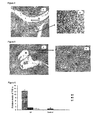
- the Figure 4A corresponds to the joint of an animal of the group III. We can observe the presence of the pannus that invades the whole intra-articular space, extending toward the cartilage where its erosion is evidenced, as well as the invasion toward the bone marrow.
- the Figure 4B shows an amplification of the region corresponding to the bone marrow where the pannus is clearly observed as a granulation tissue rich in phagocytic cells.
- Example 14 In vitro evaluation of the cytokine pattern induced by the F19-6 and F19-7 peptides, using mononuclear cells from rats where the Arthritis was induced with type II collagen.
- the mononuclear cells isolated from the spleen were cultured in 1.5 mL of RPMI 1640 in 24 well plates (Costar, USA). The assay was carried out by triplicate, and a total of 3 ⁇ 10 6 cell/well was cultured during 24 hours with F19-6 and F19-7 peptides at a concentration of 5mg/mL and incubated at 37°C, in humid atmosphere of CO 2 at 5%. The cultivated cells without the peptides were used as negative control.
- FIG. 6 are shown the levels of TNF ⁇ of the cultured cells without peptide and stimulated in vitro with F19-6 and F19-7 peptides corresponding to day 21 after the induction of the illness.
- the letter A represents the levels of TNF ⁇ in the cultivated cells without peptide
- the letter B represents the values of this cytokine when the cells are stimulated with the F19-6 peptide
- the letter C corresponds at the levels of TNF ⁇ when the cells are stimulated with the F19-7 peptide.
- CII represents the group of animals, where the Arthritis was induced with collagen type II and Control represents the group of healthy animals.
- Figure 7 shows the levels of IL-10 of cultivated cells without peptide and stimulated in vitro with F19-6 and F19-7 peptides, corresponding to the day 21 after the induction of the illness
- the letter A represents the levels of IL-10 in the cultivated cells without peptide
- the letter B represents the values of this cytokine when the cells are stimulated with the F19-6 peptide
- the letter C corresponds to the levels of IL-10 when the cells are stimulated with F19-7 peptide.
- CII represents the group of animals, where the AR was induced with collagen type II and Control represents the group of healthy animals.
- F19-6 and F19-7 peptides diminish the levels of TNF ⁇ in vitro, but in the case of the levels of IL-10, only the F19-7 peptide increases it significantly.
- F19-7 peptide is able to modulate the cytokines pattern to a regulating phenotype in the animal model where the Arthritis is induced with collagen. Therefore we can affirm that the immunomodulating action of this peptide derived from the hHsp60 in vitro can be extended to animal models of Arthritis, where the induction agent that participates in the development of the illness is not Mt or related autoantigens.
- F19-7 peptide induces a protection against arthritis induced by Mt or type II collagen in the animals, which is mediated by regulatory T cells that produce IL-10. This effect could extend to other autoantigens present in the joints that progressively contribute to the immunophatogenic process that takes place in the course of RA.
- Example 15 Cytokine measurement in patients with RA induced by F19-6 and F19-7 peptides.
- cytokines TNF ⁇ , IFN ⁇ and IL-10.
- the potentiality of the peptides of the hHsp60 to increase or to diminish the different cytokines involved in the pathogenesis of the illness was evaluated in this assay.
- the pellet of the cell was resuspended in RPMI 1640 containing 10% of bovine fetal serum and supplemented with penicillin (100U/mL), streptomycin (100 ⁇ g/mL), HEPES 25 mM/L and L-glutamine 2mM (all acquired from Gibco BRL).
- the mononuclear cells obtained were seeded in number of 10 6 cells/well in plates of 24 wells (Costar) in a volume of 800 ⁇ L.
- peptides of the hHsp60 were added at a concentration range from 0.5 ⁇ g/mL up to 100 ⁇ g/mL, in order to know the optimimal concentration of each peptide to modulate the cytokines levels.
- Phytohemagglutinin (PHA) 1% was used as positive control of cellular stimulation, while the RPMI 1640 alone was used as control of the basal cell growth.
- the cells were cultured during 24 hours and later on the supernatant of each well was taken, diluted 1 ⁇ 2, and cytokine concentration was determined by specific kits (Quantikine®, R&D Systems) according to recommendations of suppliers.
- the levels of TNF ⁇ modulated by F19-6 and F19-7 peptides in mononuclear cells of patients with RA are represented in the Figure 8.
- B corresponds to cells stimulated in vitro with F19-6 peptide
- C represents cells stimulated in vitro with F19-7 peptide.
- a different letter is assigned in bars for levels of the corresponding cytokine.
- Example 16 Cytokines measurement in patients with RA induced by E18-12 and E18-3 peptides.
- Assays for cytokines measurement using blood of patients with RA were carried out: TNF ⁇ , IFN ⁇ and IL-10.
- the potentiality of the peptides of the hHsp60 to increase or to diminish the different cytokines involved in the pathogenesis of the illness was evaluated in these assays.
- the precipitate was resuspended in RPMI 1640 medium containing 10% of bovine fetal serum supplemented with penicillin (100U/mL), streptomycin (100 ⁇ g/mL), 25 HEPES mM/L and L-glutamine 2mM (all acquired of Gibco BRL).
- the mononuclear cells obtained were seeded in a number of 10 6 cells/well in plates of 24 wells (Costar) in a volume of 800 ⁇ L. Afterwards, the peptides of the hHsp60 were added in a concentration range from 0.5 ⁇ g/ml up to 100 ⁇ g/ml, in order to know the optimal concentration of each peptide to modulate the cytokine levels.
- the Phytohemagglutinin (PHA) to 1% it was used as positive control of cell stimulation, while the RPMI 1640 alone was used as control of the basal cell growth.
- the cells were cultured during 24 hours and later on, the supernatant of each well was taken, diluted 1 ⁇ 2, and cytokine concentration was determined by specific kits (Quantikine®, R&D Systems) according to the recommendations of the suppliers.
- the levels of TNF ⁇ modulated by the E18-3 and E18-12 peptides in mononuclear cells of patients with RA are represented in Figure 9.
- Patients represented by P4, P5 and P16 express the molecule DR 0306
- the patients identified as P7, P8 and P12 express the molecule DR 0303
- patient P9 presents a genotype DR0506
- patient P10 has a genotype DR 0204.
- the letter A represents the cultured cells in vitro without peptides
- B corresponds to the cells stimulated in vitro with E18-3 peptide
- C represents cells stimulated in vitro with E18-12 peptide.
- the modulation of IL-10 levels for E18-3 and E18-12 peptides are represented in Figure 9, where the letter A represents cultured cells in vitro without peptides; while B corresponds to cells stimulated in vitro with the E18-3 peptide and C represents cells stimulated in vitro with E18-12 peptide.
- a therapeutic variant of original peptides: E18-12 or E18-3 could be their administration by oral route, so they will induce mechanisms of tolerance that contributed significantly to decrease the inflammation.
- Example 17 Cytokine measurement in patients with RA induced by derived type APL peptides.
- TNF ⁇ and IL-10 were carry out using blood of patients with RA.
- the potentiality of derived type APL peptides from the hHsp60 to increase or to diminish the different cytokines involved in the pathogenesis of the disease was evaluated in these tests.
- the original peptides were included in this evaluation (E18-3, E18-12) as experimental controls.
- the APL peptides evaluated in these tests were the following:
- the precipitate was resuspended in RPMI 1640 medium containing 10% of bovine fetal serum supplemented with penicillin (100U/mL), streptomycin (100 ⁇ g/mL), 25 HEPES mM/L and L-glutamine 2mM (all acquired of Gibco BRL).
- Obtained mononuclear cells were cultured in number of 10 6 cells / well in plates of 24 wells (Costar) in a volume of 800 ⁇ L. Cells were stimulated with peptides [E18-3, E18-12, E18-3APL1 (SEQ. ID. NO: 18), E18-3APL2 (SEQ. ID. NO: 21), E18-12APL1 (SEQ. ID. NO: 5), E18-12APL3 (SEQ. ID. NO: 12)] at a concentration of 40 ⁇ g/mL and by triplicate. RPMI 1640 was used as control of the basal cellular growth.
- FIG. 11 is shown the levels of IL-10 induced by original peptides (E18-12 and E18-3) and a peptide panel of APLs: E18-3APL1 (SEQ.ID. NO: 18); E18-3APL2 (SEQ. ID. NO: 21); E18-12APL1 (SEQ. ID. NO: 5); E18-12APL3 (SEQ. ID. NO: 12).
- FIG 12 is shown the levels of TNF ⁇ induced by different peptides in the same group of patients.
- the letters indicate statistically significant differences (we only refer statistical significance between different peptides and control cells in each patient individually).
- cells of patients P4 and P6 were not stimulated by peptides E18-3APL2 nor E18-12APL3). As it can be appraised, all the patients do not respond with the same intensity to the stimulus with different peptides, although it is notorious that the response was quite homogenous.
- the peptide that increased more the production of TNF ⁇ was the E18-12APL1, mainly in patients P4 and P5, whereas patients: P1, P2 and P3 showed a slight increase with respect to the negative control.
- peptide E18-3APL1 had a similar behavior in most of the patients.
- the cells of the patients P2, P4 and P5 showed the major increase of the levels of TNF ⁇ , although was less marked.
- IL-10 levels were considerably higher in cells stimulated with peptide E18-3APL1 with respect to cells that constituted the negative control, showing statistically significant differences.
- E18-12APL1 the differences in comparison to the negative control were not so remarkable.
- E18-3APL1 increases the levels of TNF ⁇ , the net predominant effect is the increase in the levels of IL-10, which is translated in a polarization of the response in favor of immunosuppressor cytokines.
- the fundamental advantage of the present invention is the use of pharmaceutical preparations containing immunomodulating peptides of the human stress protein Hsp60, corresponding to T cell epitopes of or variants type APL of these peptides, that can be effectively used in the treatment of patients with RA, inducing a specific immune response directed to silence the clones of involved autorreactive T cells in the immunopathogenic mechanisms of this disease.
- the treatment we propose in this invention is reasonable and can be extended to a large number of patients and also it can be used in combination with current therapies for this disease.
- the animals were separated in twelve groups of eight rats each one.
- the disease was induced in eleven animal groups (Grupo I- Grupo XI), ten of them were treated with peptides and one stayed as control of the induction of the disease.
- Animal groups that were treated with peptides received 50 ⁇ g of peptide by rat in a volume of 100 ⁇ L of PBS, days 12, 14, 16, 18, 20, 22, 24, 26 after induction of the disease with Mt.
- figure 14 we show the graphic of evolution of the disease in four animal groups pertaining to this animal model.
- the groups are: group I (animals treated with E18-12), group II (animals treated with E18-12APL1 SEQ. ID. NO: 5), group XI (control of the disease induction) and group XII (healthy animals).
- group I animals treated with E18-12
- group II animals treated with E18-12APL1 SEQ. ID. NO: 5
- group XI control of the disease induction
- group XII healthy animals.
- different letters indicate significant differences (p ⁇ 0.001) (it refers to the statistical significant between all groups in measured specific days.
Abstract
Description
- The present invention is related with peptides of human heat shock protein of 60 kDa (abbreviated hHsp60), and Altered Peptide Ligands (abbreviated APL) derived from them. Also, the invention is related with the pharmaceutical compositions comprising such peptides for the treatment of Rheumatoid Arthritis (RA).
- The RA is an autoimmune disease of unknown etiology that affects approximately 1% of world's population. It is a syndrome characterized by chronic inflammation of the joints, although systemic manifestations can be also observed. This illness begins with the inflammation of the synovial membrane and frequently causes the erosive destruction of the adjacent cartilage and the bone, which result in moderate physical inability of 80% of the patients and an early mortality (Moctezuma, J.F. (2002) Manifestaciones articulares de la Artritis Reumatide. Revista Mexicana de Reumatología 17:211-219). RA can be presented at any age, without distinctions of races or ethnic groups, but the maximum incidence of its beginning happens between 25 and 55 years old. Among people with RA, the women overcome the men in a proportion from three to one (Emery, P. (2002) Early referral recommendation for newly diagnosed rheumatoid arthritis: evidence based development of to clinical guide. Ann Rheum Dis. 61:290-297).
- The cause of RA is unknown. It is an illness that involves the presence of genetic, environmental, immunological and hormonal factors. Certain genes have a role in the immune system, associated with a tendency to develop RA. At the same time, some people with RA don't have these genes and other people having them never develop the illness. Therefore, it has been suggested that the genetic background is important but it is not decisive.
- In models of autoimmune disease, microbial antigens with similar structure to own antigens can release a crossed response to autoantigens, producing an alteration in the mechanisms of tolerance and perpetuating an autoimmune response. In general sense, the damage and local necrosis in a tissue produced by an infectious agent could discover the autoantigen cryptic epitope, being able to activate autoreactivate T cells (Albert, L.J. (1999) Mechanisms of Disease: Molecular Mimicry and autoimmunity. N Engl J Med 341:2068-2074)
- The phase of transition of T lymphocyte between tolerance and immunity/autoimmunity is regulated at different levels. Two important parameters in this transition are the state of maturation of the Antigen Presenting Cells (abbreviated APC) and the levels of autoantigens that are detected by the immune system (Ohashi, P.S. (2002) Making and breaking tolerance. Current Opinion in Immmunology 14:744-759).
- One of the current hypotheses that tries to explain the development of autoimmune diseases, outlines that APC in absence of signs of the innate immune system or of signs of danger, remain relatively immature and induce tolerance in autoreactive T cells, when own peptides are presented to them (Steiman, R.M. (2000) The induction of tolerance by dendritic cells that have captured apoptotic cells. J Exp Med. 191:411-416). The induction of peripheral tolerance is also correlated with the concentration of own antigen (Kurts, C. (1999) CD8 T cell ignorance or tolerance to islet antigens depends on antigen dose. PNAS 96:12703-12707). An increment in the presentation of own antigens due to the increment of their expression levels, allows their detection by autoreactive ignorant T cells. If levels of own antigens are increased in absence of events that promote the maturation of APC, the tolerance to these antigens is maintained, otherwise, it happens in presence of proinflammatory signs or other events that promote the maturation of the APC, the tolerance is breaks by activation of the ignorant T cells and autoimmune diseases are developed (Janeway, C.A. (2002) Innate immune recognition. Annu Rev lmmunol. 20:197-216). The infectious agents that have been object of study like cause of RA are: Epstein Bar virus, the retrovirus, virus of the hepatitis C, the Mycobacterium tuberculosis (abbreviated Mt) and the Helicobacter pylori, among others.
- The pathogenesis of RA is characterized by the concerted action of different types of cells that cause the progressive destruction of the cartilage and the bone. In normal situations a balance exists among the inflammatory cytokine as: TNFα, IL-1, IL-6, IL-15, IL-16, IL-17, IL-18 and the IFNγ, and the anti-inflammatory ones as IL-4, IL-11, IL-13 and antagonistic of IL-1 or TNFα. In the RA this balance moves however in favor of the inflammatory cytokines ( Arend, W.P. (2001) Cytokine imbalance in the pathogenesis of rheumatoid arthritis: The role of interleukin-1 receiving antagonist. Semin Arthritis Rheum 30(2):1-6)
- The recognition of an exogenous antigen or autoantigen is probably the reason of a series of events that cause destruction of the joints in patients with RA. This phenomenon cause the activation of T CD4+ lymphocytes that in cooperation with the stimulation of different cytokines, induces their differentiation to cells Th1 with the consequent liberation of proinflammatory cytokines (IL-2 and INFγ) (Simón, A.J. (2001) Biological therapy in Rheumatoid Arthritis. Magazine of Clinical Investigation 53(5):452-459). Many investigators coincide that the chronic inflammation of the joints is induced by these activated T cells that infiltrate the synovial membrane. The action of these cytokines on macrophages causes the production of high amount of
- TNFα and IL-1. These cause a series of local and systemic actions as regulating the expression of the molecules of adhesion in the endothelial cells (LFA1, ICAM-1), which recruit other cells to the inflammation sites. They also stimulate macrophages, fibroblasts, condrocytes and osteoclasts to the liberation of others mediators of the inflammation, as IL-15 and IL-8. TNFα and IL-1 stimulate the proliferation of the synovial membrane that causes formation of pannus, they can also induce the differentiation of B lymphocytes to cells producing antibodies that potentially participate in the destruction of the joint. They also inhibit the production of anti-inflammatory cytokines (IL-4 and IL-14) produced by Th2 cells and stimulate hepatocytes to liberate IL-6. IL-6 favors the production of the proteins of the acute phase, which participate strengthening the immune response (Forre, O. (2000) New possibilities of treatment in AR. Scand J Rheumatol 29(2):73-84).
- Among autoantigens involved in the pathogenesis of RA is Hsp60, a protein that belongs to the family of the Hsp, which are immunogenic proteins with exceptionally evolutionary conservation. The immune response against the strange Hsp is an important mechanism of defense against bacterial infections. The antibodies against these proteins can be abundant in healthy people and in patient with autoimmune illnesses and they can cross react with the own antigens (Chen, W. (1999) Human 60-kDa Heat-Shock Protein: To Danger Signal to the Innate Immune System. J Immunol. 162:3212-3219).
- The Hsp65 of Mt is homologous to the Hsp60 of the mammals. This suggests that the Hsp60 can be recognized as autoantigen in patient with RA. Comparing patients with osteoarthritis and patients with RA, these last ones have an increase of prolipherative response of B lymphocytes in the synovial liquid to the Hsp65 of mycobacterium. The intensity of the response correlates with the synovial inflammation. This response is not specific for RA compared with other inflammatory illnesses (Life, P. (1993) Responses to Gram negative enteric bacterial antigens by synovial T cells from patiens with juvenile chronic arthritis: recognition of heat shock protein hsp60. J Rheumatol. 20:1388-1396).
- The concentration of Hsp is a possible sign of danger for the immune system, which are released from dead cells, and can induce an inflammatory response and to begin the maturation of the APC. The Hsp, are intracellular proteins, which are not expressed in the cellular membrane, neither are secreted, so that the Hsp are attractive candidates for molecules that constitute signs of danger (Van den Berg, WB. (1998): Joint inflammation and cartilage destruction may occur uncoupled. Springer Semin lmmunopathol. 20:149-164).
- Several preparations have been proposed using the Hsp60 or its derived peptides for the treatment of some autoimmune pathologies. For example in the patent
EPO262710 - These same inventors in the patent
EPO322990 - In the patent application
W09610039 - In the patent application
WO9711966 - The patent application
WO0143691 - In the patent
US6180103 the authors intend the use of a peptide of the Hsp60 called p277 and its analogous for the diagnosis and prevention of the diabetes type I. - The patent
US5993803 protects the use of the Hsp60, the peptide p277 and a group of derived peptides of this protein to reduce the severity of the immune response during the transplant of organs. - Recently it has been considered that the atherosclerosis presents a series of similar characteristics to autoimmune processes. In the patent application
W00072023 - In the patent application
WO02057795 - At the present time, cure doesn't exist for the RA. The current methods of treatment are centered in alleviating the pain, to reduce the inflammation, to retard the damages of the joints and to improve the functions and the well-being of the patients. Recently immunomodulating medications have been elaborated, that blocks cytokines that participate in the beginning and maintenance of the inflammatory response in RA, with the purpose to stop or slow the progression of the illness. For this kind of therapy two types of medications exist: blocking the action of the Tumor Necrosis Factor (abbreviated TNFα) and those that inhibit the action of the interleukin 1 (IL-1).
- Although the results are promissory with the therapies anti-TNFα and anti-IL-1, the percentage of infections is high. Many of treated patients with these drugs develop serious infections that are fatal in some cases; including other autoimmune illnesses, neoplasias, etc. Besides, they are very expensive medications (Breshinan, B. (1998) Treatment of rheumatoid arthritis with recombinant human anti-interleukin-1 antagonist. Arthritis Reum. 41:2196-2204)
- Oral tolerance has been proposed as a method of creating peripheral tolerance in front of certain antigens. This can be induced by mechanisms of active suppression, anergy or clonal deletion, depending on the doses and the frequency of the administration of the antigen. The method can induce regulatory T cells that are activated in a specific way by the antigen, but exert its action independently (active suppression). For the regulatory action takes place, it is not necessary to administer the supposedly pathogenic antigen, but any other able to induce the active suppression in the inflammatory focus, inhibiting the activity of the pathogenic effector cells. Collagen type II (abbreviated CII) is the autoantigen that has been used more frequently in this sense. The results of the studies carried out in patients with RA, using chicken and bovine CII have given contradictory results (Trentham, D.E. (1993) Effects of oral administration of type II collagen in rheumatoid arthritis. Science 261:1727-1730; Sieper, J. (1996) Oral type II collagen treatment in early rheumatoid arthritis: to double-blind, placebo-controlled, randomize trial. Arthritis Rheum. 39:41-51).
- The patent
US6153200 intends the use of a peptide of the Hsp70, protein belonging to the family of Hsp, to induce tolerance for oral route in patients with RA. - Another variant that has been suggested to induce tolerance is through APL peptides, based on the fact that T cells are activated if the specific T CD4+ lymphocytes for a certain antigenic peptide recognize the antigen presented by competent APC. Nevertheless, if the same T cell is first activated with a different form from the antigen, in which one of the contact sites with the TcR are lightly altered, it can result in a partial activation or even inactivation of the T cells. These antigens are named APLs. The APLs are similar to immunogenic peptides with one or several substitutions in the essential positions of contact with the TcR or with MHC that interfere the cascade of necessary events for the complete activation of the T cells.
- Conceptually, APLs can be designed with similar properties to the immunogenic peptide (agonist) among other effects for increasing the response of T cells toward specific antigens. This effect is advantageous under pathological conditions as infectious illnesses. Peptides can also be designed with antagonistic properties to the immunogenic peptide that could be beneficial in the control of autoimmune illnesses since they can block the response of T cells acting as antagonistic of the TcR (M. De Magistris (1992) Antigen analog-major complex histocompatibility complexes act as antagonist of the T cell receptor. Cell 68:625 - 634), partial agonist or inducing a population of regulatory T cells that mediate the active suppression (Evavold, B.D. (1991) Separation of IL-4 production from Th cell proliferation by an altered T cell ligand. Science 252:1308-1310).The capacity to experimentally manipulate the intrinsic properties of peptide ligands allows altering the nature, the course and the power of the immune cellular response appropriately.
- Up to this moment, two clinical trials in humans using type APL peptides have been performed for the treatment of autoimmune diseases. In both assays, the peptides are derived from an epitope in position 83 to 93 of the myelin basic protein. One of these trials included 142 patients with multiple sclerosis, and it was suspended because 9% of the patients developed hypersensitivity (Ludwig Kapposi (2000) Induction of non-encephalitogenic type 2 T helper-cell autoimmune response in multiple sclerosis after administration of an altered peptide ligand in to placebo controlled, randomized phase II trial. Nature Medicine 10:1176-1182).
- The other trial included 25 patients and was also interrupted, because in three patients was observed an exacerbation of the illness (Bibiana Bielekova (2000) Encephalitogenic potential of the myelin basic protein peptide (amino acids 83-99) in multiple sclerosis: Results of to phase II clinical trial with an altered peptide ligand. Nature Medicine 6:1167-1175). The main author of this work analyzes the possible factors that determined these negative results, and outlines that the changes carried out in the APL's sites may originate new motifs of union to the HLA (like it happened in the case of the APL binding to DRB1*0404 present in patient I), the complex APL-HLA can also stimulate T cells not eliminated in the negative selection that can be cross-activated by the native autoantigen. In this trial high concentrations of the APL were used in the pharmaceutical preparations, which can stimulate T cells with high similarity for the autoantigen and to induce a heteroclitic T cells response. (Bibiana Bielekova and Roland Martin (2001) Antigen-specific immunomodulation via altered peptide tying. J Mol Med. 79:552-556).
- For carrying out these two clinical trials, the authors didn't previously analyze the in vitro response of the T cells from the patients to the APL. They also did not consider the types of HLA molecules expressed by the treated patients
- The main challenge in the treatment of the autoimmune diseases is the development of therapeutic strategies that can eliminate pathogenic T cells with specificity, without affecting other non related T cells. Therefore, therapeutic search should find a secure, specific and effective way of turning off an advanced autoimmune process.
- The present invention, in contrast to the state of the previous technique, proposes the use of peptides of the hHsp60 and its derived type APL, which induce inhibitory molecular mechanisms, in a specific way, of the course of RA.
- The present invention solves the problem previously mentioned, providing peptides of the human protein of heat shock of 60 kDa that constitute epitopes for T cells, to induce mechanisms of tolerance, in particular anergy induction mechanisms or mediated by clones of regulatory T cells in patients with RA and that present the sequences:
- E18-12 MGPKGRTVIIEQSWGSPKVTK (SEQ. ID. NO: 1)
- E18-3 SIDLKDK'YKNIGAKLVQDVANNTNEEAG (SEQ. ID. NO: 2)
- F19-6 IIDPTKVVRTALLDAA (SEQ. ID. NO: 3)
- The patent application
WO9711966 - It is well known that fundamental response of T cells in the model of adjuvant-induced arthritis (AIA) is directed against the Hsp60. We checked in this model that our derived peptides of the hHsp60 exert a very marked therapeutic protection, producing an increase of the IL-10 and a decrease of TNFα levels. The same therapeutic effect exerted by these peptides, we have also checked in the animal model of collagen- induced arthritis (CIA), where the major response is against the type II collagen.
- These facts indicate that therapeutic capacities of our peptides are independent of the induction agent and can mediate mechanisms of active suppression in the site of inflammation that could be extended to other autoantigens present in joints, which progressively contribute to the inmunopathogenic process that takes place during the course of RA. These results reinforce the therapeutic possibilities of the use of our preparations for the treatment of RA.
- These peptides are in particular useful, according to the present invention, for the design of derived type APL variants of the peptides E18-12 (SEQ. ID. NO: 1) and E18-3 (SEQ. ID. NO: 2), modified in the contact sites with the human MHC molecule and a derived type APL of the peptide F19-6 (SEQ. ID. NO: 3), modified in the contact sites with the rat MHC molecule. The respective sequences of amino acids are:
-

- it is substituted in the
position 1 for: A,F,I,L,M,V,W, or Y -
position 2 for: A,D,E,F,G,H,I,K,L,M,N,Q,R,S,T,V,W, or Y -
position 3 for: A,K,V,R,T,I,P,L,N,S,G,Y, or M -
position 4 for: A,D,E,F,G,H,I,K,L,M,N,Q,R,S,T,V,W, or Y - position 5 for: A,D,E,F,G,H,I,K,L,M,N,Q,R,S,T,V, or Y
-
position 6 for: A,D,E,F,G,H,I,K,L,M,N,P,Q,R,S,T,V,W, or Y -
position 7 for: A,D,E,F,G,H,I,K,L,M,N,Q,R,S,T,V, or Y -

- it is substituted in the
position 1 for: L,I,V,M,Y,W,F, or A -
position 2 for: A,D,E,F,G,H,I,K,L,M,N,Q,R,S,T,V,W, or Y -
position 3 for: A,F,I,K,L,M,P,R,S,T,V,W, or Y -
position 4 for: L,I,V,M,Y,W,F, or A - position 5 for: A,D,E,F,G,H,I,K,L,M,N,Q,R,S,T,V,W, or Y
-
position 6 for: A,D,E,F,G,H,I,K,L,M,N,Q,R,S,T,V,W, or Y -
position 7 for: A,F,I,K,L,M,P,R,S,T,V,W, or Y -
position 8 for: A,D,E,F,G,H,I,K,L,M,N,Q,R,S,T,V,W, or Y -

-
position 1 is substituted by H. -
position 2 is substituted by E. - In a particular carrying out, derived type APL peptides have amino acid sequence selected from the group that consists of SEQ. ID. NO: 4, SEQ. ID. NO: 5, SEQ. ID. NO: 6, SEQ. ID. NO: 7, SEQ. ID. NO: 8, SEQ. ID. NO: 9, SEQ. ID. NO: 10, SEQ. ID. NO: 11, SEQ. ID. NO: 12, SEQ. ID. NO: 13, SEQ. ID. NO: 14, SEQ. ID. NO: 15, SEQ. ID. NO: 16, SEQ. ID. NO: 17, SEQ. ID. NO: 18, SEQ. ID. NO: 19, SEQ. ID. NO: 20 y SEQ. ID. NO: 21.
- The peptides of the present invention can be produced by routine methods of peptide synthesis. The specific composition of each peptide can be assayed by the level and the quality of the immune response that it induces in experiments as those are described in the examples that will be presented later on.
- All the sequences described above and in the examples are useful and can also be used as a base for the design and synthesis of derived peptides with improved properties.
- The invention also includes pharmaceutical compositions that include one or more of previously enumerated peptides and optionally a pharmaceutically acceptable vehicle.
- The administration of the pharmaceutical preparations to the patients will be in correspondence with the HLA class II molecules that are expressed by the specific patient.
- The way of administration of the pharmaceutical compositions will depend on the cytokines pattern that these preparations induce in the "ex vivo" tests. The pharmaceutical preparations containing APL peptides inducing a regulatory response will be administered by intradermal or subcutaneous route. The pharmaceutical preparations containing the original peptides inducing a TH1 response will be able to be administered by oral route.
- The amounts of peptides in the pharmaceutical compositions of this invention are those that produce an effective immune response in the host. The effective amount is the quantity that when is administered induces molecular mechanisms that significantly diminish the inflammatory signs characteristic of the RA and stop the damages in the joints characteristic of the course of this illness. The amount of the pharmaceutical composition administered to the host can also vary depending on of several factors that include: the used peptides, ACR score (American College of Rheumatology, abbreviated ACR), HLA type II, time of having diagnosed the illness, age, sex, general health, as well as the level of immunological response in general.
- The present invention also relates a method of treatment of the RA, which includes the administration to a patient of effective amounts of peptides or the pharmaceutical compositions referred previously.
- In part, results are shown graphically in the following figures:
- Figure 1: Evaluation of clinical signs of the Arthritis in rats treated with F19-6 and F19-7 peptides by intradermal route, in AIA model.
- Figure 2: Modulation of the levels of TNFα induced by F19-6 and F19-7 peptides in mononuclear cells of rats in the AIA model at
day 15 after the induction of disease. - Figure 3: Modulation of the levels of IL-10 induced by F19-6 and F19-7 peptides in mononuclear cells of rats in the AIA model at
day 15 after the induction of disease. - Figure 4: Optical microscopy of a section of the joint belonging to a sick not treated animal. IS: intra-articular space, EC: erosion of the cartilage, P: pannus. Stained with hematoxiline-eosine.
- Figure 5: Optical microscopy of a section of a joint belonging to an animal treated with F19-7 peptide by intradermal route. IS: intra-articular space. Stained with hematoxiline-eosine.
- Figure 6: Modulation of levels of TNFα induced by F19-6 and F19-7 peptides in mononuclear cells of rats in the CIA model at
day 21 after the induction of disease. - Figure 7: Modulation of levels of IL-10 induced by F19-6 and F19-7 peptides in mononuclear cells of rats in the CIA model at
day 21 after the induction of disease. - Figure 8: Modulation of the levels of TNFα by F19-6 and F19-7 peptides in mononuclear cells of patients with RA.
- Figure 9: Modulation of the levels of TNFα by E18-3 and E18-12 peptides in mononuclear cells of patients with RA.
- Figure 10: Modulation of the levels of IL-10 by E18-3 and E18-12 peptides in mononuclear cells of patients with RA.
- Figure 11: Modulation of the levels of IL-10 induced by E18-3 and E18-12 peptides and their APLs in mononuclear cells of patients with RA.
- Figure 12: Modulation of TNFα levels induced by E18-3 and E18-12 peptides and their APLs in mononuclear cells of patients with RA.
- Figura 13: Ratio of IL-10 and TNFα levels, induced by E18-3 and E18-12 peptides and their APLs.
- Figure 14: Evaluation of clinical signs of Arthritis in rats treated with E18-12 and E18-12APL1 peptides by intradermal route, in AIA model.
- The selected peptides of the hHsp60 were those reported as epitopes of human T cells by the used programs: SYFPEITHI (Hans-Georg Rammensee, Jutta Bachmann, Niels Nikolaus Emmerich, Oskar Alexander Bachor and Stefan Stevanovic. 1999. Immunogenetics 50:213-219. SYFPEITHI: database for MHC ligands and peptide motifs. (access via: http://www.uni-tuebingen.de/uni/kxi /)) and ProPred (Singh,H. and Raghava,G.P.S. 2001. ProPred: Prediction of HLA-DR binding sites. Bioinformatics, 17(12):1236-37).
- The sequences corresponding to these peptides are shown in Table 1.
Table 1. Sequence of hHsp60 peptides presented by human HLA. Peptides Position in the sequence Amino acid Sequence E18-12 55-75 MGPKGRTVIIEQSWGSPKVTK (SEQ. ID. NO: 1) E18-3 83-110 SIDLKDKYKNIGAKLVQDVANNTNEEAG (SEQ. ID. NO: 2) - Starting from the sequence of hHsp60 we select a peptide that binds to the molecule of MHC-II of rat RT1.B1 according to the sequence motif described in the database SYFPEITHI (Hans-Georg Rammensee, Jutta Bachmann, Niels Nikolaus Emmerich, Oskar Alexander Bachor, Stefan Stevanovic. 1999. Immunogenetics 50:213-219. SYFPEITHI: database for MHC ligands and peptide motifs. (access via: http://www.uni-tuebingen.de/uni/kxi /)).
- A program that analyzes all peptides of 9 amino acids was designed to search those that fulfill with the motif. We also included variants, which in one of the positions were not included the described residuals as anchorage or preferentials.
- The sequence corresponding to one of these peptides (F19-6) it is shown in the Table 2.
Tabla 2. Sequence of hHsp60 peptide selected for rats.Peptide Position in the sequence Amino acid Sequence F19-6 518-533 IIDPTKWRTALLDAA (SEQ. ID. NO: 3) - Starting from the peptides selected in examples 1 and 2, we have modified the positions involved in the binding to the specific MHC (
positions - As a result of these analyses, the peptide E18-12 has been substituted in the
positions 1 to 7 by each one of the amino acids that are specified as follow: -

- it is substituted in the
position 1 for: A,F,I,L,M,V,W, or Y -
position 2 for: A,D,E,F,G,H,I,K,L,M,N,Q,R,S,T,V,W, or Y -
position 3 for: A,K,V,R,T,I,P,L,N,S,G,Y, or M -
position 4 for: A,D,E,F,G,H,I,K,L,M,N,Q,R,S,T,V,W, or Y - position 5 for: A,D,E,F,G,H,I,K,L,M,N,Q,R,S,T,V, or Y
-
position 6 for: A,D,E,F,G,H,I,K,L,M,N,P,Q,R,S,T,V,W, or Y -
position 7 for: A,D,E,F,G,H,I,K,L,M,N,Q,R,S,T,V, or Y -

- it is substituted in the
position 1 for L,I,V,M,Y,W,F, or A -
position 2 for A,D,E,F,G,H,I,K,L,M,N,Q,R,S,T,V,W, or Y -
position 3 for A,F,I,K,L,M,P,R,S,T,V,W, or Y -
position 4 for L,I,V,M,Y,W,F, or A - position 5 for A,D,E,F,G,H,I,K,L,M,N,Q,R,S,T,V,W, or Y
-
position 6 for A,D,E,F,G,H,I,K,L,M,N,Q,R,S,T,V,W, or Y -
position 7 for A,F,I,K,L,M,P,R,S,T,V,W, or Y -
position 8 for A,D,E,F,G,H,I,K,L,M,N,Q,R,S,T,V,W, or Y - Peptide F19-6 has been substituted in the
positions -

-
position 1 is substituted by H. -
position 2 is substituted by E. - Female inbred Lewis rats (RT1.BI MHC) were used in the experiment supplied by the National Center for the Production of Laboratory Animals (CENPALAB, Cuba). The rats were randomly selected, with an approximate weight of 101-120 g and 5-8 weeks of age.
- One of the inductor agents of the illness used was the Mt (H37Ra, Difco, England) in Incomplete Freund's Adjuvant (IFA) (Sigma, USA). The other inductor agent of the Arthritis was the bovine type II collagen in IFA.
- In this example, two different induction agents were used for developing arthritis in rats by an immune response against two different autoantigens. It is very well known that in the animal model for Arthritis using Mt as inductor of the disease, the fundamental response of the T-cells is against the Hsp60 (Anderton, SM (1995) Activation of T cells recognizing self 60-kD heat shock protein can protect against experimental arthritis. J. Exp. Med. 181: 943-952). In the model using collagen as inductor, noticeable antibody response as well as T-cell clones are developed against this antigen (M Griffiths (1988) Immunogenetics of collagen-induced arthritis in rats. Inten. Rev. Immunology 4: 1-15).
- Our fundamental objective for developing these two Animal Models was to evaluate our peptides in both models, with the purpose of demonstrating if the protective effect of these peptides was independent or not to the autoantigen used as induction agent of the Arthritis in rats.
- The inflammation grade associated with the development of the arthritis that presented each rat was evaluated according with the following average prepared for this purpose:
- 0 point: normal paw.
- 1 point: Slight, but definitive redness of the ankle or doll, apparent redness and inflammation limited to an individual finger, independently of the number of affected fingers.
- 2 points: Moderated redness and inflammation of the ankle and the doll.
- 3 points: Severe redness and inflammation of the whole paw including the fingers.
- 4 points: Maximum inflammation and deformity of the paw involving multiple articulations.
- The four paws were evaluated separately according to the established score, each paw could receive up to 4 points. Therefore, each rat could receive a maximum of 16 points.
- The visual evaluation was carried out after the induction of the illness every five days.
- The clinical evaluation of the signs of the Arthritis demonstrated that there were significant statistical differences among animals corresponding to the healthy control group (group IV), where the illness was not induced, and animals corresponding to the sick control group (group III), where illness was induced but they were not treated with peptides, like it is shown in the Figure 1.
- The peptides were synthesized by the strategy Fmoc/tBu in syringes. The resin used was Fmoc-AM-MBHA (0,54 mmol/g) and the synthesis protocol was continued with mechanical agitation. After the treatment with TFA, the peptide was lyophilized and it was characterized by HPLC and mass spectrometry.
- The animals were separated in four groups of 12 rats each one. The illness was induced in three groups of animals (Group I - Group III), and two groups were treated with peptides and one was the control of the induction of the illness. The two groups of animals that were treated with peptides received 200 µg of peptide per each rat in a volume of 100 µL of PBS, the
days - The animals separated in four groups were treated in the following way:
- group I: animals treated with peptide F19-6
- group II: animals treated with peptide F19-7
- group III: animals not treated with peptides (control of the induction of the illness)
- group IV: animals that were not induced with the illness and were not treated with peptides (negative control)
-
Days - As it is shown in the Figure 1 the signs associated with the development of the Arthritis began gradually. In the animals of group III, corresponding to the control group of the development of the illness, from
day 10 after the induction of the Arthritis, it became evident the development of an inflammatory process that began with a slight redness of the inferior joints and that went expanding to the rest of the joints until became severe in some rats. - In this figure it is evident that the administration of peptide F19-6, corresponding to the original epitope of the hHsp60, and their variant APL F19-7, induced a significant reduction of the clinical signs of the Arthritis in the animals treated with both peptides. It was obtained statistically significant differences among the groups treated with the peptides F19-6 and F19-7 and the group taken as positive control of the illness (P <0.05).
- The results obtained in this assay corroborate that peptides F19-6 and F19-7 exert a protective effect in treated animals, since it was possible to observe an almost complete remission of the illness in the rats.
- The mononuclear cells isolated from spleens of rats, were incubated approximately during 20 hours. Later, supernatants were discarded and 1mL of the reagent: TRI REAGENT TM (SIGMA, USA) was added to each well and it was left during 5 minutes at room temperature. Later on, the isolation of the mRNA was carrying out following the protocol recommended by the suppliers.
- For obtaining the complementary DNA the system Gene Amp RNA PCR Kit (Perkin - Elmmer, USA) was used. 1 mg of mRNA corresponding to each sample and the protocol was used and the protocol was continued as recommended the suppliers. The reaction was incubated in a thermocycler (Minicycler, MJ Research, USA), according to the following outline: a first cycle of 1 hour at 42°C, subsequently 5 minutes at 95°C and 5 minutes at 4°C.
- The Polymerase Chain Reaction (PCR) was carried out for the cytokines: TNFα and IL-10 and the constituent gene that codes for the enzyme GAP-DH (glyceraldehyde-3-phosphate dehydrogenase, abbreviated GAP-DH).
- For each
reaction 10 pmol of each primer was used, 5 mL of the DNA mold, 0.25 mL of the DNA Thermostable Polymerase (Taq-Pol) (Perkin - Elmmer, USA) in the buffer recommended by the suppliers (KCl 50Mm, Tris-HCl 10mM, pH 9.0, Triton X-100 to 0.1 % and MgCl2 1.5 mm) in a final volume of 25 µL. - The reaction for the GAP-DH was incubated in a thermocycler (Minicycler, MJ Research, U.S) according to the following program: a first cycle of 3 minutes at 94°C (denaturating temperature), 1 minute at 94°C, 1 minute at 58°C (hybridization temperature) and 1 minute at 72°C (elongation temperature), subsequently 35 cycles of: 1 minute at 94°C, 1 minute at 58°C and 1 minute at 72°C in each one. Lastly an extension step of 3 minutes at 72°C.
- For the cytokines, the previous outline was continued, varying only the hybridization temperature. For the IL-10, 54°C and for the TNFα 64°C.
- The result of the PCR was analyzed by electrophoresis in agarose gel. The images of the electrophoretical running were captured and analyzed with the Molecular Program Analyst (BioRad). The results were expressed as relative levels of mRNA for each cytokine.
- To determine the differences in the pattern of cytokines (according to the relative levels of mRNA for each one) among groups of treated animals, the tests of Kruskal-Wallis and Student-Newman-Keuls were used. (Statistical package
Sigma Stat v 2 1997, GraphPad Software, Inc). - The statistical analyses demonstrated that in the
day 15 after the induction of the disease, the peptides significantly diminish the levels of TNFα in comparison with the control group of the illness. - These results are shown in the Figure 2, where GI corresponds to treated animals with F19-6 peptide, GII represents the group of rats treated with the F19-7 peptide, GIII represents the not treated sick group and GIV includes the healthy animals. It was clearly proven that the treatment with peptides caused a decrease in the relative levels of this cytokine responsible in great measure of inducing the inflammatory process in the animals. There were statistically significant differences for the case of the cytokine TNFα among the groups treated with the peptides and in the group used as positive control of the illness. Significant difference was not evidenced among the different groups treated with the peptides neither among them and the group taken as negative control of the illness. This result indicates that the administration of these two peptides caused a decrease in the levels of TNFα, practically until normal levels of this cytokine as in healthy animals.
- With the purpose of determining if the peptides used in this study, could stimulate the production of anti-inflammatory cytokines as the IL-10, the relative levels of mRNA corresponding to this cytokine were evaluated.
- The IL-10 is involved in the induction of peripheral tolerance through a T cell mediated response. The IL-10 regulates directly the T cells to inhibit the production of IL-2, IL-5 and TNFα.
- The Figure 3 shows the quantitative measurement of the levels of expression of the 1L-10 in the different groups of treatment, at
day 15 after the induction of the illness. In this figure, GI corresponds to treated animals with F19-6 peptide, GII represents the group of rats treated with F19-7 peptide, GIII represents the group of sick animals not treated and GIV includes the healthy animals. - The treatment of the animals with these peptides induces an increment in the relative expression levels of the IL-10. We did not obtain statistically significant differences between groups treated with both peptides, but differences exist between these groups and animals that constitute positive and negative controls. Starting from these results we can infer that the peptides act inducing a change in the functional phenotype of the T cells involved in the pathogenesis of the illness, increasing the levels of production of anti-inflammatory cytokines like the case of IL-10 and diminishing the levels of production of the pro-inflammatory cytokines like TNFα.
- With the objective to corroborate the clinical signs observed in the animals of this animal model and to analyze the capacity of both peptides to revert or to diminish the destructive process generated by the Mt, we carried out the pathological analysis of the animal's joints of each group.
- The sheets corresponding to the joints were analyzed by the group of Pathology of CIGB.
- The joints were fixed in a neutral buffer of formalin at 10% and decalcified. The tissues were dehydrated in an alcohol gradient, absorbed in paraffin and cut in sections of 5µm. Later on, the samples were mounted on glass slides and stained with a hematoxiline-eosine mixture and observed in an optical microscope.
- The results of the histological analysis are shown in the table 3.
Table 3. Summary of the histological analysis of the animal's joints from group I to group IV. Groups # animal Sacrifice day P EC IM HO G PMN I(F19-6) 1 15 X X X 2 x/- X 3 I(F19-6) 1 21 x/- 2 x/- X 3 X X x/- X I(F19-6) 1 28 X X 2 X x/- X X x/- X 3 I (F19-6) 1 35 X x/- X X X X 2 X X x/- 3 X X X X X X II (F19-7) 16 15 17 18 II (F19-7) 16 21 X X x/- X 17 X X x/- X 18 X X x/- X II (FI9-7) 16 28 X X X X x/- X 17 X X X X x/- X 18 II (F19-7) 16 35 17 X X X X x/- X 18 III 46 15 X X X X X 47 X X X 48 X X X X X III 46 21 X X X X X 47 X X X X X 48 X X X X X X III 46 28 X X X X X X 47 X X X X 48 X X X X X X III 46 35 X X X X X 47 X X X X 48 X X X X X - In Table 3 P: represents the pannus; EC: erosion of the cartilage; IM: invasion of the bone marrow; HO: hyperplasia of the osteoclasts; G: granulematose reaction; PMN: polymorphonuclears.
- In the animals of the group III including those sacrificed at
day 15, was evident the presence of the pannus that cause the erosion of the cartilage and the invasion of the bone marrow. Also, the typical granulomatose reaction of the induction with Mt is observed and frequently the hyperplasia of the osteoclasts that are giant cells that have a great destructive power of the bony substance. These histological results reproduce the characteristic signs of the modifications in the joint that occurring in patients with AR and demonstrate that we have developed an appropriate animal model to evaluate the pharmacological candidates. - The Figure 4A corresponds to the joint of an animal of the group III. We can observe the presence of the pannus that invades the whole intra-articular space, extending toward the cartilage where its erosion is evidenced, as well as the invasion toward the bone marrow. The Figure 4B shows an amplification of the region corresponding to the bone marrow where the pannus is clearly observed as a granulation tissue rich in phagocytic cells.
- In the treatment with F19-7 peptide by intradermal route (group II) as is shown in Figure 5, sacrificed animal at
day 15 did not present histological alterations, just the presence of polymorphonuclears and abundant scaring tissues; in contrast with the rest of the animals of the group III, that were characterized to present severe histological signs. - As it is shown in Table 3, the groups of treated animals with the F19-6 and F19-7 peptides didn't show practically histological affectations during the course of the first assay. A bigger protection against to the development of the Arthritis was evidenced in the animals treated with F19-7 peptide. Half of the rats involved in this group of treatment (6 rats) didn't present any histological signs, in correspondence with the group of animals treated with F19-6, where only two rats didn't present histopathological damage. In the groups treated with F19-6 and F19-7 peptides the presence of pannus was not observed at
days - In this assay sick rats were used where the Arthritis was induced using bovine type II collagen in IFA. The spleen of each analyzed rat was extracted and the mononuclear cells were isolated.
- The mononuclear cells isolated from the spleen were cultured in 1.5 mL of RPMI 1640 in 24 well plates (Costar, USA). The assay was carried out by triplicate, and a total of 3×10⁶ cell/well was cultured during 24 hours with F19-6 and F19-7 peptides at a concentration of 5mg/mL and incubated at 37°C, in humid atmosphere of CO2 at 5%. The cultivated cells without the peptides were used as negative control.
- Later on the mRNA was isolated and the reactions of reverse transcription of the nucleic acid and the PCR were carried out in the same way as described in the previous examples. The analyses of the reaction products were performed by electrophoresis in agarose gels as described in the example 12. To determine the differences in the cytokine pattern according to the relative levels of mRNA for each sample, statistical tests were used as described in example13.
- In Figure 6 are shown the levels of TNFα of the cultured cells without peptide and stimulated in vitro with F19-6 and F19-7 peptides corresponding to
day 21 after the induction of the illness. The letter A represents the levels of TNFα in the cultivated cells without peptide, the letter B represents the values of this cytokine when the cells are stimulated with the F19-6 peptide and the letter C corresponds at the levels of TNFα when the cells are stimulated with the F19-7 peptide. In this figure CII represents the group of animals, where the Arthritis was induced with collagen type II and Control represents the group of healthy animals. - Figure 7 shows the levels of IL-10 of cultivated cells without peptide and stimulated in vitro with F19-6 and F19-7 peptides, corresponding to the
day 21 after the induction of the illness, the letter A represents the levels of IL-10 in the cultivated cells without peptide, the letter B represents the values of this cytokine when the cells are stimulated with the F19-6 peptide and the letter C corresponds to the levels of IL-10 when the cells are stimulated with F19-7 peptide. In this figure, CII represents the group of animals, where the AR was induced with collagen type II and Control represents the group of healthy animals. - As it is observed in these two figures, F19-6 and F19-7 peptides diminish the levels of TNFα in vitro, but in the case of the levels of IL-10, only the F19-7 peptide increases it significantly.
- These results indicate that F19-7 peptide is able to modulate the cytokines pattern to a regulating phenotype in the animal model where the Arthritis is induced with collagen. Therefore we can affirm that the immunomodulating action of this peptide derived from the hHsp60 in vitro can be extended to animal models of Arthritis, where the induction agent that participates in the development of the illness is not Mt or related autoantigens.
- These results suggest that the F19-7 peptide can induce tolerance for structurally related antigens, being able to mediate in vivo active suppression in the site of the inflammation.
- With these results we can suggest that F19-7 peptide induces a protection against arthritis induced by Mt or type II collagen in the animals, which is mediated by regulatory T cells that produce IL-10. This effect could extend to other autoantigens present in the joints that progressively contribute to the immunophatogenic process that takes place in the course of RA.
- Using blood of patients with RA was carried out assays of measurement of cytokines: TNFα, IFNδ and IL-10. The potentiality of the peptides of the hHsp60 to increase or to diminish the different cytokines involved in the pathogenesis of the illness was evaluated in this assay.
- Starting from 10 mL of blood of each patient that was diluted ½ in PBS 1X, 5 mL from this dilution was added to 3 mL of Ficoll-Paque (Amershan) in centrifugation tubes of 15 mL, which were centrifuged during 30 min. to 1200 rpm. The ring corresponding to mononuclear cells was extracted. Later on, the cells were washed twice with 15 mL of PBS 1X and after each washing, they were centrifuged at 900 rpm. Finally, the pellet of the cell was resuspended in RPMI 1640 containing 10% of bovine fetal serum and supplemented with penicillin (100U/mL), streptomycin (100µg/mL),
HEPES 25 mM/L and L-glutamine 2mM (all acquired from Gibco BRL). The mononuclear cells obtained were seeded in number of 106 cells/well in plates of 24 wells (Costar) in a volume of 800 µL. Later on, peptides of the hHsp60 were added at a concentration range from 0.5 µg/mL up to 100 µg/mL, in order to know the optimimal concentration of each peptide to modulate the cytokines levels. Phytohemagglutinin (PHA) 1% was used as positive control of cellular stimulation, while the RPMI 1640 alone was used as control of the basal cell growth. - The cells were cultured during 24 hours and later on the supernatant of each well was taken, diluted ½, and cytokine concentration was determined by specific kits (Quantikine®, R&D Systems) according to recommendations of suppliers.
- The levels of TNFα modulated by F19-6 and F19-7 peptides in mononuclear cells of patients with RA are represented in the Figure 8. In this figure, the letter A-represents cultured cells in vitro without peptides; while B corresponds to cells stimulated in vitro with F19-6 peptide and C represents cells stimulated in vitro with F19-7 peptide. In the figure, when exists statistically significant difference among the levels of cytokines secreted by cells stimulated with peptides with respect to non stimulated cells, a different letter is assigned in bars for levels of the corresponding cytokine.
- In the patients represented as P19 and P21, the levels of TNFα diminished significantly by the F19-6 and F19-7 peptides in comparison with the non stimulated cells as it is observed in Figure 8, although in patient P19 the decrease in levels of this cytokine is more significant than with F19-7 peptide.
- In the patient represented as P13, the levels of TNFα were increased by the F19-6 peptide, however the levels of this cytokine diminished significantly, when this patient's mononuclear cells are stimulated with the F19-7 peptide. These results evidence that F19-7 peptide, although it was designed to be presented by the MHC from rat, they can diminish the levels of this cytokine, which is largely responsible of the inflammatory process characteristic of RA, thus this peptide is a potential therapeutic candidate for the treatment of this disease.
- Assays for cytokines measurement using blood of patients with RA were carried out: TNFα, IFNδ and IL-10. The potentiality of the peptides of the hHsp60 to increase or to diminish the different cytokines involved in the pathogenesis of the illness was evaluated in these assays.
- Ten mL of blood were extracted from each patient, which were diluted ½ in PBS 1X. It was added 5 mL of this dilution to 3 mL of Ficoll-Paque (Amershan) in centrifugation tubes of 15 mL, and centrifuged during 30 min at 1200 rpm. The ring corresponding to mononuclear cells was extracted. Later, the cells were washed twice with 15 mL of PBS 1X and after each washing they were centrifuged at 900 rpm. Finally, the precipitate was resuspended in RPMI 1640 medium containing 10% of bovine fetal serum supplemented with penicillin (100U/mL), streptomycin (100ìg/mL), 25 HEPES mM/L and L-glutamine 2mM (all acquired of Gibco BRL).
- The mononuclear cells obtained were seeded in a number of 106 cells/well in plates of 24 wells (Costar) in a volume of 800 µL. Afterwards, the peptides of the hHsp60 were added in a concentration range from 0.5 µg/ml up to 100 µg/ml, in order to know the optimal concentration of each peptide to modulate the cytokine levels. The Phytohemagglutinin (PHA) to 1% it was used as positive control of cell stimulation, while the RPMI 1640 alone was used as control of the basal cell growth.
- The cells were cultured during 24 hours and later on, the supernatant of each well was taken, diluted ½, and cytokine concentration was determined by specific kits (Quantikine®, R&D Systems) according to the recommendations of the suppliers.
- The levels of TNFα modulated by the E18-3 and E18-12 peptides in mononuclear cells of patients with RA are represented in Figure 9. Patients represented by P4, P5 and P16 express the molecule DR 0306, the patients identified as P7, P8 and P12 express the molecule DR 0303, while patient P9 presents a genotype DR0506 and patient P10 has a genotype DR 0204. In this figure, the letter A represents the cultured cells in vitro without peptides; while B corresponds to the cells stimulated in vitro with E18-3 peptide and C represents cells stimulated in vitro with E18-12 peptide.
- In the figure when exists statistically significant difference among the levels of the cytokines secreted by the cells stimulated with the peptides with respect to non stimulated cells, a different letter is assigned in the bars for levels of the corresponding cytokine.
- As it is observed in Figure 9, the induction of the synthesis of TNFα in mononuclear cells of patients, stimulated with E18-12 and E18-3 peptides depends on the HLA type II molecules that these patients express. In patients P4, P5 and P6 that present a genotype DR 0306, a statistically significant increment of the concentration of TNFα was observed, in comparison with the cells not stimulated with peptides.
- This same effect takes place in the patient P9 of genotype DR 0506 and the patient P10 DR 0204.
- In patients P7, P8 and P12 presenting a genotype DR 0303, any difference in levels of TNFα was not observed, when mononuclear cells are stimulated with our peptides.
- The modulation of IL-10 levels for E18-3 and E18-12 peptides are represented in Figure 9, where the letter A represents cultured cells in vitro without peptides; while B corresponds to cells stimulated in vitro with the E18-3 peptide and C represents cells stimulated in vitro with E18-12 peptide.
- In the case of IL-10, as it is observed in Figure 10, there is a significant decrease of this cytokine in the patients P9 and P10, but in the rest of the patients were not significant changes.
- From these observations we can infer that E18-12 and E18-3 peptides induce a TH1 phenotype in peripheral mononuclear cells.
- A therapeutic variant of original peptides: E18-12 or E18-3 could be their administration by oral route, so they will induce mechanisms of tolerance that contributed significantly to decrease the inflammation.
- From previous results, we decided to modify these original peptides corresponding to epitopes of T cells, at the sites of contact with HLA molecule, using Bioinformatic tools, so that the modified type APL peptides would be able to change the pattern of cytokines to a regulating phenotype. In addition, modifications made in each of these peptides fulfill with peptide binding motifs reported for most of the HLA molecules expressed by the patients that theoretically will favor the interaction of each of them with different HLA molecules.
- Assays for measurement of the cytokines: TNFα and IL-10, were carry out using blood of patients with RA. The potentiality of derived type APL peptides from the hHsp60 to increase or to diminish the different cytokines involved in the pathogenesis of the disease was evaluated in these tests. In addition, the original peptides were included in this evaluation (E18-3, E18-12) as experimental controls.
- The APL peptides evaluated in these tests were the following:
- MGPKGRTVIILQSWGSPKVTK (SEQ. ID. NO: 5)
- MGPKGRTVIIMQSWGSPKVTK (SEQ. ID. NO: 6)
- MGPKGRTVIIAQSWGSPKVTK (SEQ. ID. NO: 7)
- MGPKGRTVIIEQSLGSPKVTK (SEQ. ID. NO: 8)
- MGPKGRTVIIEQSMGSPKVTK (SEQ. ID. NO: 9)
- MGPKGRTVIILQSLGSPKVTK (SEQ. ID. NO: 10)
- MGPKGRTVIILQSMGSPKVTK (SEQ. ID. NO: 11)
- MGPKLRTVIIEQSWGSPKVTK (SEQ. ID. NO: 12)
- MGPKLRTVIILQSMGSPKVTK (SEQ. ID. NO: 13)
- SIDLKDKLKNIGAKLVQDVANNTNEEA (SEQ. ID. NO: 14)
- SIDLKDKYKNIGAKLSQDVANNTNEEA (SEQ. ID. NO: 15)
- SIDLKDKLKNIGAKLSQDVANNTNEEA (SEQ. ID. NO: 16)
- SIDLKDKYKNAGAKLVQDVANNTNEEA (SEQ. ID. NO: 17)
- SIDLKDKYKNIGAKLVQLVANNTNEEA (SEQ. ID. NO: 18)
- SIDLKDKYKNIGAKLVQAVANNTNEEA (SEQ. ID. NO: 19)
- SIDLKDKYKNIGAKLVQDVANNANEEA (SEQ. ID. NO: 20)
- SIDLKDKYKNIGAKLVQLVANNANEEA (SEQ. ID. NO: 21)
- Ten mL of blood were extracted from each patient and diluted ½ in PBS 1X. It was added 5 mL of this dilution to 3 mL of Ficoll-Paque (Amershan) in centrifugation tubes of 15 mL, and centrifuged during 30 min at 1200 rpm. The ring corresponding to mononuclear cells was extracted. Later, the cells were washed twice with 15 mL of PBS 1X and after each washing they were centrifuged at 900 rpm. Finally, the precipitate was resuspended in RPMI 1640 medium containing 10% of bovine fetal serum supplemented with penicillin (100U/mL), streptomycin (100µg/mL), 25 HEPES mM/L and L-glutamine 2mM (all acquired of Gibco BRL).
- Obtained mononuclear cells were cultured in number of 106 cells / well in plates of 24 wells (Costar) in a volume of 800 µL. Cells were stimulated with peptides [E18-3, E18-12, E18-3APL1 (SEQ. ID. NO: 18), E18-3APL2 (SEQ. ID. NO: 21), E18-12APL1 (SEQ. ID. NO: 5), E18-12APL3 (SEQ. ID. NO: 12)] at a concentration of 40 µg/mL and by triplicate. RPMI 1640 was used as control of the basal cellular growth.
- Cells were cultured during 24 hours and later supernatants of each well were taken, diluted ½ and the concentration of cytokines were determined by specific kits (Quantikine®, R&D Systems) according to the recommendations of the suppliers. In figure 11 is shown the levels of IL-10 induced by original peptides (E18-12 and E18-3) and a peptide panel of APLs: E18-3APL1 (SEQ.ID. NO: 18); E18-3APL2 (SEQ. ID. NO: 21); E18-12APL1 (SEQ. ID. NO: 5); E18-12APL3 (SEQ. ID. NO: 12). In this figure, the letters indicate statistically significant differences (we only refer statistical significance between the different peptides and control cells in each patient individually). In addition, cells of patients P4 and P6 were not stimulated by peptides E18-3APL2 nor E18-12APL3. It is evident that, in all patients, peptide E18-3APL1 increases the levels of IL-10 very significantly with respect to the non stimulated cells. The increase induced by this peptide in some of the patients, as P2 and P4, was about 9 times higher than the amount produced by the cells without stimulation, and in patients as P1, P3 and P5 about 5 times superior. Also, similar results were obtained with E18-12APL1 due to a significant increase in levels of IL-10 in patients P1, P4 and P6. On the other hand, neither the original peptides nor peptides E18-3APL2 and E18-12APL3 induced an increase in the levels of IL-10 in comparison with the cells that represent the negative control.
- In figure 12 is shown the levels of TNFα induced by different peptides in the same group of patients. In this figure, the letters indicate statistically significant differences (we only refer statistical significance between different peptides and control cells in each patient individually). In addition, cells of patients P4 and P6 were not stimulated by peptides E18-3APL2 nor E18-12APL3). As it can be appraised, all the patients do not respond with the same intensity to the stimulus with different peptides, although it is notorious that the response was quite homogenous. On the other hand, the peptide that increased more the production of TNFα was the E18-12APL1, mainly in patients P4 and P5, whereas patients: P1, P2 and P3 showed a slight increase with respect to the negative control. Also, peptide E18-3APL1 had a similar behavior in most of the patients. In the case of peptide E18-3APL1 the cells of the patients P2, P4 and P5, showed the major increase of the levels of TNFα, although was less marked. In this test, we obtained similar results to previous evaluations with the original peptides. The previous results indicate that neither peptide nor its APL induce a decrease of TNFα expression in mononuclear cells of the patients.
- These results could be contradictory, since such APLs that increase the production of IL-10 promote an increase in the levels of TNFα. In figure 13 is shown ratio of the levels of IL-10 and TNFα induced by original peptides and their APLs. In this figure, the letters indicate statistically significant differences (we only refer statistical significance between different peptides and control cells in each patient individually). In addition, cells of patients P4 and P6 were not stimulated by peptides E18-3APL2 nor E18-12APL3). It is evident the fact that the increase of IL-10 with respect to TNFα levels was considerably higher, because the cells stimulated with the E18-3APL1 showed an index higher than 1 in four out of six patients, which indicates that IL-10 levels exceeded to those of TNFα. In addition, in all patients, IL-10 levels were considerably higher in cells stimulated with peptide E18-3APL1 with respect to cells that constituted the negative control, showing statistically significant differences. In the case of the E18-12APL1 the differences in comparison to the negative control were not so remarkable. So, although E18-3APL1 increases the levels of TNFα, the net predominant effect is the increase in the levels of IL-10, which is translated in a polarization of the response in favor of immunosuppressor cytokines.
- The results of the study are extremely promissory, because it suggested that these two APLs could be administered to patients and could induce a subpopulation of regulatory T cells secretory of IL-10 that mediate an active suppression at systemic level. Another partial conclusion that we can arrive by means of the analysis of these results, is that these APLs practically display a homogenous behavior in all the studied patients, independently of the genotype that they have. These results are extremely encouraging, because it would allow that the administration of these APL had the same effect in all the patients, in spite of the existing differences with respect to the type of HLA-II molecules that they have.
- From our experimental results, the fundamental advantage of the present invention is the use of pharmaceutical preparations containing immunomodulating peptides of the human stress protein Hsp60, corresponding to T cell epitopes of or variants type APL of these peptides, that can be effectively used in the treatment of patients with RA, inducing a specific immune response directed to silence the clones of involved autorreactive T cells in the immunopathogenic mechanisms of this disease.
- The treatment we propose in this invention is reasonable and can be extended to a large number of patients and also it can be used in combination with current therapies for this disease.
- The animals were separated in twelve groups of eight rats each one. The disease was induced in eleven animal groups (Grupo I- Grupo XI), ten of them were treated with peptides and one stayed as control of the induction of the disease. Animal groups that were treated with peptides, received 50 µg of peptide by rat in a volume of 100 µL of PBS,
days - The separated animals in twelve groups were treated with the following form:
- Group I: received peptide E18-12 (SEQ. ID. NO: 1)

- Group II: received peptide corresponding to: SEQ. ID. NO: 5
- Group III: received peptide corresponding to: SEQ. ID. NO: 8
- Group IV: received peptide corresponding to: SEQ. ID. NO: 12
- Group V: received peptide corresponding to: SEQ. ID. NO: 13
- Group VI: received peptide E18-3 (SEQ. ID. NO: 2)
- Group VII: received peptide corresponding to: SEQ. ID. NO: 14
- Group VIII: received peptide corresponding to: SEQ. ID. NO: 15
- Group IX: received peptide corresponding to: SEQ. ID. NO: 16
- Group X: received peptide corresponding to: SEQ. ID. NO: 17
- Group XI: do not received peptide (control of disease induction)
- Group XII: the disease was not induced or administered any peptide (negative control).
-
Days - In the groups of animals treated with peptides type APL, the clinical improvement was significant in comparison with the non treated animal group and with the groups treated with E18-12 and E18-3. These results were corroborated by histopathological analysis and by measurement of cytokines, demonstrating in all cases a superiority of derived type APL peptides upon original peptides.
- In figure 14 we show the graphic of evolution of the disease in four animal groups pertaining to this animal model. In this case, the groups are: group I (animals treated with E18-12), group II (animals treated with E18-12APL1 SEQ. ID. NO: 5), group XI (control of the disease induction) and group XII (healthy animals). In this graph, different letters indicate significant differences (p<0.001) (it refers to the statistical significant between all groups in measured specific days.
- It is possible to appreciate in this graph, that from
day 12 after the induction of the disease, the animals of groups I, II and XI began to show the characteristic clinical signs of RA. Many animals also presented extra-articular manifestations like nodules in the ears, in the tail and in the extremities. The most severe manifestations of the RA were observed aroundday 20 for the three ill animal groups. In the case of animals treated with E18-12, inday 21 we could not appreciate significant differences with respect to the control of the disease induction group. Nevertheless, aroundday 35 after the induction of the disease, clinical signs in animals of this group were significantly inferior to those of group XI (p<0.001). These results indicate that protective effect of the E18-12 peptide is shown aroundday 35. In this sense, it would be recommendable to administer this peptide in earlier phases of the disease in order to obtain better results atday 21. However, in the case of animals of groups II the clinical signs of inflammation were evidently smaller in comparison with groups I and XI (p<0.001), which indicates that this peptide exerts a potent protective effect in the animals where the disease was induced.


Claims (9)
- Peptides of the human heat shock protein of 60 kDa that constitute epitopes for T cells, to induce mechanisms of peripheral tolerance, in particular mechanisms inductors of anergia or mediated by clones of regulatory T cells in patients with Rheumatoid Arthritis characterized by the sequences:E18-12 MGPKGRTVIIEQSWGSPKVTK (SEQ. ID. NO: 1)E18-3 SIDLKDKYKNIGAKLVQDVANNTNEEAG (SEQ. ID. NO: 2)F19-6 IIDPTKVVRTALLDAA (SEQ. ID. NO: 3)
- Peptides according to claim 1, which are characterized to be derived peptides type APL of E18-12 peptide (SEQ.ID. NO: 1), modified at the contact sites with human MHC molecules, wherein the amino acids sequence:
 it is substituted in theposition 1 for: A,F,I,L,M,V,W, ó Yposition 2 for: A,D,E,F,G,H,I,K,L,M,N,Q,R,S,T,V,W, ó Yposition 3 for: A,K,V,R,T,I,P,L,N,S,G,Y, ó Mposition 4 for: A,D,E,F,G,H,I,K,L,M,N,Q,R,S,T,V,W, ó Yposition 5 for: A,D,E,F,G,H,I,K,L,M,N,Q,R,S,T,V, ó Yposition 6 for: A,D,E,F,G,H,I,K,L,M,N,P,Q,R,S,T,V,W, ó Yposition 7 for: A,D,E,F,G,H,I,K,L,M,N,Q,R,S,T,V, ó Y
 - Derived type APL peptides according to claim 2, having an aminoacidic sequence selected from the group consisting of: SEQ. ID. NO: 5, SEQ. ID. NO: 6, SEQ. ID. NO: 7, SEQ. ID. NO: 8, SEQ. ID. NO: 9, SEQ. ID. NO: 10, SEQ. ID. NO: 11, SEQ. ID. NO: 12 y SEQ. ID. NO: 13.
- Peptides according to claim 1, which are characterized to be derived type APL peptides of the E18-3 peptide (SEQ.ID. NO: 2), modified at the contact sites with the human MHC molecules, wherein the amino acids sequence:
 it is substituted in theposition 1 for L,I,V,M,Y,W,F, or Aposition 2 for A,D,E,F,G,H,I,K,L,M,N,Q,R,S,T,V,W, or Yposition 3 for A,F,I,K,L,M,P,R,S,T,V,W, or Yposition 4 for L,I,V,M,Y,W,F, or Aposition 5 for A,D,E,F,G,H,I,K,L,M,N,Q,R,S,T,V,W, or Yposition 6 for A,D,E,F,G,H,I,K,L,M,N,Q,R,S,T,V,W, or Yposition 7 for A,F,I,K,L,M,P,R,S,T,V,W, or Yposition 8 for A,D,E,F,G,H,I,K,L,M,N,Q,R,S,T,V,W, or Y
 - Derived peptides type APL according to claim 4, having an aminoacidic sequence selected from the group consisting of: SEQ. ID. NO: 14, SEQ. ID. NO: 15, SEQ. ID. NO: 16, SEQ. ID. NO: 17, SEQ. ID. NO: 18, SEQ. ID. NO: 19, SEQ. ID. NO: 20 y SEQ. ID. NO: 21.
- Peptide according to the claim 1, which is characterized to be derived type APL peptide of the F19-6 peptide (SEQ.ID. NO: 3), modified at the contact sites with the rat MHC molecules, wherein the amino acids sequence:
 position 1 is replaced for Hposition 2 is replaced for Eoriginating the F19-7 peptide: (SEQ.ID. NO: 4).
 - A pharmaceutical composition comprising one or more peptides of the claims from 1 to 6 and a pharmaceutically acceptable vehicle.
- Pharmaceutical composition according to claim 7, for treating the Rheumatoid Arthritis.
- A method of treating a human subject suffering from Rheumatoid Arthritis comprising administering an effective amounts of peptides according to any of claims 1 to 6 or a pharmaceutical composition according to claim 7.
Priority Applications (2)
| Application Number | Priority Date | Filing Date | Title |
|---|---|---|---|
| EP10189769.2A EP2371847B1 (en) | 2004-09-24 | 2005-09-22 | Peptides and their derived type APL of the HSP60 and pharmaceutical compositions |
| DK10189769.2T DK2371847T3 (en) | 2004-09-24 | 2005-09-22 | Peptides and their derived type APL of HSP60 and pharmaceutical compositions |
Applications Claiming Priority (2)
| Application Number | Priority Date | Filing Date | Title |
|---|---|---|---|
| CU20040207A CU23504A1 (en) | 2004-09-24 | 2004-09-24 | PEPTIDES AND DERIVATIVES APL TYPE OF HSP60 AND PHARMACEUTICAL COMPOSITIONS |
| PCT/CU2005/000008 WO2006032216A2 (en) | 2004-09-24 | 2005-09-22 | Peptides and apl-type derivatives of hsp60 and pharmaceutical compositions |
Related Child Applications (1)
| Application Number | Title | Priority Date | Filing Date |
|---|---|---|---|
| EP10189769.2 Division-Into | 2010-11-03 |
Publications (2)
| Publication Number | Publication Date |
|---|---|
| EP1803732A2 true EP1803732A2 (en) | 2007-07-04 |
| EP1803732B1 EP1803732B1 (en) | 2012-02-22 |
Family
ID=40273413
Family Applications (2)
| Application Number | Title | Priority Date | Filing Date |
|---|---|---|---|
| EP10189769.2A Active EP2371847B1 (en) | 2004-09-24 | 2005-09-22 | Peptides and their derived type APL of the HSP60 and pharmaceutical compositions |
| EP05794474A Active EP1803732B1 (en) | 2004-09-24 | 2005-09-22 | Peptides and apl-type derivatives of hsp60 and pharmaceutical compositions |
Family Applications Before (1)
| Application Number | Title | Priority Date | Filing Date |
|---|---|---|---|
| EP10189769.2A Active EP2371847B1 (en) | 2004-09-24 | 2005-09-22 | Peptides and their derived type APL of the HSP60 and pharmaceutical compositions |
Country Status (17)
| Country | Link |
|---|---|
| US (2) | US8383771B2 (en) |
| EP (2) | EP2371847B1 (en) |
| JP (1) | JP4755649B2 (en) |
| KR (2) | KR101054332B1 (en) |
| CN (2) | CN101935345B (en) |
| AR (2) | AR051928A1 (en) |
| AT (1) | ATE546462T1 (en) |
| AU (2) | AU2005287757B2 (en) |
| BR (2) | BR122018077204B8 (en) |
| CA (2) | CA2797769C (en) |
| CU (1) | CU23504A1 (en) |
| DK (2) | DK2371847T3 (en) |
| ES (2) | ES2386498T3 (en) |
| MX (1) | MX2007003624A (en) |
| RU (1) | RU2361877C2 (en) |
| WO (1) | WO2006032216A2 (en) |
| ZA (1) | ZA200702938B (en) |
Cited By (2)
| Publication number | Priority date | Publication date | Assignee | Title |
|---|---|---|---|---|
| WO2008094538A3 (en) * | 2007-01-30 | 2009-02-26 | Epivax Inc | Regulatory t cell epitopes, compositions and uses thereof |
| WO2010075824A1 (en) * | 2008-12-29 | 2010-07-08 | Centro De Ingeniería Genética Y Biotecnología | Use of apl-type peptide for treating intestinal inflammatory diseases and type 1 diabetes |
Families Citing this family (7)
| Publication number | Priority date | Publication date | Assignee | Title |
|---|---|---|---|---|
| MX2012002768A (en) * | 2009-09-06 | 2012-06-28 | Protab Ltd | Humanized antibodies specific for hsp65-derived peptide-6 methods and uses thereof. |
| WO2011059758A2 (en) * | 2009-10-28 | 2011-05-19 | Argentis Pharmaceuticals, Llc | Apls for treating arthritis |
| CU24508B1 (en) | 2017-12-29 | 2021-04-07 | Centro De Ingenieria Genetica Y Biotecnologia Biocubafarma | PHARMACEUTICAL COMPOSITION INCLUDING APL TYPE PEPTIDE |
| CU20200026A7 (en) * | 2020-04-13 | 2021-11-04 | Ct Ingenieria Genetica Biotecnologia | PEPTIDE FOR THE TREATMENT OF CYTOKINE STORM SYNDROME |
| CN111870697B (en) * | 2020-09-18 | 2022-09-09 | 山东鲁抗医药股份有限公司 | Application of carbohydrate to improvement of stability of APL-type derived peptide and freeze-dried composition |
| KR102333645B1 (en) | 2021-07-27 | 2021-12-02 | (주)문화전기 | A LED lighting system with easy angle adjustment |
| CN117883547A (en) * | 2021-07-30 | 2024-04-16 | 河北菲尼斯生物技术有限公司 | Application of SE-DR affinity peptide in preparation of medicines for treating rheumatism |
Family Cites Families (11)
| Publication number | Priority date | Publication date | Assignee | Title |
|---|---|---|---|---|
| NL8701163A (en) | 1986-09-09 | 1988-04-05 | Nederlanden Staat | USE OF A PEPTIDE FOR THE PREPARATION OF PREPARATIONS FOR LIGHTING, TREATMENT AND DIAGNOSIS OF AUTOIMMUNE DISEASES, IN PARTICULAR ARTHRITIC CONDITIONS, COMPOUNDS RELATED TO THIS PEPTIDE, AND PHARMACEUTICAL AND DIAGNOSTIC PREPARATORY. |
| NL8703107A (en) | 1987-12-22 | 1989-07-17 | Nederlanden Staat | POLYPEPTIDES AND DERIVATIVES THEREOF, AND THEIR USE THEREOF IN PHARMACEUTICAL AND DIAGNOSTIC PREPARATIONS. |
| US5773570A (en) | 1994-05-20 | 1998-06-30 | The Regents Of The University Of California | Vaccine compositions and methods useful in inducing immune protection against arthritogenic peptides involved in the pathogenesis of rheumatoid arthritis |
| GB9419553D0 (en) | 1994-09-27 | 1994-11-16 | Univ Bristol | Polypeptides and their use in the treatment of auto-immune disease |
| US6180103B1 (en) | 1994-12-21 | 2001-01-30 | Yeda Research And Development Co., Ltd. | Peptide p277 analogs, and pharmaceutical compositions comprising them for treatment or diagnosis of diabetes |
| GB9519737D0 (en) | 1995-09-27 | 1995-11-29 | Peptide Therapeutics Ltd | Polypeptides and their use in treatment and prophylaxis of auto-immune |
| US5993803A (en) | 1996-08-30 | 1999-11-30 | Yeda Research And Development Co., Ltd. | Method of reducing the severity of host vs graft reaction by down-regulating hsp60 autoimmunity |
| GB9911772D0 (en) | 1999-05-21 | 1999-07-21 | Semmelweis University Of Medic | Diagnosis and treatment of atherosclerosis |
| US20020169302A1 (en) * | 1999-08-23 | 2002-11-14 | Genesis Research And Development Corp. Ltd. | Compositions isolated from bovine mammary gland and methods for their use |
| JP2004501061A (en) * | 1999-12-15 | 2004-01-15 | ペプトル・リミテツド | Fragments and antagonists of heat shock protein 60 |
| EP1354208A2 (en) | 2001-01-19 | 2003-10-22 | Cambridge Scientific, Inc. | Methods of diagnosis and treatment of osteoporosis |
-
2004
- 2004-09-24 CU CU20040207A patent/CU23504A1/en active IP Right Grant
-
2005
- 2005-09-21 AR ARP050103950A patent/AR051928A1/en active IP Right Grant
- 2005-09-22 EP EP10189769.2A patent/EP2371847B1/en active Active
- 2005-09-22 DK DK10189769.2T patent/DK2371847T3/en active
- 2005-09-22 MX MX2007003624A patent/MX2007003624A/en active IP Right Grant
- 2005-09-22 DK DK05794474.6T patent/DK1803732T3/en active
- 2005-09-22 BR BR122018077204A patent/BR122018077204B8/en active IP Right Grant
- 2005-09-22 WO PCT/CU2005/000008 patent/WO2006032216A2/en active Application Filing
- 2005-09-22 KR KR1020077008682A patent/KR101054332B1/en active IP Right Grant
- 2005-09-22 CA CA2797769A patent/CA2797769C/en active Active
- 2005-09-22 KR KR1020107001135A patent/KR101035283B1/en active IP Right Grant
- 2005-09-22 RU RU2007115411/04A patent/RU2361877C2/en active
- 2005-09-22 ES ES05794474T patent/ES2386498T3/en active Active
- 2005-09-22 AU AU2005287757A patent/AU2005287757B2/en not_active Ceased
- 2005-09-22 CN CN2010100038161A patent/CN101935345B/en active Active
- 2005-09-22 EP EP05794474A patent/EP1803732B1/en active Active
- 2005-09-22 JP JP2007532752A patent/JP4755649B2/en active Active
- 2005-09-22 ES ES10189769.2T patent/ES2445707T3/en active Active
- 2005-09-22 AT AT05794474T patent/ATE546462T1/en active
- 2005-09-22 CA CA2581110A patent/CA2581110C/en active Active
- 2005-09-22 US US11/663,603 patent/US8383771B2/en active Active
- 2005-09-22 BR BRPI0515893A patent/BRPI0515893B8/en active IP Right Grant
- 2005-09-22 CN CN2005800403891A patent/CN101065398B/en active Active
-
2007
- 2007-04-10 ZA ZA200702938A patent/ZA200702938B/en unknown
-
2009
- 2009-12-28 AR ARP090105137A patent/AR074924A2/en active IP Right Grant
- 2009-12-30 US US12/649,687 patent/US8324164B2/en active Active
-
2010
- 2010-09-06 AU AU2010219312A patent/AU2010219312B2/en not_active Ceased
Non-Patent Citations (1)
| Title |
|---|
| See references of WO2006032216A2 * |
Cited By (31)
| Publication number | Priority date | Publication date | Assignee | Title |
|---|---|---|---|---|
| US10925940B2 (en) | 2007-01-30 | 2021-02-23 | Epivax, Inc. | Regulatory T cell epitopes, compositions and uses thereof |
| US11045532B2 (en) | 2007-01-30 | 2021-06-29 | Epivax, Inc. | Regulatory T cell epitopes, compositions and uses thereof |
| US11045529B2 (en) | 2007-01-30 | 2021-06-29 | Epivax, Inc. | Regulatory T cell epitopes, compositions and uses thereof |
| EP2423221A1 (en) * | 2007-01-30 | 2012-02-29 | Epivax, Inc. | Regulatory t cell epitopes, compositions and uses thereof |
| EP2423220A1 (en) * | 2007-01-30 | 2012-02-29 | Epivax, Inc. | Regulatory T cell epitopes, compositions and uses thereof |
| EP2423222A1 (en) * | 2007-01-30 | 2012-02-29 | Epivax, Inc. | Regulatory t cell epitopes, compositions and uses thereof |
| EP2423219A1 (en) * | 2007-01-30 | 2012-02-29 | Epivax, Inc. | Regulatory t cell epitopes, compositions and uses thereof |
| EP2450372A1 (en) * | 2007-01-30 | 2012-05-09 | Epivax, Inc. | Regulatory t cell epitopes, compositions and uses thereof |
| EP2450371A1 (en) * | 2007-01-30 | 2012-05-09 | Epivax, Inc. | Regulatory t cell epitopes, compositions and uses thereof |
| EP2450367A1 (en) * | 2007-01-30 | 2012-05-09 | Epivax, Inc. | Regulatory t cell epitopes, compositions and uses thereof |
| EP2450370A1 (en) * | 2007-01-30 | 2012-05-09 | Epivax, Inc. | Regulatory t cell epitopes, compositions and uses thereof |
| EP2450368A1 (en) * | 2007-01-30 | 2012-05-09 | Epivax, Inc. | Regulatory t cell epitopes, compositions and uses thereof |
| EP2450369A1 (en) * | 2007-01-30 | 2012-05-09 | Epivax, Inc. | Regulatory t cell epitopes, compositions and uses thereof |
| EP2450366A1 (en) * | 2007-01-30 | 2012-05-09 | Epivax, Inc. | Regulatory t cell epitopes, compositions and uses thereof |
| US7884184B2 (en) | 2007-01-30 | 2011-02-08 | Epivax, Inc. | Regulatory T cell epitopes, compositions and uses thereof |
| US11045530B2 (en) | 2007-01-30 | 2021-06-29 | Epivax, Inc. | Regulatory T cell epitopes, compositions and uses thereof |
| US11045528B2 (en) | 2007-01-30 | 2021-06-29 | Epivax, Inc. | Regulatory T cell epitopes, compositions and uses thereof |
| KR20140117662A (en) * | 2007-01-30 | 2014-10-07 | 에피백스, 인크. | Regulatory t cell epitopes, compositions and uses thereof |
| EP2896630A1 (en) * | 2007-01-30 | 2015-07-22 | Epivax, Inc. | Regulatory T cell epitopes, compositions and uses thereof |
| US10213496B2 (en) | 2007-01-30 | 2019-02-26 | Epivax, Inc. | Regulatory T cell epitopes, compositions and uses thereof |
| US10751397B2 (en) | 2007-01-30 | 2020-08-25 | Epivax, Inc. | Regulatory T cell epitopes, compositions and uses thereof |
| WO2008094538A3 (en) * | 2007-01-30 | 2009-02-26 | Epivax Inc | Regulatory t cell epitopes, compositions and uses thereof |
| US10925942B2 (en) | 2007-01-30 | 2021-02-23 | Epivax, Inc. | Regulatory T cell epitopes, compositions and uses thereof |
| US10925939B2 (en) | 2007-01-30 | 2021-02-23 | Epivax, Inc. | Regulatory T cell epitopes, compositions and uses thereof |
| US10925941B2 (en) | 2007-01-30 | 2021-02-23 | Epivax, Inc. | Regulatory T cell epitopes, compositions and uses thereof |
| US10980867B2 (en) | 2007-01-30 | 2021-04-20 | Epivax Inc. | Regulatory T cell epitopes, compositions and uses thereof |
| US11045531B2 (en) | 2007-01-30 | 2021-06-29 | Epivax Inc. | Regulatory T cell epitopes, compositions and uses thereof |
| WO2010075824A1 (en) * | 2008-12-29 | 2010-07-08 | Centro De Ingeniería Genética Y Biotecnología | Use of apl-type peptide for treating intestinal inflammatory diseases and type 1 diabetes |
| RU2524630C2 (en) * | 2008-12-29 | 2014-07-27 | Сентро Де Инхеньерия Хенетика И Биотекнолохия | Application of apl peptide for treatment of inflammatory intestinal disease and type 1 diabetes |
| AU2009335405B2 (en) * | 2008-12-29 | 2013-07-18 | Centro De Ingenieria Genetica Y Biotecnologia | Use of an APL peptide for the treatment of inflammatory bowel disease and type 1 diabetes |
| CN102307587B (en) * | 2008-12-29 | 2014-04-30 | 遗传工程与生物技术中心 | Use of APL-type peptide for treating intestinal inflammatory diseases and type 1 diabetes |
Also Published As
Similar Documents
| Publication | Publication Date | Title |
|---|---|---|
| AU2010219312B2 (en) | Peptides and APL-type derivatives of HSP60 and pharmaceutical compositions | |
| JP2021107443A (en) | Regulatory t cell epitope, composition, and use thereof | |
| US20230136112A1 (en) | Methods for stratifying diabetes patients | |
| CN102015757B (en) | Cpn10 and the PRR signal modified | |
| Ghosh et al. | Suppression of TH17-mediated pathology through BET bromodomain inhibition | |
| US20050064555A1 (en) | Ciliary neurotrophic factor variants | |
| CN111788217A (en) | Antagonist peptides of interleukin-15 activity | |
| Hong et al. | Two peptides derived from trout IL-1β have different stimulatory effects on immune gene expression after intraperitoneal administration | |
| JP2006508901A (en) | T cell non-binding peptide and use thereof | |
| EP1479689A1 (en) | Peptide complex | |
| del Carmen Domínguez et al. | Suppression of the pathogenic responses in an animal model of adjuvant-induced arthritis by a peptide corresponding to a novel epitope from heat-shock protein 60 |
Legal Events
| Date | Code | Title | Description |
|---|---|---|---|
| PUAI | Public reference made under article 153(3) epc to a published international application that has entered the european phase |
Free format text: ORIGINAL CODE: 0009012 |
|
| 17P | Request for examination filed |
Effective date: 20070417 |
|
| AK | Designated contracting states |
Kind code of ref document: A2 Designated state(s): AT BE BG CH CY CZ DE DK EE ES FI FR GB GR HU IE IS IT LI LT LU LV MC NL PL PT RO SE SI SK TR |
|
| AX | Request for extension of the european patent |
Extension state: AL BA HR MK YU |
|
| 17Q | First examination report despatched |
Effective date: 20090305 |
|
| GRAP | Despatch of communication of intention to grant a patent |
Free format text: ORIGINAL CODE: EPIDOSNIGR1 |
|
| GRAS | Grant fee paid |
Free format text: ORIGINAL CODE: EPIDOSNIGR3 |
|
| GRAA | (expected) grant |
Free format text: ORIGINAL CODE: 0009210 |
|
| AK | Designated contracting states |
Kind code of ref document: B1 Designated state(s): AT BE BG CH CY CZ DE DK EE ES FI FR GB GR HU IE IS IT LI LT LU LV MC NL PL PT RO SE SI SK TR |
|
| AX | Request for extension of the european patent |
Extension state: AL BA HR MK YU |
|
| REG | Reference to a national code |
Ref country code: GB Ref legal event code: FG4D |
|
| REG | Reference to a national code |
Ref country code: CH Ref legal event code: EP |
|
| REG | Reference to a national code |
Ref country code: AT Ref legal event code: REF Ref document number: 546462 Country of ref document: AT Kind code of ref document: T Effective date: 20120315 |
|
| REG | Reference to a national code |
Ref country code: IE Ref legal event code: FG4D |
|
| REG | Reference to a national code |
Ref country code: DE Ref legal event code: R096 Ref document number: 602005032826 Country of ref document: DE Effective date: 20120419 |
|
| REG | Reference to a national code |
Ref country code: NL Ref legal event code: T3 |
|
| REG | Reference to a national code |
Ref country code: DK Ref legal event code: T3 |
|
| REG | Reference to a national code |
Ref country code: SE Ref legal event code: TRGR |
|
| REG | Reference to a national code |
Ref country code: CH Ref legal event code: NV Representative=s name: BOVARD AG |
|
| LTIE | Lt: invalidation of european patent or patent extension |
Effective date: 20120222 |
|
| PG25 | Lapsed in a contracting state [announced via postgrant information from national office to epo] |
Ref country code: IS Free format text: LAPSE BECAUSE OF FAILURE TO SUBMIT A TRANSLATION OF THE DESCRIPTION OR TO PAY THE FEE WITHIN THE PRESCRIBED TIME-LIMIT Effective date: 20120622 Ref country code: LT Free format text: LAPSE BECAUSE OF FAILURE TO SUBMIT A TRANSLATION OF THE DESCRIPTION OR TO PAY THE FEE WITHIN THE PRESCRIBED TIME-LIMIT Effective date: 20120222 |
|
| REG | Reference to a national code |
Ref country code: ES Ref legal event code: FG2A Ref document number: 2386498 Country of ref document: ES Kind code of ref document: T3 Effective date: 20120822 |
|
| PG25 | Lapsed in a contracting state [announced via postgrant information from national office to epo] |
Ref country code: GR Free format text: LAPSE BECAUSE OF FAILURE TO SUBMIT A TRANSLATION OF THE DESCRIPTION OR TO PAY THE FEE WITHIN THE PRESCRIBED TIME-LIMIT Effective date: 20120523 Ref country code: FI Free format text: LAPSE BECAUSE OF FAILURE TO SUBMIT A TRANSLATION OF THE DESCRIPTION OR TO PAY THE FEE WITHIN THE PRESCRIBED TIME-LIMIT Effective date: 20120222 Ref country code: LV Free format text: LAPSE BECAUSE OF FAILURE TO SUBMIT A TRANSLATION OF THE DESCRIPTION OR TO PAY THE FEE WITHIN THE PRESCRIBED TIME-LIMIT Effective date: 20120222 |
|
| PG25 | Lapsed in a contracting state [announced via postgrant information from national office to epo] |
Ref country code: CY Free format text: LAPSE BECAUSE OF FAILURE TO SUBMIT A TRANSLATION OF THE DESCRIPTION OR TO PAY THE FEE WITHIN THE PRESCRIBED TIME-LIMIT Effective date: 20120222 |
|
| PG25 | Lapsed in a contracting state [announced via postgrant information from national office to epo] |
Ref country code: RO Free format text: LAPSE BECAUSE OF FAILURE TO SUBMIT A TRANSLATION OF THE DESCRIPTION OR TO PAY THE FEE WITHIN THE PRESCRIBED TIME-LIMIT Effective date: 20120222 Ref country code: SI Free format text: LAPSE BECAUSE OF FAILURE TO SUBMIT A TRANSLATION OF THE DESCRIPTION OR TO PAY THE FEE WITHIN THE PRESCRIBED TIME-LIMIT Effective date: 20120222 Ref country code: PL Free format text: LAPSE BECAUSE OF FAILURE TO SUBMIT A TRANSLATION OF THE DESCRIPTION OR TO PAY THE FEE WITHIN THE PRESCRIBED TIME-LIMIT Effective date: 20120222 Ref country code: EE Free format text: LAPSE BECAUSE OF FAILURE TO SUBMIT A TRANSLATION OF THE DESCRIPTION OR TO PAY THE FEE WITHIN THE PRESCRIBED TIME-LIMIT Effective date: 20120222 Ref country code: CZ Free format text: LAPSE BECAUSE OF FAILURE TO SUBMIT A TRANSLATION OF THE DESCRIPTION OR TO PAY THE FEE WITHIN THE PRESCRIBED TIME-LIMIT Effective date: 20120222 |
|
| PG25 | Lapsed in a contracting state [announced via postgrant information from national office to epo] |
Ref country code: SK Free format text: LAPSE BECAUSE OF FAILURE TO SUBMIT A TRANSLATION OF THE DESCRIPTION OR TO PAY THE FEE WITHIN THE PRESCRIBED TIME-LIMIT Effective date: 20120222 |
|
| PLBE | No opposition filed within time limit |
Free format text: ORIGINAL CODE: 0009261 |
|
| STAA | Information on the status of an ep patent application or granted ep patent |
Free format text: STATUS: NO OPPOSITION FILED WITHIN TIME LIMIT |
|
| 26N | No opposition filed |
Effective date: 20121123 |
|
| REG | Reference to a national code |
Ref country code: DE Ref legal event code: R097 Ref document number: 602005032826 Country of ref document: DE Effective date: 20121123 |
|
| PG25 | Lapsed in a contracting state [announced via postgrant information from national office to epo] |
Ref country code: MC Free format text: LAPSE BECAUSE OF NON-PAYMENT OF DUE FEES Effective date: 20120930 |
|
| PG25 | Lapsed in a contracting state [announced via postgrant information from national office to epo] |
Ref country code: BG Free format text: LAPSE BECAUSE OF FAILURE TO SUBMIT A TRANSLATION OF THE DESCRIPTION OR TO PAY THE FEE WITHIN THE PRESCRIBED TIME-LIMIT Effective date: 20120522 |
|
| PG25 | Lapsed in a contracting state [announced via postgrant information from national office to epo] |
Ref country code: LU Free format text: LAPSE BECAUSE OF NON-PAYMENT OF DUE FEES Effective date: 20120922 |
|
| PG25 | Lapsed in a contracting state [announced via postgrant information from national office to epo] |
Ref country code: HU Free format text: LAPSE BECAUSE OF FAILURE TO SUBMIT A TRANSLATION OF THE DESCRIPTION OR TO PAY THE FEE WITHIN THE PRESCRIBED TIME-LIMIT Effective date: 20050922 Ref country code: PT Free format text: LAPSE BECAUSE OF FAILURE TO SUBMIT A TRANSLATION OF THE DESCRIPTION OR TO PAY THE FEE WITHIN THE PRESCRIBED TIME-LIMIT Effective date: 20120222 |
|
| REG | Reference to a national code |
Ref country code: FR Ref legal event code: PLFP Year of fee payment: 12 |
|
| REG | Reference to a national code |
Ref country code: FR Ref legal event code: PLFP Year of fee payment: 13 |
|
| REG | Reference to a national code |
Ref country code: FR Ref legal event code: PLFP Year of fee payment: 14 |
|
| PGFP | Annual fee paid to national office [announced via postgrant information from national office to epo] |
Ref country code: NL Payment date: 20230725 Year of fee payment: 19 |
|
| PGFP | Annual fee paid to national office [announced via postgrant information from national office to epo] |
Ref country code: TR Payment date: 20230920 Year of fee payment: 19 Ref country code: IE Payment date: 20230920 Year of fee payment: 19 Ref country code: GB Payment date: 20230920 Year of fee payment: 19 Ref country code: AT Payment date: 20230921 Year of fee payment: 19 |
|
| PGFP | Annual fee paid to national office [announced via postgrant information from national office to epo] |
Ref country code: SE Payment date: 20230920 Year of fee payment: 19 Ref country code: FR Payment date: 20230928 Year of fee payment: 19 Ref country code: DK Payment date: 20230925 Year of fee payment: 19 Ref country code: DE Payment date: 20230920 Year of fee payment: 19 Ref country code: BE Payment date: 20230920 Year of fee payment: 19 |
|
| PGFP | Annual fee paid to national office [announced via postgrant information from national office to epo] |
Ref country code: ES Payment date: 20231123 Year of fee payment: 19 |
|
| PGFP | Annual fee paid to national office [announced via postgrant information from national office to epo] |
Ref country code: IT Payment date: 20230927 Year of fee payment: 19 Ref country code: CH Payment date: 20231001 Year of fee payment: 19 |