EP1802759B1 - Construct of swine vesicular disease virus like particle using genetic engineering and the method for manufacturing thereof - Google Patents
Construct of swine vesicular disease virus like particle using genetic engineering and the method for manufacturing thereof Download PDFInfo
- Publication number
- EP1802759B1 EP1802759B1 EP05856432.9A EP05856432A EP1802759B1 EP 1802759 B1 EP1802759 B1 EP 1802759B1 EP 05856432 A EP05856432 A EP 05856432A EP 1802759 B1 EP1802759 B1 EP 1802759B1
- Authority
- EP
- European Patent Office
- Prior art keywords
- gene
- disease virus
- swine vesicular
- vesicular disease
- particle
- Prior art date
- Legal status (The legal status is an assumption and is not a legal conclusion. Google has not performed a legal analysis and makes no representation as to the accuracy of the status listed.)
- Expired - Lifetime
Links
Images


Classifications
-
- C—CHEMISTRY; METALLURGY
- C12—BIOCHEMISTRY; BEER; SPIRITS; WINE; VINEGAR; MICROBIOLOGY; ENZYMOLOGY; MUTATION OR GENETIC ENGINEERING
- C12N—MICROORGANISMS OR ENZYMES; COMPOSITIONS THEREOF; PROPAGATING, PRESERVING, OR MAINTAINING MICROORGANISMS; MUTATION OR GENETIC ENGINEERING; CULTURE MEDIA
- C12N7/00—Viruses; Bacteriophages; Compositions thereof; Preparation or purification thereof
-
- C—CHEMISTRY; METALLURGY
- C12—BIOCHEMISTRY; BEER; SPIRITS; WINE; VINEGAR; MICROBIOLOGY; ENZYMOLOGY; MUTATION OR GENETIC ENGINEERING
- C12N—MICROORGANISMS OR ENZYMES; COMPOSITIONS THEREOF; PROPAGATING, PRESERVING, OR MAINTAINING MICROORGANISMS; MUTATION OR GENETIC ENGINEERING; CULTURE MEDIA
- C12N15/00—Mutation or genetic engineering; DNA or RNA concerning genetic engineering, vectors, e.g. plasmids, or their isolation, preparation or purification; Use of hosts therefor
- C12N15/09—Recombinant DNA-technology
- C12N15/63—Introduction of foreign genetic material using vectors; Vectors; Use of hosts therefor; Regulation of expression
- C12N15/79—Vectors or expression systems specially adapted for eukaryotic hosts
-
- C—CHEMISTRY; METALLURGY
- C12—BIOCHEMISTRY; BEER; SPIRITS; WINE; VINEGAR; MICROBIOLOGY; ENZYMOLOGY; MUTATION OR GENETIC ENGINEERING
- C12N—MICROORGANISMS OR ENZYMES; COMPOSITIONS THEREOF; PROPAGATING, PRESERVING, OR MAINTAINING MICROORGANISMS; MUTATION OR GENETIC ENGINEERING; CULTURE MEDIA
- C12N15/00—Mutation or genetic engineering; DNA or RNA concerning genetic engineering, vectors, e.g. plasmids, or their isolation, preparation or purification; Use of hosts therefor
- C12N15/09—Recombinant DNA-technology
- C12N15/63—Introduction of foreign genetic material using vectors; Vectors; Use of hosts therefor; Regulation of expression
-
- A—HUMAN NECESSITIES
- A61—MEDICAL OR VETERINARY SCIENCE; HYGIENE
- A61K—PREPARATIONS FOR MEDICAL, DENTAL OR TOILETRY PURPOSES
- A61K39/00—Medicinal preparations containing antigens or antibodies
- A61K2039/51—Medicinal preparations containing antigens or antibodies comprising whole cells, viruses or DNA/RNA
- A61K2039/525—Virus
- A61K2039/5258—Virus-like particles
-
- C—CHEMISTRY; METALLURGY
- C12—BIOCHEMISTRY; BEER; SPIRITS; WINE; VINEGAR; MICROBIOLOGY; ENZYMOLOGY; MUTATION OR GENETIC ENGINEERING
- C12N—MICROORGANISMS OR ENZYMES; COMPOSITIONS THEREOF; PROPAGATING, PRESERVING, OR MAINTAINING MICROORGANISMS; MUTATION OR GENETIC ENGINEERING; CULTURE MEDIA
- C12N2770/00—MICROORGANISMS OR ENZYMES; COMPOSITIONS THEREOF; PROPAGATING, PRESERVING, OR MAINTAINING MICROORGANISMS; MUTATION OR GENETIC ENGINEERING; CULTURE MEDIA ssRNA viruses positive-sense
- C12N2770/00011—Details
- C12N2770/32011—Picornaviridae
- C12N2770/32311—Enterovirus
- C12N2770/32323—Virus like particles [VLP]
Definitions
- the present invention relates to a swine vesicular disease virus-like particle prepared by a gene recombination technique and a preparation method thereof. More specifically, the present invention relates to a method for preparing a swine vesicular disease virus-like particle, wherein the structural protein precursor (P1) gene and 3CD protease gene of total genes of swine vesicular disease virus are inserted simultaneously into a baculovirus expression vector and are expressed in insect cells, so that the expressed 3CD protease enzyme digests the structural protein precursor into individual proteins which then form the capsid of swine vesicular disease virus by an self-assembly process.
- the structural protein precursor (P1) gene and 3CD protease gene of total genes of swine vesicular disease virus are inserted simultaneously into a baculovirus expression vector and are expressed in insect cells, so that the expressed 3CD protease enzyme digests the structural protein precursor into individual proteins which then form the
- Swine vesicular disease is a highly contagious viral disease of pigs, characterized by vesicles on the coronary bands, heels of the feet, and occasionally on the lips, tongue, snout and teats after an incubation period of 2-7 days. Affected pigs recovered from the disease in 2-3 weeks.
- the swine vesicular disease is classified as a list A disease by the Office International des Epizooties (OIE), because its symptoms are clinically indistinguishable from those caused by foot-and-mouth disease virus.
- OIE Office International des Epizooties
- swine vesicular disease When it was first observed in Italy in 1966, it was recognized as porcine enterovirus infection, after which it was termed "swine vesicular disease" after appearance in Great Britain in 1972.
- the etiology of swine vesicular disease is a swine vesicular disease virus which is a porcine variant caused by the infection of pigs with human Coxsackievirus B5. It belongs to the 'Enterovirus' genus of the 'Picornaviridae' family according to microbiological classification, and has only one serotype.
- the swine vesicular disease virus is a (+) single-chain RNA virus consisting of about 7,400 bases and is highly stable in the surrounding environment unlike foot-and-mouse disease virus so that it is not influenced at a pH range of 2.5-12.
- the swine vesicular disease virus has four main structural proteins (VP1, VP2, VP3 and VP4), and also has non-structural proteins, such as 2A, 2B, 2C, 3A, 3B, 3C and 3D, which are known to be expressed mainly in infected cells so as to perform subsidiary functions in intracellular replication of the virus.
- VP1, VP2, VP3 and VP4 main structural proteins
- non-structural proteins such as 2A, 2B, 2C, 3A, 3B, 3C and 3D, which are known to be expressed mainly in infected cells so as to perform subsidiary functions in intracellular replication of the virus.
- the virus neutralization test As a method for detecting swine vesicular disease virus antibodies, the virus neutralization test has been recognized officially as a gold standard test method, but was unsuitable to test a large amount of samples due to disadvantages in that it requires a test period of about 2-3 days and cell culture facilities.
- the enzyme-linked immunosorbent assay was developed [ Journal of Virological Methods, (1995) 52: 155-167 ] .
- an inactivated swine vesicular disease virus is used as a diagnostic antigen.
- virus-like particle to be achieved according to the present invention has not been attempted on the swine vesicular disease virus.
- virus-like particles have been studied on some viruses, such as poliovirus and enterovirus, members of the Enterovirus genus to which the swine vesicular disease virus belongs.
- virus-like particles have been produced either by infecting insect cells with a recombinant baculovirus inserted with the full-length genes (6.6-kb size including structural proteins and non-structural proteins) of poliovirus [ Journal of General Virology (1989) 70: 1453-1463 ], or by co-infecting insect cells with each of three recombinant baculoviruses inserted with structural proteins VP0 (VP4+VP2), VP3 and VP1-coding genes [ Virology (1993) 192: 512-524 ], or by co-infecting insect cells with baculoviruses inserted with each of the P1 and 3CD-encoding genes of enterobirus 71 [ Biotechnology Letters (2003) 25: 919-925 ].
- the present inventors have found from inspecting current models for recombinant antigen that an antigen obtained either by the individual preparation of each of four structural proteins forming the capsid of a swine vesicular disease virus or by the expression of only the structural protein precursor (P1) is not possible to mimic the conformational immunodominant antigenic sites. Accordingly, it was concluded that a usable recombinant protein antigen must have a structure morphologically similar to the structure of the native swine vesicular disease virus, and for this purpose, it is a preferable approach to construct a recombinant protein in the form of a virus-like particle.
- the present inventors have found that, when a recombinant baculovirus inserted with both the structural protein precursor P1 and 3CD protease genes is constructed and used, the overload problem caused by the size of inserted genes can be minimized and the standardization of the amount of its infection into insect cells can be achieved in a very easy manner, and when insect cells are infected with the baculovirus, the P1 protein expressed in the insect cells can be digested with the 3D protease protein expressed simultaneously with the P1 protein so as to produce a virus-like particle, thereby completing the present invention.
- the present invention provides a swine vesicular disease virus-like particle assembled by recombinant proteins expressed by infecting insect cells with a recombinant baculovirus constructed using a baculovirus expression vector prepared by the simultaneous insertion of the structural precursor P1 gene and 3CD protease gene of swine vesicular disease virus.
- the recombinant baculovirus expression vector (hereinafter, referred to as "pFastDual/P1/3CD") is characterized by containing both the structural protein precursor P1 gene and 3CD protease gene of swine vesicular disease virus.
- the recombinant baculovirus (hereinafter, referred to as "Bacmid/P1/3CD") according to the present invention is characterized by containing both the structural protein precursor P1 gene and 3CD protease gene of swine vesicular disease virus.
- the structural protein precursor P1 gene of swine vesicular disease virus used in the present invention is derived from the UKG/27/72 strain isolated in Great Britain in 1972, and its gene sequence (SEQ ID N0: 1) was registered in the National Center for Biotechnology Information (NCBI) under accession number X54521, and a protein (SEQ ID N0: 2) encoded by the gene consists of 851 amino acids.
- the 3CD protease gene (SEQ ID N0: 3) of swine vesicular virus gene used in the present invention is derived from the UKG/27/72 strain isolated in Great Britain in 1972, and its gene sequence (SEQ ID NO: 3) was registered in the National Center for Biotechnology Information (NCBI) under accession number X54521, and a protein (SEQ ID N0: 4) encoded by the gene consists of 645 amino acids.
- the swine vesicular disease virus-like particle according to the present invention was successfully developed for the first time in the world in the research field of swine vesicular disease virus, and can be used as a new diagnostic antigen which will substitute for the inactivated virus antigen currently used in ELISAs for detection of antibodies to swine vesicular disease virus.
- the swine vesicular disease virus-like particle according to the present invention has an advantage in that it can be prepared without handling of any hazardous chemical during the preparation of the swine vesicular disease virus-like particle.
- it can also be used as an antigen for preventive vaccines against swine vesicular disease because its antigenicity is highly similar to that of the native virus antigen.
- the present invention provides a method for preparing a swine vesicular virus-like particle using a gene recombination technique, the method comprising the steps of: (1) amplifying the structural protein precursor P1 gene and 3CD protease gene of swine vesicular disease virus by polymerase chain reaction (PCR); (2) inserting the P1 gene into a baculovirus expression vector (pFastBacDual) so as to prepare pFastBacDual/P1; (3) inserting the 3CD gene into the pFastBacDual/P1 inserted with the P1 gene so as to prepare pFastBacDual/P1/3CD; (4) transforming a baculovirus shuttle vector gene (Bacmid)-containing cell (DH10Bac) with the pFastBacDual/P1/3CD containing the P1 gene and the 3CD gene so as to prepare recombinant
- the experiment procedures, reagents and reaction conditions, which can be used in the method for preparing the recombinant baculovirus, are generally known in the prior art.
- the inventive method for preparing the virus-like particle can be used in a wide range of applications, since it can likewise be applied for other enteroviruses having the same genetic structure.
- the expression vector used in the present invention is not limited to the above-described expression vector, because other kinds of expression baculovirus expression vectors as dual expression vectors may also be used in the present invention.
- the swine vesicular disease virus-like particle prepared according to the above-described method is obtained at an expression level of more than 1.0 mg/liter, indicating that the inventive method overcomes the problem of low expression levels, which is a problem shown in the construction of the poliovirus virus-like particle according to the prior art.
- the swine vesicular disease virus-like particle prepared using the recombinant baculovirus showed a titer equal to that of the inactivated swine vesicular disease virus.
- the inventive virus-like particle can be developed into a subunit vaccine.
- Example 1 Extraction of RNA from swine vesicular disease virus
- the swine vesicular disease virus (UKG/27/72) strain isolated in Great Britain in 1972 was inoculated into IBRS-2 cells (National Veterinary Research and Quarantine Service, Korea), and then, total RNA was extracted from the supernatant of the culture using the RNeasy RNA extraction kit (Qiagen) according to the manufacturer's manual.
- Example 2 Synthesis, amplification and cloning of P1 gene cDNA of swine vesicular disease virus
- the total RNA extracted in Example 1 and a reverse primer of SEQ ID N0: 6 were added into the tube of an RT premix kit (Bioneer) where a first strand cDNA was then synthesized according to the manufacturer's manual. Then, the first strand cDNA, a forward primer of DEQ ID N0: 5 and a reverse primer of SEQ ID N0: 6 were thermally treated at 95°C for 10 min in the tube of a PCR premix kit (Bioneer) for polymerase chain reaction (PCR).
- PCR polymerase chain reaction
- the cDNA was amplified in a PCR amplifier (PE 2400; Perkin Elmer) for 30 cycles of denaturation at 94°C for 1 min, annealing at 55°C for 1 min and extension at 72°C for 2 min, and finally extended at 72°C for 5 min.
- PE 2400 Perkin Elmer
- PCR primers were synthesized based on the swine vesicular disease virus P1 gene sequence registered in the National Center for Biotechnology Information (NCBI, Genbank) under accession number X54521:
- the forward primer of SEQ ID N0: 7 and the reverse primer of SEQ ID N0: 8 were intentionally inserted into the BamHI restriction enzymes site upstream of the 5'-terminal ATG, and the HindIII restriction enzyme site upstream of the 5'.-terminal stop codon, respectively.
- the P1 gene cDNA thus amplified was cloned into a pGEM-T easy vector (Promega, USA) so as to construct "pGEMT/P1", and the base sequence of the inserted gene was comparatively analyzed with the known base sequence of SEQ ID N0: 1 using an automatic sequencer (ABI 377, USA) to examine if the P1 gene cDNA was correctly inserted.
- Example 3 Synthesis, amplification and cloning of 3CD gene cDNA of swine vesicular disease virus
- the total RNA extracted in Example 1 and a reverse primer of SEQ ID N0: 8 were added into the tube of an RT premix kit (Bioneer) where a first strand cDNA was then synthesized according to the manufacturer's manual. Then, the first strand cDNA, a forward primer of SEQ ID N0: 7 and a reverse primer of SEQ ID N0: 8 were thermally treated at 95°C for 10 minutes in the tube of a PCR.
- the cDNA was PCR-amplified in a PCR amplifier (PE 2400; Perkin Elmer) for 30 cycles of denaturation at 94°C for 1 min, annealing at 55°C for 1 min and extension at 72°C for 2 min and finally extended at 72°C for 5 min.
- PE 2400 Perkin Elmer
- PCR primers were synthesized based on the swine vesicular disease virus P1 gene sequence registered in the National Center for Biotechnology Information (NCBI, Genbank) under accession number X54521:
- the forward primer of SEQ ID NO: 7 and the reverse prime of SEQ ID N0: 8 were intentionally inserted into the XhoI restriction enzyme site upstream of the 5'-terminal ATG and the kpnI restriction enzymes site upstream of the 5'-terminal stop codon, respectively.
- the 3CD gene cDNA thus amplified was cloned into a pGEM-Teasy vector (Promega, USA) so as to construct "pGEMT/3CD", and the base sequence of the inserted gene was comparatively analyzed with the known base sequence of SEQ ID N0: 3 using an automatic sequencer (ABI 377, USA) to examine if the 3CD gene was correctly inserted.
- Example 4 Construction of recombinant expression vector inserted with P1 gene of swine vesicular disease virus
- the pGEMT/P1 vector prepared in Example 2 was digested with BamHI and HindIII restriction enzymes so as to extract the P1 DNA fragment. Meanwhile, a pFastBacDualTM vector (Invitrogen, USA) was digested with the same restriction enzymes, and the P1 DNA fragment was inserted downstream of the polyhedrin promoter (P PH )-containing gene site of the vector, thus constructing recombinant baculovirus expression vector pFastBacDual/P1 inserted with the P1 gene of swine vesicular disease virus.
- P PH polyhedrin promoter
- Example 5 Construction of recombinant expression vector inserted with both P1 and 3CD genes of swine vesicular disease virus
- the recombinant baculovirus expression vector pFastBacDual/P1 prepared in Example 4 was digested with XhoI and Kpn I restriction enzymes, and the pGEMT/3CD constructed in Example 3 was digested with the same restriction enzymes and then inserted downstream of the P10 promoter (P p10 )-containing gene site of the pFastBacDual/P1, thus constructing recombinant baculovirus expression vector pFastBacDual/P1/3CD inserted with both the P1 and 3CD genes of swine vesicular disease virus.
- FIG. 1 shows the structure of the recombinant baculovirus expression vector (pFastBacDual/P1/3CD) according to the present invention.
- Example 6 Expression in insect cells and preparation of recombinant baculovirus
- a recombinant baculovirus (bacmid/P1/3CD) according to the present invention was constructed using a Bac-to-Bac baculovirus expression system (Invitrogen, USA) in the following manner.
- the pFastDual/P1/3CD vector constructed in Example 5 was transfected into a cell (DH10Bac) containing a baculovirus gene (bacmid) so as to construct recombinant baculovirus DNA (Bacmid/P1/3CD).
- a process for preparing a recombinant baculovirus using the recombinant baculovirus DNA is as follows. A solution of 1 ⁇ g of a purified Bacmid/P1/3CD gene in 100 ⁇ l of serum-free grace medium was mixed with a solution of 6 ⁇ g of CellFectin (Invitrogen) dissolved in 100 ⁇ l of serum-free grace medium, and the mixture solution was left to stand at room temperature for 30 minutes.
- Sf9 Spodoptera frugiperda
- 3 ⁇ 10 4 Sf9 cells were applied in a monolayer to the 35-mm-diameter cell culture Petri dish well and then inoculated with 1 ml of a 10-fold dilution of the above-collected supernatant, and the cells were sensitized at 27°C for 1 hour. After removing the supernatant, the cell culture medium was overlaid with a grace medium containing 1% low-melting point agarose so as to form a layer thereon, and the resulting cells were cultured for 8 days at 27°C to confirm the formation of plaques. A few plaques were collected and transfected into Sf9 cells, and the cells were cultured for 4 days and observed for the appearance or non-appearance of CPE.
- the collected cell homogenate was subjected to the ELISA assay so as to select a recombinant baculovirus (Bacmid/P1/3CD) reacted with a swine vesicular disease virus-specific monoclonal antibody.
- Example 7 Reparation of swine vesicular disease virus-like particle using recombinant baculovirus
- the Sf9 insect cells monolayer-cultured in a 150-cm 2 cell culture flask were infected with the recombinant baculovirus (Bacmid/P1/3CD prepared in Example 6, to a concentration of 0.1 M0I (multiplicity of infection) at 27 °C for 1 hour.
- the infected cells were collected and placed into a spinner culture flask containing 200 ml of Sf9 insect cells having a cell concentration of 2.0 ⁇ 10 6 /ml, followed by culture at 27C for 4 days.
- the spinner-cultured infected cells were collected and centrifuged at 500xg for 20 minutes, after which the supernatant and the infected cells were collected separately.
- the supernatant was used as a diagnostic antigen by itself, and the infected cells were subjected to a series of the following procedures so as to prepare a diagnostic antigen. Namely, the infected cells were suspended in 0.01 M phospahte buffered saline (PBS) containing 1% Triton X-100 at a ratio of 1 (cell culture medium): 20 (PBS) and then disrupted with a sonicator. Then, the culture medium was centrifuged at 500xg for 20 minutes so as to collect only the supernatant for use as swine vesicular disease virus-like particles.
- PBS phospahte buffered saline
- Example 8 Purification and electron microphotography of swine vesicular disease virus-like particle
- the Sf9 cell culture medium infected with the recombinant baculovirus in the spinner culture flask in the same manner as in Example 7 was collected. Then, the collected culture medium was frozen and thawed three times repeatedly so as to disrupt the infected cells, followed by centrifugation at 8,000xg for 40 minutes to collect the supernatant. To the collected supernatant, 23 g/l of 0.5M NaCl and 75 g/l of 7.5% PEG were added, and the mixture was allowed to react at 4°C for 16 hours. Then, the reaction mixture was centrifuged at 8,000xg for 40 minutes so as to collect only the pellets.
- pellets were suspended in PBS at a ratio of 1 (culture medium): 50 (PBS) and dialyzed two times.
- the concentrated sample was centrifuged at 3,000xg for 5 minutes so as to collect the supernatant.
- the supernatant was subjected to ultracentrifugation using cesium chloride (CsCl) gradient so as to purify swine vesicular disease virus-like particle.
- CsCl cesium chloride
- the swine vesicular disease virus-like particles prepared in the present invention had a size of 25-27 nm similar to 25-30 nm which is the size of the native virus containing nucleic acids.
- Test Example 1 Analysis of swine vesicular disease virus-like particle by Western blotting
- Proteins of the swine vesicular disease virus-like particle purified in Example 8 were electrophoretically separated on NuPAGE Novex Bis-Tris gels (Invitrogen, USA), and then were transferred from the gels onto a nitrocellulose membrane.
- the nitrocellulose membrane was treated with a blocking solution containing 5% skimmed milk, and then allowed to react with membranes to which guinea pig positive serum and pig positive serum have been transferred, respectively.
- the membrane was washed with a PBS containing 0.05% Tween 20 (PBST) and allowed to react with various alkaline phosphatase-conjugated secondary antibodies (KPL, USA) for 1 hour.
- PBST PBS containing 0.05% Tween 20
- the swine vesicular disease antibody-positive guinea pig sera (lanes 2 and 3) and the swine vesicular disease antibody-positive pig sera (lanes 4 and 5) showed reactivity to the swine vesicular disease virus-like particle (lanes 2 and 4) in the same pattern as the swine vesicular disease virus particle (lanes 3 and 5).
- the virus-like particle observed with the electron microscope as described above is a swine vesicular disease virus-like particle having the same component as that of the native swine vesicular disease virus.
- Test Example 2 Evaluation of antigenicity of swine vesicular disease virus-like particle
- the swine vesicular disease virus-like particle according to the present invention was evaluated for its antigenicity in comparison with that of the inactivated swine vesicular disease virus antigen of the prior art.
- Swine vesicular virus monoclonal antibody 5B7 (swine vesicular disease virus-neutralizing antibody) was coated onto ELISA plates at 4°C overnight. On the ELISA plates, each of the swine vesicular disease virus and the swine vesicular disease virus-like particle was added and then allowed to react with the antibody at 37°C for 1 hour. Following washing step using PBST, pig sera (including swine vesicular disease standard-positive serum) were added thereto and allowed to react at 37°C for 1 hour.
- horseradish peroxidase-conjugated monoclonal antibody 5B7 (the same as used in the above coating) was added and allowed to react at 37°C for 1 hour.
- an ortho-phenylenediamine (OPD) solution as a color-developing agent was added thereto for 10 minutes and then allowed to react for 10 minutes, after which the reactions were terminated with a sulfuric acid solution. Then, visualized reaction was measured for the absorbance at 492nm.
- the competitive reactivity to an antigen of each serum was calculated as percent inhibition (PI) of the absorbance of a serum sample/monoclonal antibody mixture, relative to the absorbance of a monoclonal antibody, as shown in the following formula 1, and the results are shown in Table 1 below.
- PI 100 ⁇ absorbance of serum / monoclonal antibody mixture / absorbance of monoclonal antibody ⁇ 100 [Table 1] Test sera Competitive ELISA assay (PI) Inventive virus-like particles Swine vesicular disease virus Standard serum 1 4.0 1.5 Standard serum 2 98.9 99.5 Standard serum 3 91.9 93.7 Standard serum 4 79 71.7 Standard serum 5 91 85 Standard serum 6 95.3 94.5 Negative serum 7 20.3 10.3 Negative serum 8 20.1 13.7 Reactivity correlation coefficient between two antigens: 0.997 (99.7%)
- the antigenicity correlation coefficient between the inventive swine vesicular disease virus-like particle and the inactivated swine vesicular disease virus antigen was 0.997, indicating that the similarity between the two antigens is significantly high.
- inventive virus-like particle simultaneously showed capturing and detection by the same monoclonal antibody, like the case of the inactivated swine vesicular disease virus. This suggests that the inventive virus-like particle has at least two exposed antigenic sites recognized by the same monoclonal antibody, which indirectly demonstrates the formation of a virus-like particle.
- the monoclonal antibody 5B7 used above is an antibody having neutralizing activity against the swine vesicular disease virus, the above results suggest that the inventive swine vesicular disease virus-like particle has the same neutralizing antigenic sites as those of the native virus.
- reaction solutions were measured for the absorbance at 492 nm, and the swine vesicular disease virus and the swine vesicular disease virus-like particle were detected.
- the reactivities of each antigen to various monoclonal antibodies were comparatively analyzed by the measurement of absorbance, and the results are shown in Table 2 below.
- inventive virus-like particle can substitute for the prior inactivated virus antigen as a diagnostic antigen in the ELISA assay.
- the inventive swine vesicular disease virus-like particle has a structure highly similar to that of the native swine vesicular disease virus, and the use of the inventive virus-like particle allows the development of gene recombinant vaccines in the development of swine vesicular disease virus vaccines which are not yet commercialized.
- inventive method for preparing the inventive virus-like particle using one recombinant baculovirus inserted with both the P1 and 3CD genes of swine vesicular disease virus was first introduced also in the case of various viruses belonging to the Enterovirus genus.
- inventive swine vesicular disease virus-like particle has a structure and antigenicity highly similar to those of the native swine vesicular disease virus, it can be applied in various fields associated with the swine vesicular virus.
- the swine vesicular disease virus-like particle according to the present invention can substitute for the prior inactivated swine vesicular disease virus antigen which has been used as a diagnostic antigen in a kit for the detection of swine vesicular disease antibodies.
- the inventive virus-like particle has an advantage in that it is prepared without a hazardous and complicated operation where live virus must be handled to prepare the inactivated virus antigen.
- the inventive virus-like particle is highly useful as a diagnostic antigen because it can be produced in a general laboratory in a safe and easy manner.
- the inventive swine vesicular disease virus-like particle prepared by the gene recombination technique can be developed into a preventive subunit vaccine because it has an antigenicity similar to that of the native swine vesicular disease virus. Also, because the inventive virus-like particle can be easily produced in a laboratory by gene recombination, it allows the rapid and safe production of a preventive vaccine in case of emergency. Also, because a preventive vaccine against the swine vesicular disease virus has not been yet developed, the inventive swine vesicular disease virus-like particle shows a possibility for the development of a first gene recombinant vaccine.
- the inventive method for preparing the virus-like particle may likewise be applied in the case of other various viruses, members of the Enterovirus genus, which have the same genetic structure as that of the swine vesicular disease virus. Because the inventive method is a preparation method overcoming limitations shown in the preparation of virus-like particles in other enteroviruses, such as poliovirus, it may likewise be applied for other various enteroviruses, in addition to the swine vesicular disease virus shown in Examples. Accordingly, it is expected that the inventive method will allow the development of either diagnostic antigens for various enterovirus diseases or gene recombinant vaccines.
Landscapes
- Health & Medical Sciences (AREA)
- Genetics & Genomics (AREA)
- Life Sciences & Earth Sciences (AREA)
- Engineering & Computer Science (AREA)
- Chemical & Material Sciences (AREA)
- Organic Chemistry (AREA)
- Zoology (AREA)
- Wood Science & Technology (AREA)
- Bioinformatics & Cheminformatics (AREA)
- Biomedical Technology (AREA)
- Biotechnology (AREA)
- General Engineering & Computer Science (AREA)
- Microbiology (AREA)
- Biochemistry (AREA)
- General Health & Medical Sciences (AREA)
- Plant Pathology (AREA)
- Molecular Biology (AREA)
- Biophysics (AREA)
- Physics & Mathematics (AREA)
- Immunology (AREA)
- Medicinal Chemistry (AREA)
- Virology (AREA)
- Micro-Organisms Or Cultivation Processes Thereof (AREA)
- Medicines Containing Antibodies Or Antigens For Use As Internal Diagnostic Agents (AREA)
- Peptides Or Proteins (AREA)
Description
- The present invention relates to a swine vesicular disease virus-like particle prepared by a gene recombination technique and a preparation method thereof. More specifically, the present invention relates to a method for preparing a swine vesicular disease virus-like particle, wherein the structural protein precursor (P1) gene and 3CD protease gene of total genes of swine vesicular disease virus are inserted simultaneously into a baculovirus expression vector and are expressed in insect cells, so that the expressed 3CD protease enzyme digests the structural protein precursor into individual proteins which then form the capsid of swine vesicular disease virus by an self-assembly process.
- Swine vesicular disease is a highly contagious viral disease of pigs, characterized by vesicles on the coronary bands, heels of the feet, and occasionally on the lips, tongue, snout and teats after an incubation period of 2-7 days. Affected pigs recovered from the disease in 2-3 weeks. The swine vesicular disease is classified as a list A disease by the Office International des Epizooties (OIE), because its symptoms are clinically indistinguishable from those caused by foot-and-mouth disease virus.
- When it was first observed in Italy in 1966, it was recognized as porcine enterovirus infection, after which it was termed "swine vesicular disease" after appearance in Great Britain in 1972. The etiology of swine vesicular disease is a swine vesicular disease virus which is a porcine variant caused by the infection of pigs with human Coxsackievirus B5. It belongs to the 'Enterovirus' genus of the 'Picornaviridae' family according to microbiological classification, and has only one serotype. The swine vesicular disease virus is a (+) single-chain RNA virus consisting of about 7,400 bases and is highly stable in the surrounding environment unlike foot-and-mouse disease virus so that it is not influenced at a pH range of 2.5-12.
- The swine vesicular disease virus has four main structural proteins (VP1, VP2, VP3 and VP4), and also has non-structural proteins, such as 2A, 2B, 2C, 3A, 3B, 3C and 3D, which are known to be expressed mainly in infected cells so as to perform subsidiary functions in intracellular replication of the virus.
- As a method for detecting swine vesicular disease virus antibodies, the virus neutralization test has been recognized officially as a gold standard test method, but was unsuitable to test a large amount of samples due to disadvantages in that it requires a test period of about 2-3 days and cell culture facilities. As an alternative to solve such disadvantages, the enzyme-linked immunosorbent assay was developed [Journal of Virological Methods, (1995) 52: 155-167] . However, in the enzyme-linked immunosorbent assay which is currently available internationally to detect the swine vesicular disease virus antibodies, an inactivated swine vesicular disease virus is used as a diagnostic antigen. In this case, because live virus should be produced by using cell culture in the step of preparing the diagnostic antigen, a biosafety level (BSL) 3 laboratory must be required to prevent laboratory accidents (release of pathogens, etc). Also, because a carcinogenic substance, such as binary ethyleneimine, is used in a process of inactivating the virus, factors harmful to workers are latent. For these reasons, the method using the inactivated swine vesicular disease virus can be regarded as a highly hazardous and complicated operation.
- In an attempt to solve the above-described problems, studies to develop recombinant protein antigens and to substitute the inactivated viral antigen with the safe recombinant antigens are ongoing worldwide. However, a recombinant protein antigen, which can sufficiently substitute for the inactivated swine vesicular disease antigen, is not yet developed.
- For example, there is an example where a structural protein precursor (P1) was expressed in E. coli and attempted to use as a diagnostic antigen [Virus Research (1998) 57: 163-170]. However, because the swine vesicular disease virus has a densely-packed icosahedral arrangement of 60 protomers, each consisting of 4 proteins (VP1, VP2, VP3 and VP), most of antigenic sites are conformation dependent, whereas, in the case of the P1 recombinant protein only expressed in E. coli, the conformational structure of the viral antigenic sites could not be formed. This is demonstrated from the fact that a virus-neutralizing antibody was not detected in the serum of pigs immunized with the P1 precursor.
- The construction of a swine vesicular disease virus-like particle to be achieved according to the present invention has not been attempted on the swine vesicular disease virus. However, virus-like particles have been studied on some viruses, such as poliovirus and enterovirus, members of the Enterovirus genus to which the swine vesicular disease virus belongs. Also, virus-like particles have been produced either by infecting insect cells with a recombinant baculovirus inserted with the full-length genes (6.6-kb size including structural proteins and non-structural proteins) of poliovirus [Journal of General Virology (1989) 70: 1453-1463], or by co-infecting insect cells with each of three recombinant baculoviruses inserted with structural proteins VP0 (VP4+VP2), VP3 and VP1-coding genes [Virology (1993) 192: 512-524], or by co-infecting insect cells with baculoviruses inserted with each of the P1 and 3CD-encoding genes of enterobirus 71 [Biotechnology Letters (2003) 25: 919-925].
- However, as a result of close analysis of the examples for the construct of the enterovirus like particle, in the case of using the recombinant baculovirus inserted with the full-length virus genes, the metabolism in the insect cells was overly loaded, thus limiting the expression of the genes. Also, in the case of either using three recombinant baculoviruses inserted with each of structural proteins VP0 (VP4+VP2), VP3 and VP1-coding genes or using recombinant baculoviruses inserted with each of the P1 and 3CD-coding genes of enterovirus 71, the same cells must be infected with at least two kinds of baculoviruses. For this reason, because it was difficult to standardize a suitable level and rate of infection with each of baculoviruses, the stable production of the virus-like particles could not be expected and the production of the virus-like particles was also very low.
- The present inventors have found from inspecting current models for recombinant antigen that an antigen obtained either by the individual preparation of each of four structural proteins forming the capsid of a swine vesicular disease virus or by the expression of only the structural protein precursor (P1) is not possible to mimic the conformational immunodominant antigenic sites. Accordingly, it was concluded that a usable recombinant protein antigen must have a structure morphologically similar to the structure of the native swine vesicular disease virus, and for this purpose, it is a preferable approach to construct a recombinant protein in the form of a virus-like particle.
- Based on this fact, the present inventors have found that, when a recombinant baculovirus inserted with both the structural protein precursor P1 and 3CD protease genes is constructed and used, the overload problem caused by the size of inserted genes can be minimized and the standardization of the amount of its infection into insect cells can be achieved in a very easy manner, and when insect cells are infected with the baculovirus, the P1 protein expressed in the insect cells can be digested with the 3D protease protein expressed simultaneously with the P1 protein so as to produce a virus-like particle, thereby completing the present invention.
- Accordingly, it is an object of the present invention to provide a swine vesicular disease virus-like particle assembled by recombinant proteins expressed by infecting insect cells with a recombinant baculovirus inserted with both the structural protein precursor P1 and 3CD protease genes of swine vesicular disease virus, as well as a preparation method thereof.
-
-
FIG. 1 is a schematic diagram of a baculovirus expression vector inserted with both the structural protein precursor P1 and 3CD genes of swine vesicular disease virus. -
FIG. 2 is a schematic diagram showing a strategy for constructing a swine vesicular disease virus-like particle. -
FIG. 3 is an electron microphotograph of swine vesicular disease virus-like particles expressed in a gene recombinant baculovirus. -
FIG. 4 shows the results of Western blotting analysis for a swine vesicular virus particle. - Lane 1: Protein marker;
- Lane 2: Reactivity of swine vesicular disease virus-like particle to guinea pig antiserum;
- Lane 3: Reactivity of inactivated swine vesicular disease virus to guinea pig antiserum;
- Lane 4: Reactivity of swine vesicular disease virus-like particle to pig antiserum; and
- Lane 5: Reactivity of inactivated swine vesicular disease virus-like particle to pig antiserum.
- To achieve the above object, the present invention provides a swine vesicular disease virus-like particle assembled by recombinant proteins expressed by infecting insect cells with a recombinant baculovirus constructed using a baculovirus expression vector prepared by the simultaneous insertion of the structural precursor P1 gene and 3CD protease gene of swine vesicular disease virus.
- The recombinant baculovirus expression vector (hereinafter, referred to as "pFastDual/P1/3CD") is characterized by containing both the structural protein precursor P1 gene and 3CD protease gene of swine vesicular disease virus.
- The recombinant baculovirus (hereinafter, referred to as "Bacmid/P1/3CD") according to the present invention is characterized by containing both the structural protein precursor P1 gene and 3CD protease gene of swine vesicular disease virus.
- The structural protein precursor P1 gene of swine vesicular disease virus used in the present invention is derived from the UKG/27/72 strain isolated in Great Britain in 1972, and its gene sequence (SEQ ID N0: 1) was registered in the National Center for Biotechnology Information (NCBI) under accession number X54521, and a protein (SEQ ID N0: 2) encoded by the gene consists of 851 amino acids.
- The 3CD protease gene (SEQ ID N0: 3) of swine vesicular virus gene used in the present invention is derived from the UKG/27/72 strain isolated in Great Britain in 1972, and its gene sequence (SEQ ID NO: 3) was registered in the National Center for Biotechnology Information (NCBI) under accession number X54521, and a protein (SEQ ID N0: 4) encoded by the gene consists of 645 amino acids.
- The swine vesicular disease virus-like particle according to the present invention was successfully developed for the first time in the world in the research field of swine vesicular disease virus, and can be used as a new diagnostic antigen which will substitute for the inactivated virus antigen currently used in ELISAs for detection of antibodies to swine vesicular disease virus.
- Also, the swine vesicular disease virus-like particle according to the present invention has an advantage in that it can be prepared without handling of any hazardous chemical during the preparation of the swine vesicular disease virus-like particle. In addition, it can also be used as an antigen for preventive vaccines against swine vesicular disease because its antigenicity is highly similar to that of the native virus antigen.
- In another aspect, the present invention provides a method for preparing a swine vesicular virus-like particle using a gene recombination technique, the method comprising the steps of: (1) amplifying the structural protein precursor P1 gene and 3CD protease gene of swine vesicular disease virus by polymerase chain reaction (PCR); (2) inserting the P1 gene into a baculovirus expression vector (pFastBacDual) so as to prepare pFastBacDual/P1; (3) inserting the 3CD gene into the pFastBacDual/P1 inserted with the P1 gene so as to prepare pFastBacDual/P1/3CD; (4) transforming a baculovirus shuttle vector gene (Bacmid)-containing cell (DH10Bac) with the pFastBacDual/P1/3CD containing the P1 gene and the 3CD gene so as to prepare recombinant baculovirus gene Bacmid/P1/3CD; (5) transfecting insect cells with the Bacmid/P1/3CD gene; (6) subjecting the supernatant of the transfected insect cells to a plaque assay so as to isolate a pure recombinant Baculovirus; and (7) infecting insect cells with the recombinant baculovirus so as to obtain a swine vesicular disease virus-like particle.
- The experiment procedures, reagents and reaction conditions, which can be used in the method for preparing the recombinant baculovirus, are generally known in the prior art. The inventive method for preparing the virus-like particle can be used in a wide range of applications, since it can likewise be applied for other enteroviruses having the same genetic structure.
- The expression vector used in the present invention is not limited to the above-described expression vector, because other kinds of expression baculovirus expression vectors as dual expression vectors may also be used in the present invention.
- The swine vesicular disease virus-like particle prepared according to the above-described method is obtained at an expression level of more than 1.0 mg/liter, indicating that the inventive method overcomes the problem of low expression levels, which is a problem shown in the construction of the poliovirus virus-like particle according to the prior art. Also, in a test for the comparison of titer between the supernatant of insect cells infected with recombinant baculovirus and the supernatant of host cells (IBRS-2) infected with swine vesicular disease virus, the swine vesicular disease virus-like particle prepared using the recombinant baculovirus showed a titer equal to that of the inactivated swine vesicular disease virus. Thus, the inventive virus-like particle can be developed into a subunit vaccine.
- Hereinafter, the present invention will be described in more detail by examples. It will, however, be obvious to those skilled in the art that these examples are not construed to limit the scope of the present invention, and various modifications, additions and substitutions are possible within the scope of the claims.
- The swine vesicular disease virus (UKG/27/72) strain isolated in Great Britain in 1972 was inoculated into IBRS-2 cells (National Veterinary Research and Quarantine Service, Korea), and then, total RNA was extracted from the supernatant of the culture using the RNeasy RNA extraction kit (Qiagen) according to the manufacturer's manual.
- To synthesize the P1 gene cDNA of swine vesicular disease virus, the total RNA extracted in Example 1 and a reverse primer of SEQ ID N0: 6 were added into the tube of an RT premix kit (Bioneer) where a first strand cDNA was then synthesized according to the manufacturer's manual. Then, the first strand cDNA, a forward primer of DEQ ID N0: 5 and a reverse primer of SEQ ID N0: 6 were thermally treated at 95°C for 10 min in the tube of a PCR premix kit (Bioneer) for polymerase chain reaction (PCR). Then, the cDNA was amplified in a PCR amplifier (PE 2400; Perkin Elmer) for 30 cycles of denaturation at 94°C for 1 min, annealing at 55°C for 1 min and extension at 72°C for 2 min, and finally extended at 72°C for 5 min.
- To perform the above PCR reaction, the following PCR primers were synthesized based on the swine vesicular disease virus P1 gene sequence registered in the National Center for Biotechnology Information (NCBI, Genbank) under accession number X54521:
- Forward primer: 5'-GGATCCATGGGAGCTCAAGTGTCAAC-3' (SEQ ID N0: 5)
- Reverse primer: 5'-AAGCTTAAGTGGTTTTCATGGTTGTTA-3' (SEQ ID N0: 6)
- To construct a baculovirus expression vector below, the forward primer of SEQ ID N0: 7 and the reverse primer of SEQ ID N0: 8 were intentionally inserted into the BamHI restriction enzymes site upstream of the 5'-terminal ATG, and the HindIII restriction enzyme site upstream of the 5'.-terminal stop codon, respectively. The P1 gene cDNA thus amplified was cloned into a pGEM-T easy vector (Promega, USA) so as to construct "pGEMT/P1", and the base sequence of the inserted gene was comparatively analyzed with the known base sequence of SEQ ID N0: 1 using an automatic sequencer (ABI 377, USA) to examine if the P1 gene cDNA was correctly inserted.
- To synthesize the 3CD gene cDNA of swine vesicular disease virus, the total RNA extracted in Example 1 and a reverse primer of SEQ ID N0: 8 were added into the tube of an RT premix kit (Bioneer) where a first strand cDNA was then synthesized according to the manufacturer's manual. Then, the first strand cDNA, a forward primer of SEQ ID N0: 7 and a reverse primer of SEQ ID N0: 8 were thermally treated at 95°C for 10 minutes in the tube of a PCR. premix kit (Bioneer) for polymerase chain reaction (PCR). Then, the cDNA was PCR-amplified in a PCR amplifier (PE 2400; Perkin Elmer) for 30 cycles of denaturation at 94°C for 1 min, annealing at 55°C for 1 min and extension at 72°C for 2 min and finally extended at 72°C for 5 min.
- To perform the above PCR reaction, the following PCR primers were synthesized based on the swine vesicular disease virus P1 gene sequence registered in the National Center for Biotechnology Information (NCBI, Genbank) under accession number X54521:
- Forward primer: 5'-GCCCTCGAGATGGGTCCAGCGTTTGAGTTC-3' (SEQ ID N0: 7)
- Reverse primer: 5'-GCTCGGTACCTAAAAGGAGTCCAACCACTT-3' (SEQ ID N0: 8)
- To construct a baculovirus expression vector below, the forward primer of SEQ ID NO: 7 and the reverse prime of SEQ ID N0: 8 were intentionally inserted into the XhoI restriction enzyme site upstream of the 5'-terminal ATG and the kpnI restriction enzymes site upstream of the 5'-terminal stop codon, respectively. The 3CD gene cDNA thus amplified was cloned into a pGEM-Teasy vector (Promega, USA) so as to construct "pGEMT/3CD", and the base sequence of the inserted gene was comparatively analyzed with the known base sequence of SEQ ID N0: 3 using an automatic sequencer (ABI 377, USA) to examine if the 3CD gene was correctly inserted.
- The pGEMT/P1 vector prepared in Example 2 was digested with BamHI and HindIII restriction enzymes so as to extract the P1 DNA fragment. Meanwhile, a pFastBacDual™ vector (Invitrogen, USA) was digested with the same restriction enzymes, and the P1 DNA fragment was inserted downstream of the polyhedrin promoter (PPH)-containing gene site of the vector, thus constructing recombinant baculovirus expression vector pFastBacDual/P1 inserted with the P1 gene of swine vesicular disease virus.
- The recombinant baculovirus expression vector pFastBacDual/P1 prepared in Example 4 was digested with XhoI and KpnI restriction enzymes, and the pGEMT/3CD constructed in Example 3 was digested with the same restriction enzymes and then inserted downstream of the P10 promoter (Pp10)-containing gene site of the pFastBacDual/P1, thus constructing recombinant baculovirus expression vector pFastBacDual/P1/3CD inserted with both the P1 and 3CD genes of swine vesicular disease virus.
FIG. 1 shows the structure of the recombinant baculovirus expression vector (pFastBacDual/P1/3CD) according to the present invention. - A recombinant baculovirus (bacmid/P1/3CD) according to the present invention was constructed using a Bac-to-Bac baculovirus expression system (Invitrogen, USA) in the following manner. The pFastDual/P1/3CD vector constructed in Example 5 was transfected into a cell (DH10Bac) containing a baculovirus gene (bacmid) so as to construct recombinant baculovirus DNA (Bacmid/P1/3CD).
- A process for preparing a recombinant baculovirus using the recombinant baculovirus DNA (bacmid/P1/3CD) is as follows. A solution of 1 µg of a purified Bacmid/P1/3CD gene in 100 µℓ of serum-free grace medium was mixed with a solution of 6 µg of CellFectin (Invitrogen) dissolved in 100 µℓ of serum-free grace medium, and the mixture solution was left to stand at room temperature for 30 minutes. Meanwhile, a Spodoptera frugiperda (hereinafter, referred to as "Sf9") cell line (GibcoBRL) pre-cultured in a 35-mm-diameter cell culture Petri dish well to a concentration of 2×10⁶ cells was washed one time with a serum-free Grace's medium which was then removed.
- To the solution mixture, 0.8 ml of serum-free grace medium was added. Then, the mixture was added to the Sf9 cells, after which the resulting cells were transfected by culturing at 27°C for 5 hours. After removing the supernatant of the cultured cells, 2 ml of a fresh grace medium containing 10% fetal bovine serum was added thereto and the cells were cultured at 27°C for 6 days while the appearance or non-appearance of cytopathic effect (CPE) was observed. The culture supernatant of the transfected cells was collected and subjected to a plaque assay so as to isolate a pure recombinant 4 baculovirus. Namely, 3×10⁴ Sf9 cells were applied in a monolayer to the 35-mm-diameter cell culture Petri dish well and then inoculated with 1 ml of a 10-fold dilution of the above-collected supernatant, and the cells were sensitized at 27°C for 1 hour. After removing the supernatant, the cell culture medium was overlaid with a grace medium containing 1% low-melting point agarose so as to form a layer thereon, and the resulting cells were cultured for 8 days at 27°C to confirm the formation of plaques. A few plaques were collected and transfected into Sf9 cells, and the cells were cultured for 4 days and observed for the appearance or non-appearance of CPE. The collected cell homogenate was subjected to the ELISA assay so as to select a recombinant baculovirus (Bacmid/P1/3CD) reacted with a swine vesicular disease virus-specific monoclonal antibody.
- First, the Sf9 insect cells monolayer-cultured in a 150-cm2 cell culture flask were infected with the recombinant baculovirus (Bacmid/P1/3CD prepared in Example 6, to a concentration of 0.1 M0I (multiplicity of infection) at 27 °C for 1 hour. The infected cells were collected and placed into a spinner culture flask containing 200 ml of Sf9 insect cells having a cell concentration of 2.0×10⁶/mℓ, followed by culture at 27C for 4 days. The spinner-cultured infected cells were collected and centrifuged at 500xg for 20 minutes, after which the supernatant and the infected cells were collected separately. The supernatant was used as a diagnostic antigen by itself, and the infected cells were subjected to a series of the following procedures so as to prepare a diagnostic antigen. Namely, the infected cells were suspended in 0.01 M phospahte buffered saline (PBS) containing 1% Triton X-100 at a ratio of 1 (cell culture medium): 20 (PBS) and then disrupted with a sonicator. Then, the culture medium was centrifuged at 500xg for 20 minutes so as to collect only the supernatant for use as swine vesicular disease virus-like particles.
- The Sf9 cell culture medium infected with the recombinant baculovirus in the spinner culture flask in the same manner as in Example 7 was collected. Then, the collected culture medium was frozen and thawed three times repeatedly so as to disrupt the infected cells, followed by centrifugation at 8,000xg for 40 minutes to collect the supernatant. To the collected supernatant, 23 g/l of 0.5M NaCl and 75 g/l of 7.5% PEG were added, and the mixture was allowed to react at 4°C for 16 hours. Then, the reaction mixture was centrifuged at 8,000xg for 40 minutes so as to collect only the pellets. Then, the pellets were suspended in PBS at a ratio of 1 (culture medium): 50 (PBS) and dialyzed two times. The concentrated sample was centrifuged at 3,000xg for 5 minutes so as to collect the supernatant. The supernatant was subjected to ultracentrifugation using cesium chloride (CsCl) gradient so as to purify swine vesicular disease virus-like particle. More specifically, into the ultracentrifugation SW41 Ti tube of a ultracentrifuge (Beckman, USA), 5.5 ml of 40% CsCl and 5.5 ml of 5% CsCl were placed and then, to the upper layer of the CsCl, 0.5 ml of the above-concentrated supernatant was carefully added, and the mixture was centrifuged using an SW41 Ti rotor (Beckmann, USA) at 35,000 rpm for 6 hours. The fraction showing a white band was recovered and subjected to electron microscopy in the following manner.
- Twenty µℓ volume of the supernatant was placed into a parafilm, on which a grid was then floated for 5 minutes. The grid was picked up with a pincette and immersed in 1% uranyl acetate, followed by drying at room temperature for 40 minutes. Then, the grid was observed for swine vesicular disease virus-like particles using an electron microscope, and the results are shown in
FIG. 3 . It was found that the swine vesicular disease virus-like particles prepared in the present invention had a size of 25-27 nm similar to 25-30 nm which is the size of the native virus containing nucleic acids. - Proteins of the swine vesicular disease virus-like particle purified in Example 8 were electrophoretically separated on NuPAGE Novex Bis-Tris gels (Invitrogen, USA), and then were transferred from the gels onto a nitrocellulose membrane. The nitrocellulose membrane was treated with a blocking solution containing 5% skimmed milk, and then allowed to react with membranes to which guinea pig positive serum and pig positive serum have been transferred, respectively. The membrane was washed with a PBS containing 0.05% Tween 20 (PBST) and allowed to react with various alkaline phosphatase-conjugated secondary antibodies (KPL, USA) for 1 hour. Following washing step using PBST, speicific antigen-antibody reaction on the membrane was visualized by addition with a solution of BCIP (5-bromo-4-chloro-3-indolylphosphate)/NBT (nitroblue tetrazolium), and the results are shown in
FIG. 4 . - As shown in
FIG. 4 , the swine vesicular disease antibody-positive guinea pig sera (lanes 2 and 3) and the swine vesicular disease antibody-positive pig sera (lanes 4 and 5) showed reactivity to the swine vesicular disease virus-like particle (lanes 2 and 4) in the same pattern as the swine vesicular disease virus particle (lanes 3 and 5). This suggests that the virus-like particle observed with the electron microscope as described above is a swine vesicular disease virus-like particle having the same component as that of the native swine vesicular disease virus. - Using competitive ELISA assays (c-ELISA assays), the swine vesicular disease virus-like particle according to the present invention was evaluated for its antigenicity in comparison with that of the inactivated swine vesicular disease virus antigen of the prior art.
- Swine vesicular virus monoclonal antibody 5B7 (swine vesicular disease virus-neutralizing antibody) was coated onto ELISA plates at 4°C overnight. On the ELISA plates, each of the swine vesicular disease virus and the swine vesicular disease virus-like particle was added and then allowed to react with the antibody at 37°C for 1 hour. Following washing step using PBST, pig sera (including swine vesicular disease standard-positive serum) were added thereto and allowed to react at 37°C for 1 hour. To the reaction mixtures, horseradish peroxidase-conjugated monoclonal antibody 5B7 (the same as used in the above coating) was added and allowed to react at 37°C for 1 hour. Following washing step using PBST, an ortho-phenylenediamine (OPD) solution as a color-developing agent was added thereto for 10 minutes and then allowed to react for 10 minutes, after which the reactions were terminated with a sulfuric acid solution. Then, visualized reaction was measured for the absorbance at 492nm. The competitive reactivity to an antigen of each serum was calculated as percent inhibition (PI) of the absorbance of a serum sample/monoclonal antibody mixture, relative to the absorbance of a monoclonal antibody, as shown in the following
formula 1, and the results are shown in Table 1 below.
[Table 1] Test sera Competitive ELISA assay (PI) Inventive virus-like particles Swine vesicular disease virus Standard serum 1 4.0 1.5 Standard serum 298.9 99.5 Standard serum 3 91.9 93.7 Standard serum 479 71.7 Standard serum 591 85 Standard serum 6 95.3 94.5 Negative serum 7 20.3 10.3 Negative serum 8 20.1 13.7 Reactivity correlation coefficient between two antigens: 0.997 (99.7%)
- As can be seen in Table 1 above, the antigenicity correlation coefficient between the inventive swine vesicular disease virus-like particle and the inactivated swine vesicular disease virus antigen was 0.997, indicating that the similarity between the two antigens is significantly high.
- Also, the inventive virus-like particle simultaneously showed capturing and detection by the same monoclonal antibody, like the case of the inactivated swine vesicular disease virus. This suggests that the inventive virus-like particle has at least two exposed antigenic sites recognized by the same monoclonal antibody, which indirectly demonstrates the formation of a virus-like particle.
- Because the monoclonal antibody 5B7 used above is an antibody having neutralizing activity against the swine vesicular disease virus, the above results suggest that the inventive swine vesicular disease virus-like particle has the same neutralizing antigenic sites as those of the native virus.
- Various swine vesicular disease virus monoclonal antibodies were coated onto ELISA plates at 4°C overnight. Then, to the ELISA plates, each of the inactivated swine vesicular virus and the inventive swine vesicular virus-similar particle was added and then allowed to react with the antibodies at 37°C for 1 hour. Following washing step using PBST, peroxidase-conjugated standard monoclonal antibody 5B7 was added thereto and allowed to react at 37 °C for 1 hour. Following washing step using PBST, an ortho-phenylenediamine (OPD) solution as a color-developing agent was added thereto and allowed to react for 10 minutes, and the reactions were terminated with a sulfuric acid solution. Next, the reaction solutions were measured for the absorbance at 492 nm, and the swine vesicular disease virus and the swine vesicular disease virus-like particle were detected. In this regard, the reactivities of each antigen to various monoclonal antibodies were comparatively analyzed by the measurement of absorbance, and the results are shown in Table 2 below.
[Table 2] Monoclonal Antibodies Indirect ELISA (absorbance) Inventive virus-like particle Swine vesicular disease virus A 0.14 0.3 B 0.14 0.22 C 0.07 0.08 D 0.1 0.12 E 0.12 0.13 F 0.07 0.06 G 0.52 0.55 H 1.84 2.06 I 1.1 1.38 J 0.6 0.66 K 0.09 0.07 L 0.11 0.1 Reactivity correlation coefficient between two antigens: 0.995 (99.5%)
- As can be seen in Table 2 above, the comparison of reactivity between the two antigens showed that the correlation coefficient between the inventive swine vesicular disease virus-like particle and the inactivated swine vesicular disease virus antigen was 0.995, indicating that the antigenicities of the two antigens are significant ly similar to each other.
- The above results indicate that the inventive virus-like particle can substitute for the prior inactivated virus antigen as a diagnostic antigen in the ELISA assay.
- From the above results, it was found that the inventive swine vesicular disease virus-like particle has a structure highly similar to that of the native swine vesicular disease virus, and the use of the inventive virus-like particle allows the development of gene recombinant vaccines in the development of swine vesicular disease virus vaccines which are not yet commercialized.
- As can be seen from Examples above, the inventive method for preparing the inventive virus-like particle using one recombinant baculovirus inserted with both the P1 and 3CD genes of swine vesicular disease virus was first introduced also in the case of various viruses belonging to the Enterovirus genus.
- Because the inventive swine vesicular disease virus-like particle has a structure and antigenicity highly similar to those of the native swine vesicular disease virus, it can be applied in various fields associated with the swine vesicular virus.
- In terms of disease diagnosis, the swine vesicular disease virus-like particle according to the present invention can substitute for the prior inactivated swine vesicular disease virus antigen which has been used as a diagnostic antigen in a kit for the detection of swine vesicular disease antibodies. Thus, the inventive virus-like particle has an advantage in that it is prepared without a hazardous and complicated operation where live virus must be handled to prepare the inactivated virus antigen. Also, the inventive virus-like particle is highly useful as a diagnostic antigen because it can be produced in a general laboratory in a safe and easy manner.
- In terms of the prevention of disease, the inventive swine vesicular disease virus-like particle prepared by the gene recombination technique can be developed into a preventive subunit vaccine because it has an antigenicity similar to that of the native swine vesicular disease virus. Also, because the inventive virus-like particle can be easily produced in a laboratory by gene recombination, it allows the rapid and safe production of a preventive vaccine in case of emergency. Also, because a preventive vaccine against the swine vesicular disease virus has not been yet developed, the inventive swine vesicular disease virus-like particle shows a possibility for the development of a first gene recombinant vaccine.
- As can be seen from the foregoing, the inventive method for preparing the virus-like particle may likewise be applied in the case of other various viruses, members of the Enterovirus genus, which have the same genetic structure as that of the swine vesicular disease virus. Because the inventive method is a preparation method overcoming limitations shown in the preparation of virus-like particles in other enteroviruses, such as poliovirus, it may likewise be applied for other various enteroviruses, in addition to the swine vesicular disease virus shown in Examples. Accordingly, it is expected that the inventive method will allow the development of either diagnostic antigens for various enterovirus diseases or gene recombinant vaccines.
-
- <110> Republic of Korea (Management : Ministry of Agriculture and Forestry, National Veterinary Research)
- <120> Construct of swine vesicular disease virus like particle using genetic engineering and the method for manufacturing thereof
- <130> SR 30989 SG
- <140> EP 07XXXXXX
<141> 2005-10-05 - <150>
KR 2004-83994
<151> 2004-10-20 - <150>
PCT/KR2005/003274
<151> 2005-10-05 - <160> 8
- <170> Patent In version 3.1
- <210> 1
<211> 2553
<212> DNA
<213> Swine vesicular disease virus - <220>
<221> CDS
<222> (1)..(2553)
<223> - <400> 1


- <210> 2
<211> 851
<212> PRT
<213> Swine vesicular disease virus - <400> 2

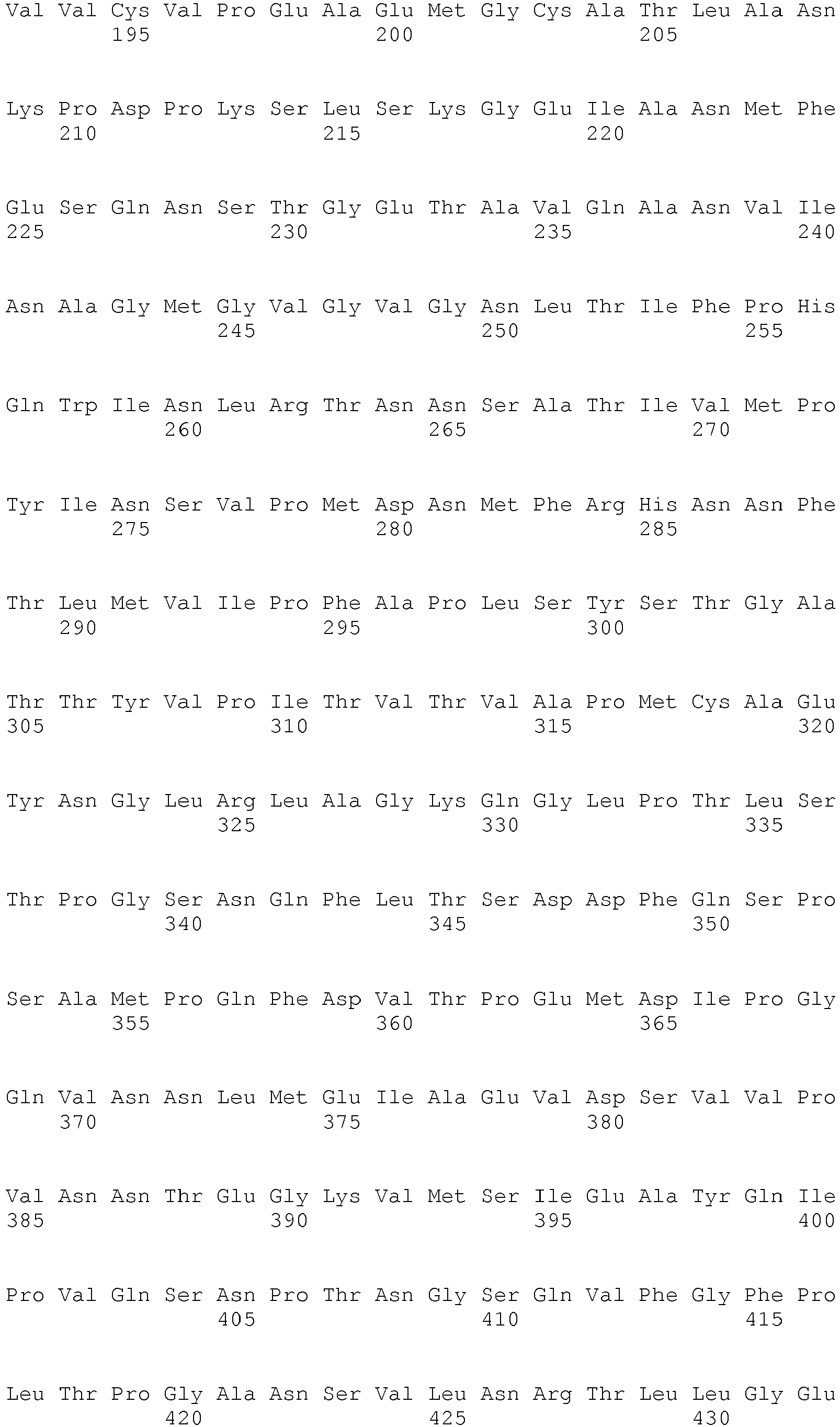


- <210> 3
<211> 1935
<212> DNA
<213> Swine vesicular disease virus - <220>
<221> CDS
<222> (1)..(1935)
<223> - <400> 3


- <210> 4
<211> 645
<212> PRT
<213> Swine vesicular disease virus - <400> 4


- <210> 5
<211> 26
<212> DNA
<213> Artificial - <220>
<223> Primer - <220>
<221> misc_feature
<222> (1)..(26)
<223> Forward primer for P1 gene - <400> 5
ggatccatgg gagctcaagt gtcaac 26 - <210> 6
<211> 27
<212> DNA
<213> Artificial - <220>
<223> Primer - <220>
<221> misc_feature
<222> (1)..(27)
<223> Reverse primer for P1 gene - <400> 6
aagcttaagt ggttttcatg gttgtta 27 - <210> 7
<211> 30
<212> DNA
<213> Artificial - <220>
<223> Primer - <220>
<221> misc_feature
<222> (1)..(30)
<223> Forward primer for 3CD gene - <400> 7
gccctcgaga tgggtccagc gtttgagttc 30 - <210> 8
<211> 30
<212> DNA
<213> Artificial - <220>
<223> Primer - <220>
<221> misc_feature
<222> (1)..(30)
<223> Reverse primer for 3CD gene - <400> 8
gctcggtacc taaaaggagt ccaaccactt 30
Claims (7)
- A baculovirus expression vector containing swine vesicular disease virus genes, wherein said swine vesicular disease virus genes consist of the structural protein precursor P1 gene of SEQ ID NO: 1 and 3CD protease gene of SEQ ID NO: 3.
- A recombinant baculovirus transformed with the baculovirus expression vector of Claim 1.
- Insect cells transfected with the recombinant baculovirus of Claim 2.
- The insect cells of Claim 3, wherein the insect cells are Spodoptera frugiperda.
- A recombinant baculovirus produced by the insect cells of Claim 3 or 4, wherein said recombinant baculovirus contains swine vesicular disease virus genes consisting of the structural protein precursor P1 gene of SEQ ID NO: 1 and 3CD protease gene of SEQ ID NO: 3.
- A swine vesicular disease virus-like particle prepared using the recombinant baculovirus of Claim 5, wherein said virus-like particle is formed from individual proteins obtained from structural protein precursor P1, wherein the structural protein precursor P1 is expressed by the structural protein precursor P1 gene of SEQ ID NO: 1, and then digested by 3CD protease expressed by 3CD protease gene of SEQ ID NO:3.
- A method for preparing a swine vesicular disease virus-like particle, the method comprising the steps of:(1) amplifying the structural protein precursor P1 gene and 3CD protease gene of swine vesicular disease virus by polymerase chain reaction (PCR);(2) inserting the P1 gene into a baculovirus expression vector pFastBacDual so as to prepare pFastBacDual/P1;(3) inserting the 3CD gene into the pFastBacDual/P1 inserted with the P1 gene so as to prepare pFastBacDual/P1/3CD;(4) transforming a cell (DH10Bac) containing a baculovirus shuttle vector Bacmid with the pFastBacDual/P1/3CD containing the P1 gene and the 3CD gene so as to prepare recombinant baculovirus gene Bacmid/P1/3CD;(5) transfecting insect cells with the Bacmid/P1/3CD gene;(6) subjecting the supernatant of the transfected insect cells to a plaque assay so as to isolate a pure recombinant baculovirus; and(7) infecting insect cells with the recombinant baculovirus so as to obtain a swine vesicular disease virus-like particle.
Applications Claiming Priority (2)
| Application Number | Priority Date | Filing Date | Title |
|---|---|---|---|
| KR1020040083994A KR100665054B1 (en) | 2004-10-20 | 2004-10-20 | Swine bullous virus-like particle using genetic recombination and its manufacturing method |
| PCT/KR2005/003274 WO2006080682A1 (en) | 2004-10-20 | 2005-10-05 | Construct of swine vesicular disease virus like particle using genetic engineering and the method for manufacturing thereof |
Publications (3)
| Publication Number | Publication Date |
|---|---|
| EP1802759A1 EP1802759A1 (en) | 2007-07-04 |
| EP1802759A4 EP1802759A4 (en) | 2009-06-17 |
| EP1802759B1 true EP1802759B1 (en) | 2016-05-18 |
Family
ID=36740666
Family Applications (1)
| Application Number | Title | Priority Date | Filing Date |
|---|---|---|---|
| EP05856432.9A Expired - Lifetime EP1802759B1 (en) | 2004-10-20 | 2005-10-05 | Construct of swine vesicular disease virus like particle using genetic engineering and the method for manufacturing thereof |
Country Status (4)
| Country | Link |
|---|---|
| EP (1) | EP1802759B1 (en) |
| KR (1) | KR100665054B1 (en) |
| CN (1) | CN101065489B (en) |
| WO (1) | WO2006080682A1 (en) |
Families Citing this family (6)
| Publication number | Priority date | Publication date | Assignee | Title |
|---|---|---|---|---|
| TW201100545A (en) * | 2009-06-18 | 2011-01-01 | Nat Univ Tsing Hua | Method for preparing enterovirus virus-like particle and use thereof |
| CN102978227A (en) * | 2012-11-16 | 2013-03-20 | 深圳出入境检验检疫局动植物检验检疫技术中心 | Swine vesicular disease virus viral protein 1 (VP1) protein secretion expression recombinant plasmid and application thereof |
| CN103710384A (en) * | 2013-12-18 | 2014-04-09 | 广东华南联合疫苗开发院有限公司 | Picornaviridae recombinant vector, virus like particle, and preparation method and use of virus like particle |
| CN112048004B (en) * | 2020-07-14 | 2021-10-22 | 桂林医学院 | A kind of coxsackie virus B5 virus-like particle, its preparation method and application |
| CN114317571B (en) * | 2021-12-29 | 2025-04-18 | 中国农业科学院兰州兽医研究所 | Virus-like particle and preparation method and application thereof |
| CN116121303B (en) * | 2022-10-12 | 2026-04-03 | 中国农业科学院兰州兽医研究所 | Porcine encephalitis myocarditis virus-like particles, their preparation methods and applications |
Family Cites Families (2)
| Publication number | Priority date | Publication date | Assignee | Title |
|---|---|---|---|---|
| AUPN720195A0 (en) * | 1995-12-18 | 1996-01-18 | University Of Melbourne, The | Equine rhinovirus 1 proteins |
| US6200576B1 (en) * | 1998-07-15 | 2001-03-13 | Development Center For Biotechnology | Swine vesicular disease virus and mutant strains and preparation process and use thereof |
-
2004
- 2004-10-20 KR KR1020040083994A patent/KR100665054B1/en not_active Ceased
-
2005
- 2005-10-05 EP EP05856432.9A patent/EP1802759B1/en not_active Expired - Lifetime
- 2005-10-05 CN CN2005800354452A patent/CN101065489B/en not_active Expired - Fee Related
- 2005-10-05 WO PCT/KR2005/003274 patent/WO2006080682A1/en not_active Ceased
Also Published As
| Publication number | Publication date |
|---|---|
| KR100665054B1 (en) | 2007-01-04 |
| KR20060034945A (en) | 2006-04-26 |
| WO2006080682A1 (en) | 2006-08-03 |
| CN101065489B (en) | 2010-12-01 |
| CN101065489A (en) | 2007-10-31 |
| EP1802759A1 (en) | 2007-07-04 |
| EP1802759A4 (en) | 2009-06-17 |
Similar Documents
| Publication | Publication Date | Title |
|---|---|---|
| Tam et al. | In vitroinfection and replication of hepatitis E virus in primary cynomolgus macaque hepatocytes | |
| CA2690668C (en) | Vaccines containing canine parvovirus genetic variants | |
| Tam et al. | In VitroPropagation and Production of Hepatitis E Virus fromin Vivo-Infected Primary Macaque Hepatocytes | |
| ES2398413T3 (en) | SRSV detection kit | |
| AU2009256397A1 (en) | Expression and assembly of human group C rotavirus-like particles and uses thereof | |
| CN113817753B (en) | Expression of SARS-CoV-2 fiber protein or its variant S Δ21 Construction and use of pseudotyped VSV viruses | |
| Skidmore | Overview of hepatitis E virus | |
| CN101928728B (en) | Preparation method and application of enterovirus virus-like particles | |
| KR100665054B1 (en) | Swine bullous virus-like particle using genetic recombination and its manufacturing method | |
| Upadhyaya et al. | Molecular analysis of rice ragged stunt oryzavirus segment 9 and sequence conservation among isolates from Thailand and India | |
| KR100267745B1 (en) | Detection method of neutralizing antibody against porcine epidermic diarrhea virus | |
| CN116286679B (en) | An isolated porcine epidemic diarrhea virus variant and its application | |
| Kobayashi et al. | Serotype‐specific antigen ELISA for detection of Chiba virus in stools | |
| KR101316102B1 (en) | Method for Preparing Recombinant Antigen Complex Using Rotavirus Nanoparticle | |
| EP1780216B1 (en) | Pantropic canine coronavirus | |
| McClenahan et al. | Expression and self-assembly of virus-like particles from two genotypes of marine vesiviruses and development of an ELISA for the detection of antibodies | |
| RU2850653C1 (en) | Strain "o n 2633/mongolia/2022" of foot-and-mouth disease virus aphtae epizooticae serotype o for manufacture of biopreparations for diagnosis and specific prevention of foot-and-mouth disease | |
| RU2804634C1 (en) | Sat-1/kenya/2017 strain of aphtae epizooticae foot and mouth disease virus for the manufacture of biological products for the diagnostics and specific prevention of foot and mouth disease virus of sat-1/nwz genotype | |
| Xu et al. | Generation, characterization, and application in serodiagnosis of recombinant swine vesicular disease virus-like particles | |
| EP2331117B1 (en) | Vaccines containing canine parvovirus genetic variants | |
| US11191824B1 (en) | Method of purifying virus-like-particles | |
| KR20070025122A (en) | Diagnosis of Swine Bullous Disease Virus Antibody Using Monoclonal Antibodies Secreted by Non-infectious Swine Bacterial Virus Similar Particles and Hybridoma Cell Lines | |
| Purcell et al. | ORF3 Protein of Hepatitis E Virus Is Not | |
| Huang | Molecular characterization of animal strains of hepatitis E virus (HEV): Avian HEV and swine HEV | |
| Xu WanHong et al. | Generation, characterization, and application in serodiagnosis of recombinant swine vesicular disease virus-like particles. |
Legal Events
| Date | Code | Title | Description |
|---|---|---|---|
| PUAI | Public reference made under article 153(3) epc to a published international application that has entered the european phase |
Free format text: ORIGINAL CODE: 0009012 |
|
| 17P | Request for examination filed |
Effective date: 20070416 |
|
| AK | Designated contracting states |
Kind code of ref document: A1 Designated state(s): AT BE BG CH CY CZ DE DK EE ES FI FR GB GR HU IE IS IT LI LT LU LV MC NL PL PT RO SE SI SK TR |
|
| AX | Request for extension of the european patent |
Extension state: AL BA HR MK YU |
|
| A4 | Supplementary search report drawn up and despatched |
Effective date: 20090518 |
|
| 17Q | First examination report despatched |
Effective date: 20090826 |
|
| GRAP | Despatch of communication of intention to grant a patent |
Free format text: ORIGINAL CODE: EPIDOSNIGR1 |
|
| INTG | Intention to grant announced |
Effective date: 20151211 |
|
| GRAS | Grant fee paid |
Free format text: ORIGINAL CODE: EPIDOSNIGR3 |
|
| GRAA | (expected) grant |
Free format text: ORIGINAL CODE: 0009210 |
|
| AK | Designated contracting states |
Kind code of ref document: B1 Designated state(s): AT BE BG CH CY CZ DE DK EE ES FI FR GB GR HU IE IS IT LI LT LU LV MC NL PL PT RO SE SI SK TR |
|
| AX | Request for extension of the european patent |
Extension state: AL BA HR MK YU |
|
| REG | Reference to a national code |
Ref country code: GB Ref legal event code: FG4D |
|
| REG | Reference to a national code |
Ref country code: CH Ref legal event code: EP |
|
| REG | Reference to a national code |
Ref country code: IE Ref legal event code: FG4D Ref country code: AT Ref legal event code: REF Ref document number: 800497 Country of ref document: AT Kind code of ref document: T Effective date: 20160615 |
|
| REG | Reference to a national code |
Ref country code: DE Ref legal event code: R096 Ref document number: 602005049395 Country of ref document: DE |
|
| REG | Reference to a national code |
Ref country code: NL Ref legal event code: MP Effective date: 20160518 |
|
| REG | Reference to a national code |
Ref country code: LT Ref legal event code: MG4D |
|
| PG25 | Lapsed in a contracting state [announced via postgrant information from national office to epo] |
Ref country code: FI Free format text: LAPSE BECAUSE OF FAILURE TO SUBMIT A TRANSLATION OF THE DESCRIPTION OR TO PAY THE FEE WITHIN THE PRESCRIBED TIME-LIMIT Effective date: 20160518 Ref country code: LT Free format text: LAPSE BECAUSE OF FAILURE TO SUBMIT A TRANSLATION OF THE DESCRIPTION OR TO PAY THE FEE WITHIN THE PRESCRIBED TIME-LIMIT Effective date: 20160518 Ref country code: NL Free format text: LAPSE BECAUSE OF FAILURE TO SUBMIT A TRANSLATION OF THE DESCRIPTION OR TO PAY THE FEE WITHIN THE PRESCRIBED TIME-LIMIT Effective date: 20160518 |
|
| REG | Reference to a national code |
Ref country code: AT Ref legal event code: MK05 Ref document number: 800497 Country of ref document: AT Kind code of ref document: T Effective date: 20160518 |
|
| PG25 | Lapsed in a contracting state [announced via postgrant information from national office to epo] |
Ref country code: LV Free format text: LAPSE BECAUSE OF FAILURE TO SUBMIT A TRANSLATION OF THE DESCRIPTION OR TO PAY THE FEE WITHIN THE PRESCRIBED TIME-LIMIT Effective date: 20160518 Ref country code: ES Free format text: LAPSE BECAUSE OF FAILURE TO SUBMIT A TRANSLATION OF THE DESCRIPTION OR TO PAY THE FEE WITHIN THE PRESCRIBED TIME-LIMIT Effective date: 20160518 Ref country code: SE Free format text: LAPSE BECAUSE OF FAILURE TO SUBMIT A TRANSLATION OF THE DESCRIPTION OR TO PAY THE FEE WITHIN THE PRESCRIBED TIME-LIMIT Effective date: 20160518 Ref country code: GR Free format text: LAPSE BECAUSE OF FAILURE TO SUBMIT A TRANSLATION OF THE DESCRIPTION OR TO PAY THE FEE WITHIN THE PRESCRIBED TIME-LIMIT Effective date: 20160819 Ref country code: PT Free format text: LAPSE BECAUSE OF FAILURE TO SUBMIT A TRANSLATION OF THE DESCRIPTION OR TO PAY THE FEE WITHIN THE PRESCRIBED TIME-LIMIT Effective date: 20160919 |
|
| PG25 | Lapsed in a contracting state [announced via postgrant information from national office to epo] |
Ref country code: EE Free format text: LAPSE BECAUSE OF FAILURE TO SUBMIT A TRANSLATION OF THE DESCRIPTION OR TO PAY THE FEE WITHIN THE PRESCRIBED TIME-LIMIT Effective date: 20160518 Ref country code: RO Free format text: LAPSE BECAUSE OF FAILURE TO SUBMIT A TRANSLATION OF THE DESCRIPTION OR TO PAY THE FEE WITHIN THE PRESCRIBED TIME-LIMIT Effective date: 20160518 Ref country code: DK Free format text: LAPSE BECAUSE OF FAILURE TO SUBMIT A TRANSLATION OF THE DESCRIPTION OR TO PAY THE FEE WITHIN THE PRESCRIBED TIME-LIMIT Effective date: 20160518 Ref country code: CZ Free format text: LAPSE BECAUSE OF FAILURE TO SUBMIT A TRANSLATION OF THE DESCRIPTION OR TO PAY THE FEE WITHIN THE PRESCRIBED TIME-LIMIT Effective date: 20160518 Ref country code: SK Free format text: LAPSE BECAUSE OF FAILURE TO SUBMIT A TRANSLATION OF THE DESCRIPTION OR TO PAY THE FEE WITHIN THE PRESCRIBED TIME-LIMIT Effective date: 20160518 |
|
| REG | Reference to a national code |
Ref country code: DE Ref legal event code: R097 Ref document number: 602005049395 Country of ref document: DE |
|
| PG25 | Lapsed in a contracting state [announced via postgrant information from national office to epo] |
Ref country code: BE Free format text: LAPSE BECAUSE OF FAILURE TO SUBMIT A TRANSLATION OF THE DESCRIPTION OR TO PAY THE FEE WITHIN THE PRESCRIBED TIME-LIMIT Effective date: 20160518 Ref country code: AT Free format text: LAPSE BECAUSE OF FAILURE TO SUBMIT A TRANSLATION OF THE DESCRIPTION OR TO PAY THE FEE WITHIN THE PRESCRIBED TIME-LIMIT Effective date: 20160518 Ref country code: PL Free format text: LAPSE BECAUSE OF FAILURE TO SUBMIT A TRANSLATION OF THE DESCRIPTION OR TO PAY THE FEE WITHIN THE PRESCRIBED TIME-LIMIT Effective date: 20160518 |
|
| PLBE | No opposition filed within time limit |
Free format text: ORIGINAL CODE: 0009261 |
|
| STAA | Information on the status of an ep patent application or granted ep patent |
Free format text: STATUS: NO OPPOSITION FILED WITHIN TIME LIMIT |
|
| 26N | No opposition filed |
Effective date: 20170221 |
|
| REG | Reference to a national code |
Ref country code: DE Ref legal event code: R119 Ref document number: 602005049395 Country of ref document: DE |
|
| PG25 | Lapsed in a contracting state [announced via postgrant information from national office to epo] |
Ref country code: SI Free format text: LAPSE BECAUSE OF FAILURE TO SUBMIT A TRANSLATION OF THE DESCRIPTION OR TO PAY THE FEE WITHIN THE PRESCRIBED TIME-LIMIT Effective date: 20160518 |
|
| REG | Reference to a national code |
Ref country code: CH Ref legal event code: PL |
|
| PG25 | Lapsed in a contracting state [announced via postgrant information from national office to epo] |
Ref country code: MC Free format text: LAPSE BECAUSE OF FAILURE TO SUBMIT A TRANSLATION OF THE DESCRIPTION OR TO PAY THE FEE WITHIN THE PRESCRIBED TIME-LIMIT Effective date: 20160518 |
|
| REG | Reference to a national code |
Ref country code: IE Ref legal event code: MM4A |
|
| REG | Reference to a national code |
Ref country code: FR Ref legal event code: ST Effective date: 20170630 |
|
| PG25 | Lapsed in a contracting state [announced via postgrant information from national office to epo] |
Ref country code: CH Free format text: LAPSE BECAUSE OF NON-PAYMENT OF DUE FEES Effective date: 20161031 Ref country code: LI Free format text: LAPSE BECAUSE OF NON-PAYMENT OF DUE FEES Effective date: 20161031 Ref country code: FR Free format text: LAPSE BECAUSE OF NON-PAYMENT OF DUE FEES Effective date: 20161102 Ref country code: DE Free format text: LAPSE BECAUSE OF NON-PAYMENT OF DUE FEES Effective date: 20170503 |
|
| PG25 | Lapsed in a contracting state [announced via postgrant information from national office to epo] |
Ref country code: LU Free format text: LAPSE BECAUSE OF NON-PAYMENT OF DUE FEES Effective date: 20161005 |
|
| PGFP | Annual fee paid to national office [announced via postgrant information from national office to epo] |
Ref country code: GB Payment date: 20170921 Year of fee payment: 13 |
|
| PG25 | Lapsed in a contracting state [announced via postgrant information from national office to epo] |
Ref country code: IE Free format text: LAPSE BECAUSE OF NON-PAYMENT OF DUE FEES Effective date: 20161005 |
|
| PGFP | Annual fee paid to national office [announced via postgrant information from national office to epo] |
Ref country code: IT Payment date: 20171013 Year of fee payment: 13 |
|
| PG25 | Lapsed in a contracting state [announced via postgrant information from national office to epo] |
Ref country code: CY Free format text: LAPSE BECAUSE OF FAILURE TO SUBMIT A TRANSLATION OF THE DESCRIPTION OR TO PAY THE FEE WITHIN THE PRESCRIBED TIME-LIMIT Effective date: 20160518 Ref country code: HU Free format text: LAPSE BECAUSE OF FAILURE TO SUBMIT A TRANSLATION OF THE DESCRIPTION OR TO PAY THE FEE WITHIN THE PRESCRIBED TIME-LIMIT; INVALID AB INITIO Effective date: 20051005 |
|
| PG25 | Lapsed in a contracting state [announced via postgrant information from national office to epo] |
Ref country code: IS Free format text: LAPSE BECAUSE OF FAILURE TO SUBMIT A TRANSLATION OF THE DESCRIPTION OR TO PAY THE FEE WITHIN THE PRESCRIBED TIME-LIMIT Effective date: 20160518 Ref country code: TR Free format text: LAPSE BECAUSE OF FAILURE TO SUBMIT A TRANSLATION OF THE DESCRIPTION OR TO PAY THE FEE WITHIN THE PRESCRIBED TIME-LIMIT Effective date: 20160518 |
|
| PG25 | Lapsed in a contracting state [announced via postgrant information from national office to epo] |
Ref country code: BG Free format text: LAPSE BECAUSE OF FAILURE TO SUBMIT A TRANSLATION OF THE DESCRIPTION OR TO PAY THE FEE WITHIN THE PRESCRIBED TIME-LIMIT Effective date: 20160518 |
|
| GBPC | Gb: european patent ceased through non-payment of renewal fee |
Effective date: 20181005 |
|
| PG25 | Lapsed in a contracting state [announced via postgrant information from national office to epo] |
Ref country code: GB Free format text: LAPSE BECAUSE OF NON-PAYMENT OF DUE FEES Effective date: 20181005 Ref country code: IT Free format text: LAPSE BECAUSE OF NON-PAYMENT OF DUE FEES Effective date: 20181005 |