EP1801582A1 - Method of presuming the metabolism of drug in human liver and liver function - Google Patents
Method of presuming the metabolism of drug in human liver and liver function Download PDFInfo
- Publication number
- EP1801582A1 EP1801582A1 EP05780351A EP05780351A EP1801582A1 EP 1801582 A1 EP1801582 A1 EP 1801582A1 EP 05780351 A EP05780351 A EP 05780351A EP 05780351 A EP05780351 A EP 05780351A EP 1801582 A1 EP1801582 A1 EP 1801582A1
- Authority
- EP
- European Patent Office
- Prior art keywords
- chimeric animal
- animal
- human
- chimeric
- liver
- Prior art date
- Legal status (The legal status is an assumption and is not a legal conclusion. Google has not performed a legal analysis and makes no representation as to the accuracy of the status listed.)
- Granted
Links
Images



Classifications
-
- G—PHYSICS
- G01—MEASURING; TESTING
- G01N—INVESTIGATING OR ANALYSING MATERIALS BY DETERMINING THEIR CHEMICAL OR PHYSICAL PROPERTIES
- G01N33/00—Investigating or analysing materials by specific methods not covered by groups G01N1/00 - G01N31/00
- G01N33/48—Biological material, e.g. blood, urine; Haemocytometers
-
- G—PHYSICS
- G01—MEASURING; TESTING
- G01N—INVESTIGATING OR ANALYSING MATERIALS BY DETERMINING THEIR CHEMICAL OR PHYSICAL PROPERTIES
- G01N33/00—Investigating or analysing materials by specific methods not covered by groups G01N1/00 - G01N31/00
- G01N33/48—Biological material, e.g. blood, urine; Haemocytometers
- G01N33/50—Chemical analysis of biological material, e.g. blood, urine; Testing involving biospecific ligand binding methods; Immunological testing
- G01N33/5005—Chemical analysis of biological material, e.g. blood, urine; Testing involving biospecific ligand binding methods; Immunological testing involving human or animal cells
- G01N33/5008—Chemical analysis of biological material, e.g. blood, urine; Testing involving biospecific ligand binding methods; Immunological testing involving human or animal cells for testing or evaluating the effect of chemical or biological compounds, e.g. drugs, cosmetics
- G01N33/502—Chemical analysis of biological material, e.g. blood, urine; Testing involving biospecific ligand binding methods; Immunological testing involving human or animal cells for testing or evaluating the effect of chemical or biological compounds, e.g. drugs, cosmetics for testing non-proliferative effects
- G01N33/5038—Chemical analysis of biological material, e.g. blood, urine; Testing involving biospecific ligand binding methods; Immunological testing involving human or animal cells for testing or evaluating the effect of chemical or biological compounds, e.g. drugs, cosmetics for testing non-proliferative effects involving detection of metabolites per se
-
- G—PHYSICS
- G01—MEASURING; TESTING
- G01N—INVESTIGATING OR ANALYSING MATERIALS BY DETERMINING THEIR CHEMICAL OR PHYSICAL PROPERTIES
- G01N33/00—Investigating or analysing materials by specific methods not covered by groups G01N1/00 - G01N31/00
- G01N33/15—Medicinal preparations ; Physical properties thereof, e.g. dissolubility
-
- G—PHYSICS
- G01—MEASURING; TESTING
- G01N—INVESTIGATING OR ANALYSING MATERIALS BY DETERMINING THEIR CHEMICAL OR PHYSICAL PROPERTIES
- G01N33/00—Investigating or analysing materials by specific methods not covered by groups G01N1/00 - G01N31/00
- G01N33/48—Biological material, e.g. blood, urine; Haemocytometers
- G01N33/50—Chemical analysis of biological material, e.g. blood, urine; Testing involving biospecific ligand binding methods; Immunological testing
- G01N33/5005—Chemical analysis of biological material, e.g. blood, urine; Testing involving biospecific ligand binding methods; Immunological testing involving human or animal cells
- G01N33/5008—Chemical analysis of biological material, e.g. blood, urine; Testing involving biospecific ligand binding methods; Immunological testing involving human or animal cells for testing or evaluating the effect of chemical or biological compounds, e.g. drugs, cosmetics
- G01N33/5082—Supracellular entities, e.g. tissue, organisms
- G01N33/5088—Supracellular entities, e.g. tissue, organisms of vertebrates
Definitions
- the present invention relates to a method for precisely predicting the metabolism of a drug in human liver and the effect of the drug on liver function.
- liver function Many substances which have been ingested by the human body are initially metabolized in the liver. Particularly in the development of pharmaceuticals, the metabolic mechanism of a drug in the liver and the effect of the drug on liver function are essential data from the aspect of safety. In addition to the case of pharmaceuticals, in order to evaluate the effects on the human body of a variety of chemical substances present in the environment, the effects of the chemical substances on liver function must be evaluated.
- in vitro assay systems such as a system employing microsomes which express human drug metabolizing enzymes and a system employing human hepatocytes.
- these in vitro assay systems have drawbacks, for example, in that the results vary depending on addition of a co-enzyme, the type of the assay system, etc. Therefore, at present, metabolism through the human liver cannot be precisely predicted.
- Patent Documents 1 and 2 Japanese Laid-Open Patent Publication No. 2002-45087
- Patent Document 2 WO 03/080821 A1
- Patent Document 1 fails to provide examples in which a test substance is administered to chimeric animals. Therefore, drug metabolism of the chimeric animals after a test substance is actually administered to them remains unclear. In addition, possible adverse effects of the test substance cannot be predicted. Actual trials have revealed that administration of a test substance to the chimeric animals causes blood coagulation and the like anomalously. Since these chimeric animals have impaired their liver functions, the value of their liver-function markers are abnormal. Thus, it has never been determined whether or not the variation in liver-function markers after a test drug has been administered is attributed to the effect of the test drug on the transplanted human hepatocytes. In addition, it has not been able to determine whether the measured values after administration of a test substance are completely attributable to transplanted human hepatocytes or it also attribute to inherent hepatocytes of the animals.
- An object of the present invention is to provide an in vivo method for precisely predicting the metabolism of a drug in human liver, the effect of the drug on liver function, and so on.
- the present inventors have conducted extensive studies in order to attain the object, and we found that when a test substance is administered to a chimeric animal transplanted with human hepatocytes, if human complement reaction can be suppressed in advance, the chimeric animal can be maintained in a favorable state.
- metabolites of a test substance produced in human liver and their effect on the liver functions can be precisely predicted by administrating the test substance to both the chimeric animal transplanted with the human hepatocytes and a non-chimeric animal of the same species, and by analyzing and comparing metabolites and function of the liver in these animals.
- the present invention has been established on the basis of these findings.
- the present invention provides a method for predicting a metabolite of a test substance produced in human liver, wherein the method comprises:
- the present invention also provides a method for determining the type of excretion for a test substance in human, wherein the method comprises:
- the present invention also provides a method for predicting an effect of a test substance on liver functions in human, wherein the method comprises:
- the present invention also provides a method for analyzing a human-specific metabolite of a test substance, wherein the method comprises:
- the present invention also provides a method for predicting an animal exhibiting a human-type metabolic profile for a test substance, wherein the method comprises:
- metabolism in the liver, the type of excretion, an effect on liver functions and the like of a drug in human can be precisely predicted in vivo analysis.
- the effect of a drug on the human body can be predicted before their administration to human, and screening of candidates and prediction of side effects or potency of the drug in human can be facilitated during the course of the development of pharmaceuticals. Therefore, the invention allows reducing development cost by narrowing down the candidates in an early stage of drug development, and preventing side effects in human.
- the present invention is remarkably useful in the development of pharmaceuticals.
- the nonhuman chimeric animal employed in the present invention is a chimeric animal which intracorporeally carries a population of human-origin hepatocytes having proliferation potential and in which the hepatocyte population substantially functions as the liver of the chimeric animal.
- the chimeric animal is preferably an immunodeficient animal whose original hepatocytes have been impaired and which exhibits no rejection against human hepatocytes (i.e., an immunodeficient animal with hepatic disorder), wherein the immunodeficient animal has been transplanted with human hepatocytes, and engraftment of the human hepatocytes has been confirmed.
- the animal with hepatic disorder employed in the present invention may be a transgenic mouse disclosed in Science, 263, 1149(1994 ), to which an albumin promoter and a fusion gene of an enhancer region and u-PA (urokinase-type plasminogen activator) have been introduced.
- the immunodeficient animal may be an SCID mouse (severe combined immunodeficiency disease mouse), which is genetically immunodeficient.
- SCID mouse severe combined immunodeficiency disease mouse
- progeny mice obtained through mating the above hepatic impaired mouse and the immunodeficient mouse may be employed as an immunodeficient animal with hepatic disorder.
- the species of these animals is preferably mouse.
- Examples of preferred immunodeficient mice with hepatic disorder include uPA-Tg (+/+)/SCID mice.
- the human hepatocytes for transplanting into the immunodeficient animal with hepatic disorder are preferably frozen hepatocytes, fresh hepatocytes, and small liver parenchymal cells with high proliferation potential. Interracial difference in the metabolism can be evaluated by transplanting the hepatocytes from Japanese, Korean, Chinese, Caucasians, Negroids, or people of other races.
- human hepatocytes are transplanted to the spleen of the immunodeficient animal with hepatic disorder. Once human hepatocytes have been transplanted into the spleen, the cells are readily engrafted successfully into the liver.
- the chimeric animal employed in the present invention is preferably a mouse derived from an uPA-Tg(+/+)/SCID(+/+) mice disclosed in Japanese Laid-Open Patent Publication No. 2002-45087 , wherein this mouse have been transplanted with human hepatocytes to the spleen and engraftment of the human hepatocytes has been confirmed.
- the non-chimeric animal of the same species as the chimeric animal employed in the invention is preferably an immunodeficient animal with hepatic disorder, more preferably an immunodeficient mouse with hepatic disorder, particularly preferably an uPA-Tg(-/-)/SCID(+/+) mouse, an uPA-Tg(+/)/SCID(+/+) mouse, or an uPA-Tg(+/+)/SCID(+/+) mouse.
- a test substance is administered to a chimeric animal and to a non-chimeric animal of the same species, while these animals are protected from the attack by a human complement produced by the hepatocytes. This is because when the animals are not protected from the attack by a human complement, administration of the test substance leads to onset of disseminated coagulation syndrome or the like.
- a complement suppressor e.g., nafamostat mesylate
- an anticoagulant for preventing blood coagulation e.g., heparin
- a test substance is administered to both a chimeric animal and a non-chimeric animal of the same species.
- the test substance may be administered through the means most common to the substance.
- the test substance is preferably administered intravenously, orally, or the like.
- metabolites of the chimeric animal and the non-chimeric animal are analyzed and the analytical results are compared, whereby the metabolites of the test substance produced in human liver can be precisely predicted.
- the term "analyzing metabolite(s)" refers to obtaining information including the type, amount and composition of the metabolites, and time-elapse or quantitative change thereof. Analysis of metabolites from plasma samples, tissue samples, bile samples, urine samples, and feces samples are preferred.
- a convenient means for analyzing the metabolites is using a substance containing a radioisotope as a test substance, to measure radioactivity of the test substance.
- the metabolites include, for example a variety of degraded products and conjugates.
- hepatic disorder of the chimeric animal per se can be distinguished from hepatic disorder caused by the test substance. Furthermore, through determination of changes in mRNA, proteins, and other substance in the liver by various methods, the mechanism of disorder in liver function or that of hepatotoxicity can be elucidated.
- metabolites of the chimeric animal and the non-chimeric animal are analyzed, and the analytical results are compared, whereby the type of excretion of the test substance in human can be determined.
- metabolites in urine samples, feces samples, and bile samples of both animals can be measured and compared to determine the type of excretion of a test substance in human.
- the type of excretion greatly varies among animal species. Therefore, in development of pharmaceuticals, determination of the type of excretion of the test substance in human is extremely important.
- the present invention which enables to evaluate the type of excretion or metabolites of a test substance in human without administering the substance to human subjects, is remarkably useful.
- liver-function markers e.g., GOT and GPT
- the control animal is preferably a non-chimeric animal of the same species; e.g., an immunodeficient animal with hepatic disorder, an immunodeficient animal, or an animal having no disorder.
- test items for liver function include biochemical scores such as GOT and GPT.
- changes in mRNA, expressed proteins, and other substance in the liver may be determined through a known method.
- metabolites of a chimeric animal and those of a non-chimeric animal are analyzed and compared, whereby the excretion type or metabolites of the test substance in human are evaluated.
- metabolites of the test substance of an animal species other than the chimeric animal are analyzed, and then these analytical results are compared, whereby an animal exhibiting a human-type metabolic profile for the test substance can be predicted.
- it can be readily determined in a variety of animals whether or not the metabolic profile for the test substance is a human-type.
- Ketoprofen was employed as a test substance, which is known to be mainly excreted in urine as a glucuronate conjugate in human.
- 3 H-ketoprofen (ART391, Lot 040219) was purchased from ARC. The compound was generally labeled and had a specific radioactivity of 1,110 GBq/mmol.
- mice from PhoenixBio Co., Ltd. were employed. Specifically, human hepatocytes were transplanted to the spleen of the transgenic mice with liver failure (uPA-Tg (+/+)/SCID mice (female)), and the mice in which engraftment of human hepatocytes was recognized were employed (body weight: 10.2 to 18.0 g). Mice employed had percent replacement of the human hepatocytes therein of 60% to 80%, and caged for 45 days to 87 days after transplantation. uPA (-/-)/SCID mice of 6 to 10 weeks aged (body weight: 15.1 to 22.2 g) were employed as control mice.
- mice were freely fed water (containing 0.012% hypochlorous acid) and a sterilized solid diet containing vitamin C (CRF-1, product of Oriental Yeast Co., Ltd.) and housed at room temperature (23 ⁇ 3°C) and a humidity of 55 ⁇ 20%.
- the chimeric mice were injected intraperitoneally with nafamostat mesylate twice per day (0.3 mg/0.2 mL/body).
- a predetermined amount of a 3 H-ketoprofen ethanol solution was dried to solid under nitrogen flow. Subsequently, a 0.125-mol/L solution of ketoprofen in sodium hydroxide was added to dissolve the solid. The pH of the solution was adjusted to 7.0 with a 0.1% aqueous citric acid solution, and the concentration was adjusted to 1 mg/mL with distilled water for injection. The liquid was bolus-administered at 5 mL/kg to the caudal vein of the mice (5 mg/5 mL/kg).
- Urine and feces were collected from chimeric mice and control mice which had been housed in metabolism cages, over 5 days after administration. Livers and kidneys were taken from other mice two hours after administration, following sacrificing through exsanguination. Blood plasma was collected by sampling blood through the orbital venousplexus and centrifuging it. Bile was collected from the chimeric mice and the control mice through bile duct cannulation until 72 hours after administration.
- Radioactivity of each sample was determined by use of a liquid scintillator (Hionic-flow, product of Perkin-Elmer) with Tri-Carb 2500 (Perkin-Elmer) as a liquid scintillation counter. The measurement was performed for two minutes. Counting efficiency was calibrated through the external standard radiation source method.
- the sample (10 ⁇ L) was collected in a 20-mL glass vial, added Hionic-flow (10 mL), and the mixture was analyzed.
- tissue samples liver and kidney
- an amount 3-fold (by weight) of acetate buffer (0.1M, pH3.0) was added to each sample, and the mixture was homogenized with a polytron-homogenizer.
- An aliquot (50 ⁇ L) of the homogenate was solubilized with 2 mL of Soluene 350 (Perkin-Elmer), added Hionic-flow (10 mL), and the mixture was analyzed.
- the total volume of each sample was adjusted to 15 or 30 mL.
- the mixture was homogenized with a polytron-homogenizer. An aliquot (0.5 mL) of the homogenate was solubilized with 2 mL of Soluene 350 (Perkin-Elmer), added Hionic-flow (10 mL), and the mixture was analyzed.
- a plasma sample (50 ⁇ L) or a tissue homogenate (100 ⁇ L) was added to an Eppendorf tube, and acetonitrile was added in an amount 4-fold of the sample, followed by vigorously stirring.
- the mixture was centrifuged (22,000 ⁇ g, 4°C, 10 min), and the supernatant was collected and dried to solid under nitrogen flow.
- the solid was re-dissolved with a 200 ⁇ L of acetate buffer (0.1M, pH3.0), and the solution was centrifuged again (22,000xg, 4°C, 10 min). The collected supernatant was applied to an HPLC column.
- Bile samples and urine samples were treated with ⁇ -glucuronidase-arylsulfatase (Helix pomatia, product of Roche Diagnoistics K.K.). Specifically, ⁇ -glucuronidase-arylsulfatase (3%) was added to each of the bile and urine samples, and the sample was incubated at 37°C for 16 hours. The incubated sample was centrifuged (22,000 ⁇ g, 4°C, 10 min), and the supernatant was applied to an HPLC column.
- ⁇ -glucuronidase-arylsulfatase Helix pomatia, product of Roche Diagnoistics K.K.
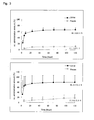
- Fig. 1 shows time-elapse change in radioactivity levels of blood plasma
- Table 1 shows kinetic parameters obtained through non-compartment analysis.
- AUCs on the basis of total radioactivity
- t1/2 (0-4 h) were 0.92 h and 0.78 h, respectively.
- AUC level were almost equivalent between the two mice groups.
- Radioactivity levels of blood plasma samples in the control mice and the chimeric mice determined at the same time were 6.82 ⁇ 1.10 ⁇ g eq./mL and 6.96 ⁇ 2.65 ⁇ g eq./mL, respectively (Table 2).
- Fig. 3 shows percent excretions of radioactivity to urine and feces.
- radioactivity was more preferentially excreted in urine.
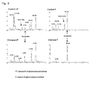
- both of the control mice and the chimeric mice predominantly exhibited a peak of an unchanged form and a hydroxyl metabolite (speculated, hereinafter referred to as M1) in their blood plasma samples collected two hours after the administration. Formation of M1 was more predominant in the control mice than in the chimeric mice.
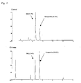
- Fig. 5 shows chromatograms of radioactivity in bile samples.
- the unchanged form was not virtually observed, and peaks attributable to glucuronide conjugates were observed.
- the control mice exhibited some peaks attributable to a hydroxyl metabolite, whereas the chimeric mice exhibited a predominant peak attributed to the unchanged form.
- liver samples collected two hours after the administration a predominant peak attributed to M1 was observed and no peak attributable to the unchanged form was observed in liver samples of the control mice (see Fig. 6).
- liver samples of the chimeric mice exhibited peaks attributed to the unchanged form and M1, respectively.
- peaks attributed to the unchanged form and M1 were observed in kidney from both of the control mice and the chimeric mice. In both of the liver and the kidney, formation of M1 was more predominant in the control mice as compared with the chimeric mice.
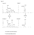
- Fig. 8 shows typical chromatograms of the urine samples.
- a number of peaks attributable to the unchanged form, a hydroxyl metabolite, and conjugate thereof were observed.
- the peak eluted at 17.8 min was considered to be a peak intrinsic to mice.
- Peaks in the chromatogram of radioactivity were found to be changed to those attributed to the unchanged form and M1, when the urine samples were treated with ⁇ -glucuronidase-arylsulphatase. Formation of a hydroxyl metabolite was more predominant in the control mice as compared with the chimeric mice.
Landscapes
- Health & Medical Sciences (AREA)
- Life Sciences & Earth Sciences (AREA)
- Engineering & Computer Science (AREA)
- Biomedical Technology (AREA)
- Immunology (AREA)
- Chemical & Material Sciences (AREA)
- Molecular Biology (AREA)
- Hematology (AREA)
- Urology & Nephrology (AREA)
- General Physics & Mathematics (AREA)
- Analytical Chemistry (AREA)
- Pathology (AREA)
- General Health & Medical Sciences (AREA)
- Biochemistry (AREA)
- Cell Biology (AREA)
- Food Science & Technology (AREA)
- Medicinal Chemistry (AREA)
- Physics & Mathematics (AREA)
- Bioinformatics & Cheminformatics (AREA)
- Biotechnology (AREA)
- Tropical Medicine & Parasitology (AREA)
- Toxicology (AREA)
- Microbiology (AREA)
- Biophysics (AREA)
- Pharmacology & Pharmacy (AREA)
- Investigating Or Analysing Biological Materials (AREA)
- Measurement Of The Respiration, Hearing Ability, Form, And Blood Characteristics Of Living Organisms (AREA)
- Medicines Containing Material From Animals Or Micro-Organisms (AREA)
- Medicines Containing Plant Substances (AREA)
Abstract
Description
- The present invention relates to a method for precisely predicting the metabolism of a drug in human liver and the effect of the drug on liver function.
- Many substances which have been ingested by the human body are initially metabolized in the liver. Particularly in the development of pharmaceuticals, the metabolic mechanism of a drug in the liver and the effect of the drug on liver function are essential data from the aspect of safety. In addition to the case of pharmaceuticals, in order to evaluate the effects on the human body of a variety of chemical substances present in the environment, the effects of the chemical substances on liver function must be evaluated.
- In the development of pharmaceuticals or other substances, the metabolism of a test substance in the human liver and its effect on human liver function cannot be determined through direct administration thereof to the human subjects. Thus, conventionally, mammals including rats, mice, rabbits, dogs, and chimpanzees have been employed to test such substances. Since drug metabolism in the liver is known to vary considerably depending on the animal species, it is difficult to predict the metabolism in humans from the test results obtained from non-human animals.
- In such circumstances, recently, in vitro assay systems have been developed and used, such as a system employing microsomes which express human drug metabolizing enzymes and a system employing human hepatocytes. However, these in vitro assay systems have drawbacks, for example, in that the results vary depending on addition of a co-enzyme, the type of the assay system, etc. Therefore, at present, metabolism through the human liver cannot be precisely predicted.
- As an animal used for in vivo studies to measure the metabolism of a test substance in the liver or the like, there have been reported chimeric animals which carry a population of human-origin hepatocytes and in which the hepatocyte population substantially functions as the liver of the chimeric animals (
Patent Documents 1 and 2).
Patent Document 1:Japanese Laid-Open Patent Publication No. 2002-45087
Patent Document 2:WO 03/080821 A1 - However, the
aforementioned Patent Document 1 fails to provide examples in which a test substance is administered to chimeric animals. Therefore, drug metabolism of the chimeric animals after a test substance is actually administered to them remains unclear. In addition, possible adverse effects of the test substance cannot be predicted. Actual trials have revealed that administration of a test substance to the chimeric animals causes blood coagulation and the like anomalously. Since these chimeric animals have impaired their liver functions, the value of their liver-function markers are abnormal. Thus, it has never been determined whether or not the variation in liver-function markers after a test drug has been administered is attributed to the effect of the test drug on the transplanted human hepatocytes. In addition, it has not been able to determine whether the measured values after administration of a test substance are completely attributable to transplanted human hepatocytes or it also attribute to inherent hepatocytes of the animals. - An object of the present invention is to provide an in vivo method for precisely predicting the metabolism of a drug in human liver, the effect of the drug on liver function, and so on.
- The present inventors have conducted extensive studies in order to attain the object, and we found that when a test substance is administered to a chimeric animal transplanted with human hepatocytes, if human complement reaction can be suppressed in advance, the chimeric animal can be maintained in a favorable state. We also found that metabolites of a test substance produced in human liver and their effect on the liver functions can be precisely predicted by administrating the test substance to both the chimeric animal transplanted with the human hepatocytes and a non-chimeric animal of the same species, and by analyzing and comparing metabolites and function of the liver in these animals. The present invention has been established on the basis of these findings.
- Accordingly, the present invention provides a method for predicting a metabolite of a test substance produced in human liver, wherein the method comprises:
- administering a test substance to a nonhuman chimeric animal and a non-chimeric animal of the same species, wherein the chimeric animal intracorporeally carries a population of human-origin hepatocytes having proliferation potential and in which the hepatocyte population substantially functions as the liver of the chimeric animal, and wherein these animals are protected from the attack by a human complement produced by the hepatocytes; and
- analyzing and comparing metabolites from the chimeric animal and the non-chimeric animal.
- The present invention also provides a method for determining the type of excretion for a test substance in human, wherein the method comprises:
- administering a test substance to a nonhuman chimeric animal and a non-chimeric animal of the same species, wherein the chimeric animal intracorporeally carries a population of human-origin hepatocytes having proliferation potential and in which the hepatocyte population substantially functions as the liver of the chimeric animal, and wherein these animals are protected from the attack by a human complement produced by the hepatocytes; and
- analyzing and comparing metabolites from the chimeric animal and the non-chimeric animal.
- The present invention also provides a method for predicting an effect of a test substance on liver functions in human, wherein the method comprises:
- administering a test substance to a nonhuman chimeric animal and a non-chimeric animal of the same species, wherein the chimeric animal intracorporeally carries a population of human-origin hepatocytes having proliferation potential and in which the hepatocyte population substantially functions as the liver of the chimeric animal, and wherein these animals are protected from the attack by a human complement produced by the hepatocytes; and
- examining and comparing liver functions of the chimeric animal and the non-chimeric animal.
- The present invention also provides a method for analyzing a human-specific metabolite of a test substance, wherein the method comprises:
- administering a test substance to a nonhuman chimeric animal and a non-chimeric animal of the same species, wherein the chimeric animal intracorporeally carries a population of human-origin hepatocytes having proliferation potential and in which the hepatocyte population substantially functions as the liver of the chimeric animal, and wherein these animals are protected from the attack by a human complement produced by the hepatocytes;
- analyzing metabolites from the chimeric animal and the non-chimeric animal;
- analyzing metabolites of the test substance from an animal of a species other than the chimeric animal; and
- comparing the analytical results.
- The present invention also provides a method for predicting an animal exhibiting a human-type metabolic profile for a test substance, wherein the method comprises:
- administering a test substance to a nonhuman chimeric animal and a non-chimeric animal of the same species, wherein the chimeric animal intracorporeally carries a population of human-origin hepatocytes having proliferation potential and in which the hepatocyte population substantially functions as the liver of the chimeric animal, and wherein the animals are protected from the attack by a human complement produced by the hepatocytes;
- analyzing metabolites from the chimeric animal and the non-chimeric animal;
- analyzing metabolites of the test substance from an animal of a species other than the chimeric animal; and
- comparing the analytical results.
- According to the present invention, metabolism in the liver, the type of excretion, an effect on liver functions and the like of a drug in human can be precisely predicted in vivo analysis. Thus, according to the present invention, the effect of a drug on the human body can be predicted before their administration to human, and screening of candidates and prediction of side effects or potency of the drug in human can be facilitated during the course of the development of pharmaceuticals. Therefore, the invention allows reducing development cost by narrowing down the candidates in an early stage of drug development, and preventing side effects in human. Thus, the present invention is remarkably useful in the development of pharmaceuticals.
-
- [Fig. 1] A graph showing time-elapse change in radioactivity levels of blood plasma samples.
- [Fig. 2] A graph showing excretion in bile.
- [Fig. 3] Graphs showing percent excretion of radioactivity in urine and feces.
- [Fig. 4] Graphs showing predominant peaks observed in blood plasma samples two hours after administration.
- [Fig. 5] Chromatograms of radioactivity in bile.
- [Fig. 6] Chromatograms of radioactivity in liver and kidney, recorded two hours after administration.
- [Fig. 7] Chromatograms of radioactivity in feces.
- [Fig. 8] Chromatograms of radioactivity in urine.
- The nonhuman chimeric animal employed in the present invention is a chimeric animal which intracorporeally carries a population of human-origin hepatocytes having proliferation potential and in which the hepatocyte population substantially functions as the liver of the chimeric animal. The chimeric animal is preferably an immunodeficient animal whose original hepatocytes have been impaired and which exhibits no rejection against human hepatocytes (i.e., an immunodeficient animal with hepatic disorder), wherein the immunodeficient animal has been transplanted with human hepatocytes, and engraftment of the human hepatocytes has been confirmed.
- The animal with hepatic disorder employed in the present invention may be a transgenic mouse disclosed in Science, 263, 1149(1994), to which an albumin promoter and a fusion gene of an enhancer region and u-PA (urokinase-type plasminogen activator) have been introduced. The immunodeficient animal may be an SCID mouse (severe combined immunodeficiency disease mouse), which is genetically immunodeficient. In a simple manner, progeny mice obtained through mating the above hepatic impaired mouse and the immunodeficient mouse may be employed as an immunodeficient animal with hepatic disorder. The species of these animals is preferably mouse. Examples of preferred immunodeficient mice with hepatic disorder include uPA-Tg (+/+)/SCID mice.
- The human hepatocytes for transplanting into the immunodeficient animal with hepatic disorder are preferably frozen hepatocytes, fresh hepatocytes, and small liver parenchymal cells with high proliferation potential. Interracial difference in the metabolism can be evaluated by transplanting the hepatocytes from Japanese, Korean, Chinese, Caucasians, Negroids, or people of other races.
- Preferably, human hepatocytes are transplanted to the spleen of the immunodeficient animal with hepatic disorder. Once human hepatocytes have been transplanted into the spleen, the cells are readily engrafted successfully into the liver.
- The chimeric animal employed in the present invention is preferably a mouse derived from an uPA-Tg(+/+)/SCID(+/+) mice disclosed in
Japanese Laid-Open Patent Publication No. 2002-45087 - The non-chimeric animal of the same species as the chimeric animal employed in the invention is preferably an immunodeficient animal with hepatic disorder, more preferably an immunodeficient mouse with hepatic disorder, particularly preferably an uPA-Tg(-/-)/SCID(+/+) mouse, an uPA-Tg(+/)/SCID(+/+) mouse, or an uPA-Tg(+/+)/SCID(+/+) mouse.
- In the present invention, a test substance is administered to a chimeric animal and to a non-chimeric animal of the same species, while these animals are protected from the attack by a human complement produced by the hepatocytes. This is because when the animals are not protected from the attack by a human complement, administration of the test substance leads to onset of disseminated coagulation syndrome or the like. In order to maintain a state in which the animals are protected from the attack by a human complement, a complement suppressor (e.g., nafamostat mesylate) or an anticoagulant for preventing blood coagulation (e.g., heparin) is preferably administered in advance to the animals. No particular limitation is imposed on the amount of such agents, so long as anticoagulation effect can be attained. These agents are administered before administration of a test substance.
- In the present invention, a test substance is administered to both a chimeric animal and a non-chimeric animal of the same species. The test substance may be administered through the means most common to the substance. However, in consideration of difference in absorption capacity among species, the test substance is preferably administered intravenously, orally, or the like.
- After administration of the test substance, metabolites of the chimeric animal and the non-chimeric animal are analyzed and the analytical results are compared, whereby the metabolites of the test substance produced in human liver can be precisely predicted. The term "analyzing metabolite(s)" refers to obtaining information including the type, amount and composition of the metabolites, and time-elapse or quantitative change thereof. Analysis of metabolites from plasma samples, tissue samples, bile samples, urine samples, and feces samples are preferred. A convenient means for analyzing the metabolites is using a substance containing a radioisotope as a test substance, to measure radioactivity of the test substance. The metabolites include, for example a variety of degraded products and conjugates.
- According to the present invention, using both chimeric animals and non-chimeric animals to compare their analytical results allows to find the action of test substances on human hepatocytes in the chimeric animal precisely, even if the percent expression of the function of human hepatocytes in the chimeric animal is several tens. In addition, hepatic disorder of the chimeric animal per se can be distinguished from hepatic disorder caused by the test substance. Furthermore, through determination of changes in mRNA, proteins, and other substance in the liver by various methods, the mechanism of disorder in liver function or that of hepatotoxicity can be elucidated.
- After administration of the test substance, metabolites of the chimeric animal and the non-chimeric animal are analyzed, and the analytical results are compared, whereby the type of excretion of the test substance in human can be determined. Specifically, for example, metabolites in urine samples, feces samples, and bile samples of both animals can be measured and compared to determine the type of excretion of a test substance in human. The type of excretion greatly varies among animal species. Therefore, in development of pharmaceuticals, determination of the type of excretion of the test substance in human is extremely important. Thus, the present invention, which enables to evaluate the type of excretion or metabolites of a test substance in human without administering the substance to human subjects, is remarkably useful.
- After administration of the test substance, functions of the liver of the chimeric animal and those of the non-chimeric animal are examined and compared, whereby the effect of a test substance on liver function in human can be precisely predicted; including a prophylactic or therapeutic effect on hepatopathy and a toxicity to hepatocytes. Since the chimeric animals have impaired liver functions, and thus the value of liver-function markers (e.g., GOT and GPT) are abnormal before administration of a test substance, a control animal must be employed with the chimeric animal, so as to elucidate the effect of the test substance on liver functions in human. The control animal is preferably a non-chimeric animal of the same species; e.g., an immunodeficient animal with hepatic disorder, an immunodeficient animal, or an animal having no disorder.
- Examples of test items for liver function include biochemical scores such as GOT and GPT. Alternatively, changes in mRNA, expressed proteins, and other substance in the liver may be determined through a known method.
- As described above, after administration of a test substance, metabolites of a chimeric animal and those of a non-chimeric animal are analyzed and compared, whereby the excretion type or metabolites of the test substance in human are evaluated. Subsequently, metabolites of the test substance of an animal species other than the chimeric animal are analyzed, and then these analytical results are compared, whereby an animal exhibiting a human-type metabolic profile for the test substance can be predicted. In other words, according to the invention, it can be readily determined in a variety of animals whether or not the metabolic profile for the test substance is a human-type. These results can be used in determining adaptability of the animal for employment in the studies on the effect of the test substance and metabolites thereof.
- The present invention will be described hereinafter in detail by way of examples, which should not be construed as limiting to the invention.
- Ketoprofen was employed as a test substance, which is known to be mainly excreted in urine as a glucuronate conjugate in human.
3H-ketoprofen (ART391, Lot 040219) was purchased from ARC. The compound was generally labeled and had a specific radioactivity of 1,110 GBq/mmol. - Chimeric mice from PhoenixBio Co., Ltd. were employed. Specifically, human hepatocytes were transplanted to the spleen of the transgenic mice with liver failure (uPA-Tg (+/+)/SCID mice (female)), and the mice in which engraftment of human hepatocytes was recognized were employed (body weight: 10.2 to 18.0 g). Mice employed had percent replacement of the human hepatocytes therein of 60% to 80%, and caged for 45 days to 87 days after transplantation. uPA (-/-)/SCID mice of 6 to 10 weeks aged (body weight: 15.1 to 22.2 g) were employed as control mice.
The thus-employed mice were freely fed water (containing 0.012% hypochlorous acid) and a sterilized solid diet containing vitamin C (CRF-1, product of Oriental Yeast Co., Ltd.) and housed at room temperature (23 ± 3°C) and a humidity of 55 ± 20%. The chimeric mice were injected intraperitoneally with nafamostat mesylate twice per day (0.3 mg/0.2 mL/body). - A predetermined amount of a 3H-ketoprofen ethanol solution was dried to solid under nitrogen flow. Subsequently, a 0.125-mol/L solution of ketoprofen in sodium hydroxide was added to dissolve the solid. The pH of the solution was adjusted to 7.0 with a 0.1% aqueous citric acid solution, and the concentration was adjusted to 1 mg/mL with distilled water for injection. The liquid was bolus-administered at 5 mL/kg to the caudal vein of the mice (5 mg/5 mL/kg).
- Urine and feces were collected from chimeric mice and control mice which had been housed in metabolism cages, over 5 days after administration. Livers and kidneys were taken from other mice two hours after administration, following sacrificing through exsanguination. Blood plasma was collected by sampling blood through the orbital venousplexus and centrifuging it. Bile was collected from the chimeric mice and the control mice through bile duct cannulation until 72 hours after administration.
- Radioactivity of each sample was determined by use of a liquid scintillator (Hionic-flow, product of Perkin-Elmer) with Tri-Carb 2500 (Perkin-Elmer) as a liquid scintillation counter. The measurement was performed for two minutes.
Counting efficiency was calibrated through the external standard radiation source method. - In analysis of blood plasma, bile, and urine, the sample (10 µL) was collected in a 20-mL glass vial, added Hionic-flow (10 mL), and the mixture was analyzed.
In analysis of tissue samples (liver and kidney), an amount 3-fold (by weight) of acetate buffer (0.1M, pH3.0) was added to each sample, and the mixture was homogenized with a polytron-homogenizer. An aliquot (50 µL) of the homogenate was solubilized with 2 mL of Soluene 350 (Perkin-Elmer), added Hionic-flow (10 mL), and the mixture was analyzed.
In analysis of feces samples, the total volume of each sample was adjusted to 15 or 30 mL. The mixture was homogenized with a polytron-homogenizer. An aliquot (0.5 mL) of the homogenate was solubilized with 2 mL of Soluene 350 (Perkin-Elmer), added Hionic-flow (10 mL), and the mixture was analyzed. - A plasma sample (50 µL) or a tissue homogenate (100 µL) was added to an Eppendorf tube, and acetonitrile was added in an amount 4-fold of the sample, followed by vigorously stirring. The mixture was centrifuged (22,000×g, 4°C, 10 min), and the supernatant was collected and dried to solid under nitrogen flow. The solid was re-dissolved with a 200 µL of acetate buffer (0.1M, pH3.0), and the solution was centrifuged again (22,000xg, 4°C, 10 min). The collected supernatant was applied to an HPLC column.
An aliquot (500 µL) of the feces homogenate was collected in a glass tube, and acetonitrile was added in an amount 9-fold of the sample, followed by vigorously stirring. The mixture was centrifuged (1,800×g, 4°C, 10 min), and the supernatant was collected. Distilled water (500 µL) was added to the residue, and the mixture was stirred. The above extraction with acetonitrile was repeated. The obtained supernatants were combined and dried to solid under nitrogen flow. The solid was re-dissolved with a 200 µL of acetate buffer (0.1M, pH3.0), and the solution was centrifuged (22,000×g, 4°C, 10 min). The collected supernatant was applied to an HPLC column. - An aliquot (25 µL) of the bile sample was diluted with an acetate buffer (0.1M, pH3.0) (75 µL), and ultrafiltered (22,000xg, 4°C, 10 min) with an Ultrafree (0.45 µm, product of Nihon Millipore Ltd.). The filtrate was applied to an HPLC column.
The urine sample was collected into a urine-collection tube attached to each metabolism cage, to which an acetate buffer (0.1M, pH3.0) (200 µL or 500 µL) had been added in advance. The sample (100 µL) was ultrafiltered (22,000×g, 4°C, 10 min) with an Ultrafree (0.45 µm, product of Nihon Millipore Ltd.), and the filtrate was applied to an HPLC column. - Bile samples and urine samples were treated with β-glucuronidase-arylsulfatase (Helix pomatia, product of Roche Diagnoistics K.K.). Specifically, β-glucuronidase-arylsulfatase (3%) was added to each of the bile and urine samples, and the sample was incubated at 37°C for 16 hours. The incubated sample was centrifuged (22,000×g, 4°C, 10 min), and the supernatant was applied to an HPLC column.
-
- HPLC system: Shimadzu LC-10 Avp series
- Column: Inertsil ODS-3, 5 µm, 4.6 mm × 150 mm (product of GL science Inc.)
- Mobile phase: Liquid A) 0.1% aqueous acetic acid solution
Liquid B) 0.1% acetonitrile - Gradient: Time (min) 0→15→25→26→35
B conc. (%) 25→80→80→25→25 - Column temperature: 40°C
- Detection: UV detector (254 nm) and continuous radioactivity detector
- Fig. 1 shows time-elapse change in radioactivity levels of blood plasma, and Table 1 shows kinetic parameters obtained through non-compartment analysis. In the control mice (n=2) and the chimeric mice (n=3), AUCs (on the basis of total radioactivity) were 45.8 µg eq.·h/mL and 35.7 µg eq.·h/mL, respectively, and t1/2 (0-4 h) were 0.92 h and 0.78 h, respectively. AUC level were almost equivalent between the two mice groups.
-
[Table 1] Tmax (min) Cmax (µg eq./mL) t1/2 (0-4 hr) tl/2 (6-24 hr) AUC (µg eq.hr/mL) Vd (mL/kg) CL total (mL/h/kg) Control (ave.) 30.8 0.92 3.72 45.8 37.8 22.0 Chimera (ave.) 27.5 0.78 6.81 35.7 56.7 29.8 - Fig. 2 shows excretion in bile. Percent excretions (relative to administration) to bile in the control mice (n=3) and the chimeric mice (n=3) were 19.9 ± 9.6% and 10.5 ± 7.8%, respectively. No significant difference was found between the two mice groups, although the control mice exhibited a slightly higher value.
- Regarding radioactivity levels of tissue samples determined at two hours after administration, radioactivity levels of the control mice (n=3) and the chimeric mice (n=3) were 2.58 ± 0.33 µg eq./mL and 2.14 ± 0.98 µg eq./mL in liver, and 4.70 ± 0.61 µg eq./mL and 5.03 ± 1.25 µg eq./mL in kidney, respectively (see Table 2). Radioactivity levels of blood plasma samples in the control mice and the chimeric mice determined at the same time were 6.82 ± 1.10 µg eq./mL and 6.96 ± 2.65 µg eq./mL, respectively (Table 2). In terms of the radioactivity levels of the tissue samples and blood plasma samples, no significant difference was found between the two mice groups. Ratio (tissue to plasma) in radioactivity level (Kp) were calculated and revealed that the ratio Kp was significantly lowered in liver of the chimeric mice, as compared with the control mice (Table 2).
-
[Table 2] Concentration (µg eq./mL) Control mice Chimeric mice ave. SD ave. SD Liver 2.58 0.33 2.14 0.98 Kidney 4.70 0.61 5.03 1.25 Plasma 6.82 1.10 6.96 2.65 Kp (g tissue/mL) Control mice Chimeric mice ave. SD ave. SD Liver 0.380 0.021 0.301* 0.033 Kidney 0.700 0.147 0.777 0.292 *; p<0.05 - Fig. 3 shows percent excretions of radioactivity to urine and feces. The percent excretion (relative to administration) to urine until 120 hours after the administration in the control mice (n=3) and the chimeric mice (n=3) were found to be 66.5 ± 6.4% and 80.3 ± 26.9%, respectively. The percent excretion (relative to administration) to feces until 120 hours after the administration in the control mice (n=3) and the chimeric mice (n=3) were found to be 17.9 ± 13.0% and 11.1 ± 2.3%, respectively. In the chimeric mice, radioactivity was more preferentially excreted in urine.
- As shown in Fig. 4, both of the control mice and the chimeric mice predominantly exhibited a peak of an unchanged form and a hydroxyl metabolite (speculated, hereinafter referred to as M1) in their blood plasma samples collected two hours after the administration. Formation of M1 was more predominant in the control mice than in the chimeric mice.
- Fig. 5 shows chromatograms of radioactivity in bile samples. In bile samples, the unchanged form was not virtually observed, and peaks attributable to glucuronide conjugates were observed. Through enzyme treatment, the control mice exhibited some peaks attributable to a hydroxyl metabolite, whereas the chimeric mice exhibited a predominant peak attributed to the unchanged form.
- Regarding tissue samples collected two hours after the administration, a predominant peak attributed to M1 was observed and no peak attributable to the unchanged form was observed in liver samples of the control mice (see Fig. 6). In contrast, liver samples of the chimeric mice exhibited peaks attributed to the unchanged form and M1, respectively. As shown in Fig. 6, peaks attributed to the unchanged form and M1 were observed in kidney from both of the control mice and the chimeric mice. In both of the liver and the kidney, formation of M1 was more predominant in the control mice as compared with the chimeric mice.
- Since feces samples exhibited low radioactivity level, the chromatography of radioactivity was heavily noisy. Nevertheless, peaks attributed to the unchanged form and M1 were observed as predominant peaks, both in the control mice and the chimeric mice (see Fig. 7).
- Fig. 8 shows typical chromatograms of the urine samples. In the urine samples of the control mice and the chimeric mice, a number of peaks attributable to the unchanged form, a hydroxyl metabolite, and conjugate thereof were observed. Particularly, in the case of the control mice, the peak eluted at 17.8 min was considered to be a peak intrinsic to mice. Peaks in the chromatogram of radioactivity were found to be changed to those attributed to the unchanged form and M1, when the urine samples were treated with β-glucuronidase-arylsulphatase. Formation of a hydroxyl metabolite was more predominant in the control mice as compared with the chimeric mice.
- When the samples were treated with acetonitrile in a pre-analysis stage, percent recovery of radioactivity in blood plasma samples, tissue (liver and kidney) samples and feces samples were all as high as 88.5 to 95.7%. Since the percent recovery was at a satisfactory level, the acetonitrile treatment was considered to not cause any problem in the determination of the metabolic profile.
-
- (1) As described above, no clear difference was found, in terms of time-elapse change in metabolite concentration in blood plasma and tissue samples, between the control mice and the chimeric mice. However, percent excretion in bile was higher in the control mice than in the chimeric mice. Since the excretion balance factor (urine/feces) was varied in a wide range, no significant difference was observed between the two mice groups. However, percent excretion in urine was higher in the chimeric mice than in the control mice. This feature was attributable to the fact that substances including metabolites are excreted in urine more preferentially in human than in rodents, indicating that chimeric mice exhibit excretion function to bile more similar to that of human.
-
- (2) Through comparison of the metabolic profiles of urine samples and bile samples, a conjugate presumably formed predominantly from the unchanged compound was observed in the samples from the chimeric mice, whereas the presence of a hydroxyl metabolite and a conjugate thereof was confirmed in the control mice, indicating that the chimeric mice exhibited a metabolic profile more similar to that of human.
Therefore, these results indicate that the excretion type of the test substance in human (e.g., whether the type is urine excretion or bile excretion) can be predicted by administering a test substance to a nonhuman chimeric animal and a non-chimeric animal of the same species, wherein the chimeric animal intracorporeally carries a population of human-origin hepatocytes and in which the hepatocyte population substantially functions as the liver of the chimeric animal, and wherein these animals are protected from the attack by a human complement produced by the hepatocytes, and analyzing and comparing metabolites from the chimeric animal and the non-chimeric animal. In addition, when the excretion type is compared with that of other animal species, the presence of a human-specific metabolite in the chimeric animal can be confirmed, or it can be determined whether the metabolic profile of the other animal is identical to that of human or not. -
- (3) In the case where a 2-phase metabolic enzyme (in particular glucuronide conjugation) predominantly participates in metabolism, extrapolation of data obtained through in vitro tests to in vivo situation has been reported to be difficult. When human hepatocytes were employed, formation of a hydroxyl metabolite has been observed, but no formation of a conjugated metabolite. However, according to the present invention, conjugated metabolites have been able to be confirmed, which have never been confirmed in in vitro tests employing human hepatocytes, indicating that the present invention is of remarkably great value.
Claims (7)
- A method for predicting a metabolite of a test substance produced in human liver, wherein the method comprises:administering a test substance to a nonhuman chimeric animal and a non-chimeric animal of the same species, wherein the chimeric animal intracorporeally carries a population of human-origin hepatocytes having proliferation potential and in which the hepatocyte population substantially functions as the liver of the chimeric animal, and wherein these animals are protected from the attack by a human complement produced by the hepatocytes; andanalyzing and comparing metabolites from the chimeric animal and the non-chimeric animal.
- A method for determining the type of excretion for a test substance in human, wherein the method comprises:administering a test substance to a nonhuman chimeric animal and a non-chimeric animal of the same species, wherein the chimeric animal intracorporeally carries a population of human-origin hepatocytes having proliferation potential and in which the hepatocyte population substantially functions as the liver of the chimeric animal, and wherein these animals are protected from the attack by a human complement produced by the hepatocytes; andanalyzing and comparing metabolites of the chimeric animal and the non-chimeric animal.
- A method for predicting the effect of a test substance on liver functions in human, wherein the method comprises:administering a test substance to a nonhuman chimeric animal and a non-chimeric animal of the same species, wherein the chimeric animal intracorporeally carries a population of human-origin hepatocytes having proliferation potential and in which the hepatocyte population substantially functions as the liver of the chimeric animal, and wherein these animals are protected from the attack by a human complement produced by the hepatocytes; andexamining and comparing liver functions of the chimeric animal and the non-chimeric animal.
- The method described in claim 3, wherein the effect on the liver functions is a prophylactic or therapeutic effect on hepatopathy, or a toxicity to hepatocytes.
- The method according to any one of claims 1 to 4, wherein the nonhuman chimeric animal is produced by transplanting human hepatocytes to an immunodeficient animal with hepatic disorder, and the non-chimeric animal is one selected from an immunodeficient animal with hepatic disorder, an immunodeficient animal, an animal with hepatic disorder, or an animal having no disorders of the same species.
- A method for analyzing a human-specific metabolite of a test substance, wherein the method comprises:administering a test substance to a nonhuman chimeric animal and a non-chimeric animal of the same species, wherein the chimeric animal intracorporeally carries a population of human-origin hepatocytes having proliferation potential and in which the hepatocyte population substantially functions as the liver of the chimeric animal, and wherein these animals are protected from the attack by a human complement produced by the hepatocytes;analyzing metabolites from the chimeric animal and the non-chimeric animal;analyzing metabolites of the test substance from an animal of a species other than the chimeric animal; andcomparing the analytical results.
- A method for predicting an animal exhibiting a human-type metabolic profile for a test substance, wherein the method comprises:administering a test substance to a nonhuman chimeric animal and a non-chimeric animal of the same species, wherein the chimeric animal intracorporeally carries a population of human-origin hepatocytes having proliferation potential and in which the hepatocyte population substantially functions as the liver of the chimeric animal, and wherein the animals are protected from the attack by a human complement produced by the hepatocytes;analyzing metabolites from the chimeric animal and the non-chimeric animal;analyzing metabolites of the test substance from an animal of a species other than the chimeric animal; andcomparing the analytical results.
Applications Claiming Priority (2)
| Application Number | Priority Date | Filing Date | Title |
|---|---|---|---|
| JP2004240827 | 2004-08-20 | ||
| PCT/JP2005/015148 WO2006019162A1 (en) | 2004-08-20 | 2005-08-19 | Method of presuming the metabolism of drug in human liver and liver function |
Publications (3)
| Publication Number | Publication Date |
|---|---|
| EP1801582A1 true EP1801582A1 (en) | 2007-06-27 |
| EP1801582A4 EP1801582A4 (en) | 2007-12-19 |
| EP1801582B1 EP1801582B1 (en) | 2010-03-17 |
Family
ID=35907545
Family Applications (1)
| Application Number | Title | Priority Date | Filing Date |
|---|---|---|---|
| EP05780351A Expired - Lifetime EP1801582B1 (en) | 2004-08-20 | 2005-08-19 | Method of predicting the metabolism of drug in human liver and liver function |
Country Status (9)
| Country | Link |
|---|---|
| US (1) | US20080199847A1 (en) |
| EP (1) | EP1801582B1 (en) |
| JP (1) | JP4854509B2 (en) |
| KR (1) | KR101159573B1 (en) |
| CN (1) | CN101014855A (en) |
| AT (1) | ATE461448T1 (en) |
| CA (1) | CA2577644C (en) |
| DE (1) | DE602005020042D1 (en) |
| WO (1) | WO2006019162A1 (en) |
Cited By (1)
| Publication number | Priority date | Publication date | Assignee | Title |
|---|---|---|---|---|
| EP1816208A4 (en) * | 2004-11-16 | 2007-12-26 | Hiroshima Ind Promotion Org | METHOD FOR SCREENING CANDIDATES FOR MEDICAMENT |
Families Citing this family (1)
| Publication number | Priority date | Publication date | Assignee | Title |
|---|---|---|---|---|
| JP2011106846A (en) * | 2009-11-13 | 2011-06-02 | Hamamatsu Univ School Of Medicine | New nsaid ulcer risk determininig method |
Family Cites Families (9)
| Publication number | Priority date | Publication date | Assignee | Title |
|---|---|---|---|---|
| IL106368A0 (en) * | 1992-07-24 | 1993-11-15 | Univ Pennsylvania | Non-native liver generation in an animal with impaired native liver function,by cell implantation |
| US6129911A (en) * | 1998-07-10 | 2000-10-10 | Rhode Island Hospital, A Lifespan Partner | Liver stem cell |
| KR100873690B1 (en) * | 1999-01-19 | 2008-12-12 | 유니버시티 오브 노스캐롤라이나 앳 채플 힐 | Human liver progenitor cells |
| US6525242B1 (en) | 1999-11-02 | 2003-02-25 | The University Of Connecticut | Propagation of human hepatocytes in non-human mammals |
| US6509514B1 (en) * | 2000-03-17 | 2003-01-21 | Kmt Hepatech, Inc. | Chimeric animal model susceptible to human hepatitis C virus infection |
| JP2002045087A (en) * | 2000-05-19 | 2002-02-12 | Japan Science & Technology Corp | Chimeric animals |
| US7211404B2 (en) * | 2001-06-22 | 2007-05-01 | Stem Cells, Inc. | Liver engrafting cells, assays, and uses thereof |
| CN1642981A (en) * | 2002-03-25 | 2005-07-20 | 独立行政法人科学技术振兴机构 | Antibodies that recognize proliferating human hepatocytes, proliferating human hepatocytes, and functional human hepatocytes |
| EP1552740A1 (en) * | 2004-01-09 | 2005-07-13 | Institut Pasteur | Chimeric murine model comprising human hepatocytes - use for screening compounds |
-
2005
- 2005-08-19 AT AT05780351T patent/ATE461448T1/en not_active IP Right Cessation
- 2005-08-19 WO PCT/JP2005/015148 patent/WO2006019162A1/en not_active Ceased
- 2005-08-19 KR KR1020077003800A patent/KR101159573B1/en not_active Expired - Fee Related
- 2005-08-19 CN CNA2005800273968A patent/CN101014855A/en active Pending
- 2005-08-19 CA CA2577644A patent/CA2577644C/en not_active Expired - Fee Related
- 2005-08-19 EP EP05780351A patent/EP1801582B1/en not_active Expired - Lifetime
- 2005-08-19 US US11/660,592 patent/US20080199847A1/en not_active Abandoned
- 2005-08-19 JP JP2006531878A patent/JP4854509B2/en not_active Expired - Fee Related
- 2005-08-19 DE DE602005020042T patent/DE602005020042D1/en not_active Expired - Lifetime
Cited By (1)
| Publication number | Priority date | Publication date | Assignee | Title |
|---|---|---|---|---|
| EP1816208A4 (en) * | 2004-11-16 | 2007-12-26 | Hiroshima Ind Promotion Org | METHOD FOR SCREENING CANDIDATES FOR MEDICAMENT |
Also Published As
| Publication number | Publication date |
|---|---|
| JPWO2006019162A1 (en) | 2008-05-08 |
| CA2577644A1 (en) | 2006-02-23 |
| JP4854509B2 (en) | 2012-01-18 |
| US20080199847A1 (en) | 2008-08-21 |
| EP1801582B1 (en) | 2010-03-17 |
| CN101014855A (en) | 2007-08-08 |
| EP1801582A4 (en) | 2007-12-19 |
| KR101159573B1 (en) | 2012-06-26 |
| DE602005020042D1 (en) | 2010-04-29 |
| WO2006019162A1 (en) | 2006-02-23 |
| CA2577644C (en) | 2012-11-27 |
| KR20070051277A (en) | 2007-05-17 |
| ATE461448T1 (en) | 2010-04-15 |
Similar Documents
| Publication | Publication Date | Title |
|---|---|---|
| Takashima et al. | PET imaging–based evaluation of hepatobiliary transport in humans with (15R)-11C-TIC-Me | |
| Kimita et al. | Iron metabolism and the exocrine pancreas | |
| Gugler et al. | Omeprazole inhibits oxidative drug metabolism: studies with diazepam and phenytoin in vivo and 7-ethoxycoumarin in vitro | |
| Mamidi et al. | Metabolism and excretion of canagliflozin in mice, rats, dogs, and humans | |
| Zhang et al. | Investigating the enteroenteric recirculation of apixaban, a factor Xa inhibitor: administration of activated charcoal to bile duct-cannulated rats and dogs receiving an intravenous dose and use of drug transporter knockout rats | |
| Kaneko et al. | A clinical quantitative evaluation of hepatobiliary transport of [11C] dehydropravastatin in humans using positron emission tomography | |
| JP2007527996A (en) | Molecular flux through the pathway determined by in vivo stable isotope labeling as a biomarker for drug action and disease activity | |
| Midgley et al. | Pharmacokinetics and metabolism of the prodrug DB289 (2, 5-bis [4-(N-methoxyamidino) phenyl] furan monomaleate) in rat and monkey and its conversion to the antiprotozoal/antifungal drug DB75 (2, 5-bis (4-guanylphenyl) furan dihydrochloride) | |
| Weimert et al. | Monitoring of inosine monophosphate dehydrogenase activity as a biomarker for mycophenolic acid effect: potential clinical implications | |
| US20050255509A1 (en) | In vivo measurement of the relative fluxes through ribonucleotide reductase vs. deoxyribonucleoside pathways using isotopes | |
| Svistounov et al. | Hepatic disposal of advanced glycation end products during maturation and aging | |
| Schmitz et al. | Metabolic profiles in urine reflect nephrotoxicity of sirolimus and cyclosporine following rat kidney transplantation | |
| Scheible et al. | Evobrutinib, a covalent Bruton’s tyrosine kinase inhibitor: mass balance, elimination route, and metabolism in healthy participants | |
| Schueller et al. | Absolute bioavailability, mass balance, and metabolic profiling assessment of [14C]‐belumosudil in healthy men: a phase 1, open‐label, 2‐part study | |
| Bencze et al. | Mito-oncology agent: fermented extract suppresses the Warburg effect, restores oxidative mitochondrial activity, and inhibits in vivo tumor growth | |
| Wang-Lakshman et al. | Pharmacokinetics, metabolism, and excretion of licogliflozin, a dual inhibitor of SGLT1/2, in rats, dogs, and humans | |
| EP1801582B1 (en) | Method of predicting the metabolism of drug in human liver and liver function | |
| Meerang et al. | Increased urinary 1, N6-ethenodeoxyadenosine and 3, N4-ethenodeoxycytidine excretion in thalassemia patients: markers for lipid peroxidation-induced DNA damage | |
| Merrigan et al. | LC–MS/MS method for quantitation of mycophenolic acid, mycophenolic acid acyl-glucuronide, and 7-O-mycophenolic acid glucuronide in serum | |
| Shaw et al. | Metabolism and excretion of the liver-protective agent (+)-catechin in experimental hepatitis | |
| Ito et al. | Human plasma concentration-time profiles of troglitazone and troglitazone sulfate simulated by in vivo experiments with chimeric mice with humanized livers and semi-physiological pharmacokinetic modeling | |
| Zhuang et al. | Pharmacokinetic evaluation of novel oral fluorouracil antitumor drug S-1 in Chinese cancer patients | |
| Remmerie et al. | Integrating duodenal sampling in a human mass balance study to quantify the elimination pathways of JNJ-53718678, a respiratory syncytial virus fusion protein inhibitor | |
| EP3540439B1 (en) | Method for assessing sugar uptake ability of liver | |
| Pantarotto et al. | Plasma kinetics and urinary elimination of saccharin in man |
Legal Events
| Date | Code | Title | Description |
|---|---|---|---|
| PUAI | Public reference made under article 153(3) epc to a published international application that has entered the european phase |
Free format text: ORIGINAL CODE: 0009012 |
|
| 17P | Request for examination filed |
Effective date: 20070219 |
|
| AK | Designated contracting states |
Kind code of ref document: A1 Designated state(s): AT BE BG CH CY CZ DE DK EE ES FI FR GB GR HU IE IS IT LI LT LU LV MC NL PL PT RO SE SI SK TR |
|
| DAX | Request for extension of the european patent (deleted) | ||
| A4 | Supplementary search report drawn up and despatched |
Effective date: 20071119 |
|
| RIC1 | Information provided on ipc code assigned before grant |
Ipc: G01N 33/50 20060101AFI20071113BHEP |
|
| 17Q | First examination report despatched |
Effective date: 20080218 |
|
| GRAP | Despatch of communication of intention to grant a patent |
Free format text: ORIGINAL CODE: EPIDOSNIGR1 |
|
| GRAS | Grant fee paid |
Free format text: ORIGINAL CODE: EPIDOSNIGR3 |
|
| RAP1 | Party data changed (applicant data changed or rights of an application transferred) |
Owner name: SEKISUI MEDICAL CO., LTD. Owner name: PHOENIXBIO CO., LTD. |
|
| GRAA | (expected) grant |
Free format text: ORIGINAL CODE: 0009210 |
|
| RTI1 | Title (correction) |
Free format text: METHOD OF PREDICTING THE METABOLISM OF DRUG IN HUMAN LIVER AND LIVER FUNCTION |
|
| AK | Designated contracting states |
Kind code of ref document: B1 Designated state(s): AT BE BG CH CY CZ DE DK EE ES FI FR GB GR HU IE IS IT LI LT LU LV MC NL PL PT RO SE SI SK TR |
|
| REG | Reference to a national code |
Ref country code: GB Ref legal event code: FG4D |
|
| REG | Reference to a national code |
Ref country code: CH Ref legal event code: EP |
|
| REG | Reference to a national code |
Ref country code: IE Ref legal event code: FG4D |
|
| REF | Corresponds to: |
Ref document number: 602005020042 Country of ref document: DE Date of ref document: 20100429 Kind code of ref document: P |
|
| REG | Reference to a national code |
Ref country code: NL Ref legal event code: VDEP Effective date: 20100317 |
|
| PG25 | Lapsed in a contracting state [announced via postgrant information from national office to epo] |
Ref country code: LT Free format text: LAPSE BECAUSE OF FAILURE TO SUBMIT A TRANSLATION OF THE DESCRIPTION OR TO PAY THE FEE WITHIN THE PRESCRIBED TIME-LIMIT Effective date: 20100317 |
|
| LTIE | Lt: invalidation of european patent or patent extension |
Effective date: 20100317 |
|
| PG25 | Lapsed in a contracting state [announced via postgrant information from national office to epo] |
Ref country code: PL Free format text: LAPSE BECAUSE OF FAILURE TO SUBMIT A TRANSLATION OF THE DESCRIPTION OR TO PAY THE FEE WITHIN THE PRESCRIBED TIME-LIMIT Effective date: 20100317 Ref country code: FI Free format text: LAPSE BECAUSE OF FAILURE TO SUBMIT A TRANSLATION OF THE DESCRIPTION OR TO PAY THE FEE WITHIN THE PRESCRIBED TIME-LIMIT Effective date: 20100317 Ref country code: LV Free format text: LAPSE BECAUSE OF FAILURE TO SUBMIT A TRANSLATION OF THE DESCRIPTION OR TO PAY THE FEE WITHIN THE PRESCRIBED TIME-LIMIT Effective date: 20100317 Ref country code: SI Free format text: LAPSE BECAUSE OF FAILURE TO SUBMIT A TRANSLATION OF THE DESCRIPTION OR TO PAY THE FEE WITHIN THE PRESCRIBED TIME-LIMIT Effective date: 20100317 Ref country code: AT Free format text: LAPSE BECAUSE OF FAILURE TO SUBMIT A TRANSLATION OF THE DESCRIPTION OR TO PAY THE FEE WITHIN THE PRESCRIBED TIME-LIMIT Effective date: 20100317 |
|
| PG25 | Lapsed in a contracting state [announced via postgrant information from national office to epo] |
Ref country code: SE Free format text: LAPSE BECAUSE OF FAILURE TO SUBMIT A TRANSLATION OF THE DESCRIPTION OR TO PAY THE FEE WITHIN THE PRESCRIBED TIME-LIMIT Effective date: 20100317 Ref country code: BE Free format text: LAPSE BECAUSE OF FAILURE TO SUBMIT A TRANSLATION OF THE DESCRIPTION OR TO PAY THE FEE WITHIN THE PRESCRIBED TIME-LIMIT Effective date: 20100317 Ref country code: CY Free format text: LAPSE BECAUSE OF FAILURE TO SUBMIT A TRANSLATION OF THE DESCRIPTION OR TO PAY THE FEE WITHIN THE PRESCRIBED TIME-LIMIT Effective date: 20100317 Ref country code: EE Free format text: LAPSE BECAUSE OF FAILURE TO SUBMIT A TRANSLATION OF THE DESCRIPTION OR TO PAY THE FEE WITHIN THE PRESCRIBED TIME-LIMIT Effective date: 20100317 Ref country code: ES Free format text: LAPSE BECAUSE OF FAILURE TO SUBMIT A TRANSLATION OF THE DESCRIPTION OR TO PAY THE FEE WITHIN THE PRESCRIBED TIME-LIMIT Effective date: 20100628 Ref country code: GR Free format text: LAPSE BECAUSE OF FAILURE TO SUBMIT A TRANSLATION OF THE DESCRIPTION OR TO PAY THE FEE WITHIN THE PRESCRIBED TIME-LIMIT Effective date: 20100618 Ref country code: NL Free format text: LAPSE BECAUSE OF FAILURE TO SUBMIT A TRANSLATION OF THE DESCRIPTION OR TO PAY THE FEE WITHIN THE PRESCRIBED TIME-LIMIT Effective date: 20100317 Ref country code: RO Free format text: LAPSE BECAUSE OF FAILURE TO SUBMIT A TRANSLATION OF THE DESCRIPTION OR TO PAY THE FEE WITHIN THE PRESCRIBED TIME-LIMIT Effective date: 20100317 |
|
| PG25 | Lapsed in a contracting state [announced via postgrant information from national office to epo] |
Ref country code: IS Free format text: LAPSE BECAUSE OF FAILURE TO SUBMIT A TRANSLATION OF THE DESCRIPTION OR TO PAY THE FEE WITHIN THE PRESCRIBED TIME-LIMIT Effective date: 20100717 Ref country code: BG Free format text: LAPSE BECAUSE OF FAILURE TO SUBMIT A TRANSLATION OF THE DESCRIPTION OR TO PAY THE FEE WITHIN THE PRESCRIBED TIME-LIMIT Effective date: 20100617 Ref country code: CZ Free format text: LAPSE BECAUSE OF FAILURE TO SUBMIT A TRANSLATION OF THE DESCRIPTION OR TO PAY THE FEE WITHIN THE PRESCRIBED TIME-LIMIT Effective date: 20100317 Ref country code: SK Free format text: LAPSE BECAUSE OF FAILURE TO SUBMIT A TRANSLATION OF THE DESCRIPTION OR TO PAY THE FEE WITHIN THE PRESCRIBED TIME-LIMIT Effective date: 20100317 |
|
| REG | Reference to a national code |
Ref country code: HU Ref legal event code: AG4A Ref document number: E008533 Country of ref document: HU |
|
| PLBE | No opposition filed within time limit |
Free format text: ORIGINAL CODE: 0009261 |
|
| STAA | Information on the status of an ep patent application or granted ep patent |
Free format text: STATUS: NO OPPOSITION FILED WITHIN TIME LIMIT |
|
| PG25 | Lapsed in a contracting state [announced via postgrant information from national office to epo] |
Ref country code: PT Free format text: LAPSE BECAUSE OF FAILURE TO SUBMIT A TRANSLATION OF THE DESCRIPTION OR TO PAY THE FEE WITHIN THE PRESCRIBED TIME-LIMIT Effective date: 20100719 Ref country code: DK Free format text: LAPSE BECAUSE OF FAILURE TO SUBMIT A TRANSLATION OF THE DESCRIPTION OR TO PAY THE FEE WITHIN THE PRESCRIBED TIME-LIMIT Effective date: 20100317 |
|
| 26N | No opposition filed |
Effective date: 20101220 |
|
| PG25 | Lapsed in a contracting state [announced via postgrant information from national office to epo] |
Ref country code: MC Free format text: LAPSE BECAUSE OF NON-PAYMENT OF DUE FEES Effective date: 20100831 |
|
| REG | Reference to a national code |
Ref country code: CH Ref legal event code: PL |
|
| PG25 | Lapsed in a contracting state [announced via postgrant information from national office to epo] |
Ref country code: CH Free format text: LAPSE BECAUSE OF NON-PAYMENT OF DUE FEES Effective date: 20100831 Ref country code: LI Free format text: LAPSE BECAUSE OF NON-PAYMENT OF DUE FEES Effective date: 20100831 |
|
| PG25 | Lapsed in a contracting state [announced via postgrant information from national office to epo] |
Ref country code: IE Free format text: LAPSE BECAUSE OF NON-PAYMENT OF DUE FEES Effective date: 20100819 |
|
| PG25 | Lapsed in a contracting state [announced via postgrant information from national office to epo] |
Ref country code: LU Free format text: LAPSE BECAUSE OF NON-PAYMENT OF DUE FEES Effective date: 20100819 |
|
| PG25 | Lapsed in a contracting state [announced via postgrant information from national office to epo] |
Ref country code: TR Free format text: LAPSE BECAUSE OF FAILURE TO SUBMIT A TRANSLATION OF THE DESCRIPTION OR TO PAY THE FEE WITHIN THE PRESCRIBED TIME-LIMIT Effective date: 20100317 |
|
| REG | Reference to a national code |
Ref country code: FR Ref legal event code: PLFP Year of fee payment: 12 |
|
| REG | Reference to a national code |
Ref country code: FR Ref legal event code: PLFP Year of fee payment: 13 |
|
| REG | Reference to a national code |
Ref country code: FR Ref legal event code: PLFP Year of fee payment: 14 |
|
| PGFP | Annual fee paid to national office [announced via postgrant information from national office to epo] |
Ref country code: IT Payment date: 20180823 Year of fee payment: 14 Ref country code: DE Payment date: 20180807 Year of fee payment: 14 Ref country code: FR Payment date: 20180712 Year of fee payment: 14 |
|
| PGFP | Annual fee paid to national office [announced via postgrant information from national office to epo] |
Ref country code: GB Payment date: 20180815 Year of fee payment: 14 Ref country code: HU Payment date: 20180724 Year of fee payment: 14 |
|
| REG | Reference to a national code |
Ref country code: DE Ref legal event code: R119 Ref document number: 602005020042 Country of ref document: DE |
|
| GBPC | Gb: european patent ceased through non-payment of renewal fee |
Effective date: 20190819 |
|
| PG25 | Lapsed in a contracting state [announced via postgrant information from national office to epo] |
Ref country code: HU Free format text: LAPSE BECAUSE OF NON-PAYMENT OF DUE FEES Effective date: 20190820 |
|
| PG25 | Lapsed in a contracting state [announced via postgrant information from national office to epo] |
Ref country code: DE Free format text: LAPSE BECAUSE OF NON-PAYMENT OF DUE FEES Effective date: 20200303 Ref country code: FR Free format text: LAPSE BECAUSE OF NON-PAYMENT OF DUE FEES Effective date: 20190831 |
|
| PG25 | Lapsed in a contracting state [announced via postgrant information from national office to epo] |
Ref country code: GB Free format text: LAPSE BECAUSE OF NON-PAYMENT OF DUE FEES Effective date: 20190819 Ref country code: IT Free format text: LAPSE BECAUSE OF NON-PAYMENT OF DUE FEES Effective date: 20190819 |