EP1778727B1 - Antibody specific for a chondroitin sulphate epitope - Google Patents
Antibody specific for a chondroitin sulphate epitope Download PDFInfo
- Publication number
- EP1778727B1 EP1778727B1 EP05748554.2A EP05748554A EP1778727B1 EP 1778727 B1 EP1778727 B1 EP 1778727B1 EP 05748554 A EP05748554 A EP 05748554A EP 1778727 B1 EP1778727 B1 EP 1778727B1
- Authority
- EP
- European Patent Office
- Prior art keywords
- chondroitin
- antibody
- sulfate
- epitope
- functional derivative
- Prior art date
- Legal status (The legal status is an assumption and is not a legal conclusion. Google has not performed a legal analysis and makes no representation as to the accuracy of the status listed.)
- Expired - Lifetime
Links
Images



Classifications
-
- C—CHEMISTRY; METALLURGY
- C07—ORGANIC CHEMISTRY
- C07K—PEPTIDES
- C07K16/00—Immunoglobulins [IG], e.g. monoclonal or polyclonal antibodies
- C07K16/18—Immunoglobulins [IG], e.g. monoclonal or polyclonal antibodies against material from animals or humans
-
- A—HUMAN NECESSITIES
- A61—MEDICAL OR VETERINARY SCIENCE; HYGIENE
- A61P—SPECIFIC THERAPEUTIC ACTIVITY OF CHEMICAL COMPOUNDS OR MEDICINAL PREPARATIONS
- A61P19/00—Drugs for skeletal disorders
- A61P19/02—Drugs for skeletal disorders for joint disorders, e.g. arthritis, arthrosis
-
- A—HUMAN NECESSITIES
- A61—MEDICAL OR VETERINARY SCIENCE; HYGIENE
- A61P—SPECIFIC THERAPEUTIC ACTIVITY OF CHEMICAL COMPOUNDS OR MEDICINAL PREPARATIONS
- A61P19/00—Drugs for skeletal disorders
- A61P19/04—Drugs for skeletal disorders for non-specific disorders of the connective tissue
-
- A—HUMAN NECESSITIES
- A61—MEDICAL OR VETERINARY SCIENCE; HYGIENE
- A61P—SPECIFIC THERAPEUTIC ACTIVITY OF CHEMICAL COMPOUNDS OR MEDICINAL PREPARATIONS
- A61P29/00—Non-central analgesic, antipyretic or antiinflammatory agents, e.g. antirheumatic agents; Non-steroidal antiinflammatory drugs [NSAID]
-
- A—HUMAN NECESSITIES
- A61—MEDICAL OR VETERINARY SCIENCE; HYGIENE
- A61P—SPECIFIC THERAPEUTIC ACTIVITY OF CHEMICAL COMPOUNDS OR MEDICINAL PREPARATIONS
- A61P43/00—Drugs for specific purposes, not provided for in groups A61P1/00-A61P41/00
-
- G—PHYSICS
- G01—MEASURING; TESTING
- G01N—INVESTIGATING OR ANALYSING MATERIALS BY DETERMINING THEIR CHEMICAL OR PHYSICAL PROPERTIES
- G01N33/00—Investigating or analysing materials by specific methods not covered by groups G01N1/00 - G01N31/00
- G01N33/48—Biological material, e.g. blood, urine; Haemocytometers
- G01N33/50—Chemical analysis of biological material, e.g. blood, urine; Testing involving biospecific ligand binding methods; Immunological testing
- G01N33/53—Immunoassay; Biospecific binding assay; Materials therefor
- G01N33/575—Immunoassay; Biospecific binding assay; Materials therefor for cancer
- G01N33/57557—Immunoassay; Biospecific binding assay; Materials therefor for cancer of other specific parts of the body, e.g. brain
-
- G—PHYSICS
- G01—MEASURING; TESTING
- G01N—INVESTIGATING OR ANALYSING MATERIALS BY DETERMINING THEIR CHEMICAL OR PHYSICAL PROPERTIES
- G01N33/00—Investigating or analysing materials by specific methods not covered by groups G01N1/00 - G01N31/00
- G01N33/48—Biological material, e.g. blood, urine; Haemocytometers
- G01N33/50—Chemical analysis of biological material, e.g. blood, urine; Testing involving biospecific ligand binding methods; Immunological testing
- G01N33/68—Chemical analysis of biological material, e.g. blood, urine; Testing involving biospecific ligand binding methods; Immunological testing involving proteins, peptides or amino acids
- G01N33/6887—Chemical analysis of biological material, e.g. blood, urine; Testing involving biospecific ligand binding methods; Immunological testing involving proteins, peptides or amino acids from muscle, cartilage or connective tissue
-
- G—PHYSICS
- G01—MEASURING; TESTING
- G01N—INVESTIGATING OR ANALYSING MATERIALS BY DETERMINING THEIR CHEMICAL OR PHYSICAL PROPERTIES
- G01N2400/00—Assays, e.g. immunoassays or enzyme assays, involving carbohydrates
- G01N2400/10—Polysaccharides, i.e. having more than five saccharide radicals attached to each other by glycosidic linkages; Derivatives thereof, e.g. ethers, esters
- G01N2400/38—Heteroglycans, i.e. polysaccharides having more than one sugar residue in the main chain in either alternating or less regular sequence, e.g. gluco- or galactomannans, Konjac gum, Locust bean gum or Guar gum
- G01N2400/40—Glycosaminoglycans, i.e. GAG or mucopolysaccharides, e.g. chondroitin sulfate, dermatan sulfate, hyaluronic acid, heparin, heparan sulfate, and related sulfated polysaccharides
-
- G—PHYSICS
- G01—MEASURING; TESTING
- G01N—INVESTIGATING OR ANALYSING MATERIALS BY DETERMINING THEIR CHEMICAL OR PHYSICAL PROPERTIES
- G01N2800/00—Detection or diagnosis of diseases
- G01N2800/10—Musculoskeletal or connective tissue disorders
-
- G—PHYSICS
- G01—MEASURING; TESTING
- G01N—INVESTIGATING OR ANALYSING MATERIALS BY DETERMINING THEIR CHEMICAL OR PHYSICAL PROPERTIES
- G01N2800/00—Detection or diagnosis of diseases
- G01N2800/10—Musculoskeletal or connective tissue disorders
- G01N2800/101—Diffuse connective tissue disease, e.g. Sjögren, Wegener's granulomatosis
- G01N2800/102—Arthritis; Rheumatoid arthritis, i.e. inflammation of peripheral joints
Definitions
- the present invention relates to an antibody and to methods for producing the antibody. Aspects of the invention also relate to diagnostic methods, test kits, and pharmaceutical compositions using the antibody. More particularly, the antibody of the present invention is an antibody to chondroitin sulfate.
- Osteoarthritis results from the degradation of articular cartilage. It is estimated that up to 60% of the population aged over 75 years old suffer from this disease (3). The real cause of osteoarthritis is still unknown but some reports mention that while this disease is developing cartilage, subchondral bone and synovial membrane undergo some changes. The pathogenesis of this disease causes the affected cartilage to lose resistance to outer impact and also the elasticity and smoothness of the joint.
- rheumatoid arthritis is a progressive destruction of the cartilage similar to osteoarthritis (OA)
- RA rheumatoid arthritis
- the degradation of the surface of articular cartilage and cartilage thickness can be assessed by x-ray diagnosis of the affected joints, but this will only be apparent after a long period of these diseases, which can often be too late for treatment (4, 5).
- Provision of an alternative diagnostic test for arthritis and/or other cartilage degradation would therefore be beneficial to patients.
- detection of cartilage degradation products in synovial fluid would assist in the development of diagnostic tests which allow diagnosis at an early stage.
- Such a test may also assist in monitoring the disease and as a prognosis marker at the different stage of the diseases.
- proteoglycans and their fragments can be done by an immunoassay using specific monoclonal antibodies to the proteoglycan fragments and other biochemical markers in the cartilage.
- Monoclonal antibodies have been produced against proteoglycan fragments such as anti-keratan sulfate peptides (KS-peptides) (7), chondroitin sulfate epitopes (CS-epitope) (8,9), and hyaluronan (HA) (10).
- KS-peptides anti-keratan sulfate peptides
- CS-epitope chondroitin sulfate epitopes
- HA hyaluronan
- cancers can produce many enzymes that degrade biomolecules in connective tissues surrounding them. So, proteoglycans containing chondroitin sulfate can be a useful marker indicating the existence of cancers and can be a potential marker for a prognosis or treatment of disease activities.
- Chan SS et al (30) and Slater Robert R Jr et al (31) refer to antibody 3-B-3(-) and to its use is detecting biochemical markers of arthritis in humans.
- the present inventors have created a hybridoma cell line which produces a monoclonal antibody specific for a particular chondroitin sulfate epitope.
- the antibody is designated WF6, and the cell line has been deposited under the Budapest Treaty with the American Type Culture Collection (ATCC) on 11 August 2004, under accession number PTA-6157.
- the cell line will be referred to herein as "the WF6 cell line", while “WF6" alone will refer to the antibody.
- the antibody is shown herein to specifically recognise a short oligosaccharide epitope comprising chondroitin sulfate, and in particular an epitope including at least one chondroitin 6-sulfate unit.
- a chondroitin sulfate chain consists of a repeating disaccharide unit, D-glucuronate and N-acetyl-D-galactosamine-sulfate linked in a ⁇ 1-3 linkage; the chondroitin sulfate oligosaccharide comprises multiple chondroitin sulfate units.
- Chondroitin sulfate is found in a number of forms, the main ones of which are chondroitin 4-sulfate (also referred to as chondroitin sulfate A, CSA), chondroitin 6-sulfate (or chondroitin sulfate C, CSC), and chondroitin sulfate D, CSD.
- a WF6 antibody produced by a hybridoma cell as deposited in the American Type Culture Collection under accession number PTA-6157, the antibody having specific reactivity to a chondroitin sulfate epitope, the chondroitin sulfate epitope comprising at least eight sugar monomers; a terminal chondroitin 6-sulfate unit; and at least one chondroitin 4-sulfate accessory unit.
- the at least 8 sugar monomers may comprise a sequence ⁇ DCCC.
- the invention also comprises functional derivatives of such an antibody, which show the same specificity.
- Functional derivatives include Fab, Fab' and F(ab) 2 fragments; single chain antibodies; and functional fragments obtained by chemical or enzymatic cleavage of the antibody or molecular biological methods such as phage display technique. Chimeric antibodies are also included within the scope of the invention.
- the invention further comprises labelled antibodies and functional derivatives thereof. Labelled antibodies include enzyme conjugated antibodies, biotinylated antibodies, fluorescently and radioactive labelled antibodies, gold labelled antibodies, and such like. The skilled person will be aware of suitable techniques for preparing such derivatives; confirming that the antibody has the required specificity may be achieved using the techniques detailed herein which were used to determine the WF6 antibody specificity.
- a method of producing antibodies comprising culturing hybridoma cells as deposited with the American Type Culture Collection (ATCC) on 11 August 2004, under accession number PTA-6157 in a suitable growth medium, the hybridoma cells producing growth supernatant comprising antibodies; and harvesting the growth supernatant to thereby extract the antibodies, wherein the antibodies exhibit specific reactivity to a chondroitin sulfate epitope comprising at least eight sugar units having a chondroitin 6-sulfate unit and a chondroitin 4-sulfate accessory unit.
- ATCC American Type Culture Collection
- a further aspect of the present invention relates to a method of detecting cartilage degradation, the method comprising contacting a sample with antibodies produced by a hybridoma cell as deposited in the American Type Culture Collection under accession number PTA-6157, the antibodies having specific reactivity to a chondroitin sulfate epitope, the chondroitin sulfate epitope comprising at least eight sugar monomers; a terminal chondroitin 6-sulfate unit; and a chondroitin-4-sulfate unit; and detecting binding of the antibodies to the sample.
- the detection step may comprise any convenient detection means; for example, ELISA, indirect ELISA, competitive ELISA, sandwich ELISA, radioimmunoassay, fluorescent immunoassay, chemiluminescence immunoassay, immunoblotting, immunohistostaining, immunochromatography, immunodiffusion, flow injection analysis, confocal microscope and the like.
- detection means for example, ELISA, indirect ELISA, competitive ELISA, sandwich ELISA, radioimmunoassay, fluorescent immunoassay, chemiluminescence immunoassay, immunoblotting, immunohistostaining, immunochromatography, immunodiffusion, flow injection analysis, confocal microscope and the like.
- the present invention also provides a method for detecting immunobinding comprising providing a sample; contacting the sample with one of an antibody and a functional derivative of the antibody, the antibody produced by a hybridoma cell as deposited in the American Type Culture Collection under accession number PTA-6157 and having specific reactivity to a chondroitin sulfate epitope comprising at least eight sugar monomers with a terminal chondroitin 6-sulfate unit and a chondroitin 4-sulfate accessory unit, the functional derivative selected from a group consisting of Fab, Fab', F(ab) 2 fragments; single chain antibodies; and functional fragments obtained by chemical or enzymatic cleavage of the antibody or molecular biological methods, the functional derivative exhibiting the same specific reactivity as compared to the antibody; and detecting binding of one of the antibody and the functional derivative with the sample, wherein the detection of binding is performed by one of ELISA, indirect ELISA, competitive ELISA, sandwich ELISA, radioimmunoas
- the present invention also provides a medicament for facilitating diagnosis of a disease of the connective tissue comprising an antibody produced by a hybridoma cell as deposited in the American Type Culture Collection under accession number PTA-6157, the antibody having binding specificity to a chondroitin sulfate epitope, the chondroitin sulfate epitope comprising at least eight sugar monomers; a terminal chondroitin 6-sulfate unit; and a chondroitin 4-sulfate accessory unit.
- the present invention also provides a medicament for facilitating diagnosis of a disease of the connective tissue comprising a functional derivative of an antibody produced by a hybridoma cell as deposited in the American Type Culture Collection under accession number PTA-6157and having a specifici reactivity to a chondroitin sulfate epitope that comprises at least eight sugar monomers; a terminal chondroitin 6-sulphate unit; and a chondroitin 4-sulfate accessory unit, wherein the functional derivative is selected from a group consisting of Fab, Fab', F(ab) 2 fragments; single chain antibodies; and functional fragments obtained by chemical or enzymatic cleavage of the antibody or molecular biological methods.
- the present invention further provides a method of detecting a chondroitin sulfate oligosaccharide epitope comprising at least 8 sugar monomers, with a terminal chondroitin 6-sulfate, the method comprising providing a sample and contacting the sample with one of an antibody and a functional derivative of the antibody, the antibody produced by a hybridoma cell as deposited in the America Type Culture Collection under accession number PTA-6157 and having specific reactivity to a chondroitin sulfate epitope comprising at least eight sugar monomers with a terminal chondroitin 6-sulfate unit and a chondroitin 4-sulfate accessory unit, wherein the functional derivative is selected from a group consisting of Fab, Fab', F(ab) 2 fragments; single chain antibodies; and functional fragments obtained by chemical or enzymatic cleavage of the antibody or molecular biological methods.
- a further aspect of the invention provides a diagnostic test kit comprising one of an antibody produced by a hybridoma cell as deposited in the America Type Culture Collection under accession number PTA-6157 and a functional derivative of the antibody, the antibody produced by a hybridoma cell as deposited in the America Type Culture Collection under accession number PTA-6157 and exhibiting binding specificity to a chondroitin sulfate epitope comprising at least 8 sugar monomers having a terminal chondroitin 6-sulfate unit and a chondroitin 4-sulfate accessory unit, wherein the functional derivative is selected from a group consisting of Fab, Fab', F(ab) 2 fragments; single chain antibodies; and functional fragments obtained by chemical or enzymatic cleavage of the antibody or molecular biological methods, the functional derivative exhibiting the same specific reactivity as the antibody.
- the skilled person will be aware of suitable protocols for the detection of binding of the antibody to a target; for example, various protocols are described herein.
- the kit may further
- compositions comprising one of an antibody produced by a hybridoma cell as deposited in the America Type Culture Collection under accession number PTA-6157 and a functional derivative of the antibody, the antibody produced by a hybridoma cell as deposited in the America Type Culture Collection under accession number PTA-6157 and exhibiting specific reactivity to a chondroitin sulfate epitope comprising at least 8 sugar monomers having a terminal chondroitin 6-sulfate and a chondroitin 4-sulfate accessory unit, wherein the functional derivative is selected from a group consisting of Fab, Fab', F(ab)2 fragments; single chain antibodies; and functional fragments obtained by chemical or enzymatic cleavage of the antibody or molecular biological methods, the functional derivative exhibiting the same specific reactivity as the antibody.
- Purified proteoglycans (A1D1 fraction) from embryonic shark cartilage were used as antigen for the induction of an immune response in Balb/c mice.
- Spleen cells from mice which gave a high titer of antibody to chondroitin 6-sulfate (chondroitin sulfate C; CS-C) were fused with myeloma cell line X63 Ag8.653 in the ratio of 5:1.
- the fused cells were cultured in medium containing hypoxanthine, aminopterine, and thymine. Cell cultures were then tested for the production of antibodies by ELISA technique using chondroitin 6-sulfate (chondroitin sulfate C) as coating antigen.
- a positive cell line was isolated as a monoclone using a limiting dilution technique.
- the resulting cell line which produced antibody specifically against chondroitin 6-sulfate was called WF6, and it was found that the isotype of this antibody is Ig M, with Kappa-light chain.
- the cell line has been deposited with the American Type Culture Collection (ATCC) on 11 August 2004 , under accession number PTA-6157.
- MAb WF6 monoclonal antibody WF6
- hybridoma cells injected into the intraperitoneal cavity of Balb/c mice which have been primed by injection with 0.5 ml pristane one week before.
- Ascitic fluid samples can be obtained after 2-4 weeks which contain a large quantity of MAb WF6.
- the hybridoma cell WF6 can be cultured in 10 % fetal calf serum Iscove's modified Eagle's medium, with gradual reduction of the percentage of serum to 5%, 1% and 0% along with an increase in commercial serum-free medium.
- the final medium should be 100% serum-free media which has been formulated with growth factors.
- the cells were cultured in 5%CO2, and 95% humidity and 37 degree C.
- the supernatant of this media may be further evaluated for antibody activity by ELISA technique and purified further using ammonium sulfate precipitation and chromatography.
- fractions 1 - 6 oligosaccharide fractions which still displayed inhibitory activities were fractions 1 - 6, and by comparison to known standards from previous studies, we determined that fraction number 6 should contain oligosaccharides having 8-12 sugar units (octasaccharides - dodecasaccharides).
- oligosaccharides from gel filtration consisted of various species of chondroitin 6-sulfate oligosaccharides. This demonstrates that the monoclonal antibody WF6 reacts with oligosaccharides containing chondroitin 6-sulfate that have a special pattern of sulfatation as shown in the fingerprinting of Mono-Q ion-exchange FPLC.
- a sulfate group at position 6 is significant for reaction against monoclonal antibody WF6
- MAb WF6 has reactivity against oligosaccharides and polysaccharides of at least 8 sugar units containing chondroitin 6-sulfate. Furthermore, this reactivity of MAb WF6 requires the functional group of 6-sulfate at the terminal residue because the 6-sulfatase recognises this position. And 4-sulfate needs to be an accessory group, including the specific sulfating pattern along with the oligosaccharide chain.
- the monoclonal antibody WF6 can be used or applied to use in the technique of immunoassay (competitive ELISA) for quantitation of proteoglycans containing the chondroitin 6-sulfate epitope, and this assay indicates that it can be used as a marker for degradation of cartilage.
- Figure 8 shows that the WF6 epitope was found at significantly higher levels in hydrocortisone treated animals than normal saline treated animals, and the Figure also demonstrates the early indication of disease activity.
- the MAb WF6 can be applied to use in a quantitative assay using immunological method for diagnosis of OA, and RA.
- OA OA
- RA RA
- the MAb WF6 can be used in the method of quantitation of its epitope in animal model and can be applied as a marker for monitoring the treatment of the disease, like cartilage degeneration.
- the MAb WF6 can be used for monitoring the degradation of cartilage in vitro by a well established technique using the induction of retinoic acid when compared with the control. This demonstration showed the applicability of MAb WF6 as a research tool for the cartilage degradation in vitro .
- the WF6 epitope can be used as a biomarker for the cancerous (ovarian) condition in human.
Landscapes
- Health & Medical Sciences (AREA)
- Life Sciences & Earth Sciences (AREA)
- Chemical & Material Sciences (AREA)
- Engineering & Computer Science (AREA)
- Immunology (AREA)
- Biomedical Technology (AREA)
- Medicinal Chemistry (AREA)
- Molecular Biology (AREA)
- General Health & Medical Sciences (AREA)
- Organic Chemistry (AREA)
- Hematology (AREA)
- Urology & Nephrology (AREA)
- Biochemistry (AREA)
- Microbiology (AREA)
- Nuclear Medicine, Radiotherapy & Molecular Imaging (AREA)
- Public Health (AREA)
- Animal Behavior & Ethology (AREA)
- Biotechnology (AREA)
- Cell Biology (AREA)
- Pharmacology & Pharmacy (AREA)
- Proteomics, Peptides & Aminoacids (AREA)
- General Chemical & Material Sciences (AREA)
- Chemical Kinetics & Catalysis (AREA)
- Food Science & Technology (AREA)
- Physics & Mathematics (AREA)
- Analytical Chemistry (AREA)
- Veterinary Medicine (AREA)
- General Physics & Mathematics (AREA)
- Pathology (AREA)
- Bioinformatics & Cheminformatics (AREA)
- Rheumatology (AREA)
- Biophysics (AREA)
- Genetics & Genomics (AREA)
- Physical Education & Sports Medicine (AREA)
- Pain & Pain Management (AREA)
- Orthopedic Medicine & Surgery (AREA)
- Peptides Or Proteins (AREA)
- Preparation Of Compounds By Using Micro-Organisms (AREA)
- Micro-Organisms Or Cultivation Processes Thereof (AREA)
- Medicines Containing Antibodies Or Antigens For Use As Internal Diagnostic Agents (AREA)
Description
- The present invention relates to an antibody and to methods for producing the antibody. Aspects of the invention also relate to diagnostic methods, test kits, and pharmaceutical compositions using the antibody. More particularly, the antibody of the present invention is an antibody to chondroitin sulfate.
- Joint diseases resulting from osteoarthritis and other diseases which trigger the destruction of cartilage are causes of economic and social loss, and can be considered as major health problems (1, 2).
- Osteoarthritis results from the degradation of articular cartilage. It is estimated that up to 60% of the population aged over 75 years old suffer from this disease (3). The real cause of osteoarthritis is still unknown but some reports mention that while this disease is developing cartilage, subchondral bone and synovial membrane undergo some changes. The pathogenesis of this disease causes the affected cartilage to lose resistance to outer impact and also the elasticity and smoothness of the joint.
- While rheumatoid arthritis is a progressive destruction of the cartilage similar to osteoarthritis (OA), patients suffering from rheumatoid arthritis (RA) experience the tearing and destruction of macromolecules which are major components of cartilage by proteolytic cleavage; the degradation products are then released to the synovial fluid. The degradation of the surface of articular cartilage and cartilage thickness can be assessed by x-ray diagnosis of the affected joints, but this will only be apparent after a long period of these diseases, which can often be too late for treatment (4, 5).
- Provision of an alternative diagnostic test for arthritis and/or other cartilage degradation would therefore be beneficial to patients. In particular, detection of cartilage degradation products in synovial fluid would assist in the development of diagnostic tests which allow diagnosis at an early stage. Such a test may also assist in monitoring the disease and as a prognosis marker at the different stage of the diseases.
- There is an existing test for diagnosis of degenerative joint diseases (6). Quantitation of proteoglycans and their fragments, especially glycosaminoglycans, can be done by an immunoassay using specific monoclonal antibodies to the proteoglycan fragments and other biochemical markers in the cartilage. Monoclonal antibodies have been produced against proteoglycan fragments such as anti-keratan sulfate peptides (KS-peptides) (7), chondroitin sulfate epitopes (CS-epitope) (8,9), and hyaluronan (HA) (10). Using these antibodies and binding proteins in immunoassays, especially, ELISA-based techniques, can allow a quantitative test of these biomolecules to be performed.
- Furthermore, certain types of cancer are known to over-produce these and related biomolecules. Furthermore, cancers can produce many enzymes that degrade biomolecules in connective tissues surrounding them. So, proteoglycans containing chondroitin sulfate can be a useful marker indicating the existence of cancers and can be a potential marker for a prognosis or treatment of disease activities.
- Chan SS et al (30) and Slater Robert R Jr et al (31) refer to antibody 3-B-3(-) and to its use is detecting biochemical markers of arthritis in humans.
- The present inventors have created a hybridoma cell line which produces a monoclonal antibody specific for a particular chondroitin sulfate epitope. The antibody is designated WF6, and the cell line has been deposited under the Budapest Treaty with the American Type Culture Collection (ATCC) on 11 August 2004, under accession number PTA-6157. The cell line will be referred to herein as "the WF6 cell line", while "WF6" alone will refer to the antibody. The antibody is shown herein to specifically recognise a short oligosaccharide epitope comprising chondroitin sulfate, and in particular an epitope including at least one chondroitin 6-sulfate unit. A chondroitin sulfate chain consists of a repeating disaccharide unit, D-glucuronate and N-acetyl-D-galactosamine-sulfate linked in a β 1-3 linkage; the chondroitin sulfate oligosaccharide comprises multiple chondroitin sulfate units. Chondroitin sulfate is found in a number of forms, the main ones of which are chondroitin 4-sulfate (also referred to as chondroitin sulfate A, CSA), chondroitin 6-sulfate (or chondroitin sulfate C, CSC), and chondroitin sulfate D, CSD.
- According to a first aspect of the present invention, there is provided a WF6 antibody produced by a hybridoma cell as deposited in the American Type Culture Collection under accession number PTA-6157, the antibody having specific reactivity to a chondroitin sulfate epitope, the chondroitin sulfate epitope comprising at least eight sugar monomers; a terminal chondroitin 6-sulfate unit; and at least one chondroitin 4-sulfate accessory unit. The at least 8 sugar monomers may comprise a sequence ΔDCCC.
- The invention also comprises functional derivatives of such an antibody, which show the same specificity. Functional derivatives include Fab, Fab' and F(ab)2 fragments; single chain antibodies; and functional fragments obtained by chemical or enzymatic cleavage of the antibody or molecular biological methods such as phage display technique. Chimeric antibodies are also included within the scope of the invention. The invention further comprises labelled antibodies and functional derivatives thereof. Labelled antibodies include enzyme conjugated antibodies, biotinylated antibodies, fluorescently and radioactive labelled antibodies, gold labelled antibodies, and such like. The skilled person will be aware of suitable techniques for preparing such derivatives; confirming that the antibody has the required specificity may be achieved using the techniques detailed herein which were used to determine the WF6 antibody specificity.
- According to a further aspect of the invention, there is provided a method of producing antibodies, comprising culturing hybridoma cells as deposited with the American Type Culture Collection (ATCC) on 11 August 2004, under accession number PTA-6157 in a suitable growth medium, the hybridoma cells producing growth supernatant comprising antibodies; and harvesting the growth supernatant to thereby extract the antibodies, wherein the antibodies exhibit specific reactivity to a chondroitin sulfate epitope comprising at least eight sugar units having a chondroitin 6-sulfate unit and a chondroitin 4-sulfate accessory unit.
- A further aspect of the present invention relates to a method of detecting cartilage degradation, the method comprising contacting a sample with antibodies produced by a hybridoma cell as deposited in the American Type Culture Collection under accession number PTA-6157, the antibodies having specific reactivity to a chondroitin sulfate epitope, the chondroitin sulfate epitope comprising at least eight sugar monomers; a terminal chondroitin 6-sulfate unit; and a chondroitin-4-sulfate unit; and detecting binding of the antibodies to the sample..
- The detection step may comprise any convenient detection means; for example, ELISA, indirect ELISA, competitive ELISA, sandwich ELISA, radioimmunoassay, fluorescent immunoassay, chemiluminescence immunoassay, immunoblotting, immunohistostaining, immunochromatography, immunodiffusion, flow injection analysis, confocal microscope and the like.
- The present invention also provides a method for detecting immunobinding comprising providing a sample; contacting the sample with one of an antibody and a functional derivative of the antibody, the antibody produced by a hybridoma cell as deposited in the American Type Culture Collection under accession number PTA-6157 and having specific reactivity to a chondroitin sulfate epitope comprising at least eight sugar monomers with a terminal chondroitin 6-sulfate unit and a chondroitin 4-sulfate accessory unit, the functional derivative selected from a group consisting of Fab, Fab', F(ab)2 fragments; single chain antibodies; and functional fragments obtained by chemical or enzymatic cleavage of the antibody or molecular biological methods, the functional derivative exhibiting the same specific reactivity as compared to the antibody; and detecting binding of one of the antibody and the functional derivative with the sample, wherein the detection of binding is performed by one of ELISA, indirect ELISA, competitive ELISA, sandwich ELISA, radioimmunoassay, fluorescent immunoassay, chemiluminescence immunoassay, immunoblotting, immunohistostaining and flow injection analysis.
- The present invention also provides a medicament for facilitating diagnosis of a disease of the connective tissue comprising an antibody produced by a hybridoma cell as deposited in the American Type Culture Collection under accession number PTA-6157, the antibody having binding specificity to a chondroitin sulfate epitope, the chondroitin sulfate epitope comprising at least eight sugar monomers; a terminal chondroitin 6-sulfate unit; and a chondroitin 4-sulfate accessory unit.
- The present invention also provides a medicament for facilitating diagnosis of a disease of the connective tissue comprising a functional derivative of an antibody produced by a hybridoma cell as deposited in the American Type Culture Collection under accession number PTA-6157and having a specifici reactivity to a chondroitin sulfate epitope that comprises at least eight sugar monomers; a terminal chondroitin 6-sulphate unit; and a chondroitin 4-sulfate accessory unit, wherein the functional derivative is selected from a group consisting of Fab, Fab', F(ab)2 fragments; single chain antibodies; and functional fragments obtained by chemical or enzymatic cleavage of the antibody or molecular biological methods.
- The present invention further provides a method of detecting a chondroitin sulfate oligosaccharide epitope comprising at least 8 sugar monomers, with a terminal chondroitin 6-sulfate, the method comprising providing a sample and contacting the sample with one of an antibody and a functional derivative of the antibody, the antibody produced by a hybridoma cell as deposited in the America Type Culture Collection under accession number PTA-6157 and having specific reactivity to a chondroitin sulfate epitope comprising at least eight sugar monomers with a terminal chondroitin 6-sulfate unit and a chondroitin 4-sulfate accessory unit, wherein the functional derivative is selected from a group consisting of Fab, Fab', F(ab)2 fragments; single chain antibodies; and functional fragments obtained by chemical or enzymatic cleavage of the antibody or molecular biological methods..
- A further aspect of the invention provides a diagnostic test kit comprising one of an antibody produced by a hybridoma cell as deposited in the America Type Culture Collection under accession number PTA-6157 and a functional derivative of the antibody, the antibody produced by a hybridoma cell as deposited in the America Type Culture Collection under accession number PTA-6157 and exhibiting binding specificity to a chondroitin sulfate epitope comprising at least 8 sugar monomers having a terminal chondroitin 6-sulfate unit and a chondroitin 4-sulfate accessory unit, wherein the functional derivative is selected from a group consisting of Fab, Fab', F(ab)2 fragments; single chain antibodies; and functional fragments obtained by chemical or enzymatic cleavage of the antibody or molecular biological methods, the functional derivative exhibiting the same specific reactivity as the antibody. The skilled person will be aware of suitable protocols for the detection of binding of the antibody to a target; for example, various protocols are described herein. The kit may further comprise one or more reagents suitable for the detection of binding of the antibody to a target.
- Also provided by the present invention are pharmaceutical compositions comprising one of an antibody produced by a hybridoma cell as deposited in the America Type Culture Collection under accession number PTA-6157 and a functional derivative of the antibody, the antibody produced by a hybridoma cell as deposited in the America Type Culture Collection under accession number PTA-6157 and exhibiting specific reactivity to a chondroitin sulfate epitope comprising at least 8 sugar monomers having a terminal chondroitin 6-sulfate and a chondroitin 4-sulfate accessory unit, wherein the functional derivative is selected from a group consisting of Fab, Fab', F(ab)2 fragments; single chain antibodies; and functional fragments obtained by chemical or enzymatic cleavage of the antibody or molecular biological methods, the functional derivative exhibiting the same specific reactivity as the antibody.
- These and other aspects of the present invention will now be described by way of example only with reference to the accompanying drawings, in which:
-
Figure 1 shows inhibition of WF6 antibody activity by various chondroitin sulfates; -
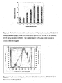
Figure 2 shows an absorbance profile of fractions of oligosaccharides containing chondroitin 6-sulfate from chondroitinase ABC digested CS-C eluted by column chromatography; -
Figure 3 shows the level of uronic acid in each fraction fromFigure 2 showing IC50 against WF6 antibody using competitive ELISA; -
Figure 4 shows the column profile offraction 6 fromFigure 2 analysed by ion exchange FPLC; -
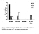
Figure 5 shows the effect of sulfatase digestion of the substrate on the reactivity of WF6 antibody; -
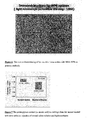
Figure 6 shows immunohistostaining of human skin tissue section using WF6 as primary antibody; -
Figure 7 shows uronic acid content of cartilage from an animal model of cartilage degradation; -
Figure 8 shows levels of WF6 epitope in samples from the animal model over time; -
Figure 9 shows the level of WF6 epitope in serum of human patients with osteoarthritis and rheumatoid arthritis; -
Figure 10 shows the effect of chitosan polysulfate on levels of WF6 epitope on a dog model of cartilage degradation; -
Figure 11 shows levels of WF6 epitope in cartilage explants induced by retinoic acid; and -
Figure 12 shows levels of proteoglycans containing WF6 epitope in serum fron-z cancer patients. - Purified proteoglycans (A1D1 fraction) from embryonic shark cartilage were used as antigen for the induction of an immune response in Balb/c mice. Spleen cells from mice which gave a high titer of antibody to chondroitin 6-sulfate (chondroitin sulfate C; CS-C) were fused with myeloma cell line X63 Ag8.653 in the ratio of 5:1. The fused cells were cultured in medium containing hypoxanthine, aminopterine, and thymine. Cell cultures were then tested for the production of antibodies by ELISA technique using chondroitin 6-sulfate (chondroitin sulfate C) as coating antigen. A positive cell line was isolated as a monoclone using a limiting dilution technique. The resulting cell line which produced antibody specifically against chondroitin 6-sulfate was called WF6, and it was found that the isotype of this antibody is Ig M, with Kappa-light chain. The cell line has been deposited with the American Type Culture Collection (ATCC) on 11 August 2004, under accession number PTA-6157.
- The mass production of this monoclonal antibody WF6 (MAb WF6) can be performed using hybridoma cells injected into the intraperitoneal cavity of Balb/c mice which have been primed by injection with 0.5 ml pristane one week before. Ascitic fluid samples can be obtained after 2-4 weeks which contain a large quantity of MAb WF6.
- An alternative technique for mass production makes use of serum-free medium. The hybridoma cell WF6 can be cultured in 10 % fetal calf serum Iscove's modified Eagle's medium, with gradual reduction of the percentage of serum to 5%, 1% and 0% along with an increase in commercial serum-free medium. The final medium should be 100% serum-free media which has been formulated with growth factors. The cells were cultured in 5%CO2, and 95% humidity and 37 degree C. The supernatant of this media may be further evaluated for antibody activity by ELISA technique and purified further using ammonium sulfate precipitation and chromatography.
-
- 3.1 Coat plate (polystyrene, Maxisorp Nunc®) with proteoglycans (fraction A1, aggregated form of shark cartilage proteoglycans) and block with 1% BSA
- 3.2 Prepare the inhibition mixture containing the optimal dilution of MAb WF6 and inhibitor, or standards or unknown samples in equal volume (175 + 175 µL each) in plastic tubes (1.5 mL tubes), then incubate at 37 degree C for 1 hour.
- 3.3 Add the inhibition mixture from 3.2 to the coated and blocked plate at 100 µL/well, and triplicate per sample (three wells), then incubate at 37 degree C for 1 hour.
- 3.4 Wash plate with washing buffer (PBS-Tween 0.05%) three times and dry before addition of 100 µL per well of peroxidase conjugated anti-Ig M antibody, then incubate at 37 degree C for 1 hour.
- 3.5 Wash plate with washing buffer three times, and dry before adding OPD substrate (100 µL/well), incubate for 15 minutes in the dark, and then stop the reaction by adding of 4 M sulfuric acid (50 µL/well). Absorbance of the plate was then measured 492/690nm by microplate reader.
- 3.6 The inhibition graph was plotted showing concentration of inhibitors (or standards) against absorbance (see
Figure 1 ). A range of test compounds were used, being chondroitin sulfate A to E, and the A1D1 proteoglycan fraction. - It was found that the monoclonal antibody shows reactivity (measured as inhibitory activities) only to chondroitin 6-sulfate (CS-C) and CS-D. It was later determined that the CS-D standard compounds contained some chondroitin 6-sulfate units, such that we believe the WF6 antibody shows specificity for chondroitin 6-sulfate containing compounds having a specific pattern of sulfation.
-
- 4.1 Chondroitin 6-sulfate (CS-C) from commercial shark cartilage (Sigma-Aldrich Chemical) was digested with chondroitinase ABC which cleaved the CS-C into small oligosaccharides at various sites. The cleavage products have a double bond at the non-reducing end of the products which has maximal absorption at 232 nm.
- 4.2 The products from enzyme digestion were separated by column chromatography using a BioGel P-6 column. The absorbance of the column fractions was measured, giving the profile shown in
Figure 2 . - 4.3 The products can be divided into 10 fractions as shown in
Figure 2 . Each fraction was pooled and concentrated by freeze drying, and the reactivity against MAb WF6 was determined using the method described insection 3. The reactivities from each fraction were compared by calculation of the uronic acid content which gave 50% inhibition (IC50; inhibition concentration at 50%), the results of which are shown inFigure 3 . Percentage inhibition can be calculated by % inhibition = 100 [((A(sample) - A (blank))/ (A(control)- A(blank)) x 100)] where A = absorbance. - It was found that the oligosaccharide fractions which still displayed inhibitory activities were fractions 1 - 6, and by comparison to known standards from previous studies, we determined that
fraction number 6 should contain oligosaccharides having 8-12 sugar units (octasaccharides - dodecasaccharides). - 4.4
Fraction number 6 of the oligosaccharides was further purified using by ion-exchange chromatography using FPLC and Mono-Q column chromatography using lithium percholate as eluent. The obtained column profile is shown inFigure 4 . - From the experimental results above, it was found that the oligosaccharides from gel filtration consisted of various species of chondroitin 6-sulfate oligosaccharides. This demonstrates that the monoclonal antibody WF6 reacts with oligosaccharides containing chondroitin 6-sulfate that have a special pattern of sulfatation as shown in the fingerprinting of Mono-Q ion-exchange FPLC.
-
- 5.1 The oligosaccharide fractions d, e and f from the column profile shown in
Figure 4 were subjected to digestion with 4- and 6-sulfatase. - 5.2 The products from these two enzymes were further evaluated for reactivity against MAb WF6 using competitive ELISA technique as described in
section 3. - 5.3 The comparison of the reactivities in each product, including a pre-digested sample, is shown in
Figure 5 . It was found that the sulfate group on the oligosaccharides is required for reactivity against MAb WF6. - When taken together, the results described in
sections 3 to 5 above demonstrate that MAb WF6 has reactivity against oligosaccharides and polysaccharides of at least 8 sugar units containing chondroitin 6-sulfate. Furthermore, this reactivity of MAb WF6 requires the functional group of 6-sulfate at the terminal residue because the 6-sulfatase recognises this position. And 4-sulfate needs to be an accessory group, including the specific sulfating pattern along with the oligosaccharide chain. -
- 6.1 Skin tissue samples from the pathological laboratories which had been processed by routine procedures in tissue staining were selected for this study.
- 6.2 The optimal dilution of MAb WF6 in PBS (for example, 1:500 from ascitic fluid, and 1: 100 tissue culture media) was used as primary antibody in the staining procedure after the tissue section was fixed to the slides.
- 6.3 Excess antibody was washed from the slide with washing buffer and restained with peroxidase conjugated anti-IgM antibody.
- 6.4 Excess conjugates and non-reacted antibody were washed from the slides, and insoluble substrate added.
- 6.5 The enzyme reaction was stopped with acid and excess colour washed out; the sample was then observed under light microscope as shown in
Figure 6 . -
- 7.1 The animal model was run as previously reported (Kongtawelert et al, 1989) for the induction of cartilage degradation by intra-articular injection of hydrocortisone (Solu-Cortef®). The rabbits were divided into two groups : the control had normal saline solution injection, while the experimental group had hydrocortisone injection of 200mg in every week for up to 12 weeks. Each week before injection the animal was bled for serum preparation.
- 7.2 The serum samples from each week were assayed for the level of WF6 epitope using the method described in
section 3. In addition to this study, the MAb 3B3 (Caterson et al, 1990) has also been used as a biosynthesis marker in cartilage as previously reported. - 7.3 At the end of the study, all animals were sacrificed and the articular cartilage were collected for analysis of proteoglycan contents as uronic acid per dried weight of cartilage. This data is shown in
Figure 7 . - 7.5 The level of the WF6 epitope and 3B3 epitope in each week were used to calculate the percentage relative change of each epitope each week. The results are shown in the graph of
Figure 8 . - It was found that the monoclonal antibody WF6 can be used or applied to use in the technique of immunoassay (competitive ELISA) for quantitation of proteoglycans containing the chondroitin 6-sulfate epitope, and this assay indicates that it can be used as a marker for degradation of cartilage.
Figure 8 shows that the WF6 epitope was found at significantly higher levels in hydrocortisone treated animals than normal saline treated animals, and the Figure also demonstrates the early indication of disease activity. -
- 8.1 Human serum samples from patients with OA, RA and healthy patients were collected to analyze the WF6 epitope. The samples were diluted 5 folds with 6% BSA-PBS to reduce non-specific binding.
- 8.2 All serum samples were subjected to analysis using competitive ELISA as described in
section 3 using shark proteoglycan fraction A1 as relative standards. - 8.3 The age of volunteers in each group was not significantly different.
- 8.4 The level of WF6 epitope from individual samples were plotted as a scatter graph as shown in
Figure 9 . - It was found that the MAb WF6 can be applied to use in a quantitative assay using immunological method for diagnosis of OA, and RA. However, there were still overlapping of the value in some samples of each group, which might be from the variation of the disease stage, and the treatment of the individual person. In addition, this might be interpreted as showing that the level of WF6 epitope can be used to monitor the disease activities and prognosis as well.
-
- 9.1 Cruciate ligament transection is used to induce the degradation of cartilage (OA) in 6 dogs by surgical operation at the hind leg (see Batiste et al, 2004).
- 9.2 These dogs were divided into 2 group (3 each group), one group were injected intramuscularly with normal saline solution and the other group injected with chitosan polysulfate, which is a potential anti-osteoarthritis agent, at the dose of 3mg/kg body weight. The injection was given every week for eight weeks.
- 9.3 The third group of dogs was not surgically operated upon, and was injected with chitosan polysulfate intramuscularly at 3 mg/kg body weight.
- 9.4 All of the animals were observed and cared for by a veterinarian. Blood for serum preparation was collected to use for the determination of WF6 by the method described in
section 3. - 9.5 The relative changes of all experimental animals were averaged in each group and the graph was plotted against time of the study as shown in
Figure 10 . - It was found that the MAb WF6 can be used in the method of quantitation of its epitope in animal model and can be applied as a marker for monitoring the treatment of the disease, like cartilage degeneration.
-
- 10.1 Porcine cartilage was sterile cut into small pieces and cultured in serum-free media under 37 degree C, and 5% carbon dioxide.
- 10.2 Retinoic acid was added to the culture at the concentration of 10 uM for stimulation of cartilage degradation.
- 10.3 The media were collected every other day for four weeks for determining the level of WF6 epitope using the method described in
section 3. - 10.4 The level of WF6 epitope from each week was plotted against time (days) of culture as shown in
Figure 11 . - It was found that the MAb WF6 can be used for monitoring the degradation of cartilage in vitro by a well established technique using the induction of retinoic acid when compared with the control. This demonstration showed the applicability of MAb WF6 as a research tool for the cartilage degradation in vitro.
- From previous reports (Nash et al, 2002), it was found that there were increased levels of chondroitin sulfate biosynthesis in cancer tissues compared with normal tissues. Furthermore, the cancer cells can produce some kinds of enzymes that can degrade the connective tissues surrounding them. These would make cancer cells intrude through the tissue and metastasize to other areas. We therefore suggest that MAb WF6 could be used for diagnosis of this pathogenesis including the tumour tissues.
- 11.1 Serum samples were collected from normal patients, patients having a tumour (non-cancer), and patients with ovarian cancers. This study was approved by the Ethics Committee.
- 11.2 All serum samples were tested for the determination of WF6 epitope level and compared between group of normal, non-cancer (tumour) and cancer. The results are shown in
Figure 12 - It was found that the WF6 epitope can be used as a biomarker for the cancerous (ovarian) condition in human.
-
- 1. Howell DS. Pathogenesis of osteoarthritis. Am J Med 1986; 80(4B):24-8
- 2. Muir H. and Hardingham TE. "Structure of proteoglycan" Biochemistry of carbohydrate. W.J. Ehelan Ed.1975; 5:153
- 3. Hochberg Mc., Altman RD., Brandt KD., Clark BM., Dieppe PA., Griffin MR., Moskowitz RW., Schnitzer TJ. Buidselines for the medical management of osteoarthritis. Part II. Ostroarthritis of the knee. American Collage of Rheumatology. Arthritis Rheum 1995; 38(11): 1541-6
- 4. Fassbender HG. Joint destruction in various arthritic disease. Articular cartilage biochemistry 1986: 371-90
- 5. Sexne T. and Heinegard D. Involvement of nonarticular cartilage, as demonstrated by release of cartilage specific protein in Rheumatoid arthritis. Arthristis Rheum; 32(9): 1080-86.
- 6. Hardingham T. and Bayliss M. Proteoglycans of articular cartilage: Change in aging and in joint disease. Seminar in arthritis and rheumatism. 1990; 20(3): 12-33
- 7. Caterson B., Christner JE., Baker TR. Characterization of monoclonal antibody that specifically recognizes corneal and skeletal keratan sulphate. J Biol Chem 1983; 258: 8848-54
- 8. Caterson B., Christner JE., Baker JR. Monoclonal antibodies against chondroitin sulphate isomer : their use as probes for investigating proteoglycan metabolism. Biochem Soc Trans.18,820-3
- 9. Nathachai T.(1996) Production and characterization of monoclonal antibody against chondroitin 6-sulfate.M.S. Thesis, Chiang Mai University,1997
- 10. Damrasamon S.(1997)The development of methods for quantitation of serum total sialic acid and hyaluronan.M.S.Thesis, Chiang Mai University,1998
- 11. Heinegard D., Hascall VC. Aggregation of cartilage proteoglycan III. Characterizations of the proteins isolated from trysin digests of aggregate. J Biol Chem 1974; 249(13): 4250-6
- 12. Fraser JR., Appelgren LE., Laurent TC. Tissue uptake of circulating hyarulonic acid : A Whole body autoradiographic study. Cell Tissue Res 1983; 233:285-93
- 13. Ratcliffe A., Shurety W., Caterson B. The quantitation of a native chondroitin sulphate epitope in synovial fluid lavages and articular cartilage from canine experimental osteoarthritis and disuse atropy. Arthritis Rhem 1993; 36(4): 543-51
- 14. Kempson GE., Tuke MA., Dingle JT., Barrett AJ., Horsfield PH. The effects of proteolytic enzymes on the mechanical properties of adult human articular cartilage. Biochim Biophys Acta 1976; 428(3): 741-60
- 15. Ratcliffe A. and Seibel MJ. Biochemical markers of osteoarthritis. Curr Opin Rheumatol 1990; 2: 770-6
- 16. Caterson B., Baker JR.., Christner JE. Immunological methods for the detection and determination of connective tissue proteoglycan. J Invest Dermatol 1982; 79(suppl 1): 45s-50s
- 17. Williams JM., Downey C., Thonar EJ., Increase in level of serum keratan sulfate following cartilage proteoglycan degradation in the rabbit knee joint. Arthritis Rheum 1988; 31(5): 557-60
- 18. Sweet MB., Coelho A., Schnitzler CM., Schnitzer TJ., Lenz ME., Jakim I., Kuetlner KE., Thonar EJ. Serum keratan sulfate levels in osteoarthritis patients. Arthritis Rheum 1988; 31(5): 648-52
- 19. Spector TD., Woodward L., Hall GM., Hammond A., Williams A., Butler MG., James IT., Hart DJ., Thompson PW., Scott DL. Keratan sulfate in rheumatoid arthritis, osteoarthritis ans inflammatory disease. Arm Rheum Dis 1992; 51(10):1134-7
- 20. Ghosh P., Sutherland JM., Taylor TK., Bellenger CR., Pettit GD. The effect of bilateral medial meniscectomy on articular cartilage of the hip joint. J Rheumatol 1984; 1(2): 197-201
- 21. Sharif M., George E., Shepstone L., Knudson W., Thonar EJ-MA., Cushnaghan J., Dieppe P. Serum hyaluronic acid level as a predictor of disease progression in osteoarthritis of the knee. Arthritis Rheum 1995; 38: 760-7
- 22. Cooper EH. And Rathbone BJ. Clinical significance of the immunometric measurements of hyaluronic acid. Ann Clin Biochem 1990; 27: 444-51
- 23. Smith PK., Krohn RI., Hermanson GT., Mallic AK., Garther FH., Prorenzano MD., Fujimoto EK., Goeke NM., Olson BJ. and Klenk DC. Measurement of protein using by bicinchoninic acid. Anal Biochem 1985; 150(1):76-85
- 24. Famdale RH. And Buttle DJ. Improved quantitative and discrimination of sulfated glycosaminoglycans by use of dimethylene blue. Biochemica-Biophysica Acta 1986; 883: 173-77
- 25. Blumenkrantz N. and Asboe-Hansen G. New method for quantitative determination of uronic acids. Anal Biochem 1973; 54:484-89
- 26. Kongtawelert P. Brooks PM, Ghosh P. Pentosan polysulfate (Cartrophen) prevents the hydrocortisone induced loss of hyaluronic acid and proteoglycans from cartilage of rabbit joints as well as normalizes the keratan sulfate levels in their serum. J Rheumatol. 1989 Nov; 16(11):1454-9.
- 27. Caterson B, Mahmoodian F. Sorrell JM. Hardingham TE, Bayliss MT, Carney SL, Ratcliffe A, Muir H. Modulation of native chondroitin sulphate structure in tissue development and in disease. J Cell Sci. 1990 Nov; 97 (Pt 3):411-7.
- 28. Batiste DL, Kirkley A, Laverty S, Thain LM, Spouse AR, Gati JS, Foster PJ, Holdsworth DW. High-resolution MRI and micro-CT in an ex vivo rabbit anterior cruciate ligament transection model of osteoarthritis. Osteoarthritis Cartilage. 2004 Aug; 12 (8):614-26.
- 29. Nash MA, Deavers MT, Freedman RS. The expression of decorin in human ovarian tumors. Clin Cancer Res. 2002 Jun;8(6):1754-60.]
- 30. Chan SS et al: A sensitive assay for the measurement of serum chondroitin sulfate 3B3(-) epitope levels in human rheumatic diseases. Clinical and Experimental Rheumatology. 2001 Sep-Oct, 19(5), Sep 2001 (2001-09): 533-40.
- 31. Slater Robert R Jr et al. Monoclonal antibodies that detect biochemical markers of arthritis in human. Arthritis and Rheumatism, 1995; 38(5): 655-9.
Claims (12)
- A WF6 antibody produced by a hybridoma cell as deposited in the American Type Culture Collection under accession number PTA-6157, the antibody having specific reactivity to a chondroitin sulfate epitope, the chondroitin sulfate epitope comprising:at least eight sugar monomers;a terminal chondroitin 6-sulfate unit; andat least one chondroitin 4-sulfate accessory unit.
- The antibody of claim 1, the at least 8 sugar monomers comprising a sequence ADCCC.
- A functional derivative of the antibody of claim 1, the functional derivative being selected from a group consisting of Fab, Fab', F(ab)2 fragments; single chain antibodies; and functional fragments obtained by chemical or enzymatic cleavage of the antibody or molecular biological methods, the functional derivative exhibiting specific reactivity to a chondroitin sulfate epitope comprising at least eight sugar monomers having a terminal chondroitin 6-sulfate unit and a chondroitin 4-sulfate accessory unit.
- A method of producing antibodies comprising:culturing hybridoma cells as deposited with the American Type Culture Collection under accession number PTA-6157 in a suitable growth medium, the hybridoma cells producing growth supernatant comprising antibodies; andharvesting the growth supernatant to thereby extract the antibodies, wherein the antibodies exhibit specific reactivity to a chondroitin sulfate epitope comprising at least eight sugar units having a terminal chondroitin 6-sulfate unit and a chondroitin 4-sulfate accessory unit.
- A method of detecting cartilage degradation, the method comprising:contacting a sample with antibodies produced by a hybridoma cell as deposited in the American Type Culture Collection under accession number PTA-6157, the antibodies having specific reactivity to a chondroitin sulfate epitope, the chondroitin sulfate epitope comprising:at least eight sugar monomers;a terminal chondroitin 6-sulfate unit; anda chondroitin-4-sulfate unit; anddetecting binding of the antibodies to the sample.
- The method of claim 5, wherein the detection of binding of the antibody to the sample is performed by one of ELISA, indirect ELISA, competitive ELISA, sandwich ELISA, radioimmunoassay, fluorescent immunoassay, chemiluminescence immunoassay, immunoblotting, immunohislostaining and flow injection analysis.
- A method for detecting immunobinding comprising:contacting a sample with one of an antibody as claimed in claim 1 and a functional derivative of the antibody as claimed in claim 3, the functional derivative exhibiting the same specific reactivity as compared to the antibody; anddetecting binding of one of the antibody and the functional derivative with the sample,wherein the detection of binding is performed by one of ELISA, indirect ELISA, competitive ELISA, sandwich ELISA, radioimmunoassay, fluorescent immunoassay, chemiluminescence immunoassay, immunoblotting, immunohistostaining and flow injection analysis.
- A medicament for facilitating diagnosis of a disease of the connective tissue comprising:an antibody produced by a hybridoma cell as deposited in the American Type Culture Collection under accession number PTA-6157, the antibody having binding specificity to a chondroitin sulfate epitope, the chondroitin sulfate epitope comprising:at least eight sugar monomers;a terminal chondroitin 6-sulfate unit; anda chondroitin 4-sulfate accessory unit.
- A medicament for facilitating diagnosis of a disease of the connective tissue comprising:a functional derivative of an antibody produced by a hybridoma cell as deposited in the American Type Culture Collection under accession number PTA-6157and having a specifici reactivity to a chondroitin sulfate epitope that comprises:at least eight sugar monomers;a terminal chondroitin 6-sulphate unit; anda chondroitin 4-sulfate accessory unit,wherein the functional derivative is selected from a group consisting of Fab, Fab', F(ab)2 fragments; single chain antibodies; and functional fragments obtained by chemical or enzymatic cleavage of the antibody or molecular biological methods.
- A method of detecting a chondroitin sulfate oligosaccharide epitope comprising at least 8 sugar monomers with a terminal chondroitin 6-sulfate unit and at least one chondroitin 4-sulfate accessory unit, the method comprising:contacting a sample with one of an antibody as claimed in claim 1 and a functional derivative of the antibody as claimed in claim 3.
- A diagnostic test kit comprising:one of an antibody as claimed in claim 1 and a functional derivative of the antibody as claimed in claim 3, the functional derivative exhibiting the same specific reactivity as the antibody.
- A pharmaceutical composition comprising:one of an antibody as claimed in claim 1 and a functional derivative of the antibody as claimed in claim 3, the functional derivative exhibiting the same specific reactivity as the antibody.
Applications Claiming Priority (3)
| Application Number | Priority Date | Filing Date | Title |
|---|---|---|---|
| TH091280 | 2004-06-03 | ||
| GBGB0418415.6A GB0418415D0 (en) | 2004-06-03 | 2004-08-18 | Antibody |
| PCT/GB2005/050077 WO2005118645A1 (en) | 2004-06-03 | 2005-06-01 | Antibody specific for a chondroitin sulphate epitope |
Publications (2)
| Publication Number | Publication Date |
|---|---|
| EP1778727A1 EP1778727A1 (en) | 2007-05-02 |
| EP1778727B1 true EP1778727B1 (en) | 2014-08-06 |
Family
ID=34970117
Family Applications (1)
| Application Number | Title | Priority Date | Filing Date |
|---|---|---|---|
| EP05748554.2A Expired - Lifetime EP1778727B1 (en) | 2004-06-03 | 2005-06-01 | Antibody specific for a chondroitin sulphate epitope |
Country Status (5)
| Country | Link |
|---|---|
| US (1) | US8481290B2 (en) |
| EP (1) | EP1778727B1 (en) |
| JP (1) | JP4896876B2 (en) |
| GB (1) | GB0418415D0 (en) |
| WO (1) | WO2005118645A1 (en) |
Families Citing this family (2)
| Publication number | Priority date | Publication date | Assignee | Title |
|---|---|---|---|---|
| JP5197922B2 (en) * | 2006-03-25 | 2013-05-15 | 独立行政法人科学技術振興機構 | Glycosylation marker recognition probe, neurite outgrowth inhibitor using the same, neuronal staining agent, tumor cell staining agent, immune tissue staining agent, pharmaceutical composition and pharmaceutical |
| FR2921928B1 (en) * | 2007-10-08 | 2011-03-04 | Sanofi Pasteur | MONOCLONAL ANTIBODIES SPECIFIC TO INFLUENZA VIRUS HEMAGGLUTININ |
Family Cites Families (4)
| Publication number | Priority date | Publication date | Assignee | Title |
|---|---|---|---|---|
| AU645742B2 (en) * | 1988-10-24 | 1994-01-27 | Bruce Caterson | Methods of and compositions for diagnosing, monitoring and treating the early stages of osteoarthritis |
| JP2000065837A (en) * | 1998-08-24 | 2000-03-03 | Seikagaku Kogyo Co Ltd | Measurement of glycosaminoglycan or glycosaminoglycan bonding molecule and measurement kit thereof |
| US6960562B2 (en) * | 1999-04-23 | 2005-11-01 | Rhode Island Hospital, A Lifespan Partner | Tribonectin polypeptides and uses thereof |
| US20090202517A1 (en) * | 2006-08-17 | 2009-08-13 | Hiroyuki Yoneyama | Agents for improving inflammatory bowel disease |
-
2004
- 2004-08-18 GB GBGB0418415.6A patent/GB0418415D0/en not_active Ceased
-
2005
- 2005-06-01 JP JP2007514145A patent/JP4896876B2/en not_active Expired - Fee Related
- 2005-06-01 EP EP05748554.2A patent/EP1778727B1/en not_active Expired - Lifetime
- 2005-06-01 US US11/628,432 patent/US8481290B2/en not_active Expired - Fee Related
- 2005-06-01 WO PCT/GB2005/050077 patent/WO2005118645A1/en not_active Ceased
Non-Patent Citations (2)
| Title |
|---|
| ITO YUMI ET AL: "Structural characterization of the epitopes of the monoclonal antibodies 473HD, CS-56, and MO-225 specific for chondroitin sulfate D-type using the oligosaccharide library.", GLYCOBIOLOGY JUN 2005, vol. 15, no. 6, June 2005 (2005-06-01), pages 593 - 603, ISSN: 0959-6658 * |
| NADANAKA S ET AL: "Characteristic hexasaccharide sequences in octasaccharides derived from shark cartilage chondroitin sulfate D with a neurite outgrowth promoting activity", JOURNAL OF BIOLOGICAL CHEMISTRY, AMERICAN SOCIETY FOR BIOCHEMISTRY AND MOLECULAR BIOLOGY, US, vol. 273, no. 6, 6 February 1998 (1998-02-06), pages 3296 - 3307, XP002369176, ISSN: 0021-9258, DOI: 10.1074/JBC.273.6.3296 * |
Also Published As
| Publication number | Publication date |
|---|---|
| US8481290B2 (en) | 2013-07-09 |
| US20080038270A1 (en) | 2008-02-14 |
| JP4896876B2 (en) | 2012-03-14 |
| WO2005118645A1 (en) | 2005-12-15 |
| GB0418415D0 (en) | 2004-09-22 |
| EP1778727A1 (en) | 2007-05-02 |
| JP2008509881A (en) | 2008-04-03 |
Similar Documents
| Publication | Publication Date | Title |
|---|---|---|
| Thonar et al. | Quantification of keratan sulfate in blood as a marker of cartilage catabolism | |
| Ishiguro et al. | Relationship of matrix metalloproteinases and their inhibitors to cartilage proteoglycan and collagen turnover: analyses of synovial fluid from patients with osteoarthritis | |
| Belcher et al. | Synovial fluid chondroitin and keratan sulphate epitopes, glycosaminoglycans, and hyaluronan in arthritic and normal knees | |
| Martins et al. | Practical determination of hyaluronan by a new noncompetitive fluorescence-based assay on serum of normal and cirrhotic patients | |
| AU705059B2 (en) | Method and measurement kit for assay of normal aggrecan, and method for evaluation of information on the joint | |
| JP5197535B2 (en) | Method for detecting lysosomal disease | |
| Slater Jr et al. | Monoclonal antibodies that detect biochemical markers of arthritis in humans | |
| JP2002525632A (en) | YKL-40 as a marker and prognostic indicator for cancer | |
| JPH09509736A (en) | Method for measuring collagen fragment in body fluid, test kit and means for carrying out the method, and method / use for using the method for diagnosing the presence of a disease related to collagen metabolism | |
| EP0145681B1 (en) | A method for determining changes occurring in cartilage | |
| Jura-Półtorak et al. | Alterations of plasma glycosaminoglycan profile in patients with rheumatoid arthritis in relation to disease activity | |
| CA2199498A1 (en) | Method of detecting autoantibody present in the serum of rheumatic | |
| Carroll | Spectrophotometric measurement of proteoglycans in osteoarthritic synovial fluid. | |
| HUT64421A (en) | Method for detecting irregularities of bone and other connective tissues in human beings and in animals | |
| JP2000504114A (en) | Method for detecting and evaluating bladder cancer | |
| Chajara et al. | Hyaluronan and hyaluronectin production in injured rat thoracic aorta | |
| US8481290B2 (en) | Antibody specific for a chondroitin sulphate epitope | |
| JP3976848B2 (en) | Method and kit for measuring serum-derived hyaluronan binding protein-hyaluronic acid conjugate | |
| EP2386857B1 (en) | Method for assaying keratan sulfate, assay kit therefor and method for detecting joint disease by using same. | |
| US6350571B1 (en) | Methods for detection and evaluation of bladder cancer | |
| Radhakrishnamurthy et al. | A monoclonal antibody that recognizes hyaluronic acid binding region of aorta proteoglycans | |
| JP2000065837A (en) | Measurement of glycosaminoglycan or glycosaminoglycan bonding molecule and measurement kit thereof | |
| JP4217717B2 (en) | Method and kit for measuring serum-derived hyaluronan-binding protein-hyaluronic acid conjugate | |
| JP4180911B2 (en) | New enzyme activity measurement method | |
| Kongtawelert et al. | Application of an enzyme-linked immunosorbent-inhibition assay to quantitate the release of KS peptides into fluids of the rat subcutaneous air-pouch model and the effects of chondroprotective drugs on the release process |
Legal Events
| Date | Code | Title | Description |
|---|---|---|---|
| PUAI | Public reference made under article 153(3) epc to a published international application that has entered the european phase |
Free format text: ORIGINAL CODE: 0009012 |
|
| 17P | Request for examination filed |
Effective date: 20061228 |
|
| AK | Designated contracting states |
Kind code of ref document: A1 Designated state(s): AT BE BG CH CY CZ DE DK EE ES FI FR GB GR HU IE IS IT LI LT LU MC NL PL PT RO SE SI SK TR |
|
| DAX | Request for extension of the european patent (deleted) | ||
| 17Q | First examination report despatched |
Effective date: 20080115 |
|
| RAP1 | Party data changed (applicant data changed or rights of an application transferred) |
Owner name: THE THAILAND RESEARCH FUND Owner name: CHIANG MAI UNIVERSITY Owner name: THE NATIONAL RESEARCH COUNCIL OF THAILAND |
|
| RIN1 | Information on inventor provided before grant (corrected) |
Inventor name: HARDINGHAM, TIM, SCHOOL OF BIOLOGICAL SCIENCES Inventor name: TIENGBURANATHUM, NATTHACHAI Inventor name: KONGTAWELERT, PRACHYA, DEPARTMENT OF BIOCHEMISTRY Inventor name: SUGAHARA, KAZUYUKI, DEPARTMENT OF BIOCHEMISTRY Inventor name: ONG-CHAI, SIRIWAN, DEPARTMENT OF BIOCHEMISTRY Inventor name: POTHACHAROEN, PERAPHAN, DEPARTMENT OF BIOCHEMISTRY |
|
| GRAP | Despatch of communication of intention to grant a patent |
Free format text: ORIGINAL CODE: EPIDOSNIGR1 |
|
| RIN1 | Information on inventor provided before grant (corrected) |
Inventor name: KONGTAWELERT, PRACHYA Inventor name: HARDINGHAM, TIM Inventor name: SUGAHARA, KAZUYUKI Inventor name: TIENGBURANATHUM, NATTHACHAI Inventor name: POTHACHAROEN, PERAPHAN Inventor name: ONG-CHAI, SIRIWAN |
|
| INTG | Intention to grant announced |
Effective date: 20131209 |
|
| GRAS | Grant fee paid |
Free format text: ORIGINAL CODE: EPIDOSNIGR3 |
|
| GRAA | (expected) grant |
Free format text: ORIGINAL CODE: 0009210 |
|
| AK | Designated contracting states |
Kind code of ref document: B1 Designated state(s): AT BE BG CH CY CZ DE DK EE ES FI FR GB GR HU IE IS IT LI LT LU MC NL PL PT RO SE SI SK TR |
|
| REG | Reference to a national code |
Ref country code: GB Ref legal event code: FG4D |
|
| REG | Reference to a national code |
Ref country code: CH Ref legal event code: EP Ref country code: AT Ref legal event code: REF Ref document number: 681044 Country of ref document: AT Kind code of ref document: T Effective date: 20140815 |
|
| REG | Reference to a national code |
Ref country code: IE Ref legal event code: FG4D |
|
| REG | Reference to a national code |
Ref country code: DE Ref legal event code: R096 Ref document number: 602005044403 Country of ref document: DE Effective date: 20140918 |
|
| REG | Reference to a national code |
Ref country code: AT Ref legal event code: MK05 Ref document number: 681044 Country of ref document: AT Kind code of ref document: T Effective date: 20140806 |
|
| REG | Reference to a national code |
Ref country code: NL Ref legal event code: VDEP Effective date: 20140806 |
|
| REG | Reference to a national code |
Ref country code: LT Ref legal event code: MG4D |
|
| PG25 | Lapsed in a contracting state [announced via postgrant information from national office to epo] |
Ref country code: SE Free format text: LAPSE BECAUSE OF FAILURE TO SUBMIT A TRANSLATION OF THE DESCRIPTION OR TO PAY THE FEE WITHIN THE PRESCRIBED TIME-LIMIT Effective date: 20140806 Ref country code: PT Free format text: LAPSE BECAUSE OF FAILURE TO SUBMIT A TRANSLATION OF THE DESCRIPTION OR TO PAY THE FEE WITHIN THE PRESCRIBED TIME-LIMIT Effective date: 20141209 Ref country code: ES Free format text: LAPSE BECAUSE OF FAILURE TO SUBMIT A TRANSLATION OF THE DESCRIPTION OR TO PAY THE FEE WITHIN THE PRESCRIBED TIME-LIMIT Effective date: 20140806 Ref country code: BG Free format text: LAPSE BECAUSE OF FAILURE TO SUBMIT A TRANSLATION OF THE DESCRIPTION OR TO PAY THE FEE WITHIN THE PRESCRIBED TIME-LIMIT Effective date: 20141106 Ref country code: LT Free format text: LAPSE BECAUSE OF FAILURE TO SUBMIT A TRANSLATION OF THE DESCRIPTION OR TO PAY THE FEE WITHIN THE PRESCRIBED TIME-LIMIT Effective date: 20140806 Ref country code: FI Free format text: LAPSE BECAUSE OF FAILURE TO SUBMIT A TRANSLATION OF THE DESCRIPTION OR TO PAY THE FEE WITHIN THE PRESCRIBED TIME-LIMIT Effective date: 20140806 Ref country code: GR Free format text: LAPSE BECAUSE OF FAILURE TO SUBMIT A TRANSLATION OF THE DESCRIPTION OR TO PAY THE FEE WITHIN THE PRESCRIBED TIME-LIMIT Effective date: 20141107 |
|
| PG25 | Lapsed in a contracting state [announced via postgrant information from national office to epo] |
Ref country code: CY Free format text: LAPSE BECAUSE OF FAILURE TO SUBMIT A TRANSLATION OF THE DESCRIPTION OR TO PAY THE FEE WITHIN THE PRESCRIBED TIME-LIMIT Effective date: 20140806 Ref country code: NL Free format text: LAPSE BECAUSE OF FAILURE TO SUBMIT A TRANSLATION OF THE DESCRIPTION OR TO PAY THE FEE WITHIN THE PRESCRIBED TIME-LIMIT Effective date: 20140806 Ref country code: IS Free format text: LAPSE BECAUSE OF FAILURE TO SUBMIT A TRANSLATION OF THE DESCRIPTION OR TO PAY THE FEE WITHIN THE PRESCRIBED TIME-LIMIT Effective date: 20141206 Ref country code: AT Free format text: LAPSE BECAUSE OF FAILURE TO SUBMIT A TRANSLATION OF THE DESCRIPTION OR TO PAY THE FEE WITHIN THE PRESCRIBED TIME-LIMIT Effective date: 20140806 Ref country code: PL Free format text: LAPSE BECAUSE OF FAILURE TO SUBMIT A TRANSLATION OF THE DESCRIPTION OR TO PAY THE FEE WITHIN THE PRESCRIBED TIME-LIMIT Effective date: 20140806 |
|
| PG25 | Lapsed in a contracting state [announced via postgrant information from national office to epo] |
Ref country code: EE Free format text: LAPSE BECAUSE OF FAILURE TO SUBMIT A TRANSLATION OF THE DESCRIPTION OR TO PAY THE FEE WITHIN THE PRESCRIBED TIME-LIMIT Effective date: 20140806 Ref country code: SK Free format text: LAPSE BECAUSE OF FAILURE TO SUBMIT A TRANSLATION OF THE DESCRIPTION OR TO PAY THE FEE WITHIN THE PRESCRIBED TIME-LIMIT Effective date: 20140806 Ref country code: DK Free format text: LAPSE BECAUSE OF FAILURE TO SUBMIT A TRANSLATION OF THE DESCRIPTION OR TO PAY THE FEE WITHIN THE PRESCRIBED TIME-LIMIT Effective date: 20140806 Ref country code: IT Free format text: LAPSE BECAUSE OF FAILURE TO SUBMIT A TRANSLATION OF THE DESCRIPTION OR TO PAY THE FEE WITHIN THE PRESCRIBED TIME-LIMIT Effective date: 20140806 Ref country code: CZ Free format text: LAPSE BECAUSE OF FAILURE TO SUBMIT A TRANSLATION OF THE DESCRIPTION OR TO PAY THE FEE WITHIN THE PRESCRIBED TIME-LIMIT Effective date: 20140806 Ref country code: RO Free format text: LAPSE BECAUSE OF FAILURE TO SUBMIT A TRANSLATION OF THE DESCRIPTION OR TO PAY THE FEE WITHIN THE PRESCRIBED TIME-LIMIT Effective date: 20140806 |
|
| REG | Reference to a national code |
Ref country code: DE Ref legal event code: R097 Ref document number: 602005044403 Country of ref document: DE |
|
| PLBE | No opposition filed within time limit |
Free format text: ORIGINAL CODE: 0009261 |
|
| STAA | Information on the status of an ep patent application or granted ep patent |
Free format text: STATUS: NO OPPOSITION FILED WITHIN TIME LIMIT |
|
| 26N | No opposition filed |
Effective date: 20150507 |
|
| PG25 | Lapsed in a contracting state [announced via postgrant information from national office to epo] |
Ref country code: SI Free format text: LAPSE BECAUSE OF FAILURE TO SUBMIT A TRANSLATION OF THE DESCRIPTION OR TO PAY THE FEE WITHIN THE PRESCRIBED TIME-LIMIT Effective date: 20140806 |
|
| REG | Reference to a national code |
Ref country code: DE Ref legal event code: R119 Ref document number: 602005044403 Country of ref document: DE |
|
| PG25 | Lapsed in a contracting state [announced via postgrant information from national office to epo] |
Ref country code: MC Free format text: LAPSE BECAUSE OF FAILURE TO SUBMIT A TRANSLATION OF THE DESCRIPTION OR TO PAY THE FEE WITHIN THE PRESCRIBED TIME-LIMIT Effective date: 20140806 |
|
| REG | Reference to a national code |
Ref country code: CH Ref legal event code: PL |
|
| PG25 | Lapsed in a contracting state [announced via postgrant information from national office to epo] |
Ref country code: LU Free format text: LAPSE BECAUSE OF FAILURE TO SUBMIT A TRANSLATION OF THE DESCRIPTION OR TO PAY THE FEE WITHIN THE PRESCRIBED TIME-LIMIT Effective date: 20150601 |
|
| REG | Reference to a national code |
Ref country code: IE Ref legal event code: MM4A |
|
| REG | Reference to a national code |
Ref country code: FR Ref legal event code: ST Effective date: 20160229 |
|
| PG25 | Lapsed in a contracting state [announced via postgrant information from national office to epo] |
Ref country code: LI Free format text: LAPSE BECAUSE OF NON-PAYMENT OF DUE FEES Effective date: 20150630 Ref country code: IE Free format text: LAPSE BECAUSE OF NON-PAYMENT OF DUE FEES Effective date: 20150601 Ref country code: CH Free format text: LAPSE BECAUSE OF NON-PAYMENT OF DUE FEES Effective date: 20150630 Ref country code: DE Free format text: LAPSE BECAUSE OF NON-PAYMENT OF DUE FEES Effective date: 20160101 |
|
| PG25 | Lapsed in a contracting state [announced via postgrant information from national office to epo] |
Ref country code: FR Free format text: LAPSE BECAUSE OF NON-PAYMENT OF DUE FEES Effective date: 20150630 |
|
| PG25 | Lapsed in a contracting state [announced via postgrant information from national office to epo] |
Ref country code: BE Free format text: LAPSE BECAUSE OF FAILURE TO SUBMIT A TRANSLATION OF THE DESCRIPTION OR TO PAY THE FEE WITHIN THE PRESCRIBED TIME-LIMIT Effective date: 20140806 |
|
| PG25 | Lapsed in a contracting state [announced via postgrant information from national office to epo] |
Ref country code: HU Free format text: LAPSE BECAUSE OF FAILURE TO SUBMIT A TRANSLATION OF THE DESCRIPTION OR TO PAY THE FEE WITHIN THE PRESCRIBED TIME-LIMIT; INVALID AB INITIO Effective date: 20050601 |
|
| PG25 | Lapsed in a contracting state [announced via postgrant information from national office to epo] |
Ref country code: TR Free format text: LAPSE BECAUSE OF FAILURE TO SUBMIT A TRANSLATION OF THE DESCRIPTION OR TO PAY THE FEE WITHIN THE PRESCRIBED TIME-LIMIT Effective date: 20140806 |
|
| PGFP | Annual fee paid to national office [announced via postgrant information from national office to epo] |
Ref country code: GB Payment date: 20180731 Year of fee payment: 14 |
|
| GBPC | Gb: european patent ceased through non-payment of renewal fee |
Effective date: 20190601 |
|
| PG25 | Lapsed in a contracting state [announced via postgrant information from national office to epo] |
Ref country code: GB Free format text: LAPSE BECAUSE OF NON-PAYMENT OF DUE FEES Effective date: 20190601 |