EP1767167A2 - Système pour l'introduction d'un marqueur après décompression - Google Patents
Système pour l'introduction d'un marqueur après décompression Download PDFInfo
- Publication number
- EP1767167A2 EP1767167A2 EP06121084A EP06121084A EP1767167A2 EP 1767167 A2 EP1767167 A2 EP 1767167A2 EP 06121084 A EP06121084 A EP 06121084A EP 06121084 A EP06121084 A EP 06121084A EP 1767167 A2 EP1767167 A2 EP 1767167A2
- Authority
- EP
- European Patent Office
- Prior art keywords
- sheath
- lumen
- tissue mass
- marker
- distal
- Prior art date
- Legal status (The legal status is an assumption and is not a legal conclusion. Google has not performed a legal analysis and makes no representation as to the accuracy of the status listed.)
- Granted
Links
- 239000003550 marker Substances 0.000 title claims abstract description 40
- 230000006837 decompression Effects 0.000 title description 4
- 238000003780 insertion Methods 0.000 claims abstract description 23
- 230000037431 insertion Effects 0.000 claims abstract description 23
- 239000000523 sample Substances 0.000 claims description 47
- 238000001574 biopsy Methods 0.000 claims description 10
- 229940030225 antihemorrhagics Drugs 0.000 claims description 9
- 239000002874 hemostatic agent Substances 0.000 claims description 9
- 238000003384 imaging method Methods 0.000 claims description 9
- 238000002513 implantation Methods 0.000 claims description 6
- 230000006835 compression Effects 0.000 claims description 4
- 238000007906 compression Methods 0.000 claims description 4
- 210000000481 breast Anatomy 0.000 description 16
- 230000003902 lesion Effects 0.000 description 16
- 238000009607 mammography Methods 0.000 description 7
- 238000002604 ultrasonography Methods 0.000 description 6
- 239000000463 material Substances 0.000 description 4
- 238000000034 method Methods 0.000 description 4
- 238000004873 anchoring Methods 0.000 description 3
- 239000000560 biocompatible material Substances 0.000 description 2
- 229920001661 Chitosan Polymers 0.000 description 1
- 102000008186 Collagen Human genes 0.000 description 1
- 108010035532 Collagen Proteins 0.000 description 1
- 108010074860 Factor Xa Proteins 0.000 description 1
- 108010049003 Fibrinogen Proteins 0.000 description 1
- 102000008946 Fibrinogen Human genes 0.000 description 1
- 108010010803 Gelatin Proteins 0.000 description 1
- 206010028980 Neoplasm Diseases 0.000 description 1
- 108090000190 Thrombin Proteins 0.000 description 1
- 230000002159 abnormal effect Effects 0.000 description 1
- 230000005856 abnormality Effects 0.000 description 1
- 230000000740 bleeding effect Effects 0.000 description 1
- 201000011510 cancer Diseases 0.000 description 1
- 229920002678 cellulose Polymers 0.000 description 1
- 239000001913 cellulose Substances 0.000 description 1
- 229920001436 collagen Polymers 0.000 description 1
- 239000010432 diamond Substances 0.000 description 1
- 229940012952 fibrinogen Drugs 0.000 description 1
- 229920000159 gelatin Polymers 0.000 description 1
- 239000008273 gelatin Substances 0.000 description 1
- 235000019322 gelatine Nutrition 0.000 description 1
- 235000011852 gelatine desserts Nutrition 0.000 description 1
- 229910000078 germane Inorganic materials 0.000 description 1
- 150000004676 glycans Chemical class 0.000 description 1
- 239000007943 implant Substances 0.000 description 1
- 210000003041 ligament Anatomy 0.000 description 1
- 239000007788 liquid Substances 0.000 description 1
- 238000002595 magnetic resonance imaging Methods 0.000 description 1
- 229910001000 nickel titanium Inorganic materials 0.000 description 1
- HLXZNVUGXRDIFK-UHFFFAOYSA-N nickel titanium Chemical compound [Ti].[Ti].[Ti].[Ti].[Ti].[Ti].[Ti].[Ti].[Ti].[Ti].[Ti].[Ni].[Ni].[Ni].[Ni].[Ni].[Ni].[Ni].[Ni].[Ni].[Ni].[Ni].[Ni].[Ni].[Ni] HLXZNVUGXRDIFK-UHFFFAOYSA-N 0.000 description 1
- 238000005192 partition Methods 0.000 description 1
- 229920001282 polysaccharide Polymers 0.000 description 1
- 239000005017 polysaccharide Substances 0.000 description 1
- 238000005070 sampling Methods 0.000 description 1
- 229910001285 shape-memory alloy Inorganic materials 0.000 description 1
- 239000002689 soil Substances 0.000 description 1
- 238000001356 surgical procedure Methods 0.000 description 1
- 229960004072 thrombin Drugs 0.000 description 1
Images


Classifications
-
- A—HUMAN NECESSITIES
- A61—MEDICAL OR VETERINARY SCIENCE; HYGIENE
- A61B—DIAGNOSIS; SURGERY; IDENTIFICATION
- A61B90/00—Instruments, implements or accessories specially adapted for surgery or diagnosis and not covered by any of the groups A61B1/00 - A61B50/00, e.g. for luxation treatment or for protecting wound edges
- A61B90/10—Instruments, implements or accessories specially adapted for surgery or diagnosis and not covered by any of the groups A61B1/00 - A61B50/00, e.g. for luxation treatment or for protecting wound edges for stereotaxic surgery, e.g. frame-based stereotaxis
- A61B90/14—Fixators for body parts, e.g. skull clamps; Constructional details of fixators, e.g. pins
- A61B90/17—Fixators for body parts, e.g. skull clamps; Constructional details of fixators, e.g. pins for soft tissue, e.g. breast-holding devices
-
- A—HUMAN NECESSITIES
- A61—MEDICAL OR VETERINARY SCIENCE; HYGIENE
- A61B—DIAGNOSIS; SURGERY; IDENTIFICATION
- A61B90/00—Instruments, implements or accessories specially adapted for surgery or diagnosis and not covered by any of the groups A61B1/00 - A61B50/00, e.g. for luxation treatment or for protecting wound edges
- A61B90/39—Markers, e.g. radio-opaque or breast lesions markers
-
- A—HUMAN NECESSITIES
- A61—MEDICAL OR VETERINARY SCIENCE; HYGIENE
- A61B—DIAGNOSIS; SURGERY; IDENTIFICATION
- A61B10/00—Other methods or instruments for diagnosis, e.g. instruments for taking a cell sample, for biopsy, for vaccination diagnosis; Sex determination; Ovulation-period determination; Throat striking implements
- A61B10/0041—Detection of breast cancer
-
- A—HUMAN NECESSITIES
- A61—MEDICAL OR VETERINARY SCIENCE; HYGIENE
- A61B—DIAGNOSIS; SURGERY; IDENTIFICATION
- A61B10/00—Other methods or instruments for diagnosis, e.g. instruments for taking a cell sample, for biopsy, for vaccination diagnosis; Sex determination; Ovulation-period determination; Throat striking implements
- A61B10/02—Instruments for taking cell samples or for biopsy
- A61B10/0233—Pointed or sharp biopsy instruments
- A61B10/0266—Pointed or sharp biopsy instruments means for severing sample
- A61B10/0275—Pointed or sharp biopsy instruments means for severing sample with sample notch, e.g. on the side of inner stylet
-
- A—HUMAN NECESSITIES
- A61—MEDICAL OR VETERINARY SCIENCE; HYGIENE
- A61B—DIAGNOSIS; SURGERY; IDENTIFICATION
- A61B10/00—Other methods or instruments for diagnosis, e.g. instruments for taking a cell sample, for biopsy, for vaccination diagnosis; Sex determination; Ovulation-period determination; Throat striking implements
- A61B10/02—Instruments for taking cell samples or for biopsy
- A61B10/0233—Pointed or sharp biopsy instruments
- A61B10/0283—Pointed or sharp biopsy instruments with vacuum aspiration, e.g. caused by retractable plunger or by connected syringe
-
- A—HUMAN NECESSITIES
- A61—MEDICAL OR VETERINARY SCIENCE; HYGIENE
- A61B—DIAGNOSIS; SURGERY; IDENTIFICATION
- A61B17/00—Surgical instruments, devices or methods, e.g. tourniquets
- A61B17/34—Trocars; Puncturing needles
- A61B2017/348—Means for supporting the trocar against the body or retaining the trocar inside the body
- A61B2017/3482—Means for supporting the trocar against the body or retaining the trocar inside the body inside
- A61B2017/3484—Anchoring means, e.g. spreading-out umbrella-like structure
- A61B2017/3488—Fixation to inner organ or inner body tissue
-
- A—HUMAN NECESSITIES
- A61—MEDICAL OR VETERINARY SCIENCE; HYGIENE
- A61B—DIAGNOSIS; SURGERY; IDENTIFICATION
- A61B90/00—Instruments, implements or accessories specially adapted for surgery or diagnosis and not covered by any of the groups A61B1/00 - A61B50/00, e.g. for luxation treatment or for protecting wound edges
- A61B90/39—Markers, e.g. radio-opaque or breast lesions markers
- A61B2090/3904—Markers, e.g. radio-opaque or breast lesions markers specially adapted for marking specified tissue
- A61B2090/3908—Soft tissue, e.g. breast tissue
-
- A—HUMAN NECESSITIES
- A61—MEDICAL OR VETERINARY SCIENCE; HYGIENE
- A61B—DIAGNOSIS; SURGERY; IDENTIFICATION
- A61B90/00—Instruments, implements or accessories specially adapted for surgery or diagnosis and not covered by any of the groups A61B1/00 - A61B50/00, e.g. for luxation treatment or for protecting wound edges
- A61B90/39—Markers, e.g. radio-opaque or breast lesions markers
- A61B2090/3987—Applicators for implanting markers
Definitions
- This invention relates generally to a medical device for marking a target site within a tissue mass and more specifically to a medical device having a marker for marking a biopsy site in breast tissue that is deployed after the breast tissue has been decompressed.
- a biopsy is a well-known medical procedure that involves taking a sample of tissue from a person and examining it for diagnostic purposes. This is often done when an abnormality is found in a tissue mass, for example when a lump is found in breast tissue or when an imaging system, such as mammography or ultrasonography detects a suspicious area. Examining a sample of tissue from an abnormal site or lesion is currently the only way to accurately diagnose cancer.
- a vacuum-assisted biopsy uses an imaging system, such as ultrasonography or mammography, to locate a lesion in the breast tissue and to guide a biopsy probe to the site.
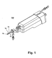
- An example of a known VAB device 200 is shown in Fig. 1.
- Such a VAB device is described in United States Patent No. 6,712,774 and is incorporated by reference in its entirety. The details of the VAB device are not germane to the invention and thus will only be briefly described.
- the probe 70 has a pointed tip 76 to facilitate its insertion through the tissue mass, an opening 78 in the side wall of the probe near the pointed tip, and a vacuum chamber 72.
- a vacuum pump creates a vacuum in chamber 72 and draws the tissue through the opening 78 and into a sampling chamber where a cutting device is advanced through the probe 70 to cut and remove a tissue sample.
- Other instruments can be inserted through the probe 70 in addition to the cutting device.
- the position of the patient during VAB depends on the imaging system used to locate the lesion and position the probe. If ultrasonography is used, the patient will be in a supine position. If mammography is used, the patient typically lies prone on a specialized table such that the breast protrudes through a hole in the table. The breast is compressed between two plates while an image of the lesion is produced on a monitor by a mammography unit. Once the lesion is imaged, the VAB probe, which is mounted to the table or the mammography unit, is inserted into the breast tissue and the tissue sample is gathered as described above.
- a marker that is made of any suitable material that can be imaged by an imaging system, such as ultrasonography, magnetic resonance, or mammography, or that is palpable through the skin and tissue of the patient.
- the marker must be accurately placed at the lesion site in the breast tissue and must remain at the site so that the lesion can be located and identified at a later time, if necessary.
- a marker there sometimes is a need for a marker to be repositioned after its initial placement, such as if the marker was not placed at the desired location or if the marker shifts upon decompression of the tissue.
- the marker must be able to remain anchored in the breast tissue, yet permit its repositioning.
- One type of marker is a biocompatible clip that can be placed at the lesion site to facilitate locating the lesion during later procedures.
- the clip has the advantage of being implanted entirely within the tissue mass, so that there is no possibility of accidental repositioning by pulling or tugging the clip.
- the clip is placed after the tissue sample has been gathered from the lesion site and while the breast is still compressed.
- the clip is inserted into the tissue mass through the VAB probe and thus does not require the tissue mass to be repierced. Since the clip is deployed when the breast tissue is compressed, upon decompression the clip may be found to be implanted away from the lesion site, leading to inaccurate marking of the lesion site.
- An illustrative example of the post-decompression shifting problem is a rubber ball that is normally 5 cm in diameter, but compressed to 2 cm. If a clip is to be placed 1 cm from the edge of the ball, the clip would be placed at the center of the ball. However, if upon decompression of the ball the clip stays at the center of the ball or shifts away from the target site, the clip is misplaced by up to several centimeters. Coopers ligaments in the breast exacerbate the problem of inaccurate marking by acting to pull the clip away from the site of implantation when the breast is uncompressed.
- an apparatus for implanting a locatable marker at a target site within a tissue mass comprises a insertion device comprising a first lumen having an exit opening, a sheath slidably received within the first lumen and comprising a second lumen having a distal opening, a locatable marker received within the second lumen and deployable through the distal opening, and an anchor operably coupled to the sheath to fix the location of the sheath in the tissue mass, wherein the insertion device can be located within the tissue mass and the sheath can be inserted into the tissue mass through the exit opening of the insertion device, and the anchor can fix the position of the sheath in the tissue mass for deployment of the locatable marker at the target site.
- the sheath can comprise a third lumen having a distal opening, with the anchor received within the third lumen and deployable through the distal opening.
- the sheath can comprise a distal terminal end and the distal terminal end can comprise an insertion tip.
- At least one of the sheath distal openings can be formed in the terminal end of the sheath.
- At least one of the sheath distal openings can be formed in a side wall of the sheath.
- At least one of the sheath distal openings formed in the side wall can comprise a ramp to guide the locatable marker through the at least one of the sheath distal openings formed in the side wall.
- the apparatus can further comprise a pushrod slidably received within the second lumen that deploys the locatable marker through the distal opening.
- the anchor can comprise an anchor wire.
- the anchor wire can be operable between a straight configuration where the anchor wire is contained within the third lumen and a curved configuration where the anchor wire is extended through a distal opening.
- the anchor wire can be embedded in the tissue mass in the curved configuration.
- the apparatus can further comprise a cannula received within the first lumen, the cannula comprising a fourth lumen having a distal opening, with the sheath received within the fourth lumen.
- the cannula distal opening can comprise a ramp to guide the sheath through the cannula distal opening.
- the insertion device can be a biopsy probe.
- the probe can be a vacuum-assisted biopsy probe.
- the exit opening can comprise a ramp.
- the sheath can be flexible.
- the sheath can comprise distance markings.
- the apparatus can further comprising a pair of compression plates for compressing the tissue mass prior to location of the insertion device into the tissue mass at the target site and for decompressing the tissue mass prior to implantation of the locatable marker.
- the apparatus can further comprise a hemostatic agent received within the second lumen and deployable through the distal opening.
- the locatable marker can be one of an imaging marker and a palpable marker.
- the locatable marker can be a clip.
- the VAB probe 70 comprises a vacuum chamber 72, a lumen 74, a closed insertion tip 76, and a proximal opening (not shown) into which the introducer system 10 can be inserted.
- An opening 78 in the cannula 72 allows a tissue sample to be taken from a tissue mass as previously described.
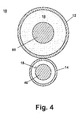
- the introducer system 10 comprises a first sheath 12 and a second sheath 14 which respectively define a first lumen 16 and a second lumen 18.
- the first sheath 12 comprises an open distal insertion tip 22 and an open proximal end (not shown).
- the second sheath 14 comprises an open distal tip 32 and an open proximal end (not shown).
- An anchor wire 40 is contained within the second lumen 18 and a marker in the form of a clip 50 and a pushrod 60 are contained within the first lumen 16.
- the sheaths 12, 14 are preferably independently fabricated from a biocompatible plastic that is flexible and bonded together as shown in Fig. 4.
- the gap between the sheaths 12, 14 and the corresponding pushrod 60 and anchor wire 40 is exaggerated in Fig. 3 to better discern the elements.
- One or both of the sheaths 12, 14 could also be formed from a coiled wire or any other biocompatible material that is sufficiently flexible such that the introducer system 10 can be inserted through the VAB probe and out of the opening 78.
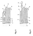
- the anchor wire 40 comprises a hook 42 and a thread 44.
- the hook 42 When mounted in the sheath 14 prior to implantation in the tissue mass, the hook 42 is contained within the second lumen 18 and a portion of the thread 44 extends exteriorly of from the proximal end of the sheath 14.
- the thread 44 is of sufficient length such that the proximal end of the thread 44 is exterior to the proximal end of the sheath 14 to manipulate the anchor wire 40 relative to the sheath 14.
- the hook 42 is fabricated from a resilient, biocompatible material, for example a shape-memory alloy such as Nitinol. This allows the hook to assume a straight first configuration in the lumen as illustrated in Fig. 3, and a curved second configuration outside the lumen as illustrated in Fig. 5.
- a shape-memory alloy such as Nitinol.
- the hook 42 is preferably formed from the same wire as the thread 44 such that the hook is a continuation of the thread with the end of the hook 42 being connected to the thread 42 to complete hook 42.
- the hook 42 and the thread 44 can be formed from different wires and or different materials. In either case, the hook 42 can be bonded or welded to the thread 44 to form the connection.
- the anchor wire 40 can be formed with any one of a number of different anchors.
- the anchor wire 40 can be formed with a diamond or square shaped anchor, a triangular shaped anchor, a circular shaped anchor, or any other anchor shape or type that provides a secure implantation of the introducer system 10 in the tissue mass.
- the shape of the anchor can be selected upon, for example, the density of the tissue into which the wire is to be placed, the size of the lesion, and/or the anchoring force required to implant the introducer system 10 in the tissue mass.
- the introducer system 10 can comprise more than one anchoring device.
- the introducer system 10 can have multiple anchor wires 40 loaded in the second sheath 14, or the introducer system 10 can have multiple sheaths that each hold one anchor wire 40.
- the anchor wires 40 can be configured to engage the tissue mass at different angles to provide for a more secure implantation of the introducer system 10.
- the pushrod 60 comprises a distal end 62 and a proximal end (not shown).
- the distal end 62 is used to force the clip 50 out of the sheath 12 and into the tissue mass.
- the pushrod 60 is of sufficient length such that the proximal end of the pushrod is exterior to the proximal end of the sheath 12 to manipulate the pushrod 60 relative to the sheath 12.
- the pushrod 60 can be made of any material that is sufficiently flexible in order to be threaded through the sheath 12, yet stiff enough to push the clip 50 out of the open tip 22 of the sheath 12.
- the clip 50 can be any suitable type of marker that can be detected and located.
- the clip 50 can be imaged by an imaging technique or palpable through the skin and tissue.
- Types of imagable markers include markers that are echogenic, radiopaque, or a combination of these types.
- the imaging technique used locate the clip 50 can be a standard imaging system such as ultrasonography, mammography or magnetic resonance imaging.
- the clip 50 is deployed into the tissue mass as follows.
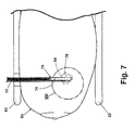
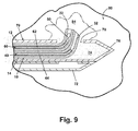
- the VAB probe 70 is inserted into the tissue mass 80 illustrated as a breast that is compressed between two plates 82 and containing a target site 84.
- the target site can comprise a lesion or biopsy site.
- the introducer system 10 is inserted through the open proximal end of the VAB probe 70.
- the introducer system is threaded through lumen 74 and through opening 78 so that the distal tips 22, 32 of the sheaths 12, 14 protrude into the tissue mass 80.
- the introducer system 10 is then secured in the tissue mass 80 using the anchor wire 40.
- the anchor wire 40 is embedded at the target site 84 by moving the thread 44 through lumen 18 relative to the sheath 14 such that the anchor wire 40 emerges from tip 32.
- hook 42 expands from the straightened first configuration to the curved second configuration. As it expands into the surrounding tissue, the hook 42 pierces the adjacent tissue to imbed the anchor wire 40 at the target site 84.
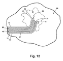
- the tissue mass 80 is uncompressed by removing the compression plates 82.
- the VAB probe is next retracted from the tissue mass 80 as illustrated by an arrow in Fig. 11.
- An image is taken of the tissue mass 80 to determine if the introducer system 10 has been correctly positioned at the target site 84. Correct positioning of the introducer system constitutes a placement that allows the clip 50 to be deployed at the target site 84 and thus is determined by the position of tip 22.
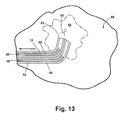
- the clip 50 is implanted in the tissue mass 80 to mark the target site 84.
- the pushrod 60 is moved through lumen 16 relative to the sheath 12 such that the distal end 62 pushing the clip 50 emerges from tip 22 thus deploying clip 50 at the target site 84.
- the pushrod 60 and the anchor wire 40 are then retracted back into their respective sheaths 12 and 14, and the introducer system 10 is retracted from the tissue mass 80, leaving the clip 50 implanted at the target site 84 as illustrated in Fig. 13.
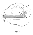
- the introducer system 10 can be repositioned within the tissue mass 80 as shown in Figs. 14-16. Repositioning is normally accomplished with the aid of an ultrasound. In the event of misplacement, it is most often the case that the introducer system 10 is deep to or beyond the target site 84 as illustrated in Fig. 14. Referring to Fig. 15, to reposition the introducer system, the anchor wire 40 is pulled back into lumen 18 by moving the thread 44 relative to the sheath 14. The introducer system 10 is next retracted back an appropriate distance such that the tips 22, 32 are at the target site 84.
- the introducer system is then secured in the tissue mass 80 using the anchor wire 40 and another image can be taken to confirm that the introducer system 10 is correctly positioned at the target site 84.
- the introducer system 10 can be repositioned as many times as necessary until the introducer system 10 is correctly positioned as illustrated in Fig. 16.
- the clip 50 is then implanted in the tissue mass 80 to mark the target site 84 as previously described.
- the sheaths 12, 14 could be provided with distance markings 96, for example centimeter markings that would enable the introducer system to be moved a distance determined from the image taken after the breast is uncompressed. Distance markings on the sheaths 12, 14 allow the introducer system 10 to be repositioned more accurately and reduces the possibility that the introducer system 10 has to be repositioned more than once to achieve correct placement of the introducer system 10.
- the introducer system 10 can be misplaced shallow to or before the target site.
- a hollow cannula can be inserted over the introducer system 10 and then the cannula and introducer system 10 are advanced an appropriate distance to the target site 84. The cannula is next removed and the clip 50 is deployed.
- FIG. 17 also illustrates the optional placement of a hemostatic agent 97 in addition to the placement of the clip 50.
- the hemostatic agent 97 can comprise a soil hemostatic agent such as a plug of collagen, chitosan, thrombin, Factor Xa, fibrinogen, nonsoluble polysaccharide, cellulose and dried gelatin; or a hemostatic agent in liquid form that is coated or impregnated in a bioabsorbable material.
- the hemostatic agent 97 can be loaded into the first sheath 12 along with the clip 50 and can be positioned relative to the clip 50 to be expelled prior to or just after the clip 50 as the push rod 60 is advanced.
- the clip 50 can be coated with or encompassed by the hemostatic agent 97. The presence of the hemostatic agent 97 can prevent the clip 50 from being displaced due to bleeding at the target site 84.
- VAB probe 70 is illustrated as the structure for providing a passageway into the tissue mass for the insertion of the introducer system, it should be noted that other insertion devices can be used and the introducer system is not limited to the VAB probe 70.
- another insertion device can be a cannula with an axial opening or an opening in the side wall.
- FIG. 18 A second embodiment of the introducer system is shown in Figs. 18 and 19 where like elements are identified with the same reference numerals.
- the first sheath 12 has a partition 86 that extends the length of the sheath and divides the sheath 12 into first lumen 16 and second lumen 18.
- Such a configuration has a smaller cross-sectional size as illustrated in Fig. 19 and the clip 50 is deployed in the same manner as described for the first embodiment of the introducer system 10.
- FIG. 20-22 A third embodiment of the introducer system is shown in Figs. 20-22 where like elements are identified with the same reference numerals.
- the tip 32 of the sheath 14 is closed and an opening 36 is provided in a side wall of the sheath 14, near the distal end of the sheath 14.
- the anchor wire 40 As the anchor wire 40 is inserted into the sheath 14 it assumes the straight first configuration as shown in previous illustrations.
- the hook 42 reaches the opening 36, it will assume the curved second configuration as it protrudes from the lumen 18 into the tissue mass to anchor the introducer system 10.
- the thread 44 is pushed forward, forcing the hook 42 through opening 36 and against the closed tip 32.
- the hook 42 remains in the curved second position but is slightly compressed. After repositioning the introducer system 10, the thread 44 is pulled back, and the hook 42 exits the opening 36 to anchor the system into the tissue mass. Referring to Fig. 22, when the introducer system 10 is removed, the thread 44 is pulled back farther such that the hook 42 abuts the proximal edge of the opening 36 and assumes the straight first configuration as the hook 42 enter the second lumen 18.
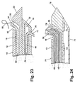
- FIG. 23 A fourth embodiment of the introducer system is shown in Fig. 23 where like elements are identified with the same reference numerals.
- both tips 22, 32 are closed and openings 26, 36 are provided near the proximal end 24, 34 of the sheaths 12, 14, respectively.
- a ramp 28 is provided on the distal side of the opening 26 that occludes lumen 16 and prevents advancement of the clip 50 and the pushrod 60 beyond opening 26. The ramp 28 is angled to guide the clip 50 and the pushrod 60 upward and through the opening 26.
- the VAB probe 70 can be altered in a similar fashion to facilitate the movement of the introducer system 10 out of the probe 70.
- a second embodiment of the probe 70 shown in FIG. 24 where like elements are identified with the same reference numerals, has a ramp 88 formed on the distal side of the opening 78 such that it occludes lumen 74 and prevents the introducer system 10 from advancing beyond the opening 78.
- the ramp 80 is angled to guide the introducer system 10 upwards and through opening 78. While the second embodiment of probe 70 is shown in conjunction with the first embodiment of the introducer system 10, it is understood that any embodiment of the introducer system 10 can be used with the second embodiment of the probe 70.
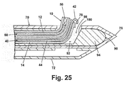
- the introducer system 10 can also be inserted through an outer cannula 90 to facilitate the movement of the introducer system 10 out of the probe 70.
- the cannula 90 defines a lumen 92 and comprises a closed distal end 94 and a proximal end (not shown).
- An opening 98 near the distal end 94 is provided with a ramp 100.
- the opening 98 is located on the cannula 90 such that when the cannula 90 is fully inserted, the opening 98 is aligned with opening 78.
- the cannula 90 is considered to be fully inserted into the probe 70 when the closed end 94 contacts the closed insertion tip 76, thus aligning opening 98 with opening 78.
- the cannula 90 has an outer diameter sized so that is can easily fit through the lumen 74 of the probe 70 and an inner diameter sized so that the introducer system 10 can easily fit through lumen 92.
- the outer cannula 90 is first inserted into the probe 70 and pushed forward until it is fully inserted. Full insertion of the cannula 90 can be determined when resistance is felt against the further forward movement of the cannula 90. Then, the introducer system 10 is inserted into the cannula 90 such that the introducer system 10 is guided up the ramp 100 and out of the opening 98. Next, the introducer system 10 is anchored by the anchor wire 40 and the probe 70 and cannula 90 are simultaneously retracted leaving the introducer system 10 in the tissue mass. The clip 50 is then deployed following the same steps as previously described.
Landscapes
- Health & Medical Sciences (AREA)
- Surgery (AREA)
- Life Sciences & Earth Sciences (AREA)
- Biomedical Technology (AREA)
- Medical Informatics (AREA)
- Oral & Maxillofacial Surgery (AREA)
- Nuclear Medicine, Radiotherapy & Molecular Imaging (AREA)
- Engineering & Computer Science (AREA)
- Veterinary Medicine (AREA)
- Heart & Thoracic Surgery (AREA)
- Pathology (AREA)
- Molecular Biology (AREA)
- Animal Behavior & Ethology (AREA)
- General Health & Medical Sciences (AREA)
- Public Health (AREA)
- Neurosurgery (AREA)
- Surgical Instruments (AREA)
- Prostheses (AREA)
Applications Claiming Priority (1)
| Application Number | Priority Date | Filing Date | Title |
|---|---|---|---|
| US59646705P | 2005-09-26 | 2005-09-26 |
Publications (3)
| Publication Number | Publication Date |
|---|---|
| EP1767167A2 true EP1767167A2 (fr) | 2007-03-28 |
| EP1767167A3 EP1767167A3 (fr) | 2008-04-16 |
| EP1767167B1 EP1767167B1 (fr) | 2011-05-18 |
Family
ID=37591882
Family Applications (1)
| Application Number | Title | Priority Date | Filing Date |
|---|---|---|---|
| EP06121084A Not-in-force EP1767167B1 (fr) | 2005-09-26 | 2006-09-22 | Système pour l'introduction d'un marqueur après décompression |
Country Status (4)
| Country | Link |
|---|---|
| EP (1) | EP1767167B1 (fr) |
| AT (1) | ATE509590T1 (fr) |
| CA (1) | CA2560816C (fr) |
| ES (1) | ES2366102T3 (fr) |
Cited By (31)
| Publication number | Priority date | Publication date | Assignee | Title |
|---|---|---|---|---|
| WO2009105177A1 (fr) * | 2007-07-26 | 2009-08-27 | Senorx, Inc. | Marqueurs polysaccharidiques |
| US8157862B2 (en) | 1997-10-10 | 2012-04-17 | Senorx, Inc. | Tissue marking implant |
| US8177792B2 (en) | 2002-06-17 | 2012-05-15 | Senorx, Inc. | Plugged tip delivery tube for marker placement |
| US8219182B2 (en) | 1999-02-02 | 2012-07-10 | Senorx, Inc. | Cavity-filling biopsy site markers |
| US8224424B2 (en) | 1999-02-02 | 2012-07-17 | Senorx, Inc. | Tissue site markers for in vivo imaging |
| US8311610B2 (en) | 2008-01-31 | 2012-11-13 | C. R. Bard, Inc. | Biopsy tissue marker |
| US8361082B2 (en) | 1999-02-02 | 2013-01-29 | Senorx, Inc. | Marker delivery device with releasable plug |
| US8401622B2 (en) | 2006-12-18 | 2013-03-19 | C. R. Bard, Inc. | Biopsy marker with in situ-generated imaging properties |
| US8437834B2 (en) | 2006-10-23 | 2013-05-07 | C. R. Bard, Inc. | Breast marker |
| US8447386B2 (en) | 2003-05-23 | 2013-05-21 | Senorx, Inc. | Marker or filler forming fluid |
| US8486028B2 (en) | 2005-10-07 | 2013-07-16 | Bard Peripheral Vascular, Inc. | Tissue marking apparatus having drug-eluting tissue marker |
| US8498693B2 (en) | 1999-02-02 | 2013-07-30 | Senorx, Inc. | Intracorporeal marker and marker delivery device |
| US8579931B2 (en) | 1999-06-17 | 2013-11-12 | Bard Peripheral Vascular, Inc. | Apparatus for the percutaneous marking of a lesion |
| US8626269B2 (en) | 2003-05-23 | 2014-01-07 | Senorx, Inc. | Fibrous marker and intracorporeal delivery thereof |
| US8634899B2 (en) | 2003-11-17 | 2014-01-21 | Bard Peripheral Vascular, Inc. | Multi mode imaging marker |
| US8668737B2 (en) | 1997-10-10 | 2014-03-11 | Senorx, Inc. | Tissue marking implant |
| US8670818B2 (en) | 2008-12-30 | 2014-03-11 | C. R. Bard, Inc. | Marker delivery device for tissue marker placement |
| US8718745B2 (en) | 2000-11-20 | 2014-05-06 | Senorx, Inc. | Tissue site markers for in vivo imaging |
| USD715442S1 (en) | 2013-09-24 | 2014-10-14 | C. R. Bard, Inc. | Tissue marker for intracorporeal site identification |
| USD715942S1 (en) | 2013-09-24 | 2014-10-21 | C. R. Bard, Inc. | Tissue marker for intracorporeal site identification |
| USD716450S1 (en) | 2013-09-24 | 2014-10-28 | C. R. Bard, Inc. | Tissue marker for intracorporeal site identification |
| USD716451S1 (en) | 2013-09-24 | 2014-10-28 | C. R. Bard, Inc. | Tissue marker for intracorporeal site identification |
| WO2015023602A1 (fr) * | 2013-08-12 | 2015-02-19 | Liu, David | Dispositif et procédés de biopsie multiple au trocart simultanée 3-dimensionnelle ou placement de marqueurs repères |
| US9327061B2 (en) | 2008-09-23 | 2016-05-03 | Senorx, Inc. | Porous bioabsorbable implant |
| US9579077B2 (en) | 2006-12-12 | 2017-02-28 | C.R. Bard, Inc. | Multiple imaging mode tissue marker |
| EP2155075A4 (fr) * | 2007-04-19 | 2017-07-12 | Searete LLC | Systèmes et procédés de fermeture de d'aponévroses |
| US9820824B2 (en) | 1999-02-02 | 2017-11-21 | Senorx, Inc. | Deployment of polysaccharide markers for treating a site within a patent |
| US9848956B2 (en) | 2002-11-18 | 2017-12-26 | Bard Peripheral Vascular, Inc. | Self-contained, self-piercing, side-expelling marking apparatus |
| US10342635B2 (en) | 2005-04-20 | 2019-07-09 | Bard Peripheral Vascular, Inc. | Marking device with retractable cannula |
| WO2020123350A1 (fr) * | 2018-12-10 | 2020-06-18 | Devicor Medical Products, Inc. | Système de biopsie avec aiguille à déploiement d'extrémité |
| EP3576635A4 (fr) * | 2017-02-06 | 2020-12-02 | Device and Design, LLC | Système, procédé et appareil pour un échantillonnage de tissu intégré et un placement de marqueur de tissu |
Citations (2)
| Publication number | Priority date | Publication date | Assignee | Title |
|---|---|---|---|---|
| US6234177B1 (en) | 1999-08-12 | 2001-05-22 | Thomas Barsch | Apparatus and method for deploying an expandable biopsy marker |
| US6712774B2 (en) | 2000-10-13 | 2004-03-30 | James W. Voegele | Lockout for a surgical biopsy device |
-
2006
- 2006-09-22 ES ES06121084T patent/ES2366102T3/es active Active
- 2006-09-22 AT AT06121084T patent/ATE509590T1/de active
- 2006-09-22 EP EP06121084A patent/EP1767167B1/fr not_active Not-in-force
- 2006-09-25 CA CA2560816A patent/CA2560816C/fr not_active Expired - Fee Related
Patent Citations (2)
| Publication number | Priority date | Publication date | Assignee | Title |
|---|---|---|---|---|
| US6234177B1 (en) | 1999-08-12 | 2001-05-22 | Thomas Barsch | Apparatus and method for deploying an expandable biopsy marker |
| US6712774B2 (en) | 2000-10-13 | 2004-03-30 | James W. Voegele | Lockout for a surgical biopsy device |
Cited By (64)
| Publication number | Priority date | Publication date | Assignee | Title |
|---|---|---|---|---|
| US8668737B2 (en) | 1997-10-10 | 2014-03-11 | Senorx, Inc. | Tissue marking implant |
| US8157862B2 (en) | 1997-10-10 | 2012-04-17 | Senorx, Inc. | Tissue marking implant |
| US9039763B2 (en) | 1997-10-10 | 2015-05-26 | Senorx, Inc. | Tissue marking implant |
| US8224424B2 (en) | 1999-02-02 | 2012-07-17 | Senorx, Inc. | Tissue site markers for in vivo imaging |
| US9237937B2 (en) | 1999-02-02 | 2016-01-19 | Senorx, Inc. | Cavity-filling biopsy site markers |
| US9044162B2 (en) | 1999-02-02 | 2015-06-02 | Senorx, Inc. | Marker delivery device with releasable plug |
| US8361082B2 (en) | 1999-02-02 | 2013-01-29 | Senorx, Inc. | Marker delivery device with releasable plug |
| US10172674B2 (en) | 1999-02-02 | 2019-01-08 | Senorx, Inc. | Intracorporeal marker and marker delivery device |
| US9861294B2 (en) | 1999-02-02 | 2018-01-09 | Senorx, Inc. | Marker delivery device with releasable plug |
| US9149341B2 (en) | 1999-02-02 | 2015-10-06 | Senorx, Inc | Deployment of polysaccharide markers for treating a site within a patient |
| US9820824B2 (en) | 1999-02-02 | 2017-11-21 | Senorx, Inc. | Deployment of polysaccharide markers for treating a site within a patent |
| US8498693B2 (en) | 1999-02-02 | 2013-07-30 | Senorx, Inc. | Intracorporeal marker and marker delivery device |
| US8219182B2 (en) | 1999-02-02 | 2012-07-10 | Senorx, Inc. | Cavity-filling biopsy site markers |
| US9649093B2 (en) | 1999-02-02 | 2017-05-16 | Senorx, Inc. | Cavity-filling biopsy site markers |
| US8626270B2 (en) | 1999-02-02 | 2014-01-07 | Senorx, Inc. | Cavity-filling biopsy site markers |
| US8965486B2 (en) | 1999-02-02 | 2015-02-24 | Senorx, Inc. | Cavity filling biopsy site markers |
| US9579159B2 (en) | 1999-06-17 | 2017-02-28 | Bard Peripheral Vascular, Inc. | Apparatus for the percutaneous marking of a lesion |
| US8579931B2 (en) | 1999-06-17 | 2013-11-12 | Bard Peripheral Vascular, Inc. | Apparatus for the percutaneous marking of a lesion |
| US8718745B2 (en) | 2000-11-20 | 2014-05-06 | Senorx, Inc. | Tissue site markers for in vivo imaging |
| US8784433B2 (en) | 2002-06-17 | 2014-07-22 | Senorx, Inc. | Plugged tip delivery tube for marker placement |
| US8177792B2 (en) | 2002-06-17 | 2012-05-15 | Senorx, Inc. | Plugged tip delivery tube for marker placement |
| US9848956B2 (en) | 2002-11-18 | 2017-12-26 | Bard Peripheral Vascular, Inc. | Self-contained, self-piercing, side-expelling marking apparatus |
| US10813716B2 (en) | 2002-11-18 | 2020-10-27 | Bard Peripheral Vascular, Inc. | Self-contained, self-piercing, side-expelling marking apparatus |
| US8639315B2 (en) | 2003-05-23 | 2014-01-28 | Senorx, Inc. | Marker or filler forming fluid |
| US9801688B2 (en) | 2003-05-23 | 2017-10-31 | Senorx, Inc. | Fibrous marker and intracorporeal delivery thereof |
| US8880154B2 (en) | 2003-05-23 | 2014-11-04 | Senorx, Inc. | Fibrous marker and intracorporeal delivery thereof |
| US10045832B2 (en) | 2003-05-23 | 2018-08-14 | Senorx, Inc. | Marker or filler forming fluid |
| US8447386B2 (en) | 2003-05-23 | 2013-05-21 | Senorx, Inc. | Marker or filler forming fluid |
| US8626269B2 (en) | 2003-05-23 | 2014-01-07 | Senorx, Inc. | Fibrous marker and intracorporeal delivery thereof |
| US10299881B2 (en) | 2003-05-23 | 2019-05-28 | Senorx, Inc. | Marker or filler forming fluid |
| US8634899B2 (en) | 2003-11-17 | 2014-01-21 | Bard Peripheral Vascular, Inc. | Multi mode imaging marker |
| US10357328B2 (en) | 2005-04-20 | 2019-07-23 | Bard Peripheral Vascular, Inc. and Bard Shannon Limited | Marking device with retractable cannula |
| US10342635B2 (en) | 2005-04-20 | 2019-07-09 | Bard Peripheral Vascular, Inc. | Marking device with retractable cannula |
| US11278370B2 (en) | 2005-04-20 | 2022-03-22 | Bard Peripheral Vascular, Inc. | Marking device with retractable cannula |
| US8486028B2 (en) | 2005-10-07 | 2013-07-16 | Bard Peripheral Vascular, Inc. | Tissue marking apparatus having drug-eluting tissue marker |
| US8437834B2 (en) | 2006-10-23 | 2013-05-07 | C. R. Bard, Inc. | Breast marker |
| US9579077B2 (en) | 2006-12-12 | 2017-02-28 | C.R. Bard, Inc. | Multiple imaging mode tissue marker |
| US10682200B2 (en) | 2006-12-12 | 2020-06-16 | C. R. Bard, Inc. | Multiple imaging mode tissue marker |
| US9901415B2 (en) | 2006-12-12 | 2018-02-27 | C. R. Bard, Inc. | Multiple imaging mode tissue marker |
| US11471244B2 (en) | 2006-12-12 | 2022-10-18 | C.R. Bard, Inc. | Multiple imaging mode tissue marker |
| US8401622B2 (en) | 2006-12-18 | 2013-03-19 | C. R. Bard, Inc. | Biopsy marker with in situ-generated imaging properties |
| US9042965B2 (en) | 2006-12-18 | 2015-05-26 | C. R. Bard, Inc. | Biopsy marker with in situ-generated imaging properties |
| EP2155075A4 (fr) * | 2007-04-19 | 2017-07-12 | Searete LLC | Systèmes et procédés de fermeture de d'aponévroses |
| WO2009105177A1 (fr) * | 2007-07-26 | 2009-08-27 | Senorx, Inc. | Marqueurs polysaccharidiques |
| US8311610B2 (en) | 2008-01-31 | 2012-11-13 | C. R. Bard, Inc. | Biopsy tissue marker |
| US9327061B2 (en) | 2008-09-23 | 2016-05-03 | Senorx, Inc. | Porous bioabsorbable implant |
| US11833275B2 (en) | 2008-09-23 | 2023-12-05 | Senorx, Inc. | Porous bioabsorbable implant |
| US10786604B2 (en) | 2008-09-23 | 2020-09-29 | Senorx, Inc. | Porous bioabsorbable implant |
| US8670818B2 (en) | 2008-12-30 | 2014-03-11 | C. R. Bard, Inc. | Marker delivery device for tissue marker placement |
| US10258428B2 (en) | 2008-12-30 | 2019-04-16 | C. R. Bard, Inc. | Marker delivery device for tissue marker placement |
| US11779431B2 (en) | 2008-12-30 | 2023-10-10 | C. R. Bard, Inc. | Marker delivery device for tissue marker placement |
| CN105473078A (zh) * | 2013-08-12 | 2016-04-06 | 大卫·刘 | 三维同时多芯活检或放置基准标记的装置和方法 |
| US10149700B2 (en) | 2013-08-12 | 2018-12-11 | Jan R. Lau | 3 dimensional simultaneous multiple core biopsy or fiducial marker placement device and methods |
| WO2015023602A1 (fr) * | 2013-08-12 | 2015-02-19 | Liu, David | Dispositif et procédés de biopsie multiple au trocart simultanée 3-dimensionnelle ou placement de marqueurs repères |
| CN105473078B (zh) * | 2013-08-12 | 2019-02-22 | 大卫·刘 | 三维同时多芯活检或放置基准标记的装置和方法 |
| USD715442S1 (en) | 2013-09-24 | 2014-10-14 | C. R. Bard, Inc. | Tissue marker for intracorporeal site identification |
| USD715942S1 (en) | 2013-09-24 | 2014-10-21 | C. R. Bard, Inc. | Tissue marker for intracorporeal site identification |
| USD716450S1 (en) | 2013-09-24 | 2014-10-28 | C. R. Bard, Inc. | Tissue marker for intracorporeal site identification |
| USD716451S1 (en) | 2013-09-24 | 2014-10-28 | C. R. Bard, Inc. | Tissue marker for intracorporeal site identification |
| EP3576635A4 (fr) * | 2017-02-06 | 2020-12-02 | Device and Design, LLC | Système, procédé et appareil pour un échantillonnage de tissu intégré et un placement de marqueur de tissu |
| WO2020123350A1 (fr) * | 2018-12-10 | 2020-06-18 | Devicor Medical Products, Inc. | Système de biopsie avec aiguille à déploiement d'extrémité |
| CN113164168A (zh) * | 2018-12-10 | 2021-07-23 | Devicor医疗产业收购公司 | 带有端部部署针的活组织检查系统 |
| EP4193932A1 (fr) * | 2018-12-10 | 2023-06-14 | Devicor Medical Products, Inc. | Système de biopsie avec aiguille à déploiement d'extrémité |
| CN113164168B (zh) * | 2018-12-10 | 2024-04-12 | Devicor医疗产业收购公司 | 带有端部部署针的活组织检查系统 |
Also Published As
| Publication number | Publication date |
|---|---|
| ATE509590T1 (de) | 2011-06-15 |
| ES2366102T3 (es) | 2011-10-17 |
| EP1767167A3 (fr) | 2008-04-16 |
| CA2560816A1 (fr) | 2007-03-26 |
| CA2560816C (fr) | 2013-12-24 |
| EP1767167B1 (fr) | 2011-05-18 |
Similar Documents
| Publication | Publication Date | Title |
|---|---|---|
| EP1767167B1 (fr) | Système pour l'introduction d'un marqueur après décompression | |
| US8419656B2 (en) | Post decompression marker introducer system | |
| CA2002763C (fr) | Dispositif de localisation d'aiguille a structure renforcee | |
| US6181960B1 (en) | Biopsy marker device | |
| US8287463B2 (en) | Removable localizing wire | |
| US6758855B2 (en) | Target tissue localization device | |
| EP2116191A1 (fr) | Système pour fermer une blessure vasculaire | |
| US20050119562A1 (en) | Fibrous marker formed of synthetic polymer strands | |
| US20080039819A1 (en) | Marker formed of starch or other suitable polysaccharide | |
| WO1992012678A1 (fr) | Ensemble comportant une aiguille de localisation | |
| US20230000587A1 (en) | Non-migrating biopsy site identifiers | |
| US9999411B2 (en) | Vascular closure device with a plug having a variable expansion rate and method for using the same | |
| US20230000585A1 (en) | Marking element for marking tissue | |
| AU2005302639A1 (en) | Fibrous marker formed of synthetic polymer strands | |
| US20240058092A1 (en) | Biopsy site marker having expandable portion | |
| WO2023215090A1 (fr) | Marqueur de site de biopsie présentant des caractéristiques de visualisation et de non-migration accrues | |
| WO2024039561A1 (fr) | Marqueur de site de biopsie ayant des parties mobiles |
Legal Events
| Date | Code | Title | Description |
|---|---|---|---|
| PUAI | Public reference made under article 153(3) epc to a published international application that has entered the european phase |
Free format text: ORIGINAL CODE: 0009012 |
|
| AK | Designated contracting states |
Kind code of ref document: A2 Designated state(s): AT BE BG CH CY CZ DE DK EE ES FI FR GB GR HU IE IS IT LI LT LU LV MC NL PL PT RO SE SI SK TR |
|
| AX | Request for extension of the european patent |
Extension state: AL BA HR MK YU |
|
| RAP1 | Party data changed (applicant data changed or rights of an application transferred) |
Owner name: BARD PERIPHERAL VASCULAR, INC. Owner name: BARD SHANNON LIMITED |
|
| PUAL | Search report despatched |
Free format text: ORIGINAL CODE: 0009013 |
|
| AK | Designated contracting states |
Kind code of ref document: A3 Designated state(s): AT BE BG CH CY CZ DE DK EE ES FI FR GB GR HU IE IS IT LI LT LU LV MC NL PL PT RO SE SI SK TR |
|
| AX | Request for extension of the european patent |
Extension state: AL BA HR MK RS |
|
| RIC1 | Information provided on ipc code assigned before grant |
Ipc: A61B 19/00 20060101AFI20070111BHEP Ipc: A61B 10/02 20060101ALI20080310BHEP |
|
| 17P | Request for examination filed |
Effective date: 20081008 |
|
| 17Q | First examination report despatched |
Effective date: 20081104 |
|
| AKX | Designation fees paid |
Designated state(s): AT BE BG CH CY CZ DE DK EE ES FI FR GB GR HU IE IS IT LI LT LU LV MC NL PL PT RO SE SI SK TR |
|
| GRAP | Despatch of communication of intention to grant a patent |
Free format text: ORIGINAL CODE: EPIDOSNIGR1 |
|
| GRAS | Grant fee paid |
Free format text: ORIGINAL CODE: EPIDOSNIGR3 |
|
| GRAA | (expected) grant |
Free format text: ORIGINAL CODE: 0009210 |
|
| AK | Designated contracting states |
Kind code of ref document: B1 Designated state(s): AT BE BG CH CY CZ DE DK EE ES FI FR GB GR HU IE IS IT LI LT LU LV MC NL PL PT RO SE SI SK TR |
|
| REG | Reference to a national code |
Ref country code: GB Ref legal event code: FG4D |
|
| REG | Reference to a national code |
Ref country code: CH Ref legal event code: EP |
|
| REG | Reference to a national code |
Ref country code: IE Ref legal event code: FG4D |
|
| REG | Reference to a national code |
Ref country code: DE Ref legal event code: R096 Ref document number: 602006021998 Country of ref document: DE Effective date: 20110630 |
|
| REG | Reference to a national code |
Ref country code: NL Ref legal event code: T3 |
|
| REG | Reference to a national code |
Ref country code: ES Ref legal event code: FG2A Ref document number: 2366102 Country of ref document: ES Kind code of ref document: T3 Effective date: 20111017 |
|
| PG25 | Lapsed in a contracting state [announced via postgrant information from national office to epo] |
Ref country code: SE Free format text: LAPSE BECAUSE OF FAILURE TO SUBMIT A TRANSLATION OF THE DESCRIPTION OR TO PAY THE FEE WITHIN THE PRESCRIBED TIME-LIMIT Effective date: 20110518 Ref country code: LT Free format text: LAPSE BECAUSE OF FAILURE TO SUBMIT A TRANSLATION OF THE DESCRIPTION OR TO PAY THE FEE WITHIN THE PRESCRIBED TIME-LIMIT Effective date: 20110518 Ref country code: PT Free format text: LAPSE BECAUSE OF FAILURE TO SUBMIT A TRANSLATION OF THE DESCRIPTION OR TO PAY THE FEE WITHIN THE PRESCRIBED TIME-LIMIT Effective date: 20110919 |
|
| PG25 | Lapsed in a contracting state [announced via postgrant information from national office to epo] |
Ref country code: IS Free format text: LAPSE BECAUSE OF FAILURE TO SUBMIT A TRANSLATION OF THE DESCRIPTION OR TO PAY THE FEE WITHIN THE PRESCRIBED TIME-LIMIT Effective date: 20110918 Ref country code: SI Free format text: LAPSE BECAUSE OF FAILURE TO SUBMIT A TRANSLATION OF THE DESCRIPTION OR TO PAY THE FEE WITHIN THE PRESCRIBED TIME-LIMIT Effective date: 20110518 Ref country code: CY Free format text: LAPSE BECAUSE OF FAILURE TO SUBMIT A TRANSLATION OF THE DESCRIPTION OR TO PAY THE FEE WITHIN THE PRESCRIBED TIME-LIMIT Effective date: 20110518 Ref country code: LV Free format text: LAPSE BECAUSE OF FAILURE TO SUBMIT A TRANSLATION OF THE DESCRIPTION OR TO PAY THE FEE WITHIN THE PRESCRIBED TIME-LIMIT Effective date: 20110518 Ref country code: FI Free format text: LAPSE BECAUSE OF FAILURE TO SUBMIT A TRANSLATION OF THE DESCRIPTION OR TO PAY THE FEE WITHIN THE PRESCRIBED TIME-LIMIT Effective date: 20110518 Ref country code: GR Free format text: LAPSE BECAUSE OF FAILURE TO SUBMIT A TRANSLATION OF THE DESCRIPTION OR TO PAY THE FEE WITHIN THE PRESCRIBED TIME-LIMIT Effective date: 20110819 |
|
| PG25 | Lapsed in a contracting state [announced via postgrant information from national office to epo] |
Ref country code: EE Free format text: LAPSE BECAUSE OF FAILURE TO SUBMIT A TRANSLATION OF THE DESCRIPTION OR TO PAY THE FEE WITHIN THE PRESCRIBED TIME-LIMIT Effective date: 20110518 Ref country code: CZ Free format text: LAPSE BECAUSE OF FAILURE TO SUBMIT A TRANSLATION OF THE DESCRIPTION OR TO PAY THE FEE WITHIN THE PRESCRIBED TIME-LIMIT Effective date: 20110518 |
|
| PG25 | Lapsed in a contracting state [announced via postgrant information from national office to epo] |
Ref country code: SK Free format text: LAPSE BECAUSE OF FAILURE TO SUBMIT A TRANSLATION OF THE DESCRIPTION OR TO PAY THE FEE WITHIN THE PRESCRIBED TIME-LIMIT Effective date: 20110518 Ref country code: PL Free format text: LAPSE BECAUSE OF FAILURE TO SUBMIT A TRANSLATION OF THE DESCRIPTION OR TO PAY THE FEE WITHIN THE PRESCRIBED TIME-LIMIT Effective date: 20110518 Ref country code: DK Free format text: LAPSE BECAUSE OF FAILURE TO SUBMIT A TRANSLATION OF THE DESCRIPTION OR TO PAY THE FEE WITHIN THE PRESCRIBED TIME-LIMIT Effective date: 20110518 Ref country code: RO Free format text: LAPSE BECAUSE OF FAILURE TO SUBMIT A TRANSLATION OF THE DESCRIPTION OR TO PAY THE FEE WITHIN THE PRESCRIBED TIME-LIMIT Effective date: 20110518 |
|
| PLBE | No opposition filed within time limit |
Free format text: ORIGINAL CODE: 0009261 |
|
| STAA | Information on the status of an ep patent application or granted ep patent |
Free format text: STATUS: NO OPPOSITION FILED WITHIN TIME LIMIT |
|
| 26N | No opposition filed |
Effective date: 20120221 |
|
| PG25 | Lapsed in a contracting state [announced via postgrant information from national office to epo] |
Ref country code: MC Free format text: LAPSE BECAUSE OF NON-PAYMENT OF DUE FEES Effective date: 20110930 |
|
| REG | Reference to a national code |
Ref country code: CH Ref legal event code: PL |
|
| REG | Reference to a national code |
Ref country code: DE Ref legal event code: R097 Ref document number: 602006021998 Country of ref document: DE Effective date: 20120221 |
|
| REG | Reference to a national code |
Ref country code: IE Ref legal event code: MM4A |
|
| PG25 | Lapsed in a contracting state [announced via postgrant information from national office to epo] |
Ref country code: IE Free format text: LAPSE BECAUSE OF NON-PAYMENT OF DUE FEES Effective date: 20110922 Ref country code: CH Free format text: LAPSE BECAUSE OF NON-PAYMENT OF DUE FEES Effective date: 20110930 Ref country code: LI Free format text: LAPSE BECAUSE OF NON-PAYMENT OF DUE FEES Effective date: 20110930 |
|
| PG25 | Lapsed in a contracting state [announced via postgrant information from national office to epo] |
Ref country code: LU Free format text: LAPSE BECAUSE OF NON-PAYMENT OF DUE FEES Effective date: 20110922 |
|
| PG25 | Lapsed in a contracting state [announced via postgrant information from national office to epo] |
Ref country code: BG Free format text: LAPSE BECAUSE OF FAILURE TO SUBMIT A TRANSLATION OF THE DESCRIPTION OR TO PAY THE FEE WITHIN THE PRESCRIBED TIME-LIMIT Effective date: 20110818 |
|
| PG25 | Lapsed in a contracting state [announced via postgrant information from national office to epo] |
Ref country code: TR Free format text: LAPSE BECAUSE OF FAILURE TO SUBMIT A TRANSLATION OF THE DESCRIPTION OR TO PAY THE FEE WITHIN THE PRESCRIBED TIME-LIMIT Effective date: 20110518 |
|
| PG25 | Lapsed in a contracting state [announced via postgrant information from national office to epo] |
Ref country code: HU Free format text: LAPSE BECAUSE OF FAILURE TO SUBMIT A TRANSLATION OF THE DESCRIPTION OR TO PAY THE FEE WITHIN THE PRESCRIBED TIME-LIMIT Effective date: 20110518 |
|
| PGFP | Annual fee paid to national office [announced via postgrant information from national office to epo] |
Ref country code: AT Payment date: 20130828 Year of fee payment: 8 |
|
| REG | Reference to a national code |
Ref country code: AT Ref legal event code: MM01 Ref document number: 509590 Country of ref document: AT Kind code of ref document: T Effective date: 20140922 |
|
| PG25 | Lapsed in a contracting state [announced via postgrant information from national office to epo] |
Ref country code: AT Free format text: LAPSE BECAUSE OF NON-PAYMENT OF DUE FEES Effective date: 20140922 |
|
| REG | Reference to a national code |
Ref country code: FR Ref legal event code: PLFP Year of fee payment: 11 |
|
| REG | Reference to a national code |
Ref country code: FR Ref legal event code: PLFP Year of fee payment: 12 |
|
| REG | Reference to a national code |
Ref country code: FR Ref legal event code: PLFP Year of fee payment: 13 |
|
| PGFP | Annual fee paid to national office [announced via postgrant information from national office to epo] |
Ref country code: NL Payment date: 20190826 Year of fee payment: 14 |
|
| PGFP | Annual fee paid to national office [announced via postgrant information from national office to epo] |
Ref country code: IT Payment date: 20190829 Year of fee payment: 14 |
|
| PGFP | Annual fee paid to national office [announced via postgrant information from national office to epo] |
Ref country code: BE Payment date: 20190823 Year of fee payment: 14 |
|
| PGFP | Annual fee paid to national office [announced via postgrant information from national office to epo] |
Ref country code: ES Payment date: 20191001 Year of fee payment: 14 |
|
| REG | Reference to a national code |
Ref country code: NL Ref legal event code: MM Effective date: 20201001 |
|
| REG | Reference to a national code |
Ref country code: BE Ref legal event code: MM Effective date: 20200930 |
|
| PG25 | Lapsed in a contracting state [announced via postgrant information from national office to epo] |
Ref country code: NL Free format text: LAPSE BECAUSE OF NON-PAYMENT OF DUE FEES Effective date: 20201001 |
|
| PG25 | Lapsed in a contracting state [announced via postgrant information from national office to epo] |
Ref country code: BE Free format text: LAPSE BECAUSE OF NON-PAYMENT OF DUE FEES Effective date: 20200930 |
|
| PG25 | Lapsed in a contracting state [announced via postgrant information from national office to epo] |
Ref country code: IT Free format text: LAPSE BECAUSE OF NON-PAYMENT OF DUE FEES Effective date: 20200922 |
|
| REG | Reference to a national code |
Ref country code: ES Ref legal event code: FD2A Effective date: 20220118 |
|
| PG25 | Lapsed in a contracting state [announced via postgrant information from national office to epo] |
Ref country code: ES Free format text: LAPSE BECAUSE OF NON-PAYMENT OF DUE FEES Effective date: 20200923 |
|
| PGFP | Annual fee paid to national office [announced via postgrant information from national office to epo] |
Ref country code: GB Payment date: 20220818 Year of fee payment: 17 Ref country code: DE Payment date: 20220616 Year of fee payment: 17 |
|
| PGFP | Annual fee paid to national office [announced via postgrant information from national office to epo] |
Ref country code: FR Payment date: 20220819 Year of fee payment: 17 |
|
| REG | Reference to a national code |
Ref country code: DE Ref legal event code: R119 Ref document number: 602006021998 Country of ref document: DE |
|
| GBPC | Gb: european patent ceased through non-payment of renewal fee |
Effective date: 20230922 |
|
| PG25 | Lapsed in a contracting state [announced via postgrant information from national office to epo] |
Ref country code: GB Free format text: LAPSE BECAUSE OF NON-PAYMENT OF DUE FEES Effective date: 20230922 |
|
| PG25 | Lapsed in a contracting state [announced via postgrant information from national office to epo] |
Ref country code: GB Free format text: LAPSE BECAUSE OF NON-PAYMENT OF DUE FEES Effective date: 20230922 Ref country code: FR Free format text: LAPSE BECAUSE OF NON-PAYMENT OF DUE FEES Effective date: 20230930 Ref country code: DE Free format text: LAPSE BECAUSE OF NON-PAYMENT OF DUE FEES Effective date: 20240403 |