EP1766411B1 - Method for determining a tissue degradation process by detection of comp neoepitopes - Google Patents
Method for determining a tissue degradation process by detection of comp neoepitopes Download PDFInfo
- Publication number
- EP1766411B1 EP1766411B1 EP05748087A EP05748087A EP1766411B1 EP 1766411 B1 EP1766411 B1 EP 1766411B1 EP 05748087 A EP05748087 A EP 05748087A EP 05748087 A EP05748087 A EP 05748087A EP 1766411 B1 EP1766411 B1 EP 1766411B1
- Authority
- EP
- European Patent Office
- Prior art keywords
- comp
- neoepitopes
- fragments
- fragment
- antibodies
- Prior art date
- Legal status (The legal status is an assumption and is not a legal conclusion. Google has not performed a legal analysis and makes no representation as to the accuracy of the status listed.)
- Expired - Lifetime
Links
Images


Classifications
-
- C—CHEMISTRY; METALLURGY
- C07—ORGANIC CHEMISTRY
- C07K—PEPTIDES
- C07K14/00—Peptides having more than 20 amino acids; Gastrins; Somatostatins; Melanotropins; Derivatives thereof
- C07K14/435—Peptides having more than 20 amino acids; Gastrins; Somatostatins; Melanotropins; Derivatives thereof from animals; from humans
- C07K14/78—Connective tissue peptides, e.g. collagen, elastin, laminin, fibronectin, vitronectin or cold insoluble globulin [CIG]
Definitions
- the present Invention relates to a method for determining a tissue degradation process by detection of COMP (Cartilage Oligomeric Matrix Protein) neoepitopes, to antibodies against COMP neoepitopes and to fragments containing COMP neoepitopes for generation of such antibodies.
- COMP Cartilage Oligomeric Matrix Protein
- Tissue degradation processes are characterized by the break down of tissue components.
- the tissue components can be degraded due to e.g. mechanical stress, toxic compounds or by enzymes.
- determination of tissue degradation processes for the purpose of diagnosis, disease monitoring, treatment etc. can be performed by numerous methods.
- One way to determine degradation processes in connective tissue diseases, such as arthritic conditions; arteriosclerosis, degenerative joint conditions etc. is the detection of the presence of degradation products of the connective tissue components. This allows direct detection of the degradation process, compared to Indirect methods as e.g. measuring increased amounts of leukocytes, which have been widely employed in the diagnosis of inflammatory processes such as arthritic conditions.
- COMP Ceretid arthritis
- Ultisol et al. J. Clin. Invest (1995), vol. 95, pp. 1077-1077 : Wolhein et al., British Journal of Rheumatology (1997), vol. 36, pp 847-849 ; Petersson et al., British Journal of Rheumatology (1998), vol 37, pp 46-50 ).
- Significant amounts of small fragments of COMP have also been found in synovial fluid from patients with rheumatoid arthritis and other forms of inflammatory arthritis ( Neidhardt et al., British Journal of Rheumatology (1997), vol. 36, pp 1151-1160 ).
- WO 9807035 discloses a method for the determination of tissue degradation by screening for the presence of one or more neoepitopes of COMP that appear after cleavage of COMP.
- the inventors of the present invention have developed a method for the determination of tissue degradation processes by using specific fragments of COMP as a marker for tissue degradation.
- fragments of COMP as a marker for tissue degradation processes, a much higher signal to noise ratio is obtained and thereby the problem having false positive test results are minimized or almost avoided.
- the higher signal to noise ratio of the present method also allows earlier detection of the tissue degradation process, which is an important factor in diagnosis of degenerative conditions, as this allows early treatment of the disease and thereby a much higher chance for recovery.
- the present invention relates to a method for determining a tissue degradation process comprising detecting in a sample the presence of one or more neoepitopes of mammalian COMP that appear after cleavage of COMP between position 625 (N 625 ) and 626 (P 626 ) of the amino acid sequence of COMP (SEQ ID No: 1).
- the method can be used for diagnosis, disease monitoring and/or therapeutic monitoring of the tissue degradation process.
- the degradation process can take place in a connective tissue, such as e.g. cartilage, tendon, ligaments, bone, blood vessels, intervertebral discs and meniscus.
- the detection of COMP fragment specific neoepitopes can be performed by using antibodies against the COMP neoepitopes, thus the present invention also relates to such antibodies and the use thereof. Furthermore the invention discloses isolated fragments of COMP comprising one or more neoepitopes, for use in the production of antibodies against COMP neoepitopes as defined herein.
- Degradation of structures in articular cartilage is seen typically in all diseases resulting chronically in the destruction of the joint structure.
- diseases may be mentioned rheumatoid arthritis, psoriatic arthritis, and osteoarthritis/osteoarthrosis.
- Acute inflammation of a joint e.g. in reactive arthritis, trauma etc, is often accompanied by a destruction of cartilage, although in most cases this will not develop into the chronically destructive disease. It is not known which factors are crucial for the acutely inflamed joint to either proceed to healing or develop into the chronic process.
- Other diseases associated with tissue changes are inflammatory joint diseases such as, e.g., juvenile chronic arthritis, systemic lupus erythematosus (SLE), infectious arthritis and juvenile idiopathic arthritis, other diseases involving joint structures such as, e.g., osteoarthritis, meniscal damage, gout, diseases involving the intervertebral disc of the spine, such as, e.g., lower back pain, diseases affecting structural elements of the musculoskeletal system such as, e.g., tendon damage, ligament damage, and bone disease such as, e.g., osteoporosis, diseases affecting major arteries including atherosclerosis, vasculitis and trauma leading to injury, lung diseases including asthma and COPD.
- joint diseases such as, e.g., juvenile chronic arthritis, systemic lupus erythematosus (SLE), infectious arthritis and juvenile idiopathic arthritis
- other diseases involving joint structures such as, e.g., osteoarthritis, meniscal
- An important object of the present invention is to improve the diagnostic possibilities of tissue degradation processes, particularly in early stages, and to provide a means of monitoring the effects of therapeutical measures taken.
- a method according to the present invention may make it possible to differentiate between tissue degradation processes like e.g. rheumatoid arthritis (RA) and osteoarthritis/osteoarthrosis (OA).
- RA rheumatoid arthritis
- OA osteoarthritis/osteoarthrosis
- the inventors of the present invention have developed a reliable method for the determination of tissue degradation processes by measuring the presence of COMP fragments in the tissue and/or in the fluids in direct or indirect contact with the tissue.
- the present method allows a reliable and early detection of the tissue degradation process, which is an important factor in diagnosis of degenerative conditions, as this allows early treatment of the disease and thereby a much higher chance for recovery.
- the early detection of the tissue degradation process also allows monitoring of therapy efficiency, and identification of patients with an active tissue degradation process, which is suitable for therapy.
- the method is based on the observation that COMP neoepitopes are formed during e.g. cartilage degradation.
- the delineation of specific fragments of COMP by the inventors allows identification of COMP neoepitopes, which can be used in the determination of the tissue degradation.
- the COMP fragments containing COMP neoepitopes can be employed in the production of antibodies against COMP neoepitopes, which may be used in the detection of the presence of COMP neoepitopes in tissue degradation processes.
- COMP Cartilage Oligomeric Matrix Protein also referred to as Thrombospondin 5
- Thrombospondin 5 is prominent in cartilage, intervertebral disc but is also present in tendon
- Cartilage matrix proteins An acidic oligomeric protein (COMP) detected only in cartilage. E. Hedborn, P. Antonsson. A. Hjerpe, D. Aeschlimann, M. Paulsson, E. Rosa-Pimentel, Y. Sommarin, M. Wendel, A. Oldberg and D. Heineg ⁇ rd. J. Biol. Chem. (1992) 267, 6132-6136 ., The distribution of cartilage oligomeric matrix protein (COMP) in tendon and its variation with tendon site, age and load. R. Smith, L. Zunino, P. Webbon and D. Heineg ⁇ rd. Matrix Biology (1997) 16, 255-271 ).
- the protein has a role in formation of the collagen network, an important structural entity in cartilage. Thus, it has been shown to bind to collagen with high affinity ( Cartilage oligomeric matrix protein shows high affinity. Zn-dependent interaction with triple helical collagen. K. Rosenberg, H. Olsson, M. Mörgelin and D. Heineg ⁇ rd. J. Biol. Chem. (1998) 273, 20397-20403 .).
- Cartilage oligomeric matrix protein A novel marker of cartilage turnover detectable in synovial fluid and blood.
- Cartilage and bone metabolism in rheumatoid arthritis Differences between rapid and slow progression of disease identified by serum markers of cartilage metabolism.
- the present inventors therefore undertook to study COMP fragments in a tissue, which is undergoing degeneration with apparent breakdown and alteration of the tissue matrix. This tissue, the intervertebral disc from elderly people was further studied.
- COMP was extracted and purified. Fragments were separated and the terminus of two such fragments determined, thereby each defining two new epitopes (neoepitopes), that is the peptide sequence on the N-terminal side of the cleavage (defining a new C-terminus) and the peptide sequence on the C-terminal side of the cleavage (defining a new N-terminus). Antibodies have been raised to one of these peptide sequences as demonstrated to react with the fragment, while not with the intact COMP.
- tissue degradation process is defined as any stage from the beginning to the end of a break down process in tissue.
- neoepitope is defined as an epitope, which is not normally exposed on normal tissue, cells or molecules. Phenotypic changes in a cell may include loss of normal cell surface antigenic epitope components and the gain of new neoepitopes. Other ways of gaining neoepitopes are by structural changes of molecules, such as proteins.
- a neoepitope is an epitope that is not normally exposed on COMP, but appears after changes of the protein. Examples of changes may be alterations in folding of the protein resulting in structural changes of the secondary and/or tertiary structure of COMP. A change of COMP may also result from e.g. cleavage of the protein.
- the neoepitope may for example be formed by a primary, secondary and/or tertiary structure of a COMP fragment appearing after cleavage.
- fragment is defined as a degradation product of a protein generated by the break at one or more points of the protein back bone.
- fragment is defined as a fragment of COMP generated by the degradation of COMP such as e.g. by cleavage of the protein.
- the fragment can be generated either by degradation of COMP in vivo or in vitro optionally followed by isolation of the fragment or the fragment can be generated in vitro by expressing parts of the COMP molecule using a synthetic, semisyntheticand/or recombinant method.
- peptide is defined as a sequence of amino acids.
- the amino acids present in the sequence can be any natural or unnatural, substituted or unsubstituted amino acids.
- the peptide comprises from two to 100 amino acids in the sequence.
- COMP fragment is intended to indicate a part of the full-length COMP sequence, e.g. a C-terminally and/or N-terminally truncated version thereof.
- a specific example of a COMP fragment contains a sequence of at least 3 amino acid residues that has a sequence identity of 100% to that of a mammalian COMP such as, e.g., human COMP as claimed herein.
- antibody is defined as a compound having a structure that binds to one or more antigens and/or epitopes.
- the antibody/compound is directed to neoepitopes of COMP.
- the compound can be one or more fragments of an antibody, such as a Fab (Fragment Antigen Binding) fragment or a F(ab)' 2 fragment (a fragment containing two Fab), or the compound can be an antibody in general.
- spacer is defined as a compound with the function to keep two other compounds, molecules or atoms (e.g. a COMP fragment and an immunogenic protein) linked to each end of the spacer, apart with a certain distance.
- spacers are one or more amino acid residues, such as e.g. one or more glycine residues, one or several monosaccharide residues, synthetic polymers including e.g crosslinking agents with a variable long aliphatic chain with hydrophilic groups examplified by the SPDP (N-(Succinimidyl-pyridyldithio-propionate-hexanoate) reagents from Pierce Chemicals.
- SPDP N-(Succinimidyl-pyridyldithio-propionate-hexanoate
- the inventors of the present invention have identified hitherto unknown specific cleavage sites of COMP.
- the cleavages sites are located in the N-terminal of COMP. more specifically between position 530 and 660 of the amino acid sequence of COMP (SEQ ID NO.1), e.g. the cleavage of COMP is at one or more sites between position 540 and 650, such as e.g. between position 540 and 640 of the amino acid sequence of COMP.
- the present inventors have found two cleavage sites of human COMP.
- the specific cleavage site between position 540 and 560 is between position 550 (N 550 ) and 551 (W 551 ), whereas the other specific cleavage site is between position 625 (N 625 ) and 626 (P 626 ) .
- the above-mentioned cleavage sites relate to human COMP.
- COMP neoepitopes allows the production of antibodies against the neo-epitopes for determination of cartilage degradation.
- An isolated form of one or more fragments of COMP comprising neoepitopes can be used to generate such antibodies.
- the desired specificity in a method according to the present invention usually requires 12 such as, e.g., 10 or less amino acid residues with the new terminus free.
- 12 such as, e.g., 10 or less amino acid residues with the new terminus free.
- peptides having 3 amino acid residues such as, e.g., 4, 5, 6, 7, 8, 9, 10, 11 or 12 or more amino acid residues having an identity of 100% to the C-terminal part (i.e. the last 1-15 amino acid residues) of the novel peptide fragments on the N-terminal side of the cleavage (defining a new C-terminus) or to the N-terminal part (i.e. the first 1-15 amino acid residues) of the novel peptide fragments on the C-teminal side of the cleavage (defining a new N-terminus), respectively, are useful in this context.
- the fragments can occur in vivo as a result of COMP degradation, or the fragments can be prepared in vitro by enzymatic cleavage or by a synthetic, semisynthetic and/or recombinant method.
- COMP fragments occurring in vivo for the generation of antibodies against COMP neoepitopes involves the isolation of the fragments from a body fluid such as blood or synovial fluid, from connective tissue, such a cartilage, bone, ligaments, tendon, blood vessels, intervertebral discs and meniscus.
- connective tissue such as cartilage, bone, ligaments, tendon, blood vessels, intervertebral discs and meniscus.
- the fragment can also be isolated from cells in a connective tissue, such as e.g. fibroblasts, chondrocytes, smooth muscle cells or tumor cells.
- the preparation of the fragment in vitro can be performed by a method for expressing the fragment, comprising the steps of
- the fragments according to the present invention are fragments of human COMP (SEQ ID NO 1).
- the invention relates to a fragment of human COMP, wherein the fragment has the amino acid sequence WKQMEQTYWQAN (COMP 814-625 ) (SEQ ID NO 22).
- the invention relates to one or more fragments of human COMP, wherein the fragment has one of the following sequences:
- the invention also relates to one or more fragments of human COMP, wherein the fragment has one of the following sequences:
- Peptides are usually not antigenic on their own. Therefore peptides are often coupled to a foreign protein thereby acting as a hapten. Usually, such proteins are chosen such that they do not occur in any species to be investigated to avoid reaction of the antibodies to this protein. In this way, reactivity will be restricted to the neoepitope on the short peptide.
- the present invention also relates to a fragment conjugate for use in the production of antibodies against one or more COMP neoepitopes as defined herein.
- Preferable fragments according to the invention are coupled via the terminal that does not represent the cleavage site to a protein carrier.
- the protein carrier is selected from the group consisting of keyhole limpet hemocyanin (KLH), ovalbumin and may also employ other proteins not present in the immunized animal with or without covalent coupling.
- neoepitopes of COMP are formed upon degradation of the protein including the identification of the cleavage sites of COMP are useful in the generation of antibodies against the COMP neo-epitopes.
- the present invention concerns one or more antibodies raised against one or more COMP neoepitopes of the invention.
- Degradation of COMP results in COMP fragments that may contain neoepitopes formed by e.g. a part of the primary, secondary and/or tertiary structure of the amino acid sequence of the fragments.
- the invention also concerns COMP neoepitopes appearing in one or more fragments of COMP produced by the cleavage of COMP, between positions 625 (N 625 ) and 626 (P 626 ) of the amino acid sequence of COMP (SEQ ID NO:1), as defined in claims 2 - 4.
- neoepitopes formed by the primary structure of the fragments are the new N- and C-terminals of COMP appearing after cleavage.
- the neoepitope may be formed by 3-12 amino acids of the new N- and C-terminals. Examples of the amino acid sequences of such neoepitopes are disclosed in the sections "Fragments for use in the production of antibodies against COMP neoepitopes" and "Peptides for use in the production of antibodies against COMP neoepitopes".
- the neoepitopes can also be formed by the secondary structure of the COMP fragments generated by cleavage, and therefore comprise secondary structural elements, such as e.g. ⁇ -helices, ⁇ -strands, ⁇ -turns, and/or cysteine loops/disulfide bridges etc. of the fragments. Furthermore, the neoepitopes may be formed by tertiary structural elements of the COMP fragments.
- the antibodies against COMP neoepitopes may be obtained by immunizing an animal with one or more fragments of COMP, and/or one or more conjugates according to the invention.
- the immunized animal may be selected from the group comprising humans, mice, rats, rabbits, sheep, non-human primates, goat, horse and poultry.
- the antibodies according to the Invention may also be obtained by In vitro immunization methods using one or more fragments of COMP, and/or one or more conjugates according to the invention.
- the antibody according to the invention may be a polyclonal antibody or a monoclonal antibody.
- an antibody according to the invention may involve the phage-display approach.
- the antibody may be one or more fragments of an antibody, such as a Fab (Fragment Antigen Binding) fragment or a F(ab)' 2 fragment (a fragment containing two Fab), or an intact antibody that binds to one or more neoepitopes of COMP.
- Fab Frament Antigen Binding
- F(ab)' 2 fragment a fragment containing two Fab
- the antibodies according to the invention may be used for the detection of COMP neoepitopes, and accordingly the present invention also relates to a method for determining a tissue degradation process according to the invention, wherein the presence of one or more neoepitopes is detected by using one or more antibodies according to the invention.
- the antibodies against COMP neoepitopes according to the invention may be used in medicine. As mentioned above, the antibodies may be used for determining a tissue degradation process, wherein COMP neoepitopes are present. Such determination method may be employed in diagnosis, disease monitoring and/or therapeutic monitoring of a tissue degradation process.
- tissue degradation processes which may be determined by a determination method according to the invention, are tissue degradation processes involved in arthritis.
- the arthritis condition may be selected from the group consisting of chronic arthritis, such as e.g. rheumatoid arthritis, osteoarthritis/osteoarthrosis, psoriatic arthritis, juvenile chronic arthritis, systemic lupus erythematosus (SLE), infectious arthritis and juvenile idiopathic arthritis, diseases involving intervertebral disc of the spine, such as e.g. lower back pain, meniscal damage, tendon and ligament diseases, diseases affecting major arteries including atherosclerosis, vasculitis, trauma leading to injury and gout.
- chronic arthritis such as e.g. rheumatoid arthritis, osteoarthritis/osteoarthrosis, psoriatic arthritis, juvenile chronic arthritis, systemic lupus erythematosus (SLE), infectious arthritis and juvenile idiopathic arthritis
- the antibodies to these neoepitopes will be used to develop specific assays that will only detect fragments that contain these specific cleavages.
- the assays provide a high specificity for the fragments present in the body fluids and are specific for RA and OA.
- the antibodies of the present invention may be used in diagnosis, differential diagnosis (e.g. differentiating between two distinct diseases or between normal and diseased cartilage or other tissues), disease monitoring and/or therapeutic monitoring of a tissue degradation process.
- a removal of the new terminal amino acid of the peptide should delete the antibody reactivity.
- the antibody will also have to be tested against the intact protein to show lack of reactivity. Further specific fragment reactivity can be tested in Western blotting. Reactivity should be inhibited by the correct synthetic peptide but not by unrelated peptides.
- One application is to raise antibodies by in vitro technology and another is to use phage display procedures to search for specific reacting peptides/proteins, not necessarily antibodies.
- kits The commercial form of an analytical method is often a kit.
- a kit includes more than one component for the performance of the assay. It may include a substance and detailed instruction for use in general, the kit includes all or most of the components necessary for the assay.
- a kit for determining a tissue degradation process by a method according to the invention may comprise the following components:
- a way to detect in a sample the presence of one or more COMP neoepitopes on fragments resulting from COMP degradation is e.g. by a competition assay.
- a competition assay one or more antibodies according to the present invention, which bind to one or more COMP neoepitopes, are coupled to a solid phase, such as e.g. a well in a plate.
- the one or more antibodies are occupied with labelled fragments containing COMP neoepitopes by binding to the neoepitopes.
- the presence of COMP neoepitopes in a sample can then be measured by adding the sample to the well in the plate.
- COMP neoepitopes If one or more COMP neoepitopes are present in the sample, they will compete with the labelled fragments containing COMP neoepitopes for the binding to the antibodies, and a decrease in the level of the labelling (e.g. radioactivity, fluorescence etc) can be detected.
- level of the labelling e.g. radioactivity, fluorescence etc
- kits for determining a tissue degradation process by a method according to the invention may comprise the following components:
- Another way to detect in a sample the presence of one or more COMP neoepitopes on fragments resulting from degradation of COMP is e.g. by a sandwich assay comprising two different components which bind to one or more fragments containing one or more COMP neoepitopes: A first component binding to an arbitrary part of the one or more fragments, and a second component comprising one or more antibodies of the invention, which bind to the one or more COMP neoepitopes of the one or more fragments.
- the first component is coupled to a solid phase, such as e.g. a well in a plate.
- the presence of COMP neoepitopes in a sample can then be detected by firstly adding the sample to the well. If one or more fragments containing COMP neoepitopes are present in the sample, the fragments will be bound to the solid phase via the first component, and the presence of COMP neoepitopes on the fragments can then be measuring by secondly adding the second component comprising the antibodies of the invention, which bind to the one or more COMP neoepitopes.
- the antibodies of the invention are optionally labelled with e.g. a radioactive or fluorescent label in order to measure the level of bound antibody to the COMP neoepitopes present in the sample. If the antibodies are not labelled, the level of bound antibodies to the COMP neoepitopes present in the sample can be measured by adding a third labelled component, which binds to antibodies.
- the component coupled to the solid phase can either be the first or the second component.
- the first component is coupled to the solid phase.
- the second component is coupled to the solid phase.
- the second component comprising the antibodies of the present invention, is coupled to a solid phase, such as e.g. a well in a plate.
- the presence of COMP neoepitopes in a sample can then be detected by firstly adding the sample to the well in the plate. If one or more fragments containing COMP neoepitopes are present in the sample, the fragments will be bound to the solid phase via the antibodies, which bind to the COMP neoepitopes on the fragments.
- the presence of fragments containing COMP neoepitopes can then be measured by secondly adding the first component comprising one or more substances, which bind to an arbitrary part of the one or more fragments.
- the one or more substances are optionally labelled with e.g. a radioactive or fluorescent label in order to measure the level of bound substance to the fragments present in the sample. If the substances are not labelled, the level of bound substance to the fragment containing COMP neoepitopes present in the sample can be measured by adding a third labelled component, which binds to the one or more substances.
- kit for determining a tissue degradation process by a method according to the invention may comprise the following components:
- the one or more fragments of COMP comprising one or more neoepitopes as defined above, are coupled to a solid phase, such as e.g. a well in a plate.
- a solid phase such as e.g. a well in a plate.
- the presence of antibodies in a sample can then be detected by firstly adding the sample to the well. If one or more antibodies against COMP neoepitopes are present in the sample, the antibodies will be bound to the solid phase via the one or more fragments, and the presence of antibodies can then be measuring by secondly adding the second component comprising substances, which bind to the one or more antibodies.
- the substances can be labelled with e.g. a radioactive or fluorescent label in order to measure the level of bound antibody to the COMP neoepitopes on the fragments in the well.
- a kit for determining a tissue degradation process determining in a sample any presence of one or more antibodies against one or more neoepitopes as defined herein may also comprise the following components:
- the tissue degradation process takes places in a connective tissue.
- the connective tissue may be selected from the group consisting of cartilage, tendon, ligaments, bone, blood vessels, intervertebral discs and meniscus.
- the tissue degradation process is involved in arthritis.
- the arthritis may be selected from the group consisting of chronic arthritis, such as e.g. rheumatoid arthritis, osteoarthritis/osteoarthrosis, psoriatic arthritis, juvenile chronic arthritis, systemic lupus erythematosus (SLE), infectious arthritis and juvenile idiopathic arthritis, diseases involving intervertebral disc of the spine, such as e.g. lower back pain, meniscal damage, tendon and ligament damage, diseases affecting major arteries including atheroclerosis, vasculitis, trauma leading to injury and gout.
- chronic arthritis such as e.g. rheumatoid arthritis, osteoarthritis/osteoarthrosis, psoriatic arthritis, juvenile chronic arthritis, systemic lupus erythematosus (SLE), infectious arthritis and juvenile idiopathic arthritis
- diseases involving intervertebral disc of the spine such as e.g. lower back pain, meniscal damage
- Guanidine-HCl extraction of human intervertebral disc (Anulus fibrosus and Nucleous pulposus): 20 g of tissue was cut at into small pieces of approximately 1g. Each piece of Anulus fibrosus and Nucleous pulposus was homogenised at 4°C with a polytron homogeniser for 3 x 30 s in 20 mL extraction buffer of PBS at pH 7.4, Proteins were extracted for 24 h at 4°C. Additional extraction steps with PBS pH 7.4 containing 100 mM EDTA and finally with 4 M guanidinium chloride (GuHCl) in 50 mM sodium acetate (NaOAc) pH 5.8 were made. A cocktail with standard protease inhibitors were added in all extractions.
- the pooled sample was similarly purified on a HiTrap Q anion exchange column using a HPLC system (Pharmacia, SMART) and a salt gradient of 0.07 to 0.4 M NaCl in 60 min. Intact COMP eluted in fractions 63-70 while COMP fragments were eluted in fractions 55-62. Individual fractions 57-59, 65 and 70 were then subjected to a gel filtration column SuperDex 200 using a running buffer of 4 M GuHCl in 50 mM NaOAc pH 5.8.
- N-glycosidase F Boehringer-Mannheim
- N-glycosidase F digestion was performed in 25 mkM ammonium bicarbonate pH 8.0 at 37°C for 12-16 hours.
- Samples were applied to 4-16 % SDS-PAGE gradient gels. Gels were fixed in 40% v/v methanol, 7% v/v acetic acid for one hour and stained with Coomassie Brilliant Blue G-250 (Serva) or silver nitrate ( Shevchenko et al. Anal Chem. 1996 Mar 1;68(5):850-8 ).
- Proteins were degraded into characteristic fragments with either trypsin (Sequencing grade, Promega), endoproteinase Glu-C or endoproteinase Lys-C (Boehringer-Mannheim). Briefly, gel pieces were re-hydrated in 12 ⁇ l of respective enzyme solutions 20-25 ⁇ g/ml in 25 mM NH 4 HCO 3 . Following re-hydration, 23 ⁇ l of the same buffer was added and samples were incubated at 37°C for 16-24 hours. For Glu-C digestion PBS was used instead of NH 4 HCO 3 . The gel pieces were then extracted with 2% v/v trifluoroacetic acid in water.
- trypsin Sequencing grade, Promega
- endoproteinase Glu-C endoproteinase Lys-C
- Peptide containing solutions were then purified on C18 Ziptips (Millipore) with subsequent elutions at 20, 50 and 70 percent trifluoroacetic acid made up in saturated ⁇ -cyano-4-hydroxycinnamic acid (Sigma).
- One-microlitre aliquots of elution solution/matrix mixtures were spotted directly onto a MALDI target plate and allowed to air dry.
- the MALDI target plate was loaded into a Bruker ReflexTM III mass spectrometer. The polarity of the instrument was set for positive ions using delayed extraction and with the detector in reflector mode. The acceleration voltage was set at 26 kV and 120 shots per sample spot were summed in each spectrum. Protease autolysis products were used as reference masses. Spectra were analysed for matches using the ProFound search engine (Rockefeller University) (Zhang et al. 2000).
- COMP fragment identification and characterisation In order to determine the identity of the faster migrating bands reactive with the anti-COMP c-terminal antibody, migrating at 18 and 26 kD (see figure 3 ), in gel digestion followed by mass spectrometry was used. The peptide sequence coverage of the bands identified them as C-terminal fragments of human COMP. In order to map and identify the new N-terminal formed in these fragments several different proteases were used. Peptides identified in 18 kD and 26 kD bands are shown in Figure 3 .
- the 26 kD band similarly showed a new c-terminus at aa 550-551 (N/W) since the tryptic digest of the fragment resulted in identification of the non-tryptic peptide 551-558 (N)/ WVVLNQGR was identified.
- the corresponding peptide 551-559 WVVLNQGRE was found in the Glu-C digest of the fragment.
- Neo-epitope antibody production The new N-terminal amino acid sequence PFRAVAE was used to synthesise a peptide to which a C-terminal cysteine was added for coupling purposes. This peptide was coupled to BSA, KLH and thiopropyl Sepharose, respectively (Schafer-N, Copenhagen, Denmark).
- KLH conjugate Five hundred micrograms of KLH conjugate was dissolved in 500 ⁇ l of PBS and then emulsified with an equal volume of complete Freund's adjuvant. New Zealand white rabbits were injected with a 500 ⁇ g dose followed by a booster injection four weeks later. Booster injections were prepared in the same manner though with incomplete adjuvant. Test bleeds were taken two to three weeks after the first boost.
- NeoCOMP antiserum A six hundred microlitre (packed bed volume) column of NeoCOMP conjugated to thiopropyl sepharose was prepared in a BioRad column fitted with a 22 gauge needle to slow down the flow rate. The column was washed with 4 ml of PBS. One millilitre of crude serum was allowed to drip through. The flow through was recovered and passed through three times more. Four millilitres of PBS containing 1M NaCl was used to wash away loosely bound material. The column was then washed with 4 ml of PBS. Specific antibodies were eluted with 100 mM Glycine-HCl, pH 2.8 and 250 ⁇ l fractions were neutralised by collection into Eppendorf tubes containing 12.5 ⁇ l of 100 mM Tris-HCl, pH 9.5.
- a neo-epitope antibody raised to the synthetic peptide was found to be reactive only with the cleaved form of COMP.
- immunoblots of OA and RA samples resulted in various fragments detected in OA vs RA. No intact COMP was detected with the antibody.
Landscapes
- Chemical & Material Sciences (AREA)
- Health & Medical Sciences (AREA)
- Life Sciences & Earth Sciences (AREA)
- Organic Chemistry (AREA)
- General Health & Medical Sciences (AREA)
- Gastroenterology & Hepatology (AREA)
- Biochemistry (AREA)
- Biophysics (AREA)
- Zoology (AREA)
- Genetics & Genomics (AREA)
- Medicinal Chemistry (AREA)
- Molecular Biology (AREA)
- Proteomics, Peptides & Aminoacids (AREA)
- Toxicology (AREA)
- Peptides Or Proteins (AREA)
- Investigating Or Analysing Biological Materials (AREA)
- Apparatus For Radiation Diagnosis (AREA)
Abstract
Description
- The present Invention relates to a method for determining a tissue degradation process by detection of COMP (Cartilage Oligomeric Matrix Protein) neoepitopes, to antibodies against COMP neoepitopes and to fragments containing COMP neoepitopes for generation of such antibodies.
- Pathological conditions resulting in tissue degradation such as cartilage degradation constitute a major medical, social and economical problem. Of persons older than 65 years of age, about 500 out of 1,000 have arthritis. Tissue degradation processes are characterized by the break down of tissue components. The tissue components can be degraded due to e.g. mechanical stress, toxic compounds or by enzymes. For this reason, determination of tissue degradation processes for the purpose of diagnosis, disease monitoring, treatment etc. can be performed by numerous methods. One way to determine degradation processes in connective tissue diseases, such as arthritic conditions; arteriosclerosis, degenerative joint conditions etc. is the detection of the presence of degradation products of the connective tissue components. This allows direct detection of the degradation process, compared to Indirect methods as e.g. measuring increased amounts of leukocytes, which have been widely employed in the diagnosis of inflammatory processes such as arthritic conditions.
- Traditionally, the clinical diagnosis of arthritis is based on the patient's history, physical examination, and radiographs. The prognosis, treatment and clinical outcome of patients with arthritis are assessed by serial determinations. However, in order to minimise permanent tissue damage caused by pathological conditions involving cartilage degeneration, it is important to be able to diagnose such conditions at an early stage. Accordingly, during the last decade efforts have been made in order to find suitable biological markers that enable an early detection of pathological cartilage degeneration.
- One such biological marker is COMP. Elevated serum level of COMP has previously been associated with joint destruction in rheumatoid arthritis (Mänsson et al., J. Clin. Invest (1995), vol. 95, pp. 1077-1077: Wolhein et al., British Journal of Rheumatology (1997), vol. 36, pp 847-849; Petersson et al., British Journal of Rheumatology (1998), vol 37, pp 46-50). Significant amounts of small fragments of COMP have also been found in synovial fluid from patients with rheumatoid arthritis and other forms of inflammatory arthritis (Neidhardt et al., British Journal of Rheumatology (1997), vol. 36, pp 1151-1160).
-
WO 9807035 - The inventors of the present invention have developed a method for the determination of tissue degradation processes by using specific fragments of COMP as a marker for tissue degradation. By using fragments of COMP as a marker for tissue degradation processes, a much higher signal to noise ratio is obtained and thereby the problem having false positive test results are minimized or almost avoided. The higher signal to noise ratio of the present method also allows earlier detection of the tissue degradation process, which is an important factor in diagnosis of degenerative conditions, as this allows early treatment of the disease and thereby a much higher chance for recovery.
- Based on the identification of specific cleavage sites in COMP, the inventors have developed an improved method for the determination of a tissue degradation process. Accordingly, the present invention relates to a method for determining a tissue degradation process comprising detecting in a sample the presence of one or more neoepitopes of mammalian COMP that appear after cleavage of COMP between position 625 (N625) and 626 (P626) of the amino acid sequence of COMP (SEQ ID No: 1). The method can be used for diagnosis, disease monitoring and/or therapeutic monitoring of the tissue degradation process. The degradation process can take place in a connective tissue, such as e.g. cartilage, tendon, ligaments, bone, blood vessels, intervertebral discs and meniscus.
- The detection of COMP fragment specific neoepitopes can be performed by using antibodies against the COMP neoepitopes, thus the present invention also relates to such antibodies and the use thereof. Furthermore the invention discloses isolated fragments of COMP comprising one or more neoepitopes, for use in the production of antibodies against COMP neoepitopes as defined herein.
- Degradation of structures in articular cartilage is seen typically in all diseases resulting chronically in the destruction of the joint structure. As examples of such disorders may be mentioned rheumatoid arthritis, psoriatic arthritis, and osteoarthritis/osteoarthrosis. Acute inflammation of a joint e.g. in reactive arthritis, trauma etc, is often accompanied by a destruction of cartilage, although in most cases this will not develop into the chronically destructive disease. It is not known which factors are crucial for the acutely inflamed joint to either proceed to healing or develop into the chronic process. Examples of diseases involving acute joint inflammation such as reactive arthritis secondary to infection by yersinia, chlamydia, salmonella, as well as pyrophosphate arthritis, gout (Arthritis urica), septic arthritis and various arthritides of traumatic etiology. Among other factors potentially conducive to the destruction of articular cartilage may be mentioned, for instance, treatment with cortisone; this has been considered for a long time to accelerate the degenerative process in osteoarthiritis/osteoarthrosis. It should be noted, however, that the actual conditions prevailing in cases of arthritis with severe inflammation of the joint are of a rather more complex character, since in those cases injection of cortisone appears to have an overall positive effect.
- Other diseases associated with tissue changes are inflammatory joint diseases such as, e.g., juvenile chronic arthritis, systemic lupus erythematosus (SLE), infectious arthritis and juvenile idiopathic arthritis, other diseases involving joint structures such as, e.g., osteoarthritis, meniscal damage, gout, diseases involving the intervertebral disc of the spine, such as, e.g., lower back pain, diseases affecting structural elements of the musculoskeletal system such as, e.g., tendon damage, ligament damage, and bone disease such as, e.g., osteoporosis, diseases affecting major arteries including atherosclerosis, vasculitis and trauma leading to injury, lung diseases including asthma and COPD.
- An important object of the present invention is to improve the diagnostic possibilities of tissue degradation processes, particularly in early stages, and to provide a means of monitoring the effects of therapeutical measures taken. In particular it is envisaged that a method according to the present invention may make it possible to differentiate between tissue degradation processes like e.g. rheumatoid arthritis (RA) and osteoarthritis/osteoarthrosis (OA).
- As described above, the inventors of the present invention have developed a reliable method for the determination of tissue degradation processes by measuring the presence of COMP fragments in the tissue and/or in the fluids in direct or indirect contact with the tissue. The present method allows a reliable and early detection of the tissue degradation process, which is an important factor in diagnosis of degenerative conditions, as this allows early treatment of the disease and thereby a much higher chance for recovery. The early detection of the tissue degradation process also allows monitoring of therapy efficiency, and identification of patients with an active tissue degradation process, which is suitable for therapy.
- The method is based on the observation that COMP neoepitopes are formed during e.g. cartilage degradation. The delineation of specific fragments of COMP by the inventors allows identification of COMP neoepitopes, which can be used in the determination of the tissue degradation. The COMP fragments containing COMP neoepitopes can be employed in the production of antibodies against COMP neoepitopes, which may be used in the detection of the presence of COMP neoepitopes in tissue degradation processes.
- COMP (Cartilage Oligomeric Matrix Protein) also referred to as Thrombospondin 5, is prominent in cartilage, intervertebral disc but is also present in tendon (Cartilage matrix proteins: An acidic oligomeric protein (COMP) detected only in cartilage. E. Hedborn, P. Antonsson. A. Hjerpe, D. Aeschlimann, M. Paulsson, E. Rosa-Pimentel, Y. Sommarin, M. Wendel, A. Oldberg and D. Heinegård. J. Biol. Chem. (1992) 267, 6132-6136., The distribution of cartilage oligomeric matrix protein (COMP) in tendon and its variation with tendon site, age and load. R. Smith, L. Zunino, P. Webbon and D. Heinegård. Matrix Biology (1997) 16, 255-271).
- The protein has a role in formation of the collagen network, an important structural entity in cartilage. Thus, it has been shown to bind to collagen with high affinity (Cartilage oligomeric matrix protein shows high affinity. Zn-dependent interaction with triple helical collagen. K. Rosenberg, H. Olsson, M. Mörgelin and D. Heinegård. J. Biol. Chem. (1998) 273, 20397-20403.). In mutations of the protein severe growth disturbances of pseudoachondroplasia and multiple ephiphyseal dysplasia are observed (Mutations in Cartilage Oligomeric Matrix Protein causing Pseudoachondroplasia and Multiple Epiphyseal Dysplasia affect binding of Calcium and Collagen I, II and IX. J. Thur, K. Rosenberg, P. Nitsche, T. Pihlajamaa, L. Ala-Kokko, D. Heinegård, M. Paulsson and P. Maurer. J. Biol. Chem. (2001) 276, 6083-6092.).
- In a variety of joint disease conditions the protein is fragmented and released from the cartilage to the synovial fluid, where the fragments can be detected (Cartilage oligomeric matrix protein. A novel marker of cartilage turnover detectable in synovial fluid and blood. T. Saxne and D. Heinegård. Brit. J. Rheumatol (1992) 31, 583-591; Cartilage and bone metabolism in rheumatoid arthritis: Differences between rapid and slow progression of disease identified by serum markers of cartilage metabolism. B. Månsson, M. lonescu, A.R. Poole, D. Heinegård and T. Saxne. J. Clin. Invest. (1995) 95, 1071-1077; Serum levels of cartilage oligomeric matrix protein (COMP) correlate with radiographic progression of knee osteoarthritis. Vilim V, Olejarova M, Machacek S, Gatterova J, Kraus VB, Pavelka K. Osteoarthritis Cartilage. 2002 Sep;10(9):707-13.). The released molecular constituents largely represent fragments, but little is known of their character albeit some mapping of the regions recognized by some of the antibodies used have been made (Monoclonal antibodies to human cartilage oligomeric matrix protein: epitope mapping and characterization of sandwich ELISA. Vilim V, Voburka Z, Vytasek R, Senolt L, Tchetverikov I, Kraus VB, Pavelka K. Clin Chim Acta. 2003 Feb;328(1-2):59-69). To date little is however known of the cleavage that generate the fragments observed.
- One problem in assays of COMP in body fluids for diagnosis and monitoring of joint disease and tissue breakdown is that there is a background turnover in particularly all cartilaginous tissues that is part of the normal adaptation of these tissues to load and environment. As a result, the increased breakdown occurring in a particular joint cartilage in disease can only be seen against a background of normal turnover. Thus, the increase is only on top of an existing level. It is desirable to identify facts about the fragments that distinguishes the pathological ones from those normally present. One approach for this is to identify the specific terminals that are present in the various fragments and then develop antibodies that only recognize a specific such terminus and thereby only a given fragment.
- The present inventors therefore undertook to study COMP fragments in a tissue, which is undergoing degeneration with apparent breakdown and alteration of the tissue matrix. This tissue, the intervertebral disc from elderly people was further studied.
- In general COMP was extracted and purified. Fragments were separated and the terminus of two such fragments determined, thereby each defining two new epitopes (neoepitopes), that is the peptide sequence on the N-terminal side of the cleavage (defining a new C-terminus) and the peptide sequence on the C-terminal side of the cleavage (defining a new N-terminus). Antibodies have been raised to one of these peptide sequences as demonstrated to react with the fragment, while not with the intact COMP.
- The term "tissue degradation process" is defined as any stage from the beginning to the end of a break down process in tissue.
- The term "neoepitope" is defined as an epitope, which is not normally exposed on normal tissue, cells or molecules. Phenotypic changes in a cell may include loss of normal cell surface antigenic epitope components and the gain of new neoepitopes. Other ways of gaining neoepitopes are by structural changes of molecules, such as proteins. In the context of this application, a neoepitope is an epitope that is not normally exposed on COMP, but appears after changes of the protein. Examples of changes may be alterations in folding of the protein resulting in structural changes of the secondary and/or tertiary structure of COMP. A change of COMP may also result from e.g. cleavage of the protein. The neoepitope may for example be formed by a primary, secondary and/or tertiary structure of a COMP fragment appearing after cleavage.
- The term "fragment" is defined as a degradation product of a protein generated by the break at one or more points of the protein back bone. In the current context, fragment is defined as a fragment of COMP generated by the degradation of COMP such as e.g. by cleavage of the protein. The fragment can be generated either by degradation of COMP in vivo or in vitro optionally followed by isolation of the fragment or the fragment can be generated in vitro by expressing parts of the COMP molecule using a synthetic, semisyntheticand/or recombinant method.
- The term "peptide" is defined as a sequence of amino acids. The amino acids present in the sequence can be any natural or unnatural, substituted or unsubstituted amino acids. The peptide comprises from two to 100 amino acids in the sequence.
- In the present context the term "COMP fragment" is intended to indicate a part of the full-length COMP sequence, e.g. a C-terminally and/or N-terminally truncated version thereof. A specific example of a COMP fragment contains a sequence of at least 3 amino acid residues that has a sequence identity of 100% to that of a mammalian COMP such as, e.g., human COMP as claimed herein.
- The term "antibody" is defined as a compound having a structure that binds to one or more antigens and/or epitopes. In the present context, the antibody/compound is directed to neoepitopes of COMP. The compound can be one or more fragments of an antibody, such as a Fab (Fragment Antigen Binding) fragment or a F(ab)'2 fragment (a fragment containing two Fab), or the compound can be an antibody in general.
- The term "spacer" is defined as a compound with the function to keep two other compounds, molecules or atoms (e.g. a COMP fragment and an immunogenic protein) linked to each end of the spacer, apart with a certain distance. Examples of spacers are one or more amino acid residues, such as e.g. one or more glycine residues, one or several monosaccharide residues, synthetic polymers including e.g crosslinking agents with a variable long aliphatic chain with hydrophilic groups examplified by the SPDP (N-(Succinimidyl-pyridyldithio-propionate-hexanoate) reagents from Pierce Chemicals.
- In the current application, the amino acids in COMP are numbered according to the sequence listing.
- The inventors of the present invention have identified hitherto unknown specific cleavage sites of COMP. The cleavages sites are located in the N-terminal of COMP. more specifically between position 530 and 660 of the amino acid sequence of COMP (SEQ ID NO.1), e.g. the cleavage of COMP is at one or more sites between position 540 and 650, such as e.g. between position 540 and 640 of the amino acid sequence of COMP.
- More specifically and as shown in the examples herein, the present inventors have found two cleavage sites of human COMP. One betweeen position 540 and 560 of the amino sequence of COMP and the other between position 610 and 630 of the amino sequence of COMP.
- The specific cleavage site between position 540 and 560 is between position 550 (N550) and 551 (W551), whereas the other specific cleavage site is between position 625 (N625) and 626 (P626).
- The above-mentioned cleavage sites relate to human COMP.
- As mentioned earlier, the identification of COMP neoepitopes allows the production of antibodies against the neo-epitopes for determination of cartilage degradation. An isolated form of one or more fragments of COMP comprising neoepitopes can be used to generate such antibodies.
- The desired specificity in a method according to the present invention usually requires 12 such as, e.g., 10 or less amino acid residues with the new terminus free. Thus, it is contemplated that peptides having 3 amino acid residues such as, e.g., 4, 5, 6, 7, 8, 9, 10, 11 or 12 or more amino acid residues having an identity of 100% to the C-terminal part (i.e. the last 1-15 amino acid residues) of the novel peptide fragments on the N-terminal side of the cleavage (defining a new C-terminus) or to the N-terminal part (i.e. the first 1-15 amino acid residues) of the novel peptide fragments on the C-teminal side of the cleavage (defining a new N-terminus), respectively, are useful in this context.
- The fragments can occur in vivo as a result of COMP degradation, or the fragments can be prepared in vitro by enzymatic cleavage or by a synthetic, semisynthetic and/or recombinant method.
- The use of COMP fragments occurring in vivo for the generation of antibodies against COMP neoepitopes, involves the isolation of the fragments from a body fluid such as blood or synovial fluid, from connective tissue, such a cartilage, bone, ligaments, tendon, blood vessels, intervertebral discs and meniscus. The fragment can also be isolated from cells in a connective tissue, such as e.g. fibroblasts, chondrocytes, smooth muscle cells or tumor cells.
- The preparation of the fragment in vitro can be performed by a method for expressing the fragment, comprising the steps of
- i) constructing a recombinant DNA expression vector comprising the polynucleotide sequence encoding the fragment to be expressed,
- ii) transforming a compatible procaryotic or eucaryotic host cell with the vector such that the fragment can be expressed by the host cell, and culturing the transformed host cell in a suitable growth medium to produce the fragment, and
- iii) optionally, isolating and/or purifying the fragment.
- The fragments according to the present invention are fragments of human COMP (SEQ ID NO 1).
- In the following, specific fragments are mentioned.
- In one embodiment, the invention relates to a fragment of human COMP, wherein the fragment has the amino acid sequence WKQMEQTYWQAN (COMP814-625) (SEQ ID NO 22).
- In other embodiments the invention relates to one or more fragments of human COMP, wherein the fragment has one of the following sequences:
- KQMEQTYWQAN (SEQ ID NO 23)
- QMEQTYWQAN (SEQ ID NO 24)
- MEQTYWQAN (SEQ ID NO 25)
- EQTYWQAN (SEQ ID NO 26)
- QTYWQAN (SEQ ID NO 27)
- TYWQAN (SEQ ID NO 28)
- YWQAN (SEQ ID NO 29)
- WQAN (SEQ ID NO 30)
- QAN (SEQ ID NO 31)
- The cleavage results in appearance of another fragment having P as a new N-terminus. Accordingly, the invention also relates to one or more fragments of human COMP, wherein the fragment has one of the following sequences:
- PFRAVAEPGIQL (COMP626-637) (SEQ ID NO 32)
- PFRAVAEPGIQ (SEQ ID NO 33)
- PFRAVAEPGI (SEQ ID NO 34)
- PFRAVAEPG (SEQ ID NO 35)
- PFRAVAEP (SEQ ID NO 36)
- PFRAVAE (SEQ ID NO 37)
- PFRAVA (SEQ ID NO 38)
- PFRAV (SEQ ID NO 39)
- PFRA (SEQ ID NO 40)
- Peptides are usually not antigenic on their own. Therefore peptides are often coupled to a foreign protein thereby acting as a hapten. Usually, such proteins are chosen such that they do not occur in any species to be investigated to avoid reaction of the antibodies to this protein. In this way, reactivity will be restricted to the neoepitope on the short peptide.
- The present invention also relates to a fragment conjugate for use in the production of antibodies against one or more COMP neoepitopes as defined herein. Preferable fragments according to the invention are coupled via the terminal that does not represent the cleavage site to a protein carrier. In a specific embodiment, the protein carrier is selected from the group consisting of keyhole limpet hemocyanin (KLH), ovalbumin and may also employ other proteins not present in the immunized animal with or without covalent coupling.
- The observations that neoepitopes of COMP are formed upon degradation of the protein including the identification of the cleavage sites of COMP are useful in the generation of antibodies against the COMP neo-epitopes. Thus, the present invention concerns one or more antibodies raised against one or more COMP neoepitopes of the invention. Degradation of COMP results in COMP fragments that may contain neoepitopes formed by e.g. a part of the primary, secondary and/or tertiary structure of the amino acid sequence of the fragments. Accordingly, the invention also concerns COMP neoepitopes appearing in one or more fragments of COMP produced by the cleavage of COMP, between positions 625 (N625) and 626 (P626) of the amino acid sequence of COMP (SEQ ID NO:1), as defined in claims 2 - 4.
- Examples of neoepitopes formed by the primary structure of the fragments are the new N- and C-terminals of COMP appearing after cleavage. The neoepitope may be formed by 3-12 amino acids of the new N- and C-terminals. Examples of the amino acid sequences of such neoepitopes are disclosed in the sections "Fragments for use in the production of antibodies against COMP neoepitopes" and "Peptides for use in the production of antibodies against COMP neoepitopes".
- The neoepitopes can also be formed by the secondary structure of the COMP fragments generated by cleavage, and therefore comprise secondary structural elements, such as e.g. α-helices, β-strands, β-turns, and/or cysteine loops/disulfide bridges etc. of the fragments. Furthermore, the neoepitopes may be formed by tertiary structural elements of the COMP fragments.
- The antibodies against COMP neoepitopes may be obtained by immunizing an animal with one or more fragments of COMP, and/or one or more conjugates according to the invention. The immunized animal may be selected from the group comprising humans, mice, rats, rabbits, sheep, non-human primates, goat, horse and poultry.
- The antibodies according to the Invention may also be obtained by In vitro immunization methods using one or more fragments of COMP, and/or one or more conjugates according to the invention.
- The antibody according to the invention may be a polyclonal antibody or a monoclonal antibody.
- Production of an antibody according to the invention may involve the phage-display approach. When generating the antibody by the phage-display approach, the antibody may be one or more fragments of an antibody, such as a Fab (Fragment Antigen Binding) fragment or a F(ab)'2 fragment (a fragment containing two Fab), or an intact antibody that binds to one or more neoepitopes of COMP.
- The antibodies according to the invention may be used for the detection of COMP neoepitopes, and accordingly the present invention also relates to a method for determining a tissue degradation process according to the invention, wherein the presence of one or more neoepitopes is detected by using one or more antibodies according to the invention.
- The antibodies against COMP neoepitopes according to the invention may be used in medicine. As mentioned above, the antibodies may be used for determining a tissue degradation process, wherein COMP neoepitopes are present. Such determination method may be employed in diagnosis, disease monitoring and/or therapeutic monitoring of a tissue degradation process.
- Examples of tissue degradation processes, which may be determined by a determination method according to the invention, are tissue degradation processes involved in arthritis. The arthritis condition may be selected from the group consisting of chronic arthritis, such as e.g. rheumatoid arthritis, osteoarthritis/osteoarthrosis, psoriatic arthritis, juvenile chronic arthritis, systemic lupus erythematosus (SLE), infectious arthritis and juvenile idiopathic arthritis, diseases involving intervertebral disc of the spine, such as e.g. lower back pain, meniscal damage, tendon and ligament diseases, diseases affecting major arteries including atherosclerosis, vasculitis, trauma leading to injury and gout.
- The antibodies to these neoepitopes will be used to develop specific assays that will only detect fragments that contain these specific cleavages.
- Accordingly, the use of these antibodies may be
- i) in combination with other COMP-reacting antibodies in sandwich type immunoassays where one antibody, e.g. specific for the neoepitope is used to catch the fragment and another antibody. The other antibody could be an antibody reacting with the other terminus of the fragment or an antibodiy reacting with any site of present on the fragment,
- ii) the use of the antibodies in inhibition immunoassays where the binding of the antibody to the specific epitope, e.g. presented by a synthetic peptide or by a fragment is inhibited by the body fluid sample to be assayed.
- The assays provide a high specificity for the fragments present in the body fluids and are specific for RA and OA.
- Accordingly, the antibodies of the present invention may be used in diagnosis, differential diagnosis (e.g. differentiating between two distinct diseases or between normal and diseased cartilage or other tissues), disease monitoring and/or therapeutic monitoring of a tissue degradation process.
- Furthermore, in order to verify the specificity of the antibodies a removal of the new terminal amino acid of the peptide should delete the antibody reactivity. The antibody will also have to be tested against the intact protein to show lack of reactivity. Further specific fragment reactivity can be tested in Western blotting. Reactivity should be inhibited by the correct synthetic peptide but not by unrelated peptides.
- One application is to raise antibodies by in vitro technology and another is to use phage display procedures to search for specific reacting peptides/proteins, not necessarily antibodies.
- The commercial form of an analytical method is often a kit. A kit includes more than one component for the performance of the assay. It may include a substance and detailed instruction for use in general, the kit includes all or most of the components necessary for the assay.
- There are numerous types of methods that can be used for the analysis of a specific substance. To find the location of a substance in tissue, e.g. fluorescent or gold labeled reactive components like antibodies can be used. Different methods for detecting and quantifying a substance in various fluids include nephelometric and turbidometric methods but most particularly ELISA and RIA. All these methods have detailed names after the special setting in performance and applications. The ELISA could e.g. be either presented as a sandwich method including a catcher situation and a later stage of detection of bound substance or an inhibition method where the substance to be analyzed may in a mixture react with reactive components like antibodies and the nonreactive antibodies in this mixture are later detected after reacting with e.g. immobilized pure substance.
- A kit for determining a tissue degradation process by a method according to the invention may comprise the following components:
- i) A first component comprising one or more antibodies according to the invention. The antibodies can be ligands, antibody fragments, such as Fab or F(ab)'2 identified by the phage-display methods or intact antibodies binding to COMP neoepitopes.
- ii) A second component comprising means for detecting whether the one and more antibodies have reacted with one or more neoepitopes as defined above.
- A way to detect in a sample the presence of one or more COMP neoepitopes on fragments resulting from COMP degradation, is e.g. by a competition assay. In an example of such a competition assay, one or more antibodies according to the present invention, which bind to one or more COMP neoepitopes, are coupled to a solid phase, such as e.g. a well in a plate. The one or more antibodies are occupied with labelled fragments containing COMP neoepitopes by binding to the neoepitopes. The presence of COMP neoepitopes in a sample can then be measured by adding the sample to the well in the plate. If one or more COMP neoepitopes are present in the sample, they will compete with the labelled fragments containing COMP neoepitopes for the binding to the antibodies, and a decrease in the level of the labelling (e.g. radioactivity, fluorescence etc) can be detected.
- Thus, a kit for determining a tissue degradation process by a method according to the invention may comprise the following components:
- i) A first component comprising one or more antibodies according to the invention, the one or more antibodies optionally being coupled to a solid phase.
- ii) A second component comprising one or more labelled fragments of COMP comprising one or more neoepitopes as defined above.
- Another way to detect in a sample the presence of one or more COMP neoepitopes on fragments resulting from degradation of COMP, is e.g. by a sandwich assay comprising two different components which bind to one or more fragments containing one or more COMP neoepitopes: A first component binding to an arbitrary part of the one or more fragments, and a second component comprising one or more antibodies of the invention, which bind to the one or more COMP neoepitopes of the one or more fragments. In an example of such a sandwich assay, the first component is coupled to a solid phase, such as e.g. a well in a plate. The presence of COMP neoepitopes in a sample can then be detected by firstly adding the sample to the well. If one or more fragments containing COMP neoepitopes are present in the sample, the fragments will be bound to the solid phase via the first component, and the presence of COMP neoepitopes on the fragments can then be measuring by secondly adding the second component comprising the antibodies of the invention, which bind to the one or more COMP neoepitopes. The antibodies of the invention are optionally labelled with e.g. a radioactive or fluorescent label in order to measure the level of bound antibody to the COMP neoepitopes present in the sample. If the antibodies are not labelled, the level of bound antibodies to the COMP neoepitopes present in the sample can be measured by adding a third labelled component, which binds to antibodies.
- In the sandwich assay, the component coupled to the solid phase can either be the first or the second component. In the example given above, the first component is coupled to the solid phase. In the following situation, the second component is coupled to the solid phase. In another example of the sandwich assay, the second component comprising the antibodies of the present invention, is coupled to a solid phase, such as e.g. a well in a plate. The presence of COMP neoepitopes in a sample can then be detected by firstly adding the sample to the well in the plate. If one or more fragments containing COMP neoepitopes are present in the sample, the fragments will be bound to the solid phase via the antibodies, which bind to the COMP neoepitopes on the fragments. The presence of fragments containing COMP neoepitopes can then be measured by secondly adding the first component comprising one or more substances, which bind to an arbitrary part of the one or more fragments. The one or more substances are optionally labelled with e.g. a radioactive or fluorescent label in order to measure the level of bound substance to the fragments present in the sample. If the substances are not labelled, the level of bound substance to the fragment containing COMP neoepitopes present in the sample can be measured by adding a third labelled component, which binds to the one or more substances.
- Alternatively a kit for determining a tissue degradation process by a method according to the invention may comprise the following components:
- i) A first component comprising one or more substances which bind to COMP or a fragment thereof, the substances optionally being coupled to a solid phase and optionally being labelled.
- ii) A second component comprising one or more antibodies according to the invention, and the one or more antibodies optionally coupled to a solid phase and optionally being labelled.
- iii) Optionally, a third component comprising means for detecting whether the one and more substances or the one or more antibodies have reacted with one or more neoepitopes as defined above.
- It is also possible to detect in a sample the presence of one or more antibodies which bind to COMP neoepitopes as defined above, e.g. by an assay comprising two different components: A first component comprising one or more fragments of COMP comprising one or more neoepitopes as defined above, and a second component comprising means for detecting whether one and more antibodies in the sample have reacted with the neoepitopes.
- In an example of the assay, the one or more fragments of COMP comprising one or more neoepitopes as defined above, are coupled to a solid phase, such as e.g. a well in a plate. The presence of antibodies in a sample can then be detected by firstly adding the sample to the well. If one or more antibodies against COMP neoepitopes are present in the sample, the antibodies will be bound to the solid phase via the one or more fragments, and the presence of antibodies can then be measuring by secondly adding the second component comprising substances, which bind to the one or more antibodies. The substances can be labelled with e.g. a radioactive or fluorescent label in order to measure the level of bound antibody to the COMP neoepitopes on the fragments in the well.
- A kit for determining a tissue degradation process determining in a sample any presence of one or more antibodies against one or more neoepitopes as defined herein may also comprise the following components:
- i) A first component comprising one or more fragments of COMP comprising one or more neoepitopes as defined in the claims, the one or more fragments optionally being coupled to a solid phase.
- ii) A second component comprising means for detecting whether one and more antibodies have reacted with the neoepitopes.
- In one embodiment, the tissue degradation process takes places in a connective tissue. The connective tissue may be selected from the group consisting of cartilage, tendon, ligaments, bone, blood vessels, intervertebral discs and meniscus.
- In a second embodiment, the tissue degradation process is involved in arthritis. The arthritis may be selected from the group consisting of chronic arthritis, such as e.g. rheumatoid arthritis, osteoarthritis/osteoarthrosis, psoriatic arthritis, juvenile chronic arthritis, systemic lupus erythematosus (SLE), infectious arthritis and juvenile idiopathic arthritis, diseases involving intervertebral disc of the spine, such as e.g. lower back pain, meniscal damage, tendon and ligament damage, diseases affecting major arteries including atheroclerosis, vasculitis, trauma leading to injury and gout.
-
-
Figure 1 shows immunoblot using PFRAVAE-neoepitope antibody of normal, RA and OA sample. The characterised fragment (18 kD) is the upper band found in all samples. However, the lower band in RA sample is specific for this kind of sample and may be used as a marker for RA. The epitope of the other terminal in this case must be located before the absolute C-terminal. The C-terminal antibody did not recognize this fragment, but the 18 kD was found here. -
Figure 2 shows the peptides identified in trypsin digest of 18 and 26 kD COMP fragments (residues are numbered from the beginning of the fragment). -
Figure 3 shows different blots to demonstrate the presence of those fragments of COMP in tissue extracts that were used for identification of neoepitopes by mass spectrometry: a) C-term anti-COMP blot (non-reduced gel); b) C-term anti-COMP blot (reduced gel); c) G-250 Brilliant Blue gel (red.,alkylated, +PNGase). - Guanidine-HCl extraction of human intervertebral disc (Anulus fibrosus and Nucleous pulposus): 20 g of tissue was cut at into small pieces of approximately 1g. Each piece of Anulus fibrosus and Nucleous pulposus was homogenised at 4°C with a polytron homogeniser for 3 x 30 s in 20 mL extraction buffer of PBS at pH 7.4, Proteins were extracted for 24 h at 4°C. Additional extraction steps with PBS pH 7.4 containing 100 mM EDTA and finally with 4 M guanidinium chloride (GuHCl) in 50 mM sodium acetate (NaOAc) pH 5.8 were made. A cocktail with standard protease inhibitors were added in all extractions. After each extraction step the samples were centrifuged for 30 min at 14000 g and the supernatant was collected, i.e. 6 supernatants were obtained. The supernatant from the first PBS extraction of Anulus fibrosus contained both intact COMP and COMP fragments and this sample was use for further characterisation.
- Isolation of intact and fragmented COMP: The supernatant from the PBS extractions were loaded on a HiTrap heparin column (Pharmacia, 1 mL) and the flowthrough containing COMP and its fragments was collected. The flowthrough was further purified on a anion exchange HiTrap Q (Pharmacia, 5 mL) column with a gradient of 0.07 to 0.4 M NaCl (in 50 mM Tris HCl pH 7.4) at a flow rate of approximately 0.5 mL/min. COMP and its fragments were eluted in fraction 36-42 (≈0.29 M NaCl), which were pooled and diluted 1:3 with 50 mM Tris HCl, pH 7.4. The pooled sample was similarly purified on a HiTrap Q anion exchange column using a HPLC system (Pharmacia, SMART) and a salt gradient of 0.07 to 0.4 M NaCl in 60 min. Intact COMP eluted in fractions 63-70 while COMP fragments were eluted in fractions 55-62. Individual fractions 57-59, 65 and 70 were then subjected to a gel
filtration column SuperDex 200 using a running buffer of 4 M GuHCl in 50 mM NaOAc pH 5.8. Fractions containing COMP (33-36) and COMP fragments (43-47) were pooled separately and run on the SMART system using reversed phase chromatography with a SC-C2/C18 Pharmacia column (2.1 x 100 mm). A gradient of 0-100 % acetonitrile in 0.1 % TFA was run in 60 min. SDS-PAGE (Lemmli, 1970) and immunoblotting (antibodies against human c-terminal COMP) was used as a tool in order track COMP during the purification steps. - Samples were treated with N-glycosidase F (Boehringer-Mannheim) to remove N-linked glycosylations. N-glycosidase F digestion was performed in 25 mkM ammonium bicarbonate pH 8.0 at 37°C for 12-16 hours. Samples were applied to 4-16 % SDS-PAGE gradient gels. Gels were fixed in 40% v/v methanol, 7% v/v acetic acid for one hour and stained with Coomassie Brilliant Blue G-250 (Serva) or silver nitrate (Shevchenko et al. Anal Chem. 1996 ). Following de-staining with distilled water, strongly staining bands were excised and washed with 400 µl of water for one hour. The gel pieces were then de-stained with four 300 µl washes with 40% v/v acetonitrile in 25 mM NH4HCO3 for 15 minutes each until they were visibly colourless. They were then dried in a Speed vac concentrator. Samples were re-hydrated in 40 µl of 10 mM dithiothreitol in 25 mM NH4HCO3. Reduction of proteins was carried out at 56°C for 30 min. Superfluous solution was removed and proteins were alkylated with 40 µl of 50 mM iodoacetamide in 25 mM NH4HCO3 and incubated in complete darkness at room temperature. The gel pieces were then washed three times with 40% v/v acetonitrile in 25 mM NH4HCO3 and evaporated to complete dryness.
- Identification of COMP fragment by mass spectrometry: Proteins were degraded into characteristic fragments with either trypsin (Sequencing grade, Promega), endoproteinase Glu-C or endoproteinase Lys-C (Boehringer-Mannheim). Briefly, gel pieces were re-hydrated in 12 µl of respective enzyme solutions 20-25 µg/ml in 25 mM NH4HCO3. Following re-hydration, 23 µl of the same buffer was added and samples were incubated at 37°C for 16-24 hours. For Glu-C digestion PBS was used instead of NH4HCO3. The gel pieces were then extracted with 2% v/v trifluoroacetic acid in water. Peptide containing solutions were then purified on C18 Ziptips (Millipore) with subsequent elutions at 20, 50 and 70 percent trifluoroacetic acid made up in saturated α-cyano-4-hydroxycinnamic acid (Sigma). One-microlitre aliquots of elution solution/matrix mixtures were spotted directly onto a MALDI target plate and allowed to air dry. The MALDI target plate was loaded into a Bruker Reflex™ III mass spectrometer. The polarity of the instrument was set for positive ions using delayed extraction and with the detector in reflector mode. The acceleration voltage was set at 26 kV and 120 shots per sample spot were summed in each spectrum. Protease autolysis products were used as reference masses. Spectra were analysed for matches using the ProFound search engine (Rockefeller University) (Zhang et al. 2000).
- COMP fragment identification and characterisation: In order to determine the identity of the faster migrating bands reactive with the anti-COMP c-terminal antibody, migrating at 18 and 26 kD (see
figure 3 ), in gel digestion followed by mass spectrometry was used. The peptide sequence coverage of the bands identified them as C-terminal fragments of human COMP. In order to map and identify the new N-terminal formed in these fragments several different proteases were used. Peptides identified in 18 kD and 26 kD bands are shown inFigure 3 . - When the trypsin digestes fragment is compared to the sequence coverage of intact human COMP that was run in parallel as a control, the peptide "QMETYWQANPFRAVAEPGIQLK" residues 617-638 was only found in the intact COMP sample. The result indicated that the new epitope is located within this sequence. When searching the unmatched peptides according to this sequence a mass match appears showing that the new N-terminal is "PFRAVAEPGIQLK" aa 626-638. The unmatched experimental mass was 1425.5 Da and tandem mass spectrometry using post-source decay (PSD) fragmentation was performed for this peptide supporting this sequence. In addition, the corresponding peptide was found also in the Glu-C (PFRAVAE) and Lys-C (PFRAVAEPGIQLK) digested sample. The 18 kD fragment was also submitted for conventional N-terminal sequencing (Bo Ek, SLU) and the results also supported the suggested sequence 9 out of 12 residues matched the first or second choice of suggestion. Two residues gave no results.
- The 26 kD band similarly showed a new c-terminus at aa 550-551 (N/W) since the tryptic digest of the fragment resulted in identification of the non-tryptic peptide 551-558 (N)/ WVVLNQGR was identified. In addition, the corresponding peptide 551-559 WVVLNQGRE was found in the Glu-C digest of the fragment.
- Neo-epitope antibody production: The new N-terminal amino acid sequence PFRAVAE was used to synthesise a peptide to which a C-terminal cysteine was added for coupling purposes. This peptide was coupled to BSA, KLH and thiopropyl Sepharose, respectively (Schafer-N, Copenhagen, Denmark).
- Five hundred micrograms of KLH conjugate was dissolved in 500 µl of PBS and then emulsified with an equal volume of complete Freund's adjuvant. New Zealand white rabbits were injected with a 500 µg dose followed by a booster injection four weeks later. Booster injections were prepared in the same manner though with incomplete adjuvant. Test bleeds were taken two to three weeks after the first boost.
- Affinity purification of NeoCOMP antiserum: A six hundred microlitre (packed bed volume) column of NeoCOMP conjugated to thiopropyl sepharose was prepared in a BioRad column fitted with a 22 gauge needle to slow down the flow rate. The column was washed with 4 ml of PBS. One millilitre of crude serum was allowed to drip through. The flow through was recovered and passed through three times more. Four millilitres of PBS containing 1M NaCl was used to wash away loosely bound material. The column was then washed with 4 ml of PBS. Specific antibodies were eluted with 100 mM Glycine-HCl, pH 2.8 and 250 µl fractions were neutralised by collection into Eppendorf tubes containing 12.5 µl of 100 mM Tris-HCl, pH 9.5.
- Testing of neo-epitope antibody specificity by Western blotting: Specificity for the cleaved form of COMP and confirmation of the neo-epitope was performed by Western blotting.
- A neo-epitope antibody raised to the synthetic peptide was found to be reactive only with the cleaved form of COMP. However, immunoblots of OA and RA samples resulted in various fragments detected in OA vs RA. No intact COMP was detected with the antibody.
- OA - 60 kD
- RA-14kD
-
- <110> AnaMar Medical AB
- <120> Method for determining a tissue degradation precess by detection of COMP neoepitopes
- <130> P11314 PC/P10859
- <160> 41
- <170> PatentIn version 3.1
- <210> 1
<211> 737
<212> PRT
<213> Homo sapiens - <400> 1

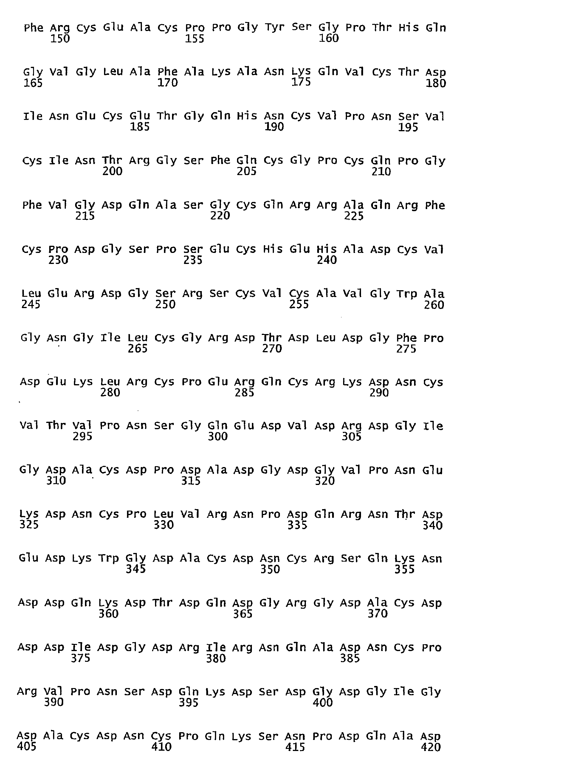


- <210> 2
<211> 12
<212> PRT
<213> Homo sapiens - <400> 2

- <210> 3
<211> 11
<212> PRT
<213> Homo sapiens - <400> 3

- <210> 4
<211> 10
<212> PRT
<213> Homo sapiens - <400> 4

- <210> 5
<211> 9
<212> PRT
<213> Homo sapiens - <400> 5

- <210> 6
<211> 8
<212> PRT
<213> Homo sapiens - <400> 6

- <210> 7
<211> 7
<212> PRT
<213> Homo sapiens - <400> 7

- <210> 8
<211> 6
<212> PRT
<213> Homo sapiens - <400> 8

- <210> 9
<211> 5
<212> PRT
<213> Homo sapiens - <400> 9

- <210> 10
<211> 4
<212> PRT
<213> Homo sapiens - <400> 10

- <210> 11
<211> 3
<212> PRT
<213> Homo sapiens - <400> 11

- <210> 12
<211> 12
<212> PRT
<213> Homo sapiens - <400> 12

- <210> 13
<211> 11
<212> PRT
<213> Homo sapiens - <400> 13

- <210> 14
<211> 10
<212> PRT
<213> Homo sapiens - <400> 14

- <210> 15
<211> 9
<212> PRT
<213> Homo sapiens - <400> 15

- <210> 16
<211> 8
<212> PRT
<213> Homo sapiens - <400> 16

- <210> 17
<211> 7
<212> PRT
<213> Homo sapiens - <400> 17

- <210> 18
<211> 6
<212> PRT
<213> Homo sapiens - <400> 18

- <210> 19
<211> 5
<212> PRT
<213> Homo sapiens - <400> 19

- <210> 20
<211> 4
<212> PRT
<213> Homo sapiens - <400> 20

- <210> 21
<211> 3
<212> PRT
<213> Homo sapiens - <400> 21

- <210> 22
<211> 12
<212> PRT
<213> Homo sapiens - <400> 22

- <210> 23
<211> 11
<212> PRT
<213> Homo sapiens - <400> 23

- <210> 24
<211> 10
<212> PRT
<213> Homo sapiens - <400> 24

- <210> 25
<211> 9
<212> PRT
<213> Homo sapiens - <400> 25

- <210> 26
<211> 8
<212> PRT
<213> Homo sapiens - <400> 26

- <210> 27
<211> 7
<212> PRT
<213> Homo sapiens - <400> 27

- <210> 28
<211> 6
<212> PRT
<213> Homo sapiens - <400> 28

- <210> 29
<211> 5
<212> PRT
<213> Homo sapiens - <400> 29

- <210> 30
<211> 4
<212> PRT
<213> Homo sapiens - <400> 30

- <210> 31
<211> 3
<212> PRT
<213> Homo sapiens - <400> 31

- <210> 32
<211> 12
<212> PRT
<213> Homo sapiens - <400> 32

- <210> 33
<211> 11
<212> PRT
<213> Homo sapiens - <400> 33

- <210> 34
<211> 10
<212> PRT
<213> Homo sapiens - <400> 34

- <210> 35
<211> 9
<212> PRT
<213> Homo sapiens - <400> 35

- <210> 36
<211> 8
<212> PRT
<213> Homo sapiens - <400> 36

- <210> 37
<211> 7
<212> PRT
<213> Homo sapiens - <400> 37

- <210> 38
<211> 6
<212> PRT
<213> Homo sapiens - <400> 38

- <210> 39
<211> 5
<212> PRT
<213> Homo sapiens - <400> 39

- <210> 40
<211> 4
<212> PRT
<213> Homo sapiens - <400> 40

- <210> 41
<211> 3
<212> PRT
<213> Homo sapiens - <400> 41

Claims (9)
- A method for determining a tissue degradation process comprising detecting in a sample the presence of one or more neoepitopes of mammalian COMP that appear after cleavage of COMP between position 625 (N625) and 626 (P626) of the amino acid sequence of COMP (SEQ ID NO: 1).
- An isolated C-terminally truncated fragment of human COMP wherein the C-terminus of the fragment is amino acid 625 of SEQ ID NO: 1, and wherein the fragment comprises an amino acid sequence given in any one of SEQ ID NOs: 22-31.
- An isolated N-terminally truncated fragment of human COMP wherein the N-terminus of the fragment is amino acid 626 of SEQ ID NO: 1, and wherein the fragment comprises an amino acid sequence given in any one of SEQ ID NOs: 32-40.
- An isolated fragment of human COMP consisting of the amino acid sequence given in any one of SEQ ID NOs: 22-40.
- Use of a fragment of COMP as defined in any one of claims 2 to 4 for the production of antibodies against one or more COMP neoepitopes that appear after cleavage of COMP between position 625 (N625) and 626 (P626) of the amino acid sequence of COMP (SEQ ID NO: 1).
- A conjugate for use in the production of antibodies against one or more COMP neoepitopes that appear after cleavage of COMP between position 625 (N625) and 626 (P626) of the amino acid sequence of COMP (SEQ ID NO: 1), consisting of (i) one or more fragments as defined in claim 4 coupled to or admixed with a protein carrier, or (ii) one or more fragments as defined in claim 2 or claim 3, coupled via the terminus that does not represent the cleavage site to a protein carrier.
- An antibody against an isolated fragment of COMP as claimed in any one of claims 2 to 4 or a conjugate as claimed in claim 6 wherein the antibody binds to one or more COMP neoepitopes that appear after cleavage of COMP between position 625 (N625) and 626 (P626) of the amino acid sequence of COMP (SEQ ID NO: 1).
- An antibody according to claim 7 for use in diagnosis, differential diagnosis, disease monitoring and/or therapeutic monitoring of a tissue degradation process.
- A method according to claim 1, wherein the presence of one or more neoepitopes of COMP is detected by using one or more antibodies as defined in claim 7 or 8.
Applications Claiming Priority (2)
| Application Number | Priority Date | Filing Date | Title |
|---|---|---|---|
| DKPA200400815 | 2004-05-24 | ||
| PCT/EP2005/005706 WO2005116658A2 (en) | 2004-05-24 | 2005-05-24 | Method for determining a tissue degradation process by detection of comp neoepitopes |
Publications (2)
| Publication Number | Publication Date |
|---|---|
| EP1766411A2 EP1766411A2 (en) | 2007-03-28 |
| EP1766411B1 true EP1766411B1 (en) | 2011-02-23 |
Family
ID=34973791
Family Applications (1)
| Application Number | Title | Priority Date | Filing Date |
|---|---|---|---|
| EP05748087A Expired - Lifetime EP1766411B1 (en) | 2004-05-24 | 2005-05-24 | Method for determining a tissue degradation process by detection of comp neoepitopes |
Country Status (6)
| Country | Link |
|---|---|
| US (1) | US20080318337A1 (en) |
| EP (1) | EP1766411B1 (en) |
| AT (1) | ATE499614T1 (en) |
| DE (1) | DE602005026525D1 (en) |
| ES (1) | ES2360804T3 (en) |
| WO (1) | WO2005116658A2 (en) |
Families Citing this family (4)
| Publication number | Priority date | Publication date | Assignee | Title |
|---|---|---|---|---|
| EP2236517A1 (en) * | 2009-03-31 | 2010-10-06 | Takara Bio, Inc. | Anti-fibronectin fragment monoclonal antibody |
| AU2011233753A1 (en) * | 2010-03-31 | 2012-09-13 | Anamar Ab | Method to detect tissue degradation leading to inflammation |
| AU2013281240B2 (en) * | 2012-06-28 | 2018-04-12 | Nordic Bioscience A/S | Determining pathological cartilage turnover |
| SE2430441A1 (en) * | 2024-09-03 | 2026-03-04 | SGPTH Life Science AB | COMP cleavage peptide and its use in diagnosis of osteoarthritis |
Family Cites Families (6)
| Publication number | Priority date | Publication date | Assignee | Title |
|---|---|---|---|---|
| US4795702A (en) * | 1986-02-05 | 1989-01-03 | Immunogon Associates | Diagnostic method for gonorrhea by assay of IgA1 fragments |
| US5387504A (en) * | 1992-09-30 | 1995-02-07 | Merck & Co., Inc. | Monospecific antibodies and assay system for detecting stromelysin cleavage products |
| US5464820A (en) * | 1993-06-22 | 1995-11-07 | The University Hospital | Specific inhibitors of tissue kallikrein |
| US7300918B2 (en) * | 1994-01-14 | 2007-11-27 | Matthias Rath | Method of producing vaccines from protein signal oligopeptides |
| DE69727582T2 (en) * | 1996-08-15 | 2004-12-23 | Novartis Ag | QUANTIFICATION OF ARTHRITIS CONDITIONS |
| AU2001250572A1 (en) * | 2000-04-07 | 2001-10-23 | Epigenomics Ag | Detection of single nucleotide polymorphisms (snp's) and cytosine-methylations |
-
2005
- 2005-05-24 EP EP05748087A patent/EP1766411B1/en not_active Expired - Lifetime
- 2005-05-24 ES ES05748087T patent/ES2360804T3/en not_active Expired - Lifetime
- 2005-05-24 US US11/597,488 patent/US20080318337A1/en not_active Abandoned
- 2005-05-24 WO PCT/EP2005/005706 patent/WO2005116658A2/en not_active Ceased
- 2005-05-24 DE DE602005026525T patent/DE602005026525D1/en not_active Expired - Lifetime
- 2005-05-24 AT AT05748087T patent/ATE499614T1/en not_active IP Right Cessation
Also Published As
| Publication number | Publication date |
|---|---|
| EP1766411A2 (en) | 2007-03-28 |
| ATE499614T1 (en) | 2011-03-15 |
| ES2360804T3 (en) | 2011-06-09 |
| WO2005116658A2 (en) | 2005-12-08 |
| DE602005026525D1 (en) | 2011-04-07 |
| US20080318337A1 (en) | 2008-12-25 |
| WO2005116658A3 (en) | 2006-05-26 |
Similar Documents
| Publication | Publication Date | Title |
|---|---|---|
| US7264939B2 (en) | Method of detecting native proBNP | |
| JP2008513536A (en) | Monoclonal antibody against progastrin | |
| JPH09500209A (en) | Assay for YKL-40 as an indicator of degradation of mammalian connective tissue matrix | |
| JPH09509736A (en) | Method for measuring collagen fragment in body fluid, test kit and means for carrying out the method, and method / use for using the method for diagnosing the presence of a disease related to collagen metabolism | |
| WO1990008195A1 (en) | Monoclonal antibody to human type ix collagen | |
| US6248869B1 (en) | Troponin I forms and use of the same | |
| JP3245052B2 (en) | Antibodies against the amino-terminal propeptide of type I procollagen and assay methods using same | |
| WO1998035235A1 (en) | Assay for collagen type ii fragments | |
| EP2867679B1 (en) | Determining pathological cartilage turnover | |
| US20080318337A1 (en) | Method for Determining a Tissue Degradation Process by Detection of Comp Neoepitopes | |
| CN101466732A (en) | Collagen type IV-like immunoreactive polypeptides | |
| US5516643A (en) | Immunochemical assays for cancer-associated SCM-recognition factor | |
| WO2004099784A2 (en) | Method for determining a tissue degradation process by detection of fibromodulin neoepitopes | |
| US10066006B2 (en) | Compositions and methods for redox modulated proteins | |
| JP4254242B2 (en) | Hair growth activity evaluation method and hair growth activity evaluation kit | |
| JPH10300749A (en) | Antibodies that recognize collagen fragments | |
| US20100233737A1 (en) | Marker specific to an oxidative degradation of tissues containing type iii collagen, means and methods and kits for the diagnosis, monitoring or prognosis of pathologies targeted by this marker | |
| US8012694B2 (en) | Assay for the detection of phosphorylated PTH | |
| JPH02152988A (en) | Peptide compound and antiserum and antibody produced by using the peptide compound | |
| JP2628336B2 (en) | Bradykinin derivatives and their determination | |
| JPH04134097A (en) | Laminine fragment | |
| Wang et al. | Development of a heterologous radioimmunoassay for canine osteocalcin | |
| JPS59138958A (en) | Antiserum | |
| EP0803065A1 (en) | A-protein as a diagnostic of cancer | |
| WO1994003806A1 (en) | Immunochemical assays for cancer-associated scm recognition factor |
Legal Events
| Date | Code | Title | Description |
|---|---|---|---|
| PUAI | Public reference made under article 153(3) epc to a published international application that has entered the european phase |
Free format text: ORIGINAL CODE: 0009012 |
|
| 17P | Request for examination filed |
Effective date: 20061221 |
|
| AK | Designated contracting states |
Kind code of ref document: A2 Designated state(s): AT BE BG CH CY CZ DE DK EE ES FI FR GB GR HU IE IS IT LI LT LU MC NL PL PT RO SE SI SK TR |
|
| DAX | Request for extension of the european patent (deleted) | ||
| 17Q | First examination report despatched |
Effective date: 20071108 |
|
| GRAP | Despatch of communication of intention to grant a patent |
Free format text: ORIGINAL CODE: EPIDOSNIGR1 |
|
| RAP1 | Party data changed (applicant data changed or rights of an application transferred) |
Owner name: ANAMAR AB |
|
| GRAS | Grant fee paid |
Free format text: ORIGINAL CODE: EPIDOSNIGR3 |
|
| GRAA | (expected) grant |
Free format text: ORIGINAL CODE: 0009210 |
|
| AK | Designated contracting states |
Kind code of ref document: B1 Designated state(s): AT BE BG CH CY CZ DE DK EE ES FI FR GB GR HU IE IS IT LI LT LU MC NL PL PT RO SE SI SK TR |
|
| REG | Reference to a national code |
Ref country code: GB Ref legal event code: FG4D |
|
| REG | Reference to a national code |
Ref country code: CH Ref legal event code: EP |
|
| REG | Reference to a national code |
Ref country code: IE Ref legal event code: FG4D |
|
| REF | Corresponds to: |
Ref document number: 602005026525 Country of ref document: DE Date of ref document: 20110407 Kind code of ref document: P |
|
| REG | Reference to a national code |
Ref country code: DE Ref legal event code: R096 Ref document number: 602005026525 Country of ref document: DE Effective date: 20110407 |
|
| REG | Reference to a national code |
Ref country code: ES Ref legal event code: FG2A Ref document number: 2360804 Country of ref document: ES Kind code of ref document: T3 Effective date: 20110609 |
|
| REG | Reference to a national code |
Ref country code: NL Ref legal event code: VDEP Effective date: 20110223 |
|
| LTIE | Lt: invalidation of european patent or patent extension |
Effective date: 20110223 |
|
| PG25 | Lapsed in a contracting state [announced via postgrant information from national office to epo] |
Ref country code: GR Free format text: LAPSE BECAUSE OF FAILURE TO SUBMIT A TRANSLATION OF THE DESCRIPTION OR TO PAY THE FEE WITHIN THE PRESCRIBED TIME-LIMIT Effective date: 20110524 Ref country code: LT Free format text: LAPSE BECAUSE OF FAILURE TO SUBMIT A TRANSLATION OF THE DESCRIPTION OR TO PAY THE FEE WITHIN THE PRESCRIBED TIME-LIMIT Effective date: 20110223 Ref country code: SE Free format text: LAPSE BECAUSE OF FAILURE TO SUBMIT A TRANSLATION OF THE DESCRIPTION OR TO PAY THE FEE WITHIN THE PRESCRIBED TIME-LIMIT Effective date: 20110223 Ref country code: PT Free format text: LAPSE BECAUSE OF FAILURE TO SUBMIT A TRANSLATION OF THE DESCRIPTION OR TO PAY THE FEE WITHIN THE PRESCRIBED TIME-LIMIT Effective date: 20110623 |
|
| PG25 | Lapsed in a contracting state [announced via postgrant information from national office to epo] |
Ref country code: FI Free format text: LAPSE BECAUSE OF FAILURE TO SUBMIT A TRANSLATION OF THE DESCRIPTION OR TO PAY THE FEE WITHIN THE PRESCRIBED TIME-LIMIT Effective date: 20110223 Ref country code: BG Free format text: LAPSE BECAUSE OF FAILURE TO SUBMIT A TRANSLATION OF THE DESCRIPTION OR TO PAY THE FEE WITHIN THE PRESCRIBED TIME-LIMIT Effective date: 20110523 Ref country code: AT Free format text: LAPSE BECAUSE OF FAILURE TO SUBMIT A TRANSLATION OF THE DESCRIPTION OR TO PAY THE FEE WITHIN THE PRESCRIBED TIME-LIMIT Effective date: 20110223 Ref country code: SI Free format text: LAPSE BECAUSE OF FAILURE TO SUBMIT A TRANSLATION OF THE DESCRIPTION OR TO PAY THE FEE WITHIN THE PRESCRIBED TIME-LIMIT Effective date: 20110223 Ref country code: NL Free format text: LAPSE BECAUSE OF FAILURE TO SUBMIT A TRANSLATION OF THE DESCRIPTION OR TO PAY THE FEE WITHIN THE PRESCRIBED TIME-LIMIT Effective date: 20110223 Ref country code: BE Free format text: LAPSE BECAUSE OF FAILURE TO SUBMIT A TRANSLATION OF THE DESCRIPTION OR TO PAY THE FEE WITHIN THE PRESCRIBED TIME-LIMIT Effective date: 20110223 Ref country code: CY Free format text: LAPSE BECAUSE OF FAILURE TO SUBMIT A TRANSLATION OF THE DESCRIPTION OR TO PAY THE FEE WITHIN THE PRESCRIBED TIME-LIMIT Effective date: 20110223 |
|
| PG25 | Lapsed in a contracting state [announced via postgrant information from national office to epo] |
Ref country code: EE Free format text: LAPSE BECAUSE OF FAILURE TO SUBMIT A TRANSLATION OF THE DESCRIPTION OR TO PAY THE FEE WITHIN THE PRESCRIBED TIME-LIMIT Effective date: 20110223 Ref country code: DK Free format text: LAPSE BECAUSE OF FAILURE TO SUBMIT A TRANSLATION OF THE DESCRIPTION OR TO PAY THE FEE WITHIN THE PRESCRIBED TIME-LIMIT Effective date: 20110223 |
|
| PG25 | Lapsed in a contracting state [announced via postgrant information from national office to epo] |
Ref country code: SK Free format text: LAPSE BECAUSE OF FAILURE TO SUBMIT A TRANSLATION OF THE DESCRIPTION OR TO PAY THE FEE WITHIN THE PRESCRIBED TIME-LIMIT Effective date: 20110223 Ref country code: RO Free format text: LAPSE BECAUSE OF FAILURE TO SUBMIT A TRANSLATION OF THE DESCRIPTION OR TO PAY THE FEE WITHIN THE PRESCRIBED TIME-LIMIT Effective date: 20110223 Ref country code: CZ Free format text: LAPSE BECAUSE OF FAILURE TO SUBMIT A TRANSLATION OF THE DESCRIPTION OR TO PAY THE FEE WITHIN THE PRESCRIBED TIME-LIMIT Effective date: 20110223 |
|
| PG25 | Lapsed in a contracting state [announced via postgrant information from national office to epo] |
Ref country code: MC Free format text: LAPSE BECAUSE OF NON-PAYMENT OF DUE FEES Effective date: 20110531 |
|
| PLBE | No opposition filed within time limit |
Free format text: ORIGINAL CODE: 0009261 |
|
| REG | Reference to a national code |
Ref country code: CH Ref legal event code: PL |
|
| STAA | Information on the status of an ep patent application or granted ep patent |
Free format text: STATUS: NO OPPOSITION FILED WITHIN TIME LIMIT |
|
| PG25 | Lapsed in a contracting state [announced via postgrant information from national office to epo] |
Ref country code: LI Free format text: LAPSE BECAUSE OF NON-PAYMENT OF DUE FEES Effective date: 20110531 Ref country code: CH Free format text: LAPSE BECAUSE OF NON-PAYMENT OF DUE FEES Effective date: 20110531 |
|
| 26N | No opposition filed |
Effective date: 20111124 |
|
| PG25 | Lapsed in a contracting state [announced via postgrant information from national office to epo] |
Ref country code: PL Free format text: LAPSE BECAUSE OF FAILURE TO SUBMIT A TRANSLATION OF THE DESCRIPTION OR TO PAY THE FEE WITHIN THE PRESCRIBED TIME-LIMIT Effective date: 20110223 |
|
| REG | Reference to a national code |
Ref country code: IE Ref legal event code: MM4A |
|
| REG | Reference to a national code |
Ref country code: DE Ref legal event code: R097 Ref document number: 602005026525 Country of ref document: DE Effective date: 20111124 |
|
| PG25 | Lapsed in a contracting state [announced via postgrant information from national office to epo] |
Ref country code: IE Free format text: LAPSE BECAUSE OF NON-PAYMENT OF DUE FEES Effective date: 20110524 |
|
| PG25 | Lapsed in a contracting state [announced via postgrant information from national office to epo] |
Ref country code: LU Free format text: LAPSE BECAUSE OF NON-PAYMENT OF DUE FEES Effective date: 20110524 |
|
| PG25 | Lapsed in a contracting state [announced via postgrant information from national office to epo] |
Ref country code: IS Free format text: LAPSE BECAUSE OF FAILURE TO SUBMIT A TRANSLATION OF THE DESCRIPTION OR TO PAY THE FEE WITHIN THE PRESCRIBED TIME-LIMIT Effective date: 20110223 |
|
| PG25 | Lapsed in a contracting state [announced via postgrant information from national office to epo] |
Ref country code: TR Free format text: LAPSE BECAUSE OF FAILURE TO SUBMIT A TRANSLATION OF THE DESCRIPTION OR TO PAY THE FEE WITHIN THE PRESCRIBED TIME-LIMIT Effective date: 20110223 |
|
| PG25 | Lapsed in a contracting state [announced via postgrant information from national office to epo] |
Ref country code: HU Free format text: LAPSE BECAUSE OF FAILURE TO SUBMIT A TRANSLATION OF THE DESCRIPTION OR TO PAY THE FEE WITHIN THE PRESCRIBED TIME-LIMIT Effective date: 20110223 |
|
| REG | Reference to a national code |
Ref country code: FR Ref legal event code: PLFP Year of fee payment: 11 |
|
| PGFP | Annual fee paid to national office [announced via postgrant information from national office to epo] |
Ref country code: ES Payment date: 20150512 Year of fee payment: 11 Ref country code: DE Payment date: 20150525 Year of fee payment: 11 Ref country code: GB Payment date: 20150526 Year of fee payment: 11 |
|
| PGFP | Annual fee paid to national office [announced via postgrant information from national office to epo] |
Ref country code: FR Payment date: 20150601 Year of fee payment: 11 Ref country code: IT Payment date: 20150513 Year of fee payment: 11 |
|
| REG | Reference to a national code |
Ref country code: DE Ref legal event code: R119 Ref document number: 602005026525 Country of ref document: DE |
|
| GBPC | Gb: european patent ceased through non-payment of renewal fee |
Effective date: 20160524 |
|
| PG25 | Lapsed in a contracting state [announced via postgrant information from national office to epo] |
Ref country code: IT Free format text: LAPSE BECAUSE OF NON-PAYMENT OF DUE FEES Effective date: 20160524 |
|
| REG | Reference to a national code |
Ref country code: FR Ref legal event code: ST Effective date: 20170131 |
|
| PG25 | Lapsed in a contracting state [announced via postgrant information from national office to epo] |
Ref country code: DE Free format text: LAPSE BECAUSE OF NON-PAYMENT OF DUE FEES Effective date: 20161201 Ref country code: FR Free format text: LAPSE BECAUSE OF NON-PAYMENT OF DUE FEES Effective date: 20160531 |
|
| PG25 | Lapsed in a contracting state [announced via postgrant information from national office to epo] |
Ref country code: GB Free format text: LAPSE BECAUSE OF NON-PAYMENT OF DUE FEES Effective date: 20160524 |
|
| PG25 | Lapsed in a contracting state [announced via postgrant information from national office to epo] |
Ref country code: ES Free format text: LAPSE BECAUSE OF NON-PAYMENT OF DUE FEES Effective date: 20160525 |
|
| REG | Reference to a national code |
Ref country code: ES Ref legal event code: FD2A Effective date: 20181204 |