EP1750711B1 - Treatment of inflammatory conditions with morphinans - Google Patents
Treatment of inflammatory conditions with morphinans Download PDFInfo
- Publication number
- EP1750711B1 EP1750711B1 EP05750004.3A EP05750004A EP1750711B1 EP 1750711 B1 EP1750711 B1 EP 1750711B1 EP 05750004 A EP05750004 A EP 05750004A EP 1750711 B1 EP1750711 B1 EP 1750711B1
- Authority
- EP
- European Patent Office
- Prior art keywords
- lps
- cultures
- vpa
- neuron
- treated
- Prior art date
- Legal status (The legal status is an assumption and is not a legal conclusion. Google has not performed a legal analysis and makes no representation as to the accuracy of the status listed.)
- Not-in-force
Links
Images


Classifications
-
- A—HUMAN NECESSITIES
- A61—MEDICAL OR VETERINARY SCIENCE; HYGIENE
- A61K—PREPARATIONS FOR MEDICAL, DENTAL OR TOILETRY PURPOSES
- A61K38/00—Medicinal preparations containing peptides
- A61K38/04—Peptides having up to 20 amino acids in a fully defined sequence; Derivatives thereof
- A61K38/06—Tripeptides
-
- A—HUMAN NECESSITIES
- A61—MEDICAL OR VETERINARY SCIENCE; HYGIENE
- A61K—PREPARATIONS FOR MEDICAL, DENTAL OR TOILETRY PURPOSES
- A61K31/00—Medicinal preparations containing organic active ingredients
- A61K31/185—Acids; Anhydrides, halides or salts thereof, e.g. sulfur acids, imidic, hydrazonic or hydroximic acids
- A61K31/19—Carboxylic acids, e.g. valproic acid
-
- A—HUMAN NECESSITIES
- A61—MEDICAL OR VETERINARY SCIENCE; HYGIENE
- A61K—PREPARATIONS FOR MEDICAL, DENTAL OR TOILETRY PURPOSES
- A61K31/00—Medicinal preparations containing organic active ingredients
- A61K31/33—Heterocyclic compounds
- A61K31/395—Heterocyclic compounds having nitrogen as a ring hetero atom, e.g. guanethidine or rifamycins
- A61K31/435—Heterocyclic compounds having nitrogen as a ring hetero atom, e.g. guanethidine or rifamycins having six-membered rings with one nitrogen as the only ring hetero atom
- A61K31/46—8-Azabicyclo [3.2.1] octane; Derivatives thereof, e.g. atropine, cocaine
-
- A—HUMAN NECESSITIES
- A61—MEDICAL OR VETERINARY SCIENCE; HYGIENE
- A61K—PREPARATIONS FOR MEDICAL, DENTAL OR TOILETRY PURPOSES
- A61K31/00—Medicinal preparations containing organic active ingredients
- A61K31/33—Heterocyclic compounds
- A61K31/395—Heterocyclic compounds having nitrogen as a ring hetero atom, e.g. guanethidine or rifamycins
- A61K31/435—Heterocyclic compounds having nitrogen as a ring hetero atom, e.g. guanethidine or rifamycins having six-membered rings with one nitrogen as the only ring hetero atom
- A61K31/47—Quinolines; Isoquinolines
- A61K31/485—Morphinan derivatives, e.g. morphine, codeine
-
- A—HUMAN NECESSITIES
- A61—MEDICAL OR VETERINARY SCIENCE; HYGIENE
- A61K—PREPARATIONS FOR MEDICAL, DENTAL OR TOILETRY PURPOSES
- A61K38/00—Medicinal preparations containing peptides
- A61K38/04—Peptides having up to 20 amino acids in a fully defined sequence; Derivatives thereof
- A61K38/08—Peptides having 5 to 11 amino acids
-
- A—HUMAN NECESSITIES
- A61—MEDICAL OR VETERINARY SCIENCE; HYGIENE
- A61K—PREPARATIONS FOR MEDICAL, DENTAL OR TOILETRY PURPOSES
- A61K38/00—Medicinal preparations containing peptides
- A61K38/16—Peptides having more than 20 amino acids; Gastrins; Somatostatins; Melanotropins; Derivatives thereof
- A61K38/17—Peptides having more than 20 amino acids; Gastrins; Somatostatins; Melanotropins; Derivatives thereof from animals; from humans
- A61K38/33—Peptides having more than 20 amino acids; Gastrins; Somatostatins; Melanotropins; Derivatives thereof from animals; from humans derived from pro-opiomelanocortin, pro-enkephalin or pro-dynorphin
-
- A—HUMAN NECESSITIES
- A61—MEDICAL OR VETERINARY SCIENCE; HYGIENE
- A61P—SPECIFIC THERAPEUTIC ACTIVITY OF CHEMICAL COMPOUNDS OR MEDICINAL PREPARATIONS
- A61P19/00—Drugs for skeletal disorders
- A61P19/02—Drugs for skeletal disorders for joint disorders, e.g. arthritis, arthrosis
-
- A—HUMAN NECESSITIES
- A61—MEDICAL OR VETERINARY SCIENCE; HYGIENE
- A61P—SPECIFIC THERAPEUTIC ACTIVITY OF CHEMICAL COMPOUNDS OR MEDICINAL PREPARATIONS
- A61P25/00—Drugs for disorders of the nervous system
-
- A—HUMAN NECESSITIES
- A61—MEDICAL OR VETERINARY SCIENCE; HYGIENE
- A61P—SPECIFIC THERAPEUTIC ACTIVITY OF CHEMICAL COMPOUNDS OR MEDICINAL PREPARATIONS
- A61P25/00—Drugs for disorders of the nervous system
- A61P25/14—Drugs for disorders of the nervous system for treating abnormal movements, e.g. chorea, dyskinesia
- A61P25/16—Anti-Parkinson drugs
-
- A—HUMAN NECESSITIES
- A61—MEDICAL OR VETERINARY SCIENCE; HYGIENE
- A61P—SPECIFIC THERAPEUTIC ACTIVITY OF CHEMICAL COMPOUNDS OR MEDICINAL PREPARATIONS
- A61P25/00—Drugs for disorders of the nervous system
- A61P25/28—Drugs for disorders of the nervous system for treating neurodegenerative disorders of the central nervous system, e.g. nootropic agents, cognition enhancers, drugs for treating Alzheimer's disease or other forms of dementia
-
- A—HUMAN NECESSITIES
- A61—MEDICAL OR VETERINARY SCIENCE; HYGIENE
- A61P—SPECIFIC THERAPEUTIC ACTIVITY OF CHEMICAL COMPOUNDS OR MEDICINAL PREPARATIONS
- A61P29/00—Non-central analgesic, antipyretic or antiinflammatory agents, e.g. antirheumatic agents; Non-steroidal antiinflammatory drugs [NSAID]
-
- A—HUMAN NECESSITIES
- A61—MEDICAL OR VETERINARY SCIENCE; HYGIENE
- A61P—SPECIFIC THERAPEUTIC ACTIVITY OF CHEMICAL COMPOUNDS OR MEDICINAL PREPARATIONS
- A61P3/00—Drugs for disorders of the metabolism
- A61P3/08—Drugs for disorders of the metabolism for glucose homeostasis
- A61P3/10—Drugs for disorders of the metabolism for glucose homeostasis for hyperglycaemia, e.g. antidiabetics
-
- A—HUMAN NECESSITIES
- A61—MEDICAL OR VETERINARY SCIENCE; HYGIENE
- A61P—SPECIFIC THERAPEUTIC ACTIVITY OF CHEMICAL COMPOUNDS OR MEDICINAL PREPARATIONS
- A61P37/00—Drugs for immunological or allergic disorders
-
- A—HUMAN NECESSITIES
- A61—MEDICAL OR VETERINARY SCIENCE; HYGIENE
- A61P—SPECIFIC THERAPEUTIC ACTIVITY OF CHEMICAL COMPOUNDS OR MEDICINAL PREPARATIONS
- A61P9/00—Drugs for disorders of the cardiovascular system
- A61P9/10—Drugs for disorders of the cardiovascular system for treating ischaemic or atherosclerotic diseases, e.g. antianginal drugs, coronary vasodilators, drugs for myocardial infarction, retinopathy, cerebrovascula insufficiency, renal arteriosclerosis
Definitions
- the invention relates to compositions for affecting various biological mechanisms related to inflammation, the resultant inflammation and the disorders that may be caused thereby. More specifically, the invention relates to administration of compounds for neuroprotective and/or neurotrophic effects for treatment and/or prevention of neurodegenerative disorders and diseases caused thereby.
- Inflammation in the brain is characterized by the activation of microglia and astroglia, and is thought to be associated with the pathogenesis of a number of neurological diseases, including Parkinson's disease (PD), Alzheimer's disease and cerebral ischemia.
- PD Parkinson's disease
- Epidemiological studies have shown a positive correlation between PD and inflammation early in life. For example, the increase in the incidence of PD in 1945-50's was highly correlated to the flu pandemic in 1910-20's. It was also found that a higher PD incidence appeared among populations who were professional boxers at a young age.
- microglia secrete various cytokines and free radicals, such as superoxide and nitric oxide (NO), resulting in cerebral inflammation and subsequent neuronal death and damage. Accumulation and/or overproduction of these factors impact neurons to induce their degeneration.
- cytokines and free radicals such as superoxide and nitric oxide (NO)
- LPS lipopolysaccharide
- cytokines and free radicals such as NO, PGE 2 , TNF ⁇ , superoxide, and other reactive oxygen species (ROS).
- ROS reactive oxygen species
- Cerebral inflammation sustained by microglia activation triggered by LPS results in a delayed and progressive degeneration of nigra dopaminergic neurons.
- Dopaminergic neurons in particular, can be especially vulnerable to oxidative damage due to antioxidant capacity It is also believed that decreased neurotrophic factor released from astroglia could play a role in susceptibility to inflammation.
- GDNF glial cell line-derived neurotrophic factor
- GDNF glial cell line-derived neurotrophic factor
- Described herein are methods of providing neuroprotective and/or neurotrophic effects by contacting, administering, or treating a mammal with an effective amount of at least one compound of the invention comprising valproic acid, sodium valproate, butyric acid, sodium butyrate, or other salts thereof; opioid peptides; a peptide comprising Gly-Gly-Phe (GGF); or morphinans, such as naloxone, naltrexone, and dextromethorphan.
- valproic acid sodium valproate, butyric acid, sodium butyrate, or other salts thereof
- opioid peptides a peptide comprising Gly-Gly-Phe (GGF); or morphinans, such as naloxone, naltrexone, and dextromethorphan.
- GGF Gly-Gly-Phe
- morphinans such as naloxone, naltrexone, and dextromethor
- Described herein are methods for activating neuronal-survival signaling pathways in a mammal that comprise administration of at least one compound described herein. Described herein are methods of inducing release of neurotrophic factors from astroglial cells by treating the cells with valproic acid, sodium valproate, butyric acid, sodium butyrate, or other salts thereof; and/or 3-hydroxymorphinan.
- Also desribed herein are methods of inhibiting over activation of innate immune cells that comprise contacting an innate immune cell with a therapeutically effective amount of at least one compound described herein.
- the inhibition of the over activation of microglia can occur either in vivo or in vitro.
- the invention includes reducing inflammation in a mammal that comprise administration of at least one compound of the invention at an effective dosage.
- the inflammation-related condition can include inflammation associated with diseases, such as Alzheimer's disease, Parkinson's disease, ALS, atherosclerosis, diabetes, arthritis, multiple sclerosis, sepsis, septic shock, endotoxemia, multiple organ failure, or organ damage, such as liver damage.
- diseases such as Alzheimer's disease, Parkinson's disease, ALS, atherosclerosis, diabetes, arthritis, multiple sclerosis, sepsis, septic shock, endotoxemia, multiple organ failure, or organ damage, such as liver damage.
- Described herein are methods of inhibiting the activity of NADPH oxidase that comprise modulating or inhibiting the NADPH oxidase with an effective amount of at least one compound described herein.
- the inhibition of the activity of NADPH oxidase can occur either in vivo or in vitro.
- the inhibition of the activity of NADPH oxidase can occur either in vivo or in vitro.
- Also described herein are methods of identifying compounds that maybe therapeutically effective in treating an inflammation-related condition that comprise contacting NADPH oxidase with at least one candidate compound, and determining whether the candidate inhibits NADPH oxidase as compared to NADPH oxidase without the compound, wherein a compound may be therapeutically effective in treating an inflammation- associated condition if the compound decreases the expression or activity of NADPH oxidase or the gp91 subunit of NADPH oxidase.
- the decrease in the release of one or more of tumor necrosis factor ⁇ (TNF ⁇ ), prostaglandin E 2 (PGE 2 ), interleukin-1 (IL-1), nitric oxide, or superoxide can occur either in vivo or in vitro.
- Described herein is a polypeptide or peptide comprising an amino acid sequence GGF that can be used in methods described herein.
- compositions that comprise ultra low concentrations of a compound described herein.
- the term “about” applies to all numeric values, whether or not explicitly indicated.
- the term “about” generally refers to a range of numbers that one of skill in the art would consider equivalent to the recited value (i.e., having the same function or result). In many instances, the term “about” may include numbers that are rounded to the nearest significant figure.
- Methods of the invention that comprise steps of contacting a compound of the invention with an enzyme or other entity can be accomplished in a solution, or otherwise.
- ultra low amounts or concentrations refers to femtomolar concentrations (from about 10 -13 M to about 10 -15 M) of the compounds of the invention. Concentrations expressed in M generally correspond to a decrease of a dose of about 100 million fold when administered in vivo. Effective concentrations for use in methods of the invention, including ultra low concentrations, are also expressed as grams per kilogram body weight of the mammal being treated. In general 1mg/kg corresponds to micromolar (1x10 -6 M), and 10pg/kg generally corresponds to femtomolar (1x10 -15 M concentrations.
- pharmaceutically acceptable salt thereof includes an acid addition salt or a base salt.
- pharmaceutically acceptable carrier includes any material which, when combined with a compound of the invention, allows the compound to retain biological activity, such as the ability to treat inflammation associated disease or affect the various mechanisms associated therewith, and is non-reactive with the subject's immune system.
- examples include, but are not limited to, any of the standard pharmaceutical carriers such as a phosphate buffered saline solution, water, emulsions such as oil/water emulsions, and various types of wetting agents.
- Compositions comprising such carriers are formulated by well known conventional methods (see, for example, Remington's Pharmaceutical Sciences, Chapter 43, 14th Ed., Mack Publishing Co., Easton, PA ).
- inhibiting the activity of NADPH oxidase refers to processes or methods that decrease the activity of NADPH oxidase either in vivo or in vitro, relative to NADPH oxidase either in vivo or in vitro, that has not been subjected to such a process or method.
- inhibiting the activity of NADPH oxidase additionally refers to processes or methods that reduce or prevent the over-activity of NADPH oxidase either in vivo or in vitro, relative to NADPH oxidase either in vivo or in vitro, that has not been subjected to such a process or method.
- “Over activity of NADPH oxidase” refers to activity of this enzyme that is more than that which is commonly seen in an untreated or control subject or cell, whether in vivo or in vitro.
- a method of inhibiting the activity of NADPH oxidase comprises a step of contacting NADPH oxidase with an effective amount of at least one compound described herein.
- the step of contacting the NADPH oxidase with the at least one compound is accomplished in vivo.
- the step of contacting the NADPH oxidase with the at least one compound is accomplished in vitro.
- a method of inhibiting the activity of NADPH oxidase that comprises a step of inhibiting the NADPH oxidase with an effective amount of a peptide comprising GGF.
- a polypeptide or peptide comprises an amino acid sequence GGF (Gly-Gly-Phe) and is other than or excludes the full length sequence of dynorphin A (SEQ ID NO: 1).
- the polypeptide can inhibit the activity of NADPH oxidase.
- a peptide comprising GGF has no more than 16 amino acids, more preferably, about 3 to 16 amino acids, more preferably about 3 to 10 amino acids, and more preferably about 3 to 5 amino acids.
- a peptide that comprises GGF may be chemically modified or linked to a heterologous polypeptide.
- a peptide comprising GGF may be linked to a molecule or compound that enhances intracellular transport or transport across the blood brain barrier.
- the effective amount of the compound is that amount that provides for inhibition of NADPH oxidase activity by at least 25%, more preferably at least 50%, and most preferably 100%, or to control levels.
- An inhibition NADPH oxidase activity can be determined by detecting a decrease in reactive oxygen species (ROS) either extracellularly or intracellularly or by other methods known to those of skill in the art.
- ROS reactive oxygen species
- the effect amount can be from about 10 -5 M to about 10 -15 M.
- the effective amount of the compound is from about 10 -5 M to about 10 -7 M, or about 10 -13 M to about 10 -15 M.
- a compound inhibits the activity of NADPH oxidase if it decreases the activity of NADPH oxidase by at least about 30 % when measured disclosure by the production of superoxide. In another embodiment of the disclosure, a compound inhibits the activity of NADPH oxidase if it decreases the activity of NADPH oxidase by at least about 50% when measured by the production of superoxide. In another embodiment of the disclosure, a compound inhibits the activity of NADPH oxidase if it decreases the activity of NADPH oxidase by at least about 70% when measured by the production of superoxide.
- Over activity of NADPH can be caused by a variety of agents, including, but not limited to lippolysaccharide (LPS), ⁇ -amyloid peptides, 1-methyl-4-phenyl-1,2,3,6-tetrahydropyridine (MPTP), or environmental toxins.
- LPS lippolysaccharide
- MPTP 1-methyl-4-phenyl-1,2,3,6-tetrahydropyridine
- environmental toxins include, but are not limited to insecticides such as rotenone, pesticides, such as paraquat; and particulate mater (e.g. ubiquitous particulate components of an air pollution).
- other compounds can be utilized to inhibit NADPH oxidase activity.
- the compounds are those that can penetrate the blood brain barrier and act on inflammation in the brain.
- the other compounds comprise naloxone, naltrexone, dextromethorphan, valproate, valproic acid or salts thereof, butyric acid or salts thereof, an opioid peptide such as Met enkephlin or Leu-enkephalin, or mixtures thereof.
- a compound that inhibits NADPH oxidase activity is administered at an "ultra low" concentration.
- the concentration of the compound is at least 10 -13 M, more preferably about 10 -13 M to 10 -15 M, more preferably about 10 -14 M, and more preferably about 10 -15 M.
- ultra low concentrations comprise about 10 pg/kg to about 1000pg/kg, more preferably about 1000pg/kg, more preferably about 100pg/kg, and more about preferably 10pg/kg.
- the concentration of the compound in the ⁇ M amount is also effective to inhibit NADPH oxidase.
- the compound is administered at about 10 -5 to about 10 -7 M, more preferably 10 -5 , more preferably 10 -6 and more preferably about 10 -7 M.
- the method comprises contacting NADPH oxidase with an effective amount of a morphinan or a peptide comprising an amino acid sequence GGF, wherein the effective amount is about 10 -13 to about 10 -15 M.
- Morphinans include without limitation, dextromethorphan, naloxone, and naltrexone.
- a peptide comprising an amino acid sequence GGF preferably, has no more than sixteen amino acids and does not include the full length sequence of dynorphin A (SEQ ID NO:1). Examples of some peptides comprising GGF are shown in Table 1.
- NADPH oxidase is a complex enzyme that contains 7 subunits, one of which is gp91.
- gp91 is the catalytic subunit of NADPH, and therefore may play a significant role in modulating the activity of NADPH.
- the invention therefore also includes methods of inhibiting the activity of NADPH oxidase by affecting the gp91 subunit of NADPH oxidase.
- affecting the gp91 subunit of NADPH oxidase refers to processes or methods that alter the configuration of one or more regions of the gp91 subunit either in vivo or in vitro, relative to a gp91 subunit either in vivo or in vitro, that has not been subjected to such a process or method; or processes or methods that block at least a portion of the gp91 subunit from binding with another protein or compound either in vivo or in vitro, relative to a gp91 subunit either in vivo or in vitro, that has not been subjected to such a process or method.
- a method of affecting the gp91 subunit of NADPH oxidase comprises the step of contacting the gp91 subunit of NADPH oxidase with an effective amount of at least one compound described herein.
- the step of contacting the gp91 subunit of NADPH oxidase with the at least one compound is accomplished in vivo.
- the step of contacting the gp91 subunit of NADPH oxidase with the at least one compound is accomplished in vitro.
- a polypeptide or peptide that comprises an amino acid sequence GGF is other than or excludes the full length sequence of dynorphin A (SEQ ID NO: 1).
- the polypeptide can inhibit the activity of NADPH oxidase.
- a peptide comprising GGF has no more than 16 amino acids, more preferably, about 3 to 16 amino acids, more preferably about 3 to 10 amino acids, and more preferably about 3 to 5 amino acids.
- a peptide that comprises GGF may be chemically modified or linked to a heterologous polypeptide.
- a peptide comprising GF may be linked to a molecule or compound that enhances intracellular transport or transport across the blood brain barrier.
- the effective amount of the compound can be from about 10 -5 M to about 10 -15 M. In another embodiment , the effective amount of the compound is from about 10 -5 M to about 10 -7 M, or about 10 -13 M to about 10 -15 M.
- NADPH controls the release of pro-inflammatory agents from the innate immune cells within a tissue system.
- innate immune cells within particular tissue systems include, but are not limited to microglia in neurological tissues, macrophages in organs, such as for example Kupffer cells in the liver, macrophages in the lungs, masengial cells in the kidney, and the endothelial cells lining the blood. Additionally described herein are methods of inhibiting microglial NADPH oxidase.
- Another embodiment of the disclosure provides methods of inhibiting the activation or over- activation of at least one innate immune cell that comprise contacting the innate immune cell with an therapeutically effective amount of at least one compound described herein.
- the step of contacting the innate immune cell with the at least one compound is accomplished in vivo.
- the step of contacting the innate immune cell with the at least one compound is accomplished in vitro.
- An embodiment of the disclosure provides methods of inhibiting the activation or over-activation of at least one innate immune cell that comprise contacting the innate immune cell with an effective amount of a peptide or polypeptide comprising tripeptide GGF.
- a polypeptide or peptide comprises an amino acid sequence GGF (Gly-Gly-Phe) and is other than or excludes the full length sequence of dynorphin A (SEQ ID NO: 1).
- the polypeptide can inhibit the activity of NADPH oxidase and/or the production of reactive oxygen species.
- a peptide comprising GGF has no more than 16 amino acids, more preferably, about 3 to 16 amino acids, more preferably about 3 to 10 amino acids, and more preferably about 3 to 5 amino acids.
- a peptide that comprises GGF may be chemically modified or linked to a heterologous polypeptide.
- a peptide comprising GGF may be linked to a molecule or compound that enhances intracellular transport or transport across the blood brain barrier.
- the effective amount of the compound can be from about 10 -5 M to about 10 -15 M.
- the effective amount of the compound is from about 10 -5 M to about 10 -7 M, or ultra low concentrations of about 10 -13 M to about 10 -15 M.
- ultra low concentrations comprise about 10 pg/kg to about 1000pg/kg, more preferably about 1000pg/kg, more preferably about 100pg/kg, and more about preferably 10pg/kg.
- Yet another embodiment of the disclosure provides methods of inhibiting the activation or over-activation of at least one microglial cell that comprises contacting the microglial with an effective amount of at least one compound described herein.
- the step of contacting the microglial with the at least one compound is accomplished in vivo.
- the step of contacting the microglial with the at least one compound is accomplished in vitro.
- other compounds can be utilized to inhibit activation of innate immune cells.
- the compounds are those that can penetrate the blood brain barrier and act on inflammation in the brain.
- the other compounds comprise naloxone, naltrexone, dextromethorphan, valproate, valproic acid or salts thereof, butyric acid or salts thereof, an opioid peptide such as Met enkephalin or Leu- enkephalin, or mixtures thereof.
- An embodiment of the disclosure provides methods of inhibiting activation of at least one innate immune cell, comprising contacting the cell with an effective amount of a compound described herein.
- the method of inhibiting activation decreases the activity of the at least one innate immune cell by at least about 30%.
- the method of inhibiting activation decreases the activity of the at least one innate immune cell by at least about 50%.
- the method of inhibiting activation decreases the activity of the at least one innate immune cell by at least about 70%.
- the effective amount of the compound can be from about 10 -5 M to about 10 -15 M. In another embodiment of the disclosure, the effective amount of the compound is from about 10 -5 M to about 10 -7 M, or about 10 -13 M to about 10 -15 M.
- the phrase "inhibiting microglial activation” refers to processes or methods that deactivate previously activated microglia either in vivo or in vitro, relative to microglia either in vivo or in vitro, that have not been subjected to such a process or method; processes or methods that slow the activation of microglia either in vivo or in vitro, relative to microglia either in vivo or in vitro, that have not been subjected to such a process or method; processes or methods that limit the number of microglia that are activated, either in vivo or in vitro, relative to microglia either in vivo or in vitro, that have not been subjected to such a process or method; or processes or methods that lessen the level of activation of activated microglia either in vivo or in vitro, relative to microglia either in vivo or in vitro, that have not been subjected to such a process or method.
- innate immune cells such as microglia or Kupffer cells
- TNF ⁇ tumor necrosis factor alpha
- PGE 2 prostaglandin E 2
- IL-1 interleukin-1
- free radicals such as nitric oxide and superoxide.
- a compound may be determined to have an ability to "inhibit NADPH activity", “affect the gp91 subunit”, or “inhibit activation of innate immune cells” by measuring and/or monitoring at least one of the following: tumor necrosis factor alpha (TNF ⁇ ), prostaglandin E 2 ( PGE2 ), interleukin-1 (IL-1), free radicals such as nitric oxide (NO) and superoxide (O 2 - ), and the immunostaining intensity of OX-42 immunoreactivity, which is a marker for the activation of microglia.
- TNF ⁇ tumor necrosis factor alpha
- PGE2 prostaglandin E 2
- IL-1 interleukin-1
- free radicals such as nitric oxide (NO) and superoxide (O 2 - )
- OX-42 immunoreactivity which is a marker for the activation of microglia.
- methods of decreasing the release of one or more of TNF ⁇ , PGE 2 , IL-1, NO and O 2 - from innate immune cells such as microglia also described herein.
- TNF ⁇ enzyme-linked immunosorbent assay ELISA
- TNF ⁇ -ELISA kit which is commercially available from R&D Systems, Minneapolis MN.
- R&D Systems R&D Systems, Minneapolis MN.
- TNF ⁇ -ELISA kit which is commercially available from R&D Systems, Minneapolis MN.
- One example of a method of measuring and/or monitoring the amount of PGE 2 in tissues and or serum includes prostaglandin E 2 enzyme immunoassay (EIA) kit.
- EIA prostaglandin E 2 enzyme immunoassay
- An example of one such kit is PGE 2 -ELISA kit, which is commercially available from Cyaman, Ann Arbor, MI.
- PGE 2 -ELISA kit which is commercially available from Cyaman, Ann Arbor, MI.
- PGE 2 -ELISA kit which is commercially available from Cyaman, Ann Arbor, MI.
- One of skill in the art, having read this specification would understand and realize what other methods could be used to monitor the amount of PGE 2 in tissue, serum, or some combination thereof.
- the disclosure also envisions and encompasses use of such other methods to monitor and/or measure the amount of PGE 2 in samples.
- IL-1 enzyme-linked immunosorbent assay kit
- An example of one such kit is IL-1 ELISA kit, which is commercially available from R&D Systems, Minneapolis MN.
- IL-1 ELISA kit which is commercially available from R&D Systems, Minneapolis MN.
- One example of a method of measuring and/or monitoring the amount of nitrite (which relates to nitric oxide) in tissues and or serum includes measuring the accumulated levels of nitrite in the supernatant with the Griess reagent. (Green et al., 1982) Griess reagent kits are available commercially, for example from Promega Corporation, Madison, WI.
- Griess reagent kits are available commercially, for example from Promega Corporation, Madison, WI.
- One of skill in the art, having read this specification would understand and realize what other methods could be used to monitor the amount of nitric oxide and/or nitrite in tissue, serum, or some combination thereof.
- the disclosure also envisions and encompasses use of such other methods to monitor and/or measure the amount of nitric oxide and/or nitrite in samples.
- a compound may be determined to have an ability to "affect the gp91 subunit” or “inhibit the activity of NADPH oxidase” by monitoring the amount of superoxide in tissues or serum.
- One example of a method of measuring and/or monitoring the amount of superoxide (O 2 - ) in tissues and or serum includes a method of measuring the superoxide dismutase (SOD) inhibitable reduction of tetrazolium salt, WST-1.
- One example of a specific method to measure the immediate release of superoxide from microglia-enriched or neuron-glia after stimulation is to grow cultures in for example, a 96-well plate in a 10% maintenance medium, and switch them to phenol red-free HBSS (50 ⁇ l/well). To each well 50 ⁇ l of HBSS containing the compound whose effect is to be determined is added. The cultures can then be incubated at about 37°C for about 30 min followed by the addition of about 50 ⁇ l of ferricytochrome c (100 ⁇ M) in HBSS, with and without 600 U/ml superoxide dismutase (SOD), 50 ⁇ l of vehicle or lipopolysacchride (LPS) in HBSS.
- SOD superoxide dismutase
- LPS lipopolysacchride
- the absorbance at 550 nm can then be read with a microplate spectrophotometer, such as a SpectraMax Plus device available commercially from Molecular Devices in Sunnyvale, CA.
- a microplate spectrophotometer such as a SpectraMax Plus device available commercially from Molecular Devices in Sunnyvale, CA.
- SpectraMax Plus device available commercially from Molecular Devices in Sunnyvale, CA.
- One of skill in the art will also understand that other similar methods and variants of this method can also be used to measure the superoxide production.
- the tissues that can be monitored for the various pro-inflammatory factors mentioned herein include, but are not limited to brain, and liver tissues.
- serum levels can be monitored for the various components mentioned herein (as well as others that have the same indications, as would be known to one of skill in the art having read this specification).
- a compound inhibits NADPH activity, affects the gp91 subunit, inhibits overactivation or activation of innate immune cells such as microglia if it decreases the release of one or more of TNF ⁇ , PGE 2 , IL-1, NO, or O 2 - by at least about 30 % when measured using a method known to those of skill in the art having read this specification.
- a compound inhibits NADPH activity, affects the gp91 subunit, inhibits overactivation or activation of innate immune cells such as microglia if it decreases the release of one or more of TNF ⁇ , PGE2, IL-1, NO 2 , or O 2 - by at least about 50 % when measured using a method known to those of skill in the art having read this specification.
- a compound inhibits NADPH activity, affects the gp91 subunit, inhibits overactivation or activation of innate immune cells such as microglia if it decreases the release of one or more of TNF ⁇ , PGE 2 , IL-1, NO, or O 2 - by at least about 70 % when measured using a method known to those of skill in the art having read this specification.
- toxins that can induce dopaminergic neuron death and/or damage include, but are not limited to LPS, A ⁇ peptides (amyloid peptides), and environmental toxins.
- LPS is an endotoxin from the outer membrane of the majority of the gram-negative bacteria, and may have implications in sepsis, organ failure and shock. LPS can induce septic shock in laboratory animals. Kupffer cells, resident macrophages in the liver, remove bacteria and their related endotoxins from the body when activated. In turn, the activated Kupffer cells release active substances, such as free radicals, and inflammatory cytokines. Examples of such free radicals and inflammatory cytokines include, but are not limited to tumor necrosis factor alpha (TNF ⁇ ), prostaglandin E 2 (PGE 2 ), interleukin-1 (IL-1), and free radicals such as nitric oxide and superoxide. Reduction of such free radicals and inflammatory cytokines may therefore assist in decreasing the likelihood, occurrence, or severity of endotoxemia, septic shock, and multiple organ failure.
- TNF ⁇ tumor necrosis factor alpha
- PGE 2 prostaglandin E 2
- IL-1 interleukin-1
- the phrase attenuating toxin-induced death and/or damage of cells refers to processes or methods that lessen the number of cells that die and/or are damaged as a result of one or more toxins either in vivo or in vitro, relative to cells either in vivo or in vitro, that have not been subjected to such a process or method; or processes or methods that lessen the severity of the effects of one or more toxins on the cells either in vivo or in vitro, relative to cells either in vivo or in vitro, that have not been subjected to such a process or method.
- a compound attenuates toxin-induced death and/or damage of cells if it decreases the percentage of cells that die and/or are damaged as a result of the toxin, relative to tissues not treated with the compound by a statistically significant amount. In another embodiment of the disclosure, a compound attenuates toxin-induced death and/or damage of cells if it decreases the percentage of cells that die as a result of the toxin, relative to cells not treated with the compound by at least about 30%. In yet another embodiment of the disclosure, a compound attenuates toxin- induced death of cells if it decreases the percentage of cells that die as a result of the toxin, relative to cells not treated with the compound by at least about 50%. In yet another embodiment of the disclosure, a compound attenuates toxin-induced death of cells if it decreases the percentage of cells that die as a result of the toxin, relative to cells not treated with the compound by at least about 70%.
- Methods of attenuating or inhibiting toxin-induced death and/or damage of cells comprise the step of contacting at least one cell with an effective amount of at least one compound of the invention.
- the method comprises contacting at least one immune or inflammatory cell with an effective amount of at least one compound of the invention.
- the step of contacting the at least one immune cell with the at least one compound is accomplished in vivo.
- the step of contacting the at least one immune cell with the at least one compound is accomplished in vitro.
- a polypeptide or peptide comprises an amino acid sequence GGF (Gly-Gly-Phe) and is other than or excludes the full length sequence of dynorphin A (SEQ ID NO:1).
- the polypeptide can inhibit the activity of NADPH oxidase.
- a peptide comprising GGF has no more than 16 amino acids, more preferably, about 3 to 16 amino acids, more preferably about 3 to 10 amino acids, and more preferably about 3 to 5 amino acids.
- a peptide that comprises GGF may be chemically modified or linked to a heterologous polypeptide.
- a peptide comprising GGF may be linked to a molecule or compound that enhances intracellular transport or transport across the blood brain barrier.
- the effective amount of the compound can be from about 10 -5 M to about 10 -15 M. In another embodiment of the disclosure. the effective amount of the compound is from about 10 -5 to about 10 -7 , or about 10 -13 to about 10 -15 M. In some embodiments, the effective amount of the compound is at least about 10 pg/kg. In additional embodiments, the effective amount of the compound is at least about 100 pg/kg to about 10 mg/kg. In a further embodiment, the effective amount is at least about 100 pg/kg to about 1 ⁇ g/kg. In another embodiment the effective amount is from about 1 ⁇ g/kg to about 10 mg/kg. In a still further embodiment, the effective amount is from about 5 mg/kg, more preferably about 6 mg/kg to about 25 mg/kg.
- other compounds can be utilized to inhibit or attenuate toxin induced death and/or damage of cells.
- Reduction of reactive oxygen species and/or clearance of TNF- ⁇ are factors in protecting cells and tissues from toxin associated damage.
- Compounds that inhibit production and/or activity of these mediators are useful to treat or inhibit toxin induced death or damage of cells.
- Such compounds comprise naloxone, naltrexone, dextromethorphan, valproate, valproic acid or salts thereof, butyric acid or salts thereof, an opioid peptide such as Met enkephalin or Leu- enkephalin, or mixtures thereof.
- Also disclosed herein are methods of reducing inflammation that comprise administering an effective amount of at least one of the compounds disclosed herein to a mammal.
- a polypeptide or peptide comprises an amino acid sequence GGF (Gly-Gly-Phe) and is other than or excludes the full length sequence of dynorphin A (SEQ ID NO: 1).
- the polypeptide can inhibit the activity of NADPH oxidase.
- a peptide comprising GGF has no more than 16 amino acids, more preferably, about 3 to 16 amino acids, more preferably about 3 to 10 amino acids, and more preferably about 3 to 5 amino acids.
- a peptide that comprises GGF may be chemically modified or linked to a heterologous polypeptide.
- a peptide comprising GGF may be linked to a molecule or compound that enhances intracellular transport or transport across the blood brain barrier.
- the effective amount of the compound can be from about 10 -5 M to about 10 -15 M. In another embodiment of the disclosure, the effective amount of the compound is from about 10 -5 M to about 10 -7 M, or about 10 -13 M to about 10 -15 M.
- the method of reducing inflammation comprises administering an effective amount at least one morphinan to a mammal or human subject in need thereof.
- Morphinans include, without limitation, dextromethorphan, naloxone, and naltrexone.
- the effective amount is an ultra low concentration. Examples of ultra low concentration comprise about 10 -13 to 10 -15 M, more preferably 10 -13 M, more preferably 10 -14 M, and more preferably 10 -15 M.
- ultra low concentrations comprise about 10 pg/kg to about 1000pg/kg, more preferably about 1000pg/kg, more preferably about 100pg/kg, and more about preferably 10pg/kg.
- the method of reducing inflammation comprises administering an effective amount of valproate, valproic acid, butyric acid, sodium valproate, sodium butyrate or other salts thereof, to a mammal or human subject in need thereof.
- the effective amount is an ultra low concentration.
- ultra low concentration comprise about 10 -13 to 10 -15 M, more preferably 10 -13 M, more preferably 10 -14 M, and more preferably 10 -15 M.
- ultra low concentrations comprise about 10 pg/kg to about 1000pg/kg, more preferably about 1000pg/kg, more preferably about 100pg/kg, and more about preferably 10pg/kg.
- the method of reducing inflammation comprises administering to a mammal or human subject in need thereof at least one opioid peptide.
- Opioid peptides include, without limitation, leu enkephalin and/or met enkephalin.
- the effective amount is an ultra low concentration. Examples of ultra low concentration comprise about 10 -13 to 10 -15 M, more preferably 10 -13 M, more preferably 10 -14 M, and more preferably 10 -15 M. In an embodiment, ultra low concentrations comprise about 10 pg/kg to about 1000pg/kg, more preferably about 1000pg/kg, more preferably about 100pg/kg, and more about preferably 10pg/kg.
- a method of reducing inflammation comprises administering to a mammal or human subject in need thereof a peptide comprising an amino acid sequence GGF.
- Peptides comprising GGF preferably do not include the full length sequence of dynorphin A (SEQ ID NO: 1). Examples of some peptide comprising GGF are shown in Table 1.
- peptides comprise no more than 16 amino acids, preferably about 3 to 16 amino acids, more preferably about 3 to 10 amino acids, and more preferably about 3-5 amino acids.
- a peptide comprising GGF further comprises another compound or a heterologous polypeptide.
- the effective amount is an ultra low concentration.
- ultra low concentration examples comprise about 10 -13 to 10 -15 M, more preferably 10 -13 M, more preferably 10 -14 M, and more preferably 10 -15 M.
- ultra low concentrations comprise about 10 pg/kg to about 1000pg/kg, more preferably about 1000pg/kg, more preferably about 100pg/kg, and more about preferably 10pg/kg.
- an inflammatory condition can exist as a result of many factors.
- the inflammatory condition is associated or related to disease or disorder including Alzheimer's disease, Parkinson's disease, ALS, MS, atherosclerosis, diabetes, arthritis, sepsis, septic shock, endotoxemia, multiple organ failure or organ damage.
- the phrase “reduce inflammation” includes lessening at least one physiological effect of inflammation, lessening at least one symptom associated with inflammation, or some combination thereof.
- the inflammation to be reduced may be associated with an inflammation-related condition, as that term is utilized below.
- the inflammation may be a precursor of, a causative effect of, or a symptom of the inflammation-related condition.
- the cause of the inflammation to be reduced may be unknown, and its relation to an inflammation-related condition also unknown.
- a compound may be determined to have reduced inflammation by monitoring the symptoms of a patient exhibiting inflammation in one or more tissues or organs.
- a compound has reduced inflammation if the physiological indications of inflammation are reduced or the symptoms are lessened.
- the disclosure includes methods of inducing release of neurotrophic factors that exhibit neurotrophic effects on neurons.
- the neurotrophic effects are on dopaminergic neurons.
- the disclosure further includes methods for mediating release of neurotrophic factors from astroglia.
- An embodiment of the disclosure provides methods for activating neuronal-survival signaling pathways in a mammal or human subject in need thereof that comprise administration of at least one compound of the invention.
- the compound is valproic acid (VPA) or salts thereof or valproate.
- the compound comprises butyric acid and salts thereof
- the compound comprises 3-hydroxy-morphinan.
- the disclosure includes any of the methods for neuroprotection comprising methods for reducing inflammation described above in combination with methods for activating neuronal-survival signaling pathways.
- one or more compounds of the invention exhibit both neurotrophic and neuroprotective activities.
- the compound is valproic acid.
- the disclosure includes methods for induction of release of glial cell-line-derived neurotrophic factor (GDNF).
- GDNF is several orders of magnitude more potent than other neurotrophins.
- a further embodiment provides methods for induction of GDNF to promote survival and protection of nerve cells.
- a still further embodiment provides methods including administration of at least one compound of the disclosure to induce release of GDNF.
- the compound is valproic acid (VPA) or valproate.
- a method comprises administering an effective amount of an inhibitor of histone deacetylase to a cell or tissue that is capable of producing glial-derived neurotrophic factor.
- the tissue is nerve tissue comprising astroglial cells.
- the inhibitor of histone deacetylase comprises valproic acid, butyric acid and/or salts thereof.
- an effective amount is about 0.1 to about 1 ⁇ M, more preferably about 0.2 to about 0.8 ⁇ M, more preferably about 0.4 to about 0.6 ⁇ M.
- a method of providing a neurotrophic effect comprises administering to a cell or tissue an effective amount of 3-hydroxymorphinan.
- the tissue or cells comprise astroglia cells.
- an effective amount comprises about 1 to 10 ⁇ M, more preferably about 1 to about 5 ⁇ M, and more preferably about 2.5 to 5 ⁇ M.
- One aspect disclosed herein is methods of treating inflammation in the brain characterized by activation of microglia and astroglia.
- Another aspect disclosed herein is methods of treating Parkinson's disease, Alzheimer's disease, ALS, MS, atherosclerosis, diabetes, arthritis, sepsis, septic shock, endotoxemia, multiple organ failure, or organ damage.
- a polypeptide or peptide comprises an amino acid sequence GGF (Gly-Gly-Phe) and is other than or excludes the full length sequence of dynorphin A (SEQ ID NO:1).
- the polypeptide can inhibit the activity of NADPH oxidase and/or the generation of reactive oxygen species.
- a peptide comprising GGF has no more than 16 amino acids, more preferably, about 3 to 16 amino acids, more preferably about 3 to 10 amino acids, and more preferably about 3 to 5 amino acids.
- a peptide that comprises GGF may be chemically modified or linked to a heterologous polypeptide.
- a peptide comprising GGF may be linked to a molecule or compound that enhances intracellular transport or transport across the blood brain barrier.
- the effective amount of the compound can be from about 10 -5 M to about 10 -15 M. In another embodiment of the invention, the effective amount of the compound is from about 10 -5 M to about 10 -7 M, or about 10 -13 M to about 10 -15 M.
- Valproic acid is a short chain fatty acid, previously used for treatment of bipolar disorders and seizures.
- the therapeutically effective amount of valproic acid is from about 10 -3 to about 10 -6 M.
- the effective amount of valproic acid or salts thereof e.g., valproate
- other compounds can be utilized to treat inflammation associated conditions and/or other neurological conditions such as Alzheimers, Parkinsons, multiple sclerosis, and ALS.
- the compounds are those that can penetrate the blood brain barrier and act on inflammation in the brain.
- the other compounds comprise naloxone, naltrexone, dextromethorphan, an opioid peptide such as Met enkephalin or Leu- enkephalin, or mixtures thereof.
- the compounds can be administered in an ultra low concentration, so as to achieve a concentration of about 10 -13 M to about 10 -15 M.
- ultra low concentrations comprise about 10 pg/kg to about 1000pg/kg, more preferably about 1000pg/kg, more preferably about 100pg/kg, and more about preferably 10pg/kg.
- treating includes, but is not limited to, alleviating or reliving symptoms associated with the disease; inhibiting the progression of the disease, i.e., arresting its development; lessening the likelihood of the occurrence of the disease; reversing or limiting or lessening the deleterious effects of the disease on the diseased and related tissue.
- inflammation-related condition includes any disease, disorder, or condition that is caused by or related to an inflammatory process within one or more tissue or serum of the body of a mammal.
- an "inflammation-related condition” includes inflammation-related diseases that are caused by or related to an inflammatory process within neurological tissue. Examples of inflammation-related condition that are caused by or related to an inflammatory process within neurological tissue include, but are not limited to Alzheimer's disease, Parkinson's disease, amytrophic lateral sclerosis (ALS), and multiple sclerosis (MS).
- inflammation-related condition that are caused by or related to an inflammatory process within tissues other than neurological tissues include, but are not limited to atherosclerosis, diabetes, arthritis, sepsis, septic shock, endotoxemia, multiple organ failure, cardiovascular disease, and organ damage, such as liver damage for example.
- Included in the disclosure are methods of treating Alzheimer's disease, Parkinson's disease, ALS, MS, atherosclerosis, diabetes, arthritis, sepsis, septic shock, endotoxemia, multiple organ failure, and organ damage that comprise administering an ultra-low dose of at least one of the compounds described herein to a mammal.
- the mammal to be treated may be already diagnosed with the inflammation-related condition, may be at risk of developing the inflammation-related condition, may have experienced a trauma that may increase the chances of the inflammation-related condition occurring, or may have no heightened risk of developing the inflammation-related condition.
- Also described herein are methods of identifying compounds that may be effective in treating an inflammation-related condition that comprise contacting NADPH oxidase or a solution containing the gp91 subunit of NADPH oxidase with a candidate compound, and determining whether the candidate compound inhibits NADPH oxidase as compared to NADPH oxidase in the absence of the compound, wherein a compound may be therapeutically effective in treating an inflammation-related condition if the compound decreases the activity of the NADPH oxidase or its gp91 subunit.
- Also described herein are methods of identifying compounds that maybe effective in treating an inflammation-related condition that comprise monitoring the behavior of the gp91 subunit NADPH oxidase and/or contacting the NADPH oxidase or a solution containing NADPH oxidase with a compound, monitoring the effect of the compound on the activity of the gp91 subunit NADPH oxidase, and comparing that effect with the activity gp91 subunit of the NADPH oxidase without the compound, wherein a compound may be therapeutically effective in treating an inflammation-related condition if the compound decreases the activity of the gp91 subunit NADPH oxidase.
- Also described herein are methods of identifying compounds that maybe effective in treating an inflammation-related condition that comprise contacting the innate immune cell with a compound, and comparing that effect in the presence of the compound with the activity, or overactivity of an innate immune cell without the compound, wherein a compound may be therapeutically effective in treating an inflammation-related condition if the compound decreases the activity, or overactivity of the innate immune cell.
- the monitoring steps referred to in the methods of identifying targets can be accomplished by monitoring one or more of the pro-inflammatory factors discussed above.
- dynorphin A (DYNA (1-17) Tyr-Gly-Gly-Phe-Leu-Arg-Arg-Ile-Arg-Pro-Lys-Leu-Lys-Trp-Asp-Asn-Gln (SEQ ID NO:1) a kappa receptor agonist, protects mesencephalic dopaminergic neurons from microglia-mediated neurotoxicity.
- a peptide comprises, or consists of an amino acid sequence GGF.
- a polypeptide or peptide comprises an amino acid sequence GGF (Gly-Gly-Phe)and is other than or excludes the full length sequence of dynorphin A (SEQ ID NO:1).
- a peptide comprising GGF has no more than 16 amino acids, more preferably, about 3 to 16 amino acids, more preferably about 3 to 10 amino acids, and more preferably about 3 to 5 amino acids.
- a peptide that comprises GGF may be chemically modified or linked to a heterologous polypeptide.
- a peptide that comprises GGF may be linked to a molecule or compound that enhances intracellular transport or transport across the blood brain barrier.
- the compounds used in the present invention are morphinans.
- Exemplary morphinans are naloxone, naltrexone, dextromethorphan.
- Opioid peptides including ⁇ Met 5 ⁇ -enkephalin, and ⁇ Leu 5 ⁇ -enkephalin can also be used in the disclosure.
- either naloxone or dextromethorphan are used in methods of the disclosure.
- Naloxone is commonly known as Narcan, and refers to the chemical compound: (5 ⁇ )-4,5-Epoxy-3,14-dihydroxy-17-(2-propenyl)morphinan-6-one, or 17-allyl-4,5 ⁇ -epoxy-3,14-dihydroxymorphinan-6-one. The structure of which is given below.
- Dextromethorphan which refers to the compound, d -3-methoxy-N-methylmorphinan, is commonly used as an antitussive, and is commercially available in Robitussin ® and Sucrets ® .
- the structure of dextromethorphan is given below.
- ultra low amounts, dosages, or concentrations of one or more compounds of the invention are used.
- the phrase "ultra low” refers to concentrations between and inclusive of about 10 -13 M to 10 -15 molar or moles/liter ("M").
- compounds of the invention are utilized in concentrations between and inclusive of about 10 -13 M to 10 -14 M.
- compounds of the disclosure are utilized in concentrations between and inclusive of about 10 -14 M.
- ultra low concentrations comprise about 10 pg/kg to about 1000pg/kg, more preferably about 1000pg/kg, more preferably about 100pg/kg, and more about preferably 10pg/kg.
- novel tripeptide fragment, GGF and peptides comprising GGF has at least two ranges at which it is "effective" in methods of the invention.
- GGF can be used at concentrations of about 10 -5 to about 10 -7 M, or about 10 -13 to about 10 -16 M.
- Valproic acid a simple eight-carbon branched-chain fatty acid.
- VPA is available either as the free acid, or in a salt form.
- One salt form of VPA is sodium valproate.
- Butyric acid is a four-carbon fatty acid. Butyric acid is available as a free acid, or in a salt form, for example Sodium Butyrate.
- Valproic acid, sodium butyrate and related compounds are effective in neurotrophic methods of the invention at concentrations from about 0.35 to 1 mM.
- GGF tripeptide
- BACHEM Maleatechin gallatechin gallate
- Valproic acid is available from Sigma-Aldrich (St. Louis, MO).
- Base salts are formed with metals or amines, such as alkali and alkaline earth metals or organic amines. Examples of metals used as cations are sodium, potassium, magnesium, calcium, and the like. Also included are heavy metal salts such as, for example, silver, zinc, cobalt, and cerium. Examples of suitable amines are N,N'-dibenzylethylenediamine, chloroprocaine, choline, diethanolamine, ethylenediamene, N-methylglucamine, and procaine.
- Pharmaceutically acceptable acid addition salts are formed with organic and inorganic acids.
- suitable acids for salt formation are hydrochloric, sulfuric, phosphoric, acetic, citric, oxalic, malonic, salicylic, malic, gluconic, fumaric, succinic, ascorbic, maleic, methanesulfonic, and the like.
- the salts are prepared by contacting the free base form with a sufficient amount of the desired acid to produce either a mono or di, etc. salt in the conventional manner.
- the free base forms can be regenerated by treating the salt form with a base. For example, dilute solutions of aqueous base can be utilized.
- Dilute aqueous sodium hydroxide, potassium carbonate, ammonia, and sodium bicarbonate solutions are suitable for this purpose.
- the free base forms differ from their respective salt forms somewhat in certain physical properties such as solubility in polar solvents, but the salts are otherwise equivalent to their respective free base forms for the purposes of the invention.
- One example of a pharmaceutically acceptable salt includes a hydrochloride salt of a compound of the disclosure.
- the compounds of the present invention can disclosure be formulated as pharmaceutical compositions and administered to a mammalian host, including a human patient, in a variety of forms adapted to the chosen route of administration.
- the compounds are preferably administered in combination with a pharmaceutically acceptable carrier, and can be combined with or conjugated to specific delivery agents, including targeting antibodies and/or cytokines.
- the compounds can be administered by known techniques, such as orally, parentally (including subcutaneous injection, intravenous, intramuscular, intrasternal or infusion techniques), by inhalation spray, topically, by absorption through a mucous membrane, or rectally, in dosage unit formulations containing conventional non-toxic pharmaceutically acceptable carriers, adjuvants or vehicles.
- Pharmaceutical compositions of the disclosure can be in the form of suspensions or tablets suitable for oral administration, nasal sprays, creams, sterile injectable preparations, such as sterile injectable aqueous or oleagenous suspensions or suppositories.
- compositions can be prepared according to techniques well-known in the art of pharmaceutical formulation.
- the compositions can contain microcrystalline cellulose for imparting bulk, alginic acid or sodium alginate as a suspending agent, methylcellulose as a viscosity enhancer, and sweeteners or flavoring agents.
- the compositions can contain microcrystalline cellulose, starch, magnesium stearate and lactose or other excipients, binders, extenders, disintegrants, diluents, and lubricants known in the art.
- compositions can be prepared according to techniques well-known in the art of pharmaceutical formulation.
- the compositions can be prepared as solutions in saline, using benzyl alcohol or other suitable preservatives, absorption promoters to enhance bioavailability, fluorocarbons, or other solubilizing or dispersing agents known in the art.
- compositions can be formulated according to techniques well-known in the art, using suitable dispersing or wetting and suspending agents, such as sterile oils, including synthetic mono- or diglycerides, and fatty acids, including oleic acid.
- suitable dispersing or wetting and suspending agents such as sterile oils, including synthetic mono- or diglycerides, and fatty acids, including oleic acid.
- compositions can be prepared by mixing with a suitable non-irritating excipient, such as cocoa butter, synthetic glyceride esters or polyethylene glycols, which are solid at ambient temperatures, but liquefy or dissolve in the rectal cavity to release the drug.
- a suitable non-irritating excipient such as cocoa butter, synthetic glyceride esters or polyethylene glycols, which are solid at ambient temperatures, but liquefy or dissolve in the rectal cavity to release the drug.
- Solutions or suspensions of the compounds can be prepared in water, isotonic saline (PBS), and optionally mixed with a nontoxic surfactant.
- Dispersions can also be prepared in glycerol, liquid polyethylene, glycols, DNA, vegetable oils, triacetin and mixtures thereof. Under ordinary conditions of storage and use, these preparations can contain a preservative to prevent the growth of microorganisms.
- the pharmaceutical dosage form suitable for injection or infusion use can include sterile, aqueous solutions, dispersions, or sterile powders comprising an active ingredient which are adapted for the extemporaneous preparation of sterile injectable or infusible solutions or dispersions.
- the final dosage form should be sterile, fluid and stable under the conditions of manufacture and storage.
- the liquid carrier or vehicle can be a solvent or liquid dispersion medium comprising, for example, water, ethanol, a polyol such as glycerol, propylene glycol, or liquid polyethylene glycols, and the like, vegetable oils, nontoxic glyceryl esters, and suitable mixtures thereof.
- the proper fluidity can be maintained, for example, by the formation of liposomes, by the maintenance of the required particle size, in the case of dispersion, or by the use of nontoxic surfactants.
- the prevention of the action of microorganisms can be accomplished by various antibacterial and antifungal agents, for example, parabens, chlorobutanol, phenol, sorbic acid, thimerosal, and the like.
- isotonic agents for example, sugars, buffers, or sodium chloride.
- Prolonged absorption of the injectable compositions can be brought about by the inclusion in the composition of agents delaying absorption such as, for example, aluminum monosterate hydrogels and gelatin.
- Sterile injectable solutions are prepared by incorporating the conjugates in the required amount in the appropriate solvent with various other ingredients as enumerated above and, as required, followed by filter sterilization.
- the preferred methods of preparation are vacuum drying and freeze-drying techniques, which yield a powder of the active ingredient plus any additional desired ingredient present in the previously sterile-filtered solutions.
- the dose of the compound to be administered can depend at least in part upon the patient, the patient's medical history, and the severity of the disease or disorder. Dosages for adult humans may range from between about 10 pg/kg to 1 mg/kg. In another embodiment of the invention, the dosage for an adult human may range from about 10 pg/kg to about 1 mg/kg. These doses may be repeated up to several times per day. In addition, lower and higher doses may be more appropriate depending on the individual patient and the disease or condition to be treated.
- the Vectastain ABC kit and biotinylated secondary antibodies were purchased from Vector Laboratories (Burlingame, CA).
- the CyQUANT cell proliferation assay kit was purchased from Molecular Probes, Inc. (Eugene, OR).
- Griess Reagent is available from Promega Corporation (Madison, WI).
- mice NADPH oxidase-deficient (gp91phox -/- ) and wild-type C57BL/6J (gp91phox +/+ ) mice were obtained from The Jackson Laboratory (Bar Harbor, ME). Breeding of the mice was performed to achieve timed pregnancy with the accuracy of ⁇ 0.5 d. Timed-pregnant Fisher F344 rats were obtained from Charles River Laboratories (Raleigh, NC). Housing and breeding of the animals were performed in strict accordance with the National Institutes of Health guidelines.
- Example 1 Femtomolar concentrations of DM protect LPS-induced dopaminergic neurodegeneration
- Neuron-glia cultures were prepared from the ventral mesencephalic tissues of embryonic day 13-14 Fisher F344 rats or day 12-13 wild-type C57BL/6J (gp91phox +/+ ) mice. Dissociated cells were seeded at 1 ⁇ 10 5 /well and 5 ⁇ 10 5 / well to poly-D-lysine-coated 96-well and 24-well plates, respectively.
- rat neuron-glia cultures were made up of 11% microglia, 48% astrocytes, 41% neurons, and 1% tyrosine hydroxylase-immunoractive (TH-IR) neurons.
- composition of the neuron-glia cultures of NADPH oxidase-deficient mice was very similar to that of the wild-type mice in that there were 12% microglia, 48% astrocytes, 40% neurons, and 1% TH-IR) neurons.
- Dextromethorphan obtained from Sigma-Aldrich (St. Louis, MO) as dextromethorphan hydrobromide
- DM was serially diluted (10x) with fresh culture medium containing 2% of fetal bovine and horse serum.
- the neuron-glia cultures were pretreated with 10 -5 -10 -17 M DM 30 minutes prior to treatment with 10 ng/ml of LPS.
- the [ 3 H]-DA uptake assays were performed as follows. Cultures were washed twice with warm Krebs-Ringer buffer (KRB, 16 mM sodium phosphate, 119 mM NaCl, 4.7 mM KCl, 1.8 mM CaCl 2 , 1.2 mM MgSO 4 , 1.3 mM EDTA, and 5.6 mM glucose; pH7.4) and then incubated for about 20 minutes at about 37°C with 1 ⁇ M [ 3 H]-DA in KRB in order to allow for the uptake of DA.
- KRB warm Krebs-Ringer buffer
- results are expressed as a percentage of the control cultures and are the mean ⁇ S.E.M of three to six individual experiments with triplicates in each experiment. ##, P ⁇ 0.01 compared with the control culture; *, P ⁇ 0.05 compared with the LPS-treated culture.
- femtomolar (10 -13 M and 10 -14 M) concentrations of DM showed equipotent neuroprotective effect as that of DM at micromolar concentrations.
- nanomolar and picomolar concentrations of DM (10 -8 M to 10 -12 M) showed no protective effects. It appears that this dose-response curve can be divided into three regions: 1) micromolar responsive region, 2) non-response region and 3) femtomolar responsive region.
- 10 - 5 M, 10 -10 M and 10 -14 M were selected as representative concentrations of each of the three regions for further study.
- TH-IR tyrosine hydroxylase-immunoreactivity
- Dopaminergic neurons were recognized with the anti-TH antibody and microglia were detected with the OX-42 antibody, which recognizes the CR3 receptor. This was accomplished as follows: 3.7% formaldehyde-fixed cultures were treated with 1% hydrogen peroxide for about 10 minutes followed by sequential incubation with a blocking solution (30 min), primary antibody (overnight, 4°C), biotinylated secondary antibody (2 hours), and ABC reagents (40 min). The color was developed with 3,3'-diaminobenzidine.
- the images were recorded with an inverted microscope (Nikon, Tokyo, Japan) connected to a charge-coupled device camera (DAGE-MTI, Michigan City, IN) operated with the MetaMorph software (Universal Imaging Corporation, Downingtown, PA).
- DAGE-MTI Charge-coupled device camera
- MetaMorph software Universal Imaging Corporation, Downingtown, PA
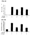
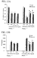
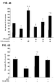
- Figure 2A shows the results as a percentage of the control cultures, and are the mean ⁇ S.E.M of five individual experiments. ##, P ⁇ 0.01 compared with the control culture. *, P ⁇ 0.05 compared with the LPS -treated culture. As seen there, treatment with 10 ng/ml LPS alone caused a significant reduction in the loss of TH-IR neurons (60%) compared with vehicle-treated control cultures. Thirty minute pretreatment with DM 10 -5 and 10 -14 M significantly attenuated the LPS-induced reduction in the number of TH-IR neurons by 37 and 28%, respectively. DM at 10- 10 M had no protective effects on dopaminergic neuron degeneration. The results from the cell counts ( Figure 2A ) were comparable to that of the [ 3 H]-dopamine uptake study of Example 1 ( Figure 1 ).
- Example 3 Femtomolar concentrations of DM protect A ⁇ -induced dopaminergic neurodegeneration
- Neuron-glia co-cultures were prepared and treated with vehicle alone, A ⁇ 0.75 ⁇ M alone, or DM 30 min prior to treatment with A ⁇ 0.75 ⁇ M similar to Example 1.
- Amyloid- ⁇ peptide 25-35 and 1-42 was obtained from American Peptide Co., Inc (Sunnyvale, CA). Neurotoxicity was assessed by DA uptake as in Example 1. Results are expressed as a percentage of the control cultures and are the mean ⁇ S.E.M. of three to eight individual experiments with triplicates in each experiment. ##, P ⁇ 0.01 compared with the control culture; *, P ⁇ 0.05 compared with A ⁇ -treated culture.
- Results are shown in Figure 3 , where the neuroprotective effect of DM at both micro- and femtomolar concentrations can be seen against A ⁇ -induced neurotoxicity.
- Neuron-enriched culture were prepared from the ventral mesencephalic tissues of embryonic day 13-14 Fisher F344 rats (Charles River Laboratories, Raleigh, NC) as follows. Dissociated cells were seeded at 1 ⁇ 10 5 /well in 96-well and 5 ⁇ 10 5 /well to poly-D-lysine-coated 96-well and 24-well plates, respectively. Glial proliferation was suppressed by the inclusion of cytosine ⁇ -D-arabinocide (5-10 ⁇ M). Seven day old cultures were used for treatment, which were composed of 91% neurons, 9% astrocytes, and ⁇ 0.1% microglia.
- the cultures were pre-treated with OM, 10 -5 M, 10 -10 M, and 10 -14 M DM 30 minutes before treatment with vehicle alone, 4 ⁇ M A ⁇ or 0.5 ⁇ M MPP +5 as in Example 1.
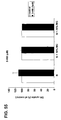
- DA uptake was also measured as in Example 1 and the results are expressed in both Figures 4A and B as a percentage of the control cultures and are the mean ⁇ S.E.M. of four individual experiments with triplicates in each experiment.
- the protective effect is only observed in neuron-glia cultures, but not in neuron-enriched cultures, since the DM failed to show a protective effect against A ⁇ - or MPP + -induced dopaminergic neurotoxicity in neuron-enriched culture regardless of the concentration of DM.
- This comparison may indicate that femtomolar DM-elicited protection against inflammation-mediated dopaminergic neurotoxicity is dependent on the presence of microglia.
- Example 5 Femtomolar DM inhibits LPS-induced microglia activation
- LPS can activate microglia to overproduce pro-inflammatory cytokines and free radicals, such as NO, PGE 2 , TNF ⁇ , superoxide, and other reactive oxygen species (ROS), which in turn can cause neurodegeneration.
- pro-inflammatory cytokines and free radicals such as NO, PGE 2 , TNF ⁇ , superoxide, and other reactive oxygen species (ROS)
- ROS reactive oxygen species
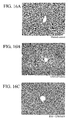
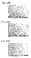
- Neuron-glia cultures were prepared and treated with vehicle, LPS (5ng/ml), or LPS plus DM respectively as in Example 1. Twelve hours later, cultures were immunostained with anti-OX-42 antibody. Images shown are representative of three separate experiments. The immunostaining and morphological analysis was accomplished as in Example 2.
- LPS treatment transformed the resting, round shape of microglia into the enlarged, irregular shape of activated microglia.
- Pre-treatment with DM at 10 -5 M and 10 -14 M prevented the LPS-induced activation of microglia.
- DM at 10 -10 M didn't significantly inhibit microglia activation by LPS.
- Figure 5 depicts photomicrographs of microglia showing the inhibitory effect of DM on LPS-induced microglial activation.
- PGE 2 in supernatant was measured with a prostaglandin E 2 EIA kit from Cayman (Ann Arbor, MI) according to the manufacturer's instructions.
- the release of superoxide was determined by measuring the superoxide dismutase (SOD)-inhibitable reduction of cytochrome c.
- SOD superoxide dismutase
- the production of intracellular reactive oxygen species was measured by DCFH oxidation.
- the DCFH-DA Molecular Probes, Eugene, OR
- the DCFH-DA passively diffuses into cells in which it is hydrolyzed by intracellular esterase to liberate 2'-7'-dichlorofluoressein which, during reaction with oxidizing species, yields a highly fluorescent compound 2'-7'-dichlorofluorescein (DCF) that is trapped inside the cell.
- DCF highly fluorescent compound 2'-7'-dichlorofluorescein
- CM-H2-DCFDA diluted to a final concentration of 1 ⁇ M in phenol red-free HBSS containing 2% FBS and 2% HS, was added to cultures and incubated for about 30 min at about 37°C. After washing two times with warm HBSS, vehicle or stimulators in HBSS were added to cultures. After incubation for about 30 min at about 37°C, fluorescence intensity was measured at 485 nm for excitation and 530 nm for emission using a SpectraMax Gemini XS fluorescence microplate reader (Molecular Devices).
- Results are expressed in Figures 6A, 6B , and 6C as a percentage of the LPS cultures, in 6D as a percentage of control, and in 6E as absorbance difference above control value.
- the results are the mean ⁇ S.E.M. of four individual experiments with triplicates in each experiment. *, P ⁇ 0.05 compared with LPS culture.
- neuron-glia cultures were prepared from NADPH oxidase-deficient (PHOX -/- ) and wild-type (PHOX +/+ ) mice.
- Microglia were prepared from the whole brains of 1-day-old Fisher F344 rats or NADPH oxidase-deficient (gp91phox -/- ) (Jackson Laboratory, Bar Harbor ME) or wild-type mice (C57 BL/6J (gp91phox +/+ ) (Jackson Laboratory, Br Harbor, ME), as in the above examples. Immunocytochemical analysis accomplished as above indicated that the cultures were 95-98% pure for microglia. Cells were seeded at 1 ⁇ 10 5 /well in 96-well plates and used for treatment the following day.
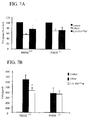
- the neuron-glia cultures were treated with vehicle, LPS 10 ng/ml alone, and DM (10 -14 M) 30 min pretreatment followed by LPS treatment as in Example 1. Neurotoxicity was assessed by DA uptake as described in Example 1. TNF ⁇ production was measured by ELISA and iROS was determined by DCFDA as in Example 5. Results are expressed as a percentage of the control culture in Figure 7A , pg/ml in Figure 7B , and difference from control in Figure 7C , respectively, and are the mean ⁇ S.E.M. of five individual experiments with triplicates in each experiment. *, P ⁇ 0.05 compared with LPS culture.
- PHOX is the major superoxide-producing enzyme in microglia and the major contributor to the increase in iROS concentrations in response to a variety of immune stimulants such as LPS, ⁇ -amyloid peptides (A ⁇ ).
- a ⁇ ⁇ -amyloid peptides
- Femtomolar DM while significantly lessening the LPS-induced DA uptake reduction in wild-type mice, has no significant protective effect in PHOX -/- mice ( Figure 7A ).
- PHOX activity Activation of PHOX in microglia not only increases the production of superoxide, but also indirectly increases the intracellular ROS concentration, possibly through the conversion of superoxide to H 2 O 2 , which is membrane permeable. Increase of iROS can intensify the activation of NF ⁇ B, which leads to higher TNF ⁇ , PGE 2 production.
- femtomolar DM inhibited TNF ⁇ production in wild type while not in PHOX -/- mice further supports the notion that femtomolar DM may be acting on PHOX.
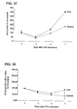
- This example tests whether or not post-treatment with DM is still neuroprotective in LPS-induced neurotoxicity.
- Neuron-glia co-cultures as prepared in Example 1 were first treated with LPS (20 ng/ml) for twelve hours, and then LPS was removed by removing the media from the cultures and washing twice with PBS.
- Different concentrations of DM (10 -5 , 10 -10 , and 10 -14 M) were then added to the cultures and incubation was continued for another 6 or 7 days.
- the DA uptake and the superoxide production were measured as in Example 1 and 5.
- Results are expressed as a percentage of the control cultures and are the mean ⁇ S.E.M of three to six experiments with triplicates in each experiment. ##, P ⁇ 0.01 compared with control culture; *, P ⁇ 0.05 compared with the LPS-treated culture.
- Example 8 Determination of possibility of direct action of DM on iNOS and COX2 enzymes
- NF ⁇ B is triggered through cascading signaling pathway to regulate the mRNAs encoding iNOS and COX2, which produce NO and PGE 2 respectively.
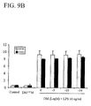
- Example 5 The cultures were prepared as in Example 1 and were first treated with LPS 20 ng/ml or vehicle; 12 hours later the LPS was removed and the cultures were treated with 10 -5 M, 10 -10 M, and 10 -14 M DM. Twenty four hours later, nitrite oxide and PGE 2 production were assessed as in Example 5. The results are expressed as a percentage of LPS treated culture and are the mean ⁇ S.E.M of four and three experiments with triplicates in each experiment. *, p ⁇ 0.05 compared with the LPS-treated culture. ###, P ⁇ 0.001 compared with the control culture.
- Figures 10A and 10B demonstrate that femtomolar DM post treatment following removal of LPS after 12 hours LPS treatment on neuron/glia culture resulted in reduction of NO and PGE 2 production without affecting iNOS and COX2 protein levels as evidenced by Figure 9 . Since iNOS and COX2 accumulated within the 12 hours of LPS treatment are sufficient for continuing the production of NO and PGE 2 even in the absence of LPS, it was concluded that DM decreased NO and PGE 2 production by directly inhibiting the activities of these enzymes. Reduction in the production of nitrite, PGE 2 and TNF ⁇ , together with the drastic suppression of ROS production, is thought to be one of the mechanisms underlying the potent neuroprotective effect of femto-molar DM.
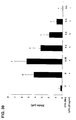
- Femtomolar concentrations of several small peptide fragments of varying lengths and sequences were tested for their ability to protect DA neurons from LPS-induced neurodegeneration in vitro.
- Neuron-glia cell cultures were prepared as in Example 1, and pretreated with the various peptide fragments of Table 1 (peptide fragments were obtained from BACHEM) for 30 minutes followed by addition of 5ng/ml of LPS. DA neurotoxicity was measured as an Example 1 at 7 days post treatment.
- Table 1 The data in Table 1 are expressed as the percent of the control cultures and are the mean ⁇ SEM of 3 experiments performed in triplicate. *P ⁇ 0.05, **P ⁇ 0.01, compared to control.
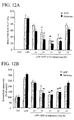
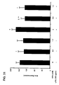
- Mesencephalic neuron-glia cultures were prepared as in Example 1 and were treated with either vehicle, LPS (5 ng/ml) or were pretreated for 30 minutes with naloxone or GGF (10 -12 -10 -16 M) followed by addition of LPS (5 ng/ml) as described in Example 1.
- DA neurotoxicity was measured at 7 days post treatment as in Example 1.
- Dopaminergic neuronal death was determined at 7 days post treatment using immunocytochemical staining as in Example 2.
- the ability of GGF and naloxone to protect DA neurons from LPS-induced damage is depicted by immunocytochemical analysis with anti-TH antibody as in Example 2. The data are expressed as the percentage of the control cultures and are the mean ⁇ SEM from three independent experiments, each performed with triplicate samples. * P ⁇ 0.05, ** P ⁇ 0.01 compared to control.
- Figure 11A shows that both the peptide GGF and naloxone exhibit similar neuroprotective qualities at femtomolar concentrations.
- Figure 11C shows that both 10 -14 M naloxone and 10 -14 M GGF protected TH immuno-reactive neurons from LPS-induced damage, such as loss of dendrites, axon disintegration, and loss of DA neurons.
- Figure 11B evidences that GGF and naloxone pretreatment protected against LPS-induced DA neuron cell loss, with the peak protection occurring at 10 -14 M for both GGF and naloxone. Taken together, these results indicate that both the peptide, GGF, and the naloxone protected neurons from LPS-induced DA neuron cell death and loss of function with a similar efficacy and dose response.
- Example 1 Primary enriched-microglia cultures were prepared as in Example 1. The cultures were pretreated with varying concentrations of naloxone for about 30 minutes and GGF followed by treatment with 10 ng/ml LPS as in Example 1. The intracellular ROS concentrations and superoxide amounts were measured as in Example 5. The data are expressed as the percent of the control cultures and are the mean ⁇ SEM of three experiments performed in triplicate. *P ⁇ 0.05, **P ⁇ 0.01, compared to control.
- the rat and mouse ventral mesencephalic neuron-glia cultures were prepared as in Example 1.
- Mesencephalic neuron-glia cultures from PHOX -/- and PHOX +/+ mice were treated with either vehicle, LPS (5 ng/ml), or were pretreated for 30 minutes with Naloxone or GGF (10 -13 M-10 -14 M) followed by addition of LPS (5 ng/ml) as in Example 1.
- DA neurotoxicity was measured by using the [ 3 H] DA uptake assay as in Example 1. The data are expressed as the percent of the control cultures and are the mean ⁇ SEM.
- the release of TNF ⁇ was measured with a commercially available enzyme-linked immunosorbent assay kit. *P ⁇ 0.05, **P ⁇ 0.01, compared to control.
- the amount of TNF ⁇ was measured as in Example 2.
- NADPH oxidase may be a component to the mechanism of neuroprotection.
- a conformational search was defined based on rotatable bonds, where 166 conformations were identified (66 of which were redundant) resulting in 100 uniquely defined conformations. (The Phe ring was kept in one planar orientation and conformations rotating this ring were not explored). These conformations were energy minimized resulting in 42 distinct energy-minimized conformations. These 42 GGF peptide conformations were compared in terms of electrostatic and steric-shape similarity with naloxone. Naloxone, as a fused ring compound, has less conformational flexibility than the GGF tripeptide.
- Binding affinities of naloxone for COS-7 cells transfected with gp91/p22 was determined using either [ 3 H]-(+)Naloxone or [ 3 H]-(-)Naloxone (2 nM; PerkinElmer Life Sciences) as ligands and displaced with 10 ⁇ M of cold (-)Naloxone in HBBS containing 0.1% (W/V) fatty acids free albumin (lot B22558, Calbiochem).
- COS-7 cells transfected with gp91/p22 and COS-7 cells that were not transfected (WT) were detached using Versene (1:5000, GibcoBRL, Life technologies). For intact cell assays, after washing twice with HBSS, cells were transferred to micro centrifuge tubes at 10 6 cells per tube.
- binding affinities of LPS for COS-7 cells transfected with gp91/p22 was determined either with [ 3 H]- naloxone (2 nM; PerkinElmer Life Sciences) as ligands displaced with 10 ⁇ M of cold naloxonein HBBS containing 0.1 % (W/V) fatty acids free albumin (lot B22558, Calbiochem).
- COS-7 gp91/p22 cells an immortal kidney cell line stably transfected with gp91/p22, have an increased binding capacity (150-180%) above the control COS-7 cells, which do not express gp91 (NADPH oxidase membrane bound catalytic subunit) or p22 (NADPH oxidase membrane anchor protein) ( Figure 15 ).
- This data offers support for the hypothesis that naloxone binds to the gp91 protein.
- mice Male, CD-1 mice (6-week-old) were purchased from Charles River laboratories, fed on a standard diet and with tap water ad libitum for two weeks. Environmental conditions were standardized, including a room temperature of 21°C and 12 hours artificial lighting. Mice were fasted 12 hrs before use.
- Endotoxic shock was induced in the mice by administering a single intraperitoneal dose of lipopolysaccharide/D-(+/-)Glactosomine (Sigma, St. Louis, MO) (LPS/GalN) (20 ⁇ g/700 mg/kg) in saline.
- LPS/GalN lipopolysaccharide/D-(+/-)Glactosomine
- DM lipopolysaccharide/D-(+/-)Glactosomine
- mice were treated with 12.5 mg/ml DM at 30 min before LPS/GalN injection, and 30, 60 and 120 min after LPS/GalN administration. Serum ALT was measured to evaluate the therapeutic effects of the DM. Survival rate was evaluated within 12 hours after endotoxin administration.
- Serum alanine aminotransferase (ALT) activity was assayed as a marker of hepatocellular death using a commercially available kit (Infinite ALT, Sigma, St. Louis, MO). A portion of the liver was fixed in 10% neutral formalin, processed by standard histological techniques, stained with hematoxylin and eosin, and examined for morphological evidence of liver injury.
- TNF ⁇ TNF ⁇ in the serum and liver were determined as in Example 1.
- Kupffer cell samples were collected by anesthetizing the CD-1 mice with pentobarbital anesthesia [60mg/kg intraperitoneally (i.p.)].
- the abdomen of the animals was shaved and opened, the portal vein was cannulated and perfused with Ca 2+ - and Mg 2+ - free Hanks' balanced salt solution (HBSS) at 37°C for 5min at a flow rate of 13 ml/min.
- HBSS Hanks' balanced salt solution
- the liver was perfused with HBSS containing 0.05% collagenase IV (Sigma Chemical, St. Louis, MO) at 37°C for 5min. After the liver was digested, it was excised and cut into small pieces in collagenase buffer.
- the suspension was filtered through nylon gauze mesh, and the filtrate was centrifuged at 450 ⁇ g for 10 min at 4°C.
- Cell pellets were resuspended in buffer, parenchymal cells were removed by centrifugation at 50 ⁇ g for 3min, and the nonparenchymal cell fraction was washed twice with buffer.
- Cells were centrifuged on a density cushion of 50 % of Percoll (Pharmacia, Upsala, Sweden) at 1,000 ⁇ g for 15min, and the Kupffer cell fraction was collected and washed again with buffer. The viability of the cells was determined by tryptan blue exclusion at >90%.
- the cells were seeded onto 24-well culture plates and cultured in RPMI 1640 (GIBCO Laboratories Life Technologies, Grand Island, NY) supplemented with 10% fetal bovine serum and antibiotics (100 U/ml penicillin G and 100 ⁇ g/ml streptomycin sulfate) at 37°C with 5% CO 2 .
- RPMI 1640 Gibco Laboratories Life Technologies, Grand Island, NY
- antibiotics 100 U/ml penicillin G and 100 ⁇ g/ml streptomycin sulfate
- mice at 12 hours post-LPS/GalN treatment are presented in Table 2.
- Table 2 Dextromethorphan (mg/kg) Animal number (n) Survived animal Rate of survival (%) 0 61 28 45.9 6.25 12 8 66.6 12.5 56 50 89.3 25 12 11 91.6
- liver samples collected from the LPS/GalN-induced mice are shown in Figures 16A (control), 16B (12 hours after LPS/GalN) and 16C (animal treated with DM).
- the liver histology was examined to evaluate the effect of DM treatment on LPS/GalN hepatotoxicity.
- the liver sections showed apparently broad hemorrhagic necrosis and apoptosis, and severe hepatocyte swelling 12 hours after LPS/GalN challenge ( Fig. 16B , arrows ).
- Fig. 16C The liver samples collected from the LPS/GalN-induced mice are shown in Figures 16A (control), 16B (12 hours after LPS/GalN) and 16C (animal treated with DM).
- the liver histology was examined to evaluate the effect of DM treatment on LPS/GalN hepatotoxicity.
- the liver sections showed apparently broad hemorrhagic necrosis and apoptosis, and severe hepatocyte swelling 12 hours after LPS
- Serum alanine aminotransferase an indicator of acute hepatocellular death
- Serum ALT increased ⁇ 35-fold over controls by 12 hrs after LPS/GalN administration ( Figure 17 )
- DM dramatically decreased serum ALT level in a dose-dependent manner and reduced to about 25% of LPS/GalN group at 12.5 and 25 mg/kg of DM.
- Time-dependent reduction of serum ALT level is shown in Figure 18 , DM administrated at different time points, including 30 min pre-treatment and 30, 60, 120 min post-treatments shows protective effects to various extracts, the later the DM treatment, the less protective.
- TNF ⁇ an important factor that plays an important role in sepsis, was studied as a mechanism of the protective effect of DM in LPS/GalN-challenge mice.
- DM (12.5 mg/kg, i.c.) was administered to mice, followed by LPS/GalN challenge 30 min later. Serum and liver TNF ⁇ level was assessed using ELISA at the indicated time points.
- DM significantly decreased TNF ⁇ level in both serum and liver at 1.5 and 2 hours after LPS/GalN challenge.
- the suppression of TNF ⁇ level was also found in DM 30 min post LPS/GalN treatment (data not shown).
- the reduction of hepatic TNF ⁇ paralleled the reduction of serum TNF ⁇ , indicating that decrease TNF ⁇ is an important mechanism of protection.
- Kupffer cells Activation of Kupffer cells by LPS is a critical event in the endotoxemia or sepsis. Therefore Kupffer cells were isolated to study the possible mechanism of DM in protection liver injury and sepsis. Endotoxin activates Kupffer cells to release inflammatory mediators such as free radicals and TNF ⁇ .
- RNA was extracted from liver tissues (n 4 to 5) by Trizol reagent (Sigma, St. Louis, MO) and purified with an RNeasy column (Qiagen, Valencia, CA). Expression of the selected genes was quantified using real-time RT-PCR analysis that began by reverse transcribing the samples with MuLV reverse transcriptase and oligo-dT primers. The forward and reverse primers for the selected genes were designed using Primer Express software and are listed in Table 3. Table 3 Gene Accession Number Forward Primer Reverse Primer MIP - 2 NM_009140 CCTCAACGGAAGAACCAAAGAG (Seq. ID No. 3) CTCAGACAGCGAGGCACATC (Seq. ID No.
- GAAGGCAACTGGATGGAAGTCT (Seq. ID No. 12) IL - 10 M37897 CCAAGCCTTATCGGAAATGATC (Seq. ID No. 13) GATTTCTGGGCCATGCTTCTC (Seq. ID No. 14) c - jun / AP-1 J04115 ACTCCGAGCTGGCATCCA (Seq. ID No. 15) CCCACTGTTAACGTGGTTCATG (Seq. ID No. 16) c-myc X01023 CGCCGCTGGGAAACTTT (Seq. ID No. 17) TCCTGGCTCGCAGATTGTAA (Seq. ID No.
- MIP-2 Macrophage Inflammatory Protein-2
- TSP-1 thrombospondin1
- mKC a mouse CXC chemokine
- ICAM-1 Intercellular Cell Adhesion Molecule-1
- IL-6 5 Interleukin-6
- IL-10 Interleukin-10
- GADD153 Growth Arrest and DNA Damage inducible protein 153
- GADD45 Growth Arrest and DNA Damage inducible protein 45.
- the SYBR green DNA PCR kit (Applied Biosystems, Foster City, CA) was used for real-time PCR analysis.
- the relative differences in expression between 10 groups were expressed using cycle time (Ct) values and the relative differences between groups were expressed as relative increases setting control as 100%. Assuming that the Ct value is reflective of the initial starting copy and that there is 100% efficacy, a difference of one cycle is equivalent to a two-fold difference in starting copy.
- MIP-2 mouse macrophage inflammatory protein
- TSP1 thrombospondin-1
- mKC mouse chemokine
- ICAM-1 intracellular adhesion molecule-1
- IL-10 interleukin-10
- GalN /LPS acute hepatotoxicity also greatly enhanced the expression of c-jun/AP-1 (28-fold), c-myc (45.4-fold), while the expression of both genes was diminished to 9.1- and 18.4-fold, respectively with DM treatment.
- the DNA damage responsible proteins such as GADD45 and GADD153 were also increased by 24.6 and 7.4-fold respectively.
- GADD45 and GADD153 protein genes There was a significant suppression of GalN/LPS-induced GADD45 and GADD153 protein genes by DM to 5.3 and 2.7-fold respectively.
- mice Endotoxic shock were induced in mice in the same was as Example 16.
- the mice were given LPS/GalN (20 ⁇ g/700 mg/kg, ip) with or without the administration of DM (10 mg/kg, 1 ⁇ g/kg and 100 pg/kg, s.c.) 30 min before LPS/GalN.
- the survival rate of the animals was evaluated 12 hrs after LPS/GalN treatment.
- Table 5 Amount of dextromethorphan administered Number of animals in group No. of animals survived Rate of survival % 0 30 17 56.7 10 mg/kg 26 22 84.6 1 ug/kg 17 10 58.8 100 pg/kg 47 36 76.6
- Figure 22 shows the levels of serum ALT, measured as in Example 16.
- FIG. 23A shows a photomicrograph of a liver sample with LPS/GalN alone. As seen there, there is a foci of necrotic parenchymal cells, cell swelling, and congestion.
- Figure 23B shows a photomicrograph of a liver sample that was administered 10 mg/kg DM plus LPS/GalN. As seen there, hepatic congestion and cell death are mild, while the cell swelling is the only notable lesion.
- Figure 23C shows a photomicrograph of a liver sample with that was administered 1 ⁇ g/kg DM plus LPS/GalN. As seen there, hepatic congestion and cell death are obvious.
- Figure 23D shows a photomicrograph of a liver sample that was administered 100 pg/kg DM plus LPS/GalN. As seen there, cell swelling is the only notable lesion, but cell death is mild.
- Kupffer cells were pre-treated with DM (10 -5 , 10 -10 or 10 -14 M) or vehicle (control or LPS treated group) for 30 min, and then stimulated with LPS 5 ng/ml or vehicle (control group). TNF- ⁇ production (6 hrs after LPS stimulation) was measured by an ELISA kit as in Example 5. The data, which are seen in Figure 24 are the mean ⁇ SEM of 3-4 individual experiments with triplicates. *, Significantly different from LPS alone treated culture, P ⁇ 0.05.
- Examples 19 through 22 show VPA protects dopaminergic neurons from LPS-induced neurotoxicity through the inhibition of microglial activation.
- the anti-inflammatory responses of cultures stimulated with LPS and pretreated with VPA are also characterized.
- Neuron-glia cultures were prepared from the ventral mesencephalic tissues of embryonic day 13-14 rats. Dissociated cells were seeded at 1 ⁇ 10 5 /well and 5 ⁇ 10 5 / well to poly-D-lysine-coated 96-well and 24-well plates, respectively.
- VPA pretreatment on morphological changes of mesencephalic dopaminergic neurons immunostained with anti-TH antibody were determined.
- the primary midbrain cultures were pretreated with the indicated concentrations of VPA for 48 h and then treated with 20 ng/ml LPS for 72 h, as described above.
- DA neurons were recognized with the anti-TH antibody and microglia were detected with the OX-42 antibody, which recognizes the CR3 receptor.
- formaldehyde (3.7%)-fixed cultures were treated with 1% hydrogen peroxide (10 ml) followed by sequential incubation with blocking solution (30 min), primary antibody (overnight, 4°C), biotinylated secondary antibody (2 h), and ABC reagents (40 min).
- Example 20 VPA pretreatment suppresses LPS-induced activation of microglia and production of pro-inflammatory factors in neuron-glia cultures
- FIGS 28A-28F show VPA pretreatment suppresses LPS-induced microglia activation revealed by OX-42 immunostaining.
- Mesencephalic neuron-glia cultures were pretreated with VPA for 48 h and then treated with 20 ng/ml LPS for 72 h, as described in Example 19.
- Immunostaining with an antibody against OX-42 was then performed, as described in Example 19.
- Mesencephalic neuron-glia cultures treated with LPS displayed the characteristics of activated microglia such as, increased cell size, irregular shape, and intensified OX-42 immunoreactivity, a specific marker for rat microglia as shown in Figures 28A and 28B .
- the LPS-stimulated activation of microglia was suppressed in neuron-glia cultures pretreated for 48 h with 0.4 or 0.6 mM VPA, as shown in Figures 28D through 28F .
- VPA alone did not show significant effects on microglia activation, as shown in Figure 28C .
- Scale bar 100 ⁇ m. The images shown are representative of 3 independent experiments.
- nitric oxide was determined in the mesencephalic neuron-glia cultures, following pretreatment and LPS stimulation as described above, by measuring the accumulated levels of nitrite in the supernatant with Griess reagent. Accumulation of nitrite, an indicator of LPS-stimulated production of NO, was determined 24 after LPS stimulation. As shown in Fig. 30 , pretreatment with 0.4 and 0.6 mM VPA reduced LPS-stimulated NO production 54% and 78% of the control, respectively.
- Results are means ⁇ S.E.M of 4 independent experiments. *, p ⁇ 0.05 compared with LPS-treated cultures; ⁇ , p ⁇ 0.05, compared with untreated control.
- Example 21 VPA pretreatment inhibits LPS-induced intracellular reactive oxygen species production in enriched microglia
- iROS intracellular reactive oxygen species
- CM-H2-DCFDA Chloromethyl-2',7'-dichlorodihydrofluorescein diacetate
- DCF 2'-7'-dichlorofluorescein
- Microglia-enriched cultures were prepared from the whole brains of 1-day-old rats as described in Example 1. Immunocytochemical analysis indicated that the cultures were 95-98% pure for microglia. Cells were seeded at 1 ⁇ 10 5 /well in 96-well plates for one day followed by treatment with 0.6 mM or 1.2 mM VPA for 24 h. The cultures were used for the assay of intracellular ROS.
- CM-H2-DCFDA diluted to a final concentration of 1 ⁇ M in phenol red-free HBSS
- HBSS Hank's balanced salt solution
- CM-H2-DCFDA diluted to a final concentration of 1 ⁇ M in phenol red-free HBSS
- the iROS level was significantly increased by LPS treatment and this increase was completely blocked by pretreatment with VPA at 0.6 or 1.2 mM ( Fig. 31 ).
- Example 22 VPA treatment decreases the number of microglia
- Microglia cell number was determined as follows. Primary microglia-enriched cultures were prepared from the whole brains of 1-day-old rats as described previously. After one day in vitro were treated with the indicated concentrations of VPA (0.05, 0.1, 0.2, 0.4, 0.6, 0.8, 1.0, or 1.2 mM) for 48 h, or with 0.6 mM VPA for different times (6, 12, 24 or 48 h). After treatment with VPA and vehicle for 48 hours, the number and morphology of microglia in cultures were observed under an inverted microscope (Nikon, Tokyo, Japan) at 100 x. The total number of microglia was counted by using the CyQUANT cell proliferation assay kit (Molecular Probes, Inc.).
- VPA time-dependently decreased the number of microglia with a significant effect at 24 h and 48 h after treatment Fig. 32A-32F and 33 .
- the VPA effect was also concentration-dependent with a robust decrease in microglia number in the dose range of 0.2 mM to 1.2 mM after 48 h treatment ( Fig. 34A-34D and 35 ).
- the loss of microglia was about 80% by treatment with 0.8 mM VPA.
- VPA-induced microglia loss was time and dose-dependently associated with aggregations or clumping of surviving cells ( Fig. 32A-32F and 34 ).
- VPA dose-dependently induces survival-promoting effects against spontaneous DA neuronal death in rat primary mesencephalic neuron-glia cultures.
- Example 23 [ 3 H]DA uptake assay and immunohistochemical analysis for TH-IR neurons were used to assess the viability of DA neurons in rat primary mesencephalic neuron-glia cultures in which approximately 1% of the neurons are dopaminergic. [ 3 H]DA uptake assays were performed as described in Example 1.
- Rat primary mesencephalic neuron-glia cultures were prepared from the ventral mesencephalic tissues of embryonic day 13-14 Fisher 344 rats. Dissociated cells were seeded at a density of 5 ⁇ 10 5 / well to poly-D-lysine-precoated 24-well plates. Cells were maintained at 37°C in a humidified atmosphere of 5% CO 2 and 95% air, in minimal essential medium containing 10% fetal bovine serum (FBS), 10% horse serum, 1 gm/l glucose, 2 mM L-glutamine, 1 mM sodium pyruvate, 100 ⁇ M non-essential amino acids, 50U/ml penicillin, and 50 ⁇ g/ml streptomycin.
- FBS fetal bovine serum
- horse serum 1 gm/l glucose
- 2 mM L-glutamine 1 mM sodium pyruvate
- 100 ⁇ M non-essential amino acids 50U/ml penicillin, and 50 ⁇ g/ml streptomycin.
- rat neuron-glia cultures contained 11% microglia, 48% astroglia, and 41% neurons, among which about 1% of cells represent tyrosine hydroxylase-immunoractive (TH-IR) neurons.
- TH-IR tyrosine hydroxylase-immunoractive
- Valproic acid (Sigma-Aldrich, St. Louis, MO) is prepared as a solution in double-distilled water and then sterile filtering immediately before use.
- Rat primary mesencephalic neuron-glia cultures were treated with 0.6 mM VPA for various times to determine the treatment time-dependency.
- Rat primary mesencephalic neuron-glia cultures in a 24-well plate were treated with 0.6 mM VPA or its vehicle for indicated time 7 days after seeding.
- the viability of dopaminergic neurons was assessed by [ 3 H]DA uptake assays, shown in Figure 37 or counting of TH-IR neurons, shown in Figure 38 . Quantified results are expressed as mean ⁇ SEM of percentage of vehicle-treated control cultures from three experiments performed in duplicate. *, p ⁇ 0.05 compared with the corresponding vehicle-treated control cultures.
- VPA treatment increased the capacity of [ 3 H]DA uptake and the number of TH-IR neurons in a time-dependent manner, as shown in Fig. 37 and 38 .
- treatment with 0.6 mM VPA for 7 days, but not 3 or 5 days resulted in a marked increase in the parameter compared with vehicle-treated control.
- VPA time-dependently induces survival-promoting effects against spontaneous DA neuronal death in rat primary mesencephalic neuron-glia cultures.
- astroglial cells are involved in the neurotrophic actions of VPA was tested by preparing media conditioned by incubation of astroglial cultures in the absence or presence 0.6 mM VPA. The results are shown in Figure 39 .
- microglia Upon reaching confluence (usually 12-14 days later), microglia were detached from astroglia by shaking the flasks for 5 hours at 180 rpm. Astroglia were then detached with trypsin-EDTA and seeded in the same culture medium. After five or more consecutive passages, cells were seeded onto 24-well plates (10 5 /well) for experiments. Immunocytochemical staining of the astroglia enriched cultures with either anti-GFAP or anti-OX-42 antibody indicated astroglial purity of greater than 98% and less than 2 % of microglia contamination.
- ACM-VPA was dialyzed overnight using the Slide-A-Lyzer® Dialysis Cassette (Pierre Biotechnology, Inc) to remove small molecular weight substances. The dialyzed ACM-VPA still retained its ability to enhance DA uptake (data not shown here).
- the astrocyte-conditioned medium (ACM) derived from incubation with astrocytes for 12,24 or 48 hours showed an approximate 4-fold increase in DA uptake following incubation of mesencephalic neuron-enriched cultures with ACM for 7 days.
- the conditioned medium derived from incubation of astrocytes in the presence of 0.6 mM VPA (ACM-VPA) displayed a more robust (more than 8-fold), time-dependent increase in DA uptake, compared with ACM. Exposure of the neuron-enriched cultures to 0.6 mM VPA for 7 days caused a less than 2-fold increase in DA uptake, suggesting that a direct action on neurons does not play a major role in VPA-induced neurotrophic effects.
- Conditioned medium derived from rat primary astroglial cultures treated with vehicle (ACM) or 0.6 mM VPA (ACM-VPA) were harvested after 12,24 and 48 hours of incubation.
- Midbrain neuron-enriched cultures seeded in 24-well plates at a density of 5x10 5 cells/well were treated with vehicle, VPA, ACM or ACM-VPA for 7 days.
- Neurotrophic effect was quantified by [ 3 H]DA uptake assay.
- the data are expressed as mean ⁇ SEM of percentage of vehicle control from four to five independent experiments performed in triplicate; *, p ⁇ 0.001 compared with the corresponding vehicle control cultures; ⁇ , p ⁇ 0.001 compared with the corresponding ACM-treated cultures.
- FIGS. 40A-40D show morphological features of neuron-enriched cultures were examined after incubation with vehicle (A), 0.6 mM VPA (B), ACM (C) or ACM-VPA (D) for 7 days and then immunocytostaining with MAP-2 antibody.
- the immunostaining was carried out according to the following procedure. Formaldehyde (3.7%)-fixed cultures were treated with 1% hydrogen peroxide (10 min) followed by sequential incubation with blocking solution (30 min), primary antibody (overnight, 4°C), biotinylated secondary antibody (2 hours), and ABC reagents (40 min) (Vector Laboratories, Burlingame, CA). The color development was achieved by the addition of 3,3'-diaminobenzidine.
- the images were recorded with an inverted microscope (Nikon, Tokyo, Japan) connected to a charge-coupled device camera (DAGE-MTI, Michigan City, IN) operated with the MetaMorph software (Universal Imaging Corporation, Downingtown, PA). For visual counting of TH-IR neurons, nine representative areas per well of the 24-well plate were counted under the microscope at 100x magnification.
- FIGS. 20A-40D The results of the morphological examination are shown in Figures 20A-40D .
- Morphological examination of neuron-enriched cultures immunostained with MAP-2 demonstrated a dramatic increase in neurite outgrowth following exposure for 7 days to ACM-VPA conditioned with 48 hours incubation with astrocytes. ( Fig. 40D ), compared with the vehicle-treated control cultures ( Fig. 40A ). Much smaller effects were observed following incubation with the VPA alone or corresponding ACM ( Fig. 40B and 40C ).
- FIG. 30 shows morphological features of neuron-enriched cultures were examined after incubation with ACM (A) or ACM-VPA (B) for 7 days and then immunocytostaining with TH-IR antibody. Results demonstrated higher density of DA neurons, more complex neurite configurations and more neuronal connections in ACM-VPA-treated cultures ( Fig. 41A ) than ACM-treated cultures ( Fig. 41B ).
- GDNF is VPA-induced, astroglia-secreted neurotrophic substance
- real time PCR and ELISA to quantify GDNF mRNA and protein levels, respectively.
- Rat primary astroglias were exposed to 0.6 mM VPA for various times ranging from 6 to 48 hours.
- Total RNA was extracted from cells by Tri reagent (Sigma) and purified with RNeasy columns (Qiagen, Valencia, CA). Expression of the selected genes was quantified using real-time RT-PCR analysis. Briefly, total RNA was reverse transcribed with MuLV reverse transcriptase and oligo-dT primers. The forward and reverse primers for selected genes were designed using Primer Express software. The SYBR green DNA PCR kit (Applied Biosystems, Foster City, CA) was used for real-time PCR analysis. The relative differences in expression between groups were expressed using cycle time (Ct) values and the relative differences between groups were expressed as relative increases setting the control as 100%. Assuming that the Ct value is reflective of the initial starting copy and that there is 100% efficacy, a difference of one cycle is equivalent to a two-fold difference in the starting copy.
- Ct cycle time
- ACM-VPA was prepared according to Example 20 and collected 48 hours after incubation with astroglia.
- ACM-VPA was analyzed for secreted GDNF by ELISA.
- GDNF levels were measured with an ELISA kit (GDNF Emax ImmunoAssay System; Promega , Madison, WI), according to the protocol of the supplier.
- the levels of GDNF were expressed as pg per ml of supernatant.
- the assay sensitivity ranged from 16 to 1000 pg/ml. Results are expressed as pg/ml from three experiments performed in duplicate.
- ACM-VPA showed a 2.1-fold over the vehicle control in levels of GDNF protein (39 vs 83 pg/ml) ( Fig. 43 ).
- ACM-VPA was pretreated with goat anti-GDNF IgG overnight prior to the addition to mesencephalic neuron-enriched cultures.
- Rat mesencephalic neuron-enriched cultures are prepared from dissociated ventral mesencephalic cells from embryonic day 13-14 Fisher 344 rats were seeded first at a density of 5 ⁇ 10 5 / well to poly-D-lysine-precoated 24-well culture plates. Twenty hours after plating, cytosine- ⁇ -D-arabinofuranoside (10 ⁇ M) was added to the cultures to suppress the proliferation of non-neuronal cells, notably glia. Three days later, the culture medium was replaced with the maintenance medium. Routinely, the seven-day-old neuron-enriched cultures were used for treatment. At this time the neuron-enriched cultures contained less than 0.1% microglia, and 8% astroglia, as revealed by immunochemical analysis. Of the Neu-N immunoractive neurons, 2.7-3.9% was TH-IR neurons.
- Example 26 VPA robustly protects DA neurons from neurotoxicity induced by LPS and MPP + .
- VPA also protects DA neurons against LPS-induced neurotoxicity was investigated in the primary mesencephalic neuron-glia cultures.
- microglia Upon reaching confluence (usually 12-14 days later), microglia were detached from astroglia by shaking the flasks for 5 h at 180 rpm. Astroglias were then detached with trypsin-EDTA and seeded in the same culture medium. After five or more consecutive passages, cells were seeded onto 24-well plates (10 5 /well) for experiments. Immunocytochemical staining of the astroglia enriched cultures with either anti-GFAP or anti-OX-42 antibody indicated a astroglial purity of greater than 98% and less than 2 % of microglia contamination.
- Time-dependent neuroprotective effects of VPA on LPS-induced DA neuron degeneration are shown in Figure 46 .
- the mixed-glia cultures seeded in a 24-well plate were treated for indicated time with 0.6 mM VPA followed by treatment with 10 ng/ml LPS. While pretreatment with 0.6 mM for 6 hours failed to induce a significant effect, a 12 hours-pretreatment produced a complete neuroprotection against LPS neurotoxicity and a 48 hours-pretreatment caused a further increase in DA uptake levels ( Fig. 46 ).
- FIGS 47A-47F Morphological assessments of dopaminergic neurons in the primary mesencephalic neuron-glia cultures are shown in Figures 47A-47F .
- the cultures were treated with vehicle alone (A), 0.6 mM VPA alone (B), 10 ng/ml LPS alone (C) or pretreated for 48 hours with 0.2 (D), 0.4 (E) or 0.6 mM VPA (F) followed by treatment with 10 ng/ml LPS. Seven days later, cultures were immunostained with anti-TH antibody. Images shown are representative of three separate experiments.
- FIG. 48 Neuroprotective effects of VPA on MPP + induced DA neurodegeneration in the mesencephalic neuron-enriched cultures is shown in Figure 48 .
- Neuron-enriched cultures seeded in 24-well culture plates were pretreated for 48 hours with 0.6 mM VPA followed by treatment with 0.5 ⁇ M MPP + .
- [ 3 H]DA uptake was measured 7 days after MPP + treatment according to procedures described in Example 1. Results are expressed as mean ⁇ SEM of percentage of vehicle control from three independent experiments performed in duplicate. *, p ⁇ 0.05 compared with vehicle-treated cultures. ⁇ , p ⁇ 0.05 compared with corresponding MPP + -treated cultures.
- HDAC is inhibited by therapeutically relevant concentrations of VPA and plays important roles in gene regulation; it could be the target of VPA-induced neuronal survival-promoting effects. We then asked whether an established HDAC inhibitor induces neurotrophic effect in midbrain neuron-glia culture similar to VPA.
- astroglial cells are also the main target of sodium butyrate-induced neurotrophic effect.
- media conditioned by incubation of astroglial cultures in the absence or presence 0.6 mM sodium butyrate were harvested after 48 hours of incubation.
- Midbrain neuron-enriched cultures seeded in 24-well plates at a density of 5x10 5 cells/well were treated with vehicle, sodium butyrate, ACM or ACM-Sodium butyrate for 7 days. Seven days after treatment, neurotrophic effect for dopaminergic neurons was assessed by [ 3 H]DA uptake assays.
- the conditioned medium derived from incubation of astrocytes in the presence of 0.6 mM sodium butyrate displayed a more robust increase in DA uptake, compared with ACM in mesencephalic neuron-enriched cultures ( Fig. 50 ).
- exposure of the neuron-enriched cultures to 0.6 mM sodium butyrate for 7 days caused a less than 2-fold increase in DA uptake, suggesting that a direct action on neurons does not play a major role in sodium butyrate-induced neurotrophic effects.
- Quantified results are expressed as mean ⁇ SEM of percentage of vehicle-treated cultures from three experiments performed in duplicate. *, p ⁇ 0.05 compared with the corresponding vehicle-treated control cultures.
- Example 28 3-HM is neurotrophic to dopaminergic neurons
- Various concentrations of 3-HM (0.1-5 ⁇ M) or vehicle were added to the following different cell cultures: neuron-enriched cultures (A); reconstituted cultures by adding 10% and 20% (5x10 4 /well and 1x10 5 /well) of microglia to the neuron-enriched cultures (B); reconstituted cultures by adding 40% and 50% (2x10 5 /well and 2.5x10 5 /well) of astroglia to the neuron-enriched cultures (C). Cultures are prepared as described in Example 25. The [ 3 H]DA uptake measurements were performed 10 days after treatment. Results were expressed as a percentage of the vehicle-treated control cultures and were the mean ⁇ SE from five (A) and four (B,C) independent experiments in triplicate.
- 3-HM (0.1-5 ⁇ M) failed to show a significant increase in the DA uptake capacity, indicating that the observed 3-HM-induced neurotrophic effect was not due to a direct effect on dopaminergic neurons ( Fig. 54 ).
- Example 30 3-HM-treated astroglia conditioned media increase DA uptake capacity in the neuron-enriched cultures
- conditioned media from astroglia-enriched cultures prepared as described above
- 3-HM and the vehicle-treated conditioned media were then added to the neuron-enriched cultures in which a new vehicle-treated control culture was viewed as the non-conditioned control.
- a DA uptake assay Ten days later, we conducted a DA uptake assay.
- 3-HM (1-5 ⁇ M)-treated conditioned media exerted a significant neurotrophic effect on dopaminergic neurons (151.3%, 160.9% and 197.8%, respectively) compared with the non-conditioned control cultures, in a dose-dependent manner.
- 3-HM (2.5-5 ⁇ M)-treated conditioned media had a dramatic neurotrophic effect on dopaminergic neurons (160.9% and 197.8%, respectively) compared with the vehicle-treated conditioned control cultures (122%).
- DM (5 ⁇ M), the parent compound of 3-HM, failed to exhibit any neurotrophic effect compared with the vehicle-treated conditioned cultures ( Fig. 57 ). This result is consistent with our previous report indicating that DM by itself has no neurotrophic effect.
- Results were expressed as a percentage of the vehicle-treated non-conditioned control cultures and were the mean ⁇ SEM from four independent experiments in triplicate. *P ⁇ 0.05 and **P ⁇ 0.001 compared with the vehicle-treated non-conditioned control cultures. cm, conditioned medium; non-cm, non-conditioned medium.
- 3-HM has a neurotrophic effect on dopaminergic neurons against LPS-induced neurotoxicity.
Landscapes
- Health & Medical Sciences (AREA)
- Life Sciences & Earth Sciences (AREA)
- Veterinary Medicine (AREA)
- Chemical & Material Sciences (AREA)
- Medicinal Chemistry (AREA)
- Pharmacology & Pharmacy (AREA)
- Public Health (AREA)
- Animal Behavior & Ethology (AREA)
- General Health & Medical Sciences (AREA)
- Engineering & Computer Science (AREA)
- Bioinformatics & Cheminformatics (AREA)
- Epidemiology (AREA)
- Immunology (AREA)
- Chemical Kinetics & Catalysis (AREA)
- General Chemical & Material Sciences (AREA)
- Nuclear Medicine, Radiotherapy & Molecular Imaging (AREA)
- Organic Chemistry (AREA)
- Proteomics, Peptides & Aminoacids (AREA)
- Gastroenterology & Hepatology (AREA)
- Emergency Medicine (AREA)
- Neurosurgery (AREA)
- Neurology (AREA)
- Biomedical Technology (AREA)
- Diabetes (AREA)
- Zoology (AREA)
- Rheumatology (AREA)
- Cardiology (AREA)
- Hematology (AREA)
- Vascular Medicine (AREA)
- Urology & Nephrology (AREA)
- Obesity (AREA)
- Psychology (AREA)
- Pain & Pain Management (AREA)
- Heart & Thoracic Surgery (AREA)
- Hospice & Palliative Care (AREA)
- Psychiatry (AREA)
- Orthopedic Medicine & Surgery (AREA)
- Physical Education & Sports Medicine (AREA)
- Endocrinology (AREA)
- Pharmaceuticals Containing Other Organic And Inorganic Compounds (AREA)
Description
- The invention relates to compositions for affecting various biological mechanisms related to inflammation, the resultant inflammation and the disorders that may be caused thereby. More specifically, the invention relates to administration of compounds for neuroprotective and/or neurotrophic effects for treatment and/or prevention of neurodegenerative disorders and diseases caused thereby.
- Inflammation in the brain is characterized by the activation of microglia and astroglia, and is thought to be associated with the pathogenesis of a number of neurological diseases, including Parkinson's disease (PD), Alzheimer's disease and cerebral ischemia. Epidemiological studies have shown a positive correlation between PD and inflammation early in life. For example, the increase in the incidence of PD in 1945-50's was highly correlated to the flu pandemic in 1910-20's. It was also found that a higher PD incidence appeared among populations who were professional boxers at a young age.
- It is thought that such primary insults and inflammation activate glial cells, specifically microglia. The activated microglia secrete various cytokines and free radicals, such as superoxide and nitric oxide (NO), resulting in cerebral inflammation and subsequent neuronal death and damage. Accumulation and/or overproduction of these factors impact neurons to induce their degeneration.
- One specific inflammatory agent that is often implicated in inflammatory conditions is lipopolysaccharide (LPS). LPS can activate microglia and other cells to overproduce proinflammatory cytokines and free radicals, such as NO, PGE2, TNFα, superoxide, and other reactive oxygen species (ROS). Cerebral inflammation sustained by microglia activation triggered by LPS results in a delayed and progressive degeneration of nigra dopaminergic neurons. Dopaminergic neurons, in particular, can be especially vulnerable to oxidative damage due to antioxidant capacity It is also believed that decreased neurotrophic factor released from astroglia could play a role in susceptibility to inflammation. For example, glial cell line-derived neurotrophic factor (GDNF) is synthesized and released from astroglia. GDNF is believed to at least partly mediate neurotrophic effects on dopaminergic (DA) neurons.
- Therefore, there remains a need for a greater understanding of the mechanisms involved in these disease states and inflammation. There is also a need for development of agents and treatments that activate neuronal-survival signaling pathways that may enhance the resilience and plasticity of brain cells.
- The invention is defined in the claims.
- Described herein are methods of providing neuroprotective and/or neurotrophic effects by contacting, administering, or treating a mammal with an effective amount of at least one compound of the invention comprising valproic acid, sodium valproate, butyric acid, sodium butyrate, or other salts thereof; opioid peptides; a peptide comprising Gly-Gly-Phe (GGF); or morphinans, such as naloxone, naltrexone, and dextromethorphan.
- Described herein are methods for activating neuronal-survival signaling pathways in a mammal that comprise administration of at least one compound described herein. Described herein are methods of inducing release of neurotrophic factors from astroglial cells by treating the cells with valproic acid, sodium valproate, butyric acid, sodium butyrate, or other salts thereof; and/or 3-hydroxymorphinan.
- Also desribed herein are methods of inhibiting over activation of innate immune cells that comprise contacting an innate immune cell with a therapeutically effective amount of at least one compound described herein. The inhibition of the over activation of microglia can occur either in vivo or in vitro.
- The invention includes reducing inflammation in a mammal that comprise administration of at least one compound of the invention at an effective dosage.
- Also described herein are methods of treating an inflammation-related condition in a mammal that comprises administration of at least one compound described herein at an effective dosage to the mammal. The inflammation-related condition can include inflammation associated with diseases, such as Alzheimer's disease, Parkinson's disease, ALS, atherosclerosis, diabetes, arthritis, multiple sclerosis, sepsis, septic shock, endotoxemia, multiple organ failure, or organ damage, such as liver damage.
- Further described herein are methods for neuroprotection comprising methods for reducing inflammation and methods for activating neuronal-survival signaling pathways.
- Described herein are methods of inhibiting the activity of NADPH oxidase that comprise modulating or inhibiting the NADPH oxidase with an effective amount of at least one compound described herein. The inhibition of the activity of NADPH oxidase can occur either in vivo or in vitro.
- Also described herein are methods of inhibiting the activity of NADPH oxidase by affecting the gp91 subunit that comprise contacting the gp91 subunit with an effective amount of at least one compound described herein. The inhibition of the activity of NADPH oxidase can occur either in vivo or in vitro.
- Also described herein are methods of inhibiting the activity of NADPH oxidase; inhibiting the NADPH oxidase activity by affecting the gp91 subunit; inhibiting the over activation of innate immune cells; decreasing the release of one or more of TNFα, PGE2, IL-1, nitric oxide or superoxide; attenuating the toxin-induced death and/or damage of tissues; attenuating the toxin-induced deathand/ or damage of dopaminergic neurons; reducing inflammation in a mammal; and treating an inflammation-related condition in a mammal that comprise contacting the particular enzyme, subunit, cell, tissue, or neuron; or administering to the mammal an effective amount of a peptide comprising the amino acid sequence GGF (SEQ ID NO:2).
- Also described herein are methods of identifying compounds that maybe therapeutically effective in treating an inflammation-related condition that comprise contacting NADPH oxidase with at least one candidate compound, and determining whether the candidate inhibits NADPH oxidase as compared to NADPH oxidase without the compound, wherein a compound may be therapeutically effective in treating an inflammation- associated condition if the compound decreases the expression or activity of NADPH oxidase or the gp91 subunit of NADPH oxidase.
- Also described herein are methods of decreasing the release of one or more of tumor necrosis factor α (TNFα), prostaglandin E2(PGE2), interleukin-1 (IL-1), nitric oxide, or superoxide that comprise administration of at least one compound described herein at an effective dosage. The decrease in the release of one or more of tumor necrosis factor α (TNFα), prostaglandin E2 (PGE2), interleukin-1 (IL-1), nitric oxide, or superoxide can occur either in vivo or in vitro.
- Described herein is a polypeptide or peptide comprising an amino acid sequence GGF that can be used in methods described herein.
- Also described herein are compositions that comprise ultra low concentrations of a compound described herein.
- Only the subject matter of the claims is part of the invention.
-
Figure 1 is a graph depicting the dopamine (DA) uptake as a percentage of the control neuron-glia cells that were pretreated with various concentrations of dextromethorphan (DM) followed by lipopolysaccharide (LPS) treatment. -
Figure 2A is a graph representing the number of tyrosine hydroxylase (TH)-immunoreactive neurons in neuron-glia cultures pretreated with various concentrations of DM followed by treatment with LPS. -
Figure 2B shows images of immunocytochemically stained dendrite networks that have and have not been pretreated with various concentrations of DM. -
Figure 3 is a graph showing DA uptake as a percentage of the control for neuron-glia cells pretreated with various concentrations of DM followed by sequential treatment with Amyloid-β peptide (Aβ). -
Figure 4A is a graph depicting DA uptake as a percentage of the control of neuron enriched cultures pretreated with various concentrations of DM followed by Aβ. -
Figure 4B is a graph depicting DA uptake as a percentage of control of neuron enriched cultures pretreated with various concentrations of DM followed by MPP+. -
Figure 5 is a graph depicting photomicrographs of microglia that are pretreated with various concentrations of DM followed by treatment with LPS. -
Figures 6A, 6B ,6C, 6D , and6E are graphs depicting the percentage of LPS-induced increase in the release of nitric oxide (Figure 6A ), PGE2 (Figure 6B ), TNFα (Figure 6C ), superoxide (Figure 6D ), and intracellular reactive oxygen species (iROS) (Figure 6E ) that are pretreated with various concentrations of DM. -
Figures 7A, 7B , and7C are graphs depicting DA uptake (Figure 7A ), TNF-α (Figure 7B ), and iROS production (Figure 7C ) production in DM pretreated and non-pretreated PHOX+/+ and PHOX-/- mice neuron-glia cultures. -
Figures 8A and 8B are graphs depicting DA uptake (Figure 8A ), and superoxide production (Figure 8B ) in neuron-glia cultures treated with LPS and various concentrations of DM post-treatment. -
Figure 9A is an image depicting a Western Blot analysis of iNOS and COX2 production in rat microglia enriched cultures. -
Figure 9B is a graph quantifying the protein levels ofFigure 9A . -
Figures 10A and 10B are graphs demonstrating nitrite oxide production (Figure 10A ) and PGE2 production (Figure 10B ) in neuron-glia cultures treated with various concentrations of DM after LPS treatment. -
Figure 11A is a graph illustrating DA uptake of neuron-glia cells with and without pretreatment by varying concentrations of the tripeptide GGF and naloxone followed by treatment with LPS. -
Figure 11B is a graph illustrating the number of TH-IR neurons after treatment with varying concentrations of the tripeptide GGF and naloxone followed by LPS treatment. -
Figure 11C are photomicrographs of immuno-reactive neurons treated with LPS, LPS plus the tripeptide GGF, and LPS plus naloxone. -
Figures 12A and 12B are graphs showing the production of iROS (Figure 12A ) and extracellular superoxide (Figure 12B ) of neuron-glia cultures treated with GGF or naloxone followed LPS treatment. -
Figures 13A and 13B are graphs showing DA uptake (Figure 13A ) and TNFα production (Figure 13B ) for mesencephalic cultures from PHOX+/+ and PHOX-/- mice pretreated with varying concentrations of GGF or naloxone followed by treatment with LPS. -
Figure 14 is a structural representation comparing of one of the GGF tripeptide conformations (light) superimposed on one of the conformations of naloxone (dark). -
Figure 15 is a graph comparing the binding capacity of naloxone to wild type cells and cells that do not express the gp91 subunit of NADPH. -
Figures 16A, 16B, and 16C are photomicrographs of a control mouse liver sample (Figure 16A ), amouse liver sample 12 hours after LPS/ galactosamine (GalN) treatment (Figure 16B ), and a mouse treated with DM plus LPS/g GalN (Figure 16C ). -
Figure 17 is a graph showing the level of serum alanine aminotransferase (ALT) in CD-1 mice treated with different amounts of DM. -
Figure 18 is a graph showing the level of serum ALT at different times in CD-1 mice treated with DM. -
Figures 19A and 19B are graphs depicting serum TNFα (Figure 19A ) and liver TNFα (Figure 19B ) at subsequent times after LPS/GalN injection. -
Figures 20A and 20B are graphs depicting extracellular superoxide (Figure 20A ) and iROS (Figure 20B ) in cells treated with various levels of DM. -
Figure 21 is a graph showing the survival rate of another group of CD-1 mice given LPS/GalN and various amounts of DM. -
Figure 22 is a graph showing the levels of serum ALT in the group of CD-1 mice fromFigure 21 . -
Figures 23A, 23B ,23C, and 23D are photomicrographs of a mouse liver sample treated with LPS/GalN alone (Figure 23A ), a mouse liver sample treated with 10 mg/kg DM plus LPS/GalN (Figure 23B ), a mouse liver sample treated with 1 µg/kg DM plus LPS/GalN (Figure 23C ), and a mouse liver sample treated with 100 pg/kg DM plus LPS/GalN (Figure 23D ). -
Figure 24 is a graph showing TNFα production in Kupffer cells of CD-1 mice. -
Figure 25 depicts DA uptake as a percentage of control of neuron-glia cultures pretreated with various concentrations of valproic acid (VPA) followed by treatment with LPS (*, p<0.05 compared with LPS-treated cultures; †, p < 0.05, ‡, p < 0.01, compared with untreated control). -
Figure 26 is a graph illustrating the number of tyrosine hydroxylase (TH)-immunoreactive neurons after treatment with various concentrations of VPA followed by treatment with LPS (*, p<0.05 compared with LPS-treated cultures; †, p < 0.05, ‡, p < 0.01, compared with untreated control). -
Figures 27A, 27B, 27C ,27D, 27E, and 27F are photomicrographs showing morphological features of neurons after incubation with vehicle (A); LPS, 20 ng/mL (B); 0.6 mM VPA (C); 0.2mM VPA, 20ng/mL LPS (D); 0.4mM VPA, 20ng/mL LPS (E); 0.6mM VPA, 20ng/mL LPS (F); for 7 days and then immunostaining. Scale bar, 25 µm. -
Figures 28A, 28B, 28C ,28D, 28E, and 28F are photomicrographs showing morphological features of neuron-enriched cultures treated as indicated and then immunostained. Scale bar, 100 µm. -
Figure 29 is a graph showing release of TNFα determined 3 hours after LPS treatment. -
Figure 30 is a graph showing levels of nitrite in the supernatant, an indicator of NO production, determined at 24 hours post LPS treatment. -
Figure 31 is a graph showing levels of iROS in enriched microglia determined by DCFDA at 2 h after LPS treatment. -
Figure 32A, 32B ,32C, 32D ,32E, and 32F are photomicrographs showing morphological features and number of microglia treated with VPA or vehicle for time indicated and then immunostained. Scale bar, 100 µm. -
Figure 33 is a graph showing number of surviving microglia after treatment with indicated concentrations of VPA. -
Figures 34A, 34B ,34C and 34D are photomicrographs showing morphological features and number of microglia treated as indicated and then immunostained. Scale bar, 100 µm. Scale bar, 100 µm. -
Figure 35 is a graph showing number of surviving microglia after treatment with indicated concentrations of VPA. -
Figure 36 is a graph depicting DA uptake as a percentage of control of neuron-glia cultures pretreated with various concentrations of VPA dose-dependently induces survival-promoting effects against spontaneous DA neuronal death in rat primary mesencephalic neuron-glia cultures. -
Figure 37 is a graph depicting DA uptake as a percentage of control of neuron-glia cultures pretreated with various concentrations of VPA followed by time-dependent treatment with LPS. -
Figure 38 is a graph illustrating the number of tyrosine hydroxylase (TH)-immunoreactive neurons after treatment with various concentrations of VPA followed by treatment with LPS. -
Figure 39 is a graph showing DA uptake as a percentage of control for rat primary mesencephalic neuron-glia cultures pretreated with VPA alone, astrocyte conditioned medium (ACM), or ACM-VPA. -
Figures 40A, 40B ,40C and 40D are photomicrographs showing morphological features of neuron-enriched cultures after incubation with vehicle (A), 0.6 mM VPA (B), ACM (C) or ACM-VPA (D) for 7 days and then immunocytostaining with MAP-2 antibody. -
Figures 41A and 41B are photomicrographs showing morphological features of neuron-enriched cultures after incubation with ACM (A) or ACM-VPA (B) for 7 days and then immunocytostaining with TH-IR antibody. -
Figure 42 is a graph showing time-dependent GDNF transcript levels extracted from rat primary astrocytes and quantitated by real-time PCR relative to vehicle control following for various times ranging from 6 to 48 hours following treatment with VPA. -
Figure 43 is a graph showing secreted GDNF levels from rat primary astrocytes collected 48 hours after ACM-VPA treatment and analyzed by ELISA. -
Figure 44 is a graph showing DA uptake measured seven days after ACM-VPA, pre-incubated overnight with 2 µg/ml of either control goat IgG or goat anti-GDNF IgG, added to mesencephalic neuron-enriched cultures. -
Figure 45 is a graph showing DA uptake in rat primary mesencephalic neuron-glia cultures treated for 48 hours with indicated concentrations of VPA followed by treatment with 10 ng/ml LPS -
Figure 46 VPA is a graph showing DA uptake in rat primary mesencephalic neuron-glia cultures treated for indicated time with 0.6 mM VPA followed by treatment with 10 ng/ml LPS. -
Figure 47A, 47B, 47C ,47D, 47E and 47F VPA are images dopaminergic neurons in the primary mesencephalic neuron-glia cultures treated with vehicle alone (A), 0.6 mM VPA alone (B), 10 ng/ml LPS alone (C) or pretreated for 48 hours with 0.2 (D), 0.4 (E) or 0.6 mM VPA (F) followed by treatment with 10 ng/ml LPS and 7 days later immunostained with anti-TH antibody. -
Figure 48 is a graph showing DA uptake in mesencephalic neuron-enriched cultures pretreated for 48 hours with 0.6 mM VPA followed by treatment with 0.5 µM MPP+and [3H]DA uptake was measured 7 days later. -
Figure 49 is a graph showing DA uptake in rat primary mesencephalic neuron-glia cultures seeded in a 24-well culture plate at density of 5 x 10⁵ per well were treated with indicated concentrations of sodium butyrate or itsvehicle 7 days after seeding. -
Figure 50 is a graph showing DA uptake in midbrain neuron-enrichedcultures 7 days after treatment with vehicle, sodium butyrate, ACM (conditioned medium derived from rat primary astroglial cultures treated with vehicle) or ACM-Sodium butyrate (conditioned medium derived from rat primary astroglial cultures treated with 0.6 mM sodium butyrate) for 7 days. -
Figure 51 is a graph showing DA uptake in mesencephalic neuron-glia cultures pretreated for 30 min with 3-hydroxymorphinan (3-HM) (1-5µM) followed by treatment with 10 ng/mL LPS. -
Figure 52 is a graph showing immunocytochemical analysis, including TH-ir neuron counts and neurite length measurements in mesencephalic neuron-glia cultures pretreated for 30 min with 3-HM (1-5µM) followed by treatment with 10 ng/mL LPS. -
Figures 53A, 53B ,53C and 53D are representative pictures of immunostaining of cells from mesencephalic neuron-glia cultures pretreated for 30 min with 3-HM (1-5µM) followed by treatment with 10 ng/mL LPS. -
Figure 54 is a graph showing DA uptake in neuron-enriched cultures treated with various concentrations of 3-HM (1-5µM)) -
Figure 55 is a graph showing DA uptake in reconstituted neuron-enriched cultures with 10% or 20% microglia added and treated with various concentrations (1-5µM) of 3-HM). -
Figure 56 is a graph showing DA uptake in neuron-enriched cultures with 40% or 50% astroglia added and treated with various concentrations (1-5µM) of 3-HM. -
Figure 57 is a graph showing DA uptake in neuron-enriched cultures with added astroglia- derived conditioned media pretreated with various concentrations of 3-HM (1-5µM). - All scientific and technical terms used in this application have meanings commonly used in the art unless otherwise specified. As used in this application, the following words or phrases have the meanings specified.
- As used herein, the term "about" applies to all numeric values, whether or not explicitly indicated. The term "about" generally refers to a range of numbers that one of skill in the art would consider equivalent to the recited value (i.e., having the same function or result). In many instances, the term "about" may include numbers that are rounded to the nearest significant figure.
- Methods of the invention that comprise steps of contacting a compound of the invention with an enzyme or other entity can be accomplished in a solution, or otherwise.
- As used herein, "ultra low" amounts or concentrations refers to femtomolar concentrations (from about 10-13 M to about 10-15 M) of the compounds of the invention. Concentrations expressed in M generally correspond to a decrease of a dose of about 100 million fold when administered in vivo. Effective concentrations for use in methods of the invention, including ultra low concentrations, are also expressed as grams per kilogram body weight of the mammal being treated. In general 1mg/kg corresponds to micromolar (1x10⁻⁶ M), and 10pg/kg generally corresponds to femtomolar (1x10-15 M concentrations.
- As used herein, "pharmaceutically acceptable salt thereof" includes an acid addition salt or a base salt.
- As used herein, "pharmaceutically acceptable carrier" includes any material which, when combined with a compound of the invention, allows the compound to retain biological activity, such as the ability to treat inflammation associated disease or affect the various mechanisms associated therewith, and is non-reactive with the subject's immune system. Examples include, but are not limited to, any of the standard pharmaceutical carriers such as a phosphate buffered saline solution, water, emulsions such as oil/water emulsions, and various types of wetting agents. Compositions comprising such carriers are formulated by well known conventional methods (see, for example, Remington's Pharmaceutical Sciences, Chapter 43, 14th Ed., Mack Publishing Co., Easton, PA).
- methods of inhibiting the activity of NADPH oxidase. As used herein, "inhibiting the activity of NADPH oxidase" refers to processes or methods that decrease the activity of NADPH oxidase either in vivo or in vitro, relative to NADPH oxidase either in vivo or in vitro, that has not been subjected to such a process or method. As used herein "inhibiting the activity of NADPH oxidase" additionally refers to processes or methods that reduce or prevent the over-activity of NADPH oxidase either in vivo or in vitro, relative to NADPH oxidase either in vivo or in vitro, that has not been subjected to such a process or method. "Over activity of NADPH oxidase" refers to activity of this enzyme that is more than that which is commonly seen in an untreated or control subject or cell, whether in vivo or in vitro.
- A method of inhibiting the activity of NADPH oxidase comprises a step of contacting NADPH oxidase with an effective amount of at least one compound described herein. In one embodiment of the disclosure, the step of contacting the NADPH oxidase with the at least one compound is accomplished in vivo. In another disclosure embodiment of the disclosure, the step of contacting the NADPH oxidase with the at least one compound is accomplished in vitro.
- Also described herein is a method of inhibiting the activity of NADPH oxidase that comprises a step of inhibiting the NADPH oxidase with an effective amount of a peptide comprising GGF. In some embodiments, a polypeptide or peptide comprises an amino acid sequence GGF (Gly-Gly-Phe) and is other than or excludes the full length sequence of dynorphin A (SEQ ID NO: 1). Preferably, the polypeptide can inhibit the activity of NADPH oxidase. In other embodiments, a peptide comprising GGF has no more than 16 amino acids, more preferably, about 3 to 16 amino acids, more preferably about 3 to 10 amino acids, and more preferably about 3 to 5 amino acids. In other embodiments, a peptide that comprises GGF may be chemically modified or linked to a heterologous polypeptide. In preferred embodiments, a peptide comprising GGF may be linked to a molecule or compound that enhances intracellular transport or transport across the blood brain barrier.
- The effective amount of the compound is that amount that provides for inhibition of NADPH oxidase activity by at least 25%, more preferably at least 50%, and most preferably 100%, or to control levels. An inhibition NADPH oxidase activity can be determined by detecting a decrease in reactive oxygen species (ROS) either extracellularly or intracellularly or by other methods known to those of skill in the art. In some embodiments, the effect amount can be from about 10-5 M to about 10-15 M. In another embodiment of the disclosure, the effective amount of the compound is from about 10-5 M to about 10-7 M, or about 10-13 M to about 10-15 M.
- In other embodiments, a compound inhibits the activity of NADPH oxidase if it decreases the activity of NADPH oxidase by at least about 30 % when measured disclosure by the production of superoxide. In another embodiment of the disclosure, a compound inhibits the activity of NADPH oxidase if it decreases the activity of NADPH oxidase by at least about 50% when measured by the production of superoxide. In another embodiment of the disclosure, a compound inhibits the activity of NADPH oxidase if it decreases the activity of NADPH oxidase by at least about 70% when measured by the production of superoxide.
- Over activity of NADPH can be caused by a variety of agents, including, but not limited to lippolysaccharide (LPS), β-amyloid peptides, 1-methyl-4-phenyl-1,2,3,6-tetrahydropyridine (MPTP), or environmental toxins. Examples of environmental toxins include, but are not limited to insecticides such as rotenone, pesticides, such as paraquat; and particulate mater (e.g. ubiquitous particulate components of an air pollution).
- In other embodiments, other compounds can be utilized to inhibit NADPH oxidase activity. Preferably, the compounds are those that can penetrate the blood brain barrier and act on inflammation in the brain. The other compounds comprise naloxone, naltrexone, dextromethorphan, valproate, valproic acid or salts thereof, butyric acid or salts thereof, an opioid peptide such as Met enkephlin or Leu-enkephalin, or mixtures thereof.
- In some embodiments, a compound that inhibits NADPH oxidase activity is administered at an "ultra low" concentration. In some embodiments, the concentration of the compound is at least 10-13 M, more preferably about 10-13 M to 10-15 M, more preferably about 10-14 M, and more preferably about 10-15 M. In an embodiment, ultra low concentrations comprise about 10 pg/kg to about 1000pg/kg, more preferably about 1000pg/kg, more preferably about 100pg/kg, and more about preferably 10pg/kg.
- In other embodiments, the concentration of the compound in the µM amount is also effective to inhibit NADPH oxidase. In some embodiments, the compound is administered at about 10-5 to about 10-7 M, more preferably 10-5, more preferably 10-6 and more preferably about 10-7 M.
- In an embodiment, the method comprises contacting NADPH oxidase with an effective amount of a morphinan or a peptide comprising an amino acid sequence GGF, wherein the effective amount is about 10-13 to about 10-15 M. Morphinans include without limitation, dextromethorphan, naloxone, and naltrexone. A peptide comprising an amino acid sequence GGF, preferably, has no more than sixteen amino acids and does not include the full length sequence of dynorphin A (SEQ ID NO:1). Examples of some peptides comprising GGF are shown in Table 1.
- NADPH oxidase is a complex enzyme that contains 7 subunits, one of which is gp91. gp91 is the catalytic subunit of NADPH, and therefore may play a significant role in modulating the activity of NADPH.
- The invention therefore also includes methods of inhibiting the activity of NADPH oxidase by affecting the gp91 subunit of NADPH oxidase. As used herein, "affecting the gp91 subunit of NADPH oxidase" refers to processes or methods that alter the configuration of one or more regions of the gp91 subunit either in vivo or in vitro, relative to a gp91 subunit either in vivo or in vitro, that has not been subjected to such a process or method; or processes or methods that block at least a portion of the gp91 subunit from binding with another protein or compound either in vivo or in vitro, relative to a gp91 subunit either in vivo or in vitro, that has not been subjected to such a process or method.
- A method of affecting the gp91 subunit of NADPH oxidase comprises the step of contacting the gp91 subunit of NADPH oxidase with an effective amount of at least one compound described herein. In one embodiment of the disclosure, the step of contacting the gp91 subunit of NADPH oxidase with the at least one compound is accomplished in vivo. In another embodiment of the disclosure, the step of contacting the gp91 subunit of NADPH oxidase with the at least one compound is accomplished in vitro.
- Also described herein are methods of affecting the gp91 subunit of NADPH oxidase that comprises the step of contacting the gp91 subunit with an effective amount of a peptide comprising tripeptide GGF. In some embodiments, a polypeptide or peptide that comprises an amino acid sequence GGF (Gly-Gly-Phe) is other than or excludes the full length sequence of dynorphin A (SEQ ID NO: 1). Preferably, the polypeptide can inhibit the activity of NADPH oxidase. In other embodiments, a peptide comprising GGF has no more than 16 amino acids, more preferably, about 3 to 16 amino acids, more preferably about 3 to 10 amino acids, and more preferably about 3 to 5 amino acids. In other embodiments, a peptide that comprises GGF may be chemically modified or linked to a heterologous polypeptide. In preferred embodiments, a peptide comprising GF may be linked to a molecule or compound that enhances intracellular transport or transport across the blood brain barrier. In one embodiment of the invention, the effective amount of the compound can be from about 10-5 M to about 10-15 M. In another embodiment , the effective amount of the compound is from about 10-5 M to about 10-7 M, or about 10-13 M to about 10-15 M.
- NADPH controls the release of pro-inflammatory agents from the innate immune cells within a tissue system. Examples of innate immune cells within particular tissue systems include, but are not limited to microglia in neurological tissues, macrophages in organs, such as for example Kupffer cells in the liver, macrophages in the lungs, masengial cells in the kidney, and the endothelial cells lining the blood. Additionally described herein are methods of inhibiting microglial NADPH oxidase.
- It is thought that preventing or reducing the amount of over-activity of NADPH oxidase can reduce the over-activation of the innate immune cells within a particular tissue system. Also described herein are methods of inhibiting activation of at least one innate immune cell within a tissue system. Further described herein are methods of inhibiting microglial activation. Another embodiment of the disclosure provides methods of inhibiting the activation or over- activation of at least one innate immune cell that comprise contacting the innate immune cell with an therapeutically effective amount of at least one compound described herein. In one embodiment of the disclosure, the step of contacting the innate immune cell with the at least one compound is accomplished in vivo. In another embodiment of the disclosure, the step of contacting the innate immune cell with the at least one compound is accomplished in vitro.
- An embodiment of the disclosure provides methods of inhibiting the activation or over-activation of at least one innate immune cell that comprise contacting the innate immune cell with an effective amount of a peptide or polypeptide comprising tripeptide GGF. In some embodiments, a polypeptide or peptide comprises an amino acid sequence GGF (Gly-Gly-Phe) and is other than or excludes the full length sequence of dynorphin A (SEQ ID NO: 1). Preferably, the polypeptide can inhibit the activity of NADPH oxidase and/or the production of reactive oxygen species. In other embodiments, a peptide comprising GGF has no more than 16 amino acids, more preferably, about 3 to 16 amino acids, more preferably about 3 to 10 amino acids, and more preferably about 3 to 5 amino acids. In other embodiments, a peptide that comprises GGF may be chemically modified or linked to a heterologous polypeptide. In preferred embodiments, a peptide comprising GGF may be linked to a molecule or compound that enhances intracellular transport or transport across the blood brain barrier. In one embodiment of the disclosure, the effective amount of the compound can be from about 10-5 M to about 10-15 M. In another embodiment of the disclosure, the effective amount of the compound is from about 10-5 M to about 10-7 M, or ultra low concentrations of about 10-13 M to about 10-15 M. In an embodiment, ultra low concentrations comprise about 10 pg/kg to about 1000pg/kg, more preferably about 1000pg/kg, more preferably about 100pg/kg, and more about preferably 10pg/kg.
- Yet another embodiment of the disclosure provides methods of inhibiting the activation or over-activation of at least one microglial cell that comprises contacting the microglial with an effective amount of at least one compound described herein. In one embodiment of the disclosure, the step of contacting the microglial with the at least one compound is accomplished in vivo. In another embodiment of the disclosure, the step of contacting the microglial with the at least one compound is accomplished in vitro.
- In other embodiments, other compounds can be utilized to inhibit activation of innate immune cells. Preferably, the compounds are those that can penetrate the blood brain barrier and act on inflammation in the brain. The other compounds comprise naloxone, naltrexone, dextromethorphan, valproate, valproic acid or salts thereof, butyric acid or salts thereof, an opioid peptide such as Met enkephalin or Leu- enkephalin, or mixtures thereof.
- An embodiment of the disclosure provides methods of inhibiting activation of at least one innate immune cell, comprising contacting the cell with an effective amount of a compound described herein. In an embodiment, the method of inhibiting activation decreases the activity of the at least one innate immune cell by at least about 30%. In an embodiment, the method of inhibiting activation decreases the activity of the at least one innate immune cell by at least about 50%. In an embodiment, the method of inhibiting activation decreases the activity of the at least one innate immune cell by at least about 70%.
- Also described herein are methods of inhibiting the activation or over-activation of at least one microglial cell that comprises contacting the microglial with an effective amount of tripeptide GGF. In one embodiment of the disclosure, the effective amount of the compound can be from about 10-5 M to about 10-15 M. In another embodiment of the disclosure, the effective amount of the compound is from about 10-5 M to about 10-7 M, or about 10-13 M to about 10-15 M.
- As used herein, the phrase "inhibiting microglial activation" refers to processes or methods that deactivate previously activated microglia either in vivo or in vitro, relative to microglia either in vivo or in vitro, that have not been subjected to such a process or method; processes or methods that slow the activation of microglia either in vivo or in vitro, relative to microglia either in vivo or in vitro, that have not been subjected to such a process or method; processes or methods that limit the number of microglia that are activated, either in vivo or in vitro, relative to microglia either in vivo or in vitro, that have not been subjected to such a process or method; or processes or methods that lessen the level of activation of activated microglia either in vivo or in vitro, relative to microglia either in vivo or in vitro, that have not been subjected to such a process or method.
- The activation of innate immune cells, such as microglia or Kupffer cells involves the release of a number of soluble pro-inflammatory factors, including, but not limited to tumor necrosis factor alpha (TNFα), prostaglandin E2 (PGE2), interleukin-1 (IL-1), and free radicals such as nitric oxide and superoxide.
- A compound may be determined to have an ability to "inhibit NADPH activity", "affect the gp91 subunit", or "inhibit activation of innate immune cells" by measuring and/or monitoring at least one of the following: tumor necrosis factor alpha (TNFα), prostaglandin E2 (PGE2), interleukin-1 (IL-1), free radicals such as nitric oxide (NO) and superoxide (O2 -), and the immunostaining intensity of OX-42 immunoreactivity, which is a marker for the activation of microglia.
- In another embodiment, methods of decreasing the release of one or more of TNFα, PGE2, IL-1, NO and O2 - from innate immune cells, such as microglia also described herein.
- One example of a method of measuring and/or monitoring the amount of TNFα in tissues and or serum includes a TNFα enzyme-linked immunosorbent assay (ELISA) kit. An example of one such kit is TNFα-ELISA kit, which is commercially available from R&D Systems, Minneapolis MN. One of skill in the art, having read this specification would understand and realize what other methods could be used to monitor the amount of TNFα in tissue, serum, or some combination thereof. The disclosure also envisions and encompasses use of such other methods to monitor and/or measure the amount of TNFα in samples.
- One example of a method of measuring and/or monitoring the amount of PGE2 in tissues and or serum includes prostaglandin E2 enzyme immunoassay (EIA) kit. An example of one such kit is PGE2-ELISA kit, which is commercially available from Cyaman, Ann Arbor, MI. One of skill in the art, having read this specification would understand and realize what other methods could be used to monitor the amount of PGE2 in tissue, serum, or some combination thereof. The disclosure also envisions and encompasses use of such other methods to monitor and/or measure the amount of PGE2 in samples.
- One example of a method of measuring and/or monitoring the amount of IL-1 in tissues and or serum includes an IL-1 enzyme-linked immunosorbent assay (ELISA) kit. An example of one such kit is IL-1 ELISA kit, which is commercially available from R&D Systems, Minneapolis MN. One of skill in the art, having read this specification would understand and realize what other methods could be used to monitor the amount of IL-1 in tissue, serum, or some combination thereof. The disclosure also envisions and encompasses use of such other methods to monitor and/or measure the amount of IL-1 in samples.
- One example of a method of measuring and/or monitoring the amount of nitrite (which relates to nitric oxide) in tissues and or serum includes measuring the accumulated levels of nitrite in the supernatant with the Griess reagent. (Green et al., 1982) Griess reagent kits are available commercially, for example from Promega Corporation, Madison, WI. One of skill in the art, having read this specification would understand and realize what other methods could be used to monitor the amount of nitric oxide and/or nitrite in tissue, serum, or some combination thereof. The disclosure also envisions and encompasses use of such other methods to monitor and/or measure the amount of nitric oxide and/or nitrite in samples.
- A compound may be determined to have an ability to "affect the gp91 subunit" or "inhibit the activity of NADPH oxidase" by monitoring the amount of superoxide in tissues or serum. One example of a method of measuring and/or monitoring the amount of superoxide (O2 -) in tissues and or serum includes a method of measuring the superoxide dismutase (SOD) inhibitable reduction of tetrazolium salt, WST-1.
- One example of a specific method to measure the immediate release of superoxide from microglia-enriched or neuron-glia after stimulation, is to grow cultures in for example, a 96-well plate in a 10% maintenance medium, and switch them to phenol red-free HBSS (50 µl/well). To each well 50 µl of HBSS containing the compound whose effect is to be determined is added. The cultures can then be incubated at about 37°C for about 30 min followed by the addition of about 50 µl of ferricytochrome c (100 µM) in HBSS, with and without 600 U/ml superoxide dismutase (SOD), 50 µl of vehicle or lipopolysacchride (LPS) in HBSS. The absorbance at 550 nm can then be read with a microplate spectrophotometer, such as a SpectraMax Plus device available commercially from Molecular Devices in Sunnyvale, CA. One of skill in the art will also understand that other similar methods and variants of this method can also be used to measure the superoxide production.
- One of skill in the art, having read this specification would understand and realize that other methods could be used to monitor the amount of superoxide in tissue, serum, or some combination thereof. The disclosure also envisions and encompasses use of such other methods to monitor and/or measure the amount of superoxide in samples.
- In one embodiment of a method of the disclosure, the tissues that can be monitored for the various pro-inflammatory factors mentioned herein (as well as others that have the same indications, as would be known to one of skill in the art having read this specification) include, but are not limited to brain, and liver tissues. In another embodiment of the disclosure, serum levels can be monitored for the various components mentioned herein (as well as others that have the same indications, as would be known to one of skill in the art having read this specification).
- In one embodiment of the disclosure, a compound inhibits NADPH activity, affects the gp91 subunit, inhibits overactivation or activation of innate immune cells such as microglia if it decreases the release of one or more of TNFα, PGE2, IL-1, NO, or O2 - by at least about 30 % when measured using a method known to those of skill in the art having read this specification. In one embodiment of the disclosure, a compound inhibits NADPH activity, affects the gp91 subunit, inhibits overactivation or activation of innate immune cells such as microglia if it decreases the release of one or more of TNFα, PGE2, IL-1, NO2, or O2 - by at least about 50 % when measured using a method known to those of skill in the art having read this specification. In one embodiment of the disclosure, a compound inhibits NADPH activity, affects the gp91 subunit, inhibits overactivation or activation of innate immune cells such as microglia if it decreases the release of one or more of TNFα, PGE2, IL-1, NO, or O2 - by at least about 70 % when measured using a method known to those of skill in the art having read this specification.
- Also described herein are methods of attenuating or inhibiting toxin-induced death and/or damage of cells, such as neurons, liver cells, lung cells, and kidney cells. Examples of toxins that can induce dopaminergic neuron death and/or damage include, but are not limited to LPS, Aβ peptides (amyloid peptides), and environmental toxins.
- LPS is an endotoxin from the outer membrane of the majority of the gram-negative bacteria, and may have implications in sepsis, organ failure and shock. LPS can induce septic shock in laboratory animals. Kupffer cells, resident macrophages in the liver, remove bacteria and their related endotoxins from the body when activated. In turn, the activated Kupffer cells release active substances, such as free radicals, and inflammatory cytokines. Examples of such free radicals and inflammatory cytokines include, but are not limited to tumor necrosis factor alpha (TNFα), prostaglandin E2 (PGE2), interleukin-1 (IL-1), and free radicals such as nitric oxide and superoxide. Reduction of such free radicals and inflammatory cytokines may therefore assist in decreasing the likelihood, occurrence, or severity of endotoxemia, septic shock, and multiple organ failure.
- As used herein, the phrase attenuating toxin-induced death and/or damage of cells, such as neurons, refers to processes or methods that lessen the number of cells that die and/or are damaged as a result of one or more toxins either in vivo or in vitro, relative to cells either in vivo or in vitro, that have not been subjected to such a process or method; or processes or methods that lessen the severity of the effects of one or more toxins on the cells either in vivo or in vitro, relative to cells either in vivo or in vitro, that have not been subjected to such a process or method.
- In one embodiment of the disclosure, a compound attenuates toxin-induced death and/or damage of cells if it decreases the percentage of cells that die and/or are damaged as a result of the toxin, relative to tissues not treated with the compound by a statistically significant amount. In another embodiment of the disclosure, a compound attenuates toxin-induced death and/or damage of cells if it decreases the percentage of cells that die as a result of the toxin, relative to cells not treated with the compound by at least about 30%. In yet another embodiment of the disclosure, a compound attenuates toxin- induced death of cells if it decreases the percentage of cells that die as a result of the toxin, relative to cells not treated with the compound by at least about 50%. In yet another embodiment of the disclosure, a compound attenuates toxin-induced death of cells if it decreases the percentage of cells that die as a result of the toxin, relative to cells not treated with the compound by at least about 70%.
- Methods of attenuating or inhibiting toxin-induced death and/or damage of cells comprise the step of contacting at least one cell with an effective amount of at least one compound of the invention. In a further embodiment, the method comprises contacting at least one immune or inflammatory cell with an effective amount of at least one compound of the invention. In one embodiment of the disclosure, the step of contacting the at least one immune cell with the at least one compound is accomplished in vivo. In another embodiment of the disclosure, the step of contacting the at least one immune cell with the at least one compound is accomplished in vitro.
- Also described herein are methods of attenuating toxin-induced death of innate immune cells that comprise the step of contacting at least one innate immune cell with an effective amount of a peptide comprising an amino acid sequence tripeptide GGF. In some embodiments, a polypeptide or peptide comprises an amino acid sequence GGF (Gly-Gly-Phe) and is other than or excludes the full length sequence of dynorphin A (SEQ ID NO:1). Preferably, the polypeptide can inhibit the activity of NADPH oxidase. In other embodiments, a peptide comprising GGF has no more than 16 amino acids, more preferably, about 3 to 16 amino acids, more preferably about 3 to 10 amino acids, and more preferably about 3 to 5 amino acids. In other embodiments, a peptide that comprises GGF may be chemically modified or linked to a heterologous polypeptide. In preferred embodiments, a peptide comprising GGF may be linked to a molecule or compound that enhances intracellular transport or transport across the blood brain barrier.
- In one embodiment of the disclosure, the effective amount of the compound can be from about 10-5 M to about 10-15 M. In another embodiment of the disclosure. the effective amount of the compound is from about 10-5 to about 10-7, or about 10-13 to about 10-15 M. In some embodiments, the effective amount of the compound is at least about 10 pg/kg. In additional embodiments, the effective amount of the compound is at least about 100 pg/kg to about 10 mg/kg. In a further embodiment, the effective amount is at least about 100 pg/kg to about 1 µg/kg. In another embodiment the effective amount is from about 1 µg/kg to about 10 mg/kg. In a still further embodiment, the effective amount is from about 5 mg/kg, more preferably about 6 mg/kg to about 25 mg/kg.
- In other embodiments of the disclosure , other compounds can be utilized to inhibit or attenuate toxin induced death and/or damage of cells. Reduction of reactive oxygen species and/or clearance of TNF-α are factors in protecting cells and tissues from toxin associated damage. Compounds that inhibit production and/or activity of these mediators are useful to treat or inhibit toxin induced death or damage of cells. Such compounds comprise naloxone, naltrexone, dextromethorphan, valproate, valproic acid or salts thereof, butyric acid or salts thereof, an opioid peptide such as Met enkephalin or Leu- enkephalin, or mixtures thereof.
- Also disclosed herein are methods of reducing inflammation that comprise administering an effective amount of at least one of the compounds disclosed herein to a mammal.
- Also disclosed herein are methods of reducing inflammation that comprise administering to a mammal or human subject in need thereof an effective amount of a peptide comprising an amino acid sequence GGF. In some embodiments, a polypeptide or peptide comprises an amino acid sequence GGF (Gly-Gly-Phe) and is other than or excludes the full length sequence of dynorphin A (SEQ ID NO: 1). Preferably, the polypeptide can inhibit the activity of NADPH oxidase. In other embodiments, a peptide comprising GGF has no more than 16 amino acids, more preferably, about 3 to 16 amino acids, more preferably about 3 to 10 amino acids, and more preferably about 3 to 5 amino acids. In other embodiments, a peptide that comprises GGF may be chemically modified or linked to a heterologous polypeptide. In preferred embodiments, a peptide comprising GGF may be linked to a molecule or compound that enhances intracellular transport or transport across the blood brain barrier.
- In one embodiment of the disclosure, the effective amount of the compound can be from about 10-5 M to about 10-15 M. In another embodiment of the disclosure, the effective amount of the compound is from about 10-5 M to about 10-7 M, or about 10-13 M to about 10-15 M.
- In some embodiments, the method of reducing inflammation comprises administering an effective amount at least one morphinan to a mammal or human subject in need thereof. Morphinans include, without limitation, dextromethorphan, naloxone, and naltrexone. In further embodiments, the effective amount is an ultra low concentration. Examples of ultra low concentration comprise about 10-13 to 10-15 M, more preferably 10-13 M, more preferably 10-14 M, and more preferably 10-15 M. In an embodiment, ultra low concentrations comprise about 10 pg/kg to about 1000pg/kg, more preferably about 1000pg/kg, more preferably about 100pg/kg, and more about preferably 10pg/kg.
- In some embodiments, the method of reducing inflammation comprises administering an effective amount of valproate, valproic acid, butyric acid, sodium valproate, sodium butyrate or other salts thereof, to a mammal or human subject in need thereof. In further embodiments, the effective amount is an ultra low concentration. Examples of ultra low concentration comprise about 10-13 to 10-15 M, more preferably 10-13 M, more preferably 10-14 M, and more preferably 10-15 M. In an embodiment, ultra low concentrations comprise about 10 pg/kg to about 1000pg/kg, more preferably about 1000pg/kg, more preferably about 100pg/kg, and more about preferably 10pg/kg.
- In some embodiments, the method of reducing inflammation comprises administering to a mammal or human subject in need thereof at least one opioid peptide. Opioid peptides, include, without limitation, leu enkephalin and/or met enkephalin. In further embodiments, the effective amount is an ultra low concentration. Examples of ultra low concentration comprise about 10-13 to 10-15 M, more preferably 10-13 M, more preferably 10-14 M, and more preferably 10-15 M. In an embodiment, ultra low concentrations comprise about 10 pg/kg to about 1000pg/kg, more preferably about 1000pg/kg, more preferably about 100pg/kg, and more about preferably 10pg/kg.
- In some embodiments, a method of reducing inflammation comprises administering to a mammal or human subject in need thereof a peptide comprising an amino acid sequence GGF. Peptides comprising GGF, preferably do not include the full length sequence of dynorphin A (SEQ ID NO: 1). Examples of some peptide comprising GGF are shown in Table 1. In some embodiments, peptides comprise no more than 16 amino acids, preferably about 3 to 16 amino acids, more preferably about 3 to 10 amino acids, and more preferably about 3-5 amino acids. In some embodiments, a peptide comprising GGF, further comprises another compound or a heterologous polypeptide. In further embodiments, the effective amount is an ultra low concentration. Examples of ultra low concentration comprise about 10-13 to 10-15 M, more preferably 10-13 M, more preferably 10-14 M, and more preferably 10-15 M. In an embodiment, ultra low concentrations comprise about 10 pg/kg to about 1000pg/kg, more preferably about 1000pg/kg, more preferably about 100pg/kg, and more about preferably 10pg/kg.
- An inflammatory condition can exist as a result of many factors. In some embodiments, the inflammatory condition is associated or related to disease or disorder including Alzheimer's disease, Parkinson's disease, ALS, MS, atherosclerosis, diabetes, arthritis, sepsis, septic shock, endotoxemia, multiple organ failure or organ damage.
- As used herein, the phrase "reduce inflammation" includes lessening at least one physiological effect of inflammation, lessening at least one symptom associated with inflammation, or some combination thereof.
- In one embodiment of the disclosure, the inflammation to be reduced may be associated with an inflammation-related condition, as that term is utilized below. The inflammation may be a precursor of, a causative effect of, or a symptom of the inflammation-related condition. In another embodiment of the disclosure, the cause of the inflammation to be reduced may be unknown, and its relation to an inflammation-related condition also unknown.
- A compound may be determined to have reduced inflammation by monitoring the symptoms of a patient exhibiting inflammation in one or more tissues or organs. In one embodiment of the disclosure, a compound has reduced inflammation if the physiological indications of inflammation are reduced or the symptoms are lessened.
- In an embodiment, the disclosure includes methods of inducing release of neurotrophic factors that exhibit neurotrophic effects on neurons. In a further embodiment, the neurotrophic effects are on dopaminergic neurons. The disclosure further includes methods for mediating release of neurotrophic factors from astroglia.
- An embodiment of the disclosure provides methods for activating neuronal-survival signaling pathways in a mammal or human subject in need thereof that comprise administration of at least one compound of the invention. In a further embodiment, the compound is valproic acid (VPA) or salts thereof or valproate. In additional embodiment, the compound comprises butyric acid and salts thereof In other embodiment, the compound comprises 3-hydroxy-morphinan.
- In an embodiment, the disclosure includes any of the methods for neuroprotection comprising methods for reducing inflammation described above in combination with methods for activating neuronal-survival signaling pathways.
- In an embodiment, one or more compounds of the invention exhibit both neurotrophic and neuroprotective activities. In a further embodiment, the compound is valproic acid.
- In an embodiment, the disclosure includes methods for induction of release of glial cell-line-derived neurotrophic factor (GDNF). GDNF is several orders of magnitude more potent than other neurotrophins. A further embodiment provides methods for induction of GDNF to promote survival and protection of nerve cells. A still further embodiment provides methods including administration of at least one compound of the disclosure to induce release of GDNF. In a further embodiment, the compound is valproic acid (VPA) or valproate.
- In an embodiment, a method comprises administering an effective amount of an inhibitor of histone deacetylase to a cell or tissue that is capable of producing glial-derived neurotrophic factor. In some embodiments, the tissue is nerve tissue comprising astroglial cells. In other embodiments, the inhibitor of histone deacetylase comprises valproic acid, butyric acid and/or salts thereof. In some embodiments, an effective amount is about 0.1 to about 1 µM, more preferably about 0.2 to about 0.8 µM, more preferably about 0.4 to about 0.6 µM.
- In another embodiment, a method of providing a neurotrophic effect comprises administering to a cell or tissue an effective amount of 3-hydroxymorphinan. In some embodiments, the tissue or cells comprise astroglia cells. In some embodiments, an effective amount comprises about 1 to 10 µM, more preferably about 1 to about 5 µM, and more preferably about 2.5 to 5 µM.
- One aspect disclosed herein is methods of treating inflammation in the brain characterized by activation of microglia and astroglia. Another aspect disclosed herein is methods of treating Parkinson's disease, Alzheimer's disease, ALS, MS, atherosclerosis, diabetes, arthritis, sepsis, septic shock, endotoxemia, multiple organ failure, or organ damage.
- Also described herein methods of treating an inflammation-related or other neurological condition that comprises administering an effective amount of a peptide comprising an amino acid sequence GGF. In some embodiments, a polypeptide or peptide comprises an amino acid sequence GGF (Gly-Gly-Phe) and is other than or excludes the full length sequence of dynorphin A (SEQ ID NO:1). Preferably, the polypeptide can inhibit the activity of NADPH oxidase and/or the generation of reactive oxygen species. In other embodiments, a peptide comprising GGF has no more than 16 amino acids, more preferably, about 3 to 16 amino acids, more preferably about 3 to 10 amino acids, and more preferably about 3 to 5 amino acids. In other embodiments, a peptide that comprises GGF may be chemically modified or linked to a heterologous polypeptide. In preferred embodiments, a peptide comprising GGF may be linked to a molecule or compound that enhances intracellular transport or transport across the blood brain barrier.
- In one embodiment of the disclosure, the effective amount of the compound can be from about 10-5 M to about 10-15 M. In another embodiment of the invention, the effective amount of the compound is from about 10-5 M to about 10-7 M, or about 10-13 M to about 10-15 M.
- Also described herein methods of treating an inflammation-related or other neurological condition that comprises administering an effective amount of valproic acid (VPA), valproate, butyric acid, or other salts thereof Valproic acid is a short chain fatty acid, previously used for treatment of bipolar disorders and seizures. In an embodiment, the therapeutically effective amount of valproic acid is from about 10-3 to about 10 -6 M. In an embodiment of the disclosure, the effective amount of valproic acid or salts thereof (e.g., valproate) is from about 0.35 to 1 mM.
- In other embodiments, other compounds can be utilized to treat inflammation associated conditions and/or other neurological conditions such as Alzheimers, Parkinsons, multiple sclerosis, and ALS. Preferably, the compounds are those that can penetrate the blood brain barrier and act on inflammation in the brain. The other compounds comprise naloxone, naltrexone, dextromethorphan, an opioid peptide such as Met enkephalin or Leu- enkephalin, or mixtures thereof. In some embodiments the compounds can be administered in an ultra low concentration, so as to achieve a concentration of about 10-13 M to about 10-15 M. In an embodiment, ultra low concentrations comprise about 10 pg/kg to about 1000pg/kg, more preferably about 1000pg/kg, more preferably about 100pg/kg, and more about preferably 10pg/kg.
- As used herein, the word "treating" includes, but is not limited to, alleviating or reliving symptoms associated with the disease; inhibiting the progression of the disease, i.e., arresting its development; lessening the likelihood of the occurrence of the disease; reversing or limiting or lessening the deleterious effects of the disease on the diseased and related tissue.
- As used herein, the phrase "inflammation-related condition" includes any disease, disorder, or condition that is caused by or related to an inflammatory process within one or more tissue or serum of the body of a mammal. In another embodiment, an "inflammation-related condition" includes inflammation-related diseases that are caused by or related to an inflammatory process within neurological tissue. Examples of inflammation-related condition that are caused by or related to an inflammatory process within neurological tissue include, but are not limited to Alzheimer's disease, Parkinson's disease, amytrophic lateral sclerosis (ALS), and multiple sclerosis (MS). Examples of inflammation-related condition that are caused by or related to an inflammatory process within tissues other than neurological tissues include, but are not limited to atherosclerosis, diabetes, arthritis, sepsis, septic shock, endotoxemia, multiple organ failure, cardiovascular disease, and organ damage, such as liver damage for example.
- Included in the disclosure are methods of treating Alzheimer's disease, Parkinson's disease, ALS, MS, atherosclerosis, diabetes, arthritis, sepsis, septic shock, endotoxemia, multiple organ failure, and organ damage that comprise administering an ultra-low dose of at least one of the compounds described herein to a mammal.
- The mammal to be treated may be already diagnosed with the inflammation-related condition, may be at risk of developing the inflammation-related condition, may have experienced a trauma that may increase the chances of the inflammation-related condition occurring, or may have no heightened risk of developing the inflammation-related condition.
- Also described herein are methods of identifying compounds that may be effective in treating an inflammation-related condition that comprise contacting NADPH oxidase or a solution containing the gp91 subunit of NADPH oxidase with a candidate compound, and determining whether the candidate compound inhibits NADPH oxidase as compared to NADPH oxidase in the absence of the compound, wherein a compound may be therapeutically effective in treating an inflammation-related condition if the compound decreases the activity of the NADPH oxidase or its gp91 subunit.
- Also described herein are methods of identifying compounds that maybe effective in treating an inflammation-related condition that comprise monitoring the behavior of the gp91 subunit NADPH oxidase and/or contacting the NADPH oxidase or a solution containing NADPH oxidase with a compound, monitoring the effect of the compound on the activity of the gp91 subunit NADPH oxidase, and comparing that effect with the activity gp91 subunit of the NADPH oxidase without the compound, wherein a compound may be therapeutically effective in treating an inflammation-related condition if the compound decreases the activity of the gp91 subunit NADPH oxidase.
- Also described herein are methods of identifying compounds that maybe effective in treating an inflammation-related condition that comprise contacting the innate immune cell with a compound, and comparing that effect in the presence of the compound with the activity, or overactivity of an innate immune cell without the compound, wherein a compound may be therapeutically effective in treating an inflammation-related condition if the compound decreases the activity, or overactivity of the innate immune cell.
- The monitoring steps referred to in the methods of identifying targets can be accomplished by monitoring one or more of the pro-inflammatory factors discussed above.
- It has previously been shown that dynorphin A (DYNA (1-17) Tyr-Gly-Gly-Phe-Leu-Arg-Arg-Ile-Arg-Pro-Lys-Leu-Lys-Trp-Asp-Asn-Gln (SEQ ID NO:1) a kappa receptor agonist, protects mesencephalic dopaminergic neurons from microglia-mediated neurotoxicity.
- It was unexpectedly discovered that the minimal, and novel fragment glycine-glycine phenylalanine (GGF) (SEQ ID NO. 2), can be used to effectuate methods of the disclosure. The observation that this particular peptide fragment could be utilized was surprising because as seen above, it does not need the initial amino acid of dynorphin A, which was commonly thought necessary for binding to the active site of the Kappa receptor. In one aspect of the disclosure, a peptide comprises, or consists of an amino acid sequence GGF. In some embodiments, a polypeptide or peptide comprises an amino acid sequence GGF (Gly-Gly-Phe)and is other than or excludes the full length sequence of dynorphin A (SEQ ID NO:1). Examples of such peptides are provided in Table 1. Preferably, the polypeptide can inhibit the activity of NADPH oxidase and/or the generation of reactive oxygen species. In other embodiments, a peptide comprising GGF has no more than 16 amino acids, more preferably, about 3 to 16 amino acids, more preferably about 3 to 10 amino acids, and more preferably about 3 to 5 amino acids. In other embodiments, a peptide that comprises GGF may be chemically modified or linked to a heterologous polypeptide. In preferred embodiments, a peptide that comprises GGF may be linked to a molecule or compound that enhances intracellular transport or transport across the blood brain barrier. The compounds used in the present invention are morphinans. Exemplary morphinans are naloxone, naltrexone, dextromethorphan. Opioid peptides including {Met 5}-enkephalin, and {Leu 5}-enkephalin can also be used in the disclosure. In one embodiment of the disclosure, either naloxone or dextromethorphan are used in methods of the disclosure.
- Naloxone is commonly known as Narcan, and refers to the chemical compound: (5α)-4,5-Epoxy-3,14-dihydroxy-17-(2-propenyl)morphinan-6-one, or 17-allyl-4,5α-epoxy-3,14-dihydroxymorphinan-6-one. The structure of which is given below.

- Dextromethorphan (DM), which refers to the compound, d-3-methoxy-N-methylmorphinan, is commonly used as an antitussive, and is commercially available in Robitussin® and Sucrets®. The structure of dextromethorphan is given below.

- In various embodiments, ultra low amounts, dosages, or concentrations of one or more compounds of the invention are used. As used herein, the phrase "ultra low" refers to concentrations between and inclusive of about 10-13 M to 10-15 molar or moles/liter ("M"). In one embodiment, compounds of the invention are utilized in concentrations between and inclusive of about 10-13 M to 10-14 M. In another embodiment of the invention compounds of the disclosure are utilized in concentrations between and inclusive of about 10-14 M. In an embodiment, ultra low concentrations comprise about 10 pg/kg to about 1000pg/kg, more preferably about 1000pg/kg, more preferably about 100pg/kg, and more about preferably 10pg/kg.
- The novel tripeptide fragment, GGF and peptides comprising GGF, has at least two ranges at which it is "effective" in methods of the invention. GGF can be used at concentrations of about 10-5 to about 10-7 M, or about 10-13 to about 10-16 M.
- Valproic acid (VPA), a simple eight-carbon branched-chain fatty acid. VPA is available either as the free acid, or in a salt form. One salt form of VPA is sodium valproate. Butyric acid is a four-carbon fatty acid. Butyric acid is available as a free acid, or in a salt form, for example Sodium Butyrate. Valproic acid, sodium butyrate and related compounds are effective in neurotrophic methods of the invention at concentrations from about 0.35 to 1 mM.
- Compounds of the disclosure can be prepared by any method known to those of skill in the art, having read this specification. Furthermore, compounds of the disclosure are commercially available through a number of different sources. For example, the tripeptide, GGF, can be obtained from BACHEM, (Torrance CA). Valproic acid is available from Sigma-Aldrich (St. Louis, MO).
- Some of the compounds of the disclosure may be capable of forming both pharmaceutically acceptable acid addition and/or base salts. Base salts are formed with metals or amines, such as alkali and alkaline earth metals or organic amines. Examples of metals used as cations are sodium, potassium, magnesium, calcium, and the like. Also included are heavy metal salts such as, for example, silver, zinc, cobalt, and cerium. Examples of suitable amines are N,N'-dibenzylethylenediamine, chloroprocaine, choline, diethanolamine, ethylenediamene, N-methylglucamine, and procaine.
- Pharmaceutically acceptable acid addition salts are formed with organic and inorganic acids. Examples of suitable acids for salt formation are hydrochloric, sulfuric, phosphoric, acetic, citric, oxalic, malonic, salicylic, malic, gluconic, fumaric, succinic, ascorbic, maleic, methanesulfonic, and the like. The salts are prepared by contacting the free base form with a sufficient amount of the desired acid to produce either a mono or di, etc. salt in the conventional manner. The free base forms can be regenerated by treating the salt form with a base. For example, dilute solutions of aqueous base can be utilized. Dilute aqueous sodium hydroxide, potassium carbonate, ammonia, and sodium bicarbonate solutions are suitable for this purpose. The free base forms differ from their respective salt forms somewhat in certain physical properties such as solubility in polar solvents, but the salts are otherwise equivalent to their respective free base forms for the purposes of the invention.
- One example of a pharmaceutically acceptable salt includes a hydrochloride salt of a compound of the disclosure.
- The compounds of the present invention can disclosure be formulated as pharmaceutical compositions and administered to a mammalian host, including a human patient, in a variety of forms adapted to the chosen route of administration. The compounds are preferably administered in combination with a pharmaceutically acceptable carrier, and can be combined with or conjugated to specific delivery agents, including targeting antibodies and/or cytokines.
- The compounds can be administered by known techniques, such as orally, parentally (including subcutaneous injection, intravenous, intramuscular, intrasternal or infusion techniques), by inhalation spray, topically, by absorption through a mucous membrane, or rectally, in dosage unit formulations containing conventional non-toxic pharmaceutically acceptable carriers, adjuvants or vehicles. Pharmaceutical compositions of the disclosure can be in the form of suspensions or tablets suitable for oral administration, nasal sprays, creams, sterile injectable preparations, such as sterile injectable aqueous or oleagenous suspensions or suppositories.
- For oral administration as a suspension, the compositions can be prepared according to techniques well-known in the art of pharmaceutical formulation. The compositions can contain microcrystalline cellulose for imparting bulk, alginic acid or sodium alginate as a suspending agent, methylcellulose as a viscosity enhancer, and sweeteners or flavoring agents. As immediate release tablets, the compositions can contain microcrystalline cellulose, starch, magnesium stearate and lactose or other excipients, binders, extenders, disintegrants, diluents, and lubricants known in the art.
- For administration by inhalation or aerosol, the compositions can be prepared according to techniques well-known in the art of pharmaceutical formulation. The compositions can be prepared as solutions in saline, using benzyl alcohol or other suitable preservatives, absorption promoters to enhance bioavailability, fluorocarbons, or other solubilizing or dispersing agents known in the art.
- For administration as injectable solutions or suspensions, the compositions can be formulated according to techniques well-known in the art, using suitable dispersing or wetting and suspending agents, such as sterile oils, including synthetic mono- or diglycerides, and fatty acids, including oleic acid.
- For rectal administration as suppositories, the compositions can be prepared by mixing with a suitable non-irritating excipient, such as cocoa butter, synthetic glyceride esters or polyethylene glycols, which are solid at ambient temperatures, but liquefy or dissolve in the rectal cavity to release the drug.
- Solutions or suspensions of the compounds can be prepared in water, isotonic saline (PBS), and optionally mixed with a nontoxic surfactant. Dispersions can also be prepared in glycerol, liquid polyethylene, glycols, DNA, vegetable oils, triacetin and mixtures thereof. Under ordinary conditions of storage and use, these preparations can contain a preservative to prevent the growth of microorganisms.
- The pharmaceutical dosage form suitable for injection or infusion use can include sterile, aqueous solutions, dispersions, or sterile powders comprising an active ingredient which are adapted for the extemporaneous preparation of sterile injectable or infusible solutions or dispersions. The final dosage form should be sterile, fluid and stable under the conditions of manufacture and storage. The liquid carrier or vehicle can be a solvent or liquid dispersion medium comprising, for example, water, ethanol, a polyol such as glycerol, propylene glycol, or liquid polyethylene glycols, and the like, vegetable oils, nontoxic glyceryl esters, and suitable mixtures thereof. The proper fluidity can be maintained, for example, by the formation of liposomes, by the maintenance of the required particle size, in the case of dispersion, or by the use of nontoxic surfactants. The prevention of the action of microorganisms can be accomplished by various antibacterial and antifungal agents, for example, parabens, chlorobutanol, phenol, sorbic acid, thimerosal, and the like. In many cases, it will be desirable to include isotonic agents, for example, sugars, buffers, or sodium chloride. Prolonged absorption of the injectable compositions can be brought about by the inclusion in the composition of agents delaying absorption such as, for example, aluminum monosterate hydrogels and gelatin.
- Sterile injectable solutions are prepared by incorporating the conjugates in the required amount in the appropriate solvent with various other ingredients as enumerated above and, as required, followed by filter sterilization. In the case of sterile powders for the preparation of sterile injectable solutions, the preferred methods of preparation are vacuum drying and freeze-drying techniques, which yield a powder of the active ingredient plus any additional desired ingredient present in the previously sterile-filtered solutions.
- The dose of the compound to be administered can depend at least in part upon the patient, the patient's medical history, and the severity of the disease or disorder. Dosages for adult humans may range from between about 10 pg/kg to 1 mg/kg. In another embodiment of the invention, the dosage for an adult human may range from about 10 pg/kg to about 1 mg/kg. These doses may be repeated up to several times per day. In addition, lower and higher doses may be more appropriate depending on the individual patient and the disease or condition to be treated.
- The following examples provide illustrations of various embodiments of the disclosure.
- Only the examples falling under the claims are part of the invention.
- Cell culture ingredients were obtained from Invitrogen (Carlsbad, CA). [3H]Dopamine (DA, 30 Ci/mmol) was purchased from PerkinElmer Life Sciences (Boston, MA). The monoclonal antibody against the CR3 complement receptor (OX-42) was obtained from BD PharMingen (San Diego, CA). The polyclonal anti-tyrosine hydroxylase (TH) antibody was a generous gift from Dr. John Reinhard (GlaxoSmithKline, Research Triangle Park, NC) (The antibody is also commercially available.). The monoclonal antibody against the CR3 complement receptor (OX-42) was obtained from BD PharMingen (San Diego, CA). The Vectastain ABC kit and biotinylated secondary antibodies were purchased from Vector Laboratories (Burlingame, CA). The CyQUANT cell proliferation assay kit was purchased from Molecular Probes, Inc. (Eugene, OR). Griess Reagent is available from Promega Corporation (Madison, WI).
- NADPH oxidase-deficient (gp91phox-/-) and wild-type C57BL/6J (gp91phox+/+) mice were obtained from The Jackson Laboratory (Bar Harbor, ME). Breeding of the mice was performed to achieve timed pregnancy with the accuracy of ± 0.5 d. Timed-pregnant Fisher F344 rats were obtained from Charles River Laboratories (Raleigh, NC). Housing and breeding of the animals were performed in strict accordance with the National Institutes of Health guidelines.
- Data are presented as the mean ± S.E.M. for multiple comparisons of groups using ANOVA. Statistical significance between groups was assessed by paired or unpaired Student's t-test, with Bonferroni's correction. A value of p<0.05 was considered statistically significant.
- In order to explore whether DM at femtomolar concentrations is neuroprotective against inflammation-mediated dopaminergic neuron degeneration, a wide range of concentrations of DM (10-5 M to 10-17 M) were tested.
- Neuron-glia cultures were prepared from the ventral mesencephalic tissues of embryonic day 13-14 Fisher F344 rats or day 12-13 wild-type C57BL/6J (gp91phox+/+) mice. Dissociated cells were seeded at 1×10⁵/well and 5×10⁵/ well to poly-D-lysine-coated 96-well and 24-well plates, respectively. Cells were maintained at about 37°C in a humidified atmosphere of 5% CO2 and 95% air, in minimal essential medium (MEM) containing 10% fetal bovine serum (FBS), 10% horse serum (HS), 1 gm/l glucose, 2 mM L-glutamine, 1 mM sodium pyruvate, 100 µM nonessential amino acids, 50 U/ml penicillin, and 50 µg/ml streptomycin. Seven-day-old cultures were used for treatment. At the time of treatment, immunocytochemical analysis indicated that the rat neuron-glia cultures were made up of 11% microglia, 48% astrocytes, 41% neurons, and 1% tyrosine hydroxylase-immunoractive (TH-IR) neurons. The composition of the neuron-glia cultures of NADPH oxidase-deficient mice was very similar to that of the wild-type mice in that there were 12% microglia, 48% astrocytes, 40% neurons, and 1% TH-IR) neurons.
- Dextromethorphan (obtained from Sigma-Aldrich (St. Louis, MO) as dextromethorphan hydrobromide) was freshly prepared as a stock solution (1 mM) in ddH2O and sterile-filtered right before use. For treatment of the cultures, the DM was serially diluted (10x) with fresh culture medium containing 2% of fetal bovine and horse serum. The neuron-glia cultures were pretreated with 10-5-10-17
M DM 30 minutes prior to treatment with 10 ng/ml of LPS. - Seven days after treatment, the degeneration of dopaminergic neurons was assessed by [3H]-dopamine (DA) uptake assays. The [3H]-DA uptake assays were performed as follows. Cultures were washed twice with warm Krebs-Ringer buffer (KRB, 16 mM sodium phosphate, 119 mM NaCl, 4.7 mM KCl, 1.8 mM CaCl2, 1.2 mM MgSO4, 1.3 mM EDTA, and 5.6 mM glucose; pH7.4) and then incubated for about 20 minutes at about 37°C with 1 µM [3H]-DA in KRB in order to allow for the uptake of DA. Afterwards, the cultures were washed (three times) with ice-cold KRB and the cells were collected in 1 N NaOH. Radioactivity was determined by liquid scintillation counting. Nonspecific DA uptake observed in the presence of mazindol (10 µM) was subtracted as a control.
- The results are expressed as a percentage of the control cultures and are the mean ± S.E.M of three to six individual experiments with triplicates in each experiment. ##, P < 0.01 compared with the control culture; *, P < 0.05 compared with the LPS-treated culture.
- As shown in
Figure 1 , DM at micromolar (10-5 and 10-6 M) concentrations attenuated the LPS-induced decrease in [3H]-dopamine uptake. However, it was surprising that femtomolar (10-13 M and 10-14 M) concentrations of DM showed equipotent neuroprotective effect as that of DM at micromolar concentrations. It is interesting to note that nanomolar and picomolar concentrations of DM (10-8 M to 10-12 M) showed no protective effects. It appears that this dose-response curve can be divided into three regions: 1) micromolar responsive region, 2) non-response region and 3) femtomolar responsive region. Thus, 10-5 M, 10-10 M and 10-14 M were selected as representative concentrations of each of the three regions for further study. - The degeneration of dopaminergic neurons was assessed by the observation of changes in tyrosine hydroxylase-immunoreactivity (TH-IR) neuron morphology and a count of the number of TH-IR neurons in neuron/glia cultures prepared and treated with LPS and 10-5 M, 10-10 M, and 10-14 M DM as in Example 1 (counting was performed in a double-blind manner by three individuals).
- Dopaminergic neurons were recognized with the anti-TH antibody and microglia were detected with the OX-42 antibody, which recognizes the CR3 receptor. This was accomplished as follows: 3.7% formaldehyde-fixed cultures were treated with 1% hydrogen peroxide for about 10 minutes followed by sequential incubation with a blocking solution (30 min), primary antibody (overnight, 4°C), biotinylated secondary antibody (2 hours), and ABC reagents (40 min). The color was developed with 3,3'-diaminobenzidine.
- For morphological analysis, the images were recorded with an inverted microscope (Nikon, Tokyo, Japan) connected to a charge-coupled device camera (DAGE-MTI, Michigan City, IN) operated with the MetaMorph software (Universal Imaging Corporation, Downingtown, PA). For visual counting of TH-IR neurons, three wells with the same treatment in the 24-well plate were counted under the microscope at 100x magnification by three different individuals. The average of these scores was reported.
-
Figure 2A shows the results as a percentage of the control cultures, and are the mean ± S.E.M of five individual experiments. ##, P < 0.01 compared with the control culture. *, P < 0.05 compared with the LPS -treated culture. As seen there, treatment with 10 ng/ml LPS alone caused a significant reduction in the loss of TH-IR neurons (60%) compared with vehicle-treated control cultures. Thirty minute pretreatment withDM Figure 2A ) were comparable to that of the [3H]-dopamine uptake study of Example 1 (Figure 1 ). - Immunocytochemical analysis, shown in
Figure 2B shows the loss of the intricate dendrite network in the LPS-treated group. A more elaborate dendrite network was observed in 10-5 M and 10-14 M DM-treated groups, whileDM 10-10 M failed to show improvement. This observation is also consistent with the DA uptake and neuron numeration analysis. - The neuroprotective effects of DM both at micro- and femtomolar concentrations against Aβ-induced neurotoxicity were also investigated.
- Neuron-glia co-cultures were prepared and treated with vehicle alone, Aβ 0.75 µM alone, or
DM 30 min prior to treatment with Aβ 0.75 µM similar to Example 1. Amyloid-β peptide (25-35 and 1-42) was obtained from American Peptide Co., Inc (Sunnyvale, CA). Neurotoxicity was assessed by DA uptake as in Example 1. Results are expressed as a percentage of the control cultures and are the mean ± S.E.M. of three to eight individual experiments with triplicates in each experiment. ##, P < 0.01 compared with the control culture; *, P < 0.05 compared with Aβ-treated culture. - Results are shown in
Figure 3 , where the neuroprotective effect of DM at both micro- and femtomolar concentrations can be seen against Aβ-induced neurotoxicity. - Neuron-enriched culture were prepared from the ventral mesencephalic tissues of embryonic day 13-14 Fisher F344 rats (Charles River Laboratories, Raleigh, NC) as follows. Dissociated cells were seeded at 1×10⁵/well in 96-well and 5×10⁵/well to poly-D-lysine-coated 96-well and 24-well plates, respectively. Glial proliferation was suppressed by the inclusion of cytosine β-D-arabinocide (5-10 µM). Seven day old cultures were used for treatment, which were composed of 91% neurons, 9% astrocytes, and <0.1% microglia. The cultures were pre-treated with OM, 10-5 M, 10-10 M, and 10-14
M DM 30 minutes before treatment with vehicle alone, 4 µM Aβ or 0.5 µM MPP+5 as in Example 1. DA uptake was also measured as in Example 1 and the results are expressed in bothFigures 4A and B as a percentage of the control cultures and are the mean ± S.E.M. of four individual experiments with triplicates in each experiment. - As shown in
Figure 4A ,9 days after treatment with 4 µM Aβ (1-42), DA uptake was reduced by 50% compared with the control cultures. Pretreatment of the neuronal cultures with DM (10-5 M, 10-10 M, and 10-14 M) before Aβ (1-42) treatment did not significantly alter the magnitude of the Aβ (1-42)-induced reduction of DA uptake in the cultures. A similar effect was observed in the samples treated with MPP+ (Figure 4B ). These results suggested that the presence of glial cells may be necessary for DM to express its neuroprotective effect. - In comparing the results from the above examples, the protective effect is only observed in neuron-glia cultures, but not in neuron-enriched cultures, since the DM failed to show a protective effect against Aβ- or MPP+-induced dopaminergic neurotoxicity in neuron-enriched culture regardless of the concentration of DM. This comparison may indicate that femtomolar DM-elicited protection against inflammation-mediated dopaminergic neurotoxicity is dependent on the presence of microglia.
- LPS can activate microglia to overproduce pro-inflammatory cytokines and free radicals, such as NO, PGE2, TNFα, superoxide, and other reactive oxygen species (ROS), which in turn can cause neurodegeneration.
- Neuron-glia cultures were prepared and treated with vehicle, LPS (5ng/ml), or LPS plus DM respectively as in Example 1. Twelve hours later, cultures were immunostained with anti-OX-42 antibody. Images shown are representative of three separate experiments. The immunostaining and morphological analysis was accomplished as in Example 2.
- As shown in
Figure 5 , LPS treatment transformed the resting, round shape of microglia into the enlarged, irregular shape of activated microglia. Pre-treatment with DM at 10-5 M and 10-14 M prevented the LPS-induced activation of microglia. In contrast, DM at 10-10 M didn't significantly inhibit microglia activation by LPS. -
Figure 5 depicts photomicrographs of microglia showing the inhibitory effect of DM on LPS-induced microglial activation. - Different pro-inflammatory factors that are released from microglia were also monitored. The production of NO was determined by measuring the accumulated levels of nitrite in the supernatant with the Griess reagent, and release of TNFα was measured with a rat TNFα enzyme-linked immunosorbent assay kit from R & D System (Minneapolis, MN).
- PGE2 in supernatant was measured with a prostaglandin E2 EIA kit from Cayman (Ann Arbor, MI) according to the manufacturer's instructions.
- The release of superoxide was determined by measuring the superoxide dismutase (SOD)-inhibitable reduction of cytochrome c. To measure the immediate release of superoxide from microglia-enriched or neuron-glia after stimulation, cultures grown in 96-well plates were switched to phenol red-free HBSS (50 µl/well). To each well was added 50 µl of HBSS containing vehicle or DM. The cultures were then incubated at about 37°C for about 30 min followed by 50 µl of ferricytochrome c (100 µM) in HBSS, with and without 600 U/ml SOD, 50 µl of vehicle or LPS in HBSS. The absorbance at 550 nm was read with a SpectraMax Plus microplate spectrophotometer (Molecular Devices, Sunnyvale, CA).
- The production of intracellular reactive oxygen species was measured by DCFH oxidation. The DCFH-DA (Molecular Probes, Eugene, OR) reagent passively diffuses into cells in which it is hydrolyzed by intracellular esterase to liberate 2'-7'-dichlorofluoressein which, during reaction with oxidizing species, yields a highly fluorescent compound 2'-7'-dichlorofluorescein (DCF) that is trapped inside the cell. For each measurement, a fresh stock solution of CM-H2-DCFDA (5 mM) was prepared in dimethylsulfoxide. CM-H2-DCFDA, diluted to a final concentration of 1 µM in phenol red-free HBSS containing 2% FBS and 2% HS, was added to cultures and incubated for about 30 min at about 37°C. After washing two times with warm HBSS, vehicle or stimulators in HBSS were added to cultures. After incubation for about 30 min at about 37°C, fluorescence intensity was measured at 485 nm for excitation and 530 nm for emission using a SpectraMax Gemini XS fluorescence microplate reader (Molecular Devices).
- Results are expressed in
Figures 6A, 6B , and6C as a percentage of the LPS cultures, in 6D as a percentage of control, and in 6E as absorbance difference above control value. The results are the mean ± S.E.M. of four individual experiments with triplicates in each experiment. *, P < 0.05 compared with LPS culture. - As seen in
Figures 6A-6C , pre-treatment with DM at 10-5 M and 10-14 M significantly decreased the LPS-induced increase in the release of NO, PGE2, TNFα,(Figure 6C ), superoxide (Figure 6D ), and intracellular reactive oxygen species (Figure 6E ) whereas DM at 10-10 M showed no significant reduction of any of the species. - To further study the role of ROS in DM-elicited neuroprotection, neuron-glia cultures were prepared from NADPH oxidase-deficient (PHOX-/-) and wild-type (PHOX+/+) mice.
- Microglia were prepared from the whole brains of 1-day-old Fisher F344 rats or NADPH oxidase-deficient (gp91phox-/-) (Jackson Laboratory, Bar Harbor ME) or wild-type mice (C57 BL/6J (gp91phox+/+) (Jackson Laboratory, Br Harbor, ME), as in the above examples. Immunocytochemical analysis accomplished as above indicated that the cultures were 95-98% pure for microglia. Cells were seeded at 1×10⁵/well in 96-well plates and used for treatment the following day.
- The neuron-glia cultures were treated with vehicle,
LPS 10 ng/ml alone, and DM (10-14 M) 30 min pretreatment followed by LPS treatment as in Example 1. Neurotoxicity was assessed by DA uptake as described in Example 1. TNFα production was measured by ELISA and iROS was determined by DCFDA as in Example 5. Results are expressed as a percentage of the control culture inFigure 7A , pg/ml inFigure 7B , and difference from control inFigure 7C , respectively, and are the mean ± S.E.M. of five individual experiments with triplicates in each experiment. *, P < 0.05 compared with LPS culture. - As shown in
Figure 7A , in neuron-glia cultures prepared from PHOX+/+ mice, LPS treatment reduced [3H]-dopamine uptake by 46%; 10-14 M significantly attenuated this decrease. In contrast, LPS treatment reduced the uptake capacity by only 25% in PHOX-/- mice and DM (10-14 M) failed to show any protective effect. Similar to DA uptake result, LPS-induced iROS production in PHOX-/- mice is only half of that in PHOX+/+ mice, and DM at 10-14 M significantly inhibited iROS production in PHOX+/+ mice, while failed to show any effect in PHOX-/- mice (Figure 7C ). Consistently, LPS-induced TNFα production in PHOX-/- mice is two thirds of that in PHOX+/+ mice, and DM at 10-14 M was able to significantly reduce TNFα production, which was not seen in PHOX-/- mice (Figure 7B ). These results strongly support the possibility that inhibition of ROS production and subsequently TNFα production may be associated with the neuroprotective effect of DM at femtomolar concentrations. - PHOX is the major superoxide-producing enzyme in microglia and the major contributor to the increase in iROS concentrations in response to a variety of immune stimulants such as LPS, β-amyloid peptides (Aβ). For example, through the activation of PHOX, Aβ at low concentrations increases the production of neurotoxic superoxide, but not the other factors, such as nitrite and TNFα. The findings that micromolar and femtomolar concentrations of DM could protect Aβ-induced dopaminergic neurotoxicity may suggest that DM affords its neuroprotection by inhibiting PHOX activity. Femtomolar DM, while significantly lessening the LPS-induced DA uptake reduction in wild-type mice, has no significant protective effect in PHOX-/- mice (
Figure 7A ). These observations support the contention that the protective effect of femtomolar DM may be mediated through the inhibition of PHOX activity. Activation of PHOX in microglia not only increases the production of superoxide, but also indirectly increases the intracellular ROS concentration, possibly through the conversion of superoxide to H2O2, which is membrane permeable. Increase of iROS can intensify the activation of NFκB, which leads to higher TNFα, PGE2 production. The result that femtomolar DM inhibited TNFα production in wild type while not in PHOX-/- mice further supports the notion that femtomolar DM may be acting on PHOX. - This example tests whether or not post-treatment with DM is still neuroprotective in LPS-induced neurotoxicity. Neuron-glia co-cultures as prepared in Example 1 were first treated with LPS (20 ng/ml) for twelve hours, and then LPS was removed by removing the media from the cultures and washing twice with PBS. Different concentrations of DM (10-5, 10-10, and 10-14 M) were then added to the cultures and incubation was continued for another 6 or 7 days. The DA uptake and the superoxide production were measured as in Example 1 and 5. Results are expressed as a percentage of the control cultures and are the mean ± S.E.M of three to six experiments with triplicates in each experiment. ##, P < 0.01 compared with control culture; *, P < 0.05 compared with the LPS-treated culture.
- As seen in
Figure 8A , the presence of LPS in the media for only 12 hours was capable of reducing the dopamine uptake capacity by 70 %. Post-treatment with DM showed a protective effect at concentrations of 10-5 and 10-14 M, but not at 10-10 M. In the same experiment, superoxide levels were measured incompanion cultures 24 hours after LPS treatment. Consistent with pre-treatment studies, post-treatment with DM at 10-5 and 10-14 M concentrations significantly inhibited LPS-induced increase in superoxide production. In contrast, neither pre-treatment nor post-treatment with DM at 10-10 M significantly affected the production of superoxide, as seen inFigure 8B . - Western blots were used to analyze possible effects of DM on iNOS and COX2 production in rat microglia enriched cultures. The cultures were prepared and treated with vehicle, 0M DM, 10-14DM, LPS alone, and LPS with different concentrations of DM. The production of iNOS (inducible nitric oxide synthenase) and COX2 (cycloxyase 2) in the culture extracts were detected by Western Blot assay. Protein levels of iNOS were quantified by a densitometer system (n=3) and are reported in
Figure 9B . Data inFigure 9B represent the mean ± S.E. - Our speculation that femto-molar DM could reduce NO and PGE2 production by directly acting on enzymes iNOS and COX2 was strongly supported by the observation that femto-
molar DM 30 min pretreatment reduced NO and PGE2 production while failing to affect the protein content of iNOS and COX2 (Figure 9 ). - After the LPS-LBP complex is bound to the membrane protein CD14/TLR4, NFκB is triggered through cascading signaling pathway to regulate the mRNAs encoding iNOS and COX2, which produce NO and PGE2 respectively.
- The cultures were prepared as in Example 1 and were first treated with
LPS 20 ng/ml or vehicle; 12 hours later the LPS was removed and the cultures were treated with 10-5 M, 10-10 M, and 10-14 M DM. Twenty four hours later, nitrite oxide and PGE2 production were assessed as in Example 5. The results are expressed as a percentage of LPS treated culture and are the mean ± S.E.M of four and three experiments with triplicates in each experiment. *, p<0.05 compared with the LPS-treated culture. ###, P<0.001 compared with the control culture. -
Figures 10A and 10B demonstrate that femtomolar DM post treatment following removal of LPS after 12 hours LPS treatment on neuron/glia culture resulted in reduction of NO and PGE2 production without affecting iNOS and COX2 protein levels as evidenced byFigure 9 . Since iNOS and COX2 accumulated within the 12 hours of LPS treatment are sufficient for continuing the production of NO and PGE2 even in the absence of LPS, it was concluded that DM decreased NO and PGE2 production by directly inhibiting the activities of these enzymes. Reduction in the production of nitrite, PGE2 and TNFα, together with the drastic suppression of ROS production, is thought to be one of the mechanisms underlying the potent neuroprotective effect of femto-molar DM. - Femtomolar concentrations of several small peptide fragments of varying lengths and sequences were tested for their ability to protect DA neurons from LPS-induced neurodegeneration in vitro.
- Neuron-glia cell cultures were prepared as in Example 1, and pretreated with the various peptide fragments of Table 1 (peptide fragments were obtained from BACHEM) for 30 minutes followed by addition of 5ng/ml of LPS. DA neurotoxicity was measured as an Example 1 at 7 days post treatment.
- The data in Table 1 are expressed as the percent of the control cultures and are the mean ± SEM of 3 experiments performed in triplicate. *P<0.05, **P<0.01, compared to control.
- The Dyn A (2-4) peptide, glycine-glycine-phenylalanine (GGF), was found to be the minimal peptide sequence required for neuroprotection, where scrambling the sequence (GFG) proved to be ineffective.
Table 1 Peptide Control LPS (5 ng/ml) LPS+ Peptide 10-15LPS+ Peptide 10-14LPS+ Peptide 10-13DynA 1-17 100±3.9 40.1±3.2 51.0±9.4* 58.0±6.5** 52.1±6.2* DynA 2-17 100±3.9 40.1±3.2 50.2±6.9* 62.7±8.0** 44.3±8.2 DynA 1-5 100±3.9 40.1±3.2 54.5±4.6* 63.5±3.2** 51.4±4.1* DynA 2-5 100±3.9 40.1±3.2 50.5±5.3* 58.9±3.9** 59.4±9.0* DynA 3-8 100±3.9 40.1±3.2 40.9±5.9 43.5±1.5 39.8±6.2 DynA 6-17 100±3.9 40.1±3.2 38.4±3.9 41.9±2.7 39.6±5.3 DynA 1-3 100±3.9 40.1±3.2 55.3±3.54* 70.3±3.38** 62±3.73** GFG 100±3.9 40.1±3.2 40.5±7.3 37.1±7.4 35.8±5.6 GG 100±3.9 40.1±3.2 40.2±5.8 42.6±4.8 38.9±4.6 GF 100±3.9 40.1±3.2 40.2±6.3 41.3±4.7 40.6±5.5 DynorphinA: YGGFLRRIRPKLKWDNQ - Mesencephalic neuron-glia cultures were prepared as in Example 1 and were treated with either vehicle, LPS (5 ng/ml) or were pretreated for 30 minutes with naloxone or GGF (10-12-10-16 M) followed by addition of LPS (5 ng/ml) as described in Example 1. DA neurotoxicity was measured at 7 days post treatment as in Example 1. Dopaminergic neuronal death was determined at 7 days post treatment using immunocytochemical staining as in Example 2. The ability of GGF and naloxone to protect DA neurons from LPS-induced damage is depicted by immunocytochemical analysis with anti-TH antibody as in Example 2. The data are expressed as the percentage of the control cultures and are the mean ± SEM from three independent experiments, each performed with triplicate samples. * P<0.05, ** P<0.01 compared to control.
-
Figure 11A shows that both the peptide GGF and naloxone exhibit similar neuroprotective qualities at femtomolar concentrations. The ability of DA neurons in mesencephalic cultures to take up [3H] DA after exposure to LPS was enhanced by approximately 35% with 30 minute pretreatment of either naloxone or GGF, with the greatest level of protection conferred at the concentration of 10-14M for both compounds. -
Figure 11C shows that both 10-14M naloxone and 10-14M GGF protected TH immuno-reactive neurons from LPS-induced damage, such as loss of dendrites, axon disintegration, and loss of DA neurons.Figure 11B evidences that GGF and naloxone pretreatment protected against LPS-induced DA neuron cell loss, with the peak protection occurring at 10-14M for both GGF and naloxone. Taken together, these results indicate that both the peptide, GGF, and the naloxone protected neurons from LPS-induced DA neuron cell death and loss of function with a similar efficacy and dose response. - Primary enriched-microglia cultures were prepared as in Example 1. The cultures were pretreated with varying concentrations of naloxone for about 30 minutes and GGF followed by treatment with 10 ng/ml LPS as in Example 1. The intracellular ROS concentrations and superoxide amounts were measured as in Example 5. The data are expressed as the percent of the control cultures and are the mean± SEM of three experiments performed in triplicate. *P < 0.05, **P < 0.01, compared to control.
- Both 10-14M Nal and 10-14M GGF reduced intracellular ROS concentrations by 65% (
Fig. 12A ) and reduced microglial superoxide response to nearly control levels (Fig. 12B ). These results demonstrate a similar efficacy and dose response of GGF and naloxone on microglial ROS levels, one of the pivotal signaling mechanisms governing microglia-mediated neurotoxicity. - The rat and mouse ventral mesencephalic neuron-glia cultures were prepared as in Example 1. Mesencephalic neuron-glia cultures from PHOX-/- and PHOX+/+ mice were treated with either vehicle, LPS (5 ng/ml), or were pretreated for 30 minutes with Naloxone or GGF (10-13 M-10-14 M) followed by addition of LPS (5 ng/ml) as in Example 1. DA neurotoxicity was measured by using the [3H] DA uptake assay as in Example 1. The data are expressed as the percent of the control cultures and are the mean ± SEM. The release of TNFα was measured with a commercially available enzyme-linked immunosorbent assay kit. *P < 0.05, **P < 0.01, compared to control. The amount of TNFα was measured as in Example 2.
- Both naloxone and GGF failed to show neuroprotection in PHOX-/- cultures (
Fig. 13A ), supporting that inhibition of this enzyme is critical to the mechanism of action. The TNFα production was measured in response to LPS in PHOX-/- and PHOX+/+ mesencephalic neuron/glia cultures pretreated for 30 minutes with GGF and Nal. - Again, PHOX-/- mice failed to show any TNFα reduction in response to LPS with pretreatment of either neuroprotective compound, while the control mice showed a reduction of TNFα with Nal and GGF treatment (10-14M) (
Figure 13B ), demonstrating that these femtomolar acting compounds also inhibit the ROS-induced amplification of TNFα expression. Together, these results support the conclusion that GGF and Nal afford neuroprotection through inactivation of NADPH oxidase. - The failure of Nal or GGF to protect against LPS-induced neurotoxicity in PHOX-/- cultures indicates that NADPH oxidase may be a component to the mechanism of neuroprotection.
- Given the striking functional and mechanistic similarity between naloxone and GGF at femtomolar doses, it was thought these two compounds might act on the same site. To investigate this hypothesis, both compounds were compared for structural and chemical similarities. The Search Compare Module of the Accelrys Insight II software package was used to provide a systematic conformational search of sterically permitted conformations for both naloxone and the tripeptide, Gly-Gly-Phe. Accessible conformations of both molecules were then compared and superimposed based on electrostatic potential similarity (as defined by the program Good, A.C., Hodgkin, E.E., Richards, W.G., "Utilization of Gaussian Function for the Rapid Evaluation of Molecular Similarity", J. Chem. Inf. Comput. Sci, 32, 188-191, 1992) and steric shape similarity (as defined in the Search_Compare User Guide, October 1995, San Diego: Accelrys/Biosym/MSI, 1995) p. 2-3). The GGF peptide was built within the InsightII software suite using the Biopolymer builder.
- A conformational search was defined based on rotatable bonds, where 166 conformations were identified (66 of which were redundant) resulting in 100 uniquely defined conformations. (The Phe ring was kept in one planar orientation and conformations rotating this ring were not explored). These conformations were energy minimized resulting in 42 distinct energy-minimized conformations. These 42 GGF peptide conformations were compared in terms of electrostatic and steric-shape similarity with naloxone. Naloxone, as a fused ring compound, has less conformational flexibility than the GGF tripeptide.
- The steric similarity function for two low energy stable conformations of naloxone and GGF was 0.854, (i.e. identical molecules would share steric similarity function=1.0; most dissimilar molecules, -1.0), indicating that the two molecules have the potential for exhibiting similar steric interactions and therefore could fit within a similarly shaped binding pocket depicted in
Figure 14 . This is particularly intriguing and surprising because while both DynA (the full length sequence from which GGF is derived), and naloxone are known to bind the kappa opioid receptor, GGF is missing the first amino acid (tyrosine) required to bind the kappa receptor, suggesting that the similarity in steric conformations and interactions is critical to a site of action independent of the opiate receptors. - Binding affinities of naloxone for COS-7 cells transfected with gp91/p22 was determined using either [3H]-(+)Naloxone or [3H]-(-)Naloxone (2 nM; PerkinElmer Life Sciences) as ligands and displaced with 10 µM of cold (-)Naloxone in HBBS containing 0.1% (W/V) fatty acids free albumin (lot B22558, Calbiochem). COS-7 cells transfected with gp91/p22 and COS-7 cells that were not transfected (WT) were detached using Versene (1:5000, GibcoBRL, Life technologies). For intact cell assays, after washing twice with HBSS, cells were transferred to micro centrifuge tubes at 106 cells per tube.
- To acquire membrane preparations, cells were lysed in buffer (20nM Tris, pH=7.4, 2mM EDTA, 10µg/ml CLAP) and homogenized. Lysate was transferred to a 50 ml conical tube, where there were centrifuged at 250 g for about 10 minutes at about 4 C. The supernatant was then transferred to another tube and spun at 100,000 G for about 90 minutes. The supernatant was then discarded and the protein pellet was re-suspended in 2 ml of lysis buffer. Finally, 50 µg of protein was aliquoted into a 1.5 ml tube for further assay.
- All competition reactions of either intact cells or membrane preparations were allowed to proceed at 4°C. Cells or membrane preparations were incubated with either [3H]-(+)Naloxone or [3H]-(-)Naloxone with gentle mixing in a roller drum for one hour. Experiments were terminated by rapid filtration through Glass fiber filters (F4144-100EA, Sigma-Aldrich) using a sampling manifold (XX2702550, Millipore). After washing with HBSS four times, the filters were collected and radioactivity retained on the filters was determined by liquid Scintillation counting. All values are expressed as percentages relative to the binding capacity of the wild type control. To determine whether COS-7 gp91/p22 transfected cells had an increased level of general non-specific binding, binding affinities of LPS for COS-7 cells transfected with gp91/p22 was determined either with [3H]- naloxone (2 nM; PerkinElmer Life Sciences) as ligands displaced with 10 µM of cold naloxonein HBBS containing 0.1 % (W/V) fatty acids free albumin (lot B22558, Calbiochem).
- The study showed that COS-7 gp91/p22 cells, an immortal kidney cell line stably transfected with gp91/p22, have an increased binding capacity (150-180%) above the control COS-7 cells, which do not express gp91 (NADPH oxidase membrane bound catalytic subunit) or p22 (NADPH oxidase membrane anchor protein) (
Figure 15 ). This data offers support for the hypothesis that naloxone binds to the gp91 protein. - Animal studies were performed in accordance with National Institutes of Health Guidelines and with the approval of the Institute's Animal Care and Use Committee, and followed NIH guidelines. Male, CD-1 mice (6-week-old) were purchased from Charles River laboratories, fed on a standard diet and with tap water ad libitum for two weeks. Environmental conditions were standardized, including a room temperature of 21°C and 12 hours artificial lighting. Mice were fasted 12 hrs before use.
- Endotoxic shock was induced in the mice by administering a single intraperitoneal dose of lipopolysaccharide/D-(+/-)Glactosomine (Sigma, St. Louis, MO) (LPS/GalN) (20 µg/700 mg/kg) in saline. To test whether DM has protective effects on septic shock, varying concentrations (6.25, 12.5 and 25 mg/kg) of DM were injected into the mice subcutaneously 30 min before, and 2, and 4 hr after the injection of LPS/GalN. Control mice received the same amount of saline. At different time points, the animals were killed, and serum and liver samples were collected.
- To examine the therapeutic effects of DM, animals were treated with 12.5 mg/ml DM at 30 min before LPS/GalN injection, and 30, 60 and 120 min after LPS/GalN administration. Serum ALT was measured to evaluate the therapeutic effects of the DM. Survival rate was evaluated within 12 hours after endotoxin administration.
- Blood was collected from the eye while the mice were anesthetized, and then perfused with saline. Perfused liver samples were collected and frozen at -70°C. The blood samples were stored at 4°C overnight and then centrifuged at 1500 x g at 4°C for 15 min. Serum was collected and stored at -70°C for ALT, and TNFα ELISA assays. The frozen liver samples were homogenized in 10 mg/ml cold lysis buffer (20 mM Tris, 0.25 M sucrose, 2 mM EDTA, 10 mM EGTA, 1% Triton X-100 and protein cocktail inhibitor), and then centrifuged at 35,000 × g for 40 min. The supernatant was then collected for protein assay using BCA Protein Assay Reagent Kit (Prod# 23227, PIERCE), and ELISA for TNFα.
- Serum alanine aminotransferase (ALT) activity was assayed as a marker of hepatocellular death using a commercially available kit (Infinite ALT, Sigma, St. Louis, MO). A portion of the liver was fixed in 10% neutral formalin, processed by standard histological techniques, stained with hematoxylin and eosin, and examined for morphological evidence of liver injury.
- The levels of TNFα in the serum and liver were determined as in Example 1.
- Kupffer cell samples were collected by anesthetizing the CD-1 mice with pentobarbital anesthesia [60mg/kg intraperitoneally (i.p.)]. The abdomen of the animals was shaved and opened, the portal vein was cannulated and perfused with Ca2+- and Mg2+- free Hanks' balanced salt solution (HBSS) at 37°C for 5min at a flow rate of 13 ml/min. Subsequently, the liver was perfused with HBSS containing 0.05% collagenase IV (Sigma Chemical, St. Louis, MO) at 37°C for 5min. After the liver was digested, it was excised and cut into small pieces in collagenase buffer. The suspension was filtered through nylon gauze mesh, and the filtrate was centrifuged at 450 × g for 10 min at 4°C. Cell pellets were resuspended in buffer, parenchymal cells were removed by centrifugation at 50 × g for 3min, and the nonparenchymal cell fraction was washed twice with buffer. Cells were centrifuged on a density cushion of 50 % of Percoll (Pharmacia, Upsala, Sweden) at 1,000 × g for 15min, and the Kupffer cell fraction was collected and washed again with buffer. The viability of the cells was determined by tryptan blue exclusion at >90%. The cells were seeded onto 24-well culture plates and cultured in RPMI 1640 (GIBCO Laboratories Life Technologies, Grand Island, NY) supplemented with 10% fetal bovine serum and antibiotics (100 U/ml penicillin G and 100 µg/ml streptomycin sulfate) at 37°C with 5% CO2. Non-adherent cells were removed after 2 hours by replacing media, and cells were cultured for 24 hours before the experiments.
- The production of superoxide and intracellular ROS by the Kupffer cells were measured as in Example 5.
- The survival rate of the mice at 12 hours post-LPS/GalN treatment are presented in Table 2.
Table 2 Dextromethorphan (mg/kg) Animal number (n) Survived animal Rate of survival (%) 0 61 28 45.9 6.25 12 8 66.6 12.5 56 50 89.3 25 12 11 91.6 - About 44% percent of animals in LPS/GalN alone group died within 12 hours of LPS/GalN challenge. Pretreatment with DM (25 and 12.5 mg/kg, i.c.) significantly increased the survival rate up to about 90% (11/12 and 50/56 mice survived). Even at the lower concentration (6.25 mg/kg, i.c.), DM pretreatment increased survival rate to 67%. This clearly shows that DM is effective in protecting LPS/GalN-induced lethal shock in mice. This effect was also observed with DM i.c. 30 min post LPS/GalN challenge.
- The liver samples collected from the LPS/GalN-induced mice are shown in
Figures 16A (control), 16B (12 hours after LPS/GalN) and 16C (animal treated with DM). The liver histology was examined to evaluate the effect of DM treatment on LPS/GalN hepatotoxicity. In the LPS/GalN and saline treated mice, the liver sections showed apparently broad hemorrhagic necrosis and apoptosis, andsevere hepatocyte swelling 12 hours after LPS/GalN challenge (Fig. 16B , arrows). These pathological alterations were dramatically ameliorated in the liver of animals receiving DM treatments (Fig. 16C ). In the LPS/GalN plus DM-treated animals, hepatic congestion and hepatocellular necrosis were rare events. - Serum alanine aminotransferase (ALT), an indicator of acute hepatocellular death, was examined on DM treated mice to determine the protective effect of DM. Serum ALT increased ∼ 35-fold over controls by 12 hrs after LPS/GalN administration (
Figure 17 ), DM dramatically decreased serum ALT level in a dose-dependent manner and reduced to about 25% of LPS/GalN group at 12.5 and 25 mg/kg of DM. Time-dependent reduction of serum ALT level is shown inFigure 18 , DM administrated at different time points, including 30 min pre-treatment and 30, 60, 120 min post-treatments shows protective effects to various extracts, the later the DM treatment, the less protective. - TNFα, an important factor that plays an important role in sepsis, was studied as a mechanism of the protective effect of DM in LPS/GalN-challenge mice. DM (12.5 mg/kg, i.c.) was administered to mice, followed by LPS/
GalN challenge 30 min later. Serum and liver TNFα level was assessed using ELISA at the indicated time points. As shown inFigures 19A and 19B , DM significantly decreased TNFα level in both serum and liver at 1.5 and 2 hours after LPS/GalN challenge. The suppression of TNFα level was also found inDM 30 min post LPS/GalN treatment (data not shown). The reduction of hepatic TNFα paralleled the reduction of serum TNFα, indicating that decrease TNFα is an important mechanism of protection. - Activation of Kupffer cells by LPS is a critical event in the endotoxemia or sepsis. Therefore Kupffer cells were isolated to study the possible mechanism of DM in protection liver injury and sepsis. Endotoxin activates Kupffer cells to release inflammatory mediators such as free radicals and TNFα.
- This study showed that both extracellular and intracellular superoxide production in CD-1 mouse Kupffer cells was increased significantly by
LPS 10 ng/ml stimulation. This increase was significantly blunted by DM at dosage of 10 µM, and attenuated by 5µM DM, as seen inFigures 20A and 20B . - Total RNA was extracted from liver tissues (n=4 to 5) by Trizol reagent (Sigma, St. Louis, MO) and purified with an RNeasy column (Qiagen, Valencia, CA). Expression of the selected genes was quantified using real-time RT-PCR analysis that began by reverse transcribing the samples with MuLV reverse transcriptase and oligo-dT primers. The forward and reverse primers for the selected genes were designed using Primer Express software and are listed in Table 3.
Table 3 Gene Accession Number Forward Primer Reverse Primer MIP-2 NM_009140 CCTCAACGGAAGAACCAAAGAG (Seq. ID No. 3) CTCAGACAGCGAGGCACATC (Seq. ID No. 4) TSP1 M87276 GCCGGATGACAAGTTCCAA (Seq. ID No. 5) GCCTCAAGGAAGCCAAGAAGA (Seq. ID No. 6) mKC NM_008176 TGGCTGGGATTCACCTCAAG (Seq. ID No. 7) GTGGCTATGACTTCGGTTTGG (Seq. ID No. 8) ICAM-1 NM_010493 GTCTCGGAAGGGAGCCAAGTA (Seq. ID No. 9) CGACGCCGCTCAGAAGAA (Seq. ID No. 10) IL-6 J03783 GCCCACCAAGAACGATAGTCA (Seq. ID No. 11) GAAGGCAACTGGATGGAAGTCT (Seq. ID No. 12) IL-10 M37897 CCAAGCCTTATCGGAAATGATC (Seq. ID No. 13) GATTTCTGGGCCATGCTTCTC (Seq. ID No. 14) c-jun/AP-1 J04115 ACTCCGAGCTGGCATCCA (Seq. ID No. 15) CCCACTGTTAACGTGGTTCATG (Seq. ID No. 16) c-myc X01023 CGCCGCTGGGAAACTTT (Seq. ID No. 17) TCCTGGCTCGCAGATTGTAA (Seq. ID No. 18) GADD45 L28177 CAGATCCATTTCACCCTCATCC (Seq. ID No. 19) TCCAGTAGCAGCAGCTCAGC (Seq. ID No. 20) GADD153 X67083 CTCCTGTCTGTCTCTCCGGAA (Seq. ID No. 21) TACCCTCAGTCCCCTCCTCA (Seq. ID No. 22) beta-actin M12481 GTATGACTCCACTCACGGCAAA (Seq. ID No. 23) GGTCTCGCTCCTGGAAGATG (Seq. ID No. 24) Abbreviation are: MIP-2, Macrophage Inflammatory Protein-2; TSP-1, thrombospondin1; mKC, a mouse CXC chemokine; ICAM-1, Intercellular Cell Adhesion Molecule-1; IL-6, 5 Interleukin-6; IL-10, Interleukin-10; GADD153, Growth Arrest and DNA Damage inducible protein 153;GADD45, Growth Arrest and DNA Damage inducible protein 45. - The SYBR green DNA PCR kit (Applied Biosystems, Foster City, CA) was used for real-time PCR analysis. The relative differences in expression between 10 groups were expressed using cycle time (Ct) values and the relative differences between groups were expressed as relative increases setting control as 100%. Assuming that the Ct value is reflective of the initial starting copy and that there is 100% efficacy, a difference of one cycle is equivalent to a two-fold difference in starting copy.
- The results of the gene expression studies can be seen in Table 4 below.
Table 4 Control LPS/GalN alone LPS/Cont LPS+DM LPS+DM/Cont Inflammatory Markers MIP-2 1.0 ± 0.3 153.3 ± 6.4 153.3 39.0 ± 2.1 * 39.0 Thrombospondin-1 1.0 ± 0.1 94.2 ± 2.5 94.2 56.0 ± 1.57* 56.0 mKC 1.0 ± 0.2 13.7 ± 2.9 13.7 5.8 ± 1.6* 5.8 ICAM-1 1.0 ± 0.1 8.45 ± 1.3 8.45 5.05 ± 2.7* 5.05 IL-6 1.0 ± 0.2 153.4 ± 13.3 153.4 43.6 ± 6.3 * 43.6 IL-10 1.0 ± 0.1 25.3 ± 2.9 25.3 15.0 ± 3.3* 15 AcutePhase Protein genes & Cell-Death Markers c-jun/AP-1 1.0 ± 0.1 28.0 ± 7.5 28.0 9.1 ± 0.8* 9.1 c-myc 1.0 ± 0.07 45.4 ± 3.6 45.4 18.4 ± 2.1* 18.4 GADD45 1.0 ± 0.2 24.6 ± 12.4 24.6 5.3 ± 2.4* 5.3 GADD153 1.0 ± 0.1 7.4 ± 0.3 7.4 2.7 ± 1.3* 2.7 Mice were given GalN/LPS (700 mg/20µg/kg, ip), or GalN/LPS + DM (12.5 mg/kg, sc, x2). Liver samples were taken at 12 hr after GalN/LPS administration, and total RNA was isolated for real-time RT-PCR analysis. In each individual sample, the expression level of each gene was first normalized with that of β-actin and then the relative differences between groups were expressed as relative increases setting controls as 1.0. Data represent means ± SE of n = 4-5 animals per group. *P < 0.05 (compared to GalN/LPS alone.) Gene abbreviations are listed in Table 3. - As shown in Table 4, 12 hrs after GalN/LPS, there were dramatic increases in the expression of mouse macrophage inflammatory protein (MIP-2, 153.3-fold), thrombospondin-1 (TSP1, 94.2-fold), mouse chemokine (mKC, 13.7-fold), intracellular adhesion molecule-1 (ICAM-1, 8.45-fold), interleukin-6 (IL-6, 153.4-fold), and interleukin-10 (IL-10, 25.3-fold) genes. DM significantly diminished the GalN/LPS-induced enhanced expression for the MIP-2, TSP1, mKC, ICAM-1, IL-6 and IL-10 genes.
- GalN /LPS acute hepatotoxicity also greatly enhanced the expression of c-jun/AP-1 (28-fold), c-myc (45.4-fold), while the expression of both genes was diminished to 9.1- and 18.4-fold, respectively with DM treatment. As a result of GalN/LPS toxicity, the DNA damage responsible proteins such as GADD45 and GADD153 were also increased by 24.6 and 7.4-fold respectively. There was a significant suppression of GalN/LPS-induced GADD45 and GADD153 protein genes by DM to 5.3 and 2.7-fold respectively.
- Endotoxic shock were induced in mice in the same was as Example 16. The mice were given LPS/GalN (20 µg/700 mg/kg, ip) with or without the administration of DM (10 mg/kg, 1 µg/kg and 100 pg/kg, s.c.) 30 min before LPS/GalN. The survival rate of the animals was evaluated 12 hrs after LPS/GalN treatment. The results are shown in Table 5 below and are displayed in
Figure 21 .Table 5 Amount of dextromethorphan administered Number of animals in group No. of animals survived Rate of survival % 0 30 17 56.7 10 mg/kg 26 22 84.6 1 ug/ kg 17 10 58.8 100 pg/kg 47 36 76.6 -
Figure 22 shows the levels of serum ALT, measured as in Example 16. - Sections of the mice livers were stained with hematoxylin and eosin, and photomicrographs were taken at 100x magnification.
Figure 23A shows a photomicrograph of a liver sample with LPS/GalN alone. As seen there, there is a foci of necrotic parenchymal cells, cell swelling, and congestion.Figure 23B shows a photomicrograph of a liver sample that was administered 10 mg/kg DM plus LPS/GalN. As seen there, hepatic congestion and cell death are mild, while the cell swelling is the only notable lesion.Figure 23C shows a photomicrograph of a liver sample with that was administered 1µg/kg DM plus LPS/GalN. As seen there, hepatic congestion and cell death are obvious.Figure 23D shows a photomicrograph of a liver sample that was administered 100 pg/kg DM plus LPS/GalN. As seen there, cell swelling is the only notable lesion, but cell death is mild. - Kupffer cells were pre-treated with DM (10-5, 10-10 or 10-14 M) or vehicle (control or LPS treated group) for 30 min, and then stimulated with
LPS 5 ng/ml or vehicle (control group). TNF-α production (6 hrs after LPS stimulation) was measured by an ELISA kit as in Example 5. The data, which are seen inFigure 24 are the mean ± SEM of 3-4 individual experiments with triplicates. *, Significantly different from LPS alone treated culture, P < 0.05. - Action of VPA in model of inflammation-mediated dopaminergic neurodegeneration
- Examples 19 through 22 show VPA protects dopaminergic neurons from LPS-induced neurotoxicity through the inhibition of microglial activation. The anti-inflammatory responses of cultures stimulated with LPS and pretreated with VPA are also characterized.
- Statistical Analysis: The data were presented as the mean ± S.E.M. For multiple comparisons of groups, ANOVA was used. Statistical significance between groups was assessed by paired or unpaired Student's t-test, with Bonferroni's correction. A value of p < 0.05 was considered statistically significant.
- The effect of concentration-dependent VPA pretreatment on LPS-induced neurotoxicity in dopaminergic neurons in rat primary mesencephalic neuron-glia cultures is shown in
Figure 25 . Neuron-glia cultures were prepared from the ventral mesencephalic tissues of embryonic day 13-14 rats. Dissociated cells were seeded at 1×10⁵/well and 5×10⁵/ well to poly-D-lysine-coated 96-well and 24-well plates, respectively. Cells were maintained at 37°C in a humidified atmosphere of 5% CO2 and 95% air, in minimal essential medium (MEM) containing 10% fetal bovine serum, 10% horse serum, 1 gm/l glucose, 2 mM L-glutamine, 1 mM sodium pyruvate, 100 µM non-essential amino acids, 50 U/ml penicillin, and 50 µg/ml streptomycin. At the time of treatment of 6-day old cultures, the medium was changed to 2% MEM (2% fetal bovine and horse serum in MEM) and the cultures were then pretreated with vehicle or the indicated concentration (0.05, 0.2, 0.4 and 0.6 mM) of VPA (Sigma-Aldrich, St. Louis, MO, which was freshly prepared with culture medium. Two days later, cells were treated with 20 ng/ml LPS 3 days and followed by the assay of [3H]DA uptake. [3H]DA uptake assays were performed as described above in Example 1. - Degeneration of dopaminergic neurons was assessed by measuring the ability of cultures to take up [3H]DA, or counting the number of TH-ir neurons after immunostaining (see below). The [3H]DA uptake assay showed that LPS treatment reduced the capacity of the cultures to take up DA to approximately 50% of the vehicle control and this LPS-induced reduction was concentration-dependently prevented by VPA pretreatment (
Figure 25 ). At 0.6 mM VPA, which is within the therapeutic range of this drug, the LPS-induced decrease in DA uptake was completely restored and VPA alone at this concentration did not affect DA uptake levels in the cultures. - The effect of VPA pretreatment on morphological changes of mesencephalic dopaminergic neurons immunostained with anti-TH antibody were determined. The primary midbrain cultures were pretreated with the indicated concentrations of VPA for 48 h and then treated with 20 ng/ml LPS for 72 h, as described above. DA neurons were recognized with the anti-TH antibody and microglia were detected with the OX-42 antibody, which recognizes the CR3 receptor. Briefly, formaldehyde (3.7%)-fixed cultures were treated with 1% hydrogen peroxide (10 ml) followed by sequential incubation with blocking solution (30 min), primary antibody (overnight, 4°C), biotinylated secondary antibody (2 h), and ABC reagents (40 min). Color was developed with 3,3'-diaminobenzidine. For morphological analysis, the images were recorded with an inverted microscope (Nikon, Tokyo, Japan) connected to a charge-coupled device camera (DAGE-MTI, Michigan City, IN) operated with MetaMorph software (Universal Imaging Corporation, Downingtown, PA). TH-ir neurons in each well of the 24-well plate were visually counted under the microscope at 400x magnification. The results are shown in
Figure 26 . Images were recorded with an inverted microscope connected to a charge-coupled device camera. Scale bar, 25 µm. - Morphological inspection revealed that LPS treatment not only decreased the number of TH-ir neurons, but also caused a loss of neuronal process (
Fig. 27A and 27B ). These characteristics were reversed by VPA pretreatment in a dose-dependent manner (Fig. 27D-27F ). LPS-induced loss of TH-ir neurons was prevented by VPA pretreatment in a concentration-dependent manner with a significant effect at 0.2, 0.4 and 0.6 mM. VPA at 0.6 mM, either alone or in conjunction with LPS, enhanced the TH immunostaining in both the cell bodies and neuronal processes compared with the vehicle control (Fig. 27C and27F ). - Data are expressed as means ± S.E.M. from 4 independent experiments. *, p < 0.05 compared with LPS-treated cultures; t, p < 0.05, ‡, p < 0.01, compared with untreated control.
-
Figures 28A-28F show VPA pretreatment suppresses LPS-induced microglia activation revealed by OX-42 immunostaining. Mesencephalic neuron-glia cultures were pretreated with VPA for 48 h and then treated with 20 ng/ml LPS for 72 h, as described in Example 19. Immunostaining with an antibody against OX-42 was then performed, as described in Example 19. - Mesencephalic neuron-glia cultures treated with LPS displayed the characteristics of activated microglia such as, increased cell size, irregular shape, and intensified OX-42 immunoreactivity, a specific marker for rat microglia as shown in
Figures 28A and 28B . The LPS-stimulated activation of microglia was suppressed in neuron-glia cultures pretreated for 48 h with 0.4 or 0.6 mM VPA, as shown inFigures 28D through 28F . VPA alone did not show significant effects on microglia activation, as shown inFigure 28C . Scale bar, 100 µm. The images shown are representative of 3 independent experiments. - Suppression of LPS-induced release of pro-inflammatory factors from rat primary midbrain cultures by VPA pretreatment is shown in
Figures 29 and30 . Activation of microglia mediates the LPS-induced dopaminergic neurodegeneration. This process has been attributed, at least in part, to secretion of a variety of pro-inflammatory and neurotoxic factors, such as TNFα, NO, and superoxide. - Mesencephalic neuron-glia cultures were pretreated with VPA or vehicle control for 48 hours prior to stimulation with 20 ng/ml LPS. The release of TNF-α was determined 3 hours after LPS treatment. The release of TNFα was measured with a rat TNFα enzyme-linked immunosorbent assay kit from R & D System (Minneapolis, MN). As shown in
Fig. 29 , pretreatment with 0.4 or 0.6 mM VPA completely blocked LPS-induced production of TNFα in neuron-glia cultures determined at 3 h after LPS stimulation. Even at 0.2 mM VPA, LPS-induced TNFα production was also significant inhibited. - Production of nitric oxide (NO) was determined in the mesencephalic neuron-glia cultures, following pretreatment and LPS stimulation as described above, by measuring the accumulated levels of nitrite in the supernatant with Griess reagent. Accumulation of nitrite, an indicator of LPS-stimulated production of NO, was determined 24 after LPS stimulation. As shown in
Fig. 30 , pretreatment with 0.4 and 0.6 mM VPA reduced LPS-stimulated NO production 54% and 78% of the control, respectively. - Results are means ± S.E.M of 4 independent experiments. *, p < 0.05 compared with LPS-treated cultures; †, p < 0.05, compared with untreated control.
- To determine if VPA pretreatment protects dopaminergic neurons against intracellular oxidative stress, the level of intracellular reactive oxygen species (iROS) was measured via DCF oxidation in enriched microglia cultures.
- Assay of intracellular ROS is performed as follows. 5-(and -6)-Chloromethyl-2',7'-dichlorodihydrofluorescein diacetate (CM-H2-DCFDA) (Molecular Probes, Eugene, OR), a chloromethyl derivative of H2-DCFDA, passively diffuses into cells in which it is hydrolyzed by intracellular esterases to liberate 2'-7'-dichlorofluorescein (DCF) which, during reaction with oxidizing species, yields a highly fluorescent compound that is trapped inside the cell. Microglia-enriched cultures were prepared from the whole brains of 1-day-old rats as described in Example 1. Immunocytochemical analysis indicated that the cultures were 95-98% pure for microglia. Cells were seeded at 1×10⁵/well in 96-well plates for one day followed by treatment with 0.6 mM or 1.2 mM VPA for 24 h. The cultures were used for the assay of intracellular ROS.
- After washing two times with warm Hank's balanced salt solution (HBSS), CM-H2-DCFDA, diluted to a final concentration of 1 µM in phenol red-free HBSS, was added to cultures and incubated for 30 min at 37°C. Then cultures were added with 0.6 or 1.2 mM VPA in HBSS again for 30 min and followed the treatment with 100 ng/ml LPS for 2 hours at 37°C, fluorescence intensity was measured at 485 nm for excitation and 530 nm for emission using a SpectraMax Gemini XS fluorescence microplate reader (Molecular Devices, Sunnyvale, CA).
- The iROS level was significantly increased by LPS treatment and this increase was completely blocked by pretreatment with VPA at 0.6 or 1.2 mM (
Fig. 31 ). VPA alone at 0.6 mM, but not 1.2 mM, also reduced basal iROS levels. - Primary rat microglia-enriched cultures were used to determine if VPA treatment affected the total number of microglia. Microglia cell number was determined as follows. Primary microglia-enriched cultures were prepared from the whole brains of 1-day-old rats as described previously. After one day in vitro were treated with the indicated concentrations of VPA (0.05, 0.1, 0.2, 0.4, 0.6, 0.8, 1.0, or 1.2 mM) for 48 h, or with 0.6 mM VPA for different times (6, 12, 24 or 48 h). After treatment with VPA and vehicle for 48 hours, the number and morphology of microglia in cultures were observed under an inverted microscope (Nikon, Tokyo, Japan) at 100 x. The total number of microglia was counted by using the CyQUANT cell proliferation assay kit (Molecular Probes, Inc.).
- Microscopic examination showed that 0.6 mM VPA time-dependently decreased the number of microglia with a significant effect at 24 h and 48 h after treatment (
Fig. 32A-32F and33 ). The VPA effect was also concentration-dependent with a robust decrease in microglia number in the dose range of 0.2 mM to 1.2 mM after 48 h treatment (Fig. 34A-34D and35 ). The loss of microglia was about 80% by treatment with 0.8 mM VPA. Moreover, VPA-induced microglia loss was time and dose-dependently associated with aggregations or clumping of surviving cells (Fig. 32A-32F and34 ). - VPA dose-dependently induces survival-promoting effects against spontaneous DA neuronal death in rat primary mesencephalic neuron-glia cultures.
- In Example 23, [3H]DA uptake assay and immunohistochemical analysis for TH-IR neurons were used to assess the viability of DA neurons in rat primary mesencephalic neuron-glia cultures in which approximately 1% of the neurons are dopaminergic. [3H]DA uptake assays were performed as described in Example 1.
- Rat primary mesencephalic neuron-glia cultures were prepared from the ventral mesencephalic tissues of embryonic day 13-14 Fisher 344 rats. Dissociated cells were seeded at a density of 5×10⁵/ well to poly-D-lysine-precoated 24-well plates. Cells were maintained at 37°C in a humidified atmosphere of 5% CO2 and 95% air, in minimal essential medium containing 10% fetal bovine serum (FBS), 10% horse serum, 1 gm/l glucose, 2 mM L-glutamine, 1 mM sodium pyruvate, 100 µM non-essential amino acids, 50U/ml penicillin, and 50 µg/ml streptomycin. Unless otherwise indicated, seven-day-old cultures were used for treatment. At this time, immunocytochemical analysis indicated that the rat neuron-glia cultures contained 11% microglia, 48% astroglia, and 41% neurons, among which about 1% of cells represent tyrosine hydroxylase-immunoractive (TH-IR) neurons.
- Valproic acid (VPA) (Sigma-Aldrich, St. Louis, MO) is prepared as a solution in double-distilled water and then sterile filtering immediately before use.
- Seven days after seeding, each well was treated with indicated concentration of VPA or its vehicle. Seven days after treatment, the viability of dopaminergic neurons was assessed by [3H]DA uptake assays. Quantified results are expressed as mean ± SEM of percentage of vehicle-treated cultures from three experiments performed in duplicate. *,p < 0.05 compared with the corresponding vehicle-treated control cultures.
- As shown in
Figure 36 , results indicated that VPA induced survival-promoting effects in a dose-dependent manner. At 0.6 mM, VPA significantly protected DA neurons from spontaneous neuronal death. - Next, the rat primary mesencephalic neuron-glia cultures were treated with 0.6 mM VPA for various times to determine the treatment time-dependency. Rat primary mesencephalic neuron-glia cultures in a 24-well plate were treated with 0.6 mM VPA or its vehicle for
indicated time 7 days after seeding. The viability of dopaminergic neurons was assessed by [3H]DA uptake assays, shown inFigure 37 or counting of TH-IR neurons, shown inFigure 38 . Quantified results are expressed as mean ± SEM of percentage of vehicle-treated control cultures from three experiments performed in duplicate. *, p < 0.05 compared with the corresponding vehicle-treated control cultures. VPA treatment increased the capacity of [3H]DA uptake and the number of TH-IR neurons in a time-dependent manner, as shown inFig. 37 and 38 . In both cases, treatment with 0.6 mM VPA for 7 days, but not 3 or 5 days, resulted in a marked increase in the parameter compared with vehicle-treated control. VPA time-dependently induces survival-promoting effects against spontaneous DA neuronal death in rat primary mesencephalic neuron-glia cultures. - Whether astroglial cells are involved in the neurotrophic actions of VPA was tested by preparing media conditioned by incubation of astroglial cultures in the absence or presence 0.6 mM VPA. The results are shown in
Figure 39 . - Mixed-glia cultures were first prepared from brains of 1-day-old Fisher 344 rat pups, as described previously (Liu, et al. 2001). Mechanically dissociated brain cells (5 x 10⁷) were seeded onto 150cm2 culture flasks in Dulbecco's modified Eagle's medium containing 10% heat-inactivated FBS, 2 mM L-glutamine, 1 mM sodium pyruvate, 100 µM non-essential amino acids, 50 U/ml penicillin, and 50 µg/ml streptomycin. The cultures were maintained at 37°C in a humidified atmosphere of 5% CO2 and 95% air, and medium was replenished 4 days after the initial seeding. Upon reaching confluence (usually 12-14 days later), microglia were detached from astroglia by shaking the flasks for 5 hours at 180 rpm. Astroglia were then detached with trypsin-EDTA and seeded in the same culture medium. After five or more consecutive passages, cells were seeded onto 24-well plates (105/well) for experiments. Immunocytochemical staining of the astroglia enriched cultures with either anti-GFAP or anti-OX-42 antibody indicated astroglial purity of greater than 98% and less than 2 % of microglia contamination.
- To exclude the possibility that the effects of ACM-VPA were due to the presence of VPA in the conditioned medium, ACM-VPA was dialyzed overnight using the Slide-A-Lyzer® Dialysis Cassette (Pierre Biotechnology, Inc) to remove small molecular weight substances. The dialyzed ACM-VPA still retained its ability to enhance DA uptake (data not shown here).
- The astrocyte-conditioned medium (ACM) derived from incubation with astrocytes for 12,24 or 48 hours showed an approximate 4-fold increase in DA uptake following incubation of mesencephalic neuron-enriched cultures with ACM for 7 days. The conditioned medium derived from incubation of astrocytes in the presence of 0.6 mM VPA (ACM-VPA) displayed a more robust (more than 8-fold), time-dependent increase in DA uptake, compared with ACM. Exposure of the neuron-enriched cultures to 0.6 mM VPA for 7 days caused a less than 2-fold increase in DA uptake, suggesting that a direct action on neurons does not play a major role in VPA-induced neurotrophic effects.
- Conditioned medium derived from rat primary astroglial cultures treated with vehicle (ACM) or 0.6 mM VPA (ACM-VPA) were harvested after 12,24 and 48 hours of incubation. Midbrain neuron-enriched cultures seeded in 24-well plates at a density of 5x10⁵ cells/well were treated with vehicle, VPA, ACM or ACM-VPA for 7 days. Neurotrophic effect was quantified by [3H]DA uptake assay.
- The data are expressed as mean ± SEM of percentage of vehicle control from four to five independent experiments performed in triplicate; *, p<0.001 compared with the corresponding vehicle control cultures; †, p<0.001 compared with the corresponding ACM-treated cultures.
- Neuron-enriched cultures were immunostained with MAP-2 for morphological examination.
Figures 40A-40D show morphological features of neuron-enriched cultures were examined after incubation with vehicle (A), 0.6 mM VPA (B), ACM (C) or ACM-VPA (D) for 7 days and then immunocytostaining with MAP-2 antibody. - The immunostaining was carried out according to the following procedure. Formaldehyde (3.7%)-fixed cultures were treated with 1% hydrogen peroxide (10 min) followed by sequential incubation with blocking solution (30 min), primary antibody (overnight, 4°C), biotinylated secondary antibody (2 hours), and ABC reagents (40 min) (Vector Laboratories, Burlingame, CA). The color development was achieved by the addition of 3,3'-diaminobenzidine. For morphological analysis, the images were recorded with an inverted microscope (Nikon, Tokyo, Japan) connected to a charge-coupled device camera (DAGE-MTI, Michigan City, IN) operated with the MetaMorph software (Universal Imaging Corporation, Downingtown, PA). For visual counting of TH-IR neurons, nine representative areas per well of the 24-well plate were counted under the microscope at 100x magnification.
- The results of the morphological examination are shown in
Figures 20A-40D . Morphological examination of neuron-enriched cultures immunostained with MAP-2 demonstrated a dramatic increase in neurite outgrowth following exposure for 7 days to ACM-VPA conditioned with 48 hours incubation with astrocytes. (Fig. 40D ), compared with the vehicle-treated control cultures (Fig. 40A ). Much smaller effects were observed following incubation with the VPA alone or corresponding ACM (Fig. 40B and40C ). - To specifically visualize morphological changes in DA neurons, we immunostained neuron-enriched cultures with anti-TH antibody. ACM and ACM-VPA were harvested after 48 hours incubation of astrocyte with vehicle and 0.6 mM VPA, respectively. Images shown are representative of at least three independent experiments.
Figure 30 shows morphological features of neuron-enriched cultures were examined after incubation with ACM (A) or ACM-VPA (B) for 7 days and then immunocytostaining with TH-IR antibody. Results demonstrated higher density of DA neurons, more complex neurite configurations and more neuronal connections in ACM-VPA-treated cultures (Fig. 41A ) than ACM-treated cultures (Fig. 41B ). - To test that GDNF is VPA-induced, astroglia-secreted neurotrophic substance, we used real time PCR and ELISA to quantify GDNF mRNA and protein levels, respectively.
- Rat primary astroglias were exposed to 0.6 mM VPA for various times ranging from 6 to 48 hours. Total RNA was extracted from cells by Tri reagent (Sigma) and purified with RNeasy columns (Qiagen, Valencia, CA). Expression of the selected genes was quantified using real-time RT-PCR analysis. Briefly, total RNA was reverse transcribed with MuLV reverse transcriptase and oligo-dT primers. The forward and reverse primers for selected genes were designed using Primer Express software. The SYBR green DNA PCR kit (Applied Biosystems, Foster City, CA) was used for real-time PCR analysis. The relative differences in expression between groups were expressed using cycle time (Ct) values and the relative differences between groups were expressed as relative increases setting the control as 100%. Assuming that the Ct value is reflective of the initial starting copy and that there is 100% efficacy, a difference of one cycle is equivalent to a two-fold difference in the starting copy.
- Quantified results, shown in
Figure 42 , are expressed as mean ± SEM of percentage of vehicle-treated control cultures from four experiments performed in triplicate. Results showed that VPA treatment caused a time-dependent increase in GDNF mRNA levels in astroglial cultures. This increase was about 180% at 12 hours, 265% at 24 hours and back to the control value at 48 hours (Fig. 42 ). - Next, we used ACM-VPA to analyze secreted GDNF protein. ACM-VPA was prepared according to Example 20 and collected 48 hours after incubation with astroglia. In
Figure 43 , ACM-VPA was analyzed for secreted GDNF by ELISA. GDNF levels were measured with an ELISA kit (GDNF Emax ImmunoAssay System; Promega , Madison, WI), according to the protocol of the supplier. The levels of GDNF were expressed as pg per ml of supernatant. The assay sensitivity ranged from 16 to 1000 pg/ml. Results are expressed as pg/ml from three experiments performed in duplicate. ACM-VPA showed a 2.1-fold over the vehicle control in levels of GDNF protein (39 vs 83 pg/ml) (Fig. 43 ). - To investigate whether GDNF-neutralization interfered with the VPA-induced effects on DA neurons, ACM-VPA was pretreated with goat anti-GDNF IgG overnight prior to the addition to mesencephalic neuron-enriched cultures.
- Rat mesencephalic neuron-enriched cultures are prepared from dissociated ventral mesencephalic cells from embryonic day 13-14 Fisher 344 rats were seeded first at a density of 5×10⁵/ well to poly-D-lysine-precoated 24-well culture plates. Twenty hours after plating, cytosine-β -D-arabinofuranoside (10 µM) was added to the cultures to suppress the proliferation of non-neuronal cells, notably glia. Three days later, the culture medium was replaced with the maintenance medium. Routinely, the seven-day-old neuron-enriched cultures were used for treatment. At this time the neuron-enriched cultures contained less than 0.1% microglia, and 8% astroglia, as revealed by immunochemical analysis. Of the Neu-N immunoractive neurons, 2.7-3.9% was TH-IR neurons.
- Neutralization of GDNF was performed by the addition of 2 µg/ml goat total anti-GDNF IgG (1:100 dilutions; R&D Systems, Minneapolis, MN) to ACM-VPA. The ACM-VPA was incubated overnight with 2 µg/ml of either control goat IgG or goat anti-GDNF IgG, and then added to the mesencephalic neuron-enriched cultures for 7 days prior to measuring DA uptake capacity. [3H]DA uptake assays were performed according to the procedure presented in Example 1. Results, shown in
Figure 44 , are expressed as means ± SEM of percentage DA uptake in the cultures treated with ACM-VPA from three independent experiments. *, p < 0.05 compared with the ACM-VPA (C) treated cultures. The GDNF-neutralizing antibody significantly reduced the ACM-VPA-induced increase in DA uptake capacity following 7 days of incubation, while pretreatment with control IgG was without effect (Fig. 44 ). - All statistical analyses were performed with SPSS software v. 10.0, and p-values of ≤0.05 were considered significant in all tests. GDNF transcript abundance was expressed as a ratio of actin internal control. All dose-response experiments were analyzed by one-way analysis of variance (ANOVA), with treatments as the independent variable, followed by Dunnett's test comparing each treatment to the vehicle.
- Whether VPA also protects DA neurons against LPS-induced neurotoxicity was investigated in the primary mesencephalic neuron-glia cultures.
- Mixed-glia cultures were first prepared from brains of 1-day-old Fisher 344 rat pups, as described previously (Liu, Wang et al. 2001). Briefly, mechanically dissociated brain cells (5 x 10⁷) were seeded onto 150-cm2 culture flasks in Dulbecco's modified Eagle's medium containing 10% heat-inactivated FBS, 2 mM L-glutamine, 1 mM sodium pyruvate, 100 µM non-essential amino acids, 50 U/ml penicillin, and 50 µg/ml streptomycin. The cultures were maintained at 37°C in a humidified atmosphere of 5% CO2 and 95% air, and medium was replenished 4 days after the initial seeding. Upon reaching confluence (usually 12-14 days later), microglia were detached from astroglia by shaking the flasks for 5 h at 180 rpm. Astroglias were then detached with trypsin-EDTA and seeded in the same culture medium. After five or more consecutive passages, cells were seeded onto 24-well plates (105/well) for experiments. Immunocytochemical staining of the astroglia enriched cultures with either anti-GFAP or anti-OX-42 antibody indicated a astroglial purity of greater than 98% and less than 2 % of microglia contamination.
- Mixed-glia cultures were pretreated with various doses of VPA for 48 hours and then expose to 10 ng/ml LPS for 5 days. Dose-dependent effect of VPA on LPS-induced DA neuron degeneration is shown in
Figure 45 . LPS treatment reduced the uptake capacity of DA by 45% and this loss was robustly blocked by VPA pretreatment in a dose-dependent manner. In fact, at 0.6 mM, the DA uptake levels in the VPA-pretreated cultures, either alone or in conjunction with LPS, were significantly higher than those in the untreated control. - Time-dependent neuroprotective effects of VPA on LPS-induced DA neuron degeneration are shown in
Figure 46 . The mixed-glia cultures seeded in a 24-well plate were treated for indicated time with 0.6 mM VPA followed by treatment with 10 ng/ml LPS. While pretreatment with 0.6 mM for 6 hours failed to induce a significant effect, a 12 hours-pretreatment produced a complete neuroprotection against LPS neurotoxicity and a 48 hours-pretreatment caused a further increase in DA uptake levels (Fig. 46 ). - Morphological assessments of dopaminergic neurons in the primary mesencephalic neuron-glia cultures are shown in
Figures 47A-47F . The cultures were treated with vehicle alone (A), 0.6 mM VPA alone (B), 10 ng/ml LPS alone (C) or pretreated for 48 hours with 0.2 (D), 0.4 (E) or 0.6 mM VPA (F) followed by treatment with 10 ng/ml LPS. Seven days later, cultures were immunostained with anti-TH antibody. Images shown are representative of three separate experiments. - Morphological assessments of DA neurons immunostained with anti-TH antibody revealed that VPA treatment promoted neurite formation and inter-neuronal networks (
Fig. 47A and 47B ). In contrast, LPS treatment caused degeneration of DA neuronal soma and loss of neuritis and dendritic nodes (Fig. 47C ). These LPS-induced morphological changes were dose-dependently, prevented by VPA pretreatment in the concentration range examined (0.2 to 0.6 mM). - Finally, we investigated whether VPA is able to protect against neurotoxicity induced by MPP+, another PD-inducing toxin, in the mesencephalic neuron-enriched cultures, which contains a much reduced % of astroglia (8% in the neuron-enriched culture vs. 50% in the neuron-glia culture). The MPP+ model in neuron-enriched cultures could provide a clue to determine the directly protective effect of VPA on neurons, since it is known that MPP+ exerted direct DA neurotoxicity. Neuron-enriched cultures were prepared according the description in Example 25.
- Neuroprotective effects of VPA on MPP+ induced DA neurodegeneration in the mesencephalic neuron-enriched cultures is shown in
Figure 48 . Neuron-enriched cultures seeded in 24-well culture plates were pretreated for 48 hours with 0.6 mM VPA followed by treatment with 0.5 µM MPP+. [3H]DA uptake was measured 7 days after MPP+ treatment according to procedures described in Example 1. Results are expressed as mean ± SEM of percentage of vehicle control from three independent experiments performed in duplicate. *, p < 0.05 compared with vehicle-treated cultures. †, p < 0.05 compared with corresponding MPP+-treated cultures. - Treatment with 0.5 µM MPP+ for 7 days resulted in a decrease by more than 50% in DA uptake levels (
Fig. 48 ). Pretreatment with 0.6 mM VPA for 48 hours blocked the MPP+-induced degeneration of DA neurons. However, the neuroprotective effect is less pronounced than the protection against LPS-induced neurotoxicity in neuron-glial cultures, which containing 48% astroglia. In addition, neuron-enriched cultures treated with VPA alone showed much less neurotrophic effects than that found in neuron-glial cultures, again suggesting that VPA-induced neurotrophic and neuroprotective effects were dependent on the presence of astroglia. - HDAC is inhibited by therapeutically relevant concentrations of VPA and plays important roles in gene regulation; it could be the target of VPA-induced neuronal survival-promoting effects. We then asked whether an established HDAC inhibitor induces neurotrophic effect in midbrain neuron-glia culture similar to VPA.
- Rat primary mesencephalic neuron-glia cultures seeded in a 24-well culture plate at density of 5 x 10⁵ per well were treated with indicated concentrations of sodium butyrate or its
vehicle 7 days after seeding. The viability of dopaminergic neurons was assessed byDA uptake assays 7 days after sodium butyrate addition. Exposure of midbrain neuron-glia culture to indicated concentrations of sodium butyrate had a pronounced neurotrophic effect closely mimicked VPA in a dose-dependent manner (Fig. 49 ). - To explore whether astroglial cells are also the main target of sodium butyrate-induced neurotrophic effect, we prepared media conditioned by incubation of astroglial cultures in the absence or presence 0.6 mM sodium butyrate. Conditioned medium derived from rat primary astroglial cultures treated with vehicle (ACM) or 0.6 mM sodium butyrate (ACM-Sodium butyrate) were harvested after 48 hours of incubation. Midbrain neuron-enriched cultures seeded in 24-well plates at a density of 5x10⁵ cells/well were treated with vehicle, sodium butyrate, ACM or ACM-Sodium butyrate for 7 days. Seven days after treatment, neurotrophic effect for dopaminergic neurons was assessed by [3H]DA uptake assays. The conditioned medium derived from incubation of astrocytes in the presence of 0.6 mM sodium butyrate (ACM- sodium butyrate) displayed a more robust increase in DA uptake, compared with ACM in mesencephalic neuron-enriched cultures (
Fig. 50 ). Moreover, exposure of the neuron-enriched cultures to 0.6 mM sodium butyrate for 7 days caused a less than 2-fold increase in DA uptake, suggesting that a direct action on neurons does not play a major role in sodium butyrate-induced neurotrophic effects. - Quantified results are expressed as mean ± SEM of percentage of vehicle-treated cultures from three experiments performed in duplicate. *, p < 0.05 compared with the corresponding vehicle-treated control cultures.
- Mesencephalic neuron-glia cultures, prepared as described above, were pretreated with vehicle or 3-HM (1-5 µM) before the treatment of LPS (10 ng/ml). Seven days later, the degeneration of dopaminergic neurons was determined by the functional assay of [3H] DA uptake and by the morphometric measurement of dopaminergic neurons following immunostaining with an anti-TH antibody. [3H] DA and immunostaining are performed as described above.
- As shown in
Fig. 51 , the results indicated that LPS reduced DA uptake capacity by ∼60% compared with the vehicle-treated control cultures. 3-HM significantly attenuated the LPS-induced reduction in DA uptake, in a dose-dependent manner. At 5 µM, 3-HM reversed the LPS-induced decrease in DA uptake almost back to the vehicle-treated control values. More interestingly, treatment with 3-HM (1-5 µM) alone for 7 days dose-dependently increased DA uptake capacity by 20-60% compared with the vehicle-treated control cultures, indicating that 3-HM exerted a neurotrophic effect on dopaminergic neurons. - Results from the morphometric measurements revealed a pattern of changes similar to that of the DA uptake studies. Cell count analysis showed that LPS reduced the number of dopaminergic neurons by 51 % compared with the vehicle-treated control cultures (
Fig. 52 ). Pretreatment with 3-HM (5 µM) significantly restored the LPS-induced reduction in the number of dopaminergic neurons to 97% of the vehicle-treated control cultures (Fig. 52 ). The average length of dopaminergic neuronal neurites in the LPS-treated cultures was 43% of the vehicle-treated control cultures, and 3-HM pretreatment increased the length to 110% of the vehicle-treated control cultures (Fig. 52 ). As shown inFig. 53 , following the LPS treatment, in addition to the reduction in the abundance of dopaminergic neurons, the neurites of the remaining TH-ir neurons became shorter, lighter-stained, or even fragmented. Following pretreatment with 3-HM (5 µM), dopaminergic neurons were significantly more numerous and their neurites were less affected compared with the LPS-treated cultures. These morphological findings were consistent with the results of the functional assay of DA uptake mentioned above. - One of the most interesting findings of this study was that 3-HM alone exerted a significant neurotrophic effect in the mesencephalic neuron-glia cultures. This effect was not observed with its parent compound DM. To determine the target of 3-HM's neurotrophic effect, we first investigated whether 3-HM has a direct effect on dopaminergic neurons using neuron-enriched cultures.
- Various concentrations of 3-HM (0.1-5 µM) or vehicle were added to the following different cell cultures: neuron-enriched cultures (A); reconstituted cultures by adding 10% and 20% (5x10⁴/well and 1x10⁵/well) of microglia to the neuron-enriched cultures (B); reconstituted cultures by adding 40% and 50% (2x10⁵/well and 2.5x10⁵/well) of astroglia to the neuron-enriched cultures (C). Cultures are prepared as described in Example 25. The [3H]DA uptake measurements were performed 10 days after treatment. Results were expressed as a percentage of the vehicle-treated control cultures and were the mean ± SE from five (A) and four (B,C) independent experiments in triplicate. *P<0.05 and **P,0.001 compared with the vehicle-treated control cultures. #P<0.05 compared with the augmented cultures with 40% astroglia. N, neuron-enriched cultures; N+10% (20%) MG: 10% (20%)of microglia were added back to the neuron-enriched cultures; N+40% (50%) AS: 40% (50%) of astroglia were added back to the neuron-enriched cultures.
- 3-HM (0.1-5 µM) failed to show a significant increase in the DA uptake capacity, indicating that the observed 3-HM-induced neurotrophic effect was not due to a direct effect on dopaminergic neurons (
Fig. 54 ). - To examine the possibility that glia cells mediated the 3-HM-induced neurotrophic effect, we performed reconstitution experiments by adding either microglia or astroglia back to the neuron-enriched cultures. Addition of 10% (5×10⁴/ well) or 20% (1×10⁵/well) of microglia back to the neuron-enriched cultures failed to increase DA uptake in the 3-HM-treated cultures (
Fig. 55 ). (Normal mesencephalic neuron-glia cultures contain ∼10% microglia). In contrast, addition of 40% (2×10⁵/well) or 50% (2.5×10⁵/well) of astroglia back to the neuron-enriched cultures increased the capacity of DA uptake by 135.8% and 158.3%, respectively. (Normal mesencephalic neuron-glia cultures contain ∼40-50% astroglia). Furthermore, it appeared that the neurotrophic effect of 3-HM was positively correlated with the composition of astroglia (Fig. 56 ). - To confirm the possible role of astroglia in the neurotrophic effect of 3-HM, conditioned media from astroglia-enriched cultures (prepared as described above) treated with either 3-HM (1-5 µM) or vehicle for 24 hours were prepared. These 3-HM and the vehicle-treated conditioned media were then added to the neuron-enriched cultures in which a new vehicle-treated control culture was viewed as the non-conditioned control. Ten days later, we conducted a DA uptake assay. As shown in
Fig. 57 , 3-HM (1-5 µM)-treated conditioned media exerted a significant neurotrophic effect on dopaminergic neurons (151.3%, 160.9% and 197.8%, respectively) compared with the non-conditioned control cultures, in a dose-dependent manner. 3-HM (2.5-5 µM)-treated conditioned media had a dramatic neurotrophic effect on dopaminergic neurons (160.9% and 197.8%, respectively) compared with the vehicle-treated conditioned control cultures (122%). However, DM (5 µM), the parent compound of 3-HM, failed to exhibit any neurotrophic effect compared with the vehicle-treated conditioned cultures (Fig. 57 ). This result is consistent with our previous report indicating that DM by itself has no neurotrophic effect. - Results were expressed as a percentage of the vehicle-treated non-conditioned control cultures and were the mean ± SEM from four independent experiments in triplicate. *P<0.05 and **P<0.001 compared with the vehicle-treated non-conditioned control cultures. cm, conditioned medium; non-cm, non-conditioned medium.
- Treating the mesencephalic neuron-glia cultures with a relatively high dose at 1-5 µM, 3-HM has a neurotrophic effect on dopaminergic neurons against LPS-induced neurotoxicity.
-
- <110> National Institutes of Health
- <120> METHODS RELATED TO THE TREATMENT OF NEURODEGENERATIVE AND INFLAMMATORY CONDITIONS
- <130> 11613.69WOU1
- <140> New Filing
<141> 2005-05-12 - <150>
US 60/570,566
<151> 2004-05-12 - <160> 24
- <170> PatentIn version 3.3
- <210> 1
<211> 17
<212> PRT
<213> Homo sapiens - <400> 1

- <210> 2
<211> 3
<212> PRT
<213> Artificial Sequence - <220>
<223> Peptide - <400> 2
Gly Gly Phe - 1
- <210> 3
<211> 22
<212> DNA
<213> Artificial Sequence - <220>
<223> Forward Primer - <400> 3
cctcaacgga agaaccaaag ag 22 - <210> > 4
<211> 20
<212> DNA
<213> Artificial Sequence - <220> >
<223> Reverse Primer - <400> 4
ctcagacagc gaggcacatc 20 - <210> 5
<211> 19
<212> DNA
<213> Artificial Sequence - <220> >
<223> Forward Primer - <400> > 5
gccggatgac aagttccaa 19 - <210> 6
<211> 21
<212> DNA
<213> Artificial Sequence - <220>
<223> Reverse Primer - <400> 6
gcctcaagga agccaagaag a 21 - <210> 7
<211> 20
<212> DNA
<213> Artificial Sequence - <220>
<223> Forward Primer - <400> 7
tggctgggat tcacctcaag 20 - <210> 8
<211> 21
<212> DNA
<213> Artificial Sequence - <220>
<223> Reverse Primer - <400> 8
gtggctatga cttcggtttg g 21 - <210> 9
<211> 21
<212> DNA
<213> Artificial Sequence - <220>
<223> Forward Primer - <400> 9
gtctcggaag ggagccaagt a 21 - <210> 10
<211> 18
<212> DNA
<213> Artificial Sequence - <220>
<223> Reverse Primer - <400> 10
cgacgccgct cagaagaa 18 - <210> 11
<211> 21
<212> DNA
<213> Artificial Sequence - <220>
<223> Forward Primer - <400> 11
gcccaccaag aacgatagtc a 21 - <210> 12
<211> 22
<212> DNA
<213> Artificial Sequence - <220>
<223> Reverse Primer - <400> 12
gaaggcaact ggatggaagt ct 22 - <210> 13
<211> 22
<212> DNA
<213> Artificial Sequence - <220>
<223> Forward Primer - <400> 13
ccaagcctta tcggaaatga tc 22 - <210> 14
<211> 21
<212> DNA
<213> Artificial Sequence - <220>
<223> Reverse Primer - <400> 14
gatttctggg ccatgcttct c 21 - <210> 15
<211> 18
<212> DNA
<213> Artificial Sequence - <220>
<223> Forward Primer - <400> 15
actccgagct ggcatcca 18 - <210> 16
<211> 22
<212> DNA
<213> Artificial Sequence - <220>
<223> Reverse Primer - <400> 16
cccactgtta acgtggttca tg 22 - <210> 17
<211> 17
<212> DNA
<213> Artificial Sequence - <220>
<223> Forward Primer - <400> 17
cgccgctggg aaacttt 17 - <210> 18
<211> 20
<212> DNA
<213> Artificial Sequence - <220>
<223> Reverse Primer - <400> 18
tcctggctcg cagattgtaa 20 - <210> 19
<211> 22
<212> DNA
<213> Artificial Sequence - <220>
<223> Forward Primer - <400> 19
cagatccatt tcaccctcat cc 22 - <210> 20
<211> 20
<212> DNA
<213> Artificial Sequence - <220>
<223> Reverse Primer - <400> 20
tccagtagca gcagctcagc 20 - <210> 21
<211> 21
<212> DNA
<213> Artificial Sequence - <220>
<223> Forward Primer - <400> 21
ctcctgtctg tctctccgga a 21 - <210> 22
<211> 20
<212> DNA
<213> Artificial Sequence - <220>
<223> Reverse Primer - <400> 22
taccctcagt cccctcctca 20 - <210> 23
<211> 22
<212> DNA
<213> Artificial Sequence - <220>
<223> Forward Primer - <400> 23
gtatgactcc actcacggca aa 22 - <210> 24
<211> 20
<212> DNA
<213> Artificial Sequence - <220>
<223> Reverse Primer - <400> 24
ggtctcgctc ctggaagatg 20
Claims (7)
- An ultra low dose of at least one morphinan for use in the treatment of sepsis, septic shock, endotoxemia, inflammation, multiple organ failure or organ damage in a mammal, wherein the morphinan is naloxone, naltrexone, dextrometorphan, or some combination thereof.
- The ultra low dose of at least one morphinan for use according to claim 1, wherein the at least one morphinan is for administration to the mammal at an effective amount of 10-5 to 10-7 M.
- The ultra low dose of at least one morphinan for use according to claim 1, wherein said ultra low dose is from 10 pg/kg to 1000 pg/kg.
- The ultra low dose of at least one morphinan for use according to claim 1, wherein said ultra low dose is from 10 pg/kg to 100 pg/kg.
- The ultra low dose of at least one morphinan for use according to claim 1, wherein the mammal is a human.
- The ultra low dose of at least one morphinan for use according to claim 1, wherein the inflammation is related to an inflammation-related condition from the group consisting of: Alzheimer's disease, Parkinson's disease, ALS, MS, athereoscierosis, diabetes, or arthritis.
- The ultra low dose of at least one morphinan for use according to claim 1, wherein the inflammation is inflammation in the brain characterized by activation of microglia and astroglia.
Applications Claiming Priority (2)
| Application Number | Priority Date | Filing Date | Title |
|---|---|---|---|
| US57056604P | 2004-05-12 | 2004-05-12 | |
| PCT/US2005/016691 WO2005112931A2 (en) | 2004-05-12 | 2005-05-12 | Methods related to the treatment of neurodegenerative and inflammatory conditions |
Publications (2)
| Publication Number | Publication Date |
|---|---|
| EP1750711A2 EP1750711A2 (en) | 2007-02-14 |
| EP1750711B1 true EP1750711B1 (en) | 2016-10-26 |
Family
ID=34979671
Family Applications (1)
| Application Number | Title | Priority Date | Filing Date |
|---|---|---|---|
| EP05750004.3A Not-in-force EP1750711B1 (en) | 2004-05-12 | 2005-05-12 | Treatment of inflammatory conditions with morphinans |
Country Status (5)
| Country | Link |
|---|---|
| US (3) | US8088787B2 (en) |
| EP (1) | EP1750711B1 (en) |
| AU (1) | AU2005244852B2 (en) |
| CA (1) | CA2566445C (en) |
| WO (1) | WO2005112931A2 (en) |
Families Citing this family (110)
| Publication number | Priority date | Publication date | Assignee | Title |
|---|---|---|---|---|
| AU2005244852B2 (en) * | 2004-05-12 | 2011-09-22 | The Government Of The United States Of America, As Represented By The Secretary, Department Of Health And Human Services | Methods related to the treatment of neurodegenerative and inflammatory conditions |
| WO2007084775A2 (en) * | 2006-01-20 | 2007-07-26 | The Trustees Of The University Of Pennsylvania | Compositions and methods for modulation of suppressor t cell activation |
| WO2008026018A1 (en) * | 2006-09-01 | 2008-03-06 | Topotarget Switzerland Sa | New method for the treatment of inflammatory diseases |
| AU2007355462A1 (en) * | 2007-06-25 | 2008-12-31 | Oncoreg Ab | TPP II inhibitors for use in the treatment of autoimmune and inflammatory diseases and transplant rejection |
| WO2009111439A2 (en) * | 2008-03-04 | 2009-09-11 | The Trustees Of The University Of Pennsylvania | In vivo detection of phospholipase activation |
| CN102421428B (en) * | 2009-05-12 | 2014-01-29 | 株式会社爱茉莉太平洋 | Composition for preventing hair loss or for stimulating hair growth |
| DK2882428T3 (en) | 2012-07-13 | 2019-04-15 | Pain Therapeutics Inc | PROCEDURE TO INHIBIT TAU PHOSPHORIZATION |
| WO2014028755A1 (en) * | 2012-08-16 | 2014-02-20 | Pain Therapeutics, Inc. | Benzazocine-ring compound inhibition of tau hyperphosphorylation |
| CN104955483A (en) | 2013-01-30 | 2015-09-30 | 法莫斯医疗公司 | Treatments for depression and other diseases with a low dose agent |
| US20220233470A1 (en) | 2013-11-05 | 2022-07-28 | Antecip Bioventures Ii Llc | Bupropion as a modulator of drug activity |
| US11517543B2 (en) | 2013-11-05 | 2022-12-06 | Antecip Bioventures Ii Llc | Bupropion as a modulator of drug activity |
| US11524007B2 (en) | 2013-11-05 | 2022-12-13 | Antecip Bioventures Ii Llc | Bupropion as a modulator of drug activity |
| US10945973B2 (en) | 2013-11-05 | 2021-03-16 | Antecip Bioventures Ii Llc | Bupropion as a modulator of drug activity |
| US9700528B2 (en) | 2013-11-05 | 2017-07-11 | Antecip Bioventures Ii Llc | Compositions and methods for increasing the metabolic lifetime of dextromethorphan and related pharmacodynamic effects |
| US9707191B2 (en) | 2013-11-05 | 2017-07-18 | Antecip Bioventures Ii Llc | Compositions and methods for increasing the metabolic lifetime of dextromethorphan and related pharmacodynamic effects |
| US9867819B2 (en) | 2013-11-05 | 2018-01-16 | Antecip Bioventures Ii Llc | Compositions and methods for increasing the metabolic lifetime of dextromethorphan and related pharmacodynamic effects |
| US11213521B2 (en) | 2013-11-05 | 2022-01-04 | Antecip Bioventures Ii Llc | Bupropion as a modulator of drug activity |
| US11298352B2 (en) | 2013-11-05 | 2022-04-12 | Antecip Bioventures Ii Llc | Bupropion as a modulator of drug activity |
| US10105327B2 (en) | 2013-11-05 | 2018-10-23 | Antecip Bioventures Ii Llc | Compositions and methods for increasing the metabolic lifetime of dextromethorphane and related pharmacodynamic effects |
| US10688066B2 (en) | 2018-03-20 | 2020-06-23 | Antecip Bioventures Ii Llc | Bupropion and dextromethorphan for treating nicotine addiction |
| US11123343B2 (en) | 2013-11-05 | 2021-09-21 | Antecip Bioventures Ii Llc | Bupropion as a modulator of drug activity |
| US11273134B2 (en) | 2013-11-05 | 2022-03-15 | Antecip Bioventures Ii Llc | Bupropion as a modulator of drug activity |
| US9198905B2 (en) | 2013-11-05 | 2015-12-01 | Antecip Bioventures Ii Llc | Compositions and methods for reducing dextrorphan plasma levels and related pharmacodynamic effects |
| US10933034B2 (en) | 2013-11-05 | 2021-03-02 | Antecip Bioventures Ii Llc | Bupropion as a modulator of drug activity |
| US20200338022A1 (en) | 2019-01-07 | 2020-10-29 | Antecip Bioventures Ii Llc | Bupropion as a modulator of drug activity |
| US11253492B2 (en) | 2013-11-05 | 2022-02-22 | Antecip Bioventures Ii Llc | Bupropion as a modulator of drug activity |
| US11291665B2 (en) | 2013-11-05 | 2022-04-05 | Antecip Bioventures Ii Llc | Bupropion as a modulator of drug activity |
| US10780064B2 (en) | 2019-01-07 | 2020-09-22 | Antecip Bioventures Ii Llc | Bupropion as a modulator of drug activity |
| US11090300B2 (en) | 2013-11-05 | 2021-08-17 | Antecip Bioventures Ii Llc | Bupropion as a modulator of drug activity |
| US11191739B2 (en) | 2013-11-05 | 2021-12-07 | Antecip Bioventures Ii Llc | Bupropion as a modulator of drug activity |
| US11439636B1 (en) | 2013-11-05 | 2022-09-13 | Antecip Bioventures Ii Llc | Bupropion as a modulator of drug activity |
| US10512643B2 (en) | 2013-11-05 | 2019-12-24 | Antecip Bioventures Ii Llc | Compositions and methods for increasing the metabolic lifetime of dextromethorphan and related pharmacodynamic effects |
| US11617747B2 (en) | 2013-11-05 | 2023-04-04 | Antecip Bioventures Ii Llc | Bupropion as a modulator of drug activity |
| US11020389B2 (en) | 2013-11-05 | 2021-06-01 | Antecip Bioventures Ii Llc | Bupropion as a modulator of drug activity |
| US11096937B2 (en) | 2013-11-05 | 2021-08-24 | Antecip Bioventures Ii Llc | Bupropion as a modulator of drug activity |
| US11058648B2 (en) | 2013-11-05 | 2021-07-13 | Antecip Bioventures Ii Llc | Bupropion as a modulator of drug activity |
| US9457025B2 (en) | 2013-11-05 | 2016-10-04 | Antecip Bioventures Ii Llc | Compositions and methods comprising bupropion or related compounds for sustained delivery of dextromethorphan |
| US11497721B2 (en) | 2013-11-05 | 2022-11-15 | Antecip Bioventures Ii Llc | Bupropion as a modulator of drug activity |
| US9861595B2 (en) | 2013-11-05 | 2018-01-09 | Antecip Bioventures Ii Llc | Compositions and methods for increasing the metabolic lifetime of dextromethorphan and related pharmacodynamic effects |
| US10874663B2 (en) | 2013-11-05 | 2020-12-29 | Antecip Bioventures Ii Llc | Bupropion as a modulator of drug activity |
| US10874664B2 (en) | 2013-11-05 | 2020-12-29 | Antecip Bioventures Ii Llc | Bupropion as a modulator of drug activity |
| US11534414B2 (en) | 2013-11-05 | 2022-12-27 | Antecip Bioventures Ii Llc | Bupropion as a modulator of drug activity |
| US11382874B2 (en) | 2013-11-05 | 2022-07-12 | Antecip Bioventures Ii Llc | Bupropion as a modulator of drug activity |
| US9474731B1 (en) | 2013-11-05 | 2016-10-25 | Antecip Bioventures Ii Llc | Compositions and methods for increasing the metabolic lifetime of dextromethorphan and related pharmacodynamic effects |
| US11478468B2 (en) | 2013-11-05 | 2022-10-25 | Antecip Bioventures Ii Llc | Bupropion as a modulator of drug activity |
| US11433067B2 (en) | 2013-11-05 | 2022-09-06 | Antecip Bioventures Ii Llc | Bupropion as a modulator of drug activity |
| US11147808B2 (en) | 2013-11-05 | 2021-10-19 | Antecip Bioventures Ii Llc | Method of decreasing the fluctuation index of dextromethorphan |
| US11285118B2 (en) | 2013-11-05 | 2022-03-29 | Antecip Bioventures Ii Llc | Bupropion as a modulator of drug activity |
| US11571417B2 (en) | 2013-11-05 | 2023-02-07 | Antecip Bioventures Ii Llc | Bupropion as a modulator of drug activity |
| US11419867B2 (en) | 2013-11-05 | 2022-08-23 | Antecip Bioventures Ii Llc | Bupropion as a modulator of drug activity |
| US11285146B2 (en) | 2013-11-05 | 2022-03-29 | Antecip Bioventures Ii Llc | Bupropion as a modulator of drug activity |
| US10966941B2 (en) | 2013-11-05 | 2021-04-06 | Antecip Bioventures Ii Llp | Bupropion as a modulator of drug activity |
| US11576909B2 (en) | 2013-11-05 | 2023-02-14 | Antecip Bioventures Ii Llc | Bupropion as a modulator of drug activity |
| US11364233B2 (en) | 2013-11-05 | 2022-06-21 | Antecip Bioventures Ii Llc | Bupropion as a modulator of drug activity |
| US20160324807A1 (en) | 2013-11-05 | 2016-11-10 | Antecip Bioventures Ii Llc | Bupropion as a modulator of drug activity |
| US10966974B2 (en) | 2013-11-05 | 2021-04-06 | Antecip Bioventures Ii Llc | Bupropion as a modulator of drug activity |
| US11197839B2 (en) | 2013-11-05 | 2021-12-14 | Antecip Bioventures Ii Llc | Bupropion as a modulator of drug activity |
| US11541021B2 (en) | 2013-11-05 | 2023-01-03 | Antecip Bioventures Ii Llc | Bupropion as a modulator of drug activity |
| US10898453B2 (en) | 2013-11-05 | 2021-01-26 | Antecip Bioventures Ii Llc | Bupropion as a modulator of drug activity |
| US11357744B2 (en) | 2013-11-05 | 2022-06-14 | Antecip Bioventures Ii Llc | Bupropion as a modulator of drug activity |
| US9408815B2 (en) | 2013-11-05 | 2016-08-09 | Antecip Bioventures Ii Llc | Bupropion as a modulator of drug activity |
| US10874665B2 (en) | 2013-11-05 | 2020-12-29 | Antecip Bioventures Ii Llc | Bupropion as a modulator of drug activity |
| US10772850B2 (en) | 2013-11-05 | 2020-09-15 | Antecip Bioventures Ii Llc | Bupropion as a modulator of drug activity |
| US9457023B1 (en) | 2013-11-05 | 2016-10-04 | Antecip Bioventures Ii Llc | Compositions and methods for increasing the metabolic lifetime of dextromethorphan and related pharmacodynamic effects |
| US11617728B2 (en) | 2013-11-05 | 2023-04-04 | Antecip Bioventures Ii Llc | Bupropion as a modulator of drug activity |
| US10105361B2 (en) | 2013-11-05 | 2018-10-23 | Antecip Bioventures Ii Llc | Compositions and methods for increasing the metabolic lifetime of dextromethorphan and related pharmacodynamic effects |
| US11596627B2 (en) | 2013-11-05 | 2023-03-07 | Antecip Bioventures Ii Llc | Bupropion as a modulator of drug activity |
| US10980800B2 (en) | 2013-11-05 | 2021-04-20 | Antecip Bioventures Ii Llc | Bupropion as a modulator of drug activity |
| US11273133B2 (en) | 2013-11-05 | 2022-03-15 | Antecip Bioventures Ii Llc | Bupropion as a modulator of drug activity |
| US11291638B2 (en) | 2013-11-05 | 2022-04-05 | Antecip Bioventures Ii Llc | Bupropion as a modulator of drug activity |
| US10813924B2 (en) | 2018-03-20 | 2020-10-27 | Antecip Bioventures Ii Llc | Bupropion and dextromethorphan for treating nicotine addiction |
| US11065248B2 (en) | 2013-11-05 | 2021-07-20 | Antecip Bioventures Ii Llc | Bupropion as a modulator of drug activity |
| US11311534B2 (en) | 2013-11-05 | 2022-04-26 | Antecip Bio Ventures Ii Llc | Bupropion as a modulator of drug activity |
| US9402843B2 (en) | 2013-11-05 | 2016-08-02 | Antecip Bioventures Ii Llc | Compositions and methods of using threohydroxybupropion for therapeutic purposes |
| US11229640B2 (en) | 2013-11-05 | 2022-01-25 | Antecip Bioventures Ii Llc | Combination of dextromethorphan and bupropion for treating depression |
| US10881657B2 (en) | 2013-11-05 | 2021-01-05 | Antecip Bioventures Ii Llc | Bupropion as a modulator of drug activity |
| US11123344B2 (en) | 2013-11-05 | 2021-09-21 | Axsome Therapeutics, Inc. | Bupropion as a modulator of drug activity |
| US11129826B2 (en) | 2013-11-05 | 2021-09-28 | Axsome Therapeutics, Inc. | Bupropion as a modulator of drug activity |
| US20160361305A1 (en) | 2013-11-05 | 2016-12-15 | Antecip Bioventures Ii Llc | Compositions and methods comprising bupropion or related compounds for sustained delivery of dextromethorphan |
| US11426370B2 (en) | 2013-11-05 | 2022-08-30 | Antecip Bioventures Ii Llc | Bupropion as a modulator of drug activity |
| US10940124B2 (en) | 2019-01-07 | 2021-03-09 | Antecip Bioventures Ii Llc | Bupropion as a modulator of drug activity |
| US10786469B2 (en) | 2013-11-05 | 2020-09-29 | Antecip Bioventures Ii Llc | Compositions and methods for increasing the metabolic lifetime of dextromethorphan and related pharmacodynamic effects |
| US11234946B2 (en) | 2013-11-05 | 2022-02-01 | Antecip Bioventures Ii Llc | Bupropion as a modulator of drug activity |
| US11253491B2 (en) | 2013-11-05 | 2022-02-22 | Antecip Bioventures Ii Llc | Bupropion as a modulator of drug activity |
| US11298351B2 (en) | 2013-11-05 | 2022-04-12 | Antecip Bioventures Ii Llc | Bupropion as a modulator of drug activity |
| US11576877B2 (en) | 2013-11-05 | 2023-02-14 | Antecip Bioventures Ii Llc | Bupropion as modulator of drug activity |
| US10894046B2 (en) | 2013-11-05 | 2021-01-19 | Antecip Bioventures Ii Llc | Bupropion as a modulator of drug activity |
| US10799497B2 (en) | 2013-11-05 | 2020-10-13 | Antecip Bioventures Ii Llc | Combination of dextromethorphan and bupropion for treating depression |
| US9763932B2 (en) | 2013-11-05 | 2017-09-19 | Antecip Bioventures Ii Llc | Compositions and methods for increasing the metabolic lifetime of dextromethorphan and related pharmacodynamic effects |
| US11541048B2 (en) | 2013-11-05 | 2023-01-03 | Antecip Bioventures Ii Llc | Bupropion as a modulator of drug activity |
| US11185515B2 (en) | 2013-11-05 | 2021-11-30 | Antecip Bioventures Ii Llc | Bupropion as a modulator of drug activity |
| US9968568B2 (en) | 2013-11-05 | 2018-05-15 | Antecip Bioventures Ii Llc | Compositions and methods for increasing the metabolic lifetime of dextromethorphan and related pharmacodynamic effects |
| US10894047B2 (en) | 2013-11-05 | 2021-01-19 | Antecip Bioventures Ii Llc | Bupropion as a modulator of drug activity |
| US11571399B2 (en) | 2013-11-05 | 2023-02-07 | Antecip Bioventures Ii Llc | Bupropion as a modulator of drug activity |
| US10864209B2 (en) | 2013-11-05 | 2020-12-15 | Antecip Bioventures Ii Llc | Bupropion as a modulator of drug activity |
| US11344544B2 (en) | 2013-11-05 | 2022-05-31 | Antecip Bioventures Ii Llc | Bupropion as a modulator of drug activity |
| US10080727B2 (en) | 2013-11-05 | 2018-09-25 | Antecip Bioventures Ii Llc | Compositions and methods for increasing the metabolic lifetime of dextromethorphan and related pharmacodynamic effects |
| US11510918B2 (en) | 2013-11-05 | 2022-11-29 | Antecip Bioventures Ii Llc | Bupropion as a modulator of drug activity |
| US11141416B2 (en) | 2013-11-05 | 2021-10-12 | Antecip Bioventures Ii Llc | Bupropion as a modulator of drug activity |
| US10966942B2 (en) | 2019-01-07 | 2021-04-06 | Antecip Bioventures Ii Llc | Bupropion as a modulator of drug activity |
| US11007189B2 (en) | 2013-11-05 | 2021-05-18 | Antecip Bioventures Ii Llc | Bupropion as a modulator of drug activity |
| US11590124B2 (en) | 2013-11-05 | 2023-02-28 | Antecip Bioventures Ii Llc | Bupropion as a modulator of drug activity |
| US11207281B2 (en) | 2013-11-05 | 2021-12-28 | Antecip Bioventures Ii Llc | Bupropion as a modulator of drug activity |
| US11426401B2 (en) | 2013-11-05 | 2022-08-30 | Antecip Bioventures Ii Llc | Bupropion as a modulator of drug activity |
| US20180353447A1 (en) * | 2015-11-27 | 2018-12-13 | Birrbeheer B.V. | Butyrate salts for use in inflammatory diseases |
| US10925842B2 (en) | 2019-01-07 | 2021-02-23 | Antecip Bioventures Ii Llc | Bupropion as a modulator of drug activity |
| AU2020349481A1 (en) * | 2019-09-16 | 2022-04-14 | Figene, Llc | Treatment of disc degenerative disease and stimulation of proteoglycan synthesis by fibroblast conditioned media and formulations thereof |
| CA3155623A1 (en) * | 2019-09-25 | 2021-04-01 | Philipps-Universitat Marburg | Short-chain fatty acid pentanoate as enhancer for cellular therapy and anti-tumor therapy |
| US11717518B1 (en) | 2022-06-30 | 2023-08-08 | Antecip Bioventures Ii Llc | Bupropion dosage forms with reduced food and alcohol dosing effects |
| US11730706B1 (en) | 2022-07-07 | 2023-08-22 | Antecip Bioventures Ii Llc | Treatment of depression in certain patient populations |
Family Cites Families (18)
| Publication number | Priority date | Publication date | Assignee | Title |
|---|---|---|---|---|
| US3995056A (en) * | 1971-09-03 | 1976-11-30 | Roussel-Uclaf | Butyric acid derivatives for treating pain and inflammation |
| US4267182A (en) * | 1979-01-16 | 1981-05-12 | The United States Of America As Represented By The Secretary Of The Army | Narcotic antagonists in the therapy of shock |
| JPS56108750A (en) * | 1980-02-01 | 1981-08-28 | Takeda Chem Ind Ltd | Polypeptide and its preparation |
| US6586443B1 (en) * | 1986-10-07 | 2003-07-01 | Bernard Bihari | Method of treating multiple sclerosis |
| US4806543A (en) | 1986-11-25 | 1989-02-21 | Board Of Trustees Of The Leland Stanford Junior University | Method and compositions for reducing neurotoxic injury |
| US4863928A (en) * | 1989-01-04 | 1989-09-05 | Baker Cummins Pharmaceuticals, Inc. | Method of treatment for arthritic and inflammatory diseases |
| US5223516A (en) * | 1990-03-22 | 1993-06-29 | E. R. Squibb & Sons, Inc. | 3,3,3-trifluoro-2-mercaptomethyl-N-tetrazolyl substituted propanamides and method of using same |
| US4994466A (en) | 1990-06-14 | 1991-02-19 | Baker Cummins Pharmaceuticals, Inc. | Method of treatment for multiple sclerosis |
| US5476839A (en) | 1990-07-10 | 1995-12-19 | Incyte Pharmaceuticals, Inc. | Basophil granule proteins |
| US5229394A (en) * | 1990-07-30 | 1993-07-20 | Arch Development Corporation | Method for treatment of amyotrophic lateral sclerosis comprising administration of dmp |
| US5166207A (en) * | 1991-06-17 | 1992-11-24 | Neurotherapeutics, Inc. | Method for enhancing the systemic delivery of dextromethorphan for the treatment of neurological disorders |
| US5482930A (en) | 1993-06-09 | 1996-01-09 | The Regents Of The University Of California | Anti-inflammatory composition and method with des-Tyr dynorphin and analogues |
| CO5111024A1 (en) * | 1998-12-29 | 2001-12-26 | Kimberly Clark Co | COMPOSITIONS OF POLYMER COATINGS TRIPPED BY ION ON WATER SENSITIVE POLYMER FILMS |
| US7132286B2 (en) * | 2001-06-18 | 2006-11-07 | Psychiatric Genomics, Inc. | Method for neural stem cell differentiation using valproate |
| ATE517855T1 (en) | 2001-11-30 | 2011-08-15 | Biogen Idec Inc | ANTIBODIES TO CHEMOTACTIC MONOCYTE PROTEINS |
| IL148401A0 (en) | 2002-02-26 | 2002-09-12 | Hadasit Med Res Service | Hsp70-derived peptides and uses thereof in the diagnosis and treatment of autoimmune diseases |
| EP2987788A1 (en) * | 2002-05-17 | 2016-02-24 | Taiwanj Pharmaceuticals Co., Ltd. | Opioid and opioid-like compounds and uses thereof |
| AU2005244852B2 (en) * | 2004-05-12 | 2011-09-22 | The Government Of The United States Of America, As Represented By The Secretary, Department Of Health And Human Services | Methods related to the treatment of neurodegenerative and inflammatory conditions |
-
2005
- 2005-05-12 AU AU2005244852A patent/AU2005244852B2/en active Active
- 2005-05-12 US US11/596,296 patent/US8088787B2/en active Active
- 2005-05-12 WO PCT/US2005/016691 patent/WO2005112931A2/en active Application Filing
- 2005-05-12 CA CA2566445A patent/CA2566445C/en active Active
- 2005-05-12 EP EP05750004.3A patent/EP1750711B1/en not_active Not-in-force
-
2011
- 2011-12-13 US US13/324,876 patent/US8796302B2/en active Active
-
2014
- 2014-07-03 US US14/323,557 patent/US9498511B2/en active Active
Also Published As
| Publication number | Publication date |
|---|---|
| AU2005244852A1 (en) | 2005-12-01 |
| CA2566445A1 (en) | 2005-12-01 |
| CA2566445C (en) | 2016-02-02 |
| US8796302B2 (en) | 2014-08-05 |
| WO2005112931A2 (en) | 2005-12-01 |
| US20140315785A1 (en) | 2014-10-23 |
| US20080038199A1 (en) | 2008-02-14 |
| EP1750711A2 (en) | 2007-02-14 |
| US20120142615A1 (en) | 2012-06-07 |
| AU2005244852B2 (en) | 2011-09-22 |
| WO2005112931A3 (en) | 2006-06-01 |
| US9498511B2 (en) | 2016-11-22 |
| US8088787B2 (en) | 2012-01-03 |
Similar Documents
| Publication | Publication Date | Title |
|---|---|---|
| EP1750711B1 (en) | Treatment of inflammatory conditions with morphinans | |
| Delgado et al. | Neuroprotective effect of vasoactive intestinal peptide (VIP) in a mouse model of Parkinson's disease by blocking microglial activation | |
| Viviani et al. | Glia increase degeneration of hippocampal neurons through release of tumor necrosis factor-α | |
| Moon et al. | Neuroprotective effect of ghrelin in the 1-methyl-4-phenyl-1, 2, 3, 6-tetrahydropyridine mouse model of Parkinson’s disease by blocking microglial activation | |
| Wright et al. | Nicotine inhibition of apoptosis suggests a role in tumor promotion | |
| EP1981489B1 (en) | Inhibition of inflammatory cytokine production with tanshinones | |
| Zhang et al. | 3‐Hydroxymorphinan is neurotrophic to dopaminergic neurons and is also neuroprotective against LPS‐induced neurotoxicity | |
| US20070042937A1 (en) | Inhibitors of 2-oxoglutarate dioxygenase as gamma globin inducers | |
| US20090227492A1 (en) | TREATMENT OF NEUROLOGICAL CONDITIONS USING COMPLEMENT C5a RECEPTOR MODULATORS | |
| CN108024976A (en) | Amitriptyline is used to block the purposes of brain hemichannel and for strengthening its method acted in vivo | |
| Wu et al. | Therapeutic efficacy of novel memantine nitrate MN‐08 in animal models of Alzheimer’s disease | |
| Shin et al. | Ceruloplasmin is an endogenous protectant against kainate neurotoxicity | |
| Liu et al. | Semaglutide protects against 6-OHDA toxicity by enhancing autophagy and inhibiting oxidative stress | |
| Uberti et al. | Contribution of NF-κB and p53 in the glutamate-induced apoptosis | |
| Diao et al. | Potential therapeutic activity of berberine in thyroid-associated ophthalmopathy: inhibitory effects on tissue remodeling in orbital fibroblasts | |
| WO2019152627A1 (en) | Method for preventing or treating alzheimer's disease | |
| AU2011265315B2 (en) | Methods related to the treatment of neurodegenerative and inflammatory conditions | |
| Sun et al. | Protective effects of nicotine against glutamate-induced neurotoxicity in PC12 cells | |
| CN113855803B (en) | Use of PRMT5 inhibitors for the preparation of hearing protection medicaments | |
| Badisa et al. | Cocaine induces alterations in mitochondrial membrane potential and dual cell cycle arrest in rat c6 astroglioma cells | |
| Östergren et al. | Differential effects of dopamine melanin on norharman-induced toxicity in PC12 cells | |
| TWI775408B (en) | Methods for the treatment of diseases associated with dysregulated activation and recruitment of neutrophils | |
| EP4356932A1 (en) | Method and drug for treating neuronal ceroid lipofuscinosis | |
| EP4353234A1 (en) | Use of pyrrolopyrimidine compound | |
| Kathol | Mechanisms of HIV-1 Tat Neurotoxicity: a role for mitochondria |
Legal Events
| Date | Code | Title | Description |
|---|---|---|---|
| PUAI | Public reference made under article 153(3) epc to a published international application that has entered the european phase |
Free format text: ORIGINAL CODE: 0009012 |
|
| 17P | Request for examination filed |
Effective date: 20061212 |
|
| AK | Designated contracting states |
Kind code of ref document: A2 Designated state(s): AT BE BG CH CY CZ DE DK EE ES FI FR GB GR HU IE IS IT LI LT LU MC NL PL PT RO SE SI SK TR |
|
| DAX | Request for extension of the european patent (deleted) | ||
| RIN1 | Information on inventor provided before grant (corrected) |
Inventor name: CHEN, PO-SEE Inventor name: ZHANG, WEI Inventor name: PENG, GIIA-SHUEN Inventor name: BLOCK, MICHELLE Inventor name: LI, GUORONG Inventor name: HONG, JAU-SHYONG Inventor name: QIN, LIYA |
|
| 17Q | First examination report despatched |
Effective date: 20120118 |
|
| GRAP | Despatch of communication of intention to grant a patent |
Free format text: ORIGINAL CODE: EPIDOSNIGR1 |
|
| INTG | Intention to grant announced |
Effective date: 20160525 |
|
| GRAS | Grant fee paid |
Free format text: ORIGINAL CODE: EPIDOSNIGR3 |
|
| GRAA | (expected) grant |
Free format text: ORIGINAL CODE: 0009210 |
|
| AK | Designated contracting states |
Kind code of ref document: B1 Designated state(s): AT BE BG CH CY CZ DE DK EE ES FI FR GB GR HU IE IS IT LI LT LU MC NL PL PT RO SE SI SK TR |
|
| REG | Reference to a national code |
Ref country code: GB Ref legal event code: FG4D |
|
| REG | Reference to a national code |
Ref country code: CH Ref legal event code: EP |
|
| REG | Reference to a national code |
Ref country code: AT Ref legal event code: REF Ref document number: 839521 Country of ref document: AT Kind code of ref document: T Effective date: 20161115 |
|
| REG | Reference to a national code |
Ref country code: IE Ref legal event code: FG4D |
|
| REG | Reference to a national code |
Ref country code: DE Ref legal event code: R096 Ref document number: 602005050520 Country of ref document: DE |
|
| REG | Reference to a national code |
Ref country code: LT Ref legal event code: MG4D |
|
| REG | Reference to a national code |
Ref country code: NL Ref legal event code: MP Effective date: 20161026 |
|
| REG | Reference to a national code |
Ref country code: AT Ref legal event code: MK05 Ref document number: 839521 Country of ref document: AT Kind code of ref document: T Effective date: 20161026 |
|
| PG25 | Lapsed in a contracting state [announced via postgrant information from national office to epo] |
Ref country code: SE Free format text: LAPSE BECAUSE OF FAILURE TO SUBMIT A TRANSLATION OF THE DESCRIPTION OR TO PAY THE FEE WITHIN THE PRESCRIBED TIME-LIMIT Effective date: 20161026 Ref country code: LT Free format text: LAPSE BECAUSE OF FAILURE TO SUBMIT A TRANSLATION OF THE DESCRIPTION OR TO PAY THE FEE WITHIN THE PRESCRIBED TIME-LIMIT Effective date: 20161026 Ref country code: GR Free format text: LAPSE BECAUSE OF FAILURE TO SUBMIT A TRANSLATION OF THE DESCRIPTION OR TO PAY THE FEE WITHIN THE PRESCRIBED TIME-LIMIT Effective date: 20170127 |
|
| PG25 | Lapsed in a contracting state [announced via postgrant information from national office to epo] |
Ref country code: PT Free format text: LAPSE BECAUSE OF FAILURE TO SUBMIT A TRANSLATION OF THE DESCRIPTION OR TO PAY THE FEE WITHIN THE PRESCRIBED TIME-LIMIT Effective date: 20170227 Ref country code: IS Free format text: LAPSE BECAUSE OF FAILURE TO SUBMIT A TRANSLATION OF THE DESCRIPTION OR TO PAY THE FEE WITHIN THE PRESCRIBED TIME-LIMIT Effective date: 20170226 Ref country code: NL Free format text: LAPSE BECAUSE OF FAILURE TO SUBMIT A TRANSLATION OF THE DESCRIPTION OR TO PAY THE FEE WITHIN THE PRESCRIBED TIME-LIMIT Effective date: 20161026 Ref country code: BE Free format text: LAPSE BECAUSE OF FAILURE TO SUBMIT A TRANSLATION OF THE DESCRIPTION OR TO PAY THE FEE WITHIN THE PRESCRIBED TIME-LIMIT Effective date: 20161026 Ref country code: PL Free format text: LAPSE BECAUSE OF FAILURE TO SUBMIT A TRANSLATION OF THE DESCRIPTION OR TO PAY THE FEE WITHIN THE PRESCRIBED TIME-LIMIT Effective date: 20161026 Ref country code: ES Free format text: LAPSE BECAUSE OF FAILURE TO SUBMIT A TRANSLATION OF THE DESCRIPTION OR TO PAY THE FEE WITHIN THE PRESCRIBED TIME-LIMIT Effective date: 20161026 Ref country code: FI Free format text: LAPSE BECAUSE OF FAILURE TO SUBMIT A TRANSLATION OF THE DESCRIPTION OR TO PAY THE FEE WITHIN THE PRESCRIBED TIME-LIMIT Effective date: 20161026 Ref country code: AT Free format text: LAPSE BECAUSE OF FAILURE TO SUBMIT A TRANSLATION OF THE DESCRIPTION OR TO PAY THE FEE WITHIN THE PRESCRIBED TIME-LIMIT Effective date: 20161026 |
|
| REG | Reference to a national code |
Ref country code: DE Ref legal event code: R097 Ref document number: 602005050520 Country of ref document: DE |
|
| PG25 | Lapsed in a contracting state [announced via postgrant information from national office to epo] |
Ref country code: SK Free format text: LAPSE BECAUSE OF FAILURE TO SUBMIT A TRANSLATION OF THE DESCRIPTION OR TO PAY THE FEE WITHIN THE PRESCRIBED TIME-LIMIT Effective date: 20161026 Ref country code: RO Free format text: LAPSE BECAUSE OF FAILURE TO SUBMIT A TRANSLATION OF THE DESCRIPTION OR TO PAY THE FEE WITHIN THE PRESCRIBED TIME-LIMIT Effective date: 20161026 Ref country code: CZ Free format text: LAPSE BECAUSE OF FAILURE TO SUBMIT A TRANSLATION OF THE DESCRIPTION OR TO PAY THE FEE WITHIN THE PRESCRIBED TIME-LIMIT Effective date: 20161026 Ref country code: EE Free format text: LAPSE BECAUSE OF FAILURE TO SUBMIT A TRANSLATION OF THE DESCRIPTION OR TO PAY THE FEE WITHIN THE PRESCRIBED TIME-LIMIT Effective date: 20161026 Ref country code: DK Free format text: LAPSE BECAUSE OF FAILURE TO SUBMIT A TRANSLATION OF THE DESCRIPTION OR TO PAY THE FEE WITHIN THE PRESCRIBED TIME-LIMIT Effective date: 20161026 |
|
| PG25 | Lapsed in a contracting state [announced via postgrant information from national office to epo] |
Ref country code: BG Free format text: LAPSE BECAUSE OF FAILURE TO SUBMIT A TRANSLATION OF THE DESCRIPTION OR TO PAY THE FEE WITHIN THE PRESCRIBED TIME-LIMIT Effective date: 20170126 Ref country code: LU Free format text: LAPSE BECAUSE OF NON-PAYMENT OF DUE FEES Effective date: 20170531 Ref country code: IT Free format text: LAPSE BECAUSE OF FAILURE TO SUBMIT A TRANSLATION OF THE DESCRIPTION OR TO PAY THE FEE WITHIN THE PRESCRIBED TIME-LIMIT Effective date: 20161026 |
|
| PLBE | No opposition filed within time limit |
Free format text: ORIGINAL CODE: 0009261 |
|
| STAA | Information on the status of an ep patent application or granted ep patent |
Free format text: STATUS: NO OPPOSITION FILED WITHIN TIME LIMIT |
|
| 26N | No opposition filed |
Effective date: 20170727 |
|
| PG25 | Lapsed in a contracting state [announced via postgrant information from national office to epo] |
Ref country code: SI Free format text: LAPSE BECAUSE OF FAILURE TO SUBMIT A TRANSLATION OF THE DESCRIPTION OR TO PAY THE FEE WITHIN THE PRESCRIBED TIME-LIMIT Effective date: 20161026 |
|
| REG | Reference to a national code |
Ref country code: DE Ref legal event code: R119 Ref document number: 602005050520 Country of ref document: DE |
|
| REG | Reference to a national code |
Ref country code: CH Ref legal event code: PL |
|
| GBPC | Gb: european patent ceased through non-payment of renewal fee |
Effective date: 20170512 |
|
| PG25 | Lapsed in a contracting state [announced via postgrant information from national office to epo] |
Ref country code: MC Free format text: LAPSE BECAUSE OF FAILURE TO SUBMIT A TRANSLATION OF THE DESCRIPTION OR TO PAY THE FEE WITHIN THE PRESCRIBED TIME-LIMIT Effective date: 20161026 |
|
| REG | Reference to a national code |
Ref country code: IE Ref legal event code: MM4A |
|
| PG25 | Lapsed in a contracting state [announced via postgrant information from national office to epo] |
Ref country code: LI Free format text: LAPSE BECAUSE OF NON-PAYMENT OF DUE FEES Effective date: 20170531 Ref country code: CH Free format text: LAPSE BECAUSE OF NON-PAYMENT OF DUE FEES Effective date: 20170531 |
|
| REG | Reference to a national code |
Ref country code: FR Ref legal event code: ST Effective date: 20180131 |
|
| PG25 | Lapsed in a contracting state [announced via postgrant information from national office to epo] |
Ref country code: LU Free format text: LAPSE BECAUSE OF NON-PAYMENT OF DUE FEES Effective date: 20170512 |
|
| PG25 | Lapsed in a contracting state [announced via postgrant information from national office to epo] |
Ref country code: GB Free format text: LAPSE BECAUSE OF NON-PAYMENT OF DUE FEES Effective date: 20170512 Ref country code: DE Free format text: LAPSE BECAUSE OF NON-PAYMENT OF DUE FEES Effective date: 20171201 Ref country code: IE Free format text: LAPSE BECAUSE OF NON-PAYMENT OF DUE FEES Effective date: 20170512 |
|
| PG25 | Lapsed in a contracting state [announced via postgrant information from national office to epo] |
Ref country code: FR Free format text: LAPSE BECAUSE OF NON-PAYMENT OF DUE FEES Effective date: 20170531 |
|
| PG25 | Lapsed in a contracting state [announced via postgrant information from national office to epo] |
Ref country code: HU Free format text: LAPSE BECAUSE OF FAILURE TO SUBMIT A TRANSLATION OF THE DESCRIPTION OR TO PAY THE FEE WITHIN THE PRESCRIBED TIME-LIMIT; INVALID AB INITIO Effective date: 20050512 |
|
| PG25 | Lapsed in a contracting state [announced via postgrant information from national office to epo] |
Ref country code: CY Free format text: LAPSE BECAUSE OF NON-PAYMENT OF DUE FEES Effective date: 20161026 |
|
| PG25 | Lapsed in a contracting state [announced via postgrant information from national office to epo] |
Ref country code: TR Free format text: LAPSE BECAUSE OF FAILURE TO SUBMIT A TRANSLATION OF THE DESCRIPTION OR TO PAY THE FEE WITHIN THE PRESCRIBED TIME-LIMIT Effective date: 20161026 |