1. INTRODUCTION
-
The present invention relates to the expression of various forms of a novel receptor in hematopoietic and endothelial cells. A variant form of this receptor has been detected in brain cells and shown to bind to the obese gene product, leptin. In particular, the present invention relates to methods of using leptin to stimulate the growth and development of receptor-positive hematopoietic and endothelial cells in vitro and in vivo. In addition, this receptor is selectively expressed in hematopoietic progenitor cells with long-term repopulating potential. Thus, agents that specifically bind to this receptor may be used to identify and isolate progenitor cells for a variety of clinical applications.
2. BACKGROUND OF THE INVENTION
2.1. HEMATOPOIETIN RECEPTOR GENE FAMILY
-
A variety of diseases, including malignancy and immunodeficiency, are related to malfunction within the lympho-hematopoietic system. Some of these conditions could be alleviated and/or cured by repopulating the hematopoietic system with progenitor cells, which when triggered to differentiate would overcome the patient's deficiency. Therefore, the ability to initiate and regulate hematopoiesis is of great importance (McCune et al., 1988, Science 241:1632).
-
The process of blood cell formation, by which a small number of self-renewing stem cells give rise to lineage specific progenitor cells that subsequently undergo proliferation and differentiation to produce the mature circulating blood cells has been shown to be at least in part regulated by specific hormones. These hormones are collectively known as hematopoietic growth factors or cytokines (Metcalf, 1985, Science 229:16; Dexter, 1987, J. Cell Sci. 88:1; Golde and Gasson, 1988, Scientific American, July:62; Tabbara and Robinson, 1991, Anti-Cancer Res. 11:81; Ogawa, 1989, Environ. Health Presp. 80:199; Dexter, 1989, Br. Med. Bull. 45:337).
-
With the advent of recombinant DNA technology, the genes encoding a number of these molecules have now been molecularly cloned and expressed in recombinant form (Souza et al., 1986, Science 232:61; Gough et al., 1984, Nature 309:763; Yokota et al., 1984, Proc. Natl. Acad. Sci. U.S.A. 81:1070; Kawasaki et al., 1985, Science 230:291). These cytokines have been studied in their structure, biology and even therapeutic potential. Some of the most well characterized factors include erythropoietin (EPO), stem cell factor (SCF) or steel factor, granulocyte macrophage colony stimulating factor (GM-CSF), macrophage colony stimulating factor (M-CSF), granulocyte colony stimulating factor (G-CSF), the interleukins (IL-1 to IL-15) and thrombopoietin (TPO).
-
These cytokines act on different cell types at different stages during blood cell development, and their potential uses in medicine are far-reaching which include blood transfusions, bone marrow transplantation, correcting immunosuppressive disorders, cancer therapy, wound healing, and activation of the immune response (Golde and Gasson, 1988, Scientific American, July:62).
-
Apart from inducing proliferation and differentiation of hematopoietic progenitor cells, such cytokines have also been shown to activate a number of functions of mature blood cells (Stanley et al., 1976, J. Exp. Med. 143:631; Schrader et al., 1981, Proc. Natl. Acad. Sci. U.S.A. 78:323; Moore et al., 1980, J. Immunol. 125:1302; Kurland et al., 1979, Proc. Natl. Acad. Sci. U.S.A. 76:2326; Handman and Burgess, 1979, J. Immunol. 122:1134; Vadas et al., 1983, Blood 61:1232; Vadas et al., 1983, J. Immunol. 130:795), including influencing the migration of mature hematopoietic cells (Weibart et al., 1986, J. Immunol. 137:3584).
-
Cytokines exert their effects on target cells by binding to specific cell surface receptors. A number of cytokine receptors have been identified and the genes encoding them molecularly cloned. Several cytokine receptors have recently been classified into a hematopoietin receptor (HR) superfamily. The grouping of these receptors was based on the conservation of key amino acid motifs in the extracellular domains (Bazan, 1990, Immunology Today 11:350). The HR family is defined by three conserved motifs in the extracellular domain of its members. The first is a Trp-Ser-X-Trp-Ser (WSXWS box) motif which is highly conserved and located amino-terminal to the transmembrane domain. Most members of the HR family contain this motif. The second consists of four conserved cysteine residues located in the amino-terminal half of the extracellular region. The third is that both the conserved cysteines and the WSXWS box are found within two separate conserved fibronectin Type III (FN III) domains. The members of the HR family include receptors for ligands such as EPO, G-CSF (Fukunaga, 1990, Cell 61:341), GM-CSF, IL-3, IL-4, IL-5, IL-6, IL-7, IL-2 (β-subunit), IL-12, IL-13, IL-15 and LIF (Cosman, 1990, TIBS 15:265).
-
Ligands for the HR are critically involved in the proliferation, maturation and differentiation of blood cells. For example, IL-3 promotes the proliferation of early multilineage pluripotent stem cells, and synergizes with EPO to produce red cells. IL-6 and IL-3 synergize to induce proliferation of early hematopoietic precursors. GM-CSF has been shown to induce the proliferation of granulocytes as well as increase macrophage function. IL-7 is a bone marrow-derived cytokine that plays a role in producing immature T and B lymphocytes. IL-4 induces proliferation of antigen-primed B cells and antigen-specific T cells. Thus, members of this receptor superfamily are involved in the regulation of the hematopoietic system.
2.2. THE OBESE GENE, GENE PRODUCT AND ITS RECEPTOR
-
In order to study the molecular mechanism of weight regulation, Zhang et al. (1994, Nature 372:425-432) cloned the mouse obese (ob) gene from ab/ob mice, which contain a single nucleotide mutation resulting in an obese phenotype. When an isolated gene fragment was used as a probe, it was shown to hybridize with RNA only in white adipose tissue by northern blot analysis, but not with RNA in any other tissue. In addition, the coding sequence of the ob gene hybridized to all vertebrate genomic DNAs tested, indicating a high level of conservation of this molecule among vertebrates. The deduced amino acid sequences are 84% identical between human and mouse, and both molecules contain features of secreted proteins.
-
In an effort to understand the physiologic function of the ob gene, several independent research groups produced recombinant ob gene product in bacteria for in vivo testing (Pelleymounter et al., 1995, Science 269:540-543; Halaas et al., 1995, Science 269:543-546; Campfield et al., 1995, Science 269:546-549). When the OB protein (also known as leptin) was injected into grossly obese mice, which possessed two mutant copies of the ob gene, the mice exhibited a reduced appetite and began to lose weight. More importantly, Campfield et al. (1995, Science 269:546-549) injected leptin directly into lateral ventricle, and observed that the animals reduced their food intake, suggesting that leptin acts on central neuronal networks to regulate feeding behavior and energy expenditure. This result also provided evidence that leptin-responsive cells might reside in the brain.
-
Recently, a leptin fusion protein was generated and used to screen for a leptin receptor (OB-R) in a cDNA expression library prepared from mouse choroid plexus, a tissue that lines brain cavities termed ventricles (Tartaglia et al., 1995, Cell 83:1263-1271). This approach led to the cloning of one form of the OB-R coding sequence, which reveals a single membrane-spanning receptor, sharing structural similarities with several Class I cytokine receptors, such as the gp130 signal-transducing component of the IL-6 receptor (Taga et al., 1989, Cell 58:573-581), the G-CSF receptor (Fukunaga et al., 1990, Cell 61:341-350), and the leukemia inhibitory factor receptor (Gearing et al., 1991, EMBO J. 10:2839-2848). Northern blot analysis and reverse transcription-polymerase chain reaction (RT-PCR) demonstrate that OB-R mRNA is expressed in several tissues, including lung, kidney, total brain and hypothalamus, but there was no report on the expression of OB-R in hematopoietic tissues.
-
The mouse OB-R isolated by Tartaglia, et al., contains a relatively short intracellular cytoplasmic domain as compared with other Class I cytokine receptors. Subsequently, when its human homolog was isolated from a human infant brain library, its predicted protein sequence contains a much longer intracellular domain. In view of this finding, it was speculated that different forms of the receptor might exist (Barinaga, 1996, Science 271:29). However, prior to the present invention, no alternative forms of the OB-R had been identified.
3. SUMMARY OF THE INVENTION
-
The present invention relates to methods for using Hu-B1.219 variants (including OB-R) as markers for the identification and isolation of progenitor cells in the hematopoietic and endothelial lineages, and methods for using the ob gene and its gene product, leptin, to stimulate hematopoietic and endothelial growth and development.
-
The invention is based, in part, on the Applicants' discovery of three forms of a novel member of the HR family, designated Hu-B1.219, which have been isolated from a human fetal liver cDNA library. Sequence comparison of these molecules with a human OB-R sequence (Tartaglia et al., 1995, Cell 83:1263-1271) shows that they are nearly identical in their extracellular domains. Therefore, these four molecules represent variant forms of the receptor that respond to leptin as a ligand. While the three isoforms described herein differ from the reported OB-R protein at only three amino acid positions in the extracellular domain, all four variants contain extensive differences in their intracellular domains at their 3' ends. Thus, although these receptors bind to leptin, they may transduce different signals upon ligand binding. In addition, Hu-B1.219 is expressed in several cell lines of hematopoietic and endothelial origin. Tissue expression analysis demonstrates that fetal lung and liver also contain high levels of its mRNA. Moreover, human CD34+ bone marrow cells express Hu-B1.219. When mouse fetal liver cells are separated into several fractions based on their expression of AA4.1 and FcR, the expression of the mouse homolog of Hu-B1.219 is detected exclusively in the AA4.1+/FcR- population, which has been shown to contain most if not all of the long-term repopulating hematopoietic progenitor cells at this stage of fetal liver development. Furthermore, bone marrow and fetal liver cells proliferate and differentiate in response to leptin stimulation by producing erythroid colony-forming units (CFU-e), erythroid burst-forming units (BFU-e) and granulocyte-macrophage (CFU-GM) colonies. In particular, leptin stimulates cells of the myeloid lineage to develop into macrophages and neutrophils. Leptin also stimulates freshly isolated murine yolk sac cells to proliferate, and produce erythroid and macrophage growth. Additionally, Hu-B1.219 is expressed in some endothelial cells and their precursors as they are derived from the embryonic yolk sac. Therefore, Hu-B1.219 is a marker for hematopoietic and endothelial progenitor cells, and its various isoforms confer responsiveness to leptin stimulation.
-
A wide variety of uses are encompassed in the present invention, including but not limited to, the use of Hu-B1.219-specific binding agents to identify and isolate hematopoietic and endothelial progenitor cells, the use of leptin to activate such progenitor cells for in vitro or ex vivo expansion, the use of leptin for in vivo stimulation of the same cell populations in patients with immunodeficiency and anemia, the use of leptin to promote myeloid growth and development, and the use of leptin to promote angiogenesis and vasculogenesis, as well as augmentation of donor cell engraftment following bone marrow transplantation.
4. BRIEF DESCRIPTION OF THE DRAWINGS
-
- Figure 1A-1G
- Nucleotide sequence and deduced amino acid sequence of Form 1 of Hu-B1.219. The 3' end of this molecule is 98% identical to a human retrotransposon sequence.
- Figure 2.
- Nucleotide Sequence comparison between Hu-B1.219 Form 1, Form 2 and Form 3 at the 3' end.
- Figure 3A-3F
- Amino acid comparison between Hu-B1.219 Forms 1 (HuB1.219-1), 2 (HuB1.219-2), 3 (HuB1.219-3), human OB-R (HuOBR) and murine OB-R (MuOBR) .
- Figure 4.
- Human adult multiple tissue northern blots are carried out with a probe derived from the extracellular domain of Hu-B1.219 according to the manufacturer's instructions (Clontech, Palo Alto, CA).
- Figure 5A and 5B.
- PCR analysis of Hu-B1.219 expression in human CD34+ and CD34- cells. Figure 5A is detected with Hu-B1.219 primers, whereas Figure 5B is detected with CD34 primers. BMMNC represents bone marrow mononuclear cells as positive controls.
- Figure 6A and 6B.
- PCR analysis of murine Hu-B1.219 expression in mouse fetal liver subpopulations separated by antibodies to AA4.1 and FcR. Figure 6A is detected with Hu-B1.219 primers, whereas Figure 6B is detected with CD34 primers. NS represents non-sorted cells and BM represents bone marrow cells as controls.
- Figure 7A-F.
- Expression of Hu-B1.219 isoforms in hematopoietic tissues: differentiating ES cells from d0-d6 (7A and B); yolk sac cells from d8-d14 embryos (7C and D); and fetal liver from d13-d18 embryos (7E-F). Expression was detected by RT-PcR.
- Figure 8A and B.
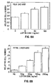
- Leptin stimulates yolk sac (8A) and fetal liver (8B) cell proliferation.
- Figure 9A-C.
- Leptin induces murine fetal liver to form myeloid colonies in colony assays.
- Figure 10A-C.
- Leptin induces murine adult bone marrow to form myeloid colonies in colony assays.
- Figure 11A and B.
- Leptin induces embroid body (EB)-derived precursors to form colonies. EB-derived precursors (d6) were replated into MeC with 100 ng/ml of steel factor and 200 ng/ml of leptin.
- Figure 12A and B.
- Leptin and EPO induce CFU-e from normal mouse bone marrow, but not db/db bone marrow. Age matched mice from normal and db/db strains are used as marrow donors for erythroid colony forming assays containing low serum concentration.
- Figure 13.
- Leptin induces CFU-e from human bone marrow in colony forming assays. All groups contain saturating levels of recombinant EPO at 2 U/ml.
5. DETAILED DESCRIPTION OF THE INVENTION
5.1. EXPRESSION OF THE OB-R IN HEMATOPOIETIC CELLS
-
The present invention relates to a novel hematopoietic progenitor cell marker and its use for cell identification and isolation, as well as the use of leptin to stimulate hematopoietic and endothelial development via this receptor known as Hu-B1.219. In a specific embodiment by way of example in Section 6, infra, several forms of this receptor were cloned and characterized. Amino acid sequence comparison of these related molecules with a published human OB-R sequence (Tartaglia et al., 1995, Cell 83:1263-1271) reveals only three amino acid differences in their extracellular domains but extensive diversity in their cytoplasmic domains. More specifically, Figure 1A-1G shows that in the Hu-B1.219 molecules described herein, nucleotide residues #349-351 encode alanine, nucleotide residues #421-423 encode arginine and nucleotide residues #763-765 encode arginine. Additionally, the four forms diverge both in length and sequence composition from nucleotide residue #2771 and beyond. In this regard, the cytoplasmic domain of Form 1 of Hu-B1.219 described herein is highly homologous to a retrotransposon sequence (Ono et al., 1987, Nucl. Acid. Res. 15:8725-8737).
-
Analysis of the Hu-B1.219 variants reveals significant homology to the FN III domain of the HR family indicating that they belong to the HR family of receptors. Northern blot hybridization and RT-PCR analyses indicates that Hu-B1.219 mRNA is highly expressed in cells of hematopoietic and endothelial origin. In addition, the Hu-81.219 sequence is expressed in certain fetal tissues and tumor cell lines. Hence, in addition to its expression in the brain for weight regulation by leptin, Hu-B1.219 (or OB-R) is expressed by hematopoietic and endothelial cells, thereby rendering these cells responsive to the action of leptin.
-
Since additional variant forms of the molecule may exist, they can be identified by labeled DNA probes made from nucleic acid fragments corresponding to any portion of the cDNA disclosed herein in a cDNA library prepared from human fetal liver, human lung, human kidney, human choroid plexus, human hypothalamus, human prostate and human ovary. More specifically, oligonucleotides corresponding to either the 5' or 3' terminus of the cDNA sequence may be used to obtain longer nucleotide sequences. Briefly, the library may be plated out to yield a maximum of 30,000 pfu for each 150 mm plate. Approximately 40 plates may be screened. The plates are incubated at 37°C until the plaques reach a diameter of 0.25 mm or are just beginning to make contact with one another (3-8 hours). Nylon filters are placed onto the soft top agarose and after 60 seconds, the filters are peeled off and floated on a DNA denaturing solution consisting of 0.4N sodium hydroxide. The filters are then immersed in neutralizing solution consisting of 1M Tris HCL, pH 7.5, before being allowed to air dry. The filters are prehybridized in casein hybridization buffer containing 10% dextran sulfate, 0.5M NaCl, 50mM Tris HCL, pH 7.5, 0.1% sodium pyrophosphate, 1% casein, 1% SDS, and denatured salmon sperm DNA at 0.5 mg/ml for 6 hours at 60°C. The radiolabelled probe is then denatured by heating to 95°C for 2 minutes and then added to the prehybridization solution containing the filters. The filters are hybridized at 60°C for 16 hours. The filters are then washed in 1X wash mix (10X wash mix contains 3M NaCl, 0.6M Tris base, and 0.02M EDTA) twice for 5 minutes each at room temperature, then in 1X wash mix containing 1% SDS at 60°C for 30 minutes, and finally in 0.3X wash mix containing 0.1% SDS at 60°C for 30 minutes. The filters are then air dried and exposed to x-ray film for autoradiography. After developing, the film is aligned with the filters to select a positive plaque. If a single, isolated positive plaque cannot be obtained, the agar plug containing the plaques will be removed and placed in lambda dilution buffer containing 0.1M NaCl, 0.01M magnesium sulfate, 0.035M Tris HCl, pH 7.5, 0.01% gelatin. The phage may then be replated and rescreened to obtain single, well isolated positive plaques. Positive plaques may be isolated and the cDNA clones sequenced using primers based on the known cDNA sequence. This step may be repeated until a full length cDNA is obtained.
-
One method for identifying all 3' isoforms is to PCR amplify the 3' ends of the variant cDNA from a variety of tissues including but not limiting to, choroid plexus, hypothalamus, fetal liver, bone marrow, ovary, or prostate. To obtain the 3' end of the cDNA, an oligo-dT primer is used to synthesize the cDNA first strand. Hu-B1.219 specific primers from the conserved region of the gene (e.g. up stream of nucleotide 2770) and oligo-dT are then used to amplify the 3' end. The PCR fragments are cloned and sequenced by standard techniques. Once obtained, these sequences may be translated into amino acid sequence and examined for certain landmarks such as continuous open reading frame, regulatory regions that associate with tyrosine kinase activation, and finally overall structural similarity to known variants.
5.2. Hu-B1.219 AS A PROGENITOR CELL MARKER
-
Hu-B1.219 is expressed in cells of hematopoietic and endothelial origin. In a specific embodiment by way of example in Section 7, infra, Hu-B1.219 is expressed in early progenitor cells, and in a small percentage of progenitors with long-term repopulating potential. In order to utilize Hu-B1.219 receptor as a marker for cell identification and isolation, specific binding agents such as antibodies may be generated to the protein.
5.2.1. GENERATION OF ANTIBODIES
-
Various procedures known in the art may be used for the production of antibodies to epitopes of natural or recombinant Hu-B1.219 receptor. Such antibodies include but are not limited to polyclonal, monoclonal, chimeric, single chain, Fab fragments and fragments produced by an Fab expression library. Neutralizing antibodies i.e., those which compete for the ligand binding site of the receptor are especially preferred for diagnostics and therapeutics.
-
Monoclonal antibodies that bind Hu-BI.219 may be radioactively labeled allowing one to follow their location and distribution in the body after injection. Radioisotope tagged antibodies may be used as a non-invasive diagnostic tool for imaging de novo cells of tumors and metastases as well as fetal tissues.
-
Immunotoxins may also be designed which target cytotoxic agents to specific sites in the body. For example, high affinity Hu-B1.219 specific monoclonal antibodies may be covalently complexed to bacterial or plant toxins, such as diphtheria toxin, or ricin. A general method of preparation of antibody/hybrid molecules may involve use of thiol-crosslinking reagents such as SPDP, which attack the primary amino groups on the antibody and by disulfide exchange, attach the toxin to the antibody. The hybrid antibodies may be used to specifically eliminate Hu-B1.219 expressing tumor cells.
-
For the production of antibodies, various host animals may be immunized by injection with the Hu-B1.219 protein, fragments thereof or synthetic peptides, including but not limited to rabbits, mice, rats, hamsters etc. Various adjuvants may be used to increase the immunological response, depending on the host species, including but not limited to Freund's (complete and incomplete), mineral gels such as aluminum hydroxide, surface active substances such as lysolecithin, pluronic polyols, polyanions, peptides, oil emulsions, keyhole limpet hemocyanin, dinitrophenol, and potentially useful human adjuvants such as BCG (bacilli Calmette-Guerin) and Corynebacterium parvum.
-
Monoclonal antibodies to Hu-B1.219 may be prepared by using any technique which provides for the production of antibody molecules by continuous cell lines in culture. These include but are not limited to the hybridoma technique originally described by Kohler and Milstein, (Nature, 1975, 256:495-497), the human B-cell hybridoma technique (Kosbor et al., 1983, Immunology Today, 4:72; Cote et al., 1983, Proc. Natl. Acad. Sci. USA, 80:2026-2030) and the EBV-hybridoma technique (Cole et al., 1985, Monoclonal Antibodies and Cancer Therapy, Alan R. Liss, Inc., pp. 77-96). In addition, techniques developed for the production of "chimeric antibodies" (Morrison et al., 1984, Proc. Natl. Acad. Sci. USA, 81:6851-6855; Neuberger et al., 1984, Nature 312:604-608; Takeda et al., 1985, Nature 314:452-454) by splicing the genes from a mouse antibody molecule of appropriate antigen specificity together with genes from a human antibody molecule of appropriate biological activity can be used. Alternatively, techniques described for the production of single chain antibodies (U.S. Patent 4,946,778) can be adapted to produce Hu-B1.219-specific single chain antibodies.
-
Antibody fragments which contain specific binding sites of Hu-B1.219 may be generated by known techniques. For example, such fragments include but are not limited to, the F(ab')2 fragments which can be produced by pepsin digestion of the antibody molecule and the Fab fragments which can be generated by reducing the disulfide bridges of the F(ab')2 fragments. Alternatively, Fab expression libraries may be constructed (Huse et al., 1989, Science 246:1275-1281) to allow rapid and easy identification of monoclonal Fab fragments with the desired specificity to Hu-B1.219.
5.2.2. PROGENITOR CELL SEPARATION
-
Human hematopoietic progenitor cells may be isolated using an antibody to Hu-B1.219 protein, a leptin ligand, a leptin peptide containing the receptor-binding domain or a leptin fusion protein using conventional cell separation methods well known in the art. Such Hu-B1.219-specific binding agents may be used in combination with other agents such as anti-CD34 antibodies.
-
Although bone marrow is the preferred cell source, other physiologic sources of hematopoietic cells may be utilized, for example, the spleen, thymus, peripheral blood, cytokine-mobilized blood, umbilical cord blood, embryonic yolk sac, or fetal liver. Bone marrow is preferably removed from the iliac crest, but may also be removed from other bone cavity. Bone marrow may be removed from bone cavity by various methods well known to those skilled in the art, including flushing the bone with a mixture of physiological media, balanced salt solution, physiological buffer, and other naturally occurring factors. Typically, the bone marrow is filtered, centrifuged and resuspended.
-
Once a source of hematopoietic cells is obtained, hematopoietic progenitor cells may be separated from the cell mixture by various methods which utilize an agent such as a specific antibody or a leptin ligand which specifically binds to cell surface Hu-B1.219-encoded receptor protein. These techniques may include, for example, flow cytometry using a fluorescence activated cell sorter (FACS) and specific fluorochromes, biotin-avidin or biotin-streptavidin separations using biotin conjugated to cell surface marker-specific antibodies and avidin or streptavidin bound to a solid support such as affinity column matrix or plastic surfaces, magnetic separations using antibody-coated magnetic beads, destructive separations such as antibody and complement or antibody bound to cytotoxins or radioactive isotopes.
-
Separation of a cell mixture via antibodies may be performed by negative or positive selection procedures. In negative separation, antibodies are used which are specific for markers present on undesired hematopoietic cells. Cells bound by an antibody may be removed or lysed and the remaining desired mixture retained. In positive separation, antibodies specific for Hu-B1.219 or leptin ligand may be used. Cells bound by the antibody or leptin are separated and retained. It will be understood that positive and negative separations may be used substantially simultaneously or in a sequential manner. It will also be understood that the present invention encompasses any separation technique which can isolate cells based on the expression of Hu-B1.219 as disclosed herein. For example, a cell mixture may be separated into CD34+ and CD34- fractions first followed by HuB1.219-specific separation.
-
At present, the most common technique for antibody-based separation has been the use of flow cytometry such as by a FACS. Typically, separation by flow cytometry is performed as follows. The suspended mixture of hematopoietic cells are centrifuged and resuspended in media. Antibodies which are conjugated to fluorochrome are added to allow the binding of the antibodies to specific cell surface markers. The cell mixture is then washed by one or more centrifugation and resuspension steps. The mixture is run through FACS which separates the cells based on different fluorescence characteristics. FACS systems are available in varying levels of performance and ability, including multi-color analysis. The cells can be identified by a characteristic profile of forward and side scatter which is influenced by size and granularity, as well as by positive and/or negative expression of certain cell surface markers.
-
Other separation techniques besides flow cytometry may provide for faster separations. One such method is biotin-avidin based separation by affinity chromatography. Typically, such a technique is performed by incubating the washed hematopoietic cells with biotin-coupled leptin or antibodies to specific markers followed by passage through an avidin column. Biotin-antibody-cell or biotin-leptin-cell complexes bind to the column via the biotin-avidin interaction, while other cells pass through the column. Finally, the column-bound cells may be released by perturbation or other methods. The specificity of the biotin-avidin system is well suited for rapid positive separation.
-
Flow cytometry and biotin-avidin techniques provide highly specific means of cell separation. If desired, a separation may be initiated by less specific techniques which, however, can remove a large proportion of mature blood cells from the hematopoietic cell source. For example, magnetic bead separations may be used to initially remove differentiated hematopoietic cell populations; including T-cells, B-cells, natural killer (NK) cells, and macrophages, as well as minor cell populations including megakaryocytes, mast cells, eosinophils, and basophils. Desirably, at least about 70% and usually at least about 80% of the total hematopoietic cells present can be removed.
-
A preferred initial separation technique is density-gradient separation. Here, the bone marrow or other hematopoietic cell mixture preparation is centrifuged and the supernatant removed. The cells are resuspended in, for example, RPMI 1640 medium (Gibco) with 10% FCS and placed in a density gradient prepared with, for example, "FICOLL" or "PERCOLL" or "EUROCOLLINS" media. The separation may then be performed by centrifugation or automatically with a Cobel & Cell Separator '2991 (Cobev, Lakewood, Colorado). Additional separation procedures may be desirable depending on the source of the hematopoietic cell mixture and its content. For example, if blood is used as a source of hematopoietic cells, it may be desirable to lyse red blood cells prior to the separation of any fraction. Furthermore, elutriation may also be used alone or in combination with all of other purification procedures described herein (Noga et al., 1990, Prog. Clin. Biol. Res. 333:345; Noga et al., 1992, Prog. Clin. Biol. Res. 377:411).
5.3. THE OBESE GENE PRODUCT, LEPTIN
-
The nucleotide and amino acid sequences of both human and mouse leptin have been published recently by Zhang et al. (1994, Nature 372:425-432). Thereafter, the mouse coding sequence was used to express functional leptin in E. coli (Pelleymounter et al., 1995, Science 269:540-543; Halaas et al., 1995, Science 269:543-546; Campfield et al., 1995, Science 269:546-549). Furthermore, human leptin was also expressed and shown to be biologically active in murine experiments (Halaas et al., 1995, Science 269:543-546). Hence, human, murine and homologous coding sequences from other species may be expressed, and the recombinant protein purified by conventional techniques such as affinity chromatography with an antibody. Alternatively, natural protein may be directly purified from cells that secrete leptin, such as adipose cell lines.
5.3.1. EXPRESSION OF LEPTIN PROTEIN
-
For the practice of the present invention, human and mouse leptin polynucleotide sequences which encode the proteins, peptide fragments, fusion proteins or functional equivalents thereof, may be used to generate recombinant DNA molecules that direct their expression in appropriate host cells.
-
Due to the inherent degeneracy of the genetic code, other DNA sequences which encode substantially the same or a functionally equivalent amino acid sequence, may be used in the practice of the invention for the cloning and expression of the leptin protein. In particular, such DNA sequences include those which are capable of hybridizing to the human leptin sequences under stringent conditions.
-
Altered DNA sequences which may be used in accordance with the invention include deletions, additions or substitutions of different nucleotide residues resulting in a sequence that encodes the same or a functionally equivalent gene product. The gene product itself may contain deletions, additions or substitutions of amino acid residues within a coding sequence, which result in a silent change thus producing a functionally equivalent leptin protein. Such amino acid substitutions may be made on the basis of similarity in polarity, charge, solubility, hydrophobicity, hydrophilicity, and/or the amphipathic nature of the residues involved. For example, negatively charged amino acids include aspartic acid and glutamic acid; positively charged amino acids include lysine, histidine and arginine; amino acids with uncharged polar head groups having similar hydrophilicity values include the following: glycine, asparagine, glutamine, serine, threonine, tyrosine; and amino acids with nonpolar head groups include alanine, valine, isoleucine, leucine, phenylalanine, proline, methionine, tryptophan.
-
The DNA sequences of leptin may be engineered in order to alter the coding sequence for a variety of ends including but not limited to alterations which modify processing and expression of the gene product. For example, mutations may be introduced using techniques which are well known in the art, e.g., site-directed mutagenesis, to insert new restriction sites, to alter glycosylation patterns, phosphorylation, etc.
-
In another embodiment of the invention, a leptin or a modified leptin sequence may be ligated to a heterologous sequence to encode a fusion protein. It may also be useful to encode a chimeric leptin protein expressing a heterologous epitope that is recognized by a commercially available antibody. A fusion protein may also be engineered to contain a cleavage site located between a leptin sequence and the heterologous protein sequence, so that the leptin protein may be cleaved away from the heterologous moiety.
-
In an alternate embodiment of the invention, the coding sequence of leptin could be synthesized in whole or in part, using chemical methods well known in the art. See, for example, Caruthers et al., 1980, Nuc. Acids Res. Symp. Ser. 7:215-233; Crea and Horn, 180, Nuc. Acids Res. 9(10):2331; Matteucci and Caruthers, 1980, Tetrahedron Letters 21:719; and Chow and Kempe, 1981, Nuc. Acids Res. 9(12):2807-2817. Alternatively, the protein itself could be produced using chemical methods to synthesize leptin amino acid sequence in whole or in part. For example, peptides can be synthesized by solid phase techniques, cleaved from the resin, and purified by preparative high performance liquid chromatography. (e.g., see Creighton, 1983, Proteins Structures And Molecular Principles, W.H. Freeman and Co., N.Y. pp. 50-60). The composition of the synthetic peptides may be confirmed by amino acid analysis or sequencing (e.g., the Edman degradation procedure; see Creighton, 1983, Proteins, structures and Molecular Principles, W.H. Freeman and Co., N.Y., pp. 34-49).
-
In order to express a biologically active leptin protein, the nucleotide sequence coding for leptin, or a functional equivalent, is inserted into an appropriate expression vector, i.e., a vector which contains the necessary elements for the transcription and translation of the inserted coding sequence. The leptin gene products as well as host cells or cell lines transfected or transformed with recombinant leptin expression vectors can be used for a variety of purposes.
5.3.2. EXPRESSIOIN SYSTEMS FOR LEPTIN
-
Methods which are well known to those skilled in the art can be used to construct expression vectors containing the leptin coding sequence and appropriate transcriptional/translational control signals. These methods include in vitro recombinant DNA techniques, synthetic techniques and in vivo recombination/genetic recombination. See, for example, the techniques described in Sambrook et al., 1989, Molecular Cloning A Laboratory Manual, Cold Spring Harbor Laboratory, N.Y. and Ausubel et al., 1989, Current Protocols in Molecular Biology, Greene Publishing Associates and Wiley Interscience, N.Y.
-
A variety of host-expression vector systems may be utilized to express the leptin coding sequence. These include but are not limited to microorganisms such as bacteria transformed with recombinant bacteriophage DNA, plasmid DNA or cosmid DNA expression vectors containing the leptin coding sequence; yeast transformed with recombinant yeast expression vectors containing the leptin coding sequence; insect cell systems infected with recombinant virus expression vectors (e.g., baculovirus) containing the leptin coding sequence; plant cell systems infected with recombinant virus expression vectors (e.g., cauliflower mosaic virus, CaMV; tobacco mosaic virus, TMV) or transformed with recombinant plasmid expression vectors (e.g., Ti plasmid) containing the leptin coding sequence; or animal cell systems.
-
The expression elements of these systems vary in their strength and specificities. Depending on the host/vector system utilized, any of a number of suitable transcription and translation elements, including constitutive and inducible promoters, may be used in the expression vector. For example, when cloning in bacterial systems, inducible promoters such as pL of bacteriophage X, plac, ptrp, ptac (ptrp-lac hybrid promoter) and the like may be used; when cloning in insect cell systems, promoters such as the baculovirus polyhedrin promoter may be used; when cloning in plant cell systems, promoters derived from the genome of plant cells (e.g., heat shock promoters; the promoter for the small subunit of RUBISCO; the promoter for the chlorophyll α/β binding protein) or from plant viruses (e.g., the 35S RNA promoter of CaMV; the coat protein promoter of TMV) may be used; when cloning in mammalian cell systems, promoters derived from the genome of mammalian cells (e.g., metallothionein promoter) or from mammalian viruses (e.g., the adenovirus late promoter; the vaccinia virus 7.5K promoter) may be used; when generating cell lines that contain multiple copies of the leptin DNA, SV40-, BPV- and EBV-based vectors may be used with an appropriate selectable marker.
-
In bacterial systems a number of expression vectors may be advantageously selected depending upon the use intended for the leptin expressed. For example, when large quantities of leptin are to be produced for the generation of antibodies or to screen peptide libraries, vectors which direct the expression of high levels of fusion protein products that are readily purified may be desirable. Such vectors include but are not limited to the E. coli expression vector pUR278 (Ruther et al., 1983, EMBO J. 2:1791), in which the leptin coding sequence may be ligated into the vector in frame with the lac Z coding region so that a hybrid AS-lac Z protein is produced; pIN vectors (Inouye & Inouye, 1985, Nucleic acids Res. 13:3101-3109; Van Heeke & Schuster, 1989, J. Biol. Chem. 264:5503-5509); and the like. pGEX vectors may also be used to express foreign polypeptides as fusion proteins with glutathione S-transferase (GST). In general, such fusion proteins are soluble and can easily be purified from lysed cells by adsorption to glutathione-agarose beads followed by elution in the presence of free glutathione. The pGEX vectors are designed to include thrombin or factor Xa protease cleavage sites so that the cloned polypeptide of interest can be released from the GST moiety.
-
In yeast, a number of vectors containing constitutive or inducible promoters may be used. For a review see, Current Protocols in Molecular Biology, Vol. 2, 1988, Ed. Ausubel et al., Greene Publish. Assoc. & Wiley Interscience, Ch. 13; Grant et al., 1987, Expression and Secretion Vectors for Yeast, Methods in Enzymology, Eds. Wu & Grossman, 1987, Acad. Press, N.Y., Vol. 153, pp. 516-544; Glover, 1986, DNA Cloning, Vol. II, IRL Press, Wash., D.C., Ch. 3; and Bitter, 1987, Heterologous Gene Expression in Yeast, Methods in Enzymology, Eds. Berger & Kimmel, Acad. Press, N.Y., Vol. 152, pp. 673-684; and The Molecular Biology of the Yeast Saccharomyces, 1982, Eds. Strathern et al., Cold Spring Harbor Press, Vols. I and II.
-
In cases where plant expression vectors are used, the expression of the leptin coding sequence may be driven by any of a number of promoters. For example, viral promoters such as the 35S RNA and 19S RNA promoters of CaMV (Brisson et al., 1984, Nature 310:511-514), or the coat protein promoter of TMV (Takamatsu et al., 1987, EMBO J. 6:307-311) may be used; alternatively, plant promoters such as the small subunit of RUBISCO (Coruzzi et al., 1984, EMBO J. 3:1671-1680; Broglie et al., 1984, Science 224:838-843); or heat shock promoters, e.g., soybean hsp17.5-E or hsp17.3-B (Gurley et al., 1986, Mol. Cell. Biol. 6:559-565) may be used. These constructs can be introduced into plant cells using Ti plasmids, Ri plasmids, plant virus vectors, direct DNA transformation, microinjection, electroporation, etc. For reviews of such techniques see, for example, Weissbach & Weissbach, 1988, Methods for Plant Molecular Biology, Academic Press, NY, Section VIII, pp. 421-463; and Grierson & Corey, 1988, Plant Molecular Biology, 2d Ed., Blackie, London, Ch. 7-9.
-
An alternative expression system which could be used to express leptin is an insect system. In one such system, Autographa californica nuclear polyhidrosis virus (AcNPV) is used as a vector to express foreign genes. The virus grows in Spodoptera frugiperda cells. The leptin coding sequence may be cloned into non-essential regions (for example the polyhedrin gene) of the virus and placed under control of an AcNPV promoter (for example the polyhedrin promoter). Successful insertion of the leptin coding sequence will result in inactivation of the polyhedrin gene and production of non-occluded recombinant virus (i.e., virus lacking the proteinaceous coat coded for by the polyhedrin gene). These recombinant viruses are then used to infect Spodoptera frugiperda cells in which the inserted gene is expressed. (e.g., see Smith et al., 1983, J. Virol. 46:584; Smith, U.S. Patent No. 4,215,051).
-
In mammalian host cells, a number of viral based expression systems may be utilized. In cases where an adenovirus is used as an expression vector, the leptin coding sequence may be ligated to an adenovirus transcription/translation control complex, e.g., the late promoter and tripartite leader sequence. This chimeric gene may then be inserted in the adenovirus genome by in vitro or in vivo recombination. Insertion in a non-essential region of the viral genome (e.g., region E1 or E3) will result in a recombinant virus that is viable and capable of expressing leptin in infected hosts (e.g., See Logan & Shenk, 1984, Proc. Natl. Acad. Sci. USA 81:3655-3659). Alternatively, the vaccinia 7.5K promoter may be used. (See, e.g., Mackett et al., 1982, Proc. Natl. Acad. Sci. USA 79:7415-7419; Mackett et al., 1984, J. Virol. 49:857-864; Panicali et al., 1982, Proc. Natl. Acad. Sci. USA 79:4927-4931).
-
Specific initiation signals may also be required for efficient translation of inserted leptin coding sequences. These signals include the ATG initiation codon and adjacent sequences. In cases where the entire leptin gene, including its own initiation codon and adjacent sequences, is inserted into the appropriate expression vector, no additional translational control signals may be needed. However, in cases where only a portion of the leptin coding sequence is inserted, exogenous translational control signals, including the ATG initiation codon, must be provided. Furthermore, the initiation codon must be in phase with the reading frame of the leptin coding sequence to ensure translation of the entire insert. These exogenous translational control signals and initiation codons can be of a variety of origins, both natural and synthetic. The efficiency of expression may be enhanced by the inclusion of appropriate transcription enhancer elements, transcription terminators, etc. (see Bittner et al., 1987, Methods in Enzymol. 153:516-544).
-
In addition, a host cell strain may be chosen which modulates the expression of the inserted sequences, or modifies and processes the gene product in the specific fashion desired. Such modifications (e.g., glycosylation) and processing (e.g., cleavage) of protein products may be important for the function of the protein. The presence of several consensus N-glycosylation sites in leptin support the possibility that proper modification may be important for leptin function. Different host cells have characteristic and specific mechanisms for the post-translational processing and modification of proteins. Appropriate cell lines or host systems can be chosen to ensure the correct modification and processing of the foreign protein expressed. To this end, eukaryotic host cells which possess the cellular machinery for proper processing of the primary transcript, glycosylation, and phosphorylation of the gene product may be used. Such mammalian host cells include but are not limited to CHO, VERO, BHK, HeLa, COS, MDCK, 293, WI38, etc.
-
For long-term, high-yield production of recombinant proteins, stable expression is preferred. For example, cell lines which stably express the leptin sequence may be engineered. Rather than using expression vectors which contain viral origins of replication, host cells can be transformed with the leptin DNA controlled by appropriate expression control elements (e.g., promoter, enhancer, sequences, transcription terminators, polyadenylation sites, etc.), and a selectable marker. Following the introduction of foreign DNA, engineered cells may be allowed to grow for 1-2 days in an enriched media, and then are switched to a selective media. The selectable marker in the recombinant plasmid confers resistance to the selection and allows cells to stably integrate the plasmid into their chromosomes and grow to form foci which in turn can be cloned and expanded into cell lines.
-
A number of selection systems may be used, including but not limited to the herpes simplex virus thymidine kinase (Wigler, et al., 1977, Cell 11:223), hypoxanthine-guanine phosphoribosyltransferase (Szybalska & Szybalski, 1962, Proc. Natl. Acad. Sci. USA 48:2026), and adenine phosphoribosyltransferase (Lowy, et al., 1980, Cell 22:817) genes can be employed in tk-, hgprt- or aprt- cells, respectively. Also, antimetabolite resistance can be used as the basis of selection for dhfr, which confers resistance to methotrexate (Wigler, et al., 1980, Proc. Natl. Acad. Sci. USA 77:3567; O'Hare, et al., 1981, Proc. Natl. Acad. Sci. USA 78:1527); gpt, which confers resistance to mycophenolic acid (Mulligan & Berg, 1981, Proc. Natl. Acad. Sci. USA 78:2072); neo, which confers resistance to the aminoglycoside G-418 (Colberre-Garapin et al., 1981, J. Mol. Biol. 150:1); and hygro, which confers resistance to hygromycin (Santerre, et al., 1984, Gene 30:147) genes. Additional selectable genes have been described, namely trpB, which allows cells to utilize indole in place of tryptophan; hisD, which allows cells to utilize histinol in place of histidine (Hartman & Mulligan, 1988, Proc. Natl. Acad. Sci. USA 85:8047); and ODC (ornithine decarboxylase) which confers resistance to the ornithine decarboxylase inhibitor, 2-(difluoromethyl)-DL-ornithine, DFMO (McConlogue L., 1987, In: Current Communications in Molecular Biology, Cold Spring Harbor Laboratory ed.).
-
Once the leptin protein is expressed by any of the aforementioned systems, supernatants of the cultured cells or cell lysates may be subjected to various standard methods of protein purification. For example, an anti-leptin antibody or Hu-B1.219 protein can be used to purify it by affinity chromatography. Alternatively, leptin may be purified by ion exchange chromatography or HPLC. Thereafter, the purity of the protein can be confirmed by various methods, including SDS-PAGE, and the protein used immediately or stored frozen for future use. For cell cultures and in vivo administration, purified leptin must be sterilized prior to use.
5.4. ACTIVATION OF HU-H1.219-EXPRESSING CELLS BY LEPTIN
-
Since various forms of Hu-B1.219, including OB-R, are essentially identical in the extracellular domain, these receptors can bind leptin as a ligand. In order to compare the binding affinity of Hu-B1.219 isoforms to leptin, the variant forms are cloned into standard expression vectors, e.g. CMV promoter expression vectors, and transfected into COS and BaF3 cells. Surface expression of the receptor is then evaluated by direct binding to an anti-Hu-B1.219 antibody. In addition, leptin binding assays can also be performed is described by Tartaglia et al. (1995, Cell 83:1263) using a leptin fusion protein or soluble leptin conjugated to a radiolabel, a fluorescent dye or an enzyme. The results are compared with mock transfected cells as negative controls.
-
Since the four variants of Hu-B1.219 contain different intracellular domains, these isoforms can be compared with respect to their signal transduction capabilities to determine the most active form. Stimulation of most if not all hematopoietic receptors with ligands results in the rapid phosphorylation of tyrosines, both on the receptors and on a cascade of cellular protein kinases (Heidin, 1995, Cell 80:213-233; Ihle, 1995, Nature 377:591-594). Phosphorylation of these molecules results in an activation signal being propagated ultimately to the nucleus leading to gene activation. Upon ligand binding to an hematopoietin receptor, some of the first molecules to be activated in this fashion are the JAK (Janus) family of protein kinases (Ziemiecki et al., 1994, Trends Cell Biol. 4:207-212). These activated kinases, in turn, phosphorylate members of the STAT family of molecules which eventually translocate to the nucleus and form active transcription complexes (Darnell et al., 1994, Science 264:1415-1421; Zhong et al., 1994, Proc. Natl. Acad. Sci. USA 91:4806-4810; Hou et al., 1994, Science 265:1701-1706).
-
Therefore, cell activation by leptin can be evaluated by studying.the pattern of phosphorylation of JAK1-3 following Hu-81.219 binding. This can be carried out by culturing hematopoietic cells or Hu-B1.219 transfectants (1-100 x 103), in RPMI 1640 (GIBCO) with gentamicin (100 µg/ml), 2 mM glutamine (GIBCO), 10% FCS, and leptin (from 0 to 500 nM) for 10-60 minutes at 37°C. Following the incubation, the cells are washed in ice-cold PBS and resuspended in lysing buffer (1% Triton X-100, 200 mM NaCl, 10 mM Tris pH 7.5, 2.5 mM p-nitrophenyl guanidinobenzoate, 100 µM Na3VO4, 1 µM pepstatin, 50 µM 3,4-dichloroisocoumarin,. 1 mM PMSF, 10 µg/ml leupeptin, 10 µg/ml aprotinin) for 30 minutes at 4°C followed by removal of nuclei and insoluble material by centrifugation. To the cell lysate is added polyclonal antibodies to JAK1 and JAK2 (Upstate Biotechnology, Lake Placid, NY) or human Tyk2, a human kinase related to the JAK family (Santa Cruz, CA) according to manufacturer's recommendation. This mixture is rotated for 30-60 minutes at 4°C and then 100 µl of a 10% protein A-sepharose solution (Sigma) is added and rotated for an additional 30 minutes at 4°C. The precipitate is washed with lysis buffer and eluted in standard SDS reducing sample buffer and resolved by SDS-PAGE. The proteins are analyzed by Western blots by transferring to Immobilon membranes (Millipore). The membranes are blocked with gelatin (1%) and incubated with anti-phosphotyrosine (4G10, Upstate Biotechnology, Lake Placid, NY) according to the manufacturer's recommendations. Phosphorylated proteins are detected with 125 I-labeled protein A (Amersham).
-
For the aforementioned experiment, hematopoietic cells can be obtained from human CD34+ bone marrow and cord blood. For the murine experiments, three primitive populations can be evaluated, e.g. Ly-6+ lineage negative bone marrow, or the equivalent in fetal liver, or AA4.1+ fetal liver.
-
While tyrosine phosphorylation is an indication of cell activation resulting from ligand-receptor interactions, additional manifestations of activation at the cellular level can be assayed by using leptin to induce the proliferation and/or differentiation of hematopoietic precursors. Cell proliferation can be easily assessed by 3H-thymidine uptake or by visual enumeration of cell numbers. Differentiation of hematopoietic cells can be assayed by testing the ability of leptin to stimulate the in vitro growth and differentiation of various hematopoietic colonies. For example, a cell mixture can be isolated from the yolk sac, fetal liver or bone marrow, and cultured in standard methylcellulose colony assays with and without serum supplements, in the presence of leptin (0.1-500 nM) with or without various other cytokines (Metcalf, 1984, in Clonal Culture of Hematopoietic Cells: Techniques and Applications, Elsevier, NY).
-
A standard protocol for such an assay involves density gradient centrifugation of a cell mixture by Ficoll-Hypaque (1.077 gm/cm3) and resuspending about 1x10⁶ viable cells in Iscove's Modified Dulbecco's Medium (IMDM) with 0.5-15% fetal calf serum (FCS). The cells (1-100 x 103/ml) are then mixed with IMDM that contains methylcellulose (1.3%), FCS (0.5-15%), BSA (1%), monothioglycerol.(100 µM), gentamicin (50 µg/ml), and leptin (0.1-500 nM). In parallel cultures, additional cytokines may include: IL-3 (100 pg/ml), steel factor or c-Kit ligand or SCF (10 ng/ml), and EPO (2 U/ml). After mixing, 1 ml of the mixture is dispensed into a 25 mm bacterial grade culture dishes at 37°C in a humidified incubator for 5 to 18 days. Using an inverted microscope the number and type of hematopoietic colonies are determined. The colony morphology is used to categorize the various colony types, e.g. BFU-e, CFU-e, CFU-G, CFU-GM, CFU-M, CFU-blast, or CFU-fibroblast (Metcalf, 1984, Clonal Culture of Hematopoietic Cells: Techniques and Applications, Elsevier, N.Y.; Freshney, 1994, in Culture of Hematopoietic Cells, Wiley-Liss, Inc., pp. 265-68). Optimal concentrations of leptin in this assay may increase both the size and the frequency of these primitive pluripotent colonies.
-
While leptin may be used alone, it can also be used synergistically with several cytokines to promote hematopoietic cell growth including but not limited to, IL-1, IL-3, IL-6, EPO, steel factor (SCF) and GM-CSF (1 ng/ml). In addition; since biologically active leptin is present in fetal calf serum (FCS), cultures with 0.5-15% of FCS can be tested. The specific activity of leptin in FCS can be inhibited by an anti-leptin antibody or a soluble variant of Hu-B1.219 as a control.
-
In particular, the effects ofleptin may be tested on the primitive precursors that form high proliferative potential cells (HPPC) (McNiece and Briddell, 1994, in Culture of Hematopoietic Cells, Wiley-Liss, Inc., pp. 23-40) and blast colonies (Leary and Ogawa, 1994, in Culture of Hematopoietic Cells, Wiley-Liss Inc., pp. 41-54). In order to evaluate the effects of leptin on even more primitive cells in these assays, CD34+ bone marrow, cord blood, and fetal liver cells can be first sorted by an antibody and tested in the above assays. Since recent evidence has suggested the existence of a CD34- stem cell, the CD34- Lin- population may also be stimulated by leptin.
-
Furthermore, the effect of leptin on the primitive long term culture initiating cells (LTCIC) and on hematopoietic stem cells can be evaluated. LTCIC are precursors that can initiate a long-term hematopoietic culture and are believed to be a function of hematopoietic stem cells (Sutherland and Eaves, 1994, in Culture of Hematopoietic Cells, Wiley-Liss, Inc., pp. 139-162; Van der Sluijs et al., 1990, Exp. Hematol. 18:893-896; Traycoff et al., 1994, Exp. Hematol. 22:215-222). The ability of these culture conditions to expand the hematopoietic stem cell can be confirmed by the competitive repopulation assay (Harrison, 1980, Blood 55:77-81). This assay allows for the quantification of hematopoietic stem cells.
-
Additionally, since endothelial cells also express HuB1.219, leptin may be used to stimulate the growth of primary endothelial cells at 0.1-500 nM in standard cultures for maintaining primary endothelial cells (Masek and Sweetenham, 1994, Br. J. Haematol. 88:855-865; Visner et al., 1994, Am. J. Physiol. 267:L406-413; Moyer et al., 1988, In Vitro Cell Dev. Biol. 24:359-368). Alternatively, leptin may be used to induce endothelial cells to produce cytokines (Broudy et al., 1987, J. Immunol. 139:464-468; Seelentag et al., 1987, EMBO J. 6:2261-2265). Supernatants of 2-5 day primary endothelial cell cultures or endothelial cell lines cultured in the presence of leptin with or without other cytokines, such as vascular endothelial growth factor (VEGF), platelet-derived growth factor (PDGF), fibroblast growth factor (FGF), transforming growth factor (TGF) and epidermal growth factor (EGF), are harvested and tested as a supplement in the hematopoietic colony assays above.
-
In another aspect of the invention, since receptors often form dimers on the cell surface, the combination of different Hu-B1.219 isoforms that give optimal signal transduction can be measured by growth stimulation and phosphorylation patterns. A Hu-B1.219- growth factor-dependent indicator cell line (e.g. BaF3) can be transfected with various combinations of the isoforms using standard CMV expression vectors. Following the demonstration of cell surface expression, leptin (0.1-500 nM) is added to the cultures followed by the measurement of growth rates and phosphorylation patterns by established techniques. Other cell lines such as TF-1, FD5 and TS1 may also be used.
-
Because of the expression of isoforms with truncated cytoplasmic tails, it is possible that another protein chain is used in some tissues as the signaling molecule in association with Hu-B1.219. Such a molecule can be screened and selected by transfecting pools of cDNA from expression libraries of a variety of tissues e.g. fetal liver, CD34+ bone marrow, lung, ovary, etc. together with constructs expressing one of the truncated Hu-B1.219 isoforms into a growth factor-dependent cell line (e.g. BaF3). The ability to grow in the presence of leptin is used as a readout. In particular, the insulin receptor-related receptor (Zhang and Roth, 1992, J. Biol. Chem. 267:18320), the LIFRα, IL-2Rγ, IL-4Rα (Mosely et al., 1989, Cell 89:335) and IL-13Rα chains (Hilton et al., 1996, Proc. Natl. Acad. Sci. USA 93:497) may function as complementary chains for Hu-B1.219 activity. The techniques to identify the unique cDNA responsible for this complementation are well established in the art.
-
Additionally, agents that activate Hu-B1.219 in a manner similar to leptin may also be used in place of leptin. These agents include small molecules and peptides, and they may be selected in the following screening assay. Ten thousand BaF3 and BaF3/Hu-B1.219 (transfectant cells that express the full length Hu-B1.219 isoform) cells will be screened in microtiter plates for proliferative effects after incubation with a test agent. Without stimulation these cells die. Any growth promoting effect seen on the transfected cell line and not with the BaF3 host would indicate that the test agent specifically activates the Hu-B1.219 receptor or its signaling pathway. This assay is used to screen small molecules, including peptides, oligonucleotides, and chemical libraries.
5.5.
IN VITRO USES OF LEPTIN
-
In view of the expression of Hu-B1.219 in diverse cell types, leptin may be used to activate these cells in culture. The activated cells expand in number due to increased proliferation and/or differentiate to become more mature cells. In this connection, the optimal effective concentration of leptin for each cell type may be determined by conventional titration experiments in which different amounts of leptin are incubated with the specific cells and their activation levels measured by tyrosine phosphorylation, proliferation or differentiation.
-
In particular, hematopoietic progenitor cells express Hu-B1.219. Hematopoietic cells may be activated by exposure to leptin in vitro which results in their expansion in number prior to their use in vitro and in vivo. Such hematopoietic cells may be obtained from the bone marrow, the peripheral blood (Demuynck et al., 1995, Ann. Hematol. 71:29-33; Scheding et al., 1994, Stem Cells 12:Suppl. 1:203-210) and cord blood. In order to selectively enhance the growth of hematopoietic stem cells, the donor cell mixture may be first separated into CD34+ cells, followed by leptin stimulation. The expanded cells may be used as donor cells in bone marrow transplantation or as long-term bone marrow cultures (Ponchio et al., 1995, Blood 86:3314-3321; Testa and Dexter, 1991, Curr. Opin. 3:272-278; Naughton and Naughton, 1991, United States Patent No. 5,032,508) for in vitro cytotoxicity testing and the discovery of novel cytokines.
-
Since Hu-B1.219 is also expressed in some endothelial cells and their progenitors, leptin may be used to induce blood vessel formation in vitro. In that regard, leptin may be used alone or in combination with VEGF, PDGF, FGF or TGF-α. Blood vessels form by a combination of two primary processes. Some blood vessel growth depends on angiogenesis, in a process similar to that associated with pathological conditions. For instance, the CNS depends solely on angiogenesis for development of its vascular supply (Nodew, 1989, Am. Rev. Respir. Dis. 140:1097-1103; Risau et al., 1988, EMBO J. 7:959-962). A second process, vasculogenesis, depends on the incorporation of migratory individual endothelial cells (angioblasts) into the developing blood vessel. Leptin may be used to promote both angiogenesis and vasculogenesis.
-
In addition, the coding sequence of leptin may be inserted in an expression vector in accordance with Section 5.3, supra, and transferred into leptin-nonproducing cell types to result in endogenous expression of leptin. For example, hematopoietic stem cells are isolated and transfected with the leptin coding sequence. Thereafter, the cells are transferred into a bone marrow transplant recipient to cause endogenous production of leptin in stimulating hematopoiesis (United States Patent No. 5,399,346).
5.6. IN VIVO USES OF LEPTIN
-
The appropriate dosage and formulation of leptin on human hematopoietic and endothelial development in vivo can be first determined in animal models. For example, normal mice may be lethally irradiated or chemically ablated and reconstituted with syngeneic or allogeneic bone marrow. Since recombinant human leptin has been shown to be active on mouse cells, the rate of donor cell engraftment can be compared between leptin-treated and non-treated groups. Because of the Hu-B1.219 expression on primitive hematopoietic cells, leptin facilitates the growth and recovery of these cells in the recipient. The effect of leptin on in vivo hematopoietic proliferation and differentiation in these situations can be evaluated by reconstituting lethally irradiating mice (900R) with normal bone marrow cells (1-5 x 10⁶ per mouse). Groups of animals are given PBS injections as controls and other groups receive leptin (0.1 - 10 mg/kg) at varying intervals. The activity of leptin is assayed by the rapidity of re-normalization and stability of blood profiles (e.g. hematocrit, WBC count, differential, etc.).
-
In addition, neonatal, sublethally irradiated adult normal or SCID mice can be reconstituted with human hematopoietic stem cell isolated from bone marrow, cord blood, or fractions thereof. In these mice, the human hematopoietic cells engraft and differentiate (McCune et al., 1988, Science 241:1632-1639; Sandhu et al., 1994, J. Immunol. 152:3806-3813). The effects of leptin can be evaluated by measuring the growth rate and extent of differentiation of human cells in these animals.
-
An effective amount of leptin may be administered into a patient who is in need of increased hematopoietic cell function. Such a need may arise from various forms of immunodeficiencies (B cell deficiencies, T cell deficiencies and combined deficiencies), myelosuppression, anemias and cancer. In these cases, leptin may be used alone or in combination with other cytokines. The ability of leptin to stimulate myeloid growth indicates that it is particularly useful for inducing macrophage and neutrophil development. Since these cell types are known to be involved in inflammatory responses such as rheumatoid arthritis, inhibitors of leptin such as an antibody or a soluble OB-R may be used to inhibit inflammation. On the other hand, since macrophages are involved in the process of wound healing, leptin may be used to promote wound healing by stimulating macrophages. Additionally, since certain tumor cells such as leukemic cells express Hu-B1.219, leptin may be used therapeutically to suppress tumor growth by inducing terminal differentiation of these cells. Alternatively, leptin may be conjugated to a growth inhibitory agent such as ricin, diphtheria toxin or a chemotherapeutic drug to specifically target and destroy tumor cells. Furthermore, antagonists of leptin, e.g., a modified leptin molecule or a fragment thereof that binds to its receptor but does not trigger signal transduction may be used to block the stimulatory effects of leptin in cases where naturally occurring leptin stimulates tumor growth in vivo.
-
Additionally, the leptin coding sequence may be inserted in a viral vector for use in gene therapy (Jolly, 1994, Cancer Gene Therapy, 1:51). In particular, retrovirus, adenovirus, vaccinia virus and adeno-associated viruses are preferred. The leptin coding sequence-carrying virus is injected into a patient to directly supply leptin by secretion into the bloodstream or to a specific target tissue.
5.6.1. DOSAGE DETERMINATION
-
The leptin protein, and nucleic acid sequences described herein can be administered to a patient at therapeutically effective doses to treat or ameliorate various hematologic disorders and deficiencies. A therapeutically effective dose refers to that amount of the protein sufficient to result in amelioration of symptoms of the disorder, or alternatively, to that amount of a nucleic acid sequence sufficient to express a concentration of gene product which results in the amelioration of the disorder.
-
Toxicity and therapeutic efficacy of leptin can be determined by standard pharmaceutical procedures in cell cultures or experimental animals, e.g., for determining the LD50 (the dose lethal to 50% of the population) and the ED50 (the dose therapeutically effective in 50% of the population). The dose ratio between toxic and therapeutic effects is the therapeutic index and it can be expressed as the ratio LD50/ED50. Purified leptin which exhibits large therapeutic indices is preferred. While leptin preparations that exhibit toxic side effects may be used, care should be taken to design a delivery system that targets such preparations to the site of affected tissue in order to minimize potential damage to normal cells and, thereby, reduce side effects.
-
The data obtained from the cell culture assays and animal studies can be used in formulating a range of dosage for use in humans. The dosage of leptin lies preferably within a range of circulating concentrations that include the ED50 with little or no toxicity. The dosage may vary within this range depending upon the dosage form employed and the route of administration utilized. A dose may be formulated in animal models to achieve a circulating plasma concentration range that includes the IC50 (i.e., the concentration of leptin which achieves a half-maximal inhibition of symptoms) as determined in cell culture. Such information can be used to more accurately determine useful doses in humans. Levels in plasma may be measured, for example, by high performance liquid chromatography.
5.6.2. FORMULATION AND ADMINISTRATION
-
Pharmaceutical compositions for use in accordance with the present invention may be formulated in conventional manner using one or more physiologically acceptable carriers or excipients (Pharmaceutical Dosage Forms and Drug Delivery Systems, 1995, 6th ed., Williams & Wilkins). The compositions may be formulated for parenteral administration i.e., intravenous, subcutaneous, intradermal or intramuscular, via, for example, bolus injection or continuous infusion. Formulations for injection may be presented in unit dosage form, e.g., in ampoules or in multidose containers, with an added preservative. The compositions may take such forms as suspensions, solutions or emulsions in oily or aqueous vehicles, and may contain formulatory agents such as suspending, stabilizing and/or dispersing agents. Alternatively, the active ingredient may be in powder form for constitution with a suitable vehicle, e.g., sterile pyrogen-free water, before use. It is preferred that leptin be introduced into patients via intravenous'administration to directly stimulate blood progenitors.
-
Leptin may be formulated for administration by inhalation or insufflation (either through the mouth or the nose) or oral, buccal, parenteral, topical or rectal administration.
-
For oral administration, the pharmaceutical compositions may take the form of, for example, tablets or capsules prepared by conventional means with pharmaceutically acceptable excipients such as binding agents (e.g., pregelatinised maize starch, polyvinylpyrrolidone or hydroxypropyl methylcellulose); fillers (e.g., lactose, microcrystalline cellulose or calcium hydrogen phosphate); lubricants (e.g., magnesium stearate, talc or silica); disintegrants (e.g., potato starch or sodium starch glycollate); or wetting agents (e.g., sodium lauryl sulphate). The tablets may be coated by methods well known in the art. Liquid preparations for oral administration may take the form of, for example, solutions, syrups or suspensions, or they may be presented as a dry product for constitution with water or other suitable vehicle before use. Such liquid preparations may be prepared by conventional means with pharmaceutically acceptable additives such as suspending agents (e.g., sorbitol syrup, cellulose derivatives or hydrogenated edible fats); emulsifying agents (e.g., lecithin or acacia); non-aqueous vehicles (e.g., almond oil, oily esters, ethyl alcohol or fractionated vegetable oils); and preservatives (e.g., methyl or propyl-p-hydroxybenzoates or sorbic acid). The preparations may also contain buffer salts, flavoring, coloring and sweetening agents as appropriate.
-
Preparations for oral administration may be suitably formulated to give controlled release of the active compound.
-
For buccal administration the compositions may take the form of tablets or lozenges formulated in conventional manner.
-
For administration by inhalation, the compositions for use according to the present invention are conveniently delivered in the form of an aerosol spray presentation from pressurized packs or a nebuliser, with the use of a suitable propellant, e.g., dichlorodifluoromethane, trichlorofluoromethane, dichlorotetrafluoroethane, carbon dioxide or other suitable gas. In the case of a pressurized aerosol the dosage unit may be determined by providing a valve to deliver a metered amount. Capsules and cartridges of e.g. gelatin for use in an inhaler or insufflator may be formulated containing a powder mix of the protein and a suitable powder base such as lactose or starch.
-
The compositions may also be formulated in rectal compositions such as suppositories or retention enemas, e.g., containing conventional suppository bases such as cocoa butter or other glycerides.
-
In addition to the formulations described above, the compositions may also be formulated as a depot preparation. Such long acting formulations may be administered by implantation (for example subcutaneously or intramuscularly) or by intramuscular injection. Thus, for example, the compositions may be formulated with suitable polymeric or hydrophobic materials (for example as an emulsion in an acceptable oil) or ion exchange resins, or as sparingly soluble derivatives, for example, as a sparingly soluble salt.
-
The compositions may, if desired, be presented in a pack or dispenser device which may contain one or more unit dosage forms containing the active ingredient. The pack may comprise metal or plastic foil, such as a blister pack. The pack or dispenser device may be accompanied by instructions for administration.
6. EXAMPLE: THE OB-R IS A VARIANT FORM OF THE HEMATOPOIETIN RECEPTOR DESIGNATED Hu-B1.219
6.1. MATERIALS AND METHODS
6.1.1. NORTHERN BLOT ANALYSIS
-
In order to study the expression of the Hu-B1.219 gene, Northern blots containing RNA obtained from a variety of human tissues (Clontech, Palo Alto, CA) were hybridized with a radiolabelled 530 base pair (bp) DNA probe corresponding to nucleotides #578 through 1107 (see Figure 1A-1G). Briefly, the blots were prehybridized at 42°C for 3-6 hours in a solution containing 5X SSPE, 10X Denhardt's solution, 100 µg/ml freshly denatured, sheared salmon sperm DNA, 50% formamide (freshly deionized), and 2% SDS. The radiolabelled probe was heat denatured and added to the prehybridization mix and allowed to hybridize at 42°C for 18-24 hours with constant shaking. The blots were rinsed in 2X SSC, 0.05% SDS several times at room temperature before being transferred to a wash solution containing O.1X SSC, 0.1% SDS and agitated at 50°C for 40 minutes. The blots were then covered with plastic wrap, mounted on Whatman paper and exposed to x-ray film at -70°C using an intensifying screen.
6.1.2. REVERSE TRANSCRIPTION/POLYMERASE
CHAIN REACTION (RT/PCR)
-
Total RNA was isolated using standard laboratory procedures (Sambrook et al., 1989, Molecular Cloning, A Laboratory Manual, Cold Spring Harbor Laboratory, NY). Approximately 1 µg of total RNA was reverse transcribed and the cDNA was amplified by PCR (Perkin Elmer, Norwalk, CT). The PCR amplification conditions were the same for Hu-B1.219 and Form 1 expression analysis. They were: 94°C for 30 sec, 60°C for 30 sec, 72°C for 30 sec for a total of 40 cycles. The amplified products (224 bp for Hu-B1.219 and 816 bp for Form 1) were resolved by agarose gel electrophoresis and visualized by ethidium bromide staining. The Form 1 specific amplimers were GACTCATTGTGCAGTGTTCAG (upper) and TAGTGGAGGGAGGGTCAGCAG (lower). The upper amplimer was commonly shared by all 3 forms, whereas the lower amplimer was Form 1-specific. The OB-R-specific (Form 4) amplimers were ACATCTTCCCAAAATAGC (upper) and TGCCTGGGCCTCTATCTC (lower).
6.2. RESULTS
-
A number of cDNA clones were isolated from a human fetal liver cDNA library (Clontech, Palo Alto, CA), and the DNA sequences of several of these clones were determined. These clones (designated Hu-B1.219 #4, #33, #34, #1, #36, #8, #55, #60, #3, #57, #62) contained overlapping sequences, which were then compiled into a contiguous nucleotide sequence. Both the nucleotide sequence and the predicted protein sequence from one such cDNA are shown in Figure 1A-1G. This cDNA sequence contains two FN III domains, each containing a "WS box", which are characteristic of genes of the HR family. Thus, this cDNA represents a novel member of the HR gene family, herein referred to as Hu-B1.219 (Table 1). Based on the sequence of Hu-B1.219 presented in Figure 1A-1G, the translation initiation site appears at position #97. The sequence encodes an open reading frame up to and including nucleotide #2970. It is believed that the sequence from about nucleotide #2629 to about #2682 encodes a transmembrane domain. The complete sequence encodes a protein of 958 amino acids.
-
Subsequent amino acid sequence comparison of this molecule with other published protein sequences revealed that it was highly homologous to a recently published human OB-R protein (Tartaglia, 1995,
Cell 83:1263-1271). In this connection, the sequence of Hu-B1.219 shown in Figure 1A-1G differs from the published human OB-R sequence only at three nucleotide positions in the extracellular domain, i.e. nucleotide residues #349, #422 and #764, resulting in amino acids alanine, arginine and arginine, respectively, in HuB1.219 protein. The two molecules are identical in the transmembrane region and a portion of the intracellular domain up to and including nucleotide #2769, then they diverge at
nucleotide #2770 and beyond.
-
In addition to the sequence in Figure 1A-1G referred to as Form 1 of Hu-B1.219 and the variant form reported to be OB-R, other lambda clones were discovered that contained different sequences from Form 1 near the 3' end known as Form 2 and Form 3. All three forms contain the identical sequence up to and including nucleotide #2770, then they diverge at nucleotide #2771 and beyond (Figure 2). An alignment of the deduced amino acid sequences of all three forms and the OB-R is shown in Figure 3A-3F. Two of the originally isolated lambda clones, #36 and #8, contain the 3' end sequences of Form 1 and Form 2, respectively. The different forms of Hu-B1.219 may derive from a common precursor mRNA by an alternative splicing mechanism. The sequence in this region is consistent with well known splice junctions. It is noteworthy that the DNA sequence of Form 1 from nucleotide #2768 to the end is 98% identical to a human retrotransposon sequence that is thought to be derived from a human endogenous retroviral DNA sequence (Singer, 1982, Cell 28:433; Weiner et al., 1986, Ann. Rev. Biochem. 55:631; Lower et al., 1993, Proc. Natl. Acad. Sci. USA 90:4480; Ono et al., 19.87, Nucl. Acid Res. 15:8725-8735).
-
In view of the foregoing, the recently published human OB-R by Tartaglia (1995, Cell 83:1263) represents a fourth form of Hu-B1.219 because of its structural similarities to the aforementioned three forms (Figure 3A-3F). Although there are three amino acid substitutions in the OB-R, such differences may have resulted from allelic disparities between genetically diverse individuals. The differences in their cytoplasmic domains may be involved in the different signalling pathways used by these receptor variants in different cells.
-
In order to examine the expression of the variant forms of cDNA, RT/PCR was performed using several human cell lines. The results in Table 2 show that
Form 1 was expressed as RNA in K-562 cells and in a human fetal liver cDNA preparation. Since Hu-B1.219 was cloned from human fetal liver cDNA library, this served as a positive control. However, with respect to several other human cell lines,
Form 1 was not detected, whereas Hu-B1.219 expression was positive. For example,
Form 1 was not expressed in KGla cells, but
Form 3 was expressed. Thus, it is possible that different forms of Hu-B1.219 are not expressed simultaneously in the same cells. There may be selective expression of certain forms in particular cell populations. Additionally, Table 3 shows expression of Hu-B1.219 in cell lines of diverse origins, including hematopoietic, endothelial, central nervous system (CNS), breast and muscle. It is interesting to note that the variant (Hu-B1.219.4) reported to be OB-R is also detected in these cells, particularly in hematopoietic cell lines, HEL and K562 (Table 3).
TABLE 2 RT/PCR ANALYSIS OF COMPARATIVE EXPRESSION OF TWO HU-B1.219 FORMS | Cell Lines | Hu-B1.219* | Form 1Δ | Form 3Δ |
| MRC5 (Lung fibroblast) | ++ | +/- | + |
| KG1a (lymphoblast) | + | - | ++ |
| Raji (B cell lymphoma) | + | - | + |
| Kit 225/K6 (T cell) | +++ | - | + |
| K562 (myelogenous leukemia) | ++++ | +++ | ++++ |
| Human Fetal Liver (positive control) | +++ | +++ | +++ |
| * - Analysis by Northern blots |
| Δ - Analysis by RT/PCR |
-
Various human tissue RNA were probed with a radiolabelled Hu-B1.219 fragment corresponding to nucleotide numbers from #578 to #1107 as disclosed in Figure 1A-1G for Northern blot analyses. Two different size mRNAs were detected. This result suggests that there may be another homologous gene or there is alternative splicing of a single RNA transcript. Hu-B1.219 expression was by far the strongest in human fetal tissues, particularly the liver and lung. Trace levels were found in several adult tissues. Interestingly, a chronic myelogenous leukemia cell line, K562 (Table 2), was strongly positive for its expression, while some expression was also detected in A549 cells, a lung carcinoma cell line (Table 4). A representative northern blot showing the expression of Hu-B1.219 in several human tissues is presented in Figure 4. Using more sensitive PCR, Hu-B1.219 was also detected in bone marrow.
TABLE 3 EXPRESSION OF Hu-B1.219 IN CELL LINES BY PCR | Tissue Type | Cell Lines | Hu-B1.219 | Hu-B1.219.1 | Hu-B1.219.4 |
| Hematopoietic | K562 | ++++ | +++ | ++ |
| | HEL | ++++ | ++ | ++++ |
| | Mo7e | + | +/- | - |
| Endothelial | HYSE | ++++ | ++ | ++ |
| | HYS-VS1 | + | - | + |
| | HuVEC | ++ | + | - |
| | ECV304 | ++ | + | - |
| CNS | U188MG | ++ | + | - |
| | SF295 | + | + | +/- |
| | U251 | ++ | + | + |
| | SNB75 | + | + | + |
| | U87MG | +++ | ++ | ++ |
| | SNB19 | ++ | + | + |
| | SF539 | ++ | + | + |
| Breast | DU4475 | ++ | ++ | ++ |
| | MCF-7 | + | + | +/- |
| Muscle | 143B | ++ | + | + |
| | fetal myoblast | +++ | ++ | +++ |
| Hu-B1.219 refers to any of the membrane-bound isoforms. |
| Hu-B1.219.1 refers to Form 1 only. |
| Hu-B1.219.4 refers to the isoform reported to be human OB-R. |
TABLE 4 SUMMARY OF NORTHERN BLOT ANALYSIS OF Hu-B1.219 EXPRESSION IN HUMAN TISSUES AND CELL LINES | Developmental Stage | Tissue Type | Expression |
| fetal | brain | - |
| | lung | +++ |
| | liver | +++++ |
| | kidney | + |
| adult | heart | ++ |
| | brain | +/- |
| | placenta | + |
| | lung | + |
| | liver | +++ |
| | skeletal muscle | + |
| | kidney | +/- |
| | pancreas | + |
| | spleen | +/- |
| | thymus | +/- |
| | prostate | ++ |
| | testis | +/- |
| | ovary | +++ |
| | small intestine | ++ |
| | colon | - |
| | peripheral blood leukocytes | - |
| cancer | HL-60 | - |
| | HeLa | - |
| | K-562 | +++ |
| | MOLT-4 | - |
| | Raji | - |
| | SW480 | - |
| | A549 | + |
| | G361 | - |
-
Taken together, the data indicates that the Hu-B1.219 represents a new member of the human hematopoietin receptor family. It was originally cloned from a hematopoietic tissue, fetal liver. It is expressed by certain fetal tissues, and shares structural homology with several receptors which interact with ligands capable of influencing hematopoietic development. In this regard, it shares certain sequence homology with the IL-6R, IL-4R, G-CSFR, IL-3R beta chain, gp130, IL-12R, and LIFR. It contains two "WS box" motifs with the correct spacing of conserved amino acids in the FN III domains, an amphipathic sequence in block 3 of the FN III domains, and alternating hydrophobic and basic amino acids in block 6 of the FN III domains. It also contains conserved cysteines in the cysteine rich regions upstream of the FN III domains.
-
Despite its structural similarities with receptors expressed by hematopoietic cells, the extracellular domain of Hu-B1.219 is nearly identical to that of the human OB-R expressed in the brain. In fact, since three variant forms of Hu-B1:219 have been isolated, which show extensive sequence diversity primarily in their intracellular cytoplasmic domains, OB-R may be considered an additional isoform of the same receptor. The data presented in Table 3 further confirm that the OB-R is expressed not only in cells of the brain, but also in hematopoietic and endothelial cells. Therefore, since leptin binds to OB-R, it is also a ligand that can trigger activation of certain isoforms of Hu-B1.219 in hematopoietic and endothelial cells that express these receptor variants.
7. EXAMPLE: Hu-B1.219 IS EXPRESSED BY LONG-TERM REPOPULATING HEMATOPOIETIC PROGENITOR CELLS
7.1. MATERIALS AND METHODS
7.1.1. RNA EXTRACTION AND cDNA SYNTHESIS
-
Total RNA was extracted using the recommended procedure for RNAzol reagent (Biotecx, Houston, Texas). RNA was added at 1µg/20µl of a random hexamer primed RT cDNA synthesis reaction. Mock RT reactions were also performed for each of the experimental samples. RT reactions were incubated at room temperature for 10 minutes, 42°C for 15 minutes, 99°C for 5 minutes and a 4°C hold. All PCR reagents were obtained from Perkin Elmer (Foster City, California).
7.1.2. PCR CONDITIONS
-
The quality of each cDNA and mock cDNA was determined by the relative level of amplification of the β-actin gene. The conditions for actin amplification were 94°C for 30 seconds; 55°C for 30 seconds; 72°C for 30 seconds for 27 to 30 cycles followed by a 72°C extension for 5 minutes and a 4°C hold. The DNA sequence of the β-actin primers were (forward) 5'GTGACGGCCCAGAGCAAGAG-3' and (reverse) 5'-AGGGGCCGGACTCATCGTACTC-3'
-
PCR amplification conditions for murine homolog of Hu-B1.219 and CD34 were 94°C for 30 seconds; 60°C for 30 seconds; 72°C for 30 seconds for 40 to 45 cycles followed by a 72°C extension for 5 minutes and a 4°C hold. The primer sequences for Hu-B1.219 were (forward) 5'-GGTCAGAAGATGTGGGAAA-3' and (reverse) 5'-GTGCCCAGGAACAATTCTT-3. These PCR primers amplified both human and murine sequences. The CD34 primer sequences for human were (forward) 5'-CTCTTCTGTCCAGTCACAGACC-3', and (reverse) 5'-GAATAGCTCTGGTGGCTTGC-3'. The CD34 primer sequences for murine were (forward) 5'-CTACCACGGAGACTTCTACAC-3' and (reverse) 5'-TGGATCCCCAGCTTTCTCAA-3'. The PCR products were analyzed on a 1.5% TAE agarose gel containing 0.5µg ethidium bromide/ml of buffer.
-
Additional primers for detecting the murine homolog of Hu-B1.219 were (forward) 5'-GTCAGAAGATGTGG GAAA-3' and (reverse) 5'-GTGCCCAGGAACAATTCTT-3'. The primers detecting the OB-R were (forward) 5'-ATTATTTCCTCTTGTGTCCTA-3' and (reverse) 5'-GTCTGATAAAAGGAAAA ATGT-3'. Primers that amplified the murine homolog of Form 3 of Hu-B1.219 were (forward) 5'-GTCAGAAGATGTGGGAAA-3' and (reverse) 5'-AAGTTGGTAGATTGGGTTCA-3'. The expected PCR products were 934 bp product for actin, 581-bp product for Hu-B1.219 isoforms, 582-bp for the OB-R, and 661-bp product for murine homolog of Form 3.
7.1.3. ES CELL CULTURES
-
CCE ES cells were maintained on DMEM containing 15% FBS and rLIF. Two days prior to the initiation of differentiation, cells were passaged into IMDM containing 15% FBS. On day zero of differentiation, the cells were trypsinized and resuspended in IMDM containing 15% FBS. Petri dishes (100 mm) were seeded at 4500 cells/ml. EB bodies were allowed to settle and collected on the indicated days.
7.2. RESULTS
-
Section 6.2, supra, demonstrates that Hu-B1.219 is expressed in hematopoietic and endothelial cells. In this connection, its expression in fetal liver is consistent with the high hematopoietic activities in the fetal liver, and its expression in fetal lung is consistent with the high level of endothelial development in fetal lung. To more precisely determine the expression of Hu-B1.219 in different populations of hematopoietic cells, human bone marrow cells were highly enriched for hematopoietic stem cells by cell sorting based on their CD34 expression (Collins et al., 1994, Stem Cells 12:577-585; Berenson, 1993, J. Hematother. 2:347-349; Civin and Gore, 1993, J. Hematother. 2:137-144). When RNA extracted from human CD34+ and CD34- bone marrow fractions were reacted with Hu-B1.219 primers in PCR, Hu-B1.219 message was detected in both fractions (Figure 5A). The fact that only the CD34+ fraction expressed CD34 message in PCR demonstrates the purity of the sorted population (Figure 5B). Since the CD34- fraction contained several cell types, the detected Hu-B1.219 message might have been produced by endothelial cells. Alternatively, Hu-B1.219 may be expressed by a CD34- hematopoietic stem cell.
-
The expression of Fc receptors (FcR) and AA4.1 antigen in murine fetal liver cells has been used to define distinct fetal liver precursor cell subpopulations (Carlsson et al., 1995, Eur. J. Immunol. 25:2308-2317; Jordan et al., 1995, Exp. Hematol. 23:1011-1015; Trevisan and Iscove, 1995, J. Exp. Med. 181:93-103). Sorting murine fetal liver cells (day 12) based on the expression of these two markers has resulted in the isolation of a small (2-4%) subpopulation of AA4.1+ and FcR- cells that are highly enriched for primitive hematopoietic stem cells. Animal repopulating experiments have shown that fetal liver cells with this phenotype contain long-term repopulating potential upon adoptive transfer into recipients with destroyed lympho-hematopoietic system. Therefore, fetal liver cells were sorted into various fractions based on expression of AA4.1 and FcR, and primers designed from Hu-B1.219 that would amplify the murine homolog of Hu-B1.219 message were used to detect its expression. Figure 6A and 6B shows that the highly enriched AA4.1+/FCR- subpopulation was the only population at this stage that expressed the murine homolog of Hu-B1.219, whereas CD34 mRNA was detected in all fractions tested. This result indicates that while CD34 has been used as a marker of human hematopoietic progenitor cells, Hu-B1.219 marks with greater specificity the long-term repopulating cells within the CD34+ fraction.
-
In addition, expression of the murine homolog of Hu-B1.219 was also observed in the subpopulation of murine adult bone marrow sorted for Ly-6 expression but negative for mature lineage markers. This fractionation procedure is a well established technique for isolating a highly purified population of hematopoietic stem cells from bone marrow (Li et al., 1992, J. Exp. Med. 175:1443-1447; Spangrude and Brooks, 1993, Blood 82:3327-3332; Szilvassy and Cory, 1993, Blood 81:2310-2320). Furthermore, murine fetal liver cells enriched for long-term repopulating cells by a recently developed method utilizing expression of the Mac-1 marker also expressed the murine homolog of Hu-B1.219 (Morrison et al., 1995, Proc. Natl. Acad. Sci. USA 92:10302-10306).
-
Taken collectively, the aforementioned mouse and human studies indicate that Hu-B1.219 is a marker of a subpopulation of early progenitor cells within the CD34+ fraction. In particular, Hu-B1.219 expression in long-term repopulating cells allows its use as a marker for their isolation. In this regard, an antibody specific for HuB1.219 or leptin may be used alone or in combination with a progenitor cell-specific antibody such as anti-CD34. For example, human bone marrow cells can be separated first into CD34+ cells followed by Hu-B1.219 sorting to obtain such progenitor cells. In addition, since Hu-B1.219 expression is detected in a CD34- subpopulation, it can also be used as a marker to isolate a CD34- stem cell.
-
Early embryonic yolk sac cells have been described to possess the potential of giving rise to both hematopoietic and endothelial cells (Wagner and Antczak, 1995, W095/02038). In the yolk sac, endothelial cells also produce the microenvironment for hematopoietic differentiation and proliferation. It is important to note that yolk sac cells with endothelial potential (Wei et al., 1995, Stem Cells 13:541-547) have also been shown to express the murine homolog of Hu-B1.219. Therefore, Hu-B1.219 may also be used as a marker for endothelial progenitor cells.
-
Embryonic stem (ES) cell differentiation cultures provide an in vitro model of erythroid development from non-hematopoietic precursors (Schmitt et al., Genes Dev. 5:728-740). As ES cells differentiate into embryoid bodies (EB), the first erythroid precursors appear at day 4 (d4) of differentiation and increase in frequency until d6 (Keller et al., 1993, Mol. Cell. Biol. 13:473). The expression of HuB1.219 was evaluated during ES differentiation to correlate its expression with erythropoietic differentiation. The HuB1.219 variant OB-R was first detected on d3.5 and gradually increased until d6 of differentiation (Figure 7A and B). This expression pattern paralleled the documented appearance of committed erythroid precursors developing in the EB.
-
In vivo, the first hematopoietic precursors appear in the yolk sac at 7-8 days of development. By d12 of gestation in the mouse, the fetal liver becomes the primary site of erythropoiesis (Hara and Ogawa, 1977, Exp. Hematol. 5:141; Moore and Metcalf, 1970, Br. J. Haematol. 18:279). This wave continues through day 18 when hematopoietic activity migrates to the bone marrow. RT-PCR showed that both the OB-R and HuB1.219 Form 3 were expressed in yolk sac (Figure 7C and D) and fetal liver (Figure 7E and F) at levels that corresponded to the hematopoietic development.
8. EXAMPLE: RECOMBINANT LEPTIN STIMULATES BONE MARROW COLONY FORMATION
8.1. MATERIALS AND METHODS
8.1.1. PRODUCTION OF RECOMBINANT LEPTIN
-
Total RNA was isolated, using RNAzol method, from brown adipose tissue from C57B mice. RT-PCR was performed using the Boehringer Mannheim "High Fidelity PCR System." PCR primers: murine leptin (mOB) U462 GGAATTCCATATGGTGCCTATCCAGAA and L462 GCGGATCCTCAGCATTCAGGGCTAA were designed based on Genbank sequence U18812 (Zhang et al., 1994, Nature 372:425-432). The PCR product was purified using the Promega Wizard column. The PCR fragment was cut with NdeI and BamHI and cloned into NdeI-BamHI cleaved pET15b vector (Novagen). Clones were obtained first in E. coli strain DH10B (GibcoBRL), then transferred to the BL21 (DE3) (Novagen) host for production. Two splicing variants of mOB were identified. One that was missing a single glutamine amino acid at residue #49 from the initiator Met and one that had a glutamine at that position. Proteins were made from both clones. Murine leptin was made in E. coli as a fusion protein with poly histidine on the amino terminus. Recombinant murine leptin was produced as insoluble inclusion bodies and purified as described in Sambrook et al., supra. The insoluble material was denatured and "refolded" to reconstitute biologically active leptin. Inclusion bodies were dissolved in 8M Urea plus 100mM DTT for 2 hr. at room temperature, diluted 1/100 into a "refolding reaction buffer" (100 mM Tris pH8.3, 100 mM (NH4)2SO4, 100 µM Triton X100, 2mM reduced glutathione, 0.4 mM oxidized glutathione) and incubated at 4°C for 3 to 5 days. The refolded leptin was recovered by adding the histidine binding resin (Novagen) for 1 hr. The resin was recovered by centrifugation, rinsed with Novagen "binding buffer", and the Novagen "wash buffer". The leptin was eluted from the resin with Novagen "elution buffer" plus 1M imidazole. The final step was dialysis in PBS. Commercially available HPLC-purified leptin (Peprotech Inc., Rocky Hill, NJ) was also used with similar results.
8.1.2. METHYLCELLULOSE COLONY FORMING ASSAYS
-
Adult bone marrow cells were isolated from normal C5-7BL/6 female mice, ob/ob mice and db/db mice. In addition, murine fetal liver and yolk sac cells, as well as human bone marrow cells, were isolated by conventional methods. About 1x10⁶ viable cells were suspended in Iscove's Modified Dulbecco's Medium (IMDM) with 0.5 to 15% fetal calf serum (FCS). The cells (1-100 x 103/ml) were then mixed with IMDM that contained methylcellulose (1.3%) (Sawyer-Biddle, NY, NY), FCS (4%), BSA (1%), monothioglycerol (100 µM), gentamicin (50 µg/ml), purified recombinant leptin (1 ng/ml) with or without IL-3 (100 pg/ml), GM-CSF (1 ng/ml) or EPO (2 U/ml). After mixing, 1 ml of the mixture was dispensed into a 25 mm bacterial grade culture dishes at 37°C in a humidified incubator for several days. Using an inverted microscope, CFU-e, BFU-e and GEMM ≥ 4 cells were counted on about day 3, 10 and 13, respectively (Metcalf, 1984, Clonal Culture of Hematopoietic Cells: Techniques and Applications, Elsevier, N.Y.; Freshney, 1994, in Culture of Hematopoietic Cells, Wiley-Liss, Inc., pp. 265-68).
8.1.3. PROLIFERATION ASSAYS
-
Murine yolk sac and fetal liver were isolated from d10 and d14 mouse embryos, respectively, and mechanically dispersed into single cells. Microtiter plates were seeded with 2.5 x 10⁴ cells in 5̈0 µl of IMDM containing 2% FBS and pulsed with the indicated cytokines (R & D, Minneapolis, MN). The plates were incubated at 37°C with 5% CO2 for 72 hrs. Cell number was measured using a colormetric MTS assay (Promega, Madison, WI).
8.2. RESULTS
-
Since yolk sac and fetal liver cells express Hu-B1.219 receptors, recombinant leptin was tested for its ability to stimulate these cells to proliferate. In cultures, leptin significantly stimulated the growth of both yolk sac (Figure 8A) and fetal liver cells (Figure 8B) in a dose dependent manner. At day 13 of development, 80-90% of the fetal liver cells express Ter119, a marker for late stage erythroid lineage (Okuta et al., 1990, Cell 62:863). Sorting analyses indicated that at this stage, leptin-responsive cells were in the Ter119 negative subpopulation, but following a 4-day culture of this population with leptin, there was a 4-fold increase in the frequency of cells that expressed macrophage/monocyte markers: Mac-1 (CD11b) and F4/80, indicating that leptin was stimulating the myeloid lineage.
-
Methylcellulose colony assays were used to further evaluate the hematopoietic lineages affected by leptin. Leptin stimulated lineage-committed colony forming precursors of the macrophage and neutrophil lineages from both the fetal liver (Figure 9A-C) and adult bone marrow (Figure 10 A-C). The only situation in which non-lineage committed precursors were stimulated was observed in ES differentiation cultures, and GM from bone marrow. In ES cell cultures, leptin stimulated a "E/Mac" bi-potential precursor that had the ability to produce both the erythroid and macrophage lineages (Figure 11 A and B). The erythroid/macrophage response required the addition of steel factor (SLF), while the macrophage response did not. Although these cultures were performed at low to moderate cell density, it is not yet clear whether these colonies are a result of single precursors or combination of precursors.
-
Additionally, methylcellulose colony assays utilizing adult bone marrow demonstrated that leptin by itself increased the number of macrophage colonies and synergized with EPO to stimulate colony-forming-unit-erythroid (CFU-e) (Figure 12 A and B). Although the effect on CFU-e was EPO dependent, the number of colonies doubled over that observed with saturating levels of EPO alone. The non-responsiveness of marrow from db/db mice, whose mutation results in dramatically reduced expression of the OB-R, to leptin demonstrates the direct involvement of the Hu-B1.219 in the observed effect, and indicates that the effect is dependent on leptin and not a contaminant in the preparation. The CFU-e counts in these experiments were linearly related to the input cell number, indicating a direct effect of leptin on the CFU-e. In addition, leptin and EPO stimulated CFU-e count synergistically from normal human bone marrow, although EPO was required in order to induce any stimulatory effects (Figure 13).
-
When normal mouse bone marrow cells were incubated with leptin in the presence of IL-3, GM-CSF and EPO, a two-fold increase in the number of CFU-e was also observed as compared to the cells stimulated with medium containing the cytokines except leptin. Similarly, leptin caused an increased number of CFU-e from ob/ob mouse bone marrow. In contrast, leptin did not stimulate CFU-e from db/db mouse bone marrow, an observation consistent with the belief of an aberrant OB-R expressed by such animals (Chen et al., 1996 Cell 84:491-495).
-
In conclusion, the Hu-B1.219/OB-R is not only expressed in early hematopoietic progenitor cells, it renders these cells responsive to leptin stimulation resulting in cellular proliferation and differentiation into cell types of diverse hematopoietic lineages. Thus, leptin is a growth and differentiation factor of hematopoietic progenitor cells.
9. DEPOSIT OF MICROORGANISMS
-
The following organisms were deposited with the American Type Culture Collection (ATCC), 12301 Parklawn Drive, Rockville, Maryland 20852.
| Strain Designation | Accession No. |
| Hu-B1.219, #1 | 75885 |
| Hu-B1 219, #4 | 75886 |
| Hu-B1.219, #8 | 75887 |
| Hu-B1.219, #33 | 75888 |
| Hu-B1.219, #34 | 75889 |
| Hu-B1.219, #36 | 75890 |
| Hu-B1.219, #55 | 75971 |
| Hu-B1.219, #60 | 75973 |
| Hu-B1.219, #3 | 75970 |
| Hu-B1.219, #57 | 75972 |
| Hu-B1.219, #62 | 75974 |
-
The present invention is not to be limited in scope by the exemplified embodiments, which are intended as illustrations of individual aspects of the invention. Indeed, various modifications for the invention in addition to those shown and described herein will become apparent to those skilled in the art from the foregoing description, and accompanying drawings. Such modifications are intended to fall within the scope of the appended claims.
-
All publications cited herein are incorporated by reference in their entirety.
Preferred Embodiments of the Invention are as follows:
-
- 1. A method for activating hematopoietic cells, comprising exposing a mixture of hematopoietic cells to an effective concentration of leptin.
- 2. The method of Claim 1 in which the hematopoietic cells are obtained from bone marrow.
- 3. The method of Claim 1 in which the hematopoietic cells are obtained from peripheral blood.
- 4. The method of Claim 1 in which the hematopoietic cells are obtained from umbilical cord.
- 5. The method of Claim 1 in which the hematopoietic cells are obtained from embryonic yolk sac.
- 6. The method of Claim 1 in which the hematopoietic cells are obtained from fetal liver.
- 7. The method of Claim 1 in which the hematopoietic cells express CD34.
- 8. The method of Claim 1 in which the hematopoietic cells express Hu-B1.219.
- 9. The method of Claim 1 in which the hematopoietic cells are exposed to leptin in combination with one or more cytokine.
- 10. The method of Claim 9 in which the cytokine is IL-1, IL-3, IL-6, EPO, steel factor, LIF or GM-CSF.
- 11. The method of Claim 1 in which the activated hematopoietic cells proliferate.
- 12. The method of Claim 1 in which the activated hematopoietic cells differentiate.
- 13. The method of Claim 12 in which the activated hematopoietic cells differentiate into myeloid cells.
- 14. The method of Claim 13 in which the myeloid cells are macrophages.
- 15. The method of Claim 13 in which the myeloid cells are neutrophils.
- 16. The method of Claim 12 in which the activated hematopoietic cells differentiate into erythrocytes.
- 17. A method for treating a subject in need of increased hematopoietic cell function, comprising administering to the subject a therapeutic amount of leptin.
- 18. The method of Claim 17 in which the subject suffers from immunodeficiency.
- 19. The method of Claim 17 in which the subject suffers from anemia.
- 20. The method of Claim 18 in which the subject suffers from myeloid deficiency.
- 21. The method of Claim 17 in which leptin is administered with one or more cytokine.
- 22. The method of Claim 21 in which the cytokine is IL-1, IL-3, IL-6, EPO, steel factor, LIF or GM-CSF.
- 23. A method for enhancing donor cell engraftment in a recipient comprising administering to the recipient a therapeutic amount of leptin following donor bone marrow transplantation.
- 24. The method of Claim 23 in which leptin is administered with one or more cytokine.
- 25. The method of Claim 24 in which the cytokine is IL-1, IL-3, IL-6, EPO, steel factor, LIF or GM-CSF.
- 26. A method for promoting angiogenesis in a subject comprising administering to the subject a therapeutic amount of leptin.
- 27. The method of Claim 26 in which leptin is administered with one or more cytokine.
- 28. The method of Claim 27 in which the cytokine is FGF, VEGF, EGF, PDGF or TGF.
- 29. A method for promoting vasculogenesis in a subject comprising administering to the subject a therapeutic amount of leptin.
- 30. The method of Claim 29 in which leptin is administered with one or more cytokine.
- 31. The method of Claim 30 in which the cytokine is FGF, VEGF, EGF, PDGF or TGF.
- 32. A method for identifying hematopoietic progenitor cells in a cell mixture, comprising contacting the cell mixture with an agent that binds to Hu-B1.219 protein and selecting the cells bound to the agent.
- 33. The method of Claim 32 in which the agent is an antibody or a fragment thereof.
- 34. The method of Claim 32 in which the agent is leptin or a fragment thereof.
- 35. A method for detecting hematopoietic progenitor cells in a cell mixture, comprising:
- (a) extracting RNA from the cell mixture;
- (b) contacting the RNA with an oligonucleotide derived from a portion of the sequence depicted in Figure 1A-1G; and
- (c) detecting hybridization of the RNA with the oligonucleotide.
- 36. A method for identifying hematopoietic progenitor cells in a tissue, comprising:
- (a) contacting a tissue with an oligonucleotide derived from a portion of the sequence deposited in Figure 1A-1G; and
- (b) detecting hybridization of the RNA with the oligonucleotide.
- 37. A method for detecting cancer, comprising:
- (a) contacting a bodily fluid with a specific binding agent for Hu-B1.219 protein; and
- (b) detecting the presence Hu-B1.219 in the fluid.
- 38. A method for treating cancer, comprising administering a therapeutic amount of leptin to a patient suffering from a cancer which expresses Hu-B1.219, to result in suppression of cancer growth.
- 39. The method of Claim 38 in which leptin is conjugated to a growth inhibitory agent.
- 40. A method for promoting wound healing, comprising administering a therapeutic amount of leptin to a patient to induce macrophage activation.
Annex to the application documents - subsequently filed sequences listing
-