EP1728866B1 - Luciferase expression cassettes and methods of use - Google Patents
Luciferase expression cassettes and methods of use Download PDFInfo
- Publication number
- EP1728866B1 EP1728866B1 EP06009933A EP06009933A EP1728866B1 EP 1728866 B1 EP1728866 B1 EP 1728866B1 EP 06009933 A EP06009933 A EP 06009933A EP 06009933 A EP06009933 A EP 06009933A EP 1728866 B1 EP1728866 B1 EP 1728866B1
- Authority
- EP
- European Patent Office
- Prior art keywords
- gram
- expression cassette
- expression
- sequences
- positive
- Prior art date
- Legal status (The legal status is an assumption and is not a legal conclusion. Google has not performed a legal analysis and makes no representation as to the accuracy of the status listed.)
- Expired - Lifetime
Links
- 230000014509 gene expression Effects 0.000 title claims abstract description 191
- 238000000034 method Methods 0.000 title claims abstract description 66
- 108060001084 Luciferase Proteins 0.000 title claims description 93
- 239000005089 Luciferase Substances 0.000 title claims description 79
- 241000192125 Firmicutes Species 0.000 claims abstract description 56
- 108091033319 polynucleotide Proteins 0.000 claims description 110
- 102000040430 polynucleotide Human genes 0.000 claims description 110
- 239000002157 polynucleotide Substances 0.000 claims description 110
- 108090000623 proteins and genes Proteins 0.000 claims description 109
- 241000894006 Bacteria Species 0.000 claims description 52
- 101150000296 luxA gene Proteins 0.000 claims description 45
- 101150065800 luxB gene Proteins 0.000 claims description 40
- 239000000758 substrate Substances 0.000 claims description 33
- 102000004169 proteins and genes Human genes 0.000 claims description 30
- 230000002708 enhancing effect Effects 0.000 claims description 29
- 230000001580 bacterial effect Effects 0.000 claims description 26
- 238000003780 insertion Methods 0.000 claims description 25
- 239000013605 shuttle vector Substances 0.000 claims description 23
- 239000012491 analyte Substances 0.000 claims description 18
- 238000013518 transcription Methods 0.000 claims description 17
- 230000035897 transcription Effects 0.000 claims description 17
- 101150005370 luxC gene Proteins 0.000 claims description 15
- 101150059295 luxE gene Proteins 0.000 claims description 15
- 238000012544 monitoring process Methods 0.000 claims description 14
- 230000000694 effects Effects 0.000 claims description 13
- 101150112190 luxD gene Proteins 0.000 claims description 13
- 230000010076 replication Effects 0.000 claims description 13
- 238000012216 screening Methods 0.000 claims description 13
- 238000004519 manufacturing process Methods 0.000 claims description 11
- 230000027455 binding Effects 0.000 claims description 10
- 239000003550 marker Substances 0.000 claims description 9
- 230000001404 mediated effect Effects 0.000 claims description 9
- 230000001131 transforming effect Effects 0.000 claims description 9
- 108091032973 (ribonucleotides)n+m Proteins 0.000 claims description 7
- 108020004705 Codon Proteins 0.000 claims description 3
- 125000002485 formyl group Chemical class [H]C(*)=O 0.000 claims 2
- 238000005415 bioluminescence Methods 0.000 abstract description 46
- 230000029918 bioluminescence Effects 0.000 abstract description 46
- 108090000363 Bacterial Luciferases Proteins 0.000 abstract description 2
- 210000004027 cell Anatomy 0.000 description 94
- 108020004414 DNA Proteins 0.000 description 74
- 241001465754 Metazoa Species 0.000 description 38
- 239000013612 plasmid Substances 0.000 description 37
- 150000001299 aldehydes Chemical class 0.000 description 36
- 108090000765 processed proteins & peptides Proteins 0.000 description 29
- 150000007523 nucleic acids Chemical class 0.000 description 28
- 239000012634 fragment Substances 0.000 description 27
- 239000013598 vector Substances 0.000 description 27
- 108091028043 Nucleic acid sequence Proteins 0.000 description 24
- 102000004196 processed proteins & peptides Human genes 0.000 description 24
- 229920001184 polypeptide Polymers 0.000 description 23
- 102000039446 nucleic acids Human genes 0.000 description 20
- 108020004707 nucleic acids Proteins 0.000 description 20
- 241000699670 Mus sp. Species 0.000 description 18
- 230000037431 insertion Effects 0.000 description 18
- 241000620209 Escherichia coli DH5[alpha] Species 0.000 description 16
- 150000001413 amino acids Chemical class 0.000 description 16
- 238000003384 imaging method Methods 0.000 description 16
- 238000010367 cloning Methods 0.000 description 15
- 238000009396 hybridization Methods 0.000 description 15
- 239000002773 nucleotide Substances 0.000 description 15
- 241000588724 Escherichia coli Species 0.000 description 14
- 208000015181 infectious disease Diseases 0.000 description 14
- 241000823609 Staphylococcus aureus subsp. aureus RN4220 Species 0.000 description 13
- 125000003729 nucleotide group Chemical group 0.000 description 13
- 108091026890 Coding region Proteins 0.000 description 12
- 238000001727 in vivo Methods 0.000 description 12
- 239000000523 sample Substances 0.000 description 12
- 238000011144 upstream manufacturing Methods 0.000 description 12
- 241001148064 Photorhabdus luminescens Species 0.000 description 10
- 150000001875 compounds Chemical class 0.000 description 10
- 238000010790 dilution Methods 0.000 description 10
- 239000012895 dilution Substances 0.000 description 10
- 239000000243 solution Substances 0.000 description 10
- 102000004190 Enzymes Human genes 0.000 description 9
- 108090000790 Enzymes Proteins 0.000 description 9
- 241000186779 Listeria monocytogenes Species 0.000 description 9
- 238000011156 evaluation Methods 0.000 description 9
- 239000013604 expression vector Substances 0.000 description 9
- 230000015572 biosynthetic process Effects 0.000 description 8
- 238000006243 chemical reaction Methods 0.000 description 8
- 238000010276 construction Methods 0.000 description 8
- 238000013519 translation Methods 0.000 description 8
- 230000014616 translation Effects 0.000 description 8
- 239000002023 wood Substances 0.000 description 8
- 101100344153 Aliivibrio fischeri luxY gene Proteins 0.000 description 7
- 108091081024 Start codon Proteins 0.000 description 7
- KSMVZQYAVGTKIV-UHFFFAOYSA-N decanal Chemical compound CCCCCCCCCC=O KSMVZQYAVGTKIV-UHFFFAOYSA-N 0.000 description 7
- 238000001514 detection method Methods 0.000 description 7
- 230000036961 partial effect Effects 0.000 description 7
- 241000894007 species Species 0.000 description 7
- IJGRMHOSHXDMSA-UHFFFAOYSA-N Atomic nitrogen Chemical compound N#N IJGRMHOSHXDMSA-UHFFFAOYSA-N 0.000 description 6
- LFQSCWFLJHTTHZ-UHFFFAOYSA-N Ethanol Chemical compound CCO LFQSCWFLJHTTHZ-UHFFFAOYSA-N 0.000 description 6
- 238000013459 approach Methods 0.000 description 6
- 210000003527 eukaryotic cell Anatomy 0.000 description 6
- 108091008146 restriction endonucleases Proteins 0.000 description 6
- 241000191967 Staphylococcus aureus Species 0.000 description 5
- 229960005091 chloramphenicol Drugs 0.000 description 5
- WIIZWVCIJKGZOK-RKDXNWHRSA-N chloramphenicol Chemical compound ClC(Cl)C(=O)N[C@H](CO)[C@H](O)C1=CC=C([N+]([O-])=O)C=C1 WIIZWVCIJKGZOK-RKDXNWHRSA-N 0.000 description 5
- 230000001939 inductive effect Effects 0.000 description 5
- 238000007918 intramuscular administration Methods 0.000 description 5
- 229930027917 kanamycin Natural products 0.000 description 5
- 229960000318 kanamycin Drugs 0.000 description 5
- SBUJHOSQTJFQJX-NOAMYHISSA-N kanamycin Chemical compound O[C@@H]1[C@@H](O)[C@H](O)[C@@H](CN)O[C@@H]1O[C@H]1[C@H](O)[C@@H](O[C@@H]2[C@@H]([C@@H](N)[C@H](O)[C@@H](CO)O2)O)[C@H](N)C[C@@H]1N SBUJHOSQTJFQJX-NOAMYHISSA-N 0.000 description 5
- 229930182823 kanamycin A Natural products 0.000 description 5
- 108020004999 messenger RNA Proteins 0.000 description 5
- 230000001717 pathogenic effect Effects 0.000 description 5
- 230000001105 regulatory effect Effects 0.000 description 5
- 230000035945 sensitivity Effects 0.000 description 5
- 239000000126 substance Substances 0.000 description 5
- 238000012546 transfer Methods 0.000 description 5
- 230000009261 transgenic effect Effects 0.000 description 5
- 210000000689 upper leg Anatomy 0.000 description 5
- 244000063299 Bacillus subtilis Species 0.000 description 4
- 235000014469 Bacillus subtilis Nutrition 0.000 description 4
- 241000186781 Listeria Species 0.000 description 4
- 206010028980 Neoplasm Diseases 0.000 description 4
- 108020004711 Nucleic Acid Probes Proteins 0.000 description 4
- 101000702488 Rattus norvegicus High affinity cationic amino acid transporter 1 Proteins 0.000 description 4
- 241000194017 Streptococcus Species 0.000 description 4
- 241000193998 Streptococcus pneumoniae Species 0.000 description 4
- 241000021375 Xenogenes Species 0.000 description 4
- 229960000723 ampicillin Drugs 0.000 description 4
- AVKUERGKIZMTKX-NJBDSQKTSA-N ampicillin Chemical compound C1([C@@H](N)C(=O)N[C@H]2[C@H]3SC([C@@H](N3C2=O)C(O)=O)(C)C)=CC=CC=C1 AVKUERGKIZMTKX-NJBDSQKTSA-N 0.000 description 4
- 244000052616 bacterial pathogen Species 0.000 description 4
- 238000004113 cell culture Methods 0.000 description 4
- 239000003795 chemical substances by application Substances 0.000 description 4
- 235000019219 chocolate Nutrition 0.000 description 4
- 230000001332 colony forming effect Effects 0.000 description 4
- 230000029087 digestion Effects 0.000 description 4
- LOKCTEFSRHRXRJ-UHFFFAOYSA-I dipotassium trisodium dihydrogen phosphate hydrogen phosphate dichloride Chemical compound P(=O)(O)(O)[O-].[K+].P(=O)(O)([O-])[O-].[Na+].[Na+].[Cl-].[K+].[Cl-].[Na+] LOKCTEFSRHRXRJ-UHFFFAOYSA-I 0.000 description 4
- 239000001963 growth medium Substances 0.000 description 4
- 238000010438 heat treatment Methods 0.000 description 4
- 239000007924 injection Substances 0.000 description 4
- 238000002347 injection Methods 0.000 description 4
- 101150066555 lacZ gene Proteins 0.000 description 4
- 239000007788 liquid Substances 0.000 description 4
- 238000009630 liquid culture Methods 0.000 description 4
- 210000004962 mammalian cell Anatomy 0.000 description 4
- 239000000463 material Substances 0.000 description 4
- 239000000203 mixture Substances 0.000 description 4
- 239000002853 nucleic acid probe Substances 0.000 description 4
- 239000002953 phosphate buffered saline Substances 0.000 description 4
- 230000008569 process Effects 0.000 description 4
- 238000010561 standard procedure Methods 0.000 description 4
- 229940031000 streptococcus pneumoniae Drugs 0.000 description 4
- 238000003786 synthesis reaction Methods 0.000 description 4
- 238000012360 testing method Methods 0.000 description 4
- RWQNBRDOKXIBIV-UHFFFAOYSA-N thymine Chemical compound CC1=CNC(=O)NC1=O RWQNBRDOKXIBIV-UHFFFAOYSA-N 0.000 description 4
- 210000001519 tissue Anatomy 0.000 description 4
- 210000004881 tumor cell Anatomy 0.000 description 4
- 239000003981 vehicle Substances 0.000 description 4
- 239000013603 viral vector Substances 0.000 description 4
- CSCPPACGZOOCGX-UHFFFAOYSA-N Acetone Chemical compound CC(C)=O CSCPPACGZOOCGX-UHFFFAOYSA-N 0.000 description 3
- YTNIXZGTHTVJBW-SCRDCRAPSA-L FMNH2(2-) Chemical compound [O-]P(=O)([O-])OC[C@@H](O)[C@@H](O)[C@@H](O)CN1C=2C=C(C)C(C)=CC=2NC2=C1NC(=O)NC2=O YTNIXZGTHTVJBW-SCRDCRAPSA-L 0.000 description 3
- PEDCQBHIVMGVHV-UHFFFAOYSA-N Glycerine Chemical compound OCC(O)CO PEDCQBHIVMGVHV-UHFFFAOYSA-N 0.000 description 3
- 241000124008 Mammalia Species 0.000 description 3
- 241000187479 Mycobacterium tuberculosis Species 0.000 description 3
- 108700008625 Reporter Genes Proteins 0.000 description 3
- 241000700605 Viruses Species 0.000 description 3
- 239000000427 antigen Substances 0.000 description 3
- 108091007433 antigens Proteins 0.000 description 3
- 102000036639 antigens Human genes 0.000 description 3
- 238000003556 assay Methods 0.000 description 3
- 239000012298 atmosphere Substances 0.000 description 3
- 230000000295 complement effect Effects 0.000 description 3
- 239000002299 complementary DNA Substances 0.000 description 3
- 229940079593 drug Drugs 0.000 description 3
- 239000003814 drug Substances 0.000 description 3
- 230000006862 enzymatic digestion Effects 0.000 description 3
- 238000002474 experimental method Methods 0.000 description 3
- 235000013305 food Nutrition 0.000 description 3
- 230000004927 fusion Effects 0.000 description 3
- 108020001507 fusion proteins Proteins 0.000 description 3
- 102000037865 fusion proteins Human genes 0.000 description 3
- 238000004020 luminiscence type Methods 0.000 description 3
- 230000014759 maintenance of location Effects 0.000 description 3
- 244000005700 microbiome Species 0.000 description 3
- 238000010369 molecular cloning Methods 0.000 description 3
- 210000003205 muscle Anatomy 0.000 description 3
- 229910052757 nitrogen Inorganic materials 0.000 description 3
- 230000003287 optical effect Effects 0.000 description 3
- 210000001236 prokaryotic cell Anatomy 0.000 description 3
- 239000007787 solid Substances 0.000 description 3
- 230000002103 transcriptional effect Effects 0.000 description 3
- 230000009466 transformation Effects 0.000 description 3
- 241000272517 Anseriformes Species 0.000 description 2
- 241000271566 Aves Species 0.000 description 2
- 241000193830 Bacillus <bacterium> Species 0.000 description 2
- 241000193738 Bacillus anthracis Species 0.000 description 2
- 108090000695 Cytokines Proteins 0.000 description 2
- 102000004127 Cytokines Human genes 0.000 description 2
- IGXWBGJHJZYPQS-SSDOTTSWSA-N D-Luciferin Chemical compound OC(=O)[C@H]1CSC(C=2SC3=CC=C(O)C=C3N=2)=N1 IGXWBGJHJZYPQS-SSDOTTSWSA-N 0.000 description 2
- 108010071146 DNA Polymerase III Proteins 0.000 description 2
- 102000007528 DNA Polymerase III Human genes 0.000 description 2
- CYCGRDQQIOGCKX-UHFFFAOYSA-N Dehydro-luciferin Natural products OC(=O)C1=CSC(C=2SC3=CC(O)=CC=C3N=2)=N1 CYCGRDQQIOGCKX-UHFFFAOYSA-N 0.000 description 2
- RTZKZFJDLAIYFH-UHFFFAOYSA-N Diethyl ether Chemical compound CCOCC RTZKZFJDLAIYFH-UHFFFAOYSA-N 0.000 description 2
- 108090000331 Firefly luciferases Proteins 0.000 description 2
- BJGNCJDXODQBOB-UHFFFAOYSA-N Fivefly Luciferin Natural products OC(=O)C1CSC(C=2SC3=CC(O)=CC=C3N=2)=N1 BJGNCJDXODQBOB-UHFFFAOYSA-N 0.000 description 2
- ZHNUHDYFZUAESO-UHFFFAOYSA-N Formamide Chemical compound NC=O ZHNUHDYFZUAESO-UHFFFAOYSA-N 0.000 description 2
- 108700028146 Genetic Enhancer Elements Proteins 0.000 description 2
- DHMQDGOQFOQNFH-UHFFFAOYSA-N Glycine Chemical compound NCC(O)=O DHMQDGOQFOQNFH-UHFFFAOYSA-N 0.000 description 2
- 108010043121 Green Fluorescent Proteins Proteins 0.000 description 2
- 102000004144 Green Fluorescent Proteins Human genes 0.000 description 2
- YQEZLKZALYSWHR-UHFFFAOYSA-N Ketamine Chemical compound C=1C=CC=C(Cl)C=1C1(NC)CCCCC1=O YQEZLKZALYSWHR-UHFFFAOYSA-N 0.000 description 2
- 108090000364 Ligases Proteins 0.000 description 2
- 102000003960 Ligases Human genes 0.000 description 2
- DDWFXDSYGUXRAY-UHFFFAOYSA-N Luciferin Natural products CCc1c(C)c(CC2NC(=O)C(=C2C=C)C)[nH]c1Cc3[nH]c4C(=C5/NC(CC(=O)O)C(C)C5CC(=O)O)CC(=O)c4c3C DDWFXDSYGUXRAY-UHFFFAOYSA-N 0.000 description 2
- 108010047357 Luminescent Proteins Proteins 0.000 description 2
- 102000006830 Luminescent Proteins Human genes 0.000 description 2
- 239000006142 Luria-Bertani Agar Substances 0.000 description 2
- TWRXJAOTZQYOKJ-UHFFFAOYSA-L Magnesium chloride Chemical compound [Mg+2].[Cl-].[Cl-] TWRXJAOTZQYOKJ-UHFFFAOYSA-L 0.000 description 2
- 206010027476 Metastases Diseases 0.000 description 2
- 241000699666 Mus <mouse, genus> Species 0.000 description 2
- 241000186359 Mycobacterium Species 0.000 description 2
- 108700026244 Open Reading Frames Proteins 0.000 description 2
- 241000607568 Photobacterium Species 0.000 description 2
- 241001148062 Photorhabdus Species 0.000 description 2
- 206010040070 Septic Shock Diseases 0.000 description 2
- 238000002105 Southern blotting Methods 0.000 description 2
- 229930006000 Sucrose Natural products 0.000 description 2
- CZMRCDWAGMRECN-UGDNZRGBSA-N Sucrose Chemical compound O[C@H]1[C@H](O)[C@@H](CO)O[C@@]1(CO)O[C@@H]1[C@H](O)[C@@H](O)[C@H](O)[C@@H](CO)O1 CZMRCDWAGMRECN-UGDNZRGBSA-N 0.000 description 2
- 108010006785 Taq Polymerase Proteins 0.000 description 2
- 206010044248 Toxic shock syndrome Diseases 0.000 description 2
- 231100000650 Toxic shock syndrome Toxicity 0.000 description 2
- 108010040921 UDP-N-acetylmuramoylalanine-D-glutamate ligase Proteins 0.000 description 2
- ISAKRJDGNUQOIC-UHFFFAOYSA-N Uracil Chemical compound O=C1C=CNC(=O)N1 ISAKRJDGNUQOIC-UHFFFAOYSA-N 0.000 description 2
- 241000607598 Vibrio Species 0.000 description 2
- 238000010521 absorption reaction Methods 0.000 description 2
- 230000003321 amplification Effects 0.000 description 2
- 238000010171 animal model Methods 0.000 description 2
- 239000003242 anti bacterial agent Substances 0.000 description 2
- 229940088710 antibiotic agent Drugs 0.000 description 2
- QVGXLLKOCUKJST-UHFFFAOYSA-N atomic oxygen Chemical compound [O] QVGXLLKOCUKJST-UHFFFAOYSA-N 0.000 description 2
- 230000003115 biocidal effect Effects 0.000 description 2
- 230000036760 body temperature Effects 0.000 description 2
- 230000037396 body weight Effects 0.000 description 2
- 239000003086 colorant Substances 0.000 description 2
- 238000011109 contamination Methods 0.000 description 2
- OPTASPLRGRRNAP-UHFFFAOYSA-N cytosine Chemical compound NC=1C=CNC(=O)N=1 OPTASPLRGRRNAP-UHFFFAOYSA-N 0.000 description 2
- 239000005547 deoxyribonucleotide Substances 0.000 description 2
- 125000002637 deoxyribonucleotide group Chemical group 0.000 description 2
- 239000012154 double-distilled water Substances 0.000 description 2
- 230000005670 electromagnetic radiation Effects 0.000 description 2
- 238000004520 electroporation Methods 0.000 description 2
- 239000003623 enhancer Substances 0.000 description 2
- 230000007613 environmental effect Effects 0.000 description 2
- 108010075712 fatty acid reductase Proteins 0.000 description 2
- 230000004907 flux Effects 0.000 description 2
- 102000034356 gene-regulatory proteins Human genes 0.000 description 2
- 108091006104 gene-regulatory proteins Proteins 0.000 description 2
- 230000002068 genetic effect Effects 0.000 description 2
- 239000005090 green fluorescent protein Substances 0.000 description 2
- UYTPUPDQBNUYGX-UHFFFAOYSA-N guanine Chemical compound O=C1NC(N)=NC2=C1N=CN2 UYTPUPDQBNUYGX-UHFFFAOYSA-N 0.000 description 2
- 230000006698 induction Effects 0.000 description 2
- 230000002401 inhibitory effect Effects 0.000 description 2
- 238000002955 isolation Methods 0.000 description 2
- 230000004807 localization Effects 0.000 description 2
- 101150114389 luxY gene Proteins 0.000 description 2
- 239000011159 matrix material Substances 0.000 description 2
- 239000002609 medium Substances 0.000 description 2
- 230000009401 metastasis Effects 0.000 description 2
- 230000009871 nonspecific binding Effects 0.000 description 2
- 238000003199 nucleic acid amplification method Methods 0.000 description 2
- 238000007899 nucleic acid hybridization Methods 0.000 description 2
- 229910052760 oxygen Inorganic materials 0.000 description 2
- 239000001301 oxygen Substances 0.000 description 2
- 230000007918 pathogenicity Effects 0.000 description 2
- 239000000816 peptidomimetic Substances 0.000 description 2
- 238000007747 plating Methods 0.000 description 2
- 238000002360 preparation method Methods 0.000 description 2
- 125000002924 primary amino group Chemical group [H]N([H])* 0.000 description 2
- 230000005855 radiation Effects 0.000 description 2
- 230000008707 rearrangement Effects 0.000 description 2
- 238000000926 separation method Methods 0.000 description 2
- 238000001228 spectrum Methods 0.000 description 2
- 238000010186 staining Methods 0.000 description 2
- 230000000638 stimulation Effects 0.000 description 2
- 239000005720 sucrose Substances 0.000 description 2
- 230000008685 targeting Effects 0.000 description 2
- 229940113082 thymine Drugs 0.000 description 2
- 230000005030 transcription termination Effects 0.000 description 2
- 241001529453 unidentified herpesvirus Species 0.000 description 2
- 230000003612 virological effect Effects 0.000 description 2
- 108091005957 yellow fluorescent proteins Proteins 0.000 description 2
- 101150084750 1 gene Proteins 0.000 description 1
- 208000030507 AIDS Diseases 0.000 description 1
- 231100000582 ATP assay Toxicity 0.000 description 1
- 102000021527 ATP binding proteins Human genes 0.000 description 1
- 108091011108 ATP binding proteins Proteins 0.000 description 1
- 206010000117 Abnormal behaviour Diseases 0.000 description 1
- 241000186046 Actinomyces Species 0.000 description 1
- 229930024421 Adenine Natural products 0.000 description 1
- GFFGJBXGBJISGV-UHFFFAOYSA-N Adenine Chemical compound NC1=NC=NC2=C1N=CN2 GFFGJBXGBJISGV-UHFFFAOYSA-N 0.000 description 1
- 229920001817 Agar Polymers 0.000 description 1
- 241000059559 Agriotes sordidus Species 0.000 description 1
- 241000607620 Aliivibrio fischeri Species 0.000 description 1
- 206010002091 Anaesthesia Diseases 0.000 description 1
- 108700025440 Bacillus subtilis DnaI Proteins 0.000 description 1
- 108700008484 Bacillus subtilis LytE Proteins 0.000 description 1
- 208000031729 Bacteremia Diseases 0.000 description 1
- 108010077805 Bacterial Proteins Proteins 0.000 description 1
- 208000035143 Bacterial infection Diseases 0.000 description 1
- 241000283690 Bos taurus Species 0.000 description 1
- 241000222122 Candida albicans Species 0.000 description 1
- 241000282472 Canis lupus familiaris Species 0.000 description 1
- 241000283707 Capra Species 0.000 description 1
- 108090000994 Catalytic RNA Proteins 0.000 description 1
- 102000053642 Catalytic RNA Human genes 0.000 description 1
- 241000700198 Cavia Species 0.000 description 1
- 101710149968 Cell division protein DivIB Proteins 0.000 description 1
- 241000193403 Clostridium Species 0.000 description 1
- 241000254173 Coleoptera Species 0.000 description 1
- 108020004635 Complementary DNA Proteins 0.000 description 1
- 241000186216 Corynebacterium Species 0.000 description 1
- 102000053602 DNA Human genes 0.000 description 1
- 206010059866 Drug resistance Diseases 0.000 description 1
- YCAGGFXSFQFVQL-UHFFFAOYSA-N Endothion Chemical compound COC1=COC(CSP(=O)(OC)OC)=CC1=O YCAGGFXSFQFVQL-UHFFFAOYSA-N 0.000 description 1
- 241000194033 Enterococcus Species 0.000 description 1
- 241000283086 Equidae Species 0.000 description 1
- 241000156978 Erebia Species 0.000 description 1
- 108700039887 Essential Genes Proteins 0.000 description 1
- 108700024394 Exon Proteins 0.000 description 1
- 241000282326 Felis catus Species 0.000 description 1
- 241000272496 Galliformes Species 0.000 description 1
- 241000287828 Gallus gallus Species 0.000 description 1
- 241000224466 Giardia Species 0.000 description 1
- 239000004471 Glycine Substances 0.000 description 1
- 238000003794 Gram staining Methods 0.000 description 1
- 208000031886 HIV Infections Diseases 0.000 description 1
- 208000037357 HIV infectious disease Diseases 0.000 description 1
- 206010018910 Haemolysis Diseases 0.000 description 1
- 241000282412 Homo Species 0.000 description 1
- 101000694017 Homo sapiens Sodium channel protein type 5 subunit alpha Proteins 0.000 description 1
- 108091092195 Intron Proteins 0.000 description 1
- 241000186660 Lactobacillus Species 0.000 description 1
- 201000009906 Meningitis Diseases 0.000 description 1
- 241000203407 Methanocaldococcus jannaschii Species 0.000 description 1
- 101100327514 Methanosarcina acetivorans (strain ATCC 35395 / DSM 2834 / JCM 12185 / C2A) cfbE gene Proteins 0.000 description 1
- MSFSPUZXLOGKHJ-UHFFFAOYSA-N Muraminsaeure Natural products OC(=O)C(C)OC1C(N)C(O)OC(CO)C1O MSFSPUZXLOGKHJ-UHFFFAOYSA-N 0.000 description 1
- 101100364836 Mus musculus Sall1 gene Proteins 0.000 description 1
- 101900151378 Mycobacterium tuberculosis UDP-N-acetylmuramoylalanine-D-glutamate ligase Proteins 0.000 description 1
- 101710097941 N-acetylmuramoyl-L-alanine amidase CwlA Proteins 0.000 description 1
- 238000000636 Northern blotting Methods 0.000 description 1
- 101710163270 Nuclease Proteins 0.000 description 1
- 108091034117 Oligonucleotide Proteins 0.000 description 1
- 102000015636 Oligopeptides Human genes 0.000 description 1
- 108010038807 Oligopeptides Proteins 0.000 description 1
- 238000012408 PCR amplification Methods 0.000 description 1
- 241001494479 Pecora Species 0.000 description 1
- 108010013639 Peptidoglycan Proteins 0.000 description 1
- 241000286209 Phasianidae Species 0.000 description 1
- 241000254064 Photinus pyralis Species 0.000 description 1
- 206010035664 Pneumonia Diseases 0.000 description 1
- 239000002202 Polyethylene glycol Substances 0.000 description 1
- 241000186429 Propionibacterium Species 0.000 description 1
- 241001427618 Pyrophorus plagiophthalamus Species 0.000 description 1
- 241000700159 Rattus Species 0.000 description 1
- 101100272689 Rattus norvegicus Bpnt1 gene Proteins 0.000 description 1
- 108091028664 Ribonucleotide Proteins 0.000 description 1
- 201000001718 Roberts syndrome Diseases 0.000 description 1
- 208000012474 Roberts-SC phocomelia syndrome Diseases 0.000 description 1
- 241000283984 Rodentia Species 0.000 description 1
- 238000012300 Sequence Analysis Methods 0.000 description 1
- 241000863430 Shewanella Species 0.000 description 1
- 108020004682 Single-Stranded DNA Proteins 0.000 description 1
- 241000191940 Staphylococcus Species 0.000 description 1
- 101900245696 Staphylococcus aureus Thioredoxin Proteins 0.000 description 1
- 241000694196 Streptococcus pneumoniae R6 Species 0.000 description 1
- 241000282887 Suidae Species 0.000 description 1
- 108020004566 Transfer RNA Proteins 0.000 description 1
- 108010074827 UDP-N-acetylglucosamine-N-acetylmuramyl-(pentapeptide)pyrophosphoryl-undecaprenol N-acetylglucosamine transferase Proteins 0.000 description 1
- 241000700618 Vaccinia virus Species 0.000 description 1
- 241000607757 Xenorhabdus Species 0.000 description 1
- 206010000269 abscess Diseases 0.000 description 1
- 239000002253 acid Substances 0.000 description 1
- 150000007513 acids Chemical class 0.000 description 1
- 229960000643 adenine Drugs 0.000 description 1
- 238000001042 affinity chromatography Methods 0.000 description 1
- 239000008272 agar Substances 0.000 description 1
- 238000000246 agarose gel electrophoresis Methods 0.000 description 1
- 229960003022 amoxicillin Drugs 0.000 description 1
- LSQZJLSUYDQPKJ-NJBDSQKTSA-N amoxicillin Chemical compound C1([C@@H](N)C(=O)N[C@H]2[C@H]3SC([C@@H](N3C2=O)C(O)=O)(C)C)=CC=C(O)C=C1 LSQZJLSUYDQPKJ-NJBDSQKTSA-N 0.000 description 1
- 230000037005 anaesthesia Effects 0.000 description 1
- 238000000137 annealing Methods 0.000 description 1
- 230000002924 anti-infective effect Effects 0.000 description 1
- 230000000845 anti-microbial effect Effects 0.000 description 1
- 230000000259 anti-tumor effect Effects 0.000 description 1
- 239000002257 antimetastatic agent Substances 0.000 description 1
- 239000004599 antimicrobial Substances 0.000 description 1
- 208000022362 bacterial infectious disease Diseases 0.000 description 1
- 230000008901 benefit Effects 0.000 description 1
- 230000001851 biosynthetic effect Effects 0.000 description 1
- 239000002981 blocking agent Substances 0.000 description 1
- 235000010633 broth Nutrition 0.000 description 1
- 239000000872 buffer Substances 0.000 description 1
- 239000001506 calcium phosphate Substances 0.000 description 1
- 229910000389 calcium phosphate Inorganic materials 0.000 description 1
- 235000011010 calcium phosphates Nutrition 0.000 description 1
- 229940095731 candida albicans Drugs 0.000 description 1
- 125000003178 carboxy group Chemical group [H]OC(*)=O 0.000 description 1
- 239000000969 carrier Substances 0.000 description 1
- 210000002421 cell wall Anatomy 0.000 description 1
- 230000001413 cellular effect Effects 0.000 description 1
- 238000005119 centrifugation Methods 0.000 description 1
- 230000008859 change Effects 0.000 description 1
- 230000003196 chaotropic effect Effects 0.000 description 1
- 235000013330 chicken meat Nutrition 0.000 description 1
- 239000007330 chocolate agar Substances 0.000 description 1
- 229910017052 cobalt Inorganic materials 0.000 description 1
- 239000010941 cobalt Substances 0.000 description 1
- GUTLYIVDDKVIGB-UHFFFAOYSA-N cobalt atom Chemical compound [Co] GUTLYIVDDKVIGB-UHFFFAOYSA-N 0.000 description 1
- 239000002131 composite material Substances 0.000 description 1
- 238000007796 conventional method Methods 0.000 description 1
- 238000004132 cross linking Methods 0.000 description 1
- 238000005520 cutting process Methods 0.000 description 1
- 229940104302 cytosine Drugs 0.000 description 1
- SUYVUBYJARFZHO-RRKCRQDMSA-N dATP Chemical compound C1=NC=2C(N)=NC=NC=2N1[C@H]1C[C@H](O)[C@@H](COP(O)(=O)OP(O)(=O)OP(O)(O)=O)O1 SUYVUBYJARFZHO-RRKCRQDMSA-N 0.000 description 1
- SUYVUBYJARFZHO-UHFFFAOYSA-N dATP Natural products C1=NC=2C(N)=NC=NC=2N1C1CC(O)C(COP(O)(=O)OP(O)(=O)OP(O)(O)=O)O1 SUYVUBYJARFZHO-UHFFFAOYSA-N 0.000 description 1
- RGWHQCVHVJXOKC-SHYZEUOFSA-J dCTP(4-) Chemical compound O=C1N=C(N)C=CN1[C@@H]1O[C@H](COP([O-])(=O)OP([O-])(=O)OP([O-])([O-])=O)[C@@H](O)C1 RGWHQCVHVJXOKC-SHYZEUOFSA-J 0.000 description 1
- HAAZLUGHYHWQIW-KVQBGUIXSA-N dGTP Chemical compound C1=NC=2C(=O)NC(N)=NC=2N1[C@H]1C[C@H](O)[C@@H](COP(O)(=O)OP(O)(=O)OP(O)(O)=O)O1 HAAZLUGHYHWQIW-KVQBGUIXSA-N 0.000 description 1
- NHVNXKFIZYSCEB-XLPZGREQSA-N dTTP Chemical compound O=C1NC(=O)C(C)=CN1[C@@H]1O[C@H](COP(O)(=O)OP(O)(=O)OP(O)(O)=O)[C@@H](O)C1 NHVNXKFIZYSCEB-XLPZGREQSA-N 0.000 description 1
- 238000004042 decolorization Methods 0.000 description 1
- 230000003247 decreasing effect Effects 0.000 description 1
- 230000001627 detrimental effect Effects 0.000 description 1
- -1 dextran sulfate Substances 0.000 description 1
- 229960000633 dextran sulfate Drugs 0.000 description 1
- 238000010586 diagram Methods 0.000 description 1
- 239000000975 dye Substances 0.000 description 1
- 208000010227 enterocolitis Diseases 0.000 description 1
- 150000002148 esters Chemical class 0.000 description 1
- ZMMJGEGLRURXTF-UHFFFAOYSA-N ethidium bromide Chemical compound [Br-].C12=CC(N)=CC=C2C2=CC=C(N)C=C2[N+](CC)=C1C1=CC=CC=C1 ZMMJGEGLRURXTF-UHFFFAOYSA-N 0.000 description 1
- 229960005542 ethidium bromide Drugs 0.000 description 1
- 239000000284 extract Substances 0.000 description 1
- 150000002192 fatty aldehydes Chemical class 0.000 description 1
- 238000000855 fermentation Methods 0.000 description 1
- 230000004151 fermentation Effects 0.000 description 1
- 108091006047 fluorescent proteins Proteins 0.000 description 1
- 102000034287 fluorescent proteins Human genes 0.000 description 1
- 238000009472 formulation Methods 0.000 description 1
- 238000013467 fragmentation Methods 0.000 description 1
- 238000006062 fragmentation reaction Methods 0.000 description 1
- 238000000227 grinding Methods 0.000 description 1
- 239000003966 growth inhibitor Substances 0.000 description 1
- 230000036541 health Effects 0.000 description 1
- 230000008588 hemolysis Effects 0.000 description 1
- 208000033519 human immunodeficiency virus infectious disease Diseases 0.000 description 1
- 238000005286 illumination Methods 0.000 description 1
- 238000000338 in vitro Methods 0.000 description 1
- 238000010348 incorporation Methods 0.000 description 1
- 230000001524 infective effect Effects 0.000 description 1
- 230000005764 inhibitory process Effects 0.000 description 1
- 230000000977 initiatory effect Effects 0.000 description 1
- 238000011081 inoculation Methods 0.000 description 1
- 238000007912 intraperitoneal administration Methods 0.000 description 1
- 239000007928 intraperitoneal injection Substances 0.000 description 1
- 238000001990 intravenous administration Methods 0.000 description 1
- 238000004255 ion exchange chromatography Methods 0.000 description 1
- BPHPUYQFMNQIOC-NXRLNHOXSA-N isopropyl beta-D-thiogalactopyranoside Chemical compound CC(C)S[C@@H]1O[C@H](CO)[C@H](O)[C@H](O)[C@H]1O BPHPUYQFMNQIOC-NXRLNHOXSA-N 0.000 description 1
- 229960003299 ketamine Drugs 0.000 description 1
- 229940015418 ketaset Drugs 0.000 description 1
- 239000003041 laboratory chemical Substances 0.000 description 1
- 229940039696 lactobacillus Drugs 0.000 description 1
- 230000000670 limiting effect Effects 0.000 description 1
- 239000002502 liposome Substances 0.000 description 1
- 230000001926 lymphatic effect Effects 0.000 description 1
- 229910001629 magnesium chloride Inorganic materials 0.000 description 1
- 208000004396 mastitis Diseases 0.000 description 1
- 239000012528 membrane Substances 0.000 description 1
- 238000000520 microinjection Methods 0.000 description 1
- 238000012986 modification Methods 0.000 description 1
- 230000004048 modification Effects 0.000 description 1
- 101150093075 murD gene Proteins 0.000 description 1
- 101150037968 murG gene Proteins 0.000 description 1
- 230000035772 mutation Effects 0.000 description 1
- 238000005457 optimization Methods 0.000 description 1
- 239000003960 organic solvent Substances 0.000 description 1
- 230000003647 oxidation Effects 0.000 description 1
- 238000007254 oxidation reaction Methods 0.000 description 1
- LSQZJLSUYDQPKJ-UHFFFAOYSA-N p-Hydroxyampicillin Natural products O=C1N2C(C(O)=O)C(C)(C)SC2C1NC(=O)C(N)C1=CC=C(O)C=C1 LSQZJLSUYDQPKJ-UHFFFAOYSA-N 0.000 description 1
- 230000008506 pathogenesis Effects 0.000 description 1
- 230000000886 photobiology Effects 0.000 description 1
- 230000008488 polyadenylation Effects 0.000 description 1
- 229920001223 polyethylene glycol Polymers 0.000 description 1
- 238000003752 polymerase chain reaction Methods 0.000 description 1
- 239000013641 positive control Substances 0.000 description 1
- 238000001556 precipitation Methods 0.000 description 1
- 101150059999 pro gene Proteins 0.000 description 1
- 238000012545 processing Methods 0.000 description 1
- 230000035755 proliferation Effects 0.000 description 1
- 101150116440 pyrF gene Proteins 0.000 description 1
- 238000011002 quantification Methods 0.000 description 1
- 238000011160 research Methods 0.000 description 1
- 230000000717 retained effect Effects 0.000 description 1
- 230000002441 reversible effect Effects 0.000 description 1
- 239000002336 ribonucleotide Substances 0.000 description 1
- 125000002652 ribonucleotide group Chemical group 0.000 description 1
- 108020004418 ribosomal RNA Proteins 0.000 description 1
- 108091092562 ribozyme Proteins 0.000 description 1
- 238000005001 rutherford backscattering spectroscopy Methods 0.000 description 1
- 150000003839 salts Chemical class 0.000 description 1
- 238000004062 sedimentation Methods 0.000 description 1
- 238000004611 spectroscopical analysis Methods 0.000 description 1
- 230000010473 stable expression Effects 0.000 description 1
- 208000024891 symptom Diseases 0.000 description 1
- 230000001225 therapeutic effect Effects 0.000 description 1
- 238000011200 topical administration Methods 0.000 description 1
- 230000001988 toxicity Effects 0.000 description 1
- 231100000419 toxicity Toxicity 0.000 description 1
- 238000011426 transformation method Methods 0.000 description 1
- QORWJWZARLRLPR-UHFFFAOYSA-H tricalcium bis(phosphate) Chemical compound [Ca+2].[Ca+2].[Ca+2].[O-]P([O-])([O-])=O.[O-]P([O-])([O-])=O QORWJWZARLRLPR-UHFFFAOYSA-H 0.000 description 1
- 239000001226 triphosphate Substances 0.000 description 1
- 235000011178 triphosphate Nutrition 0.000 description 1
- UNXRWKVEANCORM-UHFFFAOYSA-N triphosphoric acid Chemical compound OP(O)(=O)OP(O)(=O)OP(O)(O)=O UNXRWKVEANCORM-UHFFFAOYSA-N 0.000 description 1
- 201000008827 tuberculosis Diseases 0.000 description 1
- 241000701161 unidentified adenovirus Species 0.000 description 1
- 229940035893 uracil Drugs 0.000 description 1
- 108700026220 vif Genes Proteins 0.000 description 1
- 238000012800 visualization Methods 0.000 description 1
- BPICBUSOMSTKRF-UHFFFAOYSA-N xylazine Chemical compound CC1=CC=CC(C)=C1NC1=NCCCS1 BPICBUSOMSTKRF-UHFFFAOYSA-N 0.000 description 1
- 229960001600 xylazine Drugs 0.000 description 1
Images


Classifications
-
- C—CHEMISTRY; METALLURGY
- C12—BIOCHEMISTRY; BEER; SPIRITS; WINE; VINEGAR; MICROBIOLOGY; ENZYMOLOGY; MUTATION OR GENETIC ENGINEERING
- C12N—MICROORGANISMS OR ENZYMES; COMPOSITIONS THEREOF; PROPAGATING, PRESERVING, OR MAINTAINING MICROORGANISMS; MUTATION OR GENETIC ENGINEERING; CULTURE MEDIA
- C12N15/00—Mutation or genetic engineering; DNA or RNA concerning genetic engineering, vectors, e.g. plasmids, or their isolation, preparation or purification; Use of hosts therefor
- C12N15/09—Recombinant DNA-technology
- C12N15/10—Processes for the isolation, preparation or purification of DNA or RNA
- C12N15/1034—Isolating an individual clone by screening libraries
- C12N15/1051—Gene trapping, e.g. exon-, intron-, IRES-, signal sequence-trap cloning, trap vectors
-
- C—CHEMISTRY; METALLURGY
- C12—BIOCHEMISTRY; BEER; SPIRITS; WINE; VINEGAR; MICROBIOLOGY; ENZYMOLOGY; MUTATION OR GENETIC ENGINEERING
- C12N—MICROORGANISMS OR ENZYMES; COMPOSITIONS THEREOF; PROPAGATING, PRESERVING, OR MAINTAINING MICROORGANISMS; MUTATION OR GENETIC ENGINEERING; CULTURE MEDIA
- C12N15/00—Mutation or genetic engineering; DNA or RNA concerning genetic engineering, vectors, e.g. plasmids, or their isolation, preparation or purification; Use of hosts therefor
- C12N15/09—Recombinant DNA-technology
- C12N15/63—Introduction of foreign genetic material using vectors; Vectors; Use of hosts therefor; Regulation of expression
- C12N15/65—Introduction of foreign genetic material using vectors; Vectors; Use of hosts therefor; Regulation of expression using markers
-
- C—CHEMISTRY; METALLURGY
- C12—BIOCHEMISTRY; BEER; SPIRITS; WINE; VINEGAR; MICROBIOLOGY; ENZYMOLOGY; MUTATION OR GENETIC ENGINEERING
- C12N—MICROORGANISMS OR ENZYMES; COMPOSITIONS THEREOF; PROPAGATING, PRESERVING, OR MAINTAINING MICROORGANISMS; MUTATION OR GENETIC ENGINEERING; CULTURE MEDIA
- C12N15/00—Mutation or genetic engineering; DNA or RNA concerning genetic engineering, vectors, e.g. plasmids, or their isolation, preparation or purification; Use of hosts therefor
- C12N15/09—Recombinant DNA-technology
- C12N15/63—Introduction of foreign genetic material using vectors; Vectors; Use of hosts therefor; Regulation of expression
- C12N15/74—Vectors or expression systems specially adapted for prokaryotic hosts other than E. coli, e.g. Lactobacillus, Micromonospora
-
- C—CHEMISTRY; METALLURGY
- C12—BIOCHEMISTRY; BEER; SPIRITS; WINE; VINEGAR; MICROBIOLOGY; ENZYMOLOGY; MUTATION OR GENETIC ENGINEERING
- C12N—MICROORGANISMS OR ENZYMES; COMPOSITIONS THEREOF; PROPAGATING, PRESERVING, OR MAINTAINING MICROORGANISMS; MUTATION OR GENETIC ENGINEERING; CULTURE MEDIA
- C12N9/00—Enzymes; Proenzymes; Compositions thereof; Processes for preparing, activating, inhibiting, separating or purifying enzymes
- C12N9/0004—Oxidoreductases (1.)
- C12N9/0069—Oxidoreductases (1.) acting on single donors with incorporation of molecular oxygen, i.e. oxygenases (1.13)
-
- C—CHEMISTRY; METALLURGY
- C12—BIOCHEMISTRY; BEER; SPIRITS; WINE; VINEGAR; MICROBIOLOGY; ENZYMOLOGY; MUTATION OR GENETIC ENGINEERING
- C12Q—MEASURING OR TESTING PROCESSES INVOLVING ENZYMES, NUCLEIC ACIDS OR MICROORGANISMS; COMPOSITIONS OR TEST PAPERS THEREFOR; PROCESSES OF PREPARING SUCH COMPOSITIONS; CONDITION-RESPONSIVE CONTROL IN MICROBIOLOGICAL OR ENZYMOLOGICAL PROCESSES
- C12Q1/00—Measuring or testing processes involving enzymes, nucleic acids or microorganisms; Compositions therefor; Processes of preparing such compositions
- C12Q1/66—Measuring or testing processes involving enzymes, nucleic acids or microorganisms; Compositions therefor; Processes of preparing such compositions involving luciferase
-
- G—PHYSICS
- G01—MEASURING; TESTING
- G01N—INVESTIGATING OR ANALYSING MATERIALS BY DETERMINING THEIR CHEMICAL OR PHYSICAL PROPERTIES
- G01N33/00—Investigating or analysing materials by specific methods not covered by groups G01N1/00 - G01N31/00
- G01N33/48—Biological material, e.g. blood, urine; Haemocytometers
- G01N33/50—Chemical analysis of biological material, e.g. blood, urine; Testing involving biospecific ligand binding methods; Immunological testing
- G01N33/52—Use of compounds or compositions for colorimetric, spectrophotometric or fluorometric investigation, e.g. use of reagent paper and including single- and multilayer analytical elements
Definitions
- the present invention relates to luciferase expression vectors, methods of making same and methods of use thereof.
- Bioluminescent bacteria are widely found in both marine and terrestrial environments. Interestingly, all identified species of naturally occurring marine and terrestrial bioluminescent bacteria are Gram-negative. To date, at least eleven species in four Gram-negative genera have been described: Vibrio, Photobacterium, Shewanella ( Altermonas ) and Photorhabdus ( Xenorhabdus ). In all these species, the five genes responsible for bioluminescence are clustered in the lux operon ( luxCDABE ).
- the bioluminescence (emitted blue-green light having a wavelength of about 490 nm) is thought to result from a luciferase-catalyzed oxidation of reduced flavin mononucleotide (FMNH 2 ) and a long-chain fatty aldehyde.
- the luciferase enzyme is encoded by two subunits ( luxAB ), whereas the fatty acid reductase polypeptides responsible for the biosynthesis of the aldehyde substrate for the luminescent reaction are encoded by the three genes luxCDE .
- luxCDE genes flank the luxAB genes, with transcription in the order luxCDABE .
- luxA-E are essential for the biosynthesis of light (reviewed by Meighen, E., (1993,The FASEB Journal 7:1016-1022 and Ulitzur, S., (1997), J. Biolumin Chemilumin 12:179-192 ).
- infections caused by Stapholococcus are ubiquitous and include, e.g., abscesses, mastitis, pneumonia, bacteremia, osteomyletis, enterocolitis and toxic shock syndrome (TSS).
- Another Gram-positive cocci, Streptococcus is the primary cause of pharyngeal infections ("strep" throat).
- Gram-positive bacilli such as Anthrax and Listeria (which causes meningitis) can cause severe, and even fatal infections in humans and other mammals.
- non-bioluminescent Gram-negative bacterium can typically be engineered to have bioluminescence properties by cloning into it a luxCDABE operon (under control of a suitable promoter) from a bioluminescent species (see, e.g., Contag, et al., U.S. Patent Serial Number 5,650,135 ), previous attempts to make bioluminescent Gram-positive bacteria have met with limited success.
- one approach employed an expression cassette encoding a functional LuxAB fusion protein ( Jacobs, M., et al., (1991) Mol. Gen. Genet. 230:251-256 ).
- a Gram-positive ribosome binding site (RBS) was inserted upstream of luxA , with the luxB gene cloned in frame downstream of luxA.
- RBS ribosome binding site
- WO 99/14311 discloses a method of screening for anti-microbial compounds which uses bioluminescent bacteria containing the marker gene lux CDABE.
- bioluminescent Gram-positive bacteria which have been published to date produce enough light in vivo to make them useful for the in vivo monitoring applications discussed above. It would therefore be desirable to have a method by which Gram-positive bacteria could be made to bioluminescence at temperatures found in mammalian host cells, and at levels of brightness suitable for monitoring in living animals.
- the present invention provides, inter alia , such methods, expression cassettes, and other tools useful for generating bioluminescent Gram-positive bacteria suitable for studies relating to infection and/or pathogenesis.
- an expression cassette comprising a polynucleotide encoding luxA , luxB , luxC , luxD and luxE gene products, wherein (a) the arrangement of coding sequences for the gene products is in the following relative order 5' - luxA-luxB-luxC-luxD-luxE - 3'; (b) transcription of the polynucleotide results in a polycistronic RNA encoding all the gene products; and (c) each of the luxA, luxB, luxC, luxD and luxE gene products is expressed as an individual polypeptide.
- the expression cassette includes a multiple-insertion site located adjacent the 5' end of the luxA coding sequences.
- the expression cassette further comprises at least one Gram-positive ribosome binding site sequence (SEQ ID NO:1) upstream of each of the polynucleotide sequences encoding each of the luxA, luxB, luxC, luxD and luxE gene products.
- the coding sequences of the gene products preferably encode a luciferase that is stable at 37°C, such as the luciferase of Photorhabdus luminescens . Accordingly, the nucleotide coding sequences for the luciferase are preferably derived from such organisms.
- transcription of the polynucleotide is mediated by a promoter contained in an Expression Enhancing Sequence selected from the group consisting of Sa1-Sa6; such as Sa2 or Sa4.
- transcription of the polynucleotide is mediated by a promoter contained in an Expression Enhancing Sequence selected from the group consisting of Sp1, Sp5, Sp6, Sp9, Sp16 and Sp17 (e.g., Sp16).
- an expression cassette comprising a polynucleotide encoding lurA , and luxB gene products, wherein (a) transcription of the polynucleotide results in a polycistronic RNA encoding both gene products, and (b) polynucleotide sequences comprising Gram-positive ribosome-binding site sequences are located adjacent the 5' end of the luxA coding sequences and adjacent the 5' end of the luxB coding sequences.
- the expression cassette further comprises an insertion site 5' to at least one of either the luxA or luxB coding sequences.
- the insertion site may, for example, further comprise a multiple-insertion site.
- the multiple-insertion site is located 5' to the luxA coding sequences. In a related embodiment, the multiple-insertion site is located 5' to the luxB coding sequences.
- the polynucleotide further encodes luxC , luxD and luxE gene products.
- the arrangement of the coding sequences for the lux gene products may be, for example, in the following relative order 5'- luxA-luxB-luxC-luxD-luxE - 3'.
- Gram-positive bacterial Shine-Dalgarno sequences are 5' to all of the lux coding sequences.
- transcription of the polynucleotide is mediated by a promoter contained in an Expression Enhancing Sequence selected from the group consisting of Sa1-Sa6, e.g., Sa2 or Sa4.
- transcription of the polynucleotide is mediated by a promoter contained in an Expression Enhancing Sequence selected from the group consisting of Sp1, Sp5, Sp6, Sp9, Sp16 and Sp 17, such as Sp16.
- the coding sequences for luxA and luxB are preferably obtained from an organism with a luciferase that is stable at 37°C, such as Photorhadus luminescens .
- the invention provides an expression cassette comprising a polynucleotide encoding luxA , luxB , and luc gene products, wherein (a) transcription of the polynucleotide results in a polycistronic RNA encoding all three gene products, and (b) polynucleotide sequences comprising Gram-positive bacterial Shine-Datgamo sequences are located adjacent the 5' end of the luxA coding sequences, adjacent the 5' end of the luxB coding sequences, and adjacent the 5' end of the luc coding sequences.
- the polynucleotide further encodes luxC, luxD and luxE gene products.
- Gram-positive bacterial Shine-Dalgamo sequences are located 5' to all of the lux coding sequences or 5' to luxA and luxC only.
- transcription of the polynucleotide is mediated by a promoter contained in an Expression Enhancing Sequence selected from the group consisting of Sa1-Sa6, e.g., Sa2 or Sa4.
- transcription of the polynucleotide is mediated by a promoter contained in an Expression Enhancing Sequence selected from the group consisting of Sp1, Sp5, Sp6, Sp9, Sp16 and Sp17, e.g., Sp16.
- the expression cassette may further include a multiple-insertion site located adjacent the 5' end of the luxA coding sequences.
- the coding sequences for luxA and luxB are obtained from Photorhadus luminescens .
- an expression cassette comprising a polynucleotide encoding an in-frame fusion of lurA and luxB gene products, wherein (a) polynucleotide sequences comprising Gram-positive Shine-Daigamo sequences are located adjacent the 5' end of the luxA coding sequences, and (b) an insertion site is located between the luxA and luxB coding sequences.
- the insertion site may further comprise a multiple-insertion site.
- the polynucleotide further encodes luxC, luxD and luxE gene products.
- coding sequences for the gene products is preferably, but not necessarily, in the following relative order 5' - luxA-luxB-luxC-luxD-luxE - 3'.
- Gram-positive bacterial Shine-Dalgamo sequences are 5' to the luxA-luxB fusion coding sequences and all of the luxC, luxD , and luxE coding sequences.
- the expression cassettes described above may be contained within a bacterial transposon or bacterial mini-transposon. Further, in all these cassettes, the coding sequences of the gene products may comprise codons that are optimal for expression of the gene products in a host system into which the expression cassette is to be introduced.
- the method includes the steps of (i) preparing fragments of genomic DNA isolated from the selected cell type, and (ii) inserting the fragments into the insertion site of an expression cassette comprising, a polynucleotide encoding an in-frame fusion of luxA and luxB gene products, wherein (a) polynucleotide sequences comprising Gram-positive Shine-Dalgamo sequences are located adjacent the 5' end of the luxA coding sequences, and (b) an insertion site is located between the luxA and luxB coding sequences.
- the expression cassette is preferably capable of expressing the gene products in the selected cell type.
- Step (iii) of the method is introducing the expression cassettes carrying the fragments into cells of the selected cell type, and step (iv) is screening for cells producing light, where the light production is mediated by the expression cassette.
- the fragments may be produced, for example, by enzymatic digestion of genomic DNA, partial digestion using a selected restriction endonuclease, or by mechanical fragmentation of genomic DNA. Transcription of the lux genes is preferably mediated by a promoter that is obtained from the selected cell type, for example, Staphylococcus, Streptococcus, Actinomyces, Lactobacillus, Corynebacterium, Mycobacterium, Clostridium, Propionibacterium, Enterococcus, or Bacillus.
- the screening is carried out at a temperature greater than about 37°C.
- a luciferase expression cassette comprising: a) a polynucleotide encoding luc ; and b) polynucleotide sequences comprising expression enhancing sequences (e.g., Gram-positive promoter and/or Gram-positive Shine-Dalgamo sequences) obtained from Gram-positive bacteria 5' to the luc -encoding polynucleotide.
- expression enhancing sequences e.g., Gram-positive promoter and/or Gram-positive Shine-Dalgamo sequences
- the small DNA fragment comprising expression enhancing sequences is preferably between luc and the promoter.
- a luciferase expression cassette comprising: a) a polynucleotide encoding luxY ; and b) polynucleotide sequences comprising expression enhancing sequences (e.g., Gram-positive promoter and/or Gram-positive Shine-Dalgamo sequences) obtained from Gram-positive bacteria 5' to the luxY -encoding polynucleotide.
- expression enhancing sequences e.g., Gram-positive promoter and/or Gram-positive Shine-Dalgamo sequences
- plasmids designated as pCMOR G+1 Sa1-6 and pCMOR G+2 Sp1, Sp5, Sp6, Sp9, Sp16 and Sp17.
- the invention includes a shuttle vector comprising a) an expression cassette according to any of the expression cassettes described above; b) a polynucleotide encoding a selectable marker; c) a Gram-positive origin of replication; and d) a Gram-negative origin of replication.
- the method comprises the steps of a) introducing DNA fragments from a Gram-positive bacterial genome into an expression cassette comprising (i) polynucleotides encoding luxA, luxB, luxC, luxD and luxE gene products, where the polynucleotides are in the following relative order 5' -luxABCDE ; (ii) polynucleotide sequences comprising expression enhancing sequences obtained from Gram-positive bacteria 5' to at least one of the lux -encoding polynucleotides and (iii) an insertion site 5' to at least one of the lux -encoding polynucleotides; b) transforming the expression cassette of step (a) into a Gram-positive bacteria host cells; and c) determining the level of luciferase activity in the host cell, thereby identifying Gram-positive expression
- the method includes the steps of a) introducing DNA fragments from a Gram-positive bacterial genome into an expression cassette comprising (i) polynucleotides encoding luxA, luxB gene products (ii) polynucleotide sequences comprising expression enhancing sequences obtained from Gram-positive bacteria 5' to at least one of the lux -encoding polynucleotides and (iii) an insertion site 5' to at least one of the lux -encoding polynucleotides; b) transforming the expression cassette of step (a) into a Gram-positive bacteria host cells; and c) determining the level of luciferase activity in the host cell, thereby identifying Gram-positive expression enhancing DNA sequences that are useful in obtaining expression of luciferase in Gram-positive bacteria.
- the method comprises the steps of: a) introducing DNA fragments from a Gram-positive bacterial genome into an expression cassette comprising (i) a polynucleotide encoding luc ; (ii) polynucleotide sequences comprising expression enhancmg sequences obtained from Gram-positive bacteria 5' to the luc -encoding polynucleotide and (iii) an insertion site 5' to at least one of the luc -encoding polynucleotide; b) transforming the expression cassette of step (a) into a Gram-positive bacteria host cells; and c) determining the level of luciferase activity in the host cell, thereby identifying Gram-positive expression enhancing DNA sequences that are useful in obtaining expression of luciferase in Gram-positive bacteria.
- a method of making a luciferase expression cassette comprising the steps of: (a) preparing polynucleotides encoding in a 5'-3' direction luxA, luxB, luxC, tuxD and luxE gene products; and Gram-positive Shine-Dalgamo nucleotide sequences operably linked to one or more of the lux -encoding polynucleotides; and (b) inserting small sequences of nucleic acids between one or more of the polynucleotides encoding a lux gene product.
- Also described is a method of making a luciferase expression cassette comprising the steps of. (a) preparing polynucleotides encoding luxA and luxB gene products; and Gram-positive Shine-Dalgamo nucleotide sequences operably linked to one or more of the lux -encoding polynucleotides; and (b) inserting small sequences of nucleic acids between one or more of the polynucleotides encoding a lux gene product.
- a method of making a luciferase expression cassette comprising the steps of: (a) preparing polynucleotides encoding luc gene product; and Gram-positive Shine-Dalgamo nucleotide sequences operably linked to the luc -encoding polynucleotide; and (b) inserting small sequences of nucleic acids 5' to the luc -encoding polynucleotide.
- a method of making a luciferase expression cassette comprising the steps of: (a) preparing polynucleotides encoding luxY gene product; and Gram-positive Shine-Dalgamo nucleotide sequences operably linked to the luxY- encoding polynucleotide; and (b) inserting small sequences of nucleic acids 5' to the luxY -encoding polynucleotide.
- Also part of the invention is a method of modifying a Gram-positive organism to produce light, comprising transforming the Gram-positive organism with any of the expression cassettes described above.
- the invention includes a method of screening an analyte for its ability to affect expression of a reporter marker, comprising: (a) transforming Gram-positive bacteria with any of the luciferase expression cassettes described above; (b) providing the analyte to the bacteria; (c) providing, if necessary, the substrate required for luciferase light production; and (d) monitoring the effect of the analyte on the ability of the Gram-positive bacteria to produce light, thereby identifying whether the analyte affects expression of the reporter in Gram-positive bacteria.
- the substrate is aldehyde and is provided as a vapor.
- the method includes the steps of (a) transforming Gram-positive bacteria with any of the luciferase expression cassettes described above; (b) introducing the bacteria into a whole animal; (c) providing the analyte to the animal; (d) providing, if necessary, the substrate required for luciferase light production; and (e) monitoring the effect of the analyte on the ability of the Gram-positive bacteria to produce light, thereby identifying whether the analyte affects expression of the reporter in Gram-positive bacteria.
- the substrate is aldehyde and is provided by injection.
- Gram-positive bacteria capable of producing light
- the bacteria comprise / uxA and luxB coding sequences
- about 1 x 10 6 bacterial cells can produce at least about 1 x 10 4 Relative Light Units at about 37°C.
- cells emitting at least about 10 photons per second per cell are disclosed.
- Cells emitting at least about 25 photons per second per cell are also included.
- Cells emitting at least about 50 photons per second per cell are disclosed.
- Cells emitting at least about 75 photons per second per cell are disclosed.
- Cells emitting at least about 100 photons are also disclosed.
- transgenic non-human animal comprising any of the expression cassettes described above.
- the promoter sequence is selected from Expression-Enhancing Sequences selected from the group consisting of SEQ ID NOS:15-26.
- the invention includes an expression cassette comprising a promoter sequence as defined in the above paragraph operably linked to a polynucleotide sequence encoding a light-generating protein (LGP).
- LGP is a fluorescent protein, such as green fluorescent protein.
- the LGP is a luminescent or bioluminescent protein, such as luciferase.
- the luciferase may either a prokaryotic luciferase (a lux -encoded luciferase) or a eukaryotic ( luc -enc-oded) luciferase.
- Also described is a method for localizing an entity in a non-human mammalian subject comprising the following steps: (a) administering to the subject a conjugate of the entity and a prokaryotic luciferase comprising the alpha and beta subunits, (b) delivering aldehyde to the subject, (c) after a period of time in which the conjugate can achieve localization in the subject, measuring through opaque tissue, photon emission from the luciferase localized in the subject, with a photodetector device until an image of photon emission can be constructed, and (d) constructing an image of photon emission, wherein the image shows the localization of the entity in the mammalian subject.
- the invention also includes bacterial host cells, for example gram-positive bacteria, comprising one or more the expression vectors, plasmids, transposons, etc described herein.
- nucleic acid molecule and “polynucleotide” are used interchangeably to and refer to a polymeric form of nucleotides of any length, either deoxyribonucleotides or ribonucleotides, or analogs thereof. Polynucleotides may have any three-dimensional structure, and may perform any function, known or unknown.
- Non-limiting examples of polynucleotides include a gene, a gene fragment, exons, introns, messenger RNA (mRNA), transfer RNA, ribosomal RNA, ribozymes, cDNA, recombinant polynucleotides, branched polynucleotides, plasmids, vectors, isolated DNA of any sequence, isolated RNA of any sequence, nucleic acid probes, and primers.
- mRNA messenger RNA
- transfer RNA transfer RNA
- ribosomal RNA ribozymes
- cDNA recombinant polynucleotides
- branched polynucleotides branched polynucleotides
- plasmids vectors, isolated DNA of any sequence, isolated RNA of any sequence, nucleic acid probes, and primers.
- a polynucleotide is typically composed of a specific sequence of four nucleotide bases: adenine (A); cytosine (C); guanine (G); and thymine (T) (uracil (U) for thymine (T) when the polynucleotide is RNA).
- A adenine
- C cytosine
- G guanine
- T thymine
- U uracil
- T thymine
- the term polynucleotide sequence is the alphabetical representation of a polynucleotide molecule. This alphabetical representation can be input into databases in a computer having a central processing unit and used for bioinformatics applications such as functional genomics and homology searching.
- a “coding sequence” or a sequence which "encodes” a selected polypeptide is a nucleic acid molecule which is transcribed (in the case of DNA) and translated (in the case of mRNA) into a polypeptide in vivo when placed under the control of appropriate regulatory sequences (or “control elements”).
- the boundaries of the coding sequence are determined by a start codon at the 5' (amino) terminus and a translation stop codon at the 3' (carboxy) terminus.
- a coding sequence can include, but is not limited to, cDNA from viral, prokaryotic or eukaryotic mRNA, genomic DNA sequences from viral or prokaryotic DNA, and even synthetic DNA sequences.
- a transcription termination sequence may be located 3' to the coding sequence.
- control elements include, but are not limited to, transcription regulators, such as promoters, transcription enhancer elements, transcription termination signals, and polyadenylation sequences; and translation regulators, such as sequences for optimization of initiation of translation, e . g ., Shine-Dalgarno (ribosome binding site) sequences, and translation termination sequences.
- transcription regulators such as promoters, transcription enhancer elements, transcription termination signals, and polyadenylation sequences
- translation regulators such as sequences for optimization of initiation of translation, e . g ., Shine-Dalgarno (ribosome binding site) sequences, and translation termination sequences.
- Promoters can include inducible promoters (where expression of a polynucleotide sequence operably linked to the promoter is induced by an analyte, cofactor, regulatory protein, etc.), repressible promoters (where expression of a polynucleotide sequence operably linked to the promoter is induced by an analyte, cofactor, regulatory protein, etc.), and constitutive promoters.
- “Expression enhancing sequences” typically refer to control elements that improve transcription or translation of a polynucleotide relative to the expression level in the absence of such control elements (for example, promoters, promoter enhancers, enhancer elements, and translational enhancers (e.g., Shine-Dalgarno sequences)).
- isolated polynucleotide is a nucleic acid molecule separate and discrete from the whole organism with which the molecule is found in nature; or a nucleic acid molecule devoid, in whole or part, of sequences normally associated with it in nature; or a sequence, as it exists in nature, but having heterologous sequences (as defined below) in association therewith.
- a "polypeptide” is used in it broadest sense to refer to a compound of two or more subunit amino acids, amino acid analogs, or other peptidomimetics. The subunits may be linked by peptide bonds or by other bonds, for example ester, ether, etc.
- amino acid refers to either natural and/or unnatural or synthetic amino acids, including glycine and both the D or L optical isomers, and amino acid analogs and peptidomimetics.
- a peptide of three or more amino acids is commonly called an oligopeptide if the peptide chain is short. If the peptide chain is long, the peptide is typically called a polypeptide or a protein.
- operably linked refers to an arrangement of elements wherein the components so described are configured so as to perform their usual function.
- a given promoter that is operably linked to a coding sequence e.g., a reporter gene
- the promoter or other control elements need not be contiguous with the coding sequence, so long as they function to direct the expression thereof.
- intervening untranslated yet transcribed sequences can be present between the promoter sequence and the coding sequence and the promoter sequence can still be considered “operably linked" to the coding sequence.
- Recombinant as used herein to describe a nucleic acid molecule means a polynucleotide of genomic, cDNA, semisynthetic, or synthetic origin which, by virtue of its origin or manipulation: (1) is not associated with all or a portion of the polynucleotide with which it is associated in nature; and/or (2) is linked to a polynucleotide other than that to which it is linked in nature.
- the term "recombinant” as used with respect to a protein or polypeptide means a polypeptide produced by expression of a recombinant polynucleotide.
- Recombinant host cells refer to cells which can be, or have been, used as recipients for recombinant vectors or other transfer DNA, and include the progeny of the original cell which has been transformed. It is understood that the progeny of a single parental cell may not necessarily be completely identical in morphology or in genomic or total DNA complement to the original parent, due to accidental or deliberate mutation.
- Progeny of the parental cell which are sufficiently similar to the parent to be characterized by the relevant property, such as the presence of a nucleotide sequence encoding a desired peptide, are included in the progeny intended by this definition, and are covered by the above terms.
- sequence identity also is known in the art. Typically, such techniques include determining the nucleotide sequence of the mRNA for a gene and/or determining the amino acid sequence encoded thereby, and comparing these sequences to a second nucleotide or amino acid sequence. In general, “identity” refers to an exact nucleotide-to-nucleotide or amino acid-to-amino acid correspondence of two polynucleotides or polypeptide sequences, respectively.
- Two or more sequences can be compared by determining their "percent identity.”
- the percent identity of two sequences, whether nucleic acid or amino acid sequences is the number of exact matches between two aligned sequences divided by the length of the shorter sequences and multiplied by 100.
- An approximate alignment for nucleic acid sequences is provided by the local homology algorithm of Smith and Waterman, Advances in Applied Mathematics 2:482-489 (1981 ). This algorithm can be applied to amino acid sequences by using the scoring matrix developed by Dayhoff, Atlas of Protein Sequences and Structure, M.O. Dayhoff ed., 5 suppl.
- homology can be determined by hybridization of polynucleotides under conditions which form stable duplexes between homologous regions, followed by digestion with single-stranded-specific nuclease(s), and size determination of the digested fragments.
- Two DNA, or two polypeptide sequences are "substantially homologous" to each other when the sequences exhibit at least about 80%-85%, preferably at least about 90%, and most preferably at least about 95%-98% sequence identity over a defined length of the molecules, as determined using the methods above.
- substantially homologous also refers to sequences showing complete identity to the specified DNA or polypeptide sequence.
- DNA sequences that are substantially homologous can be identified in a Southern hybridization experiment under, for example, stringent conditions, as defined for that particular system. Defining appropriate hybridization conditions is within the skill of the art. See, e.g., Sambrook et al., supra ; DNA Cloning, supra ; Nucleic Acid Hybridization, supra .
- a “gene” refers to a polynucleotide containing at least one open reading frame that is capable of encoding a particular polypeptide or protein after being transcribed or translated. Any of the polynucleotide sequences described herein may be used to identify larger fragments or full-length coding sequences of the genes with which they are associated. Methods of isolating larger fragment sequences are known to those of skill in the art.
- Two nucleic acid fragments are considered to "selectively hybridize" as described herein.
- the degree of sequence identity between two nucleic acid molecules affects the efficiency and strength of hybridization events between such molecules.
- a partially identical nucleic acid sequence will at least partially inhibit a completely identical sequence from hybridizing to a target molecule. Inhibition of hybridization of the completely identical sequence can be assessed using hybridization assays that are well known in the art (e.g., Southern blot, Northern blot, solution hybridization, or the like, see Sambrook, et al., Molecular Cloning: A Laboratory Manual, Second Edition, (1989) Cold Spring Harbor, N.Y .).
- Such assays can be conducted using varying degrees of selectivity, for example, using conditions varying from low to high stringency. If conditions of low stringency are employed, the absence of non-specific binding can be assessed using a secondary probe that lacks even a partial degree of sequence identity (for example, a probe having less than about 30% sequence identity with the target molecule), such that, in the absence of non-specific binding events, the secondary probe will not hybridize to the target.
- a secondary probe that lacks even a partial degree of sequence identity (for example, a probe having less than about 30% sequence identity with the target molecule), such that, in the absence of non-specific binding events, the secondary probe will not hybridize to the target.
- a nucleic acid probe When utilizing a hybridization-based detection system, a nucleic acid probe is chosen that is complementary to a target nucleic acid sequence, and then by selection of appropriate conditions the probe and the target sequence "selectively hybridize,” or bind, to each other to form a hybrid molecule.
- a nucleic acid molecule that is capable of hybridizing selectively to a target sequence under "moderately stringent” conditions typically hybridizes under conditions that allow detection of a target nucleic acid sequence of at least about 10-14 nucleotides in length having at least approximately 70% sequence identity with the sequence of the selected nucleic acid probe.
- Stringent hybridization conditions typically allow detection of target nucleic acid sequences of at least about 10-14 nucleotides in length having a sequence identity of greater than about 90-95% with the sequence of the selected nucleic acid probe.
- Hybridization conditions useful for probe/target hybridization where the probe and target have a specific degree of sequence identity can be determined as is known in the art (see, for example, Nucleic Acid Hybridization: A Practical Approach, editors B.D. Hames and S.J. Higgins, (1985) Oxford; Washington, DC; IRL Press ).
- stringency conditions for hybridization it is well known in the art that numerous equivalent conditions can be employed to establish a particular stringency by varying, for example, the following factors: the length and nature of probe and target sequences, base composition of the various sequences, concentrations of salts and other hybridization solution components, the presence or absence of blocking agents in the hybridization solutions (e.g., formamide, dextran sulfate, and polyethylene glycol), hybridization reaction temperature and time parameters, as well as, varying wash conditions.
- the selection of a particular set of hybridization conditions is selected following standard methods in the art (see, for example, Sambrook, et al., Molecular Cloning: A Laboratory Manual, Second Edition, (1989) Cold Spring Harbor, N.Y .).
- Encoded by refers to a nucleic acid sequence which codes for a polypeptide sequence, wherein the polypeptide sequence or a portion thereof contains an amino acid sequence of at least 3 to 5 amino acids, more preferably at least 8 to 10 amino acids, and even more preferably at least 15 to 20 amino acids from a polypeptide encoded by the nucleic acid sequence. Also encompassed are polypeptide sequences which are immunologically identifiable with a polypeptide encoded by the sequence.
- Polynucleotide refers to a polynucleotide of interest or fragment thereof which is essentially free, e.g., contains less than about 50%, preferably less than about 70%, and more preferably less than about 90%, of the protein with which the polynucleotide is naturally associated.
- Techniques for purifying polynucleotides of interest include, for example, disruption of the cell containing the polynucleotide with a chaotropic agent and separation of the polynucleotide(s) and proteins by ion-exchange chromatography, affinity chromatography and sedimentation according to density.
- a “vector” is capable of transferring gene sequences to target cells (e.g., viral vectors, non-viral vectors, particulate carriers, and liposomes).
- target cells e.g., viral vectors, non-viral vectors, particulate carriers, and liposomes.
- vector construct e.g., viral vectors, non-viral vectors, particulate carriers, and liposomes.
- expression vector e transfer vector
- the term includes cloning and expression vehicles, as well as viral vectors.
- Nucleic acid expression vector refers to an assembly which is capable of directing the expression of a sequence or gene of interest.
- the nucleic acid expression vector includes a promoter which is operably linked to the sequences or gene(s) of interest. Other control elements may be present as well.
- the plasmid construct may also include one or more bacterial origin(s) of replication, one or more selectable markers, a signal which allows the plasmid construct to exist as single-stranded DNA (e . g ., a M13 origin of replication), a multiple cloning site, and a "mammalian" origin of replication (e.g., a SV40 or adenovirus origin of replication).
- An “expression cassette” comprises any nucleic acid construct which contains polynucleotide gene(s) or sequence(s) capable of being expressed in a cell.
- Expression cassettes may contain, in addition to polynucleotide gene(s) or sequence(s) of interest, additional transcriptional, translational or other regulatory or control elements.
- Such cassettes are typically constructed into a “vector,” “vector construct,” “expression vector,” (i.e., a “nucleic acid expression vector”) or "gene transfer vector,” in order to transfer the expression cassette into target cells.
- the term includes cloning and expression vehicles, as well as viral vectors.
- Gram-positive is a taxonomic feature referring to bacteria which resist decolorization with any standard Gram-staining dyes.
- Gram-negative bacteria are easily decolorized with certain organic solvents such as ethanol or acetone.
- the ability of bacteria to retain or resist staining generally reflects the structure of the cell wall and it has been suggested that Gram-negative bacteria have more extensive peptidoglycan crosslinking and less permeable cells walls than their Gram-negative counterparts.
- Non-limiting examples of Gram-positive bacteria include: Stapholococcus, Streptococcus , certain Bacillus, Anthrax, Mycobacterium , etc.
- Light-generating is defined as capable of generating light through a chemical reaction or through the absorption of radiation.
- Light is defined herein, unless stated otherwise, as electromagnetic radiation having a wavelength of between about 300 nm and about 1100 nm.
- Vehicle light is defined herein, unless stated otherwise, as electromagnetic radiation having a wavelength of between about 400 nm and about 750 nm.
- Light-generating protein is defined as a protein or polypeptide capable of generating light through a chemical reaction (e.g., bioluminescence, as generated by luciferase) or through the absorption of radiation (e.g., fluorescence, as generated by Green Fluorescent Protein).
- Luciferase includes prokaryotic and eukaryotic luciferases, as well as variants possessing varied or altered optical properties, such as luciferases that produce different colors of light (e.g., Kajiyama, N., and Nakano, E., (1991) Protein Engineering 4(6):691-693 .
- Lux refers to prokaryotic genes associated with luciferase and photon emission.
- Luc refers to eukaryotic genes associated with luciferase and photon emission.
- Animal typically refers to a non-human mammal, including, without limitation, farm animals such as cattle, sheep, pigs, goats and horses; domestic mammals such as dogs and cats; laboratory animals including rodents such as mice, rats and guinea pigs; birds, including domestic, wild and game birds such as chickens, turkeys and other gallinaceous birds, ducks, geese, and the like.
- farm animals such as cattle, sheep, pigs, goats and horses
- domestic mammals such as dogs and cats
- laboratory animals including rodents such as mice, rats and guinea pigs
- birds including domestic, wild and game birds such as chickens, turkeys and other gallinaceous birds, ducks, geese, and the like.
- the term does not denote a particular age. Thus, both adult and newborn individuals are intended to be covered.
- Analyte refers to any compound or substance whose effects (e . g ., induction or repression of a specific promoter) can be evaluated using the test animals and methods of the present invention.
- Such analytes include, but are not limited to, chemical compounds, pharmaceutical compounds, polypeptides, peptides, polynucleotides, and polynucleotide analogs.
- Many organizations e . g ., the National Institutes of Health, pharmaceutical and chemical corporations have large libraries of chemical or biological compounds from natural or synthetic processes, or fermentation broths or extracts. Such compounds/analytes can be employed in the practice of the present invention.
- bioluminescent bacteria As discussed above, the synthesis of light in naturally occurring bioluminescent bacteria is encoded by five essential genes. These genes are clustered in an operon ( luxCDABE ) that can be moved into non-bioluminescent bacteria to produce a bioluminescent phenotype. Since all identified species of naturally occurring marine and terrestrial bioluminescent bacteria are Gram-negative however, the transformation of Gram-positive bacteria to a bioluminescent phenotype has been limited, due in part to the differing genetics of these two bacterial groups. The present invention solves this problem in one aspect by re-engineering the entire Photorhabdus luminescens lux operon to introduce Gram-positive control elements.
- This novel luxABCDE cassette was inserted into several different Gram-positive/negative shuttle vectors (pCMOR G+ series) and these constructs were then used as promoter-probe vehicles to select Gram-positive promoters which resulted in strong light production by the host bacterium.
- pCMOR G+ series Gram-positive/negative shuttle vectors
- promoter-probe vehicles to select Gram-positive promoters which resulted in strong light production by the host bacterium.
- several different genera of Gram-positive bacteria were made brightly bioluminescent, including several strains of Staphylococcus aureus and Streptococcus pneumoniae . In both the latter bacteria, as few as 100 colony-forming units (c.f.u.) could be detected at 37°C using bioluminescence.
- the luciferase enzyme is encoded by luxA and luxB , whereas the enzymes responsible for the aldehyde biosynthesis are encoded by the three genes luxC, luxD and luxE .
- luxC the enzymes responsible for the aldehyde biosynthesis
- luxCDE the genes encoding the synthesis of this substrate ( luxCDE ) are not an absolute necessity for bioluminescence and can be substituted by the addition of this compound exogenously. In order to generate a bioluminescent Gram-positive bacterium therefore, it is only necessary to ensure that the cell can synthesize a functional luciferase.
- the present invention relates to luciferase expression cassettes. These expression cassettes can then be inserted into a suitable backbone (e . g ., a shuttle vector) and thereby confer the ability to produce light in a cell or animal.
- a suitable backbone e . g ., a shuttle vector
- the expression cassettes described herein allow, for the first time, more than minimal amount of light to be produced from Gram-positive bacteria at physiological temperatures.
- the expression cassette contains bacterial lux genes recombinantly engineered to promote functional expression of lux , for example, by arranging the genes in the order luxABCDE .
- this cassette rearranges the unmodified order of these genes, namely luxCABDE .
- luxAB structural genes
- luxCDE substrate-encoding genes
- a Gram-positive Shine-Dalgarno sequence is preferably inserted before (typically 5' to) more than one, or all of the rearranged lux genes.
- short DNA sequences comprising promoters or other transcriptional or translational regulators are inserted before the lux cassette.
- Another expression cassette includes polynucleotides encoding luxAB , but not including the substrate encoding genes.
- exogenous substrate for example, aldehyde
- the luxAB expression cassettes typically include a DNA sequence which enhances translation between the genes encoding for luxA and luxB (for example, Shine-Dalgamo sequences).
- luxY another bacterial gene isolated from Vibrio fischeri strain Y-I, encodes a yellow fluorescent protein (YFP), a substrate which emits yellow light with a lambda max of 545 nm when acted upon by the luciferase enzyme.
- YFP yellow fluorescent protein
- another expression cassette comprises polynucleotides encoding functional luxY .
- the expression cassette includes polynucleotides encoding luxY and control elements, such as promoters and/or Shine-Dalgamo sequences, for example, from Gram-positive bacteria.
- the luxY expression cassettes may also contain DNA sequence encoding polypeptide sequences, where this polypeptide-encoding sequence is typically positioned between the 20 promoter and the luxY -encoding sequence. Also provided are luxABCDEY, luxABY, etc .. Adding the luxY gene to, for example, the luxABCDE gene cassette, results in broadening the range of wavelength of light emitted during bioluminescence towards the red end of the visible light spectrum.
- selected embodiments of the luxABCDE gene cassette of the present invention will therefore additionally include the luxY coding sequence, as a means of increasing the sensitivity of applications which employ bioluminescence as a reporter means.
- An expression cassette of the invention includes polynucleotides encoding functional luc , an eukaryotic luciferase gene.
- the expression cassette comprises polynucleotides encoding luc and control elements, such as promoters and/or Shine-Dalgamo sequences, for example, from Gram-positive bacteria.
- the luc expression cassettes may also contain DNA sequence encoding polypeptide sequences, where this polypeptide-encoding sequence is typically positioned between the promoter and the luc -encoding sequence.
- luciferase encoding genes have been identified including, but not limited to, the following: B.A. Sherf and K.V. Wood, U.S. Patent No. 5,670,356, Kazami, J., et al. , U.S. Patent No. 5,604,123, S. Zenno, et al , U.S. Patent No. 5,618,722; K.V. Wood , U.S. Patent No. 5,650,289, K.V. Wood , U.S. Patent No. 5,641,641, N. Kajiyama and E. Nakano , U.S. Patent No. 5,229,285, M.J. Cormier and W.W. Lorenz , U.S. Patent No.
- sequences which enhance luciferase expression using the expression cassettes described herein.
- various sequences can be inserted into these expression cassettes (e . g ., between the luxA and luxB encoding nucleotides or between luc and the promoter sequence).
- the expression cassettes can be introduced into a suitable vector backbone, for example a shuttle vector.
- light-producing ability conferred by the expression cassette and inserted sequence to a particular cell type for example a related microbe or mammalian cell
- a particular cell type for example a related microbe or mammalian cell
- the expression cassettes are useful in methods of monitoring cells (e . g ., prokaryotic and eukaryotic) in culture systems.
- a luciferase expression cassette described herein is introduced into Gram-positive bacteria and the effect of analytes on these cells monitored by their ability to produce light.
- antibiotics can be readily screened in cells for their ability to kill or suppress growth of the cells.
- certain expression cassettes e . g ., luxAB and luc
- the invention also includes methods of administering a substrate (e . g ., aldehyde), for example by adding aldehyde vapor to the atmosphere in contact with a culture medium containing the cells carrying the expression cassettes of the present invention.
- the expression cassettes ( i . e ., including a suitable backbone) of the invention can be introduced into a whole animal.
- expression cassettes are first introduced into cells, for example, Gram-positive bacteria. The effect of an analyte on Gram-positive bacteria in whole animals can then be evaluated.
- exogenous substrate e . g ., aldehyde
- it may be provided to the animal, for example, by injection or by allowing the animal to breath in aldehyde vapor and these methods are also provided.
- expression cassettes of the present invention can be used to create transgenic animals.
- Advantages of the present invention include, but are not limited to, (i) obtaining high levels of luciferase ( lux or luc ) expression in virulent strains of bacteria, particularly Gram-positive bacteria, which, for example, allows monitoring of infections in cells; (ii) obtaining high levels of luciferase ( lux or luc ) expression in virulent strains of bacteria, particularly Gram-positive bacteria, which, for example, allows monitoring of infections when using luciferase as a reporter gene in a cell or animal system, (iii) expression of the substrate-coding genes of lux eliminates the need for addition of exogenous substrate; (iv) rearrangement of lux operon to from CDABE to ABCDE allows for separation of functional components of the operon ( e . g ., separately transform with lux AB and/or lux CDE components).
- Bioluminescence provides a powerful reporter system for studying bacterial infection (e.g., U.S. Patent No. 5,650,135 ).
- Luciferase is a term applied to members of a family of diverse enzymes which share the property of producing light when provided with a substrate (e . g ., luciferin, long-chain aldehyde or coientrazine), an energy source (e . g ., ATP or FMNH 2 ) and oxygen.
- Luciferases can be broadly classified into eukaryotic luciferases and prokaryotic luciferases.
- Eukaryotic luciferase (“/ uc ”) is typically encoded by a single gene (see, e.g., de Wet, J.R., et al., (1985), Proc. Natl. Acad. Sci. U.S.A. 82:7870-7873 ; de Wet, J.R., et al., (1987) Mol. Cell. Biol. 7:725-737 ).
- An exemplary eukaryotic organism containing a luciferase system is the North American firefly Photinus pyralis. Firefly luciferase has been extensively studied, and is widely used in ATP assays.
- the last class (593 nm) may be particularly advantageous for use as a light-generating moiety with the present invention, because the emitted light has a wavelength that penetrates tissues more easily than shorter wavelength light.
- Bacterial luciferase (“lux ”) is typically made up of two subunits ( ⁇ and ⁇ ) encoded by two different genes (luxA and luxB ) on the lux operon. Three other genes on the operon ( lux C , lux D and luxE ) encode the enzymes required for biosynthesis of the aldehyde substrate. Bacterial lux is present in certain bioluminescent Gram-negative bacteria ( e . g ., Photorhabdus luminescens ) and is ordered CDABE.
- luciferase encoding genes have been identified including, but not limited to, the following: B.A. Sherf and K.V. Wood, U.S. Patent No. 5,670,356 , Kazami, J., et al., U.S. Patent No. 5,604,123 , S. Zenno, et al, U.S. Patent No. 5,618,722 ; K.V. Wood, U.S. Patent No. 5,650,289 , K.V. Wood, U.S. Patent No. 5,641,641 , N. Kajiyama and E. Nakano, U.S. Patent No. 5,229,285 , M.J. Cormier and W.W. Lorenz, U.S.
- Patent No. 5,292,658 M.J. Cormier and W.W. Lorenz, U.S. Patent No. 5,418,155 , de Wet, J.R., et al, (1987) Molec. Cell. Biol. 7:725-737 ; Tatsumi, H.N., et al, (1992) Biochim. Biophys. Acta 1131:161-165 and Wood, K.V., et al, (1989) Science 244:700-702 .
- expression cassettes comprising polynucleotides encoding both the structural and substrate-encoding lux gene-products are provided.
- the present inventors have determined that rearranging the lux genes, for example, from CABDE to ABCDE and inserting Gram-positive Shine-Dalgarno sequences before one or more of the lux genes confers on the resulting luciferase an enhanced ability to produce light.
- Suitable Gram-positive Shine-Dalgarno sequences e.g., SEQ ID NO:1 will be known to those of skill in the art in view of the teachings of the specification, and are also described in the Examples below.
- the luxABCDE expression cassettes express not only luciferase, but also the biosynthetic enzymes necessary for the synthesis of the lux luciferase's substrate - aldehyde. Accordingly, oxygen is the only extrinsic requirement for bioluminescence when this expression cassette is used.
- luxAB expression cassettes are provided.
- the luxAB cassettes typically contain a Gram-positive ribosome binding site (also referred to as a "Shine - Dalgarno sequence) operably linked upstream of each of the polynucleotides encoding luxA and B .
- a Gram-positive ribosome binding site also referred to as a "Shine - Dalgarno sequence
- these cassettes confer higher levels of luciferase activity than found in known constructs, particularly when expressed in Gram-positive bacteria such as Stapholococcus or Streptococcus.
- Both the luxABCDE and luxAB expression cassettes described herein optionally contain a site for insertion of known or unknown sequence.
- the insertion site is typically located 5' to the luxB gene ( i . e ., between luxA and luxB ).
- RFEESS random fragment expression enhancing sequence screen
- a DNA of interest e.g., DNA obtained from Gram-positive bacteria
- a DNA of interest e.g., DNA obtained from Gram-positive bacteria
- inserting these fragments 5' to the luxB gene e.g., DNA obtained from Gram-positive bacteria
- suitable vectors containing the lux expression cassettes e.g., suitable vectors containing the lux expression cassettes
- transforming them into cells e. g ., Gram-positive bacteria
- the present invention also includes expression cassettes that allow for expression of eukaryotic luciferase.
- the luc expression cassette includes a polynucleotide encoding the luc gene product operably linked to a constitutively expressed promoter.
- the promoter is obtained from a Gram-positive bacteria.
- the expression cassette can then be introduced into a suitable vector backbone, for example as a shuttle vector.
- the shuttle vector includes a selectable marker and two origins of replication, one for replication in Gram-negative organisms, and the other for replication in Gram-positive organisms.
- promoters can be identified by any method known in the art in view of the teachings of the present specification. In one such method, described above and below in the Examples, a random fragment expression enhancing sequence screen (RFEESS) is conducted using partially digested DNA (e . g ., using Sau IIIa) obtained from Gram-positive bacteria. The random fragments are then cloned into vectors containing luc , transformed into bacteria, preferably Gram-positive bacteria, and evaluated for their ability to cause luminescence.
- RFEESS random fragment expression enhancing sequence screen
- the luciferase expression cassettes are inserted into a vector backbone, e . g ., a shuttle vector, such as pMK4 ( Sullivan, M., et al., (1984) Gene 29:21-26 ), pDL289 ( Buckley, N., et al., (1995) J. Bacteriol 177:5028-5034 ) and the pSUM series ( Ainsa, J.A., et al., (1996) Gene 176:23-26 ).
- a shuttle vector such as pMK4 ( Sullivan, M., et al., (1984) Gene 29:21-26 ), pDL289 ( Buckley, N., et al., (1995) J. Bacteriol 177:5028-5034 ) and the pSUM series ( Ainsa, J.A., et al., (1996) Gene 176:23-26 ).
- the shuttle vectors include the following: (1) a Gram-positive origin of replication; (2) a Gram-negative origin of replication (3) polylinkers; and (4) a polynucleotide encoding a selectable marker (e . g ., ampicillin, chloramphenicol).
- a selectable marker e . g ., ampicillin, chloramphenicol
- expression cassettes described herein can be constructed utilizing methodologies known in the art of molecular biology (see, for example, Ausubel, F.M., et al., Current Protocols in Molecular Biology, John Wiley and Sons, Inc., Media, PA (1995 ), or Sambrook, et al .) in view of the teachings of the specification.
- expression cassettes are assembled from polynucleotides encoding lux or luc genes by operably linking these polynucleotides to suitable transcriptional (e . g ., a promoter) and translational regulatory elements ( e . g ., Gram-positive Shine-Dalgarno sequences). Short, random nucleotide sequences, selectable markers, and the like can also be introduced into the expression cassettes at suitable positions.
- PCR A preferred method of obtaining polynucleotides, suitable regulatory sequences and short, random nucleotide sequences is PCR.
- PCR conditions for each application reaction may be empirically determined.
- a number of parameters influence the success of a reaction. Among these parameters are annealing temperature and time, extension time, Mg2+ and ATP concentration, pH, and the relative concentration of primers, templates and deoxyribonucleotides.
- Exemplary primers are described below in Example 1. After amplification, the resulting fragments can be detected by agarose gel electrophoresis followed by visualization with ethidium bromide staining and ultraviolet illumination.
- polynucleotides for example, short, random nucleotide sequences
- Another method for obtaining polynucleotides is by enzymatic digestion.
- short DNA sequences generated by digestion of DNA from a suitable bacterium with, e . g ., a blunt-cutting four-nucleotide recognition restriction enzyme such as Alu I, Hae III and Sau 3AI, were ligated with the modified lux cassette.
- Polynucleotides are inserted into vector genomes using methods known in the art.
- insert and vector DNA can be contacted, under suitable conditions, with a restriction enzyme to create complementary or blunt ends on each molecule that can pair with each other and be joined with a ligase.
- synthetic nucleic acid linkers can be ligated to the termini of a polynucleotide. These synthetic linkers can contain nucleic acid sequences that correspond to a particular restriction site in the vector DNA. Other means are known and available in the art as well.
- Luciferase vector constructs such as the ones described above and in the Examples, can be adapted for use in transforming a variety of host cells, including most bacteria (e . g ., Gram-positive bacteria, Gram-negative bacteria), and many eukaryotic cells (including, but not limited to microorganisms, plant cells, mammalian cells).
- certain viruses such as herpes virus and vaccinia virus, can be genetically-engineered to express luciferase.
- Kovacs, et al. teach the stable expression of the gene encoding firefly luciferase in a herpes virus.
- Brasier, et al. teach the use of luciferase gene constructs in mammalian cells.
- Luciferase expression from mammalian cells in culture has been studied using CCD imaging both macroscopically ( Israel, H., (1991) Gene 104:139-145 ) and microscopically ( Hooper, C., et al., (1990) Journal of Bioluminescence and Chemiluminescence 5:123-130 ).
- cells both prokaryotic and eukaryotic are useful targets for the expression cassettes of the present invention.
- Cells can be loaded with relatively high concentrations of expression cassettes, provided by, for example, a heterologous genetic construct used to transform the cells.
- cells can be selected that express "targeting moieties", or molecules effective to target them to desired locations within the subject.
- the cells can be transformed with a vector construct expressing an appropriate targeting moiety.
- Transformation methods for both prokaryotic cells and eukaryotic cells are well known in the art (e . g ., Sambrook, et al.) and include, but are not limited to, calcium phosphate precipitation, microinjection or electroporation.
- Vectors containing the appropriate regulatory elements and multiple cloning sites are widely commercially available ( e . g ., Stratagene, La Jolla, Calif.; Clontech, Palo Alto, Calif.).
- the expression cassettes described herein are useful reporter systems in both prokaryotic and eukaryotic cells.
- promoters and analytes can be evaluated in cell culture systems.
- a promoter obtained from a gene whose induction is associated with drug resistance can be operatively linked to a luciferase expression cassette described herein ( e . g ., luxAB or luxABCDE ).
- the expression cassettes are introduced into cells ( e . g ., by shuttle vector) and effectiveness of analytes evaluated by monitoring luminescence.
- Tumorigenicity can also be evaluated using the luciferase expression cassettes described herein.
- eukaryotic cells e . g ., Candida albicans, Giardia and tumor cells
- luciferase expression cassettes containing a regulatable promoter that is expressed under certain conditions, for example upon infection of the cell with a virus or stimulation by a cytokine. Promoters that respond to factors associated with these and other stimuli are known in the art.
- inducible promoters such as the Tet system (Gossen, et al.) can be used to transiently activate expression of the light-generating protein.
- the luxABCDE expression cassette can be operatively linked to tumor associated promoters and the cells transformed with this cassette used to screen for anti-tumor compounds.
- Non-invasive imaging of whole animals is described in co-owned U.S. Patent No. 5,650,135, by Contag, et al ., (see, also, Contag, et al., (1998) Nature Medicine 4(2):245-247 ; Contag, et al., (1996) OSA Tops on Biomedical Optical Spectroscopy and Diagnostics 3:220-224 ; Contag, et al., (1997) Photochemistry and Photobiology, 66(4):523-531 ; and Contag, et al., (1995) Mol. Microbiol. 18:593-603 .
- the conjugates contain a biocompatible entity (e.g., a transformed bacterium) and a light-generating moiety (e.g., a luciferase enzyme).
- a biocompatible entity e.g., a transformed bacterium
- a light-generating moiety e.g., a luciferase enzyme.
- Light-emitting conjugates are typically administered to a subject by any of a variety of methods, allowed to localize within the subject, and imaged. Since the imaging, or measuring photon emission from the subject, may last up to tens of minutes, the subject is typically, but not necessarily, immobilized during the imaging process.
- Imaging of the light-emitting entities involves the use of a photo detector capable of detecting extremely low levels of light-typically single photon events-and integrating photon emission until an image can be constructed.
- sensitive photo detectors include devices that intensify the single photon events before the events are detected by a camera, and cameras (cooled, for example, with liquid nitrogen) that are capable of detecting single photons over the background noise inherent in a detection system.
- a photon emission image is generated, it is typically expressed as a pseudocolor image superimposed on a "photographic" reflected light image of the subject to provide a frame of reference for the source of the emitted photons ( i . e . localize the light-emitting conjugates with respect to the subject).
- a "composite" image is then analyzed to determine the location and/or level of expression of a reporter gene in the subject.
- the luciferase expression cassettes described herein are useful in evaluating both prokaryotic and eukaryotic cells in an animal.
- Pathogenic bacteria e . g ., Gram-positive bacteria
- the animal can then be used to follow the infection process in vivo and to evaluate potential anti-infective drugs, such as new antibiotics, for their efficacy in inhibiting the infection.
- the expression cassettes described herein are useful in non-invasive imaging and/or detecting of light-emitting conjugates in mammalian subjects infected with bacteria carrying a luciferase expression cassette.
- the luciferase expression cassettes can be used to screen agents useful in inhibiting the growth and/or proliferation of pathogenic bacteria.
- E. coli libraries containing bacteria expressing surface-bound antibodies which can be screened to identify a colony expressing an antibody against a selected antigen (Stratagene, La Jolla, Calif.). Bacteria from this colony can then be transformed with a luciferase expression cassette of the present invention, and transformants can be utilized in the methods of the present invention, as described above, to localize the antigen in a mammalian host.
- the transformed cells may be administered to a test subject such that they become uniformly distributed in the subject.
- a regulatable promoter may be employed in the expression cassette such that the light-generating protein is expressed under certain conditions, for example upon infection by a virus or stimulation by a cytokine. Promoters that respond to factors associated with these and other stimuli are known in the art.
- inducible promoters such as the Tet system (Gossen, et al.) can be used to transiently activate expression of the light-generating protein.
- CD4+ lymphatic cells can be transformed with a construct containing tat-responsive HIV LTR elements, and used as an assay for infection by HIV ( Israel, H., (1991) Gene 104:139-145 ). Cells transformed with such a construct can be introduced into SCID-hu mice ( McCune, et al, (1997) Science 278:2141-2 ) and used as model for human HIV infection and AIDS.
- Tumor cell lines transformed as above, for example, with a constitutively-active promoter may be used to monitor the growth and metastasis of tumors.
- Transformed tumor cells may be injected into an animal model, allowed to form a tumor mass, and the size and metastasis of the tumor mass monitored during treatment with putative growth or metastasis inhibitors.
- Tumor cells may also be generated from cells transformed with constructs containing regulatable promoters, whose activity is sensitive to various infective agents, or to therapeutic compounds.
- the expression cassettes described herein can be used to generate transgenic animals.
- Methods of generating transgenic, non-human animals are known in the art ( Leder, P., et al, U.S. Patent No. 4,736,866 ; Melmed, S., et al., U.S. Patent No. 5,824,838 ; Bosch; F., et al, U.S. Patent 5,837,875 ; Capecchi, M.R., et al, U.S. Patent No. 5,487,992 ; Bradley, A., et al, U.S. Patent No. 5,614,396 ; Ruley, H.E., U.S. Patent No. 5,627,058 ).
- the substrate is aldehyde.
- aldehyde When administered to cells, aldehyde may be applied in the atmosphere surrounding the culture media as a vapor or directly to the culture media as a liquid or solid.
- the substrate may also be administered to the whole animals. Appropriate concentrations for the substrate can be empirically determined for each line of test animal constructed.
- the substrate typically, luciferin or aldehyde
- the routes of administration of the substrate can be as described for the analyte. Preferred routes of administration for the substrate include, but are not limited to, intravenous or topical administration or by providing substrate in the atmosphere, for example, as a vapor.
- DNA samples were heated to 85°C for 15 min to inactivate the restriction enzymes. Ligations were performed at 16°C overnight.
- Competent Cells Unless indicated otherwise, bacterial cells were transformed as follows. Bacterial cultures were grown overnight in LB. Five mls. of each culture were used to inoculate fresh 500 mls. volumes of LB. These cultures were shaken at 37°C until an O.D (600 nm) of approximately 0.6 was reached. The cells were then chilled on ice for 30 min before being harvested by centrifugation at 3,000 x g for 10 min at 4°C. The cells were resuspended in 50 mls. of either cold 0.5 M sucrose ( S. aureus ) or ddH 2 O ( E.
- Plasmid DNA was purified using a Qiagen column, dialyzed, and electroporated into competent cells using a "GenePulser" (BioRad). The settings were 25 ⁇ F, 2.5 kV, and either 100 ohms resistance for S. aureus, or 400 ohms resistance for E. coli and S. pneumoniae. The cells were left to recover in 1 ml. of culture medium 2 hr at 37°C before being plated on a suitable agar containing the requisite selection antibiotic.
- Both types of cameras use a charge-coupled device array (CCD array), to generate a signal proportional to the number of photons per selected unit area.
- the selected unit area may be as small as that detected by a single CCD pixel, or, if binning is used, that detected by any selected group of pixels.
- This signal may optionally be routed through an image processor, such as the Argus available from Hamamatsu Photonics, and is then transmitted to a computer (either a PC running Windows NT (Dell Computer Corporation; Microsoft Corporation, Redmond, WA) or a Macintosh (Apple Computer, Cupertino, CA) running an image-processing software application, such as "LivingImage” (Xenogen Corporation, Alameda, CA)).
- the software and/or image processor are used to acquire an image, stored as a computer data file.
- the data generally take the form of (x, y, z) values, where x and y represent the spatial coordinates of the point or area from which the signal was collected, and z represents the amount of signal at that point or area, expressed as "Relative Light Units" (RLUs).
- RLUs Relative Light Units
- the data are typically displayed as a "pseudocolor” image, where a color spectrum is used to denote the z value (amount of signal) at a particular point.
- the pseudocolor signal image is typically superimposed over a reflected light or "photographic" image to provide a frame of reference.
- the RLU signal values from any camera can be compared to the RLUs from any other camera that has been calibrated using the same photo-emission standard. Further, after calibrating the photo-emission standard for an absolute photon flux (photons emitted from a unit area in a unit of time), one of skill in the art can convert the RLU values from any such camera to photon flux values, which then allows for the estimation of the number of photons emitted by a transformed cell in the sample per unit time.
- a stable photo-emission standard available from, e . g ., Xenogen Corporation
- the amount of light generated by cells in solution was quantified by plating dilutions of the solution into wells of a 96-well plate, and imaging the plate as described above in the Xen-3 camera.
- the Livinglmage software was then used to superimpose defined borders around the each area of the image showing a signal corresponding to light from a particular well.
- the signal from each of these areas was then quantified, and expressed as a single RLU value for each well.
- the five genes of the Photorhabdus luminescens lux operon, lux A-E were PCR amplified using the polymerase chain reaction (PCR; Mullins; Mullins, et al .) to incorporate the sequence of the Gram-positive ribosome binding site (RBS) AGGAGG (SEQ ID NO:1) such that this site was at least seven nucleotides upstream of each start codon.
- PCR polymerase chain reaction
- PCR primer chain reaction
- Mullins Mullins
- Mullins, et al . the sequence of the Gram-positive ribosome binding site AGGAGG (SEQ ID NO:1) such that this site was at least seven nucleotides upstream of each start codon.
- RBS Gram-positive ribosome binding site
- Each of the lux genes was amplified individually using the primer sets shown in Table 1, below. In each case, nucleotides highlighted in bold show the position and sequence of the different restriction endonucleotides (identified in far
- PCR was performed with an automated thermocycler (Techne Progene, Princeton, N.J.) with 200 ⁇ l thin walled PCR tubes (Molecular BioProducts, San Diego, CA). Reactions were carried out in 50 ⁇ l volumes containing 5 ⁇ l of 10X PCR buffer (supplied with Taq DNA polymerase obtained from Roche Molecular Biochemicals (Switzerland)), 2.0 mM MgCl 2 , 50 pmol of each oligonucleotide primer (Operon; see Table 1 for sequences), 0.2 mM of each deoxynucleotide triphosphate (dATP, dCTP, dGTP, dTTP; Amersham Pharmacia Biotech, (Uppsala, Sweden)), 1 U of Taq DNA polymerase Roche Molecular Biochemicals (Switzerland), and 10 ng of plasmid DNA containing the P.
- 10X PCR buffer supplied with Taq DNA polymerase obtained from Roche Molecular Biochemicals (Switze
- luminescens luxCDABE cassette either pSB417 or pSB384; Winson, et al., (1998), FEMS, 163:185-202 ).
- Amplification of each gene was achieved using 30 cycles at 95 °C for 15 sec., 50 °C for 30 sec., and 72 °C for 1 min., followed by a final extension step at 72 °C for 2 min.
- the sequence of the Photorhabdus luminescens (formerly referred to as Xenorhabdus luminescens ) luxCDABE cassette is available from GenBank, under accession number M90092.1 (GI:155411; XENLABCDEB) ( Meighen, E.A. and Szittner, R.. J. Bacteriol. 174:5371-5381 (1992 )).
- the luxA PCR product was digested with BamH I/ Sal I and ligated into pBluescript SK - at the BamH I/ Sol I sites (directionally orientated downstream of the IPTG-inducible / acZ promoter), generating plasmid pSK - G+ luxA .
- Plasmid pSK - G+ luxA was then electroporated into DH5 ⁇ E . coli (Stratagene), and the cells were plated on LB agar plates containing 100 ⁇ g/ml ampicillin.
- pSK - G+ luxA G+ luxB was electroporated into DH5 ⁇ E. coli cells, plated on LB agar containing 100 ⁇ g/ml ampicillin and the resulting transformants screened for light in the presence of exogenous aldehyde (see Materials and Methods) using a photon-counting CCD camera (Hamamatsu Photonics, Shizuoka Pref., Japan; model 2400-32). Bioluminescent colonies were purified and monitored for their light intensity. Extremely high levels of bioluminescence were recorded (camera sensitivity only reaching 2.0).
- luxC-E PCR amplification products were individually digested with the compatible enzymes Sal I and Xho I, and each step of the cloning procedure was confirmed by PCR of the E . coli transformants.
- the fidelity of the final luxCDE cassette was confirmed by inserting this sequence, cut with Sal I/ Xho I, at the Sal I site downstream of the luxAB genes in pSK - G+ luxA G+ luxB , generating pSK' luxABCDE.
- pMK4 is available from the American Type Culture Collection (ATCC; Manassas, VA) under ATCC Number 37315.
- the cloning was carried out such that (i) the luxAB cassette was oriented to oppose the IPTG inducible lacZ promoter, and (ii) a BamH I restriction site was maintained upstream of the luxA coding region.
- the resulting vector (pMK4 luxAB ) construct was electroporated into DH5 ⁇ and plated on LB containing 100 ⁇ g/ml ampicillin.
- EESs Expression Enhancing Sequences
- Staphylococcus aureus genomic DNA was cut with Sau 3 A in a partial digest (see, e.g., Ausubel, F.M., et al., Current Protocols in Molecular Biology, John Wiley and Sons, Inc., Media, PA (1995 )) and ligated into the pMK4 luxAB plasmid that had been cut with BamH I.
- the luxCDE genes were introduced into pMK4 luxAB Sa1 - Sa6 to generate plasmid family pMK4 luxABCDE Sal - Sa6 (renamed pCMOR G+1 Sa1 - Sa6 or pMK4 luxABCDE P1-P6) as follows: Plasmids Sa1 - Sa6 were reamplified in E. coli DH5 ⁇ and cut with Sal I.
- the six ligations were electroporated into S. aureus RN4220 and plated on LB plates containing 5 ⁇ g/ml chloramphenicol, and resulting colonies were screened for light in the absence of exogenous aldehyde.
- the levels of bioluminescence recorded from the Sa1 - Sa6 luxABCDE transformants differed from the corresponding luxAB transformants.
- the EES which resulted in the lowest levels of bioluminescence in the pMK4 luxAB construct (Sa2) produced the brightest signal when used in the luxABCDE construct.
- Plasmid pMK4 luxABCDE Sa2 (pCMOR G+1 Sa2; a.k.a. pXGN-lux-1) in S. aureus strain 8325-4 (transformed strained termed StaphA-XGN-1) was deposited on June 15, 1999 under the Budapest treaty with the American Type Culture Collection (ATCC) under accession number PTA-222.
- S. aureus EESs (Example 4B) in pCMOR G+1 Sa1-Sa6 were sequenced with standard methods using the luxA backprimer (LUXA-REV; SEQ ID NO:12: CCA CAC TCC TCA GAG ATG CG), and are presented below. Each sequence ends just upstream of the Bam H I promoter insertion site indicated in Fig. 1 (pCMOR G+1), with the last nucleotide in each sequence corresponding to the first position in the Bam H I recognition sequence ( G GATCC; SEQ ID NO:13).
- the vector sequence between the Bam H I promoter insertion site and the ATG start codon (inclusive) of the luxABCDE cassette is as follows (SEQ ID NO:14): GGA TCC TGC AGA TGA AGC AAG AGG AGG ACT CTC T ATG .
- the Bam H I site is indicated in bold and Gram-positive Shine-Dalgarno sequence and ATG start codon underlined .
- the Sa1 sequence has similarity to sequences associated with Bacillus subtilis LytE/papce cell wall hydrolase ( Margot, et al., J. Bact. 180:769, (1998 )).
- the Sa2 sequence has limited similarity to sequences associated with the YlpC protein from Bacillus subtilis (Accession numbers emb CAA74247; Y13937; gi 2633960), as well as to sequences associated with the putative PlsX protein of Bacillus subtilis (Accession numbers emb CAA74248; Y13937).
- the Sa3 sequence has similarity to sequences associated with Staphylococcus aureus thioredoxin (Accession numbers emb CAA11404; AJ223480).
- the Sa4 sequence has similarity to sequences associated with Staphylococcus aureus MnhG (Accession numbers dbj BAA35101; AB015981).
- the Sa6 sequence has similarity to sequences associated with Bacillus subtilis DnaI Bacsu Primosomal Protein (Accession numbers sp P06567; gi 279708).
- RFEESS is a useful method for the isolation of EESs effective to result in bioluminescence when the EESs are operably linked to luciferase genes. Furthermore, the data provide examples of specific S. aureus EESs effective to produce such bioluminescence
- mice were injected IP at 0, 2, 4 and 6 hr with 500 ⁇ l volumes of n-decyl aldehyde at concentrations of 0.1 % and 0.01%.
- Aldehyde solutions were prepared as follows: 100 ⁇ l of aldehyde was diluted in 900 ⁇ l of ethanol. 10 ⁇ l of this 10% solution was then diluted into 990 ⁇ l of sterile phosphate buffered saline (PBS) pH 7.4 to give a 0.1% final volume of aldehyde solution. The animals were observed over a 24-hour period. None of the mice showed any apparent symptoms of illness or abnormal behavior after 24 hrs.
- PBS sterile phosphate buffered saline
- mice tested as described in Example 5 were injected with a pathogenic strain (8325-4) and a clinical methacillin-resistant (MRSA) strain of S . aureus containing pMK4 luxAB Sa3.
- the S. aureus strains were grown to an O.D (600 nm) of approximately 0.5 in 10 ml. volumes of LB containing 5 ⁇ g/ml chloramphenicol.
- the bacteria were pelleted and each sample resuspended in 10 mls. of sterile PBS.
- the dilutions were confirmed by plating on chocolate plates containing 5 ⁇ g/ml chloramphenicol.
- Doses were either 250 ⁇ l intra-peritoneal (IP; 1 x 10 5 and 1 x 10 6 per ml.) or 100 ⁇ l intra-muscular (IM; in the thigh with 1 x 10 6 and 1 x 10 7 per ml.).
- IP intra-peritoneal
- IM intra-muscular
- the four mice were then injected IP with 500 ⁇ l of 0.1% n-decyl aldehyde. This administration of aldehyde was repeated at 2, 4 and 6 hrs just prior to imaging for bioluminescence.
- mice were anesthetized by intra-muscular (IM) injection with a 4:1 mixture of "Ketaset” (Ketamine [Fort Dodge Products]) at 100mg/ml and "Rompumn” (Xylazine small animal [Darby Drug Company]) at 20mg/ml, at a dose of 15 ⁇ l per 10g body weight. Accordingly, a 20 g mouse received 30 ⁇ l. The anesthesia typically took effect in 2-3 minutes, and the animals typically remained sedated for 20-30 minutes. If necessary, a second dose of 7 ⁇ l per 10g of body weight was administered. In general, the animals recovered 60-90 minutes following administration of a single dose. Animals which had been dosed twice, however, could take as long as 4 hours to recover.
- IM intra-muscular
- mice were imaged essentially as described in Contag, et al ., U.S. Patent 5,650,135 . Bioluminescence was observed at 0 hr. and at 2 hr, indicating that exogenously-administered aldehyde may be used in vivo to image cells transformed only with luxA and luxB . These results demonstrate that the exogenously-administered aldehyde can diffuse throughout the body, since an IP injection of aldehyde enables the generation of light from luxAB bacteria located in the thigh muscle.
- S. aureus strains 8325-4 and MRSA containing pCMOR G+1 SA2 (pMK4 luxABCDE Sa2; pXEN-lux-1) were prepared as described in Example 6 and tested for bioluminescence in mice.
- the strains were inoculated into mice at 100 ⁇ l IP (4x10 6 per ml.) and 100 ⁇ l IM (4x10 6 per ml. in right thigh and 4x10 7 per ml. in left thigh) and monitored at time 0, 4, 6 and 24 hr.
- the mice were then imaged as described above at times 0, 4 hr, 6 hr, and 24 hr. Both strains were readily visualized in the animals in vivo .
- the luxABCDE cassette generated as described in Example 3 was isolated from pSK - luxABCDE via a Bam H I lSal I digest and cloned into the Bam H I/ Xho I sites of the Gram-positive/negative shuttle vector pDL289 ( Buckley, N., et al., (1995) J. Bacteriol 177:5028-5034 , incorporated herein by reference), generating pDL289 luxABCDE (pCMOR G+2).
- Example 3 the cloning was carried out so that a Bam HI restriction site was maintained upstream of the luxA coding region, but in this case, the luxABCDE cassette was in the same orientation as, and downstream of, the lacZ promoter.
- pCMOR G+2 was then electoporated into E. coli DH5 ⁇ . The resulting positive clones were extremely bright (since the cassette was downstream of lacZ promoter), with a pure culture allowing the camera to reach a sensitivity of 4.0.
- Genomic DNA from Streptococcus pneumoniae R6 was cut with Sau3A in a partial digest ( Ausubel, F.M., et al., Current Protocols in Molecular Biology, John Wiley and Sons, Inc., Media, PA (1995 )) and ligated with the pCMOR G+2 plasmid cut with Bam HI. These ligations were then electroporated into E. coli DH5 ⁇ . The resulting transformants were pooled directly from plates, their plasmid DNA extracted, and this DNA electroporated into competent cells of a pathogenic encapsulated strain of Streptococcus pneumoniae.
- Bit Range Light Units are equal to the rate of bit range change (expressed as bit range per second) on a Hamamatsu Photonics model 2400-32 intensified CCD camera connected to an Argus Image Processor set at a gain of "zero". A summary of this information is shown in Table 3, below: Table 3 Isolate Light Intensity (BRLU/second) c.f.u. at inoculation c.f.u.
- the S. pneumoniae EESs in pDL289 luxABCDE Sp1, 5, 6, 9, 16 and 17 were sequenced with standard methods using the luxA backprimer (LUXA-REV; SEQ ID NO:12), and are presented below. Each sequence ends just upstream of the Bam H I promoter insertion site, with the last nucleotide in each sequence corresponding to the first position in the BamH I recognition sequence ( G GATCC; SEQ ID NO:13). Note that only two of the EESs (Sp9 and Sp16) ended with a "G", thereby preserving the integrity of the Bam H I site in the final pDL289 luxABCDE Sp9 and pDL289 luxABCDE Sp16 constructs.
- LUXA-REV luxA backprimer
- the vector sequence between the Bam H I promoter insertion site and the ATG start codon (inclusive) of the luxABCDE cassette is as follows (SEQ ID NO:14): GGA TCC TGC AGA TGA AGC AAG AGG AGG ACT CTC T ATG .
- the BamH I site is indicated in bold and Gram-positive Shine-Dalgarno sequence and ATG start codon underlined .
- the Sp1 sequence has similarity to sequences associated with Streptococcus pneumoniae D-glutamic acid adding enzyme MurD (murD), undecaprenyl-PP-MurNAc-pentapeptide-UDPGlcNAc GlcNAc transferase (murG), cell division protein DivIB (divIB), orotidine-5'-decarboxylase PyrF (pyrF) ( Massidda,O., et al., Microbiology 144 (11):3069-3078 (1998 ); Accession number gb
- the Sp5 sequence has similarity to sequences associated with Mycobacterium tuberculosis UDP-N-acetylmuramoylalanine--D-glutamate ligase (UDP-N-acetylmuranoyl-L-alanyl-D-glutamate synthetase; D-glutamic acid adding enzyme) (Accession numbers sp
- the Sp9 sequence has similarity to sequences associated with Methanococcus jannaschii cobalt transport ATP-binding protein O homolog (Accession numbers gi
- the Sp 16 sequence has similarity to sequences associated with Bacillus subtilis DNA polymerase III alpha chain (Accession numbers gi
- mice The 16 hr liquid cultures of S. pneumoniae containing pDL289 luxABCDE Sp1, 5, 6, 9 and 16 were tested in mice. Bacteria from 1 ml. of each culture were pelleted, resuspended in 1 ml. PBS, and 100 ⁇ l of this and a 1/10 dilution were inoculated into the left and right thigh muscles of a mouse, respectively. The lower of each dilution was plated on chocolate agar containing 250 ⁇ g/ml kanamycin to assess colony forming units (c.f.u. at innoculation; see Table 3, above). Each of the mice was monitored at time 0, 4, 7 and 24 hr for 5 min periods under the CCD camera.
- S. pneumoniae containing pDL289 luxABCDE Sp16 was selected as the best candidate strain for further studies.
- the luxABCDE cassette generated as described in Example 2 is isolated from pSK - luxABCDE via a BamH I/ Kpn I digest and cloned into the BamH I lKpn I sites of the Gram-positive/negative shuttle vector pSUM39 ( Ainsa, et al., (1996) Gene 176:23-26 , incorporated herein by reference), generating pSUM39 luxABCDE (pCMOR G+3).
- the cloning is carried out so that a Bam H I restriction site is maintained upstream of the luxA coding region.
- genomic DNA from Mycobacterium tuberculosis is cut with Sau 3A in a partial digest ( Ausubel, F.M., et al., Current Protocols in Molecular Biology, John Wiley and Sons, Inc., Media, PA (1995 )) as described above and ligated with pCMOR G+3 plasmid cut with Bam H I. These ligations are then electroporated into E. coli DH5 ⁇ . The resulting transformants are then pooled, their plasmid DNA extracted, and this DNA is then electroporated into competent M. smegmartis host cells. Transformants which have incorporated the vector are then picked, expanded, and their plasmid DNA is electroporated into competent M. tuberculosis host cells.
- Gram-positive transformants are screened for bioluminescence using a photon counting CCD camera as described above.
- Genomic DNA from Listeria monocytogenes is cut with Sau 3 A in a partial digest (see, e.g., Ausubel, F.M., et al., Current Protocols in Molecular Biology, John Wiley and Sons, Inc., Media, PA (1995 ) and ligated with pCMOR G+1 plasmid (Example 4) that had been cut with BamH I. These ligations are then electroporated into E. coli DH5 ⁇ . The resulting transformants are pooled directly from plates, their plasmid DNA extracted, and this DNA electroporated into competent Listeria monocytogenes host cells.
- Gram-positive transformants are screened for bioluminescence using a photon counting CCD camera as described above.
- pCMOR G+1 constructs containing S. aureus EESs (Sa1 - 6) were electroporated into a pathogenic strain of Listeria monocytogenes (ATCC 23074) to test whether any of the S. aureus EESs would induce light in Listeria.
- ATCC 23074 Listeria monocytogenes
- pCMOR G+1 Sa4 was found to give significant levels of light, with the remainder of the constructs inducing only low levels of bioluminescence. Since pCMOR G+1 Sa4 was able to induce high levels of light in both S. aureus and L. monocytogenes , this construct may be used to transform other genera of Gram-positive bacteria to a light phenotype.
- the S. aureus construct pCMOR Sa1 (which retained the BamH I site between the promoter and the luxABCDE cassette; see Example 4) was selected as the starting vector for comparing the levels of bioluminescence generated using the engineered luxABCDE cassette with levels of bioluminescence generated using the native luxCDABE cassette (where the two cassettes were each under the control of the S. aureus Sa1 promoter).
- the luxCDABE construct was generated as follows: The native luxCDABE cassette was first isolated from pSB417 ( Winson, et al., 1998, FEMS, 163:185-202 ) as a Bam HI/ Sal I fragment, and this fragment was then used to replace the corresponding luxABCDE Bam HI/ Sal I fragment dropped out of pCMOR Sa1 to generate pMK4 luxCDABE Sa 1. The cloning was carried out in E. coli DH5 ⁇ and the finished plasmid was moved into S. aureus RN4220.
- Bioluminescence generated by the two different cassettes was compared using transformed E. coli DH5 ⁇ and transformed S. aureus RN4220 cells, each containing either pCMOR G+1 Sa1 (pMK4 luxABCDE Sa 1) or pMK4 luxCDABE Sa 1.
- Exponential cultures of each of the four bacterial strains were diluted across black 96-well microtitre plates in doubling dilutions (-0.3 log) and monitored for light over a period of 30 min at 37°C using a photon counting CCD camera (Hamamatsu, model 2400-32). The contents of each well were then plated to allow the number of colony forming units (CFU) to be compared to levels of bioluminescence (relative light units; RLU).
- CFU colony forming units
- the minimum number of S. aureus RN4220 pCMOR Sa1 detectable at 37°C using the Hamamatsu Photonics model 2400-32 CCD camera was approximately 400 c.f.u. However, this minimum number was significantly improved upon by using a more sensitive, liquid nitrogen cooled integrating CCD camera (see Example 14, below).
- the MRSA strain produced significantly more light (approximately 4-fold) than either RN4220 or 8325-4, regardless of the plasmid (pCMOR Sa1 - 20) tested.
- aureus RN4220 pCMOR G+1 Sa1 E. coli DH5 ⁇ pCMOR G+1 Sa1
- E. coli DH5 ⁇ pMK4 luxCDABE Sa1 were tested in this set of experiments. Exponential cultures of the latter three bacterial strains were grown to approximately 10 7 c.f.u/ml at 30°C and 1 ml. volumes of each placed in heating blocks set at 31, 33, 35, 37, 39, 41, 43, 45 and 47°C.
- the 9 heating blocks were sequentially placed inside the chamber of a photon counting CCD camera (Hamamatsu, model 2400-32) and light from each of the three cultures recorded for a period of 1 min. To eliminate errors in the number of relative light units arising from variations in bacterial numbers, each culture was plated to allow c.f.u. to be recorded and the light data adjusted accordingly.
- a modified luxABCDE plasmid was used to successful transform gram-positive Listeria monocytogenes , as described above in Example 14.
- the gram positive bacteria carrying a modified luxABCDE operon were highly bioluminscent and, in addition, could be monitored in vivo in animals. Plasmid loss in the absence of antibiotic selection was shown to be minimal from L. monocytogenes over a period of 24 to 48 hours in vivo (>80% plasmid retention) with no observable structural instability.
Landscapes
- Health & Medical Sciences (AREA)
- Life Sciences & Earth Sciences (AREA)
- Chemical & Material Sciences (AREA)
- Engineering & Computer Science (AREA)
- Genetics & Genomics (AREA)
- Organic Chemistry (AREA)
- Zoology (AREA)
- Wood Science & Technology (AREA)
- Biomedical Technology (AREA)
- Biotechnology (AREA)
- Bioinformatics & Cheminformatics (AREA)
- General Engineering & Computer Science (AREA)
- Molecular Biology (AREA)
- Microbiology (AREA)
- Biochemistry (AREA)
- General Health & Medical Sciences (AREA)
- Physics & Mathematics (AREA)
- Biophysics (AREA)
- Immunology (AREA)
- Plant Pathology (AREA)
- Hematology (AREA)
- Medicinal Chemistry (AREA)
- Urology & Nephrology (AREA)
- Proteomics, Peptides & Aminoacids (AREA)
- Analytical Chemistry (AREA)
- Food Science & Technology (AREA)
- Cell Biology (AREA)
- Crystallography & Structural Chemistry (AREA)
- Bioinformatics & Computational Biology (AREA)
- General Physics & Mathematics (AREA)
- Pathology (AREA)
- Micro-Organisms Or Cultivation Processes Thereof (AREA)
- Measuring Or Testing Involving Enzymes Or Micro-Organisms (AREA)
- Enzymes And Modification Thereof (AREA)
- Apparatus Associated With Microorganisms And Enzymes (AREA)
Abstract
Description
- The present invention relates to luciferase expression vectors, methods of making same and methods of use thereof.
- Bioluminescent bacteria are widely found in both marine and terrestrial environments. Interestingly, all identified species of naturally occurring marine and terrestrial bioluminescent bacteria are Gram-negative. To date, at least eleven species in four Gram-negative genera have been described: Vibrio, Photobacterium, Shewanella (Altermonas) and Photorhabdus (Xenorhabdus). In all these species, the five genes responsible for bioluminescence are clustered in the lux operon (luxCDABE).
- The bioluminescence (emitted blue-green light having a wavelength of about 490 nm) is thought to result from a luciferase-catalyzed oxidation of reduced flavin mononucleotide (FMNH2) and a long-chain fatty aldehyde. The luciferase enzyme is encoded by two subunits (luxAB), whereas the fatty acid reductase polypeptides responsible for the biosynthesis of the aldehyde substrate for the luminescent reaction are encoded by the three genes luxCDE. The genes encoding luciferase and the fatty acid reductase polypeptides have been cloned from the lux operons of Vibrio, Photobacterium and Photorhabdus and sequenced. In each case, the luxCDE genes flank the luxAB genes, with transcription in the order luxCDABE. Although a number of additional lux genes have been identified in each of these three bacteria, only luxA-E are essential for the biosynthesis of light (reviewed by Meighen, E., (1993,The FASEB Journal 7:1016-1022 and Ulitzur, S., (1997), J. Biolumin Chemilumin 12:179-192).
- Methods described in
U.S. Patent 5,650,135 , make possible the detection of bioluminescent bacteria in a living animal without dissecting or otherwise opening the animal up ("in vivo monitoring") - the light is detected through muscle, skin, fur & other traditionally "opaque" tissues using a highly sensitive camera. In this context and others, it would therefore be desirable to confer bioluminescence properties on a bacterium of one's choice, so that the bacterium could be followed with in vivo monitoring in various models of infection. In particular, it would be desirable to confer such bioluminescence properties on Gram-positive bacteria, since many bacteria pathogenic to mammals are in fact Gram-positive. For example, infections caused by Stapholococcus, a Gram-positive cocci, are ubiquitous and include, e.g., abscesses, mastitis, pneumonia, bacteremia, osteomyletis, enterocolitis and toxic shock syndrome (TSS). Another Gram-positive cocci, Streptococcus is the primary cause of pharyngeal infections ("strep" throat). Gram-positive bacilli such as Anthrax and Listeria (which causes meningitis) can cause severe, and even fatal infections in humans and other mammals. - While a non-bioluminescent Gram-negative bacterium can typically be engineered to have bioluminescence properties by cloning into it a luxCDABE operon (under control of a suitable promoter) from a bioluminescent species (see, e.g.,
Contag, et al., U.S. Patent Serial Number 5,650,135 ), previous attempts to make bioluminescent Gram-positive bacteria have met with limited success. For example, one approach employed an expression cassette encoding a functional LuxAB fusion protein (Jacobs, M., et al., (1991) Mol. Gen. Genet. 230:251-256). In this cassette, a Gram-positive ribosome binding site (RBS) was inserted upstream of luxA, with the luxB gene cloned in frame downstream of luxA. Although this approach has been successful in generating a number of novel genera of bioluminescent Gram-positive bacteria useful for certain environmental and food safety studies (e.g., the assessment of food products for contamination by such bacteria), these bacteria are not useful for studying pathogenicity. A major reason for this limitation is that the LuxAB fusion proteins described in the prior art are not stable at mammalian body temperatures, and are thus capable of catalyzing only minimal light production in bacterial cells at 37°C. -
WO 99/14311 - Described herein is an expression cassette comprising a polynucleotide encoding luxA, luxB, luxC, luxD and luxE gene products, wherein (a) the arrangement of coding sequences for the gene products is in the following relative order 5' - luxA-luxB-luxC-luxD-luxE- 3'; (b) transcription of the polynucleotide results in a polycistronic RNA encoding all the gene products; and (c) each of the luxA, luxB, luxC, luxD and luxE gene products is expressed as an individual polypeptide. In one embodiment, the expression cassette includes a multiple-insertion site located adjacent the 5' end of the luxA coding sequences. In another embodiment, the expression cassette further comprises at least one Gram-positive ribosome binding site sequence (SEQ ID NO:1) upstream of each of the polynucleotide sequences encoding each of the luxA, luxB, luxC, luxD and luxE gene products. The coding sequences of the gene products preferably encode a luciferase that is stable at 37°C, such as the luciferase of Photorhabdus luminescens. Accordingly, the nucleotide coding sequences for the luciferase are preferably derived from such organisms. In one series of embodiments, transcription of the polynucleotide is mediated by a promoter contained in an Expression Enhancing Sequence selected from the group consisting of Sa1-Sa6; such as Sa2 or Sa4. In a related series of embodiments, transcription of the polynucleotide is mediated by a promoter contained in an Expression Enhancing Sequence selected from the group consisting of Sp1, Sp5, Sp6, Sp9, Sp16 and Sp17 (e.g., Sp16).
- Also described is an expression cassette comprising a polynucleotide encoding lurA, and luxB gene products, wherein (a) transcription of the polynucleotide results in a polycistronic RNA encoding both gene products, and (b) polynucleotide sequences comprising Gram-positive ribosome-binding site sequences are located adjacent the 5' end of the luxA coding sequences and adjacent the 5' end of the luxB coding sequences. In one embodiment, the expression cassette further comprises an insertion site 5' to at least one of either the luxA or luxB coding sequences. The insertion site may, for example, further comprise a multiple-insertion site. In one embodiment, the multiple-insertion site is located 5' to the luxA coding sequences. In a related embodiment, the multiple-insertion site is located 5' to the luxB coding sequences. In another embodiment, the polynucleotide further encodes luxC, luxD and luxE gene products. The arrangement of the coding sequences for the lux gene products may be, for example, in the following relative order 5'- luxA-luxB-luxC-luxD-luxE- 3'. Preferably, Gram-positive bacterial Shine-Dalgarno sequences are 5' to all of the lux coding sequences. In one group of embodiments, transcription of the polynucleotide is mediated by a promoter contained in an Expression Enhancing Sequence selected from the group consisting of Sa1-Sa6, e.g., Sa2 or Sa4. In another group of embodiments, transcription of the polynucleotide is mediated by a promoter contained in an Expression Enhancing Sequence selected from the group consisting of Sp1, Sp5, Sp6, Sp9, Sp16 and Sp 17, such as Sp16. As was described above, the coding sequences for luxA and luxB are preferably obtained from an organism with a luciferase that is stable at 37°C, such as Photorhadus luminescens.
- The invention provides an expression cassette comprising a polynucleotide encoding luxA, luxB, and luc gene products, wherein (a) transcription of the polynucleotide results in a polycistronic RNA encoding all three gene products, and (b) polynucleotide sequences comprising Gram-positive bacterial Shine-Datgamo sequences are located adjacent the 5' end of the luxA coding sequences, adjacent the 5' end of the luxB coding sequences, and adjacent the 5' end of the luc coding sequences. In one embodiment, the polynucleotide further encodes luxC, luxD and luxE gene products. In another embodiment, Gram-positive bacterial Shine-Dalgamo sequences are located 5' to all of the lux coding sequences or 5' to luxA and luxC only. In one set of embodiments, transcription of the polynucleotide is mediated by a promoter contained in an Expression Enhancing Sequence selected from the group consisting of Sa1-Sa6, e.g., Sa2 or Sa4. In a related set, transcription of the polynucleotide is mediated by a promoter contained in an Expression Enhancing Sequence selected from the group consisting of Sp1, Sp5, Sp6, Sp9, Sp16 and Sp17, e.g., Sp16. The expression cassette may further include a multiple-insertion site located adjacent the 5' end of the luxA coding sequences. In a preferred embodiment, the coding sequences for luxA and luxB are obtained from Photorhadus luminescens.
- Also described is an expression cassette comprising a polynucleotide encoding an in-frame fusion of lurA and luxB gene products, wherein (a) polynucleotide sequences comprising Gram-positive Shine-Daigamo sequences are located adjacent the 5' end of the luxA coding sequences, and (b) an insertion site is located between the luxA and luxB coding sequences. The insertion site may further comprise a multiple-insertion site. In one embodiment, the polynucleotide further encodes luxC, luxD and luxE gene products. Arrangement of coding sequences for the gene products is preferably, but not necessarily, in the following relative order 5' - luxA-luxB-luxC-luxD-luxE- 3'. In a preferred embodiment, Gram-positive bacterial Shine-Dalgamo sequences are 5' to the luxA-luxB fusion coding sequences and all of the luxC, luxD, and luxE coding sequences.
- It will be appreciated that all of the expression cassettes described above may be contained within a bacterial transposon or bacterial mini-transposon. Further, in all these cassettes, the coding sequences of the gene products may comprise codons that are optimal for expression of the gene products in a host system into which the expression cassette is to be introduced.
- Also described is a method of selecting a light-producing expression cassette for use in a selected cell type. The method includes the steps of (i) preparing fragments of genomic DNA isolated from the selected cell type, and (ii) inserting the fragments into the insertion site of an expression cassette comprising, a polynucleotide encoding an in-frame fusion of luxA and luxB gene products, wherein (a) polynucleotide sequences comprising Gram-positive Shine-Dalgamo sequences are located adjacent the 5' end of the luxA coding sequences, and (b) an insertion site is located between the luxA and luxB coding sequences. The expression cassette is preferably capable of expressing the gene products in the selected cell type. Step (iii) of the method is introducing the expression cassettes carrying the fragments into cells of the selected cell type, and step (iv) is screening for cells producing light, where the light production is mediated by the expression cassette. The fragments may be produced, for example, by enzymatic digestion of genomic DNA, partial digestion using a selected restriction endonuclease, or by mechanical fragmentation of genomic DNA. Transcription of the lux genes is preferably mediated by a promoter that is obtained from the selected cell type, for example, Staphylococcus, Streptococcus, Actinomyces, Lactobacillus, Corynebacterium, Mycobacterium, Clostridium, Propionibacterium, Enterococcus, or Bacillus. In one embodiment, the screening is carried out at a temperature greater than about 37°C.
- Also described is a luciferase expression cassette comprising: a) a polynucleotide encoding luc; and b) polynucleotide sequences comprising expression enhancing sequences (e.g., Gram-positive promoter and/or Gram-positive Shine-Dalgamo sequences) obtained from Gram-positive bacteria 5' to the luc-encoding polynucleotide. The small DNA fragment comprising expression enhancing sequences is preferably between luc and the promoter.
- Also described is a luciferase expression cassette comprising: a) a polynucleotide encoding luxY; and b) polynucleotide sequences comprising expression enhancing sequences (e.g., Gram-positive promoter and/or Gram-positive Shine-Dalgamo sequences) obtained from Gram-positive bacteria 5' to the luxY-encoding polynucleotide. The small DNA fragment comprising expression enhancing sequences is preferably between luxY and the promoter.
- Also described are the plasmids designated as pCMOR G+1 Sa1-6 and pCMOR G+2 Sp1, Sp5, Sp6, Sp9, Sp16 and Sp17.
- In one aspect, the invention includes a shuttle vector comprising a) an expression cassette according to any of the expression cassettes described above; b) a polynucleotide encoding a selectable marker; c) a Gram-positive origin of replication; and d) a Gram-negative origin of replication.
- Also described is a method of screening for expression enhancing sequences that are useful in obtaining expression of luciferase in Gram-positive bacteria. The method comprises the steps of a) introducing DNA fragments from a Gram-positive bacterial genome into an expression cassette comprising (i) polynucleotides encoding luxA, luxB, luxC, luxD and luxE gene products, where the polynucleotides are in the following relative order 5' -luxABCDE; (ii) polynucleotide sequences comprising expression enhancing sequences obtained from Gram-positive bacteria 5' to at least one of the lux-encoding polynucleotides and (iii) an insertion site 5' to at least one of the lux-encoding polynucleotides; b) transforming the expression cassette of step (a) into a Gram-positive bacteria host cells; and c) determining the level of luciferase activity in the host cell, thereby identifying Gram-positive expression enhancing DNA sequences that are useful in obtaining expression of luciferase in Gram-positive bacteria.
- Also described is a method of screening for expression enhancing sequences that are useful in obtaining expression of luciferase in Gram-positive bacteria. The method includes the steps of a) introducing DNA fragments from a Gram-positive bacterial genome into an expression cassette comprising (i) polynucleotides encoding luxA, luxB gene products (ii) polynucleotide sequences comprising expression enhancing sequences obtained from Gram-positive bacteria 5' to at least one of the lux-encoding polynucleotides and (iii) an insertion site 5' to at least one of the lux-encoding polynucleotides; b) transforming the expression cassette of step (a) into a Gram-positive bacteria host cells; and c) determining the level of luciferase activity in the host cell, thereby identifying Gram-positive expression enhancing DNA sequences that are useful in obtaining expression of luciferase in Gram-positive bacteria.
- Also described is a method of screening for expression enhancing sequences that are useful in obtaining expression of luciferase in Gram-positive bacteria. The method comprises the steps of: a) introducing DNA fragments from a Gram-positive bacterial genome into an expression cassette comprising (i) a polynucleotide encoding luc; (ii) polynucleotide sequences comprising expression enhancmg sequences obtained from Gram-positive bacteria 5' to the luc-encoding polynucleotide and (iii) an insertion site 5' to at least one of the luc-encoding polynucleotide; b) transforming the expression cassette of step (a) into a Gram-positive bacteria host cells; and c) determining the level of luciferase activity in the host cell, thereby identifying Gram-positive expression enhancing DNA sequences that are useful in obtaining expression of luciferase in Gram-positive bacteria.
- Also described is a method of making a luciferase expression cassette, comprising the steps of: (a) preparing polynucleotides encoding in a 5'-3' direction luxA, luxB, luxC, tuxD and luxE gene products; and Gram-positive Shine-Dalgamo nucleotide sequences operably linked to one or more of the lux-encoding polynucleotides; and (b) inserting small sequences of nucleic acids between one or more of the polynucleotides encoding a lux gene product.
- Also described is a method of making a luciferase expression cassette, comprising the steps of. (a) preparing polynucleotides encoding luxA and luxB gene products; and Gram-positive Shine-Dalgamo nucleotide sequences operably linked to one or more of the lux-encoding polynucleotides; and (b) inserting small sequences of nucleic acids between one or more of the polynucleotides encoding a lux gene product.
- Also described is a method of making a luciferase expression cassette, comprising the steps of: (a) preparing polynucleotides encoding luc gene product; and Gram-positive Shine-Dalgamo nucleotide sequences operably linked to the luc-encoding polynucleotide; and (b) inserting small sequences of nucleic acids 5' to the luc-encoding polynucleotide.
- Also described is a method of making a luciferase expression cassette, comprising the steps of: (a) preparing polynucleotides encoding luxY gene product; and Gram-positive Shine-Dalgamo nucleotide sequences operably linked to the luxY-encoding polynucleotide; and (b) inserting small sequences of nucleic acids 5' to the luxY-encoding polynucleotide.
- Also part of the invention is a method of modifying a Gram-positive organism to produce light, comprising transforming the Gram-positive organism with any of the expression cassettes described above.
- In another aspect, the invention includes a method of screening an analyte for its ability to affect expression of a reporter marker, comprising: (a) transforming Gram-positive bacteria with any of the luciferase expression cassettes described above; (b) providing the analyte to the bacteria; (c) providing, if necessary, the substrate required for luciferase light production; and (d) monitoring the effect of the analyte on the ability of the Gram-positive bacteria to produce light, thereby identifying whether the analyte affects expression of the reporter in Gram-positive bacteria. In one embodiment, the substrate is aldehyde and is provided as a vapor.
- Also described is a method of screening an analyte for its ability to affect expression of a reporter marker in a whole animal. The method includes the steps of (a) transforming Gram-positive bacteria with any of the luciferase expression cassettes described above; (b) introducing the bacteria into a whole animal; (c) providing the analyte to the animal; (d) providing, if necessary, the substrate required for luciferase light production; and (e) monitoring the effect of the analyte on the ability of the Gram-positive bacteria to produce light, thereby identifying whether the analyte affects expression of the reporter in Gram-positive bacteria. In one embodiment, the substrate is aldehyde and is provided by injection.
- Also described are Gram-positive bacteria capable of producing light, wherein (a) the bacteria comprise /uxA and luxB coding sequences, and (b) about 1 x 10⁶ bacterial cells can produce at least about 1 x 10⁴ Relative Light Units at about 37°C. In other embodiments, cells emitting at least about 10 photons per second per cell are disclosed. Cells emitting at least about 25 photons per second per cell are also included. Cells emitting at least about 50 photons per second per cell are disclosed. Cells emitting at least about 75 photons per second per cell are disclosed. Cells emitting at least about 100 photons are also disclosed.
- Also described is a transgenic non-human animal comprising any of the expression cassettes described above.
- Described are a promoter sequences contained in any of Expression-enhancing sequences Sa1-Sa6 or Sp sequences (as disclosed below). In a preferred embodiment, the promoter sequence is selected from Expression-Enhancing Sequences selected from the group consisting of SEQ ID NOS:15-26.
- In a general embodiment, the invention includes an expression cassette comprising a promoter sequence as defined in the above paragraph operably linked to a polynucleotide sequence encoding a light-generating protein (LGP). In one embodiment, the LGP is a fluorescent protein, such as green fluorescent protein. In another embodiment, the LGP is a luminescent or bioluminescent protein, such as luciferase. In specific embodiments, the luciferase may either a prokaryotic luciferase (a lux-encoded luciferase) or a eukaryotic (luc-enc-oded) luciferase.
- Also described is a method for localizing an entity in a non-human mammalian subject, comprising the following steps: (a) administering to the subject a conjugate of the entity and a prokaryotic luciferase comprising the alpha and beta subunits, (b) delivering aldehyde to the subject, (c) after a period of time in which the conjugate can achieve localization in the subject, measuring through opaque tissue, photon emission from the luciferase localized in the subject, with a photodetector device until an image of photon emission can be constructed, and (d) constructing an image of photon emission, wherein the image shows the localization of the entity in the mammalian subject.
- The invention also includes bacterial host cells, for example gram-positive bacteria, comprising one or more the expression vectors, plasmids, transposons, etc described herein.
- These and other embodiments of the present invention will readily occur to those of ordinary skill in the art in view of the disclosure herein. Furthermore, various forms of the different embodiments described herein may be combined.
-
-
Fig.1 is a schematic diagram of the plasmid pCMOR G+1. Plasmid backbone is pMK4 (9). Nucleotide sequences of the lux genes, ordered as shown, are as given in GenBank (accession number M90093) flanked by the relevant sequences shown in table I. Plasmid can be used as a promoter-probe vehicle by ligating genomic DNA (partially digested by 4 base cutter) at the unique BamHI or SmaI sites and selecting for light in the Gram-positive bacterium from which the DNA was derived. -
Fig. 2 is a comparison of bioluminescence from S. aureus and E. coli containing the native luxCDABE vs the modified luxABCDE. Exponential cultures of S. aureus RN4220 pCMOR Sal (-■-), S. aureus RN4220 pMK4 luxCDABE Sal (-▲-), E. coil DH5α pCMOR Sal (..■..) and E. coli DH5α pMK4 luxCDABE Sal (..▲..) were diluted across black 96-well microtitre plates in doubling dilutions (-0.3 log) and monitored for light over a period of 30 min using a photon counting CCD camera (Hamamatsu, model 2400-32). The contents of each well was then plated to allow the number of colony forming units (CFU) to be compared to levels of bioluminescence (RLU). pCMOR Sal is also known as pMK4 luxABCDE P1. -
Fig. 3 is a plot showing the temperature stability of the modified luxABCDE. Exponential cultures of S. aureus RN4220 pCMOR Sal (-■-), E. coli DH5α pCMOR Sal (..■..) and E. coli DH5α pMK4 luxCDABE Sal (..▲..) were grown to approximately 107 c.f.u/ml at 30°C and I ml. volumes of each placed in heating blocks set at 31, 33, 35, 37, 39, 41, 43, 45 and 47°C. After 1 hour at each of these elevated temperatures, the 9 heating blocks were sequentially placed inside the chamber of a photon counting CCD camera (Hamamatsu, model 2400-32) and light from each of the three cultures recorded for a period of 1 min. Shown are the RLU at each of the temperatures, with this data expressed as a percentage of the maximum bioluminescence attained and adjusted for variations in the number of CFU. -
Figure 4 , panels A and B, are graphs depicting bioluminescence data recorded from S. aureus 8325-2 pMK4 luxABCDE P1 - (panel A) and S. aureus 8325-4 pMK4 luxABCDE P2- (panel B) infected mice. Each data set represents the mean number of RLU from six mice either untreated (■) or treated (▲) with amoxicillin (10 mg/kg), imaged both dorsally and ventrally for 5 minutes at 0, 4, 8, and 24 hours post-infection using an ICCD camera. Error bars depict standard errors of the means. - The practice of the present invention will employ, unless otherwise indicated, conventional methods of chemistry, biochemistry, molecular biology, immunology and pharmacology, within the skill of the art. Such techniques are explained fully in the literature. See, e.g., Remington's Pharmaceutical Sciences, 18th Edition (Easton, Pennsylvania: Mack Publishing Company, 1990); Methods In Enzymology (S. Colowick and N. Kaplan, eds., Academic Press, Inc.); and Handbook of Experimental Immunology, Vols. I-IV (D.M. Weir and C.C. Blackwell, eds., 1986, Blackwell Scientific Publications); Ausubel, F.M., et al., Current Protocols in Molecular Biology, John Wiley and Sons, Inc., Media, PA (1995). Sambrook, J., et al., Molecular Cloning: A Laboratory Manual, Second Edition, Cold Spring Harbor Laboratory (Cold Spring Harbor, NY) (1989)).
- In describing the present invention, the following terms will be employed, and are intended to be defined as indicated below. Unless otherwise indicated, all terms used herein have the same meaning as they would to one skilled in the art of the present invention.
- As used in this specification and the appended claims, the singular forms "a," "an" and "the" include plural references unless the content clearly dictates otherwise. Thus, for example, reference to "an antigen" includes a mixture of two or more such agents.
- The terms "nucleic acid molecule" and "polynucleotide" are used interchangeably to and refer to a polymeric form of nucleotides of any length, either deoxyribonucleotides or ribonucleotides, or analogs thereof. Polynucleotides may have any three-dimensional structure, and may perform any function, known or unknown. Non-limiting examples of polynucleotides include a gene, a gene fragment, exons, introns, messenger RNA (mRNA), transfer RNA, ribosomal RNA, ribozymes, cDNA, recombinant polynucleotides, branched polynucleotides, plasmids, vectors, isolated DNA of any sequence, isolated RNA of any sequence, nucleic acid probes, and primers.
- A polynucleotide is typically composed of a specific sequence of four nucleotide bases: adenine (A); cytosine (C); guanine (G); and thymine (T) (uracil (U) for thymine (T) when the polynucleotide is RNA). Thus, the term polynucleotide sequence is the alphabetical representation of a polynucleotide molecule. This alphabetical representation can be input into databases in a computer having a central processing unit and used for bioinformatics applications such as functional genomics and homology searching.
- A "coding sequence" or a sequence which "encodes" a selected polypeptide, is a nucleic acid molecule which is transcribed (in the case of DNA) and translated (in the case of mRNA) into a polypeptide in vivo when placed under the control of appropriate regulatory sequences (or "control elements"). The boundaries of the coding sequence are determined by a start codon at the 5' (amino) terminus and a translation stop codon at the 3' (carboxy) terminus. A coding sequence can include, but is not limited to, cDNA from viral, prokaryotic or eukaryotic mRNA, genomic DNA sequences from viral or prokaryotic DNA, and even synthetic DNA sequences. A transcription termination sequence may be located 3' to the coding sequence.
- Typical "control elements", include, but are not limited to, transcription regulators, such as promoters, transcription enhancer elements, transcription termination signals, and polyadenylation sequences; and translation regulators, such as sequences for optimization of initiation of translation, e.g., Shine-Dalgarno (ribosome binding site) sequences, and translation termination sequences. Promoters can include inducible promoters (where expression of a polynucleotide sequence operably linked to the promoter is induced by an analyte, cofactor, regulatory protein, etc.), repressible promoters (where expression of a polynucleotide sequence operably linked to the promoter is induced by an analyte, cofactor, regulatory protein, etc.), and constitutive promoters.
- "Expression enhancing sequences" typically refer to control elements that improve transcription or translation of a polynucleotide relative to the expression level in the absence of such control elements (for example, promoters, promoter enhancers, enhancer elements, and translational enhancers (e.g., Shine-Dalgarno sequences)).
- An "isolated polynucleotide" molecule is a nucleic acid molecule separate and discrete from the whole organism with which the molecule is found in nature; or a nucleic acid molecule devoid, in whole or part, of sequences normally associated with it in nature; or a sequence, as it exists in nature, but having heterologous sequences (as defined below) in association therewith.
- A "polypeptide" is used in it broadest sense to refer to a compound of two or more subunit amino acids, amino acid analogs, or other peptidomimetics. The subunits may be linked by peptide bonds or by other bonds, for example ester, ether, etc. As used herein, the term "amino acid" refers to either natural and/or unnatural or synthetic amino acids, including glycine and both the D or L optical isomers, and amino acid analogs and peptidomimetics. A peptide of three or more amino acids is commonly called an oligopeptide if the peptide chain is short. If the peptide chain is long, the peptide is typically called a polypeptide or a protein.
- "Operably linked" refers to an arrangement of elements wherein the components so described are configured so as to perform their usual function. Thus, a given promoter that is operably linked to a coding sequence (e.g., a reporter gene) is capable of effecting the expression of the coding sequence when the proper enzymes are present. The promoter or other control elements need not be contiguous with the coding sequence, so long as they function to direct the expression thereof. For example, intervening untranslated yet transcribed sequences can be present between the promoter sequence and the coding sequence and the promoter sequence can still be considered "operably linked" to the coding sequence.
- "Recombinant" as used herein to describe a nucleic acid molecule means a polynucleotide of genomic, cDNA, semisynthetic, or synthetic origin which, by virtue of its origin or manipulation: (1) is not associated with all or a portion of the polynucleotide with which it is associated in nature; and/or (2) is linked to a polynucleotide other than that to which it is linked in nature. The term "recombinant" as used with respect to a protein or polypeptide means a polypeptide produced by expression of a recombinant polynucleotide. "Recombinant host cells," "host cells," "cells," "cell lines," "cell cultures," and other such terms denoting prokaryotic microorganisms or eukaryotic cell lines cultured as unicellular entities, are used interchangeably, and refer to cells which can be, or have been, used as recipients for recombinant vectors or other transfer DNA, and include the progeny of the original cell which has been transformed. It is understood that the progeny of a single parental cell may not necessarily be completely identical in morphology or in genomic or total DNA complement to the original parent, due to accidental or deliberate mutation. Progeny of the parental cell which are sufficiently similar to the parent to be characterized by the relevant property, such as the presence of a nucleotide sequence encoding a desired peptide, are included in the progeny intended by this definition, and are covered by the above terms.
- Techniques for determining nucleic acid and amino acid "sequence identity" also are known in the art. Typically, such techniques include determining the nucleotide sequence of the mRNA for a gene and/or determining the amino acid sequence encoded thereby, and comparing these sequences to a second nucleotide or amino acid sequence. In general, "identity" refers to an exact nucleotide-to-nucleotide or amino acid-to-amino acid correspondence of two polynucleotides or polypeptide sequences, respectively. Two or more sequences (polynucleotide or amino acid) can be compared by determining their "percent identity." The percent identity of two sequences, whether nucleic acid or amino acid sequences, is the number of exact matches between two aligned sequences divided by the length of the shorter sequences and multiplied by 100. An approximate alignment for nucleic acid sequences is provided by the local homology algorithm of Smith and Waterman, Advances in Applied Mathematics 2:482-489 (1981). This algorithm can be applied to amino acid sequences by using the scoring matrix developed by Dayhoff, Atlas of Protein Sequences and Structure, M.O. Dayhoff ed., 5 suppl. 3:353-358, National Biomedical Research Foundation, Washington, D.C., USA, and normalized by Gribskov, Nucl. Acids Res. 14(6):6745-6763 (1986). An exemplary implementation of this algorithm to determine percent identity of a sequence is provided by the Genetics Computer Group (Madison, WI) in the "BestFit" utility application. The default parameters for this method are described in the Wisconsin Sequence Analysis Package Program Manual, Version 8 (1995) (available from Genetics Computer Group, Madison, WI). A preferred method of establishing percent identity in the context of the present invention is to use the MPSRCH package of programs copyrighted by the University of Edinburgh, developed by John F. Collins and Shane S. Sturrok, and distributed by IntelliGenetics, Inc. (Mountain View, CA). From this suite of packages the Smith-Waterman algorithm can be employed where default parameters are used for the scoring table (for example, gap open penalty of 12, gap extension penalty of one, and a gap of six). From the data generated the "Match" value reflects "sequence identity." Other suitable programs for calculating the percent identity or similarity between sequences are generally known in the art, for example, another alignment program is BLAST, used with default parameters. For example, BLASTN and BLASTP can be used using the following default parameters: genetic code = standard; filter = none; strand = both; cutoff = 60; expect = 10; Matrix = BLOSUM62; Descriptions = 50 sequences; sort by = HIGH SCORE; Databases = non-redundant, GenBank + EMBL + DDBJ + PDB + GenBank CDS translations + Swiss protein + Spupdate + PIR. Details of these programs can be found at the following internet address: http://www.ncbi.nlm.gov/cgi-binBLAST.
- Alternatively, homology can be determined by hybridization of polynucleotides under conditions which form stable duplexes between homologous regions, followed by digestion with single-stranded-specific nuclease(s), and size determination of the digested fragments. Two DNA, or two polypeptide sequences are "substantially homologous" to each other when the sequences exhibit at least about 80%-85%, preferably at least about 90%, and most preferably at least about 95%-98% sequence identity over a defined length of the molecules, as determined using the methods above. As used herein, substantially homologous also refers to sequences showing complete identity to the specified DNA or polypeptide sequence. DNA sequences that are substantially homologous can be identified in a Southern hybridization experiment under, for example, stringent conditions, as defined for that particular system. Defining appropriate hybridization conditions is within the skill of the art. See, e.g., Sambrook et al., supra; DNA Cloning, supra; Nucleic Acid Hybridization, supra.
- A "gene" refers to a polynucleotide containing at least one open reading frame that is capable of encoding a particular polypeptide or protein after being transcribed or translated. Any of the polynucleotide sequences described herein may be used to identify larger fragments or full-length coding sequences of the genes with which they are associated. Methods of isolating larger fragment sequences are known to those of skill in the art.
- Two nucleic acid fragments are considered to "selectively hybridize" as described herein. The degree of sequence identity between two nucleic acid molecules affects the efficiency and strength of hybridization events between such molecules. A partially identical nucleic acid sequence will at least partially inhibit a completely identical sequence from hybridizing to a target molecule. Inhibition of hybridization of the completely identical sequence can be assessed using hybridization assays that are well known in the art (e.g., Southern blot, Northern blot, solution hybridization, or the like, see Sambrook, et al., Molecular Cloning: A Laboratory Manual, Second Edition, (1989) Cold Spring Harbor, N.Y.). Such assays can be conducted using varying degrees of selectivity, for example, using conditions varying from low to high stringency. If conditions of low stringency are employed, the absence of non-specific binding can be assessed using a secondary probe that lacks even a partial degree of sequence identity (for example, a probe having less than about 30% sequence identity with the target molecule), such that, in the absence of non-specific binding events, the secondary probe will not hybridize to the target.
- When utilizing a hybridization-based detection system, a nucleic acid probe is chosen that is complementary to a target nucleic acid sequence, and then by selection of appropriate conditions the probe and the target sequence "selectively hybridize," or bind, to each other to form a hybrid molecule. A nucleic acid molecule that is capable of hybridizing selectively to a target sequence under "moderately stringent" conditions typically hybridizes under conditions that allow detection of a target nucleic acid sequence of at least about 10-14 nucleotides in length having at least approximately 70% sequence identity with the sequence of the selected nucleic acid probe. Stringent hybridization conditions typically allow detection of target nucleic acid sequences of at least about 10-14 nucleotides in length having a sequence identity of greater than about 90-95% with the sequence of the selected nucleic acid probe. Hybridization conditions useful for probe/target hybridization where the probe and target have a specific degree of sequence identity, can be determined as is known in the art (see, for example, Nucleic Acid Hybridization: A Practical Approach, editors B.D. Hames and S.J. Higgins, (1985) Oxford; Washington, DC; IRL Press).
- With respect to stringency conditions for hybridization, it is well known in the art that numerous equivalent conditions can be employed to establish a particular stringency by varying, for example, the following factors: the length and nature of probe and target sequences, base composition of the various sequences, concentrations of salts and other hybridization solution components, the presence or absence of blocking agents in the hybridization solutions (e.g., formamide, dextran sulfate, and polyethylene glycol), hybridization reaction temperature and time parameters, as well as, varying wash conditions. The selection of a particular set of hybridization conditions is selected following standard methods in the art (see, for example, Sambrook, et al., Molecular Cloning: A Laboratory Manual, Second Edition, (1989) Cold Spring Harbor, N.Y.).
- "Encoded by" refers to a nucleic acid sequence which codes for a polypeptide sequence, wherein the polypeptide sequence or a portion thereof contains an amino acid sequence of at least 3 to 5 amino acids, more preferably at least 8 to 10 amino acids, and even more preferably at least 15 to 20 amino acids from a polypeptide encoded by the nucleic acid sequence. Also encompassed are polypeptide sequences which are immunologically identifiable with a polypeptide encoded by the sequence.
- "Purified polynucleotide" refers to a polynucleotide of interest or fragment thereof which is essentially free, e.g., contains less than about 50%, preferably less than about 70%, and more preferably less than about 90%, of the protein with which the polynucleotide is naturally associated. Techniques for purifying polynucleotides of interest are well-known in the art and include, for example, disruption of the cell containing the polynucleotide with a chaotropic agent and separation of the polynucleotide(s) and proteins by ion-exchange chromatography, affinity chromatography and sedimentation according to density.
- A "vector" is capable of transferring gene sequences to target cells (e.g., viral vectors, non-viral vectors, particulate carriers, and liposomes). Typically, "vector construct," "expression vector," and "gene transfer vector," mean any nucleic acid construct capable of directing the expression of a gene of interest and which can transfer gene sequences to target cells. Thus, the term includes cloning and expression vehicles, as well as viral vectors.
- "Nucleic acid expression vector" refers to an assembly which is capable of directing the expression of a sequence or gene of interest. The nucleic acid expression vector includes a promoter which is operably linked to the sequences or gene(s) of interest. Other control elements may be present as well. For example, in addition.to the components of an expression cassette, the plasmid construct may also include one or more bacterial origin(s) of replication, one or more selectable markers, a signal which allows the plasmid construct to exist as single-stranded DNA (e.g., a M13 origin of replication), a multiple cloning site, and a "mammalian" origin of replication (e.g., a SV40 or adenovirus origin of replication).
- An "expression cassette" comprises any nucleic acid construct which contains polynucleotide gene(s) or sequence(s) capable of being expressed in a cell. Expression cassettes may contain, in addition to polynucleotide gene(s) or sequence(s) of interest, additional transcriptional, translational or other regulatory or control elements. Such cassettes are typically constructed into a "vector," "vector construct," "expression vector," (i.e., a "nucleic acid expression vector") or "gene transfer vector," in order to transfer the expression cassette into target cells. Thus, the term includes cloning and expression vehicles, as well as viral vectors.
- "Gram-positive" is a taxonomic feature referring to bacteria which resist decolorization with any standard Gram-staining dyes. In contrast, Gram-negative bacteria are easily decolorized with certain organic solvents such as ethanol or acetone. The ability of bacteria to retain or resist staining generally reflects the structure of the cell wall and it has been suggested that Gram-negative bacteria have more extensive peptidoglycan crosslinking and less permeable cells walls than their Gram-negative counterparts. Non-limiting examples of Gram-positive bacteria include: Stapholococcus, Streptococcus, certain Bacillus, Anthrax, Mycobacterium, etc.
- "Light-generating" is defined as capable of generating light through a chemical reaction or through the absorption of radiation.
- "Light" is defined herein, unless stated otherwise, as electromagnetic radiation having a wavelength of between about 300 nm and about 1100 nm.
- "Visible light" is defined herein, unless stated otherwise, as electromagnetic radiation having a wavelength of between about 400 nm and about 750 nm.
- "Light-generating protein" is defined as a protein or polypeptide capable of generating light through a chemical reaction (e.g., bioluminescence, as generated by luciferase) or through the absorption of radiation (e.g., fluorescence, as generated by Green Fluorescent Protein).
- "Luciferase," unless stated otherwise, includes prokaryotic and eukaryotic luciferases, as well as variants possessing varied or altered optical properties, such as luciferases that produce different colors of light (e.g., Kajiyama, N., and Nakano, E., (1991) Protein Engineering 4(6):691-693. "Lux" refers to prokaryotic genes associated with luciferase and photon emission. "Luc" refers to eukaryotic genes associated with luciferase and photon emission.
- "Animal" as used herein typically refers to a non-human mammal, including, without limitation, farm animals such as cattle, sheep, pigs, goats and horses; domestic mammals such as dogs and cats; laboratory animals including rodents such as mice, rats and guinea pigs; birds, including domestic, wild and game birds such as chickens, turkeys and other gallinaceous birds, ducks, geese, and the like. The term does not denote a particular age. Thus, both adult and newborn individuals are intended to be covered.
- "Analyte" as used herein refers to any compound or substance whose effects (e.g., induction or repression of a specific promoter) can be evaluated using the test animals and methods of the present invention. Such analytes include, but are not limited to, chemical compounds, pharmaceutical compounds, polypeptides, peptides, polynucleotides, and polynucleotide analogs. Many organizations (e.g., the National Institutes of Health, pharmaceutical and chemical corporations) have large libraries of chemical or biological compounds from natural or synthetic processes, or fermentation broths or extracts. Such compounds/analytes can be employed in the practice of the present invention.
- Before describing the present invention in detail, it is to be understood that this invention is not limited to particular formulations or process parameters as such may, of course, vary. It is also to be understood that the terminology used herein is for the purpose of describing particular embodiments of the invention only, and is not intended to be limiting.
- Although a number of methods and materials similar or equivalent to those described herein can be used in the practice of the present invention, the preferred materials and methods are described herein.
- As discussed above, the synthesis of light in naturally occurring bioluminescent bacteria is encoded by five essential genes. These genes are clustered in an operon (luxCDABE) that can be moved into non-bioluminescent bacteria to produce a bioluminescent phenotype. Since all identified species of naturally occurring marine and terrestrial bioluminescent bacteria are Gram-negative however, the transformation of Gram-positive bacteria to a bioluminescent phenotype has been limited, due in part to the differing genetics of these two bacterial groups. The present invention solves this problem in one aspect by re-engineering the entire Photorhabdus luminescens lux operon to introduce Gram-positive control elements. This novel luxABCDE cassette was inserted into several different Gram-positive/negative shuttle vectors (pCMOR G+ series) and these constructs were then used as promoter-probe vehicles to select Gram-positive promoters which resulted in strong light production by the host bacterium. Using this approach several different genera of Gram-positive bacteria were made brightly bioluminescent, including several strains of Staphylococcus aureus and Streptococcus pneumoniae. In both the latter bacteria, as few as 100 colony-forming units (c.f.u.) could be detected at 37°C using bioluminescence.
- The luciferase enzyme is encoded by luxA and luxB, whereas the enzymes responsible for the aldehyde biosynthesis are encoded by the three genes luxC, luxD and luxE. However, since aldehyde can rapidly diffuse across cellular membranes and is commercially available (e.g., Sigma), the genes encoding the synthesis of this substrate (luxCDE) are not an absolute necessity for bioluminescence and can be substituted by the addition of this compound exogenously. In order to generate a bioluminescent Gram-positive bacterium therefore, it is only necessary to ensure that the cell can synthesize a functional luciferase.
- As discussed in the "Background of the Invention", this has been achieved in many Gram-positive bacteria by introducing a reengineered luxAB cassette in which a Gram-positive ribosome binding site (RBS) has been inserted upstream of lux, and this gene fused in-frame to luxB, so allowing the synthesis of a functional LuxAB fusion protein (Jacobs, M., et al., (1991) Mol. Gen. Genet. 230:251-256). Although this approach has been successful in generating a number of novel genera of bioluminescent Gram-positive bacteria that are useful for environmental studies (e.g., the assessment of food products for contamination by such bacteria), existing luxAB constructs are of limited use for studying pathogenicity, since none of the strains or constructs published to date produce enough light in vivo to make them useful for the in vivo monitoring applications discussed above.
- The present invention relates to luciferase expression cassettes. These expression cassettes can then be inserted into a suitable backbone (e.g., a shuttle vector) and thereby confer the ability to produce light in a cell or animal. The expression cassettes described herein allow, for the first time, more than minimal amount of light to be produced from Gram-positive bacteria at physiological temperatures.
- In one embodiment, the expression cassette contains bacterial lux genes recombinantly engineered to promote functional expression of lux, for example, by arranging the genes in the order luxABCDE. Thus, this cassette rearranges the unmodified order of these genes, namely luxCABDE. By including both the structural genes (luxAB) and substrate-encoding genes (luxCDE), this expression cassette does not require the addition of exogenous substrate. Moreover, the rearrangement of genes together with the introduction of Gram + Shine - Dalgarno sequences confers a greater light-producing ability than the unmodified order. A Gram-positive Shine-Dalgarno sequence is preferably inserted before (typically 5' to) more than one, or all of the rearranged lux genes. Optionally, short DNA sequences comprising promoters or other transcriptional or translational regulators are inserted before the lux cassette.
- Another expression cassette includes polynucleotides encoding luxAB, but not including the substrate encoding genes. When employing such luxAB expression cassettes, exogenous substrate, for example, aldehyde, is provided to monitor the ability to produce light. The luxAB expression cassettes typically include a DNA sequence which enhances translation between the genes encoding for luxA and luxB (for example, Shine-Dalgamo sequences).
- In addition, another bacterial gene, luxY, isolated from Vibrio fischeri strain Y-I, encodes a yellow fluorescent protein (YFP), a substrate which emits yellow light with a lambda max of 545 nm when acted upon by the luciferase enzyme. See Baldwin, T.O., et al. (1990) Biochem 29:5509-5515. Accordingly, another expression cassette comprises polynucleotides encoding functional luxY. In one embodiment, the expression cassette includes polynucleotides encoding luxY and control elements, such as promoters and/or Shine-Dalgamo sequences, for example, from Gram-positive bacteria. The luxY expression cassettes may also contain DNA sequence encoding polypeptide sequences, where this polypeptide-encoding sequence is typically positioned between the 20 promoter and the luxY-encoding sequence. Also provided are luxABCDEY, luxABY, etc.. Adding the luxY gene to, for example, the luxABCDE gene cassette, results in broadening the range of wavelength of light emitted during bioluminescence towards the red end of the visible light spectrum. Given that longer-wavelength light more easily penetrates living tissue as compared to light of shorter wavelengths, selected embodiments of the luxABCDE gene cassette of the present invention (e.g., as described above) will therefore additionally include the luxY coding sequence, as a means of increasing the sensitivity of applications which employ bioluminescence as a reporter means.
- An expression cassette of the invention includes polynucleotides encoding functional luc, an eukaryotic luciferase gene. In one embodiment, the expression cassette comprises polynucleotides encoding luc and control elements, such as promoters and/or Shine-Dalgamo sequences, for example, from Gram-positive bacteria. The luc expression cassettes may also contain DNA sequence encoding polypeptide sequences, where this polypeptide-encoding sequence is typically positioned between the promoter and the luc-encoding sequence.
- A variety of luciferase encoding genes have been identified including, but not limited to, the following: B.A. Sherf and K.V. Wood,
U.S. Patent No. 5,670,356, Kazami, J., et al. ,U.S. Patent No. 5,604,123, S. Zenno, et al ,U.S. Patent No. 5,618,722; K.V. Wood ,U.S. Patent No. 5,650,289, K.V. Wood ,U.S. Patent No. 5,641,641, N. Kajiyama and E. Nakano ,U.S. Patent No. 5,229,285, M.J. Cormier and W.W. Lorenz ,U.S. Patent No. 5,292,658, M.J. Cormier and W.W. Lorenz ,U.S. Patent No. 5,418,155 , de Wet, J.R., et al, (1987) Molec. Cell. Biol. 7:725-737; Tatsumi, H.N., et al, (1992) Biochim. Biophys. Acta 1131:161-165 and Wood, K.V., et al, (1989) Science 244:700-702. Such luciferase encoding genes may be modified by the methods described herein to produce polypeptide sequences and/or expression cassettes useful, for example, in Gram-positive microorganisms. - Also provided are methods of screening for sequences which enhance luciferase expression using the expression cassettes described herein. As noted, various sequences can be inserted into these expression cassettes (e.g., between the luxA and luxB encoding nucleotides or between luc and the promoter sequence). Either before or after insertion of such sequences, the expression cassettes can be introduced into a suitable vector backbone, for example a shuttle vector. Subsequently, light-producing ability conferred by the expression cassette and inserted sequence to a particular cell type (for example a related microbe or mammalian cell) can be evaluated.
- In another aspect, the expression cassettes are useful in methods of monitoring cells (e.g., prokaryotic and eukaryotic) in culture systems. In one embodiment, a luciferase expression cassette described herein is introduced into Gram-positive bacteria and the effect of analytes on these cells monitored by their ability to produce light. In this way, for example, antibiotics can be readily screened in cells for their ability to kill or suppress growth of the cells. As described above, certain expression cassettes (e.g., luxAB and luc) require the addition of exogenous substrate. Thus, the invention also includes methods of administering a substrate (e.g., aldehyde), for example by adding aldehyde vapor to the atmosphere in contact with a culture medium containing the cells carrying the expression cassettes of the present invention.
- Alternatively, the expression cassettes (i.e., including a suitable backbone) of the invention can be introduced into a whole animal. In one embodiment, expression cassettes are first introduced into cells, for example, Gram-positive bacteria. The effect of an analyte on Gram-positive bacteria in whole animals can then be evaluated. When exogenous substrate (e.g., aldehyde) is required, it may be provided to the animal, for example, by injection or by allowing the animal to breath in aldehyde vapor and these methods are also provided.
- Further, the expression cassettes of the present invention can be used to create transgenic animals.
- Advantages of the present invention include, but are not limited to, (i) obtaining high levels of luciferase (lux or luc) expression in virulent strains of bacteria, particularly Gram-positive bacteria, which, for example, allows monitoring of infections in cells; (ii) obtaining high levels of luciferase (lux or luc) expression in virulent strains of bacteria, particularly Gram-positive bacteria, which, for example, allows monitoring of infections when using luciferase as a reporter gene in a cell or animal system, (iii) expression of the substrate-coding genes of lux eliminates the need for addition of exogenous substrate; (iv) rearrangement of lux operon to from CDABE to ABCDE allows for separation of functional components of the operon (e.g., separately transform with lux AB and/or lux CDE components).
- Bioluminescence provides a powerful reporter system for studying bacterial infection (e.g.,
U.S. Patent No. 5,650,135 ). Luciferase is a term applied to members of a family of diverse enzymes which share the property of producing light when provided with a substrate (e.g., luciferin, long-chain aldehyde or coientrazine), an energy source (e.g., ATP or FMNH2) and oxygen. Luciferases can be broadly classified into eukaryotic luciferases and prokaryotic luciferases. Eukaryotic luciferase ("/uc") is typically encoded by a single gene (see, e.g., de Wet, J.R., et al., (1985), Proc. Natl. Acad. Sci. U.S.A. 82:7870-7873; de Wet, J.R., et al., (1987) Mol. Cell. Biol. 7:725-737). An exemplary eukaryotic organism containing a luciferase system is the North American firefly Photinus pyralis. Firefly luciferase has been extensively studied, and is widely used in ATP assays. cDNAs encoding luciferases from Pyrophorus plagiophthalamus, another species of click beetle, have been cloned and expressed (Wood, et al.). This beetle is unusual in that different members of the species emit bioluminescence of different colors. Four classes of clones, having 95-99% horology with each other, were isolated. They emit light at 546 nm (green), 560 nm (yellow-green), 578 nm (yellow) and 593 nm (orange). The last class (593 nm) may be particularly advantageous for use as a light-generating moiety with the present invention, because the emitted light has a wavelength that penetrates tissues more easily than shorter wavelength light. - Bacterial luciferase ("lux") is typically made up of two subunits (α and β) encoded by two different genes (luxA and luxB) on the lux operon. Three other genes on the operon (lux C, lux D and luxE) encode the enzymes required for biosynthesis of the aldehyde substrate. Bacterial lux is present in certain bioluminescent Gram-negative bacteria (e.g., Photorhabdus luminescens) and is ordered CDABE.
- A variety of luciferase encoding genes have been identified including, but not limited to, the following:
B.A. Sherf and K.V. Wood, U.S. Patent No. 5,670,356 ,Kazami, J., et al., U.S. Patent No. 5,604,123 ,S. Zenno, et al, U.S. Patent No. 5,618,722 ;K.V. Wood, U.S. Patent No. 5,650,289 ,K.V. Wood, U.S. Patent No. 5,641,641 ,N. Kajiyama and E. Nakano, U.S. Patent No. 5,229,285 ,M.J. Cormier and W.W. Lorenz, U.S. Patent No. 5,292,658 ,M.J. Cormier and W.W. Lorenz, U.S. Patent No. 5,418,155 , de Wet, J.R., et al, (1987) Molec. Cell. Biol. 7:725-737; Tatsumi, H.N., et al, (1992) Biochim. Biophys. Acta 1131:161-165 and Wood, K.V., et al, (1989) Science 244:700-702. - In one aspect of the invention, expression cassettes comprising polynucleotides encoding both the structural and substrate-encoding lux gene-products are provided. The present inventors have determined that rearranging the lux genes, for example, from CABDE to ABCDE and inserting Gram-positive Shine-Dalgarno sequences before one or more of the lux genes confers on the resulting luciferase an enhanced ability to produce light. Suitable Gram-positive Shine-Dalgarno sequences (e.g., SEQ ID NO:1) will be known to those of skill in the art in view of the teachings of the specification, and are also described in the Examples below. The luxABCDE expression cassettes express not only luciferase, but also the biosynthetic enzymes necessary for the synthesis of the lux luciferase's substrate - aldehyde. Accordingly, oxygen is the only extrinsic requirement for bioluminescence when this expression cassette is used.
- In another aspect, luxAB expression cassettes are provided. The luxAB cassettes typically contain a Gram-positive ribosome binding site (also referred to as a "Shine - Dalgarno sequence) operably linked upstream of each of the polynucleotides encoding luxA and B. As described herein, these cassettes confer higher levels of luciferase activity than found in known constructs, particularly when expressed in Gram-positive bacteria such as Stapholococcus or Streptococcus.
- Both the luxABCDE and luxAB expression cassettes described herein optionally contain a site for insertion of known or unknown sequence. In both cassettes, the insertion site is typically located 5' to the luxB gene (i.e., between luxA and luxB). Using this insertion site, a random fragment expression enhancing sequence screen (RFEESS), for instance as described in the Examples, can be conducted by doing (1) partial enzymatic digestions (e.g., using SauIIIa) of a DNA of interest, e.g., DNA obtained from Gram-positive bacteria; (2) inserting these fragments 5' to the luxB gene; (3) cloning these polynucleotide fragments into suitable vectors containing the lux expression cassettes; (4) transforming them into cells (e.g., Gram-positive bacteria) and (5) evaluating them for their ability to luminesce.
- The present invention also includes expression cassettes that allow for expression of eukaryotic luciferase. In one embodiment, the luc expression cassette includes a polynucleotide encoding the luc gene product operably linked to a constitutively expressed promoter. Preferably, the promoter is obtained from a Gram-positive bacteria. The expression cassette can then be introduced into a suitable vector backbone, for example as a shuttle vector. In one embodiment, the shuttle vector includes a selectable marker and two origins of replication, one for replication in Gram-negative organisms, and the other for replication in Gram-positive organisms.
- Appropriate promoters can be identified by any method known in the art in view of the teachings of the present specification. In one such method, described above and below in the Examples, a random fragment expression enhancing sequence screen (RFEESS) is conducted using partially digested DNA (e.g., using SauIIIa) obtained from Gram-positive bacteria. The random fragments are then cloned into vectors containing luc, transformed into bacteria, preferably Gram-positive bacteria, and evaluated for their ability to cause luminescence.
- In a preferred embodiment of the present invention, the luciferase expression cassettes are inserted into a vector backbone, e.g., a shuttle vector, such as pMK4 (Sullivan, M., et al., (1984) Gene 29:21-26), pDL289 (Buckley, N., et al., (1995) J. Bacteriol 177:5028-5034) and the pSUM series (Ainsa, J.A., et al., (1996) Gene 176:23-26). Typically, the shuttle vectors include the following: (1) a Gram-positive origin of replication; (2) a Gram-negative origin of replication (3) polylinkers; and (4) a polynucleotide encoding a selectable marker (e.g., ampicillin, chloramphenicol).
- The expression cassettes described herein can be constructed utilizing methodologies known in the art of molecular biology (see, for example, Ausubel, F.M., et al., Current Protocols in Molecular Biology, John Wiley and Sons, Inc., Media, PA (1995), or Sambrook, et al.) in view of the teachings of the specification. Typically, expression cassettes are assembled from polynucleotides encoding lux or luc genes by operably linking these polynucleotides to suitable transcriptional (e.g., a promoter) and translational regulatory elements (e.g., Gram-positive Shine-Dalgarno sequences). Short, random nucleotide sequences, selectable markers, and the like can also be introduced into the expression cassettes at suitable positions.
- A preferred method of obtaining polynucleotides, suitable regulatory sequences and short, random nucleotide sequences is PCR. General procedures for PCR as taught in MacPherson et al., PCR: A PRACTICAL APPROACH, (IRL Press at Oxford University Press, (1991)). PCR conditions for each application reaction may be empirically determined. A number of parameters influence the success of a reaction. Among these parameters are annealing temperature and time, extension time, Mg2+ and ATP concentration, pH, and the relative concentration of primers, templates and deoxyribonucleotides. Exemplary primers are described below in Example 1. After amplification, the resulting fragments can be detected by agarose gel electrophoresis followed by visualization with ethidium bromide staining and ultraviolet illumination.
- Another method for obtaining polynucleotides, for example, short, random nucleotide sequences, is by enzymatic digestion. As described below in the Examples, short DNA sequences generated by digestion of DNA from a suitable bacterium with, e.g., a blunt-cutting four-nucleotide recognition restriction enzyme such as AluI, HaeIII and Sau3AI, were ligated with the modified lux cassette.
- Polynucleotides are inserted into vector genomes using methods known in the art. For example, insert and vector DNA can be contacted, under suitable conditions, with a restriction enzyme to create complementary or blunt ends on each molecule that can pair with each other and be joined with a ligase. Alternatively, synthetic nucleic acid linkers can be ligated to the termini of a polynucleotide. These synthetic linkers can contain nucleic acid sequences that correspond to a particular restriction site in the vector DNA. Other means are known and available in the art as well.
- Luciferase vector constructs such as the ones described above and in the Examples, can be adapted for use in transforming a variety of host cells, including most bacteria (e.g., Gram-positive bacteria, Gram-negative bacteria), and many eukaryotic cells (including, but not limited to microorganisms, plant cells, mammalian cells). In addition, certain viruses, such as herpes virus and vaccinia virus, can be genetically-engineered to express luciferase. For example, Kovacs, et al. teach the stable expression of the gene encoding firefly luciferase in a herpes virus. Brasier, et al., teach the use of luciferase gene constructs in mammalian cells. Luciferase expression from mammalian cells in culture has been studied using CCD imaging both macroscopically (Israel, H., (1991) Gene 104:139-145) and microscopically (Hooper, C., et al., (1990) Journal of Bioluminescence and Chemiluminescence 5:123-130).
- Thus, cells, both prokaryotic and eukaryotic, are useful targets for the expression cassettes of the present invention. Cells can be loaded with relatively high concentrations of expression cassettes, provided by, for example, a heterologous genetic construct used to transform the cells. In addition, cells can be selected that express "targeting moieties", or molecules effective to target them to desired locations within the subject. Alternatively, the cells can be transformed with a vector construct expressing an appropriate targeting moiety.
- Transformation methods for both prokaryotic cells and eukaryotic cells are well known in the art (e.g., Sambrook, et al.) and include, but are not limited to, calcium phosphate precipitation, microinjection or electroporation. Vectors containing the appropriate regulatory elements and multiple cloning sites are widely commercially available (e.g., Stratagene, La Jolla, Calif.; Clontech, Palo Alto, Calif.).
- The expression cassettes described herein are useful reporter systems in both prokaryotic and eukaryotic cells. By monitoring luminescence, promoters and analytes can be evaluated in cell culture systems. For example, a promoter obtained from a gene whose induction is associated with drug resistance can be operatively linked to a luciferase expression cassette described herein (e.g., luxAB or luxABCDE). The expression cassettes are introduced into cells (e.g., by shuttle vector) and effectiveness of analytes evaluated by monitoring luminescence.
- Tumorigenicity can also be evaluated using the luciferase expression cassettes described herein. For example, eukaryotic cells (e.g., Candida albicans, Giardia and tumor cells) can be transformed with luciferase expression cassettes containing a regulatable promoter that is expressed under certain conditions, for example upon infection of the cell with a virus or stimulation by a cytokine. Promoters that respond to factors associated with these and other stimuli are known in the art. In a related aspect, inducible promoters, such as the Tet system (Gossen, et al.) can be used to transiently activate expression of the light-generating protein. For example, the luxABCDE expression cassette can be operatively linked to tumor associated promoters and the cells transformed with this cassette used to screen for anti-tumor compounds.
- The expression cassettes described herein are particularly useful for non-invasive imaging of whole animals. Non-invasive imaging of whole animals is described in co-owned
U.S. Patent No. 5,650,135, by Contag, et al ., (see, also, Contag, et al., (1998) Nature Medicine 4(2):245-247; Contag, et al., (1996) OSA Tops on Biomedical Optical Spectroscopy and Diagnostics 3:220-224; Contag, et al., (1997) Photochemistry and Photobiology, 66(4):523-531; and Contag, et al., (1995) Mol. Microbiol. 18:593-603. - In the imaging method, the conjugates contain a biocompatible entity (e.g., a transformed bacterium) and a light-generating moiety (e.g., a luciferase enzyme). Light-emitting conjugates are typically administered to a subject by any of a variety of methods, allowed to localize within the subject, and imaged. Since the imaging, or measuring photon emission from the subject, may last up to tens of minutes, the subject is typically, but not necessarily, immobilized during the imaging process.
- Imaging of the light-emitting entities involves the use of a photo detector capable of detecting extremely low levels of light-typically single photon events-and integrating photon emission until an image can be constructed. Examples of such sensitive photo detectors include devices that intensify the single photon events before the events are detected by a camera, and cameras (cooled, for example, with liquid nitrogen) that are capable of detecting single photons over the background noise inherent in a detection system.
- Once a photon emission image is generated, it is typically expressed as a pseudocolor image superimposed on a "photographic" reflected light image of the subject to provide a frame of reference for the source of the emitted photons (i.e. localize the light-emitting conjugates with respect to the subject). Such a "composite" image is then analyzed to determine the location and/or level of expression of a reporter gene in the subject.
- The luciferase expression cassettes described herein are useful in evaluating both prokaryotic and eukaryotic cells in an animal. Pathogenic bacteria (e.g., Gram-positive bacteria) can be conjugated and/or transformed with the luciferase expression cassettes described herein and subsequently introduced into a whole animal. The animal can then be used to follow the infection process in vivo and to evaluate potential anti-infective drugs, such as new antibiotics, for their efficacy in inhibiting the infection. Thus, in one aspect, the expression cassettes described herein are useful in non-invasive imaging and/or detecting of light-emitting conjugates in mammalian subjects infected with bacteria carrying a luciferase expression cassette. By way of example, the luciferase expression cassettes can be used to screen agents useful in inhibiting the growth and/or proliferation of pathogenic bacteria.
- In addition, it is possible to obtain E. coli libraries containing bacteria expressing surface-bound antibodies which can be screened to identify a colony expressing an antibody against a selected antigen (Stratagene, La Jolla, Calif.). Bacteria from this colony can then be transformed with a luciferase expression cassette of the present invention, and transformants can be utilized in the methods of the present invention, as described above, to localize the antigen in a mammalian host.
- Alternatively, the transformed cells may be administered to a test subject such that they become uniformly distributed in the subject. Further, a regulatable promoter may be employed in the expression cassette such that the light-generating protein is expressed under certain conditions, for example upon infection by a virus or stimulation by a cytokine. Promoters that respond to factors associated with these and other stimuli are known in the art. In a related aspect, inducible promoters, such as the Tet system (Gossen, et al.) can be used to transiently activate expression of the light-generating protein.
- For example, CD4+ lymphatic cells can be transformed with a construct containing tat-responsive HIV LTR elements, and used as an assay for infection by HIV (Israel, H., (1991) Gene 104:139-145). Cells transformed with such a construct can be introduced into SCID-hu mice (McCune, et al, (1997) Science 278:2141-2) and used as model for human HIV infection and AIDS.
- Tumor cell lines transformed as above, for example, with a constitutively-active promoter, may be used to monitor the growth and metastasis of tumors. Transformed tumor cells may be injected into an animal model, allowed to form a tumor mass, and the size and metastasis of the tumor mass monitored during treatment with putative growth or metastasis inhibitors.
- Tumor cells may also be generated from cells transformed with constructs containing regulatable promoters, whose activity is sensitive to various infective agents, or to therapeutic compounds.
- The expression cassettes described herein can be used to generate transgenic animals. Methods of generating transgenic, non-human animals are known in the art (
Leder, P., et al, U.S. Patent No. 4,736,866 ;Melmed, S., et al., U.S. Patent No. 5,824,838 ;Bosch; F., et al, U.S. Patent 5,837,875 ;Capecchi, M.R., et al, U.S. Patent No. 5,487,992 ;Bradley, A., et al, U.S. Patent No. 5,614,396 ;Ruley, H.E., U.S. Patent No. 5,627,058 ). - As described above, certain expression cassettes described herein require the addition of exogenous substrate for the production of light (e.g., luc and luxAB expression cassettes). In a preferred embodiment of the present invention, the substrate is aldehyde. When administered to cells, aldehyde may be applied in the atmosphere surrounding the culture media as a vapor or directly to the culture media as a liquid or solid.
- In addition, the substrate may also be administered to the whole animals. Appropriate concentrations for the substrate can be empirically determined for each line of test animal constructed. The substrate (typically, luciferin or aldehyde) can be administered before, concomitantly with, or after the administration of the analyte of interest. The routes of administration of the substrate can be as described for the analyte. Preferred routes of administration for the substrate include, but are not limited to, intravenous or topical administration or by providing substrate in the atmosphere, for example, as a vapor.
- The following examples are intended to illustrate, but not limit this invention.
- Unless indicated otherwise, manipulation of cells, proteins and nucleic acids (e.g., DNA) were performed using standard methods, as described in, e.g., Sambrook, et al., and Ausubel, F.M., et al., Current Protocols in Molecular Biology, John Wiley and Sons, Inc., Media, PA (1995). Unless indicated otherwise, restriction enzymes were obtained from New England Biolabs, modifying enzymes were obtained from Promega or Boehringer Mannheim, and other laboratory chemicals were obtained from Sigma Chemical Company (St. Louis, MO).
- Screening using aldehyde. Exogenous aldehyde substrate was added prior to imaging plates or cultures of bacteria not containing the luxCDE genes. For imaging plates, n-decyl aldehyde (decanal; Sigma Chemical Company) was spread on the inside surface of lids covering the plates containing the bacteria to be imaged ("aldehyde vapor imaging"), and the plates then imaged using an intensified CCD camera (Hamamatsu Photonics model 2400-32) essentially as described in
U.S. Patent 5,650,135 . For imaging liquid cultures, 1 µl of decanal was added to 1 ml. of the appropriate 10-fold dilutions of the culture. - Unless indicated otherwise, following digestion with one or more restriction endonucleases, DNA samples were heated to 85°C for 15 min to inactivate the restriction enzymes. Ligations were performed at 16°C overnight.
- Preparation of Competent Cells. Unless indicated otherwise, bacterial cells were transformed as follows. Bacterial cultures were grown overnight in LB. Five mls. of each culture were used to inoculate fresh 500 mls. volumes of LB. These cultures were shaken at 37°C until an O.D (600 nm) of approximately 0.6 was reached. The cells were then chilled on ice for 30 min before being harvested by centrifugation at 3,000 x g for 10 min at 4°C. The cells were resuspended in 50 mls. of either cold 0.5 M sucrose (S. aureus) or ddH2O (E. coli), before being re-centrifuged and resuspended in 5 mls. of either cold 0.5 M sucrose (S. aureus) or ddH2O (E. coil). At this stage, the cells were held on ice for 30 min, and then re-centrifuged and resuspended in 5 mls. of cold 10% glycerol. Aliquots of each cell type were frozen down and stored at -80°C.
- Electroporation. Plasmid DNA was purified using a Qiagen column, dialyzed, and electroporated into competent cells using a "GenePulser" (BioRad). The settings were 25 µF, 2.5 kV, and either 100 ohms resistance for S. aureus, or 400 ohms resistance for E. coli and S. pneumoniae. The cells were left to recover in 1 ml. of culture medium 2 hr at 37°C before being plated on a suitable agar containing the requisite selection antibiotic.
- Samples were imaged essentially as described in
Contag, et al., U.S. Patent 5,650,135 , with minor modifications as indicated below. In experiments performed in support of the present invention (detailed below), the amount of light generated by a sample was quantified using either an intensified photon-counting camera (Hamamatsu Photonics Model 2400-32) or a cooled integrating camera (Princeton Instruments Model LN/CCD 1340-1300-EB/l). Unless indicated otherwise, the photon-counting camera was camera XEN-3 and the integrating camera was camera XEN-5, both located at Xenogen Corporation, Alameda, California. Both types of cameras use a charge-coupled device array (CCD array), to generate a signal proportional to the number of photons per selected unit area. The selected unit area may be as small as that detected by a single CCD pixel, or, if binning is used, that detected by any selected group of pixels. This signal may optionally be routed through an image processor, such as the Argus available from Hamamatsu Photonics, and is then transmitted to a computer (either a PC running Windows NT (Dell Computer Corporation; Microsoft Corporation, Redmond, WA) or a Macintosh (Apple Computer, Cupertino, CA) running an image-processing software application, such as "LivingImage" (Xenogen Corporation, Alameda, CA)). The software and/or image processor are used to acquire an image, stored as a computer data file. The data generally take the form of (x, y, z) values, where x and y represent the spatial coordinates of the point or area from which the signal was collected, and z represents the amount of signal at that point or area, expressed as "Relative Light Units" (RLUs). - To facilitate interpretation, the data are typically displayed as a "pseudocolor" image, where a color spectrum is used to denote the z value (amount of signal) at a particular point. Further, the pseudocolor signal image is typically superimposed over a reflected light or "photographic" image to provide a frame of reference.
- It will be appreciated that if the signal is acquired on a camera that has been calibrated using a stable photo-emission standard (available from, e.g., Xenogen Corporation), the RLU signal values from any camera can be compared to the RLUs from any other camera that has been calibrated using the same photo-emission standard. Further, after calibrating the photo-emission standard for an absolute photon flux (photons emitted from a unit area in a unit of time), one of skill in the art can convert the RLU values from any such camera to photon flux values, which then allows for the estimation of the number of photons emitted by a transformed cell in the sample per unit time.
- The amount of light generated by cells in solution was quantified by plating dilutions of the solution into wells of a 96-well plate, and imaging the plate as described above in the Xen-3 camera. The Livinglmage software was then used to superimpose defined borders around the each area of the image showing a signal corresponding to light from a particular well. The signal from each of these areas was then quantified, and expressed as a single RLU value for each well. These RLUs were used in several of the studies detailed below, including Examples 13, 14 and 15.
- The five genes of the Photorhabdus luminescens lux operon, lux A-E, were PCR amplified using the polymerase chain reaction (PCR; Mullins; Mullins, et al.) to incorporate the sequence of the Gram-positive ribosome binding site (RBS) AGGAGG (SEQ ID NO:1) such that this site was at least seven nucleotides upstream of each start codon. Each of the lux genes was amplified individually using the primer sets shown in Table 1, below. In each case, nucleotides highlighted in bold show the position and sequence of the different restriction endonucleotides (identified in far-right column) incorporated to facilitate cloning. Gram-positive RBSs and start codons are underscored by solid and broken lines, respectively.
Table 1 Gene Primer SEQ # Sequence Restriction Sites luxA XAF3 2 CCCCGGATCCTGCAGATGAAGCAAGAGGAGGACTCTCTATG BamH I, Pstl I XAR 3 GGCGGATCCGTCGACCTTAATATAATAGCGAACGTTG BamH I, Sal I luxB XBF 4 GGGAATTCTC GAGGAGGAGAGAAAGAAATGAAATTTGGA EcoR I, Xho I XBR 5 GGCGGATCCGTCGAC TTAGGTATATTCCATGTGGTAC BamH I, Sal I luxC XCF 6 GGGAATTCTC GAGGAGGATGGCAAATATGACTAA EcoR I, Xho I XCR 7 GGCGGATCCGTCGAC TTATGGGACAAATACAAGGAAC BamH I, Sal I luxD XDF 8 GGGAATTCTC GAGGAGGAGTAAAAGTATGGAAAATGA EcoR I, Xho I XDR 9 GGCGGATCCCTCGAC TTAAGACAGAGAAATTGCTTGA BamH I, Sal I luxE XEF 10 GGGAATTCTC GAGGAGGAAAACAGGTATGACTTCATATG EcoR I, Xho I XER 11 GGCGGATCCGTCGAC TTAACTATCAAACGCTTCGGTTA BamH I, Sal I - PCR was performed with an automated thermocycler (Techne Progene, Princeton, N.J.) with 200 µl thin walled PCR tubes (Molecular BioProducts, San Diego, CA). Reactions were carried out in 50 µl volumes containing 5 µl of 10X PCR buffer (supplied with Taq DNA polymerase obtained from Roche Molecular Biochemicals (Switzerland)), 2.0 mM MgCl2, 50 pmol of each oligonucleotide primer (Operon; see Table 1 for sequences), 0.2 mM of each deoxynucleotide triphosphate (dATP, dCTP, dGTP, dTTP; Amersham Pharmacia Biotech, (Uppsala, Sweden)), 1 U of Taq DNA polymerase Roche Molecular Biochemicals (Switzerland), and 10 ng of plasmid DNA containing the P. luminescens luxCDABE cassette (either pSB417 or pSB384; Winson, et al., (1998), FEMS, 163:185-202). Amplification of each gene was achieved using 30 cycles at 95 °C for 15 sec., 50 °C for 30 sec., and 72 °C for 1 min., followed by a final extension step at 72 °C for 2 min.
- The sequence of the Photorhabdus luminescens (formerly referred to as Xenorhabdus luminescens) luxCDABE cassette is available from GenBank, under accession number M90092.1 (GI:155411; XENLABCDEB) (Meighen, E.A. and Szittner, R.. J. Bacteriol. 174:5371-5381 (1992)).
- The genes amplified in Example 1, above, were individually assembled on pBluescript SK- vectors (Stratagene, LaJolla, CA). The luxA PCR product was digested with BamH I/Sal I and ligated into pBluescript SK- at the BamH I/Sol I sites (directionally orientated downstream of the IPTG-inducible /acZ promoter), generating plasmid pSK-G+luxA. Plasmid pSK-G+luxA was then electroporated into DH5α E. coli (Stratagene), and the cells were plated on LB agar plates containing 100 µg/ml ampicillin. Selected colonies were grown up for plasmid preps, and the plasmid DNA was isolated and cut with Sal I. The resulting fragments were ligated with Sal I/Xho I-cut luxB PCR amplified DNA (Example 1) to generate pSK-G+luxAG+luxB.
- pSK-G+luxAG+luxB was electroporated into DH5α E. coli cells, plated on LB agar containing 100 µg/ml ampicillin and the resulting transformants screened for light in the presence of exogenous aldehyde (see Materials and Methods) using a photon-counting CCD camera (Hamamatsu Photonics, Shizuoka Pref., Japan; model 2400-32). Bioluminescent colonies were purified and monitored for their light intensity. Extremely high levels of bioluminescence were recorded (camera sensitivity only reaching 2.0). Even in the absence of exogenous aldehyde, background levels of light could be detected in both solution and from plates (switching the bit range from 0-5 in 1 min in the latter case). Surprisingly, the level of light from the Gram-negative E. coli colonies containing pSK-G+luxAG+luxB was significantly greater (in the presence of exogenous aldehyde) than the level of light from E. coli colonies transformed with the native Photorhabdus luminescens lux operon.
- These results show that functional Photorhabdus luminescens luciferase α and β subunits can be individually expressed in Gram-negative bacteria (e.g., E. coli) from a DNA expression cassette driven by the lacZ promoter, where the DNA expression cassette contains Gram-positive Shine-Dalgarno sequences upstream of each of the luxA and lux B coding sequences.
- Assembly of a separate luxCDE cassette in pBluescript SK- was achieved by the sequential cloning of luxC, luxD and luxE essentially as described in Example 2 for the generation of the luxAB cassette. The luxC-E PCR amplification products were individually digested with the compatible enzymes SalI and XhoI, and each step of the cloning procedure was confirmed by PCR of the E. coli transformants. The fidelity of the final luxCDE cassette was confirmed by inserting this sequence, cut with Sal I/Xho I, at the Sal I site downstream of the luxAB genes in pSK-G+luxAG+luxB, generating pSK' luxABCDE. Screening was performed as described above, except that no aldehyde treatment was performed, since the substrate was encoded by the luxCDE genes. As above, E. coli DH5α containing pSK- luxABCDE were considerably brighter than bacteria containing the native Photorhabdus luminescens lux operon.
- The luxAB cassette generated as described in Example 2, above, was isolated from pSK-G+luxAG+luxB via a BamH I/Sal I digest and cloned into the BamH I/Sal I sites of the Gram-positive/negative shuttle vector pMK4 (Sullivan, M., et al., (1984), "New shuttle vectors for Bacillus subtilis and Escherichia coli which allow rapid detection of inserted fragments", Gene 29:21-26, incorporated herein by reference). pMK4 is available from the American Type Culture Collection (ATCC; Manassas, VA) under ATCC Number 37315. The cloning was carried out such that (i) the luxAB cassette was oriented to oppose the IPTG inducible lacZ promoter, and (ii) a BamH I restriction site was maintained upstream of the luxA coding region. The resulting vector (pMK4luxAB) construct was electroporated into DH5α and plated on LB containing 100 µg/ml ampicillin.
- To screen for suitable Expression Enhancing Sequences (EESs) (e.g., promoter sequences), Staphylococcus aureus genomic DNA was cut with Sau3 A in a partial digest (see, e.g., Ausubel, F.M., et al., Current Protocols in Molecular Biology, John Wiley and Sons, Inc., Media, PA (1995)) and ligated into the pMK4luxAB plasmid that had been cut with BamH I. Five different DNA concentrations were digested (1µg/µl, 500 ng/µl, 200 ng/µl, 100 ng/µl and 50 ng/µl) in sets of 4 x 0.6 log enzyme dilutions (starting with 4U Sau3A into a 20 µl DNA dilution). The 20 separate ligations were then electroporated into S. aureus RN4220, pooled, incubated for 2 h and plated on BHI containing 5 µg/ml chloramphenicol. Approximately 20,000 colonies (100 plates with 200 colonies) were screened for light in the presence of exogenous aldehyde. This resulted in the isolation of 73 highly bioluminescent transformants (pMK4luxABSa1 - Sa73; abbreviated as Sa1 - Sa73 in Table 2, below).
- These isolates were colony purified and graded according to their bioluminescence on LB plates in the presence of aldehyde vapor. Each plate was placed under the CCD camera and graded during continuous monitoring (i.e., without collecting data). Grading was in the order Off Scale - OS (camera sensitivity automatically switching down to the number indicated), Very High - VH (on the upper limits of normal camera detection at sensitivity 10), High - H (some areas continuously flashing red), Medium - M (high frequency of hits), Low - L (low frequency of hits).
Table 2 Brightness EXPRESSION-ENHANCING SEQUENCE IDENTIFIERS OS3 Sa3 OS4 Sa6 OS5 Sa5, Sa7, Sa21 OS6 Sa4, Sa8, Sal3, Sal4, Sa19, Sa20 OS7 Sa1, Sa12, Sa17 OS8 Sa9, Sa10, Sa11, Sa22 OS9 Sa15, Sa16, Sa18, Sa23, Sa24 VH Sa2, Sa25 - Sa29 H Sa30 - Sa46 M Sa47 - Sa58 L Sa59 - Sa73 - The luxCDE genes were introduced into pMK4luxABSa1 - Sa6 to generate plasmid family pMK4luxABCDESal - Sa6 (renamed pCMOR G+1 Sa1 - Sa6 or pMK4 luxABCDE P1-P6) as follows: Plasmids Sa1 - Sa6 were reamplified in E. coli DH5α and cut with Sal I. These digests were then individually ligated with Xho I/Sal I · cut luxCDE (with the Xho I site at the 5' end and the Sal I site at the 3' end of the luxCDE cassette), which was PCR-amplified (35 cycles of 95°C 30", 50°C I' and 72°C 3') from pSK luxCDE using M13 (-20) and M13 reverse primers (see, e.g., 1999 Stratagene Catalog, page 320 for sequences). A map representative of the resulting plasmids (minus EESs Sa1-Sa6, with the BamH I insertion site substituted for the Sal-6 sequences shown instead) is illustrated in
Fig. 1 . - The six ligations were electroporated into S. aureus RN4220 and plated on LB plates containing 5 µg/ml chloramphenicol, and resulting colonies were screened for light in the absence of exogenous aldehyde. Interestingly, the levels of bioluminescence recorded from the Sa1 - Sa6 luxABCDE transformants differed from the corresponding luxAB transformants. The EES which resulted in the lowest levels of bioluminescence in the pMK4 luxAB construct (Sa2) produced the brightest signal when used in the luxABCDE construct.
- The plasmids giving the most light in S. aureus RN4220, pMK4 luxABCDE Sa2 (pCMOR G+1 Sa2) and pMK4 luxABCDE Sa4 (pCMOR G+1 Sa4), were mini-prepped and electroporated into the pathogenic isolate 8325-4 of S. aureus. The resulting transformants were highly bioluminescent (light levels comparable to those achieved with engineered Gram-negative bacteria). Plasmid pMK4 luxABCDE Sa2 (pCMOR G+1 Sa2; a.k.a. pXGN-lux-1) in S. aureus strain 8325-4 (transformed strained termed StaphA-XGN-1) was deposited on June 15, 1999 under the Budapest treaty with the American Type Culture Collection (ATCC) under accession number PTA-222.
- The S. aureus EESs (Example 4B) in pCMOR G+1 Sa1-Sa6 were sequenced with standard methods using the luxA backprimer (LUXA-REV; SEQ ID NO:12: CCA CAC TCC TCA GAG ATG CG), and are presented below. Each sequence ends just upstream of the BamH I promoter insertion site indicated in
Fig. 1 (pCMOR G+1), with the last nucleotide in each sequence corresponding to the first position in the BamH I recognition sequence (GGATCC; SEQ ID NO:13). Note that only one of the EESs (Sa1) ended with a "G", thereby preserving the integrity of the BamH I site in the final pMK4 luxABCDE Sa1 (a.k.a. pMK4 luxABCDE P1) construct. - The vector sequence between the BamH I promoter insertion site and the ATG start codon (inclusive) of the luxABCDE cassette is as follows (SEQ ID NO:14): GGA TCC TGC AGA TGA AGC AAG AGG AGG ACT CTC TATG. The BamH I site is indicated in bold and Gram-positive Shine-Dalgarno sequence and ATG start codon underlined .
-

- The Sa1 sequence has similarity to sequences associated with Bacillus subtilis LytE/papce cell wall hydrolase (Margot, et al., J. Bact. 180:769, (1998)).
-

- The Sa2 sequence has limited similarity to sequences associated with the YlpC protein from Bacillus subtilis (Accession numbers emb CAA74247; Y13937; gi 2633960), as well as to sequences associated with the putative PlsX protein of Bacillus subtilis (Accession numbers emb CAA74248; Y13937).
-

- The Sa3 sequence has similarity to sequences associated with Staphylococcus aureus thioredoxin (Accession numbers emb CAA11404; AJ223480).
-
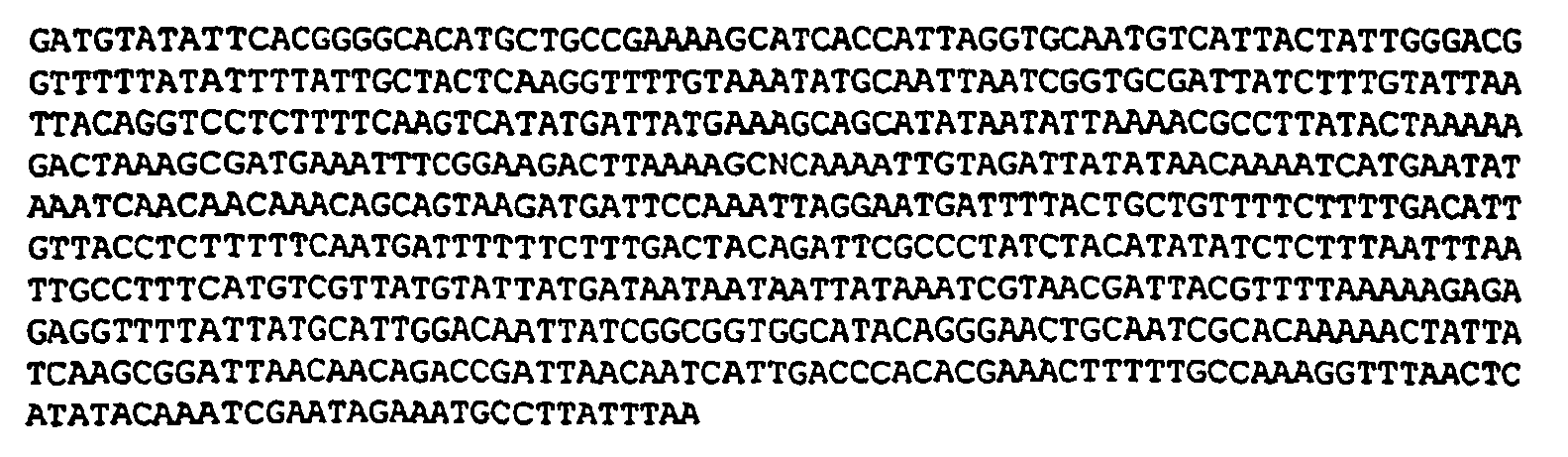
- The Sa4 sequence has similarity to sequences associated with Staphylococcus aureus MnhG (Accession numbers dbj BAA35101; AB015981).
-

-

- The Sa6 sequence has similarity to sequences associated with Bacillus subtilis DnaI Bacsu Primosomal Protein (Accession numbers sp P06567; gi 279708).
- The results discussed above indicate that RFEESS is a useful method for the isolation of EESs effective to result in bioluminescence when the EESs are operably linked to luciferase genes. Furthermore, the data provide examples of specific S. aureus EESs effective to produce such bioluminescence
- Four mice were injected IP at 0, 2, 4 and 6 hr with 500 µl volumes of n-decyl aldehyde at concentrations of 0.1 % and 0.01%. Aldehyde solutions were prepared as follows: 100 µl of aldehyde was diluted in 900 µl of ethanol. 10 µl of this 10% solution was then diluted into 990 µl of sterile phosphate buffered saline (PBS) pH 7.4 to give a 0.1% final volume of aldehyde solution. The animals were observed over a 24-hour period. None of the mice showed any apparent symptoms of illness or abnormal behavior after 24 hrs.
- Twenty four hours after initial injections, the four mice tested as described in Example 5 were injected with a pathogenic strain (8325-4) and a clinical methacillin-resistant (MRSA) strain of S. aureus containing pMK4 luxAB Sa3. The S. aureus strains were grown to an O.D (600 nm) of approximately 0.5 in 10 ml. volumes of LB containing 5 µg/ml chloramphenicol. The bacteria were pelleted and each sample resuspended in 10 mls. of sterile PBS. The O.D of the samples was re-measured and adjusted to give 1 x 10⁵, 1 x 10⁶, and 1 x 10⁷ cells per ml. using the conversion: # of cells = (A600)(11.1 x 10⁸). The dilutions were confirmed by plating on chocolate plates containing 5 µg/ml chloramphenicol.
- Doses were either 250 µl intra-peritoneal (IP; 1 x 10⁵ and 1 x 10⁶ per ml.) or 100 µl intra-muscular (IM; in the thigh with 1 x 10⁶ and 1 x 10⁷ per ml.). The four mice were then injected IP with 500 µl of 0.1% n-decyl aldehyde. This administration of aldehyde was repeated at 2, 4 and 6 hrs just prior to imaging for bioluminescence. Immediately prior to imaging, the mice were anesthetized by intra-muscular (IM) injection with a 4:1 mixture of "Ketaset" (Ketamine [Fort Dodge Products]) at 100mg/ml and "Rompumn" (Xylazine small animal [Darby Drug Company]) at 20mg/ml, at a dose of 15 µl per 10g body weight. Accordingly, a 20 g mouse received 30 µl. The anesthesia typically took effect in 2-3 minutes, and the animals typically remained sedated for 20-30 minutes. If necessary, a second dose of 7 µl per 10g of body weight was administered. In general, the animals recovered 60-90 minutes following administration of a single dose. Animals which had been dosed twice, however, could take as long as 4 hours to recover.
- The mice were imaged essentially as described in Contag, et al.,
U.S. Patent 5,650,135 . Bioluminescence was observed at 0 hr. and at 2 hr, indicating that exogenously-administered aldehyde may be used in vivo to image cells transformed only with luxA and luxB. These results demonstrate that the exogenously-administered aldehyde can diffuse throughout the body, since an IP injection of aldehyde enables the generation of light from luxAB bacteria located in the thigh muscle. - S. aureus strains 8325-4 and MRSA containing pCMOR G+1 SA2 (pMK4 luxABCDE Sa2; pXEN-lux-1) were prepared as described in Example 6 and tested for bioluminescence in mice. The strains were inoculated into mice at 100 µl IP (4x10⁶ per ml.) and 100 µl IM (4x10⁶ per ml. in right thigh and 4x10⁷ per ml. in left thigh) and monitored at
time times - The luxABCDE cassette generated as described in Example 3 was isolated from pSK- luxABCDE via a BamH IlSal I digest and cloned into the BamH I/Xho I sites of the Gram-positive/negative shuttle vector pDL289 (Buckley, N., et al., (1995) J. Bacteriol 177:5028-5034, incorporated herein by reference), generating pDL289 luxABCDE (pCMOR G+2). As was the case in Example 3, the cloning was carried out so that a BamHI restriction site was maintained upstream of the luxA coding region, but in this case, the luxABCDE cassette was in the same orientation as, and downstream of, the lacZ promoter. pCMOR G+2 was then electoporated into E. coli DH5α. The resulting positive clones were extremely bright (since the cassette was downstream of lacZ promoter), with a pure culture allowing the camera to reach a sensitivity of 4.0.
- One of positive clones identified in part (A), above, was plasmid-prepped and the resulting DNA was used to build a promoter library that could be screened in S. pneumoniae. Genomic DNA from Streptococcus pneumoniae R6 was cut with Sau3A in a partial digest (Ausubel, F.M., et al., Current Protocols in Molecular Biology, John Wiley and Sons, Inc., Media, PA (1995)) and ligated with the pCMOR G+2 plasmid cut with BamHI. These ligations were then electroporated into E. coli DH5α. The resulting transformants were pooled directly from plates, their plasmid DNA extracted, and this DNA electroporated into competent cells of a pathogenic encapsulated strain of Streptococcus pneumoniae.
- Approximately 20,000 Gram-positive transformants on chocolate plates containing 250 µg/ml kanamycin were then screened for bioluminescence using a photon counting CCD camera (Hamamatsu Photonics, model 2400-32) as described above. Eighty medium to high light intensity colonies were picked and the 21 brightest of these streaked for single colonies. These 21 isolates were monitored for light intensity readings at both 24 and 72 hrs on chocolate plates containing 400 µg/ml kanamycin. At 24 hr., individual colonies were less than about 0.5 mm in diameter, with light being emitted from the entire streak. By 72 hr however, the single colonies had grown to a size comparable to than achieved by E. coli growing for 16 hr., and were strongly bioluminescent, with very little to no light being emitted from the solid streak. Light intensities were also measured from 16 hr liquid cultures of BHI containing 250 µg/ml kanamycin (O.D. 0.5 - 0.8). Bit Range Light Units (BRLUs) are equal to the rate of bit range change (expressed as bit range per second) on a Hamamatsu Photonics model 2400-32 intensified CCD camera connected to an Argus Image Processor set at a gain of "zero". A summary of this information is shown in Table 3, below:
Table 3 Isolate Light Intensity (BRLU/second) c.f.u. at inoculation c.f.u. from animals Plate - 24 hr Plate - 72 hr Solution - 16 hr Sp1 9.85 8.53 9.31 Sp2 7.64 13.84 1.12 1.7 x 10⁶ Sp3* 0.70 1.10 1.42 Sp4 4.34 5.82 2.61 Sp5 5.45 9.48 12.19 3.2 x 10⁵ Sp6 6.74 9.48 20.08 6.1 x 10³ Sp7 6.74 8.00 2.25 Sp8 5.33 6.40 1.60 Sp9 7.31 16.00 12.19 3.4 x 10⁶ ∼ 0 Sp10 3.88 7.76 2.56 Sp11 7.31 8.83 1.42 Sp12 4.00 5.33 8.00 Sp13 7.11 6.24 2.13 Sp14 17.36 13.13 11.38 Sp15 6.10 14.63 5.12 Sp16 8.83 15.06 23.27 5.8 x 10⁵ 2.5 x 10⁵ Sp17 5.82 7.11 10.89 Sp18 3.76 8.83 0.91 Sp19 7.53 13.47 1.68 Sp20 Not measured Not measured < 0.27 Sp21 7.53 Not measured Not measured * Isolate Sp3 did not give a zone of haemolysis on chocolate plates containing 250 µg/ml kanamycin. - The S. pneumoniae EESs in pDL289 luxABCDE Sp1, 5, 6, 9, 16 and 17 were sequenced with standard methods using the luxA backprimer (LUXA-REV; SEQ ID NO:12), and are presented below. Each sequence ends just upstream of the BamH I promoter insertion site, with the last nucleotide in each sequence corresponding to the first position in the BamH I recognition sequence (GGATCC; SEQ ID NO:13). Note that only two of the EESs (Sp9 and Sp16) ended with a "G", thereby preserving the integrity of the BamH I site in the final pDL289 luxABCDE Sp9 and pDL289 luxABCDE Sp16 constructs.
- The vector sequence between the BamH I promoter insertion site and the ATG start codon (inclusive) of the luxABCDE cassette is as follows (SEQ ID NO:14): GGA TCC TGC AGA TGA AGC AAG AGG AGG ACT CTC TATG. The BamH I site is indicated in bold and Gram-positive Shine-Dalgarno sequence and ATG start codon underlined.
-

- The Sp1 sequence has similarity to sequences associated with Streptococcus pneumoniae D-glutamic acid adding enzyme MurD (murD), undecaprenyl-PP-MurNAc-pentapeptide-UDPGlcNAc GlcNAc transferase (murG), cell division protein DivIB (divIB), orotidine-5'-decarboxylase PyrF (pyrF) (Massidda,O., et al., Microbiology 144 (11):3069-3078 (1998); Accession number gb|AF068902).
-

- The Sp5 sequence has similarity to sequences associated with Mycobacterium tuberculosis UDP-N-acetylmuramoylalanine--D-glutamate ligase (UDP-N-acetylmuranoyl-L-alanyl-D-glutamate synthetase; D-glutamic acid adding enzyme) (Accession numbers sp|O06222; MURD_MYCTU).
-

-
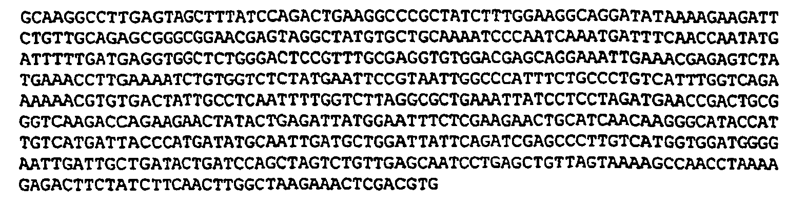
- The Sp9 sequence has similarity to sequences associated with Methanococcus jannaschii cobalt transport ATP-binding protein O homolog (Accession numbers gi|1591732 and U67551).
-

- The
Sp 16 sequence has similarity to sequences associated with Bacillus subtilis DNA polymerase III alpha chain (Accession numbers gi|1591732 and U67551) and Staphylococcus aureus DNA polymerase III (Accession numbers dbj|BAA13160; D86727). -

- The 16 hr liquid cultures of S. pneumoniae containing pDL289 luxABCDE Sp1, 5, 6, 9 and 16 were tested in mice. Bacteria from 1 ml. of each culture were pelleted, resuspended in 1 ml. PBS, and 100 µl of this and a 1/10 dilution were inoculated into the left and right thigh muscles of a mouse, respectively. The lower of each dilution was plated on chocolate agar containing 250 µg/ml kanamycin to assess colony forming units (c.f.u. at innoculation; see Table 3, above). Each of the mice was monitored at
time - As is evident from the data in Table 3, S. pneumoniae containing pDL289 luxABCDE Sp1, 5, and 6, gave between 1 x 10⁴ and 6 x 10⁴ c.f.u., with >80% plasmid retention. No c.f.u. were recovered from Sp9 (probably due to ineffective grinding). Whereas Sp16 gave 2.5 x 10⁵ c.f.u. with >90% plasmid retention.
- Based on the above data, S. pneumoniae containing pDL289 luxABCDE Sp16 was selected as the best candidate strain for further studies.
- The luxABCDE cassette generated as described in Example 2 is isolated from pSK- luxABCDE via a BamH I/Kpn I digest and cloned into the BamH IlKpn I sites of the Gram-positive/negative shuttle vector pSUM39 (Ainsa, et al., (1996) Gene 176:23-26, incorporated herein by reference), generating pSUM39 luxABCDE (pCMOR G+3). As above, the cloning is carried out so that a BamH I restriction site is maintained upstream of the luxA coding region. One of skill in the art will recognize that other Gram-positive/negative shuttle vectors suitable for use with Mycobacterium tuberculosis, such as
pSUM 40 or pSUM 41 (Ainsa, J.A., et al., (1996) Gene 176:23-26), could be used instead ofpSUM 39. - To identify potentially useful promoter sequences, genomic DNA from Mycobacterium tuberculosis is cut with Sau3A in a partial digest (Ausubel, F.M., et al., Current Protocols in Molecular Biology, John Wiley and Sons, Inc., Media, PA (1995)) as described above and ligated with pCMOR G+3 plasmid cut with BamH I. These ligations are then electroporated into E. coli DH5α. The resulting transformants are then pooled, their plasmid DNA extracted, and this DNA is then electroporated into competent M. smegmartis host cells. Transformants which have incorporated the vector are then picked, expanded, and their plasmid DNA is electroporated into competent M. tuberculosis host cells.
- Gram-positive transformants are screened for bioluminescence using a photon counting CCD camera as described above.
- Genomic DNA from Listeria monocytogenes is cut with Sau3 A in a partial digest (see, e.g., Ausubel, F.M., et al., Current Protocols in Molecular Biology, John Wiley and Sons, Inc., Media, PA (1995) and ligated with pCMOR G+1 plasmid (Example 4) that had been cut with BamH I. These ligations are then electroporated into E. coli DH5α. The resulting transformants are pooled directly from plates, their plasmid DNA extracted, and this DNA electroporated into competent Listeria monocytogenes host cells.
- Gram-positive transformants are screened for bioluminescence using a photon counting CCD camera as described above.
- Due to the wide host range of pMK4, a set of the pCMOR G+1 constructs containing S. aureus EESs (Sa1 - 6) were electroporated into a pathogenic strain of Listeria monocytogenes (ATCC 23074) to test whether any of the S. aureus EESs would induce light in Listeria. Although all six plasmid were successfully moved into this strain of Listeria, only pCMOR G+1 Sa4 was found to give significant levels of light, with the remainder of the constructs inducing only low levels of bioluminescence. Since pCMOR G+1 Sa4 was able to induce high levels of light in both S. aureus and L. monocytogenes, this construct may be used to transform other genera of Gram-positive bacteria to a light phenotype.
- The S. aureus construct pCMOR Sa1 (which retained the BamH I site between the promoter and the luxABCDE cassette; see Example 4) was selected as the starting vector for comparing the levels of bioluminescence generated using the engineered luxABCDE cassette with levels of bioluminescence generated using the native luxCDABE cassette (where the two cassettes were each under the control of the S. aureus Sa1 promoter). The luxCDABE construct was generated as follows: The native luxCDABE cassette was first isolated from pSB417 (Winson, et al., 1998, FEMS, 163:185-202) as a BamHI/SalI fragment, and this fragment was then used to replace the corresponding luxABCDE BamHI/SalI fragment dropped out of pCMOR Sa1 to generate pMK4 luxCDABE Sa1. The cloning was carried out in E. coli DH5α and the finished plasmid was moved into S. aureus RN4220.
- Bioluminescence generated by the two different cassettes was compared using transformed E. coli DH5α and transformed S. aureus RN4220 cells, each containing either pCMOR G+1 Sa1 (pMK4 luxABCDE Sa1) or pMK4 luxCDABE Sa1. Exponential cultures of each of the four bacterial strains were diluted across black 96-well microtitre plates in doubling dilutions (-0.3 log) and monitored for light over a period of 30 min at 37°C using a photon counting CCD camera (Hamamatsu, model 2400-32). The contents of each well were then plated to allow the number of colony forming units (CFU) to be compared to levels of bioluminescence (relative light units; RLU).
- The results are shown in
Figure 2 . Both the luxCDABE and the /uxABCDE cassettes function comparably well in E. coli, but only luxABCDE resulted in significant bioluminescence in S. aureus, Sa1:luxABCDE produced approximately 4-fold less light than Sa1:luxCDABE in E. coli. One possible explanation for this phenomenon is that high transcription and high translation efficiencies may be energetically-costly and thus detrimental to the cell, leading to decreased bioluminescence. - The minimum number of S. aureus RN4220 pCMOR Sa1 detectable at 37°C using the Hamamatsu Photonics model 2400-32 CCD camera was approximately 400 c.f.u. However, this minimum number was significantly improved upon by using a more sensitive, liquid nitrogen cooled integrating CCD camera (see Example 14, below). The MRSA strain produced significantly more light (approximately 4-fold) than either RN4220 or 8325-4, regardless of the plasmid (pCMOR Sa1 - 20) tested.
- The results show that using the methods of the present invention, one can generate Gram-positive organisms capable of producing over I x 104 RLU per 1 x 10⁶ organisms (as measured on the XEN-3 Hamamatsu Photonics model 2400-32 photon counting CCD camera).
- Exponential cultures of light S. aureus RN4220 pCMOR G+1 Sa1, S. pneumoniae pCMOR G+1 Sp16 and L. monocytogenes ATCC23074 pCMOR G+1 Sa4 were monitored using a highly sensitive liquid nitrogen cooled integrating CCD camera (Princeton Instruments, Trenton, NJ; model LN/CCD 1340-1300-EB/1) to determine the minimum number of c.f.u detectable of each of these strains of bacteria. Cultures were diluted across black 96-well microtitre plates from bacterial concentrations of approximately 103/well to 101/well in doubling dilutions (-0.3 log) and monitored for light over a period of 10 min. As few as 80 c.f.u of both S. aureus and S. pneumoniae could be detected at 37°C using the Princeton Instruments camera, whereas approximately 400 c.f.u of L. monocytogenes were detectable by this same method.
- To monitor pathogenic bacteria from within animals using bioluminescence (Contag, C., et al., (1995) Mol. Microbiol. 18:593-603), it is important that both the lux genes and Lux proteins function adequately at body temperature (i.e., around 37°C). In order to determine whether modifying the lux genes had altered the temperature range over which bioluminescence occurs optimally in bacterial cells, light arising from the modified luxABCDE cassette was compared to that from the native luxCDABE in both Gram-negative and Gram-positive bacteria between 31°C and 47°C. Since S. aureus RN4220 pMK4 luxCDABE Sa1 was previously shown to be dark (
Fig. 2 ), only S. aureus RN4220 pCMOR G+1 Sa1, E. coli DH5α pCMOR G+1 Sa1 and E. coli DH5α pMK4 luxCDABE Sa1 were tested in this set of experiments. Exponential cultures of the latter three bacterial strains were grown to approximately 107 c.f.u/ml at 30°C and 1 ml. volumes of each placed in heating blocks set at 31, 33, 35, 37, 39, 41, 43, 45 and 47°C. After allowing the bacteria to acclimatize and grow at each of the elevated temperatures for a period of 1 hour, the 9 heating blocks were sequentially placed inside the chamber of a photon counting CCD camera (Hamamatsu, model 2400-32) and light from each of the three cultures recorded for a period of 1 min. To eliminate errors in the number of relative light units arising from variations in bacterial numbers, each culture was plated to allow c.f.u. to be recorded and the light data adjusted accordingly. - As can be seen from Fig. 5, the maximum light to be recorded from a culture of S. aureus RN4220 pCMOR G+1 Sa1 was at 37°C. Furthermore, between 31°C and 41°C the light emission from this strain remained above 60% of this maximum, even at 2 and 4 hours, indicating that the Lux enzymes were stable within this narrower temperature range. In contrast, both E. coli DH5α pCMOR Sa1 and E. coli DH5α pMK4 luxCDABE Sal gave maximum light at 41°C, with E. coli DH5α pCMOR Sa1 actually being slightly brighter at this temperature.
- A modified luxABCDE plasmid was used to successful transform gram-positive Listeria monocytogenes, as described above in Example 14. The gram positive bacteria carrying a modified luxABCDE operon were highly bioluminscent and, in addition, could be monitored in vivo in animals. Plasmid loss in the absence of antibiotic selection was shown to be minimal from L. monocytogenes over a period of 24 to 48 hours in vivo (>80% plasmid retention) with no observable structural instability.
- All of plasmids described have been deposited at Xenogen Corporation, 860 Atlantic Avenue, Alameda, California 94501.
-
- <110> Francis, Kevin P.
Contag, Pamela R.
Joh, Danny J. - <120> LUCIFERASE EXPRESSION CASSETTES AND METHODS OF USE
- <130> PXE-006.PC
- <140>
<141> - <160> 26
- <170> PatentIn Ver. 2.0
- <210> 1
<211> 6
<212> DNA
<213> Artificial Sequence - <220>
<223> Description of Artificial Sequence: Gram-positive ribosome binding site - <400> 1
aggagg 6 - <210> 2
<211> 41
<212> DNA
<213> Artificial Sequence - <220>
<223> Description of Artificial Sequence: Primer XAF3 - <400> 2
ccccggatcc tgcagatgaa gcaagaggag gactctctatg 41 - <210> 3
<211> 36
<212> DNA
<213> Artificial Sequence - <220>
<223> Description of Artificial Sequence: Primer XAR - <400> 3
ggcggatccg tcgacttaat ataatagcga acgttg 36 - <210> 4
<211> 39
<212> DNA
<213> Artificial Sequence - <220>
<223> Description of Artificial Sequence: Primer XBF - <400> 4
gggaattctc gaggaggaga gaaagaaatg aaatttgga 39 - <210> 5
<211> 37
<212> DNA
<213> Artificial Sequence - <220>
<223> Description of Artificial Sequence: Primer XBR - <400> 5
ggcggatccg tcgacttagg tatattccat gtggtac 37 - <210> 6
<211> 34
<212> DNA
<213> Artificial Sequence - <220>
<223> Description of Artificial Sequence: Primer XCF - <400> 6
gggaattctc gaggaggatg gcaaatatga ctaa 34 - <210> 7
<211> 37
<212> DNA
<213> Artificial Sequence - <220>
<223> Description of Artificial Sequence: Primer XCR - <400> 7
ggcggatccg tcgacttatg ggacaaatac aaggaac 37 - <210> 8
<211> 37
<212> DNA
<213> Artificial Sequence - <220>
<223> Description of Artificial Sequence: Primer XDF - <400> 8
gggaattctc gaggaggagt aaaagtatgg aaaatga 37 - <210> 9
<211> 37
<212> DNA
<213> Artificial Sequence - <220>
<223> Description of Artificial Sequence: Primer XDR - <400> 9
ggcggatccg tcgacttaag acagagaaat tgcttga 37 - <210> 10
<211> 39
<212> DNA
<213> Artificial Sequence - <220>
<223> Description of Artificial Sequence: Primer XEF - <400> 10
gggaattctc gaggaggaaa acaggtatga cttcatatg 39 - <210> 11
<211> 38
<212> DNA
<213> Artificial Sequence - <220>
<223> Description of Artificial Sequence: Primer XER - <400> 11
ggcggatccg tcgacttaac tatcaaacgc ttcggtta 38 - <210> 12
<211> 20
<212> DNA
<213> Artificial Sequence - <220>
<223> Description of Artificial Sequence: LUXA-REV - <400> 12
ccacactcct cagagatgcg 20 - <210> 13
<211> 6
<212> DNA
<213> Artificial Sequence - <220>
<223> Description of Artificial Sequence: BamH I recognition sequence - <400> 13
ggatcc 6 - <210> 14
<211> 37
<212> DNA
<213> Artificial Sequence - <220>
<223> Description of Artificial Sequence: vector sequence - <400> 14
ggatcctgca gatgaagcaa gaggaggact ctctatg 37 - <210> 15
<211> 645
<212> DNA
<213> Artificial Sequence - <220>
<223> Description of Artificial Sequence: pMK4 luxABCDE Sa1 - <400> 15

- <210> 16
<211> 671
<212> DNA
<213> Artificial Sequence - <220>
<223> Description of Artificial Sequence: pMK4 luxABCDE Sa2 - <400> 16

- <210> 17
<211> 623
<212> DNA
<213> Artificial Sequence - <220>
<223> Description of Artificial Sequence: pMK4 luxABCDE Sa3 - <400> 17

- <210> 18
<211> 671
<212> DNA
<213> Artificial Sequence - <220>
<223> Description of Artificial Sequence: pMK4 luxABCDE Sa4 - <400> 18


- <210> 19
<211> 650
<212> DNA
<213> Artificial Sequence - <220>
<223> Description of Artificial Sequence: pMK4 luxABCDE Sa5 - <400> 19

- <210> 20
<211> 677
<212> DNA
<213> Artificial Sequence - <220>
<223> Description of Artificial Sequence: pMK4 luxABCDE Sa6 - <400> 20

- <210> 21
<211> 622
<212> DNA
<213> Artificial Sequence - <220>
<223> Description of Artificial Sequence: pDL289 luxABCDE Sp1 - <400> 21


- <210> 22
<211> 610
<212> DNA
<213> Artificial Sequence - <220>
<223> Description of Artificial Sequence: pDL289 luxABCDE Sp5 - <400> 22

- <210> 23
<211> 626
<212> DNA
<213> Artificial Sequence - <220>
<223> Description of Artificial Sequence: pDL289 luxABCDE Sp6 - <400> 23

- <210> 24
<211> 607
<212> DNA
<213> Artificial Sequence - <220>
<223> Description of Artificial Sequence: pDL289 luxABCDE Sp9 - <400> 24


- <210> 25
<211> 616
<212> DNA
<213> Artificial Sequence - <220>
<223> Description of Artificial Sequence: pDL289 luxABCDE Sp16 - <400> 25

- <210> 26
<211> 609
<212> DNA
<213> Artificial Sequence - <220>
<223> Description of Artificial Sequence: pDL289 luxABCDE Sp17 - <400> 26

Claims (21)
- An expression cassette comprising:a polynucleotide encoding luciferase luxA, luxB, and eukaryotic luc gene products, wherein (a) transcription of the polynucleotide results in a polycistronic RNA encoding all three gene products, and (b) polynucleotide sequences comprising Gram-positive ribosome-binding site sequences are located adjacent the 5' end of the luxA coding sequences, adjacent the 5' end of the luxB coding sequences, and adjacent the 5' end of the luc coding sequences.
- The expression cassette of claim 1 wherein the lux gene products are obtained from bacteria having a naturally occurring lux operon ordered luxCDABE.
- The expression cassette of claim 1 or 2, wherein said polynucleotide further encodes luxC, luxD and luxE gene products, and wherein Gram-positive ribosome-binding site sequences are located 5' to each of the luxC, luxD, and luxE coding sequences.
- The expression cassette according to any one of claims 1 to 3, wherein the polynucleotide further comprises a promoter located 5' to all of said lux and luc coding sequences wherein transcription of the polynucleotide results in a polycistronic RNA encoding all the lux and luc gene products.
- The expression cassette of claim 4, wherein said promoter is contained in an Expression Enhancing Sequence selected from the group consisting of Sa1 (SEQ ID NO:15), Sa2 (SEQ ID NO: 16), Sa3 (SEQ ID NO: 17), Sa4 (SEQ ID NO: 18), Sa5 (SEQ ID NO: 19), and Sa6 (SEQ ID NO:20).
- The expression cassette of claim 4, wherein said promoter is contained in an Expression Enhancing Sequence selected from the group consisting of Sp1 (SEQ ID NO:21), Sp5 (SEQ ID NO:22), Sp6 (SEQ ID NO:23), Sp9 (SEQ ID NO:24), Sp16 (SEQ ID NO:25) and Sp17 (SEQ ID NO:26).
- The expression cassette of claim 6, wherein said promoter is contained in Expression Enhancing Sequence Sp16 (SEQ ID NO:25).
- The expression cassette of claim 1, further comprising a multiple-insertion site located adjacent the 5' end of the luxA coding sequences.
- The expression cassette according to claim 1, wherein the coding sequences for luxA and luxB are obtained from Photorhadus luminescens.
- The expression cassette of claim 3, wherein the arrangement of the coding sequences for the lux gene products is in the following relative order 5' - luxA-luxB-luxC-luxD-luxE- 3'.
- The expression cassette of any of claims 1 to 10, wherein the expression cassette is contained within a bacterial transposon.
- The expression cassette of any of claims 1 to 10, wherein the expression cassette is contained within a bacterial mini-transposon.
- The expression cassette of any of claims 1 to 12, wherein the coding sequences of the gene products comprise codons that are optimal for expression of the gene products in a host system into which the expression cassette is to be introduced.
- A shuttle vector comprising:an expression cassette according to any of claims 1 to 13;a polynucleotide encoding a selectable marker;a Gram-positive origin of replication; anda Gram-negative origin of replication.
- A Gram-positive bacterium comprising an expression cassette according to any of claims 1 to 13.
- A bacterium comprising the shuttle vector of claim 14.
- A method of modifying a Gram-positive bacterium to produce light, comprising transforming the Gram-positive bacterium with an expression cassette according to any of claims 1 to 13.
- The method of claim 17, further comprising providing the substrate required for luc-mediated luciferase activity.
- A method of screening an analyte for its ability to affect expression of a reporter marker, comprising:providing the analyte to Gram-positive bacteria comprising the luciferase expression cassette of any of claims 1 to 13, wherein said reporter marker comprises luciferase;providing a substrate required for luciferase light production; andmonitoring the effect of the analyte on the ability of the Gram-positive bacteria to produce light, thereby identifying whether the analyte affects expression of the reporter in Gram-positive bacteria.
- The method of claim 19, wherein said substrate comprises an aldehyde, and said aldehyde is provided as a vapor.
- The method of claim 19 or 20, wherein said substrate is a substrate for the luc gene product.
Applications Claiming Priority (2)
| Application Number | Priority Date | Filing Date | Title |
|---|---|---|---|
| US15290499P | 1999-09-08 | 1999-09-08 | |
| EP00960044A EP1212429B1 (en) | 1999-09-08 | 2000-09-07 | Luciferase expression cassettes and methods of use |
Related Parent Applications (1)
| Application Number | Title | Priority Date | Filing Date |
|---|---|---|---|
| EP00960044A Division EP1212429B1 (en) | 1999-09-08 | 2000-09-07 | Luciferase expression cassettes and methods of use |
Publications (3)
| Publication Number | Publication Date |
|---|---|
| EP1728866A2 EP1728866A2 (en) | 2006-12-06 |
| EP1728866A3 EP1728866A3 (en) | 2007-03-14 |
| EP1728866B1 true EP1728866B1 (en) | 2009-11-11 |
Family
ID=22544956
Family Applications (2)
| Application Number | Title | Priority Date | Filing Date |
|---|---|---|---|
| EP06009933A Expired - Lifetime EP1728866B1 (en) | 1999-09-08 | 2000-09-07 | Luciferase expression cassettes and methods of use |
| EP00960044A Expired - Lifetime EP1212429B1 (en) | 1999-09-08 | 2000-09-07 | Luciferase expression cassettes and methods of use |
Family Applications After (1)
| Application Number | Title | Priority Date | Filing Date |
|---|---|---|---|
| EP00960044A Expired - Lifetime EP1212429B1 (en) | 1999-09-08 | 2000-09-07 | Luciferase expression cassettes and methods of use |
Country Status (8)
| Country | Link |
|---|---|
| EP (2) | EP1728866B1 (en) |
| JP (2) | JP4669180B2 (en) |
| AT (2) | ATE331790T1 (en) |
| AU (1) | AU779263B2 (en) |
| CA (1) | CA2384496A1 (en) |
| DE (2) | DE60029107T2 (en) |
| ES (1) | ES2267561T3 (en) |
| WO (1) | WO2001018195A2 (en) |
Families Citing this family (20)
| Publication number | Priority date | Publication date | Assignee | Title |
|---|---|---|---|---|
| US7255851B2 (en) | 1994-07-01 | 2007-08-14 | The Board Of Trustees Of The Leland Stanford Junior University | Non-invasive localization of a light-emitting conjugate in a mammal |
| US6217847B1 (en) | 1994-07-01 | 2001-04-17 | The Board Of Trustees Of The Leland Stanford Junior University | Non-invasive localization of a light-emitting conjugate in a mammal |
| ATE341637T1 (en) | 1998-12-17 | 2006-10-15 | Xenogen Corp | BLOODLESS DETERMINATION OF A PHYSIOLOGICAL RESPONSE IN A MAMMAL |
| US6610503B1 (en) | 1999-03-17 | 2003-08-26 | Xenogen Corporation | Animal models for predicting sepsis mortality |
| US6737245B1 (en) | 1999-09-08 | 2004-05-18 | Xenogen Corporation | Luciferase expression cassettes and methods of use |
| US6867348B1 (en) | 1999-12-16 | 2005-03-15 | Xenogen Corporation | Methods and compositions for screening for angiogenesis modulating compounds |
| IT1311335B1 (en) | 1999-12-21 | 2002-03-12 | Otc Optical Technologies Ct S | PROCEDURE AND DEVICE FOR MAKING FIBER OPTIC LICENSES. |
| US7056728B2 (en) | 2000-07-06 | 2006-06-06 | Xenogen Corporation | Compositions and methods for use thereof in modifying the genomes of microorganisms |
| US6858773B2 (en) | 2001-04-20 | 2005-02-22 | Xenogen Corporation | Transgenic mice comprising transcription control elements associated with mouse eosinophil peroxidase expression |
| CN1738649A (en) * | 2001-07-09 | 2006-02-22 | 抗癌公司 | Imaging infection using fluorescent protein as a marker |
| EP1281772A1 (en) * | 2001-07-31 | 2003-02-05 | Aladar A. Szalay | Light-emitting microorganisms and cells for tumour diagnosis/therapy |
| EP1281767A3 (en) * | 2001-07-31 | 2003-05-28 | Aladar A. Szalay | Light emitting microorganisms and cells for diagnosis and therapy of tumors |
| WO2005047458A2 (en) | 2003-06-18 | 2005-05-26 | Genelux Corporation | Modified recombinant vaccina viruses and other microorganisms, uses thereof |
| WO2008100292A2 (en) | 2006-10-16 | 2008-08-21 | Genelux Corporation | Modified vaccinia virus strains for use in diagnostic and therapeutic methods |
| WO2008109459A1 (en) | 2007-03-02 | 2008-09-12 | University Of Massachusetts | Luciferins |
| WO2015031859A1 (en) * | 2013-08-29 | 2015-03-05 | The Regents Of The University Of California | Bacteria engineered for ester production |
| EP3976116A4 (en) | 2019-05-30 | 2023-08-23 | 490 Biotech, Inc. | Lux expression in cells and methods of use |
| CN110295181B (en) * | 2019-06-18 | 2022-10-11 | 山东大学 | Method for identifying antibiotic species by using bioluminescence report system |
| DE102020129009A1 (en) | 2020-11-04 | 2022-05-05 | Rheinisch-Westfälische Technische Hochschule (RWTH) Aachen, Körperschaft des öffentlichen Rechts | DETECTION OF ENVIRONMENTAL INFLUENCES USING BIOLUMINESCENCE |
| CN113403243B (en) * | 2021-06-16 | 2022-07-19 | 中国人民解放军军事科学院军事医学研究院 | Photogenic bacteria of Streptococcus suis based on lux reporter system and its construction method |
Family Cites Families (3)
| Publication number | Priority date | Publication date | Assignee | Title |
|---|---|---|---|---|
| EP0798384A1 (en) * | 1992-10-02 | 1997-10-01 | Lonza A.G. | Biotechnological method of producing biotin |
| FI96697C (en) * | 1993-08-18 | 2001-05-16 | Valio Oy | New Streptococcus thermophilus strains and their use for antibiotic testing |
| WO1999014311A1 (en) * | 1997-09-18 | 1999-03-25 | Smithkline Beecham Corporation | Method of screening for antimicrobial compounds |
-
2000
- 2000-09-07 EP EP06009933A patent/EP1728866B1/en not_active Expired - Lifetime
- 2000-09-07 AU AU71266/00A patent/AU779263B2/en not_active Ceased
- 2000-09-07 EP EP00960044A patent/EP1212429B1/en not_active Expired - Lifetime
- 2000-09-07 AT AT00960044T patent/ATE331790T1/en not_active IP Right Cessation
- 2000-09-07 DE DE60029107T patent/DE60029107T2/en not_active Expired - Lifetime
- 2000-09-07 ES ES00960044T patent/ES2267561T3/en not_active Expired - Lifetime
- 2000-09-07 CA CA002384496A patent/CA2384496A1/en not_active Abandoned
- 2000-09-07 DE DE60043321T patent/DE60043321D1/en not_active Expired - Lifetime
- 2000-09-07 AT AT06009933T patent/ATE448309T1/en not_active IP Right Cessation
- 2000-09-07 JP JP2001522406A patent/JP4669180B2/en not_active Expired - Lifetime
- 2000-09-07 WO PCT/US2000/024699 patent/WO2001018195A2/en not_active Ceased
-
2010
- 2010-11-16 JP JP2010256354A patent/JP5295206B2/en not_active Expired - Fee Related
Also Published As
| Publication number | Publication date |
|---|---|
| DE60043321D1 (en) | 2009-12-24 |
| ES2267561T3 (en) | 2007-03-16 |
| AU7126600A (en) | 2001-04-10 |
| JP4669180B2 (en) | 2011-04-13 |
| AU779263B2 (en) | 2005-01-13 |
| CA2384496A1 (en) | 2001-03-15 |
| JP2011041577A (en) | 2011-03-03 |
| WO2001018195A3 (en) | 2001-06-21 |
| ATE331790T1 (en) | 2006-07-15 |
| EP1728866A2 (en) | 2006-12-06 |
| JP5295206B2 (en) | 2013-09-18 |
| DE60029107D1 (en) | 2006-08-10 |
| EP1212429A2 (en) | 2002-06-12 |
| ATE448309T1 (en) | 2009-11-15 |
| EP1728866A3 (en) | 2007-03-14 |
| EP1212429B1 (en) | 2006-06-28 |
| JP2003509029A (en) | 2003-03-11 |
| DE60029107T2 (en) | 2007-01-11 |
| WO2001018195A2 (en) | 2001-03-15 |
Similar Documents
| Publication | Publication Date | Title |
|---|---|---|
| JP5295206B2 (en) | Luciferase expression cassette and method of use | |
| JP3375337B2 (en) | Bioluminescent protein | |
| US20240279621A1 (en) | Novel luciferases and methods for using same | |
| JP2003509029A5 (en) | ||
| US6737245B1 (en) | Luciferase expression cassettes and methods of use | |
| JPH08510387A (en) | Mutant luciferase | |
| CN111269324B (en) | Fusion protein of Gaussian luciferase and digoxigenin single-chain antibody and its application | |
| Jansson | Tracking genetically engineered microorganisms in nature | |
| JP2008148715A (en) | Screening method for introduction of DNA into target cells | |
| US7056728B2 (en) | Compositions and methods for use thereof in modifying the genomes of microorganisms | |
| AU2002258480A1 (en) | Methods of screening for introduction of DNA into a target cell | |
| JP2025522599A (en) | Luciferase variants and uses thereof | |
| JP5105864B2 (en) | Vectors and methods for detecting transcriptional changes | |
| WO2002008431A1 (en) | Compositions and methods for use thereof in modifying the genomes of microorganisms | |
| RU2809367C2 (en) | Hispidin synthases and their use | |
| Patterson | Optimization of bacterial luciferase for expression in mammalian cells | |
| JPH1028591A (en) | Novel DNA, recombinant vector containing the same, microorganism containing the same, odor sensor comprising the microorganism, and method for detecting odor using the microorganism |
Legal Events
| Date | Code | Title | Description |
|---|---|---|---|
| PUAI | Public reference made under article 153(3) epc to a published international application that has entered the european phase |
Free format text: ORIGINAL CODE: 0009012 |
|
| 17P | Request for examination filed |
Effective date: 20060601 |
|
| AC | Divisional application: reference to earlier application |
Ref document number: 1212429 Country of ref document: EP Kind code of ref document: P |
|
| AK | Designated contracting states |
Kind code of ref document: A2 Designated state(s): AT BE CH CY DE DK ES FI FR GB GR IE IT LI LU MC NL PT SE |
|
| RAP1 | Party data changed (applicant data changed or rights of an application transferred) |
Owner name: XENOGEN CORPORATION |
|
| RIN1 | Information on inventor provided before grant (corrected) |
Inventor name: FRANCIS, KEVIN P. Inventor name: JOH, DANNY J. Inventor name: CONTAG, PAMELA R. |
|
| PUAL | Search report despatched |
Free format text: ORIGINAL CODE: 0009013 |
|
| AK | Designated contracting states |
Kind code of ref document: A3 Designated state(s): AT BE CH CY DE DK ES FI FR GB GR IE IT LI LU MC NL PT SE |
|
| 17Q | First examination report despatched |
Effective date: 20070821 |
|
| AKX | Designation fees paid |
Designated state(s): AT BE CH CY DE DK ES FI FR GB GR IE IT LI LU MC NL PT SE |
|
| GRAP | Despatch of communication of intention to grant a patent |
Free format text: ORIGINAL CODE: EPIDOSNIGR1 |
|
| GRAS | Grant fee paid |
Free format text: ORIGINAL CODE: EPIDOSNIGR3 |
|
| GRAA | (expected) grant |
Free format text: ORIGINAL CODE: 0009210 |
|
| AC | Divisional application: reference to earlier application |
Ref document number: 1212429 Country of ref document: EP Kind code of ref document: P |
|
| AK | Designated contracting states |
Kind code of ref document: B1 Designated state(s): AT BE CH CY DE DK ES FI FR GB GR IE IT LI LU MC NL PT SE |
|
| REG | Reference to a national code |
Ref country code: GB Ref legal event code: FG4D |
|
| REG | Reference to a national code |
Ref country code: CH Ref legal event code: EP |
|
| REG | Reference to a national code |
Ref country code: IE Ref legal event code: FG4D |
|
| REF | Corresponds to: |
Ref document number: 60043321 Country of ref document: DE Date of ref document: 20091224 Kind code of ref document: P |
|
| NLV1 | Nl: lapsed or annulled due to failure to fulfill the requirements of art. 29p and 29m of the patents act | ||
| PG25 | Lapsed in a contracting state [announced via postgrant information from national office to epo] |
Ref country code: FI Free format text: LAPSE BECAUSE OF FAILURE TO SUBMIT A TRANSLATION OF THE DESCRIPTION OR TO PAY THE FEE WITHIN THE PRESCRIBED TIME-LIMIT Effective date: 20091111 Ref country code: ES Free format text: LAPSE BECAUSE OF FAILURE TO SUBMIT A TRANSLATION OF THE DESCRIPTION OR TO PAY THE FEE WITHIN THE PRESCRIBED TIME-LIMIT Effective date: 20100222 Ref country code: PT Free format text: LAPSE BECAUSE OF FAILURE TO SUBMIT A TRANSLATION OF THE DESCRIPTION OR TO PAY THE FEE WITHIN THE PRESCRIBED TIME-LIMIT Effective date: 20100311 Ref country code: SE Free format text: LAPSE BECAUSE OF FAILURE TO SUBMIT A TRANSLATION OF THE DESCRIPTION OR TO PAY THE FEE WITHIN THE PRESCRIBED TIME-LIMIT Effective date: 20091111 |
|
| PG25 | Lapsed in a contracting state [announced via postgrant information from national office to epo] |
Ref country code: CY Free format text: LAPSE BECAUSE OF FAILURE TO SUBMIT A TRANSLATION OF THE DESCRIPTION OR TO PAY THE FEE WITHIN THE PRESCRIBED TIME-LIMIT Effective date: 20091111 |
|
| PG25 | Lapsed in a contracting state [announced via postgrant information from national office to epo] |
Ref country code: AT Free format text: LAPSE BECAUSE OF FAILURE TO SUBMIT A TRANSLATION OF THE DESCRIPTION OR TO PAY THE FEE WITHIN THE PRESCRIBED TIME-LIMIT Effective date: 20091111 Ref country code: BE Free format text: LAPSE BECAUSE OF FAILURE TO SUBMIT A TRANSLATION OF THE DESCRIPTION OR TO PAY THE FEE WITHIN THE PRESCRIBED TIME-LIMIT Effective date: 20091111 |
|
| PG25 | Lapsed in a contracting state [announced via postgrant information from national office to epo] |
Ref country code: DK Free format text: LAPSE BECAUSE OF FAILURE TO SUBMIT A TRANSLATION OF THE DESCRIPTION OR TO PAY THE FEE WITHIN THE PRESCRIBED TIME-LIMIT Effective date: 20091111 |
|
| PLBE | No opposition filed within time limit |
Free format text: ORIGINAL CODE: 0009261 |
|
| STAA | Information on the status of an ep patent application or granted ep patent |
Free format text: STATUS: NO OPPOSITION FILED WITHIN TIME LIMIT |
|
| 26N | No opposition filed |
Effective date: 20100812 |
|
| PG25 | Lapsed in a contracting state [announced via postgrant information from national office to epo] |
Ref country code: GR Free format text: LAPSE BECAUSE OF FAILURE TO SUBMIT A TRANSLATION OF THE DESCRIPTION OR TO PAY THE FEE WITHIN THE PRESCRIBED TIME-LIMIT Effective date: 20100212 |
|
| PG25 | Lapsed in a contracting state [announced via postgrant information from national office to epo] |
Ref country code: IT Free format text: LAPSE BECAUSE OF FAILURE TO SUBMIT A TRANSLATION OF THE DESCRIPTION OR TO PAY THE FEE WITHIN THE PRESCRIBED TIME-LIMIT Effective date: 20091111 |
|
| PG25 | Lapsed in a contracting state [announced via postgrant information from national office to epo] |
Ref country code: MC Free format text: LAPSE BECAUSE OF NON-PAYMENT OF DUE FEES Effective date: 20100930 |
|
| PG25 | Lapsed in a contracting state [announced via postgrant information from national office to epo] |
Ref country code: IE Free format text: LAPSE BECAUSE OF NON-PAYMENT OF DUE FEES Effective date: 20100907 |
|
| PG25 | Lapsed in a contracting state [announced via postgrant information from national office to epo] |
Ref country code: LU Free format text: LAPSE BECAUSE OF NON-PAYMENT OF DUE FEES Effective date: 20100907 Ref country code: NL Free format text: LAPSE BECAUSE OF FAILURE TO SUBMIT A TRANSLATION OF THE DESCRIPTION OR TO PAY THE FEE WITHIN THE PRESCRIBED TIME-LIMIT Effective date: 20091111 |
|
| REG | Reference to a national code |
Ref country code: FR Ref legal event code: PLFP Year of fee payment: 17 |
|
| REG | Reference to a national code |
Ref country code: FR Ref legal event code: PLFP Year of fee payment: 18 |
|
| REG | Reference to a national code |
Ref country code: FR Ref legal event code: PLFP Year of fee payment: 19 |
|
| PGFP | Annual fee paid to national office [announced via postgrant information from national office to epo] |
Ref country code: FR Payment date: 20190925 Year of fee payment: 20 |
|
| PGFP | Annual fee paid to national office [announced via postgrant information from national office to epo] |
Ref country code: GB Payment date: 20190927 Year of fee payment: 20 |
|
| PGFP | Annual fee paid to national office [announced via postgrant information from national office to epo] |
Ref country code: DE Payment date: 20190927 Year of fee payment: 20 |
|
| PGFP | Annual fee paid to national office [announced via postgrant information from national office to epo] |
Ref country code: CH Payment date: 20191002 Year of fee payment: 20 |
|
| REG | Reference to a national code |
Ref country code: DE Ref legal event code: R071 Ref document number: 60043321 Country of ref document: DE |
|
| REG | Reference to a national code |
Ref country code: CH Ref legal event code: PL |
|
| REG | Reference to a national code |
Ref country code: GB Ref legal event code: PE20 Expiry date: 20200906 |
|
| PG25 | Lapsed in a contracting state [announced via postgrant information from national office to epo] |
Ref country code: GB Free format text: LAPSE BECAUSE OF EXPIRATION OF PROTECTION Effective date: 20200906 |