EP1716173B1 - Carcinoembryonic antigen fusion protein and uses thereof - Google Patents
Carcinoembryonic antigen fusion protein and uses thereof Download PDFInfo
- Publication number
- EP1716173B1 EP1716173B1 EP05707188A EP05707188A EP1716173B1 EP 1716173 B1 EP1716173 B1 EP 1716173B1 EP 05707188 A EP05707188 A EP 05707188A EP 05707188 A EP05707188 A EP 05707188A EP 1716173 B1 EP1716173 B1 EP 1716173B1
- Authority
- EP
- European Patent Office
- Prior art keywords
- cea
- vector
- sequence
- pv1j
- ltb
- Prior art date
- Legal status (The legal status is an assumption and is not a legal conclusion. Google has not performed a legal analysis and makes no representation as to the accuracy of the status listed.)
- Expired - Lifetime
Links
Images




























Classifications
-
- C—CHEMISTRY; METALLURGY
- C07—ORGANIC CHEMISTRY
- C07K—PEPTIDES
- C07K14/00—Peptides having more than 20 amino acids; Gastrins; Somatostatins; Melanotropins; Derivatives thereof
- C07K14/435—Peptides having more than 20 amino acids; Gastrins; Somatostatins; Melanotropins; Derivatives thereof from animals; from humans
-
- C—CHEMISTRY; METALLURGY
- C07—ORGANIC CHEMISTRY
- C07K—PEPTIDES
- C07K14/00—Peptides having more than 20 amino acids; Gastrins; Somatostatins; Melanotropins; Derivatives thereof
- C07K14/435—Peptides having more than 20 amino acids; Gastrins; Somatostatins; Melanotropins; Derivatives thereof from animals; from humans
- C07K14/705—Receptors; Cell surface antigens; Cell surface determinants
- C07K14/70503—Immunoglobulin superfamily
-
- A—HUMAN NECESSITIES
- A61—MEDICAL OR VETERINARY SCIENCE; HYGIENE
- A61K—PREPARATIONS FOR MEDICAL, DENTAL OR TOILETRY PURPOSES
- A61K39/00—Medicinal preparations containing antigens or antibodies
- A61K39/395—Antibodies; Immunoglobulins; Immune serum, e.g. antilymphocytic serum
- A61K39/39533—Antibodies; Immunoglobulins; Immune serum, e.g. antilymphocytic serum against materials from animals
- A61K39/39558—Antibodies; Immunoglobulins; Immune serum, e.g. antilymphocytic serum against materials from animals against tumor tissues, cells, antigens
-
- A—HUMAN NECESSITIES
- A61—MEDICAL OR VETERINARY SCIENCE; HYGIENE
- A61P—SPECIFIC THERAPEUTIC ACTIVITY OF CHEMICAL COMPOUNDS OR MEDICINAL PREPARATIONS
- A61P35/00—Antineoplastic agents
-
- A—HUMAN NECESSITIES
- A61—MEDICAL OR VETERINARY SCIENCE; HYGIENE
- A61P—SPECIFIC THERAPEUTIC ACTIVITY OF CHEMICAL COMPOUNDS OR MEDICINAL PREPARATIONS
- A61P37/00—Drugs for immunological or allergic disorders
- A61P37/02—Immunomodulators
- A61P37/04—Immunostimulants
-
- C—CHEMISTRY; METALLURGY
- C07—ORGANIC CHEMISTRY
- C07K—PEPTIDES
- C07K14/00—Peptides having more than 20 amino acids; Gastrins; Somatostatins; Melanotropins; Derivatives thereof
- C07K14/435—Peptides having more than 20 amino acids; Gastrins; Somatostatins; Melanotropins; Derivatives thereof from animals; from humans
- C07K14/705—Receptors; Cell surface antigens; Cell surface determinants
-
- C—CHEMISTRY; METALLURGY
- C12—BIOCHEMISTRY; BEER; SPIRITS; WINE; VINEGAR; MICROBIOLOGY; ENZYMOLOGY; MUTATION OR GENETIC ENGINEERING
- C12N—MICROORGANISMS OR ENZYMES; COMPOSITIONS THEREOF; PROPAGATING, PRESERVING, OR MAINTAINING MICROORGANISMS; MUTATION OR GENETIC ENGINEERING; CULTURE MEDIA
- C12N15/00—Mutation or genetic engineering; DNA or RNA concerning genetic engineering, vectors, e.g. plasmids, or their isolation, preparation or purification; Use of hosts therefor
- C12N15/09—Recombinant DNA-technology
- C12N15/11—DNA or RNA fragments; Modified forms thereof; Non-coding nucleic acids having a biological activity
- C12N15/62—DNA sequences coding for fusion proteins
-
- A—HUMAN NECESSITIES
- A61—MEDICAL OR VETERINARY SCIENCE; HYGIENE
- A61K—PREPARATIONS FOR MEDICAL, DENTAL OR TOILETRY PURPOSES
- A61K39/00—Medicinal preparations containing antigens or antibodies
- A61K2039/51—Medicinal preparations containing antigens or antibodies comprising whole cells, viruses or DNA/RNA
- A61K2039/53—DNA (RNA) vaccination
-
- C—CHEMISTRY; METALLURGY
- C07—ORGANIC CHEMISTRY
- C07K—PEPTIDES
- C07K2319/00—Fusion polypeptide
- C07K2319/30—Non-immunoglobulin-derived peptide or protein having an immunoglobulin constant or Fc region, or a fragment thereof, attached thereto
-
- C—CHEMISTRY; METALLURGY
- C07—ORGANIC CHEMISTRY
- C07K—PEPTIDES
- C07K2319/00—Fusion polypeptide
- C07K2319/55—Fusion polypeptide containing a fusion with a toxin, e.g. diphteria toxin
Definitions
- the present invention relates generally to the therapy of cancer. More specifically, the present invention relates to polynucleotides encoding fusion proteins wherein the fusion proteins comprise at least a portion of the tumor associated polypeptide carcinoembryonic antigen.
- the present invention also provides recombinant vectors and hosts comprising said polynucleotides, purified fusion proteins and methods for enhancing an immune response against CEA using the compositions and molecules disclosed herein.
- immunoglobulin superfamily consists of numerous genes that code for proteins with diverse functions, one of which is intercellular adhesion. IgSF proteins contain at least one Ig-related domain that is important for maintaining proper intermolecular binding interactions. Because such interactions are necessary to the diverse biological functions of the IgSF members, disruption or aberrant expression of many IgSF adhesion molecules has been correlated with many human diseases.
- CEA carcinoembryonic antigen
- CEACAMs cell surface glycoproteins known as CEA-related cell adhesion molecules (CEACAMs).
- CEACAMs have been shown to act as both homotypic and heterotypic intercellular adhesion molecules ( Benchimol et al., Cell 57: 327-334 (1989 )).
- CEA also known as CEACAM5
- Bcl2 and C-Myc Bcl2 and C-Myc
- CEA normal expression of CEA has been detected during fetal development and in adult colonic mucosa.
- CEA overexpression was first detected in human colon tumors over thirty years ago ( Gold and Freedman, J. Exp. Med. 121:439-462 (1965 )) and has since been found in nearly all colorectal tumors. Additionally, CEA overexpression is detectable in a high percentage of adenocarcinomas of the pancreas, liver, breast, ovary, cervix, and lung. Because of its prevalence in these tumor types and limited normal tissue expression, CEA is considered a self tumor-associated antigen and a target for active and passive immunotherapy. Recent clinical data have established that different vaccine strategies can generate human B and T cells specific for CEA, providing additional evidence that CEA is a target for molecular and immunological intervention for treatment of these cancer types.
- Therapeutic approaches targeting CEA include the use of anti-CEA antibodies (see Chester et al., Cancer Chemother. Pharmacol. 46 (Suppl): S8-S12 (2000 )), as well as CEA-based vaccines (for review, see Berinstein, supra ).
- the development and commercialization of many vaccines have been hindered by difficulties associated with obtaining high expression levels of exogenous genes.
- Success of DNA-based vaccines has also been hindered by an inability to generate an immune response of sufficient magnitude in treated individuals.
- DNA vaccines targeting various proteins have been developed, the resulting immune responses have been relatively weak compared with conventional vaccines.
- HSP heat shock protein
- the enhancement of immune responses to target antigens is particularly relevant for cancer vaccines in view of the limited immunogenicity of tumor antigens and of the need to overcome tolerance to exert effective antitumor effects.
- the present invention provides polynucleotides encoding fusion proteins wherein the fusion proteins comprise at least a portion of the tumor associated polypeptide carcinoembryonic antigen, fused to a substantial portion of an immunoenhancing element.
- the CEA portion of the encoded CEA fusion protein is deleted of its C-terminal anchoring domain, i.e. amino acids 679-702 of SEQ ID NO:20, and the immunoenhancing element is the B subunit of the heat labile enterotoxin of E.coli , optionally truncated of its signal sequence.
- the present invention also provides recombinant vectors, including but not limited to, adenovirus and plasmid vectors, comprising said polynucleotides and host cells comprising said recombinant vectors. Also provided herein are purified fusion proteins encoded by invention polynucleotides.
- the present invention further provides compositions for use in methods for inhibiting or preventing the development of a cancer in a mammal by eliciting an immune response to the CEA protein by administering a vaccine or pharmaceutical composition comprising the CEA fusions or CEA fusion proteins described herein.
- the immune response is enhanced relative to the response elicited by a wild-type CEA vaccine.
- DOM refers generally to the N-terminal domain of fragment C of tetanus toxoid.
- LT refers generally to the heat labile enterotoxin of E. coli.
- LT may refer to the complete enterotoxin, comprising subunits A and B or a substantial portion of subunit A, or a substantial portion of subunit B.
- LTA refers to the A subunit of the heat labile enterotoxin of E. coli, or substantial portion thereof, including subunits which are truncated on the C-terminal or N-terminal end but maintain biological activity, as well as subunits that contain internal amino acid insertions, deletions, or substitutions but maintain biological activity.
- LTB refers to the B subunit of the heat labile enterotoxin of E. coli, or substantial portion thereof, including subunits which are truncated on the C-terminal or N-terminal end but maintain biological activity, as well as subunits that contain internal amino acid insertions, deletions, or substitutions but maintain biological activity.
- pV1J/hCEAopt refers to a plasmid construct, disclosed herein, comprising the CMV immediate-early (IE) promoter with intron A, a full-length cordon-optimized human CEA gene, bovine growth hormone-derived polyadenylation and transcriptional termination sequences, and a minimal pUC backbone (see EXAMPLE 2).
- IE immediate-early
- pV1J/hCEA refers to a construct essentially as described above, except the construct comprises a wild-type human CEA gene instead of a codon-optimized human CEA gene.
- pV1J/hCEA-LTB refers to a plasmid construct, disclosed herein, comprising the CMV immediate-early (IE) promoter with intron A, a human CEA gene devoid of its GPI anchor coding sequence, fused at its C-terminal end to the B subunit of E. coli heat labile enterotoxin, bovine growth hormone-derived polyadenylation and transcriptional termination sequences, and a minimal pUC backbone.
- IE immediate-early
- V1J/hCEAopt-LTB refers to a construct essentially as described immediately above, except the construct comprises a codon-optimized human CEA gene devoid of its GPI anchor coding sequence instead of the corresponding portion of the wild-type human CEA gene.
- pV1J/hCEAopt-LTBopt refers to a plasmid construct essentially as described immediately above, except that both the CEA sequences and the LTB sequences are codon-optimized for high level expression in human cells.
- pV1J/rhCEAopt-LTBopt refers to a construct essentially as described above except that the human codon-optimized CEA gene is replaced with a rhesus monkey CEA gene, codon-optimized for high-level expression in human cells.
- pV1J/hCEA-LTA refers to a plasmid construct, disclosed herein, comprising the CMV immediate-early (IE) promoter with intron A, a human CEA gene devoid of the GPI anchor coding sequence, fused at its C-terminal end to the A subunit of E. coli heat labile enterotoxin, bovine growth hormone-derived polyadenylation and transcriptional termination sequences, and a minimal pUC backbone. Construction of plasmid vectors comprising various CEA-LT fusions is described in EXAMPLE 2.
- IE immediate-early
- pV1J/hCEA-DOM refers to a plasmid construct, disclosed herein, comprising the CMV immediate-early (IE) promoter with intron A, a human CEA gene devoid of its GPI anchor coding sequence, fused at its C-terminal end to the N-terminal domain of Fragment C of tetanus toxoid (DOM), bovine growth hormone-derived polyadenylation and transcriptional termination sequences, and a minimal pUC backbone (EXAMPLE 2).
- IE CMV immediate-early
- DOM tetanus toxoid
- EXAMPLE 2 bovine growth hormone-derived polyadenylation and transcriptional termination sequences
- V1J/rhCEAopt-DOMopt refers to a construct essentially as described above except that the human codon-optimized CEA gene is replaced with a rhesus monkey CEA gene, codon-optimized for high-level expression in human cells.
- pV1J/hCEA-FcIgG refers to a plasmid construct, disclosed herein, comprising the CMV immediate-early (IE) promoter with intron A, a human CEA gene devoid of the GPI anchor coding sequence, fused at its C-terminal end to the heavy fragment of constant chain of immunoglobulin G1, bovine growth hormone-derived polyadenylation and transcriptional termination sequences, and a minimal pUC backbone.
- IE immediate-early
- pV1J/hCEAopt-FcIgGopt refers to a construct essentially as described, except the nucleotide sequences encoding CEA and FcIgG have been codon-optimized for high-level expression in human cells.
- Ad5/hCEAopt and “Ad5/hCEA” refer to two constructs, disclosed herein, which comprise an Ad5 adenoviral genome deleted of the E1 and E3 regions.
- Ad5/hCEAopt the E1 region is replaced by a codon-optimized human CEA gene in an E1 parallel orientation under the control of a human CMV promoter without intron A, followed by a bovine growth hormone polyadenylation signal.
- the "Ad5/hCEA” construct is essentially as described above, except the E1 region of the Ad5 genome is replaced with a wild-type human CEA sequence.
- Ad5/hCEAopt-LTB refers to an Ad5 construct, essentially as described above, except that the codon-optimized human CEA sequence is devoid of the GPI anchor coding sequence and is fused at its C-terminus to the B subunit of E. coli heat labile enterotoxin. Construction of adenovirus vectors comprising various CEA-LT fusions is described in EXAMPLE 3.
- Immunoenhancing element refers to a portion of the CEA fusion proteins of the present invention which is capable of stimulating or enhancing the immune response to the associated CEA protein, relative to full-length wild-type CEA.
- Immunoenhancing elements of the present invention are selected from the group consisting of: heat shock protein (HSP) 70, lysosome-associated membrane protein (LAMP), fragment C of tetanus toxoid (FrC), the N-terminal domain of FrC (DOM), the heavy fragment of constant chain of immune globulin G1 (FcIgG), the vesicular stomatitis virus glycoprotein (VSV-G), cholera toxin (CT) from Vibrio cholerae , and heat labile enterotoxin of E.coli (LT).
- HSP heat shock protein
- LAMP lysosome-associated membrane protein
- FrC fragment C of tetanus toxoid
- DOM N-terminal domain of FrC
- fusion protein refers to a protein having at least two polypeptides covalently linked in which one polypeptide comes from one protein sequence or domain and the other polypeptide comes from a second protein sequence or domain.
- the fusion proteins of the present invention comprise a CEA polypeptide or fragment or variant thereof, and a second polypeptide, which comprises a substantial portion of an immunoenhancing element, which, in some cases, is a bacterial toxin.
- the CEA polypeptide, fragment or variant thereof may be a human CEA or CEA homolog from another species.
- the polypeptides that comprise the fusion protein are preferably linked N-terminus to C-terminus.
- the CEA polypeptide and the toxin subunit can be fused in any order.
- CEA polypeptide is fused to the N-terminus of the toxin subunit, as exemplified in FIGURE 1A.
- fusion proteins in which the immunoenhancing element is fused to the N-terminus of the CEA polypeptide are also contemplated.
- CEA fusion protein is intended to be a general term which refers to a fusion as described above, which comprises a CEA polypeptide or fragment or variant thereof fused to a polypeptide comprising an immunoenhancing element.
- CEA-LT fusion refers to a nucleic acid sequence in which at least a portion of the CEA gene is fused to a substantial portion of either the LTA or the LTB subunit of E. coli heat labile enterotoxin.
- CEA-LT fusion protein refers to a polypeptide encoded by a CEA-LT fusion as described.
- CEA-LT fusion and CEA-LT fusion protein are also understood to refer to fragments thereof, homologs thereof, and functional equivalents thereof (collectively referred to as "variants"), such as those in which one or more amino acids is inserted, deleted or replaced by other amino acid(s).
- the CEA-LT fusions of the present invention upon administration to a mammal such as a human being, can stimulate an immune response by helper T cells or cytotoxic T cells, or stimulate the production of antibodies at least as well as a "wild-type" CEA sequence.
- the CEA-LT fusion can enhance the immune response as compared to a wild-type CEA.
- CEA-DOM fusion refers to a nucleic acid sequence in which at least a portion of the CEA gene is fused to a substantial portion of the minimized domain of tetanus toxin fragment C, unless the context clearly dictates that said term refers to the protein sequence.
- CEA-DOM fusion protein refers to a polypeptide encoded by a CEA-DOM fusion as described.
- CEA-DOM fusion and CEA-DOM fusion protein are also understood to refer to fragments thereof, homologs thereof, and functional equivalents thereof (collectively referred to as "variants"), such as those in which one or more amino acids is inserted, deleted or replaced by other amino acid(s).
- the CEA-DOM fusions of the present invention upon administration to a mammal such as a human being, can stimulate an immune response by helper T cells or cytotoxic T cells, or stimulate the production of antibodies at least as well as a "wild-type" CEA sequence.
- the CEA-DOM fusion can enhance the immune response as compared to a wild-type CEA.
- AD refers to the anchoring domain of a CEA gene or protein.
- the anchoring domain of the wild-type human CEA is located from about amino acid 679 to about amino acid 702 of SEQ ID NO:20.
- treatment refers to both therapeutic treatment and prophylactic or preventative measures. Those in need of treatment include those already with the disorder as well as those prone to have the disorder or those in which the disorder is to be prevented.
- a “disorder” is any condition that would benefit from treatment with the molecules of the present invention, including the nucleic acid molecules described herein and the fusion proteins that are encoded by said nucleic acid molecules.
- disorder are chronic and acute disorders or diseases including those pathological conditions which predispose the mammal to the disorder in question.
- the molecules of the present invention are intended for use as treatments for disorders or conditions characterized by aberrant cell proliferation, including, but not limited to, breast cancer, colorectal cancer, and lung cancer.
- an effective amount means sufficient vaccine composition is introduced to produce the adequate levels of the polypeptide, so that an immune response results.
- this level may vary.
- a “conservative amino acid substitution” refers to the replacement of one amino acid residue by another, chemically similar, amino acid residue. Examples of such conservative substitutions are: substitution of one hydrophobic residue (isoleucine, leucine, valine, or methionine) for another; substitution of one polar residue for another polar residue of the same charge (e.g., arginine for lysine; glutamic acid for aspartic acid).
- hCEA and hCEAopt refer to a human carcinoembryonic antigen and a human codon-optimized carcinoembryonic antigen, respectively.
- rhCEA and rhCEAopt refer to a rhesus monkey carcinoembryonic antigen and a rhesus monkey codon-optimized carcinoembryonic antigen, respectively.
- substantially similar means that a given nucleic acid or amino acid sequence shares at least 75%, preferably 85%, more preferably 90%, and even more preferably 95% identity with a reference sequence.
- the reference sequence can be relevant portions of the wild-type human CEA nucleotide or amino acid sequence, or the wild-type nucleotide or amino acid sequence of a bacterial toxin or subunit thereof, such as the LTB or LTA subunits of the E. coli heat labile enterotoxin, as dictated by the context of the text.
- the reference sequence may also be, for example, the wild-type rhesus monkey CEA sequence.
- a CEA protein sequence that is "substantially similar" to the wild-type human CEA protein or fragment thereof will share at least 75% identity with the relevant fragment of the wild-type human CEA, along the length of the fragment, preferably 85% identity, more preferably 90% identity and even more preferably 95% identity.
- Whether a given CEA, LTB, or LTA protein or nucleotide sequence is "substantially similar" to a reference sequence can be determined for example, by comparing sequence information using sequence analysis software such as the GAP computer program, version 6.0, available from the University of Wisconsin Genetics Computer Group (UWGCG).
- the GAP program utilizes the alignment method of Needleman and Wunsch (J. Mol. Biol. 48:443, 1970 ), as revised by Smith and Waterman (Adv. Appl. Math. 2:482, 1981 ).
- a "substantial portion" of a gene, variant, fragment, or subunit thereof means a portion of at least 50%, preferably 75%, more preferably 90%, and even more preferably 95% of a reference sequence.
- a “gene” refers to a nucleic acid molecule whose nucleotide sequence codes for a polypeptide molecule. Genes may be uninterrupted sequences of nucleotides or they may include such intervening segments as introns, promoter regions, splicing sites and repetitive sequences. A gene can be either RNA or DNA. A preferred gene is one that encodes the invention peptide.
- nucleic acid or “nucleic acid molecule” is intended for ribonucleic acid (RNA) or deoxyribonucleic acid (DNA), probes, oligonucleotides, fragment or portions thereof, and primers.
- DNA can be either complementary DNA (cDNA) or genomic DNA, e.g. a gene encoding a CEA fusion protein.
- Wild-type CEA or wild-type protein or “wt protein” refers to a protein comprising a naturally occurring sequence of amino acids or variant thereof.
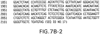
- the amino acid sequence of wild-type human CEA is shown in FIGURE 7E (SEQ ID NO:20).
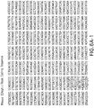
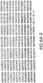
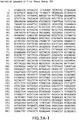
- the amino acid sequence of the wild-type rhesus monkey CEA was previously described ( WO 2004/072287 , see FIGURES 7A-7B ).
- Wild-type CEA gene refers to a gene comprising a sequence of nucleotides that encodes a naturally occurring CEA protein, including proteins of human origin or proteins obtained from another organism, including, but not limited to, other mammals such as rat, mouse and rhesus monkey.
- the nucleotide sequence of the human CEA gene is available in the art ( supra ). See also Beauchemin et al., Mol. Cell. Biol. 7:3221-3230 (1987 ); Zimmerman et al., Proc. Natl. Acad. Sci. USA 84:920-924 (1987 ); and Thompson et al. Proc. Natl. Acad. Sci. USA 84(9):2965-69 (1987 ).
- the nucleotide sequence of the wild-type rhesus monkey gene is shown in FIGURES 7C-7D .
- mamalian refers to any mammal, including a human being.
- Ag refers to an antigen
- Ab and mAb refer to an antibody and a monoclonal antibody, respectively.
- ORF refers to the open reading frame of a gene.
- Carcinoembryonic antigen is commonly associated with the development of adenocarcinomas.
- the present invention relates to compositions and methods to elicit or enhance immunity to the protein product expressed by the CEA tumor-associated antigen, wherein aberrant CEA expression is associated with the carcinoma or its development. Association of aberrant CEA expression with a carcinoma does not require that the CEA protein be expressed in tumor tissue at all timepoints of its development, as abnormal CEA expression may be present at tumor initiation and not be detectable late into tumor progression or vice-versa.
- the present invention provides polynucleotides, vectors, host cells, and encoded proteins comprising a CEA sequence or variant thereof for use in vaccines and pharmaceutical compositions for the treatment and/or prevention of a cancer.
- the polynucleotides of the present invention comprise a nucleotide sequence encoding a C-terminally truncated human CEA protein or variant thereof, fused to a nucleotide sequence encoding at least a subunit of an immunoenhancing element, namely LTB or substantial portion thereof, which can effectively adjuvant an immune response to the associated CEA.
- the CEA nucleotide sequences of the present invention are of human origin.
- the wild-type human CEA nucleotide sequence has been reported ( see , e.g ., U.S. Patent No. 5,274,087 ; U.S. Patent No 5,571,710 ; and U.S. Patent No 5,843,761 ).
- the rhesus monkey CEA sequence was recently described ( WO 2004/072287 ).
- the CEA portion of the CEA fusion may be any variant sufficient to elicit a CEA-specific immune response in a mammal.
- CEA variants of the present invention include, but are not limited to sequences that are N-terminally truncated, sequences with conservative substitutions, and sequences with internal deletions or insertions.
- the present invention relates to a synthetic polynucleotide as defined in claim 1 hereinafter.
- the synthetic polynucleotides of the present invention encode mRNA molecules that express a functional CEA fusion protein so as to be useful in the development of a therapeutic or prophylactic cancer vaccine.
- the CEA portion of the encoded CEA fusion protein is a human CEA deleted of its C-terminal anchoring domain (AD) (SEQ ID NO: 23), which is located from about amino acid 679 to about amino acid 702 of the human full-length CEA. While not being bound by theory, deletion of the anchoring domain increases secretion of the CEA fusion protein, thereby enhancing cross priming of the CEA-LTB immune response.
- AD C-terminal anchoring domain
- the immunoenhancing element portion of the CEA fusion proteins of the present invention is capable of stimulating or enhancing the immune response to the associated CEA protein and is derived from the heat labile enterotoxin of E.coli (LT).
- the adjuvant portion of the CEA fusion comprises the B subunit of LT, or substantial portion thereof.
- CEA fusion comprising a truncated human CEA fused to a single epitope of tetanus toxin (Q830 - L844) has been described ( Lund et al. Cancer Gene Therapy 10: 365-376 (2003 )).
- the CEA fusions of the present invention comprise a substantial portion of an immunoenhancing element or subunit thereof, as described above, which is capable of enhancing the immunogenicity of a CEA protein or variant thereof.
- a substantial portion of an immunoenhancing element to be used for the compositions and methods described herein does not include portions that are less than 50% of a full-length toxin subunit.
- the strategy used herein which utilizes full-length adjuvant subunits or substantial portions thereof, was employed to ensure a greater immune response to the fused CEA sequence. While not being bound by theory, it is believed that if the bacterial toxin chosen as adjuvant comprises greater than one helper epitope, limiting the toxin sequence of the fusion protein to a single epitope would arguably lead to a reduced effect on the immunogenicity of the target protein. Additionally, it is believed that if the adjuvant-mediated enhancement of the immune response is dependent on the interaction of the adjuvant with specific cell receptors and not based on a universal epitope, then the receptor interaction could depend on a specific structural configuration that would require a substantial portion of the immunoenhancing element to exert an adjuvant effect. In such a case, a short adjuvant sequence comprising a single epitope would be insufficient in mediating an increase of the immune response.
- nucleotide sequences encoding variants or mutants of the immunoenhancing elements described herein, including but not necessarily limited to: nucleotide substitutions, deletions, additions, amino-terminal truncations and carboxy-terminal truncations.
- an LT subunit is fused to the CEA sequence of the CEA fusion, wherein the LT subunit is truncated of its signal sequence. While not being bound by theory, deletion of the toxin signal sequence, i.e. the LTB signal sequence, ensures that posttranslational processing of the CEA fusion is driven by the CEA signal sequence.
- the immunoenhancing element, subunit, or substantial portion thereof may be fused to the amino terminus or the carboxy terminus of the CEA sequence. Further, the immunoenhancing element sequence and the CEA sequence can be fused N-terminus to N-terminus, C-terminus to C-terminus, C-terminus to N-terminus or N-terminus to N-terminus. In preferred embodiments of the present invention, the C-terminus of the CEA polypeptide is fused to the N-terminus of the immunoenhancing element.
- the present invention relates to a synthetic nucleic acid molecule (polynucleotide) comprising a sequence of nucleotides which encodes mRNA that expresses a novel CEA fusion protein; for example, nucleotide sequences encoding the fusion proteins as set forth in SEQ ID NOs: 10 and 13.
- the nucleic acid molecules of the present invention are substantially free from other nucleic acids.
- the present invention also relates to recombinant vectors and recombinant host cells, both prokaryotic and eukaryotic, which contain the nucleic acid molecules disclosed throughout this specification.
- the synthetic DNA molecules, associated vectors, and hosts of the present invention are useful for the development of a cancer vaccine.
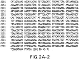
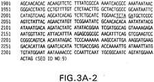
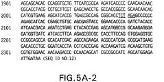
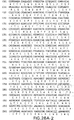
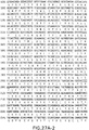
- Exemplary nucleic acid molecules of the present invention comprise a nucleotide sequence selected from the group consisting of SEQ ID NOs: 9, 11 and 12, as shown in FIGUREs 3-5, which encode exemplary CEA-LTB fusion proteins of the present invention.
- the present invention also includes biologically active fragments or mutants of SEQ ID NOs: 9, 11 and 12, which encode mRNA expressing exemplary CEA fusion proteins.
- Any such biologically active fragment and/or mutant will encode either a protein or protein fragment which at least substantially mimics the pharmacological properties of the hCEA protein, including but not limited to the hCEA protein as set forth in SEQ ID NO:20.
- Any such polynucleotide includes but is not necessarily limited to: nucleotide substitutions, deletions, additions, amino-terminal truncations and carboxy-terminal truncations.
- the mutations of the present invention encode mRNA molecules that express a functional CEA fusion protein in a eukaryotic cell so as to be useful in cancer vaccine development.
- mutations in the DNA sequence that do not substantially alter the ultimate physical properties of the expressed protein. For example, substitution of valine for leucine, arginine for lysine, or asparagine for glutamine may not cause a change in the functionality of the polypeptide.
- the present invention further relates to recombinant vectors that comprise the nucleic acid molecules disclosed throughout this specification.
- These vectors may be comprised of DNA or RNA.
- DNA vectors are preferred.
- Typical vectors include plasmids, modified viruses, baculovirus, bacteriophage, cosmids, yeast artificial chromosomes, and other forms of episomal or integrated DNA that can encode a CEA fusion protein. It is well within the purview of the skilled artisan to determine an appropriate vector for a particular gene transfer or other use.
- CEA fusion proteins encoded by the nucleic acids disclosed throughout this specification.
- the CEA fusion protein comprises a sequence of amino acids selected from the group consisting of: SEQ ID NOs: 10 and 13.
- DNA sequences that hybridize to SEQ ID NOs: 9, 11 and 12 under stringent conditions include DNA sequences that hybridize to SEQ ID NOs: 9, 11 and 12 under stringent conditions.
- a procedure using conditions of high stringency is as follows. Prehybridization of filters containing DNA is carried out for about 2 hours to overnight at about 65°C in buffer composed of 6x SSC, 5x Denhardt's solution, and 100 ⁇ g/ml denatured salmon sperm DNA. Filters are hybridized for about 12 to 48 hrs at 65°C in prehybridization mixture containing 100 ⁇ g/ml denatured salmon sperm DNA and 5-20 x 10 6 cpm of 32 P-labeled probe.
- Washing of filters is done at 37°C for about 1 hour in a solution containing 2x SSC, 0.1% SDS. This is followed by a wash in 0.1x SSC, 0.1% SDS at 50°C for 45 minutes before autoradiography.
- Other procedures using conditions of high stringency would include either a hybridization step carried out in 5x SSC, 5x Denhardt's solution, 50% formamide at about 42°C for about 12 to 48 hours or a washing step carried out in 0.2x SSPE, 0.2% SDS at about 65°C for about 30 to 60 minutes. Reagents mentioned in the foregoing procedures for carrying out high stringency hybridization are well known in the art.
- An expression vector containing a CEA fusion protein-encoding nucleic acid molecule may be used for high-level expression of CEA fusion protein in a recombinant host cell.
- Expression vectors may include, but are not limited to, cloning vectors, modified cloning vectors, specifically designed plasmids or viruses.
- a variety of bacterial expression vectors may be used to express recombinant CEA fusion sequences in bacterial cells if desired.
- a variety of fungal cell expression vectors may be used to express recombinant CEA fusion sequences in fungal cells.
- a variety of insect cell expression vectors may be used to express recombinant protein in insect cells.
- the present invention also relates to host cells transformed or transfected with vectors comprising the nucleic acid molecules of the present invention.
- Recombinant host cells may be prokaryotic or eukaryotic, including but not limited to, bacteria such as E. coli, fungal cells such as yeast, mammalian cells including, but not limited to, cell lines of bovine, porcine, monkey and rodent origin; and insect cells including but not limited to Drosophila and silkworm derived cell lines.
- Such recombinant host cells can be cultured under suitable conditions to produce a CEA fusion protein or a biologically equivalent form.
- the host cell is human.
- the term "host cell" is not intended to include a host cell in the body of a human being, human fetus, or human embryo.
- an expression vector containing DNA encoding a CEA fusion protein may be used for expression of CEA fusion protein in a recombinant host cell. Therefore, another aspect of this invention is a process for expressing a CEA fusion protein in a recombinant host cell, comprising: (a) introducing a vector comprising a nucleic acid in accordance with claim 1 hereinafter into a suitable host cell; and, (b) culturing the host cell under conditions which allow expression of said CEA fusion protein.
- the nucleotide sequence of the CEA portion of the fusion and/or the immunoenhancing element portion of the fusion are codon-optimized for high-level expression in human cells.
- the LT subunit is a substantial portion of LTB, wherein the LTB sequence is deleted of its signal sequence.
- CEA fusion protein may be recovered to provide CEA fusion protein in active form.
- Several protein purification procedures are available and suitable for use.
- Recombinant protein may be purified from cell lysates and extracts by various combinations of, or individual application of salt fractionation, ion exchange chromatography, size exclusion chromatography, hydroxylapatite adsorption chromatography and hydrophobic interaction chromatography.
- recombinant CEA fusion protein can be separated from other cellular proteins by use of an immunoaffinity column made with monoclonal or polyclonal antibodies specific for a CEA protein, or polypeptide fragments of a CEA protein.
- the nucleic acid molecules comprising CEA fusions and the encoded fusion proteins of this invention were designed to enhance the CEA-specific immune response, relative to full-length cDNA encoding CEA, for use in vaccine development.
- the polynucleotides encoding CEA fusion proteins comprise optimized codons for further high level expression in a host cell, as described below.
- at least a portion of the codons of the CEA fusions are designed so as to use the codons preferred by the projected host cell, which in preferred embodiments is a human cell.
- the optimized CEA fusions may be used for the development of recombinant adenovirus or plasmid-based DNA vaccines, which provide effective immunoprophylaxis against CEA-associated cancer through neutralizing antibody and cell-mediated immunity.
- the synthetic molecules may be used as an immunogenic composition.
- This invention provides codon-optimized CEA fusion polynucleotides which, when directly introduced into a vertebrate in vivo , including mammals such as primates and humans, induce the expression of encoded proteins within the animal.
- the synthetic molecules comprise a sequence of nucleotides, wherein some of the nucleotides have been altered so as to use the codons preferred by a human cell, thus allowing for high-level fusion protein expression in a human host cell.
- the synthetic molecules may be used as a source of a CEA fusion protein, for example, CEA-LTB fusion protein, which may be used in a cancer vaccine to provide effective immunoprophylaxis against CEA-associated carcinomas through neutralizing antibody and cell-mediated immunity.
- the nucleic acid molecules disclosed herein may also serve as the basis for a DNA-based cancer vaccine.
- a "triplet" codon of four possible nucleotide bases can exist in over 60 variant forms. Because these codons provide the message for only 20 different amino acids (as well as transcription initiation and termination), some amino acids can be coded for by more than one codon, a phenomenon known as codon redundancy. For reasons not completely understood, alternative codons are not uniformly present in the endogenous DNA of differing types of cells. Indeed, there appears to exist a variable natural hierarchy or "preference" for certain codons in certain types of cells. As one example, the amino acid leucine is specified by any of six DNA codons including CTA, CTC, CTG, CTT, TTA, and TTG.
- one aspect of this invention is a CEA fusion gene that is codon-optimized for expression in a human cell.
- the nucleic acid molecules which encode the CEA fusion proteins are converted to a polynucleotide sequence having an identical translated sequence but with alternative codon usage as described by Lathe, "Synthetic Oligonucleotide Probes Deduced from Amino Acid Sequence Data: Theoretical and Practical Considerations" J. Molec. Biol. 183:1-12 (1985 ), which is hereby incorporated by reference.
- the methodology generally consists of identifying codons in the wild-type sequence that are not commonly associated with highly expressed human genes and replacing them with optimal codons for high expression in human cells.
- the new gene sequence is then inspected for undesired sequences generated by these codon replacements (e.g., "ATTTA" sequences, inadvertent creation of intron splice recognition sites, unwanted restriction enzyme sites, etc.). Undesirable sequences are eliminated by substitution of the existing codons with different codons coding for the same amino acid.
- the synthetic gene segments are then tested for improved expression.
- nucleic acid molecules may be constructed that provide for high levels of CEA fusion expression in human cells, wherein only a portion of the codons of the DNA molecules are codon-optimized.
- codons comprising the CEA portion of the CEA fusion are optimized for high-level expression in human cells, and codons comprising the adjuvant portion of the CEA fusion are substantially similar to the wild-type adjuvant-encoding nucleotide sequence.
- codons comprising the adjuvant portion of the CEA fusion are optimized for high-level expression in human cells, and codons comprising the CEA portion of the CEA fusion are substantially similar to a wild-type CEA gene.
- both the CEA and the adjuvant portions of the CEA fusion are codon-optimized for high-level expression in human cells.
- CEA fusions in which only a subset of codons are optimized within the CEA and/or the adjuvant portion of the CEA fusion are also contemplated by this invention.
- the nucleic acids of the present invention may be assembled into an expression cassette which comprises sequences designed to provide for efficient expression of the protein in a human cell.
- the cassette preferably contains CEA fusion protein-encoding gene, with related transcriptional and translations control sequences operatively linked to it, such as a promoter, and termination sequences.
- the promoter is the cytomegalovirus promoter without the intron A sequence (CMV), although those skilled in the art will recognize that any of a number of other known promoters such as the strong immunoglobulin, or other eukaryotic gene promoters may be used.
- a preferred transcriptional terminator is the bovine growth hormone terminator, although other known transcriptional terminators may also be used. The combination of CMV-BGH terminator is particularly preferred.
- the CEA fusion expression cassette is inserted into a vector.
- the vector is preferably an adenoviral or plasmid vector, although linear DNA linked to a promoter, or other vectors, such as adeno-associated virus or a modified vaccinia virus, retroviral or lentiviral vector may also be used.
- the vector chosen is an adenovirus
- the vector be a so-called first-generation adenoviral vector.
- These adenoviral vectors are characterized by having a non-functional E1 gene region, and preferably a deleted adenoviral E1 gene region.
- the expression cassette is inserted in the position where the adenoviral E1 gene is normally located.
- these vectors optionally have a non-functional or deleted E3 region. It is preferred that the adenovirus genome used be deleted of both the E1 and E3 regions ( ⁇ E1 ⁇ E3).
- the adenoviruses can be multiplied in known cell lines which express the viral E1 gene, such as 293 cells, or PERC.6 cells, or in cell lines derived from 293 or PERC.6 cell which are transiently or stablily transformed to express an extra protein.
- cell lines which express the viral E1 gene, such as 293 cells, or PERC.6 cells, or in cell lines derived from 293 or PERC.6 cell which are transiently or stablily transformed to express an extra protein.
- the cell line may express components involved in the regulatory system.
- T-Rex-293 are known in the art.
- the adenovirus may be in a shuttle plasmid form.
- This invention is also directed to a shuttle plasmid vector which comprises a plasmid portion and an adenovirus portion, the adenovirus portion comprising an adenoviral genome which has a deleted E1 and optional E3 deletion, and has an inserted expression cassette comprising a CEA fusion protein encoding nucleotide sequence.
- there is a restriction site flanking the adenoviral portion of the plasmid so that the adenoviral vector can easily be removed.
- the shuttle plasmid may be replicated in prokaryotic cells or eukaryotic cells.
- the expression cassette is inserted into the pMRKAd5-HV0 adenovirus plasmid ( See Emini et al., WO 02/22080 ).
- This plasmid comprises an Ad5 adenoviral genome deleted of the E1 and E3 regions.
- the design of the pMRKAd5-HV0 plasmid was improved over prior adenovectors by extending the 5' cis-acting packaging region further into the E1 gene to incorporate elements found to be important in optimizing viral packaging, resulting in enhanced virus amplification.
- this enhanced adenoviral vector is capable of maintaining genetic stability following high passage propagation.
- Standard techniques of molecular biology for preparing and purifying DNA constructs enable the preparation of the adenoviruses, shuttle plasmids, and DNA immunogens of this invention.
- CEA-LT fusion protein-encoding molecules described herein e.g. SEQ ID NO:12
- SEQ ID NO:12 which comprise a substantial portion of the LTA or LTB subunits of E.coli heat labile enterotoxin
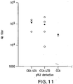
- plasmids pV1J/hCEA-LTA and pV1J/hCEA-LTB elicited a greater antibody response than pV1J/hCEA, confirming the adjuvant effect exerted by the LT subunits on the CEA specific immune response ( See EXAMPLE 11).
- the data described herein demonstrate that fusion of the CEA coding sequence to the LTA or LTB cDNA results in an increase the CEA specific immune response. It appears that LTB exerts a greater enhancing effect on the immune response with a prevalent induction of CD8 + T cells, whereas LTA elicits a predominant CD4 + response.
- the vectors described above may be used in immunogenic compositions and vaccines for preventing the development of adenocarcinomas associated with aberrant CEA expression and/or for treating existing cancers.
- the vectors of the present invention allow for vaccine development and commercialization by eliminating difficulties with obtaining high expression levels of exogenous CEA in successfully transformed host organisms and by providing a CEA fusion protein which can elicit an enhanced immune response when administered to a mammal such as a human being.
- one aspect of the instant invention is a vaccine vector for use in preventing or treating CEA-associated cancer in a mammal, said vaccine vector comprising a polynucleotide according to claim 1 hereinafter.
- the vaccine vector may be administered for the treatment or prevention of a cancer in any mammal, including but not limited to: lung cancer, breast cancer, and colorectal cancer.
- the mammal is a human.
- the vector is an adenovirus vector or a plasmid vector.
- the instant invention further relates to an adenovirus vaccine vector comprising an adenoviral genome with a deletion in the E1 region, and an insert in the E1 region, wherein the insert comprises an expression cassette comprising: (a) a sequence of nucleotides that encodes a CEA fusion protein, wherein the CEA fusion protein comprises a C-terminally truncated human CEA protein or variant thereof, fused to a substantial portion of an immunoenhancing element; wherein the immunoenhancing element is LTB; and wherein the fusion protein is capable of producing an immune response in a mammal; and (b) a promoter operably linked to the polynucleotide.
- the adenovirus vector is an Ad 5 vector.
- the adenovirus vector is an Ad 6 vector.
- the adenovirus vector is an Ad 24 vector.
- adenovirus vaccine vector comprising a adenovirus genome that naturally infects a species other than human, including, but not limited to, chimpanzee adenoviral vectors.
- a preferred embodiment of this aspect of the invention is a chimp Ad 3 vaccine vector.
- the invention in another aspect, relates to a vaccine plasmid comprising a plasmid portion and an expression cassette portion, the expression cassette portion comprising: (a) a sequence of nucleotides that encodes a CEA fusion protein, wherein the CEA fusion protein comprises a C-terminally truncated human CEA protein or variant thereof, fused to an immunoenhancing element which is LTB or substantial portion thereof; and wherein the fusion protein is capable of producing an immune response in a mammal; and (b) a promoter operably linked to the polynucleotide.
- the recombinant adenovirus and plasmid-based polynucleotide vaccines disclosed herein are used in various prime/boost combinations in order to induce an enhanced immune response.
- the two vectors are administered in a "prime and boost" regimen.
- the first type of vector is administered one or more times, then after a predetermined amount of time, for example, 2 weeks, 1 month, 2 months, six months, or other appropriate interval, a second type of vector is administered one or more times.
- the vectors carry expression cassettes encoding the same polynucleotide or combination of polynucleotides.
- the vector contain one or more promoters recognized by mammalian or insect cells.
- the plasmid would contain a strong promoter such as, but not limited to, the CMV promoter.
- the synthetic CEA fusion gene or other gene to be expressed would be linked to such a promoter.
- An example of such a plasmid would be the mammalian expression plasmid V1Jns as described ( J. Shiver et. al. in DNA Vaccines, M. Liu et al. eds., N.Y. Acad. Sci., N.Y., 772:198-208 (1996 )).
- an adenoviral vector vaccine and a plasmid vaccine may be administered to a vertebrate as part of a single therapeutic regime to induce an immune response.
- the first vector is a plasmid and the second vector is an adenovirus vector.
- the first vector is an adenovirus vector and the second vector is a plasmid.
- the first type of vector may be administered more than once, with each administration of the vector separated by a predetermined amount of time.
- Such a series of administration of the first type of vector may be followed by administration of a second type of vector one or more times, after a predetermined amount of time has passed.
- the second type of vector may also be given one time or more than once, following predetermined intervals of time.
- an immunologically or prophylactically effective dose of about 1 ng to 100 mg, and preferably about 10 ⁇ g to 300 ⁇ g of a plasmid vaccine vector is administered directly into muscle tissue.
- An effective dose for recombinant adenovirus is approximately 10 6 - 10 12 particles and, preferably about 10 7 -10 11 particles.
- Subcutaneous injection, intradermal introduction, impression though the skin, and other modes of administration such as intraperitoneal, intravenous, intramuscular or inhalation delivery are also contemplated.
- the vaccine vectors are introduced to the recipient through intramuscular injection.
- the vaccine vectors of this invention may be naked, i.e., unassociated with any proteins, or other agents which impact on the recipient's immune system.
- it is desirable for the vaccine vectors to be in a physiologically acceptable solution such as, but not limited to, sterile saline or sterile buffered saline.
- a physiologically acceptable solution such as, but not limited to, sterile saline or sterile buffered saline.
- an agent which assists in the cellular uptake of DNA such as, but not limited to calcium ion.
- transfection facilitating reagents and pharmaceutically acceptable carriers are generally referred to as transfection facilitating reagents and pharmaceutically acceptable carriers.
- CEA fusion proteins were constructed encoding amino acids (hereinafter aa) 1 to 679 of the human CEA protein fused to a panel of selected polypeptides (see EXAMPLE 2). These sequences were chosen in view of their reported immunoenhancing properties, which have been demonstrated in a variety of experimental systems.
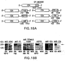
- the CEA fusions were engineered by joining the cDNA of the CEA protein deleted of the GPI anchoring sequence to the foreign polypeptides (exemplary constructs are depicted in FIGURE 18A ).
- the tumor antigen was linked to the HSP70, FcIgG or LAMP sequences to determine whether enhancement of antigen uptake or retargeting to the endosomal compartment would lead to an increased immune response.
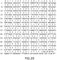
- fusion to fragment C of tetanus toxin (FrC) or to a minimal domain devoid of potentially competitive MHC class I binding epitopes (DOM, see FIGURE 29 ) was constructed to promote humoral and CD4 + T cell responses.
- CEA was also linked to the VSV-G coding sequence to determine whether fusion to a viral glycoprotein would influence the immunogenic properties of CEA.
- CEA-fusions were cloned into vectors pV1Jns under the control of the human CMV/intron A promoter plus the bovine growth hormone (BGH) polyadenylation signal (EXAMPLE 2).
- Plasmids pV1J/CEA-FRC, pV1J/CEA-DOM, pV1J/CEA-FcIgG, pV1J/CEA-LAMP, pV1J/CEA-VSV-G, and pV1J/CEA-HSP70 carry the wild type cDNA of CEA fused to the coding sequences of the indicated foreign polypeptides.
- Exemplary nucleotide and amino acid sequences of hCEA-DOM and hCEA-FcIgG fusions are shown in FIGUREs 26 , 27 , and 30 .
- FIGURE 1 A schematic representation of the structure of exemplary CEA-LTA and CEA-LTB fusions developed for this study are shown in FIGURE 1 .
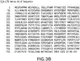
- Exemplary nucleotide and amino acid sequences of CEA-LT fusions are shown in FIGURES 2-6 .
- CEA-LT fusions were engineered by joining the cDNA of the CEA protein deleted of the anchoring sequence to the LT subunits to which the signal peptide coding sequence had been removed.
- the CEA-fusions coding sequences were cloned into vectors pV1Jns under the control of the human cytomegalovirus (CMV)/intron A promoter plus the bovine growth hormone (BGH) polyadenylation signal.
- CMV human cytomegalovirus
- BGH bovine growth hormone
- All constructs carrying the CEA-LTB fusion were generated by fusing the CEA cDNA from nt 1 to 2037, with the LTB cDNA fragment encompassing nt 64 to 375.
- the LTB coding sequence was obtained by PCR amplification of E. coli genomic DNA using sequence specific primers LTB-S1 5'-TATTCTAGATGCTCCCCAGACTATTACAGAA-3' (SEQ ID NO:1) and LTB-A1 5'-TATGCGGCCGCCTAGTTTTCCATACTGATTGCC GC-3' (SEQ ID NO:2).
- the amplified DNA was introduced at the 3' end of the CEA coding sequence generating plasmids.
- pV1J/CEA opt and pV1J/CEA carry the codon usage optimized and wild type cDNA of CEA, respectively.
- the CEA coding sequence is located between the CMV/intronA immediate early promoter of cytomegalovirus and the bovine growth hormone polyadenylation signal.
- plasmid pCR-hCEAopt was digested with Eco RI for 1 hr at 37°C. The resulting 2156 bp insert was purified and cloned into the Eco RI site of plasmid pV1JnsB (( Montgomery et al. DNA Cell Biol 12(9): 777-83 (1993 )).
- plasmid pCI/hCEA ( Song et al. Regulation of T-helper-1 versus T-helper-2 activity and enhancement of tumour immunity by combined DNA-based vaccination and nonviral cytokine gene transfer. Gene Therapy 7: 481-492 (2000 )) was digested with Eco RI. The resulting 2109 bp insert was cloned into the Eco RI site of plasmid pV1JnsA (Montgomery et al., supra ).
- pV1J/hCEA-LTB and pV1J/hCEA opt -LTB The codon optimized cDNA of LTB was synthetized by oligonucleotide assembly (Geneart GmbH, Regensburg, Germany) and cloned in pCR-script vector (Stratagene, LA Jolla, CA).
- LTB opt was amplified by PCR using the following PCR primers: LTBopt-5'XbaI (5' end) 5'-GCTCTAGAG CCCCCCAGAGCATCACCGAGCTGTGC-3' (SEQ ID NO:3) and LTBopt-3'BglII (3'end) 5'-GCTCTAGAACCCCTCAGAACATCACCGATCTGTGCGCC-3' (SEQ ID NO:4).
- the amplified product was then inserted into the Xba I/ Bgl II sites of plasmid pV1J/hCEAopt.
- pV1J/hCEA-LTA The LTA coding sequence corresponding to nt 54 to 774 that encode aa 18 to 259, was amplified by PCR from genomic DNA of E. coli using sequence specific primers LTA-S1 5'-TATTCTAGATAATGGCGACAAATTATACCG-3' (SEQ ID NO:5) and LTA-A1 5'-TATGCGGCCGCTCATAATTCATCCCGAATTCT GTT-3' (SEQ ID NO:6). The amplified DNA was digested with appropriate restriction enzymes and inserted into plasmid pV1J/hCEA.
- pV1J/rhCEAopt-LTB A 3' fragment of the rhesus monkey CEA cDNA (nt 1641 to 2026), which was codon-optimized for high level expression in human cells, was amplified by PCR from pV1J-rhCEAopt. The amplified cDNA lacked the GPI anchor coding sequence and carried the Xba I/ Bgl II restriction sites. This fragment was inserted into the Pst I site of pCR-blunt-rhCEAopt, thus obtaining the intermediate pCR-blunt-rhCEAopt XbaI/BgllII.
- rhCEAopt was extracted as a Bgl II lSal I fragment and cloned in the same sites in pVIJ-nsB, thus obtaining pV1J-rhCEAopt XbaI/BglII.
- LTBopt was amplified by PCR from pCR-script-LTBopt adding Xba I and Bgl II sites at 5' and 3' ends, respectively, and was cloned in pV1J-rhCEAopt XbaI/BglII, thus obtaining pV1J-rhCEAopt-LTBopt .
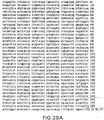
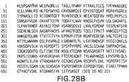
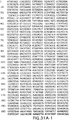
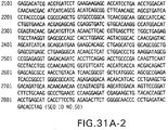
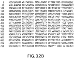
- All the constructs encoding the referenced CEA fusion proteins were generated by fusing the CEA cDNA from nt 1 to nt 2037 (SEQ ID NO:22, FIGURE 28A ), corresponding to aa 1 to aa 679 (SEQ ID NO:23, FIGURE 28B ), with the cDNA fragment corresponding to the following: fragment C of tetanus toxoid (CEA-FrC, SEQ ID NO:24), the N-terminal domain of FrC (CEA-DOM, SEQ ID NOs:21 and 49), the heavy fragment of constant chain of immune globulin G1 (CEA-FcIgG, SEQ ID NO:25),
- FrC and DOM coding sequences were obtained by PCR amplification from pRep-TeT.C plasmid as described in Rice et al. (J. Immunol. 169: 3908-13 (2002 )).
- FcIgG was obtained from total RNA of human PBMC.
- VSV-G and HSP70 were obtained from p-FAST-VSV-G and from plasmid pY3111, respectively.
- LAMP1 was obtained by gene assembly.
- FrC sense (5'-TAT TCTAGA TTCAACACCAATTCC ATTTTCTTATTC-3' (SEQ ID NO:29)
- FrC antisense (5'-GCGGCCGCTAGAATCA TTTGTCCATCCTTCATC-3' (SEQ DJ NO:30)
- DOM sense (5'-TAT TCTAGA TTC AACACCAATTCCATTTTCTTATTC-3' (SEQ ID NO:31)
- DOM antisense (5-TTA GCGGCCGCTAGTTCTGTATCATATCGTAAAGGG-3' (SEQ ID NO:32), FcIgG sense (5'- TCTAGA TAAAACTCACACATGCCCA-3' (SEQ ID NO:33)
- FcIgG antisense (5'-GCCGACTCATTTACCCGGAGACAGGGAG-3' (SEQ ID NO:34), LAMP sense (5- TCTAGA TTTGATCCCCATTGCTGTGGGCGGTGCCCTG-3' (SEQ ID NO:34), L
- the amplified DNA was introduced at the 3' end of the CEA coding sequence generating plasmids pV1J/CEA-FrC, pV1J/CEA-DOM, pV1J/CEA-FcIgG, pV1J/CEA-LAMP, pV1J/CEA-HSP70 and pV1J/CEA-VSV-G.
- pV1J/CEA-DOMopt and pV1J/CEA-FcIgGopt The codon usage optimized cDNA of DOM and FcIgG were synthesized by oligonucleotide assembly (Geneart GmbH, Regensburg, Germany) and cloned in pCR-script vector (Stratagene, La Jolla, CA).
- DOM opt was amplified by PCR using the following primers: Dom opt sense (5'- GTTA T CTAGA AGCACCCCCATCCC -3' (SEQ ID NO:41)) and Dom opt reverse (5'-TTAAG ATCTCTAAGATCTGGTGTCGTATCTCAGGGG-3' (SEQ ID NO:42). The amplified product was then inserted into the Xba I/ Bgl II sites of plasmid pV1J/CEAopt.
- FcIgGopt was amplified by PCR using the following primers: FcIgGopt sense (5'-TTA TCTAGA AAGACCCACACCTGCCCCCCTTGC-3'(SEQ ID NO:43))and as FcIgG opt reverse (5'-TATAGATCTTAGGGTACCTTACTTGCCG G G G -3' (SEQ ID NO:44)) the amplified product was inserted into Xba I/ Bgl II sites of plasmid pV1J/CEAopt.
- FcIgGopt sense 5'-TTA TCTAGA AAGACCCACACCTGCCCCCCTTGC-3'(SEQ ID NO:43)
- FcIgG opt reverse 5'-TATAGATCTTAGGGTACCTTACTTGCCG G G G G -3' (SEQ ID NO:44)
- Ad5/hCEA opt Plasmid pCR-hCEAopt was digested with Eco RI . The resulting 2156 bp insert was purified and cloned into the Eco RI of the polyMRK-Ad5 shuttle plasmid.
- Ad5/CEA The shuttle plasmid pMRK-hCEA for generation of Ad5 vector was obtained by digesting plasmid pDelta1sp1B/hCEA with Ssp I and Eco RV. The 9.52kb fragment was then ligated with a 1272 bp Bgl II/ Bam HI-restricted, Klenow-treated product from plasmid polyMRK. A Pac I/ Stu I fragment from pMRK-hCEA and pMRK-hCEAopt containing the expression cassette for hCEA and E1 flanking Ad5 regions was recombined to Cla I linearized plasmid pAd5 in BJ5183 E . coli cells.
- MRKAd5/hCEA and MRKAd5/hCEAopt were purified through standard CsCl gradient purification and extensively dialyzed against A105 buffer (5mM Tris-Cl pH 8.0, 1mM MgCl 2 , 75 mM NaCl, 5% Sucrose, 0.005 Tween 20).
- Ad5/hCEAopt-LTB Plasmid pMRK-hCEAopt-LTB was constructed by cutting polyMRK-Ad5 shuttle plasmid with SwaI and by ligating the linearized vector with the 2300 bp DNA fragment derived from pV1J/hCEAopt-LTB that had been restricted with Eco RI , Bgl II and treated with Klenow. The pMRK-hCEAopt-LTB was linearized and recombined into the Ad genome as indicated above.
- Ad5/CEA-DOMopt and Ad5/CEA FcIgG opt Plasmid pMRK-CEA-DOMopt and pMRK-CEA-FcIgGopt were constructed by cutting polyMRK-Ad5 shuttle plasmid with Swa I and by ligating the linearized vector with the 2.9 kb DNA fragment derived from pV1J/CEA-DOMopt or ligating the linearized vector with the 2700 bp DNA fragment derived from pV1J/CEA-FcIgGlopt that had been restricted with Eco RI, Bgl II and treated with Klenow.
- pMRK-CEA-FcIgGopt and pMRK-CEA-DOMopt were linearized and recombined into the Ad genome as indicated above.
- an Adenovirus type 5 vector encoding the CEAopt-LTB fusion flanked by the CMV/intron A promoter and the BGH polyadenylation signal was also constructed.
- the molecular mass of the CEA fusion proteins expressed by both plasmid and Ad vectors did not differ from that derived from the corresponding vectors encoding the full length form of CEA cDNA (data not shown).
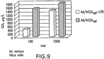
- CEA expression efficiency of these two vectors was comparable at moi 1000 (1790 and 1400 ⁇ g/l, respectively, FIGURE 9 ) whereas at moi 100, vector Ad/hCEAopt-LTB yielded approximately four fold lower amounts of CEA protein detectable in the culture supernatant than Ad/hCEAopt (390 and 1500 ⁇ g/l, respectively).
- CEA expression by plasmid and Ad vectors was monitored by Western blot analysis and ELISA. Plasmids were transfected in HeLa cells with Lipofectamine 2000 (Life Technologies). Adenovirus infections of HeLa cells were performed in serum free medium for 30 min at 37°C, and then fresh medium was added. After 48hr incubation, whole cell lysates were harvested. The CEA protein present in the cell lysates was detected by Western blot analysis using a rabbit polyclonal antiserum. The protein was detected as a 180-200 kDa band. The amount of expressed CEA was detected in the cell lysates using the Direct Elisa CEA Kit (DBC-Diagnostics Biochem Canada Inc).
- fusion proteins in transfected cells was examined by Western blot analysis using antibodies specific for CEA, VSV-G, FcIgG, tetanus toxin, or HSP70.
- HeLa cells were either transfected with the indicated plasmid or infected with the selected Ad vector. After 48 hr incubation, whole cell lysates and culture supernatant were harvested.
- CEA expression in cell lysate or supernatant was also monitored using the Direct Elisa CEA Kit (DBC-Diagnostics Biochem Canada Inc).
- CEA protein was detected with the antibody specific for the fused polypeptide in transfected cell lysates, whereas no expression of the target antigen was observed in the mock transfected control samples ( FIGURE 18B ).
- the molecular mass of the fusion proteins did not differ significantly from that of CEA. This apparent lack of difference in molecular mass between the various CEA polypeptides is probably due to the high degree of glycosylation of the tumor antigen.
- mice Female C57BL/6 mice (H-2 b ) were purchased from Charles River (Lecco, Italy). HLA-A2.1 mice (HHD) were kindly provided by F. Lemmonier (Institute Pasteur, Paris, France). C57BL/DR4 mice were purchased from Taconic (Germantown, NY). CEA.tg mice (H-2 b ) were provided by J. Primus (Vanderbilt University) and kept in standard conditions ( Clarke et al. Cancer Res. 58:1469-77 (1998 )). Fifty micrograms of plasmid DNA were electroinjected in a 50 ⁇ l volume in mice quadriceps as previously described ( Rizzuto et al. Proc. Natl. Acad. Sci. U.S.A. 96(11): 6417-22 (1999 )). Ad injections were carried out in mice quadriceps in 50 ⁇ l volume. Humoral and cell mediated immune response were analyzed at the indicated time.
- mice C57BL/6 mice were subjected to two DNA injections in quadriceps muscle followed by electrical stimulation as previously described (Rizzuto et al. supra). Injections were carried out at three-week intervals.
- CEA transgenic mice were subjected to either 5 weekly injections of plasmid DNA (50 ⁇ g/injection), 2 injections of Ad vectors (1x10 9 viral particles/injection), or 5 weekly injections followed by a boost with Ad. Two weeks after the last injection, humoral and cell mediated immune response were analyzed. Mice were also challenged with a subcutaneous (s.c.) injection of 5x10 5 MC38-CEA cells (Clarke et al., supra ). At weekly intervals, mice were examined for tumor growth.
- Sera for antibody titration were obtained by retro-orbital bleeding.
- ELISA plates Nunc maxisorp
- coating buffer 50mM NaHCO 3 , pH 9.4
- O/N O/N at 4°C as previously described (Facciabene et al., supra ). Plates were then blocked with PBS containing 5% BSA for 1 hr at 37°C.
- Mouse sera were diluted in PBS 5% BSA (dilution 1/50 to evaluate seroconversion rate; dilutions from 1:10 to 1:31,2150 to evaluate titer). Pre-immune sera were used as background.
- Assays were carried out using mouse splenocytes and CEA-specific peptides as previously described (Facciabene et al., supra ).
- Ninety-six wells MAIP plates (Millipore Corp., Billerica, MA) were coated with 100 ⁇ l/ well of purified rat anti-mouse IFN- ⁇ (IgG1, clone R4-6A2, Pharmingen) diluted to 2.5 ⁇ g/ml in sterile PBS. After washing with PBS, blocking of plates was carried out with 200 ⁇ l/well of R10 medium for 2 hrs at 37°C.
- Splenocytes were obtained by removing the spleen from the euthanized mice in a sterile manner and by spleen disruption by grating on a metal grid. Red blood cells were removed by osmotic lysis by adding 1 ml of 0.1X PBS to the cell pellet and vortexing for approximately 15s. One ml of 2x PBS was then added and the volume was brought to 4ml with 1x PBS. Cells were pelleted by centrifugation at 1200 rpm for 10 min at RT, and the pellet was resuspended in 1 ml R10 medium. Viable cells were counted using Mosks staining.
- Splenocytes were plated at 5x10 5 and 2.5x 10⁵ cells/well in duplicate and incubated for 20h at 37°C with 1 ⁇ g/ml suspension of each peptide.
- Concanavalin A (ConA) was used as positive internal control for each mouse at 5 ⁇ g/ml. After washing with PBS, 0.05% Tween 20, plates were incubated O/N at 4°C with 50 ⁇ l/well of biotin-conjugated rat anti-mouse IFN ⁇ (RatIgG1, clone XMG 1.2, PharMingen) diluted to 1:2500 in assay buffer.
- NBTB-CIP Pieris Biotechnology, Inc., Rockford, IL
- mice were immunized with two injections i.m. of 50 ⁇ g each of plasmids pV1J/hCEA, pV1J/hCEA-LTA and pV1J/hCEA-LTB.
- mice were immunized by coinjecting 25 ⁇ g each of plasmids pV1J/hCEA-LTA and pV1J/hCEA-LTB. Immunizations were administered three weeks apart. The plasmid DNA was routinely electroinjected into mouse skeletal muscle in view of the enhanced transduction and immunogenicity connected with this particular procedure ( Zucchelli et al. J. Virol. 74: 11598-11607 (2000 ); Widera et al. J. Immunol. 164: 4635-4640 (2000 )).
- the cellular immunity elicited by the different plasmids was measured by ELISPOT assay 2 weeks after the last injection.
- Antigen-specific IFN ⁇ secretion from stimulated splenocytes was measured using four pools of 15mer peptides overlapping by 11 aa and encompassing the entire CEA glycoprotein. Pool A covers aa 1 to 147, pool B aa 137 to 237, pool C aa 317 to 507, and pool D aa 497 to 703.
- cytokine production was also measured upon stimulation of the splenocytes with DMSO at the same concentration utilized to solubilize the CEA peptides.
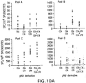
- the immune response elicited by DNA vaccination in C57BL/6 mice was primarily biased towards the C-terminal region of the protein since the SFC values detected with the peptide pool A were slightly above background with all constructs ( FIGURE 10 ).
- the pV1J/hCEA-LTB vaccination regimen was superior to that elicited by pV1J/hCEA as indicated by the higher geometric mean values of the SFC detected with peptide pools B, C and D (pV1J/hCEA-LTB: 482, 1436, and 2054 SFC/10 6 splenocytes, respectively; pV1J/hCEA: 45, 350, and 264 SFC/10 6 splenocytes, respectively).
- plasmid pV1J/hCEA-LTA had also an enhancing effect on the CEA specific immune response when compared to pV1J/hCEA.
- the increase in immune response was only observed with peptide pools C and D (925 and 528 SFC/10 6 splenocytes, respectively), while the immune response measured with peptide pool B was low (15 SFC/10 6 splenocytes).
- IFN ⁇ intracellular staining was carried out on pooled splenocytes from injected mice using peptide pool D.
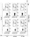
- a CD8 + -specific response was detected in mice injected with pV1J/hCEA-LTB (4.5%) superior to that detected with pV1J/hCEA-LTA and pV1J/hCEA (0.14% and 0.8%, respectively, FIGURE 10B ).
- pV1J/CEA-LTA elicited a strong CD4 + -specific response (1.21%) greater than that observed with pV1J/hCEA-LTB and pV1J/hCEA (0.55% and 0.58%, respectively).
- DNA based immunizations were carried out in BALB/c, C57/DR4 and HLA-A2.1 (HHD) mice.
- the BALB/c mice were chosen in view of their immunocompetence, being a mouse strain extremely reactive to immunization regimens of various sorts.
- the HHD transgenic mice express the human MHC class I genes.
- C57/DR4 transgenic mice carry the human MHC class II genes.
- these two transgenic mouse strains may provide information as to the immunoreactivity of the CEA-LT fusions in the context of human MHC class I and II haplotypes.
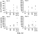
- the CEA specific immune response in BALB/c mice was first assessed by ELISPOT assay. Enhancement of the antigen specific immune response upon immunization with plasmid pV1J/hCEA-LTB was detected with peptide pools A, B, C, D (pV1J/hCEA-LTB: 166, 1353, 796, 899 SFC/10 6 splenocytes, respectively; pV1J/hCEA: 57, 312, 327, 318, SFC/10 6 splenocytes respectively, FIGURE 12 ).
- the N-terminal region of the CEA protein appeared to be the least immunogenic as compared to other sections of the tumor antigen pV1J/hCEA-LTA immunization also yielded an increase in the antigen specific immune response as compared to pV1J/hCEA.
- the increase in the immune response was detected with peptide pools B, C and D (936, 727, and 650 SFC/10 6 splenocytes, respectively).
- the CEA specific immune response in C57/DR4 mice was considerably enhanced by the immunization with pV1J/hCEA-LTB, and was detected only peptide pool D ( FIGURE 13 ).
- IFN ⁇ intracellular staining performed on pooled PBMC from injected mice showed that the CD8 + response to CEA was highest in mice immunized with pV1J/hCEA-LTB (15.32%), whereas was very weak in the pV1J/hCEA treated group (0.5%).
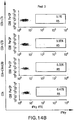
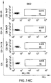
- the immune response elicited by the different CEA encoding plasmids was assessed in HHD mice by performing IFN ⁇ intracellular staining on pooled PBMC.
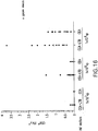
- the immune response was only detected with peptide pools B and D, and as shown in FIGURE 14 , immunization with pV1J/hCEA-LTB resulted in more than 10 fold increase in the CD8 + response to the target antigens.
- no increase in the immune response was detected using pV1J/hCEA-LTA either alone or upon coinjection with pV1J/hCEA-LTB.
- No CD4 + T cell response was detected in the immunized mice (data not shown).
- hCEA transgenic mice were immunized with vectors carrying either the fully codon optimized cDNA of hCEA or CEA-LTB. These transgenic mice carry the entire human CEA gene and flanking sequences and express the hCEA protein in the intestine, mainly in the cecum and colon. Thus, this mouse line is a useful model for studying the safety and efficacy of immunotherapy strategies directed against this tumour self antigen ( Clarke et al., Cancer Research 58: 1469-1477 (1998 )).
- Immunization with pV1J/hCEA-LTBopt resulted in a significant increase in the CEA specific immune response measured by IFN ⁇ intracellular staining on PBMC of the injected mice ( FIGURE 15A ).
- the enhancement of the T cell response was detected with peptide pool D and was predominantly CD8 + .
- the CEA specific humoral response was increased in the CEA-LTB treated mice as shown by the 47 fold increase in the geometric mean values of the Ab titer as compared to the pV1J/hCEAopt treated group ( FIGURE 15B ).
- mice were immunized with Ad5/hCEAopt-LTB and Ad/hCEAopt at a dose of 1x10 7 , 1x10 8 , and 1x10 9 vp. Mice were subjected to two injections two weeks apart and the immune response was measured by IFN ⁇ intracellular staining on PBMC two weeks after the last injection. The immune response was assessed using the peptide pool D.
- Ad/hCEAopt-LTB was more immunogenic than Ad/hCEAopt since significant immune responses to CEA could be detected with the 1x10 8 vp dose, whereas 1x10 9 vp of Ad/hCEAopt were necessary to break tolerance to the target antigen ( FIGURE 16 ). No CD4 + response could be detected in any of the immunized mice (data not shown).
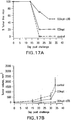
- CEA tg mice Two weeks after the last immunization, the CEA tg mice were challenged with a subcutaneous injection of 5x10 5 MC38-CEA tumor cells. This syngenic cell line was derived from a chemically induced colon cancer and expresses CEA. Tumor development in mock treated mice was detected by 22 days post challenge as all the treated mice were no longer tumor free ( FIGURE 17A ). Additionally, there was a concomitant increase in the average size of the tumor mass that reached significant volume by 34 days post challenge. Mice vaccinated with vectors encoding pV1J/hCEAopt showed a partial resistance to tumor development since 2 out of 10 treated mice remained tumor free at day 34 post challenge. The average size of the tumors of this group was smaller than that observed in the mock treated mice.
- CEA-DOM and CEA-FcIgG fusions enhance the immunogenicity of the CEA protein.
- mice were immunized with two i.m. injections of 50 or 5 ⁇ g of each plasmid. The immunizations were three weeks apart.
- electroporation Zucchelli et al. J. Virology 74:11598 (2000 ), Widera et al., J. Immunol. 164: 4635 (2000 )
- plasmid DNAs were routinely electroporated (DNA-EP) into mouse skeletal muscle.
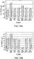
- the immune response elicited by different plasmids was measured by IFN ⁇ ELISPOT assay, 2 weeks after the last injection.
- Antigen-specific IFN ⁇ secretion from stimulated splenocytes was measured using a pool of 15mer peptides overlapping by 11 aa and encompassing the C-terminal region of CEA (pool D, aa 497-703) (Zucchelli et al., supra ).
- the analysis of the immune response to CEA was carried out with peptide pool D since the cellular immune response to CEA in C57BL/6 mice is primarily biased towards the C-terminal region of this protein (Zuccelli et al., supra ).
- As a negative control cytokine production was also measured upon stimulation of the splenocytes with DMSO at the same concentration utilized to solubilize the CEA peptides.
- Plasmids pV1J/CEA-DOM and pV1J/CEA-FcIgG had similar immunogenic properties and exerted a 3- to 4-fold increase in CEA-specific immune responses upon injection of 5 or 50 ⁇ g of plasmid DNA (pV 1J/CEA-DOM: 590 and 1098 SFC/10 6 splenocytes, pV1J/CEA-FcIgG: 510 and 1160, pV1J/CEA: 146 and 264 SFC/10 6 splenocytes, respectively).
- FIGURE 20B To determine the effect of the CEA-fusions on the humoral response to CEA, sera from immunized mice were tested in ELISA using purified CEA protein as substrate ( FIGURE 20B ). An increase in CEA-specific antibody titer was observed upon injection of 50 ⁇ g of plasmids pV1J/CEA-DOM, pV1J/CEA-FcIgG, pV1J/CEA-FrC and pV1J/CEA-HSP70. On the contrary, injection of pV1J/CEA-LAMP and pV1J/CEA-VSV-G resulted in a CEA-specific antibody response similar to that observed upon immunization with pV1J/CEA. Taken together, these data demonstrate that fusion of the CEA coding sequence to the DOM or FcIgG cDNA results in an increase in the CEA-specific cell mediated and humoral immune response.
- CEA-DOM and CEA-FcIgG fusions break tolerance to target antigen in CEA transgenic mice.
- CEA transgenic mice were utilized to perform comparative immunization studies. These transgenic mice carry the entire human CEA gene and flanking sequences and express the CEA protein in the intestine, mainly in the cecum and colon. This mouse line is a useful model for studying the safety and efficacy of immunotherapy strategies directed against this tumor self antigen (Clarke et al., supra ) .
- both plasmid and Adenovirus vectors were engineered to carry the cDNAopt of the CEA-DOM (CEA-DOMopt) or CEA-FcIgG (CEA-FcIgGopt) fusions.
- CEA-DOMopt CEA-DOMopt
- CEA-FcIgGopt CEA-FcIgGopt
- CEA transgenic mice were immunized with the following varying regimens: i) 5 injections at weekly intervals of 50 ⁇ g of plasmid DNA (DNA/DNA), ii) 2 biweekly injections of Adenovirus in doses ranging from 1x10 7 to 1x10 9 viral particles (vp) of Adenovirus (Ad/Ad), or iii) 5 weekly injections of plasmid DNA followed by a final injection of 1x10 9 vp of Adenovirus (DNA/Ad). Immune responses were analyzed by intracellular IFNystaining on PBMC or splenocytes of each immunized mouse using pool D peptides. Additionally, the induction of CEA-specific antibodies was monitored by ELISA.
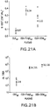
- CEA-specific antibody titer was detected in all mice immunized with pV1J/CEA-DOMopt and pV1J/CEA-FcIgGopt and the average of the antibody titer was 56,136 and 24,725, respectively.
- the pV1J/CEAopt immunized group showed an at least a 77 fold lower average titer of CEA-specific antibody (318) ( FIGURE 21B ).
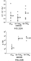
- CEA transgenic mice treated with the Ad/Ad vaccination regimen also showed a better efficiency in breaking tolerance to CEA upon vaccination with CEA-DOMopt and CEA-FcIgGopt Ad vectors than with Ad-CEAopt.
- a CEA-specific CD8 + T cell response could be observed in the vaccinated mice upon injection of a little as 10 7 vp of Ad-CEA-DOM or Ad-CEA-FcIgG, the CEA-specific response was comparable between the two antigens, and increased upon injection of 109 vp (1.55% and 1.15%, respectively).
- 109 vp of Ad-CEAopt were necessary to elicit significant CD8 + T-cell precursor frequencies (2.1%) ( FIGURE 21C ).
- CEA-specific antibodies were detected in all mice immunized with Ad-CEA-DOMopt and Ad-CEA-FcIgGopt.
- the averages of the antibody titer were 19,600 and 33,000, respectively.
- Injection of Ad-CEAopt resulted in a measurable CEA-specific response in only 2 of the treated mice, and the antibody titer was significantly lower (Zucchelli et al., supra ) ( FIGURE 21D ).
- the DNA/Ad immunization showed reduced differences in the CD8 + T cell precursor frequencies elicited by CEA, CEA-DOM and CEA-FcIgG vectors ( FIGURE 22A ).
- averages of CEA-specific antibody titers were greater upon vaccination with vectors expressing CEA-DOM and CEA-FcIgG than CEA (31,200, 26,120 and 412, respectively) ( FIGURE 22B ).
- CEA-DOM and CEA-FcIgG fusion proteins can break tolerance to CEA in transgenic mice with greater efficacy than the CEA protein.
- the enhanced immunogenic properties of these fusion proteins can be observed upon immunization with DNA or Ad vectors.
- the greater ability of these two fusion proteins in eliciting CD8 + T cells to CEA can be overcome, at least in part, by DNA/Ad vaccination regimen.
- Immunized animals were depleted of CD4 + T cells, CD8 + T cells, NK cells, by i.p. injection of anti-CD4 (GK1.5 hybridoma), anti-CD8 (Lyt 2.2 hybridoma), or anti Asialo GM1 (Wako Chemicals, Richmond, VA) as described ( Perricone et al., J. Immunother. 27(4):273-81 (2004 ); Yoon et al., J. Ethnopharmacol. 93 (2-3):247-53 (2004 )).
- Antibodies 100 ⁇ l diluted ascitic fluid/dose were injected on day - 7 relative to the tumor challenge and then injected every week for 3 weeks after injection of 5x10 5 MC38-CEA cells. Depletion conditions were validated by flow cytometry analysis of peripheral blood using phycoerythrin-conjugated MAbs anti-CD4, and-CD8, and anti-NK (PharMingen, San Diego, CA); 99% of the relevant cell subset was depleted, whereas all other subsets remained within normal levels.
- CEA-DOM or CEA-FcIgG fusions were subjected to the DNA/DNA, Ad/Ad or DNA/Ad immunization regimens with the CEA-DOM, CEA-FcIgG, or CEA vectors.
- the CEA transgenic mice were challenged with a subcutaneous injection of 5x10 5 MC38-CEA cells, a syngenic tumor cell line that expresses CEA (Clarke et al., supra).
- mice were depleted of CD4 + , CD8 + T cells, or NK cells by MAbs. Antibodies were given during the course of tumor challenge to ensure continued depletion of the relevant NK and T cell subsets. The depletion of all three cell types was monitored by flow cytometry analysis using antibodies specific for cell surface markers (data hot shown).
- results were analyzed by the log rank or two tailed Student t test. A p value ⁇ 0.05 was considered significant.
Landscapes
- Health & Medical Sciences (AREA)
- Life Sciences & Earth Sciences (AREA)
- Chemical & Material Sciences (AREA)
- Organic Chemistry (AREA)
- Immunology (AREA)
- Genetics & Genomics (AREA)
- General Health & Medical Sciences (AREA)
- Medicinal Chemistry (AREA)
- Molecular Biology (AREA)
- Zoology (AREA)
- Engineering & Computer Science (AREA)
- Biochemistry (AREA)
- Biophysics (AREA)
- Gastroenterology & Hepatology (AREA)
- Proteomics, Peptides & Aminoacids (AREA)
- Toxicology (AREA)
- Bioinformatics & Cheminformatics (AREA)
- Cell Biology (AREA)
- Biomedical Technology (AREA)
- Pharmacology & Pharmacy (AREA)
- Veterinary Medicine (AREA)
- Public Health (AREA)
- Animal Behavior & Ethology (AREA)
- Nuclear Medicine, Radiotherapy & Molecular Imaging (AREA)
- General Chemical & Material Sciences (AREA)
- Chemical Kinetics & Catalysis (AREA)
- Wood Science & Technology (AREA)
- Microbiology (AREA)
- General Engineering & Computer Science (AREA)
- Biotechnology (AREA)
- Physics & Mathematics (AREA)
- Epidemiology (AREA)
- Mycology (AREA)
- Plant Pathology (AREA)
- Oncology (AREA)
- Peptides Or Proteins (AREA)
- Medicines Containing Antibodies Or Antigens For Use As Internal Diagnostic Agents (AREA)
- Micro-Organisms Or Cultivation Processes Thereof (AREA)
- Medicines That Contain Protein Lipid Enzymes And Other Medicines (AREA)
- Medicines Containing Material From Animals Or Micro-Organisms (AREA)
Abstract
Description
- The present invention relates generally to the therapy of cancer. More specifically, the present invention relates to polynucleotides encoding fusion proteins wherein the fusion proteins comprise at least a portion of the tumor associated polypeptide carcinoembryonic antigen. The present invention also provides recombinant vectors and hosts comprising said polynucleotides, purified fusion proteins and methods for enhancing an immune response against CEA using the compositions and molecules disclosed herein.
- The immunoglobulin superfamily (IgSF) consists of numerous genes that code for proteins with diverse functions, one of which is intercellular adhesion. IgSF proteins contain at least one Ig-related domain that is important for maintaining proper intermolecular binding interactions. Because such interactions are necessary to the diverse biological functions of the IgSF members, disruption or aberrant expression of many IgSF adhesion molecules has been correlated with many human diseases.
- The carcinoembryonic antigen (CEA) belongs to a subfamily of the Ig superfamily consisting of cell surface glycoproteins known as CEA-related cell adhesion molecules (CEACAMs). CEACAMs have been shown to act as both homotypic and heterotypic intercellular adhesion molecules (Benchimol et al., Cell 57: 327-334 (1989)). In addition to cell adhesion, CEA (also known as CEACAM5) inhibits cell death resulting from detachment of cells from the extracellular matrix and can contribute to cellular transformation associated with certain proto-oncogenes such as Bcl2 and C-Myc (see Berinstein, J. Clin Oncol. 20(8): 2197-2207 (2002)). Sequences coding for human CEA have been cloned and characterized (
U.S. Patent No. 5,274,087 ;U.S. Patent No 5,571,710 ; andU.S. Patent No 5,843,761 . See also Beauchemin et al., Mol. Cell. Biol. 7:3221-3230 (1987); Zimmerman et al., Proc. Natl. Acad. Sci. USA 84:920-924 (1987); Thompson et al. Proc. Natl. Acad. Sci. USA 84(9):2965-69 (1987)). - Normal expression of CEA has been detected during fetal development and in adult colonic mucosa. CEA overexpression was first detected in human colon tumors over thirty years ago (Gold and Freedman, J. Exp. Med. 121:439-462 (1965)) and has since been found in nearly all colorectal tumors. Additionally, CEA overexpression is detectable in a high percentage of adenocarcinomas of the pancreas, liver, breast, ovary, cervix, and lung. Because of its prevalence in these tumor types and limited normal tissue expression, CEA is considered a self tumor-associated antigen and a target for active and passive immunotherapy. Recent clinical data have established that different vaccine strategies can generate human B and T cells specific for CEA, providing additional evidence that CEA is a target for molecular and immunological intervention for treatment of these cancer types.
- Therapeutic approaches targeting CEA include the use of anti-CEA antibodies (see Chester et al., Cancer Chemother. Pharmacol. 46 (Suppl): S8-S12 (2000)), as well as CEA-based vaccines (for review, see Berinstein, supra). The development and commercialization of many vaccines have been hindered by difficulties associated with obtaining high expression levels of exogenous genes. Success of DNA-based vaccines has also been hindered by an inability to generate an immune response of sufficient magnitude in treated individuals. Although DNA vaccines targeting various proteins have been developed, the resulting immune responses have been relatively weak compared with conventional vaccines.
- The ease of DNA manipulation has offered an opportunity to develop vaccines incorporating gene fusion strategies in which antigens are linked to various immunoenhancing elements. Enhancement of immune response to target antigens has been demonstrated in animal models by vectors encoding antigens fused to heat shock protein (HSP) 70 (Liu et al., J. Virol. 74: 2888-94 (2000); Cheng et al. J. Immunol. 166: 6218-26 (2001); Chen et al., Cancer Res. 60: 1035-42 (2000)), to Fc portion of IgG1 (You et al., J. Immunol. 165: 4581-92 (2000)), to lysosome-associated membrane protein (LAMP) (Su et al., Cancer Res. 62: 5041-48 (2002)), and universal Th epitope from tetanus toxin (Renard et al., J. Immunol. 171:1588-95 (2003); King et al., Nature Med. 4: 1281-86 (1998); Lund et al., Cancer Gene Ther. 10: 365-76 (2003); Padua et al., Nature Med. 9(11): 1413-17 (2003); Savelyeva et al., Nature Biotechnol. 19: 760-64 (2001);
Wahren et al., WO 2004/092216 ). The enhancement of immune responses to target antigens is particularly relevant for cancer vaccines in view of the limited immunogenicity of tumor antigens and of the need to overcome tolerance to exert effective antitumor effects. - Therefore, despite the identification of the wild-type nucleotide sequences encoding CEA proteins described above, it would be highly desirable to develop a vaccine which is capable of eliciting an enhanced CEA-specific immune response relative to a wild-type full-length CEA cDNA, when delivered to a mammal. It would also be desirable to develop methods for treating or preventing CEA-associated cancers which utilize nucleic acid molecules or proteins that safely and effectively potentiate a CEA-specific immune response.
- The present invention provides polynucleotides encoding fusion proteins wherein the fusion proteins comprise at least a portion of the tumor associated polypeptide carcinoembryonic antigen, fused to a substantial portion of an immunoenhancing element. The CEA portion of the encoded CEA fusion protein is deleted of its C-terminal anchoring domain, i.e. amino acids 679-702 of SEQ ID NO:20, and the immunoenhancing element is the B subunit of the heat labile enterotoxin of E.coli, optionally truncated of its signal sequence. The present invention also provides recombinant vectors, including but not limited to, adenovirus and plasmid vectors, comprising said polynucleotides and host cells comprising said recombinant vectors. Also provided herein are purified fusion proteins encoded by invention polynucleotides.
- The present invention further provides compositions for use in methods for inhibiting or preventing the development of a cancer in a mammal by eliciting an immune response to the CEA protein by administering a vaccine or pharmaceutical composition comprising the CEA fusions or CEA fusion proteins described herein. In preferred embodiments of the methods herein, the immune response is enhanced relative to the response elicited by a wild-type CEA vaccine.
- As used throughout the specification and in the appended claims, the singular forms "a," "an," and "the" include the plural reference unless the context clearly dictates otherwise.
- As used throughout the specification and appended claims, the following definitions and abbreviations apply:
- The term "promoter" refers to a recognition site on a DNA strand to which the RNA polymerase binds. The promoter forms an initiation complex with RNA polymerase to initiate and drive transcriptional activity. The complex can be modified by activating sequences termed "enhancers" or inhibiting sequences termed "silencers".
- The term "cassette" refers to a nucleotide or gene sequence that is to be expressed from a vector, for example, the nucleotide or gene sequence encoding the hCEA-LTB fusion. In general, a cassette comprises a gene sequence that can be inserted into a vector, which in some embodiments, provides regulatory sequences for expressing the nucleotide or gene sequence. In other embodiments, the nucleotide or gene sequence provides the regulatory sequences for its expression. In further embodiments, the vector provides some regulatory sequences and the nucleotide or gene sequence provides other regulatory sequences. For example, the vector can provide a promoter for transcribing the nucleotide or gene sequence and the nucleotide or gene sequence provides a transcription termination sequence. The regulatory sequences that can be provided by the vector include, but are not limited to, enhancers, transcription termination sequences, splice acceptor and donor sequences, introns, ribosome binding sequences, and poly(A) addition sequences.
- The term "vector" refers to some means by which DNA fragments can be introduced into a host organism or host tissue. There are various types of vectors including plasmid, virus (including adenovirus), bacteriophages and cosmids.
- The term "first generation," as used in reference to adenoviral vectors, describes adenoviral vectors that are replication-defective. First generation adenovirus vectors typically have a deleted or inactivated E1 gene region, and preferably have a deleted or inactivated E3 gene region.
- The abbreviation "DOM" refers generally to the N-terminal domain of fragment C of tetanus toxoid.
- The abbreviation "LT" refers generally to the heat labile enterotoxin of E. coli. "LT" may refer to the complete enterotoxin, comprising subunits A and B or a substantial portion of subunit A, or a substantial portion of subunit B. The abbreviation "LTA" refers to the A subunit of the heat labile enterotoxin of E. coli, or substantial portion thereof, including subunits which are truncated on the C-terminal or N-terminal end but maintain biological activity, as well as subunits that contain internal amino acid insertions, deletions, or substitutions but maintain biological activity. The abbreviation "LTB" refers to the B subunit of the heat labile enterotoxin of E. coli, or substantial portion thereof, including subunits which are truncated on the C-terminal or N-terminal end but maintain biological activity, as well as subunits that contain internal amino acid insertions, deletions, or substitutions but maintain biological activity.
- The designation "pV1J/hCEAopt" refers to a plasmid construct, disclosed herein, comprising the CMV immediate-early (IE) promoter with intron A, a full-length cordon-optimized human CEA gene, bovine growth hormone-derived polyadenylation and transcriptional termination sequences, and a minimal pUC backbone (see EXAMPLE 2). The designation "pV1J/hCEA" refers to a construct essentially as described above, except the construct comprises a wild-type human CEA gene instead of a codon-optimized human CEA gene.
- The designation "pV1J/hCEA-LTB" refers to a plasmid construct, disclosed herein, comprising the CMV immediate-early (IE) promoter with intron A, a human CEA gene devoid of its GPI anchor coding sequence, fused at its C-terminal end to the B subunit of E. coli heat labile enterotoxin, bovine growth hormone-derived polyadenylation and transcriptional termination sequences, and a minimal pUC backbone.
- The designation "pV1J/hCEAopt-LTB" refers to a construct essentially as described immediately above, except the construct comprises a codon-optimized human CEA gene devoid of its GPI anchor coding sequence instead of the corresponding portion of the wild-type human CEA gene.
- The designation "pV1J/hCEAopt-LTBopt" refers to a plasmid construct essentially as described immediately above, except that both the CEA sequences and the LTB sequences are codon-optimized for high level expression in human cells.
- The designation "pV1J/rhCEAopt-LTBopt" refers to a construct essentially as described above except that the human codon-optimized CEA gene is replaced with a rhesus monkey CEA gene, codon-optimized for high-level expression in human cells.
- The designation "pV1J/hCEA-LTA" refers to a plasmid construct, disclosed herein, comprising the CMV immediate-early (IE) promoter with intron A, a human CEA gene devoid of the GPI anchor coding sequence, fused at its C-terminal end to the A subunit of E. coli heat labile enterotoxin, bovine growth hormone-derived polyadenylation and transcriptional termination sequences, and a minimal pUC backbone. Construction of plasmid vectors comprising various CEA-LT fusions is described in EXAMPLE 2.
- The designation "pV1J/hCEA-DOM" refers to a plasmid construct, disclosed herein, comprising the CMV immediate-early (IE) promoter with intron A, a human CEA gene devoid of its GPI anchor coding sequence, fused at its C-terminal end to the N-terminal domain of Fragment C of tetanus toxoid (DOM), bovine growth hormone-derived polyadenylation and transcriptional termination sequences, and a minimal pUC backbone (EXAMPLE 2).
- The designation "pV1J/rhCEAopt-DOMopt" refers to a construct essentially as described above except that the human codon-optimized CEA gene is replaced with a rhesus monkey CEA gene, codon-optimized for high-level expression in human cells.
- The designation "pV1J/hCEA-FcIgG" refers to a plasmid construct, disclosed herein, comprising the CMV immediate-early (IE) promoter with intron A, a human CEA gene devoid of the GPI anchor coding sequence, fused at its C-terminal end to the heavy fragment of constant chain of immunoglobulin G1, bovine growth hormone-derived polyadenylation and transcriptional termination sequences, and a minimal pUC backbone. (EXAMPLE 2). pV1J/hCEAopt-FcIgGopt refers to a construct essentially as described, except the nucleotide sequences encoding CEA and FcIgG have been codon-optimized for high-level expression in human cells.
- The designations "Ad5/hCEAopt" and "Ad5/hCEA" refer to two constructs, disclosed herein, which comprise an Ad5 adenoviral genome deleted of the E1 and E3 regions. In the "Ad5/hCEAopt" construct, the E1 region is replaced by a codon-optimized human CEA gene in an E1 parallel orientation under the control of a human CMV promoter without intron A, followed by a bovine growth hormone polyadenylation signal. The "Ad5/hCEA" construct is essentially as described above, except the E1 region of the Ad5 genome is replaced with a wild-type human CEA sequence. The designation "Ad5/hCEAopt-LTB" refers to an Ad5 construct, essentially as described above, except that the codon-optimized human CEA sequence is devoid of the GPI anchor coding sequence and is fused at its C-terminus to the B subunit of E. coli heat labile enterotoxin. Construction of adenovirus vectors comprising various CEA-LT fusions is described in EXAMPLE 3.
- "Immunoenhancing element" refers to a portion of the CEA fusion proteins of the present invention which is capable of stimulating or enhancing the immune response to the associated CEA protein, relative to full-length wild-type CEA. Immunoenhancing elements of the present invention are selected from the group consisting of: heat shock protein (HSP) 70, lysosome-associated membrane protein (LAMP), fragment C of tetanus toxoid (FrC), the N-terminal domain of FrC (DOM), the heavy fragment of constant chain of immune globulin G1 (FcIgG), the vesicular stomatitis virus glycoprotein (VSV-G), cholera toxin (CT) from Vibrio cholerae, and heat labile enterotoxin of E.coli (LT). The term "immunoenhancing element" is used interchangeably herein with the term "adjuvant."
- As used herein, a "fusion protein" refers to a protein having at least two polypeptides covalently linked in which one polypeptide comes from one protein sequence or domain and the other polypeptide comes from a second protein sequence or domain. The fusion proteins of the present invention comprise a CEA polypeptide or fragment or variant thereof, and a second polypeptide, which comprises a substantial portion of an immunoenhancing element, which, in some cases, is a bacterial toxin. The CEA polypeptide, fragment or variant thereof may be a human CEA or CEA homolog from another species. The polypeptides that comprise the fusion protein are preferably linked N-terminus to C-terminus. The CEA polypeptide and the toxin subunit can be fused in any order. In some embodiments of this invention, the C-terminus of the CEA polypeptide is fused to the N-terminus of the toxin subunit, as exemplified in FIGURE 1A. However, fusion proteins in which the immunoenhancing element is fused to the N-terminus of the CEA polypeptide are also contemplated. The term "CEA fusion protein" is intended to be a general term which refers to a fusion as described above, which comprises a CEA polypeptide or fragment or variant thereof fused to a polypeptide comprising an immunoenhancing element.
- The term "CEA-LT fusion" refers to a nucleic acid sequence in which at least a portion of the CEA gene is fused to a substantial portion of either the LTA or the LTB subunit of E. coli heat labile enterotoxin. The term "CEA-LT fusion protein" refers to a polypeptide encoded by a CEA-LT fusion as described. The terms "CEA-LT fusion" and "CEA-LT fusion protein" are also understood to refer to fragments thereof, homologs thereof, and functional equivalents thereof (collectively referred to as "variants"), such as those in which one or more amino acids is inserted, deleted or replaced by other amino acid(s). The CEA-LT fusions of the present invention, upon administration to a mammal such as a human being, can stimulate an immune response by helper T cells or cytotoxic T cells, or stimulate the production of antibodies at least as well as a "wild-type" CEA sequence. In preferred embodiments of the invention, the CEA-LT fusion can enhance the immune response as compared to a wild-type CEA.
- The term "CEA-DOM fusion" refers to a nucleic acid sequence in which at least a portion of the CEA gene is fused to a substantial portion of the minimized domain of tetanus toxin fragment C, unless the context clearly dictates that said term refers to the protein sequence. The term "CEA-DOM fusion protein" refers to a polypeptide encoded by a CEA-DOM fusion as described. The terms "CEA-DOM fusion" and "CEA-DOM fusion protein" are also understood to refer to fragments thereof, homologs thereof, and functional equivalents thereof (collectively referred to as "variants"), such as those in which one or more amino acids is inserted, deleted or replaced by other amino acid(s). The CEA-DOM fusions of the present invention, upon administration to a mammal such as a human being, can stimulate an immune response by helper T cells or cytotoxic T cells, or stimulate the production of antibodies at least as well as a "wild-type" CEA sequence. In preferred embodiments of the invention, the CEA-DOM fusion can enhance the immune response as compared to a wild-type CEA.
- The abbreviation "AD" refers to the anchoring domain of a CEA gene or protein. The anchoring domain of the wild-type human CEA is located from about amino acid 679 to about amino acid 702 of SEQ ID NO:20.
- The term "treatment" refers to both therapeutic treatment and prophylactic or preventative measures. Those in need of treatment include those already with the disorder as well as those prone to have the disorder or those in which the disorder is to be prevented.
- A "disorder" is any condition that would benefit from treatment with the molecules of the present invention, including the nucleic acid molecules described herein and the fusion proteins that are encoded by said nucleic acid molecules. Encompassed by the term "disorder" are chronic and acute disorders or diseases including those pathological conditions which predispose the mammal to the disorder in question. The molecules of the present invention are intended for use as treatments for disorders or conditions characterized by aberrant cell proliferation, including, but not limited to, breast cancer, colorectal cancer, and lung cancer.
- The term "effective amount" means sufficient vaccine composition is introduced to produce the adequate levels of the polypeptide, so that an immune response results. One skilled in the art recognizes that this level may vary.
- A "conservative amino acid substitution" refers to the replacement of one amino acid residue by another, chemically similar, amino acid residue. Examples of such conservative substitutions are: substitution of one hydrophobic residue (isoleucine, leucine, valine, or methionine) for another; substitution of one polar residue for another polar residue of the same charge (e.g., arginine for lysine; glutamic acid for aspartic acid).
- "hCEA" and "hCEAopt" refer to a human carcinoembryonic antigen and a human codon-optimized carcinoembryonic antigen, respectively.
- "rhCEA" and "rhCEAopt" refer to a rhesus monkey carcinoembryonic antigen and a rhesus monkey codon-optimized carcinoembryonic antigen, respectively.
- "Substantially similar" means that a given nucleic acid or amino acid sequence shares at least 75%, preferably 85%, more preferably 90%, and even more preferably 95% identity with a reference sequence. In the present invention, the reference sequence can be relevant portions of the wild-type human CEA nucleotide or amino acid sequence, or the wild-type nucleotide or amino acid sequence of a bacterial toxin or subunit thereof, such as the LTB or LTA subunits of the E. coli heat labile enterotoxin, as dictated by the context of the text. The reference sequence may also be, for example, the wild-type rhesus monkey CEA sequence. Thus, a CEA protein sequence that is "substantially similar" to the wild-type human CEA protein or fragment thereof will share at least 75% identity with the relevant fragment of the wild-type human CEA, along the length of the fragment, preferably 85% identity, more preferably 90% identity and even more preferably 95% identity. Whether a given CEA, LTB, or LTA protein or nucleotide sequence is "substantially similar" to a reference sequence can be determined for example, by comparing sequence information using sequence analysis software such as the GAP computer program, version 6.0, available from the University of Wisconsin Genetics Computer Group (UWGCG). The GAP program utilizes the alignment method of Needleman and Wunsch (J. Mol. Biol. 48:443, 1970), as revised by Smith and Waterman (Adv. Appl. Math. 2:482, 1981).
- A "substantial portion" of a gene, variant, fragment, or subunit thereof, means a portion of at least 50%, preferably 75%, more preferably 90%, and even more preferably 95% of a reference sequence.
- A "gene" refers to a nucleic acid molecule whose nucleotide sequence codes for a polypeptide molecule. Genes may be uninterrupted sequences of nucleotides or they may include such intervening segments as introns, promoter regions, splicing sites and repetitive sequences. A gene can be either RNA or DNA. A preferred gene is one that encodes the invention peptide.
- The term "nucleic acid" or "nucleic acid molecule" is intended for ribonucleic acid (RNA) or deoxyribonucleic acid (DNA), probes, oligonucleotides, fragment or portions thereof, and primers. DNA can be either complementary DNA (cDNA) or genomic DNA, e.g. a gene encoding a CEA fusion protein.
- "Wild-type CEA" or "wild-type protein" or "wt protein" refers to a protein comprising a naturally occurring sequence of amino acids or variant thereof. The amino acid sequence of wild-type human CEA is shown in
FIGURE 7E (SEQ ID NO:20). The amino acid sequence of the wild-type rhesus monkey CEA was previously described (WO 2004/072287 , seeFIGURES 7A-7B ). - "Wild-type CEA gene" refers to a gene comprising a sequence of nucleotides that encodes a naturally occurring CEA protein, including proteins of human origin or proteins obtained from another organism, including, but not limited to, other mammals such as rat, mouse and rhesus monkey. The nucleotide sequence of the human CEA gene is available in the art (supra). See also Beauchemin et al., Mol. Cell. Biol. 7:3221-3230 (1987); Zimmerman et al., Proc. Natl. Acad. Sci. USA 84:920-924 (1987); and Thompson et al. Proc. Natl. Acad. Sci. USA 84(9):2965-69 (1987). The nucleotide sequence of the wild-type rhesus monkey gene is shown in
FIGURES 7C-7D . - The term "mammalian" refers to any mammal, including a human being.
- The abbreviation "Ag" refers to an antigen.
- The abbreviations "Ab" and "mAb" refer to an antibody and a monoclonal antibody, respectively.
- The abbreviation "ORF" refers to the open reading frame of a gene.
-
-
FIGURE 1 shows a schematic representation of the vectors developed in this study. The essential features of the plasmid and Ad vectors encoding the CEA-LTA and CEA-LTB fusions are indicated. The inverted terminal repeats (ITR) of the Ad5 genome are also shown. -
FIGURE 2 shows the nucleotide (SEQ ID NO:7, Panel A) and amino acid sequence (SEQ ID NO:8, Panel B) of an exemplary hCEA-LTA fusion. The LTA nucleotide sequence is shown in bold. -
FIGURE 3 shows the nucleotide (SEQ ID NO:9, Panel A) and amino acid sequence (SEQ ID NO:10, Panel B) of an exemplary hCEA-LTB fusion. The LTB nucleotide sequence is shown in bold. -
FIGURE 4 shows the nucleotide sequence of an exemplary hCEAopt-LTB fusion (SEQ ID NO:11). The LTB nucleotide sequence is shown in bold. -
FIGURE 5 shows the nucleotide (SEQ ID NO:12, Panel A) and amino acid sequence (SEQ ID NO:13, Panel B) of an exemplary fully optimized hCEA-LTB fusion, designated herein hCEAopt-LTBopt. The LTB nucleotide and amino acid sequences are shown in bold. Junction sequences, created by the cloning strategy employed to fuse the CEA and LTB sequences are underlined. -
FIGURE 6 shows the nucleotide (SEQ ID NO: 14, Panel A) and amino acid sequence (SEQ ID NO:15, Panel B) of a fully optimized rhesus monkey CEA-LTB fusion, designated herein rhCEAoptLTBopt. LTB nucleotide and amino acid sequences are shown in bold. Junction sequences, created by the cloning strategy employed to fuse the CEA and LTB sequences, are underlined. -
FIGURE 7 shows nucleotide sequences of wild-type genes encoding rhesus monkey CEA (Panels A and B, SEQ ID NOs: 16 and 17) and the amino acid sequences of the corresponding proteins (Panels C and D, SEQ ID NOs:18 and 19), as previously described (U.S.S.N. 60/447,203 U.S. Patent No. 5,274,087 ). -
FIGURE 8 shows a comparison of CEA expression efficiency in cells transfected with different CEA constructs. Panel A depicts the expression efficiencies of HeLa cells transfected with 3 µg of plasmids carrying the wild type sequences of hCEA, hCEA-LTA, and hCEA-LTB, in conjunction with 0.2 µg of plasmid pV1J/mEPO as tracer. Panel B shows results from a similar transfection experiment using pV1J/hCEAopt and pV1J/hCEAopt-LTB. Expression efficiency was determined three days post-transfection by measuring the amount of CEA protein present in cell extracts and by normalizing this value for EPO expression. Data shown relates to the average CEA expression values of two independent transfections. -
FIGURE 9 shows a comparison of the expression efficiency of different Adenovirus recombinant vectors expressing CEA. HeLa cells were infected at an moi of 100 and 1000 with Ad/hCEAopt and Ad/hCEAopt-LTB. Expression efficiency was determined by measuring three days post infection the amount of CEA protein released in cell extracts. Data shown reflects the average CEA expression values of two independent infections. -
FIGURE 10 shows an analysis of the cell mediated immune response elicited by different plasmid vectors encoding human CEA. Three groups of C57BL/6 mice were electroinjected intramuscularly with 50µg of the indicated plasmid (CEA, CEA-LTA fusion or CEA-LTB fusion) at 0 and 3 weeks. A fourth group of mice was immunized with a mixture of 25 µg of pV1J/hCEA-LTA and 25 µg of PV1J/hCEA-LTB. Panel A. Two weeks post boost, the number of IFNγ-secreting T cells specific for CEA was determined by ELISPOT assay on splenocytes from individual mice (empty circles) using peptide pools that encompass the entire protein. Geometric mean values (filled diamonds) are also indicated. Panel B depicts results of IFNγ intracellular staining of pooled splenocytes from immunized mice using peptide pool D. The nonspecific IFNγ production (DMSO) is shown for each group. -
FIGURE 11 shows antibody titers from mice immunized with plasmid DNA vectors encoding CEA. Individual titers against purified human CEA protein were measured by ELISA on serum from individual mice immunized with plasmids pV1J/hCEA, pV1J/hCEA-LTA and pV1J/hCEA-LTB. Average values are also shown (filled diamonds). -
FIGURE 12 shows an analysis of the cell mediated immune response elicited by different plasmid vectors encoding CEA. Groups of 4 BALB/c mice were electroinjected with the indicated plasmid as indicated above (FIGURE 4 ). Two weeks after the last injection, the number of IFNγ secreting T cells specific for CEA was determined by ELISPOT assay on splenocytes from individual mice (empty circles) using peptide pools that encompass the entire protein. Average values (filled diamonds) are also indicated. -
FIGURE 13 shows an analysis of the CEA-specific CD8+ T cell response elicited by different plasmid vectors encoding CEA. C57/DR4 mice were electroinjected with the indicated plasmid as described above (seeFIGURE 4 ). Two weeks after the last injection, IFNγ intracellular staining of pooled splenocytes from immunized mice was performed using peptide pool D. The nonspecific IFNγ production (DMSO) is shown for each group. -
FIGURE 14 shows an analysis of CEA-specific CD8+ T cell response elicited by different plasmid vectors encoding CEA. HHD mice were electroinjected with the indicated plasmid as described above (seeFIGURE 4 ). Two weeks after the last injection, IFNγ intracellular staining of pooled splenocytes from immunized mice was performed using peptide pools B and D. The nonspecific IFNγ production (DMSO) is shown for each group. -
FIGURE 15 shows the cell-mediated and humoral immune response of CEA transgenic mice (N=9) immunized with 5 weekly electroinjections of the indicated plasmids. A total amount of 50 µg of plasmid DNA was injected i.m. at each vaccination. Panel A. Two weeks after the last injection, the number of IPNγ secreting T cells specific for CEA was determined by intracellular staining on splenocytes from individual mice (circles) using peptide pool D. Geometric mean values (triangles) are also indicated. Panel B. Individual titers against purified human CEA protein were measured by ELISA on each serum from mice immunized with plasmids pV1J/hCEAopt and pV1J/hCEA-LTB. Geometric mean values are also shown (filled diamonds). These data indicate that the CEA-LTB fusion breaks tolerance to CEA in transgenic mice. -
FIGURE 16 shows an analysis of the CEA-specific CD8+ T cell response elicited by different Adenovirus vectors encoding CEA. CEA transgenic mice were immunized with different doses of Ad/hCEAopt and Ad/CEAopt-LTB at 0 and 2 weeks. Two weeks after the last injection, IFNγ intracellular staining of PBMC from each immunized mouse was performed using peptide pool D (filled circles). Geometric mean values are also shown (filled diamonds). The nonspecific IFNγ production (DMSO) of each injected group was less or equal to 0.01%. -
FIGURE 17 shows the results of tumor protection studies of immunized CEA transgenic mice challenged with MC38-CEA cells. Groups of 10 CEA transgenic mice were immunized with 5 weekly electroinjections of the indicated plasmid DNA (50 µg/injection). Two weeks after the last DNA injection, mice were boosted with a single injection of 1x10¹⁰ vp of the corresponding Ad vector. Fourteen days after the Adenovirus boost, mice were challenged with a subcutaneous injection of 5x10⁵ MC38-CEA cells. Panel A shows the percentage of tumor free mice at the indicated timepoint. Panel B reports the average tumor volumes of each immunized group. These data demonstrate that immunization of CEA transgenic mice with CEA-LTB protects mice from tumor development -
FIGURE 18 . Panel A shows a schematic representation of representative CEA fusion proteins used in this study. Vectors expressing the CEA fusion proteins were derived from plasmid pV1Jns as described in EXAMPLE 2. The constructs comprise a CEA nucleotide sequence from nt 1 to nt 2037 with a net deletion of 64 aa corresponding to the GPI anchoring sequence and express CEA fromaa 1 to aa 679. The sequence coordinates of each protein fused to CEA are also indicated. Panel B shows expression of pV1J-derived constructs in transfected cells. HeLa cells were transfected with plasmids pV1J/CEA-VSV-G, pV1J/CEA-FcIgG, pV1J/CEA-DOM, pV1J/CEA-HSP70, pV1J/CEA-LAMP, or pV1J/CEA and processed for Western blot analysis as described in EXAMPLE 5. The specificity of the antibody used for Western blot is indicated. The CEA protein is indicated (black arrow). The positions of molecular size standards (in kilodaltons) are also shown. -
FIGURE 19 shows a comparison of expression efficiency of the CEA fusion constructs. HeLa cells were transfected with the indicated plasmids and CEA derived protein present in cell lysates (A) and supernatants (B) was measured by ELISA as described in EXAMPLE 8. Results obtained are representative of two independent experiments. -
FIGURE 20 shows a comparison of immunogenicity of different constructs encoding CEA fusion proteins. C57BL/6 mice were electroporated intramuscularly with a 5 or 50 µg/dose of the indicated plasmids. Injections were carried out atdays -
FIGURE 21 shows the induction of CEA-specific immune responses in CEA transgenic mice. Groups of 12 CEA transgenic mice were immunized with plasmid DNA (50 µg/dose electroinjected in the quadriceps muscle) or Adenovirus vectors (109 vp/dose) carrying the codon usage optimized cDNA of CEA, CEA-DOM or CEA-FcIgG. CEA-specific CD8+ T cells elicited by the DNA/DNA (A) and Ad/Ad (C) immunization regimen were measured by intracellular IFNγ staining on PBMC of each immunized mouse. The average values for each cohort are also shown (filled circle). The CEA-DOM and CEA-FcIgG cohorts immunized with DNA/DNA and Ad/Ad regimens were significantly different from the CEA vaccinated group. CEA-specific antibody titers of each individual mouse vaccinated with the DNA/DNA (B) or Ad/Ad (D) immunization regimen were measured by ELISA. Titers elicited by CEA-DOM and CEA-FcIgG vectors were significantly different from those elicited by CEA. -
FIGURE 22 shows the immunogenicity of the DNA/Ad regimen. Groups of 12 CEA transgenic mice were immunized with plasmid DNA (50 µg/dose) and Adenovirus vectors (109 vp/dose) carrying the codon usage optimized cDNA of CEA, CEA-DOM or CEA-FcIgG. CEA-specific CD8+ T cells were measured by intracellular IFNγ staining on PBMC of each immunized mouse (A). The average values for each cohort are also shown (filled circle). The CEA-DOM and CEA-FcIgG cohorts were significantly different from the CEA vaccinated group. CEA-specific antibody titers of each individual mouse were measured by ELISA (B). Titers elicited by CEA-DOM and CEA-FcIgG vectors were significantly different from those elicited by CEA. Average values are shown (filled circles). -
FIGURE 23 shows the detection of CD4+ T cell response to tetanus toxoid protein. CEA transgenic mice were immunized with pV1J/CEA-DOMopt as described in EXAMPLE 16. IFNγ intracellular staining on pooled PBMC from immunized mice was performed with peptide p30. Whole lymphocyte gating and gating for CD8+ (R3) and CD4+ T cells (R4) are shown. -
FIGURE 24 shows the antitumor effect of vaccination with vectors carrying codon optimized cDNA of CEA, CEA-DOM or CEA-FcIgG. Groups of 10 CEA transgenic mice were immunized were with DNA/DNA (A), Ad/Ad (B) and DNA/Ad (C) vaccination regimens using plasmid DNA and Ad vectors carrying the codon usage optimized cDNAs of CEA, CEA-DOM or CEA-FcIgG, as described in EXAMPLE 18. Two weeks after the last injection, mice were challenged with sc inoculation of 5x10⁵ MC38-CEA tumor cells. Percentage of tumor free mice in the vaccinated groups was determined at weekly intervals and compared to that of untreated controls. Mice vaccinated with CEA-DOM vectors (DNA/Ad modality) was significantly different from control mice (log rank test p<0.05). -
FIGURE 25 shows the effect of CD4, CD8, or NK depletion on the induction of antitumor effect induced by CEA-DOM DNA/Ad immunization. CEA transgenic mice were immunized with repeated weekly injections of 50 µg of pV1J/CEA-DOMopt followed by a boost with 1x10⁹ vp of Ad-CPA-DOMopt (EXAMPLE 19). One week after the last injection, mice were either not depleted, or were depleted of CD4+ T cells, CD8+ T cells, or NK cells. Two weeks after the last immunization, mice were challenged with sc inoculation of 5x10⁵ MC38-CEA tumor cells. Percentage of tumor free mice in the vaccinated groups was determined at weekly intervals and compared to that of untreated controls. The data indicate that the percentage of tumor-free mice in the vaccinated group was significantly different from untreated controls and depleted cohorts. -
FIGURE 26 shows the nucleotide sequence (SEQ ID NO:21) of an exemplary, fully optimized hCEA-DOM fusion, herein designated hCEAoptDOMopt. The amino acid sequence of the encoded protein is also shown (SEQ ID NO:45). The CEA portion of the nucleotide sequence of this particular CEA fusion consists ofnucleotides 1 to 2037, which are codon-optimized for high-level expression in a human host cell. The DOM portion of the nucleotide sequence is shown in bold and is also codon-optimized for high-level expression in human cells. Junction sequences, created by the cloning strategy employed to fuse the CEA and LTB sequences are underlined. -
FIGURE 27 shows an exemplary nucleotide (SEQ ID NO:25) sequence of a hCEA-FcIgGopt fusion, herein designated hCEAoptFcIgGopt. The sequence of the encoded protein (SEQ ID NO:46) is also shown. The CEA portion of the nucleotide sequence of this particular CEA fusion consists ofnucleotides 1 to 2037, which are codon-optimized for high-level expression in a human host cell. The FcIgG portion of the nucleotide sequence, which is also codon-optimized for high-level expression in human cells, is shown in bold. Junction sequences, created by the cloning strategy employed to fuse the CEA and LTB sequences are underlined. -
FIGURE 28 shows the nucleotide sequence of a portion of the wild-type human CEA cDNA from nt 1 to nt 2037 (SEQ ID NO:22, Panel A), encoding a portion of the hCEA protein fromaa 1 to aa 679 (SEQ ID NO:23, Panel B). -
FIGURE 29 shows the non-optimized nucleotide sequence of the minimized domain of tetanus toxin fragment C (DOM) cDNA from nt 1 to nt 825 (SEQ ID NO:47), encoding the DOM protein, also shown (SEQ ID NO:48). -
FIGURE 30 shows the non-optimized nucleotide sequence of an exemplary human CEA-DOM fusion (SEQ ID NO:49). The CEA portion of the nucleotide sequence of this particular CEA fusion consists ofnucleotides 1 to 2037. The DOM portion of the nucleotide sequence is shown in bold. -
FIGURE 31 shows an exemplary nucleotide sequence (SEQ ID NO:50) of a rhesus monkey CEA-DOM fusion, herein designated rhCEA-DOMopt. The sequence of the encoded fusion protein (SEQ ID NO:51) is also shown. The CEA portion of the nucleotide sequence of this particular CEA fusion consists ofnucleotides 1 to 2037, which are codon-optimized for high-level expression in a human host cell. The DOM portion of the nucleotide sequence, which is also codon-optimized for high-level expression in human cells, is shown in bold. -
FIGURE 32 shows an exemplary nucleotide sequence (SEQ ID NO:52) of a rhesus monkey CEA- CTB fusion, herein designated rhCEA-CTBopt. The sequence of the encoded fusion protein (SEQ ID NO:53) is also shown. The CEA portion of the nucleotide sequence of this particular CEA fusion consists ofnucleotides 1 to 2037, which are codon-optimized for high-level expression in a human host cell. The CTB portion of the nucleotide sequence, which is also codon-optimized for high-level expression in human cells, is shown in bold. - Carcinoembryonic antigen (CEA) is commonly associated with the development of adenocarcinomas. The present invention relates to compositions and methods to elicit or enhance immunity to the protein product expressed by the CEA tumor-associated antigen, wherein aberrant CEA expression is associated with the carcinoma or its development. Association of aberrant CEA expression with a carcinoma does not require that the CEA protein be expressed in tumor tissue at all timepoints of its development, as abnormal CEA expression may be present at tumor initiation and not be detectable late into tumor progression or vice-versa.
- To this end, the present invention provides polynucleotides, vectors, host cells, and encoded proteins comprising a CEA sequence or variant thereof for use in vaccines and pharmaceutical compositions for the treatment and/or prevention of a cancer. The polynucleotides of the present invention comprise a nucleotide sequence encoding a C-terminally truncated human CEA protein or variant thereof, fused to a nucleotide sequence encoding at least a subunit of an immunoenhancing element, namely LTB or substantial portion thereof, which can effectively adjuvant an immune response to the associated CEA.
- The CEA nucleotide sequences of the present invention are of human origin. The wild-type human CEA nucleotide sequence has been reported (see, e.g.,
U.S. Patent No. 5,274,087 ;U.S. Patent No 5,571,710 ; andU.S. Patent No 5,843,761 ). The rhesus monkey CEA sequence was recently described (WO 2004/072287 ). The CEA portion of the CEA fusion may be any variant sufficient to elicit a CEA-specific immune response in a mammal. CEA variants of the present invention include, but are not limited to sequences that are N-terminally truncated, sequences with conservative substitutions, and sequences with internal deletions or insertions. - Accordingly, the present invention relates to a synthetic polynucleotide as defined in
claim 1 hereinafter. The synthetic polynucleotides of the present invention encode mRNA molecules that express a functional CEA fusion protein so as to be useful in the development of a therapeutic or prophylactic cancer vaccine. - In preferred embodiments of the present invention, the CEA portion of the encoded CEA fusion protein is a human CEA deleted of its C-terminal anchoring domain (AD) (SEQ ID NO: 23), which is located from about amino acid 679 to about amino acid 702 of the human full-length CEA. While not being bound by theory, deletion of the anchoring domain increases secretion of the CEA fusion protein, thereby enhancing cross priming of the CEA-LTB immune response.
- The immunoenhancing element portion of the CEA fusion proteins of the present invention is capable of stimulating or enhancing the immune response to the associated CEA protein and is derived from the heat labile enterotoxin of E.coli (LT). Specifically, the adjuvant portion of the CEA fusion comprises the B subunit of LT, or substantial portion thereof.
- A CEA fusion comprising a truncated human CEA fused to a single epitope of tetanus toxin (Q830 - L844) has been described (Lund et al. Cancer Gene Therapy 10: 365-376 (2003)). Unlike this single-epitope fusion, the CEA fusions of the present invention comprise a substantial portion of an immunoenhancing element or subunit thereof, as described above, which is capable of enhancing the immunogenicity of a CEA protein or variant thereof. A substantial portion of an immunoenhancing element to be used for the compositions and methods described herein does not include portions that are less than 50% of a full-length toxin subunit. The strategy used herein, which utilizes full-length adjuvant subunits or substantial portions thereof, was employed to ensure a greater immune response to the fused CEA sequence. While not being bound by theory, it is believed that if the bacterial toxin chosen as adjuvant comprises greater than one helper epitope, limiting the toxin sequence of the fusion protein to a single epitope would arguably lead to a reduced effect on the immunogenicity of the target protein. Additionally, it is believed that if the adjuvant-mediated enhancement of the immune response is dependent on the interaction of the adjuvant with specific cell receptors and not based on a universal epitope, then the receptor interaction could depend on a specific structural configuration that would require a substantial portion of the immunoenhancing element to exert an adjuvant effect. In such a case, a short adjuvant sequence comprising a single epitope would be insufficient in mediating an increase of the immune response.
- Also contemplated for use in the present invention are nucleotide sequences encoding variants or mutants of the immunoenhancing elements described herein, including but not necessarily limited to: nucleotide substitutions, deletions, additions, amino-terminal truncations and carboxy-terminal truncations. In some cases, it may be advantageous to add specific point mutations to the nucleotide sequence encoding the adjuvant or subunit thereof to reduce or eliminate toxicity of the encoded protein. In exemplary embodiments of this aspect of the present invention, an LT subunit is fused to the CEA sequence of the CEA fusion, wherein the LT subunit is truncated of its signal sequence. While not being bound by theory, deletion of the toxin signal sequence, i.e. the LTB signal sequence, ensures that posttranslational processing of the CEA fusion is driven by the CEA signal sequence.
- The immunoenhancing element, subunit, or substantial portion thereof may be fused to the amino terminus or the carboxy terminus of the CEA sequence. Further, the immunoenhancing element sequence and the CEA sequence can be fused N-terminus to N-terminus, C-terminus to C-terminus, C-terminus to N-terminus or N-terminus to N-terminus. In preferred embodiments of the present invention, the C-terminus of the CEA polypeptide is fused to the N-terminus of the immunoenhancing element.
- The present invention relates to a synthetic nucleic acid molecule (polynucleotide) comprising a sequence of nucleotides which encodes mRNA that expresses a novel CEA fusion protein; for example, nucleotide sequences encoding the fusion proteins as set forth in SEQ ID NOs: 10 and 13. The nucleic acid molecules of the present invention are substantially free from other nucleic acids.
- The present invention also relates to recombinant vectors and recombinant host cells, both prokaryotic and eukaryotic, which contain the nucleic acid molecules disclosed throughout this specification. The synthetic DNA molecules, associated vectors, and hosts of the present invention are useful for the development of a cancer vaccine.
- Exemplary nucleic acid molecules of the present invention comprise a nucleotide sequence selected from the group consisting of SEQ ID NOs: 9, 11 and 12, as shown in FIGUREs 3-5, which encode exemplary CEA-LTB fusion proteins of the present invention.
- The present invention also includes biologically active fragments or mutants of SEQ ID NOs: 9, 11 and 12, which encode mRNA expressing exemplary CEA fusion proteins. Any such biologically active fragment and/or mutant will encode either a protein or protein fragment which at least substantially mimics the pharmacological properties of the hCEA protein, including but not limited to the hCEA protein as set forth in SEQ ID NO:20. Any such polynucleotide includes but is not necessarily limited to: nucleotide substitutions, deletions, additions, amino-terminal truncations and carboxy-terminal truncations. The mutations of the present invention encode mRNA molecules that express a functional CEA fusion protein in a eukaryotic cell so as to be useful in cancer vaccine development.
- Also included within the scope of this invention are mutations in the DNA sequence that do not substantially alter the ultimate physical properties of the expressed protein. For example, substitution of valine for leucine, arginine for lysine, or asparagine for glutamine may not cause a change in the functionality of the polypeptide.
- As stated above, the present invention further relates to recombinant vectors that comprise the nucleic acid molecules disclosed throughout this specification. These vectors may be comprised of DNA or RNA. For most cloning purposes, DNA vectors are preferred. Typical vectors include plasmids, modified viruses, baculovirus, bacteriophage, cosmids, yeast artificial chromosomes, and other forms of episomal or integrated DNA that can encode a CEA fusion protein. It is well within the purview of the skilled artisan to determine an appropriate vector for a particular gene transfer or other use.
- Also provided by the present invention are purified CEA fusion proteins encoded by the nucleic acids disclosed throughout this specification. In exemplary embodiments of this aspect of the invention, the CEA fusion protein comprises a sequence of amino acids selected from the group consisting of: SEQ ID NOs: 10 and 13.
- Included in the present invention are DNA sequences that hybridize to SEQ ID NOs: 9, 11 and 12 under stringent conditions. By way of example, and not limitation, a procedure using conditions of high stringency is as follows. Prehybridization of filters containing DNA is carried out for about 2 hours to overnight at about 65°C in buffer composed of 6x SSC, 5x Denhardt's solution, and 100 µg/ml denatured salmon sperm DNA. Filters are hybridized for about 12 to 48 hrs at 65°C in prehybridization mixture containing 100 µg/ml denatured salmon sperm DNA and 5-20 x 106 cpm of 32P-labeled probe. Washing of filters is done at 37°C for about 1 hour in a solution containing 2x SSC, 0.1% SDS. This is followed by a wash in 0.1x SSC, 0.1% SDS at 50°C for 45 minutes before autoradiography. Other procedures using conditions of high stringency would include either a hybridization step carried out in 5x SSC, 5x Denhardt's solution, 50% formamide at about 42°C for about 12 to 48 hours or a washing step carried out in 0.2x SSPE, 0.2% SDS at about 65°C for about 30 to 60 minutes. Reagents mentioned in the foregoing procedures for carrying out high stringency hybridization are well known in the art. Details of the composition of these reagents can be found in Sambrook et al., Molecular Cloning: A Laboratory Manual 2nd Edition; Cold Spring Harbor Laboratory Press, Cold Spring Harbor, New York, (1989) or Sambrook and Russell, Molecular Cloning: A Laboratory Manual, 3rd Edition. Cold Spring Harbor Laboratory Press, Plainview, NY (2001). In addition to the foregoing, other conditions of high stringency which may be used are also well known in the art.
- An expression vector containing a CEA fusion protein-encoding nucleic acid molecule may be used for high-level expression of CEA fusion protein in a recombinant host cell. Expression vectors may include, but are not limited to, cloning vectors, modified cloning vectors, specifically designed plasmids or viruses. Also, a variety of bacterial expression vectors may be used to express recombinant CEA fusion sequences in bacterial cells if desired. In addition, a variety of fungal cell expression vectors may be used to express recombinant CEA fusion sequences in fungal cells. Further, a variety of insect cell expression vectors may be used to express recombinant protein in insect cells.
- The present invention also relates to host cells transformed or transfected with vectors comprising the nucleic acid molecules of the present invention. Recombinant host cells may be prokaryotic or eukaryotic, including but not limited to, bacteria such as E. coli, fungal cells such as yeast, mammalian cells including, but not limited to, cell lines of bovine, porcine, monkey and rodent origin; and insect cells including but not limited to Drosophila and silkworm derived cell lines. Such recombinant host cells can be cultured under suitable conditions to produce a CEA fusion protein or a biologically equivalent form. In a preferred embodiment of the present invention, the host cell is human. As defined herein, the term "host cell" is not intended to include a host cell in the body of a human being, human fetus, or human embryo.
- As noted above, an expression vector containing DNA encoding a CEA fusion protein may be used for expression of CEA fusion protein in a recombinant host cell. Therefore, another aspect of this invention is a process for expressing a CEA fusion protein in a recombinant host cell, comprising: (a) introducing a vector comprising a nucleic acid in accordance with
claim 1 hereinafter into a suitable host cell; and, (b) culturing the host cell under conditions which allow expression of said CEA fusion protein. - In a further preferred embodiment of this aspect of the invention, the nucleotide sequence of the CEA portion of the fusion and/or the immunoenhancing element portion of the fusion are codon-optimized for high-level expression in human cells.
- In preferred embodiments of the process for expressing a CEA-LTB fusion protein described above, the LT subunit is a substantial portion of LTB, wherein the LTB sequence is deleted of its signal sequence.
- Following expression of a CEA fusion in a host cell, CEA fusion protein may be recovered to provide CEA fusion protein in active form. Several protein purification procedures are available and suitable for use. Recombinant protein may be purified from cell lysates and extracts by various combinations of, or individual application of salt fractionation, ion exchange chromatography, size exclusion chromatography, hydroxylapatite adsorption chromatography and hydrophobic interaction chromatography. In addition, recombinant CEA fusion protein can be separated from other cellular proteins by use of an immunoaffinity column made with monoclonal or polyclonal antibodies specific for a CEA protein, or polypeptide fragments of a CEA protein.
- The nucleic acid molecules comprising CEA fusions and the encoded fusion proteins of this invention were designed to enhance the CEA-specific immune response, relative to full-length cDNA encoding CEA, for use in vaccine development. To further enhance the immunogenic properties of the CEA fusion sequences of the present invention, in some embodiments described herein, the polynucleotides encoding CEA fusion proteins comprise optimized codons for further high level expression in a host cell, as described below. In these embodiments, at least a portion of the codons of the CEA fusions are designed so as to use the codons preferred by the projected host cell, which in preferred embodiments is a human cell. The optimized CEA fusions may be used for the development of recombinant adenovirus or plasmid-based DNA vaccines, which provide effective immunoprophylaxis against CEA-associated cancer through neutralizing antibody and cell-mediated immunity. The synthetic molecules may be used as an immunogenic composition. This invention provides codon-optimized CEA fusion polynucleotides which, when directly introduced into a vertebrate in vivo, including mammals such as primates and humans, induce the expression of encoded proteins within the animal.
- As stated above, in some embodiments of the present invention, the synthetic molecules comprise a sequence of nucleotides, wherein some of the nucleotides have been altered so as to use the codons preferred by a human cell, thus allowing for high-level fusion protein expression in a human host cell. The synthetic molecules may be used as a source of a CEA fusion protein, for example, CEA-LTB fusion protein, which may be used in a cancer vaccine to provide effective immunoprophylaxis against CEA-associated carcinomas through neutralizing antibody and cell-mediated immunity. The nucleic acid molecules disclosed herein may also serve as the basis for a DNA-based cancer vaccine.
- A "triplet" codon of four possible nucleotide bases can exist in over 60 variant forms. Because these codons provide the message for only 20 different amino acids (as well as transcription initiation and termination), some amino acids can be coded for by more than one codon, a phenomenon known as codon redundancy. For reasons not completely understood, alternative codons are not uniformly present in the endogenous DNA of differing types of cells. Indeed, there appears to exist a variable natural hierarchy or "preference" for certain codons in certain types of cells. As one example, the amino acid leucine is specified by any of six DNA codons including CTA, CTC, CTG, CTT, TTA, and TTG. Exhaustive analysis of genome codon frequencies for microorganisms has revealed endogenous DNA of E. coli most commonly contains the CTG leucine-specifying codon, while the DNA of yeasts and slime molds most commonly includes a TTA leucine-specifying codon. In view of this hierarchy, it is generally believed that the likelihood of obtaining high levels of expression of a leucine-rich polypeptide by an E. coli host will depend to some extent on the frequency of codon use. For example, it is likely that a gene rich in TTA codons will be poorly expressed in E. coli, whereas a CTG rich gene will probably be highly expressed in this host. Similarly, a preferred codon for expression of a leucine-rich polypeptide in yeast host cells would be TTA.
- The implications of codon preference phenomena on recombinant DNA techniques are manifest, and the phenomenon may serve to explain many prior failures to achieve high expression levels of exogenous genes in successfully transformed host organisms-a less "preferred" codon may be repeatedly present in the inserted gene and the host cell machinery for expression may not operate as efficiently. This phenomenon suggests that synthetic genes which have been designed to include a projected host cell's preferred codons provide an optimal form of foreign genetic material for practice of recombinant DNA techniques. Thus, one aspect of this invention is a CEA fusion gene that is codon-optimized for expression in a human cell. In a preferred embodiment of this invention, it has been found that the use of alternative codons encoding the same protein sequence may remove the constraints on expression of exogenous CEA fusion protein in human cells.
- In accordance with some embodiments of the present invention, the nucleic acid molecules which encode the CEA fusion proteins are converted to a polynucleotide sequence having an identical translated sequence but with alternative codon usage as described by Lathe, "Synthetic Oligonucleotide Probes Deduced from Amino Acid Sequence Data: Theoretical and Practical Considerations" J. Molec. Biol. 183:1-12 (1985), which is hereby incorporated by reference. The methodology generally consists of identifying codons in the wild-type sequence that are not commonly associated with highly expressed human genes and replacing them with optimal codons for high expression in human cells. The new gene sequence is then inspected for undesired sequences generated by these codon replacements (e.g., "ATTTA" sequences, inadvertent creation of intron splice recognition sites, unwanted restriction enzyme sites, etc.). Undesirable sequences are eliminated by substitution of the existing codons with different codons coding for the same amino acid. The synthetic gene segments are then tested for improved expression.
- The methods described above were used to create synthetic gene sequences which encode CEA fusion proteins, resulting in a gene comprising codons optimized for high level expression. While the above procedure provides a summary of our methodology for designing codon-optimized genes for use in cancer vaccines, it is understood by one skilled in the art that similar vaccine efficacy or increased expression of genes may be achieved by minor variations in the procedure or by minor variations in the sequence.
- One of skill in the art will also recognize that additional nucleic acid molecules may be constructed that provide for high levels of CEA fusion expression in human cells, wherein only a portion of the codons of the DNA molecules are codon-optimized. For example, in some embodiments of the present invention, codons comprising the CEA portion of the CEA fusion are optimized for high-level expression in human cells, and codons comprising the adjuvant portion of the CEA fusion are substantially similar to the wild-type adjuvant-encoding nucleotide sequence. In other embodiments of the present invention, codons comprising the adjuvant portion of the CEA fusion are optimized for high-level expression in human cells, and codons comprising the CEA portion of the CEA fusion are substantially similar to a wild-type CEA gene. In still other embodiments of the present invention, both the CEA and the adjuvant portions of the CEA fusion are codon-optimized for high-level expression in human cells. CEA fusions in which only a subset of codons are optimized within the CEA and/or the adjuvant portion of the CEA fusion are also contemplated by this invention.
- The nucleic acids of the present invention may be assembled into an expression cassette which comprises sequences designed to provide for efficient expression of the protein in a human cell. The cassette preferably contains CEA fusion protein-encoding gene, with related transcriptional and translations control sequences operatively linked to it, such as a promoter, and termination sequences. In a preferred embodiment, the promoter is the cytomegalovirus promoter without the intron A sequence (CMV), although those skilled in the art will recognize that any of a number of other known promoters such as the strong immunoglobulin, or other eukaryotic gene promoters may be used. A preferred transcriptional terminator is the bovine growth hormone terminator, although other known transcriptional terminators may also be used. The combination of CMV-BGH terminator is particularly preferred.
- In accordance with this invention, the CEA fusion expression cassette is inserted into a vector. The vector is preferably an adenoviral or plasmid vector, although linear DNA linked to a promoter, or other vectors, such as adeno-associated virus or a modified vaccinia virus, retroviral or lentiviral vector may also be used.
- If the vector chosen is an adenovirus, it is preferred that the vector be a so-called first-generation adenoviral vector. These adenoviral vectors are characterized by having a non-functional E1 gene region, and preferably a deleted adenoviral E1 gene region. In some embodiments, the expression cassette is inserted in the position where the adenoviral E1 gene is normally located. In addition, these vectors optionally have a non-functional or deleted E3 region. It is preferred that the adenovirus genome used be deleted of both the E1 and E3 regions (ΔE1ΔE3). The adenoviruses can be multiplied in known cell lines which express the viral E1 gene, such as 293 cells, or PERC.6 cells, or in cell lines derived from 293 or PERC.6 cell which are transiently or stablily transformed to express an extra protein. For examples, when using constructs that have a controlled gene expression, such as a tetracycline regulatable promoter system, the cell line may express components involved in the regulatory system. One example of such a cell line is T-Rex-293; others are known in the art.
- For convenience in manipulating the adenoviral vector, the adenovirus may be in a shuttle plasmid form. This invention is also directed to a shuttle plasmid vector which comprises a plasmid portion and an adenovirus portion, the adenovirus portion comprising an adenoviral genome which has a deleted E1 and optional E3 deletion, and has an inserted expression cassette comprising a CEA fusion protein encoding nucleotide sequence. In preferred embodiments, there is a restriction site flanking the adenoviral portion of the plasmid so that the adenoviral vector can easily be removed. The shuttle plasmid may be replicated in prokaryotic cells or eukaryotic cells.
- In a preferred embodiment of the invention, the expression cassette is inserted into the pMRKAd5-HV0 adenovirus plasmid (See
Emini et al., WO 02/22080 - Standard techniques of molecular biology for preparing and purifying DNA constructs enable the preparation of the adenoviruses, shuttle plasmids, and DNA immunogens of this invention.
- It has been determined in accordance with the present invention that the CEA-LT fusion protein-encoding molecules described herein (e.g. SEQ ID NO:12), which comprise a substantial portion of the LTA or LTB subunits of E.coli heat labile enterotoxin, are expressed with equivalent efficiency compared to the corresponding wild type CEA sequence (See EXAMPLE 4). It has also been shown herein that plasmids pV1J/hCEA-LTA and pV1J/hCEA-LTB elicited a greater antibody response than pV1J/hCEA, confirming the adjuvant effect exerted by the LT subunits on the CEA specific immune response (See EXAMPLE 11). Thus, the data described herein demonstrate that fusion of the CEA coding sequence to the LTA or LTB cDNA results in an increase the CEA specific immune response. It appears that LTB exerts a greater enhancing effect on the immune response with a prevalent induction of CD8+ T cells, whereas LTA elicits a predominant CD4+ response.
- It has also been shown in accordance with the present invention that tolerance to the CEA self antigen can be broken more efficiently, relative to the full-length wild-type CEA cDNA, due to the increased immunogenic properties of the CEA-LTB fusion. The enhancing effect of LTB on the immunogenic properties of CEA was also observed upon injection of a plasmid carrying a fully codon optimized cDNA of the CEA-LTB fusion. Lastly, the results described herein, using adenovirus vectors carrying CEA-LT fusions, indicate that that enhanced immunogenicity of CEA-LT fusions is not limited to plasmid DNA immunization (see EXAMPLE 13).
- Therefore, the vectors described above may be used in immunogenic compositions and vaccines for preventing the development of adenocarcinomas associated with aberrant CEA expression and/or for treating existing cancers. The vectors of the present invention allow for vaccine development and commercialization by eliminating difficulties with obtaining high expression levels of exogenous CEA in successfully transformed host organisms and by providing a CEA fusion protein which can elicit an enhanced immune response when administered to a mammal such as a human being.
- To this end, one aspect of the instant invention is a vaccine vector for use in preventing or treating CEA-associated cancer in a mammal, said vaccine vector comprising a polynucleotide according to
claim 1 hereinafter. - In accordance with the method described above, the vaccine vector may be administered for the treatment or prevention of a cancer in any mammal, including but not limited to: lung cancer, breast cancer, and colorectal cancer. In a preferred embodiment of the invention, the mammal is a human.
- Further, one of skill in the art may choose any type of vector for use in the treatment and prevention method described. Preferably, the vector is an adenovirus vector or a plasmid vector.
- The instant invention further relates to an adenovirus vaccine vector comprising an adenoviral genome with a deletion in the E1 region, and an insert in the E1 region, wherein the insert comprises an expression cassette comprising: (a) a sequence of nucleotides that encodes a CEA fusion protein, wherein the CEA fusion protein comprises a C-terminally truncated human CEA protein or variant thereof, fused to a substantial portion of an immunoenhancing element; wherein the immunoenhancing element is LTB; and wherein the fusion protein is capable of producing an immune response in a mammal; and (b) a promoter operably linked to the polynucleotide.
- In a preferred embodiment of this aspect of the invention, the adenovirus vector is an
Ad 5 vector. - In another preferred embodiment of the invention, the adenovirus vector is an Ad 6 vector.
- In yet another preferred embodiment, the adenovirus vector is an Ad 24 vector.
- Also contemplated for use in the present invention is an adenovirus vaccine vector comprising a adenovirus genome that naturally infects a species other than human, including, but not limited to, chimpanzee adenoviral vectors. A preferred embodiment of this aspect of the invention is a
chimp Ad 3 vaccine vector. - In another aspect, the invention relates to a vaccine plasmid comprising a plasmid portion and an expression cassette portion, the expression cassette portion comprising: (a) a sequence of nucleotides that encodes a CEA fusion protein, wherein the CEA fusion protein comprises a C-terminally truncated human CEA protein or variant thereof, fused to an immunoenhancing element which is LTB or substantial portion thereof; and wherein the fusion protein is capable of producing an immune response in a mammal; and (b) a promoter operably linked to the polynucleotide.
- In some embodiments of this invention, the recombinant adenovirus and plasmid-based polynucleotide vaccines disclosed herein are used in various prime/boost combinations in order to induce an enhanced immune response. In this case, the two vectors are administered in a "prime and boost" regimen. For example the first type of vector is administered one or more times, then after a predetermined amount of time, for example, 2 weeks, 1 month, 2 months, six months, or other appropriate interval, a second type of vector is administered one or more times. Preferably the vectors carry expression cassettes encoding the same polynucleotide or combination of polynucleotides. In the embodiment where a plasmid DNA is also used, it is preferred that the vector contain one or more promoters recognized by mammalian or insect cells. In a preferred embodiment, the plasmid would contain a strong promoter such as, but not limited to, the CMV promoter. The synthetic CEA fusion gene or other gene to be expressed would be linked to such a promoter. An example of such a plasmid would be the mammalian expression plasmid V1Jns as described (J. Shiver et. al. in DNA Vaccines, M. Liu et al. eds., N.Y. Acad. Sci., N.Y., 772:198-208 (1996)).
- As stated above, an adenoviral vector vaccine and a plasmid vaccine may be administered to a vertebrate as part of a single therapeutic regime to induce an immune response.
- In one embodiment of the method of treatment or protection described above, the first vector is a plasmid and the second vector is an adenovirus vector. In an alternative embodiment, the first vector is an adenovirus vector and the second vector is a plasmid.
- In the method described above, the first type of vector may be administered more than once, with each administration of the vector separated by a predetermined amount of time. Such a series of administration of the first type of vector may be followed by administration of a second type of vector one or more times, after a predetermined amount of time has passed. Similar to treatment with the first type of vector, the second type of vector may also be given one time or more than once, following predetermined intervals of time.
- The amount of expressible DNA or transcribed RNA to be introduced into a vaccine recipient will depend partially on the strength of the promoters used and on the immunogenicity of the expressed gene product. In general, an immunologically or prophylactically effective dose of about 1 ng to 100 mg, and preferably about 10 µg to 300 µg of a plasmid vaccine vector is administered directly into muscle tissue. An effective dose for recombinant adenovirus is approximately 106 - 1012 particles and, preferably about 107-1011 particles. Subcutaneous injection, intradermal introduction, impression though the skin, and other modes of administration such as intraperitoneal, intravenous, intramuscular or inhalation delivery are also contemplated.
- In preferred embodiments of the present invention, the vaccine vectors are introduced to the recipient through intramuscular injection.
- The vaccine vectors of this invention may be naked, i.e., unassociated with any proteins, or other agents which impact on the recipient's immune system. In this case, it is desirable for the vaccine vectors to be in a physiologically acceptable solution, such as, but not limited to, sterile saline or sterile buffered saline. Alternatively, it may be advantageous to administer an agent which assists in the cellular uptake of DNA, such as, but not limited to calcium ion. These agents are generally referred to as transfection facilitating reagents and pharmaceutically acceptable carriers. Those of skill in the art will be able to determine the particular reagent or pharmaceutically acceptable carrier as well as the appropriate time and mode of administration.
- Having described preferred embodiments of the invention with reference to the accompanying drawings, it is to be understood that the invention is not limited to those precise embodiments, and that various changes and modifications may be effected therein by one skilled in the art.
- The following examples illustrate, but do not limit the invention.
- To determine the immunogenicity of CEA fusion proteins, a series of vectors were constructed encoding amino acids (hereinafter aa) 1 to 679 of the human CEA protein fused to a panel of selected polypeptides (see EXAMPLE 2). These sequences were chosen in view of their reported immunoenhancing properties, which have been demonstrated in a variety of experimental systems. The CEA fusions were engineered by joining the cDNA of the CEA protein deleted of the GPI anchoring sequence to the foreign polypeptides (exemplary constructs are depicted in
FIGURE 18A ). The tumor antigen was linked to the HSP70, FcIgG or LAMP sequences to determine whether enhancement of antigen uptake or retargeting to the endosomal compartment would lead to an increased immune response. Similarly, fusion to fragment C of tetanus toxin (FrC) or to a minimal domain devoid of potentially competitive MHC class I binding epitopes (DOM, seeFIGURE 29 ) (Rice et al. J. Immunol. 169: 3908-13 (2002)) was constructed to promote humoral and CD4+ T cell responses. CEA was also linked to the VSV-G coding sequence to determine whether fusion to a viral glycoprotein would influence the immunogenic properties of CEA. - The coding sequences of these CEA-fusions were cloned into vectors pV1Jns under the control of the human CMV/intron A promoter plus the bovine growth hormone (BGH) polyadenylation signal (EXAMPLE 2). Plasmids pV1J/CEA-FRC, pV1J/CEA-DOM, pV1J/CEA-FcIgG, pV1J/CEA-LAMP, pV1J/CEA-VSV-G, and pV1J/CEA-HSP70 carry the wild type cDNA of CEA fused to the coding sequences of the indicated foreign polypeptides. Exemplary nucleotide and amino acid sequences of hCEA-DOM and hCEA-FcIgG fusions are shown in
FIGUREs 26 ,27 , and30 . - To assess the effect of LTA and LTB subunits of the E. coli heat labile enterotoxin on CEA immunogenicity, a series of additional fusion constructs were generated encoding
amino acids 1 to 679 of CEA protein fused to either the LTA (aa 18 to 259) or the LTB (aa 21 to 125) coding sequence. A schematic representation of the structure of exemplary CEA-LTA and CEA-LTB fusions developed for this study are shown inFIGURE 1 . Exemplary nucleotide and amino acid sequences of CEA-LT fusions are shown inFIGURES 2-6 . - CEA-LT fusions were engineered by joining the cDNA of the CEA protein deleted of the anchoring sequence to the LT subunits to which the signal peptide coding sequence had been removed. The CEA-fusions coding sequences were cloned into vectors pV1Jns under the control of the human cytomegalovirus (CMV)/intron A promoter plus the bovine growth hormone (BGH) polyadenylation signal. Plasmids pV1J/hCEA-LTA and pV1J/hCEA-LTB carry the wild type cDNA of CEA fused to the coding sequences of LTA and LTB, respectively (see EXAMPLE 2).
- All constructs carrying the CEA-LTB fusion were generated by fusing the CEA cDNA from nt 1 to 2037, with the LTB cDNA fragment encompassing nt 64 to 375. The LTB coding sequence was obtained by PCR amplification of E. coli genomic DNA using sequence specific primers LTB-
S1 5'-TATTCTAGATGCTCCCCAGACTATTACAGAA-3' (SEQ ID NO:1) and LTB-A1 5'-TATGCGGCCGCCTAGTTTTCCATACTGATTGCC GC-3' (SEQ ID NO:2). The amplified DNA was introduced at the 3' end of the CEA coding sequence generating plasmids. - pV1J/CEA opt and pV1J/CEA: These two constructs carry the codon usage optimized and wild type cDNA of CEA, respectively. The CEA coding sequence is located between the CMV/intronA immediate early promoter of cytomegalovirus and the bovine growth hormone polyadenylation signal. For generation of pV1J/hCEAopt, plasmid pCR-hCEAopt was digested with EcoRI for 1 hr at 37°C. The resulting 2156 bp insert was purified and cloned into the EcoRI site of plasmid pV1JnsB ((Montgomery et al. DNA Cell Biol 12(9): 777-83 (1993)).
- For generation of pV1J/hCEA, plasmid pCI/hCEA (Song et al. Regulation of T-helper-1 versus T-helper-2 activity and enhancement of tumour immunity by combined DNA-based vaccination and nonviral cytokine gene transfer. Gene Therapy 7: 481-492 (2000)) was digested with EcoRI. The resulting 2109 bp insert was cloned into the EcoRI site of plasmid pV1JnsA (Montgomery et al., supra).
- pV1J/hCEA-LTB and pV1J/hCEA opt -LTB: The codon optimized cDNA of LTB was synthetized by oligonucleotide assembly (Geneart GmbH, Regensburg, Germany) and cloned in pCR-script vector (Stratagene, LA Jolla, CA). To generate pV1J/hCEA opt -LTB opt, LTBopt was amplified by PCR using the following PCR primers: LTBopt-5'XbaI (5' end) 5'-GCTCTAGAG CCCCCCAGAGCATCACCGAGCTGTGC-3' (SEQ ID NO:3) and LTBopt-3'BglII (3'end) 5'-GCTCTAGAACCCCTCAGAACATCACCGATCTGTGCGCC-3' (SEQ ID NO:4). The amplified product was then inserted into the XbaI/BglII sites of plasmid pV1J/hCEAopt.
- pV1J/hCEA-LTA: The LTA coding sequence corresponding to nt 54 to 774 that encode
aa 18 to 259, was amplified by PCR from genomic DNA of E. coli using sequence specific primers LTA-S1 5'-TATTCTAGATAATGGCGACAAATTATACCG-3' (SEQ ID NO:5) and LTA-A1 5'-TATGCGGCCGCTCATAATTCATCCCGAATTCT GTT-3' (SEQ ID NO:6). The amplified DNA was digested with appropriate restriction enzymes and inserted into plasmid pV1J/hCEA. - pV1J/rhCEAopt-LTB: A 3' fragment of the rhesus monkey CEA cDNA (nt 1641 to 2026), which was codon-optimized for high level expression in human cells, was amplified by PCR from pV1J-rhCEAopt. The amplified cDNA lacked the GPI anchor coding sequence and carried the XbaI/BglII restriction sites. This fragment was inserted into the PstI site of pCR-blunt-rhCEAopt, thus obtaining the intermediate pCR-blunt-rhCEAopt XbaI/BgllII. rhCEAopt was extracted as a BglIIlSalI fragment and cloned in the same sites in pVIJ-nsB, thus obtaining pV1J-rhCEAopt XbaI/BglII. LTBopt was amplified by PCR from pCR-script-LTBopt adding XbaI and BglII sites at 5' and 3' ends, respectively, and was cloned in pV1J-rhCEAopt XbaI/BglII, thus obtaining pV1J-rhCEAopt-LTBopt.
- pV1J/CEA-FrC, pV1J/CEA-DOM, pV1J/CEA-FcIgG, pV1J/CEA-LAMP, pV1J/CEA-HSP70 and pV1J/CEA-VSV-G: All the constructs encoding the referenced CEA fusion proteins were generated by fusing the CEA cDNA from nt 1 to nt 2037 (SEQ ID NO:22,
FIGURE 28A ), corresponding toaa 1 to aa 679 (SEQ ID NO:23,FIGURE 28B ), with the cDNA fragment corresponding to the following: fragment C of tetanus toxoid (CEA-FrC, SEQ ID NO:24), the N-terminal domain of FrC (CEA-DOM, SEQ ID NOs:21 and 49), the heavy fragment of constant chain of immune globulin G1 (CEA-FcIgG, SEQ ID NO:25), the lysosome-associated membrane protein (CEA-LAMP, SEQ ID NO:26), the heat shock protein 70 (CEA-HSP70, SEQ ID NO:27), or the vesicular stomatitis virus glycoprotein (CEA-VSV-G, SEQ ID NO:28). - FrC and DOM coding sequences were obtained by PCR amplification from pRep-TeT.C plasmid as described in Rice et al. (J. Immunol. 169: 3908-13 (2002)). FcIgG was obtained from total RNA of human PBMC. VSV-G and HSP70 were obtained from p-FAST-VSV-G and from plasmid pY3111, respectively. LAMP1 was obtained by gene assembly. Amplifications were carried out using the following primers: FrC sense (5'-TATTCTAGATTCAACACCAATTCC ATTTTCTTATTC-3' (SEQ ID NO:29) FrC antisense (5'-GCGGCCGCTAGAATCA TTTGTCCATCCTTCATC-3' (SEQ DJ NO:30), DOM sense (5'-TATTCTAGATTC AACACCAATTCCATTTTCTTATTC-3' (SEQ ID NO:31) DOM antisense (5-TTA GCGGCCGCTAGTTCTGTATCATATCGTAAAGGG-3' (SEQ ID NO:32), FcIgG sense (5'-TCTAGATAAAACTCACACATGCCCA-3' (SEQ ID NO:33) FcIgG antisense (5'-GCCGACTCATTTACCCGGAGACAGGGAG-3' (SEQ ID NO:34), LAMP sense (5-TCTAGATTTGATCCCCATTGCTGTGGGCGGTGCCCTG-3' (SEQ ID NO:35) LAMP antisense (5'-GGCGTGACTCCTCTTCCTGCCAATGA GGTAGGCAATGAG-3' (SEQ ID NO:36),VSV-Gsense (5'-ATATCTAGATTTCA CCATAGTTTTTCCACACAACC-3' (SEQ ID NO:37) VSV-G antisense (5'-G C G G C CGCCTTCCTTCCAAGTCGGTTCATCTCTATG-3' (SEQ ID NO:38), HSP70 sense (5'-GCTCTAGATATGGCTCGTGCGGTCGGGATCGACC-3' (SEQ ID NO:39)) and HSP70 antisense (5'GCCGCGGCCGCTCACTTGGCCTCCCGGCC GTCGTCG-3' (SEQ ID NO:40). The amplified DNA was introduced at the 3' end of the CEA coding sequence generating plasmids pV1J/CEA-FrC, pV1J/CEA-DOM, pV1J/CEA-FcIgG, pV1J/CEA-LAMP, pV1J/CEA-HSP70 and pV1J/CEA-VSV-G.
- pV1J/CEA-DOMopt and pV1J/CEA-FcIgGopt: The codon usage optimized cDNA of DOM and FcIgG were synthesized by oligonucleotide assembly (Geneart GmbH, Regensburg, Germany) and cloned in pCR-script vector (Stratagene, La Jolla, CA). To generate pV1J/CEA-DOMopt, DOMopt was amplified by PCR using the following primers: Domopt sense (5'- GTTAT CTAGAAGCACCCCCATCCC -3' (SEQ ID NO:41)) and Domopt reverse (5'-TTAAG ATCTCTAAGATCTGGTGTCGTATCTCAGGGG-3' (SEQ ID NO:42). The amplified product was then inserted into the XbaI/BglII sites of plasmid pV1J/CEAopt. To generate pV1J/CEA-FcIgGopt, FcIgGopt was amplified by PCR using the following primers: FcIgGopt sense (5'-TTATCTAGAAAGACCCACACCTGCCCCCCTTGC-3'(SEQ ID NO:43))and as FcIgGopt reverse (5'-TATAGATCTTAGGGTACCTTACTTGCCG G G G -3' (SEQ ID NO:44)) the amplified product was inserted into XbaI/BglII sites of plasmid pV1J/CEAopt.
- Ad5/hCEA opt : Plasmid pCR-hCEAopt was digested with EcoRI. The resulting 2156 bp insert was purified and cloned into the EcoRI of the polyMRK-Ad5 shuttle plasmid.
- Ad5/CEA: The shuttle plasmid pMRK-hCEA for generation of Ad5 vector was obtained by digesting plasmid pDelta1sp1B/hCEA with SspI and EcoRV. The 9.52kb fragment was then ligated with a 1272 bp BglII/BamHI-restricted, Klenow-treated product from plasmid polyMRK. A PacI/StuI fragment from pMRK-hCEA and pMRK-hCEAopt containing the expression cassette for hCEA and E1 flanking Ad5 regions was recombined to ClaI linearized plasmid pAd5 in BJ5183 E. coli cells. The resulting plasmids were pAd5-hCEA and pAd5-hCEAopt, respectively. Both plasmids were cut with PacI to release the Ad ITRs and transfected in PerC-6 cells. Ad5 vectors amplification was carried out by serial passage. MRKAd5/hCEA and MRKAd5/hCEAopt were purified through standard CsCl gradient purification and extensively dialyzed against A105 buffer (5mM Tris-Cl pH 8.0, 1mM MgCl2, 75 mM NaCl, 5% Sucrose, 0.005 Tween 20).
- Ad5/hCEAopt-LTB: Plasmid pMRK-hCEAopt-LTB was constructed by cutting polyMRK-Ad5 shuttle plasmid with SwaI and by ligating the linearized vector with the 2300 bp DNA fragment derived from pV1J/hCEAopt-LTB that had been restricted with EcoRI, BglII and treated with Klenow. The pMRK-hCEAopt-LTB was linearized and recombined into the Ad genome as indicated above.
- Ad5/CEA-DOMopt and Ad5/CEA FcIgG opt : Plasmid pMRK-CEA-DOMopt and pMRK-CEA-FcIgGopt were constructed by cutting polyMRK-Ad5 shuttle plasmid with SwaI and by ligating the linearized vector with the 2.9 kb DNA fragment derived from pV1J/CEA-DOMopt or ligating the linearized vector with the 2700 bp DNA fragment derived from pV1J/CEA-FcIgGlopt that had been restricted with EcoRI, BglII and treated with Klenow. pMRK-CEA-FcIgGopt and pMRK-CEA-DOMopt were linearized and recombined into the Ad genome as indicated above.
- The use of codon optimized cDNAs for genetic vaccination against viral diseases has been shown to elicit a greater immune response due, at least in part, to an increased expression of the target protein. To verify whether the LTB coding sequence would also enhance the immunogenic properties of the CEA cDNA designed to incorporate human-preferred (humanized) codons for each amino acid residue, plasmid pV1J/hCEAopt-LTB was also constructed. Finally, a fully codon optimized version of the CEA-LTB fusion was also constructed using a synthetic codon optimized cDNA of LTB to generate plasmid pV1J/hCEA-LTBopt.
- To determine whether the LTB effect on CEA immunogenicity was not limited to plasmid DNA immunization, an
Adenovirus type 5 vector encoding the CEAopt-LTB fusion flanked by the CMV/intron A promoter and the BGH polyadenylation signal was also constructed. The molecular mass of the CEA fusion proteins expressed by both plasmid and Ad vectors did not differ from that derived from the corresponding vectors encoding the full length form of CEA cDNA (data not shown). - To compare the efficiency of expression of the vectors encoding the CEA-LTA and CEA-LTB fusions and that of the cDNA of full length CEA, HeLa cells were transfected with plasmids pV1J/hCEA-LTA, and pV1J/hCEA-LTB. The CEA expression of these constructs was compared to that of the corresponding plasmid carrying the wt cDNA of CEA, pV1J/hCEA. Similarly, plasmid pV1J/hCEAopt-LTB expression efficiency was compared to that of pV1J/hCEAopt. Expression efficiency of these constructs were determined two days post transfection by monitoring the amount of CEA protein in cell extracts.
- Transfection of plasmids pV1J/hCEA-LTA and pV1J/hCEA-LTB yielded approximately two fold higher amounts of CEA protein (183 and 139 µg/l, respectively,
FIGURE 8A ) detected in the culture supernatant as compared to plasmid pV1J/CEA (91 µg/l). Similarly, the expression efficiency of constructs pV1J/hCEAopt and pV1J/hCEAopt-LTB was also comparable (113 and 136 µg/l, respectively;FIGURE 8B ). Finally, the expression efficiency of the Ad/hCEAopt and Ad/hCEAopt-LTB was also compared by infecting HeLa cells at different moi. The CEA expression efficiency of these two vectors was comparable at moi 1000 (1790 and 1400 µg/l, respectively,FIGURE 9 ) whereas atmoi 100, vector Ad/hCEAopt-LTB yielded approximately four fold lower amounts of CEA protein detectable in the culture supernatant than Ad/hCEAopt (390 and 1500 µg/l, respectively). - Thus, these results indicate that the cDNA encoding the CEA-LTA and CEA-LTB fusion proteins are expressed with equivalent efficiency to that of the corresponding cDNA encoding the full length CEA protein. Additionally, the comparable CEA expression of these cDNAs is not influenced by the type of the gene transfer vehicle utilized for their delivery.
- CEA expression by plasmid and Ad vectors was monitored by Western blot analysis and ELISA. Plasmids were transfected in HeLa cells with Lipofectamine 2000 (Life Technologies). Adenovirus infections of HeLa cells were performed in serum free medium for 30 min at 37°C, and then fresh medium was added. After 48hr incubation, whole cell lysates were harvested. The CEA protein present in the cell lysates was detected by Western blot analysis using a rabbit polyclonal antiserum. The protein was detected as a 180-200 kDa band. The amount of expressed CEA was detected in the cell lysates using the Direct Elisa CEA Kit (DBC-Diagnostics Biochem Canada Inc).
- Expression of the fusion proteins in transfected cells was examined by Western blot analysis using antibodies specific for CEA, VSV-G, FcIgG, tetanus toxin, or HSP70. HeLa cells were either transfected with the indicated plasmid or infected with the selected Ad vector. After 48 hr incubation, whole cell lysates and culture supernatant were harvested.
- CEA expression in cell lysate or supernatant was also monitored using the Direct Elisa CEA Kit (DBC-Diagnostics Biochem Canada Inc). CEA protein was detected with the antibody specific for the fused polypeptide in transfected cell lysates, whereas no expression of the target antigen was observed in the mock transfected control samples (
FIGURE 18B ). The molecular mass of the fusion proteins did not differ significantly from that of CEA. This apparent lack of difference in molecular mass between the various CEA polypeptides is probably due to the high degree of glycosylation of the tumor antigen. - To compare the efficiency of expression of the vectors encoding the CEA-fusions to that of pV1J/CEA, HeLa cells were transfected with the different plasmids and CEA expression of these constructs was determined two days post transfection by ELISA. Plasmids pV1J/CEA-FrC, pV1J/CEA-DOM, pV1J/CEA-FcIgG, pV1J/CEA-LAMP, pV1J/CEA-VSV-G and pV1J/CEA-HSP70 expressed CEA with comparable efficiency as pV1J/CEA (
FIGURE 19A ). Most of the fusion proteins were secreted and detected in the cell supernatant; however, CEA-LAMP was not released from the transfected cells, probably due to its re-routing to the lysosomal compartment. (FIGURE 19B ). Thus, these results indicate that the cDNA encoding the CEA-FrC, CEA-DOM, CEA-VSV-G, CEA-FcIgG, CEA-HSP70 and CEA-LAMP fusion proteins are expressed with equivalent efficiency to that of the cDNA encoding the full length CEA protein. - Lyophilized hCEA peptides were purchased from Bio-Synthesis and resuspended in DMSO at 40 mg/ml. Pools of
peptides 15 aa long overlapping by 11 residues were assembled as described (Facciabene et al. J. Virol. 78: 8663-72 (2004). Final concentrations were the following: pool A=1.2 mg/ml, pool B 0.89 mg/ml, pool C 0.89 mg/ml, pool D 0.8 mg/ml. Peptides were stored at -80°C. Immune response to DOM was monitored using the tetanus toxoid peptide p30 (F947NNFTVSFWLRVPKVSASHLE967 (SEQ ID NO:54)) (Rice et al. J. Immunol. 167: 1558-65 (2001)). - All animal studies were approved by the IRBM institutional animal care and use committee. Female C57BL/6 mice (H-2b) were purchased from Charles River (Lecco, Italy). HLA-A2.1 mice (HHD) were kindly provided by F. Lemmonier (Institute Pasteur, Paris, France). C57BL/DR4 mice were purchased from Taconic (Germantown, NY). CEA.tg mice (H-2b) were provided by J. Primus (Vanderbilt University) and kept in standard conditions (Clarke et al. Cancer Res. 58:1469-77 (1998)). Fifty micrograms of plasmid DNA were electroinjected in a 50µl volume in mice quadriceps as previously described (Rizzuto et al. Proc. Natl. Acad. Sci. U.S.A. 96(11): 6417-22 (1999)). Ad injections were carried out in mice quadriceps in 50µl volume. Humoral and cell mediated immune response were analyzed at the indicated time.
- C57BL/6 mice were subjected to two DNA injections in quadriceps muscle followed by electrical stimulation as previously described (Rizzuto et al. supra). Injections were carried out at three-week intervals. CEA transgenic mice were subjected to either 5 weekly injections of plasmid DNA (50µg/injection), 2 injections of Ad vectors (1x10⁹ viral particles/injection), or 5 weekly injections followed by a boost with Ad. Two weeks after the last injection, humoral and cell mediated immune response were analyzed. Mice were also challenged with a subcutaneous (s.c.) injection of 5x10⁵ MC38-CEA cells (Clarke et al., supra). At weekly intervals, mice were examined for tumor growth.
- Sera for antibody titration were obtained by retro-orbital bleeding. ELISA plates (Nunc maxisorp) were coated with 100ng/well of highly purified CEA protein (Fitzgerald), diluted in coating buffer (50mM NaHCO3, pH 9.4) and incubated O/N at 4°C as previously described (Facciabene et al., supra). Plates were then blocked with PBS containing 5% BSA for 1 hr at 37°C. Mouse sera were diluted in
PBS 5% BSA (dilution 1/50 to evaluate seroconversion rate; dilutions from 1:10 to 1:31,2150 to evaluate titer). Pre-immune sera were used as background. Diluted sera were incubated O/N at 4°C. Washes were carried out withPBS 1% BSA, 0.05% Tween 20. Secondary antibody (goat anti-mouse, IgG Peroxidase, Sigma) was diluted 1/2000 in PBS, 5% BSA and incubated 2-3 hr at RT on a shaker. After washing, plates were developed with 100µl/well of TMB substrate (Pierce Biotechnology, Inc., Rockford, IL). Reaction was stopped with 25 µl/well of 1M H2SO4 solution and plates were read at 450nm/620 nm. Anti-CEA serum titers were calculated as the reciprocal limiting dilution of serum producing an absorbance at least 3-fold greater than the absorbance of autologous pre-immune serum at the same dilution. - Assays were carried out using mouse splenocytes and CEA-specific peptides as previously described (Facciabene et al., supra). Ninety-six wells MAIP plates (Millipore Corp., Billerica, MA) were coated with 100 µl/ well of purified rat anti-mouse IFN-γ (IgG1, clone R4-6A2, Pharmingen) diluted to 2.5 µg/ml in sterile PBS. After washing with PBS, blocking of plates was carried out with 200 µl/well of R10 medium for 2 hrs at 37°C.
- Splenocytes were obtained by removing the spleen from the euthanized mice in a sterile manner and by spleen disruption by grating on a metal grid. Red blood cells were removed by osmotic lysis by adding 1 ml of 0.1X PBS to the cell pellet and vortexing for approximately 15s. One ml of 2x PBS was then added and the volume was brought to 4ml with 1x PBS. Cells were pelleted by centrifugation at 1200 rpm for 10 min at RT, and the pellet was resuspended in 1 ml R10 medium. Viable cells were counted using Türks staining.
- Splenocytes were plated at 5x10⁵ and 2.5x 10⁵ cells/well in duplicate and incubated for 20h at 37°C with 1µg/ml suspension of each peptide. Concanavalin A (ConA) was used as positive internal control for each mouse at 5µg/ml. After washing with PBS, 0.05
% Tween 20, plates were incubated O/N at 4°C with 50µl/well of biotin-conjugated rat anti-mouse IFNγ (RatIgG1, clone XMG 1.2, PharMingen) diluted to 1:2500 in assay buffer. After extensive washing, plates were developed by adding 50 µl/well NBTB-CIP (Pierce Biotechnology, Inc., Rockford, IL) until development of spots was clearly visible. The reaction was stopped by washing plates thoroughly with distilled water. Plates were air dried and spots were then counted using an automated ELISPOT reader. - One to two million mouse splenocytes or PBMC in 1ml RPMI 10% FCS were incubated with pool of peptides (5-6 µg/ml final concentration of each peptide) and brefeldin A (1 µg/ml; BD Pharmingen cat #555028/2300kk) at 37°C and 5% CO2 for 12-16 hours as previously described (Facciabene et al., supra). Cells were then washed with FACS buffer (
PBS 1% FBS, 0.01% NaN3) and incubated with purified anti-mouse CD16/CD32 Fc block (BD Pharmingen cat # 553142) for 15 min at 4°C. Cells were then washed and stained with surface antibodies: CD4-PE conjugated anti-mouse (BD Pharmingen, cat.# 553049), PercP CD8 conjugated anti mouse (BD Pharmingen cat# 553036) and APC- conjugated anti-mouse CD3e (BD Pharmingen cat# 553066) for 30 minutes at room temperature in the dark. After the washing cells were fixed and permeabilized with Cytofix-Cytoperm Solution (BD Pharmingen cat #555028/2300kk) for 20 min at 4°C in the dark. After washing with PermWash Solution (BD Pharmingen cat #555028/2300kk) cells were incubated with the IFNγ-FITC antibodies (BD Pharmingen). Cells were then washed, fixed withformaldehyde 1% in PBS and analyzed on a FACS-Calibur flow cytometer, using CellQuest software (Becton Dickinson, San Jose, CA). - To examine the immune responses induced by the plasmids encoding the CEA-LTA and CEA-LTB fusions, groups of 9 C57BL/6 mice were immunized with two injections i.m. of 50 µg each of plasmids pV1J/hCEA, pV1J/hCEA-LTA and pV1J/hCEA-LTB. Additionally, to verify whether coexpression of the CEA-LTA and CEA-LTB fusion proteins could have an additive effect on the immunogenicity of the CEA protein, a group of mice was immunized by coinjecting 25 µg each of plasmids pV1J/hCEA-LTA and pV1J/hCEA-LTB. Immunizations were administered three weeks apart. The plasmid DNA was routinely electroinjected into mouse skeletal muscle in view of the enhanced transduction and immunogenicity connected with this particular procedure (Zucchelli et al. J. Virol. 74: 11598-11607 (2000); Widera et al. J. Immunol. 164: 4635-4640 (2000)).
- The cellular immunity elicited by the different plasmids was measured by
ELISPOT assay 2 weeks after the last injection. Antigen-specific IFNγ secretion from stimulated splenocytes was measured using four pools of 15mer peptides overlapping by 11 aa and encompassing the entire CEA glycoprotein. Pool A coversaa 1 to 147, pool B aa 137 to 237, pool C aa 317 to 507, and pool D aa 497 to 703. As a negative control, cytokine production was also measured upon stimulation of the splenocytes with DMSO at the same concentration utilized to solubilize the CEA peptides. - The immune response elicited by DNA vaccination in C57BL/6 mice was primarily biased towards the C-terminal region of the protein since the SFC values detected with the peptide pool A were slightly above background with all constructs (
FIGURE 10 ). The pV1J/hCEA-LTB vaccination regimen was superior to that elicited by pV1J/hCEA as indicated by the higher geometric mean values of the SFC detected with peptide pools B, C and D (pV1J/hCEA-LTB: 482, 1436, and 2054 SFC/106 splenocytes, respectively; pV1J/hCEA: 45, 350, and 264 SFC/106 splenocytes, respectively). Similarly, plasmid pV1J/hCEA-LTA had also an enhancing effect on the CEA specific immune response when compared to pV1J/hCEA. However, the increase in immune response was only observed with peptide pools C and D (925 and 528 SFC/106 splenocytes, respectively), while the immune response measured with peptide pool B was low (15 SFC/106 splenocytes). Additionally, coinjection of plasmids pV1J/hCEA-LTA and pV1J/hCEA-LTB did not have a significant synergic effect on the immune response to CEA when compared to the immune response measured in the pV1J/hCEA-LTB treated group, but rather, it resulted in a decrease of the SFC values detected with peptides pool B and D (210 and 528 SFC/106 splenocytes, respectively). - To define the T-cell specificity elicited upon vaccination with the different CEA constructs, IFNγ intracellular staining was carried out on pooled splenocytes from injected mice using peptide pool D. A CD8+-specific response was detected in mice injected with pV1J/hCEA-LTB (4.5%) superior to that detected with pV1J/hCEA-LTA and pV1J/hCEA (0.14% and 0.8%, respectively,
FIGURE 10B ). In contrast, pV1J/CEA-LTA elicited a strong CD4+-specific response (1.21%) greater than that observed with pV1J/hCEA-LTB and pV1J/hCEA (0.55% and 0.58%, respectively). - The induction of the humoral immune response to CEA was examined by measuring antigen specific antibodies (
FIGURE 11 ). Both plasmids pV1J/hCEA-LTA and pV1J/hCEA-LTB elicited a greater antibody response than pV1J/hCEA, confirming the adjuvant effect exerted by the LT subunits on the CEA specific immune response. Thus, these data demonstrate that fusion of the CEA coding sequence to the LTA or LTB cDNA results in an increase the CEA specific immune response. However, LTB appears to have a greater enhancing effect on the immune response with a prevalent induction of CD8+ T cells, whereas LTA elicits a predominant CD4+ response. - To determine whether the enhancing effect of the LT subunits on the CEA specific immune response was not limited to a single mouse genetic background, DNA based immunizations were carried out in BALB/c, C57/DR4 and HLA-A2.1 (HHD) mice. The BALB/c mice were chosen in view of their immunocompetence, being a mouse strain extremely reactive to immunization regimens of various sorts. The HHD transgenic mice express the human MHC class I genes. Similarly, C57/DR4 transgenic mice carry the human MHC class II genes. Thus, these two transgenic mouse strains may provide information as to the immunoreactivity of the CEA-LT fusions in the context of human MHC class I and II haplotypes.
- The CEA specific immune response in BALB/c mice was first assessed by ELISPOT assay. Enhancement of the antigen specific immune response upon immunization with plasmid pV1J/hCEA-LTB was detected with peptide pools A, B, C, D (pV1J/hCEA-LTB: 166, 1353, 796, 899 SFC/106 splenocytes, respectively; pV1J/hCEA: 57, 312, 327, 318, SFC/106 splenocytes respectively,
FIGURE 12 ). As observed in the C57BL/6 mice, the N-terminal region of the CEA protein appeared to be the least immunogenic as compared to other sections of the tumor antigen pV1J/hCEA-LTA immunization also yielded an increase in the antigen specific immune response as compared to pV1J/hCEA. The increase in the immune response was detected with peptide pools B, C and D (936, 727, and 650 SFC/106 splenocytes, respectively). Additionally, coinjection of the two plasmids pV1J/hCEA-LTA and pV1J/hCEA-LTB yielded a significant additive effect thats was detected mainly with peptide pools C and D (1783 and 2141 SFC/106 splenocytes, respectively). - The CEA specific immune response in C57/DR4 mice was considerably enhanced by the immunization with pV1J/hCEA-LTB, and was detected only peptide pool D (
FIGURE 13 ). IFNγ intracellular staining performed on pooled PBMC from injected mice showed that the CD8+ response to CEA was highest in mice immunized with pV1J/hCEA-LTB (15.32%), whereas was very weak in the pV1J/hCEA treated group (0.5%). pV1J/CEA-LTA immunization increased the antigen specific immune response only moderately (0.43%), and did not further enhance the CEA immunogenicity when coinjected with the construct encoding the CEA-LTB fusion (13.44 %). Interestingly, no significant CD4+ T cell response was detected in the immunized mice (data not shown). - The immune response elicited by the different CEA encoding plasmids was assessed in HHD mice by performing IFNγ intracellular staining on pooled PBMC. The immune response was only detected with peptide pools B and D, and as shown in
FIGURE 14 , immunization with pV1J/hCEA-LTB resulted in more than 10 fold increase in the CD8+ response to the target antigens. In contrast, no increase in the immune response was detected using pV1J/hCEA-LTA either alone or upon coinjection with pV1J/hCEA-LTB. No CD4+ T cell response was detected in the immunized mice (data not shown). - Taken together, these data confirm that fusion of the LTB coding sequence to CEA results in a considerable increase in the antigen specific immune response. Interestingly, this response is predominantly CD8+-specific and can be observed in different mouse strains, thus indicating that the enhancing effect exerted by the LT subunit is not genotype restricted.
- To determine whether the enhanced immunogenic properties of the hCEA-LTB fusion would break tolerance more efficiently to human CEA, hCEA transgenic mice were immunized with vectors carrying either the fully codon optimized cDNA of hCEA or CEA-LTB. These transgenic mice carry the entire human CEA gene and flanking sequences and express the hCEA protein in the intestine, mainly in the cecum and colon. Thus, this mouse line is a useful model for studying the safety and efficacy of immunotherapy strategies directed against this tumour self antigen (Clarke et al., Cancer Research 58: 1469-1477 (1998)).
- Immunization with pV1J/hCEA-LTBopt resulted in a significant increase in the CEA specific immune response measured by IFNγ intracellular staining on PBMC of the injected mice (
FIGURE 15A ). The enhancement of the T cell response was detected with peptide pool D and was predominantly CD8+. Additionally, also the CEA specific humoral response was increased in the CEA-LTB treated mice as shown by the 47 fold increase in the geometric mean values of the Ab titer as compared to the pV1J/hCEAopt treated group (FIGURE 15B ). - To determine whether the enhancing effect exerted by LTB on the CEA specific immune response could also be observed upon immunization with vectors other than plasmid DNA, groups of 12 CEA tg mice were immunized with Ad5/hCEAopt-LTB and Ad/hCEAopt at a dose of 1x10⁷, 1x10⁸, and 1x10⁹ vp. Mice were subjected to two injections two weeks apart and the immune response was measured by IFNγ intracellular staining on PBMC two weeks after the last injection. The immune response was assessed using the peptide pool D. Ad/hCEAopt-LTB was more immunogenic than Ad/hCEAopt since significant immune responses to CEA could be detected with the 1x10⁸vp dose, whereas 1x10⁹ vp of Ad/hCEAopt were necessary to break tolerance to the target antigen (
FIGURE 16 ). No CD4+ response could be detected in any of the immunized mice (data not shown). - These data confirm that tolerance to this self antigen can be broken more efficiently due to the increased immunogenic properties of the CEA-LTB fusion. Furthermore, the enhancing effect of LTB on the immunogenic properties of CEA is also observable upon injection of plasmid carrying the fully codon optimized cDNA of the CEA-LTB fusion. Lastly, these results indicate that that enhanced immunogenicity of CEA-LTB is not limited to plasmid DNA immunization.
- It was deemed appropriate to ascertain whether the increased immunogenicity of the CEA-LTB fusion would also lead to an enhanced therapeutic effect capable of interfering with tumor progression. For this purpose, groups of 10 CEA-tg mice were subjected to 5 weekly injections of plasmids pV1J/hCEAopt or pV1J/CEAopt-LTB followed by a final boost with 1x10¹⁰ vp of the corresponding Ad vector. In view of recent reports that indicate that high levels of cellular immunity can be induced against viral and bacterial antigens by utilizing plasmid DNA prime-Ad boost modality, the same immunization protocol was employed in this study. Two weeks after the last immunization, the CEA tg mice were challenged with a subcutaneous injection of 5x10⁵ MC38-CEA tumor cells. This syngenic cell line was derived from a chemically induced colon cancer and expresses CEA. Tumor development in mock treated mice was detected by 22 days post challenge as all the treated mice were no longer tumor free (
FIGURE 17A ). Additionally, there was a concomitant increase in the average size of the tumor mass that reached significant volume by 34 days post challenge. Mice vaccinated with vectors encoding pV1J/hCEAopt showed a partial resistance to tumor development since 2 out of 10 treated mice remained tumor free at day 34 post challenge. The average size of the tumors of this group was smaller than that observed in the mock treated mice. Immunization with vectors encoding the CEAopt-LTB fusion resulted in a significant protective effect from tumor development. Five out of 10 treated mice remained tumor free at day 34 post challenge, and the average size of the tumor mass in this group was significantly smaller that that observed in the mock or pV1J/hCEAopt treated mice. Thus, these results indicate that the enhanced CEA-specific immune response associated with vectors encoding the CEA-LTB fusion correlates with a significant antitumor effect resulting in partial protection from tumor growth and reduced growth kinetics of the tumor mass. - To examine the immune responses induced by the plasmids encoding CEA-FRC, CEA-DOM, CEA-VSV-G, CEA-FcIgG, CEA-HSP70 and CEA-LAMP fusions, groups of 9 C57BL/6 mice were immunized with two i.m. injections of 50 or 5 µg of each plasmid. The immunizations were three weeks apart. In view of the enhanced transduction and immunogenicity reported with electroporation (Zucchelli et al. J. Virology 74:11598 (2000), Widera et al., J. Immunol. 164: 4635 (2000)), plasmid DNAs were routinely electroporated (DNA-EP) into mouse skeletal muscle.
- The immune response elicited by different plasmids was measured by IFNγELISPOT assay, 2 weeks after the last injection. Antigen-specific IFNγ secretion from stimulated splenocytes was measured using a pool of 15mer peptides overlapping by 11 aa and encompassing the C-terminal region of CEA (pool D, aa 497-703) (Zucchelli et al., supra). The analysis of the immune response to CEA was carried out with peptide pool D since the cellular immune response to CEA in C57BL/6 mice is primarily biased towards the C-terminal region of this protein (Zuccelli et al., supra). As a negative control, cytokine production was also measured upon stimulation of the splenocytes with DMSO at the same concentration utilized to solubilize the CEA peptides.
- Injection of pV1J/CEA-DOM or pV1J/CEA-Fc elicited a greater immune response to CEA as compared to pV1J/CEA. The greater immunogenicity of these two fusion proteins resulted in higher geometric mean values of spot forming cells (SFC) per 106 splenocytes (
FIGURE 20A ). Plasmids pV1J/CEA-DOM and pV1J/CEA-FcIgG had similar immunogenic properties and exerted a 3- to 4-fold increase in CEA-specific immune responses upon injection of 5 or 50 µg of plasmid DNA (pV 1J/CEA-DOM: 590 and 1098 SFC/106 splenocytes, pV1J/CEA-FcIgG: 510 and 1160, pV1J/CEA: 146 and 264 SFC/106 splenocytes, respectively). No significant differences were noted between the SFC values elicited by the pV1J/CEA-FrC, pV1J/CEA-LAMP, pV1J/CEA-HSP70 and pV1J/CEA. No CEA specific immune responses were detected in negative control samples. - To determine the effect of the CEA-fusions on the humoral response to CEA, sera from immunized mice were tested in ELISA using purified CEA protein as substrate (
FIGURE 20B ). An increase in CEA-specific antibody titer was observed upon injection of 50 µg of plasmids pV1J/CEA-DOM, pV1J/CEA-FcIgG, pV1J/CEA-FrC and pV1J/CEA-HSP70. On the contrary, injection of pV1J/CEA-LAMP and pV1J/CEA-VSV-G resulted in a CEA-specific antibody response similar to that observed upon immunization with pV1J/CEA. Taken together, these data demonstrate that fusion of the CEA coding sequence to the DOM or FcIgG cDNA results in an increase in the CEA-specific cell mediated and humoral immune response. - Tolerance to the target antigen is one of the hurdles that a cancer vaccine must overcome to elicit an immune response and to exert an efficient antitumor effect. Thus, it was deemed appropriate to determine whether the enhanced immunogenic properties of CEA-DOM and CEA-FcIgG fusions would break tolerance to CEA more efficiently than the CEA protein. To this end, CEA transgenic mice were utilized to perform comparative immunization studies. These transgenic mice carry the entire human CEA gene and flanking sequences and express the CEA protein in the intestine, mainly in the cecum and colon. This mouse line is a useful model for studying the safety and efficacy of immunotherapy strategies directed against this tumor self antigen (Clarke et al., supra).
- In view of the enhanced immunogenic properties of vectors carrying the codon usage optimized cDNA (cDNAopt) of CEA, both plasmid and Adenovirus vectors were engineered to carry the cDNAopt of the CEA-DOM (CEA-DOMopt) or CEA-FcIgG (CEA-FcIgGopt) fusions. As observed for CEA, CEA-DOMopt and CEA-FcIgGopt cDNAs were shown to be expressed with a greater efficiency of the corresponding wild type cDNA leading to an enhanced immune response to CEA (data not shown).
- The immunogenicity of these two fusion proteins was compared to that of CEA by a series of immunization studies based on the use of plasmid DNA and Ad vectors administered either alone or in combination. Cohorts of CEA transgenic mice were immunized with the following varying regimens: i) 5 injections at weekly intervals of 50 µg of plasmid DNA (DNA/DNA), ii) 2 biweekly injections of Adenovirus in doses ranging from 1x10⁷ to 1x10⁹ viral particles (vp) of Adenovirus (Ad/Ad), or iii) 5 weekly injections of plasmid DNA followed by a final injection of 1x10⁹ vp of Adenovirus (DNA/Ad). Immune responses were analyzed by intracellular IFNystaining on PBMC or splenocytes of each immunized mouse using pool D peptides. Additionally, the induction of CEA-specific antibodies was monitored by ELISA.
- DNA/DNA immunization of the CEA transgenic mice revealed that the CEA-DOMopt and CEA-FcIgGopt vectors exerted a measurable CD8+ T cell response to the target antigen (
FIGURE 21A ). Thus, both constructs were able to break tolerance to CEA in these mice. The antigen specific response elicited by CEA-DOM and CEA-FcIgG fusion proteins was comparable as indicated by the average values of IFNγ intracellular staining (0.22 and 0.34%, respectively). Nonetheless, the immune response elicited by these two constructs was greater than that observed upon vaccination with pV1J/CEAopt (0.07%). Similarly, anti-CEA humoral response was also greater upon vaccination with the fusion proteins. CEA-specific antibody titer was detected in all mice immunized with pV1J/CEA-DOMopt and pV1J/CEA-FcIgGopt and the average of the antibody titer was 56,136 and 24,725, respectively. By contrast, the pV1J/CEAopt immunized group showed an at least a 77 fold lower average titer of CEA-specific antibody (318) (FIGURE 21B ). - CEA transgenic mice treated with the Ad/Ad vaccination regimen also showed a better efficiency in breaking tolerance to CEA upon vaccination with CEA-DOMopt and CEA-FcIgGopt Ad vectors than with Ad-CEAopt. A CEA-specific CD8+ T cell response could be observed in the vaccinated mice upon injection of a little as 107 vp of Ad-CEA-DOM or Ad-CEA-FcIgG, the CEA-specific response was comparable between the two antigens, and increased upon injection of 109 vp (1.55% and 1.15%, respectively). By contrast, 109 vp of Ad-CEAopt were necessary to elicit significant CD8+ T-cell precursor frequencies (2.1%) (
FIGURE 21C ). CEA-specific antibodies were detected in all mice immunized with Ad-CEA-DOMopt and Ad-CEA-FcIgGopt. The averages of the antibody titer were 19,600 and 33,000, respectively. Injection of Ad-CEAopt resulted in a measurable CEA-specific response in only 2 of the treated mice, and the antibody titer was significantly lower (Zucchelli et al., supra) (FIGURE 21D ). Interestingly, the DNA/Ad immunization showed reduced differences in the CD8+ T cell precursor frequencies elicited by CEA, CEA-DOM and CEA-FcIgG vectors (FIGURE 22A ). However, averages of CEA-specific antibody titers were greater upon vaccination with vectors expressing CEA-DOM and CEA-FcIgG than CEA (31,200, 26,120 and 412, respectively) (FIGURE 22B ). - Interestingly, regardless of the antigen, no obvious CD4+ cell Th1 response to CEA was detected in any of the three vaccination regimens (data not shown). However, significant CD4+ cell Thl response against to the helper epitope, p30, present within DOM sequences (Rice et al., J. Immuno/. 167: 1558-65 (2001)) were detected after DNA/DNA vaccination (0.4%) (
FIGURE 23 ). - Thus, these data demonstrate that the CEA-DOM and CEA-FcIgG fusion proteins can break tolerance to CEA in transgenic mice with greater efficacy than the CEA protein. The enhanced immunogenic properties of these fusion proteins can be observed upon immunization with DNA or Ad vectors. However, the greater ability of these two fusion proteins in eliciting CD8+ T cells to CEA can be overcome, at least in part, by DNA/Ad vaccination regimen.
- Immunized animals were depleted of CD4+ T cells, CD8+ T cells, NK cells, by i.p. injection of anti-CD4 (GK1.5 hybridoma), anti-CD8 (Lyt 2.2 hybridoma), or anti Asialo GM1 (Wako Chemicals, Richmond, VA) as described (Perricone et al., J. Immunother. 27(4):273-81 (2004); Yoon et al., J. Ethnopharmacol. 93 (2-3):247-53 (2004)). Antibodies (100 µl diluted ascitic fluid/dose) were injected on day - 7 relative to the tumor challenge and then injected every week for 3 weeks after injection of 5x10⁵ MC38-CEA cells. Depletion conditions were validated by flow cytometry analysis of peripheral blood using phycoerythrin-conjugated MAbs anti-CD4, and-CD8, and anti-NK (PharMingen, San Diego, CA); 99% of the relevant cell subset was depleted, whereas all other subsets remained within normal levels.
- We next ascertained whether the increased immunogenicity of the CEA-DOM or CEA-FcIgG fusions would also lead to an enhanced therapeutic effect, capable of interfering with tumor progression. For this purpose, groups of 10 CEA transgenic mice were subjected to the DNA/DNA, Ad/Ad or DNA/Ad immunization regimens with the CEA-DOM, CEA-FcIgG, or CEA vectors. Two weeks after the last immunization, the CEA transgenic mice were challenged with a subcutaneous injection of 5x10⁵ MC38-CEA cells, a syngenic tumor cell line that expresses CEA (Clarke et al., supra). Immunization with DNA/DNA or Ad/Ad modalities did not result in any significant antitumor effect, regardless of the protein expressed by the injected vectors (
FIGURE 24 ). In contrast, DNA-EP/Ad immunization with vectors encoding the CEA-DOM fusion protein resulted in a significant antitumor effect with 7 out of 10 treated mice remaining tumor free by day 34 post challenge. Thus, these results indicate that the enhanced CEA-specific immune response associated with the CEA-DOMopt cDNA, and the DNA/Ad vaccination regimen correlate with a significant antitumor effect in CEA transgenic mice. - The effector cells involved in the antitumor effect observed upon DNA-EP and Ad immunization with vectors encoding CEA-DOM fusion were characterized. After DNA/Ad immunization, but prior to tumor challenge, mice were depleted of CD4+, CD8+ T cells, or NK cells by MAbs. Antibodies were given during the course of tumor challenge to ensure continued depletion of the relevant NK and T cell subsets. The depletion of all three cell types was monitored by flow cytometry analysis using antibodies specific for cell surface markers (data hot shown). Depletion of CD4+, CD8+ T cells, or NK cells had a negative effect on survival of the immunized mice resulting in the drastic reduction of tumor-free mice as compared to the vaccinated group (
FIGURE 25 ). Thus, these data indicate that NK, CD4+ and CD8+ T cells play an important role in the antitumor effect exerted by CEA-DOM vaccination. - Where indicated, results were analyzed by the log rank or two tailed Student t test. A p value < 0.05 was considered significant.
-
- <110> Istituto Di Ricerche Di Biologia Molecolare P. Angeletti S.P.A.
- <120> CARCINOEMBRYONIC ANTIGEN FUSIONS AND
USES THEREOF - <130> ITR0073Y PCT
- <150>
60/635,791
<151> 2004-12-14 - <150>
60/543,649
<151> 2004-02-11 - <160> 54
- <170> FastSEQ for Windows Version 4.0
- <210> 1
<211> 31
<212> DNA
<213> Artificial Sequence - <220>
<223> PCR Primer - <400> 1
tattctagat gctccccaga ctattacaga a 31 - <210> 2
<211> 35
<212> DNA
<213> Artificial Sequence - <220>
<223> PCR Primer - <400> 2
tatgcggccg cctagttttc catactgatt gccgc 35 - <210> 3
<211> 35
<212> DNA
<213> Artificial Sequence - <220>
<223> PCR Primer - <400> 3
gctctagagc cccccagagc atcaccgagc tgtgc 35 - <210> 4
<211> 38
<212> DNA
<213> Artificial Sequence - <220>
<223> PCR Primer - <400> 4
gctctagaac ccctcagaac atcaccgatc tgtgcgcc 38 - <210> 5
<211> 30
<212> DNA
<213> Artificial Sequence - <220>
<223> PCR Primer - <400> 5
tattctagat aatggcgaca aattataccg 30 - <210> 6
<211> 35
<212> DNA
<213> Artificial Sequence - <220>
<223> PCR Primer - <400> 6
tatgcggccg ctcataattc atcccgaatt ctgtt 35 - <210> 7
<211> 2766
<212> DNA
<213> Artificial Sequence - <220>
<223> CEA-LTA fusion - <400> 7


- <210> 8
<211> 921
<212> PRT
<213> Artificial Sequence - <220>
<223> CEA-LTB fusion - <400> 8


- <210> 9
<211> 2355
<212> DNA
<213> Artificial Sequence - <220>
<223> CEA-LTB fusion - <400> 9

- <210> 10
<211> 784
<212> PRT
<213> Artificial Sequence - <220>
<223> CEA-LTB fusion - <400> 10


- <210> 11
<211> 2355
<212> DNA
<213> Artificial Sequence - <220>
<223> CEAopt-LTB fusion - <400> 11

- <210> 12
<211> 2358
<212> DNA
<213> Artificial Sequence - <220>
<223> CEAopt-LTBopt fusion - <400> 12


- <210> 13
<211> 784
<212> PRT
<213> Artificial Sequence - <220>
<223> CEAopt-LTBopt fusion - <400> 13

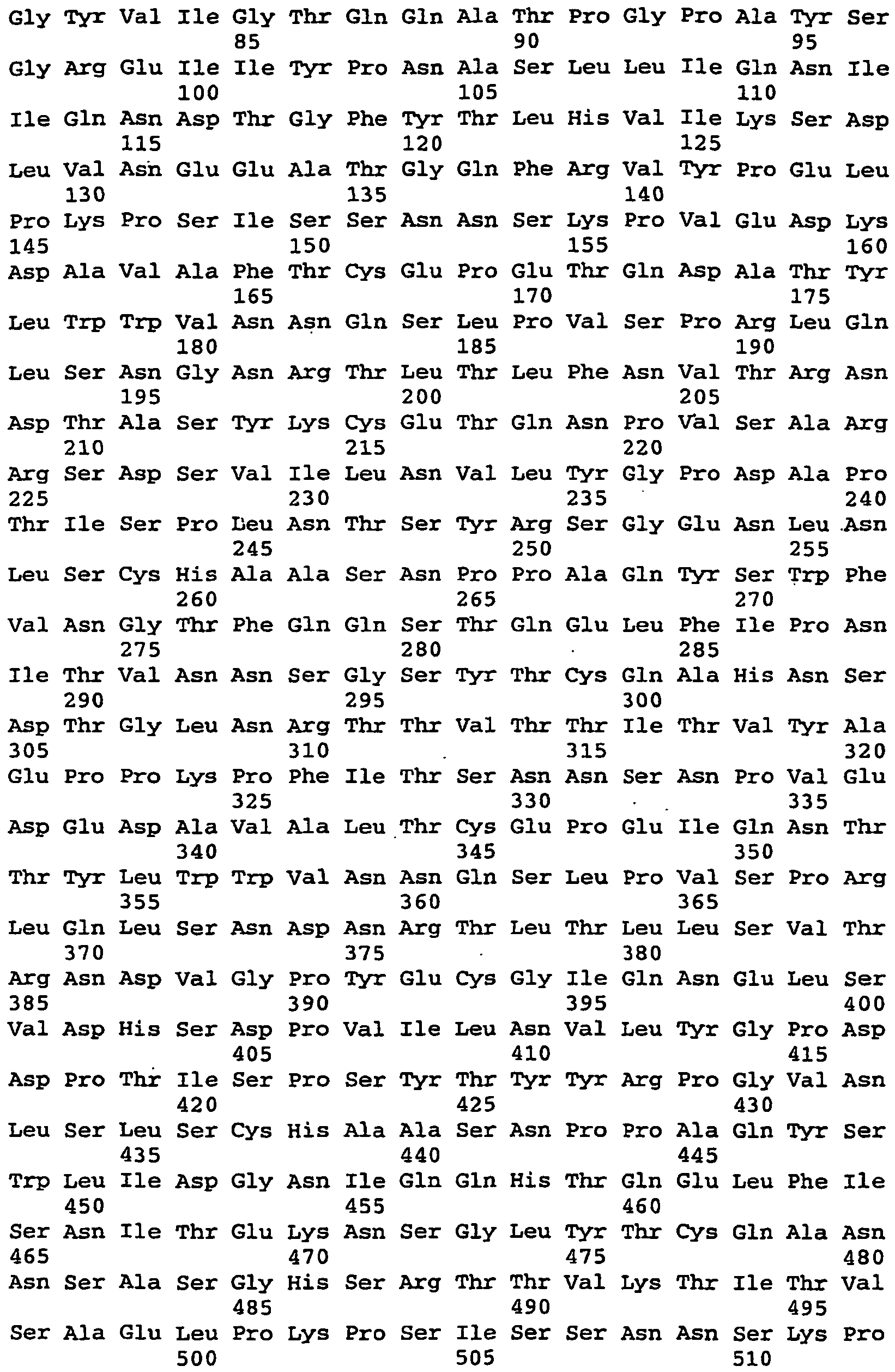

- <210> 14
<211> 2358
<212> DNA
<213> Artificial Sequence - <220>
<223> rhCEAopt-LTBopt fusion - <400> 14

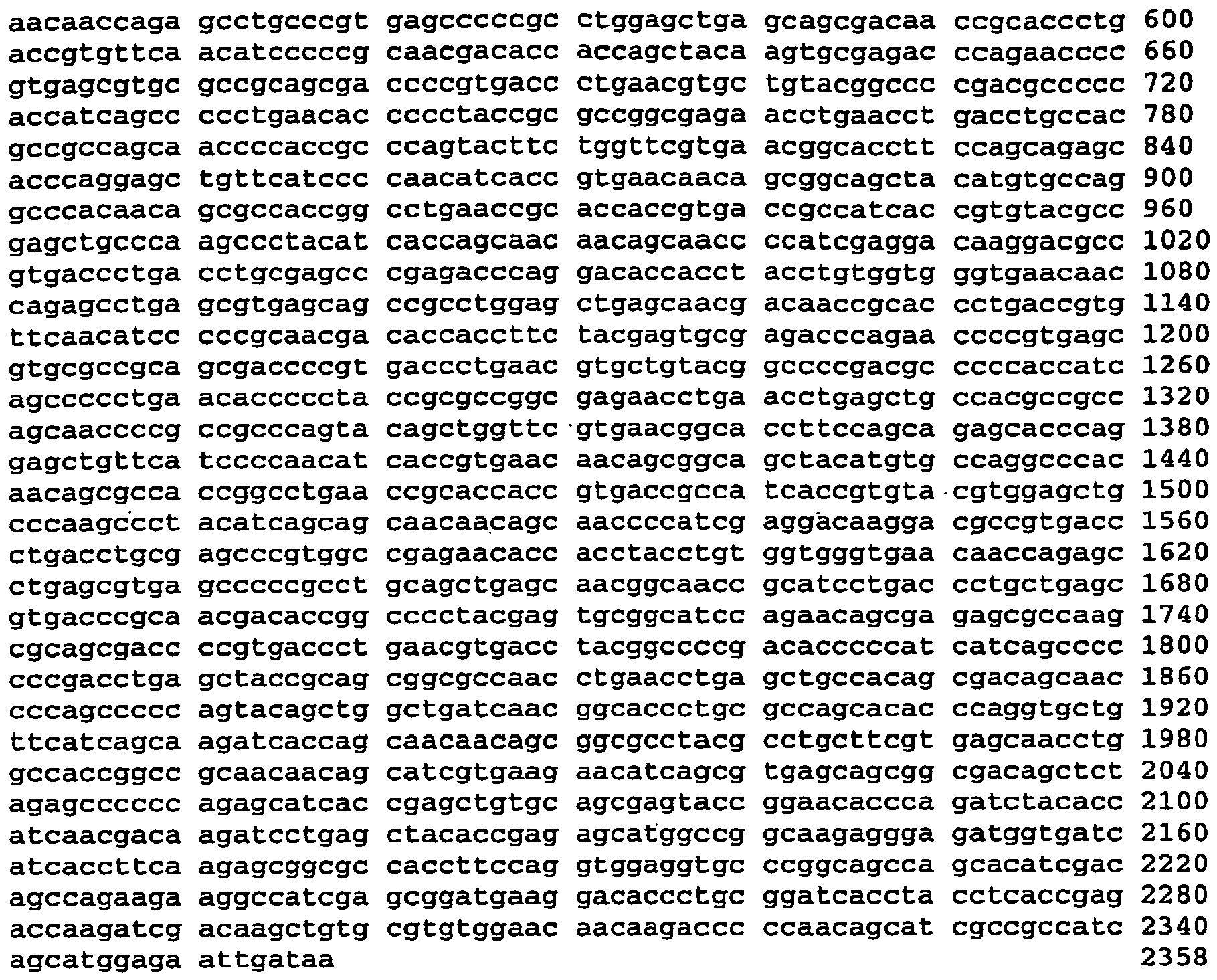
- <210> 15
<211> 784
<212> PRT
<213> Artificial Sequence - <220>
<223> rhCEAopt-LTBopt fusion - <400> 15


- <210> 16
<211> 2118
<212> DNA
<213> Macaca mulatta - <400> 16


- <210> 17
<211> 2118
<212> DNA
<213> Macaca mulatta - <400> 17


- <210> 18
<211> 705
<212> PRT
<213> Macaca mulatta - <400> 18


- <210> 19
<211> 705
<212> PRT
<213> Macaca mulatta - <400> 19
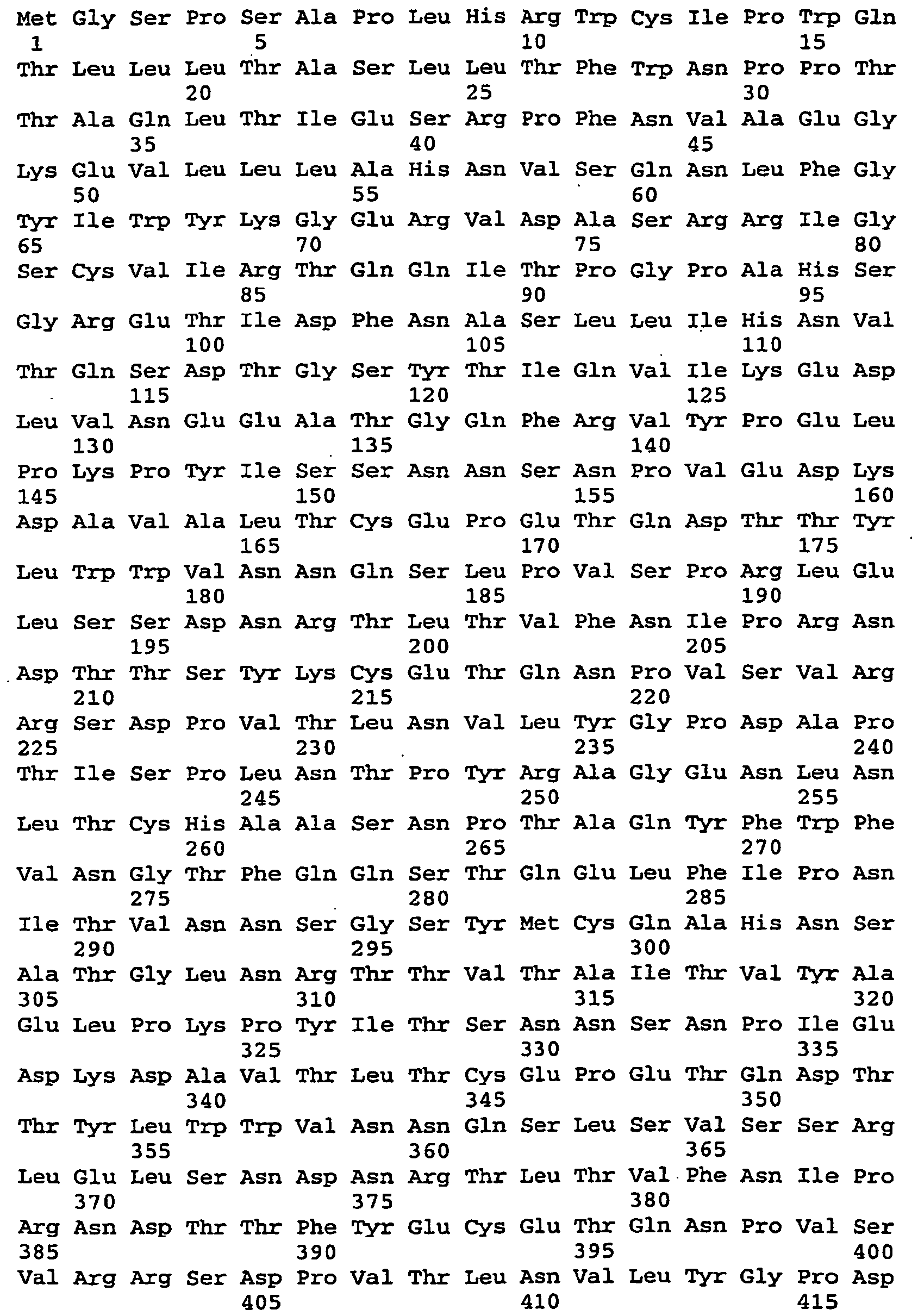

- <210> 20
<211> 702
<212> PRT
<213> Homo sapiens - <400> 20

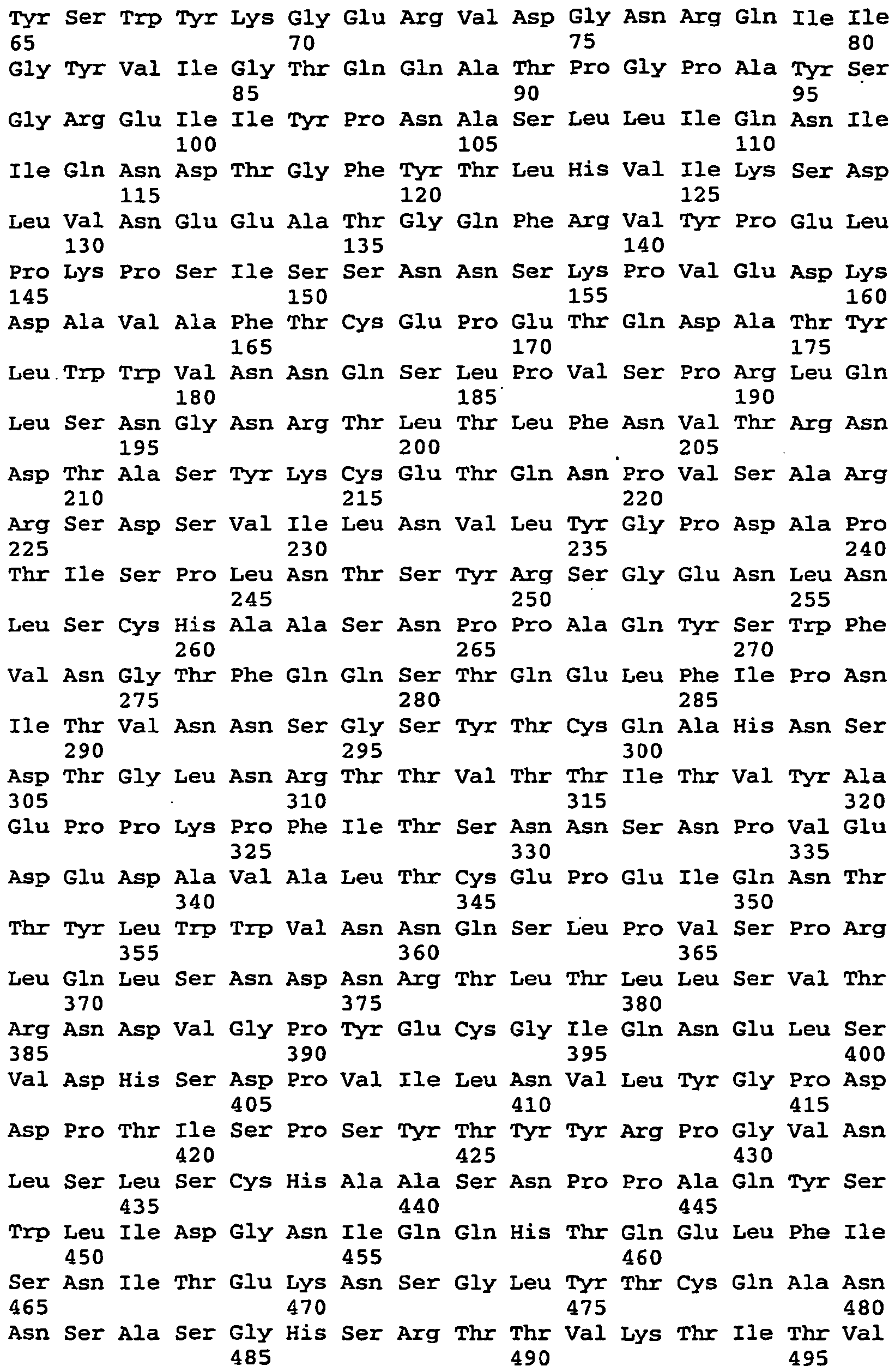

- <210> 21
<211> 2859
<212> DNA
<213> Artificial Sequence - <220>
<223> CEAoptDOMopt fusion - <400> 21


- <210> 22
<211> 2037
<212> DNA
<213> Artificial Sequence - <220>
<223> deleted human CEA - <400> 22
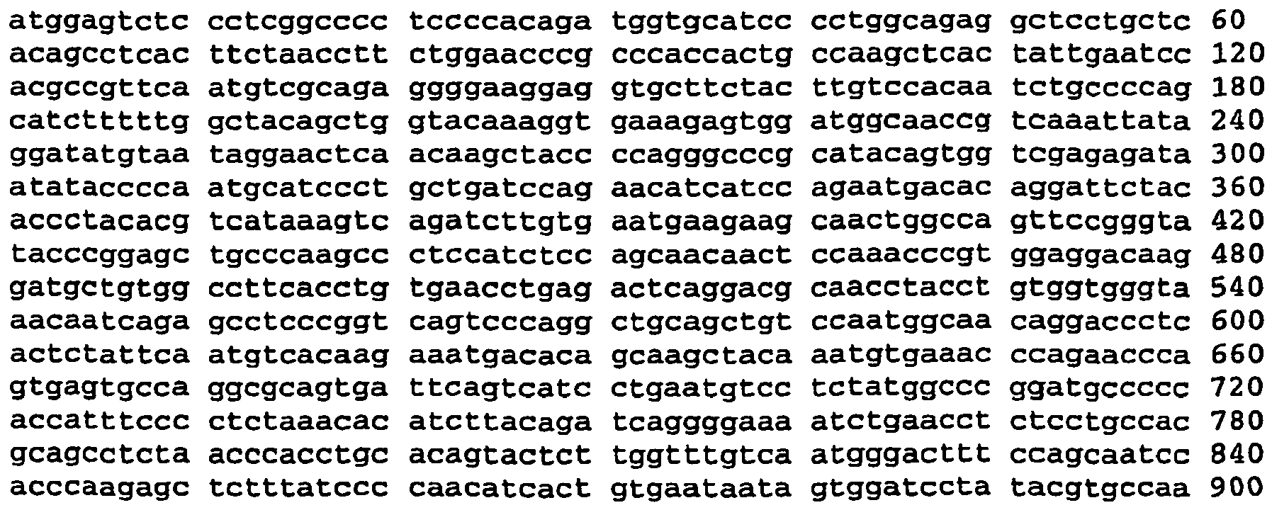

- <210> 23
<211> 679
<212> PRT
<213> Artificial Sequence - <220>
<223> deleted human CEA - <400> 23


- <210> 24
<211> 3426
<212> DNA
<213> Artificial Sequence - <220>
<223> CEA-FRC fusion - <400> 24


- <210> 25
<211> 2664
<212> DNA
<213> Artificial Sequence - <220>
<223> CEA-FcIgG fusion - <400> 25


- <210> 26
<211> 2167
<212> DNA
<213> Artificial Sequence - <220>
<223> CEA-LAMP fusion - <400> 26

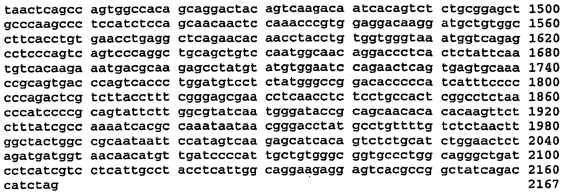
- <210> 27
<211> 3921
<212> DNA
<213> Artificial Sequence - <220>
<223> CEA-HSP70 fusion - <400> 27


- <210> 28
<211> 3585
<212> DNA
<213> Artificial Sequence - <220>
<223> CEA-VSVG fusion - <400> 28


- <210> 29
<211> 36
<212> DNA
<213> Artificial Sequence - <220>
<223> PCR primer - <400> 29
tattctagat tcaacaccaa ttccattttc ttattc 36 - <210> 30
<211> 33
<212> DNA
<213> Artificial Sequence - <220>
<223> PCR primer - <400> 30
gcggccgcta gaatcatttg tccatccttc atc 33 - <210> 31
<211> 36
<212> DNA
<213> Artificial Sequence - <220>
<223> PCR primer - <400> 31
tattctagat tcaacaccaa ttccattttc ttattc 36 - <210> 32
<211> 36
<212> DNA
<213> Artificial Sequence - <220>
<223> PCR primer - <400> 32
ttagcggccg ctagttctgt atcatatcgt aaaggg 36 - <210> 33
<211> 25
<212> DNA
<213> Artificial Sequence - <220>
<223> PCR primer - <400> 33
tctagataaa actcacacat gccca 25 - <210> 34
<211> 28
<212> DNA
<213> Artificial Sequence - <220>
<223> PCR primer - <400> 34
gccgactcat ttacccggag acagggag 28 - <210> 35
<211> 37
<212> DNA
<213> Artificial Sequence - <220>
<223> PCR primer - <400> 35
tctagatttg atccccattg ctgtgggcgg tgccctg 37 - <210> 36
<211> 39
<212> DNA
<213> Artificial Sequence - <220>
<223> PCR primer - <400> 36
ggcgtgactc ctcttcctgc caatgaggta ggcaatgag 39 - <210> 37
<211> 35
<212> DNA
<213> Artificial Sequence - <220>
<223> PCR primer - <400> 37
atatctagat ttcaccatag tttttccaca caacc 35 - <210> 38
<211> 36
<212> DNA
<213> Artificial Sequence - <220>
<223> PCR primer - <400> 38
gcggccgcct tccttccaag tcggttcatc tctatg 36 - <210> 39
<211> 34
<212> DNA
<213> Artificial Sequence - <220>
<223> PCR primer - <400> 39
gctctagata tggctcgtgc ggtcgggatc gacc 34 - <210> 40
<211> 36
<212> DNA
<213> Artificial Sequence - <220>
<223> PCR primer - <400> 40
gccgcggccg ctcacttggc ctcccggccg tcgtcg 36 - <210> 41
<211> 24
<212> DNA
<213> Artificial Sequence - <220>
<223> PCR primer - <400> 41
gttatctaga agcaccccca tccc 24 - <210> 42
<211> 36
<212> DNA
<213> Artificial Sequence - <220>
<223> PCR primer - <400> 42
ttaagatctc taagatctgg tgtcgtatct cagggg 36 - <210> 43
<211> 33
<212> DNA
<213> Artificial Sequence - <220>
<223> PCR primer - <400> 43
ttatctagaa agacccacac ctgcccccct tgc 33 - <210> 44
<211> 31
<212> DNA
<213> Artificial Sequence - <220>
<223> PCR primer - <400> 44
tatagatctt agggtacctt acttgccggg g 31 - <210> 45
<211> 952
<212> PRT
<213> Artificial Sequence - <220>
<223> CEA-Dom fusion - <400> 45


- <210> 46
<211> 907
<212> PRT
<213> Artificial Sequence - <220>
<223> CEA-FcIgG fusion - <400> 46


- <210> 47
<211> 825
<212> DNA
<213> Artificial Sequence - <220>
<223> DOM - <400> 47

- <210> 48
<211> 274
<212> PRT
<213> Artificial Sequence - <220>
<223> DOM - <400> 48


- <210> 49
<211> 2857
<212> DNA
<213> Artificial Sequence - <220>
<223> CEA-DOM fusion - <400> 49


- <210> 50
<211> 2859
<212> DNA
<213> Artificial Sequence - <220>
<223> rhCEA-DOMopt fusion - <400> 50


- <210> 51
<211> 952
<212> PRT
<213> Artificial Sequence - <220>
<223> rhCEA-DOMopt fusion - <400> 51
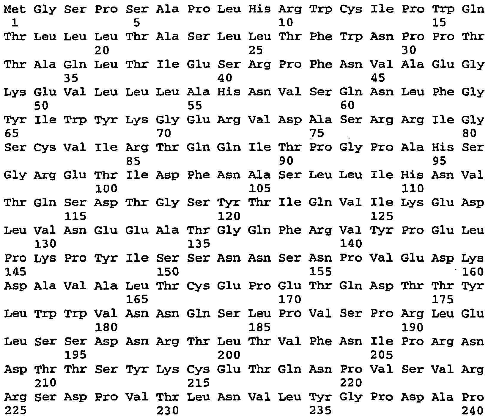


- <210> 52
<211> 2359
<212> DNA
<213> Artificial Sequence - <220>
<223> rhCEA-CTBopt fusion - <400> 52


- <210> 53
<211> 784
<212> PRT
<213> Artificial Sequence - <220>
<223> rhCEA-CTBopt fusion - <400> 53


- <210> 54
<211> 20
<212> PRT
<213> Artificial Sequence - <220>
<223> tetanus toxoid peptide - <400> 54

Claims (20)
- A nucleic acid molecule comprising a sequence of nucleotides that encodes a CEA fusion protein, wherein the CEA fusion protein comprises a C-terminally truncated human CEA protein which lacks amino acids 679 - 702 of SEQ ID NO:20, fused to an immunoenhancing element which is the B subunit of the heat labile enterotoxin of E. coli (LTB) optionally truncated of its signal sequence; and wherein the fusion protein is capable of producing an immune response in a mammal.
- The nucleic acid molecule of claim 1, wherein the LT subunit B is truncated of its signal sequence.
- The nucleic acid molecule of claim 2 wherein the sequence of nucleotides comprises a sequence of nucleotides as set forth in SEQ ID NO:9, SEQ ID NO:11, or SEQ ID NO:12.
- A nucleic acid molecule according to claim 3 wherein the sequence of nucleotides is as set forth in SEQ ID NO:12.
- The nucleic acid molecule of claim 2, wherein the C-terminal end of the CEA protein is fused to the N-terminal end of LT subunit B.
- A vector comprising the nucleic acid molecule of claim 1.
- The vector of claim 6, wherein the vector is an adenovirus vector or a plasmid vector.
- The vector of claim 7, wherein the vector is an Ad 5 vector.
- The vector of claim 7, wherein the vector is an Ad 6 vector or an Ad 24 vector.
- The vector of claim 7, wherein the vector is a chimp Ad vector.
- The vector of claim 7, wherein the vector is pV1JnsB.
- A host cell comprising the vector of claim 7.
- A process for expressing a CEA fusion protein in a recombinant host cell, comprising:(a) introducing a vector comprising the nucleic acid molecule of claim 1 into a suitable host cell; and,(b) culturing the host cell under conditions which allow expression of said human CEA fusion protein.
- A purified CEA fusion protein encoded by the nucleic acid molecule of claim 1.
- The purified CEA fusion protein of claim 14, wherein the fusion protein comprises a sequence of amino acids selected from the group consisting of: SEQ ID NOS: 10 and 13.
- An adenovirus vaccine vector comprising an adenoviral genome with a deletion in the E1 region, and an insert in the E1 region, wherein the insert comprises an expression cassette comprising:(a) a polynucleotide comprising a sequence of nucleotides that encodes a CEA fusion protein, wherein the CEA fusion protein comprises a C-terminally truncated human CEA protein which lacks amino acids 679 - 702 of SEQ ID NO:20, fused to an immunoenhancing element which is the B subunit of the heat labile enterotoxin of E. coli (LTB) optionally truncated of its signal sequence; and wherein the fusion protein is capable of producing an immune response in a mammal; and(b) a promoter operably linked to the polynucleotide.
- An adenovirus vector according to claim 16 which is an Ad 5 vector, an Ad 6 vector or an Ad 24 vector.
- A vaccine plasmid comprising a plasmid portion and an expression cassette portion, the expression cassette portion comprising:(a) a polynucleotide comprising a sequence of nucleotides that encodes a CEA fusion protein, wherein the CEA fusion protein comprises a C-terminally truncated human CEA protein which lacks amino acids 679 - 702 of SEQ ID NO:20, fused to an immunoenhancing element which is the B subunit of the heat labile enterotoxin of E. coli (LTB) optionally truncated of its signal sequence; and wherein the fusion protein is capable of producing an immune response in a mammal; and,(b) a promoter operably linked to the polynucleotide.
- A vaccine vector comprising the nucleic acid molecule of claim 1 for use in preventing or treating cancer in a mammal.
- A vaccine vector according to claim 19 wherein the vector is in accordance with any of claims 16-18.
Applications Claiming Priority (3)
| Application Number | Priority Date | Filing Date | Title |
|---|---|---|---|
| US54364904P | 2004-02-11 | 2004-02-11 | |
| US63579104P | 2004-12-14 | 2004-12-14 | |
| PCT/EP2005/001114 WO2005077977A2 (en) | 2004-02-11 | 2005-02-03 | Carcinoembryonic antigen fusions proteins and uses thereof |
Publications (3)
| Publication Number | Publication Date |
|---|---|
| EP1716173A2 EP1716173A2 (en) | 2006-11-02 |
| EP1716173B1 true EP1716173B1 (en) | 2012-04-25 |
| EP1716173B8 EP1716173B8 (en) | 2012-06-27 |
Family
ID=34864529
Family Applications (1)
| Application Number | Title | Priority Date | Filing Date |
|---|---|---|---|
| EP05707188A Expired - Lifetime EP1716173B8 (en) | 2004-02-11 | 2005-02-03 | Carcinoembryonic antigen fusion protein and uses thereof |
Country Status (14)
| Country | Link |
|---|---|
| US (1) | US8188244B2 (en) |
| EP (1) | EP1716173B8 (en) |
| JP (1) | JP5148116B2 (en) |
| KR (1) | KR101195400B1 (en) |
| AT (1) | ATE555127T1 (en) |
| AU (1) | AU2005212479B2 (en) |
| BR (1) | BRPI0507579A (en) |
| CA (1) | CA2555013C (en) |
| ES (1) | ES2387850T3 (en) |
| IL (1) | IL177277A (en) |
| NO (1) | NO340715B1 (en) |
| NZ (1) | NZ549048A (en) |
| RU (1) | RU2380375C2 (en) |
| WO (1) | WO2005077977A2 (en) |
Families Citing this family (20)
| Publication number | Priority date | Publication date | Assignee | Title |
|---|---|---|---|---|
| EP1934246B8 (en) * | 2005-10-07 | 2012-02-08 | Istituto di Ricerche di Biologia Molecolare P. Angeletti S.R.L. | Matrix metalloproteinase 11 vaccine |
| CN101522706B (en) | 2006-10-12 | 2013-09-04 | P.安杰莱蒂分子生物学研究所 | Telomerase reverse transcriptase fusion protein, nucleotide encoding it and use thereof |
| ES2725450T3 (en) | 2007-07-02 | 2019-09-24 | Etubics Corp | Methods and compositions for the production of an adenoviral vector for use in multiple vaccinations |
| US20110182926A1 (en) * | 2008-08-12 | 2011-07-28 | Nicola La Monica | Minigene |
| EP2342334A4 (en) | 2008-09-29 | 2012-03-14 | Univ Pennsylvania | TARGETED VACCINES ON A TUMOR VASCULAR MARKER |
| CN103037896B (en) * | 2010-05-11 | 2017-06-13 | 多伦多大学理事会 | N domains of carcinomebryonic antigen and combinations thereof, method and purposes |
| US9605276B2 (en) | 2012-08-24 | 2017-03-28 | Etubics Corporation | Replication defective adenovirus vector in vaccination |
| EA031234B1 (en) * | 2012-10-29 | 2018-12-28 | Дзе Борд Оф Трастиз Оф Дзе Юниверсити Оф Арканзас | Vaccine with an adjuvant based on mannosylated chitosan |
| WO2014140894A2 (en) | 2013-03-15 | 2014-09-18 | Bioven 3 Limited | Self-assembling synthetic proteins |
| US9753036B2 (en) | 2014-04-29 | 2017-09-05 | Edp Biotech Corporation | Methods and compositions for screening and detecting biomarkers |
| AU2016205215B2 (en) | 2015-01-09 | 2021-03-11 | Etubics Corporation | Methods and compositions for combination immunotherapy |
| US10695417B2 (en) | 2015-01-09 | 2020-06-30 | Etubics Corporation | Human adenovirus serotype 5 vectors containing E1 and E2B deletions encoding the ebola virus glycoprotein |
| EP3280731A1 (en) * | 2015-04-10 | 2018-02-14 | Preclinics Gesellschaft für präklinische Forschung mbH | Vhh constructs for diverting an immune response |
| US11149087B2 (en) | 2015-04-20 | 2021-10-19 | Etubics Corporation | Methods and compositions for combination immunotherapy |
| US10751398B2 (en) * | 2016-10-07 | 2020-08-25 | Loyola University Chicago | Inflammasome activators and methods of use to treat tumors |
| WO2018187260A1 (en) * | 2017-04-04 | 2018-10-11 | Heat Biologics, Inc. | Intratumoral vaccination |
| WO2019143606A1 (en) * | 2018-01-17 | 2019-07-25 | Nantbio, Inc. | Enhanced immunogenicity for gpi-anchored antigens |
| EP3864169A4 (en) * | 2018-10-12 | 2022-07-06 | Children's Hospital Medical Center | MODULAR EXPRESSION SYSTEMS FOR GENE EXPRESSION AND METHODS OF USING THE SAME |
| IT201900016736A1 (en) * | 2019-09-19 | 2021-03-19 | Takis S R L | Fused antigens to the Toxoplasma Gondii Profilin-like protein (PFTG) and their use in preventive and therapeutic vaccination. |
| US20240165213A1 (en) * | 2022-11-21 | 2024-05-23 | Avstera Therapeutics Corp. | mRNA COMPOSITION FOR PREVENTING AND TREATING CANCER |
Family Cites Families (21)
| Publication number | Priority date | Publication date | Assignee | Title |
|---|---|---|---|---|
| US5274087A (en) * | 1986-08-13 | 1993-12-28 | Molecular Diagnostics, Inc. | cDNA coding for carcinoembryonic antigen (CEA) |
| US5122599A (en) | 1986-08-13 | 1992-06-16 | Molecular Diagnostics, Inc. | CDNAS coding for members of the carcinoembryonic antigen family |
| EP0584266B1 (en) * | 1991-05-06 | 2003-09-03 | THE GOVERNMENT OF THE UNITED STATES OF AMERICA as represented by the SECRETARY OF THE DEPARTMENT OF HEALTH AND HUMAN SERVICES | Recombinant virus expressing carcinoembryonic antigen and methods of use thereof |
| EP0700445B1 (en) * | 1993-06-04 | 2002-01-23 | Whitehead Institute For Biomedical Research | Stress proteins and uses therefor |
| US5736524A (en) * | 1994-11-14 | 1998-04-07 | Merck & Co.,. Inc. | Polynucleotide tuberculosis vaccine |
| WO2000063253A1 (en) | 1999-04-16 | 2000-10-26 | Amgen Inc. | Agp-1 fusion protein compositions and methods |
| ES2233437T3 (en) * | 1999-08-25 | 2005-06-16 | MERCK & CO., INC. | SYNTHETIC GENES OF PAPILOMAVIRUS OPTIMIZED FOR EXPRESSION IN HUMAN CELLS. |
| WO2001024832A2 (en) | 1999-09-27 | 2001-04-12 | Gabriele Pecher | Pharmaceutical composition for treating and preventing human tumors, which express the tumor antigen mucin and/or the carcinoembryonic antigen (cea), and the use thereof |
| CA2388301C (en) * | 1999-10-22 | 2011-01-04 | Aventis Pasteur Limited | Modified gp100 and uses thereof |
| EP2420247A1 (en) * | 2000-03-07 | 2012-02-22 | Merck Sharp & Dohme Corp. | Adenovirus formulations |
| AU2001271268A1 (en) | 2000-05-19 | 2001-12-03 | The Administrators Of The Tulane Educational Fund | Hybrid lt-a/ct-b holotoxin for use as an adjuvant |
| JP2004508064A (en) | 2000-09-15 | 2004-03-18 | メルク エンド カムパニー インコーポレーテッド | Enhanced first generation adenovirus vaccine expressing codon-optimized HIV1-GAG, POL, NEF and modifications |
| DE10055545A1 (en) | 2000-11-09 | 2002-07-25 | Deutsches Krebsforsch | HPV 16-L1 and HPV 16-L2 encoding DNA sequences optimized for expression in eukaryotes |
| ATE442858T1 (en) | 2000-11-27 | 2009-10-15 | Powderject Vaccines Inc | NUCLEIC ACID ADJUVANTS |
| GB0030067D0 (en) | 2000-12-11 | 2001-01-24 | Univ Bristol | Therapeutic agent |
| EP1390065A2 (en) | 2001-05-14 | 2004-02-25 | Duotol Ab | Methods for promoting antigen presentation and modulating immune responses using cholera toxin and its b subunit |
| WO2003059379A2 (en) * | 2002-01-17 | 2003-07-24 | Pharmexa A/S | Immunogenic carcinoembryonic antigen (cea) |
| AU2004211481A1 (en) * | 2003-02-13 | 2004-08-26 | Istituto Di Ricerche Di Biologia Molecolare P. Angeletti Spa | Rhesus carcino embryonic antigen, nucleotides encoding same, and uses thereof |
| WO2004092216A1 (en) | 2003-04-15 | 2004-10-28 | Trangene S.A. | Carcinoembryonic antigen (cea) lacking a signal peptide, nucleic acid encoding it and fusion of cea with a t cell epitope and their use for the treatment and/or prophylaxis of cancer |
| EP1622937A2 (en) * | 2003-05-05 | 2006-02-08 | Istituto di Richerche di Biologia Molecolare P. Angeletti S.p.A. | Synthetic gene encoding human carcinoembryonic antigen and uses thereof |
| US20060286114A1 (en) * | 2003-08-22 | 2006-12-21 | Luigi Aurisicchio | Synthetic gene encoding rhesus monkey carcinoembryonic antigen and uses thereof |
-
2005
- 2005-02-03 AT AT05707188T patent/ATE555127T1/en active
- 2005-02-03 AU AU2005212479A patent/AU2005212479B2/en not_active Ceased
- 2005-02-03 EP EP05707188A patent/EP1716173B8/en not_active Expired - Lifetime
- 2005-02-03 KR KR1020067018595A patent/KR101195400B1/en not_active Expired - Fee Related
- 2005-02-03 US US10/589,180 patent/US8188244B2/en active Active
- 2005-02-03 ES ES05707188T patent/ES2387850T3/en not_active Expired - Lifetime
- 2005-02-03 WO PCT/EP2005/001114 patent/WO2005077977A2/en not_active Ceased
- 2005-02-03 NZ NZ549048A patent/NZ549048A/en not_active IP Right Cessation
- 2005-02-03 CA CA2555013A patent/CA2555013C/en not_active Expired - Fee Related
- 2005-02-03 RU RU2006132340/13A patent/RU2380375C2/en not_active IP Right Cessation
- 2005-02-03 JP JP2006552512A patent/JP5148116B2/en not_active Expired - Fee Related
- 2005-02-03 BR BRPI0507579-3A patent/BRPI0507579A/en not_active Application Discontinuation
-
2006
- 2006-08-03 IL IL177277A patent/IL177277A/en not_active IP Right Cessation
- 2006-09-08 NO NO20064044A patent/NO340715B1/en not_active IP Right Cessation
Also Published As
| Publication number | Publication date |
|---|---|
| WO2005077977A2 (en) | 2005-08-25 |
| RU2380375C2 (en) | 2010-01-27 |
| CA2555013C (en) | 2013-10-15 |
| ES2387850T3 (en) | 2012-10-02 |
| WO2005077977A3 (en) | 2005-10-06 |
| NO340715B1 (en) | 2017-06-06 |
| NO20064044L (en) | 2006-09-08 |
| JP2007537722A (en) | 2007-12-27 |
| EP1716173B8 (en) | 2012-06-27 |
| EP1716173A2 (en) | 2006-11-02 |
| KR20060129456A (en) | 2006-12-15 |
| NZ549048A (en) | 2009-04-30 |
| AU2005212479A1 (en) | 2005-08-25 |
| US8188244B2 (en) | 2012-05-29 |
| KR101195400B1 (en) | 2012-10-29 |
| BRPI0507579A (en) | 2007-07-03 |
| IL177277A0 (en) | 2006-12-10 |
| IL177277A (en) | 2014-08-31 |
| ATE555127T1 (en) | 2012-05-15 |
| RU2006132340A (en) | 2008-03-20 |
| CA2555013A1 (en) | 2005-08-25 |
| AU2005212479B2 (en) | 2009-09-10 |
| US20080311137A1 (en) | 2008-12-18 |
| JP5148116B2 (en) | 2013-02-20 |
Similar Documents
| Publication | Publication Date | Title |
|---|---|---|
| US8188244B2 (en) | Carcinoembryonic antigen fusions and uses thereof | |
| EP2076531B1 (en) | Telomerase reverse transcriptase fusion protein, nucleotides encoding it, and uses thereof | |
| AU2004261724B2 (en) | Synthetic gene encoding human epidermal growth factor 2/neu antigen and uses thereof | |
| US20070104685A1 (en) | Synthetic gene encoding human carcinoembryonic antigen and uses thereof | |
| WO2008012237A1 (en) | Multi-antigen construct and uses thereof | |
| AU2003294023B2 (en) | Rhesus HER2/neu, nucleotides encoding same, and uses thereof | |
| CN101001869B (en) | Carcinoembryonic antigen fusions and uses thereof | |
| US20060286114A1 (en) | Synthetic gene encoding rhesus monkey carcinoembryonic antigen and uses thereof | |
| MXPA06009202A (en) | Carcinoembryonic antigen fusions proteins and uses thereof | |
| US20060228335A1 (en) | Rhesus carcino embryonic antigen, nucleotides encoding same, and uses thereof | |
| ZA200508013B (en) | Synthetic gene encoding human carcinoembryonic antigen and use thereof |
Legal Events
| Date | Code | Title | Description |
|---|---|---|---|
| PUAI | Public reference made under article 153(3) epc to a published international application that has entered the european phase |
Free format text: ORIGINAL CODE: 0009012 |
|
| 17P | Request for examination filed |
Effective date: 20060911 |
|
| AK | Designated contracting states |
Kind code of ref document: A2 Designated state(s): AT BE BG CH CY CZ DE DK EE ES FI FR GB GR HU IE IS IT LI LT LU MC NL PL PT RO SE SI SK TR |
|
| AX | Request for extension of the european patent |
Extension state: LV |
|
| RAX | Requested extension states of the european patent have changed |
Extension state: LV Payment date: 20060911 |
|
| 17Q | First examination report despatched |
Effective date: 20080206 |
|
| GRAP | Despatch of communication of intention to grant a patent |
Free format text: ORIGINAL CODE: EPIDOSNIGR1 |
|
| GRAS | Grant fee paid |
Free format text: ORIGINAL CODE: EPIDOSNIGR3 |
|
| GRAA | (expected) grant |
Free format text: ORIGINAL CODE: 0009210 |
|
| AK | Designated contracting states |
Kind code of ref document: B1 Designated state(s): AT BE BG CH CY CZ DE DK EE ES FI FR GB GR HU IE IS IT LI LT LU MC NL PL PT RO SE SI SK TR |
|
| AX | Request for extension of the european patent |
Extension state: LV |
|
| REG | Reference to a national code |
Ref country code: GB Ref legal event code: FG4D |
|
| REG | Reference to a national code |
Ref country code: CH Ref legal event code: EP |
|
| REG | Reference to a national code |
Ref country code: AT Ref legal event code: REF Ref document number: 555127 Country of ref document: AT Kind code of ref document: T Effective date: 20120515 |
|
| REG | Reference to a national code |
Ref country code: IE Ref legal event code: FG4D |
|
| RAP2 | Party data changed (patent owner data changed or rights of a patent transferred) |
Owner name: ISTITUTO DI RICERCHE DI BIOLOGIA MOLECOLARE P. ANG |
|
| REG | Reference to a national code |
Ref country code: DE Ref legal event code: R096 Ref document number: 602005033841 Country of ref document: DE Effective date: 20120621 |
|
| REG | Reference to a national code |
Ref country code: CH Ref legal event code: NV Representative=s name: MARKS & CLERK (LUXEMBOURG) LLP |
|
| REG | Reference to a national code |
Ref country code: NL Ref legal event code: T3 |
|
| REG | Reference to a national code |
Ref country code: AT Ref legal event code: MK05 Ref document number: 555127 Country of ref document: AT Kind code of ref document: T Effective date: 20120425 |
|
| LTIE | Lt: invalidation of european patent or patent extension |
Effective date: 20120425 |
|
| REG | Reference to a national code |
Ref country code: ES Ref legal event code: FG2A Ref document number: 2387850 Country of ref document: ES Kind code of ref document: T3 Effective date: 20121002 |
|
| PG25 | Lapsed in a contracting state [announced via postgrant information from national office to epo] |
Ref country code: FI Free format text: LAPSE BECAUSE OF FAILURE TO SUBMIT A TRANSLATION OF THE DESCRIPTION OR TO PAY THE FEE WITHIN THE PRESCRIBED TIME-LIMIT Effective date: 20120425 Ref country code: CY Free format text: LAPSE BECAUSE OF FAILURE TO SUBMIT A TRANSLATION OF THE DESCRIPTION OR TO PAY THE FEE WITHIN THE PRESCRIBED TIME-LIMIT Effective date: 20120425 Ref country code: SE Free format text: LAPSE BECAUSE OF FAILURE TO SUBMIT A TRANSLATION OF THE DESCRIPTION OR TO PAY THE FEE WITHIN THE PRESCRIBED TIME-LIMIT Effective date: 20120425 Ref country code: IS Free format text: LAPSE BECAUSE OF FAILURE TO SUBMIT A TRANSLATION OF THE DESCRIPTION OR TO PAY THE FEE WITHIN THE PRESCRIBED TIME-LIMIT Effective date: 20120825 Ref country code: PL Free format text: LAPSE BECAUSE OF FAILURE TO SUBMIT A TRANSLATION OF THE DESCRIPTION OR TO PAY THE FEE WITHIN THE PRESCRIBED TIME-LIMIT Effective date: 20120425 Ref country code: LT Free format text: LAPSE BECAUSE OF FAILURE TO SUBMIT A TRANSLATION OF THE DESCRIPTION OR TO PAY THE FEE WITHIN THE PRESCRIBED TIME-LIMIT Effective date: 20120425 |
|
| PG25 | Lapsed in a contracting state [announced via postgrant information from national office to epo] |
Ref country code: GR Free format text: LAPSE BECAUSE OF FAILURE TO SUBMIT A TRANSLATION OF THE DESCRIPTION OR TO PAY THE FEE WITHIN THE PRESCRIBED TIME-LIMIT Effective date: 20120726 Ref country code: SI Free format text: LAPSE BECAUSE OF FAILURE TO SUBMIT A TRANSLATION OF THE DESCRIPTION OR TO PAY THE FEE WITHIN THE PRESCRIBED TIME-LIMIT Effective date: 20120425 Ref country code: PT Free format text: LAPSE BECAUSE OF FAILURE TO SUBMIT A TRANSLATION OF THE DESCRIPTION OR TO PAY THE FEE WITHIN THE PRESCRIBED TIME-LIMIT Effective date: 20120827 |
|
| PG25 | Lapsed in a contracting state [announced via postgrant information from national office to epo] |
Ref country code: BE Free format text: LAPSE BECAUSE OF FAILURE TO SUBMIT A TRANSLATION OF THE DESCRIPTION OR TO PAY THE FEE WITHIN THE PRESCRIBED TIME-LIMIT Effective date: 20120425 |
|
| PG25 | Lapsed in a contracting state [announced via postgrant information from national office to epo] |
Ref country code: DK Free format text: LAPSE BECAUSE OF FAILURE TO SUBMIT A TRANSLATION OF THE DESCRIPTION OR TO PAY THE FEE WITHIN THE PRESCRIBED TIME-LIMIT Effective date: 20120425 Ref country code: AT Free format text: LAPSE BECAUSE OF FAILURE TO SUBMIT A TRANSLATION OF THE DESCRIPTION OR TO PAY THE FEE WITHIN THE PRESCRIBED TIME-LIMIT Effective date: 20120425 Ref country code: EE Free format text: LAPSE BECAUSE OF FAILURE TO SUBMIT A TRANSLATION OF THE DESCRIPTION OR TO PAY THE FEE WITHIN THE PRESCRIBED TIME-LIMIT Effective date: 20120425 Ref country code: RO Free format text: LAPSE BECAUSE OF FAILURE TO SUBMIT A TRANSLATION OF THE DESCRIPTION OR TO PAY THE FEE WITHIN THE PRESCRIBED TIME-LIMIT Effective date: 20120425 Ref country code: CZ Free format text: LAPSE BECAUSE OF FAILURE TO SUBMIT A TRANSLATION OF THE DESCRIPTION OR TO PAY THE FEE WITHIN THE PRESCRIBED TIME-LIMIT Effective date: 20120425 Ref country code: SK Free format text: LAPSE BECAUSE OF FAILURE TO SUBMIT A TRANSLATION OF THE DESCRIPTION OR TO PAY THE FEE WITHIN THE PRESCRIBED TIME-LIMIT Effective date: 20120425 |
|
| PLBE | No opposition filed within time limit |
Free format text: ORIGINAL CODE: 0009261 |
|
| STAA | Information on the status of an ep patent application or granted ep patent |
Free format text: STATUS: NO OPPOSITION FILED WITHIN TIME LIMIT |
|
| 26N | No opposition filed |
Effective date: 20130128 |
|
| REG | Reference to a national code |
Ref country code: DE Ref legal event code: R097 Ref document number: 602005033841 Country of ref document: DE Effective date: 20130128 |
|
| PG25 | Lapsed in a contracting state [announced via postgrant information from national office to epo] |
Ref country code: BG Free format text: LAPSE BECAUSE OF FAILURE TO SUBMIT A TRANSLATION OF THE DESCRIPTION OR TO PAY THE FEE WITHIN THE PRESCRIBED TIME-LIMIT Effective date: 20120725 |
|
| PG25 | Lapsed in a contracting state [announced via postgrant information from national office to epo] |
Ref country code: MC Free format text: LAPSE BECAUSE OF NON-PAYMENT OF DUE FEES Effective date: 20130228 |
|
| REG | Reference to a national code |
Ref country code: IE Ref legal event code: MM4A |
|
| PG25 | Lapsed in a contracting state [announced via postgrant information from national office to epo] |
Ref country code: IE Free format text: LAPSE BECAUSE OF NON-PAYMENT OF DUE FEES Effective date: 20130203 |
|
| REG | Reference to a national code |
Ref country code: DE Ref legal event code: R082 Ref document number: 602005033841 Country of ref document: DE Representative=s name: ABITZ & PARTNER, DE |
|
| REG | Reference to a national code |
Ref country code: CH Ref legal event code: PFUS Owner name: MSD ITALIA S.R.L., IT Free format text: FORMER OWNER: ISTITUTO DI RICERCHE DI BIOLOGIA MOLECOLARE P. ANGELETTI S.R.L., IT |
|
| REG | Reference to a national code |
Ref country code: DE Ref legal event code: R082 Ref document number: 602005033841 Country of ref document: DE Representative=s name: ABITZ & PARTNER, DE Effective date: 20140514 Ref country code: DE Ref legal event code: R081 Ref document number: 602005033841 Country of ref document: DE Owner name: MSD ITALIA S.R.L., IT Free format text: FORMER OWNER: ISTITUTO DI RICERCHE DI BIOLOGIA MOLECOLARE P. ANGELETTI S.P.A., 00040 POMEZIA, IT Effective date: 20120426 Ref country code: DE Ref legal event code: R081 Ref document number: 602005033841 Country of ref document: DE Owner name: MSD ITALIA S.R.L., IT Free format text: FORMER OWNER: ISTITUTO DI RICERCHE DI BIOLOGIA MOLECOLARE P. ANGELETTI S.P.A., 00040 POMEZIA, IT Effective date: 20120601 Ref country code: GB Ref legal event code: 732E Free format text: REGISTERED BETWEEN 20140522 AND 20140528 Ref country code: DE Ref legal event code: R081 Ref document number: 602005033841 Country of ref document: DE Owner name: MSD ITALIA S.R.L., IT Free format text: FORMER OWNER: ISTITUTO DI RICERCHE DI BIOLOGIA MOLECOLARE, ROMA, IT Effective date: 20140514 Ref country code: DE Ref legal event code: R082 Ref document number: 602005033841 Country of ref document: DE Representative=s name: ABITZ & PARTNER PATENTANWAELTE MBB, DE Effective date: 20140514 |
|
| REG | Reference to a national code |
Ref country code: NL Ref legal event code: SD Effective date: 20140723 |
|
| REG | Reference to a national code |
Ref country code: ES Ref legal event code: PC2A Owner name: MSD ITALIA S.RL Effective date: 20140905 |
|
| REG | Reference to a national code |
Ref country code: FR Ref legal event code: TP Owner name: MSD ITALIA S.R.L., IT Effective date: 20140909 |
|
| PG25 | Lapsed in a contracting state [announced via postgrant information from national office to epo] |
Ref country code: TR Free format text: LAPSE BECAUSE OF FAILURE TO SUBMIT A TRANSLATION OF THE DESCRIPTION OR TO PAY THE FEE WITHIN THE PRESCRIBED TIME-LIMIT Effective date: 20120425 |
|
| PG25 | Lapsed in a contracting state [announced via postgrant information from national office to epo] |
Ref country code: LU Free format text: LAPSE BECAUSE OF NON-PAYMENT OF DUE FEES Effective date: 20130203 Ref country code: HU Free format text: LAPSE BECAUSE OF FAILURE TO SUBMIT A TRANSLATION OF THE DESCRIPTION OR TO PAY THE FEE WITHIN THE PRESCRIBED TIME-LIMIT; INVALID AB INITIO Effective date: 20050203 |
|
| REG | Reference to a national code |
Ref country code: FR Ref legal event code: PLFP Year of fee payment: 12 |
|
| REG | Reference to a national code |
Ref country code: FR Ref legal event code: PLFP Year of fee payment: 13 |
|
| REG | Reference to a national code |
Ref country code: FR Ref legal event code: PLFP Year of fee payment: 14 |
|
| PGFP | Annual fee paid to national office [announced via postgrant information from national office to epo] |
Ref country code: NL Payment date: 20180209 Year of fee payment: 14 |
|
| PGFP | Annual fee paid to national office [announced via postgrant information from national office to epo] |
Ref country code: CH Payment date: 20180125 Year of fee payment: 14 Ref country code: ES Payment date: 20180302 Year of fee payment: 14 |
|
| REG | Reference to a national code |
Ref country code: CH Ref legal event code: PL |
|
| REG | Reference to a national code |
Ref country code: NL Ref legal event code: MM Effective date: 20190301 |
|
| PG25 | Lapsed in a contracting state [announced via postgrant information from national office to epo] |
Ref country code: LI Free format text: LAPSE BECAUSE OF NON-PAYMENT OF DUE FEES Effective date: 20190228 Ref country code: CH Free format text: LAPSE BECAUSE OF NON-PAYMENT OF DUE FEES Effective date: 20190228 |
|
| PG25 | Lapsed in a contracting state [announced via postgrant information from national office to epo] |
Ref country code: NL Free format text: LAPSE BECAUSE OF NON-PAYMENT OF DUE FEES Effective date: 20190301 |
|
| REG | Reference to a national code |
Ref country code: ES Ref legal event code: FD2A Effective date: 20200327 |
|
| PG25 | Lapsed in a contracting state [announced via postgrant information from national office to epo] |
Ref country code: ES Free format text: LAPSE BECAUSE OF NON-PAYMENT OF DUE FEES Effective date: 20190204 |
|
| PGFP | Annual fee paid to national office [announced via postgrant information from national office to epo] |
Ref country code: DE Payment date: 20240109 Year of fee payment: 20 Ref country code: GB Payment date: 20240111 Year of fee payment: 20 |
|
| PGFP | Annual fee paid to national office [announced via postgrant information from national office to epo] |
Ref country code: IT Payment date: 20240213 Year of fee payment: 20 Ref country code: FR Payment date: 20240108 Year of fee payment: 20 |
|
| REG | Reference to a national code |
Ref country code: DE Ref legal event code: R071 Ref document number: 602005033841 Country of ref document: DE |
|
| REG | Reference to a national code |
Ref country code: GB Ref legal event code: PE20 Expiry date: 20250202 |
|
| PG25 | Lapsed in a contracting state [announced via postgrant information from national office to epo] |
Ref country code: GB Free format text: LAPSE BECAUSE OF EXPIRATION OF PROTECTION Effective date: 20250202 |