EP1715046B1 - New polypeptide, DNA encoding the polypeptide, recombinant vector bearing the DNA and recombinant virus utilizing the recombinant vector as well as use thereof - Google Patents
New polypeptide, DNA encoding the polypeptide, recombinant vector bearing the DNA and recombinant virus utilizing the recombinant vector as well as use thereof Download PDFInfo
- Publication number
- EP1715046B1 EP1715046B1 EP06012377A EP06012377A EP1715046B1 EP 1715046 B1 EP1715046 B1 EP 1715046B1 EP 06012377 A EP06012377 A EP 06012377A EP 06012377 A EP06012377 A EP 06012377A EP 1715046 B1 EP1715046 B1 EP 1715046B1
- Authority
- EP
- European Patent Office
- Prior art keywords
- sequence
- dna
- polypeptide
- fragment
- seq
- Prior art date
- Legal status (The legal status is an assumption and is not a legal conclusion. Google has not performed a legal analysis and makes no representation as to the accuracy of the status listed.)
- Expired - Lifetime
Links
- 108090000765 processed proteins & peptides Proteins 0.000 title claims description 112
- 229920001184 polypeptide Polymers 0.000 title claims description 108
- 102000004196 processed proteins & peptides Human genes 0.000 title claims description 108
- 241000700605 Viruses Species 0.000 title claims description 51
- 108090000623 proteins and genes Proteins 0.000 claims description 84
- 241000204022 Mycoplasma gallisepticum Species 0.000 claims description 76
- 102000004169 proteins and genes Human genes 0.000 claims description 66
- 239000012528 membrane Substances 0.000 claims description 58
- 230000000890 antigenic effect Effects 0.000 claims description 36
- 241000700663 Avipoxvirus Species 0.000 claims description 34
- 229960005486 vaccine Drugs 0.000 claims description 32
- 150000001413 amino acids Chemical class 0.000 claims description 24
- 241000204031 Mycoplasma Species 0.000 claims description 12
- 208000015181 infectious disease Diseases 0.000 claims description 12
- 244000144977 poultry Species 0.000 claims description 11
- 108010052285 Membrane Proteins Proteins 0.000 claims description 9
- 102000018697 Membrane Proteins Human genes 0.000 claims description 9
- 125000003178 carboxy group Chemical group [H]OC(*)=O 0.000 claims description 3
- 125000003275 alpha amino acid group Chemical group 0.000 claims 3
- 108020004414 DNA Proteins 0.000 description 214
- 239000012634 fragment Substances 0.000 description 152
- 108020004707 nucleic acids Proteins 0.000 description 75
- 102000039446 nucleic acids Human genes 0.000 description 75
- 150000007523 nucleic acids Chemical class 0.000 description 75
- 230000029087 digestion Effects 0.000 description 64
- 239000013612 plasmid Substances 0.000 description 58
- 210000004027 cell Anatomy 0.000 description 56
- 238000000034 method Methods 0.000 description 42
- 239000000047 product Substances 0.000 description 42
- FAPWRFPIFSIZLT-UHFFFAOYSA-M Sodium chloride Chemical compound [Na+].[Cl-] FAPWRFPIFSIZLT-UHFFFAOYSA-M 0.000 description 40
- 108091008146 restriction endonucleases Proteins 0.000 description 39
- 239000002609 medium Substances 0.000 description 33
- 210000004379 membrane Anatomy 0.000 description 33
- 230000008018 melting Effects 0.000 description 30
- 238000002844 melting Methods 0.000 description 30
- 102000003960 Ligases Human genes 0.000 description 29
- 108090000364 Ligases Proteins 0.000 description 29
- 108700026244 Open Reading Frames Proteins 0.000 description 29
- 101100101156 Caenorhabditis elegans ttm-1 gene Proteins 0.000 description 28
- 238000000246 agarose gel electrophoresis Methods 0.000 description 28
- 239000000243 solution Substances 0.000 description 27
- 238000009396 hybridization Methods 0.000 description 21
- 239000004677 Nylon Substances 0.000 description 20
- 238000011534 incubation Methods 0.000 description 20
- 229920001778 nylon Polymers 0.000 description 20
- 239000011780 sodium chloride Substances 0.000 description 20
- 241000287828 Gallus gallus Species 0.000 description 19
- 235000013330 chicken meat Nutrition 0.000 description 19
- 238000010367 cloning Methods 0.000 description 18
- 238000010276 construction Methods 0.000 description 18
- 241000588724 Escherichia coli Species 0.000 description 16
- HEMHJVSKTPXQMS-UHFFFAOYSA-M Sodium hydroxide Chemical compound [OH-].[Na+] HEMHJVSKTPXQMS-UHFFFAOYSA-M 0.000 description 15
- 239000002773 nucleotide Substances 0.000 description 14
- 125000003729 nucleotide group Chemical group 0.000 description 14
- 241000712469 Fowl plague virus Species 0.000 description 13
- 239000013615 primer Substances 0.000 description 13
- CSCPPACGZOOCGX-UHFFFAOYSA-N Acetone Chemical compound CC(C)=O CSCPPACGZOOCGX-UHFFFAOYSA-N 0.000 description 12
- 108020005038 Terminator Codon Proteins 0.000 description 11
- 210000000170 cell membrane Anatomy 0.000 description 11
- 239000000499 gel Substances 0.000 description 11
- QKNYBSVHEMOAJP-UHFFFAOYSA-N 2-amino-2-(hydroxymethyl)propane-1,3-diol;hydron;chloride Chemical compound Cl.OCC(N)(CO)CO QKNYBSVHEMOAJP-UHFFFAOYSA-N 0.000 description 9
- 229920001817 Agar Polymers 0.000 description 9
- LFQSCWFLJHTTHZ-UHFFFAOYSA-N Ethanol Chemical compound CCO LFQSCWFLJHTTHZ-UHFFFAOYSA-N 0.000 description 9
- 239000008272 agar Substances 0.000 description 9
- 238000000376 autoradiography Methods 0.000 description 9
- 238000012869 ethanol precipitation Methods 0.000 description 9
- 238000006386 neutralization reaction Methods 0.000 description 9
- 210000002966 serum Anatomy 0.000 description 9
- 239000006228 supernatant Substances 0.000 description 9
- 239000000725 suspension Substances 0.000 description 9
- 239000006142 Luria-Bertani Agar Substances 0.000 description 8
- 108091034117 Oligonucleotide Proteins 0.000 description 8
- ISWSIDIOOBJBQZ-UHFFFAOYSA-N Phenol Chemical compound OC1=CC=CC=C1 ISWSIDIOOBJBQZ-UHFFFAOYSA-N 0.000 description 8
- AVKUERGKIZMTKX-NJBDSQKTSA-N ampicillin Chemical compound C1([C@@H](N)C(=O)N[C@H]2[C@H]3SC([C@@H](N3C2=O)C(O)=O)(C)C)=CC=CC=C1 AVKUERGKIZMTKX-NJBDSQKTSA-N 0.000 description 8
- 229960000723 ampicillin Drugs 0.000 description 8
- 230000008859 change Effects 0.000 description 8
- 238000003776 cleavage reaction Methods 0.000 description 8
- 238000001962 electrophoresis Methods 0.000 description 8
- 239000000203 mixture Substances 0.000 description 8
- 235000013594 poultry meat Nutrition 0.000 description 8
- 230000007017 scission Effects 0.000 description 8
- OPIFSICVWOWJMJ-AEOCFKNESA-N 5-bromo-4-chloro-3-indolyl beta-D-galactoside Chemical compound O[C@@H]1[C@@H](O)[C@@H](O)[C@@H](CO)O[C@H]1OC1=CNC2=CC=C(Br)C(Cl)=C12 OPIFSICVWOWJMJ-AEOCFKNESA-N 0.000 description 7
- KCXVZYZYPLLWCC-UHFFFAOYSA-N EDTA Chemical compound OC(=O)CN(CC(O)=O)CCN(CC(O)=O)CC(O)=O KCXVZYZYPLLWCC-UHFFFAOYSA-N 0.000 description 7
- 238000002105 Southern blotting Methods 0.000 description 7
- 238000006243 chemical reaction Methods 0.000 description 7
- 238000010790 dilution Methods 0.000 description 7
- 239000012895 dilution Substances 0.000 description 7
- 238000002360 preparation method Methods 0.000 description 7
- 230000009257 reactivity Effects 0.000 description 7
- 239000000523 sample Substances 0.000 description 7
- 241000711404 Avian avulavirus 1 Species 0.000 description 6
- CSNNHWWHGAXBCP-UHFFFAOYSA-L Magnesium sulfate Chemical compound [Mg+2].[O-][S+2]([O-])([O-])[O-] CSNNHWWHGAXBCP-UHFFFAOYSA-L 0.000 description 6
- 241001545525 Mycoplasma gallisepticum S6 Species 0.000 description 6
- 241000204003 Mycoplasmatales Species 0.000 description 6
- YTRQFSDWAXHJCC-UHFFFAOYSA-N chloroform;phenol Chemical compound ClC(Cl)Cl.OC1=CC=CC=C1 YTRQFSDWAXHJCC-UHFFFAOYSA-N 0.000 description 6
- 239000004615 ingredient Substances 0.000 description 6
- 238000011081 inoculation Methods 0.000 description 6
- 239000007788 liquid Substances 0.000 description 6
- 241000972773 Aulopiformes Species 0.000 description 5
- 102000053602 DNA Human genes 0.000 description 5
- VMHLLURERBWHNL-UHFFFAOYSA-M Sodium acetate Chemical compound [Na+].CC([O-])=O VMHLLURERBWHNL-UHFFFAOYSA-M 0.000 description 5
- 239000011543 agarose gel Substances 0.000 description 5
- 238000007605 air drying Methods 0.000 description 5
- 239000006285 cell suspension Substances 0.000 description 5
- 238000004925 denaturation Methods 0.000 description 5
- 230000036425 denaturation Effects 0.000 description 5
- 230000000694 effects Effects 0.000 description 5
- 239000001963 growth medium Substances 0.000 description 5
- 238000000746 purification Methods 0.000 description 5
- 235000019515 salmon Nutrition 0.000 description 5
- 239000011734 sodium Substances 0.000 description 5
- 239000001632 sodium acetate Substances 0.000 description 5
- 235000017281 sodium acetate Nutrition 0.000 description 5
- 239000001509 sodium citrate Substances 0.000 description 5
- NLJMYIDDQXHKNR-UHFFFAOYSA-K sodium citrate Chemical compound O.O.[Na+].[Na+].[Na+].[O-]C(=O)CC(O)(CC([O-])=O)C([O-])=O NLJMYIDDQXHKNR-UHFFFAOYSA-K 0.000 description 5
- 229920000936 Agarose Polymers 0.000 description 4
- RTZKZFJDLAIYFH-UHFFFAOYSA-N Diethyl ether Chemical compound CCOCC RTZKZFJDLAIYFH-UHFFFAOYSA-N 0.000 description 4
- 101710133291 Hemagglutinin-neuraminidase Proteins 0.000 description 4
- 108010033104 M-81 Proteins 0.000 description 4
- 241000700667 Pigeonpox virus Species 0.000 description 4
- 108020004682 Single-Stranded DNA Proteins 0.000 description 4
- 238000004458 analytical method Methods 0.000 description 4
- 238000005119 centrifugation Methods 0.000 description 4
- 238000012217 deletion Methods 0.000 description 4
- 230000037430 deletion Effects 0.000 description 4
- 238000003780 insertion Methods 0.000 description 4
- 230000037431 insertion Effects 0.000 description 4
- 239000007793 ph indicator Substances 0.000 description 4
- 230000006798 recombination Effects 0.000 description 4
- 238000005215 recombination Methods 0.000 description 4
- 102000004594 DNA Polymerase I Human genes 0.000 description 3
- 108010017826 DNA Polymerase I Proteins 0.000 description 3
- 241000283073 Equus caballus Species 0.000 description 3
- 208000000666 Fowlpox Diseases 0.000 description 3
- WQZGKKKJIJFFOK-GASJEMHNSA-N Glucose Natural products OC[C@H]1OC(O)[C@H](O)[C@@H](O)[C@@H]1O WQZGKKKJIJFFOK-GASJEMHNSA-N 0.000 description 3
- 108060003951 Immunoglobulin Proteins 0.000 description 3
- 241001465754 Metazoa Species 0.000 description 3
- 239000002202 Polyethylene glycol Substances 0.000 description 3
- 229920001213 Polysorbate 20 Polymers 0.000 description 3
- 241000385708 Turkeypox virus Species 0.000 description 3
- 238000007792 addition Methods 0.000 description 3
- 239000000427 antigen Substances 0.000 description 3
- 102000036639 antigens Human genes 0.000 description 3
- 108091007433 antigens Proteins 0.000 description 3
- 239000000872 buffer Substances 0.000 description 3
- 230000009089 cytolysis Effects 0.000 description 3
- 239000008103 glucose Substances 0.000 description 3
- 102000018358 immunoglobulin Human genes 0.000 description 3
- 230000001939 inductive effect Effects 0.000 description 3
- BPHPUYQFMNQIOC-NXRLNHOXSA-N isopropyl beta-D-thiogalactopyranoside Chemical compound CC(C)S[C@@H]1O[C@H](CO)[C@H](O)[C@H](O)[C@H]1O BPHPUYQFMNQIOC-NXRLNHOXSA-N 0.000 description 3
- 229910052943 magnesium sulfate Inorganic materials 0.000 description 3
- 238000004519 manufacturing process Methods 0.000 description 3
- 229920001223 polyethylene glycol Polymers 0.000 description 3
- 239000000256 polyoxyethylene sorbitan monolaurate Substances 0.000 description 3
- 235000010486 polyoxyethylene sorbitan monolaurate Nutrition 0.000 description 3
- 239000002244 precipitate Substances 0.000 description 3
- 238000012216 screening Methods 0.000 description 3
- 238000006467 substitution reaction Methods 0.000 description 3
- 238000013519 translation Methods 0.000 description 3
- 230000003612 virological effect Effects 0.000 description 3
- LINMATFDVHBYOS-MBJXGIAVSA-N (2s,3r,4s,5r,6r)-2-[(5-bromo-1h-indol-3-yl)oxy]-6-(hydroxymethyl)oxane-3,4,5-triol Chemical compound O[C@@H]1[C@@H](O)[C@@H](O)[C@@H](CO)O[C@H]1OC1=CNC2=CC=C(Br)C=C12 LINMATFDVHBYOS-MBJXGIAVSA-N 0.000 description 2
- FWMNVWWHGCHHJJ-SKKKGAJSSA-N 4-amino-1-[(2r)-6-amino-2-[[(2r)-2-[[(2r)-2-[[(2r)-2-amino-3-phenylpropanoyl]amino]-3-phenylpropanoyl]amino]-4-methylpentanoyl]amino]hexanoyl]piperidine-4-carboxylic acid Chemical compound C([C@H](C(=O)N[C@H](CC(C)C)C(=O)N[C@H](CCCCN)C(=O)N1CCC(N)(CC1)C(O)=O)NC(=O)[C@H](N)CC=1C=CC=CC=1)C1=CC=CC=C1 FWMNVWWHGCHHJJ-SKKKGAJSSA-N 0.000 description 2
- 240000003291 Armoracia rusticana Species 0.000 description 2
- 235000011330 Armoracia rusticana Nutrition 0.000 description 2
- 241000894006 Bacteria Species 0.000 description 2
- HEDRZPFGACZZDS-UHFFFAOYSA-N Chloroform Chemical compound ClC(Cl)Cl HEDRZPFGACZZDS-UHFFFAOYSA-N 0.000 description 2
- 108020004705 Codon Proteins 0.000 description 2
- 241001643084 Cyrtanthus elatus virus A Species 0.000 description 2
- MHAJPDPJQMAIIY-UHFFFAOYSA-N Hydrogen peroxide Chemical compound OO MHAJPDPJQMAIIY-UHFFFAOYSA-N 0.000 description 2
- TWRXJAOTZQYOKJ-UHFFFAOYSA-L Magnesium chloride Chemical compound [Mg+2].[Cl-].[Cl-] TWRXJAOTZQYOKJ-UHFFFAOYSA-L 0.000 description 2
- 229910019142 PO4 Inorganic materials 0.000 description 2
- BELBBZDIHDAJOR-UHFFFAOYSA-N Phenolsulfonephthalein Chemical compound C1=CC(O)=CC=C1C1(C=2C=CC(O)=CC=2)C2=CC=CC=C2S(=O)(=O)O1 BELBBZDIHDAJOR-UHFFFAOYSA-N 0.000 description 2
- 102000006382 Ribonucleases Human genes 0.000 description 2
- 108010083644 Ribonucleases Proteins 0.000 description 2
- 238000012300 Sequence Analysis Methods 0.000 description 2
- 108091081024 Start codon Proteins 0.000 description 2
- 101150003725 TK gene Proteins 0.000 description 2
- 102000006601 Thymidine Kinase Human genes 0.000 description 2
- 108020004440 Thymidine kinase Proteins 0.000 description 2
- 241000700618 Vaccinia virus Species 0.000 description 2
- OIRDTQYFTABQOQ-KQYNXXCUSA-N adenosine Chemical compound C1=NC=2C(N)=NC=NC=2N1[C@@H]1O[C@H](CO)[C@@H](O)[C@H]1O OIRDTQYFTABQOQ-KQYNXXCUSA-N 0.000 description 2
- 230000003321 amplification Effects 0.000 description 2
- 239000007864 aqueous solution Substances 0.000 description 2
- 239000007640 basal medium Substances 0.000 description 2
- 229940041514 candida albicans extract Drugs 0.000 description 2
- 210000003837 chick embryo Anatomy 0.000 description 2
- 210000003711 chorioallantoic membrane Anatomy 0.000 description 2
- 230000003247 decreasing effect Effects 0.000 description 2
- 235000013601 eggs Nutrition 0.000 description 2
- 108010030074 endodeoxyribonuclease MluI Proteins 0.000 description 2
- 238000000605 extraction Methods 0.000 description 2
- 210000002950 fibroblast Anatomy 0.000 description 2
- 230000008014 freezing Effects 0.000 description 2
- 238000007710 freezing Methods 0.000 description 2
- 238000010353 genetic engineering Methods 0.000 description 2
- 230000009036 growth inhibition Effects 0.000 description 2
- UYTPUPDQBNUYGX-UHFFFAOYSA-N guanine Chemical compound O=C1NC(N)=NC2=C1N=CN2 UYTPUPDQBNUYGX-UHFFFAOYSA-N 0.000 description 2
- 238000002744 homologous recombination Methods 0.000 description 2
- 230000006801 homologous recombination Effects 0.000 description 2
- 230000002401 inhibitory effect Effects 0.000 description 2
- 239000007924 injection Substances 0.000 description 2
- 238000002347 injection Methods 0.000 description 2
- 101150066555 lacZ gene Proteins 0.000 description 2
- 239000006166 lysate Substances 0.000 description 2
- 230000035772 mutation Effects 0.000 description 2
- 238000003199 nucleic acid amplification method Methods 0.000 description 2
- 229960003531 phenolsulfonphthalein Drugs 0.000 description 2
- 239000002504 physiological saline solution Substances 0.000 description 2
- 238000003752 polymerase chain reaction Methods 0.000 description 2
- 125000002924 primary amino group Chemical group [H]N([H])* 0.000 description 2
- 235000020183 skimmed milk Nutrition 0.000 description 2
- 238000001179 sorption measurement Methods 0.000 description 2
- 239000000126 substance Substances 0.000 description 2
- 230000001502 supplementing effect Effects 0.000 description 2
- 238000012360 testing method Methods 0.000 description 2
- 238000010257 thawing Methods 0.000 description 2
- 238000011144 upstream manufacturing Methods 0.000 description 2
- 238000005406 washing Methods 0.000 description 2
- 239000012138 yeast extract Substances 0.000 description 2
- JTTIOYHBNXDJOD-UHFFFAOYSA-N 2,4,6-triaminopyrimidine Chemical compound NC1=CC(N)=NC(N)=N1 JTTIOYHBNXDJOD-UHFFFAOYSA-N 0.000 description 1
- LVSPDZAGCBEQAV-UHFFFAOYSA-N 4-chloronaphthalen-1-ol Chemical compound C1=CC=C2C(O)=CC=C(Cl)C2=C1 LVSPDZAGCBEQAV-UHFFFAOYSA-N 0.000 description 1
- 229930024421 Adenine Natural products 0.000 description 1
- GFFGJBXGBJISGV-UHFFFAOYSA-N Adenine Chemical compound NC1=NC=NC2=C1N=CN2 GFFGJBXGBJISGV-UHFFFAOYSA-N 0.000 description 1
- 102000002260 Alkaline Phosphatase Human genes 0.000 description 1
- 108020004774 Alkaline Phosphatase Proteins 0.000 description 1
- 241000178270 Canarypox virus Species 0.000 description 1
- 108020001019 DNA Primers Proteins 0.000 description 1
- 108010066072 DNA modification methylase EcoRI Proteins 0.000 description 1
- 239000003155 DNA primer Substances 0.000 description 1
- 238000001712 DNA sequencing Methods 0.000 description 1
- 102000007260 Deoxyribonuclease I Human genes 0.000 description 1
- 108010008532 Deoxyribonuclease I Proteins 0.000 description 1
- 238000002965 ELISA Methods 0.000 description 1
- 241000701959 Escherichia virus Lambda Species 0.000 description 1
- 241001524679 Escherichia virus M13 Species 0.000 description 1
- 241000206602 Eukaryota Species 0.000 description 1
- 241000700662 Fowlpox virus Species 0.000 description 1
- 102000003886 Glycoproteins Human genes 0.000 description 1
- 108090000288 Glycoproteins Proteins 0.000 description 1
- 101150008820 HN gene Proteins 0.000 description 1
- 101000724418 Homo sapiens Neutral amino acid transporter B(0) Proteins 0.000 description 1
- 101710172804 K protein Proteins 0.000 description 1
- FFEARJCKVFRZRR-BYPYZUCNSA-N L-methionine Chemical compound CSCC[C@H](N)C(O)=O FFEARJCKVFRZRR-BYPYZUCNSA-N 0.000 description 1
- 108091036060 Linker DNA Proteins 0.000 description 1
- 102100028267 Neutral amino acid transporter B(0) Human genes 0.000 description 1
- 208000010359 Newcastle Disease Diseases 0.000 description 1
- 108091028043 Nucleic acid sequence Proteins 0.000 description 1
- 241000283973 Oryctolagus cuniculus Species 0.000 description 1
- 101710116435 Outer membrane protein Proteins 0.000 description 1
- 239000008118 PEG 6000 Substances 0.000 description 1
- 229920002584 Polyethylene Glycol 6000 Polymers 0.000 description 1
- 239000004743 Polypropylene Substances 0.000 description 1
- 108010059712 Pronase Proteins 0.000 description 1
- 241000569181 Quailpox virus Species 0.000 description 1
- MEFKEPWMEQBLKI-AIRLBKTGSA-N S-adenosyl-L-methioninate Chemical compound O[C@@H]1[C@H](O)[C@@H](C[S+](CC[C@H](N)C([O-])=O)C)O[C@H]1N1C2=NC=NC(N)=C2N=C1 MEFKEPWMEQBLKI-AIRLBKTGSA-N 0.000 description 1
- 240000004808 Saccharomyces cerevisiae Species 0.000 description 1
- 239000007984 Tris EDTA buffer Substances 0.000 description 1
- 108090000631 Trypsin Proteins 0.000 description 1
- 102000004142 Trypsin Human genes 0.000 description 1
- JLCPHMBAVCMARE-UHFFFAOYSA-N [3-[[3-[[3-[[3-[[3-[[3-[[3-[[3-[[3-[[3-[[3-[[5-(2-amino-6-oxo-1H-purin-9-yl)-3-[[3-[[3-[[3-[[3-[[3-[[5-(2-amino-6-oxo-1H-purin-9-yl)-3-[[5-(2-amino-6-oxo-1H-purin-9-yl)-3-hydroxyoxolan-2-yl]methoxy-hydroxyphosphoryl]oxyoxolan-2-yl]methoxy-hydroxyphosphoryl]oxy-5-(5-methyl-2,4-dioxopyrimidin-1-yl)oxolan-2-yl]methoxy-hydroxyphosphoryl]oxy-5-(6-aminopurin-9-yl)oxolan-2-yl]methoxy-hydroxyphosphoryl]oxy-5-(6-aminopurin-9-yl)oxolan-2-yl]methoxy-hydroxyphosphoryl]oxy-5-(6-aminopurin-9-yl)oxolan-2-yl]methoxy-hydroxyphosphoryl]oxy-5-(6-aminopurin-9-yl)oxolan-2-yl]methoxy-hydroxyphosphoryl]oxyoxolan-2-yl]methoxy-hydroxyphosphoryl]oxy-5-(5-methyl-2,4-dioxopyrimidin-1-yl)oxolan-2-yl]methoxy-hydroxyphosphoryl]oxy-5-(4-amino-2-oxopyrimidin-1-yl)oxolan-2-yl]methoxy-hydroxyphosphoryl]oxy-5-(5-methyl-2,4-dioxopyrimidin-1-yl)oxolan-2-yl]methoxy-hydroxyphosphoryl]oxy-5-(5-methyl-2,4-dioxopyrimidin-1-yl)oxolan-2-yl]methoxy-hydroxyphosphoryl]oxy-5-(6-aminopurin-9-yl)oxolan-2-yl]methoxy-hydroxyphosphoryl]oxy-5-(6-aminopurin-9-yl)oxolan-2-yl]methoxy-hydroxyphosphoryl]oxy-5-(4-amino-2-oxopyrimidin-1-yl)oxolan-2-yl]methoxy-hydroxyphosphoryl]oxy-5-(4-amino-2-oxopyrimidin-1-yl)oxolan-2-yl]methoxy-hydroxyphosphoryl]oxy-5-(4-amino-2-oxopyrimidin-1-yl)oxolan-2-yl]methoxy-hydroxyphosphoryl]oxy-5-(6-aminopurin-9-yl)oxolan-2-yl]methoxy-hydroxyphosphoryl]oxy-5-(4-amino-2-oxopyrimidin-1-yl)oxolan-2-yl]methyl [5-(6-aminopurin-9-yl)-2-(hydroxymethyl)oxolan-3-yl] hydrogen phosphate Polymers Cc1cn(C2CC(OP(O)(=O)OCC3OC(CC3OP(O)(=O)OCC3OC(CC3O)n3cnc4c3nc(N)[nH]c4=O)n3cnc4c3nc(N)[nH]c4=O)C(COP(O)(=O)OC3CC(OC3COP(O)(=O)OC3CC(OC3COP(O)(=O)OC3CC(OC3COP(O)(=O)OC3CC(OC3COP(O)(=O)OC3CC(OC3COP(O)(=O)OC3CC(OC3COP(O)(=O)OC3CC(OC3COP(O)(=O)OC3CC(OC3COP(O)(=O)OC3CC(OC3COP(O)(=O)OC3CC(OC3COP(O)(=O)OC3CC(OC3COP(O)(=O)OC3CC(OC3COP(O)(=O)OC3CC(OC3COP(O)(=O)OC3CC(OC3COP(O)(=O)OC3CC(OC3COP(O)(=O)OC3CC(OC3COP(O)(=O)OC3CC(OC3CO)n3cnc4c(N)ncnc34)n3ccc(N)nc3=O)n3cnc4c(N)ncnc34)n3ccc(N)nc3=O)n3ccc(N)nc3=O)n3ccc(N)nc3=O)n3cnc4c(N)ncnc34)n3cnc4c(N)ncnc34)n3cc(C)c(=O)[nH]c3=O)n3cc(C)c(=O)[nH]c3=O)n3ccc(N)nc3=O)n3cc(C)c(=O)[nH]c3=O)n3cnc4c3nc(N)[nH]c4=O)n3cnc4c(N)ncnc34)n3cnc4c(N)ncnc34)n3cnc4c(N)ncnc34)n3cnc4c(N)ncnc34)O2)c(=O)[nH]c1=O JLCPHMBAVCMARE-UHFFFAOYSA-N 0.000 description 1
- 238000002835 absorbance Methods 0.000 description 1
- 239000002253 acid Substances 0.000 description 1
- 229960000643 adenine Drugs 0.000 description 1
- 239000000443 aerosol Substances 0.000 description 1
- 238000000137 annealing Methods 0.000 description 1
- 230000002725 anti-mycoplasma Effects 0.000 description 1
- 238000013459 approach Methods 0.000 description 1
- 239000008346 aqueous phase Substances 0.000 description 1
- 238000003556 assay Methods 0.000 description 1
- 102000005936 beta-Galactosidase Human genes 0.000 description 1
- 108010005774 beta-Galactosidase Proteins 0.000 description 1
- 230000000903 blocking effect Effects 0.000 description 1
- 239000007853 buffer solution Substances 0.000 description 1
- 239000006143 cell culture medium Substances 0.000 description 1
- 238000004587 chromatography analysis Methods 0.000 description 1
- 238000012258 culturing Methods 0.000 description 1
- 238000000432 density-gradient centrifugation Methods 0.000 description 1
- 238000001514 detection method Methods 0.000 description 1
- BNIILDVGGAEEIG-UHFFFAOYSA-L disodium hydrogen phosphate Chemical compound [Na+].[Na+].OP([O-])([O-])=O BNIILDVGGAEEIG-UHFFFAOYSA-L 0.000 description 1
- 229910000397 disodium phosphate Inorganic materials 0.000 description 1
- 239000003651 drinking water Substances 0.000 description 1
- 235000020188 drinking water Nutrition 0.000 description 1
- 238000001976 enzyme digestion Methods 0.000 description 1
- 239000000284 extract Substances 0.000 description 1
- 239000000706 filtrate Substances 0.000 description 1
- 230000012447 hatching Effects 0.000 description 1
- 230000002209 hydrophobic effect Effects 0.000 description 1
- 230000003053 immunization Effects 0.000 description 1
- 238000010166 immunofluorescence Methods 0.000 description 1
- 238000000338 in vitro Methods 0.000 description 1
- 238000011835 investigation Methods 0.000 description 1
- 238000002955 isolation Methods 0.000 description 1
- 229910001629 magnesium chloride Inorganic materials 0.000 description 1
- 239000003550 marker Substances 0.000 description 1
- 229930182817 methionine Natural products 0.000 description 1
- 229960004452 methionine Drugs 0.000 description 1
- 238000002156 mixing Methods 0.000 description 1
- 238000012986 modification Methods 0.000 description 1
- 230000004048 modification Effects 0.000 description 1
- 238000002703 mutagenesis Methods 0.000 description 1
- 231100000350 mutagenesis Toxicity 0.000 description 1
- 230000003472 neutralizing effect Effects 0.000 description 1
- 230000017448 oviposition Effects 0.000 description 1
- 238000004806 packaging method and process Methods 0.000 description 1
- 230000036961 partial effect Effects 0.000 description 1
- 230000001717 pathogenic effect Effects 0.000 description 1
- 239000008188 pellet Substances 0.000 description 1
- 150000002978 peroxides Chemical class 0.000 description 1
- NBIIXXVUZAFLBC-UHFFFAOYSA-K phosphate Chemical compound [O-]P([O-])([O-])=O NBIIXXVUZAFLBC-UHFFFAOYSA-K 0.000 description 1
- 239000010452 phosphate Substances 0.000 description 1
- -1 polypropylene Polymers 0.000 description 1
- 229920001155 polypropylene Polymers 0.000 description 1
- 230000003389 potentiating effect Effects 0.000 description 1
- 230000001376 precipitating effect Effects 0.000 description 1
- 238000001556 precipitation Methods 0.000 description 1
- 230000008569 process Effects 0.000 description 1
- 239000002510 pyrogen Substances 0.000 description 1
- 238000011084 recovery Methods 0.000 description 1
- 230000002829 reductive effect Effects 0.000 description 1
- 238000011160 research Methods 0.000 description 1
- 238000005185 salting out Methods 0.000 description 1
- 238000006748 scratching Methods 0.000 description 1
- 230000002393 scratching effect Effects 0.000 description 1
- 230000003248 secreting effect Effects 0.000 description 1
- 238000012163 sequencing technique Methods 0.000 description 1
- 239000002356 single layer Substances 0.000 description 1
- 238000007711 solidification Methods 0.000 description 1
- 230000008023 solidification Effects 0.000 description 1
- 239000002904 solvent Substances 0.000 description 1
- 239000007921 spray Substances 0.000 description 1
- 238000003756 stirring Methods 0.000 description 1
- 239000000758 substrate Substances 0.000 description 1
- BDHFUVZGWQCTTF-UHFFFAOYSA-M sulfonate Chemical compound [O-]S(=O)=O BDHFUVZGWQCTTF-UHFFFAOYSA-M 0.000 description 1
- 239000004094 surface-active agent Substances 0.000 description 1
- 230000002194 synthesizing effect Effects 0.000 description 1
- 238000013518 transcription Methods 0.000 description 1
- 230000035897 transcription Effects 0.000 description 1
- 230000005030 transcription termination Effects 0.000 description 1
- 230000014621 translational initiation Effects 0.000 description 1
- 239000012588 trypsin Substances 0.000 description 1
Images


Classifications
-
- C—CHEMISTRY; METALLURGY
- C07—ORGANIC CHEMISTRY
- C07K—PEPTIDES
- C07K14/00—Peptides having more than 20 amino acids; Gastrins; Somatostatins; Melanotropins; Derivatives thereof
- C07K14/195—Peptides having more than 20 amino acids; Gastrins; Somatostatins; Melanotropins; Derivatives thereof from bacteria
- C07K14/30—Peptides having more than 20 amino acids; Gastrins; Somatostatins; Melanotropins; Derivatives thereof from bacteria from Mycoplasmatales, e.g. Pleuropneumonia-like organisms [PPLO]
-
- A—HUMAN NECESSITIES
- A61—MEDICAL OR VETERINARY SCIENCE; HYGIENE
- A61P—SPECIFIC THERAPEUTIC ACTIVITY OF CHEMICAL COMPOUNDS OR MEDICINAL PREPARATIONS
- A61P31/00—Antiinfectives, i.e. antibiotics, antiseptics, chemotherapeutics
- A61P31/04—Antibacterial agents
-
- A—HUMAN NECESSITIES
- A61—MEDICAL OR VETERINARY SCIENCE; HYGIENE
- A61K—PREPARATIONS FOR MEDICAL, DENTAL OR TOILETRY PURPOSES
- A61K39/00—Medicinal preparations containing antigens or antibodies
- A61K2039/51—Medicinal preparations containing antigens or antibodies comprising whole cells, viruses or DNA/RNA
- A61K2039/525—Virus
- A61K2039/5256—Virus expressing foreign proteins
-
- A—HUMAN NECESSITIES
- A61—MEDICAL OR VETERINARY SCIENCE; HYGIENE
- A61K—PREPARATIONS FOR MEDICAL, DENTAL OR TOILETRY PURPOSES
- A61K39/00—Medicinal preparations containing antigens or antibodies
-
- C—CHEMISTRY; METALLURGY
- C07—ORGANIC CHEMISTRY
- C07K—PEPTIDES
- C07K2319/00—Fusion polypeptide
- C07K2319/01—Fusion polypeptide containing a localisation/targetting motif
- C07K2319/02—Fusion polypeptide containing a localisation/targetting motif containing a signal sequence
-
- C—CHEMISTRY; METALLURGY
- C07—ORGANIC CHEMISTRY
- C07K—PEPTIDES
- C07K2319/00—Fusion polypeptide
- C07K2319/01—Fusion polypeptide containing a localisation/targetting motif
- C07K2319/035—Fusion polypeptide containing a localisation/targetting motif containing a signal for targeting to the external surface of a cell, e.g. to the outer membrane of Gram negative bacteria, GPI- anchored eukaryote proteins
-
- C—CHEMISTRY; METALLURGY
- C12—BIOCHEMISTRY; BEER; SPIRITS; WINE; VINEGAR; MICROBIOLOGY; ENZYMOLOGY; MUTATION OR GENETIC ENGINEERING
- C12N—MICROORGANISMS OR ENZYMES; COMPOSITIONS THEREOF; PROPAGATING, PRESERVING, OR MAINTAINING MICROORGANISMS; MUTATION OR GENETIC ENGINEERING; CULTURE MEDIA
- C12N2799/00—Uses of viruses
- C12N2799/02—Uses of viruses as vector
- C12N2799/021—Uses of viruses as vector for the expression of a heterologous nucleic acid
- C12N2799/023—Uses of viruses as vector for the expression of a heterologous nucleic acid where the vector is derived from a poxvirus
-
- Y—GENERAL TAGGING OF NEW TECHNOLOGICAL DEVELOPMENTS; GENERAL TAGGING OF CROSS-SECTIONAL TECHNOLOGIES SPANNING OVER SEVERAL SECTIONS OF THE IPC; TECHNICAL SUBJECTS COVERED BY FORMER USPC CROSS-REFERENCE ART COLLECTIONS [XRACs] AND DIGESTS
- Y10—TECHNICAL SUBJECTS COVERED BY FORMER USPC
- Y10S—TECHNICAL SUBJECTS COVERED BY FORMER USPC CROSS-REFERENCE ART COLLECTIONS [XRACs] AND DIGESTS
- Y10S530/00—Chemistry: natural resins or derivatives; peptides or proteins; lignins or reaction products thereof
- Y10S530/82—Proteins from microorganisms
- Y10S530/825—Bacteria
Definitions
- the present invention relates to a novel polypeptide showing antigenicity to Mycoplasma gallisepticum, a fused polypeptide between the said polypeptide and a signal membrane anchor, and a recombinant Avipox virus capable of expressing a polypeptide showing antigenicity to Mycoplasma gallisepticum, especially a polypeptide showing antigenicity on the membrane surface of a host cell, as well as use thereof.
- a polypeptide showing antigenicity to Mycoplasma gallisepticum can be utilized as an effective ingredient of a vaccine for Mycoplasma gallisepticum infections, since an egg-laying rate and a hatching rate of eggs produced by infected chickens are markedly reduced when infected with Mycoplasma gallisepticum .
- the system using Escherichia coli or yeast is known to prepare the antigenic protein of Mycoplasma gallisepticum by genetic engineering (Japanese Patent Application Laid-Open No. 2-111795 ).
- a virus protein where the virus infects cells
- one type of a protein expressed is transported to the cell surface and the protein is expressed on the surface of a cell membrane (hereinafter such a state is sometimes merely referred to as being expressed on the cell surface) and another type of a protein that is not expressed on the cell surface.
- a representative example of the former protein is a glycoprotein contained in the coat of a virus.
- a recombinant virus that expresses such a protein efficiently exhibits the protein on the cell surface. It is thus considered that a high antibody titer can be induced in poultry infected with this recombinant virus (Japanese Patent Application Laid-Open No. 1-157381 ).
- an example of the latter type of protein includes a protein originating in bacteria, such as an antigenic protein of Mycoplasma gallisepticum.
- DNA encoding a signal protein having the function of secreting a protein on the cell membrane surface and DNA encoding a membrane anchor protein having the function of retaining the secreted protein so as not to leave out of the cell membrane surface are ligated with the 5' end and the 3' end of DNA encoding an antigenic protein, respectively, and a recombinant vaccinia virus inserted with the resulting hybrid DNA expresses the antigenic protein on the cell membrane surface of a host ( J. Viol., 64, 4776-4783 (1990 ) or Mol. Cell. Biol., 6, 3191-3199 (1986 )).
- DNA encoding a signal and DNA encoding a membrane anchor are independently ligated with DNA encoding an antigenic protein in these examples so that it is hardly applicable practically due to complicated preparation of a recombinant virus.
- the present inventors have made extensive studies to provide a polypeptide having antigenicity originating in Mycoplasma and showing a high antigenicity, a polypeptide having antigenicity to Mycoplasma gallisepticum expressed on the cell membrane surface especially in a large quantity, DNA encoding the polypeptide, a recombinant virus inserted with the same DNA and a vaccine utilizing the virus. As a result, the present invention has come to be accomplished.
- the invention provides a substantially pure antigenic protein which is reactive with Mycoplasma gallisepticum- immunized sera or Mycoplasma gallisepticum -infected sera and which has the amino acid sequence shown by SEQ ID NO: 27.
- the invention further provides an isolated DNA encoding the antigenic protein.
- the invention also provides a fused protein comprising a polypeptide of Mycoplasma gallisepticum ligated at the N-terminus thereof with a signal membrane anchor of type II external membrane protein of a virus that infects poultry, wherein said polypeptide of Mycoplasma gallisepticum has the amino acid sequence shown by SEQ ID NO: 27.
- the signal membrane anchor may be encoded by DNA comprising SEQ ID NO: 13.
- a hydrophilic sequence having between 10 and 50 amino acids may be present at the carboxy terminal of the signal membrane anchor.
- the invention provides a component vaccine comprising a protein according to the invention.
- the invention provides a recombinant Avipox virus comprising DNA encoding a polypeptide of Mycoplasma gallisepticum , wherein said polypeptide of Mycoplasma gallisepticum has the amino acid sequence shown by SEQ ID NO: 27.
- the polypeptide of Mycoplasma gallisepticum may have at the N-terminus thereof, a signal membrane anchor of a type II external membrane protein from a virus that infects poultry.
- a live vaccine for poultry Mycoplasma gallisepticum infection comprising a live recombinant Avipox virus according to the invention.
- Described herein is a novel polypeptide which shows an antigenicity which originates in Mycoplasma gallisepticum having a high antigenicity, and which includes a polypeptide showing an antigenicity which causes an antigen-antibody reaction with sera immunized with Mycoplasma gallisepticum or sera and which is encoded by the DNA sequence having the restriction enzyme map shown in Fig. 7 originating in Mycoplasma gallisepticum , or a modified polypeptide thereof.
- Specific examples of the polypeptide having such an polypeptide include those showing an antigenicity and having amino acid sequences of SEQ ID NOS: 1, 15, 16 and 27.
- the modified polypeptide showing an antigenicity referred to herein is a polypeptide in which the amino acid sequence is modified by substitution, loss, deletion, insertion or addition but which shows an antigenicity comparable to that of the aforesaid polypeptide.
- a modified polypeptide is used to mean a polypeptide having the same antigenicity as in an antigenic protein having the amino acid sequence equivalent thereto and having a homology of at least 70% to the amino acid sequence of said polypeptide, preferably 80% or more, most preferably 90% or more.
- the homology referred to herein is used to mean the homology determined as an index by DNA sequencing input analysis system "DNASIS" (marketed by Takara Shuzo Co.).
- Sequence No. 1 is sometimes referred to as Sequence 1.
- the DNA which encodes the polypeptide showing an antigenicity described herein includes DNA encoding a polypeptide in which the amino acid is modified by deletion, additions, insertion, loss, substitution, etc., so long as it causes an antigen-antibody reaction with sera immunized with Mycoplasma gallisepticum or sera and shows an antigenicity originating in Mycoplasma gallisepticum or an antigenicity equivalent thereto.
- Avipox virus which is a second aspect of the present invention is a recombinant Avipox virus inserted with a hybrid DNA in which DNA encoding the antigenic polypeptide of Mycoplasma gallisepticum (hereinafter abbreviated as antigenic DNA) or DNA encoding a signal membrane anchor of Type II external membrane protein is ligated with DNA encoding a polypeptide showing an antigenicity of Mycoplasma gallisepticum .
- antigenic DNA DNA encoding the antigenic polypeptide of Mycoplasma gallisepticum
- DNA encoding a signal membrane anchor of Type II external membrane protein DNA encoding a signal membrane anchor of Type II external membrane protein
- a polypeptide showing an antigenicity of Mycoplasma gallisepticum (hereinafter sometimes merely referred to as antigenic protein), a fused polypeptide ligated at the N terminus of the polypeptide with a signal membrane anchor of type II outer membrane protein of a virus infected to poultry (hereinafter merely referred to as signal membrane anchor), a vaccine against Mycoplasma gallisepticum infections comprising as an effective ingredient the antigenic protein or the fused polypeptide, a hybrid DNA which encodes the fused polypeptide, a recombinant Avipox virus inserted into the genomic region non-essential to growth of Avipox virus (hereinafter referred to as non-essential region) with DNA encoding the antigenic protein or the hybrid DNA, and a live vaccine against Mycoplasma gallisepticum which comprises the Avipox virus as an effective ingredient.
- the signal membrane anchor which is employed in the present invention as the second aspect is a polypeptide region having the function of transporting type II external membrane protein of a virus infected to poultry to the surface of cell membrane and expressing the transported protein on the surface of cell membrane, and is preferably derived from a virus which is non-pathogenic to human.
- the DNA encoding the signal membrane anchor which is employed in the present invention (hereinafter referred to as signal membrane anchor DNA) can be readily found by amino acid sequencing analysis of the hydrophobic peptide region of type II external membrane protein at the amino terminus.
- a specific example of the signal membrane anchor is that having the sequence shown by SEQ ID NO: 13 ( Mol. Cell. Biol., 10, 449-457 (1990 )).
- This DNA codes for 22 amino acids at the amino terminus of hemagglutinin neuraminidase (hereinafter abbreviated as HN protein) of Newcastle disease virus (hereinafter abbreviated as NDV).
- DNA encoding a hydrophilic peptide be added downstream the signal membrane anchor DNA.
- DNA to be added comprises base pairs corresponding to 10 to 50 amino acids, preferably 20 to 30 amino acids.
- TTM-1 DNA encoding the antigenic protein described herein, in addition to the four sequences described herein, DNA described in Japanese Patent Application Laid-Open No. 1-111795 , a genomic DNA fragment of Mycoplasma gallisepticum containing the aforementioned DNA, DNA (hereinafter referred to as TTM-1) encoding a polypeptide of about 40 kilodaltons showing an antigenicity and having the sequence shown by SEQ. ID NO: 14 (hereinafter referred to as TTM-1' polypeptide), DNA derived from natural Mycoplasma gallisepticum substantially equivalent to TTM-1' (hereinafter referred to as TTM-1), and the like.
- TTM-1 and 1' are disclosed in WO 93/24646 .
- the DNA encoding the antigenic protein may also be DNA encoding such a polypeptide that a part of the sequence is modified by substitution, loss, deletion, insertion, addition, etc. as long as it retains an antigenicity substantially equivalent to that of the antigenic protein encoded by the nucleotide sequence.
- Sources for collecting such a DNA may be any of the sources so long as they belong to Mycoplasma gallisepticum . Specific examples include S6 strain (ATCC 15302), PG31 (ATCC 19610) and the like.
- the hybrid DNA which is used in the present invention as its second aspect is the aforesaid signal membrane anchor DNA ligated with DNA encoding a polypeptide showing an antigenicity.
- the fused polypeptide of the present invention is a polypeptide encoded by the hybrid DNA described above which contains a part of the signal membrane anchor and a part of the polypeptide showing an antigenicity in the molecule of the polypeptide.
- the hybrid DNA can be produced in a conventional manner, e.g., by modifying the 3' end of the signal membrane anchor DNA and the 5' end of the DNA encoding the antigenic protein so as to form ligatable restriction enzyme digestion fragments, and ligating both DNAs according to the method for ligation using a ligase or the method for ligating both DNAs with a ligase by inserting an appropriate linker therebetween.
- the signal membrane anchor and the DNA encoding the polypeptide showing an antigenicity may contain therebetween, for example, DNA encoding a hydrophilic peptide, DNA encoding other antigenic protein, linker DNA, etc., so long as the signal membrane anchor DNA and the DNA encoding the polypeptide showing an antigenicity are expressed as one polypeptide.
- the fused polypeptide of the present invention is obtained by incubating a recombinant Avipox virus, later described, in culture cells such as chick embryo fibroblast (hereinafter referred to as CEF cells) or embryonated chorioallantoic membrane cells, etc., and purifying the desired polypeptide by a method optionally chosen from chromatography, precipitation by salting-out, density gradient centrifugation, etc.
- CEF cells chick embryo fibroblast
- chorioallantoic membrane cells etc.
- the fused polypeptide thus obtained can be used as a component vaccine which will be later described.
- the recombinant Avipox virus of the present invention is a recombinant Avipox virus in which the aforesaid DNA or hybrid DNA is inserted in the non-essential region.
- the recombinant Avipox virus of the present invention may be constructed in a conventional manner, e.g., by the method described in Japanese Patent Application Laid-Open No. 1-168279 . That is, the non-essential region of Avipox virus is incorporated into a DNA fragment, if necessary, inserted with a promoter in the non-essential region, to construct a first recombinant vector.
- TK gene region of quail pox virus As the non-essential region of Avipox virus which is used in the present invention, there are a TK gene region of quail pox virus, a TK gene region of turkey pox virus and DNA fragments described in Japanese Patent Application Laid-Open 1-168279 , preferably a region which causes homologous recombination with EcoRI fragment of about 7.3 Kbp, HindIII fragment of about 5.2 Kbp, EcoRI-HindIII fragment of about 5.0 Kbp, BamHI fragment of about 4.0 Kbp, described in the patent specification supra.
- Examples of the vector used in the present invention include plasmids such as pBR322, pBR325, pBR327, pBR328, pUC7, pUC8, pUC9, pUC19, and the like; phages such as ⁇ phage, M13 phage, etc.; cosmid such as pHC79 ( Gene, 11, 291, 1980 ) and the like.
- the Avipox virus used in the present invention is not particularly limited so long as it is a virus infected to poultry.
- a virus include pigeon pox virus, fowl pox virus (thereafter abbreviated as FPV), canary pox virus, turkey pox virus, preferably turkey pox virus, pigeon pox virus and FPV, more preferably pigeon pox virus and FPV.
- Avipox virus examples include FPVs such as ATCC VR-251, ATCC VR-249, ATCC VR-250, ATCC VR-229, ATCC VR-288, Nishigahara strain, Shisui strain, CEVA strain and a viral strain among CEVA strain-derived viruses which forms a large plaque when infected to chick embryo fibroblast, and a virus such as NP strain (chick embryo-conditioned pigeon pox virus Nakano strain), etc. which is akin to FPV and used as a fowlpox live vaccine strain. These strains are commercially available and readily accessible.
- the aforesaid antigenic DNA or hybrid DNA is inserted into the non-essential region of the first recombinant vector described above to construct a second recombinant vector.
- a promoter is generally inserted upstream the hybrid DNA.
- the promoter used may be a promoter having any nucleotide sequence, irrespective of a synthetic or natural promoter, as far as it effectively functions as a promoter in the system of transcription possessed by APV.
- a promoter inherent to APV such as a promoter of APV gene encoding thymidine kinase but also DNA derived from viruses other than APV and DNA derived from eucaryote or procaryote may also be employed in the present invention, as long as these substances meet the requirements described above.
- Specific examples of such a promoter include a promoter of vaccinia virus (hereinafter sometimes abbreviated as VV) described in J.
- a synthetic promoter obtained by modification of the Moss et al. article (J. Mol.
- a marker DNA such as DNA encoding ⁇ -galactosidase may also be inserted.
- the recombinant Avipox virus may be constructed by transfecting the second recombinant vector described above to animal culture cells previously infected with Avipox virus and causing homologous recombination between the vector DNA and the viral genome DNA.
- the animal culture cells used herein may be any cells in which Avipox can grow. Specific examples of such animal culture cells are CEF cells, embryonated egg chorioallantoic membrane cells, and the like.
- the desired recombinant Avipox virus is isolated from the virus infected to host cells by the method of plaque hybridization, etc.
- the recombinant Avipox virus may be further purified by plaque assay, etc.
- the recombinant virus of the present invention constructed by the method described above can be inoculated to fowl as a live vaccine against Mycoplasma gallisepticum infection.
- the live vaccine of the present invention is prepared by, e.g., the following method, though the process is not particularly limited.
- the recombinant virus of the present invention is infected to cells in which the virus can grow (hereafter referred to as host cells). After the recombinant virus grows, the cells are recovered and homogenated. The homogenate is centrifuged with a centrifuging machine to separate into the precipitates and the high titer supernatant containing the recombinant virus in a centrifuging tube. The resulting supernatant is substantially free of host cells but contains the cell culture medium and the recombinant virus and hence can be used as a live vaccine.
- the supernatant may be diluted by adding a pharmacologically inactive carrier, e.g., physiological saline, etc.
- the supernatant may be freeze-dried to be provided for use as a live vaccine.
- a method for administration of the live vaccine of the present invention is not particularly limited and examples of the administration include a method for scratching the skin and inoculating the live vaccine on the scratch, effecting the inoculation through injection, oral administration by mixing the live vaccine with feed or drinking water, inhalation by aerosol or spray, etc.
- the dosage may be the same as ordinary live vaccine; for example, approximately 10 2 to 108 plaque forming unit (hereinafter abbreviated as PFU) is inoculated per chick.
- PFU plaque forming unit
- the recombinant virus of the present invention is generally suspended in about 0.1 ml of an isotonic solvent such as physiological saline and the resulting suspension is provided for use.
- the live vaccine of the present invention may be stored under ordinary conditions and provided for use. For example, when the recombinant virus of the present invention is freeze-dried, it is possible to store at room temperature (20 to 22°C). It is also possible to freeze the virus suspension at -20 to -70°C and store the frozen suspension.
- the component vaccine of the present invention comprises as an effective ingredient the polypeptide showing an antigenicity in accordance with the present invention, especially the fused polypeptide.
- the component vaccine may be administered to fowl in the same manner as in the live vaccine described above.
- the dose is generally in the range of approximately 1 ⁇ g to 1 mg per one subject.
- the polypeptide showing an Mycoplasma gallisepticum antigenicity and the, fused polypeptide between the said polypeptide and the signal membrane anchor are obtained.
- this fused polypeptide is effective as a vaccine against Mycoplasma gallisepticum infections.
- the recombinant Avipox virus which can express the polypeptide showing an Mycoplasma gallisepticum antigenicity is obtained.
- the recombinant Avipox virus is effective as a potent live vaccine against Mycoplasma gallisepticum infections.
- the novel polypeptide showing an antigenicity of the present invention and DNA encoding the same can be utilized as a component vaccine and a live vaccine, respectively.
- Mycoplasma gallisepticum S6 strain was cultured at 37°C for 3 to 5 days in liquid medium prepared by supplementing 20% horse serum, 5% yeast extract, 1% glucose and a trace amount of phenol red as a pH indicator in 100 ml of PPLO broth basal medium. As Mycoplasma gallisepticum proliferated, pH of the culture broth decreased. At the point of time when the color of the pH indicator contained in the culture broth changed from red to yellow, incubation was terminated. The culture medium was centrifuged at 8000G for 20 minutes to collect the cells. The cells were then suspended in 1/10 volume of PBS based on the volume of culture medium. The suspension was again centrifuged at 10,000 rpm x G for 20 minutes to collect the cells.
- the collected cells were resuspended in 2.7 ml of PBS and SDS was added thereto in a final concentration of 1%. Furthermore 10 ⁇ g of RNase was added to the mixture. The mixture was incubated at 37°C for 30 minutes to cause lysis.
- the lysate was extracted 3 times with an equal volume of phenol and then 3 times with ethyl ether.
- the extract was precipitated with ethanol to give 200 ⁇ g of genomic DNA of Mycoplasma gallisepticum.
- Example 1 (1) After 4 ⁇ g of Mycoplasma gallisepticum DNA obtained in Example 1 (1) described above was digested with restriction enzyme XbaI, the digestion product was subject to 0.6% low melting agarose gel electrophoresis. After the electrophoresis, the fragment of about 3.4 kbp was recovered. The fragment was ligated with XbaI-digested pUC-19 using ligase and competent E . coli TG1 strain was transformed by the ligation product.
- restriction enzyme XbaI restriction enzyme
- the transformants were cultured at 37°C for 15 hours in LB agar medium containing 0.003% of 5-bromo-4-chloro-3-indolyl- ⁇ -D-galactopyranoside, 0.03 mM of isopropylthio- ⁇ -D-galactopyranoside and 40 ⁇ g/ml of ampicillin.
- White colonies grown on the agar medium were transferred onto a nylon membrane followed by hybridization in a manner similar to (2) above. Autoradiography revealed that cloning was effected and, the thus obtained plasmid was named pUTTM1.
- pUTTM-1 of (3) described above was digested with restriction enzymes SacI and EcoRI and the digestion product was then subjected to 0.8% low melting agarose gel electrophoresis.
- the 1.1 kbp fragment containing the 5'-end of TTM-1 was recovered by treating with phenol-chloroform and precipitating with ethanol.
- the fragment was ligated with the fragment obtained by digestion of M13mp11 phage with SacI and EcoRI.
- the ligation reaction solution was mixed at m.o.i. of 0.1 with a solution obtained by culturing E. coli TG1 at 37°C for 24 hours, adding IPTG thereto in a final concentration of 100 mM and further supplementing IPTG in a X-gal concentration of 2%.
- phage TTM-1N containing 1.1 kbp DNA of TTM-1 was collected from the phage in which the color did not change to blue.
- pUTTM-1 was digested with EcoRI and EcoRV. After 0.8% low melting agarose gel electrophoresis, the 0.4 kbp fragment containing the 3'-end of TTM-1 was recovered from the gel. A phenol-chloroform treatment followed by ethanol precipitation grave M13mp10 phage. M13mp10 phage was ligated with the fragment obtained by digestion with EcoRI and EcoRV using ligase. The reaction solution was treated as in the cloning of the 1.1 kbp DNA. Recombinant phage TTM-1C containing 0.4 kbp DNA of TTM-1 was thus obtained.
- the two recombinant phage obtained in (4) described above were added at m.o.i. of 0.1, respectively, to E. coli TG1 proliferated at 37°C in 100 ml of 2 x YT medium. After shake culture at 37°C for 5 hours, centrifugation was performed at 5000G for 30 minutes to obtain the cell-free supernatant. A 0.2-fold volume of polyethylene glycol/sodium chloride mixture (20% polyethylene glycol #6000, 2.5 M NaCl) was added to the supernatant. After settlement at 4°C for an hour, the mixture was centrifuged at 5000G for 20 minutes to recover the precipitates.
- the precipitates were dissolved in 500 ⁇ l of TE buffer (10 mM Tris-HCl, mM EDTA, pH 8.0). After extraction with phenol-chloroform, single stranded DNA of each recombinant phage was recovered by ethanol precipitation.
- TE buffer 10 mM Tris-HCl, mM EDTA, pH 8.0
- the thus obtained DNA has TGA at the middle of the sequence.
- This TGA sequence is recognized as a termination codon in a normal cell so that the TGA sequence does not translate the sequence added thereafter. Therefore, in order to translate the TGA portion as methionine, the basic adenine which corresponds to the third nucleotide in codon NNN must be modified to guanine. Thus, the following two oligonucleotides were synthesized.
- the oligonucleotide shown by Sequence No. 17 (SEQ ID NO: 17) is annealed to single stranded DNA of TTM-1N and the oligonucleotide shown by Sequence No. 18 to single stranded DNA of TTM-1C to cause the desired mutation by the method of Frits Eckstein et al. (Nucleic Acid Research, 8749-8764, 1985 ).
- the thus obtained recombinant phages were named TTM-1N' and TTM-1C', respectively.
- the TTM-1N' and TTM-1C' phage DNAs thus obtained were digested with restriction enzymes SacI-EcoRI and EcoRI-BglII, respectively.
- the nucleotide sequence of TTM-1' is as shown by SEQ ID NO: 14 according to the Dideoxy method by Sanger et al. (Proc. Natl. Acad. Sci. USA, 74, 5463 (1977 )).
- the nucleotide sequence is substantially the same as the 40 kilodalton TTM-1 polypeptide of M . gallisepticum.
- the ECORI fragment (about 7.3 kbp) of NP strain was inserted into pUC18 at the EcoRI digestion site (terminus at the multi-cloning site) to obtain plasmid pNZ133 (about 10.0 kbp).
- the HpaI-SpeI fragment about 3.0 kbp fragment derived from NP strain
- the EcoRI-HindIII fragment was removed from pUC18 and rendered blunt end by Klenow fragment. The two fragments were ligated with each other to form a plasmid.
- the EcoRI-HindIII fragment (multi-cloning site of 52 bp) of pUC18 is inserted therein using HindIII linker (5'-CAAGCTTG-3') and EcoRI linker (5'-GGAATTCC-3') to construct plasmid pNZ133SR.
- Sequence No. 2 (SEQ ID NO: 2) and Sequence No. 3 (SEQ ID NO: 3) (bearing FPV promoter of 17 bases and linked to a translation initiation codon for lacZ) were annealed to double strands.
- Sequence No. 4 (SEQ ID NO: 4) annealed to the lacZ gene (derived from pMC1871 an pMA001, Sirakawa et al., Gene, 28, 127-132, 1984 ) and Sequence No. 5 (SEQ ID NO: 5), Sequence No. 6 (SEQ ID NO: 6) and Sequence No. 7 (SEQ ID NO: 7), Sequence No. 8 (SEQ ID NO: 8) and Sequence No. 9 (SEQ ID NO: 9), Sequence No.
- DraI digestion site upstream ATG corresponding to initiation codon of TTM-1' protein in plasmid pUTTM1' (see WO 93/24646 ) containing the full length TTM-1' DNA obtained in Reference Example 1, the following oligonucleotide was firstly prepared.
- pUTTM-1' was digested with restriction enzymes SacI and EcoRI
- the fragment of about 2300 bp was recovered and then ligated with the fragment obtained by digestion of M13mp10 with SacI and EcoRI to obtain recombinant phage TTM-1'.
- the oligonucleotide described above was annealed to single stranded TTM-1' to cause the desired variation by the method of Frits Eckstein et al.
- This recombinant phage DNA variant was digested with restriction enzymes SacI and EcoRI.
- the fragment of about 2300 bp was recovered and cloned to the vector-bearing fragment obtained by digestion of pUTTM-1' again with SacI and EcoRI to obtain pUTTMID.
- a synthetic promoter was prepared by synthesizing DNAs of Sequence-20 and Sequence-21 followed by annealing, whereby the digestion sites with restriction enzymes HindIII and HincII at the end.
- vector pNZ1729R EP-A-0520753
- FPV recombination obtained in Reference Example 2
- restriction enzymes HindIII and KpnI restriction enzymes HindIII and KpnI.
- the two fragments were ligated with each other to obtain the desired vector pNZ7929-R1 (about 10.3 kbp) for recombination.
- the suspension was subjected to electrophorasis under conditions of 3.0 kV cm -1 , 0.4 msec and 25°C, using Gene Pulser (Bio-Rad) at room temperature.
- the plasmid-infected cells were then cultured at 37°C for 72 hours.
- the cells were lysed by freeze and thaw 3 times to recover viruses containing the recombinant virus.
- the recombinant virus recovered was selected as follows.
- the recovered viral solution was infected to monolayered CEF and 10 ml of agar solution containing growth medium was overlaid thereon. After agar was solidified at room temperature, incubation was performed at 37°C until plaques of FPV appeared. Then agar medium containing Bluo gal in a concentration of 200 ⁇ g/ml was overlaid on the agar followed by incubation at 37°C for further 48 hours. Among all of the plaques, about 1% of the plaques were colored blue. These blue plaques were isolated and recovered. By the same procedures, isolation and recovery were repeated to purify the virus until all the plaques were stained to blue with Bluo gal.
- the purified virus was named fNZ7929-R1.
- fNZ7929-R1 each position of the DNA inserted was confirmed by dot blotting hybridization and Southern blotting hybridization.
- Mycoplasma gallisepticum S6 strain 200 ⁇ g of Mycoplasma gallisepticum DNA was obtained in a manner similar to Reference Example 1 (1) described above.
- S-Adenosyl-L-methionine was added to 1.2 ⁇ g of the AluI-partially digested-DNA fragment in a final concentration of 80 ⁇ M and 20 units of EcoRI methylase was further added thereto to methylate the deoxy-adenosine site in the EcoRI recognition sequence, thereby to render the sequence non-sensitive to EcoRI.
- EcoRI linker was ligated with this DNA fragment using ligase.
- the ligation product was then mixed with the EcoRI digestion fragment of ⁇ gt11 DNA to ligate with each other by ligase.
- the reaction solution was used to effect in vitro packaging in a conventional manner ( DNA Cloning, Vol. 1, A Practical Approach, edited by D.M. Glover ).
- the resulting product was transfected to Escherichia coli Y1088 strain (Amersham) followed by incubation at 37°C for 12 hours in LB agar medium containing 0.003% of 5-bromo-4-chloro-3-indolyl- ⁇ -D-galactopyranoside and 0.03 mM isopropylthio- ⁇ -D-galactopyranoside.
- a library size was estimated by the count of white plaques to prepare DNA library of 106 pfu (plaque forming unit).
- the phage obtained from the DNA library prepared in (2) was added to a suspension of Escherichia coli Y1090 strain (Amersham) in an aqueous solution of 10 mM MgSO 4 to form 500 to 1000 plaques on one plate of 8 cm ⁇ to effect adsorption for 15 minutes. Furthermore 2.5 ml of LB soft agar medium warmed to 45°C was added and overlaid on the LB agar medium followed by incubation at 42°C for 3 to 4 hours. A nylon membrane filter was immersed in 10 mM of IPTG aqueous solution, air-dried and then overlaid on the plate described above followed by incubation at 37°C for further 2 to 3 hours.
- the nylon membrane filter was peeled apart from the plate and washed with TBS (50 mM Tris-HCl, pH 8.0, 150 mM NaCl).
- TBS 50 mM Tris-HCl, pH 8.0, 150 mM NaCl
- the filter was immersed in 2% skimmed milk-containing TBS for 30 minutes and then treated for an hour with anti- Mycoplasma chicken serum diluted with TBS to 500-fold. Thereafter, the filter was immersed in TBS for 15 minutes to wash the filter.
- the filter was further washed by immersing in TBS containing 0.05% of a surfactant (Tween 20) for 10 to 15 minutes. This step was repeated 4 to 5 times. Then the filter was treated with biotinylated antibody against chicken IgG for 60 minutes.
- TBS 50 mM Tris-HCl, pH 8.0, 150 mM NaCl
- the filter was washed with PBS containing 0.05% of Tween 20 5 to 6 times and then treated for 60 minutes by immersing in horse radish peroxidase-avidin D solution. After the treatment, the filter washed with PBS containing 0.05% of Tween 20 5 to 6 times and then washed with 10 mM Tris-HCl, pH 8.0. Then, the filter was immersed in a buffer containing 4-chloronaphthol and hydrogen peroxide. By a series of these operations, only the plaques that expressed an antigenic protein originating in Mycoplasma gallisepticum were colored purple.
- TM buffer 50 mM Tris-HCl, pH 7.4, 10 mM MgSO 4
- DNase I DNase I in a concentration of 0.016 mg/ml
- NaCl and polyethylene glycol (PEG 6000) were added to the culture broth in concentrations of 0.5 M and 0.1 g/ml, respectively, the mixture was shaken at 0°C for 15 minutes. After centrifugation at 10,000 rpm for 10 minutes, the supernatant was removed. The resulting pellets were dissolved in a 1/100 volume of TM buffer and an equal volume of chloroform was added thereto followed by vigorous stirring. By centrifugation at 15,000 rpm for 10 minutes, recombinant ⁇ gt11 phage was collected in the aqueous phase to obtain the phage solution.
- EDTA, SDS and pronase E were added to the phage solution in final concentrations of 0.025 M, 1% and 1 mg/ml, respectively. After incubation at 37°C for 4 hours, the solution was subjected to phenol extraction and ethanol precipitation to give ⁇ gt11 phage DNA containing the cloned antigenic DNA (M-81).
- the recombinant ⁇ gt11 phage DNA obtained in (4) was digested with restriction enzyme EcoRI, the digestion product was subjected to 0.8% low melting agarose gel electrophoresis.
- the genomic DNA fragment of Mycoplasma gallisepticum inserted into the genomic DNA of ⁇ gt11 phage at the cloning site showed a strand length of about 2.8 kbp.
- This DNA fragment was extracted from the agarose gel and then with phenol-chloroform (1:1) and recovered by ethanol precipitation.
- the digested pUC18 was extracted with phenol-chloroform and recovered by ethanol precipitation, in a similar manner. Then, the phosphate at the 5' end was removed by an alkaline phosphatase treatment. After pUC18 DNA was again extracted with phenol-chloroform, DNA was recovered by ethanol precipitation.
- the digested pUC18 was ligated with the EcoRI digestion product (about 0.8 kbp) derived from Mycoplasma gallisepticum using ligase. Competent Escherichia coli TG1 strain was transformed with the ligation product. The transformants were cultured at 37°C for 15 hours in LB agar medium supplemented with 0.003% of 5-bromo-4-chloro-3-indolyl- ⁇ -D-galactopyranoside, 0.03 mM of isopropylthio- ⁇ -D-galactopyranoside and 40 ⁇ g/ml of ampicillin. Among the transformed E.
- plasmid was extracted by the method of Birnboin & Doly (Nuc. Acid Res., 7, 1513 - (1979 )]. After digestion with EcoRI, the recombinant plasmid containing the same length of DNA fragment as that of the original EcoRI fragment derived from Mycoplasma gallisepticum was detected by 0.8% low melting agarose electrophoresis; this plasmid was named pM-81.
- the transformants were cultured at 37°C for 15 hours in LB agar medium supplemented with 0.003% of 5-bromo-4-chloro-3-indolyl- ⁇ -D-galactopyranoside, 0.03 mM of isopropylthio- ⁇ -D-galactopyranoside and 40 ⁇ g/ml of ampicillin.
- White colonies grown on the medium were transferred onto a nylon membrane and hybridization was carried out in a manner similar to (2) described above. It was confirmed by autoradiography that cloning was effected and this plasmid was named pUM-81.
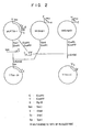
- the restriction enzyme map of the open reading frame (hereinafter abbreviated as ORF) present in this fragment is shown in Fig. 1 .
- the nucleotide sequence of this ORF and the amino acid sequence deduced therefrom are shown by SEQ ID NO: 1.
- the polypeptide deduced from this ORF was named TM-81 polypeptide.
- This synthetic DNA was ligated with the digestion fragment of PUC18 with HindIII and BamHI to obtain plasmid of about 2.8 kbp named PUC18P.
- the HN-bearing DNA fragment of about 1800 bp and the fragment containing the synthetic promoter recovered by 2.0% low melting agarose gel electrophoresis after full digestion of pUC18P with BamHI and SacI were ligated by ligase and these three fragments-ligated plasmid was extracted.
- the resulting plasmid of about 4.6 kbp was named pNZ87N.
- pUTTM1P was digested with HindIII and KpnI, the fragment of about 1300 bp and ligated with the fragment obtained by digestion of M13mp10 with HindIII and KpnI to obtain the single stranded recombinant phage.
- the oligonucleotide described above was annealed to the single stranded recombinant phage to cause the desired mutation by the method of Frits Eckstein et al.
- the fragment of about 1300 bp was recovered and ligated with the fragment obtained by digestion of pNZ1729R with restriction enzymes HindIII and KpnI using ligase to obtain plasmid pNZ7929-R2 (about 10.3 kbp) with the AluI cleavage site of pNZ7929-R1 being changed into the EcoRI cleavage site.
- pNZ87N was fully digested with restriction enzyme XbaI and the cleavage site was rendered blunt by Klenow fragment. Then EcoRI linker (5'-GGAATTCC-3') was added to and ligated with the digestion product using ligase. The plasmid was digested with EcoRI and HindIII. The fragment of about 300 bp was recovered by 1.2% low melting agarose gel electrophoresis.
- pNZ7929R2 was digested with restriction enzyme EcoT22I and then partially digested with EcoRI. The fragment of about 550 bp which is a part of TTM-1 DNA was recovered by 0.8% low melting agarose gel electrophoresis.
- pNZ7929RI was digested with restriction enzymes EcoT22I and HindIII and the fragment of about 9.4 kbp was recovered by 8% low melting agarose gel electrophoresis. These fragments were ligated by ligase and the three fragments-ligated plasmid was extracted. The plasmid of about 10.3 kbp was named pNZ2929XM1.
- Example 2 Construction and purification were carried out in a manner similar to Example 1 (3).
- the purified virus was named fNZ2929-XM1.
- dot blotting hybridization and Southern blot hybridization the location of each DNA inserted was confirmed in fNZ2929-XMI.
- TTM-1 polypeptide expressed in cells infected with fNZ7929-R1 and fNZ2929XM1
- fNZ7929-R1 and fNZ2929XM1 express TTM-1 polypeptide in infected cells
- immunofluorescent antibody technique using antisera against Mycoplasma gallisepticum S6 was employed.
- fN7929-R1 and fNZ2929XM1 were infected to CEF and incubation was carried out at 37°C until plaques appeared.
- fNZ2929XM1 not only expresses TTMG-1 polypeptide in the infected cells but also exhibits TTM-1 polypeptide on the surface of the infected cells.
- fNZ7929-R1 and fNZ2929XM1 were cultured in CEF at 37°C for 48 hours, the procedure of freezing and thawing was repeated twice to recover the cell suspension.
- the cell suspension was adjusted to have a virus titer of 106 pfu/ml and then inoculated to SPF chick (Line M, Nippon Seibutsu Kagaku Kenkyusho) of 7 days old at the right wing web in a dose of 10 ⁇ l. After the inoculation, generation of the pock was observed. Two weeks after the inoculation, sera were collected. The antibody titer of the sera collected was determined by ELISA.
- the purified TTM-1 polypeptide was dissolved in bicarbonate buffer in a concentration of 1 ⁇ g/well. After adsorbing to a 96 well microtiter plate, blocking was performed with skimmed milk to prevent the following non-specific adsorption. Next, a dilution of the sample serum was charged in each well and then horse radish peroxide-bound anti-chick immunoglobulin antibody (rabbit antibody) was added thereto as a secondary antibody. After thoroughly washing, 2,2'-azinodiethylbenzothiazoline sulfonate was added to the mixture as a substrate and a relative dilution magnification of the antibody was measured with an immuno-reader in terms of absorbance at a wavelength of 405 nm.
- anti-TTM-1 polypeptide chicken serum was used as a primary antibody for control.
- the results are shown in Table 2.
- Table 2. Antibody titer of fNZ2929XM1-inoculated chick to TTM-1 polypeptide Inoculated virus
- Antibody titer to anti-TTM-1 polypeptide (dilution magnification)* fNZ2929XM1 256 fNZ7929-R1 32 NP 1 ** 1 anti-TTM-1 polypeptide 256 * dilution magnification when SPF chicken serum dilution is as 1 ** not inoculated
- fNZ2929XM1 and fNZ2929-R1 which are recombinant viruses, can induce anti-TTN-1 polypeptide antibody and can be used as a vaccine for effectively preventing fowlpox and Mycoplasma gallisepticum infections.
- the digestion product was subjected to 0.6% low melting agarose gel electrophoresis. After the electrophoresis, the gel was immersed in an alkaline denaturation solution (0.5 M NaOH, 1.5 M NaCl) for 10 minutes to denature DNA. The gel was then immersed in a neutralization solution (3 M sodium acetate, pH 5.5) for 10 minutes for neutralization and then transferred onto a nylon membrane in 6-fold SSC solution (0.7 M NaCl, 0.07 M sodium citrate, pH 7.5).
- alkaline denaturation solution 0.5 M NaOH, 1.5 M NaCl
- the gel was then immersed in a neutralization solution (3 M sodium acetate, pH 5.5) for 10 minutes for neutralization and then transferred onto a nylon membrane in 6-fold SSC solution (0.7 M NaCl, 0.07 M sodium citrate, pH 7.5).
- the nylon membrane was baked at 80°C for 2 hours and 4-fold SET (0.6 M NaCl, 0.08 M Tris-HCl, 4 mM EDTA, pH 7.8)-10-fold Denhardt-0.1% SDS-0.1% Na 4 P 2 O 7 -50 ⁇ g/ml of denatured salmon sperm DNA and pUM-1 (cf. Japanese Patent Application Laid-Open No. 2-111795 ) labelled in a conventional manner was added thereto to perform hybridization at 68°C for 14 hours.
- the nylon membrane was overlaid on an X ray film. It was confirmed by autoradiography that hybridization occurred to the about 3.4 kbp fragment different from the fragment confirmed in Reference Example 1 (2).
- the transformants were cultured at 37°C for 15 hours in LB agar medium supplemented with 0.003% of 5-bromo-4-chloro-3-indolyl-B-D-galactopyranoside, 0.03 mM of isopropylthio-B-D-galactopyranoside and 40 ⁇ g/ml of ampicillin.
- white colonies were cultured at 37°C for 15 hours in LB liquid medium supplemented with 40 ⁇ g/ml ampicillin and plasmid was extracted by the method of Birnboim & Doly.
- the recombinant plasmid containing the same length as that of the XbaI fragment derived from MG was detected by 0.8% low melting agarose electrophoresis; this plasmid was named pUM67.
- the about 3.4 kbp fragment inserted into pUM67 was analyzed by the dideoxy method by Sanger et al.
- the restriction enzyme map of the open reading frame (ORF) present in this fragment is shown in Fig. 8 and the nucleotide sequence of this ORF and the amino acid sequence are shown by SEQ ID NO: 27.
- the polypeptide deduced from this ORF was named TM-67 polypeptide.
- TGA codons were concentrated at the downstream 10 portion in ORF of TM-67. Therefore, the EcoRI and PstI fragment of about 1300 bp containing all TGA codons were recovered from pUM67 and ligated with pUC19 digested with EcoRI and PstI to obtain PUCT1 (4.0 kbp).
- PUCT1 a template 15 according to polymerase chain reaction (PCR: Science, 230, 1350-1354 (1985 )
- primer DNAs for PCR shown by SEQ ID NOS: 28-33 were synthesized.
- Primers 1 to 6 corresponding to SEQ ID NOS: 28-33 which were employed for PCR are as follows.
- the fragment of 600 bp was amplified using Primer-1 and Primer-2 and then recovered; the fragment of 360 bp using Primer-3 and Primer-4; and the fragment of 340 bp using Primer-5 and Primer-6.
- the fragment of 600 bp was digested with EcoRI and NheI; the fragment of 360 bp was digested with Nhe and HindIII; and the fragment of 340 bp was digested with HindIII and PstI. Thereafter each digestion product was subjected to 2.0% low melting agarose gel electrophoresis and recovered from the agarose.
- pUC19 and pUC18 were digested with DraI and then XhoI linker was inserted to obtain plasmids pUC19X and pUC18X.
- the fragment of 600 bp and the fragment of 360 bp treated with the respective restriction enzymes and recovered were ligated with the digestion product of pUC19X with EcoRI and HindIII by ligase.
- the resulting plasmid was extracted and this plasmid was named pUC19XL (about 3.6 kbp).
- the 340 bp fragment digested with HindIII and PstI was ligated with the fragment obtained by digesting pUC18 with HindIII and PstI, using ligase.
- the resulting plasmid was extracted and named pUC18R (about 3 kbp).
- the fragment of about 2.5 kbp obtained by digestion of pUCI9XL with HindIII and XhoI, the fragment of 180 bp obtained by digestion of pUC18R with HindIII and SpeI, and the fragment of 1.1 kbp obtained by digestion of pUC18X with XbaI and XhoI were subjected to agarose gel electrophoresis, respectively, and then recovered. These fragments were ligated using ligase.
- the resulting plasmid was extracted and named pTM67 (about 3.7 kbp).
- Example 1 (1) After pUTTM1P obtained in Example 1 (1) was digested with SpeI and KpnI, the digestion product was subjected to agarose gel electrophoresis to recover the fragment of 3.9 kbp.
- the digestion product was subjected to agarose gel electrophoresis to recover the fragment of 0.9 kbp. The thus recovered fragment was ligated with the 3.9 kbp fragment described above using ligase.
- the resulting plasmid pUTM67 (4.8 kbp) was recovered. After this pUTM67 was digested with KpnI, the digestion product was partially digested with HindIII.
- the product was then subjected to agarose gel electrophoresis to recover the fragment of 2.1 kbp.
- the thus recovered fragment was ligated with the 9.0 kbp fragment obtained by digestion of PNZ1729R (cf. Reference Example 2) with HindIII and KpnI, using ligase.
- the resulting plasmid pNZ7929-67 (11.1 kbp) was recovered.
- Example 1 The procedures similar to Example 1 (3) were repeated using pNZ7929-67 obtained in (4) described above to obtain fNZ7929-67.
- the digestion product was subjected to 0.6% low melting agarose gel electrophoresis. After the electrophoresis, the gel was immersed in an alkaline denaturation solution (0.5 M NaOH, 1.5 M NaCl) for 10 minutes to denature DNA. The gel was then immersed in a neutralization solution (3 M sodium acetate, pH 5.5) for 10 minutes for neutralization and then transferred onto a nylon membrane in 6-fold SSC solution (0.7 M NaCl, 0.07 M sodium citrate, pH 7.5).
- alkaline denaturation solution 0.5 M NaOH, 1.5 M NaCl
- the gel was then immersed in a neutralization solution (3 M sodium acetate, pH 5.5) for 10 minutes for neutralization and then transferred onto a nylon membrane in 6-fold SSC solution (0.7 M NaCl, 0.07 M sodium citrate, pH 7.5).
- the nylon membrane was baked at 8.0°C for 2 hours and 4-fold SET (0.6 M NaCl, 0.08 M Tris-HCl, 4 mM EDTA, pH 7.8)-10-fold Denhardt-0.1% SDS-0.1% Na 4 P 2 O 7 -50 ⁇ g/ml of denatured salmon sperm DNA and pUM-1 (cf. Japanese Patent Application Laid-Open No. 2-111795 ) labelled in a conventional manner was added thereto to perform hybridization at 68°C for 14 hours.
- the nylon membrane was overlaid on an X ray film. It was confirmed by autoradiography that hybridization occurred to the about 6.3 kbp fragment.
- the transformants were cultured at 37°C for 15 hours in LB agar medium supplemented with 0.003% of 5-bromo-4-chloro-3-indolyl- ⁇ -D-galactopyranoside, 0.03 mM of isopropylthio- ⁇ -0-galactopyranoside and 40 ⁇ g/ml of ampicillin.
- White colonies grown on the medium were transferred to a nylon membrane and hybridization was carried out in a manner similar to (1) described above. Autoradiography reveals that cloning was effected and this plasmid was named pUM66 (about 9 kbp).
- the about 6.3 kbp fragment inserted into pUM66 was analyzed by the dideoxy method by Sanger et al.
- the polypeptide deduced from this ORF was named TM-66 polypeptide.
- Primers 1 to 10 corresponding to SEQ ID NOS: 34-43 using PCR are as follows.
- pUM66 was digested with B g 1II and SpeI and the fragment of about 1.2 kbp was recovered from 0.5% low melting agarose. The 1.2 kbp fragment was ligated with the digestion product of pUC19 with BamHI and XbaI to obtain pUCT2 (3.9 kbp). Next, using pUCT2 as a template and using Primer-1 and Primer-2, the fragment of 620 bp was amplified following conventional procedures for PCR and then recovered; after amplification of the fragment of about 550 bp using Primer-3 and Primer-4, the amplified fragment was recovered.
- the fragment of about 620 bp was digested with HindIII and AvaII; the fragment of 550 bp was digested with AvaII and BamHI. These fragments were ligated with the digestion product of pUC19 with HindIII and BamHI by ligase, respectively. The resulting plasmid was extracted and named pUC19-1 (3.9 kbp).
- the fragment of about 500 bp was amplified following conventional procedures for PCR and then recovered; after amplification of the fragment of about 700 bp using Primer-1 and ,Primer-5, the amplified fragment was recovered. Furthermore, the fragment of about 500 bp was digested with AflII and EcoRI; the fragment of about 700 bp was digested with HindIII and AflII. These fragments were ligated with the digestion product of pUC19 with HindIII and AflII by ligase. The resulting plasmid was extracted and named pUC19-2 (about 3.9 kbp).
- pUC19-1 was digested with EcoRI and the digestion product was subjected to 0.6% low melting agarose gel electrophoresis to recover the fragment of about 3.3 kbp.
- pUC19-2 was also digested with EcoRI and the digestion product was subjected to 2.0% low melting agarose gel electrophoresis to recover the fragment of about 550 bp.
- This fragment was ligated with the about 3.3 kbp fragment derived from pUC19-1 described above using ligase to obtain plasmid pUC19L bearing the fragment in which two TGA codons at the 5' end in ORF of TM-66 have been changed to TGG.
- pUM66 was digested with EcoRI and PvuII and the fragment of about 1720 bp was recovered from 0.6% low melting agarose gel. The recovered fragment was ligated with the digestion product of pUC19 with EcoRI and HincII to obtain plasmid pUCT3 (about 4.4 kbp).
- pUCT3 as a template and using Primer-4 and Primer-7, the fragment of about 820 bp was amplified following conventional procedures for PCR and also the fragment of about 900 bp using Primer-8 and Primer-1 was amplified likewise, and the both fragments were then recovered, respectively.
- the digestion product was ligated with the aforesaid about 900 bp fragment obtained by digestion with XbaI and HindIII and the digestion product of pUC19 with HindIII and EcoRI, using ligase to obtain plasmid pUCT4 (about 4.4 kbp).
- the fragment of about 880 bp was amplified following conventional procedures for PCR and also the fragment of about 900 bp using pUCT3 as a primer and using Primer-I and Primer-10 was amplified likewise following the conventional procedures for PCR; and the fragments were then recovered, respectively.
- this 880 bp fragment was digested with EcoRI and KpnI
- the digestion product was ligated with the aforesaid about 850 bp fragment obtained by digestion with HindIII and KpnI and the digestion product of pUC19 with EcoRI and HindIII, using ligase to obtain plasmid pUC19R.
- pUM66 was digested with MluI and PvuII and the fragment of about 4.8 kbp was then recovered from 0.6% low melting agarose gel. The recovered fragment was ligated with the about 1.0 kbp fragment obtained by the digestion of pUC19R with MluI and PstI to obtain plasmid. This plasmid was further digested with EcoT22I and NheI.
- the resulting fragment of about 5.2 kbp was ligated with the fragment of about 640 bp obtained by the digestion of pUC19L with EcoT22I and NheI, using ligase to obtain plasmid bearing the full length of ORF in which TGA codons in ORF of TM-66 were all changed to TGG.
- This plasmid was named pTM66 (about 5.8 kbp).
- pTM66 was digested with PstI
- the digestion product was partially digested with SspI to recover the fragment of about 2.4 kbp.
- pUTM66P was digested with HindIII and BamHI and the digestion product was recovered from low melting agarose gel.
- This fragment (about 2.5 kbp) was ligated with the fragment obtained by the digestion of pNZ1729R with HindIII and BamHI, using ligase to obtain the desired plasmid pNZ7929-66 (about 11.5 kbp).
- Example 1 The procedures similar to Example 1 (3) were repeated using pNZ7929-66 obtained in (4) described above to obtain fNZ7929-66.
- TM-67 and TM-66 polypeptides in cells infected with fNZ7929-67 and fNZ7929-66
- fNZ7929-67 and fNZ7929XM66 express the TM-67 and TM-66 polypeptides in infected cells.
- the immuno-fluorescence antibody method was carried out.
- fNZ7929-67 and fNZ7929-66 were infected to CEF, respectively and cultured at 37°C until plaques appeared. Thereafter the medium was fixed with cold acetone.
- Mycoplasma gallisepticum S6-immunized chicken serum or Mycoplasma gallisepticum- infected chicken serum as a primary antibody the medium was diluted to 100- to 1000-fold and the dilution was reacted.
- fNZ7929-67 and fNZ7929-66 were cultured in CEF at 37°C for 48 hours, the procedure of freezing and thawing was repeated twice to recover the cell suspension.
- the cell suspension was adjusted to have a virus titer of 10 6 pfu/ml and then inoculated through a stab needle to SPF chicken (Line M, Nippon Seibutsu Kagaku Kenkyusho) of 7 days old at the right wing web in a dose of 10 ⁇ l. After the inoculation, generation of the pock was observed. Two weeks after the inoculation, sera were collected.
- Mycoplasma gallisepticum S6 was inoculated on PPLO liquid medium (modified Chanock's medium) in a 10% concentration. After incubation at 37°C for 3 days, the cell mass was removed through a membrane filter of 0.45 ⁇ m. The filtrate was diluted with PPLO liquid medium in a cell count of 103 CFU/ml and the resulting dilution was provided as the cell solution for determination of activity.
- PPLO liquid medium modified Chanock's medium
- the cell solution was put in a polypropylene tube by 400 ⁇ l each and 100 ⁇ l each of standard chick serum, TMG-1 immunized serum (Japanese Patent Application Laid-Open No. 2-111795 ) and various sera were added thereto, respectively. By incubation at 37°C for 2 to 5 days, growth inhibition test was conducted.
- Mycoplasma gallisepticum S6 strain was incubated at 37°C for 3 to 5 days in liquid medium prepared by adding to 10 ml of PPLO broth basal medium 20% equine sera, 5% yeast extract, 1% glucose and a trace amount of Phenol Red as a pH indicator. As Mycoplasma gallisepticum grew, the pH of the culture medium decreased. At the time when the color of the pH indicator contained in the medium was changed from red to yellow, the incubation was terminated. After the culture medium was centrifuged at 8000G for 20 minutes, the cells were collected. The cells were suspended in PBS in a 1/10 volume of the medium. The suspension was again centrifuged at 10,000 rpm for 20 minutes and the cells were then collected. The collected cells were again suspended in 2.7 ml of PBS. After SDS was added to the suspension in a concentration of 1% and further 10 ⁇ g of RNase was added thereto, incubation was performed at 37°C for 30 minutes for lysis.
- the lysate was extracted 3 times with an equal volume of phenol and then 3 times with ethyl ether. By ethanol precipitation, 200 ⁇ g of Mycoplasma gallisepticum genomic DNA was obtained.
- the digestion product was subjected to 0.6% low melting agarose gel electrophoresis. After the electrophoresis, the gel was immersed in an alkaline denaturation solution (0.5 M NaOH, 1.5 M NaCl) for 10 minutes to denature DNA. The gel was then immersed in a neutralization solution (3 M sodium acetate, pH 5.5) for 10 minutes for neutralization and then transferred onto a nylon membrane in 6-fold SSC solution (0.7 M NaCl, 0.07 M sodium citrate, pH 7.5).
- alkaline denaturation solution 0.5 M NaOH, 1.5 M NaCl
- the gel was then immersed in a neutralization solution (3 M sodium acetate, pH 5.5) for 10 minutes for neutralization and then transferred onto a nylon membrane in 6-fold SSC solution (0.7 M NaCl, 0.07 M sodium citrate, pH 7.5).
- the nylon membrane was baked at 80°C for 2 hours and 4-fold SET (0.6 M NaCl, 0.08 M Tris-HCl, 4 mM EDTA, pH 7.8)-10-fold Denhardt-0.1% SDS-0.1% Na 4 P 2 O 7 -50 ⁇ g/ml of denatured salmon sperm DNA and pUM-16 (M-16 gene is contained in this plasmid; cf. Japanese Patent Application Laid-Open No. 2-111795 ) labelled in a conventional manner, was added thereto to perform hybridization at 68°C for 14 hours.
- the nylon membrane was overlaid on an X ray film. It was confirmed by autoradiography that hybridization occurred to the about 5.5 kbp fragment.
- the transformants were cultured at 37°C for 15 hours in LB agar medium supplemented with 0.003% of 5-bromo-4-chloro-3-indolyl- ⁇ -D-galactopyranoside, 0.03 mM of isopropylthio- ⁇ -D-galactopyranoside and 40 ⁇ g/ml of ampicillin.
- White colonies grown on the medium were transferred onto a nylon membrane and hybridization was carried out in a manner similar to (2) described above. It was confirmed by autoradiography that cloning was effected and this plasmid was named pUM16.
- the restriction enzyme map of this fragment is shown in Fig. 13 .
- the restriction enzyme map of the open reading frame present in this fragment is also shown in Fig. 14 and the nucleotide sequence of this ORF and the amino acid sequence deduced therefrom are shown by SEQ ID NO: 15.
- the polypeptide deduced from this ORP was named TM-16 polypeptide.
- sequences employed are described as sequence listing.
- the sequences used for the primers are described basically from the 3' end.
- the primers depicted from the 5' end in the body of the specification are described from the 5' end to conform to the description.
Description
- The present invention relates to a novel polypeptide showing antigenicity to Mycoplasma gallisepticum, a fused polypeptide between the said polypeptide and a signal membrane anchor, and a recombinant Avipox virus capable of expressing a polypeptide showing antigenicity to Mycoplasma gallisepticum, especially a polypeptide showing antigenicity on the membrane surface of a host cell, as well as use thereof.
- It is expected that a polypeptide showing antigenicity to Mycoplasma gallisepticum can be utilized as an effective ingredient of a vaccine for Mycoplasma gallisepticum infections, since an egg-laying rate and a hatching rate of eggs produced by infected chickens are markedly reduced when infected with Mycoplasma gallisepticum. At present, the system using Escherichia coli or yeast is known to prepare the antigenic protein of Mycoplasma gallisepticum by genetic engineering (Japanese Patent Application Laid-Open No.
2-111795 - In a virus protein where the virus infects cells, one type of a protein expressed is transported to the cell surface and the protein is expressed on the surface of a cell membrane (hereinafter such a state is sometimes merely referred to as being expressed on the cell surface) and another type of a protein that is not expressed on the cell surface. A representative example of the former protein is a glycoprotein contained in the coat of a virus. A recombinant virus that expresses such a protein efficiently exhibits the protein on the cell surface. It is thus considered that a high antibody titer can be induced in poultry infected with this recombinant virus (Japanese Patent Application Laid-Open No.
1-157381 - It is not expectable to induce a high antibody titer from such recombinant viruses that express these proteins, since they are expressed on the cell membrane surface merely in an extremely small quantity. However, if such a protein can be expressed on the cell membrane surface in a large quantity by genetic engineering, a high antibody titer will be induced. Thus, investigations have been made to express on the membrane surface such a protein that is not principally expressed on the membrane surface. For example, there is a report that DNA encoding a signal protein having the function of secreting a protein on the cell membrane surface and DNA encoding a membrane anchor protein having the function of retaining the secreted protein so as not to leave out of the cell membrane surface are ligated with the 5' end and the 3' end of DNA encoding an antigenic protein, respectively, and a recombinant vaccinia virus inserted with the resulting hybrid DNA expresses the antigenic protein on the cell membrane surface of a host (J. Viol., 64, 4776-4783 (1990) or Mol. Cell. Biol., 6, 3191-3199 (1986)). However, DNA encoding a signal and DNA encoding a membrane anchor are independently ligated with DNA encoding an antigenic protein in these examples so that it is hardly applicable practically due to complicated preparation of a recombinant virus.
- The present inventors have made extensive studies to provide a polypeptide having antigenicity originating in Mycoplasma and showing a high antigenicity, a polypeptide having antigenicity to Mycoplasma gallisepticum expressed on the cell membrane surface especially in a large quantity, DNA encoding the polypeptide, a recombinant virus inserted with the same DNA and a vaccine utilizing the virus. As a result, the present invention has come to be accomplished.
- In a first aspect, the invention provides a substantially pure antigenic protein which is reactive with Mycoplasma gallisepticum-immunized sera or Mycoplasma gallisepticum-infected sera and which has the amino acid sequence shown by SEQ ID NO: 27.
- The invention further provides an isolated DNA encoding the antigenic protein.
- The invention also provides a fused protein comprising a polypeptide of Mycoplasma gallisepticum ligated at the N-terminus thereof with a signal membrane anchor of type II external membrane protein of a virus that infects poultry, wherein said polypeptide of Mycoplasma gallisepticum has the amino acid sequence shown by SEQ ID NO: 27.
- The signal membrane anchor may be encoded by DNA comprising SEQ ID NO: 13. A hydrophilic sequence having between 10 and 50 amino acids may be present at the carboxy terminal of the signal membrane anchor.
- Also provided is a hybrid DNA encoding the fused protein.
- In a further aspect, the invention provides a component vaccine comprising a protein according to the invention.
- In a further aspect, the invention provides a recombinant Avipox virus comprising DNA encoding a polypeptide of Mycoplasma gallisepticum, wherein said polypeptide of Mycoplasma gallisepticum has the amino acid sequence shown by SEQ ID NO: 27.
- The polypeptide of Mycoplasma gallisepticum may have at the N-terminus thereof, a signal membrane anchor of a type II external membrane protein from a virus that infects poultry.
- Further provided is a live vaccine for poultry Mycoplasma gallisepticum infection comprising a live recombinant Avipox virus according to the invention.
-
-
Fig. 1 shows a restriction enzyme map of DNA including the open reading frame of TM-81. -
Fig. 2 shows the procedure for construction of TTM-1N and TTM-1C. -
Fig. 3 shows the procedure for constructing pNZ7929-R1. -
Fig. 4 shows the procedure for constructing pNZ87N. -
Fig. 5 shows the procedure for constructing pNZ7929-R2. -
Fig. 6 (A) and(B) show the procedure for constructing pNZ2929XM1. -
Fig. 7 shows a restriction enzyme map of DNA including the open reading frame of TTM-1 polypeptide. -
Fig. 8 shows a restriction enzyme map of DNA including the open reading frame of TM-67 polypeptide and the position of synthetic primers on ORF. -
Fig. 9 (A) and(B) show the procedure for constructing pHZ79Z9-67. -
Fig. 10 shows a restriction enzyme map of DNA including the open reading frame of TM-66 polypeptide and the position of synthetic primers on ORF. -
Fig. 11(A) ,11(B) and11(C) show the procedure for constructing pTM66. -
Fig. 12 shows the procedure for constricting pNZ7929-66. -
Fig. 13 shows a restriction enzyme map of DNA encoding the full length of TM-16 polypeptide. -
Fig. 14 shows a restriction enzyme map of the open reading frame of TM-16 polypeptide. - Described herein is a novel polypeptide which shows an antigenicity which originates in Mycoplasma gallisepticum having a high antigenicity, and which includes a polypeptide showing an antigenicity which causes an antigen-antibody reaction with sera immunized with Mycoplasma gallisepticum or sera and which is encoded by the DNA sequence having the restriction enzyme map shown in
Fig. 7 originating in Mycoplasma gallisepticum, or a modified polypeptide thereof. Specific examples of the polypeptide having such an polypeptide include those showing an antigenicity and having amino acid sequences of SEQ ID NOS: 1, 15, 16 and 27. The modified polypeptide showing an antigenicity referred to herein is a polypeptide in which the amino acid sequence is modified by substitution, loss, deletion, insertion or addition but which shows an antigenicity comparable to that of the aforesaid polypeptide. Taking SEQ ID NO: 1 as an example, a modified polypeptide is used to mean a polypeptide having the same antigenicity as in an antigenic protein having the amino acid sequence equivalent thereto and having a homology of at least 70% to the amino acid sequence of said polypeptide, preferably 80% or more, most preferably 90% or more. The homology referred to herein is used to mean the homology determined as an index by DNA sequencing input analysis system "DNASIS" (marketed by Takara Shuzo Co.). - Hereinafter a sequence number is sometimes simply referred to as sequence in the specification. For example, Sequence No. 1 is sometimes referred to as
Sequence 1. - Furthermore, the DNA which encodes the polypeptide showing an antigenicity described herein includes DNA encoding a polypeptide in which the amino acid is modified by deletion, additions, insertion, loss, substitution, etc., so long as it causes an antigen-antibody reaction with sera immunized with Mycoplasma gallisepticum or sera and shows an antigenicity originating in Mycoplasma gallisepticum or an antigenicity equivalent thereto.
- Avipox virus which is a second aspect of the present invention is a recombinant Avipox virus inserted with a hybrid DNA in which DNA encoding the antigenic polypeptide of Mycoplasma gallisepticum (hereinafter abbreviated as antigenic DNA) or DNA encoding a signal membrane anchor of Type II external membrane protein is ligated with DNA encoding a polypeptide showing an antigenicity of Mycoplasma gallisepticum. In order to express large quantities of the polypeptide showing an antigenicity of Mycoplasma gallisepticum that is not basically expressed on the surface of cell membrane, it is preferred to employ the hybrid DNA.
- That is, in the second aspect of the present invention, there are provided a polypeptide showing an antigenicity of Mycoplasma gallisepticum (hereinafter sometimes merely referred to as antigenic protein), a fused polypeptide ligated at the N terminus of the polypeptide with a signal membrane anchor of type II outer membrane protein of a virus infected to poultry (hereinafter merely referred to as signal membrane anchor), a vaccine against Mycoplasma gallisepticum infections comprising as an effective ingredient the antigenic protein or the fused polypeptide, a hybrid DNA which encodes the fused polypeptide, a recombinant Avipox virus inserted into the genomic region non-essential to growth of Avipox virus (hereinafter referred to as non-essential region) with DNA encoding the antigenic protein or the hybrid DNA, and a live vaccine against Mycoplasma gallisepticum which comprises the Avipox virus as an effective ingredient.
- The signal membrane anchor which is employed in the present invention as the second aspect is a polypeptide region having the function of transporting type II external membrane protein of a virus infected to poultry to the surface of cell membrane and expressing the transported protein on the surface of cell membrane, and is preferably derived from a virus which is non-pathogenic to human. The DNA encoding the signal membrane anchor which is employed in the present invention (hereinafter referred to as signal membrane anchor DNA) can be readily found by amino acid sequencing analysis of the hydrophobic peptide region of type II external membrane protein at the amino terminus. A specific example of the signal membrane anchor is that having the sequence shown by SEQ ID NO: 13 (Mol. Cell. Biol., 10, 449-457 (1990)). This DNA codes for 22 amino acids at the amino terminus of hemagglutinin neuraminidase (hereinafter abbreviated as HN protein) of Newcastle disease virus (hereinafter abbreviated as NDV).
- In order to stably exhibit the expressed antigenic protein on the cell membrane, it is effective for a hydrophilic peptide to be present at the carboxy terminal of the signal membrane anchor. Accordingly, it is preferred that DNA encoding a hydrophilic peptide be added downstream the signal membrane anchor DNA. DNA to be added comprises base pairs corresponding to 10 to 50 amino acids, preferably 20 to 30 amino acids.
- Specific examples of the DNA encoding the antigenic protein described herein, in addition to the four sequences described herein, DNA described in Japanese Patent Application Laid-Open No.
1-111795 WO 93/24646 - Sources for collecting such a DNA may be any of the sources so long as they belong to Mycoplasma gallisepticum. Specific examples include S6 strain (ATCC 15302), PG31 (ATCC 19610) and the like.
- The hybrid DNA which is used in the present invention as its second aspect is the aforesaid signal membrane anchor DNA ligated with DNA encoding a polypeptide showing an antigenicity. The fused polypeptide of the present invention is a polypeptide encoded by the hybrid DNA described above which contains a part of the signal membrane anchor and a part of the polypeptide showing an antigenicity in the molecule of the polypeptide. The hybrid DNA can be produced in a conventional manner, e.g., by modifying the 3' end of the signal membrane anchor DNA and the 5' end of the DNA encoding the antigenic protein so as to form ligatable restriction enzyme digestion fragments, and ligating both DNAs according to the method for ligation using a ligase or the method for ligating both DNAs with a ligase by inserting an appropriate linker therebetween. The signal membrane anchor and the DNA encoding the polypeptide showing an antigenicity may contain therebetween, for example, DNA encoding a hydrophilic peptide, DNA encoding other antigenic protein, linker DNA, etc., so long as the signal membrane anchor DNA and the DNA encoding the polypeptide showing an antigenicity are expressed as one polypeptide. The fused polypeptide of the present invention is obtained by incubating a recombinant Avipox virus, later described, in culture cells such as chick embryo fibroblast (hereinafter referred to as CEF cells) or embryonated chorioallantoic membrane cells, etc., and purifying the desired polypeptide by a method optionally chosen from chromatography, precipitation by salting-out, density gradient centrifugation, etc. The fused polypeptide thus obtained can be used as a component vaccine which will be later described.
- The recombinant Avipox virus of the present invention is a recombinant Avipox virus in which the aforesaid DNA or hybrid DNA is inserted in the non-essential region. The recombinant Avipox virus of the present invention may be constructed in a conventional manner, e.g., by the method described in Japanese Patent Application Laid-Open No.
1-168279 - As the non-essential region of Avipox virus which is used in the present invention, there are a TK gene region of quail pox virus, a TK gene region of turkey pox virus and DNA fragments described in Japanese Patent Application Laid-Open
1-168279 - Examples of the vector used in the present invention include plasmids such as pBR322, pBR325, pBR327, pBR328, pUC7, pUC8, pUC9, pUC19, and the like; phages such as λ phage, M13 phage, etc.; cosmid such as pHC79 (Gene, 11, 291, 1980) and the like.
- The Avipox virus used in the present invention is not particularly limited so long as it is a virus infected to poultry. Specific examples of such a virus include pigeon pox virus, fowl pox virus (thereafter abbreviated as FPV), canary pox virus, turkey pox virus, preferably turkey pox virus, pigeon pox virus and FPV, more preferably pigeon pox virus and FPV. Specific examples of the most preferred Avipox virus include FPVs such as ATCC VR-251, ATCC VR-249, ATCC VR-250, ATCC VR-229, ATCC VR-288, Nishigahara strain, Shisui strain, CEVA strain and a viral strain among CEVA strain-derived viruses which forms a large plaque when infected to chick embryo fibroblast, and a virus such as NP strain (chick embryo-conditioned pigeon pox virus Nakano strain), etc. which is akin to FPV and used as a fowlpox live vaccine strain. These strains are commercially available and readily accessible.
- Then, the aforesaid antigenic DNA or hybrid DNA is inserted into the non-essential region of the first recombinant vector described above to construct a second recombinant vector. Where the hybrid DNA is employed, a promoter is generally inserted upstream the hybrid DNA. The promoter used may be a promoter having any nucleotide sequence, irrespective of a synthetic or natural promoter, as far as it effectively functions as a promoter in the system of transcription possessed by APV. Accordingly, not only a promoter inherent to APV such as a promoter of APV gene encoding thymidine kinase but also DNA derived from viruses other than APV and DNA derived from eucaryote or procaryote may also be employed in the present invention, as long as these substances meet the requirements described above. Specific examples of such a promoter include a promoter of vaccinia virus (hereinafter sometimes abbreviated as VV) described in J. Virol., 51, 662-669 (1984), more specifically a promoter of VV DNA encoding 7.5 K polypeptide, a promoter of VV DNA encoding 19 K polypeptide, a promoter of VV DNA encoding 42 K polypeptide, a promoter of VV DNA encoding thymidine kinase, a promoter of VV DNA encoding 28 K polypeptide, etc. Furthermore, there may be used a synthetic promoter obtained by modification of the Moss et al. article (J. Mol. Biol., 210, 749-776, 771-784, 1989), a promoter synthesized by Davidson, a promoter obtained by modifying a part of the Davidson promoter through deletion or change within such a range that does not lose the promoter activity (e.g., T T T T T T T T T T T T G G C A T A T A A A T A A T A A T A A A T A C A A T A A T T A A T T A C G C G T A A A A A T T G A A A A A C T A T T C T A A T T T A T T G C A C T C or T T T T T T T T T T T T T T T T T T T T G C C A T A T A A A T A A T A A A T A C A A T A A T T A A TTACGCGTAAAAATTGAAAAACTAT T C T A A T T T A T T G C A C T C etc.).
- Further in view of easy detection of the recombinant virus, a marker DNA such as DNA encoding β-galactosidase may also be inserted.
- The recombinant Avipox virus may be constructed by transfecting the second recombinant vector described above to animal culture cells previously infected with Avipox virus and causing homologous recombination between the vector DNA and the viral genome DNA. The animal culture cells used herein may be any cells in which Avipox can grow. Specific examples of such animal culture cells are CEF cells, embryonated egg chorioallantoic membrane cells, and the like.
- The desired recombinant Avipox virus is isolated from the virus infected to host cells by the method of plaque hybridization, etc. The recombinant Avipox virus may be further purified by plaque assay, etc.
- The recombinant virus of the present invention constructed by the method described above can be inoculated to fowl as a live vaccine against Mycoplasma gallisepticum infection.
- The live vaccine of the present invention is prepared by, e.g., the following method, though the process is not particularly limited. The recombinant virus of the present invention is infected to cells in which the virus can grow (hereafter referred to as host cells). After the recombinant virus grows, the cells are recovered and homogenated. The homogenate is centrifuged with a centrifuging machine to separate into the precipitates and the high titer supernatant containing the recombinant virus in a centrifuging tube. The resulting supernatant is substantially free of host cells but contains the cell culture medium and the recombinant virus and hence can be used as a live vaccine. The supernatant may be diluted by adding a pharmacologically inactive carrier, e.g., physiological saline, etc. The supernatant may be freeze-dried to be provided for use as a live vaccine. A method for administration of the live vaccine of the present invention is not particularly limited and examples of the administration include a method for scratching the skin and inoculating the live vaccine on the scratch, effecting the inoculation through injection, oral administration by mixing the live vaccine with feed or drinking water, inhalation by aerosol or spray, etc. In order to use as the live vaccine, the dosage may be the same as ordinary live vaccine; for example, approximately 102 to 108 plaque forming unit (hereinafter abbreviated as PFU) is inoculated per chick. Where the inoculation is effected by injection, the recombinant virus of the present invention is generally suspended in about 0.1 ml of an isotonic solvent such as physiological saline and the resulting suspension is provided for use. The live vaccine of the present invention may be stored under ordinary conditions and provided for use. For example, when the recombinant virus of the present invention is freeze-dried, it is possible to store at room temperature (20 to 22°C). It is also possible to freeze the virus suspension at -20 to -70°C and store the frozen suspension.
- On the other hand, the component vaccine of the present invention comprises as an effective ingredient the polypeptide showing an antigenicity in accordance with the present invention, especially the fused polypeptide. The component vaccine may be administered to fowl in the same manner as in the live vaccine described above. The dose is generally in the range of approximately 1 µg to 1 mg per one subject.
- According to the present invention, the polypeptide showing an Mycoplasma gallisepticum antigenicity and the, fused polypeptide between the said polypeptide and the signal membrane anchor are obtained. In particular, this fused polypeptide is effective as a vaccine against Mycoplasma gallisepticum infections. By utilizing DNA encoding the fused protein, the recombinant Avipox virus which can express the polypeptide showing an Mycoplasma gallisepticum antigenicity is obtained. The recombinant Avipox virus is effective as a potent live vaccine against Mycoplasma gallisepticum infections. In addition, the novel polypeptide showing an antigenicity of the present invention and DNA encoding the same can be utilized as a component vaccine and a live vaccine, respectively.
- Hereinafter the present invention will be described with reference to the examples and the reference examples but is not deemed to be limited thereto.
- Obtaining of polypeptide DNA TTM-1 in which Mycoplasma gallisepticum is expressed:
- Mycoplasma gallisepticum S6 strain was cultured at 37°C for 3 to 5 days in liquid medium prepared by supplementing 20% horse serum, 5% yeast extract, 1% glucose and a trace amount of phenol red as a pH indicator in 100 ml of PPLO broth basal medium. As Mycoplasma gallisepticum proliferated, pH of the culture broth decreased. At the point of time when the color of the pH indicator contained in the culture broth changed from red to yellow, incubation was terminated. The culture medium was centrifuged at 8000G for 20 minutes to collect the cells. The cells were then suspended in 1/10 volume of PBS based on the volume of culture medium. The suspension was again centrifuged at 10,000 rpm x G for 20 minutes to collect the cells. The collected cells were resuspended in 2.7 ml of PBS and SDS was added thereto in a final concentration of 1%. Furthermore 10 µg of RNase was added to the mixture. The mixture was incubated at 37°C for 30 minutes to cause lysis.
- The lysate was extracted 3 times with an equal volume of phenol and then 3 times with ethyl ether. The extract was precipitated with ethanol to give 200 µg of genomic DNA of Mycoplasma gallisepticum.
- After 1 µg of Mycoplasma gallisepticum DNA obtained in (1) described above was digested with XbaI, the digestion product was subjected to 0.6% low melting agarose gel electrophoresis. After the electrophoresis, the gel was immersed in an alkaline denaturation solution (0.5 M NaOH, 1.5 M NaCl) for 10 minutes to denature DNA and further immersed in a neutralizing solution (3 M sodium acetate, pH 5.5) for 10 minutes to neutralize. Following the neutralization, the DNA was transferred onto a nylon membrane in 6-fold SSC solution (0.7 M NaCl, 0.07 M sodium citrate, pH 7.5). After air drying, the membrane was heated at 80°C for 2 hours. 4-Fold SET (0.6 M NaCl, 0.08 M Tris-HCl, 4 mM EDTA, pH 7.8)-10-fold Denhardt-0.1% SDS-0.1% Na4P2O7-50 µg/ml of denatured salmon sperm DNA and pUM-1 (see Japanese Patent Application Laid-Open No.
2-111795 - After 4 µg of Mycoplasma gallisepticum DNA obtained in Example 1 (1) described above was digested with restriction enzyme XbaI, the digestion product was subject to 0.6% low melting agarose gel electrophoresis. After the electrophoresis, the fragment of about 3.4 kbp was recovered. The fragment was ligated with XbaI-digested pUC-19 using ligase and competent E. coli TG1 strain was transformed by the ligation product. The transformants were cultured at 37°C for 15 hours in LB agar medium containing 0.003% of 5-bromo-4-chloro-3-indolyl-β-D-galactopyranoside, 0.03 mM of isopropylthio-β-D-galactopyranoside and 40 µg/ml of ampicillin. White colonies grown on the agar medium were transferred onto a nylon membrane followed by hybridization in a manner similar to (2) above. Autoradiography revealed that cloning was effected and, the thus obtained plasmid was named pUTTM1.
- After pUTTM-1 of (3) described above was digested with restriction enzymes SacI and EcoRI and the digestion product was then subjected to 0.8% low melting agarose gel electrophoresis. The 1.1 kbp fragment containing the 5'-end of TTM-1 was recovered by treating with phenol-chloroform and precipitating with ethanol. The fragment was ligated with the fragment obtained by digestion of M13mp11 phage with SacI and EcoRI. The ligation reaction solution was mixed at m.o.i. of 0.1 with a solution obtained by culturing E. coli TG1 at 37°C for 24 hours, adding IPTG thereto in a final concentration of 100 mM and further supplementing IPTG in a X-gal concentration of 2%. The resulting mixture was inoculated on soft agar for solidification. Incubation was then performed at 37°C for 24 hours. Among the phage plaques formed, recombinant phage TTM-1N containing 1.1 kbp DNA of TTM-1 was collected from the phage in which the color did not change to blue.
- Likewise, pUTTM-1 was digested with EcoRI and EcoRV. After 0.8% low melting agarose gel electrophoresis, the 0.4 kbp fragment containing the 3'-end of TTM-1 was recovered from the gel. A phenol-chloroform treatment followed by ethanol precipitation grave M13mp10 phage. M13mp10 phage was ligated with the fragment obtained by digestion with EcoRI and EcoRV using ligase. The reaction solution was treated as in the cloning of the 1.1 kbp DNA. Recombinant phage TTM-1C containing 0.4 kbp DNA of TTM-1 was thus obtained.
- The two recombinant phage obtained in (4) described above were added at m.o.i. of 0.1, respectively, to E. coli TG1 proliferated at 37°C in 100 ml of 2 x YT medium. After shake culture at 37°C for 5 hours, centrifugation was performed at 5000G for 30 minutes to obtain the cell-free supernatant. A 0.2-fold volume of polyethylene glycol/sodium chloride mixture (20% polyethylene glycol #6000, 2.5 M NaCl) was added to the supernatant. After settlement at 4°C for an hour, the mixture was centrifuged at 5000G for 20 minutes to recover the precipitates. The precipitates were dissolved in 500 µl of TE buffer (10 mM Tris-HCl, mM EDTA, pH 8.0). After extraction with phenol-chloroform, single stranded DNA of each recombinant phage was recovered by ethanol precipitation.
- The thus obtained DNA has TGA at the middle of the sequence. This TGA sequence is recognized as a termination codon in a normal cell so that the TGA sequence does not translate the sequence added thereafter. Therefore, in order to translate the TGA portion as methionine, the basic adenine which corresponds to the third nucleotide in codon NNN must be modified to guanine. Thus, the following two oligonucleotides were synthesized.
- Sequence No. 17:
3'-TACGTTCTTCCTGGCAAACCTTACCACTACTT-5' - Sequence No. 18:
3'-CTACAAAGAACCTAAATATCA-5' - The oligonucleotide shown by Sequence No. 17 (SEQ ID NO: 17) is annealed to single stranded DNA of TTM-1N and the oligonucleotide shown by Sequence No. 18 to single stranded DNA of TTM-1C to cause the desired mutation by the method of Frits Eckstein et al. (Nucleic Acid Research, 8749-8764, 1985). The thus obtained recombinant phages were named TTM-1N' and TTM-1C', respectively. The TTM-1N' and TTM-1C' phage DNAs thus obtained were digested with restriction enzymes SacI-EcoRI and EcoRI-BglII, respectively. By 0.8% low melting agarose gel electrophoresis, the fragments of 1.1 kbp and 0.4 kbp were extracted from the agarose gel and recovered by ethanol precipitation. On the other hand, plasmid pUTTM-1 was also digested with SacI-BglII. The 4.8 kbp fragment bearing a vector was extracted by 0.8% low melting agarose gel electrophoresis and recovered by ethanol precipitation. The thus obtained three fragments were ligated by ligase and competent E. coli TG1 strain was transformed to obtain plasmid pUTTM-1' bear-ing TTM-1' with mutagenesis at the desired site thereof. The nucleotide sequence of TTM-1' is as shown by SEQ ID NO: 14 according to the Dideoxy method by Sanger et al. (Proc. Natl. Acad. Sci. USA, 74, 5463 (1977)). The nucleotide sequence is substantially the same as the 40 kilodalton TTM-1 polypeptide of M. gallisepticum.
- The ECORI fragment (about 7.3 kbp) of NP strain was inserted into pUC18 at the EcoRI digestion site (terminus at the multi-cloning site) to obtain plasmid pNZ133 (about 10.0 kbp). From the plasmid the HpaI-SpeI fragment (about 3.0 kbp fragment derived from NP strain) was excised out and rendered blunt end by Klenow fragment. Furthermore, the EcoRI-HindIII fragment (multi-cloning site of 52 bp) was removed from pUC18 and rendered blunt end by Klenow fragment. The two fragments were ligated with each other to form a plasmid. After removing the EcoRV site in the BpaI-SpeI fragment, the EcoRI-HindIII fragment (multi-cloning site of 52 bp) of pUC18 is inserted therein using HindIII linker (5'-CAAGCTTG-3') and EcoRI linker (5'-GGAATTCC-3') to construct plasmid pNZ133SR.
- Sequence No. 2 (SEQ ID NO: 2) and Sequence No. 3 (SEQ ID NO: 3) (bearing FPV promoter of 17 bases and linked to a translation initiation codon for lacZ) were annealed to double strands. Sequence No. 4 (SEQ ID NO: 4) annealed to the lacZ gene (derived from pMC1871 an pMA001, Sirakawa et al., Gene, 28, 127-132, 1984) and Sequence No. 5 (SEQ ID NO: 5), Sequence No. 6 (SEQ ID NO: 6) and Sequence No. 7 (SEQ ID NO: 7), Sequence No. 8 (SEQ ID NO: 8) and Sequence No. 9 (SEQ ID NO: 9), Sequence No. 10 (SEQ ID NO: 10) and Sequence No. 11 (SEQ ID NO: 11), were ligated with each other (which contains a modified synthetic promoter of poxvirus shown by nucleotide sequence: T T T T T TTTTTTTTTTTTTTTGGCATATAAA TAATAAATACAATAATTAATTACGC G T A A A A A T T G A A A A A C T A T T C T A A T TTATTGCACTC from the next T of AGC at the 5' end of Sequence No. 3 to C before G of Sequence No. 5 at the 3' end, and further linked to the multi-cloning site and poxvirus initial transcription termination signal on the both directions (SEQ ID NO: 12) (Yuen et al., Proc. Natl. Acad. Sci., USA, 88, 6417-6421, 1989) thereby to obtain the EcoRI-HindIII fragment (about 3.5 kbp). The EcoRI-HindIII fragment was inserted into pNZ133SR to construct plasmid pNZ1729R.
- In order to form the restriction enzyme DraI digestion site upstream ATG corresponding to initiation codon of TTM-1' protein in plasmid pUTTM1' (see
WO 93/24646 - Sequence No. 19
3'-TATAGAATTAAATTTTACTTATTC-5' - Next, after pUTTM-1' was digested with restriction enzymes SacI and EcoRI, the fragment of about 2300 bp was recovered and then ligated with the fragment obtained by digestion of M13mp10 with SacI and EcoRI to obtain recombinant phage TTM-1'. The oligonucleotide described above was annealed to single stranded TTM-1' to cause the desired variation by the method of Frits Eckstein et al. This recombinant phage DNA variant was digested with restriction enzymes SacI and EcoRI. The fragment of about 2300 bp was recovered and cloned to the vector-bearing fragment obtained by digestion of pUTTM-1' again with SacI and EcoRI to obtain pUTTMID.
- A synthetic promoter was prepared by synthesizing DNAs of Sequence-20 and Sequence-21 followed by annealing, whereby the digestion sites with restriction enzymes HindIII and HincII at the end.
- Sequence-20 5'-AGCTTTTTTTTTTTTTTTCCCATATAAATAATAAATACAATAATATACCCTAAAATT
- Sequence-21

- Finally, the 1200 bp fragment obtained by digestion of pUTTMID with restriction enzymes DraI and BglII was ligated with the synthetic promoter described above and the fragment obtained by digestion of pUC18 with HindIII and BamHI to give plasmid pUTTMIP of about 4.0 kbp.
- After plasmid pUTTMlp obtained in (1) was digested with restriction enzymes HindIII and KpnI, the fragment of about 1300 bp was recovered. Next, vector pNZ1729R (
EP-A-0520753 ) for FPV recombination obtained in Reference Example 2 was digested with restriction enzymes HindIII and KpnI. The two fragments were ligated with each other to obtain the desired vector pNZ7929-R1 (about 10.3 kbp) for recombination. - NP strain, which is a fowlpox live vaccine strain, was infected to monolayered CEF at m.o.i. = 0.1. Three hours after, these cells were peeled apart from the monolayer by a treatment with trypsin to form a cell suspension. After 2 x 10⁷ cells in the suspension were mixed with 10 µg of plasmid pNZ7929-R1 for recombination, the mixture was suspended in Saline G (0.14M NaCl, 0.5 mM KCl, 1.1 mM Na2HPO4, 1.5 mM K92PO4, 0.5 mM MgCl2 6H2O, 0.011% glucose). The suspension was subjected to electrophorasis under conditions of 3.0 kV cm-1, 0.4 msec and 25°C, using Gene Pulser (Bio-Rad) at room temperature. The plasmid-infected cells were then cultured at 37°C for 72 hours. The cells were lysed by freeze and
thaw 3 times to recover viruses containing the recombinant virus. - The recombinant virus recovered was selected as follows. The recovered viral solution was infected to monolayered CEF and 10 ml of agar solution containing growth medium was overlaid thereon. After agar was solidified at room temperature, incubation was performed at 37°C until plaques of FPV appeared. Then agar medium containing Bluo gal in a concentration of 200 µg/ml was overlaid on the agar followed by incubation at 37°C for further 48 hours. Among all of the plaques, about 1% of the plaques were colored blue. These blue plaques were isolated and recovered. By the same procedures, isolation and recovery were repeated to purify the virus until all the plaques were stained to blue with Bluo gal. In general, the repeated procedures were terminated by 3 to 4 times. The purified virus was named fNZ7929-R1. In fNZ7929-R1, each position of the DNA inserted was confirmed by dot blotting hybridization and Southern blotting hybridization.
- Using Mycoplasma gallisepticum S6 strain, 200 µg of Mycoplasma gallisepticum DNA was obtained in a manner similar to Reference Example 1 (1) described above.
- After 4 units of restriction enzyme AluI was added to 40 µg of Mycoplasma gallisepticum genomic DNA obtained in (1), incubation was conducted at 37°C for 10 minutes for partial digestion. The partially digested genomic DNA was subjected to 0.8% low melting agarose gel electrophoresis. The DNA fragment having a strand length of approximately 1.0 kbp to 4.0 kbp was recovered from the gel. The DNA fragment was treated with phenol and then precipitated with ethanol to give 4 µg of the DNA fragment partially digested with AluI.
- S-Adenosyl-L-methionine was added to 1.2 µg of the AluI-partially digested-DNA fragment in a final concentration of 80 µM and 20 units of EcoRI methylase was further added thereto to methylate the deoxy-adenosine site in the EcoRI recognition sequence, thereby to render the sequence non-sensitive to EcoRI. EcoRI linker was ligated with this DNA fragment using ligase. The ligation product was then mixed with the EcoRI digestion fragment of λgt11 DNA to ligate with each other by ligase. The reaction solution was used to effect in vitro packaging in a conventional manner (DNA Cloning, Vol. 1, A Practical Approach, edited by D.M. Glover). The resulting product was transfected to Escherichia coli Y1088 strain (Amersham) followed by incubation at 37°C for 12 hours in LB agar medium containing 0.003% of 5-bromo-4-chloro-3-indolyl-β-D-galactopyranoside and 0.03 mM isopropylthio-β-D-galactopyranoside. Among the plaques formed, a library size was estimated by the count of white plaques to prepare DNA library of 106 pfu (plaque forming unit).
- The phage obtained from the DNA library prepared in (2) was added to a suspension of Escherichia coli Y1090 strain (Amersham) in an aqueous solution of 10 mM MgSO4 to form 500 to 1000 plaques on one plate of 8 cmφ to effect adsorption for 15 minutes. Furthermore 2.5 ml of LB soft agar medium warmed to 45°C was added and overlaid on the LB agar medium followed by incubation at 42°C for 3 to 4 hours. A nylon membrane filter was immersed in 10 mM of IPTG aqueous solution, air-dried and then overlaid on the plate described above followed by incubation at 37°C for further 2 to 3 hours. After the incubation, the nylon membrane filter was peeled apart from the plate and washed with TBS (50 mM Tris-HCl, pH 8.0, 150 mM NaCl). The filter was immersed in 2% skimmed milk-containing TBS for 30 minutes and then treated for an hour with anti-Mycoplasma chicken serum diluted with TBS to 500-fold. Thereafter, the filter was immersed in TBS for 15 minutes to wash the filter. The filter was further washed by immersing in TBS containing 0.05% of a surfactant (Tween 20) for 10 to 15 minutes. This step was repeated 4 to 5 times. Then the filter was treated with biotinylated antibody against chicken IgG for 60 minutes. After treating with a secondary antibody, the filter was washed with PBS containing 0.05% of Tween 20 5 to 6 times and then treated for 60 minutes by immersing in horse radish peroxidase-avidin D solution. After the treatment, the filter washed with PBS containing 0.05% of Tween 20 5 to 6 times and then washed with 10 mM Tris-HCl, pH 8.0. Then, the filter was immersed in a buffer containing 4-chloronaphthol and hydrogen peroxide. By a series of these operations, only the plaques that expressed an antigenic protein originating in Mycoplasma gallisepticum were colored purple.
- By the aforesaid immuno-screening of about 5 x 10⁴ plaques, 50 positive plaques were obtained.
- Escherichia coli Y1090 strain was incubated at 37°C for 12 hours in LB medium supplemented with 50 µg/ml ampicillin. The culture broth was added to a 10-fold amount of LB medium containing mM MgSO4. Then, recombinant λgt11 phage which was obtained in (3) and became positive by immuno-screening was added to the medium at m.o.i. = 0.05, followed by incubation at 37°C for 5 to 10 hours. After lysis of Escherichia coli, centrifugation was carried out at 8,000 rpm for 10 minutes to obtain the supernatant. To the supernatant were added an equal volume of TM buffer (50 mM Tris-HCl, pH 7.4, 10 mM MgSO4) and DNase I in a concentration of 0.016 mg/ml, followed by incubation for 15 minutes. After NaCl and polyethylene glycol (PEG 6000) were added to the culture broth in concentrations of 0.5 M and 0.1 g/ml, respectively, the mixture was shaken at 0°C for 15 minutes. After centrifugation at 10,000 rpm for 10 minutes, the supernatant was removed. The resulting pellets were dissolved in a 1/100 volume of TM buffer and an equal volume of chloroform was added thereto followed by vigorous stirring. By centrifugation at 15,000 rpm for 10 minutes, recombinant λgt11 phage was collected in the aqueous phase to obtain the phage solution.
- EDTA, SDS and pronase E were added to the phage solution in final concentrations of 0.025 M, 1% and 1 mg/ml, respectively. After incubation at 37°C for 4 hours, the solution was subjected to phenol extraction and ethanol precipitation to give λgt11 phage DNA containing the cloned antigenic DNA (M-81).
- The recombinant λgt11 phage DNA obtained in (4) was digested with restriction enzyme EcoRI, the digestion product was subjected to 0.8% low melting agarose gel electrophoresis. The genomic DNA fragment of Mycoplasma gallisepticum inserted into the genomic DNA of λgt11 phage at the cloning site showed a strand length of about 2.8 kbp. This DNA fragment was extracted from the agarose gel and then with phenol-chloroform (1:1) and recovered by ethanol precipitation. On the other hand, after plasmid pUC18 was digested with EcoRI, the digested pUC18 was extracted with phenol-chloroform and recovered by ethanol precipitation, in a similar manner. Then, the phosphate at the 5' end was removed by an alkaline phosphatase treatment. After pUC18 DNA was again extracted with phenol-chloroform, DNA was recovered by ethanol precipitation.
- The digested pUC18 was ligated with the EcoRI digestion product (about 0.8 kbp) derived from Mycoplasma gallisepticum using ligase. Competent Escherichia coli TG1 strain was transformed with the ligation product. The transformants were cultured at 37°C for 15 hours in LB agar medium supplemented with 0.003% of 5-bromo-4-chloro-3-indolyl-β-D-galactopyranoside, 0.03 mM of isopropylthio-β-D-galactopyranoside and 40 µg/ml of ampicillin. Among the transformed E. coli grown on the agar medium, white colonies were cultured at 37°C for 15 hours in 40 µg/ml ampicillin-supplemented LB liquid medium and plasmid was extracted by the method of Birnboin & Doly (Nuc. Acid Res., 7, 1513 - (1979)]. After digestion with EcoRI, the recombinant plasmid containing the same length of DNA fragment as that of the original EcoRI fragment derived from Mycoplasma gallisepticum was detected by 0.8% low melting agarose electrophoresis; this plasmid was named pM-81.
- After 1 µg of pM81 obtained in (5) described above was digested with EcoRI and HindIII, the digestion product was subjected to 0.6% low melting agarose gel electrophoresis. After the electrophoresis, the gel was immersed in an alkaline denaturation solution (0.5 M NaOH, 1.5 M NaCl) for 10 minutes to denature DNA. The gel was then immersed in a neutralization solution (3 M sodium acetate, pH 5.5) for 10 minutes for neutralization and then transferred onto a nylon membrane in 6-fold SSC solution (0.7 M NaCl, 0.07 M sodium citrate, pH 7.5). After air-drying, the nylon membrane was baked at 80°C for 2 hours and 4-fold SET (0.6 M NaCl. 0.08 M Tris-HCl, 4 mM EDTA, pH 7.8)-10-fold Denhardt-0.1% SDS-0.1% Na4P2O7-50 µg/ml of denatured salmon sperm DNA and pM-81 (M-81 gene is contained in this plasmid) labelled in a conventional manner was added thereto to perform hybridization at 68°C for 14 hours. The nylon membrane was overlaid on an X ray film. It was confirmed by autoradiography that M-81 was hybridized to the about 5.0 kbp fragment of Mycoplasma gallisepticum.
- After 4 µg of the Mycoplasma gallisepticum DNA obtained in (6) described above was digested with EcoRI and HindIII, the digestion product was subjected to 0.6% low melting agarose gel electrophoresis to recover the fragment of about 5.5 kbp. The fragment was ligated with pUC-19 cleaved by digestion with EcoRI and HindIII using ligase. Competent Escherichia coli TG1 strain was transformed with the ligation product. The transformants were cultured at 37°C for 15 hours in LB agar medium supplemented with 0.003% of 5-bromo-4-chloro-3-indolyl-β-D-galactopyranoside, 0.03 mM of isopropylthio-β-D-galactopyranoside and 40 µg/ml of ampicillin. White colonies grown on the medium were transferred onto a nylon membrane and hybridization was carried out in a manner similar to (2) described above. It was confirmed by autoradiography that cloning was effected and this plasmid was named pUM-81.
- The sequence of about 5.0 kbp fragment inserted into pUM-81 prepared in (7) above was analyzed by the dideoxy method by Sanger et al.
- The restriction enzyme map of the open reading frame (hereinafter abbreviated as ORF) present in this fragment is shown in
Fig. 1 . The nucleotide sequence of this ORF and the amino acid sequence deduced therefrom are shown by SEQ ID NO: 1. The polypeptide deduced from this ORF was named TM-81 polypeptide. - The following synthetic promoters bearing the HindIII and BamHI restriction enzyme sites at both ends were synthesized.


- This synthetic DNA was ligated with the digestion fragment of PUC18 with HindIII and BamHI to obtain plasmid of about 2.8 kbp named PUC18P.
- After plasmid XLIII-10H bearing HN gene of NDV was fully digested with SacI, the digestion product was then partially digested with AvaII. The fragment of about 1800 bp was recovered by 0.8% low melting agarose gel electrophoresis. In order to form the BamHI cleavage site at the AvaII site of this fragment, the following DNA was synthesized.

- Three of the synthetic DNA, the HN-bearing DNA fragment of about 1800 bp and the fragment containing the synthetic promoter recovered by 2.0% low melting agarose gel electrophoresis after full digestion of pUC18P with BamHI and SacI were ligated by ligase and these three fragments-ligated plasmid was extracted. The resulting plasmid of about 4.6 kbp was named pNZ87N.
- In order to change the restriction enzyme AluI cleavage site in the 279 nucleotide portion of SEQ ID NO: 14 into the EcoRI cleavage site, the following oligonucleotide was synthesized.
- Sequence-26 5'-GGGATTTCGAATTCTATGTCT-3 '
- After pUTTM1P was digested with HindIII and KpnI, the fragment of about 1300 bp and ligated with the fragment obtained by digestion of M13mp10 with HindIII and KpnI to obtain the single stranded recombinant phage. The oligonucleotide described above was annealed to the single stranded recombinant phage to cause the desired mutation by the method of Frits Eckstein et al. After the recombinant phage DNA mutant was digested with restriction enzymes HindIII and KpnI, the fragment of about 1300 bp was recovered and ligated with the fragment obtained by digestion of pNZ1729R with restriction enzymes HindIII and KpnI using ligase to obtain plasmid pNZ7929-R2 (about 10.3 kbp) with the AluI cleavage site of pNZ7929-R1 being changed into the EcoRI cleavage site.
- Firstly pNZ87N was fully digested with restriction enzyme XbaI and the cleavage site was rendered blunt by Klenow fragment. Then EcoRI linker (5'-GGAATTCC-3') was added to and ligated with the digestion product using ligase. The plasmid was digested with EcoRI and HindIII. The fragment of about 300 bp was recovered by 1.2% low melting agarose gel electrophoresis. Next, pNZ7929R2 was digested with restriction enzyme EcoT22I and then partially digested with EcoRI. The fragment of about 550 bp which is a part of TTM-1 DNA was recovered by 0.8% low melting agarose gel electrophoresis. Furthermore, pNZ7929RI was digested with restriction enzymes EcoT22I and HindIII and the fragment of about 9.4 kbp was recovered by 8% low melting agarose gel electrophoresis. These fragments were ligated by ligase and the three fragments-ligated plasmid was extracted. The plasmid of about 10.3 kbp was named pNZ2929XM1.
- Construction and purification were carried out in a manner similar to Example 1 (3). The purified virus was named fNZ2929-XM1. By dot blotting hybridization and Southern blot hybridization, the location of each DNA inserted was confirmed in fNZ2929-XMI.
- In order to confirm that fNZ7929-R1 and fNZ2929XM1 express TTM-1 polypeptide in infected cells, immunofluorescent antibody technique using antisera against Mycoplasma gallisepticum S6 was employed. fN7929-R1 and fNZ2929XM1 were infected to CEF and incubation was carried out at 37°C until plaques appeared. After fixing with cold acetone, chicken antisera (anti-S6) immunized with Mycoplasma gallisepticum S6 strain or Mycoplasma gallisepticum S6-infected chicken sera (S6 infected) and TTM-1 polypeptide-immunized chicken antisera (anti-TTMG-1) diluted as a primary antibody to 100 to 1000-fold were reacted. These culture cells were further reacted with anti-chicken immunoglobulin bound to a fluorescent substance (FITC) and the non-specific reaction portion was washed out. Then, microscopic observation was made under the excited fluorescent wavelength light. With respect to the infected cells where no acetone fixing was performed (namely, unfixed cells), the reactivity was likewise examined. Using FPV-NP strain and fNZ2337 (Japanese Patent Application Laid-Open No.
1-157381 Table 1. Reactivity of recombinant virus-infected CEF to various antisera Reactivity to primary antibody Infected Infected virus (acetone fixation) anti-S6 S6 anti-TTM-1 anti-NDVSPF fNZ2929XM1 (acetone-fixed) ++ ++ ++ - - (non-fixed) + + + - - fNZ7929-R1 (acetone-fixed) + + - - (non-fixed) ± ± ± - - fNZ2337 (acetone-fixed) - - - + - (non-fixed) - - - + - NP (acetone-fixed) - - - - - (non-fixed) - - - - - None (acetone-fixed) - - - - - (non-fixed) - - - - - ++ : strongly reacted
+ : reacted
± : weakly reacted
- : not reacted - The results reveal that the cells infected with the recombinant viruses fNZ7929-R1 and fNZ2929XM1 of the present invention are reactive with anti-S6, S6 infection and anti-TTM-1; and that fNZ7929-R1 are reactive with anti-56, S6 infection and anti-TTMG-1 also in non-fixed completed cells. This indicates that fNZ2929XM1 not only expresses TTMG-1 polypeptide in the infected cells but also exhibits TTM-1 polypeptide on the surface of the infected cells.
- After fNZ7929-R1 and fNZ2929XM1 were cultured in CEF at 37°C for 48 hours, the procedure of freezing and thawing was repeated twice to recover the cell suspension. The cell suspension was adjusted to have a virus titer of 106 pfu/ml and then inoculated to SPF chick (Line M, Nippon Seibutsu Kagaku Kenkyusho) of 7 days old at the right wing web in a dose of 10 µl. After the inoculation, generation of the pock was observed. Two weeks after the inoculation, sera were collected. The antibody titer of the sera collected was determined by ELISA. The purified TTM-1 polypeptide was dissolved in bicarbonate buffer in a concentration of 1 µg/well. After adsorbing to a 96 well microtiter plate, blocking was performed with skimmed milk to prevent the following non-specific adsorption. Next, a dilution of the sample serum was charged in each well and then horse radish peroxide-bound anti-chick immunoglobulin antibody (rabbit antibody) was added thereto as a secondary antibody. After thoroughly washing, 2,2'-azinodiethylbenzothiazoline sulfonate was added to the mixture as a substrate and a relative dilution magnification of the antibody was measured with an immuno-reader in terms of absorbance at a wavelength of 405 nm. As a primary antibody for control, anti-TTM-1 polypeptide chicken serum was used. The results are shown in Table 2.
Table 2. Antibody titer of fNZ2929XM1-inoculated chick to TTM-1 polypeptide Inoculated virus Antibody titer to anti-TTM-1 polypeptide (dilution magnification)* fNZ2929XM1 256 fNZ7929-R1 32 NP 1 ** 1 anti-TTM-1 polypeptide 256 * dilution magnification when SPF chicken serum dilution is as 1
** not inoculated - The results reveal that both fNZ2929XM1 and fNZ2929-R1 which are recombinant viruses, can induce anti-TTN-1 polypeptide antibody and can be used as a vaccine for effectively preventing fowlpox and Mycoplasma gallisepticum infections.
- After 1 µg of the Mycoplasma gallisepticum DNA obtained in Reference Example (1) was digested with XbaI, the digestion product was subjected to 0.6% low melting agarose gel electrophoresis. After the electrophoresis, the gel was immersed in an alkaline denaturation solution (0.5 M NaOH, 1.5 M NaCl) for 10 minutes to denature DNA. The gel was then immersed in a neutralization solution (3 M sodium acetate, pH 5.5) for 10 minutes for neutralization and then transferred onto a nylon membrane in 6-fold SSC solution (0.7 M NaCl, 0.07 M sodium citrate, pH 7.5). After air-drying, the nylon membrane was baked at 80°C for 2 hours and 4-fold SET (0.6 M NaCl, 0.08 M Tris-HCl, 4 mM EDTA, pH 7.8)-10-fold Denhardt-0.1% SDS-0.1% Na4P2O7-50 µg/ml of denatured salmon sperm DNA and pUM-1 (cf. Japanese Patent Application Laid-Open No.
2-111795 - After 4 µg of the Mycoplasma gallisepticum DNA obtained in Reference Example 1 (1) was digested with restriction enzyme XbaI, the digestion product was subjected to 0.6% low melting agarose gel electrophoresis to recover the fragment of about 3.4 kbp confirmed in Example 6 (1) described above. The fragment was ligated with pUC-19 cleaved by digestion with XbaI using ligase. Competent Escherichia coli TG1 strain was transformed with the ligation product. The transformants were cultured at 37°C for 15 hours in LB agar medium supplemented with 0.003% of 5-bromo-4-chloro-3-indolyl-B-D-galactopyranoside, 0.03 mM of isopropylthio-B-D-galactopyranoside and 40 µg/ml of ampicillin. Among the transformed E. coli grown on the medium, white colonies were cultured at 37°C for 15 hours in LB liquid medium supplemented with 40 µg/ml ampicillin and plasmid was extracted by the method of Birnboim & Doly. After digestion with XbaI, the recombinant plasmid containing the same length as that of the XbaI fragment derived from MG was detected by 0.8% low melting agarose electrophoresis; this plasmid was named pUM67.
- The about 3.4 kbp fragment inserted into pUM67 was analyzed by the dideoxy method by Sanger et al.
- The restriction enzyme map of the open reading frame (ORF) present in this fragment is shown in
Fig. 8 and the nucleotide sequence of this ORF and the amino acid sequence are shown by SEQ ID NO: 27. The polypeptide deduced from this ORF was named TM-67 polypeptide. - TGA codons were concentrated at the downstream 10 portion in ORF of TM-67. Therefore, the EcoRI and PstI fragment of about 1300 bp containing all TGA codons were recovered from pUM67 and ligated with pUC19 digested with EcoRI and PstI to obtain PUCT1 (4.0 kbp). Next, in order to change TGA to TGG using PUCT1 as a template 15 according to polymerase chain reaction (PCR: Science, 230, 1350-1354 (1985)), primer DNAs for PCR shown by SEQ ID NOS: 28-33 were synthesized.
-
Primers 1 to 6 corresponding to SEQ ID NOS: 28-33 which were employed for PCR are as follows. - Primer-1 5'-GTTTTCCCAGTCACGAC-3' (M13 primer)
-

-

-

-

- Primer-6 3'-CAGTATCGACAAAGGAC-5' (M13 RV primer)
- Following the conventional procedures for PCR, the fragment of 600 bp was amplified using Primer-1 and Primer-2 and then recovered; the fragment of 360 bp using Primer-3 and Primer-4; and the fragment of 340 bp using Primer-5 and Primer-6. In addition, the fragment of 600 bp was digested with EcoRI and NheI; the fragment of 360 bp was digested with Nhe and HindIII; and the fragment of 340 bp was digested with HindIII and PstI. Thereafter each digestion product was subjected to 2.0% low melting agarose gel electrophoresis and recovered from the agarose. For cloning of each fragment, pUC19 and pUC18 were digested with DraI and then XhoI linker was inserted to obtain plasmids pUC19X and pUC18X. The fragment of 600 bp and the fragment of 360 bp treated with the respective restriction enzymes and recovered were ligated with the digestion product of pUC19X with EcoRI and HindIII by ligase. The resulting plasmid was extracted and this plasmid was named pUC19XL (about 3.6 kbp). The 340 bp fragment digested with HindIII and PstI was ligated with the fragment obtained by digesting pUC18 with HindIII and PstI, using ligase. The resulting plasmid was extracted and named pUC18R (about 3 kbp). The fragment of about 2.5 kbp obtained by digestion of pUCI9XL with HindIII and XhoI, the fragment of 180 bp obtained by digestion of pUC18R with HindIII and SpeI, and the fragment of 1.1 kbp obtained by digestion of pUC18X with XbaI and XhoI were subjected to agarose gel electrophoresis, respectively, and then recovered. These fragments were ligated using ligase. The resulting plasmid was extracted and named pTM67 (about 3.7 kbp).
- After pUTTM1P obtained in Example 1 (1) was digested with SpeI and KpnI, the digestion product was subjected to agarose gel electrophoresis to recover the fragment of 3.9 kbp. In a similar manner, after pTM67 was digested with SpeI and KpnI, the digestion product was subjected to agarose gel electrophoresis to recover the fragment of 0.9 kbp. The thus recovered fragment was ligated with the 3.9 kbp fragment described above using ligase. The resulting plasmid pUTM67 (4.8 kbp) was recovered. After this pUTM67 was digested with KpnI, the digestion product was partially digested with HindIII. The product was then subjected to agarose gel electrophoresis to recover the fragment of 2.1 kbp. The thus recovered fragment was ligated with the 9.0 kbp fragment obtained by digestion of PNZ1729R (cf. Reference Example 2) with HindIII and KpnI, using ligase. The resulting plasmid pNZ7929-67 (11.1 kbp) was recovered.
- The procedures similar to Example 1 (3) were repeated using pNZ7929-67 obtained in (4) described above to obtain fNZ7929-67.
- After 1 µg of the Mycoplasma gallisepticum DNA obtained in Reference Example (1) was digested with XbaI, the digestion product was subjected to 0.6% low melting agarose gel electrophoresis. After the electrophoresis, the gel was immersed in an alkaline denaturation solution (0.5 M NaOH, 1.5 M NaCl) for 10 minutes to denature DNA. The gel was then immersed in a neutralization solution (3 M sodium acetate, pH 5.5) for 10 minutes for neutralization and then transferred onto a nylon membrane in 6-fold SSC solution (0.7 M NaCl, 0.07 M sodium citrate, pH 7.5). After air-drying, the nylon membrane was baked at 8.0°C for 2 hours and 4-fold SET (0.6 M NaCl, 0.08 M Tris-HCl, 4 mM EDTA, pH 7.8)-10-fold Denhardt-0.1% SDS-0.1% Na4P2O7-50 µg/ml of denatured salmon sperm DNA and pUM-1 (cf. Japanese Patent Application Laid-Open No.
2-111795 - After 4 µg of the My-coplasma gallisepticum DNA obtained in Reference Example 1 (1) was digested with restriction enzyme XbaI, the digestion product was subjected to 0.6% low melting agarose gel electrophoresis to recover the fragment.of about 6.3 kbp confirmed in Example 7 (1) described above. The fragment was ligated with pUC-19 cleaved by digestion with XbaI using ligase. Competent Escherichia coli TG1 strain was transformed with the ligation product. The transformants were cultured at 37°C for 15 hours in LB agar medium supplemented with 0.003% of 5-bromo-4-chloro-3-indolyl-β-D-galactopyranoside, 0.03 mM of isopropylthio-β-0-galactopyranoside and 40 µg/ml of ampicillin. White colonies grown on the medium were transferred to a nylon membrane and hybridization was carried out in a manner similar to (1) described above. Autoradiography reveals that cloning was effected and this plasmid was named pUM66 (about 9 kbp).
- The about 6.3 kbp fragment inserted into pUM66 was analyzed by the dideoxy method by Sanger et al.
- The restriction enzyme map of ORF present in this fragment is shown in
Fig. 10 and the nucleotide sequence of this ORF and the amino acid sequence deduced therefrom are shown by SEQ ID NO: 16. - The polypeptide deduced from this ORF was named TM-66 polypeptide.
- In order to modify the TGA codon in ORF of TM-66 to TGG codon, the change was made using polymerase chain reaction (PCR: Science, 230, 1350, 1354 (1985)) as in TM-67. DNA primers for PCR synthesized for the change are shown by SEQ ID NOS: 34-43.
-
Primers 1 to 10 corresponding to SEQ ID NOS: 34-43 using PCR are as follows. - Primer-1 5'-CAGGAAACAGCTATGAC-3' (M13 RV primer)
-

-

- Primer-4 5'-GTTTTCCCAGTCACGAC-3' (M13 primer)
-

-

-

-

-

-

- pUM66 was digested with Bg1II and SpeI and the fragment of about 1.2 kbp was recovered from 0.5% low melting agarose. The 1.2 kbp fragment was ligated with the digestion product of pUC19 with BamHI and XbaI to obtain pUCT2 (3.9 kbp). Next, using pUCT2 as a template and using Primer-1 and Primer-2, the fragment of 620 bp was amplified following conventional procedures for PCR and then recovered; after amplification of the fragment of about 550 bp using Primer-3 and Primer-4, the amplified fragment was recovered. Furthermore, the fragment of about 620 bp was digested with HindIII and AvaII; the fragment of 550 bp was digested with AvaII and BamHI. These fragments were ligated with the digestion product of pUC19 with HindIII and BamHI by ligase, respectively. The resulting plasmid was extracted and named pUC19-1 (3.9 kbp).
- Next, using pUCT2 as a template and using Primer-4 and Primer-6, the fragment of about 500 bp was amplified following conventional procedures for PCR and then recovered; after amplification of the fragment of about 700 bp using Primer-1 and ,Primer-5, the amplified fragment was recovered. Furthermore, the fragment of about 500 bp was digested with AflII and EcoRI; the fragment of about 700 bp was digested with HindIII and AflII. These fragments were ligated with the digestion product of pUC19 with HindIII and AflII by ligase. The resulting plasmid was extracted and named pUC19-2 (about 3.9 kbp). Furthermore, pUC19-1 was digested with EcoRI and the digestion product was subjected to 0.6% low melting agarose gel electrophoresis to recover the fragment of about 3.3 kbp. pUC19-2 was also digested with EcoRI and the digestion product was subjected to 2.0% low melting agarose gel electrophoresis to recover the fragment of about 550 bp. This fragment was ligated with the about 3.3 kbp fragment derived from pUC19-1 described above using ligase to obtain plasmid pUC19L bearing the fragment in which two TGA codons at the 5' end in ORF of TM-66 have been changed to TGG.
- In order to change two TGA codons at the 3' end of ORF of TM66 to TGG, firstly pUM66 was digested with EcoRI and PvuII and the fragment of about 1720 bp was recovered from 0.6% low melting agarose gel. The recovered fragment was ligated with the digestion product of pUC19 with EcoRI and HincII to obtain plasmid pUCT3 (about 4.4 kbp). Using pUCT3 as a template and using Primer-4 and Primer-7, the fragment of about 820 bp was amplified following conventional procedures for PCR and also the fragment of about 900 bp using Primer-8 and Primer-1 was amplified likewise, and the both fragments were then recovered, respectively. After this 820 bp fragment was digested with EcoRI and XbaI, the digestion product was ligated with the aforesaid about 900 bp fragment obtained by digestion with XbaI and HindIII and the digestion product of pUC19 with HindIII and EcoRI, using ligase to obtain plasmid pUCT4 (about 4.4 kbp). Next, using pOCT-4 as a template and also using Primers-4 and Primer-9, the fragment of about 880 bp was amplified following conventional procedures for PCR and also the fragment of about 900 bp using pUCT3 as a primer and using Primer-I and Primer-10 was amplified likewise following the conventional procedures for PCR; and the fragments were then recovered, respectively. After this 880 bp fragment was digested with EcoRI and KpnI, the digestion product was ligated with the aforesaid about 850 bp fragment obtained by digestion with HindIII and KpnI and the digestion product of pUC19 with EcoRI and HindIII, using ligase to obtain plasmid pUC19R.
- In order to obtain plasmid in which TGA codons in ORF of TM-66 are all changed to TGG, pUM66 was digested with MluI and PvuII and the fragment of about 4.8 kbp was then recovered from 0.6% low melting agarose gel. The recovered fragment was ligated with the about 1.0 kbp fragment obtained by the digestion of pUC19R with MluI and PstI to obtain plasmid. This plasmid was further digested with EcoT22I and NheI. The resulting fragment of about 5.2 kbp was ligated with the fragment of about 640 bp obtained by the digestion of pUC19L with EcoT22I and NheI, using ligase to obtain plasmid bearing the full length of ORF in which TGA codons in ORF of TM-66 were all changed to TGG. This plasmid was named pTM66 (about 5.8 kbp).
- After pTM66 was digested with PstI, the digestion product was partially digested with SspI to recover the fragment of about 2.4 kbp. Three of the about 2.4 kbp fragment, the fragment obtained by the digestion of the synthetic promoter in Reference Example with HindIII and HincII and the fragment obtained by the digestion of pUC18 with HindIII and PstI were ligated using ligase to obtain pUTM66P (about 5.2 kbp). Next, pUTM66P was digested with HindIII and BamHI and the digestion product was recovered from low melting agarose gel. This fragment (about 2.5 kbp) was ligated with the fragment obtained by the digestion of pNZ1729R with HindIII and BamHI, using ligase to obtain the desired plasmid pNZ7929-66 (about 11.5 kbp).
- The procedures similar to Example 1 (3) were repeated using pNZ7929-66 obtained in (4) described above to obtain fNZ7929-66.
- In order to examine that fNZ7929-67 and fNZ7929XM66 express the TM-67 and TM-66 polypeptides in infected cells, the immuno-fluorescence antibody method was carried out. fNZ7929-67 and fNZ7929-66 were infected to CEF, respectively and cultured at 37°C until plaques appeared. Thereafter the medium was fixed with cold acetone. Using Mycoplasma gallisepticum S6-immunized chicken serum or Mycoplasma gallisepticum-infected chicken serum as a primary antibody, the medium was diluted to 100- to 1000-fold and the dilution was reacted. These culture cells were further reacted with fluorescence (FITC)-bound anti-chick immunoglobulin. After washing out the non-specific reaction portion, microscopic observation was made under fluorescence-excited wavelength. The reactivity is shown in Table 3.
Table 3. Reactivity of recombinant virus-infected CEP to various antisera Reactivity to primary antibody Infected Infected virus anti-S6 S6 SPF fNZ7929-67 +++ +++ - fNZ7929-66 +++ +++ - fNZ2929XM1 ++ ++ - NP - - - +++ : strongly reacted over the entire surface
++ : strongly reacted
+ : reacted
± : weakly reacted
- : not reacted - The results reveal that fNZ7929-67, fNZ7929-66 and fNZ2929XM1 which are the recombinant viruses were reactive with anti-S6 and S6 infection that are reactive with the infected cells alone.
- After fNZ7929-67 and fNZ7929-66 were cultured in CEF at 37°C for 48 hours, the procedure of freezing and thawing was repeated twice to recover the cell suspension. The cell suspension was adjusted to have a virus titer of 106 pfu/ml and then inoculated through a stab needle to SPF chicken (Line M, Nippon Seibutsu Kagaku Kenkyusho) of 7 days old at the right wing web in a dose of 10 µl. After the inoculation, generation of the pock was observed. Two weeks after the inoculation, sera were collected.
- On the other hand, Mycoplasma gallisepticum S6 was inoculated on PPLO liquid medium (modified Chanock's medium) in a 10% concentration. After incubation at 37°C for 3 days, the cell mass was removed through a membrane filter of 0.45 µm. The filtrate was diluted with PPLO liquid medium in a cell count of 103 CFU/ml and the resulting dilution was provided as the cell solution for determination of activity.
- The cell solution was put in a polypropylene tube by 400 µl each and 100 µl each of standard chick serum, TMG-1 immunized serum (Japanese Patent Application Laid-Open No.
2-111795 - On
Days Day 3 are shown in Table 4.Table 4 Sample Cell Count on Day 3SPF chicken sera 1.3 x 10⁸ anti-TTMG-1 chicken sera 1.8 x 10⁵ fNZ2929XMI-inoculated chicken sera 4.5 x 10⁵ fNZ7929-67-inoculated chicken sera 2.8 x 10⁴ fNZ7929-66-inoculated chicken sera 3.2 x 10⁴ - In the culture broth of the medium in which SPF chick sera or equine sera were supplemented, there was no difference in growth rate of MG and the cell count reached saturation on
Day 3 of the incubation. In the culture broth in which fNZ7929-67- or fNZ7929-66-inoculated sera were added, the growth of MG was more effectively inhibited than the case of fNZ2929XMI or than the case of immunizing an antigen inducing an antibody for inhibiting the growth of MG as in anti-TTMG-1 chick sera. This fact indicates that TM67 polypeptide and TM66 polypeptide are antigens capable of inducing antibodies which can inhibit the growth of MG more effectively than in TTMG-1. - Mycoplasma gallisepticum S6 strain was incubated at 37°C for 3 to 5 days in liquid medium prepared by adding to 10 ml of PPLO broth basal medium 20% equine sera, 5% yeast extract, 1% glucose and a trace amount of Phenol Red as a pH indicator. As Mycoplasma gallisepticum grew, the pH of the culture medium decreased. At the time when the color of the pH indicator contained in the medium was changed from red to yellow, the incubation was terminated. After the culture medium was centrifuged at 8000G for 20 minutes, the cells were collected. The cells were suspended in PBS in a 1/10 volume of the medium. The suspension was again centrifuged at 10,000 rpm for 20 minutes and the cells were then collected. The collected cells were again suspended in 2.7 ml of PBS. After SDS was added to the suspension in a concentration of 1% and further 10 µg of RNase was added thereto, incubation was performed at 37°C for 30 minutes for lysis.
- The lysate was extracted 3 times with an equal volume of phenol and then 3 times with ethyl ether. By ethanol precipitation, 200 µg of Mycoplasma gallisepticum genomic DNA was obtained.
- After 1 µg of the Mycoplasma gallisepticum DNA obtained in (1) described above was digested with XbaI, the digestion product was subjected to 0.6% low melting agarose gel electrophoresis. After the electrophoresis, the gel was immersed in an alkaline denaturation solution (0.5 M NaOH, 1.5 M NaCl) for 10 minutes to denature DNA. The gel was then immersed in a neutralization solution (3 M sodium acetate, pH 5.5) for 10 minutes for neutralization and then transferred onto a nylon membrane in 6-fold SSC solution (0.7 M NaCl, 0.07 M sodium citrate, pH 7.5). After air-drying, the nylon membrane was baked at 80°C for 2 hours and 4-fold SET (0.6 M NaCl, 0.08 M Tris-HCl, 4 mM EDTA, pH 7.8)-10-fold Denhardt-0.1% SDS-0.1% Na4P2O7-50 µg/ml of denatured salmon sperm DNA and pUM-16 (M-16 gene is contained in this plasmid; cf. Japanese Patent Application Laid-Open No.
2-111795 - After 4 µg of the Mycoplasma gallisepticum DNA obtained in (1) described above was digested with XbaI, the digestion product was subjected to 0.6% low melting agarose gel electrophoresis to recover the fragment of about 5.5 kbp. The fragment was ligated with the digestion product of pUC-19 with XbaI using ligase. Competent Escherichia coli TG1 strain was transformed with the ligation product. The transformants were cultured at 37°C for 15 hours in LB agar medium supplemented with 0.003% of 5-bromo-4-chloro-3-indolyl-β-D-galactopyranoside, 0.03 mM of isopropylthio-β-D-galactopyranoside and 40 µg/ml of ampicillin. White colonies grown on the medium were transferred onto a nylon membrane and hybridization was carried out in a manner similar to (2) described above. It was confirmed by autoradiography that cloning was effected and this plasmid was named pUM16.
- The sequence of about 5.5 kbp fragment inserted into pUM-16 prepared in (3) described above was analyzed by the dideoxy method by Sanger et al. (Proc. Natl. Acad. Sci. USA, 74, 5463 (1977)).
- The restriction enzyme map of this fragment is shown in
Fig. 13 . The restriction enzyme map of the open reading frame present in this fragment is also shown inFig. 14 and the nucleotide sequence of this ORF and the amino acid sequence deduced therefrom are shown by SEQ ID NO: 15. The polypeptide deduced from this ORP was named TM-16 polypeptide. - Hereinafter the sequences employed are described as sequence listing. The sequences used for the primers are described basically from the 3' end. However, the primers depicted from the 5' end in the body of the specification are described from the 5' end to conform to the description.
- The following numbered paragraphs (paras.) contain statements of broad combinations of technical features herein disclosed:-
- 1. A recombinant Avipox virus inserted with DNA encoding a polypeptide showing antigenicity to Mycoplasma gallisepticum.
- 2. A recombinant Avipox virus according to para. 1, wherein DNA encoding a signal membrane anchor of type II external membrane protein which infects to fowl is inserted at the terminus of DNA encoding a polypeptide showing an antigenicity to Mycoplasma gallisepticum.
- 3. A recombinant Avipox virus according to para. 2, wherein said DNA encoding a signal membrane anchor is DNA encoding a signal membrane anchor of Newcastle disease virus.
- 4. A recombinant Avipox virus according to para. 1, 2 or 3, wherein said inserted DNA encoding a polypeptide showing an antigenicity is DNA encoding a polypeptide showing an antigenicity which is reactive with Mycoplasma gallisepticum-immunized sera or Mycoplasma gallisepticum-infected sera and is substantially pure, its nucleotide sequence being shown by SEQ ID NO: 1, SEQ ID NO: 14, SEQ ID NO: 15, SEQ ID NO: 16 or SEQ ID NO: 27.
- 5. A recombinant Avipox virus according to para. 1 or 2, wherein said DNA has a nucleotide sequence shown by SEQ ID NO: 14 or SEQ ID NO: 15, or a nucleotide sequence having the function substantially equivalent thereto.
- 6. A recombinant live vaccine for poultry Mycoplasma gallisepticum infection comprising as an effective ingredient a recombinant Avipox virus according to para. 1, 2, 3, 4 or 5.
- 7. A substantially pure antigenic protein which is reactive with Mycoplasma gallisepticum-immunized sera or Mycoplasma gallisepticum-infected sera and encoded by a gene derived from Mycoplasma gallisepticum having a restriction enzyme map shown in
Fig. 1 ,and a modified antigenic protein which may be modified so long as it shows an antigenicity equivalent thereto. - 8. A gene encoding an antigenic protein according to para. 7.
- 9. A substantially pure antigenic protein which is reactive with Mycoplasma-immunized sera or Mycoplasma-infected sera and encoded by a gene derived from Mycoplasma gallisepticum having a restriction enzyme map shown in
Fig. 7 , and a modified antigenic protein which may be modified so long as it shows an antigenicity equivalent thereto. - 10. A gene encoding an antigenic protein according to para. 9.
- 11. A substantially pure antigenic protein which is reactive with Mycoplasma gallisepticum-immunized sera or Mycoplasma gallisepticum-infected sera and encoded by a gene derived from Mycoplasma gallisepticum having a restriction enzyme map shown in
Fig. 8 , and a modified antigenic protein which may be modified so long as it shows an antigenicity equivalent thereto. - 12. A gene encoding an antigenic protein according to para. 11.
- 13. A substantially pure antigenic protein which is reactive with Mycoplasma-immunized sera or Mycoplasma-infected sera and encoded by a gene derived from Mycoplasma gallisepticum having a restriction enzyme map shown in
Fig. 10 and a modified antigenic protein which may be modified so long as it shows an antigenicity equivalent thereto. - 14. A gene encoding an antigenic protein according to para. 13.
- 15. A fused protein comprising a polypeptide showing an antigenicity of Mycoplasma gallisepticum ligated at the N-terminus thereof with a signal membrane anchor of type II external membrane protein which infects to fowl.
- 16. A fused protein according to para. 11, wherein said signal membrane anchor is a signal membrane anchor of hemagglutinin neuraminidase of Newcastle disease virus.
- 17. A hybrid DNA encoding a fused protein according to para. 15.
- 18. A component vaccine comprising as an effective ingredient a protein according to para. 7, 9, 11, 13, 15 or 16.
-
- (1) General information:
- (i) Applicant: USA
KATSUHIKO NAKANO
SHUJI SAITO
SETSUKO OKAWA
YOSHIHIKO SHIONO
KOICHI IRITANI
SHIGEMI AOYAMA
KIYOTO TAKAHASHI
SAKIKO SAEKI
IKUROU OSAWA
HIRONO FUNATO
Designated countries other than USA
NIPPON ZEON CO., LTD.
SHIONOGI PHARMACEUTICAL CO., LTD. - (ii) Title of Invention:
NEW POLYPEPTIDE, DNA ENCODING THE POLYPEPTIDE,
RECOMBINANT VECTOR BEARING THE DNA AND
RECOMBINANT VIRUS UTILIZING THE RECOMBINANT
VECTOR AS WELL AS USE THEREOF - (iii) Number of sequences: 43
- (i) Applicant: USA
- (2) Information for SEQ ID No. 1
- (i) Sequence characteristics:
- (A) Length of sequence: 2369
- (B) Type of sequence: amino acid
- (C) Number of strand: double strand
- (D) Topology: linear
- (E) Kind of sequence: DNA
- (xi) Sequence description: SEQ ID NO: 1


- (i) Sequence characteristics:
- (2) Information for SEQ ID Neo. 2
- (i) Sequence characteristics:
- (A) Length of sequence: 48
- (B) Type of sequence: nucleic acid
- (C) Number of strand: single
- (D) Topology: linear
- (E) Kind of sequence: other nucleic acid, synthetic DNA
- (xi) Indication of sequence: SEQ ID NO: 2
GATCTTCCAT TTTAGGATCT ATATTATTTT TTCAACGATC CGAGCTCG 48
- (i) Sequence characteristics:
- (2) Information for SEQ ID NO. 3
- (i) Sequence characteristics:
- (A) Length of sequence: 48
- (B) Type of sequence: nucleic acid
- (C) Number of strand: single
- (D) Topology: linear
- (E) Kind of sequence: other nucleic acid, synthetic DNA
- (xi) Indication of sequence: SEQ ID NO: 3
CATCTTCCAT TTTAGGATCT ATATTATTTT TTCAACGATC CCAGCTCG 48
- (i) Sequence characteristics:
- (2) Information for SEQ ID NO. 4
- (i) Sequence characteristics:
- (A) Length of sequence: 55
- (B) Type of sequence: nucleic acid
- (C) Number of strand: single
- (D) Topology: linear
- (E) Kind of sequence: other nucleic acid, synthetic DNA
- (xi) Indication of sequence: SEQ ID NO: 4
AGCTTTTTTT TTTTTTTTTT TTTGGCATAT AAATAATAAA TACAATAATT AATTA 55
- (i) Sequence characteristics:
- (2) Information for SEQ ID No. 5
- (i) Sequence characteristics:
- (A) Length of sequence: 55
- (B) Type of sequence: nucleic acid
- (C) Number of strand: single
- (D) Topology: linear
- (E) Kind of sequence: other nucleic acid, synthetic DNA
- (xi) Indication of sequence: SEQ ID NO: 5
CGCGTAATTA ATTATTGTAT TTATTATTTA TATGCCAAAA AAAAAAAAAA AAAAA 55
- (i) Sequence characteristics:
- (2) Information for SEQ ID No. 6
- (i) Sequence characteristics:
- (A) Length of sequence: 40
- (B) Type of sequence: nucleic acid
- (C) Number of strand: single
- (D) Topology: linear
- (E) Kind of sequence: other nucleic acid, synthetic DNA
- (xi) Indication of sequence: SEQ ID NO: 6
CGCGTAAAAA TTGAAAAACT ATTCTAATTT ATTGCACTCG 40
- (i) Sequence characteristics:
- (2) Information for SEQ ID No. 7
- (i) Sequence characteristics:
- (A) Length of sequence: 40
- (B) Type of sequence: nucleic acid
- (C) Number of strand: single
- (D) Topology: linear
- (E) Kind of sequence: other nucleic acid, synthetic DNA
- (xi) Indication of sequence: SEQ ID NO: 7
GATCCGAGTG CAATAAATTA.GAATAGTTTT TCAATTTTTA 40
- (i) Sequence characteristics:
- (2) Information for SEQ ID No. 8
- (i) Sequence characteristics:
- (A) Length of sequence: 42
- (B) Type of sequence: nucleic acid
- (C) Number of strand: single
- (D) Topology: linear
- (E) Kind of sequence: other nucleic acid, synthetic DNA
- (xi) Indication of sequence: SEQ ID NO: 8
GATCCCCGGG CGAGCTCGCT AGCGGGCCCG CATGCGGTAC CG 42
- (i) Sequence characteristics:
- (2) Information for SEQ ID No. 9
- (i) Sequence characteristics:
- (A) Length of sequence: 42
- (B) Type of sequence: nucleic acid
- (C) Number of strand: single
- (D) Topology: linear
- (E) Kind of sequence: other nucleic acid, synthetic DNA
- (xi) Indication of sequence: SEQ ID NO: 9
TCGACGGATC CGCATGCGGG CCCGCTAGCG AGCTCGCCCG GG 42
- (i) Sequence characteristics:
- (2) Information for SEQ ID NO. 10
- (i) Sequence characteristics:
- (A) Length of sequence: 39
- (B) Type of sequence: nucleic acid
- (C) Number of strand: single
- (D) Topology: linear
- (E) Kind of sequence: other nucleic acid, synthetic DNA
- (xi) Indication of sequence: SEQ ID NO: 10
TCGACCCGGT ACATTTTTAT AAAAATGTAC CCGGGGATC 39
- (i) Sequence characteristics:
- (2) Information for SEQ ID NO. 11
- (i) Sequence characteristics:
- (A) Length of sequence: 35
- (B) Type of sequence: nucleic acid
- (C) Number of strand: single
- (D) Topology: linear
- (E) Kind of sequence: other nucleic acid, synthetic DNA
- (xi) Indication of sequence: SEQ ID NO: 11
GATCCCCGGG TACATTTTTA TAAAAATGTA CCGGG 35
- (i) Sequence characteristics:
- (2) Information for SEQ ID NO. 12
- (i) Sequence characteristics:
- (A) Length of sequence: 14
- (B) Type of sequence: nucleic acid
- (C) Number of strand: single
- (D) Topology: linear
- (E) Kind of sequence: other nucleic acid, synthetic DNA
- (xi) Indication of sequence: SEQ ID NO: 12
ATTTTTATAA AAAT 14
- (i) Sequence characteristics:
- (2) Information for SEQ ID NO. 13
- (i) Sequence characteristics:
- (A) Length of sequence: 66
- (B) Type of sequence: amino acid
- (C) Number of strand: single
- (D) Topology: linear
- (E) Kind of sequence: DNA
- (xi) Indication of sequence: SEQ ID NO: 13

- (i) Sequence characteristics:
- (2) Information for SEQ ID NO. 14
- (i) Sequence characteristics:
- (A) Length of sequence: 1387
- (B) Type of sequence: amino acid
- (C) Number of strand: double
- (D) Topology: linear
- (E) Kind of sequence: DNA
- (xi) Indication of sequence: SEQ ID NO: 14


- (i) Sequence characteristics:
- (2) Information for SEQ ID NO. 15
- (i) Sequence characteristics:
- (A) Length of sequence: 1945
- (B) Type of sequence: amino acid
- (C) Number of strand: double
- (D) Topology: linear
- (E) Kind of sequence: DNA
- (xi) Indication of sequence: SEQ ID NO: 15


- (i) Sequence characteristics:
- (2) Information for SEQ ID NO. 16
- (i) Sequence characteristics:
- (A) Length of sequence: 1935
- (B) Type of sequence: amino acid
- (D) Topology: linear
- (E) Kind of sequence: DNA
- (xi) Indication of sequence: SEQ ID NO: 16


- (i) Sequence characteristics:
- (2) Information for SEQ ID NO. 17
- (i) Sequence characteristics:
- (A) Length of sequence: 32
- (B) Type of sequence: nucleic acid
- (C) Number of strand: single
- (D) Topology: linear
- (E) Kind of sequence: other nucleic acid, synthetic DNA
- (xi) Indication of sequence:
TACGTTCTTCCTGGCAAACCTTACCACTACTT 32
- (i) Sequence characteristics:
- (2) Information for SEQ ID NO. 18
- (i) Sequence characteristics:
- (A) Length of sequence: 21
- (B) Type of sequence: nucleic acid
- (C) Number of strand: single
- (D) Topology: linear
- (E) Kind of sequence: other nucleic acid, synthetic DNA
- (xi) Indication of sequence:
CTACAAAGAACCTAAATATCA 21
- (i) Sequence characteristics:
- (2) Information for SEQ ID NO. 19
- (i) Sequence characteristics:
- (A) Length of sequence: 24
- (B) Type of sequence: nucleic acid
- (C) Number of strand: single
- (D) Topology: linear
- (E) Kind of sequence: other nucleic acid, synthetic DNA
- (xi) Indication of sequence:
TATAGAATTAAATTTTACTTATTC 24
- (i) Sequence characteristics:
- (2) Information for SEQ ID NO. 20
- (i) Sequence characteristics:
- (A) Length of sequence: 97
- (B) Type of sequence: nucleic acid
- (C) Number of strand: single
- (D) Topology: linear
- (E) Kind of sequence: other nucleic acid, synthetic DNA
- (xi) Indication of sequence:

- (i) Sequence characteristics:
- (2) Information for SEQ ID NO. 21
- (i) Sequence characteristics:
- (A) Length of sequence: 93
- (B) Type of sequence: nucleic acid
- (C) Number of strand: single
- (D) Topology: linear
- (E) Kind of sequence: other nucleic acid, synthetic DNA
- (xi) Indication of sequence:

- (i) Sequence characteristics:
- (2) Information for SEQ ID NO. 22
- (i) Sequence characteristics:
- (A) Length of sequence: 95
- (B) Type of sequence: nucleic acid
- (C) Number of strand: single
- (D) Topology: linear
- (E) Kind of sequence: other nucleic acid, synthetic DNA
- (xi) Indication of sequence:

- (i) Sequence characteristics:
- (2) Information for SEQ ID NO. 23
- (i) Sequence characteristics:
- (A) Length of sequence: 96
- (B) Type of sequence: nucleic acid
- (C) Number of strand: single
- (D) Topology: linear
- (E) Kind of sequence: other nucleic acid, synthetic DNA
- (xi) Indication of sequence:

- (i) Sequence characteristics:
- (2) Information for SEQ ID NO. 24
- (i) Sequence characteristics:
- (A) Length of sequence: 11
- (B) Type of sequence: nucleic acid
- (C) Number of strand: single
- (D) Topology: linear
- (E) Kind of sequence: other nucleic acid, synthetic DNA
- (xi) Indication of sequence:
GATCCAGCATG 11
- (i) Sequence characteristics:
- (2) Information for SEQ ID NO. 25
- (i) Sequence characteristics:
- (A) Length of sequence: 10
- (B) Type of sequence: nucleic acid
- (C) Number of strand: single
- (D) Topology: linear
- (E) Kind of sequence: other nucleic acid, synthetic DNA
- (xi) Indication of sequence:
GTCGTACCTG 10
- (i) Sequence characteristics:
- (2) Information for SEQ ID NO. 26
- (i) Sequence characteristics:
- (A) Length of sequence: 21
- (B) Type of sequence: nucleic acid
- (C) Number of strand: single
- (D) Topology: linear
- (E) Kind of sequence: other nucleic acid, synthetic DNA
- (xi) Indication of sequence:
GGGATTTCGAATTCTATGTCT 21
- (i) Sequence characteristics:
- (2) Information for SEQ ID NO. 27
- (i) Sequence characteristics:
- (A) Length of sequence: 2346
- (B) Type of sequence: amino acid
- (C) Number of strand: single
- (D) Topology: linear
- (E) Kind of sequence: DNA
- (xi) Indication of sequence: SEQ ID NO: 2.7


- (i) Sequence characteristics:
- (2) Information for SEQ ID NO. 28
- (i) Sequence characteristics:
- (A) Length of sequence: 17
- (B) Type of sequence: nucleic acid
- (C) Number of strand: single
- (D) Topology: linear
- (E) Kind of sequence: other nucleic acid, synthetic DNA
- (xi) Indication of sequence: SEQ ID NO: 28
GTTTTCCCAGTCACGAC 17
- (i) Sequence characteristics:
- (2) Information for SEQ ID NO. 29
- (i) Sequence characteristics:
- (A) Length of sequence: 27
- (B) Type of sequence: nucleic acid
- (C) Number of strand: single
- (D) Topology: linear
- (E) Kind of sequence: other nucleic acid, synthetic DNA
- (xi) Indication of sequence: SEQ ID NO: 29
AACCAACCAACCCGATCGCTAGTCT 27
- (i) Sequence characteristics:
- (2) Information for SEQ ID NO. 30
- (i) Sequence characteristics:
- (A) Length of sequence: 20
- (B) Type of sequence: nucleic acid
- (C) Number of strand: single
- (D) Topology: linear
- (E) Kind of sequence: other nucleic acid, synthetic DNA
- (xi) Indication of sequence: SEQ ID NO: 30
TGATTGGGCGCTAGCGATCA 20
- (i) Sequence characteristics:
- (2) Information for SEQ ID NO. 31
- (i) Sequence characteristics:
- (A) Length of sequence: 23
- (B) Type of sequence: nucleic acid
- (C) Number of strand: single
- (D) Topology: linear
- (E) Kind of sequence: other nucleic acid, synthetic DNA
- (xi) Indication of sequence: SEQ ID NO: 31
TGCCAACCTTGTTCGAAATACAA 23
- (i) Sequence characteristics:
- (2) Information for SEQ ID NO. 32
- (i) Sequence characteristics:
- (A) Length of sequence: 19
- (B) Type of sequence: nucleic acid
- (C) Number of strand: single
- (D) Topology: linear
- (E) Kind of sequence: other nucleic acid, DNA
- (xi) Indication of sequence: SEQ ID NO: 32
TGAAACAAGCTTTATGTTT 19
- (i) Sequence characteristics:
- (2) Information for SEQ ID NO. 33
- (i) Sequence characteristics:
- (A) Length of sequence: 17
- (B) Type of sequence: nucleic acid
- (C) Number of strand: single
- (D) Topology: linear
- (E) Kind of sequence: other nucleic acid, DNA
- (xi) Indication of sequence: SEQ ID NO: 33
CAGTATCGACAAAGGAC 17
- (i) Sequence characteristics:
- (2) Information for SEQ ID NO. 34
- (i) Sequence characteristics:
- (A) Length of sequence: 17
- (B) Type of sequence: nucleic acid
- (C) Number of strand: single
- (D) Topology: linear
- (E) Kind of sequence: other nucleic acid, synthetic DNA
- (xi) Indication of sequence: SEQ ID NO: 34
CAGGAAACAGCTATGAC . 17
- (i) Sequence characteristics:
- (2) Information for SEQ ID NO. 35
- (i) Sequence characteristics:
- (A) Length of sequence: 20
- (B) Type of sequence: nucleic acid
- (C) Number of strand: single
- (D) Topology: linear
- (E) Kind of sequence: other nucleic acid, synthetic DNA
- (xi) Indication of sequence: SEQ ID NO: 35
GTTCTTCCTGGCAAACTTTA 20
- (i) Sequence characteristics:
- (2) Information for SEQ ID NO. 36
- (i) Sequence characteristics:
- (A) Length of sequence: 20
- (B) Type of sequence: nucleic acid
- (C) Number of strand: single
- (D) Topology: linear
- (E) Kind of sequence: other nucleic acid, - synthetic DNA
- (xi) Indication of sequence: SEQ ID NO: 36
AAGAAGGACCGTTTGGAATG 20
- (i) Sequence characteristics:
- (2) Information for SEQ ID NO. 37
- (i) Sequence characteristics:
- (A) Length of sequence: 17
- (B) Type of sequence: nucleic acid
- (C) Number of strand: single
- (D) Topology: linear
- (E) Kind of sequence: other nucleic acid, synthetic DNA
- (xi) Indication of sequence: SEQ ID NO: 37
GTTTTCCCAGTCACGAC 17
- (i) Sequence characteristics:
- (2) Information for SEQ ID NO. 38
- (i) Sequence characteristics:
- (A) Length of sequence: 27
- (B) Type of sequence: nucleic acid
- (C) Number of strand: single
- (D) Topology: linear
- (E) Kind of sequence: other nucleic acid, synthetic DNA
- (xi) Indication of sequence: SEQ ID NO: 38
CAAAGTACCTAAATATCGAATTCACCT 27
- (i) Sequence characteristics:
- (2) Information for SEQ ID NO. 39
- (i) Sequence characteristics:
- (A) Length of sequence: 23
- (B) Type of sequence: nucleic acid
- (C) Number of strand: single
- (D) Topology: linear
- (E) Kind of sequence: other nucleic acid, synthetic DNA
- (xi) Indication of sequence: SEQ ID NO: 39
ATAGCTTAAGTGGAACAAACACG 23
- (i) Sequence characteristics:
- (2) Information for SEQ ID NO. 40
- (i) Sequence characteristics:
- (A) Length of sequence: 20
- (B) Type of sequence: nucleic acid
- (C) Number of strand: single
- (D) Topology: linear
- (E) Kind of sequence: other nucleic acid, synthetic DNA
- (xi) Indication of sequence: SEQ ID NO: 40
GGAACCAGATCTTGTTTCCC 20
- (i) Sequence characteristics:
- (2) Information for SEQ ID NO. 41
- (i) Sequence characteristics:
- (A) Length of sequence: 21
- (B) Type of sequence: nucleic acid
- (C) Number of strand: single
- (D) Topology: linear
- (E) Kind of sequence: other nucleic acid, synthetic DNA
- (xi) Indication of sequence: SEQ ID NO: 41
GGTCTAGAACAAAGGGATTGGACA 21
- (i) Sequence characteristics:
- (2) Information for SEQ ID NO. 42
- (i) Sequence characteristics:
- (A) Length of sequence: 20
- (B) Type of sequence: nucleic acid
- (C) Number of strand: single
- (D) Topology: linear
- (E) Kind of sequence: other nucleic acid, synthetic DNA
- (xi) Indication of sequence: SEQ ID NO: 42
CTACCTACCATGGTGATGAT 20
- (i) Sequence characteristics:
- (2) Information for SEQ ID NO. 43
- (i) Sequence characteristics:
- (A) Length of sequence: 27
- (B) Type of sequence: nucleic acid
- (C) Number of strand: single
- (D) Topology: linear
- (E) Kind of sequence: other nucleic acid, synthetic DNA
- (xi) Indication of sequence: SEQ ID NO: 43
GATGGTACCACTACTATTTCATGGACA 27
- (i) Sequence characteristics:
Claims (10)
- A substantially pure antigenic protein which is reactive with Mycoplasma gallisepticum-immunized sera or Mycoplasma gallisepticum-infected sera and which has the amino acid sequence shown by SEQ ID NO: 27.
- An isolated DNA encoding an antigenic protein according to claim 1.
- A fused protein comprising a polypeptide of Mycoplasma gallisepticum ligated at the N-terminus thereof with a signal membrane anchor of type II external membrane protein of a virus that infects poultry, wherein said polypeptide of Mycoplasma gallisepticum has the amino acid sequence shown by SEQ ID NO: 27.
- A fused protein according to claim 3 wherein the signal membrane anchor is encoded by DNA comprising SEQ ID NO: 13.
- A hybrid DNA encoding the fused protein according to claim 3 or 4.
- A component vaccine comprising a protein according to claim 1, 3 or 4.
- A recombinant Avipox virus comprising DNA encoding a polypeptide of Mycoplasma gallisepticum, wherein said polypeptide of Mycoplasma gallisepticum has the amino acid sequence of SEQ ID NO: 27.
- The recombinant Avipox virus according to claim 7, wherein the polypeptide of Mycoplasma gallisepticum has at the N-terminus thereof, a signal membrane anchor of a type II external membrane protein from a virus that infects poultry.
- A live vaccine for poultry Mycoplasma gallisepticum infection comprising a live recombinant Avipox virus according to claim 8.
- A fused protein according to claim 3 or 4, wherein a hydrophilic sequence having between 10 and 50 amino acids is present at the carboxy terminal of the signal membrane anchor.
Applications Claiming Priority (4)
| Application Number | Priority Date | Filing Date | Title |
|---|---|---|---|
| JP7413993 | 1993-03-31 | ||
| JP24562593 | 1993-09-30 | ||
| EP04013109A EP1512693B1 (en) | 1993-03-31 | 1994-03-31 | Novel polypeptide, DNA coding for said polypeptide, recombinant vector containing said DNA, recombinant virus prepared using said vector, and use thereof |
| EP94910586A EP0692532B1 (en) | 1993-03-31 | 1994-03-31 | Novel polypeptide, dna coding for said polypeptide, recombinant vector containing said dna, recombinant virus prepared using said vector, and use thereof |
Related Parent Applications (1)
| Application Number | Title | Priority Date | Filing Date |
|---|---|---|---|
| EP04013109A Division EP1512693B1 (en) | 1993-03-31 | 1994-03-31 | Novel polypeptide, DNA coding for said polypeptide, recombinant vector containing said DNA, recombinant virus prepared using said vector, and use thereof |
Publications (3)
| Publication Number | Publication Date |
|---|---|
| EP1715046A2 EP1715046A2 (en) | 2006-10-25 |
| EP1715046A3 EP1715046A3 (en) | 2007-02-21 |
| EP1715046B1 true EP1715046B1 (en) | 2009-04-29 |
Family
ID=26415277
Family Applications (3)
| Application Number | Title | Priority Date | Filing Date |
|---|---|---|---|
| EP04013109A Expired - Lifetime EP1512693B1 (en) | 1993-03-31 | 1994-03-31 | Novel polypeptide, DNA coding for said polypeptide, recombinant vector containing said DNA, recombinant virus prepared using said vector, and use thereof |
| EP94910586A Expired - Lifetime EP0692532B1 (en) | 1993-03-31 | 1994-03-31 | Novel polypeptide, dna coding for said polypeptide, recombinant vector containing said dna, recombinant virus prepared using said vector, and use thereof |
| EP06012377A Expired - Lifetime EP1715046B1 (en) | 1993-03-31 | 1994-03-31 | New polypeptide, DNA encoding the polypeptide, recombinant vector bearing the DNA and recombinant virus utilizing the recombinant vector as well as use thereof |
Family Applications Before (2)
| Application Number | Title | Priority Date | Filing Date |
|---|---|---|---|
| EP04013109A Expired - Lifetime EP1512693B1 (en) | 1993-03-31 | 1994-03-31 | Novel polypeptide, DNA coding for said polypeptide, recombinant vector containing said DNA, recombinant virus prepared using said vector, and use thereof |
| EP94910586A Expired - Lifetime EP0692532B1 (en) | 1993-03-31 | 1994-03-31 | Novel polypeptide, dna coding for said polypeptide, recombinant vector containing said dna, recombinant virus prepared using said vector, and use thereof |
Country Status (7)
| Country | Link |
|---|---|
| US (1) | US5871742A (en) |
| EP (3) | EP1512693B1 (en) |
| JP (1) | JP3535515B2 (en) |
| AU (1) | AU691175B2 (en) |
| CA (1) | CA2158024C (en) |
| DE (3) | DE69435169D1 (en) |
| WO (1) | WO1994023019A1 (en) |
Families Citing this family (11)
| Publication number | Priority date | Publication date | Assignee | Title |
|---|---|---|---|---|
| KR100262208B1 (en) * | 1992-05-29 | 2000-07-15 | 나카노 카쯔히코 | Fowl mycoplasma antigen, gene thereof, recombinant vector containing said gene, and vaccine prepared by utilizing the same |
| JP3417950B2 (en) * | 1995-12-28 | 2003-06-16 | 日本ゼオン株式会社 | Novel antigenic protein of Mycoplasma genus, its gene, recombinant vector and its use |
| WO1997036924A1 (en) * | 1996-03-29 | 1997-10-09 | Nippon Zeon Co., Ltd. | Novel fused protein, gene therefor, recombinant vector, recombinant virus, and its use |
| GB2333810A (en) * | 1998-01-31 | 1999-08-04 | Lucas France | A cover for a fuel pump |
| US6936707B2 (en) | 2001-07-11 | 2005-08-30 | Zeon Corporation | Modified DNA molecule, recombinant containing the same, and uses thereof |
| US20030059799A1 (en) * | 2001-07-11 | 2003-03-27 | Takashi Okuda | Modified DNA molecule, recombinant containing the same, and uses thereof |
| US7217420B2 (en) | 2002-07-13 | 2007-05-15 | The University Of Georgia Research Foundation, Inc. | Mycoplasma gallisepticum formulation |
| US6727277B1 (en) | 2002-11-12 | 2004-04-27 | Kansas State University Research Foundation | Compounds affecting cholesterol absorption |
| EP1790332B1 (en) * | 2004-09-10 | 2010-07-21 | Asahi Glass Company, Limited | Human papilloma virus vaccine for oral administration |
| KR20150021088A (en) | 2012-06-05 | 2015-02-27 | 디 오스트레일리언 내셔널 유니버시티 | Vaccination with interleukin-4 antagonists |
| CN102961743A (en) * | 2012-12-20 | 2013-03-13 | 中国农业科学院哈尔滨兽医研究所 | Recombinant Newcastle disease LaSota attenuated vaccine strain expressing mycoplasma gallisepticum TM1protein |
Family Cites Families (9)
| Publication number | Priority date | Publication date | Assignee | Title |
|---|---|---|---|---|
| JP2531729B2 (en) * | 1987-03-27 | 1996-09-04 | 日本ゼオン株式会社 | Recombinant avipox virus |
| CA1340203C (en) * | 1987-09-16 | 1998-12-15 | Noboru Yanagida | Recombinant apivoxvirus |
| JP2638622B2 (en) | 1987-09-16 | 1997-08-06 | 日本ゼオン株式会社 | Recombinant avipox virus |
| CA1339260C (en) * | 1988-06-02 | 1997-08-12 | Kazumi Kodama | Poultry mycoplasma antigens and recombinant vectors containing the gene as well as diagnostics and vaccines utilizing the same |
| US5093258A (en) * | 1988-08-26 | 1992-03-03 | Therion Biologics Corporation | Recombinant fowlpox virus and recombination vector |
| WO1990002564A1 (en) * | 1988-09-12 | 1990-03-22 | Codon | Vaccine diagnostic employing proteins homologous to heat shock proteins of trypanosoma cruzi |
| JP2781605B2 (en) * | 1989-06-22 | 1998-07-30 | 日本ゼオン株式会社 | Recombinant avipox virus |
| US5196514A (en) * | 1990-03-06 | 1993-03-23 | The University Of Georgia Research Foundation, Inc. | Method of detecting mycoplasma infection in poultry and compositions therefor |
| KR100262208B1 (en) * | 1992-05-29 | 2000-07-15 | 나카노 카쯔히코 | Fowl mycoplasma antigen, gene thereof, recombinant vector containing said gene, and vaccine prepared by utilizing the same |
-
1994
- 1994-03-31 JP JP52192794A patent/JP3535515B2/en not_active Expired - Lifetime
- 1994-03-31 EP EP04013109A patent/EP1512693B1/en not_active Expired - Lifetime
- 1994-03-31 DE DE69435169T patent/DE69435169D1/en not_active Expired - Fee Related
- 1994-03-31 DE DE69435209T patent/DE69435209D1/en not_active Expired - Fee Related
- 1994-03-31 WO PCT/JP1994/000541 patent/WO1994023019A1/en active IP Right Grant
- 1994-03-31 US US08/525,742 patent/US5871742A/en not_active Expired - Lifetime
- 1994-03-31 DE DE69433976T patent/DE69433976T2/en not_active Expired - Fee Related
- 1994-03-31 CA CA002158024A patent/CA2158024C/en not_active Expired - Fee Related
- 1994-03-31 EP EP94910586A patent/EP0692532B1/en not_active Expired - Lifetime
- 1994-03-31 EP EP06012377A patent/EP1715046B1/en not_active Expired - Lifetime
- 1994-03-31 AU AU62926/94A patent/AU691175B2/en not_active Ceased
Also Published As
| Publication number | Publication date |
|---|---|
| EP0692532A1 (en) | 1996-01-17 |
| DE69435209D1 (en) | 2009-06-10 |
| EP1512693A3 (en) | 2005-11-30 |
| DE69433976D1 (en) | 2004-10-07 |
| EP1512693B1 (en) | 2008-11-19 |
| EP1512693A2 (en) | 2005-03-09 |
| EP1715046A3 (en) | 2007-02-21 |
| AU6292694A (en) | 1994-10-24 |
| AU691175B2 (en) | 1998-05-14 |
| EP0692532B1 (en) | 2004-09-01 |
| DE69435169D1 (en) | 2009-01-02 |
| WO1994023019A1 (en) | 1994-10-13 |
| EP1715046A2 (en) | 2006-10-25 |
| EP0692532A4 (en) | 1997-03-12 |
| DE69433976T2 (en) | 2005-09-01 |
| US5871742A (en) | 1999-02-16 |
| CA2158024C (en) | 2000-09-12 |
| JP3535515B2 (en) | 2004-06-07 |
| CA2158024A1 (en) | 1994-09-15 |
Similar Documents
| Publication | Publication Date | Title |
|---|---|---|
| JP2538367B2 (en) | Fowlpox virus nonessential region | |
| JP2534246B2 (en) | Newcastle disease virus gene clone | |
| US5187087A (en) | Recominbant herpesvirus of turkeys and live vector vaccines derived thereof | |
| JP2577280B2 (en) | Recombinant poxvirus and streptococcal M protein vaccine | |
| JPH05501206A (en) | Recombinant herpesvirus used for vaccine production, its production method, plasmids obtained in the process, and the obtained vaccine | |
| EP1715046B1 (en) | New polypeptide, DNA encoding the polypeptide, recombinant vector bearing the DNA and recombinant virus utilizing the recombinant vector as well as use thereof | |
| JP2001258581A (en) | Method for using bovine rs virus recombinant dna, protein vaccine, antibody, and transformed cell | |
| JP2005040129A (en) | Measles virus recombinant poxvirus vaccine | |
| US20100008948A1 (en) | Recombinant herpesvirus useful in vaccine production | |
| CA2156423A1 (en) | Recombinant fowlpox viruses and uses thereof | |
| US5489430A (en) | Poultry mycoplasma antigen, gene thereof and recombinant vectors containing the gene as well as vaccines utilizing the same | |
| CA2067469C (en) | Recombinant vaccine against marek's disease | |
| JP3964458B2 (en) | Recombinant infectious laryngotracheitis virus and use thereof | |
| ZA200103677B (en) | Methods of identifying antigen gene sequences. | |
| US20050244431A1 (en) | Recombinant herpesvirus and polyvalent vaccine | |
| EP0905140B1 (en) | Novel fused protein, gene therefor, recombinant vector, recombinant virus, and its use | |
| HUT56136A (en) | Process for producing vaccine against infectious bronchitis virus | |
| JPH01157379A (en) | Virus vector and recombination dna which code one or more of structural proteins of measles virus group (ha, f and/or np), infective cell culture, and protein, vaccine and antibody obtained from it | |
| JP3417950B2 (en) | Novel antigenic protein of Mycoplasma genus, its gene, recombinant vector and its use | |
| AU727278B2 (en) | Recombinant fowlpox viruses and uses thereof II | |
| US6312696B1 (en) | Antigenic protein originating in infectious laryngotracheitis virus | |
| JPH1028586A (en) | Non-essential region of avipoxvirus, recombinant avipoxvirus and vaccine using this region | |
| JP2003088391A (en) | Modified dna molecule, recombinant containing the same, and its utilization | |
| JPH099978A (en) | Recombinant virus and vaccine comprising the same |
Legal Events
| Date | Code | Title | Description |
|---|---|---|---|
| PUAI | Public reference made under article 153(3) epc to a published international application that has entered the european phase |
Free format text: ORIGINAL CODE: 0009012 |
|
| 17P | Request for examination filed |
Effective date: 20060704 |
|
| AC | Divisional application: reference to earlier application |
Ref document number: 1512693 Country of ref document: EP Kind code of ref document: P Ref document number: 0692532 Country of ref document: EP Kind code of ref document: P |
|
| AK | Designated contracting states |
Kind code of ref document: A2 Designated state(s): DE FR GB |
|
| PUAL | Search report despatched |
Free format text: ORIGINAL CODE: 0009013 |
|
| RIN1 | Information on inventor provided before grant (corrected) |
Inventor name: AOYAMA, SHIGEMI Inventor name: OHSAWA, IKUROH Inventor name: FUNATO, HIRONO Inventor name: IRITANI, YOSHIKAZU,C/O SHIONOGI & CO.,LTD. Inventor name: SAITO, SHUJI,C/O ZEON CORPORATION Inventor name: SAEKI, SAKIKO,C/O ZEON CORPORATION Inventor name: OHKAWA, SETSUKO Inventor name: TAKAHASHI, KIYOHITO |
|
| AK | Designated contracting states |
Kind code of ref document: A3 Designated state(s): DE FR GB |
|
| AKX | Designation fees paid |
Designated state(s): DE FR GB |
|
| 17Q | First examination report despatched |
Effective date: 20080130 |
|
| GRAP | Despatch of communication of intention to grant a patent |
Free format text: ORIGINAL CODE: EPIDOSNIGR1 |
|
| RAP1 | Party data changed (applicant data changed or rights of an application transferred) |
Owner name: ZEON CORPORATION |
|
| GRAS | Grant fee paid |
Free format text: ORIGINAL CODE: EPIDOSNIGR3 |
|
| GRAA | (expected) grant |
Free format text: ORIGINAL CODE: 0009210 |
|
| AC | Divisional application: reference to earlier application |
Ref document number: 1512693 Country of ref document: EP Kind code of ref document: P Ref document number: 0692532 Country of ref document: EP Kind code of ref document: P |
|
| AK | Designated contracting states |
Kind code of ref document: B1 Designated state(s): DE FR GB |
|
| REG | Reference to a national code |
Ref country code: GB Ref legal event code: FG4D |
|
| REF | Corresponds to: |
Ref document number: 69435209 Country of ref document: DE Date of ref document: 20090610 Kind code of ref document: P |
|
| PLBE | No opposition filed within time limit |
Free format text: ORIGINAL CODE: 0009261 |
|
| STAA | Information on the status of an ep patent application or granted ep patent |
Free format text: STATUS: NO OPPOSITION FILED WITHIN TIME LIMIT |
|
| 26N | No opposition filed |
Effective date: 20100201 |
|
| GBPC | Gb: european patent ceased through non-payment of renewal fee |
Effective date: 20100331 |
|
| REG | Reference to a national code |
Ref country code: FR Ref legal event code: ST Effective date: 20101130 |
|
| PG25 | Lapsed in a contracting state [announced via postgrant information from national office to epo] |
Ref country code: FR Free format text: LAPSE BECAUSE OF NON-PAYMENT OF DUE FEES Effective date: 20100331 |
|
| PG25 | Lapsed in a contracting state [announced via postgrant information from national office to epo] |
Ref country code: DE Free format text: LAPSE BECAUSE OF NON-PAYMENT OF DUE FEES Effective date: 20101001 |
|
| PG25 | Lapsed in a contracting state [announced via postgrant information from national office to epo] |
Ref country code: GB Free format text: LAPSE BECAUSE OF NON-PAYMENT OF DUE FEES Effective date: 20100331 |