EP1689330B1 - Prosthetic device for cartilage repair - Google Patents
Prosthetic device for cartilage repair Download PDFInfo
- Publication number
- EP1689330B1 EP1689330B1 EP04803413.6A EP04803413A EP1689330B1 EP 1689330 B1 EP1689330 B1 EP 1689330B1 EP 04803413 A EP04803413 A EP 04803413A EP 1689330 B1 EP1689330 B1 EP 1689330B1
- Authority
- EP
- European Patent Office
- Prior art keywords
- cells
- component
- hollow body
- body component
- hollow bodies
- Prior art date
- Legal status (The legal status is an assumption and is not a legal conclusion. Google has not performed a legal analysis and makes no representation as to the accuracy of the status listed.)
- Expired - Lifetime
Links
- 210000000845 cartilage Anatomy 0.000 title claims description 38
- 230000008439 repair process Effects 0.000 title description 11
- 229920000642 polymer Polymers 0.000 claims description 47
- 210000004027 cell Anatomy 0.000 claims description 37
- 239000000463 material Substances 0.000 claims description 35
- 238000000034 method Methods 0.000 claims description 23
- 239000011575 calcium Substances 0.000 claims description 20
- 239000000835 fiber Substances 0.000 claims description 17
- QORWJWZARLRLPR-UHFFFAOYSA-H tricalcium bis(phosphate) Chemical compound [Ca+2].[Ca+2].[Ca+2].[O-]P([O-])([O-])=O.[O-]P([O-])([O-])=O QORWJWZARLRLPR-UHFFFAOYSA-H 0.000 claims description 16
- 229910019142 PO4 Inorganic materials 0.000 claims description 13
- 239000000919 ceramic Substances 0.000 claims description 13
- 239000007787 solid Substances 0.000 claims description 12
- 210000001612 chondrocyte Anatomy 0.000 claims description 10
- 239000001506 calcium phosphate Substances 0.000 claims description 9
- 238000009826 distribution Methods 0.000 claims description 9
- 239000000126 substance Substances 0.000 claims description 9
- 230000004888 barrier function Effects 0.000 claims description 8
- 229910052588 hydroxylapatite Inorganic materials 0.000 claims description 8
- XYJRXVWERLGGKC-UHFFFAOYSA-D pentacalcium;hydroxide;triphosphate Chemical compound [OH-].[Ca+2].[Ca+2].[Ca+2].[Ca+2].[Ca+2].[O-]P([O-])([O-])=O.[O-]P([O-])([O-])=O.[O-]P([O-])([O-])=O XYJRXVWERLGGKC-UHFFFAOYSA-D 0.000 claims description 8
- 239000007788 liquid Substances 0.000 claims description 7
- 239000000203 mixture Substances 0.000 claims description 7
- 210000000130 stem cell Anatomy 0.000 claims description 7
- 235000011010 calcium phosphates Nutrition 0.000 claims description 6
- 229910000389 calcium phosphate Inorganic materials 0.000 claims description 5
- OSGAYBCDTDRGGQ-UHFFFAOYSA-L calcium sulfate Chemical compound [Ca+2].[O-]S([O-])(=O)=O OSGAYBCDTDRGGQ-UHFFFAOYSA-L 0.000 claims description 5
- 230000002950 deficient Effects 0.000 claims description 5
- 229920005615 natural polymer Polymers 0.000 claims description 5
- 229920001059 synthetic polymer Polymers 0.000 claims description 5
- 229910000391 tricalcium phosphate Inorganic materials 0.000 claims description 5
- OYPRJOBELJOOCE-UHFFFAOYSA-N Calcium Chemical compound [Ca] OYPRJOBELJOOCE-UHFFFAOYSA-N 0.000 claims description 4
- VTYYLEPIZMXCLO-UHFFFAOYSA-L Calcium carbonate Chemical compound [Ca+2].[O-]C([O-])=O VTYYLEPIZMXCLO-UHFFFAOYSA-L 0.000 claims description 4
- 210000001124 body fluid Anatomy 0.000 claims description 4
- 239000010839 body fluid Substances 0.000 claims description 4
- 229910052791 calcium Inorganic materials 0.000 claims description 4
- 229940043256 calcium pyrophosphate Drugs 0.000 claims description 4
- RBLGLDWTCZMLRW-UHFFFAOYSA-K dicalcium;phosphate;dihydrate Chemical compound O.O.[Ca+2].[Ca+2].[O-]P([O-])([O-])=O RBLGLDWTCZMLRW-UHFFFAOYSA-K 0.000 claims description 4
- 239000011777 magnesium Substances 0.000 claims description 4
- 229910052749 magnesium Inorganic materials 0.000 claims description 4
- 210000004369 blood Anatomy 0.000 claims description 3
- 239000008280 blood Substances 0.000 claims description 3
- 239000000316 bone substitute Substances 0.000 claims description 3
- 150000001875 compounds Chemical class 0.000 claims description 3
- 210000001778 pluripotent stem cell Anatomy 0.000 claims description 3
- 239000011148 porous material Substances 0.000 claims description 3
- 229910014771 Ca4(PO4)2O Inorganic materials 0.000 claims description 2
- 229910014772 Ca8H2(PO4)6 Inorganic materials 0.000 claims description 2
- 235000019739 Dicalciumphosphate Nutrition 0.000 claims description 2
- DGAQECJNVWCQMB-PUAWFVPOSA-M Ilexoside XXIX Chemical compound C[C@@H]1CC[C@@]2(CC[C@@]3(C(=CC[C@H]4[C@]3(CC[C@@H]5[C@@]4(CC[C@@H](C5(C)C)OS(=O)(=O)[O-])C)C)[C@@H]2[C@]1(C)O)C)C(=O)O[C@H]6[C@@H]([C@H]([C@@H]([C@H](O6)CO)O)O)O.[Na+] DGAQECJNVWCQMB-PUAWFVPOSA-M 0.000 claims description 2
- WHXSMMKQMYFTQS-UHFFFAOYSA-N Lithium Chemical compound [Li] WHXSMMKQMYFTQS-UHFFFAOYSA-N 0.000 claims description 2
- FYYHWMGAXLPEAU-UHFFFAOYSA-N Magnesium Chemical compound [Mg] FYYHWMGAXLPEAU-UHFFFAOYSA-N 0.000 claims description 2
- ZLMJMSJWJFRBEC-UHFFFAOYSA-N Potassium Chemical compound [K] ZLMJMSJWJFRBEC-UHFFFAOYSA-N 0.000 claims description 2
- XUIMIQQOPSSXEZ-UHFFFAOYSA-N Silicon Chemical compound [Si] XUIMIQQOPSSXEZ-UHFFFAOYSA-N 0.000 claims description 2
- 229910052586 apatite Inorganic materials 0.000 claims description 2
- JUNWLZAGQLJVLR-UHFFFAOYSA-J calcium diphosphate Chemical compound [Ca+2].[Ca+2].[O-]P([O-])(=O)OP([O-])([O-])=O JUNWLZAGQLJVLR-UHFFFAOYSA-J 0.000 claims description 2
- FUFJGUQYACFECW-UHFFFAOYSA-L calcium hydrogenphosphate Chemical compound [Ca+2].OP([O-])([O-])=O FUFJGUQYACFECW-UHFFFAOYSA-L 0.000 claims description 2
- NEFBYIFKOOEVPA-UHFFFAOYSA-K dicalcium phosphate Chemical compound [Ca+2].[Ca+2].[O-]P([O-])([O-])=O NEFBYIFKOOEVPA-UHFFFAOYSA-K 0.000 claims description 2
- 229910000390 dicalcium phosphate Inorganic materials 0.000 claims description 2
- 229940038472 dicalcium phosphate Drugs 0.000 claims description 2
- 229910052587 fluorapatite Inorganic materials 0.000 claims description 2
- 229910052744 lithium Inorganic materials 0.000 claims description 2
- CVPJXKJISAFJDU-UHFFFAOYSA-A nonacalcium;magnesium;hydrogen phosphate;iron(2+);hexaphosphate Chemical compound [Mg+2].[Ca+2].[Ca+2].[Ca+2].[Ca+2].[Ca+2].[Ca+2].[Ca+2].[Ca+2].[Ca+2].[Fe+2].OP([O-])([O-])=O.OP([O-])([O-])=O.[O-]P([O-])([O-])=O.[O-]P([O-])([O-])=O.[O-]P([O-])([O-])=O.[O-]P([O-])([O-])=O.[O-]P([O-])([O-])=O.[O-]P([O-])([O-])=O CVPJXKJISAFJDU-UHFFFAOYSA-A 0.000 claims description 2
- 229910000392 octacalcium phosphate Inorganic materials 0.000 claims description 2
- 210000004663 osteoprogenitor cell Anatomy 0.000 claims description 2
- VSIIXMUUUJUKCM-UHFFFAOYSA-D pentacalcium;fluoride;triphosphate Chemical compound [F-].[Ca+2].[Ca+2].[Ca+2].[Ca+2].[Ca+2].[O-]P([O-])([O-])=O.[O-]P([O-])([O-])=O.[O-]P([O-])([O-])=O VSIIXMUUUJUKCM-UHFFFAOYSA-D 0.000 claims description 2
- 239000011591 potassium Substances 0.000 claims description 2
- 229910052700 potassium Inorganic materials 0.000 claims description 2
- 229910052710 silicon Inorganic materials 0.000 claims description 2
- 239000010703 silicon Substances 0.000 claims description 2
- 239000011734 sodium Substances 0.000 claims description 2
- 229910052708 sodium Inorganic materials 0.000 claims description 2
- 229910052712 strontium Inorganic materials 0.000 claims description 2
- CIOAGBVUUVVLOB-UHFFFAOYSA-N strontium atom Chemical compound [Sr] CIOAGBVUUVVLOB-UHFFFAOYSA-N 0.000 claims description 2
- YIGWVOWKHUSYER-UHFFFAOYSA-F tetracalcium;hydrogen phosphate;diphosphate Chemical compound [Ca+2].[Ca+2].[Ca+2].[Ca+2].OP([O-])([O-])=O.[O-]P([O-])([O-])=O.[O-]P([O-])([O-])=O YIGWVOWKHUSYER-UHFFFAOYSA-F 0.000 claims description 2
- GBNXLQPMFAUCOI-UHFFFAOYSA-H tetracalcium;oxygen(2-);diphosphate Chemical compound [O-2].[Ca+2].[Ca+2].[Ca+2].[Ca+2].[O-]P([O-])([O-])=O.[O-]P([O-])([O-])=O GBNXLQPMFAUCOI-UHFFFAOYSA-H 0.000 claims description 2
- 229910052591 whitlockite Inorganic materials 0.000 claims description 2
- 239000012736 aqueous medium Substances 0.000 claims 1
- 229910000019 calcium carbonate Inorganic materials 0.000 claims 1
- 230000007547 defect Effects 0.000 description 35
- 210000001188 articular cartilage Anatomy 0.000 description 13
- 238000002513 implantation Methods 0.000 description 10
- 210000001519 tissue Anatomy 0.000 description 9
- 239000007943 implant Substances 0.000 description 8
- 239000011159 matrix material Substances 0.000 description 8
- 238000004513 sizing Methods 0.000 description 8
- 238000000338 in vitro Methods 0.000 description 7
- 239000004753 textile Substances 0.000 description 7
- 210000000988 bone and bone Anatomy 0.000 description 6
- 210000004271 bone marrow stromal cell Anatomy 0.000 description 5
- 238000005266 casting Methods 0.000 description 5
- 239000004568 cement Substances 0.000 description 5
- 229910052500 inorganic mineral Inorganic materials 0.000 description 5
- 239000011707 mineral Substances 0.000 description 5
- 235000010755 mineral Nutrition 0.000 description 5
- 229920001610 polycaprolactone Polymers 0.000 description 5
- 239000004632 polycaprolactone Substances 0.000 description 5
- 102000008186 Collagen Human genes 0.000 description 4
- 108010035532 Collagen Proteins 0.000 description 4
- 229920001436 collagen Polymers 0.000 description 4
- 239000002131 composite material Substances 0.000 description 4
- 239000012528 membrane Substances 0.000 description 4
- 238000002054 transplantation Methods 0.000 description 4
- 229920000615 alginic acid Polymers 0.000 description 3
- 235000010443 alginic acid Nutrition 0.000 description 3
- 229960005069 calcium Drugs 0.000 description 3
- 229960001714 calcium phosphate Drugs 0.000 description 3
- 230000002648 chondrogenic effect Effects 0.000 description 3
- 238000005553 drilling Methods 0.000 description 3
- 238000003780 insertion Methods 0.000 description 3
- 230000037431 insertion Effects 0.000 description 3
- 210000004623 platelet-rich plasma Anatomy 0.000 description 3
- 229920002635 polyurethane Polymers 0.000 description 3
- 239000004814 polyurethane Substances 0.000 description 3
- 230000006641 stabilisation Effects 0.000 description 3
- 238000011105 stabilization Methods 0.000 description 3
- 238000001356 surgical procedure Methods 0.000 description 3
- XLYOFNOQVPJJNP-UHFFFAOYSA-N water Substances O XLYOFNOQVPJJNP-UHFFFAOYSA-N 0.000 description 3
- FHVDTGUDJYJELY-UHFFFAOYSA-N 6-{[2-carboxy-4,5-dihydroxy-6-(phosphanyloxy)oxan-3-yl]oxy}-4,5-dihydroxy-3-phosphanyloxane-2-carboxylic acid Chemical compound O1C(C(O)=O)C(P)C(O)C(O)C1OC1C(C(O)=O)OC(OP)C(O)C1O FHVDTGUDJYJELY-UHFFFAOYSA-N 0.000 description 2
- 229920001661 Chitosan Polymers 0.000 description 2
- 229940072056 alginate Drugs 0.000 description 2
- 238000001574 biopsy Methods 0.000 description 2
- 235000011132 calcium sulphate Nutrition 0.000 description 2
- 239000006285 cell suspension Substances 0.000 description 2
- 238000010276 construction Methods 0.000 description 2
- 238000005516 engineering process Methods 0.000 description 2
- 238000005530 etching Methods 0.000 description 2
- 239000002657 fibrous material Substances 0.000 description 2
- 238000011049 filling Methods 0.000 description 2
- 238000010438 heat treatment Methods 0.000 description 2
- 230000003993 interaction Effects 0.000 description 2
- 229920000747 poly(lactic acid) Polymers 0.000 description 2
- 239000004626 polylactic acid Substances 0.000 description 2
- 230000008929 regeneration Effects 0.000 description 2
- 238000011069 regeneration method Methods 0.000 description 2
- 230000008961 swelling Effects 0.000 description 2
- KIUKXJAPPMFGSW-DNGZLQJQSA-N (2S,3S,4S,5R,6R)-6-[(2S,3R,4R,5S,6R)-3-Acetamido-2-[(2S,3S,4R,5R,6R)-6-[(2R,3R,4R,5S,6R)-3-acetamido-2,5-dihydroxy-6-(hydroxymethyl)oxan-4-yl]oxy-2-carboxy-4,5-dihydroxyoxan-3-yl]oxy-5-hydroxy-6-(hydroxymethyl)oxan-4-yl]oxy-3,4,5-trihydroxyoxane-2-carboxylic acid Chemical compound CC(=O)N[C@H]1[C@H](O)O[C@H](CO)[C@@H](O)[C@@H]1O[C@H]1[C@H](O)[C@@H](O)[C@H](O[C@H]2[C@@H]([C@@H](O[C@H]3[C@@H]([C@@H](O)[C@H](O)[C@H](O3)C(O)=O)O)[C@H](O)[C@@H](CO)O2)NC(C)=O)[C@@H](C(O)=O)O1 KIUKXJAPPMFGSW-DNGZLQJQSA-N 0.000 description 1
- OELQSSWXRGADDE-UHFFFAOYSA-N 2-methylprop-2-eneperoxoic acid Chemical compound CC(=C)C(=O)OO OELQSSWXRGADDE-UHFFFAOYSA-N 0.000 description 1
- OFNXOACBUMGOPC-HZYVHMACSA-N 5'-hydroxystreptomycin Chemical compound CN[C@H]1[C@H](O)[C@@H](O)[C@H](CO)O[C@H]1O[C@@H]1[C@](C=O)(O)[C@H](CO)O[C@H]1O[C@@H]1[C@@H](NC(N)=N)[C@H](O)[C@@H](NC(N)=N)[C@H](O)[C@H]1O OFNXOACBUMGOPC-HZYVHMACSA-N 0.000 description 1
- 102000016284 Aggrecans Human genes 0.000 description 1
- 108010067219 Aggrecans Proteins 0.000 description 1
- 102000016942 Elastin Human genes 0.000 description 1
- 108010014258 Elastin Proteins 0.000 description 1
- 102000010834 Extracellular Matrix Proteins Human genes 0.000 description 1
- 108010037362 Extracellular Matrix Proteins Proteins 0.000 description 1
- 229920002683 Glycosaminoglycan Polymers 0.000 description 1
- 241001465754 Metazoa Species 0.000 description 1
- 241000283973 Oryctolagus cuniculus Species 0.000 description 1
- 229920003171 Poly (ethylene oxide) Polymers 0.000 description 1
- 108010081750 Reticulin Proteins 0.000 description 1
- FAPWRFPIFSIZLT-UHFFFAOYSA-M Sodium chloride Chemical compound [Na+].[Cl-] FAPWRFPIFSIZLT-UHFFFAOYSA-M 0.000 description 1
- 208000007536 Thrombosis Diseases 0.000 description 1
- 239000006096 absorbing agent Substances 0.000 description 1
- 210000004504 adult stem cell Anatomy 0.000 description 1
- 239000000783 alginic acid Substances 0.000 description 1
- 229960001126 alginic acid Drugs 0.000 description 1
- 150000004781 alginic acids Chemical class 0.000 description 1
- 238000004873 anchoring Methods 0.000 description 1
- 239000003242 anti bacterial agent Substances 0.000 description 1
- 229940088710 antibiotic agent Drugs 0.000 description 1
- 230000000712 assembly Effects 0.000 description 1
- 238000000429 assembly Methods 0.000 description 1
- 239000000560 biocompatible material Substances 0.000 description 1
- 230000001851 biosynthetic effect Effects 0.000 description 1
- 235000010216 calcium carbonate Nutrition 0.000 description 1
- 150000001720 carbohydrates Chemical class 0.000 description 1
- 235000014633 carbohydrates Nutrition 0.000 description 1
- 239000001913 cellulose Substances 0.000 description 1
- 229920002678 cellulose Polymers 0.000 description 1
- 230000008859 change Effects 0.000 description 1
- 238000010382 chemical cross-linking Methods 0.000 description 1
- 238000006243 chemical reaction Methods 0.000 description 1
- 230000001684 chronic effect Effects 0.000 description 1
- 230000000295 complement effect Effects 0.000 description 1
- 229920001577 copolymer Polymers 0.000 description 1
- 238000004132 cross linking Methods 0.000 description 1
- 230000003247 decreasing effect Effects 0.000 description 1
- 230000007850 degeneration Effects 0.000 description 1
- 230000001419 dependent effect Effects 0.000 description 1
- 238000009792 diffusion process Methods 0.000 description 1
- 239000006185 dispersion Substances 0.000 description 1
- 229920002549 elastin Polymers 0.000 description 1
- 210000002310 elbow joint Anatomy 0.000 description 1
- 230000003628 erosive effect Effects 0.000 description 1
- 210000002744 extracellular matrix Anatomy 0.000 description 1
- 238000001125 extrusion Methods 0.000 description 1
- 239000003102 growth factor Substances 0.000 description 1
- -1 growth factors Chemical class 0.000 description 1
- 238000003306 harvesting Methods 0.000 description 1
- 230000035876 healing Effects 0.000 description 1
- 210000001624 hip Anatomy 0.000 description 1
- 210000004394 hip joint Anatomy 0.000 description 1
- 239000012510 hollow fiber Substances 0.000 description 1
- 229920002674 hyaluronan Polymers 0.000 description 1
- 229960003160 hyaluronic acid Drugs 0.000 description 1
- OFNXOACBUMGOPC-UHFFFAOYSA-N hydroxystreptomycin Natural products CNC1C(O)C(O)C(CO)OC1OC1C(C=O)(O)C(CO)OC1OC1C(N=C(N)N)C(O)C(N=C(N)N)C(O)C1O OFNXOACBUMGOPC-UHFFFAOYSA-N 0.000 description 1
- 230000010354 integration Effects 0.000 description 1
- OKPOKMCPHKVCPP-UHFFFAOYSA-N isoorientaline Natural products C1=C(O)C(OC)=CC(CC2C3=CC(OC)=C(O)C=C3CCN2C)=C1 OKPOKMCPHKVCPP-UHFFFAOYSA-N 0.000 description 1
- 210000003127 knee Anatomy 0.000 description 1
- 210000000629 knee joint Anatomy 0.000 description 1
- 230000007774 longterm Effects 0.000 description 1
- 238000005461 lubrication Methods 0.000 description 1
- 238000004519 manufacturing process Methods 0.000 description 1
- 239000000155 melt Substances 0.000 description 1
- 210000002901 mesenchymal stem cell Anatomy 0.000 description 1
- 229910052751 metal Inorganic materials 0.000 description 1
- 239000002184 metal Substances 0.000 description 1
- 230000000399 orthopedic effect Effects 0.000 description 1
- 201000008482 osteoarthritis Diseases 0.000 description 1
- 210000000963 osteoblast Anatomy 0.000 description 1
- 238000012856 packing Methods 0.000 description 1
- 229920003023 plastic Polymers 0.000 description 1
- 239000004033 plastic Substances 0.000 description 1
- 229920001983 poloxamer Polymers 0.000 description 1
- 229920001245 poly(D,L-lactide-co-caprolactone) Polymers 0.000 description 1
- 238000003825 pressing Methods 0.000 description 1
- 230000008569 process Effects 0.000 description 1
- 230000000750 progressive effect Effects 0.000 description 1
- 102000004169 proteins and genes Human genes 0.000 description 1
- 108090000623 proteins and genes Proteins 0.000 description 1
- 238000000746 purification Methods 0.000 description 1
- JTQHYPFKHZLTSH-UHFFFAOYSA-N reticulin Natural products COC1CC(OC2C(CO)OC(OC3C(O)CC(OC4C(C)OC(CC4OC)OC5CCC6(C)C7CCC8(C)C(CCC8(O)C7CC=C6C5)C(C)O)OC3C)C(O)C2OC)OC(C)C1O JTQHYPFKHZLTSH-UHFFFAOYSA-N 0.000 description 1
- 238000007789 sealing Methods 0.000 description 1
- 230000035939 shock Effects 0.000 description 1
- 239000002904 solvent Substances 0.000 description 1
- 230000000087 stabilizing effect Effects 0.000 description 1
- 238000006467 substitution reaction Methods 0.000 description 1
- 230000007704 transition Effects 0.000 description 1
- 230000000472 traumatic effect Effects 0.000 description 1
- 235000019731 tricalcium phosphate Nutrition 0.000 description 1
- 229940078499 tricalcium phosphate Drugs 0.000 description 1
Images


Classifications
-
- A—HUMAN NECESSITIES
- A61—MEDICAL OR VETERINARY SCIENCE; HYGIENE
- A61F—FILTERS IMPLANTABLE INTO BLOOD VESSELS; PROSTHESES; DEVICES PROVIDING PATENCY TO, OR PREVENTING COLLAPSING OF, TUBULAR STRUCTURES OF THE BODY, e.g. STENTS; ORTHOPAEDIC, NURSING OR CONTRACEPTIVE DEVICES; FOMENTATION; TREATMENT OR PROTECTION OF EYES OR EARS; BANDAGES, DRESSINGS OR ABSORBENT PADS; FIRST-AID KITS
- A61F2/00—Filters implantable into blood vessels; Prostheses, i.e. artificial substitutes or replacements for parts of the body; Appliances for connecting them with the body; Devices providing patency to, or preventing collapsing of, tubular structures of the body, e.g. stents
- A61F2/02—Prostheses implantable into the body
- A61F2/30—Joints
- A61F2/30756—Cartilage endoprostheses
-
- A—HUMAN NECESSITIES
- A61—MEDICAL OR VETERINARY SCIENCE; HYGIENE
- A61L—METHODS OR APPARATUS FOR STERILISING MATERIALS OR OBJECTS IN GENERAL; DISINFECTION, STERILISATION OR DEODORISATION OF AIR; CHEMICAL ASPECTS OF BANDAGES, DRESSINGS, ABSORBENT PADS OR SURGICAL ARTICLES; MATERIALS FOR BANDAGES, DRESSINGS, ABSORBENT PADS OR SURGICAL ARTICLES
- A61L27/00—Materials for grafts or prostheses or for coating grafts or prostheses
- A61L27/50—Materials characterised by their function or physical properties, e.g. injectable or lubricating compositions, shape-memory materials, surface modified materials
-
- A—HUMAN NECESSITIES
- A61—MEDICAL OR VETERINARY SCIENCE; HYGIENE
- A61L—METHODS OR APPARATUS FOR STERILISING MATERIALS OR OBJECTS IN GENERAL; DISINFECTION, STERILISATION OR DEODORISATION OF AIR; CHEMICAL ASPECTS OF BANDAGES, DRESSINGS, ABSORBENT PADS OR SURGICAL ARTICLES; MATERIALS FOR BANDAGES, DRESSINGS, ABSORBENT PADS OR SURGICAL ARTICLES
- A61L27/00—Materials for grafts or prostheses or for coating grafts or prostheses
- A61L27/50—Materials characterised by their function or physical properties, e.g. injectable or lubricating compositions, shape-memory materials, surface modified materials
- A61L27/56—Porous materials, e.g. foams or sponges
-
- A—HUMAN NECESSITIES
- A61—MEDICAL OR VETERINARY SCIENCE; HYGIENE
- A61F—FILTERS IMPLANTABLE INTO BLOOD VESSELS; PROSTHESES; DEVICES PROVIDING PATENCY TO, OR PREVENTING COLLAPSING OF, TUBULAR STRUCTURES OF THE BODY, e.g. STENTS; ORTHOPAEDIC, NURSING OR CONTRACEPTIVE DEVICES; FOMENTATION; TREATMENT OR PROTECTION OF EYES OR EARS; BANDAGES, DRESSINGS OR ABSORBENT PADS; FIRST-AID KITS
- A61F2/00—Filters implantable into blood vessels; Prostheses, i.e. artificial substitutes or replacements for parts of the body; Appliances for connecting them with the body; Devices providing patency to, or preventing collapsing of, tubular structures of the body, e.g. stents
- A61F2/02—Prostheses implantable into the body
- A61F2/30—Joints
- A61F2/3094—Designing or manufacturing processes
- A61F2/30965—Reinforcing the prosthesis by embedding particles or fibres during moulding or dipping
-
- A—HUMAN NECESSITIES
- A61—MEDICAL OR VETERINARY SCIENCE; HYGIENE
- A61F—FILTERS IMPLANTABLE INTO BLOOD VESSELS; PROSTHESES; DEVICES PROVIDING PATENCY TO, OR PREVENTING COLLAPSING OF, TUBULAR STRUCTURES OF THE BODY, e.g. STENTS; ORTHOPAEDIC, NURSING OR CONTRACEPTIVE DEVICES; FOMENTATION; TREATMENT OR PROTECTION OF EYES OR EARS; BANDAGES, DRESSINGS OR ABSORBENT PADS; FIRST-AID KITS
- A61F2/00—Filters implantable into blood vessels; Prostheses, i.e. artificial substitutes or replacements for parts of the body; Appliances for connecting them with the body; Devices providing patency to, or preventing collapsing of, tubular structures of the body, e.g. stents
- A61F2/02—Prostheses implantable into the body
- A61F2/30—Joints
- A61F2/32—Joints for the hip
-
- A—HUMAN NECESSITIES
- A61—MEDICAL OR VETERINARY SCIENCE; HYGIENE
- A61F—FILTERS IMPLANTABLE INTO BLOOD VESSELS; PROSTHESES; DEVICES PROVIDING PATENCY TO, OR PREVENTING COLLAPSING OF, TUBULAR STRUCTURES OF THE BODY, e.g. STENTS; ORTHOPAEDIC, NURSING OR CONTRACEPTIVE DEVICES; FOMENTATION; TREATMENT OR PROTECTION OF EYES OR EARS; BANDAGES, DRESSINGS OR ABSORBENT PADS; FIRST-AID KITS
- A61F2/00—Filters implantable into blood vessels; Prostheses, i.e. artificial substitutes or replacements for parts of the body; Appliances for connecting them with the body; Devices providing patency to, or preventing collapsing of, tubular structures of the body, e.g. stents
- A61F2/02—Prostheses implantable into the body
- A61F2/30—Joints
- A61F2/38—Joints for elbows or knees
-
- A—HUMAN NECESSITIES
- A61—MEDICAL OR VETERINARY SCIENCE; HYGIENE
- A61F—FILTERS IMPLANTABLE INTO BLOOD VESSELS; PROSTHESES; DEVICES PROVIDING PATENCY TO, OR PREVENTING COLLAPSING OF, TUBULAR STRUCTURES OF THE BODY, e.g. STENTS; ORTHOPAEDIC, NURSING OR CONTRACEPTIVE DEVICES; FOMENTATION; TREATMENT OR PROTECTION OF EYES OR EARS; BANDAGES, DRESSINGS OR ABSORBENT PADS; FIRST-AID KITS
- A61F2/00—Filters implantable into blood vessels; Prostheses, i.e. artificial substitutes or replacements for parts of the body; Appliances for connecting them with the body; Devices providing patency to, or preventing collapsing of, tubular structures of the body, e.g. stents
- A61F2/02—Prostheses implantable into the body
- A61F2/30—Joints
- A61F2/38—Joints for elbows or knees
- A61F2/3804—Joints for elbows or knees for elbows
-
- A—HUMAN NECESSITIES
- A61—MEDICAL OR VETERINARY SCIENCE; HYGIENE
- A61F—FILTERS IMPLANTABLE INTO BLOOD VESSELS; PROSTHESES; DEVICES PROVIDING PATENCY TO, OR PREVENTING COLLAPSING OF, TUBULAR STRUCTURES OF THE BODY, e.g. STENTS; ORTHOPAEDIC, NURSING OR CONTRACEPTIVE DEVICES; FOMENTATION; TREATMENT OR PROTECTION OF EYES OR EARS; BANDAGES, DRESSINGS OR ABSORBENT PADS; FIRST-AID KITS
- A61F2/00—Filters implantable into blood vessels; Prostheses, i.e. artificial substitutes or replacements for parts of the body; Appliances for connecting them with the body; Devices providing patency to, or preventing collapsing of, tubular structures of the body, e.g. stents
- A61F2/0077—Special surfaces of prostheses, e.g. for improving ingrowth
- A61F2002/0086—Special surfaces of prostheses, e.g. for improving ingrowth for preferentially controlling or promoting the growth of specific types of cells or tissues
-
- A—HUMAN NECESSITIES
- A61—MEDICAL OR VETERINARY SCIENCE; HYGIENE
- A61F—FILTERS IMPLANTABLE INTO BLOOD VESSELS; PROSTHESES; DEVICES PROVIDING PATENCY TO, OR PREVENTING COLLAPSING OF, TUBULAR STRUCTURES OF THE BODY, e.g. STENTS; ORTHOPAEDIC, NURSING OR CONTRACEPTIVE DEVICES; FOMENTATION; TREATMENT OR PROTECTION OF EYES OR EARS; BANDAGES, DRESSINGS OR ABSORBENT PADS; FIRST-AID KITS
- A61F2/00—Filters implantable into blood vessels; Prostheses, i.e. artificial substitutes or replacements for parts of the body; Appliances for connecting them with the body; Devices providing patency to, or preventing collapsing of, tubular structures of the body, e.g. stents
- A61F2/02—Prostheses implantable into the body
- A61F2/28—Bones
- A61F2002/2835—Bone graft implants for filling a bony defect or an endoprosthesis cavity, e.g. by synthetic material or biological material
-
- A—HUMAN NECESSITIES
- A61—MEDICAL OR VETERINARY SCIENCE; HYGIENE
- A61F—FILTERS IMPLANTABLE INTO BLOOD VESSELS; PROSTHESES; DEVICES PROVIDING PATENCY TO, OR PREVENTING COLLAPSING OF, TUBULAR STRUCTURES OF THE BODY, e.g. STENTS; ORTHOPAEDIC, NURSING OR CONTRACEPTIVE DEVICES; FOMENTATION; TREATMENT OR PROTECTION OF EYES OR EARS; BANDAGES, DRESSINGS OR ABSORBENT PADS; FIRST-AID KITS
- A61F2/00—Filters implantable into blood vessels; Prostheses, i.e. artificial substitutes or replacements for parts of the body; Appliances for connecting them with the body; Devices providing patency to, or preventing collapsing of, tubular structures of the body, e.g. stents
- A61F2/02—Prostheses implantable into the body
- A61F2/30—Joints
- A61F2002/30001—Additional features of subject-matter classified in A61F2/28, A61F2/30 and subgroups thereof
- A61F2002/30003—Material related properties of the prosthesis or of a coating on the prosthesis
- A61F2002/30004—Material related properties of the prosthesis or of a coating on the prosthesis the prosthesis being made from materials having different values of a given property at different locations within the same prosthesis
- A61F2002/30009—Material related properties of the prosthesis or of a coating on the prosthesis the prosthesis being made from materials having different values of a given property at different locations within the same prosthesis differing in fibre orientations
-
- A—HUMAN NECESSITIES
- A61—MEDICAL OR VETERINARY SCIENCE; HYGIENE
- A61F—FILTERS IMPLANTABLE INTO BLOOD VESSELS; PROSTHESES; DEVICES PROVIDING PATENCY TO, OR PREVENTING COLLAPSING OF, TUBULAR STRUCTURES OF THE BODY, e.g. STENTS; ORTHOPAEDIC, NURSING OR CONTRACEPTIVE DEVICES; FOMENTATION; TREATMENT OR PROTECTION OF EYES OR EARS; BANDAGES, DRESSINGS OR ABSORBENT PADS; FIRST-AID KITS
- A61F2/00—Filters implantable into blood vessels; Prostheses, i.e. artificial substitutes or replacements for parts of the body; Appliances for connecting them with the body; Devices providing patency to, or preventing collapsing of, tubular structures of the body, e.g. stents
- A61F2/02—Prostheses implantable into the body
- A61F2/30—Joints
- A61F2002/30001—Additional features of subject-matter classified in A61F2/28, A61F2/30 and subgroups thereof
- A61F2002/30003—Material related properties of the prosthesis or of a coating on the prosthesis
- A61F2002/30004—Material related properties of the prosthesis or of a coating on the prosthesis the prosthesis being made from materials having different values of a given property at different locations within the same prosthesis
- A61F2002/30011—Material related properties of the prosthesis or of a coating on the prosthesis the prosthesis being made from materials having different values of a given property at different locations within the same prosthesis differing in porosity
-
- A—HUMAN NECESSITIES
- A61—MEDICAL OR VETERINARY SCIENCE; HYGIENE
- A61F—FILTERS IMPLANTABLE INTO BLOOD VESSELS; PROSTHESES; DEVICES PROVIDING PATENCY TO, OR PREVENTING COLLAPSING OF, TUBULAR STRUCTURES OF THE BODY, e.g. STENTS; ORTHOPAEDIC, NURSING OR CONTRACEPTIVE DEVICES; FOMENTATION; TREATMENT OR PROTECTION OF EYES OR EARS; BANDAGES, DRESSINGS OR ABSORBENT PADS; FIRST-AID KITS
- A61F2/00—Filters implantable into blood vessels; Prostheses, i.e. artificial substitutes or replacements for parts of the body; Appliances for connecting them with the body; Devices providing patency to, or preventing collapsing of, tubular structures of the body, e.g. stents
- A61F2/02—Prostheses implantable into the body
- A61F2/30—Joints
- A61F2002/30001—Additional features of subject-matter classified in A61F2/28, A61F2/30 and subgroups thereof
- A61F2002/30003—Material related properties of the prosthesis or of a coating on the prosthesis
- A61F2002/30004—Material related properties of the prosthesis or of a coating on the prosthesis the prosthesis being made from materials having different values of a given property at different locations within the same prosthesis
- A61F2002/30028—Material related properties of the prosthesis or of a coating on the prosthesis the prosthesis being made from materials having different values of a given property at different locations within the same prosthesis differing in tissue ingrowth capacity, e.g. made from both ingrowth-promoting and ingrowth-preventing parts
-
- A—HUMAN NECESSITIES
- A61—MEDICAL OR VETERINARY SCIENCE; HYGIENE
- A61F—FILTERS IMPLANTABLE INTO BLOOD VESSELS; PROSTHESES; DEVICES PROVIDING PATENCY TO, OR PREVENTING COLLAPSING OF, TUBULAR STRUCTURES OF THE BODY, e.g. STENTS; ORTHOPAEDIC, NURSING OR CONTRACEPTIVE DEVICES; FOMENTATION; TREATMENT OR PROTECTION OF EYES OR EARS; BANDAGES, DRESSINGS OR ABSORBENT PADS; FIRST-AID KITS
- A61F2/00—Filters implantable into blood vessels; Prostheses, i.e. artificial substitutes or replacements for parts of the body; Appliances for connecting them with the body; Devices providing patency to, or preventing collapsing of, tubular structures of the body, e.g. stents
- A61F2/02—Prostheses implantable into the body
- A61F2/30—Joints
- A61F2002/30001—Additional features of subject-matter classified in A61F2/28, A61F2/30 and subgroups thereof
- A61F2002/30003—Material related properties of the prosthesis or of a coating on the prosthesis
- A61F2002/3006—Properties of materials and coating materials
- A61F2002/30062—(bio)absorbable, biodegradable, bioerodable, (bio)resorbable, resorptive
-
- A—HUMAN NECESSITIES
- A61—MEDICAL OR VETERINARY SCIENCE; HYGIENE
- A61F—FILTERS IMPLANTABLE INTO BLOOD VESSELS; PROSTHESES; DEVICES PROVIDING PATENCY TO, OR PREVENTING COLLAPSING OF, TUBULAR STRUCTURES OF THE BODY, e.g. STENTS; ORTHOPAEDIC, NURSING OR CONTRACEPTIVE DEVICES; FOMENTATION; TREATMENT OR PROTECTION OF EYES OR EARS; BANDAGES, DRESSINGS OR ABSORBENT PADS; FIRST-AID KITS
- A61F2/00—Filters implantable into blood vessels; Prostheses, i.e. artificial substitutes or replacements for parts of the body; Appliances for connecting them with the body; Devices providing patency to, or preventing collapsing of, tubular structures of the body, e.g. stents
- A61F2/02—Prostheses implantable into the body
- A61F2/30—Joints
- A61F2002/30001—Additional features of subject-matter classified in A61F2/28, A61F2/30 and subgroups thereof
- A61F2002/30003—Material related properties of the prosthesis or of a coating on the prosthesis
- A61F2002/3006—Properties of materials and coating materials
- A61F2002/30062—(bio)absorbable, biodegradable, bioerodable, (bio)resorbable, resorptive
- A61F2002/30064—Coating or prosthesis-covering structure made of biodegradable material
-
- A—HUMAN NECESSITIES
- A61—MEDICAL OR VETERINARY SCIENCE; HYGIENE
- A61F—FILTERS IMPLANTABLE INTO BLOOD VESSELS; PROSTHESES; DEVICES PROVIDING PATENCY TO, OR PREVENTING COLLAPSING OF, TUBULAR STRUCTURES OF THE BODY, e.g. STENTS; ORTHOPAEDIC, NURSING OR CONTRACEPTIVE DEVICES; FOMENTATION; TREATMENT OR PROTECTION OF EYES OR EARS; BANDAGES, DRESSINGS OR ABSORBENT PADS; FIRST-AID KITS
- A61F2/00—Filters implantable into blood vessels; Prostheses, i.e. artificial substitutes or replacements for parts of the body; Appliances for connecting them with the body; Devices providing patency to, or preventing collapsing of, tubular structures of the body, e.g. stents
- A61F2/02—Prostheses implantable into the body
- A61F2/30—Joints
- A61F2002/30001—Additional features of subject-matter classified in A61F2/28, A61F2/30 and subgroups thereof
- A61F2002/30003—Material related properties of the prosthesis or of a coating on the prosthesis
- A61F2002/3006—Properties of materials and coating materials
- A61F2002/30075—Properties of materials and coating materials swellable, e.g. when wetted
-
- A—HUMAN NECESSITIES
- A61—MEDICAL OR VETERINARY SCIENCE; HYGIENE
- A61F—FILTERS IMPLANTABLE INTO BLOOD VESSELS; PROSTHESES; DEVICES PROVIDING PATENCY TO, OR PREVENTING COLLAPSING OF, TUBULAR STRUCTURES OF THE BODY, e.g. STENTS; ORTHOPAEDIC, NURSING OR CONTRACEPTIVE DEVICES; FOMENTATION; TREATMENT OR PROTECTION OF EYES OR EARS; BANDAGES, DRESSINGS OR ABSORBENT PADS; FIRST-AID KITS
- A61F2/00—Filters implantable into blood vessels; Prostheses, i.e. artificial substitutes or replacements for parts of the body; Appliances for connecting them with the body; Devices providing patency to, or preventing collapsing of, tubular structures of the body, e.g. stents
- A61F2/02—Prostheses implantable into the body
- A61F2/30—Joints
- A61F2002/30001—Additional features of subject-matter classified in A61F2/28, A61F2/30 and subgroups thereof
- A61F2002/30003—Material related properties of the prosthesis or of a coating on the prosthesis
- A61F2002/3006—Properties of materials and coating materials
- A61F2002/30084—Materials having a crystalline structure
-
- A—HUMAN NECESSITIES
- A61—MEDICAL OR VETERINARY SCIENCE; HYGIENE
- A61F—FILTERS IMPLANTABLE INTO BLOOD VESSELS; PROSTHESES; DEVICES PROVIDING PATENCY TO, OR PREVENTING COLLAPSING OF, TUBULAR STRUCTURES OF THE BODY, e.g. STENTS; ORTHOPAEDIC, NURSING OR CONTRACEPTIVE DEVICES; FOMENTATION; TREATMENT OR PROTECTION OF EYES OR EARS; BANDAGES, DRESSINGS OR ABSORBENT PADS; FIRST-AID KITS
- A61F2/00—Filters implantable into blood vessels; Prostheses, i.e. artificial substitutes or replacements for parts of the body; Appliances for connecting them with the body; Devices providing patency to, or preventing collapsing of, tubular structures of the body, e.g. stents
- A61F2/02—Prostheses implantable into the body
- A61F2/30—Joints
- A61F2002/30001—Additional features of subject-matter classified in A61F2/28, A61F2/30 and subgroups thereof
- A61F2002/30108—Shapes
- A61F2002/3011—Cross-sections or two-dimensional shapes
- A61F2002/30112—Rounded shapes, e.g. with rounded corners
- A61F2002/30113—Rounded shapes, e.g. with rounded corners circular
-
- A—HUMAN NECESSITIES
- A61—MEDICAL OR VETERINARY SCIENCE; HYGIENE
- A61F—FILTERS IMPLANTABLE INTO BLOOD VESSELS; PROSTHESES; DEVICES PROVIDING PATENCY TO, OR PREVENTING COLLAPSING OF, TUBULAR STRUCTURES OF THE BODY, e.g. STENTS; ORTHOPAEDIC, NURSING OR CONTRACEPTIVE DEVICES; FOMENTATION; TREATMENT OR PROTECTION OF EYES OR EARS; BANDAGES, DRESSINGS OR ABSORBENT PADS; FIRST-AID KITS
- A61F2/00—Filters implantable into blood vessels; Prostheses, i.e. artificial substitutes or replacements for parts of the body; Appliances for connecting them with the body; Devices providing patency to, or preventing collapsing of, tubular structures of the body, e.g. stents
- A61F2/02—Prostheses implantable into the body
- A61F2/30—Joints
- A61F2002/30001—Additional features of subject-matter classified in A61F2/28, A61F2/30 and subgroups thereof
- A61F2002/30108—Shapes
- A61F2002/30199—Three-dimensional shapes
- A61F2002/30205—Three-dimensional shapes conical
-
- A—HUMAN NECESSITIES
- A61—MEDICAL OR VETERINARY SCIENCE; HYGIENE
- A61F—FILTERS IMPLANTABLE INTO BLOOD VESSELS; PROSTHESES; DEVICES PROVIDING PATENCY TO, OR PREVENTING COLLAPSING OF, TUBULAR STRUCTURES OF THE BODY, e.g. STENTS; ORTHOPAEDIC, NURSING OR CONTRACEPTIVE DEVICES; FOMENTATION; TREATMENT OR PROTECTION OF EYES OR EARS; BANDAGES, DRESSINGS OR ABSORBENT PADS; FIRST-AID KITS
- A61F2/00—Filters implantable into blood vessels; Prostheses, i.e. artificial substitutes or replacements for parts of the body; Appliances for connecting them with the body; Devices providing patency to, or preventing collapsing of, tubular structures of the body, e.g. stents
- A61F2/02—Prostheses implantable into the body
- A61F2/30—Joints
- A61F2002/30001—Additional features of subject-matter classified in A61F2/28, A61F2/30 and subgroups thereof
- A61F2002/30108—Shapes
- A61F2002/30199—Three-dimensional shapes
- A61F2002/30224—Three-dimensional shapes cylindrical
-
- A—HUMAN NECESSITIES
- A61—MEDICAL OR VETERINARY SCIENCE; HYGIENE
- A61F—FILTERS IMPLANTABLE INTO BLOOD VESSELS; PROSTHESES; DEVICES PROVIDING PATENCY TO, OR PREVENTING COLLAPSING OF, TUBULAR STRUCTURES OF THE BODY, e.g. STENTS; ORTHOPAEDIC, NURSING OR CONTRACEPTIVE DEVICES; FOMENTATION; TREATMENT OR PROTECTION OF EYES OR EARS; BANDAGES, DRESSINGS OR ABSORBENT PADS; FIRST-AID KITS
- A61F2/00—Filters implantable into blood vessels; Prostheses, i.e. artificial substitutes or replacements for parts of the body; Appliances for connecting them with the body; Devices providing patency to, or preventing collapsing of, tubular structures of the body, e.g. stents
- A61F2/02—Prostheses implantable into the body
- A61F2/30—Joints
- A61F2002/30001—Additional features of subject-matter classified in A61F2/28, A61F2/30 and subgroups thereof
- A61F2002/30108—Shapes
- A61F2002/30199—Three-dimensional shapes
- A61F2002/30224—Three-dimensional shapes cylindrical
- A61F2002/30235—Three-dimensional shapes cylindrical tubular, e.g. sleeves
-
- A—HUMAN NECESSITIES
- A61—MEDICAL OR VETERINARY SCIENCE; HYGIENE
- A61F—FILTERS IMPLANTABLE INTO BLOOD VESSELS; PROSTHESES; DEVICES PROVIDING PATENCY TO, OR PREVENTING COLLAPSING OF, TUBULAR STRUCTURES OF THE BODY, e.g. STENTS; ORTHOPAEDIC, NURSING OR CONTRACEPTIVE DEVICES; FOMENTATION; TREATMENT OR PROTECTION OF EYES OR EARS; BANDAGES, DRESSINGS OR ABSORBENT PADS; FIRST-AID KITS
- A61F2/00—Filters implantable into blood vessels; Prostheses, i.e. artificial substitutes or replacements for parts of the body; Appliances for connecting them with the body; Devices providing patency to, or preventing collapsing of, tubular structures of the body, e.g. stents
- A61F2/02—Prostheses implantable into the body
- A61F2/30—Joints
- A61F2002/30001—Additional features of subject-matter classified in A61F2/28, A61F2/30 and subgroups thereof
- A61F2002/30316—The prosthesis having different structural features at different locations within the same prosthesis; Connections between prosthetic parts; Special structural features of bone or joint prostheses not otherwise provided for
- A61F2002/30317—The prosthesis having different structural features at different locations within the same prosthesis
- A61F2002/30324—The prosthesis having different structural features at different locations within the same prosthesis differing in thickness
-
- A—HUMAN NECESSITIES
- A61—MEDICAL OR VETERINARY SCIENCE; HYGIENE
- A61F—FILTERS IMPLANTABLE INTO BLOOD VESSELS; PROSTHESES; DEVICES PROVIDING PATENCY TO, OR PREVENTING COLLAPSING OF, TUBULAR STRUCTURES OF THE BODY, e.g. STENTS; ORTHOPAEDIC, NURSING OR CONTRACEPTIVE DEVICES; FOMENTATION; TREATMENT OR PROTECTION OF EYES OR EARS; BANDAGES, DRESSINGS OR ABSORBENT PADS; FIRST-AID KITS
- A61F2/00—Filters implantable into blood vessels; Prostheses, i.e. artificial substitutes or replacements for parts of the body; Appliances for connecting them with the body; Devices providing patency to, or preventing collapsing of, tubular structures of the body, e.g. stents
- A61F2/02—Prostheses implantable into the body
- A61F2/30—Joints
- A61F2002/30001—Additional features of subject-matter classified in A61F2/28, A61F2/30 and subgroups thereof
- A61F2002/30316—The prosthesis having different structural features at different locations within the same prosthesis; Connections between prosthetic parts; Special structural features of bone or joint prostheses not otherwise provided for
- A61F2002/30317—The prosthesis having different structural features at different locations within the same prosthesis
- A61F2002/30327—The prosthesis having different structural features at different locations within the same prosthesis differing in diameter
-
- A—HUMAN NECESSITIES
- A61—MEDICAL OR VETERINARY SCIENCE; HYGIENE
- A61F—FILTERS IMPLANTABLE INTO BLOOD VESSELS; PROSTHESES; DEVICES PROVIDING PATENCY TO, OR PREVENTING COLLAPSING OF, TUBULAR STRUCTURES OF THE BODY, e.g. STENTS; ORTHOPAEDIC, NURSING OR CONTRACEPTIVE DEVICES; FOMENTATION; TREATMENT OR PROTECTION OF EYES OR EARS; BANDAGES, DRESSINGS OR ABSORBENT PADS; FIRST-AID KITS
- A61F2/00—Filters implantable into blood vessels; Prostheses, i.e. artificial substitutes or replacements for parts of the body; Appliances for connecting them with the body; Devices providing patency to, or preventing collapsing of, tubular structures of the body, e.g. stents
- A61F2/02—Prostheses implantable into the body
- A61F2/30—Joints
- A61F2002/30001—Additional features of subject-matter classified in A61F2/28, A61F2/30 and subgroups thereof
- A61F2002/30316—The prosthesis having different structural features at different locations within the same prosthesis; Connections between prosthetic parts; Special structural features of bone or joint prostheses not otherwise provided for
- A61F2002/30535—Special structural features of bone or joint prostheses not otherwise provided for
- A61F2002/30602—Shaft made of a bundle of anchoring rods or bristles, e.g. fasciculate shaft
-
- A—HUMAN NECESSITIES
- A61—MEDICAL OR VETERINARY SCIENCE; HYGIENE
- A61F—FILTERS IMPLANTABLE INTO BLOOD VESSELS; PROSTHESES; DEVICES PROVIDING PATENCY TO, OR PREVENTING COLLAPSING OF, TUBULAR STRUCTURES OF THE BODY, e.g. STENTS; ORTHOPAEDIC, NURSING OR CONTRACEPTIVE DEVICES; FOMENTATION; TREATMENT OR PROTECTION OF EYES OR EARS; BANDAGES, DRESSINGS OR ABSORBENT PADS; FIRST-AID KITS
- A61F2/00—Filters implantable into blood vessels; Prostheses, i.e. artificial substitutes or replacements for parts of the body; Appliances for connecting them with the body; Devices providing patency to, or preventing collapsing of, tubular structures of the body, e.g. stents
- A61F2/02—Prostheses implantable into the body
- A61F2/30—Joints
- A61F2002/30001—Additional features of subject-matter classified in A61F2/28, A61F2/30 and subgroups thereof
- A61F2002/30667—Features concerning an interaction with the environment or a particular use of the prosthesis
- A61F2002/30677—Means for introducing or releasing pharmaceutical products, e.g. antibiotics, into the body
-
- A—HUMAN NECESSITIES
- A61—MEDICAL OR VETERINARY SCIENCE; HYGIENE
- A61F—FILTERS IMPLANTABLE INTO BLOOD VESSELS; PROSTHESES; DEVICES PROVIDING PATENCY TO, OR PREVENTING COLLAPSING OF, TUBULAR STRUCTURES OF THE BODY, e.g. STENTS; ORTHOPAEDIC, NURSING OR CONTRACEPTIVE DEVICES; FOMENTATION; TREATMENT OR PROTECTION OF EYES OR EARS; BANDAGES, DRESSINGS OR ABSORBENT PADS; FIRST-AID KITS
- A61F2/00—Filters implantable into blood vessels; Prostheses, i.e. artificial substitutes or replacements for parts of the body; Appliances for connecting them with the body; Devices providing patency to, or preventing collapsing of, tubular structures of the body, e.g. stents
- A61F2/02—Prostheses implantable into the body
- A61F2/30—Joints
- A61F2/30767—Special external or bone-contacting surface, e.g. coating for improving bone ingrowth
- A61F2/30771—Special external or bone-contacting surface, e.g. coating for improving bone ingrowth applied in original prostheses, e.g. holes or grooves
- A61F2002/30772—Apertures or holes, e.g. of circular cross section
- A61F2002/30784—Plurality of holes
- A61F2002/30785—Plurality of holes parallel
-
- A—HUMAN NECESSITIES
- A61—MEDICAL OR VETERINARY SCIENCE; HYGIENE
- A61F—FILTERS IMPLANTABLE INTO BLOOD VESSELS; PROSTHESES; DEVICES PROVIDING PATENCY TO, OR PREVENTING COLLAPSING OF, TUBULAR STRUCTURES OF THE BODY, e.g. STENTS; ORTHOPAEDIC, NURSING OR CONTRACEPTIVE DEVICES; FOMENTATION; TREATMENT OR PROTECTION OF EYES OR EARS; BANDAGES, DRESSINGS OR ABSORBENT PADS; FIRST-AID KITS
- A61F2/00—Filters implantable into blood vessels; Prostheses, i.e. artificial substitutes or replacements for parts of the body; Appliances for connecting them with the body; Devices providing patency to, or preventing collapsing of, tubular structures of the body, e.g. stents
- A61F2/02—Prostheses implantable into the body
- A61F2/30—Joints
- A61F2/30767—Special external or bone-contacting surface, e.g. coating for improving bone ingrowth
- A61F2/30907—Nets or sleeves applied to surface of prostheses or in cement
- A61F2002/30909—Nets
-
- A—HUMAN NECESSITIES
- A61—MEDICAL OR VETERINARY SCIENCE; HYGIENE
- A61F—FILTERS IMPLANTABLE INTO BLOOD VESSELS; PROSTHESES; DEVICES PROVIDING PATENCY TO, OR PREVENTING COLLAPSING OF, TUBULAR STRUCTURES OF THE BODY, e.g. STENTS; ORTHOPAEDIC, NURSING OR CONTRACEPTIVE DEVICES; FOMENTATION; TREATMENT OR PROTECTION OF EYES OR EARS; BANDAGES, DRESSINGS OR ABSORBENT PADS; FIRST-AID KITS
- A61F2/00—Filters implantable into blood vessels; Prostheses, i.e. artificial substitutes or replacements for parts of the body; Appliances for connecting them with the body; Devices providing patency to, or preventing collapsing of, tubular structures of the body, e.g. stents
- A61F2/02—Prostheses implantable into the body
- A61F2/30—Joints
- A61F2/30767—Special external or bone-contacting surface, e.g. coating for improving bone ingrowth
- A61F2002/3092—Special external or bone-contacting surface, e.g. coating for improving bone ingrowth having an open-celled or open-pored structure
-
- A—HUMAN NECESSITIES
- A61—MEDICAL OR VETERINARY SCIENCE; HYGIENE
- A61F—FILTERS IMPLANTABLE INTO BLOOD VESSELS; PROSTHESES; DEVICES PROVIDING PATENCY TO, OR PREVENTING COLLAPSING OF, TUBULAR STRUCTURES OF THE BODY, e.g. STENTS; ORTHOPAEDIC, NURSING OR CONTRACEPTIVE DEVICES; FOMENTATION; TREATMENT OR PROTECTION OF EYES OR EARS; BANDAGES, DRESSINGS OR ABSORBENT PADS; FIRST-AID KITS
- A61F2/00—Filters implantable into blood vessels; Prostheses, i.e. artificial substitutes or replacements for parts of the body; Appliances for connecting them with the body; Devices providing patency to, or preventing collapsing of, tubular structures of the body, e.g. stents
- A61F2/02—Prostheses implantable into the body
- A61F2/30—Joints
- A61F2/30767—Special external or bone-contacting surface, e.g. coating for improving bone ingrowth
- A61F2002/30925—Special external or bone-contacting surface, e.g. coating for improving bone ingrowth etched
-
- A—HUMAN NECESSITIES
- A61—MEDICAL OR VETERINARY SCIENCE; HYGIENE
- A61F—FILTERS IMPLANTABLE INTO BLOOD VESSELS; PROSTHESES; DEVICES PROVIDING PATENCY TO, OR PREVENTING COLLAPSING OF, TUBULAR STRUCTURES OF THE BODY, e.g. STENTS; ORTHOPAEDIC, NURSING OR CONTRACEPTIVE DEVICES; FOMENTATION; TREATMENT OR PROTECTION OF EYES OR EARS; BANDAGES, DRESSINGS OR ABSORBENT PADS; FIRST-AID KITS
- A61F2/00—Filters implantable into blood vessels; Prostheses, i.e. artificial substitutes or replacements for parts of the body; Appliances for connecting them with the body; Devices providing patency to, or preventing collapsing of, tubular structures of the body, e.g. stents
- A61F2/02—Prostheses implantable into the body
- A61F2/30—Joints
- A61F2/30767—Special external or bone-contacting surface, e.g. coating for improving bone ingrowth
- A61F2002/30929—Special external or bone-contacting surface, e.g. coating for improving bone ingrowth having at least two superposed coatings
-
- A—HUMAN NECESSITIES
- A61—MEDICAL OR VETERINARY SCIENCE; HYGIENE
- A61F—FILTERS IMPLANTABLE INTO BLOOD VESSELS; PROSTHESES; DEVICES PROVIDING PATENCY TO, OR PREVENTING COLLAPSING OF, TUBULAR STRUCTURES OF THE BODY, e.g. STENTS; ORTHOPAEDIC, NURSING OR CONTRACEPTIVE DEVICES; FOMENTATION; TREATMENT OR PROTECTION OF EYES OR EARS; BANDAGES, DRESSINGS OR ABSORBENT PADS; FIRST-AID KITS
- A61F2/00—Filters implantable into blood vessels; Prostheses, i.e. artificial substitutes or replacements for parts of the body; Appliances for connecting them with the body; Devices providing patency to, or preventing collapsing of, tubular structures of the body, e.g. stents
- A61F2/02—Prostheses implantable into the body
- A61F2/30—Joints
- A61F2/30767—Special external or bone-contacting surface, e.g. coating for improving bone ingrowth
- A61F2002/3093—Special external or bone-contacting surface, e.g. coating for improving bone ingrowth for promoting ingrowth of bone tissue
-
- A—HUMAN NECESSITIES
- A61—MEDICAL OR VETERINARY SCIENCE; HYGIENE
- A61F—FILTERS IMPLANTABLE INTO BLOOD VESSELS; PROSTHESES; DEVICES PROVIDING PATENCY TO, OR PREVENTING COLLAPSING OF, TUBULAR STRUCTURES OF THE BODY, e.g. STENTS; ORTHOPAEDIC, NURSING OR CONTRACEPTIVE DEVICES; FOMENTATION; TREATMENT OR PROTECTION OF EYES OR EARS; BANDAGES, DRESSINGS OR ABSORBENT PADS; FIRST-AID KITS
- A61F2/00—Filters implantable into blood vessels; Prostheses, i.e. artificial substitutes or replacements for parts of the body; Appliances for connecting them with the body; Devices providing patency to, or preventing collapsing of, tubular structures of the body, e.g. stents
- A61F2/02—Prostheses implantable into the body
- A61F2/30—Joints
- A61F2/3094—Designing or manufacturing processes
- A61F2002/30971—Laminates, i.e. layered products
-
- A—HUMAN NECESSITIES
- A61—MEDICAL OR VETERINARY SCIENCE; HYGIENE
- A61F—FILTERS IMPLANTABLE INTO BLOOD VESSELS; PROSTHESES; DEVICES PROVIDING PATENCY TO, OR PREVENTING COLLAPSING OF, TUBULAR STRUCTURES OF THE BODY, e.g. STENTS; ORTHOPAEDIC, NURSING OR CONTRACEPTIVE DEVICES; FOMENTATION; TREATMENT OR PROTECTION OF EYES OR EARS; BANDAGES, DRESSINGS OR ABSORBENT PADS; FIRST-AID KITS
- A61F2/00—Filters implantable into blood vessels; Prostheses, i.e. artificial substitutes or replacements for parts of the body; Appliances for connecting them with the body; Devices providing patency to, or preventing collapsing of, tubular structures of the body, e.g. stents
- A61F2/02—Prostheses implantable into the body
- A61F2/30—Joints
- A61F2/46—Special tools for implanting artificial joints
- A61F2/4644—Preparation of bone graft, bone plugs or bone dowels, e.g. grinding or milling bone material
- A61F2002/4648—Means for culturing bone graft
-
- A—HUMAN NECESSITIES
- A61—MEDICAL OR VETERINARY SCIENCE; HYGIENE
- A61F—FILTERS IMPLANTABLE INTO BLOOD VESSELS; PROSTHESES; DEVICES PROVIDING PATENCY TO, OR PREVENTING COLLAPSING OF, TUBULAR STRUCTURES OF THE BODY, e.g. STENTS; ORTHOPAEDIC, NURSING OR CONTRACEPTIVE DEVICES; FOMENTATION; TREATMENT OR PROTECTION OF EYES OR EARS; BANDAGES, DRESSINGS OR ABSORBENT PADS; FIRST-AID KITS
- A61F2/00—Filters implantable into blood vessels; Prostheses, i.e. artificial substitutes or replacements for parts of the body; Appliances for connecting them with the body; Devices providing patency to, or preventing collapsing of, tubular structures of the body, e.g. stents
- A61F2/02—Prostheses implantable into the body
- A61F2/30—Joints
- A61F2/46—Special tools for implanting artificial joints
- A61F2/4644—Preparation of bone graft, bone plugs or bone dowels, e.g. grinding or milling bone material
- A61F2002/4649—Bone graft or bone dowel harvest sites
-
- A—HUMAN NECESSITIES
- A61—MEDICAL OR VETERINARY SCIENCE; HYGIENE
- A61F—FILTERS IMPLANTABLE INTO BLOOD VESSELS; PROSTHESES; DEVICES PROVIDING PATENCY TO, OR PREVENTING COLLAPSING OF, TUBULAR STRUCTURES OF THE BODY, e.g. STENTS; ORTHOPAEDIC, NURSING OR CONTRACEPTIVE DEVICES; FOMENTATION; TREATMENT OR PROTECTION OF EYES OR EARS; BANDAGES, DRESSINGS OR ABSORBENT PADS; FIRST-AID KITS
- A61F2210/00—Particular material properties of prostheses classified in groups A61F2/00 - A61F2/26 or A61F2/82 or A61F9/00 or A61F11/00 or subgroups thereof
- A61F2210/0004—Particular material properties of prostheses classified in groups A61F2/00 - A61F2/26 or A61F2/82 or A61F9/00 or A61F11/00 or subgroups thereof bioabsorbable
-
- A—HUMAN NECESSITIES
- A61—MEDICAL OR VETERINARY SCIENCE; HYGIENE
- A61F—FILTERS IMPLANTABLE INTO BLOOD VESSELS; PROSTHESES; DEVICES PROVIDING PATENCY TO, OR PREVENTING COLLAPSING OF, TUBULAR STRUCTURES OF THE BODY, e.g. STENTS; ORTHOPAEDIC, NURSING OR CONTRACEPTIVE DEVICES; FOMENTATION; TREATMENT OR PROTECTION OF EYES OR EARS; BANDAGES, DRESSINGS OR ABSORBENT PADS; FIRST-AID KITS
- A61F2210/00—Particular material properties of prostheses classified in groups A61F2/00 - A61F2/26 or A61F2/82 or A61F9/00 or A61F11/00 or subgroups thereof
- A61F2210/0061—Particular material properties of prostheses classified in groups A61F2/00 - A61F2/26 or A61F2/82 or A61F9/00 or A61F11/00 or subgroups thereof swellable
-
- A—HUMAN NECESSITIES
- A61—MEDICAL OR VETERINARY SCIENCE; HYGIENE
- A61F—FILTERS IMPLANTABLE INTO BLOOD VESSELS; PROSTHESES; DEVICES PROVIDING PATENCY TO, OR PREVENTING COLLAPSING OF, TUBULAR STRUCTURES OF THE BODY, e.g. STENTS; ORTHOPAEDIC, NURSING OR CONTRACEPTIVE DEVICES; FOMENTATION; TREATMENT OR PROTECTION OF EYES OR EARS; BANDAGES, DRESSINGS OR ABSORBENT PADS; FIRST-AID KITS
- A61F2230/00—Geometry of prostheses classified in groups A61F2/00 - A61F2/26 or A61F2/82 or A61F9/00 or A61F11/00 or subgroups thereof
- A61F2230/0002—Two-dimensional shapes, e.g. cross-sections
- A61F2230/0004—Rounded shapes, e.g. with rounded corners
- A61F2230/0006—Rounded shapes, e.g. with rounded corners circular
-
- A—HUMAN NECESSITIES
- A61—MEDICAL OR VETERINARY SCIENCE; HYGIENE
- A61F—FILTERS IMPLANTABLE INTO BLOOD VESSELS; PROSTHESES; DEVICES PROVIDING PATENCY TO, OR PREVENTING COLLAPSING OF, TUBULAR STRUCTURES OF THE BODY, e.g. STENTS; ORTHOPAEDIC, NURSING OR CONTRACEPTIVE DEVICES; FOMENTATION; TREATMENT OR PROTECTION OF EYES OR EARS; BANDAGES, DRESSINGS OR ABSORBENT PADS; FIRST-AID KITS
- A61F2230/00—Geometry of prostheses classified in groups A61F2/00 - A61F2/26 or A61F2/82 or A61F9/00 or A61F11/00 or subgroups thereof
- A61F2230/0063—Three-dimensional shapes
- A61F2230/0067—Three-dimensional shapes conical
-
- A—HUMAN NECESSITIES
- A61—MEDICAL OR VETERINARY SCIENCE; HYGIENE
- A61F—FILTERS IMPLANTABLE INTO BLOOD VESSELS; PROSTHESES; DEVICES PROVIDING PATENCY TO, OR PREVENTING COLLAPSING OF, TUBULAR STRUCTURES OF THE BODY, e.g. STENTS; ORTHOPAEDIC, NURSING OR CONTRACEPTIVE DEVICES; FOMENTATION; TREATMENT OR PROTECTION OF EYES OR EARS; BANDAGES, DRESSINGS OR ABSORBENT PADS; FIRST-AID KITS
- A61F2230/00—Geometry of prostheses classified in groups A61F2/00 - A61F2/26 or A61F2/82 or A61F9/00 or A61F11/00 or subgroups thereof
- A61F2230/0063—Three-dimensional shapes
- A61F2230/0069—Three-dimensional shapes cylindrical
-
- A—HUMAN NECESSITIES
- A61—MEDICAL OR VETERINARY SCIENCE; HYGIENE
- A61F—FILTERS IMPLANTABLE INTO BLOOD VESSELS; PROSTHESES; DEVICES PROVIDING PATENCY TO, OR PREVENTING COLLAPSING OF, TUBULAR STRUCTURES OF THE BODY, e.g. STENTS; ORTHOPAEDIC, NURSING OR CONTRACEPTIVE DEVICES; FOMENTATION; TREATMENT OR PROTECTION OF EYES OR EARS; BANDAGES, DRESSINGS OR ABSORBENT PADS; FIRST-AID KITS
- A61F2240/00—Manufacturing or designing of prostheses classified in groups A61F2/00 - A61F2/26 or A61F2/82 or A61F9/00 or A61F11/00 or subgroups thereof
- A61F2240/001—Designing or manufacturing processes
-
- A—HUMAN NECESSITIES
- A61—MEDICAL OR VETERINARY SCIENCE; HYGIENE
- A61F—FILTERS IMPLANTABLE INTO BLOOD VESSELS; PROSTHESES; DEVICES PROVIDING PATENCY TO, OR PREVENTING COLLAPSING OF, TUBULAR STRUCTURES OF THE BODY, e.g. STENTS; ORTHOPAEDIC, NURSING OR CONTRACEPTIVE DEVICES; FOMENTATION; TREATMENT OR PROTECTION OF EYES OR EARS; BANDAGES, DRESSINGS OR ABSORBENT PADS; FIRST-AID KITS
- A61F2250/00—Special features of prostheses classified in groups A61F2/00 - A61F2/26 or A61F2/82 or A61F9/00 or A61F11/00 or subgroups thereof
- A61F2250/0014—Special features of prostheses classified in groups A61F2/00 - A61F2/26 or A61F2/82 or A61F9/00 or A61F11/00 or subgroups thereof having different values of a given property or geometrical feature, e.g. mechanical property or material property, at different locations within the same prosthesis
- A61F2250/0023—Special features of prostheses classified in groups A61F2/00 - A61F2/26 or A61F2/82 or A61F9/00 or A61F11/00 or subgroups thereof having different values of a given property or geometrical feature, e.g. mechanical property or material property, at different locations within the same prosthesis differing in porosity
-
- A—HUMAN NECESSITIES
- A61—MEDICAL OR VETERINARY SCIENCE; HYGIENE
- A61F—FILTERS IMPLANTABLE INTO BLOOD VESSELS; PROSTHESES; DEVICES PROVIDING PATENCY TO, OR PREVENTING COLLAPSING OF, TUBULAR STRUCTURES OF THE BODY, e.g. STENTS; ORTHOPAEDIC, NURSING OR CONTRACEPTIVE DEVICES; FOMENTATION; TREATMENT OR PROTECTION OF EYES OR EARS; BANDAGES, DRESSINGS OR ABSORBENT PADS; FIRST-AID KITS
- A61F2250/00—Special features of prostheses classified in groups A61F2/00 - A61F2/26 or A61F2/82 or A61F9/00 or A61F11/00 or subgroups thereof
- A61F2250/0014—Special features of prostheses classified in groups A61F2/00 - A61F2/26 or A61F2/82 or A61F9/00 or A61F11/00 or subgroups thereof having different values of a given property or geometrical feature, e.g. mechanical property or material property, at different locations within the same prosthesis
- A61F2250/0028—Special features of prostheses classified in groups A61F2/00 - A61F2/26 or A61F2/82 or A61F9/00 or A61F11/00 or subgroups thereof having different values of a given property or geometrical feature, e.g. mechanical property or material property, at different locations within the same prosthesis differing in fibre orientations
-
- A—HUMAN NECESSITIES
- A61—MEDICAL OR VETERINARY SCIENCE; HYGIENE
- A61F—FILTERS IMPLANTABLE INTO BLOOD VESSELS; PROSTHESES; DEVICES PROVIDING PATENCY TO, OR PREVENTING COLLAPSING OF, TUBULAR STRUCTURES OF THE BODY, e.g. STENTS; ORTHOPAEDIC, NURSING OR CONTRACEPTIVE DEVICES; FOMENTATION; TREATMENT OR PROTECTION OF EYES OR EARS; BANDAGES, DRESSINGS OR ABSORBENT PADS; FIRST-AID KITS
- A61F2250/00—Special features of prostheses classified in groups A61F2/00 - A61F2/26 or A61F2/82 or A61F9/00 or A61F11/00 or subgroups thereof
- A61F2250/0014—Special features of prostheses classified in groups A61F2/00 - A61F2/26 or A61F2/82 or A61F9/00 or A61F11/00 or subgroups thereof having different values of a given property or geometrical feature, e.g. mechanical property or material property, at different locations within the same prosthesis
- A61F2250/0036—Special features of prostheses classified in groups A61F2/00 - A61F2/26 or A61F2/82 or A61F9/00 or A61F11/00 or subgroups thereof having different values of a given property or geometrical feature, e.g. mechanical property or material property, at different locations within the same prosthesis differing in thickness
-
- A—HUMAN NECESSITIES
- A61—MEDICAL OR VETERINARY SCIENCE; HYGIENE
- A61F—FILTERS IMPLANTABLE INTO BLOOD VESSELS; PROSTHESES; DEVICES PROVIDING PATENCY TO, OR PREVENTING COLLAPSING OF, TUBULAR STRUCTURES OF THE BODY, e.g. STENTS; ORTHOPAEDIC, NURSING OR CONTRACEPTIVE DEVICES; FOMENTATION; TREATMENT OR PROTECTION OF EYES OR EARS; BANDAGES, DRESSINGS OR ABSORBENT PADS; FIRST-AID KITS
- A61F2250/00—Special features of prostheses classified in groups A61F2/00 - A61F2/26 or A61F2/82 or A61F9/00 or A61F11/00 or subgroups thereof
- A61F2250/0014—Special features of prostheses classified in groups A61F2/00 - A61F2/26 or A61F2/82 or A61F9/00 or A61F11/00 or subgroups thereof having different values of a given property or geometrical feature, e.g. mechanical property or material property, at different locations within the same prosthesis
- A61F2250/0039—Special features of prostheses classified in groups A61F2/00 - A61F2/26 or A61F2/82 or A61F9/00 or A61F11/00 or subgroups thereof having different values of a given property or geometrical feature, e.g. mechanical property or material property, at different locations within the same prosthesis differing in diameter
-
- A—HUMAN NECESSITIES
- A61—MEDICAL OR VETERINARY SCIENCE; HYGIENE
- A61F—FILTERS IMPLANTABLE INTO BLOOD VESSELS; PROSTHESES; DEVICES PROVIDING PATENCY TO, OR PREVENTING COLLAPSING OF, TUBULAR STRUCTURES OF THE BODY, e.g. STENTS; ORTHOPAEDIC, NURSING OR CONTRACEPTIVE DEVICES; FOMENTATION; TREATMENT OR PROTECTION OF EYES OR EARS; BANDAGES, DRESSINGS OR ABSORBENT PADS; FIRST-AID KITS
- A61F2310/00—Prostheses classified in A61F2/28 or A61F2/30 - A61F2/44 being constructed from or coated with a particular material
- A61F2310/00005—The prosthesis being constructed from a particular material
- A61F2310/00179—Ceramics or ceramic-like structures
- A61F2310/00293—Ceramics or ceramic-like structures containing a phosphorus-containing compound, e.g. apatite
-
- A—HUMAN NECESSITIES
- A61—MEDICAL OR VETERINARY SCIENCE; HYGIENE
- A61L—METHODS OR APPARATUS FOR STERILISING MATERIALS OR OBJECTS IN GENERAL; DISINFECTION, STERILISATION OR DEODORISATION OF AIR; CHEMICAL ASPECTS OF BANDAGES, DRESSINGS, ABSORBENT PADS OR SURGICAL ARTICLES; MATERIALS FOR BANDAGES, DRESSINGS, ABSORBENT PADS OR SURGICAL ARTICLES
- A61L2430/00—Materials or treatment for tissue regeneration
- A61L2430/06—Materials or treatment for tissue regeneration for cartilage reconstruction, e.g. meniscus
Definitions
- the present invention is directed to a triphasic prosthetic device for repairing or replacing cartilage or cartilage-like tissues.
- Said prosthetic devices are useful as articular cartilage substitution material and as scaffold for regeneration of articular cartilagenous tissues.
- Articular cartilage tissue covers the ends of all bones that form diarthrodial joints.
- the resilient tissues provide the important characteristic of friction, lubrication, and wear in a joint. Furthermore, it acts as a shock absorber, distributing the load to the bones below. Without articular cartilage, stress and friction would occur to the extent that the joint would not permit motion.
- Articular cartilage has only a very limited capacity of regeneration. If this tissue is damaged or lost by traumatic events, or by chronic and progressive degeneration, it usually leads to painful arthrosis and decreased range of joint motion.
- the tibial articular cartilage has been shown to comprise a radial zone in which the aggrecan component of the extracellular matrix was arranged generally oriented in columns in the radial direction.
- As a terminating member a superficial zone, next to the tibial plateau, is provided and having a spongy collagen architecture.
- Osteochondroal transplantation i.e. mosaicplasty
- ACT autologous chondrocyte transplantation
- U.S. Pat. No. 5,624,463 describes a prosthetic articular cartilage device comprising a dry, porous volume matrix of biocompatible and at least bioresorbable fibres and a base component. Said matrix establishes a bioresorbable scaffold adapted for the ingrowth of articular chondrocytes and for supporting natural articulating joint forces.
- Useful fibres include collagen, reticulin, elastin, cellulose, alginic acid, chitosan or synthetic and biosynthetic analogs thereof. Fibres are ordered in substantially circumferentially extending or substantially radially extending orientations.
- the base component is provided as a support on which the fiber matrix is applied. It is configured to fit in a complementary aperture in defective bone to secure the position of such a device in the bone.
- the base component is a composite material comprising a dispersion of collagen and composition consisting of tricalcium phosphate and hydroxyapatite.
- DE19803673 A1 discloses a triphasic prosthetic device for repairing or replacing cartilage or cartilage like-tissue according the preamble of claim 1.
- the prosthetic device should be biomechanically able to withstand normal joint forces and to promote repair and replacement of cartilage tissue or cartilage-like tissue.
- the present invention relates to a prosthetic device for repairing or replacing cartilage or cartilage-like tissue which comprises a polymeric hollow body component 3, with a number of oriented hollow bodies, a base component 4 to anchor said polymeric hollow body component 3 in or onto an osteochondral environment and at least one superficial layer comprising randomly oriented fibres 2 provided on said polymeric hollow body component 3, wherein said number of highly oriented hollow bodies of the polymeric hollow body component 3 are aligned essentially in parallel to the insertion axis of the prosthetic device, i.e. perpendicularly to the plane of the articulating surface.
- the stability of a prosthetic articular cartilage device can be essentially improved by providing a polymeric hollow body component with a number of highly oriented hollow bodies 3 in such a way that the hollow bodies are aligned essentially in parallel to the insertion axis of the prosthetic device.
- the polymeric hollow body component is flanked by a base component and a superficial layer to form the triphasic structure of the device of the invention.
- the specific alignment of the hollow bodies in the layer perfectly mimics the cartilage and cartilage-like tissues providing an excellent mechanical stability.
- a basis for rapid cartilage in-growth is provided, thus assuring a long term cartilage replacement.
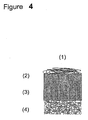
- Fig. 1 depicts a cross-section of the preferred form of a prosthetic device 1 embodying the invention.
- the device 1 includes at least one superficial layer comprising randomly oriented fibres of the biocompatible and/or at least partially resorbable material 2, a polymeric hollow body component 3, and a base component of a bone substitute material 4.
- any materials can be used for the construction of the device of the invention as long as they are biocompatible. Preferably all materials are biodegradable.
- the hollow body component 3 and the random fibres component 2 include synthetic polymers or molecules, natural polymers or molecules, biotechnologically derived polymers or molecules, biomacromolecules, or any combination thereof, while the base component 4 is based on a calcium phosphate material.
- the hollow bodies of the polymeric hollow body component 3 are essentially aligned in a direction perpendicular to a top surface of the base component 4, which top surface faces the hollow bodies.
- the hollow bodies thus form a brush-like structure in a direction perpendicular to the base component 4.
- the hollow bodies can be aligned to more than 50 % in a direction perpendicular to the top surface of the base component 4. An alignment of more than 90 % in a direction perpendicular to the base component 4 is preferred, more than 95 % alignment is particularly preferred.
- the hollow bodies may change alignment direction and self-organize at the uppermost end of the brush like structure. This might occur under pressure after implantation.
- the material to be used for the hollow bodies of the hollow body component 3 of the device of the invention is not particularly restricted to specific materials provided, however, the materials are bio-compatible.
- a bio-degradable solid polymer is used which can be of any shape with the proviso that a channel may be provided therein.
- a strang-like solid polymer is used, e.g. made by extrusion.
- the solid polymer is porous. Porosity of the polymer may be provided during manufacturing the polymer.
- the inner channel diameter of the hollow bodies of the polymeric hollow body component 3 is in range of 500 nm to 500 ⁇ m, with a preferred range of 5 ⁇ m to 150 mm.
- the hollow bodies of component 3 of the device 1 of the invention usually have a wall thickness ranging between 1 nm and 500 ⁇ m, a wall thickness being between 100 nm and 250 ⁇ m is preferred.
- the hollow bodies themselves should usually have a height of 50 ⁇ m to 10 mm. A height between 100 ⁇ m to 2 mm is particularly preferred.
- the device of the present invention comprises a polymeric hollow body component which is formed by an assembly of oriented tubes.
- the space between the assembled tubes is empty or filled with a substance selected from at least one synthetic polymer, natural polymer, biologically engineered polymer, or molecules thereof, biomicromolecules, or any combination thereof.
- Fig. 2 depicts in different cross-sections some possible arrangements of the hollow bodies of component 3.
- any type of distribution is possible, such as a homogenous or random distribution or a distribution in a specific pattern.
- the diameter of the hollow bodies and the wall thickness can be homogenous or variable within a hollow body component 3.
- Fig. 3 depicts a second preferred form of a prosthetic device 1 embodying the invention. It may be suitable to use a solid or porous block of polymer with manufactured channels as hollow body component 3. There are different methods to create these channels, well-known to persons skilled in the art. Techniques may include erosion, drilling, etching, form casting, etc. Again, channel diameter, and distribution may be homogenous or variable.
- any material can be used for the fibres of the superficial layer 2 which are randomly oriented to form three-dimensional structures of any kind as long as they are biocompatible. In order to enhance the stability of the structure 2, it may be that at least a fraction of material of the fibres is cross-linked.
- the fibres 2 include synthetic polymers, natural polymers, biologically engineered polymers, the molecules thereof, biomacromolecules and any combination thereof.
- the fibres of the superficial layer 2 themselves are not limited to any structure. They may be straight, twisted, curled, or of any tertiary structure. It is also possible to use a combination thereof. Additionally, the fibres themselves can be linear, branched or grafted.
- the fibres of the superficial layer 2 may be constituted out of single polymer molecules, or out of assemblies of many molecules.
- the shape and character of the fibres of the superficial layer 2 can be homogeneous or comprise a combination of various fibres previously mentioned different forms, including chemical, physical composition, and origin.
- the fibres can form a compact or loose random network, or an at least partially oriented assembly.
- the fibre-to-fibre distance can be varied within a broad range, i.e. between 1 nm to 1 mm, with a preferred fibre-to-fibre distance of 1 nanometer to 100 micrometers.
- the distances themselves can be homogeneous or heterogeneous. Examples of heterogeneous distances are gradient-like distributions, or random distributions, or specific pattern alignment, or any combination thereof.
- the fibres of the superficial layer 2 of the device of the present invention can be provided as mono-filament or multi-filament fibres of any length. Fiber arrangement in a woven, non-woven twisted, knitted, or any combination thereof is possible. If desired, the lateral cross-section of the fibres 2 can be solid or hollow.
- the fiber diameter may be varied in a broad range.
- a range of 50 nm to 1 mm is proposed.
- the fiber diameter is in range of 1 ⁇ m to 250 ⁇ m.
- the assembly of multiple layer structures can be a head-head, head-tail, or tail-tail, and any combination thereof. It can also be an intercalated assembly wherein the clear interface border is lost between the different layers and gets continuous.
- the superficial layer 2 usually has a thickness of 1 nm to 5 mm. It is preferred that the thickness is in a range of 10 ⁇ m to 2 mm. In some instances, however, that layer 2 can be missing and the hollow body component 3 is directly exposed at the surface.
- the superficial layer 2 is added separately to the device or intra-operatively, only after implantation of the device.
- the superficial layer 2 may be either in the form of a solid thin fibrous membrane or formed by adding a gelating liquid containing fibrous polymer.
- a selection may be made from synthetic or natural materials with a glass-like structure, crystalline structure, or any combination thereof.
- the fibres of the superficial layer 2 and the hollow bodies of the component 3 may have a flexible structure or a rigid structure depending on the final use of the device 1.
- the fibres 2 should form a flexible structure.
- the fiber material is usually homogeneous.
- the fiber material can also be heterogeneous, i.e., selected from various materials or it can comprise an engineered combination of the materials as mentioned above.
- the fibres 2 and/or the hollow bodies of component 3 can be coated or grafted with one or more of the previously mentioned materials.
- the device of the present invention comprises, as a further essential structural component, a base component 4.
- the function of the base component 4 is to anchor the polymeric hollow body component 3 in or onto an osteochondral environment. This osteochondral anchor function helps to keep the device 1 in place when implanted.
- the base component 4 can be of variable size and shape. Preferably, the shape of the base component 4 is round cylindrical or conical.
- the diameter of the base component 4 can vary in stepwise manner or in a continuous transition zone of any size. In practice, the diameter is related to the defect size and ranges between 4 and 20 mm, with a total height being 1 to 30 mm. Preferably, the diameter is in a range of 4 and 20 mm, with a height being between 1 to 10 mm.
- the top surface of the base component 4 is usually either flat or it mimics the contour of the subchondral plate or the cartilage surface to be replaced.
- the material of the base component 4 of the device of the invention can be a material, which is normally used as a bone substitute. Examples of the material are those as listed above in connection with the material of the fibres of the superficial layer 2.
- the material for the base component 4 is a mineral material such as synthetic ceramic.
- the ceramic can be selected out of one or several of the following groups: calcium phosphates, calcium sulphates, calcium carbonates and any mixture thereof.
- the base component 4 of the device 1 is a calcium phosphate
- one or more of the following composition groups is preferred: dicalcium phosphate dihydrate (CaHPO 4 x2H 2 O), dicalcium phosphate (CaHPO 4 ), alpha-tricalcium phosphate (alpha-Ca 3 (PO 4 ) 2 ), beta-tricalcium phosphate (beta-Ca 3 (PO 4 ) 2 ), calcium deficient hydroxyl apatite (Ca 9 (PO 4 ) 5 (HPO 4 )OH), hydroxyl apatite (Ca 10 (PO 4 ) 6 OH 2 ), carbonated apatite (Ca 10 (PO 4 ) 3 (CO 3 ) 3 )(OH) 2 ), fluoroapatite (Ca 10 (PO 4 ) 6 (F, OH) 2 ), chloroapatite (Ca 10 (PO 4 ) 6 (Cl, OH) 2 ), whitlockite ((
- mineral materials doped or mixed with metallic, semi-metallic and/or non-metallic components, preferably magnesium, silicon, sodium, potassium, strontium and/or lithium.
- the material of the base component 4 is a composite material comprising at least two different components.
- such composite materials are those comprising a mineral, inorganic, organic, biological, and/or biotechnological derived non-crystalline component and a mineral crystalline component.
- the non-crystalline components are often of polymeric nature.
- the structure of the materials of the base component 4 is highly porous with interconnecting pores. This would allow any substances and cell in the subchondral environment to diffuse or migrate, respectively, into the base component 4.
- At least one of components 2, 3 and 4 has a liquid absorbing capacity by interactions with a solvent.
- the liquid absorbing capacity is in a range of 0.1 to 99.9 %, a range of 20.0 to 95.0 % being particularly preferred.
- the liquid to be absorbed is water and/or body fluid available at the position where the device 1 is implanted.
- the fibres 2 advantageously form a gel or transform to a gel-like state.
- the components can swell and, therefore, an internal pressure within the fiber component is built up. That pressure helps stabilizing the structure. Furthermore, externally added components including cells are entrapped under the pressure within the fiber structure as in a natural cartilage.
- the device 1 of the invention may comprise a cell barrier layer between the polymeric hollow body component 3 and the base component 4.
- This layer acts as a barrier for cells and blood to prevent diffusion from the base component 4 into the polymeric hollow body component 3. It is, however, also possible to provide a barrier layer that is porous and/or has specific pores to allow selective or non-selective cells to pass through.
- the interface between random fibre layer 2 and the hollow body component 3, and the hollow body component 3 and the base component 4 respectively, can be formed in various ways. It can be either a chemical, or a physical, or mechanical interaction, or any combination thereof that forms the stabilization zones comprising at least one layer.
- the stabilization zones can be either formed by material used for device components 2, 3, or 4, or by externally added components, and any combination thereof.
- At least one externally added component is included in any of the components.
- said components are dispersed throughout component 2 and/or component 4 and/or component 3.
- Said components can be cells of different origin. The function is to support the generation of cartilage material and to enhance to improve healing, integration and mechanical properties of the device 1.
- the cells are preferably autologous cells, allogenous cells, xenogenous cells, transfected cells and/or genetically engineered cells and mixtures thereof.
- Particularly preferred cells which can be present throughout the polymeric hollow body component 3 and the fibre layer of 2 are chondrocytes, chondral progenitor cells, pluripotent stem cells, tutipotent stem cells or combinations thereof.
- Examples for cells included in the base component 4 are osteoblasts, osteo-progenitor cells, pluripotent stem cells, tutipotent stem cells and combinations thereof. In some instances it can be desired to include blood or any fraction thereof in the base component 4.
- Examples for another internally added components are pharmaceutical compounds including growth factors, engineered peptide-sequences, or antibiotics.
- gelating compounds including proteins, glycoaminoglycanes, carbohydrates, or polyethyleneoxides. These components can be added as free components, or they can be immobilized within the device of claim 1 by chemical, physical, or entrapment methods to prevent the washing-out.
- the polymeric components of the device of the invention may be cross-linked.
- the device of the present invention can be directly implanted in a defect, diseased, or deceased cartilaginous area such as articulating joints in humans and animals.
- articulating joints are the cartilage areas in hip, elbow, and knee joints.
- implanting the device into a joint is made by surgical procedures.
- the insertion procedure can be as following:
- the operation can be either carried out in an open or in an arthroscopic manner.
- the device of the invention can be seeded with cells and other externally added substances.
- One of the procedures includes the harvesting of cells prior to the effective operational procedure. After purification and treatment of the harvested cells, they can be seeded either directly into the device 1 for in-vitro cultivation, or subsequent to a short or extended in-vitro expansion and cultivation step, all according to methods established in the art.
- An other preferred procedure bypasses extensive in-vitro cultivation and is carried out as an intra-operative procedure. For that, cells are harvested during the operational procedure from the patient, purified and treated according to the methods established in the art. These cells are then seeded into the device 1, and device 1 is immediately implanted into the defect site.
- the base component 4 is implanted first, and subsequently the hollow body component 3 is immobilized on to the base component 4.
- the height of the hollow.body component 3 is adjusted to the contour of the joint after the immobilization procedure e.g. by shaving or heat treatment.
- at least one superficial layer 2 is provided onto the hollow body component 3.
- a prosthetic device is engineered from a porous interconnected cylindrical beta-tri-calcium-phosphate body of sizing 5 mm in diameter and 10 mm in height, as a subchondral anchor, and a 4 mm layer of a degradable polyurethane above.
- the polyurethane layer embodies vertical oriented hollow bodies of a diameter of 60 micrometer in a random lateral arrangement with a mean center-to-center distance of the hollow bodies of 100 micrometer.
- the hollow bodies in the polymer layer are produced in a casting process.
- the resulting prosthetic device is an ideal implant for cartilage repair.
- a properly sized tubular chisel is introduced perpendicular to the defect site in the joint.
- the chisel is tapped into cartilage and the osseous base, slightly larger than the defect (1-3 mm larger) at the defect site.
- the defect size is exacted regarding depth and diameter to the specific dimensions of the prosthetic device.
- the anchor of the graft is soaked in a saline solution before the prosthetic device is inserted through the universal guide tool. No additional fixation of the prosthetic device is necessary due to the exact fit.
- the surface of the prosthetic device is resurfaced - if necessary - to match the exact curvature of the joint surface and the height of the surrounding articular surface.
- autologous chondrocytes are filled into the hollow bodies in the polymer layer and a fibrous permeable polyurethane membrane is placed above the hollow bodies to prevent the cells coming out and to prevent the tubes filling with blood clots and before the wound site is closed.
- a prosthetic device is engineered from a porous interconnected cylindrical hydroxy apatite body of sizing 8 mm in diameter and 15 mm in height, as subchondral anchor, and a 8 mm layer of poly hydroxy methacrylate (pHEMA) with random arranged hollow bodies of diameters ranging between 10 and 50 micrometers. These vertical oriented tube like hollow bodies in the pHEMA layer are obtained casting the polymer into an appropriate form. The resulting prosthetic device is an ideal implant for cartilage repair.
- pHEMA poly hydroxy methacrylate
- the defect site is cleaned of frayed cartilagenous tissue and it is adjusted to the size of the prosthetic device.
- a properly sized tubular chisel is introduced perpendicular to the defect site in the joint.
- the chisel is tapped into cartilage and the osseous base of the defect site.
- the defect size is exacted regarding depth and diameter to the specific dimensions of the prosthetic device.
- harvested bone marrow stromal cells are added to the ceramic anchor.
- the prosthetic device is inserted through the universal guide tool. No additional fixation of the prosthetic device is necessary due to the exact fit and the swelling of the fiber layer.
- the surface of the prosthetic device is resurfaced - if necessary - to match the exact curvature of the joint surface and the height of the surrounding articular surface.
- a prosthetic device is engineered from a porous interconnected cylindrical beta-tri-calcium-phosphate and calcium sulfate composite body sizing 12 mm in diameter and 10 mm in height, as subchondral anchor, and a 6 mm polymer layer consisting of mixture of hollow polycaprolactone (PCL) filaments and polyethylenoxid (PEO) filaments.
- the inner diameter of the hollow filaments ranges between 10 and 80 micrometer.
- the PEO filaments have typically a diameter of 1 to 20 micrometer.
- the lateral distribution and arrangement of the hollow filaments is random, the space between the hollow fibers is filled with the PEO material.
- the polymer structure is stabilized by chemical crosslinking.
- the polymer layer is immobilized on the ceramic anchor by a melt process.
- the resulting prosthetic device is an ideal implant for cartilage repair.
- a properly sized tubular chisel is introduced perpendicular to the defect site in the joint.
- the chisel is tapped into cartilage and the osseous base of the defect site.
- the defect size is exacted regarding depth and diameter to the specific dimensions of the prosthetic device.
- Bone marrow stromal cells and platelet rich plasma is added to the anchor, and the prosthetic device is inserted subsequently by the universal guide tool. No additional fixation of the prosthetic device is necessary due to the exact fit.
- the surface of the prosthetic device is finally resurfaced to match the exact curvature of the joint surface and the height of the surrounding articular surface.
- adult stem cells and cells of a chondrogenic phenotype are mixed in a specific ratio and applied onto the polymer layer.
- a fibrous gelating matrix is used to seal the hollow bodies.
- a prosthetic device is engineered from a porous interconnected cylindrical beta-tri-calcium-phosphate body sizing 30 mm in diameter and 25 mm in height.with a convex surface curvature, as subchondral anchor, and a 6 mm layer of degradable Pluronic polymer with vertical tube like hollow bodies that have a random lateral arrangement.
- the diameter of the hollow bodies is variable, ranging between 5 and 150 micrometer.
- the ceramic anchor and the polymer layer are fused together by a cement reaction.
- the resulting prosthetic device is an ideal implant for cartilage repair.
- a properly sized tubular chisel is introduced perpendicular to the defect site in the joint.
- the chisel is tapped into cartilage and the osseous base of the defect site.
- the defect size is exacted regarding depth and diameter to the specific dimensions of the prosthetic device.
- Chondrocytes and mesemchymal progenitor cells are harvested by a biopsy intra-operatively and prepared for immediate application onto the polymer layer. Platelet rich plasma is added to the anchor, an the prosthetic device is inserted subsequently by the universal guide tool. No additional fixation of the prosthetic device is necessary due to the exact fit.
- the surface of the prosthetic device is finally resurfaced to match the exact curvature of the joint surface and the height of the surrounding articular surface.
- the hollow bodies are sealed with a thin layer of fibrous polymer.
- a prosthetic device is engineered from a porous interconnected cylindrical beta-tri-calcium-phosphate body sizing 8 mm in diameter and 10 mm in height, as subchrondral anchor, and a 3 mm layer of alginate polymer. Vertical hollow bodies of 50 micrometer diameter in the alginate polymer are formed while casting the polymer on top of the ceramic anchor. The resulting prosthetic device is an ideal implant for cartilage repair.
- a properly sized tubular chisel is introduced perpendicular to the defect site in the joint.
- the chisel is tapped into cartilage and the osseous base of the defect site.
- the defect size is exacted regarding depth and diameter to the specific dimensions of the prosthetic device.
- Bone marrow stromal cells are added to the anchor and the prosthetic device is inserted subsequently by the universal guide tool. No additional fixation of the prosthetic device is necessary due to the exact fit of the anchor. Additionally, the device is stabilized by the swelling of the polymer layer after in-vitro cultivated cells of chondrogenic phenotype are added. If necessary, the surface of the prosthetic device is finally resurfaced to match the exact curvature of the joint surface and the height of the surrounding articular surface.
- a prosthetic device is engineered from a porous interconnected cylindrical calcium deficient hydroxy apatite (CDHA) body sizing 4 mm in diameter and 5 mm in height, as subchondral anchor and a 3 mm layer of a chitosan fibers mesh with vertical oriented hollow bodies that exhibit diameters ranging between 20 and 100 micrometers.
- the hollow bodies were created by laser drilling in random lateral arrangement.
- the polymer layer is grafted onto a ceramic layer that acts as a selective barrier between the polymer layer and the anchor.
- the resulting prosthetic device is an ideal implant for cartilage repair.
- a properly sized tubular chisel is introduced perpendicular to the defect site in the joint.
- the chisel is tapped into cartilage and the osseous base of the defect site.
- the defect size is exacted regarding depth and diameter to the specific dimensions of the prosthetic device.
- Bone marrow stromal cells are added to the anchor, and the prosthetic device is inserted subsequently by the universal guide tool. No additional fixation of the prosthetic device is necessary due to the exact fit. If necessary, the surface of the prosthetic device is finally resurfaced to match the exact curvature of the joint surface and the height of the surrounding articular surface.
- intra-operatively harvested and isolated mesenchymal progenitor cells are applied onto the polymer layer and the top is sealed with a thin fibrous membrane or gel-like fibrous matrix.
- a prosthetic device is engineered from a porous interconnected cylindrical beta-tri-calcium-phosphate body sizing 10 mm in diameter and 10 mm in height, as subchondral anchor and a 3 mm layer of copolymer polylacticacid/polycaprolactone (PLA/PCL).
- Vertical tube like hollow bodies have diameters ranging between 30 and 300 micrometer and are drilled mechanically prior to graft the polymer layer onto the ceramic anchor.
- the hollow bodies are arranged according to a well-defined pattern.
- the resulting prosthetic device is an ideal implant for cartilage repair.
- a properly sized tubular chisel is introduced perpendicular to the defect site in the joint.
- the chisel is tapped into cartilage and the osseous base of the defect site.
- the defect size is exacted regarding depth and diameter to the specific dimensions of the prosthetic device.
- Bone marrow stromal cells are added to the anchor, and the prosthetic device is inserted subsequently by the universal guide tool. No additional fixation of the prosthetic device is necessary due to the exact fit.
- the surface of the prosthetic device is finally resurfaced to match the exact curvature of the joint surface and the height of the surrounding articular surface.
- in-vitro cultivated autologous cells of a chondrogenic phenotype are applied in a fibrous gelating matrix as cell suspension to the polymer layer.
- a prosthetic device is engineered from a porous interconnected cylindrical calcium deficient hydroxy apatite body sizing 4 mm in diameter and 5 mm in height, as subchondral anchor, and a 2 mm layer of hollow PCL filaments, intermixed with hyaluronic acid and collagen that also form the cover layer.
- the hollow filaments are vertically arranged and have an inner open diameter of 60 micrometer. The lateral arrangement is randomly and the polymer construct is stabilized by crosslinking of the polymers.
- the polymer layer is embedded in a ceramic layer that acts as a selective barrier between the fiber layer and the anchor. The resulting prosthetic device is an ideal implant for cartilage repair.
- Autologous chondrocytes are added to layer and the device is pre-cultivated in-vitro.
- a properly sized tubular chisel is introduced perpendicular to the defect site in the joint.
- the chisel is tapped into cartilage and the osseous base of the defect site.
- the defect size is exacted regarding depth and diameter to the specific dimensions of the prosthetic device.
- Platelet Rich Plasma is added to the anchor, and the prosthetic device is inserted subsequently by the special guide tool.
- a prosthetic device is engineered of textile polymer sheet with vertically arranged hollow bodies of a mean diameter of 100 micrometer.
- the polymer sheet with its hollow bodies is created by state-of-the-art textile technology out of PCL/PLA filaments.
- the hollow bodies are created by ultrathin woven fiber textiles.
- the prosthetic device assembly is carried intra-operative according to the following procedure.
- the defect site is exacted with the help of a chisel is tapped into cartilage and the osseous base of the defect site.
- the polymer textile sheet with its hollow bodies is cut into appropriate size.
- the prosthetic device anchoring is achieved by applying a calcium phosphate based cement into the subchondral space. Subsequently, the cut textile is placed on top of the cement, which will immobilize it upon hardening. A dense polymer layer at bottom side of the textile prevents the filling up of the hollow bodies with cement. The height of the polymer layer is adjusted to the surrounding cartilage by shaving the polymer and pressing into the cement anchor.
- chondrocytes and progenitor cells are mixed with a gelating matrix and applied to the textile containing the hollow bodies.
- the fibrous gelating matrix is also used to seal the hollow bodies by a random oriented fibrous layer.
- the operational procedure may be carried out as open surgery or as arthroscopy in minimal invasive manner.
Landscapes
- Health & Medical Sciences (AREA)
- General Health & Medical Sciences (AREA)
- Animal Behavior & Ethology (AREA)
- Public Health (AREA)
- Veterinary Medicine (AREA)
- Oral & Maxillofacial Surgery (AREA)
- Transplantation (AREA)
- Chemical & Material Sciences (AREA)
- Life Sciences & Earth Sciences (AREA)
- Epidemiology (AREA)
- Medicinal Chemistry (AREA)
- Dermatology (AREA)
- Rheumatology (AREA)
- Dispersion Chemistry (AREA)
- Orthopedic Medicine & Surgery (AREA)
- Cardiology (AREA)
- Engineering & Computer Science (AREA)
- Biomedical Technology (AREA)
- Heart & Thoracic Surgery (AREA)
- Vascular Medicine (AREA)
- Prostheses (AREA)
- Materials For Medical Uses (AREA)
Description
- The present invention is directed to a triphasic prosthetic device for repairing or replacing cartilage or cartilage-like tissues. Said prosthetic devices are useful as articular cartilage substitution material and as scaffold for regeneration of articular cartilagenous tissues.
- Articular cartilage tissue covers the ends of all bones that form diarthrodial joints. The resilient tissues provide the important characteristic of friction, lubrication, and wear in a joint. Furthermore, it acts as a shock absorber, distributing the load to the bones below. Without articular cartilage, stress and friction would occur to the extent that the joint would not permit motion. Articular cartilage has only a very limited capacity of regeneration. If this tissue is damaged or lost by traumatic events, or by chronic and progressive degeneration, it usually leads to painful arthrosis and decreased range of joint motion.
- Recently, the structure of rabbit articular cartilage has been further elucidated in an article by I. ap Gwynn et al, European Cells and Materials, Vo. 4, pp. 18-29, 2002. The tibial articular cartilage has been shown to comprise a radial zone in which the aggrecan component of the extracellular matrix was arranged generally oriented in columns in the radial direction. As a terminating member a superficial zone, next to the tibial plateau, is provided and having a spongy collagen architecture.
- Several methods have been established in the last decades for the treatment of injured and degenerated articular cartilage. Osteochondroal transplatation, microfracturing, heat treatment for sealing the surface, shaving, autologous chondrocyte transplantation (ACT), or total joint replacement are among the common techniques applied in today's orthopedic surgery.
- Joint replacement techniques where metal, ceramic and/or plastic components are used to substitute partially or totally the damaged or degenerated joint have already a long and quite successful tradition. The use of allograft material has been successful to some extent for small transplants, however, good quality allografts are hardly available.
- Osteochondroal transplantation (i.e. mosaicplasty) or autologous chondrocyte transplantation (ACT) are applied whenever total joint replacement is not yet indicated. These methods can be used to treat small and partial defects in a joint. In mosaicplasty defects are filled with osteochondral plugs harvested in non-load bearing areas. In ACT, chondrocytes are harvested by biopsy and grown in-vitro before a highly concentrated cell suspension is injected below an membrane (artificial or autologous) covering the defect area.
- Commonly, the replacement of cartilage tissue with solid permanent artificial inserts has been unsatisfactorily because the opposing articular joint surface is damaged by unevenness or by the hardness of the inserts. Therefore, the transplantation technology had to take a step forward in the research of alternative cartilage materials such as biocompatible materials and structures for articular cartilage replacement.
- For example,
U.S. Pat. No. 5,624,463 describes a prosthetic articular cartilage device comprising a dry, porous volume matrix of biocompatible and at least bioresorbable fibres and a base component. Said matrix establishes a bioresorbable scaffold adapted for the ingrowth of articular chondrocytes and for supporting natural articulating joint forces. Useful fibres include collagen, reticulin, elastin, cellulose, alginic acid, chitosan or synthetic and biosynthetic analogs thereof. Fibres are ordered in substantially circumferentially extending or substantially radially extending orientations. The base component is provided as a support on which the fiber matrix is applied. It is configured to fit in a complementary aperture in defective bone to secure the position of such a device in the bone. The base component is a composite material comprising a dispersion of collagen and composition consisting of tricalcium phosphate and hydroxyapatite. -
DE19803673 A1 discloses a triphasic prosthetic device for repairing or replacing cartilage or cartilage like-tissue according the preamble ofclaim 1. - It has been shown, however, that the function of the above construction has not been always satisfactory. The reason is that said known prosthetic articular cartilage device is frequently unstable due to its structure and thus had to be replaced in the joint area by another surgical operation in to again repair cartilage joints such as knee and hip.
- In view of this situation, in the field of articular cartilage replacement materials, there is a need for a structure suitable as a prosthetic articular cartilage which is made of natural resorbable materials or analogs thereof and having an improved structure stability and an accurate positioning in the bone. At the same time, the prosthetic device should be biomechanically able to withstand normal joint forces and to promote repair and replacement of cartilage tissue or cartilage-like tissue.
- These objects are solved by the prosthetic device according to
claim 1. - The present invention relates to a prosthetic device for repairing or replacing cartilage or cartilage-like tissue which comprises a polymeric
hollow body component 3, with a number of oriented hollow bodies, abase component 4 to anchor said polymerichollow body component 3 in or onto an osteochondral environment and at least one superficial layer comprising randomly orientedfibres 2 provided on said polymerichollow body component 3, wherein said number of highly oriented hollow bodies of the polymerichollow body component 3 are aligned essentially in parallel to the insertion axis of the prosthetic device, i.e. perpendicularly to the plane of the articulating surface. - The subclaims concern preferred embodiments of the prosthetic device of the present invention.
- It has been surprisingly found that the stability of a prosthetic articular cartilage device can be essentially improved by providing a polymeric hollow body component with a number of highly oriented
hollow bodies 3 in such a way that the hollow bodies are aligned essentially in parallel to the insertion axis of the prosthetic device. The polymeric hollow body component is flanked by a base component and a superficial layer to form the triphasic structure of the device of the invention. The specific alignment of the hollow bodies in the layer perfectly mimics the cartilage and cartilage-like tissues providing an excellent mechanical stability. At the same time, a basis for rapid cartilage in-growth is provided, thus assuring a long term cartilage replacement. - The invention itself may be more fully understood from the following description when read together with the accompanying Figures wherein
-
Fig. 1 shows a vertical cross-sectional view of an embodiment of the prosthetic device of the invention; -
Fig. 2 shows a horizontal cross-section of the hollow bodies of the polymerichollow body component 3 in different packings and sizes; -
Fig. 3 illustrates a vertical cross section of an embodiment of the device of the invention where physically/mechanically produced channels are incorporated insolid polymer components 3 and -
Fig. 4 is a vertical cross-section of another embodiment of the device of the invention wherein cells are seeded incomponents -
Fig. 1 depicts a cross-section of the preferred form of aprosthetic device 1 embodying the invention. Thedevice 1 includes at least one superficial layer comprising randomly oriented fibres of the biocompatible and/or at least partiallyresorbable material 2, a polymerichollow body component 3, and a base component of abone substitute material 4. - In principle, any materials can be used for the construction of the device of the invention as long as they are biocompatible. Preferably all materials are biodegradable. In one of the preferred embodiment of the invention the
hollow body component 3 and therandom fibres component 2 include synthetic polymers or molecules, natural polymers or molecules, biotechnologically derived polymers or molecules, biomacromolecules, or any combination thereof, while thebase component 4 is based on a calcium phosphate material. - As can be seen from
Fig. 1 , the hollow bodies of the polymerichollow body component 3 are essentially aligned in a direction perpendicular to a top surface of thebase component 4, which top surface faces the hollow bodies. The hollow bodies thus form a brush-like structure in a direction perpendicular to thebase component 4. - The hollow bodies can be aligned to more than 50 % in a direction perpendicular to the top surface of the
base component 4. An alignment of more than 90 % in a direction perpendicular to thebase component 4 is preferred, more than 95 % alignment is particularly preferred. The hollow bodies may change alignment direction and self-organize at the uppermost end of the brush like structure. This might occur under pressure after implantation. - The material to be used for the hollow bodies of the
hollow body component 3 of the device of the invention is not particularly restricted to specific materials provided, however, the materials are bio-compatible. Preferably, a bio-degradable solid polymer is used which can be of any shape with the proviso that a channel may be provided therein. More preferably, a strang-like solid polymer is used, e.g. made by extrusion. Once the solid polymer has the desired shape, hollow spaces such as channels are formed therein by mechanical, physical and/or chemical methods. Examples for such methods are casting, drilling, etching, etc. which are well known to the person skilled in the art. - For some reason, it may be suitable that the solid polymer is porous. Porosity of the polymer may be provided during manufacturing the polymer.
- Preferably, in the device of the invention, the inner channel diameter of the hollow bodies of the polymeric
hollow body component 3 is in range of 500 nm to 500 µm, with a preferred range of 5 µm to 150 mm. - The hollow bodies of
component 3 of thedevice 1 of the invention usually have a wall thickness ranging between 1 nm and 500 µm, a wall thickness being between 100 nm and 250 µm is preferred. - The hollow bodies themselves should usually have a height of 50 µm to 10 mm. A height between 100 µm to 2 mm is particularly preferred.
- Specifically, the device of the present invention comprises a polymeric hollow body component which is formed by an assembly of oriented tubes. In this case, the space between the assembled tubes is empty or filled with a substance selected from at least one synthetic polymer, natural polymer, biologically engineered polymer, or molecules thereof, biomicromolecules, or any combination thereof.
-
Fig. 2 depicts in different cross-sections some possible arrangements of the hollow bodies ofcomponent 3. With respect to the lateral distribution of the hollow bodies ofcomponent 3, any type of distribution is possible, such as a homogenous or random distribution or a distribution in a specific pattern. Furthermore, the diameter of the hollow bodies and the wall thickness can be homogenous or variable within ahollow body component 3. -
Fig. 3 depicts a second preferred form of aprosthetic device 1 embodying the invention. It may be suitable to use a solid or porous block of polymer with manufactured channels ashollow body component 3. There are different methods to create these channels, well-known to persons skilled in the art. Techniques may include erosion, drilling, etching, form casting, etc. Again, channel diameter, and distribution may be homogenous or variable. - It may be suitable to engineer the
component 3 from molecules that self-assemble forming tube like hollow body structures to thefinal polymeric component 3. For stabilization reasons, such structures can be crosslinked. - In principle, any material can be used for the fibres of the
superficial layer 2 which are randomly oriented to form three-dimensional structures of any kind as long as they are biocompatible. In order to enhance the stability of thestructure 2, it may be that at least a fraction of material of the fibres is cross-linked. In one preferred embodiment of the invention thefibres 2 include synthetic polymers, natural polymers, biologically engineered polymers, the molecules thereof, biomacromolecules and any combination thereof. - The fibres of the
superficial layer 2 themselves are not limited to any structure. They may be straight, twisted, curled, or of any tertiary structure. It is also possible to use a combination thereof. Additionally, the fibres themselves can be linear, branched or grafted. - The fibres of the
superficial layer 2 may be constituted out of single polymer molecules, or out of assemblies of many molecules. - According to the invention, the shape and character of the fibres of the
superficial layer 2 can be homogeneous or comprise a combination of various fibres previously mentioned different forms, including chemical, physical composition, and origin. The fibres can form a compact or loose random network, or an at least partially oriented assembly. The fibre-to-fibre distance can be varied within a broad range, i.e. between 1 nm to 1 mm, with a preferred fibre-to-fibre distance of 1 nanometer to 100 micrometers. The distances themselves can be homogeneous or heterogeneous. Examples of heterogeneous distances are gradient-like distributions, or random distributions, or specific pattern alignment, or any combination thereof. - The fibres of the
superficial layer 2 of the device of the present invention can be provided as mono-filament or multi-filament fibres of any length. Fiber arrangement in a woven, non-woven twisted, knitted, or any combination thereof is possible. If desired, the lateral cross-section of thefibres 2 can be solid or hollow. - According to the invention, the fiber diameter may be varied in a broad range. Advantageously, a range of 50 nm to 1 mm is proposed. Preferably, the fiber diameter is in range of 1 µm to 250 µm.
- It has been shown that one layer of fibres of the
superficial layer 2 already brings about good results. However, in some instances, it can be advisable to provide a couple of layers of fibres which is, of course, dependent on the final use of the device of the invention. The assembly of multiple layer structures can be a head-head, head-tail, or tail-tail, and any combination thereof. It can also be an intercalated assembly wherein the clear interface border is lost between the different layers and gets continuous. - The
superficial layer 2 usually has a thickness of 1 nm to 5 mm. It is preferred that the thickness is in a range of 10 µm to 2 mm. In some instances, however, thatlayer 2 can be missing and thehollow body component 3 is directly exposed at the surface. - Some specific indications require that the
superficial layer 2 is added separately to the device or intra-operatively, only after implantation of the device. In this case thesuperficial layer 2 may be either in the form of a solid thin fibrous membrane or formed by adding a gelating liquid containing fibrous polymer. - In case of using mineral based materials for the
fibre layer 2 and/or thehollow body component 3, a selection may be made from synthetic or natural materials with a glass-like structure, crystalline structure, or any combination thereof. - According to the invention, the fibres of the
superficial layer 2 and the hollow bodies of thecomponent 3 may have a flexible structure or a rigid structure depending on the final use of thedevice 1. In case of adapting to the articulation of a joint or opposing tissue, thefibres 2 should form a flexible structure. - The fiber material is usually homogeneous. Depending on the final use of the device of the invention, the fiber material can also be heterogeneous, i.e., selected from various materials or it can comprise an engineered combination of the materials as mentioned above.
- In some instances, however, the
fibres 2 and/or the hollow bodies ofcomponent 3 can be coated or grafted with one or more of the previously mentioned materials. - The device of the present invention comprises, as a further essential structural component, a
base component 4. The function of thebase component 4 is to anchor the polymerichollow body component 3 in or onto an osteochondral environment. This osteochondral anchor function helps to keep thedevice 1 in place when implanted. Thebase component 4 can be of variable size and shape. Preferably, the shape of thebase component 4 is round cylindrical or conical. The diameter of thebase component 4 can vary in stepwise manner or in a continuous transition zone of any size. In practice, the diameter is related to the defect size and ranges between 4 and 20 mm, with a total height being 1 to 30 mm. Preferably, the diameter is in a range of 4 and 20 mm, with a height being between 1 to 10 mm. The top surface of thebase component 4 is usually either flat or it mimics the contour of the subchondral plate or the cartilage surface to be replaced. - The material of the
base component 4 of the device of the invention can be a material, which is normally used as a bone substitute. Examples of the material are those as listed above in connection with the material of the fibres of thesuperficial layer 2. If desired, the material for thebase component 4 is a mineral material such as synthetic ceramic. The ceramic can be selected out of one or several of the following groups: calcium phosphates, calcium sulphates, calcium carbonates and any mixture thereof. - If the
base component 4 of thedevice 1 is a calcium phosphate, one or more of the following composition groups is preferred: dicalcium phosphate dihydrate (CaHPO4x2H2O), dicalcium phosphate (CaHPO4), alpha-tricalcium phosphate (alpha-Ca3(PO4)2), beta-tricalcium phosphate (beta-Ca3(PO4)2), calcium deficient hydroxyl apatite (Ca9(PO4)5(HPO4)OH), hydroxyl apatite (Ca10(PO4)6OH2), carbonated apatite (Ca10(PO4)3(CO3)3)(OH)2), fluoroapatite (Ca10(PO4)6(F, OH)2), chloroapatite (Ca10(PO4)6(Cl, OH)2), whitlockite ((Ca,Mg)3(PO4)2), tetracalcium phosphate (Ca4(PO4)2O), oxyapatite (Ca10(PO4)6O), beta-calcium pyrophosphate (beta-Ca2(P2O7), alpha-calcium pyrophosphate, gamma-calcium pyrophosphate, octacalcium phosphate (Ca8H2(PO4)6x5H2O). - It is also possible to have the above mentioned mineral materials doped or mixed with metallic, semi-metallic and/or non-metallic components, preferably magnesium, silicon, sodium, potassium, strontium and/or lithium.
- In another preferred embodiment of the invention, the material of the
base component 4 is a composite material comprising at least two different components. Examples of such composite materials are those comprising a mineral, inorganic, organic, biological, and/or biotechnological derived non-crystalline component and a mineral crystalline component. The non-crystalline components are often of polymeric nature. - In a preferred embodiment of the invention, the structure of the materials of the
base component 4 is highly porous with interconnecting pores. This would allow any substances and cell in the subchondral environment to diffuse or migrate, respectively, into thebase component 4. - In various forms of the invention, at least one of
components - Usually, the liquid to be absorbed is water and/or body fluid available at the position where the
device 1 is implanted. When absorbing water and/or body fluids, thefibres 2 advantageously form a gel or transform to a gel-like state. - Upon uptake of water and/or body fluids the components can swell and, therefore, an internal pressure within the fiber component is built up. That pressure helps stabilizing the structure. Furthermore, externally added components including cells are entrapped under the pressure within the fiber structure as in a natural cartilage.
- If desired, the
device 1 of the invention may comprise a cell barrier layer between the polymerichollow body component 3 and thebase component 4. This layer acts as a barrier for cells and blood to prevent diffusion from thebase component 4 into the polymerichollow body component 3. It is, however, also possible to provide a barrier layer that is porous and/or has specific pores to allow selective or non-selective cells to pass through. - The interface between
random fibre layer 2 and thehollow body component 3, and thehollow body component 3 and thebase component 4 respectively, can be formed in various ways. It can be either a chemical, or a physical, or mechanical interaction, or any combination thereof that forms the stabilization zones comprising at least one layer. The stabilization zones can be either formed by material used fordevice components - In another preferred embodiment of the device of the
invention 1 as illustrated inFig. 4 , at least one externally added component is included in any of the components. Usually said components are dispersed throughoutcomponent 2 and/orcomponent 4 and/orcomponent 3. Said components can be cells of different origin. The function is to support the generation of cartilage material and to enhance to improve healing, integration and mechanical properties of thedevice 1. - The cells are preferably autologous cells, allogenous cells, xenogenous cells, transfected cells and/or genetically engineered cells and mixtures thereof.
- Particularly preferred cells, which can be present throughout the polymeric
hollow body component 3 and the fibre layer of 2 are chondrocytes, chondral progenitor cells, pluripotent stem cells, tutipotent stem cells or combinations thereof. Examples for cells included in thebase component 4 are osteoblasts, osteo-progenitor cells, pluripotent stem cells, tutipotent stem cells and combinations thereof. In some instances it can be desired to include blood or any fraction thereof in thebase component 4. - Examples for another internally added components are pharmaceutical compounds including growth factors, engineered peptide-sequences, or antibiotics.
- An example for another internally added components are gelating compounds including proteins, glycoaminoglycanes, carbohydrates, or polyethyleneoxides. These components can be added as free components, or they can be immobilized within the device of
claim 1 by chemical, physical, or entrapment methods to prevent the washing-out. - The polymeric components of the device of the invention may be cross-linked.
- The device of the present invention can be directly implanted in a defect, diseased, or deceased cartilaginous area such as articulating joints in humans and animals. Examples of these articulating joints are the cartilage areas in hip, elbow, and knee joints. Usually, implanting the device into a joint is made by surgical procedures. For example the insertion procedure can be as following:
- In a first step, the defect area is cleaned and an osteochondral plug is removed with a chisel. Special equipment allows for exacting bottom and walls with regard to depths and widths. The prosthetic device of the invention is carefully pressed into position in such a manner that the upper edge of the
base component 4 is on the same level with the calcified zone dividing the cartilage and the bone. The top surface of thefiber layer 2 should equal the height of the surrounding cartilage. Height differences may be exacted. - The operation can be either carried out in an open or in an arthroscopic manner.
- As mentioned above and depicted in
Fig. 4 , the device of the invention can be seeded with cells and other externally added substances. There are different procedure possible. One of the procedures includes the harvesting of cells prior to the effective operational procedure. After purification and treatment of the harvested cells, they can be seeded either directly into thedevice 1 for in-vitro cultivation, or subsequent to a short or extended in-vitro expansion and cultivation step, all according to methods established in the art. - An other preferred procedure bypasses extensive in-vitro cultivation and is carried out as an intra-operative procedure. For that, cells are harvested during the operational procedure from the patient, purified and treated according to the methods established in the art. These cells are then seeded into the
device 1, anddevice 1 is immediately implanted into the defect site. - For special applications, it will be also possible to assemble the device of the invention intra-operatively. I.e. the
base component 4 is implanted first, and subsequently thehollow body component 3 is immobilized on to thebase component 4. The height of thehollow.body component 3 is adjusted to the contour of the joint after the immobilization procedure e.g. by shaving or heat treatment. Finally, at least onesuperficial layer 2 is provided onto thehollow body component 3. - The present invention is illustrated by means of the following examples.
- A prosthetic device is engineered from a porous interconnected cylindrical beta-tri-calcium-phosphate body of sizing 5 mm in diameter and 10 mm in height, as a subchondral anchor, and a 4 mm layer of a degradable polyurethane above. The polyurethane layer embodies vertical oriented hollow bodies of a diameter of 60 micrometer in a random lateral arrangement with a mean center-to-center distance of the hollow bodies of 100 micrometer. The hollow bodies in the polymer layer are produced in a casting process. The resulting prosthetic device is an ideal implant for cartilage repair.
- A properly sized tubular chisel is introduced perpendicular to the defect site in the joint. In a first step in the implantation, the chisel is tapped into cartilage and the osseous base, slightly larger than the defect (1-3 mm larger) at the defect site. The defect size is exacted regarding depth and diameter to the specific dimensions of the prosthetic device. Subsequently, the anchor of the graft is soaked in a saline solution before the prosthetic device is inserted through the universal guide tool. No additional fixation of the prosthetic device is necessary due to the exact fit. Then, the surface of the prosthetic device is resurfaced - if necessary - to match the exact curvature of the joint surface and the height of the surrounding articular surface. Finally autologous chondrocytes are filled into the hollow bodies in the polymer layer and a fibrous permeable polyurethane membrane is placed above the hollow bodies to prevent the cells coming out and to prevent the tubes filling with blood clots and before the wound site is closed.
- A prosthetic device is engineered from a porous interconnected cylindrical hydroxy apatite body of sizing 8 mm in diameter and 15 mm in height, as subchondral anchor, and a 8 mm layer of poly hydroxy methacrylate (pHEMA) with random arranged hollow bodies of diameters ranging between 10 and 50 micrometers. These vertical oriented tube like hollow bodies in the pHEMA layer are obtained casting the polymer into an appropriate form. The resulting prosthetic device is an ideal implant for cartilage repair.
- In a first step, the defect site is cleaned of frayed cartilagenous tissue and it is adjusted to the size of the prosthetic device. A properly sized tubular chisel is introduced perpendicular to the defect site in the joint. The chisel is tapped into cartilage and the osseous base of the defect site. The defect size is exacted regarding depth and diameter to the specific dimensions of the prosthetic device. Subsequently, harvested bone marrow stromal cells are added to the ceramic anchor. Next, the prosthetic device is inserted through the universal guide tool. No additional fixation of the prosthetic device is necessary due to the exact fit and the swelling of the fiber layer. Finally, the surface of the prosthetic device is resurfaced - if necessary - to match the exact curvature of the joint surface and the height of the surrounding articular surface.
- A prosthetic device is engineered from a porous interconnected cylindrical beta-tri-calcium-phosphate and calcium sulfate composite body sizing 12 mm in diameter and 10 mm in height, as subchondral anchor, and a 6 mm polymer layer consisting of mixture of hollow polycaprolactone (PCL) filaments and polyethylenoxid (PEO) filaments. The inner diameter of the hollow filaments ranges between 10 and 80 micrometer. The PEO filaments have typically a diameter of 1 to 20 micrometer. The lateral distribution and arrangement of the hollow filaments is random, the space between the hollow fibers is filled with the PEO material. The polymer structure is stabilized by chemical crosslinking. The polymer layer is immobilized on the ceramic anchor by a melt process. The resulting prosthetic device is an ideal implant for cartilage repair.
- A properly sized tubular chisel is introduced perpendicular to the defect site in the joint. In a first step in the implantation, the chisel is tapped into cartilage and the osseous base of the defect site. The defect size is exacted regarding depth and diameter to the specific dimensions of the prosthetic device. Bone marrow stromal cells and platelet rich plasma is added to the anchor, and the prosthetic device is inserted subsequently by the universal guide tool. No additional fixation of the prosthetic device is necessary due to the exact fit. If necessary, the surface of the prosthetic device is finally resurfaced to match the exact curvature of the joint surface and the height of the surrounding articular surface. Finally, adult stem cells and cells of a chondrogenic phenotype are mixed in a specific ratio and applied onto the polymer layer. A fibrous gelating matrix is used to seal the hollow bodies.
- A prosthetic device is engineered from a porous interconnected cylindrical beta-tri-calcium-phosphate body sizing 30 mm in diameter and 25 mm in height.with a convex surface curvature, as subchondral anchor, and a 6 mm layer of degradable Pluronic polymer with vertical tube like hollow bodies that have a random lateral arrangement. The diameter of the hollow bodies is variable, ranging between 5 and 150 micrometer. The ceramic anchor and the polymer layer are fused together by a cement reaction. The resulting prosthetic device is an ideal implant for cartilage repair.
- A properly sized tubular chisel is introduced perpendicular to the defect site in the joint. In a first step in the implantation, the chisel is tapped into cartilage and the osseous base of the defect site. The defect size is exacted regarding depth and diameter to the specific dimensions of the prosthetic device. Chondrocytes and mesemchymal progenitor cells are harvested by a biopsy intra-operatively and prepared for immediate application onto the polymer layer. Platelet rich plasma is added to the anchor, an the prosthetic device is inserted subsequently by the universal guide tool. No additional fixation of the prosthetic device is necessary due to the exact fit. The surface of the prosthetic device is finally resurfaced to match the exact curvature of the joint surface and the height of the surrounding articular surface. The hollow bodies are sealed with a thin layer of fibrous polymer.
- A prosthetic device is engineered from a porous interconnected cylindrical beta-tri-calcium-phosphate body sizing 8 mm in diameter and 10 mm in height, as subchrondral anchor, and a 3 mm layer of alginate polymer. Vertical hollow bodies of 50 micrometer diameter in the alginate polymer are formed while casting the polymer on top of the ceramic anchor. The resulting prosthetic device is an ideal implant for cartilage repair.
- A properly sized tubular chisel is introduced perpendicular to the defect site in the joint. In a first step in the implantation, the chisel is tapped into cartilage and the osseous base of the defect site. The defect size is exacted regarding depth and diameter to the specific dimensions of the prosthetic device. Bone marrow stromal cells are added to the anchor and the prosthetic device is inserted subsequently by the universal guide tool. No additional fixation of the prosthetic device is necessary due to the exact fit of the anchor. Additionally, the device is stabilized by the swelling of the polymer layer after in-vitro cultivated cells of chondrogenic phenotype are added. If necessary, the surface of the prosthetic device is finally resurfaced to match the exact curvature of the joint surface and the height of the surrounding articular surface.
- A prosthetic device is engineered from a porous interconnected cylindrical calcium deficient hydroxy apatite (CDHA) body sizing 4 mm in diameter and 5 mm in height, as subchondral anchor and a 3 mm layer of a chitosan fibers mesh with vertical oriented hollow bodies that exhibit diameters ranging between 20 and 100 micrometers. The hollow bodies were created by laser drilling in random lateral arrangement. The polymer layer is grafted onto a ceramic layer that acts as a selective barrier between the polymer layer and the anchor. The resulting prosthetic device is an ideal implant for cartilage repair.
- A properly sized tubular chisel is introduced perpendicular to the defect site in the joint. In a first step in the implantation, the chisel is tapped into cartilage and the osseous base of the defect site. The defect size is exacted regarding depth and diameter to the specific dimensions of the prosthetic device. Bone marrow stromal cells are added to the anchor, and the prosthetic device is inserted subsequently by the universal guide tool. No additional fixation of the prosthetic device is necessary due to the exact fit. If necessary, the surface of the prosthetic device is finally resurfaced to match the exact curvature of the joint surface and the height of the surrounding articular surface. Finally, intra-operatively harvested and isolated mesenchymal progenitor cells are applied onto the polymer layer and the top is sealed with a thin fibrous membrane or gel-like fibrous matrix.
- A prosthetic device is engineered from a porous interconnected cylindrical beta-tri-calcium-phosphate body sizing 10 mm in diameter and 10 mm in height, as subchondral anchor and a 3 mm layer of copolymer polylacticacid/polycaprolactone (PLA/PCL). Vertical tube like hollow bodies have diameters ranging between 30 and 300 micrometer and are drilled mechanically prior to graft the polymer layer onto the ceramic anchor. The hollow bodies are arranged according to a well-defined pattern. The resulting prosthetic device is an ideal implant for cartilage repair.
- A properly sized tubular chisel is introduced perpendicular to the defect site in the joint. In a first step in the implantation, the chisel is tapped into cartilage and the osseous base of the defect site. The defect size is exacted regarding depth and diameter to the specific dimensions of the prosthetic device. Bone marrow stromal cells are added to the anchor, and the prosthetic device is inserted subsequently by the universal guide tool. No additional fixation of the prosthetic device is necessary due to the exact fit. If necessary, the surface of the prosthetic device is finally resurfaced to match the exact curvature of the joint surface and the height of the surrounding articular surface. Finally, in-vitro cultivated autologous cells of a chondrogenic phenotype are applied in a fibrous gelating matrix as cell suspension to the polymer layer.
- A prosthetic device is engineered from a porous interconnected cylindrical calcium deficient hydroxy apatite body sizing 4 mm in diameter and 5 mm in height, as subchondral anchor, and a 2 mm layer of hollow PCL filaments, intermixed with hyaluronic acid and collagen that also form the cover layer. The hollow filaments are vertically arranged and have an inner open diameter of 60 micrometer. The lateral arrangement is randomly and the polymer construct is stabilized by crosslinking of the polymers. The polymer layer is embedded in a ceramic layer that acts as a selective barrier between the fiber layer and the anchor. The resulting prosthetic device is an ideal implant for cartilage repair.
- Autologous chondrocytes are added to layer and the device is pre-cultivated in-vitro. For implantation, a properly sized tubular chisel is introduced perpendicular to the defect site in the joint. The chisel is tapped into cartilage and the osseous base of the defect site. The defect size is exacted regarding depth and diameter to the specific dimensions of the prosthetic device. Platelet Rich Plasma is added to the anchor, and the prosthetic device is inserted subsequently by the special guide tool.
- A prosthetic device is engineered of textile polymer sheet with vertically arranged hollow bodies of a mean diameter of 100 micrometer. The polymer sheet with its hollow bodies is created by state-of-the-art textile technology out of PCL/PLA filaments. The hollow bodies are created by ultrathin woven fiber textiles. The prosthetic device assembly is carried intra-operative according to the following procedure.
- For implantation, the defect site is exacted with the help of a chisel is tapped into cartilage and the osseous base of the defect site. The polymer textile sheet with its hollow bodies is cut into appropriate size. The prosthetic device anchoring is achieved by applying a calcium phosphate based cement into the subchondral space. Subsequently, the cut textile is placed on top of the cement, which will immobilize it upon hardening. A dense polymer layer at bottom side of the textile prevents the filling up of the hollow bodies with cement. The height of the polymer layer is adjusted to the surrounding cartilage by shaving the polymer and pressing into the cement anchor. Finally, intra-operatively harvested and isolated chondrocytes and progenitor cells are mixed with a gelating matrix and applied to the textile containing the hollow bodies. The fibrous gelating matrix is also used to seal the hollow bodies by a random oriented fibrous layer. The operational procedure may be carried out as open surgery or as arthroscopy in minimal invasive manner.
Claims (38)
- A triphasic prosthetic device for repairing or replacing cartilage or cartilage like-tissue (1), characterised in comprising- a polymeric hollow body component (3) with a number of highly oriented hollow bodies;- a base component (4) of a synthetic ceramic to anchor said polymeric hollow body component (3) in or onto an osteochondral environment and- at least one superficial layer comprising randomly oriented fibres (2) provided on said polymeric hollow body component (3)wherein said number of highly oriented hollow bodies of the polymeric hollow body component (3) are aligned perpendicularly to the plane of the articulating surface to more than 50%.
- The device according to claim 1,
wherein said hollow bodies are aligned to more than 90 %, preferably more than 95 %. - The device according to at least one of claims 1 or 2,
wherein the inner channel diameter of the hollow bodies of polymeric hollow body component (3) is in a range of 500 nm to 500 µm. - The device according to claim 3,
wherein said inner channel diameter is in a range of 5 µm to 150 µm. - The device according to at least one of claims 1 to 4,
wherein the polymeric hollow body component (3) is formed by an assembly of oriented tubes. - The device according to claim 5,
wherein the space between the assembled tubes is empty or filled with a substance selected from the group consisting of synthetic polymers, natural polymers, biologically engineered polymers, the molecules thereof, biomacromolecules and any combination thereof. - The device according to at least one of claims 3 to 6,
wherein the channels have a wall thickness ranging between 1 nm and 500 µm. - The device according to claim 7,
wherein the wall thickness is between 100 nm and 250 µm. - The device according to at least one of claims 1 to 8,
wherein the hollow body component is a solid block of polymer with channels. - The device according to at least one of claims 3 to 9,
wherein the channels are formed by mechanical, physical and/or chemical methods in a solid polymer. - The device according to at least one of the claims 1 to 10,
wherein said solid polymer is porous. - The device according to at least one of claims 1 to 11,
wherein the lateral distribution of the hollow bodies of component (3) is homogenous, random or in an specific pattern. - The device according to at least one of claims 1 to 12,
wherein said hollow bodies of the hollow body component (3) have a height of 50 µm to 10 mm. - The device according to claim 13,
wherein the height is between 100 µm to 2 mm. - The device according to at least one of claims 1 to 14,
wherein the fibers of the superficial layer (2) comprise a material selected from the group consisting of synthetic polymers, natural polymers, biologically engineered polymers, the molecules thereof, biomacromolecules and any combination thereof. - The device according to at least one of claims 1 to 15,
wherein said a synthetic ceramic comprises at least one of calcium phosphate, calcium sulfate and calcium carbonate. - The device according to claim 16,
wherein said calcium phosphate is selected from the group consisting of dicalcium phosphate dihydrate (CaHPO4x2H2O), dicalcium phosphate (CaHPO4), alpha-tricalcium phosphate (alpha-Ca3(PO4)2), beta-tricalcium phosphate (beta-Ca3(PO4)2), calcium deficient hydroxyl apatite (Ca9(PO4)5(HPO4)OH), hydroxyl apatite (Ca10(PO4)6OH2), carbonated apatite (Ca10(PO4)3(C03) 3)(OH) 2), fluoroapatite (Ca10(PO4)6(F,OH)2), chloroapatite (Ca10(PO4)6(Cl,OH)2), whitlockite ((Ca,Mg)3(PO4)2), tetracalcium phosphate (Ca4(PO4)2O), oxyapatite (Ca10(PO)6O), beta-calcium pyrophosphate (beta-Ca2(P2O7), alpha-calcium pyrophosphate, gama-calcium pyrophosphate, octacalcium phosphate (Ca8H2(PO4)6x5H2O) and mixtures thereof. - The device according to claim 16,
wherein said synthetic ceramic comprises metallic, semimetallic components and/or non-metallic components, preferably magnesium, silicon, sodium, potassium, strontium and/or lithium. - The device according to any of claims 16 to 18,
wherein the bone substitute material is highly porous with interconnecting pores. - The device according to any of claims 16 to 19,
wherein the shape of the base component (4) is round cylindrical or conical. - The device according to claim 20,
wherein the diameter of the base component (4) ranges between 4 and 20 mm, with a height being 1 to 30 mm. - The device according to claim 21,
wherein the diameter of the base component (4) ranges between 4 and 20 mm, with a height being between 1 to 10 mm. - The device according to at least of claims 1 to 22, wherein said superficial layer (2) has a thickness of 1 nm to 5 mm.
- The device according to claim 23,
wherein said thickness is in the range of 10 µm to 2 mm. - The device according to claim 23 and 24,
wherein said superficial layer (2) is missing, or formed by uppermost end of the hollow body component. - The device according to at least one of claims 1 to 25,
wherein at least one of components (2), (3) and (4) has a liquid absorbing capacity in a range of 0.1 % to 99.9 %. - The device according to claim 26, wherein said liquid absorbing capacity is in a range of 20.0 to 95.0 %.
- The device according to claim 26 or 27,
wherein the liquid is an aqueous media and/or a body fluid. - The device according to at least one of the preceding claims,
wherein the polymeric components are cross-linked. - The device according to at least one of preceding claims further comprising at least one externally added component.
- The device according to claim 30,
wherein said components are cells of different origin. - The device according to claim 31,
wherein said cells are autologous cells, allogenous cells, xenogenous cells, transfected cells and/or genetically engineered cells. - The device according to claim 30, 31 or 32,
wherein chondrocytes, chondral progenitor cells, pluripotent cells, tutipotent cells or combinations thereof are present throughout the components (2) and/or (3). - The device according to claim 30, 31 or 32,
wherein osteoplasts, osteo-progenitor cells, pluripotent stem cells, tutipotent stem cells or combinations thereof are present throughout the base component (4). - The device according to claim 30, 31 or 32,
wherein blood or any fraction thereof is present throughout the base component (4). - The device according to claim 30,
wherein pharmaceutical compounds are contained. - A device according to at least one of the preceding claims,
wherein a cell barrier layer is additionally provided between said polymeric hollow body component (3) and said base component (4). - A device according to claim 37,
wherein the cell barrier layer is a cell selective barrier layer.
Priority Applications (1)
| Application Number | Priority Date | Filing Date | Title |
|---|---|---|---|
| EP04803413.6A EP1689330B1 (en) | 2003-12-02 | 2004-12-01 | Prosthetic device for cartilage repair |
Applications Claiming Priority (3)
| Application Number | Priority Date | Filing Date | Title |
|---|---|---|---|
| EP03027740A EP1537839A1 (en) | 2003-12-02 | 2003-12-02 | Prosthetic device for cartilage repair |
| PCT/EP2004/013649 WO2005053578A1 (en) | 2003-12-02 | 2004-12-01 | Prosthetic device for cartilage repair |
| EP04803413.6A EP1689330B1 (en) | 2003-12-02 | 2004-12-01 | Prosthetic device for cartilage repair |
Publications (2)
| Publication Number | Publication Date |
|---|---|
| EP1689330A1 EP1689330A1 (en) | 2006-08-16 |
| EP1689330B1 true EP1689330B1 (en) | 2015-09-16 |
Family
ID=34442946
Family Applications (2)
| Application Number | Title | Priority Date | Filing Date |
|---|---|---|---|
| EP03027740A Withdrawn EP1537839A1 (en) | 2003-12-02 | 2003-12-02 | Prosthetic device for cartilage repair |
| EP04803413.6A Expired - Lifetime EP1689330B1 (en) | 2003-12-02 | 2004-12-01 | Prosthetic device for cartilage repair |
Family Applications Before (1)
| Application Number | Title | Priority Date | Filing Date |
|---|---|---|---|
| EP03027740A Withdrawn EP1537839A1 (en) | 2003-12-02 | 2003-12-02 | Prosthetic device for cartilage repair |
Country Status (7)
| Country | Link |
|---|---|
| US (1) | US8206457B2 (en) |
| EP (2) | EP1537839A1 (en) |
| JP (1) | JP4790625B2 (en) |
| AU (1) | AU2004294292B2 (en) |
| CA (1) | CA2548161C (en) |
| ES (1) | ES2554767T3 (en) |
| WO (1) | WO2005053578A1 (en) |
Families Citing this family (90)
| Publication number | Priority date | Publication date | Assignee | Title |
|---|---|---|---|---|
| US20020095157A1 (en) | 1999-07-23 | 2002-07-18 | Bowman Steven M. | Graft fixation device combination |
| US6179840B1 (en) | 1999-07-23 | 2001-01-30 | Ethicon, Inc. | Graft fixation device and method |
| US8177841B2 (en) | 2000-05-01 | 2012-05-15 | Arthrosurface Inc. | System and method for joint resurface repair |
| US6610067B2 (en) | 2000-05-01 | 2003-08-26 | Arthrosurface, Incorporated | System and method for joint resurface repair |
| US7678151B2 (en) | 2000-05-01 | 2010-03-16 | Ek Steven W | System and method for joint resurface repair |
| EP2314257B9 (en) | 2000-05-01 | 2013-02-27 | ArthroSurface, Inc. | System for joint resurface repair |
| US7163541B2 (en) | 2002-12-03 | 2007-01-16 | Arthrosurface Incorporated | Tibial resurfacing system |
| CA2365376C (en) | 2000-12-21 | 2006-03-28 | Ethicon, Inc. | Use of reinforced foam implants with enhanced integrity for soft tissue repair and regeneration |
| US20040078090A1 (en) | 2002-10-18 | 2004-04-22 | Francois Binette | Biocompatible scaffolds with tissue fragments |
| US7824701B2 (en) | 2002-10-18 | 2010-11-02 | Ethicon, Inc. | Biocompatible scaffold for ligament or tendon repair |
| US7901408B2 (en) | 2002-12-03 | 2011-03-08 | Arthrosurface, Inc. | System and method for retrograde procedure |
| US7914545B2 (en) | 2002-12-03 | 2011-03-29 | Arthrosurface, Inc | System and method for retrograde procedure |
| US8388624B2 (en) | 2003-02-24 | 2013-03-05 | Arthrosurface Incorporated | Trochlear resurfacing system and method |
| US8197837B2 (en) | 2003-03-07 | 2012-06-12 | Depuy Mitek, Inc. | Method of preparation of bioabsorbable porous reinforced tissue implants and implants thereof |
| US8226715B2 (en) | 2003-06-30 | 2012-07-24 | Depuy Mitek, Inc. | Scaffold for connective tissue repair |
| US10583220B2 (en) | 2003-08-11 | 2020-03-10 | DePuy Synthes Products, Inc. | Method and apparatus for resurfacing an articular surface |
| GB0322145D0 (en) * | 2003-09-22 | 2003-10-22 | Howmedica Internat S De R L | Apparatus for use in the regeneration of structured human tissue |
| WO2005051231A2 (en) | 2003-11-20 | 2005-06-09 | Arthrosurface, Inc. | Retrograde delivery of resurfacing devices |
| US7316822B2 (en) | 2003-11-26 | 2008-01-08 | Ethicon, Inc. | Conformable tissue repair implant capable of injection delivery |
| US7901461B2 (en) | 2003-12-05 | 2011-03-08 | Ethicon, Inc. | Viable tissue repair implants and methods of use |
| US11395865B2 (en) * | 2004-02-09 | 2022-07-26 | DePuy Synthes Products, Inc. | Scaffolds with viable tissue |
| US8137686B2 (en) | 2004-04-20 | 2012-03-20 | Depuy Mitek, Inc. | Nonwoven tissue scaffold |
| US8221780B2 (en) | 2004-04-20 | 2012-07-17 | Depuy Mitek, Inc. | Nonwoven tissue scaffold |
| JP2008504107A (en) * | 2004-06-28 | 2008-02-14 | アースロサーフィス・インコーポレーテッド | Joint surface replacement system |
| US7473678B2 (en) | 2004-10-14 | 2009-01-06 | Biomimetic Therapeutics, Inc. | Platelet-derived growth factor compositions and methods of use thereof |
| CN100591364C (en) * | 2004-10-28 | 2010-02-24 | 独立行政法人物质·材料研究机构 | Method for producing porous body containing apatite/collagen composite fiber |
| US7828853B2 (en) | 2004-11-22 | 2010-11-09 | Arthrosurface, Inc. | Articular surface implant and delivery system |
| EP1843800B1 (en) | 2005-01-14 | 2009-05-13 | Henkel AG & Co. KGaA | Radiopaque cyanoacrylate compositions |
| US20070042326A1 (en) * | 2005-06-01 | 2007-02-22 | Osseous Technologies Of America | Collagen antral membrane expander |
| EP1948089A1 (en) * | 2005-10-21 | 2008-07-30 | Artimplant AB | Biodegradable osteochondral implant |
| WO2007061889A2 (en) | 2005-11-17 | 2007-05-31 | Biomimetic Therapeutics, Inc. | Maxillofacial bone augmentation using rhpdgf-bb and a biocompatible matrix |
| EP1991282B1 (en) | 2006-02-09 | 2013-06-19 | BioMimetic Therapeutics, LLC | Compositions and methods for treating bone |
| WO2007148788A1 (en) * | 2006-06-23 | 2007-12-27 | Kuraray Co., Ltd. | Porous ceramic material and method of producing the same |
| US9161967B2 (en) | 2006-06-30 | 2015-10-20 | Biomimetic Therapeutics, Llc | Compositions and methods for treating the vertebral column |
| CA2656278C (en) | 2006-06-30 | 2016-02-09 | Biomimetic Therapeutics, Inc. | Compositions and methods for treating rotator cuff injuries |
| DE102006051393B4 (en) * | 2006-10-27 | 2008-08-28 | Krehl, Frieder W., Dr. | A joint endoprosthesis |
| US8106008B2 (en) | 2006-11-03 | 2012-01-31 | Biomimetic Therapeutics, Inc. | Compositions and methods for arthrodetic procedures |
| NL1032851C2 (en) * | 2006-11-10 | 2008-05-14 | Fondel Finance B V | Kit and method for fixing a prosthesis or part thereof and / or filling bony defects. |
| GB0623065D0 (en) * | 2006-11-18 | 2006-12-27 | Smith & Nephew | Annular ring |
| AU2007332787A1 (en) | 2006-12-11 | 2008-06-19 | Arthrosurface Incorporated | Retrograde resection apparatus and method |
| US8066770B2 (en) * | 2007-05-31 | 2011-11-29 | Depuy Products, Inc. | Sintered coatings for implantable prostheses |
| CA2699528C (en) * | 2007-09-12 | 2013-11-12 | Naoko Kimura | Artificial bone |
| ES2422259T3 (en) | 2008-02-07 | 2013-09-10 | Biomimetic Therapeutics Inc | Compositions for distraction osteogenesis |
| WO2009111481A1 (en) | 2008-03-03 | 2009-09-11 | Arthrosurface Incorporated | Bone resurfacing system and method |
| CA2734183C (en) * | 2008-08-13 | 2016-11-01 | Smed-Ta/Td, Llc | Orthopaedic implant with spatially varying porosity |
| KR20110067035A (en) | 2008-09-09 | 2011-06-20 | 바이오미메틱 세라퓨틱스, 인크. | Platelet-derived growth factor compositions and methods for the treatment of tendon and ligament injury |
| US20100166822A1 (en) * | 2008-12-31 | 2010-07-01 | Howmedica Osteonics Corp. | Adhesive cartilage implant |
| US9456890B2 (en) * | 2009-01-15 | 2016-10-04 | The Curators Of The University Of Missouri | Scaffold for bone and tissue repair in mammals |
| FI20095084A0 (en) | 2009-01-30 | 2009-01-30 | Pekka Vallittu | Composite and its use |
| GB2479514A (en) * | 2009-02-23 | 2011-10-12 | Arthrosurface Inc | Nanorough alloy substrate |
| WO2010099463A2 (en) * | 2009-02-27 | 2010-09-02 | University Of Pittsburgh-Of The Commonwealth System Of Higher Education | Joint bioscaffolds |
| US10945743B2 (en) | 2009-04-17 | 2021-03-16 | Arthrosurface Incorporated | Glenoid repair system and methods of use thereof |
| WO2010121250A1 (en) | 2009-04-17 | 2010-10-21 | Arthrosurface Incorporated | Glenoid resurfacing system and method |
| CA3064646C (en) | 2009-04-17 | 2023-01-03 | Arthrosurface Incorporated | Glenoid resurfacing system and method |
| US20100331998A1 (en) * | 2009-06-30 | 2010-12-30 | Ringeisen Timothy A | Electrokinetic device for tissue repair |
| US9744123B2 (en) * | 2009-06-30 | 2017-08-29 | Kensey Nash Corporation | Biphasic implant device providing gradient |
| US10016278B2 (en) * | 2009-06-30 | 2018-07-10 | Dsm Ip Assets B.V. | Biphasic implant device providing joint fluid therapy |
| AU2010330909B2 (en) | 2009-12-18 | 2014-09-11 | Howmedica Osteonics Corp. | Post irradiation shelf-stable dual paste direct injectable bone cement precursor systems and methods of making same |
| CN107080834A (en) | 2010-02-22 | 2017-08-22 | 生物模拟治疗有限责任公司 | Platelet-derived growth factor compositions and method for treating tendon disease |
| BR112012022482A2 (en) | 2010-03-05 | 2016-07-19 | Arthrosurface Inc | tibial surface recomposition system and method. |
| US10182973B2 (en) | 2010-11-10 | 2019-01-22 | Stryker European Holdings I, Llc | Polymeric bone foam composition and method |
| GB201021438D0 (en) | 2010-12-16 | 2011-02-02 | Imp Innovations Ltd | Layered fibrous construct |
| US9066716B2 (en) | 2011-03-30 | 2015-06-30 | Arthrosurface Incorporated | Suture coil and suture sheath for tissue repair |
| JP5846550B2 (en) * | 2011-05-02 | 2016-01-20 | 国立研究開発法人物質・材料研究機構 | Short fiber scaffold material, short fiber-cell composite aggregate preparation method and short fiber-cell composite aggregate |
| TW201311303A (en) * | 2011-06-21 | 2013-03-16 | Bvw Holding Ag | Implant localization device |
| EP2804565B1 (en) | 2011-12-22 | 2018-03-07 | Arthrosurface Incorporated | System for bone fixation |
| DE112013003358T5 (en) | 2012-07-03 | 2015-03-19 | Arthrosurface, Inc. | System and procedure for joint surface replacement and repair |
| US9492200B2 (en) | 2013-04-16 | 2016-11-15 | Arthrosurface Incorporated | Suture system and method |
| US9931219B2 (en) | 2014-03-07 | 2018-04-03 | Arthrosurface Incorporated | Implant and anchor assembly |
| US10624748B2 (en) | 2014-03-07 | 2020-04-21 | Arthrosurface Incorporated | System and method for repairing articular surfaces |
| US11607319B2 (en) | 2014-03-07 | 2023-03-21 | Arthrosurface Incorporated | System and method for repairing articular surfaces |
| EP3188693B1 (en) | 2014-09-07 | 2020-05-27 | Ossio Ltd. | Anisotropic biocomposite material, medical implants comprising same and methods of treatment thereof |
| CN107106210B (en) | 2014-12-26 | 2021-05-04 | 奥西奥有限公司 | Continuous Fiber Reinforced Biocomposite Medical Implants |
| AU2015374114B2 (en) | 2014-12-29 | 2018-07-26 | Bioventus, Llc | Systems and methods for improved delivery of osteoinductive molecules in bone repair |
| WO2016118604A1 (en) * | 2015-01-23 | 2016-07-28 | The Trustees Of Columbia University In The City Of New York | Engineering mechanically functional human cartilage and method of making same |
| US20180168811A1 (en) * | 2015-06-15 | 2018-06-21 | Rowan University | Novel biodegradable and non-biodegradable 3d printed implants as a drug delivery system |
| EP3426312B1 (en) | 2016-03-07 | 2026-03-25 | Ossio Ltd. | Surface treated biocomposite material, medical implants comprising same and methods of treatment thereof |
| KR102456201B1 (en) | 2016-06-27 | 2022-10-19 | 오씨오 리미티드 | Fiber-reinforced biocomposite medical implants with high mineral content |
| AU2017204355B2 (en) * | 2016-07-08 | 2021-09-09 | Mako Surgical Corp. | Scaffold for alloprosthetic composite implant |
| CA3108761A1 (en) | 2017-08-04 | 2019-02-07 | Arthrosurface Incorporated | Multicomponent articular surface implant |
| CN109528358B (en) * | 2019-02-01 | 2023-11-24 | 北京爱康宜诚医疗器材有限公司 | cartilage substitute |
| GB2609338B (en) | 2019-03-12 | 2023-06-14 | Arthrosurface Inc | Humeral and glenoid articular surface implant systems and methods |
| JP7329384B2 (en) * | 2019-08-08 | 2023-08-18 | グンゼ株式会社 | tissue regeneration substrate |
| US11628065B2 (en) * | 2020-03-24 | 2023-04-18 | Georg-August-Universität Göttingen Stiftung Öffentlichen Rechts, Universitätsmedizin | Microchannels in subchondral bone and membranes comprising same for the treatment of osteoarthritis |
| EP4640188A3 (en) * | 2020-10-02 | 2026-01-07 | Stryker Corporation | Spinal interbody implants |
| US12544233B2 (en) | 2020-10-14 | 2026-02-10 | Vb Spine Us Opco Llc | Spinal interbody implants |
| IL294542B1 (en) | 2021-07-19 | 2026-02-01 | Ossio Ltd | Cannulated implant delivery device with adjustable insertion depth |
| IT202200014341A1 (en) * | 2022-07-07 | 2024-01-07 | Limacorporate Spa | Partially trabecular element for bone augmentation or filler |
| CN115554475B (en) * | 2022-09-29 | 2023-09-05 | 中国人民解放军总医院第一医学中心 | Osteochondral scaffold and preparation method thereof |
| CN118652112B (en) * | 2024-05-29 | 2026-01-09 | 浙江大学 | A porous γ-TCP material, its preparation method and application |
Citations (1)
| Publication number | Priority date | Publication date | Assignee | Title |
|---|---|---|---|---|
| DE19803673A1 (en) * | 1998-01-30 | 1999-08-05 | Norbert M Dr Meenen | Biohybrid joint replacement |
Family Cites Families (16)
| Publication number | Priority date | Publication date | Assignee | Title |
|---|---|---|---|---|
| US5306311A (en) * | 1987-07-20 | 1994-04-26 | Regen Corporation | Prosthetic articular cartilage |
| JP2706467B2 (en) | 1988-05-27 | 1998-01-28 | 住友大阪セメント株式会社 | Artificial bone structure for bone transplantation |
| AU6908591A (en) * | 1989-12-07 | 1991-07-18 | Biosynthesis, Inc. | Hollow viscus prosthesis and method of implantation |
| GB9206504D0 (en) * | 1992-03-25 | 1992-05-06 | Jevco Ltd | Heteromorphic sponges as wound implants |
| DE69426414T2 (en) * | 1993-09-24 | 2001-05-03 | Takiron Co. Ltd., Osaka | IMPLANT MATERIAL |
| EP0692227A1 (en) | 1994-07-11 | 1996-01-17 | SULZER Medizinaltechnik AG | Sheet implant |
| US6599515B1 (en) * | 1995-01-16 | 2003-07-29 | Baxter International Inc. | Fibrin porous structure |
| DE19751031A1 (en) * | 1997-11-19 | 1999-06-24 | Ingo Dipl Ing Heschel | Process for the production of porous structures |
| US6656489B1 (en) | 1999-02-10 | 2003-12-02 | Isotis N.V. | Scaffold for tissue engineering cartilage having outer surface layers of copolymer and ceramic material |
| US6306424B1 (en) * | 1999-06-30 | 2001-10-23 | Ethicon, Inc. | Foam composite for the repair or regeneration of tissue |
| US6576265B1 (en) * | 1999-12-22 | 2003-06-10 | Acell, Inc. | Tissue regenerative composition, method of making, and method of use thereof |
| AU2001251688A1 (en) | 2000-07-21 | 2002-02-05 | 3Tex, Inc. | Three-dimensional fiber scaffolds for injury repair |
| US6626950B2 (en) * | 2001-06-28 | 2003-09-30 | Ethicon, Inc. | Composite scaffold with post anchor for the repair and regeneration of tissue |
| DE10135275A1 (en) | 2001-07-13 | 2003-01-30 | Jotec Gmbh | Implant and process for its manufacture |
| US20030181978A1 (en) * | 2002-03-25 | 2003-09-25 | Brown Kelly R. | Channeled biomedical foams and method for producing same |
| ATE396669T1 (en) | 2003-06-24 | 2008-06-15 | Robert Mathys Foundation Dr H | PROSTHETIC DEVICE FOR RESTORING CARTILAGE |
-
2003
- 2003-12-02 EP EP03027740A patent/EP1537839A1/en not_active Withdrawn
-
2004
- 2004-12-01 CA CA2548161A patent/CA2548161C/en not_active Expired - Fee Related
- 2004-12-01 AU AU2004294292A patent/AU2004294292B2/en not_active Ceased
- 2004-12-01 ES ES04803413.6T patent/ES2554767T3/en not_active Expired - Lifetime
- 2004-12-01 JP JP2006541883A patent/JP4790625B2/en not_active Expired - Fee Related
- 2004-12-01 WO PCT/EP2004/013649 patent/WO2005053578A1/en not_active Ceased
- 2004-12-01 US US10/581,270 patent/US8206457B2/en not_active Expired - Fee Related
- 2004-12-01 EP EP04803413.6A patent/EP1689330B1/en not_active Expired - Lifetime
Patent Citations (1)
| Publication number | Priority date | Publication date | Assignee | Title |
|---|---|---|---|---|
| DE19803673A1 (en) * | 1998-01-30 | 1999-08-05 | Norbert M Dr Meenen | Biohybrid joint replacement |
Also Published As
| Publication number | Publication date |
|---|---|
| AU2004294292A1 (en) | 2005-06-16 |
| US8206457B2 (en) | 2012-06-26 |
| AU2004294292B2 (en) | 2011-05-12 |
| CA2548161A1 (en) | 2005-06-16 |
| US20070282455A1 (en) | 2007-12-06 |
| WO2005053578A1 (en) | 2005-06-16 |
| ES2554767T3 (en) | 2015-12-23 |
| EP1689330A1 (en) | 2006-08-16 |
| JP2007515210A (en) | 2007-06-14 |
| CA2548161C (en) | 2012-04-17 |
| EP1537839A1 (en) | 2005-06-08 |
| JP4790625B2 (en) | 2011-10-12 |
Similar Documents
| Publication | Publication Date | Title |
|---|---|---|
| US8206457B2 (en) | Prosthetic device for cartilage repair | |
| EP1635741B1 (en) | Prosthetic device for cartilage repair | |
| US20220331491A1 (en) | Scaffolds with viable tissue | |
| EP1883377B1 (en) | Synthetic loadbearing collagen-mineral composites for spinal implants | |
| US20090164014A1 (en) | Biodegradable ostochondreal implant | |
| CN101400381A (en) | Composite implant material | |
| JP4313005B2 (en) | Implant material | |
| US20090148489A1 (en) | Bioabsorbable material | |
| Moroni et al. | Regenerating articular tissue by converging technologies | |
| JP2007236803A (en) | Implant composite material | |
| WO2009129285A1 (en) | Fibrous implants for cartilage repair or replacement | |
| JP2004073290A (en) | Method for regenerating articular cartilage and member for regenerating the same | |
| Taylor et al. | Strategies for Bone Grafting and Bone Tissue Engineering | |
| JP2004121301A (en) | Implant material |
Legal Events
| Date | Code | Title | Description |
|---|---|---|---|
| PUAI | Public reference made under article 153(3) epc to a published international application that has entered the european phase |
Free format text: ORIGINAL CODE: 0009012 |
|
| 17P | Request for examination filed |
Effective date: 20060531 |
|
| AK | Designated contracting states |
Kind code of ref document: A1 Designated state(s): AT BE BG CH CY CZ DE DK EE ES FI FR GB GR HU IE IS IT LI LT LU MC NL PL PT RO SE SI SK TR |
|
| DAX | Request for extension of the european patent (deleted) | ||
| 17Q | First examination report despatched |
Effective date: 20080725 |
|
| GRAP | Despatch of communication of intention to grant a patent |
Free format text: ORIGINAL CODE: EPIDOSNIGR1 |
|
| INTG | Intention to grant announced |
Effective date: 20150402 |
|
| GRAS | Grant fee paid |
Free format text: ORIGINAL CODE: EPIDOSNIGR3 |
|
| GRAA | (expected) grant |
Free format text: ORIGINAL CODE: 0009210 |
|
| AK | Designated contracting states |
Kind code of ref document: B1 Designated state(s): AT BE BG CH CY CZ DE DK EE ES FI FR GB GR HU IE IS IT LI LT LU MC NL PL PT RO SE SI SK TR |
|
| REG | Reference to a national code |
Ref country code: GB Ref legal event code: FG4D |
|
| REG | Reference to a national code |
Ref country code: DE Ref legal event code: R081 Ref document number: 602004047916 Country of ref document: DE Owner name: STIFTUNG, ROBERT MATHYS H. C. DR., CH Free format text: FORMER OWNERS: DR. H.C. ROBERT MATHYS STIFTUNG, BETTLACH, CH; AO RESEARCH INSTITUTE, DAVOS, CH |
|
| REG | Reference to a national code |
Ref country code: CH Ref legal event code: EP Ref country code: CH Ref legal event code: NV Representative=s name: E. BLUM AND CO. AG PATENT- UND MARKENANWAELTE , CH |
|
| REG | Reference to a national code |
Ref country code: IE Ref legal event code: FG4D |
|
| REG | Reference to a national code |
Ref country code: AT Ref legal event code: REF Ref document number: 749181 Country of ref document: AT Kind code of ref document: T Effective date: 20151015 |
|
| REG | Reference to a national code |
Ref country code: DE Ref legal event code: R096 Ref document number: 602004047916 Country of ref document: DE |
|
| REG | Reference to a national code |
Ref country code: FR Ref legal event code: PLFP Year of fee payment: 12 |
|
| REG | Reference to a national code |
Ref country code: ES Ref legal event code: FG2A Ref document number: 2554767 Country of ref document: ES Kind code of ref document: T3 Effective date: 20151223 |
|
| REG | Reference to a national code |
Ref country code: SE Ref legal event code: TRGR |
|
| REG | Reference to a national code |
Ref country code: NL Ref legal event code: FP |
|
| PG25 | Lapsed in a contracting state [announced via postgrant information from national office to epo] |
Ref country code: FI Free format text: LAPSE BECAUSE OF FAILURE TO SUBMIT A TRANSLATION OF THE DESCRIPTION OR TO PAY THE FEE WITHIN THE PRESCRIBED TIME-LIMIT Effective date: 20150916 Ref country code: LT Free format text: LAPSE BECAUSE OF FAILURE TO SUBMIT A TRANSLATION OF THE DESCRIPTION OR TO PAY THE FEE WITHIN THE PRESCRIBED TIME-LIMIT Effective date: 20150916 Ref country code: GR Free format text: LAPSE BECAUSE OF FAILURE TO SUBMIT A TRANSLATION OF THE DESCRIPTION OR TO PAY THE FEE WITHIN THE PRESCRIBED TIME-LIMIT Effective date: 20151217 |
|
| REG | Reference to a national code |
Ref country code: LT Ref legal event code: MG4D |
|
| PG25 | Lapsed in a contracting state [announced via postgrant information from national office to epo] |
Ref country code: IS Free format text: LAPSE BECAUSE OF FAILURE TO SUBMIT A TRANSLATION OF THE DESCRIPTION OR TO PAY THE FEE WITHIN THE PRESCRIBED TIME-LIMIT Effective date: 20160116 Ref country code: EE Free format text: LAPSE BECAUSE OF FAILURE TO SUBMIT A TRANSLATION OF THE DESCRIPTION OR TO PAY THE FEE WITHIN THE PRESCRIBED TIME-LIMIT Effective date: 20150916 Ref country code: SK Free format text: LAPSE BECAUSE OF FAILURE TO SUBMIT A TRANSLATION OF THE DESCRIPTION OR TO PAY THE FEE WITHIN THE PRESCRIBED TIME-LIMIT Effective date: 20150916 Ref country code: CZ Free format text: LAPSE BECAUSE OF FAILURE TO SUBMIT A TRANSLATION OF THE DESCRIPTION OR TO PAY THE FEE WITHIN THE PRESCRIBED TIME-LIMIT Effective date: 20150916 |
|
| PG25 | Lapsed in a contracting state [announced via postgrant information from national office to epo] |
Ref country code: RO Free format text: LAPSE BECAUSE OF FAILURE TO SUBMIT A TRANSLATION OF THE DESCRIPTION OR TO PAY THE FEE WITHIN THE PRESCRIBED TIME-LIMIT Effective date: 20150916 Ref country code: PT Free format text: LAPSE BECAUSE OF FAILURE TO SUBMIT A TRANSLATION OF THE DESCRIPTION OR TO PAY THE FEE WITHIN THE PRESCRIBED TIME-LIMIT Effective date: 20160118 Ref country code: PL Free format text: LAPSE BECAUSE OF FAILURE TO SUBMIT A TRANSLATION OF THE DESCRIPTION OR TO PAY THE FEE WITHIN THE PRESCRIBED TIME-LIMIT Effective date: 20150916 Ref country code: BE Free format text: LAPSE BECAUSE OF NON-PAYMENT OF DUE FEES Effective date: 20151231 |
|
| REG | Reference to a national code |
Ref country code: DE Ref legal event code: R097 Ref document number: 602004047916 Country of ref document: DE |
|
| PLBE | No opposition filed within time limit |
Free format text: ORIGINAL CODE: 0009261 |
|
| STAA | Information on the status of an ep patent application or granted ep patent |
Free format text: STATUS: NO OPPOSITION FILED WITHIN TIME LIMIT |
|
| REG | Reference to a national code |
Ref country code: DE Ref legal event code: R082 Ref document number: 602004047916 Country of ref document: DE Representative=s name: MITSCHERLICH, PATENT- UND RECHTSANWAELTE PARTM, DE Ref country code: DE Ref legal event code: R081 Ref document number: 602004047916 Country of ref document: DE Owner name: STIFTUNG, ROBERT MATHYS H. C. DR., CH Free format text: FORMER OWNERS: AO RESEARCH INSTITUTE, DAVOS, CH; STIFTUNG, ROBERT MATHYS H. C. DR., BETTLACH, CH |
|
| PG25 | Lapsed in a contracting state [announced via postgrant information from national office to epo] |
Ref country code: LU Free format text: LAPSE BECAUSE OF FAILURE TO SUBMIT A TRANSLATION OF THE DESCRIPTION OR TO PAY THE FEE WITHIN THE PRESCRIBED TIME-LIMIT Effective date: 20151201 Ref country code: MC Free format text: LAPSE BECAUSE OF FAILURE TO SUBMIT A TRANSLATION OF THE DESCRIPTION OR TO PAY THE FEE WITHIN THE PRESCRIBED TIME-LIMIT Effective date: 20150916 |
|
| 26N | No opposition filed |
Effective date: 20160617 |
|
| PG25 | Lapsed in a contracting state [announced via postgrant information from national office to epo] |
Ref country code: DK Free format text: LAPSE BECAUSE OF FAILURE TO SUBMIT A TRANSLATION OF THE DESCRIPTION OR TO PAY THE FEE WITHIN THE PRESCRIBED TIME-LIMIT Effective date: 20150916 |
|
| REG | Reference to a national code |
Ref country code: CH Ref legal event code: PUEA Owner name: STIFTUNG, ROBERT MATHYS H. C. DR., CH Free format text: FORMER OWNER: STIFTUNG, ROBERT MATHYS H. C. DR., CH |
|
| REG | Reference to a national code |
Ref country code: IE Ref legal event code: MM4A |
|
| REG | Reference to a national code |
Ref country code: NL Ref legal event code: PD Owner name: STIFTUNG, ROBERT MATHYS H. C. DR.; CH Free format text: DETAILS ASSIGNMENT: VERANDERING VAN EIGENAAR(S), OVERDRACHT; FORMER OWNER NAME: STIFTUNG, ROBERT MATHYS H. C. DR. Effective date: 20160916 |
|
| REG | Reference to a national code |
Ref country code: ES Ref legal event code: PC2A Owner name: DR. H.C.ROBERT MATHYS STIFTUNG Effective date: 20161019 |
|
| PG25 | Lapsed in a contracting state [announced via postgrant information from national office to epo] |
Ref country code: IE Free format text: LAPSE BECAUSE OF NON-PAYMENT OF DUE FEES Effective date: 20151201 |
|
| REG | Reference to a national code |
Ref country code: GB Ref legal event code: 732E Free format text: REGISTERED BETWEEN 20161027 AND 20161102 |
|
| PG25 | Lapsed in a contracting state [announced via postgrant information from national office to epo] |
Ref country code: SI Free format text: LAPSE BECAUSE OF FAILURE TO SUBMIT A TRANSLATION OF THE DESCRIPTION OR TO PAY THE FEE WITHIN THE PRESCRIBED TIME-LIMIT Effective date: 20150916 |
|
| REG | Reference to a national code |
Ref country code: FR Ref legal event code: PLFP Year of fee payment: 13 |
|
| PG25 | Lapsed in a contracting state [announced via postgrant information from national office to epo] |
Ref country code: BE Free format text: LAPSE BECAUSE OF FAILURE TO SUBMIT A TRANSLATION OF THE DESCRIPTION OR TO PAY THE FEE WITHIN THE PRESCRIBED TIME-LIMIT Effective date: 20150916 |
|
| REG | Reference to a national code |
Ref country code: FR Ref legal event code: TP Owner name: DR. H. C. ROBERT MATHYS STIFTUNG, CH Effective date: 20161219 |
|
| PGFP | Annual fee paid to national office [announced via postgrant information from national office to epo] |
Ref country code: GB Payment date: 20161222 Year of fee payment: 13 Ref country code: CH Payment date: 20161222 Year of fee payment: 13 Ref country code: NL Payment date: 20161221 Year of fee payment: 13 |
|
| PGFP | Annual fee paid to national office [announced via postgrant information from national office to epo] |
Ref country code: SE Payment date: 20161222 Year of fee payment: 13 Ref country code: IT Payment date: 20161220 Year of fee payment: 13 Ref country code: ES Payment date: 20161221 Year of fee payment: 13 Ref country code: AT Payment date: 20161219 Year of fee payment: 13 Ref country code: FR Payment date: 20161221 Year of fee payment: 13 |
|
| PGFP | Annual fee paid to national office [announced via postgrant information from national office to epo] |
Ref country code: DE Payment date: 20161228 Year of fee payment: 13 |
|
| PG25 | Lapsed in a contracting state [announced via postgrant information from national office to epo] |
Ref country code: BG Free format text: LAPSE BECAUSE OF FAILURE TO SUBMIT A TRANSLATION OF THE DESCRIPTION OR TO PAY THE FEE WITHIN THE PRESCRIBED TIME-LIMIT Effective date: 20150916 Ref country code: HU Free format text: LAPSE BECAUSE OF FAILURE TO SUBMIT A TRANSLATION OF THE DESCRIPTION OR TO PAY THE FEE WITHIN THE PRESCRIBED TIME-LIMIT; INVALID AB INITIO Effective date: 20041201 |
|
| PG25 | Lapsed in a contracting state [announced via postgrant information from national office to epo] |
Ref country code: CY Free format text: LAPSE BECAUSE OF FAILURE TO SUBMIT A TRANSLATION OF THE DESCRIPTION OR TO PAY THE FEE WITHIN THE PRESCRIBED TIME-LIMIT Effective date: 20150916 |
|
| PG25 | Lapsed in a contracting state [announced via postgrant information from national office to epo] |
Ref country code: TR Free format text: LAPSE BECAUSE OF FAILURE TO SUBMIT A TRANSLATION OF THE DESCRIPTION OR TO PAY THE FEE WITHIN THE PRESCRIBED TIME-LIMIT Effective date: 20150916 |
|
| REG | Reference to a national code |
Ref country code: DE Ref legal event code: R119 Ref document number: 602004047916 Country of ref document: DE |
|
| REG | Reference to a national code |
Ref country code: CH Ref legal event code: PL |
|
| REG | Reference to a national code |
Ref country code: NL Ref legal event code: MM Effective date: 20180101 |
|
| REG | Reference to a national code |
Ref country code: AT Ref legal event code: MM01 Ref document number: 749181 Country of ref document: AT Kind code of ref document: T Effective date: 20171201 |
|
| GBPC | Gb: european patent ceased through non-payment of renewal fee |
Effective date: 20171201 |
|
| PG25 | Lapsed in a contracting state [announced via postgrant information from national office to epo] |
Ref country code: SE Free format text: LAPSE BECAUSE OF NON-PAYMENT OF DUE FEES Effective date: 20171202 |
|
| PG25 | Lapsed in a contracting state [announced via postgrant information from national office to epo] |
Ref country code: NL Free format text: LAPSE BECAUSE OF NON-PAYMENT OF DUE FEES Effective date: 20180101 |
|
| REG | Reference to a national code |
Ref country code: FR Ref legal event code: ST Effective date: 20180831 |
|
| PG25 | Lapsed in a contracting state [announced via postgrant information from national office to epo] |
Ref country code: DE Free format text: LAPSE BECAUSE OF NON-PAYMENT OF DUE FEES Effective date: 20180703 Ref country code: FR Free format text: LAPSE BECAUSE OF NON-PAYMENT OF DUE FEES Effective date: 20180102 Ref country code: IT Free format text: LAPSE BECAUSE OF NON-PAYMENT OF DUE FEES Effective date: 20171201 |
|
| PG25 | Lapsed in a contracting state [announced via postgrant information from national office to epo] |
Ref country code: LI Free format text: LAPSE BECAUSE OF NON-PAYMENT OF DUE FEES Effective date: 20171231 Ref country code: CH Free format text: LAPSE BECAUSE OF NON-PAYMENT OF DUE FEES Effective date: 20171231 Ref country code: AT Free format text: LAPSE BECAUSE OF NON-PAYMENT OF DUE FEES Effective date: 20171201 Ref country code: GB Free format text: LAPSE BECAUSE OF NON-PAYMENT OF DUE FEES Effective date: 20171201 |
|
| REG | Reference to a national code |
Ref country code: ES Ref legal event code: FD2A Effective date: 20190103 |
|
| PG25 | Lapsed in a contracting state [announced via postgrant information from national office to epo] |
Ref country code: ES Free format text: LAPSE BECAUSE OF NON-PAYMENT OF DUE FEES Effective date: 20171202 |
|
| REG | Reference to a national code |
Ref country code: AT Ref legal event code: UEP Ref document number: 749181 Country of ref document: AT Kind code of ref document: T Effective date: 20150916 |