EP1670489B1 - Use of xenon with hypothermia for treating neonatal asphyxia - Google Patents
Use of xenon with hypothermia for treating neonatal asphyxia Download PDFInfo
- Publication number
- EP1670489B1 EP1670489B1 EP04768829A EP04768829A EP1670489B1 EP 1670489 B1 EP1670489 B1 EP 1670489B1 EP 04768829 A EP04768829 A EP 04768829A EP 04768829 A EP04768829 A EP 04768829A EP 1670489 B1 EP1670489 B1 EP 1670489B1
- Authority
- EP
- European Patent Office
- Prior art keywords
- xenon
- hypothermia
- use according
- administered
- brain
- Prior art date
- Legal status (The legal status is an assumption and is not a legal conclusion. Google has not performed a legal analysis and makes no representation as to the accuracy of the status listed.)
- Not-in-force
Links
Images


Classifications
-
- A—HUMAN NECESSITIES
- A61—MEDICAL OR VETERINARY SCIENCE; HYGIENE
- A61K—PREPARATIONS FOR MEDICAL, DENTAL OR TOILETRY PURPOSES
- A61K33/00—Medicinal preparations containing inorganic active ingredients
-
- A—HUMAN NECESSITIES
- A61—MEDICAL OR VETERINARY SCIENCE; HYGIENE
- A61K—PREPARATIONS FOR MEDICAL, DENTAL OR TOILETRY PURPOSES
- A61K45/00—Medicinal preparations containing active ingredients not provided for in groups A61K31/00 - A61K41/00
- A61K45/06—Mixtures of active ingredients without chemical characterisation, e.g. antiphlogistics and cardiaca
-
- A—HUMAN NECESSITIES
- A61—MEDICAL OR VETERINARY SCIENCE; HYGIENE
- A61P—SPECIFIC THERAPEUTIC ACTIVITY OF CHEMICAL COMPOUNDS OR MEDICINAL PREPARATIONS
- A61P11/00—Drugs for disorders of the respiratory system
-
- A—HUMAN NECESSITIES
- A61—MEDICAL OR VETERINARY SCIENCE; HYGIENE
- A61P—SPECIFIC THERAPEUTIC ACTIVITY OF CHEMICAL COMPOUNDS OR MEDICINAL PREPARATIONS
- A61P43/00—Drugs for specific purposes, not provided for in groups A61P1/00-A61P41/00
-
- A—HUMAN NECESSITIES
- A61—MEDICAL OR VETERINARY SCIENCE; HYGIENE
- A61P—SPECIFIC THERAPEUTIC ACTIVITY OF CHEMICAL COMPOUNDS OR MEDICINAL PREPARATIONS
- A61P9/00—Drugs for disorders of the cardiovascular system
- A61P9/10—Drugs for disorders of the cardiovascular system for treating ischaemic or atherosclerotic diseases, e.g. antianginal drugs, coronary vasodilators, drugs for myocardial infarction, retinopathy, cerebrovascula insufficiency, renal arteriosclerosis
-
- A—HUMAN NECESSITIES
- A61—MEDICAL OR VETERINARY SCIENCE; HYGIENE
- A61K—PREPARATIONS FOR MEDICAL, DENTAL OR TOILETRY PURPOSES
- A61K9/00—Medicinal preparations characterised by special physical form
- A61K9/0012—Galenical forms characterised by the site of application
- A61K9/007—Pulmonary tract; Aromatherapy
Definitions
- the present invention relates to treating neonatal asphyxia.
- Neonatal (or perinatal) asphyxia also known as hypoxia-ischemia (HI)
- HI hypoxia-ischemia
- Neonatal asphyxia remains a major cause of chronic neurological morbidity and acute mortality in the newborn (Balduini et al, 2000; Vannucci et al, 1997) and commonly leads to hypoxic-ischemic encephalopathy.
- Neonatal asphyxia meets the criteria for an orphan drug indication since it affects less then 5 patients in 10,000 inhabitants, and is a life-threatening, serious debilitating disease without an established therapy.
- the present invention seeks to provide a treatment for neonatal asphyxia.
- a first aspect of the invention relates to the use of xenon in the preparation of a medicament for the treatment of neonatal asphyxia, wherein said medicament is for use in combination with hypothermia.
- a second aspect of the invention relates to treating neonatal asphyxia in a mammal in need thereof by,
- a third aspect of the invention relates to treating neonatal asphyxia in a mammal in need thereof, by administering a therapeutically effective amount of xenon to the mammal in combination with hypothermia.
- a fourth aspect of the invention relates to the use of xenon in the preparation of a medicament for the treatment of neonatal asphyxia, wherein said treatment comprises administering to a subject simultaneously, sequentially or separately xenon in combination with hypothermia.
- a fifth aspect of the invention relates to the use of xenon, in combination with hypothermia, for the treatment of neonatal asphyxia.
- EAAs glutamate and aspartate are the chief mediators of excitatory synaptic transmission in the mature central nervous system (CNS) (Dingledine and McBain, 1999). They also play a pivotal role in the ontogeny of the immature CNS, where they are involved in a number of physiological processes such as synaptogenesis, neuronal survival, synaptic plasticity, and dendritic and axonal structure. However, excessive activation of these amino acid receptors during development can produce neuronal injury and death. This is termed 'excitotoxicity'.
- Glutamate is the most abundant of the EAAs (Dingledine and McBain, 1999). It is stored in synaptic vesicles and causes calcium-dependent membrane depolarisation of postsynaptic membranes when it is released from the presynaptic terminals. Glutamate exerts its excitatory effect at a variety of receptor subtypes that can be divided into N-methyl-D-aspartate (NMDA) and non-NMDA types, but in the developing CNS it is the NMDA receptor subtype that has been found to play the primary role in brain injury associated with HI (Ikonomidou et al, 1989; Komuro, 1993; MacDonald et al, 1986).
- NMDA N-methyl-D-aspartate
- the NMDA receptor is a major subclass of glutamate receptor and glutamate is believed to be the most important excitatory neurotransmitter in the mammalian central nervous system. Importantly, activation of the NMDA receptor has been shown to be the central event which leads to excitotoxicity and neuronal death in many disease states, as well as a result of hypoxia and ischaemia following head trauma, stroke and following cardiac arrest.
- the NMDA receptor is an ionotropic receptor found ubiquitously throughout the CNS, located on the surface of both postsynaptic and extrasynaptic membranes (Riccio and Ginty, 2002; Sattler et al, 2000). It is coupled to a cationic channel that is permeable to both Na + and Ca 2+ ions and under normal physiological conditions, is blocked by Mg 2+ at a negative resting membrane potential. It becomes unblocked on depolarisation of the cell membrane, thus allowing an influx of Ca 2+ through the channel and enabling the receptor to exert its intracellular effects (Hardingham and Bading, 2003).
- NMDA receptors are vital for normal brain function and their importance in normal physiology is demonstrated by their central role in memory and learning (Bliss and Collingridge, 1993). Conversely, pathological activation of NMDA receptors by excess glutamate is the primary cause of neuronal cell death following an ischaemic insult to the brain, due to the disruption of intracellular Ca 2+ regulation. This emphasizes the central role played by NMDA receptors in HI.
- glutamate transporters located in neuronal cells, but expressed preferentially in glial cells.
- glutamate transporters located in neuronal cells, but expressed preferentially in glial cells.
- glutamate uptake carrier There are several different kinds of glutamate uptake carrier, but essentially, they all function in the same way, transporting two Na + cations and one glutamate anion into the cell, while transporting one K + cation and one OH - anion out of the cell and into the extracellular space (Dingledine and McBain, 1999).
- These ionic pumps act against an electrochemical gradient and thus rely on energy in the form of ATP in order to function correctly.
- Excitotoxicity occurs when the excess extracellular glutamate continuously activates postsynaptic receptors (particularly NMDA receptors) and the resulting calcium influx sets up an osmotic gradient down which water moves causing the cells to swell. Calcium-dependent enzyme systems are also activated within the cell and these two processes result in acute neuronal cell death (Choi and Rothman, 1990).
- Apoptosis and necrosis are usually distinguished by their distinctly different morphological appearances. Apoptosis requires ATP and new protein synthesis, and is identified by cell shrinkage, chromatin-clumping with margination and formation of membrane-enclosed apoptotic bodies, whereas necrosis is recognised by nuclear shrinkage with karyorrhectic and pyknotic nuclear changes (Hill et al, 1995).
- apoptosis is a regular event by which unwanted or damaged neurons 'commit suicide' (Ikonomidou et al, 2001).
- the initial excitotoxic cell death is mediated by excessive activation of NMDA receptors, resulting in the uncontrolled release of glutamate, which damages the surrounding neurons.
- the natural response to damage during synaptogenesis is for the neurons to initiate programmed cell death (Olney, 2003), and this is thought to be a mechanism that is activated to protect the neighbouring tissue (Leist and Nicotera, 1998).
- NMDA receptor plays a major role in the synaptic plasticity which underlies many higher cognitive functions, such as memory and learning, as well as in certain nociceptive pathways and in the perception of pain ( Collingridge et al, The NMDA Receptor, Oxford University Press, 1994 ).
- certain properties of NMDA receptors suggest that they may be involved in the information-processing in the brain which underlies consciousness itself.
- NMDA receptor antagonists are therapeutically valuable for a number of reasons. Firstly, NMDA receptor antagonists confer profound analgesia, a highly desirable component of general anaesthesia and sedation. Secondly, NMDA receptor antagonists are neuroprotective under many clinically relevant circumstances (including ischemia, brain trauma, neuropathic pain states, and certain types of convulsions). Thirdly, NMDA receptor antagonists confer a valuable degree of amnesia.
- NMDA antagonists have been investigated as possible neuroprotective agents.
- Many NMDA antagonists, such as MK-801 and ketamine have been shown to be neuroprotective in both in vitro and in vivo models (Albers et al, 1989; Arias et al, 1999; Choi et al, 1988; Kudo et al, 2001).
- NMDA receptor antagonists have also been shown to have psychotomimetic side effects in humans (Krystal et al, 1994) and to cause damage to the posterior cingulate (PC) and retrosplenial cortices (RS) (Olney et al, 1991).
- NMDA receptor antagonists lead to the production of involuntary movements, stimulation of the sympathetic nervous system, induction of neurotoxicity at high doses (which is pertinent since NMDA receptor antagonists have low potencies as general anaesthetics), depression of the myocardium, and proconvulsions in some epileptogenic paradigms e.g., "kindling" ( Wlaz P et al, Eur. J. Neurosci. 1994; 6:1710-1719 ). There have also been considerable difficulties in developing new NMDA receptor antagonists that are able to cross the blood-brain barrier.
- Xenon is an apolar, inert gas that is a potent NMDA antagonist (Franks et al, 1998). Like other NMDA antagonists, it has also been shown to be neuroprotective against many forms of neuronal injury, both in vitro (Petzelt et al, 2003) and in vivo (Homi et al, 2003; Wilhelm et al, 2002). However, unlike many of the other NMDA receptor antagonists, xenon is not neurotoxic (Ma et al, 2002). A further advantage of using xenon as an NMDA antagonist is that the molecule is an inert, volatile gas that can be rapidly eliminated via respiration.
- Xenon has many other favourable properties. Since its first use in surgery ( Cullen SC et al, Science 1951; 113:580-582 ), a number of research groups have shown it has an excellent pharmacological profile, including the absence of metabolic by-products, profound analgesia, rapid onset and recovery, and minimal effects on the cardiovascular system ( Lachmann B et al, Lancet 1990; 335:1413-1415 ; Kennedy RR et al, Anaesth. Intens. Care 1992; 20:66-70 ; Luttropp HH et al, Acta Anaesthesiol. Scand.
- xenon is a small, uncharged atom, it can easily pass through the blood-brain barrier thus producing a rapid onset of action (Nakata et al, 2001). It also has a very low blood: gas partition coefficient lending to fast emergence from xenon anaesthesia (Goto et al, 1997). As well as these advantages, xenon is non-explosive, non-toxic and unreactive (Shichino et al, 2002), and this makes xenon an ideal candidate for use as a neuroprotectant in the neonate.
- neuroprotectant means an agent that is capable of providing neuroprotection, i.e., protecting a neural entity, such as a neuron from ongoing injury from,for example, an ischaemic injury or traumatic injury.
- hypothermia exerts its neuroprotective effect
- many theories have been postulated.
- the mechanisms by which hypothermia is protective are temperature and time-dependent, and may act at more than one point along the cascade of events that leads to HI injury (Yager and Asselin, 1996). This is supported by the fact that a moderate temperature of 31°C has been shown to be neuroprotective by decreasing cerebral energy metabolism, whereas a mild hypothermia of 34°C while also neuroprotective, has no effect on energy metabolism and must therefore act via a different mechanism (Yager and Asselin, 1996).
- hypothermia instituted after the HI insult was more effective than intra-ischaemic hypothermia, and suggested that this may be due to a decrease of deleterious effects that occur during the recovery period.
- An example of one such mechanism could be that hypothermia decreases the excitotoxic damage that ensues during reperfusion (Taylor et al, 2002).
- hypothermia Many other mechanisms of protection by hypothermia have been suggested, including the reduction of reactive oxygen species (Taylor et al, 2002), a reduction in tissue acidosis (Chopp et al, 1989) and the attenuation of post-HI neuronal apoptosis (Xu et al, 2002).
- a first aspect of the present invention relates to the use of xenon in the preparation of a medicament for the treatment of neonatal asphyxia in a neonatal subject, wherein said medicament is for use in combination with hypothermia.
- hypothermia refers to subjecting a particular subject (in this case, a neonatal subject) to hypothermic conditions, for example, by lowering the body temperature, preferably by 3-5°C, through passive or active techniques.
- subjecting to hypothermic conditions leads to a decrease in metabolism of body tissues of the subject, thereby decreasing the need for oxygen.
- hypothermia in the treatment of neonatal asphyxia has been well documented in the art (see for example, Volpe, 2001; Gunn et al, 2000).
- hypothermia could be use in combination with the administration of xenon.
- combination therapy would lead to such a surprising and unexpected enhancement in the resulting neuroprotective effect.
- xenon has neuroprotective properties.
- WO 01/08692 the contents of which are incorporated herein by reference, relates to the use of xenon as a neuroprotectant and/or as an inhibitor of synaptic plasticity.
- xenon would be effective as a neuroprotectant in the context of the presently claimed invention.
- the xenon is admixed with a pharmaceutically acceptable diluent, excipient or carrier.
- Acceptable carriers or diluents for therapeutic use are well known in the pharmaceutical art, and are described, for example, in Remington's Pharmaceutical Sciences, Mack Publishing Co. (A. R. Gennaro edit. 1985 ).
- suitable carriers include lactose, starch, glucose, methyl cellulose, magnesium stearate, mannitol, sorbitol and the like.
- suitable diluents include ethanol, glycerol and water.
- compositions may comprise as, or in addition to, the carrier, excipient or diluent any suitable binder(s), lubricant(s), suspending agent(s), coating agent(s), solubilising agent(s).
- Suitable binders include starch, gelatin, natural sugars such as glucose, anhydrous lactose, free-flow lactose, beta-lactose, corn sweeteners, natural and synthetic gums, such as acacia, tragacanth or sodium alginate, carboxymethyl cellulose and polyethylene glycol.
- Suitable lubricants include sodium oleate, sodium stearate, magnesium stearate, sodium benzoate, sodium acetate, sodium chloride and the like.
- Preservatives, stabilizers and dyes may be provided in the pharmaceutical composition.
- preservatives include sodium benzoate, sorbic acid and esters of p-hydroxybenzoic acid.
- Antioxidants and suspending agents may be also used.
- the present invention is also applicable to the treatment of animals.
- the invention further relates to the use of xenon in combination with a veterinarily acceptable diluent, excipient or carrier.
- the xenon is typically administered in accordance with normal veterinary practice and the veterinary surgeon will determine the dosing regimen and route of administration which will be most appropriate for a particular animal.
- the xenon may also be administered in combination with another pharmaceutically active agent.
- the agent may be any suitable pharmaceutically active agent including anaesthetic or sedative agents which promote GABAergic activity. Examples of such GABAergic agents include isoflurane, propofol and benzodiazapines.
- the xenon is administered in combination with a volatile anesthetic agent, preferably isoflurane, sevoflurane or desflurane.
- the xenon may also be administered in combination with other active ingredients such as L-type calcium channel blockers, N-type calcium channel blockers, substance P antagonists, sodium channel blockers, purinergic receptor blockers, or combinations thereof.
- active ingredients such as L-type calcium channel blockers, N-type calcium channel blockers, substance P antagonists, sodium channel blockers, purinergic receptor blockers, or combinations thereof.
- the xenon may be administered by any suitable delivery mechanism, or two or more suitable delivery mechanisms.
- the xenon is administered by perfusion.
- perfusion refers to the introduction of an oxygen/xenon mixture into, and the removal of carbon dioxide from, a patient using a specialised heart-lung machine.
- the heart-lung machine replaces the function of the heart and lungs and provides a bloodless, motionless surgical field for the surgeon.
- the perfusionist ventilates the patient's blood to control the level of oxygen and carbon dioxide.
- the perfusionist also introduces xenon into the patient's blood. The perfusionist then propels the blood back into the arterial system to provide nutrient blood flow to all the patient's vital organs and tissues during surgery.
- the medicament is in gaseous form.
- the xenon is administered by inhalation. More preferably, the xenon is administered by inhalation of a 70-30% v/v xenon/oxygen mixture.
- the xenon is administered in the form of a 20-70% v/v xenon/air mixture.
- the medicament is in the form of a liquid or solution.
- the liquid is administered in the form of a solution or an emulsion prepared from sterile or sterilisable solutions, which may be injected intravenously, intraarterially, intrathecally, subcutaneously, intradermally, intraperitoneally or intramuscularly.
- the xenon is administered in the form of a lipid emulsion.
- the intravenous formulation typically contains a lipid emulsion (such as the commercially available Intralipid ® 10, lntralipid ® 20, Intrafat ® , Lipofundin ® S or Liposyn ® emulsions, or one specially formulated to maximise solubility) which sufficiently increases the solubility of the xenon to achieve the desired clinical effect. Further information on lipid emulsions of this sort may be found in G. Kleinberger and H. Pamperl, Infusionstherapie, 108-117 (1983) 3 .
- the lipid phase of the present invention which dissolves or disperses the gas is typically formed from saturated and unsaturated long and medium chain fatty acid esters containing 8 to 30 carbon atoms. These lipids form liposomes in aqueous solution. Examples include fish oil, and plant oils such as soya bean oil, thistle oil or cottonseed oil.
- the lipid emulsions of the invention are typically oil-in-water emulsions wherein the proportion of fat in the emulsion is conventionally 5 to 30% by weight, and preferably 10 to 20% by weight. Oil-in-water emulsions of this sort are often prepared in the presence of an emulsifying agent such as a soya phosphatide.
- the lipids which form the liposomes of the present invention may be natural or synthetic and include cholesterol, glycolipids, sphingomyelin, glucolipids, glycosphingolipids, phosphatidylcholine, phosphatidylethanolamine, phosphatidyl-serine, phosphatidyglycerol, phosphatidylinositol.
- the lipid emulsions of the present invention may also comprise additional components. These may include antioxidants, additives which make the osmolarity of the aqueous phase surrounding the lipid phase isotonic with the blood, or polymers which modify the surface of the liposomes.
- xenon can be dissolved or dispersed in concentrations of 0.2 to 10 ml or more per ml of emulsion.
- concentration of dissolved gas is dependent on a number of factors, including temperature, pressure and the concentration of lipid.
- the lipid emulsions of the present invention may be loaded with gaseous xenon.
- a device is filled with the emulsion and anaesthetics as gases or vapours passed through sintered glass bubblers immersed in the emulsion.
- the emulsion is allowed to equilibrate with the anaesthetic gas or vapour at a chosen partial pressure.
- these lipid emulsions show sufficient stability for the anaesthetic not to be released as a gas over conventional storage periods.
- the lipid emulsions of the present invention may be loaded so that the xenon is at the saturation level.
- the xenon may be present in lower concentrations, provided, for example, that the administration of the emulsion produces the desired pharmaceutical activity.
- the concentration of xenon employed in the invention may be the minimum concentration required to achieve the desired clinical effect. It is usual for a physician to determine the actual dosage that will be most suitable for an individual patient, and this dose will vary with the age, weight and response of the particular patient. There can, of course, be individual instances where higher or lower dosage ranges are merited, and such are within the scope of this invention.
- the medicament is in a form suitable for intravenous, neuraxial or transdermal delivery.
- the xenon is administered simultaneously, in combination, sequentially or separately with hypothermia.
- xenon is administered concurrently with hypothermia
- in combination is used to mean the xenon is administered, if not simultaneously, then “sequentially” within a timeframe in which the xenon and the hypothermia both exhibit a therapeutic effect, i.e. they are both are available to act therapeutically within the same time-frame.
- administration "sequentially” may permit the xenon to be administered within 5 minutes, 10 minutes or a matter of hours before the hypothermia, provided the circulatory half-life of the xenon is such that it is present in a therapeutically effective amount when the neonatal subject is exposed to hypothermic conditions.
- the neonate is subjected to hypothermia prior to treatment with xenon.
- the xenon is administered sequentially with hypothermia.
- the xenon is administered sequentially before the hypothermia.
- the xenon is administered separately before the hypothermia.
- the xenon is administered sequentially after the hypothermia.
- the xenon is administered separately after the hypothermia.
- the xenon is administered sequentially or simultaneously with hypothermia, more preferably simultaneously.
- the xenon is administered in a therapeutically effective amount.
- the xenon is administered in a sub-therapeutically effective amount.
- the xenon is administered in an amount that would be insufficient to produce the desired therapeutic effect if administered in the absence of hypothermic conditions.
- the combination of xenon and hypothermia has a synergistic effect, i.e., the combination is synergistic.
- the xenon is administered prior to the hypoxic insult.
- the xenon is administered to the neonate via the mother prior to birth, for example, by administering to the mother prior to or during labour.
- the xenon is administered to the mother for up to about 48 or 24 hours prior to birth, more preferably up to about 12 hours, more preferably up to about 6 hours or 3 hours or 1 hour prior to birth.
- the neonate is then subjected to hypothermic conditions.
- Another aspect of the invention relates to treating neonatal asphyxia in a mammal in need thereof by
- the hypothermia is maintained for a period of at least about 6 hours, more preferably at least about 12 hours, after the hypoxic-ischemic (HI) insult.
- HI hypoxic-ischemic
- hypothermia is maintained for a period of from about 6 to about 24 hours after the hypoxic-ischemic (HI) insult.
- HI hypoxic-ischemic
- the hypothermia is maintained for a period of at least about 6 hours, more preferably at least about 12 hours, after birth.
- hypothermia is maintained for a period of from about 6 to about 24 hours after birth.
- treatment in accordance with the invention is initiated within about 6 hours of the hypoxic-ischemic (HI) insult, and more preferably within about 2 hours of the hypoxic-ischemic insult.
- HI hypoxic-ischemic
- Hypothermia may be produced passively, by allowing the temperature to drift downwards and not purposefully sustain body temperature. Being poikilothermic, neonates rapidly assume the temperature of their surroundings. Alternatively the patient may be actively rendered hypothermic by deliberately reducing their ambient temperature.
- a second aspect of the invention relates to treating neonatal asphyxia in a mammal in need thereof by:
- the mammal is a newborn subject in the first four weeks after birth. More preferably, the mammal is in the first two weeks, more preferably still, the first week after birth.
- the mammal is a human.
- the mammal is subjected to conditions of mild hypothermia.
- mild hypothermia typically refers to a decrease in the core temperature from 37°C to about 33°C
- the temperature of the mammal is maintained at a temperature of from about 31°C to about 36°C.
- the temperature of the mammal is maintained at a temperature of from about 32 °C to about 36 °C, more preferably from about 32 °C to about 35 °C, more preferably still from about 33 °C to about 35 °C.
- Preferred embodiments for the second aspect of the invention are the same as those described above in respect of the first aspect.
- Another aspect of the invention relates to treating neonatal asphyxia in a mammal in need thereof by administering a therapeutically effective amount of xenon to the mammal in combination with hypothermia.
- Yet another aspect of the invention relates to the use of xenon in the preparation of a medicament for the treatment of neonatal asphyxia, wherein said treatment comprises administering to a subject simultaneously, sequentially or separately xenon in combination with hypothermia.
- a further aspect of the invention relates to the use of xenon, in combination with hypothermia, for the treatment of neonatal asphyxia.
- the neonatal rat HI model is very established and has been validated for use in a number of previous studies (Levine, 1960; Rice et al, 1981).
- the age of the rats used in this model has been found to correspond to the brain maturity of the term human neonate (Clancy et al, 2001; Ikonimidou et al, 1989) and thus a reasonably accurate comparison can be made between the two.
- the temperature of the rat pups was monitored using a probe that was inserted into the cortex of one of the pups.
- the probe took approximately 15 minutes to equilibrate and this was allowed for by delaying the start time of the experiment until the probe began recording the correct temperature. There were fluctuations in temperature around the mean value, but these were controlled by continuous monitoring and manual adjustment of the water bath as necessary. Only one rat per group was monitored for temperature in order to minimise the trauma caused to the rats and also the damage inflicted upon the cortex by the probe; rats with the probe inserted could not be used for histological analysis.
- the anaesthetic gas xenon has been shown to exhibit neuroprotection in several models of adult neuronal injury.
- the results of this study corroborate previous findings that xenon has significant neuroprotective properties and in addition, suggest that this neuroprotection extends to neonatal models of brain injury induced by hypoxia-ischaemia.
- xenon significantly protects against neonatal HI via an anti-apoptotic mechanism. Both apoptosis and necrosis are important components of neuronal loss after HI injury, but apoptosis appears to be the more important type of cell death in determining neonatal outcome (Taylor et al, 1999). Apoptotic death is often preceded by the activation of many genes, (including transcription factors) which may be either pro-apoptotic or anti-apoptotic. As xenon appears to interfere with apoptotic cell death, it is possible that it may exert its effect on one of these genes, or at some point along the apoptotic pathway.
- the extrinsic pathway (also referred to as the "death receptor pathway”) involves the binding of cytokines to death receptors which activate caspase-8 and this in turn, activates the "executor caspsase", caspase-3, which goes on to induce apoptotic cell death (Mehmet, 2000).
- the intrinsic pathway is heavily dependent on mitochondria and involves an increase in mitochondrial membrane permeability caused by the pro-apoptotic protein bax.
- cytochrome c This leads to the release of cytochrome c, the formation of a complex of cytochrome c, Apaf-1 (apoptosis protease-activating factor-1) and caspase-9, and the subsequent activation of caspase-3. It is entirely possible that xenon acts on either one of these pathways, but there is evidence to suggest that apoptotic neurodegeneration induced by HI is mediated via the mitochondrial pathway and the initiation of bax-dependent mitochondrial changes (Taylor et al, 1999).
- the NMDA antagonist ketamine has been shown to protect against incomplete cerebral ischaemia and reperfusion by early modulation of the balance between pro-and anti-apoptotic proteins, namely by inhibiting the HI-induced bax increase (Engelhard et al, 2003).
- Bcl-2 is an anti-apoptotic protein that acts to decrease the permeability of the mitochondria and hence inhibit the release of cytochrome c. Its overexpression has been shown to decrease neuronal damage caused by transient global cerebral ischaemia in gerbils (Engelhard et al, 2003). Therefore, the upregulation of bcl-2 is another potential target for xenon.
- xenon is apolar and fat soluble, it is able to distribute itself widely throughout the body. It can penetrate membranes and consequently, it may also have an effect in the nucleus by altering gene transcription to upregulate survival pathways, or inhibiting the RNA and protein synthesis of pro-apoptotic molecules.
- xenon exerts an anti-necrotic effect in the cortex at 48 h, it may be that while xenon is unable to prevent necrosis that occurs before its administration (as in the 16 and 24 h groups), it is some how able to combat the secondary necrotic wave that occurs after its administration.
- Initial necrosis occurs as early as 3 h after the HI insult (Northington et al, 2001) and at this point xenon has not yet been administered. It is therefore unlikely to be able to arrest or reverse a process that has already occurred.
- the secondary necrotic wave occurs at a time at which xenon has been present in the brain for 48 h, and this suggests that the presence of xenon at the advent of necrosis may be able to decrease this type of cell death. Further work must to be completed to ascertain the exact mechanism of this interaction.
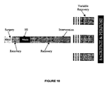
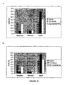
- hypothermia is anti-necrotic and in the gyrus, it is anti-apoptotic ( Figure 16).
- the data in this study do not explain this effect, but one possible justification could be that different brain regions express differential vulnerability (Northington et al, 2001).
- the secondary necrotic wave (discussed above) occurs at a time at which hypothermia has already been administered, and this may make it more effective.
- the gyrus there is no delayed necrosis and thus no anti-necrotic effect is observed.
- Anti-apoptosis appears to be the neuroprotective mechanism in this region, and it is possible that the expected anti-apoptotic neuroprotective effect of hypothermia, that is not evident at the earlier time intervals, may be exposed after longer periods.
- the present study has shown using an in vivo rat model to show that xenon is neuroprotective in the neonate, and significantly protects against apoptosis induced by hypoxic-ischaemic injury.
- the data in this study also suggest that when xenon and hypothermia are used in combination in the same model, they interact synergistically to dramatically decrease apoptotic cell death. Accordingly, this combination may represent an effective treatment to protect against the devastating neurological consequences of neonatal asphyxia.
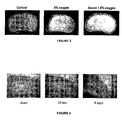
- Figure 2 Brain sections from animals that suffered 90 minutes of hypoxia-ischemia injury are shown in Figure 2.
- Figure 2 centre
- the brain slices on the right are from animals that have suffered the same hypoxia-ischemia but have been breathing 70% xenon during the hypoxic period. These brains look close to normal showing the remarkable neuroprotection afforded by xenon.
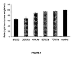
- Figure 4 shows the concentration-dependence of xenon neuroprotection (ratio right hemisphere weight/left against xenon concentration) of Figure 4.
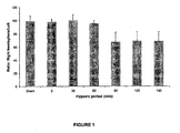
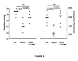
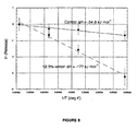
- the neuroprotective effect (ratio of right hemisphere/left) observed with with N 2 and xenon respectively is shown in Figure 6, when xenon is administered 2 hours post HI insult.
- the data show that xenon is effective in providing neuroprotection even if it is administered 2 hours after the end of the hypoxic period.
- the ratios of ipsilateral/contralateral hemispheric weight of 14 day rat brain after 90 min hypoxic-ischemia insult and then 2 hrs recovery following exposure with 70%N 2 or 70% Xe + 30% O 2 for 90 min at 7 days old. Results are mean ⁇ SEM (n 6).
- FIG. 7 The effect of mild hypothermia on the neuroprotective effect of xenon (LDH release against xenon concentration, % atm) is shown in Figure 7.
- Modest hypothermia produces a very large and unexpected enhancement in xenon neuroprotection. Cooling by 4 degrees greatly enhances the potency of xenon in blocking LDH release.
- this figure shows the effect of a combination of xenon and hypothermia on oxygen-glucose deprivation (OGD)-induced lactate dehydrogenase (LDH) release.
- Figure 7 shows the results of exposing neuronal cultures to 75 minutes OGD in the presence of increasing concentrations of xenon, either at 37°C (red), or at 33°C (blue).
- the ED 50 values for xenon at 37°C vs xenon at 33°C were 35.9 +/- 2.15% and 11.5 +/-2.0% (means +/- SEM) respectively.
- Neuronal injury is expressed as a percentage of the maximal LDH release after 75 minutes of OGD and 6 hours of recovery in the absence of either xenon or hypothermia. Points represent mean values, with error bars indicating standard errors.
- Example 2 More detailed studies are outlined below in Example 2.
- p7 Sprague-Dawley rat pups (weighing between 10 and 14 g) from Harlan, U.K. were subjected to a previously described model of HI injury (Levine, 1960; Rice et al, 1981).
- rat pups were anaesthetised with 2% isoflurane before undergoing permanent unilateral ligation of the right common carotid artery, using a midline neck incision and 5.0 silk suture. Once surgery was complete, pups recovered from the anaesthesia and were then returned to the dam until the time of experimentation.
- Control groups consisted of (a) non-treated littermates that underwent incision but no ligation (i.e. sham animals), used as negative controls, and (b) littermates exposed to HI, but not to the experimental intervention, to act as positive controls. These animals were subjected to 90 minutes at 37°C and a gas mixture composed of 25% oxygen and balanced nitrogen.
- mice Following HI and recovery, experimental rats were treated with 90 minutes of one of the five experimental interventions below. Each of the five treatments was carried out on separate groups of rats.
- This temperature was chosen as it represents "mild” hypothermia, and was thus thought to be clinically relevant, providing a good balance between side effects and benefit.
- the pups recovered with their mother until the time of sacrifice.
- the pup with the temperature probe in place was culled immediately after the experiment, and its brain was not used for analysis.
- the rats underwent both hypothermia and xenon concurrently for 90 minutes. Again, the pups were placed in airtight chambers, but on this occasion, their temperatures were maintained at 35°C and the gas mixture consisted of 25% oxygen, only 20% xenon and balanced nitrogen. This temperature and xenon concentration was shown in preliminary experiments, to confer no neuroprotective benefit to the developing brain when used independently. Thus, by using these values, any neuroprotective benefit at all is indicative of synergy between the two agents. Following treatment, pups were returned to their mothers until sacrifice.
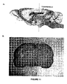
- FIG. 10 illustrates the area of the brain from which the slices were taken. Approximately 20 slices were taken from each brain in the region of the hippocampus, (as this is the area most vulnerable to HI injury) around -3.6mm from the bregma.
- the brains were cryoprotected in 30% sucrose in PBS (this also contained 2mg/ml sodium azide) and stored in the fridge for 48 h, or until they had sunk to the bottom of the tube.
- the brains were then frozen in O.C.T. compound (BDH, Poole, England) at -22°C and coronal sections were cut at 30 ⁇ m on a sliding cryostat (Bright Instrument Company Ltd., Huntingdon, U.K.). Sliced brains were stored in the fridge in wells containing 0.1 M PBS and 1mg/ml sodium azide, before being stained for immunohistochemistry (see below).
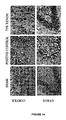
- Paraffin-embedded sections were mounted on slides and stained with cresyl violet for histology as described previously (Wilhelm et al, 2002).
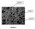
- Cresyl violet is a basic stain that binds to acidic components of neuronal cytoplasm such as RNA-rich ribosomes, and also the nuclei and nucleoli of nerve cells. This technique was used to assess cell viability and whether non-viable cells exhibited apoptosis or necrosis on the basis of validated morphological criteria (Nakajima et al, 2000).
- Each experimental group consisted of three time groups (16, 24, 48 h), and each time group contained three animals, (thus there were nine animals in total for each experimental group). Three slides per animal were chosen from the area of the brain that was -3.6mm from the bregma ( Figure 11). Slides were then sorted into time groups and the examiner was blinded to the intervention.
- Two areas from the ipsilateral side of each brain were analysed using a BX60 light microscope (Olympus, Southall, U.K.) - one in the cortex and one in the gyrus of the hippocampus ( Figure 11).
- a 40x objective lens with a grid was used to count the total number of cells that appeared in the grid. Cells were scored as either viable, apoptotic, or necrotic based on their morphological appearance, and the percentage of each cell type was noted down for each of the brain areas.
- An Axiocam digital camera (Zeiss, Göttingen, Germany) was used along with the microscope to take photomicrographs of the brain slices.
- Xenon is neuroprotective in the neonate by an anti-apoptotic mechanism
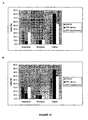
- apoptosis in the cortex was reduced from 36.5% ⁇ 2.5% in positive controls, to 8.5% ⁇ 1.6% (p ⁇ 0.001), and the viable cell count was increased from 52.9% ⁇ 2.3% in positive controls, to 80.6% ⁇ 0.2% (p ⁇ 0.001) ( Figure 13A).
- apoptosis was decreased from 33.6% ⁇ 1.8% in positive controls, to 13.9% ⁇ 2,4% (p ⁇ 0.01), and the viable cell count was raised from 56.5% ⁇ 2.6% in positive controls, to 77.1% ⁇ 3.3% (p ⁇ 0.01) ( Figure 13B).
- 35°C hypothermia used alone is ineffective against HI, and shows no statistical difference in either brain area when compared to positive controls.
- the percentage of apoptosis is 48% ⁇ 10.1% versus 37% ⁇ 2.5% in positive controls, and percentage cell viability is 44% ⁇ 10.3% versus 53% ⁇ 2.3%.
- hypothermia Treatment with a combination of 20% xenon + 35°C hypothermia demonstrates synergistic neuroprotection via an anti-apoptotic mechanism.
- xenon (20%) or hypothermia (35°C) in combination By using proven ineffective interventions of either xenon (20%) or hypothermia (35°C) in combination, a profound synergistic neuroprotection was demonstrated in both areas of the brain, and across all three of the time groups (16, 24 and 48 h) via an anti-apoptotic mechanism.
- Post-ischaemic application of the combination treatment significantly reduced the degree of apoptotic cell death, and increased the proportion of viable cells (see Figure 17).
Abstract
Description
- The present invention relates to treating neonatal asphyxia.
- Neonatal (or perinatal) asphyxia, also known as hypoxia-ischemia (HI), is a condition arising from the inadequate intake of oxygen in an infant during labour, delivery, or the immediate postnatal period. Neonatal asphyxia remains a major cause of chronic neurological morbidity and acute mortality in the newborn (Balduini et al, 2000; Vannucci et al, 1997) and commonly leads to hypoxic-ischemic encephalopathy.
- Studies have shown that neonatal asphyxia (hypoxia) for as short a time as six minutes can lead to permanent neurological damage. Loss of brain tissue has been demonstrated in asphyxiated newborn primates and correlated with memory dysfunction and spastic paralysis (Windle, WF, 1969).
- About 14.6% of all deaths at birth are caused by neonatal asphyxia. In the western world about 0.9% (i.e. 100-130,000) of newborns suffer from neonatal asphyxia. About 15-20% die, and of the survivors, 25% are severely handicapped due to long-term complications such as mental retardation, cerebral palsy, spasticity, learning difficulties and/or epilepsy (Law et al, 1993; Perlman et al, 1999). Furthermore, it is increasingly recognized that children with rather mild asphyxia, who seem initially to recover without complications, have behavioral problems in childhood, which can be traced back to this neonatal insult. Neonatal asphyxia meets the criteria for an orphan drug indication since it affects less then 5 patients in 10,000 inhabitants, and is a life-threatening, serious debilitating disease without an established therapy.
- It has been demonstrated in neonatal animal models of HI that the mechanisms of cell death involved in this type of brain injury, involve a combination of excitotoxic damage (or necrosis), caused by excessive activation of glutamate receptors, particularly N-methyl-D-aspartate (NMDA) receptors, as they are most sensitive to neurotoxicity during periods of synaptogenesis (Jevtovic-Todorovic and Olney, 2003), and by apoptotic neurodegeneration (Ikonomidou et al, 1989; Pohl et al, 1999). The type of damage is related to the severity of the hypoxic insult (Jevtovic-Todorovic and Olney, 2003) and also to the variation in vulnerability of the different brain regions (Northington et al, 2001). Currently, no effective therapy exists to combat the acute neuronal cell death caused by HI, although a variety of both pharmacological and non-pharmacological interventions are under experimental investigation (Vannucci and Perlman, 1997).
- The present invention seeks to provide a treatment for neonatal asphyxia.
- A first aspect of the invention relates to the use of xenon in the preparation of a medicament for the treatment of neonatal asphyxia, wherein said medicament is for use in combination with hypothermia.
- A second aspect of the invention relates to treating neonatal asphyxia in a mammal in need thereof by,
- (a) administering a therapeutically effective amount of xenon to the mammal; and
- (b) subjecting the mammal to hypothermia.
- A third aspect of the invention relates to treating neonatal asphyxia in a mammal in need thereof, by administering a therapeutically effective amount of xenon to the mammal in combination with hypothermia.
- A fourth aspect of the invention relates to the use of xenon in the preparation of a medicament for the treatment of neonatal asphyxia, wherein said treatment comprises administering to a subject simultaneously, sequentially or separately xenon in combination with hypothermia.
- A fifth aspect of the invention relates to the use of xenon, in combination with hypothermia, for the treatment of neonatal asphyxia.
- The excitatory amino acids (EAAs) glutamate and aspartate are the chief mediators of excitatory synaptic transmission in the mature central nervous system (CNS) (Dingledine and McBain, 1999). They also play a pivotal role in the ontogeny of the immature CNS, where they are involved in a number of physiological processes such as synaptogenesis, neuronal survival, synaptic plasticity, and dendritic and axonal structure. However, excessive activation of these amino acid receptors during development can produce neuronal injury and death. This is termed 'excitotoxicity'.
- Glutamate is the most abundant of the EAAs (Dingledine and McBain, 1999). It is stored in synaptic vesicles and causes calcium-dependent membrane depolarisation of postsynaptic membranes when it is released from the presynaptic terminals. Glutamate exerts its excitatory effect at a variety of receptor subtypes that can be divided into N-methyl-D-aspartate (NMDA) and non-NMDA types, but in the developing CNS it is the NMDA receptor subtype that has been found to play the primary role in brain injury associated with HI (Ikonomidou et al, 1989; Komuro, 1993; MacDonald et al, 1986).
- The NMDA receptor is a major subclass of glutamate receptor and glutamate is believed to be the most important excitatory neurotransmitter in the mammalian central nervous system. Importantly, activation of the NMDA receptor has been shown to be the central event which leads to excitotoxicity and neuronal death in many disease states, as well as a result of hypoxia and ischaemia following head trauma, stroke and following cardiac arrest.
- The NMDA receptor is an ionotropic receptor found ubiquitously throughout the CNS, located on the surface of both postsynaptic and extrasynaptic membranes (Riccio and Ginty, 2002; Sattler et al, 2000). It is coupled to a cationic channel that is permeable to both Na+ and Ca2+ ions and under normal physiological conditions, is blocked by Mg2+ at a negative resting membrane potential. It becomes unblocked on depolarisation of the cell membrane, thus allowing an influx of Ca2+ through the channel and enabling the receptor to exert its intracellular effects (Hardingham and Bading, 2003).
- NMDA receptors are vital for normal brain function and their importance in normal physiology is demonstrated by their central role in memory and learning (Bliss and Collingridge, 1993). Conversely, pathological activation of NMDA receptors by excess glutamate is the primary cause of neuronal cell death following an ischaemic insult to the brain, due to the disruption of intracellular Ca2+ regulation. This emphasizes the central role played by NMDA receptors in HI.
- In order for the brain to function, it requires a continuous supply of oxygen and glucose and is thus reliant on an adequate blood supply (Choi and Rothman, 1990). Should the blood supply become interrupted, as is the case in neonatal asphyxia, hypoxic-ischaemic damage to the area downstream will ensue within minutes. Under these conditions of oxygen depletion, cellular metabolism shifts from aerobic to anaerobic (Vannucci and Perlman, 1997), which is less effective at meeting the energy requirements of the cell. This leads to a depletion of energy stores, particularly affecting high-energy phosphate reserves such as ATP in the neuronal and glial cell compartments (Dingledine and McBain, 1999). There is concomitant accumulation of H+ ions, leading to acidosis, and release of free radicals that contribute to further damage of the cells.
- Under physiological conditions, the extracellular concentration of glutamate is maintained at low levels by the action of glutamate transporters located in neuronal cells, but expressed preferentially in glial cells (Dingledine and McBain, 1999). There are several different kinds of glutamate uptake carrier, but essentially, they all function in the same way, transporting two Na+ cations and one glutamate anion into the cell, while transporting one K+ cation and one OH- anion out of the cell and into the extracellular space (Dingledine and McBain, 1999). These ionic pumps act against an electrochemical gradient and thus rely on energy in the form of ATP in order to function correctly. Therefore, the ability of these pumps to maintain the resting membrane potential is decreased by the reduction in the concentration of ATP that results from HI. Consequently, failure of the ATP-dependent pump leads to depolarisation of the membrane, and a reversal in the direction of pumping (Eilers and Bickler, 1996; Kauppinen et al, 1988). Thus, glutamate is transported out of the cell and an excess concentration of glutamate accumulates in the extracellular space. Not only does the glutamate concentration increase due to a decreased uptake, but there is also an increased release of glutamate from the presynaptic terminals as the membrane depolarisation sets up an action potential (Dingledine and McBain, 1999). Examples of these processes leading to excess extracellular glutamate have been seen both in vitro (Bosley et al, 1983; Hauptman et al, 1984; Pellegrino-Giampietro et al, 1990) and in vivo (Erecinska et al, 1984; Graham et al, 1990; Ikeda et al, 1989).
- Excitotoxicity occurs when the excess extracellular glutamate continuously activates postsynaptic receptors (particularly NMDA receptors) and the resulting calcium influx sets up an osmotic gradient down which water moves causing the cells to swell. Calcium-dependent enzyme systems are also activated within the cell and these two processes result in acute neuronal cell death (Choi and Rothman, 1990).
- Neuronal cell death has always been thought to arise from one of two mechanisms: necrosis and apoptosis, as hypothesised by Wyllie et al (Wyllie et al, 1980). However, recently these categories have been questioned as more evidence has come to light to suggest that cell death should be divided into the categories: excitotoxic cell death and apoptosis (Olney, 2003). Excitotoxic cell death has been described as a necrotic process (Gwag et al, 1997; Katja and Green, 2001), an apoptotic process, and a continuum of the two (Leist and Nicotera, 1998; Nakajima et al, 2000). Apoptosis and necrosis are usually distinguished by their distinctly different morphological appearances. Apoptosis requires ATP and new protein synthesis, and is identified by cell shrinkage, chromatin-clumping with margination and formation of membrane-enclosed apoptotic bodies, whereas necrosis is recognised by nuclear shrinkage with karyorrhectic and pyknotic nuclear changes (Hill et al, 1995).
- Cell death from HI has been found to involve an initial period of necrosis, followed by a delayed wave of apoptotic cell death (Northington et al, 2001). The type of injury that ensues appears to be both time-dependent and location-dependent, with the initial necrotic injury being confined to the ipsilateral forebrain in a neonatal rat model of HI, and the delayed apoptotic injury occurring in the thalamus (Northington et al, 2001). This suggests that the different brain regions may express differential vulnerability to each type of cell death at different times following HI.
- In normal development, apoptosis is a regular event by which unwanted or damaged neurons 'commit suicide' (Ikonomidou et al, 2001). In HI, the initial excitotoxic cell death is mediated by excessive activation of NMDA receptors, resulting in the uncontrolled release of glutamate, which damages the surrounding neurons. The natural response to damage during synaptogenesis is for the neurons to initiate programmed cell death (Olney, 2003), and this is thought to be a mechanism that is activated to protect the neighbouring tissue (Leist and Nicotera, 1998).
- It is known in the art that the NMDA receptor plays a major role in the synaptic plasticity which underlies many higher cognitive functions, such as memory and learning, as well as in certain nociceptive pathways and in the perception of pain (Collingridge et al, The NMDA Receptor, Oxford University Press, 1994). In addition, certain properties of NMDA receptors suggest that they may be involved in the information-processing in the brain which underlies consciousness itself.
- NMDA receptor antagonists are therapeutically valuable for a number of reasons. Firstly, NMDA receptor antagonists confer profound analgesia, a highly desirable component of general anaesthesia and sedation. Secondly, NMDA receptor antagonists are neuroprotective under many clinically relevant circumstances (including ischemia, brain trauma, neuropathic pain states, and certain types of convulsions). Thirdly, NMDA receptor antagonists confer a valuable degree of amnesia.
- Given the importance of NMDA receptors in the pathogenesis of HI, it is fitting that NMDA antagonists have been investigated as possible neuroprotective agents. Many NMDA antagonists, such as MK-801 and ketamine have been shown to be neuroprotective in both in vitro and in vivo models (Albers et al, 1989; Arias et al, 1999; Choi et al, 1988; Kudo et al, 2001). However, despite these encouraging results, NMDA receptor antagonists have also been shown to have psychotomimetic side effects in humans (Krystal et al, 1994) and to cause damage to the posterior cingulate (PC) and retrosplenial cortices (RS) (Olney et al, 1991). In addition, many conventional NMDA receptor antagonists lead to the production of involuntary movements, stimulation of the sympathetic nervous system, induction of neurotoxicity at high doses (which is pertinent since NMDA receptor antagonists have low potencies as general anaesthetics), depression of the myocardium, and proconvulsions in some epileptogenic paradigms e.g., "kindling" (Wlaz P et al, Eur. J. Neurosci. 1994; 6:1710-1719). There have also been considerable difficulties in developing new NMDA receptor antagonists that are able to cross the blood-brain barrier.
- Xenon is an apolar, inert gas that is a potent NMDA antagonist (Franks et al, 1998). Like other NMDA antagonists, it has also been shown to be neuroprotective against many forms of neuronal injury, both in vitro (Petzelt et al, 2003) and in vivo (Homi et al, 2003; Wilhelm et al, 2002). However, unlike many of the other NMDA receptor antagonists, xenon is not neurotoxic (Ma et al, 2002). A further advantage of using xenon as an NMDA antagonist is that the molecule is an inert, volatile gas that can be rapidly eliminated via respiration.
- Xenon has many other favourable properties. Since its first use in surgery (Cullen SC et al, Science 1951; 113:580-582), a number of research groups have shown it has an excellent pharmacological profile, including the absence of metabolic by-products, profound analgesia, rapid onset and recovery, and minimal effects on the cardiovascular system (Lachmann B et al, Lancet 1990; 335:1413-1415; Kennedy RR et al, Anaesth. Intens. Care 1992; 20:66-70; Luttropp HH et al, Acta Anaesthesiol. Scand. 1994; 38:121-125; Goto T et al, Anesthesiology 1997; 86:1273-1278; Marx T et al, Br. J. Anaesth. 1997; 78:326-327). Moreover, as xenon is a small, uncharged atom, it can easily pass through the blood-brain barrier thus producing a rapid onset of action (Nakata et al, 2001). It also has a very low blood: gas partition coefficient lending to fast emergence from xenon anaesthesia (Goto et al, 1997). As well as these advantages, xenon is non-explosive, non-toxic and unreactive (Shichino et al, 2002), and this makes xenon an ideal candidate for use as a neuroprotectant in the neonate.
- As used herein, the term "neuroprotectant" means an agent that is capable of providing neuroprotection, i.e., protecting a neural entity, such as a neuron from ongoing injury from,for example, an ischaemic injury or traumatic injury.
- Talbot first demonstrated the neuroprotective properties of hypothermia for surgical use in 1941 (Talbot, 1941). Currently, the only routine use of hypothermia is during cardiopulmonary bypass to protect the brain from intra-operative ischaemia. However, there have been several publications demonstrating the therapeutic effect of hypothermia in other models of brain injury. For example, numerous publications exist showing the beneficial effect of hypothermia in both in vitro (Onitsuka et al, 1998) and in vivo models of neonatal asphyxia (Debillon et al, 2003; Treschera et al, 1997). It has been demonstrated that a direct correlation exists between tissue injury and the extent of brain cooling (Towfighi et al, 1994), and in normoxic conditions, every 1°C decrease in body temperature leads to a 5% decrease in the cerebral metabolic rate (Yager and Asselin, 1996).
- The mechanism by which hypothermia exerts its neuroprotective effect has yet to be elucidated, but many theories have been postulated. Studies have suggested that the mechanisms by which hypothermia is protective are temperature and time-dependent, and may act at more than one point along the cascade of events that leads to HI injury (Yager and Asselin, 1996). This is supported by the fact that a moderate temperature of 31°C has been shown to be neuroprotective by decreasing cerebral energy metabolism, whereas a mild hypothermia of 34°C while also neuroprotective, has no effect on energy metabolism and must therefore act via a different mechanism (Yager and Asselin, 1996). Another study by Taylor et al (Taylor et al, 2002) demonstrated that hypothermia instituted after the HI insult was more effective than intra-ischaemic hypothermia, and suggested that this may be due to a decrease of deleterious effects that occur during the recovery period. An example of one such mechanism could be that hypothermia decreases the excitotoxic damage that ensues during reperfusion (Taylor et al, 2002). Many other mechanisms of protection by hypothermia have been suggested, including the reduction of reactive oxygen species (Taylor et al, 2002), a reduction in tissue acidosis (Chopp et al, 1989) and the attenuation of post-HI neuronal apoptosis (Xu et al, 2002).
- As mentioned above, a first aspect of the present invention relates to the use of xenon in the preparation of a medicament for the treatment of neonatal asphyxia in a neonatal subject, wherein said medicament is for use in combination with hypothermia.
- As used herein, the term "hypothermia" refers to subjecting a particular subject (in this case, a neonatal subject) to hypothermic conditions, for example, by lowering the body temperature, preferably by 3-5°C, through passive or active techniques. Typically, subjecting to hypothermic conditions leads to a decrease in metabolism of body tissues of the subject, thereby decreasing the need for oxygen.
- As mentioned above, the use of hypothermia in the treatment of neonatal asphyxia has been well documented in the art (see for example, Volpe, 2001; Gunn et al, 2000). However, to date there has been no teaching or suggestion in the art that hypothermia could be use in combination with the administration of xenon. Nor has there been any suggestion that such combination therapy would lead to such a surprising and unexpected enhancement in the resulting neuroprotective effect.
- Previous studies by the applicant have revealed that xenon has neuroprotective properties. In particular,
WO 01/08692 - In one preferred embodiment of the invention, the xenon is admixed with a pharmaceutically acceptable diluent, excipient or carrier.
- Examples of such suitable excipients for the various different forms of pharmaceutical compositions described herein may be found in the "Handbook of Pharmaceutical Excipients, 2nd Edition, (1994), Edited by A Wade and PJ Weller.
- Acceptable carriers or diluents for therapeutic use are well known in the pharmaceutical art, and are described, for example, in Remington's Pharmaceutical Sciences, Mack Publishing Co. (A. R. Gennaro edit. 1985). Examples of suitable carriers include lactose, starch, glucose, methyl cellulose, magnesium stearate, mannitol, sorbitol and the like. Examples of suitable diluents include ethanol, glycerol and water.
- The choice of pharmaceutical carrier, excipient or diluent can be selected with regard to the intended route of administration and standard pharmaceutical practice. The pharmaceutical compositions may comprise as, or in addition to, the carrier, excipient or diluent any suitable binder(s), lubricant(s), suspending agent(s), coating agent(s), solubilising agent(s).
- Examples of suitable binders include starch, gelatin, natural sugars such as glucose, anhydrous lactose, free-flow lactose, beta-lactose, corn sweeteners, natural and synthetic gums, such as acacia, tragacanth or sodium alginate, carboxymethyl cellulose and polyethylene glycol.
- Examples of suitable lubricants include sodium oleate, sodium stearate, magnesium stearate, sodium benzoate, sodium acetate, sodium chloride and the like.
- Preservatives, stabilizers and dyes may be provided in the pharmaceutical composition. Examples of preservatives include sodium benzoate, sorbic acid and esters of p-hydroxybenzoic acid. Antioxidants and suspending agents may be also used.
- The present invention is also applicable to the treatment of animals. In this regard, the invention further relates to the use of xenon in combination with a veterinarily acceptable diluent, excipient or carrier.
- For veterinary use, the xenon is typically administered in accordance with normal veterinary practice and the veterinary surgeon will determine the dosing regimen and route of administration which will be most appropriate for a particular animal.
- The xenon may also be administered in combination with another pharmaceutically active agent. The agent may be any suitable pharmaceutically active agent including anaesthetic or sedative agents which promote GABAergic activity. Examples of such GABAergic agents include isoflurane, propofol and benzodiazapines.
- In one preferred embodiment, the xenon is administered in combination with a volatile anesthetic agent, preferably isoflurane, sevoflurane or desflurane.
- The xenon may also be administered in combination with other active ingredients such as L-type calcium channel blockers, N-type calcium channel blockers, substance P antagonists, sodium channel blockers, purinergic receptor blockers, or combinations thereof.
- The xenon may be administered by any suitable delivery mechanism, or two or more suitable delivery mechanisms.
- In one particularly preferred embodiment, the xenon is administered by perfusion. In the context of the present invention, the term "perfusion" refers to the introduction of an oxygen/xenon mixture into, and the removal of carbon dioxide from, a patient using a specialised heart-lung machine. In general terms, the heart-lung machine replaces the function of the heart and lungs and provides a bloodless, motionless surgical field for the surgeon. The perfusionist ventilates the patient's blood to control the level of oxygen and carbon dioxide. In the context of the present invention, the perfusionist also introduces xenon into the patient's blood. The perfusionist then propels the blood back into the arterial system to provide nutrient blood flow to all the patient's vital organs and tissues during surgery.
- In one particularly preferred embodiment, the medicament is in gaseous form.
- In another highly preferred embodiment, the xenon is administered by inhalation. More preferably, the xenon is administered by inhalation of a 70-30% v/v xenon/oxygen mixture.
- More preferably, the xenon is administered in the form of a 20-70% v/v xenon/air mixture.
- In yet another preferred embodiment of the invention, the medicament is in the form of a liquid or solution.
- Preferably, the liquid is administered in the form of a solution or an emulsion prepared from sterile or sterilisable solutions, which may be injected intravenously, intraarterially, intrathecally, subcutaneously, intradermally, intraperitoneally or intramuscularly.
- In one particularly preferred embodiment, the xenon is administered in the form of a lipid emulsion. The intravenous formulation typically contains a lipid emulsion (such as the commercially
available Intralipid ®10, lntralipid®20, Intrafat®, Lipofundin®S or Liposyn® emulsions, or one specially formulated to maximise solubility) which sufficiently increases the solubility of the xenon to achieve the desired clinical effect. Further information on lipid emulsions of this sort may be found in G. Kleinberger and H. Pamperl, Infusionstherapie, 108-117 (1983) 3. - The lipid phase of the present invention which dissolves or disperses the gas is typically formed from saturated and unsaturated long and medium chain fatty acid esters containing 8 to 30 carbon atoms. These lipids form liposomes in aqueous solution. Examples include fish oil, and plant oils such as soya bean oil, thistle oil or cottonseed oil. The lipid emulsions of the invention are typically oil-in-water emulsions wherein the proportion of fat in the emulsion is conventionally 5 to 30% by weight, and preferably 10 to 20% by weight. Oil-in-water emulsions of this sort are often prepared in the presence of an emulsifying agent such as a soya phosphatide.
- The lipids which form the liposomes of the present invention may be natural or synthetic and include cholesterol, glycolipids, sphingomyelin, glucolipids, glycosphingolipids, phosphatidylcholine, phosphatidylethanolamine, phosphatidyl-serine, phosphatidyglycerol, phosphatidylinositol.
- The lipid emulsions of the present invention may also comprise additional components. These may include antioxidants, additives which make the osmolarity of the aqueous phase surrounding the lipid phase isotonic with the blood, or polymers which modify the surface of the liposomes.
- It has been established that appreciable amounts of xenon maybe added to a lipid emulsion. Even by the simplest means, at 20°C and normal pressure, xenon can be dissolved or dispersed in concentrations of 0.2 to 10 ml or more per ml of emulsion. The concentration of dissolved gas is dependent on a number of factors, including temperature, pressure and the concentration of lipid.
- The lipid emulsions of the present invention may be loaded with gaseous xenon. In general, a device is filled with the emulsion and anaesthetics as gases or vapours passed through sintered glass bubblers immersed in the emulsion. The emulsion is allowed to equilibrate with the anaesthetic gas or vapour at a chosen partial pressure. When stored in gas tight containers, these lipid emulsions show sufficient stability for the anaesthetic not to be released as a gas over conventional storage periods.
- The lipid emulsions of the present invention may be loaded so that the xenon is at the saturation level. Alternatively, the xenon may be present in lower concentrations, provided, for example, that the administration of the emulsion produces the desired pharmaceutical activity.
- The concentration of xenon employed in the invention may be the minimum concentration required to achieve the desired clinical effect. It is usual for a physician to determine the actual dosage that will be most suitable for an individual patient, and this dose will vary with the age, weight and response of the particular patient. There can, of course, be individual instances where higher or lower dosage ranges are merited, and such are within the scope of this invention.
- Preferably, the medicament is in a form suitable for intravenous, neuraxial or transdermal delivery.
- Preferably, the xenon is administered simultaneously, in combination, sequentially or separately with hypothermia.
- As used herein, "simultaneously" is used to mean that the xenon is administered concurrently with hypothermia, whereas the term "in combination" is used to mean the xenon is administered, if not simultaneously, then "sequentially" within a timeframe in which the xenon and the hypothermia both exhibit a therapeutic effect, i.e. they are both are available to act therapeutically within the same time-frame. Thus, administration "sequentially" may permit the xenon to be administered within 5 minutes, 10 minutes or a matter of hours before the hypothermia, provided the circulatory half-life of the xenon is such that it is present in a therapeutically effective amount when the neonatal subject is exposed to hypothermic conditions.
- In another preferred embodiment of the invention, the neonate is subjected to hypothermia prior to treatment with xenon.
- In contrast to "in combination" or "sequentially", "separately" is used herein to mean that the gap between administering the xenon and exposing the neonatal subject to hypothermia is significant i.e. the xenon may no longer be present in the bloodstream in a therapeutically effective amount when the neonatal subject is exposed to hypothermic conditions.
- In one preferred embodiment, the xenon is administered sequentially with hypothermia.
- More preferably, the xenon is administered sequentially before the hypothermia.
- In another preferred embodiment, the xenon is administered separately before the hypothermia.
- In one preferred embodiment, the xenon is administered sequentially after the hypothermia.
- In another preferred embodiment, the xenon is administered separately after the hypothermia.
- More preferably, the xenon is administered sequentially or simultaneously with hypothermia, more preferably simultaneously.
- In one preferred embodiment of the invention, the xenon is administered in a therapeutically effective amount.
- In another preferred embodiment, the xenon is administered in a sub-therapeutically effective amount. In other words, the xenon is administered in an amount that would be insufficient to produce the desired therapeutic effect if administered in the absence of hypothermic conditions.
- Even more preferably, the combination of xenon and hypothermia has a synergistic effect, i.e., the combination is synergistic.
- In one particularly preferred embodiment, the xenon is administered prior to the hypoxic insult. Thus, in one preferred embodiment, the xenon is administered to the neonate via the mother prior to birth, for example, by administering to the mother prior to or during labour. Preferably, the xenon is administered to the mother for up to about 48 or 24 hours prior to birth, more preferably up to about 12 hours, more preferably up to about 6 hours or 3 hours or 1 hour prior to birth. After birth, the neonate is then subjected to hypothermic conditions.
- Another aspect of the invention relates to treating neonatal asphyxia in a mammal in need thereof by
- (a) administering a therapeutically effective amount of xenon to the mother of the mammal prior to and/or during labour; and
- (b) subjecting the mammal to hypothermia after birth.
- Preferably, the hypothermia is maintained for a period of at least about 6 hours, more preferably at least about 12 hours, after the hypoxic-ischemic (HI) insult.
- In one preferred embodiment, the hypothermia is maintained for a period of from about 6 to about 24 hours after the hypoxic-ischemic (HI) insult.
- Preferably, the hypothermia is maintained for a period of at least about 6 hours, more preferably at least about 12 hours, after birth.
- In one preferred embodiment, the hypothermia is maintained for a period of from about 6 to about 24 hours after birth.
- Preferably, treatment in accordance with the invention is initiated within about 6 hours of the hypoxic-ischemic (HI) insult, and more preferably within about 2 hours of the hypoxic-ischemic insult.
- Hypothermia may be produced passively, by allowing the temperature to drift downwards and not purposefully sustain body temperature. Being poikilothermic, neonates rapidly assume the temperature of their surroundings. Alternatively the patient may be actively rendered hypothermic by deliberately reducing their ambient temperature.
- A second aspect of the invention relates to treating neonatal asphyxia in a mammal in need thereof by:
- (a) administering a therapeutically effective amount of xenon to the mammal; and
- (b) subjecting the mammal to hypothermia, or hypothermic conditions.
- In a preferred embodiment, the mammal is a newborn subject in the first four weeks after birth. More preferably, the mammal is in the first two weeks, more preferably still, the first week after birth.
- Preferably, the mammal is a human.
- Preferably, the mammal is subjected to conditions of mild hypothermia. As used herein, the term "mild hypothermia" typically refers to a decrease in the core temperature from 37°C to about 33°C
- In one preferred embodiment, the temperature of the mammal is maintained at a temperature of from about 31°C to about 36°C.
- More preferably, the temperature of the mammal is maintained at a temperature of from about 32 °C to about 36 °C, more preferably from about 32 °C to about 35 °C, more preferably still from about 33 °C to about 35 °C.
- Preferred embodiments for the second aspect of the invention are the same as those described above in respect of the first aspect.
- Another aspect of the invention relates to treating neonatal asphyxia in a mammal in need thereof by administering a therapeutically effective amount of xenon to the mammal in combination with hypothermia.
Yet another aspect of the invention relates to the use of xenon in the preparation of a medicament for the treatment of neonatal asphyxia, wherein said treatment comprises administering to a subject simultaneously, sequentially or separately xenon in combination with hypothermia. - A further aspect of the invention relates to the use of xenon, in combination with hypothermia, for the treatment of neonatal asphyxia.
- Using an animal model of HI, neonatal rats were exposed to treatment with xenon and hypothermia independently of each other. Xenon was shown to be neuroprotective against HI in the neonate by reducing the amount of apoptotic cell death, while hypothermia appeared less effective. In combination, xenon and hypothermia were neuroprotective via an anti-apoptotic mechanism (Figure 17). Their combined effect was found to be synergistic.
- The neonatal rat HI model is very established and has been validated for use in a number of previous studies (Levine, 1960; Rice et al, 1981). The age of the rats used in this model has been found to correspond to the brain maturity of the term human neonate (Clancy et al, 2001; Ikonimidou et al, 1989) and thus a reasonably accurate comparison can be made between the two.
- During the hypothermia experiments, the temperature of the rat pups was monitored using a probe that was inserted into the cortex of one of the pups. The probe took approximately 15 minutes to equilibrate and this was allowed for by delaying the start time of the experiment until the probe began recording the correct temperature. There were fluctuations in temperature around the mean value, but these were controlled by continuous monitoring and manual adjustment of the water bath as necessary. Only one rat per group was monitored for temperature in order to minimise the trauma caused to the rats and also the damage inflicted upon the cortex by the probe; rats with the probe inserted could not be used for histological analysis.
- The anaesthetic gas xenon has been shown to exhibit neuroprotection in several models of adult neuronal injury. Currently, no published data exist to confirm the same neuroprotective effect of xenon in neonates. The results of this study corroborate previous findings that xenon has significant neuroprotective properties and in addition, suggest that this neuroprotection extends to neonatal models of brain injury induced by hypoxia-ischaemia.
- It has long been known that the activation of the NMDA subtype of glutamate receptors is required to sustain ongoing neuronal injury and death in HI, and it is well documented that xenon exerts its analgesic and anaesthetic effect via blockade of these receptors, thus it has been postulated that xenon's neuroprotective properties are as a result of this antagonism. Previously, several other NMDA antagonists have demonstrated neuroprotection in in vitro studies, but have subsequently failed when utilised in clinical settings (Muir and Lees, 1995). The reason behind these clinical failures is unknown, however it is possible that blockade of the glutamate receptor subtype is insufficient to protect against injury, which would imply that xenon exerts its neuroprotective effect through another mechanism.
- In the present study, it has been demonstrated that xenon significantly protects against neonatal HI via an anti-apoptotic mechanism. Both apoptosis and necrosis are important components of neuronal loss after HI injury, but apoptosis appears to be the more important type of cell death in determining neonatal outcome (Taylor et al, 1999). Apoptotic death is often preceded by the activation of many genes, (including transcription factors) which may be either pro-apoptotic or anti-apoptotic. As xenon appears to interfere with apoptotic cell death, it is possible that it may exert its effect on one of these genes, or at some point along the apoptotic pathway. Currently, there is evidence for two different apoptosis pathways: the extrinsic pathway, and the intrinsic pathway. The extrinsic pathway (also referred to as the "death receptor pathway") involves the binding of cytokines to death receptors which activate caspase-8 and this in turn, activates the "executor caspsase", caspase-3, which goes on to induce apoptotic cell death (Mehmet, 2000). The intrinsic pathway is heavily dependent on mitochondria and involves an increase in mitochondrial membrane permeability caused by the pro-apoptotic protein bax. This leads to the release of cytochrome c, the formation of a complex of cytochrome c, Apaf-1 (apoptosis protease-activating factor-1) and caspase-9, and the subsequent activation of caspase-3. It is entirely possible that xenon acts on either one of these pathways, but there is evidence to suggest that apoptotic neurodegeneration induced by HI is mediated via the mitochondrial pathway and the initiation of bax-dependent mitochondrial changes (Taylor et al, 1999). Further to this, the NMDA antagonist ketamine has been shown to protect against incomplete cerebral ischaemia and reperfusion by early modulation of the balance between pro-and anti-apoptotic proteins, namely by inhibiting the HI-induced bax increase (Engelhard et al, 2003). Thus it is possible that xenon could inhibit apoptosis by downregulating bax. Bcl-2 is an anti-apoptotic protein that acts to decrease the permeability of the mitochondria and hence inhibit the release of cytochrome c. Its overexpression has been shown to decrease neuronal damage caused by transient global cerebral ischaemia in gerbils (Engelhard et al, 2003). Therefore, the upregulation of bcl-2 is another potential target for xenon. As xenon is apolar and fat soluble, it is able to distribute itself widely throughout the body. It can penetrate membranes and consequently, it may also have an effect in the nucleus by altering gene transcription to upregulate survival pathways, or inhibiting the RNA and protein synthesis of pro-apoptotic molecules.
- Anti-necrosis by xenon was shown to be statistically significant in the cortex at 48 h, but not in the gyrus (Figure 16). At all other time groups, xenon was not anti-necrotic. One possible explanation for this is that in accordance with a previous study (Northington et al, 2001), there is a secondary wave of necrotic cell death at 48 h that is only apparent in the cortex. This would explain the increase in the percentage of necrotic cells present in the positive controls at 48 h, compared with 16 and 24 h. Although it is not certain how xenon exerts an anti-necrotic effect in the cortex at 48 h, it may be that while xenon is unable to prevent necrosis that occurs before its administration (as in the 16 and 24 h groups), it is some how able to combat the secondary necrotic wave that occurs after its administration. Initial necrosis occurs as early as 3 h after the HI insult (Northington et al, 2001) and at this point xenon has not yet been administered. It is therefore unlikely to be able to arrest or reverse a process that has already occurred. However, the secondary necrotic wave occurs at a time at which xenon has been present in the brain for 48 h, and this suggests that the presence of xenon at the advent of necrosis may be able to decrease this type of cell death. Further work must to be completed to ascertain the exact mechanism of this interaction.
- Previous studies have demonstrated that mild hypothermia of 33°C is neuroprotective against ischaemic neuronal injury (Busto et al, 1987). Other studies have suggested that this neuroprotection is achieved via an anti-apoptotic mechanism (Xu et al, 2002). Experiments showed that there was no neuroprotection at 16 or 24 h (Figures 13 and 15 respectively.
- At 48 h however, significant neuroprotection was achieved in both the cortex and the gyrus, but by different mechanisms. In the cortex, hypothermia is anti-necrotic and in the gyrus, it is anti-apoptotic (Figure 16). The data in this study do not explain this effect, but one possible justification could be that different brain regions express differential vulnerability (Northington et al, 2001). In the cortex, the secondary necrotic wave (discussed above) occurs at a time at which hypothermia has already been administered, and this may make it more effective. In the gyrus however, there is no delayed necrosis and thus no anti-necrotic effect is observed. Anti-apoptosis appears to be the neuroprotective mechanism in this region, and it is possible that the expected anti-apoptotic neuroprotective effect of hypothermia, that is not evident at the earlier time intervals, may be exposed after longer periods.
- The results demonstrated that when used in combination, 20% xenon and 35°C hypothermia provided an astounding level of neuroprotection. As these values provided no neuroprotection when each agent was used alone, the result could not be explained by an additive mechanism, but instead had to be due to synergistic interaction between the two agents.
- By way of summary, the present study has shown using an in vivo rat model to show that xenon is neuroprotective in the neonate, and significantly protects against apoptosis induced by hypoxic-ischaemic injury. The data in this study also suggest that when xenon and hypothermia are used in combination in the same model, they interact synergistically to dramatically decrease apoptotic cell death. Accordingly, this combination may represent an effective treatment to protect against the devastating neurological consequences of neonatal asphyxia.
- The present invention is further described by way of example, and with reference to the following figures, wherein:
- Figure I shows the relationship between damage, as measured by loss of brain weight (ratio of right hemisphere/left) and the duration of the hypoxic period (in minutes) in Sprague-Dawley rats.
- Figure 2 shows brain sections from Sprague-Dawley rats that have suffered 90 minutes of hypoxia-ischemia injury.
- Figure 3 shows the major cellular damage which is evident in Sprague-
Dawley rats 24 hours after 90 minutes of hypoxia-ischemia. - Figure 4 shows the concentration-dependence of xenon neuroprotection (ratio right hemisphere weight/left against xenon concentration).
- Figure 5 shows the effect of 70% xenon on neurological functions assessed remotely after hypoxic-ischemic (HI) insult.
- Figure 6 compares the neuroprotective effect (ratio of right hemisphere/left) observed with with N2 and xenon respectively, when xenon is administered 2 hours post HI insult.
- Figure 7 shows the effect of mild hypothermia on the neuroprotective effect of xenon (LDH release against xenon concentration, % atm).
- Figure 8 shows a van't Hoff plot of the natural logarithm of LDH release plotted against reciprocal absolute temperature.
- Figure 9 shows a photograph of the purpose-built airtight chambers used for gas delivery. The water bath and closed circuit xenon delivery system are also depicted.
- Figure 10 shows a schematic timeline of the method used. 60 minutes is the time taken for surgery of n=12 pups. Recovery periods were undertaken in the dam. The interventions used were: sham animals, positive controls, 75% xenon (balance oxygen), 33°C hypothermia, 20% xenon (25% oxygen, 55% nitrogen), 35°C hypothermia, and a combination of 35°C hypothermia and 20% xenon. Unless otherwise indicated, animals were kept at 37°C and breathed a gas mixture of 25% oxygen balanced with nitrogen. Sham animals underwent incision, but no ligation or HI, and positive controls had both surgery and HI. Animals in each group were divided equally between the variable recovery periods (16, 24 and 48 h) before being sacrificed. Abbreviations: HI, hypoxia-ischamia.
- Figure 11 shows (A) Sagittal section of a rat brain modified from website: faculty.virginia.edu/.../RatBrainLabels.jpg. The broken line represents the area of the brain from which coronal sections (B) were taken, approximately -3.6 mm from the bregma. (B) shows a cresyl violet-stained coronal section. The boxes indicate the areas in which the counting frames were placed and the cells were analysed - the top box indicates the cortex and the bottom one, the gyrus. X overlies a hole that was intentionally created with a safety pin in order to demonstrate the non-ligated (contra lateral) hemisphere.
- Figure 12 shows a photomicrograph of the cortex (cresyl-violet stained), taken with a 100x oil emersion lens and an Axiocam digital camera, demonstrating the difference in morphological appearance between an apoptotic, necrotic and viable cell. Viable cells stain less intensely than either type of cell death and therefore have a paler cytoplasm, whereas dead cells are more darkly stained. Necrotic and apoptotic cells are differentiated on the basis of their different nuclear appearances - necrotic nuclei are large and irregularly shaped, whereas apoptotic nuclei are small, shrunken, and spherically shaped.
- Figure 13 shows that xenon is neuroprotective at 16 h via an anti-apoptotic mechanism. More specifically, Figure 13 shows graphs for apoptotic and necrotic neuronal death induced by hypoxic-ischaemia, and the effects of 75% xenon and 33°C hypothermia on such cell death at 16 h in (A) the cortex, and (B) the gyrus. In both brain areas xenon significantly increases the percentage of viable cells as well as decreasing the percentage of apoptotic cells compared to positive control animals. In the cortex, hypothermia decreases the percentage of apoptotic cells, although it does not increase the viable cell count and can therefore not be considered neuroprotective. Results are mean ± SD (n=3). *p < 0.05, **p < 0.01, ***p < 0.001 vs. positive controls.
- Figure 14 shows a photomicrograph demonstrating the cortex and gyrus in the sham, positive control and 75 % xenon animals at 16 hours. The 75 % group is more similar in appearance to the sham group than the positive control group. This confirms the neuroprotective effect of xenon at 16 hours. The gyrus of the control group is distorted in shape due to the increased amount of cell death and vacuolation.
- Figure 15 shows that xenon is neuroprotective at 24 h via an anti-apoptotic mechanism. More specifically, Figure 15 shows graphs for apoptotic and necrotic neuronal death induced by hypoxic-ischaemia, and the effects of 75% xenon and 33°C hypothermia on such cell death at 24 h in (A) the cortex, and (B) the gyrus. In both brain areas xenon causes a significant increase in the percentage of viable cells due to a decreased necrotic cell count. Results are mean ± SD (n=3). *p < 0.05 vs. positive controls.
- Figure 16 shows that xenon is neuroprotective at 48 h via an anti-apoptotic mechanism More specifically, Figure 16 shows graphs for apoptotic and necrotic neuronal death induced by hypoxic-ischaemia, and the effects of 75% xenon and 33°C hypothermia on such cell death at 48 h in (A) the cortex, and (B) the gyrus. Xenon is neuroprotective via an anti-apoptotic mechanism in both the cortex and the gyrus. In addition, xenon has an anti-necrotic effect in the cortex. 33°C hypothermia appears to be neuroprotective in both brain areas, but by a different mechanism - it is anti-necrotic in the cortex, and anti-apoptotic in the gyrus. Results are mean ± SD (n=3). *p < 0.05, **p < 0.01, ***p < 0.001 vs. positive controls.
- Figure 17 shows that a combination of xenon and hypothermia interact synergistically to produce anti-apoptotic neuroprotection. More specifically, Figure 17 shows graphs for apoptotic and necrotic neuronal death induced by hypoxic-ischaemia, and the effect of a combination of 20% xenon and 35°C hypothermia on such cell death at 16, 24 and 48 h in (A) the cortex, and (B) the gyrus. No difference was seen when the 20% xenon group and the 35°C hypothermia group were compared to positive controls, thus at these values there is no neuroprotection. When these values are used in combination however, there is a dramatic increase in the percentage of viable cells due to a significant decrease in the apoptotic cell count as compared to positive control animals. In the gyrus, the combination provides an additional anti-necrotic effect at 24 h. Results are mean ± SD (n=3). *p < 0.05, **p < 0.01, ***p < 0.001 vs. positive controls.
- A more detailed discussion of these figures may be found in the following Examples section.
- Seven day old postnatal Sprague-Dawley rats underwent right common carotid artery ligation under surgical anaesthesia (1%-1.5% isoflurane in pure oxygen). After ligation, the animals were returned to their mothers and placed in a specially designed area with constant of room temperature (23°C) and humidity (48%). One hour after surgery, neonatal rats were placed in specially designed chamber with 8% oxygen combined with 0, 20, 40, 60 or 70% Xenon (with nitrogen making up the balance) for 90 min at 37°C (temperature kept by water bath running outside chambers). At post-experimental day 7, rats (14 day old) were killed and their brains removed. The ratio of right hemisphere weight against left (R/L ratio) was calculated. Rat pups in some groups were allowed to live up to 30 days of postnatal age, at which time their neuromotor function and co-ordination were assessed with established protocols (Neuromotor testing and Rotarod testing).
- The results indicate that with increasing times of hypoxia, damage (as measured by loss of brain weight) is only evident when the hypoxia exceeds 90 minutes (Figure 1). Hence, the standard period of hypoxic injury was set to be 90 minutes.
- Brain sections from animals that suffered 90 minutes of hypoxia-ischemia injury are shown in Figure 2. In more detail, Figure 2 (centre) show gross anatomical deterioration (on the side of the brain that sustained the injury - left side in this view) compared to control animals (left). The brain slices on the right are from animals that have suffered the same hypoxia-ischemia but have been breathing 70% xenon during the hypoxic period. These brains look close to normal showing the remarkable neuroprotection afforded by xenon.
- The major cellular damage which is evident 24 hours after 90 minutes of hypoxia-ischemia is shown in Figure 3.
- The concentration-dependence of xenon neuroprotection (ratio right hemisphere weight/left against xenon concentration) is shown in Figure 4. In more detail, Figure 4 shows the ratios of ipsilateral/contralateral hemispheric weight of 14 day rat brain after hypoxia/ischemia with or without various concentrations of xenon at 7 days old. Neuroprotection is evident even at sub-anaesthetic concentrations. Control animals were subjected to carotid ligation but no hypoxia was given. Results are mean ± SEM (n = 5-8). *P < 0.01
vs 8% O2. - The effect of 70% xenon on neurological functions assessed remotely after hypoxic-ischemic (HI) insult is shown in Figure 5. At postnatal day 7 the right carotid artery was ligated and rat pups were exposed to a hypoxic environment (8% oxygen + 70% xe and balance with nitrogen) for 90 min. Thirty days after the insult, rats were evaluated for neuromotor function (A) using a panel that included assays of prehensile traction, strength, and balance beam performance (graded on a 0-9 scale) and (B) balance on a Rotarod, a standard test of balance and neuromotor function. The data point from an individual rat is the sum of three tests. The horizontal bars indicate the median for each group.
- The neuroprotective effect (ratio of right hemisphere/left) observed with with N2 and xenon respectively is shown in Figure 6, when xenon is administered 2 hours post HI insult. In more detail, the data show that xenon is effective in providing neuroprotection even if it is administered 2 hours after the end of the hypoxic period. The ratios of ipsilateral/contralateral hemispheric weight of 14 day rat brain after 90 min hypoxic-ischemia insult and then 2 hrs recovery following exposure with 70%N2 or 70% Xe + 30% O2 for 90 min at 7 days old. Results are mean ± SEM (n = 6).
- The effect of mild hypothermia on the neuroprotective effect of xenon (LDH release against xenon concentration, % atm) is shown in Figure 7. Modest hypothermia produces a very large and unexpected enhancement in xenon neuroprotection. Cooling by 4 degrees greatly enhances the potency of xenon in blocking LDH release. In more detail, this figure shows the effect of a combination of xenon and hypothermia on oxygen-glucose deprivation (OGD)-induced lactate dehydrogenase (LDH) release. Figure 7 shows the results of exposing neuronal cultures to 75 minutes OGD in the presence of increasing concentrations of xenon, either at 37°C (red), or at 33°C (blue). The ED50 values for xenon at 37°C vs xenon at 33°C were 35.9 +/- 2.15% and 11.5 +/-2.0% (means +/- SEM) respectively. Neuronal injury is expressed as a percentage of the maximal LDH release after 75 minutes of OGD and 6 hours of recovery in the absence of either xenon or hypothermia. Points represent mean values, with error bars indicating standard errors.
- The extent of the temperature-dependence of the process is shown in Figure 8 which shows a van't Hoff plot of the natural logarithm of LDH release plotted against reciprocal absolute temperature. From the slope of such a plot the enthalpy change of the process can be calculated, its size being a measure of the temperature dependence. The data in red show the effect of temperature on LDH release in the absence of xenon. The reduction of release as the temperature is reduced is expected but modest. When 12.5% xenon is present, the temperature dependence is very large and unexpected. Hypothermia therefore appears to greatly enhance the neuroprotective effects of xenon. Accordingly, the results suggest that hypothermia and xenon act synergistically as neuroprotectants.
- More detailed studies are outlined below in Example 2.
- This study conformed to the United Kingdom Animals (Scientific Procedures) Act of 1986 and was approved by the Home Office (U.K.).
- This study used a 7-day-old (p7) rat model of focal HI in which the pattern of brain injury resembled that of hypoxic-ischemic injury in the term human neonate (Johnston, 1983).
- p7 Sprague-Dawley rat pups (weighing between 10 and 14 g) from Harlan, U.K. were subjected to a previously described model of HI injury (Levine, 1960; Rice et al, 1981). In brief, rat pups were anaesthetised with 2% isoflurane before undergoing permanent unilateral ligation of the right common carotid artery, using a midline neck incision and 5.0 silk suture. Once surgery was complete, pups recovered from the anaesthesia and were then returned to the dam until the time of experimentation.
- 1 hr after surgery, pups were exposed to hypoxia by placing them into purpose-built, airtight chambers that were partially submerged in a 37°C water bath (Figure 9). A hypoxic period of 90 minutes was chosen as preliminary experiments indicated that hypoxic-ischaemic damage, as measured by hemisphere weight, is maximal after this time. Hypoxia was induced by a continuous flow of humidified 8% oxygen, balanced with nitrogen, and this mixture was monitored every 15 minutes by a Datex Ohmeda (Bradford, U.K.). (All gases were supplied by BOC.)
- Following HI, pups were returned to the dam for 4 hrs to recover, after which time they were subjected to 90 minutes of one of the experimental interventions below. Data from preliminary experiments demonstrated that the optimum time at which to apply the intervention was either concurrently with HI or 4 h afterwards. No significant difference existed between the two time periods and thus 4 h post-insult was chosen as the time to apply the intervention, as it was thought to be the most clinically relevant.
- Pups were returned to their mothers until sacrifice at 16, 24 and 48 h following treatment (Northington et al, 2001) (Figure 10).
- Control groups consisted of (a) non-treated littermates that underwent incision but no ligation (i.e. sham animals), used as negative controls, and (b) littermates exposed to HI, but not to the experimental intervention, to act as positive controls. These animals were subjected to 90 minutes at 37°C and a gas mixture composed of 25% oxygen and balanced nitrogen.
- Following HI and recovery, experimental rats were treated with 90 minutes of one of the five experimental interventions below. Each of the five treatments was carried out on separate groups of rats.
- Rat pups underwent 90 minutes of treatment with mild hypothermia (33°C). One pup was selected at random, and under isoflurane and local anaesthesia, a temperature probe (Mini-Mitter Co. Inc., Bend, OR, U.S.A.) was inserted into the cortex and held in place with superglue. All pups were then placed into the airtight chambers (as before), and a mixture of 25% oxygen and balanced nitrogen was pumped through. The chambers were partially submerged in a water bath that was maintained so as to keep the cortical temperature of the pups at exactly 33°C, as measured by the temperature probe and Vital View computer software. This temperature was chosen as it represents "mild" hypothermia, and was thus thought to be clinically relevant, providing a good balance between side effects and benefit. After 90 minutes of treatment, the pups recovered with their mother until the time of sacrifice. The pup with the temperature probe in place was culled immediately after the experiment, and its brain was not used for analysis.
- The same experimental paradigm was followed for treatment with xenon, but instead of hypothermia, the water bath was maintained at 37°C and the gas mixture was changed to 25% oxygen and 75% xenon for 90 minutes. Gas was delivered into a purpose-built, closed system to minimise xenon leakage (Figure 9). Once again, the pups were returned to their mothers until sacrifice.
- In the combination paradigm, the rats underwent both hypothermia and xenon concurrently for 90 minutes. Again, the pups were placed in airtight chambers, but on this occasion, their temperatures were maintained at 35°C and the gas mixture consisted of 25% oxygen, only 20% xenon and balanced nitrogen. This temperature and xenon concentration was shown in preliminary experiments, to confer no neuroprotective benefit to the developing brain when used independently. Thus, by using these values, any neuroprotective benefit at all is indicative of synergy between the two agents. Following treatment, pups were returned to their mothers until sacrifice.
- In order to demonstrate that the values used in the combination group conferred no neuroprotection when used independently, two more groups of experimental rats were used: one group underwent hypothermia (as before) at 35°C, and the other group was exposed to xenon at a concentration of 20%.
- Brains were retrieved at 16, 24 and 48 h following the end of the experimental protocol.
- Animals were killed with an overdose of pentobarbital (100 mg/kg) intraperitoneally, and then exsanguinated with 2.5 u/ml heparin in PBS via transcardial perfusion through the left ventricle. This was followed by perfusion with 20
ml 4% paraformaldehyde in PBS and subsequent brain removal. Brains were then post-fixed overnight in the same fixative. For each time group, the number of control, or experimental brains was divided equally and either sliced as frozen sections for immunohistochemistry, or embedded in paraffin for histological analysis. In order to distinguish between the ipsilateral and contra lateral hemispheres, a paperclip was used to make a hole in the contra lateral, unaffected (left) hemisphere before slicing. - After post-fixing, brains for histology were dehydrated using a Histokinette 2000 tissue-embedding processor (Leica U.K. Ltd., Milton Keynes, U.K.) and then embedded in paraffin wax blocks. The paraffin-embedded brains were cut in coronal sections at a thickness of 5µm using a microtome (Leica, Germany). Figure 10.3 illustrates the area of the brain from which the slices were taken. Approximately 20 slices were taken from each brain in the region of the hippocampus, (as this is the area most vulnerable to HI injury) around -3.6mm from the bregma.
- Once the brains had been post-fixed overnight, they were cryoprotected in 30% sucrose in PBS (this also contained 2mg/ml sodium azide) and stored in the fridge for 48 h, or until they had sunk to the bottom of the tube. The brains were then frozen in O.C.T. compound (BDH, Poole, England) at -22°C and coronal sections were cut at 30µm on a sliding cryostat (Bright Instrument Company Ltd., Huntingdon, U.K.). Sliced brains were stored in the fridge in wells containing 0.1 M PBS and 1mg/ml sodium azide, before being stained for immunohistochemistry (see below).
- Paraffin-embedded sections were mounted on slides and stained with cresyl violet for histology as described previously (Wilhelm et al, 2002).
- Neuronal injury was assessed by histological analysis of paraffin brain slices stained with cresyl violet. Cresyl violet is a basic stain that binds to acidic components of neuronal cytoplasm such as RNA-rich ribosomes, and also the nuclei and nucleoli of nerve cells. This technique was used to assess cell viability and whether non-viable cells exhibited apoptosis or necrosis on the basis of validated morphological criteria (Nakajima et al, 2000).
- Each experimental group consisted of three time groups (16, 24, 48 h), and each time group contained three animals, (thus there were nine animals in total for each experimental group). Three slides per animal were chosen from the area of the brain that was -3.6mm from the bregma (Figure 11). Slides were then sorted into time groups and the examiner was blinded to the intervention.
- Two areas from the ipsilateral side of each brain were analysed using a BX60 light microscope (Olympus, Southall, U.K.) - one in the cortex and one in the gyrus of the hippocampus (Figure 11). A 40x objective lens with a grid was used to count the total number of cells that appeared in the grid. Cells were scored as either viable, apoptotic, or necrotic based on their morphological appearance, and the percentage of each cell type was noted down for each of the brain areas. An Axiocam digital camera (Zeiss, Göttingen, Germany) was used along with the microscope to take photomicrographs of the brain slices.
- The criteria for assigning cells to each category was as follows (Figure 12):
- 1. Cells undergoing either type of cell death (apoptosis or necrosis) took up the cresyl violet stain more intensely than viable cells, which were regularly shaped with pale cytoplasm, and a clearly visible, darker nucleus.
- 2. Cells classed as apoptotic had very darkly stained, shrunken nuclei that were spherically shaped, and an intact cell membrane, often with a surrounding area of vacuolation.
- 3. Necrotic cells on the other hand, though also very intensely stained, had very irregularly shaped, enlarged nuclei. The number of apoptotic, necrotic and viable cells was expressed as a percentage of the total cell number and noted down for each slide. A mean percentage was then calculated for each animal from the three slides, so that each of the three animals in a time group had only one value. A further mean was taken from these three animals in order to obtain only one value for each time group, and the standard deviation was recorded.
- Data analysis was performed using one-way ANOVA followed by Student-Newman-Keuls where appropriate. A p < 0.05 was considered to be statistically significant.
- Microscopic analysis of cortical and hippocampal brain regions demonstrated the neuroprotective properties of xenon, by the similar morphological appearance of xenon-treated brains as compared to sham brains, and the difference in appearance when compared to brains from rats that were not treated with xenon (Figure 14). Profound neuroprotection against hypoxic-ischaemic injury in the neonatal rat was achieved by the use of xenon at its maximal concentration (75%), and this was quantified by histological analysis of brain slices stained with cresyl violet. The independent use of this concentration of xenon significantly decreased apoptotic cell death and increased the viable cell count. At 16 h, apoptosis in the cortex was reduced from 36.5% ± 2.5% in positive controls, to 8.5% ± 1.6% (p < 0.001), and the viable cell count was increased from 52.9% ± 2.3% in positive controls, to 80.6% ± 0.2% (p < 0.001) (Figure 13A). In the gyrus, apoptosis was decreased from 33.6% ± 1.8% in positive controls, to 13.9% ± 2,4% (p < 0.01), and the viable cell count was raised from 56.5% ± 2.6% in positive controls, to 77.1% ± 3.3% (p < 0.01) (Figure 13B). The 24 and 48 h groups (Figures 15 and 16 respectively), showed similar results to the 16 h group, with xenon exhibiting statistically significant anti-apoptosis when compared to the positive control animals, in both the cortex and the gyrus. Anti-necrosis by xenon was shown to be statistically significant in the cortex at 48 h, where it decreased necrosis from 16.6% ± 0.2% in positive controls, to 10.7% ± 0.4% (p < 0.01) (Figure 16A). Xenon was not however anti-necrotic in the gyrus at 48 h (Figure 16B). At all other time groups (16 and 24 h) xenon was not anti-necrotic.
- No neuroprotection was observed with 33°C hypothermia at 16 or 24 h (Figures 13 and 15 respectively). At 16 h, hypothermia appeared to have a significant anti-apoptotic effect in the cortex, but as the viable cell count was not statistically different to the positive control animals, it can be concluded that this intervention provided no neuroprotection. At 48 h however, hypothermia was neuroprotective via an anti-necrotic mechanism in the cortex, reducing the necrotic cell count from 16.6% ± 0.2% in the positive controls, to 12% ± 2.3%, and increasing the viable cell count from 43% ± 3.4% to 52.3% ± 3.1% (Figure 16A). In the gyrus at 48 h, hypothermia provided neuroprotection in an anti-apoptotic manner (Figure 16B).
- Contrary to the results obtained with 75% xenon, 20% xenon exerts no neuroprotective effect. By looking at Figure 17, it can be seen that the percentage of apoptosis found in the cortex of the 20% xenon group at 16 h, is 36% ± 5.7% compared with 37% ± 2.5% in the positive control animals (p > 0.05) and the percentage of viability is 51% ± 7.8% compared to 53% ± 2.3% respectively (p > 0.05), (i.e. there is no statistical difference between 20% and positive control groups). The data from the gyrus produced very similar results.
- 35°C hypothermia used alone is ineffective against HI, and shows no statistical difference in either brain area when compared to positive controls. The percentage of apoptosis is 48% ± 10.1% versus 37% ± 2.5% in positive controls, and percentage cell viability is 44% ± 10.3% versus 53% ± 2.3%.
- Treatment with a combination of 20% xenon + 35°C hypothermia demonstrates synergistic neuroprotection via an anti-apoptotic mechanism. By using proven ineffective interventions of either xenon (20%) or hypothermia (35°C) in combination, a profound synergistic neuroprotection was demonstrated in both areas of the brain, and across all three of the time groups (16, 24 and 48 h) via an anti-apoptotic mechanism. Post-ischaemic application of the combination treatment significantly reduced the degree of apoptotic cell death, and increased the proportion of viable cells (see Figure 17). At 16 h in the cortex, the reduction of apoptosis due to the combination therapy, was found to be from 35.8% ± 5.7% and 47.6% ± 10.1% in the 20% xenon and 35°C hypothermia groups respectively, to only 7.2% ± 2% in the combination group (p < 0.01 and p < 0.001 respectively), while the viable cells were increased from 51 % ± 7.8% and 43.7% ± 10.3%, to 82.3% ± 4.9% (p < 0.01 in both groups). Data from the gyrus produced similar results (Figure 17B) apart from the 24 h group, which appeared to provide anti-necrotic as well as anti-apoptotic protection
- Considering the fact that no neuroprotection was provided by the individual agents, the results of the combination are astounding, and certainly far superior than had been anticipated. The level of neuroprotection provided by the combination of two individually ineffective interventions, demonstrates that synergy exists in vivo between xenon and hypothermia.
-
- Albers, G., Goldberg, M.P., Choi, D.W., 1989. N-methyl-D-aspartate antagonists: ready for clinical trial in brain ischemia? Ann Neurol 25:398-403.
- Ankarcrona, M., 1995. Glutamate-induced neuronal death: A succession of necrosis or apoptosis depending on mitochondrial function. Neuron 15:961-973.
- Arias, R.L., Tasse, J.R.P., Bowlby, M.R., 1999. Neuroprotective interaction effects of NMDA and AMPA receptor antagonists in an in vitro model of cerebral ischemia. Brain Research 816:299-308.
- Balduini, W., De Angelis, V., Mazzoni, E., Cimino, M., 2000. Long-lasting behavioural alterations following a hypoxic/ischemic brain injury in neonatal rats. Brain Research 859:318-325.
- Bliss, T.V.P., Collingridge, G.L., 1993. A synaptic model of memory: long-term potentiation in the hippocampus. Nature 361:31-38.
- Bosley, T.M., Woodhams, P.L., Gordon, R.D., Belazs, R., 1983. Effects of anoxia on the stimulated release of amino acid neurotransmitters in the cerebellum in vitro. J. Neurochem 40:189-201.
- Busto, R., Dietrich, W.D., Globus, M.Y.T., 1987. Small differences in intraischemic brain temperature critically determine the extent of ischemic neuronal injury. Blood Flow Metab 7:729-738.
- Choi, D.W., Koh, J-Y., Peters, S., 1988. Pharmacology of glutamate neurotoxicity in cortical cell culture: attenuation by NMDA antagonists. J. Neurosci 8:185-196.
- Choi, D.W., Rothman, S.M., 1990. The role of glutamate neurotoxicity in hypoxic-ischemic neuronal death. Annu Rev Neurosci 13:171-182.
- Chopp, M., Knight, R., Tidwell, C.D., Helpern, J.A., Brown, E., Welch, K.M., 1989. The metabolic effects of mild hypothermia on global cerebral ischemia and recirculation in the cat: comparison to normothermia and hypothermia. J Cereb Blood Flow Metab 9:141-148.
- Chow, A., Ma, D., Hossain, M., Franks, N.P., Maze, M., 2003. Combined neuroprotection by xenon and hypothermia. Society Neuroscience Abstract 893(1).
- Clancy, B., Darlington, R.B., Finlay, B.L., 2001. Translating developmental time across mammalian species. Neurosci 105(1):7-17.
- Cullen, S.C., Gross, E.G., 1951. The anaesthetic properties of xenon in animals and human beings, with additional observations on krypton. Science 113:580-581.
- Debillon, T., Daoud, P., Durand, P., Cantagrel, S., Jouvet, P., Saizou, C., Zupan, V., 2003. Whole-body cooling after perinatal asphyxia: a pilot study in term neonates. Developmental Medicine and Child Neurology 45:17-23.
- Dingledine, R., McBain, C.J., 1999. Glutamate and aspartate in: Siegel, S.J., Agranoff, B.W., Albers, R.W., Fisher, S.K., Uhler, M.D., (Eds), Basic neurochemistry: Molecular, cellular and medical aspects, 6th edition, Lippincott-Raven, pp316-333.
- Eilers, H., Bickler, P.E., 1996. Hypothermia and Isoflurane similarly inhibit glutamate release evoked by chemical anoxia in rat cortical brain slices. Anesthesiology 85:600-607.
- Engelhard, K., Werner, C., Eberspächer, E., Bachl, M., Blobner, M., Eberhard, H., Hutzler, P., Eberhard, K., 2003. The effect of the α2-agonist dexmedetomidine and the N-methyl-D-aspartate antagonist S(+)-ketamine on the expression of apoptosis-regulating proteins after incomplete cerebral ischemia and reperfusion in rats. Anesth Analg 96:524-531.
- Erecinska, M., Nelson, D., Wilson, F., Silver, I.A., 1984. Neurotransmitter amino acid levels in rat brain during ischemia and reperfusion. Brain Research 304:9-22.
- Franks, N.P., Dickinson, R., de Sousa, S.L.M., Hall, A.C., Lieb, W.R., 1998. How does xenon produce anaesthesia? Nature 396(6709):324.
- Goto, T., Nakata, Y., Morita, S., 2003. Will xenon be a stranger or a friend? Anesthesiology 98:1-2.
- Goto, T., Saito, H., Nakata, Y., Uezono, S., Ichinose, F., Morita, S., 1997. Emergence times from xenon anaesthesia are independent of the duration of anaesthesia. British Journal of Anaesthesia 79:595-599.
- Graham, S.H., Shiraishi, K., Panter, S.S., Simon, E.P., Faden, A.I., 1990. Changes in extracellular amino acid neurotransmitters produced by focal cerebral ischemia. Neurosci Lett 110:124-130..
- Gunn et al, 2000. Curr Opin Pediatr. Apr; 12(2):111-5.
- Gwag, B.J., Koh, J.Y., Demaro, J.A., Ying, H.S., Jacquin, M., Choi, D.W., 1997. Slowly triggered excitotoxicity occurs by necrosis in cortical cultures. Neurosci 77:393-401.
- Hardingham, G.E., Bading, H., 2003. The yin and yang of NMDA receptor signalling. Trends in Neuroscience 26(2):81-89.
- Hauptman, M., Nelson, D., Wilson, D.F., Erecinska, M., 1984. Some changes in amino acid levels in rat brain synaptosomes during and after in vitro anoxia and simulated ischemia. Brain Research 304:23-35.
- Hill, I.E., MacManus, J.P., Rasquinha, I., Tuor, U.I., 1995. DNA fragmentation indicative of apoptosis following unilateral cerebral hypoxia-ischemia in the neonatal rat. Brain Res 676:398-403.
- Homi, H.M., Yokoo, N., Ma, D., Warner, D.S., Franks, N.P., Maze, M., Grocott, H.P., 2003. The neuroprotective effect of xenon administration during transient middle cerebral artery occlusion in mice. Anesthesiology 99:876-881.
- Ikeda, M., Nakazawa, T., Abe, K., Kaneko, T., Yamatsu, K., 1989. Extracellular accumulation of glutamate in the hippocampus induced by ischemia is not calcium dependent in vitro and in vivo evidence. Neurosci Lett 96:202-206.
- Ikonomidou, C., Bittigau, P., Koch, C., Genz, K., Hoerster, F., Felderhoff-Mueser, U., Tenkova, T., Dikranian, K., Olney, J.W., 2001. Neurotransmitters and apoptosis in the developing brain. Biochemical Pharmacology 62:401-405.
- Ikonomidou, C., Mosinger, J.L., Olney, J.W., 1989. Hypothermia enhances protective effect of MK-801 against hypoxic/ischemic brain damage in infant rats. Brain Research 487:184-187.
- Ikonomidou, C., Mosinger, J.L., Salles, K.S., 1989. Sensitivity of the developing rat brain to hypobaric/ischemic damage parallels sensitivity to N-methyl-D-aspartate neurotoxicity. J Neurosci 9:2809.
- Ikonomidou, C., Price, M.T., Mosinger, J.L., 1989. Hypobaric-ischemic conditions produce glutamate-like cytopathology in infant rat brain. J Neurosci 9:1693.
- Jevtovic-Todorovic, V., Olney, J.W., 2003. Neuroprotective agents in: Evers, A.S., Maze, M., (Eds), Anesthetic Pharmacology: Physiological principles and clinical practice, Churchill Livingstone, pp557-572.
- Johnston, M.V., 1983. Neurotransmitter alterations in a model of perinatal hypoxic-ischemic brain injury. Ann Neurol 13:511-518.
- Katja, C.Z., Green, D.R., 2001. How cells die: Apoptosis pathways. J Allergy Clin Immunol 108(4):S99-S103.
- Kauppinen, R.A., McMahon, H., Nicholls, D.G., 1988. Ca2+-dependent and Ca2+-independent glutamate release, energy status and cytosolic free Ca2+-concentration in isolated nerve terminals following in vitro hypoglycaemia and anoxia. Neuroscience 27:175-182.
- Komuro, R.P., 1993. Modulation of neuronal migration by NMDA receptors. Science 260:95.
- Krystal, J.H., Karper, L.P., Seibyl, J.P., Freeman, R., Delaney, R., Bremner, J.D., Heninger, G.R., Bowers, M.B., Charney, D.S., 1994. Subanaesthetic effects of the non-competitive NMDA antagonist, ketamine, in humans: psychotomimetic, perceptual, cognitive and neuroendocrine responses. Arch Gen Psychiatry 51:199-214.
- Kudo, M., Aono, M., Lee, Y., Massey, G., Pearlstein, R.D., Warner, D.S., 2001. Effects of volatile anesthetics on N-methyl-D-aspartate excitotoxicity in primary rat neuronalglial cultures. Anesthesiology 95:756-765.
- Leist, M., Nicotera, P., 1998. Apoptosis, excitotoxicity, and neuropathology. Experimental Cell Research 239:183-201.
- Levine, S., 1960. Anoxic-ischemic encephalopathy in rats. Am J Pathol 36:1-17.
- Low, J.A., 1993. The relationship of asphyxia in the mature fetus to long-term neurologic function. Clinical Obstetrics and Gynaecology 36(1):82-90.
- Ma, D., Wilhelm, S., Maze, M., Franks, N.P., 2002. Neuroprotective and neurotoxic properties of the 'inert' gas, xenon. British Journal of Anaesthesia 89:739-746.
- MacDonald, J.F., Schneiderman, J.H., Miljkovic, Z., 1986. Excitatory amino acids and regenerative activity in cultured neurons. Adv Exp Med Biol 203:425.
- Mehmet, H., 2000. Caspases find a new place to hide. Nature 403:29-30.
- Muir, K.W., Lees, K.R., 1995. Clinical experience with excitatory amino acid antagonist drugs. Stroke 26:503-513.
- Nakajima, W., Ishida, A., Lange, M.S., Gabrielson, K.L., Wilson, M.A., Martin, L.J., Blue, M.E., Johnston, M.V., 2000. Apoptosis has a prolonged role in the neurodegeneration after hypoxic ischemia in the newborn rat. J Neurosci 20(2):7994-8004.
- Nakata, Y., Goto, T., Morito, S., 2001. Clinical pharmacology of xenon. Int Anesthiol Clin 39:63-75.
- Nicholls, D.G., Budd, S.L., 2000. Mithochondria and neuronal survival. Physiol Rev 80:315-360.
- Northington, F.J., Ferriero, D.M., Graham, E.M., Traystman, R.J., Martin, L.J., 2001. Early neurodegeneration after hypoxia-ischemia in neonatal rat is necrosis while delayed neuronal death is apoptosis. Neurobiology of Disease 8:207-219.
- Olney, J.W., 2003. Excitotoxicity, apoptosis and neuropsychiatric disorders. Current Opinion in Pharmacology 3:101-109.
- Olney, J.W., Labruyere, J., Wang, G., Wozniak, D.F., Price, M.T., Sesma, M.A., 1991. NMDA antagonist neurotoxicity: mechanism and prevention. Science 254:1515-1518.
- Onitsuka, M., Satoshi, M., Inokuchi, H., Shigemori, M., Higashi, H., 1998. Mild hypothermia protects rat hippocampal CA1 neurons from irreversible membrane dysfunction induced by experimental ischemia. Neuroscience Research 30:1-6.
- Pellegrino-Giampietro, D.E., Cherici, G., Alesiana, M., Carla, V., Moroni, F., 1990. Excitatory amino acid release and free radical formation may cooperate in the genesis of ischemia-induced damage. J. Neurosci 10:1035-1041.
- Perlman, J.M., 1999. Markers of asphyxia and neonatal brain injury. NEJM 341(5):364-365.
- Petzelt, C., Blom, P., Schmehl, W., Müller, J., Kox, W.J., 2003. Prevention of neurotoxicity in hypoxic cortical neurons by the noble gas xenon. Life Sciences 72:1909-1918.
- Pohl, D., Bittigau, P., Ishimaru, M.J., 1999. N-methyl-D-aspartate antagonists and apoptotic cell death triggered by head trauma in developing rat brain. Proc Natl Acad Sci 96:2508.
- Reinelt, H., Marx, T., Schirmer, U., Schmidt, M., 2001. Xenon expenditure and nitrogen accumulation in closed-circuit anaesthesia. Anaesthesia 56(4):309-311.
- Riccio, A., Ginty, D.D., 2002. Nature Neurosci 5(5):389-390.
- Rice, J.E., Vannucci, R.C., Brierley, J.B., 1981. The influence of immaturity on hypoxic-ischemic brain damage in the rat. Ann Neurol 9:131-141.
- Rothman, S.M., Olney, J.W., 1986. Glutamate and the pathophysiology of hypoxic-ischemic brain damage. Ann Neurol 19:105-111.
- Sanders, R.D., Franks, N.P., Maze, M., 2003. Xenon: no stranger to anaesthesia. BJA 91(5):709-717.
- Sattler, R., Xiong, Z., Lu, W., MacDonald, J.F., Tymianski, M., 2000. Distinct roles of synaptic and extrasynaptic NMDA receptors in excitotoxicity. J Neurosci 20(1):22-33.
- Shichino, T., Murakawa, M., Adachi, T., Miyazaki, Y., Segawa, H., Fukuda, K., Mori, K., 2002. Effects of xenon on acetlycholine release in the rat cerebral cortex in vivo. British Journal of Anaesthesia 88(6):866-868.
- Talbot, J.H., 1941. The physiologic and therapeutic effects of hypothermia. N Eng J Med 224:281.
- Taylor, D.L., Mehmet, H., Cady, E.B., Edwards, A.D., 2002. Improved neuroprotection with hypothermia delayed by 6 hours following cerebral hypoxia-ischemia in the 14-day-old rat. Ped Res 51(1):13-19.
- Towfighi, J., Housman, C., Heitjan, D.F., Vannucci, R.C., Yager, J.Y., 1994. The effect of focal cerebral cooling on perinatal hypoxic-ischemic brain damage. Acta Neuropathol (Berl.) 87:598-604.
- Treschera, W.H., Ishiwac, S., Johnston, M.V., 1997. Brief post-hypoxic-ischemic hypothermia markedly delays neonatal brain injury. Brain and Development 19:326-338.
- Vannucci, R.C., 1997. Hypoxia-ischemia: Clinical Aspects in: Fanaroff, A.A., Martin, R.J., (Eds), Neonatal-perinatal medicine IV, Mosby-Yearbook Inc, Philadelphia, pp877-891.
- Vannucci, R.C., Connor, J.R., Mauger, D.T., Palmer, C., Smith, M.B., Towfighi, J., Vannucci, S.J., 1999. Mini-Review: Rat model of perinatal hypoxic-ischemic brain damage. J Neuro Res 55:158-163.
- Vannucci, R.C., Perlman, J.M., 1997. Interventions for perinatal hypoxic-ischemic encephalopathy. Pediatrics 100(6):1004-1014.
- Volpe, 2001. Mental Retardation and Developmental Disabilities Research Reviews 7: 56-64.
- Wilhelm, S., Ma, D., Maze, M., Franks, N.P., 2002. Effects of xenon on in vitro and in vivo models of neuronal injury. Anesthesiology 96:1485-1491.
- Wyllie, A.H., Kerr, J.F.R., Currie, A.R., 1980. Cell death: the significance of apoptosis. Int Rev Cytol 68:251-306.
- Windle, WF, 1969. Brain Damage by Asphyxia at Birth. Scientific American Oct; 221(4):76-84.
- Xu, L., Yenari, M.A., Steinberg, G.K., Giffard, R.G., 2002. Mild hypothermia reduces apoptosis of mouse neurons in vitro early in the cascade. J Cereb Blood Flow Metab 22:21-28.
- Yager, J.Y., Asselin, J., 1996. Effect of mild hypothermia on cerebral energy metabolism during the evolution of hypoxic-ischemic brain damage in the immature rat. Stroke 27:919-926.
Claims (22)
- Use of xenon in the preparation of a medicament for the treatment of neonatal asphyxia in a neonatal subject, wherein said medicament is for use in combination with hypothermia.
- Use according to claim 1 wherein the xenon is admixed with a pharmaceutically acceptable diluent, excipient or carrier.
- Use according to claim 1 or claim 2 wherein the medicament is in gaseous form.
- Use according to claim 3 wherein the medicament is to be administered by inhalation.
- Use according to any preceding claim wherein the xenon is to be administered in the form of a 20 to 70 % v/v xenon/air mixture.
- Use according to claim 1 or claim 2 wherein the xenon is to be administered by perfusion.
- Use according to claim 1 or claim 2 wherein the medicament is in the form of a liquid or solution.
- Use according to claim 7 wherein the medicament is in the form of a lipid emulsion.
- Use according to claim 7 or claim 8 wherein the medicament is in a form suitable for intravenous, neuraxial or transdermal delivery.
- Use according to any preceding claim wherein the xenon is to be administered simultaneously, sequentially or separately with hypothermia.
- Use according to claim 10 wherein the xenon is to be administered simultaneously with hypothermia.
- Use according to any preceding claim wherein the xenon is to be administered to the the mother of the neonatal subject prior to birth.
- Use according to claim 12 wherein the xenon is to be administered to the mother of the neonatal subject prior to, or during, labour.
- Use according to claim 13 wherein the xenon is to be administered to the mother of the neonatal subject for up to about 24 hours prior to birth.
- Use according to any preceding claim wherein the hypothermia is to be maintained for a period of at least about 6 hours after the hypoxic-ischemic (HI) insult.
- Use according to any preceding claim wherein the hypothermia is to be maintained for a period of from about 6 to about 24 hours after the hypoxic-ischemic (HI) insult.
- Use according to any preceding claim wherein the xenon is to be administered in a therapeutically effective amount.
- Use according to any one of claims 1 to 16 wherein the xenon is to be administered in a sub-therapeutically effective amount.
- Use according to any preceding claim wherein the xenon is to be administered in combination with an anesthetic selected from isoflurane, sevoflurane and desflurane.
- Use according to any preceding claim wherein the neonatal subject is a mammal and the temperature of the mammal is to be maintained at a temperature of from about 32°C to about 36°C.
- Use according to claim 20 wherein the temperature of the mammal is to be maintained at a temperature of from about 33°C to about 35°C.
- Use of xenon in the preparation of a medicament for the treatment of neonatal asphyxia, wherein said treatment comprises administering to a subject simultaneously, sequentially or separately xenon in combination with hypothermia.
Applications Claiming Priority (3)
| Application Number | Priority Date | Filing Date | Title |
|---|---|---|---|
| GB0323861A GB0323861D0 (en) | 2003-10-10 | 2003-10-10 | Use |
| GB0418539A GB0418539D0 (en) | 2004-08-19 | 2004-08-19 | Use |
| PCT/GB2004/004298 WO2005034966A1 (en) | 2003-10-10 | 2004-10-11 | Use of xenon with hypothermia for treating neonatal asphyxia |
Publications (2)
| Publication Number | Publication Date |
|---|---|
| EP1670489A1 EP1670489A1 (en) | 2006-06-21 |
| EP1670489B1 true EP1670489B1 (en) | 2007-12-12 |
Family
ID=34436835
Family Applications (1)
| Application Number | Title | Priority Date | Filing Date |
|---|---|---|---|
| EP04768829A Not-in-force EP1670489B1 (en) | 2003-10-10 | 2004-10-11 | Use of xenon with hypothermia for treating neonatal asphyxia |
Country Status (13)
| Country | Link |
|---|---|
| US (1) | US7390508B2 (en) |
| EP (1) | EP1670489B1 (en) |
| JP (1) | JP4880466B2 (en) |
| AT (1) | ATE380554T1 (en) |
| AU (1) | AU2004280118B2 (en) |
| BR (1) | BRPI0415232A (en) |
| CA (1) | CA2538104C (en) |
| DE (1) | DE602004010691T2 (en) |
| DK (1) | DK1670489T3 (en) |
| ES (1) | ES2298815T3 (en) |
| MX (1) | MXPA06003471A (en) |
| PT (1) | PT1670489E (en) |
| WO (1) | WO2005034966A1 (en) |
Cited By (1)
| Publication number | Priority date | Publication date | Assignee | Title |
|---|---|---|---|---|
| AU2004280118B2 (en) * | 2003-10-10 | 2009-01-15 | Imperial Innovations Limited | Use of xenon with hypothermia for treating neonatal asphyxia |
Families Citing this family (11)
| Publication number | Priority date | Publication date | Assignee | Title |
|---|---|---|---|---|
| GB0418540D0 (en) | 2004-08-19 | 2004-09-22 | Protexeon Ltd | Use |
| GB0520176D0 (en) * | 2005-10-04 | 2005-11-09 | Imp College Innovations Ltd | Use |
| ITMI20071031A1 (en) * | 2007-05-22 | 2008-11-23 | Acetilene & Deriv Ati Siad Spa | USE OF XENON FOR THE PROTECTION OF DAMAGED DAMAGE ORGANS |
| AU2009206391A1 (en) * | 2008-01-22 | 2009-07-30 | Board Of Regents, The University Of Texas System | Volatile anesthetic compositions and methods of use |
| AU2014277860A1 (en) * | 2008-01-22 | 2015-01-22 | Board Of Regents, The University Of Texas System | Volatile anesthetic compositions and methods of use |
| US9744142B2 (en) * | 2009-05-05 | 2017-08-29 | Board Of Regents, The University Of Texas Systems | Formulations of volatile anesthetics and methods of use for reducing inflammation |
| CN110464709A (en) | 2012-08-10 | 2019-11-19 | 德克萨斯州大学系统董事会 | For treating the neuroprotective liposome composition and method of apoplexy |
| CA3132914C (en) | 2013-03-15 | 2023-11-07 | The Board Of Regents Of The University Of Texas System | Liquids rich in noble gas and methods of their preparation and use |
| FR3021220B1 (en) * | 2014-05-21 | 2017-08-25 | Air Liquide | XENON AND ANTIOXIDANT ASSOCIATION TO CONTROL PARKINSON'S DISEASE-LIKE NEURODEGENERATIVE DISEASE |
| AU2016281656B2 (en) * | 2015-06-23 | 2019-05-30 | Nobilis Therapeutics, Inc. | Therapeutic immune modulation using noble gas compositions |
| US10022397B2 (en) * | 2016-06-20 | 2018-07-17 | Nobilis Therapeutics, Inc. | Treatment of rheumatoid arthritis using noble gas mixtures |
Family Cites Families (8)
| Publication number | Priority date | Publication date | Assignee | Title |
|---|---|---|---|---|
| US5099834A (en) * | 1991-07-16 | 1992-03-31 | Union Carbide Industrial Gases Technology Corporation | Method for anesthesia |
| DE19709704C2 (en) * | 1997-03-10 | 1999-11-04 | Michael Georgieff | Use of a liquid preparation of xenon for intravenous administration when inducing and / or maintaining anesthesia |
| DE19910986C2 (en) * | 1999-03-11 | 2001-06-07 | Aga Ab | Use of xenon in the treatment of neurointoxication |
| DE19933704A1 (en) * | 1999-07-19 | 2001-01-25 | Michael Georgieff | Use of liquid preparations containing lipophilic gases such as xenon for neuroprotection or neuroregeneration e.g. in cases of cerebral hypoxia and ischemia |
| GB9917822D0 (en) * | 1999-07-29 | 1999-09-29 | Imperial College | Nmda antagonist |
| US6653354B2 (en) * | 1999-07-29 | 2003-11-25 | Protexeon Limited | NMDA antagonist comprising xenon |
| WO2001078580A2 (en) * | 2000-03-31 | 2001-10-25 | Innercool Therapies, Inc. | A method combining inducing hypothermia and administering a therapeutic agent |
| ATE380554T1 (en) * | 2003-10-10 | 2007-12-15 | Protexeon Ltd | USE OF XENON WITH HYPOTHERMIA TO TREAT NEWBORN ASPHYXIA |
-
2004
- 2004-10-11 AT AT04768829T patent/ATE380554T1/en active
- 2004-10-11 AU AU2004280118A patent/AU2004280118B2/en not_active Ceased
- 2004-10-11 JP JP2006530602A patent/JP4880466B2/en not_active Expired - Fee Related
- 2004-10-11 MX MXPA06003471A patent/MXPA06003471A/en active IP Right Grant
- 2004-10-11 DE DE602004010691T patent/DE602004010691T2/en active Active
- 2004-10-11 ES ES04768829T patent/ES2298815T3/en active Active
- 2004-10-11 WO PCT/GB2004/004298 patent/WO2005034966A1/en active IP Right Grant
- 2004-10-11 CA CA2538104A patent/CA2538104C/en not_active Expired - Fee Related
- 2004-10-11 US US10/573,093 patent/US7390508B2/en not_active Expired - Fee Related
- 2004-10-11 EP EP04768829A patent/EP1670489B1/en not_active Not-in-force
- 2004-10-11 DK DK04768829T patent/DK1670489T3/en active
- 2004-10-11 PT PT04768829T patent/PT1670489E/en unknown
- 2004-10-11 BR BRPI0415232-8A patent/BRPI0415232A/en not_active Application Discontinuation
Cited By (1)
| Publication number | Priority date | Publication date | Assignee | Title |
|---|---|---|---|---|
| AU2004280118B2 (en) * | 2003-10-10 | 2009-01-15 | Imperial Innovations Limited | Use of xenon with hypothermia for treating neonatal asphyxia |
Also Published As
| Publication number | Publication date |
|---|---|
| JP2007508284A (en) | 2007-04-05 |
| ES2298815T3 (en) | 2008-05-16 |
| CA2538104A1 (en) | 2005-04-21 |
| ATE380554T1 (en) | 2007-12-15 |
| DE602004010691D1 (en) | 2008-01-24 |
| JP4880466B2 (en) | 2012-02-22 |
| AU2004280118B2 (en) | 2009-01-15 |
| CA2538104C (en) | 2013-12-10 |
| MXPA06003471A (en) | 2006-06-14 |
| PT1670489E (en) | 2008-02-28 |
| DE602004010691T2 (en) | 2008-12-04 |
| WO2005034966A1 (en) | 2005-04-21 |
| DK1670489T3 (en) | 2008-01-07 |
| AU2004280118A1 (en) | 2005-04-21 |
| BRPI0415232A (en) | 2006-12-12 |
| US20070104796A1 (en) | 2007-05-10 |
| EP1670489A1 (en) | 2006-06-21 |
| US7390508B2 (en) | 2008-06-24 |
Similar Documents
| Publication | Publication Date | Title |
|---|---|---|
| CN1191836C (en) | Hydrogenous medicament | |
| JP4598203B2 (en) | Brain function improver | |
| US9326996B2 (en) | Use of xenon as neuroprotectant in a neonatal subject | |
| DE69533190T2 (en) | TREATMENT OF PULMONARY VASOCONSTRICTION AND ASTHMA | |
| EP1670489B1 (en) | Use of xenon with hypothermia for treating neonatal asphyxia | |
| Farias et al. | Prevention of ischemic-hypoxic brain injury and death in rabbits with fructose-1, 6-diphosphate. | |
| Lawson et al. | Corticosteroids as treatment for aspiration of gastric contents: An experimental study | |
| Pfenninger et al. | The effect of ketamine on intracranial pressure during haemorrhagic shock under the conditions of both spontaneous breathing and controlled ventilation | |
| Luisada | Therapy of paroxysmal pulmonary edema by antifoaming agents | |
| Hoff et al. | Pentobarbital protection from cerebral infarction without suppression of edema. | |
| AU2003227896B2 (en) | Use of xenon for the control of neurological deficits associated with cardiopulmonary bypass | |
| JP5536970B2 (en) | Painkillers for neonatal or fetal subjects | |
| DE60022070T2 (en) | USE OF NITROGEN OXIDE FOR THE TREATMENT OF RESPIRATORY AIRWAYS | |
| Nagasawa et al. | Effects of a transfusion with a blood substitute (Fluosol-DA, 20%) on cerebral circulation in the monkey | |
| Kross et al. | No dantrolene protection in a dog model of complete cerebral ischaemia | |
| Von Oettingen | Toxicity and potential dangers of aliphatic and aromatic hydrocarbons: a critical review of the literature | |
| JPS6368526A (en) | Remedy for neuropathy disease consisting of sialosyl glycerolipid | |
| Pierce | Oxygen: A drug | |
| CH549006A (en) | Analeptic compositions contg alpha-pinene oxide and |
Legal Events
| Date | Code | Title | Description |
|---|---|---|---|
| PUAI | Public reference made under article 153(3) epc to a published international application that has entered the european phase |
Free format text: ORIGINAL CODE: 0009012 |
|
| 17P | Request for examination filed |
Effective date: 20060329 |
|
| AK | Designated contracting states |
Kind code of ref document: A1 Designated state(s): AT BE BG CH CY CZ DE DK EE ES FI FR GB GR HU IE IT LI LU MC NL PL PT RO SE SI SK TR |
|
| DAX | Request for extension of the european patent (deleted) | ||
| GRAP | Despatch of communication of intention to grant a patent |
Free format text: ORIGINAL CODE: EPIDOSNIGR1 |
|
| GRAS | Grant fee paid |
Free format text: ORIGINAL CODE: EPIDOSNIGR3 |
|
| GRAA | (expected) grant |
Free format text: ORIGINAL CODE: 0009210 |
|
| AK | Designated contracting states |
Kind code of ref document: B1 Designated state(s): AT BE BG CH CY CZ DE DK EE ES FI FR GB GR HU IE IT LI LU MC NL PL PT RO SE SI SK TR |
|
| REG | Reference to a national code |
Ref country code: GB Ref legal event code: FG4D |
|
| REG | Reference to a national code |
Ref country code: CH Ref legal event code: EP Ref country code: CH Ref legal event code: NV Representative=s name: MICHELI & CIE SA |
|
| REG | Reference to a national code |
Ref country code: DK Ref legal event code: T3 |
|
| REG | Reference to a national code |
Ref country code: IE Ref legal event code: FG4D |
|
| REG | Reference to a national code |
Ref country code: SE Ref legal event code: TRGR |
|
| REF | Corresponds to: |
Ref document number: 602004010691 Country of ref document: DE Date of ref document: 20080124 Kind code of ref document: P |
|
| REG | Reference to a national code |
Ref country code: PT Ref legal event code: SC4A Free format text: AVAILABILITY OF NATIONAL TRANSLATION Effective date: 20080215 |
|
| PLBI | Opposition filed |
Free format text: ORIGINAL CODE: 0009260 |
|
| 26 | Opposition filed |
Opponent name: L'AIR LIQUIDE, SOCIETE ANONYME P Effective date: 20080228 |
|
| REG | Reference to a national code |
Ref country code: ES Ref legal event code: FG2A Ref document number: 2298815 Country of ref document: ES Kind code of ref document: T3 |
|
| PG25 | Lapsed in a contracting state [announced via postgrant information from national office to epo] |
Ref country code: SI Free format text: LAPSE BECAUSE OF FAILURE TO SUBMIT A TRANSLATION OF THE DESCRIPTION OR TO PAY THE FEE WITHIN THE PRESCRIBED TIME-LIMIT Effective date: 20071212 Ref country code: PL Free format text: LAPSE BECAUSE OF FAILURE TO SUBMIT A TRANSLATION OF THE DESCRIPTION OR TO PAY THE FEE WITHIN THE PRESCRIBED TIME-LIMIT Effective date: 20071212 |
|
| NLR1 | Nl: opposition has been filed with the epo |
Opponent name: L'AIR LIQUIDE, SOCIETE ANONYME POUR L'ETUDE ET L'E |
|
| PG25 | Lapsed in a contracting state [announced via postgrant information from national office to epo] |
Ref country code: CZ Free format text: LAPSE BECAUSE OF FAILURE TO SUBMIT A TRANSLATION OF THE DESCRIPTION OR TO PAY THE FEE WITHIN THE PRESCRIBED TIME-LIMIT Effective date: 20071212 |
|
| ET | Fr: translation filed | ||
| PG25 | Lapsed in a contracting state [announced via postgrant information from national office to epo] |
Ref country code: RO Free format text: LAPSE BECAUSE OF FAILURE TO SUBMIT A TRANSLATION OF THE DESCRIPTION OR TO PAY THE FEE WITHIN THE PRESCRIBED TIME-LIMIT Effective date: 20071212 Ref country code: SK Free format text: LAPSE BECAUSE OF FAILURE TO SUBMIT A TRANSLATION OF THE DESCRIPTION OR TO PAY THE FEE WITHIN THE PRESCRIBED TIME-LIMIT Effective date: 20071212 |
|
| PLAX | Notice of opposition and request to file observation + time limit sent |
Free format text: ORIGINAL CODE: EPIDOSNOBS2 |
|
| PLAX | Notice of opposition and request to file observation + time limit sent |
Free format text: ORIGINAL CODE: EPIDOSNOBS2 |
|
| PG25 | Lapsed in a contracting state [announced via postgrant information from national office to epo] |
Ref country code: GR Free format text: LAPSE BECAUSE OF FAILURE TO SUBMIT A TRANSLATION OF THE DESCRIPTION OR TO PAY THE FEE WITHIN THE PRESCRIBED TIME-LIMIT Effective date: 20080313 |
|
| PLAF | Information modified related to communication of a notice of opposition and request to file observations + time limit |
Free format text: ORIGINAL CODE: EPIDOSCOBS2 |
|
| PLBB | Reply of patent proprietor to notice(s) of opposition received |
Free format text: ORIGINAL CODE: EPIDOSNOBS3 |
|
| PG25 | Lapsed in a contracting state [announced via postgrant information from national office to epo] |
Ref country code: EE Free format text: LAPSE BECAUSE OF FAILURE TO SUBMIT A TRANSLATION OF THE DESCRIPTION OR TO PAY THE FEE WITHIN THE PRESCRIBED TIME-LIMIT Effective date: 20071212 Ref country code: BG Free format text: LAPSE BECAUSE OF FAILURE TO SUBMIT A TRANSLATION OF THE DESCRIPTION OR TO PAY THE FEE WITHIN THE PRESCRIBED TIME-LIMIT Effective date: 20080312 |
|
| PG25 | Lapsed in a contracting state [announced via postgrant information from national office to epo] |
Ref country code: MC Free format text: LAPSE BECAUSE OF NON-PAYMENT OF DUE FEES Effective date: 20081031 |
|
| PG25 | Lapsed in a contracting state [announced via postgrant information from national office to epo] |
Ref country code: CY Free format text: LAPSE BECAUSE OF FAILURE TO SUBMIT A TRANSLATION OF THE DESCRIPTION OR TO PAY THE FEE WITHIN THE PRESCRIBED TIME-LIMIT Effective date: 20071212 |
|
| PLCK | Communication despatched that opposition was rejected |
Free format text: ORIGINAL CODE: EPIDOSNREJ1 |
|
| PG25 | Lapsed in a contracting state [announced via postgrant information from national office to epo] |
Ref country code: HU Free format text: LAPSE BECAUSE OF FAILURE TO SUBMIT A TRANSLATION OF THE DESCRIPTION OR TO PAY THE FEE WITHIN THE PRESCRIBED TIME-LIMIT Effective date: 20080613 Ref country code: LU Free format text: LAPSE BECAUSE OF NON-PAYMENT OF DUE FEES Effective date: 20081011 |
|
| APAH | Appeal reference modified |
Free format text: ORIGINAL CODE: EPIDOSCREFNO |
|
| APBM | Appeal reference recorded |
Free format text: ORIGINAL CODE: EPIDOSNREFNO |
|
| APBP | Date of receipt of notice of appeal recorded |
Free format text: ORIGINAL CODE: EPIDOSNNOA2O |
|
| PG25 | Lapsed in a contracting state [announced via postgrant information from national office to epo] |
Ref country code: TR Free format text: LAPSE BECAUSE OF FAILURE TO SUBMIT A TRANSLATION OF THE DESCRIPTION OR TO PAY THE FEE WITHIN THE PRESCRIBED TIME-LIMIT Effective date: 20071212 |
|
| APBQ | Date of receipt of statement of grounds of appeal recorded |
Free format text: ORIGINAL CODE: EPIDOSNNOA3O |
|
| APAH | Appeal reference modified |
Free format text: ORIGINAL CODE: EPIDOSCREFNO |
|
| REG | Reference to a national code |
Ref country code: CH Ref legal event code: PCOW Free format text: NEW ADDRESS: HERSHAM PLACE TECHNOLOGY PARK MOLESEY ROAD WALTON-ON-THAMES, SURREY KT12 4RZ (GB) Ref country code: CH Ref legal event code: PUE Owner name: IMPERIAL INNOVATIONS LIMITED, GB Free format text: FORMER OWNER: PROTEXEON LIMITED, GB |
|
| RAP2 | Party data changed (patent owner data changed or rights of a patent transferred) |
Owner name: IMPERIAL INNOVATIONS LIMITED |
|
| REG | Reference to a national code |
Ref country code: ES Ref legal event code: PC2A Owner name: IMPERIAL INNOVATIONS LIMITED Effective date: 20140127 |
|
| APBU | Appeal procedure closed |
Free format text: ORIGINAL CODE: EPIDOSNNOA9O |
|
| REG | Reference to a national code |
Ref country code: PT Ref legal event code: PC4A Owner name: IMPERIAL INNOVATIONS LIMITED, GB Effective date: 20140225 |
|
| REG | Reference to a national code |
Ref country code: FR Ref legal event code: TP Owner name: IMPERIAL INNOVATIONS LIMITED, GB Effective date: 20140220 |
|
| REG | Reference to a national code |
Ref country code: DE Ref legal event code: R082 Ref document number: 602004010691 Country of ref document: DE Representative=s name: WSL PATENTANWAELTE PARTNERSCHAFT MBB, DE Effective date: 20140227 Ref country code: DE Ref legal event code: R081 Ref document number: 602004010691 Country of ref document: DE Owner name: IMPERIAL INNOVATIONS LTD., GB Free format text: FORMER OWNER: PROTEXEON LTD., LONDON, GB Effective date: 20140227 |
|
| REG | Reference to a national code |
Ref country code: GB Ref legal event code: 732E Free format text: REGISTERED BETWEEN 20140320 AND 20140326 |
|
| PLBN | Opposition rejected |
Free format text: ORIGINAL CODE: 0009273 |
|
| STAA | Information on the status of an ep patent application or granted ep patent |
Free format text: STATUS: OPPOSITION REJECTED |
|
| REG | Reference to a national code |
Ref country code: NL Ref legal event code: SD Effective date: 20140516 |
|
| 27O | Opposition rejected |
Effective date: 20140218 |
|
| REG | Reference to a national code |
Ref country code: DE Ref legal event code: R100 Ref document number: 602004010691 Country of ref document: DE Effective date: 20140218 |
|
| REG | Reference to a national code |
Ref country code: AT Ref legal event code: PC Ref document number: 380554 Country of ref document: AT Kind code of ref document: T Owner name: IMPERIAL INNOVATIONS LIMITED, GB Effective date: 20140910 |
|
| REG | Reference to a national code |
Ref country code: FR Ref legal event code: PLFP Year of fee payment: 12 |
|
| PGFP | Annual fee paid to national office [announced via postgrant information from national office to epo] |
Ref country code: DK Payment date: 20151027 Year of fee payment: 12 |
|
| PGFP | Annual fee paid to national office [announced via postgrant information from national office to epo] |
Ref country code: CH Payment date: 20151026 Year of fee payment: 12 Ref country code: IE Payment date: 20151026 Year of fee payment: 12 Ref country code: FI Payment date: 20151027 Year of fee payment: 12 |
|
| PGFP | Annual fee paid to national office [announced via postgrant information from national office to epo] |
Ref country code: ES Payment date: 20151028 Year of fee payment: 12 Ref country code: BE Payment date: 20151023 Year of fee payment: 12 Ref country code: NL Payment date: 20151026 Year of fee payment: 12 Ref country code: AT Payment date: 20151027 Year of fee payment: 12 |
|
| PG25 | Lapsed in a contracting state [announced via postgrant information from national office to epo] |
Ref country code: BE Free format text: LAPSE BECAUSE OF NON-PAYMENT OF DUE FEES Effective date: 20161031 |
|
| REG | Reference to a national code |
Ref country code: FR Ref legal event code: PLFP Year of fee payment: 13 |
|
| REG | Reference to a national code |
Ref country code: DK Ref legal event code: EBP Effective date: 20161031 |
|
| REG | Reference to a national code |
Ref country code: CH Ref legal event code: PL |
|
| REG | Reference to a national code |
Ref country code: NL Ref legal event code: MM Effective date: 20161101 |
|
| REG | Reference to a national code |
Ref country code: AT Ref legal event code: MM01 Ref document number: 380554 Country of ref document: AT Kind code of ref document: T Effective date: 20161011 |
|
| REG | Reference to a national code |
Ref country code: IE Ref legal event code: MM4A |
|
| PG25 | Lapsed in a contracting state [announced via postgrant information from national office to epo] |
Ref country code: FI Free format text: LAPSE BECAUSE OF NON-PAYMENT OF DUE FEES Effective date: 20161011 Ref country code: LI Free format text: LAPSE BECAUSE OF NON-PAYMENT OF DUE FEES Effective date: 20161031 Ref country code: CH Free format text: LAPSE BECAUSE OF NON-PAYMENT OF DUE FEES Effective date: 20161031 |
|
| PG25 | Lapsed in a contracting state [announced via postgrant information from national office to epo] |
Ref country code: NL Free format text: LAPSE BECAUSE OF NON-PAYMENT OF DUE FEES Effective date: 20161101 Ref country code: SE Free format text: LAPSE BECAUSE OF NON-PAYMENT OF DUE FEES Effective date: 20161012 Ref country code: AT Free format text: LAPSE BECAUSE OF NON-PAYMENT OF DUE FEES Effective date: 20161011 |
|
| REG | Reference to a national code |
Ref country code: FR Ref legal event code: PLFP Year of fee payment: 14 |
|
| PG25 | Lapsed in a contracting state [announced via postgrant information from national office to epo] |
Ref country code: DK Free format text: LAPSE BECAUSE OF NON-PAYMENT OF DUE FEES Effective date: 20161031 Ref country code: IE Free format text: LAPSE BECAUSE OF NON-PAYMENT OF DUE FEES Effective date: 20161011 |
|
| REG | Reference to a national code |
Ref country code: BE Ref legal event code: MM Effective date: 20161031 |
|
| PG25 | Lapsed in a contracting state [announced via postgrant information from national office to epo] |
Ref country code: ES Free format text: LAPSE BECAUSE OF NON-PAYMENT OF DUE FEES Effective date: 20161012 |
|
| REG | Reference to a national code |
Ref country code: ES Ref legal event code: FD2A Effective date: 20180626 |
|
| REG | Reference to a national code |
Ref country code: FR Ref legal event code: PLFP Year of fee payment: 15 |
|
| PGFP | Annual fee paid to national office [announced via postgrant information from national office to epo] |
Ref country code: SE Payment date: 20181211 Year of fee payment: 18 |
|
| PG25 | Lapsed in a contracting state [announced via postgrant information from national office to epo] |
Ref country code: PT Free format text: LAPSE BECAUSE OF NON-PAYMENT OF DUE FEES Effective date: 20200515 |
|
| PGFP | Annual fee paid to national office [announced via postgrant information from national office to epo] |
Ref country code: DE Payment date: 20200429 Year of fee payment: 16 Ref country code: FR Payment date: 20200428 Year of fee payment: 16 |
|
| PGFP | Annual fee paid to national office [announced via postgrant information from national office to epo] |
Ref country code: IT Payment date: 20200429 Year of fee payment: 16 Ref country code: GB Payment date: 20200428 Year of fee payment: 16 |
|
| REG | Reference to a national code |
Ref country code: DE Ref legal event code: R119 Ref document number: 602004010691 Country of ref document: DE |
|
| GBPC | Gb: european patent ceased through non-payment of renewal fee |
Effective date: 20201011 |
|
| PG25 | Lapsed in a contracting state [announced via postgrant information from national office to epo] |
Ref country code: DE Free format text: LAPSE BECAUSE OF NON-PAYMENT OF DUE FEES Effective date: 20210501 Ref country code: FR Free format text: LAPSE BECAUSE OF NON-PAYMENT OF DUE FEES Effective date: 20201031 |
|
| PG25 | Lapsed in a contracting state [announced via postgrant information from national office to epo] |
Ref country code: GB Free format text: LAPSE BECAUSE OF NON-PAYMENT OF DUE FEES Effective date: 20201011 |
|
| PG25 | Lapsed in a contracting state [announced via postgrant information from national office to epo] |
Ref country code: IT Free format text: LAPSE BECAUSE OF NON-PAYMENT OF DUE FEES Effective date: 20201011 |