EP1655032B1 - USE OF THE P2X7 ANTAGONISTS o-ATP OR BBG FOR THE TREATMENT OF THE NEURODEGENERATIVE PHASE OF MULTIPLE SCLEROSIS - Google Patents
USE OF THE P2X7 ANTAGONISTS o-ATP OR BBG FOR THE TREATMENT OF THE NEURODEGENERATIVE PHASE OF MULTIPLE SCLEROSIS Download PDFInfo
- Publication number
- EP1655032B1 EP1655032B1 EP04766900A EP04766900A EP1655032B1 EP 1655032 B1 EP1655032 B1 EP 1655032B1 EP 04766900 A EP04766900 A EP 04766900A EP 04766900 A EP04766900 A EP 04766900A EP 1655032 B1 EP1655032 B1 EP 1655032B1
- Authority
- EP
- European Patent Office
- Prior art keywords
- atp
- receptors
- oligodendrocytes
- treatment
- multiple sclerosis
- Prior art date
- Legal status (The legal status is an assumption and is not a legal conclusion. Google has not performed a legal analysis and makes no representation as to the accuracy of the status listed.)
- Expired - Lifetime
Links
- 201000006417 multiple sclerosis Diseases 0.000 title claims abstract description 26
- 238000011282 treatment Methods 0.000 title claims abstract description 22
- 239000005557 antagonist Substances 0.000 title claims abstract description 21
- 102100037602 P2X purinoceptor 7 Human genes 0.000 title claims description 34
- 101710189965 P2X purinoceptor 7 Proteins 0.000 title claims description 34
- 230000000626 neurodegenerative effect Effects 0.000 title claims description 8
- 229940079593 drug Drugs 0.000 claims description 7
- 239000003814 drug Substances 0.000 claims description 7
- 238000002360 preparation method Methods 0.000 claims description 2
- 241000124008 Mammalia Species 0.000 claims 1
- 201000010099 disease Diseases 0.000 abstract description 17
- 208000037265 diseases, disorders, signs and symptoms Diseases 0.000 abstract description 17
- 208000024891 symptom Diseases 0.000 abstract description 14
- 102000000033 Purinergic Receptors Human genes 0.000 abstract description 9
- 108010080192 Purinergic Receptors Proteins 0.000 abstract description 9
- 238000000338 in vitro Methods 0.000 abstract description 6
- 239000000126 substance Substances 0.000 abstract description 3
- 208000023275 Autoimmune disease Diseases 0.000 abstract description 2
- 208000016192 Demyelinating disease Diseases 0.000 abstract description 2
- 238000010171 animal model Methods 0.000 abstract description 2
- 230000003210 demyelinating effect Effects 0.000 abstract description 2
- 210000004248 oligodendroglia Anatomy 0.000 description 44
- ZKHQWZAMYRWXGA-KQYNXXCUSA-N Adenosine triphosphate Chemical compound C1=NC=2C(N)=NC=NC=2N1[C@@H]1O[C@H](COP(O)(=O)OP(O)(=O)OP(O)(O)=O)[C@@H](O)[C@H]1O ZKHQWZAMYRWXGA-KQYNXXCUSA-N 0.000 description 33
- 102000002294 Purinergic P2X Receptors Human genes 0.000 description 25
- 108010000836 Purinergic P2X Receptors Proteins 0.000 description 25
- PNFZSRRRZNXSMF-UHFFFAOYSA-N 5'-phosphopyridoxal-6-azobenzene-2,4-disulfonic acid Chemical compound O=CC1=C(O)C(C)=NC(N=NC=2C(=CC(=CC=2)S(O)(=O)=O)S(O)(=O)=O)=C1COP(O)(O)=O PNFZSRRRZNXSMF-UHFFFAOYSA-N 0.000 description 18
- 201000002491 encephalomyelitis Diseases 0.000 description 16
- 210000004027 cell Anatomy 0.000 description 15
- 230000004913 activation Effects 0.000 description 12
- 210000001328 optic nerve Anatomy 0.000 description 12
- 230000004044 response Effects 0.000 description 12
- OYPRJOBELJOOCE-UHFFFAOYSA-N Calcium Chemical compound [Ca] OYPRJOBELJOOCE-UHFFFAOYSA-N 0.000 description 11
- 239000011575 calcium Substances 0.000 description 11
- 229910052791 calcium Inorganic materials 0.000 description 11
- 102000005962 receptors Human genes 0.000 description 11
- 108020003175 receptors Proteins 0.000 description 11
- 102000006386 Myelin Proteins Human genes 0.000 description 10
- 108010083674 Myelin Proteins Proteins 0.000 description 10
- 230000000694 effects Effects 0.000 description 10
- FIAFUQMPZJWCLV-UHFFFAOYSA-N suramin Chemical compound OS(=O)(=O)C1=CC(S(O)(=O)=O)=C2C(NC(=O)C3=CC=C(C(=C3)NC(=O)C=3C=C(NC(=O)NC=4C=C(C=CC=4)C(=O)NC=4C(=CC=C(C=4)C(=O)NC=4C5=C(C=C(C=C5C(=CC=4)S(O)(=O)=O)S(O)(=O)=O)S(O)(=O)=O)C)C=CC=3)C)=CC=C(S(O)(=O)=O)C2=C1 FIAFUQMPZJWCLV-UHFFFAOYSA-N 0.000 description 10
- 230000034994 death Effects 0.000 description 9
- 238000000034 method Methods 0.000 description 9
- 150000003839 salts Chemical class 0.000 description 9
- 238000001228 spectrum Methods 0.000 description 9
- AWJJLYZBWRIBCZ-UGTJMOTHSA-N 3'-O-(4-Benzoyl)benzoyl ATP Chemical compound O([C@@H]1[C@@H](COP(O)(=O)OP(O)(=O)OP(O)(O)=O)O[C@H]([C@@H]1O)N1C=2N=CN=C(C=2N=C1)N)C(=O)C(C=C1)=CC=C1C(=O)C1=CC=CC=C1 AWJJLYZBWRIBCZ-UGTJMOTHSA-N 0.000 description 8
- OIRDTQYFTABQOQ-KQYNXXCUSA-N adenosine Chemical compound C1=NC=2C(N)=NC=NC=2N1[C@@H]1O[C@H](CO)[C@@H](O)[C@H]1O OIRDTQYFTABQOQ-KQYNXXCUSA-N 0.000 description 8
- 239000000556 agonist Substances 0.000 description 8
- 210000005012 myelin Anatomy 0.000 description 8
- 229940044551 receptor antagonist Drugs 0.000 description 8
- 239000002464 receptor antagonist Substances 0.000 description 8
- RWVGQQGBQSJDQV-UHFFFAOYSA-M sodium;3-[[4-[(e)-[4-(4-ethoxyanilino)phenyl]-[4-[ethyl-[(3-sulfonatophenyl)methyl]azaniumylidene]-2-methylcyclohexa-2,5-dien-1-ylidene]methyl]-n-ethyl-3-methylanilino]methyl]benzenesulfonate Chemical compound [Na+].C1=CC(OCC)=CC=C1NC1=CC=C(C(=C2C(=CC(C=C2)=[N+](CC)CC=2C=C(C=CC=2)S([O-])(=O)=O)C)C=2C(=CC(=CC=2)N(CC)CC=2C=C(C=CC=2)S([O-])(=O)=O)C)C=C1 RWVGQQGBQSJDQV-UHFFFAOYSA-M 0.000 description 8
- 229960005314 suramin Drugs 0.000 description 8
- 102100040479 P2X purinoceptor 2 Human genes 0.000 description 7
- 101710189968 P2X purinoceptor 2 Proteins 0.000 description 7
- 102100037601 P2X purinoceptor 4 Human genes 0.000 description 7
- 101710189967 P2X purinoceptor 4 Proteins 0.000 description 7
- 241000700159 Rattus Species 0.000 description 7
- 102000004127 Cytokines Human genes 0.000 description 6
- 108090000695 Cytokines Proteins 0.000 description 6
- 230000001404 mediated effect Effects 0.000 description 6
- 210000005036 nerve Anatomy 0.000 description 6
- JMONOAYAUIIKMS-UHFFFAOYSA-N 8-[[3-[[3-[(4,6,8-trisulfonaphthalen-1-yl)carbamoyl]phenyl]carbamoylamino]benzoyl]amino]naphthalene-1,3,5-trisulfonic acid Chemical compound OS(=O)(=O)C1=CC(S(O)(=O)=O)=C2C(NC(=O)C=3C=CC=C(C=3)NC(=O)NC=3C=CC=C(C=3)C(=O)NC=3C4=C(C=C(C=C4C(=CC=3)S(O)(=O)=O)S(=O)(=O)O)S(O)(=O)=O)=CC=C(S(O)(=O)=O)C2=C1 JMONOAYAUIIKMS-UHFFFAOYSA-N 0.000 description 5
- 150000001875 compounds Chemical class 0.000 description 5
- 210000000172 cytosol Anatomy 0.000 description 5
- 238000001727 in vivo Methods 0.000 description 5
- 238000011065 in-situ storage Methods 0.000 description 5
- 230000000770 proinflammatory effect Effects 0.000 description 5
- -1 ADP purines Chemical class 0.000 description 4
- 239000002126 C01EB10 - Adenosine Substances 0.000 description 4
- 241001465754 Metazoa Species 0.000 description 4
- 102100040444 P2X purinoceptor 1 Human genes 0.000 description 4
- 101710189973 P2X purinoceptor 1 Proteins 0.000 description 4
- DSFVPALDDWRTFF-DIBLVGDCSA-N TNP-ATP Chemical compound O([C@@H]1[C@@H](COP(O)(=O)OP(O)(=O)OP(O)(O)=O)O[C@H]([C@@H]1O1)N2C=3N=CN=C(C=3N=C2)N)C21C([N+]([O-])=O)C=C([N+]([O-])=O)C=C2[N+]([O-])=O DSFVPALDDWRTFF-DIBLVGDCSA-N 0.000 description 4
- 229960005305 adenosine Drugs 0.000 description 4
- 210000001130 astrocyte Anatomy 0.000 description 4
- 210000003169 central nervous system Anatomy 0.000 description 4
- 239000003795 chemical substances by application Substances 0.000 description 4
- 230000001684 chronic effect Effects 0.000 description 4
- 230000006378 damage Effects 0.000 description 4
- 210000000987 immune system Anatomy 0.000 description 4
- 230000003902 lesion Effects 0.000 description 4
- 210000000278 spinal cord Anatomy 0.000 description 4
- RQQJJXVETXFINY-UHFFFAOYSA-N 5-(N,N-hexamethylene)amiloride Chemical compound N1=C(N)C(C(=O)N=C(N)N)=NC(Cl)=C1N1CCCCCC1 RQQJJXVETXFINY-UHFFFAOYSA-N 0.000 description 3
- 206010018341 Gliosis Diseases 0.000 description 3
- 206010060860 Neurological symptom Diseases 0.000 description 3
- 102100040460 P2X purinoceptor 3 Human genes 0.000 description 3
- 101710189970 P2X purinoceptor 3 Proteins 0.000 description 3
- 102100037603 P2X purinoceptor 5 Human genes 0.000 description 3
- 101710189969 P2X purinoceptor 5 Proteins 0.000 description 3
- 210000001744 T-lymphocyte Anatomy 0.000 description 3
- 230000001154 acute effect Effects 0.000 description 3
- QVGXLLKOCUKJST-UHFFFAOYSA-N atomic oxygen Chemical compound [O] QVGXLLKOCUKJST-UHFFFAOYSA-N 0.000 description 3
- 230000001363 autoimmune Effects 0.000 description 3
- 230000005784 autoimmunity Effects 0.000 description 3
- 230000000903 blocking effect Effects 0.000 description 3
- XYBQYEJAMIDMPT-UHFFFAOYSA-N chembl69727 Chemical compound O=CC1=C(O)C(C)=NC(N=NC=2C(=CC=C(C=2)S(O)(=O)=O)S(O)(=O)=O)=C1COP(O)(O)=O XYBQYEJAMIDMPT-UHFFFAOYSA-N 0.000 description 3
- 238000011161 development Methods 0.000 description 3
- 238000002474 experimental method Methods 0.000 description 3
- 230000002757 inflammatory effect Effects 0.000 description 3
- 238000002347 injection Methods 0.000 description 3
- 239000007924 injection Substances 0.000 description 3
- 210000004498 neuroglial cell Anatomy 0.000 description 3
- 229910052760 oxygen Inorganic materials 0.000 description 3
- 239000001301 oxygen Substances 0.000 description 3
- KHIWWQKSHDUIBK-UHFFFAOYSA-N periodic acid Chemical compound OI(=O)(=O)=O KHIWWQKSHDUIBK-UHFFFAOYSA-N 0.000 description 3
- 230000000750 progressive effect Effects 0.000 description 3
- 230000002035 prolonged effect Effects 0.000 description 3
- OLBCVFGFOZPWHH-UHFFFAOYSA-N propofol Chemical compound CC(C)C1=CC=CC(C(C)C)=C1O OLBCVFGFOZPWHH-UHFFFAOYSA-N 0.000 description 3
- 229960004134 propofol Drugs 0.000 description 3
- 108090000623 proteins and genes Proteins 0.000 description 3
- 239000003227 purinergic agonist Substances 0.000 description 3
- 239000000243 solution Substances 0.000 description 3
- 231100000419 toxicity Toxicity 0.000 description 3
- 230000001988 toxicity Effects 0.000 description 3
- 238000001262 western blot Methods 0.000 description 3
- IYZXGLGVLYBKAI-UHFFFAOYSA-N 1-amino-4-[4-[[4-chloro-6-(4-sulfoanilino)-1,3,5-triazin-2-yl]amino]-3-sulfoanilino]-9,10-dioxoanthracene-2-sulfonic acid Chemical compound C1=2C(=O)C3=CC=CC=C3C(=O)C=2C(N)=C(S(O)(=O)=O)C=C1NC(C=C1S(O)(=O)=O)=CC=C1NC(N=1)=NC(Cl)=NC=1NC1=CC=C(S(O)(=O)=O)C=C1 IYZXGLGVLYBKAI-UHFFFAOYSA-N 0.000 description 2
- ZFRBZRZEKIOGQI-UHFFFAOYSA-N 4-amino-5-hydroxynaphthalene-1,3-disulfonic acid Chemical compound C1=CC(O)=C2C(N)=C(S(O)(=O)=O)C=C(S(O)(=O)=O)C2=C1 ZFRBZRZEKIOGQI-UHFFFAOYSA-N 0.000 description 2
- 102000002233 Myelin-Oligodendrocyte Glycoprotein Human genes 0.000 description 2
- 108010000123 Myelin-Oligodendrocyte Glycoprotein Proteins 0.000 description 2
- 102100037606 P2X purinoceptor 6 Human genes 0.000 description 2
- 101710190089 P2X purinoceptor 6 Proteins 0.000 description 2
- 229940124639 Selective inhibitor Drugs 0.000 description 2
- 108050000101 Tumour necrosis factor alpha Proteins 0.000 description 2
- 102000009270 Tumour necrosis factor alpha Human genes 0.000 description 2
- ZOSTZYMLOPBGQI-NKWVEPMBSA-N [[(2r)-2-[(1r)-1-(6-aminopurin-9-yl)-2-oxoethoxy]-3-oxopropoxy]-hydroxyphosphoryl] phosphono hydrogen phosphate Chemical compound NC1=NC=NC2=C1N=CN2[C@H](O[C@H](COP(O)(=O)OP(O)(=O)OP(O)(O)=O)C=O)C=O ZOSTZYMLOPBGQI-NKWVEPMBSA-N 0.000 description 2
- NLTUCYMLOPLUHL-KQYNXXCUSA-N adenosine 5'-[gamma-thio]triphosphate Chemical compound C1=NC=2C(N)=NC=NC=2N1[C@@H]1O[C@H](COP(O)(=O)OP(O)(=O)OP(O)(O)=S)[C@@H](O)[C@H]1O NLTUCYMLOPLUHL-KQYNXXCUSA-N 0.000 description 2
- 230000003110 anti-inflammatory effect Effects 0.000 description 2
- 208000037875 astrocytosis Diseases 0.000 description 2
- 230000007341 astrogliosis Effects 0.000 description 2
- 210000003050 axon Anatomy 0.000 description 2
- 230000007844 axonal damage Effects 0.000 description 2
- 230000030833 cell death Effects 0.000 description 2
- 210000001175 cerebrospinal fluid Anatomy 0.000 description 2
- XHWIRFKQZFSILU-UHFFFAOYSA-J chembl1256476 Chemical compound [Na+].[Na+].[Na+].[Na+].O=CC1=C(O)C(C)=NC(N=NC=2C=C3C(=CC(=CC3=C(C=2)S([O-])(=O)=O)[N+]([O-])=O)S([O-])(=O)=O)=C1COP([O-])([O-])=O XHWIRFKQZFSILU-UHFFFAOYSA-J 0.000 description 2
- 230000001419 dependent effect Effects 0.000 description 2
- 230000006866 deterioration Effects 0.000 description 2
- 230000008034 disappearance Effects 0.000 description 2
- 238000009826 distribution Methods 0.000 description 2
- 231100000673 dose–response relationship Toxicity 0.000 description 2
- 230000006870 function Effects 0.000 description 2
- 239000001963 growth medium Substances 0.000 description 2
- 230000002519 immonomodulatory effect Effects 0.000 description 2
- 238000002649 immunization Methods 0.000 description 2
- 238000003119 immunoblot Methods 0.000 description 2
- 230000000984 immunochemical effect Effects 0.000 description 2
- 238000003364 immunohistochemistry Methods 0.000 description 2
- 239000002955 immunomodulating agent Substances 0.000 description 2
- 230000002584 immunomodulator Effects 0.000 description 2
- 229940121354 immunomodulator Drugs 0.000 description 2
- 230000006698 induction Effects 0.000 description 2
- 230000003834 intracellular effect Effects 0.000 description 2
- 150000002500 ions Chemical class 0.000 description 2
- 238000011694 lewis rat Methods 0.000 description 2
- 238000004519 manufacturing process Methods 0.000 description 2
- 238000005259 measurement Methods 0.000 description 2
- 230000007246 mechanism Effects 0.000 description 2
- 239000002609 medium Substances 0.000 description 2
- 210000000274 microglia Anatomy 0.000 description 2
- 230000007388 microgliosis Effects 0.000 description 2
- 239000000203 mixture Substances 0.000 description 2
- INMHJULHWVWVFN-UHFFFAOYSA-N naphthalene-1,3,5-trisulfonic acid Chemical compound C1=CC=C(S(O)(=O)=O)C2=CC(S(=O)(=O)O)=CC(S(O)(=O)=O)=C21 INMHJULHWVWVFN-UHFFFAOYSA-N 0.000 description 2
- 210000000944 nerve tissue Anatomy 0.000 description 2
- 210000002569 neuron Anatomy 0.000 description 2
- 230000003204 osmotic effect Effects 0.000 description 2
- 230000000144 pharmacologic effect Effects 0.000 description 2
- 230000008569 process Effects 0.000 description 2
- 238000004393 prognosis Methods 0.000 description 2
- 239000000111 purinergic antagonist Substances 0.000 description 2
- 230000001696 purinergic effect Effects 0.000 description 2
- 229920006395 saturated elastomer Polymers 0.000 description 2
- 230000011664 signaling Effects 0.000 description 2
- 230000000638 stimulation Effects 0.000 description 2
- 230000001225 therapeutic effect Effects 0.000 description 2
- 230000000451 tissue damage Effects 0.000 description 2
- 231100000827 tissue damage Toxicity 0.000 description 2
- 231100000331 toxic Toxicity 0.000 description 2
- 230000002588 toxic effect Effects 0.000 description 2
- MZOFCQQQCNRIBI-VMXHOPILSA-N (3s)-4-[[(2s)-1-[[(2s)-1-[[(1s)-1-carboxy-2-hydroxyethyl]amino]-4-methyl-1-oxopentan-2-yl]amino]-5-(diaminomethylideneamino)-1-oxopentan-2-yl]amino]-3-[[2-[[(2s)-2,6-diaminohexanoyl]amino]acetyl]amino]-4-oxobutanoic acid Chemical compound OC[C@@H](C(O)=O)NC(=O)[C@H](CC(C)C)NC(=O)[C@H](CCCN=C(N)N)NC(=O)[C@H](CC(O)=O)NC(=O)CNC(=O)[C@@H](N)CCCCN MZOFCQQQCNRIBI-VMXHOPILSA-N 0.000 description 1
- SGBOMHMSBNYUPM-DHUJRADRSA-N 3-[4-[(2s)-2-(isoquinolin-5-ylsulfonylmethylamino)-3-oxo-3-(4-phenylpiperazin-1-yl)propyl]phenyl]isoquinoline-1-sulfonic acid Chemical compound O=C([C@@H](NCS(=O)(=O)C=1C2=CC=NC=C2C=CC=1)CC1=CC=C(C=C1)C=1N=C(C2=CC=CC=C2C=1)S(=O)(=O)O)N(CC1)CCN1C1=CC=CC=C1 SGBOMHMSBNYUPM-DHUJRADRSA-N 0.000 description 1
- 102000004008 5'-Nucleotidase Human genes 0.000 description 1
- 102000009346 Adenosine receptors Human genes 0.000 description 1
- 108050000203 Adenosine receptors Proteins 0.000 description 1
- XUKUURHRXDUEBC-KAYWLYCHSA-N Atorvastatin Chemical compound C=1C=CC=CC=1C1=C(C=2C=CC(F)=CC=2)N(CC[C@@H](O)C[C@@H](O)CC(O)=O)C(C(C)C)=C1C(=O)NC1=CC=CC=C1 XUKUURHRXDUEBC-KAYWLYCHSA-N 0.000 description 1
- XUKUURHRXDUEBC-UHFFFAOYSA-N Atorvastatin Natural products C=1C=CC=CC=1C1=C(C=2C=CC(F)=CC=2)N(CCC(O)CC(O)CC(O)=O)C(C(C)C)=C1C(=O)NC1=CC=CC=C1 XUKUURHRXDUEBC-UHFFFAOYSA-N 0.000 description 1
- 206010003694 Atrophy Diseases 0.000 description 1
- 208000032116 Autoimmune Experimental Encephalomyelitis Diseases 0.000 description 1
- 108090000790 Enzymes Proteins 0.000 description 1
- 102000004190 Enzymes Human genes 0.000 description 1
- 208000034826 Genetic Predisposition to Disease Diseases 0.000 description 1
- 208000035150 Hypercholesterolemia Diseases 0.000 description 1
- 206010061218 Inflammation Diseases 0.000 description 1
- 102000003996 Interferon-beta Human genes 0.000 description 1
- 108090000467 Interferon-beta Proteins 0.000 description 1
- 102000008070 Interferon-gamma Human genes 0.000 description 1
- 108010074328 Interferon-gamma Proteins 0.000 description 1
- WHXSMMKQMYFTQS-UHFFFAOYSA-N Lithium Chemical class [Li] WHXSMMKQMYFTQS-UHFFFAOYSA-N 0.000 description 1
- 208000019430 Motor disease Diseases 0.000 description 1
- 241000186359 Mycobacterium Species 0.000 description 1
- 208000028389 Nerve injury Diseases 0.000 description 1
- 241000283283 Orcinus orca Species 0.000 description 1
- 241000283973 Oryctolagus cuniculus Species 0.000 description 1
- 206010033799 Paralysis Diseases 0.000 description 1
- 208000037273 Pathologic Processes Diseases 0.000 description 1
- 208000020764 Sensation disease Diseases 0.000 description 1
- 108060008682 Tumor Necrosis Factor Proteins 0.000 description 1
- 102000000852 Tumor Necrosis Factor-alpha Human genes 0.000 description 1
- 239000002253 acid Substances 0.000 description 1
- 230000009471 action Effects 0.000 description 1
- 230000003213 activating effect Effects 0.000 description 1
- 229960001456 adenosine triphosphate Drugs 0.000 description 1
- 239000002671 adjuvant Substances 0.000 description 1
- 230000004075 alteration Effects 0.000 description 1
- 238000004458 analytical method Methods 0.000 description 1
- 229940121363 anti-inflammatory agent Drugs 0.000 description 1
- 239000002260 anti-inflammatory agent Substances 0.000 description 1
- 239000000427 antigen Substances 0.000 description 1
- 102000036639 antigens Human genes 0.000 description 1
- 108091007433 antigens Proteins 0.000 description 1
- 230000006907 apoptotic process Effects 0.000 description 1
- 229960005370 atorvastatin Drugs 0.000 description 1
- 230000037444 atrophy Effects 0.000 description 1
- 230000003376 axonal effect Effects 0.000 description 1
- 230000004071 biological effect Effects 0.000 description 1
- 230000015572 biosynthetic process Effects 0.000 description 1
- 230000008499 blood brain barrier function Effects 0.000 description 1
- 210000001218 blood-brain barrier Anatomy 0.000 description 1
- 230000005587 bubbling Effects 0.000 description 1
- 238000004113 cell culture Methods 0.000 description 1
- 210000000170 cell membrane Anatomy 0.000 description 1
- 230000003833 cell viability Effects 0.000 description 1
- 230000008614 cellular interaction Effects 0.000 description 1
- 230000002490 cerebral effect Effects 0.000 description 1
- 230000008859 change Effects 0.000 description 1
- 210000000349 chromosome Anatomy 0.000 description 1
- 239000003086 colorant Substances 0.000 description 1
- 238000004891 communication Methods 0.000 description 1
- 238000009833 condensation Methods 0.000 description 1
- 230000005494 condensation Effects 0.000 description 1
- 125000004122 cyclic group Chemical group 0.000 description 1
- 210000000805 cytoplasm Anatomy 0.000 description 1
- 230000001086 cytosolic effect Effects 0.000 description 1
- 231100000858 damage to nervous tissue Toxicity 0.000 description 1
- 230000006735 deficit Effects 0.000 description 1
- 230000001934 delay Effects 0.000 description 1
- 230000004069 differentiation Effects 0.000 description 1
- 108010047482 ectoATPase Proteins 0.000 description 1
- 230000008030 elimination Effects 0.000 description 1
- 238000003379 elimination reaction Methods 0.000 description 1
- 239000012645 endogenous antigen Substances 0.000 description 1
- 239000006274 endogenous ligand Substances 0.000 description 1
- 210000003989 endothelium vascular Anatomy 0.000 description 1
- 230000007613 environmental effect Effects 0.000 description 1
- 150000002148 esters Chemical class 0.000 description 1
- DEFVIWRASFVYLL-UHFFFAOYSA-N ethylene glycol bis(2-aminoethyl)tetraacetic acid Chemical compound OC(=O)CN(CC(O)=O)CCOCCOCCN(CC(O)=O)CC(O)=O DEFVIWRASFVYLL-UHFFFAOYSA-N 0.000 description 1
- 208000012997 experimental autoimmune encephalomyelitis Diseases 0.000 description 1
- YFHXZQPUBCBNIP-UHFFFAOYSA-N fura-2 Chemical compound CC1=CC=C(N(CC(O)=O)CC(O)=O)C(OCCOC=2C(=CC=3OC(=CC=3C=2)C=2OC(=CN=2)C(O)=O)N(CC(O)=O)CC(O)=O)=C1 YFHXZQPUBCBNIP-UHFFFAOYSA-N 0.000 description 1
- 101150022753 galc gene Proteins 0.000 description 1
- 229940044627 gamma-interferon Drugs 0.000 description 1
- 230000002068 genetic effect Effects 0.000 description 1
- 239000011521 glass Substances 0.000 description 1
- 230000007387 gliosis Effects 0.000 description 1
- 230000000285 glutaminergic effect Effects 0.000 description 1
- 230000004116 glycogenolysis Effects 0.000 description 1
- 230000001744 histochemical effect Effects 0.000 description 1
- 238000010562 histological examination Methods 0.000 description 1
- 238000007654 immersion Methods 0.000 description 1
- 230000001900 immune effect Effects 0.000 description 1
- 230000037189 immune system physiology Effects 0.000 description 1
- 238000003365 immunocytochemistry Methods 0.000 description 1
- 230000002055 immunohistochemical effect Effects 0.000 description 1
- 238000010874 in vitro model Methods 0.000 description 1
- 238000011534 incubation Methods 0.000 description 1
- 230000004054 inflammatory process Effects 0.000 description 1
- 238000001802 infusion Methods 0.000 description 1
- 239000003112 inhibitor Substances 0.000 description 1
- 230000005764 inhibitory process Effects 0.000 description 1
- PGHMRUGBZOYCAA-ADZNBVRBSA-N ionomycin Chemical compound O1[C@H](C[C@H](O)[C@H](C)[C@H](O)[C@H](C)/C=C/C[C@@H](C)C[C@@H](C)C(/O)=C/C(=O)[C@@H](C)C[C@@H](C)C[C@@H](CCC(O)=O)C)CC[C@@]1(C)[C@@H]1O[C@](C)([C@@H](C)O)CC1 PGHMRUGBZOYCAA-ADZNBVRBSA-N 0.000 description 1
- PGHMRUGBZOYCAA-UHFFFAOYSA-N ionomycin Natural products O1C(CC(O)C(C)C(O)C(C)C=CCC(C)CC(C)C(O)=CC(=O)C(C)CC(C)CC(CCC(O)=O)C)CCC1(C)C1OC(C)(C(C)O)CC1 PGHMRUGBZOYCAA-UHFFFAOYSA-N 0.000 description 1
- 230000001057 ionotropic effect Effects 0.000 description 1
- 208000028867 ischemia Diseases 0.000 description 1
- 210000004698 lymphocyte Anatomy 0.000 description 1
- 210000002540 macrophage Anatomy 0.000 description 1
- 230000010534 mechanism of action Effects 0.000 description 1
- 102000006240 membrane receptors Human genes 0.000 description 1
- 108020004084 membrane receptors Proteins 0.000 description 1
- 210000001616 monocyte Anatomy 0.000 description 1
- 230000008764 nerve damage Effects 0.000 description 1
- 210000000653 nervous system Anatomy 0.000 description 1
- 230000001537 neural effect Effects 0.000 description 1
- 230000007971 neurological deficit Effects 0.000 description 1
- 230000000926 neurological effect Effects 0.000 description 1
- 230000001067 neuroprotector Effects 0.000 description 1
- 239000002858 neurotransmitter agent Substances 0.000 description 1
- 230000003472 neutralizing effect Effects 0.000 description 1
- 108010028584 nucleotidase Proteins 0.000 description 1
- 230000009054 pathological process Effects 0.000 description 1
- 230000035515 penetration Effects 0.000 description 1
- 230000009984 peri-natal effect Effects 0.000 description 1
- 230000035699 permeability Effects 0.000 description 1
- 239000008194 pharmaceutical composition Substances 0.000 description 1
- 230000003389 potentiating effect Effects 0.000 description 1
- 239000002243 precursor Substances 0.000 description 1
- 230000035755 proliferation Effects 0.000 description 1
- 108010043671 prostatic acid phosphatase Proteins 0.000 description 1
- 230000001012 protector Effects 0.000 description 1
- 102000004169 proteins and genes Human genes 0.000 description 1
- 229940121374 purinergic receptor antagonist Drugs 0.000 description 1
- 150000003212 purines Chemical class 0.000 description 1
- 230000008439 repair process Effects 0.000 description 1
- 230000028327 secretion Effects 0.000 description 1
- 159000000000 sodium salts Chemical class 0.000 description 1
- 238000001356 surgical procedure Methods 0.000 description 1
- 230000005062 synaptic transmission Effects 0.000 description 1
- 210000001519 tissue Anatomy 0.000 description 1
- 231100000820 toxicity test Toxicity 0.000 description 1
- 239000001226 triphosphate Substances 0.000 description 1
- 230000001228 trophic effect Effects 0.000 description 1
- 201000008827 tuberculosis Diseases 0.000 description 1
- 210000004885 white matter Anatomy 0.000 description 1
- CAWZRIXWFRFUQB-IOSLPCCCSA-N α,β Methylene ATP Chemical compound C1=NC=2C(N)=NC=NC=2N1[C@@H]1O[C@H](COP(O)(=O)CP(O)(=O)OP(O)(O)=O)[C@@H](O)[C@H]1O CAWZRIXWFRFUQB-IOSLPCCCSA-N 0.000 description 1
Images



Classifications
-
- A—HUMAN NECESSITIES
- A61—MEDICAL OR VETERINARY SCIENCE; HYGIENE
- A61K—PREPARATIONS FOR MEDICAL, DENTAL OR TOILETRY PURPOSES
- A61K31/00—Medicinal preparations containing organic active ingredients
- A61K31/185—Acids; Anhydrides, halides or salts thereof, e.g. sulfur acids, imidic, hydrazonic or hydroximic acids
-
- A—HUMAN NECESSITIES
- A61—MEDICAL OR VETERINARY SCIENCE; HYGIENE
- A61K—PREPARATIONS FOR MEDICAL, DENTAL OR TOILETRY PURPOSES
- A61K31/00—Medicinal preparations containing organic active ingredients
-
- A—HUMAN NECESSITIES
- A61—MEDICAL OR VETERINARY SCIENCE; HYGIENE
- A61K—PREPARATIONS FOR MEDICAL, DENTAL OR TOILETRY PURPOSES
- A61K31/00—Medicinal preparations containing organic active ingredients
- A61K31/33—Heterocyclic compounds
- A61K31/395—Heterocyclic compounds having nitrogen as a ring hetero atom, e.g. guanethidine or rifamycins
- A61K31/53—Heterocyclic compounds having nitrogen as a ring hetero atom, e.g. guanethidine or rifamycins having six-membered rings with three nitrogens as the only ring hetero atoms, e.g. chlorazanil, melamine
-
- A—HUMAN NECESSITIES
- A61—MEDICAL OR VETERINARY SCIENCE; HYGIENE
- A61K—PREPARATIONS FOR MEDICAL, DENTAL OR TOILETRY PURPOSES
- A61K31/00—Medicinal preparations containing organic active ingredients
- A61K31/66—Phosphorus compounds
- A61K31/675—Phosphorus compounds having nitrogen as a ring hetero atom, e.g. pyridoxal phosphate
-
- A—HUMAN NECESSITIES
- A61—MEDICAL OR VETERINARY SCIENCE; HYGIENE
- A61P—SPECIFIC THERAPEUTIC ACTIVITY OF CHEMICAL COMPOUNDS OR MEDICINAL PREPARATIONS
- A61P25/00—Drugs for disorders of the nervous system
- A61P25/28—Drugs for disorders of the nervous system for treating neurodegenerative disorders of the central nervous system, e.g. nootropic agents, cognition enhancers, drugs for treating Alzheimer's disease or other forms of dementia
Definitions
- the present invention is related to the field of multiple sclerosis, as well as with the use of antagonist substances of the P2X7 receptors, present in oligodendrocytes, for the treatment of the aforementioned diseases, and with compositions which may contain the aforementioned antagonists.
- MS Multiple sclerosis
- MS susceptibility to MS is due to unknown genetic and environmental factors.
- the prevalence of the disease is between 50 to 100 persons per 100,000 inhabitants in regions of high risk, which are mainly located in the northern part of the northern hemisphere, in Europe and America.
- the risk of suffering from MS increases 10-20 fold in first degree relatives of patients, and concordance between monozygotic twins (genetically identical) is increased by 30%-50%, while in dizygotic twins it only rises to 2%-5%.
- the genetic susceptibility is not characterised. Up to this moment there is evidence that it can reside in some polymorphism of the genes which code human leucocyte antigens (HLA), myelin oligodendrocyte glycoprotein (MOG) and other genes of chromosomes 10 and 15.
- HLA human leucocyte antigens
- MOG myelin oligodendrocyte glycoprotein
- ⁇ - interferon immunomodulator approved for the treatment of remittent-recurrent multiple sclerosis, also prevents cellular interactions which lead to penetration of the activated T cells through the vascular endothelium.
- Other treatments in clinical trial phase are directed towards neutralising the activity of the pro-inflammatory cytokines and/or boost the anti-inflammatories ones.
- the drug atorvastatin used in the treatment of hypercholesterolaemia, is also a potent immunomodulator which prevents or reverses chronic EAE by means of increasing the secretion of anti-inflammatory cytokines and the inhibition of the production of pro-inflammatory cytokines.
- Purinergic receptors are a type of membrane receptor activated by extracellular purines such as ADP and ATP and which mediate different biological effects, such as the modulation of neuronal activity, the release of neurotransmitters, glycogenolysis, vessel wall contractility or certain immunological processes, etc.
- the purinergic receptors are classified into two large groups called P1, whose activation is mediated by adenosine, and P2 whose endogenous ligands are ATP and ADP purines and the UTP and UDP pyrimidines.
- P1 receptors transduce the signal to the interior of the cell through G- proteins and depending on their molecular, biochemical or pharmacological are subdivided into four groups: A1, A2A, A2B and A3.
- the P2 are divided into ionotropic (P2X) and metabotropic (P2Y) (Barnard et al, 1997; Ralevic and Burnstock, 1998).
- the purinergic receptors besides participating in signals common to neurotransmission, also mediate effects on the glial cells (Rathbone et al, 1999).
- the expression of the purinergic receptors in the central nervous system is not only limited to neurons, but also affect the glia (Dunn et al, 2001; Franke et al, 2001 a; Stevens et al, 2002).
- purinergic signalling in the astrocytes and microglia act as a means of glia-glia and glia-neuron communication (Fields and Stevens, 2000).
- the signalling by purinergic receptors is also important in cell viability in response to cerebral pathological processes (reviewed in Abbracchio and Burnstock, 1998). Thus, they are involved in the gliotic response to nerve damage (Franke et al, 2001b; James and Butt, 2001), and in the repair response of the central nervous system by means of the production of trophic factors in astrocytes (Ciccarelli et al, 2001). For its part, the presence of ectonucleotidases which break down ATP to adenosine is a neuroprotector element in ischaemia (Braun et al, 1998), while ATP causes glial cell death (Honda and Kohsaka, 2001).
- the novelty of the present invention is based on the discovery on the part of the inventors in that the administration of a determined quantity of some P2X7 receptor antagonists, selected from BBG or oxidised ATP (hereinafter o-ATP), a selective inhibitor of the P2X7 receptors, causes a remission in the symptoms of the disease.
- some P2X7 receptor antagonists selected from BBG or oxidised ATP (hereinafter o-ATP), a selective inhibitor of the P2X7 receptors, causes a remission in the symptoms of the disease.
- the problem to resolve by the present invention is to provide a series of compounds for the treatment of neurodegenerative phase of multiple sclerosis.
- the solution presented in this document is based on the capacity possessed by the P2X7 purinergic receptors to stop the development of the aforementioned disease in vivo as well as in vitro studies.
- the invention is illustrated in the example where the studies carried out by the inventors are described in which on the one hand it is demonstrated that the oligodendrocytes in cultures express P2X7 receptors on their surface and, on the other, that the activation of the same with ATP produces an increase in cytosolic calcium and, if the stimulation is prolonged, it finally causes cell death.
- studies are described in which it is demonstrated in in vivo and in vitro models of multiple sclerosis, that treatment with antagonists to P2X7 purinergic receptors slow down the development of the disease. Therefore, one aspect of the invention refers to the use of P2X7 receptor antagonists, selected from BBG or o-ATP for the treatment of neurodegenerative phase of multiple sclerosi.s
- the first aspect of the invention refers to the use of P2X7 purinergic receptor antagonists for the treatment of neurodegenerative phase of multiple sclerosis.
- Autoimmunity requires the activation of a precise cascade of processes in cells of the immune system.
- One part of these cells, the macrophages and the lymphocytes express P2X1, P2X2, P2X5 and P2X7 receptors, and the activation of the latter causes the release of pro-inflammatory cytokines such as tumour necrosis factor- ⁇ (TNF- ⁇ ) and IL-1 ⁇ as well as apoptosis by mechanisms which are still not characterised ( Burnstock, 2002, Arteriorscler Thromb Vasc Biol. 22, 364-373 ).
- the exact functions which mediate the P2X receptors in the immune system are still not well understood. It is this expression of P2X receptors in cells of the immune system which makes the use of the P2X receptor antagonists suitable for the treatment of autoimmune diseases.
- P2X receptor antagonists there are some which are called wide spectrum owing to the fact that they are capable of binding themselves to several of the family of P2X receptors, although with different affinities to each one of them; and others which are selective to a group of receptors from the P2X family.
- MRS2220 cyclic pyridoxine- ⁇ 4,5-monophosphate-6-azo-phenyl-2',5'disulfonate
- Ip51 penentapotassium salt of P 1 ,P 5 -Diinosine-5-pentaphosphate
- TNP-ATP trioxide-phosphate
- selective ones such as, for example, HMA (5-(N,N hexamethylene) amiloride).
- IC 50 of some of the previous compounds in relation to the different PX2 receptor subgroups are set out in Table 1.
- one of the aforementioned antagonists is a P2X7 selective receptor antagonist, o-ATP.
- this compound has been shown to be specially suitable for the treatment of the neurodegenerative phase of multiple sclerosis owing to the relative importance of the presence of P2X7 receptors in oligodendrocytes compared to the other P2X receptors.
- the preferred wide spectrum inhibitor is BBG.
- a preferred realisation contemplates a pharmaceutical composition which contains at least o-ATP, a selective antagonist of P2X7 receptors.
- the cell cultures were carried out from the optic nerve of the perinatal rat (P12) following established protocols, which were adapted and introduced into the laboratory according to a recent description ( Matute et al, 1997, Proc. Natl. Acad. Sci. USA 94, 8830-8835 ).
- the electrophysiological recordings were carried out in 2 to 5 day cultures, and according to the guidelines indicated in previous works ( Patneau et al 1994, Neuron12: 357-371 ).
- the cells were recorded in a chamber which allowed the composition of the extracellular medium to vary by means of a constant flow (0.5-1 mL/min).
- the recording electrodes were glass capillaries which contained specific solutions compatible with the cytoplasm ion concentrations.
- the study of the responses mediated by the purinergic receptors was carried out using the "whole-cell patch-clamp" technique, measuring the currents generated by the external application of selective agonists and antagonists of the aforementioned receptors.
- the concentration of cytosol calcium was determined by the method of Grynkiewikcz et al (1985; J. Biol. Chem. 260, 3440-3450 ).
- the oligodendrocytes were loaded with 5 mM of Fura-2/AM, and they were then washed and studied in a Zeiss inverted microscope equipped with a monochromator, 40X immersion objective, an Orca high resolution digital camera, and AquaCosmos software (Hamamatsu Photonics).
- the changes in cytosol calcium levels in response to agonists and antagonists, in the presence and absence of extracellular calcium, were studied.
- the calibration was performed at the end of the studies by means of the successive application of ionomycin and EGTA, and the calcium concentration was estimated using the measurement of the 340/380 nm ratio.
- the nerves were isolated from young adult rats, and were perfused for 30 min in artificial cerebrospinal fluid (aCSF) saturated in oxygen by bubbling with 95% oxygen and 5% CO2, under conditions comparable to those described for oligodendrocytes in culture ( Fern and Möller, 2000, J. Neurosci. 20: 34-42 ). Next, they were incubated with purinergic agonists and antagonists for different times. Later, the nerves were perfused for 1 to 24 hours with normal aCSF saturated with oxygen. After this time passed the damage was evaluated histologically as we have described in vivo ( Matute, 1998, Proc. Natl. Acad. Sci. USA 95: 10229-10234 ), and the biochemical changes which are underlying to this damage were analysed.
- aCSF cerebrospinal fluid
- the experiments on the optic nerve were carried out in rabbits (New Zealand White) which, due to their size, enable better manipulation in experimental surgery.
- the procedure used was that described previously ( Matute, 1998, Proc. Natl. Acad. Sci. 95, 10229-10234 ).
- the purinergic agonists were applied using osmotic micropumps which released small quantities of solute for a determined time. Later, the effect of this application on the nerve was evaluated by means of a panel of oligodendrocyte markers and their progenitors, myelin, axonal integrity, astrogliosis and microgliosis.
- Lewis rats were used which were immunised subcutaneously with basic myelin protein in the back paws (100 micrograms/animal in 100 microlitres) and Freund's adjuvant with 5.5 mg/ml of tuberculosis Mycobacterium H37Ra.
- the spinal cord was extracted when the animals had symptoms of the disease (12-14 days post-immunisation) and the expression of the purinergic receptors was analysed using immunochemical techniques (immunoblotting and inmunohistochemical).
- the PPADS wide spectrum antagonist (100 ⁇ M), for its part, completely blocked the currents induced by ATP ( Fig. 1 ).
- oxidised ATP a preferential antagonist of the P2X7 receptors
- Cu 2+ (1 mM) which is a selective inhibitor of the P2X7 receptors (Virginio et al, 1997), reduces ATP currents, while propofol (60 ⁇ M), a potentiator of the P2X4 receptors, does not alter these currents.
- [Ca 2+ ] i was monitored after applying ATP and BzATP with the objective of characterising the effects of the activation of P2X receptors on oligodendrocytes. These cells respond to ATP (10 ⁇ M) with a rapid increase in basal cytosol [Ca 2+ ] i (250 ⁇ 65 nM) to 1200 ⁇ 468 nM ( Fig. 2a ). These responses are repressed in the presence of PPADS (50 ⁇ M) and with the absence of Ca 2+ in the incubation solution. These results indicate that the [Ca 2+ ] ⁇ increases are due to the entrance of Ca 2+ across the plasma membrane and not due to the liberation of this from intracellular deposits.
- Bz-ATP (0.01-1 mM) also activates the entrance of Ca 2+ into oligodendrocytes in a dose dependent way ( Fig. 2b , d ). This effect disappears in the absence of extracellular Ca 2+ and is blocked by PPADS ( Fig. 2b , d ).
- PPADS Fig. 2b , d
- the o-ATP P2X7 selective antagonist (1 mM) (Fernández et al, 2001), reduces the increase in [Ca 2+ ] induced by Bz-ATP by 63 ⁇ 8 ( Fig. 2d , e ).
- P2X4 For its part, propofol (60 ⁇ M), which triggers the responses mediated by P2X4 (Tomioka et al., 2000), promotes the increase of [Ca 2+ ] i generated by 0.1 and 1 mM ATP in 60% ⁇ 22 and 77% ⁇ 34 respectively ( Fig. 2c , d ). Therefore, the P2X native receptors which contain P2X4 also contribute to the entrance of calcium induced by ATP in oligodendrocytes.
- oligodendrocytes At all ATP concentrations (0.01-1 mM) tested, death was produced in 15-27% of oligodendrocytes which is inhibited in the presence of 50 ⁇ M PPADS and after removing Ca 2+ from the culture medium ( Fig. 3a ). In the same way the Bz-ATP agonist caused a toxicity similar to ATP ( Fig. 3b ).
- Other purinergic agonists such as ATP- ⁇ -S, which is a more stable analogue than ATP, and ⁇ , ⁇ ,-meATP, are also toxic for the oligodendrocytes, which exclude the possibility that the metabolites of ATP might be the agents causing the toxicity after activating receptors different to the P2X ones. Overall, the toxicity tests showed that the oligodendrocytes are vulnerable to the activation of P2X receptors by ATP and its analogues.
- the oligodendrocytes express P2X receptors in oligodendrocytes in vitro and in situ
- the electrophysiological, pharmacological and molecular properties of these receptors, as well as their increased permeability to calcium, are given in detail.
- This latter property results in that the oligodendrocytes may be vulnerable to intense and/or prolonged stimulus mediated by these receptors, as has been demonstrated with glutamergic receptors in this cell population ( Matute et al, 2001, Trends Neurosci 24, 224-230 ).
- the vulnerability of the oligodendrocytes to the signals mediated by the P2X receptors is one of the causes of nervous tissue damage which underlies the experimental disease, EAE, a model of multiple sclerosis.
- blocking of the P2X7 receptors until triggering off the disease drastically reduces the neurological symptoms in acute EAE, and improves the outcome and prognosis in chronic EAE once the symptoms are established.
- the invention described herein constitutes a means for the treatment of multiple sclerosis, a disease which lacks efficient treatments which slow down or check its progression.
- the routes of intervention which have resulted in the development of drugs in clinical trial phase or for use as drugs in the treatment of multiple sclerosis have mechanisms of action which regulate the functioning of the immune system.
- the P2X7 receptor antagonists on being protector agents of the death of oligodendrocytes, the cell population which suffers most damage in MS, have great therapeutic potential in the neurodegenerative phase of this disease, a phase which is prolonged for decades and in which patients suffer a progressive deterioration which continues its course with motor and sensory disorders causing invalidity.
Landscapes
- Health & Medical Sciences (AREA)
- Medicinal Chemistry (AREA)
- Veterinary Medicine (AREA)
- Public Health (AREA)
- General Health & Medical Sciences (AREA)
- Animal Behavior & Ethology (AREA)
- Life Sciences & Earth Sciences (AREA)
- Chemical & Material Sciences (AREA)
- Pharmacology & Pharmacy (AREA)
- Epidemiology (AREA)
- Neurosurgery (AREA)
- Neurology (AREA)
- Engineering & Computer Science (AREA)
- Bioinformatics & Cheminformatics (AREA)
- Biomedical Technology (AREA)
- Chemical Kinetics & Catalysis (AREA)
- Psychiatry (AREA)
- Hospice & Palliative Care (AREA)
- General Chemical & Material Sciences (AREA)
- Organic Chemistry (AREA)
- Nuclear Medicine, Radiotherapy & Molecular Imaging (AREA)
- Medicines That Contain Protein Lipid Enzymes And Other Medicines (AREA)
- Pharmaceuticals Containing Other Organic And Inorganic Compounds (AREA)
- Acyclic And Carbocyclic Compounds In Medicinal Compositions (AREA)
Abstract
Description
- The present invention is related to the field of multiple sclerosis, as well as with the use of antagonist substances of the P2X7 receptors, present in oligodendrocytes, for the treatment of the aforementioned diseases, and with compositions which may contain the aforementioned antagonists.
- Multiple sclerosis (MS) is the most common demyelinating disease of the central nervous system. It affects one and a half million people in the world, and its symptoms generally appear in young adults, therefore its consequences at a personal and socioeconomical level are very serious.
- It is thought that susceptibility to MS is due to unknown genetic and environmental factors. The prevalence of the disease is between 50 to 100 persons per 100,000 inhabitants in regions of high risk, which are mainly located in the northern part of the northern hemisphere, in Europe and America. The risk of suffering from MS increases 10-20 fold in first degree relatives of patients, and concordance between monozygotic twins (genetically identical) is increased by 30%-50%, while in dizygotic twins it only rises to 2%-5%. The genetic susceptibility is not characterised. Up to this moment there is evidence that it can reside in some polymorphism of the genes which code human leucocyte antigens (HLA), myelin oligodendrocyte glycoprotein (MOG) and other genes of
chromosomes - There is a consensus among investigators of MS that it has two phases, an initial inflammatory one, autoimmune in character, and another second one, progressive neurogenerative. In the first, activated T cells cross the blood-brain barrier, and once within the Central Nervous System they liberate pro-inflammatory cytokines which trigger an immunological cascade which ends in the destruction of myelin and death of the oligodendrocytes. The knowledge with a certain detail of the autoimmune process has come in useful for developing agents of an immunomodulatory character whose therapeutic efficacies are very modest. However, no medication has been created which delays or stops the advance of the neurodegenerative phase of the disease which follows a course of progressive neurological deterioration and disability, and which is characterised by the appearance of severe demyelinating lesions in the white matter with a massive loss of oligodendrocytes, atrophy and severe axonal damage. Up until now, different targets for intervention during the inflammatory phase of multiple sclerosis have been described (Zamvil and Steinman, 2003, Neuron 38, 685-688). Among them are found those which are directed to reducing the inflammation of the nervous system started by the activation of myelin specific T cells, which promote autoimmunity particularly against components of myelin, penetrate the central nervous tissue and are released in the pro-inflammatory cytokines such as γ -interferon and tumour necrosis factor- α. β- interferon immunomodulator, approved for the treatment of remittent-recurrent multiple sclerosis, also prevents cellular interactions which lead to penetration of the activated T cells through the vascular endothelium. Other treatments in clinical trial phase are directed towards neutralising the activity of the pro-inflammatory cytokines and/or boost the anti-inflammatories ones. A recent study (Youssef et al., 2002, Nature 420, 78-84) has demonstrated that the drug atorvastatin, used in the treatment of hypercholesterolaemia, is also a potent immunomodulator which prevents or reverses chronic EAE by means of increasing the secretion of anti-inflammatory cytokines and the inhibition of the production of pro-inflammatory cytokines. Purinergic receptors are a type of membrane receptor activated by extracellular purines such as ADP and ATP and which mediate different biological effects, such as the modulation of neuronal activity, the release of neurotransmitters, glycogenolysis, vessel wall contractility or certain immunological processes, etc. The purinergic receptors are classified into two large groups called P1, whose activation is mediated by adenosine, and P2 whose endogenous ligands are ATP and ADP purines and the UTP and UDP pyrimidines. The P1 receptors transduce the signal to the interior of the cell through G- proteins and depending on their molecular, biochemical or pharmacological are subdivided into four groups: A1, A2A, A2B and A3. For their part, the P2 are divided into ionotropic (P2X) and metabotropic (P2Y) (Barnard et al, 1997; Ralevic and Burnstock, 1998).
- In recent years it has been demonstrated that the purinergic receptors, besides participating in signals common to neurotransmission, also mediate effects on the glial cells (Rathbone et al, 1999). In fact, the expression of the purinergic receptors in the central nervous system is not only limited to neurons, but also affect the glia (Dunn et al, 2001; Franke et al, 2001 a; Stevens et al, 2002). In particular, purinergic signalling in the astrocytes and microglia act as a means of glia-glia and glia-neuron communication (Fields and Stevens, 2000). Also, some very recent studies indicate the presence of functional receptors in oligodendrocytes in vitro (Stevens et al, 2002), which point to a relevant participation in functions common to this cell type. In particular, Stevens et al (2002) show that the adenosine released from the axons due to electrical activity, inhibit the proliferation of oligodendroglial precursors, stimulate their differentiation and promote the formation of myelin.
- The signalling by purinergic receptors is also important in cell viability in response to cerebral pathological processes (reviewed in Abbracchio and Burnstock, 1998). Thus, they are involved in the gliotic response to nerve damage (Franke et al, 2001b; James and Butt, 2001), and in the repair response of the central nervous system by means of the production of trophic factors in astrocytes (Ciccarelli et al, 2001). For its part, the presence of ectonucleotidases which break down ATP to adenosine is a neuroprotector element in ischaemia (Braun et al, 1998), while ATP causes glial cell death (Honda and Kohsaka, 2001).
- Knowledge on the involvement of the purinergic system in multiple sclerosis is very limited. That information indicates that there are alterations in the activity of 5'-nucleotidase, the enzyme which breaks down ATP to adenosine. This activity is higher in the blood monocytes cultivated for several days in multiple sclerosis patients (Armstrong et al, 1988). For their part, the regions of the central nervous where lesions common to multiple sclerosis are produced have a lower nucleotidase activity (Ansari et al, 1978), which can produce higher concentrations of extracellular ATP and an increased activation of the P2 purinergic receptors.
- The novelty of the present invention is based on the discovery on the part of the inventors in that the administration of a determined quantity of some P2X7 receptor antagonists, selected from BBG or oxidised ATP (hereinafter o-ATP), a selective inhibitor of the P2X7 receptors, causes a remission in the symptoms of the disease.
- The problem to resolve by the present invention is to provide a series of compounds for the treatment of neurodegenerative phase of multiple sclerosis.
- The solution presented in this document is based on the capacity possessed by the P2X7 purinergic receptors to stop the development of the aforementioned disease in vivo as well as in vitro studies.
The invention is illustrated in the example where the studies carried out by the inventors are described in which on the one hand it is demonstrated that the oligodendrocytes in cultures express P2X7 receptors on their surface and, on the other, that the activation of the same with ATP produces an increase in cytosolic calcium and, if the stimulation is prolonged, it finally causes cell death. Likewise, studies are described in which it is demonstrated in in vivo and in vitro models of multiple sclerosis, that treatment with antagonists to P2X7 purinergic receptors slow down the development of the disease.
Therefore, one aspect of the invention refers to the use of P2X7 receptor antagonists, selected from BBG or o-ATP for the treatment of neurodegenerative phase of multiple sclerosi.s -
-
Figure 1 shows the electrophysiological properties of the P2X receptors in cultured oligodendrocytes. The activation of the aforementioned receptors produces an input current which can be increased in the absence of divalent ions. The dose-response curves of the natural endogenous antigen, ATP, and its analogues, such as BzATP, indicate that the properties of the response are similar to those of recombinant P2X7 receptors expressed in heterologous systems. -
Figure 2 demonstrates that, both ATP and BzATP produce an elevated increase in the concentration of intracellular calcium which is prevented in the presence of PPADS, a wide spectrum P2X and P2Y antagonist, and also by eliminating calcium from the extracellular medium. Also, it is seen that the responses are increased with propofol and are inhibited with o-ATP, a selective antagonist of the P2X7 receptors. -
Figure 3 demonstrates that the application of ATP or BzATP for 15 minutes causes the death of the oligodendrocytes in culture. Death is calcium dependent, since its elimination from the culture media leads to it not being caused. The wide spectrum antagonist, PPADS is capable of preventing it, if it is applied at the same time as the agonists. -
Figure 4 demonstrates that oligodendroglial death due to ATP can be prevented by means of the selective antagonist of P2X7, o-ATP. -
Figure 5 shows the expression in situ of P2X receptors in oligodendrocytes of the optic nerve by means of immunohistochemical techniques using specific antibodies. It is observed that the P2X2, P2X4 and P2X7 receptors (green) are very abundant in oligodendrocytes (red) of the optic nerve. The yellow colour indicates the overlap of both colours, therefore the aforementioned receptors are abundantly expressed in oligodendrocytes. In the same way, it is clear that these are not expressed very much in astrocytes. -
Figure 6 shows how the slow infusion (1 µl/hour) of BzATP (100 mM) produces lesions in the optic nerve, in which tissue damage can be seen with astrogliosis and microgliosis, as well as the disappearance of myelin in the damaged area and breaking up of the axons. -
Figure 7 demonstrates that rats in which EAE is induced have severe neurological symptoms which include paralysis of the limbs and even death. However, treatment with o-ATP before the appearance of symptoms causes the virtual disappearance of the symptoms. -
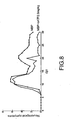
Figure 8 demonstrates how, twelve days after the induction of EAE, the administration of o-ATP makes the neurological symptoms caused by the disease disappear. -
Figure 9 demonstrates that in EAE the levels of the P2X2 receptors do not change significantly, however, those of P2X7 drop drastically. This indicates that there is a loss of cells which express it, mainly oligodendrocytes. - The first aspect of the invention refers to the use of P2X7 purinergic receptor antagonists for the treatment of neurodegenerative phase of multiple sclerosis. Autoimmunity requires the activation of a precise cascade of processes in cells of the immune system. One part of these cells, the macrophages and the lymphocytes, express P2X1, P2X2, P2X5 and P2X7 receptors, and the activation of the latter causes the release of pro-inflammatory cytokines such as tumour necrosis factor- α(TNF-α) and IL-1β as well as apoptosis by mechanisms which are still not characterised (Burnstock, 2002, Arteriorscler Thromb Vasc Biol. 22, 364-373). However, the exact functions which mediate the P2X receptors in the immune system are still not well understood. It is this expression of P2X receptors in cells of the immune system which makes the use of the P2X receptor antagonists suitable for the treatment of autoimmune diseases.
- Among the P2X receptor antagonists there are some which are called wide spectrum owing to the fact that they are capable of binding themselves to several of the family of P2X receptors, although with different affinities to each one of them; and others which are selective to a group of receptors from the P2X family.
- The following formulas represent some of these wide spectrum P2X receptor antagonists. Only compounds of formula VII and IX are part of this invention.


- The following formulas represent the selective P2X receptor antagonists:


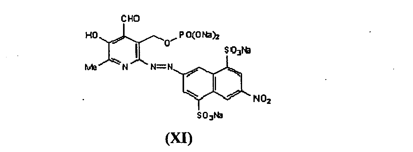

- The compounds previously represented by their structural formulas are:
- ■ PPADS (tetrasodium salt of pyridoxalphosphate-6-azophenyl-2',4'-disulfonic acid) (I)
- ■ iso-PPADS (tetrasodium salt of pyridoxalphosphate-6-azophenyl-1',4'-disulfonic acid) (II)
- ■ Suramin (hexasodium salt of 8,8'-[carbonylbis[imino-3,1-phenylenecarbonylimino(4-methyl-3,1-phenylene)carbonylimino]]bisnaphthalene-1,3,5-trisulphonic acid (III)
- ■ Evans Blue (tetrasodium salt of 6,6'-[(3,3'-Dimethyl[1,1'-biphenyl]-4,4'-diyl)bis[4-amino-5-hydroxy-1,3-naphthalenedisulfonic acid]) (IV)
- ■ NF023 (hexasodium salt of 8,8'-[carbonylbis[imino-3,1-phenylenecarbonylimino]bis-1,3,5-naphthalenetrisulfonic acid] (V)
- ■ NF279 Hexasodium salt of (8,8'-[carbonylbis(imino-4,1-phenylenecarbonylimino-4,1-phenylenecarbonylimino))bis(1,3,5-naphthalenetrisulfonic acid] (VI)
- ■ CBB-G (Coomassie brilliant blue G)(VII)
- ■ NF449 (octasodium salt of 4,4',4",4"'-(carbonylbis(imino-5,1,3-benzenetriylbis(carbonylimino)))tetrakis-benzene-1,3-disulfonic acid) (VIII)
- ■ o-ATP (sodium salt of Adenosine 5-triphosphate, oxidised with periodate) (IX)
- ■ KN-62 (ester of 4-[(2S)-2-[(5-isoquinolinylsulfonyl)methylamino]-3-oxo-3-(4-phenyl-1-piperazinyl)propyl] phenyl isoquinolinesulfonic acid) (X)
- ■ PPNDS (tetrasodium salt of pyridoxal-5'-phosphate-6-(2'-naphthylazo-6'-nitro-4',8'-disulfonate) (XI)
- ■ RB2 1-Amino-4-[[4-[[4-chloro-6-[[3 (or 4)-sulfophenyl]amino]-1,3,5-triazin-2-yl]amino]-3-sulfophenyl]amino]-9,10-dihydro-9,10-dioxo-2-anthracenesulfonic acid) (XII)
- Besides those mentioned previously there are other wide spectrum antagonists such as MRS2220 (cyclic pyridoxine-α4,5-monophosphate-6-azo-phenyl-2',5'disulfonate), Ip51 (pentapotassium salt of P1,P5-Diinosine-5-pentaphosphate) o TNP-ATP (monolithium salt of 2',3'-O-2,4,6-trinitrophenyladenosine 5'-triphosphate), as well as selective ones such as, for example, HMA (5-(N,N hexamethylene) amiloride).
- The IC50 of some of the previous compounds in relation to the different PX2 receptor subgroups are set out in Table 1.
Table 1- IC50 of P2X antagonists in relation to each P2X receptor subtype P2X subtypes P2X1 P2X2 P2X3 P2X4 P2X5 P2X6 P2X7 Antagonists: IC50 (µM) PPADS: 1-5 PPADS: 2 PPADS: 1 PPADS: 27.5 PPADS: 2.6 PPADS: >100 PPADS: 4.2 Suramin: 1- 5 Suramin: 1-5 Suramin: 3 Suramin: 178 Suramin: 4 Suramin: >100 Suramin: 4 NF023: 0.21 NF 023: 63 NF 023: 29 NF 023: >100 NF279: 002 NF279: 0.77 NF279: 1.6 NF279: >30 NF279: 2.8 KN-62: 0.015 Evans Blue: 1-400 Evans Blue: 1-400 Evans Blue: 1-400 Evans Blue: 1-400 Evans Blue: 1-400 Evans Blue: 1-400 Evans Blue: 1-400 isoPPADS: 1-5 isoPPADS: 1 RB-2: 1 HMA: 4,5 o- ATP 5BBG: >10 BBG: 0.01 Ip5I: 0.003 Ip5I: 3 MRS2220: 10 MRS2220: 58 NF449: 0.01 NF449: <0.006 PPNDS: 0.015 TNP- ATP: 0.001 TNP-ATP: 1 TNP- ATP: 0.001 TNP-ATP: 15 TNP-ATP: >30 - In a preferred embodiment, one of the aforementioned antagonists is a P2X7 selective receptor antagonist, o-ATP. In studies carried out by the inventors (see example further on) this compound has been shown to be specially suitable for the treatment of the neurodegenerative phase of multiple sclerosis owing to the relative importance of the presence of P2X7 receptors in oligodendrocytes compared to the other P2X receptors.
- The preferred wide spectrum inhibitor is BBG. A preferred realisation contemplates a pharmaceutical composition which contains at least o-ATP, a selective antagonist of P2X7 receptors.
- In the following example the studies carried out by the inventors are detailed which illustrates the basis of the invention.
- The cell cultures were carried out from the optic nerve of the perinatal rat (P12) following established protocols, which were adapted and introduced into the laboratory according to a recent description (Matute et al, 1997, Proc. Natl. Acad. Sci. USA 94, 8830-8835).
- The electrophysiological recordings were carried out in 2 to 5 day cultures, and according to the guidelines indicated in previous works (Patneau et al 1994, Neuron12: 357-371). The cells were recorded in a chamber which allowed the composition of the extracellular medium to vary by means of a constant flow (0.5-1 mL/min). The recording electrodes were glass capillaries which contained specific solutions compatible with the cytoplasm ion concentrations. The study of the responses mediated by the purinergic receptors was carried out using the "whole-cell patch-clamp" technique, measuring the currents generated by the external application of selective agonists and antagonists of the aforementioned receptors.
- The concentration of cytosol calcium was determined by the method of Grynkiewikcz et al (1985; J. Biol. Chem. 260, 3440-3450). The oligodendrocytes were loaded with 5 mM of Fura-2/AM, and they were then washed and studied in a Zeiss inverted microscope equipped with a monochromator, 40X immersion objective, an Orca high resolution digital camera, and AquaCosmos software (Hamamatsu Photonics). The changes in cytosol calcium levels in response to agonists and antagonists, in the presence and absence of extracellular calcium, were studied. The calibration was performed at the end of the studies by means of the successive application of ionomycin and EGTA, and the calcium concentration was estimated using the measurement of the 340/380 nm ratio.
- The nerves were isolated from young adult rats, and were perfused for 30 min in artificial cerebrospinal fluid (aCSF) saturated in oxygen by bubbling with 95% oxygen and 5% CO2, under conditions comparable to those described for oligodendrocytes in culture (Fern and Möller, 2000, J. Neurosci. 20: 34-42). Next, they were incubated with purinergic agonists and antagonists for different times. Later, the nerves were perfused for 1 to 24 hours with normal aCSF saturated with oxygen. After this time passed the damage was evaluated histologically as we have described in vivo (Matute, 1998, Proc. Natl. Acad. Sci. USA 95: 10229-10234), and the biochemical changes which are underlying to this damage were analysed.
- Commercial antibodies were used for the study of the presence of oligodendroglial lineage markers, components of myelin, astrocytes and microglia. The techniques included immunocytochemistry, immunohistochemistry and immunoblotting (Western blot), all these are described in detail (see for example, Domercq et al, 1999, Eur. J. Neurosci. 11, 2226-2236)
- The experiments on the optic nerve were carried out in rabbits (New Zealand White) which, due to their size, enable better manipulation in experimental surgery. The procedure used was that described previously (Matute, 1998, Proc. Natl. Acad. Sci. 95, 10229-10234). The purinergic agonists were applied using osmotic micropumps which released small quantities of solute for a determined time. Later, the effect of this application on the nerve was evaluated by means of a panel of oligodendrocyte markers and their progenitors, myelin, axonal integrity, astrogliosis and microgliosis.
- Lewis rats were used which were immunised subcutaneously with basic myelin protein in the back paws (100 micrograms/animal in 100 microlitres) and Freund's adjuvant with 5.5 mg/ml of tuberculosis Mycobacterium H37Ra. The spinal cord was extracted when the animals had symptoms of the disease (12-14 days post-immunisation) and the expression of the purinergic receptors was analysed using immunochemical techniques (immunoblotting and inmunohistochemical).
- ATP (1 mM) induces an input current which does not desensitise in the majority of oligodendrocytes examined (77.3 % ± 7.9 ; n= 47;
Fig. 1a ). The ATP analogue, 2',3'-O-(4-benzoyl-4-benzoyl) (BzATP, 100 µM), which is a wide spectrum P2X agonist but with a higher affinity for the P2X7 receptor (Ralevic & Burnstock, 1998), also induced similar responses (Fig. 1a ). On the other hand, α,β-methylene-ATP (α,β-Me-ATP, 100 µM), a selective P2X1, P2X3 and P2X2/3 heteromers agonist, did not generate currents in oligodendrocytes. It was observed that the amplitude of currents generated by ATP and BzATP depends on the concentration of the corresponding agonist (Fig. 1a ) (EC50= 8.77 mM and 0.52 mM respectively). Likewise, it could be established that the absence of Mg2+ and Ca2+, which increase the concentration of ATP4-, the active form of the P2X receptors, increase the responses by 4-10 fold (Fig. 1 a). - The PPADS wide spectrum antagonist (100 µM), for its part, completely blocked the currents induced by ATP (
Fig. 1 ). For its part, oxidised ATP (o-ATP), a preferential antagonist of the P2X7 receptors, partially blocks the ATP currents. For its part, Cu2+ (1 mM) which is a selective inhibitor of the P2X7 receptors (Virginio et al, 1997), reduces ATP currents, while propofol (60µM), a potentiator of the P2X4 receptors, does not alter these currents. These results indicate that the P2X receptors present in oligodendrocytes have electrophysiological properties compatible with a predominance of the P2X7 sub-unit. - [Ca2+]i was monitored after applying ATP and BzATP with the objective of characterising the effects of the activation of P2X receptors on oligodendrocytes. These cells respond to ATP (10 µM) with a rapid increase in basal cytosol [Ca2+]i (250 ± 65 nM) to 1200 ± 468 nM (
Fig. 2a ). These responses are repressed in the presence of PPADS (50 µM) and with the absence of Ca2+ in the incubation solution. These results indicate that the [Ca2+]¡ increases are due to the entrance of Ca2+ across the plasma membrane and not due to the liberation of this from intracellular deposits. - Bz-ATP (0.01-1 mM) also activates the entrance of Ca2+ into oligodendrocytes in a dose dependent way (
Fig. 2b , d). This effect disappears in the absence of extracellular Ca2+ and is blocked by PPADS (Fig. 2b , d). These results suggest that the P2X receptors which contain the P2X7 sub-unit are the principal mediators of the response to ATP. In agreement with this idea, the o-ATP P2X7 selective antagonist (1 mM) (Fernández et al, 2001), reduces the increase in [Ca2+] induced by Bz-ATP by 63 ± 8 (Fig. 2d , e). For its part, propofol (60 µM), which triggers the responses mediated by P2X4 (Tomioka et al., 2000), promotes the increase of [Ca2+]i generated by 0.1 and 1 mM ATP in 60% ± 22 and 77% ± 34 respectively (Fig. 2c , d). Therefore, the P2X native receptors which contain P2X4 also contribute to the entrance of calcium induced by ATP in oligodendrocytes. - At all ATP concentrations (0.01-1 mM) tested, death was produced in 15-27% of oligodendrocytes which is inhibited in the presence of 50 µM PPADS and after removing Ca2+ from the culture medium (
Fig. 3a ). In the same way the Bz-ATP agonist caused a toxicity similar to ATP (Fig. 3b ). Other purinergic agonists such as ATP-γ-S, which is a more stable analogue than ATP, and α,β,-meATP, are also toxic for the oligodendrocytes, which exclude the possibility that the metabolites of ATP might be the agents causing the toxicity after activating receptors different to the P2X ones. Overall, the toxicity tests showed that the oligodendrocytes are vulnerable to the activation of P2X receptors by ATP and its analogues. - The analysis of the expression P2X receptors using immunohistochemistry with specific antibodies in cultures of differentiated oligodendrocytes (GalC+/MBP+) demonstrates that these cells mainly have the P2X2, P2X4 y P2X7 subunits (see Table 2).
Table 2. Expression of P2X receptors in oligodendrocyte cultures. Subunit P2x1 P2x2 P2x3 P2x4 P2x5 P2x6 P2x7 Expression +/- ++ --- ++ +/- +/- ++ - This expression profile is consistent with the electrophysiological properties and toxicity characteristics observed in these cultures. Also, the pattern of the subunits observed in vitro also corresponds with that observed in situ in optic nerve (Table 3) by means of double marking of the subunits and antibodies specific to the oligodendroglial and astroglial lineage (
Fig.5 ).Table 3. Distribution of P2X receptors in oligodendrocytes in the optic nerve of the rat Subunit P2X1 P2X2 P2X3 P2X4 P2X5 P2X6 P2X7 Distribution - +++ - +++ + - +++ - To determine if ATP is toxic to the oligodendrocytes in a preparation of nerve tissue without dissociating, entire optic nerves isolated from adult rats were perfused with artificial cerebrospinal fluid with ATP (100 µM) for 3 h. Under these conditions an increase of > 3 times the number of cells which showed nuclear condensation as compared to the control nerves perfused without ATP was produced (
Fig. 5 ). The damaged cells are located in the longitudinal axis of the nerve and make up part of interfascicular oligodendrocyte rows. Stimulation with ATP in the presence of PPADS (10 µM) prevents the death of the oligodendrocytes. - Next, the ATP-γ-S and BzATP agonists were infused over the optic nerve using osmotic pumps which released very small quantities of solute for 3 days. The histological examination of the nerves 7 days after starting the application showed tissue damage in an area restricted to the proximity of the cannula (
Fig. 6 ). Also, this zone had intense gliosis, lack of myelin and axonal damage (Fig. 6 ). Overall these results indicate that the activation of P2X kills oligodendrocytes in situ and that the lesions in vivo share properties common to multiple sclerosis plaques. - The effects of the wide spectrum antagonist PPADS and the more selective o-ATP in the triggering off and on the course of EAE induced by the immunisation of Lewis rats with basic myelin protein were investigated. The immunised rats showed signs of motor deficits around 10 days post-injection, and they reached a maximum at 14 days (
Fig. 7 ). The treatment with PPADS (30 mg/kg, two times per day) from 7 to 14 days post-injection did not improve the symptoms or the course of the disease. On the other hand the application of o-ATP (1 and 5 mg/kg, every12 h) for the same period reduced or prevented the appearance of symptoms common to EAE (Fig. 7 ). - Later, the efficacy of o-ATP in improving the symptoms of EAE was evaluated in a chronic-recurrent-remittent model. For this, DA rats were immunised with syngeneic spinal cord, the appearance of severe neurological deficits being observed 7-9 days post-injection, and which reached their first peak around 11 days. Treatment with o-ATP (2.5 mg/kg, every 12 h), once the maximum intensity of the symptoms were established, reduced the symptoms and also eliminated those common to the chronic phase (
Fig. 8 ). - With the objective of understanding the mechanism of action by which o-ATP improves the prognosis of EAE, the levels of P2X7 receptors over which the preferred form of this drug acts on the lumbar-sacral spinal cord, the region most affected in this experimental disease, was evaluated using Western blot. We found that the levels of this subunit were reduced by half in animals subjected to EAE, and that these levels returned to those of the controls in those animals with EAE treated with o-ATP (
Fig. 9 ). These results indicate that treatment with o-ATP protects the cells which express P2X7 from dying, and consequently, the oligodendrocytes, which are the main type of cells which express this subunit in the spinal cord. - The results shown previously demonstrate for the first time that the oligodendrocytes have P2X receptors. Likewise, the electrophysiological, pharmacological and molecular properties of these receptors, as well as their increased permeability to calcium, are given in detail. This latter property results in that the oligodendrocytes may be vulnerable to intense and/or prolonged stimulus mediated by these receptors, as has been demonstrated with glutamergic receptors in this cell population (Matute et al, 2001, Trends Neurosci 24, 224-230). The vulnerability of the oligodendrocytes to the signals mediated by the P2X receptors is one of the causes of nervous tissue damage which underlies the experimental disease, EAE, a model of multiple sclerosis. Finally, blocking of the P2X7 receptors until triggering off the disease drastically reduces the neurological symptoms in acute EAE, and improves the outcome and prognosis in chronic EAE once the symptoms are established.
- The invention described herein constitutes a means for the treatment of multiple sclerosis, a disease which lacks efficient treatments which slow down or check its progression. The routes of intervention which have resulted in the development of drugs in clinical trial phase or for use as drugs in the treatment of multiple sclerosis have mechanisms of action which regulate the functioning of the immune system. The fact that the blocking of the P2X7 may prevent the symptoms of acute EAE, a model of MS which mimics the inflammatory/autoimmune phase of the disease, indicates that these drugs can in fact be powerful immunomodulatory agents which may prevent the autoimmunity which triggers off MS and other diseases. Finally, the P2X7 receptor antagonists on being protector agents of the death of oligodendrocytes, the cell population which suffers most damage in MS, have great therapeutic potential in the neurodegenerative phase of this disease, a phase which is prolonged for decades and in which patients suffer a progressive deterioration which continues its course with motor and sensory disorders causing invalidity.
-
- Abbracchio, M.P. and Burnstock, G. (1998) Purinergic signalling: pathophysiological roles. Jpn. J. Pharmacol. 78: 113-145.
- Ansari KA, Rand A, Loch JA (1978) Biochemical and immunological studies with human optic and olfactory tracts J Neuropathol Exp Neurol 37:756-67
- Armstrong MA, Shah S, Hawkins SA, Bell AL, Roberts SD (1988) Reduction of monocyte 5'nucleotidase activity by gamma-interferon in multiple Ann Neurol 24:12-6
- Barnard, E.A., Simon, J. and Webb, T.E. (1997). Nucleotide receptors in the nervous system. An abundant component using diverse signal transduction mechanisms. Mol. Neurobiol. 15: 103-129.
- Braun, N., Zhu, Y., Krieglstein, J., Clumsee, C. and Zimmermann, H. (1998). Upregulation of the enzyme chain hydrolysing extracellular ATP after transient forebrain ischemia in the rat. J. Neurosci. 18: 4891-4900.
- Ciccarelli, R. Ballerini, P., Sabatino, G., Rathbone, M.P., D'Onofrio, M.. Caciagli, F. and lorio, P. (2001). Involvement of astrocytes in purine-mediated reparative processes in the brain. Int. J. Dev. Neurosci. 19: 395-414.
- Dunn, P.M., Zhong, Y. and Burnstock, G. (2001). P2X receptors in peripheral neurons. Prog. Neurobiol. 65:107-134.
- Fields, R.D. and Stevens, B. (2000). ATP: an extracellular signalling molecule between neurons and glia. Trends Neurosci. 23: 625-633.
- Franke, H., Grosche, J., Schädlich, H., Krügel, U., Allgaier, C. and Illes, P. (2001 a). P2X receptor expression on astrocytes in the nucleus accumbens of rats. Neuroscience 108: 421-429.
- Franke, H., Krugel, U., Schmidt, R., Grosche, J., Reichenback, A. and Illes, P. (2001 b). P2 receptor-types involved in astrogliosis in vivo. Brit. J. Pharmacol. 134: 1180-1189.
- Honda, S. and Kohsaka, S. (2001). Regulation of microglial cell function by ATP. Nihon Shinke 21: 89-93.
- James, G. and Butt, A.M. (2001). Changes in P2Y and P2X purinoceptors in reactive glia following axonal degeneration in the rat optic nerve. Neurosci. Lett. 212: 33-36.
- Matute C, Alberdi E, Domercq M, Pérez-Cerdá F, Pérez-Samartín A and Sanchez-Gomez MV (2001) The link between excitotoxicity and demyelinating diseases. Trends Neurosci. 24, 224-230.
- Matute C, Alberdi E, Ibarretxe G and Sánchez-Gómez MV (2002) Excitotoxicity in glial cells. Eur. J. Pharmacol 447:239-246.
- Queiroz, G., Gebicke-Haerter, P.J., Schobert, A., Starke, K. and von Kugelgen, I. (1997). Release of ATP from cultured rat astrocytes elicited by glutamate receptor activation. Neuroscience 78: 1203-1208.
- Ralevic, V. and Burnstock, G. (1998) Receptors for purines and pyrimidines. Pharmacol. Rev. 50: 413-492.
- Rathbone, M.P., Meddlemiss, P.J., Gysbers, J.W., Andrew, C., Herman, M.A., Reed, J.K., Ciccarelli, R. Di lorio, P. and Caciagli, F. (1999). Trophic effects of purines in neurons and glia. Prog. Neurobiol. 59: 663-690.
- Stevens, B, Porta, S., Haak, L.L., Gallo, V. and Fields, R.D. (2002) Adenosine: a neuron-glial transmitter promoting myelination in the CNS in response to action potentials. Neuron 36: 855-868.
- Zamvil, S.S. and Steinman L. (2003) Diverse targets for intervention during inflammatory and neurodegenerative phases of multiple sclerosis. Neuron 38, 685-688.
Claims (1)
- Use of the antagonist of P2X7 purinergic receptors o-ATP or BBG in the preparation of a drug for the treatment of the neurodegenerative phase of multiple sclerosis, in mammals, including man.
Priority Applications (1)
| Application Number | Priority Date | Filing Date | Title |
|---|---|---|---|
| PL04766900T PL1655032T3 (en) | 2003-08-04 | 2004-08-04 | USE OF THE P2X7 ANTAGONISTS o-ATP OR BBG FOR THE TREATMENT OF THE NEURODEGENERATIVE PHASE OF MULTIPLE SCLEROSIS |
Applications Claiming Priority (2)
| Application Number | Priority Date | Filing Date | Title |
|---|---|---|---|
| ES200301853A ES2223287B1 (en) | 2003-08-04 | 2003-08-04 | COMPOUNDS FOR THE TREATMENT OF DEMIELINIZING AND AUTOIMMUNE DISEASES. |
| PCT/ES2004/000361 WO2005014007A1 (en) | 2003-08-04 | 2004-08-04 | Compounds for the treatment of autoimmune and demyelinating diseases |
Publications (2)
| Publication Number | Publication Date |
|---|---|
| EP1655032A1 EP1655032A1 (en) | 2006-05-10 |
| EP1655032B1 true EP1655032B1 (en) | 2009-10-07 |
Family
ID=34130551
Family Applications (1)
| Application Number | Title | Priority Date | Filing Date |
|---|---|---|---|
| EP04766900A Expired - Lifetime EP1655032B1 (en) | 2003-08-04 | 2004-08-04 | USE OF THE P2X7 ANTAGONISTS o-ATP OR BBG FOR THE TREATMENT OF THE NEURODEGENERATIVE PHASE OF MULTIPLE SCLEROSIS |
Country Status (9)
| Country | Link |
|---|---|
| US (1) | US20070099863A1 (en) |
| EP (1) | EP1655032B1 (en) |
| AT (1) | ATE444757T1 (en) |
| CA (1) | CA2534868A1 (en) |
| DE (1) | DE602004023517D1 (en) |
| ES (2) | ES2223287B1 (en) |
| PL (1) | PL1655032T3 (en) |
| PT (1) | PT1655032E (en) |
| WO (1) | WO2005014007A1 (en) |
Families Citing this family (8)
| Publication number | Priority date | Publication date | Assignee | Title |
|---|---|---|---|---|
| CA2717166A1 (en) * | 2008-03-03 | 2009-09-19 | Converge Biotech Inc. | Methods of modulating t cell-dependent immune responses |
| ES2338970B1 (en) * | 2008-07-31 | 2011-03-11 | Universidad Complutense De Madrid | USE OF P2X7 RECEIVER ANTAGONISTS TO FAVOR AXONAL GROWTH AND RAMIFICATION. |
| WO2010022169A1 (en) * | 2008-08-19 | 2010-02-25 | Cedars-Sinai Medical Center | Methods of inhibiting fgfr3 signaling |
| ITTO20080759A1 (en) * | 2008-10-15 | 2010-04-16 | Medestea Res & Production S R L | USE OF OATP AS AN IMMUNODEPRESSIVE DRUG |
| WO2013082565A1 (en) * | 2011-12-02 | 2013-06-06 | Michael Kaleko | Therapies for disorders of the cornea and conjunctiva |
| CN103301462B (en) * | 2012-03-09 | 2015-06-03 | 海南康芝药业股份有限公司 | Component and method for treating virus disease |
| WO2017070660A1 (en) * | 2015-10-23 | 2017-04-27 | Baylor College Of Medicine | Targeting p2 purinergic receptors to treat hepatocellular carcinoma (hcc) |
| US10624866B2 (en) * | 2018-03-05 | 2020-04-21 | The Industry & Academic Cooperation In Chungnam National University (Iac) | Composition for prevention or treatment of chronic pain comprising Evans blue |
Family Cites Families (4)
| Publication number | Priority date | Publication date | Assignee | Title |
|---|---|---|---|---|
| US5589458A (en) * | 1992-11-13 | 1996-12-31 | Thomas Jefferson University | Compounds that inhibit T cell proliferation and methods for using the same |
| GB9615202D0 (en) * | 1996-07-19 | 1996-09-04 | Smithkline Beecham Plc | Pharmaceuticals |
| US6117445A (en) * | 1998-01-28 | 2000-09-12 | Link Technology Inc. | Methods for the prevention and treatment of fibrosis and sclerosis |
| AU2003237460A1 (en) * | 2002-06-06 | 2003-12-22 | Endacea, Inc. | Combination treatments for purinoceptor-related disorders |
-
2003
- 2003-08-04 ES ES200301853A patent/ES2223287B1/en not_active Expired - Fee Related
-
2004
- 2004-08-04 CA CA002534868A patent/CA2534868A1/en not_active Abandoned
- 2004-08-04 PL PL04766900T patent/PL1655032T3/en unknown
- 2004-08-04 EP EP04766900A patent/EP1655032B1/en not_active Expired - Lifetime
- 2004-08-04 AT AT04766900T patent/ATE444757T1/en not_active IP Right Cessation
- 2004-08-04 ES ES04766900T patent/ES2334505T3/en not_active Expired - Lifetime
- 2004-08-04 DE DE602004023517T patent/DE602004023517D1/en not_active Expired - Lifetime
- 2004-08-04 WO PCT/ES2004/000361 patent/WO2005014007A1/en not_active Ceased
- 2004-08-04 US US10/567,238 patent/US20070099863A1/en not_active Abandoned
- 2004-08-04 PT PT04766900T patent/PT1655032E/en unknown
Also Published As
| Publication number | Publication date |
|---|---|
| ATE444757T1 (en) | 2009-10-15 |
| WO2005014007A1 (en) | 2005-02-17 |
| DE602004023517D1 (en) | 2009-11-19 |
| ES2334505T3 (en) | 2010-03-11 |
| CA2534868A1 (en) | 2005-02-17 |
| PT1655032E (en) | 2010-01-07 |
| US20070099863A1 (en) | 2007-05-03 |
| ES2223287A1 (en) | 2005-02-16 |
| EP1655032A1 (en) | 2006-05-10 |
| ES2223287B1 (en) | 2006-04-16 |
| WO2005014007B1 (en) | 2005-03-31 |
| PL1655032T3 (en) | 2010-03-31 |
Similar Documents
| Publication | Publication Date | Title |
|---|---|---|
| Gao et al. | Ginsenoside Rb1 promotes motor functional recovery and axonal regeneration in post-stroke mice through cAMP/PKA/CREB signaling pathway | |
| US9480658B2 (en) | Modulation of synaptic maintenance | |
| US9382313B2 (en) | Modulation of synaptic maintenance | |
| Simon et al. | Chronic pharmacological increase of neuronal activity improves sensory-motor dysfunction in spinal muscular atrophy mice | |
| TW201038544A (en) | Anti-neurodegenerative diseases agents | |
| US20220241316A1 (en) | 25-hydroxycholesterol (25hc), cryab aggregation inhibitor, is a novel senolytic | |
| CN106714821B (en) | Novel use of cell-penetrating peptide inhibitors of the JNK signaling pathway for the treatment of various diseases | |
| EP1655032A1 (en) | Compounds for the treatment of autoimmune and demyelinating diseases | |
| Masuda et al. | GPR3 expression in retinal ganglion cells contributes to neuron survival and accelerates axonal regeneration after optic nerve crush in mice | |
| Acharya | Recent advances in epileptogenesis | |
| Li et al. | Upregulation of Cdh1 in the trigeminal spinal subnucleus caudalis attenuates trigeminal neuropathic pain via inhibiting GABAergic neuronal apoptosis | |
| WO2018205927A1 (en) | Use of potassium ion channel inhibitor for treatment of depression and pharmaceutical composition | |
| CN100525764C (en) | Application of pyrazolopyridine in preparation of medicine for treating cognitive deficiency | |
| Yasuhara et al. | Toxicity of semaphorin3A for dopaminergic neurons | |
| Nikolaev | Effects of antagonists of different types of NMDA receptor on evoked responses of pyramidal neurons in rat cerebral cortex slices | |
| Simon | Role of Müller cells in the onset and progression of neuronal degeneration after acute retinal ischemia | |
| Dickendesher et al. | Axon regeneration | |
| Calovi | STUDY OF THE EFFECT OF P2X7 RECEPTOR PROTEIN FUNCTIONAL EXPRESSION IN THE PHENCYCLIDINE MURINE MODELS FOR SCHIZOPHRENIA | |
| KR100671309B1 (en) | Pharmaceutical composition for preventing neuronal cell damage caused by cerebral ischemia, comprising a BAA-T inhibitor and a VII2 receptor antagonist | |
| Habib | Erythropoietin dependent regulation of the anti-apoptotic TMBIM family members FAIM2 and GRINA after murine cerebral ischemia | |
| Schultz | Role of Müller cells in the onset and progression of neuronal degeneration after acute retinal ischemia | |
| Sze et al. | Structure-function study of ubiquitin c-terminal hydrolase L1 | |
| Sze et al. | Structure-function study of ubiquitin c-terminal hydrolase L1 (UCH-L1) by NMR spectroscopy-insights into UCH-L1 mutation's association with the risk of Parkinson's disease | |
| Lam et al. | Regulatory role of proheparanase with peri-synaptic heparan | |
| Fu et al. | Aldose reductase deficiency protects the retinal neurons in a |
Legal Events
| Date | Code | Title | Description |
|---|---|---|---|
| PUAI | Public reference made under article 153(3) epc to a published international application that has entered the european phase |
Free format text: ORIGINAL CODE: 0009012 |
|
| 17P | Request for examination filed |
Effective date: 20060221 |
|
| AK | Designated contracting states |
Kind code of ref document: A1 Designated state(s): AT BE BG CH CY CZ DE DK EE ES FI FR GB GR HU IE IT LI LU MC NL PL PT RO SE SI SK TR |
|
| DAX | Request for extension of the european patent (deleted) | ||
| 17Q | First examination report despatched |
Effective date: 20061114 |
|
| 17Q | First examination report despatched |
Effective date: 20061208 |
|
| GRAP | Despatch of communication of intention to grant a patent |
Free format text: ORIGINAL CODE: EPIDOSNIGR1 |
|
| RTI1 | Title (correction) |
Free format text: USE OF THE P2X7 ANTAGONISTS O-ATP OR BBG FOR THE TREATMENT OF THE NEURODEGENERATIVE PHASE OF MULTIPLE SCLEROSIS |
|
| RTI1 | Title (correction) |
Free format text: USE OF THE P2X7 ANTAGONISTS O-ATP OR BBG FOR THE TREATMENT OF THE NEURODEGENERATIVE PHASE OF MULTIPLE SCLEROSIS |
|
| GRAS | Grant fee paid |
Free format text: ORIGINAL CODE: EPIDOSNIGR3 |
|
| GRAA | (expected) grant |
Free format text: ORIGINAL CODE: 0009210 |
|
| AK | Designated contracting states |
Kind code of ref document: B1 Designated state(s): AT BE BG CH CY CZ DE DK EE ES FI FR GB GR HU IE IT LI LU MC NL PL PT RO SE SI SK TR |
|
| REG | Reference to a national code |
Ref country code: GB Ref legal event code: FG4D |
|
| REG | Reference to a national code |
Ref country code: CH Ref legal event code: EP |
|
| REG | Reference to a national code |
Ref country code: IE Ref legal event code: FG4D |
|
| REF | Corresponds to: |
Ref document number: 602004023517 Country of ref document: DE Date of ref document: 20091119 Kind code of ref document: P |
|
| REG | Reference to a national code |
Ref country code: RO Ref legal event code: EPE |
|
| REG | Reference to a national code |
Ref country code: PT Ref legal event code: SC4A Free format text: AVAILABILITY OF NATIONAL TRANSLATION Effective date: 20091230 |
|
| REG | Reference to a national code |
Ref country code: GR Ref legal event code: EP Ref document number: 20100400012 Country of ref document: GR |
|
| PG25 | Lapsed in a contracting state [announced via postgrant information from national office to epo] |
Ref country code: SI Free format text: LAPSE BECAUSE OF FAILURE TO SUBMIT A TRANSLATION OF THE DESCRIPTION OR TO PAY THE FEE WITHIN THE PRESCRIBED TIME-LIMIT Effective date: 20091007 |
|
| NLV1 | Nl: lapsed or annulled due to failure to fulfill the requirements of art. 29p and 29m of the patents act | ||
| REG | Reference to a national code |
Ref country code: ES Ref legal event code: FG2A Ref document number: 2334505 Country of ref document: ES Kind code of ref document: T3 |
|
| REG | Reference to a national code |
Ref country code: PL Ref legal event code: T3 |
|
| PG25 | Lapsed in a contracting state [announced via postgrant information from national office to epo] |
Ref country code: FI Free format text: LAPSE BECAUSE OF FAILURE TO SUBMIT A TRANSLATION OF THE DESCRIPTION OR TO PAY THE FEE WITHIN THE PRESCRIBED TIME-LIMIT Effective date: 20091007 Ref country code: SE Free format text: LAPSE BECAUSE OF FAILURE TO SUBMIT A TRANSLATION OF THE DESCRIPTION OR TO PAY THE FEE WITHIN THE PRESCRIBED TIME-LIMIT Effective date: 20091007 |
|
| PG25 | Lapsed in a contracting state [announced via postgrant information from national office to epo] |
Ref country code: BE Free format text: LAPSE BECAUSE OF FAILURE TO SUBMIT A TRANSLATION OF THE DESCRIPTION OR TO PAY THE FEE WITHIN THE PRESCRIBED TIME-LIMIT Effective date: 20091007 Ref country code: AT Free format text: LAPSE BECAUSE OF FAILURE TO SUBMIT A TRANSLATION OF THE DESCRIPTION OR TO PAY THE FEE WITHIN THE PRESCRIBED TIME-LIMIT Effective date: 20091007 |
|
| PG25 | Lapsed in a contracting state [announced via postgrant information from national office to epo] |
Ref country code: NL Free format text: LAPSE BECAUSE OF FAILURE TO SUBMIT A TRANSLATION OF THE DESCRIPTION OR TO PAY THE FEE WITHIN THE PRESCRIBED TIME-LIMIT Effective date: 20091007 Ref country code: EE Free format text: LAPSE BECAUSE OF FAILURE TO SUBMIT A TRANSLATION OF THE DESCRIPTION OR TO PAY THE FEE WITHIN THE PRESCRIBED TIME-LIMIT Effective date: 20091007 Ref country code: DK Free format text: LAPSE BECAUSE OF FAILURE TO SUBMIT A TRANSLATION OF THE DESCRIPTION OR TO PAY THE FEE WITHIN THE PRESCRIBED TIME-LIMIT Effective date: 20091007 Ref country code: BG Free format text: LAPSE BECAUSE OF FAILURE TO SUBMIT A TRANSLATION OF THE DESCRIPTION OR TO PAY THE FEE WITHIN THE PRESCRIBED TIME-LIMIT Effective date: 20100107 |
|
| PLBE | No opposition filed within time limit |
Free format text: ORIGINAL CODE: 0009261 |
|
| STAA | Information on the status of an ep patent application or granted ep patent |
Free format text: STATUS: NO OPPOSITION FILED WITHIN TIME LIMIT |
|
| PG25 | Lapsed in a contracting state [announced via postgrant information from national office to epo] |
Ref country code: SK Free format text: LAPSE BECAUSE OF FAILURE TO SUBMIT A TRANSLATION OF THE DESCRIPTION OR TO PAY THE FEE WITHIN THE PRESCRIBED TIME-LIMIT Effective date: 20091007 Ref country code: CZ Free format text: LAPSE BECAUSE OF FAILURE TO SUBMIT A TRANSLATION OF THE DESCRIPTION OR TO PAY THE FEE WITHIN THE PRESCRIBED TIME-LIMIT Effective date: 20091007 |
|
| 26N | No opposition filed |
Effective date: 20100708 |
|
| PG25 | Lapsed in a contracting state [announced via postgrant information from national office to epo] |
Ref country code: MC Free format text: LAPSE BECAUSE OF NON-PAYMENT OF DUE FEES Effective date: 20100831 |
|
| REG | Reference to a national code |
Ref country code: CH Ref legal event code: PL |
|
| PG25 | Lapsed in a contracting state [announced via postgrant information from national office to epo] |
Ref country code: CH Free format text: LAPSE BECAUSE OF NON-PAYMENT OF DUE FEES Effective date: 20100831 Ref country code: LI Free format text: LAPSE BECAUSE OF NON-PAYMENT OF DUE FEES Effective date: 20100831 |
|
| PG25 | Lapsed in a contracting state [announced via postgrant information from national office to epo] |
Ref country code: CY Free format text: LAPSE BECAUSE OF FAILURE TO SUBMIT A TRANSLATION OF THE DESCRIPTION OR TO PAY THE FEE WITHIN THE PRESCRIBED TIME-LIMIT Effective date: 20091007 |
|
| PG25 | Lapsed in a contracting state [announced via postgrant information from national office to epo] |
Ref country code: HU Free format text: LAPSE BECAUSE OF FAILURE TO SUBMIT A TRANSLATION OF THE DESCRIPTION OR TO PAY THE FEE WITHIN THE PRESCRIBED TIME-LIMIT Effective date: 20100408 Ref country code: LU Free format text: LAPSE BECAUSE OF NON-PAYMENT OF DUE FEES Effective date: 20100804 |
|
| PGFP | Annual fee paid to national office [announced via postgrant information from national office to epo] |
Ref country code: TR Payment date: 20130725 Year of fee payment: 10 |
|
| PGFP | Annual fee paid to national office [announced via postgrant information from national office to epo] |
Ref country code: GR Payment date: 20140827 Year of fee payment: 11 Ref country code: RO Payment date: 20140729 Year of fee payment: 11 Ref country code: DE Payment date: 20140822 Year of fee payment: 11 Ref country code: IE Payment date: 20140828 Year of fee payment: 11 |
|
| PGFP | Annual fee paid to national office [announced via postgrant information from national office to epo] |
Ref country code: FR Payment date: 20140818 Year of fee payment: 11 Ref country code: PL Payment date: 20140728 Year of fee payment: 11 Ref country code: GB Payment date: 20140820 Year of fee payment: 11 |
|
| PGFP | Annual fee paid to national office [announced via postgrant information from national office to epo] |
Ref country code: PT Payment date: 20140205 Year of fee payment: 11 Ref country code: IT Payment date: 20140828 Year of fee payment: 11 |
|
| REG | Reference to a national code |
Ref country code: PT Ref legal event code: MM4A Free format text: LAPSE DUE TO NON-PAYMENT OF FEES Effective date: 20160204 |
|
| REG | Reference to a national code |
Ref country code: DE Ref legal event code: R119 Ref document number: 602004023517 Country of ref document: DE |
|
| GBPC | Gb: european patent ceased through non-payment of renewal fee |
Effective date: 20150804 |
|
| PG25 | Lapsed in a contracting state [announced via postgrant information from national office to epo] |
Ref country code: IT Free format text: LAPSE BECAUSE OF NON-PAYMENT OF DUE FEES Effective date: 20150804 |
|
| PG25 | Lapsed in a contracting state [announced via postgrant information from national office to epo] |
Ref country code: RO Free format text: LAPSE BECAUSE OF NON-PAYMENT OF DUE FEES Effective date: 20150804 Ref country code: PT Free format text: LAPSE BECAUSE OF NON-PAYMENT OF DUE FEES Effective date: 20160204 Ref country code: GR Free format text: LAPSE BECAUSE OF NON-PAYMENT OF DUE FEES Effective date: 20160303 |
|
| REG | Reference to a national code |
Ref country code: IE Ref legal event code: MM4A |
|
| REG | Reference to a national code |
Ref country code: FR Ref legal event code: ST Effective date: 20160429 |
|
| REG | Reference to a national code |
Ref country code: GR Ref legal event code: ML Ref document number: 20100400012 Country of ref document: GR Effective date: 20160303 |
|
| PG25 | Lapsed in a contracting state [announced via postgrant information from national office to epo] |
Ref country code: GB Free format text: LAPSE BECAUSE OF NON-PAYMENT OF DUE FEES Effective date: 20150804 Ref country code: IE Free format text: LAPSE BECAUSE OF NON-PAYMENT OF DUE FEES Effective date: 20150804 Ref country code: DE Free format text: LAPSE BECAUSE OF NON-PAYMENT OF DUE FEES Effective date: 20160301 |
|
| PG25 | Lapsed in a contracting state [announced via postgrant information from national office to epo] |
Ref country code: FR Free format text: LAPSE BECAUSE OF NON-PAYMENT OF DUE FEES Effective date: 20150831 |
|
| PG25 | Lapsed in a contracting state [announced via postgrant information from national office to epo] |
Ref country code: PL Free format text: LAPSE BECAUSE OF NON-PAYMENT OF DUE FEES Effective date: 20150804 |
|
| PG25 | Lapsed in a contracting state [announced via postgrant information from national office to epo] |
Ref country code: TR Free format text: LAPSE BECAUSE OF NON-PAYMENT OF DUE FEES Effective date: 20150804 |
|
| PG25 | Lapsed in a contracting state [announced via postgrant information from national office to epo] |
Ref country code: ES Free format text: LAPSE BECAUSE OF NON-PAYMENT OF DUE FEES Effective date: 20100805 |
|
| PG25 | Lapsed in a contracting state [announced via postgrant information from national office to epo] |
Ref country code: ES Free format text: LAPSE BECAUSE OF NON-PAYMENT OF DUE FEES Effective date: 20100805 |
|
| PGRI | Patent reinstated in contracting state [announced from national office to epo] |
Ref country code: ES Effective date: 20160831 |
|
| PGFP | Annual fee paid to national office [announced via postgrant information from national office to epo] |
Ref country code: ES Payment date: 20230831 Year of fee payment: 20 |
|
| REG | Reference to a national code |
Ref country code: ES Ref legal event code: FD2A Effective date: 20240826 |
|
| PG25 | Lapsed in a contracting state [announced via postgrant information from national office to epo] |
Ref country code: ES Free format text: LAPSE BECAUSE OF EXPIRATION OF PROTECTION Effective date: 20240805 |
|
| PG25 | Lapsed in a contracting state [announced via postgrant information from national office to epo] |
Ref country code: ES Free format text: LAPSE BECAUSE OF EXPIRATION OF PROTECTION Effective date: 20240805 |