EP1650567A1 - Method of detecting protein dirt on contact lens - Google Patents
Method of detecting protein dirt on contact lens Download PDFInfo
- Publication number
- EP1650567A1 EP1650567A1 EP04747685A EP04747685A EP1650567A1 EP 1650567 A1 EP1650567 A1 EP 1650567A1 EP 04747685 A EP04747685 A EP 04747685A EP 04747685 A EP04747685 A EP 04747685A EP 1650567 A1 EP1650567 A1 EP 1650567A1
- Authority
- EP
- European Patent Office
- Prior art keywords
- contact lens
- protein
- coloring reagent
- solution
- contact
- Prior art date
- Legal status (The legal status is an assumption and is not a legal conclusion. Google has not performed a legal analysis and makes no representation as to the accuracy of the status listed.)
- Granted
Links
Images
Classifications
-
- G—PHYSICS
- G01—MEASURING; TESTING
- G01N—INVESTIGATING OR ANALYSING MATERIALS BY DETERMINING THEIR CHEMICAL OR PHYSICAL PROPERTIES
- G01N33/00—Investigating or analysing materials by specific methods not covered by groups G01N1/00 - G01N31/00
- G01N33/48—Biological material, e.g. blood, urine; Haemocytometers
- G01N33/50—Chemical analysis of biological material, e.g. blood, urine; Testing involving biospecific ligand binding methods; Immunological testing
- G01N33/68—Chemical analysis of biological material, e.g. blood, urine; Testing involving biospecific ligand binding methods; Immunological testing involving proteins, peptides or amino acids
Definitions
- the present invention relates to a method of detecting protein deposit on a contact lens, in particular, a method of detecting the protein deposit adhering to the contact lens easily and in a short period of time.
- contact lenses made of various materials.
- a protein contained in lacrimal fluid tends to adhere to a contact lens, in particular, to a contact lens made of an ionic polymer material obtained by using, as a raw material, monomeric components having ionizable groups. If the protein gradually adheres to and accumulates on surfaces of and inside the contact lens, removal of the protein tends to be difficult.
- there is an anxiety that the adhesion and the accumulation of the protein may cause various troubles such as deteriorating the wearing comfort as felt by the lens wearer, lowering the oxygen permeability of the lens, lowering the eyesight of the lens wearer, allergy, and corneal injury.
- JP-A-58-222154 Patent Document 1
- JP-A-58-222155 Patent Document 2
- the solutions include, as a coloring reagent, Nile Blue sulfate or erythrosin.
- these coloring reagents may also color lipid component, as well as the protein component, so that there is a problem that the protein component cannot be selectively detected.
- JP-A-2000-65840 Patent Document 3 proposes a method of detecting the protein deposit and the lipid deposit adhering to the contact lens, by using a basic dye solution.
- JP-U-A-5-40874 proposes a reference color spectrum.
- the Patent Document 4 explains that, there is commonly used tetrabromophenol blue as a color former of a urinary protein test paper, which is used for inspecting the protein components in the urine by means of a method of protein error of a pH indicator.
- JP-A-8-505938 proposes a test panel which represents an immediate result, wherein bromochlorophenol blue and tetrabromophenol blue are used as visual indicators.
- JP-A-11-304803 proposes a method and a kit for analyzing the protein in a sample.
- JP-A-11-304803 discloses a method of analyzing the protein by using an association of triphenylmethane dye color, such as bromochlorophenol blue or tetrabromophenol blue, with the protein, and causing the association to adsorb on a hydrophobic filter.
- Patent Document 1 JP-A-58-222154
- the present invention was made in view of the background art described above.
- a problem to be solved by the invention is to provide a method of detecting only the protein deposit adhering to a contact lens easily and in a short period of time.
- a method of detecting protein deposit on a contact lens characterized in that a protein component adhering to the contact lens, after the contact lens has been worn, is detected when the protein component adhering to the contact lens is in its colored state, wherein the protein component is colored by causing the contact lens to contact a solution having pH of not more than 3, after causing the contact lens to contact a coloring reagent solution containing bromochlorophenol blue or tetrabromophenol blue.
- the coloring reagent in the coloring reagent solution has a concentration of 100 - 500 ppm.
- the coloring reagent solution is obtained by dissolving the coloring reagent in water, ethanol, or a mixture thereof.
- the solution having pH of not more than 3 is an aqueous solution of phosphoric acid.
- the solution having pH of not more than 3 there is preferably employed a solution, whose pH is within a range of 2 - 3.
- the detection of the protein deposit adhering to the contact lens is implemented by a colorimetric analysis, a spectral analysis, or an analysis obtained by combining these analyses.
- the detection of the protein deposit adhering to the contact lens is implemented by image processing of an image taken with a camera, which image is of the contact lens caused to contact the coloring reagent solution and the solution having pH of not more than 3.
- bromochlorophenol blue or tetrabromophenol blue is used as the coloring reagent. Owing to this, selective detection of the protein component adhering to the contact lens is advantageously realized, without causing a problem such that a result of the detection is influenced by a kind of the protein component. Namely, the coloring reagent develops a color by associating or reacting with various protein components, without coloring the lipid component.
- the contact lens after it has been worn, is caused to contact a solution including the coloring reagent (coloring reagent solution), and is further caused to contact a solution whose pH is adjusted to be not more than 3 (acidic solution) by, for example, immersing the contact lens in the solution.
- a solution including the coloring reagent coloring reagent solution
- a solution whose pH is adjusted to be not more than 3 acidic solution
- the protein component adhering to the contact lens can be detected easily, economically, and in a short period of time, by causing the contact lens to contact the coloring reagent solution and the acid solution in due order, without employing conventionally employed methods of detecting the protein, such as amino aid analysis, thin layer chromatography, high-performance liquid chromatography, and ELISA method, which require a long period of time for the detection, troublesome processes for the detection, or a high cost.
- the concentration of the coloring reagent is within a predetermined range, so that the development of the color of the protein component adhering to the contact lens is further advantageously realized.
- the detection of the protein deposit adhering to the contact lens is implemented by the colorimetric analysis, the spectral analysis, or the analysis obtained by combining these analyses, and owing to this, the detection of the protein component can be executed more inexpensively, compared with the conventional methods.
- the amount of the protein adhering to overall the contact lens can be easily obtained as a numerical value.
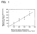
- FIG. 1 This is a graph representing an interrelationship between the results of the quantitative measurement by the colorimetric analysis and those by the size exclusion chromatography, which results are obtained in the EXAMPLE 2.
- bromochlorophenol blue BCPB
- TBPB tetrabromophenol blue
- Each of these bromochlorophenol blue and tetrabromophenol blue is one of the triphenylmethane dye colors, which turn green by associating or reacting with every kind of the protein.
- tetrabromophenol blue has a characteristic of being easy to give color reaction, at a regular rate, with various proteins under room temperature in a short period of time.
- tetrabromophenol blue has superiority over other coloring reagents in terms of the determination of the amount of the protein component, so that the tetrabromophenol blue can be preferably adopted.
- Each of bromochlorophenol blue and tetrabromophenol blue turns yellow if the pH is not more than 3. If the pH exceeds 3, hydrogen of the hydroxyl group dissociates, and bromochlorophenol blue and tetrabromophenol blue turn the same color as that of the association of the protein and the coloring reagent (protein-coloring reagent association product), i.e., bromochlorophenol blue and tetrabromophenol blue turn green.
- the coloring reagent solution is prepared by dissolving and including the above-mentioned coloring reagent in a predetermined solvent. If the concentration of the coloring reagent in the coloring reagent solution is too high, the dissolution of the coloring reagent in the solvent becomes difficult. On the other hand, if the concentration of the coloring reagent in the coloring reagent solution is too low, there is an inconvenience, caused by difficulty in developing the color, or by taking a long period of time for the measurement. Therefore, in general, there is employed a concentration within a range of about 1 to about 1000 ppm. In this range of the concentration, it is particularly desirable for the present invention to employ a concentration within a range of 100 to 500 ppm. This is because, if there is arranged the concentration of the coloring reagent to be within the range of 100 to 500 ppm and is used a suitable amount of the coloring reagent solution, the coloring reagent associates with the protein in a short period of time.
- the solvent for dissolving the coloring reagent is not particularly limited, as long as it is able to dissolve the coloring reagent.
- the solvent include water, methanol, ethanol, acetone, and a mixture of two or more of these solvents.
- water, ethanol, and a mixture of water and ethanol are especially preferably used, because these solvents are also used at a hospital, etc..
- additives may be suitably selected and added to the coloring reagent according to the present invention.
- the additives include: stabilizing agents such as butylhydroxytoluene; and solubilizers such as sodium lauryl sulphate. These additives are used within an amount which does not obstruct the object of the present invention.
- the coloring reagent solution can be easily prepared in a way as usually used for preparing a solution, without requiring any special procedure. Namely, the coloring reagent is prepared by dissolving the coloring reagent and additives, which are added as needed, in the solvent.
- the protein deposit is detected by using the coloring reagent as obtained above, and detecting or determining the amount of the protein component adhering to the contact lens, in which the protein component is adhered to the contact lens by wearing.
- a suitable amount of the coloring reagent solution containing bromochlorophenol blue or tetrabromophenol blue is generally accommodated in a container, which has a size to be able to accommodate the contact lens.
- the contact lens which has been worn, i.e., the contact lens, to which the protein deposit is adhered, is immersed in a way that overall the contact lens is immersed, so that the contact lens is caused to contact the coloring reagent solution. Accordingly, the various protein components adhering to the contact lens are associated or reacted with the coloring reagent in the coloring reagent solution in a short period of time.
- the period of time for the contact (immersion) is not particularly limited, and is suitably determined according to the concentration, etc., of the coloring reagent solution to be used.
- the period of time for contacting the contact lens with the coloring reagent solution is too short, it is difficult to give the quantitative association or reaction, so that there is an anxiety that the development of the color cannot be sufficiently achieved.
- the period of time for contacting (immersing) the contact lens with the coloring reagent solution is too long, the association or the reaction is excessively progressed, so that there is an anxiety that the protein-coloring reagent association product is eluted or dissolved from the contact lens. Therefore, it is desirable that the period of time for the contact is one second to 10 minutes, and more preferably 1 to 5 minutes.
- the contact lens is taken out of the coloring reagent solution, the coloring reagent solution adheres to overall the surfaces of the contact lens, so that overall the contact lens is colored and turned green.
- the color of the coloring reagent is overlapped with the color of the protein -coloring reagent association product, so that it is not possible to distinguish and detect only the colored protein component. Therefore, it is needed to remove the obstruction caused by the coloring reagent adhering to the surfaces of the contact lens.
- the contact lens is immersed in the solution (acid solution), whose pH is adjusted to be not more than 3. In this case, overall the contact lens is immersed in the solution, so as to cause the contact lens to contact the acid solution.
- the coloring reagent adhering to the surfaces of the contact lens is removed, or the color of the coloring reagent adhering to the contact lens is changed from green to yellow in a short period of time, whereby only the green color of the protein - coloring reagent association product remains.
- the pH of the above-mentioned acid solution is more than 3, the color of the coloring reagent adhering to the contact lens cannot be sufficiently changed. For this reason, it is not possible to advantageously develop the color of only a part of the contact lens, to which the protein is adhered, whereby the detection of the protein deposit cannot be accurately executed.
- the contact lens which has been immersed in the coloring reagent solution, is rinsed with, e.g., distilled water or tap water, of which the pH is not adjusted, in place of the above-mentioned acid solution, the coloring reagent adhering to the contact lens cannot be completely removed, so that the color of the coloring reagent is left on overall the contact lens, whereby it is not possible to visually confirm only the protein ⁇ coloring reagent association product.
- the lower limit of the pH of the acid solution as described above is not particularly limited. However, if the pH is excessively low, there is an anxiety that the protein or the contact lens material may be decomposed, so that it is desirable that the lower limit of the pH is not less than 2.
- the above-mentioned acid solution is easily prepared by suitably selecting conventionally known pH adjuster or pH buffer and dissolving it in an aqueous solution, as required.
- the aqueous solution include water and water-based various solvents, such as a solvent obtained by mixing water and ethanol, etc.
- the pH adjuster or the pH buffer include acids such as phosphoric acid, hydrochloric acid, sulfuric acid, and nitric acid. Among these acids, for the safety reason, an aqueous solution of phosphoric acid is preferably used.
- the period of time for contacting (immersing) the contact lens with the acid solution is not particularly limited, as long as it is sufficient to change the color of the coloring reagent adhering to the contact lens to yellow. However, it is desirable that the period of time for the contacting is one second to 10 minutes, preferably 1 to 5 minutes. If the period of time for contacting the contact lens with the coloring reagent solution is too short, there is an anxiety that the color of the coloring reagent cannot be completely changed to yellow, and it is tended to be difficult to distinguish the green color of only the protein-coloring reagent association product. On the other hand, if the period of time for contacting (immersing) the contact lens with the coloring reagent solution is too long, there is an anxiety that the protein adhering to the contact lens is decomposed by the acid solution.
- the coloring reagent which is associated with the protein adhering to the contact lens, is colored in green, whereby the protein deposit can be easily detected.
- This detection may be implemented while the contact lens is existed in the acid solution, or the detection may be implemented after the contact lens has been taken out of the acid solution.
- the protein component adhering to the contact lens by analyzing the color change of the contact lens, in other words, the depth of the color (degree of the development of the color) of the protein-coloring reagent association product of the contact lens, by using various known methods, such as: visual colorimetric analysis; spectral analysis by using a spectrophotometer; a method which comprises an image processing, such as an accumulation of a luminance based on an image taken with a camera, which is equipped with a sensor such as CCD or CMOS; and a method obtained by combining these methods.
- various known methods such as: visual colorimetric analysis; spectral analysis by using a spectrophotometer; a method which comprises an image processing, such as an accumulation of a luminance based on an image taken with a camera, which is equipped with a sensor such as CCD or CMOS; and a method obtained by combining these methods.
- the method of obtaining the luminance accumulation value (luminance accumulation method) by image processing of the image taken with the camera is preferably used, because a total amount of the protein adhering to the contact lens can be obtained as a numerical value. Meanwhile, if there is needed a more convenient or a more inexpensive detection, the visual colorimetric analysis is preferably used.
- control samples there are prepared standard color tone samples (control samples), and the amount of the protein component is detected by visually comparing the contact lens as a subject of the detection with the color tone samples.
- control samples by artificially attaching the protein component to the contact lenses, in a similar way to obtain the contact lens as the subject of the detection, such that the respective amounts of the protein component adhering to the contact lenses as the control samples are 0 ⁇ g/lens, 100 ⁇ g/lens, 200 ⁇ g/lens, 300 ⁇ g/lens, and 400 ⁇ g/lens.
- the amount of the adhered protein is detected by the spectral analysis
- the amount of the protein can be detected by measuring the absorption under a light of 624 nm, which absorption wavelength (maximum absorption wavelength) is peculiar to the protein-coloring reagent association product, by using the spectrophotometer.
- the amount of the protein can be detected by obtaining the luminance (the degree of brightness) under a predetermined wavelength band, by taking an image of the contact lens by using an image-taking device such as a color CCD camera, and processing the image taken with the camera.
- the protein deposit is detected according to the present invention, only the protein component adhering to the contact lens can be advantageously detected easily, inexpensively, and in a short period of time, by the simple processe of causing the contact lens to contact the coloring reagent solution and the acid solution, in due order.
- the amount of the protein adhering to the contact lens can be measured, without imposing the economical burden and troublesome tests on the contact lens wearer, there can be advantageously prevented or reduced the occurrence of various troubles such as deteriorating the wearing comfort as felt by the lens wearer, lowering the oxygen permeability of the lens, shortening the lifetime of the lens, lowering the eyesight of the lens wearer, allergy, and corneal injury.
- the contact lens was caused to contact the coloring reagent solution or the acid solution by being immerged in the solution.
- the method of the contact is not limited to the "immersing", and each of the solutions may be poured or sprayed to the contact lens, for thereby causing the solution to contact the contact lens.
- contact lenses to be used as specimens two each of the following four kinds of commercially available contact lenses (A to D).
- the following contact lenses A and B were made of similar materials, but were manufactured by different companies.
- the other one of the each kind of the above-mentioned water-contained contact lenses was not immersed in the aqueous solution of lysozym, and was used as the lens for comparison (the contact lens to which the lysozym is not adhered) A to D.
- each of total eight contact lenses consisting of the contact lenses A to D, to which the protein is adhered, and the lenses for comparison A to D, was immersed in 400 ppm TBPB ethanol solution at room temperature for one minute, and was taken out of the solution.
- each of the contact lenses was immersed in 10 mmol/L aqueous solution of phosphoric acid (the pH of the solution is about 2 to about 3) for not less than 30 seconds. In this way, the color of the protein component adhering to the contact lens was developed.
- the colors of the contact lenses were visually observed, while the contact lenses were still immersed in the solution. As a result, there was confirmed that the green color was not developed on each of the lenses for comparison A to D, to which the protein was not adhered.
- the contact lens A which was immersed in the aqueous solution of lysozym, exhibited a distinctively deep green color, compared with the lens for comparison A.
- the contact lens B which was immersed in the aqueous solution of lysozym, exhibited a slightly green color, compared with the lens for comparison B.
- the contact lenses C and D which were immersed in the aqueous solution of lysozym, did not exhibit difference in the color, compared with the lenses for comparison C and D. According to these results, there was confirmed that the protein deposit could be easily detected by the visual colorimetric analysis.
- the eight contact lenses which were immersed in the TBPB ethanol solution and the aqueous solution of phosphoric acid, in due order, as described above, were imaged with a digital camera, which was equipped with a CCD, while the contact lenses were still immerged in the solution.
- the luminance accumulation value of the each kind of the contact lenses was obtained as the difference between the luminance, under a light of 624nm, of each of the protein adhering lenses and that of the corresponding lens for comparison, by processing the image of the each kind of the contact lenses, by using an image processing soft "AQUACOSMOS" available from KABUSHIKI KAISHA HAMAMATSU PHOTONICS.
- luminance accumulation values of the contact lenses A, B, C, and D were 5058, 999, 111, and 79, respectively.
- the protein deposit adhering to the contact lens can be described in numerical values, and even some kinds of the protein deposits, which were difficult to be detected by the visual colorimetric analysis, could be detected.
- each of the two kinds of the commercially available contact lenses A and C was respectively worn by each of the above-mentioned three contact lens wearers for one hour, three hours, five hours, and one day.
Landscapes
- Life Sciences & Earth Sciences (AREA)
- Health & Medical Sciences (AREA)
- Engineering & Computer Science (AREA)
- Molecular Biology (AREA)
- Biomedical Technology (AREA)
- Chemical & Material Sciences (AREA)
- Urology & Nephrology (AREA)
- Hematology (AREA)
- Immunology (AREA)
- Medicinal Chemistry (AREA)
- General Health & Medical Sciences (AREA)
- Cell Biology (AREA)
- Biotechnology (AREA)
- Food Science & Technology (AREA)
- Proteomics, Peptides & Aminoacids (AREA)
- Physics & Mathematics (AREA)
- Analytical Chemistry (AREA)
- Biochemistry (AREA)
- Microbiology (AREA)
- General Physics & Mathematics (AREA)
- Pathology (AREA)
- Investigating Or Analysing Biological Materials (AREA)
- Investigating Or Analysing Materials By The Use Of Chemical Reactions (AREA)
- Eyeglasses (AREA)
- Investigating Or Analyzing Non-Biological Materials By The Use Of Chemical Means (AREA)
- Investigating Materials By The Use Of Optical Means Adapted For Particular Applications (AREA)
- Pinball Game Machines (AREA)
- Facsimile Heads (AREA)
- Preparing Plates And Mask In Photomechanical Process (AREA)
Abstract
Description
- The present invention relates to a method of detecting protein deposit on a contact lens, in particular, a method of detecting the protein deposit adhering to the contact lens easily and in a short period of time.
- In recent years, in order to provide intended characteristics, there have been provided contact lenses made of various materials. However, a protein contained in lacrimal fluid tends to adhere to a contact lens, in particular, to a contact lens made of an ionic polymer material obtained by using, as a raw material, monomeric components having ionizable groups. If the protein gradually adheres to and accumulates on surfaces of and inside the contact lens, removal of the protein tends to be difficult. In addition to this, there is an anxiety that the adhesion and the accumulation of the protein may cause various troubles such as deteriorating the wearing comfort as felt by the lens wearer, lowering the oxygen permeability of the lens, lowering the eyesight of the lens wearer, allergy, and corneal injury.
- As the methods of quantitatively detecting and determining the protein component adhering to the contact lens, there are conventionally known amino acid analysis, thin layer chromatography, high-performance liquid chromatography, and ELISA method. However, each of these methods requires, before the detection, a process of extraction, in which the deposit adhering to the contact lens is extracted to a predetermined solvent. Accordingly, these methods are not suitable for usual analyses, because the detections are troublesome, taking a long time, or requiring a large and expensive device.
- Besides, JP-A-58-222154 (Patent Document 1) and JP-A-58-222155 (Patent Document 2) propose methods of detecting particular components adhering to the contact lenses, by immersing the contact lenses in the solutions for detecting deposits, wherein the solutions include, as a coloring reagent, Nile Blue sulfate or erythrosin. However, these is an anxiety that these coloring reagents may also color lipid component, as well as the protein component, so that there is a problem that the protein component cannot be selectively detected. In addition, JP-A-2000-65840 (Patent Document 3) proposes a method of detecting the protein deposit and the lipid deposit adhering to the contact lens, by using a basic dye solution.
- Meanwhile, JP-U-A-5-40874 (Patent Document 4) proposes a reference color spectrum. The Patent Document 4 explains that, there is commonly used tetrabromophenol blue as a color former of a urinary protein test paper, which is used for inspecting the protein components in the urine by means of a method of protein error of a pH indicator. JP-A-8-505938 (Patent Document 5) proposes a test panel which represents an immediate result, wherein bromochlorophenol blue and tetrabromophenol blue are used as visual indicators. The Patent Document 5 explains that an existence or a loss of a substance to be analyzed, e.g., the protein, included in a body fluid such as lacrimal fluid, can be recognized according to a color tone of the indicator. Moreover, JP-A-11-304803 (Patent Document 6) proposes a method and a kit for analyzing the protein in a sample. JP-A-11-304803 discloses a method of analyzing the protein by using an association of triphenylmethane dye color, such as bromochlorophenol blue or tetrabromophenol blue, with the protein, and causing the association to adsorb on a hydrophobic filter. However, these methods disclosed in the Patent Documents 4 to 6 are mainly intended to detect the protein component included in a fluid such as the urine. Therefore, there is no disclosure of a concrete method of detecting the protein component included in the lacrimal fluid adhering to the contact lens.
- Patent Document 1: JP-A-58-222154
- Patent Document 2: JP-A-58-222155
- Patent Document 3: JP-A-2000-65840
- Patent Document 4: JP-U-A-5-40874
- Patent Document 5: JP-A-8-505938
- Patent Document 6: JP-A-11-304803
- The present invention was made in view of the background art described above. A problem to be solved by the invention is to provide a method of detecting only the protein deposit adhering to a contact lens easily and in a short period of time.
- The present invention was made to solve the problem described above. In first preferred form of the present invention, there is provided a method of detecting protein deposit on a contact lens characterized in that a protein component adhering to the contact lens, after the contact lens has been worn, is detected when the protein component adhering to the contact lens is in its colored state, wherein the protein component is colored by causing the contact lens to contact a solution having pH of not more than 3, after causing the contact lens to contact a coloring reagent solution containing bromochlorophenol blue or tetrabromophenol blue.
- Besides, in second preferred form of the present invention, the coloring reagent in the coloring reagent solution has a concentration of 100 - 500 ppm.
- Moreover, in third preferred form of the present invention, it is desirable that the coloring reagent solution is obtained by dissolving the coloring reagent in water, ethanol, or a mixture thereof.
- In addition, in fourth preferred form of the present invention, it is desirable that the solution having pH of not more than 3 is an aqueous solution of phosphoric acid.
- Also, in fifth preferred form of the present invention, as the solution having pH of not more than 3, there is preferably employed a solution, whose pH is within a range of 2 - 3.
- Moreover, in sixth preferred form of the method of detecting the protein deposit on the contact lens in accordance with the present invention, the detection of the protein deposit adhering to the contact lens is implemented by a colorimetric analysis, a spectral analysis, or an analysis obtained by combining these analyses.
- Furthermore, in seventh preferred form of the present invention, the detection of the protein deposit adhering to the contact lens is implemented by image processing of an image taken with a camera, which image is of the contact lens caused to contact the coloring reagent solution and the solution having pH of not more than 3.
- According to the above-mentioned first preferred form of the present invention, bromochlorophenol blue or tetrabromophenol blue is used as the coloring reagent. Owing to this, selective detection of the protein component adhering to the contact lens is advantageously realized, without causing a problem such that a result of the detection is influenced by a kind of the protein component. Namely, the coloring reagent develops a color by associating or reacting with various protein components, without coloring the lipid component.
- Besides, in the present invention, the contact lens, after it has been worn, is caused to contact a solution including the coloring reagent (coloring reagent solution), and is further caused to contact a solution whose pH is adjusted to be not more than 3 (acidic solution) by, for example, immersing the contact lens in the solution. Owing to this, color of only the protein component is advantageously developed or given in a short period of time. Accordingly, positions on the contact lens, to which the protein component is adhered to, are colored in green. Accordingly, the protein component can be detected, based on the extent of the development of the color (depth of the color).
- As described above, in the present invention, only the protein component adhering to the contact lens can be detected easily, economically, and in a short period of time, by causing the contact lens to contact the coloring reagent solution and the acid solution in due order, without employing conventionally employed methods of detecting the protein, such as amino aid analysis, thin layer chromatography, high-performance liquid chromatography, and ELISA method, which require a long period of time for the detection, troublesome processes for the detection, or a high cost.
- Among various contact lenses, particularly large amount of the protein component is adhered to the ionic soft contact lens, and this results in increasing possibility of causing various troubles, such as deteriorating the wearing comfort as felt by the lens wearer, lowering the oxygen permeability of the lens, shortening the lifetime of the lens, lowering the eyesight of the lens wearer, allergy, and corneal injury. Owing to the above described preferred forms of the present invention, the protein deposit adhering to the contact lens, without imposing an economical burden and troublesome tests on the lens user, for thereby effectively preventing the occurrences of the above-mentioned troubles.
- Besides, in the second preferred form of the present invention, the concentration of the coloring reagent is within a predetermined range, so that the development of the color of the protein component adhering to the contact lens is further advantageously realized.
- Moreover, in the sixth preferred form of the present invention, the detection of the protein deposit adhering to the contact lens is implemented by the colorimetric analysis, the spectral analysis, or the analysis obtained by combining these analyses, and owing to this, the detection of the protein component can be executed more inexpensively, compared with the conventional methods.
- In addition to the above, if the detection of the protein deposit adhering to the contact lens is implemented by an image processing of the image taken with the camera, which image is of the contact lens caused to contact the coloring reagent solution and the solution having pH of not more than 3, in accordance with the seventh preferred form of the present invention, the amount of the protein adhering to overall the contact lens can be easily obtained as a numerical value.
- [Fig. 1] This is a graph representing an interrelationship between the results of the quantitative measurement by the colorimetric analysis and those by the size exclusion chromatography, which results are obtained in the EXAMPLE 2.
- Meanwhile, for the coloring reagent solution to be employed in the present invention, bromochlorophenol blue (BCPB) or tetrabromophenol blue (TBPB) is used as the coloring reagent. Each of these bromochlorophenol blue and tetrabromophenol blue is one of the triphenylmethane dye colors, which turn green by associating or reacting with every kind of the protein. In particular, tetrabromophenol blue has a characteristic of being easy to give color reaction, at a regular rate, with various proteins under room temperature in a short period of time. In other words, tetrabromophenol blue has superiority over other coloring reagents in terms of the determination of the amount of the protein component, so that the tetrabromophenol blue can be preferably adopted. Each of bromochlorophenol blue and tetrabromophenol blue turns yellow if the pH is not more than 3. If the pH exceeds 3, hydrogen of the hydroxyl group dissociates, and bromochlorophenol blue and tetrabromophenol blue turn the same color as that of the association of the protein and the coloring reagent (protein-coloring reagent association product), i.e., bromochlorophenol blue and tetrabromophenol blue turn green.
- In the present invention, the coloring reagent solution is prepared by dissolving and including the above-mentioned coloring reagent in a predetermined solvent. If the concentration of the coloring reagent in the coloring reagent solution is too high, the dissolution of the coloring reagent in the solvent becomes difficult. On the other hand, if the concentration of the coloring reagent in the coloring reagent solution is too low, there is an inconvenience, caused by difficulty in developing the color, or by taking a long period of time for the measurement. Therefore, in general, there is employed a concentration within a range of about 1 to about 1000 ppm. In this range of the concentration, it is particularly desirable for the present invention to employ a concentration within a range of 100 to 500 ppm. This is because, if there is arranged the concentration of the coloring reagent to be within the range of 100 to 500 ppm and is used a suitable amount of the coloring reagent solution, the coloring reagent associates with the protein in a short period of time.
- The solvent for dissolving the coloring reagent is not particularly limited, as long as it is able to dissolve the coloring reagent. Examples of the solvent include water, methanol, ethanol, acetone, and a mixture of two or more of these solvents. Among these solvents, water, ethanol, and a mixture of water and ethanol are especially preferably used, because these solvents are also used at a hospital, etc..
- In addition, various known additives may be suitably selected and added to the coloring reagent according to the present invention. Examples of the additives include: stabilizing agents such as butylhydroxytoluene; and solubilizers such as sodium lauryl sulphate. These additives are used within an amount which does not obstruct the object of the present invention.
- The coloring reagent solution can be easily prepared in a way as usually used for preparing a solution, without requiring any special procedure. Namely, the coloring reagent is prepared by dissolving the coloring reagent and additives, which are added as needed, in the solvent.
- In the present invention, the protein deposit is detected by using the coloring reagent as obtained above, and detecting or determining the amount of the protein component adhering to the contact lens, in which the protein component is adhered to the contact lens by wearing.
- In detail, at first a suitable amount of the coloring reagent solution containing bromochlorophenol blue or tetrabromophenol blue is generally accommodated in a container, which has a size to be able to accommodate the contact lens. In this coloring reagent solution, the contact lens, which has been worn, i.e., the contact lens, to which the protein deposit is adhered, is immersed in a way that overall the contact lens is immersed, so that the contact lens is caused to contact the coloring reagent solution. Accordingly, the various protein components adhering to the contact lens are associated or reacted with the coloring reagent in the coloring reagent solution in a short period of time.
- In this case, the period of time for the contact (immersion) is not particularly limited, and is suitably determined according to the concentration, etc., of the coloring reagent solution to be used. However, if the period of time for contacting the contact lens with the coloring reagent solution is too short, it is difficult to give the quantitative association or reaction, so that there is an anxiety that the development of the color cannot be sufficiently achieved. On the other hand, if the period of time for contacting (immersing) the contact lens with the coloring reagent solution is too long, the association or the reaction is excessively progressed, so that there is an anxiety that the protein-coloring reagent association product is eluted or dissolved from the contact lens. Therefore, it is desirable that the period of time for the contact is one second to 10 minutes, and more preferably 1 to 5 minutes.
- If the contact lens is taken out of the coloring reagent solution, the coloring reagent solution adheres to overall the surfaces of the contact lens, so that overall the contact lens is colored and turned green. However, if it is left as it is, the color of the coloring reagent is overlapped with the color of the protein -coloring reagent association product, so that it is not possible to distinguish and detect only the colored protein component. Therefore, it is needed to remove the obstruction caused by the coloring reagent adhering to the surfaces of the contact lens. For this reason, in the present invention, the contact lens is immersed in the solution (acid solution), whose pH is adjusted to be not more than 3. In this case, overall the contact lens is immersed in the solution, so as to cause the contact lens to contact the acid solution. By treating the contact lens with the acid solution as described above, the coloring reagent adhering to the surfaces of the contact lens is removed, or the color of the coloring reagent adhering to the contact lens is changed from green to yellow in a short period of time, whereby only the green color of the protein - coloring reagent association product remains.
- If the pH of the above-mentioned acid solution is more than 3, the color of the coloring reagent adhering to the contact lens cannot be sufficiently changed. For this reason, it is not possible to advantageously develop the color of only a part of the contact lens, to which the protein is adhered, whereby the detection of the protein deposit cannot be accurately executed. If the contact lens, which has been immersed in the coloring reagent solution, is rinsed with, e.g., distilled water or tap water, of which the pH is not adjusted, in place of the above-mentioned acid solution, the coloring reagent adhering to the contact lens cannot be completely removed, so that the color of the coloring reagent is left on overall the contact lens, whereby it is not possible to visually confirm only the protein―coloring reagent association product. In addition, the lower limit of the pH of the acid solution as described above is not particularly limited. However, if the pH is excessively low, there is an anxiety that the protein or the contact lens material may be decomposed, so that it is desirable that the lower limit of the pH is not less than 2.
- In addition, the above-mentioned acid solution is easily prepared by suitably selecting conventionally known pH adjuster or pH buffer and dissolving it in an aqueous solution, as required. Examples of the aqueous solution include water and water-based various solvents, such as a solvent obtained by mixing water and ethanol, etc.. Meanwhile, examples of the pH adjuster or the pH buffer include acids such as phosphoric acid, hydrochloric acid, sulfuric acid, and nitric acid. Among these acids, for the safety reason, an aqueous solution of phosphoric acid is preferably used.
- The period of time for contacting (immersing) the contact lens with the acid solution is not particularly limited, as long as it is sufficient to change the color of the coloring reagent adhering to the contact lens to yellow. However, it is desirable that the period of time for the contacting is one second to 10 minutes, preferably 1 to 5 minutes. If the period of time for contacting the contact lens with the coloring reagent solution is too short, there is an anxiety that the color of the coloring reagent cannot be completely changed to yellow, and it is tended to be difficult to distinguish the green color of only the protein-coloring reagent association product. On the other hand, if the period of time for contacting (immersing) the contact lens with the coloring reagent solution is too long, there is an anxiety that the protein adhering to the contact lens is decomposed by the acid solution.
- As described above, the coloring reagent, which is associated with the protein adhering to the contact lens, is colored in green, whereby the protein deposit can be easily detected. This detection may be implemented while the contact lens is existed in the acid solution, or the detection may be implemented after the contact lens has been taken out of the acid solution.
- There can be easily and inexpensively detected and determined the protein component adhering to the contact lens, by analyzing the color change of the contact lens, in other words, the depth of the color (degree of the development of the color) of the protein-coloring reagent association product of the contact lens, by using various known methods, such as: visual colorimetric analysis; spectral analysis by using a spectrophotometer; a method which comprises an image processing, such as an accumulation of a luminance based on an image taken with a camera, which is equipped with a sensor such as CCD or CMOS; and a method obtained by combining these methods. Among these analyzing methods, the method of obtaining the luminance accumulation value (luminance accumulation method) by image processing of the image taken with the camera is preferably used, because a total amount of the protein adhering to the contact lens can be obtained as a numerical value. Meanwhile, if there is needed a more convenient or a more inexpensive detection, the visual colorimetric analysis is preferably used.
- Where the amount of the adhered protein is detected by the visual colorimetric analysis, there are prepared standard color tone samples (control samples), and the amount of the protein component is detected by visually comparing the contact lens as a subject of the detection with the color tone samples. For example, there are obtained the control samples, by artificially attaching the protein component to the contact lenses, in a similar way to obtain the contact lens as the subject of the detection, such that the respective amounts of the protein component adhering to the contact lenses as the control samples are 0 µg/lens, 100 µg/lens, 200 µ g/lens, 300 µg/lens, and 400 µ g/lens.
- Where the amount of the adhered protein is detected by the spectral analysis, the amount of the protein can be detected by measuring the absorption under a light of 624 nm, which absorption wavelength (maximum absorption wavelength) is peculiar to the protein-coloring reagent association product, by using the spectrophotometer. Alternatively, the amount of the protein can be detected by obtaining the luminance (the degree of brightness) under a predetermined wavelength band, by taking an image of the contact lens by using an image-taking device such as a color CCD camera, and processing the image taken with the camera.
- As is described above, if the protein deposit is detected according to the present invention, only the protein component adhering to the contact lens can be advantageously detected easily, inexpensively, and in a short period of time, by the simple processe of causing the contact lens to contact the coloring reagent solution and the acid solution, in due order.
- Therefore, the amount of the protein adhering to the contact lens can be measured, without imposing the economical burden and troublesome tests on the contact lens wearer, there can be advantageously prevented or reduced the occurrence of various troubles such as deteriorating the wearing comfort as felt by the lens wearer, lowering the oxygen permeability of the lens, shortening the lifetime of the lens, lowering the eyesight of the lens wearer, allergy, and corneal injury.
- While the presently preferred embodiments of this invention have been described in detail, it should be understood that the invention is not limited to the details of the foregoing description, but may be otherwise embodied.
- For example, in the foregoing embodiments, the contact lens was caused to contact the coloring reagent solution or the acid solution by being immerged in the solution. However, the method of the contact is not limited to the "immersing", and each of the solutions may be poured or sprayed to the contact lens, for thereby causing the solution to contact the contact lens.
- It is to be understood that the present invention may be embodied with various changes, modifications and improvements, which may occur to those skilled in the art, without departing from the spirit and scope of the invention defined in the attached claims.
- There will be described some examples of the present invention to further clarify the present invention. It is to be understood, however, that the present invention is not limited to the details of the following examples.
- First of all, there were prepared, as the contact lenses to be used as specimens, two each of the following four kinds of commercially available contact lenses (A to D). The following contact lenses A and B were made of similar materials, but were manufactured by different companies.
- A:
- HEMA (hydroxyethyl methacrylate)-based ionic high water content soft contact lens
- B:
- HEMA-based ionic high water content soft contact lens
- C:
- N-vinyl pyrrolidone-based nonionic high water content soft contact lens
- D:
- HEMA-based nonionic low water content soft contact lens
- One of the each kind of the above-mentioned water-contained soft contact lenses was immersed in 0.7% aqueous solution of lysozym for 16 hours, whereby the protein component (lysozym) was artificially adhered to the contact lenses.
- The other one of the each kind of the above-mentioned water-contained contact lenses was not immersed in the aqueous solution of lysozym, and was used as the lens for comparison (the contact lens to which the lysozym is not adhered) A to D.
- Thereafter, each of total eight contact lenses, consisting of the contact lenses A to D, to which the protein is adhered, and the lenses for comparison A to D, was immersed in 400 ppm TBPB ethanol solution at room temperature for one minute, and was taken out of the solution. Subsequently, each of the contact lenses was immersed in 10 mmol/L aqueous solution of phosphoric acid (the pH of the solution is about 2 to about 3) for not less than 30 seconds. In this way, the color of the protein component adhering to the contact lens was developed.
- The colors of the contact lenses were visually observed, while the contact lenses were still immersed in the solution. As a result, there was confirmed that the green color was not developed on each of the lenses for comparison A to D, to which the protein was not adhered. The contact lens A, which was immersed in the aqueous solution of lysozym, exhibited a distinctively deep green color, compared with the lens for comparison A. The contact lens B, which was immersed in the aqueous solution of lysozym, exhibited a slightly green color, compared with the lens for comparison B. The contact lenses C and D, which were immersed in the aqueous solution of lysozym, did not exhibit difference in the color, compared with the lenses for comparison C and D. According to these results, there was confirmed that the protein deposit could be easily detected by the visual colorimetric analysis.
- Besides, the eight contact lenses, which were immersed in the TBPB ethanol solution and the aqueous solution of phosphoric acid, in due order, as described above, were imaged with a digital camera, which was equipped with a CCD, while the contact lenses were still immerged in the solution. Thus obtained images taken with the digital camera were imported to a computer. The luminance accumulation value of the each kind of the contact lenses was obtained as the difference between the luminance, under a light of 624nm, of each of the protein adhering lenses and that of the corresponding lens for comparison, by processing the image of the each kind of the contact lenses, by using an image processing soft "AQUACOSMOS" available from KABUSHIKI KAISHA HAMAMATSU PHOTONICS. Thus obtained luminance accumulation values of the contact lenses A, B, C, and D were 5058, 999, 111, and 79, respectively. In this way, the protein deposit adhering to the contact lens can be described in numerical values, and even some kinds of the protein deposits, which were difficult to be detected by the visual colorimetric analysis, could be detected.
- Two kinds of the above-mentioned commercially available water-contained contact lenses A and C were obtained, and each of these contact lenses were respectively worn by each of the three contact lens wearers a to c for one hour, three hours, five hours, and one day. Thus obtained worn contact lenses were immersed in 400 ppm TBPB ethanol solution at room temperature for one minute, and then the contact lenses were immerged in 10 mmol/L aqueous solution of phosphoric acid for not less than 30 seconds. In this way, the color of the protein deposit adhering to the contact lenses was developed. These lenses were visually observed, and there were found that the lens A turned green and the green color of the lens A became deeper, as the duration of the wearing increases, while the color of the lens C was hardly developed. There were also executed the visual colorimetric analyses with respect of the lens A. The results are shown in TABLE 2. As the standard color tone samples (control samples), there were prepared the control lenses, to which the protein component was artificially attached (0 µ g/lens, 100 µg/lens, 200 µ g/lens, 300 µ g/lens, and 400 µ g/lens), and the colors of the contact lenses were developed in the same way as the development of the color of the above-described lenses A and C. The colorimetric analysis was executed by comparing the colors of the contact lenses A and C with those of the control samples.
- Meanwhile, similar to the above, each of the two kinds of the commercially available contact lenses A and C was respectively worn by each of the above-mentioned three contact lens wearers for one hour, three hours, five hours, and one day. The each of the worn contact lenses was separately accommodated in a vial container, together with 1mL extract (0.2% aqueous solution of trifluoroacetic acid/acetonitrile = 50/50), and the protein adhering to the contact lens was extracted. After the extraction, the extract was analyzed by a size exclusion chromatography ("Alliance 2690" available from Waters Corporation), by using a detector and conditions as described below. In this way, the amounts of the adhering protein were quantitatively measured. The results are shown in the following TABLE 1 and TABLE 2. In the TABLE 1, there are shown the average values of the measured values, while in the TABLE 2, there are only shown the amounts of the protein adhering to the lens A. Moreover, there are plotted the relationships between the quantitatively measured values obtained by the size exclusion chromatography (HPLC) and the above-mentioned measured values obtained by the colorimetric analysis (see Fig. 1).
PDA (Photodiode Array) Detector: - "PDA 996" available from Waters Corporation in USA
- "TSK-GEL G3000SWXL" 7.8 X 300mm available from TOSOH CORPORATION in JAPAN
- 0.5 mol/L sodium chloride+0.1 mol/L phosphate buffer (pH 5)
-
[TABLE 1] Duration of wearing One hour Three hours Five hours One day Kind of the lens A C A C A C A C Measured values obtained by HPLC* (µg/lens) 84 0.1 229 0.3 286 0.2 448 0.3 * Each of the values is average value obtained from the three people: a to c. -
[TABLE 2] Duration of wearing Wearer Measured values obtained by visual colorimetric analysis (µg/lens) Measured values obtained by HPLC (µg/lens) One hour a 100 30 b 100 130 c 100 92 Three Three hours a 200 260 b 200 185 c 200 243 Five hours a 300 240 b 300 291 c 300 326 One day a 400 512 b 400 389 c 400 444 - As is apparent from the TABLE 2 and the Fig. 1, there were confirmed excellent interrelationships between the results of the quantitative measurement of the amount of the protein obtained by the coloring reagent (visual colorimetric analysis) and the quantitative measurement of the amount of the protein obtained by the size exclusion chromatography, whereby there is recognized that the measurement method by using the colorimetric reagent is effective. In addition, as is apparent from the above TABLE 1, the relationships between the duration of the wearing and the amount of the adhering protein component are varied, depending on the kinds of the contact lenses. Therefore, there is regarded that the troubles, which are caused by the accumulation of the protein component (allergy, deterioration of the eyesight, and deterioration of the wearing comfort) can be advantageously forecasted.
Moving phase:
Claims (7)
- A method of detecting protein deposit on a contact lens characterized in that:a protein component adhering to the contact lens, after said contact lens has been worn, is detected when the protein component adhering to the contact lens is in its colored state, wherein the protein component is colored by causing said contact lens to contact a solution having pH of not more than 3, after causing said contact lens to contact a coloring reagent solution containing bromochlorophenol blue or tetrabromophenol blue.
- A method of detecting protein deposit on a contact lens according to claim 1, wherein said coloring reagent in the coloring reagent solution has a concentration of 100 - 500 ppm.
- A method of detecting protein deposit on a contact lens according to claim 1 or 2, wherein said coloring reagent solution is obtained by dissolving the coloring reagent in water, ethanol, or a mixture thereof.
- A method of detecting protein deposit on a contact lens according to any one of claims 1 to 3, wherein said solution having pH of not more than 3 is an aqueous solution of phosphoric acid.
- A method of detecting protein deposit on a contact lens according to any one of claims 1 to 4, wherein pH of said solution having pH of not more than 3 is within a range of 2 - 3.
- A method of detecting protein deposit on a contact lens according to any one of claims 1 to 5, wherein said detection of the protein deposit adhering to the contact lens comprises a colorimetric analysis, a spectral analysis, or an analysis obtained by combining said analyses.
- A method of detecting protein deposit on a contact lens according to any one of claims 1 to 5, wherein said detection of the protein deposit adhering to the contact lens comprises image processing of an image taken with a camera, which image is of the contact lens caused to contact the coloring reagent solution and the solution having pH of not more than 3.
Applications Claiming Priority (2)
| Application Number | Priority Date | Filing Date | Title |
|---|---|---|---|
| JP2003283356A JP4265758B2 (en) | 2003-07-31 | 2003-07-31 | Method for detecting protein contamination in contact lenses |
| PCT/JP2004/010220 WO2005012915A1 (en) | 2003-07-31 | 2004-07-16 | Method of detecting protein dirt on contact lens |
Publications (3)
| Publication Number | Publication Date |
|---|---|
| EP1650567A1 true EP1650567A1 (en) | 2006-04-26 |
| EP1650567A4 EP1650567A4 (en) | 2007-09-05 |
| EP1650567B1 EP1650567B1 (en) | 2009-10-07 |
Family
ID=30438742
Family Applications (1)
| Application Number | Title | Priority Date | Filing Date |
|---|---|---|---|
| EP04747685A Expired - Lifetime EP1650567B1 (en) | 2003-07-31 | 2004-07-16 | Method of detecting protein dirt on contact lens |
Country Status (5)
| Country | Link |
|---|---|
| EP (1) | EP1650567B1 (en) |
| JP (1) | JP4265758B2 (en) |
| AT (1) | ATE445166T1 (en) |
| DE (1) | DE602004023511D1 (en) |
| WO (1) | WO2005012915A1 (en) |
Cited By (6)
| Publication number | Priority date | Publication date | Assignee | Title |
|---|---|---|---|---|
| JP2014228292A (en) * | 2013-05-20 | 2014-12-08 | 一般社団法人日本真珠振興会 | Method for identifying pearl nucleus |
| WO2019175660A1 (en) * | 2018-03-14 | 2019-09-19 | Menicon Co. Ltd. | Method for detecting a health condition with biomarkers |
| CN114062327A (en) * | 2020-07-31 | 2022-02-18 | 苏州三个臭皮匠生物科技有限公司 | Method for effectively visually and visually detecting protein cleaning effect |
| CN116869732A (en) * | 2023-07-04 | 2023-10-13 | 珠海溪谷医疗科技有限公司 | An artificial tear and a method for characterizing the protein removal effect of contact lens care solution |
| US12064237B2 (en) | 2018-03-13 | 2024-08-20 | Menicon Co., Ltd. | Determination system, computing device, determination method, and program |
| US12629057B2 (en) | 2019-03-13 | 2026-05-19 | Menicon Co., Ltd. | System for collecting and utilizing health data |
Families Citing this family (4)
| Publication number | Priority date | Publication date | Assignee | Title |
|---|---|---|---|---|
| EP2452710A1 (en) | 2010-11-12 | 2012-05-16 | Sanofi-Aventis Deutschland GmbH | Assembly for a drug delivery device and piston rod |
| CN113848192A (en) * | 2020-07-31 | 2021-12-28 | 苏州三个臭皮匠生物科技有限公司 | A method for effectively detecting protein concentration in a solution containing available chlorine |
| CN114689538A (en) * | 2020-12-30 | 2022-07-01 | 苏州三个臭皮匠生物科技有限公司 | Method for effectively measuring tear protein content of soft hydrophilic corneal contact lens |
| CN115096837A (en) * | 2021-06-23 | 2022-09-23 | 温州医科大学附属眼视光医院 | Method for detecting tear protein content of hard corneal contact lens, application and cleaning device |
Family Cites Families (9)
| Publication number | Priority date | Publication date | Assignee | Title |
|---|---|---|---|---|
| BE566273A (en) * | 1957-04-02 | |||
| JPS58222155A (en) * | 1982-06-18 | 1983-12-23 | Tokyo Contact Lens Kenkyusho:Kk | Dirt detecting solution for contact lens |
| JPS58222154A (en) * | 1982-06-18 | 1983-12-23 | Tokyo Contact Lens Kenkyusho:Kk | Dirt detecting solution for contact lens |
| JPH03284725A (en) * | 1990-03-30 | 1991-12-16 | Hoya Corp | Detergent for hard contact lens |
| JPH0540874A (en) | 1991-08-05 | 1993-02-19 | Fuji Electric Co Ltd | Fluorescent light controller for vending machines |
| US5268146A (en) | 1992-03-25 | 1993-12-07 | Litmus Concepts, Inc. | Fast response test panel |
| JP4039739B2 (en) * | 1998-02-23 | 2008-01-30 | 株式会社シノテスト | Method for analyzing protein in sample and kit thereof |
| JP3978288B2 (en) * | 1998-08-24 | 2007-09-19 | 株式会社サンコンタクトレンズ | Aqueous composition for detecting dirt on contact lenses and method for detecting dirt using the same |
| JP2004212813A (en) * | 2003-01-07 | 2004-07-29 | Menicon Co Ltd | How to choose contact lenses and their care systems |
-
2003
- 2003-07-31 JP JP2003283356A patent/JP4265758B2/en not_active Expired - Fee Related
-
2004
- 2004-07-16 DE DE602004023511T patent/DE602004023511D1/en not_active Expired - Fee Related
- 2004-07-16 WO PCT/JP2004/010220 patent/WO2005012915A1/en not_active Ceased
- 2004-07-16 AT AT04747685T patent/ATE445166T1/en not_active IP Right Cessation
- 2004-07-16 EP EP04747685A patent/EP1650567B1/en not_active Expired - Lifetime
Cited By (6)
| Publication number | Priority date | Publication date | Assignee | Title |
|---|---|---|---|---|
| JP2014228292A (en) * | 2013-05-20 | 2014-12-08 | 一般社団法人日本真珠振興会 | Method for identifying pearl nucleus |
| US12064237B2 (en) | 2018-03-13 | 2024-08-20 | Menicon Co., Ltd. | Determination system, computing device, determination method, and program |
| WO2019175660A1 (en) * | 2018-03-14 | 2019-09-19 | Menicon Co. Ltd. | Method for detecting a health condition with biomarkers |
| US12629057B2 (en) | 2019-03-13 | 2026-05-19 | Menicon Co., Ltd. | System for collecting and utilizing health data |
| CN114062327A (en) * | 2020-07-31 | 2022-02-18 | 苏州三个臭皮匠生物科技有限公司 | Method for effectively visually and visually detecting protein cleaning effect |
| CN116869732A (en) * | 2023-07-04 | 2023-10-13 | 珠海溪谷医疗科技有限公司 | An artificial tear and a method for characterizing the protein removal effect of contact lens care solution |
Also Published As
| Publication number | Publication date |
|---|---|
| WO2005012915A1 (en) | 2005-02-10 |
| JP4265758B2 (en) | 2009-05-20 |
| ATE445166T1 (en) | 2009-10-15 |
| DE602004023511D1 (en) | 2009-11-19 |
| WO2005012915A8 (en) | 2005-08-25 |
| JP2004004117A (en) | 2004-01-08 |
| EP1650567A4 (en) | 2007-09-05 |
| EP1650567B1 (en) | 2009-10-07 |
Similar Documents
| Publication | Publication Date | Title |
|---|---|---|
| EP1650567B1 (en) | Method of detecting protein dirt on contact lens | |
| Ragone et al. | Determination of tyrosine exposure in proteins by second-derivative spectroscopy | |
| JP2519102B2 (en) | Compositions and methods for testing aqueous liquids for specific gravity | |
| CN107884396B (en) | Optical microscope-based ascorbic acid concentration online sensing analysis method and auxiliary color developing agent | |
| CN103278501B (en) | COD quick detection solution, test box and determination method | |
| KR20130107268A (en) | Method for measuring glycated hemoglobin | |
| KR101244068B1 (en) | A method for measuring concentration of air and water pollutants | |
| EP2927683B1 (en) | Method for judging the concentration of lithium in biomaterial samples | |
| CA2071206A1 (en) | Reagent and methods for calcium determination | |
| Matsushita et al. | Determination of proteins by a reverse biuret method combined with the copper-bathocuproine chelate reaction | |
| EP0861439B1 (en) | Composition and method for manufacturing ion selective electrode sensors | |
| CN110763674A (en) | A method for rapid detection of vitamin C content in vegetables and fruits | |
| CN1210558C (en) | Test piece for detecting calcium and preparing method thereof | |
| WO2017021759A1 (en) | Methods and probes for determining the concentration of copper | |
| Ouirungroj et al. | Smart-detection approach for protein residues to evaluate the cleaning efficacy of reusable medical devices | |
| CA2019612A1 (en) | Method and reagent for the prevention of turbidity | |
| Sims et al. | Determination of serum creatinine by reaction with methyl-3, 5-dinitrobenzoate in methyl sulfoxide | |
| JP2019535006A (en) | Ammonium determination method | |
| EP0646795A1 (en) | Reagent composition and method for determining lithium | |
| JP2003502641A (en) | Allergen detection | |
| US20130338368A1 (en) | Method for analysis of protein and analytical reagent | |
| KR102458178B1 (en) | Amine-aldehyde compound, composition, testing strips and chemical sensor for detection of sulfide | |
| Artiss et al. | Application of a sensitive and specific reagent for the determination of serum iron to the Bayer DAX48 | |
| JP2004347481A (en) | Tear analysis reagent and tear analysis method using the same | |
| JPH08338834A (en) | Calcium detector |
Legal Events
| Date | Code | Title | Description |
|---|---|---|---|
| PUAI | Public reference made under article 153(3) epc to a published international application that has entered the european phase |
Free format text: ORIGINAL CODE: 0009012 |
|
| 17P | Request for examination filed |
Effective date: 20060206 |
|
| AK | Designated contracting states |
Kind code of ref document: A1 Designated state(s): AT BE BG CH CY CZ DE DK EE ES FI FR GB GR HU IE IT LI LU MC NL PL PT RO SE SI SK TR |
|
| DAX | Request for extension of the european patent (deleted) | ||
| A4 | Supplementary search report drawn up and despatched |
Effective date: 20070807 |
|
| 17Q | First examination report despatched |
Effective date: 20071213 |
|
| GRAP | Despatch of communication of intention to grant a patent |
Free format text: ORIGINAL CODE: EPIDOSNIGR1 |
|
| GRAS | Grant fee paid |
Free format text: ORIGINAL CODE: EPIDOSNIGR3 |
|
| GRAA | (expected) grant |
Free format text: ORIGINAL CODE: 0009210 |
|
| AK | Designated contracting states |
Kind code of ref document: B1 Designated state(s): AT BE BG CH CY CZ DE DK EE ES FI FR GB GR HU IE IT LI LU MC NL PL PT RO SE SI SK TR |
|
| REG | Reference to a national code |
Ref country code: GB Ref legal event code: FG4D |
|
| REG | Reference to a national code |
Ref country code: CH Ref legal event code: EP |
|
| REG | Reference to a national code |
Ref country code: IE Ref legal event code: FG4D |
|
| REF | Corresponds to: |
Ref document number: 602004023511 Country of ref document: DE Date of ref document: 20091119 Kind code of ref document: P |
|
| PG25 | Lapsed in a contracting state [announced via postgrant information from national office to epo] |
Ref country code: SI Free format text: LAPSE BECAUSE OF FAILURE TO SUBMIT A TRANSLATION OF THE DESCRIPTION OR TO PAY THE FEE WITHIN THE PRESCRIBED TIME-LIMIT Effective date: 20091007 |
|
| NLV1 | Nl: lapsed or annulled due to failure to fulfill the requirements of art. 29p and 29m of the patents act | ||
| PG25 | Lapsed in a contracting state [announced via postgrant information from national office to epo] |
Ref country code: FI Free format text: LAPSE BECAUSE OF FAILURE TO SUBMIT A TRANSLATION OF THE DESCRIPTION OR TO PAY THE FEE WITHIN THE PRESCRIBED TIME-LIMIT Effective date: 20091007 Ref country code: PT Free format text: LAPSE BECAUSE OF FAILURE TO SUBMIT A TRANSLATION OF THE DESCRIPTION OR TO PAY THE FEE WITHIN THE PRESCRIBED TIME-LIMIT Effective date: 20100208 Ref country code: SE Free format text: LAPSE BECAUSE OF FAILURE TO SUBMIT A TRANSLATION OF THE DESCRIPTION OR TO PAY THE FEE WITHIN THE PRESCRIBED TIME-LIMIT Effective date: 20091007 Ref country code: ES Free format text: LAPSE BECAUSE OF FAILURE TO SUBMIT A TRANSLATION OF THE DESCRIPTION OR TO PAY THE FEE WITHIN THE PRESCRIBED TIME-LIMIT Effective date: 20100118 |
|
| PG25 | Lapsed in a contracting state [announced via postgrant information from national office to epo] |
Ref country code: PL Free format text: LAPSE BECAUSE OF FAILURE TO SUBMIT A TRANSLATION OF THE DESCRIPTION OR TO PAY THE FEE WITHIN THE PRESCRIBED TIME-LIMIT Effective date: 20091007 |
|
| PG25 | Lapsed in a contracting state [announced via postgrant information from national office to epo] |
Ref country code: BE Free format text: LAPSE BECAUSE OF FAILURE TO SUBMIT A TRANSLATION OF THE DESCRIPTION OR TO PAY THE FEE WITHIN THE PRESCRIBED TIME-LIMIT Effective date: 20091007 Ref country code: AT Free format text: LAPSE BECAUSE OF FAILURE TO SUBMIT A TRANSLATION OF THE DESCRIPTION OR TO PAY THE FEE WITHIN THE PRESCRIBED TIME-LIMIT Effective date: 20091007 |
|
| PG25 | Lapsed in a contracting state [announced via postgrant information from national office to epo] |
Ref country code: DK Free format text: LAPSE BECAUSE OF FAILURE TO SUBMIT A TRANSLATION OF THE DESCRIPTION OR TO PAY THE FEE WITHIN THE PRESCRIBED TIME-LIMIT Effective date: 20091007 Ref country code: NL Free format text: LAPSE BECAUSE OF FAILURE TO SUBMIT A TRANSLATION OF THE DESCRIPTION OR TO PAY THE FEE WITHIN THE PRESCRIBED TIME-LIMIT Effective date: 20091007 Ref country code: BG Free format text: LAPSE BECAUSE OF FAILURE TO SUBMIT A TRANSLATION OF THE DESCRIPTION OR TO PAY THE FEE WITHIN THE PRESCRIBED TIME-LIMIT Effective date: 20100107 Ref country code: RO Free format text: LAPSE BECAUSE OF FAILURE TO SUBMIT A TRANSLATION OF THE DESCRIPTION OR TO PAY THE FEE WITHIN THE PRESCRIBED TIME-LIMIT Effective date: 20091007 Ref country code: EE Free format text: LAPSE BECAUSE OF FAILURE TO SUBMIT A TRANSLATION OF THE DESCRIPTION OR TO PAY THE FEE WITHIN THE PRESCRIBED TIME-LIMIT Effective date: 20091007 |
|
| PLBE | No opposition filed within time limit |
Free format text: ORIGINAL CODE: 0009261 |
|
| STAA | Information on the status of an ep patent application or granted ep patent |
Free format text: STATUS: NO OPPOSITION FILED WITHIN TIME LIMIT |
|
| PG25 | Lapsed in a contracting state [announced via postgrant information from national office to epo] |
Ref country code: CZ Free format text: LAPSE BECAUSE OF FAILURE TO SUBMIT A TRANSLATION OF THE DESCRIPTION OR TO PAY THE FEE WITHIN THE PRESCRIBED TIME-LIMIT Effective date: 20091007 Ref country code: SK Free format text: LAPSE BECAUSE OF FAILURE TO SUBMIT A TRANSLATION OF THE DESCRIPTION OR TO PAY THE FEE WITHIN THE PRESCRIBED TIME-LIMIT Effective date: 20091007 |
|
| 26N | No opposition filed |
Effective date: 20100708 |
|
| PG25 | Lapsed in a contracting state [announced via postgrant information from national office to epo] |
Ref country code: GR Free format text: LAPSE BECAUSE OF FAILURE TO SUBMIT A TRANSLATION OF THE DESCRIPTION OR TO PAY THE FEE WITHIN THE PRESCRIBED TIME-LIMIT Effective date: 20100108 |
|
| PG25 | Lapsed in a contracting state [announced via postgrant information from national office to epo] |
Ref country code: MC Free format text: LAPSE BECAUSE OF NON-PAYMENT OF DUE FEES Effective date: 20100731 |
|
| REG | Reference to a national code |
Ref country code: CH Ref legal event code: PL |
|
| GBPC | Gb: european patent ceased through non-payment of renewal fee |
Effective date: 20100716 |
|
| PG25 | Lapsed in a contracting state [announced via postgrant information from national office to epo] |
Ref country code: IT Free format text: LAPSE BECAUSE OF FAILURE TO SUBMIT A TRANSLATION OF THE DESCRIPTION OR TO PAY THE FEE WITHIN THE PRESCRIBED TIME-LIMIT Effective date: 20091007 |
|
| REG | Reference to a national code |
Ref country code: FR Ref legal event code: ST Effective date: 20110331 |
|
| PG25 | Lapsed in a contracting state [announced via postgrant information from national office to epo] |
Ref country code: DE Free format text: LAPSE BECAUSE OF NON-PAYMENT OF DUE FEES Effective date: 20110201 Ref country code: CH Free format text: LAPSE BECAUSE OF NON-PAYMENT OF DUE FEES Effective date: 20100731 Ref country code: LI Free format text: LAPSE BECAUSE OF NON-PAYMENT OF DUE FEES Effective date: 20100731 |
|
| REG | Reference to a national code |
Ref country code: DE Ref legal event code: R119 Ref document number: 602004023511 Country of ref document: DE Effective date: 20110201 |
|
| PG25 | Lapsed in a contracting state [announced via postgrant information from national office to epo] |
Ref country code: FR Free format text: LAPSE BECAUSE OF NON-PAYMENT OF DUE FEES Effective date: 20100802 |
|
| PG25 | Lapsed in a contracting state [announced via postgrant information from national office to epo] |
Ref country code: IE Free format text: LAPSE BECAUSE OF NON-PAYMENT OF DUE FEES Effective date: 20100716 Ref country code: GB Free format text: LAPSE BECAUSE OF NON-PAYMENT OF DUE FEES Effective date: 20100716 |
|
| PG25 | Lapsed in a contracting state [announced via postgrant information from national office to epo] |
Ref country code: CY Free format text: LAPSE BECAUSE OF FAILURE TO SUBMIT A TRANSLATION OF THE DESCRIPTION OR TO PAY THE FEE WITHIN THE PRESCRIBED TIME-LIMIT Effective date: 20091007 |
|
| PG25 | Lapsed in a contracting state [announced via postgrant information from national office to epo] |
Ref country code: HU Free format text: LAPSE BECAUSE OF FAILURE TO SUBMIT A TRANSLATION OF THE DESCRIPTION OR TO PAY THE FEE WITHIN THE PRESCRIBED TIME-LIMIT Effective date: 20100408 Ref country code: LU Free format text: LAPSE BECAUSE OF NON-PAYMENT OF DUE FEES Effective date: 20100716 |
|
| PG25 | Lapsed in a contracting state [announced via postgrant information from national office to epo] |
Ref country code: TR Free format text: LAPSE BECAUSE OF FAILURE TO SUBMIT A TRANSLATION OF THE DESCRIPTION OR TO PAY THE FEE WITHIN THE PRESCRIBED TIME-LIMIT Effective date: 20091007 |