EP1642969B1 - Fluorescent protein and pigment protein - Google Patents
Fluorescent protein and pigment protein Download PDFInfo
- Publication number
- EP1642969B1 EP1642969B1 EP04746255A EP04746255A EP1642969B1 EP 1642969 B1 EP1642969 B1 EP 1642969B1 EP 04746255 A EP04746255 A EP 04746255A EP 04746255 A EP04746255 A EP 04746255A EP 1642969 B1 EP1642969 B1 EP 1642969B1
- Authority
- EP
- European Patent Office
- Prior art keywords
- amino acid
- acid sequence
- dna
- fluorescent protein
- seq
- Prior art date
- Legal status (The legal status is an assumption and is not a legal conclusion. Google has not performed a legal analysis and makes no representation as to the accuracy of the status listed.)
- Expired - Lifetime
Links
Images


Classifications
-
- C—CHEMISTRY; METALLURGY
- C07—ORGANIC CHEMISTRY
- C07K—PEPTIDES
- C07K14/00—Peptides having more than 20 amino acids; Gastrins; Somatostatins; Melanotropins; Derivatives thereof
- C07K14/435—Peptides having more than 20 amino acids; Gastrins; Somatostatins; Melanotropins; Derivatives thereof from animals; from humans
- C07K14/43504—Peptides having more than 20 amino acids; Gastrins; Somatostatins; Melanotropins; Derivatives thereof from animals; from humans from invertebrates
Definitions
- the present invention relates to a novel fluorescent protein. More specifically, the present invention relates to a novel fluorescent protein derived from Montipora sp. and the use thereof.
- Green fluorescent protein derived from Aequorea victoria, a jellyfish, has many purposes in biological systems. Recently, various GFP mutants have been produced based on the random mutagenesis and semi-rational mutagenesis, wherein a color is changed, a folding property is improved, luminance is enhanced, or pH sensitivity is modified. Fluorescent proteins such as GFP are fused with other proteins by gene recombinant technique, and monitoring of the expression and transportation of the fusion proteins is carried out.
- YFP Yellow fluorescent protein
- Aequorea-derived GFP mutants exhibits the fluorescence with the longest wavelength.
- the values ⁇ and ⁇ of the majority of YEPs are 60,000 to 100,000 M -1 cm -1 and 0.6 to 0.8, respectively ( Tsien, R. Y (1998). Ann. Rev. Biochem. 67, 509-544 ). These values are comparable to those of the general fluorescent group (fluorescein, rhodamine, etc.). Accordingly, improvement of the absolute luminance of YFP is nearly approaching its limit.
- cyan fluorescent protein is another example of the GFP mutant.
- ECFP enhanced cyan fluorescent protein
- RFP red fluorescent protein
- DasRed 4 types of fluorescent proteins, that are, green fluorescent protein, yellow fluorescent protein, cyan fluorescent protein, and red fluorescent protein, have successively been developed. The range of the spectrum has significantly been expanded.
- the present inventors have conducted intensive studies directed towards achieving the aforementioned objects.
- the inventors have designed preferred primers based on information regarding the amino acid sequences of known fluorescent proteins. Thereafter, using the aforementioned primers, the inventors have succeeded in amplifying genes encoding novel fluorescent proteins obtained from cDNA libraries derived from Montipora sp. and in cloning them. Moreover, the present inventors have analyzed the fluorescence properties and pH sensitivity of the obtained fluorescent proteins derived from Montipora sp. The present invention has been completed based on these findings.
- the present invention provides the following (1) to (35):
- the first fluorescent protein of the present invention is derived from Montipora sp., and it has the following properties:
- Montipora sp. is one kind of coral, which belongs to Cnidaria, Anthozoa, Hexacorallia, Seleractinia, Artrocoeniidae. This coral often forms a massive or covered colony.
- the first fluorescent protein of the present invention has an excitation maximum wavelength of 507 nm and a fluorescence maximum wavelength of 517 nm.
- its molar absorption coefficient at 507 nm is 104,050
- its quantum yield is 0.29.
- the molar absorption coefficient represents the amount of photon absorbed per mole of a fluorescent molecule.
- the quantum yield is a numerical value representing the degree of the absorbed photon that can be emitted as a fluorescence.
- the second fluorescent protein of the present invention is derived from Montipora sp., and it has the following properties:
- the second fluorescent protein of the present invention has an excitation maximum wavelength of 557 nm and a fluorescence maximum wavelength of 574 nm.
- its molar absorption coefficient at 557 nm is 41,750, and its quantum yield is 0.41.
- the molar absorption coefficient represents the amount of photon absorbed per mole of a fluorescent molecule.
- the quantum yield is a numerical value representing the degree of the absorbed photon that can be emitted as a fluorescence.
- one to 20 in the phrase “an amino acid sequence comprising a deletion, substitution and/or addition of one to 20 amino acids” used herein is not particularly limited. For example, it means 1 to 10, more preferably 1 to 7, further preferably 1 to 5, and particularly preferably 1 to 3.
- the term "has a fluorescence” covers all of the cases where any fluorescence is given. Various properties such as fluorescence intensity, excitation wavelength, fluorescence wavelength or pH sensitivity, may be changed or may remain unchanged, as compared with the case of the protein having an amino acid sequence shown in SEQ ID NO: 1.
- light-absorbing properties is used in the present specification to mean properties capable of absorbing a light with a certain wavelength.
- an absorption maximum wavelength may be 592 nm, or the value of such an absorption maximum wavelength may be shifted.
- the pH sensitivity of the light absorption property is preferably stable in the range between pH 5 and 10.
- the method of obtaining the fluorescent protein of the present invention is not particularly limited.
- the proteins may be either a protein synthesized by chemosynthesis, or recombinant protein produced by a gene recombination technique.
- a recombinant protein is produced, it is necessary to obtain DNA encoding the protein.
- Appropriate primers are designed by using information regarding the amino acid sequence shown in SEQ ID NO: 1 or 11 of the sequence listing of the present specification and the nucleotide sequence shown in SEQ ID NO: 2, or 10 thereof. Using these primers, PCR is carried out by using cDNA library derived from Montipora sp. as a template, so that DNA encoding the fluorescent protein or the chromoprotein of the present invention can be obtained.
- a partial fragment of DNA encoding the fluorescent protein or the chromoprotei of the present invention are obtained by the above-described PCR
- the produced DNA fragments are ligated to one another by a gene recombination technique, so that DNA encoding the desired fluorescent protein or the chromoprotein can be obtained.
- the fluorescent protein or the chromoprotein of the present invention can be produced by introducing this DNA into an appropriate expression system. Expression in an expression system will be described later in the present specification.
- the present invention provides genes encoding the fluorescent proteins of the present invention.
- DNA encoding the fluorescent proteins of the present invention is either the following DNA (a) or (b):
- DNA encoding the florescent proteins of the present invention is the following DNA: DNA having the nucleotide sequence shown in SEQ ID NO: 2 or 10.
- the DNA of the present invention can be synthesized by, for example, the phosphoamidite method, or it can also be produced by polymerase chain reaction (PCR) using specific primers.
- PCR polymerase chain reaction
- the DNA of the present invention or its fragment is produced by the method described above in the specification.
- a method of introducing a desired mutation into a certain nucleic acid sequence is known to a person skilled in the art.
- known techniques such as a site-directed mutagenesis, PCR using degenerated oligonucleotides, or the exposure of cells containing nucleic acid to mutagens or radioactive rays, are appropriately used, so as to construct DNA having a mutation:
- Such known techniques are described in, for example, Molecular Cloning: A Laboratory Manual, 2nd Ed., Cold Spring Harbor Laboratory, Cold Spring Harbor, NY, 1989 ; and Current Protocols in Molecular Biology, Supplements 1 to 38, John Wiley & Sons (1987-1997 ).
- the DNA of the present invention can be inserted into a suitable vector and used.
- the type of a vector used in the present invention is not particularly limited. For example, it may be either a vector that can autonomously replicate (e.g., a plasmid, etc.), or vector that is incorporated into the genomes of host cells when it is introduced into the host cells and is then replicated together with the chromosome into which it is incorporated.
- the vector used in the present invention is preferably an expression vector.
- elements necessary for transcription e.g., a promoter, etc.
- the promoter is a DNA sequence which shows a transcriptional activity in host cells, and it is appropriately selected depending on the type of host cells.
- Examples of a promoter which can operate in bacterial cells may include a Bacillus stearothermophilus maltogenic amylase gene promoter, a Bacillus licheniformis alpha-amylase gene promoter, a Bacillus amyloliquefaciens BAN amylase gene promoter, a Bacillus subtilis alkaline protease gene promoter, a Bacillus pumilus xylosidase gene promoter, P R and P L promoters of phage rhamda, and lac, trp and tac promoters of Escherichia coli.
- Examples of a promoter which can operate in mammalian cells may include an SV40 promoter, an MT-1 (metallothionein gene) promoter, and an adenovirus-2 major late promoter.
- Examples of a promoter which can operate in insect cells may include a polyhedrin promoter, a P10 promoter, an Autographa californica polyhedrosis basic protein promoter, a baculovirus immediate-early gene 1 promoter, and a baculovirus 39K delayed-early gene promoter.
- Examples of a promoter which can be operate in yeast host cells may include promoters derived from yeast glycolytic genes, an alcohol dehydrogenase gene promoter, a TPI1 promoter, and an ADH2-4c promoter.
- Examples of a promoter which can operate in filamentous cells may include an ADH3 promoter and a tpiA promoter.
- an appropriate terminator such as a human growth hormone terminator, or a TPI1 terminator or ADH3 terminator for fungal cells, may be functionally bound to the DNA of the present invention, as necessary.
- the recombinant vector of the present invention may further have elements such as a polyadenylation signal (e.g., one derived from SV40 or the adenovirus 5E1b region), a transcription enhancer sequence (e.g., an SV40 enhancer), or a translation enhancer sequence (e.g., one encoding the adenovirus VA RNA).
- the recombinant vector of the present invention may further comprise a DNA sequence which enables the replication of the recombinant vector in host cells.
- SV40 replication origin is an example of such a sequence (when the host cells are mammalian cells).
- the recombinant vector of the present invention may further comprise a selective marker.
- a selective marker may include genes, complements of which are absent from host cells, such as a dihydrofolate reductase (DHFR) gene or a Shizosaccharomyces pombe TPI gene, and drug resistant genes such as ampicillin, kanamycin, tetracycline, chloramphenicol, neomycin or hygromycin-resistant genes.
- DHFR dihydrofolate reductase
- Shizosaccharomyces pombe TPI Shizosaccharomyces pombe TPI gene
- a method for ligating the DNA of the present invention, a promoter and, as desired, a terminator and/or a secretory signal sequence to one another and inserting these items into a suitable vector is known to a person skilled in the art.
- a transformant can be produced by introducing the DNA or recombinant vector of the present invention into a suitable host.
- Any cell can be used as a host cell into which the DNA or recombinant vector of the present invention is introduced, as long as the DNA construct of the present invention can be expressed therein.
- Examples of such a cell may include bacteria, yeasts, fungal cells, and higher eukaryotic cells.
- bacteria may include Gram-positive bacteria such as Bacillus or Streptomyces, and Gram-negative bacteria such as Escherichia coli. These bacteria may be transformed by the protoplast method or other known methods, using competent cells.
- mammalian cells may include HEK 293 cells, HeLa cells, COS cells, BHK cells, CHL cells, and CHO cells.
- a method of transforming mammalian cells and expressing the introduced DNA sequence in the cells is also known. Examples of such a method may include the electroporation, the calcium phosphate method, and the lipofection method.
- yeast cells may include those belonging to Saccharomyces or Shizosaccharomyces. Examples of such cells may include Saccharomyces cerevisiae and Saccharomyces kluyveri. Examples of a method of introducing a recombinant vector into yeast host cells may include the electroporation, the spheroplast method, and the lithium acetate method.
- Examples of other fungal cells may include those belonging to Filamentous fungi such as Aspergillus, Neurospora, Fusarium or Trichoderma. Where Filamentous fungi are used as host cells, transformation can be carried out by incorporating DNA constructs into host chromosomes, so as to obtain recombinant host cells. Incorporation of DNA constructs into the host chromosomes is carried out by known methods, and such known methods may include homologous recombination and heterologous recombination.
- both a vector into which a recombinant gene is introduced and a baculovirus are co-introduced into insect cells, and a recombinant virus is obtained in the culture supernatant of the insect cells. Thereafter, insect cells are infected with the recombinant virus, so as to allow the cells to express proteins (described in, for example, Baculovirus Expression Vectors, A Laboratory Manual; and Current Protocols in Molecular Biology, Bio/Technology, 6, 47 (1988 )).
- the Autographa californica nuclear polyhedrosis virus which is a virus infecting to insects belonging to Barathra brassicae, can be used as baculovirus.
- insect cells used herein may include Sf9 and Sf21, which are Spodoptera frugiperda ovarian cells [ Baculovirus Expression Vectors, A Laboratory Manual, W. H. Freeman & Company, New York, (1992 )], and HiFive (manufactured by Invitrogen), which are Trichoplusia ni ovarian cells.
- Examples of the method of co-introducing both a vector into which a recombinant gene has been introduced and the above baculovirus into insect cells to prepare a recombinant virus may include the calcium phosphate method and the lipofection method.
- the above transformant is cultured in an appropriate nutritive medium under conditions enabling the introduced DNA construct to be expressed.
- common methods of isolating and purifying proteins may be used.
- the protein of the present invention is expressed in a state dissolved in cells
- cells are recovered by centrifugal separation, and the recovered cells are suspended in a water type buffer. Thereafter, the cells are disintegrated using an ultrasonic disintegrator or the like, so as to obtain a cell-free extract.
- a supernatant is obtained by centrifuging the cell-free extract, and then, a purified sample can be obtained from the supernatant by applying, singly or in combination, the following ordinary protein isolation and purification methods: the solvent extraction, the salting-out method using ammonium sulfate or the like, the desalting method, the precipitation method using an organic solvent, the anion exchange chromatography using resins such as diethylaminoethyl (DEAE) sepharose, the cation exchange chromatography using resins such as S-Sepharose FF (manufactured by Pharmacia), the hydrophobic chromatography using resins such as butyl sepharose or phenyl sepharose, the gel filtration method using a molecular sieve, the affinity chromatography, the chromatofocusing method, and the electrophoresis such as isoelectric focusing.
- the solvent extraction such as diethylaminoethyl (DEAE) sepharose
- the fluorescent protein of the present invention can be fused with another protein, so as to construct a fusion fluorescent protein.
- a method of obtaining the fusion fluorescent protein of the present invention is not particularly limited. It may be either a protein synthesized by chemosynthesis, or recombinant protein produced by a gene recombination technique.
- DNA encoding the protein is produced, it is necessary to obtain DNA encoding the protein.
- Appropriate primers are designed using the information regarding the amino acid sequence shown in SEQ ID NO: 1 or 9 of the sequence listing of the present specification and the nucleotide sequence shown in SEQ ID NO: 2 or 10 thereof. Using these primers, PCR is carried out using a DNA fragment containing the gene of the fluorescent protein of the present invention as a template, so as to produce DNA fragments necessary for construction of the DNA encoding the fluorescent protein of the present invention. Moreover, DNA fragment encoding a protein to be fused is also obtained in the same above manner.
- DNA fragments are ligated to one another by a gene recombination technique, so that DNA encoding the desired fusion fluorescent protein can be obtained.
- This DNA is then introduced into an appropriate expression system, so that the fusion fluorescent protein of the present invention can be produced.
- the fluorescent protein of the present invention has an extremely high utility value as a marker.
- the fluorescent protein of the present invention is purified as a fusion protein with an amino acid sequence to be tested, and the fusion protein is introduced into cells by methods such as the microinjection. By observing the distribution of the fusion protein over time, targeting activity of the amino acid sequence to be tested can be detected in the cells.
- the type of another protein (an amino acid sequence to be tested) with which the fluorescent protein of the present invention is fused is not particularly limited. Preferred examples may include proteins localizing in cells, proteins specific for intracellular organelles, and targeting signals (e.g., a nuclear transport signal, a mitochondrial presequence, etc.).
- the fluorescent protein of the present invention can be expressed in cells and used, as well as being introduced into cells by the microinjection or the like. In this case, a vector into which the DNA encoding the fluorescent protein of the present invention is inserted in such a way that it can be expressed, is introduced into host cells.
- the fluorescent protein of the present invention can also be used as a reporter protein to determine promoter activity.
- a vector is constructed such that DNA encoding the fluorescent protein of the present invention is located downstream of a promoter to be tested, and the vector is then introduced into host cells. By detecting the fluorescence of the fluorescent protein of the present invention which is emitted from the cells, the activity of the promoter to be tested can be determined.
- the type of a promoter to be tested is not particularly limited, as long as it operates in host cells.
- a vector used to detect the targeting activity of the above amino acid sequence to be tested or to determine promoter activity is not particularly limited.
- Examples of a vector preferably used for animal cells may include pNEO ( P. Southern, and P. Berg (1982) J. Mol. Appl. Genet. 1: 327 ), pCAGGS ( H. Niwa, K. Yamamura, and J. Miyazaki, Gene 108, 193-200 (1991 )), pRc/CMV (manufactured by Invitrogen), and pCDM8 (manufactured by Invitrogen).
- Examples of a vector preferably used for yeasts may include pRS303, pRS304, pRS305, pRS306, pRS313, pRS314, pRS315, pRS316 ( R. S. Sikorski and P. Hieter (1989) Genetics 122: 19-27 ), pRS423, pRS424, pRS425, pRS426 ( T. W. Christianson, R. S. Sikorski, M. Dante, J. H. Shero, and P. Hieter (1992) Gene 110: 119-122 ).
- the type of cells used herein is also not particularly limited.
- Various types of animal cells such as L cells, BalbC-3T3 cells, NIH3T3 cells, CHO (Chinese hamster ovary) cells, HeLa cells or NRK (normal rat kidney) cells, yeast cells such as Saccharomyces cerevisiae, Escherichia coli cells, or the like can be used.
- Vector can be introduced into host cells by common methods such as the calcium phosphate method or the electroporation.
- the above obtained fusion fluorescent protein of the present invention wherein the fluorescent protein of the present invention is fused with another protein (referred to as a protein X) is allowed to be expressed in cells.
- a protein X another protein
- a protein specific for an intracellular organella as a protein X
- the distribution and movement of a nucleus, a mitochondria, an endoplasmic reticulum, a Golgi body, a secretory vesicle, a peroxisome, etc. can be observed.
- axis cylinders or dendrites of the nerve cells show an extremely complicated change in strikes in an individual who is under development. Accordingly, fluorescent labeling of these sites enables a dynamic analysis.
- the fluorescence of the fluorescent protein of the present invention can be detected with a viable cell.
- detection can be carried out using, for example, a fluorescence microscope (Axiophoto Filter Set 09 manufactured by Carl Zeiss) or an image analyzer (Digital Image Analyzer manufactured by ATTO).
- the type of a microscope can be appropriately selected depending on purposes. Where frequent observation such as pursuit of a change over time is carried out, an ordinary incident-light fluorescence microscope is preferable. Where observation is carried out while resolution is emphasized, for example, in the case of searching localization in cells specifically, a confocal laser scanning microscope is preferable. In terms of maintenance of the physiological state of cells and prevention from contamination, an inverted microscope is preferable as a microscope system. When an erecting microscope with a high-powered lens is used, a water immersion lens can be used.
- a filter set can be appropriately selected depending on the fluorescence wavelength of a fluorescent protein.
- a filter having an excitation light between approximately 490 and 510 nm and a fluorescence between approximately 510 and 530 nm is preferably used.
- a filter having an excitation light between approximately 550 and 565 nm and a fluorescence between approximately 570 and 580 nm is preferably used.
- a high sensitive cooled CCD camera is used, since photography is carried out in a short time.
- CCD is cooled to decrease thermal noise, so that a weak fluorescence image can be clearly photographed by exposure in a short time.
- the present invention provides a kit for analyzing the localization of intracellular components and/or analyzing physiologically active substances, which is characterized in that it comprises at least one selected from the fluorescent protein, the fusion fluorescent protein, the DNA, the recombinant vector, or the transformant, which are described in the present specification.
- the kit of the present invention can be produced from commonly used materials that are known per se, by using common methods.
- the present invention provides a light-absorbing reagent kit, which is characterized in that it comprises at least one selected from the chromoprotein, fusion protein, DNA, recombinant vector or transformant, which are described in the present specification.
- the kit of the present invention can be produced from commonly used materials that are known per se, by using common methods.
- Reagents such as the fluorescent protein, the chromoprotein or the DNA are dissolved in an appropriate solvent, so that the reagents can be prepared in a form suitable for conservation.
- Water, ethanol, various types of buffer solution, etc. can be used as such a solvent.
- Example 1 Isolation of novel fluorescent protein gene (COG) from stony coral, and analysis of fluorescence properties thereof
- a fluorescent protein gene was isolated from coral. Montipora sp. was used as a material. A frozen Montipora sp. was crushed in a mortar, and 7.5 ml of "TRIzol” (GIBCO BRL) was then added to 2 g (wet weight) of the crushed Montipora sp. Thereafter, the obtained mixture was homogenized and then centrifuged at 1,500 x g for 10 minutes. Thereafter, 1.5 ml of chloroform was added to the obtained supernatant, and the mixture was then stirred for 15 seconds. Thereafter, the mixture was left at rest for 3 minutes. The resultant was then centrifuged at 7,500 x g for 15 minutes.
- RNA dissolved in the DEPC water was 100 times diluted, and the values of O.D.260 and O.D.280 were then measured, so as to determine RNA concentration. As a result, 22 ⁇ g of total RNA was obtained.
- cDNA (33 ⁇ l) was synthesized from 4 ⁇ g of the total RNA, using a kit for synthesizing first strand cDNA "Ready To Go” (Amersham Pharmacia).
- 3 ⁇ l of the synthesized first strand cDNA (33 ⁇ l) was used as a template to carry out PCR.
- Primers were produced by making comparison among the amino acid sequences of known fluorescent proteins, extracting similar portions, and converting them to nucleotide sequences. Primers used:
- PCR was carried out again under the same above conditions. A 350-bp fragment was cut out via agarose gel electrophoresis, and it was then purified.
- the purified DNA fragment was ligated to a pT7-blue vector (Novagen). Escherichia coli (TG1) was transformed therewith, and it was then subjected to blue white selection. Plasmid DNA was purified from white colonies of Escherichia coli, and the nucleotide sequence of the inserted DNA fragment was determined using a DNA sequencer. Thereafter, the obtained nucleotide sequence was compared with the nucleotide sequences of other fluorescent protein genes, so as to determine whether the nucleotide sequence of the DNA fragment was derived from a fluorescent protein. With regard to those that were determined to be a part of the fluorescent protein genes, the full-length genes were cloned by the 5'-RACE method and the 3'-RACE method.
- the 5'-RACE method was applied using 5'-RACE System for Rapid Amplification of cDNA Ends, Version 2.0 (GIBCO BRL). 5 ⁇ g of the total RNA prepared in (1) above was used as a template.
- I represents inosine
- the amplified 350-bp band was cut out of the gel via agarose gel electrophoresis and then purified.
- the purified DNA fragment was ligated to a pT7-blue vector (Novagen).
- Escherichia coli (TG1) was transformed therewith, and it was then subjected to blue white selection.
- Plasmid DNA was purified from white colonies of Escherichia coli, and the nucleotide sequence of the inserted DNA fragment was determined using a DNA sequencer.
- the nucleotide sequence on the 3'-side of the DNA fragment obtained by the degenerated PCR was obtained by PCR using a primer prepared based on the information obtained by determination of the nucleotide sequence in (4) above and an oligo dT primer. 3 ⁇ l of the first strand cDNA prepared in (2) above was used as a template.
- the prepared primer was 5'-ATGGCTCTTTCAAAGCGAGGTG-3' (primer 7) (SEQ ID NO: 21).
- PCR reaction solution Template (first strand cDNA) 3 ⁇ l X10 taq buffer 5 ⁇ l 2.5 mM dNTPs 4 ⁇ l 20 ⁇ M primer 7 1 ⁇ l 10 ⁇ M oligo dT primer 1 ⁇ l Milli-Q 35 ⁇ l Taq polymerase (5 U/ ⁇ l) 1 ⁇ l PCR reaction conditions:
- the amplified band with a length of approximately 1,000 bp was cut out of the gel via agarose gel electrophoresis and then purified.
- the purified DNA fragment was ligated to a pT7-blue vector (Novagen).
- Escherichia coli (TG1) was transformed therewith, and it was then subjected to blue white selection.
- Plasmid DNA was purified from white colonies of Escherichia coli, and the nucleotide sequence of the inserted DNA fragment was determined using a DNA sequencer.
- the obtained full-length nucleotide sequence is shown in SEQ ID NO: 2 of the sequence listing, and the obtained full-length amino acid sequence is shown in SEQ ID NO: 1 thereof.
- This clone was named COG.
- a primer was produced with a portion corresponding to the N-terminus of the protein.
- An oligo dT primer was used as a primer corresponding to the C-terminal side thereof.
- PCR was carried out employing the first strand cDNA prepared in (2) above as a template.
- the amplified band with a length of approximately 1,000 bp was cut out of the gel via agarose gel electrophoresis and then purified.
- the purified DNA fragment was subcloned into the BamHI-EcoRI site of a pRSET vector (Invitrogen), and it was then allowed to express in Escherichia coli (JM109-DE3).
- the expressed protein was constructed such that His-tag was attached to the N-terminus thereof, and thus it was purified with Ni-Agarose gel (QIAGEN). Purification was carried out in accordance with the attached protocols. Subsequently, the properties of the purified protein were analyzed.
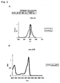
- the absorption spectrum of the protein was measured ( Figure 1B ). Thereafter, the molar absorption coefficient of the protein was calculated from the value of the peak (507 nm) of the spectrum.
- the fluorescent protein was diluted with the above buffer solution such that the absorption at 450 nm became 0.002. Its fluorescence spectrum by exciting at 450 nm and its excitation spectrum by a fluorescence at 550 nm were measured ( Figure 1A ).
- EGFP (CLONTECH) was diluted such that the absorption at 450 nm became 0.002, and its fluorescence spectrum was measured.
- the fluorescent protein was diluted with each of the following buffer solutions to the same concentration. The value of the absorption at 570 nm was determined, and thus the pH sensitivity thereof was measured ( Figure C1).
- the pH levels of the buffer solutions are as follows. pH 4 and 5: Acetate buffer pH 6 and 11: Phosphate buffer pH 7 and 8: HEPES buffer pH 9 and 10: Glycine buffer
- This fluorescent protein has such characteristics that when pH 5 is compared with pH 6 to 10, the absorption peak is shifted to a short wavelength side (from 507 nm to 493 nm), and the fluorescence peak is also shifted to a short wavelength side (from 517 nm to 508 nm).
- the measurement results are shown in Figures 2A and 2B .
- Example 2 Isolation of novel fluorescent protein gene (COR) from stony coral, and analysis of fluorescence properties thereof
- a fluorescent protein gene was isolated from coral. Montipora sp. was used as a material. A frozen Montipora sp. was crushed in a mortar, and 7.5 ml of "TRIzol” (GIBCO BRL) was then added to 2 g (wet weight) of the crushed Montipora sp. Thereafter, the obtained mixture was homogenized and then centrifuged at 1,500 x g for 10 minutes. Thereafter, 1.5 ml of chloroform was added to the obtained supernatant, and the mixture was then stirred for 15 seconds. Thereafter, the mixture was left at rest for 3 minutes. The resultant was then centrifuged at 7,500 x g for 15 minutes.
- RNA dissolved in the DEPC water was 100 times diluted, and the values of O.D.260 and O.D.280 were then measured, so as to determine RNA concentration. As a result, 22 ⁇ g of total RNA was obtained.
- cDNA (33 ⁇ l) was synthesized from 4 ⁇ g of the total RNA, using a kit for synthesizing first strand cDNA "Ready To Go” (Amersham Pharmacia).
- 3 ⁇ l of the synthesized first strand cDNA (33 ⁇ l) was used as a template to carry out PCR.
- Primers were produced by making comparison among the amino acid sequences of known fluorescent proteins, extracting similar portions, and converting them to nucleotide sequences. Primers used:
- PCR was carried out again under the same above conditions. A 350-bp fragment was cut out via agarose gel electrophoresis, and it was then purified.
- the purified DNA fragment was ligated to a pT7-blue vector (Novagen). Escherichia coli (TG1) was transformed therewith, and it was then subjected to blue white selection. Plasmid DNA was purified from white colonies of Escherichia coli, and the nucleotide sequence of the inserted DNA fragment was determined using a DNA sequencer. Thereafter, the obtained nucleotide sequence was compared with the nucleotide sequences of other fluorescent protein genes, so as to determine whether the nucleotide sequence of the DNA fragment was derived from a fluorescent protein. With regard to those that were determined to be a part of the fluorescent protein genes, the full-length genes were cloned by the 5'-RACE method and the 3'-RACE method.
- the 5'-RACE method was applied using 5'-RACE System for Rapid Amplification of cDNA Ends, Version 2.0 (GIBCO BRL). 5 ⁇ g of the total RNA prepared in (1) above was used as a template.
- I represents inosine
- the amplified 350-bp band was cut out of the gel via agarose gel electrophoresis and then purified.
- the purified DNA fragment was ligated to a pT7-blue vector (Novagen).
- Escherichia coli (TG1) was transformed therewith, and it was then subjected to blue white selection.
- Plasmid DNA was purified from white colonies of Escherichia coli, and the nucleotide sequence of the inserted DNA fragment was determined using a DNA sequencer.
- the nucleotide sequence on the 3'-side of the DNA fragment obtained by the degenerated PCR was obtained by PCR using a primer prepared based on the information obtained by determination of the nucleotide sequence in (4) above and an oligo dT primer. 3 ⁇ l of the first strand cDNA prepared in (2) above was used as a template.
- the prepared primer was 5'-ATGGCTCTTTCAAAGCACGGTC-3' (primer 7) (SEQ ID NO: 31).
- PCR reaction solution Template (first strand cDNA) 3 ⁇ l X 10 taq buffer 5 ⁇ l 2.5 mM dNTPs 4 ⁇ l 20 ⁇ M primer 7 1 ⁇ l 10 ⁇ M oligo dT primer 1 ⁇ l Milli-Q 35 ⁇ l Taq polymerase (5 U/ ⁇ l) 1 ⁇ l PCR reaction conditions:
- the amplified band with a length of approximately 1,000 bp was cut out of the gel via agarose gel electrophoresis and then purified.
- the purified DNA fragment was ligated to a pT7-blue vector (Novagen).
- Escherichia coli (TG1) was transformed therewith, and it was then subjected to blue white selection.
- Plasmid DNA was purified from white colonies of Escherichia coli, and the nucleotide sequence of the inserted DNA fragment was determined using a DNA sequencer.
- the obtained full-length nucleotide sequence is shown in SEQ ID NO: 10 of the sequence listing, and the obtained full-length amino acid sequence is shown in SEQ ID NO: 9 thereof.
- This clone was named COR.
- a primer was produced with a portion corresponding to the N-terminus of the protein.
- An oligo dT primer was used as a primer corresponding to the C-terminal side thereof.
- PCR was carried out employing the first strand cDNA prepared in (2) above as a template.
- the amplified band with a length of approximately 1,000 bp was cut out of the gel via agarose gel electrophoresis and then purified.
- the purified DNA fragment was subcloned into the BamHI-EcoRI site of a pRSET vector (Invitrogen), and it was then allowed to express in Escherichia coli (JM109-DE3).
- the expressed protein was constructed such that His-tag was attached to the N-terminus thereof, and thus it was purified with Ni-Agarose gel (QIAGEN). Purification was carried out in accordance with the attached protocols. Subsequently, the properties of the purified protein were analyzed.
- the absorption spectrum of the protein was measured ( Figure 3B ). Thereafter, the molar absorption coefficient of the protein was calculated from the value of the peak (557 nm) of the spectrum.
- the fluorescent protein was diluted with the above buffer solution such that the absorption at 520 nm became 0.002. Its fluorescence spectrum by exciting at 520 nm and its excitation spectrum by a fluorescence at 600 nm were measured ( Figure 3A ). Likewise, DsRed2 (CLONTECH) was diluted such that the absorption at 520 nm became 0.002, and its fluorescence spectrum was measured.
- the fluorescent protein was diluted with each of the following buffer solutions to the same concentration. The value of the absorption at 557 nm was determined, and thus the pH sensitivity thereof was measured.
- the pH levels of the buffer solutions are as follows. pH 4 and 5: Acetate buffer pH 6 and 11: Phosphate buffer pH 7 and 8: HEPES buffer pH 9 and 10: Glycine buffer
- the present invention provides novel fluorescent proteins derived from Montipora sp..
- the fluorescent proteins of the present invention are novel proteins having a primary structure different from that of the conventional fluorescent proteins.
- the fluorescent proteins of the present invention have certain fluorescence properties, and thus they are useful for molecular biological analysis. That is to say, the use of fluorescent proteins of the present invention enables fluorescent labeling in mammalian cells without exhibiting toxicity.
- a mutation is introduced into the fluorescent proteins of the present invention, so as to generate new fluorescence properties.
- the fluorescent protein (COR) of the present invention when compared with the wide excitation spectrum of the conventional RFP (DsRed; CLONTECH), the fluorescent protein (COR) of the present invention has a sharper spectrum. Furthermore, a mutation is introduced into the fluorescent protein of the present invention, so as to diversify the fluorescence properties in the red region.
Landscapes
- Health & Medical Sciences (AREA)
- Chemical & Material Sciences (AREA)
- Organic Chemistry (AREA)
- Life Sciences & Earth Sciences (AREA)
- Biophysics (AREA)
- Medicinal Chemistry (AREA)
- Zoology (AREA)
- Biochemistry (AREA)
- Toxicology (AREA)
- General Health & Medical Sciences (AREA)
- Genetics & Genomics (AREA)
- Gastroenterology & Hepatology (AREA)
- Molecular Biology (AREA)
- Proteomics, Peptides & Aminoacids (AREA)
- Tropical Medicine & Parasitology (AREA)
- Peptides Or Proteins (AREA)
- Micro-Organisms Or Cultivation Processes Thereof (AREA)
- Measuring Or Testing Involving Enzymes Or Micro-Organisms (AREA)
Abstract
Description
- The present invention relates to a novel fluorescent protein. More specifically, the present invention relates to a novel fluorescent protein derived from Montipora sp. and the use thereof.
- Green fluorescent protein (GFP) derived from Aequorea victoria, a jellyfish, has many purposes in biological systems. Recently, various GFP mutants have been produced based on the random mutagenesis and semi-rational mutagenesis, wherein a color is changed, a folding property is improved, luminance is enhanced, or pH sensitivity is modified. Fluorescent proteins such as GFP are fused with other proteins by gene recombinant technique, and monitoring of the expression and transportation of the fusion proteins is carried out.
- One of the most commonly used types of GFP mutant is Yellow fluorescent protein (YFP). Among Aequorea-derived GFP mutants, YFP exhibits the fluorescence with the longest wavelength. The values ε and Φ of the majority of YEPs are 60,000 to 100,000 M-1cm-1 and 0.6 to 0.8, respectively (Tsien, R. Y (1998). Ann. Rev. Biochem. 67, 509-544). These values are comparable to those of the general fluorescent group (fluorescein, rhodamine, etc.). Accordingly, improvement of the absolute luminance of YFP is nearly approaching its limit.
- In addition, cyan fluorescent protein (CFP) is another example of the GFP mutant. Of this type of protein, ECFP (enhanced cyan fluorescent protein) has been known. Moreover, red fluorescent protein (RFP) has been isolated from sea anemone (Discoma sp.). Of this type of protein, DasRed has been known. Thus, 4 types of fluorescent proteins, that are, green fluorescent protein, yellow fluorescent protein, cyan fluorescent protein, and red fluorescent protein, have successively been developed. The range of the spectrum has significantly been expanded.
- In addition, some cnidarians emit a fluorescence. The cloning of fluorescent protein genes derived from such cnidarians has been attempted. However, in order to expand the repertoire of fluorescent and biochemical properties, it is necessary to clone more genes.
- It is an object of the present invention to provide a novel fluorescent protein derived from Montipora sp..
- It is another object of the present invention to provide a fluorescent protein having a sharper spectrum than the wide excitation spectrum of the conventional RFP (DsRed; CLONTECH).
- The present inventors have conducted intensive studies directed towards achieving the aforementioned objects. The inventors have designed preferred primers based on information regarding the amino acid sequences of known fluorescent proteins. Thereafter, using the aforementioned primers, the inventors have succeeded in amplifying genes encoding novel fluorescent proteins obtained from cDNA libraries derived from Montipora sp. and in cloning them. Moreover, the present inventors have analyzed the fluorescence properties and pH sensitivity of the obtained fluorescent proteins derived from Montipora sp. The present invention has been completed based on these findings.
- Thus, the present invention provides the the following (1) to (35):
- (1) A fluorescent protein derived from Montipora sp., which has the following properties:
- [1] the excitation maximum wavelength is 507 nm;
- [2] the fluorescence maximum wavelength is 517 nm;
- [3] the molar absorption coefficient at 507 nm is 104,050;
- [4] the quantum yield is 0.29; and
- [5] the pH sensitivity of light absorption property is pKa of approximately 5.5; wherein the fluorescent protein is having either the following amino acid sequence (a) or (b):
- (a) an amino acid sequence shown in SEQ ID NO: 1; or
- (b) an amino acid sequence, which comprises a deletion, substitution, and/or addition of one to 20 amino acids with respect to the amino acid sequence shown in SEQ ID NO: 1, and which has a fluorescence.
- (2) A fluorescent protein derived from Montipora sp., which has the following properties:
- [1] the excitation maximum wavelength is 557 nm;
- [2] the fluorescence maximum wavelength is 574 nm;
- [3] the molar absorption coefficient at 557 nm is 41,750;
- [4] the quantum yield is 0.41; and
- [5] the pH sensitivity of light absorption property is pKa < approximately 4.0 ; wherein the fluorescent protein having either the following amino acid sequence (a) or (b):
- (a) an amino acid sequence shown in SEQ ID NO: 9; or
- (b) an amino acid sequence, which comprises a deletion, substitution, and/or addition of one to 20 amino acids with respect to the amino acid sequence shown in SEQ ID NO: 9, and which has a fluorescence.
- (3) A fluorescent protein having either the following amino acid sequence (a) or (b):
- (a) an amino acid sequence shown in SEQ ID NO: 1; or
- (b) an amino acid sequence, which comprises a deletion, substitution, and/or addition of one to 20 amino acids with respect to the amino acid sequence shown in SEQ ID NO: 1, and which has a fluorescence.
- (4) A fluorescent protein having either the following amino acid sequence (a) or (b):
- (a) an amino acid sequence shown in SEQ ID NO: 9; or
- (b) an amino acid sequence, which comprises a deletion, substitution, and/or addition of one to 20 amino acids with respect to the amino acid sequence shown in SEQ ID NO: 9, and which has a fluorescence.
- (5) DNA encoding the protein according to any one of (1) to (9) above.
- (6) DNA of either the following (a) or (b):
- (a) DNA encoding an amino acid sequence shown in SEQ ID NO: 1; or
- (b) DNA encoding an amino acid sequence comprising a deletion, substitution, and/or addition of one to 20 amino acids with respect to the amino acid sequence shown in SEQ ID NO: 1, and which encodes a fluorescent protein.
- (7) DNA having the nucleotide sequence: shown in SEQ ID NO: 2.
- (8) DNA of either the following (a) or (b):
- (a) DNA encoding an amino acid sequence shown in SEQ ID NO: 9; or
- (b) DNA encoding an amino acid sequence comprising a deletion, substitution, and/or addition of one to 20 amino acids with respect to the amino acid sequence shown in SEQ ID NO: 9, and which encodes a fluorescent protein.
- (9) DNA having the nucleotide sequence shown in SEQ ID NO: 10.
- (10) A recombinant vector having the DNA according to any one of (5) to (9) above.
- (11) A transformant having the DNA according to any one of (5) to (9) above or the recombinant vector according to (10) above.
- (12) A fusion fluorescent protein, which consists of the fluorescent protein according to any one of (1) to (4) and another protein.
- (13) The fusion fluorescent protein according to (12) above, wherein another protein is a protein that localizes in a cell.
- (14) The fusion fluorescent protein according to (12) or (13) above, wherein another protein is a protein specific to a cell organella.
- (15) An in vitro method for analyzing the localization or dynamics of a protein in a cell, which is characterized in that the fusion fluorescent protein according to any one of (12) to (14) above is allowed to express in the cell.
- (16) A fluorescent reagent kit, which comprises: the fluorescent protein of any one of (1) to (4) above; the DNA of any one of (5) to (9); the recombinant vector of (10) above; the transformant of (11) above; or the fusion fluorescent protein of any of (12) to (14).
- (17) An absorbance reagent kit, which comprises: the recombinant vector of (10) above; the transformant of (11) above.
-
-
Figure 1 shows the fluorescence spectrum and excitation spectrum of the fluorescent protein (COG) of the present invention derived from Montipora sp. (Figure A), the absorption spectrum of the fluorescent protein (COG) (Figure B), and the pH sensitivity of the fluorescent protein (COG) (Figure C). In Figure C, the horizontal axis represents pH value, and the longitudinal axis represents absorbance. -
Figure 2 shows the fluorescence spectrum and excitation spectrum atpH 5 of the fluorescent protein (COG) of the present invention derived from Montipora sp. (Figure A), and the absorption spectrum thereof at pH 5 (Figure B). -
Figure 3 shows the fluorescence spectrum and excitation spectrum of the fluorescent protein (COR) of the present invention derived from Montipora sp. (Figure A), the absorption spectrum of the fluorescent protein (COR) (Figure B), and the pH sensitivity of the fluorescent protein (COR) (Figure C). In Figure C, the horizontal axis represents pH value, and the longitudinal axis represents absorbance. - The embodiments of the present invention will be described in detail below.
- The first fluorescent protein of the present invention is derived from Montipora sp., and it has the following properties:
- [1] the excitation maximum wavelength is 507 nm;
- [2] the fluorescence maximum wavelength is 517 nm;
- [3] the molar absorption coefficient at 507 nm is 104,050;
- [4] the quantum yield is 0.29; and
- [5] the pH sensitivity of light absorption property is pKa of approximately 5.5 ; wherein the first fluorescent protein of the present invention is a fluorescent protein having either the following amino acid sequence (a) or (b):
- (a) an amino acid sequence shown in SEQ ID NO: 1; or
- (b) an amino acid sequence, which comprises a deletion, substitution, and/or addition of one to 20 amino acids with respect to the amino acid sequence shown in SEQ ID NO: 1, and which has a fluorescence.
- Montipora sp. is one kind of coral, which belongs to Cnidaria, Anthozoa, Hexacorallia, Seleractinia, Artrocoeniidae. This coral often forms a massive or covered colony.
- As described in examples below, the first fluorescent protein of the present invention has an excitation maximum wavelength of 507 nm and a fluorescence maximum wavelength of 517 nm. In addition, its molar absorption coefficient at 507 nm is 104,050, and its quantum yield is 0.29. The molar absorption coefficient represents the amount of photon absorbed per mole of a fluorescent molecule. The quantum yield is a numerical value representing the degree of the absorbed photon that can be emitted as a fluorescence.
- The second fluorescent protein of the present invention is derived from Montipora sp., and it has the following properties:
- [1] the excitation maximum wavelength is 557 nm;
- [2] the fluorescence maximum wavelength is 574 nm;
- [3] the molar absorption coefficient at 557 nm is 41,750;
- [4] the quantum yield is 0.41; and
- [5] the pH sensitivity of light absorption property is pKa < approximately 4.0; wherein the fourth fluorescent protein of the present invention is a fluorescent protein having either the following amino acid sequence (a) or (b):
- (a) the amino acid sequence shown in SEQ ID NO: 9; or
- (b) an amino acid sequence, which comprises a deletion, substitution, and/or addition of one to 20 amino acids with respect to the amino acid sequence shown in SEQ ID NO: 9, and which has a fluorescence.
- As described in examples below, the second fluorescent protein of the present invention has an excitation maximum wavelength of 557 nm and a fluorescence maximum wavelength of 574 nm. In addition, its molar absorption coefficient at 557 nm is 41,750, and its quantum yield is 0.41. The molar absorption coefficient represents the amount of photon absorbed per mole of a fluorescent molecule. The quantum yield is a numerical value representing the degree of the absorbed photon that can be emitted as a fluorescence.
- The scope of "one to 20" in the phrase "an amino acid sequence comprising a deletion, substitution and/or addition of one to 20 amino acids" used herein is not particularly limited. For example, it means 1 to 10, more preferably 1 to 7, further preferably 1 to 5, and particularly preferably 1 to 3.
- The term "has a fluorescence" covers all of the cases where any fluorescence is given. Various properties such as fluorescence intensity, excitation wavelength, fluorescence wavelength or pH sensitivity, may be changed or may remain unchanged, as compared with the case of the protein having an amino acid sequence shown in SEQ ID NO: 1.
- The term "light-absorbing properties" is used in the present specification to mean properties capable of absorbing a light with a certain wavelength. For example, an absorption maximum wavelength may be 592 nm, or the value of such an absorption maximum wavelength may be shifted. The pH sensitivity of the light absorption property is preferably stable in the range between
pH - The method of obtaining the fluorescent protein of the present invention is not particularly limited. The proteins may be either a protein synthesized by chemosynthesis, or recombinant protein produced by a gene recombination technique.
- Where a recombinant protein is produced, it is necessary to obtain DNA encoding the protein. Appropriate primers are designed by using information regarding the amino acid sequence shown in SEQ ID NO: 1 or 11 of the sequence listing of the present specification and the nucleotide sequence shown in SEQ ID NO: 2, or 10 thereof. Using these primers, PCR is carried out by using cDNA library derived from Montipora sp. as a template, so that DNA encoding the fluorescent protein or the chromoprotein of the present invention can be obtained. Where a partial fragment of DNA encoding the fluorescent protein or the chromoprotei of the present invention are obtained by the above-described PCR, the produced DNA fragments are ligated to one another by a gene recombination technique, so that DNA encoding the desired fluorescent protein or the chromoprotein can be obtained. The fluorescent protein or the chromoprotein of the present invention can be produced by introducing this DNA into an appropriate expression system. Expression in an expression system will be described later in the present specification.
- The present invention provides genes encoding the fluorescent proteins of the present invention.
- A specific example of DNA encoding the fluorescent proteins of the present invention is either the following DNA (a) or (b):
- (a) DNA encoding an amino acid sequence shown in SEQ ID NO: 1 or 9; or
- (b) DNA, which encodes an amino acid sequence comprising a deletion, substitution, and/or addition of one to 20 amino acids with respect to the amino acid sequence shown in SEQ ID NO: 1 or 9, and which encodes a fluorescent protein.
- Another specific example of DNA encoding the florescent proteins of the present invention is the following DNA: DNA having the nucleotide sequence shown in SEQ ID NO: 2 or 10.
- The DNA of the present invention can be synthesized by, for example, the phosphoamidite method, or it can also be produced by polymerase chain reaction (PCR) using specific primers. The DNA of the present invention or its fragment is produced by the method described above in the specification.
- A method of introducing a desired mutation into a certain nucleic acid sequence is known to a person skilled in the art. For example, known techniques such as a site-directed mutagenesis, PCR using degenerated oligonucleotides, or the exposure of cells containing nucleic acid to mutagens or radioactive rays, are appropriately used, so as to construct DNA having a mutation: Such known techniques are described in, for example, Molecular Cloning: A Laboratory Manual, 2nd Ed., Cold Spring Harbor Laboratory, Cold Spring Harbor, NY, 1989; and Current Protocols in Molecular Biology, Supplements 1 to 38, John Wiley & Sons (1987-1997).
- The DNA of the present invention can be inserted into a suitable vector and used. The type of a vector used in the present invention is not particularly limited. For example, it may be either a vector that can autonomously replicate (e.g., a plasmid, etc.), or vector that is incorporated into the genomes of host cells when it is introduced into the host cells and is then replicated together with the chromosome into which it is incorporated.
- The vector used in the present invention is preferably an expression vector. In an expression vector, elements necessary for transcription (e.g., a promoter, etc.) are functionally ligated to the DNA of the present invention. The promoter is a DNA sequence which shows a transcriptional activity in host cells, and it is appropriately selected depending on the type of host cells.
- Examples of a promoter which can operate in bacterial cells may include a Bacillus stearothermophilus maltogenic amylase gene promoter, a Bacillus licheniformis alpha-amylase gene promoter, a Bacillus amyloliquefaciens BAN amylase gene promoter, a Bacillus subtilis alkaline protease gene promoter, a Bacillus pumilus xylosidase gene promoter, PR and PL promoters of phage rhamda, and lac, trp and tac promoters of Escherichia coli.
- Examples of a promoter which can operate in mammalian cells may include an SV40 promoter, an MT-1 (metallothionein gene) promoter, and an adenovirus-2 major late promoter. Examples of a promoter which can operate in insect cells may include a polyhedrin promoter, a P10 promoter, an Autographa californica polyhedrosis basic protein promoter, a baculovirus immediate-
early gene 1 promoter, and a baculovirus 39K delayed-early gene promoter. Examples of a promoter which can be operate in yeast host cells may include promoters derived from yeast glycolytic genes, an alcohol dehydrogenase gene promoter, a TPI1 promoter, and an ADH2-4c promoter. - Examples of a promoter which can operate in filamentous cells may include an ADH3 promoter and a tpiA promoter.
- In addition, an appropriate terminator such as a human growth hormone terminator, or a TPI1 terminator or ADH3 terminator for fungal cells, may be functionally bound to the DNA of the present invention, as necessary. The recombinant vector of the present invention may further have elements such as a polyadenylation signal (e.g., one derived from SV40 or the adenovirus 5E1b region), a transcription enhancer sequence (e.g., an SV40 enhancer), or a translation enhancer sequence (e.g., one encoding the adenovirus VA RNA).
- The recombinant vector of the present invention may further comprise a DNA sequence which enables the replication of the recombinant vector in host cells. SV40 replication origin is an example of such a sequence (when the host cells are mammalian cells).
- The recombinant vector of the present invention may further comprise a selective marker. Examples of such a selective marker may include genes, complements of which are absent from host cells, such as a dihydrofolate reductase (DHFR) gene or a Shizosaccharomyces pombe TPI gene, and drug resistant genes such as ampicillin, kanamycin, tetracycline, chloramphenicol, neomycin or hygromycin-resistant genes.
- A method for ligating the DNA of the present invention, a promoter and, as desired, a terminator and/or a secretory signal sequence to one another and inserting these items into a suitable vector is known to a person skilled in the art.
- A transformant can be produced by introducing the DNA or recombinant vector of the present invention into a suitable host.
- Any cell can be used as a host cell into which the DNA or recombinant vector of the present invention is introduced, as long as the DNA construct of the present invention can be expressed therein. Examples of such a cell may include bacteria, yeasts, fungal cells, and higher eukaryotic cells.
- Examples of bacteria may include Gram-positive bacteria such as Bacillus or Streptomyces, and Gram-negative bacteria such as Escherichia coli. These bacteria may be transformed by the protoplast method or other known methods, using competent cells.
- Examples of mammalian cells may include HEK 293 cells, HeLa cells, COS cells, BHK cells, CHL cells, and CHO cells. A method of transforming mammalian cells and expressing the introduced DNA sequence in the cells is also known. Examples of such a method may include the electroporation, the calcium phosphate method, and the lipofection method.
- Examples of yeast cells may include those belonging to Saccharomyces or Shizosaccharomyces. Examples of such cells may include Saccharomyces cerevisiae and Saccharomyces kluyveri. Examples of a method of introducing a recombinant vector into yeast host cells may include the electroporation, the spheroplast method, and the lithium acetate method.
- Examples of other fungal cells may include those belonging to Filamentous fungi such as Aspergillus, Neurospora, Fusarium or Trichoderma. Where Filamentous fungi are used as host cells, transformation can be carried out by incorporating DNA constructs into host chromosomes, so as to obtain recombinant host cells. Incorporation of DNA constructs into the host chromosomes is carried out by known methods, and such known methods may include homologous recombination and heterologous recombination.
- Where insect cells are used as host cells, both a vector into which a recombinant gene is introduced and a baculovirus are co-introduced into insect cells, and a recombinant virus is obtained in the culture supernatant of the insect cells. Thereafter, insect cells are infected with the recombinant virus, so as to allow the cells to express proteins (described in, for example, Baculovirus Expression Vectors, A Laboratory Manual; and Current Protocols in Molecular Biology, Bio/Technology, 6, 47 (1988)).
- The Autographa californica nuclear polyhedrosis virus, which is a virus infecting to insects belonging to Barathra brassicae, can be used as baculovirus.
- Examples of insect cells used herein may include Sf9 and Sf21, which are Spodoptera frugiperda ovarian cells [Baculovirus Expression Vectors, A Laboratory Manual, W. H. Freeman & Company, New York, (1992)], and HiFive (manufactured by Invitrogen), which are Trichoplusia ni ovarian cells.
- Examples of the method of co-introducing both a vector into which a recombinant gene has been introduced and the above baculovirus into insect cells to prepare a recombinant virus may include the calcium phosphate method and the lipofection method.
- The above transformant is cultured in an appropriate nutritive medium under conditions enabling the introduced DNA construct to be expressed. In order to isolate and purify the protein of the present invention from the culture product of the transformant, common methods of isolating and purifying proteins may be used.
- For example, where the protein of the present invention is expressed in a state dissolved in cells, after completion of the culture, cells are recovered by centrifugal separation, and the recovered cells are suspended in a water type buffer. Thereafter, the cells are disintegrated using an ultrasonic disintegrator or the like, so as to obtain a cell-free extract. A supernatant is obtained by centrifuging the cell-free extract, and then, a purified sample can be obtained from the supernatant by applying, singly or in combination, the following ordinary protein isolation and purification methods: the solvent extraction, the salting-out method using ammonium sulfate or the like, the desalting method, the precipitation method using an organic solvent, the anion exchange chromatography using resins such as diethylaminoethyl (DEAE) sepharose, the cation exchange chromatography using resins such as S-Sepharose FF (manufactured by Pharmacia), the hydrophobic chromatography using resins such as butyl sepharose or phenyl sepharose, the gel filtration method using a molecular sieve, the affinity chromatography, the chromatofocusing method, and the electrophoresis such as isoelectric focusing.
- The fluorescent protein of the present invention can be fused with another protein, so as to construct a fusion fluorescent protein.
- A method of obtaining the fusion fluorescent protein of the present invention is not particularly limited. It may be either a protein synthesized by chemosynthesis, or recombinant protein produced by a gene recombination technique.
- Where a recombinant protein is produced, it is necessary to obtain DNA encoding the protein. Appropriate primers are designed using the information regarding the amino acid sequence shown in SEQ ID NO: 1 or 9 of the sequence listing of the present specification and the nucleotide sequence shown in SEQ ID NO: 2 or 10 thereof. Using these primers, PCR is carried out using a DNA fragment containing the gene of the fluorescent protein of the present invention as a template, so as to produce DNA fragments necessary for construction of the DNA encoding the fluorescent protein of the present invention. Moreover, DNA fragment encoding a protein to be fused is also obtained in the same above manner.
- Subsequently, the thus obtained DNA fragments are ligated to one another by a gene recombination technique, so that DNA encoding the desired fusion fluorescent protein can be obtained. This DNA is then introduced into an appropriate expression system, so that the fusion fluorescent protein of the present invention can be produced.
- The fluorescent protein of the present invention has an extremely high utility value as a marker. This is to say, the fluorescent protein of the present invention is purified as a fusion protein with an amino acid sequence to be tested, and the fusion protein is introduced into cells by methods such as the microinjection. By observing the distribution of the fusion protein over time, targeting activity of the amino acid sequence to be tested can be detected in the cells.
- The type of another protein (an amino acid sequence to be tested) with which the fluorescent protein of the present invention is fused is not particularly limited. Preferred examples may include proteins localizing in cells, proteins specific for intracellular organelles, and targeting signals (e.g., a nuclear transport signal, a mitochondrial presequence, etc.). In addition, the fluorescent protein of the present invention can be expressed in cells and used, as well as being introduced into cells by the microinjection or the like. In this case, a vector into which the DNA encoding the fluorescent protein of the present invention is inserted in such a way that it can be expressed, is introduced into host cells.
- Moreover, the fluorescent protein of the present invention can also be used as a reporter protein to determine promoter activity. This is to say, a vector is constructed such that DNA encoding the fluorescent protein of the present invention is located downstream of a promoter to be tested, and the vector is then introduced into host cells. By detecting the fluorescence of the fluorescent protein of the present invention which is emitted from the cells, the activity of the promoter to be tested can be determined. The type of a promoter to be tested is not particularly limited, as long as it operates in host cells.
- A vector used to detect the targeting activity of the above amino acid sequence to be tested or to determine promoter activity is not particularly limited. Examples of a vector preferably used for animal cells may include pNEO (P. Southern, and P. Berg (1982) J. Mol. Appl. Genet. 1: 327), pCAGGS (H. Niwa, K. Yamamura, and J. Miyazaki, Gene 108, 193-200 (1991)), pRc/CMV (manufactured by Invitrogen), and pCDM8 (manufactured by Invitrogen). Examples of a vector preferably used for yeasts may include pRS303, pRS304, pRS305, pRS306, pRS313, pRS314, pRS315, pRS316 (R. S. Sikorski and P. Hieter (1989) Genetics 122: 19-27), pRS423, pRS424, pRS425, pRS426 (T. W. Christianson, R. S. Sikorski, M. Dante, J. H. Shero, and P. Hieter (1992) Gene 110: 119-122).
- In addition, the type of cells used herein is also not particularly limited. Various types of animal cells such as L cells, BalbC-3T3 cells, NIH3T3 cells, CHO (Chinese hamster ovary) cells, HeLa cells or NRK (normal rat kidney) cells, yeast cells such as Saccharomyces cerevisiae, Escherichia coli cells, or the like can be used. Vector can be introduced into host cells by common methods such as the calcium phosphate method or the electroporation.
- The above obtained fusion fluorescent protein of the present invention wherein the fluorescent protein of the present invention is fused with another protein (referred to as a protein X) is allowed to be expressed in cells. By monitoring a fluorescence emitted, it becomes possible to analyze the localization or dynamics of the protein X in cells. That is, cells transformed or transfected with DNA encoding the fusion fluorescent protein of the present invention are observed with a fluorescence microscope, so that the localization and dynamics of the protein X in the cells can be visualized and thus analyzed.
- For example,-by using a protein specific for an intracellular organella as a protein X, the distribution and movement of a nucleus, a mitochondria, an endoplasmic reticulum, a Golgi body, a secretory vesicle, a peroxisome, etc., can be observed.
- Moreover, for example, axis cylinders or dendrites of the nerve cells show an extremely complicated change in strikes in an individual who is under development. Accordingly, fluorescent labeling of these sites enables a dynamic analysis.
- The fluorescence of the fluorescent protein of the present invention can be detected with a viable cell. Such detection can be carried out using, for example, a fluorescence microscope (Axiophoto Filter Set 09 manufactured by Carl Zeiss) or an image analyzer (Digital Image Analyzer manufactured by ATTO).
- The type of a microscope can be appropriately selected depending on purposes. Where frequent observation such as pursuit of a change over time is carried out, an ordinary incident-light fluorescence microscope is preferable. Where observation is carried out while resolution is emphasized, for example, in the case of searching localization in cells specifically, a confocal laser scanning microscope is preferable. In terms of maintenance of the physiological state of cells and prevention from contamination, an inverted microscope is preferable as a microscope system. When an erecting microscope with a high-powered lens is used, a water immersion lens can be used.
- A filter set can be appropriately selected depending on the fluorescence wavelength of a fluorescent protein. In the case of the first fluorescent protein of the present invention, a filter having an excitation light between approximately 490 and 510 nm and a fluorescence between approximately 510 and 530 nm is preferably used.
- In the case of the second fluorescent protein of the present invention, a filter having an excitation light between approximately 550 and 565 nm and a fluorescence between approximately 570 and 580 nm is preferably used.
- When viable cells are observed over time using a fluorescence microscope, a high sensitive cooled CCD camera is used, since photography is carried out in a short time. In the case of the cooled CCD camera, CCD is cooled to decrease thermal noise, so that a weak fluorescence image can be clearly photographed by exposure in a short time.
- The present invention provides a kit for analyzing the localization of intracellular components and/or analyzing physiologically active substances, which is characterized in that it comprises at least one selected from the fluorescent protein, the fusion fluorescent protein, the DNA, the recombinant vector, or the transformant, which are described in the present specification. The kit of the present invention can be produced from commonly used materials that are known per se, by using common methods.
- Further, the present invention provides a light-absorbing reagent kit, which is characterized in that it comprises at least one selected from the chromoprotein, fusion protein, DNA, recombinant vector or transformant, which are described in the present specification. The kit of the present invention can be produced from commonly used materials that are known per se, by using common methods.
- Reagents such as the fluorescent protein, the chromoprotein or the DNA are dissolved in an appropriate solvent, so that the reagents can be prepared in a form suitable for conservation. Water, ethanol, various types of buffer solution, etc. can be used as such a solvent.
- The present invention will be further described in the following examples. However, the present invention is not limited by these examples.
- A fluorescent protein gene was isolated from coral. Montipora sp. was used as a material. A frozen Montipora sp. was crushed in a mortar, and 7.5 ml of "TRIzol" (GIBCO BRL) was then added to 2 g (wet weight) of the crushed Montipora sp. Thereafter, the obtained mixture was homogenized and then centrifuged at 1,500 x g for 10 minutes. Thereafter, 1.5 ml of chloroform was added to the obtained supernatant, and the mixture was then stirred for 15 seconds. Thereafter, the mixture was left at rest for 3 minutes. The resultant was then centrifuged at 7,500 x g for 15 minutes. Thereafter, 3.75 ml of isopropanol was added to the obtained supernatant, and the mixture was then stirred for 15 seconds, followed by leaving the mixture at rest for 10 minutes. Thereafter, the resultant was centrifuged at 17,000 x g for 10 minutes. The obtained supernatant was discarded, and 6 ml of 70% ethanol was added to the residue, followed by centrifugation at 17,000 x g for 10 minutes. The obtained supernatant was discarded, and the precipitate was then dissolved in 200 µl of DEPC water. Total RNA dissolved in the DEPC water was 100 times diluted, and the values of O.D.260 and O.D.280 were then measured, so as to determine RNA concentration. As a result, 22 µg of total RNA was obtained.
- cDNA (33 µl) was synthesized from 4 µg of the total RNA, using a kit for synthesizing first strand cDNA "Ready To Go" (Amersham Pharmacia).
- 3 µl of the synthesized first strand cDNA (33 µl) was used as a template to carry out PCR.
- Primers were produced by making comparison among the amino acid sequences of known fluorescent proteins, extracting similar portions, and converting them to nucleotide sequences.
Primers used: - 5'-GAAGGRTGYGTCAAYGGRCAY-3' (primer 1) (SEQ ID NO: 15)
- 5 '-ACVGGDCCATYDGVAAGAAARTT-3' (primer 2) (SEQ ID NO: 16)
- 94°C x 1 min (PAD)
- 94°C x 30 sec (denaturation)
- 52°C x 30 sec (annealing of primers to template)
- 72°C x 1 min (primer elongation)
- A cycle consisting of the aforementioned 3 steps was repeated 35 times.
72°C x 7 min (final elongation)
4°C (maintenance) - Using 1 µl of the amplified product obtained in the first PCR reaction as a template, PCR was carried out again under the same above conditions. A 350-bp fragment was cut out via agarose gel electrophoresis, and it was then purified.
- The purified DNA fragment was ligated to a pT7-blue vector (Novagen). Escherichia coli (TG1) was transformed therewith, and it was then subjected to blue white selection. Plasmid DNA was purified from white colonies of Escherichia coli, and the nucleotide sequence of the inserted DNA fragment was determined using a DNA sequencer. Thereafter, the obtained nucleotide sequence was compared with the nucleotide sequences of other fluorescent protein genes, so as to determine whether the nucleotide sequence of the DNA fragment was derived from a fluorescent protein. With regard to those that were determined to be a part of the fluorescent protein genes, the full-length genes were cloned by the 5'-RACE method and the 3'-RACE method.
- In order to determine the nucleotide sequence on the 5'-side of the DNA fragment obtained by the degenerated PCR, the 5'-RACE method was applied using 5'-RACE System for Rapid Amplification of cDNA Ends, Version 2.0 (GIBCO BRL). 5 µg of the total RNA prepared in (1) above was used as a template.
- The following primers were used in the first amplification of dC-tailed cDNA:
- 5'-GGCCACGCGTCGACTAGTACGGGIIGGGIIGGGIIG-3' (primer 3) (SEQ ID NO: 17); and
- 5'-CCATCTTCAAAGAGAAAAGACCTTT-3' (primer 4) (SEQ ID NO: 18)
- Herein, I represents inosine.
- The following primers were used in the second amplification:
- 5'-GGCCACGCGTCGACTAGTAC-3' (primer 5) (SEQ ID NO: 19); and
- 5'-CATGAGTTCTTGAAATAGTCAAC-3' (primer 6) (SEQ ID NO: 20). PCR reaction conditions were applied in accordance with protocols attached to the kit.
- The amplified 350-bp band was cut out of the gel via agarose gel electrophoresis and then purified. The purified DNA fragment was ligated to a pT7-blue vector (Novagen). Escherichia coli (TG1) was transformed therewith, and it was then subjected to blue white selection. Plasmid DNA was purified from white colonies of Escherichia coli, and the nucleotide sequence of the inserted DNA fragment was determined using a DNA sequencer.
- The nucleotide sequence on the 3'-side of the DNA fragment obtained by the degenerated PCR was obtained by PCR using a primer prepared based on the information obtained by determination of the nucleotide sequence in (4) above and an oligo dT primer. 3 µl of the first strand cDNA prepared in (2) above was used as a template. The prepared primer was 5'-ATGGCTCTTTCAAAGCGAGGTG-3' (primer 7) (SEQ ID NO: 21).
Composition of PCR reaction solution:Template (first strand cDNA) 3 µl X10 taq buffer 5 µl 2.5 mM dNTPs 4 µl 20 µM primer 71 µl 10 µM oligo dT primer 1 µl Milli-Q 35 µl Taq polymerase (5 U/µl) 1 µl - 94°C x 1 minute (PAD)
- 94°C x 30 seconds (denaturation)
- 52°C x 30 seconds (annealing of primers to template)
- 72°C x 1 minute (primer elongation)
- A cycle consisting of the aforementioned 3 steps was repeated 30 times. 72°C x 7 minutes (final elongation)
4°C (maintenance) - The amplified band with a length of approximately 1,000 bp was cut out of the gel via agarose gel electrophoresis and then purified. The purified DNA fragment was ligated to a pT7-blue vector (Novagen). Escherichia coli (TG1) was transformed therewith, and it was then subjected to blue white selection. Plasmid DNA was purified from white colonies of Escherichia coli, and the nucleotide sequence of the inserted DNA fragment was determined using a DNA sequencer.
- The obtained full-length nucleotide sequence is shown in SEQ ID NO: 2 of the sequence listing, and the obtained full-length amino acid sequence is shown in SEQ ID NO: 1 thereof. This clone was named COG.
- Based on the obtained full-length nucleotide sequence, a primer was produced with a portion corresponding to the N-terminus of the protein. An oligo dT primer was used as a primer corresponding to the C-terminal side thereof. Thereafter, using such primers, PCR was carried out employing the first strand cDNA prepared in (2) above as a template.
Primer used: - 5'-GGGGGATCCGACCATGGCTCTTTCAAAGCGAGGTG-3' (primer 8) (SEQ ID NO: 22)
- 94°C x 1 minute (PAD)
- 94°C x 30 seconds (denaturation)
- 52°C x 30 seconds (annealing of primers to template)
- 72°C x 1 minute (primer elongation)
- A cycle consisting of the aforementioned 3 steps was repeated 30 times. 72°C x 7 minutes (final elongation)
4°C (maintenance) - The amplified band with a length of approximately 1,000 bp was cut out of the gel via agarose gel electrophoresis and then purified. The purified DNA fragment was subcloned into the BamHI-EcoRI site of a pRSET vector (Invitrogen), and it was then allowed to express in Escherichia coli (JM109-DE3). The expressed protein was constructed such that His-tag was attached to the N-terminus thereof, and thus it was purified with Ni-Agarose gel (QIAGEN). Purification was carried out in accordance with the attached protocols. Subsequently, the properties of the purified protein were analyzed.
- Using a solution comprising 20 µM fluorescent protein (COG), 150 mM KCI, and 50 mM HEPES(pH 7.5), the absorption spectrum of the protein was measured (
Figure 1B ). Thereafter, the molar absorption coefficient of the protein was calculated from the value of the peak (507 nm) of the spectrum. The fluorescent protein was diluted with the above buffer solution such that the absorption at 450 nm became 0.002. Its fluorescence spectrum by exciting at 450 nm and its excitation spectrum by a fluorescence at 550 nm were measured (Figure 1A ). Likewise, EGFP (CLONTECH) was diluted such that the absorption at 450 nm became 0.002, and its fluorescence spectrum was measured. Setting the quantum yield of EGFP to 0.6, the quantum yield of the cloned fluorescent protein was obtained. The results are shown in Table 1.Table 1 Excitation maximum Fluorescence maximum Molar absorption coefficient Quantum yield pH sensitivity Number of amino acids COG 507 nm 517 nm 104,050 (507 nm) 0.29 pKa = 5.5 227 - The fluorescent protein was diluted with each of the following buffer solutions to the same concentration. The value of the absorption at 570 nm was determined, and thus the pH sensitivity thereof was measured (Figure C1). The pH levels of the buffer solutions are as follows.
pH 4 and 5: Acetate buffer
pH 6 and 11: Phosphate buffer
pH 7 and 8: HEPES buffer
pH 9 and 10: Glycine buffer - This fluorescent protein has such characteristics that when
pH 5 is compared withpH 6 to 10, the absorption peak is shifted to a short wavelength side (from 507 nm to 493 nm), and the fluorescence peak is also shifted to a short wavelength side (from 517 nm to 508 nm). The measurement results are shown inFigures 2A and 2B . - A fluorescent protein gene was isolated from coral. Montipora sp. was used as a material. A frozen Montipora sp. was crushed in a mortar, and 7.5 ml of "TRIzol" (GIBCO BRL) was then added to 2 g (wet weight) of the crushed Montipora sp. Thereafter, the obtained mixture was homogenized and then centrifuged at 1,500 x g for 10 minutes. Thereafter, 1.5 ml of chloroform was added to the obtained supernatant, and the mixture was then stirred for 15 seconds. Thereafter, the mixture was left at rest for 3 minutes. The resultant was then centrifuged at 7,500 x g for 15 minutes. Thereafter, 3.75 ml of isopropanol was added to the obtained supernatant, and the mixture was then stirred for 15 seconds, followed by leaving the mixture at rest for 10 minutes. Thereafter, the resultant was centrifuged at 17,000 x g for 10 minutes. The obtained supernatant was discarded, and 6 ml of 70% ethanol was added to the residue, followed by centrifugation at 17,000 x g for 10 minutes. The obtained supernatant was discarded, and the precipitate was then dissolved in 200 µl of DEPC water. Total RNA dissolved in the DEPC water was 100 times diluted, and the values of O.D.260 and O.D.280 were then measured, so as to determine RNA concentration. As a result, 22 µg of total RNA was obtained.
- cDNA (33 µl) was synthesized from 4 µg of the total RNA, using a kit for synthesizing first strand cDNA "Ready To Go" (Amersham Pharmacia).
- 3 µl of the synthesized first strand cDNA (33 µl) was used as a template to carry out PCR.
- Primers were produced by making comparison among the amino acid sequences of known fluorescent proteins, extracting similar portions, and converting them to nucleotide sequences.
Primers used: - 5'-GAAGGRTGYGTCAAYGGRCAY-3' (primer 1) (SEQ ID NO: 15)
- 5'-ACVGGDCCATYDGVAAGAAARTT-3' (primer 2) (SEQ ID NO: 16)
- 94°C x 1 min (PAD)
- 94°C x 30 sec (denaturation)
- 52°C x 30 sec (annealing of primers to template)
- 72°C x 1 min (primer elongation)
- A cycle consisting of the aforementioned 3 steps was repeated 35 times. 72°C x 7 min (final elongation)
4°C (maintenance) - Using 1 µl of the amplified product obtained in the first PCR reaction as a template, PCR was carried out again under the same above conditions. A 350-bp fragment was cut out via agarose gel electrophoresis, and it was then purified.
- The purified DNA fragment was ligated to a pT7-blue vector (Novagen). Escherichia coli (TG1) was transformed therewith, and it was then subjected to blue white selection. Plasmid DNA was purified from white colonies of Escherichia coli, and the nucleotide sequence of the inserted DNA fragment was determined using a DNA sequencer. Thereafter, the obtained nucleotide sequence was compared with the nucleotide sequences of other fluorescent protein genes, so as to determine whether the nucleotide sequence of the DNA fragment was derived from a fluorescent protein. With regard to those that were determined to be a part of the fluorescent protein genes, the full-length genes were cloned by the 5'-RACE method and the 3'-RACE method.
- In order to determine the nucleotide sequence on the 5'-side of the DNA fragment obtained by the degenerated PCR, the 5'-RACE method was applied using 5'-RACE System for Rapid Amplification of cDNA Ends, Version 2.0 (GIBCO BRL). 5 µg of the total RNA prepared in (1) above was used as a template.
- The following primers were used in the first amplification of dC-tailed cDNA:
- 5'-GGCCACGCGTCGACTAGTACGGGIIGGGIIGGGIIG-3' (primer 3) (SEQ ID NO: 17); and
- 5'-CCATCTTCAAAGAGAAAAGACCTTT-3' (primer 4) (SEQ ID NO: 18).
- Herein, I represents inosine.
- The following primers were used in the second amplification:
- 5'-GGCCACGCGTCGACTAGTAC-3' (primer 5) (SEQ ID NO: 19); and
- 5'-CATGAGTTCTTGAAATAGTCAAC-3' (primer 6) (SEQ ID NO: 20). PCR reaction conditions were applied in accordance with protocols attached to the kit.
- The amplified 350-bp band was cut out of the gel via agarose gel electrophoresis and then purified. The purified DNA fragment was ligated to a pT7-blue vector (Novagen). Escherichia coli (TG1) was transformed therewith, and it was then subjected to blue white selection. Plasmid DNA was purified from white colonies of Escherichia coli, and the nucleotide sequence of the inserted DNA fragment was determined using a DNA sequencer.
- The nucleotide sequence on the 3'-side of the DNA fragment obtained by the degenerated PCR was obtained by PCR using a primer prepared based on the information obtained by determination of the nucleotide sequence in (4) above and an oligo dT primer. 3 µl of the first strand cDNA prepared in (2) above was used as a template. The prepared primer was 5'-ATGGCTCTTTCAAAGCACGGTC-3' (primer 7) (SEQ ID NO: 31).
Composition of PCR reaction solution:Template (first strand cDNA) 3 µl X 10 taq buffer 5 µl 2.5 mM dNTPs 4 µl 20 µM primer 71 µl 10 µM oligo dT primer 1 µl Milli-Q 35 µl Taq polymerase (5 U/µl) 1 µl - 94°C x 1 minute (PAD)
- 94°C x 30 seconds (denaturation)
- 52°C x 30 seconds (annealing of primers to template)
- 72°C x 1 minute (primer elongation)
- A cycle consisting of the aforementioned 3 steps was repeated 30 times. 72°C x 7 minutes (final elongation)
4°C (maintenance) - The amplified band with a length of approximately 1,000 bp was cut out of the gel via agarose gel electrophoresis and then purified. The purified DNA fragment was ligated to a pT7-blue vector (Novagen). Escherichia coli (TG1) was transformed therewith, and it was then subjected to blue white selection. Plasmid DNA was purified from white colonies of Escherichia coli, and the nucleotide sequence of the inserted DNA fragment was determined using a DNA sequencer.
- The obtained full-length nucleotide sequence is shown in SEQ ID NO: 10 of the sequence listing, and the obtained full-length amino acid sequence is shown in SEQ ID NO: 9 thereof. This clone was named COR.
- Based on the obtained full-length nucleotide sequence, a primer was produced with a portion corresponding to the N-terminus of the protein. An oligo dT primer was used as a primer corresponding to the C-terminal side thereof. Thereafter, using such primers, PCR was carried out employing the first strand cDNA prepared in (2) above as a template.
Primer used: - 5'-GGGGGATCCGACCATGGCTCTTTCAAAGCACGGTC-3' (primer 8) (SEQ ID NO: 32)
- 94°C x 1 minute (PAD)
- 94°C x 30 seconds (denaturation)
- 52°C x 30 seconds (annealing of primers to template)
- 72°C x 1 minute (primer elongation)
- A cycle consisting of the aforementioned 3 steps was repeated 30 times. 72°C x 7 minutes (final elongation)
4°C (maintenance) - The amplified band with a length of approximately 1,000 bp was cut out of the gel via agarose gel electrophoresis and then purified. The purified DNA fragment was subcloned into the BamHI-EcoRI site of a pRSET vector (Invitrogen), and it was then allowed to express in Escherichia coli (JM109-DE3). The expressed protein was constructed such that His-tag was attached to the N-terminus thereof, and thus it was purified with Ni-Agarose gel (QIAGEN). Purification was carried out in accordance with the attached protocols. Subsequently, the properties of the purified protein were analyzed.
- Using a solution comprising 20 µM fluorescent protein (COR), 150 mM KCI, and 50 mM HEPES(pH 7.5), the absorption spectrum of the protein was measured (
Figure 3B ). Thereafter, the molar absorption coefficient of the protein was calculated from the value of the peak (557 nm) of the spectrum. The fluorescent protein was diluted with the above buffer solution such that the absorption at 520 nm became 0.002. Its fluorescence spectrum by exciting at 520 nm and its excitation spectrum by a fluorescence at 600 nm were measured (Figure 3A ). Likewise, DsRed2 (CLONTECH) was diluted such that the absorption at 520 nm became 0.002, and its fluorescence spectrum was measured. Setting the quantum yield of DsRed2 to 0.55, the quantum yield of the cloned fluorescent protein was obtained. The measurement results are shown in Table 4.Table 4 Excitation maximum Fluorescence maximum Molar absorption coefficient Quantum yield pH sensitivity Number of amino acids COR 557 nm 574 nm 41,750 (557 nm) 0.41 pKa < 4.0 232 - The fluorescent protein was diluted with each of the following buffer solutions to the same concentration. The value of the absorption at 557 nm was determined, and thus the pH sensitivity thereof was measured. The pH levels of the buffer solutions are as follows.
pH 4 and 5: Acetate buffer
pH 6 and 11: Phosphate buffer
pH 7 and 8: HEPES buffer
pH 9 and 10: Glycine buffer - The measurement results are shown in
Figure 3C . - The present invention provides novel fluorescent proteins derived from Montipora sp.. The fluorescent proteins of the present invention are novel proteins having a primary structure different from that of the conventional fluorescent proteins. The fluorescent proteins of the present invention have certain fluorescence properties, and thus they are useful for molecular biological analysis. That is to say, the use of fluorescent proteins of the present invention enables fluorescent labeling in mammalian cells without exhibiting toxicity. In addition, a mutation is introduced into the fluorescent proteins of the present invention, so as to generate new fluorescence properties.
- Moreover, when compared with the wide excitation spectrum of the conventional RFP (DsRed; CLONTECH), the fluorescent protein (COR) of the present invention has a sharper spectrum. Furthermore, a mutation is introduced into the fluorescent protein of the present invention, so as to diversify the fluorescence properties in the red region.
-
- <110> RIKEN
- <120> Fluorescent protein and color protein
- <130> A41347A
- <160> 44
- <210> 1
<211> 227
<212> PRT
<213> Montipora sp. - <400> 1


- <210> 2
<211> 684
<212> DNA
<213> Montipora sp. - <400> 2


- <210> 3
<211> 232
<212> PRT
<213> Acropora sp. - <400> 3


- <210> 4
<211> 699
<212> DNA
<213> Acropora sp. - <400> 4


- <210> 5
<211> 232
<212> PRT
<213> Acropora sp. - <400> 5


- <210> 6
<211> 699
<212> DNA
<213> Acropora sp. - <400> 6



- <210> 7
<211> 232
<212> PRT
<213> Acropora sp. - <400> 7


- <210> 8
<211> 699
<212> DNA
<213> Acropora sp. - <400> 8


- <210> 9
<211> 232
<212> PRT
<213> Montipora sp. - <400> 9


- <210> 10
<211> 699
<212> DNA
<213> Montipora sp. - <400> 10


- <210> 11
<211> 232
<212> PRT
<213> Actinia equina - <400> 11


- <210> 12
<211> 699
<212> DNA
<213> Actinia equina - <400> 12


- <210> 13
<211> 224
<212> PRT
<213> Lobophytum crassum - <400> 13


- <210> 14
<211> 675
<212> DNA
<213> Lobophytum crassum - <400> 14


- <210> 15
<211> 21
<212> DNA
<213> Artificial Sequence - <220>
<223> Description of Artificial Sequence: Synthetic DNA - <400> 15
gaaggrtgyg tcaayggrca y 21 - <210> 16
<211> 23
<212> DNA
<213> Artificial Sequence - <220>
<223> Description of Artificial Sequence: Synthetic DNA - <400> 16
acvggdccat ydgvaagaaa rtt 23 - <210> 17
<211> 36
<212> DNA
<213> Artificial Sequence - <220>
<223> Description of Artificial Sequence: Synthetic DNA - <400> 17
ggccacgcgt cgactagtac gggiigggii gggiig 36 - <210> 18
<211> 25
<212> DNA
<213> Artificial Sequence - <220>
<223> Description of Artificial Sequence: Synthetic DNA - <400> 18
ccatcttcaa agagaaaaga ccttt 25 - <210> 19
<211> 20
<212> DNA
<213> Artificial Sequence - <220>
<223> Description of Artificial Sequence: Synthetic DNA - <400> 19
ggccacgcgt cgactagtac 20 - <210> 20
<211> 23
<212> DNA
<213> Artificial Sequence - <220>
<223> Description of Artificial Sequence: Synthetic DNA - <400> 20
catgagttct tgaaatagtc aac 23 - <210> 21
<211> 22
<212> DNA
<213> Artificial Sequence - <220>
<223> Description of Artificial Sequence: Synthetic DNA - <400> 21
atggctcttt caaagcgagg tg 22 - <210> 22
<211> 35
<212> DNA
<213> Artificial Sequence - <220>
<223> Description of Artificial Sequence: Synthetic DNA - <400> 22
gggggatccg accatggctc tttcaaagcg aggtg 35 - <210> 23
<211> 26
<212> DNA
<213> Artificial Sequence - <220>
<223> Description of Artificial Sequence: Synthetic DNA - <400> 23
tagaaatgac ctttcatatg acattc 26 - <210> 24
<211> 23
<212> DNA
<213> Artificial Sequence - <220>
<223> Description of Artificial Sequence: Synthetic DNA - <400> 24
tctgtttcca tattgaaagg ctg 23 - <210> 25
<211> 32
<212> DNA
<213> Artificial Sequence - <220>
<223> Description of Artificial Sequence: Synthetic DNA - <400> 25
atggtgtctt attcaaagca aggcatcgca ca 32 - <210> 26
<211> 44
<212> DNA
<213> Artificial Sequence - <220>
<223> Description of Artificial Sequence: Synthetic DNA - <400> 26
cgggatccga ccatggtgtc ttattcaaag caaggcatcg caca 44 - <210> 27
<211> 26
<212> DNA
<213> Artificial Sequence - <220>
<223> Description of Artificial Sequence: Synthetic DNA - <400> 27
tagaaatgac ctttcatatg acattc 26 - <210> 28
<211> 23
<212> DNA
<213> Artificial Sequence - <220>
<223> Description of Artificial Sequence: Synthetic DNA - <400> 28
tctgtttcca tattgaaagg ctg 23 - <210> 29
<211> 32
<212> DNA
<213> Artificial Sequence - <220>
<223> Description of Artificial Sequence: Synthetic DNA - <400> 29
atggtgtctt attcaaagca aggcatcgca ca 32 - <210> 30
<211> 44
<212> DNA
<213> Artificial Sequence - <220>
<223> Description of Artificial Sequence: Synthetic DNA - <400> 30
cgggatccga ccatggtgtc ttattcaaag caaggcatcg caca 44 - <210> 31
<211> 22
<212> DNA
<213> Artificial Sequence - <220>
<223> Description of Artificial Sequence: Synthetic DNA - <400> 31
atggctcttt caaagcacgg tc 22 - <210> 32
<211> 35
<212> DNA
<213> Artificial Sequence - <220>
<223> Description of Artificial Sequence: Synthetic DNA - <400> 32
gggggatccg accatggctc tttcaaagca cggtc 35 - <210> 33
<211> 23
<212> DNA
<213> Artificial Sequence - <220>
<223> Description of Artificial Sequence: Synthetic DNA - <400> 33
ggiwsbgtia ayggvcayda ntt 23 - <210> 34
<211> 35
<212> DNA
<213> Artificial Sequence - <220>
<223> Description of Artificial Sequence: Synthetic DNA - <400> 34
gtcitcttyt gcaciacigg iccatydgva ggaaa 35 - <210> 35
<211> 20
<212> DNA
<213> Artificial Sequence - <220>
<223> Description of Artificial Sequence: Synthetic DNA - <400> 35
ccttgaaaat aaagctatct 20 - <210> 36
<211> 20
<212> DNA
<213> Artificial Sequence - <220>
<223> Description of Artificial Sequence: Synthetic DNA - <400> 36
ccctgtatgc ttgtgtcctg 20 - <210> 37
<211> 36
<212> DNA
<213> Artificial Sequence - <220>
<223> Description of Artificial Sequence: Synthetic DNA - <400> 37
cccggatccg accatggtgt cttcattggt taagaa 36 - <210> 38
<211> 21
<212> DNA
<213> Artificial Sequence - <220>
<223> Description of Artificial Sequence: Synthetic DNA - <400> 38
grraggiwsb gthaayggvc a 21 - <210> 39
<211> 27
<212> DNA
<213> Artificial Sequence - <220>
<223> Description of Artificial Sequence: Synthetic DNA - <400> 39
aactggaaga attcgcggcc gcaggaa 27 - <210> 40
<211> 35
<212> DNA
<213> Artificial Sequence - <220>
<223> Description of Artificial Sequence: Synthetic DNA - <400> 40
gtcitcttyt gcaciacigg iccatydgva ggaaa 35 - <210> 41
<211> 30
<212> DNA
<213> Artificial Sequence - <220>
<223> Description of Artificial Sequence: Synthetic DNA <400> 41
ttgtcaagat atcgaaagcg aacggcagag 30 - <210> 42
<211> 20
<212> DNA
<213> Artificial Sequence - <220>
<223> Description of Artificial Sequence: Synthetic DNA - <400> 42
ggccacgcgt cgactagtac 20 - <210> 43
<211> 20
<212> DNA
<213> Artificial Sequence - <220>
<223> Description of Artificial Sequence: Synthetic DNA - <400> 43
cttctcacgt tgcaaatggc 20
<210> 44
<211> 44
<212> DNA
<213> Artificial Sequence - <220>
<223> Description of Artificial Sequence: Synthetic DNA - <400> 44
cccggatccg atgagtgtga ttacawcaga aatgaagatg gagc 44
Composition of PCR reaction solution:
| Template (first strand cDNA) | 3 |
| X | |
| 10 | 5 µl |
| 2.5 mM | 4 µl |
| 100 | 1 µl |
| 100 | 1 µl |
| Milli-Q | 35 µl |
| taq polymerase (5 U/µl) | 1 µl |
| Template (first strand cDNA) | 3 µl |
| | 5 µl |
| 2.5 mM | 4 µl |
| 100 | 1 µl |
| 100 µM | 1 µl |
| Milli-Q | 35 µl |
| Pyrobest polymerase (5 U/µl) | 1 µl |
Composition of PCR reaction solution:
| Template (first strand cDNA) | 3 |
| X | |
| 10 | 5 µl |
| 2.5 mM | 4 µl |
| 100 | 1 µl |
| 100 | 1 µl |
| Milli-Q | 35 µl |
| taq polymerase (5 U/µl) | 1 µl |
| Template (first strand cDNA) | 3 µl |
| | 5 µl |
| 2.5 mM | 4 µl |
| 100 | 1 µl |
| 100 µM | 1 µl |
| Milli-Q | 35 µl |
| Pyrobest polymerase (5 U/µl) | 1 µl |
Claims (17)
- A fluorescent protein derived from Montipora sp., which has the following properties:[1] the excitation maximum wavelength is 507 nm;[2] the fluorescence maximum wavelength is 517 nm;[3] the molar absorption coefficient at 507 nm is 104,050;[4] the quantum yield is 0.29; and[5] the pH sensitivity of light absorption property is pKa of approximately 5.5;
wherein the fluorescent protein is having either the following amino acid sequence (a) or (b):(a) an amino acid sequence shown in SEQ ID NO: 1; or(b) an amino acid sequence, which comprises a deletion, substitution, and/or addition of one to 20 amino acids with respect to the amino acid sequence shown in SEQ ID NO: 1, and which has a fluorescence. - A fluorescent protein derived from Montipora sp., which has the following properties:[1] the excitation maximum wavelength is 557 nm;[2] the fluorescence maximum wavelength is 574 nm;[3] the molar absorption coefficient at 557 nm is 41,750;[4] the quantum yield is 0.41; and[5] the pH sensitivity of light absorption property is pKa < approximately 4.0;
wherein the fluorescent protein is having either the following amino acid sequence (a) or (b):(a) an amino acid sequence shown in SEQ ID NO: 9; or(b) an amino acid sequence, which comprises a deletion, substitution, and/or addition of one to 20 amino acids with respect to the amino acid sequence shown in SEQ ID NO: 9, and which has a fluorescence. - A fluorescent protein having either the following amino acid sequence (a) or (b):(a) an amino acid sequence shown in SEQ ID NO: 1; or(b) an amino acid sequence, which comprises a deletion, substitution, and/or addition of one to 20 amino acids with respect to the amino acid sequence shown in SEQ ID NO: 1, and which has a fluorescence.
- A fluorescent protein having either the following amino acid sequence (a) or (b):(a) an amino acid sequence shown in SEQ ID NO: 9; or(b) an amino acid sequence, which comprises a deletion, substitution, and/or addition of one to 20 amino acids with respect to the amino acid sequence shown in SEQ ID NO: 9, and which has a fluorescence.
- DNA encoding the protein according to any one of claims 1 to 4.
- DNA of either the following (a) or (b):(a) DNA encoding an amino acid sequence shown in SEQ ID NO: 1; or(b) DNA encoding an amino acid sequence comprising a deletion, substitution, and/or addition of one to20 amino acids with respect to the amino acid sequence shown in SEQ ID NO: 1, and which encodes a fluorescent protein.
- DNA having the nucleotide sequence shown in SEQ ID NO: 2.
- DNA of either the following (a) or (b):(a) DNA encoding an amino acid sequence shown in SEQ ID NO: 9; or(b) DNA encoding an amino acid sequence comprising a deletion, substitution, and/or addition of one to 20 amino acids with respect to the amino acid sequence shown in SEQ ID NO: 9, and which encodes a fluorescent protein.
- DNA having the nucleotide sequence shown in SEQ ID NO: 10.
- A recombinant vector having the DNA according to any one of claims 5 to 9.
- A transformant having the DNA according to any one of claims 5 to 9 or the recombinant vector according to claim 10.
- A fusion fluorescent protein, which consists of the fluorescent protein according to any one of claims 1 to 4 and another protein.
- The fusion fluorescent protein according to claim 12, wherein another protein is a protein that localizes in a cell.
- The fusion fluorescent protein according to claim 12 or 13, wherein another protein is a protein specific to a cell organella.
- An in vitro method for analyzing the localization or dynamics of a protein in a cell, which is characterized in that the fusion fluorescent protein according to any one of claim 12 to 14 is allowed to express in the cell.
- A fluorescent reagent kit, which comprises: the fluorescent protein of any one of claims 1 to 4; the DNA of any one of claims 5 to 9; the recombinant vector of claim 10; the transformant of claim 11; or the fusion fluorescent protein of any of claims 12 to 14.
- An absorbance reagent kit, which comprises the recombinant vector of claim 10; or the transformant of claim 11.
Priority Applications (1)
| Application Number | Priority Date | Filing Date | Title |
|---|---|---|---|
| EP09163975A EP2098535B1 (en) | 2003-06-16 | 2004-06-16 | Fluorescent protein and chromoprotein |
Applications Claiming Priority (7)
| Application Number | Priority Date | Filing Date | Title |
|---|---|---|---|
| JP2003170327 | 2003-06-16 | ||
| JP2003170325 | 2003-06-16 | ||
| JP2003170326 | 2003-06-16 | ||
| JP2003170328 | 2003-06-16 | ||
| JP2003170324 | 2003-06-16 | ||
| JP2003170329 | 2003-06-16 | ||
| PCT/JP2004/008786 WO2004111235A1 (en) | 2003-06-16 | 2004-06-16 | Fluorescent protein and pigment protein |
Related Child Applications (1)
| Application Number | Title | Priority Date | Filing Date |
|---|---|---|---|
| EP09163975A Division EP2098535B1 (en) | 2003-06-16 | 2004-06-16 | Fluorescent protein and chromoprotein |
Publications (3)
| Publication Number | Publication Date |
|---|---|
| EP1642969A1 EP1642969A1 (en) | 2006-04-05 |
| EP1642969A4 EP1642969A4 (en) | 2007-02-21 |
| EP1642969B1 true EP1642969B1 (en) | 2009-09-16 |
Family
ID=33556769
Family Applications (2)
| Application Number | Title | Priority Date | Filing Date |
|---|---|---|---|
| EP04746255A Expired - Lifetime EP1642969B1 (en) | 2003-06-16 | 2004-06-16 | Fluorescent protein and pigment protein |
| EP09163975A Expired - Lifetime EP2098535B1 (en) | 2003-06-16 | 2004-06-16 | Fluorescent protein and chromoprotein |
Family Applications After (1)
| Application Number | Title | Priority Date | Filing Date |
|---|---|---|---|
| EP09163975A Expired - Lifetime EP2098535B1 (en) | 2003-06-16 | 2004-06-16 | Fluorescent protein and chromoprotein |
Country Status (7)
| Country | Link |
|---|---|
| US (2) | US7956172B2 (en) |
| EP (2) | EP1642969B1 (en) |
| JP (1) | JP4852676B2 (en) |
| AT (2) | ATE527280T1 (en) |
| AU (1) | AU2004248054A1 (en) |
| DE (1) | DE602004023199D1 (en) |
| WO (1) | WO2004111235A1 (en) |
Families Citing this family (11)
| Publication number | Priority date | Publication date | Assignee | Title |
|---|---|---|---|---|
| EP1452591B1 (en) | 2001-10-11 | 2009-12-02 | Riken | Fluorescent protein |
| US7345157B2 (en) | 2002-08-23 | 2008-03-18 | Riken | Fluorescent protein and chromoprotein |
| ATE527280T1 (en) | 2003-06-16 | 2011-10-15 | Riken | FLUORESCENT PROTEIN AND CHROMEPROTEIN |
| ATE506434T1 (en) | 2003-06-16 | 2011-05-15 | Riken | FLUORESCENT PROTEIN |
| DE602004023970D1 (en) | 2003-12-03 | 2009-12-17 | Riken Wako | FLUORESCENT PROTEINS |
| WO2005113772A1 (en) * | 2004-05-20 | 2005-12-01 | Riken | Fluorescent protein |
| JP4794887B2 (en) * | 2005-03-31 | 2011-10-19 | 独立行政法人理化学研究所 | Fluorescent protein |
| US8772454B2 (en) * | 2009-03-20 | 2014-07-08 | Board Of Regents, The University Of Texas System | Isolation and characterization of novel green fluorescent proteins from copepods |
| JP5339387B2 (en) * | 2009-08-10 | 2013-11-13 | 独立行政法人科学技術振興機構 | Autophagy measurement method |
| EP3037534B1 (en) | 2013-08-23 | 2021-02-17 | Riken | Polypeptide exhibiting fluorescent properties, and utilization of same |
| KR102139288B1 (en) * | 2019-04-01 | 2020-07-29 | 고려대학교산학협력단 | Novel red fluorescence protein derived from Acropora digitifera |
Family Cites Families (27)
| Publication number | Priority date | Publication date | Assignee | Title |
|---|---|---|---|---|
| US6197928B1 (en) * | 1997-03-14 | 2001-03-06 | The Regents Of The University Of California | Fluorescent protein sensors for detection of analytes |
| US5998204A (en) * | 1997-03-14 | 1999-12-07 | The Regents Of The University Of California | Fluorescent protein sensors for detection of analytes |
| US7090976B2 (en) * | 1999-11-10 | 2006-08-15 | Rigel Pharmaceuticals, Inc. | Methods and compositions comprising Renilla GFP |
| US6140132A (en) * | 1998-06-09 | 2000-10-31 | The Regents Of The University Of California | Fluorescent protein sensors for measuring the pH of a biological sample |
| WO2000034321A1 (en) | 1998-12-11 | 2000-06-15 | Clontech Laboratories, Inc. | Fluorescent proteins from non-bioluminescent species of class anthozoa, genes encoding such proteins and uses thereof |
| WO2000034318A1 (en) * | 1998-12-11 | 2000-06-15 | Clontech Laboratories, Inc. | Fluorescent proteins from non-bioluminescent species of class anthozoa, genes encoding such proteins and uses thereof |
| ATE502116T1 (en) * | 1998-12-11 | 2011-04-15 | Clontech Lab Inc | FLUORESCENT PROTEINS FROM NON-BIOLUMINESCENT SPECIES OF THE CLASS ANTHOZOA, GENES ENCODING THESE PROTEINS AND THEIR USES |
| AUPP846399A0 (en) | 1999-02-02 | 1999-02-25 | University Of Sydney, The | Pigment protein from coral tissue |
| CA2383642C (en) * | 1999-10-14 | 2014-03-25 | Clontech Laboratories, Inc. | Anthozoa derived chromophores/fluorophores and methods for using the same |
| WO2002068459A2 (en) | 2001-02-21 | 2002-09-06 | Clontech Laboratories, Inc. | Non aggregating fluorescent proteins and methods for using the same |
| JP2002253261A (en) * | 2001-03-05 | 2002-09-10 | Inst Of Physical & Chemical Res | Fluorescent protein |
| WO2002096924A1 (en) * | 2001-05-25 | 2002-12-05 | Clontech Laboratories, Inc. | Kindling fluorescent proteins and methods for their use |
| EP1452591B1 (en) * | 2001-10-11 | 2009-12-02 | Riken | Fluorescent protein |
| JP2005509420A (en) | 2001-11-13 | 2005-04-14 | クローンテック ラボラトリーズ インク. | Novel chromophores / fluorescent chromophores and their use |
| EP2371953A1 (en) * | 2001-12-20 | 2011-10-05 | Riken | Fluorescent protein |
| US7375201B2 (en) * | 2002-02-25 | 2008-05-20 | Riken | Fluorescent protein |
| AU2003242180B2 (en) | 2002-06-10 | 2008-04-03 | Medical & Biological Laboratories Co., Ltd. | Chromoprotein |
| AU2003242182A1 (en) | 2002-06-10 | 2003-12-22 | Medical & Biological Laboratories Co., Ltd | Chromoprotein |
| US7345157B2 (en) * | 2002-08-23 | 2008-03-18 | Riken | Fluorescent protein and chromoprotein |
| US7413874B2 (en) * | 2002-12-09 | 2008-08-19 | University Of Miami | Nucleic acid encoding fluorescent proteins from aquatic species |
| JP2006509612A (en) | 2002-12-13 | 2006-03-23 | ニューメイヤー,ステファン | Manufacturing process of dental implant abutment tooth, dental implant using this abutment tooth and denture using dental implant |
| WO2005019252A2 (en) | 2003-05-22 | 2005-03-03 | University Of Florida Research Foundation, Inc. | Novel fluorescent and colored proteins, and polynucleotides that encode these proteins |
| ATE506434T1 (en) | 2003-06-16 | 2011-05-15 | Riken | FLUORESCENT PROTEIN |
| ATE527280T1 (en) | 2003-06-16 | 2011-10-15 | Riken | FLUORESCENT PROTEIN AND CHROMEPROTEIN |
| DE602004023970D1 (en) | 2003-12-03 | 2009-12-17 | Riken Wako | FLUORESCENT PROTEINS |
| JP5019771B2 (en) | 2005-03-29 | 2012-09-05 | 独立行政法人理化学研究所 | Analysis method of protein interaction using fluorescent protein |
| WO2006121978A2 (en) * | 2005-05-06 | 2006-11-16 | Albion Engineering Company | Dispenser for viscous material |
-
2004
- 2004-06-16 AT AT09163975T patent/ATE527280T1/en not_active IP Right Cessation
- 2004-06-16 JP JP2005507025A patent/JP4852676B2/en not_active Expired - Fee Related
- 2004-06-16 WO PCT/JP2004/008786 patent/WO2004111235A1/en not_active Ceased
- 2004-06-16 EP EP04746255A patent/EP1642969B1/en not_active Expired - Lifetime
- 2004-06-16 US US10/561,041 patent/US7956172B2/en not_active Expired - Fee Related
- 2004-06-16 EP EP09163975A patent/EP2098535B1/en not_active Expired - Lifetime
- 2004-06-16 DE DE602004023199T patent/DE602004023199D1/en not_active Expired - Lifetime
- 2004-06-16 AU AU2004248054A patent/AU2004248054A1/en not_active Abandoned
- 2004-06-16 AT AT04746255T patent/ATE443135T1/en not_active IP Right Cessation
-
2011
- 2011-04-27 US US13/095,603 patent/US8207322B2/en not_active Expired - Fee Related
Also Published As
| Publication number | Publication date |
|---|---|
| WO2004111235A1 (en) | 2004-12-23 |
| US8207322B2 (en) | 2012-06-26 |
| EP2098535B1 (en) | 2011-10-05 |
| US20090170073A1 (en) | 2009-07-02 |
| EP2098535A1 (en) | 2009-09-09 |
| AU2004248054A1 (en) | 2004-12-23 |
| ATE527280T1 (en) | 2011-10-15 |
| US7956172B2 (en) | 2011-06-07 |
| JP4852676B2 (en) | 2012-01-11 |
| EP1642969A1 (en) | 2006-04-05 |
| ATE443135T1 (en) | 2009-10-15 |
| EP1642969A4 (en) | 2007-02-21 |
| US20110223636A1 (en) | 2011-09-15 |
| DE602004023199D1 (en) | 2009-10-29 |
| JPWO2004111235A1 (en) | 2006-07-27 |
Similar Documents
| Publication | Publication Date | Title |
|---|---|---|
| US7541451B2 (en) | Fluorescent proteins from Fungia | |
| US8207322B2 (en) | Fluorescent protein and chromoprotein | |
| US8420781B2 (en) | Fluorescent protein | |
| EP2314683B1 (en) | Fluorescent protein and chromoprotein | |
| US8378077B2 (en) | Fluorescent protein | |
| EP1452591B1 (en) | Fluorescent protein | |
| US20060154296A1 (en) | Pigment protein | |
| US7375201B2 (en) | Fluorescent protein | |
| EP1512744B1 (en) | Pigment protein | |
| US8013119B2 (en) | Fluorescent protein |
Legal Events
| Date | Code | Title | Description |
|---|---|---|---|
| PUAI | Public reference made under article 153(3) epc to a published international application that has entered the european phase |
Free format text: ORIGINAL CODE: 0009012 |
|
| 17P | Request for examination filed |
Effective date: 20060111 |
|
| AK | Designated contracting states |
Kind code of ref document: A1 Designated state(s): AT BE BG CH CY CZ DE DK EE ES FI FR GB GR HU IE IT LI LU MC NL PL PT RO SE SI SK TR |
|
| DAX | Request for extension of the european patent (deleted) | ||
| A4 | Supplementary search report drawn up and despatched |
Effective date: 20070123 |
|
| 17Q | First examination report despatched |
Effective date: 20070724 |
|
| GRAP | Despatch of communication of intention to grant a patent |
Free format text: ORIGINAL CODE: EPIDOSNIGR1 |
|
| GRAS | Grant fee paid |
Free format text: ORIGINAL CODE: EPIDOSNIGR3 |
|
| GRAA | (expected) grant |
Free format text: ORIGINAL CODE: 0009210 |
|
| AK | Designated contracting states |
Kind code of ref document: B1 Designated state(s): AT BE BG CH CY CZ DE DK EE ES FI FR GB GR HU IE IT LI LU MC NL PL PT RO SE SI SK TR |
|
| REG | Reference to a national code |
Ref country code: GB Ref legal event code: FG4D |
|
| REG | Reference to a national code |
Ref country code: CH Ref legal event code: EP |
|
| REG | Reference to a national code |
Ref country code: IE Ref legal event code: FG4D |
|
| REF | Corresponds to: |
Ref document number: 602004023199 Country of ref document: DE Date of ref document: 20091029 Kind code of ref document: P |
|
| REG | Reference to a national code |
Ref country code: CH Ref legal event code: NV Representative=s name: ISLER & PEDRAZZINI AG |
|
| RAP2 | Party data changed (patent owner data changed or rights of a patent transferred) |
Owner name: MEDICAL & BIOLOGICAL LABORATORIES CO., LTD. Owner name: RIKEN |
|
| NLT2 | Nl: modifications (of names), taken from the european patent patent bulletin |
Owner name: RIKEN EN MEDICAL & BIOLOGICAL LABORATORIES CO., LT Effective date: 20091111 |
|
| RAP2 | Party data changed (patent owner data changed or rights of a patent transferred) |
Owner name: MEDICAL & BIOLOGICAL LABORATORIES CO., LTD. Owner name: RIKEN |
|
| PG25 | Lapsed in a contracting state [announced via postgrant information from national office to epo] |
Ref country code: SE Free format text: LAPSE BECAUSE OF FAILURE TO SUBMIT A TRANSLATION OF THE DESCRIPTION OR TO PAY THE FEE WITHIN THE PRESCRIBED TIME-LIMIT Effective date: 20090916 Ref country code: FI Free format text: LAPSE BECAUSE OF FAILURE TO SUBMIT A TRANSLATION OF THE DESCRIPTION OR TO PAY THE FEE WITHIN THE PRESCRIBED TIME-LIMIT Effective date: 20090916 |
|
| PG25 | Lapsed in a contracting state [announced via postgrant information from national office to epo] |
Ref country code: SI Free format text: LAPSE BECAUSE OF FAILURE TO SUBMIT A TRANSLATION OF THE DESCRIPTION OR TO PAY THE FEE WITHIN THE PRESCRIBED TIME-LIMIT Effective date: 20090916 Ref country code: PL Free format text: LAPSE BECAUSE OF FAILURE TO SUBMIT A TRANSLATION OF THE DESCRIPTION OR TO PAY THE FEE WITHIN THE PRESCRIBED TIME-LIMIT Effective date: 20090916 Ref country code: NL Free format text: LAPSE BECAUSE OF FAILURE TO SUBMIT A TRANSLATION OF THE DESCRIPTION OR TO PAY THE FEE WITHIN THE PRESCRIBED TIME-LIMIT Effective date: 20090916 |
|
| NLV1 | Nl: lapsed or annulled due to failure to fulfill the requirements of art. 29p and 29m of the patents act | ||
| PG25 | Lapsed in a contracting state [announced via postgrant information from national office to epo] |
Ref country code: CY Free format text: LAPSE BECAUSE OF FAILURE TO SUBMIT A TRANSLATION OF THE DESCRIPTION OR TO PAY THE FEE WITHIN THE PRESCRIBED TIME-LIMIT Effective date: 20090916 |
|
| PG25 | Lapsed in a contracting state [announced via postgrant information from national office to epo] |
Ref country code: CZ Free format text: LAPSE BECAUSE OF FAILURE TO SUBMIT A TRANSLATION OF THE DESCRIPTION OR TO PAY THE FEE WITHIN THE PRESCRIBED TIME-LIMIT Effective date: 20090916 Ref country code: EE Free format text: LAPSE BECAUSE OF FAILURE TO SUBMIT A TRANSLATION OF THE DESCRIPTION OR TO PAY THE FEE WITHIN THE PRESCRIBED TIME-LIMIT Effective date: 20090916 Ref country code: PT Free format text: LAPSE BECAUSE OF FAILURE TO SUBMIT A TRANSLATION OF THE DESCRIPTION OR TO PAY THE FEE WITHIN THE PRESCRIBED TIME-LIMIT Effective date: 20100118 Ref country code: RO Free format text: LAPSE BECAUSE OF FAILURE TO SUBMIT A TRANSLATION OF THE DESCRIPTION OR TO PAY THE FEE WITHIN THE PRESCRIBED TIME-LIMIT Effective date: 20090916 Ref country code: ES Free format text: LAPSE BECAUSE OF FAILURE TO SUBMIT A TRANSLATION OF THE DESCRIPTION OR TO PAY THE FEE WITHIN THE PRESCRIBED TIME-LIMIT Effective date: 20091227 |
|
| PG25 | Lapsed in a contracting state [announced via postgrant information from national office to epo] |
Ref country code: SK Free format text: LAPSE BECAUSE OF FAILURE TO SUBMIT A TRANSLATION OF THE DESCRIPTION OR TO PAY THE FEE WITHIN THE PRESCRIBED TIME-LIMIT Effective date: 20090916 |
|
| PG25 | Lapsed in a contracting state [announced via postgrant information from national office to epo] |
Ref country code: BE Free format text: LAPSE BECAUSE OF FAILURE TO SUBMIT A TRANSLATION OF THE DESCRIPTION OR TO PAY THE FEE WITHIN THE PRESCRIBED TIME-LIMIT Effective date: 20090916 Ref country code: AT Free format text: LAPSE BECAUSE OF FAILURE TO SUBMIT A TRANSLATION OF THE DESCRIPTION OR TO PAY THE FEE WITHIN THE PRESCRIBED TIME-LIMIT Effective date: 20090916 |
|
| PLBE | No opposition filed within time limit |
Free format text: ORIGINAL CODE: 0009261 |
|
| STAA | Information on the status of an ep patent application or granted ep patent |
Free format text: STATUS: NO OPPOSITION FILED WITHIN TIME LIMIT |
|
| PG25 | Lapsed in a contracting state [announced via postgrant information from national office to epo] |
Ref country code: DK Free format text: LAPSE BECAUSE OF FAILURE TO SUBMIT A TRANSLATION OF THE DESCRIPTION OR TO PAY THE FEE WITHIN THE PRESCRIBED TIME-LIMIT Effective date: 20090916 |
|
| 26N | No opposition filed |
Effective date: 20100617 |
|
| PG25 | Lapsed in a contracting state [announced via postgrant information from national office to epo] |
Ref country code: GR Free format text: LAPSE BECAUSE OF FAILURE TO SUBMIT A TRANSLATION OF THE DESCRIPTION OR TO PAY THE FEE WITHIN THE PRESCRIBED TIME-LIMIT Effective date: 20091217 |
|
| PG25 | Lapsed in a contracting state [announced via postgrant information from national office to epo] |
Ref country code: MC Free format text: LAPSE BECAUSE OF NON-PAYMENT OF DUE FEES Effective date: 20100630 |
|
| PG25 | Lapsed in a contracting state [announced via postgrant information from national office to epo] |
Ref country code: IT Free format text: LAPSE BECAUSE OF FAILURE TO SUBMIT A TRANSLATION OF THE DESCRIPTION OR TO PAY THE FEE WITHIN THE PRESCRIBED TIME-LIMIT Effective date: 20090916 |
|
| PG25 | Lapsed in a contracting state [announced via postgrant information from national office to epo] |
Ref country code: IE Free format text: LAPSE BECAUSE OF NON-PAYMENT OF DUE FEES Effective date: 20100616 |
|
| PGFP | Annual fee paid to national office [announced via postgrant information from national office to epo] |
Ref country code: FR Payment date: 20110621 Year of fee payment: 8 Ref country code: CH Payment date: 20110614 Year of fee payment: 8 |
|
| PGFP | Annual fee paid to national office [announced via postgrant information from national office to epo] |
Ref country code: GB Payment date: 20110615 Year of fee payment: 8 |
|
| PGFP | Annual fee paid to national office [announced via postgrant information from national office to epo] |
Ref country code: DE Payment date: 20110608 Year of fee payment: 8 |
|
| PG25 | Lapsed in a contracting state [announced via postgrant information from national office to epo] |
Ref country code: HU Free format text: LAPSE BECAUSE OF FAILURE TO SUBMIT A TRANSLATION OF THE DESCRIPTION OR TO PAY THE FEE WITHIN THE PRESCRIBED TIME-LIMIT Effective date: 20100317 Ref country code: BG Free format text: LAPSE BECAUSE OF FAILURE TO SUBMIT A TRANSLATION OF THE DESCRIPTION OR TO PAY THE FEE WITHIN THE PRESCRIBED TIME-LIMIT Effective date: 20090916 Ref country code: LU Free format text: LAPSE BECAUSE OF NON-PAYMENT OF DUE FEES Effective date: 20100616 |
|
| PG25 | Lapsed in a contracting state [announced via postgrant information from national office to epo] |
Ref country code: TR Free format text: LAPSE BECAUSE OF FAILURE TO SUBMIT A TRANSLATION OF THE DESCRIPTION OR TO PAY THE FEE WITHIN THE PRESCRIBED TIME-LIMIT Effective date: 20090916 |
|
| REG | Reference to a national code |
Ref country code: CH Ref legal event code: PL |
|
| REG | Reference to a national code |
Ref country code: CH Ref legal event code: PL |
|
| GBPC | Gb: european patent ceased through non-payment of renewal fee |
Effective date: 20120616 |
|
| REG | Reference to a national code |
Ref country code: FR Ref legal event code: ST Effective date: 20130228 |
|
| REG | Reference to a national code |
Ref country code: DE Ref legal event code: R119 Ref document number: 602004023199 Country of ref document: DE Effective date: 20130101 |
|
| PG25 | Lapsed in a contracting state [announced via postgrant information from national office to epo] |
Ref country code: FR Free format text: LAPSE BECAUSE OF NON-PAYMENT OF DUE FEES Effective date: 20120702 Ref country code: CH Free format text: LAPSE BECAUSE OF NON-PAYMENT OF DUE FEES Effective date: 20120630 Ref country code: LI Free format text: LAPSE BECAUSE OF NON-PAYMENT OF DUE FEES Effective date: 20120630 Ref country code: GB Free format text: LAPSE BECAUSE OF NON-PAYMENT OF DUE FEES Effective date: 20120616 Ref country code: DE Free format text: LAPSE BECAUSE OF NON-PAYMENT OF DUE FEES Effective date: 20130101 |