EP1638552B1 - Use of (3.2.0) heterocyclic compounds and analogs thereof for the treatment of cancer - Google Patents
Use of (3.2.0) heterocyclic compounds and analogs thereof for the treatment of cancer Download PDFInfo
- Publication number
- EP1638552B1 EP1638552B1 EP04776757A EP04776757A EP1638552B1 EP 1638552 B1 EP1638552 B1 EP 1638552B1 EP 04776757 A EP04776757 A EP 04776757A EP 04776757 A EP04776757 A EP 04776757A EP 1638552 B1 EP1638552 B1 EP 1638552B1
- Authority
- EP
- European Patent Office
- Prior art keywords
- formula
- substituted
- compound
- unsaturated
- alkyl
- Prior art date
- Legal status (The legal status is an assumption and is not a legal conclusion. Google has not performed a legal analysis and makes no representation as to the accuracy of the status listed.)
- Expired - Lifetime
Links
- 206010028980 Neoplasm Diseases 0.000 title claims abstract description 83
- 201000011510 cancer Diseases 0.000 title claims abstract description 57
- 238000011282 treatment Methods 0.000 title claims description 51
- 150000002391 heterocyclic compounds Chemical class 0.000 title abstract description 4
- 241001465754 Metazoa Species 0.000 claims abstract description 32
- 241000124008 Mammalia Species 0.000 claims abstract description 5
- 241000283984 Rodentia Species 0.000 claims abstract 2
- 150000001875 compounds Chemical class 0.000 claims description 379
- -1 amino, aminocarbonyl Chemical group 0.000 claims description 109
- 229910052739 hydrogen Inorganic materials 0.000 claims description 63
- 239000001257 hydrogen Substances 0.000 claims description 57
- 125000000217 alkyl group Chemical group 0.000 claims description 50
- 150000002431 hydrogen Chemical class 0.000 claims description 50
- 125000003545 alkoxy group Chemical group 0.000 claims description 44
- 239000003814 drug Substances 0.000 claims description 39
- 229910052801 chlorine Inorganic materials 0.000 claims description 38
- 229910052794 bromium Inorganic materials 0.000 claims description 37
- 229910052740 iodine Inorganic materials 0.000 claims description 37
- 229910052731 fluorine Inorganic materials 0.000 claims description 36
- 125000001072 heteroaryl group Chemical group 0.000 claims description 31
- 125000002887 hydroxy group Chemical group [H]O* 0.000 claims description 30
- 125000003118 aryl group Chemical group 0.000 claims description 28
- 229940079593 drug Drugs 0.000 claims description 27
- 150000003839 salts Chemical class 0.000 claims description 26
- 125000000753 cycloalkyl group Chemical group 0.000 claims description 25
- 229910052736 halogen Inorganic materials 0.000 claims description 25
- 125000000392 cycloalkenyl group Chemical group 0.000 claims description 24
- 125000002252 acyl group Chemical group 0.000 claims description 23
- 150000002367 halogens Chemical class 0.000 claims description 23
- 125000004423 acyloxy group Chemical group 0.000 claims description 22
- 125000003178 carboxy group Chemical group [H]OC(*)=O 0.000 claims description 22
- 125000004093 cyano group Chemical group *C#N 0.000 claims description 22
- 125000006686 (C1-C24) alkyl group Chemical group 0.000 claims description 21
- 125000005194 alkoxycarbonyloxy group Chemical group 0.000 claims description 21
- 125000004414 alkyl thio group Chemical group 0.000 claims description 21
- 125000005098 aryl alkoxy carbonyl group Chemical group 0.000 claims description 21
- 125000005110 aryl thio group Chemical group 0.000 claims description 21
- 125000005200 aryloxy carbonyloxy group Chemical group 0.000 claims description 21
- 125000000000 cycloalkoxy group Chemical group 0.000 claims description 21
- 238000002360 preparation method Methods 0.000 claims description 16
- 230000002829 reductive effect Effects 0.000 claims description 12
- 230000014509 gene expression Effects 0.000 claims description 9
- 102100033350 ATP-dependent translocase ABCB1 Human genes 0.000 claims description 7
- 125000002496 methyl group Chemical group [H]C([H])([H])* 0.000 claims description 7
- 230000004913 activation Effects 0.000 claims description 6
- 108010047230 Member 1 Subfamily B ATP Binding Cassette Transporter Proteins 0.000 claims description 5
- 208000014018 liver neoplasm Diseases 0.000 claims description 5
- 201000007270 liver cancer Diseases 0.000 claims description 4
- 108010015742 Cytochrome P-450 Enzyme System Proteins 0.000 claims description 2
- 102000002004 Cytochrome P-450 Enzyme System Human genes 0.000 claims description 2
- 230000008439 repair process Effects 0.000 claims description 2
- 102100021339 Multidrug resistance-associated protein 1 Human genes 0.000 claims 2
- 230000004075 alteration Effects 0.000 claims 2
- 108010066052 multidrug resistance-associated protein 1 Proteins 0.000 claims 2
- 208000016691 refractory malignant neoplasm Diseases 0.000 claims 2
- 230000005778 DNA damage Effects 0.000 claims 1
- 231100000277 DNA damage Toxicity 0.000 claims 1
- 230000005775 apoptotic pathway Effects 0.000 claims 1
- 238000000034 method Methods 0.000 abstract description 48
- 208000035473 Communicable disease Diseases 0.000 abstract description 2
- 208000015181 infectious disease Diseases 0.000 abstract 1
- 230000004968 inflammatory condition Effects 0.000 abstract 1
- 210000004027 cell Anatomy 0.000 description 247
- NGWSFRIPKNWYAO-SHTIJGAHSA-N salinosporamide A Chemical compound C([C@@H]1[C@H](O)[C@]23C(=O)O[C@]2([C@H](C(=O)N3)CCCl)C)CCC=C1 NGWSFRIPKNWYAO-SHTIJGAHSA-N 0.000 description 174
- 229950002736 marizomib Drugs 0.000 description 163
- NGWSFRIPKNWYAO-UHFFFAOYSA-N salinosporamide A Natural products N1C(=O)C(CCCl)C2(C)OC(=O)C21C(O)C1CCCC=C1 NGWSFRIPKNWYAO-UHFFFAOYSA-N 0.000 description 162
- IAZDPXIOMUYVGZ-UHFFFAOYSA-N Dimethylsulphoxide Chemical compound CS(C)=O IAZDPXIOMUYVGZ-UHFFFAOYSA-N 0.000 description 154
- 230000000694 effects Effects 0.000 description 109
- XEKOWRVHYACXOJ-UHFFFAOYSA-N Ethyl acetate Chemical compound CCOC(C)=O XEKOWRVHYACXOJ-UHFFFAOYSA-N 0.000 description 98
- WEVYAHXRMPXWCK-UHFFFAOYSA-N Acetonitrile Chemical compound CC#N WEVYAHXRMPXWCK-UHFFFAOYSA-N 0.000 description 87
- 108090000708 Proteasome Endopeptidase Complex Proteins 0.000 description 83
- 102000004245 Proteasome Endopeptidase Complex Human genes 0.000 description 83
- 108010057466 NF-kappa B Proteins 0.000 description 72
- 102000003945 NF-kappa B Human genes 0.000 description 72
- 239000000203 mixture Substances 0.000 description 64
- 230000005764 inhibitory process Effects 0.000 description 51
- XLYOFNOQVPJJNP-UHFFFAOYSA-N water Substances O XLYOFNOQVPJJNP-UHFFFAOYSA-N 0.000 description 44
- 235000019439 ethyl acetate Nutrition 0.000 description 42
- VLKZOEOYAKHREP-UHFFFAOYSA-N n-Hexane Chemical compound CCCCCC VLKZOEOYAKHREP-UHFFFAOYSA-N 0.000 description 42
- 239000000243 solution Substances 0.000 description 42
- 108060001084 Luciferase Proteins 0.000 description 39
- 239000005089 Luciferase Substances 0.000 description 39
- 235000002639 sodium chloride Nutrition 0.000 description 39
- 239000000758 substrate Substances 0.000 description 37
- 239000000460 chlorine Substances 0.000 description 36
- 238000002474 experimental method Methods 0.000 description 36
- VYPSYNLAJGMNEJ-UHFFFAOYSA-N Silicium dioxide Chemical compound O=[Si]=O VYPSYNLAJGMNEJ-UHFFFAOYSA-N 0.000 description 34
- 239000002246 antineoplastic agent Substances 0.000 description 34
- 238000003556 assay Methods 0.000 description 34
- 229910001868 water Inorganic materials 0.000 description 33
- ZAMOUSCENKQFHK-UHFFFAOYSA-N Chlorine atom Chemical compound [Cl] ZAMOUSCENKQFHK-UHFFFAOYSA-N 0.000 description 32
- 239000002609 medium Substances 0.000 description 32
- ZCYVEMRRCGMTRW-UHFFFAOYSA-N 7553-56-2 Chemical compound [I] ZCYVEMRRCGMTRW-UHFFFAOYSA-N 0.000 description 31
- WKBOTKDWSSQWDR-UHFFFAOYSA-N Bromine atom Chemical compound [Br] WKBOTKDWSSQWDR-UHFFFAOYSA-N 0.000 description 31
- 239000006146 Roswell Park Memorial Institute medium Substances 0.000 description 31
- GDTBXPJZTBHREO-UHFFFAOYSA-N bromine Substances BrBr GDTBXPJZTBHREO-UHFFFAOYSA-N 0.000 description 31
- 231100000673 dose–response relationship Toxicity 0.000 description 31
- 239000011630 iodine Substances 0.000 description 31
- PXGOKWXKJXAPGV-UHFFFAOYSA-N Fluorine Chemical compound FF PXGOKWXKJXAPGV-UHFFFAOYSA-N 0.000 description 30
- 239000011737 fluorine Substances 0.000 description 30
- 230000037012 chymotrypsin-like activity Effects 0.000 description 29
- 230000003013 cytotoxicity Effects 0.000 description 28
- 231100000135 cytotoxicity Toxicity 0.000 description 28
- CSCPPACGZOOCGX-UHFFFAOYSA-N Acetone Chemical compound CC(C)=O CSCPPACGZOOCGX-UHFFFAOYSA-N 0.000 description 27
- 108090000623 proteins and genes Proteins 0.000 description 27
- 210000004881 tumor cell Anatomy 0.000 description 27
- 238000000338 in vitro Methods 0.000 description 26
- 238000006243 chemical reaction Methods 0.000 description 25
- 230000001404 mediated effect Effects 0.000 description 24
- 235000018102 proteins Nutrition 0.000 description 23
- 102000004169 proteins and genes Human genes 0.000 description 23
- 238000000425 proton nuclear magnetic resonance spectrum Methods 0.000 description 23
- 238000011218 seed culture Methods 0.000 description 23
- IAZDPXIOMUYVGZ-WFGJKAKNSA-N Dimethyl sulfoxide Chemical compound [2H]C([2H])([2H])S(=O)C([2H])([2H])[2H] IAZDPXIOMUYVGZ-WFGJKAKNSA-N 0.000 description 22
- 229920000776 Poly(Adenosine diphosphate-ribose) polymerase Polymers 0.000 description 22
- PLXBWHJQWKZRKG-UHFFFAOYSA-N Resazurin Chemical compound C1=CC(=O)C=C2OC3=CC(O)=CC=C3[N+]([O-])=C21 PLXBWHJQWKZRKG-UHFFFAOYSA-N 0.000 description 22
- 239000003112 inhibitor Substances 0.000 description 22
- 238000003776 cleavage reaction Methods 0.000 description 21
- 230000007017 scission Effects 0.000 description 21
- 102000012338 Poly(ADP-ribose) Polymerases Human genes 0.000 description 20
- 108010061844 Poly(ADP-ribose) Polymerases Proteins 0.000 description 20
- 239000008194 pharmaceutical composition Substances 0.000 description 20
- 239000002904 solvent Substances 0.000 description 20
- 241000283973 Oryctolagus cuniculus Species 0.000 description 19
- 108060008682 Tumor Necrosis Factor Proteins 0.000 description 19
- 102000000852 Tumor Necrosis Factor-alpha Human genes 0.000 description 19
- 238000009472 formulation Methods 0.000 description 19
- 230000001665 lethal effect Effects 0.000 description 19
- MZOFCQQQCNRIBI-VMXHOPILSA-N (3s)-4-[[(2s)-1-[[(2s)-1-[[(1s)-1-carboxy-2-hydroxyethyl]amino]-4-methyl-1-oxopentan-2-yl]amino]-5-(diaminomethylideneamino)-1-oxopentan-2-yl]amino]-3-[[2-[[(2s)-2,6-diaminohexanoyl]amino]acetyl]amino]-4-oxobutanoic acid Chemical compound OC[C@@H](C(O)=O)NC(=O)[C@H](CC(C)C)NC(=O)[C@H](CCCN=C(N)N)NC(=O)[C@H](CC(O)=O)NC(=O)CNC(=O)[C@@H](N)CCCCN MZOFCQQQCNRIBI-VMXHOPILSA-N 0.000 description 18
- 230000001472 cytotoxic effect Effects 0.000 description 18
- 231100000518 lethal Toxicity 0.000 description 18
- 230000003389 potentiating effect Effects 0.000 description 18
- 239000003053 toxin Substances 0.000 description 18
- 231100000765 toxin Toxicity 0.000 description 18
- 108700012359 toxins Proteins 0.000 description 18
- 238000010790 dilution Methods 0.000 description 17
- 239000012895 dilution Substances 0.000 description 17
- 238000004128 high performance liquid chromatography Methods 0.000 description 17
- 239000000047 product Substances 0.000 description 17
- 241000193738 Bacillus anthracis Species 0.000 description 16
- 102000003952 Caspase 3 Human genes 0.000 description 16
- 108090000397 Caspase 3 Proteins 0.000 description 16
- 230000006907 apoptotic process Effects 0.000 description 16
- 230000015572 biosynthetic process Effects 0.000 description 16
- 230000015556 catabolic process Effects 0.000 description 16
- 238000006731 degradation reaction Methods 0.000 description 16
- 230000012010 growth Effects 0.000 description 16
- 238000001727 in vivo Methods 0.000 description 16
- 230000002401 inhibitory effect Effects 0.000 description 16
- 239000011541 reaction mixture Substances 0.000 description 16
- 239000000377 silicon dioxide Substances 0.000 description 16
- 125000001424 substituent group Chemical group 0.000 description 16
- 208000034578 Multiple myelomas Diseases 0.000 description 15
- 238000004519 manufacturing process Methods 0.000 description 15
- 230000000638 stimulation Effects 0.000 description 15
- 238000003786 synthesis reaction Methods 0.000 description 15
- 239000006144 Dulbecco’s modified Eagle's medium Substances 0.000 description 14
- FAPWRFPIFSIZLT-UHFFFAOYSA-M Sodium chloride Chemical compound [Na+].[Cl-] FAPWRFPIFSIZLT-UHFFFAOYSA-M 0.000 description 14
- 239000012131 assay buffer Substances 0.000 description 14
- 239000003795 chemical substances by application Substances 0.000 description 14
- 208000037265 diseases, disorders, signs and symptoms Diseases 0.000 description 14
- 239000007924 injection Substances 0.000 description 14
- 238000002347 injection Methods 0.000 description 14
- 239000011780 sodium chloride Substances 0.000 description 14
- LMBFAGIMSUYTBN-MPZNNTNKSA-N teixobactin Chemical compound C([C@H](C(=O)N[C@@H]([C@@H](C)CC)C(=O)N[C@@H](CO)C(=O)N[C@H](CCC(N)=O)C(=O)N[C@H]([C@@H](C)CC)C(=O)N[C@@H]([C@@H](C)CC)C(=O)N[C@@H](CO)C(=O)N[C@H]1C(N[C@@H](C)C(=O)N[C@@H](C[C@@H]2NC(=N)NC2)C(=O)N[C@H](C(=O)O[C@H]1C)[C@@H](C)CC)=O)NC)C1=CC=CC=C1 LMBFAGIMSUYTBN-MPZNNTNKSA-N 0.000 description 14
- 108090000317 Chymotrypsin Proteins 0.000 description 13
- 206010035226 Plasma cell myeloma Diseases 0.000 description 13
- 229960002376 chymotrypsin Drugs 0.000 description 13
- 230000009036 growth inhibition Effects 0.000 description 13
- 108090000765 processed proteins & peptides Proteins 0.000 description 13
- 239000011734 sodium Substances 0.000 description 13
- 239000000725 suspension Substances 0.000 description 13
- 230000035899 viability Effects 0.000 description 13
- FWPWHHUJACGNMZ-NBBQQVJHSA-N (1s,2r,5r)-5-[(1s)-1-hydroxy-2-methylpropyl]-2-methyl-7-oxa-4-azabicyclo[3.2.0]heptane-3,6-dione Chemical compound N1C(=O)[C@H](C)[C@@H]2OC(=O)[C@@]21[C@@H](O)C(C)C FWPWHHUJACGNMZ-NBBQQVJHSA-N 0.000 description 12
- 230000030833 cell death Effects 0.000 description 12
- 201000010099 disease Diseases 0.000 description 12
- 238000000855 fermentation Methods 0.000 description 12
- 230000004151 fermentation Effects 0.000 description 12
- 210000003205 muscle Anatomy 0.000 description 12
- 239000000126 substance Substances 0.000 description 12
- 241000699670 Mus sp. Species 0.000 description 11
- 102000018745 NF-KappaB Inhibitor alpha Human genes 0.000 description 11
- 108010052419 NF-KappaB Inhibitor alpha Proteins 0.000 description 11
- 229940079156 Proteasome inhibitor Drugs 0.000 description 11
- 102100040247 Tumor necrosis factor Human genes 0.000 description 11
- 210000004369 blood Anatomy 0.000 description 11
- 239000008280 blood Substances 0.000 description 11
- 230000007062 hydrolysis Effects 0.000 description 11
- 238000006460 hydrolysis reaction Methods 0.000 description 11
- KKZJGLLVHKMTCM-UHFFFAOYSA-N mitoxantrone Chemical compound O=C1C2=C(O)C=CC(O)=C2C(=O)C2=C1C(NCCNCCO)=CC=C2NCCNCCO KKZJGLLVHKMTCM-UHFFFAOYSA-N 0.000 description 11
- 229960001156 mitoxantrone Drugs 0.000 description 11
- 239000003207 proteasome inhibitor Substances 0.000 description 11
- 238000000746 purification Methods 0.000 description 11
- 239000000523 sample Substances 0.000 description 11
- 239000007858 starting material Substances 0.000 description 11
- 239000011550 stock solution Substances 0.000 description 11
- 210000001519 tissue Anatomy 0.000 description 11
- 239000003981 vehicle Substances 0.000 description 11
- KDLHZDBZIXYQEI-UHFFFAOYSA-N Palladium Chemical compound [Pd] KDLHZDBZIXYQEI-UHFFFAOYSA-N 0.000 description 10
- 210000002540 macrophage Anatomy 0.000 description 10
- 201000001441 melanoma Diseases 0.000 description 10
- 230000036377 pgph activity Effects 0.000 description 10
- 239000000546 pharmaceutical excipient Substances 0.000 description 10
- 239000002953 phosphate buffered saline Substances 0.000 description 10
- 230000034190 positive regulation of NF-kappaB transcription factor activity Effects 0.000 description 10
- 229920005989 resin Polymers 0.000 description 10
- 239000011347 resin Substances 0.000 description 10
- 230000004083 survival effect Effects 0.000 description 10
- 108091003079 Bovine Serum Albumin Proteins 0.000 description 9
- YMWUJEATGCHHMB-UHFFFAOYSA-N Dichloromethane Chemical compound ClCCl YMWUJEATGCHHMB-UHFFFAOYSA-N 0.000 description 9
- 241000426682 Salinispora Species 0.000 description 9
- 230000003833 cell viability Effects 0.000 description 9
- 231100000433 cytotoxic Toxicity 0.000 description 9
- 230000034994 death Effects 0.000 description 9
- 231100000517 death Toxicity 0.000 description 9
- 230000007423 decrease Effects 0.000 description 9
- 239000000975 dye Substances 0.000 description 9
- 210000003743 erythrocyte Anatomy 0.000 description 9
- 238000001819 mass spectrum Methods 0.000 description 9
- 230000037361 pathway Effects 0.000 description 9
- IJGRMHOSHXDMSA-UHFFFAOYSA-N Atomic nitrogen Chemical compound N#N IJGRMHOSHXDMSA-UHFFFAOYSA-N 0.000 description 8
- 206010006187 Breast cancer Diseases 0.000 description 8
- 101000891649 Homo sapiens Transcription elongation factor A protein-like 1 Proteins 0.000 description 8
- 101000596402 Mus musculus Neuronal vesicle trafficking-associated protein 1 Proteins 0.000 description 8
- 101000800539 Mus musculus Translationally-controlled tumor protein Proteins 0.000 description 8
- 101000781972 Schizosaccharomyces pombe (strain 972 / ATCC 24843) Protein wos2 Proteins 0.000 description 8
- 101001009610 Toxoplasma gondii Dense granule protein 5 Proteins 0.000 description 8
- 102100040250 Transcription elongation factor A protein-like 1 Human genes 0.000 description 8
- 108050008367 Transmembrane emp24 domain-containing protein 7 Proteins 0.000 description 8
- 229920001429 chelating resin Polymers 0.000 description 8
- 229940127089 cytotoxic agent Drugs 0.000 description 8
- DOGIDQKFVLKMLQ-JTHVHQAWSA-N epoxomicin Chemical compound CC[C@H](C)[C@H](N(C)C(C)=O)C(=O)N[C@@H]([C@@H](C)CC)C(=O)N[C@@H]([C@@H](C)O)C(=O)N[C@@H](CC(C)C)C(=O)[C@@]1(C)CO1 DOGIDQKFVLKMLQ-JTHVHQAWSA-N 0.000 description 8
- 108700002672 epoxomicin Proteins 0.000 description 8
- 238000000105 evaporative light scattering detection Methods 0.000 description 8
- 239000000284 extract Substances 0.000 description 8
- 239000012091 fetal bovine serum Substances 0.000 description 8
- 239000008187 granular material Substances 0.000 description 8
- 239000006166 lysate Substances 0.000 description 8
- 238000004305 normal phase HPLC Methods 0.000 description 8
- 230000009467 reduction Effects 0.000 description 8
- 238000006722 reduction reaction Methods 0.000 description 8
- UVFAEQZFLBGVRM-MSMWPWNWSA-N succinyl-Leu-Leu-Val-Tyr-7-amino-4-methylcoumarin Chemical compound C([C@H](NC(=O)[C@H](C(C)C)NC(=O)[C@H](CC(C)C)NC(=O)[C@@H](NC(=O)CCC(O)=O)CC(C)C)C(=O)NC=1C=C2OC(=O)C=C(C)C2=CC=1)C1=CC=C(O)C=C1 UVFAEQZFLBGVRM-MSMWPWNWSA-N 0.000 description 8
- 238000012360 testing method Methods 0.000 description 8
- 230000001225 therapeutic effect Effects 0.000 description 8
- 238000005160 1H NMR spectroscopy Methods 0.000 description 7
- 206010009944 Colon cancer Diseases 0.000 description 7
- 229930012538 Paclitaxel Natural products 0.000 description 7
- 239000002775 capsule Substances 0.000 description 7
- 229910052799 carbon Inorganic materials 0.000 description 7
- 230000003247 decreasing effect Effects 0.000 description 7
- 239000003937 drug carrier Substances 0.000 description 7
- 238000002330 electrospray ionisation mass spectrometry Methods 0.000 description 7
- 230000004927 fusion Effects 0.000 description 7
- 239000001963 growth medium Substances 0.000 description 7
- 230000001613 neoplastic effect Effects 0.000 description 7
- 239000002674 ointment Substances 0.000 description 7
- 229960001592 paclitaxel Drugs 0.000 description 7
- 239000008188 pellet Substances 0.000 description 7
- RCINICONZNJXQF-MZXODVADSA-N taxol Chemical compound O([C@@H]1[C@@]2(C[C@@H](C(C)=C(C2(C)C)[C@H](C([C@]2(C)[C@@H](O)C[C@H]3OC[C@]3([C@H]21)OC(C)=O)=O)OC(=O)C)OC(=O)[C@H](O)[C@@H](NC(=O)C=1C=CC=CC=1)C=1C=CC=CC=1)O)C(=O)C1=CC=CC=C1 RCINICONZNJXQF-MZXODVADSA-N 0.000 description 7
- 230000001810 trypsinlike Effects 0.000 description 7
- 238000001262 western blot Methods 0.000 description 7
- JKMHFZQWWAIEOD-UHFFFAOYSA-N 2-[4-(2-hydroxyethyl)piperazin-1-yl]ethanesulfonic acid Chemical compound OCC[NH+]1CCN(CCS([O-])(=O)=O)CC1 JKMHFZQWWAIEOD-UHFFFAOYSA-N 0.000 description 6
- VTYYLEPIZMXCLO-UHFFFAOYSA-L Calcium carbonate Chemical compound [Ca+2].[O-]C([O-])=O VTYYLEPIZMXCLO-UHFFFAOYSA-L 0.000 description 6
- OKTJSMMVPCPJKN-UHFFFAOYSA-N Carbon Chemical compound [C] OKTJSMMVPCPJKN-UHFFFAOYSA-N 0.000 description 6
- WQZGKKKJIJFFOK-GASJEMHNSA-N Glucose Natural products OC[C@H]1OC(O)[C@H](O)[C@@H](O)[C@@H]1O WQZGKKKJIJFFOK-GASJEMHNSA-N 0.000 description 6
- 239000007995 HEPES buffer Substances 0.000 description 6
- UFHFLCQGNIYNRP-UHFFFAOYSA-N Hydrogen Chemical compound [H][H] UFHFLCQGNIYNRP-UHFFFAOYSA-N 0.000 description 6
- 241001529936 Murinae Species 0.000 description 6
- 102400001093 PAK-2p27 Human genes 0.000 description 6
- BELBBZDIHDAJOR-UHFFFAOYSA-N Phenolsulfonephthalein Chemical group C1=CC(O)=CC=C1C1(C=2C=CC(O)=CC=2)C2=CC=CC=C2S(=O)(=O)O1 BELBBZDIHDAJOR-UHFFFAOYSA-N 0.000 description 6
- 206010060862 Prostate cancer Diseases 0.000 description 6
- 229920002472 Starch Polymers 0.000 description 6
- 239000004480 active ingredient Substances 0.000 description 6
- 150000001412 amines Chemical class 0.000 description 6
- 230000001093 anti-cancer Effects 0.000 description 6
- 125000004429 atom Chemical group 0.000 description 6
- WQZGKKKJIJFFOK-VFUOTHLCSA-N beta-D-glucose Chemical compound OC[C@H]1O[C@@H](O)[C@H](O)[C@@H](O)[C@@H]1O WQZGKKKJIJFFOK-VFUOTHLCSA-N 0.000 description 6
- 230000008859 change Effects 0.000 description 6
- 239000003153 chemical reaction reagent Substances 0.000 description 6
- NKLCNNUWBJBICK-UHFFFAOYSA-N dess–martin periodinane Chemical compound C1=CC=C2I(OC(=O)C)(OC(C)=O)(OC(C)=O)OC(=O)C2=C1 NKLCNNUWBJBICK-UHFFFAOYSA-N 0.000 description 6
- 229960001031 glucose Drugs 0.000 description 6
- 239000002502 liposome Substances 0.000 description 6
- 239000003960 organic solvent Substances 0.000 description 6
- 229960003531 phenolsulfonphthalein Drugs 0.000 description 6
- 238000011533 pre-incubation Methods 0.000 description 6
- 238000003756 stirring Methods 0.000 description 6
- UCSJYZPVAKXKNQ-HZYVHMACSA-N streptomycin Chemical compound CN[C@H]1[C@H](O)[C@@H](O)[C@H](CO)O[C@H]1O[C@@H]1[C@](C=O)(O)[C@H](C)O[C@H]1O[C@@H]1[C@@H](NC(N)=N)[C@H](O)[C@@H](NC(N)=N)[C@H](O)[C@H]1O UCSJYZPVAKXKNQ-HZYVHMACSA-N 0.000 description 6
- 239000003826 tablet Substances 0.000 description 6
- 230000001988 toxicity Effects 0.000 description 6
- 231100000419 toxicity Toxicity 0.000 description 6
- GUBGYTABKSRVRQ-XLOQQCSPSA-N Alpha-Lactose Chemical compound O[C@@H]1[C@@H](O)[C@@H](O)[C@@H](CO)O[C@H]1O[C@@H]1[C@@H](CO)O[C@H](O)[C@H](O)[C@H]1O GUBGYTABKSRVRQ-XLOQQCSPSA-N 0.000 description 5
- FBPFZTCFMRRESA-KVTDHHQDSA-N D-Mannitol Chemical compound OC[C@@H](O)[C@@H](O)[C@H](O)[C@H](O)CO FBPFZTCFMRRESA-KVTDHHQDSA-N 0.000 description 5
- KCXVZYZYPLLWCC-UHFFFAOYSA-N EDTA Chemical compound OC(=O)CN(CC(O)=O)CCN(CC(O)=O)CC(O)=O KCXVZYZYPLLWCC-UHFFFAOYSA-N 0.000 description 5
- PEDCQBHIVMGVHV-UHFFFAOYSA-N Glycerine Chemical compound OCC(O)CO PEDCQBHIVMGVHV-UHFFFAOYSA-N 0.000 description 5
- GUBGYTABKSRVRQ-QKKXKWKRSA-N Lactose Natural products OC[C@H]1O[C@@H](O[C@H]2[C@H](O)[C@@H](O)C(O)O[C@@H]2CO)[C@H](O)[C@@H](O)[C@H]1O GUBGYTABKSRVRQ-QKKXKWKRSA-N 0.000 description 5
- 229930195725 Mannitol Natural products 0.000 description 5
- OKKJLVBELUTLKV-UHFFFAOYSA-N Methanol Chemical compound OC OKKJLVBELUTLKV-UHFFFAOYSA-N 0.000 description 5
- 102000004232 Mitogen-Activated Protein Kinase Kinases Human genes 0.000 description 5
- 108090000744 Mitogen-Activated Protein Kinase Kinases Proteins 0.000 description 5
- 241000699666 Mus <mouse, genus> Species 0.000 description 5
- 239000004365 Protease Substances 0.000 description 5
- 150000004945 aromatic hydrocarbons Chemical group 0.000 description 5
- 230000008901 benefit Effects 0.000 description 5
- 230000027455 binding Effects 0.000 description 5
- 230000003197 catalytic effect Effects 0.000 description 5
- 238000009903 catalytic hydrogenation reaction Methods 0.000 description 5
- 230000010261 cell growth Effects 0.000 description 5
- 238000005119 centrifugation Methods 0.000 description 5
- 238000001514 detection method Methods 0.000 description 5
- 239000008298 dragée Substances 0.000 description 5
- 239000000706 filtrate Substances 0.000 description 5
- 238000013100 final test Methods 0.000 description 5
- 230000009422 growth inhibiting effect Effects 0.000 description 5
- 229930195733 hydrocarbon Natural products 0.000 description 5
- UWKQSNNFCGGAFS-XIFFEERXSA-N irinotecan Chemical compound C1=C2C(CC)=C3CN(C(C4=C([C@@](C(=O)OC4)(O)CC)C=4)=O)C=4C3=NC2=CC=C1OC(=O)N(CC1)CCC1N1CCCCC1 UWKQSNNFCGGAFS-XIFFEERXSA-N 0.000 description 5
- 230000002147 killing effect Effects 0.000 description 5
- 239000008101 lactose Substances 0.000 description 5
- 239000007788 liquid Substances 0.000 description 5
- 210000004072 lung Anatomy 0.000 description 5
- 239000000594 mannitol Substances 0.000 description 5
- 235000010355 mannitol Nutrition 0.000 description 5
- 244000005700 microbiome Species 0.000 description 5
- 229910052757 nitrogen Inorganic materials 0.000 description 5
- 208000002154 non-small cell lung carcinoma Diseases 0.000 description 5
- 239000001267 polyvinylpyrrolidone Substances 0.000 description 5
- 229920000036 polyvinylpyrrolidone Polymers 0.000 description 5
- 235000013855 polyvinylpyrrolidone Nutrition 0.000 description 5
- 230000019491 signal transduction Effects 0.000 description 5
- 235000019698 starch Nutrition 0.000 description 5
- 238000005556 structure-activity relationship Methods 0.000 description 5
- 235000000346 sugar Nutrition 0.000 description 5
- 239000000829 suppository Substances 0.000 description 5
- 239000000375 suspending agent Substances 0.000 description 5
- QTBSBXVTEAMEQO-UHFFFAOYSA-N Acetic acid Chemical compound CC(O)=O QTBSBXVTEAMEQO-UHFFFAOYSA-N 0.000 description 4
- 208000026310 Breast neoplasm Diseases 0.000 description 4
- 102100025566 Chymotrypsin-like protease CTRL-1 Human genes 0.000 description 4
- AOJJSUZBOXZQNB-TZSSRYMLSA-N Doxorubicin Chemical compound O([C@H]1C[C@@](O)(CC=2C(O)=C3C(=O)C=4C=CC=C(C=4C(=O)C3=C(O)C=21)OC)C(=O)CO)[C@H]1C[C@H](N)[C@H](O)[C@H](C)O1 AOJJSUZBOXZQNB-TZSSRYMLSA-N 0.000 description 4
- 102000004190 Enzymes Human genes 0.000 description 4
- 108090000790 Enzymes Proteins 0.000 description 4
- 239000004593 Epoxy Substances 0.000 description 4
- 241000282412 Homo Species 0.000 description 4
- 101000856199 Homo sapiens Chymotrypsin-like protease CTRL-1 Proteins 0.000 description 4
- VEXZGXHMUGYJMC-UHFFFAOYSA-N Hydrochloric acid Chemical compound Cl VEXZGXHMUGYJMC-UHFFFAOYSA-N 0.000 description 4
- 206010061218 Inflammation Diseases 0.000 description 4
- XEEYBQQBJWHFJM-UHFFFAOYSA-N Iron Chemical compound [Fe] XEEYBQQBJWHFJM-UHFFFAOYSA-N 0.000 description 4
- ZDXPYRJPNDTMRX-VKHMYHEASA-N L-glutamine Chemical compound OC(=O)[C@@H](N)CCC(N)=O ZDXPYRJPNDTMRX-VKHMYHEASA-N 0.000 description 4
- 229930182816 L-glutamine Natural products 0.000 description 4
- 231100000111 LD50 Toxicity 0.000 description 4
- DAQAKHDKYAWHCG-UHFFFAOYSA-N Lactacystin Natural products CC(=O)NC(C(O)=O)CSC(=O)C1(C(O)C(C)C)NC(=O)C(C)C1O DAQAKHDKYAWHCG-UHFFFAOYSA-N 0.000 description 4
- 102000035195 Peptidases Human genes 0.000 description 4
- 108091005804 Peptidases Proteins 0.000 description 4
- 108700008625 Reporter Genes Proteins 0.000 description 4
- GWEVSGVZZGPLCZ-UHFFFAOYSA-N Titan oxide Chemical compound O=[Ti]=O GWEVSGVZZGPLCZ-UHFFFAOYSA-N 0.000 description 4
- 229920004890 Triton X-100 Polymers 0.000 description 4
- 102000007537 Type II DNA Topoisomerases Human genes 0.000 description 4
- 108010046308 Type II DNA Topoisomerases Proteins 0.000 description 4
- 238000009825 accumulation Methods 0.000 description 4
- 238000010171 animal model Methods 0.000 description 4
- 239000012298 atmosphere Substances 0.000 description 4
- 239000007640 basal medium Substances 0.000 description 4
- 239000002585 base Substances 0.000 description 4
- UHOVQNZJYSORNB-MZWXYZOWSA-N benzene-d6 Chemical compound [2H]C1=C([2H])C([2H])=C([2H])C([2H])=C1[2H] UHOVQNZJYSORNB-MZWXYZOWSA-N 0.000 description 4
- 230000033228 biological regulation Effects 0.000 description 4
- 201000008274 breast adenocarcinoma Diseases 0.000 description 4
- 239000003054 catalyst Substances 0.000 description 4
- 238000000006 cell growth inhibition assay Methods 0.000 description 4
- 239000006285 cell suspension Substances 0.000 description 4
- 238000000576 coating method Methods 0.000 description 4
- 210000000172 cytosol Anatomy 0.000 description 4
- 239000008367 deionised water Substances 0.000 description 4
- 229910021641 deionized water Inorganic materials 0.000 description 4
- 239000003085 diluting agent Substances 0.000 description 4
- 150000002009 diols Chemical class 0.000 description 4
- LOKCTEFSRHRXRJ-UHFFFAOYSA-I dipotassium trisodium dihydrogen phosphate hydrogen phosphate dichloride Chemical compound P(=O)(O)(O)[O-].[K+].P(=O)(O)([O-])[O-].[Na+].[Na+].[Cl-].[K+].[Cl-].[Na+] LOKCTEFSRHRXRJ-UHFFFAOYSA-I 0.000 description 4
- HKSZLNNOFSGOKW-UHFFFAOYSA-N ent-staurosporine Natural products C12=C3N4C5=CC=CC=C5C3=C3CNC(=O)C3=C2C2=CC=CC=C2N1C1CC(NC)C(OC)C4(C)O1 HKSZLNNOFSGOKW-UHFFFAOYSA-N 0.000 description 4
- 229940088598 enzyme Drugs 0.000 description 4
- 125000001495 ethyl group Chemical group [H]C([H])([H])C([H])([H])* 0.000 description 4
- 238000011156 evaluation Methods 0.000 description 4
- 238000003818 flash chromatography Methods 0.000 description 4
- 239000000499 gel Substances 0.000 description 4
- 239000008103 glucose Substances 0.000 description 4
- 125000000623 heterocyclic group Chemical group 0.000 description 4
- 150000002430 hydrocarbons Chemical class 0.000 description 4
- 230000004054 inflammatory process Effects 0.000 description 4
- 238000001990 intravenous administration Methods 0.000 description 4
- DAQAKHDKYAWHCG-RWTHQLGUSA-N lactacystin Chemical compound CC(=O)N[C@H](C(O)=O)CSC(=O)[C@]1([C@@H](O)C(C)C)NC(=O)[C@H](C)[C@@H]1O DAQAKHDKYAWHCG-RWTHQLGUSA-N 0.000 description 4
- 238000003670 luciferase enzyme activity assay Methods 0.000 description 4
- 239000011777 magnesium Substances 0.000 description 4
- 229910052749 magnesium Inorganic materials 0.000 description 4
- 231100000682 maximum tolerated dose Toxicity 0.000 description 4
- 239000002207 metabolite Substances 0.000 description 4
- 238000002156 mixing Methods 0.000 description 4
- 235000015097 nutrients Nutrition 0.000 description 4
- 150000002924 oxiranes Chemical class 0.000 description 4
- 239000013641 positive control Substances 0.000 description 4
- IOLCXVTUBQKXJR-UHFFFAOYSA-M potassium bromide Chemical compound [K+].[Br-] IOLCXVTUBQKXJR-UHFFFAOYSA-M 0.000 description 4
- BWHMMNNQKKPAPP-UHFFFAOYSA-L potassium carbonate Chemical compound [K+].[K+].[O-]C([O-])=O BWHMMNNQKKPAPP-UHFFFAOYSA-L 0.000 description 4
- 239000000843 powder Substances 0.000 description 4
- 239000003755 preservative agent Substances 0.000 description 4
- 235000019419 proteases Nutrition 0.000 description 4
- LEHBURLTIWGHEM-UHFFFAOYSA-N pyridinium chlorochromate Chemical compound [O-][Cr](Cl)(=O)=O.C1=CC=[NH+]C=C1 LEHBURLTIWGHEM-UHFFFAOYSA-N 0.000 description 4
- 230000004044 response Effects 0.000 description 4
- 229920006395 saturated elastomer Polymers 0.000 description 4
- 230000028327 secretion Effects 0.000 description 4
- 229910000033 sodium borohydride Inorganic materials 0.000 description 4
- 239000012279 sodium borohydride Substances 0.000 description 4
- JHJLBTNAGRQEKS-UHFFFAOYSA-M sodium bromide Chemical compound [Na+].[Br-] JHJLBTNAGRQEKS-UHFFFAOYSA-M 0.000 description 4
- 239000007787 solid Substances 0.000 description 4
- 239000007921 spray Substances 0.000 description 4
- 239000008107 starch Substances 0.000 description 4
- 239000006188 syrup Substances 0.000 description 4
- 235000020357 syrup Nutrition 0.000 description 4
- FOYHOBVZPWIGJM-KCHLEUMXSA-N (4s)-4-[[(2s)-4-methyl-2-[[(2s)-4-methyl-2-(phenylmethoxycarbonylamino)pentanoyl]amino]pentanoyl]amino]-5-[(4-methyl-2-oxochromen-7-yl)amino]-5-oxopentanoic acid Chemical compound N([C@@H](CC(C)C)C(=O)N[C@@H](CC(C)C)C(=O)N[C@@H](CCC(O)=O)C(=O)NC=1C=C2OC(=O)C=C(C)C2=CC=1)C(=O)OCC1=CC=CC=C1 FOYHOBVZPWIGJM-KCHLEUMXSA-N 0.000 description 3
- NHQDETIJWKXCTC-UHFFFAOYSA-N 3-chloroperbenzoic acid Chemical compound OOC(=O)C1=CC=CC(Cl)=C1 NHQDETIJWKXCTC-UHFFFAOYSA-N 0.000 description 3
- QGZKDVFQNNGYKY-UHFFFAOYSA-N Ammonia Chemical compound N QGZKDVFQNNGYKY-UHFFFAOYSA-N 0.000 description 3
- CIWBSHSKHKDKBQ-JLAZNSOCSA-N Ascorbic acid Chemical compound OC[C@H](O)[C@H]1OC(=O)C(O)=C1O CIWBSHSKHKDKBQ-JLAZNSOCSA-N 0.000 description 3
- 208000023275 Autoimmune disease Diseases 0.000 description 3
- 238000011725 BALB/c mouse Methods 0.000 description 3
- 0 C[C@@](C*1)CC1[C@@](*1)(C(*)(*2)C(*)C1=I=C)C2=* Chemical compound C[C@@](C*1)CC1[C@@](*1)(C(*)(*2)C(*)C1=I=C)C2=* 0.000 description 3
- OYPRJOBELJOOCE-UHFFFAOYSA-N Calcium Chemical compound [Ca] OYPRJOBELJOOCE-UHFFFAOYSA-N 0.000 description 3
- 108010031896 Cell Cycle Proteins Proteins 0.000 description 3
- 102000005483 Cell Cycle Proteins Human genes 0.000 description 3
- 102000016950 Chemokine CXCL1 Human genes 0.000 description 3
- XTHFKEDIFFGKHM-UHFFFAOYSA-N Dimethoxyethane Chemical compound COCCOC XTHFKEDIFFGKHM-UHFFFAOYSA-N 0.000 description 3
- FYYHWMGAXLPEAU-UHFFFAOYSA-N Magnesium Chemical compound [Mg] FYYHWMGAXLPEAU-UHFFFAOYSA-N 0.000 description 3
- 239000000020 Nitrocellulose Substances 0.000 description 3
- 229930182555 Penicillin Natural products 0.000 description 3
- JGSARLDLIJGVTE-MBNYWOFBSA-N Penicillin G Chemical compound N([C@H]1[C@H]2SC([C@@H](N2C1=O)C(O)=O)(C)C)C(=O)CC1=CC=CC=C1 JGSARLDLIJGVTE-MBNYWOFBSA-N 0.000 description 3
- 208000000236 Prostatic Neoplasms Diseases 0.000 description 3
- 101710194807 Protective antigen Proteins 0.000 description 3
- HEMHJVSKTPXQMS-UHFFFAOYSA-M Sodium hydroxide Chemical compound [OH-].[Na+] HEMHJVSKTPXQMS-UHFFFAOYSA-M 0.000 description 3
- 108010073929 Vascular Endothelial Growth Factor A Proteins 0.000 description 3
- 102000005789 Vascular Endothelial Growth Factors Human genes 0.000 description 3
- 108010019530 Vascular Endothelial Growth Factors Proteins 0.000 description 3
- 240000008042 Zea mays Species 0.000 description 3
- 235000005824 Zea mays ssp. parviglumis Nutrition 0.000 description 3
- 235000002017 Zea mays subsp mays Nutrition 0.000 description 3
- 238000010521 absorption reaction Methods 0.000 description 3
- 239000002253 acid Substances 0.000 description 3
- 125000003172 aldehyde group Chemical group 0.000 description 3
- 150000001299 aldehydes Chemical class 0.000 description 3
- 239000007864 aqueous solution Substances 0.000 description 3
- 239000007900 aqueous suspension Substances 0.000 description 3
- 230000004071 biological effect Effects 0.000 description 3
- 230000037396 body weight Effects 0.000 description 3
- 210000000481 breast Anatomy 0.000 description 3
- 239000011575 calcium Substances 0.000 description 3
- 229910052791 calcium Inorganic materials 0.000 description 3
- 229910000019 calcium carbonate Inorganic materials 0.000 description 3
- 150000001720 carbohydrates Chemical class 0.000 description 3
- 235000014633 carbohydrates Nutrition 0.000 description 3
- 239000001768 carboxy methyl cellulose Substances 0.000 description 3
- 239000000969 carrier Substances 0.000 description 3
- 230000036953 caspase-like activity Effects 0.000 description 3
- 229920002678 cellulose Polymers 0.000 description 3
- 239000001913 cellulose Substances 0.000 description 3
- 230000003034 chemosensitisation Effects 0.000 description 3
- 238000002512 chemotherapy Methods 0.000 description 3
- 208000029742 colonic neoplasm Diseases 0.000 description 3
- 235000005822 corn Nutrition 0.000 description 3
- ZYGHJZDHTFUPRJ-UHFFFAOYSA-N coumarin Chemical compound C1=CC=C2OC(=O)C=CC2=C1 ZYGHJZDHTFUPRJ-UHFFFAOYSA-N 0.000 description 3
- 239000000287 crude extract Substances 0.000 description 3
- 125000000113 cyclohexyl group Chemical group [H]C1([H])C([H])([H])C([H])([H])C([H])(*)C([H])([H])C1([H])[H] 0.000 description 3
- 210000000805 cytoplasm Anatomy 0.000 description 3
- 238000007865 diluting Methods 0.000 description 3
- 238000010828 elution Methods 0.000 description 3
- 230000002708 enhancing effect Effects 0.000 description 3
- 150000002148 esters Chemical class 0.000 description 3
- 230000006698 induction Effects 0.000 description 3
- 230000001939 inductive effect Effects 0.000 description 3
- 230000003834 intracellular effect Effects 0.000 description 3
- 238000007912 intraperitoneal administration Methods 0.000 description 3
- 210000003734 kidney Anatomy 0.000 description 3
- 208000032839 leukemia Diseases 0.000 description 3
- 230000007246 mechanism Effects 0.000 description 3
- LULAYUGMBFYYEX-UHFFFAOYSA-N metachloroperbenzoic acid Natural products OC(=O)C1=CC=CC(Cl)=C1 LULAYUGMBFYYEX-UHFFFAOYSA-N 0.000 description 3
- 230000004048 modification Effects 0.000 description 3
- 238000012986 modification Methods 0.000 description 3
- 229930014626 natural product Natural products 0.000 description 3
- 229920001220 nitrocellulos Polymers 0.000 description 3
- 231100000252 nontoxic Toxicity 0.000 description 3
- 230000003000 nontoxic effect Effects 0.000 description 3
- 238000000655 nuclear magnetic resonance spectrum Methods 0.000 description 3
- 229940049954 penicillin Drugs 0.000 description 3
- 230000000144 pharmacologic effect Effects 0.000 description 3
- 230000008569 process Effects 0.000 description 3
- 210000002307 prostate Anatomy 0.000 description 3
- 201000001514 prostate carcinoma Diseases 0.000 description 3
- 230000002797 proteolythic effect Effects 0.000 description 3
- 108020003175 receptors Proteins 0.000 description 3
- 102000005962 receptors Human genes 0.000 description 3
- 230000001105 regulatory effect Effects 0.000 description 3
- 238000007142 ring opening reaction Methods 0.000 description 3
- 229930195734 saturated hydrocarbon Natural products 0.000 description 3
- 238000005834 sharpless asymmetric dihydroxylation reaction Methods 0.000 description 3
- 230000011664 signaling Effects 0.000 description 3
- FVAUCKIRQBBSSJ-UHFFFAOYSA-M sodium iodide Chemical compound [Na+].[I-] FVAUCKIRQBBSSJ-UHFFFAOYSA-M 0.000 description 3
- 239000003381 stabilizer Substances 0.000 description 3
- HKSZLNNOFSGOKW-FYTWVXJKSA-N staurosporine Chemical compound C12=C3N4C5=CC=CC=C5C3=C3CNC(=O)C3=C2C2=CC=CC=C2N1[C@H]1C[C@@H](NC)[C@@H](OC)[C@]4(C)O1 HKSZLNNOFSGOKW-FYTWVXJKSA-N 0.000 description 3
- CGPUWJWCVCFERF-UHFFFAOYSA-N staurosporine Natural products C12=C3N4C5=CC=CC=C5C3=C3CNC(=O)C3=C2C2=CC=CC=C2N1C1CC(NC)C(OC)C4(OC)O1 CGPUWJWCVCFERF-UHFFFAOYSA-N 0.000 description 3
- 229960005322 streptomycin Drugs 0.000 description 3
- 208000024891 symptom Diseases 0.000 description 3
- 239000000454 talc Substances 0.000 description 3
- 229910052623 talc Inorganic materials 0.000 description 3
- 208000037965 uterine sarcoma Diseases 0.000 description 3
- 239000012138 yeast extract Substances 0.000 description 3
- QIJRTFXNRTXDIP-UHFFFAOYSA-N (1-carboxy-2-sulfanylethyl)azanium;chloride;hydrate Chemical compound O.Cl.SCC(N)C(O)=O QIJRTFXNRTXDIP-UHFFFAOYSA-N 0.000 description 2
- IIZPXYDJLKNOIY-JXPKJXOSSA-N 1-palmitoyl-2-arachidonoyl-sn-glycero-3-phosphocholine Chemical compound CCCCCCCCCCCCCCCC(=O)OC[C@H](COP([O-])(=O)OCC[N+](C)(C)C)OC(=O)CCC\C=C/C\C=C/C\C=C/C\C=C/CCCCC IIZPXYDJLKNOIY-JXPKJXOSSA-N 0.000 description 2
- 108020004465 16S ribosomal RNA Proteins 0.000 description 2
- FJKROLUGYXJWQN-UHFFFAOYSA-N 4-hydroxybenzoic acid Chemical compound OC(=O)C1=CC=C(O)C=C1 FJKROLUGYXJWQN-UHFFFAOYSA-N 0.000 description 2
- 244000215068 Acacia senegal Species 0.000 description 2
- 208000036832 Adenocarcinoma of ovary Diseases 0.000 description 2
- 108010088751 Albumins Proteins 0.000 description 2
- 102000009027 Albumins Human genes 0.000 description 2
- 101001027327 Bos taurus Growth-regulated protein homolog alpha Proteins 0.000 description 2
- COVZYZSDYWQREU-UHFFFAOYSA-N Busulfan Chemical compound CS(=O)(=O)OCCCCOS(C)(=O)=O COVZYZSDYWQREU-UHFFFAOYSA-N 0.000 description 2
- FERIUCNNQQJTOY-UHFFFAOYSA-N Butyric acid Chemical compound CCCC(O)=O FERIUCNNQQJTOY-UHFFFAOYSA-N 0.000 description 2
- 208000005623 Carcinogenesis Diseases 0.000 description 2
- 102000019034 Chemokines Human genes 0.000 description 2
- 108010012236 Chemokines Proteins 0.000 description 2
- VEXZGXHMUGYJMC-UHFFFAOYSA-M Chloride anion Chemical compound [Cl-] VEXZGXHMUGYJMC-UHFFFAOYSA-M 0.000 description 2
- 208000001333 Colorectal Neoplasms Diseases 0.000 description 2
- 206010052360 Colorectal adenocarcinoma Diseases 0.000 description 2
- UHDGCWIWMRVCDJ-CCXZUQQUSA-N Cytarabine Chemical compound O=C1N=C(N)C=CN1[C@H]1[C@@H](O)[C@H](O)[C@@H](CO)O1 UHDGCWIWMRVCDJ-CCXZUQQUSA-N 0.000 description 2
- 102000004127 Cytokines Human genes 0.000 description 2
- 108090000695 Cytokines Proteins 0.000 description 2
- FBPFZTCFMRRESA-FSIIMWSLSA-N D-Glucitol Natural products OC[C@H](O)[C@H](O)[C@@H](O)[C@H](O)CO FBPFZTCFMRRESA-FSIIMWSLSA-N 0.000 description 2
- FBPFZTCFMRRESA-JGWLITMVSA-N D-glucitol Chemical compound OC[C@H](O)[C@@H](O)[C@H](O)[C@H](O)CO FBPFZTCFMRRESA-JGWLITMVSA-N 0.000 description 2
- 229920002307 Dextran Polymers 0.000 description 2
- 102100031480 Dual specificity mitogen-activated protein kinase kinase 1 Human genes 0.000 description 2
- 101710146526 Dual specificity mitogen-activated protein kinase kinase 1 Proteins 0.000 description 2
- LFQSCWFLJHTTHZ-UHFFFAOYSA-N Ethanol Chemical compound CCO LFQSCWFLJHTTHZ-UHFFFAOYSA-N 0.000 description 2
- GHASVSINZRGABV-UHFFFAOYSA-N Fluorouracil Chemical compound FC1=CNC(=O)NC1=O GHASVSINZRGABV-UHFFFAOYSA-N 0.000 description 2
- DHMQDGOQFOQNFH-UHFFFAOYSA-N Glycine Chemical compound NCC(O)=O DHMQDGOQFOQNFH-UHFFFAOYSA-N 0.000 description 2
- 244000068988 Glycine max Species 0.000 description 2
- 235000010469 Glycine max Nutrition 0.000 description 2
- 229920000084 Gum arabic Polymers 0.000 description 2
- CPELXLSAUQHCOX-UHFFFAOYSA-N Hydrogen bromide Chemical compound Br CPELXLSAUQHCOX-UHFFFAOYSA-N 0.000 description 2
- 102000001284 I-kappa-B kinase Human genes 0.000 description 2
- 108060006678 I-kappa-B kinase Proteins 0.000 description 2
- 230000004950 I-kappaB phosphorylation Effects 0.000 description 2
- DGAQECJNVWCQMB-PUAWFVPOSA-M Ilexoside XXIX Chemical compound C[C@@H]1CC[C@@]2(CC[C@@]3(C(=CC[C@H]4[C@]3(CC[C@@H]5[C@@]4(CC[C@@H](C5(C)C)OS(=O)(=O)[O-])C)C)[C@@H]2[C@]1(C)O)C)C(=O)O[C@H]6[C@@H]([C@H]([C@@H]([C@H](O6)CO)O)O)O.[Na+] DGAQECJNVWCQMB-PUAWFVPOSA-M 0.000 description 2
- 229930194542 Keto Natural products 0.000 description 2
- COLNVLDHVKWLRT-QMMMGPOBSA-N L-phenylalanine Chemical compound OC(=O)[C@@H](N)CC1=CC=CC=C1 COLNVLDHVKWLRT-QMMMGPOBSA-N 0.000 description 2
- 229940124647 MEK inhibitor Drugs 0.000 description 2
- NWIBSHFKIJFRCO-WUDYKRTCSA-N Mytomycin Chemical compound C1N2C(C(C(C)=C(N)C3=O)=O)=C3[C@@H](COC(N)=O)[C@@]2(OC)[C@@H]2[C@H]1N2 NWIBSHFKIJFRCO-WUDYKRTCSA-N 0.000 description 2
- 206010028851 Necrosis Diseases 0.000 description 2
- 206010033128 Ovarian cancer Diseases 0.000 description 2
- 206010061328 Ovarian epithelial cancer Diseases 0.000 description 2
- 101150053185 P450 gene Proteins 0.000 description 2
- 239000001888 Peptone Substances 0.000 description 2
- 108010080698 Peptones Proteins 0.000 description 2
- NBIIXXVUZAFLBC-UHFFFAOYSA-N Phosphoric acid Chemical compound OP(O)(O)=O NBIIXXVUZAFLBC-UHFFFAOYSA-N 0.000 description 2
- 239000002202 Polyethylene glycol Substances 0.000 description 2
- ZLMJMSJWJFRBEC-UHFFFAOYSA-N Potassium Chemical compound [K] ZLMJMSJWJFRBEC-UHFFFAOYSA-N 0.000 description 2
- 208000009052 Precursor T-Cell Lymphoblastic Leukemia-Lymphoma Diseases 0.000 description 2
- 239000012980 RPMI-1640 medium Substances 0.000 description 2
- PXIPVTKHYLBLMZ-UHFFFAOYSA-N Sodium azide Chemical compound [Na+].[N-]=[N+]=[N-] PXIPVTKHYLBLMZ-UHFFFAOYSA-N 0.000 description 2
- 229920002125 Sokalan® Polymers 0.000 description 2
- 229930006000 Sucrose Natural products 0.000 description 2
- CZMRCDWAGMRECN-UGDNZRGBSA-N Sucrose Chemical compound O[C@H]1[C@H](O)[C@@H](CO)O[C@@]1(CO)O[C@@H]1[C@H](O)[C@@H](O)[C@H](O)[C@@H](CO)O1 CZMRCDWAGMRECN-UGDNZRGBSA-N 0.000 description 2
- 208000029052 T-cell acute lymphoblastic leukemia Diseases 0.000 description 2
- NKANXQFJJICGDU-QPLCGJKRSA-N Tamoxifen Chemical compound C=1C=CC=CC=1C(/CC)=C(C=1C=CC(OCCN(C)C)=CC=1)/C1=CC=CC=C1 NKANXQFJJICGDU-QPLCGJKRSA-N 0.000 description 2
- DKGAVHZHDRPRBM-UHFFFAOYSA-N Tert-Butanol Chemical compound CC(C)(C)O DKGAVHZHDRPRBM-UHFFFAOYSA-N 0.000 description 2
- AYFVYJQAPQTCCC-UHFFFAOYSA-N Threonine Natural products CC(O)C(N)C(O)=O AYFVYJQAPQTCCC-UHFFFAOYSA-N 0.000 description 2
- 239000004473 Threonine Substances 0.000 description 2
- 239000013504 Triton X-100 Substances 0.000 description 2
- 108090000631 Trypsin Proteins 0.000 description 2
- 102000004142 Trypsin Human genes 0.000 description 2
- HCHKCACWOHOZIP-UHFFFAOYSA-N Zinc Chemical compound [Zn] HCHKCACWOHOZIP-UHFFFAOYSA-N 0.000 description 2
- 235000010489 acacia gum Nutrition 0.000 description 2
- 239000000205 acacia gum Substances 0.000 description 2
- DPXJVFZANSGRMM-UHFFFAOYSA-N acetic acid;2,3,4,5,6-pentahydroxyhexanal;sodium Chemical compound [Na].CC(O)=O.OCC(O)C(O)C(O)C(O)C=O DPXJVFZANSGRMM-UHFFFAOYSA-N 0.000 description 2
- RJURFGZVJUQBHK-UHFFFAOYSA-N actinomycin D Natural products CC1OC(=O)C(C(C)C)N(C)C(=O)CN(C)C(=O)C2CCCN2C(=O)C(C(C)C)NC(=O)C1NC(=O)C1=C(N)C(=O)C(C)=C2OC(C(C)=CC=C3C(=O)NC4C(=O)NC(C(N5CCCC5C(=O)N(C)CC(=O)N(C)C(C(C)C)C(=O)OC4C)=O)C(C)C)=C3N=C21 RJURFGZVJUQBHK-UHFFFAOYSA-N 0.000 description 2
- 230000009471 action Effects 0.000 description 2
- 201000011186 acute T cell leukemia Diseases 0.000 description 2
- 229940050528 albumin Drugs 0.000 description 2
- 150000001298 alcohols Chemical class 0.000 description 2
- 125000001931 aliphatic group Chemical group 0.000 description 2
- 229940024606 amino acid Drugs 0.000 description 2
- 235000001014 amino acid Nutrition 0.000 description 2
- 150000001413 amino acids Chemical class 0.000 description 2
- 150000003863 ammonium salts Chemical class 0.000 description 2
- 230000033115 angiogenesis Effects 0.000 description 2
- 208000022338 anthrax infection Diseases 0.000 description 2
- 239000003242 anti bacterial agent Substances 0.000 description 2
- 230000000259 anti-tumor effect Effects 0.000 description 2
- 239000004599 antimicrobial Substances 0.000 description 2
- 238000013459 approach Methods 0.000 description 2
- 238000003149 assay kit Methods 0.000 description 2
- XSCHRSMBECNVNS-UHFFFAOYSA-N benzopyrazine Natural products N1=CC=NC2=CC=CC=C21 XSCHRSMBECNVNS-UHFFFAOYSA-N 0.000 description 2
- 230000001851 biosynthetic effect Effects 0.000 description 2
- 230000036983 biotransformation Effects 0.000 description 2
- 210000000601 blood cell Anatomy 0.000 description 2
- 239000000872 buffer Substances 0.000 description 2
- 230000036952 cancer formation Effects 0.000 description 2
- 229940041514 candida albicans extract Drugs 0.000 description 2
- 231100000504 carcinogenesis Toxicity 0.000 description 2
- 238000012754 cardiac puncture Methods 0.000 description 2
- 108010079058 casein hydrolysate Proteins 0.000 description 2
- 238000000423 cell based assay Methods 0.000 description 2
- 238000004113 cell culture Methods 0.000 description 2
- 230000022131 cell cycle Effects 0.000 description 2
- 230000004663 cell proliferation Effects 0.000 description 2
- 230000001413 cellular effect Effects 0.000 description 2
- 230000033077 cellular process Effects 0.000 description 2
- 235000013351 cheese Nutrition 0.000 description 2
- 239000012829 chemotherapy agent Substances 0.000 description 2
- 125000001309 chloro group Chemical group Cl* 0.000 description 2
- DQLATGHUWYMOKM-UHFFFAOYSA-L cisplatin Chemical compound N[Pt](N)(Cl)Cl DQLATGHUWYMOKM-UHFFFAOYSA-L 0.000 description 2
- 229960004316 cisplatin Drugs 0.000 description 2
- 210000001072 colon Anatomy 0.000 description 2
- 201000010989 colorectal carcinoma Diseases 0.000 description 2
- 229940124301 concurrent medication Drugs 0.000 description 2
- 238000013270 controlled release Methods 0.000 description 2
- KVFDZFBHBWTVID-UHFFFAOYSA-N cyclohexanecarbaldehyde Chemical compound O=CC1CCCCC1 KVFDZFBHBWTVID-UHFFFAOYSA-N 0.000 description 2
- 125000000596 cyclohexenyl group Chemical group C1(=CCCCC1)* 0.000 description 2
- PAFZNILMFXTMIY-UHFFFAOYSA-N cyclohexylamine Chemical compound NC1CCCCC1 PAFZNILMFXTMIY-UHFFFAOYSA-N 0.000 description 2
- VSSAZBXXNIABDN-UHFFFAOYSA-N cyclohexylmethanol Chemical compound OCC1CCCCC1 VSSAZBXXNIABDN-UHFFFAOYSA-N 0.000 description 2
- 229960001305 cysteine hydrochloride Drugs 0.000 description 2
- 238000002784 cytotoxicity assay Methods 0.000 description 2
- 231100000263 cytotoxicity test Toxicity 0.000 description 2
- 239000007857 degradation product Substances 0.000 description 2
- 230000001419 dependent effect Effects 0.000 description 2
- 239000008121 dextrose Substances 0.000 description 2
- 239000000032 diagnostic agent Substances 0.000 description 2
- 229940039227 diagnostic agent Drugs 0.000 description 2
- 235000005911 diet Nutrition 0.000 description 2
- 230000037213 diet Effects 0.000 description 2
- PXEDJBXQKAGXNJ-QTNFYWBSSA-L disodium L-glutamate Chemical compound [Na+].[Na+].[O-]C(=O)[C@@H](N)CCC([O-])=O PXEDJBXQKAGXNJ-QTNFYWBSSA-L 0.000 description 2
- 208000035475 disorder Diseases 0.000 description 2
- 238000009826 distribution Methods 0.000 description 2
- 239000000839 emulsion Substances 0.000 description 2
- 150000002170 ethers Chemical class 0.000 description 2
- FKRCODPIKNYEAC-UHFFFAOYSA-N ethyl propionate Chemical compound CCOC(=O)CC FKRCODPIKNYEAC-UHFFFAOYSA-N 0.000 description 2
- 208000021045 exocrine pancreatic carcinoma Diseases 0.000 description 2
- 239000004744 fabric Substances 0.000 description 2
- 239000007850 fluorescent dye Substances 0.000 description 2
- 229960002949 fluorouracil Drugs 0.000 description 2
- 230000006870 function Effects 0.000 description 2
- 235000011187 glycerol Nutrition 0.000 description 2
- 238000009499 grossing Methods 0.000 description 2
- 125000005843 halogen group Chemical group 0.000 description 2
- 125000005842 heteroatom Chemical group 0.000 description 2
- 239000005556 hormone Substances 0.000 description 2
- 229940088597 hormone Drugs 0.000 description 2
- 230000003301 hydrolyzing effect Effects 0.000 description 2
- RCBVKBFIWMOMHF-UHFFFAOYSA-L hydroxy-(hydroxy(dioxo)chromio)oxy-dioxochromium;pyridine Chemical compound C1=CC=NC=C1.C1=CC=NC=C1.O[Cr](=O)(=O)O[Cr](O)(=O)=O RCBVKBFIWMOMHF-UHFFFAOYSA-L 0.000 description 2
- 238000010874 in vitro model Methods 0.000 description 2
- 238000001802 infusion Methods 0.000 description 2
- 239000004615 ingredient Substances 0.000 description 2
- 229960004768 irinotecan Drugs 0.000 description 2
- RUTXIHLAWFEWGM-UHFFFAOYSA-H iron(3+) sulfate Chemical compound [Fe+3].[Fe+3].[O-]S([O-])(=O)=O.[O-]S([O-])(=O)=O.[O-]S([O-])(=O)=O RUTXIHLAWFEWGM-UHFFFAOYSA-H 0.000 description 2
- 229910000360 iron(III) sulfate Inorganic materials 0.000 description 2
- FZWBNHMXJMCXLU-BLAUPYHCSA-N isomaltotriose Chemical compound O[C@@H]1[C@@H](O)[C@H](O)[C@@H](CO)O[C@@H]1OC[C@@H]1[C@@H](O)[C@H](O)[C@@H](O)[C@@H](OC[C@@H](O)[C@@H](O)[C@H](O)[C@@H](O)C=O)O1 FZWBNHMXJMCXLU-BLAUPYHCSA-N 0.000 description 2
- 125000000468 ketone group Chemical group 0.000 description 2
- 210000003292 kidney cell Anatomy 0.000 description 2
- 239000004922 lacquer Substances 0.000 description 2
- 229960001375 lactose Drugs 0.000 description 2
- 235000010445 lecithin Nutrition 0.000 description 2
- 239000000787 lecithin Substances 0.000 description 2
- 229940067606 lecithin Drugs 0.000 description 2
- 239000006193 liquid solution Substances 0.000 description 2
- 239000006194 liquid suspension Substances 0.000 description 2
- 238000011068 loading method Methods 0.000 description 2
- 238000003468 luciferase reporter gene assay Methods 0.000 description 2
- 210000001165 lymph node Anatomy 0.000 description 2
- HQKMJHAJHXVSDF-UHFFFAOYSA-L magnesium stearate Chemical compound [Mg+2].CCCCCCCCCCCCCCCCCC([O-])=O.CCCCCCCCCCCCCCCCCC([O-])=O HQKMJHAJHXVSDF-UHFFFAOYSA-L 0.000 description 2
- 230000003211 malignant effect Effects 0.000 description 2
- 238000007726 management method Methods 0.000 description 2
- 229960001855 mannitol Drugs 0.000 description 2
- 238000004949 mass spectrometry Methods 0.000 description 2
- 235000012054 meals Nutrition 0.000 description 2
- 238000005259 measurement Methods 0.000 description 2
- 230000010534 mechanism of action Effects 0.000 description 2
- SGDBTWWWUNNDEQ-LBPRGKRZSA-N melphalan Chemical compound OC(=O)[C@@H](N)CC1=CC=C(N(CCCl)CCCl)C=C1 SGDBTWWWUNNDEQ-LBPRGKRZSA-N 0.000 description 2
- 229960001924 melphalan Drugs 0.000 description 2
- 230000004060 metabolic process Effects 0.000 description 2
- 239000003068 molecular probe Substances 0.000 description 2
- 235000013923 monosodium glutamate Nutrition 0.000 description 2
- 238000010172 mouse model Methods 0.000 description 2
- UPSFMJHZUCSEHU-JYGUBCOQSA-N n-[(2s,3r,4r,5s,6r)-2-[(2r,3s,4r,5r,6s)-5-acetamido-4-hydroxy-2-(hydroxymethyl)-6-(4-methyl-2-oxochromen-7-yl)oxyoxan-3-yl]oxy-4,5-dihydroxy-6-(hydroxymethyl)oxan-3-yl]acetamide Chemical compound CC(=O)N[C@@H]1[C@@H](O)[C@H](O)[C@@H](CO)O[C@H]1O[C@H]1[C@H](O)[C@@H](NC(C)=O)[C@H](OC=2C=C3OC(=O)C=C(C)C3=CC=2)O[C@@H]1CO UPSFMJHZUCSEHU-JYGUBCOQSA-N 0.000 description 2
- 230000017074 necrotic cell death Effects 0.000 description 2
- 239000013642 negative control Substances 0.000 description 2
- 230000017095 negative regulation of cell growth Effects 0.000 description 2
- 239000002773 nucleotide Substances 0.000 description 2
- 125000003729 nucleotide group Chemical group 0.000 description 2
- 239000003921 oil Substances 0.000 description 2
- 235000019198 oils Nutrition 0.000 description 2
- 239000003791 organic solvent mixture Substances 0.000 description 2
- XSXHWVKGUXMUQE-UHFFFAOYSA-N osmium dioxide Inorganic materials O=[Os]=O XSXHWVKGUXMUQE-UHFFFAOYSA-N 0.000 description 2
- 229910000489 osmium tetroxide Inorganic materials 0.000 description 2
- 230000002611 ovarian Effects 0.000 description 2
- 208000013371 ovarian adenocarcinoma Diseases 0.000 description 2
- 201000006588 ovary adenocarcinoma Diseases 0.000 description 2
- CTSLXHKWHWQRSH-UHFFFAOYSA-N oxalyl chloride Chemical compound ClC(=O)C(Cl)=O CTSLXHKWHWQRSH-UHFFFAOYSA-N 0.000 description 2
- 230000003647 oxidation Effects 0.000 description 2
- 238000007254 oxidation reaction Methods 0.000 description 2
- 150000002923 oximes Chemical class 0.000 description 2
- 229910052760 oxygen Inorganic materials 0.000 description 2
- 239000001301 oxygen Substances 0.000 description 2
- 239000006179 pH buffering agent Substances 0.000 description 2
- 208000008443 pancreatic carcinoma Diseases 0.000 description 2
- 230000008506 pathogenesis Effects 0.000 description 2
- 235000019319 peptone Nutrition 0.000 description 2
- 229940124531 pharmaceutical excipient Drugs 0.000 description 2
- COLNVLDHVKWLRT-UHFFFAOYSA-N phenylalanine Natural products OC(=O)C(N)CC1=CC=CC=C1 COLNVLDHVKWLRT-UHFFFAOYSA-N 0.000 description 2
- LFSXCDWNBUNEEM-UHFFFAOYSA-N phthalazine Chemical compound C1=NN=CC2=CC=CC=C21 LFSXCDWNBUNEEM-UHFFFAOYSA-N 0.000 description 2
- 239000000049 pigment Substances 0.000 description 2
- BASFCYQUMIYNBI-UHFFFAOYSA-N platinum Substances [Pt] BASFCYQUMIYNBI-UHFFFAOYSA-N 0.000 description 2
- 229920001223 polyethylene glycol Polymers 0.000 description 2
- 239000011591 potassium Substances 0.000 description 2
- 229910052700 potassium Inorganic materials 0.000 description 2
- 229910000027 potassium carbonate Inorganic materials 0.000 description 2
- 239000002243 precursor Substances 0.000 description 2
- 230000001023 pro-angiogenic effect Effects 0.000 description 2
- 102000004196 processed proteins & peptides Human genes 0.000 description 2
- 238000012545 processing Methods 0.000 description 2
- 239000013587 production medium Substances 0.000 description 2
- 230000000770 proinflammatory effect Effects 0.000 description 2
- 201000005825 prostate adenocarcinoma Diseases 0.000 description 2
- 239000003531 protein hydrolysate Substances 0.000 description 2
- 238000001959 radiotherapy Methods 0.000 description 2
- 238000011084 recovery Methods 0.000 description 2
- 238000006268 reductive amination reaction Methods 0.000 description 2
- 230000000717 retained effect Effects 0.000 description 2
- 238000004007 reversed phase HPLC Methods 0.000 description 2
- 238000012216 screening Methods 0.000 description 2
- 238000013207 serial dilution Methods 0.000 description 2
- 210000002966 serum Anatomy 0.000 description 2
- 239000008159 sesame oil Substances 0.000 description 2
- 235000011803 sesame oil Nutrition 0.000 description 2
- JXOHGGNKMLTUBP-HSUXUTPPSA-N shikimic acid Chemical compound O[C@@H]1CC(C(O)=O)=C[C@@H](O)[C@H]1O JXOHGGNKMLTUBP-HSUXUTPPSA-N 0.000 description 2
- JXOHGGNKMLTUBP-JKUQZMGJSA-N shikimic acid Natural products O[C@@H]1CC(C(O)=O)=C[C@H](O)[C@@H]1O JXOHGGNKMLTUBP-JKUQZMGJSA-N 0.000 description 2
- 229910052708 sodium Inorganic materials 0.000 description 2
- 235000019812 sodium carboxymethyl cellulose Nutrition 0.000 description 2
- 229920001027 sodium carboxymethylcellulose Polymers 0.000 description 2
- BEOOHQFXGBMRKU-UHFFFAOYSA-N sodium cyanoborohydride Chemical compound [Na+].[B-]C#N BEOOHQFXGBMRKU-UHFFFAOYSA-N 0.000 description 2
- 229940073490 sodium glutamate Drugs 0.000 description 2
- JQWHASGSAFIOCM-UHFFFAOYSA-M sodium periodate Chemical compound [Na+].[O-]I(=O)(=O)=O JQWHASGSAFIOCM-UHFFFAOYSA-M 0.000 description 2
- JXKPEJDQGNYQSM-UHFFFAOYSA-M sodium propionate Chemical compound [Na+].CCC([O-])=O JXKPEJDQGNYQSM-UHFFFAOYSA-M 0.000 description 2
- 239000004324 sodium propionate Substances 0.000 description 2
- 235000010334 sodium propionate Nutrition 0.000 description 2
- 229960003212 sodium propionate Drugs 0.000 description 2
- 239000000600 sorbitol Substances 0.000 description 2
- 206010041823 squamous cell carcinoma Diseases 0.000 description 2
- 239000008174 sterile solution Substances 0.000 description 2
- 238000003860 storage Methods 0.000 description 2
- 125000003107 substituted aryl group Chemical group 0.000 description 2
- 239000005720 sucrose Substances 0.000 description 2
- 150000008163 sugars Chemical class 0.000 description 2
- 239000006228 supernatant Substances 0.000 description 2
- 239000004094 surface-active agent Substances 0.000 description 2
- 238000001356 surgical procedure Methods 0.000 description 2
- 235000012222 talc Nutrition 0.000 description 2
- 125000005309 thioalkoxy group Chemical group 0.000 description 2
- 230000036962 time dependent Effects 0.000 description 2
- 239000004408 titanium dioxide Substances 0.000 description 2
- 231100000331 toxic Toxicity 0.000 description 2
- 230000002588 toxic effect Effects 0.000 description 2
- 238000012546 transfer Methods 0.000 description 2
- 230000005945 translocation Effects 0.000 description 2
- 239000012588 trypsin Substances 0.000 description 2
- 239000012137 tryptone Substances 0.000 description 2
- 230000004614 tumor growth Effects 0.000 description 2
- 230000034512 ubiquitination Effects 0.000 description 2
- 238000010798 ubiquitination Methods 0.000 description 2
- 238000005303 weighing Methods 0.000 description 2
- 239000000080 wetting agent Substances 0.000 description 2
- 238000010626 work up procedure Methods 0.000 description 2
- 229910052725 zinc Inorganic materials 0.000 description 2
- 239000011701 zinc Substances 0.000 description 2
- HFVMEOPYDLEHBR-UHFFFAOYSA-N (2-fluorophenyl)-phenylmethanol Chemical compound C=1C=CC=C(F)C=1C(O)C1=CC=CC=C1 HFVMEOPYDLEHBR-UHFFFAOYSA-N 0.000 description 1
- LNAZSHAWQACDHT-XIYTZBAFSA-N (2r,3r,4s,5r,6s)-4,5-dimethoxy-2-(methoxymethyl)-3-[(2s,3r,4s,5r,6r)-3,4,5-trimethoxy-6-(methoxymethyl)oxan-2-yl]oxy-6-[(2r,3r,4s,5r,6r)-4,5,6-trimethoxy-2-(methoxymethyl)oxan-3-yl]oxyoxane Chemical compound CO[C@@H]1[C@@H](OC)[C@H](OC)[C@@H](COC)O[C@H]1O[C@H]1[C@H](OC)[C@@H](OC)[C@H](O[C@H]2[C@@H]([C@@H](OC)[C@H](OC)O[C@@H]2COC)OC)O[C@@H]1COC LNAZSHAWQACDHT-XIYTZBAFSA-N 0.000 description 1
- YJGVMLPVUAXIQN-LGWHJFRWSA-N (5s,5ar,8ar,9r)-5-hydroxy-9-(3,4,5-trimethoxyphenyl)-5a,6,8a,9-tetrahydro-5h-[2]benzofuro[5,6-f][1,3]benzodioxol-8-one Chemical compound COC1=C(OC)C(OC)=CC([C@@H]2C3=CC=4OCOC=4C=C3[C@@H](O)[C@@H]3[C@@H]2C(OC3)=O)=C1 YJGVMLPVUAXIQN-LGWHJFRWSA-N 0.000 description 1
- IEXUMDBQLIVNHZ-YOUGDJEHSA-N (8s,11r,13r,14s,17s)-11-[4-(dimethylamino)phenyl]-17-hydroxy-17-(3-hydroxypropyl)-13-methyl-1,2,6,7,8,11,12,14,15,16-decahydrocyclopenta[a]phenanthren-3-one Chemical compound C1=CC(N(C)C)=CC=C1[C@@H]1C2=C3CCC(=O)C=C3CC[C@H]2[C@H](CC[C@]2(O)CCCO)[C@@]2(C)C1 IEXUMDBQLIVNHZ-YOUGDJEHSA-N 0.000 description 1
- BJEPYKJPYRNKOW-REOHCLBHSA-N (S)-malic acid Chemical compound OC(=O)[C@@H](O)CC(O)=O BJEPYKJPYRNKOW-REOHCLBHSA-N 0.000 description 1
- YUCBLVFHJWOYDN-HVLQGHBFSA-N 1,4-bis[(s)-[(2r,4s,5r)-5-ethyl-1-azabicyclo[2.2.2]octan-2-yl]-(6-methoxyquinolin-4-yl)methoxy]phthalazine Chemical compound C1=C(OC)C=C2C([C@H](OC=3C4=CC=CC=C4C(O[C@H]([C@@H]4N5CC[C@H]([C@H](C5)CC)C4)C=4C5=CC(OC)=CC=C5N=CC=4)=NN=3)[C@H]3C[C@@H]4CCN3C[C@@H]4CC)=CC=NC2=C1 YUCBLVFHJWOYDN-HVLQGHBFSA-N 0.000 description 1
- IXPNQXFRVYWDDI-UHFFFAOYSA-N 1-methyl-2,4-dioxo-1,3-diazinane-5-carboximidamide Chemical compound CN1CC(C(N)=N)C(=O)NC1=O IXPNQXFRVYWDDI-UHFFFAOYSA-N 0.000 description 1
- OWEGMIWEEQEYGQ-UHFFFAOYSA-N 100676-05-9 Natural products OC1C(O)C(O)C(CO)OC1OCC1C(O)C(O)C(O)C(OC2C(OC(O)C(O)C2O)CO)O1 OWEGMIWEEQEYGQ-UHFFFAOYSA-N 0.000 description 1
- IOOMXAQUNPWDLL-UHFFFAOYSA-N 2-[6-(diethylamino)-3-(diethyliminiumyl)-3h-xanthen-9-yl]-5-sulfobenzene-1-sulfonate Chemical compound C=12C=CC(=[N+](CC)CC)C=C2OC2=CC(N(CC)CC)=CC=C2C=1C1=CC=C(S(O)(=O)=O)C=C1S([O-])(=O)=O IOOMXAQUNPWDLL-UHFFFAOYSA-N 0.000 description 1
- IKCLCGXPQILATA-UHFFFAOYSA-N 2-chlorobenzoic acid Chemical compound OC(=O)C1=CC=CC=C1Cl IKCLCGXPQILATA-UHFFFAOYSA-N 0.000 description 1
- HZLCGUXUOFWCCN-UHFFFAOYSA-N 2-hydroxynonadecane-1,2,3-tricarboxylic acid Chemical compound CCCCCCCCCCCCCCCCC(C(O)=O)C(O)(C(O)=O)CC(O)=O HZLCGUXUOFWCCN-UHFFFAOYSA-N 0.000 description 1
- CTRPRMNBTVRDFH-UHFFFAOYSA-N 2-n-methyl-1,3,5-triazine-2,4,6-triamine Chemical compound CNC1=NC(N)=NC(N)=N1 CTRPRMNBTVRDFH-UHFFFAOYSA-N 0.000 description 1
- XVKFDCVTYBMNRZ-UHFFFAOYSA-N 3-methylidenecyclohexene Chemical compound C=C1CCCC=C1 XVKFDCVTYBMNRZ-UHFFFAOYSA-N 0.000 description 1
- YUCBLVFHJWOYDN-PPIALRKJSA-N 4-[(r)-[(2r,4s,5r)-5-ethyl-1-azabicyclo[2.2.2]octan-2-yl]-(6-methoxyquinolin-4-yl)methoxy]-1-[(r)-[(2r,4r,5s)-5-ethyl-1-azabicyclo[2.2.2]octan-2-yl]-(6-methoxyquinolin-4-yl)methoxy]phthalazine Chemical compound C1=C(OC)C=C2C([C@@H](OC=3C4=CC=CC=C4C(O[C@@H]([C@@H]4N5CC[C@@H]([C@@H](C5)CC)C4)C=4C5=CC(OC)=CC=C5N=CC=4)=NN=3)[C@H]3C[C@@H]4CCN3C[C@@H]4CC)=CC=NC2=C1 YUCBLVFHJWOYDN-PPIALRKJSA-N 0.000 description 1
- TVZGACDUOSZQKY-LBPRGKRZSA-N 4-aminofolic acid Chemical compound C1=NC2=NC(N)=NC(N)=C2N=C1CNC1=CC=C(C(=O)N[C@@H](CCC(O)=O)C(O)=O)C=C1 TVZGACDUOSZQKY-LBPRGKRZSA-N 0.000 description 1
- 229940090248 4-hydroxybenzoic acid Drugs 0.000 description 1
- PXRKCOCTEMYUEG-UHFFFAOYSA-N 5-aminoisoindole-1,3-dione Chemical compound NC1=CC=C2C(=O)NC(=O)C2=C1 PXRKCOCTEMYUEG-UHFFFAOYSA-N 0.000 description 1
- QRXMUCSWCMTJGU-UHFFFAOYSA-N 5-bromo-4-chloro-3-indolyl phosphate Chemical compound C1=C(Br)C(Cl)=C2C(OP(O)(=O)O)=CNC2=C1 QRXMUCSWCMTJGU-UHFFFAOYSA-N 0.000 description 1
- STQGQHZAVUOBTE-UHFFFAOYSA-N 7-Cyan-hept-2t-en-4,6-diinsaeure Natural products C1=2C(O)=C3C(=O)C=4C(OC)=CC=CC=4C(=O)C3=C(O)C=2CC(O)(C(C)=O)CC1OC1CC(N)C(O)C(C)O1 STQGQHZAVUOBTE-UHFFFAOYSA-N 0.000 description 1
- 108010022579 ATP dependent 26S protease Proteins 0.000 description 1
- 241000203809 Actinomycetales Species 0.000 description 1
- 208000036762 Acute promyelocytic leukaemia Diseases 0.000 description 1
- 229920001817 Agar Polymers 0.000 description 1
- 102000002260 Alkaline Phosphatase Human genes 0.000 description 1
- 108020004774 Alkaline Phosphatase Proteins 0.000 description 1
- 235000019489 Almond oil Nutrition 0.000 description 1
- 244000118350 Andrographis paniculata Species 0.000 description 1
- 239000004475 Arginine Substances 0.000 description 1
- 241000416162 Astragalus gummifer Species 0.000 description 1
- NOWKCMXCCJGMRR-UHFFFAOYSA-N Aziridine Chemical compound C1CN1 NOWKCMXCCJGMRR-UHFFFAOYSA-N 0.000 description 1
- 238000009020 BCA Protein Assay Kit Methods 0.000 description 1
- 241000894006 Bacteria Species 0.000 description 1
- LSNNMFCWUKXFEE-UHFFFAOYSA-M Bisulfite Chemical compound OS([O-])=O LSNNMFCWUKXFEE-UHFFFAOYSA-M 0.000 description 1
- 108010006654 Bleomycin Proteins 0.000 description 1
- 102000004506 Blood Proteins Human genes 0.000 description 1
- 108010017384 Blood Proteins Proteins 0.000 description 1
- 241000283690 Bos taurus Species 0.000 description 1
- JOIZJRSMXITJKQ-UHFFFAOYSA-N CCC(C)[O]1C(C)CCC1 Chemical compound CCC(C)[O]1C(C)CCC1 JOIZJRSMXITJKQ-UHFFFAOYSA-N 0.000 description 1
- CWMIZGWZMGYVFU-ZVXJXNTCSA-N C[C@]([C@H]1CCCl)([C@@]2(C(C3C=CCCC3)=O)NC1=O)OC2=O Chemical compound C[C@]([C@H]1CCCl)([C@@]2(C(C3C=CCCC3)=O)NC1=O)OC2=O CWMIZGWZMGYVFU-ZVXJXNTCSA-N 0.000 description 1
- TYQKHAYEQDOUFO-BFPXCNAZSA-N C[C@]([C@H]1CCO)([C@@]2([C@H]([C@@H]3C=CCCC3)O)N(C)C1=O)OC2=O Chemical compound C[C@]([C@H]1CCO)([C@@]2([C@H]([C@@H]3C=CCCC3)O)N(C)C1=O)OC2=O TYQKHAYEQDOUFO-BFPXCNAZSA-N 0.000 description 1
- GAWIXWVDTYZWAW-UHFFFAOYSA-N C[CH]O Chemical group C[CH]O GAWIXWVDTYZWAW-UHFFFAOYSA-N 0.000 description 1
- 108010032088 Calpain Proteins 0.000 description 1
- 102000007590 Calpain Human genes 0.000 description 1
- 241000282472 Canis lupus familiaris Species 0.000 description 1
- 241000283707 Capra Species 0.000 description 1
- BVKZGUZCCUSVTD-UHFFFAOYSA-L Carbonate Chemical compound [O-]C([O-])=O BVKZGUZCCUSVTD-UHFFFAOYSA-L 0.000 description 1
- 229920002134 Carboxymethyl cellulose Polymers 0.000 description 1
- 208000024172 Cardiovascular disease Diseases 0.000 description 1
- 102000011727 Caspases Human genes 0.000 description 1
- 108010076667 Caspases Proteins 0.000 description 1
- 102000005572 Cathepsin A Human genes 0.000 description 1
- 108010059081 Cathepsin A Proteins 0.000 description 1
- 229920000623 Cellulose acetate phthalate Polymers 0.000 description 1
- 241000282693 Cercopithecidae Species 0.000 description 1
- 108010014419 Chemokine CXCL1 Proteins 0.000 description 1
- 240000000560 Citrus x paradisi Species 0.000 description 1
- 101800004419 Cleaved form Proteins 0.000 description 1
- 229920002261 Corn starch Polymers 0.000 description 1
- 102000003903 Cyclin-dependent kinases Human genes 0.000 description 1
- 108090000266 Cyclin-dependent kinases Proteins 0.000 description 1
- CMSMOCZEIVJLDB-UHFFFAOYSA-N Cyclophosphamide Chemical compound ClCCN(CCCl)P1(=O)NCCCO1 CMSMOCZEIVJLDB-UHFFFAOYSA-N 0.000 description 1
- 108010074922 Cytochrome P-450 CYP1A2 Proteins 0.000 description 1
- 108010026925 Cytochrome P-450 CYP2C19 Proteins 0.000 description 1
- 108010000543 Cytochrome P-450 CYP2C9 Proteins 0.000 description 1
- 108010001237 Cytochrome P-450 CYP2D6 Proteins 0.000 description 1
- 108010081668 Cytochrome P-450 CYP3A Proteins 0.000 description 1
- 102100026533 Cytochrome P450 1A2 Human genes 0.000 description 1
- 102100029363 Cytochrome P450 2C19 Human genes 0.000 description 1
- 102100029358 Cytochrome P450 2C9 Human genes 0.000 description 1
- 102100021704 Cytochrome P450 2D6 Human genes 0.000 description 1
- 102100039205 Cytochrome P450 3A4 Human genes 0.000 description 1
- 101710112752 Cytotoxin Proteins 0.000 description 1
- WQZGKKKJIJFFOK-QTVWNMPRSA-N D-mannopyranose Chemical compound OC[C@H]1OC(O)[C@@H](O)[C@@H](O)[C@@H]1O WQZGKKKJIJFFOK-QTVWNMPRSA-N 0.000 description 1
- 230000006820 DNA synthesis Effects 0.000 description 1
- 230000004568 DNA-binding Effects 0.000 description 1
- 108010092160 Dactinomycin Proteins 0.000 description 1
- 101100285410 Danio rerio eng2b gene Proteins 0.000 description 1
- WEAHRLBPCANXCN-UHFFFAOYSA-N Daunomycin Natural products CCC1(O)CC(OC2CC(N)C(O)C(C)O2)c3cc4C(=O)c5c(OC)cccc5C(=O)c4c(O)c3C1 WEAHRLBPCANXCN-UHFFFAOYSA-N 0.000 description 1
- MYMOFIZGZYHOMD-UHFFFAOYSA-N Dioxygen Chemical compound O=O MYMOFIZGZYHOMD-UHFFFAOYSA-N 0.000 description 1
- LVGKNOAMLMIIKO-UHFFFAOYSA-N Elaidinsaeure-aethylester Natural products CCCCCCCCC=CCCCCCCCC(=O)OCC LVGKNOAMLMIIKO-UHFFFAOYSA-N 0.000 description 1
- 229930189413 Esperamicin Natural products 0.000 description 1
- OTMSDBZUPAUEDD-UHFFFAOYSA-N Ethane Chemical compound CC OTMSDBZUPAUEDD-UHFFFAOYSA-N 0.000 description 1
- PIICEJLVQHRZGT-UHFFFAOYSA-N Ethylenediamine Chemical compound NCCN PIICEJLVQHRZGT-UHFFFAOYSA-N 0.000 description 1
- 235000019733 Fish meal Nutrition 0.000 description 1
- 229930091371 Fructose Natural products 0.000 description 1
- RFSUNEUAIZKAJO-ARQDHWQXSA-N Fructose Chemical compound OC[C@H]1O[C@](O)(CO)[C@@H](O)[C@@H]1O RFSUNEUAIZKAJO-ARQDHWQXSA-N 0.000 description 1
- 239000005715 Fructose Substances 0.000 description 1
- 108010010803 Gelatin Proteins 0.000 description 1
- 229920002148 Gellan gum Polymers 0.000 description 1
- 239000004471 Glycine Substances 0.000 description 1
- 238000003747 Grignard reaction Methods 0.000 description 1
- 101000573199 Homo sapiens Protein PML Proteins 0.000 description 1
- AVXURJPOCDRRFD-UHFFFAOYSA-N Hydroxylamine Chemical compound ON AVXURJPOCDRRFD-UHFFFAOYSA-N 0.000 description 1
- 229920002153 Hydroxypropyl cellulose Polymers 0.000 description 1
- 208000022559 Inflammatory bowel disease Diseases 0.000 description 1
- 108090001005 Interleukin-6 Proteins 0.000 description 1
- PIWKPBJCKXDKJR-UHFFFAOYSA-N Isoflurane Chemical compound FC(F)OC(Cl)C(F)(F)F PIWKPBJCKXDKJR-UHFFFAOYSA-N 0.000 description 1
- ODKSFYDXXFIFQN-BYPYZUCNSA-P L-argininium(2+) Chemical compound NC(=[NH2+])NCCC[C@H]([NH3+])C(O)=O ODKSFYDXXFIFQN-BYPYZUCNSA-P 0.000 description 1
- FFEARJCKVFRZRR-BYPYZUCNSA-N L-methionine Chemical compound CSCC[C@H](N)C(O)=O FFEARJCKVFRZRR-BYPYZUCNSA-N 0.000 description 1
- FBOZXECLQNJBKD-ZDUSSCGKSA-N L-methotrexate Chemical compound C=1N=C2N=C(N)N=C(N)C2=NC=1CN(C)C1=CC=C(C(=O)N[C@@H](CCC(O)=O)C(O)=O)C=C1 FBOZXECLQNJBKD-ZDUSSCGKSA-N 0.000 description 1
- AYFVYJQAPQTCCC-GBXIJSLDSA-N L-threonine Chemical compound C[C@@H](O)[C@H](N)C(O)=O AYFVYJQAPQTCCC-GBXIJSLDSA-N 0.000 description 1
- KZSNJWFQEVHDMF-BYPYZUCNSA-N L-valine Chemical compound CC(C)[C@H](N)C(O)=O KZSNJWFQEVHDMF-BYPYZUCNSA-N 0.000 description 1
- 206010058467 Lung neoplasm malignant Diseases 0.000 description 1
- 235000019759 Maize starch Nutrition 0.000 description 1
- GUBGYTABKSRVRQ-PICCSMPSSA-N Maltose Natural products O[C@@H]1[C@@H](O)[C@H](O)[C@@H](CO)O[C@@H]1O[C@@H]1[C@@H](CO)OC(O)[C@H](O)[C@H]1O GUBGYTABKSRVRQ-PICCSMPSSA-N 0.000 description 1
- 108010006035 Metalloproteases Proteins 0.000 description 1
- 102000005741 Metalloproteases Human genes 0.000 description 1
- 239000012359 Methanesulfonyl chloride Substances 0.000 description 1
- GMPKIPWJBDOURN-UHFFFAOYSA-N Methoxyamine Chemical compound CON GMPKIPWJBDOURN-UHFFFAOYSA-N 0.000 description 1
- BZLVMXJERCGZMT-UHFFFAOYSA-N Methyl tert-butyl ether Chemical class COC(C)(C)C BZLVMXJERCGZMT-UHFFFAOYSA-N 0.000 description 1
- 229930192392 Mitomycin Natural products 0.000 description 1
- ZOKXTWBITQBERF-UHFFFAOYSA-N Molybdenum Chemical compound [Mo] ZOKXTWBITQBERF-UHFFFAOYSA-N 0.000 description 1
- 238000005481 NMR spectroscopy Methods 0.000 description 1
- 229910020889 NaBH3 Inorganic materials 0.000 description 1
- PVNIIMVLHYAWGP-UHFFFAOYSA-N Niacin Chemical compound OC(=O)C1=CC=CN=C1 PVNIIMVLHYAWGP-UHFFFAOYSA-N 0.000 description 1
- 102000007999 Nuclear Proteins Human genes 0.000 description 1
- 108010089610 Nuclear Proteins Proteins 0.000 description 1
- 108700020796 Oncogene Proteins 0.000 description 1
- BPQQTUXANYXVAA-UHFFFAOYSA-N Orthosilicate Chemical compound [O-][Si]([O-])([O-])[O-] BPQQTUXANYXVAA-UHFFFAOYSA-N 0.000 description 1
- 229910019142 PO4 Inorganic materials 0.000 description 1
- 108090000526 Papain Proteins 0.000 description 1
- 235000019483 Peanut oil Nutrition 0.000 description 1
- OAICVXFJPJFONN-UHFFFAOYSA-N Phosphorus Chemical compound [P] OAICVXFJPJFONN-UHFFFAOYSA-N 0.000 description 1
- 108010001014 Plasminogen Activators Proteins 0.000 description 1
- 102000001938 Plasminogen Activators Human genes 0.000 description 1
- RJKFOVLPORLFTN-LEKSSAKUSA-N Progesterone Chemical class C1CC2=CC(=O)CC[C@]2(C)[C@@H]2[C@@H]1[C@@H]1CC[C@H](C(=O)C)[C@@]1(C)CC2 RJKFOVLPORLFTN-LEKSSAKUSA-N 0.000 description 1
- 208000033826 Promyelocytic Acute Leukemia Diseases 0.000 description 1
- 108010029485 Protein Isoforms Proteins 0.000 description 1
- 102000001708 Protein Isoforms Human genes 0.000 description 1
- 241000700159 Rattus Species 0.000 description 1
- 108010022999 Serine Proteases Proteins 0.000 description 1
- 102000012479 Serine Proteases Human genes 0.000 description 1
- VMHLLURERBWHNL-UHFFFAOYSA-M Sodium acetate Chemical compound [Na+].CC([O-])=O VMHLLURERBWHNL-UHFFFAOYSA-M 0.000 description 1
- UIIMBOGNXHQVGW-UHFFFAOYSA-M Sodium bicarbonate Chemical compound [Na+].OC([O-])=O UIIMBOGNXHQVGW-UHFFFAOYSA-M 0.000 description 1
- 244000062793 Sorghum vulgare Species 0.000 description 1
- 235000019764 Soybean Meal Nutrition 0.000 description 1
- QAOWNCQODCNURD-UHFFFAOYSA-L Sulfate Chemical compound [O-]S([O-])(=O)=O QAOWNCQODCNURD-UHFFFAOYSA-L 0.000 description 1
- NINIDFKCEFEMDL-UHFFFAOYSA-N Sulfur Chemical compound [S] NINIDFKCEFEMDL-UHFFFAOYSA-N 0.000 description 1
- FEWJPZIEWOKRBE-UHFFFAOYSA-N Tartaric Acid Chemical compound [H+].[H+].[O-]C(=O)C(O)C(O)C([O-])=O FEWJPZIEWOKRBE-UHFFFAOYSA-N 0.000 description 1
- 229940123237 Taxane Drugs 0.000 description 1
- FOCVUCIESVLUNU-UHFFFAOYSA-N Thiotepa Chemical compound C1CN1P(N1CC1)(=S)N1CC1 FOCVUCIESVLUNU-UHFFFAOYSA-N 0.000 description 1
- 108090000190 Thrombin Proteins 0.000 description 1
- 108010060804 Toll-Like Receptor 4 Proteins 0.000 description 1
- 102000008233 Toll-Like Receptor 4 Human genes 0.000 description 1
- 229920001615 Tragacanth Polymers 0.000 description 1
- GSEJCLTVZPLZKY-UHFFFAOYSA-N Triethanolamine Chemical compound OCCN(CCO)CCO GSEJCLTVZPLZKY-UHFFFAOYSA-N 0.000 description 1
- 102100040411 Tripeptidyl-peptidase 2 Human genes 0.000 description 1
- KZSNJWFQEVHDMF-UHFFFAOYSA-N Valine Natural products CC(C)C(N)C(O)=O KZSNJWFQEVHDMF-UHFFFAOYSA-N 0.000 description 1
- JXLYSJRDGCGARV-WWYNWVTFSA-N Vinblastine Natural products O=C(O[C@H]1[C@](O)(C(=O)OC)[C@@H]2N(C)c3c(cc(c(OC)c3)[C@]3(C(=O)OC)c4[nH]c5c(c4CCN4C[C@](O)(CC)C[C@H](C3)C4)cccc5)[C@@]32[C@H]2[C@@]1(CC)C=CCN2CC3)C JXLYSJRDGCGARV-WWYNWVTFSA-N 0.000 description 1
- 229940122803 Vinca alkaloid Drugs 0.000 description 1
- 238000007239 Wittig reaction Methods 0.000 description 1
- YKTSYUJCYHOUJP-UHFFFAOYSA-N [O--].[Al+3].[Al+3].[O-][Si]([O-])([O-])[O-] Chemical compound [O--].[Al+3].[Al+3].[O-][Si]([O-])([O-])[O-] YKTSYUJCYHOUJP-UHFFFAOYSA-N 0.000 description 1
- 230000002159 abnormal effect Effects 0.000 description 1
- 125000002777 acetyl group Chemical group [H]C([H])([H])C(*)=O 0.000 description 1
- 239000012445 acidic reagent Substances 0.000 description 1
- 230000002378 acidificating effect Effects 0.000 description 1
- RJURFGZVJUQBHK-IIXSONLDSA-N actinomycin D Chemical compound C[C@H]1OC(=O)[C@H](C(C)C)N(C)C(=O)CN(C)C(=O)[C@@H]2CCCN2C(=O)[C@@H](C(C)C)NC(=O)[C@H]1NC(=O)C1=C(N)C(=O)C(C)=C2OC(C(C)=CC=C3C(=O)N[C@@H]4C(=O)N[C@@H](C(N5CCC[C@H]5C(=O)N(C)CC(=O)N(C)[C@@H](C(C)C)C(=O)O[C@@H]4C)=O)C(C)C)=C3N=C21 RJURFGZVJUQBHK-IIXSONLDSA-N 0.000 description 1
- 125000004442 acylamino group Chemical group 0.000 description 1
- 238000007259 addition reaction Methods 0.000 description 1
- 239000003470 adrenal cortex hormone Substances 0.000 description 1
- 230000001780 adrenocortical effect Effects 0.000 description 1
- 229940009456 adriamycin Drugs 0.000 description 1
- 230000002411 adverse Effects 0.000 description 1
- 239000008272 agar Substances 0.000 description 1
- 235000010419 agar Nutrition 0.000 description 1
- 229940040563 agaric acid Drugs 0.000 description 1
- 235000010443 alginic acid Nutrition 0.000 description 1
- 229920000615 alginic acid Polymers 0.000 description 1
- 239000000783 alginic acid Substances 0.000 description 1
- 229960001126 alginic acid Drugs 0.000 description 1
- 150000004781 alginic acids Chemical class 0.000 description 1
- 229910052783 alkali metal Inorganic materials 0.000 description 1
- 229910052784 alkaline earth metal Inorganic materials 0.000 description 1
- 150000001336 alkenes Chemical class 0.000 description 1
- 125000003342 alkenyl group Chemical group 0.000 description 1
- 125000000033 alkoxyamino group Chemical group 0.000 description 1
- 125000004453 alkoxycarbonyl group Chemical group 0.000 description 1
- 150000003973 alkyl amines Chemical class 0.000 description 1
- 150000008052 alkyl sulfonates Chemical class 0.000 description 1
- 229940100198 alkylating agent Drugs 0.000 description 1
- 239000002168 alkylating agent Substances 0.000 description 1
- 239000008168 almond oil Substances 0.000 description 1
- WQZGKKKJIJFFOK-PHYPRBDBSA-N alpha-D-galactose Chemical compound OC[C@H]1O[C@H](O)[C@H](O)[C@@H](O)[C@H]1O WQZGKKKJIJFFOK-PHYPRBDBSA-N 0.000 description 1
- 125000000266 alpha-aminoacyl group Chemical group 0.000 description 1
- 150000004645 aluminates Chemical class 0.000 description 1
- 229910000147 aluminium phosphate Inorganic materials 0.000 description 1
- 229960003896 aminopterin Drugs 0.000 description 1
- 229910021529 ammonia Inorganic materials 0.000 description 1
- 238000004458 analytical method Methods 0.000 description 1
- 239000003098 androgen Substances 0.000 description 1
- 239000005557 antagonist Substances 0.000 description 1
- RGHILYZRVFRRNK-UHFFFAOYSA-N anthracene-1,2-dione Chemical compound C1=CC=C2C=C(C(C(=O)C=C3)=O)C3=CC2=C1 RGHILYZRVFRRNK-UHFFFAOYSA-N 0.000 description 1
- 229940045799 anthracyclines and related substance Drugs 0.000 description 1
- 230000002280 anti-androgenic effect Effects 0.000 description 1
- 230000000844 anti-bacterial effect Effects 0.000 description 1
- 229940046836 anti-estrogen Drugs 0.000 description 1
- 230000001833 anti-estrogenic effect Effects 0.000 description 1
- 230000000843 anti-fungal effect Effects 0.000 description 1
- 230000000947 anti-immunosuppressive effect Effects 0.000 description 1
- 230000003110 anti-inflammatory effect Effects 0.000 description 1
- 230000000340 anti-metabolite Effects 0.000 description 1
- 230000000845 anti-microbial effect Effects 0.000 description 1
- 230000000840 anti-viral effect Effects 0.000 description 1
- 239000000051 antiandrogen Substances 0.000 description 1
- 229940088710 antibiotic agent Drugs 0.000 description 1
- 229940100197 antimetabolite Drugs 0.000 description 1
- 239000002256 antimetabolite Substances 0.000 description 1
- 239000003963 antioxidant agent Substances 0.000 description 1
- 235000006708 antioxidants Nutrition 0.000 description 1
- 239000012736 aqueous medium Substances 0.000 description 1
- ODKSFYDXXFIFQN-UHFFFAOYSA-N arginine Natural products OC(=O)C(N)CCCNC(N)=N ODKSFYDXXFIFQN-UHFFFAOYSA-N 0.000 description 1
- 125000004104 aryloxy group Chemical group 0.000 description 1
- 239000011668 ascorbic acid Substances 0.000 description 1
- 229960005070 ascorbic acid Drugs 0.000 description 1
- 235000010323 ascorbic acid Nutrition 0.000 description 1
- QVGXLLKOCUKJST-UHFFFAOYSA-N atomic oxygen Chemical compound [O] QVGXLLKOCUKJST-UHFFFAOYSA-N 0.000 description 1
- 230000004888 barrier function Effects 0.000 description 1
- 230000003542 behavioural effect Effects 0.000 description 1
- WPYMKLBDIGXBTP-UHFFFAOYSA-N benzoic acid Chemical compound OC(=O)C1=CC=CC=C1 WPYMKLBDIGXBTP-UHFFFAOYSA-N 0.000 description 1
- 125000001584 benzyloxycarbonyl group Chemical group C(=O)(OCC1=CC=CC=C1)* 0.000 description 1
- 125000003180 beta-lactone group Chemical group 0.000 description 1
- GUBGYTABKSRVRQ-QUYVBRFLSA-N beta-maltose Chemical compound OC[C@H]1O[C@H](O[C@H]2[C@H](O)[C@@H](O)[C@H](O)O[C@@H]2CO)[C@H](O)[C@@H](O)[C@@H]1O GUBGYTABKSRVRQ-QUYVBRFLSA-N 0.000 description 1
- 239000011230 binding agent Substances 0.000 description 1
- 230000003115 biocidal effect Effects 0.000 description 1
- 230000008512 biological response Effects 0.000 description 1
- 239000012455 biphasic mixture Substances 0.000 description 1
- OWMVSZAMULFTJU-UHFFFAOYSA-N bis-tris Chemical compound OCCN(CCO)C(CO)(CO)CO OWMVSZAMULFTJU-UHFFFAOYSA-N 0.000 description 1
- 229960001561 bleomycin Drugs 0.000 description 1
- OYVAGSVQBOHSSS-UAPAGMARSA-O bleomycin A2 Chemical compound N([C@H](C(=O)N[C@H](C)[C@@H](O)[C@H](C)C(=O)N[C@@H]([C@H](O)C)C(=O)NCCC=1SC=C(N=1)C=1SC=C(N=1)C(=O)NCCC[S+](C)C)[C@@H](O[C@H]1[C@H]([C@@H](O)[C@H](O)[C@H](CO)O1)O[C@@H]1[C@H]([C@@H](OC(N)=O)[C@H](O)[C@@H](CO)O1)O)C=1N=CNC=1)C(=O)C1=NC([C@H](CC(N)=O)NC[C@H](N)C(N)=O)=NC(N)=C1C OYVAGSVQBOHSSS-UAPAGMARSA-O 0.000 description 1
- 230000036770 blood supply Effects 0.000 description 1
- 210000001185 bone marrow Anatomy 0.000 description 1
- 210000002798 bone marrow cell Anatomy 0.000 description 1
- UWTDFICHZKXYAC-UHFFFAOYSA-N boron;oxolane Chemical compound [B].C1CCOC1 UWTDFICHZKXYAC-UHFFFAOYSA-N 0.000 description 1
- ZADPBFCGQRWHPN-UHFFFAOYSA-N boronic acid Chemical compound OBO ZADPBFCGQRWHPN-UHFFFAOYSA-N 0.000 description 1
- 210000004556 brain Anatomy 0.000 description 1
- 244000309464 bull Species 0.000 description 1
- 229960002092 busulfan Drugs 0.000 description 1
- 125000004744 butyloxycarbonyl group Chemical group 0.000 description 1
- 229910052793 cadmium Inorganic materials 0.000 description 1
- BDOSMKKIYDKNTQ-UHFFFAOYSA-N cadmium atom Chemical compound [Cd] BDOSMKKIYDKNTQ-UHFFFAOYSA-N 0.000 description 1
- FUFJGUQYACFECW-UHFFFAOYSA-L calcium hydrogenphosphate Chemical compound [Ca+2].OP([O-])([O-])=O FUFJGUQYACFECW-UHFFFAOYSA-L 0.000 description 1
- 230000005907 cancer growth Effects 0.000 description 1
- 208000035269 cancer or benign tumor Diseases 0.000 description 1
- 239000004202 carbamide Substances 0.000 description 1
- 125000003917 carbamoyl group Chemical group [H]N([H])C(*)=O 0.000 description 1
- 150000001719 carbohydrate derivatives Chemical class 0.000 description 1
- 150000001721 carbon Chemical group 0.000 description 1
- 125000004432 carbon atom Chemical group C* 0.000 description 1
- RBHJBMIOOPYDBQ-UHFFFAOYSA-N carbon dioxide;propan-2-one Chemical compound O=C=O.CC(C)=O RBHJBMIOOPYDBQ-UHFFFAOYSA-N 0.000 description 1
- 125000002915 carbonyl group Chemical group [*:2]C([*:1])=O 0.000 description 1
- YAYRGNWWLMLWJE-UHFFFAOYSA-L carboplatin Chemical compound O=C1O[Pt](N)(N)OC(=O)C11CCC1 YAYRGNWWLMLWJE-UHFFFAOYSA-L 0.000 description 1
- 229960004562 carboplatin Drugs 0.000 description 1
- 235000010948 carboxy methyl cellulose Nutrition 0.000 description 1
- 125000004181 carboxyalkyl group Chemical group 0.000 description 1
- 239000008112 carboxymethyl-cellulose Substances 0.000 description 1
- XREUEWVEMYWFFA-CSKJXFQVSA-N carminomycin Chemical compound C1[C@H](N)[C@H](O)[C@H](C)O[C@H]1O[C@@H]1C2=C(O)C(C(=O)C3=C(O)C=CC=C3C3=O)=C3C(O)=C2C[C@@](O)(C(C)=O)C1 XREUEWVEMYWFFA-CSKJXFQVSA-N 0.000 description 1
- 229930188550 carminomycin Natural products 0.000 description 1
- XREUEWVEMYWFFA-UHFFFAOYSA-N carminomycin I Natural products C1C(N)C(O)C(C)OC1OC1C2=C(O)C(C(=O)C3=C(O)C=CC=C3C3=O)=C3C(O)=C2CC(O)(C(C)=O)C1 XREUEWVEMYWFFA-UHFFFAOYSA-N 0.000 description 1
- 229950001725 carubicin Drugs 0.000 description 1
- 230000024245 cell differentiation Effects 0.000 description 1
- 230000032823 cell division Effects 0.000 description 1
- 210000000170 cell membrane Anatomy 0.000 description 1
- 230000005754 cellular signaling Effects 0.000 description 1
- 229940081734 cellulose acetate phthalate Drugs 0.000 description 1
- 201000007455 central nervous system cancer Diseases 0.000 description 1
- 238000012512 characterization method Methods 0.000 description 1
- 229940044683 chemotherapy drug Drugs 0.000 description 1
- 238000013375 chromatographic separation Methods 0.000 description 1
- 230000001886 ciliary effect Effects 0.000 description 1
- 239000011248 coating agent Substances 0.000 description 1
- 229910017052 cobalt Inorganic materials 0.000 description 1
- 239000010941 cobalt Substances 0.000 description 1
- GUTLYIVDDKVIGB-UHFFFAOYSA-N cobalt atom Chemical compound [Co] GUTLYIVDDKVIGB-UHFFFAOYSA-N 0.000 description 1
- 239000003240 coconut oil Substances 0.000 description 1
- 235000019864 coconut oil Nutrition 0.000 description 1
- 201000010897 colon adenocarcinoma Diseases 0.000 description 1
- 239000003086 colorant Substances 0.000 description 1
- 238000007796 conventional method Methods 0.000 description 1
- 229920001577 copolymer Polymers 0.000 description 1
- 239000003246 corticosteroid Substances 0.000 description 1
- 229960001334 corticosteroids Drugs 0.000 description 1
- 235000001671 coumarin Nutrition 0.000 description 1
- 229960000956 coumarin Drugs 0.000 description 1
- 230000008878 coupling Effects 0.000 description 1
- 238000010168 coupling process Methods 0.000 description 1
- 238000005859 coupling reaction Methods 0.000 description 1
- 210000004748 cultured cell Anatomy 0.000 description 1
- 150000001923 cyclic compounds Chemical class 0.000 description 1
- 125000004122 cyclic group Chemical group 0.000 description 1
- 230000026374 cyclin catabolic process Effects 0.000 description 1
- 229940043378 cyclin-dependent kinase inhibitor Drugs 0.000 description 1
- 150000001925 cycloalkenes Chemical class 0.000 description 1
- 229960004397 cyclophosphamide Drugs 0.000 description 1
- 230000009089 cytolysis Effects 0.000 description 1
- 231100000599 cytotoxic agent Toxicity 0.000 description 1
- 239000002619 cytotoxin Substances 0.000 description 1
- 229960000640 dactinomycin Drugs 0.000 description 1
- STQGQHZAVUOBTE-VGBVRHCVSA-N daunorubicin Chemical compound O([C@H]1C[C@@](O)(CC=2C(O)=C3C(=O)C=4C=CC=C(C=4C(=O)C3=C(O)C=21)OC)C(C)=O)[C@H]1C[C@H](N)[C@H](O)[C@H](C)O1 STQGQHZAVUOBTE-VGBVRHCVSA-N 0.000 description 1
- 230000018044 dehydration Effects 0.000 description 1
- 238000006297 dehydration reaction Methods 0.000 description 1
- 230000008021 deposition Effects 0.000 description 1
- 238000013461 design Methods 0.000 description 1
- 238000011161 development Methods 0.000 description 1
- 230000018109 developmental process Effects 0.000 description 1
- 235000019700 dicalcium phosphate Nutrition 0.000 description 1
- 235000014113 dietary fatty acids Nutrition 0.000 description 1
- 125000004177 diethyl group Chemical group [H]C([H])([H])C([H])([H])* 0.000 description 1
- 125000000118 dimethyl group Chemical group [H]C([H])([H])* 0.000 description 1
- 229960001760 dimethyl sulfoxide Drugs 0.000 description 1
- 235000021186 dishes Nutrition 0.000 description 1
- 239000002552 dosage form Substances 0.000 description 1
- 229960004679 doxorubicin Drugs 0.000 description 1
- 239000006196 drop Substances 0.000 description 1
- 238000009509 drug development Methods 0.000 description 1
- 230000002900 effect on cell Effects 0.000 description 1
- 230000008030 elimination Effects 0.000 description 1
- 238000003379 elimination reaction Methods 0.000 description 1
- 230000001804 emulsifying effect Effects 0.000 description 1
- 230000012202 endocytosis Effects 0.000 description 1
- 230000002255 enzymatic effect Effects 0.000 description 1
- 230000009088 enzymatic function Effects 0.000 description 1
- YJGVMLPVUAXIQN-UHFFFAOYSA-N epipodophyllotoxin Natural products COC1=C(OC)C(OC)=CC(C2C3=CC=4OCOC=4C=C3C(O)C3C2C(OC3)=O)=C1 YJGVMLPVUAXIQN-UHFFFAOYSA-N 0.000 description 1
- 238000006735 epoxidation reaction Methods 0.000 description 1
- 239000000262 estrogen Substances 0.000 description 1
- 229940011871 estrogen Drugs 0.000 description 1
- 239000000328 estrogen antagonist Substances 0.000 description 1
- 125000003754 ethoxycarbonyl group Chemical group C(=O)(OCC)* 0.000 description 1
- LVGKNOAMLMIIKO-QXMHVHEDSA-N ethyl oleate Chemical compound CCCCCCCC\C=C/CCCCCCCC(=O)OCC LVGKNOAMLMIIKO-QXMHVHEDSA-N 0.000 description 1
- 229940093471 ethyl oleate Drugs 0.000 description 1
- VJJPUSNTGOMMGY-MRVIYFEKSA-N etoposide Chemical compound COC1=C(O)C(OC)=CC([C@@H]2C3=CC=4OCOC=4C=C3[C@@H](O[C@H]3[C@@H]([C@@H](O)[C@@H]4O[C@H](C)OC[C@H]4O3)O)[C@@H]3[C@@H]2C(OC3)=O)=C1 VJJPUSNTGOMMGY-MRVIYFEKSA-N 0.000 description 1
- 229960005420 etoposide Drugs 0.000 description 1
- 230000005284 excitation Effects 0.000 description 1
- 238000013265 extended release Methods 0.000 description 1
- 239000003889 eye drop Substances 0.000 description 1
- 229940012356 eye drops Drugs 0.000 description 1
- 239000003885 eye ointment Substances 0.000 description 1
- 229930195729 fatty acid Natural products 0.000 description 1
- 239000000194 fatty acid Substances 0.000 description 1
- 239000010685 fatty oil Substances 0.000 description 1
- 239000012894 fetal calf serum Substances 0.000 description 1
- 210000002950 fibroblast Anatomy 0.000 description 1
- 239000000945 filler Substances 0.000 description 1
- 239000004467 fishmeal Substances 0.000 description 1
- 239000000796 flavoring agent Substances 0.000 description 1
- 235000013312 flour Nutrition 0.000 description 1
- 239000012530 fluid Substances 0.000 description 1
- 239000004052 folic acid antagonist Substances 0.000 description 1
- 235000013355 food flavoring agent Nutrition 0.000 description 1
- 230000037406 food intake Effects 0.000 description 1
- 235000003599 food sweetener Nutrition 0.000 description 1
- 125000002485 formyl group Chemical group [H]C(*)=O 0.000 description 1
- 239000003205 fragrance Substances 0.000 description 1
- 229930182830 galactose Natural products 0.000 description 1
- 239000008273 gelatin Substances 0.000 description 1
- 229920000159 gelatin Polymers 0.000 description 1
- 235000019322 gelatine Nutrition 0.000 description 1
- 235000011852 gelatine desserts Nutrition 0.000 description 1
- 235000010492 gellan gum Nutrition 0.000 description 1
- 239000000216 gellan gum Substances 0.000 description 1
- 230000002068 genetic effect Effects 0.000 description 1
- 239000010651 grapefruit oil Substances 0.000 description 1
- 238000000227 grinding Methods 0.000 description 1
- 229940093915 gynecological organic acid Drugs 0.000 description 1
- 238000003306 harvesting Methods 0.000 description 1
- 230000036541 health Effects 0.000 description 1
- 125000005553 heteroaryloxy group Chemical group 0.000 description 1
- 125000004470 heterocyclooxy group Chemical group 0.000 description 1
- 239000002044 hexane fraction Substances 0.000 description 1
- 229940125697 hormonal agent Drugs 0.000 description 1
- 102000054896 human PML Human genes 0.000 description 1
- 239000000017 hydrogel Substances 0.000 description 1
- 125000004435 hydrogen atom Chemical group [H]* 0.000 description 1
- 229960000811 hydroquinidine Drugs 0.000 description 1
- 229960004251 hydroquinine Drugs 0.000 description 1
- 125000002349 hydroxyamino group Chemical group [H]ON([H])[*] 0.000 description 1
- 235000010977 hydroxypropyl cellulose Nutrition 0.000 description 1
- 239000001863 hydroxypropyl cellulose Substances 0.000 description 1
- 235000010979 hydroxypropyl methyl cellulose Nutrition 0.000 description 1
- 239000001866 hydroxypropyl methyl cellulose Substances 0.000 description 1
- 229920003088 hydroxypropyl methyl cellulose Polymers 0.000 description 1
- UFVKGYZPFZQRLF-UHFFFAOYSA-N hydroxypropyl methyl cellulose Chemical compound OC1C(O)C(OC)OC(CO)C1OC1C(O)C(O)C(OC2C(C(O)C(OC3C(C(O)C(O)C(CO)O3)O)C(CO)O2)O)C(CO)O1 UFVKGYZPFZQRLF-UHFFFAOYSA-N 0.000 description 1
- HOMGKSMUEGBAAB-UHFFFAOYSA-N ifosfamide Chemical compound ClCCNP1(=O)OCCCN1CCCl HOMGKSMUEGBAAB-UHFFFAOYSA-N 0.000 description 1
- 229960001101 ifosfamide Drugs 0.000 description 1
- 208000026278 immune system disease Diseases 0.000 description 1
- 239000012535 impurity Substances 0.000 description 1
- 238000011534 incubation Methods 0.000 description 1
- 239000000411 inducer Substances 0.000 description 1
- 230000006882 induction of apoptosis Effects 0.000 description 1
- 238000011221 initial treatment Methods 0.000 description 1
- 239000007972 injectable composition Substances 0.000 description 1
- 230000000968 intestinal effect Effects 0.000 description 1
- 238000007918 intramuscular administration Methods 0.000 description 1
- 238000007913 intrathecal administration Methods 0.000 description 1
- 238000010253 intravenous injection Methods 0.000 description 1
- 238000007914 intraventricular administration Methods 0.000 description 1
- PNDPGZBMCMUPRI-UHFFFAOYSA-N iodine Chemical compound II PNDPGZBMCMUPRI-UHFFFAOYSA-N 0.000 description 1
- 150000002500 ions Chemical class 0.000 description 1
- 229910052742 iron Inorganic materials 0.000 description 1
- 125000000959 isobutyl group Chemical group [H]C([H])([H])C([H])(C([H])([H])[H])C([H])([H])* 0.000 description 1
- 229960002725 isoflurane Drugs 0.000 description 1
- 238000002955 isolation Methods 0.000 description 1
- 125000005928 isopropyloxycarbonyl group Chemical group [H]C([H])([H])C([H])(OC(*)=O)C([H])([H])[H] 0.000 description 1
- 150000004715 keto acids Chemical class 0.000 description 1
- JVTAAEKCZFNVCJ-UHFFFAOYSA-N lactic acid Chemical compound CC(O)C(O)=O JVTAAEKCZFNVCJ-UHFFFAOYSA-N 0.000 description 1
- 231100000225 lethality Toxicity 0.000 description 1
- 230000000670 limiting effect Effects 0.000 description 1
- 238000004895 liquid chromatography mass spectrometry Methods 0.000 description 1
- 239000012669 liquid formulation Substances 0.000 description 1
- 210000004185 liver Anatomy 0.000 description 1
- 239000000314 lubricant Substances 0.000 description 1
- 201000005202 lung cancer Diseases 0.000 description 1
- 208000020816 lung neoplasm Diseases 0.000 description 1
- 229910001623 magnesium bromide Inorganic materials 0.000 description 1
- 229910001629 magnesium chloride Inorganic materials 0.000 description 1
- 235000019359 magnesium stearate Nutrition 0.000 description 1
- LVKCSZQWLOVUGB-UHFFFAOYSA-M magnesium;propane;bromide Chemical compound [Mg+2].[Br-].C[CH-]C LVKCSZQWLOVUGB-UHFFFAOYSA-M 0.000 description 1
- 238000012423 maintenance Methods 0.000 description 1
- 230000014759 maintenance of location Effects 0.000 description 1
- WPBNNNQJVZRUHP-UHFFFAOYSA-L manganese(2+);methyl n-[[2-(methoxycarbonylcarbamothioylamino)phenyl]carbamothioyl]carbamate;n-[2-(sulfidocarbothioylamino)ethyl]carbamodithioate Chemical compound [Mn+2].[S-]C(=S)NCCNC([S-])=S.COC(=O)NC(=S)NC1=CC=CC=C1NC(=S)NC(=O)OC WPBNNNQJVZRUHP-UHFFFAOYSA-L 0.000 description 1
- 239000003550 marker Substances 0.000 description 1
- 239000000463 material Substances 0.000 description 1
- 229960004961 mechlorethamine Drugs 0.000 description 1
- HAWPXGHAZFHHAD-UHFFFAOYSA-N mechlorethamine Chemical class ClCCN(C)CCCl HAWPXGHAZFHHAD-UHFFFAOYSA-N 0.000 description 1
- 239000012528 membrane Substances 0.000 description 1
- IBIKHMZPHNKTHM-RDTXWAMCSA-N merck compound 25 Chemical compound C1C[C@@H](C(O)=O)[C@H](O)CN1C(C1=C(F)C=CC=C11)=NN1C(=O)C1=C(Cl)C=CC=C1C1CC1 IBIKHMZPHNKTHM-RDTXWAMCSA-N 0.000 description 1
- 230000002503 metabolic effect Effects 0.000 description 1
- 229910052751 metal Inorganic materials 0.000 description 1
- 239000002184 metal Substances 0.000 description 1
- 229910052987 metal hydride Inorganic materials 0.000 description 1
- 150000004681 metal hydrides Chemical class 0.000 description 1
- 150000002739 metals Chemical class 0.000 description 1
- 208000037819 metastatic cancer Diseases 0.000 description 1
- 208000011575 metastatic malignant neoplasm Diseases 0.000 description 1
- QARBMVPHQWIHKH-UHFFFAOYSA-N methanesulfonyl chloride Chemical compound CS(Cl)(=O)=O QARBMVPHQWIHKH-UHFFFAOYSA-N 0.000 description 1
- 229930182817 methionine Natural products 0.000 description 1
- 229960000485 methotrexate Drugs 0.000 description 1
- 125000000956 methoxy group Chemical group [H]C([H])([H])O* 0.000 description 1
- 125000001160 methoxycarbonyl group Chemical group [H]C([H])([H])OC(*)=O 0.000 description 1
- 229920000609 methyl cellulose Polymers 0.000 description 1
- 235000010981 methylcellulose Nutrition 0.000 description 1
- 239000001923 methylcellulose Substances 0.000 description 1
- 229940073584 methylene chloride Drugs 0.000 description 1
- 230000000813 microbial effect Effects 0.000 description 1
- 239000004005 microsphere Substances 0.000 description 1
- 235000019713 millet Nutrition 0.000 description 1
- 150000007522 mineralic acids Chemical class 0.000 description 1
- 229960004857 mitomycin Drugs 0.000 description 1
- 239000003607 modifier Substances 0.000 description 1
- 238000001823 molecular biology technique Methods 0.000 description 1
- 229910052750 molybdenum Inorganic materials 0.000 description 1
- 239000011733 molybdenum Substances 0.000 description 1
- 238000012544 monitoring process Methods 0.000 description 1
- HDZGCSFEDULWCS-UHFFFAOYSA-N monomethylhydrazine Chemical class CNN HDZGCSFEDULWCS-UHFFFAOYSA-N 0.000 description 1
- 230000036457 multidrug resistance Effects 0.000 description 1
- 239000003471 mutagenic agent Substances 0.000 description 1
- 231100000707 mutagenic chemical Toxicity 0.000 description 1
- PSZYNBSKGUBXEH-UHFFFAOYSA-N naphthalene-1-sulfonic acid Chemical compound C1=CC=C2C(S(=O)(=O)O)=CC=CC2=C1 PSZYNBSKGUBXEH-UHFFFAOYSA-N 0.000 description 1
- 230000006654 negative regulation of apoptotic process Effects 0.000 description 1
- 210000005170 neoplastic cell Anatomy 0.000 description 1
- 230000007935 neutral effect Effects 0.000 description 1
- 125000000449 nitro group Chemical group [O-][N+](*)=O 0.000 description 1
- 125000004433 nitrogen atom Chemical group N* 0.000 description 1
- 229910000069 nitrogen hydride Inorganic materials 0.000 description 1
- OSTGTTZJOCZWJG-UHFFFAOYSA-N nitrosourea Chemical compound NC(=O)N=NO OSTGTTZJOCZWJG-UHFFFAOYSA-N 0.000 description 1
- 238000010606 normalization Methods 0.000 description 1
- 102000039446 nucleic acids Human genes 0.000 description 1
- 108020004707 nucleic acids Proteins 0.000 description 1
- 150000007523 nucleic acids Chemical class 0.000 description 1
- 239000012038 nucleophile Substances 0.000 description 1
- 230000000269 nucleophilic effect Effects 0.000 description 1
- 239000002777 nucleoside Substances 0.000 description 1
- JRZJOMJEPLMPRA-UHFFFAOYSA-N olefin Natural products CCCCCCCC=C JRZJOMJEPLMPRA-UHFFFAOYSA-N 0.000 description 1
- 239000004006 olive oil Substances 0.000 description 1
- 235000008390 olive oil Nutrition 0.000 description 1
- 229950011093 onapristone Drugs 0.000 description 1
- 239000002997 ophthalmic solution Substances 0.000 description 1
- 230000004768 organ dysfunction Effects 0.000 description 1
- 150000007524 organic acids Chemical class 0.000 description 1
- 235000005985 organic acids Nutrition 0.000 description 1
- 239000012044 organic layer Substances 0.000 description 1
- 125000002524 organometallic group Chemical group 0.000 description 1
- 210000001672 ovary Anatomy 0.000 description 1
- BSCHIACBONPEOB-UHFFFAOYSA-N oxolane;hydrate Chemical compound O.C1CCOC1 BSCHIACBONPEOB-UHFFFAOYSA-N 0.000 description 1
- 238000004806 packaging method and process Methods 0.000 description 1
- 229940055729 papain Drugs 0.000 description 1
- 235000019834 papain Nutrition 0.000 description 1
- 238000007911 parenteral administration Methods 0.000 description 1
- 239000002245 particle Substances 0.000 description 1
- 244000052769 pathogen Species 0.000 description 1
- 239000000312 peanut oil Substances 0.000 description 1
- 125000001147 pentyl group Chemical group C(CCCC)* 0.000 description 1
- 230000000737 periodic effect Effects 0.000 description 1
- 239000008177 pharmaceutical agent Substances 0.000 description 1
- 239000000825 pharmaceutical preparation Substances 0.000 description 1
- ANRQGKOBLBYXFM-UHFFFAOYSA-M phenylmagnesium bromide Chemical compound Br[Mg]C1=CC=CC=C1 ANRQGKOBLBYXFM-UHFFFAOYSA-M 0.000 description 1
- 239000010452 phosphate Substances 0.000 description 1
- NBIIXXVUZAFLBC-UHFFFAOYSA-K phosphate Chemical compound [O-]P([O-])([O-])=O NBIIXXVUZAFLBC-UHFFFAOYSA-K 0.000 description 1
- 229910052698 phosphorus Inorganic materials 0.000 description 1
- 239000011574 phosphorus Substances 0.000 description 1
- 230000026731 phosphorylation Effects 0.000 description 1
- 238000006366 phosphorylation reaction Methods 0.000 description 1
- 239000002504 physiological saline solution Substances 0.000 description 1
- 239000006187 pill Substances 0.000 description 1
- 229940127126 plasminogen activator Drugs 0.000 description 1
- 239000004014 plasticizer Substances 0.000 description 1
- 229910052697 platinum Inorganic materials 0.000 description 1
- 229930001119 polyketide Natural products 0.000 description 1
- 125000000830 polyketide group Chemical group 0.000 description 1
- 229920001592 potato starch Polymers 0.000 description 1
- 238000002953 preparative HPLC Methods 0.000 description 1
- 230000002265 prevention Effects 0.000 description 1
- 125000002924 primary amino group Chemical group [H]N([H])* 0.000 description 1
- 229940002612 prodrug Drugs 0.000 description 1
- 239000000651 prodrug Chemical class 0.000 description 1
- 239000000583 progesterone congener Substances 0.000 description 1
- 125000004742 propyloxycarbonyl group Chemical group 0.000 description 1
- 210000005267 prostate cell Anatomy 0.000 description 1
- 208000023958 prostate neoplasm Diseases 0.000 description 1
- 238000002731 protein assay Methods 0.000 description 1
- 230000006337 proteolytic cleavage Effects 0.000 description 1
- 230000004063 proteosomal degradation Effects 0.000 description 1
- 239000002212 purine nucleoside Substances 0.000 description 1
- 150000003230 pyrimidines Chemical class 0.000 description 1
- 230000010837 receptor-mediated endocytosis Effects 0.000 description 1
- 230000022983 regulation of cell cycle Effects 0.000 description 1
- 239000003488 releasing hormone Substances 0.000 description 1
- 206010039073 rheumatoid arthritis Diseases 0.000 description 1
- 229940100486 rice starch Drugs 0.000 description 1
- 229930189723 salinosporamide Natural products 0.000 description 1
- 239000012723 sample buffer Substances 0.000 description 1
- 238000007789 sealing Methods 0.000 description 1
- 125000005930 sec-butyloxycarbonyl group Chemical group [H]C([H])([H])C([H])([H])C([H])(OC(*)=O)C([H])([H])[H] 0.000 description 1
- 150000003333 secondary alcohols Chemical class 0.000 description 1
- 108010021648 semen liquefaction factor Proteins 0.000 description 1
- 230000035945 sensitivity Effects 0.000 description 1
- 238000000926 separation method Methods 0.000 description 1
- 238000012807 shake-flask culturing Methods 0.000 description 1
- 230000035939 shock Effects 0.000 description 1
- 108091006024 signal transducing proteins Proteins 0.000 description 1
- 102000034285 signal transducing proteins Human genes 0.000 description 1
- 230000007781 signaling event Effects 0.000 description 1
- 239000000741 silica gel Substances 0.000 description 1
- 229910002027 silica gel Inorganic materials 0.000 description 1
- 210000002027 skeletal muscle Anatomy 0.000 description 1
- 239000002002 slurry Substances 0.000 description 1
- 150000003384 small molecules Chemical class 0.000 description 1
- 235000010413 sodium alginate Nutrition 0.000 description 1
- 239000000661 sodium alginate Substances 0.000 description 1
- 229940005550 sodium alginate Drugs 0.000 description 1
- WXMKPNITSTVMEF-UHFFFAOYSA-M sodium benzoate Chemical compound [Na+].[O-]C(=O)C1=CC=CC=C1 WXMKPNITSTVMEF-UHFFFAOYSA-M 0.000 description 1
- 239000004299 sodium benzoate Substances 0.000 description 1
- 235000010234 sodium benzoate Nutrition 0.000 description 1
- MNWBNISUBARLIT-UHFFFAOYSA-N sodium cyanide Chemical compound [Na+].N#[C-] MNWBNISUBARLIT-UHFFFAOYSA-N 0.000 description 1
- 238000002415 sodium dodecyl sulfate polyacrylamide gel electrophoresis Methods 0.000 description 1
- 235000009518 sodium iodide Nutrition 0.000 description 1
- 239000012439 solid excipient Substances 0.000 description 1
- 230000003381 solubilizing effect Effects 0.000 description 1
- 239000011877 solvent mixture Substances 0.000 description 1
- 239000004455 soybean meal Substances 0.000 description 1
- 239000003549 soybean oil Substances 0.000 description 1
- 241000894007 species Species 0.000 description 1
- 210000000130 stem cell Anatomy 0.000 description 1
- 239000008223 sterile water Substances 0.000 description 1
- 238000007920 subcutaneous administration Methods 0.000 description 1
- 125000005415 substituted alkoxy group Chemical group 0.000 description 1
- 125000000547 substituted alkyl group Chemical group 0.000 description 1
- 125000005346 substituted cycloalkyl group Chemical group 0.000 description 1
- 150000005846 sugar alcohols Chemical class 0.000 description 1
- 229910052717 sulfur Inorganic materials 0.000 description 1
- 239000011593 sulfur Substances 0.000 description 1
- 239000003765 sweetening agent Substances 0.000 description 1
- 238000007910 systemic administration Methods 0.000 description 1
- 229960001603 tamoxifen Drugs 0.000 description 1
- 230000008685 targeting Effects 0.000 description 1
- DKPFODGZWDEEBT-QFIAKTPHSA-N taxane Chemical class C([C@]1(C)CCC[C@@H](C)[C@H]1C1)C[C@H]2[C@H](C)CC[C@@H]1C2(C)C DKPFODGZWDEEBT-QFIAKTPHSA-N 0.000 description 1
- NRUKOCRGYNPUPR-QBPJDGROSA-N teniposide Chemical compound COC1=C(O)C(OC)=CC([C@@H]2C3=CC=4OCOC=4C=C3[C@@H](O[C@H]3[C@@H]([C@@H](O)[C@@H]4O[C@@H](OC[C@H]4O3)C=3SC=CC=3)O)[C@@H]3[C@@H]2C(OC3)=O)=C1 NRUKOCRGYNPUPR-QBPJDGROSA-N 0.000 description 1
- 229960001278 teniposide Drugs 0.000 description 1
- 150000003505 terpenes Chemical class 0.000 description 1
- 235000007586 terpenes Nutrition 0.000 description 1
- 125000005931 tert-butyloxycarbonyl group Chemical group [H]C([H])([H])C(OC(*)=O)(C([H])([H])[H])C([H])([H])[H] 0.000 description 1
- 229940124597 therapeutic agent Drugs 0.000 description 1
- 238000002560 therapeutic procedure Methods 0.000 description 1
- 125000005323 thioketone group Chemical group 0.000 description 1
- 150000003573 thiols Chemical class 0.000 description 1
- 229960001196 thiotepa Drugs 0.000 description 1
- 125000000341 threoninyl group Chemical group [H]OC([H])(C([H])([H])[H])C([H])(N([H])[H])C(*)=O 0.000 description 1
- 229960004072 thrombin Drugs 0.000 description 1
- 238000004448 titration Methods 0.000 description 1
- JOXIMZWYDAKGHI-UHFFFAOYSA-N toluene-4-sulfonic acid Chemical compound CC1=CC=C(S(O)(=O)=O)C=C1 JOXIMZWYDAKGHI-UHFFFAOYSA-N 0.000 description 1
- 230000000699 topical effect Effects 0.000 description 1
- 239000012049 topical pharmaceutical composition Substances 0.000 description 1
- 231100000027 toxicology Toxicity 0.000 description 1
- 108091008023 transcriptional regulators Proteins 0.000 description 1
- 150000003626 triacylglycerols Chemical class 0.000 description 1
- AYNNSCRYTDRFCP-UHFFFAOYSA-N triazene Chemical compound NN=N AYNNSCRYTDRFCP-UHFFFAOYSA-N 0.000 description 1
- 108010039189 tripeptidyl-peptidase 2 Proteins 0.000 description 1
- LENZDBCJOHFCAS-UHFFFAOYSA-N tris Chemical compound OCC(N)(CO)CO LENZDBCJOHFCAS-UHFFFAOYSA-N 0.000 description 1
- 229960001322 trypsin Drugs 0.000 description 1
- 208000029729 tumor suppressor gene on chromosome 11 Diseases 0.000 description 1
- 230000006663 ubiquitin-proteasome pathway Effects 0.000 description 1
- 229930195735 unsaturated hydrocarbon Natural products 0.000 description 1
- 230000003827 upregulation Effects 0.000 description 1
- 238000011144 upstream manufacturing Methods 0.000 description 1
- 150000003672 ureas Chemical class 0.000 description 1
- 239000004474 valine Substances 0.000 description 1
- 210000003462 vein Anatomy 0.000 description 1
- 229960003048 vinblastine Drugs 0.000 description 1
- JXLYSJRDGCGARV-XQKSVPLYSA-N vincaleukoblastine Chemical compound C([C@@H](C[C@]1(C(=O)OC)C=2C(=CC3=C([C@]45[C@H]([C@@]([C@H](OC(C)=O)[C@]6(CC)C=CCN([C@H]56)CC4)(O)C(=O)OC)N3C)C=2)OC)C[C@@](C2)(O)CC)N2CCC2=C1NC1=CC=CC=C21 JXLYSJRDGCGARV-XQKSVPLYSA-N 0.000 description 1
- GBABOYUKABKIAF-GHYRFKGUSA-N vinorelbine Chemical compound C1N(CC=2C3=CC=CC=C3NC=22)CC(CC)=C[C@H]1C[C@]2(C(=O)OC)C1=CC([C@]23[C@H]([C@]([C@H](OC(C)=O)[C@]4(CC)C=CCN([C@H]34)CC2)(O)C(=O)OC)N2C)=C2C=C1OC GBABOYUKABKIAF-GHYRFKGUSA-N 0.000 description 1
- 229960002066 vinorelbine Drugs 0.000 description 1
- 229920002554 vinyl polymer Polymers 0.000 description 1
- 230000003442 weekly effect Effects 0.000 description 1
- 229940100445 wheat starch Drugs 0.000 description 1
Images
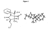
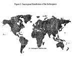
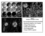
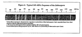


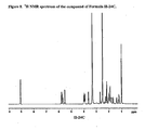
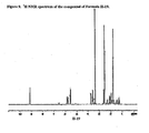
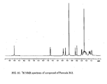
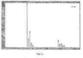


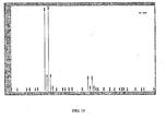
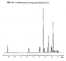
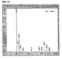
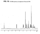

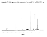

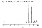

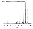

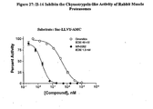
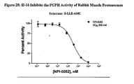
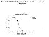

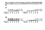
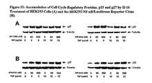

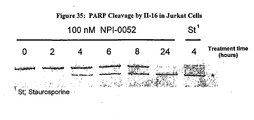
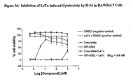
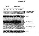
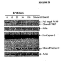
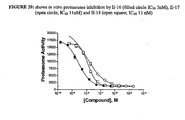
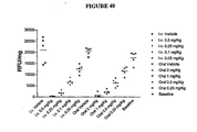
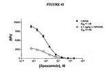
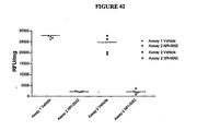

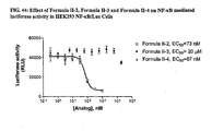

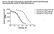
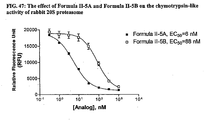
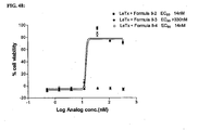
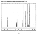
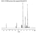
Classifications
-
- A—HUMAN NECESSITIES
- A61—MEDICAL OR VETERINARY SCIENCE; HYGIENE
- A61K—PREPARATIONS FOR MEDICAL, DENTAL OR TOILETRY PURPOSES
- A61K31/00—Medicinal preparations containing organic active ingredients
- A61K31/33—Heterocyclic compounds
- A61K31/395—Heterocyclic compounds having nitrogen as a ring hetero atom, e.g. guanethidine or rifamycins
- A61K31/40—Heterocyclic compounds having nitrogen as a ring hetero atom, e.g. guanethidine or rifamycins having five-membered rings with one nitrogen as the only ring hetero atom, e.g. sulpiride, succinimide, tolmetin, buflomedil
- A61K31/407—Heterocyclic compounds having nitrogen as a ring hetero atom, e.g. guanethidine or rifamycins having five-membered rings with one nitrogen as the only ring hetero atom, e.g. sulpiride, succinimide, tolmetin, buflomedil condensed with other heterocyclic ring systems, e.g. ketorolac, physostigmine
-
- A—HUMAN NECESSITIES
- A61—MEDICAL OR VETERINARY SCIENCE; HYGIENE
- A61P—SPECIFIC THERAPEUTIC ACTIVITY OF CHEMICAL COMPOUNDS OR MEDICINAL PREPARATIONS
- A61P11/00—Drugs for disorders of the respiratory system
- A61P11/06—Antiasthmatics
-
- A—HUMAN NECESSITIES
- A61—MEDICAL OR VETERINARY SCIENCE; HYGIENE
- A61P—SPECIFIC THERAPEUTIC ACTIVITY OF CHEMICAL COMPOUNDS OR MEDICINAL PREPARATIONS
- A61P17/00—Drugs for dermatological disorders
- A61P17/06—Antipsoriatics
-
- A—HUMAN NECESSITIES
- A61—MEDICAL OR VETERINARY SCIENCE; HYGIENE
- A61P—SPECIFIC THERAPEUTIC ACTIVITY OF CHEMICAL COMPOUNDS OR MEDICINAL PREPARATIONS
- A61P19/00—Drugs for skeletal disorders
- A61P19/02—Drugs for skeletal disorders for joint disorders, e.g. arthritis, arthrosis
-
- A—HUMAN NECESSITIES
- A61—MEDICAL OR VETERINARY SCIENCE; HYGIENE
- A61P—SPECIFIC THERAPEUTIC ACTIVITY OF CHEMICAL COMPOUNDS OR MEDICINAL PREPARATIONS
- A61P25/00—Drugs for disorders of the nervous system
-
- A—HUMAN NECESSITIES
- A61—MEDICAL OR VETERINARY SCIENCE; HYGIENE
- A61P—SPECIFIC THERAPEUTIC ACTIVITY OF CHEMICAL COMPOUNDS OR MEDICINAL PREPARATIONS
- A61P25/00—Drugs for disorders of the nervous system
- A61P25/28—Drugs for disorders of the nervous system for treating neurodegenerative disorders of the central nervous system, e.g. nootropic agents, cognition enhancers, drugs for treating Alzheimer's disease or other forms of dementia
-
- A—HUMAN NECESSITIES
- A61—MEDICAL OR VETERINARY SCIENCE; HYGIENE
- A61P—SPECIFIC THERAPEUTIC ACTIVITY OF CHEMICAL COMPOUNDS OR MEDICINAL PREPARATIONS
- A61P29/00—Non-central analgesic, antipyretic or antiinflammatory agents, e.g. antirheumatic agents; Non-steroidal antiinflammatory drugs [NSAID]
-
- A—HUMAN NECESSITIES
- A61—MEDICAL OR VETERINARY SCIENCE; HYGIENE
- A61P—SPECIFIC THERAPEUTIC ACTIVITY OF CHEMICAL COMPOUNDS OR MEDICINAL PREPARATIONS
- A61P31/00—Antiinfectives, i.e. antibiotics, antiseptics, chemotherapeutics
-
- A—HUMAN NECESSITIES
- A61—MEDICAL OR VETERINARY SCIENCE; HYGIENE
- A61P—SPECIFIC THERAPEUTIC ACTIVITY OF CHEMICAL COMPOUNDS OR MEDICINAL PREPARATIONS
- A61P31/00—Antiinfectives, i.e. antibiotics, antiseptics, chemotherapeutics
- A61P31/04—Antibacterial agents
-
- A—HUMAN NECESSITIES
- A61—MEDICAL OR VETERINARY SCIENCE; HYGIENE
- A61P—SPECIFIC THERAPEUTIC ACTIVITY OF CHEMICAL COMPOUNDS OR MEDICINAL PREPARATIONS
- A61P31/00—Antiinfectives, i.e. antibiotics, antiseptics, chemotherapeutics
- A61P31/10—Antimycotics
-
- A—HUMAN NECESSITIES
- A61—MEDICAL OR VETERINARY SCIENCE; HYGIENE
- A61P—SPECIFIC THERAPEUTIC ACTIVITY OF CHEMICAL COMPOUNDS OR MEDICINAL PREPARATIONS
- A61P33/00—Antiparasitic agents
-
- A—HUMAN NECESSITIES
- A61—MEDICAL OR VETERINARY SCIENCE; HYGIENE
- A61P—SPECIFIC THERAPEUTIC ACTIVITY OF CHEMICAL COMPOUNDS OR MEDICINAL PREPARATIONS
- A61P33/00—Antiparasitic agents
- A61P33/02—Antiprotozoals, e.g. for leishmaniasis, trichomoniasis, toxoplasmosis
-
- A—HUMAN NECESSITIES
- A61—MEDICAL OR VETERINARY SCIENCE; HYGIENE
- A61P—SPECIFIC THERAPEUTIC ACTIVITY OF CHEMICAL COMPOUNDS OR MEDICINAL PREPARATIONS
- A61P33/00—Antiparasitic agents
- A61P33/02—Antiprotozoals, e.g. for leishmaniasis, trichomoniasis, toxoplasmosis
- A61P33/06—Antimalarials
-
- A—HUMAN NECESSITIES
- A61—MEDICAL OR VETERINARY SCIENCE; HYGIENE
- A61P—SPECIFIC THERAPEUTIC ACTIVITY OF CHEMICAL COMPOUNDS OR MEDICINAL PREPARATIONS
- A61P35/00—Antineoplastic agents
-
- A—HUMAN NECESSITIES
- A61—MEDICAL OR VETERINARY SCIENCE; HYGIENE
- A61P—SPECIFIC THERAPEUTIC ACTIVITY OF CHEMICAL COMPOUNDS OR MEDICINAL PREPARATIONS
- A61P35/00—Antineoplastic agents
- A61P35/02—Antineoplastic agents specific for leukemia
-
- A—HUMAN NECESSITIES
- A61—MEDICAL OR VETERINARY SCIENCE; HYGIENE
- A61P—SPECIFIC THERAPEUTIC ACTIVITY OF CHEMICAL COMPOUNDS OR MEDICINAL PREPARATIONS
- A61P43/00—Drugs for specific purposes, not provided for in groups A61P1/00-A61P41/00
-
- A—HUMAN NECESSITIES
- A61—MEDICAL OR VETERINARY SCIENCE; HYGIENE
- A61P—SPECIFIC THERAPEUTIC ACTIVITY OF CHEMICAL COMPOUNDS OR MEDICINAL PREPARATIONS
- A61P9/00—Drugs for disorders of the cardiovascular system
- A61P9/10—Drugs for disorders of the cardiovascular system for treating ischaemic or atherosclerotic diseases, e.g. antianginal drugs, coronary vasodilators, drugs for myocardial infarction, retinopathy, cerebrovascula insufficiency, renal arteriosclerosis
-
- Y—GENERAL TAGGING OF NEW TECHNOLOGICAL DEVELOPMENTS; GENERAL TAGGING OF CROSS-SECTIONAL TECHNOLOGIES SPANNING OVER SEVERAL SECTIONS OF THE IPC; TECHNICAL SUBJECTS COVERED BY FORMER USPC CROSS-REFERENCE ART COLLECTIONS [XRACs] AND DIGESTS
- Y02—TECHNOLOGIES OR APPLICATIONS FOR MITIGATION OR ADAPTATION AGAINST CLIMATE CHANGE
- Y02A—TECHNOLOGIES FOR ADAPTATION TO CLIMATE CHANGE
- Y02A50/00—TECHNOLOGIES FOR ADAPTATION TO CLIMATE CHANGE in human health protection, e.g. against extreme weather
- Y02A50/30—Against vector-borne diseases, e.g. mosquito-borne, fly-borne, tick-borne or waterborne diseases whose impact is exacerbated by climate change
Definitions
- the present invention relates to the use of certain heterocyclic compounds for the manufacture of a medicament for the treatment of specific forms of cancer, as defined in the claims.
- Cancer is a leading cause of death in the United States. Despite significant efforts to find new approaches for treating cancer, the primary treatment options remain surgery, chemotherapy and radiation therapy, either alone or in combination. Surgery and radiation therapy, however, are generally useful only for fairly defined types of cancer, and are of limited use for treating patients with disseminated disease.
- Chemotherapy is the method that is generally useful in treating patients with metastatic cancer or diffuse cancers such as leukemias. Although chemotherapy can provide a therapeutic benefit, it often fails to result in cure of the disease due to the patient's cancer cells becoming resistant to the chemotherapeutic agent. Due, in part, to the likelihood of cancer cells becoming resistant to a chemotherapeutic agent, such agents are commonly used in combination to treat patients.
- Marine-derived natural products are a rich source of potential new anti-cancer agents.
- the oceans are massively complex and house a diverse assemblage of microbes that occur in environments of extreme variations in pressure, salinity, and temperature.
- Marine microorganisms have therefore developed unique metabolic and physiological capabilities that not only ensure survival in extreme and varied habitats, but also offer the potential to produce metabolites that would not be observed from terrestrial microorganisms ( Okami, Y. 1993 J Mar Biotechnol 1:59 ).
- Representative structural classes of such metabolites include terpenes, peptides, polyketides, and compounds with mixed biosynthetic origins.
- WO 02/47610 A2 discloses novel taxons of marine bacteria of the order Actinomycetales and the use of this taxon for the discovery and production of proteins and other pharmaceutically useful biomolecules.
- Salinosporamide A is described as a potent cytotoxin against the human colon carcinoma cell line HCT-116 and a highly potent anticancer agent.
- the disclosure herein generally relates to chemical compounds, including heterocyclic compounds and analogs thereof. Disclosed is the use of compounds as proteasome inhibitors.
- the compounds can be used to treat neoplastic diseases, for example, to inhibit the growth of tumors, cancers and other neoplastic tissues.
- the treatments disclosed herein may be employed with any patient suspected of carrying tumorous growths, cancers, or other neoplastic growths, either benign or malignant ("tumor” or “tumors” as used herein encompasses tumors, cancers, disseminated neoplastic cells and localized neoplastic growths).
- tumors are selected from liver cancers , and angiomas;
- the tumor or growth to be treated may be any tumor or cancer, primary or secondary.
- the disclosure relates to the treatment of neoplastic diseases in animals.
- the treament can include, for example, administering an effective amount of a compound to a patient in need thereof. Also disclosed is the use of compounds in the manufacture of a pharmaceutical or medicament for the treatment of a neoplastic disease.
- the compounds can be administered in combination with a chemotherapeutic agent.
- Some embodiments relate to uses of a compound having the structure of Formula I, and pharmaceutically acceptable salts thereof:
- R1 is separately selected from the group consisting of a hydrogen, a halogen, mono-substituted, poly-substituted or unsubstituted variants of the following residues: saturated C 1 -C 24 alkyl, unsaturated C 2 -C 24 alkenyl, unsaturated C 2 -C 24 alkynyl, acyl, acyloxy, alkyloxycarbonyloxy, aryloxycarbonyloxy, cycloalkyl, cycloalkenyl, alkoxy, cycloalkoxy, aryl, heteroaryl, arylalkoxy carbonyl, alkoxy carbonylacyl, amino, aminocarbonyl, aminocarbonyloxy, nitro, azido, phenyl, cycloalkylacyl, hydroxy, alkylthio, arylthio, oxysulfonyl, carboxy, cyano,
- R 2 is selected from the group consisting of hydrogen, a halogen, mono-substituted, poly-substituted or unsubstituted variants of the following residues: saturated C 1 -C 24 alkyl, unsaturated C 2 -C 24 alkenyl, unsaturated C 2 -C 24 alkynyl, acyl, acyloxy, alkyloxycarbonyloxy, aryloxycarbonyloxy, cycloalkyl, cycloalkenyl (including, for example, cyclohexylcarbinol), alkoxy, cycloalkoxy, aryl, heteroaryl, arylalkoxy carbonyl, alkoxy carbonylacyl, amino, aminocarbonyl, aminocarbonyloxy, nitro, azido, phenyl, cycloalkylacyl, hydroxy, alkylthio, arylthio, oxysulfonyl, carboxy, cyano, and
- R 3 is selected from the group consisting of a halogen, mono-substituted, poly-substituted or unsubstituted variants of the following residues:
- the treatment can include, for example, administering to the animal, a therapeutically effective amount of a compound of a formula selected from Formulae I-V, and pharmaceutically acceptable salts thereof.
- compositions which include a compound of a formula selected from Formulae I-V.
- the pharmaceutical compositions can further include an anti-microbial agent.
- Still further disclosures relate to the inhibition of the growth of a cancer cell.
- the inhibition can include, for example, contacting a cancer cell with a compound of a formula selected from Formulae I-V, and pharmaceutically acceptable salts thereof.
- Also disclosed is the inhibition of proteasome activity that includes the step contacting a cell with a compound of a formula selected from Formulae I-V, and pharmaceutically acceptable salts thereof.
- Also disclosed is the inhibition of NF- ⁇ B activation including the step contacting a cell with a compound of a formula selected from Formulae I-V, and pharmaceutically acceptable salts thereof.
- FIG. 1 shows the chemical structure of Salinosporamide A.
- FIG. 2 shows the pan-tropical distribution of the Salinospora.
- X denotes Salinospora collection sites.
- FIG. 3 shows colonies of Salinospora.
- FIG. 4 shows the typical 16S rDNA sequence of the Salinospora. Bars represent characteristic signature nucleotides of the Salinospora that separate them from their nearest relatives.
- FIG. 5 shows Omuralide, a degradation product of the microbial metabolite Lactacystin. Also shown is a compound of Formula II-16, also referred to as Salinosporamide A.
- FIG. 6 illustrates lethal toxin-mediated macrophage cytotoxicity.
- NPI-0052 represents the compound of Formula II-16.
- FIG. 7 depicts the 1H NMR spectrum of a compound having structure Formula II-20.
- FIG. 8 depicts the 1H NMR spectrum of a compound having structure Formula II-24C.
- FIG. 9 depicts the 1H NMR spectrum of a compound having structure Formula II-19.
- FIG. 10 depicts the 1H NMR spectrum of a compound having structure Formula II-2.
- FIG. 11 depicts the mass spectrum of a compound having structure Formula II-2.
- FIG. 12 depicts the 1H NMR spectrum of a compound having structure Formula II-3.
- FIG. 13 depicts the mass spectrum of a compound having structure Formula II-3.
- FIG. 14 depicts the 1H NMR spectrum of a compound having structure Formula II-4.
- FIG. 15 depicts the mass spectrum of a compound having structure Formula II-4.
- FIG. 16 depicts the 1H NMR spectrum of a compound having structure Formula II-5A.
- FIG. 17 depicts the mass spectrum of a compound having structure Formula II-5A.
- FIG. 18 depicts the 1H NMR spectrum of a compound having structure Formula II-5B.
- FIG. 19 depicts the mass spectrum of a compound having structure Formula II-5B.
- FIG. 20 depicts the 1H NMR spectrum of a compound having structure Formula IV-3C in DMSO-d 6 .
- FIG. 21 depicts the 1H NMR spectrum of a compound having structure Formula IV-3C in C 6 D 6 /DMSO-d 6 .
- FIG. 22 depicts the 1H NMR spectrum of a compound having structure Formula II-13C.
- FIG. 23 depicts the 1H NMR spectrum of a compound having structure Formula II-8C.
- FIG. 24 depicts the 1H NMR spectrum of a compound having structure Formula II-25.
- FIG. 25 depicts the 1H NMR spectrum of a compound having structure Formula II-21.
- FIG. 26 depicts the 1H NMR spectrum of a compound having structure Formula II-22.
- FIG. 27 shows inhibition of the chymotrypsin-like activity of rabbit muscle proteasomes.
- FIG. 28 shows inhibition of the PGPH activity of rabbit muscle proteasomes.
- FIG. 29 shows inhibition of the chymotrypsin-like activity of human erythrocyte proteasomes.
- FIG. 30 shows the effect of II-16 treatment on chymotrypsin-mediated cleavage of LLVY-AMC substrate.
- FIG. 31 shows NF- ⁇ B/luciferase activity and cytotoxicity profiles of II-16.
- FIG. 32 shows reduction of I ⁇ B ⁇ degradation and retention of phosphorylated I ⁇ B ⁇ by II-16 in HEK293 cells (A) and the HEK293 NF- ⁇ B/Luciferase reporter clone (B).
- FIG. 33 shows accumulation of cell cycle regulatory proteins, p21 and p27, by II-16 treatment of HEK293 cells (A) and the HEK293 NF- ⁇ B/Luciferase reporter clone (B).
- FIG. 34 shows activation of Caspase-3 by II-16 in Jurkat cells.
- FIG. 35 shows PARP cleavage by II-16 in Jurkat cells.
- FIG. 36 shows inhibition of LeTx-induced cytotoxicity by II-16 in RAW264.7 cells.
- FIG. 37 shows the effects of II-16 treatment on PARP and Pro-Caspase 3 cleavage in RPMI 8226 and PC-3 cells.
- FIG. 38 shows II-16 treatment of RPMI 8226 results in a dose-dependent cleavage of PARP and Pro-Caspase 3.
- FIG. 39 shows in vitro proteasome inhibition by II-16, II-17, and II-18.
- FIG. 40 shows proteasomal activity in PWBL prepared from II-16 treated mice.
- FIG. 41 shows epoxomicin treatment in the PWBL assay.
- FIG. 42 shows intra-assay comparison.
- FIG. 43 shows decreased plasma TNF levels in mice treated with LPS.
- FIG. 44 depicts assay results showing the effect of Formula II-2, Formula II-3 and Formula II-4 on NF- ⁇ B mediated luciferase activity in HEK293 NF- ⁇ B/Luc Cells.
- FIG. 45 depicts assay results showing the effect of Formula II-5A and Formula II-5B on NF- ⁇ B mediated luciferase activity in HEK293 NF- ⁇ B/Luc Cells
- FIG. 46 depicts assay results showing the effect of Formula II-2, Formula II-3, and Formula II-4 on the chymotrypsin-like activity of rabbit 20S proteasome.
- FIG. 47 depicts the effect of Formula II-5A and Formula II-5B on the chymotrypsin-like activity of rabbit 20S proteasome.
- FIG. 48 depicts the effect of Formulae II-2, II-3, and II-4 against LeTx-mediated cytotoxicity.
- FIG. 49 depicts the 1H NMR spectrum of a compound having structure Formula II-17.
- FIG. 50 depicts the 1H NMR spectrum of a compound having structure Formula II-18.
- the methods can include the compositions in relatively high yield, wherein the compounds and/or their derivatives are among the active ingredients in these compositions.
- the description relates to the treatment of cancer, particularly those affecting humans.
- the treatments may include, for example, the step of administering an effective amount of a member of a class of new compounds.
- Preferred disclosures relate to the compounds and methods of making such compounds and the use disclosed herein, but not necessarily in all embodiments of the present invention, these objectives are met.
- each stereogenic carbon may be of R or S configuration.
- the specific compounds exemplified in this application may be depicted in a particular configuration, compounds having either the opposite stereochemistry at any given chiral center or mixtures thereof are also envisioned.
- chiral centers are found in the derivatives of this invention, it is to be understood that the compounds encompasses all possible stereoisomers.
- Some embodiments provide compounds, and methods of producing a class of compounds, pharmaceutically acceptable salts thereof, wherein the compounds are represented by Formula I:
- the substituent(s) R 1 and R 2 separately include a hydrogen, a halogen, a mono-substituted, a poly-substituted or an unsubstituted variant of the following residues: saturated C 1 -C 24 alkyl, unsaturated C 2- C 24 alkenyl , unsaturated C 2 -C 24 alkynyl, acyl, acyloxy, alkyloxycarbonyloxy, aryloxycarbonyloxy, cycloalkyl (including for example, cyclohexylcarbinol), cycloalkenyl, alkoxy, cycloallcoxy, aryl, heteroaryl, arylalkoxy carbonyl, alkoxy carbonylacyl, amino, aminocarbonyl, aminocarboniloxy, nitro, azido, phenyl, cycloalkylacyl, hydroxy, alkylthio, arylthio, oxysulfonyl, carboxy, cyan
- n is equal to 1 or equal to 2.
- the substituents R 1 can be the same or can be different.
- R 2 may be a formyl.
- the compound may have the following structure I-1:
- R 8 may include, for example, hydrogen, fluorine, chlorine, bromine and iodine.
- the structure of Formula I-1 may have the following stereochemistry:
- R 8 may include, for example, hydrogen, fluorine, chlorine, bromine and iodine.
- R 2 may be a carbinol.
- the compound may have the following structure 1-2:
- R 8 may include, for example, hydrogen, fluorine, chlorine, bromine and iodine.
- the structure of Formula 1-2 may have the following stereochemistry:
- R 8 may include, for example, hydrogen, fluorine, chlorine, bromine and iodine.
- exemplary compound of Formula I may be the compound having the following structure I-3:
- R 8 may include, for example, hydrogen, fluorine, chlorine, bromine and iodine.
- the compound of Formula I-3 may have the following stereochemical structure:
- R 8 may include, for example, hydrogen, fluorine, chlorine, bromine and iodine.
- Another exemplary compound Formula I may be the compound having the following structure I-4:
- R 8 may include, for example, hydrogen, fluorine, chlorine, bromine and iodine.
- the compound of Formula 1-4 may have the following stereochemical structure:
- R 8 may include, for example, hydrogen, fluorine, chlorine, bromine and iodine.
- Still a further exemplary compound of Formula I is the compound having the following structure I-5:
- R 8 may include, for example, hydrogen, fluorine, chlorine, bromine and iodine.
- the compound of Formula 1-5 may have the following stereochemistry:
- R 8 may include, for example, hydrogen, fluorine, chlorine, bromine and iodine.
- R 2 of Formula I may be, for example, a 3-methylenecyclohexene.
- the compound may have the following structure of Formula 1-6:
- R 8 may include, for example, hydrogen, fluorine, chlorine, bromine and iodine.
- the compound of Formula 1-6 may have the following. stereochemistry:
- R 8 may include, for example, hydrogen, fluorine, chlorine, bromine and iodine.
- R 2 may be a cyclohexylalkylamine.
- R 2 may be a C-Cyclohexyl-methyleneamine. In others, R 2 may be a cyclohexanecarbaldehyde O-oxime.
- R 2 may be a cycloalkylacyl.
- the substituent(s) R 1 R4 separately may include a hydrogen, a halogen, a mono-substituted, a poly-substituted or an unsubstituted variant of the following residues: saturated C 1 -C 24 alkyl, unsaturated C 2 -C 24 alkenyl, unsaturated C 2 -C 24 alkynyl, acyl, acyloxy, alkyloxycarbonyloxy, aryloxycarbonyloxy, cycloalkyl, cycloalkenyl, alkoxy, cycloalkoxy, aryl, heteroaryl, arylalkoxy carbonyl, alkoxy carbonylacyl, amino, aminocarbonyl, aminocarbonyloxy, nitro, azido, phenyl, cycloalkylacyl, hydroxy, alkylthio, arylthio, oxysulfonyl, carboxy, cyano, and halogenated alkyl including
- n is equal to 1 or 2.
- the substituents R 1 can be the same or can be different.
- m is equal to 1 or 2
- R 4 can be the same or different.
- E 5 is OH, O, OR 10 , S, SR 11 , SO 2 R 11 , NH, NH 2 , NOH, NHOH, NR 12 , and NHOR 13 , wherein R 10-13 are selected from hydrogen, a substituted or unsubstituted of any of the following: alkyl, an aryl, a heteroaryl
- R 1 may be CH 2 CH 2 X, wherein X is selected from H, F, Cl, Br, and I.
- R 3 may be methyl.
- R 4 may include a cyclohexyl.
- R 1 may be CH 2 CH 2 X, wherein X is selected from the group consisting of H, F, Cl Br, and I; wherein R 4 may include a cyclohexyl; wherein R3 may be methyl; and wherein each of E 1 , E 3 and E 4 separately may be O and E 2 may be NH.
- an exemplary compound of Formula II has the following structure II-1:
- R 8 may include, for example, hydrogen, fluorine, chlorine, bromine and iodine.
- the compound of Formula II has any of the following structures:
- R 4 may include a 7-oxa-bicyclo[4.1.0]hept-2-yl).
- An exemplary compound of Formula II is the following structure II-5:
- R 8 may include, for example, hydrogen, fluorine, chlorine, bromine and iodine.
- At least one R 4 may include a subsituted or an unsubstituted branched alkyl.
- a compound of Formula II may be the following structure II-6:
- R 8 may include, for example, hydrogen, fluorine, chlorine, bromine and iodine.
- the compound of Formula II may be the following structure II-7:
- R 8 may include, for example, hydrogen, fluorine, chlorine, bromine and iodine.
- At least one R 4 may be a cycloalkyl and E 5 may be an oxygen.
- An exemplary compound of Formula II may be the following structure II-8:
- R 8 may include, for example, hydrogen (II-8A), fluorine (II-8B), chlorine (II-8C); bromine (II-8D) and iodine (II-8E).
- E5 may be an amine oxide, giving rise to an oxime.
- An exemplary compound of Formula II has the following structure II-9:
- R 8 may include, for example, hydrogen, fluorine, chlorine, bromine and iodine; R may be hydrogen, and a substituted or unsubstituted alkyl, aryl, or heteroaryl, and the like.
- a further exemplary compound of Formula II has the following structure II-10:
- R 8 may include, for example, hydrogen, fluorine, chlorine, bromine and iodine.
- E 5 may be NH 2 .
- An exemplary compound of Formula II has the following structure II-11:
- R 8 may include, for example, hydrogen, fluorine, chlorine, bromine and iodine.
- At least one R 4 may include a cycloalkyl and E 5 may be NH2.
- An exemplary compound of Formula II has the following structure II-12:
- R 8 may include, for example, hydrogen, fluorine, chlorine, bromine and iodine.
- a further exemplary compound of Formula II has the following structure II-13:
- R 8 may include, for example, hydrogen (II-13A), fluorine (II-13B), chlorine (II-13C), bromine (II-13D) and iodine (II-13E).
- a still further exemplary compound of Formula II has the following structure II-14:
- R 8 may include, for example, hydrogen, fluorine, chlorine, bromine and iodine.
- the compounds of Formula II may include as R 4 at least one cycloalkene, for example. Furthermore, in some embodiments, the compounds may include a hydroxy at E 5 , for example.
- a further exemplary compound of Formula II has the following structure II-15:
- R 8 may include, for example, hydrogen, fluorine, chlorine, bromine and iodine.
- the compounds of Formulae II-16, II-17, II-18 and II-19 may be obtained by fermentation, synthesis, or semi-synthesis and isolated/purified as set forth below. Furthermore, the compounds of Formulae II-16, II-17, II-18 and II-19 may be used, and are referred to, as "starting materials" to make other compounds described herein.
- the compounds of Formula II may include a methyl group as R 1 for example.
- a further exemplary compound, Formula II-20 has the following structure and stereochemistry:
- the compounds of Formula II may include hydroxyethyl as R 1 , for example.
- a further exemplary compound, Formula II-21 has the following structure and stereochemistry:
- the hydroxyl group of Formula II-21 may be esterified such that R 1 may include ethylpropionate, for example.
- R 1 may include ethylpropionate, for example.
- An exemplary compound, Formula II-22 has the following structure and stereochemistry:
- the compounds of Formula II may include an ethyl group as R 3 , for example.
- a further exemplary compound of Formula II has the following structure II-23:
- R 8 may include, for example, hydrogen, fluorine, chlorine, bromine and iodine.
- Exemplary stereochemistry may be as follows:
- the compounds of Formula II-23 may have the following structure and stereochemistry, exemplified by Formula II-24C, where R 8 is chlorine:
- the compounds of Formula II-15 may have the following stereochemistry, exemplified by the compound of Formula II-25, where R 8 is chlorine:
- the substituent(s) R 1 separately are selected from a hydrogen, a halogen, a mono-substituted, a poly-substituted or an unsubstituted variant of the following residues: saturated C 1 -C 24 alkyl, unsaturated C 2 -C 24 alkenyl, unsatured C 2 -C 24 alkynyl, acyl, acyloxy, alkyloxycarbonyloxy, aryloxycarbonyloxy, cycloalkyl, cycloalkenyl, alkoxy, cycloalkoxy, aryl, heteroaryl, arylalkoxy carbonyl, alkoxy carbonylacyl amino, aminocarbonyl, aminocarbonyloxy nitro, azido, phenyl, hydroxy, alkylthio, arylthio, oxysulfonyl, carboxy, cyano, and halogenated alkyl including polyhalogenated alkyl, where
- R 4 can be a hydrogen, a halogen, a mono-substituted, a poly-substituted or an unsubstituted variants of the following residues: saturated C 1 -C 24 alkyl, unsaturated C 2 -C 24 alkenyl, unsaturated C 2 -C 24 alkynyl, acyl, acyloxy, alkyloxycarbonyloxy, aryloxycarbonyloxy, cycloalkyl, cycloalkenyl, alkoxy, cycloalkoxy, aryl, heteroaryl, arylalkoxy carbonyl, alkoxy carbonylacyl, amino, aminocarbonyl, aminocarbonyloxy, nitro, azido, phenyl, hydroxy, alkylthio, arylthio, oxysulfonyl, carboxy, cyano, and halogenated alkyl including polyhalogenated alkyl; wherein m is equal to 1 or 2, and
- the substituent(s) R 1 and R 5 are separately selected from a hydrogen, a halogen, a mono-substituted, a poly-substituted or an unsubstituted variants of the following residues: saturated C 1 -C 24 alkyl, unsaturated C 2 -C 24 alkenyl, unsaturated C 2 -C 24 alkynyl, acyl, acyloxy, alkyloxycarbonyloxy, aryloxycarbonyloxy, cycloalkyl, cycloalkenyl, alkoxy, cycloalkoxy, aryl, heteroaryl, arylalkoxy carbonyl, alkoxy carbonylacyl, amino, aminocarbonyl, aminocarbonyloxy nitro, azido, phenyl, hydroxy, alkylthio, arylthio, oxysulfonyl, carboxy, cyano, and halogenated alkyl including polyhalogenated alkyl including polyhal
- R 3 has the same meaning, except that it cannot be hydrogen. Also, E 1, E 3 and E 4 is O, E 2 is NH. n is equal to 1 or 2. When n is equal to 2, the substituents R 1 can be the same or can be different. Also, m is 0, 1, 2, 3, 4, 5, 6, 7, 8, 9, 10, or 11. When m is greater than 1, the substituents R 5 can be the same or different.
- R 5 may give rise to a di-substituted cyclohexyl.
- An exemplary compound of Formula IV is the following structure IV-1, with and without exemplary stereochemistry:
- R 8 may include, for example, hydrogen, fluorine, chlorine, bromine and iodine.
- the substituent(s) R 6 and R 7 may separately include a hydrogen, a halogen, a mono-substituted, a poly-substituted or an unsubstituted variants of the following residues: saturated C 1 -C 24 alkyl, unsaturated C 2 -C 24 alkenyl or C 2 -C 24 alkynyl, acyl, acyloxy, alkyloxycarbonyloxy, aryloxycarbonyloxy, cycloalkyl, cycloalkenyl, alkoxy, cycloalkoxy, aryl, heteroaryl, arylalkoxy carbonyl, alkoxy carbonylacyl, amino; aminocarbonyl, aminocarboyloxy, nitro, azido, phenyl, hydroxy, alkylthio, arylthio, oxysulfonyl, carboxy
- an exemplary compound of Formula IV has the following structure IV-2:
- R 8 may include, for example, hydrogen, fluorine, chlorine, bromine and iodine.
- an exemplary compound of Formula IV has the following structure IV-3:
- R 8 may include, for example, hydrogen (IV-3A), fluorine (IV-3B), chlorine, (IV-3C), bromine (IV-3D) and iodine (IV-3E).
- an exemplary compound of Formula IV has the following structure IV-4:
- R 8 may include, for example, hydrogen, fluorine, chlorine, bromine and iodine.
- Some embodiments provide compounds, and methods of producing a class of compounds, pharmaceutically acceptable salts and pro-drug esters thereof, wherein the compounds are represented by Formula V:
- the substituent(s) R 1 and R 5 are separately selected from a hydrogen, a halogen, a mono-substituted, a poly-substituted or unsubstituted variants of the following residues: saturated C 1 -C 24 alkyl, unsaturated C 2 -C 24 alkenyl, unsaturated C 2 -C 24 alkynyl, acyl, acyloxy, alkyloxycarbonyloxy, aryloxycarbonyloxy, cycloalkyl, cycloalkenyl, alkoxy, cycloalkoxy, aryl, heteroaryl, arylalkoxy carbonyl, alkoxy carbonylacyl, amino, aminocarbonyl, aminocarbonyloxy, nitro, azido, phenyl, hydroxy, alkylthio, arylthio, oxysulfonyl, carboxy, cyano, and halogenated alkyl including polyhalogenated alkyl including polyhal
- E 1 is O
- E 2 is NH wherein E5 is OH, O, OR 10 , S, SR 11 , SO 2 R 11 , NH, NH 2 , NOH, NHOH, NR 12 , or NHOR 13, wherein R 10 , R 11 , R 12 , and R 13 are selected from the group consisting of hydrogen, a substituted or unsubstituted alkyl, a substituted or unsubstituted aryl, and a substituted or unsubstituted heteroaryl.
- n is equal to 1 or 2, and when n is equal to 2, the substituents R 1 can be the same or different.
- m is 0, 1, 2, 3, 4, 5, 6, 7, 8, 9, 10, or 11. When m is greater than 1, R 5 may be the same or different.
- Certain embodiments also provide pharmaceutically acceptable salts of the compound of Formulae I-V. Also provided are methods of obtaining and purifying such compounds by the methods disclosed herein.
- pharmaceutically acceptable salt refers to any pharmaceutically acceptable salts of a compound, and preferably refers to an acid addition salt of a compound.
- pharmaceutically acceptable salt are the alkali metal salts (sodium or potassium), the alkaline earth metal salts (calcium or magnesium), or ammonium salts derived from ammonia or from pharmaceutically acceptable organic amines, for example C 1 - C 7 alkylamine, cyclohexylamine, triethanolamine, ethylenediamine or tris-(hydroxymethyl)-aminomethane.
- the preferred examples of pharmaceutically acceptable salts are acid addition salts of pharmaceutically acceptable inorganic or organic acids, for example, hydrohalic, sulfuric, phosphoric acid or aliphatic or aromatic carboxylic or sulfonic acid, for example acetic, succinic, lactic, malic, tartaric, citric, ascorbic, nicotinic, methanesulfonic, p-toluensulfonic or naphthalenesulfonic acid.
- pharmaceutically acceptable inorganic or organic acids for example, hydrohalic, sulfuric, phosphoric acid or aliphatic or aromatic carboxylic or sulfonic acid, for example acetic, succinic, lactic, malic, tartaric, citric, ascorbic, nicotinic, methanesulfonic, p-toluensulfonic or naphthalenesulfonic acid.
- Preferred pharmaceutical compositions disclosed herein include pharmaceutically acceptable salts of the compound of Formulae I-V obtained and purified by the methods disclosed herein. Accordingly, if the manufacture of pharmaceutical formulations involves intimate mixing of the pharmaceutical excipients and the active ingredient in its salt form, then it is preferred to use pharmaceutical excipients which are non-basic, that is, either acidic or neutral excipients.
- the phrase "compounds and compositions comprising the compound,” or any like phrase, is meant to encompass compounds in any suitable form for pharmaceutical delivery, as discussed in further detail herein.
- the compounds or compositions comprising the same may include a pharmaceutically acceptable salt of the compound.
- the compounds are used to treat Liver, cancer or angioma.
- the use may also include the steps of administering a compound or composition comprising the compound to an individual with these cancers.
- the compound or composition can be administered in an amount effective to treat the particular cancer
- the compound or composition may be administered with a pharmaceutically acceptable carrier, diluent, excipient, and the like.
- the cancer is liver cancer or angioma
- halogen atom means any one of the radio-stable atoms of column 7 of the Periodic Table of the Elements, i.e ., fluorine, chlorine, bromine, or iodine, with bromine and chlorine being preferred.
- alkyl means any unbranched or branched, substituted or unsubstituted, saturated hydrocarbon, with C 1 -C 6 unbranched, saturated, unsubstituted hydrocarbons being preferred, with methyl, ethyl, isobutyl, and tert-butylpropyl, and pentyl being most preferred.
- substituted, saturated hydrocarbons C 1 -C 6 mono- and di- and per-halogen substituted saturated hydrocarbons and amino-substituted hydrocarbons are preferred, with perfluromethyl, perchloromethyl, perfluoro-tert-butyl, and perchloro-tert-butyl being the most preferred.
- substituted has its ordinary meaning, as found in numerous contemporary patents from the related art. See, for example, U.S. Patent Nos. 6,509,331 ; 6,506,787 ; 6,500,825 ; 5,922,683 ; 5,886,210 ; 5,874,443 ; and 6,350,759 . Specifically, the definition of substituted is as broad as that provided in U.S. Patent No.
- substituted alkyl such that it refers to an alkyl group, preferably of from 1 to 10 carbon atoms, having from 1 to 5 substituents, and preferably 1 to 3 substituents, selected from the group consisting of alkoxy, substituted alkoxy, cycloalkyl, substituted cycloalkyl, cycloalkenyl, substituted cycloalkenyl, acyl, acylamino, acyloxy, amino, substituted amino, aminoacyl, aminoacyloxy, oxyacylamino, cyano, halogen, hydroxyl, carboxyl, carboxylalkyl, keto, thioketo, thiol, thioalkoxy,substituted thioalkoxy, aryl, aryloxy, heteroaryl, heteroaryloxy, heterocyclic, heterocyclooxy, hydroxyamino, alkoxyamino, nitro, --SO-al
- cycloalkyl refers to any non-aromatic hydrocarbon ring, preferably having five to twelve atoms comprising the ring.
- acyl refers to alkyl or aryl groups derived from an oxoacid, with an acetyl group being preferred.
- alkenyl means any unbranched or branched, substituted or unsubstituted, unsaturated hydrocarbon including polyunsaturated hydrocarbons, with C 1 -C 6 unbranched, mono-unsaturated and di-unsaturated, unsubstituted hydrocarbons being preferred, and mono-unsaturated, di-halogen substituted hydrocarbons being most preferred.
- cycloalkenyl refers to any non-aromatic hydrocarbon ring, preferably having five to twelve atoms comprising the ring.
- aryl refers to aromatic hydrocarbon rings, preferably having five, six, or seven atoms, and most preferably having six atoms comprising the ring.
- Heteroaryl and substituted heteroaryl refer to aromatic hydrocarbon rings in which at least one heteroatom, e.g., oxygen, sulfur, or nitrogen atom, is in the ring along with at least one carbon atom.
- heterocycle refers to any cyclic compound containing one or more heteroatoms. The substituted aryls, heterocycles and heteroaryls can be substituted with any substituent, including those described above and those known in the art.
- alkoxy refers to any unbranched, or branched, substituted or unsubstituted, saturated or unsaturated ether, with C 1 -C 6 unbranched, saturated, unsubstituted ethers being preferred, with methoxy being preferred, and also with dimethyl, diethyl, methyl-isobutyl, and methyl-tert-butyl ethers also being preferred.
- cycloalkoxy refers to any non-aromatic hydrocarbon ring, preferably having five to twelve atoms comprising the ring.
- alkoxy carbonyl refers to any linear, branched, cyclic, saturated, unsaturated, aliphatic or aromatic alkoxy attached to a carbonyl group.
- the examples include methoxycarbonyl group, ethoxycarbonyl group, propyloxycarbonyl group, isopropyloxycarbonyl group, butoxycarbonyl group, sec-butoxycarbonyl group, tert-butoxycarbonyl group, cyclopentyloxycarbonyl group, cyclohexyloxycarbonyl group, benzyloxycarbonyl group, allyloxycarbonyl group, phenyloxycarbonyl group, and pyridyloxycarbonyl group.
- the terms “pure,” “purified,” “substantially purified,” and “isolated” as used herein refer to the compound of the embodiment being free of other, dissimilar compounds with which the compound, if found in its natural state, would be associated in its natural state.
- the compound may comprise at least 0.5%, 1%, 5%, 10%, or 20%, and most preferably at least 50% or 75% of the mass, by weight, of a given sample.
- derivative refers to a compound that is an analog of the other compound.
- Certain of the compounds of Formula I-V may be obtained and purified or may be obtained via semi-synthesis from purified compounds as set forth herein.
- the compounds of Formula II-15 preferably, Formulae II-16, II-17, II-18 and II-19, may be obtained synthetically or by fermentation. Exemplary fermentation procedures are provided below. Futher, the compounds of Formula II-15, preferably, Formulae II-16, II-17, II-18 and II-19 may be used as starting compounds in order to obtain/synthesize various of the other compounds described herein. Exemplary syntheses are provided herein.
- Formula II-16 is currently produced through a high-yield saline fermentation ( ⁇ 200 mg/L) and modifications of the conditions has yielded new analogs in the fermentation extracts.
- FIG. 1 shows the chemical structure of II-16. Additional analogs can be generated through directed biosynthesis. Directed biosynthesis is the modification of a natural product by adding biosynthetic precursor analogs to the fermentation of producing microorganisms ( Lam, et al., J Antibiot (Tokyo) 44:934 (1991 ), Lam, et al., J Antibiot (Tokyo) 54:1 (2001 )).
- Biotransformation reactions are chemical reactions catalyzed by enzymes or whole cells containing these enzymes.
- Microbial natural products are ideal substrates for biotransformation reactions as they are synthesized by a series of enzymatic reactions inside microbial cells. Riva, S., Curr Opin Chem Biol 5:106 (2001 ).
- Ethylmalonyl-CoA is derived from butyryl-CoA, which can be derived either from valine or crotonyl-CoA. Liu, et al., Metab Eng 3:40 (2001 ). Phenylalanine is derived from shikimic acid.
- the production of compounds of Formulae II-16, II-17, and II-18 may be carried out by cultivating strain CNB476 in a suitable nutrient medium under conditions described herein, preferably under submerged aerobic conditions, until a substantial amount of compounds are detected in the fermentation; harvesting by extracting the active components from the fermentation broth with a suitable solvent; concentrating the solvent containing the desired components; then subjecting the concentrated material to chromatographic separation to isolate the compounds from other metabolites also present in the cultivation medium.
- FIG. 2 shows some collection sites worldwide for the culture (CNB476), which is also refered to as Salinospora.
- FIG. 3 shows colonies of Salinospora.
- FIG. 4 shows the typical 16S rDNA sequence of the Salinospora. Bars represent characteristic signature nucleotides of the Salinospora that separate them from their nearest relatives.
- the culture (CNB476) was deposited on June 20, 2003 with the American Type Culture Collection (ATCC) in Rockville, MD and assigned the ATCC patent deposition number PTA-5275.
- ATCC American Type Culture Collection
- the ATCC deposit meets all of the requirements of the Budapest treaty.
- the culture is also maintained at and available from Nereus Pharmaceutical Culture Collection at 10480 Wateridge Circle, San Diego, CA 92121 .
- mutants such as those produced by the use of chemical or physical mutagens including X-rays, etc. and organisms whose genetic makeup has been modified by molecular biology techniques, may also be cultivated to produce the starting compounds of Formulae II-16, II-17, and II-18.
- Production of compounds can be achieved at temperature conducive to satisfactory growth of the producing organism, e.g. from 16 degree C to 40 degree C, but it is preferable to conduct the fermentation at 22 degree C to 32 degree C.
- the aqueous medium can be incubated for a period of time necessary to complete the production of compounds as monitored by high pressure liquid chromatography (HPLC), preferably for a period of about 2 to 10 days, on a rotary shaker operating at about 50 rpm to 400 rpm, preferably at 150 rpm to 250 rpm, for example.
- HPLC high pressure liquid chromatography
- the sources of carbon include glucose, fructose, mannose, maltose, galactose, mannitol and glycerol, other sugars and sugar alcohols, starches and other carbohydrates, or carbohydrate derivatives such as dextran, cerelose, as well as complex nutrients such as oat flour, corn meal, millet, corn, and the like.
- the exact quantity of the carbon source that is utilized in the medium will depend in part, upon the other ingredients in the medium, but an amount of carbohydrate between 0.5 to 25 percent by weight of the medium can be satisfactorily used, for example.
- These carbon sources can be used individually or several such carbon sources may be combined in the same medium, for example. Certain carbon sources are preferred as hereinafter set forth.
- the sources of nitrogen include amino acids such as glycine, arginine, threonine, methionine and the like, ammonium salt, as well as complex sources such as yeast extracts, corn steep liquors, distiller solubles, soybean meal, cotttonseed meal, fish meal, peptone, and the like.
- the various sources of nitrogen can be used alone or in combination in amounts ranging from 0.5 to 25 percent by weight of the medium, for example.
- the customary salts capable of yielding sodium, potassium, magnesium, calcium, phosphate, sulfate, chloride, carbonate, and like ions.
- trace metals such as cobalt, manganese, iron, molybdenum, zinc, cadmium, and the like.
- the compounds disclosed herein may be used to treat cancer.
- the treatments may include, for example, the step of administering an effective amount of a member of a class of new compounds.
- the compounds have various biological activities.
- the compounds have chemosensitizing activity, and anti-cancer activity.
- the compounds have proteasome inhibitory activity.
- the proteasome inhibitory activity may, in whole or in part, contribute to the ability of the compounds to act as anti-cancer agents.
- the proteasome is a multisubunit protease that degrades intracellular proteins through its chymotrypsin-like, trypsin-like and peptidylglutamyl-peptide hydrolyzing (PGPH; and also know as the caspase-like activity) activities.
- the 26S proteasome contains a proteolytic core called the 20S proteasome and two 19S regulatory subunits.
- the 20S proteasome is responsible for the proteolytic activity against many substrates including damaged proteins, the transcription factor NF- ⁇ B and its inhibitor I ⁇ B, signaling molecules, tumor suppressors and cell cycle regulators.
- compounds of Formula II-16 were more potent (EC 50 2nM) at inhibiting the chymotrypsin-like activity of rabbit muscle proteasomes than Omuralide (EC 50 52 nM) and also inhibited the chymotrypsin-like activity of human erythrocyte derived proteasomes (EC 50 ⁇ 250pM).
- FIG. 5 shows omuralide, which is a degradation product of Lactacystin, and it shows a compound of Formula II-16.
- Compounds of Formula II-16 exhibit a significant preference for inhibiting chymotrypsin-like activity of the proteasome over inhibiting the catalytic activity of chymotrypsin.
- Compounds of Formula II-16 also exhibit low nM trypsin-like inhibitory activity ( ⁇ 10 nM), but are less potent at inhibiting the PGPH activity of the proteasome (EC 50 ⁇ 350 nM).
- HEK293 cells human embryonic kidney
- Tumor Necrosis Factor-alpha TNF- ⁇
- TNF- ⁇ Tumor Necrosis Factor-alpha
- HEK293 cells were pre-treated for 1 hour with compounds of Formula II-16 followed by TNF- ⁇ stimulation.
- Treatment with compounds of Formula II-16 promoted the accumulation of phosphorylated I ⁇ B ⁇ suggesting that the proteasome-mediated I ⁇ B ⁇ degradation was inhibited.
- a stable HEK293 clone (NF- ⁇ B/Luc 293) was generated carrying a luciferase reporter gene under the regulation of 5x NF- ⁇ B binding sites. Stimulation of NF- ⁇ B/Luc 293 cells with TNF- ⁇ increases luciferase activity as a result of NF- ⁇ B activation while pretreatment with compounds of Formula II-16 decreases activity.
- Western blot analyses demonstrated that compounds of Formula II-16 promoted the accumulation of phosphorylated-I ⁇ B ⁇ and decreased the degradation of total I ⁇ B ⁇ in the NF- ⁇ B/Luc 293 cells.
- Compounds of Formula II-16 were also shown to increase the levels of the cell cycle regulatory proteins, p21 and p27.
- Tumor cells may be more sensitive to proteasome inhibitors than normal cells. Moreover, proteasome inhibition increases the sensitivity of cancer cells to anticancer agents.
- the cytotoxic activity of the compounds described herein, including Formula II-16, were examined for cytotoxic activity against various cancer cell lines. Formula II-16 was examined, for example, in the National Cancer Institute screen of 60 human tumor cell lines. Formula II-16 exhibited selective cytotoxic activity with a mean GI 50 value (the concentration to achieve 50% growth inhibition) of less than 10 nM. The greatest potency was observed against SK-MEL-28 melanoma and MDA-MB-235 breast cancer cells [both with LC 50 (the concentration with 50% cell lethality) ⁇ 10 nM].
- a panel of cell lines including human colorectal (HT-29 and LoVo), prostate (PC3), breast (MDA-MB-231), lung (NCI-H292), ovarian (OVCAR3), acute T-cell leukemia (Jurkat), murine melanoma (B16-F10) and normal human fibroblasts (CCD-27sk) was treated with Salinosporamide A for 48h to assess cytotoxic activity.
- HT-29, LoVo, PC3, MDA-MB-231, NCI-H292, OVCAR3, Jurkat, and B16-F10 cells were sensitive with EC 50 values of 47, 69, 78, 67, 97, 69, 10, and 33 nM, respectively.
- EC 50 values for CCD-27sk cells were 196 nM.
- Treatment of Jurkat cells with Salinosporamide A at the approximate EC 50 resulted in Caspase-3 activation and cleavage of PARP confirming the induction of apoptosis.
- the anti-anthrax activity of the described compounds was evaluated using an in vitro LeTx induced cytotoxicity assay.
- the results indicate that Formula II-16 is a potent inhibitor of LeTx-induced cytotoxicity of murine macrophage-like RAW264.7 cells.
- Treatment of RAW264.7 cells with Formula II-16 resulted in a 10-fold increase in the viability of LeTx-treated cells compared to LeTx treatment alone (average EC 50 of ⁇ 4 nM).
- NF- ⁇ B transcription factor nuclear factor-kappa B
- I ⁇ B inhibitory protein- ⁇ B
- Chemotherapy agents such as CPT-11 (Irinotecan) can activate NF- ⁇ B in human colon cancer cell lines including LoVo cells, resulting in a decreased ability of these cells to undergo apoptosis. Painter, R.B. Cancer Res 38:4445 (1978 ).
- VelcadeTM is a dipeptidyl boronic acid that inhibits the chymotrypsin-like activity of the proteasome ( Lightcap, et al., Clin Chem 46:673 (2000 ), Adams, et al., Cancer Res 59:2615 (1999 ), Adams, Curr Opin Oncol 14:628 (2002 )) while enhancing the trypsin and PGPH activities.
- VelcadeTM Proangiogenic chemokines/cytokines Growth Related Oncogene-alpha (GRO- ⁇ ) and Vascular Endothelial Growth Factor (VEGF) in squamous cell carcinoma, presumably through inhibition of the NF- ⁇ B pathway.
- GRO- ⁇ proangiogenic chemokines/cytokines Growth Related Oncogene-alpha
- VEGF Vascular Endothelial Growth Factor
- anthracis anthracis
- Anthrax spores are inhaled and lodge in the lungs where they are ingested by macrophages. Within the macrophage, spores germinate, the organism replicates, resulting ultimately in killing of the cell. Before killing occurs, however, infected macrophages migrate to the lymph nodes where, upon death, they release their contents allowing the organism to enter the bloodstream, further replicate, and secrete lethal toxins.
- Anthrax toxins are responsible for the symptoms associated with anthrax.
- LF lethal toxin
- LeTx lethal toxin
- LF has an enzymatic function, but requires PA to achieve its biological effect. Neither PA or LF cause death individually; however, when combined they cause death when injected intravenously in animals. Kalns, et al., Biochem Biophys Res Commun 297:506 (2002 ), Kalns, et al., Biochem Biophys Res Commun 292:41 (2002 ).
- Protective antigen the receptor-binding component of anthrax toxin, is responsible for transporting lethal factor into the host cell.
- PA oligomerizes into a ringshaped heptamer (see FIG. 6 ).
- the complex formed between the PA heptamer and LF is taken into the cell by receptor-mediated endocytosis. Following endocytosis, LF is released into the cytosol where it attacks various cellular targets.
- Lethal factor is a zinc dependent metalloprotease, which in the cytosol can cleave and inactivate signaling proteins of the mitogen-activated protein kinase kinase family (MAPKK). Duesbery, et al., Science 280:734 (1998 ), Bodart, et al., Cell Cycle 1:10 (2002 ), Vitale, et al., J Appl Microbiol 87:288 (1999 ), Vitale, et al., Biochem J 352 Pt 3:739 (2000 ). Of the seven different known MAPK kinases, six have been shown to be cleaved by LF.
- MAPK kinase pathways transduce various signals involved in cell death, proliferation, and differentiation making these proteins highly significant targets.
- certain inhibitors that prevent LeTx-induced cell death do not prevent MAPKK cleavage by LF suggesting that this activity is not sufficient for induction of cell death.
- the receptor for PA has been identified and is expressed by many cell types. Escuyer, et al., Infect Immun 59:3381 (1991 ). Lethal toxin is active in a few cell culture lines of macrophages causing cell death within a few hours. Hanna, et al., Proc Natl Acad Sci U S A 90:10198 (1993 ), Kim, et al., J Biol Chem 278:7413 (2003 ), Lin, et al., Curr Microbiol 33:224 (1996 ). LeTx can induce both necrosis and apoptosis in mouse macrophage-like RAW264.7 and J774A.1 cells upon in vitro treatment.
- Treatment of RAW264.7 cells with, for example, compounds of Formula II-16 resulted in a 10-fold increase in the viability of LeTx-treated cells compared to LeTx treatment alone (average EC 5o of ⁇ 4 nM) and therefore provide a valuable therapy for anthrax infections.
- Formula II-16 for example, promoted survival of RAW264.7 macrophage-like cells in the presence of LeTx indicating that this compound and its derivatives provide a valuable clinical therapeutic for anthrax infection.
- the compounds disclosed herein are used in pharmaceutical compositions.
- the compounds preferably can be produced by the methods disclosed herein.
- the compounds can be used, for example, in pharmaceutical compositions comprising a pharmaceutically acceptable carrier prepared for storage and subsequent administration.
- a pharmaceutically effective amount of the products and compounds disclosed above in a pharmaceutically acceptable carrier or diluent are well known in the pharmaceutical art, and are described, for example, in Remington's Pharmaceutical Sciences, Mack Publishing Co. (A.R. Gennaro edit. 1985 ).
- Preservatives, stabilizers, dyes and even flavoring agents may be provided in the pharmaceutical composition.
- sodium benzoate, ascorbic acid and esters of p-hydroxybenzoic acid may be added as preservatives.
- antioxidants and suspending agents may be used.
- compositions may be formulated and used as tablets, capsules, or elixirs for oral administration; suppositories for rectal administration; sterile solutions, suspensions for injectable administration; patches for transdermal administration, and sub-dermal deposits and the like.
- injectables can be prepared in conventional forms, either as liquid solutions or suspensions, solid forms suitable for solution or suspension in liquid prior to injection, or as emulsions. Suitable excipients are, for example, water, saline, dextrose, mannitol, lactose, lecithin, albumin, sodium glutamate, cysteine hydrochloride, and the like.
- the injectable pharmaceutical compositions may contain minor amounts of nontoxic auxiliary substances, such as wetting agents, pH buffering agents, and the like. If desired, absorption enhancing preparations (for example, liposomes), may be utilized.
- compositions for parenteral administration include aqueous solutions of the active compounds in water-soluble form. Additionally, suspensions of the active compounds may be prepared as appropriate oily injection suspensions. Suitable lipophilic solvents or vehicles include fatty oils such as sesame oil, or other organic oils such as soybean, grapefruit or almond oils, or synthetic fatty acid esters, such as ethyl oleate or triglycerides, or liposomes.
- Aqueous injection suspensions may contain substances that increase the viscosity of the suspension, such as sodium carboxymethyl cellulose, sorbitol, or dextran.
- the suspension may also contain suitable stabilizers or agents that increase the solubility of the compounds to allow for the preparation of highly concentrated solutions.
- compositions for oral use can be obtained by combining the active compounds with solid excipient, optionally grinding a resulting mixture, and processing the mixture of granules, after adding suitable auxiliaries, if desired, to obtain tablets or dragee cores.
- suitable excipients are, in particular, fillers such as sugars, including lactose, sucrose, mannitol, or sorbitol; cellulose preparations such as, for example, maize starch, wheat starch, rice starch, potato starch, gelatin, gum tragacanth, methyl cellulose, hydroxypropylmethyl-cellulose, sodium carboxymethylcellulose, and/or polyvinylpyrrolidone (PVP).
- fillers such as sugars, including lactose, sucrose, mannitol, or sorbitol
- cellulose preparations such as, for example, maize starch, wheat starch, rice starch, potato starch, gelatin, gum tragacanth, methyl cellulose, hydroxypropyl
- disintegrating agents may be added, such as the cross-linked polyvinyl pyrrolidone, agar, or alginic acid or a salt thereof such as sodium alginate.
- Dragee cores are provided with suitable coatings.
- suitable coatings may be used, which may optionally contain gum arabic, talc, polyvinyl pyrrolidone, carbopol gel, polyethylene glycol, and/or titanium dioxide, lacquer solutions, and suitable organic solvents or solvent mixtures.
- Dyestuffs or pigments may be added to the tablets or dragee coatings for identification or to characterize different combinations of active compound doses.
- concentrated sugar solutions may be used, which may optionally contain gum arabic, talc, polyvinyl pyrrolidone, carbopol gel, polyethylene glycol, and/or titanium dioxide, lacquer solutions, and suitable organic solvents or solvent mixtures.
- Dyestuffs or pigments may be added to the tablets or dragee coatings for identification or to characterize different combinations of active compound doses.
- Such formulations can be made using methods known in the art (see, for example, U.S. Patent Nos.
- compositions well known in the pharmaceutical art for uses that include intraocular, intranasal, and intraauricular delivery.
- Pharmaceutical formulations include aqueous ophthalmic solutions of the active compounds in water-soluble form, such as eyedrops, or in gellan gum ( Shedden et al., Clin. Ther., 23(3):440-50 (2001 )) or hydrogels ( Mayer et al., Ophthalmologica, 210(2):101-3 (1996 )); ophthalmic ointments; ophthalmic suspensions, such as microparticulates, drug-containing small polymeric particles that are suspended in a liquid carrier medium ( Joshi, A.
- lipid-soluble formulations Alm et al., Prog. Clin. Biol. Res., 312:447-58 (1989 )), and microspheres ( Mordenti, Toxicol. Sci., 52(1):101-6 (1999 )); and ocular inserts.
- suitable pharmaceutical formulations are most often and preferably formulated to be sterile, isotonic and buffered for stability and comfort.
- Pharmaceutical compositions may also include drops and sprays often prepared to simulate in many respects nasal secretions to ensure maintenance of normal ciliary action.
- suitable formulations are most often and preferably isotonic, slightly buffered to maintain a pH of 5.5 to 6.5, and most often and preferably include anti-microbial preservatives and appropriate drug stabilizers.
- Pharmaceutical formulations for intraauricular delivery include suspensions and ointments for topical application in the ear. Common solvents for such aural formulations include glycerin and water.
- the compounds of Formulae I-V or compositions including Formulae I-V can be administered by either oral or non-oral pathways.
- it can be administered in capsule, tablet, granule, spray, syrup, or other such form.
- it can be administered as an aqueous suspension, an oily preparation or the like or as a drip, suppository, salve, ointment or the like, when administered via injection, subcutaneously, intraperitoneally, intravenously, intramuscularly, or the like.
- the anti-cancer agents can be mixed with additional substances to enhance their effectiveness.
- Administration modes include, among others, (a) administration though oral pathways, which administration includes administration in capsule, tablet, granule, spray, syrup, or other such forms; (b) administration through non-oral pathways, which administration includes administration as an aqueous suspension, an oily preparation or the like or as a drip, suppository, salve, ointment or the like; administration via injection, subcutaneously, intraperitoneally, intravenously, intramuscularly, intradermally, or the like; as well as (c) administration topically, (d) administration rectally, or (e) administration vaginally, as deemed appropriate by those of skill in the art for bringing the compound of the present embodiment into contact with living tissue; and (f) administration via controlled released formulations, depot formulations, and infusion pump delivery.
- modes of administration and as further disclosure of modes of administration, disclosed herein are various methods for administration of the disclosed chemical compounds and pharmaceutical compositions including modes of administration through intraocular, intranasal, and intraauricular pathways.
- compositions that include the described compounds, including those of Formulae I-V, required as a dose will depend on the route of administration, the type of animal, including human, being treated, and the physical characteristics of the specific animal under consideration.
- the dose can be tailored to achieve a desired effect, but will depend on such factors as weight, diet, concurrent medication and other factors which those skilled in the medical arts will recognize.
- the products or compositions can be used alone or in combination with one another, or in combination with other therapeutic or diagnostic agents. These products can be utilized in vivo, ordinarily in a mammal, preferably in a human, or in vitro. In employing them in vivo, the products or compositions can be administered to the mammal in a variety of ways, including parenterally, intravenously, subcutaneously, intramuscularly, colonically, rectally, vaginally, nasally or intraperitoneally, employing a variety of dosage forms. Such administrations may also be applied to testing chemical activity in vivo.
- the useful in vivo dosage to be administered and the particular mode of administration will vary depending upon the age, weight and mammalian species treated, the particular compounds employed, and the specific use for which these compounds are employed.
- the determination of effective dosage levels can be accomplished by one skilled in the art using routine pharmacological methods. Typically, human clinical applications of products are commenced at lower dosage levels, with dosage level being increased until the desired effect is achieved. Alternatively, acceptable in vitro studies can be used to establish useful doses and routes of administration of the compositions identified by the present methods using established pharmacological methods.
- dosages may range broadly, depending upon the desired affects and the therapeutic indication. Typically, dosages may be between about 10 microgram/kg and 100 mg/kg body weight, preferably between about 100 microgram/kg and 10 mg/kg body weight. Alternatively dosages may be based and calculated upon the surface area of the patient, as understood by those of skill in the art. Administration is preferably oral on a daily or twice daily basis.
- the exact formulation, route of administration and dosage can be chosen by the individual physician in view of the patient's condition. See for example, Fingl et al., in The Pharmacological Basis of Therapeutics, 1975 . It should be noted that the attending physician would know how to and when to terminate, interrupt, or adjust administration due to toxicity, or to organ dysfunctions. Conversely, the attending physician would also know to adjust treatment to higher levels if the clinical response were not adequate (precluding toxicity).
- the magnitude of an administrated dose in the management of the disorder of interest will vary with the severity of the condition to be treated and to the route of administration. The severity of the condition may, for example, be evaluated, in part, by standard prognostic evaluation methods. Further, the dose and perhaps dose frequency, will also vary according to the age, body weight, and response of the individual patient. A program comparable to that discussed above may be used in veterinary medicine.
- Suitable administration routes may include oral, rectal, transdermal, vaginal, transmucosal, or intestinal administration; parenteral delivery, including intramuscular, subcutaneous, intramedullary injections, as well as intrathecal, direct intraventricular, intravenous, intraperitoneal, intranasal, or intraocular injections.
- the agents of the embodiment may be formulated in aqueous solutions, preferably in physiologically compatible buffers such as Hanks' solution, Ringer's solution, or physiological saline buffer.
- physiologically compatible buffers such as Hanks' solution, Ringer's solution, or physiological saline buffer.
- penetrants appropriate to the barrier to be permeated are used in the formulation.
- penetrants are generally known in the art.
- Use of pharmaceutically acceptable carriers to formulate the compounds herein disclosed for the practice of the embodiment into dosages suitable for systemic administration is within the scope of the embodiment.
- the compositions disclosed herein, in particular, those formulated as solutions may be administered parenterally, such as by intravenous injection.
- the compounds can be formulated readily using pharmaceutically acceptable carriers well known in the art into dosages suitable for oral administration.
- Such carriers enable the compounds of the embodiment to be formulated as tablets, pills, capsules, liquids, gels, syrups, slurries, suspensions and the like, for oral ingestion by
- Agents intended to be administered intracellularly may be administered using techniques well known to those of ordinary skill in the art. For example, such agents may be encapsulated into liposomes, then administered as described above. All molecules present in an aqueous solution at the time of liposome formation are incorporated into the aqueous interior. The liposomal contents are both protected from the external microenvironment and, because liposomes fuse with cell membranes, are efficiently delivered into the cell cytoplasm. Additionally, due to their hydrophobicity, small organic molecules may be directly administered intracellularly.
- these pharmaceutical compositions may contain suitable pharmaceutically acceptable carriers comprising excipients and auxiliaries which facilitate processing of the active compounds into preparations which can be used pharmaceutically.
- suitable pharmaceutically acceptable carriers comprising excipients and auxiliaries which facilitate processing of the active compounds into preparations which can be used pharmaceutically.
- the preparations formulated for oral administration may be in the form of tablets, dragees, capsules, or solutions.
- the pharmaceutical compositions may be manufactured in a manner that is itself known, for example, by means of conventional mixing, dissolving, granulating, dragee-making, levitating, emulsifying, encapsulating, entrapping, or lyophilizing processes.
- the toxicology of a particular compound, or of a subset of the compounds, sharing certain chemical moieties may be established by determining in vitro toxicity towards a cell line, such as a mammalian, and preferably human, cell line. The results of such studies are often predictive of toxicity in animals, such as mammals, or more specifically, humans.
- a cell line such as a mammalian, and preferably human, cell line.
- the results of such studies are often predictive of toxicity in animals, such as mammals, or more specifically, humans.
- the toxicity of particular compounds in an animal model such as mice, rats, rabbits, dogs or monkeys, may be determined using known methods.
- the efficacy of a particular compound may be established using several art recognized methods, such as in vitro methods, animal models, or human clinical trials.
- the compounds disclosed herein may be administered by either oral or a non-oral pathways.
- it can be administered in capsule, tablet, granule, spray, syrup, or other such form.
- it can be administered as an aqueous suspension, an oily preparation or the like or as a drip, suppository, salve, ointment or the like, when administered via injection, subcutaneously, intraperitoneally, intravenously, intramuscularly, intradermally, or the like. Controlled release formulations, depot formulations, and infusion pump delivery are similarly contemplated.
- compositions disclosed herein in pharmaceutical compositions may also comprise a pharmaceutically acceptable carrier.
- Such compositions may be prepared for storage and for subsequent administration.
- Acceptable carriers or diluents for therapeutic use are well known in the pharmaceutical art, and are described, for example, in Remington's Pharmaceutical Sciences, Mack Publishing Co. (A.R. Gennaro edit. 1985 ).
- such compositions may be formulated and used as tablets, capsules or solutions for oral administration; suppositories for rectal or vaginal administration; sterile solutions or suspensions for injectable administration.
- injectables can be prepared in conventional forms, either as liquid solutions or suspensions, solid forms suitable for solution or suspension in liquid prior to injection, or as emulsions.
- Suitable excipients include, but are not limited to, saline, dextrose, mannitol, lactose, lecithin, albumin, sodium glutamate, cysteine hydrochloride, and the like.
- the injectable pharmaceutical compositions may contain minor amounts of nontoxic auxiliary substances, such as wetting agents, pH buffering agents, and the like.
- absorption enhancing preparations for example, liposomes
- the pharmaceutically effective amount of the composition required as a dose will depend on the route of administration, the type of animal being treated, and the physical characteristics of the specific animal under consideration.
- the dose can be tailored to achieve a desired effect, but will depend on such factors as weight, diet, concurrent medication and other factors which those skilled in the medical arts will recognize.
- compositions of the embodiment may be used alone or in combination with one another, or in combination with other therapeutic or diagnostic agents. These products can be utilized in vivo or in vitro.
- the useful dosages and the most useful modes of administration will vary depending upon the age, weight and animal treated, the particular compounds employed, and the specific use for which these composition or compositions are employed.
- the magnitude of a dose in the management or treatment for a particular disorder will vary with the severity of the condition to be treated and to the route of administration, and depending on the disease conditions and their severity, the compositions may be formulated and administered either systemically or locally. A variety of techniques for formulation and administration may be found in Remington's Pharmaceutical Sciences, 18th ed., Mack Publishing Co., Easton, PA (1990 ).
- ком ⁇ онентs may be used as surface active agents; sucrose, glucose, lactose, starch, crystallized cellulose, mannitol, light anhydrous silicate, magnesium aluminate, magnesium methasilicate aluminate, synthetic aluminum silicate, calcium carbonate, sodium acid carbonate, calcium hydrogen phosphate, calcium carboxymethyl cellulose, and the like may be used as excipients; magnesium stearate, talc, hardened oil and the like may be used as smoothing agents; coconut oil, olive oil, sesame oil, peanut oil, soya may be used as suspension agents or lubricants; cellulose acetate phthalate as a derivative of a carbohydrate such as cellulose or sugar, or
- the compounds and compositions may be orally or non-orally administered to a human patient in the amount of about 0.001 mg/kg/day to about 10,000 mg/kg/day of the active ingredient, and more preferably about 0.1 mg/kg/day to about 100 mg/kg/day of the active ingredient at, preferably, one time per day or, less preferably, over two to about ten times per day.
- the compound produced by the method of the embodiment may preferably be administered in the stated amounts continuously by, for example, an intravenous drip.
- the preferred daily dose of the active ingredient would be about 0.07 mg/day to about 700 gm/day, and more preferable, 7 mg/day to about 7 grams/day.
- organic solvents include, for example, methanol, methylsulfoxide, and the like.
- the formulation can, for example, be a powder, granular or other solid inhibitor, or a liquid inhibitor prepared using an organic solvent or a hydrous organic solvent.
- a preferred concentration of the compound produced by the method of the embodiment for use as an anticancer or anti-tumor compound is generally in the range of about 1 to about 100 ⁇ g/ml, the most appropriate use amount varies depending on the type of cultured cell system and the purpose of use, as will be appreciated by persons of ordinary skill in the art. Also, in certain applications it may be necessary or preferred to persons of ordinary skill in the art to use an amount outside the foregoing range.
- the method of using a compound as an anti-cancer agent involves administering an effective amount of any of the compounds of Formulae I-V or compositions of those compounds. .
- needle is not an absolute term and merely implies that the patient can benefit from the treatment of the anti-cancer agent in use.
- patient what is meant is an organism that can benefit by the use of an anti-cancer agent.
- any organism with cancer such as, lives cancer or angioma may benefit from the application of an anti-cancer agent that may in turn reduce the amount of cancer present in the patient.
- the patient's health may not require that an, anti-cancer agent be administered, however, the patient may still obtain some benefit by the reduction of the level of cancer cells present in the patient, and thus be in need.
- the anti-cancer agent is effective against one type of cancer, but not against other types; thus, allowing a high degree of selectivity in the treatment of the patient. .
- the methods and results disclosed in the Examples may be useful.
- the anti-cancer agent may be effective against a broad spectrum of cancers.
- the anti-cancer agent is effective against a broad spectrum of cancers or all cancers. Cancers, against which the compounds are effective include a liver cancer and angioma.
- “Therapeutically effective amount,” “pharmaceutically effective amount,” or similar term means that amount of drug or pharmaceutical agent that will result in a biological or medical response of a cell, tissue, system, animal, or human that is being sought. In a preferred embodiment, the medical response is one sought by a researcher, veterinarian, medical doctor, or other clinician.
- “Anti-cancer agent” refers to a compound or composition including the compound that reduces the likelihood of survival of a cancer cell. In one embodiment, the likelihood of survival is determined as a function of an individual cancer cell; thus, the anti-cancer agent will increase the chance that an individual cancer cell will die.
- the likelihood of survival is determined as a function of a population of cancer cells; thus, the anti-cancer agent will increase the chances that there will be a decrease in the population of cancer cells.
- anti-cancer agent means chemotherapeutic agent or other similar term.
- chemotherapeutic agent is a chemical compound useful in the treatment of a neoplastic disease, such as cancer.
- chemotherapeutic agents include alkylating agents, such as a nitrogen mustard, an ethyleneimine and a methylmelamine, an alkyl sulfonate, a nitrosourea, and a triazene, folic acid antagonists, anti-metabolites of nucleic acid metabolism, antibiotics, pyrimidine analogs, 5-fluorouracil, cisplatin, purine nucleosides, amines, amino acids, triazol nucleosides, corticosteroids, a natural product such as a vinca alkaloid, an epipodophyllotoxin, an antibiotic, an enzyme, a taxane, and a biological response modifier; miscellaneous agents such as a platinum coordination complex, an anthracenedione, an anthracycline, a substituted urea, a methyl hydrazine derivative
- Adriamycin Doxorubicin, 5-Fluorouracil, Cytosine arabinoside ("Ara-C"), Cyclophosphamide, Thiotepa, Busulfan, Cytoxin, Taxol, Toxotere, Methotrexate, Cisplatin, Melphalan, Vinblastine, Bleomycin, Etoposide, Ifosfamide, Mitomycin C, Mitoxantrone, Vincreistine, Vinorelbine, Carboplatin, Teniposide, Daunomycin, Carminomycin, Aminopterin, Dactinomycin, Mitomycins, Esperamicins, Melphalan, and other related nitrogen mustards. Also included in this definition are hormonal agents that act to regulate or inhibit hormone action on tumors, such as tamoxifen and onapristone.
- the anti-cancer agent may act directly upon a cancer cell to kill the cell, induce death of the cell or to prevent division of the cell.
- the anti-cancer agent may indirectly act upon the cancer cell by limiting nutrient or blood supply to the cell, for example.
- Such anti-cancer agents are capable of destroying or suppressing the growth or reproduction of cancer cells, such as a colorectal carcinoma, a prostate carcinoma, a breast adenocarcinoma, a non-small cell lung carcinoma, an ovarian carcinoma, multiple myelomas, a melanoma, and the like.
- Neoplasm refers to a cell or a population of cells, including a tumor or tissue (including cell suspensions such as bone marrow and fluids such as blood or serum), that exhibits abnormal growth by cellular proliferation greater than normal tissue. Neoplasms can be benign or malignant.
- a described compound preferably a compound having the Formulae I-V, including those as described herein, is considered an effective anti-cancer agent if the compound can influence 10% of the cancer cells, for example.
- the compound is effective if it can influence 10 to 50% of the cancer cells.
- the compound is effective if it can influence 50-80% of the cancer cells.
- the compound is effective if it can influence 80-95% of the cancer cells.
- the compound is effective if it can influence 95-99% of the cancer cells.
- "Influence" is defined by the mechanism of action for each compound. .
- a compound prevents the division of cancer cells
- influence is a measure of prevention of cancer cell division. Not all mechanisms of action need be at the same percentage of effectiveness.
- a low percentage effectiveness may be desirable if the lower degree of effectiveness is offset by other factors, such as the specificity of the compound, for example.
- a compound that is only 10% effective, for example, but displays little in the way of harmful side-effects to the host, or cells, can still be considered effective.
- the compounds described herein are administered simply to remove cancer cells, and need not be administered to a patient.
- the compounds can be administered ex vivo to a cell sample, such as a bone marrow or stem cell transplant to ensure that only non-cancerous cells are introduced into the recipient. After the compounds are administered they may optionally be removed. Whether or not this is an option will depend upon the relative needs of the situation and the risks associated with the compound, which in part can be determined as described in the Examples below.
- Strain CNB476 was grown in a 500-ml flask containing 100 ml of vegetative medium consisting of the following per liter of deionized water: glucose, 4 g; Bacto tryptone, 3 g; Bacto casitone, 5 g; and synthetic sea salt (Instant Ocean, Aquarium Systems), 30 g.
- the first seed culture was incubated at 28 degree C for 3 days on a rotary shaker operating at 250 rpm.
- Four ml each of the first seed culture was inoculated into three 500-ml flasks containing of 100 ml of the vegetative medium.
- the second seed cultures were incubated at 28 degree C and 250 rpm on a rotary shaker for 2 days.
- each of the second seed culture was inoculated into thirty-five 500-ml flasks containing of 100 ml of the vegetative medium.
- the third seed cultures were incubated at 28 degree and 250 rpm on a rotary shaker for 2 days.
- Four ml each of the third seed culture was inoculated into four hundred 500-ml flasks containing 100 ml of the production medium consisting of the following per liter of deionized water: starch, 10 g; yeast extract, 4 g; Hy-Soy, 4 g; ferric sulfate, 40 mg; potassium bromide, 100 mg; calcium carbonate, 1 g; and synthetic sea salt (Instant Ocean, Aquarium Systems), 30 g.
- the production cultures were incubated at 28 degree C and 250 rpm on roatry shakers for 1 day. Approximately 2 to 3 grams of sterile Amberlite XAD-7 resin were added to the production cultures. The production cultures were further incubated at 28 degree C and 250 rpm on rotary shakers for 5 days. The culture broth was filtered through cheese cloth to recover the Amberlite XAD-7 resin. The resin was extracted with 2 times 6 liters ethyl acetate followed by 1 time 1.5 liters ethyl acetate. The combined extracts were dried in vacuo. The dried extract, containing 3.8 grams the compound of Formula II-16 and lesser quantities of compounds of formulae II-20 and II-24C, was then processed for the recovery of the compounds of Formula II-16, II-20 and II-24C.
- the pure compounds of Formulae II-16, II-20 and II-24C were obtained by flash chromatography followed by HPLC. Eight grams crude extract containing 3.8 grams of the compound of Formula II-16 and lesser quantities of II-20 and II-24C was processed by flash chromatography using Biotage Flash40i system and Flash 40M cartridge (KP-Sil Silica, 32-63 ⁇ m, 90 grams). The flash chromatography was developed by the following step gradient:
- the fraction enriched in compound of Formula II-16 (described above; ⁇ 70% pure with respect to II-16) was dissolved in acetone (60mg/ml). Aliquots (950 ul) of this solution were injected onto a normal-phase HPLC column using the conditions described above. The compound of Formula II-16 eluted at about 14 minutes, and minor compounds II-24C and II-20 eluted at 11 and 23 minutes, respectively. Fractions containing II-16, II-24C, and II-20 were pooled based on composition of compound present. Fractions containing the desired compounds were concentrated under reduced pressure to yield pure compound of Formula II-16, as well as separate fractions containing II-24C and II-20, which were further purified as described below.
- Sample containing II-24C (70 mg) was dissolved in acetonitrile at a concentration of 10 mg/ml, and 500 ⁇ l was loaded on an HPLC column of dimensions 21 mm i.d. by 15 cm length containing Eclipse XDB-C18 support.
- the solvent gradient increased linearly from 15% acetonitrile /85% water to 100% acetonitrile over 23 minutes at a flow rate of 14.5 ml/min.
- the solvent composition was held at 100% acetonitrile for 3 minutes before returning to the starting solvent mixture.
- Compound II-24C eluted at 19 minutes as a pure compound under these conditions.
- FIG. 7 depicts the 1H NMR spectrum of a compound having the structure of Formula II-20.
- FIG. 8 depicts the 1H NMR spectrum of a compound having the structure of Formula II-24C.
- Strain CNB476 was grown in a 500-ml flask containing 100 ml of the first vegetative medium consisting of the following per liter of deionized water: glucose, 4 g; Bacto tryptone, 3 g; Bacto casitone, 5 g; and synthetic sea salt (Instant Ocean, Aquarium Systems), 30 g.
- the first seed culture was incubated at 28 degree C for 3 days on a rotary shaker operating at 250 rpm.
- the first seed culture was inoculated into a 500-ml flask containing 100 ml of the second vegetative medium consisting of the following per liter of deionized water: starch, 10 g; yeast extract, 4 g; peptone, 2 g; ferric sulfate, 40 mg; potassium bromide, 100 mg; calcium carbonate, 1 g; and sodium bromide, 30 g.
- the second seed cultures were incubated at 28°C for 7 days on a rotary shaker operating at 250 rpm. Approximately 2 to 3 gram of sterile Amberlite XAD-7 resin were added to the second seed culture.
- the second seed culture was further incubated at 28°C for 2 days on a rotary shaker operating at 250 rpm.
- Five ml of the second seed culture was inoculated into a 500-ml flask containing 100 ml of the second vegetative medium.
- the third seed culture was incubated at 28°C for 1 day on a rotary shaker operating at 250 rpm. Approximately 2 to 3 gram of sterile Amberlite XAD-7 resin were added to the third seed culture.
- the third seed culture was further incubated at 28°C for 2 days on a rotary shaker operating at 250 rpm. Five ml of the third culture was inoculated into a 500-ml flask containing 100 ml of the second vegetative medium.
- the fourth seed culture was incubated at 28°C for 1 day on a rotary shaker operating at 250 rpm. Approximately 2 to 3 gram of sterile Amberlite XAD-7 resin were added to the fourth seed culture. The fourth seed culture was further incubated at 28°C for 1 day on a rotary shaker operating at 250 rpm. Five ml each of the fourth seed culture was inoculated into ten 500-ml flasks containing 100 ml of the second vegetative medium. The fifth seed cultures were incubated at 28°C for 1 day on a rotary shaker operating at 250 rpm. Approximately 2 to 3 grams of sterile Amberlite XAD-7 resin were added to the fifth seed cultures.
- the fifth seed cultures were further incubated at 28°C for 3 days on a rotary shaker operating at 250 rpm.
- Four ml each of the fifth seed culture was inoculated into one hundred and fifty 500-ml flasks containing 100 ml of the production medium having the same composition as the second vegetative medium.
- Approximately 2 to 3 grams of sterile Amberlite XAD-7 resin were also added to the production culture.
- the production cultures were incubated at 28°C for 6 day on a rotary shaker operating at 250 rpm.
- the culture broth was filtered through cheese cloth to recover the Amberlite XAD-7 resin.
- the resin was extracted with 2 times 3 liters ethyl acetate followed by 1 time 1 liter ethyl acetate.
- the combined extracts were dried in vacuo.
- the dried extract containing 0.42 g of the compound Formula II-17 and 0.16 gram the compound of Formula II-18, was then processed for the recovery of the compounds.
- FIG. 49 depicts the 1 H NMR spectrum of a compound having the structure of Formula II-17.
- FIG. 50 depicts the 1 H NMR spectrum of a compound having the structure of Formula II-18.
- a sample of compound of Formula II-16 (250 mg) was added to an acetone solution of sodium iodide (1.5 g in 10 ml) and the resulting mixture stirred for 6 days. The solution was then filtered through a 0.45 micron syringe filter and injected directly on a normal phase silica HPLC column (Phenomenex Luna 10u Silica, 25 cm x 21.2 mm) in 0.95 ml aliquots.
- the HPLC conditions for the separation of compound formula II-19 from unreacted II-16 employed an isocratic HPLC method consisting of 24% ethyl acetate and 76% hexane, in which the majority of compound II-19 eluted 2.5 minutes before compound II-16.
- Compounds of Formulae II-2, II-3 and II-4 can be synthesized from compounds of Formulae II-16, II-17 and II-18, respectively, by catalytic hydrogenation.
- FIG. 10 depicts the NMR spectrum of the compound of Formula II-2 in DMSO-d6.
- FIG. 11 depicts the low resolution mass spectrum of the compound of Formula II-2: m/z 316 (M+H), 338 (M+Na).
- FIG. 12 depicts the NMR spectrum of the compound of Formula II-3 in DMSO-d6.
- FIG. 13 depicts the low resolution mass spectrum of the compound of Formula II-3: m/z 282 (M+H), 304 (M+Na).
- FIG. 14 depicts the NMR spectrum of the compound of Formula II-4 in DMSO-d6.
- FIG. 15 depicts the low resolution mass of the compound of Formula II-4: m/z 360 (M+H), 382 (M+Na).
- Compounds of Formula II-5A and Formula II-5B can be synthesized from compound of Formula II-16 by epoxidation with mCPBA.
- Compound Formula II-5A (major product) and II-5B (minor product) eluted at 21.5 and 19 min, respectively, as pure compounds.
- Compound II-5B was further chromatographed on a 3cc silica flash column to remove traces of chlorobenzoic acid reagent.
- Formula II-5A UV (Acetonitrile/H 2 O) ⁇ max 225 (sh) nm. Low Res. Mass: m/z 330 (M+H), 352 (M+Na).
- FIGS. 16-17 respectively depict the 1H NMR spectrum of Formula II-5A and the mass spectrum of Formula II-5A.
- FIGS. 18-19 respectively depict the 1H NMR spectrum of II-5B and the mass spectrum of II-5B.
- Diols may be synthesized by Sharpless dihydroxylation using AD mix- ⁇ and ⁇ : AD mix- ⁇ is a premix of four reagents, K 2 OsO 2 (OH) 4 K 2 CO 3 ; K 3 Fe(CN) 6 ; (DHQ) 2 -PHAL [1,4-bis(9-O-dihydroquinine)phthalazine] and AD mix- ⁇ is a premix of K 2 OsO 2 (OH) 4 K 2 CO 3 ; K 3 Fe(CN) 6 ; (DHQD) 2 -PHAL [1,4-bis(9-O-dihydroquinidine)phthalazine] which are commercially available from Aldrich. Diol can also be synthesized by acid or base hydrolysis of epoxy compounds (Formula II-5A and II-5B) which may be different to that of products obtained in Sharpless dihydroxylation in their stereochemistry at carbons bearing hydroxyl groups
- any of the compounds of Formulae II-16, II-17 and II-18 may be used as the starting compound.
- compound of Formula II-16 is used.
- the starting compound is dissolved in t-butanol/water in a round bottom flask to which is added AD mix- ⁇ or ⁇ and a magnetic stir bar.
- the reaction is monitored by silica TLC as well as mass spectrometer.
- the pure diols are obtained by usual workup and purification by flash chromatography or HPLC.
- the structures are confirmed by NMR spectroscopy and mass spectrometry. In this method both hydroxyl groups are on same side.
- the epoxy ring is opened with various nucleophiles like NaCN, NaN 3 , NaOAc, HBr, HCl, etc. to creat various substituents on the cyclohexane ring, including a hydroxyl substituent.
- Reductive ring opening of epoxides (II-5): The compound of Formula is treated with metalhydrides like BH 3 -THF complex to make compound of Formula IV-4.
- the fraction eluted with 30% EtOAc in Hexane contained a mixture of rotamers of Formula II-13C in a ratio of 1.5:8.5.
- the mixture was further purified by normal phase HPLC using the Phenomenex Luna 10u Silica column (25 cm x 21.2 mm ID) with a solvent gradient of 25% to 80% EtOAc/Hex over 19 min, 80 to 100% EtOAc over 1 min, holding at 100% EtOAc for 5 min, at a flow rate of 14.5 ml/min.
- An ELSD was used to monitor the purification process.
- Compound of Formula II-13C eluted at 13.0 and 13.2 mins as a mixture of rotamers with in a ratio of 1.5:8.5 (7 mg).
- Formula II-13C UV (Acetonitrile/H 2 O) ⁇ max 226 (sh) & 300 (sh) nm; ESMS, m/z 312 (M+H) + , 334 (M+Na) + ; 1 H NMR inDMSO-d 6 (see FIG. 22 ).
- the rotamer mixture of Formula II-13C (4 mg) was dissolved in acetone (1 ml) in a scintillation vial (20 ml) to which a catalytic amount (0.5 mg) of 10% (w/w) Pd/C and a magnetic stir bar were added.
- the reaction mixture was stirred in a hydrogen atmosphere at room temperature for about 15 hours.
- the reaction mixture was filtered through a 0.2 ⁇ m Gelman Acrodisc to remove the catalyst.
- Formula II-8C UV (Acetonitrile/H 2 O) ⁇ max 225 (sh) nm; ESMS, m/z 314 (M+H) + , 336 (M+Na) + ; 1 H NMR inDMSO-d 6 (See FIG. 23 ).
- the rotamer mixture of Formula II-13C (5 mg) was dissolved in dimethoxy ethane (monoglyme; 1.5 ml) in a scintillation vial (20 ml) to which water (15 ⁇ l (1% of the final solution concentration)) and a magnetic stir bar were added.
- the above solution was cooled to -78°C on a dry ice-acetone bath, and a sodium borohydride solution (3.7 mg of NaBH 4 in 0.5 ml of monoglyme (created to allow for slow addition)) was added drop-wise.
- the reaction mixture was stirred at -78°C for about 14 minutes.
- the reaction mixture was acidified using 2 ml of 4% HCl solution in water and extracted with CH 2 Cl 2 .
- any of the compounds of Formulae II-16, II-17 and II-18 may be used as the starting compound.
- the secondary hydroxyl group in the starting compound is oxidized using either of the following reagents: pyridinium dichromate (PDC), pyridinium chlorochromate (PCC), Dess-Martin periodinane or oxalyl chloride (Swem oxidation) (Ref: Organic Syntheses, collective volumes I-VIII).
- PDC pyridinium dichromate
- PCC pyridinium chlorochromate
- Dess-Martin periodinane or oxalyl chloride Swem oxidation
- Dess-Martin periodinane may be used as a reagent for this reaction.
- the resulting keto compound is treated with hydroxylamine or methoxy amine to generate oximes.
- keto derivatives for example Formula II-8 and II-13, are treated with sodium cyanoborohydride (NaBH 3 CN) in the presence of various bases to yield amine derivatives of the starting compounds which are subsequently hydrogenated with 10%Pd/C, H 2 to reduce the double bond in the cyclohexene ring.
- NaBH 3 CN sodium cyanoborohydride
- Any compound of Formulae II-16, II-17 and II-18 may be used as a starting compound.
- the starting compound is treated with OsO 4 and NaIO 4 in THT-H 2 O solution to yield dial derivatives which are reduced to alcohol with NaBH 4 in the same pot.
- a starting compound of any of Formulae II-16, II-17 or II-18 is treated with mesylchloride in the presence of base to yield a dehydrated derivative.
- the resulting dehydrated compound is treated with OsO 4 and NaIO 4 in THF-H 2 O to yield an aldehyde group at the lactone-lactam ring junction.
- Wittig reactions are performed on the aldehyde group using various phosphorus ylides [ e.g ., (triphenylphosphoranylidene)ethane] to yield an olefin.
- the double bond in the side chain is reduced by catalytic hydrogenation.
- Reductive amination is performed on the aldehyde group using various bases (eg. NH 3 ) and sodium cyanoborohydride to yield amine derivatives.
- bases eg. NH 3
- sodium cyanoborohydride e.g. NaBH 4
- the aldehyde is reduced with NaBH 4 to form alcohols in the side chain.
- Organometallic addition reactions to the aldehyde carbonyl may be performed using various alkyl magnesium bromide or chloride reagents (eg. isopropylmagnesium bromide, phenylmagnesium bromide) to yield various substituted secondary alcohols.
- alkyl magnesium bromide or chloride reagents eg. isopropylmagnesium bromide, phenylmagnesium bromide
- NCI National Cancer Institute
- each of the 60 human tumor cell lines were grown in RPMI 1640 medium, supplemented with 5% fetal bovine serum and 2 mM L-glutamine.
- Cells were plated at their appropriate density in 96-well microtiter plates and incubated at 37°C, 5% CO 2 , 95% air and 100% relative humidity.
- 100 ⁇ L of various 10-fold serial dilutions of Salinosporamide A were added to the appropriate wells containing 100 ⁇ L of cells, resulting in a final Salinosporamide A concentration ranging from 10 nM to 100 ⁇ M.
- Cells were incubated for an additional 48 hours and a sulforhodamine B protein assay was used to estimate cell viability or growth.
- Salinosporamide A (1) is a potent compound with a mean GI 50 value of ⁇ 10 nM, and (2) displays good tumor selectivity of more than 1000-fold difference in both the mean TGI and mean LC 50 values between the most sensitive and resistant tumor cell lines.
- B16-F10 (ATCC; CRL-6475), DU 145 (ATCC; HTB-81), HEK293 (ATCC; CRL-1573), HT-29 (ATCC; HTB-38), LoVo (ATCC; CCL-229), MDA-MB-231 (ATCC; HTB-26), MIA PaCa-2 (ATCC; CRL-1420), NCI-H292 (ATCC; CRL-1848), OVCAR-3 (ATCC, HTB-161), PANC-1 (ATCC; CRL-1469), PC-3 (ATCC; CRL-1435), RPMI 8226 (ATCC; CCL-155) and U266 (ATCC; TIB-196) were maintained in appropriate culture media.
- the cells were cultured in an incubator at 37 °C in 5% CO2 and 95% humidified air.
- B16-F10, DU 145, HEK293, HT-29, LoVo, MDA-MB-231, MIA PaCa-2, NCI-H292, OVCAR-3, PANC-1, PC-3, RPMI 8226 and U266 cells were seeded at 1.25x10 3 , 5x10 3 , 1.5x10 4 , 5x10 3 , 5x10 3 , 1x10 4 , 2x10 3 , 4x10 3 , 1x10 4 , 7.5x10 3 , 5x10 3 , 2x10 4 , 2.5x10 4 cells/well respectively in 90 ⁇ l complete media into Corning 3904 black-walled, clear-bottom tissue culture plates.
- the data were normalized to the average fluorescence of the cells treated with media + 0.25% DMSO (100% cell growth) and EC 50 values (the drug concentration at which 50% of the maximal observed growth inhibition is established) were determined using a standard sigmoidal dose response curve fitting algorithm (generated by XLfit 3.0, ID Business Solutions Ltd or Prism 3.0, GraphPad Software Inc).
- Table 2 Mean EC 50 values of Formula II-16 against various tumor cell lines
- Cell line Source EC 50 (nM) mean ⁇ SD * n B16-F10 Mouse, melanoma 47 ⁇ 20 12 DU 145 Human, prostate carcinoma 37 ⁇ 10 3 HEK293 Human, embryonic kidney 47 2 HT-29 Human, colorectal adenocarcinoma 40 ⁇ 26 5 LoVo Human, colorectal adenocarcinoma 70 ⁇ 8 3 MDA-MB-231 Human, breast adenocarcinoma 87 ⁇ 40 12 MIA PaCa-2 Human, pancreatic carcinoma 46 2 NCI-H292 Human, non small cell lung carcinoma 66 ⁇ 29 12 OVCAR-3 Human, ovarian adenocarcinoma 49 ⁇ 31 6 PANC-1 Human, pancreatic carcinoma 60 2 PC-3 Human, prostate adenocarcinoma 64 ⁇ 26 19 RPMI
- the EC 50 values indicate that Formula II-16 was cytotoxic against B16-F10, DU 145, HEK293, HT-29, LoVo, MDA-MB-231, MIA PaCa-2, NCI-H292, OVCAR-3, PANC-1, PC-3, RPMI 8226 and U266 cells.
- the compounds were prepared as 20 mM stock solution in DMSO and stored in small aliquots at -80°C.
- Purified rabbit muscle 20S proteasome was obtained from CalBiochem.
- the assay buffer (20 mM HEPES, pH7.3, 0.5 mM EDTA, and 0.05% Triton X100) was supplemented with SDS resulting in a final SDS concentration of 0.035%.
- the substrate used was suc-LLVY-AMC, a fluorogenic peptide substrate specifically cleaved by the chymotrypsin-like activity of the proteasome.
- Assays were performed at a proteasome concentration of 1 ⁇ g/ml in a final volume of 200 ⁇ l in 96-well Costar microtiter plates.
- Formula II-2, Formula II-4, Formula II-16, Formula II-17, Formula II-18, Formula II-19, Formula II-21 and Formula II-22 were tested as eight-point dose response curves with final concentrations ranging from 500 nM to 158 pM.
- Formula II-5A, Formula II-5B and Formula II-20 were tested at concentrations ranging from 1 ⁇ M to 0.32nM.
- Formula II-3 was tested as an eight-dose response curve with final concentrations ranging from 10 ⁇ M to 3.2 nM, while Formula II-8C, Formula II-13C, Formula II-24C, Formula II-25 and Formula IV-3C were tested with final concentrations ranging from 20 ⁇ M to 6.3nM.
- the samples were incubated at 37°C for five minutes in a temperature controlled plate reader. During the preincubation step, the substrate was diluted 25-fold in SDS-containing assay buffer. After the preincubation period, the reactions were initiated by the addition of 10 ⁇ l of the diluted substrate and the plates were returned to the plate reader. The final concentration of substrate in the reactions was 20 ⁇ M.
- Formula II-22, II-24C, II-25 and Formula IV-3C were tested at concentrations ranging from 20 ⁇ M to 6.3nM.
- Formula II-2 was tested at concentrations ranging from 10 ⁇ M to 3.2nM, while Formula II-16 and Formula II-19 were tested at concentrations ranging from 5 ⁇ M to 1.58nM.
- the SDS was omitted from the assay buffer and Boc -LRR-AMC was used as the peptide substrate.
- Formula II-20 was tested at concentrations ranging from 5 ⁇ M to 1.6 nM.
- Formulae II-3, II-8C, II-13C, II-17, II-21, II-22, II-24C, II-25 and IV-3C were tested at concentrations ranging from 20 ⁇ M to 6.3nM.
- concentrations tested ranged from 10 ⁇ M to 3.2nM
- Formulae II-4, II-5A, II-16, II-18 and II-19 were tested at concentrations ranging from 1 ⁇ M to 0.32nM.
- Results are shown in Table 3 and illustrate that among the tested compounds, Formulae II-SA, II-16, II-18, II-19, II-20, II-21 and II-22 are the most potent inhibitors of the chymotrypsin-like activity of the 20S proteasome with EC 50 values ranging from 2.2 nM to 7 nM.
- Formulae II-2, II-4, II-5B and II-17 inhibit the proteasomal chymotrypsin-like activity with EC 50 values ranging from 14.2nM to 87nM, while the EC 50 value of Formula II-3 is 927 nM.
- Formula II-24C, II-13C and IV-3C inhibited the chymotrypsin-like activity with EC 50 values of 2.2 ⁇ M, 8.2 ⁇ M and 7.8 ⁇ M respectively.
- EC 50 values for Formulae II-8C and II-25 were greater than 20 ⁇ M.
- Formulae II-2, II-3, II-4, II-5A, II-5B, II-13C, II-16, II-17, II-18, II-19, II-20, II-21, II-22 and II-24C were able to inhibit the trypsin-like activity of the 20S proteasome.
- Formulae II-4, II-5A, II-16, II-18 and II-19 inhibited the caspase-like activity with EC 50 values ranging from 250nM to 744nM, while Formulae II-2, II-5B, II-17, II-20, II-21, and II-22 had EC 50 values ranging from 1.2 ⁇ M to 3.3 ⁇ M.
- Table 3 Effects of Formulae II-2, II-3, II-4, II-5A, II-5B, II-8C, II-13C, II-16, II-17, II-18, II-19.
- Salinosporamide A (II-16) Inhibits Chymotrypsin-like Activity of Rabbit Muscle 20S Proteasomes
- Salinosporamide A (II-16) was examined using a commercially available kit from Calbiochem (catalog no. 539158), which uses a fluorogenic peptide substrate to measure the activity of rabbit muscle 20S proteasomes (Calbiochem 20S Proteasome Kit). This peptide substrate is specific for the chymotrypsin-like enzyme activity of the proteasome.
- Omuralide was prepared as a 10 mM stock in DMSO and stored in 5 ⁇ L aliquots at -80°C.
- Salinosporamide A was prepared as a 25.5 mM solution in DMSO and stored in aliquots at -80°C.
- the assay measures the hydrolysis of Suc-LLVY-AMC into Suc-LLVY and AMC.
- the assays were performed in a microtiter plate (Coming 3904), and followed kinetically with measurements every five minutes.
- the instrument used was a Thermo Lab Systems Fluoroskan, with the incubation chamber set to 37°C.
- the assays were performed according to the manufacturer's protocol, with the following changes.
- the proteasome was activated as described with SDS, and held on ice prior to the assay.
- Salinosporamide A and Omuralide were serially diluted in assay buffer to make an 8-point dose-response curve.
- Ten microliters of each dose were added in triplicate to the assay plate, and 190 ⁇ L of the activated proteasome was added and mixed.
- the samples were pre-incubated in the Fluoroskan for 5 minutes at 37°C. Substrate was added and the kinetics of AMC were followed for one hour. All data were collected and plotted as the mean of triplicate data points.
- the data were normalized to reactions performed in the absence of Salinosporamide A and modeled in Prism as a sigmoidal dose-response, variable slope.
- Salinosporamide A is a potent inhibitor of the chymotrypsin-like activity of the proteasome.
- the EC 50 values for cytotoxicity were in the 10-200 nM range suggesting that the ability of Salinosporamide A to induce cell death was due, at least in large part, to proteasome inhibition.
- the data suggest that Salinosporamide A is a potent small molecule inhibitor of the proteasome.
- Omuralide can inhibit the PGPH activity (also known as the caspase-like) of the proteasome; therefore, the ability of Salinosporamide A to inhibit the PGPH activity of purified rabbit muscle 20S proteasomes was assessed.
- a commercially available fluorogenic substrate specific for the PGPH activity was used instead of the chymotrypsin substrate supplied in the proteasome assay kit described above.
- Salinosporamide A (II-16) was prepared as a 20 mM solution in DMSO and stored in small aliquots at -80°C.
- the substrate Z-LLE-AMC was prepared as a 20 mM stock solution in DMSO, stored at -20°C.
- the proteasomes were activated with SDS and held on ice as per manufacturer's recommendation.
- Salinosporamide A was diluted in DMSO to generate a 400-fold concentrated 8-point dilution series. The series was diluted 20-fold with assay buffer and preincubated with the proteasomes as described for the chymotrypsin-like activity. After addition of substrate, the samples were incubated at 37°C, and release of the fluorescent AMC was monitored in a fluorimeter. All data were collected and plotted as the mean of triplicate points. In these experiments, the EC 50 was modeled in Prism as normalized activity, where the amount of AMC released in the absence of Salinosporamide A represents 100% activity. As before, the model chosen was a sigmoidal dose-response, with a variable slope.
- Salinosporamide A (II-16) to inhibit the chymotrypsin-like activity of human erythrocyte 20S proteasomes was assessed in vitro.
- the calculated EC 50 values ranged from 45 to 247 pM, and seemed to depend upon the lot of proteasomes tested (BIOMOL, Cat# SE-221). These data indicate that the inhibitory effect of Salinosporamide A is not limited to rabbit skeletal muscle proteasomes.
- Salinosporamide A was prepared as a 20 mM solution in DMSO and stored in small aliquots at -80°C.
- the substrate, suc-LLVY-AMC was prepared as a 20 mM solution in DMSO and stored at -20°C.
- Salinosporamide A was diluted in DMSO to generate a 400-fold concentrated 8-point dilution series. The series was then diluted 20-fold with assay buffer and pre-incubated with proteasomes at 37°C. The reaction was initiated with substrate, and the release of AMC was followed in a Fluoroskan microplate fluorimeter. Data were collected and plotted as the mean of triplicate points. Data were captured kinetically for 3 hours, and indicated that these reactions showed linear kinetics in this time regime. The data were normalized to reactions performed in the absence of Salinosporamide A and modeled in Prism as a sigmoidal dose-response, variable slope.
- FIG. 29 shows a representative experiment. It has been reported that 20S proteasomes purified from human erythrocytes are highly heterogeneous in subunit composition. Claverol, et al., Mol Cell Proteomics 1:567 (2002 ). The variability in these experiments may therefore be due to differences in the composition and activity of the human erythrocyte proteasome preparations. Regardless, these results indicate that the in vitro chymotrypsin-like activity of human erythrocyte 20S proteasomes is sensitive to Salinosporamide A.
- Salinosporamide A inhibits the proteasome is by the reaction of the ⁇ -lactone functionality of Salinosporamide A with the active site threonine of the proteasome. This covalent modification of the proteasome would block the active site, as this residue is essential for the catalytic activity of the proteasome. Fenteany, et al., J Biol Chem 273:8545 (1998 ).
- Lactacystin has been shown to also inhibit cathepsin A ( Ostrowska, et al., Int J Biochem Cell Biol 32:747 (2000 ), Kozlowski, et al., Tumour Biol 22:211 (2001 ), Ostrowska, et al., Biochem Biophys Res Commun 234:729 (1997 )) and TPPII ( Geier, et al., Science 283:978 (1999 )) but not trypsin, chymotrypsin, papain, calpain ( Fenteany, et al., Science 268:726 (1995 )), thrombin, or plasminogen activator ( Omura, et al., J Antibiot (Tokyo) 44:113 (1991 )). Similar studies were initiated to explore the specificity of Salinosporanude A for the proteasome by evaluating its ability to inhibit the catalytic activity of a prototypical serine prote
- Salinosporamide A was prepared as a 20 mM solution in DMSO and stored in small aliquots at -80°C.
- Bovine pancreatic chymotrypsin was obtained from Sigma (Cat.
- FIG. 30 shows a representative experiment.
- the data indicate that there is a preference for Salinosporamide A-mediated inhibition of the in vitro chymotrypsin-like activity of proteasomes over inhibition of the catalytic activity of chymotrypsin.
- Salinosporamide A inhibits the chymotrypsin-like and PGPH activity of the proteasome.
- Preliminary studies indicate that Salinosporamide A also inhibits the trypsin-like activity of the proteasome with an EC 50 value of ⁇ 10 nM (data not shown).
- the HEK293 NF- ⁇ B/luciferase reporter cell line is a derivative of the human embryonic kidney cell line (ATCC; CRL-1573) and carries a luciferase reporter gene under the regulation of 5X NF- ⁇ B binding sites.
- the reporter cell line was routinely maintained in complete DMEM medium (DMEM plus 10%(v/v) Fetal bovine serum, 2mM L-glutamine, 10 mM HEPES and Penicillin/Streptomycin at 100 IU/ml and 100 ⁇ g/ml, respectively) supplemented with 250 ⁇ g/ml G418.
- the DMEM basal medium was replaced with phenol-red free DMEM basal medium and the G418 was omitted.
- the cells were cultured in an incubator at 37 °C in 5% CO 2 and 95% humidified air.
- HEK293 NF- ⁇ B/luciferase cells were seeded at 1.5x10 4 cells/well in 90 ⁇ l phenol-red free DMEM complete medium into Corning 3917 white opaque-bottom tissue culture plates.
- Formula II-2, Formula II-4, Formula II-5A, Formula II-16 and Formula II-18 a 400 ⁇ M starting dilution was made in 100% DMSO and this dilution was used to generate a 8-point half log dilution series.
- This dilution series was further diluted 40X in appropriate culture medium and ten ⁇ l aliquots were added to the test wells in triplicate resulting in final test concentrations ranging from 1 ⁇ M to 320 pM.
- a 8mM starting dilution was made in 100% DMSO and the same procedure was followed as described above resulting in final test concentrations ranging from 20 ⁇ M. to 6.3nM.
- a 127 ⁇ M starting dilution was made in 100% DMSO and the final test concentrations ranging from 317 nM to 0.1 nM.
- the plates were returned to the incubator for 1 hour.
- 10 ⁇ l of a 50 ng/ml TNF- ⁇ solution, prepared in the phenol-red free DMEM medium was added, and the plates were incubated for an additional 6 hr.
- the final concentration of DMSO was 0.25% in all samples.
- NF- ⁇ B regulates the expression of a large number of genes important in inflammation, apoptosis, tumorigenesis, and autoimmune diseases.
- NF- ⁇ B complexes with I ⁇ B in the cytosol and upon stimulation, I ⁇ B is phosphorylated, ubiquitinated and subsequently degraded by the proteasome. The degradation of I ⁇ B leads to the activation of NF- ⁇ B and its translocation to the nucleus.
- Pretreatment of NF- ⁇ B/Luc 293 cells with Formula II-2, Formula II-4, Formula II-SA, Formula II-5B, Formula II-16, Formula II-17, Formula II-18, Formula II-19, Formula II-20, Formula II-21, Formula II-22 and Formula II-24C resulted in a dose-dependent decrease of luciferase activity upon TNF- ⁇ stimulation.
- the mean EC 50 values to inhibit NF- ⁇ B-mediated luciferase activity are shown in Table 4 and demonstrate that compounds of Formula II-2, Formula II-4, Formula II-SA, Formula II-SB, Formula II-16, Formula II-17, Formula II-18, Formula II-19, Formula II-20, Formula II-21, Formula II-22 and Formula II-24C inhibited NF- ⁇ B activity in this cell-based assay.
- Table 4 Mean EC 50 values of Formulae II-2, II-3, II-4, II-5A, II-5B, II-8C, II-13C, II-16, II-17, II-18, II-19, II-20, II-21, II-22, II-24C, II-25 and IV-3C from NF- ⁇ B-mediated luciferase reporter gene assay
- Compound EC 50 (nM) Formula II-2 82 Formula II-3, >20,000 Formula II-4 77.7 Formula II-5A 31.5 Formula II-5B 270 Formula II-8C* >20,000 Formula II-13C >20,000 Formula II-16 11.8 Formula II-17 876 Formula II-18 9.5 Formula II-19 8.5 Formula II-20* 154 Formula II-21* 3,172 Formula II-22* 1,046 Formula II-24C* 5,298 Formula II-25* >20,000 Formula IV-3C >20,000 * n 1
- NF- ⁇ B/Luc 293 cells were pre-treated with 8-point half-log serial dilutions of Salinosporamide A (ranging from 1 ⁇ M to 317 pM) for 1 hour followed by a 6 hour stimulation with TNF- ⁇ (10 ng/mL). NF- ⁇ B inducible luciferase activity was measured at 6 hours. Viability of NF- ⁇ B/Luc 293 cells, after treatment with Salinosporamide A for 24 hr, was assessed by the addition of resazurin dye, as previously described.
- the ubiquitin-proteasome pathway is an essential proteolytic system involved in cell cycle control by regulating the degradation of cyclins and cyclin-dependent kinase (Cdk) inhibitors such as p21 and p27.
- Cdk cyclin-dependent kinase
- p21 and p27 protein levels are increased in the presence of proteasome inhibitors.
- the Western blots presented in FIG. 33 were reprobed using antibodies against p21 and p27 to determine the endogenous level of each protein and anti-Tubulin antibody was used to confirm equal loading of protein.
- Jurkat cells were plated at 2 X10 6 cells / 3 mL per well in a 6-well plate and incubated at 37°C, 5% (v/v) CO 2 and 95% (v/v) humidity.
- Salinosporamide A and Mitoxantrone (Sigma, St. Louis, MO. Cat # M6545), were prepared in DMSO at stock concentrations of 20 mM and 40 mM, respectively.
- Mitoxantrone is a chemotherapeutic drug that induces apoptosis in dividing and non-dividing cells via inhibition of DNA synthesis and repair and was included as a positive control. Bhalla, et al., Blood 82:3133 (1993 ).
- PARP is a 116 kDa nuclear protein that is one of the main intracellular targets of Caspase-3. Decker, et al., J Biol Chem 275:9043 (2000 ), Nicholson, D.W, Nat Biotechnol 14:297 (1996 ). The cleavage of PARP generates a stable 89 kDa product, and this process can be monitored by western blotting. Cleavage of PARP by caspases is a hallmark of apoptosis, and as such serves as an excellent marker for this process.
- Jurkat cells were maintained in RPMI supplemented with 10% Fetal Bovine Serum (FBS) at low density (2x10 5 cells per mL) prior to the experiment. Cells were harvested by centrifugation, and resuspended in media to 1 X 10 6 cells per 3 mL. Twenty mL of the cell suspension were treated with 100 nM Salinosporamide A (20 mM DMSO stock stored at -80°C), and a 3 mL aliquot removed and placed on ice for the To sample. Three mL aliquots of the cell suspension plus Salinosporamide A were placed in 6-well dishes and returned to the incubator.
- FBS Fetal Bovine Serum
- the western blot presented in FIG. 35 shows the cleavage of PARP within the Jurkat cells in a time-dependent fashion.
- the cleaved form (denoted by the asterisk, *) appears in the treated cells between 2 and 4 hrs after exposure to Salinosporamide A while the majority of the remaining PARP is cleaved by 24 hrs.
- the Staurosporine treated cells (St) show rapid cleavage of PARP with most of this protein being cleaved within 4 hours.
- RAW264.7 cells (ATCC # TIB-71) were adapted to and maintained in Advanced Dulbecco's Modified Eagle Medium (Invitrogen, Carlsbad, CA) supplemented with 5% fetal bovine serum (ADMEM, Mediatech, Herndon, VA) at 37°C in a humidified 5% CO 2 incubator. Cells were plated overnight in ADMEM supplemented with 5% FBS at 37°C in a humidified 5% CO 2 incubator at a concentration of 50,000 cells/well in a 96-well plate. Alternatively, cells cultured in DMEM supplemented with 10% fetal calf serum were also used and found to be amenable to this assay.
- % viability 100*(observed OD -positive control)/ (negative control-positive control).
- the data represented in FIG. 36 indicate that treatment with Salinosporamide A can prevent LeTx-induced cell death of macrophage-like RAW264.7 cells in vitro.
- Treatment of RAW cells with either LF or PA alone or Salinosporamide A alone resulted in little reduction in cell viability, whereas treatment with LeTx resulted in approximately 0.27% cell viability as compared to controls.
- Salinosporamide A may enhance macrophage survival by inhibiting the degradation of specific proteins and decreasing the synthesis of cytokines, which will ultimately lead to the inhibition of the lethal effects of anthrax toxins in vivo.
- Salinosporamide A treatment alone produced very modest cytotoxicity at concentrations of 100 nM and above, treatment with lower, relatively nontoxic levels revealed a marked increase in RAW 264.7 cell viability in LeTx treated cells ( FIG. 36 ).
- the Salinosporamide A + LeTx treated group showed 82% cell-viability when pretreated with 12 nM Salinosporamide A, which was a concentration that showed 96% viability with Salinosporamide A alone.
- the average EC 50 for Salinosporamide A in these studies was 3.6 nM.
- Omuralide showed relatively little effect on cell viability until concentrations of 1 ⁇ M were reached.
- Salinosporamide A promoted survival of RAW264.7 cells in the presence of LeTx indicating that this compound or it's derivatives may be a valuable clinical therapeutic for anthrax.
- Salinosporamide A is much less cytotoxic on RAW 264.7 cells than for many tumor cells.
- NF- ⁇ B appears to be critical to the growth and resistance to apoptosis in Multiple Myeloma and has also been reported to be constitutively active in various prostate cancer cell lines (Hideshima T et al. 2002, Shimada K et al. 2002 and Palayoor ST et al. 1999).
- NF- ⁇ B activity is regulated by the proteasomal degradation of its inhibitor I ⁇ B ⁇ . Since Salinosporamide A has been shown to inhibit the proteasome in vitro and to interfere with the NF- ⁇ B signaling pathway, the activity of Salinosporamide A against the multiple myeloma cell line RPMI 8226 and the prostate cancer cell lines PC-3 and DU 145 was evaluated.
- EC 50 values were determined in standard growth inhibition assays using Resazurin dye and 48 hour of drug exposure. Results from 2-5 independent experiments (Table 6) show that the EC 50 values for Salinosporamide A against RPMI 8226 and the prostate cell lines range from 10-37nM.
- Salinosporamide A The ability of Salinosporamide A to induce apoptosis in RPMI 8226 and PC-3 cells was evaluated by monitoring the cleavage of PARP and Pro-Caspase 3 using western blot analysis. Briefly, PC-3 and RPMI 8226 cells were treated with 100nM Salinosporamide A (2345R01) for 0, 8 or 24 hours. Total protein lysates were made and 20 ⁇ g of the lysates were then resolved under reducing/denaturing conditions and blotted onto nitrocellulose. The blots were then probed with anti-PARP or anti-caspase 3 antibodies followed by stripping and reprobing with an anti-actin antibody.
- RPMI 8226 cells were used to evaluate the effect of treating the cells for 8 hours with various concentrations of Salinosporamide A. Briefly, RPMI 8226 cells were treated with varying concentrations of Salinosporamide A (2345R01) for 8 hours and protein lysates were made. 25 ⁇ g of the lysates were then resolved under reducing/denaturing conditions and blotted onto nitrocellulose. The blots were then probed with anti-PARP or anti-caspase 3 antibodies followed by stripping and reprobing with an anti-actin antibody.
- FIG. 38 demonstrates that Salinosporamide A induces a dose-dependent cleavage of both PARP and Pro-Caspase 3.
- the human multiple myeloma cell lines RPMI 8226 (ATCC; CCL-155) and U266 (ATCC; TIB-196) were maintained in appropriate culture media.
- the cells were cultured in an incubator at 37 °C in 5% CO 2 and 95% humidified air.
- RPMI 8226 cells and U266 were seeded at 2x10 4 and 2.5x10 4 cells/well respectively in 90 ⁇ l complete media into Corning 3904 black-walled, clear-bottom tissue culture plates.
- 20mM stock solutions of the compounds were prepared in 100% DMSO, aliquoted and stored at -80°C.
- the compounds were serially diluted and added in triplicate to the test wells.
- the final concentration range of Formula II-3, II-8C, II-5B, II-13C, II-17, IV-3C and II-20 were from 20 ⁇ M to 6.32nM.
- the final concentration of Formula II-16, II-18 and II-19 ranged from 632nM to 200pM.
- the final concentration range of Formula II-2, II-4 and II-5A were from 2 ⁇ M to 632pM.
- the final concentration of DMSO was 0.25% in all samples.
- Salinosporamide A (II-16) retains activity against the multi-drug resistant cell lines MES-SA/Dx5 and HL-60/MX2
- the EC 50 values of Salinosporamide A against the human uterine sarcoma MES-SA cell line and its multidrug-resistant derivative MES-SA/Dx5 were determined to evaluate whether Salinosporamide A retains activity against a cell line overexpressing the P-glycoprotein efflux pump.
- Paclitaxel a known substrate for the P-glycoprotein pump was included as a control.
- Table 7 EC 50 values of Salinosporamide A against MES-SA and the drug-resistant derivative MES-SA/Dx5 MES-SA MES-SA/Dx5 Fold change EC 50 (nM), mean % cytotoxicity, mean ⁇ SD EC 50 (nM), mean ⁇ SD % cytotoxicity, mean ⁇ SD Salinospo ramide A 20 ⁇ 5 94 ⁇ 1 23 ⁇ 1 92 ⁇ 2 1.2 Paclitaxel 5 ⁇ 2 63 ⁇ 7 2040 ⁇ 150 78 ⁇ 1 408
- Results from these growth inhibition assays show that, as expected, Paclitaxel did not retain its activity against MES-SA/Dx5 cells as reflected by the 408 fold increase in the EC 50 values.
- EC 50 values for Salinosporamide A against MES-SA and MES-SA/Dx5 were similar. This illustrates that Salinosporamide A is able to inhibit the growth of the multi-drug resistant cell line MES-SA/Dx5 suggesting that Salinosporamide A does not seem to be a substrate for the P-glycoprotein efflux pump.
- Salinosporamide A was evaluated against HL-60/MX2, the drug resistant derivative of the human leukemia cell line, HL-60, characterized by having a reduced Topoisomerase II activity and considered to have atypical multidrug resistance.
- EC 50 values for growth inhibition were determined for Salinosporamide A against the HL-60 and HL-60/MX2.
- the DNA binding agent Mitoxantrone was included as a control, as HL-60/MX2 cells are reported to be resistant to this chemotherapeutic agent (Harker W.G. et al. 1989).
- Table 8 EC 50 values of Salinosporamide A against HL-60 and the drug resistant derivative HL-60/MX2 HL-60 HL-60/MX2 Fold change EC 50 (nM) % cytotoxicity EC 50 (nM) % cytotoxicity Salinospora mide A 27, 30 88, 91 28, 25 84, 89 1.0,0.8 Mitoxantrone 59, 25 98, 100 1410, 827 98, 99 24, 33
- Salinosporamide A and several analogs Structure Activity Relationship
- Salinosporamide A To establish an initial structure activity relationship (SAR) for Salinosporamide A, a series of Salinosporamide A analogs were evaluated against the multiple myeloma cell line RPMI 8226. EC 50 values were determined in standard growth inhibition assays using Resazurin dye and 48 hour of drug exposure.
- mice were weighed and various Salinosporamide A concentrations (ranging from 0.01 mg/kg to 0.5 mg/kg) were administered intravenously as a single dose (qdxl) or daily for five consecutive days (qdx5). Animals were observed daily for clinical signs and were weighed individually twice weekly until the end of the experiment (maximum of 14 days after the last day of dosing). Results are shown in Table 11 and indicate that a single intravenous Salinosporamide A dose of up to 0.25 mg/kg was tolerated. When administered daily for five consecutive days, concentrations of Salinosporamide A up to 0.1mg/kg were well tolerated. No behavioral changes were noted during the course of the experiment.
- Results from the preliminary P450 inhibition screen showed that Salinosporamide A, when tested at 10 ⁇ M, showed no or low inhibition of all P450 isoforms: CYP1A2, CYP2C9 and CYP3A4 were inhibited by 3%, 6% and 6% respectively, while CYP2D6 and CYP2C19 were inhibited by 19% and 22% respectively.
- Salinosporamide A was previously demonstrated to be a potent and specific inhibitor of the proteasome in vitro, with an IC 50 of 2 nM towards the chymotrypsin-like activity of purified 20S proteasomes.
- IC 50 2 nM towards the chymotrypsin-like activity of purified 20S proteasomes.
- a rapid and reproducible assay (adapted from Lightcap et al. 2000) was developed to assess the proteosome activity in whole blood.
- Proteasome activity was determined by measuring the hydrolysis of a fluorogenic substrate specific for the chymotrypsin-like activity of proteasomes (suc-LLVY-AMC, Bachem Cat. I-1395). Control experiments indicated that >98% of the hydrolysis of this peptide in these extracts is mediated by the proteasome. Assays were set up by mixing 5 ⁇ L of a PWBL from an animal with 185 ⁇ L of assay buffer (20 mM HEPES, 0.5 mM EDTA, 0.05% Triton X-100, 0.05% SDS, pH 7.3) in Costar 3904 plates.
- Salinosporamide A To explore the in vivo activity of Salinosporamide A, male Swiss-Webster mice (5 per group, 20-25g in weight) were treated with various concentrations of Salinosporamide A. Salinosporamide A was administered intravenously and given its LogD 7.4 value of 2.4, suggestive of oral availability, Salinosporamide A was also administered orally. Salinosporamide A dosing solutions were generated immediately prior to administration by dilution of Salinosporamide A stock solutions (100% DMSO) using 10% solutol yielding a final concentration of 2% DMSO. The vehicle control consisted of 2% DMSO in 10% solutol. One group of animals was not dosed with either vehicle or Salinosporamide A in order to establish a baseline for proteasome activity.
- Salinosporamide A or vehicle was administered at 10 mL/kg and ninety minutes after administration the animals were anesthetized and blood withdrawn by cardiac puncture. Packed whole blood cells were collected by centrifugation, washed with PBS, and recentrifuged. All samples were stored at -80°C prior to the evaluation of the proteasome activity.
- FIG. 40 is a scatter plot displaying the normalized proteasome activity in PWBL's derived from the individual mice (5 mice per group). In each group, the horizontal bar represents the mean normalized activity.
- Epoxomicin is a peptide epoxide that has been shown to highly specific for the proteasome, with no inhibitory activity towards any other known protease (Meng et al. ,1999). Lysates from a vehicle control and also from animals treated intravenous (i.v.) with 0.1 mg/kg Salinosporamide A were incubated with varying concentration of Epoxomicin, and IC 50 values were determined. Palayoor et al., Oncogene 18:7389-94 (1999 ). As shown in FIG.
- Epoxomicin caused a dose dependent inhibition in the hydrolysis of the proteasome substrate.
- the IC 50 obtained in these experiments matches well with the 10 nM value observed using purified 20S proteasomes in vitro (not shown). These data also indicate that the remaining activity towards this substrate in these lysates prepared from animals treated with 0.1 mg/kg Salinosporamide A is due to the proteasome, and not some other protease.
- the residual activity seen in extracts treated with high doses of Epoxomicin is less than 2% of the total signal, indicating that over 98% of the activity observed with suc-LLVY-AMC as a substrate is due solely to the activity of the proteasomes present in the PWBL.
- proteasome plays a role in the activation of many signaling molecules, including the transcription factor NF- ⁇ B via protealytic degradation of the inhibitor of NF- ⁇ B (I ⁇ B).
- LPS signaling through the TLR4 receptor activates NF- ⁇ B and other transcriptional regulators resulting in the expression of a host of proinflammatory genes like TNF, IL-6, and IL-1 ⁇ .
- proinflammatory cytokines has been identified as a major factor in many diseases.
- Inhibitors of TNF and IL-1 ⁇ have shown efficacy in many inflammation models including the LPS murine model, as well as animal models of rheumatoid arthritis and inflammatory bowel disease.
- mice Male Swiss Webster mice (12/group weighing 20-25g) were injected with LPS (2mg/kg) by the i.p. route. Thirty minutes later, mice were injected i.v. (tail vein) with Salinosporamide A at 2.5 mg/kg after approximately 5 minutes under a heat lamp. Ninety minutes after LPS injection, the mice were anesthetized with Isoflurane and bled by cardiac puncture to obtain plasma. Remaining blood pellet was then resuspended in 500 ⁇ L of PBS to wash away residual serum proteins and centrifuged again. Supernatant was removed and blood pellet frozen for analysis of proteasome inhibition in packed whole blood lysate.
- Dosing solutions were prepared using a 10mg/mL Salinosporamide A stock solution in 100% DMSO.
- a 10% solutol solution was prepared by diluting w/w with endotoxin-free water and a 1:160 dilution was made of the 10mg/ml Salinosporamide A stock. Animals were dosed i.v. with 4ml/kg.
- a vehicle control solution was also prepared by making the same 1:160 dilution with 100% DMSO into 10% solutol solution giving a final concentration of 9.375% solutol in water and 0.625% DMSO.
- Measurements of plasma TNF were performed using the Biosource mTNF Cytoset kit (Biosource Intl., Camarillo, CA; catalog # CMC3014) according to manufacturer's instructions. Samples were diluted 1:60 for the assay.
- Chemotherapy agents such as CPT-11 (Irinotecan) can activate the transcription factor nuclear factor-kappa B (NF- ⁇ B) in human colon cancer cell lines including LoVo cells, resulting in a decreased ability of these cells to undergo apoptosis.
- NF- ⁇ B transcription factor nuclear factor-kappa B
- I ⁇ B inhibitory protein
- Various stimuli can cause I ⁇ B phosphorylation by I ⁇ B kinase, followed by ubiquitination and degradation of I ⁇ B by the proteasome.
- TNF- ⁇ B translocates to the nucleus and regulates gene expression, affecting many cellular processes, including upregulation of survival genes thereby inhibiting apoptosis.
- Human colon adenocarcinoma (HT-29; HTB-38), prostate adenocarcinoma (PC-3; CRL-1435), breast adenocarcinoma (MDA-MB-231; HTB-26), non-small cell lung carcinoma (NCI-H292; CRL-1848), ovarian adenocarcinoma (OVCAR-3; HTB-161), multiple myeloma (RPMI 8226; CCL-155), multiple myeloma (U266; TIB-196) and mouse melanoma (B16-F10; CRL-6475) cells were all purchased from ATCC and maintained in appropriate culture media. The cells were cultured in an incubator at 37 °C in 5% CO 2 and 95% humidified air.
- HT-29, PC-3, MDA-MB-231, NCI-H292, OVCAR-3 and B16-F10 cells were seeded at 5x10 3 , 5x10 3 , 1x10 4 , 4x10 3 , 1x10 4 and 1.25x10 3 cells/ well respectively in 90 ⁇ l complete media into 96 well (Coming; 3904) black-walled, clear-bottom tissue culture plates and the plates were incubated overnight to allow cells to establish and enter log phase growth.
- RPMI 8226 and U266 cells were seeded at 2x10 4 and 2.5x10 4 cells/well respectively in 90 ⁇ l complete media into 96 well plates on the day of the assay.
- the data were normalized to the average fluorescence of the cells treated with media + 0.25% DMSO (100% cell growth) and EC 50 values (the drug concentration at which 50% of the maximal observed growth inhibition is established) were determined using a standard sigmoidal dose response curve fitting algorithm (XLfit 3.0, ID Business Solutions Ltd). Where the maximum inhibition of cell growth was less than 50%, an EC 50 value was not determined.
- Table 13 summarize the growth inhibitory effects of Formulae II-2, II-3, II-4, II-5A, II-58, II-17, II-18 and II-19 against the human colorectal carcinoma, HT-29, human prostate carcinoma, PC-3, human breast adenocarcinoma, MDA-MB-231, human non-small cell lung carcinoma, NCI-H292, human ovarian carcinoma, OVCAR-3, human multiple myelomas, RPMI 8226 and U266 and murine melanoma B16-F10 cell lines.
- the EC 50 values indicate that the Formulae II-2, II-4 and II-18 were cytotoxic against the HT-29, PC-3, MDA-MB-231, NCI-H292, RPMI 8226, U266 and B16-F10 tumor cell lines.
- II-2 was also cytotoxic against the OVCAR-3 tumor cells.
- Formula II-17 was cytotoxic against MDA-MB-231, RPMI 8226, U266 and B16-F10 tumor cell lines.
- Formulae II-5A, II-5B and II-19 were cytotoxic against HT-29, PC-3, MDA-MB-231, RPMI 8226 and U266 tumor cells.
- Formula II-5A and II-19 were also cytotoxic against NCI-H292 tumor cells.
- Table 15 summarize the growth inhibitory effects of Formulae II-2, II-3, II-4, II-5A, II-5B, II-8C, II-13C, II-16, II-17, II-18, II-19, IV-3C and Formula II-20 against the human multiple myeloma cell lines, RPMI 8226 and U266.
- Table 15 Mean EC 50 values of Formulae II-2, II-3, II-4, II-5A, II-5B, II-8C, II-13C, II-16, II-17, II-18, II-19, IV-3C and Formula II-20 against RPMI 8226 and U266 cells
- the EC 50 values indicate that Formulae II-2, II-4, II-5A, II-5B, II-16, II-17, II-18, II-19 and II-20 were cytotoxic against RPMI 8226 and U266 cells.
- Formula IV-3C was cytotoxic against U266 cells
- MES-SA Human uterine sarcoma
- MES-SA/Dx5 human acute promyelocytic leukemia cells
- HL-60/MX2 human acute promyelocytic leukemia cells
- HL-60/MX2 human acute promyelocytic leukemia cells
- CRL-2257 human acute promyelocytic leukemia cells
- MES-SA and MES-SA/Dx5 cells were both seeded at 3x10 3 cells/ well in 90 ⁇ l complete media into 96 well (Corning; 3904) black-walled, clear-bottom tissue culture plates and the plates were incubated overnight to allow cells to establish and enter log phase growth.
- HL-60 and HL-60/MX2 cells were both seeded at 5x10 4 cells/ well in 90 ⁇ l complete media into 96 well plates on the day of compound addition. 20mM stock solutions of the compounds were prepared in 100% DMSO and stored at -80°C. The compounds were serially diluted and added in triplicate to the test wells. Concentrations ranging from 6.32 ⁇ M to 2nM were tested for II-2 and II-4.
- the data were normalized to the average fluorescence of the cells treated with media + 0.25% DMSO (100% cell growth) and EC 50 values (the drug concentration at which 50% of the maximal observed growth inhibition is established) were determined using a standard sigmoidal dose response curve fitting algorithm (XLfit 3.0, ID Business. Solutions Ltd). Where the maximum inhibition of cell growth was less than 50%, an EC 50 value was not determined.
- the multidrug resistant MES-SA/Dx5 tumor cell line was derived from the human uterine sarcoma MES-SA tumor cell line and expresses elevated P-Glycoprotein (P-gp), an ATP dependent efflux pump.
- P-gp P-Glycoprotein
- Table 16 summarize the growth inhibitory effects of Formulae II-2, II-3, II-4, II-17 and II-18 against MES-SA and its multidrug resistant derivative MES-SA/Dx5.
- Paclitaxel, a known substrate of the P-gp pump was included as a control.
- the EC 50 values indicate that II-2, II-4, II-17 and II-18 have cytotoxic activity against both MES-SA and MES-SA/Dx5 tumor cell lines.
- the multidrug resistant phenotype was confirmed by the observation that Paclitaxel was ⁇ 800 times less active against the resistant MES-SA/Dx5 cells.
- HL-60/MX2 is a multidrug resistant tumor cell line derived from the human promyelocytic leukemia cell line, HL-60 and expresses reduced topoisomerase II activity.
- Table 17 summarize the growth inhibitory effects of Formulae II-2, II-3, II-4, II-17 and II-18 against HL-60 and its multidrug resistant derivative HL-60/MX2. Mitoxantrone, the topoisomerase II targeting agent was included as a control.
- the EC 50 values indicate that II-2, II-4 and II-18 retained cytotoxic activity against both HL-60 and HL-60/MX2 tumor cell lines.
- the multidrug resistant phenotype was confirmed by the observation that Mitoxantrone was ⁇ 30 times less active against the resistant HL-60/MX2 cells.
- the HEK293 NF- ⁇ B/luciferase reporter cell line is a derivative of the human embryonic kidney cell line (ATCC; CRL-1573) and carries a luciferase reporter gene under the regulation of 5X NF- ⁇ B binding sites.
- the reporter cell line was routinely maintained in complete DMEM medium (DMEM plus 10%(v/v) Fetal bovine serum, 2mM L-glutamine, 10 mM HEPES and Penicillin/Streptomycin at 100 IU/ml and 100 ⁇ g/ml, respectively) supplemented with 250 ⁇ g/ml G418.
- the DMEM basal medium was replaced with phenol-red free DMEM basal medium and the G418 was omitted.
- the cells were cultured in an incubator at 37 °C in 5% CO2 and 95% humidified air.
- HEK293 NF- ⁇ B/luciferase cells were seeded at 1.5x10 4 cells/well in 90 ⁇ l phenol-red free DMEM complete medium into Corning 3917 white opaque-bottom tissue culture plates.
- Formula II-2, Formula II-4 and Formula II-5A a 400 ⁇ M starting dilution was made in 100% DMSO and this dilution was used to generate a 8-point half log dilution series. This dilution series was further diluted 40x in appropriate culture medium and ten ⁇ l aliquots were added to the test wells in triplicate resulting in final test concentrations ranging from 1 ⁇ M to 320 pM.
- NF- ⁇ B regulates the expression of a large number of genes important in inflammation, apoptosis, tumorigenesis, and autoimmune diseases.
- compounds capable of modulating or affecting NF- ⁇ B activity are useful in treating diseases related to inflammation, cancer, and autoimmune diseases, for example.
- NF- ⁇ B complexes with I ⁇ B in the cytosol and upon stimulation I ⁇ B is phosphorylated, ubiquitinated and subsequently degraded by the proteasome. The degradation of I ⁇ B leads to the activation of NF- ⁇ B and its translocation to the nucleus.
- Results from a representative experiment evaluating Formula II-2, Formula II-3 and Formula II-4 revealed that pretreatment with Formula II-2 and Formula II-4 resulted in a dose-dependent decrease of luciferase activity in NF- ⁇ B/Luc 293 cells upon TNF- ⁇ stimulation.
- the calculated EC 50 to inhibit NF- ⁇ B inducible luciferase activity in this experiment was 73 nM for Formula II-2, while EC 50 value for Formula II-4 was 67 nM. Similar data were observed in a replicate experiment.
- Results from a representative experiment evaluating Formula II-5A and Formula II-5B are shown in FIG. 45 and illustrate that Formula II-5A and Formula II-5B inhibit NF- ⁇ B inducible luciferase activity with EC 50 values of 30 nM and 261 nM respectively. Similar data were observed in a replicate experiment.
- Formula II-2, Formula II-3, Formula II-4, Formula II-5A and Formula II-5B were prepared as 20 mM stock solution in DMSO and stored in small aliquots at -80°C.
- Purified rabbit muscle 20S proteasome was obtained from CalBiochem.
- the assay buffer (20 mM HEPES, pH7.3, 0.5 mM EDTA, and 0.05% Triton X100) was supplemented with SDS resulting in a final SDS concentration of 0.035%.
- the substrate used was sucLLVY-AMC, a fluorogenic peptide substrate specifically cleaved by the chymotrypsin-like activity of the proteasome.
- Assays were performed at a proteasome concentration of 1 ⁇ g/ml in a final volume of 200 ⁇ l in 96-well Costar microtiter plates.
- Formula II-2 and Formula II-4 were tested as eight-point dose response curves with fmal concentrations ranging from 500 nM to 0.16 nM, while Formula II-3 was tested with final concentrations ranging from 10 ⁇ M to 3.2 nM.
- Formula II-5A and Formula II-5B were tested with final concentrations ranging from 1 ⁇ M to 0.32 nM.
- the samples were incubated at 37°C for five minutes in a temperature controlled plate reader. During the preincubation step, the substrate was diluted 25-fold in assay buffer supplemented with 0.035% SDS.
- the reactions were initiated by the addition of 10 ⁇ l of the diluted substrate and the plates were returned to the plate reader.
- the final concentration of substrate in the reactions was 20 ⁇ M. All data were collected every five minutes for more than 1.5 hour and plotted as the mean of triplicate data points.
- the EC 50 values (the drug concentration at which 50% of the maximal relative fluorescence unit is inhibited) were calculated by Prism (GraphPad Software) using a sigmoidal dose-response, variable slope model.
- Results from a representative experiment evaluating Formula II-2, Formula II-3 and Formula II-4 are shown in FIG. 46 and illustrate that Formula II-2 and Formula II-4 inhibit the chymotrypsin-like activity of the proteasome with EC 50 values of 18.5 nM and 15nM. respectively.
- Formula II-3 is active in this assay with an EC 50 value of 890 nM. Similar results were obtained from an independent experiment.
- Results from a representative experiment evaluating Formula II-5A and Formula II-5B are shown in FIG. 47 and illustrate that Formula II-5A and Formula II-5B inhibit the chymotrypsin-like activity of the proteasome with EC 50 values of 6 nM and 88 nM respectively. Similar results were obtained in an independent experiment.
- Anthrax toxin is responsible for the symptoms associated with anthrax.
- B. anthracis spores are inhaled and lodge in the lungs where they are ingested by macrophages. Within the macrophage, spores germinate, replicate, resulting in killing of the cell. Before killing occurs, however, infected macrophages migrate to the lymph nodes where, upon death, they release their contents, allowing the organism to enter the bloodsteam, further replicate, and secrete lethal toxins.
- lethal toxin Two proteins called protective antigen (PA 83 kDa) and lethal factor (LF, 90 kDa), play a key role in the pathogenesis of anthrax. These proteins are collectively known as lethal toxin (LeTx). When combined, PA and LF cause death when injected intravenously in animals. Lethal toxin is also active in a few cell culture lines of macrophages causing cell death within a few hours. LeTx can induce both necrosis and apoptosis in mouse macrophage-like RAW264.7 cells upon in vitro treatment.
- RAW264.7 cells obtained from the American Type Culture Collection
- RPMI-1640 medium supplemented with 10% fetal bovine serum, 2mM L-glutamine and 1% Penicillin/Streptomycin (complete medium) at 37°C in a humidified 5% CO 2 incubator.
- cells were plated overnight in complete medium at a concentration of 50,000 cells/well in a 96-well plate.
- FIG. 48 summarize the effects of Formula II-2, Formula II-3 and Formula II-4 against LeTx-mediated cytotoxicity of the RAW 264.7 murine macrophage-like cell line.
- Treatment of RAW 264.7 cells with Formula II-2 and Formula II-4 resulted in an increase in the viability of LeTx treated cells with EC 50 values of 14nM ( FIG. 48 ).
- the EC 50 values for Formula II-3 for LeTx protection was not be determined at the concentrations tested (EC 50 >330nM, the maximum concentration evaluated).
- Data in Table 18 show the effects of Formulae II-5A, II-5B, II-13C, II-17, II-18 and IV-3C against LeTx-mediated cytotoxicity of the RAW 264.7 murine macrophage-like cell line.
- Treatment of RAW 264.7 cells with Formula II-5A and II-18 showed an increase in the viability of LeTx treated RAW 264.7 cells with EC 50 values of 3nM and 4nM respectively.
- Treatment with Formula II-17 and Formula II-5B resulted in an increase in the viability of LeTx treated cells with EC 50 values of 42nM and 45nM respectively.
- the EC 50 values for Formulae II-13C and IV-3C for LeTx protection could not be determined at the concentrations tested (EC 50 >330nM, the maximum concentration evaluated).
- Table 18 EC 50 values for inhibition of RAW 264.7 cell cytotoxicity mediated by anthrax lethal toxin Compound EC 50 (nM) Formula II-17 42 Formula II-18 4 Formula II-5A 3 Formula II-5B 45 Formula II-13C >330nM Formula IV-3C >330nM
- a mixture obtained by thoroughly blending 1 g of a compound obtained and purified by the method of the embodiment, 98 g of lactose and 1 g of hydroxypropyl cellulose is formed into granules by any conventional method.
- the granules are thoroughly dried and sifted to obtain a granule preparation suitable for packaging in bottles or by heat sealing.
- the resultant granule preparations are orally administered at between approximately 100 ml/day to approximately 1000 ml/day, depending on the symptoms, as deemed appropriate by those of ordinary skill in the art of treating cancerous tumors in humans.
Landscapes
- Health & Medical Sciences (AREA)
- Veterinary Medicine (AREA)
- Medicinal Chemistry (AREA)
- Chemical & Material Sciences (AREA)
- Pharmacology & Pharmacy (AREA)
- Life Sciences & Earth Sciences (AREA)
- Animal Behavior & Ethology (AREA)
- General Health & Medical Sciences (AREA)
- Public Health (AREA)
- Chemical Kinetics & Catalysis (AREA)
- General Chemical & Material Sciences (AREA)
- Nuclear Medicine, Radiotherapy & Molecular Imaging (AREA)
- Organic Chemistry (AREA)
- Engineering & Computer Science (AREA)
- Bioinformatics & Cheminformatics (AREA)
- Tropical Medicine & Parasitology (AREA)
- Oncology (AREA)
- Communicable Diseases (AREA)
- Epidemiology (AREA)
- Biomedical Technology (AREA)
- Neurology (AREA)
- Neurosurgery (AREA)
- Pulmonology (AREA)
- Rheumatology (AREA)
- Hospice & Palliative Care (AREA)
- Dermatology (AREA)
- Psychiatry (AREA)
- Heart & Thoracic Surgery (AREA)
- Urology & Nephrology (AREA)
- Vascular Medicine (AREA)
- Hematology (AREA)
- Cardiology (AREA)
- Immunology (AREA)
- Orthopedic Medicine & Surgery (AREA)
- Physical Education & Sports Medicine (AREA)
- Pain & Pain Management (AREA)
- Pharmaceuticals Containing Other Organic And Inorganic Compounds (AREA)
- Medicines That Contain Protein Lipid Enzymes And Other Medicines (AREA)
- Nitrogen Condensed Heterocyclic Rings (AREA)
Abstract
Description
- The present invention relates to the use of certain heterocyclic compounds for the manufacture of a medicament for the treatment of specific forms of cancer, as defined in the claims.
- Cancer is a leading cause of death in the United States. Despite significant efforts to find new approaches for treating cancer, the primary treatment options remain surgery, chemotherapy and radiation therapy, either alone or in combination. Surgery and radiation therapy, however, are generally useful only for fairly defined types of cancer, and are of limited use for treating patients with disseminated disease. Chemotherapy is the method that is generally useful in treating patients with metastatic cancer or diffuse cancers such as leukemias. Although chemotherapy can provide a therapeutic benefit, it often fails to result in cure of the disease due to the patient's cancer cells becoming resistant to the chemotherapeutic agent. Due, in part, to the likelihood of cancer cells becoming resistant to a chemotherapeutic agent, such agents are commonly used in combination to treat patients.
- Therefore, a need exists for additional chemotherapeutics to treat cancer
. A continuing effort is being made by individual investigators, academia and companies to identify new, potentially useful chemotherapeutic agents. - Marine-derived natural products are a rich source of potential new anti-cancer agents. The oceans are massively complex and house a diverse assemblage of microbes that occur in environments of extreme variations in pressure, salinity, and temperature. Marine microorganisms have therefore developed unique metabolic and physiological capabilities that not only ensure survival in extreme and varied habitats, but also offer the potential to produce metabolites that would not be observed from terrestrial microorganisms (Okami, Y. 1993 J Mar Biotechnol 1:59). Representative structural classes of such metabolites include terpenes, peptides, polyketides, and compounds with mixed biosynthetic origins. Many of these molecules have demonstrable anti-tumor, anti-bacterial, anti-fungal, anti-inflammatory or immunosuppressive activities (Bull, A.T. et al. 2000 Microbiol Mol Biol Rev 64:573; Cragg, G.M. & D.J. Newman 2002 Trends Pharmacol Sci 23:404; Kerr, R.G. & S.S. Kerr 1999 Exp Opin Ther Patents 9:1207; Moore, B.S 1999 Nat Prod Rep 16:653; Faulkner, D.J. 2001 Nat Prod Rep 18:1; Mayer, A. M. & V.K. Lehmann 2001 Anticancer Res 21:2489), validating the utility of this source for isolating invaluable therapeutic agents. Further, the isolation of novel anti-cancer agents that represent alternative mechanistic classes to those currently on the market will help to address resistance concerns, including any mechanism-based resistance that may have been engineered into pathogens for bioterrorism purposes.
- Feling, R.H. (Angew. Chem. Int. Ed. Engl. 2003, 42(3), 355-7) describes that the compound salinosporamide A displays potent in vitro cytotoxicity against human colon carcinoma, non-small cell lung cancer, CNS cancer, melanoma and breast cancer.
- Fenical W.(Pharm. News 2002, 9(6), 489-94) reports that salinosporamide A is an authenticated inhibitor of the intracellular proteasome and is in development for the treatment of cancer.
-
WO 02/47610 A2 - http://newton.corriere.it/PrimoPiano/News/2003/02_Febbraio/03/Antibiotico.shtml is an Italien internet page relating to a potent inhibitor of natural tumor growth, called Salinosporamide, which has been proven to be effective in the treatment of colon carcinoma, lung cancer and breast cancer.
- The disclosure herein generally relates to chemical compounds, including heterocyclic compounds and analogs thereof. Disclosed is the use of compounds as proteasome inhibitors.
- The compounds can be used to treat neoplastic diseases, for example, to inhibit the growth of tumors, cancers and other neoplastic tissues. The treatments disclosed herein may be employed with any patient suspected of carrying tumorous growths, cancers, or other neoplastic growths, either benign or malignant ("tumor" or "tumors" as used herein encompasses tumors, cancers, disseminated neoplastic cells and localized neoplastic growths). According to the invention, of such growths are selected from liver cancers , and angiomas; In general, the tumor or growth to be treated may be any tumor or cancer, primary or secondary. The disclosure relates to the treatment of neoplastic diseases in animals. The treament can include, for example, administering an effective amount of a compound to a patient in need thereof. Also disclosed is the use of compounds in the manufacture of a pharmaceutical or medicament for the treatment of a neoplastic disease. The compounds can be administered in combination with a chemotherapeutic agent.
- Some embodiments relate to uses of a compound having the structure of Formula I, and pharmaceutically acceptable salts thereof:

- wherein the dashed lines represent a single or a double bond, wherein R1 is separately selected from the group consisting of a hydrogen, a halogen, mono-substituted, poly-substituted or unsubstituted variants of the following residues: saturated C1-C24 alkyl, unsaturated C2-C24 alkenyl, unsaturated C2-C24 alkynyl, acyl, acyloxy, alkyloxycarbonyloxy, aryloxycarbonyloxy, cycloalkyl, cycloalkenyl, alkoxy, cycloalkoxy, aryl, heteroaryl, arylalkoxy carbonyl, alkoxy carbonylacyl, amino, aminocarbonyl, aminocarbonyloxy, nitro, azido, phenyl, cycloalkylacyl, hydroxy, alkylthio, arylthio, oxysulfonyl, carboxy, cyano, and halogenated alkyl including polyhalogenated alkyl, where n is equal to 1 or 2, and if n is equal to 2, then R1 can be the same or different;
- wherein R2, is selected from the group consisting of hydrogen, a halogen, mono-substituted, poly-substituted or unsubstituted variants of the following residues: saturated C1-C24 alkyl, unsaturated C2-C24 alkenyl, unsaturated C2-C24 alkynyl, acyl, acyloxy, alkyloxycarbonyloxy, aryloxycarbonyloxy, cycloalkyl, cycloalkenyl (including, for example, cyclohexylcarbinol), alkoxy, cycloalkoxy, aryl, heteroaryl, arylalkoxy carbonyl, alkoxy carbonylacyl, amino, aminocarbonyl, aminocarbonyloxy, nitro, azido, phenyl, cycloalkylacyl, hydroxy, alkylthio, arylthio, oxysulfonyl, carboxy, cyano, and halogenated alkyl including polyhalogenated alkyl;
- wherein R3 is selected from the group consisting of a halogen, mono-substituted, poly-substituted or unsubstituted variants of the following residues:
- saturated C1-C24 alkyl, unsaturated C2-C24 alkenyl, unsaturated C2-C24 alkynyl, acyl, acyloxy, alkyloxycarbonyloxy, aryloxycarbonyloxy, cycloalkyl, cycloalkenyl, alkoxy, cycloalkoxy, aryl, heteroaryl, arylalkoxy carbonyl, alkoxy carbonylacyl, amino, aminocarbonyl, aminocarbonyloxy, nitro, azido phenyl, cycloalkylacyl, hydroxy, alkylthio, arylthio, oxysulfonyl, carboxy, cyano, and halogenated alkyl including polyhalogenated alkyl; and
wherein E1, E3 and E1 is O and E2 is NH; in the treatment of lives cancers or argioma. - Also disclosed is the treatment of a neoplastic disease in an animal. The treatment can include, for example, administering to the animal, a therapeutically effective amount of a compound of a formula selected from Formulae I-V, and pharmaceutically acceptable salts thereof.
- Further disclosed are pharmaceutical compositions which include a compound of a formula selected from Formulae I-V. The pharmaceutical compositions can further include an anti-microbial agent.
- Still further disclosures relate to the inhibition of the growth of a cancer cell. The inhibition can include, for example, contacting a cancer cell with a compound of a formula selected from Formulae I-V, and pharmaceutically acceptable salts thereof.
- Also disclosed is the inhibition of proteasome activity that includes the step contacting a cell with a compound of a formula selected from Formulae I-V, and pharmaceutically acceptable salts thereof.
- Also disclosed is the inhibition of NF-κB activation including the step contacting a cell with a compound of a formula selected from Formulae I-V, and pharmaceutically acceptable salts thereof.
- The accompanying drawings merely illustrate certain preferred embodiments of the present invention. Together with the remainder of the specification, they are meant to serve to explain preferred modes of making certain compounds of the invention to those of skilled in the art. In the drawings:
-
FIG. 1 shows the chemical structure of Salinosporamide A. -
FIG. 2 shows the pan-tropical distribution of the Salinospora. "X" denotes Salinospora collection sites. -
FIG. 3 shows colonies of Salinospora. -
FIG. 4 shows the typical 16S rDNA sequence of the Salinospora. Bars represent characteristic signature nucleotides of the Salinospora that separate them from their nearest relatives. -
FIG. 5 shows Omuralide, a degradation product of the microbial metabolite Lactacystin. Also shown is a compound of Formula II-16, also referred to as Salinosporamide A. -
FIG. 6 illustrates lethal toxin-mediated macrophage cytotoxicity. NPI-0052 represents the compound of Formula II-16. -
FIG. 7 depicts the 1H NMR spectrum of a compound having structure Formula II-20. -
FIG. 8 depicts the 1H NMR spectrum of a compound having structure Formula II-24C. -
FIG. 9 depicts the 1H NMR spectrum of a compound having structure Formula II-19. -
FIG. 10 depicts the 1H NMR spectrum of a compound having structure Formula II-2. -
FIG. 11 depicts the mass spectrum of a compound having structure Formula II-2. -
FIG. 12 depicts the 1H NMR spectrum of a compound having structure Formula II-3. -
FIG. 13 depicts the mass spectrum of a compound having structure Formula II-3. -
FIG. 14 depicts the 1H NMR spectrum of a compound having structure Formula II-4. -
FIG. 15 depicts the mass spectrum of a compound having structure Formula II-4. -
FIG. 16 depicts the 1H NMR spectrum of a compound having structure Formula II-5A. -
FIG. 17 depicts the mass spectrum of a compound having structure Formula II-5A. -
FIG. 18 depicts the 1H NMR spectrum of a compound having structure Formula II-5B. -
FIG. 19 depicts the mass spectrum of a compound having structure Formula II-5B. -
FIG. 20 depicts the 1H NMR spectrum of a compound having structure Formula IV-3C in DMSO-d6. -
FIG. 21 depicts the 1H NMR spectrum of a compound having structure Formula IV-3C in C6D6/DMSO-d6. -
FIG. 22 depicts the 1H NMR spectrum of a compound having structure Formula II-13C. -
FIG. 23 depicts the 1H NMR spectrum of a compound having structure Formula II-8C. -
FIG. 24 depicts the 1H NMR spectrum of a compound having structure Formula II-25. -
FIG. 25 depicts the 1H NMR spectrum of a compound having structure Formula II-21. -
FIG. 26 depicts the 1H NMR spectrum of a compound having structure Formula II-22. -
FIG. 27 shows inhibition of the chymotrypsin-like activity of rabbit muscle proteasomes. -
FIG. 28 shows inhibition of the PGPH activity of rabbit muscle proteasomes. -
FIG. 29 shows inhibition of the chymotrypsin-like activity of human erythrocyte proteasomes. -
FIG. 30 shows the effect of II-16 treatment on chymotrypsin-mediated cleavage of LLVY-AMC substrate. -
FIG. 31 shows NF-κB/luciferase activity and cytotoxicity profiles of II-16. -
FIG. 32 shows reduction of IκBα degradation and retention of phosphorylated IκBα by II-16 in HEK293 cells (A) and the HEK293 NF-κB/Luciferase reporter clone (B). -
FIG. 33 shows accumulation of cell cycle regulatory proteins, p21 and p27, by II-16 treatment of HEK293 cells (A) and the HEK293 NF-κB/Luciferase reporter clone (B). -
FIG. 34 shows activation of Caspase-3 by II-16 in Jurkat cells. -
FIG. 35 shows PARP cleavage by II-16 in Jurkat cells. -
FIG. 36 shows inhibition of LeTx-induced cytotoxicity by II-16 in RAW264.7 cells. -
FIG. 37 shows the effects of II-16 treatment on PARP and Pro-Caspase 3 cleavage inRPMI 8226 and PC-3 cells. -
FIG. 38 shows II-16 treatment ofRPMI 8226 results in a dose-dependent cleavage of PARP and Pro-Caspase 3. -
FIG. 39 shows in vitro proteasome inhibition by II-16, II-17, and II-18. -
FIG. 40 shows proteasomal activity in PWBL prepared from II-16 treated mice. -
FIG. 41 shows epoxomicin treatment in the PWBL assay. -
FIG. 42 shows intra-assay comparison. -
FIG. 43 shows decreased plasma TNF levels in mice treated with LPS. -
FIG. 44 depicts assay results showing the effect of Formula II-2, Formula II-3 and Formula II-4 on NF-κB mediated luciferase activity in HEK293 NF-κB/Luc Cells. -
FIG. 45 depicts assay results showing the effect of Formula II-5A and Formula II-5B on NF-κB mediated luciferase activity in HEK293 NF-κB/Luc Cells -
FIG. 46 depicts assay results showing the effect of Formula II-2, Formula II-3, and Formula II-4 on the chymotrypsin-like activity of rabbit 20S proteasome. -
FIG. 47 depicts the effect of Formula II-5A and Formula II-5B on the chymotrypsin-like activity of rabbit 20S proteasome. -
FIG. 48 depicts the effect of Formulae II-2, II-3, and II-4 against LeTx-mediated cytotoxicity. -
FIG. 49 depicts the 1H NMR spectrum of a compound having structure Formula II-17. -
FIG. 50 depicts the 1H NMR spectrum of a compound having structure Formula II-18. - Disclosed are a method for the preparation of compounds, including compounds, for example, those described herein and analogs thereof, and a method for producing pharmaceutically acceptable anti-cancer, compositions. The methods can include the compositions in relatively high yield, wherein the compounds and/or their derivatives are among the active ingredients in these compositions. Also disclosed is the provision of novel compounds not obtainable by currently available methods. Furthermore, the description relates to the treatment of cancer, particularly those affecting humans. The treatments may include, for example, the step of administering an effective amount of a member of a class of new compounds. Preferred disclosures relate to the compounds and methods of making such compounds and the use disclosed herein, but not necessarily in all embodiments of the present invention, these objectives are met.
- For the compounds described herein, each stereogenic carbon may be of R or S configuration. Although the specific compounds exemplified in this application may be depicted in a particular configuration, compounds having either the opposite stereochemistry at any given chiral center or mixtures thereof are also envisioned. When chiral centers are found in the derivatives of this invention, it is to be understood that the compounds encompasses all possible stereoisomers.
- Some embodiments provide compounds, and methods of producing a class of compounds, pharmaceutically acceptable salts thereof, wherein the compounds are represented by Formula I:

- The substituent(s) R1 and R2, separately include a hydrogen, a halogen, a mono-substituted, a poly-substituted or an unsubstituted variant of the following residues: saturated C1-C24 alkyl, unsaturated C2-C24 alkenyl , unsaturated C2-C24 alkynyl, acyl, acyloxy, alkyloxycarbonyloxy, aryloxycarbonyloxy, cycloalkyl (including for example, cyclohexylcarbinol), cycloalkenyl, alkoxy, cycloallcoxy, aryl, heteroaryl, arylalkoxy carbonyl, alkoxy carbonylacyl, amino, aminocarbonyl, aminocarboniloxy, nitro, azido, phenyl, cycloalkylacyl, hydroxy, alkylthio, arylthio, oxysulfonyl, carboxy, cyano, and halogenated alkyl including polyhalogenated alkyl. R3 has the same meanings, except that it is not hydrogen. Further, E1, E3 and E4 is O and E2 is NH.
- n is equal to 1 or equal to 2. When n is equal to 2, the substituents R1 can be the same or can be different.
- Preferably, R2 may be a formyl. For example, the compound may have the following structure I-1:

- R8 may include, for example, hydrogen, fluorine, chlorine, bromine and iodine.
- Preferably, the structure of Formula I-1 may have the following stereochemistry:

- R8 may include, for example, hydrogen, fluorine, chlorine, bromine and iodine.
- Preferably, R2 may be a carbinol. For example, the compound may have the following structure 1-2:

- R8 may include, for example, hydrogen, fluorine, chlorine, bromine and iodine.
- As an example, the structure of Formula 1-2 may have the following stereochemistry:

- R8 may include, for example, hydrogen, fluorine, chlorine, bromine and iodine.
- As exemplary compound of Formula I may be the compound having the following structure I-3:

- R8 may include, for example, hydrogen, fluorine, chlorine, bromine and iodine.
- The compound of Formula I-3 may have the following stereochemical structure:

- R8 may include, for example, hydrogen, fluorine, chlorine, bromine and iodine.
- Another exemplary compound Formula I may be the compound having the following structure I-4:

- R8 may include, for example, hydrogen, fluorine, chlorine, bromine and iodine.
- Preferably, the compound of Formula 1-4 may have the following stereochemical structure:
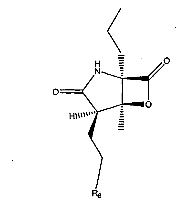
- R8 may include, for example, hydrogen, fluorine, chlorine, bromine and iodine.
- Still a further exemplary compound of Formula I is the compound having the following structure I-5:
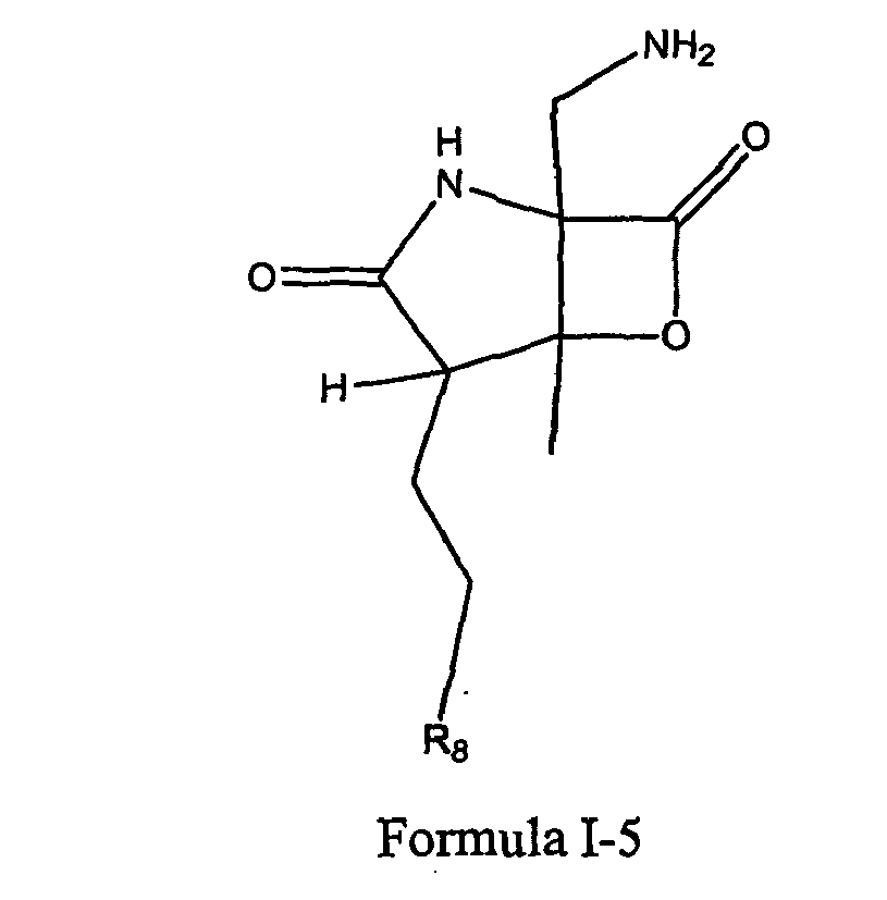
- R8 may include, for example, hydrogen, fluorine, chlorine, bromine and iodine.
- For example, the compound of Formula 1-5 may have the following stereochemistry:

- R8 may include, for example, hydrogen, fluorine, chlorine, bromine and iodine.
- In some embodiments, R2 of Formula I may be, for example, a 3-methylenecyclohexene. For example, the compound may have the following structure of Formula 1-6:

- R8 may include, for example, hydrogen, fluorine, chlorine, bromine and iodine.
- Preferably, the compound of Formula 1-6 may have the following. stereochemistry:
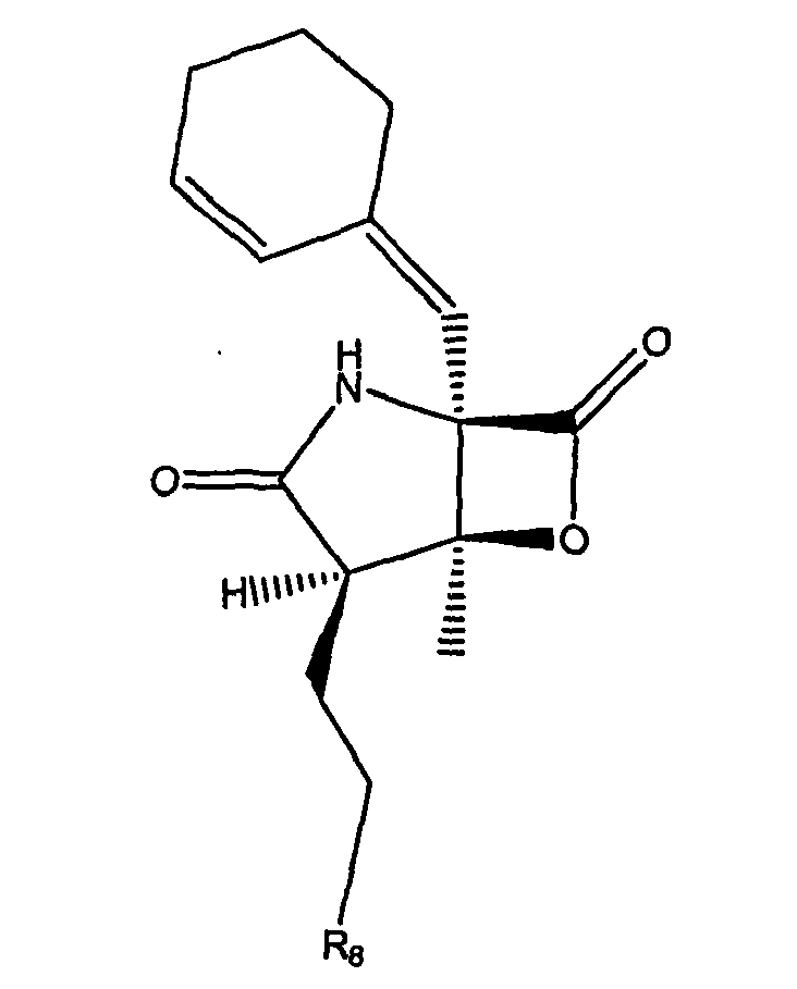
- R8 may include, for example, hydrogen, fluorine, chlorine, bromine and iodine.
- In other embodiments, R2 may be a cyclohexylalkylamine.
- Also, in other embodiments, R2 may be a C-Cyclohexyl-methyleneamine. In others, R2 may be a cyclohexanecarbaldehyde O-oxime.
- Furthermore, in some embodiments, R2 may be a cycloalkylacyl.
- Other embodiments provide compounds, and methods of producing a class of compounds, and pharmaceutically acceptable salts thereof, wherein the compounds are represented by Formula II:

- In certain embodiments the substituent(s) R1 R4 separately may include a hydrogen, a halogen, a mono-substituted, a poly-substituted or an unsubstituted variant of the following residues: saturated C1-C24 alkyl, unsaturated C2-C24 alkenyl, unsaturated C2-C24 alkynyl, acyl, acyloxy, alkyloxycarbonyloxy, aryloxycarbonyloxy, cycloalkyl, cycloalkenyl, alkoxy, cycloalkoxy, aryl, heteroaryl, arylalkoxy carbonyl, alkoxy carbonylacyl, amino, aminocarbonyl, aminocarbonyloxy, nitro, azido, phenyl, cycloalkylacyl, hydroxy, alkylthio, arylthio, oxysulfonyl, carboxy, cyano, and halogenated alkyl including polyhalogenated alkyl. R3 has the same meaning, except that it cannot be hydrogen. Further, E1, E3 and E4 is O, E2 is NH.
- n is equal to 1 or 2. When n is equal to 2, the substituents R1 can be the same or can be different. m is equal to 1 or 2, and when m is equal to 2, R4 can be the same or different.
- E5 is OH, O, OR10, S, SR11, SO2R11, NH, NH2, NOH, NHOH, NR12, and NHOR13, wherein R10-13 are selected from hydrogen, a substituted or unsubstituted of any of the following: alkyl, an aryl, a heteroaryl Also, R1 may be CH2CH2X, wherein X is selected from H, F, Cl, Br, and I. R3 may be methyl. Furthermore, R4 may include a cyclohexyl. Preferably, R1 may be CH2CH2X, wherein X is selected from the group consisting of H, F, Cl Br, and I; wherein R4 may include a cyclohexyl; wherein R3 may be methyl; and wherein each of E1, E3 and E4 separately may be O and E2 may be NH.
- For example, an exemplary compound of Formula II has the following structure II-1:

- R8 may include, for example, hydrogen, fluorine, chlorine, bromine and iodine.
- Exemplary stereochemistry may be as follows:
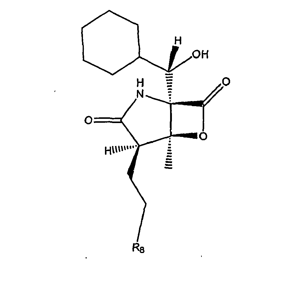
- In preferred embodiments, the compound of Formula II has any of the following structures:
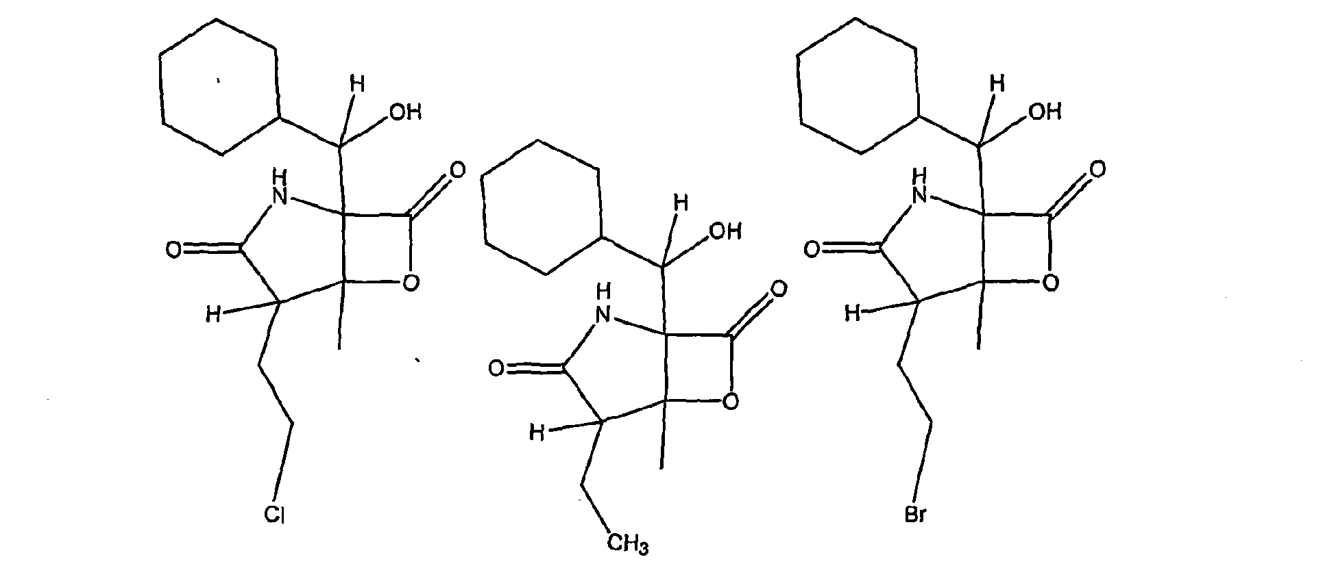
- The following is exemplary stereochemistry for compounds having the structures II-2, II-3, and II-4, respectively:

- In other embodiments wherein R4 may include a 7-oxa-bicyclo[4.1.0]hept-2-yl). An exemplary compound of Formula II is the following structure II-5:

- R8 may include, for example, hydrogen, fluorine, chlorine, bromine and iodine.
- The following are examples of compounds having the structure of Formula II-5:

- In still further embodiments, at least one R4 may include a subsituted or an unsubstituted branched alkyl. For example, a compound of Formula II may be the following structure II-6:

- R8 may include, for example, hydrogen, fluorine, chlorine, bromine and iodine.
- The following is exemplary stereochemistry for a compound having the structure of Formula II-6:

- As another example, the compound of Formula II may be the following structure II-7:

- R8 may include, for example, hydrogen, fluorine, chlorine, bromine and iodine.
- The following is exemplary stereochemistry for a compound having the structure of Formula II-7:

- In other embodiments, at least one R4 may be a cycloalkyl and E5 may be an oxygen. An exemplary compound of Formula II may be the following structure II-8:

- R8 may include, for example, hydrogen (II-8A), fluorine (II-8B), chlorine (II-8C); bromine (II-8D) and iodine (II-8E).
- The following is exemplary stereochemistry for a compound having the structure of Formula II-8:

- In some embodiments E5 may be an amine oxide, giving rise to an oxime. An exemplary compound of Formula II has the following structure II-9:

- R8 may include, for example, hydrogen, fluorine, chlorine, bromine and iodine; R may be hydrogen, and a substituted or unsubstituted alkyl, aryl, or heteroaryl, and the like.
- The following is exemplary stereochemistry for a compound having the structure of Formula II-9:

- A further exemplary compound of Formula II has the following structure II-10:

- R8 may include, for example, hydrogen, fluorine, chlorine, bromine and iodine.
- The following is exemplary stereochemistry for a compound having the structure of Formula II-10:
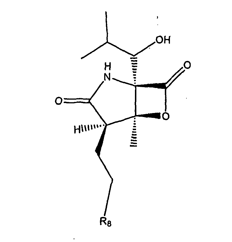
- In some embodiments, E5 may be NH2. An exemplary compound of Formula II has the following structure II-11:

- R8 may include, for example, hydrogen, fluorine, chlorine, bromine and iodine.
- The following is exemplary stereochemistry for a compound having the structure of Formula II-11:

- In some embodiments, at least one R4 may include a cycloalkyl and E5 may be NH2. An exemplary compound of Formula II has the following structure II-12:
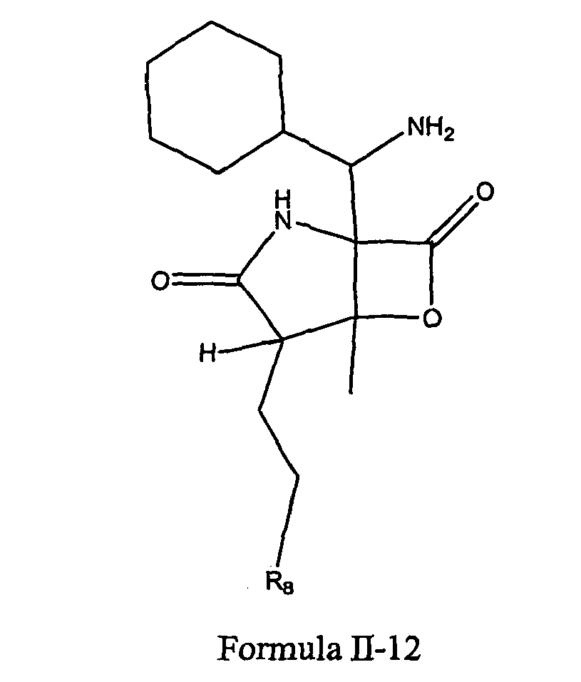
- R8 may include, for example, hydrogen, fluorine, chlorine, bromine and iodine.
- The following is exemplary stereochemistry for a compound having the structure of Formula II-12:

- A further exemplary compound of Formula II has the following structure II-13:

- R8 may include, for example, hydrogen (II-13A), fluorine (II-13B), chlorine (II-13C), bromine (II-13D) and iodine (II-13E).
- The following is exemplary stereochemistry for a compound having the structure of Formula II-13:

- A still further exemplary compound of Formula II has the following structure II-14:

- R8 may include, for example, hydrogen, fluorine, chlorine, bromine and iodine.
- The following is exemplary stereochemistry for a compound having the structure of Formula II-14:

- In some embodiments, the compounds of Formula II, may include as R4 at least one cycloalkene, for example. Furthermore, in some embodiments, the compounds may include a hydroxy at E5, for example. A further exemplary compound of Formula II has the following structure II-15:
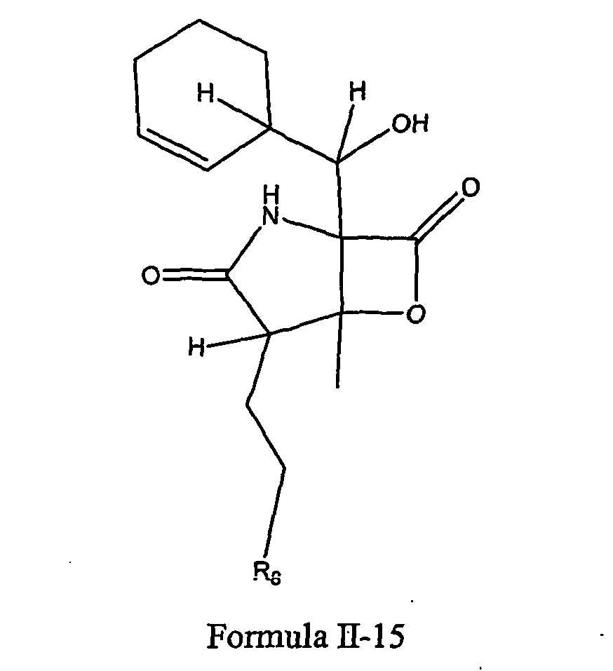
- R8 may include, for example, hydrogen, fluorine, chlorine, bromine and iodine.
- Exemplary stereochemistry may be as follows:

- The following is exemplary stereochemistry for compounds having the structures II-16, II-17, II-18, and II-19, respectively:

- The compounds of Formulae II-16, II-17, II-18 and II-19 may be obtained by fermentation, synthesis, or semi-synthesis and isolated/purified as set forth below. Furthermore, the compounds of Formulae II-16, II-17, II-18 and II-19 may be used, and are referred to, as "starting materials" to make other compounds described herein.
- In some embodiments, the compounds of Formula II, may include a methyl group as R1 for example. A further exemplary compound, Formula II-20, has the following structure and stereochemistry:
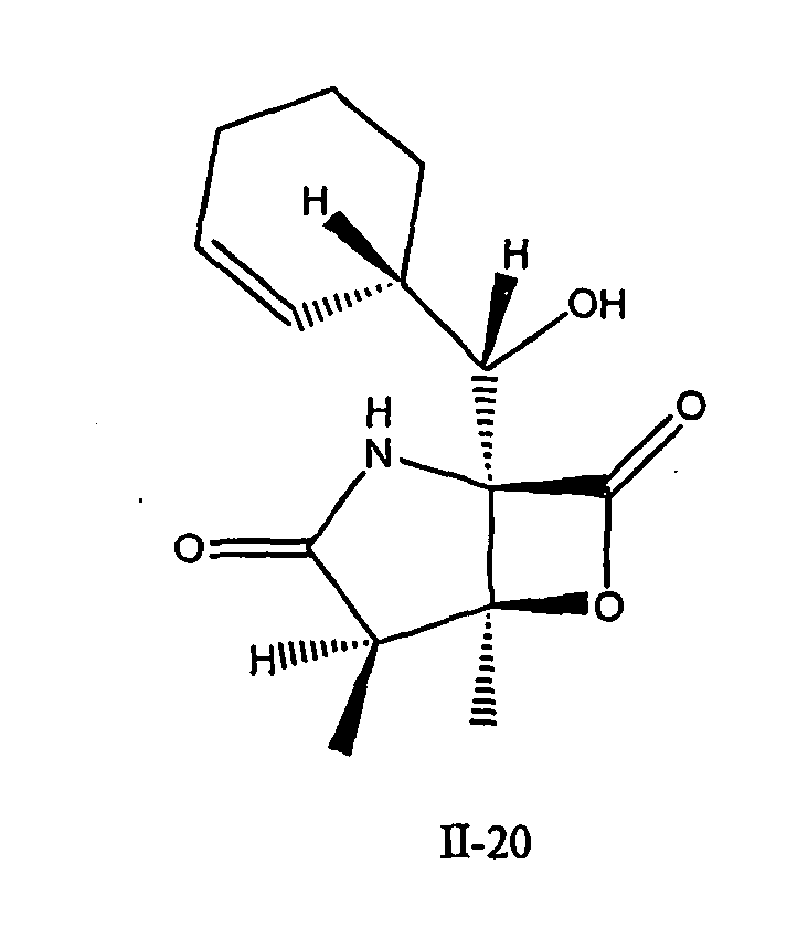
- In some embodiments, the compounds of Formula II, may include hydroxyethyl as R1, for example. A further exemplary compound, Formula II-21, has the following structure and stereochemistry:

- In some embodiments, the hydroxyl group of Formula II-21 may be esterified such that R1 may include ethylpropionate, for example. An exemplary compound, Formula II-22, has the following structure and stereochemistry:

- In some embodiments, the compounds of Formula II may include an ethyl group as R3, for example. A further exemplary compound of Formula II has the following structure II-23:

- R8 may include, for example, hydrogen, fluorine, chlorine, bromine and iodine. Exemplary stereochemistry may be as follows:

- In some embodiments, the compounds of Formula II-23 may have the following structure and stereochemistry, exemplified by Formula II-24C, where R8 is chlorine:
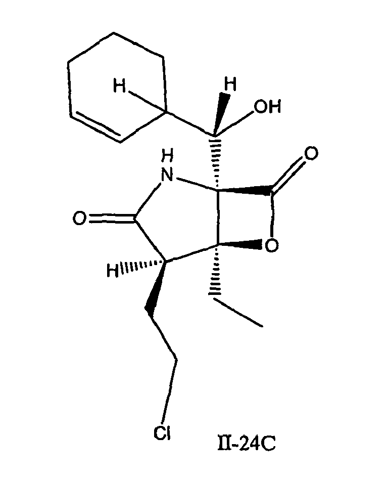
- In some embodiments, the compounds of Formula II-15 may have the following stereochemistry, exemplified by the compound of Formula II-25, where R8 is chlorine:

- Other embodiments provide compounds, and methods of producing a class of compounds, and pharmaceutically acceptable salts thereof, wherein the compounds are represented by Formula III:

- In certain embodiments, the substituent(s) R1 separately are selected from a hydrogen, a halogen, a mono-substituted, a poly-substituted or an unsubstituted variant of the following residues: saturated C1-C24 alkyl, unsaturated C2-C24 alkenyl, unsatured C2-C24 alkynyl, acyl, acyloxy, alkyloxycarbonyloxy, aryloxycarbonyloxy, cycloalkyl, cycloalkenyl, alkoxy, cycloalkoxy, aryl, heteroaryl, arylalkoxy carbonyl, alkoxy carbonylacyl amino, aminocarbonyl, aminocarbonyloxy nitro, azido, phenyl, hydroxy, alkylthio, arylthio, oxysulfonyl, carboxy, cyano, and halogenated alkyl including polyhalogenated alkyl, where n is equal to 1 or 2, and if n is equal to 2, then R1 can be the same or different;
- wherein R4 can be a hydrogen, a halogen, a mono-substituted, a poly-substituted or an unsubstituted variants of the following residues: saturated C1-C24 alkyl, unsaturated C2-C24 alkenyl, unsaturated C2-C24 alkynyl, acyl, acyloxy, alkyloxycarbonyloxy, aryloxycarbonyloxy, cycloalkyl, cycloalkenyl, alkoxy, cycloalkoxy, aryl, heteroaryl, arylalkoxy carbonyl, alkoxy carbonylacyl, amino, aminocarbonyl, aminocarbonyloxy, nitro, azido, phenyl, hydroxy, alkylthio, arylthio, oxysulfonyl, carboxy, cyano, and halogenated alkyl including polyhalogenated alkyl; wherein m is equal to 1 or 2, and where m is equal to 2, the substituents can the same or different. Also, E1, E3 and E4 is O, E2 is NH and ES is as defined for formula II.
- Other embodiments provide compounds, and methods of producing a class of compounds, and pharmaceutically acceptable salts thereof, wherein the compounds are represented by Formula IV:
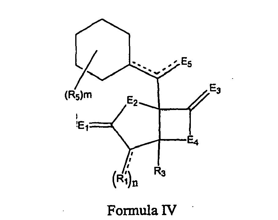
- In certain embodiments, the substituent(s) R1 and R5 are separately selected from a hydrogen, a halogen, a mono-substituted, a poly-substituted or an unsubstituted variants of the following residues: saturated C1-C24 alkyl, unsaturated C2-C24 alkenyl, unsaturated C2-C24 alkynyl, acyl, acyloxy, alkyloxycarbonyloxy, aryloxycarbonyloxy, cycloalkyl, cycloalkenyl, alkoxy, cycloalkoxy, aryl, heteroaryl, arylalkoxy carbonyl, alkoxy carbonylacyl, amino, aminocarbonyl, aminocarbonyloxy nitro, azido, phenyl, hydroxy, alkylthio, arylthio, oxysulfonyl, carboxy, cyano, and halogenated alkyl including polyhalogenated alkyl. R3 has the same meaning, except that it cannot be hydrogen. Also, E1,E3 and E4 is O, E2 is NH. n is equal to 1 or 2. When n is equal to 2, the substituents R1 can be the same or can be different. Also, m is 0, 1, 2, 3, 4, 5, 6, 7, 8, 9, 10, or 11. When m is greater than 1, the substituents R5 can be the same or different.
- In some embodiments R5 may give rise to a di-substituted cyclohexyl. An exemplary compound of Formula IV is the following structure IV-1, with and without exemplary stereochemistry:

- R8 may include, for example, hydrogen, fluorine, chlorine, bromine and iodine. The substituent(s) R6 and R7 may separately include a hydrogen, a halogen, a mono-substituted, a poly-substituted or an unsubstituted variants of the following residues: saturated C1-C24 alkyl, unsaturated C2-C24 alkenyl or C2-C24 alkynyl, acyl, acyloxy, alkyloxycarbonyloxy, aryloxycarbonyloxy, cycloalkyl, cycloalkenyl, alkoxy, cycloalkoxy, aryl, heteroaryl, arylalkoxy carbonyl, alkoxy carbonylacyl, amino; aminocarbonyl, aminocarboyloxy, nitro, azido, phenyl, hydroxy, alkylthio, arylthio, oxysulfonyl, carboxy, cyano, and halogenated alkyl including polyhalogenated alkyl. Further, R6 and R7 both may be the same or different.
- For example, an exemplary compound of Formula IV has the following structure IV-2:

- R8 may include, for example, hydrogen, fluorine, chlorine, bromine and iodine.
- Exemplary stereochemistry may be as follows:

- For example, an exemplary compound of Formula IV has the following structure IV-3:
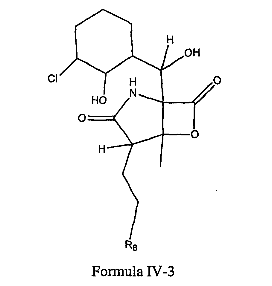
- R8 may include, for example, hydrogen (IV-3A), fluorine (IV-3B), chlorine, (IV-3C), bromine (IV-3D) and iodine (IV-3E).
- Exemplary structure and stereochemistry may be as follows:

- Additional exemplary structure and stereochemistry may be as follows:

- For example, an exemplary compound of Formula IV has the following structure IV-4:

- R8 may include, for example, hydrogen, fluorine, chlorine, bromine and iodine.
- Exemplary stereochemistry may be as follows:

- Some embodiments provide compounds, and methods of producing a class of compounds, pharmaceutically acceptable salts and pro-drug esters thereof, wherein the compounds are represented by Formula V:

- In certain embodiments, the substituent(s) R1 and R5 are separately selected from a hydrogen, a halogen, a mono-substituted, a poly-substituted or unsubstituted variants of the following residues: saturated C1-C24 alkyl, unsaturated C2-C24 alkenyl, unsaturated C2-C24 alkynyl, acyl, acyloxy, alkyloxycarbonyloxy, aryloxycarbonyloxy, cycloalkyl, cycloalkenyl, alkoxy, cycloalkoxy, aryl, heteroaryl, arylalkoxy carbonyl, alkoxy carbonylacyl, amino, aminocarbonyl, aminocarbonyloxy, nitro, azido, phenyl, hydroxy, alkylthio, arylthio, oxysulfonyl, carboxy, cyano, and halogenated alkyl including polyhalogenated alkyl. Futher, E1, E3 and E4 is O, E2 is NH wherein E5 is OH, O, OR10, S, SR11, SO2R11, NH, NH2, NOH, NHOH, NR12, or NHOR13, wherein R10, R11, R12, and R13 are selected from the group consisting of hydrogen, a substituted or unsubstituted alkyl, a substituted or unsubstituted aryl, and a substituted or unsubstituted heteroaryl. n is equal to 1 or 2, and when n is equal to 2, the substituents R1 can be the same or different.
m is 0, 1, 2, 3, 4, 5, 6, 7, 8, 9, 10, or 11. When m is greater than 1, R5 may be the same or different. - Certain embodiments also provide pharmaceutically acceptable salts of the compound of Formulae I-V. Also provided are methods of obtaining and purifying such compounds by the methods disclosed herein.
- The term "pharmaceutically acceptable salt," as used herein, and particularly when referring to a pharmaceutically acceptable salt of a compound, including Formulae I-V, and Formula I-V as produced and synthesized by the methods disclosed herein, refers to any pharmaceutically acceptable salts of a compound, and preferably refers to an acid addition salt of a compound. Preferred examples of pharmaceutically acceptable salt are the alkali metal salts (sodium or potassium), the alkaline earth metal salts (calcium or magnesium), or ammonium salts derived from ammonia or from pharmaceutically acceptable organic amines, for example C1- C7 alkylamine, cyclohexylamine, triethanolamine, ethylenediamine or tris-(hydroxymethyl)-aminomethane. With respect to compounds synthesized by the method of this embodiment that are basic amines, the preferred examples of pharmaceutically acceptable salts are acid addition salts of pharmaceutically acceptable inorganic or organic acids, for example, hydrohalic, sulfuric, phosphoric acid or aliphatic or aromatic carboxylic or sulfonic acid, for example acetic, succinic, lactic, malic, tartaric, citric, ascorbic, nicotinic, methanesulfonic, p-toluensulfonic or naphthalenesulfonic acid.
- Preferred pharmaceutical compositions disclosed herein include pharmaceutically acceptable salts of the compound of Formulae I-V obtained and purified by the methods disclosed herein. Accordingly, if the manufacture of pharmaceutical formulations involves intimate mixing of the pharmaceutical excipients and the active ingredient in its salt form, then it is preferred to use pharmaceutical excipients which are non-basic, that is, either acidic or neutral excipients.
- It will be also appreciated that the phrase "compounds and compositions comprising the compound," or any like phrase, is meant to encompass compounds in any suitable form for pharmaceutical delivery, as discussed in further detail herein. For example, in certain embodiments, the compounds or compositions comprising the same may include a pharmaceutically acceptable salt of the compound.
- The compounds are used to treat Liver, cancer or angioma.
The use may also include the steps of administering a compound or composition comprising the compound to an individual with these cancers. The compound or composition can be administered in an amount effective to treat the particular cancer
The compound or composition may be administered with a pharmaceutically acceptable carrier, diluent, excipient, and the like. - The cancer is liver cancer or angioma
- The term "halogen atom," as used herein, means any one of the radio-stable atoms of
column 7 of the Periodic Table of the Elements, i.e., fluorine, chlorine, bromine, or iodine, with bromine and chlorine being preferred. - The term "alkyl," as used herein, means any unbranched or branched, substituted or unsubstituted, saturated hydrocarbon, with C1-C6 unbranched, saturated, unsubstituted hydrocarbons being preferred, with methyl, ethyl, isobutyl, and tert-butylpropyl, and pentyl being most preferred. Among the substituted, saturated hydrocarbons, C1-C6 mono- and di- and per-halogen substituted saturated hydrocarbons and amino-substituted hydrocarbons are preferred, with perfluromethyl, perchloromethyl, perfluoro-tert-butyl, and perchloro-tert-butyl being the most preferred.
- The term "substituted'' has its ordinary meaning, as found in numerous contemporary patents from the related art. See, for example,
U.S. Patent Nos. 6,509,331 ;6,506,787 ;6,500,825 ;5,922,683 ;5,886,210 ;5,874,443 ; and6,350,759 . Specifically, the definition of substituted is as broad as that provided inU.S. Patent No. 6,509,331 , which defines the term "substituted alkyl" such that it refers to an alkyl group, preferably of from 1 to 10 carbon atoms, having from 1 to 5 substituents, and preferably 1 to 3 substituents, selected from the group consisting of alkoxy, substituted alkoxy, cycloalkyl, substituted cycloalkyl, cycloalkenyl, substituted cycloalkenyl, acyl, acylamino, acyloxy, amino, substituted amino, aminoacyl, aminoacyloxy, oxyacylamino, cyano, halogen, hydroxyl, carboxyl, carboxylalkyl, keto, thioketo, thiol, thioalkoxy,substituted thioalkoxy, aryl, aryloxy, heteroaryl, heteroaryloxy, heterocyclic, heterocyclooxy, hydroxyamino, alkoxyamino, nitro, --SO-alkyl, -SO-substituted alkyl, -SO-aryl, --SO-heteroaryl, --SO2-alkyl, --SO2-substituted alkyl, --SO2-aryl and -SO2-heteroaryl. The other above-listed patents also provide standard definitions for the term "substituted" that are well-understood by those of skill in the art. - The term "cycloalkyl" refers to any non-aromatic hydrocarbon ring, preferably having five to twelve atoms comprising the ring. The term "acyl" refers to alkyl or aryl groups derived from an oxoacid, with an acetyl group being preferred.
- The term "alkenyl," as used herein, means any unbranched or branched, substituted or unsubstituted, unsaturated hydrocarbon including polyunsaturated hydrocarbons, with C1-C6 unbranched, mono-unsaturated and di-unsaturated, unsubstituted hydrocarbons being preferred, and mono-unsaturated, di-halogen substituted hydrocarbons being most preferred. The term "cycloalkenyl" refers to any non-aromatic hydrocarbon ring, preferably having five to twelve atoms comprising the ring.
- The terms "aryl," "substituted aryl," "heteroaryl," and "substituted heteroaryl," as used herein, refer to aromatic hydrocarbon rings, preferably having five, six, or seven atoms, and most preferably having six atoms comprising the ring. "Heteroaryl" and "substituted heteroaryl," refer to aromatic hydrocarbon rings in which at least one heteroatom, e.g., oxygen, sulfur, or nitrogen atom, is in the ring along with at least one carbon atom. The term "heterocycle" or "heterocyclic" refer to any cyclic compound containing one or more heteroatoms. The substituted aryls, heterocycles and heteroaryls can be substituted with any substituent, including those described above and those known in the art.
- The term "alkoxy" refers to any unbranched, or branched, substituted or unsubstituted, saturated or unsaturated ether, with C1-C6 unbranched, saturated, unsubstituted ethers being preferred, with methoxy being preferred, and also with dimethyl, diethyl, methyl-isobutyl, and methyl-tert-butyl ethers also being preferred. The term "cycloalkoxy" refers to any non-aromatic hydrocarbon ring, preferably having five to twelve atoms comprising the ring. The term "alkoxy carbonyl" refers to any linear, branched, cyclic, saturated, unsaturated, aliphatic or aromatic alkoxy attached to a carbonyl group. The examples include methoxycarbonyl group, ethoxycarbonyl group, propyloxycarbonyl group, isopropyloxycarbonyl group, butoxycarbonyl group, sec-butoxycarbonyl group, tert-butoxycarbonyl group, cyclopentyloxycarbonyl group, cyclohexyloxycarbonyl group, benzyloxycarbonyl group, allyloxycarbonyl group, phenyloxycarbonyl group, and pyridyloxycarbonyl group.
- The terms "pure," "purified," "substantially purified," and "isolated" as used herein refer to the compound of the embodiment being free of other, dissimilar compounds with which the compound, if found in its natural state, would be associated in its natural state. In certain embodiments described as "pure," "purified," "substantially purified," or "isolated" herein, the compound may comprise at least 0.5%, 1%, 5%, 10%, or 20%, and most preferably at least 50% or 75% of the mass, by weight, of a given sample.
- The terms "derivative," "variant," or other similar term refers to a compound that is an analog of the other compound.
- Certain of the compounds of Formula I-V may be obtained and purified or may be obtained via semi-synthesis from purified compounds as set forth herein. Generally, the compounds of Formula II-15, preferably, Formulae II-16, II-17, II-18 and II-19, may be obtained synthetically or by fermentation. Exemplary fermentation procedures are provided below. Futher, the compounds of Formula II-15, preferably, Formulae II-16, II-17, II-18 and II-19 may be used as starting compounds in order to obtain/synthesize various of the other compounds described herein. Exemplary syntheses are provided herein.


- Formula II-16 is currently produced through a high-yield saline fermentation (∼200 mg/L) and modifications of the conditions has yielded new analogs in the fermentation extracts.
FIG. 1 shows the chemical structure of II-16. Additional analogs can be generated through directed biosynthesis. Directed biosynthesis is the modification of a natural product by adding biosynthetic precursor analogs to the fermentation of producing microorganisms (Lam, et al., J Antibiot (Tokyo) 44:934 (1991), Lam, et al., J Antibiot (Tokyo) 54:1 (2001)). - Exposing the producing culture to analogs of acetic acid, phenylalanine, valine, butyric acid, shikimic acid, and halogens, preferably, other than chlorine, can lead to the formation of new analogs. The new analogs produced can be easily detected in crude extracts by HPLC and LC-MS. For example, after manipulating the medium with different concentrations of sodium bromide, a bromo-analog, Formula II-18, was successfully produced in shake-flask culture at a titer of 14 mg/L.
- A second approach to generate new analogs is through biotransformation. Biotransformation reactions are chemical reactions catalyzed by enzymes or whole cells containing these enzymes. Zaks, A., Curr Opin Chem Biol 5: 130 (2001). Microbial natural products are ideal substrates for biotransformation reactions as they are synthesized by a series of enzymatic reactions inside microbial cells. Riva, S., Curr Opin Chem Biol 5:106 (2001).
- Given the structure of the described compounds, including those of Formula II-15, for example, the possible biosynthetic origins are acetyl-CoA, ethyhnalonyl-CoA, phenylalanine and chlorine. Ethylmalonyl-CoA is derived from butyryl-CoA, which can be derived either from valine or crotonyl-CoA. Liu, et al., Metab Eng 3:40 (2001). Phenylalanine is derived from shikimic acid.
- The production of compounds of Formulae II-16, II-17, and II-18 may be carried out by cultivating strain CNB476 in a suitable nutrient medium under conditions described herein, preferably under submerged aerobic conditions, until a substantial amount of compounds are detected in the fermentation; harvesting by extracting the active components from the fermentation broth with a suitable solvent; concentrating the solvent containing the desired components; then subjecting the concentrated material to chromatographic separation to isolate the compounds from other metabolites also present in the cultivation medium.
-
FIG. 2 shows some collection sites worldwide for the culture (CNB476), which is also refered to as Salinospora.FIG. 3 shows colonies of Salinospora.FIG. 4 shows the typical 16S rDNA sequence of the Salinospora. Bars represent characteristic signature nucleotides of the Salinospora that separate them from their nearest relatives. - The culture (CNB476) was deposited on June 20, 2003 with the American Type Culture Collection (ATCC) in Rockville, MD and assigned the ATCC patent deposition number PTA-5275. The ATCC deposit meets all of the requirements of the Budapest treaty. The culture is also maintained at and available from Nereus Pharmaceutical Culture Collection at 10480 Wateridge Circle, San Diego,
CA 92121 . In addition to the specific microorganism described herein, it should be understood that mutants, such as those produced by the use of chemical or physical mutagens including X-rays, etc. and organisms whose genetic makeup has been modified by molecular biology techniques, may also be cultivated to produce the starting compounds of Formulae II-16, II-17, and II-18. - Production of compounds can be achieved at temperature conducive to satisfactory growth of the producing organism, e.g. from 16 degree C to 40 degree C, but it is preferable to conduct the fermentation at 22 degree C to 32 degree C. The aqueous medium can be incubated for a period of time necessary to complete the production of compounds as monitored by high pressure liquid chromatography (HPLC), preferably for a period of about 2 to 10 days, on a rotary shaker operating at about 50 rpm to 400 rpm, preferably at 150 rpm to 250 rpm, for example.
- Growth of the microorganisms may be achieved by one of ordinary skill of the art by the use of appropriate medium. Broadly, the sources of carbon include glucose, fructose, mannose, maltose, galactose, mannitol and glycerol, other sugars and sugar alcohols, starches and other carbohydrates, or carbohydrate derivatives such as dextran, cerelose, as well as complex nutrients such as oat flour, corn meal, millet, corn, and the like. The exact quantity of the carbon source that is utilized in the medium will depend in part, upon the other ingredients in the medium, but an amount of carbohydrate between 0.5 to 25 percent by weight of the medium can be satisfactorily used, for example. These carbon sources can be used individually or several such carbon sources may be combined in the same medium, for example. Certain carbon sources are preferred as hereinafter set forth.
- The sources of nitrogen include amino acids such as glycine, arginine, threonine, methionine and the like, ammonium salt, as well as complex sources such as yeast extracts, corn steep liquors, distiller solubles, soybean meal, cotttonseed meal, fish meal, peptone, and the like. The various sources of nitrogen can be used alone or in combination in amounts ranging from 0.5 to 25 percent by weight of the medium, for example.
- Among the nutrient inorganic salts, which can be incorporated in the culture media, are the customary salts capable of yielding sodium, potassium, magnesium, calcium, phosphate, sulfate, chloride, carbonate, and like ions. Also included are trace metals such as cobalt, manganese, iron, molybdenum, zinc, cadmium, and the like.
- The compounds disclosed herein may be used to treat cancer. The treatments may include, for example, the step of administering an effective amount of a member of a class of new compounds.
- The compounds have various biological activities. For example, the compounds have chemosensitizing activity, and anti-cancer activity.
- The compounds have proteasome inhibitory activity. The proteasome inhibitory activity may, in whole or in part, contribute to the ability of the compounds to act as anti-cancer agents.
- The proteasome is a multisubunit protease that degrades intracellular proteins through its chymotrypsin-like, trypsin-like and peptidylglutamyl-peptide hydrolyzing (PGPH; and also know as the caspase-like activity) activities. The 26S proteasome contains a proteolytic core called the 20S proteasome and two 19S regulatory subunits. The 20S proteasome is responsible for the proteolytic activity against many substrates including damaged proteins, the transcription factor NF-κB and its inhibitor IκB, signaling molecules, tumor suppressors and cell cycle regulators. There are three distinct protease activities within the proteasome: 1) chymotrypsin-like; 2) trypsin-like; and the 3) peptidyl glutamyl peptide hydrolyzing (PGPH) activity.
- As an example, compounds of Formula II-16 were more potent (EC50 2nM) at inhibiting the chymotrypsin-like activity of rabbit muscle proteasomes than Omuralide (EC50 52 nM) and also inhibited the chymotrypsin-like activity of human erythrocyte derived proteasomes (EC50 ∼250pM).
FIG. 5 shows omuralide, which is a degradation product of Lactacystin, and it shows a compound of Formula II-16. Compounds of Formula II-16 exhibit a significant preference for inhibiting chymotrypsin-like activity of the proteasome over inhibiting the catalytic activity of chymotrypsin. Compounds of Formula II-16 also exhibit low nM trypsin-like inhibitory activity (∼10 nM), but are less potent at inhibiting the PGPH activity of the proteasome (EC50 ∼350 nM). - Additional studies have characterized the effects of compounds described herein, , including studies of Formula II-16 on the NF-κB/ IκB signaling pathway. Treatment of HEK293 cells (human embryonic kidney) with Tumor Necrosis Factor-alpha (TNF-α) induces phosphorylation and proteasome-mediated degradation of IκBα followed by NF-κB activation. To confirm proteasome inhibition, HEK293 cells were pre-treated for 1 hour with compounds of Formula II-16 followed by TNF-α stimulation. Treatment with compounds of Formula II-16 promoted the accumulation of phosphorylated IκBα suggesting that the proteasome-mediated IκBα degradation was inhibited.
- Furthermore, a stable HEK293 clone (NF-κB/Luc 293) was generated carrying a luciferase reporter gene under the regulation of 5x NF-κB binding sites. Stimulation of NF-κB/Luc 293 cells with TNF-α increases luciferase activity as a result of NF-κB activation while pretreatment with compounds of Formula II-16 decreases activity. Western blot analyses demonstrated that compounds of Formula II-16 promoted the accumulation of phosphorylated-IκBα and decreased the degradation of total IκBα in the NF-κB/Luc 293 cells. Compounds of Formula II-16 were also shown to increase the levels of the cell cycle regulatory proteins, p21 and p27.
- Tumor cells may be more sensitive to proteasome inhibitors than normal cells. Moreover, proteasome inhibition increases the sensitivity of cancer cells to anticancer agents. The cytotoxic activity of the compounds described herein, including Formula II-16, were examined for cytotoxic activity against various cancer cell lines. Formula II-16 was examined, for example, in the National Cancer Institute screen of 60 human tumor cell lines. Formula II-16 exhibited selective cytotoxic activity with a mean GI50 value (the concentration to achieve 50% growth inhibition) of less than 10 nM. The greatest potency was observed against SK-MEL-28 melanoma and MDA-MB-235 breast cancer cells [both with LC50 (the concentration with 50% cell lethality) <10 nM].
- A panel of cell lines including human colorectal (HT-29 and LoVo), prostate (PC3), breast (MDA-MB-231), lung (NCI-H292), ovarian (OVCAR3), acute T-cell leukemia (Jurkat), murine melanoma (B16-F10) and normal human fibroblasts (CCD-27sk) was treated with Salinosporamide A for 48h to assess cytotoxic activity. HT-29, LoVo, PC3, MDA-MB-231, NCI-H292, OVCAR3, Jurkat, and B16-F10 cells were sensitive with EC50 values of 47, 69, 78, 67, 97, 69, 10, and 33 nM, respectively. In contrast, the EC50 values for CCD-27sk cells were 196 nM. Treatment of Jurkat cells with Salinosporamide A at the approximate EC50 resulted in Caspase-3 activation and cleavage of PARP confirming the induction of apoptosis.
- The anti-anthrax activity of the described compounds was evaluated using an in vitro LeTx induced cytotoxicity assay. As one example, the results indicate that Formula II-16 is a potent inhibitor of LeTx-induced cytotoxicity of murine macrophage-like RAW264.7 cells. Treatment of RAW264.7 cells with Formula II-16 resulted in a 10-fold increase in the viability of LeTx-treated cells compared to LeTx treatment alone (average EC50 of <4 nM).
- Additional studies have characterized the effects of the compounds described herein on the NF-κB/ IκB signaling pathway (see the Examples). In unstimulated cells, the transcription factor nuclear factor-kappa B (NF-κB) resides in the cytoplasm in an inactive complex with the inhibitory protein IκB (inhibitor of NF-κB). Various stimuli can cause IκB phosphorylation by IκB kinase, followed by ubiquitination and degradation by the proteasome. Following the degradation of IκB, NF-κB translocates to the nucleus and regulates gene expression, affecting many cellular processes including inhibition of apoptosis. Chemotherapy agents such as CPT-11 (Irinotecan) can activate NF-κB in human colon cancer cell lines including LoVo cells, resulting in a decreased ability of these cells to undergo apoptosis. Painter, R.B. Cancer Res 38:4445 (1978). Velcade™ is a dipeptidyl boronic acid that inhibits the chymotrypsin-like activity of the proteasome (Lightcap, et al., Clin Chem 46:673 (2000), Adams, et al., Cancer Res 59:2615 (1999), Adams, Curr Opin Oncol 14:628 (2002)) while enhancing the trypsin and PGPH activities. Recently approved as a proteasome inhibitor, Velcade™, (PS-341; Millennium Pharmaceuticals, Inc.) has been shown to be directly toxic to cancer cells and also enhance the cytotoxic activity of Cat-11 in LoVo cells in vitro and in a LoVo xenograft model by inhibiting IκB degradation by the proteasome. Blum, et al., Ann Intern Med 80:249 (1974). In addition, Velcade™ was found to inhibit the expression of proangiogenic chemokines/cytokines Growth Related Oncogene-alpha (GRO-α) and Vascular Endothelial Growth Factor (VEGF) in squamous cell carcinoma, presumably through inhibition of the NF-κB pathway. Dick, et al., J Biol Chem 271:7273 (1996). These data suggest that proteasome inhibition may not only decrease tumor cell survival and growth, but also angiogenesis.
- Another potential application for proteasome inhibitors comes from recent studies on the biodefense Category A agent B. anthracis (anthrax). Anthrax spores are inhaled and lodge in the lungs where they are ingested by macrophages. Within the macrophage, spores germinate, the organism replicates, resulting ultimately in killing of the cell. Before killing occurs, however, infected macrophages migrate to the lymph nodes where, upon death, they release their contents allowing the organism to enter the bloodstream, further replicate, and secrete lethal toxins. Hanna, et al., Proc Natl Acad Sci U S A 90:10198 (1993). Anthrax toxins are responsible for the symptoms associated with anthrax. Two proteins that play a key role in the pathogenesis of anthrax are protective antigen (PA, 83 kDa) and lethal factor (LF, 90 kDa) which are collectively known as lethal toxin (LeTx). LF has an enzymatic function, but requires PA to achieve its biological effect. Neither PA or LF cause death individually; however, when combined they cause death when injected intravenously in animals. Kalns, et al., Biochem Biophys Res Commun 297:506 (2002), Kalns, et al., Biochem Biophys Res Commun 292:41 (2002).
- Protective antigen, the receptor-binding component of anthrax toxin, is responsible for transporting lethal factor into the host cell. PA oligomerizes into a ringshaped heptamer (see
FIG. 6 ). Each heptamer, bound to its receptor on the surface of a cell, has the ability to bind up to three molecules of LF. The complex formed between the PA heptamer and LF is taken into the cell by receptor-mediated endocytosis. Following endocytosis, LF is released into the cytosol where it attacks various cellular targets. Mogridge, et al., Biochemistry 41:1079 (2002), Lacy, et al., J Biol Chem 277:3006 (2002), Bradley, et al., Nature 414:225 (2001). - Lethal factor is a zinc dependent metalloprotease, which in the cytosol can cleave and inactivate signaling proteins of the mitogen-activated protein kinase kinase family (MAPKK). Duesbery, et al., Science 280:734 (1998), Bodart, et al., Cell Cycle 1:10 (2002), Vitale, et al., J Appl Microbiol 87:288 (1999), Vitale, et al., Biochem J 352 Pt 3:739 (2000). Of the seven different known MAPK kinases, six have been shown to be cleaved by LF. Within the cell, MAPK kinase pathways transduce various signals involved in cell death, proliferation, and differentiation making these proteins highly significant targets. However, certain inhibitors that prevent LeTx-induced cell death, do not prevent MAPKK cleavage by LF suggesting that this activity is not sufficient for induction of cell death. Kim, et al., J Biol Chem 278:7413 (2003), Lin, et al., Curr Microbiol 33:224 (1996).
- Studies have suggested that inhibition of the proteasome can prevent LeTx-induced cell death. Tang, et al., Infect Immun 67:3055 (1999). Data have shown that proteasome activity is required for LeTx-mediated killing of RAW264.7 macrophage-like cells and that proteasome inhibitors protect RAW264.7 cells from LeTx. Proteasome inhibition did not block MEK1 cleavage, suggesting the LeTx pathway is not blocked upstream of MEK1 cleavage in these studies. Additionally, there is no increase in proteasome activity in cells treated with LeTx. These data suggested that a novel, potent proteasome inhibitor like the compounds described herein, may also prevent LeTx-induced cell death as illustrated in
FIG. 6 . - The receptor for PA has been identified and is expressed by many cell types. Escuyer, et al., Infect Immun 59:3381 (1991). Lethal toxin is active in a few cell culture lines of macrophages causing cell death within a few hours. Hanna, et al., Proc Natl Acad Sci U S A 90:10198 (1993), Kim, et al., J Biol Chem 278:7413 (2003), Lin, et al., Curr Microbiol 33:224 (1996). LeTx can induce both necrosis and apoptosis in mouse macrophage-like RAW264.7 and J774A.1 cells upon in vitro treatment.
- The results indicate that the compounds described herein act as a potent inhibitor of LeTx-induced cytotoxicity of murine macrophage-like RAW264.7 cells. Treatment of RAW264.7 cells with, for example, compounds of Formula II-16, resulted in a 10-fold increase in the viability of LeTx-treated cells compared to LeTx treatment alone (average EC5o of <4 nM) and therefore provide a valuable therapy for anthrax infections. Formula II-16, for example, promoted survival of RAW264.7 macrophage-like cells in the presence of LeTx indicating that this compound and its derivatives provide a valuable clinical therapeutic for anthrax infection.
- In one embodiment, the compounds disclosed herein are used in pharmaceutical compositions. The compounds preferably can be produced by the methods disclosed herein. The compounds can be used, for example, in pharmaceutical compositions comprising a pharmaceutically acceptable carrier prepared for storage and subsequent administration. Also, disclosed is a pharmaceutically effective amount of the products and compounds disclosed above in a pharmaceutically acceptable carrier or diluent. Acceptable carriers or diluents for therapeutic use are well known in the pharmaceutical art, and are described, for example, in Remington's Pharmaceutical Sciences, Mack Publishing Co. (A.R. Gennaro edit. 1985). Preservatives, stabilizers, dyes and even flavoring agents may be provided in the pharmaceutical composition. For example, sodium benzoate, ascorbic acid and esters of p-hydroxybenzoic acid may be added as preservatives. In addition, antioxidants and suspending agents may be used.
- The compositions, particularly those of Formulae I-V, may be formulated and used as tablets, capsules, or elixirs for oral administration; suppositories for rectal administration; sterile solutions, suspensions for injectable administration; patches for transdermal administration, and sub-dermal deposits and the like. Injectables can be prepared in conventional forms, either as liquid solutions or suspensions, solid forms suitable for solution or suspension in liquid prior to injection, or as emulsions. Suitable excipients are, for example, water, saline, dextrose, mannitol, lactose, lecithin, albumin, sodium glutamate, cysteine hydrochloride, and the like. In addition, if desired, the injectable pharmaceutical compositions may contain minor amounts of nontoxic auxiliary substances, such as wetting agents, pH buffering agents, and the like. If desired, absorption enhancing preparations (for example, liposomes), may be utilized.
- Pharmaceutical formulations for parenteral administration include aqueous solutions of the active compounds in water-soluble form. Additionally, suspensions of the active compounds may be prepared as appropriate oily injection suspensions. Suitable lipophilic solvents or vehicles include fatty oils such as sesame oil, or other organic oils such as soybean, grapefruit or almond oils, or synthetic fatty acid esters, such as ethyl oleate or triglycerides, or liposomes. Aqueous injection suspensions may contain substances that increase the viscosity of the suspension, such as sodium carboxymethyl cellulose, sorbitol, or dextran. Optionally, the suspension may also contain suitable stabilizers or agents that increase the solubility of the compounds to allow for the preparation of highly concentrated solutions.
- Pharmaceutical preparations for oral use can be obtained by combining the active compounds with solid excipient, optionally grinding a resulting mixture, and processing the mixture of granules, after adding suitable auxiliaries, if desired, to obtain tablets or dragee cores. Suitable excipients are, in particular, fillers such as sugars, including lactose, sucrose, mannitol, or sorbitol; cellulose preparations such as, for example, maize starch, wheat starch, rice starch, potato starch, gelatin, gum tragacanth, methyl cellulose, hydroxypropylmethyl-cellulose, sodium carboxymethylcellulose, and/or polyvinylpyrrolidone (PVP). If desired, disintegrating agents may be added, such as the cross-linked polyvinyl pyrrolidone, agar, or alginic acid or a salt thereof such as sodium alginate. Dragee cores are provided with suitable coatings. For this purpose, concentrated sugar solutions may be used, which may optionally contain gum arabic, talc, polyvinyl pyrrolidone, carbopol gel, polyethylene glycol, and/or titanium dioxide, lacquer solutions, and suitable organic solvents or solvent mixtures. Dyestuffs or pigments may be added to the tablets or dragee coatings for identification or to characterize different combinations of active compound doses. For this purpose, concentrated sugar solutions may be used, which may optionally contain gum arabic, talc, polyvinyl pyrrolidone, carbopol gel, polyethylene glycol, and/or titanium dioxide, lacquer solutions, and suitable organic solvents or solvent mixtures. Dyestuffs or pigments may be added to the tablets or dragee coatings for identification or to characterize different combinations of active compound doses. Such formulations can be made using methods known in the art (see, for example,
U.S. Patent Nos. 5,733,888 (injectable compositions);5,726,181 (poorly water soluble compounds);5,707,641 (therapeutically active proteins or peptides);5,667,809 (lipophilic agents);5,576,012 (solubilizing polymeric agents);5,707,615 (anti-viral formulations);5,683,676 (particulate medicaments);5,654,286 (topical formulations);5,688,529 (oral suspensions);5,445,829 (extended release formulations);5,653,987 (liquid formulations);5,641,515 (controlled release formulations) and5,601,845 (spheroid formulations). - Further disclosed herein are various pharmaceutical compositions well known in the pharmaceutical art for uses that include intraocular, intranasal, and intraauricular delivery. Pharmaceutical formulations include aqueous ophthalmic solutions of the active compounds in water-soluble form, such as eyedrops, or in gellan gum (Shedden et al., Clin. Ther., 23(3):440-50 (2001)) or hydrogels (Mayer et al., Ophthalmologica, 210(2):101-3 (1996)); ophthalmic ointments; ophthalmic suspensions, such as microparticulates, drug-containing small polymeric particles that are suspended in a liquid carrier medium (Joshi, A. 1994 J Ocul Pharmacol 10:29-45), lipid-soluble formulations (Alm et al., Prog. Clin. Biol. Res., 312:447-58 (1989)), and microspheres (Mordenti, Toxicol. Sci., 52(1):101-6 (1999)); and ocular inserts. Such suitable pharmaceutical formulations are most often and preferably formulated to be sterile, isotonic and buffered for stability and comfort. Pharmaceutical compositions may also include drops and sprays often prepared to simulate in many respects nasal secretions to ensure maintenance of normal ciliary action. As disclosed in Remington's Pharmaceutical Sciences (Mack Publishing, 18h Edition), and well-known to those skilled in the art, suitable formulations are most often and preferably isotonic, slightly buffered to maintain a pH of 5.5 to 6.5, and most often and preferably include anti-microbial preservatives and appropriate drug stabilizers. Pharmaceutical formulations for intraauricular delivery include suspensions and ointments for topical application in the ear. Common solvents for such aural formulations include glycerin and water.
- When used as an anti-cancer compound, for example, the compounds of Formulae I-V or compositions including Formulae I-V can be administered by either oral or non-oral pathways. When administered orally, it can be administered in capsule, tablet, granule, spray, syrup, or other such form. When administered non-orally, it can be administered as an aqueous suspension, an oily preparation or the like or as a drip, suppository, salve, ointment or the like, when administered via injection, subcutaneously, intraperitoneally, intravenously, intramuscularly, or the like.
- In one embodiment, the anti-cancer agents can be mixed with additional substances to enhance their effectiveness.
- Administration modes include, among others, (a) administration though oral pathways, which administration includes administration in capsule, tablet, granule, spray, syrup, or other such forms; (b) administration through non-oral pathways, which administration includes administration as an aqueous suspension, an oily preparation or the like or as a drip, suppository, salve, ointment or the like; administration via injection, subcutaneously, intraperitoneally, intravenously, intramuscularly, intradermally, or the like; as well as (c) administration topically, (d) administration rectally, or (e) administration vaginally, as deemed appropriate by those of skill in the art for bringing the compound of the present embodiment into contact with living tissue; and (f) administration via controlled released formulations, depot formulations, and infusion pump delivery. As further examples of such modes of administration and as further disclosure of modes of administration, disclosed herein are various methods for administration of the disclosed chemical compounds and pharmaceutical compositions including modes of administration through intraocular, intranasal, and intraauricular pathways.
- The pharmaceutically effective amount of the compositions that include the described compounds, including those of Formulae I-V, required as a dose will depend on the route of administration, the type of animal, including human, being treated, and the physical characteristics of the specific animal under consideration. The dose can be tailored to achieve a desired effect, but will depend on such factors as weight, diet, concurrent medication and other factors which those skilled in the medical arts will recognize.
- The products or compositions can be used alone or in combination with one another, or in combination with other therapeutic or diagnostic agents. These products can be utilized in vivo, ordinarily in a mammal, preferably in a human, or in vitro. In employing them in vivo, the products or compositions can be administered to the mammal in a variety of ways, including parenterally, intravenously, subcutaneously, intramuscularly, colonically, rectally, vaginally, nasally or intraperitoneally, employing a variety of dosage forms. Such administrations may also be applied to testing chemical activity in vivo.
- As will be readily apparent to one skilled in the art, the useful in vivo dosage to be administered and the particular mode of administration will vary depending upon the age, weight and mammalian species treated, the particular compounds employed, and the specific use for which these compounds are employed. The determination of effective dosage levels, that is the dosage levels necessary to achieve the desired result, can be accomplished by one skilled in the art using routine pharmacological methods. Typically, human clinical applications of products are commenced at lower dosage levels, with dosage level being increased until the desired effect is achieved. Alternatively, acceptable in vitro studies can be used to establish useful doses and routes of administration of the compositions identified by the present methods using established pharmacological methods.
- In non-human animal studies, applications of potential products are commenced at higher dosage levels, with dosage being decreased until the desired effect is no longer achieved or adverse side effects disappear. The dosage may range broadly, depending upon the desired affects and the therapeutic indication. Typically, dosages may be between about 10 microgram/kg and 100 mg/kg body weight, preferably between about 100 microgram/kg and 10 mg/kg body weight. Alternatively dosages may be based and calculated upon the surface area of the patient, as understood by those of skill in the art. Administration is preferably oral on a daily or twice daily basis.
- The exact formulation, route of administration and dosage can be chosen by the individual physician in view of the patient's condition. See for example, Fingl et al., in The Pharmacological Basis of Therapeutics, 1975. It should be noted that the attending physician would know how to and when to terminate, interrupt, or adjust administration due to toxicity, or to organ dysfunctions. Conversely, the attending physician would also know to adjust treatment to higher levels if the clinical response were not adequate (precluding toxicity). The magnitude of an administrated dose in the management of the disorder of interest will vary with the severity of the condition to be treated and to the route of administration. The severity of the condition may, for example, be evaluated, in part, by standard prognostic evaluation methods. Further, the dose and perhaps dose frequency, will also vary according to the age, body weight, and response of the individual patient. A program comparable to that discussed above may be used in veterinary medicine.
- Depending on the specific conditions being treated, such agents may be formulated and administered systemically or locally. A variety of techniques for formulation and administration may be found in Remington's Pharmaceutical Sciences, 18th Ed., Mack Publishing Co., Easton, PA(1990). Suitable administration routes may include oral, rectal, transdermal, vaginal, transmucosal, or intestinal administration; parenteral delivery, including intramuscular, subcutaneous, intramedullary injections, as well as intrathecal, direct intraventricular, intravenous, intraperitoneal, intranasal, or intraocular injections.
- For injection, the agents of the embodiment may be formulated in aqueous solutions, preferably in physiologically compatible buffers such as Hanks' solution, Ringer's solution, or physiological saline buffer. For such transmucosal administration, penetrants appropriate to the barrier to be permeated are used in the formulation. Such penetrants are generally known in the art. Use of pharmaceutically acceptable carriers to formulate the compounds herein disclosed for the practice of the embodiment into dosages suitable for systemic administration is within the scope of the embodiment. With proper choice of carrier and suitable manufacturing practice, the compositions disclosed herein, in particular, those formulated as solutions, may be administered parenterally, such as by intravenous injection. The compounds can be formulated readily using pharmaceutically acceptable carriers well known in the art into dosages suitable for oral administration. Such carriers enable the compounds of the embodiment to be formulated as tablets, pills, capsules, liquids, gels, syrups, slurries, suspensions and the like, for oral ingestion by a patient to be treated.
- Agents intended to be administered intracellularly may be administered using techniques well known to those of ordinary skill in the art. For example, such agents may be encapsulated into liposomes, then administered as described above. All molecules present in an aqueous solution at the time of liposome formation are incorporated into the aqueous interior. The liposomal contents are both protected from the external microenvironment and, because liposomes fuse with cell membranes, are efficiently delivered into the cell cytoplasm. Additionally, due to their hydrophobicity, small organic molecules may be directly administered intracellularly.
- Determination of the effective amounts is well within the capability of those skilled in the art, especially in light of the detailed disclosure provided herein. In addition to the active ingredients, these pharmaceutical compositions may contain suitable pharmaceutically acceptable carriers comprising excipients and auxiliaries which facilitate processing of the active compounds into preparations which can be used pharmaceutically. The preparations formulated for oral administration may be in the form of tablets, dragees, capsules, or solutions. The pharmaceutical compositions may be manufactured in a manner that is itself known, for example, by means of conventional mixing, dissolving, granulating, dragee-making, levitating, emulsifying, encapsulating, entrapping, or lyophilizing processes.
- Compounds disclosed herein can be evaluated for efficacy and toxicity using known methods. For example, the toxicology of a particular compound, or of a subset of the compounds, sharing certain chemical moieties, may be established by determining in vitro toxicity towards a cell line, such as a mammalian, and preferably human, cell line. The results of such studies are often predictive of toxicity in animals, such as mammals, or more specifically, humans. Alternatively, the toxicity of particular compounds in an animal model, such as mice, rats, rabbits, dogs or monkeys, may be determined using known methods. The efficacy of a particular compound may be established using several art recognized methods, such as in vitro methods, animal models, or human clinical trials. Art-recognized in vitro models exist for nearly every class of condition, including the conditions abated by the compounds disclosed herein, including cancer, cardiovascular disease, and various immune dysfunction, and infectious diseases. Similarly, acceptable animal models may be used to establish efficacy of chemicals to treat such conditions. When selecting a model to determine efficacy, the skilled artisan can be guided by the state of the art to choose an appropriate model, dose, and route of administration, and regime. Of course, human clinical trials can also be used to determine the efficacy of a compound in humans.
- When used as an anti-cancer agent, the compounds disclosed herein may be administered by either oral or a non-oral pathways. When administered orally, it can be administered in capsule, tablet, granule, spray, syrup, or other such form. When administered non-orally, it can be administered as an aqueous suspension, an oily preparation or the like or as a drip, suppository, salve, ointment or the like, when administered via injection, subcutaneously, intraperitoneally, intravenously, intramuscularly, intradermally, or the like. Controlled release formulations, depot formulations, and infusion pump delivery are similarly contemplated.
- The compositions disclosed herein in pharmaceutical compositions may also comprise a pharmaceutically acceptable carrier. Such compositions may be prepared for storage and for subsequent administration. Acceptable carriers or diluents for therapeutic use are well known in the pharmaceutical art, and are described, for example, in Remington's Pharmaceutical Sciences, Mack Publishing Co. (A.R. Gennaro edit. 1985). For example, such compositions may be formulated and used as tablets, capsules or solutions for oral administration; suppositories for rectal or vaginal administration; sterile solutions or suspensions for injectable administration. Injectables can be prepared in conventional forms, either as liquid solutions or suspensions, solid forms suitable for solution or suspension in liquid prior to injection, or as emulsions. Suitable excipients include, but are not limited to, saline, dextrose, mannitol, lactose, lecithin, albumin, sodium glutamate, cysteine hydrochloride, and the like. In addition, if desired, the injectable pharmaceutical compositions may contain minor amounts of nontoxic auxiliary substances, such as wetting agents, pH buffering agents, and the like. If desired, absorption enhancing preparations (for example, liposomes), may be utilized.
- The pharmaceutically effective amount of the composition required as a dose will depend on the route of administration, the type of animal being treated, and the physical characteristics of the specific animal under consideration. The dose can be tailored to achieve a desired effect, but will depend on such factors as weight, diet, concurrent medication and other factors which those skilled in the medical arts will recognize.
- The products or compositions of the embodiment, as described above, may be used alone or in combination with one another, or in combination with other therapeutic or diagnostic agents. These products can be utilized in vivo or in vitro. The useful dosages and the most useful modes of administration will vary depending upon the age, weight and animal treated, the particular compounds employed, and the specific use for which these composition or compositions are employed. The magnitude of a dose in the management or treatment for a particular disorder will vary with the severity of the condition to be treated and to the route of administration, and depending on the disease conditions and their severity, the compositions may be formulated and administered either systemically or locally. A variety of techniques for formulation and administration may be found in Remington's Pharmaceutical Sciences, 18th ed., Mack Publishing Co., Easton, PA (1990).
- To formulate the compounds of Formulae I-V as an an anti-cancer agent, known surface active agents, excipients, smoothing agents, suspension agents and pharmaceutically acceptable film-forming substances and coating assistants, and the like may be used. Preferably alcohols, esters, sulfated aliphatic alcohols, and the like may be used as surface active agents; sucrose, glucose, lactose, starch, crystallized cellulose, mannitol, light anhydrous silicate, magnesium aluminate, magnesium methasilicate aluminate, synthetic aluminum silicate, calcium carbonate, sodium acid carbonate, calcium hydrogen phosphate, calcium carboxymethyl cellulose, and the like may be used as excipients; magnesium stearate, talc, hardened oil and the like may be used as smoothing agents; coconut oil, olive oil, sesame oil, peanut oil, soya may be used as suspension agents or lubricants; cellulose acetate phthalate as a derivative of a carbohydrate such as cellulose or sugar, or methylacetate-methacrylate copolymer as a derivative of polyvinyl may be used as suspension agents; and plasticizers such as ester phthalates and the like may be used as suspension agents. In addition to the foregoing preferred ingredients, sweeteners, fragrances, colorants, preservatives and the like may be added to the administered formulation of the compound produced by the method of the embodiment, particularly when the compound is to be administered orally.
- The compounds and compositions may be orally or non-orally administered to a human patient in the amount of about 0.001 mg/kg/day to about 10,000 mg/kg/day of the active ingredient, and more preferably about 0.1 mg/kg/day to about 100 mg/kg/day of the active ingredient at, preferably, one time per day or, less preferably, over two to about ten times per day. Alternatively and also preferably, the compound produced by the method of the embodiment may preferably be administered in the stated amounts continuously by, for example, an intravenous drip. Thus, for the example of a patient weighing 70 kilograms, the preferred daily dose of the active ingredient would be about 0.07 mg/day to about 700 gm/day, and more preferable, 7 mg/day to about 7 grams/day. Nonetheless, as will be understood by those of skill in the art, in certain situations it may be necessary to administer the anti-cancer compound of the embodiment in amounts that excess, or even far exceed, the above-stated, preferred dosage range to effectively and aggressively treat particularly advanced cancers.
Usable organic solvents include, for example, methanol, methylsulfoxide, and the like. The formulation can, for example, be a powder, granular or other solid inhibitor, or a liquid inhibitor prepared using an organic solvent or a hydrous organic solvent. While a preferred concentration of the compound produced by the method of the embodiment for use as an anticancer or anti-tumor compound is generally in the range of about 1 to about 100 µg/ml, the most appropriate use amount varies depending on the type of cultured cell system and the purpose of use, as will be appreciated by persons of ordinary skill in the art. Also, in certain applications it may be necessary or preferred to persons of ordinary skill in the art to use an amount outside the foregoing range. - In one embodiment, the method of using a compound as an anti-cancer agent involves administering an effective amount of any of the compounds of Formulae I-V or compositions of those compounds. .
- As will be understood by one of skill in the art, "need" is not an absolute term and merely implies that the patient can benefit from the treatment of the anti-cancer agent in use. By "patient" what is meant is an organism that can benefit by the use of an anti-cancer agent.
For example, any organism with cancer, such as, lives cancer or angioma may benefit from the application of an anti-cancer agent that may in turn reduce the amount of cancer present in the patient.
In one embodiment, the patient's health may not require that an, anti-cancer agent be administered, however, the patient may still obtain some benefit by the reduction of the level of cancer cells present in the patient, and thus be in need. In one embodiment, the anti-cancer agent is effective against one type of cancer, but not against other types; thus, allowing a high degree of selectivity in the treatment of the patient.
. In choosing such an anti-cancer agent, the methods and results disclosed in the Examples may be useful.
. In embodiments, the anti-cancer agent may be effective against a broad spectrum of cancers. In still further embodiments, the anti-cancer agent is effective against a broad spectrum of cancers or all cancers. Cancers, against which the compounds are effective include a liver cancer and angioma.
"Therapeutically effective amount," "pharmaceutically effective amount," or similar term, means that amount of drug or pharmaceutical agent that will result in a biological or medical response of a cell, tissue, system, animal, or human that is being sought. In a preferred embodiment, the medical response is one sought by a researcher, veterinarian, medical doctor, or other clinician.
"Anti-cancer agent" refers to a compound or composition including the compound that reduces the likelihood of survival of a cancer cell. In one embodiment, the likelihood of survival is determined as a function of an individual cancer cell; thus, the anti-cancer agent will increase the chance that an individual cancer cell will die. In one embodiment, the likelihood of survival is determined as a function of a population of cancer cells; thus, the anti-cancer agent will increase the chances that there will be a decrease in the population of cancer cells. In one embodiment, anti-cancer agent means chemotherapeutic agent or other similar term. - A "chemotherapeutic agent" is a chemical compound useful in the treatment of a neoplastic disease, such as cancer. Examples of chemotherapeutic agents include alkylating agents, such as a nitrogen mustard, an ethyleneimine and a methylmelamine, an alkyl sulfonate, a nitrosourea, and a triazene, folic acid antagonists, anti-metabolites of nucleic acid metabolism, antibiotics, pyrimidine analogs, 5-fluorouracil, cisplatin, purine nucleosides, amines, amino acids, triazol nucleosides, corticosteroids, a natural product such as a vinca alkaloid, an epipodophyllotoxin, an antibiotic, an enzyme, a taxane, and a biological response modifier; miscellaneous agents such as a platinum coordination complex, an anthracenedione, an anthracycline, a substituted urea, a methyl hydrazine derivative, or an adrenocortical suppressant; or a hormone or an antagonist such as an adrenocorticosteroid, a progestin, an estrogen, an antiestrogen, an androgen, an antiandrogen, or a gouadotropin-releasing hormone analog. Specific examples include Adriamycin, Doxorubicin, 5-Fluorouracil, Cytosine arabinoside ("Ara-C"), Cyclophosphamide, Thiotepa, Busulfan, Cytoxin, Taxol, Toxotere, Methotrexate, Cisplatin, Melphalan, Vinblastine, Bleomycin, Etoposide, Ifosfamide, Mitomycin C, Mitoxantrone, Vincreistine, Vinorelbine, Carboplatin, Teniposide, Daunomycin, Carminomycin, Aminopterin, Dactinomycin, Mitomycins, Esperamicins, Melphalan, and other related nitrogen mustards. Also included in this definition are hormonal agents that act to regulate or inhibit hormone action on tumors, such as tamoxifen and onapristone.
- The anti-cancer agent may act directly upon a cancer cell to kill the cell, induce death of the cell or to prevent division of the cell. Alternatively, the anti-cancer agent may indirectly act upon the cancer cell by limiting nutrient or blood supply to the cell, for example. Such anti-cancer agents are capable of destroying or suppressing the growth or reproduction of cancer cells, such as a colorectal carcinoma, a prostate carcinoma, a breast adenocarcinoma, a non-small cell lung carcinoma, an ovarian carcinoma, multiple myelomas, a melanoma, and the like.
- A "neoplastic disease" or a "neoplasm" refers to a cell or a population of cells, including a tumor or tissue (including cell suspensions such as bone marrow and fluids such as blood or serum), that exhibits abnormal growth by cellular proliferation greater than normal tissue. Neoplasms can be benign or malignant.
- In one embodiment, a described compound, preferably a compound having the Formulae I-V, including those as described herein, is considered an effective anti-cancer agent if the compound can influence 10% of the cancer cells, for example. In a more preferred embodiment, the compound is effective if it can influence 10 to 50% of the cancer cells. In an even more preferred embodiment, the compound is effective if it can influence 50-80% of the cancer cells.
In an even more preferred embodiment, the compound is effective if it can influence 80-95% of the cancer cells. In an even more preferred embodiment, the compound is effective if it can influence 95-99% of the cancer cells. "Influence" is defined by the mechanism of action for each compound.
. For example, if a compound prevents the division of cancer cells, then influence is a measure of prevention of cancer cell division.
Not all mechanisms of action need be at the same percentage of effectiveness. In an alternative embodiment, a low percentage effectiveness may be desirable if the lower degree of effectiveness is offset by other factors, such as the specificity of the compound, for example. Thus a compound that is only 10% effective, for example, but displays little in the way of harmful side-effects to the host, or cells, can still be considered effective. - In one embodiment, the compounds described herein are administered simply to remove cancer cells, and need not be administered to a patient.
As another example, the compounds can be administered ex vivo to a cell sample, such as a bone marrow or stem cell transplant to ensure that only non-cancerous cells are introduced into the recipient. After the compounds are administered they may optionally be removed.
Whether or not this is an option will depend upon the relative needs of the situation and the risks associated with the compound, which in part can be determined as described in the Examples below. - The following examples are meant to describe the preferred embodiments.
- Strain CNB476 was grown in a 500-ml flask containing 100 ml of vegetative medium consisting of the following per liter of deionized water: glucose, 4 g; Bacto tryptone, 3 g; Bacto casitone, 5 g; and synthetic sea salt (Instant Ocean, Aquarium Systems), 30 g. The first seed culture was incubated at 28 degree C for 3 days on a rotary shaker operating at 250 rpm. Four ml each of the first seed culture was inoculated into three 500-ml flasks containing of 100 ml of the vegetative medium. The second seed cultures were incubated at 28 degree C and 250 rpm on a rotary shaker for 2 days. Four ml each of the second seed culture was inoculated into thirty-five 500-ml flasks containing of 100 ml of the vegetative medium. The third seed cultures were incubated at 28 degree and 250 rpm on a rotary shaker for 2 days. Four ml each of the third seed culture was inoculated into four hundred 500-ml flasks containing 100 ml of the production medium consisting of the following per liter of deionized water: starch, 10 g; yeast extract, 4 g; Hy-Soy, 4 g; ferric sulfate, 40 mg; potassium bromide, 100 mg; calcium carbonate, 1 g; and synthetic sea salt (Instant Ocean, Aquarium Systems), 30 g. The production cultures were incubated at 28 degree C and 250 rpm on roatry shakers for 1 day. Approximately 2 to 3 grams of sterile Amberlite XAD-7 resin were added to the production cultures. The production cultures were further incubated at 28 degree C and 250 rpm on rotary shakers for 5 days. The culture broth was filtered through cheese cloth to recover the Amberlite XAD-7 resin. The resin was extracted with 2
times 6 liters ethyl acetate followed by 1 time 1.5 liters ethyl acetate. The combined extracts were dried in vacuo. The dried extract, containing 3.8 grams the compound of Formula II-16 and lesser quantities of compounds of formulae II-20 and II-24C, was then processed for the recovery of the compounds of Formula II-16, II-20 and II-24C. - The pure compounds of Formulae II-16, II-20 and II-24C were obtained by flash chromatography followed by HPLC. Eight grams crude extract containing 3.8 grams of the compound of Formula II-16 and lesser quantities of II-20 and II-24C was processed by flash chromatography using Biotage Flash40i system and Flash 40M cartridge (KP-Sil Silica, 32-63 µm, 90 grams). The flash chromatography was developed by the following step gradient:
- 1. Hexane (1L)
- 2. 10% Ethyl acetate in hexane (1L)
- 3. 20% Ethyl acetate in hexane, first elution (1L)
- 4. 20% Ethyl acetate in hexane, second elution (1L)
- 5. 20% Ethyl acetate in hexane, third elution (1L)
- 6. 25% Ethyl acetate in hexane (1L)
- 7. 50% Ethyl acetate in hexane (1L)
- 8. Ethyl acetate (1L)
- Fractions containing the compound of Formula II-16 in greater or equal to 70% UV purity by HPLC were pooled and subject to HPLC purification, as described below, to obtain II-16, along with II-20 and II-24C, each as pure compounds
Column Phenomenex Luna 10u Silica Dimensions 25 cm X 21.2 mm ID Flow rate 25 ml/min Detection ELSD Solvent Gradient of 24% EtOAc/hexane for 19 min, 24% EtOAc/hexane to 100%EtOAc in 1 min, then 100% EtOAc for 4 min - The fraction enriched in compound of Formula II-16 (described above;~ 70% pure with respect to II-16) was dissolved in acetone (60mg/ml). Aliquots (950 ul) of this solution were injected onto a normal-phase HPLC column using the conditions described above. The compound of Formula II-16 eluted at about 14 minutes, and minor compounds II-24C and II-20 eluted at 11 and 23 minutes, respectively. Fractions containing II-16, II-24C, and II-20 were pooled based on composition of compound present. Fractions containing the desired compounds were concentrated under reduced pressure to yield pure compound of Formula II-16, as well as separate fractions containing II-24C and II-20, which were further purified as described below.
- Sample containing II-24C (70 mg) was dissolved in acetonitrile at a concentration of 10 mg/ml, and 500 µl was loaded on an HPLC column of
dimensions 21 mm i.d. by 15 cm length containing Eclipse XDB-C18 support. The solvent gradient increased linearly from 15% acetonitrile /85% water to 100% acetonitrile over 23 minutes at a flow rate of 14.5 ml/min. The solvent composition was held at 100% acetonitrile for 3 minutes before returning to the starting solvent mixture. Compound II-24C eluted at 19 minutes as a pure compound under these conditions. - To obtain pure compound II-20, the enriched samples generated from the preparative HPLC method described above were triturated with EtOAc to remove minor lipophilic impurities. The resulting sample contained compound II-20 in >95% purity.
- Compound of Formula II-16: UV (Acetonitrile/ H2O) λmax 225(sh) nm. Low Res. Mass: m/z 314 (M+H), 336 (M+Na).
- Compound of Formula II-20: UV (Acetonitrile/ H2O) λmax 225(sh) nm. Low Res. Mass: m/z 266 (M+H).
FIG. 7 depicts the 1H NMR spectrum of a compound having the structure of Formula II-20. - Compound of Formula II-24C: UV (Acetonitrile/ H2O) λmax 225(sh) nm. Low Res. Mass: m/z 328 (M+H), 350 (M+Na).
FIG. 8 depicts the 1H NMR spectrum of a compound having the structure of Formula II-24C. - Strain CNB476 was grown in a 500-ml flask containing 100 ml of the first vegetative medium consisting of the following per liter of deionized water: glucose, 4 g; Bacto tryptone, 3 g; Bacto casitone, 5 g; and synthetic sea salt (Instant Ocean, Aquarium Systems), 30 g. The first seed culture was incubated at 28 degree C for 3 days on a rotary shaker operating at 250 rpm. Five ml of the first seed culture was inoculated into a 500-ml flask containing 100 ml of the second vegetative medium consisting of the following per liter of deionized water: starch, 10 g; yeast extract, 4 g; peptone, 2 g; ferric sulfate, 40 mg; potassium bromide, 100 mg; calcium carbonate, 1 g; and sodium bromide, 30 g. The second seed cultures were incubated at 28°C for 7 days on a rotary shaker operating at 250 rpm. Approximately 2 to 3 gram of sterile Amberlite XAD-7 resin were added to the second seed culture. The second seed culture was further incubated at 28°C for 2 days on a rotary shaker operating at 250 rpm. Five ml of the second seed culture was inoculated into a 500-ml flask containing 100 ml of the second vegetative medium. The third seed culture was incubated at 28°C for 1 day on a rotary shaker operating at 250 rpm. Approximately 2 to 3 gram of sterile Amberlite XAD-7 resin were added to the third seed culture. The third seed culture was further incubated at 28°C for 2 days on a rotary shaker operating at 250 rpm. Five ml of the third culture was inoculated into a 500-ml flask containing 100 ml of the second vegetative medium. The fourth seed culture was incubated at 28°C for 1 day on a rotary shaker operating at 250 rpm. Approximately 2 to 3 gram of sterile Amberlite XAD-7 resin were added to the fourth seed culture. The fourth seed culture was further incubated at 28°C for 1 day on a rotary shaker operating at 250 rpm. Five ml each of the fourth seed culture was inoculated into ten 500-ml flasks containing 100 ml of the second vegetative medium. The fifth seed cultures were incubated at 28°C for 1 day on a rotary shaker operating at 250 rpm. Approximately 2 to 3 grams of sterile Amberlite XAD-7 resin were added to the fifth seed cultures. The fifth seed cultures were further incubated at 28°C for 3 days on a rotary shaker operating at 250 rpm. Four ml each of the fifth seed culture was inoculated into one hundred and fifty 500-ml flasks containing 100 ml of the production medium having the same composition as the second vegetative medium. Approximately 2 to 3 grams of sterile Amberlite XAD-7 resin were also added to the production culture. The production cultures were incubated at 28°C for 6 day on a rotary shaker operating at 250 rpm. The culture broth was filtered through cheese cloth to recover the Amberlite XAD-7 resin. The resin was extracted with 2
times 3 liters ethyl acetate followed by 1time 1 liter ethyl acetate. The combined extracts were dried in vacuo. The dried extract, containing 0.42 g of the compound Formula II-17 and 0.16 gram the compound of Formula II-18, was then processed for the recovery of the compounds. - The pure compounds of Formula II-17 and II-18 were obtained by reversed-phase HPLC as described below:
Column ACE 5 C18-HL Dimensions 15 cm X 21 mm IDFlow rate 14.5 ml/min Detection 214 nm Solvent Gradient of 35% Acetonitrile/65% H2O to 90% Acetonitrile/10% H2O over 15 min - Crude extract (100 mg) was dissolved in 15 ml of acetonitrile. Aliquots (900 ul) of this solution were injected onto a reversed-phase HPLC column using the conditions described above. Compounds of Formulae II-17 and II-18 eluted at 7.5 and 9 minutes, respectively. Fractions containing the pure compounds were first concentrated using nitrogen to remove organic solvent. The remaining solution was then frozen and lyophilized to dryness.
- Compound of Formula II-17: UV (Acetonitrile/ H2O) λmax 225(sh) nm. High Res. Mass (APCI): m/z 280.156 (M+H), Δcalc=2.2 ppm, C15H22NO4.
FIG. 49 depicts the 1H NMR spectrum of a compound having the structure of Formula II-17. - Compound of Formula II-18: UV (Acetonitrile/ H2O) λmax 225(sh) nm. High Res. Mass (APCI): m/z 358.065 (M+H), Δcalc= -1.9 ppm, C15H21NO4Br.
FIG. 50 depicts the 1H NMR spectrum of a compound having the structure of Formula II-18. - A sample of compound of Formula II-16 (250 mg) was added to an acetone solution of sodium iodide (1.5 g in 10 ml) and the resulting mixture stirred for 6 days. The solution was then filtered through a 0.45 micron syringe filter and injected directly on a normal phase silica HPLC column (Phenomenex Luna 10u Silica, 25 cm x 21.2 mm) in 0.95 ml aliquots. The HPLC conditions for the separation of compound formula II-19 from unreacted II-16 employed an isocratic HPLC method consisting of 24% ethyl acetate and 76% hexane, in which the majority of compound II-19 eluted 2.5 minutes before compound II-16. Equivalent fractions from each of 10 injections were pooled to yield 35 mg compound II-19. Compound II-19: UV (Acetonitrile/H2O) 225 (sh), 255 (sh) nm; ESMS, m/z 406.0 (M+H); 1H NMR in DMSO-d6 (see
FIG. 9 ).
- Compounds of Formulae II-2, II-3 and II-4 can be synthesized from compounds of Formulae II-16, II-17 and II-18, respectively, by catalytic hydrogenation.
-

- Compound of Formula II-16 (10 mg) was dissolved in acetone (5 mL) in a scintillation vial (20 mL) to which was added the 10% (w/w) Pd/C (1-2mg) and a magnetic stirrer bar. The reaction mixture was stirred in a hydrogen atmosphere at room temperature for about 15 hours. The reaction mixture was filtered through a 3 cc silica column and washed with acetone. The filtrate was filtered again through 0.2 µm German Acrodisc to remove any traces of catalyst. The solvent was evaporated off from filtrate under reduced pressure to yield the compound of Formula II-2 as a pure white powder: UV (acetonitrile/H2O): λmax 225 (sh) nm.
FIG. 10 depicts the NMR spectrum of the compound of Formula II-2 in DMSO-d6.FIG. 11 depicts the low resolution mass spectrum of the compound of Formula II-2: m/z 316 (M+H), 338 (M+Na). - Compound of Formula II-17 (5 mg) was dissolved in acetone (3 mL) in a scintillation vial (20 mL) to which was added the 10% (w/w) Pd/C (about 1mg) and a magnetic stirrer bar. The reaction mixture was stirred in a hydrogen atmosphere at room temperature for about 15 hours. The reaction mixture was filtered through a 0.2 µm Gelman Acrodisc to remove the catalyst. The solvent was evaporated off from filtrate to yield the compound of Formula II-3 as a white powder which was purified by normal phase HPLC using the following conditions:
Column: Phenomenex Luna 10u Silica Dimensions: 25 cm x 21.2 mm ID Flow rate: 14.5 ml/min Detection: ELSD Solvent: 5% to 60% EtOAc/Hex for 19 min, 60 to 100% EtOAc in 1 min, then 4 min at 100% EtOAc - Compound of Formula II-3 eluted at 22.5 min as a pure compound: UV (acetonitrile/H2O): λmax 225 (sh) nm.
FIG. 12 depicts the NMR spectrum of the compound of Formula II-3 in DMSO-d6.FIG. 13 depicts the low resolution mass spectrum of the compound of Formula II-3: m/z 282 (M+H), 304 (M+Na). - 3.2 mg of compound of Formula II-18 was dissolved in acetone (3 mL) in a scintillation vial (20 mL) to which was added the 10% (w/w) Pd/C (about 1 mg) and a magnetic stirrer bar. The reaction mixture was stirred in hydrogen atmosphere at room temperature for about 15 hours. The reaction mixture was filtered through a 0.2 µm Gelman Acrodisc to remove the catalyst. The solvent was evaporated off from filtrate to yield the compound of Formula II-4 as a white powder which was further purified by normal phase HPLC using the following conditions:
Column: Phenomenex Luna 10u Silica Dimensions: 25 cm x 21.2 mm ID Flow rate: 14.5 ml/min Detection: ELSD Solvent: 5% to 80% EtOAc/Hex for 19 min, 80 to 100% EtOAc in 1 min, then 4 min at 100% EtOAc - Compound of Formula II-4 eluted at 16.5 min as a pure compound: UV (acetonitrile/H2O): λmax 225 (sh) nm.
FIG. 14 depicts the NMR spectrum of the compound of Formula II-4 in DMSO-d6.FIG. 15 depicts the low resolution mass of the compound of Formula II-4: m/z 360 (M+H), 382 (M+Na). - Compound of Formula II-16 (101 mg, 0.32 mmole) was dissolved in methylenechloride (30 mL) in a 100 ml of round bottom flask to which was added 79 mg (0.46 mmole) of meta-chloroperbenzoic acid (mCPBA) and a magnetic stir bar. The reaction mixture was stirred at room temperature for about 18 hours. The reaction mixture was poured onto a 20 cc silica flash column and eluted with 120 ml of CH2Cl2, 75 ml of 1:1 ethyl acetate/hexane and finally with 40 ml of 100% ethyl acetate. The 1:1 ethyl acatete/hexane fractions yield a mixture of diastereomers of epoxyderivatives, Formula II-5A and II-5B, which were separated by normal phase HPLC using the following conditions:
Column Phenomenex Luna 10u Silica Dimensions 25 cm x 21.2 mm ID Flow rate 14.5 ml/min Detection ELSD Solvent 25% to 80% EtOAc/Hex over 19 min, 80 to 100% EtOAc in 1 min, then 5 min at 100% EtOAc - Compound Formula II-5A (major product) and II-5B (minor product) eluted at 21.5 and 19 min, respectively, as pure compounds. Compound II-5B was further chromatographed on a 3cc silica flash column to remove traces of chlorobenzoic acid reagent.
-
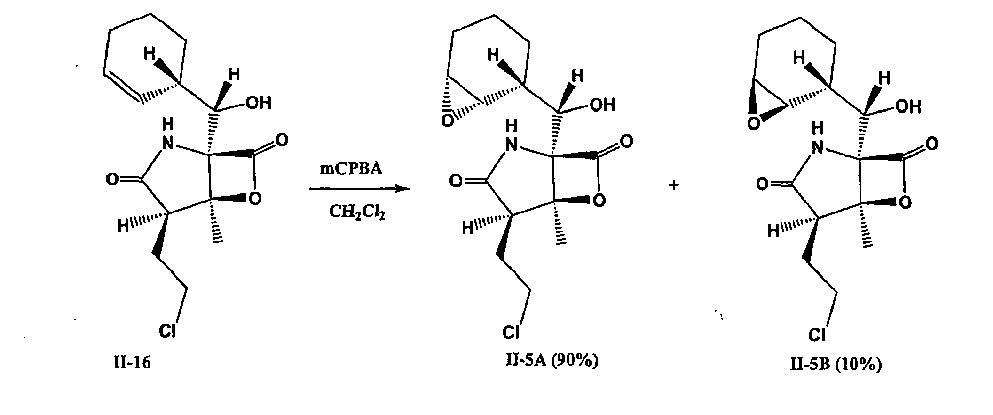
- Formula II-5A: UV (Acetonitrile/H2O) λmax 225 (sh) nm. Low Res. Mass: m/z 330 (M+H), 352 (M+Na).
FIGS. 16-17 , respectively depict the 1H NMR spectrum of Formula II-5A and the mass spectrum of Formula II-5A. - Formula II-5B: UV (Acetonitrile/H2O) λmax 225 (sh) nm. Low Res. Mass: m/z 330 (M+H), 352 (M+Na).
FIGS. 18-19 , respectively depict the 1H NMR spectrum of II-5B and the mass spectrum of II-5B. - Diols may be synthesized by Sharpless dihydroxylation using AD mix-α and β: AD mix-α is a premix of four reagents, K2OsO2(OH)4 K2CO3; K3Fe(CN)6; (DHQ)2-PHAL [1,4-bis(9-O-dihydroquinine)phthalazine] and AD mix-β is a premix of K2OsO2(OH)4 K2CO3; K3Fe(CN)6; (DHQD)2-PHAL [1,4-bis(9-O-dihydroquinidine)phthalazine] which are commercially available from Aldrich. Diol can also be synthesized by acid or base hydrolysis of epoxy compounds (Formula II-5A and II-5B) which may be different to that of products obtained in Sharpless dihydroxylation in their stereochemistry at carbons bearing hydroxyl groups
- Any of the compounds of Formulae II-16, II-17 and II-18 may be used as the starting compound. In the example below, compound of Formula II-16 is used. The starting compound is dissolved in t-butanol/water in a round bottom flask to which is added AD mix-α or β and a magnetic stir bar. The reaction is monitored by silica TLC as well as mass spectrometer. The pure diols are obtained by usual workup and purification by flash chromatography or HPLC. The structures are confirmed by NMR spectroscopy and mass spectrometry. In this method both hydroxyl groups are on same side.
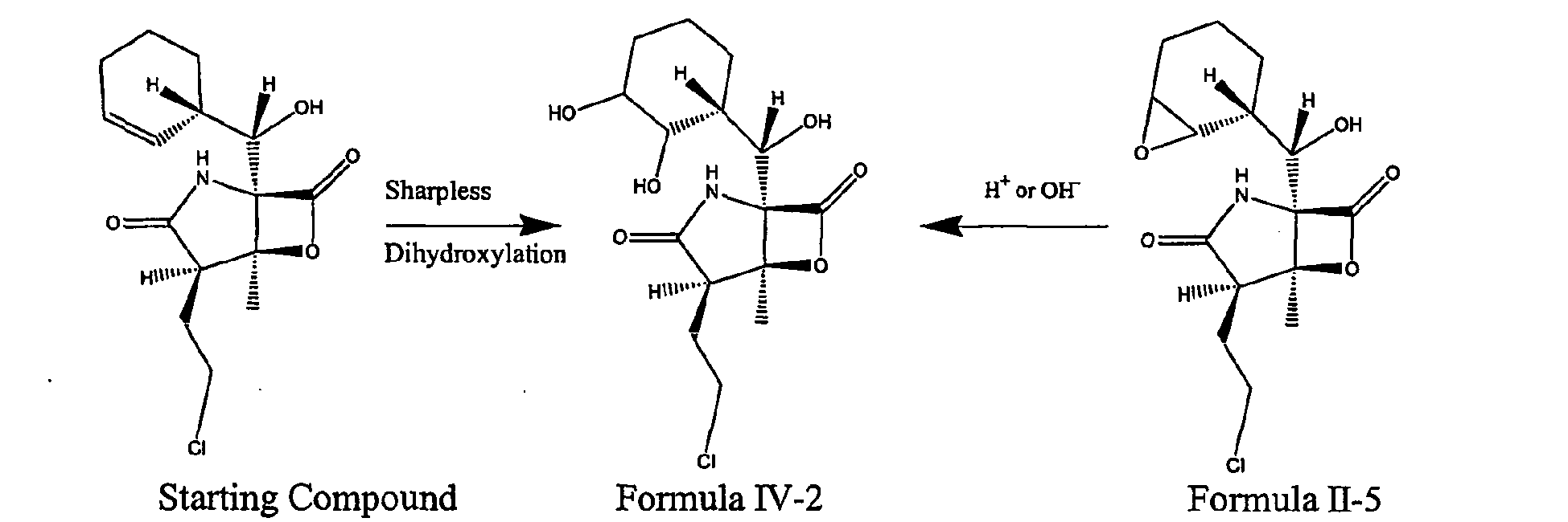
- The epoxy ring is opened with various nucleophiles like NaCN, NaN3, NaOAc, HBr, HCl, etc. to creat various substituents on the cyclohexane ring, including a hydroxyl substituent.
-

- The epoxy is opened with HCl to make Formula IV-3:

- Compound of Formula II-5A (3.3 mg) was dissolved in acetonitrile (0.5 ml) in a 1 dram vial to which was added 5% HCl (500 ul) and a magnetic stir bar. The reaction mixture was stirred at room temperature for about an hour. The reaction was monitored by mass spectrometry. The reaction mixture was directly injected on normal phase HPLC to obtain compound of Formula IV-3C as a pure compound without any work up. The HPLC conditions used for the purification were as follows: Phenomenex Luna 10u Silica column (25 cm x 21.2 mm ID) with a solvent gradient of 25% to 80% EtOAc/Hex over 19 min, 80 to 100% EtOAc in 1 min, then 5 min at 100% EtOAc at a flow rate of 14.5 ml/min. An ELSD was used to monitor the purification process. Compound of Formula IV-3C eluted at about 18 min (2.2 mg). Compound of Formula IV-3C: UV (Acetonitrile/H20) λmax 225 (sh) nm; ESMS, m/z 366 (M+H), 388 (M+Na); 1H NMR in DMSO-d6 (
FIG. 20 ) The stereochemistry of the compound of Formula IV-3C was determined based on coupling constants observed in the cyclohexane ring in 1:1 C6D6/DMSO-d6 (FIG. 21 )
- Reductive ring opening of epoxides (II-5): The compound of Formula is treated with metalhydrides like BH3-THF complex to make compound of Formula IV-4.

- Compound of Formula II-16 (30 mg) was dissolved in CH2Cl2 (6 ml) in a scintillation vial (20 ml) to which Dess-Martin Periodinane (122 mg) and a magnetic stir bar were added. The reaction mixture was stirred at room temperature for about 2 hours. The progress of the reaction was monitored by TLC (Hex:EtOAc, 6:4) and analytical HPLC. From the reaction mixture, the solvent volume was reduced to one third, absorbed on silica gel, poured on top of a 20 cc silica flash column and eluted in 20 ml fractions using a gradient of Hexane/EtOAc from 10 to 100%. The fraction eluted with 30% EtOAc in Hexane contained a mixture of rotamers of Formula II-13C in a ratio of 1.5:8.5. The mixture was further purified by normal phase HPLC using the Phenomenex Luna 10u Silica column (25 cm x 21.2 mm ID) with a solvent gradient of 25% to 80% EtOAc/Hex over 19 min, 80 to 100% EtOAc over 1 min, holding at 100% EtOAc for 5 min, at a flow rate of 14.5 ml/min. An ELSD was used to monitor the purification process. Compound of Formula II-13C eluted at 13.0 and 13.2 mins as a mixture of rotamers with in a ratio of 1.5:8.5 (7 mg). Formula II-13C: UV (Acetonitrile/H2O) λmax 226 (sh) & 300 (sh) nm; ESMS, m/z 312 (M+H)+, 334 (M+Na)+; 1H NMR inDMSO-d6 (see
FIG. 22 ). - The rotamer mixture of Formula II-13C (4 mg) was dissolved in acetone (1 ml) in a scintillation vial (20 ml) to which a catalytic amount (0.5 mg) of 10% (w/w) Pd/C and a magnetic stir bar were added. The reaction mixture was stirred in a hydrogen atmosphere at room temperature for about 15 hours. The reaction mixture was filtered through a 0.2 µm Gelman Acrodisc to remove the catalyst. The solvent was evaporated from the filtrate to yield compound of Formula II-8C as a colorless gum which was further purified by normal phase HPLC using a Phenomenex Luna 10u Silica column (25 cm x 21.2 mm ID) with a solvent gradient of 25% to 80% EtOAc/Hex over 19 min, 80 to 100% EtOAc over 1 min, holding at 100% EtOAc for 5 min, at a flow rate of 14.5 ml/min. An ELSD was used to monitor the purification process. Compound of Formula II-8C (1 mg) eluted at 13.5 min as a pure compound. Formula II-8C: UV (Acetonitrile/H2O) λmax 225 (sh) nm; ESMS, m/z 314 (M+H)+, 336 (M+Na)+; 1H NMR inDMSO-d6 (See
FIG. 23 ).
- The rotamer mixture of Formula II-13C (5 mg) was dissolved in dimethoxy ethane (monoglyme; 1.5 ml) in a scintillation vial (20 ml) to which water (15 µl (1% of the final solution concentration)) and a magnetic stir bar were added. The above solution was cooled to -78°C on a dry ice-acetone bath, and a sodium borohydride solution (3.7 mg of NaBH4 in 0.5 ml of monoglyme (created to allow for slow addition)) was added drop-wise. The reaction mixture was stirred at -78°C for about 14 minutes. The reaction mixture was acidified using 2 ml of 4% HCl solution in water and extracted with CH2Cl2. The organic layer was evaporated to yield mixture of compound of formulae II-25 and II-16 in a 9.5:0.5 ratio as a white solid, which was further purified by normal phase HPLC using a Phenomenex Luna 10u Silica column (25 cm x 21.2 mm ID). The mobile phase was 24% EtOAc/76% Hexane, which was held isocratic for 19 min, followed by a linear gradient of 24% to 100% EtOAc over 1 min, and held at 100% EtOAc for 3 min; the flow rate was 25 ml/min. An ELSD was used to monitor the purification process. Compound of formula II-25 (1.5 mg) eluted at 11.64 min as a pure compound. Compound of Formula II-25: UV (Acetonitrile/H2O) λmax 225 (sh) nm; ESMS, mlz 314 (M+H)+, 336 (M+Na)+; 1H NMR in DMSO-d6 (see
FIG. 24 ).
- Acetone (7.5 ml) was vigorously mixed with 5 N NaOH (3 ml) and the resulting mixture evaporated to a minimum volume in vacuo. A sample of 100 µl of this solution was mixed with compound of Formula II-19 (6.2 mg) in acetone (1 ml) and the resulting biphasic mixture vortexed for 2 minutes. The reaction solution was immediately subjected to preparative C18 HPLC. Conditions for the purification involved a linear gradient if 10% acetonitrile/90% water to 90% acetonitrile/ 10% water over 17 minutes using an
Ace 5 µ C18 HPLC column ofdimensions 22 mm id by 150 mm length. Compound of Formula II-21 eluted at 9.1 minutes under these conditions to yield 0.55 mg compound. Compound of Formula II-21: UV (Acetonitrile/H2O) 225 (sh), ESMS, m/z 296.1 (M+H); 1H NMR in DMSO-d6 (seeFIG. 25 ).
- A sample of 60 mg sodium propionate was added to a solution of compound of Formula II-19 (5.3 mg) in DMSO (1 ml) and the mixture sonicated for 5 minutes, though the sodium propionate did not completely dissolve. After 45 minutes, the solution was filtered through a 0.45 µ syringe filter and purified directly using HPLC. Conditions for the purification involved a linear gradient if 10% acetonitrile/90% water to 90% acetonitrile/ 10% water over 17 minutes using an
Ace 5 µ C18 HPLC column ofdimensions 22 mm id by 150 mm length. Under these conditions, compound of Formula II-22 eluted at 12.3 minutes to yield 0.7 mg compound (15% isolated yield). UV (Acetonitrile/H2O) 225 (sh), ESMS, m/z 352.2 (M+H); 1H NMR in DMSO-d6 (seeFIG. 26 ).
- Any of the compounds of Formulae II-16, II-17 and II-18 may be used as the starting compound. The secondary hydroxyl group in the starting compound is oxidized using either of the following reagents: pyridinium dichromate (PDC), pyridinium chlorochromate (PCC), Dess-Martin periodinane or oxalyl chloride (Swem oxidation) (Ref: Organic Syntheses, collective volumes I-VIII). Preferably, Dess-Martin periodinane may be used as a reagent for this reaction. (Ref: Fenteany G. et al. Science, 1995, 268, 726-73). The resulting keto compound is treated with hydroxylamine or methoxy amine to generate oximes.
-

- The keto derivatives, for example Formula II-8 and II-13, are treated with sodium cyanoborohydride (NaBH3CN) in the presence of various bases to yield amine derivatives of the starting compounds which are subsequently hydrogenated with 10%Pd/C, H2 to reduce the double bond in the cyclohexene ring.
-

- Any compound of Formulae II-16, II-17 and II-18 may be used as a starting compound. The starting compound is treated with OsO4 and NaIO4 in THT-H2O solution to yield dial derivatives which are reduced to alcohol with NaBH4 in the same pot.
-

- A starting compound of any of Formulae II-16, II-17 or II-18 is treated with mesylchloride in the presence of base to yield a dehydrated derivative. The resulting dehydrated compound is treated with OsO4 and NaIO4 in THF-H2O to yield an aldehyde group at the lactone-lactam ring junction.

- Wittig reactions are performed on the aldehyde group using various phosphorus ylides [e.g., (triphenylphosphoranylidene)ethane] to yield an olefin. The double bond in the side chain is reduced by catalytic hydrogenation.
-

- Reductive amination is performed on the aldehyde group using various bases (eg. NH3) and sodium cyanoborohydride to yield amine derivatives. Alternatively, the aldehyde is reduced with NaBH4 to form alcohols in the side chain.
-

- Organometallic addition reactions to the aldehyde carbonyl, such as Grignard reactions, may be performed using various alkyl magnesium bromide or chloride reagents (eg. isopropylmagnesium bromide, phenylmagnesium bromide) to yield various substituted secondary alcohols.
-

- Initial studies of a compound of Formula II-16, which is also referred to as Salinosporamide A, employed the National Cancer Institute (NCI) screening panel, which consists of 60 human tumor cell lines that represent leukemia, melanoma and cancers of the lung, colon, brain, ovary, breast, prostate and kidney. A detailed description of the screening procedure can be found at hypertext transfer protocol (http://) "dtp.nci.nih.gov/branches/btb/ivclsp.html."
- In brief, each of the 60 human tumor cell lines were grown in RPMI 1640 medium, supplemented with 5% fetal bovine serum and 2 mM L-glutamine. Cells were plated at their appropriate density in 96-well microtiter plates and incubated at 37°C, 5% CO2, 95% air and 100% relative humidity. After 24 hours, 100 µL of various 10-fold serial dilutions of Salinosporamide A were added to the appropriate wells containing 100 µL of cells, resulting in a final Salinosporamide A concentration ranging from 10 nM to 100 µM. Cells were incubated for an additional 48 hours and a sulforhodamine B protein assay was used to estimate cell viability or growth.
- Three dose response parameters were calculated as follows:
- GI50 indicates the concentration that inhibits growth by 50%.
- TGI indicates the concentration that completely inhibits growth.
- LC50 indicates the concentration that is lethal to 50% of the cells.
- An example of a study evaluating Salinosporamide A in the NCI screen is shown in Table 1 below.
- Data indicate that the mean GI50 value of Salinosporamide A was less than 10 nM. The wide range (>1000-fold difference) observed in both the mean TGI and mean LC50 values for the most sensitive and the most resistant tumor cell lines illustrates that Salinosporamide A displays good selectivity and does not appear to be a general toxin. Furthermore, the mean TGI data suggest that Salinosporamide A shows preferred specificity towards melanoma and breast cancer cell lines. The assay was repeated and showed similar results.
- The results of the NCI tumor screen show that Salinosporamide A: (1) is a potent compound with a mean GI50 value of < 10 nM, and (2) displays good tumor selectivity of more than 1000-fold difference in both the mean TGI and mean LC50 values between the most sensitive and resistant tumor cell lines.

- B16-F10 (ATCC; CRL-6475), DU 145 (ATCC; HTB-81), HEK293 (ATCC; CRL-1573), HT-29 (ATCC; HTB-38), LoVo (ATCC; CCL-229), MDA-MB-231 (ATCC; HTB-26), MIA PaCa-2 (ATCC; CRL-1420), NCI-H292 (ATCC; CRL-1848), OVCAR-3 (ATCC, HTB-161), PANC-1 (ATCC; CRL-1469), PC-3 (ATCC; CRL-1435), RPMI 8226 (ATCC; CCL-155) and U266 (ATCC; TIB-196) were maintained in appropriate culture media. The cells were cultured in an incubator at 37 °C in 5% CO2 and 95% humidified air.
- For cell growth inhibition assays, B16-F10, DU 145, HEK293, HT-29, LoVo, MDA-MB-231, MIA PaCa-2, NCI-H292, OVCAR-3, PANC-1, PC-3,
RPMI 8226 and U266 cells were seeded at 1.25x10³, 5x10³, 1.5x10⁴, 5x10³, 5x10³, 1x10⁴, 2x10³, 4x10³, 1x10⁴, 7.5x10³, 5x10³, 2x10⁴, 2.5x10⁴ cells/well respectively in 90µl complete media into Corning 3904 black-walled, clear-bottom tissue culture plates. 20mM stock solutions of Formula II-16 were prepared in 100% DMSO, aliquoted and stored at -80°C. Formula II-16 was serially diluted and added in triplicate to the test wells resulting in final concentrations ranging from of 20 µM to 0.2pM. The plates were returned to the incubator for 48 hours. The final concentration of DMSO was 0.25% in all samples. - Following 48 hours of drug exposure, 10µl of 0.2mg/ml resazurin (obtained from Sigma-Aldrich Chemical Co.) in Mg2+, Ca2+ free phosphate buffered saline was added to each well and the plates were returned to the incubator for 3-6 hours. Since living cells metabolize Resazurin, the fluorescence of the reduction product of Resazurin was measured using a Fusion microplate fluorometer (Packard Bioscience) with λex = 535 nm and λem = 590 nm filters. Resazurin dye in medium without cells was used to determine the background, which was subtracted from the data for all experimental wells. The data were normalized to the average fluorescence of the cells treated with media + 0.25% DMSO (100% cell growth) and EC50 values (the drug concentration at which 50% of the maximal observed growth inhibition is established) were determined using a standard sigmoidal dose response curve fitting algorithm (generated by XLfit 3.0, ID Business Solutions Ltd or Prism 3.0, GraphPad Software Inc).
- The data in Table 2 summarize the growth inhibitory effects of Formula II-16 against 13 diverse human and mouse tumor cell lines.
Table 2 Mean EC 50 values of Formula II-16 against various tumor cell lines Cell line Source EC50 (nM), mean ± SD * n B16-F10 Mouse, melanoma 47 ± 20 12 DU 145 Human, prostate carcinoma 37 ± 10 3 HEK293 Human, embryonic kidney 47 2 HT-29 Human, colorectal adenocarcinoma 40 ± 26 5 LoVo Human, colorectal adenocarcinoma 70 ± 8 3 MDA-MB-231 Human, breast adenocarcinoma 87 ± 40 12 MIA PaCa-2 Human, pancreatic carcinoma 46 2 NCI-H292 Human, non small cell lung carcinoma 66 ± 29 12 OVCAR-3 Human, ovarian adenocarcinoma 49 ± 31 6 PANC-1 Human, pancreatic carcinoma 60 2 PC-3 Human, prostate adenocarcinoma 64 ± 26 19 RPMI 8226 Human, multiple myeloma 8.6 ± 1.9 26 U266 Human, multiple myeloma 4.7 ± 0.7 6 - * Where n (number of independent experiments) = 2, the mean value is presented
- The EC50 values indicate that Formula II-16 was cytotoxic against B16-F10, DU 145, HEK293, HT-29, LoVo, MDA-MB-231, MIA PaCa-2, NCI-H292, OVCAR-3, PANC-1, PC-3,
RPMI 8226 and U266 cells. - All the compounds were prepared as 20 mM stock solution in DMSO and stored in small aliquots at -80°C. Purified rabbit muscle 20S proteasome was obtained from CalBiochem. To enhance the chymotrypsin-like activity of the proteasome, the assay buffer (20 mM HEPES, pH7.3, 0.5 mM EDTA, and 0.05% Triton X100) was supplemented with SDS resulting in a final SDS concentration of 0.035%. The substrate used was suc-LLVY-AMC, a fluorogenic peptide substrate specifically cleaved by the chymotrypsin-like activity of the proteasome. Assays were performed at a proteasome concentration of 1 µg/ml in a final volume of 200µl in 96-well Costar microtiter plates. Formula II-2, Formula II-4, Formula II-16, Formula II-17, Formula II-18, Formula II-19, Formula II-21 and Formula II-22 were tested as eight-point dose response curves with final concentrations ranging from 500 nM to 158 pM. Formula II-5A, Formula II-5B and Formula II-20 were tested at concentrations ranging from 1 µM to 0.32nM. Formula II-3 was tested as an eight-dose response curve with final concentrations ranging from 10 µM to 3.2 nM, while Formula II-8C, Formula II-13C, Formula II-24C, Formula II-25 and Formula IV-3C were tested with final concentrations ranging from 20 µM to 6.3nM. The samples were incubated at 37°C for five minutes in a temperature controlled plate reader. During the preincubation step, the substrate was diluted 25-fold in SDS-containing assay buffer. After the preincubation period, the reactions were initiated by the addition of 10 µl of the diluted substrate and the plates were returned to the plate reader. The final concentration of substrate in the reactions was 20 µM. All data were collected every five minutes for more than 1.5 hour and plotted as the mean of triplicate data points. The EC50 values (the drug concentration at which 50% of the maximal relative fluorescence unit is inhibited) were calculated by Prism (GraphPad Software) using a sigmoidal dose-response, variable slope model. To evaluate the activity of the compounds against the caspase-like activity of the 20S proteasomes, reactions were performed as described above except that Z-LLE-AMC was used as the peptide substrate. Formulae II-3, II-4, II-5A, II-5B, II-8C, II-13C, II-17, II-18, II-20, II-21. II-22, II-24C, II-25 and Formula IV-3C were tested at concentrations ranging from 20µM to 6.3nM. Formula II-2 was tested at concentrations ranging from 10µM to 3.2nM, while Formula II-16 and Formula II-19 were tested at concentrations ranging from 5µM to 1.58nM. For the evaluation of the compounds against the trypsin-like activity of the proteasome, the SDS was omitted from the assay buffer and Boc-LRR-AMC was used as the peptide substrate. Formula II-20 was tested at concentrations ranging from 5 µM to 1.6 nM. Formulae II-3, II-8C, II-13C, II-17, II-21, II-22, II-24C, II-25 and IV-3C were tested at concentrations ranging from 20µM to 6.3nM. For Formulae II-2 and II-5B the concentrations tested ranged from 10 µM to 3.2nM, while Formulae II-4, II-5A, II-16, II-18 and II-19 were tested at concentrations ranging from 1 µM to 0.32nM.
- Results (mean EC50 values) are shown in Table 3 and illustrate that among the tested compounds, Formulae II-SA, II-16, II-18, II-19, II-20, II-21 and II-22 are the most potent inhibitors of the chymotrypsin-like activity of the 20S proteasome with EC50 values ranging from 2.2 nM to 7 nM. Formulae II-2, II-4, II-5B and II-17 inhibit the proteasomal chymotrypsin-like activity with EC50 values ranging from 14.2nM to 87nM, while the EC50 value of Formula II-3 is 927 nM. Formula II-24C, II-13C and IV-3C inhibited the chymotrypsin-like activity with EC50 values of 2.2 µM, 8.2 µM and 7.8 µM respectively. EC50 values for Formulae II-8C and II-25 were greater than 20 µM. Under the conditions tested, Formulae II-2, II-3, II-4, II-5A, II-5B, II-13C, II-16, II-17, II-18, II-19, II-20, II-21, II-22 and II-24C were able to inhibit the trypsin-like activity of the 20S proteasome. Formulae II-4, II-5A, II-16, II-18 and II-19 inhibited the caspase-like activity with EC50 values ranging from 250nM to 744nM, while Formulae II-2, II-5B, II-17, II-20, II-21, and II-22 had EC50 values ranging from 1.2µM to 3.3µM.
Table 3 Effects of Formulae II-2, II-3, II-4, II-5A, II-5B, II-8C, II-13C, II-16, II-17, II-18, II-19. II- 20, II-21, II-22, II-24C, II-25 and IV-3C on the various enzymatic activities of purified rabbit 20S proteasomes Analog EC50 Values Chymotrypsin-like Trypsin-like Caspase-like Formula II-2 18 nM 230 nM 1.5µM Formula II-3 927 nM 6.6 µM > 20 µM Formula II-4 14.2 nM 109 nM 744 nM Formula II-5A 6.5 nM 89 nM 487 nM Formula II-5B 87 nM 739 nM 3.3µM Formula II-8C* > 20 µM > 20 µM > 20 µM Formula II-13C 8.2 µM 10.7 µM > 20 µM Formula II-16 2.5 nM 21 nM 401 nM Formula II-17 29.5 nM 588 nM 1.2 µM Formula II-18 2.2 nM 14 nM 250 nM Formula II-19* 3 nM 13 nM 573 nM Formula II-20* 5 nM 318 nM 1.4 µM Formula II-21 * 7 nM 720 nM 2.6 µM Formula II-22* 5 nM 308 nM 1.3 µM Formula II-24C* 2.2 µM 3.2 µM > 20 µM Formula II-25* > 20 µM > 20 µM > 20 µM Formula IV-3C 7.8 µM > 20 µM > 20 µM * n=1 - The effect of Salinosporamide A (II-16) on proteasomes was examined using a commercially available kit from Calbiochem (catalog no. 539158), which uses a fluorogenic peptide substrate to measure the activity of rabbit muscle 20S proteasomes (Calbiochem 20S Proteasome Kit). This peptide substrate is specific for the chymotrypsin-like enzyme activity of the proteasome.
- Omuralide was prepared as a 10 mM stock in DMSO and stored in 5 µL aliquots at -80°C. Salinosporamide A was prepared as a 25.5 mM solution in DMSO and stored in aliquots at -80°C. The assay measures the hydrolysis of Suc-LLVY-AMC into Suc-LLVY and AMC. The released coumarin (AMC) was measured fluorometrically by using λex = 390 nm and λem = 460 nm. The assays were performed in a microtiter plate (Coming 3904), and followed kinetically with measurements every five minutes. The instrument used was a Thermo Lab Systems Fluoroskan, with the incubation chamber set to 37°C. The assays were performed according to the manufacturer's protocol, with the following changes. The proteasome was activated as described with SDS, and held on ice prior to the assay. Salinosporamide A and Omuralide were serially diluted in assay buffer to make an 8-point dose-response curve. Ten microliters of each dose were added in triplicate to the assay plate, and 190 µL of the activated proteasome was added and mixed. The samples were pre-incubated in the Fluoroskan for 5 minutes at 37°C. Substrate was added and the kinetics of AMC were followed for one hour. All data were collected and plotted as the mean of triplicate data points. The data were normalized to reactions performed in the absence of Salinosporamide A and modeled in Prism as a sigmoidal dose-response, variable slope.
- Similar to the results obtained for the in vitro cytotoxicity (Table 2), Feling, et al., Angew Chem Int Ed Engl 42:355 (2003), the EC50 values in the 20S proteasome assay showed that Salinosporamide A was approximately 40-fold more potent than Omuralide, with an average value of 1.3 nM versus 49 nM, respectively (
FIG. 27 ). This experiment was repeated and the average EC50 in the two assays was determined to be 2 nM for Salinosporamide A and 52 nM for Omuralide. - Salinosporamide A is a potent inhibitor of the chymotrypsin-like activity of the proteasome. The EC50 values for cytotoxicity were in the 10-200 nM range suggesting that the ability of Salinosporamide A to induce cell death was due, at least in large part, to proteasome inhibition. The data suggest that Salinosporamide A is a potent small molecule inhibitor of the proteasome.
- Omuralide can inhibit the PGPH activity (also known as the caspase-like) of the proteasome; therefore, the ability of Salinosporamide A to inhibit the PGPH activity of purified rabbit muscle 20S proteasomes was assessed. A commercially available fluorogenic substrate specific for the PGPH activity was used instead of the chymotrypsin substrate supplied in the proteasome assay kit described above.
- Salinosporamide A (II-16) was prepared as a 20 mM solution in DMSO and stored in small aliquots at -80°C. The substrate Z-LLE-AMC was prepared as a 20 mM stock solution in DMSO, stored at -20°C. The source of the proteasomes was the commercially available kit from Calbiochem (Cat. # 539158). As with the chymotrypsin substrate, the proteasome can cleave Z-LLE-AMC into Z-LLE and free AMC. The activity can then be determined by measuring the fluorescence of the released AMC (λex = 390 nm and λem = 460 nm). The proteasomes were activated with SDS and held on ice as per manufacturer's recommendation. Salinosporamide A was diluted in DMSO to generate a 400-fold concentrated 8-point dilution series. The series was diluted 20-fold with assay buffer and preincubated with the proteasomes as described for the chymotrypsin-like activity. After addition of substrate, the samples were incubated at 37°C, and release of the fluorescent AMC was monitored in a fluorimeter. All data were collected and plotted as the mean of triplicate points. In these experiments, the EC50 was modeled in Prism as normalized activity, where the amount of AMC released in the absence of Salinosporamide A represents 100% activity. As before, the model chosen was a sigmoidal dose-response, with a variable slope.
- Data revealed that Salinosporamide A inhibited the PGPH activity in rabbit muscle 20S proteasomes with an EC50 of 350 nM (
FIG. 28 ). A replicate experiment was performed, which gave a predicted EC50 of 610 nM. These results indicate that Salinosporamide A does block the in vitro PGPH activity of purified rabbit muscle 20S proteasomes, albeit with lower potency than seen towards the chymotrypsin-like activity. - The ability of Salinosporamide A (II-16) to inhibit the chymotrypsin-like activity of human erythrocyte 20S proteasomes was assessed in vitro. The calculated EC50 values ranged from 45 to 247 pM, and seemed to depend upon the lot of proteasomes tested (BIOMOL, Cat# SE-221). These data indicate that the inhibitory effect of Salinosporamide A is not limited to rabbit skeletal muscle proteasomes.
- Salinosporamide A was prepared as a 20 mM solution in DMSO and stored in small aliquots at -80°C. The substrate, suc-LLVY-AMC, was prepared as a 20 mM solution in DMSO and stored at -20°C. Human erythrocyte 20S proteasomes were obtained from BIOMOL (Cat. # SE-221). The proteasome can cleave suc-LLVY-AMC into suc-LLVY and free AMC and the activity can then be determined by measuring the fluorescence of the released AMC (λex = 390 nm and λem = 460 nm). The proteasomes were activated by SDS and stored on ice as with the experiments using rabbit muscle proteasomes. Salinosporamide A was diluted in DMSO to generate a 400-fold concentrated 8-point dilution series. The series was then diluted 20-fold with assay buffer and pre-incubated with proteasomes at 37°C. The reaction was initiated with substrate, and the release of AMC was followed in a Fluoroskan microplate fluorimeter. Data were collected and plotted as the mean of triplicate points. Data were captured kinetically for 3 hours, and indicated that these reactions showed linear kinetics in this time regime. The data were normalized to reactions performed in the absence of Salinosporamide A and modeled in Prism as a sigmoidal dose-response, variable slope.
- Replicate experiments performed using human erythrocyte proteasomes from separate lots resulted in a range of EC50 values between 45 and 250 pM (
FIG. 29 shows a representative experiment). It has been reported that 20S proteasomes purified from human erythrocytes are highly heterogeneous in subunit composition. Claverol, et al., Mol Cell Proteomics 1:567 (2002). The variability in these experiments may therefore be due to differences in the composition and activity of the human erythrocyte proteasome preparations. Regardless, these results indicate that the in vitro chymotrypsin-like activity of human erythrocyte 20S proteasomes is sensitive to Salinosporamide A. - A possible mechanism by which Salinosporamide A inhibits the proteasome is by the reaction of the β-lactone functionality of Salinosporamide A with the active site threonine of the proteasome. This covalent modification of the proteasome would block the active site, as this residue is essential for the catalytic activity of the proteasome. Fenteany, et al., J Biol Chem 273:8545 (1998). A structurally related compound, Lactacystin, has been shown to also inhibit cathepsin A (Ostrowska, et al., Int J Biochem Cell Biol 32:747 (2000), Kozlowski, et al., Tumour Biol 22:211 (2001), Ostrowska, et al., Biochem Biophys Res Commun 234:729 (1997)) and TPPII (Geier, et al., Science 283:978 (1999)) but not trypsin, chymotrypsin, papain, calpain (Fenteany, et al., Science 268:726 (1995)), thrombin, or plasminogen activator (Omura, et al., J Antibiot (Tokyo) 44:113 (1991)). Similar studies were initiated to explore the specificity of Salinosporanude A for the proteasome by evaluating its ability to inhibit the catalytic activity of a prototypical serine protease, chymotrypsin.
- Salinosporamide A was prepared as a 20 mM solution in DMSO and stored in small aliquots at -80°C. The substrate, suc-LLVY-AMC, was prepared as a 20 mM solution in DMSO and stored at -20°C. Proteolytic cleavage of this substrate by either proteasomes or chymotrypsin liberates the fluorescent product AMC, which can be monitored in a fluorimeter (λex = 390 nm and λem = 460 nm). Bovine pancreatic chymotrypsin was obtained from Sigma (Cat. # C-4129), and prepared as a 5 mg/ml solution in assay buffer (10 mM HEPES, 0.5 mM EDTA, 0.05% Triton X-100, pH 7.5) daily. Immediately prior to the assay, the chymotrypsin was diluted to 1 µg/ml (0.2 µg/well) in assay buffer and held on ice. Salinosporamide A was diluted in DMSO to generate an 8-point dose-response curve. The high final Salinosporamide A concentrations needed to obtain complete inhibition of chymotrypsin required that the diluted enzyme be directly added to the compound dilution series. The inclusion of 1% DMSO (the final concentration of solvent in the test wells) into the reaction had no significant effect on chymotrypsin activity towards this substrate. The reactions were pre-incubated for 5 minutes at 37°C and the reactions were initiated by the addition of substrate. Data were collected kinetically for one hour at 37°C in the Fluoroskan and plotted as the mean of triplicate data points. The data were normalized to reactions performed in the absence of Salinosporamide A, and modeled in Prism as a sigmoidal dose-response, variable slope. Normalized data from Salinosporamide A inhibition of the chymotrypsin-like activity of rabbit 20S proteasomes has been included on the same graph.
- The average inhibition observed in two experiments using Salinosporamide A pretreatment of chymotrypsin was 17.5 µM (
FIG. 30 shows a representative experiment). The data indicate that there is a preference for Salinosporamide A-mediated inhibition of the in vitro chymotrypsin-like activity of proteasomes over inhibition of the catalytic activity of chymotrypsin. - Thus, Salinosporamide A inhibits the chymotrypsin-like and PGPH activity of the proteasome. Preliminary studies indicate that Salinosporamide A also inhibits the trypsin-like activity of the proteasome with an EC50 value of~10 nM (data not shown).
- The HEK293 NF-κB/luciferase reporter cell line is a derivative of the human embryonic kidney cell line (ATCC; CRL-1573) and carries a luciferase reporter gene under the regulation of 5X NF-κB binding sites. The reporter cell line was routinely maintained in complete DMEM medium (DMEM plus 10%(v/v) Fetal bovine serum, 2mM L-glutamine, 10 mM HEPES and Penicillin/Streptomycin at 100 IU/ml and 100µg/ml, respectively) supplemented with 250 µg/ml G418. When performing the luciferase assay, the DMEM basal medium was replaced with phenol-red free DMEM basal medium and the G418 was omitted. The cells were cultured in an incubator at 37 °C in 5% CO2 and 95% humidified air.
- For NF-κB-mediated luciferase assays, HEK293 NF-κB/luciferase cells were seeded at 1.5x10⁴ cells/well in 90µl phenol-red free DMEM complete medium into Corning 3917 white opaque-bottom tissue culture plates. For Formula II-2, Formula II-4, Formula II-5A, Formula II-16 and Formula II-18, a 400µM starting dilution was made in 100% DMSO and this dilution was used to generate a 8-point half log dilution series. This dilution series was further diluted 40X in appropriate culture medium and ten µl aliquots were added to the test wells in triplicate resulting in final test concentrations ranging from 1 µM to 320 pM. For Formula II-3, Formula II-5B, Formula II-8C, Formula II-13C, Formula II-17, Formula II-20, Formula II-21, Formula II-22, Formula II-24C, Formula II-25 and Formula IV-3C, a 8mM starting dilution was made in 100% DMSO and the same procedure was followed as described above resulting in final test concentrations ranging from 20µM. to 6.3nM. For Formula II-19, a 127 µM starting dilution was made in 100% DMSO and the final test concentrations ranging from 317 nM to 0.1 nM. The plates were returned to the incubator for 1 hour. After 1 hr pretreatment, 10 µl of a 50 ng/ml TNF-α solution, prepared in the phenol-red free DMEM medium was added, and the plates were incubated for an additional 6 hr. The final concentration of DMSO was 0.25% in all samples.
- At the end of the TNF-α stimulation, 100 µl of Steady Lite HTS luciferase reagent (Packard Bioscience) was added to each well and the plates were left undisturbed for 10 min at room temperature before measuring the luciferase activity. The relative luciferase units (RLU) were measured by using a Fusion microplate fluorometer (Packard Bioscience). The EC50 values (the drug concentration at which 50% of the maximal relative luciferase unit inhibition is established) were calculated in Prism (GraphPad Software) using a sigmoidal dose response, variable slope model.
- NF-κB regulates the expression of a large number of genes important in inflammation, apoptosis, tumorigenesis, and autoimmune diseases. In its inactive form, NF-κB complexes with IκB in the cytosol and upon stimulation, IκB is phosphorylated, ubiquitinated and subsequently degraded by the proteasome. The degradation of IκB leads to the activation of NF-κB and its translocation to the nucleus. The effects of Formula II-2, Formula II-3, Formula II-4, Formula II-5A, Formula II-5B, Formula II-8C, Formula II-13C, Formula II-16, Formula II-17, Formula II-18, Formula II-19, Formula II-20, Formula II-21, Formula II-22, Formula II-24C, Formula II-25 and Formula IV-3C on the activation of NF-κB were evaluated by assessing the NF-κB-mediated luciferase activity in HEK293 NF-κB/Luc cells upon TNF-α stimulation.
- Pretreatment of NF-κB/Luc 293 cells with Formula II-2, Formula II-4, Formula II-SA, Formula II-5B, Formula II-16, Formula II-17, Formula II-18, Formula II-19, Formula II-20, Formula II-21, Formula II-22 and Formula II-24C resulted in a dose-dependent decrease of luciferase activity upon TNF-α stimulation. The mean EC50 values to inhibit NF-κB-mediated luciferase activity are shown in Table 4 and demonstrate that compounds of Formula II-2, Formula II-4, Formula II-SA, Formula II-SB, Formula II-16, Formula II-17, Formula II-18, Formula II-19, Formula II-20, Formula II-21, Formula II-22 and Formula II-24C inhibited NF-κB activity in this cell-based assay.
Table 4 Mean EC 50 values of Formulae II-2, II-3, II-4, II-5A, II-5B, II-8C, II-13C, II-16, II-17, II-18, II-19, II-20, II-21, II-22, II-24C, II-25 and IV-3C from NF-κB-mediated luciferase reporter gene assay Compound EC50 (nM) Formula II-2 82 Formula II-3, >20,000 Formula II-4 77.7 Formula II-5A 31.5 Formula II-5B 270 Formula II-8C* >20,000 Formula II-13C >20,000 Formula II-16 11.8 Formula II-17 876 Formula II-18 9.5 Formula II-19 8.5 Formula II-20* 154 Formula II-21* 3,172 Formula II-22* 1,046 Formula II-24C* 5,298 Formula II-25* >20,000 Formula IV-3C >20,000 * n=1 - Experiments were carried out to study the role of Salinosporamide A in the NF-κB signaling pathway. A stable HEK293 clone (NF-κB/Luc 293) was generated carrying a luciferase reporter gene under the regulation of 5X NF-κB binding sites. Stimulation of this cell line with TNF-α leads to increased luciferase activity as a result of NF-κB activation.
- NF-κB/Luc 293 cells were pre-treated with 8-point half-log serial dilutions of Salinosporamide A (ranging from 1 µM to 317 pM) for 1 hour followed by a 6 hour stimulation with TNF-α (10 ng/mL). NF-κB inducible luciferase activity was measured at 6 hours. Viability of NF-κB/Luc 293 cells, after treatment with Salinosporamide A for 24 hr, was assessed by the addition of resazurin dye, as previously described.
- Pretreatment of NF-κB/Luc 293 cells with Salinosporamide A resulted in a dose-dependent decrease of luciferase activity upon TNF-α stimulation (
FIG. 31 , right y-axis). The calculated EC50 for inhibition of NF-κB/luciferase activity was ∼7 nM. A cytotoxicity assay was simultaneously performed, and showed that this concentration of Salinosporamide A did not affect cell viability (FIG. 31 , left y-axis). These representative data suggested that the observed decrease in luciferase activity by Salinosporamide A treatment was primarily due to an NF-κB mediated-signaling event rather than cell death. - In addition to the NF-κB luciferase reporter gene assay, the effect of Salinosporamide A on the levels of phosphorylated-IκBα and total IκBα was evaluated by western blot. Endogenous protein levels were assessed in both HEK293 cells and the NF-κB/Luc 293 reporter clone.
- Cells were pre-treated for 1 hour with Salinosporamide A at the indicated concentrations followed by stimulation with 10 ng/mL of TNF-α for 30 minutes. Antibodies against total and phosphorylated forms of IκBα were used to determine the endogenous level of each protein and anti-Tubulin antibody was used to confirm equal loading of protein.
- As shown in
FIG. 32 , treatment of both cell lines with Salinosporamide A at 50 and 500 nM not only reduced the degradation of total IκBα but also retained the phospho-IκBα level when stimulated with TNF-α. These results strongly support the mechanism of action of Salinosporamide A as a proteasome inhibitor, which prevents the degradation of phosphorylated IκBα upon TNF-α stimulation. - The ubiquitin-proteasome pathway is an essential proteolytic system involved in cell cycle control by regulating the degradation of cyclins and cyclin-dependent kinase (Cdk) inhibitors such as p21 and p27. Pagano, et al., Science 269:682 (1995), Kisselev, et al., Chem Biol 8:739 (2001), King, et al., Science 274:1652 (1996). Furthermore, p21 and p27 protein levels are increased in the presence of proteasome inhibitors. Fukuchi, et al., Biochim Biophys Acta 1451:206 (1999), Takeuchi, et al., Jpn J Cancer Res 93:774 (2002). Therefore, western blot analysis was performed to evaluate the effect of Salinosporamide A treatment on endogenous levels of p21 and p27 using the HEK293 cells and the HEK293 NF-κB/Luciferase reporter clone.
- The Western blots presented in
FIG. 33 were reprobed using antibodies against p21 and p27 to determine the endogenous level of each protein and anti-Tubulin antibody was used to confirm equal loading of protein. - As shown in
FIG. 33A and 33B , preliminary results indicated that p21 and p27 protein levels were elevated when both cell lines were treated with Salinosporamide A at various concentrations. Data showed that Salinosporamide A acts by inhibiting proteasome activity thereby preventing the TNF-α induced activation of NF-κB. In addition, this proteasomal inhibition results in the accumulation of the Cdk inhibitors, p21 and p27, which has been reported to sensitize cells to apoptosis. Pagano, et al., supra (1995), King, et al., supra (1996). - To address whether Salinosporamide A induces apoptosis, its effect on the induction of Caspase-3 activity was evaluated using Jurkat cells (American Type Culture Collection (ATCC) TIB-152, human acute T cell leukemia).
- Jurkat cells were plated at 2 X10⁶ cells / 3 mL per well in a 6-well plate and incubated at 37°C, 5% (v/v) CO2 and 95% (v/v) humidity. Salinosporamide A and Mitoxantrone (Sigma, St. Louis, MO. Cat # M6545), were prepared in DMSO at stock concentrations of 20 mM and 40 mM, respectively. Mitoxantrone is a chemotherapeutic drug that induces apoptosis in dividing and non-dividing cells via inhibition of DNA synthesis and repair and was included as a positive control. Bhalla, et al., Blood 82:3133 (1993). Cells were treated with EC50 concentrations (Table 5) and incubated 19 hours prior to assessing of Caspase-3 activity. Cells treated with 0.25% DMSO served as the negative control. The cells were collected by centrifugation and the media removed. Cell pellets were processed for the Caspase-3 activity assay as described in the manufacturer's protocol (EnzChek Caspase-3 Assay Kit from Molecular Probes (E-13183; see Appendix G, which form a part of this application and is also available at hypertext transfer protocol on the worldwide web at "probes.com/media/pis/mp13183.pdf.". In brief, cell pellets were lysed on ice, mixed with the EnzChek Caspase-3 components in a 96-well plate, and then incubated in the dark for 30 minutes prior to reading fluorescence of cleaved benzyloxycarbonyl-DEVD-AMC using a Packard Fusion with λex = 485 nm and λem = 530 nm filters. Protein concentrations for lysates were determined using the BCA Protein Assay Kit (Pierce) and these values were used for normalization.
- Data from representative experiments indicate that Salinosporamide A treatment of Jurkat cells results in cytotoxicity and activation of Caspase-3 (Table 5,
FIG. 34 ).Table 5: EC50 Values of Salinosporamide A and Mitoxantrone Cytotoxicity against Jurkat Cells Compound Jurkat Cells EC50 (nM) % max cell kill Salinosporamide A 10 97 Mitoxantrone 50 99 - In order to assess the ability of Salinosporamide A to induce apoptosis in Jurkat cells, cleavage of poly (ADP-ribose) polymerase (PARP) was monitored. PARP is a 116 kDa nuclear protein that is one of the main intracellular targets of Caspase-3. Decker, et al., J Biol Chem 275:9043 (2000), Nicholson, D.W, Nat Biotechnol 14:297 (1996). The cleavage of PARP generates a stable 89 kDa product, and this process can be monitored by western blotting. Cleavage of PARP by caspases is a hallmark of apoptosis, and as such serves as an excellent marker for this process.
- Jurkat cells were maintained in RPMI supplemented with 10% Fetal Bovine Serum (FBS) at low density (2x10⁵ cells per mL) prior to the experiment. Cells were harvested by centrifugation, and resuspended in media to 1
X 106 cells per 3 mL. Twenty mL of the cell suspension were treated with 100 nM Salinosporamide A (20 mM DMSO stock stored at -80°C), and a 3 mL aliquot removed and placed on ice for the To sample. Three mL aliquots of the cell suspension plus Salinosporamide A were placed in 6-well dishes and returned to the incubator. As a positive control for PARP cleavage, an identical cell suspension was treated with 350 nM Staurosporine, a known apoptosis inducer (Sigma S5921, 700 µM DMSO stock stored at -20°C). Samples were removed at 2. 4, 6, 8, and 24 hrs in the case of Salinosporamide A treated cells, and at 4 hrs for the Staurosporine control. For each time point, the cells were recovered by brief centrifugation, the cells were washed with 400 µl of PBS, and the cells pelleted again. After removal of the PBS, the pellets were stored at -20°C prior to SDS PAGE. Each cell pellet was resuspended in 100 µL of NuPAGE sample buffer (Invitrogen 46-5030) and 10 µL of each sample were separated on 10% NuPAGE BIS-Tris gels (Invitrogen NB302). After electrotransfer to nitrocellulose, the membrane was probed with a rabbit polyclonal antibody to PARP (Cell Signaling 9542), followed by goat anti-rabbit alkaline phosphatase conjugated secondary antibody (Jackson 11-055-045). Bound antibodies were detected colorimetrically using BCIP/NBT (Roche 1681451). - The western blot presented in
FIG. 35 shows the cleavage of PARP within the Jurkat cells in a time-dependent fashion. The cleaved form (denoted by the asterisk, *) appears in the treated cells between 2 and 4 hrs after exposure to Salinosporamide A while the majority of the remaining PARP is cleaved by 24 hrs. The Staurosporine treated cells (St) show rapid cleavage of PARP with most of this protein being cleaved within 4 hours. These data strongly suggest that Salinosporamide A can induce apoptosis in Jurkat cells. - In order to assay for the ability of Salinosporamide A or other compounds to prevent cell death resulting from LeTx exposure, RAW264.7 macrophage-like cells and recombinant LF and PA lethal toxin components were used as an in vitro model system assaying for cytotoxicity, as described below.
- RAW264.7 cells (ATCC # TIB-71) were adapted to and maintained in Advanced Dulbecco's Modified Eagle Medium (Invitrogen, Carlsbad, CA) supplemented with 5% fetal bovine serum (ADMEM, Mediatech, Herndon, VA) at 37°C in a humidified 5% CO2 incubator. Cells were plated overnight in ADMEM supplemented with 5% FBS at 37°C in a humidified 5% CO2 incubator at a concentration of 50,000 cells/well in a 96-well plate. Alternatively, cells cultured in DMEM supplemented with 10% fetal calf serum were also used and found to be amenable to this assay. Media was removed the following morning and replaced with serum-free ADMEM with or without Salinosporamide A or Omuralide at doses ranging from 1 µM to 0.5 nM for an 8-point dose-response. The compounds were prepared from a 1 mg/mL DMSO stock solution and diluted to the final concentration in ADMEM. After a 15 minute pre-incubation, 200 ng/mL LF or 400 ng/mL PA alone or in combination (LeTx) were added to cells. Recombinant LF and PA were obtained from List Biological Laboratories and stored as 1 mg/mL stock solutions in sterile water containing 1mg/ml BSA at -80 °C as described by the manufacturer. Cells were incubated for 6 hours at 37°C, followed by addition of Resazurin as previously described. Plates were incubated an additional 6 hours prior to assessing cell viability by measuring fluorescence. The data are a summary of three experiments with three to six replicates per experiment and are expressed as the percent viability using the DMSO (negative) and the LeTx controls (positive) to normalize the data using the following equation: % viability = 100*(observed OD -positive control)/ (negative control-positive control).
- The data represented in
FIG. 36 indicate that treatment with Salinosporamide A can prevent LeTx-induced cell death of macrophage-like RAW264.7 cells in vitro. Treatment of RAW cells with either LF or PA alone or Salinosporamide A alone resulted in little reduction in cell viability, whereas treatment with LeTx resulted in approximately 0.27% cell viability as compared to controls. Salinosporamide A may enhance macrophage survival by inhibiting the degradation of specific proteins and decreasing the synthesis of cytokines, which will ultimately lead to the inhibition of the lethal effects of anthrax toxins in vivo. - Although Salinosporamide A treatment alone produced very modest cytotoxicity at concentrations of 100 nM and above, treatment with lower, relatively nontoxic levels revealed a marked increase in RAW 264.7 cell viability in LeTx treated cells (
FIG. 36 ). For example, the Salinosporamide A + LeTx treated group showed 82% cell-viability when pretreated with 12 nM Salinosporamide A, which was a concentration that showed 96% viability with Salinosporamide A alone. The average EC50 for Salinosporamide A in these studies was 3.6 nM. In contrast, Omuralide showed relatively little effect on cell viability until concentrations of 1 µM were reached. Even at this high concentration of Omuralide, only 37% viability was observed indicating that Salinosporamide A is a more potent inhibitor of LeTx-induced RAW264.7 cell death. Consistent with these data, Tang et. al., Infect Immun 67:3055 (1999), found that the EC50 concentrations for MG132 and Lactacystin (the precursor to Omuralide) in the LeTx assay were 3 µM. Taken together, these data further illustrate that Salinosporamide A is a more potent inhibitor of LeTx-induced cytotoxicity than any other compound described to date. - Salinosporamide A promoted survival of RAW264.7 cells in the presence of LeTx indicating that this compound or it's derivatives may be a valuable clinical therapeutic for anthrax. In addition, it is worth noting that Salinosporamide A is much less cytotoxic on RAW 264.7 cells than for many tumor cells.
- NF-κB appears to be critical to the growth and resistance to apoptosis in Multiple Myeloma and has also been reported to be constitutively active in various prostate cancer cell lines (Hideshima T et al. 2002, Shimada K et al. 2002 and Palayoor ST et al. 1999). NF-κB activity is regulated by the proteasomal degradation of its inhibitor IκBα. Since Salinosporamide A has been shown to inhibit the proteasome in vitro and to interfere with the NF-κB signaling pathway, the activity of Salinosporamide A against the multiple myeloma
cell line RPMI 8226 and the prostate cancer cell lines PC-3 and DU 145 was evaluated. - EC50 values were determined in standard growth inhibition assays using Resazurin dye and 48 hour of drug exposure. Results from 2-5 independent experiments (Table 6) show that the EC50 values for Salinosporamide A against
RPMI 8226 and the prostate cell lines range from 10-37nM.Table 6: EC 50 values of Salinosporamide A (II-16) against Multiple Myeloma and Prostate Tumor cell lines Compound RPMI 8226 (n=5) DU 145 (n=3) PC-3 (n=2) EC50 (nM), mean ± SD % cytotoxicity, mean ± SD EC50 (nM), mean ± SD % cytotoxicity, mean ± SD EC50 (nM) % cytotoxicity Salinosporamide A 10 ± 3 94 ± 1 37 ± 10 75 ± 4 31,25 88,89 - The ability of Salinosporamide A to induce apoptosis in
RPMI 8226 and PC-3 cells was evaluated by monitoring the cleavage of PARP and Pro-Caspase 3 using western blot analysis. Briefly, PC-3 andRPMI 8226 cells were treated with 100nM Salinosporamide A (2345R01) for 0, 8 or 24 hours. Total protein lysates were made and 20µg of the lysates were then resolved under reducing/denaturing conditions and blotted onto nitrocellulose. The blots were then probed with anti-PARP or anti-caspase 3 antibodies followed by stripping and reprobing with an anti-actin antibody. - Results of these experiments illustrate that Salinosporamide A treatment of
RPMI 8226 cells leads to the cleavage of PARP and Pro-caspase 3 in a time-dependent manner (FIG. 37 ).RPMI 8226 cells seem to be more sensitive to Salinosporamide A than PC-3 cells since the induction of PARP cleavage is already noticeable at 8 hours and complete by 24 hours. In contrast, in PC-3 cells the cleavage of PARP is noticeable at 24 hours, while the cleavage of Pro-Caspase 3 is not detected in this experiment (FIG. 37 ). -
RPMI 8226 cells were used to evaluate the effect of treating the cells for 8 hours with various concentrations of Salinosporamide A. Briefly,RPMI 8226 cells were treated with varying concentrations of Salinosporamide A (2345R01) for 8 hours and protein lysates were made. 25µg of the lysates were then resolved under reducing/denaturing conditions and blotted onto nitrocellulose. The blots were then probed with anti-PARP or anti-caspase 3 antibodies followed by stripping and reprobing with an anti-actin antibody.FIG. 38 demonstrates that Salinosporamide A induces a dose-dependent cleavage of both PARP and Pro-Caspase 3. - The human multiple myeloma cell lines, RPMI 8226 (ATCC; CCL-155) and U266 (ATCC; TIB-196) were maintained in appropriate culture media. The cells were cultured in an incubator at 37 °C in 5% CO2 and 95% humidified air.
- For cell growth inhibition assays,
RPMI 8226 cells and U266 were seeded at 2x10⁴ and 2.5x10⁴ cells/well respectively in 90µl complete media into Corning 3904 black-walled, clear-bottom tissue culture plates. 20mM stock solutions of the compounds were prepared in 100% DMSO, aliquoted and stored at -80°C. The compounds were serially diluted and added in triplicate to the test wells. The final concentration range of Formula II-3, II-8C, II-5B, II-13C, II-17, IV-3C and II-20 were from 20µM to 6.32nM. The final concentration of Formula II-16, II-18 and II-19 ranged from 632nM to 200pM. The final concentration range of Formula II-2, II-4 and II-5A were from 2µM to 632pM. The final concentration of DMSO was 0.25% in all samples. - Following 48 hours of drug exposure, 10µl of 0.2mg/ml resazurin (obtained from Sigma-Aldrich Chemical Co.) in Mg2+, Ca2+ free phosphate buffered saline was added to each well and the plates were returned to the incubator for 3-6 hours. Since living cells metabolize Resazurin, the fluorescence of the reduction product of Resazurin was measured using a Fusion microplate fluorometer (Packard Bioscience) with λex = 535 nm and λem = 590 nm filters. Resazurin dye in medium without cells was used to determine the background, which was subtracted from the data for all experimental wells. The data were normalized to the average fluorescence of the cells treated with media + 0.25% DMSO (100% cell growth) and EC50 values (the drug concentration at which 50% of the maximal observed growth inhibition is established) were determined using a standard sigmoidal dose response curve fitting algorithm (generated by XLfit 3.0, ID Business Solutions Ltd). The data are summarized in Tables 13 and 15.
- The EC50 values of Salinosporamide A against the human uterine sarcoma MES-SA cell line and its multidrug-resistant derivative MES-SA/Dx5 were determined to evaluate whether Salinosporamide A retains activity against a cell line overexpressing the P-glycoprotein efflux pump. Paclitaxel, a known substrate for the P-glycoprotein pump was included as a control.
Table 7: EC 50 values of Salinosporamide A against MES-SA and the drug-resistant derivative MES-SA/Dx5 MES-SA MES-SA/Dx5 Fold change EC50 (nM), mean % cytotoxicity, mean ± SD EC50 (nM), mean ± SD % cytotoxicity, mean ± SD Salinospo ramide A 20 ± 5 94 ± 1 23 ± 1 92 ± 2 1.2 Paclitaxel 5 ± 2 63 ± 7 2040 ± 150 78 ± 1 408 - Results from these growth inhibition assays (Table 7) show that, as expected, Paclitaxel did not retain its activity against MES-SA/Dx5 cells as reflected by the 408 fold increase in the EC50 values. EC50 values for Salinosporamide A against MES-SA and MES-SA/Dx5 were similar. This illustrates that Salinosporamide A is able to inhibit the growth of the multi-drug resistant cell line MES-SA/Dx5 suggesting that Salinosporamide A does not seem to be a substrate for the P-glycoprotein efflux pump.
- In addition, Salinosporamide A was evaluated against HL-60/MX2, the drug resistant derivative of the human leukemia cell line, HL-60, characterized by having a reduced Topoisomerase II activity and considered to have atypical multidrug resistance. EC50 values for growth inhibition were determined for Salinosporamide A against the HL-60 and HL-60/MX2. The DNA binding agent Mitoxantrone was included as a control, as HL-60/MX2 cells are reported to be resistant to this chemotherapeutic agent (Harker W.G. et al. 1989).
Table 8: EC 50 values of Salinosporamide A against HL-60 and the drug resistant derivative HL-60/MX2 HL-60 HL-60/MX2 Fold change EC50 (nM) % cytotoxicity EC50 (nM) % cytotoxicity Salinospora mide A 27, 30 88, 91 28, 25 84, 89 1.0,0.8 Mitoxantrone 59, 25 98, 100 1410, 827 98, 99 24, 33 - The data in Table 8 reveals that Salinosporamide A was able to retain its activity against HL-60/MX2 cells relative to HL-60 cells, indicating that Salinosporamide A is active in cells expressing reduced Topoisomerase II activity. In contrast, Mitoxantrone was about 29 fold less active against HL-60/MX2 cells.
- To establish an initial structure activity relationship (SAR) for Salinosporamide A, a series of Salinosporamide A analogs were evaluated against the multiple myeloma
cell line RPMI 8226. EC50 values were determined in standard growth inhibition assays using Resazurin dye and 48 hour of drug exposure. - The results of this initial series of SAR (Table 9) indicate that the addition of a halogen group to the ethyl group seems to enhance the cytotoxic activity.
Table 9: Initial SAR series of Salinosporamide A Compound No. Molecular Structure EC50, µM (mean± SD) % Cytotoxicity (mean ± SD) II-16 
0.007 ± 0.0001 94 ± 0 II-17 
2.6, 2.3 94, 95 II-18 
0.017, 0.022 94, 94 Where n>2, mean ± standard deviation was determined - In vivo studies were designed to determine the MTD of Salinosporamide A when administered intravenously to female BALB/c mice.
- BALB/c mice were weighed and various Salinosporamide A concentrations (ranging from 0.01 mg/kg to 0.5 mg/kg) were administered intravenously as a single dose (qdxl) or daily for five consecutive days (qdx5). Animals were observed daily for clinical signs and were weighed individually twice weekly until the end of the experiment (maximum of 14 days after the last day of dosing). Results are shown in Table 11 and indicate that a single intravenous Salinosporamide A dose of up to 0.25 mg/kg was tolerated. When administered daily for five consecutive days, concentrations of Salinosporamide A up to 0.1mg/kg were well tolerated. No behavioral changes were noted during the course of the experiment.
Table 11: MTD Determination of Salinosporamide A in female BALB/c Mice Group Dose (mg/kg) Route/Schedule Deaths/Total Days of Death 1 0.5 i.v.; qdx1 3/3 3, 3 ,4 2 0.25 i.v.; qdx1 0/3 3 0.1 i.v.; qdx1 0/3 4 0.05 i.v.; qdx1 0/3 5 0.01 i.v.; qdx1 0/3 6 0 i.v.; qdx1 0/3 7 0.5 i.v.; qdx5 3/3 4, 6, 7 8 0.25 i.v.; qdx5 3/3 4, 5, 5 9 0.1 i.v.; qdx5 0/3 10 0.05 i.v.; qdx5 0/3 11 0.01 i.v.; qdx5 0/3 12 0 i.v.; qdx5 0/3 - Studies to initiate the evaluation of the ADME properties of Salinosporamide A were performed. These studies consisted of solubility assessment, LogD7.4 determination and a preliminary screen to detect cytochrome P450 enzyme inhibition. Results from these studies showed an estimated solubility of Salinosporamide A in PBS (pH 7.4) of 9.6µM (3µg/ml) and a LogD7.4 value of 2.4. This LogD7.4 value is within the accepted limits compatible with drug development (LogD7.4 <5.0) and suggests oral availability. Results from the preliminary P450 inhibition screen showed that Salinosporamide A, when tested at 10µM, showed no or low inhibition of all P450 isoforms: CYP1A2, CYP2C9 and CYP3A4 were inhibited by 3%, 6% and 6% respectively, while CYP2D6 and CYP2C19 were inhibited by 19% and 22% respectively.
- Salinosporamide A was previously demonstrated to be a potent and specific inhibitor of the proteasome in vitro, with an IC50 of 2 nM towards the chymotrypsin-like activity of purified 20S proteasomes. To monitor the activity of Salinosporamide A in vivo, a rapid and reproducible assay (adapted from Lightcap et al. 2000) was developed to assess the proteosome activity in whole blood.
- In brief, frozen whole blood samples were thawed on ice for one hour, and resuspended in 700 µL of ice cold 5 mM EDTA, pH 8.0 in order to lyse the cells by hypotonic shock. This represents approximately 2-3 times the volume of the packed whole blood cells. Lysis was allowed to proceed for one hour, and the cellular debris was removed by centrifugation at 14,000 X g for 10 minutes. The supernatant (Packed Whole Blood Lysate, PWBL) was transferred to a fresh tube, and the pellet discarded. Protein concentration of the PWBL was determined by BCA assay (Pierce) using BSA as a standard. Approximately 80% of the samples had a total protein concentration between 800 and 1200 µ/mL.
- Proteasome activity was determined by measuring the hydrolysis of a fluorogenic substrate specific for the chymotrypsin-like activity of proteasomes (suc-LLVY-AMC, Bachem Cat. I-1395). Control experiments indicated that >98% of the hydrolysis of this peptide in these extracts is mediated by the proteasome. Assays were set up by mixing 5 µL of a PWBL from an animal with 185 µL of assay buffer (20 mM HEPES, 0.5 mM EDTA, 0.05% Triton X-100, 0.05% SDS, pH 7.3) in Costar 3904 plates. Titration experiments revealed there is a linear relationship between protein concentration and hydrolysis rate if the protein concentration in the assay is between 200 and 1000 µg. The reactions were initiated by the addition of 10 µL of 0.4 mM suc-LLVY-AMC (prepared by diluting a 10 mM solution of the peptide in DMSO 1:25 with assay buffer), and incubated in a fluorometer (Labsystems Fluoroskan) at 37°C. Hydrolysis of the substrate results in the release of free AMC, which was measured fluorometrically by using λex = 390 nm and λem = 460 nm. The rate of hydrolysis in this system is linear for at least one hour. The hydrolysis rate of each sample is then normalized to relative fluorescent units per milligram of protein (RFU/mg).
- To explore the in vivo activity of Salinosporamide A, male Swiss-Webster mice (5 per group, 20-25g in weight) were treated with various concentrations of Salinosporamide A. Salinosporamide A was administered intravenously and given its LogD7.4 value of 2.4, suggestive of oral availability, Salinosporamide A was also administered orally. Salinosporamide A dosing solutions were generated immediately prior to administration by dilution of Salinosporamide A stock solutions (100% DMSO) using 10% solutol yielding a final concentration of 2% DMSO. The vehicle control consisted of 2% DMSO in 10% solutol. One group of animals was not dosed with either vehicle or Salinosporamide A in order to establish a baseline for proteasome activity. Salinosporamide A or vehicle was administered at 10 mL/kg and ninety minutes after administration the animals were anesthetized and blood withdrawn by cardiac puncture. Packed whole blood cells were collected by centrifugation, washed with PBS, and recentrifuged. All samples were stored at -80°C prior to the evaluation of the proteasome activity.
- In order to be certain that the hydrolysis of the substrate observed in these experiments was due solely to the activity of the proteasome, dose response experiments on the extracts were performed using the highly specific proteasomal inhibitor Epoxomicin. PWBL lysates were diluted 1:40 in assay buffer, and 180 µL were added to Costar 3904 plates. Epoxomicin (Calbochem Cat. 324800) was serially diluted in DMSO to generate an eight point dose response curve, diluted 1:50 in assay buffer, and 10 µL added to the diluted PWBL in triplicate. The samples were preincubated for 5 minutes at 37°C, and the reactions initiated with substrate as above. The dose response curves were analyzed in Prism, using a sigmoidal dose response with variable slope as a model.
-
FIG. 40 is a scatter plot displaying the normalized proteasome activity in PWBL's derived from the individual mice (5 mice per group). In each group, the horizontal bar represents the mean normalized activity. These data show that Salinosporamide A causes a profound decrease in proteasomal activity in PWBL, and that this inhibition is dose dependent. In addition, these data indicate that Salinosporamide A is active upon oral administration. - The specificity of the assay was shown by examining the effect of a known proteasome inhibitor, Epoxomicin, on hydrolysis of the peptide substrate. Epoxomicin is a peptide epoxide that has been shown to highly specific for the proteasome, with no inhibitory activity towards any other known protease (Meng et al.,1999). Lysates from a vehicle control and also from animals treated intravenous (i.v.) with 0.1 mg/kg Salinosporamide A were incubated with varying concentration of Epoxomicin, and IC50 values were determined. Palayoor et al., Oncogene 18:7389-94 (1999). As shown in
FIG. 41 , Epoxomicin caused a dose dependent inhibition in the hydrolysis of the proteasome substrate. The IC50 obtained in these experiments matches well with the 10 nM value observed using purified 20S proteasomes in vitro (not shown). These data also indicate that the remaining activity towards this substrate in these lysates prepared from animals treated with 0.1 mg/kg Salinosporamide A is due to the proteasome, and not some other protease. The residual activity seen in extracts treated with high doses of Epoxomicin is less than 2% of the total signal, indicating that over 98% of the activity observed with suc-LLVY-AMC as a substrate is due solely to the activity of the proteasomes present in the PWBL. - Comparison of intra-run variation in baseline activity and the ability of Salinosporamide A to inhibit proteasomal activity was also assessed. In
FIG. 42 , the results of separate assays run several weeks apart are shown. Qureshi, et al., J. Immunol. 171(3):1515-25 (2003). For clarity, only the vehicle control and matching dose results are shown. While there was some variation in the proteasomal activity in PWBL derived from individual animals in the control groups, the overall mean was very similar between the two groups. The animals treated with Salinosporamide A (0.1 mg/kg i.v.) also show very similar residual activity and average inhibition. This suggests that results between assays can be compared with confidence. - Studies suggest that the proteasome plays a role in the activation of many signaling molecules, including the transcription factor NF-κB via protealytic degradation of the inhibitor of NF-κB (IκB). LPS signaling through the TLR4 receptor activates NF-κB and other transcriptional regulators resulting in the expression of a host of proinflammatory genes like TNF, IL-6, and IL-1β. The continued expression of proinflammatory cytokines has been identified as a major factor in many diseases. Inhibitors of TNF and IL-1β have shown efficacy in many inflammation models including the LPS murine model, as well as animal models of rheumatoid arthritis and inflammatory bowel disease. Recent studies have suggested that inhibition of the proteasome can prevent LPS-induced TNF secretion (Qureshi et al., 2003). These data suggest that Salinosporamide A, a novel potent proteasome inhibitor, may prevent TNF secretion in vivo in the high-dose LPS murine model.
- To assess the ability of Salinosporamide A to inhibit in vivo LPS-induced plasma TNF levels in mice, in vivo studies were initiated at BolderBioPATH, Inc. in Boulder, CO. The following methods outline the protocol design for these studies.
- Male Swiss Webster mice (12/group weighing 20-25g) were injected with LPS (2mg/kg) by the i.p. route. Thirty minutes later, mice were injected i.v. (tail vein) with Salinosporamide A at 2.5 mg/kg after approximately 5 minutes under a heat lamp. Ninety minutes after LPS injection, the mice were anesthetized with Isoflurane and bled by cardiac puncture to obtain plasma. Remaining blood pellet was then resuspended in 500 µL of PBS to wash away residual serum proteins and centrifuged again. Supernatant was removed and blood pellet frozen for analysis of proteasome inhibition in packed whole blood lysate.
Table 12 Time Group ID Group n= 0 min +30 min No injections / baseline 1 5 Saline + solutol vehicle 2 5 saline Saline + solutol vehicles 3 5 saline Solutol/DMSO LPS i.p./ Vehicle (-30min) 4 12 LPS LPSi.p./Vehicle (+30m) 5 12 LPS Solutol/DMSO saline/ Salinosporamide A (-30min) 0.25 mg/ kg 6 12 saline saline/ Salinosporamide A (+30m) 0.25 mg/ kg 7 12 saline 0.25 mg/kg LPS / Salinosporamide A (-30min) 0.25 mg/ kg 8 12 LPS LPS / Salinosporamide A (+30m) 0.25 mg/ kg 9 12 LPS 0.25 mg/kg - Dosing solutions were prepared using a 10mg/mL Salinosporamide A stock solution in 100% DMSO. A 10% solutol solution was prepared by diluting w/w with endotoxin-free water and a 1:160 dilution was made of the 10mg/ml Salinosporamide A stock. Animals were dosed i.v. with 4ml/kg. A vehicle control solution was also prepared by making the same 1:160 dilution with 100% DMSO into 10% solutol solution giving a final concentration of 9.375% solutol in water and 0.625% DMSO. Measurements of plasma TNF were performed using the Biosource mTNF Cytoset kit (Biosource Intl., Camarillo, CA; catalog # CMC3014) according to manufacturer's instructions. Samples were diluted 1:60 for the assay.
- Data from two independent experiments with at least ten replicate animals per group indicated that treatment with 0.125 or 0.25 mg/kg Salinosporamide A decreased LPS-induced TNF secretion in vivo. A representative experiment is shown in
FIG. 43 . These data reveal that treatment of animals with 0.25 mg/kg Salinosporamide A thirty minutes after 2 mg/kg LPS injection resulted in significant reduction in serum TNF levels. Packed whole blood samples were also analyzed for ex vivo proteasome inhibition revealing 70±3% inhibition in animals treated with 0.125 mg/kg and 94± 3% in animals treated with 0.25 mg/kg. No significant differences were seen in proteasome inhibition in animals treated with or without LPS. Salinosporamide A reduces LPS-induced plasma TNF levels by approximately 65% when administered at 0.125 or 0.25 mg/kg i.v. intomice 30 minutes post-LPS treatment. - Chemotherapy agents such as CPT-11 (Irinotecan) can activate the transcription factor nuclear factor-kappa B (NF-κB) in human colon cancer cell lines including LoVo cells, resulting in a decreased ability of these cells to undergo apoptosis. Cusack, et al., Cancer Res 61:3535 (2001). In unstimulated cells, NF-κB resides in the cytoplasm in an inactive complex with the inhibitory protein IκB (inhibitor of NF-κB). Various stimuli can cause IκB phosphorylation by IκB kinase, followed by ubiquitination and degradation of IκB by the proteasome. Following the degradation of IκB, TNF-κB translocates to the nucleus and regulates gene expression, affecting many cellular processes, including upregulation of survival genes thereby inhibiting apoptosis.
- The recently approved proteasome inhibitor, Velcade™ (PS-341; Millennium Pharmaceuticals, Inc.), is directly toxic to cancer cells and can also enhance the cytotoxic activity of CPT-11 against LoVo cells in vitro and in a LoVo xenograft model by inhibiting proteasome induced degradation of IκB. Adams, J., Eur J Haematol 70:265 (2003). In addition, Velcade™ was found to inhibit the expression of proangiogenic chemokines/cytokines GRO-α and VEGF in squamous cell carcinoma, presumably through inhibition of the NF-κB pathway. Sunwoo, et al., Clin Cancer Res 7:1419 (2001). The data indicate that proteasome inhibition may not only decrease tumor cell survival and growth, but also angiogenesis.
- Human colon adenocarcinoma (HT-29; HTB-38), prostate adenocarcinoma (PC-3; CRL-1435), breast adenocarcinoma (MDA-MB-231; HTB-26), non-small cell lung carcinoma (NCI-H292; CRL-1848), ovarian adenocarcinoma (OVCAR-3; HTB-161), multiple myeloma (
RPMI 8226; CCL-155), multiple myeloma (U266; TIB-196) and mouse melanoma (B16-F10; CRL-6475) cells were all purchased from ATCC and maintained in appropriate culture media. The cells were cultured in an incubator at 37 °C in 5% CO2 and 95% humidified air. - For cell growth inhibition assays, HT-29, PC-3, MDA-MB-231, NCI-H292, OVCAR-3 and B16-F10 cells were seeded at 5x10³, 5x10³, 1x10⁴, 4x10³, 1x10⁴ and 1.25x10³ cells/ well respectively in 90µl complete media into 96 well (Coming; 3904) black-walled, clear-bottom tissue culture plates and the plates were incubated overnight to allow cells to establish and enter log phase growth.
RPMI 8226 and U266 cells were seeded at 2x10⁴ and 2.5x10⁴ cells/well respectively in 90µl complete media into 96 well plates on the day of the assay. 20mM stock solutions of the compounds were prepared in 100% DMSO and stored at -80°C. The compounds were serially diluted and added in triplicate to the test wells. Concentrations ranging from 6.32µM to 632pM were tested for II-2 and II-4. II-3 and II-17 were tested at concentrations ranging from 20µM to 6.32nM. Formula II-18 and II-19 were tested at concentrations ranging from 2µM to 200pM. Formula II-5A and Formula II-5B were tested at final concentrations ranging from 2µM to 632pM and 20µM to 6.32nM respectively. The plates were returned to the incubator for 48 hours. The final concentration of DMSO was 0.25% in all samples. - Following 48 hours of drug exposure, 10µl of 0.2mg/ml resazurin (obtained from Sigma-Aldrich Chemical Co.) in Mg2+, Ca2+ free phosphate buffered saline was added to each well and the plates were returned to the incubator for 3-6 hours. Since living cells metabolize Resazurin, the fluorescence of the reduction product of Resazurin was measured using a Fusion microplate fluorometer (Packard Bioscience) with λex = 535 nm and λem = 590 nm filters. Resazurin dye in medium without cells was used to determine the background, which was subtracted from the data for all experimental wells. The data were normalized to the average fluorescence of the cells treated with media + 0.25% DMSO (100% cell growth) and EC50 values (the drug concentration at which 50% of the maximal observed growth inhibition is established) were determined using a standard sigmoidal dose response curve fitting algorithm (XLfit 3.0, ID Business Solutions Ltd). Where the maximum inhibition of cell growth was less than 50%, an EC50 value was not determined.
- The data in Table 13 summarize the growth inhibitory effects of Formulae II-2, II-3, II-4, II-5A, II-58, II-17, II-18 and II-19 against the human colorectal carcinoma, HT-29, human prostate carcinoma, PC-3, human breast adenocarcinoma, MDA-MB-231, human non-small cell lung carcinoma, NCI-H292, human ovarian carcinoma, OVCAR-3, human multiple myelomas,
RPMI 8226 and U266 and murine melanoma B16-F10 cell lines.Table 13 EC 50 values of Formulae II 2, II-3, II 4, II-5A, II-5B, II-17, II-18 and II-19 against various tumor cell linesCell line EC 50 (nM)* II-2 II-3 II-4 II-5A II-5B II-17 II-18 II-19 HT-29 129±21 >20000 132±36 85 1070 >20000 18±7.8 13 PC-3 284±110 >20000 204±49 97 1330 >20000 35±5.6 27 MDA-MB-231 121±23 >20000 114±4 66 1040 5900±601 16±2.8 17 NCI-H292 322 >20000 192 90 >20000 29 31 395 >20000 213 >20000 >20000 41 OVCAR-3 188 >20000 >6320 NT NT >20000 >2000 NT 251 >6320 >20000 >2000 RPMI 822649 >20000 57 36 326 6200 6.3 5.9 45 >20000 51 29 328 3500 6.3 7.1 U266 39 >20000 39 10 118 1620 4.2 3.2 32 >20000 34 9 111 1710 4.2 3.4 B16-F10 194 >20000 163 NT NT 10500 19 NT 180 >20000 175 10300 36 * Where n=3, mean ± standard deviation is presented; NT = not tested - The EC50 values indicate that the Formulae II-2, II-4 and II-18 were cytotoxic against the HT-29, PC-3, MDA-MB-231, NCI-H292,
RPMI 8226, U266 and B16-F10 tumor cell lines. II-2 was also cytotoxic against the OVCAR-3 tumor cells. Formula II-17 was cytotoxic against MDA-MB-231,RPMI 8226, U266 and B16-F10 tumor cell lines. Formulae II-5A, II-5B and II-19 were cytotoxic against HT-29, PC-3, MDA-MB-231,RPMI 8226 and U266 tumor cells. Formula II-5A and II-19 were also cytotoxic against NCI-H292 tumor cells. - The data in Table 15 summarize the growth inhibitory effects of Formulae II-2, II-3, II-4, II-5A, II-5B, II-8C, II-13C, II-16, II-17, II-18, II-19, IV-3C and Formula II-20 against the human multiple myeloma cell lines,
RPMI 8226 and U266.Table 15 Mean EC 50 values of Formulae II-2, II-3, II-4, II-5A, II-5B, II-8C, II-13C, II-16, II-17, II-18, II-19, IV-3C and Formula II-20 against RPMI 8226 and U266 cells Compound RPMI 8226 U266 EC50 (nM) EC50 (nM) Formula II-17 4800 1670 Formula II-16 7.0 4.1 Formula II-18 6.3 4.2 FormulaII-2 47 36 Formula II-3 >20000 >20000 Formula II-4 54 36 FormulaII-5A 33 10 Formula II-5B 327 115 Formula II-8C >20000 >20000 Formula II-13C >20000 >20000 Formula II-19 6.5 3.3 Formula IV-3C >20000 8020 Formula II-20* 10500 3810 * n=1 - The EC50 values indicate that Formulae II-2, II-4, II-5A, II-5B, II-16, II-17, II-18, II-19 and II-20 were cytotoxic against
RPMI 8226 and U266 cells. Formula IV-3C was cytotoxic against U266 cells - Human uterine sarcoma (MES-SA; CRL-1976), its multidrug resistant derivative (MES-SA/Dx5; CRL-1977), human acute promyelocytic leukemia cells (HL-60; CCL-240) and its multidrug resistant derivative (HL-60/MX2; CRL-2257) were purchased from ATCC and maintained in appropriate culture media. The cells were cultured in an incubator at 37 °C in 5% CO2 and 95% humidified air.
- For cell growth inhibition assays, MES-SA and MES-SA/Dx5 cells were both seeded at 3x10³ cells/ well in 90µl complete media into 96 well (Corning; 3904) black-walled, clear-bottom tissue culture plates and the plates were incubated overnight to allow cells to establish and enter log phase growth. HL-60 and HL-60/MX2 cells were both seeded at 5x10⁴ cells/ well in 90µl complete media into 96 well plates on the day of compound addition. 20mM stock solutions of the compounds were prepared in 100% DMSO and stored at -80°C. The compounds were serially diluted and added in triplicate to the test wells. Concentrations ranging from 6.32µM to 2nM were tested for II-2 and II-4. II-3 and II-17 were tested at concentrations ranging from 20pM to 6.32nM. Compound II-18 was tested at concentrations ranging from 2µM to 632pM. The plates were returned to the incubator for 48 hours. The final concentration of DMSO was 0.25% in all samples.
- Following 48 hours of drug exposure, 10µl of 0.2mg/ml resazurin (obtained from Sigma-Aldrich Chemical Co.) in Mg2+, Ca2+ free phosphate buffered saline was added to each well and the plates were returned to the incubator for 3-6 hours. Since living cells metabolize Resazurin, the fluorescence of the reduction product of Resazurin was measured using a Fusion microplate fluorometer (Packard Bioscience) with λex = 535 nm and λem = 590 nm filters. Resazurin dye in medium without cells was used to determine the background, which was subtracted from the data for all experimental wells. The data were normalized to the average fluorescence of the cells treated with media + 0.25% DMSO (100% cell growth) and EC50 values (the drug concentration at which 50% of the maximal observed growth inhibition is established) were determined using a standard sigmoidal dose response curve fitting algorithm (XLfit 3.0, ID Business. Solutions Ltd). Where the maximum inhibition of cell growth was less than 50%, an EC50 value was not determined.
- The multidrug resistant MES-SA/Dx5 tumor cell line was derived from the human uterine sarcoma MES-SA tumor cell line and expresses elevated P-Glycoprotein (P-gp), an ATP dependent efflux pump. The data in Table 16 summarize the growth inhibitory effects of Formulae II-2, II-3, II-4, II-17 and II-18 against MES-SA and its multidrug resistant derivative MES-SA/Dx5. Paclitaxel, a known substrate of the P-gp pump was included as a control.
Table 16 EC 50 values of Formulae II-2, II-3, II-4, II-17 and II-18 against MES-SA and MES-SA/Dx5 tumor cell lines Compound EC50 (nM) Fold
change *MES-SA MES-SA/Dx5 II-2 193 220 1.0 155 138 II-3 >20000 >20000 NA >20000 >20000 II-4 163 178 0.9 140 93 II-17 9230 9450 0.8 12900 7530 II-18 22 32 1.2 17 14 Paclitaxel 5.6 2930 798 4.6 5210 * Fold change = the ratio of EC50 values (MES-SA/Dx5 : MES-SA) - The EC50 values indicate that II-2, II-4, II-17 and II-18 have cytotoxic activity against both MES-SA and MES-SA/Dx5 tumor cell lines. The multidrug resistant phenotype was confirmed by the observation that Paclitaxel was ∼800 times less active against the resistant MES-SA/Dx5 cells.
- HL-60/MX2 is a multidrug resistant tumor cell line derived from the human promyelocytic leukemia cell line, HL-60 and expresses reduced topoisomerase II activity. The data presented in Table 17 summarize the growth inhibitory effects of Formulae II-2, II-3, II-4, II-17 and II-18 against HL-60 and its multidrug resistant derivative HL-60/MX2. Mitoxantrone, the topoisomerase II targeting agent was included as a control.
Table 17 EC 50 values of Formulae II-2, II-3, II-4, II-17 and II-18 against HL-60 and HL-60/MX2 tumor cell lines Compound EC50 (nM) Fold change * HL-60 HL-60/MX2 II-2 237 142 0.7 176 133 II-3 >20000 >20000 NA >20000 >20000 II-4 143 103 0.8 111 97 II-17 >20000 >20000 NA II-18 27 19 0.7 23 18 Mitoxantrone 42 1340 30.6 40 1170 * Fold change = the ratio of EC50 values (HL-60/MX2 : HL-60) - The EC50 values indicate that II-2, II-4 and II-18 retained cytotoxic activity against both HL-60 and HL-60/MX2 tumor cell lines. The multidrug resistant phenotype was confirmed by the observation that Mitoxantrone was ~30 times less active against the resistant HL-60/MX2 cells.
- The HEK293 NF-κB/luciferase reporter cell line is a derivative of the human embryonic kidney cell line (ATCC; CRL-1573) and carries a luciferase reporter gene under the regulation of 5X NF-κB binding sites. The reporter cell line was routinely maintained in complete DMEM medium (DMEM plus 10%(v/v) Fetal bovine serum, 2mM L-glutamine, 10 mM HEPES and Penicillin/Streptomycin at 100 IU/ml and 100µg/ml, respectively) supplemented with 250 µg/ml G418. When performing the luciferase assay, the DMEM basal medium was replaced with phenol-red free DMEM basal medium and the G418 was omitted. The cells were cultured in an incubator at 37 °C in 5% CO2 and 95% humidified air.
- For NF-κB-mediated luciferase assays, HEK293 NF-κB/luciferase cells were seeded at 1.5x10⁴ cells/well in 90µl phenol-red free DMEM complete medium into Corning 3917 white opaque-bottom tissue culture plates. For Formula II-2, Formula II-4 and Formula II-5A, a 400µM starting dilution was made in 100% DMSO and this dilution was used to generate a 8-point half log dilution series. This dilution series was further diluted 40x in appropriate culture medium and ten µl aliquots were added to the test wells in triplicate resulting in final test concentrations ranging from 1 µM to 320 pM. For Formula II-3 and Formula II-5B, a 8mM starting dilution was made in 100% DMSO and the same procedure was followed as described above resulting in final test concentrations ranging from 20µM to 6.3nM. The plates were returned to the incubator for 1 hour. After 1 hr pretreatment, 10 µl of a 50 ng/ml TNF-α solution, prepared in the phenol-red free DMEM medium was added, and the plates were incubated for an additional 6 hr. The final concentration of DMSO was 0.25% in all samples.
- At the end of the TNF-α stimulation, 100 µl of Steady Lite HTS luciferase reagent (Packard Bioscience) was added to each well and the plates were left undisturbed for 10 min at room temperature before measuring the luciferase activity. The relative luciferase units (RLU) were measured by using a Fusion microplate fluorometer (Packard Bioscience). The EC50 values (the drug concentration at which 50% of the maximal relative luciferase unit inhibition is established) were calculated in Prism (GraphPad Software) using a sigmoidal dose response, variable slope model.
- NF-κB regulates the expression of a large number of genes important in inflammation, apoptosis, tumorigenesis, and autoimmune diseases. Thus compounds capable of modulating or affecting NF-κB activity are useful in treating diseases related to inflammation, cancer, and autoimmune diseases, for example. In its inactive form, NF-κB complexes with IκB in the cytosol and upon stimulation, IκB is phosphorylated, ubiquitinated and subsequently degraded by the proteasome. The degradation of IκB leads to the activation of NF-κB and its translocation to the nucleus. The effects of Formula II-2, Formula II-3, Formula II-4, Formula II-5A and Formula II-5B on the activation of NF-κB was evaluated by assessing the NF-κB-mediated luciferase activity in HEK293 NF-κB/Luc cells upon TNF-α stimulation.
- Results from a representative experiment evaluating Formula II-2, Formula II-3 and Formula II-4 (
FIG. 44 ) revealed that pretreatment with Formula II-2 and Formula II-4 resulted in a dose-dependent decrease of luciferase activity in NF-κB/Luc 293 cells upon TNF-α stimulation. The calculated EC50 to inhibit NF-κB inducible luciferase activity in this experiment was 73 nM for Formula II-2, while EC50 value for Formula II-4 was 67 nM. Similar data were observed in a replicate experiment. - Results from a representative experiment evaluating Formula II-5A and Formula II-5B are shown in
FIG. 45 and illustrate that Formula II-5A and Formula II-5B inhibit NF-κB inducible luciferase activity with EC50 values of 30 nM and 261 nM respectively. Similar data were observed in a replicate experiment. - Formula II-2, Formula II-3, Formula II-4, Formula II-5A and Formula II-5B were prepared as 20 mM stock solution in DMSO and stored in small aliquots at -80°C. Purified rabbit muscle 20S proteasome was obtained from CalBiochem. To enhance the chymotrypsin-like activity of the proteasome, the assay buffer (20 mM HEPES, pH7.3, 0.5 mM EDTA, and 0.05% Triton X100) was supplemented with SDS resulting in a final SDS concentration of 0.035%. The substrate used was sucLLVY-AMC, a fluorogenic peptide substrate specifically cleaved by the chymotrypsin-like activity of the proteasome. Assays were performed at a proteasome concentration of 1 µg/ml in a final volume of 200 µl in 96-well Costar microtiter plates. Formula II-2 and Formula II-4 were tested as eight-point dose response curves with fmal concentrations ranging from 500 nM to 0.16 nM, while Formula II-3 was tested with final concentrations ranging from 10 µM to 3.2 nM. Formula II-5A and Formula II-5B were tested with final concentrations ranging from 1 µM to 0.32 nM. The samples were incubated at 37°C for five minutes in a temperature controlled plate reader. During the preincubation step, the substrate was diluted 25-fold in assay buffer supplemented with 0.035% SDS. After the preincubation period, the reactions were initiated by the addition of 10 µl of the diluted substrate and the plates were returned to the plate reader. The final concentration of substrate in the reactions was 20 µM. All data were collected every five minutes for more than 1.5 hour and plotted as the mean of triplicate data points. The EC50 values (the drug concentration at which 50% of the maximal relative fluorescence unit is inhibited) were calculated by Prism (GraphPad Software) using a sigmoidal dose-response, variable slope model.
- Results from a representative experiment evaluating Formula II-2, Formula II-3 and Formula II-4 are shown in
FIG. 46 and illustrate that Formula II-2 and Formula II-4 inhibit the chymotrypsin-like activity of the proteasome with EC50 values of 18.5 nM and 15nM. respectively. Formula II-3 is active in this assay with an EC50 value of 890 nM. Similar results were obtained from an independent experiment. - Results from a representative experiment evaluating Formula II-5A and Formula II-5B are shown in
FIG. 47 and illustrate that Formula II-5A and Formula II-5B inhibit the chymotrypsin-like activity of the proteasome with EC50 values of 6 nM and 88 nM respectively. Similar results were obtained in an independent experiment. - Anthrax toxin is responsible for the symptoms associated with anthrax. In this disease, B. anthracis spores are inhaled and lodge in the lungs where they are ingested by macrophages. Within the macrophage, spores germinate, replicate, resulting in killing of the cell. Before killing occurs, however, infected macrophages migrate to the lymph nodes where, upon death, they release their contents, allowing the organism to enter the bloodsteam, further replicate, and secrete lethal toxins.
- Two proteins called protective antigen (PA 83 kDa) and lethal factor (LF, 90 kDa), play a key role in the pathogenesis of anthrax. These proteins are collectively known as lethal toxin (LeTx). When combined, PA and LF cause death when injected intravenously in animals. Lethal toxin is also active in a few cell culture lines of macrophages causing cell death within a few hours. LeTx can induce both necrosis and apoptosis in mouse macrophage-like RAW264.7 cells upon in vitro treatment.
- RAW264.7 cells (obtained from the American Type Culture Collection) were adapted to and maintained in RPMI-1640 medium supplemented with 10% fetal bovine serum, 2mM L-glutamine and 1% Penicillin/Streptomycin (complete medium) at 37°C in a humidified 5% CO2 incubator. For the assay, cells were plated overnight in complete medium at a concentration of 50,000 cells/well in a 96-well plate. Media was removed the following day and replaced with serum-free complete medium with or without varying concentrations of Formulae II-2, II-3, II-4, II-5A, II-5B, II-13C, II-17, II-18 and IV-3C starting at 330nM and diluting at ½ log intervals for an 8-point dose-response. After a 45 minute preincubation, 1µg/ml LF and 1µg/ml PA alone or in combination (LF:PA, also termed lethal toxin (LeTx)) were added to cells. Recombinant LF and PA were obtained from List Biological Laboratories. Additional plates with no LeTx added were included as a control. Cells were then incubated for six hours followed by addition of 0.02 mg/ml resazurin dye (Molecular Probes, Eugene, OR) prepared in Mg++, Ca++ free PBS (Mediatech, Herndon, VA). Plates were then incubated an additional 1.5 hours prior to the assessment of cell viability. Since resazurin is metabolized by living cells, cytotoxicity or cell viability can be assessed by measuring fluorescence using 530 excitation and 590 emission filters. Data are expressed as the percent viability as compared to a DMSO alone control (high) and the LeTx alone control (low) using the following equation: Percent viability =100*((Measured OD-low control)/(high control-low control)).
- Data in
FIG. 48 summarize the effects of Formula II-2, Formula II-3 and Formula II-4 against LeTx-mediated cytotoxicity of the RAW 264.7 murine macrophage-like cell line. Treatment of RAW 264.7 cells with Formula II-2 and Formula II-4 resulted in an increase in the viability of LeTx treated cells with EC50 values of 14nM (FIG. 48 ). The EC50 values for Formula II-3 for LeTx protection was not be determined at the concentrations tested (EC50>330nM, the maximum concentration evaluated). Data in Table 18 show the effects of Formulae II-5A, II-5B, II-13C, II-17, II-18 and IV-3C against LeTx-mediated cytotoxicity of the RAW 264.7 murine macrophage-like cell line. Treatment of RAW 264.7 cells with Formula II-5A and II-18 showed an increase in the viability of LeTx treated RAW 264.7 cells with EC50 values of 3nM and 4nM respectively. Treatment with Formula II-17 and Formula II-5B resulted in an increase in the viability of LeTx treated cells with EC50 values of 42nM and 45nM respectively. The EC50 values for Formulae II-13C and IV-3C for LeTx protection could not be determined at the concentrations tested (EC50>330nM, the maximum concentration evaluated).Table 18: EC 50 values for inhibition of RAW 264.7 cell cytotoxicity mediated by anthrax lethal toxin Compound EC50 (nM) Formula II-17 42 Formula II-18 4 Formula II- 5A 3 Formula II-5B 45 Formula II-13C >330nM Formula IV-3C >330nM - A mixture obtained by thoroughly blending 1 g of a compound obtained and purified by the method of the embodiment, 98 g of lactose and 1 g of hydroxypropyl cellulose is formed into granules by any conventional method. The granules are thoroughly dried and sifted to obtain a granule preparation suitable for packaging in bottles or by heat sealing. The resultant granule preparations are orally administered at between approximately 100 ml/day to approximately 1000 ml/day, depending on the symptoms, as deemed appropriate by those of ordinary skill in the art of treating cancerous tumors in humans. -
- The examples described above are set forth solely to assist in the understanding of the embodiments.
- The methods and procedures described herein are presently representative of preferred embodiments and are exemplary.
Claims (14)
- Use of a compound having the structure of Formula I, or a pharmaceutically acceptable salt thereof, for the preparation of a medicament for the treatment of cancer:

wherein R1 is separately selected from the group consisting of a hydrogen, a halogen, a mono-substituted, a poly-substituted or an unsubstituted variant of the following residues: saturated C1-C24 alkyl, unsaturated C2-C24 alkenyl, unsaturated C2-C24 alkynyl, acyl, acyloxy, alkyloxycarbonyloxy, aryloxycarbonyloxy, cycloalkyl, cycloalkenyl, alkoxy, cycloalkoxy, aryl, heteroaryl, arylalkoxy carbonyl, alkoxy carbonylacyl, amino, aminocarbonyl, aminocarbonyloxy, nitro, azido, phenyl, cycloalkylacyl, hydroxy, alkylthio, arylthio, oxysulfonyl, carboxy, cyano, and halogenated alkyl including polyhalogenated alkyl, where n is equal to 1 or 2, and if n is equal to 2, then R1 can be the same or different;
wherein R2 is selected from the group consisting of a hydrogen, a halogen, a mono-substituted, a poly-substituted or an unsubstituted variant of the following residues: saturated C1-C24 alkyl, unsaturated C2-C24 alkenyl, unsaturated C2-C24 alkynyl, acyl, acyloxy, alkyloxycarbonyloxy, aryloxycarbonyloxy, cycloalkyl, cycloalkenyl, alkoxy, cycloalkoxy, aryl, heteroaryl, arylalkoxy carbonyl, alkoxy carbonylacyl, amino, aminocarbonyl, aminocarbonyloxy, nitro, azido, phenyl, cycloalkylacyl, hydroxy, alkylthio, arylthio, oxysulfonyl, carboxy, cyano, and halogenated alkyl including polyhalogenated alkyl;
wherein R3 is selected from the group consisting of a halogen, a mono-substituted, a poly-substituted or an unsubstituted variant of the following residues:saturated C1-C24 alkyl, unsaturated C2-C24 alkenyl, unsaturated C2-C24 alkynyl, acyl, acyloxy, alkyloxycarbonyloxy, aryloxycarbonyloxy, cycloalkyl, cycloalkenyl, alkoxy, cycloalkoxy, aryl, heteroaryl, arylalkoxy carbonyl, alkoxy carbonylacyl, amino, aminocarbonyl, aminocarbonyloxy, nitro, azido, phenyl, cycloalkylacyl, hydroxy, alkylthio, arylthio, oxysulfonyl, carboxy, cyano, and halogenated alkyl including polyhalogenated alkyl;
wherein E1, E3 and E4 is O and E2 is NH; and
wherein the cancer is liver cancer or angioma. - Use of a compound having the structure of Formula II, or a pharmaceutically acceptable salt thereof, for the preparation of a medicament for the treatment of cancer
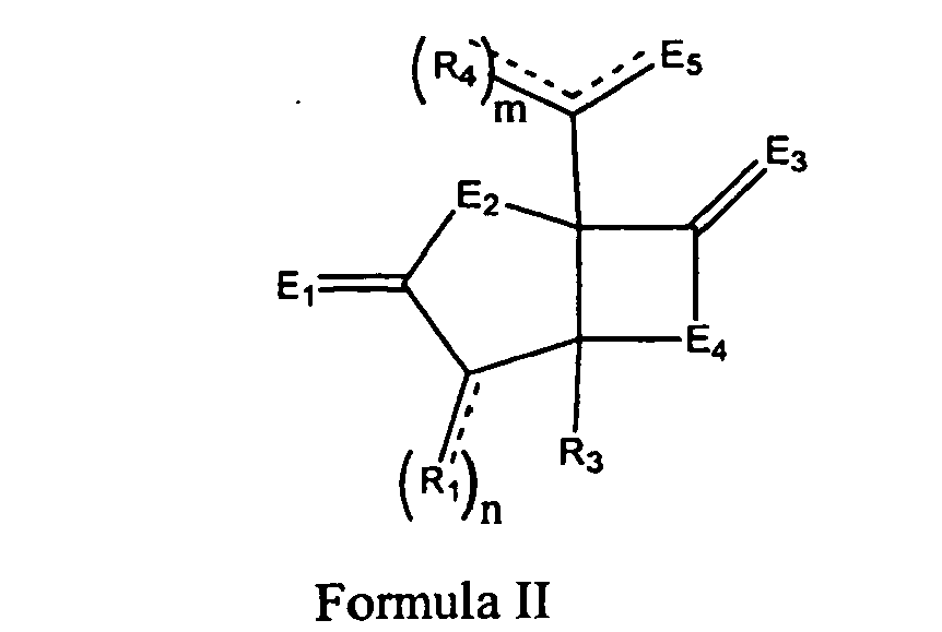
wherein R1 is separately selected from the group consisting of a hydrogen, a halogen, a mono-substituted, a poly-substituted or an unsubstituted variant of the following residues: saturated C1-C24 alkyl, unsaturated C2-C24 alkenyl, unsaturated C2-C24 alkynyl, acyl, acyloxy, alkyloxycarbonyloxy, aryloxycarbonyloxy, cycloalkyl, cycloalkenyl, alkoxy, cycloalkoxy, aryl, heteroaryl, arylalkoxy carbonyl, alkoxy carbonylacyl, amino, aminocarbonyl, aminocarbonyloxy, nitro, azido, phenyl, cycloalkylacyl, hydroxy, alkylthio, arylthio, oxysulfonyl, carboxy, cyano, and halogenated alkyl including polyhalogenated alkyl, where n is equal to 1 or 2, and if n is equal to 2, then R1 can be the same or different;
wherein R3 is selected from the group consisting of a halogen, a mono-substituted, a poly-substituted or an unsubstituted variant of the following residues: saturated C1-C24 alkyl, unsaturated C2-C24 alkenyl, unsaturated C2-C24 alkynyl, acyl, acyloxy, alkyloxycarbonyloxy, aryloxycarbonyloxy, cycloalkyl, cycloalkenyl, alkoxy, cycloalkoxy, aryl, heteroaryl, arylalkoxy carbonyl, alkoxy carbonylacyl, amino, aminocarbonyl, aminocarbonyloxy, nitro, azido, phenyl, cycloalkylacyl, hydroxy, alkylthio, arylthio, oxysulfonyl, carboxy, cyano, and halogenated alkyl including polyhalogenated alkyl;
wherein R4 can be separately selected from the group consisting of a hydrogen, a halogen, a mono-substituted, a poly-substituted or an unsubstituted variant of the following residues: saturated C1-C24 alkyl, unsaturated C2-C24 alkenyl, unsaturated C2-C24 alkynyl, acyl, acyloxy, alkyloxycarbonyloxy, aryloxycarbonyloxy, cycloalkyl, cycloalkenyl, alkoxy, cycloalkoxy, aryl, heteroaryl, arylalkoxy carbonyl, alkoxy carbonylacyl, amino, aminocarbonyl, aminocarbonyloxy, nitro, azido, phenyl, cycloalkylacyl, hydroxy, alkylthio, arylthio, oxysulfonyl, carboxy, cyano, and halogenated alkyl including polyhalogenated alkyl;
wherein m is 1 or 2, and when m is 2, R4 can be the same or different;
wherein E1, E3 and E4 is O, E2 is NH and E5 is OH, O, OR10, S, SR11, SO2R11, NH, NH2, NOH, NHOH, NR12, or NHOR13, wherein R10, R11, R12, and R13 are selected from the group consisting of hydrogen, a substituted or unsubstituted alkyl, a substituted or unsubstituted aryl, and a substituted or unsubstituted heteroaryl; and
wherein the cancer is liver cancer or angioma. - The use of Claim 2, wherein the cancer is a drug resistant cancer.
- The use of Claim 3, wherein the drug-resistant cancer displays at least one of the following: elevated levels of the P-glycoprotein efflux pump, increased expression of the multidrug-resistance associated protein 1 encoded by MRP1, reduced drug uptake, alteration of the drug's target or increasing repair of drug-induced DNA damage, alteration of the apoptotic pathway or the activation of cytochrome P450 enzymes.
- The use of Claim 2, wherein the animal is a mammal.
- The use of Claim 2, wherein the animal is a human.
- The use of Claim 2, wherein the animal is a rodent.
- The use of Claim 2, wherein the compound is:
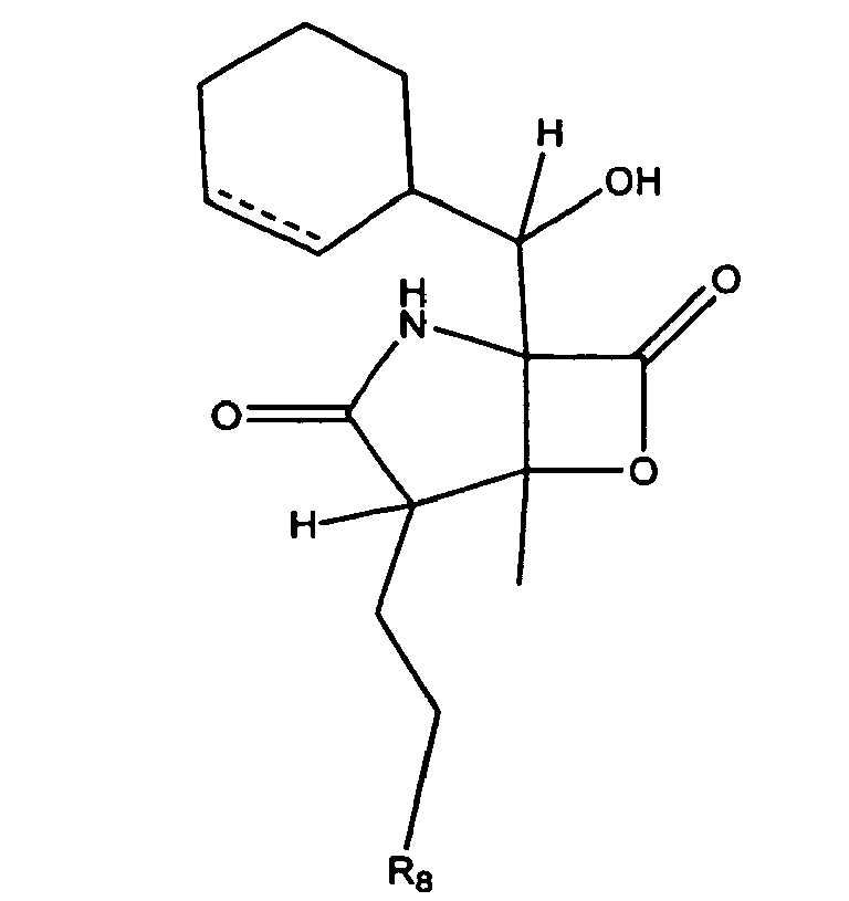
- The use of Claim 2, wherein the compound is :

- The use of Claim 2, wherein:wherein R1, is separately selected from the group consisting of a hydrogen, a halogen, a mono-substituted, a poly-substituted or an unsubstituted variant of the following residues: saturated C1-C24 alkyl, unsaturated C2-C24 alkenyl, unsaturated C2-C24 alkynyl, acyl, acyloxy, alkyloxycarbonyloxy, aryloxycarbonyloxy, cycloalkyl, cycloalkenyl, alkoxy, cycloalkoxy, aryl, heteroaryl, arylalkoxy carbonyl, alkoxy carbonylacyl, amino, aminocarbonyl, aminocarbonyloxy, nitro, azido, phenyl, hydroxy, alkylthio, arylthio, oxysulfonyl, carboxy, cyano, and halogenated alkyl including polyhalogenated alkyl, where n is equal to 1 or 2, and if n is equal to 2, then R1 can be the same or different;wherein R3 is methyl; andwherein R4 can be separately selected from the group consisting of a hydrogen, a halogen, a mono-substituted, a poly-substituted or an unsubstituted variant of the following residues: saturated C1-C24 alkyl, unsaturated C2-C24 alkenyl, unsaturated C2-C24 alkynyl, acyl, acyloxy, alkyloxycarbonyloxy, aryloxycarbonyloxy, cycloalkyl, cycloalkenyl, alkoxy, cycloalkoxy, aryl, heteroaryl, arylalkoxy carbonyl, alkoxy carbonylacyl, amino, aminocarbonyl, aminocarbonyloxy, nitro, azido, phenyl, hydroxy, alkylthio, arylthio, oxysulfonyl, carboxy, cyano, and halogenated alkyl including polyhalogenated alkyl.
- The use of Claim 2, wherein:wherein R1 is separately selected from the group consisting of a hydrogen, a halogen, a mono-substituted, a poly-substituted or an unsubstituted variant of the following residues: saturated C1-C24 alkyl, unsaturated C2-C24 alkenyl, unsaturated C2-C24 alkynyl, acyl, acyloxy, alkyloxycarbonyloxy, aryloxycarbonyloxy, cycloalkyl, cycloalkenyl, alkoxy, cycloalkoxy, aryl, heteroaryl, arylalkoxy carbonyl, alkoxy carbonylacyl, amino, aminocarbonyl, aminocarbonyloxy, nitro, azido, phenyl, hydroxy, alkylthio, arylthio, oxysulfonyl, carboxy, cyano, and halogenated alkyl including polyhalogenated alkyl, where n is equal to 1 or 2, and if n is equal to 2, then R1 can be the same or different;wherein R3 is selected from the group consisting of a halogen, a mono-substituted, a poly-substituted or an unsubstituted variant of the following residues:saturated C1-C24 alkyl, unsaturated C2-C24 alkenyl, unsaturated C2-C24 alkynyl, acyl, acyloxy, alkyloxycarbonyloxy, aryloxycarbonyloxy, cycloalkyl, cycloalkenyl, alkoxy, cycloalkoxy, aryl, heteroaryl, arylalkoxy carbonyl, alkoxy carbonylacyl, amino, aminocarbonyl, aminocarbonyloxy, nitro, azido, phenyl, hydroxy, alkylthio, arylthio, oxysulfonyl, carboxy, cyano, and halogenated alkyl including polyhalogenated alkyl;wherein m is 1;wherein R4 is
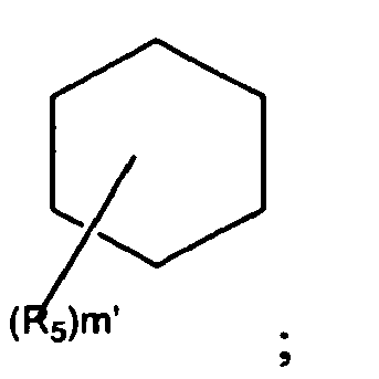 wherein R5 is separately selected from the group consisting of a hydrogen, a halogen, a mono-substituted, a poly-substituted or an unsubstituted variant of the following residues: saturated C1-C24 alkyl, unsaturated C2-C24 alkenyl, unsaturated C2-C24 alkynyl, acyl, acyloxy, alkyloxycarbonyloxy, aryloxycarbonyloxy, cycloalkyl, cycloalkenyl, alkoxy, cycloalkoxy, aryl, heteroaryl, arylalkoxy carbonyl, alkoxy carbonylacyl, amino, aminocarbonyl, aminocarbonyloxy, nitro, azido, phenyl, hydroxy, alkylthio, arylthio, oxysulfonyl, carboxy, cyano, and halogenated alkyl including polyhalogenated alkyl, and
m' is 0, 1, 2, 3, 4, 5, 6, 7, 8, 9, 10, or 11 and if m' is more than 1, then R5 can be the same or different. - The use of Claim 11, wherein R3 is methyl.
- The use of Claim 2, wherein the compound is :

- The use of Claim 2, wherein at least one R1 is CH2CH2X, wherein X is selected from the group consisting of H, F, Cl, Br, and I and R3 is methyl.
Applications Claiming Priority (3)
| Application Number | Priority Date | Filing Date | Title |
|---|---|---|---|
| US48027003P | 2003-06-20 | 2003-06-20 | |
| US56695204P | 2004-04-30 | 2004-04-30 | |
| PCT/US2004/019543 WO2005002572A2 (en) | 2003-06-20 | 2004-06-18 | Use of [3.2.0] heterocyclic compounds and analogs thereof for the treatment of cancer, inflammation and infectious diseases |
Publications (2)
| Publication Number | Publication Date |
|---|---|
| EP1638552A2 EP1638552A2 (en) | 2006-03-29 |
| EP1638552B1 true EP1638552B1 (en) | 2011-03-02 |
Family
ID=33567612
Family Applications (1)
| Application Number | Title | Priority Date | Filing Date |
|---|---|---|---|
| EP04776757A Expired - Lifetime EP1638552B1 (en) | 2003-06-20 | 2004-06-18 | Use of (3.2.0) heterocyclic compounds and analogs thereof for the treatment of cancer |
Country Status (14)
| Country | Link |
|---|---|
| US (2) | US20050049294A1 (en) |
| EP (1) | EP1638552B1 (en) |
| JP (2) | JP2007523862A (en) |
| KR (1) | KR20060026052A (en) |
| CN (1) | CN102151261A (en) |
| AT (1) | ATE499934T1 (en) |
| AU (1) | AU2004253478A1 (en) |
| BR (1) | BRPI0411677A (en) |
| CA (1) | CA2532066C (en) |
| DE (1) | DE602004031614D1 (en) |
| IL (1) | IL172705A (en) |
| MX (1) | MXPA05013982A (en) |
| NZ (1) | NZ544588A (en) |
| WO (1) | WO2005002572A2 (en) |
Families Citing this family (48)
| Publication number | Priority date | Publication date | Assignee | Title |
|---|---|---|---|---|
| JP4395549B2 (en) * | 2000-11-16 | 2010-01-13 | ザ レジェンツ オブ ザ ユニヴァースティ オブ カリフォルニア | Marine actinomycete taxa for the discovery of drugs and fermentation products |
| US7176232B2 (en) | 2002-06-24 | 2007-02-13 | The Regents Of The University Of California | Salinosporamides and methods for use thereof |
| US7179834B2 (en) * | 2002-06-24 | 2007-02-20 | The Regents Of The University Of California | Salinosporamides and methods for use thereof |
| KR20060026052A (en) | 2003-06-20 | 2006-03-22 | 니리어스 파마슈티컬즈, 인코퍼레이션 | How to use [3.2.0] heterocyclic compounds and their analogs for the treatment of cancer, inflammatory and infectious diseases |
| EP2441767B1 (en) * | 2003-06-20 | 2015-06-10 | The Regents of The University of California | Salinosporamides and methods for use thereof |
| JP4691041B2 (en) | 2003-11-20 | 2011-06-01 | チルドレンズ ホスピタル メディカル センター | GTPase inhibitors and methods of use |
| WO2005099687A2 (en) * | 2004-04-09 | 2005-10-27 | President And Fellows Of Harvard College | Analogs of salinosporamide a |
| US7579371B2 (en) | 2004-04-30 | 2009-08-25 | Nereus Pharmaceuticals, Inc. | Methods of using [3.2.0] heterocyclic compounds and analogs thereof |
| BRPI0509824A (en) * | 2004-04-30 | 2007-10-09 | Nereus Pharmaceuticals Inc | heterocyclic compounds [3.2.0] and methods of use thereof |
| MX2007000392A (en) * | 2004-07-12 | 2007-06-15 | Bayer Cropscience Ag | Substituted 2-pyrrolidone derivatives as fungicides and insecticides. |
| CA2590334A1 (en) * | 2004-12-03 | 2006-06-08 | Nereus Pharmaceuticals, Inc. | Methods of using [3.2.0] heterocyclic compounds and analogs thereof |
| SG157365A1 (en) * | 2004-12-03 | 2009-12-29 | Dana Farber Cancer Inst Inc | Compositions and methods for treating neoplastic diseases |
| US7826982B2 (en) | 2005-07-29 | 2010-11-02 | Children's Hospital Medical Center | Method of identifying inhibitors using a 3-D structure of RAC-1 GTPASE |
| US7691896B2 (en) | 2005-08-10 | 2010-04-06 | President And Fellows Of Harvard College | Analogs of salinosporamide A |
| WO2007021897A1 (en) * | 2005-08-10 | 2007-02-22 | President And Fellows Of Harvard College | Analogs of salinosporamide a |
| US7572606B1 (en) * | 2005-09-09 | 2009-08-11 | Nereus Pharmaceuticals, Inc. | Biosyntheses of salinosporamide A and its analogs and related methods of making salinosporamide A and its analogs |
| US7465720B2 (en) | 2005-09-12 | 2008-12-16 | President And Fellows Of Harvard College | Proteasome inhibiting β-lactam compounds |
| BRPI0618198A2 (en) * | 2005-11-04 | 2018-06-26 | Nereus Pharmaceuticals Inc | cancer and lymphoma treatment, prevention or inhibition methods, composition, kit, and therapy resistant cancer treatment method |
| NZ572026A (en) | 2006-04-06 | 2011-12-22 | Nereus Pharmaceuticals Inc | Total synthesis of salinosporamide a and analogs thereof |
| US20090280130A1 (en) * | 2006-06-14 | 2009-11-12 | Children's Medical Center Corporation | Method for the treatment of anthrax toxicity |
| US8986971B2 (en) | 2006-09-22 | 2015-03-24 | Triphase Research And Development I Corp. | Salt formulations for the fermentation of marine microorganisms |
| WO2008095195A2 (en) | 2007-02-02 | 2008-08-07 | Nereus Pharmaceuticals, Inc. | Lyophilized formulations of salinosporamide a |
| WO2008137780A2 (en) * | 2007-05-04 | 2008-11-13 | Nereus Pharmaceuticals, Inc. | Use of [3.2.0] heterocyclic compounds and analogs thereof for treating infectious diseases |
| US8394816B2 (en) * | 2007-12-07 | 2013-03-12 | Irene Ghobrial | Methods of using [3.2.0] heterocyclic compounds and analogs thereof in treating Waldenstrom's Macroglobulinemia |
| EP2088205A1 (en) | 2008-02-11 | 2009-08-12 | Institut National De La Sante Et De La Recherche Medicale (Inserm) | PSMB10: A diagnosis marker and therapeutic target of chronic rejection. |
| JP2011514352A (en) | 2008-03-07 | 2011-05-06 | ネレアス ファーマシューティカルズ インコーポレイテッド | Total synthesis of salinosporamide A and its analogs |
| US20090285836A1 (en) * | 2008-04-14 | 2009-11-19 | Nereus Pharmaceuticals, Inc. | Use of salinosporamide a to inhibit metastasis |
| CN102089312A (en) * | 2008-05-12 | 2011-06-08 | 尼瑞斯药品公司 | Salinosporamide derivatives as proteasome inhibitors |
| US20110111417A1 (en) | 2008-05-14 | 2011-05-12 | Millennium Pharmaceuticals, Inc. | Methods and kits for monitoring the effects of immunomodulators on adaptive immunity |
| US8073633B2 (en) * | 2008-12-30 | 2011-12-06 | The Invention Science Fund I, Llc | Computational methods and systems for suggesting modulators of CYP450 as treatment options |
| US8321151B2 (en) | 2008-12-30 | 2012-11-27 | The Invention Science Fund I, Llc | Computational methods and systems for treatment in relation to modulation of CYP450 enzyme activity |
| US8315815B2 (en) | 2008-12-30 | 2012-11-20 | The Invention Science Fund I, Llc | Computational methods and systems for suggesting modulators of CYP450 as treatment options |
| WO2010096574A1 (en) | 2009-02-20 | 2010-08-26 | Lisanti Michael P | A method of diagnosis or prognosis of a neoplasm comprising determining the level of expression of a protein in stromal cells adjacent to the neoplasm |
| US9126997B1 (en) | 2010-09-07 | 2015-09-08 | Northwestern University | Synergistic effect of glucocorticoid receptor agonists in combination with proteosome inhibitors for treating leukemia and myeloma |
| US9272014B2 (en) | 2011-03-29 | 2016-03-01 | Texas Tech University System | Galectin-3C combination therapy for human cancer |
| JP6238900B2 (en) | 2011-10-28 | 2017-11-29 | ミレニアム ファーマシューティカルズ, インコーポレイテッドMillennium Pharmaceuticals, Inc. | Biomarkers of response to NAE inhibitors |
| CN104126017A (en) | 2011-11-11 | 2014-10-29 | 米伦纽姆医药公司 | Biomarkers of response to proteasome inhibitors |
| CA2862492A1 (en) | 2012-01-24 | 2013-08-01 | Millennium Pharmaceuticals, Inc. | Methods of treatment of cancer |
| WO2013112881A1 (en) | 2012-01-27 | 2013-08-01 | Thomas Jefferson University | Mct protein inhibitor-related prognostic and therapeutic methods |
| EP2904115B1 (en) | 2012-10-01 | 2018-08-08 | Millennium Pharmaceuticals, Inc. | Biomarkers and methods to predict response to inhibitors and uses thereof |
| EA201590916A1 (en) | 2012-11-09 | 2016-02-29 | Корнэлл Юниверсити | LOW MOLECULAR INHIBITORS MALT1 |
| US10022372B2 (en) | 2013-04-19 | 2018-07-17 | Thomas Jefferson University | Caveolin-1 related methods for treating glioblastoma with temozolomide |
| US10028503B2 (en) | 2014-06-18 | 2018-07-24 | Children's Hospital Medical Center | Platelet storage methods and compositions for same |
| JP7171550B2 (en) | 2016-06-01 | 2022-11-15 | セルジュヌ アンテルナシオナル ドゥジエム ソシエテ ア レスポンサビリテ リミティー | Use of marizomib for treatment of central nervous system (CNS) cancer |
| US10703760B2 (en) | 2016-08-19 | 2020-07-07 | Celgene International Ii Sàrl | Morphic forms of marizomib and uses thereof |
| KR101953671B1 (en) * | 2017-08-21 | 2019-03-04 | 충남대학교산학협력단 | Composition for preventing, improving or treating atopic dermatitis containing proteasome inhibitor as effective component |
| EP3546447A1 (en) * | 2018-03-29 | 2019-10-02 | Heidelberg Pharma GmbH | Synthesis of (2s,3r,4r)-4,5-dihydroxyisoleucine and derivatives |
| CN115006532A (en) * | 2021-03-05 | 2022-09-06 | 广州医科大学 | Use of proteasome inhibitors |
Family Cites Families (64)
| Publication number | Priority date | Publication date | Assignee | Title |
|---|---|---|---|---|
| US5133974A (en) * | 1989-05-05 | 1992-07-28 | Kv Pharmaceutical Company | Extended release pharmaceutical formulations |
| US5276182A (en) * | 1990-07-09 | 1994-01-04 | The Dow Chemical Company | Process for preparing polyurea oligomers |
| DE4039602A1 (en) * | 1990-12-12 | 1992-06-17 | Bauer Kurt Heinz Prof Dr | PHARMACEUTICAL FORMULATIONS |
| KR100221695B1 (en) * | 1991-08-12 | 1999-09-15 | 그린 마틴, 브라이언 쥐 테슬리 | Pharmaceutical Formulation Formulations |
| US5653962A (en) * | 1991-12-12 | 1997-08-05 | Glaxo Group Limited | Aerosol formulations containing P134a and particulate medicaments |
| AU5612694A (en) * | 1992-11-27 | 1994-06-22 | Napro Biotherapeutics, Inc. | Injectable composition |
| JPH08507754A (en) | 1993-02-10 | 1996-08-20 | ザ・プレジデント・アンド・フェローズ・オブ・ハーバード・カレッジ | Role of ATP-ubiquitin-dependent proteolysis in MHC-1-restricted antigen presentation and its inhibitor |
| US5654286A (en) * | 1993-05-12 | 1997-08-05 | Hostetler; Karl Y. | Nucleotides for topical treatment of psoriasis, and methods for using same |
| EP1475091A1 (en) * | 1993-10-01 | 2004-11-10 | Roche Palo Alto LLC | Mycophenolate mofetil - high dose oral suspensions |
| US5707641A (en) * | 1994-10-13 | 1998-01-13 | Pharmaderm Research & Development Ltd. | Formulations comprising therapeutically-active proteins or polypeptides |
| IE80468B1 (en) * | 1995-04-04 | 1998-07-29 | Elan Corp Plc | Controlled release biodegradable nanoparticles containing insulin |
| US6838477B2 (en) * | 1995-04-12 | 2005-01-04 | President And Fellows Of Harvard College | Lactacystin analogs |
| US6335358B1 (en) * | 1995-04-12 | 2002-01-01 | President And Fellows Of Harvard College | Lactacystin analogs |
| US5653987A (en) * | 1995-05-16 | 1997-08-05 | Modi; Pankaj | Liquid formulations for proteinic pharmaceuticals |
| US5726181A (en) * | 1995-06-05 | 1998-03-10 | Bionumerik Pharmaceuticals, Inc. | Formulations and compositions of poorly water soluble camptothecin derivatives |
| US5667809A (en) * | 1995-06-07 | 1997-09-16 | Alliance Pharmaceutical Corp. | Continuous fluorochemical microdispersions for the delivery of lipophilic pharmaceutical agents |
| US5874443A (en) * | 1995-10-19 | 1999-02-23 | Trega Biosciences, Inc. | Isoquinoline derivatives and isoquinoline combinatorial libraries |
| US5886210A (en) * | 1996-08-22 | 1999-03-23 | Rohm And Haas Company | Method for preparing aromatic compounds |
| US6271199B2 (en) | 1997-02-15 | 2001-08-07 | Millennium Pharmaceuticals, Inc. | Treatment of infarcts |
| CA2283636A1 (en) | 1997-03-20 | 1998-09-24 | Variagenics, Inc. | Target genes for allele-specific drugs |
| US5922683A (en) * | 1997-05-29 | 1999-07-13 | Abbott Laboratories | Multicyclic erythromycin derivatives |
| US6333358B1 (en) * | 1997-08-04 | 2001-12-25 | Taisho Pharmaceutical Co., Ltd. | Aryloxyaniline derivatives |
| BR9811304A (en) | 1997-08-15 | 2001-11-13 | Francois Soucy | Synthesis of clasto-lactacistin "beta" -lactone and its analogues |
| US6133308A (en) * | 1997-08-15 | 2000-10-17 | Millennium Pharmaceuticals, Inc. | Synthesis of clasto-lactacystin beta-lactone and analogs thereof |
| US20010051654A1 (en) * | 1997-09-25 | 2001-12-13 | Elliott Peter J. | Treatment of inflammatory and autoimmune diseases |
| WO1999015183A1 (en) | 1997-09-25 | 1999-04-01 | Proscript Inc. | PROTEASOME INHIBITORS, UBIQUITIN PATHWAY INHIBITORS OR AGENTS THAT INTERFERE WITH THE ACTIVATION OF NF-λB VIA THE UBIQUITIN PROTEASOME PATHWAY TO TREAT INFLAMMATORY AND AUTOIMMUNE DISEASES |
| CA2219867A1 (en) | 1997-10-31 | 1999-04-30 | Jiangping Wu | The use of proteasome inhibitors for treating cancer, inflammation, autoimmune disease, graft rejection and septic shock |
| EP1032377B1 (en) * | 1997-11-21 | 2010-11-17 | Purdue Neuroscience Company | Substituted 2-aminoacetamides and the use thereof |
| US6617171B2 (en) * | 1998-02-27 | 2003-09-09 | The General Hospital Corporation | Methods for diagnosing and treating autoimmune disease |
| BR9909146A (en) * | 1998-03-26 | 2000-12-05 | Shionogi & Co | Indole derivatives showing antiviral activity |
| US6509331B1 (en) * | 1998-06-22 | 2003-01-21 | Elan Pharmaceuticals, Inc. | Deoxyamino acid compounds, pharmaceutical compositions comprising same, and methods for inhibiting β-amyloid peptide release and/or its synthesis by use of such compounds |
| KR20010080267A (en) * | 1998-10-20 | 2001-08-22 | 밀레니엄 파머슈티컬스 인코퍼레이티드 | Method for monitoring proteasome inhibitor drug action |
| FR2784988B1 (en) * | 1998-10-23 | 2002-09-20 | Adir | NOVEL DIHYDRO AND TETRAHYDROQUINOLEINIC COMPOUNDS, PROCESS FOR THEIR PREPARATION AND THE PHARMACEUTICAL COMPOSITIONS CONTAINING THEM |
| WO2000033654A1 (en) | 1998-12-04 | 2000-06-15 | University Of Maryland Biotechnology Institute | Use of protease inhibitors to modulate cellular pathways, immunity and therapies associated therewith |
| US20020049157A1 (en) * | 1999-08-25 | 2002-04-25 | Jiangping Wu | Use of proteasome inhibitors for treating cancer, inflammation, autoimmune disease, graft rejection and septic shock |
| ES2272558T3 (en) * | 2000-10-12 | 2007-05-01 | Viromics Gmbh | PROTEASOME INHIBITORS FOR THE TREATMENT OF INFECTIONS CAUSED BY HEPATITIS VIRUSES. |
| JP4395549B2 (en) * | 2000-11-16 | 2010-01-13 | ザ レジェンツ オブ ザ ユニヴァースティ オブ カリフォルニア | Marine actinomycete taxa for the discovery of drugs and fermentation products |
| DE10316735A1 (en) * | 2002-04-05 | 2003-11-20 | Viromics Gmbh | Inhibiting release, maturation and replication of Flaviviridae family viruses, e.g. for the treatment of hepatitis C virus infections, using proteasome inhibitor such as lactacystine or epoxomicin |
| US7176232B2 (en) * | 2002-06-24 | 2007-02-13 | The Regents Of The University Of California | Salinosporamides and methods for use thereof |
| US7179834B2 (en) * | 2002-06-24 | 2007-02-20 | The Regents Of The University Of California | Salinosporamides and methods for use thereof |
| AU2003249930A1 (en) | 2002-07-03 | 2004-01-23 | Charite - Universitatsmedizin Berlin | Proteaseome inhibitors for the treatment of herpesviridae infected individuals |
| EP1565193B1 (en) | 2002-11-06 | 2013-04-24 | Dana-Farber Cancer Institute, Inc. | Compositions for treating cancer using proteasome inhibitor PS-341 |
| ATE448232T1 (en) * | 2003-02-14 | 2009-11-15 | Intermed Discovery Gmbh | SUBSTITUTED HETEROCYCLES |
| EP2441767B1 (en) * | 2003-06-20 | 2015-06-10 | The Regents of The University of California | Salinosporamides and methods for use thereof |
| KR20060026052A (en) * | 2003-06-20 | 2006-03-22 | 니리어스 파마슈티컬즈, 인코퍼레이션 | How to use [3.2.0] heterocyclic compounds and their analogs for the treatment of cancer, inflammatory and infectious diseases |
| US7371875B2 (en) * | 2004-03-12 | 2008-05-13 | Miikana Therapeutics, Inc. | Cytotoxic agents and methods of use |
| US7183417B2 (en) * | 2004-04-09 | 2007-02-27 | President And Fellows Of Harvard College | Simple stereocontrolled synthesis of salinosporamide A |
| WO2005099687A2 (en) * | 2004-04-09 | 2005-10-27 | President And Fellows Of Harvard College | Analogs of salinosporamide a |
| BRPI0509824A (en) | 2004-04-30 | 2007-10-09 | Nereus Pharmaceuticals Inc | heterocyclic compounds [3.2.0] and methods of use thereof |
| US20060264495A1 (en) * | 2004-04-30 | 2006-11-23 | Michael Palladino | Methods of using [3.2.0] heterocyclic compounds and analogs thereof |
| US7579371B2 (en) * | 2004-04-30 | 2009-08-25 | Nereus Pharmaceuticals, Inc. | Methods of using [3.2.0] heterocyclic compounds and analogs thereof |
| SG157365A1 (en) * | 2004-12-03 | 2009-12-29 | Dana Farber Cancer Inst Inc | Compositions and methods for treating neoplastic diseases |
| WO2006124902A2 (en) * | 2005-05-16 | 2006-11-23 | Sloan-Kettering Institute For Cancer Research | Synthesis of salinosporamide a and analogues thereof |
| US7572606B1 (en) * | 2005-09-09 | 2009-08-11 | Nereus Pharmaceuticals, Inc. | Biosyntheses of salinosporamide A and its analogs and related methods of making salinosporamide A and its analogs |
| DE102005046344A1 (en) * | 2005-09-28 | 2007-03-29 | Saltigo Gmbh | Process for the coupling of benzylamines with haloaromatics |
| BRPI0618198A2 (en) * | 2005-11-04 | 2018-06-26 | Nereus Pharmaceuticals Inc | cancer and lymphoma treatment, prevention or inhibition methods, composition, kit, and therapy resistant cancer treatment method |
| GB0605217D0 (en) * | 2006-03-15 | 2006-04-26 | Novartis Ag | Method and compositions for assessing acute rejection |
| NZ572026A (en) | 2006-04-06 | 2011-12-22 | Nereus Pharmaceuticals Inc | Total synthesis of salinosporamide a and analogs thereof |
| US8088923B2 (en) * | 2006-07-07 | 2012-01-03 | The Texas A&M University System | Cyclic-fused beta-lactones and their synthesis |
| WO2008137780A2 (en) * | 2007-05-04 | 2008-11-13 | Nereus Pharmaceuticals, Inc. | Use of [3.2.0] heterocyclic compounds and analogs thereof for treating infectious diseases |
| US8394816B2 (en) * | 2007-12-07 | 2013-03-12 | Irene Ghobrial | Methods of using [3.2.0] heterocyclic compounds and analogs thereof in treating Waldenstrom's Macroglobulinemia |
| EP2088205A1 (en) | 2008-02-11 | 2009-08-12 | Institut National De La Sante Et De La Recherche Medicale (Inserm) | PSMB10: A diagnosis marker and therapeutic target of chronic rejection. |
| JP2011514352A (en) | 2008-03-07 | 2011-05-06 | ネレアス ファーマシューティカルズ インコーポレイテッド | Total synthesis of salinosporamide A and its analogs |
| CN102089312A (en) * | 2008-05-12 | 2011-06-08 | 尼瑞斯药品公司 | Salinosporamide derivatives as proteasome inhibitors |
-
2004
- 2004-06-18 KR KR1020057024503A patent/KR20060026052A/en not_active Ceased
- 2004-06-18 AT AT04776757T patent/ATE499934T1/en not_active IP Right Cessation
- 2004-06-18 CN CN2010102534518A patent/CN102151261A/en active Pending
- 2004-06-18 NZ NZ544588A patent/NZ544588A/en not_active IP Right Cessation
- 2004-06-18 WO PCT/US2004/019543 patent/WO2005002572A2/en not_active Ceased
- 2004-06-18 AU AU2004253478A patent/AU2004253478A1/en not_active Abandoned
- 2004-06-18 BR BRPI0411677-1A patent/BRPI0411677A/en not_active IP Right Cessation
- 2004-06-18 EP EP04776757A patent/EP1638552B1/en not_active Expired - Lifetime
- 2004-06-18 US US10/871,368 patent/US20050049294A1/en not_active Abandoned
- 2004-06-18 MX MXPA05013982A patent/MXPA05013982A/en not_active Application Discontinuation
- 2004-06-18 JP JP2006517404A patent/JP2007523862A/en not_active Withdrawn
- 2004-06-18 CA CA2532066A patent/CA2532066C/en not_active Expired - Lifetime
- 2004-06-18 DE DE602004031614T patent/DE602004031614D1/en not_active Expired - Lifetime
-
2005
- 2005-12-20 IL IL172705A patent/IL172705A/en active IP Right Grant
-
2008
- 2008-06-10 US US12/136,688 patent/US8168803B2/en not_active Expired - Lifetime
-
2011
- 2011-10-28 JP JP2011238014A patent/JP2012031198A/en not_active Withdrawn
Non-Patent Citations (2)
| Title |
|---|
| INTERNET CITATION, Retrieved from the Internet <URL:http://en.wikipedia.org/wiki/Myeloma> * |
| INTERNET CITATION, Retrieved from the Internet <URL:http://en.wikipedia.org/wiki/Sarcoma> * |
Also Published As
| Publication number | Publication date |
|---|---|
| CA2532066C (en) | 2015-07-28 |
| US8168803B2 (en) | 2012-05-01 |
| MXPA05013982A (en) | 2006-05-25 |
| JP2007523862A (en) | 2007-08-23 |
| BRPI0411677A (en) | 2006-08-29 |
| WO2005002572A3 (en) | 2005-05-12 |
| CA2532066A1 (en) | 2005-01-13 |
| NZ544588A (en) | 2010-06-25 |
| EP1638552A2 (en) | 2006-03-29 |
| IL172705A0 (en) | 2006-04-10 |
| JP2012031198A (en) | 2012-02-16 |
| IL172705A (en) | 2017-06-29 |
| AU2004253478A1 (en) | 2005-01-13 |
| CN102151261A (en) | 2011-08-17 |
| US20090182027A1 (en) | 2009-07-16 |
| US20050049294A1 (en) | 2005-03-03 |
| ATE499934T1 (en) | 2011-03-15 |
| WO2005002572A2 (en) | 2005-01-13 |
| KR20060026052A (en) | 2006-03-22 |
| DE602004031614D1 (en) | 2011-04-14 |
Similar Documents
| Publication | Publication Date | Title |
|---|---|---|
| EP1638552B1 (en) | Use of (3.2.0) heterocyclic compounds and analogs thereof for the treatment of cancer | |
| US7544814B2 (en) | [3.2.0] Heterocyclic compounds and methods of using the same | |
| US8394816B2 (en) | Methods of using [3.2.0] heterocyclic compounds and analogs thereof in treating Waldenstrom's Macroglobulinemia | |
| CA2590334A1 (en) | Methods of using [3.2.0] heterocyclic compounds and analogs thereof | |
| US20080280968A1 (en) | Methods of using [3.2.0] heterocyclic compounds and analogs thereof for treating infectious diseases | |
| US7579371B2 (en) | Methods of using [3.2.0] heterocyclic compounds and analogs thereof | |
| US20060264495A1 (en) | Methods of using [3.2.0] heterocyclic compounds and analogs thereof | |
| US7572606B1 (en) | Biosyntheses of salinosporamide A and its analogs and related methods of making salinosporamide A and its analogs | |
| WO2007130404A1 (en) | Methods of using [3.2.0] heterocyclic compounds and analogs thereof for the treatment of lung cancer | |
| WO2006118973A2 (en) | Methods of using heterobyclic compounds for treatment of rectal cancer | |
| WO2008124699A1 (en) | A method of using proteasome inhibitors in combination with histone deacetylase inhibitors to treat cancer | |
| ZA200600536B (en) | Use of [3.2.0] heterocyclic compounds and analogs thereof for the treatment of cancer, inflammation and infectious diseases | |
| HK1129662A (en) | [3.2.0] heterocyclic compounds and methods of using the same | |
| HK1151023A (en) | [3.2.0] heterocyclic compounds and methods of using the same | |
| US7112581B2 (en) | Macrocyclic lactams | |
| CN101208087A (en) | [3.2.0] Application methods of heterocyclic compounds and their analogues |
Legal Events
| Date | Code | Title | Description |
|---|---|---|---|
| PUAI | Public reference made under article 153(3) epc to a published international application that has entered the european phase |
Free format text: ORIGINAL CODE: 0009012 |
|
| 17P | Request for examination filed |
Effective date: 20060116 |
|
| AK | Designated contracting states |
Kind code of ref document: A2 Designated state(s): AT BE BG CH CY CZ DE DK EE ES FI FR GB GR HU IE IT LI LU MC NL PL PT RO SE SI SK TR |
|
| DAX | Request for extension of the european patent (deleted) | ||
| REG | Reference to a national code |
Ref country code: HK Ref legal event code: DE Ref document number: 1087923 Country of ref document: HK |
|
| RIN1 | Information on inventor provided before grant (corrected) |
Inventor name: POTTS, BARBARA, CHRISTINE Inventor name: PALLADINO, MICHAEL Inventor name: NEUTEBOOM, SASKIA, THEODORA, CORNELIA Inventor name: MACHERLA, VENKATA, RAMI, REDDY |
|
| 17Q | First examination report despatched |
Effective date: 20060612 |
|
| GRAP | Despatch of communication of intention to grant a patent |
Free format text: ORIGINAL CODE: EPIDOSNIGR1 |
|
| RTI1 | Title (correction) |
Free format text: USE OF (3.2.0) HETEROCYCLIC COMPOUNDS AND ANALOGS THEREOF FOR THE TREATMENT OF CANCER |
|
| GRAS | Grant fee paid |
Free format text: ORIGINAL CODE: EPIDOSNIGR3 |
|
| GRAA | (expected) grant |
Free format text: ORIGINAL CODE: 0009210 |
|
| AK | Designated contracting states |
Kind code of ref document: B1 Designated state(s): AT BE BG CH CY CZ DE DK EE ES FI FR GB GR HU IE IT LI LU MC NL PL PT RO SE SI SK TR |
|
| REG | Reference to a national code |
Ref country code: GB Ref legal event code: FG4D |
|
| REG | Reference to a national code |
Ref country code: CH Ref legal event code: EP |
|
| REG | Reference to a national code |
Ref country code: IE Ref legal event code: FG4D |
|
| REF | Corresponds to: |
Ref document number: 602004031614 Country of ref document: DE Date of ref document: 20110414 Kind code of ref document: P |
|
| REG | Reference to a national code |
Ref country code: DE Ref legal event code: R096 Ref document number: 602004031614 Country of ref document: DE Effective date: 20110414 |
|
| REG | Reference to a national code |
Ref country code: NL Ref legal event code: VDEP Effective date: 20110302 |
|
| PG25 | Lapsed in a contracting state [announced via postgrant information from national office to epo] |
Ref country code: ES Free format text: LAPSE BECAUSE OF FAILURE TO SUBMIT A TRANSLATION OF THE DESCRIPTION OR TO PAY THE FEE WITHIN THE PRESCRIBED TIME-LIMIT Effective date: 20110613 Ref country code: GR Free format text: LAPSE BECAUSE OF FAILURE TO SUBMIT A TRANSLATION OF THE DESCRIPTION OR TO PAY THE FEE WITHIN THE PRESCRIBED TIME-LIMIT Effective date: 20110603 Ref country code: SE Free format text: LAPSE BECAUSE OF FAILURE TO SUBMIT A TRANSLATION OF THE DESCRIPTION OR TO PAY THE FEE WITHIN THE PRESCRIBED TIME-LIMIT Effective date: 20110302 |
|
| PG25 | Lapsed in a contracting state [announced via postgrant information from national office to epo] |
Ref country code: BG Free format text: LAPSE BECAUSE OF FAILURE TO SUBMIT A TRANSLATION OF THE DESCRIPTION OR TO PAY THE FEE WITHIN THE PRESCRIBED TIME-LIMIT Effective date: 20110602 Ref country code: AT Free format text: LAPSE BECAUSE OF FAILURE TO SUBMIT A TRANSLATION OF THE DESCRIPTION OR TO PAY THE FEE WITHIN THE PRESCRIBED TIME-LIMIT Effective date: 20110302 Ref country code: NL Free format text: LAPSE BECAUSE OF FAILURE TO SUBMIT A TRANSLATION OF THE DESCRIPTION OR TO PAY THE FEE WITHIN THE PRESCRIBED TIME-LIMIT Effective date: 20110302 Ref country code: CY Free format text: LAPSE BECAUSE OF FAILURE TO SUBMIT A TRANSLATION OF THE DESCRIPTION OR TO PAY THE FEE WITHIN THE PRESCRIBED TIME-LIMIT Effective date: 20110302 Ref country code: FI Free format text: LAPSE BECAUSE OF FAILURE TO SUBMIT A TRANSLATION OF THE DESCRIPTION OR TO PAY THE FEE WITHIN THE PRESCRIBED TIME-LIMIT Effective date: 20110302 Ref country code: SI Free format text: LAPSE BECAUSE OF FAILURE TO SUBMIT A TRANSLATION OF THE DESCRIPTION OR TO PAY THE FEE WITHIN THE PRESCRIBED TIME-LIMIT Effective date: 20110302 |
|
| PG25 | Lapsed in a contracting state [announced via postgrant information from national office to epo] |
Ref country code: BE Free format text: LAPSE BECAUSE OF FAILURE TO SUBMIT A TRANSLATION OF THE DESCRIPTION OR TO PAY THE FEE WITHIN THE PRESCRIBED TIME-LIMIT Effective date: 20110302 |
|
| PG25 | Lapsed in a contracting state [announced via postgrant information from national office to epo] |
Ref country code: PT Free format text: LAPSE BECAUSE OF FAILURE TO SUBMIT A TRANSLATION OF THE DESCRIPTION OR TO PAY THE FEE WITHIN THE PRESCRIBED TIME-LIMIT Effective date: 20110704 Ref country code: EE Free format text: LAPSE BECAUSE OF FAILURE TO SUBMIT A TRANSLATION OF THE DESCRIPTION OR TO PAY THE FEE WITHIN THE PRESCRIBED TIME-LIMIT Effective date: 20110302 |
|
| PG25 | Lapsed in a contracting state [announced via postgrant information from national office to epo] |
Ref country code: RO Free format text: LAPSE BECAUSE OF FAILURE TO SUBMIT A TRANSLATION OF THE DESCRIPTION OR TO PAY THE FEE WITHIN THE PRESCRIBED TIME-LIMIT Effective date: 20110302 Ref country code: CZ Free format text: LAPSE BECAUSE OF FAILURE TO SUBMIT A TRANSLATION OF THE DESCRIPTION OR TO PAY THE FEE WITHIN THE PRESCRIBED TIME-LIMIT Effective date: 20110302 Ref country code: SK Free format text: LAPSE BECAUSE OF FAILURE TO SUBMIT A TRANSLATION OF THE DESCRIPTION OR TO PAY THE FEE WITHIN THE PRESCRIBED TIME-LIMIT Effective date: 20110302 |
|
| PLBE | No opposition filed within time limit |
Free format text: ORIGINAL CODE: 0009261 |
|
| STAA | Information on the status of an ep patent application or granted ep patent |
Free format text: STATUS: NO OPPOSITION FILED WITHIN TIME LIMIT |
|
| REG | Reference to a national code |
Ref country code: CH Ref legal event code: PL |
|
| 26N | No opposition filed |
Effective date: 20111205 |
|
| GBPC | Gb: european patent ceased through non-payment of renewal fee |
Effective date: 20110618 |
|
| PG25 | Lapsed in a contracting state [announced via postgrant information from national office to epo] |
Ref country code: DK Free format text: LAPSE BECAUSE OF FAILURE TO SUBMIT A TRANSLATION OF THE DESCRIPTION OR TO PAY THE FEE WITHIN THE PRESCRIBED TIME-LIMIT Effective date: 20110302 Ref country code: PL Free format text: LAPSE BECAUSE OF FAILURE TO SUBMIT A TRANSLATION OF THE DESCRIPTION OR TO PAY THE FEE WITHIN THE PRESCRIBED TIME-LIMIT Effective date: 20110302 |
|
| REG | Reference to a national code |
Ref country code: FR Ref legal event code: ST Effective date: 20120229 |
|
| REG | Reference to a national code |
Ref country code: IE Ref legal event code: MM4A |
|
| REG | Reference to a national code |
Ref country code: DE Ref legal event code: R097 Ref document number: 602004031614 Country of ref document: DE Effective date: 20111205 |
|
| REG | Reference to a national code |
Ref country code: DE Ref legal event code: R119 Ref document number: 602004031614 Country of ref document: DE Effective date: 20120103 |
|
| PG25 | Lapsed in a contracting state [announced via postgrant information from national office to epo] |
Ref country code: IE Free format text: LAPSE BECAUSE OF NON-PAYMENT OF DUE FEES Effective date: 20110618 Ref country code: CH Free format text: LAPSE BECAUSE OF NON-PAYMENT OF DUE FEES Effective date: 20110630 Ref country code: DE Free format text: LAPSE BECAUSE OF NON-PAYMENT OF DUE FEES Effective date: 20120103 Ref country code: FR Free format text: LAPSE BECAUSE OF NON-PAYMENT OF DUE FEES Effective date: 20110630 Ref country code: LI Free format text: LAPSE BECAUSE OF NON-PAYMENT OF DUE FEES Effective date: 20110630 |
|
| PG25 | Lapsed in a contracting state [announced via postgrant information from national office to epo] |
Ref country code: IT Free format text: LAPSE BECAUSE OF FAILURE TO SUBMIT A TRANSLATION OF THE DESCRIPTION OR TO PAY THE FEE WITHIN THE PRESCRIBED TIME-LIMIT Effective date: 20110302 |
|
| PG25 | Lapsed in a contracting state [announced via postgrant information from national office to epo] |
Ref country code: GB Free format text: LAPSE BECAUSE OF NON-PAYMENT OF DUE FEES Effective date: 20110618 |
|
| REG | Reference to a national code |
Ref country code: HK Ref legal event code: WD Ref document number: 1087923 Country of ref document: HK |
|
| PG25 | Lapsed in a contracting state [announced via postgrant information from national office to epo] |
Ref country code: MC Free format text: LAPSE BECAUSE OF NON-PAYMENT OF DUE FEES Effective date: 20110630 |
|
| PG25 | Lapsed in a contracting state [announced via postgrant information from national office to epo] |
Ref country code: LU Free format text: LAPSE BECAUSE OF NON-PAYMENT OF DUE FEES Effective date: 20110618 |
|
| PG25 | Lapsed in a contracting state [announced via postgrant information from national office to epo] |
Ref country code: TR Free format text: LAPSE BECAUSE OF FAILURE TO SUBMIT A TRANSLATION OF THE DESCRIPTION OR TO PAY THE FEE WITHIN THE PRESCRIBED TIME-LIMIT Effective date: 20110302 |
|
| PG25 | Lapsed in a contracting state [announced via postgrant information from national office to epo] |
Ref country code: HU Free format text: LAPSE BECAUSE OF FAILURE TO SUBMIT A TRANSLATION OF THE DESCRIPTION OR TO PAY THE FEE WITHIN THE PRESCRIBED TIME-LIMIT Effective date: 20110302 |