EP1619248A1 - Mouse with deficiency of glutamate transporter glast function - Google Patents
Mouse with deficiency of glutamate transporter glast function Download PDFInfo
- Publication number
- EP1619248A1 EP1619248A1 EP04727110A EP04727110A EP1619248A1 EP 1619248 A1 EP1619248 A1 EP 1619248A1 EP 04727110 A EP04727110 A EP 04727110A EP 04727110 A EP04727110 A EP 04727110A EP 1619248 A1 EP1619248 A1 EP 1619248A1
- Authority
- EP
- European Patent Office
- Prior art keywords
- mouse
- glast
- gene
- cells
- knockout mouse
- Prior art date
- Legal status (The legal status is an assumption and is not a legal conclusion. Google has not performed a legal analysis and makes no representation as to the accuracy of the status listed.)
- Granted
Links
- 108091006151 Glutamate transporters Proteins 0.000 title abstract description 11
- 230000007812 deficiency Effects 0.000 title description 9
- 102000006941 Amino Acid Transport System X-AG Human genes 0.000 title 1
- 238000011813 knockout mouse model Methods 0.000 claims abstract description 117
- 230000002207 retinal effect Effects 0.000 claims abstract description 42
- 150000001875 compounds Chemical class 0.000 claims abstract description 37
- 208000003098 Ganglion Cysts Diseases 0.000 claims abstract description 35
- 206010067013 Normal tension glaucoma Diseases 0.000 claims abstract description 35
- 201000002978 low tension glaucoma Diseases 0.000 claims abstract description 35
- 230000004410 intraocular pressure Effects 0.000 claims abstract description 27
- 108010000722 Excitatory Amino Acid Transporter 1 Proteins 0.000 claims abstract 21
- 102100031563 Excitatory amino acid transporter 1 Human genes 0.000 claims abstract 15
- 210000004027 cell Anatomy 0.000 claims description 124
- 241000699666 Mus <mouse, genus> Species 0.000 claims description 117
- 108090000623 proteins and genes Proteins 0.000 claims description 99
- 241000699670 Mus sp. Species 0.000 claims description 53
- 238000000034 method Methods 0.000 claims description 32
- 238000012360 testing method Methods 0.000 claims description 30
- 230000002950 deficient Effects 0.000 claims description 27
- 238000011740 C57BL/6 mouse Methods 0.000 claims description 21
- 210000002569 neuron Anatomy 0.000 claims description 18
- 238000012216 screening Methods 0.000 claims description 13
- 229930193140 Neomycin Natural products 0.000 claims description 12
- 238000004519 manufacturing process Methods 0.000 claims description 12
- 229960004927 neomycin Drugs 0.000 claims description 12
- 210000001328 optic nerve Anatomy 0.000 claims description 11
- 230000002068 genetic effect Effects 0.000 claims description 10
- 210000000349 chromosome Anatomy 0.000 claims description 8
- 230000002265 prevention Effects 0.000 claims description 8
- 230000009467 reduction Effects 0.000 abstract description 8
- 108020004414 DNA Proteins 0.000 description 20
- 208000010412 Glaucoma Diseases 0.000 description 15
- 239000013598 vector Substances 0.000 description 15
- 230000008685 targeting Effects 0.000 description 14
- 239000000243 solution Substances 0.000 description 13
- WZUVPPKBWHMQCE-UHFFFAOYSA-N Haematoxylin Chemical compound C12=CC(O)=C(O)C=C2CC2(O)C1C1=CC=C(O)C(O)=C1OC2 WZUVPPKBWHMQCE-UHFFFAOYSA-N 0.000 description 12
- 210000001508 eye Anatomy 0.000 description 11
- 238000002744 homologous recombination Methods 0.000 description 11
- 230000006801 homologous recombination Effects 0.000 description 11
- 230000035772 mutation Effects 0.000 description 11
- 210000001525 retina Anatomy 0.000 description 11
- 101000866285 Mus musculus Excitatory amino acid transporter 1 Proteins 0.000 description 10
- 239000003550 marker Substances 0.000 description 10
- 238000005259 measurement Methods 0.000 description 10
- 241001465754 Metazoa Species 0.000 description 9
- 210000002459 blastocyst Anatomy 0.000 description 9
- 210000002257 embryonic structure Anatomy 0.000 description 9
- 101100388545 Mus musculus Slc1a3 gene Proteins 0.000 description 8
- 201000010099 disease Diseases 0.000 description 8
- 208000037265 diseases, disorders, signs and symptoms Diseases 0.000 description 8
- 239000012634 fragment Substances 0.000 description 8
- BRZYSWJRSDMWLG-CAXSIQPQSA-N geneticin Natural products O1C[C@@](O)(C)[C@H](NC)[C@@H](O)[C@H]1O[C@@H]1[C@@H](O)[C@H](O[C@@H]2[C@@H]([C@@H](O)[C@H](O)[C@@H](C(C)O)O2)N)[C@@H](N)C[C@H]1N BRZYSWJRSDMWLG-CAXSIQPQSA-N 0.000 description 8
- 229930195712 glutamate Natural products 0.000 description 8
- 230000013011 mating Effects 0.000 description 8
- 108091008146 restriction endonucleases Proteins 0.000 description 8
- 102000034575 Glutamate transporters Human genes 0.000 description 7
- WHUUTDBJXJRKMK-VKHMYHEASA-N L-glutamic acid Chemical compound OC(=O)[C@@H](N)CCC(O)=O WHUUTDBJXJRKMK-VKHMYHEASA-N 0.000 description 7
- 238000002105 Southern blotting Methods 0.000 description 7
- 102000053602 DNA Human genes 0.000 description 6
- 108010053187 Diphtheria Toxin Proteins 0.000 description 6
- 102000016607 Diphtheria Toxin Human genes 0.000 description 6
- 108700024394 Exon Proteins 0.000 description 6
- 230000000694 effects Effects 0.000 description 6
- YQGOJNYOYNNSMM-UHFFFAOYSA-N eosin Chemical compound [Na+].OC(=O)C1=CC=CC=C1C1=C2C=C(Br)C(=O)C(Br)=C2OC2=C(Br)C(O)=C(Br)C=C21 YQGOJNYOYNNSMM-UHFFFAOYSA-N 0.000 description 6
- 238000010363 gene targeting Methods 0.000 description 6
- 230000007246 mechanism Effects 0.000 description 6
- 238000002360 preparation method Methods 0.000 description 6
- 230000000007 visual effect Effects 0.000 description 6
- 108091003079 Bovine Serum Albumin Proteins 0.000 description 5
- 239000006144 Dulbecco’s modified Eagle's medium Substances 0.000 description 5
- 238000004458 analytical method Methods 0.000 description 5
- 239000012894 fetal calf serum Substances 0.000 description 5
- 210000003128 head Anatomy 0.000 description 5
- 238000002372 labelling Methods 0.000 description 5
- 239000000203 mixture Substances 0.000 description 5
- 238000010186 staining Methods 0.000 description 5
- 238000011746 C57BL/6J (JAX™ mouse strain) Methods 0.000 description 4
- RTZKZFJDLAIYFH-UHFFFAOYSA-N Diethyl ether Chemical compound CCOCC RTZKZFJDLAIYFH-UHFFFAOYSA-N 0.000 description 4
- LFQSCWFLJHTTHZ-UHFFFAOYSA-N Ethanol Chemical compound CCO LFQSCWFLJHTTHZ-UHFFFAOYSA-N 0.000 description 4
- 229930040373 Paraformaldehyde Natural products 0.000 description 4
- 206010039897 Sedation Diseases 0.000 description 4
- 208000005400 Synovial Cyst Diseases 0.000 description 4
- 150000001413 amino acids Chemical group 0.000 description 4
- 238000012258 culturing Methods 0.000 description 4
- 238000012217 deletion Methods 0.000 description 4
- 230000037430 deletion Effects 0.000 description 4
- 239000006196 drop Substances 0.000 description 4
- 230000001605 fetal effect Effects 0.000 description 4
- DVGHHMFBFOTGLM-UHFFFAOYSA-L fluorogold Chemical compound F[Au][Au]F DVGHHMFBFOTGLM-UHFFFAOYSA-L 0.000 description 4
- 210000004602 germ cell Anatomy 0.000 description 4
- 239000007924 injection Substances 0.000 description 4
- 238000002347 injection Methods 0.000 description 4
- 238000003780 insertion Methods 0.000 description 4
- 230000037431 insertion Effects 0.000 description 4
- 229920002866 paraformaldehyde Polymers 0.000 description 4
- 230000001575 pathological effect Effects 0.000 description 4
- 102000004169 proteins and genes Human genes 0.000 description 4
- 230000036280 sedation Effects 0.000 description 4
- 238000006467 substitution reaction Methods 0.000 description 4
- 238000012546 transfer Methods 0.000 description 4
- IAZDPXIOMUYVGZ-UHFFFAOYSA-N Dimethylsulphoxide Chemical compound CS(C)=O IAZDPXIOMUYVGZ-UHFFFAOYSA-N 0.000 description 3
- YQEZLKZALYSWHR-UHFFFAOYSA-N Ketamine Chemical compound C=1C=CC=C(Cl)C=1C1(NC)CCCCC1=O YQEZLKZALYSWHR-UHFFFAOYSA-N 0.000 description 3
- NWIBSHFKIJFRCO-WUDYKRTCSA-N Mytomycin Chemical compound C1N2C(C(C(C)=C(N)C3=O)=O)=C3[C@@H](COC(N)=O)[C@@]2(OC)[C@@H]2[C@H]1N2 NWIBSHFKIJFRCO-WUDYKRTCSA-N 0.000 description 3
- FAPWRFPIFSIZLT-UHFFFAOYSA-M Sodium chloride Chemical compound [Na+].[Cl-] FAPWRFPIFSIZLT-UHFFFAOYSA-M 0.000 description 3
- 238000007792 addition Methods 0.000 description 3
- 230000003444 anaesthetic effect Effects 0.000 description 3
- 238000009395 breeding Methods 0.000 description 3
- 230000001488 breeding effect Effects 0.000 description 3
- 239000006285 cell suspension Substances 0.000 description 3
- 238000007796 conventional method Methods 0.000 description 3
- 230000034994 death Effects 0.000 description 3
- 239000003814 drug Substances 0.000 description 3
- 238000004520 electroporation Methods 0.000 description 3
- 210000001671 embryonic stem cell Anatomy 0.000 description 3
- 210000002950 fibroblast Anatomy 0.000 description 3
- 238000003209 gene knockout Methods 0.000 description 3
- 239000011521 glass Substances 0.000 description 3
- 238000001727 in vivo Methods 0.000 description 3
- 229960003299 ketamine Drugs 0.000 description 3
- HRLIOXLXPOHXTA-UHFFFAOYSA-N medetomidine Chemical compound C=1C=CC(C)=C(C)C=1C(C)C1=CN=C[N]1 HRLIOXLXPOHXTA-UHFFFAOYSA-N 0.000 description 3
- 229960002140 medetomidine Drugs 0.000 description 3
- 238000010172 mouse model Methods 0.000 description 3
- 230000004770 neurodegeneration Effects 0.000 description 3
- 239000002773 nucleotide Substances 0.000 description 3
- 125000003729 nucleotide group Chemical group 0.000 description 3
- 230000001105 regulatory effect Effects 0.000 description 3
- 210000003994 retinal ganglion cell Anatomy 0.000 description 3
- 230000000638 stimulation Effects 0.000 description 3
- 210000001519 tissue Anatomy 0.000 description 3
- XLYOFNOQVPJJNP-UHFFFAOYSA-N water Substances O XLYOFNOQVPJJNP-UHFFFAOYSA-N 0.000 description 3
- JKMHFZQWWAIEOD-UHFFFAOYSA-N 2-[4-(2-hydroxyethyl)piperazin-1-yl]ethanesulfonic acid Chemical compound OCC[NH+]1CCN(CCS([O-])(=O)=O)CC1 JKMHFZQWWAIEOD-UHFFFAOYSA-N 0.000 description 2
- 108010072151 Agouti Signaling Protein Proteins 0.000 description 2
- 102000006822 Agouti Signaling Protein Human genes 0.000 description 2
- IJGRMHOSHXDMSA-UHFFFAOYSA-N Atomic nitrogen Chemical compound N#N IJGRMHOSHXDMSA-UHFFFAOYSA-N 0.000 description 2
- 201000004569 Blindness Diseases 0.000 description 2
- 108010078791 Carrier Proteins Proteins 0.000 description 2
- 241000484025 Cuniculus Species 0.000 description 2
- KCXVZYZYPLLWCC-UHFFFAOYSA-N EDTA Chemical compound OC(=O)CN(CC(O)=O)CCN(CC(O)=O)CC(O)=O KCXVZYZYPLLWCC-UHFFFAOYSA-N 0.000 description 2
- 108010010803 Gelatin Proteins 0.000 description 2
- 239000007995 HEPES buffer Substances 0.000 description 2
- 108020004440 Thymidine kinase Proteins 0.000 description 2
- 102000004142 Trypsin Human genes 0.000 description 2
- 108090000631 Trypsin Proteins 0.000 description 2
- 238000013459 approach Methods 0.000 description 2
- HSWPZIDYAHLZDD-UHFFFAOYSA-N atipamezole Chemical compound C1C2=CC=CC=C2CC1(CC)C1=CN=CN1 HSWPZIDYAHLZDD-UHFFFAOYSA-N 0.000 description 2
- 229960003002 atipamezole Drugs 0.000 description 2
- 230000005540 biological transmission Effects 0.000 description 2
- 210000004204 blood vessel Anatomy 0.000 description 2
- 230000008859 change Effects 0.000 description 2
- 230000001684 chronic effect Effects 0.000 description 2
- 239000006059 cover glass Substances 0.000 description 2
- 230000006735 deficit Effects 0.000 description 2
- 230000003412 degenerative effect Effects 0.000 description 2
- 239000007850 fluorescent dye Substances 0.000 description 2
- 238000011010 flushing procedure Methods 0.000 description 2
- 229920000159 gelatin Polymers 0.000 description 2
- 239000008273 gelatin Substances 0.000 description 2
- 235000019322 gelatine Nutrition 0.000 description 2
- 235000011852 gelatine desserts Nutrition 0.000 description 2
- 239000001963 growth medium Substances 0.000 description 2
- 230000001744 histochemical effect Effects 0.000 description 2
- 230000000302 ischemic effect Effects 0.000 description 2
- 210000004185 liver Anatomy 0.000 description 2
- 239000002609 medium Substances 0.000 description 2
- 229960004857 mitomycin Drugs 0.000 description 2
- 239000003068 molecular probe Substances 0.000 description 2
- 108700039855 mouse a Proteins 0.000 description 2
- 208000015122 neurodegenerative disease Diseases 0.000 description 2
- 230000009223 neuronal apoptosis Effects 0.000 description 2
- 108020004707 nucleic acids Proteins 0.000 description 2
- 102000039446 nucleic acids Human genes 0.000 description 2
- 150000007523 nucleic acids Chemical class 0.000 description 2
- 208000001749 optic atrophy Diseases 0.000 description 2
- 230000003534 oscillatory effect Effects 0.000 description 2
- 239000012188 paraffin wax Substances 0.000 description 2
- 238000011084 recovery Methods 0.000 description 2
- 230000004044 response Effects 0.000 description 2
- 239000000523 sample Substances 0.000 description 2
- 238000012163 sequencing technique Methods 0.000 description 2
- 210000003625 skull Anatomy 0.000 description 2
- 239000011780 sodium chloride Substances 0.000 description 2
- 238000005507 spraying Methods 0.000 description 2
- 210000003863 superior colliculi Anatomy 0.000 description 2
- 208000024891 symptom Diseases 0.000 description 2
- 208000001608 teratocarcinoma Diseases 0.000 description 2
- 238000012384 transportation and delivery Methods 0.000 description 2
- 239000012588 trypsin Substances 0.000 description 2
- 238000011144 upstream manufacturing Methods 0.000 description 2
- 210000004291 uterus Anatomy 0.000 description 2
- 108700026220 vif Genes Proteins 0.000 description 2
- DGVVWUTYPXICAM-UHFFFAOYSA-N β‐Mercaptoethanol Chemical compound OCCS DGVVWUTYPXICAM-UHFFFAOYSA-N 0.000 description 2
- IPVFGAYTKQKGBM-BYPJNBLXSA-N 1-[(2r,3s,4r,5r)-3-fluoro-4-hydroxy-5-(hydroxymethyl)oxolan-2-yl]-5-iodopyrimidine-2,4-dione Chemical compound F[C@H]1[C@H](O)[C@@H](CO)O[C@H]1N1C(=O)NC(=O)C(I)=C1 IPVFGAYTKQKGBM-BYPJNBLXSA-N 0.000 description 1
- VLEIUWBSEKKKFX-UHFFFAOYSA-N 2-amino-2-(hydroxymethyl)propane-1,3-diol;2-[2-[bis(carboxymethyl)amino]ethyl-(carboxymethyl)amino]acetic acid Chemical compound OCC(N)(CO)CO.OC(=O)CN(CC(O)=O)CCN(CC(O)=O)CC(O)=O VLEIUWBSEKKKFX-UHFFFAOYSA-N 0.000 description 1
- QKNYBSVHEMOAJP-UHFFFAOYSA-N 2-amino-2-(hydroxymethyl)propane-1,3-diol;hydron;chloride Chemical compound Cl.OCC(N)(CO)CO QKNYBSVHEMOAJP-UHFFFAOYSA-N 0.000 description 1
- YZGMZUFNZHEFFQ-DAFODLJHSA-N 3-amino-4-[(e)-2-(4-carbamimidoylphenyl)ethenyl]benzenecarboximidamide Chemical compound C1=CC(C(=N)N)=CC=C1\C=C\C1=CC=C(C(N)=N)C=C1N YZGMZUFNZHEFFQ-DAFODLJHSA-N 0.000 description 1
- FWMNVWWHGCHHJJ-SKKKGAJSSA-N 4-amino-1-[(2r)-6-amino-2-[[(2r)-2-[[(2r)-2-[[(2r)-2-amino-3-phenylpropanoyl]amino]-3-phenylpropanoyl]amino]-4-methylpentanoyl]amino]hexanoyl]piperidine-4-carboxylic acid Chemical compound C([C@H](C(=O)N[C@H](CC(C)C)C(=O)N[C@H](CCCCN)C(=O)N1CCC(N)(CC1)C(O)=O)NC(=O)[C@H](N)CC=1C=CC=CC=1)C1=CC=CC=C1 FWMNVWWHGCHHJJ-SKKKGAJSSA-N 0.000 description 1
- 108700028369 Alleles Proteins 0.000 description 1
- 206010003694 Atrophy Diseases 0.000 description 1
- 201000006474 Brain Ischemia Diseases 0.000 description 1
- OYPRJOBELJOOCE-UHFFFAOYSA-N Calcium Chemical compound [Ca] OYPRJOBELJOOCE-UHFFFAOYSA-N 0.000 description 1
- 229920000049 Carbon (fiber) Polymers 0.000 description 1
- 206010008120 Cerebral ischaemia Diseases 0.000 description 1
- 238000011765 DBA/2 mouse Methods 0.000 description 1
- 206010012689 Diabetic retinopathy Diseases 0.000 description 1
- 102100031562 Excitatory amino acid transporter 2 Human genes 0.000 description 1
- 102100031560 Excitatory amino acid transporter 3 Human genes 0.000 description 1
- 102100031559 Excitatory amino acid transporter 4 Human genes 0.000 description 1
- 102100031766 Excitatory amino acid transporter 5 Human genes 0.000 description 1
- 101150116572 GLT-1 gene Proteins 0.000 description 1
- 101000866302 Homo sapiens Excitatory amino acid transporter 3 Proteins 0.000 description 1
- 101000866308 Homo sapiens Excitatory amino acid transporter 4 Proteins 0.000 description 1
- 101000866606 Homo sapiens Excitatory amino acid transporter 5 Proteins 0.000 description 1
- 108091092195 Intron Proteins 0.000 description 1
- 239000007836 KH2PO4 Substances 0.000 description 1
- CKLJMWTZIZZHCS-REOHCLBHSA-N L-aspartic acid Chemical compound OC(=O)[C@@H](N)CC(O)=O CKLJMWTZIZZHCS-REOHCLBHSA-N 0.000 description 1
- FYYHWMGAXLPEAU-UHFFFAOYSA-N Magnesium Chemical compound [Mg] FYYHWMGAXLPEAU-UHFFFAOYSA-N 0.000 description 1
- 229930192392 Mitomycin Natural products 0.000 description 1
- 206010029350 Neurotoxicity Diseases 0.000 description 1
- 108091028043 Nucleic acid sequence Proteins 0.000 description 1
- 241000283973 Oryctolagus cuniculus Species 0.000 description 1
- 229910019142 PO4 Inorganic materials 0.000 description 1
- 108020004511 Recombinant DNA Proteins 0.000 description 1
- 206010057430 Retinal injury Diseases 0.000 description 1
- 101150041420 Slc1a2 gene Proteins 0.000 description 1
- 102000006601 Thymidine Kinase Human genes 0.000 description 1
- 206010044221 Toxic encephalopathy Diseases 0.000 description 1
- BGDKAVGWHJFAGW-UHFFFAOYSA-N Tropicamide Chemical compound C=1C=CC=CC=1C(CO)C(=O)N(CC)CC1=CC=NC=C1 BGDKAVGWHJFAGW-UHFFFAOYSA-N 0.000 description 1
- 206010047555 Visual field defect Diseases 0.000 description 1
- 230000005856 abnormality Effects 0.000 description 1
- 230000036982 action potential Effects 0.000 description 1
- 230000002776 aggregation Effects 0.000 description 1
- 238000004220 aggregation Methods 0.000 description 1
- 210000000411 amacrine cell Anatomy 0.000 description 1
- 229940024606 amino acid Drugs 0.000 description 1
- 125000000539 amino acid group Chemical group 0.000 description 1
- 229940009098 aspartate Drugs 0.000 description 1
- 230000037444 atrophy Effects 0.000 description 1
- 230000003542 behavioural effect Effects 0.000 description 1
- 210000004556 brain Anatomy 0.000 description 1
- 239000000872 buffer Substances 0.000 description 1
- 210000005252 bulbus oculi Anatomy 0.000 description 1
- 239000011575 calcium Substances 0.000 description 1
- 229910052791 calcium Inorganic materials 0.000 description 1
- 239000000298 carbocyanine Substances 0.000 description 1
- 239000004917 carbon fiber Substances 0.000 description 1
- 230000030833 cell death Effects 0.000 description 1
- 210000003169 central nervous system Anatomy 0.000 description 1
- 238000005119 centrifugation Methods 0.000 description 1
- 206010008118 cerebral infarction Diseases 0.000 description 1
- 239000002299 complementary DNA Substances 0.000 description 1
- 230000006835 compression Effects 0.000 description 1
- 238000007906 compression Methods 0.000 description 1
- 238000010276 construction Methods 0.000 description 1
- 238000005138 cryopreservation Methods 0.000 description 1
- 230000006378 damage Effects 0.000 description 1
- 230000004300 dark adaptation Effects 0.000 description 1
- 230000007850 degeneration Effects 0.000 description 1
- 230000003111 delayed effect Effects 0.000 description 1
- 238000011161 development Methods 0.000 description 1
- 206010012601 diabetes mellitus Diseases 0.000 description 1
- 230000003292 diminished effect Effects 0.000 description 1
- BNIILDVGGAEEIG-UHFFFAOYSA-L disodium hydrogen phosphate Chemical compound [Na+].[Na+].OP([O-])([O-])=O BNIILDVGGAEEIG-UHFFFAOYSA-L 0.000 description 1
- 229910000397 disodium phosphate Inorganic materials 0.000 description 1
- 230000005684 electric field Effects 0.000 description 1
- 230000004406 elevated intraocular pressure Effects 0.000 description 1
- 210000002308 embryonic cell Anatomy 0.000 description 1
- 238000005516 engineering process Methods 0.000 description 1
- 239000003797 essential amino acid Substances 0.000 description 1
- 235000020776 essential amino acid Nutrition 0.000 description 1
- 230000000763 evoking effect Effects 0.000 description 1
- 230000002964 excitative effect Effects 0.000 description 1
- 238000002474 experimental method Methods 0.000 description 1
- 239000003889 eye drop Substances 0.000 description 1
- 229940012356 eye drops Drugs 0.000 description 1
- 230000004438 eyesight Effects 0.000 description 1
- 238000000855 fermentation Methods 0.000 description 1
- 230000004151 fermentation Effects 0.000 description 1
- 210000003754 fetus Anatomy 0.000 description 1
- 239000012530 fluid Substances 0.000 description 1
- 238000002073 fluorescence micrograph Methods 0.000 description 1
- 210000001061 forehead Anatomy 0.000 description 1
- 238000004108 freeze drying Methods 0.000 description 1
- 239000012595 freezing medium Substances 0.000 description 1
- IRSCQMHQWWYFCW-UHFFFAOYSA-N ganciclovir Chemical compound O=C1NC(N)=NC2=C1N=CN2COC(CO)CO IRSCQMHQWWYFCW-UHFFFAOYSA-N 0.000 description 1
- 229960002963 ganciclovir Drugs 0.000 description 1
- 238000010353 genetic engineering Methods 0.000 description 1
- 150000002306 glutamic acid derivatives Chemical class 0.000 description 1
- 238000009396 hybridization Methods 0.000 description 1
- 238000007689 inspection Methods 0.000 description 1
- 208000028867 ischemia Diseases 0.000 description 1
- 238000001638 lipofection Methods 0.000 description 1
- 239000007788 liquid Substances 0.000 description 1
- 238000011068 loading method Methods 0.000 description 1
- 239000011777 magnesium Substances 0.000 description 1
- 229910052749 magnesium Inorganic materials 0.000 description 1
- 210000005171 mammalian brain Anatomy 0.000 description 1
- 210000001161 mammalian embryo Anatomy 0.000 description 1
- 238000000691 measurement method Methods 0.000 description 1
- VNWKTOKETHGBQD-UHFFFAOYSA-N methane Chemical compound C VNWKTOKETHGBQD-UHFFFAOYSA-N 0.000 description 1
- 238000000520 microinjection Methods 0.000 description 1
- 239000002480 mineral oil Substances 0.000 description 1
- 235000010446 mineral oil Nutrition 0.000 description 1
- 238000012986 modification Methods 0.000 description 1
- 230000004048 modification Effects 0.000 description 1
- 229910000402 monopotassium phosphate Inorganic materials 0.000 description 1
- 210000001640 nerve ending Anatomy 0.000 description 1
- 210000004126 nerve fiber Anatomy 0.000 description 1
- 230000001537 neural effect Effects 0.000 description 1
- 210000004498 neuroglial cell Anatomy 0.000 description 1
- 230000016273 neuron death Effects 0.000 description 1
- 230000007135 neurotoxicity Effects 0.000 description 1
- 231100000228 neurotoxicity Toxicity 0.000 description 1
- 239000002858 neurotransmitter agent Substances 0.000 description 1
- 229910052757 nitrogen Inorganic materials 0.000 description 1
- 230000010355 oscillation Effects 0.000 description 1
- VLTRZXGMWDSKGL-UHFFFAOYSA-M perchlorate Inorganic materials [O-]Cl(=O)(=O)=O VLTRZXGMWDSKGL-UHFFFAOYSA-M 0.000 description 1
- VLTRZXGMWDSKGL-UHFFFAOYSA-N perchloric acid Chemical compound OCl(=O)(=O)=O VLTRZXGMWDSKGL-UHFFFAOYSA-N 0.000 description 1
- SONNWYBIRXJNDC-VIFPVBQESA-N phenylephrine Chemical compound CNC[C@H](O)C1=CC=CC(O)=C1 SONNWYBIRXJNDC-VIFPVBQESA-N 0.000 description 1
- 229960001802 phenylephrine Drugs 0.000 description 1
- NBIIXXVUZAFLBC-UHFFFAOYSA-K phosphate Chemical compound [O-]P([O-])([O-])=O NBIIXXVUZAFLBC-UHFFFAOYSA-K 0.000 description 1
- 239000010452 phosphate Substances 0.000 description 1
- 239000002504 physiological saline solution Substances 0.000 description 1
- QWYZFXLSWMXLDM-UHFFFAOYSA-M pinacyanol iodide Chemical compound [I-].C1=CC2=CC=CC=C2N(CC)C1=CC=CC1=CC=C(C=CC=C2)C2=[N+]1CC QWYZFXLSWMXLDM-UHFFFAOYSA-M 0.000 description 1
- 239000000419 plant extract Substances 0.000 description 1
- 239000013600 plasmid vector Substances 0.000 description 1
- 238000005498 polishing Methods 0.000 description 1
- GNSKLFRGEWLPPA-UHFFFAOYSA-M potassium dihydrogen phosphate Chemical compound [K+].OP(O)([O-])=O GNSKLFRGEWLPPA-UHFFFAOYSA-M 0.000 description 1
- 230000008569 process Effects 0.000 description 1
- 108090000765 processed proteins & peptides Proteins 0.000 description 1
- 102000004196 processed proteins & peptides Human genes 0.000 description 1
- 239000000047 product Substances 0.000 description 1
- 230000035755 proliferation Effects 0.000 description 1
- 210000001747 pupil Anatomy 0.000 description 1
- 238000010188 recombinant method Methods 0.000 description 1
- 230000001850 reproductive effect Effects 0.000 description 1
- 238000011160 research Methods 0.000 description 1
- 230000035945 sensitivity Effects 0.000 description 1
- 238000002741 site-directed mutagenesis Methods 0.000 description 1
- 210000001082 somatic cell Anatomy 0.000 description 1
- 238000010561 standard procedure Methods 0.000 description 1
- 239000006228 supernatant Substances 0.000 description 1
- 239000000725 suspension Substances 0.000 description 1
- 230000000946 synaptic effect Effects 0.000 description 1
- 238000010361 transduction Methods 0.000 description 1
- 230000026683 transduction Effects 0.000 description 1
- 238000001890 transfection Methods 0.000 description 1
- 230000001052 transient effect Effects 0.000 description 1
- 229960004791 tropicamide Drugs 0.000 description 1
- 230000004382 visual function Effects 0.000 description 1
- 230000004393 visual impairment Effects 0.000 description 1
- 239000001993 wax Substances 0.000 description 1
Images


Classifications
-
- C—CHEMISTRY; METALLURGY
- C12—BIOCHEMISTRY; BEER; SPIRITS; WINE; VINEGAR; MICROBIOLOGY; ENZYMOLOGY; MUTATION OR GENETIC ENGINEERING
- C12N—MICROORGANISMS OR ENZYMES; COMPOSITIONS THEREOF; PROPAGATING, PRESERVING, OR MAINTAINING MICROORGANISMS; MUTATION OR GENETIC ENGINEERING; CULTURE MEDIA
- C12N15/00—Mutation or genetic engineering; DNA or RNA concerning genetic engineering, vectors, e.g. plasmids, or their isolation, preparation or purification; Use of hosts therefor
- C12N15/09—Recombinant DNA-technology
- C12N15/63—Introduction of foreign genetic material using vectors; Vectors; Use of hosts therefor; Regulation of expression
- C12N15/79—Vectors or expression systems specially adapted for eukaryotic hosts
- C12N15/85—Vectors or expression systems specially adapted for eukaryotic hosts for animal cells
- C12N15/8509—Vectors or expression systems specially adapted for eukaryotic hosts for animal cells for producing genetically modified animals, e.g. transgenic
-
- A—HUMAN NECESSITIES
- A01—AGRICULTURE; FORESTRY; ANIMAL HUSBANDRY; HUNTING; TRAPPING; FISHING
- A01K—ANIMAL HUSBANDRY; AVICULTURE; APICULTURE; PISCICULTURE; FISHING; REARING OR BREEDING ANIMALS, NOT OTHERWISE PROVIDED FOR; NEW BREEDS OF ANIMALS
- A01K67/00—Rearing or breeding animals, not otherwise provided for; New or modified breeds of animals
- A01K67/027—New or modified breeds of vertebrates
- A01K67/0275—Genetically modified vertebrates, e.g. transgenic
-
- A—HUMAN NECESSITIES
- A01—AGRICULTURE; FORESTRY; ANIMAL HUSBANDRY; HUNTING; TRAPPING; FISHING
- A01K—ANIMAL HUSBANDRY; AVICULTURE; APICULTURE; PISCICULTURE; FISHING; REARING OR BREEDING ANIMALS, NOT OTHERWISE PROVIDED FOR; NEW BREEDS OF ANIMALS
- A01K2217/00—Genetically modified animals
- A01K2217/07—Animals genetically altered by homologous recombination
- A01K2217/072—Animals genetically altered by homologous recombination maintaining or altering function, i.e. knock in
-
- A—HUMAN NECESSITIES
- A01—AGRICULTURE; FORESTRY; ANIMAL HUSBANDRY; HUNTING; TRAPPING; FISHING
- A01K—ANIMAL HUSBANDRY; AVICULTURE; APICULTURE; PISCICULTURE; FISHING; REARING OR BREEDING ANIMALS, NOT OTHERWISE PROVIDED FOR; NEW BREEDS OF ANIMALS
- A01K2217/00—Genetically modified animals
- A01K2217/07—Animals genetically altered by homologous recombination
- A01K2217/075—Animals genetically altered by homologous recombination inducing loss of function, i.e. knock out
-
- A—HUMAN NECESSITIES
- A01—AGRICULTURE; FORESTRY; ANIMAL HUSBANDRY; HUNTING; TRAPPING; FISHING
- A01K—ANIMAL HUSBANDRY; AVICULTURE; APICULTURE; PISCICULTURE; FISHING; REARING OR BREEDING ANIMALS, NOT OTHERWISE PROVIDED FOR; NEW BREEDS OF ANIMALS
- A01K2227/00—Animals characterised by species
- A01K2227/10—Mammal
- A01K2227/105—Murine
-
- A—HUMAN NECESSITIES
- A01—AGRICULTURE; FORESTRY; ANIMAL HUSBANDRY; HUNTING; TRAPPING; FISHING
- A01K—ANIMAL HUSBANDRY; AVICULTURE; APICULTURE; PISCICULTURE; FISHING; REARING OR BREEDING ANIMALS, NOT OTHERWISE PROVIDED FOR; NEW BREEDS OF ANIMALS
- A01K2267/00—Animals characterised by purpose
- A01K2267/03—Animal model, e.g. for test or diseases
-
- G—PHYSICS
- G01—MEASURING; TESTING
- G01N—INVESTIGATING OR ANALYSING MATERIALS BY DETERMINING THEIR CHEMICAL OR PHYSICAL PROPERTIES
- G01N2333/00—Assays involving biological materials from specific organisms or of a specific nature
- G01N2333/435—Assays involving biological materials from specific organisms or of a specific nature from animals; from humans
- G01N2333/705—Assays involving receptors, cell surface antigens or cell surface determinants
- G01N2333/70571—Assays involving receptors, cell surface antigens or cell surface determinants for neuromediators, e.g. serotonin receptor, dopamine receptor
Definitions
- the present invention relates to a GLAST knockout mouse deficient in the function of GLAST, which is one of glutamate transporters, and a process of producing the same.
- the present invention further relates to use of said knockout mouse as a model mouse of normal tension glaucoma and a method of screening a compound useful for the prevention and/or treatment of normal tension glaucoma using the knockout mouse.
- Normal tension glaucoma is one type of glaucoma and is a disease recently getting attention particularly due to its high prevalence rate.
- glaucoma is a disease characterized by elevated intraocular pressure (watery fluid pressure within the eyeball) resulting in compression of the optic nerve to produce atrophy and thus impairing the visual performance to narrow the visual field. If it is left untreated, the symptoms will eventually progress to blindness at a high risk.
- normal tension glaucoma is a pathological condition that mimics the findings on glaucoma with high intraocular pressure (optic atrophy and visual field defects), notwithstanding that the intraocular pressure lies within the normal range (usually 10-21 mmHg in human).
- glaucoma is ranked as the second leading cause of vision loss next to diabetes mellitus.
- the prevalence of glaucoma in the population aged over 40 years is 3.5% in Japan and the number of patients is estimated to be about two million.
- 70% of glaucoma is normal tension glaucoma. Because of slow progress and paucity of subjective symptoms, it is difficult to detect normal tension glaucoma at an early stage. At present, there is no decisive treatment except to further decrease the intraocular pressure.
- glutamate is one of the main excitatory neurotransmitters and plays an important role in regulating a higher order function of the brain.
- glutamate causes neurotoxicity by an excessive rise, resulting in various neurodegenerative diseases or delayed neuronal cell death after cerebral ischemia.
- glutamate transporters are functional molecules, the main role of which is to take up glutamate once released from nerve endings into cells and maintain a low glutamate level at the synaptic cleft.
- EAAC1, EAAT4 and EAAT5 (Kanai, Y. &henger, M.A., Nature, 360, 467-471, 1992; Fairman, W.A., et al., Nature, 375, 599-603, 1995; Arrizal, J.E., et al., Proc. Natl. Acad. Sci. USA, 94, 4155-4160, 1997) present in neurons as well as GLT1 and GLAST (also referred to as GluT-1) present in glial cells (Pines, D.
- GLAST knockout mice normal knockout mice
- This knockout mouse was found to be useful as a model mouse for normal tension glaucoma.
- the present invention provides a GLAST knockout mouse deficient in the function of an endogenous GLAST gene, as a model for normal tension glaucoma and more particularly, a GLAST knockout mouse, in which 1) the intraocular pressure is within the normal range and 2) the number of cells in the retinal ganglions is reduced as compared to a wild-type mouse.
- the intraocular pressure of the GLAST knockout mouse is generally 21 mmHg or lower, for example, 10 to 21 mmHg. Also, the number of cells in the retinal ganglions is reduced by at least 20% in the GLAST knockout mouse, as compared to a wild-type mouse.
- the genetic background of the GLAST knockout mouse is preferably the same or substantially the same as the genetic background of a C57BL/6 strain mouse, e.g., a C57BL/6J strain mouse.
- the present invention provides a GLAST knockout mouse carrying a neomycin-resistant gene inserted into the region of endogenous GLAST gene, for example, into the exon 6.
- the present invention further provides use of such a GLAST knockout mouse as a model mouse for normal tension glaucoma.
- the present invention provides a method of producing a GLAST knockout mouse deficient in the function of an endogenous GLAST gene.
- This production method comprises the following steps 1) to 6):
- step 4 it is preferred to repeat the crossing described in step 4 at least a total of 9 times in step 5.
- the present invention further includes the GLAST knockout mice produced by the production method of the invention, and the GLAST knockout mice thus produced can be used as model mice for normal tension glaucoma.
- the present invention provides a method of using the GLAST knockout mice of the invention described above or the GLAST knockout mice produced by the production method of the invention described above, as model mice for normal tension glaucoma.
- the present invention provides a method of screening a compound useful for the prevention and/or treatment of normal tension glaucoma, which comprises using such GLAST knockout mice. More specifically, the screening method comprises:
- the number of nerve cells in the retinal ganglions is counted to assess the number of surviving optic neurons or the function of the optic neurons and in addition thereto, the assessment is conducted preferably by measurements of electroretinograms or visual evoked potentials (Porciatti et al., Vision Res., 39, 3071-3081, 1999), behavioral analysis such as the Visual Cliff test (Ma, L. et al., Neuron 36, 623-634, 2002), etc. in combination.
- mouse glutamate transporter GLAST Glutamate/Aspartate Transporter refers to a protein encoded by DNA strand having the base sequence represented by SEQ ID NO: 1 and having the amino acid sequence represented by SEQ ID NO: 2 (Tanaka, K., Neurosci. Lett., 159, 1803-186, 1993)). This protein is also termed GluT-1 in rat (Tanaka, K., Neurosci. Res. 16, 149-153, 1993; Storck, T., et al., Proc. Natl. Acad. Sci. USA, 89, 10955-10959, 1992). Both transporters are so-called counterparts.
- the mouse GLAST may undergo mutation in the encoded nucleotide sequence and amino acid sequence described above and its genomic sequence within the range to maintain the function, for instance, may undergo substitutions, deletions, additions or insertions of bases or amino acid residues.
- the mouse GLAST also includes these mutants such as a mutant with substitutions, deletions, additions or insertions of, e.g., 1 to 10, preferably 1 to 5 bases in the encoded nucleotide sequence described above, a mutant with substitutions, deletions, additions or insertions of, e.g., 1 to 10, preferably 1 to 5 amino acids in the amino acid sequence described above, etc.
- Table 1 shows the sequencing of exon/intron splice site for GLAST gene.
- the nucleotide sequences for exons are represented by capitals and for introns by small letters.
- the deficiency in the function of the glutamate transporter GLAST gene means that within the region of one or two endogenous GLAST genes present in one or two GLAST loci, functional GLAST remains unexpressed or expression of the GLAST gene is constantly suppressed, either by introducing a mutation into the region encoding its structure, e.g., into the exon, or by introducing a mutation into the region associated with expression of the GLAST gene, e.g., into the promoter or intron region.
- the term refers to such a state that one or two endogenous GLAST genes are substantially dysfunctional in vivo.
- the GLAST knockout mouse includes a homozygote deficient in the function of two endogenous GLAST genes and a heterozygote deficient in the function of one endogenous GLAST gene.
- homozygous mice are preferred.
- Such a deficiency in the function of the gene can be achieved by publicly known methods for generating knockout mice, e.g., a gene targeting strategy.
- Introduction of the mutation described above may also be substitutions of bases or deletions of bases in the GLAST gene region, or insertions of bases into the region.
- the term "the same or substantially the same genetic background” is used to mean that all genotypes other than the targeted genotype (GLAST genotype) are the same by 99% or more. Specifically, this means that in EXAMPLES, F1 GLAST heterozygous knockout mice are backcrossed to normal C57BL/6 mice for at least 9 generations and the genes derived from the 129 strain have reached 1 % or less than all genes.
- the present invention provides a GLAST knockout mouse as a model mouse with normal tension glaucoma, which is deficient in the function of one or two endogenous GLAST genes on the homologous chromosome.
- the present invention provides a GLAST knockout mouse, in which 1) the intraocular pressure is within the normal range and 2) the number of cells in the retinal ganglions is reduced as compared to a wild-type mouse.
- the mouse preferably has the same or substantially the same genetic background as that of a C57BL/6 strain mouse, e.g., a C57BL/6J strain mouse.
- the intraocular pressure of a wild-type normal mouse is generally 10 to 21 mmHg.
- the intraocular pressure of the knockout mouse of the present invention also lies within the normal range.
- the intraocular pressure may be outside the range above, but does not reach such a range as termed high pressure, e.g., 30 mmHg or higher.
- the intraocular pressure in mice can be determined by using, e.g., an electronic tonometer.
- the number of nerve cells in the retinal ganglions is reduced by at least 20%, preferably at least about 50%, as compared to the number in normal mouse.
- the reduction in the number of nerve cells in the retinal ganglions can be microscopically determined by conventional histochemical means, for example, by hematoxylin/eosin staining using a section.
- a reduced b-wave which is a sort of potential change in the retina by photic stimulation, is also noted, when compared with a wild-type normal mouse.
- the b-wave reflects an action potential in the inner retinal layer containing Müller cells in which GLAST exists, suggesting that a mechanism regulating glutamate levels by GLAST would have an important role also in visual transmission (Harada, T., et al., Proc. Natl. Acad. Sci. USA, 95, 4663-4666, 1998).
- This reduction in the number of nerve cells in the retinal ganglions can be regarded as neurodegeneration or neuronal apoptosis.
- optic disc atrophy or deficits of the retinal nerve fibers are observed in glaucoma including normal tension glaucoma.
- the final clinical picture of glaucoma is thought to be retinal ganglion cell death.
- the mouse can be used as a model mouse for normal tension glaucoma.
- the present invention provides a method of producing the GLAST knockout mouse of the present invention. This method comprises the following steps 1) to 6):
- step 5 described above the crossing is repeated preferably at least 9 times in total.
- knockout mice e.g., gene targeting strategy, gene trapping strategy, etc.
- Basic methods for producing knockout mice are not particularly limited, as far as the GLAST gene can be disrupted and mice whose surviving and reproductive potentials are not lost are obtained.
- the steps 1 through 4 described above can be performed in accordance with conventional methods, e.g., the gene targeting strategy. These steps are disclosed also in Japanese Patent Laid-Open Application No. 10-33087 by the research group of the present inventors and Watase, K. et al, Eur. J. Neurosci., 10, 976-988, 1998.
- homozygous or heterozygous GLAST knockout mice obtained by mating female and male of the heterozygous GLAST knockout mice obtained in step 4 described above are already disclosed but in this type of GLAST knockout mice, no significant change was noted in the number of cells in the retinal ganglions, when compared with wild-type normal mice; a decrease in the number of cells in the retinal ganglions was observed only after transient ischemia was achieved by instilling sterile saline into the eye of the GLAST knockout at 150 cm H 2 O pressure for 60 minutes (Harada, T., et al., Proc. Natl. Acad. Sci. USA, 95, 4663-4666, 1998).
- the method for producing the knockout mouse of the present invention will be described below, taking as an example the gene targeting strategy which is a standard method for producing a knockout mouse.
- a targeting vector is used to introduce a mutation into the locus.
- GLAST gene In order to render the function of GLAST gene defective, bases are deleted, point mutation is introduced or other genes are inserted into any part of the GLAST gene, e.g., one or more exon regions. Generally in order to select the endogenous GLAST gene-disrupted ES cells more readily, it is preferred to insert a selection marker gene.
- a marker gene for positive selection for example, a neomycin (neo)-resistant gene can be employed.
- This neomycin-resistant gene enables to screen the objective gene by using the neomycin analog G418.
- a marker gene for negative selection can also be used to screen and remove the objective gene. Examples of such genes used include thymidine kinase (tk) gene (using ganciclovir, FIAU, etc.
- a non-homologous recombinant is screened and removed by the sensitivity thereto) and diphtheria toxin A fragment (DT-A) gene (a non-homologous recombinant is screened and removed by diphtheria toxin expressed by DT-A).
- DT-A diphtheria toxin A fragment
- a combination of the foregoing can also be used for positive/negative selection.
- neomycin-resistant gene and diphtheria toxin A fragment gene Yagi, Nada, Watanabe, et al., Analytical Biochemistry, 214, 77-86, 1993
- neomycin-resistant gene and thymidine kinase gene Mansour, Thomas and Capacchi, Nature, 336, 348-352, 1988.
- a site to introduce a mutation for example, a site to insert the marker gene described above is not particularly limited, so far as it is a site that function of the gene is lost, but the site is usually an exon site.
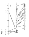
- the genomic structure (restriction enzyme map and each exon-intron splice point) of GLAT gene are already known (Hagiwara, T., et al., Genomics, 33, 508-515, 1996), which structure is outlined in FIG. 1 and Table 1.
- the mouse GLAST gene comprises 10 exons. It is preferred to insert a marker gene into any one of the exons so as to cause deficiency of the gene.
- a targeting vector DNA for homologous recombination
- DNA for homologous recombination capable of introducing a mutation into the target gene can be prepared by conventional DNA recombinant techniques, for example, PCR or site-specific mutagenesis, based on the GLAST-encoding nucleotide sequence (SEQ ID NO: 1) and information of genomic sequence of the GLAST gene (Hagiwara, T., et al., Genomics, 33, 508-515, 1996).
- a DNA molecule containing the entire gene or its fragment is isolated in a conventional manner from the mouse strain, from which ES cells used are derived.
- the DNA molecule may be a DNA molecule containing the entire GLAST gene or further containing the 5' upstream region and/or the 3' downstream region of the gene, in addition to the entire gene.
- a modified DNA molecule is prepared from the resulting DNA molecule by introducing a desired mutation into the site corresponding to the mutation site in the gene, e.g., by introducing the marker gene described above.
- Modification of the base sequence can be made by conventional recombinant DNA techniques such as ligation of DNA molecules amplified by PCR, site-specific mutation, etc.
- plasmid vectors commercially available for targeting vector construction may also be used.
- the thus obtained targeting vector is introduced into mouse embryonic stem cells (ES cells) to perform homologous recombination.
- Introduction of the targeting vector into ES cells can be made by conventional DNA transfection techniques, e.g., electroporation, lipofection, etc.
- homologous recombination occurs between the GLAST gene on the chromosome and the counterpart on the targeting vector so that the modified base sequence in the targeting vector, e.g., a marker gene is introduced into the endogenous gene.
- the ES cells are deficient in the function of endogenous GLAST gene and at the same time contains, e.g., the marker gene.
- the cells where the targeting vector is introduced are then screened by the screening function of, e.g., the marker gene, or in a conventional manner such as southern blotting for confirming homologous recombination, PCR, etc. to obtain ES cells deficient in the function of GLAST gene (hereinafter referred to as recombinant ES cells).
- recombinant ES cells usually ES cells with disrupted GLAST gene only on one homologous chromosome are obtained.
- Mouse ES cells used are generally 129 ES cells already established.
- ES cells are established by publicly known methods (Teratocarcinomas and Embryonic Stem Cells: A Practical Approach (Robertson, E.J., ed.), IRL press, Oxford, 1987) using C57BL/6 or BDF1 strain mice (F1 mice obtained by crossing C57BL/6 with DBA/2). These ES cells may also be used.
- 129-derived ES cells are used.
- the resulting recombinant ES cells are developed to generate a chimeric mouse.
- the recombinant ES cells are injected into normal mouse embryos at the blastocyst stage, the 8-cell stage, etc. by microinjection or aggregation.
- the thus obtained chimeric embryos are transplanted to the uterine horn of a pseudopregnant female mouse.
- This transplanted mouse can be bred in a conventional manner and allowed to deliver the offspring of the chimeric mouse.
- the recombinant ES cells are injected into the embryos of C57BL/6 strain mice.
- this chimeric mouse comprises cells derived from the recombinant ES cells and normal cells as its somatic cells and germ cells.
- a wild-type mouse of a proper line preferably a C57BL/6 strain mouse, e.g., a C57BL/6J strain mouse
- heterozygous F1 offspring are obtained.
- a male chimeric mouse is mated with a female wild-type mouse to generate heterozygous offspring of the F1 generation.
- the germ cells of the chimeric mouse used for the mating are derived from the recombinant ES cells described above, i.e., cells carrying the disrupted endogenous GLAST gene present on one of the homologous chromosomes, then desired heterozygous F1 mice deficient in the function of the gene can be obtained.
- the heterozygous F1 mice with high efficiency for example, normal host embryonic cells derived from a mouse having a coat color different from the mouse of the recombinant ES cell origin are used in combination, e.g., in the preparation of chimeric embryos.
- a chimeric mouse showing a higher rate of the recombinant ES cells in vivo or a heterozygous F1 mouse can be easily screened.
- the F1 heterozygous mouse produced as described above is further crossed with a C57BL/6 strain mouse, e.g., a C57BL/6J strain mouse and the delivered heterozygous mouse is again crossed with a C57BL/6 strain wild-type mouse.
- the crossing procedures are repeated normally at least 5 times in total, preferably at least 9 times and more preferably at least 15 times.
- the homozygous or heterozygous knockout mouse of the present invention deficient in the function of GLAST gene can be obtained.
- homozygous mice are preferred.
- Whether a desired genotype is achieved in the respective generations or not may be determined by conventional techniques, including southern blotting, PCR, base sequencing, etc., as described above.
- knockout mice of the present invention which can be produced as above are obtained in the combination of males and females, subsequently knockout mice having the same genotype can be readily obtained in the number as required, by appropriately breeding the offspring, depending on necessity.
- the intraocular pressure of the GLAST knockout mouse of the present invention is generally about 21 mmHg or lower, for example, about 10-21 mmHg.
- This intraocular pressure range may be somewhat varied depending upon the strain of mice, from which the ES cells used are derived, or the strain of mice from which the normal embryos used to prepare chimeric embryos are originated. Even in view of the foregoing, the intraocular pressure should be not higher than 30 mmHg.
- the number of nerve cells in the retinal ganglions is reduced by at least 20%, preferably at least about 50%, as compared to a wild-type mouse.
- the wild-type normal mice delivered simultaneously with the homozygous or heterozygous knockout mice of the present invention described above or mere normal (wild-type) C57BL/6 strain mice can be used as control mouse against these mice of the present invention.
- the measurements are also applied, if necessary, to the homozygous or heterozygous GLAST knockout mice obtained by mating the F1 heterozygous knockout mice described above or their females and males, heterozygous knockout mice obtained during backcrossing, and so on.
- the number of cells in the mouse retinal ganglion can be measured by conventional histochemical means or retrograde labeling.
- Any anesthetic can be used in such a concentration range that mice are not dead, as far as it is an anesthetic available for ordinary animal tests.
- a 1:1 mixture of ketamine (10 mg/ml)/medetomidine (1 mg/ml) (0.15-0.2 ml/mouse) may be employed.
- the animal can be awakened with atipamezole (5 mg/ml) (0.15-0.2 ml/mouse).
- EMG electroretinograms
- the knockout mouse of the present invention can be used for screening a compound useful for the prevention/treatment of normal tension glaucoma, especially a compound useful for preventing the death or degeneration of optic nerve cells including retinal ganglions, or for preventing diminished activity of the function, or a compound useful for recovering optic nerve cells or their function.
- the present invention provides a method of screening a compound useful for the prevention/treatment of normal tension glaucoma and comprises:
- Whether a test compound is useful for the prevention/treatment of normal tension glaucoma or not can be determined by examining whether the compound can improve a sign characteristic of glaucoma. For example, after a test compound is administered, the number of nerve cells in the retinal ganglions is counted; when the number is recovered by at least 10%, preferably by at least 20% and more preferably by at least 30%, as compared to the control mouse, the test compound can be judged to be medically effective.
- the retinal potentials are measured; when the amplitude of, e.g., b-wave or oscillation potential is recovered by at least 10%, preferably by at least 20% and more preferably by at least 30%, as compared to the control mouse, the test compound can be judged to be medically effective.
- the number of cells in the retinal ganglions is found to be gradually reduced with the age of weeks after birth (FIG. 4). Accordingly, in one embodiment of the screening method described above, a compound which prevents a reduction of the number of cells in the retinal ganglions with passage of time can also be screened by 1) regularly administering a test compound to a group of heterozygous knockout mice immediately after birth but administering no test compound to another group of heterozygous knockout mice, 2) counting the number of cells in the retinal ganglions at the age of respective weeks, and 3) comparing the two groups.
- the test compound which can be used includes, in addition to naturally occurring and synthetic compounds, optional compounds such as animal and plant extracts, fermentation products, peptides, proteins, nucleic acid molecules, etc.
- the test compound may be a gene vector for expressing a desired protein.
- routes may be attempted as far as properties of the test compound permit, and the compound may be administered, e.g., as eye drops or by oral administration.
- Dosing period or dosing mode can be chosen so as to maximize the effect of a test compound.
- kind and dosing of such a test compound may also be in accordance with conventional methods in the pharmaceutical field or medical field.
- the GLAST knockout mouse of the present invention can be crossed with other strain of knockout mouse or other strain of disease model mouse to generate a novel disease model mouse.
- the present invention also includes such use of the GLAST knockout mouse of the present invention.
- EXAMPLE 1 Production of mouse deficient in the function of GLAST (GluT-1) gene
- Genomic DNA extracted from the liver of a 129SV mouse was partially digested with restriction enzyme Sau3AI.
- genomic library lambda FIXII hybridization was carried out using as a probe a partial sequence of cDNA for mouse GLAST (GIuT-1) gene. Colonies of 1 ⁇ 10 6 were screened to give 26 positive clones. After the clones were incompletely digested with restriction enzymes EcoRV and XhoI,. the full length genomic DNA of 9 kbp containing exons 6 to 8 was subcloned thereto (FIG. 2, upper column).
- GLAST GluT-1
- a 1.5 kbp region following the BamHI site in exon 6 was made defective, and the neomycin-resistant gene was inserted therein and a diphtheria toxin A fragment gene was further inserted into the downstream of exon 8 (FIG. 2, middle column).
- the homologous region with the genomic DNA was constructed to be 2.5 kb length at the upstream of the neomycin-resistant gene and 5 kb length between the neomycin-resistant gene and the diphtheria toxin A fragment gene.
- the thus obtained construct was inserted into pBluescriptSK and digested with restriction enzyme NotI for linearization at the time of transfer into ES cells, whereby a targeting vector (DNA:pGluT1NeoDT for homologous recombination) was obtained (FIG. 2, middle column).
- G418 resistant colonies were transferred into a 96-well microplate (FALCON 3077) charged with 60 ⁇ l of Tris-EDTA solution (solution composed of 10 mm Tris-HCl, pH 8.0 and 1 mm EDTA, pH 8.0) using a micropipette, and treated for several minutes to prepare single cells by pipetting. These cells were transferred to a 24-well plate (FALCON 3047) and the culture was continued. The colonies collected were those having the major axis reaching at least 1/2 of the inner diameter of the microchip and the number of cells at this stage showed 1 ⁇ 10 4 to 10 5 . The number of surviving cells after the electroporation was 6.0 ⁇ 10 7 . The number of G418-resistant colonies was 2.4 ⁇ 10 2 , which was 1/2.5 ⁇ 10 5 of the number of surviving cells.
- the cells were treated with 0.25 % trypsin at 37°C for 5 minutes and sequentially cultured in a 35 mm (FALCON 3001) or 60 mm (FALCON 3002) Petri dish to effect proliferation of the cells.
- the ES cells were all cultured on feeder cells. The homologous recombinants were confirmed by southern blotting as described below.
- the genomic DNA for southern blotting analysis was extracted from the G418-resistant cells, digested with restriction enzyme PvuII and then subjected to the analysis using as a probe 0.5 kb of ApaI-EcoRV fragment in intron 5.
- the homologous recombinant containing the disrupted allele (FIG. 2, lower column) and non-homologous recombinant were confirmed by detecting the bands of 4.2 kb and 7 kb, respectively.
- the number of homologous recombinant colonies was 1 (2B7) out of 242 colonies of the G418 resistant colonies.
- the ES cells used were those of the E14 strain derived from 129/SvJ mouse blastocysts.
- the ES cells were cultured in SCM culture medium (Robertson, Teratocarcinomas and Embryonic Stem Cells: A Practical Approach, 1987), namely, Dulbecco's modified Eagle's medium (DMEM, 11960-010, GIBCO) supplemented with 15% fetal calf serum (FCS), 0.1 mM 2-mercaptoethanol, a nucleic acid mixture solution, a non-essential amino acid solution and 10 3 unit/ml of LIF (AMRAD).
- SCM culture medium Robottson, Teratocarcinomas and Embryonic Stem Cells: A Practical Approach, 1987
- FCS fetal calf serum
- FCS fetal calf serum
- mouse fetal fibroblasts used as feeder cells for the ES cells were cultured in DMEM containing 10% FCS.
- the preparation and culture of the mouse fetal fibroblasts were carried out by the following procedures. After ICR mouse fetuses with a fetal age of 13-14 days were aseptically collected and washed with phosphate buffered physiological saline containing no calcium or magnesium (PBS-), the hearts, livers and bowels were removed using forceps and morcellated with ophthalmic scissors. Subsequently, the morcellated pieces were then treated at room temperature for 20 minutes with PBS- containing 0.25% trypsin and 0.04% EDTA (hereinafter referred to as TE solution) to obtain a cell suspension.
- PBS- phosphate buffered physiological saline containing no calcium or magnesium
- the cell suspension was centrifuged at 1500 rpm for 5 minutes, the supernatant was removed off and the cells were suspended in DMEM supplemented with 10% FCS, which was allowed to settle for 2 minutes.
- the cell suspension was washed once with PBS-, followed by culturing. Subculture was performed at 3 to 4 day intervals and the cells from the 1st to 3rd generation of subculture were subjected to mitomycin treatment for use as feeder cells.
- the mouse fetal fibroblasts which had grown to confluence, were treated for 3 to 4 hours with 75 ⁇ l of 2 mg/ml mitomycin C, washed 3 times with PBS-, and then treated with TE solution at room temperature for 3 minutes to detach the cells. After centrifugation, the number of cells was adjusted to 5 x 10 5 /ml, and 3 ml each was dispensed onto 60 x 10 mm gelatin-coated dishes (FALCON 3002).
- the feeder cells prepared in the manner described above were used within one week. Subculture of the ES cells was carried out by treating with TE solution at room temperature for 5 minutes, followed by dispersing the ES cells as single cells by pipetting and then inoculating 4 ⁇ 10 5 cells onto a layer of feeder cells.
- the culture medium was replaced at 24 hour intervals, and the subculturing interval was 56 to 64 hours.
- 1 ⁇ 10 6 cells were suspended in SCM and transferred to a tube for lyophilization (2 ml, FALCON 4818) and 0.5 ml of freezing medium (DMEM supplemented with 20% DMSO) was dropwise added thereto. Thereafter, the mixture was allowed to stand overnight at -80°C and then stored in liquid nitrogen.
- the ES cells were injected into the blastocysts from C57BL/6J mice, and the resulting host embryos were then transferred into the uterine horn of pseudopregnant mice to obtain offspring.
- the host embryos were collected on the 4th day of natural mating by flushing the uterus with HEPES-buffered Whitten's medium.
- the ES cells used for the injection had been subjected to treatment with TE solution on the 2nd or 3rd day of subculture and then placed in gelatin-coated dishes for 30 minutes to remove the feeder cells. The ES cells were then placed on ice until they were provided for micromanipulation.
- the injection pipette for the ES cells was prepared from a glass capillary (NARISHIGE) having an outer diameter of 1 mm by thinly pulling the capillary using a microelectrode puller (NARISHIGE, PN-3), polishing the tip with a grinder (NARISHIGE) to an inner diameter of about 20 ⁇ m, and finally sharpening the tip with a microforge (De Fonburun).
- NARISHIGE glass capillary
- PN-3 microelectrode puller
- NARISHIGE polishing the tip with a grinder
- De Fonburun The embryo-holding pipette was prepared by using a microforge to cut a glass capillary pulled by the procedure above at a section in a 50-100 ⁇ m outer diameter, and then further finishing the aperture to 10-20 ⁇ m.
- Both of the injection pipette and holding pipette were bent to an angle of about 30° at the position of about 5 mm from the tip and then connected to a micromanipulator (LEITZ).
- the chamber used for the micromanipulation was a perforated slide glass with a cover glass, to which the cover glass was glued with yellow wax.
- Two drops of HEPES-buffered Whitten's medium supplemented with about 20 ⁇ l of 5 % FCS were placed thereon.
- the surfaces of the drops were covered with mineral oil (M8410, Sigma). In one of the drops, approximately 100 ES cells were placed. Ten to fifteen expanded blastocysts were placed in the other drop. Ten to fifteen ES cells were injected per embryo.
- ES cells 2B7 were injected into 160 blastocysts from C57BL/6J mice collected on the 4th day of natural mating by flushing the uterus and as a result, 123 blastocysts survived with a success rate of 77%.
- F1 heterozygous male mouse which was confirmed to be deficient in the GLAST gene, was mated with a C57BL/6J wild type female mouse to obtain offspring.
- the genotype was analyzed by southern blotting to select a next generation heterozygous male mouse confirmed to be deficient in the GLAST gene.
- the heterozygous male mouse was again mated with the C57BL/6J female mouse to obtain a heterozygous male mouse for the next generation. As such, a total of 9 matings were repeated generation after generation to generate the GLAST gene-deficient heterozygous female and male mice of renewed generation.
- mice were crossed with each other thereby to obtain GLAST knockout mice homozygous for GLAST gene deficiency (GLAST-/-), GLAST knockout mice heterozygous for GLAST gene deficiency (GLAST+/-), and wild type mice (GLAST+/+). These mice were used for the following measurement.
- the intraocular pressure was measured in the homozygous GLAST knockout mice obtained in EXAMPLE 1 and wild type normal mice. After four mice per group were anesthetized, the intraocular pressure was measured with an electronic tonometer. The results are shown below. Wild-type normal mice: 19 ⁇ 4 mmHg Homozygous knockout mice: 15 ⁇ 3 mmHg (mean ⁇ standard deviation)
- EXAMPLE 3 Measurement of the number of cells in the retinal ganglions
- the number of cells in the retinal ganglions was measured in each mouse by the hematoxylin/eosin staining of retinal sections and the retrograde labeling of retinal nerve cells by Fluoro-Gold or DiI.
- FIGS. 3 to 5 revealed that in the homozygous GLAST knockout mice, the number of nerve cells in the retinal ganglions was significantly reduced, when compared with the wild-type mice and heterozygous knockout mice.
- the results of the hematoxylin/eosin staining indicates the reduced number of cells in the retinal ganglions, which are shown by arrows. Also, the reduction in the number of cells (amacrine cells, bipolar cells, etc.) located at the center and skin thinning in the inner nuclear layer accompanied by the reduction were observed.
- the endogenous GLAST gene-deficient homozygous or heterozygous GLAST knockout mouse of the invention is expected to be extremely useful for developing therapeutics effective for the treatment of said disease, establishing a remedy therefor and identifying the cause of said disease or its onset mechanism.
Landscapes
- Life Sciences & Earth Sciences (AREA)
- Health & Medical Sciences (AREA)
- Genetics & Genomics (AREA)
- Engineering & Computer Science (AREA)
- Zoology (AREA)
- Biotechnology (AREA)
- Biomedical Technology (AREA)
- Environmental Sciences (AREA)
- Veterinary Medicine (AREA)
- General Engineering & Computer Science (AREA)
- Organic Chemistry (AREA)
- General Health & Medical Sciences (AREA)
- Bioinformatics & Cheminformatics (AREA)
- Chemical & Material Sciences (AREA)
- Wood Science & Technology (AREA)
- Biophysics (AREA)
- Microbiology (AREA)
- Biochemistry (AREA)
- Plant Pathology (AREA)
- Molecular Biology (AREA)
- Animal Behavior & Ethology (AREA)
- Animal Husbandry (AREA)
- Biodiversity & Conservation Biology (AREA)
- Physics & Mathematics (AREA)
- Micro-Organisms Or Cultivation Processes Thereof (AREA)
- Investigating Or Analysing Biological Materials (AREA)
- Peptides Or Proteins (AREA)
- Medicines That Contain Protein Lipid Enzymes And Other Medicines (AREA)
- Measuring Or Testing Involving Enzymes Or Micro-Organisms (AREA)
Abstract
Description
- The present invention relates to a GLAST knockout mouse deficient in the function of GLAST, which is one of glutamate transporters, and a process of producing the same. The present invention further relates to use of said knockout mouse as a model mouse of normal tension glaucoma and a method of screening a compound useful for the prevention and/or treatment of normal tension glaucoma using the knockout mouse.
- Normal tension glaucoma is one type of glaucoma and is a disease recently getting attention particularly due to its high prevalence rate. In general, glaucoma is a disease characterized by elevated intraocular pressure (watery fluid pressure within the eyeball) resulting in compression of the optic nerve to produce atrophy and thus impairing the visual performance to narrow the visual field. If it is left untreated, the symptoms will eventually progress to blindness at a high risk. On the other hand, normal tension glaucoma is a pathological condition that mimics the findings on glaucoma with high intraocular pressure (optic atrophy and visual field defects), notwithstanding that the intraocular pressure lies within the normal range (usually 10-21 mmHg in human). In the developed countries, glaucoma is ranked as the second leading cause of vision loss next to diabetes mellitus. The prevalence of glaucoma in the population aged over 40 years is 3.5% in Japan and the number of patients is estimated to be about two million. According to a recent epidemiological survey, reportedly 70% of glaucoma is normal tension glaucoma. Because of slow progress and paucity of subjective symptoms, it is difficult to detect normal tension glaucoma at an early stage. At present, there is no decisive treatment except to further decrease the intraocular pressure.
- In recent years, degenerative loss of retinal ganglion cells, namely, neuronal apoptosis, which is induced by a mild and chronic increase of glutamate level, is proposed to be one of the causes of glaucoma and diabetic retinopathy (Harada, T., et al. Proc. Natl. Acad. Sci. USA, 95, 4663-4666, 1998; Harada, C. et al., Neurosci. Lett., 292, 134-136, 2000).
- In the mammalian central nervous system, glutamate is one of the main excitatory neurotransmitters and plays an important role in regulating a higher order function of the brain. On the other hand, it is known that glutamate causes neurotoxicity by an excessive rise, resulting in various neurodegenerative diseases or delayed neuronal cell death after cerebral ischemia. One of the mechanisms for regulating the level of this glutamate is glutamate transporters. Glutamate transporters are functional molecules, the main role of which is to take up glutamate once released from nerve endings into cells and maintain a low glutamate level at the synaptic cleft.
- Currently, EAAC1, EAAT4 and EAAT5 (Kanai, Y. & Heidiger, M.A., Nature, 360, 467-471, 1992; Fairman, W.A., et al., Nature, 375, 599-603, 1995; Arrizal, J.E., et al., Proc. Natl. Acad. Sci. USA, 94, 4155-4160, 1997) present in neurons as well as GLT1 and GLAST (also referred to as GluT-1) present in glial cells (Pines, D. et al., Nature, 360, 464-467, 1992; Mukainaka et al., Biochimica et Biophysica Acta, 1244, 233-237, 1995; Tanaka, K., Neurosci. Res., 16, 149-153, 1993; Tanaka, K., Neurosci. Lett., 159, 183-186, 1993; Storck, T., et al., Proc. Natl. Acad. Sci. USA, 89, 10955-10959, 1992) are known as glutamate transporters in the mammalian brain. Abnormalities in the function of these glutamate transporters are known to be associated with various neurodegenerative diseases.
- Under such circumstances, it has become clear that GLAST is present in Müller cells within the retina and retinal damages after ischemic load are exacerbated in GLAST knockout mice as compared to wild-type mice, based on the experiments using GLAST knockout mice (Watase, K. et al, Eur. J. Neurosci., 10, 976-988, 1998; Japanese Patent Laid-Open Application No. 10-33087). For this reason, it is suggested that GLAST present in Müller cells of the retina would be involved in the onset of glaucoma (Harada, T., et al., Proc. Natl. Acad. Sci. USA, 95, 4663-4666, 1998). However, unless ischemic load is applied, damage to the retinal tissue is not observed in this GLAST knockout mouse so that the mouse cannot be used as a model for normal tension glaucoma.
- For the development of therapeutics for glaucoma and the elucidation of its onset mechanism, genetically chronic glaucoma model mice or high tension glaucoma model rabbits induced by water loading are already available as glaucoma model animals, but no model animal for normal tension glaucoma has ever been known heretofore. Also, there is no report to point out the relation of normal tension glaucoma to GLAST, and the onset mechanism of normal tension glaucoma yet remains unknown.
- Accordingly, it is expected that if a model animal for normal tension glaucoma is obtained, the animal will be extremely useful for developing therapeutics effective for the treatment of said disease, establishing a remedy therefor and identifying the cause of said disease or its onset mechanism. However, any model animal for normal tension glaucoma is unknown at present and, such a model animal has been earnestly desired in the medical or pharmaceutical field.
- The inventor has improved normal knockout mice (GLAST knockout mice), which conventionally exist and are deficient in the function of a glutamate transporter gene and as a result, could obtain improved GLAST knockout mice with the markedly reduced number of retinal ganglion cells due to degenerative loss of the cells, although the intraocular pressure is within the normal range. This knockout mouse was found to be useful as a model mouse for normal tension glaucoma.
- Therefore, the present invention provides a GLAST knockout mouse deficient in the function of an endogenous GLAST gene, as a model for normal tension glaucoma and more particularly, a GLAST knockout mouse, in which 1) the intraocular pressure is within the normal range and 2) the number of cells in the retinal ganglions is reduced as compared to a wild-type mouse.
- According to the present invention, the intraocular pressure of the GLAST knockout mouse is generally 21 mmHg or lower, for example, 10 to 21 mmHg. Also, the number of cells in the retinal ganglions is reduced by at least 20% in the GLAST knockout mouse, as compared to a wild-type mouse.
- In the present invention, the genetic background of the GLAST knockout mouse is preferably the same or substantially the same as the genetic background of a C57BL/6 strain mouse, e.g., a C57BL/6J strain mouse.
- Specifically, the present invention provides a GLAST knockout mouse carrying a neomycin-resistant gene inserted into the region of endogenous GLAST gene, for example, into the
exon 6. - The present invention further provides use of such a GLAST knockout mouse as a model mouse for normal tension glaucoma.
- In another aspect, the present invention provides a method of producing a GLAST knockout mouse deficient in the function of an endogenous GLAST gene. This production method comprises the following steps 1) to 6):
- 1) obtaining an ES cell from any mouse deficient in the function of one endogenous GLAST gene on the homologous chromosome,
- 2) obtaining a chimeric mouse carrying the ES cell using the cell obtained in
step 1, - 3) crossing the chimeric mouse obtained in
step 2 with a normal C57BL/6 strain mouse to obtain a heterozygous knockout mouse, - 4) crossing the heterozygous mouse obtained in
step 3 with a normal C57BL/6 strain mouse to generate a heterozygous knockout mouse, - 5) repeating the crossing described in
step 4 at least a total of 5 times to generate a heterozygous knockout mouse thereby to bring the genetic background closer to the C57BL/6 strain mouse, and, - 6) crossing the heterozygous knockout mice obtained in
step 5 with each other to generate a homozygous or heterozygous GLAST knockout mouse. - In the production method of the invention, it is preferred to repeat the crossing described in
step 4 at least a total of 9 times instep 5. - The present invention further includes the GLAST knockout mice produced by the production method of the invention, and the GLAST knockout mice thus produced can be used as model mice for normal tension glaucoma.
- In yet another aspect, the present invention provides a method of using the GLAST knockout mice of the invention described above or the GLAST knockout mice produced by the production method of the invention described above, as model mice for normal tension glaucoma.
- Therefore, the present invention provides a method of screening a compound useful for the prevention and/or treatment of normal tension glaucoma, which comprises using such GLAST knockout mice. More specifically, the screening method comprises:
- 1) administering a test compound to the GLAST knockout mouse of the invention,
- 2) administering a test compound to a wild-type mouse,
- 3) assessing the number or function of surviving optic nerve cells in each of the mice described above, prior to and after a given time period of the administration, and,
- 4) comparing the GLAST knockout mouse with the wild-type mouse in terms of the test results to determine effectiveness of the test compound.
- According to the screening method of the invention, the number of nerve cells in the retinal ganglions is counted to assess the number of surviving optic neurons or the function of the optic neurons and in addition thereto, the assessment is conducted preferably by measurements of electroretinograms or visual evoked potentials (Porciatti et al., Vision Res., 39, 3071-3081, 1999), behavioral analysis such as the Visual Cliff test (Ma, L. et al., Neuron 36, 623-634, 2002), etc. in combination.
-
- FIG. 1 shows an outlined genomic structure (restriction enzyme sites and exon sites) of mouse GLAST gene. In the Figure, the restriction enzymes involved in the restriction sites shown by the respective symbols are as follows. E: EcoRI and B: BamHI. The black box designates
Exons 1 to 10. - FIG. 2 shows the structures of the targeted gene region intended to disrupt for the functional deficit of mouse GLAST gene (upper column), the targeting vector used (middle column) and the GLAST gene disrupted (lower column), in EXAMPLE 1. In the Figure, the restriction enzymes involved in the restriction sites shown by the respective symbols are as follows. P: PvuII, RV: EcoRV, B: BamHI, E: EcoRI and X: XhoI. The black box designates
Exons 6 to 8 (E6 to E8). The symbol neo designates neomycin-resistant gene and DT-A designates diphtheria toxin A fragment gene. - FIG. 3 shows the images of pathological sections in the retina of homozygous GLAST knockout mice (GLAST-/-) and wild-type normal mice (GLAST+/+).
- FIG. 4 shows the number of cells in the retinal ganglions in homozygous GLAST knockout mice (GLAST-/-), heterozygous GLAST knockout mice (GLAST+/-) and wild-type normal mice (GLAST+/+), at the age of given weeks after birth. The number of these cells was determined by counting the number of nerve cells in the pathological section of retinal ganglions after hematoxylin/eosin staining. The ordinate represents a mean cell count per section from 3 to 22 sections. The abscissa represents the age of weeks after birth.
- FIG. 5 shows the fluorescence images representing nerve cells labeled with Fluoro-Gold by retrograde labeling in the retinal ganglions of homozygous GLAST knockout mice (GLAST-/-) and wild-type normal mice (GLAST+/+).
- In the present invention, mouse glutamate transporter GLAST (Glutamate/Aspartate Transporter) refers to a protein encoded by DNA strand having the base sequence represented by SEQ ID NO: 1 and having the amino acid sequence represented by SEQ ID NO: 2 (Tanaka, K., Neurosci. Lett., 159, 1803-186, 1993)). This protein is also termed GluT-1 in rat (Tanaka, K., Neurosci. Res. 16, 149-153, 1993; Storck, T., et al., Proc. Natl. Acad. Sci. USA, 89, 10955-10959, 1992). Both transporters are so-called counterparts.
- The genomic structure of mouse GLAST has already been clarified and the details are described in Hagiwara, T., et al., Genomics, 33, 508-515, 1996. The structure of this gene is outlined in Table 1 and FIG. 1.
- However, depending upon mouse strains, the mouse GLAST may undergo mutation in the encoded nucleotide sequence and amino acid sequence described above and its genomic sequence within the range to maintain the function, for instance, may undergo substitutions, deletions, additions or insertions of bases or amino acid residues. In the present invention, the mouse GLAST also includes these mutants such as a mutant with substitutions, deletions, additions or insertions of, e.g., 1 to 10, preferably 1 to 5 bases in the encoded nucleotide sequence described above, a mutant with substitutions, deletions, additions or insertions of, e.g., 1 to 10, preferably 1 to 5 amino acids in the amino acid sequence described above, etc.

- Table 1 shows the sequencing of exon/intron splice site for GLAST gene. The nucleotide sequences for exons are represented by capitals and for introns by small letters.
- In the present invention, the deficiency in the function of the glutamate transporter GLAST gene means that within the region of one or two endogenous GLAST genes present in one or two GLAST loci, functional GLAST remains unexpressed or expression of the GLAST gene is constantly suppressed, either by introducing a mutation into the region encoding its structure, e.g., into the exon, or by introducing a mutation into the region associated with expression of the GLAST gene, e.g., into the promoter or intron region. In any case, the term refers to such a state that one or two endogenous GLAST genes are substantially dysfunctional in vivo. In the present invention, therefore, the GLAST knockout mouse includes a homozygote deficient in the function of two endogenous GLAST genes and a heterozygote deficient in the function of one endogenous GLAST gene. In view of the effect of deficiency in the function of the gene, homozygous mice are preferred.
- Such a deficiency in the function of the gene can be achieved by publicly known methods for generating knockout mice, e.g., a gene targeting strategy. Introduction of the mutation described above may also be substitutions of bases or deletions of bases in the GLAST gene region, or insertions of bases into the region.
- In the present invention, the term "the same or substantially the same genetic background" is used to mean that all genotypes other than the targeted genotype (GLAST genotype) are the same by 99% or more. Specifically, this means that in EXAMPLES, F1 GLAST heterozygous knockout mice are backcrossed to normal C57BL/6 mice for at least 9 generations and the genes derived from the 129 strain have reached 1 % or less than all genes.
- The present invention provides a GLAST knockout mouse as a model mouse with normal tension glaucoma, which is deficient in the function of one or two endogenous GLAST genes on the homologous chromosome. Specifically, the present invention provides a GLAST knockout mouse, in which 1) the intraocular pressure is within the normal range and 2) the number of cells in the retinal ganglions is reduced as compared to a wild-type mouse. More particularly, the mouse preferably has the same or substantially the same genetic background as that of a C57BL/6 strain mouse, e.g., a C57BL/6J strain mouse.
- The intraocular pressure of a wild-type normal mouse is generally 10 to 21 mmHg. The intraocular pressure of the knockout mouse of the present invention also lies within the normal range. The intraocular pressure may be outside the range above, but does not reach such a range as termed high pressure, e.g., 30 mmHg or higher. The intraocular pressure in mice can be determined by using, e.g., an electronic tonometer.
- Furthermore, in the knockout mouse of the present invention, the number of nerve cells in the retinal ganglions is reduced by at least 20%, preferably at least about 50%, as compared to the number in normal mouse. The reduction in the number of nerve cells in the retinal ganglions can be microscopically determined by conventional histochemical means, for example, by hematoxylin/eosin staining using a section.
- Further in the knockout mouse of the present invention, a reduced b-wave, which is a sort of potential change in the retina by photic stimulation, is also noted, when compared with a wild-type normal mouse. The b-wave reflects an action potential in the inner retinal layer containing Müller cells in which GLAST exists, suggesting that a mechanism regulating glutamate levels by GLAST would have an important role also in visual transmission (Harada, T., et al., Proc. Natl. Acad. Sci. USA, 95, 4663-4666, 1998).
- This reduction in the number of nerve cells in the retinal ganglions can be regarded as neurodegeneration or neuronal apoptosis. In general, optic disc atrophy or deficits of the retinal nerve fibers are observed in glaucoma including normal tension glaucoma. Currently, the final clinical picture of glaucoma is thought to be retinal ganglion cell death.
- Thus, taking into account these ocular properties of the GLAST knockout mouse of the present invention, namely, the intraocular pressure within the normal range and the reduction in the number of cells in the retinal ganglions, the mouse can be used as a model mouse for normal tension glaucoma.
- In a second aspect, the present invention provides a method of producing the GLAST knockout mouse of the present invention. This method comprises the following steps 1) to 6):
- 1) obtaining an ES cell from any mouse, which is deficient in the function of one endogenous GLAST gene on the homologous chromosome,
- 2) obtaining a chimeric mouse carrying the ES cell using the cell obtained in
step 1, - 3) crossing the chimeric mouse obtained in
step 2 with a wild-type C57BL/6 strain mouse to generate a heterozygous knockout mouse, - 4) crossing the heterozygous mouse obtained in
step 3 with a wild-type C57BL/6 strain mouse to generate a heterozygous knockout mouse, - 5) repeating the crossing described in
step 4 at least a total of 5 times to generate a heterozygous knockout mouse thereby to bring the genetic background closer to the C57BL/6 strain mouse, and, - 6) crossing the heterozygous knockout mice obtained in
step 5 with each other to generate a homozygous or heterozygous GLAST knockout mouse. - In
step 5 described above, the crossing is repeated preferably at least 9 times in total. - Basically, publicly known methods for producing knockout mice, e.g., gene targeting strategy, gene trapping strategy, etc., can be used to produce the GLAST knockout mouse of the present invention available as a model for normal tension glaucoma. Basic methods for producing knockout mice are not particularly limited, as far as the GLAST gene can be disrupted and mice whose surviving and reproductive potentials are not lost are obtained. For the methods of producing knockout mice, reference can be made to, e.g., "Jikken-Igaku-Bessatsu, Kaitei-Idenshi-Kogaku Handbook (Handbook of Genetic Engineering), 3rd revised version", edited by Masami Muramatsu and Tadashi Yamamoto (published by Yodosha Co., Ltd., 1996) or "Jikken-Igaku-Bessatsu, The Protocol Series: Latest Technology of Gene Targeting", edited by Takeshi Yagi (published by Yodosha Co., Ltd., 2000), which can be applied to carry out the present invention.
- First, the
steps 1 through 4 described above can be performed in accordance with conventional methods, e.g., the gene targeting strategy. These steps are disclosed also in Japanese Patent Laid-Open Application No. 10-33087 by the research group of the present inventors and Watase, K. et al, Eur. J. Neurosci., 10, 976-988, 1998. - Heretofore, homozygous or heterozygous GLAST knockout mice obtained by mating female and male of the heterozygous GLAST knockout mice obtained in
step 4 described above are already disclosed but in this type of GLAST knockout mice, no significant change was noted in the number of cells in the retinal ganglions, when compared with wild-type normal mice; a decrease in the number of cells in the retinal ganglions was observed only after transient ischemia was achieved by instilling sterile saline into the eye of the GLAST knockout at 150 cm H2O pressure for 60 minutes (Harada, T., et al., Proc. Natl. Acad. Sci. USA, 95, 4663-4666, 1998). - For these reasons, such GLAST knockout mice hitherto known cannot be used as a model for normal tension glaucoma.
- According to the production method of the present invention, such conventional GLAST knockout mice are further improved, whereby the GLAST knockout mouse of the present invention with normal tension glaucoma described above can be produced.
- Hereinafter, the method for producing the knockout mouse of the present invention will be described below, taking as an example the gene targeting strategy which is a standard method for producing a knockout mouse.
- According to the gene targeting strategy, in order to disrupt the GLAST locus on the chromosome in mouse ES cells, a targeting vector is used to introduce a mutation into the locus.
- In order to render the function of GLAST gene defective, bases are deleted, point mutation is introduced or other genes are inserted into any part of the GLAST gene, e.g., one or more exon regions. Generally in order to select the endogenous GLAST gene-disrupted ES cells more readily, it is preferred to insert a selection marker gene.
- As such a gene, a marker gene for positive selection, for example, a neomycin (neo)-resistant gene can be employed. This neomycin-resistant gene enables to screen the objective gene by using the neomycin analog G418. Also, a marker gene for negative selection can also be used to screen and remove the objective gene. Examples of such genes used include thymidine kinase (tk) gene (using ganciclovir, FIAU, etc. as a screening marker, a non-homologous recombinant is screened and removed by the sensitivity thereto) and diphtheria toxin A fragment (DT-A) gene (a non-homologous recombinant is screened and removed by diphtheria toxin expressed by DT-A). Alternatively, a combination of the foregoing can also be used for positive/negative selection. It is preferred to insert, e.g., neomycin-resistant gene and diphtheria toxin A fragment gene (Yagi, Nada, Watanabe, et al., Analytical Biochemistry, 214, 77-86, 1993), or neomycin-resistant gene and thymidine kinase gene (Mansour, Thomas and Capacchi, Nature, 336, 348-352, 1988).
- In the gene sequence, a site to introduce a mutation, for example, a site to insert the marker gene described above is not particularly limited, so far as it is a site that function of the gene is lost, but the site is usually an exon site.
- The genomic structure (restriction enzyme map and each exon-intron splice point) of GLAT gene are already known (Hagiwara, T., et al., Genomics, 33, 508-515, 1996), which structure is outlined in FIG. 1 and Table 1. The mouse GLAST gene comprises 10 exons. It is preferred to insert a marker gene into any one of the exons so as to cause deficiency of the gene.
- In order to disrupt the function of the gene as described above, homologous recombination with the target gene is available so that a targeting vector (DNA for homologous recombination) capable of introducing a mutation into the target gene can be prepared by conventional DNA recombinant techniques, for example, PCR or site-specific mutagenesis, based on the GLAST-encoding nucleotide sequence (SEQ ID NO: 1) and information of genomic sequence of the GLAST gene (Hagiwara, T., et al., Genomics, 33, 508-515, 1996).
- For example, a DNA molecule containing the entire gene or its fragment is isolated in a conventional manner from the mouse strain, from which ES cells used are derived. The DNA molecule may be a DNA molecule containing the entire GLAST gene or further containing the 5' upstream region and/or the 3' downstream region of the gene, in addition to the entire gene.
- Next, a modified DNA molecule is prepared from the resulting DNA molecule by introducing a desired mutation into the site corresponding to the mutation site in the gene, e.g., by introducing the marker gene described above. Modification of the base sequence can be made by conventional recombinant DNA techniques such as ligation of DNA molecules amplified by PCR, site-specific mutation, etc. In constructing such a targeting vector, plasmid vectors commercially available for targeting vector construction may also be used.
- The thus obtained targeting vector is introduced into mouse embryonic stem cells (ES cells) to perform homologous recombination. Introduction of the targeting vector into ES cells can be made by conventional DNA transfection techniques, e.g., electroporation, lipofection, etc. In the targeting vector-transfected cells, homologous recombination occurs between the GLAST gene on the chromosome and the counterpart on the targeting vector so that the modified base sequence in the targeting vector, e.g., a marker gene is introduced into the endogenous gene. As a result, the ES cells are deficient in the function of endogenous GLAST gene and at the same time contains, e.g., the marker gene. The cells where the targeting vector is introduced are then screened by the screening function of, e.g., the marker gene, or in a conventional manner such as southern blotting for confirming homologous recombination, PCR, etc. to obtain ES cells deficient in the function of GLAST gene (hereinafter referred to as recombinant ES cells). By such homologous recombination, usually ES cells with disrupted GLAST gene only on one homologous chromosome are obtained.
- Mouse ES cells used are generally 129 ES cells already established. In addition, ES cells are established by publicly known methods (Teratocarcinomas and Embryonic Stem Cells: A Practical Approach (Robertson, E.J., ed.), IRL press, Oxford, 1987) using C57BL/6 or BDF1 strain mice (F1 mice obtained by crossing C57BL/6 with DBA/2). These ES cells may also be used. Preferably, 129-derived ES cells are used.
- Next, the resulting recombinant ES cells are developed to generate a chimeric mouse. For this purpose, the recombinant ES cells are injected into normal mouse embryos at the blastocyst stage, the 8-cell stage, etc. by microinjection or aggregation. The thus obtained chimeric embryos are transplanted to the uterine horn of a pseudopregnant female mouse. This transplanted mouse can be bred in a conventional manner and allowed to deliver the offspring of the chimeric mouse. Preferably, the recombinant ES cells are injected into the embryos of C57BL/6 strain mice.
- In general, this chimeric mouse comprises cells derived from the recombinant ES cells and normal cells as its somatic cells and germ cells. By crossing the chimeric mouse with a wild-type mouse of a proper line, preferably a C57BL/6 strain mouse, e.g., a C57BL/6J strain mouse, heterozygous F1 offspring are obtained. Usually, a male chimeric mouse is mated with a female wild-type mouse to generate heterozygous offspring of the F1 generation. If the germ cells of the chimeric mouse used for the mating are derived from the recombinant ES cells described above, i.e., cells carrying the disrupted endogenous GLAST gene present on one of the homologous chromosomes, then desired heterozygous F1 mice deficient in the function of the gene can be obtained.
- In the step described above, to generate the heterozygous F1 mice with high efficiency, for example, normal host embryonic cells derived from a mouse having a coat color different from the mouse of the recombinant ES cell origin are used in combination, e.g., in the preparation of chimeric embryos. By inspection of the coat color, a chimeric mouse showing a higher rate of the recombinant ES cells in vivo or a heterozygous F1 mouse can be easily screened.
- It can be confirmed by analysis of DNA extracted from the tail using southern blotting or PCR whether a desired genotype is achieved in the F1 generation or not.
- In the present invention, it is preferred to bring the genetic background of the GLAST gene knockout mouse as closer as possible to the C57BL/6 strain mouse. For this purpose, the F1 heterozygous mouse produced as described above is further crossed with a C57BL/6 strain mouse, e.g., a C57BL/6J strain mouse and the delivered heterozygous mouse is again crossed with a C57BL/6 strain wild-type mouse. The crossing procedures are repeated normally at least 5 times in total, preferably at least 9 times and more preferably at least 15 times. Finally by crossing female and male of the resulting heterozygous mice with each other, the homozygous or heterozygous knockout mouse of the present invention deficient in the function of GLAST gene can be obtained. In view of the effect of deficiency in the function of glutamate transporter gene, homozygous mice are preferred.
- Whether a desired genotype is achieved in the respective generations or not may be determined by conventional techniques, including southern blotting, PCR, base sequencing, etc., as described above.
- Once the knockout mice of the present invention which can be produced as above are obtained in the combination of males and females, subsequently knockout mice having the same genotype can be readily obtained in the number as required, by appropriately breeding the offspring, depending on necessity.
- Finally, it is confirmed on the homozygous or heterozygous GLAST knockout mice produced as described above that the intraocular pressure is within the normal and nerve cells in the retinal ganglions are reduced. When these ocular properties cannot be confirmed, the crossing described in
step 5 is repeated so that the knockout mouse of the present invention satisfying the properties above can be obtained. - The intraocular pressure of the GLAST knockout mouse of the present invention is generally about 21 mmHg or lower, for example, about 10-21 mmHg. This intraocular pressure range may be somewhat varied depending upon the strain of mice, from which the ES cells used are derived, or the strain of mice from which the normal embryos used to prepare chimeric embryos are originated. Even in view of the foregoing, the intraocular pressure should be not higher than 30 mmHg.
- In the GLAST knockout mouse of the present invention, the number of nerve cells in the retinal ganglions is reduced by at least 20%, preferably at least about 50%, as compared to a wild-type mouse.
- Hereinafter, a method for measurement of the number of cells in the retinal ganglions and a method for measurement of the intraocular pressure will be described by way of examples but is not deemed to be limited thereto and any conventional publicly known methods may also be used. Reference may be made to, e.g., Harada, T., et al., Proc. Natl. Acad. Sci. USA, 95: 4663-4666, 1998 or Harada, C., et al., Neurosci. Lett., 292, 134-136, 2000.
- In these measurements, the wild-type normal mice delivered simultaneously with the homozygous or heterozygous knockout mice of the present invention described above or mere normal (wild-type) C57BL/6 strain mice can be used as control mouse against these mice of the present invention.
- In addition to the control mouse described above, the measurements are also applied, if necessary, to the homozygous or heterozygous GLAST knockout mice obtained by mating the F1 heterozygous knockout mice described above or their females and males, heterozygous knockout mice obtained during backcrossing, and so on.
- The number of cells in the mouse retinal ganglion can be measured by conventional histochemical means or retrograde labeling.
- (a) Method using pathological section
- 1) Test mice are anesthetized to keep them still and perfused with 4% paraformaldehyde/PBS solution to fix.
- 2) Eye globes are enucleated and fixed at 4°C in the same solution for further 2 hours.
- 3) After the eye globes are embedded in paraffin, sections, e.g., 7 µm thick sections of the retina are prepared.
- 4) The sections are stained with hematoxylin/eosin and the number of cells in the ganglion was counted under microscope on the cross-section containing optic nerves.
- (b) Method using retrograde labeling
- 1) Following anesthetic sedation, the mouse is placed in a stereotaxic head frame.
- 2) After spraying ethanol under microscope, an incision is made with scissors along the midline of the head, exposing the skull.
- 3) After confirming sutures and blood vessels, holes for operation are drilled with a grinder and through the holes, e.g., fluorescent dye Fluoro-Gold (general name, aminostilbamidine; Molecular Probes, Inc.) or a carbocyanine fluorescent dye such as DiI (general name, 1,10-dioctadecyl-3,3,30,30-tetramethylindocarbocyanine perchlorate; Molecular Probes, Inc.) is injected into the superior colliculi.
- 4) After the skin is fastened with clips, an antihypnotic is intraperitoneally injected and the recovery is ensured.
- 5) Following normal breeding for 7 days after the operation, the treated mice are anesthetized with ether resulting in death and eye globes are enucleated. The anterior part of the eye is removed.
- 6) The posterior part of the eye including the retina is placed in a solution of 4% paraformaldehyde and fixed at 4°C for 20 minutes.
- 7) The retina is taken out and a whole-mount preparation is made.
- 8) After photographs are taken with a fluorescence microscope, the number of fluorescence-labeled cells in the retinal ganglions is counted.
- Any anesthetic can be used in such a concentration range that mice are not dead, as far as it is an anesthetic available for ordinary animal tests. For example, a 1:1 mixture of ketamine (10 mg/ml)/medetomidine (1 mg/ml) (0.15-0.2 ml/mouse) may be employed. In this case, the animal can be awakened with atipamezole (5 mg/ml) (0.15-0.2 ml/mouse).
-
- 1) Mice are anesthetized with sedation in a conventional manner, as described above.
- 2) The intraocular pressure of sedated mice is measured with an electronic tonometer (e.g., TONOPEN XL, manufactured by Medtronic Solan, US).
- To assess the visual function or the function of optic nerve cells, electroretinograms (ERG) may be measured. This is to measure an electric response obtained from the retina caused by photic stimulation and is a good indicator representing the activity of neuronal transduction of optic nerve cells in vivo. For more details, reference is made to "Gendai-No-Me-Kagaku (Modern Textbook of Ophthalmology)," revised 8th edition (edited by Takashi Tokoro, Atsushi Kanai, published by Kanehara & Co., Ltd.), "Shino-Kyoseigaku (Visual Orthotics), revised 2nd edition" (edited by Toshio Maruo, published by Kanehara & Co., Ltd.), or Non-Patent Literature 1 (Harada, T., et al. Proc. Natl. Acad. Sci. USA, 95: 4663-4666, 1998).
- The measurement method is briefly explained below
- 1) Mice are anesthetized in a conventional manner and secured with a head holder to fix the position of each mouse.
- 2) The pupils are dilated by administration of 0.5% phenylephrine and 0.5% tropicamide.
- 3) A carbon fiber electrode is placed on the corneal surface and a reference electrode is attached subcutaneously on the forehead.
- 4) Mice undergo dark adaptation for 30 minutes.
- 5) Test flashes of 10 µs duration are given with an intensity of 0.6 or 1.2 J for retinal stimulation, using the photostimulator (SLS-3100, Nihon Kohden, Japan) were presented (SLS-3100, Nihon Kohden).
- 6) A bandpass frequency is set at 50 to 1000 Hz and 1 to 1000 Hz on the amplifier (MEB-5304, Nihon Kohden), and the potentials generated (in the case where each oscillatory potential OP and the a-wave and b-wave are measured, respectively.) are amplified.
- 7) The two responses are averaged and recorded.
- 8) The a-wave, b-wave and oscillatory potentials are analyzed as data.
- The knockout mouse of the present invention can be used for screening a compound useful for the prevention/treatment of normal tension glaucoma, especially a compound useful for preventing the death or degeneration of optic nerve cells including retinal ganglions, or for preventing diminished activity of the function, or a compound useful for recovering optic nerve cells or their function.
- Therefore, the present invention provides a method of screening a compound useful for the prevention/treatment of normal tension glaucoma and comprises:
- 1) administering a test compound to the homozygous or heterozygous GLAST knockout mouse of the present invention,
- 2) administering a test compound to a wild-type normal mouse,
- 3) assessing the number or function of surviving optic nerve cells in each of the mice described above, prior to and after a given time period of the administration, and,
- 4) comparing the GLAST knockout mouse with the wild-type mouse in terms of the test results to determine effectiveness of the test compound.
- Whether a test compound is useful for the prevention/treatment of normal tension glaucoma or not can be determined by examining whether the compound can improve a sign characteristic of glaucoma. For example, after a test compound is administered, the number of nerve cells in the retinal ganglions is counted; when the number is recovered by at least 10%, preferably by at least 20% and more preferably by at least 30%, as compared to the control mouse, the test compound can be judged to be medically effective. Furthermore, after a test compound is administered, the retinal potentials are measured; when the amplitude of, e.g., b-wave or oscillation potential is recovered by at least 10%, preferably by at least 20% and more preferably by at least 30%, as compared to the control mouse, the test compound can be judged to be medically effective.
- In the heterozygous knockout mouse of the present invention, the number of cells in the retinal ganglions is found to be gradually reduced with the age of weeks after birth (FIG. 4). Accordingly, in one embodiment of the screening method described above, a compound which prevents a reduction of the number of cells in the retinal ganglions with passage of time can also be screened by 1) regularly administering a test compound to a group of heterozygous knockout mice immediately after birth but administering no test compound to another group of heterozygous knockout mice, 2) counting the number of cells in the retinal ganglions at the age of respective weeks, and 3) comparing the two groups.
- The test compound which can be used includes, in addition to naturally occurring and synthetic compounds, optional compounds such as animal and plant extracts, fermentation products, peptides, proteins, nucleic acid molecules, etc. The test compound may be a gene vector for expressing a desired protein. For administration of the test compound, a variety of routes may be attempted as far as properties of the test compound permit, and the compound may be administered, e.g., as eye drops or by oral administration. Dosing period or dosing mode can be chosen so as to maximize the effect of a test compound. Kind and dosing of such a test compound may also be in accordance with conventional methods in the pharmaceutical field or medical field.
- Furthermore, the GLAST knockout mouse of the present invention can be crossed with other strain of knockout mouse or other strain of disease model mouse to generate a novel disease model mouse. The present invention also includes such use of the GLAST knockout mouse of the present invention.
- Hereinafter, the present invention is described in more detail by referring to EXAMPLES but is not deemed to be limited thereto.
- Genomic DNA extracted from the liver of a 129SV mouse was partially digested with restriction enzyme Sau3AI. For the resulting genomic library lambda FIXII, hybridization was carried out using as a probe a partial sequence of cDNA for mouse GLAST (GIuT-1) gene. Colonies of 1 × 10⁶ were screened to give 26 positive clones. After the clones were incompletely digested with restriction enzymes EcoRV and XhoI,. the full length genomic DNA of 9
kbp containing exons 6 to 8 was subcloned thereto (FIG. 2, upper column). Next, in order to disrupt the GLAST (GluT-1) gene, a 1.5 kbp region following the BamHI site inexon 6 was made defective, and the neomycin-resistant gene was inserted therein and a diphtheria toxin A fragment gene was further inserted into the downstream of exon 8 (FIG. 2, middle column). The homologous region with the genomic DNA was constructed to be 2.5 kb length at the upstream of the neomycin-resistant gene and 5 kb length between the neomycin-resistant gene and the diphtheria toxin A fragment gene. The thus obtained construct was inserted into pBluescriptSK and digested with restriction enzyme NotI for linearization at the time of transfer into ES cells, whereby a targeting vector (DNA:pGluT1NeoDT for homologous recombination) was obtained (FIG. 2, middle column). - Seventy-five (75) micrograms of DNA for homologous recombination was suspended in buffer for electroporation (137 mM NaCl, 2.7 mM KCl, 10 mM Na2HPO4, 1.8 mM KH2PO4) containing 3 × 10⁷ mouse ES cells (E14 strain), and a gene transfer was performed under the conditions of an electric field strength of 210 V/cm and a capacitance of 500 µF. After 24 hours of the transfer, selection culture was performed with 250 µg/ml of G418 (Geneticin, GIBCO BRL).
- After 192 hours of the gene transfer, G418 resistant colonies were transferred into a 96-well microplate (FALCON 3077) charged with 60 µl of Tris-EDTA solution (solution composed of 10 mm Tris-HCl, pH 8.0 and 1 mm EDTA, pH 8.0) using a micropipette, and treated for several minutes to prepare single cells by pipetting. These cells were transferred to a 24-well plate (FALCON 3047) and the culture was continued. The colonies collected were those having the major axis reaching at least 1/2 of the inner diameter of the microchip and the number of cells at this stage showed 1 × 10⁴ to 105. The number of surviving cells after the electroporation was 6.0 × 10⁷. The number of G418-resistant colonies was 2.4 × 10², which was 1/2.5 × 105 of the number of surviving cells.
- At the stage when cells on the 24-well plate reached confluent by culturing for 3 to 4 days, the cells were treated with 0.25 % trypsin at 37°C for 5 minutes and sequentially cultured in a 35 mm (FALCON 3001) or 60 mm (FALCON 3002) Petri dish to effect proliferation of the cells. The ES cells were all cultured on feeder cells. The homologous recombinants were confirmed by southern blotting as described below.
- The genomic DNA for southern blotting analysis was extracted from the G418-resistant cells, digested with restriction enzyme PvuII and then subjected to the analysis using as a probe 0.5 kb of ApaI-EcoRV fragment in
intron 5. The homologous recombinant containing the disrupted allele (FIG. 2, lower column) and non-homologous recombinant were confirmed by detecting the bands of 4.2 kb and 7 kb, respectively. The number of homologous recombinant colonies was 1 (2B7) out of 242 colonies of the G418 resistant colonies. - The ES cells used were those of the E14 strain derived from 129/SvJ mouse blastocysts. The ES cells were cultured in SCM culture medium (Robertson, Teratocarcinomas and Embryonic Stem Cells: A Practical Approach, 1987), namely, Dulbecco's modified Eagle's medium (DMEM, 11960-010, GIBCO) supplemented with 15% fetal calf serum (FCS), 0.1 mM 2-mercaptoethanol, a nucleic acid mixture solution, a non-essential amino acid solution and 103 unit/ml of LIF (AMRAD).
- Also, the mouse fetal fibroblasts used as feeder cells for the ES cells were cultured in DMEM containing 10% FCS. The preparation and culture of the mouse fetal fibroblasts were carried out by the following procedures. After ICR mouse fetuses with a fetal age of 13-14 days were aseptically collected and washed with phosphate buffered physiological saline containing no calcium or magnesium (PBS-), the hearts, livers and bowels were removed using forceps and morcellated with ophthalmic scissors. Subsequently, the morcellated pieces were then treated at room temperature for 20 minutes with PBS- containing 0.25% trypsin and 0.04% EDTA (hereinafter referred to as TE solution) to obtain a cell suspension.
- After the cell suspension was centrifuged at 1500 rpm for 5 minutes, the supernatant was removed off and the cells were suspended in DMEM supplemented with 10% FCS, which was allowed to settle for 2 minutes. The suspension, from which the tissue fragments precipitated at the bottom had been removed, was then transferred to a 100 x 20 mm Petri dish for tissue culture (FALCON 3003), and provided for culture under the conditions of 37°C in 5% CO2 and 95% air. On the following day, the cell suspension was washed once with PBS-, followed by culturing. Subculture was performed at 3 to 4 day intervals and the cells from the 1st to 3rd generation of subculture were subjected to mitomycin treatment for use as feeder cells.
- The mouse fetal fibroblasts, which had grown to confluence, were treated for 3 to 4 hours with 75 µl of 2 mg/ml mitomycin C, washed 3 times with PBS-, and then treated with TE solution at room temperature for 3 minutes to detach the cells. After centrifugation, the number of cells was adjusted to 5 x 10⁵/ml, and 3 ml each was dispensed onto 60 x 10 mm gelatin-coated dishes (FALCON 3002). The feeder cells prepared in the manner described above were used within one week. Subculture of the ES cells was carried out by treating with TE solution at room temperature for 5 minutes, followed by dispersing the ES cells as single cells by pipetting and then inoculating 4×10⁵ cells onto a layer of feeder cells.
- The culture medium was replaced at 24 hour intervals, and the subculturing interval was 56 to 64 hours. For cryopreservation, 1 × 10⁶ cells were suspended in SCM and transferred to a tube for lyophilization (2 ml, FALCON 4818) and 0.5 ml of freezing medium (DMEM supplemented with 20% DMSO) was dropwise added thereto. Thereafter, the mixture was allowed to stand overnight at -80°C and then stored in liquid nitrogen.
- The ES cells were injected into the blastocysts from C57BL/6J mice, and the resulting host embryos were then transferred into the uterine horn of pseudopregnant mice to obtain offspring. The host embryos were collected on the 4th day of natural mating by flushing the uterus with HEPES-buffered Whitten's medium. The ES cells used for the injection had been subjected to treatment with TE solution on the 2nd or 3rd day of subculture and then placed in gelatin-coated dishes for 30 minutes to remove the feeder cells. The ES cells were then placed on ice until they were provided for micromanipulation.
- The injection pipette for the ES cells was prepared from a glass capillary (NARISHIGE) having an outer diameter of 1 mm by thinly pulling the capillary using a microelectrode puller (NARISHIGE, PN-3), polishing the tip with a grinder (NARISHIGE) to an inner diameter of about 20 µm, and finally sharpening the tip with a microforge (De Fonburun). The embryo-holding pipette was prepared by using a microforge to cut a glass capillary pulled by the procedure above at a section in a 50-100 µm outer diameter, and then further finishing the aperture to 10-20 µm.
- Both of the injection pipette and holding pipette were bent to an angle of about 30° at the position of about 5 mm from the tip and then connected to a micromanipulator (LEITZ). The chamber used for the micromanipulation was a perforated slide glass with a cover glass, to which the cover glass was glued with yellow wax. Two drops of HEPES-buffered Whitten's medium supplemented with about 20 µl of 5 % FCS were placed thereon. The surfaces of the drops were covered with mineral oil (M8410, Sigma). In one of the drops, approximately 100 ES cells were placed. Ten to fifteen expanded blastocysts were placed in the other drop. Ten to fifteen ES cells were injected per embryo.
- All micromanipulation was carried out under an inverted microscope. After culturing for 1 to 2 hours, the manipulated embryos were transplanted into the uterine horn of ICR recipient females on the second day of pseudopregnancy. When no offspring were delivered on the expected date of delivery, the recipient females were subjected to Caesarean operation, and foster parents were used to raise the offspring. ES cells 2B7 were injected into 160 blastocysts from C57BL/6J mice collected on the 4th day of natural mating by flushing the uterus and as a result, 123 blastocysts survived with a success rate of 77%. These 123 blastocysts were transplanted into the uterine horn of ICR recipient females on the second day of pseudopregnancy, upon which 103 were found to be implanted and 95 offspring were obtained. Of the 83 offspring which became weaned, 30 were judged by their coat color to be chimeric mice, and 26 of these mice were morphologically male. The contribution rate of ES cells in these chimeric mice ranged from 10 to 95%, having a contribution ratio of less than 60% in 10 cases, from 60% or higher to less than 90% in 14 cases, and 90% or higher in 2 cases.
- (b) The chimeric mice obtained were crossed with C57BL/6J mice. It was determined whether the delivered offspring (F1 heterozygous mice) were derived from the GLAST (GluT-1) gene-deficient ES cells. When the germ cells of the chimeric mice are derived from the ES cells, then the coat color of the delivered offspring are an agouti color, and when the offspring are derived from the blastocysts of C57BL/6J mice, they are a black color. To date, of the 9 chimeric mice (Nos. 1, 5, 13, 20, 33, 41, 54, 62 and 81) having a higher contribution rate (70% or higher) of the ES cells, the germ line transmission of the ES cells has been confirmed for 8 cases (Nos.1, 5, 20, 33, 41, 54, 62 and 81).
- Upon mating of No. 5 with a C57BL/6J female mouse, a total of 23 offspring were obtained in 3 deliveries, of which 23 exhibited an agouti coat color. Also, of the 15 offspring obtained by mating of No. 33 with a C57BL/6J female mouse, 12 were the agouti coat color. Of these mice with the agouti color, 23 were analyzed by southern blotting and 11 mice were confirmed to lack the GLAST (GluT-1)-1 gene.
- F1 heterozygous male mouse, which was confirmed to be deficient in the GLAST gene, was mated with a C57BL/6J wild type female mouse to obtain offspring. The genotype was analyzed by southern blotting to select a next generation heterozygous male mouse confirmed to be deficient in the GLAST gene. The heterozygous male mouse was again mated with the C57BL/6J female mouse to obtain a heterozygous male mouse for the next generation. As such, a total of 9 matings were repeated generation after generation to generate the GLAST gene-deficient heterozygous female and male mice of renewed generation. Finally, the heterozygous female and male mice were crossed with each other thereby to obtain GLAST knockout mice homozygous for GLAST gene deficiency (GLAST-/-), GLAST knockout mice heterozygous for GLAST gene deficiency (GLAST+/-), and wild type mice (GLAST+/+). These mice were used for the following measurement.
- At the age of 1 year old, the intraocular pressure was measured in the homozygous GLAST knockout mice obtained in EXAMPLE 1 and wild type normal mice. After four mice per group were anesthetized, the intraocular pressure was measured with an electronic tonometer. The results are shown below.
Wild-type normal mice: 19±4 mmHg Homozygous knockout mice: 15±3 mmHg (mean ± standard deviation) - From the results, it was confirmed that the intraocular pressure of homozygous GLAST knockout mice was within the normal pressure.
- In the homozygous GLAST knockout mice and heterozygous GLAST knockout mice obtained in EXAMPLE 1 and wild-type normal mice, the number of cells in the retinal ganglions was measured in each mouse by the hematoxylin/eosin staining of retinal sections and the retrograde labeling of retinal nerve cells by Fluoro-Gold or DiI.
-
- 1) Test mice were anesthetized for sedation with a 1:1 mixture of ketamine (10 mg/ml)/medetomidine (1 mg/ml) (0.15-0.2 ml/mouse) and perfused in 4% paraformaldehyde to fix.
- 2) Eye globes were enucleated and fixed at 4°C in the same solution for further 2 hours.
- 3) After the eye globes were embedded in paraffin, sections, e.g., 7 µm thick sections were prepared.
- 4) The sections were stained with hematoxylin/eosin and the nerve cells in the retinal ganglion region were observed under microscope. The microscopic images are shown in FIG. 3.
- 5) The number of nerve cells in the section containing the retinal ganglion segment was counted in each mouse. The results are shown in FIG. 4.
-
- 1) Mice were anesthetized for sedation with a 1:1 mixture of ketamine (10 mg/ml)/medetomidine (1 mg/ml) (0.15-0.2 ml/mouse) and then placed in a stereotaxic head frame.
- 2) After spraying ethanol under microscope, an incision was made with scissors along the midline of the head, exposing the skull.
- 3) After confirming sutures and blood vessels, holes for operation were drilled with a grinder and through the holes, Fluoro-Gold or DiI was injected into the superior colliculi.
- 4) After the skin was fastened with clips, atipamezole (5 mg/ml) (0.15-0.2 ml/mouse) was intraperitoneally injected and the recovery was ensured.
- 5) Following normal breeding for 7 days after the operation, the treated mice were anesthetized with ether resulting in death and the eye globes were enucleated. The anterior part of the eye was then removed.
- 6) The posterior part of the eye including the retina was placed in a solution of 4% paraformaldehyde and fixed at 4°C for 20 minutes.
- 7) The retina was taken out and a whole-mount preparation was made.
- 8) Photographs of images of the fluorescence-labeled cells were taken with a fluorescence microscope. The images are shown in FIG. 5.
- These results (FIGS. 3 to 5) revealed that in the homozygous GLAST knockout mice, the number of nerve cells in the retinal ganglions was significantly reduced, when compared with the wild-type mice and heterozygous knockout mice. The results of the hematoxylin/eosin staining (FIG. 3) indicates the reduced number of cells in the retinal ganglions, which are shown by arrows. Also, the reduction in the number of cells (amacrine cells, bipolar cells, etc.) located at the center and skin thinning in the inner nuclear layer accompanied by the reduction were observed. In addition, it was found that though the number of cells was reduced in the homozygous GLAST knockout mice when they were born, the number of cells was reduced also in the heterozygous knockout mice with passage of time, indicating a significant reduction with the age of 5 weeks or after, when compared with the wild-type mice (FIG. 4).
- The endogenous GLAST gene-deficient homozygous or heterozygous GLAST knockout mouse of the invention, as a model mouse for normal tension glaucoma, is expected to be extremely useful for developing therapeutics effective for the treatment of said disease, establishing a remedy therefor and identifying the cause of said disease or its onset mechanism.


Claims (14)
- A GLAST knockout mouse deficient in the function of an endogenous GLAST gene, as a model for normal tension glaucoma.
- A GLAST knockout mouse deficient in the function of an endogenous GLAST gene, in which:1) the intraocular pressure is within the normal range, and,2) the number of cells in the retinal ganglions is reduced, when compared to a wild-type mouse.
- The GLAST knockout mouse according to claim 2, wherein the intraocular pressure is not greater than 21 mmHg.
- The GLAST knockout mouse according to claim 2, wherein the number of cells in the retinal ganglions is reduced by at least 20%, when compared to a wild-type mouse.
- The GLAST knockout mouse according to claim 1 or 2, wherein the genetic background is the same or substantially the same as the genetic background of a C57BL/6 strain mouse.
- The GLAST knockout mouse according to claim 1 or 2, wherein a neomycin-resistant gene is inserted into a region of the endogenous GLAST gene.
- The GLAST knockout mouse according to claim 6, wherein the neomycin-resistant gene is inserted into the exon 6 of the endogenous GLAST gene.
- Use of the GLAST knockout mouse according to claim 2 as a model for normal tension glaucoma.
- A method of producing a GLAST knockout mouse deficient in the function of an endogenous GLAST gene, which comprises the following steps 1) to 6):1) obtaining an ES cell from any mouse deficient in the function of one endogenous GLAST gene on the homologous chromosome,2) introducing the ES cell obtained in step 1 into the mouse to generate a chimeric mouse carrying said cell,3) crossing the chimeric mouse obtained in step 2 with a normal C57BL/6 strain mouse to obtain a heterozygous knockout mouse,4) crossing the heterozygous mouse obtained in step 3 with a normal C57BL/6 strain mouse to generate a heterozygous knockout mouse,5) repeating the crossing defined in step 4 at least a total of 5 times to generate a heterozygous knockout mouse thereby to bring the genetic background closer to the C57BL/6 strain mouse, and,6) crossing the heterozygous knockout mice obtained in step 5 with each other to generate a homozygous or heterozygous GLAST knockout mouse.
- The production method according to claim 9, wherein the crossing defined in step 4 is repeated at least a total of 9 times in step 5.
- A homozygous or heterozygous GLAST knockout mouse produced by the production method according to claim 9.
- A method of screening a compound useful for the prevention and/or treatment of normal tension glaucoma, which comprises using the GLAST knockout mouse according to any one of claims 1, 2 and 11.
- A method of screening a compound useful for the prevention and/or treatment of normal tension glaucoma, which comprises:1) administering a test compound to the GLAST knockout mouse according to any one of claims 1, 2 and 11,2) administering a test compound to a wild-type mouse,3) assessing the number or function of surviving optic nerve cells in each of the mice defined above, prior to and after a given time period of the administration, and,4) comparing the GLAST knockout mouse with the wild-type mouse in terms of the test results to determine effectiveness of the test compound.
- The screening method according to claim 13, wherein the number of nerve cells in the retinal ganglions is counted and/or the retinal potential is measured to assess the number of surviving optic nerve cells or the function of optic nerve cells.
Applications Claiming Priority (2)
| Application Number | Priority Date | Filing Date | Title |
|---|---|---|---|
| JP2003114793 | 2003-04-18 | ||
| PCT/JP2004/005253 WO2004092371A1 (en) | 2003-04-18 | 2004-04-13 | Mouse with deficiency of glutamate trasnporter glast function |
Publications (3)
| Publication Number | Publication Date |
|---|---|
| EP1619248A1 true EP1619248A1 (en) | 2006-01-25 |
| EP1619248A4 EP1619248A4 (en) | 2007-08-01 |
| EP1619248B1 EP1619248B1 (en) | 2012-06-13 |
Family
ID=33296160
Family Applications (1)
| Application Number | Title | Priority Date | Filing Date |
|---|---|---|---|
| EP04727110A Expired - Lifetime EP1619248B1 (en) | 2003-04-18 | 2004-04-13 | Mouse with deficiency of glutamate transporter glast function |
Country Status (5)
| Country | Link |
|---|---|
| US (1) | US7642399B2 (en) |
| EP (1) | EP1619248B1 (en) |
| JP (1) | JP4494340B2 (en) |
| CA (1) | CA2522597C (en) |
| WO (1) | WO2004092371A1 (en) |
Cited By (1)
| Publication number | Priority date | Publication date | Assignee | Title |
|---|---|---|---|---|
| EP2172101A1 (en) * | 2007-07-12 | 2010-04-07 | Shinshu University | Normal-pressure glaucoma model and method of evaluating test substance by using the same |
Families Citing this family (2)
| Publication number | Priority date | Publication date | Assignee | Title |
|---|---|---|---|---|
| WO2013059095A1 (en) * | 2011-10-19 | 2013-04-25 | The Regents Of The University Of Colorado, A Body Corporate | Use of photosensitive agents to target the aqueous outflow system of the eye |
| CN114568343B (en) * | 2022-03-11 | 2023-04-07 | 成都合拓创展生物科技有限公司 | Headwear for preparing eye disease model and application thereof |
Citations (2)
| Publication number | Priority date | Publication date | Assignee | Title |
|---|---|---|---|---|
| JPS6433087A (en) * | 1987-07-28 | 1989-02-02 | Sumitomo Spec Metals | Superconducting ceramics having superior water resistance |
| WO2001073020A1 (en) * | 2000-03-29 | 2001-10-04 | University College Cardiff Consultants Limited | Materials and methods relating to a novel splice variant of a na+ dependent glutamate transporter |
Family Cites Families (5)
| Publication number | Priority date | Publication date | Assignee | Title |
|---|---|---|---|---|
| JPS6433087U (en) | 1987-08-21 | 1989-03-01 | ||
| JPH1033087A (en) * | 1996-07-23 | 1998-02-10 | Koichi Tanaka | Non-human animal having functionally defective glutamate transporter gene |
| JP3785460B2 (en) * | 2000-07-24 | 2006-06-14 | 独立行政法人科学技術振興機構 | Screening method for PTPζ activity promoting or inhibiting substance |
| JP4714368B2 (en) * | 2001-05-24 | 2011-06-29 | 独立行政法人理化学研究所 | Conditional knockout mammal for sphingolipid synthesis |
| JP3926596B2 (en) * | 2001-09-28 | 2007-06-06 | 独立行政法人科学技術振興機構 | Autoimmune disease model non-human mammal introduced with OX40L gene |
-
2004
- 2004-04-13 CA CA2522597A patent/CA2522597C/en not_active Expired - Fee Related
- 2004-04-13 JP JP2005505408A patent/JP4494340B2/en not_active Expired - Fee Related
- 2004-04-13 WO PCT/JP2004/005253 patent/WO2004092371A1/en active Application Filing
- 2004-04-13 EP EP04727110A patent/EP1619248B1/en not_active Expired - Lifetime
- 2004-04-13 US US10/553,051 patent/US7642399B2/en not_active Expired - Fee Related
Patent Citations (2)
| Publication number | Priority date | Publication date | Assignee | Title |
|---|---|---|---|---|
| JPS6433087A (en) * | 1987-07-28 | 1989-02-02 | Sumitomo Spec Metals | Superconducting ceramics having superior water resistance |
| WO2001073020A1 (en) * | 2000-03-29 | 2001-10-04 | University College Cardiff Consultants Limited | Materials and methods relating to a novel splice variant of a na+ dependent glutamate transporter |
Non-Patent Citations (3)
| Title |
|---|
| HARADA T ET AL: "Functions of the two glutamate transporters GLAST and GLT-1 in the retina" PROCEEDINGS OF THE NATIONAL ACADEMY OF SCIENCES OF USA, NATIONAL ACADEMY OF SCIENCE, WASHINGTON, DC, US, vol. 95, no. 8, April 1998 (1998-04), pages 4663-4666, XP002984704 ISSN: 0027-8424 * |
| See also references of WO2004092371A1 * |
| VORWERK C K ET AL: "Depression of retinal glutamate transporter function leads to elevated intravitreal glutamate levels and ganglion cell death" INVESTIGATIVE OPHTHALMOLOGY & VISUAL SCIENCE, ASSOCIATION FOR RESEARCH IN VISION AND, US, vol. 41, no. 11, October 2000 (2000-10), pages 3615-3621, XP002984707 ISSN: 0146-0404 & NASKAR R ET AL: "Concurrent downregulation of a glutamate transporter and receptor in glaucoma" INVESTIGATIVE OPHTHALMOLOGY & VISUAL SCIENCE, ASSOCIATION FOR RESEARCH IN VISION AND, US, vol. 41, no. 7, June 2000 (2000-06), pages 1940-1944, XP002984708 ISSN: 0146-0404 * |
Cited By (3)
| Publication number | Priority date | Publication date | Assignee | Title |
|---|---|---|---|---|
| EP2172101A1 (en) * | 2007-07-12 | 2010-04-07 | Shinshu University | Normal-pressure glaucoma model and method of evaluating test substance by using the same |
| EP2172101A4 (en) * | 2007-07-12 | 2010-07-21 | Univ Shinshu | Normal-pressure glaucoma model and method of evaluating test substance by using the same |
| US8367887B2 (en) | 2007-07-12 | 2013-02-05 | Shinshu University | Normal-tension glaucoma model and method for evaluation of test substances by using same |
Also Published As
| Publication number | Publication date |
|---|---|
| EP1619248A4 (en) | 2007-08-01 |
| JPWO2004092371A1 (en) | 2006-07-06 |
| US7642399B2 (en) | 2010-01-05 |
| US20070011758A1 (en) | 2007-01-11 |
| EP1619248B1 (en) | 2012-06-13 |
| CA2522597C (en) | 2013-01-22 |
| WO2004092371A1 (en) | 2004-10-28 |
| CA2522597A1 (en) | 2004-10-28 |
| JP4494340B2 (en) | 2010-06-30 |
Similar Documents
| Publication | Publication Date | Title |
|---|---|---|
| Hung et al. | Smaller dendritic spines, weaker synaptic transmission, but enhanced spatial learning in mice lacking Shank1 | |
| White et al. | Huntingtin is required for neurogenesis and is not impaired by the Huntington's disease CAG expansion | |
| JPH07132033A (en) | Transgenic animal for alzheimer's disease model | |
| US6452065B2 (en) | Transgenic mouse expressing non-native wild-type and familial Alzheimer's Disease mutant presenilin 1 protein on native presenilin 1 null background | |
| EP1044605B1 (en) | Gene mutant animals | |
| US7642399B2 (en) | Mouse with deficiency of glutamate transporter GLAST function | |
| US6642433B1 (en) | Fgl-2 knockout mice | |
| US6245963B1 (en) | Knockout-transgenic mouse model of spinal muscular atrophy | |
| US5869718A (en) | Homologous recombination for animal model exhibiting reduced levels or elimination of a neuronal intermediate filament protein | |
| MacDonald et al. | Targeted inactivation of the mouse Huntington's disease gene homolog Hdh | |
| JP5308651B2 (en) | Drp1-deficient non-human mammal | |
| EP1407661B1 (en) | Transgenic animal transformed using green fluorescent protein gene | |
| KR100517831B1 (en) | Double Transgenic Alzheimer's mice expressing mutant PS2 gene and APPsw gene | |
| US8101816B2 (en) | RGMc modified transgenic animals | |
| JP4217782B2 (en) | Mice exhibiting characteristics of Rossmond-Thomson syndrome and method for producing the same | |
| JP4940477B2 (en) | OASIS gene-deficient mouse | |
| JP2006158397A (en) | F066 transgenic mouse expressing pathogenic variant of sca2 gene | |
| CA2447276C (en) | Non-human animal model of oligodendrocyte developmental disorder | |
| JPH1033087A (en) | Non-human animal having functionally defective glutamate transporter gene | |
| JP2004121040A (en) | EphB6 RECEPTOR GENE KNOCKOUT ANIMAL AND METHOD FOR UTILIZING THE SAME | |
| JP2003018944A (en) | Cell death-inducing model nonhuman animal | |
| JPH10295216A (en) | Bombesin-like peptide receptor gene function lacking non-homo sapiens animal | |
| JP2001017027A (en) | Hydrocephalic model animal | |
| JP2006081496A (en) | Knockout nonhuman mammal in which secretion of neurotrophic factor is suppressed |
Legal Events
| Date | Code | Title | Description |
|---|---|---|---|
| PUAI | Public reference made under article 153(3) epc to a published international application that has entered the european phase |
Free format text: ORIGINAL CODE: 0009012 |
|
| 17P | Request for examination filed |
Effective date: 20051017 |
|
| AK | Designated contracting states |
Kind code of ref document: A1 Designated state(s): AT BE BG CH CY CZ DE DK EE ES FI FR GB GR HU IE IT LI LU MC NL PL PT RO SE SI SK TR |
|
| AX | Request for extension of the european patent |
Extension state: AL HR LT LV MK |
|
| DAX | Request for extension of the european patent (deleted) | ||
| RBV | Designated contracting states (corrected) |
Designated state(s): CH DE FR GB LI |
|
| RIN1 | Information on inventor provided before grant (corrected) |
Inventor name: HARADA, TAKAYUKI Inventor name: TANAKA, KOHICHI |
|
| A4 | Supplementary search report drawn up and despatched |
Effective date: 20070628 |
|
| 17Q | First examination report despatched |
Effective date: 20080225 |
|
| GRAP | Despatch of communication of intention to grant a patent |
Free format text: ORIGINAL CODE: EPIDOSNIGR1 |
|
| GRAC | Information related to communication of intention to grant a patent modified |
Free format text: ORIGINAL CODE: EPIDOSCIGR1 |
|
| GRAS | Grant fee paid |
Free format text: ORIGINAL CODE: EPIDOSNIGR3 |
|
| GRAA | (expected) grant |
Free format text: ORIGINAL CODE: 0009210 |
|
| AK | Designated contracting states |
Kind code of ref document: B1 Designated state(s): CH DE FR GB LI |
|
| REG | Reference to a national code |
Ref country code: GB Ref legal event code: FG4D |
|
| REG | Reference to a national code |
Ref country code: CH Ref legal event code: EP |
|
| REG | Reference to a national code |
Ref country code: DE Ref legal event code: R096 Ref document number: 602004038193 Country of ref document: DE Effective date: 20120809 |
|
| PLBE | No opposition filed within time limit |
Free format text: ORIGINAL CODE: 0009261 |
|
| STAA | Information on the status of an ep patent application or granted ep patent |
Free format text: STATUS: NO OPPOSITION FILED WITHIN TIME LIMIT |
|
| 26N | No opposition filed |
Effective date: 20130314 |
|
| REG | Reference to a national code |
Ref country code: DE Ref legal event code: R097 Ref document number: 602004038193 Country of ref document: DE Effective date: 20130314 |
|
| REG | Reference to a national code |
Ref country code: CH Ref legal event code: PL |
|
| GBPC | Gb: european patent ceased through non-payment of renewal fee |
Effective date: 20130413 |
|
| PG25 | Lapsed in a contracting state [announced via postgrant information from national office to epo] |
Ref country code: DE Free format text: LAPSE BECAUSE OF NON-PAYMENT OF DUE FEES Effective date: 20131101 Ref country code: LI Free format text: LAPSE BECAUSE OF NON-PAYMENT OF DUE FEES Effective date: 20130430 Ref country code: GB Free format text: LAPSE BECAUSE OF NON-PAYMENT OF DUE FEES Effective date: 20130413 Ref country code: CH Free format text: LAPSE BECAUSE OF NON-PAYMENT OF DUE FEES Effective date: 20130430 |
|
| REG | Reference to a national code |
Ref country code: FR Ref legal event code: ST Effective date: 20131231 |
|
| REG | Reference to a national code |
Ref country code: DE Ref legal event code: R119 Ref document number: 602004038193 Country of ref document: DE Effective date: 20131101 |
|
| PG25 | Lapsed in a contracting state [announced via postgrant information from national office to epo] |
Ref country code: FR Free format text: LAPSE BECAUSE OF NON-PAYMENT OF DUE FEES Effective date: 20130430 |