EP1600513B1 - Method of examining a cell - Google Patents
Method of examining a cell Download PDFInfo
- Publication number
- EP1600513B1 EP1600513B1 EP04714457A EP04714457A EP1600513B1 EP 1600513 B1 EP1600513 B1 EP 1600513B1 EP 04714457 A EP04714457 A EP 04714457A EP 04714457 A EP04714457 A EP 04714457A EP 1600513 B1 EP1600513 B1 EP 1600513B1
- Authority
- EP
- European Patent Office
- Prior art keywords
- cancer
- activity
- measurement value
- cell
- value
- Prior art date
- Legal status (The legal status is an assumption and is not a legal conclusion. Google has not performed a legal analysis and makes no representation as to the accuracy of the status listed.)
- Expired - Lifetime
Links
- 238000000034 method Methods 0.000 title claims abstract description 40
- 206010028980 Neoplasm Diseases 0.000 claims abstract description 56
- 201000011510 cancer Diseases 0.000 claims abstract description 47
- 230000000694 effects Effects 0.000 claims description 81
- 238000005259 measurement Methods 0.000 claims description 55
- 230000014509 gene expression Effects 0.000 claims description 51
- 108010034798 CDC2 Protein Kinase Proteins 0.000 claims description 35
- 108010024986 Cyclin-Dependent Kinase 2 Proteins 0.000 claims description 35
- 102100036239 Cyclin-dependent kinase 2 Human genes 0.000 claims description 33
- 108010025464 Cyclin-Dependent Kinase 4 Proteins 0.000 claims description 26
- 102100036252 Cyclin-dependent kinase 4 Human genes 0.000 claims description 25
- 108010025468 Cyclin-Dependent Kinase 6 Proteins 0.000 claims description 22
- 102000016736 Cyclin Human genes 0.000 claims description 9
- 108050006400 Cyclin Proteins 0.000 claims description 8
- 101100005789 Caenorhabditis elegans cdk-4 gene Proteins 0.000 claims description 7
- 229940126074 CDK kinase inhibitor Drugs 0.000 claims description 6
- 102100034770 Cyclin-dependent kinase inhibitor 3 Human genes 0.000 claims description 6
- 101000945639 Homo sapiens Cyclin-dependent kinase inhibitor 3 Proteins 0.000 claims description 6
- 239000002875 cyclin dependent kinase inhibitor Substances 0.000 claims description 6
- 229940043378 cyclin-dependent kinase inhibitor Drugs 0.000 claims description 6
- 238000000338 in vitro Methods 0.000 claims description 2
- 102100026804 Cyclin-dependent kinase 6 Human genes 0.000 claims 3
- 102100032857 Cyclin-dependent kinase 1 Human genes 0.000 claims 2
- 108090000623 proteins and genes Proteins 0.000 abstract description 38
- 102000004169 proteins and genes Human genes 0.000 abstract description 38
- 230000022131 cell cycle Effects 0.000 abstract description 30
- 238000012360 testing method Methods 0.000 abstract description 8
- 238000003745 diagnosis Methods 0.000 abstract description 5
- 238000002405 diagnostic procedure Methods 0.000 abstract description 4
- 206010059866 Drug resistance Diseases 0.000 abstract description 3
- 238000004393 prognosis Methods 0.000 abstract description 3
- 229940126585 therapeutic drug Drugs 0.000 abstract description 3
- 238000002560 therapeutic procedure Methods 0.000 abstract description 3
- 210000001519 tissue Anatomy 0.000 description 45
- 210000004027 cell Anatomy 0.000 description 39
- 102000003903 Cyclin-dependent kinases Human genes 0.000 description 34
- 108090000266 Cyclin-dependent kinases Proteins 0.000 description 34
- 102000009728 CDC2 Protein Kinase Human genes 0.000 description 33
- 239000006166 lysate Substances 0.000 description 30
- 102000013698 Cyclin-Dependent Kinase 6 Human genes 0.000 description 18
- 108091000080 Phosphotransferase Proteins 0.000 description 14
- 102000020233 phosphotransferase Human genes 0.000 description 14
- 239000000523 sample Substances 0.000 description 13
- 208000005718 Stomach Neoplasms Diseases 0.000 description 12
- 206010017758 gastric cancer Diseases 0.000 description 12
- 201000011549 stomach cancer Diseases 0.000 description 12
- 102100025064 Cellular tumor antigen p53 Human genes 0.000 description 11
- 230000010190 G1 phase Effects 0.000 description 11
- 101000891649 Homo sapiens Transcription elongation factor A protein-like 1 Proteins 0.000 description 10
- 210000002429 large intestine Anatomy 0.000 description 10
- 210000002784 stomach Anatomy 0.000 description 9
- 206010009944 Colon cancer Diseases 0.000 description 8
- 208000000461 Esophageal Neoplasms Diseases 0.000 description 8
- 201000004101 esophageal cancer Diseases 0.000 description 8
- 210000003238 esophagus Anatomy 0.000 description 8
- 208000029742 colonic neoplasm Diseases 0.000 description 7
- 201000011061 large intestine cancer Diseases 0.000 description 7
- 210000004379 membrane Anatomy 0.000 description 7
- 239000012528 membrane Substances 0.000 description 7
- 210000004877 mucosa Anatomy 0.000 description 7
- 230000035755 proliferation Effects 0.000 description 7
- 108010058546 Cyclin D1 Proteins 0.000 description 6
- 108090000257 Cyclin E Proteins 0.000 description 6
- 102100024165 G1/S-specific cyclin-D1 Human genes 0.000 description 6
- 239000002246 antineoplastic agent Substances 0.000 description 6
- 239000013610 patient sample Substances 0.000 description 6
- 102000003909 Cyclin E Human genes 0.000 description 5
- 101150073031 cdk2 gene Proteins 0.000 description 5
- 230000004663 cell proliferation Effects 0.000 description 5
- 238000006243 chemical reaction Methods 0.000 description 5
- 210000001072 colon Anatomy 0.000 description 5
- 210000004400 mucous membrane Anatomy 0.000 description 5
- 239000000758 substrate Substances 0.000 description 5
- QKNYBSVHEMOAJP-UHFFFAOYSA-N 2-amino-2-(hydroxymethyl)propane-1,3-diol;hydron;chloride Chemical compound Cl.OCC(N)(CO)CO QKNYBSVHEMOAJP-UHFFFAOYSA-N 0.000 description 4
- 238000002965 ELISA Methods 0.000 description 4
- 102100029604 Interferon alpha-inducible protein 27, mitochondrial Human genes 0.000 description 4
- 102100033254 Tumor suppressor ARF Human genes 0.000 description 4
- 238000004458 analytical method Methods 0.000 description 4
- 101150012716 CDK1 gene Proteins 0.000 description 3
- 108091007914 CDKs Proteins 0.000 description 3
- 102000003910 Cyclin D Human genes 0.000 description 3
- 102000004190 Enzymes Human genes 0.000 description 3
- 108090000790 Enzymes Proteins 0.000 description 3
- 206010027476 Metastases Diseases 0.000 description 3
- 230000002159 abnormal effect Effects 0.000 description 3
- 239000000872 buffer Substances 0.000 description 3
- 229940127089 cytotoxic agent Drugs 0.000 description 3
- 210000004185 liver Anatomy 0.000 description 3
- 230000009401 metastasis Effects 0.000 description 3
- 230000035772 mutation Effects 0.000 description 3
- 238000002360 preparation method Methods 0.000 description 3
- 230000022983 regulation of cell cycle Effects 0.000 description 3
- 108091003079 Bovine Serum Albumin Proteins 0.000 description 2
- 206010006187 Breast cancer Diseases 0.000 description 2
- 208000026310 Breast neoplasm Diseases 0.000 description 2
- 102100033270 Cyclin-dependent kinase inhibitor 1 Human genes 0.000 description 2
- 101100059559 Emericella nidulans (strain FGSC A4 / ATCC 38163 / CBS 112.46 / NRRL 194 / M139) nimX gene Proteins 0.000 description 2
- 206010017993 Gastrointestinal neoplasms Diseases 0.000 description 2
- 108010025076 Holoenzymes Proteins 0.000 description 2
- 206010030155 Oesophageal carcinoma Diseases 0.000 description 2
- 239000002033 PVDF binder Substances 0.000 description 2
- 108050002653 Retinoblastoma protein Proteins 0.000 description 2
- 230000018199 S phase Effects 0.000 description 2
- FAPWRFPIFSIZLT-UHFFFAOYSA-M Sodium chloride Chemical compound [Na+].[Cl-] FAPWRFPIFSIZLT-UHFFFAOYSA-M 0.000 description 2
- 230000004913 activation Effects 0.000 description 2
- NLTUCYMLOPLUHL-KQYNXXCUSA-N adenosine 5'-[gamma-thio]triphosphate Chemical compound C1=NC=2C(N)=NC=NC=2N1[C@@H]1O[C@H](COP(O)(=O)OP(O)(=O)OP(O)(O)=S)[C@@H](O)[C@H]1O NLTUCYMLOPLUHL-KQYNXXCUSA-N 0.000 description 2
- 230000004075 alteration Effects 0.000 description 2
- 239000011324 bead Substances 0.000 description 2
- 230000033228 biological regulation Effects 0.000 description 2
- 229940098773 bovine serum albumin Drugs 0.000 description 2
- 230000012820 cell cycle checkpoint Effects 0.000 description 2
- 230000001276 controlling effect Effects 0.000 description 2
- 230000004069 differentiation Effects 0.000 description 2
- 230000006870 function Effects 0.000 description 2
- 230000002779 inactivation Effects 0.000 description 2
- 230000009545 invasion Effects 0.000 description 2
- 238000011835 investigation Methods 0.000 description 2
- 210000001165 lymph node Anatomy 0.000 description 2
- 239000012139 lysis buffer Substances 0.000 description 2
- 238000000691 measurement method Methods 0.000 description 2
- 239000000203 mixture Substances 0.000 description 2
- 210000000056 organ Anatomy 0.000 description 2
- 108700025694 p53 Genes Proteins 0.000 description 2
- 210000004303 peritoneum Anatomy 0.000 description 2
- JTJMJGYZQZDUJJ-UHFFFAOYSA-N phencyclidine Chemical compound C1CCCCN1C1(C=2C=CC=CC=2)CCCCC1 JTJMJGYZQZDUJJ-UHFFFAOYSA-N 0.000 description 2
- 229920002981 polyvinylidene fluoride Polymers 0.000 description 2
- 230000002285 radioactive effect Effects 0.000 description 2
- 239000000126 substance Substances 0.000 description 2
- 230000004083 survival effect Effects 0.000 description 2
- 238000005406 washing Methods 0.000 description 2
- BYCLVUPPFSCPPQ-GSZUSEIOSA-N 5-[(3as,4s,6ar)-2-oxo-1,3,3a,4,6,6a-hexahydrothieno[3,4-d]imidazol-4-yl]-2-(2-iodoacetyl)pentanoic acid Chemical compound N1C(=O)N[C@@H]2[C@H](CCCC(C(=O)O)C(=O)CI)SC[C@@H]21 BYCLVUPPFSCPPQ-GSZUSEIOSA-N 0.000 description 1
- 101150042514 B1 gene Proteins 0.000 description 1
- 208000032791 BCR-ABL1 positive chronic myelogenous leukemia Diseases 0.000 description 1
- 208000017897 Carcinoma of esophagus Diseases 0.000 description 1
- 208000010833 Chronic myeloid leukaemia Diseases 0.000 description 1
- 208000001333 Colorectal Neoplasms Diseases 0.000 description 1
- 206010052360 Colorectal adenocarcinoma Diseases 0.000 description 1
- 102000002427 Cyclin B Human genes 0.000 description 1
- 108010068150 Cyclin B Proteins 0.000 description 1
- 108010060385 Cyclin B1 Proteins 0.000 description 1
- 108090000259 Cyclin D Proteins 0.000 description 1
- 102000015792 Cyclin-Dependent Kinase 2 Human genes 0.000 description 1
- 108010025454 Cyclin-Dependent Kinase 5 Proteins 0.000 description 1
- 102100026805 Cyclin-dependent-like kinase 5 Human genes 0.000 description 1
- 101150097493 D gene Proteins 0.000 description 1
- 108020004414 DNA Proteins 0.000 description 1
- 230000033616 DNA repair Effects 0.000 description 1
- 230000006820 DNA synthesis Effects 0.000 description 1
- KCXVZYZYPLLWCC-UHFFFAOYSA-N EDTA Chemical compound OC(=O)CN(CC(O)=O)CCN(CC(O)=O)CC(O)=O KCXVZYZYPLLWCC-UHFFFAOYSA-N 0.000 description 1
- 102100032340 G2/mitotic-specific cyclin-B1 Human genes 0.000 description 1
- 241000589989 Helicobacter Species 0.000 description 1
- 108010033040 Histones Proteins 0.000 description 1
- 102000006947 Histones Human genes 0.000 description 1
- 101000944380 Homo sapiens Cyclin-dependent kinase inhibitor 1 Proteins 0.000 description 1
- 208000033761 Myelogenous Chronic BCR-ABL Positive Leukemia Diseases 0.000 description 1
- 102000001708 Protein Isoforms Human genes 0.000 description 1
- 108010029485 Protein Isoforms Proteins 0.000 description 1
- 102100033479 RAF proto-oncogene serine/threonine-protein kinase Human genes 0.000 description 1
- 101710141955 RAF proto-oncogene serine/threonine-protein kinase Proteins 0.000 description 1
- 102000007056 Recombinant Fusion Proteins Human genes 0.000 description 1
- 108010008281 Recombinant Fusion Proteins Proteins 0.000 description 1
- 102100039977 Regulator of chromosome condensation Human genes 0.000 description 1
- 101710150974 Regulator of chromosome condensation Proteins 0.000 description 1
- 108091006627 SLC12A9 Proteins 0.000 description 1
- 108010090804 Streptavidin Proteins 0.000 description 1
- RYYWUUFWQRZTIU-UHFFFAOYSA-N Thiophosphoric acid Chemical compound OP(O)(S)=O RYYWUUFWQRZTIU-UHFFFAOYSA-N 0.000 description 1
- 108091023040 Transcription factor Proteins 0.000 description 1
- 102000040945 Transcription factor Human genes 0.000 description 1
- 229920004890 Triton X-100 Polymers 0.000 description 1
- 239000013504 Triton X-100 Substances 0.000 description 1
- 238000001793 Wilcoxon signed-rank test Methods 0.000 description 1
- 230000001093 anti-cancer Effects 0.000 description 1
- 230000006907 apoptotic process Effects 0.000 description 1
- 238000003556 assay Methods 0.000 description 1
- 238000001574 biopsy Methods 0.000 description 1
- 210000004899 c-terminal region Anatomy 0.000 description 1
- 239000013592 cell lysate Substances 0.000 description 1
- 230000007541 cellular toxicity Effects 0.000 description 1
- 239000003153 chemical reaction reagent Substances 0.000 description 1
- 201000010897 colon adenocarcinoma Diseases 0.000 description 1
- 230000008878 coupling Effects 0.000 description 1
- 238000010168 coupling process Methods 0.000 description 1
- 238000005859 coupling reaction Methods 0.000 description 1
- 231100000433 cytotoxic Toxicity 0.000 description 1
- 230000001472 cytotoxic effect Effects 0.000 description 1
- 238000011161 development Methods 0.000 description 1
- 201000005619 esophageal carcinoma Diseases 0.000 description 1
- 230000001747 exhibiting effect Effects 0.000 description 1
- 238000010195 expression analysis Methods 0.000 description 1
- 206010073071 hepatocellular carcinoma Diseases 0.000 description 1
- 231100000844 hepatocellular carcinoma Toxicity 0.000 description 1
- 230000002209 hydrophobic effect Effects 0.000 description 1
- 238000011534 incubation Methods 0.000 description 1
- 208000015181 infectious disease Diseases 0.000 description 1
- 230000002401 inhibitory effect Effects 0.000 description 1
- 238000002955 isolation Methods 0.000 description 1
- 230000003211 malignant effect Effects 0.000 description 1
- 230000011278 mitosis Effects 0.000 description 1
- 230000000869 mutational effect Effects 0.000 description 1
- 230000002018 overexpression Effects 0.000 description 1
- 230000001575 pathological effect Effects 0.000 description 1
- 230000035515 penetration Effects 0.000 description 1
- 238000002810 primary assay Methods 0.000 description 1
- 230000008569 process Effects 0.000 description 1
- 230000002062 proliferating effect Effects 0.000 description 1
- 238000004445 quantitative analysis Methods 0.000 description 1
- 238000011160 research Methods 0.000 description 1
- 238000002271 resection Methods 0.000 description 1
- 230000035945 sensitivity Effects 0.000 description 1
- 230000019491 signal transduction Effects 0.000 description 1
- 230000020837 signal transduction in absence of ligand Effects 0.000 description 1
- 239000011780 sodium chloride Substances 0.000 description 1
- 238000005063 solubilization Methods 0.000 description 1
- 230000007928 solubilization Effects 0.000 description 1
- 238000007619 statistical method Methods 0.000 description 1
- 239000013076 target substance Substances 0.000 description 1
- 230000008685 targeting Effects 0.000 description 1
- 230000008719 thickening Effects 0.000 description 1
- 238000013519 translation Methods 0.000 description 1
- 238000011282 treatment Methods 0.000 description 1
- 230000005740 tumor formation Effects 0.000 description 1
- 230000004614 tumor growth Effects 0.000 description 1
- DGVVWUTYPXICAM-UHFFFAOYSA-N β‐Mercaptoethanol Chemical compound OCCS DGVVWUTYPXICAM-UHFFFAOYSA-N 0.000 description 1
Images
Classifications
-
- G—PHYSICS
- G01—MEASURING; TESTING
- G01N—INVESTIGATING OR ANALYSING MATERIALS BY DETERMINING THEIR CHEMICAL OR PHYSICAL PROPERTIES
- G01N33/00—Investigating or analysing materials by specific methods not covered by groups G01N1/00 - G01N31/00
- G01N33/48—Biological material, e.g. blood, urine; Haemocytometers
- G01N33/50—Chemical analysis of biological material, e.g. blood, urine; Testing involving biospecific ligand binding methods; Immunological testing
- G01N33/53—Immunoassay; Biospecific binding assay; Materials therefor
- G01N33/574—Immunoassay; Biospecific binding assay; Materials therefor for cancer
-
- C—CHEMISTRY; METALLURGY
- C12—BIOCHEMISTRY; BEER; SPIRITS; WINE; VINEGAR; MICROBIOLOGY; ENZYMOLOGY; MUTATION OR GENETIC ENGINEERING
- C12Q—MEASURING OR TESTING PROCESSES INVOLVING ENZYMES, NUCLEIC ACIDS OR MICROORGANISMS; COMPOSITIONS OR TEST PAPERS THEREFOR; PROCESSES OF PREPARING SUCH COMPOSITIONS; CONDITION-RESPONSIVE CONTROL IN MICROBIOLOGICAL OR ENZYMOLOGICAL PROCESSES
- C12Q1/00—Measuring or testing processes involving enzymes, nucleic acids or microorganisms; Compositions therefor; Processes of preparing such compositions
- C12Q1/48—Measuring or testing processes involving enzymes, nucleic acids or microorganisms; Compositions therefor; Processes of preparing such compositions involving transferase
- C12Q1/485—Measuring or testing processes involving enzymes, nucleic acids or microorganisms; Compositions therefor; Processes of preparing such compositions involving transferase involving kinase
-
- G—PHYSICS
- G01—MEASURING; TESTING
- G01N—INVESTIGATING OR ANALYSING MATERIALS BY DETERMINING THEIR CHEMICAL OR PHYSICAL PROPERTIES
- G01N33/00—Investigating or analysing materials by specific methods not covered by groups G01N1/00 - G01N31/00
- G01N33/48—Biological material, e.g. blood, urine; Haemocytometers
- G01N33/50—Chemical analysis of biological material, e.g. blood, urine; Testing involving biospecific ligand binding methods; Immunological testing
- G01N33/5005—Chemical analysis of biological material, e.g. blood, urine; Testing involving biospecific ligand binding methods; Immunological testing involving human or animal cells
- G01N33/5008—Chemical analysis of biological material, e.g. blood, urine; Testing involving biospecific ligand binding methods; Immunological testing involving human or animal cells for testing or evaluating the effect of chemical or biological compounds, e.g. drugs, cosmetics
- G01N33/5011—Chemical analysis of biological material, e.g. blood, urine; Testing involving biospecific ligand binding methods; Immunological testing involving human or animal cells for testing or evaluating the effect of chemical or biological compounds, e.g. drugs, cosmetics for testing antineoplastic activity
-
- G—PHYSICS
- G01—MEASURING; TESTING
- G01N—INVESTIGATING OR ANALYSING MATERIALS BY DETERMINING THEIR CHEMICAL OR PHYSICAL PROPERTIES
- G01N2800/00—Detection or diagnosis of diseases
- G01N2800/44—Multiple drug resistance
Definitions
- the present invention relates to a profiling method of a tissue and a cell by simultaneously measuring cell cycle related proteins on multi parameters, and specifically, relates to a molecular diagnostic method of a cancer tissue or a cancer cell.
- Cancer cells are characterized by unlimited proliferation. If means for selectively inhibiting proliferation of cancer cells without impairing the proliferation ability of normal cells can be found, termination of tumor growth is enabled irrespective of extent of differentiation or penetration of the tumor, or degree of metastasis. Development of anticancer agents targeting for proliferating cells has been extensively carried out to provide a therapeutic drug of cancer. However, such anticancer agents have involved significant problems of accompanying cell toxicity because they also inhibit the proliferation of normal cells.
- Cell proliferation according to a cell cycle is determined by a holoenzyme formed by a combination of a protein called cyclin, the concentration of which varies through the cell cycle, and cyclin dependent kinase (hereinafter, referred to as "CDK”) which is converted into its active form upon binding. Further, the holoenzyme which is a cyclin/CDK complex is inhibited by a protein called cyclin dependent kinase inhibitor (hereinafter, referred to as "CDKI”) including a protein p21 WAF1/CIP1 (p21).
- CDKI cyclin dependent kinase inhibitor
- Cyclin/CDK2 complex and cyclin D/CDK4 or cyclin D/CDK6 complex control cell cycle progress through cell cycle checkpoint during the G1 phase and the S phase that is a DNA synthesis phase. Also, cyclin B1/CDK1 complex controls cell cycle checkpoint immediately before mitosis.
- a p53 molecule (tumor suppressing molecule) has been known as a protein playing a central role in mainly controlling the G1 phase of a cell cycle . It has been already reported that mutation in the p53 gene is caused at a high incidence rate in a variety of cancer tissues .
- a normal p53 molecule functions as a transcription factor, and controls cell proliferation, DNA repair, differentiation, and apoptosis.
- the p53 molecule promotes expression of p21 cip1/kip1 that is a CDK1 molecule, suppresses endogenously existing CDK2 kinase activity, and arrests the cell cycle at the restriction (R) point on the boundary of G1/S phase.
- mutated p53 molecules can not regulate the cell cycle, but lead to proliferation of uncontrollable cells and instability of a genome, resulting in the cell turned into a malignant tumor.
- CDK controlling molecule A series of molecular pathological investigations have elucidated clinical significance of a CDK controlling molecule in the G1 phase.
- WO 99/42821 A2 describes a method of diagnosing cancer by measuring the protein expression of the CDK1 and CDK4, and optionally detecting of mutations in the p53 gene.
- J. H. Kim et al. (Cancer, vol. 85, no. 3, 1999, pages 546-553 ) describe the determination of protein levels of cyclins D1, D3, E and A as well as protein levels and activities of CDK1, CDK2 and CDK4 in cancer and adjacent normal tissue of primary colorectal adenocarcinomas.
- European Application EP 1 233 060 A1 describes a method for determining the activity of CDKs without using a radioisotope and a method for diagnosing cancer on the basis of this determination.
- WO 99/42090 A2 describes a method for selecting a chemotherapeutic agent for treating cancer by determining the expression level of cyclin D1 in a sample comprising p53 mutant cells.
- WO 99/42834 A2 describes a method for measuring the sensitivity of a cancer cell to an anti-cancer agent by testing a sample for mutational status, expression and/or function of a negative signal transduction factor such as p53 or p21 and of a positive signal transduction factor such as cyclin D1 or CDK1.
- WO 99/42835 A1 describes a method for selecting a chemotherapeutic agent for treating cancer by determining the expression level of cyclin D1 in a sample comprising cells that substantially do not express p21 and/or in which p21 protein is substantially undetectable.
- WO 99/42828 A2 describes a method for measuring the resistance of p53 mutant cancer cells to the cytotoxic effect of a chemotherapeutic agent by determining the expression level of cyclin D1 in a sample comprising p53 mutant cells.
- WO 99/42837 A1 describes a method for measuring the radiosensitivity of cells by determining the expression level of p21 and the expression level of Raf-1.
- WO 97/04316 A1 describes assays for determining of the presence of abnormal cellular proliferation by measuring the concentrations of CDKs in a sample.
- An object of the present invention is to aid in providing a more accurate diagnostic method of a tissue and cell for carrying out definite diagnosis of cancer, drug resistance test and prognosis.
- the present inventors focused attention on the presence of a relationship between existence of various types of cell cycle regulation factors and the state of a cancer cell. As a result of elaborate investigations, it was found that more accurate diagnosis of cancer is enabled by measuring the expression and activity of cell cycle related proteins, and analyzing a cell cycle profile. Thus, the present invention was accomplished.
- cell cycle related protein means a factor which can regulate a cell cycle, and specifically, a factor which can accelerate or arrest the cell cycle in a cycle starting from division of a cell until the next division once again.
- a factor include e.g., cyclin, cyclin dependent kinase (CDK), as well as cyclin dependent kinase inhibitor (CDKI) and the like.
- cell cycle profiling means concurrent measurement of a minimum of 2 or more, preferably 3 or more values of expression or activity of cell cycle related proteins.
- the term means to obtain the information (profile) on states or properties of a tissue and cell to be a target of the measurement by analyzing, on the basis of a measurement value of one cell cycle related protein, another relative measurement value.
- Examples of the measurement parameter include e.g., CDK activities such as CDK1 activity, CDK2 activity, CDK4 activity and CDK6 activity; CDK expression such as CDK1 protein expression, CDK2 protein expression, CDK4 protein expression and CDK6 protein expression; cyclin expression such as cyclin B protein expression, cyclin D protein expression and cyclin E protein expression; CDK1 expression such as p16 protein expression, p21 protein expression and p27 protein expression; p53 protein expression; Rb protein expression and the like.
- CDK activities such as CDK1 activity, CDK2 activity, CDK4 activity and CDK6 activity
- CDK expression such as CDK1 protein expression, CDK2 protein expression, CDK4 protein expression and CDK6 protein expression
- cyclin expression such as cyclin B protein expression, cyclin D protein expression and cyclin E protein expression
- CDK1 expression such as p16 protein expression, p21 protein expression and p27 protein expression
- p53 protein expression p53 protein expression
- the reference value for obtaining the relative value may be, for example, a CDK1 activity value or a CDK1 protein expression value, however, a measurement value other than the above parameters may be also used.
- the CDK enzyme activity can be measured using conventional radioactive isotope (RI), or can be also used without using RI (Japanese Patent Provisional Publication No. 2002-335997 ).
- RI radioactive isotope
- the measurement is conducted with a method in which RI is not used for readily and quickly measuring on a tissue collected from multiple patients or the like by biopsy, surgical resection or the like.
- any measurement method can be employed.
- the measurement can be conducted with a simple quantitative analysis system based on a dot blot technique by way of a protein expression analysis.
- Grounds for abnormal proliferation of cancer cells can not be simply analyzed with expression of a cell cycle related protein. Accordingly, in addition to alteration of the amount of expression of the protein, mutation or the like of the molecule has been reported as a cause of inactivation or activation of these molecules. It is difficult to determine various causes as described above with a primary assay system. Thus, it was believed that measuring the CDK activity, the regulation of which involves many of cell cycle related proteins, is useful in diagnosis of cancer.
- CDK1 The expression of CDK was for CDK1, which exhibited a higher value in three kinds of the cancer tissues of: large intestine, stomach and esophagus, in comparison with normal mucosa.
- cut off value of the amount of CDK1 expression per total protein was determined as [mean value in corresponding normal tissue +2SD], the value was 1.99 ng/ ⁇ g in colon, 1.60 ng/ ⁇ g in stomach, and 0.91 ng/ ⁇ g in esophagus.
- CDK2 Among CDK activities, marked difference was found for CDK2 between cancer tissues and normal tissues (colon; P ⁇ 0.01, stomach; P ⁇ 0.01, esophagus; P ⁇ 0.01, with a Wilcoxon signed-ranks test).
- CDK4 activity and CDK6 activity lower value was observed in cancer tissues compared to those in normal mucosa (large intestine; CDK4 P ⁇ 0.05, large intestine; CDK6 P ⁇ 0.05, with a Wilcoxon signed-ranks test).
- the description encompasses reagents and examination systems used in methods of tissue examination and methods of cell examination; methods of cancer examination by way of the present method of the examination; and therapeutic drugs and therapeutic methods of cancer which were selected by analyzing the profile.
- NP-40 Nonidet P-40
- a sample containing 100 pg of the total protein was added to 2 pg of an antibody (anti-CDK1, 2, 4 and 6 antibodies, manufactured by Santa Cruz) and 20 ⁇ l of protein A beads (manufactured by BioRad) to allow them to react at 4°C for 1 hour and to precipitate CDK molecules.
- an antibody anti-CDK1, 2, 4 and 6 antibodies, manufactured by Santa Cruz
- protein A beads manufactured by BioRad
- the substrate mixture contains 10 ⁇ g of histone H1 corresponding to CDK1 and CDK2 (manufactured by Upstate Biotechnology, Inc.), 10 ⁇ g of C-terminal domain of recombinant Rb protein corresponding to CDK4 and 5 (a.a. 769-921), 5 mM adenosine-5'-O-(3-thiotriphosphate) (ATP- ⁇ S, manufactured by Sigma Corporation, USA), and buffer (20 mM Tris-HCl, pH 7.4, 0.1% Triton X-100).
- the substrate in an amount of 0.4 pg after completing the reaction was added onto a PVDF membrane using a slot blotter, and aspirated.
- resulting membrane was blocked with 4% bovine serum albumin (BSA) for 30 minutes, and allowed to react with avidin-FITC (manufactured by Vector Laboratories) at 37°C for 1 hour.
- BSA bovine serum albumin
- 1 U means an enzyme activity which is equal to that of K-562 cell with 1 pg of total protein.
- Surgically collected tissue (2 mm 3 ) was ground with a lysis buffer containing Nonidet P-40 (NP-40), and solubilized by a homogenizer. Insoluble substances were removed with a filter. Then, 2.5 ⁇ g of the protein was added to wells (2 x 2 x 3 mm, permissible amount: 50 ⁇ l) of a slot blotter having a hydrophobic membrane (PVDF, manufactured by Millipore Corporation) set therein.
- Target substance in the crude sample bound on the membrane was quantitatively detected by the following reaction, i.e., the reaction with an anti-cell cycle related protein antibody, a biotinized secondary antibody and a fluorescent labeled streptavidin.
- TBS 25 mM Tris-HCl pH 7.4, 150 mM NaCl
- Fluorescent images on the membrane were analyzed by an image analyzer (manufactured by BioRad), and the fluorescent intensity of the dot was measured. Quantitative determination of the cell cycle related protein was carried out with a standard curve obtained using a standard recombinant protein (CDK1; 2.5-25 ng/dot, CDK2; 1.0-10 ng/dot, CDK4; 1.0-10 ng/dot, CDK6; 2.5-25 mg/dot).
- CDK1 standard recombinant protein
- Each enzyme activity of CDK1, 2, 4 and 6 was measured for gastrointestinal cancer tissues and normal mucosa. Measurement of the activity was conducted with the method described in Example 1, for each of the tissues of (A) large intestine cancer of 22 cases, (B) stomach cancer of 8 cases, and (C) esophagus cancer of 7 cases as well as normal mucosa.
- CDK CDK2, 4, 6
- the activity values of CDK2, 4 and 6 were standardized on the basis of the CDK1 activity value that is G2/M kinase to determine the profile. Consequently, as shown by a solid line in Fig. 2A , in seven among 8 cases of normal large intestine mucosal tissues, CDK profile in the G1 phase positioned in the fold width of 0.024 to 0.43 for CDK2/CDK1, 1.2 to 39 for CDK4/CDK1, and 1.9 to 26 for CDK6/CDK1, exhibiting a common pattern. This suggests that relative values of the CDK activities are similar among the normal large intestine tissues.
- the method of the present invention in which cell cycle profiling that involves measurement of the activity is carried out will be extremely valuable one which can be adapted to tailor made medical treatments, because it was reported in connection with breast cancer that expression of an isomer of cyclin E that imparts excessive activity to CDK2 strongly correlates to survival rate of patients.
Abstract
Description
- The present invention relates to a profiling method of a tissue and a cell by simultaneously measuring cell cycle related proteins on multi parameters, and specifically, relates to a molecular diagnostic method of a cancer tissue or a cancer cell.
- Cancer cells are characterized by unlimited proliferation. If means for selectively inhibiting proliferation of cancer cells without impairing the proliferation ability of normal cells can be found, termination of tumor growth is enabled irrespective of extent of differentiation or penetration of the tumor, or degree of metastasis. Development of anticancer agents targeting for proliferating cells has been extensively carried out to provide a therapeutic drug of cancer. However, such anticancer agents have involved significant problems of accompanying cell toxicity because they also inhibit the proliferation of normal cells.
- In an attempt to solve such problems, it is interesting to comprehend as to how division of cancer cells differs from that of normal cells.
- Cell proliferation according to a cell cycle is determined by a holoenzyme formed by a combination of a protein called cyclin, the concentration of which varies through the cell cycle, and cyclin dependent kinase (hereinafter, referred to as "CDK") which is converted into its active form upon binding. Further, the holoenzyme which is a cyclin/CDK complex is inhibited by a protein called cyclin dependent kinase inhibitor (hereinafter, referred to as "CDKI") including a protein p21 WAF1/CIP1 (p21).
- Various studies have been conventionally made on proteins of cyclin E gene, D gene and B1 gene, and CDK that corresponds to each cyclin dependent kinase. Cyclin/CDK2 complex and cyclin D/CDK4 or cyclin D/CDK6 complex control cell cycle progress through cell cycle checkpoint during the G1 phase and the S phase that is a DNA synthesis phase. Also, cyclin B1/CDK1 complex controls cell cycle checkpoint immediately before mitosis.
- An invention relating to a diagnostic method and therapeutic method of cancer focusing attention to the aforementioned cyclin/CDK complex has been already disclosed in Japanese Translation Provisional Publication Nos.
2002-504683 2002-519681 2002-335997 - When the state of G1 phase in a cell cycle is disrupted, abnormal cell proliferation is induced. A p53 molecule (tumor suppressing molecule) has been known as a protein playing a central role in mainly controlling the G1 phase of a cell cycle . It has been already reported that mutation in the p53 gene is caused at a high incidence rate in a variety of cancer tissues . A normal p53 molecule functions as a transcription factor, and controls cell proliferation, DNA repair, differentiation, and apoptosis. Moreover, in noncancerous cells having a damaged DNA, the p53 molecule promotes expression of p21 cip1/kip1 that is a CDK1 molecule, suppresses endogenously existing CDK2 kinase activity, and arrests the cell cycle at the restriction (R) point on the boundary of G1/S phase. On the other hand, mutated p53 molecules can not regulate the cell cycle, but lead to proliferation of uncontrollable cells and instability of a genome, resulting in the cell turned into a malignant tumor.
- A series of molecular pathological investigations have elucidated clinical significance of a CDK controlling molecule in the G1 phase. Over expression of cyclin E corresponding to CDK2 that is a cell cycle regulatory protein and cyclin D1 corresponding to CDK4 and CDK6, and inactivation of CDK1 molecules, e.g., p21 corresponding to CDK2, p27 corresponding to CDK2, 4 and 6 and p16 corresponding to CDK2 are playing important roles in tumor formation in various tissues.
- However, on the grounds of unestablished immunohistological techniques depending on specificity for each antibody and the like, ratios of expression of these cyclin E, cyclin D1 and the like in cancer tissues greatly vary with respect to each report.
-
WO 99/42821 A2 - B. Salh et al. (Anticancer Research, vol. 19, 1999, pages 741-748,) describe the determination of CDK expression and activation in human colon cancer.
- H. Yamamoto et al. (British J. of Cancer, vol. 71, 1995, pages 1231-1236) describe the determination of the expression of pRB as well as that of its related kinases CDK 1 and CDK2 in colorectal cancer.
- J. H. Kim et al. (Cancer, vol. 85, no. 3, 1999, pages 546-553) describe the determination of protein levels of cyclins D1, D3, E and A as well as protein levels and activities of CDK1, CDK2 and CDK4 in cancer and adjacent normal tissue of primary colorectal adenocarcinomas.
- K. Li et al. (Liver, vol. 22, 2002, pages 259-268) describe the determination of protein expression and kinase activities associated with CDK1 and CDK2 in human hepatocellular carcinoma.
- European Application
EP 1 233 060 A1 describes a method for determining the activity of CDKs without using a radioisotope and a method for diagnosing cancer on the basis of this determination. -
WO 99/42090 A2 -
WO 99/42834 A2 -
WO 99/42835 A1 -
WO 99/42828 A2 -
WO 99/42837 A1 -
WO 97/04316 A1 - L. A. Seabra et al. describe the determination of CDK1 and CDK4 proteins levels in colonic adenocarcinomas and normal colon.
- An object of the present invention is to aid in providing a more accurate diagnostic method of a tissue and cell for carrying out definite diagnosis of cancer, drug resistance test and prognosis.
- The present inventors focused attention on the presence of a relationship between existence of various types of cell cycle regulation factors and the state of a cancer cell. As a result of elaborate investigations, it was found that more accurate diagnosis of cancer is enabled by measuring the expression and activity of cell cycle related proteins, and analyzing a cell cycle profile. Thus, the present invention was accomplished.
- Accordingly, the present invention in its broadest form is directed to:
- 1. An in vitro method of the examination of a cell which comprises:
- a step of measuring as a first measurement value the activity of cyclin dependent kinase 1 (CDK1) and as a second measurement value the activity of cyclin dependent kinase 2 (CDK2) in a cell collected from a patient;
- a step of obtaining a first relative activity value represented by a ratio of the second measurement value to the first measurement value;
- a step of comparing the first relative activity value with that of a normal cell; and
- a step of determining whether or not the cell collected from the patient is a cancer cell, on the basis of the comparing result.
- 2. The method of examination according to item 1, wherein the measuring step further measures as a third measurement value the activity of cyclin dependent kinase 4 (CDK 4), the obtaining step further obtains a second relative activity value represented by a ratio of the third measurement value to the first measurement value, and the comparing step is performed by comparing the first and second relative activity values with that of normal cell.
- 3. The method of examination according to item 1, wherein the measuring step further measures as a third measurement value the activity of cyclin dependent kinase 6 (CDK 6), the obtaining step further obtains a third relative activity value represented by a ratio of the third measurement value to the first measurement value, and the comparing step is performed by comparing the first and third relative activity values with that of normal cell.
- 4. The method of examination according to item 1, wherein the measuring step further measures as a third measurement value the activity of cyclin dependent kinase 4 (CDK4) and as a fourth measurement value the activity of cyclin dependent kinase 6 (CDK6), the obtaining step further obtains a second relative activity value represented by a ratio of the third measurement value to the first measurement value and a third relative activity value represented by a ratio of the fourth measurement value to the first measurement value, and the comparing step is performed by comparing the first, second and third relative activity values with that of normal cell.
- 5. The method of the examination according to item 1, comprising a step of measuring an expression amount of cyclin.
- 6. The method of the examination according to item 1, comprising a step of measuring an expression amount of a cyclin dependent kinase inhibitor.
-
-
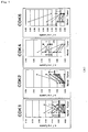
Fig. 1 is a drawing illustrating CDK kinase activities of cancer tissues (A: large intestine cancer, B: stomach cancer, C: esophagus cancer) and normal mucosa. (Example 2) -
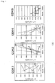
Figs. 2A and 2B are drawings illustrating profiling of CDK kinase activity. (Example 3) - In the present disclosure, the term "cell cycle related protein" means a factor which can regulate a cell cycle, and specifically, a factor which can accelerate or arrest the cell cycle in a cycle starting from division of a cell until the next division once again. Examples of such a factor include e.g., cyclin, cyclin dependent kinase (CDK), as well as cyclin dependent kinase inhibitor (CDKI) and the like.
- Moreover, in the present disclosure, the term "cell cycle profiling" means concurrent measurement of a minimum of 2 or more, preferably 3 or more values of expression or activity of cell cycle related proteins. Preferably, the term means to obtain the information (profile) on states or properties of a tissue and cell to be a target of the measurement by analyzing, on the basis of a measurement value of one cell cycle related protein, another relative measurement value. Examples of the measurement parameter include e.g., CDK activities such as CDK1 activity, CDK2 activity, CDK4 activity and CDK6 activity; CDK expression such as CDK1 protein expression, CDK2 protein expression, CDK4 protein expression and CDK6 protein expression; cyclin expression such as cyclin B protein expression, cyclin D protein expression and cyclin E protein expression; CDK1 expression such as p16 protein expression, p21 protein expression and p27 protein expression; p53 protein expression; Rb protein expression and the like.
- Furthermore, the reference value for obtaining the relative value may be, for example, a CDK1 activity value or a CDK1 protein expression value, however, a measurement value other than the above parameters may be also used.
- For the measurement of the cell cycle related protein conducted in the method of the examination of the preset invention, any measurement method can be employed. For example, the CDK enzyme activity can be measured using conventional radioactive isotope (RI), or can be also used without using RI (Japanese Patent Provisional Publication No.
2002-335997 - Furthermore, for the measurement of the amount of protein expression, any measurement method can be employed. For example, the measurement can be conducted with a simple quantitative analysis system based on a dot blot technique by way of a protein expression analysis.
- For carrying out the method of the examination according to the present invention, preparation of a sample for the measurement is required. As a specific method of solubilization of a cell and a method of isolation of a sample containing CDK, those described in Japanese Patent Provisional Publication No.
2002-335997 - In normal cells, cell proliferation is strictly controlled by a regulation factor in the G1 phase. On the other hand, progress of the cell cycle after passing through the restriction point (R) is mildly controlled in comparison with the G1 phase. Therefore, it is reasonable to carry out profiling of the G1 phase CDK (CDK2, 4, 6) activity on the basis of the G2/M kinase or CDK1 activity value.
- Grounds for abnormal proliferation of cancer cells can not be simply analyzed with expression of a cell cycle related protein. Accordingly, in addition to alteration of the amount of expression of the protein, mutation or the like of the molecule has been reported as a cause of inactivation or activation of these molecules. It is difficult to determine various causes as described above with a primary assay system. Thus, it was believed that measuring the CDK activity, the regulation of which involves many of cell cycle related proteins, is useful in diagnosis of cancer.
- In Table 1, clinicopathological features of patients suffering from gastrointestinal cancer determined in the Examples described below are shown. Median age of the studied group was 65 years old (in the range of from 23 years to 86 years old). One among 22 cases of large intestine cancer, five among 8 cases of stomach cancer, and three among 7 cases of esophagus cancer were in the clinical stage of IV phase.
- Kinase activities of four kinds of CDK and expression results of nine kinds of cell cycle related proteins in normal mucosal tissues and cancer tissues derived from colon cancer patients in Table 2 and Table 3, from stomach cancer patients in Table 4 and Table 5, and from esophagus cancer patients in Table 6 and Table 7 are shown. Furthermore, results of analysis of each CDK in cancer tissues and normal mucosal tissues are shown in Table 8.
- In Table 2 to Table 7, to the measurement value beyond [mean value + 2SD (standard deviation)] is appended the sign of (+), while to the measurement value less than [mean value -2SD] is appended the sign of (-). The measurement was conducted according to the process in Examples described below.
- The expression of CDK was for CDK1, which exhibited a higher value in three kinds of the cancer tissues of: large intestine, stomach and esophagus, in comparison with normal mucosa. When cut off value of the amount of CDK1 expression per total protein was determined as [mean value in corresponding normal tissue +2SD], the value was 1.99 ng/µg in colon, 1.60 ng/µg in stomach, and 0.91 ng/µg in esophagus.
- When analysis was carried out using this cut off value, it was decided that the expression of CDK1 is positive in 27% of tissues of colon cancer (6/22), 75% of tissues of stomach cancer (6/8), and 57% of tissues of esophagus cancer tissue (4/7). These differences were all statistically significant (large intestine; P<0.05, stomach; P<0.05, esophagus; P<0.05, with a Wilcoxon signed-rank test).
- To the contrary, statistically significant difference was not found between cancer tissues and normal tissues for the expression of CDK2, 4 and 6.
- Among CDK activities, marked difference was found for CDK2 between cancer tissues and normal tissues (colon; P<0.01, stomach; P<0.01, esophagus; P<0.01, with a Wilcoxon signed-ranks test).
- When cut off value of the CDK2 activity per weight of protein was determined for each tissue as [mean value of the CDK2 activity exhibited in the normal tissue +2SD], the value was 0.054 U/µg in large intestine, 0.102 U/µg in stomach, and 0.033 U/µg in esophagus. Accordingly, 72% of tissues of large intestine cancer (16/22), 75% of tissues of stomach cancer (6/8), and 100% of tissues of esophagus cancer tissue (7/7) were decided to be positive. For example, in connection with breast cancer, it was reported that expression of an isoform of cyclin E having an excessive activity strongly correlates to survival rate of patients. This correlation suggests that high CDK2 activity is a major cause of instability of the genome and malignant alteration of cancer.
- With respect to CDK4 activity and CDK6 activity, lower value was observed in cancer tissues compared to those in normal mucosa (large intestine; CDK4 P<0.05, large intestine; CDK6 P<0.05, with a Wilcoxon signed-ranks test).
- In the stomach tissue, higher expression and activity was found for both CDK1 and CDK2 compared to other organs. These results suggest that the mucosal tissue in stomach regenerate more actively than other organs, or thickening and proliferation owing to infection with Helicobacter Pyroli often found in Japanese may be suggested.
- The description encompasses reagents and examination systems used in methods of tissue examination and methods of cell examination; methods of cancer examination by way of the present method of the examination; and therapeutic drugs and therapeutic methods of cancer which were selected by analyzing the profile.
- Hereinafter, the present invention is explained in detail with reference to Examples.
- Each surgically collected tissue (2 mm3) was ground with a lysis buffer containing Nonidet P-40 (NP-40) (manufactured by Calbiochem), and solubilized with a homogenizer. Insoluble substances were removed by a filter. Thus resulting cell lysate was employed as a tissue sample for the measurement.
- A sample containing 100 pg of the total protein was added to 2 pg of an antibody (anti-CDK1, 2, 4 and 6 antibodies, manufactured by Santa Cruz) and 20 µl of protein A beads (manufactured by BioRad) to allow them to react at 4°C for 1 hour and to precipitate CDK molecules.
- After washing with a buffer (0.1% NP-40, 50 mM Tris-HCl, pH 7.0) three times, 50 µl of a substrate mixture containing proteins was added thereto followed by incubation at 37°C for 10 minutes while shaking.
- The substrate mixture contains 10 µg of histone H1 corresponding to CDK1 and CDK2 (manufactured by Upstate Biotechnology, Inc.), 10 µg of C-terminal domain of recombinant Rb protein corresponding to CDK4 and 5 (a.a. 769-921), 5 mM adenosine-5'-O-(3-thiotriphosphate) (ATP-γS, manufactured by Sigma Corporation, USA), and buffer (20 mM Tris-HCl, pH 7.4, 0.1% Triton X-100).
- After removing the beads, monothiophosphoric acid induced into the substrate was incubated with a 10 mM substrate further labeled with iodoacetyl-biotin (manufactured by Pierce, USA) in a coupling buffer (100 mM Tris-HCl, pH 8.5, 1 mM EDTA) in a dark place at room temperature for 90 minutes, followed by termination of the reaction with B-mercaptoethanol.
- The substrate in an amount of 0.4 pg after completing the reaction was added onto a PVDF membrane using a slot blotter, and aspirated. Thus resulting membrane was blocked with 4% bovine serum albumin (BSA) for 30 minutes, and allowed to react with avidin-FITC (manufactured by Vector Laboratories) at 37°C for 1 hour.
- After washing the membrane, images on the membrane were analyzed by a fluorescent image analyzer (manufactured by BioRad) . The activity was calculated based on a standard curve corresponding to 0, 12.5, 25, 50, 100 and 150 µg of CDK included in K-562 chronic myeloid leukemia cell strain.
- For reference, 1 U means an enzyme activity which is equal to that of K-562 cell with 1 pg of total protein.
- Surgically collected tissue (2 mm3) was ground with a lysis buffer containing Nonidet P-40 (NP-40), and solubilized by a homogenizer. Insoluble substances were removed with a filter. Then, 2.5 µg of the protein was added to wells (2 x 2 x 3 mm, permissible amount: 50 µl) of a slot blotter having a hydrophobic membrane (PVDF, manufactured by Millipore Corporation) set therein. Target substance in the crude sample bound on the membrane was quantitatively detected by the following reaction, i.e., the reaction with an anti-cell cycle related protein antibody, a biotinized secondary antibody and a fluorescent labeled streptavidin.
- During each reaction, the wells were automatically washed with TBS (25 mM Tris-HCl pH 7.4, 150 mM NaCl).
- Fluorescent images on the membrane were analyzed by an image analyzer (manufactured by BioRad), and the fluorescent intensity of the dot was measured. Quantitative determination of the cell cycle related protein was carried out with a standard curve obtained using a standard recombinant protein (CDK1; 2.5-25 ng/dot, CDK2; 1.0-10 ng/dot, CDK4; 1.0-10 ng/dot, CDK6; 2.5-25 mg/dot).
- Each enzyme activity of CDK1, 2, 4 and 6 was measured for gastrointestinal cancer tissues and normal mucosa. Measurement of the activity was conducted with the method described in Example 1, for each of the tissues of (A) large intestine cancer of 22 cases, (B) stomach cancer of 8 cases, and (C) esophagus cancer of 7 cases as well as normal mucosa.
- The results are depicted in
Figs. 1A to C . Consequently, with respect to difference of each enzyme activity between cancer tissues and normal mucosa, statistical significance was found in elevation of the CDK2 activity in the large intestine, stomach and esophagus tissues (P<0.01). On the other hand, CDK4 and CDK6 in the large intestine and stomach cancer tissues exhibited activities that were comparatively lower than those in normal mucosa. Statistical analysis was carried out with a Wilcoxon signed-ranks test. In crease in CDK2 enzyme activity was observed in almost of the large intestine, stomach and esophagus cancer tissues. - With respect to CDK (CDK2, 4, 6) in the G1 phase, the activity values of CDK2, 4 and 6 were standardized on the basis of the CDK1 activity value that is G2/M kinase to determine the profile. Consequently, as shown by a solid line in
Fig. 2A , in seven among 8 cases of normal large intestine mucosal tissues, CDK profile in the G1 phase positioned in the fold width of 0.024 to 0.43 for CDK2/CDK1, 1.2 to 39 for CDK4/CDK1, and 1.9 to 26 for CDK6/CDK1, exhibiting a common pattern. This suggests that relative values of the CDK activities are similar among the normal large intestine tissues. - Next, the profile on the basis of value of CDK1 activity that is G2/M kinase was similarly determined for CDKs (CDK2, 4, 6) in the G1 phase on randomly selected nine cases of large intestine cancer tissue. Consequently, as shown in
Fig. 2B , no profile was observed which was identical to the common profile found in the normal mucosal tissues - According to the present invention, it was found that more accurate examination of cancer tissues and cancer cells is possible when cell cycle profiling that involves measurement of the activity is carried out. It is expected that high probability to result in a prognostic factor would be found on analyses of expression, activity, and profiling and the like of each cell cycle regulation factor in retrospective or prospective tests.
- In addition, the method of the present invention in which cell cycle profiling that involves measurement of the activity is carried out will be extremely valuable one which can be adapted to tailor made medical treatments, because it was reported in connection with breast cancer that expression of an isomer of cyclin E that imparts excessive activity to CDK2 strongly correlates to survival rate of patients.
- When the cell cycle profiling that involves measurement of the activity is carried out according to the method of the present invention, more accurate examination of cancer tissues and cancer cells can be executed, and definite diagnosis of cancer, drug resistance test and prognosis are enabled.
Table 1 -1 Clinicopathological features of patients Patieut Sex Age Stage Tissue-type Site Metastasis Invasion Lymph-node Peritoneum Liver Distant Colon cancer C001 F 53 lllb well/mod As N2 P0 H0 M0 V1 Ly1 C002 M 66 I well/mod Rs N0 P0 H0 M0 V3 Ly1 C003 M 67 IIIa well/mod Rs-S N1 P0 H0 M0 V1 Ly1 C005 M 56 IIIb mod-por Ac N2 P0 H0 M0 V1 Ly1 C006 M 48 IIIa well-mod Rb N0 P0 H0 M0 V1 Ly1 C007 F 80 IIIa ne Rb N1 P0 H0 M0 V1 Ly2 C008 M 73 II muc A N0 P0 H0 M0 V0 Ly1 C009 M 60 I well/mod Rs N0 P0 H0 M0 V1 Ly0 C010 M 83 IIIb well/mod T N2 P0 H0 M0 V2 Ly1 C011 M 77 IIIa well/mod Rs N1 P0 H0 M0 V2 Ly1 C012 M 86 IIIa well/mod Rb N1 P0 H0 M0 V2 Ly1 C01 3 F 71 I por>mod A N0 P0 H0 M0 V0 Ly1 C014 F 81 II well/mod Rs N0 P0 H0 M0 V0 Ly1 C015 F 70 III well/mod Rab N1 P0 H0 M0 V1 Ly2 C016 F 43 IV well S N1 P0 H0 M1 V1 Ly1 C017 M 47 II well/mod RaRs N0 P0 H0 M0 V2 Ly1 C018 M 59 II well/mod Rs N0 P0 H0 M0 V1 Ly1 C019 F 34 IIIa well/mod Re N1 P0 H0 M0 V1 Ly1 C020 M 73 II well/mod A N0 P0 H0 M0 V1 Ly1 C021 M 48 IIIb mod-por Ds N2 P0 H0 M0 V3 Ly3 C022 M 23 IIIb por Ce N3 P0 H0 M0 V1 Ly1 C023 F 79 II well/mod Rs N0 P0 H0 M0 V1 Ly1 Table 1 - 2 Clinicopathological features of patients Patieut Sex Age Stage Tissue-type Site Metastasis Invasion Lymph-node Peritoneum Liver Distant Gastric cancer S001 M 70 IV tub2 Ue N2 P0 H1 M0 V1 Ly1 S002 M 66 IIIb por>tub2 Ue N2 P0 H0 M0 V1 Ly1 S005 M 59 Ib tub1-2 U N0 P0 H0 M0 V2 Ly2 S006 F 67 IV por2 Lm N2 P1 H0 M0 V3 Ly3 S008 M 75 IV tub2>porl L N2 P0 H1 M0 V3 Ly3 S009 M 67 IV tub2 M N3 P0 H1 M0 V2 Ly2 S010 M 72 IV tub2>por MI N2 P0 H1 M1 V2 Ly3 S011 F 40 II muc M N1 P0 H0 M0 V0 Ly2 Esophageal carcinoma E002 M 46 IVa por> > mod Lt-Ae N2 M0 V1 Ly1 E003 M 77 IVb mod Lt N3 M1 V2 Ly2 E005 M 44 III mod>well MtLt N3 M0 V1 Ly2 E006 M 60 III mod>por UtLt N1 M0 V0 Ly2 E008 M 61 IVa mod Mt N2 M0 V2 Ly2 E009 M 65 II well Mt N1 M0 V1 Ly1 E010 M 59 III mod>por Ae N3 M0 V1 Ly1 Table2 Each CDK activity in colon cancer patients Sample name cdk1 kinase (U/ug lysate) cdk2 kinase (U/ug lysate) cdk4 (U/ug lysate) cdk6 kinase (U/ug lysate) C001 0.000 0.056 (+) 0.082 0.041 C002 0.049 0.072 (+) C003 0.065 (+) 0.065 (+) 0.141 0.109 C005 0.024 0.097 (+) C006 0.042 0.117 (+) C007 0.020 0.057 (+) C008 0.008 0.124 (+) 0.138 0.039 C009 0.001 0.038 C010 0.000 0.179 (+) C011 0.004 0.062 (+) C012 0.024 0.034 0.020 (-) (0.032) (-) C013 0.000 0.059 (+) (0.094) (-) (0.035) (-) C014 0.037 0.068 (+) C015 0.022 0.053 (+) C016 0.009 0.051 (+) 0.085 0.021 C017 0.041 0.091 (+) 0.095 0.089 C018 0.017 0.033 0.110 0.040 C019 0.000 0.045 (+) 0.099 0.154 C020 0.007 0.040 (0.003) (-) 0.050 C021 0.012 0.007 0.020 (-) 0.034 C022 0.003 0.063 (+) 0.012 (-) 0.012 C023 0.001 0.000 0.093 0.042 Table3-1 Amount of each protein expression in colon cancer patient Sample name cdk1 (ng/ug lysate) cdk2 (ng/ug lysate) cdk4 (ng/ug lysate) cdk6 (ng/ug lysate) cyclinD1 (ng/ug lysate) C001 1.285 0.263 0.620 0.278 0.052 C002 2.822 (+) 0.519 0.989 (+) 0.579 0.123 C003 1.392 0.367 0.777 0.307 0.051 C005 2.606 (+) 0.492 0.970 (+) 0.405 0.089 C006 1.618 0.000 0.343 0.000 0.077 C007 2.363 (+) 0.349 0.640 0.309 0.053 C008 3.022 (+) 0.359 0.567 0.538 0.093 C009 0.000 (-) 0.000 0.000 (-) 0.000 0.000 (-) C010 2.058 (+) 0.000 0.379 0.373 0.076 C011 1.093 0.227 0.492 0.000 0.095 C012 0.673 0.164 0.348 0.000 0.046 C013 1.567 0.225 0.437 0.334 0.077 E014 1.444 0.209 0.766 0.385 0.143 C015 0.869 0.000 0.192 0.000 0.074 C016 1.417 0.200 0.866 (+) 0.806 (+) 0.115 C017 0.824 0.000 0.486 0.000 0.058 C018 0.917 0.000 0.299 0.000 0.075 C019 0.981 0.000 0.570 0.000 0.073 C020 0.800 0.194 0.149 0.000 0.041 C021 0.910 0.551 0.064 C022 1.991 (+) 0.343 0.531 0.422 0.075 C023 0.800 0.202 0.517 0.000 0.069 Table3-2 Amount of each protein expression in colon cancer patient Sample name cyclinE (ng/ug lysate) p53 ELISA (pg/ug lysate) p21 ELISA (mU/ug lysate) p16 WB (CNT×mm) p27 (ng/ug lysate) C001 0.501 0.134 C002 0.561 0.162 0.133 C003 0.433 1.355 (+) 2.600 (-) 0.123 C005 0.762 0.162 7.070 0.152 C006 0.427 1.768 (+) 5.696 0.089 C007 0.319 1.010 (+) 3.890 61.2 (+) 0.128 C008 0.691 0.513 (+) 12.690 107.5 (+) 0.114 C009 0.000 (-) 0.077 (-) 4.804 0.000 (-) C010 0.303 0.054 (-) 5.607 0.117 C011 0.000 (-) 0.365 (+) 1.554 (-) 0.158 C012 0.000 (-) 0.339 (+) 3.290 37.5 0.000 (-) C013 0.429 0.178 8.160 21.6 0.166 C014 0.371 0.067 (-) 8.034 0.091 C015 0.501 0.057 (-) 1.178 (-) 0.066 C016 0.482 0.171 3.470 130.2 (+) 0.107 C017 0.425 0.226 0.084 C018 1.480 0.193 4.250 35.2 0.160 C019 0.618 2.963 (+) 6.970 18.2 0.089 C020 1.054 0.247 (+) 5.030 29.7 0.220 C021 1.411 0.180 5.010 31.7 0.397 C022 0.468 0.224 7.890 25.4 0.128 C023 0.494 0.223 12.720 19.7 0.198 Table4 Each CDK activity in gastric cancer patient Sample name cdk1 kinase (U/ug lysate) cdk2 kinase (U/ug lysate) cdk4 kinase (U/ug lysate) cdk6 kinase (U/ug lysate) S001 0.096 0.116 (+) (0.014) (0.038) (-) S002 0.011 0.014 S005 0.074 0.129 (+) S006 0.000 0.182 (+) 0.015 0.038 (-) S008 0.027 0.181 (+) (0.012) 0.108 S009 0.000 0.555 (+) 0.067 0.024 (-) S010 0.000 0.226 (+) 0.037 0.101 5011 0.000 0.030 0.040 0.041 (-) Table5 - 1 Amount of each protein expression in gastric cancer patient Sample name cdk1 (ng/ug lysate) cdk2 (ng/ug lysate) cdk4 (ng/ug lysate) cdk6 (ng/ugl lysate) cyclinD1 (ng/ug lysate) S001 1.727 0.504 0.813 0.584 0.105 S002 6.960 (+) 2.232 (+) 2.255 (+) 3.627 (+) 0.256 (+) S005 1.035 0.000 (-) 0.282 0.000 (-) 0.051 S006 2.524 (+) 0.377 0.645 0.514 0.083 S008 3.023 (+) 0.404 1.536 (+) 0.376 0.195 (+) S009 2.155 (+) 0.330 0.405 0.451 0.044 S010 4.278 (+) 0.343 0.690 0.518 0.103 S011 0.713 0.000 (-) 0.208 0.000 (-) 0.057 Table5-2 Amount of each protein expression in gastric cancer patient Sample name cyclinE (ng/ug lysate) p53 ELISA (pg/ug lysate) p21 ELISA (mU/ug lysate) p16 WB (CNT×mm) p27 (ng/ug lysate) S001 0.518 0.158 4.050 58.0 (+) 0.141 S002 3.692 (+) 0.182 8.550 0.566 (+) S005 0.300 1.279 3.824 0.109 S006 0.840 3.431 (+) 8.270 78.4 (+) 0.138 S008 0.298 2.236 (+) 7.065 92.3 (+) 0.077 S009 0.600 0.980 3.050 72.7 (+) 0.107 S010 0.420 0.104 16.560 (+) 68.4 (+) 0.096 S011 0.312 0.000 1.150 16.7 0.086 Table6 Each CDK activity in esophageal cancer patient Sample name cdk1 kinase (U/ug lysate) cdk2 kinase (U/ug lysate) cdk4 kinase (U/ug lysate) cdk6 kinase (U/ug lysate) E002 0.000 0.073 (+) 0.125 0.149 E003 0.000 0.034 E005 0.000 0.089 (+) E006 0.000 0.047 (+) E008 0.000 0.033 E009 0.012 (+) 0.037 (+) 0.154 0.133 E010 0.002 0.048 (+) 0.015 0.201 

Table8 Activity (U/µg total protein ± s.d.) CDK1 CDK2 CDK4 CDK6 Nomal Tumor Nomal Tumor Nomal Tumor Nomal Tumor Colon 0.017±0.020 0.017±0.019 0.012±0.016 0.065±0.040 0.086±0.043 0.069±0.051 0.094±0.11 0.049±0.044 Stomac 0.034±0.036 0.027±0.038 0.040±0.031 0.18±0.17 0.10±0.086 0.027±0.026 0.10±0.050 0.052±0.043 Esophagus 0.004±0.003 0.003±0.004 0.011±0.011 0.055±0.022 0.098 0.098±0.073 0.16 0.16±0.036 Expession (ng/µg total protein ± s.d.) CDK1 CDK2 CDK4 CDK6 Nomal Tumor Nomal Tumor Nomal Tumor Nomal Tumor Colon 0.79±0.60 1.5±0.78 0.23±0.24 0.21±0.17 0.35±0.23 0,54±0.25 0.19±0.23 0.24±0.25 Stomac 0.84±0.38 2.8±2.0 0.39±0.29 0.52±0.71 0.38±0.17 0.85±0.70 0.28±0.26 0.76±1.2 Esophagus 0.63±0.14 1.7±1.0 0.084±0.11 0.21±0.16 0.59±0.39 0.55±0.33 0.001 0.30±0.25
Claims (6)
- An in vitro method of the examination of a cell which comprises:a step of measuring as a first measurement value the activity of cyclin dependent kinase 1 (CDK1) and as a second measurement value the activity of cyclin dependent kinase 2 (CDK2) in a cell collected from a patient;a step of obtaining a first relative activity value represented by a ratio of the second measurement value to the first measurement value;a step of comparing the first relative activity value with that of a normal cell; anda step of determining whether or not the cell collected from the patient is a cancer cell, on the basis of the comparing result.
- The method of examination according to claim 1, wherein the measuring step further measures as a third measurement value the activity of cyclin dependent kinase 4 (CDK 4), the obtaining step further obtains a second relative activity value represented by a ratio of the third measurement value to the first measurement value, and the comparing step is performed by comparing the first and second relative activity values with that of normal cell.
- The method of examination according to claim 1, wherein the measuring step further measures as a third measurement value the activity of cyclin dependent kinase 6 (CDK 6), the obtaining step further obtains a third relative activity value represented by a ratio of the third measurement value to the first measurement value, and the comparing step is performed by comparing the first and third relative activity values with that of normal cell.
- The method of examination according to claim 1, wherein the measuring step further measures as a third measurement value the activity of cyclin dependent kinase 4 (CDK4) and as a fourth measurement value the activity of cyclin dependent kinase 6 (CDK6), the obtaining step further obtains a second relative activity value represented by a ratio of the third measurement value to the first measurement value and third relative activity value represented by a ratio of the fourth measurement value to the first measurement value, and the comparing step is performed by comparing the first, second and third relative activity values with that of normal cell.
- The method of the examination according to claim 1, comprising a step of measuring an expression amount of cyclin.
- The method of the examination according to claim 1, comprising a step of measuring an expression amount of a cyclin dependent kinase inhibitor.
Applications Claiming Priority (3)
| Application Number | Priority Date | Filing Date | Title |
|---|---|---|---|
| JP2003048653 | 2003-02-26 | ||
| JP2003048653 | 2003-02-26 | ||
| PCT/JP2004/002164 WO2004076686A1 (en) | 2003-02-26 | 2004-02-25 | Method of examining cell |
Publications (3)
| Publication Number | Publication Date |
|---|---|
| EP1600513A1 EP1600513A1 (en) | 2005-11-30 |
| EP1600513A4 EP1600513A4 (en) | 2007-09-12 |
| EP1600513B1 true EP1600513B1 (en) | 2010-02-24 |
Family
ID=32923298
Family Applications (1)
| Application Number | Title | Priority Date | Filing Date |
|---|---|---|---|
| EP04714457A Expired - Lifetime EP1600513B1 (en) | 2003-02-26 | 2004-02-25 | Method of examining a cell |
Country Status (6)
| Country | Link |
|---|---|
| US (1) | US7501257B2 (en) |
| EP (1) | EP1600513B1 (en) |
| JP (1) | JP4317188B2 (en) |
| AT (1) | ATE458830T1 (en) |
| DE (1) | DE602004025676D1 (en) |
| WO (1) | WO2004076686A1 (en) |
Families Citing this family (12)
| Publication number | Priority date | Publication date | Assignee | Title |
|---|---|---|---|---|
| WO2005116241A1 (en) | 2004-05-31 | 2005-12-08 | Sysmex Corporation | Method of judging properties of mammalian cell and method of diagnosing cancer |
| US7957910B2 (en) | 2005-01-31 | 2011-06-07 | Sysmex Corporation | Method for predicting effectiveness of chemotherapy |
| JP4944446B2 (en) * | 2005-01-31 | 2012-05-30 | シスメックス株式会社 | Effectiveness prediction method of anticancer drug treatment |
| US7682785B2 (en) | 2005-06-30 | 2010-03-23 | Sysmex Corporation | Method for predicting effectiveness of chemotherapy using anticancer agent |
| JP5046574B2 (en) * | 2005-06-30 | 2012-10-10 | シスメックス株式会社 | Effectiveness prediction method of anticancer drug treatment |
| EP1750131B1 (en) * | 2005-08-01 | 2008-07-09 | Sysmex Corporation | Method for judging feature of malignant tumor |
| JP4766969B2 (en) * | 2005-09-14 | 2011-09-07 | シスメックス株式会社 | Organization property judgment device |
| JP2007259846A (en) | 2006-02-28 | 2007-10-11 | Sysmex Corp | Method for judging property of malignant tumor |
| JP5408839B2 (en) * | 2006-10-20 | 2014-02-05 | オリンパス株式会社 | Cell cycle analysis method |
| JP5111902B2 (en) * | 2007-03-14 | 2013-01-09 | シスメックス株式会社 | Cancer diagnosis support device |
| JP2009089672A (en) * | 2007-10-10 | 2009-04-30 | Sysmex Corp | Method for determining risk of cancer relapse |
| JP2010057486A (en) * | 2008-09-02 | 2010-03-18 | Sysmex Corp | Method for predicting response of cancer patient to chemotherapy |
Family Cites Families (10)
| Publication number | Priority date | Publication date | Assignee | Title |
|---|---|---|---|---|
| AU6593196A (en) | 1995-07-20 | 1997-02-18 | Paracelsian, Inc. | Determination of the presence of abnormal cellular proliferation through the detection of one or more cyclin dependent kinases |
| US5672508A (en) * | 1996-01-23 | 1997-09-30 | Mitotix, Inc. | Inhibitors of cell-cycle progression, and uses related thereto |
| US6048693A (en) * | 1996-10-16 | 2000-04-11 | Bittech, Inc. | Phenotypic assays of cyclin/cyclin-dependent kinase function |
| US5914249A (en) * | 1997-12-05 | 1999-06-22 | Incyte Pharmaceuticals, Inc. | Cell-cycle phosphoproteins |
| DE69907155T2 (en) * | 1998-02-18 | 2004-02-05 | Theryte Ltd. | CANCER TREATMENT |
| DE19829473C2 (en) * | 1998-07-01 | 2000-08-10 | Magnus Von Knebel Doeberitz Ch | Procedure for early diagnosis of carcinomas |
| US20030064426A1 (en) * | 2001-02-01 | 2003-04-03 | Jason Poole | Reagents and methods for identifying and modulating expression of genes regulated by CDK inhibitors |
| ATE393821T1 (en) * | 2001-02-14 | 2008-05-15 | Sysmex Corp | METHOD FOR DETERMINING CELL CYCLE REGULATORY FACTOR ACTIVITY AND METHOD FOR DIAGNOSING CANCER USING THE SAME |
| JP4036655B2 (en) | 2001-02-14 | 2008-01-23 | シスメックス株式会社 | Method for measuring activity of cell cycle regulator and reagent used in the method |
| WO2004007754A2 (en) * | 2002-07-12 | 2004-01-22 | Rigel Pharmaceuticals, Inc. | Modulators of cellular proliferation |
-
2004
- 2004-02-25 WO PCT/JP2004/002164 patent/WO2004076686A1/en active Application Filing
- 2004-02-25 DE DE602004025676T patent/DE602004025676D1/en not_active Expired - Lifetime
- 2004-02-25 US US10/547,072 patent/US7501257B2/en active Active
- 2004-02-25 EP EP04714457A patent/EP1600513B1/en not_active Expired - Lifetime
- 2004-02-25 JP JP2005502891A patent/JP4317188B2/en not_active Expired - Lifetime
- 2004-02-25 AT AT04714457T patent/ATE458830T1/en not_active IP Right Cessation
Also Published As
| Publication number | Publication date |
|---|---|
| JP4317188B2 (en) | 2009-08-19 |
| US20070031813A1 (en) | 2007-02-08 |
| EP1600513A1 (en) | 2005-11-30 |
| WO2004076686A1 (en) | 2004-09-10 |
| DE602004025676D1 (en) | 2010-04-08 |
| ATE458830T1 (en) | 2010-03-15 |
| EP1600513A4 (en) | 2007-09-12 |
| JPWO2004076686A1 (en) | 2006-06-08 |
| US7501257B2 (en) | 2009-03-10 |
Similar Documents
| Publication | Publication Date | Title |
|---|---|---|
| Heinrich et al. | Molecular correlates of imatinib resistance in gastrointestinal stromal tumors | |
| Do et al. | Phase I study of single-agent AZD1775 (MK-1775), a Wee1 kinase inhibitor, in patients with refractory solid tumors | |
| Hsieh et al. | p63 and SOX2 dictate glucose reliance and metabolic vulnerabilities in squamous cell carcinomas | |
| Mahajan et al. | Ack1 mediated AKT/PKB tyrosine 176 phosphorylation regulates its activation | |
| Catenacci et al. | Absolute quantitation of Met using mass spectrometry for clinical application: assay precision, stability, and correlation with MET gene amplification in FFPE tumor tissue | |
| EP1600513B1 (en) | Method of examining a cell | |
| Lee et al. | Detection of novel and potentially actionable anaplastic lymphoma kinase (ALK) rearrangement in colorectal adenocarcinoma by immunohistochemistry screening | |
| KR102067327B1 (en) | Quantifying Her2 Protein for Optimal Cancer Treatment | |
| Adams et al. | Targeted MDM2 degradation reveals a new vulnerability for p53-inactivated triple-negative breast cancer | |
| Nisa et al. | Targeting the MET receptor tyrosine kinase as a strategy for radiosensitization in locoregionally advanced head and neck squamous cell carcinoma | |
| Smith et al. | MET–GRB2 signaling-associated complexes correlate with oncogenic MET signaling and sensitivity to MET kinase inhibitors | |
| Xiao et al. | MEX3C-mediated decay of SOCS3 mRNA promotes JAK2/STAT3 signaling to facilitate metastasis in hepatocellular carcinoma | |
| Lai et al. | Refining patient selection of MET-activated non-small cell lung cancer through biomarker precision | |
| Sala et al. | Discoidin Domain Receptor 2 orchestrates melanoma resistance combining phenotype switching and proliferation | |
| JP2020522501A (en) | Prediction method of cancer treatment results by T-DM1 | |
| US10288616B2 (en) | Quantifying met protein for cancer treatment | |
| Zhou et al. | Anaplastic lymphoma kinase (ALK) rearrangement in adult renal cell carcinoma with lung metastasis: a case report and literature review | |
| US20230139516A1 (en) | Ripk2 inhibition for the treatment of cancer | |
| Seo et al. | Differential Clinical Significance of Neurotrophin-3 Expression according to MYCN Amplification and TrkC Expression in Neuroblastoma | |
| Kato et al. | Long-term efficacy of immune checkpoint inhibitors in non-small cell lung cancer patients harboring MET exon 14 skipping mutations | |
| TWI784969B (en) | Use of c-met inhibitors to treat cancers harbouring met mutations | |
| Miyawaki et al. | Correlation between 22C3–PD-L1 expression and EGFR mutations in Japanese patients with advanced lung adenocarcinoma | |
| KR101988120B1 (en) | Diagnosis of gastric cancer using gastrokine 1 protein within blood | |
| US20200278353A1 (en) | Protein Expression Analysis For Breast Cancer Prognosis And Treatment | |
| Gurav et al. | Targeted molecular profiling of solid tumours-Indian tertiary cancer centre experience |
Legal Events
| Date | Code | Title | Description |
|---|---|---|---|
| PUAI | Public reference made under article 153(3) epc to a published international application that has entered the european phase |
Free format text: ORIGINAL CODE: 0009012 |
|
| 17P | Request for examination filed |
Effective date: 20050916 |
|
| AK | Designated contracting states |
Kind code of ref document: A1 Designated state(s): AT BE BG CH CY CZ DE DK EE ES FI FR GB GR HU IE IT LI LU MC NL PT RO SE SI SK TR |
|
| AX | Request for extension of the european patent |
Extension state: AL LT LV MK |
|
| RIN1 | Information on inventor provided before grant (corrected) |
Inventor name: SAKAI, TOSHIYUKI Inventor name: ISHIHARA, HIDEKI,C/O SYSMEX CORPORATION |
|
| DAX | Request for extension of the european patent (deleted) | ||
| A4 | Supplementary search report drawn up and despatched |
Effective date: 20070809 |
|
| RIC1 | Information provided on ipc code assigned before grant |
Ipc: G01N 33/50 20060101ALI20070803BHEP Ipc: G01N 33/574 20060101ALI20070803BHEP Ipc: C12Q 1/48 20060101AFI20070803BHEP |
|
| 17Q | First examination report despatched |
Effective date: 20071115 |
|
| GRAP | Despatch of communication of intention to grant a patent |
Free format text: ORIGINAL CODE: EPIDOSNIGR1 |
|
| RTI1 | Title (correction) |
Free format text: METHOD OF EXAMINING A CELL |
|
| GRAS | Grant fee paid |
Free format text: ORIGINAL CODE: EPIDOSNIGR3 |
|
| GRAA | (expected) grant |
Free format text: ORIGINAL CODE: 0009210 |
|
| AK | Designated contracting states |
Kind code of ref document: B1 Designated state(s): AT BE BG CH CY CZ DE DK EE ES FI FR GB GR HU IE IT LI LU MC NL PT RO SE SI SK TR |
|
| REG | Reference to a national code |
Ref country code: GB Ref legal event code: FG4D |
|
| REG | Reference to a national code |
Ref country code: CH Ref legal event code: EP |
|
| REG | Reference to a national code |
Ref country code: IE Ref legal event code: FG4D |
|
| REF | Corresponds to: |
Ref document number: 602004025676 Country of ref document: DE Date of ref document: 20100408 Kind code of ref document: P |
|
| REG | Reference to a national code |
Ref country code: NL Ref legal event code: VDEP Effective date: 20100224 |
|
| PG25 | Lapsed in a contracting state [announced via postgrant information from national office to epo] |
Ref country code: PT Free format text: LAPSE BECAUSE OF FAILURE TO SUBMIT A TRANSLATION OF THE DESCRIPTION OR TO PAY THE FEE WITHIN THE PRESCRIBED TIME-LIMIT Effective date: 20100625 |
|
| PG25 | Lapsed in a contracting state [announced via postgrant information from national office to epo] |
Ref country code: FI Free format text: LAPSE BECAUSE OF FAILURE TO SUBMIT A TRANSLATION OF THE DESCRIPTION OR TO PAY THE FEE WITHIN THE PRESCRIBED TIME-LIMIT Effective date: 20100224 Ref country code: SI Free format text: LAPSE BECAUSE OF FAILURE TO SUBMIT A TRANSLATION OF THE DESCRIPTION OR TO PAY THE FEE WITHIN THE PRESCRIBED TIME-LIMIT Effective date: 20100224 Ref country code: AT Free format text: LAPSE BECAUSE OF FAILURE TO SUBMIT A TRANSLATION OF THE DESCRIPTION OR TO PAY THE FEE WITHIN THE PRESCRIBED TIME-LIMIT Effective date: 20100224 |
|
| REG | Reference to a national code |
Ref country code: CH Ref legal event code: PL |
|
| PG25 | Lapsed in a contracting state [announced via postgrant information from national office to epo] |
Ref country code: GR Free format text: LAPSE BECAUSE OF FAILURE TO SUBMIT A TRANSLATION OF THE DESCRIPTION OR TO PAY THE FEE WITHIN THE PRESCRIBED TIME-LIMIT Effective date: 20100525 Ref country code: SE Free format text: LAPSE BECAUSE OF FAILURE TO SUBMIT A TRANSLATION OF THE DESCRIPTION OR TO PAY THE FEE WITHIN THE PRESCRIBED TIME-LIMIT Effective date: 20100224 Ref country code: RO Free format text: LAPSE BECAUSE OF FAILURE TO SUBMIT A TRANSLATION OF THE DESCRIPTION OR TO PAY THE FEE WITHIN THE PRESCRIBED TIME-LIMIT Effective date: 20100224 Ref country code: NL Free format text: LAPSE BECAUSE OF FAILURE TO SUBMIT A TRANSLATION OF THE DESCRIPTION OR TO PAY THE FEE WITHIN THE PRESCRIBED TIME-LIMIT Effective date: 20100224 Ref country code: MC Free format text: LAPSE BECAUSE OF NON-PAYMENT OF DUE FEES Effective date: 20100301 Ref country code: LI Free format text: LAPSE BECAUSE OF NON-PAYMENT OF DUE FEES Effective date: 20100228 Ref country code: ES Free format text: LAPSE BECAUSE OF FAILURE TO SUBMIT A TRANSLATION OF THE DESCRIPTION OR TO PAY THE FEE WITHIN THE PRESCRIBED TIME-LIMIT Effective date: 20100604 Ref country code: EE Free format text: LAPSE BECAUSE OF FAILURE TO SUBMIT A TRANSLATION OF THE DESCRIPTION OR TO PAY THE FEE WITHIN THE PRESCRIBED TIME-LIMIT Effective date: 20100224 Ref country code: CY Free format text: LAPSE BECAUSE OF FAILURE TO SUBMIT A TRANSLATION OF THE DESCRIPTION OR TO PAY THE FEE WITHIN THE PRESCRIBED TIME-LIMIT Effective date: 20100224 Ref country code: CH Free format text: LAPSE BECAUSE OF NON-PAYMENT OF DUE FEES Effective date: 20100228 Ref country code: BE Free format text: LAPSE BECAUSE OF FAILURE TO SUBMIT A TRANSLATION OF THE DESCRIPTION OR TO PAY THE FEE WITHIN THE PRESCRIBED TIME-LIMIT Effective date: 20100224 |
|
| PG25 | Lapsed in a contracting state [announced via postgrant information from national office to epo] |
Ref country code: CZ Free format text: LAPSE BECAUSE OF FAILURE TO SUBMIT A TRANSLATION OF THE DESCRIPTION OR TO PAY THE FEE WITHIN THE PRESCRIBED TIME-LIMIT Effective date: 20100224 Ref country code: SK Free format text: LAPSE BECAUSE OF FAILURE TO SUBMIT A TRANSLATION OF THE DESCRIPTION OR TO PAY THE FEE WITHIN THE PRESCRIBED TIME-LIMIT Effective date: 20100224 Ref country code: BG Free format text: LAPSE BECAUSE OF FAILURE TO SUBMIT A TRANSLATION OF THE DESCRIPTION OR TO PAY THE FEE WITHIN THE PRESCRIBED TIME-LIMIT Effective date: 20100524 |
|
| PLBE | No opposition filed within time limit |
Free format text: ORIGINAL CODE: 0009261 |
|
| STAA | Information on the status of an ep patent application or granted ep patent |
Free format text: STATUS: NO OPPOSITION FILED WITHIN TIME LIMIT |
|
| PG25 | Lapsed in a contracting state [announced via postgrant information from national office to epo] |
Ref country code: IE Free format text: LAPSE BECAUSE OF NON-PAYMENT OF DUE FEES Effective date: 20100225 Ref country code: DK Free format text: LAPSE BECAUSE OF FAILURE TO SUBMIT A TRANSLATION OF THE DESCRIPTION OR TO PAY THE FEE WITHIN THE PRESCRIBED TIME-LIMIT Effective date: 20100224 |
|
| 26N | No opposition filed |
Effective date: 20101125 |
|
| PG25 | Lapsed in a contracting state [announced via postgrant information from national office to epo] |
Ref country code: IT Free format text: LAPSE BECAUSE OF FAILURE TO SUBMIT A TRANSLATION OF THE DESCRIPTION OR TO PAY THE FEE WITHIN THE PRESCRIBED TIME-LIMIT Effective date: 20100224 |
|
| PG25 | Lapsed in a contracting state [announced via postgrant information from national office to epo] |
Ref country code: HU Free format text: LAPSE BECAUSE OF FAILURE TO SUBMIT A TRANSLATION OF THE DESCRIPTION OR TO PAY THE FEE WITHIN THE PRESCRIBED TIME-LIMIT Effective date: 20100825 Ref country code: LU Free format text: LAPSE BECAUSE OF NON-PAYMENT OF DUE FEES Effective date: 20100225 |
|
| PG25 | Lapsed in a contracting state [announced via postgrant information from national office to epo] |
Ref country code: TR Free format text: LAPSE BECAUSE OF FAILURE TO SUBMIT A TRANSLATION OF THE DESCRIPTION OR TO PAY THE FEE WITHIN THE PRESCRIBED TIME-LIMIT Effective date: 20100224 |
|
| REG | Reference to a national code |
Ref country code: FR Ref legal event code: PLFP Year of fee payment: 12 |
|
| REG | Reference to a national code |
Ref country code: FR Ref legal event code: PLFP Year of fee payment: 13 |
|
| REG | Reference to a national code |
Ref country code: FR Ref legal event code: PLFP Year of fee payment: 14 |
|
| REG | Reference to a national code |
Ref country code: FR Ref legal event code: PLFP Year of fee payment: 15 |
|
| PGFP | Annual fee paid to national office [announced via postgrant information from national office to epo] |
Ref country code: FR Payment date: 20220118 Year of fee payment: 19 |
|
| PGFP | Annual fee paid to national office [announced via postgrant information from national office to epo] |
Ref country code: GB Payment date: 20230105 Year of fee payment: 20 Ref country code: DE Payment date: 20221230 Year of fee payment: 20 |
|
| REG | Reference to a national code |
Ref country code: DE Ref legal event code: R071 Ref document number: 602004025676 Country of ref document: DE |
|
| REG | Reference to a national code |
Ref country code: GB Ref legal event code: PE20 Expiry date: 20240224 |