FIELD OF THE INVENTION
-
The present invention relates to pharmaceutical compositions
comprising Nedd9 protein. The present invention also relates to
pharmaceutical compositions comprising Nedd9 protein and FAK protein.
Pharmaceutical compositions of the present invention can be used for the
treatment of neuronal damages and neurodegenerative diseases, including
ischemic injury such as transient cerebral global ischemia and cerebral stroke.
Furthermore, the present invention relates to methods for differentiating
neuronal cells.
BACKGROUND OF THE INVENTION
-
The recent identification of neuronal stem cells in the adult
mammalian brain suggests the possibility that the adult brain has a latent
capacity for self-repair in response to ischemia. Ischemia is a powerful
reformatting and reprogramming stimulus for the brain, which induces
endogenous proteins related to the pathophysiology of the injured brain
(Barone, F.C. and Feuerstein, G.Z. 1999. J. Cereb. Blood Flow Metab.
19:819-834). It has been shown that some pathological conditions such as
ischemia induce neurogenesis in the adult mammalian brain (Liu, J. et al.
1998. J. Neurosci. 18:7768-7778). Recent findings have raised the possibility
that the brain has as latent capacity for self-repair in response to injury or
disease through the use of the endogenous NSCs or neural progenitor cells
(Magavi, S.S. et al. 2000. Nature 405:951-955).
-
In focal ischemia model of mice, upregulation of several genes has
been investigated and grouped as temporal episodes or "waves" of expression
of different groups of genes (Barone, F.C. and Feuerstein, G.Z. 1999. J. Cereb.
Blood Flow Metab. 19:819-834). Some of these proteins, such as Neurocan
(Haas, C.A. et al. 1999. J. Neurosci. 19:9953-9963) and neuronal precursor
cell-expressed, developmentally down-regulated gene (Nedd9)/Caspase2
(Kinoshita, M. et al. 1997. J. Cereb. Blood Flow Metab. 17:507-514), are
primarily expressed by neurons or glia during embryonic development and
strongly down-regulated during the early postnatal phase.
-
Nedd9 was initially identified as a neuronal precursor cell
(NPC)-expressed, developmentally down-regulated gene in the mouse central
nervous system (CNS) (Kumar, S. et al. 1992. Biochem. Biophys. Res.
Commun. 185:1155-1161). Gene expression of Nedd9 is detected in
embryonal day 10 and 14 and disappeared in adult mouse brain (Kumar, S. et
al. 1992. supra). Later waves of new gene expression like Nedd9 include
mediators that appear to be important in tissue remodeling and recovery of
function (Read, S.J. et al. 2001. J. Cereb. Blood Flow Metab. 21:755-778),
such as transforming growth factor-β (Wang, X. et al. 1995. Brain Res. Bull.
36:607-609) and osteopontin (Wang, X. et al. 1998. J. Neurosci.
18:2075-2083).
-
The product of Nedd9 was subsequently reported to be identical to the
mouse Crk-associated substrate (Cas) lymphocyte-type (Cas-L) (Homology
database available at the website of the National Center for Biotechnology
Information located on the worldwide web at www.ncbi.nlm.nih.gov.
Accessed November 4, 2003), which is also known as human enhancer
filamentation 1 (HEF1) cloned by another group via a completely different
strategy (Law, S.F. et al. 1996. Mol. Cell. Biol. 16:3327-3337). Human Cas-L
was first identified in the present inventors' laboratory as a 105 kDa protein
predominantly tyrosine phosphorylated by the ligation of β1 integrins in
human leukemia H9 cells (Minegishi, M. et al. 1996. J. Exp. Med.
184:1365-1375). The major biological function of Cas-L is the restoration of
IL-2 production by costimulation with β1 integrins and T-cell receptor (TCR)
complex (Kamiguchi, K. et al. 1999. J. Immunol. 163:563-568), and the
enhancement of cell migration by the engagement of β1 integrins and TCR
complex, or β1 integrins alone (Ohashi, Y. et al. 1999. J. Immunol.
163:3727-3734). To exert these functions, it is necessary that Cas-L is
associated with FAK or Pyk2 and is tyrosine phosphorylated by these kinases
(Tachibana, K., et al. 1997. J. Biol. Chem. 272:29083-29090). The present
inventors have demonstrated that Cas-L is a hematopoietic variant of p130Cas
(Minegishi, M. et al., 1996. supra). P130Cas was identified as a 130-kDa
protein that is highly tyrosine phosphorylated in v-Src- (Reynolds, A.B. et al.
1989. Mol. Cell. Biol. 9:3951-3958) and v-Crk-transformed cells (Sakai, R., et
al., 1994. EMBO J. 13:3748-3756). These proteins and Efs/Sin compose the
Cas family, which has a conserved amino acid secondary structure with
numerous protein-protein interactions, such as Src-homology 3(SH3) domain,
substrate domain, serine-rich domain, coiled-coil regions, helix-loop-helix
domain, and C-terminal domain (Minegishi, M. et al., 1996. supra; O'Neill,
G.M. et al. 2000. Trends in Cell Biology 10:111-119). These structures
feature a docking molecule, which interacts with focal adhesion kinase (FAK)
or proline-rich tyrosine kinase 2 (Pyk2). Although Nedd9/Cas-L is likely to
play some roles in the development and differentiation of the CNS, its protein
expression and function in fetal brain has not been reported. More importantly
its function in relation to the pathogenesis of brain ischemia as well as the
expression in adult brain remains unknown.
-
The nonreceptor tyrosine kinase FAK and Pyk2 were identified as
potential common mediators of signaling by growth factors and integrins
(Clark, E.A. and Brugge, J.S. 1995. Science. 268:233-239; Neet, K. and
Hunter, T. 1996. Genes to Cells. 1:147-169; Avraham, H. et al. 2000. Cell.
Signal. 12:123-133; Girault, J.A. et al. 1999. Trends Neurosci. 22:257-263).
Pyk2 is highly expressed in the CNS (Menegon, A. et al. 1999. European
Journal of Neuroscience. 11:3777-3788) and acts as an upstream regulator of
MAPK signaling pathways in response to diverse cellular stimuli, including
depolarization and increase of intracellular Ca2+ levels (Lev, S. et al. 1995.
Nature. 376:737-745; Yu, H. et al. 1996. J. Biol. Chem. 271:29993-29998).
Pyk2 is rapidly tyrosine-phosphorylated in cortical neurons and co-localized
with activated p38MAPK in both neuron and microglia after focal cerebral
ischemia in rats (Tian, D. et al. 2000. J. Neurosci. 20:6478-6487). FAK is also
highly expressed in the CNS as a splice isoform FAK+6,7 mainly (Burgaya, F.
et al. 1995. European Journal of Neuroscience. 7:1810-1821; Toutant, M. et
al. 2000. Biochem. J. 348 Pt 1:119-128; Menegon, A. et al. 1999. supra). FAK
has an important role in the prevention of apoptosis by cell attachment in
non-neuronal cells (Frisch, S.M. et al. 1996. J. Cell Biol. 134:793-799;
Hungerford, J.E. et al. 1996. J. Cell Biol. 135:1383-1390). Although FAK
plays important roles in the development and differentiation of the CNS
(Hungerford, J.E. et al. 1996. supra), there are few reports regarding its
function in adult brain, especially in relation to the pathogenesis of brain
ischemia.
-
Stroke is the second largest cause of death and the leading cause of
long-term disability in Japan and United States. Much work has been devoted
to decrease neuronal damage in order to reduce the resultant neurologic
deficits of ischemia. Although most efforts for treating stroke aim at reducing
or limiting the size of the ensuing acute lesion, this goal will not be
accomplished for many patients, and treatment for later phase of stroke is
unfortunately not well established. As the therapeutic time window toward
acute hypoperfusion and energy failure is limited, identification of
endogenous molecules involved in neuroprotection or remodeling of neurons
would be potentially important for therapy.
SUMMARY OF THE INVENTION
-
An objective of the present invention is to provide pharmaceutical
compositions comprising Nedd9 protein. Another objective of this invention
is to provide uses of Nedd9 protein for the manufacture of a medicament for
treating neuronal damages or neurodegenerative diseases.
-
To identify an endogenous molecule involved in protection of nerves
or remodeling of neurons, the present inventors cloned the mouse Cas-L gene
and assessed the temporal profile of Nedd9/Cas-L, as well as its related
molecules p130Cas, FAK, and PYK2, in brain of transient global ischemia in
rats. The inventors also investigated its function in brain using mouse
embryos, neurospheres, and transfectants of PC-12 cells. The results indicate
that Nedd9 was a splicing variant of Cas-L and selectively induced in neurons
of cerebral cortex and hippocampus 3 to 14 days after the ischemia. Induced
Nedd9 protein was tyrosine phosphorylated and was bound to FAK in dendrite
and soma of neurons after the ischemia. In physiological process, Nedd9 was
transiently expressed in neurites and cell body of developing neurons in
cerebral cortex and hippocampus during development. Nedd9 promoted
neurite outgrowth of PC-12 cells in an NGF-independent manner. These
results suggest that delayed upregulation of Nedd9 in neurons plays an
important role in neuron remodeling after global ischemia in rats.
-
In one aspect, the present invention provides pharmaceutical
compositions comprising Nedd9 protein. The pharmaceutical compositions
may further comprise FAK protein.
-
In another aspect, the present invention provides uses of Nedd9 protein
for the manufacture of a medicament for treating neuronal damages or
neurodegenerative diseases, including ischemic injury such as transient global
ischemia and stroke.
-
In still another aspect, the present invention provides methods for
differentiating neuronal cells, the method comprising introducing a gene
encoding Nedd9 protein into neuronal cells and allowing the expression of the
gene in the cells. More specifically, the method comprises transforming
neuronal cells with an expression vector comprising a gene encoding Nedd9
protein and incubating the cells under the conditions that allow the expression
of the gene.
-
Nedd9 protein, which has been identified to an endogenous molecule
involved in protection of nerves or remodeling of neurons, would be useful as
a pharmaceutical agent for alleviating neuronal damages caused by ischemia
such as stroke. Transplantation of Nedd9 positive neurons into the core of
cerebral ischemia may help the recovery of brain tissue from ischemic insult,
with such delivery system of neurons having been established recently
(Borlongan, C.V. et al. 1998. Exp. Neurol. 149:310-321; Kondziolka, D. et al.
2000. Neurology 55:565-569). Furthermore, the fact that Nedd9 is not
expressed in normal adult brain is useful for its application for therapy
targeted to neuron, because its overexpression can facilitate remodeling of
regenerated neurons specifically. As the range of targets for regeneration
therapy has been widened, future study of Nedd9 expression may be
applicable to other diseases such as neurodegenerative diseases (Alzheimer's,
amyotrophic lateral sclerosis, cerebellar ataxias, Huntington's, Parkinson's,
and progressive supranuclear palsy) or brain and spinal cord injury.
BRIEF DESCRIPTION OF THE DRAWINGS
-
Fig. 1(a) shows comparison of the deduced amino acid sequences of
Nedd9 and Cas-L from mouse and rat. Fig. 1(b) shows a proposed model for
the generation of Nedd9 and Cas-L mRNA variants by multiple domain
structure of Cas-L. (Top) The organization of the Nedd9/Cas-L gene. Exons
are indicated by black boxes and numbered. (Middle) The presence of exons
in the different mRNAs represented by boxes linked by connecting lines.
Translation initiation sites in exons 2A and 2B and the translation stop site in
exon 8 are indicated. Arrows indicate the primer sites for PCR amplification
designed to give products from Nedd9 mRNA and Cas-L mRNA,
respectively. (Bottom) The domain structure of Cas-L is demonstrated. A bar
indicates the responsible domain to be recognized by anti-Cas-L antibody
used in this study. SD: substrate domain, SR: serine-rich region, CC:
coiled-coil regions, HLH: helix-loop-helix domain, CT: C-terminal domain.
-
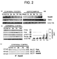
Fig. 2(a) shows a representative result of RT-PCR discriminating the
two splicing variants of Nedd9 with β-actin used as an internal control. H9
cells serve as positive control of Cas-L. Fig. 2(b) presents photographs
showing the expression of proteins related to Nedd9/Cas-L by western blot
analysis. Both Nedd9 protein and FAK were up-regulated, consistent with the
temporal profile of Nedd9 expression. However, other proteins did not exhibit
significant changes. Fig. 2(c) presents photographs showing the FAK content
in immunoprecipitates of Nedd9 at 0, 1, 3, 7 or 14 days following transient
global ischemia. The FAK content in the immunoprecipitates was determined
by visualization with the anti-FAK antibody. The immunoprecipitates were
also blotted by anti-phosphotyrosine antibody 4G10.
-
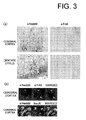
Fig. 3(a) presents photographs showing Nedd9-immunoreactive cells
detected at the cerebral cortex and hippocampus. Rat cerebral cortex and
hippocampal coronal slices reveal intense Nedd9-immunoreactivity in the
neuronal dendrites. In contrast, the neuronal cell nucleus appeared devoid of
Nedd9 staining. Fig. 3(b) presents photographs showing co-localization of
Nedd9 and NeuN or FAK.
-
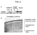
Fig. 4(a) presents photographs showing the results of Western blotting
analysis of Nedd9 temporal expression during mouse brain development.
Nedd9 expression was determined in developing brains at embryonal day 12.5
and 14, and postnatal day 0 and 3 in adult brain. Standard Western blot with
anti-Nedd9 Ab showed expression of Nedd9 protein during natal and early
postnatal phase of developing brains, and not in adult brains. Fig. 4 (b)
presents a photograph showing immunohistochemical distribution of Nedd9
in the cerebral cortex and hippocampus at P0. Positive immunoreactivity with
Nedd9 was fibrillary in the marginal zone and ventricular part of the cortical
plate.
-
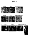
Fig. 5(a) presents photographs showing immunocytochemical
distribution of Nedd9 (green), O-4/GFAP (red), and Hoechst 33258 (blue) in
mouse neurosphere cells. Nedd9 was not expressed in oligodendroglia nor
astroglia. Fig.5(b) presents photographs showing immunocytochemical
distribution of Nedd9 (red), Tuj1 (green), and Hoechst33258 (blue) in mouse
neurosphere cells. Nedd9 was expressed in developing neurons.
-
Fig. 6(a) schematically shows GFP-Nedd9 deletion mutants.
Abbreviations: WT: wild-type, dSH3: delta SH3, SD: substrate domain. F:
deletion in YDYVHL motif, SR: serine-rich domain, CC: coiled-coil regions,
HLH: helix-loop-helix domain, CT: C-terminal domain. Fig. 6(b) shows
neurite formation in the absence or presence of NGF. Data are expressed as
neurite length averaged over diameter of soma (bar diagrams; y axis = cell
diameters) and as number of neurites per cell. Statistical significance is tested
with respect to unstimulated control PC-12 cells (*, p<0.001).
DETAILED DESCRIPTION OF THE INVENTION
-
The present invention provides a pharmaceutical composition
containing a Nedd9 protein as an active ingredient. A Nedd9 protein
contained in a pharmaceutical composition of the present invention needs to
promote neurite elongation and to contribute differentiation of nerve cells. Its
origin and structure are not particularly limited. For example, it is possible to
use Nedd9 proteins derived from humans, mice, rats, rabbits, dogs, horses,
cats, pigs, cattle, goats, sheep, primates such as monkeys and gorillas, and
other mammals. Nedd9 genes of desired species are prepared by utilizing
techniques such as hybridization, PCR and the like using a probe or primer
prepared based on nucleotide sequences encoding known Nedd9 proteins.
Nedd9 proteins of various animals are thus prepared and can be used in this
invention. In a pharmaceutical composition of the present invention, it is
preferred to use a protein derived from the same kind of animals as a target for
administration. In other words, it is preferred to use a human Nedd9 protein
when the composition is administered to humans. The amino acid sequences
of Nedd9 proteins of mice and rats are shown in SEQ ID NOs: 5 and 6,
respectively, and both in Fig. 1a. The mRNA encoding mouse Nedd9 protein
is registered as GenBank Accession No. NM 017464. Using the nucleotide
sequence of the mRNA or a part thereof as a probe, a gene library or the like is
screened to obtain Nedd9 mRNA of various mammals including humans.
Then, various Nedd9 proteins of various mammals are prepared and can be
used in the present invention.
-
In the present invention, the term "Nedd9 protein" includes wild type
Nedd9 proteins as well as Nedd9 proteins having the same functions as the
wild type protein and that are recombinantly or synthetically prepared, and
then used.
-
Examples of Nedd9 proteins having the same functions as those of
wild type Nedd9 proteins include proteins having high amino acid sequence
homology with wild type Nedd9 proteins. The term "high amino acid
sequence homology" means identity of 70% or more, preferably 80% or more,
more preferably 90% or more, still more preferably 95% or more, furthermore
preferably 97% or more, for example, 99%. The term "% identity" is defined
as a function of the number of identical positions shared by two amino acid
sequences, except any conservative substitution, when the two sequences are
aligned with or without allowing gaps for optimal comparison purposes. The
percent identity of amino acid sequences is a value obtained by dividing the
number of identical residues by the total number of amino acid residues in the
alignment. The identity of amino acid sequences can be calculated using a
software known in the art, such as BLAST, BLAST-2, Megalign and the like.
The regulable parameters necessary for calculation can be properly
determined by persons skilled in the art, considering sensitivity or the like.
-
Proteins having high homology with such natural Nedd9 proteins can
be prepared by, for example, the following manner. Using a polynucleotide
encoding a known Nedd9 protein or a part thereof as a probe or primer, a gene
encoding a target protein is isolated from a tissue expressing the gene or an
appropriate gene library by a hybridization or PCR procedure and the isolated
gene is expressed to prepare the protein (referred to Sambrook, J. et al. 1989
"Molecular Cloning: A Laboratory Manual, 2nd ed.", Vol. 1-3, Cold Spring
Harbor Laboratory Press).
-
A Nedd9 protein contained in a pharmaceutical composition according
to the present invention may be a variant of a known Nedd9 protein produced
by deleting a part of the protein or by adding amino acid residue(s), or another
polypeptide, wherein the variant protein has the same function as wild type
Nedd9. Methods for deletion, insertion, replacement, addition and
modification of any amino acid residue in a protein having a known amino
acid sequence are well known. In accordance with the known methods, a
Nedd9 protein contained in a pharmaceutical composition according to the
present invention can also be modified for any purposes such as for increasing
the in vivo activity, for stabilization or the like. Preferable modified Nedd9
proteins in the present invention are conservatively modified proteins having
the same biological activity as the natural Nedd9 protein. For example, one or
more amino acid residues can be modified by glycosylation or
phosphorylation. Proteins can be modified by chemical or enzymatic
de-glycosylation or de-phosphorylation. Proteins may also be modified by
adding one or more amino acid residues to the N terminal or the C terminal of
a natural amino acid sequence, or deleting one or more amino acid residues.
Moreover, proteins may be modified by inserting one or more amino acid
residues into any portion of a natural amino acid sequence. Alternatively,
proteins can be modified by replacing one or more amino acid residues in a
natural amino acid sequence with another amino acid residue(s). Such
replacement is carried out by substitution with amino acid residue(s) having
the same side chain as the amino acid residue to be substituted. This is called
conservative amino acid substitution. In the conservative amino acid
substitution, mutually exchangeable amino acids are classified in accordance
with the chemical properties of their side chains into the following groups:
- (1) Neutral hydrophobic side chain (alanine, valine, leucine,
proline, tryptophan, phenyl alanine or methionine),
- (2) Neutral polar side chain (glycine, serine, threonine, tyrosine,
cysteine, asparagines or glutamine),
- (3) Basic side chain (lysine, arginine or histidine),
- (4) Acidic side chain (aspartic acid or glutamic acid),
- (5) Aliphatic side chain (glycine, alanine, valine, leucine or
isoleucine),
- (6) Aliphatic hydroxyl group side chain (serine or threonine),
- (7) Amine-containing side chain (asparagines, glutamine,
lysine, arginine or histidine),
- (8) Aromatic side chain (phenyl alanine, tyrosine or
tryptophan), and
- (9) sulfur-containing side chain (cysteine or methionine).
-
-
The neurite growth accelerating activity of a modified Nedd9 protein
is measured to confirm whether the biological activity is retained, thereby
determining whether it can be used as a pharmaceutical composition of the
present invention. The retention of the biological activity does not always
mean the same activity level as that of natural proteins. It is enough to have
the same activity as the activity of natural proteins. The activity level may be
higher than or somewhat lower than the level of natural proteins.
-
The present invention indicates that Nedd9 protein and FAK protein
are simultaneously expressed in vivo and it is considered that they
collaboratively act on differentiation to neurons in the developing brain.
-
Therefore, the present invention also relates to a pharmaceutical
composition containing a FAK protein in addition to a Nedd9 protein. Similar
to the case of Nedd9 protein, the origin and structure of a FAK protein
contained in a pharmaceutical composition of the present invention are not
particularly limited as long as the protein binds to a Nedd9 protein and
promotes elongation of neurites. In a pharmaceutical composition of the
present invention, it is preferred to contain a protein derived from the same
kind of animals as a target for administration (when administered to humans, a
human FAK protein is preferably used). Preferable examples of FAK proteins
include proteins having an amino acid sequence encoded by mRNA registered
as GenBank Accession No. L13616 (Whitney, G.S. Chan, P.Y. Blake, J.
Cosand, W.L. Neubauer, M.G. Aruffo, A. and Kanner, S.B. 1993, "Human T
and B lymphocytes express a structurally conserved focal adhesion kinase,
pp125FAK." DNA Cell Biol, 12(9): 823-30). Similar to the case of the
Nedd9 protein, in accordance with necessity, a modified FAK is prepared by
modifying a natural amino acid sequence, and the resulting modified FAK
may be contained in a pharmaceutical composition of the present invention.
-
A method for preparing recombinant proteins is well known. Nedd9
protein and FAK protein can be prepared using an appropriate expression
system including in vitro system and host-vector system. For example, an
expression vector including a chimera gene composed of an appropriate
transcription/translation regulatory region and a protein coding sequence is
constructed. The term "transcription/translation regulatory region" means a
DNA sequence necessary for expressing an operably linked protein coding
sequence in a specific host cell, and includes a promoter, an enhancer, a
polyadenylation signal, an operator sequence, a ribosome-binding site, an
initiation signal, and the like. For expression in a eukaryotic cell, a
promoter/enhancer element is preferably contained in an expression vector to
control the desired protein expression in host cells. The term "operably"
linked or bound means that a protein coding sequence connected downstream
a control sequence is expressed in a host by the direction of the control
sequence without displacement of the reading frame. Examples the
expression vector include known various vectors suitable for expression of
mammalian cells, bacteria cells, insect cells, plant cells, yeast cells, and the
like. These vectors may be used for the production of Nedd9 protein and FAK
protein. Host cells are transformed with the constructed expression vector.
Various cells are established as host cell lines. Persons skilled in the art could
readily select a method of introducing an expression vector suitable for the
selected host cell. Prokaryotic cells can be transformed by, for example,
calcium treatment or electroporation. Plant cells can be transformed by,
without limitation, a known method of using agrobacterium. For mammalian
cells, a calcium phosphate precipitation method can be exemplified.
Additionally, there are known methods such as nuclear microinjection,
protoplast fusion, DEAE-dextran method, cell fusion, electric pulse
perforation method, lipofectamine method (GIBCO BRL) and a method of
using FuGENE6 reagent (Boehringer-Mannheim). The transformation of
mammalian cells can refer to, for example, the descriptions in Keown, W.A. et
al. Methods in Enzymol, 1990, 185:527-37 and Mansour, S.L. et al. Nature,
1988, 336: 348-52. To secure the glycosylation of natural proteins, it is
desired to use mammalian cells as a host cell. Examples of mammalian cells
include known various cells such as CHO, COS, 293, A431, 205, CV-1, Hela,
BHK, H1-60, U937, Hak and Jurkat cells. These cells can be used for the
preparation of Nedd9 protein and FAK protein.
-
A host cell in which a desired gene has been introduced is cultured, a
desired protein is expressed, and the protein is recovered from the resulting
culture. It is also possible to obtain a desired protein from a culture medium in
such a way that a secretory signal sequence recognized in a host is added to the
desired protein and thereby the desired protein is secreted extracellularly. If
necessary, a transgenic animal is prepared, and Nedd9 protein and FAK
protein used in the present invention may be prepared from the animal. When
the protein production is carried out with a transgenic animal, the protein is
preferably expressed using a promoter capable of tissue-specific expression
(for example, protein expression in a milk of a mammal).
-
In a pharmaceutical composition of the present invention, a protein is
preferably contained as a main ingredient in a physiologically and
pharmaceutically acceptable carrier. Nedd9 protein and/or FAK protein,
which are main ingredients, are desirably contained in a substantially purified
state in the carrier. The term "substantially purified" means the state not
containing contaminants in the environment, namely, the state not containing
proteins that coexist with Nedd9 protein or FAK protein in nature or proteins
derived from hosts, which may be contaminated during the preparation of
recombinant proteins. Substantially purified proteins can be prepared by
chromatography such as gel filtration, reversed phase, adsorption, ion
exchange and hydrophobic interaction, and other purification methods using
known procedures such as ethanol precipitation, electrophoresis, evaporation,
solvent extraction, immunoprecipitation, dialysis, re-crystallization, and
ammonium sulfate precipitation. Protein purification may also be carried out
by affinity chromatography utilizing a polyclonal or monoclonal antibody
capable of specifically recognizing a protein to be purified. However, a
pharmaceutical composition of the present invention may contain, as an active
ingredient, proteins other than Nedd9 protein and FAK protein. That is, a
pharmaceutical composition of the present invention may optionally contain
active ingredients other than Nedd9 protein or FAK protein. Preferable active
ingredients optionally added are ingredients that do not inhibit the activities of
Nedd9 protein and FAK protein, and can treat or prevent nerve disorders or
neurodegerative diseases by action mechanism different from these proteins.
Example of the active ingredients include members of family such as
fibloblast growth factors (FGF) (basic FGF; acidic FGF; FGF-3; FGF-4;
FGF-5; FGF-6, FGF-7; FGF-8 and FGF-9), neurotrophines(NT) (nerve
growth factor (NGF); brain derived nerve neutrient factor (BDNF); NT-3;
NT4/5 and NT-6), insulin like growth factor (IGF) (IGF-1 and IGF-2), ciliary
neutrophic factor, epidermal growth factors (EGF) (EGF; TGF α; heparin
binding EGF; amphiregulin; beta-cellulin; vaccinia growth factor and neu
differentiation factor), transforming growth factors β(TGF β), platelet derived
growth factor, and vascular endothelial growth factor (VEGF). Other
examples include leukocyte inhibitory factor (LIF), oncostatine,
interleukin-11(IL-11), IL-6, and IL-1. Furthermore, in addition to the above
proteins, a pharmaceutical composition of the present invention may contain
compounds that do not inhibit the activities of Nedd9 protein and FAK
protein, and that can treat or prevent nerve disorders or neurodegerative
diseases by action mechanism different from these proteins. For example,
compounds that bind to Tacrolimus binding proteins (FKBP) such as FK-506
reportedly stimulate the growth of a neurite in a nerve cell (Lyons, W.E. et al.
Proc. Natl. Acad. Sci. USA. 1994, 91:3191-5 U.S. Patent 5,696,135). When
the growth of neuroglial cells, neuriglial astrocyte cells and the like around the
part where a pharmaceutical composition of the present invention is
administered is undesirable, it is possible to use known antimitotic agents such
as cytosine, arabinoside, 5-fluorouracil, hydroxyurea, methotrexate, and the
like.
-
A pharmaceutical composition of the present invention is preferably
formulated into an injection. For example, a pharmaceutical composition of
the present invention may be administered in a cerebral ventricle or injected to
an affected area.
-
Further, Nedd9 protein and/or FAK protein may be bound to an agent
that confers desired pharmaceutical or pharmacological properties. For
example, it is possible to prepare a pharmaceutical composition of the present
invention so that the administered proteins can be delivered to a target affected
site through blood-brain barrier and to administer the composition by
intravenous injection. Furthermore, it is also possible to selectively deliver
the proteins into the target cells by incorporating Nedd9 protein and/or FAK
protein in a carrier such as a liposome or a polymer whose surface is bound to
a molecule capable of selectively binding to a specific cell surface molecule
such as antibody, ligand, receptor or the like.
-
The term "carrier" used herein means a substance capable of
dispersing and retaining proteins having catalytic functions. Any substance
can be used as the carrier as long as, when used at doses and concentrations for
administration, it has no toxicity to target animals and does not inhibit the
functions of the proteins that are main ingredients. Examples of the carrier
include buffer solutions of phosphate, citrate, or other organic acid salts,
glucose solution, and sterile water. Nedd9 protein and/or FAK protein
dissolved in such a solution can be lyophilized if necessary and kept in a
germ-free ampoule. The lyophilized product can be reconstituted with sterile
water just prior to administration and then administered. Furthermore, it is
also possible to incorporate Nedd9 protein and/or FAK protein in a solid,
semi-solid or compatible substrate to be embedded in a tissue in need of
treatment.
-
If necessary, a pharmaceutical composition of the present invention
may be combined with additional ingredients such as an excipient, a diluent, a
lubricant, a humectant, an emulsifier, a suspending agent, a preservative, a
sweetener, an aromatic, a flavoring agent, a stabilizer, an antioxidant, a
surfactant, a disintegrating agent, a binder, a viscosity stabilizer, and a
permeating agent. In order to control the releasing rate of the active
ingredients after the administration, the composition can be prepared as an
appropriate dosage form using known methods. Poly(lactic acid-co-glycolic
acid) (PLGA) polymer is known as a sustained-release agent.
-
A pharmaceutical composition of the present invention can be used for
treating nerve disorders or neurodegenerative diseases. The "treatment" used
herein includes treatment for therapy and treatment for prophyraxis. A
pharmaceutical composition of the present invention can be administered to
patients in need of treatment. The patients in need of treatment include
patients suffering from a disease and persons who do not have a disease yet
but are suspected to have a disease. Nerve disorders or neurodegenerative
diseases to be treated by a pharmaceutical composition of the present
invention is preferably damages and diseases in the brain or the spinal cord.
Examples of nerve disorders include ischemic diseases such as transient
global cerebral ischemia or cerebral stroke. Examples of neurodegenerative
diseases include Alzheimer's disease, amyotrophic lateral sclerosis, cerebellar
ataxias, spinal damyotrophy, Huntington's disease, Parkinson's disease, and
progressive supranuclear palsy.
-
A pharmaceutical composition of the present invention can be used for
mammals as a target. The target is not limited to mammals, but include
humans, domesticated animals such as pets, and farm animals such as
livestock. More specifically, any animals classified in mammals such as
humans, dogs, cats, horses, cattle, sheep, mice, rats and the like, are
exemplified. A pharmaceutical composition of the present invention is
preferably used to treat humans.
-
The dose of a pharmaceutical composition of the present invention
should be sufficient to induce the final differentiation of neurons, and varies
depending on the dosage form, administration method, and patient's
symptom, weight, sex, and age. The daily dose to an adult (weighing 60 kg)
ranges usually from 0.1 µg to 10 g, preferably 1 µg to 1 g, more preferably 100
µg to 100 mg. For patients other than humans, the composition containing
proteins in an amount calculated based on weight may be administered.
-
The present inventors confirmed that Nedd9 promotes neurite
outgrowth of PC-12 cell when overexpressed in the cells. Therefore, the
present invention relates to a method of differentiating a neuronal cell, the
method comprising transforming a cell with an expression vector comprising a
gene encoding a Nedd9 protein, culturing the cell under conditions that the
gene is expressed. An expression vector used in this method comprises an
appropriate transcription/translation regulatory region, which is recognized by
a cell to be transformed, and a gene encoding a Nedd9 protein, which is
operably linked to the transcription/translation regulatory region. The
expression vector may further contain a marker for selecting the cell
transformed with the vector. The markers are not particularly limited and
includes those capable of detecting transformants based on thymidinekinase
activity, antibiotic resistance, phenotype of transformants or the presence or
absence of formation of inclusion bodies by a baculovirus. Cells to be
transformed may be any cells capable of differentiating to neurons, and
include immature neurons at any developmental stage. Such cells include
neural stem cells and hematopoietic stem cells and can be isolated from bone
marrow, peripheral blood, cord blood and the like. The isolation and cloning
of such stem cell lines from brains of rodents and humans have been already
succeeded (Snyder, E.Y. et al. Proc. Natl. Acad. Sci. USA. 1997. 94:11663-8;
Gage, F.H. et al. Proc. Natl. Acad. Sci. USA. 1995. 92: 11879-83; Kuhn, H.G.
et al. J. Neurosci. 1997. 17: 5820-9; U.S. Patents 5,270,191, 5,750,376,
5,753,506, 5,968,829).
-
In the culturing step of the method for differentiation of neuron cells
according to the present invention, for example, cells can be cultured in a
medium such as DMEM supplemented with serum. The culture is maintained
in an appropriate culture vessel under conditions of 5% CO2 and 37°C. The
culturing is preferably carried out with sufficient aeration and shaking.
Culturing conditions used in the method of the present invention are not
limited to the above conditions, and any conditions are used as long as the cell
culture is maintained and the differentiation to neurons can occur. The
culturing is preferably carried out at pH and temperature near to those in
physiological conditions. For example, the culturing is carried out at pH of
from 6 to 8, preferably at near pH 7 (e.g. pH 7.4) at a temperature of from 30 to
40°C, preferably 32 to 38°C, more preferably 35 to 37°C. Nerve growth
factors and/or nerve stimulants necessary for the growth and differentiation to
neurons may be added to the medium. Examples thereof may include
members of family such as FGF (basic FGF; acidic FGF; FGF-3; FGF-4;
FGF-5; FGF-6; FGF-7; FGF-8 and FGF-9), NT (NGF; BDNF; NT-3; NT4/5;
NT6), IGF (IGF-1; IGF-2), ciliary neutrophic growth factors, EGF (EGF;
TGFα; heparin binding EGF; amphiregulin; beta-cellulin; vaccinia growth
factor and neu differentiation factor), TGF β, platelet-derived growth factors,
VEGF and the like, LIF, oncostatine M, IL-11, and IL-6.
-
Terminally differentiated neurons prepared by the method of the
present invention can be used in transplantation for the treatment of various
nerve disorders or neurodegerative diseases. When used in transplantation, it
is preferred that stem cells be taken from a patient and, if needed, cultured or
concentrated in vitro prior to transplantation. A desired neuronal cell can be
selected based on the presence of a marker protein on the cell surface (for
example, microtubule-associated protein 2 (MAP2); tau; β-tubulin;
neurofilament L or M; nerve-specific enolase; and NeuN). For cell
transplantation, the neuronal cells can be administered directly in the spinal
fluid of a stroke cavity (cerebral intraventricular, intrathecal or intracisternal),
or the periphery of an affected area. The cells are preferably administered in a
state contained in a physiologically acceptable liquid carrier. The cell
administration site can be determined by, for example, standard CT and MRI
scan. The cells can be administered in an amount of generally 106 to 1012
cells, preferably 107 to 1011 cells, and more preferably 108 to 1010 cells in total.
The cell administration may be carried out at once or, if necessary, plural
times at proper intervals.
-
The present invention reveals that overexpression of Nedd9 protein
and FAK protein would be effective particularly for treatment of cerebral
ischemia. Therefore, nervus disorders and neurodegerative diseases such as
cerebral ischemia can be treated by introducing, in place of Nedd9 protein and
FAK protein, genes encoding these proteins so that the genes are expressed in
an affected area or its periphery. Gene tranfer may be carried out by an in vivo
method of directly introducing genes in the body or an ex vivo method of
introducing a gene into a cell taken from a patient and thereafter returning the
cell in the body. The in vivo method is preferred because it is simple and easy.
-
The above gene transfer can be carried out in accordance with a known
gene therapy method using virus vectors or non-virus vectors. Examples of
known virus vectors include retrovirus, adenovirus, adeno-related virus,
vaccinia virus, poxvirus, Sendai virus, herpes virus, poliovirus,
immunodeficiency virus and other vectors derived from various DNA or RNA
viruses. As a non-virus method for introducing a desired gene, known
techniques include those utilizing liposome, polylysine complex, artificial
viral envelope or beads such as colloidal gold particles. Gene therapy
methods can be referred to, for example, "Supplement experimental medicine,
Basic techniques of gene therapy" (Yodosha 1996), "Supplement
experimental medicine, Experimental methods for gene transfer and
expression analysis" (Yodosha 1997) and "Gene therapy development and
research handbook" edited by The Japan Society of Gene Therapy (NTS;
1999). Another treatment can also be possible by obtaining a Nedd9 positive
neuron and implanting the neuron in an affected area or its periphery.
-
The present invention will be explained in detail below with reference
to examples, but it is not to be construed as being limited thereto.
EXAMPLE
1. Experimental Methods
(1) Antibodies and Reagents
-
Monoclonal antibodies against pp125FAK, p130Cas, and PYK2 were
obtained from Transduction Laboratories (Lexington, KY).
Anti-phosphotyrosine antibody (4G10) was purchased from Upstate
Biotechnology, Inc. (Lake Placid, NY). Anti-β actin polyclonal antibody
(pAb) (Santa Cruz Biotechnology, Santa Cruz, CA), mouse anti-neuron
specific nuclear protein (NeuN, Chemicon), normal rabbit serum (Sigma, St.
Louis, MO), normal mouse IgG1 (Sigma), and goat anti-mouse and goat
anti-rabbit conjugated to HRP (Promega, Madison, WI) were purchased.
Production of pAb specific to Cas-L has been described previously (Ohashi,
Y. et al. 1998. J. Biol. Chem. 273:6446-6451; Miyake-Nishijima, R. et al.
2003. Arthritis Rheum. 48:1890-1900). Briefly, rabbits were immunized with
glutathione S-transferase (GST) fusion protein of Cas-L (amino acids
419-524). Those portions of each protein were selected because of the
relatively low homology of amino acid sequences with p130Cas. After the
sera of anti-Cas-L and anti-p130Cas were absorbed with GST-p130Cas,
specific antibodies for Cas-L pAb was affinity purified with GST-Cas-L.
Basically, immunoprecipitates with anti-Cas-L pAb do not contain p130Cas
(Ohashi, Y. et al. 1998. J. Biol. Chem. 273:6446-6451). Affinity-purified
rabbit anti-mouse antibody was purchased from Jackson Laboratories (West
Grove, PA). All other chemicals were purchased from Sigma Chemical Co.
(St. Louis, MO) unless otherwise stated. Protein A Sepharose beads were
from Pharmacia Biotech (Uppsala, Sweden).
(2) cDNA cloning and (automated cycle) sequencing of murine
Cas-L/HEF1
-
A λgt1 human placenta cDNA library (Clontech) was screened by
hybridization with a 32P-labeled probe for 16 h at 50°C in a solution of 50
mM Tris-HCl (pH 7.5), 1 M NaCl, 1% SDS, and 100 µg/ml sonicated salmon
testis DNA, and then washed at 65°C in 0.1 x SSC (SSC; 150 mM NaCl, 15
mM tri-sodium citrate, pH 7.0) containing 1% SDS. Approximately 1.0 x 10⁶
plaques were spread on petri dishes and lifted onto nylon filters
(colony/plaque screen, NEN Research Products, Boston, MA) for screening.
The hybridization probe used was the full-length cDNA for human
Cas-L/HEF1, which was labeled with [α-32P] dCTP (Amersham) by the
random primer labeling method (Feinberg, A.P. and Vogelstein, B. 1983.
Anal. Biochem. 132:6-13). The hybridization-positive clones were analyzed
by restriction enzyme mapping and nucleotide sequencing, which was carried
out via primer walking and sub-cloning. Sequencing of the pBluescript
phagemids (Stratagene) subclones was performed by ABI Dyedeoxy
Terminator Cycle Sequencing kit (Applied Biosystems, Foster City, CA) and
analyzed on DNA sequencing systems (model 373A, Applied Biosystems).
The open reading frame of murine Cas-L was sequenced completely on both
strands using internal primers.
-
The present inventors are the first to demonstrate that Cas-L and
Nedd9 are splicing variants. The genetic difference between Cas-L and
Nedd9 is just three amino acids in the first portion of open reading frame (Fig.
1a).
(3) Animals
-
For transient global ischemia, male Sprague-Dawley rats weighing
250-350 g were anesthetized by an intraperitoneal injection of pentobarbital
sodium (40 mg/kg). The body temperature was continuously monitored and
maintained at 37.0-37.5 °C during ischemia and reperfusion with a
thermostatically controlled heating pad connected to a rectal thermistor probe.
The unilateral femoral vein and a tail artery were catheterized for induction of
exsanguination and continuous monitoring of the blood pressure, respectively.
The blood pressure was recorded continuously using a multi-pen recorder via
a pressure transducer. Both of the common carotid arteries were isolated and
loops made of PE-10 polyethylene catheter were passed around the carotid
arteries for subsequent occlusion.
-
Global ischemia was induced by occlusion of both common arteries
using polyethylene loops with systemic hypotension. The mean arterial
pressure was maintained below 50 mmHg by drawing blood from the femoral
vein (Smith, M.L. et al. 1984. Acta Neurol. Scand. 69:385-401). The focal
cerebral blood flow was measured by laser-Doppler flowmetry in several
cases to ensure that ischemia had been induced. After 21 min of ischemia,
releasing the polyethylene loops around the common carotid arteries and
normalizing the blood pressure achieved cerebral reperfusion.
-
Physiological parameters (blood pressure, PaO2, PaCO2, and the pH of
arterial blood) were measured before induction of ischemia.
-
For developmental study, ICR mice were purchased from Charles
River Japan Inc. The day of birth was designated as embryonal day (E) 12.5,
14, and postnatal day (P) 0, 3.
(4) Reverse transcription-polymerase chain reaction
-
The cerebral cortex and hippocampus were separated after
administration of excess sodium pentobarbital, the brains of sham-operated
rats and rats subjected to 1 to 14 days of reperfusion after the transient global
ischemia were removed from the skull immediately. H9 T-cell lines were
cultivated for positive control of Cas-L mRNA. The isolation of total RNA
from the samples was immediately performed using ISOGEN agent (Nippon
Gene Co. LTD., Tokyo, Japan). RNA content was measured with a
spectrophotometer (DU7500, Beckman, USA). Complementary DNA
(cDNA) was synthesized from the RNA samples and polymerase chain
reaction (PCR) was performed using BcaBESTTM RNA PCR Kit (Takara
Biomedicals, Tokyo, Japan). Briefly, first-strand cDNA aliquots were mixed
with 2.5 mmol/L MgCl2, 0.5 mmol/L dNTP, 2.5 U / 100 µl Bca-Optimized
Taq polymerase, and 0.2 µmol of the following primers: forward
5'-AAATGTGGGCGAGGAAT-3' (SEQ ID NO: 1) for rat Nedd9, 5'-AGGGCTCATCTGACCAC-3'
(SEQ ID NO: 2) for rat Cas-L and reverse 5'-TGACTGGAGGGCTCTTG-3'
(SEQ ID NO: 3) for both cDNA. PCR cycles
were as follows; 95°C, 3 min: 94°C, 1 min: 55.6°C, 1 min: 72°C, 2 min (30
cycles): 72°C, 2 min. The cDNA of a house keeping gene, β-actin, was
amplified as a control under identical conditions using the following primers:
5'-CTATGAGCTGCCTGACGGTC-3' (SEQ ID NO: 4).
-
The fact that only Nedd9 but not Cas-L expression was induced in
response to ischemia recapitulated Nedd9 physiological role since it is
expressed in NPCs during brain development (Fig. 2a). The difference in
expression between Nedd9 and Cas-L in brain may be due to the
transactivation of the Nedd9 gene mediated by Nuclear Factor-kappa B, heat
shock factor 1 or other related transcription factors known to be induced
gradually after ischemia, since their binding sites exist in the upstream region
of the rat Nedd9 gene. The molecular mechanism of Nedd9 gene regulation in
these neurons will be an important subject for further studies.
(5) Immunoprecipitation and immunoblotting
-
Protein extraction from rats and mice was performed under anesthesia
with pentobarbital 40 mg/kg and ether inhalation, respectively. Tissues of
cerebral cortex and hippocampus of rats were cut into pieces 1, 3, 7, and 14
days after reperfusion and those of whole brain of fetal mouse E12.5, E 14, P0,
P3 and adult mouse were also cut into pieces. They were homogenized gently
by bouncing 30 times in a glass tissue grinder in 1 ml of cold suspension buffer
(20 mmol/L HEPES-KOH[pH 7.5], 250 mmol/L sucrose, 10 mmol/L KCI, 1.5
mmol/L MgCl2, 1 mmol/L EDTA, 1 mmol/L EGTA, 1 mmol/L dithiothreitol,
0.1 mmol/L phenylmethylsulfonyl fluoride, 2 µg/mL aprotinin, 10 µg/mL
leupeptin, and 5 µg/mL pepstatin). Protein concentrations were determined by
the Comassee Method (Bio-Rad, Hercules, CA, U.S.A.). For
immunoprecipitation, the lysate were immunoprecipitated with anti-Cas-L
pAb and protein A Sepharose beads. The precipitated or tissue lysates were
mixed with 1x or 3x sample buffer, respectively, and boiled at 95°C for 10
minutes. Aliquots containing appropriate amount of protein were separated
by 8% SDS-PAGE and electrotransferred onto PVDF membranes (Millipore
Corp., Bedford, Massachusetts, USA). After blocking with 5% skim milk, the
membranes were blotted with the first Ab in TBS: anti-Cas-L pAb (1:1000),
anti-PYK2 mAb (1:1000), anti-FAK mAb (1:1000), anti p130Cas mAb
(1:1000). They were washed with TBS-T, incubated with HRP-conjugated
anti-rabbit or anti-mouse IgG Ab in TBS-T. The membranes were developed
by the enhanced chemiluminescence (ECL) system (Amersham Corp.,
Arlington Heights, IL).
(6) Densitometric analysis
-
The amounts of proteins were determined by densitometry. They were
relative to the amount of protein in the sham-operated rats. Each amount of
protein was normalized to the amount of the protein loaded on the lane.
(7) Histological examinations
-
For histological examinations for transient global ischemia, rats were
deeply anesthetized at 0, 1, 3, 7, or 14 days after forebrain ischemia with
excess pentobarbital sodium (100 mg/kg) and perfused transcardially with
heparinized saline followed by 4% paraformaldehyde/PBS for tissue fixation.
Brains were postfixed in 4% paraformaldehyde/PBS at 4°C and parafinized
sections were made. The parafinized sections (10 µm) were dewaxed and
washed in PBS three times. The sections were permeabilized with 0.1%
Triton X-100/PBS at room temperature for 10 minutes and then blocked in 4%
FBS/PBS for 60 minutes. Subsequently, they were incubated with primary
antibodies at 4°C overnight. Primary Abs and their dilutions were as follows:
anti-Cas-L rabbit pAb, 1:200; anti-FAK mAb, 1:200; NeuN, 1:200.
Biotinylated anti-rabbit IgG and anti-rabbit IgG at 1:1000 was used as the
secondary antibody.
-
For developmental study, pups and adults anesthetized by ether
inhalation were perfused through the left ventricle with 4% paraformaldehyde
in 0.1 M PBS, pH 7.4, and then the brains were dissected and post-fixed
overnight at 4°C in the same fixative. Sections (8 µm) were cut on a
microtome and mounted on MAS-coated slides. The visualization of Cas-L
was carried out with tyramide signal amplification using TSATM Systems
(FITC, NEL701, NEMTM Life Science Products, Boston, MA, U.S.A.).
(8) Neurosphere Culture
-
The basic culture medium, containing 20 ng/ml epidermal growth
factor (Sigma) and 10 ng/ml FGF2 (R & D Systems), and the procedures for
the neurosphere formation and differentiation assays were as described
(Nakamura, Y. et al. 2000. J. Neurosci. 20:283-293). Briefly, cells from the
subventricular zone of the P0 striatum were used for primary sphere formation
(1 × 10⁵ cells per ml). After 24 h of incubation, spheres were mechanically
dissociated, and plated onto matrigel-coated coverslips. These slides were
processed 1, 3, and 5 days later for triple-labeled indirect
immunocytochemistry to detect three major cell types: neurons,
oligodendrocytes, and astrocytes, using TuJ1, anti-04, and anti-GFAP
antibodies, respectively. The phenotype of the progenitor was determined
based on the cell types (neuron, astrocyte, and/or oligodendrocyte) that were
present in the clone, regardless of the cell number. Approximately 24 spheres
per each animal were examined in three independent experiments.
(9) Gene transfer using retrovirus
-
Retroviral gene transfer was carried out using Ping-pong infection
method. Briefly, Plat-E cells (Gojo, S. et al. 1996. Cell Transplantation.
5:S81-S84) were seeded at a density of 2 x 10⁵ cells/well in a 6-well plate. The
next day, cells were transfected using the Fugene6 reagent (Boehringer
Mannheim) with 1 µg of pMX-Cas-Lwt-IRES-GFP, pMX-SH3-IRES-GFP,
pMX-Cas-LF-IRES-GFP, pMX-Cas-LdeltaSH3-IRES-GFP,
pMX-Cas-LdeltaSD-IRES-GFP, or pMX-IRES-GFP. Forty-eight hours
post-transfection, the viral supernatant was collected, filtered through a 0.45
mm polysulfone filter and used to infect PT-67 cells (Clontech). After
establishing PT-67 infectants stably producing each retrovirus, the
supernatant were collected and then used to infect PC-12 cells at 32°C with 5
µg/ml of polybrene (Sigma). 96 hours post-infection, the infected cells were
placed in regular growth media.
(10) Assessment of neurite outgrowth of PC-12 cells
-
Infected PC-12 cells were cultured in Dulbecco's modified Eeagle's
medium plus 10% fetal calf serum and 5% horse serum. For neuritogenesis, 5
x 104 cells were seeded per 35-mm plate, grown overnight, and starved (0.5%
fetal calf serum plus 0.5% horse serum) for 16-20 h. To this medium was then
added 50ng/ml NGF. After 4 days of stimulation, neurite length was
measured on photographed fields containing 100-250 cells. Data were
expressed in two ways; first, as neurite length averaged over diameter of soma
(bar diagrams; y axis = cell diameters); second, as number of neurites per cell.
The level of statistical significance was assessed by a two-tailed Students' t
test (unequal variance). All key conclusions were reconfirmed on
independent clones and/or pools of clones selected en masse.
2. Results
(1) Cloning of mouse Cas-L revealed that Nedd9 and Cas-L were
splicing variants but their functional domains were identical to
each other.
-
In order to clone the mouse Cas-L cDNA, human Cas-L cDNA was
used as a probe to screen a mouse placenta cDNA library of lambda phage.
Using the cloned mouse Cas-L cDNA, sequence homology searches were
performed with cDNA database and genomic database. Nedd9 was found to
share high degrees of sequence homology with mouse Cas-L in base sequence
and amino acid sequence (Fig. 1a). Analysis of mouse genomic database
revealed that Nedd9 was a splicing variant of Cas-L, and functional domains
of both proteins were identical to each other. The rat genomic database
showed that the deduced amino acid sequence of mouse and rat Nedd9 were
highly conserved between the species. Rat Nedd9 shared high degrees of
sequence homology with rat Cas-L, both in base sequence and amino acid
sequence (Fig. 1a, b).
-
Although the function of Nedd9 has not been investigated, it can be
presumed from that of Cas-L in other cell types since the functional domains
of Nedd9 are identical to those of Cas-L (Fig. 1b). According to current
knowledge, the general signaling pathway for Cas-L can be proposed thusly:
following integrin receptor ligation by means of binding to the extracellular
matrix, the Cas-protein-associated kinase FAK/PYK2 undergoes
autophosphorylation, creating a binding site for Crk and other proteins
(O'Neill, G.M. et al. 2000. Trends in Cell Biology. 10:111-119). Cas-L binds
to the C-terminal FAK/PYK2 poly-proline region via their SH3 domains
(Minegishi, M.et al. 1996. J. Exp. Med. 184:1365-1375; Astier, A. et al. 1997.
J. Biol. Chem. 272:19719-19724) and are tyrosine phosphorylated directly by
FAK/PYK2 (Astier, A. et al. 1997. J. Biol. Chem. 272:19719-19724;
Tachibana, K. et al. 1997. J. Biol. Chem. 272:29083-29090).
(2) Nedd9, not Cas-L, was transcriptionally upregulated and
tyrosine phosphorylated along with FAK in cerebral cortex and
hippocampus after transient global ischemia.
-
| shows physiological parameters just prior to the induction of ischemia. The values in Table 1 are expressed as means +/- S.E.M.). |
| sacrifice day |
pH |
O2(mmHg) |
PPCO2 (mmHg) |
| sham treatment |
7.415+/-0.022 |
73.1+/-2.2 |
35.8+/-2.9 |
| 1 day after ischemia |
7.445+/-0.011 |
84.7+/-3.8 |
38.3+/-3.0 |
| 3 days after ischemia |
7.442+/-0.003 |
77.0+/-3.1 |
39.0+/-1.9 |
| 5 days after ischemia |
7.409+/-0.014 |
84.9+/-2.4 |
38.9+/-2.6 |
| 7 days after ischemia |
7.437+/-0.023 |
90.7+/-4.7 |
37.9+/-4.6 |
| 14 days after ischemia |
7.449+/-0.019 |
84.8+/-6.1 |
40.2+/-1.8 |
-
There were no significant differences in the parameters among the
groups, demonstrating that all rats used in the ischemia study had no
significant difference in their condition. Expression of Nedd9 and Cas-L gene
in rats with global ischemia was analyzed by RT-PCR using mRNA derived
from the animals at 1, 3, 5, 7, or 14 d after 21-min ischemia or at 1 h after sham
operation. The RT-PCR was performed using a set of primers that amplifies
the fragments from rat Nedd9 mRNA and Cas-L mRNA (Fig. 1b). The
expression of Nedd9 was upregulated in both cerebral cortex and
hippocampus around 1 to 14 days after ischemia. In contrast, the expression
of Cas-L was not observed (Fig. 2a).
-
In order to address the pathway of signal transudation related to the
upregulation of Nedd9, the present inventors examined the expression of other
proteins related to Cas-L/Nedd9, i.e., FAK, PYK2, p130cas, by Western blot
analysis. PYK2 and FAK, non-receptor tyrosine kinase, are the most
intensively studied interactive partners in their signal transductions (O'Neill,
G.M. et al. 2000. Trends in Cell Biology. 10:111-119) and p130Cas is another
member of the Cas protein family. Both Nedd9 protein and FAK were
upregulated in the same time course of Nedd9 expression, whereas other
proteins did not exhibit significant changes (Fig. 2b). To determine whether
Nedd9 protein and FAK combined to be upregulated after transient global
ischemia, Nedd9 was immunoprecipitated at 0, 1, 3, 7 or 14 days following
transient global ischemia, and the FAK content of immunoprecipitates was
determined by visualization with anti-FAK antibody. The
immunoprecipitates were also blotted by the anti-phosphotyrosine antibody
4G10 since activated Cas-L/Nedd9 is known to be tyrosine phosphorylated.
The data hence demonstrated that the Nedd9 protein appeared to bind to FAK
and be tyrosine-phosphorylated to comparable level with similar kinetics as
observed with Nedd9 protein accumulation.
-
In this study, expression of Nedd9 protein and FAK was
simultaneously induced in the cerebral cortex and hippocampus 3 to 14 days
after 21 min transient global ischemia (Fig. 2b). Induced Nedd9 protein was
tyrosine phosphorylated and was also co-immunoprecipitated with FAK after
the ischemia (Fig. 2c).
(3) Nedd9 protein was localized in dendrite-like structure and
soma of neurons in cerebral cortex and hippocampus.
-
To evaluate the cellular and subcellular distributions of Nedd9 and
FAK, rat brain slices of 7 days after the ischemic insult were stained for Nedd9
and FAK. Nedd9 and FAK-immunoreactive cells were detected at the
cerebral cortex and hippocampus (Fig. 3a). The rat cerebral cortex and
hippocampal coronal slices revealed intense Nedd9 immunoreactivity in the
dendrite-like structures and cytosols. In contrast, the nucleus appeared devoid
of Nedd9 staining. Co-localization of Nedd9 protein and FAK or NeuN
confirmed the fact that Nedd9 protein was expressed with FAK in neurons
(Fig. 3b).
-
Results from histochemical analysis revealed that induced Nedd9
protein as well as FAK was localized in dendrite-like structure and soma of
neurons in the cerebral cortex and hippocampus (Fig. 3a, b). Considering the
results of histochemical analysis, previous reports of Cas-L that is now
revealed as a splicing variant of Nedd9 in (1) above, and results that Nedd9
protein and FAK expression were induced by transient global ischemia in (2)
above, the present inventors postulated that Nedd9 was required for neuronal
differentiation, being primarily expressed in the embryonic brain but not in the
fully differentiated adult neuron. In short, the upregulation of Nedd9 in adult
brain appears to play a role in remodeling neurons after ischemia.
(4) Nedd9 is highly expressed in neurites of neurons at the
marginal zone and ventricular part of cortical plate of
embryonic and early postnatal phase of mouse brain.
-
In order to clarify the physiological role of Nedd9, the present
inventors examined its temporal expression during the embryonic and
postnatal phases of mouse brains, since Nedd9 has been reported to be
transcriptionally expressed during embryonic phase and disappeared in adult
(Kumar, S. et al. 1992. Biochem. Biophys. Res. Commun. 185:1155-1161).
Western blot analysis showed that Nedd9 protein was expressed during the
embryonic and postnatal phases, peaking at P3, and not in adult (Fig. 4a). This
result is consistent with that disclosed in the previous report (Kumar, S. et al.
1992. Biochem. Biophys. Res. Commun. 185:1155-1161).
-
Immunohistochemistry revealed that Nedd9 was expressed at the
distal end of dendrite-like structure in neurons of marginal zone and
ventricular part of cortical plate (Fig. 4b). This staining pattern suggested that
Nedd9 was mostly localized to neurites of neurons undergoing the later phase
of differentiation, but not completely differentiated neurons. Whereas the
markers of progenitor cells, such as proneural bHLH factors, are expressed in
ventricular zone and subsequently give rise to neurons and glia, expression of
bHLH differentiation genes such as the NeuroD/Nex family begins in the
immature neuron and is maintained during neuronal differentiation at the
cortical plate (Schwab, M.H. et al. 1998. JNeurosci 18, 1408-1418). Thus, the
expression pattern of Nedd9 suggested that Nedd9 mediated the terminal
differentiation of neurons. In mammals, FAK is expressed at the highest level
in brain and is enriched in neurons (Andr, E. and Becker-Andr, M. 1993.
Biochem Biophys Res Commun 190, 140-147; Burgaya, F. et al. 1995.
European Journal of Neuroscience. 7:1810-1821; Grant, S.G. et al. 1995.
Genes Dev 9, 1909-1921). Burgaya et al reported that FAK is found in the
developing brain and is considered to be important for neurite outgrowth and
maintenance (Burgaya, F. et al. 1995. European Journal of Neuroscience.
7:1810-1821). FAK protein levels are maximal at the embryonic life and
decreased progressively during the postnatal life (Xie, Z. et al. 2003. Cell 114,
469-482), consistent with the temporal expression of Nedd9. Thus, Nedd9
and FAK were co-expressed and may collaborate to differentiate neurons in
the developing brain.
(5) Nedd9 was expressed in differentiating neurons from neuronal
progenitor cells.
-
To further elucidate the expression pattern of Nedd9 during neuronal
differentiation, we prepared cultures of stem/progenitor cells from the
neurogenic subventricular zone of newborn mouse brain. Clones derived
from single cell suspensions were expanded and cultured as neurospheres
under nonadherent conditions in the presence of EGF, bFGF and then were
plated with 10% fetal bovine serum to trigger substrate attachment and
differentiation. Cells as the centers of the cultured neurospheres proliferate as
stem/progenitor cells, whereas those that migrate to the culture periphery
differentiate into neurons, oligodendroglia, and astroglia. Co-localization
study was performed with the neuronal, oligodendroglial, and astroglial
markers O-4, GFAP, and Tuj1, respectively. Nedd9 was co-expressed with
Tuj 1 but not with O-4 and GFAP, indicating that Nedd9 was expressed in
developing neurons (Fig. 5a, b). Nedd9 expression was localized at proximal
neurites and soma of neurons, while Tuj 1 was stained at distal part of neurites
and soma (Fig. 5b).
-
In this study we confirmed that Nedd9 protein was expressed in Tuj 1
and positive cells in neurosphere (Fig. 5b). These findings suggest that Nedd9
is expressed in mouse neural progenitor cells, consistent with the results from
our immunohistochemical analysis that Nedd9 protein was expressed in the
marginal zone and ventricular part of the cortical plate in neurites of immature
neurons of developing mouse brain. Coexpression of Nedd9 and Tuj 1 at the
perinuclear region of neuronal progenitor cells revealed that Nedd9 plays a
role in microtubule organization since Tuj 1 is a beta tubulin type III protein
specific to neurons. Recently, Xie et al. have reported that FAK is also
expressed at the perinuclear region of cortical cultured neurons. They
demonstrated that Cdk5 phosphorylation of FAK is critical for neuronal
migration through the regulation of a microtubule fork (Xie, Z. et al. 2003.
Cell 114, 469-482). Cas-L, a Nedd9 homologue, localizes mainly to focal
adhesions and proximal stress fibers and reorganizes the cytoskeleton in
response to various external environmental triggers (O'Neill, G.M. et al. 2000.
Trends in Cell Biology 10:111-119). These data suggest the involvement of
Nedd9 in microtubule-mediated functions such as neural migration or neural
differentiation.
(6) Nedd9 promoted neuronal outgrowth of PC-12 cells in
NGF-independent manner.
-
The expression of Nedd9 in neurons undergoing differentiation in
mouse embryo and neurosphere studies led us to examine the effect of Nedd9
on neurite outgrowth in PC-12 cells. We subcloned myc-tagged Nedd9 and its
mutants into the pMx-IRES-GFP vector and infected it into PC-12 cells (Fig.
6a). Four days later NGF or vehicle was added the infected cells and
expression of GFP and Nedd9 was assessed for the appearance of neurites. In
contrast to cells infected with empty vector and mutants such as delta SH3,
SH3, and delta SD, those expressing exogenous wild type Nedd9 showed
promotion of neurite genesis in the presence or absence of NGF (Fig. 6b).
These findings suggested that a Nedd9-dependent function contributed to
promote the differentiation of PC-12 cells in NGF-independent manner.
-
In the present study we demonstrated that Nedd9 had a great impact on
NGF-independent neurite outgrowth when overexpressed in PC-12 cells. The
fact that expression of delta SH3, SH3, and delta substrate domain inhibited
neurite outgrowth is compatible with the function of components of Nedd9,
since Nedd9/Cas-L binds to FAK via their SH3 domains (Minegishi, M. et al.
1996. J. Exp. Med. 184:1365-1375) and substrate domain is required for
interaction with SH2-containing proteins, such as Crk, Nck, and SH-PTP2.
Crk has been reported to induce differentiation of PC-12 in an
NGF-independent manner (Tanaka, S. et al. 1993. Mol. Cell. Biol.
13:4409-4415). Altum-Gultekin et al reported that v-Crk-expressing PC-12
cells exhibit neurite outgrowth and upregulation of FAK expression
(Altun-Gultekin, Z.F. et al. 1998. Mol. Cell. Biol. 18:3044-3058), supporting
that FAK is involved in NGF-independent neurite-outgrowth of PC-12 cells.
These results thus confirm the hypothesis that delayed expression of Nedd9
and FAK may contribute to remodeling of neurons after ischemic injury in
brain.
-
Having now fully described the present invention in some detail by
way of illustration and example for purposes of clarity of understanding, it
will be obvious to one of ordinary skill in the art that the same can be
performed by modifying or changing the invention within a wide and
equivalent range of conditions, formulations and other parameters without
affecting the scope of the invention or any specific embodiment thereof, and
that such modifications or changes are intended to be encompassed within the
scope of the appended claims.
-
All publications, patents and patent applications mentioned in this
specification are indicative of the level of skill of those skilled in the art to
which this invention pertains, and are herein incorporated by reference to the
same extent as if each individual publication, patent or patent application was
specifically and individually indicated to be incorporated by reference.