EP1584686A1 - Stammzellen Schutzfaktor und verwandte Methoden und Produkte - Google Patents
Stammzellen Schutzfaktor und verwandte Methoden und Produkte Download PDFInfo
- Publication number
- EP1584686A1 EP1584686A1 EP05014080A EP05014080A EP1584686A1 EP 1584686 A1 EP1584686 A1 EP 1584686A1 EP 05014080 A EP05014080 A EP 05014080A EP 05014080 A EP05014080 A EP 05014080A EP 1584686 A1 EP1584686 A1 EP 1584686A1
- Authority
- EP
- European Patent Office
- Prior art keywords
- fril
- cells
- cell
- family member
- progenitor
- Prior art date
- Legal status (The legal status is an assumption and is not a legal conclusion. Google has not performed a legal analysis and makes no representation as to the accuracy of the status listed.)
- Withdrawn
Links
- 210000000130 stem cell Anatomy 0.000 title claims abstract description 203
- 238000004321 preservation Methods 0.000 title abstract description 24
- 238000000034 method Methods 0.000 title description 77
- 101100013466 Lablab purpureus FRIL gene Proteins 0.000 claims abstract description 320
- 230000000694 effects Effects 0.000 claims abstract description 63
- 230000003394 haemopoietic effect Effects 0.000 claims abstract description 45
- 239000003814 drug Substances 0.000 claims abstract description 11
- 150000007523 nucleic acids Chemical group 0.000 claims description 97
- 238000011282 treatment Methods 0.000 claims description 95
- 239000000203 mixture Substances 0.000 claims description 68
- 101000932478 Homo sapiens Receptor-type tyrosine-protein kinase FLT3 Proteins 0.000 claims description 67
- 102100020718 Receptor-type tyrosine-protein kinase FLT3 Human genes 0.000 claims description 66
- 210000003958 hematopoietic stem cell Anatomy 0.000 claims description 56
- GHASVSINZRGABV-UHFFFAOYSA-N Fluorouracil Chemical compound FC1=CNC(=O)NC1=O GHASVSINZRGABV-UHFFFAOYSA-N 0.000 claims description 52
- 229960002949 fluorouracil Drugs 0.000 claims description 52
- 230000001225 therapeutic effect Effects 0.000 claims description 39
- 230000000973 chemotherapeutic effect Effects 0.000 claims description 36
- UHDGCWIWMRVCDJ-CCXZUQQUSA-N Cytarabine Chemical compound O=C1N=C(N)C=CN1[C@H]1[C@@H](O)[C@H](O)[C@@H](CO)O1 UHDGCWIWMRVCDJ-CCXZUQQUSA-N 0.000 claims description 35
- AOJJSUZBOXZQNB-TZSSRYMLSA-N Doxorubicin Chemical compound O([C@H]1C[C@@](O)(CC=2C(O)=C3C(=O)C=4C=CC=C(C=4C(=O)C3=C(O)C=21)OC)C(=O)CO)[C@H]1C[C@H](N)[C@H](O)[C@H](C)O1 AOJJSUZBOXZQNB-TZSSRYMLSA-N 0.000 claims description 35
- 108091028043 Nucleic acid sequence Proteins 0.000 claims description 33
- 206010028980 Neoplasm Diseases 0.000 claims description 27
- 230000003439 radiotherapeutic effect Effects 0.000 claims description 27
- 201000011510 cancer Diseases 0.000 claims description 26
- 230000004069 differentiation Effects 0.000 claims description 22
- 229960004679 doxorubicin Drugs 0.000 claims description 17
- 229960000684 cytarabine Drugs 0.000 claims description 15
- 230000037396 body weight Effects 0.000 claims description 10
- 238000002360 preparation method Methods 0.000 claims description 6
- 102000007056 Recombinant Fusion Proteins Human genes 0.000 claims description 5
- 108010008281 Recombinant Fusion Proteins Proteins 0.000 claims description 5
- 230000001939 inductive effect Effects 0.000 claims description 4
- 230000002427 irreversible effect Effects 0.000 claims description 3
- 230000002147 killing effect Effects 0.000 claims description 2
- 125000003275 alpha amino acid group Chemical group 0.000 claims 6
- 238000001727 in vivo Methods 0.000 abstract description 13
- 239000002253 acid Substances 0.000 abstract description 5
- 150000007513 acids Chemical class 0.000 abstract description 2
- 239000012830 cancer therapeutic Substances 0.000 abstract 1
- 210000004027 cell Anatomy 0.000 description 612
- 241000699670 Mus sp. Species 0.000 description 102
- 239000011324 bead Substances 0.000 description 99
- 102000004127 Cytokines Human genes 0.000 description 97
- 108090000695 Cytokines Proteins 0.000 description 97
- 101000777663 Homo sapiens Hematopoietic progenitor cell antigen CD34 Proteins 0.000 description 87
- 210000004700 fetal blood Anatomy 0.000 description 86
- 102100031573 Hematopoietic progenitor cell antigen CD34 Human genes 0.000 description 85
- 102000005962 receptors Human genes 0.000 description 73
- 108020003175 receptors Proteins 0.000 description 73
- 108090000623 proteins and genes Proteins 0.000 description 68
- 102000039446 nucleic acids Human genes 0.000 description 63
- 108020004707 nucleic acids Proteins 0.000 description 63
- 150000001413 amino acids Chemical group 0.000 description 58
- 235000018102 proteins Nutrition 0.000 description 58
- 102000004169 proteins and genes Human genes 0.000 description 58
- 210000001185 bone marrow Anatomy 0.000 description 50
- 101100335081 Mus musculus Flt3 gene Proteins 0.000 description 47
- 238000003752 polymerase chain reaction Methods 0.000 description 47
- 239000013615 primer Substances 0.000 description 47
- 238000010804 cDNA synthesis Methods 0.000 description 45
- 108020004635 Complementary DNA Proteins 0.000 description 41
- 239000002299 complementary DNA Substances 0.000 description 41
- 238000002474 experimental method Methods 0.000 description 40
- 244000046052 Phaseolus vulgaris Species 0.000 description 38
- 238000000746 purification Methods 0.000 description 35
- 241001465754 Metazoa Species 0.000 description 34
- 239000002609 medium Substances 0.000 description 34
- 239000013598 vector Substances 0.000 description 34
- 238000003556 assay Methods 0.000 description 32
- 230000027455 binding Effects 0.000 description 32
- 239000007787 solid Substances 0.000 description 32
- 230000000638 stimulation Effects 0.000 description 32
- 238000006243 chemical reaction Methods 0.000 description 31
- 241000699666 Mus <mouse, genus> Species 0.000 description 30
- 239000008194 pharmaceutical composition Substances 0.000 description 30
- 108090001090 Lectins Proteins 0.000 description 29
- 102000004856 Lectins Human genes 0.000 description 29
- 239000002523 lectin Substances 0.000 description 29
- 230000000670 limiting effect Effects 0.000 description 29
- 108090000765 processed proteins & peptides Proteins 0.000 description 29
- 210000004369 blood Anatomy 0.000 description 28
- 239000008280 blood Substances 0.000 description 28
- 238000000684 flow cytometry Methods 0.000 description 28
- 102100020715 Fms-related tyrosine kinase 3 ligand protein Human genes 0.000 description 27
- 101710162577 Fms-related tyrosine kinase 3 ligand protein Proteins 0.000 description 27
- 244000046095 Psophocarpus tetragonolobus Species 0.000 description 27
- 238000004458 analytical method Methods 0.000 description 26
- 235000010627 Phaseolus vulgaris Nutrition 0.000 description 25
- 239000013604 expression vector Substances 0.000 description 24
- 230000014509 gene expression Effects 0.000 description 24
- 101150053778 CSF1R gene Proteins 0.000 description 23
- 241000196324 Embryophyta Species 0.000 description 23
- 241000588724 Escherichia coli Species 0.000 description 23
- 101150060629 def gene Proteins 0.000 description 23
- 102000037865 fusion proteins Human genes 0.000 description 23
- 108020001507 fusion proteins Proteins 0.000 description 23
- FAPWRFPIFSIZLT-UHFFFAOYSA-M Sodium chloride Chemical compound [Na+].[Cl-] FAPWRFPIFSIZLT-UHFFFAOYSA-M 0.000 description 22
- 230000001965 increasing effect Effects 0.000 description 22
- 239000000047 product Substances 0.000 description 22
- 239000013599 cloning vector Substances 0.000 description 21
- 230000035755 proliferation Effects 0.000 description 20
- 235000007007 Dolichos lablab Nutrition 0.000 description 19
- 101001046686 Homo sapiens Integrin alpha-M Proteins 0.000 description 19
- 102100022338 Integrin alpha-M Human genes 0.000 description 19
- 102100022297 Integrin alpha-X Human genes 0.000 description 19
- 229920001184 polypeptide Polymers 0.000 description 19
- 102000004196 processed proteins & peptides Human genes 0.000 description 19
- 150000001875 compounds Chemical class 0.000 description 18
- 210000004443 dendritic cell Anatomy 0.000 description 18
- 230000003321 amplification Effects 0.000 description 17
- 230000000925 erythroid effect Effects 0.000 description 17
- 210000005087 mononuclear cell Anatomy 0.000 description 17
- 238000003199 nucleic acid amplification method Methods 0.000 description 17
- -1 SCF Proteins 0.000 description 16
- 229920002684 Sepharose Polymers 0.000 description 16
- 238000002105 Southern blotting Methods 0.000 description 16
- 238000007792 addition Methods 0.000 description 16
- 239000000872 buffer Substances 0.000 description 16
- 239000012634 fragment Substances 0.000 description 16
- 238000011534 incubation Methods 0.000 description 16
- 235000021374 legumes Nutrition 0.000 description 16
- 241001529936 Murinae Species 0.000 description 15
- 229920000609 methyl cellulose Polymers 0.000 description 15
- 239000001923 methylcellulose Substances 0.000 description 15
- 229920002477 rna polymer Polymers 0.000 description 15
- 238000004114 suspension culture Methods 0.000 description 15
- 101000777636 Homo sapiens ADP-ribosyl cyclase/cyclic ADP-ribose hydrolase 1 Proteins 0.000 description 14
- 108010002386 Interleukin-3 Proteins 0.000 description 14
- 102000000646 Interleukin-3 Human genes 0.000 description 14
- 229940024606 amino acid Drugs 0.000 description 14
- 235000001014 amino acid Nutrition 0.000 description 14
- 238000002512 chemotherapy Methods 0.000 description 14
- 238000005516 engineering process Methods 0.000 description 14
- 230000004083 survival effect Effects 0.000 description 14
- 102100031585 ADP-ribosyl cyclase/cyclic ADP-ribose hydrolase 1 Human genes 0.000 description 13
- 108010076504 Protein Sorting Signals Proteins 0.000 description 13
- 238000005119 centrifugation Methods 0.000 description 13
- 238000010367 cloning Methods 0.000 description 13
- 230000003511 endothelial effect Effects 0.000 description 13
- 238000002054 transplantation Methods 0.000 description 13
- 210000000601 blood cell Anatomy 0.000 description 12
- 239000000499 gel Substances 0.000 description 12
- 230000013595 glycosylation Effects 0.000 description 12
- 238000006206 glycosylation reaction Methods 0.000 description 12
- 210000005260 human cell Anatomy 0.000 description 12
- 239000003446 ligand Substances 0.000 description 12
- 108091003079 Bovine Serum Albumin Proteins 0.000 description 11
- WQZGKKKJIJFFOK-QTVWNMPRSA-N D-mannopyranose Chemical compound OC[C@H]1OC(O)[C@@H](O)[C@@H](O)[C@@H]1O WQZGKKKJIJFFOK-QTVWNMPRSA-N 0.000 description 11
- 108010017213 Granulocyte-Macrophage Colony-Stimulating Factor Proteins 0.000 description 11
- 230000022131 cell cycle Effects 0.000 description 11
- 230000034994 death Effects 0.000 description 11
- 230000003247 decreasing effect Effects 0.000 description 11
- 108700014844 flt3 ligand Proteins 0.000 description 11
- 238000002955 isolation Methods 0.000 description 11
- 102100039619 Granulocyte colony-stimulating factor Human genes 0.000 description 10
- 102100039620 Granulocyte-macrophage colony-stimulating factor Human genes 0.000 description 10
- 102000004889 Interleukin-6 Human genes 0.000 description 10
- 108090001005 Interleukin-6 Proteins 0.000 description 10
- 108091034117 Oligonucleotide Proteins 0.000 description 10
- 238000011579 SCID mouse model Methods 0.000 description 10
- 230000001351 cycling effect Effects 0.000 description 10
- 238000010790 dilution Methods 0.000 description 10
- 239000012895 dilution Substances 0.000 description 10
- 239000012091 fetal bovine serum Substances 0.000 description 10
- 239000002773 nucleotide Substances 0.000 description 10
- 125000003729 nucleotide group Chemical group 0.000 description 10
- 239000000523 sample Substances 0.000 description 10
- 239000011780 sodium chloride Substances 0.000 description 10
- 238000012360 testing method Methods 0.000 description 10
- 210000001519 tissue Anatomy 0.000 description 10
- 231100000419 toxicity Toxicity 0.000 description 10
- 230000001988 toxicity Effects 0.000 description 10
- QKNYBSVHEMOAJP-UHFFFAOYSA-N 2-amino-2-(hydroxymethyl)propane-1,3-diol;hydron;chloride Chemical compound Cl.OCC(N)(CO)CO QKNYBSVHEMOAJP-UHFFFAOYSA-N 0.000 description 9
- 108010017080 Granulocyte Colony-Stimulating Factor Proteins 0.000 description 9
- 239000012911 assay medium Substances 0.000 description 9
- 239000000284 extract Substances 0.000 description 9
- 238000009396 hybridization Methods 0.000 description 9
- 238000002415 sodium dodecyl sulfate polyacrylamide gel electrophoresis Methods 0.000 description 9
- 239000000243 solution Substances 0.000 description 9
- 239000006228 supernatant Substances 0.000 description 9
- HDTRYLNUVZCQOY-UHFFFAOYSA-N α-D-glucopyranosyl-α-D-glucopyranoside Natural products OC1C(O)C(O)C(CO)OC1OC1C(O)C(O)C(O)C(CO)O1 HDTRYLNUVZCQOY-UHFFFAOYSA-N 0.000 description 8
- 102100024222 B-lymphocyte antigen CD19 Human genes 0.000 description 8
- 239000012981 Hank's balanced salt solution Substances 0.000 description 8
- 101000980825 Homo sapiens B-lymphocyte antigen CD19 Proteins 0.000 description 8
- 108010047620 Phytohemagglutinins Proteins 0.000 description 8
- 235000010580 Psophocarpus tetragonolobus Nutrition 0.000 description 8
- HDTRYLNUVZCQOY-LIZSDCNHSA-N alpha,alpha-trehalose Chemical compound O[C@@H]1[C@@H](O)[C@H](O)[C@@H](CO)O[C@@H]1O[C@@H]1[C@H](O)[C@@H](O)[C@H](O)[C@@H](CO)O1 HDTRYLNUVZCQOY-LIZSDCNHSA-N 0.000 description 8
- 239000005547 deoxyribonucleotide Substances 0.000 description 8
- 125000002637 deoxyribonucleotide group Chemical group 0.000 description 8
- 239000003937 drug carrier Substances 0.000 description 8
- 210000005259 peripheral blood Anatomy 0.000 description 8
- 239000011886 peripheral blood Substances 0.000 description 8
- 239000013612 plasmid Substances 0.000 description 8
- 230000003389 potentiating effect Effects 0.000 description 8
- 210000000952 spleen Anatomy 0.000 description 8
- 102000004190 Enzymes Human genes 0.000 description 7
- 108090000790 Enzymes Proteins 0.000 description 7
- 241000283973 Oryctolagus cuniculus Species 0.000 description 7
- HDTRYLNUVZCQOY-WSWWMNSNSA-N Trehalose Natural products O[C@@H]1[C@@H](O)[C@@H](O)[C@@H](CO)O[C@@H]1O[C@@H]1[C@H](O)[C@@H](O)[C@@H](O)[C@@H](CO)O1 HDTRYLNUVZCQOY-WSWWMNSNSA-N 0.000 description 7
- 239000007983 Tris buffer Substances 0.000 description 7
- 238000001042 affinity chromatography Methods 0.000 description 7
- 229940088598 enzyme Drugs 0.000 description 7
- 238000011124 ex vivo culture Methods 0.000 description 7
- 239000012997 ficoll-paque Substances 0.000 description 7
- 229940076264 interleukin-3 Drugs 0.000 description 7
- 238000002826 magnetic-activated cell sorting Methods 0.000 description 7
- 230000035772 mutation Effects 0.000 description 7
- 238000003259 recombinant expression Methods 0.000 description 7
- 238000000926 separation method Methods 0.000 description 7
- 238000010561 standard procedure Methods 0.000 description 7
- 230000009466 transformation Effects 0.000 description 7
- LENZDBCJOHFCAS-UHFFFAOYSA-N tris Chemical compound OCC(N)(CO)CO LENZDBCJOHFCAS-UHFFFAOYSA-N 0.000 description 7
- 238000005406 washing Methods 0.000 description 7
- QTBSBXVTEAMEQO-UHFFFAOYSA-N Acetic acid Chemical compound CC(O)=O QTBSBXVTEAMEQO-UHFFFAOYSA-N 0.000 description 6
- UXVMQQNJUSDDNG-UHFFFAOYSA-L Calcium chloride Chemical compound [Cl-].[Cl-].[Ca+2] UXVMQQNJUSDDNG-UHFFFAOYSA-L 0.000 description 6
- ZHNUHDYFZUAESO-UHFFFAOYSA-N Formamide Chemical compound NC=O ZHNUHDYFZUAESO-UHFFFAOYSA-N 0.000 description 6
- DHMQDGOQFOQNFH-UHFFFAOYSA-N Glycine Chemical compound NCC(O)=O DHMQDGOQFOQNFH-UHFFFAOYSA-N 0.000 description 6
- 101000738771 Homo sapiens Receptor-type tyrosine-protein phosphatase C Proteins 0.000 description 6
- 102000007651 Macrophage Colony-Stimulating Factor Human genes 0.000 description 6
- 108010046938 Macrophage Colony-Stimulating Factor Proteins 0.000 description 6
- TWRXJAOTZQYOKJ-UHFFFAOYSA-L Magnesium chloride Chemical compound [Mg+2].[Cl-].[Cl-] TWRXJAOTZQYOKJ-UHFFFAOYSA-L 0.000 description 6
- 244000061176 Nicotiana tabacum Species 0.000 description 6
- 235000002637 Nicotiana tabacum Nutrition 0.000 description 6
- 102100037422 Receptor-type tyrosine-protein phosphatase C Human genes 0.000 description 6
- HEMHJVSKTPXQMS-UHFFFAOYSA-M Sodium hydroxide Chemical compound [OH-].[Na+] HEMHJVSKTPXQMS-UHFFFAOYSA-M 0.000 description 6
- 244000213793 Sphenostylis stenocarpa Species 0.000 description 6
- 235000011989 Sphenostylis stenocarpa Nutrition 0.000 description 6
- JLCPHMBAVCMARE-UHFFFAOYSA-N [3-[[3-[[3-[[3-[[3-[[3-[[3-[[3-[[3-[[3-[[3-[[5-(2-amino-6-oxo-1H-purin-9-yl)-3-[[3-[[3-[[3-[[3-[[3-[[5-(2-amino-6-oxo-1H-purin-9-yl)-3-[[5-(2-amino-6-oxo-1H-purin-9-yl)-3-hydroxyoxolan-2-yl]methoxy-hydroxyphosphoryl]oxyoxolan-2-yl]methoxy-hydroxyphosphoryl]oxy-5-(5-methyl-2,4-dioxopyrimidin-1-yl)oxolan-2-yl]methoxy-hydroxyphosphoryl]oxy-5-(6-aminopurin-9-yl)oxolan-2-yl]methoxy-hydroxyphosphoryl]oxy-5-(6-aminopurin-9-yl)oxolan-2-yl]methoxy-hydroxyphosphoryl]oxy-5-(6-aminopurin-9-yl)oxolan-2-yl]methoxy-hydroxyphosphoryl]oxy-5-(6-aminopurin-9-yl)oxolan-2-yl]methoxy-hydroxyphosphoryl]oxyoxolan-2-yl]methoxy-hydroxyphosphoryl]oxy-5-(5-methyl-2,4-dioxopyrimidin-1-yl)oxolan-2-yl]methoxy-hydroxyphosphoryl]oxy-5-(4-amino-2-oxopyrimidin-1-yl)oxolan-2-yl]methoxy-hydroxyphosphoryl]oxy-5-(5-methyl-2,4-dioxopyrimidin-1-yl)oxolan-2-yl]methoxy-hydroxyphosphoryl]oxy-5-(5-methyl-2,4-dioxopyrimidin-1-yl)oxolan-2-yl]methoxy-hydroxyphosphoryl]oxy-5-(6-aminopurin-9-yl)oxolan-2-yl]methoxy-hydroxyphosphoryl]oxy-5-(6-aminopurin-9-yl)oxolan-2-yl]methoxy-hydroxyphosphoryl]oxy-5-(4-amino-2-oxopyrimidin-1-yl)oxolan-2-yl]methoxy-hydroxyphosphoryl]oxy-5-(4-amino-2-oxopyrimidin-1-yl)oxolan-2-yl]methoxy-hydroxyphosphoryl]oxy-5-(4-amino-2-oxopyrimidin-1-yl)oxolan-2-yl]methoxy-hydroxyphosphoryl]oxy-5-(6-aminopurin-9-yl)oxolan-2-yl]methoxy-hydroxyphosphoryl]oxy-5-(4-amino-2-oxopyrimidin-1-yl)oxolan-2-yl]methyl [5-(6-aminopurin-9-yl)-2-(hydroxymethyl)oxolan-3-yl] hydrogen phosphate Polymers Cc1cn(C2CC(OP(O)(=O)OCC3OC(CC3OP(O)(=O)OCC3OC(CC3O)n3cnc4c3nc(N)[nH]c4=O)n3cnc4c3nc(N)[nH]c4=O)C(COP(O)(=O)OC3CC(OC3COP(O)(=O)OC3CC(OC3COP(O)(=O)OC3CC(OC3COP(O)(=O)OC3CC(OC3COP(O)(=O)OC3CC(OC3COP(O)(=O)OC3CC(OC3COP(O)(=O)OC3CC(OC3COP(O)(=O)OC3CC(OC3COP(O)(=O)OC3CC(OC3COP(O)(=O)OC3CC(OC3COP(O)(=O)OC3CC(OC3COP(O)(=O)OC3CC(OC3COP(O)(=O)OC3CC(OC3COP(O)(=O)OC3CC(OC3COP(O)(=O)OC3CC(OC3COP(O)(=O)OC3CC(OC3COP(O)(=O)OC3CC(OC3CO)n3cnc4c(N)ncnc34)n3ccc(N)nc3=O)n3cnc4c(N)ncnc34)n3ccc(N)nc3=O)n3ccc(N)nc3=O)n3ccc(N)nc3=O)n3cnc4c(N)ncnc34)n3cnc4c(N)ncnc34)n3cc(C)c(=O)[nH]c3=O)n3cc(C)c(=O)[nH]c3=O)n3ccc(N)nc3=O)n3cc(C)c(=O)[nH]c3=O)n3cnc4c3nc(N)[nH]c4=O)n3cnc4c(N)ncnc34)n3cnc4c(N)ncnc34)n3cnc4c(N)ncnc34)n3cnc4c(N)ncnc34)O2)c(=O)[nH]c1=O JLCPHMBAVCMARE-UHFFFAOYSA-N 0.000 description 6
- 239000000427 antigen Substances 0.000 description 6
- 230000004071 biological effect Effects 0.000 description 6
- 239000001110 calcium chloride Substances 0.000 description 6
- 229910001628 calcium chloride Inorganic materials 0.000 description 6
- 239000003636 conditioned culture medium Substances 0.000 description 6
- 238000001514 detection method Methods 0.000 description 6
- LOKCTEFSRHRXRJ-UHFFFAOYSA-I dipotassium trisodium dihydrogen phosphate hydrogen phosphate dichloride Chemical compound P(=O)(O)(O)[O-].[K+].P(=O)(O)([O-])[O-].[Na+].[Na+].[Cl-].[K+].[Cl-].[Na+] LOKCTEFSRHRXRJ-UHFFFAOYSA-I 0.000 description 6
- 231100000673 dose–response relationship Toxicity 0.000 description 6
- 210000003743 erythrocyte Anatomy 0.000 description 6
- 238000009472 formulation Methods 0.000 description 6
- 230000011132 hemopoiesis Effects 0.000 description 6
- 238000007912 intraperitoneal administration Methods 0.000 description 6
- 210000000265 leukocyte Anatomy 0.000 description 6
- 238000004519 manufacturing process Methods 0.000 description 6
- 210000000066 myeloid cell Anatomy 0.000 description 6
- 210000000056 organ Anatomy 0.000 description 6
- 239000002953 phosphate buffered saline Substances 0.000 description 6
- 230000001885 phytohemagglutinin Effects 0.000 description 6
- 239000013641 positive control Substances 0.000 description 6
- 230000002829 reductive effect Effects 0.000 description 6
- 230000002629 repopulating effect Effects 0.000 description 6
- 230000004044 response Effects 0.000 description 6
- 241000124008 Mammalia Species 0.000 description 5
- 102000004278 Receptor Protein-Tyrosine Kinases Human genes 0.000 description 5
- 108090000873 Receptor Protein-Tyrosine Kinases Proteins 0.000 description 5
- 239000006146 Roswell Park Memorial Institute medium Substances 0.000 description 5
- 240000004808 Saccharomyces cerevisiae Species 0.000 description 5
- 239000012505 Superdex™ Substances 0.000 description 5
- 108010073929 Vascular Endothelial Growth Factor A Proteins 0.000 description 5
- 102000005789 Vascular Endothelial Growth Factors Human genes 0.000 description 5
- 108010019530 Vascular Endothelial Growth Factors Proteins 0.000 description 5
- 102000036639 antigens Human genes 0.000 description 5
- 108091007433 antigens Proteins 0.000 description 5
- 239000003153 chemical reaction reagent Substances 0.000 description 5
- 230000001767 chemoprotection Effects 0.000 description 5
- 229940044683 chemotherapy drug Drugs 0.000 description 5
- 238000011161 development Methods 0.000 description 5
- 230000018109 developmental process Effects 0.000 description 5
- 229940079593 drug Drugs 0.000 description 5
- 210000003013 erythroid precursor cell Anatomy 0.000 description 5
- 210000001035 gastrointestinal tract Anatomy 0.000 description 5
- 230000012010 growth Effects 0.000 description 5
- 238000002347 injection Methods 0.000 description 5
- 239000007924 injection Substances 0.000 description 5
- 239000003550 marker Substances 0.000 description 5
- 239000000463 material Substances 0.000 description 5
- 239000012528 membrane Substances 0.000 description 5
- 210000003643 myeloid progenitor cell Anatomy 0.000 description 5
- 210000000822 natural killer cell Anatomy 0.000 description 5
- 231100000252 nontoxic Toxicity 0.000 description 5
- 230000003000 nontoxic effect Effects 0.000 description 5
- 230000002285 radioactive effect Effects 0.000 description 5
- 108091008598 receptor tyrosine kinases Proteins 0.000 description 5
- 102000027426 receptor tyrosine kinases Human genes 0.000 description 5
- 230000009467 reduction Effects 0.000 description 5
- 230000000717 retained effect Effects 0.000 description 5
- 150000003839 salts Chemical class 0.000 description 5
- 238000010186 staining Methods 0.000 description 5
- 238000006467 substitution reaction Methods 0.000 description 5
- YBJHBAHKTGYVGT-ZKWXMUAHSA-N (+)-Biotin Chemical compound N1C(=O)N[C@@H]2[C@H](CCCCC(=O)O)SC[C@@H]21 YBJHBAHKTGYVGT-ZKWXMUAHSA-N 0.000 description 4
- 102100027314 Beta-2-microglobulin Human genes 0.000 description 4
- HEDRZPFGACZZDS-UHFFFAOYSA-N Chloroform Chemical compound ClC(Cl)Cl HEDRZPFGACZZDS-UHFFFAOYSA-N 0.000 description 4
- 102000007644 Colony-Stimulating Factors Human genes 0.000 description 4
- 108010071942 Colony-Stimulating Factors Proteins 0.000 description 4
- HTTJABKRGRZYRN-UHFFFAOYSA-N Heparin Chemical compound OC1C(NC(=O)C)C(O)OC(COS(O)(=O)=O)C1OC1C(OS(O)(=O)=O)C(O)C(OC2C(C(OS(O)(=O)=O)C(OC3C(C(O)C(O)C(O3)C(O)=O)OS(O)(=O)=O)C(CO)O2)NS(O)(=O)=O)C(C(O)=O)O1 HTTJABKRGRZYRN-UHFFFAOYSA-N 0.000 description 4
- 241000282412 Homo Species 0.000 description 4
- 101001008874 Homo sapiens Mast/stem cell growth factor receptor Kit Proteins 0.000 description 4
- 101000851007 Homo sapiens Vascular endothelial growth factor receptor 2 Proteins 0.000 description 4
- 102000003815 Interleukin-11 Human genes 0.000 description 4
- 108090000177 Interleukin-11 Proteins 0.000 description 4
- 102000000588 Interleukin-2 Human genes 0.000 description 4
- 108010002350 Interleukin-2 Proteins 0.000 description 4
- 235000001591 Pachyrhizus erosus Nutrition 0.000 description 4
- 235000018669 Pachyrhizus tuberosus Nutrition 0.000 description 4
- ISWSIDIOOBJBQZ-UHFFFAOYSA-N Phenol Chemical compound OC1=CC=CC=C1 ISWSIDIOOBJBQZ-UHFFFAOYSA-N 0.000 description 4
- 208000037065 Subacute sclerosing leukoencephalitis Diseases 0.000 description 4
- 206010042297 Subacute sclerosing panencephalitis Diseases 0.000 description 4
- 210000001744 T-lymphocyte Anatomy 0.000 description 4
- 102100033177 Vascular endothelial growth factor receptor 2 Human genes 0.000 description 4
- 238000005349 anion exchange Methods 0.000 description 4
- 229940069765 bean extract Drugs 0.000 description 4
- 210000002960 bfu-e Anatomy 0.000 description 4
- 210000000988 bone and bone Anatomy 0.000 description 4
- 210000002798 bone marrow cell Anatomy 0.000 description 4
- 150000001720 carbohydrates Chemical class 0.000 description 4
- 229940047120 colony stimulating factors Drugs 0.000 description 4
- 230000000295 complement effect Effects 0.000 description 4
- 239000000287 crude extract Substances 0.000 description 4
- 238000012217 deletion Methods 0.000 description 4
- 230000037430 deletion Effects 0.000 description 4
- MXCPYJZDGPQDRA-UHFFFAOYSA-N dialuminum;2-acetyloxybenzoic acid;oxygen(2-) Chemical compound [O-2].[O-2].[O-2].[Al+3].[Al+3].CC(=O)OC1=CC=CC=C1C(O)=O MXCPYJZDGPQDRA-UHFFFAOYSA-N 0.000 description 4
- 210000003527 eukaryotic cell Anatomy 0.000 description 4
- 125000000524 functional group Chemical group 0.000 description 4
- 210000003566 hemangioblast Anatomy 0.000 description 4
- 229920000669 heparin Polymers 0.000 description 4
- 229960002897 heparin Drugs 0.000 description 4
- 230000001976 improved effect Effects 0.000 description 4
- 238000001990 intravenous administration Methods 0.000 description 4
- 235000021332 kidney beans Nutrition 0.000 description 4
- 238000002372 labelling Methods 0.000 description 4
- 208000032839 leukemia Diseases 0.000 description 4
- 230000001404 mediated effect Effects 0.000 description 4
- 108020004999 messenger RNA Proteins 0.000 description 4
- 238000002156 mixing Methods 0.000 description 4
- 210000005170 neoplastic cell Anatomy 0.000 description 4
- 230000008569 process Effects 0.000 description 4
- 238000011084 recovery Methods 0.000 description 4
- 230000004936 stimulating effect Effects 0.000 description 4
- 238000007920 subcutaneous administration Methods 0.000 description 4
- 239000000126 substance Substances 0.000 description 4
- 235000000346 sugar Nutrition 0.000 description 4
- FWMNVWWHGCHHJJ-SKKKGAJSSA-N 4-amino-1-[(2r)-6-amino-2-[[(2r)-2-[[(2r)-2-[[(2r)-2-amino-3-phenylpropanoyl]amino]-3-phenylpropanoyl]amino]-4-methylpentanoyl]amino]hexanoyl]piperidine-4-carboxylic acid Chemical compound C([C@H](C(=O)N[C@H](CC(C)C)C(=O)N[C@H](CCCCN)C(=O)N1CCC(N)(CC1)C(O)=O)NC(=O)[C@H](N)CC=1C=CC=CC=1)C1=CC=CC=C1 FWMNVWWHGCHHJJ-SKKKGAJSSA-N 0.000 description 3
- WEVYAHXRMPXWCK-UHFFFAOYSA-N Acetonitrile Chemical compound CC#N WEVYAHXRMPXWCK-UHFFFAOYSA-N 0.000 description 3
- 229920000936 Agarose Polymers 0.000 description 3
- 241000589158 Agrobacterium Species 0.000 description 3
- 102100022749 Aminopeptidase N Human genes 0.000 description 3
- 238000011725 BALB/c mouse Methods 0.000 description 3
- 241000894006 Bacteria Species 0.000 description 3
- 241000700198 Cavia Species 0.000 description 3
- 108010012236 Chemokines Proteins 0.000 description 3
- 102000019034 Chemokines Human genes 0.000 description 3
- IAZDPXIOMUYVGZ-UHFFFAOYSA-N Dimethylsulphoxide Chemical compound CS(C)=O IAZDPXIOMUYVGZ-UHFFFAOYSA-N 0.000 description 3
- 239000006144 Dulbecco’s modified Eagle's medium Substances 0.000 description 3
- KCXVZYZYPLLWCC-UHFFFAOYSA-N EDTA Chemical compound OC(=O)CN(CC(O)=O)CCN(CC(O)=O)CC(O)=O KCXVZYZYPLLWCC-UHFFFAOYSA-N 0.000 description 3
- 238000002965 ELISA Methods 0.000 description 3
- 101150048336 Flt1 gene Proteins 0.000 description 3
- PEDCQBHIVMGVHV-UHFFFAOYSA-N Glycerine Chemical compound OCC(O)CO PEDCQBHIVMGVHV-UHFFFAOYSA-N 0.000 description 3
- 101000757160 Homo sapiens Aminopeptidase N Proteins 0.000 description 3
- 101000934338 Homo sapiens Myeloid cell surface antigen CD33 Proteins 0.000 description 3
- 101000581981 Homo sapiens Neural cell adhesion molecule 1 Proteins 0.000 description 3
- 101000851018 Homo sapiens Vascular endothelial growth factor receptor 1 Proteins 0.000 description 3
- 108010001336 Horseradish Peroxidase Proteins 0.000 description 3
- 102000004388 Interleukin-4 Human genes 0.000 description 3
- 108090000978 Interleukin-4 Proteins 0.000 description 3
- 101150088608 Kdr gene Proteins 0.000 description 3
- OKKJLVBELUTLKV-UHFFFAOYSA-N Methanol Chemical compound OC OKKJLVBELUTLKV-UHFFFAOYSA-N 0.000 description 3
- 102100025243 Myeloid cell surface antigen CD33 Human genes 0.000 description 3
- 238000011789 NOD SCID mouse Methods 0.000 description 3
- 102100027347 Neural cell adhesion molecule 1 Human genes 0.000 description 3
- 244000258470 Pachyrhizus tuberosus Species 0.000 description 3
- 108010002747 Pfu DNA polymerase Proteins 0.000 description 3
- 108090000190 Thrombin Proteins 0.000 description 3
- 102000016549 Vascular Endothelial Growth Factor Receptor-2 Human genes 0.000 description 3
- 108010053100 Vascular Endothelial Growth Factor Receptor-3 Proteins 0.000 description 3
- 102000016663 Vascular Endothelial Growth Factor Receptor-3 Human genes 0.000 description 3
- 102100033178 Vascular endothelial growth factor receptor 1 Human genes 0.000 description 3
- 102100033179 Vascular endothelial growth factor receptor 3 Human genes 0.000 description 3
- 101710196023 Vicilin Proteins 0.000 description 3
- 230000004913 activation Effects 0.000 description 3
- 125000000539 amino acid group Chemical group 0.000 description 3
- 125000003277 amino group Chemical group 0.000 description 3
- 229960002685 biotin Drugs 0.000 description 3
- 239000011616 biotin Substances 0.000 description 3
- 238000004820 blood count Methods 0.000 description 3
- 210000004556 brain Anatomy 0.000 description 3
- 235000014633 carbohydrates Nutrition 0.000 description 3
- 230000004663 cell proliferation Effects 0.000 description 3
- 230000002759 chromosomal effect Effects 0.000 description 3
- 238000010494 dissociation reaction Methods 0.000 description 3
- 230000005593 dissociations Effects 0.000 description 3
- 238000001962 electrophoresis Methods 0.000 description 3
- 230000002255 enzymatic effect Effects 0.000 description 3
- 238000000605 extraction Methods 0.000 description 3
- 230000001605 fetal effect Effects 0.000 description 3
- 239000012737 fresh medium Substances 0.000 description 3
- 230000006870 function Effects 0.000 description 3
- 229960004198 guanidine Drugs 0.000 description 3
- PJJJBBJSCAKJQF-UHFFFAOYSA-N guanidinium chloride Chemical compound [Cl-].NC(N)=[NH2+] PJJJBBJSCAKJQF-UHFFFAOYSA-N 0.000 description 3
- 230000006698 induction Effects 0.000 description 3
- 230000003993 interaction Effects 0.000 description 3
- 229940028885 interleukin-4 Drugs 0.000 description 3
- 229940100601 interleukin-6 Drugs 0.000 description 3
- 230000003834 intracellular effect Effects 0.000 description 3
- 238000010253 intravenous injection Methods 0.000 description 3
- BPHPUYQFMNQIOC-NXRLNHOXSA-N isopropyl beta-D-thiogalactopyranoside Chemical compound CC(C)S[C@@H]1O[C@H](CO)[C@H](O)[C@H](O)[C@H]1O BPHPUYQFMNQIOC-NXRLNHOXSA-N 0.000 description 3
- 238000009630 liquid culture Methods 0.000 description 3
- 210000004698 lymphocyte Anatomy 0.000 description 3
- 229910001629 magnesium chloride Inorganic materials 0.000 description 3
- 239000011159 matrix material Substances 0.000 description 3
- 239000013642 negative control Substances 0.000 description 3
- 238000007857 nested PCR Methods 0.000 description 3
- 238000010899 nucleation Methods 0.000 description 3
- 238000007747 plating Methods 0.000 description 3
- 108091033319 polynucleotide Proteins 0.000 description 3
- 102000040430 polynucleotide Human genes 0.000 description 3
- 239000002157 polynucleotide Substances 0.000 description 3
- 229920002981 polyvinylidene fluoride Polymers 0.000 description 3
- 239000000843 powder Substances 0.000 description 3
- 210000001948 pro-b lymphocyte Anatomy 0.000 description 3
- 230000002062 proliferating effect Effects 0.000 description 3
- 230000002035 prolonged effect Effects 0.000 description 3
- XJMOSONTPMZWPB-UHFFFAOYSA-M propidium iodide Chemical compound [I-].[I-].C12=CC(N)=CC=C2C2=CC=C(N)C=C2[N+](CCC[N+](C)(CC)CC)=C1C1=CC=CC=C1 XJMOSONTPMZWPB-UHFFFAOYSA-M 0.000 description 3
- 238000001959 radiotherapy Methods 0.000 description 3
- 108010038196 saccharide-binding proteins Proteins 0.000 description 3
- 238000012163 sequencing technique Methods 0.000 description 3
- 239000012679 serum free medium Substances 0.000 description 3
- 230000019491 signal transduction Effects 0.000 description 3
- 238000002741 site-directed mutagenesis Methods 0.000 description 3
- 150000003384 small molecules Chemical class 0.000 description 3
- 125000003396 thiol group Chemical group [H]S* 0.000 description 3
- 229960004072 thrombin Drugs 0.000 description 3
- 231100000041 toxicology testing Toxicity 0.000 description 3
- 238000012384 transportation and delivery Methods 0.000 description 3
- 238000001262 western blot Methods 0.000 description 3
- COYZTUXPHRHKGV-UHFFFAOYSA-N 4-methoxy-5-[3-(2-methoxy-4-nitro-5-sulfophenyl)-5-(phenylcarbamoyl)-1h-tetrazol-2-yl]-2-nitrobenzenesulfonic acid Chemical compound COC1=CC([N+]([O-])=O)=C(S(O)(=O)=O)C=C1N1N(C=2C(=CC(=C(C=2)S(O)(=O)=O)[N+]([O-])=O)OC)N=C(C(=O)NC=2C=CC=CC=2)N1 COYZTUXPHRHKGV-UHFFFAOYSA-N 0.000 description 2
- STQGQHZAVUOBTE-UHFFFAOYSA-N 7-Cyan-hept-2t-en-4,6-diinsaeure Natural products C1=2C(O)=C3C(=O)C=4C(OC)=CC=CC=4C(=O)C3=C(O)C=2CC(O)(C(C)=O)CC1OC1CC(N)C(O)C(C)O1 STQGQHZAVUOBTE-UHFFFAOYSA-N 0.000 description 2
- 208000030507 AIDS Diseases 0.000 description 2
- 241000589155 Agrobacterium tumefaciens Species 0.000 description 2
- 101710171801 Alpha-amylase inhibitor Proteins 0.000 description 2
- IJGRMHOSHXDMSA-UHFFFAOYSA-N Atomic nitrogen Chemical compound N#N IJGRMHOSHXDMSA-UHFFFAOYSA-N 0.000 description 2
- 108090001008 Avidin Proteins 0.000 description 2
- 102100026189 Beta-galactosidase Human genes 0.000 description 2
- 241000283690 Bos taurus Species 0.000 description 2
- 101100289995 Caenorhabditis elegans mac-1 gene Proteins 0.000 description 2
- 241000283707 Capra Species 0.000 description 2
- 108700010070 Codon Usage Proteins 0.000 description 2
- 241000701022 Cytomegalovirus Species 0.000 description 2
- 108020004414 DNA Proteins 0.000 description 2
- 102000053602 DNA Human genes 0.000 description 2
- VYZAHLCBVHPDDF-UHFFFAOYSA-N Dinitrochlorobenzene Chemical compound [O-][N+](=O)C1=CC=C(Cl)C([N+]([O-])=O)=C1 VYZAHLCBVHPDDF-UHFFFAOYSA-N 0.000 description 2
- 102000003951 Erythropoietin Human genes 0.000 description 2
- 108090000394 Erythropoietin Proteins 0.000 description 2
- 101150009958 FLT4 gene Proteins 0.000 description 2
- 108010087819 Fc receptors Proteins 0.000 description 2
- 102000009109 Fc receptors Human genes 0.000 description 2
- 239000004471 Glycine Substances 0.000 description 2
- 241000238631 Hexapoda Species 0.000 description 2
- 101100220044 Homo sapiens CD34 gene Proteins 0.000 description 2
- 101001033279 Homo sapiens Interleukin-3 Proteins 0.000 description 2
- 102000004125 Interleukin-1alpha Human genes 0.000 description 2
- 108010082786 Interleukin-1alpha Proteins 0.000 description 2
- UQSXHKLRYXJYBZ-UHFFFAOYSA-N Iron oxide Chemical compound [Fe]=O UQSXHKLRYXJYBZ-UHFFFAOYSA-N 0.000 description 2
- DCXYFEDJOCDNAF-REOHCLBHSA-N L-asparagine Chemical compound OC(=O)[C@@H](N)CC(N)=O DCXYFEDJOCDNAF-REOHCLBHSA-N 0.000 description 2
- CKLJMWTZIZZHCS-REOHCLBHSA-N L-aspartic acid Chemical compound OC(=O)[C@@H](N)CC(O)=O CKLJMWTZIZZHCS-REOHCLBHSA-N 0.000 description 2
- 102000004058 Leukemia inhibitory factor Human genes 0.000 description 2
- 108090000581 Leukemia inhibitory factor Proteins 0.000 description 2
- 108010087870 Mannose-Binding Lectin Proteins 0.000 description 2
- 102000009112 Mannose-Binding Lectin Human genes 0.000 description 2
- 102100027754 Mast/stem cell growth factor receptor Kit Human genes 0.000 description 2
- 102000016943 Muramidase Human genes 0.000 description 2
- 108010014251 Muramidase Proteins 0.000 description 2
- 108010062010 N-Acetylmuramoyl-L-alanine Amidase Proteins 0.000 description 2
- 125000001429 N-terminal alpha-amino-acid group Chemical group 0.000 description 2
- 239000000020 Nitrocellulose Substances 0.000 description 2
- 108091005461 Nucleic proteins Proteins 0.000 description 2
- 108010058846 Ovalbumin Proteins 0.000 description 2
- 229910019142 PO4 Inorganic materials 0.000 description 2
- 108010046016 Peanut Agglutinin Proteins 0.000 description 2
- 240000004713 Pisum sativum Species 0.000 description 2
- 235000016816 Pisum sativum subsp sativum Nutrition 0.000 description 2
- 108010089814 Plant Lectins Proteins 0.000 description 2
- 239000012564 Q sepharose fast flow resin Substances 0.000 description 2
- 238000002123 RNA extraction Methods 0.000 description 2
- 108020004511 Recombinant DNA Proteins 0.000 description 2
- 229930006000 Sucrose Natural products 0.000 description 2
- CZMRCDWAGMRECN-UGDNZRGBSA-N Sucrose Chemical compound O[C@H]1[C@H](O)[C@@H](CO)O[C@@]1(CO)O[C@@H]1[C@H](O)[C@@H](O)[C@H](O)[C@@H](CO)O1 CZMRCDWAGMRECN-UGDNZRGBSA-N 0.000 description 2
- DTQVDTLACAAQTR-UHFFFAOYSA-N Trifluoroacetic acid Chemical compound OC(=O)C(F)(F)F DTQVDTLACAAQTR-UHFFFAOYSA-N 0.000 description 2
- 241000700605 Viruses Species 0.000 description 2
- 230000002159 abnormal effect Effects 0.000 description 2
- 230000021736 acetylation Effects 0.000 description 2
- 238000006640 acetylation reaction Methods 0.000 description 2
- 239000011149 active material Substances 0.000 description 2
- 230000001154 acute effect Effects 0.000 description 2
- 230000007059 acute toxicity Effects 0.000 description 2
- 231100000403 acute toxicity Toxicity 0.000 description 2
- 230000002411 adverse Effects 0.000 description 2
- 239000000443 aerosol Substances 0.000 description 2
- 238000005571 anion exchange chromatography Methods 0.000 description 2
- 239000002246 antineoplastic agent Substances 0.000 description 2
- 239000012298 atmosphere Substances 0.000 description 2
- 108010058966 bacteriophage T7 induced DNA polymerase Proteins 0.000 description 2
- 108010005774 beta-Galactosidase Proteins 0.000 description 2
- 230000015572 biosynthetic process Effects 0.000 description 2
- 235000020958 biotin Nutrition 0.000 description 2
- 210000001772 blood platelet Anatomy 0.000 description 2
- 210000004204 blood vessel Anatomy 0.000 description 2
- AIYUHDOJVYHVIT-UHFFFAOYSA-M caesium chloride Chemical compound [Cl-].[Cs+] AIYUHDOJVYHVIT-UHFFFAOYSA-M 0.000 description 2
- 230000015556 catabolic process Effects 0.000 description 2
- 238000004113 cell culture Methods 0.000 description 2
- 238000004587 chromatography analysis Methods 0.000 description 2
- 238000003776 cleavage reaction Methods 0.000 description 2
- 230000003021 clonogenic effect Effects 0.000 description 2
- 230000021615 conjugation Effects 0.000 description 2
- 230000008878 coupling Effects 0.000 description 2
- 238000010168 coupling process Methods 0.000 description 2
- 238000005859 coupling reaction Methods 0.000 description 2
- 210000004748 cultured cell Anatomy 0.000 description 2
- 229940127089 cytotoxic agent Drugs 0.000 description 2
- 230000006378 damage Effects 0.000 description 2
- STQGQHZAVUOBTE-VGBVRHCVSA-N daunorubicin Chemical compound O([C@H]1C[C@@](O)(CC=2C(O)=C3C(=O)C=4C=CC=C(C=4C(=O)C3=C(O)C=21)OC)C(C)=O)[C@H]1C[C@H](N)[C@H](O)[C@H](C)O1 STQGQHZAVUOBTE-VGBVRHCVSA-N 0.000 description 2
- 229960000975 daunorubicin Drugs 0.000 description 2
- 238000006731 degradation reaction Methods 0.000 description 2
- 238000013461 design Methods 0.000 description 2
- 238000010586 diagram Methods 0.000 description 2
- 229940042399 direct acting antivirals protease inhibitors Drugs 0.000 description 2
- 239000000975 dye Substances 0.000 description 2
- 210000002472 endoplasmic reticulum Anatomy 0.000 description 2
- 210000002889 endothelial cell Anatomy 0.000 description 2
- 239000006167 equilibration buffer Substances 0.000 description 2
- 108010014507 erythroagglutinating phytohemagglutinin Proteins 0.000 description 2
- 210000000267 erythroid cell Anatomy 0.000 description 2
- 229940105423 erythropoietin Drugs 0.000 description 2
- 210000002950 fibroblast Anatomy 0.000 description 2
- MHMNJMPURVTYEJ-UHFFFAOYSA-N fluorescein-5-isothiocyanate Chemical compound O1C(=O)C2=CC(N=C=S)=CC=C2C21C1=CC=C(O)C=C1OC1=CC(O)=CC=C21 MHMNJMPURVTYEJ-UHFFFAOYSA-N 0.000 description 2
- 238000001943 fluorescence-activated cell sorting Methods 0.000 description 2
- 230000002440 hepatic effect Effects 0.000 description 2
- 238000004128 high performance liquid chromatography Methods 0.000 description 2
- 210000003917 human chromosome Anatomy 0.000 description 2
- 238000003119 immunoblot Methods 0.000 description 2
- 229930027917 kanamycin Natural products 0.000 description 2
- 229960000318 kanamycin Drugs 0.000 description 2
- SBUJHOSQTJFQJX-NOAMYHISSA-N kanamycin Chemical compound O[C@@H]1[C@@H](O)[C@H](O)[C@@H](CN)O[C@@H]1O[C@H]1[C@H](O)[C@@H](O[C@@H]2[C@@H]([C@@H](N)[C@H](O)[C@@H](CO)O2)O)[C@H](N)C[C@@H]1N SBUJHOSQTJFQJX-NOAMYHISSA-N 0.000 description 2
- 229930182823 kanamycin A Natural products 0.000 description 2
- OOYGSFOGFJDDHP-KMCOLRRFSA-N kanamycin A sulfate Chemical compound OS(O)(=O)=O.O[C@@H]1[C@@H](O)[C@H](O)[C@@H](CN)O[C@@H]1O[C@H]1[C@H](O)[C@@H](O[C@@H]2[C@@H]([C@@H](N)[C@H](O)[C@@H](CO)O2)O)[C@H](N)C[C@@H]1N OOYGSFOGFJDDHP-KMCOLRRFSA-N 0.000 description 2
- 229960002064 kanamycin sulfate Drugs 0.000 description 2
- 230000003902 lesion Effects 0.000 description 2
- 238000007834 ligase chain reaction Methods 0.000 description 2
- 210000005229 liver cell Anatomy 0.000 description 2
- 230000007774 longterm Effects 0.000 description 2
- 229960000274 lysozyme Drugs 0.000 description 2
- 239000004325 lysozyme Substances 0.000 description 2
- 235000010335 lysozyme Nutrition 0.000 description 2
- 238000012423 maintenance Methods 0.000 description 2
- 210000004962 mammalian cell Anatomy 0.000 description 2
- 239000011325 microbead Substances 0.000 description 2
- 238000010369 molecular cloning Methods 0.000 description 2
- 229920001220 nitrocellulos Polymers 0.000 description 2
- 229910052757 nitrogen Inorganic materials 0.000 description 2
- 238000005457 optimization Methods 0.000 description 2
- 229940092253 ovalbumin Drugs 0.000 description 2
- 108091000699 pea lectin Proteins 0.000 description 2
- 239000008188 pellet Substances 0.000 description 2
- 239000000137 peptide hydrolase inhibitor Substances 0.000 description 2
- 230000002688 persistence Effects 0.000 description 2
- YBYRMVIVWMBXKQ-UHFFFAOYSA-N phenylmethanesulfonyl fluoride Chemical compound FS(=O)(=O)CC1=CC=CC=C1 YBYRMVIVWMBXKQ-UHFFFAOYSA-N 0.000 description 2
- NBIIXXVUZAFLBC-UHFFFAOYSA-K phosphate Chemical compound [O-]P([O-])([O-])=O NBIIXXVUZAFLBC-UHFFFAOYSA-K 0.000 description 2
- 239000010452 phosphate Substances 0.000 description 2
- 239000003726 plant lectin Substances 0.000 description 2
- OXCMYAYHXIHQOA-UHFFFAOYSA-N potassium;[2-butyl-5-chloro-3-[[4-[2-(1,2,4-triaza-3-azanidacyclopenta-1,4-dien-5-yl)phenyl]phenyl]methyl]imidazol-4-yl]methanol Chemical compound [K+].CCCCC1=NC(Cl)=C(CO)N1CC1=CC=C(C=2C(=CC=CC=2)C2=N[N-]N=N2)C=C1 OXCMYAYHXIHQOA-UHFFFAOYSA-N 0.000 description 2
- 125000002924 primary amino group Chemical group [H]N([H])* 0.000 description 2
- 210000001236 prokaryotic cell Anatomy 0.000 description 2
- 238000012342 propidium iodide staining Methods 0.000 description 2
- 230000004224 protection Effects 0.000 description 2
- 238000001742 protein purification Methods 0.000 description 2
- WQGWDDDVZFFDIG-UHFFFAOYSA-N pyrogallol Chemical compound OC1=CC=CC(O)=C1O WQGWDDDVZFFDIG-UHFFFAOYSA-N 0.000 description 2
- 230000009257 reactivity Effects 0.000 description 2
- 230000007115 recruitment Effects 0.000 description 2
- 230000008439 repair process Effects 0.000 description 2
- 238000011160 research Methods 0.000 description 2
- 238000003757 reverse transcription PCR Methods 0.000 description 2
- 230000007017 scission Effects 0.000 description 2
- 238000007423 screening assay Methods 0.000 description 2
- 238000010517 secondary reaction Methods 0.000 description 2
- 210000002966 serum Anatomy 0.000 description 2
- 208000002491 severe combined immunodeficiency Diseases 0.000 description 2
- 210000003491 skin Anatomy 0.000 description 2
- 238000005063 solubilization Methods 0.000 description 2
- 230000007928 solubilization Effects 0.000 description 2
- 239000000758 substrate Substances 0.000 description 2
- 239000005720 sucrose Substances 0.000 description 2
- 230000009885 systemic effect Effects 0.000 description 2
- 230000008685 targeting Effects 0.000 description 2
- 231100000331 toxic Toxicity 0.000 description 2
- 230000002588 toxic effect Effects 0.000 description 2
- 238000012546 transfer Methods 0.000 description 2
- 241000701161 unidentified adenovirus Species 0.000 description 2
- 241001430294 unidentified retrovirus Species 0.000 description 2
- 210000000689 upper leg Anatomy 0.000 description 2
- 238000011179 visual inspection Methods 0.000 description 2
- LOGFVTREOLYCPF-KXNHARMFSA-N (2s,3r)-2-[[(2r)-1-[(2s)-2,6-diaminohexanoyl]pyrrolidine-2-carbonyl]amino]-3-hydroxybutanoic acid Chemical compound C[C@@H](O)[C@@H](C(O)=O)NC(=O)[C@H]1CCCN1C(=O)[C@@H](N)CCCCN LOGFVTREOLYCPF-KXNHARMFSA-N 0.000 description 1
- MZOFCQQQCNRIBI-VMXHOPILSA-N (3s)-4-[[(2s)-1-[[(2s)-1-[[(1s)-1-carboxy-2-hydroxyethyl]amino]-4-methyl-1-oxopentan-2-yl]amino]-5-(diaminomethylideneamino)-1-oxopentan-2-yl]amino]-3-[[2-[[(2s)-2,6-diaminohexanoyl]amino]acetyl]amino]-4-oxobutanoic acid Chemical compound OC[C@@H](C(O)=O)NC(=O)[C@H](CC(C)C)NC(=O)[C@H](CCCN=C(N)N)NC(=O)[C@H](CC(O)=O)NC(=O)CNC(=O)[C@@H](N)CCCCN MZOFCQQQCNRIBI-VMXHOPILSA-N 0.000 description 1
- QRXMUCSWCMTJGU-UHFFFAOYSA-L (5-bromo-4-chloro-1h-indol-3-yl) phosphate Chemical compound C1=C(Br)C(Cl)=C2C(OP([O-])(=O)[O-])=CNC2=C1 QRXMUCSWCMTJGU-UHFFFAOYSA-L 0.000 description 1
- NWUYHJFMYQTDRP-UHFFFAOYSA-N 1,2-bis(ethenyl)benzene;1-ethenyl-2-ethylbenzene;styrene Chemical compound C=CC1=CC=CC=C1.CCC1=CC=CC=C1C=C.C=CC1=CC=CC=C1C=C NWUYHJFMYQTDRP-UHFFFAOYSA-N 0.000 description 1
- GEYOCULIXLDCMW-UHFFFAOYSA-N 1,2-phenylenediamine Chemical compound NC1=CC=CC=C1N GEYOCULIXLDCMW-UHFFFAOYSA-N 0.000 description 1
- GZCWLCBFPRFLKL-UHFFFAOYSA-N 1-prop-2-ynoxypropan-2-ol Chemical compound CC(O)COCC#C GZCWLCBFPRFLKL-UHFFFAOYSA-N 0.000 description 1
- NHBKXEKEPDILRR-UHFFFAOYSA-N 2,3-bis(butanoylsulfanyl)propyl butanoate Chemical compound CCCC(=O)OCC(SC(=O)CCC)CSC(=O)CCC NHBKXEKEPDILRR-UHFFFAOYSA-N 0.000 description 1
- NIPYQLPZPLBOLF-GBLLEDPASA-N 3'-hydroxy-6'-[(2s,3r,4s,5r,6r)-3,4,5-trihydroxy-6-(hydroxymethyl)oxan-2-yl]oxyspiro[2-benzofuran-3,9'-xanthene]-1-one Chemical compound O[C@@H]1[C@@H](O)[C@@H](O)[C@@H](CO)O[C@H]1OC1=CC=C2C3(C4=CC=CC=C4C(=O)O3)C3=CC=C(O)C=C3OC2=C1 NIPYQLPZPLBOLF-GBLLEDPASA-N 0.000 description 1
- OSJPPGNTCRNQQC-UWTATZPHSA-N 3-phospho-D-glyceric acid Chemical compound OC(=O)[C@H](O)COP(O)(O)=O OSJPPGNTCRNQQC-UWTATZPHSA-N 0.000 description 1
- QFVHZQCOUORWEI-UHFFFAOYSA-N 4-[(4-anilino-5-sulfonaphthalen-1-yl)diazenyl]-5-hydroxynaphthalene-2,7-disulfonic acid Chemical compound C=12C(O)=CC(S(O)(=O)=O)=CC2=CC(S(O)(=O)=O)=CC=1N=NC(C1=CC=CC(=C11)S(O)(=O)=O)=CC=C1NC1=CC=CC=C1 QFVHZQCOUORWEI-UHFFFAOYSA-N 0.000 description 1
- CJIJXIFQYOPWTF-UHFFFAOYSA-N 7-hydroxycoumarin Natural products O1C(=O)C=CC2=CC(O)=CC=C21 CJIJXIFQYOPWTF-UHFFFAOYSA-N 0.000 description 1
- 102000013563 Acid Phosphatase Human genes 0.000 description 1
- 108010051457 Acid Phosphatase Proteins 0.000 description 1
- 208000031261 Acute myeloid leukaemia Diseases 0.000 description 1
- 102000002260 Alkaline Phosphatase Human genes 0.000 description 1
- 108020004774 Alkaline Phosphatase Proteins 0.000 description 1
- 108010037870 Anthranilate Synthase Proteins 0.000 description 1
- 244000105975 Antidesma platyphyllum Species 0.000 description 1
- 108020000948 Antisense Oligonucleotides Proteins 0.000 description 1
- 208000032467 Aplastic anaemia Diseases 0.000 description 1
- 244000105624 Arachis hypogaea Species 0.000 description 1
- DCXYFEDJOCDNAF-UHFFFAOYSA-N Asparagine Natural products OC(=O)C(N)CC(N)=O DCXYFEDJOCDNAF-UHFFFAOYSA-N 0.000 description 1
- 241000972773 Aulopiformes Species 0.000 description 1
- 241000713838 Avian myeloblastosis virus Species 0.000 description 1
- 241000193830 Bacillus <bacterium> Species 0.000 description 1
- 235000014469 Bacillus subtilis Nutrition 0.000 description 1
- 102100036850 C-C motif chemokine 23 Human genes 0.000 description 1
- 0 CCCCCCC(CC)(COC)C(C)C(C)(CCCC)C(**C)C(N)=* Chemical compound CCCCCCC(CC)(COC)C(C)C(C)(CCCC)C(**C)C(N)=* 0.000 description 1
- 102000013925 CD34 antigen Human genes 0.000 description 1
- 108050003733 CD34 antigen Proteins 0.000 description 1
- 108010003639 CD56 Antigen Proteins 0.000 description 1
- 102000004652 CD56 Antigen Human genes 0.000 description 1
- 101710132601 Capsid protein Proteins 0.000 description 1
- 102000000844 Cell Surface Receptors Human genes 0.000 description 1
- 108010001857 Cell Surface Receptors Proteins 0.000 description 1
- 108020004638 Circular DNA Proteins 0.000 description 1
- 101710094648 Coat protein Proteins 0.000 description 1
- 108020004705 Codon Proteins 0.000 description 1
- 206010010144 Completed suicide Diseases 0.000 description 1
- 206010010356 Congenital anomaly Diseases 0.000 description 1
- 235000019750 Crude protein Nutrition 0.000 description 1
- 238000007399 DNA isolation Methods 0.000 description 1
- 239000003155 DNA primer Substances 0.000 description 1
- 206010012735 Diarrhoea Diseases 0.000 description 1
- 102100025137 Early activation antigen CD69 Human genes 0.000 description 1
- 241000283073 Equus caballus Species 0.000 description 1
- 241001131785 Escherichia coli HB101 Species 0.000 description 1
- 241001302584 Escherichia coli str. K-12 substr. W3110 Species 0.000 description 1
- 241000701959 Escherichia virus Lambda Species 0.000 description 1
- 241000220485 Fabaceae Species 0.000 description 1
- 241000282326 Felis catus Species 0.000 description 1
- 241000233866 Fungi Species 0.000 description 1
- 230000035519 G0 Phase Effects 0.000 description 1
- 230000010190 G1 phase Effects 0.000 description 1
- 108010010803 Gelatin Proteins 0.000 description 1
- 108700039691 Genetic Promoter Regions Proteins 0.000 description 1
- WQZGKKKJIJFFOK-GASJEMHNSA-N Glucose Natural products OC[C@H]1OC(O)[C@H](O)[C@@H](O)[C@@H]1O WQZGKKKJIJFFOK-GASJEMHNSA-N 0.000 description 1
- SXRSQZLOMIGNAQ-UHFFFAOYSA-N Glutaraldehyde Chemical compound O=CCCCC=O SXRSQZLOMIGNAQ-UHFFFAOYSA-N 0.000 description 1
- 108010024636 Glutathione Proteins 0.000 description 1
- 102000005720 Glutathione transferase Human genes 0.000 description 1
- 108010070675 Glutathione transferase Proteins 0.000 description 1
- 102100021181 Golgi phosphoprotein 3 Human genes 0.000 description 1
- 108010054017 Granulocyte Colony-Stimulating Factor Receptors Proteins 0.000 description 1
- 102100039622 Granulocyte colony-stimulating factor receptor Human genes 0.000 description 1
- 208000032843 Hemorrhage Diseases 0.000 description 1
- 241001622557 Hesperia Species 0.000 description 1
- 108091027305 Heteroduplex Proteins 0.000 description 1
- 101000713081 Homo sapiens C-C motif chemokine 23 Proteins 0.000 description 1
- 101100168714 Homo sapiens CSF1R gene Proteins 0.000 description 1
- 101000980898 Homo sapiens Cell division cycle-associated protein 4 Proteins 0.000 description 1
- 101000934374 Homo sapiens Early activation antigen CD69 Proteins 0.000 description 1
- 101000932480 Homo sapiens Fms-related tyrosine kinase 3 ligand Proteins 0.000 description 1
- 101000746367 Homo sapiens Granulocyte colony-stimulating factor Proteins 0.000 description 1
- 101001010568 Homo sapiens Interleukin-11 Proteins 0.000 description 1
- 101001076408 Homo sapiens Interleukin-6 Proteins 0.000 description 1
- 101001071312 Homo sapiens Platelet glycoprotein IX Proteins 0.000 description 1
- 101000800116 Homo sapiens Thy-1 membrane glycoprotein Proteins 0.000 description 1
- 108091006905 Human Serum Albumin Proteins 0.000 description 1
- 102000008100 Human Serum Albumin Human genes 0.000 description 1
- 206010020751 Hypersensitivity Diseases 0.000 description 1
- 108010073807 IgG Receptors Proteins 0.000 description 1
- 102000009490 IgG Receptors Human genes 0.000 description 1
- 102100034343 Integrase Human genes 0.000 description 1
- 102000003777 Interleukin-1 beta Human genes 0.000 description 1
- 108090000193 Interleukin-1 beta Proteins 0.000 description 1
- 108010038498 Interleukin-7 Receptors Proteins 0.000 description 1
- 102100020880 Kit ligand Human genes 0.000 description 1
- OUYCCCASQSFEME-QMMMGPOBSA-N L-tyrosine Chemical compound OC(=O)[C@@H](N)CC1=CC=C(O)C=C1 OUYCCCASQSFEME-QMMMGPOBSA-N 0.000 description 1
- 206010024264 Lethargy Diseases 0.000 description 1
- GDBQQVLCIARPGH-UHFFFAOYSA-N Leupeptin Natural products CC(C)CC(NC(C)=O)C(=O)NC(CC(C)C)C(=O)NC(C=O)CCCN=C(N)N GDBQQVLCIARPGH-UHFFFAOYSA-N 0.000 description 1
- 208000035967 Long Term Adverse Effects Diseases 0.000 description 1
- 108010058398 Macrophage Colony-Stimulating Factor Receptor Proteins 0.000 description 1
- 102100025354 Macrophage mannose receptor 1 Human genes 0.000 description 1
- 101710125418 Major capsid protein Proteins 0.000 description 1
- PEEHTFAAVSWFBL-UHFFFAOYSA-N Maleimide Chemical compound O=C1NC(=O)C=C1 PEEHTFAAVSWFBL-UHFFFAOYSA-N 0.000 description 1
- 101710175625 Maltose/maltodextrin-binding periplasmic protein Proteins 0.000 description 1
- 108010031099 Mannose Receptor Proteins 0.000 description 1
- 108010068997 Mannose-Binding Lectins Proteins 0.000 description 1
- 102000001698 Mannose-Binding Lectins Human genes 0.000 description 1
- 101100066431 Mus musculus Fcgr2 gene Proteins 0.000 description 1
- 101100066433 Mus musculus Fcgr3 gene Proteins 0.000 description 1
- 102000008934 Muscle Proteins Human genes 0.000 description 1
- 108010074084 Muscle Proteins Proteins 0.000 description 1
- 208000033776 Myeloid Acute Leukemia Diseases 0.000 description 1
- 102100030856 Myoglobin Human genes 0.000 description 1
- 108010062374 Myoglobin Proteins 0.000 description 1
- 230000004988 N-glycosylation Effects 0.000 description 1
- 238000011887 Necropsy Methods 0.000 description 1
- 101710163270 Nuclease Proteins 0.000 description 1
- 101710141454 Nucleoprotein Proteins 0.000 description 1
- 239000004677 Nylon Substances 0.000 description 1
- 206010030113 Oedema Diseases 0.000 description 1
- 108700020796 Oncogene Proteins 0.000 description 1
- 208000001388 Opportunistic Infections Diseases 0.000 description 1
- 238000012408 PCR amplification Methods 0.000 description 1
- 101150012394 PHO5 gene Proteins 0.000 description 1
- 244000215747 Pachyrhizus erosus Species 0.000 description 1
- 241001494479 Pecora Species 0.000 description 1
- 241000009328 Perro Species 0.000 description 1
- 241000365326 Phaseoleae Species 0.000 description 1
- 241000219833 Phaseolus Species 0.000 description 1
- 108010004729 Phycoerythrin Proteins 0.000 description 1
- 102100036851 Platelet glycoprotein IX Human genes 0.000 description 1
- 101710083689 Probable capsid protein Proteins 0.000 description 1
- 241000283080 Proboscidea <mammal> Species 0.000 description 1
- 102000001253 Protein Kinase Human genes 0.000 description 1
- 241000589516 Pseudomonas Species 0.000 description 1
- 239000012614 Q-Sepharose Substances 0.000 description 1
- 230000008305 RNA mechanism Effects 0.000 description 1
- 108010092799 RNA-directed DNA polymerase Proteins 0.000 description 1
- 230000018199 S phase Effects 0.000 description 1
- 108020004682 Single-Stranded DNA Proteins 0.000 description 1
- 101800001839 Soluble interleukin-6 receptor subunit alpha Proteins 0.000 description 1
- 102400001298 Soluble interleukin-6 receptor subunit alpha Human genes 0.000 description 1
- 108010039445 Stem Cell Factor Proteins 0.000 description 1
- 108010090804 Streptavidin Proteins 0.000 description 1
- 241000187747 Streptomyces Species 0.000 description 1
- 241000282898 Sus scrofa Species 0.000 description 1
- 108091008874 T cell receptors Proteins 0.000 description 1
- 230000005867 T cell response Effects 0.000 description 1
- 102000016266 T-Cell Antigen Receptors Human genes 0.000 description 1
- 101150052863 THY1 gene Proteins 0.000 description 1
- 102000036693 Thrombopoietin Human genes 0.000 description 1
- 108010041111 Thrombopoietin Proteins 0.000 description 1
- 102100033523 Thy-1 membrane glycoprotein Human genes 0.000 description 1
- IQFYYKKMVGJFEH-XLPZGREQSA-N Thymidine Chemical compound O=C1NC(=O)C(C)=CN1[C@@H]1O[C@H](CO)[C@@H](O)C1 IQFYYKKMVGJFEH-XLPZGREQSA-N 0.000 description 1
- 102000003932 Transgelin Human genes 0.000 description 1
- 108090000333 Transgelin Proteins 0.000 description 1
- 108700019146 Transgenes Proteins 0.000 description 1
- 229920004890 Triton X-100 Polymers 0.000 description 1
- GLNADSQYFUSGOU-GPTZEZBUSA-J Trypan blue Chemical compound [Na+].[Na+].[Na+].[Na+].C1=C(S([O-])(=O)=O)C=C2C=C(S([O-])(=O)=O)C(/N=N/C3=CC=C(C=C3C)C=3C=C(C(=CC=3)\N=N\C=3C(=CC4=CC(=CC(N)=C4C=3O)S([O-])(=O)=O)S([O-])(=O)=O)C)=C(O)C2=C1N GLNADSQYFUSGOU-GPTZEZBUSA-J 0.000 description 1
- 108060008682 Tumor Necrosis Factor Proteins 0.000 description 1
- 102000000852 Tumor Necrosis Factor-alpha Human genes 0.000 description 1
- 108010053099 Vascular Endothelial Growth Factor Receptor-2 Proteins 0.000 description 1
- 235000010726 Vigna sinensis Nutrition 0.000 description 1
- 244000042314 Vigna unguiculata Species 0.000 description 1
- 241000219994 Vigna unguiculata subsp. unguiculata Species 0.000 description 1
- 240000008042 Zea mays Species 0.000 description 1
- 235000016383 Zea mays subsp huehuetenangensis Nutrition 0.000 description 1
- 235000002017 Zea mays subsp mays Nutrition 0.000 description 1
- 230000001594 aberrant effect Effects 0.000 description 1
- 238000002835 absorbance Methods 0.000 description 1
- 238000010521 absorption reaction Methods 0.000 description 1
- 238000009825 accumulation Methods 0.000 description 1
- 239000008351 acetate buffer Substances 0.000 description 1
- 230000002378 acidificating effect Effects 0.000 description 1
- 230000006978 adaptation Effects 0.000 description 1
- 210000000577 adipose tissue Anatomy 0.000 description 1
- 239000002671 adjuvant Substances 0.000 description 1
- 238000001261 affinity purification Methods 0.000 description 1
- 239000011543 agarose gel Substances 0.000 description 1
- 208000026935 allergic disease Diseases 0.000 description 1
- WQZGKKKJIJFFOK-PQMKYFCFSA-N alpha-D-mannose Chemical compound OC[C@H]1O[C@H](O)[C@@H](O)[C@@H](O)[C@@H]1O WQZGKKKJIJFFOK-PQMKYFCFSA-N 0.000 description 1
- 230000033115 angiogenesis Effects 0.000 description 1
- 210000004102 animal cell Anatomy 0.000 description 1
- 230000000692 anti-sense effect Effects 0.000 description 1
- 239000003146 anticoagulant agent Substances 0.000 description 1
- 229940127219 anticoagulant drug Drugs 0.000 description 1
- 239000000074 antisense oligonucleotide Substances 0.000 description 1
- 238000012230 antisense oligonucleotides Methods 0.000 description 1
- 230000006907 apoptotic process Effects 0.000 description 1
- 238000013459 approach Methods 0.000 description 1
- 229960001230 asparagine Drugs 0.000 description 1
- 235000009582 asparagine Nutrition 0.000 description 1
- 125000000613 asparagine group Chemical group N[C@@H](CC(N)=O)C(=O)* 0.000 description 1
- 235000003704 aspartic acid Nutrition 0.000 description 1
- 150000001540 azides Chemical class 0.000 description 1
- 210000003719 b-lymphocyte Anatomy 0.000 description 1
- 230000001580 bacterial effect Effects 0.000 description 1
- 230000008901 benefit Effects 0.000 description 1
- 102000015736 beta 2-Microglobulin Human genes 0.000 description 1
- 108010081355 beta 2-Microglobulin Proteins 0.000 description 1
- OQFSQFPPLPISGP-UHFFFAOYSA-N beta-carboxyaspartic acid Natural products OC(=O)C(N)C(C(O)=O)C(O)=O OQFSQFPPLPISGP-UHFFFAOYSA-N 0.000 description 1
- 239000012148 binding buffer Substances 0.000 description 1
- 238000004166 bioassay Methods 0.000 description 1
- 230000008512 biological response Effects 0.000 description 1
- 229960000074 biopharmaceutical Drugs 0.000 description 1
- 230000006287 biotinylation Effects 0.000 description 1
- 238000007413 biotinylation Methods 0.000 description 1
- 235000005774 blackeyed pea Nutrition 0.000 description 1
- 208000034158 bleeding Diseases 0.000 description 1
- 230000000740 bleeding effect Effects 0.000 description 1
- 230000017531 blood circulation Effects 0.000 description 1
- 210000002449 bone cell Anatomy 0.000 description 1
- 210000004271 bone marrow stromal cell Anatomy 0.000 description 1
- UDSAIICHUKSCKT-UHFFFAOYSA-N bromophenol blue Chemical compound C1=C(Br)C(O)=C(Br)C=C1C1(C=2C=C(Br)C(O)=C(Br)C=2)C2=CC=CC=C2S(=O)(=O)O1 UDSAIICHUKSCKT-UHFFFAOYSA-N 0.000 description 1
- 239000007975 buffered saline Substances 0.000 description 1
- 210000004899 c-terminal region Anatomy 0.000 description 1
- 244000309466 calf Species 0.000 description 1
- 239000002775 capsule Substances 0.000 description 1
- 150000001718 carbodiimides Chemical class 0.000 description 1
- 210000000845 cartilage Anatomy 0.000 description 1
- 239000003729 cation exchange resin Substances 0.000 description 1
- 230000018486 cell cycle phase Effects 0.000 description 1
- 230000011712 cell development Effects 0.000 description 1
- 230000024245 cell differentiation Effects 0.000 description 1
- 230000011748 cell maturation Effects 0.000 description 1
- 230000003833 cell viability Effects 0.000 description 1
- 230000001413 cellular effect Effects 0.000 description 1
- 230000004640 cellular pathway Effects 0.000 description 1
- 230000008859 change Effects 0.000 description 1
- 238000012512 characterization method Methods 0.000 description 1
- 239000003795 chemical substances by application Substances 0.000 description 1
- 239000012829 chemotherapy agent Substances 0.000 description 1
- 238000009104 chemotherapy regimen Methods 0.000 description 1
- 201000002687 childhood acute myeloid leukemia Diseases 0.000 description 1
- 108700010039 chimeric receptor Proteins 0.000 description 1
- 210000004978 chinese hamster ovary cell Anatomy 0.000 description 1
- 239000012501 chromatography medium Substances 0.000 description 1
- 210000000349 chromosome Anatomy 0.000 description 1
- 230000004087 circulation Effects 0.000 description 1
- 238000003501 co-culture Methods 0.000 description 1
- 229910017052 cobalt Inorganic materials 0.000 description 1
- 239000010941 cobalt Substances 0.000 description 1
- GUTLYIVDDKVIGB-UHFFFAOYSA-N cobalt atom Chemical compound [Co] GUTLYIVDDKVIGB-UHFFFAOYSA-N 0.000 description 1
- 230000000052 comparative effect Effects 0.000 description 1
- 230000001447 compensatory effect Effects 0.000 description 1
- 230000001010 compromised effect Effects 0.000 description 1
- 239000012468 concentrated sample Substances 0.000 description 1
- 238000010276 construction Methods 0.000 description 1
- 230000030944 contact inhibition Effects 0.000 description 1
- NKLPQNGYXWVELD-UHFFFAOYSA-M coomassie brilliant blue Chemical compound [Na+].C1=CC(OCC)=CC=C1NC1=CC=C(C(=C2C=CC(C=C2)=[N+](CC)CC=2C=C(C=CC=2)S([O-])(=O)=O)C=2C=CC(=CC=2)N(CC)CC=2C=C(C=CC=2)S([O-])(=O)=O)C=C1 NKLPQNGYXWVELD-UHFFFAOYSA-M 0.000 description 1
- 239000007822 coupling agent Substances 0.000 description 1
- 238000012258 culturing Methods 0.000 description 1
- 210000000805 cytoplasm Anatomy 0.000 description 1
- 231100000433 cytotoxic Toxicity 0.000 description 1
- 230000001472 cytotoxic effect Effects 0.000 description 1
- 230000007123 defense Effects 0.000 description 1
- 230000001419 dependent effect Effects 0.000 description 1
- 229960000633 dextran sulfate Drugs 0.000 description 1
- 235000005911 diet Nutrition 0.000 description 1
- 230000000378 dietary effect Effects 0.000 description 1
- 239000003085 diluting agent Substances 0.000 description 1
- 239000002552 dosage form Substances 0.000 description 1
- 238000012137 double-staining Methods 0.000 description 1
- 238000012377 drug delivery Methods 0.000 description 1
- 235000013399 edible fruits Nutrition 0.000 description 1
- 238000001493 electron microscopy Methods 0.000 description 1
- 238000010828 elution Methods 0.000 description 1
- 238000012869 ethanol precipitation Methods 0.000 description 1
- 230000007717 exclusion Effects 0.000 description 1
- 239000012894 fetal calf serum Substances 0.000 description 1
- 238000011049 filling Methods 0.000 description 1
- 239000012467 final product Substances 0.000 description 1
- GNBHRKFJIUUOQI-UHFFFAOYSA-N fluorescein Chemical compound O1C(=O)C2=CC=CC=C2C21C1=CC=C(O)C=C1OC1=CC(O)=CC=C21 GNBHRKFJIUUOQI-UHFFFAOYSA-N 0.000 description 1
- 238000005194 fractionation Methods 0.000 description 1
- 230000002538 fungal effect Effects 0.000 description 1
- 239000008273 gelatin Substances 0.000 description 1
- 229920000159 gelatin Polymers 0.000 description 1
- 235000019322 gelatine Nutrition 0.000 description 1
- 235000011852 gelatine desserts Nutrition 0.000 description 1
- 238000002523 gelfiltration Methods 0.000 description 1
- 239000008103 glucose Substances 0.000 description 1
- RWSXRVCMGQZWBV-WDSKDSINSA-N glutathione Chemical compound OC(=O)[C@@H](N)CCC(=O)N[C@@H](CS)C(=O)NCC(O)=O RWSXRVCMGQZWBV-WDSKDSINSA-N 0.000 description 1
- 150000004676 glycans Chemical class 0.000 description 1
- 230000002414 glycolytic effect Effects 0.000 description 1
- 239000003102 growth factor Substances 0.000 description 1
- 239000001963 growth medium Substances 0.000 description 1
- 235000009424 haa Nutrition 0.000 description 1
- 230000036541 health Effects 0.000 description 1
- 235000008216 herbs Nutrition 0.000 description 1
- 239000000833 heterodimer Substances 0.000 description 1
- 238000002744 homologous recombination Methods 0.000 description 1
- 230000006801 homologous recombination Effects 0.000 description 1
- 229940088597 hormone Drugs 0.000 description 1
- 239000005556 hormone Substances 0.000 description 1
- 102000052645 human CD38 Human genes 0.000 description 1
- 102000044493 human CDCA4 Human genes 0.000 description 1
- 102000049885 human IL11 Human genes 0.000 description 1
- 102000055276 human IL3 Human genes 0.000 description 1
- 102000055590 human KDR Human genes 0.000 description 1
- 108091008044 human SRC Proteins 0.000 description 1
- 229940116886 human interleukin-6 Drugs 0.000 description 1
- 230000002209 hydrophobic effect Effects 0.000 description 1
- 238000004191 hydrophobic interaction chromatography Methods 0.000 description 1
- 230000009610 hypersensitivity Effects 0.000 description 1
- 210000002865 immune cell Anatomy 0.000 description 1
- 230000001900 immune effect Effects 0.000 description 1
- 238000001114 immunoprecipitation Methods 0.000 description 1
- 238000007901 in situ hybridization Methods 0.000 description 1
- 238000005462 in vivo assay Methods 0.000 description 1
- 210000003000 inclusion body Anatomy 0.000 description 1
- 238000010348 incorporation Methods 0.000 description 1
- 230000005764 inhibitory process Effects 0.000 description 1
- 230000000977 initiatory effect Effects 0.000 description 1
- 230000015788 innate immune response Effects 0.000 description 1
- 238000003780 insertion Methods 0.000 description 1
- 230000037431 insertion Effects 0.000 description 1
- 238000001361 intraarterial administration Methods 0.000 description 1
- 238000007918 intramuscular administration Methods 0.000 description 1
- 239000007928 intraperitoneal injection Substances 0.000 description 1
- 238000004255 ion exchange chromatography Methods 0.000 description 1
- 229940096010 iron polysaccharide Drugs 0.000 description 1
- 238000005184 irreversible process Methods 0.000 description 1
- 239000012948 isocyanate Substances 0.000 description 1
- 150000002513 isocyanates Chemical class 0.000 description 1
- 150000002540 isothiocyanates Chemical class 0.000 description 1
- 238000005304 joining Methods 0.000 description 1
- 108010045069 keyhole-limpet hemocyanin Proteins 0.000 description 1
- 210000003734 kidney Anatomy 0.000 description 1
- 238000011813 knockout mouse model Methods 0.000 description 1
- 230000021633 leukocyte mediated immunity Effects 0.000 description 1
- GDBQQVLCIARPGH-ULQDDVLXSA-N leupeptin Chemical compound CC(C)C[C@H](NC(C)=O)C(=O)N[C@@H](CC(C)C)C(=O)N[C@H](C=O)CCCN=C(N)N GDBQQVLCIARPGH-ULQDDVLXSA-N 0.000 description 1
- 108010052968 leupeptin Proteins 0.000 description 1
- 230000029226 lipidation Effects 0.000 description 1
- 150000002632 lipids Chemical class 0.000 description 1
- 239000007788 liquid Substances 0.000 description 1
- 239000006193 liquid solution Substances 0.000 description 1
- 239000006194 liquid suspension Substances 0.000 description 1
- 210000004185 liver Anatomy 0.000 description 1
- 239000012160 loading buffer Substances 0.000 description 1
- HWYHZTIRURJOHG-UHFFFAOYSA-N luminol Chemical compound O=C1NNC(=O)C2=C1C(N)=CC=C2 HWYHZTIRURJOHG-UHFFFAOYSA-N 0.000 description 1
- 210000005210 lymphoid organ Anatomy 0.000 description 1
- 238000010841 mRNA extraction Methods 0.000 description 1
- 235000009973 maize Nutrition 0.000 description 1
- 150000002703 mannose derivatives Chemical class 0.000 description 1
- 238000002844 melting Methods 0.000 description 1
- 230000008018 melting Effects 0.000 description 1
- 229910021645 metal ion Inorganic materials 0.000 description 1
- 230000002297 mitogenic effect Effects 0.000 description 1
- 230000004048 modification Effects 0.000 description 1
- 238000012986 modification Methods 0.000 description 1
- 230000000877 morphologic effect Effects 0.000 description 1
- 239000004570 mortar (masonry) Substances 0.000 description 1
- 210000003205 muscle Anatomy 0.000 description 1
- 210000000663 muscle cell Anatomy 0.000 description 1
- 238000002703 mutagenesis Methods 0.000 description 1
- 231100000350 mutagenesis Toxicity 0.000 description 1
- 208000010125 myocardial infarction Diseases 0.000 description 1
- 239000007923 nasal drop Substances 0.000 description 1
- 229940100662 nasal drops Drugs 0.000 description 1
- 230000001613 neoplastic effect Effects 0.000 description 1
- 210000005036 nerve Anatomy 0.000 description 1
- 230000003472 neutralizing effect Effects 0.000 description 1
- 238000011587 new zealand white rabbit Methods 0.000 description 1
- JPXMTWWFLBLUCD-UHFFFAOYSA-N nitro blue tetrazolium(2+) Chemical compound COC1=CC(C=2C=C(OC)C(=CC=2)[N+]=2N(N=C(N=2)C=2C=CC=CC=2)C=2C=CC(=CC=2)[N+]([O-])=O)=CC=C1[N+]1=NC(C=2C=CC=CC=2)=NN1C1=CC=C([N+]([O-])=O)C=C1 JPXMTWWFLBLUCD-UHFFFAOYSA-N 0.000 description 1
- 230000009871 nonspecific binding Effects 0.000 description 1
- 230000031787 nutrient reservoir activity Effects 0.000 description 1
- 229920001778 nylon Polymers 0.000 description 1
- 229920002113 octoxynol Polymers 0.000 description 1
- 230000003287 optical effect Effects 0.000 description 1
- 210000004789 organ system Anatomy 0.000 description 1
- 239000003960 organic solvent Substances 0.000 description 1
- 210000000496 pancreas Anatomy 0.000 description 1
- 238000004091 panning Methods 0.000 description 1
- 230000036961 partial effect Effects 0.000 description 1
- 230000037361 pathway Effects 0.000 description 1
- 235000020232 peanut Nutrition 0.000 description 1
- 210000004197 pelvis Anatomy 0.000 description 1
- 229950000964 pepstatin Drugs 0.000 description 1
- 108010091212 pepstatin Proteins 0.000 description 1
- FAXGPCHRFPCXOO-LXTPJMTPSA-N pepstatin A Chemical compound OC(=O)C[C@H](O)[C@H](CC(C)C)NC(=O)[C@H](C)NC(=O)C[C@H](O)[C@H](CC(C)C)NC(=O)[C@H](C(C)C)NC(=O)[C@H](C(C)C)NC(=O)CC(C)C FAXGPCHRFPCXOO-LXTPJMTPSA-N 0.000 description 1
- 210000004976 peripheral blood cell Anatomy 0.000 description 1
- 239000000546 pharmaceutical excipient Substances 0.000 description 1
- 125000001997 phenyl group Chemical group [H]C1=C([H])C([H])=C(*)C([H])=C1[H] 0.000 description 1
- 230000026731 phosphorylation Effects 0.000 description 1
- 238000006366 phosphorylation reaction Methods 0.000 description 1
- 230000004962 physiological condition Effects 0.000 description 1
- 230000035790 physiological processes and functions Effects 0.000 description 1
- 239000002504 physiological saline solution Substances 0.000 description 1
- 239000004033 plastic Substances 0.000 description 1
- 229920003023 plastic Polymers 0.000 description 1
- 230000008488 polyadenylation Effects 0.000 description 1
- 229920001282 polysaccharide Polymers 0.000 description 1
- 239000005017 polysaccharide Substances 0.000 description 1
- 230000004481 post-translational protein modification Effects 0.000 description 1
- 239000002244 precipitate Substances 0.000 description 1
- 239000002243 precursor Substances 0.000 description 1
- 238000002203 pretreatment Methods 0.000 description 1
- 238000012545 processing Methods 0.000 description 1
- 238000011321 prophylaxis Methods 0.000 description 1
- 108060006633 protein kinase Proteins 0.000 description 1
- 239000013014 purified material Substances 0.000 description 1
- 239000012521 purified sample Substances 0.000 description 1
- 229940079877 pyrogallol Drugs 0.000 description 1
- 238000011002 quantification Methods 0.000 description 1
- 238000004445 quantitative analysis Methods 0.000 description 1
- 230000005855 radiation Effects 0.000 description 1
- 238000011536 re-plating Methods 0.000 description 1
- 230000036647 reaction Effects 0.000 description 1
- 230000001105 regulatory effect Effects 0.000 description 1
- 230000010076 replication Effects 0.000 description 1
- 238000004366 reverse phase liquid chromatography Methods 0.000 description 1
- 238000012340 reverse transcriptase PCR Methods 0.000 description 1
- PYWVYCXTNDRMGF-UHFFFAOYSA-N rhodamine B Chemical compound [Cl-].C=12C=CC(=[N+](CC)CC)C=C2OC2=CC(N(CC)CC)=CC=C2C=1C1=CC=CC=C1C(O)=O PYWVYCXTNDRMGF-UHFFFAOYSA-N 0.000 description 1
- 235000019515 salmon Nutrition 0.000 description 1
- 230000035945 sensitivity Effects 0.000 description 1
- 239000013605 shuttle vector Substances 0.000 description 1
- 238000001542 size-exclusion chromatography Methods 0.000 description 1
- 239000002002 slurry Substances 0.000 description 1
- 229940126586 small molecule drug Drugs 0.000 description 1
- 210000002460 smooth muscle Anatomy 0.000 description 1
- 239000011734 sodium Substances 0.000 description 1
- 239000001509 sodium citrate Substances 0.000 description 1
- 239000001488 sodium phosphate Substances 0.000 description 1
- 229910000162 sodium phosphate Inorganic materials 0.000 description 1
- 210000001082 somatic cell Anatomy 0.000 description 1
- 241000894007 species Species 0.000 description 1
- 230000009870 specific binding Effects 0.000 description 1
- 238000011476 stem cell transplantation Methods 0.000 description 1
- 150000008163 sugars Chemical class 0.000 description 1
- 239000004094 surface-active agent Substances 0.000 description 1
- 239000000725 suspension Substances 0.000 description 1
- 208000024891 symptom Diseases 0.000 description 1
- 238000003786 synthesis reaction Methods 0.000 description 1
- 230000009897 systematic effect Effects 0.000 description 1
- MPLHNVLQVRSVEE-UHFFFAOYSA-N texas red Chemical compound [O-]S(=O)(=O)C1=CC(S(Cl)(=O)=O)=CC=C1C(C1=CC=2CCCN3CCCC(C=23)=C1O1)=C2C1=C(CCC1)C3=[N+]1CCCC3=C2 MPLHNVLQVRSVEE-UHFFFAOYSA-N 0.000 description 1
- WROMPOXWARCANT-UHFFFAOYSA-N tfa trifluoroacetic acid Chemical compound OC(=O)C(F)(F)F.OC(=O)C(F)(F)F WROMPOXWARCANT-UHFFFAOYSA-N 0.000 description 1
- 229940124597 therapeutic agent Drugs 0.000 description 1
- 210000001541 thymus gland Anatomy 0.000 description 1
- 210000002303 tibia Anatomy 0.000 description 1
- 239000003104 tissue culture media Substances 0.000 description 1
- 230000017423 tissue regeneration Effects 0.000 description 1
- 230000000699 topical effect Effects 0.000 description 1
- 231100000027 toxicology Toxicity 0.000 description 1
- 238000013518 transcription Methods 0.000 description 1
- 230000035897 transcription Effects 0.000 description 1
- 230000014616 translation Effects 0.000 description 1
- HRXKRNGNAMMEHJ-UHFFFAOYSA-K trisodium citrate Chemical compound [Na+].[Na+].[Na+].[O-]C(=O)CC(O)(CC([O-])=O)C([O-])=O HRXKRNGNAMMEHJ-UHFFFAOYSA-K 0.000 description 1
- 229940038773 trisodium citrate Drugs 0.000 description 1
- RYFMWSXOAZQYPI-UHFFFAOYSA-K trisodium phosphate Chemical compound [Na+].[Na+].[Na+].[O-]P([O-])([O-])=O RYFMWSXOAZQYPI-UHFFFAOYSA-K 0.000 description 1
- GPRLSGONYQIRFK-MNYXATJNSA-N triton Chemical compound [3H+] GPRLSGONYQIRFK-MNYXATJNSA-N 0.000 description 1
- 238000000108 ultra-filtration Methods 0.000 description 1
- ORHBXUUXSCNDEV-UHFFFAOYSA-N umbelliferone Chemical compound C1=CC(=O)OC2=CC(O)=CC=C21 ORHBXUUXSCNDEV-UHFFFAOYSA-N 0.000 description 1
- HFTAFOQKODTIJY-UHFFFAOYSA-N umbelliferone Natural products Cc1cc2C=CC(=O)Oc2cc1OCC=CC(C)(C)O HFTAFOQKODTIJY-UHFFFAOYSA-N 0.000 description 1
- 238000010200 validation analysis Methods 0.000 description 1
- 235000013311 vegetables Nutrition 0.000 description 1
- 239000003981 vehicle Substances 0.000 description 1
- 210000003462 vein Anatomy 0.000 description 1
- 230000035899 viability Effects 0.000 description 1
Images


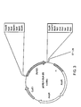
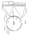


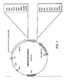
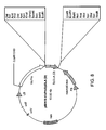


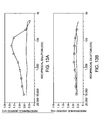

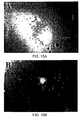

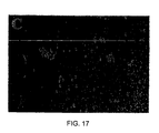
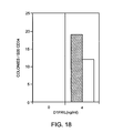


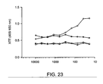
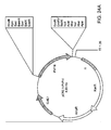
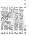
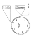


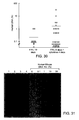
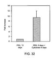
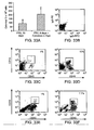

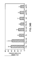
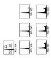
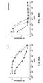


Classifications
-
- C—CHEMISTRY; METALLURGY
- C07—ORGANIC CHEMISTRY
- C07K—PEPTIDES
- C07K14/00—Peptides having more than 20 amino acids; Gastrins; Somatostatins; Melanotropins; Derivatives thereof
- C07K14/415—Peptides having more than 20 amino acids; Gastrins; Somatostatins; Melanotropins; Derivatives thereof from plants
- C07K14/42—Lectins, e.g. concanavalin, phytohaemagglutinin
-
- A—HUMAN NECESSITIES
- A61—MEDICAL OR VETERINARY SCIENCE; HYGIENE
- A61K—PREPARATIONS FOR MEDICAL, DENTAL OR TOILETRY PURPOSES
- A61K38/00—Medicinal preparations containing peptides
- A61K38/16—Peptides having more than 20 amino acids; Gastrins; Somatostatins; Melanotropins; Derivatives thereof
- A61K38/168—Peptides having more than 20 amino acids; Gastrins; Somatostatins; Melanotropins; Derivatives thereof from plants
-
- A—HUMAN NECESSITIES
- A61—MEDICAL OR VETERINARY SCIENCE; HYGIENE
- A61P—SPECIFIC THERAPEUTIC ACTIVITY OF CHEMICAL COMPOUNDS OR MEDICINAL PREPARATIONS
- A61P39/00—General protective or antinoxious agents
Definitions
- the invention relates to the preservation of progenitor cells. More specifically, the invention relates to the in vivo or ex vivo preservation of progenitor cells, such as hematopoietic progenitor cells.
- hematopoiesis involves the process of producing a balanced supply of different blood cells from such progenitor cells found in the adult bone marrow.
- the development of other cell types also depends upon production of the differentiated cells from such progenitor cells.
- Progenitor cells are activated by signals, such as cell-cell contact or soluble regulators, to generate daughter cells that are identical to the parent ( i . e ., self-renewal of the parent) and/or to generate daughter cells that are more differentiated than the parent, thus beginning an irreversible process that ends with the production of differentiated, functional cells.
- signals such as cell-cell contact or soluble regulators
- differentiation is coupled to proliferation as a progenitor cell gives rise to more differentiated daughter cells that progressively become committed to producing only one blood cell type.
- potent soluble regulators e . g ., colony stimulating factors and cytokines
- progenitor cells eventually produce so many of the mature cells of the body, they occur only rarely. Moreover, typically the more primitive (i.e., undifferentiated) the progenitor cell, the more rare the progenitor cell. For example, the currently believed most primitive of the hematopoietic progenitor cells, which are called hematopoietic stem cells, occur at a frequency of only from about 1 in 10,000 to about 1 in 100,000 of the cells in the bone marrow. Hematopoietic stem cells have the capacity to generate more than 10 13 mature blood cells of all lineages, including other progenitor cells which, although more differentiated than hematopoietic stem cells, are themselves capable of giving rise to several different types of mature blood cells.
- Hematopoietic stem cells are responsible for sustaining blood cell production over the life of an animal.
- the small population of hematopoietic stem cells is sufficient to produce all the mature blood cells in a healthy individuals; however, some unhealthy individuals suffer from a lack of a sufficient number of progenitor cells and/or mature blood cells.
- cancer patients receiving chemotherapeutic or radiotherapy treatments designed to kill the rapidly dividing cancer cells also suffer from the depletion of white blood cells and platelets, thus exposing these patients to life threatening opportunistic infections and bleeding episodes. Indeed, this hematopoietic progenitor cell-depleting activity is the dose-limiting factor for most of these chemotherapeutic and radiotherapeutic agents.
- cytokines including G-CSF, GM-CSF, SCF, Erythropoietin, and IL-11, to accelerate restoration of hematopoiesis following chemotherapy (Moore, M.A., Blood 78:1-19,1991).
- these cytokines lead to the irreversible differentiation of hematopoietic progenitor cells, including hematopoietic stem cells, into more differentiated daughter cells.
- better protection of hematopoietic progenitor cells is needed during chemotherapy.
- the invention provides a family of factors that preserve progenitor cells.
- Members of this family, the FRIL family are non-toxic, inexpensively produced reagents that preserve progenitor cells.
- the invention provides compositions comprising at least one member of the FRIL family, as well as methods for using members of the FRIL family to preserve progenitor cells both in vivo and ex vivo .
- the invention provides an essentially pure composition of one or more members of the FRIL family of progenitor cell preservation factors.
- the FRIL family member is from a legume.
- the legume is Dolichos lab lab .
- the legume is Phaseolus vulgaris .
- the legume is Sphenostylis stenocarpa.
- the FRIL family member is a mutant derived from a second member of the FRIL family, wherein the mutant is selected from the group consisting of a substitution mutant, a deletion mutant, an addition mutant, or a combination thereof.
- the FRIL family member is a fusion protein comprising a first portion and a second portion, wherein the first portion is derived from a second member of the FRIL family.
- the invention provides a recombinant nucleic acid molecule encoding a composition of a member of the FRIL family of progenitor cell preservation factors.
- the invention provides a pharmaceutical formulation comprising an essentially pure composition of one or more members of the FRIL family of progenitor cell preservation factors and a pharmaceutically acceptable carrier.
- administering comprises administering a therapeutically effective amount of the formulation to a patient suffering from a condition whereby the patient's hematopoietic progenitor cells are depleted alleviates and/or reduces the condition in the patient.
- administration of a therapeutically effective amount of the formulation to a patient prior to treatment of the patient with a therapeutic treatment having a hematopoietic progenitor cell-depleting activity alleviates and/or reduces the hematopoietic progenitor cell-depleting activity of the therapeutic treatment in the patient.
- the patient is a human or is a domesticated mammal. In some embodiments, the patient has cancer.
- the therapeutic treatment is a radiotherapeutic or a chemotherapeutic treatment, including, without limitation, cytarabine (Ara-C), doxorubicin (Dox), or 5-fluorouracil (5-FU), or a combination of a radiotherapeutic and a chemotherapeutic.
- a radiotherapeutic or a chemotherapeutic treatment including, without limitation, cytarabine (Ara-C), doxorubicin (Dox), or 5-fluorouracil (5-FU), or a combination of a radiotherapeutic and a chemotherapeutic.
- the invention provides a method for alleviating or reducing the hematopoietic progenitor cell-depleting activity of a therapeutic treatment in a patient, comprising administering to the animal a therapeutically effective amount of a composition of a FRIL family member prior to administration of the therapeutic treatment to the patient.
- the patient is a human or is a domesticated mammal.
- the patient has cancer.
- the therapeutic treatment is a radiotherapeutic or a chemotherapeutic treatment, including, without limitation, cytarabine (Ara-C), doxorubicin (Dox), or 5-fluorouracil (5-FU), or a combination of a radiotherapeutic and a chemotherapeutic.
- the invention provides a method for isolating a population of progenitor cells, comprising contacting a population of cells with a plurality of FRIL family member molecules, and separating the unbound cells, wherein the cells bound to the FRIL family member molecules are an isolated population of progenitor cells.
- the isolated population of progenitor cells is from a human.
- the FRIL family member molecules are detectably labeled.
- the detectably labeled FRIL family member molecules are labeled with a chromophore.
- the unbound cells are separated by using a flow cytometry cell sorter to sort the population of cells contacted with the FRIL family member molecules detectably labeled with a chromophore.
- the FRIL family member molecules is immobilized on a solid support.
- the solid support is a bead, such as a magnetic bead.
- the unbound cells are separated by applying a magnet to the population of cells contacted with the FRIL family member molecules immobilized on the magnetic bead.
- the population of cells bound to the FRIL family member molecules immobilized on a magnetic bead is rinsed with a physiologically acceptable solution while the magnet is applied.
- the solid support is the bottom of a tissue culture plate.
- the unbound cells are separated by rinsing the population of cells contacted with the FRIL family member immobilized on the bottom of a tissue culture plate with a physiologically acceptable solution.
- the isolated population of progenitor cells is a population of hematopoietic progenitor cell.
- the population of cells is whole blood, umbilical cord blood, bone marrow cells, or fetal liver cells.
- the population of cells is a sorted population of cells, wherein a cell of the sorted population does not express a cell surface molecule selected from the group consisting of CD11b, CD11c, and CD38.
- the sorted population of cells is sorted by flow cytometry or by magnetic bead selection.
- invention provides an isolated population of progenitor cells isolated by a method comprising contacting a population of cells with a plurality of FRIL family member molecules, and separating the unbound cells, wherein the cells bound to the FRIL family member molecules are an isolated population of progenitor cell.
- the progenitor cell is from a human.
- the cells of the isolated population do not express CD34.
- the cells of the isolated population express a receptor tyrosine kinase selected from the group consisting of from FLK1, FLT1, FLT3, FLT4, and Kit.
- the cells of the isolated population express a cell surface molecule selected from the group consisting of CD11b and CD11c.
- the cells of the isolated population express FLT3.
- the cells of the isolated population are hemangioblasts, messengerchymal stem cells, bone progenitor cells, hepatic progenitor cells, endothelial progenitor cells, hematopoietic progenitor cells, embryonal stem cells, brain progenitor cells, or dendritic progenitor cells.
- the cells of the isolated population are hematopoietic progenitor cells.
- the cells of the isolated population are hematopoietic progenitor cells
- transplantation of the isolated population into an animal lacking a population of hematopoietic progenitor cells sufficient to enable survival of the animal reconstitutes the animal, wherein the transplanted animal survives.
- the hematopoietic progenitor cells are from a human or a mouse and wherein the animal is a mouse.
- the mouse is a SCID mouse or the mouse is sublethally irradiated or the mouse is treated with a sublethal dose of a chemotherapeutic.
- the hematopoietic progenitor cells are from a human and the animal is a human.
- the human is a cancer patient receiving a treatment that depletes the patient's hematopoietic progenitor cells.
- the treatment is a radiotherapeutic or a chemotherapeutic treatment, including, without limitation, cytarabine (Ara-C), doxorubicin (Dox), or 5-fluorouracil (5-FU), or a combination of a radiotherapeutic and a chemotherapeutic.
- the invention provides a method for preserving progenitor cells ex vivo comprising contacting a population of cells comprising at least one progenitor cell with an effective amount of a composition of a FRIL family member for an effective period of time, wherein the progenitor cells in the population are rendered quiescent.
- the progenitor cells are from a human.
- the population of cells is bone marrow cells.
- the non-progenitor cells in the population of cells differentiate or die.
- the population of cells is removed from a cancer patient prior to treatment of the cancer patient with a therapeutic treatment having a hematopoietic progenitor cell-depleting activity.
- the therapeutic treatment is a radiotherapeutic or a chemotherapeutic treatment, including, without limitation, cytarabine (Ara-C), doxorubicin (Dox), or 5-fluorouracil (5-FU), or a combination of a radiotherapeutic and a chemotherapeutic.
- the invention provides a method for preserving progenitor cells in vivo, comprising administering to a patient an effective amount of a composition of a FRIL family member for an effective period of time, wherein the progenitor cells in the patient are rendered quiescent.
- the patient is a human or a domesticated animal.
- the patient is a cancer patient.
- the effective amount of the composition of a FRIL family member is administered prior to the treatment of the patient with a therapeutic treatment having a hematopoietic progenitor cell-depleting activity.
- the therapeutic treatment is a radiotherapeutic or a chemotherapeutic treatment, including, without limitation, cytarabine (Ara-C), doxorubicin (Dox), or 5-fluorouracil (5-FU), or a combination of a radiotherapeutic and a chemotherapeutic.
- the invention provides a method for identifying a progenitor cell, comprising contacting a candidate cell with a FRIL family member molecule, wherein binding of the candidate cell to the FRIL family member molecule identifies the candidate cell as a progenitor cell.
- the candidate cell is in a population of cells. In certain embodiments, the candidate cell is from a human.
- the invention provides a progenitor cell identified by a method comprising contacting a candidate cell with a FRIL family member molecule, wherein binding of the candidate cell to the FRIL family member identifies the candidate cell as a progenitor cell.
- the invention provides a method for identifying a composition of a member of the FRIL family of progenitor cell preservation factors, comprising contacting a candidate compound with a glycosylated extracellular domain of an FLT3 receptor, wherein the glycosylation pattern of the extracellular domain of the FLT3 receptor is the same as the glycosylation pattern of an extracellular domain of a normally glycosylated FLT3 receptor, wherein a candidate compound that binds the glycosylated extracellular domain of the FLT3 receptor is identified as a composition of a FRIL family member.
- the candidate compound is a lectin.
- the lectin is synthetic.
- the lectin is from a legume.
- the invention provides an essentially pure composition of a FRIL family member identified by the method comprising contacting a candidate compound with a glycosylated extracellular domain of an FLT3 receptor, wherein the glycosylation pattern of the extracellular domain of the FLT3 receptor is the same as the glycosylation pattern of an extracellular domain of a normally glycosylated FLT3 receptor, wherein a candidate compound that binds the glycosylated extracellular domain of the FLT3 receptor is identified as a member of the FRIL family.
- compositions of a FRIL family member may be used as therapeutic agents to preserve progenitor cells in patients, such as cancer patients receiving chemotherapy, who have suffer from a condition that diminishes their progenitor cells.
- compositions of a FRIL family member may be administered with a pharmaceutically-acceptable carrier (e . g ., physiological sterile saline solution) via any route of administration to a cancer patient receiving chemotherapy in an attempt to reduce the progenitor cell-depleting effects of the chemotherapeutic so that the patient can receive a higher dose of the chemotherapeutic and, preferably, recover from cancer.
- a pharmaceutically-acceptable carrier e . g ., physiological sterile saline solution
- Pharmaceutically-acceptable carriers and their formulations are well-known and generally described in, for example, Remington's Pharmaceutical Sciences (18th Edition, ed. A. Gennaro, Mack Publishing Co., Easton, PA, 1990).
- the invention relates the preservation of progenitor cells by members of the FRIL family of progenitor cell preservation factors. More specifically, the invention relates to the in vivo or ex vivo preservation of progenitor cells, such as hematopoietic progenitor cells, using members of the FRIL family of progenitor cell preservation factors.
- the invention provides a family of factors that preserve progenitor cells.
- Members of this family, the FRIL family are non-toxic, inexpensively produced reagents that preserve progenitor cells.
- the invention provides compositions comprising at least one member of the FRIL family, as well as methods for using members of the FRIL family to preserve progenitor cells both in vivo and ex vivo .
- the invention provides an essentially pure composition of a member of the FRIL family of progenitor cell preservation factors.
- FRIL family of progenitor cell preservation factors is used to mean a family of lectins, wherein each FRIL family member molecule binds to a normally glycosylated FLT3 receptor, wherein each FRIL family member molecule preserves progenitor cells, and wherein one FRIL family member molecule that is isolated from a hyacinth bean (i.e., Dolichos lab lab) has an amino acid sequence which comprises the following eight amino acid sequence: TNNVLQXT (SEQ ID NO: 24).
- a composition of a FRIL family member which includes a mutant of another FRIL family member molecule or a fusion protein comprising a portion derived from a FRIL family member molecule or mutant thereof, wherein each FRIL family member molecule binds to a normally glycosylated FLT3 receptor has at least about 45% amino acid sequence identity with the amino acid sequence of another member of the FRIL family, preferably at least about 50% identity, even more preferably at least about 55% identity, still more preferably at least about 60% identity, and still more preferably at least about 65% identity with the sequence of the second protein.
- the amino acid sequence of the first protein shares at least about 75% sequence identity, preferably at least about 85% identity, and more preferably at least about 95% identity, with the amino acid sequence of another member of the FRIL family.
- Both amino acid sequence identity and nucleic acid sequence identity between two proteins or two nucleic acid molecules can be measured according to standard methods. For example, in order to compare a first amino acid sequence to a second amino acid sequence or a first nucleic acid sequence to a second nucleic acid sequence for the purpose of determining percentage identity between the two sequences, the sequences are aligned so as to maximize the number of identical amino acid or nucleic acid residues.
- the sequences of proteins sharing at least 50% amino acid sequence identity or the sequences of nucleic acids sharing at least 45% nucleic acid sequence identity can usually be aligned by visual inspection. If visual inspection is insufficient, the proteins or nucleic acids may be aligned in accordance with the FASTA method in accordance with Pearson and Lipman (Proc.
- sequence analysis software Another method for determining amino acid or nucleic acid sequence identity between two proteins or nucleic acids is by using sequence analysis software with the default parameters specified therein.
- sequence analysis software with the default parameters specified therein.
- Various software packages exist including Sequence Analysis Software Package of the Genetics Computer Group (University of Wisconsin Biotechnology Center, Madison, WI), and the various BLAST programs of the National Center for Biotechnology (National Library of Medicine, Bethesda, MD).
- percentage of amino acid sequence identity or percentage of nucleic acid sequence identity is determined using the basic BLAST program of the National Center for Biotechnology (National Library of Medicine, Bethesda, MD), using the default settings defined therein.
- Another test for percentage identity of two nucleic acid sequences is whether they hybridize under normal hybridization conditions, preferably under stringent hybridization conditions.
- proteins that are encoded by nucleic acid molecules that hybridize under high stringent conditions to a sequence complementary to SEQ ID NO: 1, SEQ ID NO: 3, SEQ ID NO: 5, and/or SEQ ID NO: 7.
- stringent conditions is equivalent to "high stringent conditions” and "high stringency.” These terms are used interchangeably in the art.
- Stringent conditions are defined in a number of ways. In one definition, stringent conditions are selected to be about 50°C lower than the thermal melting point (T m ) for a specific sequence at a defined ionic strength and pH. The T m is the temperature (under defined ionic strength and pH) at which 50% of the target sequence hybridizes to a perfectly matched sequence. Typical stringent conditions are those in which the salt concentration is at least about 0.02 M at pH 7 and the temperature is at least about 60°C. "Stringent conditions,” in referring to percentage identity ( e.g. , homology) or substantial similarity in the hybridization context, can be combined conditions of salt, temperature, organic solvents or other parameters that are typically known to control hybridization reactions.
- stringent conditions can be provided in a variety of ways such as overnight incubation at 42°C in a solution comprising: 20% formamide, 5 x SSC (150 mM NaCl, 15 mM trisodium citrate), 50 mM sodium phosphate (pH 7.6), 5 x Denhardt's solution, 10% dextran sulfate, and 20 ⁇ g/ml denatured, sheared salmon sperm DNA.
- the stringent conditions are characterized by a hybridization buffer comprising 30% formamide in 5 x SSPE (0.18 M NaCl, 0.01 M NaPO 4 , pH 7.7, 0.0001 M EDTA) buffer at a temperature of 42°C, and subsequent washing at 42°C with 0.2 x SSPE.
- stringent conditions involve the use of a hybridization buffer comprising 50% formamide in 5 x SSPE at a temperature of 42°C and washing at the same temperature with 0.2 x SSPE.
- progenitor cells an ability of a FRIL family member molecule (or mutant thereof or fusion protein comprising a FRIL family member molecule or mutant thereof) to retain ( i . e ., preserve) progenitor cells in an undifferentiated state, which can be determined using the assays described below (e.g., the SCID mouse reconstituting cell assay and the methylcellulose or other semi-solid medium based hematopoietic progenitor cell assay).
- progenitor cell refers to any normal somatic cell that has the capacity to generate fully differentiated, functional progeny by differentiation and proliferation.
- Progenitor cells include progenitors from any tissue or organ system, including, but not limited to, blood, mesenchymal, embryonic, nerve, muscle, skin, gut, bone, kidney, liver, pancreas, thymus, brain and the like.
- Progenitor cells are distinguished from "differentiated cells," the latter being defined as those cells that may or may not have the capacity to proliferate, i.e. , self-replicate, but that are unable to undergo further differentiation to a different cell type under normal physiological conditions.
- progenitor cells are further distinguished from abnormal cells such as neoplastic cells, as defined herein. For example, leukemia cells proliferate (self-replicate), but generally do not further differentiate, despite appearing to be immature or undifferentiated.
- Progenitor cells include all the cells in a lineage of differentiation and proliferation prior to the most differentiated or the fully mature cell.
- progenitors include the skin progenitor in the mature individual.
- the skin progenitor is capable of differentiation to only one type of cell, but is itself not fully mature or fully differentiated.
- hematopoiesis is meant the development of mature, functional blood cells.
- the progenitor cells that give rise to mature, functional blood cells are called hematopoietic progenitor cells.
- the most primitive, undifferentiated hematopoietic progenitor cell is called a hematopoietic stem cell.
- Hematopoietic stem cells typically reside in the bone marrow primarily in a quiescent state, and may form identical daughter cells through a process called "self-renewal.”
- hematopoietic progenitor For red blood cell (erythrocyte) production, a unipotential progenitor called a "CFU-E" (colony forming unit-erythroid) has the capacity to generate two to 32 mature progeny cells.
- CFU-E colony forming unit-erythroid
- hematopoietic progenitor cells include those cells that are capable of successive cycles of differentiating and proliferating to yield up to eight different mature hematopoietic cell lineages.
- Uncommitted progenitor cells such as hematopoietic stem cells
- Progenitor cells that retain a capacity to generate all cell lineages, but that can not self-renew, are termed “pluripotent.”
- Plotent Progenitor cells that can produce some but not all blood lineages and can not self-renew.
- Progenitor cells can be defined by mRNA levels of genes that either specifically regulate progenitors or serve as markers of lineage commitment.
- genes induced in primitive human hematopoietic progenitor cells include those encoding the shared beta subunits of the IL3, IL5, and/or granulocyte-macrophage colony-stimulating factor (GM-CSF) receptors, termed the beta common chain (McClanahan et al., Blood 81:3903-2915,1993); CD34 genes; and/or the receptors for Kit (Turner et al., Blood 88:3383-3390, 1996), FLT1, FLT4 (Galland et al., Oncogene 8:3233-1240, 1993), FLK1 (Broxmeyer et al., Int.
- Those genes for intermediate progenitors include the c-Fms, G-CSF receptor, and/or CD34 genes; and the IL-7 receptor gene, a gene induced for B lymphopoiesis.
- Murine primitive progenitor populations include receptors for interleukin-1 alpha (IL-1 ⁇ ), IL-3, IL-6, granulocyte colony-stimulating factor (G- CSF), and/or FLK1-1 (the murine homologue of human KDR which binds VEGF) (Broxmeyer, supra ), but lack receptors for macrophage colony-stimulating factor (M-CSF), granulocyte-macrophage colony stimulating factor (GM-CSF), and leukemia inhibitory factor (LIF).
- Cells within the intermediate progenitor cell population include receptors for GM-CSF, G-CSF, IL-6, and/or IL-1 ⁇ .
- bind binds to a normally glycosylated FLT3 receptor with an affinity higher than the affinity with which the FLT3-Ligand binds the FLT3 receptor.
- a FRIL family member molecule binds to a normally glycosylated FLT3 receptor with an affinity that is at least as high as the affinity with which an antibody binds its specific ligand.
- a FRIL family member molecule of the invention binds to a normally glycosylated FLT3 receptor with an affinity that is higher than the affinity with which an antibody binds its specific ligand. Still more preferably, a FRIL family member molecule of the invention binds to a normally glycosylated FLT3 receptor with a dissociation constant (K D ) of at least 10 -7 M, more preferably 10 -8 M, even more preferably 10 -9 M, still more preferably, at least 10 -10 M, and most preferably, a FRIL family member molecule of the invention binds to a normally glycosylated FLT3 receptor with a dissociation constant (K D ) of at least 10 -11 M. Standard methods for determining binding and binding affinity are known.
- normally glycosylated FLT3 receptor an FLT3 receptor that has a glycosylation pattern of an FLT3 cell glycosylated by a normal cell.
- normal cell as used herein in accordance with all aspects of the present invention, is meant a cell that is not neoplastic.
- neoplastic cell is meant a cell that shows aberrant proliferation, particularly increased proliferation, that is not regulated by such factors as cell-cell contact inhibition and soluble regulators (e.g.
- cytokines or hormones cytokines or hormones
- abnormally glycosylates the FLT3 receptor such that the glycosylation pattern on the FLT3 receptor on the neoplastic cells is abnormal and such that the FLT3 receptor on the neoplastic cell is not bound by a FRIL family member molecule.
- essentially pure means a molecule, such as a nucleic acid or protein (e . g ., a FRIL family member molecule), or composition of a molecule that is more free from other organic molecules (e.g. , carbohydrates, nucleic acids, proteins, and lipids) that naturally occur with an impure molecule, and is substantially free as well of materials used during the purification process.
- a protein or nucleic acid molecule is considered to be essentially pure if it is at least approximately 60%, preferably at least approximately 75%, more preferably approximately at least 85%, most preferably approximately at least 90%, and optimally approximately at least 95% pure, i .
- a FRIL family member can also be purified by binding to a mannose, which may be coupled on a sold support (e.g. , a sepharose bead).
- nucleic acids Methods for purifying nucleic acids are known in the art and include, without limitation, Guanidine-HCl extraction, polymerase chain reaction, CsCl gradient fractionation, phenol: chloroform extraction, ethanol precipitation, and standard recombinant DNA methodologies. Standard methods for purifying both proteins and nudeic acid molecules are provided in, e . g ., Ausubel et al., Current Protocols in Molecular Biology, John Wiley & Sons, New York, NY,1994; Sambrook et al., supra.
- a FRIL family member molecule may be purified from a natural source by methods well known in the art.
- the purification of D1-FRIL from Dolichos lab lab is described below in Example 1.
- the purification of YamFRIL from Sphenostylis stenocarpa is described below in Example 22..
- Such methods also include, for example, those described by Moore in PCT application PCT/US97/22486 and by Gowda et al., supra.
- a suitable natural source from which to purify a FRIL family member molecule includes plants, especially legume plants.
- Legumes such as the garden pea or the common bean
- Plants are plants (“leguminous plants”) from a family ( Leguminosae ) of dicotyledonous herbs, shrubs, and trees bearing (nitrogen-fixing bacteria) nodules on their roots. These plants are commonly associated with their seeds (e.g., the garden pea or the common bean)
- a FRIL family member molecule according to the first aspect of the invention can be purified from members of the tribe Phaseoleae.
- a FRIL family member molecule can be purified from Dolichos lab lab (e.g., hyacinth beans, which is also known by other common names throughout the world).
- a FRIL family member molecule can be purified from varieties of the common bean (Phaseolus vulgaris) (e.g., red kidney beans and white kidney beans), from yam bean (Sphenostylis stenocarpa ) or from Vigna sinensis, commonly known as the black-eyed pea.
- FRIL family member molecule purification of a FRIL family member molecule from a legume is rapid and inexpensive, and results in a large amount of essentially pure lectin.
- a native FRIL family member molecule can be easily purified from legumes, such as hyacinth beans (pesticide-free), by mannose-affinity chromatography or ovalbumin affinity chromatography, and is more than 100 times cheaper to produce than recombinant cytokines.
- lectin is meant a protein that binds sugar residues with high affinity.
- a FRIL family member molecule is a mannose/glucose-specific legume lectin.
- FRIL family member molecules and compositions of FRIL family member molecules, have many attributes as reagents to either alleviate the progenitor cell-depleting activity of a therapeutic (e.g., a chemotherapeutic) or to alleviate the symptoms of a condition where the patient's progenitor cells are depleted.
- a therapeutic e.g., a chemotherapeutic
- FRIL family members have unique properties and are the first soluble regulators reported to preserve hematopoietic stem cells and progenitors in a dormant state for extended periods, even in the presence of potent stimulators of proliferation and differentiation.
- mice tolerate very high levels of compositions of FRIL family members, this may permit more effective protection of stem cells and progenitors by preventing their recruitment during aggressive dose intensification regimens aimed at increasing frequency and dosage levels of chemotherapy. While the biological activity of Dl-FRIL is similar to cytokines (ng/ml range), as demonstrated in the examples below, mice tolerated up to a 1,000-fold more Dl-FRIL than cytokines.
- one or more members of the FRIL family allows for the administration of a broad range of cell cycle active chemotherapy drugs with greater frequency and higher dose.
- administration of a composition of one or more FRIL family members may permit more aggressive dose-intensification chemotherapy regimens for a broad range of chemotherapy drugs.
- Administration of a composition of a FRIL family member also provides for a larger reservoir of progenitor cells which could rapidly respond to stimulatory signals after completing chemotherapy.
- the FRIL family member of the invention is from a legume (e.g., a bean plant).
- the FRIL family member molecule of the invention is from a hyacinth bean (i.e., Dolichos lab lab ), and has an amino acid sequence comprising the sequence of SEQ ID NO: 24.
- the FRIL family member molecule of the invention isolated from a hyacinth bean has the amino acid sequence of SEQ ID NO: 2 or comprises a signal sequence having the amino acid sequence of SEQ ID NO: 4, and, even more preferably, is encoded by a nucleic acid having the nucleic acid sequence of SEQ ID NO: 1 or the nucleic acid sequence of SEQ ID NO: 3.
- the FRIL family member molecule of the invention is from a red kidney bean (i.e., Phaseolus vulgaris ).
- the FRIL family member of the invention isolated from a red kidney bean has the amino acid sequence of SEQ ID NO: 6, and, even more preferably, is encoded by a nucleic acid having the nucleic acid sequence of SEQ ID NO: 5.
- the FRIL family member of the invention is from a yam bean (i.e., Sphenostylis stenocarpa ).
- the FRIL family member of the invention isolated from a yam bean has the amino acid sequence comprising the amino acid sequence of SEQ ID NO: 8, more preferably has a ⁇ subunit having an amino acid sequence which comprises the amino acid sequence of SEQ ID NO: 9, even more preferably has an ⁇ subunit having an amino acid sequence which comprises the amino acid sequence of SEQ ID NO: 10, and, even more preferably, is encoded by a nucleic acid having a nucleic acid sequence which comprises the nudeic acid sequence of SEQ ID NO: 7.
- the FRIL family member molecule is a mutant derived from a second member of the FRIL family, wherein the mutant is selected from the group consisting of a substitution mutant, a deletion mutant, an addition mutant, or a combination thereof (e.g., a mutant of Dl-FRIL or Pv-FRIL described below).
- the mutant is selected from the group consisting of a substitution mutant, a deletion mutant, an addition mutant, or a combination thereof (e.g., a mutant of Dl-FRIL or Pv-FRIL described below).
- Groups of amino acids known normally to be equivalent are: (1) Ala(A), Ser(S), Thr(T), Pro(P), and Gly(G); (2) Asn(N), Asp(D), Glu(E), Gln(Q); (3) His(H), Arg(R), Lys(K); (4) Met(M), Leu(L), Ile(I), Val(V); and (5) Phe(F), Tyr(Y), Trp(W). Substitutions, additions, and/or deletions in an amino acid sequence can be made as long as the mutant FRIL family member molecule continues to satisfy the functional criteria described herein.
- amino acid sequence that is substantially the same as another sequence, but that differs from the other sequence by means of one or more substitutions, additions, and/or deletions, is considered to be an equivalent sequence.
- less than 50%, more preferably less than 25%, and still more preferably less than 10%, of the number of amino acid residues in a sequence are substituted for, added to, or deleted from the FRIL family member molecule upon which the mutant FRIL family member was derived.
- the FRIL family member molecule is a fusion protein comprising a first portion and a second portion, wherein the first portion is derived from a second member of the FRIL family.
- fusion protein is meant a molecule comprising at least two proteins or polypeptide fragments thereof joined together, wherein the proteins or polypeptide fragments thereof are not joined together in the naturally-occurring organism from which the proteins or polypeptide fragments thereof were derived.
- the two proteins or polypeptide fragments thereof of a fusion protein may be joined by any means, including, without limitation, a chemical linker, a peptide bond, or a noncovalent bond, such as an ionic bond.
- protein or “polypeptide” is meant a chain of two or more amino acid residues joined with a peptide bond regardless of length or post-translational modification such as acetylation, glycosylation, lipidation, acetylation, or phosphorylation.
- a FRIL family member molecule that is a fusion protein may comprise a first portion derived from a FRIL family member and a second portion derived from a protein or other molecule not related to the FRIL family (e.g., the heavy chain of an antibody).
- An additional FRIL family member molecule that is a fusion protein is FRIL family member comprising the a subunit from a first FRIL family member and a ⁇ subunit from a second FRIL family member.
- a fusion protein may be generated, for example, by joining a nucleic acid sequence encoding the a subunit of the first FRIL family member in frame with a nucleic acid sequence encoding the ⁇ subunit of the second FRIL family member.
- the nucleic acid encoding such a fusion protein can be engineered to encode an enzyme-specific deavage site between the portion encoding the ⁇ subunit of the first FRIL family member and the portion encoding the ⁇ subunit of the second FRIL family member.
- identity of the fusion protein as a FRIL family member is determined by the sequence identity between the FRIL family member-derived portion of the fusion protein and a second FRIL family member, where the FRIL family member-derived portion of the fusion protein and the second FRIL family member share at least about 45% amino acid sequence identity, even more preferably at least about 50% identity, even more preferably at least about 55% identity, still more preferably at least about 60% identity, still more preferably at least about 65% identity yet more preferably at least about 75% sequence identity, still more preferably at least about 85% identity, and most preferably at least about 95% identity, with the amino acid sequence of a second member of the FRIL family.
- a FRIL family member in accordance with the first aspect of the invention can also be a recombinant protein made by expressing a recombinant nucleic acid that encodes FRIL in a suitable host.
- the invention features an essentially pure nucleic acid molecule encoding a member of the FRIL family of progenitor cell preservation factors. Exemplary purifications of the nucleic acid molecules of the invention from Dolichos lab lab and Phaseolus vulgaris are described below.
- nucleic acid in accordance to this aspect of the invention need not have a naturally occurring sequence, but need only encode a FRIL family member according to the first aspect of the invention.
- a "member of the FRIL family of progenitor cell preservation factors” is a described above the first aspect of the invention. "Essentially pure” is used as described for the first aspect of the invention.
- a “recombinant nucleic acid” is meant a nucleic acid which encodes a FRIL family member molecule, or a portion encoding at least 15 contiguous amino acids thereof, or a mutant thereof, or a fusion protein comprising the molecule, portion thereof or mutant thereof or is capable of expressing an antisense molecule specifically complementary thereto, or a sense molecule that shares nucleic acid sequence identity thereto wherein the recombinant nucleic acid may be in the form of linear DNA or RNA, covalently closed circular DNA or RNA, or as part of a chromosome, provided however that it cannot be the native chromosomal locus for a FRIL family member molecule.
- Preferred recombinant nucleic acids of the invention are vectors, which may include an origin of replication and are thus replicatable in one or more cell type. Certain preferred recombinant nucleic acids are expression vectors, and further comprise at least a promoter and passive terminator, thereby allowing transcription of the recombinant nucleic acid in a bacterial, fungal, plant, insect or mammalian cell.
- nucleic acid or “nucleic acid molecule” as used herein, means any deoxyribonudeic acid (DNA) or ribonucleic acid (RNA), including, without limitation, complementary DNA (cDNA), genomic DNA, RNA, hnRNA, messenger RNA (mRNA), DNA/RNA hybrids, or synthetic nucleic acids (e.g., an oligonucleotide) comprising ribonucleic and/or deoxyribonucleic acids or synthetic variants thereof.
- the nucleic acid of the invention includes, without limitation, an oligonucleotide or a polynucleotide.
- the nucleic acid can be single stranded, or partially or completely double stranded (duplex).
- Duplex nucleic acids can be homoduplex or heteroduplex.
- a nucleic acid encoding a FRIL family member has at least about 50% nucleic acid sequence identity with a nucleic acid encoding another member of the FRIL family, preferably at least about 555% nucleic acid sequence identity, more preferably at least about 60% nucleic acid sequence identity, more preferably at least about 65% nucleic acid sequence identity, still more preferably at least about 75% nucleic acid sequence identity, still more preferably at least about 85% nucleic acid sequence identity, and most preferably at least about 95% nucleic acid sequence identity with a nucleic acid encoding another member of the FRIL family. Percentage nucleic acid sequence identity can be determined as described for the first aspect of the invention.
- a recombinant nucleic acid according to the second aspect of the invention can also be chemically synthesized by methods known in the art.
- recombinant DNA can be synthesized chemically from the four nucleotides in whole or in part by methods known in the art. Such methods include those described in Caruthers, M.H., Science 230(4723):281-285,1985).
- DNA can also be synthesized by preparing overlapping double-stranded oligonucleotides, filling in the gaps, and ligating the ends together. See, generally, Sambrook et al., supra, and Glover and Hames, eds., DNA Cloning, 2d ed., Vols. 1-4, IRL Press, Oxford, 1995.
- a recombinant nucleic acid molecule of the invention encoding a mutant FRIL family member can be prepared from wild-type DNA by site-directed mutagenesis (see, for example, Zoller and Smith, Nucleic. Acids. Res. 10:6487-6500, 1982; Zoller, M.J., Methods Enzymol . 100:468-500,1983; Zoller, M.J., DNA 3(6):479-488,1984.; and McPherson, M.J., ed., Directed Mutagenesis: A Practical Approach , IRL Press, Oxford, 1991.
- a recombinant nucleic acid of the second aspect of the invention can be amplified by methods known in the art.
- PCR polymerase chain reaction
- vectors e.g., recombinant expression vectors
- vectors which permit the incorporation of recombinant nucleic acids of interest for cloning and transformation of other cells.
- the invention further includes vectors (e.g., plasmids, phages, and cosmids) which incorporate a nucleotide sequence of the invention, especially vectors which include the recombinant nucleic acid molecule of the invention for expression of a FRIL family member.
- a recombinant nucleic acid of the invention can be replicated and used to express a FRIL family member following insertion into a wide variety of host cells in a wide variety of cloning and expression vectors.
- the host can be prokaryotic or eukaryotic.
- the nucleic acid can be obtained from natural sources and, optionally, modified.
- the genes can also be synthesized in whole or in part.
- Cloning vectors can comprise segments of chromosomal, non-chromosomal and synthetic DNA sequences.
- Some suitable prokaryotic cloning vectors include plasmids from E. coli , such as colE1, pCR1, pBR322, pMB9, pUC, pKSM, and RP4.
- Prokaryotic vectors also include derivatives of phage DNA such as M13fd, and other filamentous single-stranded DNA phages.
- Vectors for expressing proteins in bacteria are also known.
- Such vectors include the pK233 (or any of the tac family of plasmids), T7, and lambda P L .
- Examples of vectors that express fusion proteins are PATH vectors described in Dieckmann and Tzagoloff ( J. Biol. Chem. 260(3):1513-1520,1985). These vectors contain DNA sequences that encode anthranilate synthetase (TrpE) followed by a polylinker at the carboxy terminus.
- TrpE anthranilate synthetase
- pEX beta-galactosidase
- pMAL maltose binding protein
- pGST glutathione S-transferase
- Vectors useful for cloning and expression in yeast are also available.
- a suitable example is the 2 ⁇ circle plasmid.
- Suitable cloning/expression vectors for use in mammalian cells are also known.
- Such vectors include well-known derivatives of SV-40, adenovirus, cytomegalovirus (CMV) retrovirus-derived DNA sequences.
- CMV cytomegalovirus
- the expression vectors useful in the present invention contain at least one expression control sequence that is operatively linked to the recombinant nucleic acid molecule or fragment thereof to be expressed.
- the control sequence is inserted in the vector in order to control and to regulate the expression of the recombinant nucleic acid of the invention.
- Examples of useful expression control sequences are the lac system, the trp system, the tac system, the trc system, major operator and promoter regions of phage lambda, the control region of fd coat protein, the glycolytic promoters of yeast, e.g., the promoter for 3-phosphoglycerate kinase, the promoters of yeast acid phosphatase, e.g., Pho5, the promoters of the yeast alpha-mating factors, and promoters derived from polyoma, adenovirus, retrovirus, and simian virus, e.g. , the early and late promoters or SV40, and other sequences known to control the expression of genes of prokaryotic or eukaryotic cells and their viruses or combinations thereof.
- Useful expression hosts for expressing the recombinant nucleic acids of the invention include well-known prokaryotic and eukaryotic cells.
- suitable prokaryotic hosts include, for example, E. coli, such as E. coli SG-936, E. coli HB101, E. coli W3110, E. coli X1776, E. coli X2282, E. coli DHI, and E. coli MRC1, Pseudomonas, Bacillus, such as B. subtilis, and Streptomyces.
- Suitable eukaryotic cells include yeasts and other fungi, insect, animal cells, such as COS cells and CHO cells, human cells and plant cells in tissue culture.
- oligonucleotides or polynucleotides for use as primers in nucleic acid amplification procedures, such as the polymerase chain reaction (PCR), ligase chain reaction (LCR), Repair Chain Reaction (RCR), PCR oligonucleotide ligation assay (PCR-OLA), and the like. Oligonucleotides useful as probes in hybridization studies, such as in situ hybridization, can be constructed.
- probes of the invention can be adapted for easy detectability.
- Oligonucleotides can also be designed and manufactured for other purposes.
- the invention enables the artisan to design antisense oligonucleotides, and triplex-forming oligonucleotides, and the like, for use in the study of structure/function relationships.
- Homologous recombination can be implemented by adaptation of the nucleic acid of the invention for use as targeting means.
- Recombinant nucleic acids of the invention produced as described above can further be modified to alter biophysical or biological properties by means of techniques known in the art.
- the recombinant nucleic acid can be modified to increase its stability against nucleases (e.g., "end-capping"), or to modify its lipophilicity, solubility, or binding affinity to complementary sequences. Methods for modifying nucleic acids to achieve specific purposes are disclosed in the art, for example, in Sambrook et al., supra.
- the recombinant nucleic acid of the invention can include one or more portions of nucleotide sequence that are non-coding for a FRIL family member.
- the invention provides a pharmaceutical formulation comprising an essentially pure composition of one or more members of the FRIL family of progenitor cell preservation factors and a pharmaceutically acceptable carrier.
- pharmaceutically acceptable carrier is meant any inert carrier that is non-toxic to the animal to which it is administered and that retains the therapeutic properties of the compound with which it is administered (i.e. , the FRIL family member).
- Pharmaceutically acceptable carriers and their formulations are well-known and generally described in, for example, Remington's Pharmaceutical Sciences (18th Edition, ed. A. Gennaro, Mack Publishing Co., Easton, PA, 1990).
- One exemplary pharmaceutically acceptable carrier is physiological saline.
- Pharmaceutical formulations of the invention may employ any pharmaceutically acceptable carrier, depending upon the route of administration of the composition.
- compositions of FRIL family members may be used safely and efficaciously as a therapeutics.
- the gastrointestinal tracts of animals come in constant contact with lectins, such as FRIL family members, in raw and/or cooked vegetables and fruits. Many lectins pass through the gastrointestinal tract biologically intact (Pusztai, A., Eur. J. Clin. Nutr. 47: 691-699, 1993). Some lectins interact with the gut and are transported into the peripheral blood circulation.
- PNA peanut agglutinin
- Antibodies to dietary lectins are commonly found in people at levels of ⁇ 1 ⁇ g/ml (Tchernychev and Wilchek, FEBS Lett. 397:139-142,1996). These circulating antibodies do not block carbohydrate binding of the lectins.
- administering comprises administering a therapeutically effective amount of the pharmaceutical formulation to a patient suffering from a condition whereby the patient's hematopoietic progenitor cells are depleted alleviates and/or reduces the condition in the patient.
- a therapeutically effective amount is meant a dosage of a composition of a FRIL family member or pharmaceutical formulation comprising a composition of a FRIL family member that is effective to alleviate and/or reduce either a condition whereby the patient's hematopoietic progenitor cells are depleted or a hematopoietic progenitor cell-depleting activity of a therapeutic (e.g., a chemotherapeutic).
- a therapeutically effective amount is an amount of between about 500 ng of the FRIL family member/kg total body weight and about 5 mg/kg total body weight per day.
- a therapeutically effective amount is between about 500 ng/kg and 500 ⁇ g/kg total body weight of the FRIL family member per day. Still more preferably, a therapeutically effective amount is between about 5 ⁇ g/kg and 50 ⁇ g/kg total body weight of the FRIL family member per day. Most preferably, a therapeutically effective amount is an amount that delivers about 50 ⁇ g/kg total body weight of the FRIL family member per day.
- a composition of a FRIL family member of the invention and pharmaceutical formulation comprising a composition of a FRIL family member of the invention may be administered patients having, or predisposed to developing, a condition whereby the patient's hematopoietic progenitor cells are depleted.
- a condition may be congenital.
- the patient may have severe combined immunodeficiency or aplastic anemia.
- the condition may also be induced by a drug.
- administration of a therapeutically effective amount of the pharmaceutical formulation to a patient prior to treatment of the patient with a therapeutic treatment having a hematopoietic progenitor cell-depleting activity alleviates the hematopoietic progenitor cell-depleting activity of the therapeutic in the patient.
- cancer patients are often treated with radiotherapeutics or chemotherapeutics that have hematopoietic progenitor cell-depleting activity.
- hematopoietic progenitor cell-depleting activity an activity of a therapeutic treatment whereby the hematopoietic progenitor cells in the patient being treated with the therapeutic treatment are depleted, either by killing the progenitor cells or by inducing the progenitor cells to undergo irreversible differentiation.
- therapeutic treatments having hematopoietic progenitor cell-depleting activity are the chemotherapeutic agents cytarabine (Ara-C), doxorubicin (Dox), daunorubicin, and 5-fluorouracil (5-FU).
- administration of the pharmaceutical formulation of the invention to a patient prior to the treatment of the patient with a therapeutic treatment having a hematopoietic progenitor cell-depleting activity enables treatment of the patient with a higher dosage of the therapeutic treatment.
- the higher dosage of the therapeutic treatment may be accomplished by either an increased dose of the therapeutic treatment and/or an increased duration of treatment with the therapeutic treatment.
- AML Acute Myelogenous Leukemia
- a child diagnosed with childhood Acute Myelogenous Leukemia (AML) is typically initially treated for the first seven days with daunorubicin at 45 mg/m 2 on Days 1-3 plus Ara-C at 100 mg/m 2 for 7 days plus GTG at 100 mg/m 2 for 7 days.
- the same child pretreated with a composition in accordance with this aspect of the invention may be able to tolerate a higher dosage (i.e. , higher dose and/or prolonged treatment period) of any or all of these chemotherapeutics.
- a higher dosage i.e. , higher dose and/or prolonged treatment period
- Such an increase in dosage tolerance of a therapeutic treatment(e.g., a chemotherapeutic) having a hematopoietic progenitor cell-depleting activity in a cancer patient is desirable since a higher dosage may result in the destruction of more cancerous cells.
- the pharmaceutical formulations and/or compositions of the invention may be administered by any appropriate means.
- the pharmaceutical formulations and/or compositions of the invention may be administered to an mammal within a pharmaceutically-acceptable diluent, carrier, or excipient, in unit dosage form according to conventional pharmaceutical practice. Administration may begin before the mammal is symptomatic for a condition whereby the patient's hematopoietic progenitor cells are depleted.
- administration of the pharmaceutical formulations of the third aspect of the invention to a cancer patient may begin before the patient receives radiotherapy and/or chemotherapy treatment.
- any appropriate route of administration of a pharmaceutical formulation and/or composition of the invention may be employed, including, without limitation, parenteral intravenous, intra-arterial, subcutaneous, sublingual, transdermal, topical, intrapulmonary, intramuscular, intraperitoneal, by inhalation, intranasal, aerosol, intrarectal, intravaginal, or by oral administration.
- Pharmaceutical formulations and/or compositions of the invention may be in the form of liquid solutions or suspensions; for oral administration, formulations may be in the form of tablets or capsules; and for intranasal formulations, in the form of powders, nasal drops, or aerosols.
- the pharmaceutical formulations and/or compositions may be administered locally to the area affected by a condition whereby the patient's hematopoietic progenitor cells are depleted.
- the pharmaceutical formulations and/or compositions of the invention may be administered directly into the patient's bone marrow.
- the pharmaceutical formulations and/or compositions of the invention may be administered systemically.
- the patient is human or a domesticated animal.
- domesticated animal is meant an animal domesticated by humans, including, without limitation, a cat, dog, elephants, horse, sheep, cow, pig, and goat.
- the patient has cancer.
- the reatment is a radiotherapeutic or a chemotherapeutic treatment, including, without limitation, cytarabine (Ara-C), doxorubicin (Dox), or 5-fluorouracil (5-FU), or a combination of a radiotherapeutic and a chemotherapeutic.
- a radiotherapeutic or a chemotherapeutic treatment including, without limitation, cytarabine (Ara-C), doxorubicin (Dox), or 5-fluorouracil (5-FU), or a combination of a radiotherapeutic and a chemotherapeutic.
- the invention provides a method for a method for alleviating and/or reducing the hematopoietic progenitor cell-depleting activity of a therapeutic treatment in a patient, comprising administering to the animal a therapeutically effective amount of a composition of a FRIL family member prior to administration of the therapeutic treatment to the patient.
- a composition of a FRIL family member prior to administration of the therapeutic treatment to the patient.
- Routes of administration of a composition of a FRIL family member of this aspect of the invention are as described for the administration of the pharmaceutical formulation of the third aspect of the invention.
- “Therapeutically effective amount” is as described for the third aspect of the invention.
- the patient is a human or a domesticated animal. "Domesticated animal" is as described for the third aspect of the invention.
- the patient has cancer.
- the therapeutic treatment is a radiotherapeutic or a chemotherapeutic treatment, including, without limitation, cytarabine (Ara-C), doxorubicin (Dox), or 5-fluorouracil (5-FU), or a combination of a radiotherapeutic and a chemotherapeutic.
- the invention provides a method for isolating a population of progenitor cells, comprising contacting a population of cells with a plurality of FRIL family member molecules, and separating the unbound cells, wherein the cells bound to the FRIL family member molecules are an isolated population of progenitor cells.
- FRIL family member molecule and "progenitor cell” are as described for the first aspect of the invention.
- unbound cell is meant a cell that does not bind to a FRIL family member.
- Bod is a described for the first aspect of the invention.
- isolated is meant a population of progenitor cells that is separated from a larger population of cells, wherein the percentage of progenitor cells in the isolated population is at least two fold greater than the percentage of progenitor cells in the larger population. Preferably, the percentage of progenitor cells in the isolated population is at least four fold greater than the percentage of progenitor cells in the larger population.
- a non-limiting example of the increased percentage of progenitor cells in an isolated population of progenitor cells of the invention is shown below in Table 11.
- the isolated population of progenitor cells of the invention is at most 2% of the total population of umbilical cord blood mononuclear cells (CB mnc).
- the population of progenitor cells of the invention is at most 1% of the total population of umbilical cord blood mononuclear cells (CB mnc).
- an isolated population of progenitor cells binds to a normally glycosylated FLT3 receptor. "Normally glycosylated FLT3 receptor" is as defined above.
- the isolated population of progenitor cells is from a human or is from a domesticated animal.
- the FRIL family member molecules are detectably labeled.
- detectably labeled is meant that the FRIL family member is attached to a label that is detectable visually or instrumentally.
- Detectable labels such as enzymes and chromophoric molecules can be conjugated to the FRIL family member molecules by means of coupling agents, such as dialdehydes, carbodiimides, and dimaleimides. Numerous methods of labeling proteins are known in the art.
- the label can also be directly attached through a functional group on the FRIL family member.
- Such a functional group may be present on the FRIL family member molecule to be detectably labeled; alternatively, the FRIL family member molecules can be modified using standard techniques to contain a functional group.
- suitable functional groups include, without limitation, amino, carboxyl, sulfhydryl, maleimide, isocyanate, isothiocyanate.
- the detectable label is radioactive or non-radioactive.
- useful radioactive labels include 32 P, 125 I, 131 I, and 3 H. Use of radioactive labels have been described in U.K. patent document 2,034,323, and U.S. Patent Nos. 4,358,535, and 4,302,204.
- non-radioactive labels include enzymes, chromophores, atoms and molecules detectable by electron microscopy, and metal ions detectable by their magnetic properties.
- the detectable label is an enzymatic label.
- Some useful enzymatic labels include enzymes that cause a detectable change in a substrate.
- Some useful enzymes and their substrates include, for example, horseradish peroxidase (pyrogallol and o-phenylenediamine), beta-galactosidase (fluorescein beta-D-galactopyranoside), and alkaline phosphatase (5-bromo-4-chloro-3-indolyl phosphate/nitro blue tetrazolium).
- horseradish peroxidase pyrogallol and o-phenylenediamine
- beta-galactosidase fluorescein beta-D-galactopyranoside
- alkaline phosphatase 5-bromo-4-chloro-3-indolyl phosphate/nitro blue tetrazolium.
- the detectably labeled FRIL family member molecules are labeled by being specifically bound by an antibody that is detectably labeled.
- the FRIL family member molecules are conjugated to a receptor (or ligand) and is detectably labeled by binding the receptor (or ligand) with the ligand (or receptor), wherein the ligand (or receptor) is detectably labeled.
- a receptor or ligand
- Any of the known ligand-receptor combinations is suitable.
- Some suitable ligand-receptor pairs include, for example, biotin-avidin or -streptavidin, and antibody-antigen. In certain embodiments, biotin-avidin combination is preferred.
- the detectable label is a chromophore.
- useful chromophores include, for example, fluorescent, chemiluminescent, and bioluminescent molecules, as well as dyes.
- Some specific chromophores useful in the present invention include, for example, fluorescein, rhodamine, Texas red, phycoerythrin, umbelliferone, luminol.
- the unbound cells are separated by using a flow cytometry cell sorter to sort the population of cells contacted with the FRIL family member molecules detectably labeled with a fluorescent marker.
- the FRIL family member molecules are immobilized on a solid support.
- Solid support includes any surface, including, without limitation, the surface of a sepharose bead, a gel, a matrix, a magnetic bead, and a plastic surface (e.g., the bottom of a tissue culture dish or flask).
- the solid support is a bead, for example, a magnetic bead.
- the unbound cells are separated by applying a magnet to the population of cells contacted with the FRIL family member molecules immobilized on the magnetic bead.
- the population of cells bound to the FRIL family member molecules immobilized on a magnetic bead is rinsed with a physiologically acceptable solution while the magnet is applied.
- physiologically acceptable solution is meant an inert solution, such as sterile saline solution or tissue culture medium, which is non-toxic to the cells.
- Magnetic beads are commerically available (e.g., from Dynabeads Tosylactivated, Lake Success, NY; or from Miltenyi Biotec, Auburn, CA). Since the FRIL family member is protein, it can be conjugated to a magnetic bead via amino- or sulfhydryl-groups of the protein. A FRIL family member molecule can also immobilized on magnetic beads by a biotin-strepavidin interaction.
- Preferred magnetic beads are the MACS super-paramagnetic MicroBeads (from Miltenyi Biotec) which are extremely small, approximately 50 nm in diameter (MACS beads are about one million times smaller in volume than eukaryotic cells). Because MACS beads react like magnetic antibodies, magnetic labeling is achieved within minutes. MACS MicroBeads form a stable colloidal suspension and do not precipitate or aggregate in magnetic fields. Because of their size and composition (iron oxide and polysaccharide) make the MACS biodegradable, so labeled cells retain their physiological function. This property of MACS beads is particularly useful for bead-sorted FRIL family member-binding cells, which bind the FRIL family member with such high affinity that it is difficult to remove the beads.
- the solid support is the bottom of a tissue culture plate.
- the unbound cells are separated by rinsing the population of cells contacted with the FRIL family member molecules immobilized on the bottom of a tissue culture plate with a physiologically acceptable solution.
- FRIL family member molecules may be directly or indirectly attached to the bottom of a tissue culture plate.
- a standard "panning" protocol see, e.g., Stengelin et al., EMBO J. 7(4):1053-1059,1988; Aruffo and Seed, Proc. Natl. Acad. Sci. USA 84(23): 8573-8577,1987)
- a population of cells suspected of containing FRIL family member-binding progenitor cells is incubated on the plate.
- the plate is then gently rinsed with a physiologically acceptable solution, thereby removing the unbound cells while leaving the FRIL family member-binding population of cells attached to the FRIL family member-coated plate.
- the isolated population of progenitor cell is a population of hematopoietic progenitor cells.
- the population of cells is whole blood, umbilical cord blood, bone marrow cells, or fetal liver cells.
- the population of cells is a sorted population of cells, wherein a cell of the sorted population does not express CD11b, CD11c, or CD38. Because more primitive progenitor cell stypically expresses few cell surface molecules, prior to isolating a population of progenitor cells that binds to a FRIL family member, the population of cells is preferably sorted to first remove cells that express one or more of the following cell surface molecules: CD11b, CD11c, and CD38. Following this negative sort ( i.e. , a sort, wherein the cells retained do not express CD11b, CD11c, and/or CD38), the sorted population is positively sorted for an ability to bind a FRIL family member.
- a sort wherein the cells retained do not express CD11b, CD11c, and/or CD38
- the sorted population of cells is sorted by flow cytometry or by magnetic bead selection.
- a population of cells e.g., human umbilical cord blood cells
- chromophore-labeled antibodies or other molecule such as a ligand
- the population of cells is then negatively sorted by flow cytometry, where the cells which are not bound by the antibodies (and so do not express CD11b, CD11c, and/or CD38) are retained and further sorted for an ability to bind a FRIL family member, wherein the population of cells that binds a FRIL family member is an isolated population of progenitor cells according to the invention.
- a population of cells may also be contacted with a molecule, such as an antibody, which specifically binds CD11b, CD11c, and/or CD38, wherein the molecule is attached to a solid support, such as a magnetic bead.
- the population is then negatively sorted by applying a magnet to the beads, and retaining the cells that do not bind the beads and so are not attracted to the magnet.
- These sorted cells are then further sorted for an ability to bind a FRIL family member, wherein the population of cells that binds a FRIL family member is an isolated population of progenitor cells according to the invention.
- invention provides an isolated population of progenitor cells isolated by a method comprising contacting a population of cells with a plurality of FRIL family member molecules, and separating the unbound cells, wherein the cells bound to the FRIL family member are an isolated population of progenitor cells.
- FRIL family member molecule "progenitor cell,” “unbound cell,” and “bind” are as described above.
- the isolated population of progenitor cells is from a human or a domesticated animal.
- the cells of the isolated population do not express CD34 on their cell surface.
- the cells of the isolated population express the FLK1, FLT1, FLT3, FLT4, or Kit receptor tyrosine kinases.
- the cells of the isolated population express the CD11b or CD11c cell surface molecules.
- the cells of the isolated population express FLT3.
- the cells of the isolated population are hemangioblasts, messengerchymal stem cells, bone progenitor cells, hepatic progenitor cells, endothelial progenitor cells, hematopoietic progenitor cells, embryonal stem cells, brain progenitor cells, or dendritic progenitor cells.
- hemangioblast is meant a cell that is a progenitor cell for both hemaopoietic and endothelial lineages.
- the cells of the isolated population are hematopoietic progenitor cells.
- a “messenchymal stem cells” is meant the population of cells that is the progenitor for bone marrow stromal cells, including, without limitation, adipose tissue cells, cartilage-producing cells, muscle cells, and bone cells. Such cells of this aspect of the invention can be used, for example, for tissue repair.
- progenitor cell of the invention Determination of what type of cells a progenitor cell of the invention will give rise to is made by the various progenitor cell assays described below. For example, if a progenitor cell is suspected of being an endothelial progenitor cell, the cell may be cultured in a methylcellulose progenitor cell assay in the presence of vascular endothelial growth factor. Should cells arising from such a culture have endothelial cell markers, the progenitor cell of the invention is a endothelial progenitor cell or perhaps a hemangioblast.
- transplantation of the cell into an animal lacking a population of hematopoietic progenitor cells sufficient to enable survival of the animal reconstitutes the animal, wherein the transplanted animal survives.
- Determination of the ability of a hematopoietic progenitor cell to reconstitute a animal lacking a population of hematopoietic progenitor cells sufficient to enable survival of the animal may be made using the methods described below for the NOD-SCID mouse.
- the hematopoietic progenitor cells are from a mouse or a human and the animal is a mouse.
- the mouse is a severe combined immunodeficient (SCID) mouse (e . g ., the NOD-SCID mouse described below) or a mouse that has been exposed to a sublethal dose of radiation and/or chemotherapy
- the hematopoietic progenitor cells are from a human and the animal is a human.
- the human is a cancer patient receiving a treatment that depletes the patient's hematopoietic progenitor cells.
- the treatment is a radiotherapeutic or a chemotherapeutic treatment, including, without limitation, cytarabine (Ara-C), doxorubicin (Dox), or 5-fluorouracil (5-FU), or a combination of a radiotherapeutic and a chemotherapeutic.
- the invention provides a method for preserving progenitor cells ex vivo , comprising contacting a population of cells comprising at least one progenitor cell with an effective amount of a composition of a FRIL family member for an effective period of time, wherein the progenitor cell in the population are rendered quiescent.
- FRIL family member and "progenitor cell” are as described above for the first aspect of the invention.
- the terms "effective amount” and "effective period of time” are used to denote known treatments at dosages and for periods of time effective to preserve progenitor cells. Where administered to a patient, preferably, such administration is systemic ( e . g ., by intravenous injection). Effective amounts and effective periods of time can be determined using the models and assays described herein. For example, the Examples below describe the preservation of progenitor cells that have SCID-reconstituting ability.
- an effective amount may range from about 0.1 ng/mL to about 1 ⁇ g/mL of a FRIL family member, preferably about 1.0 ng/mL to to about 1.0 ⁇ g/mL, more preferably about 1.0 ng/mL to about 100 ng/mL, even more preferably about 10 ng/mL to about 50 ng/mL, and most preferably about 50 ng/mL of a FRIL family member in culture.
- an effective period of time includes culturing the cells in the presence of a FRIL family member for between would include from about 2 hours to 5 days, more preferably from about 12 hours to about 3 days, and most preferably for about 24 hours..
- the progenitor cells are from a human or from a domesticated animal.
- the population of cells is bone marrow cells.
- the non-progenitor cells in the population of cells differentiate or die.
- the progenitor cells in the culture do not actually expand in number, they are enriched relative to the number of cells in the culture.
- the population of cells is removed from a cancer patient prior to treatment of the cancer patient with a therapeutic treatment having a hematopoietic progenitor cell-depleting activity.
- the therapeutic treatment is a radiotherapeutic or a chemotherapeutic treatment, including, without limitation, cytarabine (Ara-C), doxorubicin (Dox), or 5-fluorouracil (5-FU), or a combination of a radiotherapeutic and a chemotherapeutic.
- the invention provides a method for preserving progenitor cells in vivo, comprising administering to a patient a therapeutically effective amount of a composition of a FRIL family member for a therapeutically effective period of time, wherein the progenitor cells in the patient are rendered quiescent.
- FRIL family member and “progenitor cell” are as described above for the first aspect of the invention.
- “Therapeutically effective amount” is as described for the third aspect of the invention.
- a therapeutically effective period of time is meant treatment for a period of time effective to preserve progenitor cells. Where administered to a patient, preferably, such administration is systemic (e.g., by intravenous injection). Effective amounts and effective periods of time can be determined using the models and assays described herein. For example, the examples below describe the preservation of progenitor cells that have SCID-reconstituting ability.
- a therapeutically effective period of time is injecting a therapeutically effective amount of a composition and/or pharmaceutical formulation of a FRIL family member between about 5 days before the patient receives treatment with a therapeutic treatment ( e .
- a chemotherapeutic having a progenitor cell-depleting activity to about 2 hours prior to treatment with the therapeutic treatment, wherein the therapeutically effective amount of a composition and/or pharmaceutical formulation of the FRIL family member is administered daily.
- a preferred therapeutically effective period of time is injecting a patient (e.g., a cancer patient) with a therapeutically effective amount of a composition and/or pharmaceutical formulation of a FRIL family member between about 2 days before the patient receives treatment with a therapeutic treatment (e.g., a chemotherapeutic) having a progenitor cell-depleting activity to about 1 day prior to treatment with the therapeutic treatment, wherein the therapeutically effective amount of a composition and/or pharmaceutical formulation of a FRIL is administered daily.
- a therapeutic treatment e.g., a chemotherapeutic
- the therapeutically effective amount of a composition and/or pharmaceutical formulation of a FRIL family member may be different from the therapeutically effective amount of the a composition and/or pharmaceutical formulation of a FRIL family member that the patient received prior to receiving treatment with the therapeutic treatment.
- the patient is a human or a domesticated animal.
- Domesticated animal is as described for the third aspect of the invention.
- the patient is a cancer patient.
- the effective amount of the composition of the FRIL family member is administered prior to the treatment of the patient with a therapeutic treatment having a hematopoietic progenitor cell-depleting activity.
- the therapeutic treatment is a radiotherapeutic or a chemotherapeutic treatment, including, without limitation, cytarabine (Ara-C), doxorubicin (Dox), or 5-fluorouracil (5-FU), or a combination of a radiotherapeutic and a chemotherapeutic.
- the invention provides a method for identifying a progenitor cell, comprising contacting a candidate cell with a FRIL family member molecule, wherein binding of the candidate cell to the FRIL family member molecule identifies the candidate cell as a progenitor cell.
- FRIL family member molecule and "progenitor cell” are as described above for the first aspect of the invention.
- the candidate cell is in a population of cells. In certain embodiments, the candidate cell is from a human.
- the invention provides a progenitor cell identified by a method comprising contacting a candidate cell with a FRIL family member molecule, wherein binding of the candidate cell to the FRIL family member molecule identifies the candidate cell as a progenitor cell.
- FRIL family member and "progenitor cell” are as described above for the first aspect of the invention.
- the invention provides a method for identifying a composition of a member of the FRIL family of progenitor cell preservation factors, comprising contacting a candidate compound with a glycosylated extracellular domain of an FLT3 receptor, wherein the glycosylation pattern of the extracellular domain of the FLT3 receptor is the same as the glycosylation pattern of an extracellular domain of a normally glycosylated FLT3 receptor, wherein a candidate compound that binds the glycosylated extracellular domain of the FLT3 receptor is identified as a composition of a FRIL family member.
- "FRIL family member,” "progenitor cell,” and "normally glycosylated FLT3 receptor” are as described above for the first aspect of the invention.
- the normally glycosylated extracellular domain of the FLT3 receptor may expressed on a cell surface (e.g. , on the surface of an NIH 3T3 cell, as described in the examples below).
- a cell surface e.g. , on the surface of an NIH 3T3 cell, as described in the examples below.
- Any normal cell may be transfected with a nucleic acid sequence encoding the extracellular domain of the FLT3 receptor (from, e.g. , a human or a mouse) provided that the cell normally glycosylates the FLT3 receptor it expresses on its cell surface. It should be noted that the cell need not be transfected with a nucleic acid sequence encoding the entire FLT3 receptor.
- the cell may be transfected with a nucleic acid molecule encoding a fusion protein comprising the intracellular domain of a non-FLT3 receptor (e.g., the Fms receptor) and the extracellular domain of the FLT3 receptor.
- a candidate compound according to this aspect of the invention is then incubated with the cell expressing the normally glycosylated extracellular domain of the FLT3 receptor, and binding of the candidate compound to the cell (as can be measured, as described below, by survival of the cell) identifies the candidate compound as a FRIL family member.
- the normally glycosylated extracellular domain of the FLT3 receptor need not be expressed by a cell.
- the normally glycosylated extracellular domain of the FLT3 receptor may be detectably labeled or immobilized on a solid support (e.g. , a magnetic bead).
- the normally glycosylated extracellular domain of the FLT3 receptor can be bound to the surface of a 96 well microtiter plate.
- Compositions comprising different candidate compounds (or pools of such compounds) can be detectably labeled (e.g. , with biotin), and added to the wells. After incubation for an amount of time required for binding of a positive control ( e .
- the plate is washed and detectably labeled avidin is added to each well.
- the plate can then be read on a standard microtiter plate reader to determine binding of a composition of a candidate compound to the plate-bound normally glycosylated extracellular domain of the FLT3 receptor, wherein binding (as measured by detection of the bound detectably labeled avidin) identifies the candidate compound in the composition as a FRIL family member.
- the candidate compound is a lectin.
- the lectin is synthetic.
- the lectin is from a legume.
- the invention provides an essentially pure composition of a FRIL family member identified by the method comprising contacting a candidate compound with a glycosylated extracellular domain of an FLT3 receptor, wherein the glycosylation pattern of the extracellular domain of the FLT3 receptor is the same as the glycosylation pattern of an extracellular domain of a normally glycosylated FLT3 receptor, wherein a candidate compound that binds the glycosylated extracellular domain of the FLT3 receptor is identified as an essentially pure composition of a member of the FRIL family.
- "FRIL family member,” "progenitor cell,” and "normally glycosylated FLT3 receptor,” and "essentially pure” are as described above for the first aspect of the invention.
- Seeds from the hyacinth beans were purchased from Stokes Seeds (Buffalo, NY) and grown in a greenhouse. Dry seeds were ground in a coffee mill and the power was extracted in 5 volumes of 50 mM Tris/HCl containing 1 nM each of MgCl 2 and CaCl 2 for 4 hours at 4°C. Bean solids were pelleted by centrifugation at 10,000 x g for 20 min. The pH of the supernatant was acidified to pH 4.0 with acetic acid, followed by a second centrifugation to clarify the supernatant, and finally the pH was readjusted to 8.0 with sodium hydroxide. This crude extract was stored at -20°C.
- FRIL family member Single-step purification of the FRIL family member was achieved by binding to a mannose-Sepharose matrix (Sigma).
- the gel i.e. , matrix
- the gel was tumbled with the thawed crude extract for 4-12 hours at 4°C, carefully washed several times with 50 mM Tris/HCl containing 1 nM each of MgCl 2 and CaCl 2 , and then eluted with 20 mM ⁇ -methyl ⁇ -D-mannoside. Because this FRIL family member was isolated from Dolichos lab lab, it is referred to herein as Dl-FRIL.
- RNA was prepared from mid-maturation Dolichos lab lab seeds stored at -70°C following the procedure of Pawloski et al.Mol. Plant Biol. Manual 5: 1-13, 1994.
- Poly (A + ) RNA was obtained from this total RNA using the PolyATract mRNA Isolation System (Promega) according to the manufacturer's instructions.
- Avian myeloblastosis virus reverse transcriptase (Promega) was used to generate cDNA from 0.5 ⁇ g poly(A + )RNA, or from 3.0 ⁇ g of total RNA, using 1 ⁇ g of oligo(dT) in standard reaction conditions (Sambrook et al., Molecular Cloning. A Laboratory Manual , 2d ed., Cold Spring Harbor Laboratory, Cold Spring Harbor, 1989).
- a 500+ bp product was amplified from cDNA prepared as described abpve, by 30 cycles of polymerase chain reaction (PCR), each cycle comprising 40 seconds at 94°C, 40 seconds at 50°C, 60 seconds at 72°C, followed by an extension step at 72°C for 10 min. Reactions were performed in 50 ⁇ L containing 30 pmol of each primer, 0.2 mM deoxyribonucleotides, and 0.5 unit of AmpliTaq polymerase (Perkin Elmer, Norwalk, CT) in the corresponding buffer.
- PCR polymerase chain reaction
- the 500 bp product obtained by PCR was cloned in the cloning vector, pCR2.1 (Invitrogen, Carlsbad, CA), and sequenced by sequenase dideoxy chain termination (United States Biochemicals) using the following primers:
- D1-FRILa This sequence was designated "D1-FRILa,” as relating to the gene encoding the protein of interest, designated “D1-FRIL” as noted above.
- the MLX and MLI primers were used in combination to amplify a 480 bp product from cDNA prepared as described above, through 30 PCR cycles using the same conditions described above. This secondary amplified fragment was cloned in the pCR2.1 vector and sequenced as described above, and was designated "Dl-FRILb.”
- the 3' end of Dl-FRIL was obtained through rapid amplification of cDNA ends by polymerase chain reaction (RACE-PCR) (see, e.g. , Frohman "RACE: Rapid amplification of cDNA ends," pp. 28-38 in PCR Protocols: A Guide to Methods and Applications, Innis MA, Gelfand DH, Sninsky JJ, and White TJ, eds., Academic Press, San Diego, 1990) using the 5'/3' RACEKIT (Boehringer Mannheim, Indianapolis, IN) according to the manufacturer's instructions.
- an oligo(dT) anchor primer supplied with the kit was used, at a concentration of 32.5 ⁇ M, using the standard conditions described earlier in this Example.
- Nested PCR amplifications were performed using the AP anchor primer in combination with a specific primer having the following sequence:
- the amplification conditions were again 30 cycles of 40 seconds at 94°C, 40 seconds at 55°C, 60 seconds at 72°C each; with an extension step at 72°C for 10 min.
- a 900+ bp product was obtained, which was subcloned in pCR2.1 and sequenced as described above, and was designated "D1-FRILc" (SEQ ID NO:1).
- the anchor primer AP was used in combination with a specific primer corresponding to the first 5 amino acids encoded at the 5'-terminus:
- the full length cDNA was obtained through 30 cycles of PCR, each cycle comprising 60 seconds at 94°C, 60 seconds at 58°C, 90 seconds at 72°C, with an extension step at 72°C for 10 min.
- the reaction was performed in 100 ⁇ L containing 30 pmol of each primer, 0.2 mM deoxyribonucleotide, 1.0 unit of Pfu polymerase (Stratagene, La Jolla, CA).
- the MLII and AP primers were designed to generate an Eco RI site at each end (3' and 5') of the polynucleotide sequence.
- the full length cDNA was ligated into the EcoRI site of the cloning vector pCR2.1, resulting in the final product "pCR2.1-DLA" illustrated schematically in Fig.1.
- the Dl-FRILc clone was sequenced completely using the dideoxy chain termination method.
- the nucleotide sequence of the full-length cDNA was determined to be:
- the Dl-FRIL nucleotide sequence enabled inference of the following derived amino acid sequence for the D1-FRIL protein:
- Dl-FRIL The naturally-occurring signal sequence from the FRIL family member isolated from Dolichos lab lab (i.e., Dl-FRIL) has the following sequence:
- This sequence is located directly N-terminal to the first amino acid of SEQ ID NO: 2.
- the nucleic acid sequence of the naturally-occurring Dl-FRIL protein is provided below.
- FIG. 2 A comparative illustration of the derived D1-FRIL amino acid sequence with the reported amino acid sequence of the mannose lectin as determined by Gowda et al. (J. Biol. Chem . 269:18789-18793,1994) is shown in Fig. 2.
- the single sequence derived for D1-FRIL protein comprises domains that correspond directly and with substantial homology to the a subunit (SEQ ID NO: _) and ⁇ subunit (SEQ ID NO: _) of the protein described by Gowda et al., supra. When the ⁇ subunit of the Gowda et al.
- Dl-FRIL (supra) protein is assigned to the N-terminal domain and is followed linearly by the a subunit, the arrangement of the polypeptides shows homology to other legume lectins.
- the derived Dl-FRIL amino acid sequence comprises an additional of seven amino acid residues (aa27-34) that does not occur in the amino acid sequence described Gowda et al., supra.
- the primary PCR products were purified with the QIAquick PCR Purification kit (QIAGEN, Valencia, CA), according to the manufacturer's instructions. The overlapping primary products were then combined and amplified together in a single second reaction using flanking primers:
- Both the primary and the secondary PCR reactions were performed in 100 ⁇ L containing 50 pmol of each primer, 0.4 mM deoxyribonucleotide and 1.0 unit Pfu polymerase (Stratagene) in the corresponding buffer.
- the primary PCR reaction amplified the two separate fragments in 30 cycles, each cycle comprising 40 seconds at 94°C, 40 seconds at 50°C, 60 seconds at 72°C, with an extension step at 72°C for 10 min.
- the second PCR reaction amplified the recombinant fragment in 12 cycles using the same conditions described above.
- the resulting full-length fragment contained the mutation.
- the recombinant mutated product was cloned in the Eco RI site of the cloning vector pCR2.1, as illustrated schematically in Fig. 3, and sequenced as described above. This plasmid is referred to as "pCR2.1-DLA(D)."
- Recombinant PCR was used to modify the 5' ends of both the wild-type and the mutant Dl-FRIL clones, to introduce a signal peptide for entry of the protein into the endoplasmic reticulum.
- sequence encoding the signal peptide and the full-length cDNA clones were amplified in two separate primary PCR reactions.
- the signal peptide sequence was obtained from the amplification of the binary vector pTA4, harboring the complete sequence of the ⁇ -amylase inhibitor gene (Hoffman et al., Nucleic Acids Res . 10:7819-7828,1982; Moreno et al., Proc. Natl. Acad. Sci. USA 86:7885-7889,1989).
- the primers used for the secondary reactions were Sigforw and AP Eco RI, which were designed to generate EcoRI sites at the 5' and the 3' ends. Both the primary and the secondary PCR reactions were performed as discussed above for the site-directed mutagenesis.
- the wild-type recombinant product SpDLA was cloned in the Eco RI site of the pBluescript SK+ cloning vector (Stratagene) to give the vector pBS-SpDLA, as shown in Fig. 4.
- the mutant SpDLA(D) was cloned in the same site of the cloning vector pCR2.1 to give the vector pCR2.1-SpM1, as shown in Fig. 5.
- the nucleotide sequence of each PCR product was determined as described above to verify the correct attachment of the signal peptide.
- the nucleotide sequence of SpDLA is defined by SEQ ID NO:22, and the derived amino acid sequence is defined by SEQ ID NO:23.
- sequences of SEQ ID NO: 22 and SEQ ID NO: 23 are as follows:
- a binary vector was constructed for seed-specific expression of Dl-FRIL.
- the vicilin promoter obtained from the pCW66 Higgins et al., Plant Mol. Biol . 11:683-695, 1988
- the SpDLA cDNA sequence was ligated into the Eco RI/ Sac I site, giving rise to the pBINVicPro-SpDLA, which is illustrated in Fig. 7.
- the mutated cDNA done SpDLA(D) was ligated in EcoRI site of the pBINVicPro vector to yield pBINVicPro-SpDLA(D), which is illustrated in Fig. 8. No additional termination sequences were added, relying instead on the stop codons and the polyadenylation site of the DLA and DLA(D) cDNA clones. Both vectors were transferred into Agrobacterium tumefaciens strain LBA4404 according to the freeze-thaw procedure reported by An et al., "Binary vectors," in Plant Molecular Biology Manual, Vol. A3, Gelvin SB, Schilperoort RA, and Verma DPS, eds., Kluwer Academic Publisher, Dordrecht, The Netherlands, pp. 1-19,1988).
- Nicotiana tabacum leaf disks were carried out and assayed as described (Horsch et al., Science 227:1229-1231,1985) using LBA4404 harboring the seed-specific expression vector pBINVicPro-SpDLA ( Figure 9).
- Kanamycin-resistant plants (resistance being conferred by transformation with the pBIN19-based vectors that carry the gene) were scored for their ability to form roots in two consecutive steps of propagation in Murashige-Skoog medium containing 3% of sucrose and kanamycin sulfate (Sigma, St. Louis, MO) at 100 mg/mL.
- D1-FRIL fusion proteins were cleaved by the transformed plant cells in vivo .
- the transformed plant cells produced mature Dl-FRIL which, when purified, had a molecular mass of 60 kDa and comprised four subunits, two alpha subunits and two beta subunits (i.e., an ⁇ 2 ⁇ 2 heterodimer), where each subunit is about 15-18 kDa.
- the Dl-FRIL wild-type cDNA and mutant clones were ligated into the Eco RI/ Sal I and Eco RI/ Xho I of the expression vector pGEX 4T-1 (Pharmacia Biotech, Uppsala, Sweden), to form the expression constructs pGEX-M1 and pGEX M1(D), respectively illustrated in Figs. 9 and 10.
- the host E The Dl-FRIL wild-type cDNA and mutant clones (without signal peptides), were ligated into the Eco RI/ Sal I and Eco RI/ Xho I of the expression vector pGEX 4T-1 (Pharmacia Biotech, Uppsala, Sweden), to form the expression constructs pGEX-M1 and pGEX M1(D), respectively illustrated in Figs. 9 and 10.
- coli strain, BL21(D3) was purchased from Novagen (Madison, WI, and transformed with the above construct using the calcium chloride method (see Sambrook et al., supra; Gelvin and Schilperoort, Plant Molecular Biology Manual, Kluwer Academic Publishers, Dordrecht, The Netherlands, 1988; Altabella et al., Plant Physiol 93:805-810, 1990; and Pueyo et al., Planta 196:586-596,1995).
- the induction of the tac promoter was achieved by adding IPTG (isopropyl- ⁇ -D-thiogalactopyranoside) (Sigma) at a 1.0 mM final concentration when the cells reached an optical density of 0.4 - 0.6 at 600 nm.
- IPTG isopropyl- ⁇ -D-thiogalactopyranoside
- the cultures were allowed to grow for 12 hours at 37°C after the addition of IPTG. Control non-induced cultures were maintained under similar conditions.
- the cells were lysed by treatment with 4 mg/mL lysozyme in phosphate-buffered saline containing 1% TRITON® X-100.
- Total cellular protein was extracted from transformed E. coli cells and analyzed on SDS-PAGE on a 15% gel using a standard procedure (Sambrook et al., supra).
- the cells from 1 mL of E. coli culture were suspended in the same volume of loading buffer (50 mM Tris HCl pH 6.8, 100 mM DTT, 2% SDS, 10% glycerol, 0.1% bromophenol blue) and vortexed. Following transfer to a nitrocellulose membrane, protein was stained with Coomassie Brilliant Blue R250. A representative separation is shown in Fig. 11, with the lanes identified in Table 1, below.
- Fig.11 shows that the induced cells (lanes 3, 5) both produced an abundant polypeptide having a molecular mass of about 60 kDa (indicated by arrow). The non-induced cells failed to produce any significant amount of this protein (Fig. 11, lanes 2,4).
- E. coli cells (200 mL) as described above were harvested after 12 hour induction at 37°C by centrifugation at 5000 g for 10 min. The pellet was washed with 50 mM Tris-HCl pH 8.0,2 mM EDTA, and resuspended in 1/10 vol of 1% TRITON surfactant in TBS (20 mM Tris pH 7.5, 500 mM NaCl). The cells were lysed by adding 4 mg/mL of lysozyme and incubating at room temperature for 30-60 min.
- the recombinant fusion protein solubilized by guanidine-HCl was purified on GST-Sepharose beads (Pharmacia Biotech, Uppsala, Sweden) according the manufacturer's instructions and eluted in 1 mL of reduced glutathione (Sigma). Samples of the purified fusion proteins were cleaved with thrombin (Novagen) using 5 cleavage units /mL purified fusion protein.
- Blotting was followed by incubation with a primary antibody (a polyclonal rabbit serum raised against the N-terminal peptide of the ⁇ -subunit of Phaseolus vulgaris FRIL (i.e., Pv-FRIL), 1:100 dilution, 3 hour; described below in Example 5), followed by incubation with a secondary antibody (goat anti-rabbit IgG conjugated to horseradish peroxidase at 1:1000 dilution for 1 hour). The blots were washed and the color developed with the color development reagent (Bio-Rad). A representative result is shown in Fig. 12, with the lanes identified in Table 2, below.
- a primary antibody a polyclonal rabbit serum raised against the N-terminal peptide of the ⁇ -subunit of Phaseolus vulgaris FRIL (i.e., Pv-FRIL), 1:100 dilution, 3 hour; described below in Example 5
- a secondary antibody goat anti-rabbit IgG conjugated to horseradish peroxid
- Fig. 12 demonstrates that the two forms of fusion protein have similar molecular masses of about 60 kDa, and that thrombin cleaved both types of fusion protein to produce a new polypeptide of molecular mass 30 kDa.
- D1-FRIL interacts with the mammalian FLK2/FLT3 tyrosine kinase receptor.
- 3T3 cells cultured in tissue culture plates were removed from the plates by washing cells twice in Hank's buffered saline solution (HBSS; Gibco Laboratories, Grand Island, NY).
- HBSS Hank's buffered saline solution
- Non-enzymatic cell dissociation buffer Gibco was added for 15 minutes at room temperature. The resulting cells wee washed in medium.
- FLT3 3T3 cells were cultured at a final concentration of 3,000 cells per well in a volume of 100 ⁇ L of serum-defined medium containing 10 mg/mL rhIL1- ⁇ , 10% AIMV (Gibco, Grand Island, NY) and 90% Dulbecco's modification of Eagle's medium (DMEM; Gibco) in 96 well plates. Under these assay conditions, cells die after two to four days of culture in a humidified incubator at 37°C and 5% CO 2 unless exogenously added ligand rescues cells from death. Each 96 well plate contained wells of cells containing calf serum, which stimulates all 3T3 cells, as a positive controls and wells of cells containing medium only as a negative control ("blank").
- Figs. 13A and 13B The crude protein extract from the E. coli cultures described in Example 1, above, was tested to determine whether expressed recombinant Dl-FRIL possessed any capacity to stimulate FLT3 3T3 cells using this assay.
- the data from this experiment are summarized in Figs. 13A and 13B.
- Fig. 13A is a graph showing that the crude extract of the E. coli culture containing expressed Dl-FRIL specifically stimulates hFLT3 cells
- Figure 13B is a graph showing that the same extract does not stimulate untransfected 3T3 cells.
- medium control is represented by a solid line.
- the ordinate indicates cell viability measured by XTT at three days; the abscissa shows the reciprocal dilution of the extract sample.
- the apparent inhibition of proliferation observed at higher concentrations (Fig.13A) is not understood, but may relate to toxic components in the crude E. coli extract or the consequences of dose-related preservation of the 3T3 fibroblasts.
- Figs. 14A and 14B, and Table 3 illustrate the results of experiments in which recombinant D1-FRIL was shown to act in a dose-responsive manner to preserve human cord blood progenitors.
- CB mnc Cord blood mononuclear cells
- FICOLL-PAQUE® separation Pharmacia Biotech, Piscataway, NJ
- X-VIVO 10 medium BioWhittaker, Walkersville, MD
- CB mnc were then cultured in six well tissue culture plates (Corning Inc., Corning, NY) at a concentration of 200,000 cells/mL in a volume of 4 mL of X-VIVO 10 (i.e., 800,000 cells total per well).
- Dl-FRIL and/or recombinant E Dl-FRIL and/or recombinant E.
- coli Flt3-L (recFL; BioSource International, Camarillo, CA) were added at a concentration of 40 ng/ml at the outset (with no addition as a control). Cultures were incubated in humidified chambers without medium changes for up to 29 days.
- the cultured CB mnc cells were harvested by washing in X-VIVO 10 (i.e. , harvested cells were pelleted and resuspended in X-VIVO 10) to remove the Dl-FRIL and/or recFL, and then determining viable cell number by trypan blue (Sigma) exclusion.
- Fig. 14A The progenitor number and capacity of harvested cells were assessed by plating the washed cells in triplicate in fetal bovine serum-free, methylcellulose colony assay medium containing IL-2, granulocyte-macrophage CSF, and kit ligand (StemCell Technologies, Vancouver, BC, Canada).
- Figs. 14A and 14B show that recombinant D1-FRIL preserved cord blood mononuclear cells and progenitors in a dose-responsive manner in liquid culture.
- Table 3 shows the resulting colonies after 15, 21, or 29 days of incubation, demonstrating that D1-FRIL, but not recFL, preserved progenitors in suspension culture.
- Figs. 14A and 14B in Table 3, "blast” refers to colonies consisting of primitive, morphologically undifferentiated cells; "mix” refers to colonies consisting of myeloid and erythroid cells; “erythroid” refers to colonies consisting of erythroid cells; and “myeloid” refers to colonies consisting of myeloid cells.
- cell number (Fig. 14A) or colony number (Fig. 14B) is shown on the ordinate; the abscissa shows the reciprocal dilution of the sample.
- Fig. 15A in addition to myeloid and erythroid colonies, day 21 cultures contained small colonies consisting of undifferentiated cells.
- Fig. 15B shows that only blast-like colonies were observed when the cells were cultured in D1-FRIL for 29 days (see also Table 3).
- the frequency of blast-like colonies cultured in Dl-FRIL alone decreased by 2.1 fold from day 21 to day 29, from 1 in 4,790 to 1 in 10,000, and 7.5 fold in Dl-FRIL + recFL cultures from 1 in 2,667 to 1 in 20,000 of the initial CB mnc cells cultures.
- small, diffuse, blast-like colonies were detected at a frequency of 1 in 67 (900 colonies/600,000 CB mnc) exclusively in dishes of cells initially cultured in Dl-FRIL alone, and at a frequency of 1 in 132 (990 colonies/131,000 CB mnc) for cells cultured in Dl-FRIL + recFL (see examplary colonies in Fig.17).
- cord blood mononuclear cells were first enriched for progenitors expressing the CD34 antigen by immunomagnetic bead isolation (Dynal Corp., Lake Success, NY).
- CD34 + cells Five hundred CD34 + cells were placed into wells containing 100 ⁇ L of serum-free medium (BIT9500, StemCell Technologies, Vancouver, BC, Canada) either in the presence of recFL (PeproTech, Princeton, NJ) or a cytokine cocktail of recombinant human interleukin 3 (rhIL3) + recombinant human interleukin 6 (rhIL6) + recombinant human interleukin 11 (rhIL11) + rhTpo Thrombopoietin + FL (FLT3-Ligand) (BioSource International, Camarillo, CA) in 96-well plates and cultured for four weeks without medium changes.
- recFL PeproTech, Princeton, NJ
- rhIL11 recombinant human interleukin 6
- rhIL11 recombinant human interleukin 11
- FL FLT3-Ligand
- the mFlt3/Fms 3T3 cell line is a 3T3 cell line transfected with nucleic acid encoding a fusion protein consisting of the murine extracellular domain of the Flt3 receptor fused to the transmembrane and intracellular domains of the human Fms (provided by Dr. Ihor Lemischka, Princeton University, Princeton, NJ).
- the Stk 3T3 cell line is a 3T3 cell line transfected with the full-length human Flt3 receptor (provided by Dr.
- the human FMS 3T3 cell line is a 3T3 cell line transfected with the full-length human Fms receptor (provided by Dr. Charles Sherr, Saint Jude Children's Research Hospital, Memphis, TN.
- Parent 3T3 cells were purchased from American Type Culture Collection ("ATCC"; Manassas, VA). Receptor-transfected cells contained Neo resistance genes and were maintained in medium containing 750 g/ml of G418 (Life Technologies, Rockville, MD).
- 3T3 cell lines were assayed in 96 well plates (Becton Dickinson Labware, Lincoln Park, NJ) containing 3,000 cells in 100 ⁇ L of serum-free medium consisting of 10% AIMV (Life Technologies) and 90% DMEM. In each experiment, samples were serially diluted two-fold across rows starting at a 1:10 dilution. Viable cells were quantitated after 3-5 days by XTT (2,3-bis[Methoxy-4-nitro-5sulfophenyl]-2H-tetrazolium-5 carboxanilide inner salt) (Sigma, St. Louis, MO) (Roehm et al., supra). Relative activity (units/ml) and specific activity (units/mg) are defined as the reciprocal dilution at which half-maximal stimulation of cells was detected.
- the FMS 3T3 cells (transfected with cDNA encoding the human Fms tyrosine kinase receptor) and its ligand, human M-CSF, served as a model system.
- Recombinant human M-CSF stock of 1 ⁇ g/mL was serially diluted (where the first dilution, 1:20, was 50 ng/mL) was used to stimulate Fms3T3 cells.
- Fms 3T3 cells responded to M-CSF in a dose-responsive manner.
- PHA phytohemagglutinin
- PHA-LCM PHA leukocyte-conditioned medium
- Mononuclear cells were isolated by FICOLL-PAQUE®, washed in AIMV, and cultured at a concentration of 2 x 10 6 cells/ml in AIMV containing a 1% volume of crude red kidney bean extract containing PHA from Life Technologies (catalog number 10576-015) in either T150 flasks (Becton Dickinson Labware, Lincoln Park, NJ) or roller bottles (Becton Dickinson Labware) for one week. Cells and debris were removed by centrifugation and conditioned medium was stored at -20°C.
- PHA-LCM induced proliferation of mFlt3/Fms 3T3 cells and Stk 3T3 (expressing the human Flt3 receptor) in an indistinguishable manner at approximately 200 units/mL (Fig. 19B). Untransfected 3T3 cells did not respond to PHA-LCM ( Figure 19B) and Fms 3T3 cells responded weakly (data not shown).
- PHA-LCM Each batch of PHA-LCM was generated in serum-free medium with cells from individual normal donors. To start purification, the PHA-LCM with Plt3 3T3 activity was pooled in 30 liter lots with approximately 10 7 Flt3 3T3 units. Twenty-five liters of PHA-LCM was diafiltered into 50 mM Tris-HCl, pH 7.6, 50 mM NaCl and then concentrated to 2-2.5 liters by tangential flow ultrafiltration on a 10 kDa molecular weight cutoff membrane (Pellicon, Millipore, Bedford, MA).
- a phenyl-sepharose HP (Pharmada Biotech) column (1.6 cm x 10 cm) was equilibrated with 20 mM phosphate, pH 7,1.5 M NH 4 SO 4 .
- the pooled sample from Q-sepharose was adjusted to 1.5 M NH 4 SO 4 and applied to the phenyl-sepharose column.
- the column was washed with equilibration buffer and eluted at 1 ml/min with a gradient of 1.5-0.1 M NH 4 SO 4 in 20 mM phosphate, pH 7.
- Fractions (1 ml) were collected and tested for Flt3 3T3 activity. Fractions with Flt3 3T3 activity were pooled and dialyzed against 50 mM Tris-HCl, pH 7.2, 100 mM NaCl and concentrated by vacuum centrifugation.
- a Superdex 75 (Pharmacia Biotech) column (1.6 cm x 60 cm) was equilibrated with 50 mM Tris-HCl, pH 7.4, 100 mM NaCl. The pooled sample from phenyl-sepharose was applied to the Superdex 75 column and eluted at 0.6 ml/min. Fractions (1.8 ml) were collected and tested for Flt3 3T3 activity. The active fractions were dialyzed against Tris-HCl, pH 7.2 and concentrated by vacuum centrifugation.
- a C4 reverse-phase column (4.6 mm x 100 mm, Vydac, Hesperia, CA) was equilibrated with 0.1% trifluoroacetic acid (TFA) in HPLC grade H 2 O. Pooled and concentrated sample from Superdex 75 chromatography was applied to the C4 column and the column was eluted with a gradient of 10-55% acetonitrile, 0.1% TFA over 70 min at 0.5 ml/min. Fractions of 0.5 mL were collected and evaporated by vacuum centrifugation.
- TFA trifluoroacetic acid
- cytokines IL1- ⁇ , IL1- ⁇ , IL2, IL3, IL4, IL6, GM-CSF, G-CSF and SCF
- cytokines IL1- ⁇ , IL1- ⁇ , IL2, IL3, IL4, IL6, GM-CSF, G-CSF and SCF
- AQS-peptide a peptide corresponding to Pv-FRIL (AQSLSF[N, C, S]FTKFDLD), referred to as the AQS-peptide.
- Samples were glutaraldehyde conjugated to keyhole limpet hemocyanin (KLH, Sigma).
- KLH keyhole limpet hemocyanin
- the rabbit was immunized with KLH-AQS peptide-containing samples using either Complete Freund's Adjuvant (Sigma) or Hunter's Titermax (Vaxcel, Inc., Norcross, GA).
- Antiserum demonstrated a 1:5,000 titer to the AQS peptide in an ELISA (data not shown). Since the antiserum contained reactivities to other proteins, further enrichment for AQS peptide-specific antibodies was achieved by either depletion of cross-reactive antibodies or by affinity purification using a AQS peptide covalently linked to an agarose support (AminoLink coupling gel, Pierce).
- An anti-AQS affinity column was prepared either by purifying IgG from high titer rabbit antiserum by protein A affinity chromatography (ImmunoPure kit, Pierce) or antibody isolated from the AQS peptide column and then covalently linking antibody to an activated agarose support (AminoLink coupling gel, Pierce).
- a suspension culture of human cord blood cells enriched for Flt3 + progenitors by CD34 immunomagnetic bead selection was adapted to a 96 well plate format.
- umbilical cord blood from healthy donors was collected in 100 units/ml of heparin (Fujisawa Healthcare, Deerfield, IL).
- Mononuclear cells were isolated by FICOLL-PAQUE® (Pharmacia Biotech, Piscataway, NJ), washed in HBSS, and resuspended in serum-defined medium, either AIMV or XVIVO-10 (BioWhittaker, Walkersville, MD). Mononuclear cells were enriched for Flt3 + progenitors by CD34 immunomagnetic bead selection (Dynal Corporation, Lake Success, NY).
- CD34 + cells were cultured in 6 well plates (Becton Dickinson Labware) in at a concentration of 10 5 cells in 1 mL of DMEM containing 10 ng/mL recombinant human IL3 (BioSource International, Camarillo, CA) and 10% fetal calf serum. The number of refractive cells present in culture wells was scored microscopically.
- FIG. 20A shows that cord blood cells responded to column fractions in two regions of the material eluted from an anion exchange column. The first region of activity corresponded with Flt3 3T3 stimulatory activity (Figs. 20B and 20C); the second associated with an activity detected with Fms 3T3 cells (Fig. 20D); no response was detected in untransfected 3T3 cells (data not shown).
- the active material, corresponding to Flt3 3T3 activity peak one in Fig.
- cytokines that act on hematopoietic progenitors (interleukin 1- ⁇ (IL1- ⁇ ), interleukin 1- ⁇ (IL1- ⁇ ), interleukin 2 (IL2), interleukin 3 (IL3), interleukin 4 (IL4), interleukin 6 (IL6), granulocyte-macrophage colony stimulating factor (GM-CSF), G-CSF (granulocyte colony stimulating factor), and stem cell factor (SCF)) in fractions containing Pv-FRIL purified to near homogeneity revealed that IL1- ⁇ had remained with Pv-FRIL through every step of purification (data not shown).
- IL1- ⁇ interleukin 1- ⁇
- IL1- ⁇ interleukin 2
- IL3 interleukin 3
- IL4 interleukin 4
- IL6 interleukin 6
- GM-CSF granulocyte-macrophage colony stimulating factor
- G-CSF granulocyte colony
- Flt3 3T3 activity was depleted but not eliminated either by adding neutralizing antibodies to IL1 or by removing IL1 by antibody affinity chromatography (data not shown).
- levels ⁇ 1 ng/mL
- exogenous recombinant hIL1- a by itself had no stimulatory activity (data not shown).
- This observation suggested the possibility that IL1- ⁇ may act as a necessary co-factor to obtain maximal stimulation by Pv-FRIL.
- the co-factor requirement was met by the addition of either IL1- a or PHA-LCM added at a concentration that did not stimulate Flt3 3T3 cells by itself.
- SDS-PAGE showed the purified material to contain a limited number of polypeptides and the molecular size of the active material was determined by eluting the protein from SDS-PAGE gel slices run under non-reducing conditions and assaying the activity of the eluted material.
- FIt3 3T3 activity was always found in a gel slice that contained 14-22 kDa polypeptides and sometimes in a gel slice containing 32-43 kDa polypeptides (data not shown).
- polypeptide(s) in the active fraction corresponding to the 14-22 kDa range were subjected to aminoterminal sequencing by Edman degradation.
- an 18 kDa species from C4 reverse-phase chromatography was resolved by SDS-PAGE, electroblotted onto polyvinylidene difluoride (PVDF) membrane (Immobilon-P, Millipore) and stained with Ponceau S (Bio-Rad Laboratories, Inc., Hercules, CA).
- PVDF polyvinylidene difluoride
- the 18 kDa band was cut from the PVDF membrane and N-terminal sequence was determined by automated Edman degradation on an ABI model 477A protein sequencer (PE Applied Biosystems, Foster, CA).
- the derived peptide sequences were compared against the SwissProt protein sequence database.
- sequence AQSLSFXFTKDALD was obtained from a polypeptide of 18 kDa (where X is an unknown amino acid).
- X is an unknown amino acid
- the aminoterminal sequence of TDSRVVAVEFDXFP was found twice.
- the amino terminus of a smooth muscle protein (SM22- ⁇ ) was found twice and the amino terminus of myoglobin identified once. Since the sequence starting with AQS was the only sequence identified in each experiment, this polypeptide was concluded to be responsible for Flt3 3T3 activity.
- Pv-FRIL Further purification of Pv-FRIL was obtained by inununoaffinity chromatography using a rabbit antiserum described above that was generated against a synthetic peptide of 13 amino acids corresponding to the N-terminus obtained for the 18 kDa polypeptide (referred to as anti-AQS).
- Crude PHA-LCM was applied to a rabbit anti-AQS affinity column, and after washing, bound protein was eluted under acidic conditions.
- Four pools of fractions were assayed for activity in the two different assay systems.
- the Flt3 3T3 cells responded weakly ( ⁇ 100 u/mL) to the pooled fractions (data not shown). Figs.
- FIG. 22A-22D shows results of an experiment where the pooled fractions were assayed on cord blood cells in the presence of IL3. After two weeks of suspension culture, the number of viable cells and status of CD34 expression was evaluated. A representative of three AQS affinity chromatography experiments is shown in Figs. 22A-22D.
- Cell cultures supplemented with the two early column fraction pools contained approximately four-fold more cells (426,000 cells and 466,250 cells, respectively) than at the 100,000 cells seeded and no appreciable CD34 staining (Figs. 22A and 22B). The increase in cell number and loss of CD34 expression is attributed to the expected consequences of IL3-induced proliferation and differentiation. In contrast, cell cultures treated with the late-eluting fraction pool (Fig.
- PHA is derived form red kidney bean extract and because a FRIL family member, Dl-FRIL, was isolated from another legume, namely Dolichos lab lab, mannose-binding lectins were isolated from red kidney bean ( Phaseolus vulgaris ) extract using standard methods, such as the procedure of Rudiger, H., Isolation of Plant Lectins , H.-J. Gabius and S. Gabius, eds., pp. 31-46, Berlin, 1993).
- kidney bean mannose-binding lectin consisted of polypeptides with molecular weights of 18 kDa and 15 kDa, and the aminotermini of these two polypeptides started with AQSLSFXFKFDPN AND TDSRWAVEDF, respectively (where X is an unknown amino acid).
- Pv-FRIL isolated from Phaseolus vulgaris was tested for activity in the Flt3 3T3 cell assay. As shown in Fig. 23, Flt3 3T3 cells responded in a dose dependent manner to Pv-FRIL, while parent untransfected 3T3 cells did not.
- Dry seeds from the red and white kidney beans (Phaseolus vulgaris) were purchased from W. Atlee Burpee & Company, Warminster, PA. Lectins were eluted using a standard protocol. Briefly, beans were pulverized in a home coffee grinder and added to buffer of 50 mM Tris/HCl, pH. 8.0, 1 nM each of MgCl 2 and CaCl 2 for 4 hours at 4°C with constant mixing. Bean solids were pelleted by centrifugation at 10,000 x g for 20 min.
- the pH of the supernatant was modified to pH 4.0 with acetic acid and constant mixing to remove contaminating storage proteins, followed by centrifugation to clarify the supernatant, and finally the pH was readjusted to 8.0 with sodium hydroxide before storing at -20°C.
- PCR Two sequential polymerase chain reactions (PCR) were performed.
- 10 ng of bean genomic DNA was amplified by 30 cycles of PCR, each cycle comprising 40 seconds at 94 °C, 40 seconds at 50 °C, 60 seconds at 72 °C, and an extension step at 72 °C for 10 min.
- the reactions were performed in 50 ⁇ l containing 30 pmol of each primer, PVbeta 1 and PValfa1, 0.2 mM deoxyribonucleotides and 0.5 unit of Ampli-Taq polymerase (Perkin Elmer) in the corresponding buffer.
- One microliter of the PCR product was amplified by 30 PCR cycles using the same conditions as described above.
- the reaction was performed in 50 ⁇ L containing 30 pmol of the two primers, PVbeta 2 and PV alfa2, using 0.2 mM deoxyribonucleotides and 0.5 unit of Ampli-Taq polymerase (Perkin Elmer) in the corresponding buffer.
- the 460 bp fragment obtained was cloned in a T/A plasmid, pCR2.1 (Invitrogen) and sequenced by sequenase dideoxy chain termination (United States Biochemicals).
- RNA was prepared from mid-maturation Phaseolus vulgaris seeds stored at -70°C following the procedure reported by Pawloski et al. (Mol. Plant Biol. Manual 5:1-13, 1994).
- the 5'/3' RACEKIT Boehringer Mannheim was used to generate cDNA from 5.0 ⁇ g total RNA used according to the manufacturer's instructions.
- the oligo(dT) anchor primer was at the concentration of 32.5 ⁇ M, in the standard conditions.
- a specific primer (SPV1) was used at the concentration of 32.5 ⁇ M.
- SPV1 specific primer
- Pv-FRIL The 3' end of Pv-FRIL was obtained through rapid amplification of cDNA ends by polymerase chain reaction (RACE-PCR) using the 5'/3' RACEKIT (Boehringer Mannheim) used according to the manufacturer's instructions. Nested PCR amplifications were performed using the PCR-Anchor primer with the specific primers (PV3 and PV4) in two successive amplification reactions. The sequences of these primers is as follows:
- the amplification conditions were 30 cycles of 40 seconds at 94 °C, 40 seconds at 55 °C, 60 seconds at 72 °C each and an extension step at 72 °C for 10 min.
- the reactions were performed in 50 ⁇ L containing 30 pmol of each primer, PV3 and PCR-Anchor primer in the first, and PV4 and PCR-Anchor primer in the second, 0.2 mM deoxyribonucleotides and 0.5 units of Ampli-Taq polymerase (Perkin Elmer) in the corresponding buffer.
- the 831 bp product obtained was sub-cloned in pCR2.1 and sequenced as above reported.
- recombinant PCR was performed (Higuchi R., supra). Two PCR reactions were carried out separately, one on the 5' fragment and the other on the 3' RACE product using primers with an overlapping region. The overlapping primary products were subsequently re-amplified using the flanking primers resulting in a full-length fragment.
- the primary PCR products were purified with the QIAquick PCR Purification kit (QIAGEN) used according to the manufacturer's instructions, and amplified together in a single second reaction.
- QIAGEN QIAquick PCR Purification kit
- the primers PVEcoRI and the APEcoRI were used. The sequences of these primers is as follows:
- Both primary and secondary PCR reactions were performed in 100 ⁇ L containing 50 pmol of each primer, 0.4 mM deoxyribonucleotide and 1.0 unit Pfu polymerase (Stratagene) in the corresponding buffer.
- the primary PCR reaction amplified the two separate fragments by 30 cycles, each cycle comprising 40 seconds at 94°C, 40 seconds at 50°C, 60 seconds at 72°C and an extension step at 72°C for 10 min.
- the second PCR reaction amplified the recombinant fragment in 12 cycles using the same conditions reported above.
- the full lengthed product was cloned in the EcoRI site of the cloning vector pCR2.1 (Fig. 24A) and sequenced as noted above. This plasmid is referred to as pCR2.1-Pv-FRIL.
- the nucleic acid sequence of the Pv-FRIL cDNA is as follows:
- amino acid sequence of Pv-FRIL is as follows:
- the amino acid sequence of Pv-FRIL was compared to the amino acid sequences of Dl-FRIL and of the PHA-E lectin. This comparison is shown on Fig. 24B.
- Recombinant PCR was used to introduce a signal peptide for entry of Pv-FRIL into the endoplasmic reticulum at the 5' end of the Pv-FRIL cDNA clone.
- the signal peptide and the full length cDNA clone were amplified in two separate primary PCR reactions.
- the signal peptide was obtained from the amplification of the binary vector pTA4, harboring the complete sequence of the bean ⁇ -amylase inhibitor gene (Hoffman et al., L.M., Y. Ma and R.F. Barker, Nucleic Acid Res. 10: 7819-7828,1982; Moreno and Chrispeels,
- the primers used for the two primary reactions are the following:
- the primers used for the secondary reactions were designed to generate BglII sites at the 5' and XhoI at the 3' ends. Both, primary and secondary PCR reactions were performed as discussed above.
- the recombinant product SpPv-FRIL was incubated for 10 min. at 72 °C with 0.5 units of Ampli-Taq polymerase (Perkin Eliner) and cloned in the cloning vector pCR2.1 (Fig. 25).
- the nucleotide sequence of the PCR product was determined as described above to verify the correct attachment of the signal peptide.
- a binary vector was constructed for constitutive expression of Pv-FRIL in tobacco plants.
- the recombinant SpPv-FRIL was cloned in BglII/XhoI sites of a plant expression vector resulting in the formation of pM-SpPv-FRIL (Fig. 26).
- the binary vector was transferred in Agrobacterium tumefaciens strain C58 according to the freeze-thaw procedure reported of An et al., supra.
- Agrobacterium-mediated transformation of Nicotiana tabacum leaf disks was carried out as described by Horsh et al. (Science 227:1229 -1231,1985) using C58 harboring the expression vector pM-SpPv-FRIL.
- Kanamycin resistant plants were scored for their ability to form roots in two consecutive steps of propagation in Murashige-Skoog medium containing 3% sucrose and kanamycin sulfate (Sigma) at 100 mg/L.
- Tobacco plants transformed with this construct were grown in a growth room under controlled conditions.
- the leaves (20 grams) of young plants were harvested and frozen in liquid nitrogen and powdered in a mortar with a pestle.
- the powder was stirred in a buffer mixture consisting of 1 x phosphate buffered saline containing 1mM CaCl 2 with a cocktail of protease inhibitors (PMSF, Pepstatin and Leupeptin). This slurry was centrifuged at 2000 rpm and the supernatant was centrifuged at 40,000 rpm in a Beckman ultracentrifuge. The clear supernatant was tumbled with 1 ml of ovalbumin-Sepharose for 3 hours.
- PMSF protease inhibitors
- the beads were washed with the same buffer and tumbled overnight with 200 mM trehalose in 1/10 phosphate buffered saline containing the cocktail of protease inhibitors. Coomassie blue stained gel showed this preparation to be pure Pv-FRIL. An immunoblot showed the presence of both alpha and beta subunits, thus, the single polypeptide chain encoding both subunits was cleaved by the transformed cells in vivo . Binding to the ovalbumin-sepharose and release by trehalose shows that the product of the transgene is an active lectin.
- Dl-FRIL Supports Prolonged ex vivo Maintenance of Quiescent Human CD34+,CD38-/SCID-Repopulating Cells
- CB samples Human umbilical cord blood (CB) samples were obtained from full term deliveries. The blood samples were diluted 1:1 in phosphate-buffered-saline (PBS) without Mg +2 /Ca +2 , supplemented with 10% fetal bovine serum (FBS). Low density mononuclear cells were collected after standard separation on Ficoll-Paque (Pharmacia Biotech, Uppsala, Sweden), and washed in RPMI with 10% FBS. Some samples were frozen in 10% DMSO, while the others were used fresh. Enrichment of CD34 + cells was performed with mini MACS separation kit (Miltenyi Biotec, Bergisch Gladbach, Germany) according to the manufacturer's instructions.
- PBS phosphate-buffered-saline
- FBS fetal bovine serum
- CD34 + CD38 -/low cells were purified by FACS sorting (FACStar + , Becton Dickinson, San Jose, CA) after staining CD34 + enriched cells with mAb anti human CD34-FITC (Becton Dickinson) and anti human CD38 PE (Coulter, Miami FL USA) (Purity >99%).
- NOD-SCID mice and NOD/SCID ⁇ 2 microglobulin knockout mice hereafter termed NOD/SCID B2M null (Christianson et al., J. Immunol . 158:3578-3586,1997), bred and maintained under defined flora conditions in sterile micro isolator cages, were irradiated with a sublethal dose of 375 cGy at 67cGy/min. from a cobalt ( 60 Co) source prior to transplantation.
- irradiated mice Human cells were injected into the tail vein of irradiated mice in 0.5 mL of RPMI with 10% FBS.
- non engrafting irradiated (1500 cGy) CD34 - cells served as carrier cells, and were cotransplanted with cultured cells at a final concentration of 0.5x10 6 cells/mouse.
- Mice were sacrificed 1 month post transplantation, and bone marrow (BM) cells were flushed from the 8 bones of each mouse (femurs, tibias, humeri, and pelvis).
- BM bone marrow
- Human CD34 + enriched cells were cultured in 24 well plates (2-4x10 5 cells in 0.5 mL), containing RPMI supplemented with 10% FBS+1% BSA. Ex vivo cultures contained the following cytokine combination: Stem cell factor (SCF) -100 ng/mL and Flt3 ligand (Flt3-L) - 100 ng/mL (R&D Systems Inc. Minneapolis, MN, USA), rhIL-6 - 50 ng/mL and sIL6R -1280 ng/mL, (InterPharm Laboratories, Ares-Serono Group, Ness Ziona, Israel) or D1-FRIL -10 ng/mL (ImClone Systems Inc., NY, USA).
- SCF Stem cell factor
- Flt3-L Flt3 ligand
- rhIL-6 50 ng/mL
- sIL6R -1280 ng/mL (InterPharm Laboratories, Ares-Ser
- the growth factor cocktail included 300 ng/mL SCF, 300 ng/mL Flt3-L, 50 ng/mL G-CSF, 10 ng/mL IL-3 (R&D Systems), and 10 ng/mL IL-6.
- the cultures were incubated at 37°C in a humidified atmosphere containing 5% CO 2 . After 3, 6, 10 and 13 days, the cells were collected, counted, analyzed by FACS, seeded in to semisolid cultures and transplanted into NOD-SCID mice.
- BM cells from engrafted mice were cultured with 100 ng/mL of SCF and IL-15 (R&D) for 10-14 days.
- D1-FRIL was isolated from the seeds of hyacinth beans ( Dolichos lab lab) using the protocol described above in Example 1.
- Semisolid cultures were performed in order to detect the levels of human progenitors in ex vivo cultures, and in the marrow of transplanted mice.
- the cells were plated (4x10 3 cells/mL) in 0.9% methylcellulose (Sigma, St. Louis, MO, USA), 30% FBS, 5x10 -5 M 2ME, 50 ng/mL SCF, 5 ng/mL IL-3, 5 ng/mL GM-CSF (R&D), and 2 u/mL Erythropoietin (Ortho Bio Tech, Don Mills, ON, Canada).
- Human cells from the BM of engrafted mice were plated (2 x10 5 cells/mL) in 15% FBS + 15% human plasma, selective for human colonies only. The cultures were incubated at 37°C in a humidified atmosphere containing 5% CO 2 and scored 14 days later with an inverted microscope for myeloid, erythroid, and mixed colonies by morphologic criteria.
- Cells were analyzed for their DNA content by staining with propidium iodide (Sigma). The cells were cultured in ex vivo cultures as described, for 3,6,10, and 13 days as indicated. At each time point, the cells were collected, resuspended to a final concentration of 0.1-1x10 6 cells/mL, and incubated with 0.1% Triton X 100 (Sigma) for 20 minutes on ice. 50 mg/mL propidium iodide (PI) were added before analysis. Flow cytometeric analyses were performed using FACSort (Becton Dickinson, San Jose, CA).
- Human and mouse Fc receptors on BM cells from transplanted mice were blocked by using human plasma (1:50) and anti mouse Fc receptor blockers (anti mouse CD16/CD32 mAb, Pharmingen, San Diego, CA, USA). Isotype control mAb were used in order to exclude false positive cells (Coulter, Miami FL USA). The purity of enriched subpopulations after magnetic bead separation, were analyzed by two color staining, using anti-human CD34 FITC (Becton Dickinson, San Jose, CA, USA) and anti-human CD38 PE (Coulter).
- the levels of human cells and lymphoid lineages in the marrow of engrafted mice were detected by double staining with anti-human CD45 FITC (Immuno Quality Products, Groningen, The Netherlands) together with anti-human CD19 PE (Coulter) for detection of pre B cells, or with anti-human CD56 PE (Coulter) for detection of NK cells.
- Cells were washed with PBS supplemented with 1% FBS and 0.02% azide, suspended to a volume of 0.1-1x10 6 cells/mL, stained with direct-labeled mAb and incubated for 25 minutes on ice. After staining, cells were washed once in the same buffer and analyzed on a FACSort (Becton Dickinson). Analysis was performed using CELLquest software (Becton Dickinson).
- the levels of human cell engraftment were determined by both flow cytometry for analysis of human myeloid CD45 + and lymphoid CD45 + CD19 + pre B cells and quantification of human DNA as previously described (Lapidot et al., Science 255:1137,1992; Larochelle et al., Nat. Med. 2:1329-1337, 1996; Peled et al., Science 283:845-848,1999). Briefly, high molecular weight DNA was obtained from the BM of transplanted mice by phenol/chloroform extraction.
- DNA (5 g) was digested with EcoRI, subjected to electrophoresis on 0.6% agarose gel, blotted onto a nylon membrane, and hybridized with a human chromosome 17-specific ⁇ -satellite probe (p17H8) labeled with 32 P (Lapidot et al., supra).
- the intensity of the bands in the samples were compared to artificial human/mouse DNA mixtures (0%, 0.1%, 1%, and 10% human DNA) to quantify the human DNA (lanes to the right of lane 4 in Fig. 31). Multiple exposures of the autoradiographs were taken to ensure sensitivity down to 0.01% human DNA.
- a transplanted mouse was scored positive when both human myeloid and lymphoid cells and human DNA were detected in its BM.
- D1-FRIL maintains but does not expand CB CD34 ⁇ progenitors in suspension culture
- Dl-FRIL by itself preserves immature CB progenitor cells up to a month in suspension culture without medium changes (see, e.g., Figs 14A and 14B, and Table 3).
- D1-FRIL's progenitor-preserving properties to a combination of cytokines (SCF, Flt3-L, IL-6, and sIL6-R) shown to maintain CB progenitor cells in suspension culture (Sui et al., Proc. Natl. Acad. Sci.
- enriched CB CD34 + cells were cultured with either D1-FRIL or cytokines for 3, 6, 10, or 13 days in medium containing 10% FBS and 1% BSA in RPMI. Fresh media and D1-FRIL and/or cytokines were added on day 6. Cells harvested at each time point were counted and assayed for clonogenic progenitor cells by plating (i.e., seeding) in semisolid media.
- Fig. 27A the total number of cells in Dl-FRIL cultures gradually declined over time from 2x10 5 cells initially seeded to 1.26x10 5 cells at day 13, in contrast to the expected 14.3-fold increase to 2.8x10 6 cells in cytokine cultures at day 13.
- Fig. 27B the levels of progenitor cells in Dl-FRIL cultures also remained relatively constant until day 10, after which they declined to 9 % of the starting population (from 24.7x10 3 colonies on day 0 to 2.3x10 3 colonies on day 13).
- Progenitor levels increased in cytokine cultures by 10-fold from the outset to day 13 of culture (Fig. 27B).
- D1-FRIL maintains the expansion capacity of CD34 ⁇ progenitors up to 2 weeks in ex vivo culture
- CD34 + cells cultured for 6 days with Dl-FRIL were washed and exposed to cytokines (without D1-FRIL) for an additional 4 days.
- Total cell counts and clonogenic progenitor assays were performed and the results were compared to cells cultured for 10 days with Dl-FRIL.
- Fresh media and Dl-FRIL were added on day 6.
- the Dl-FRIL-cultured cells proliferated in response to cytokine stimulation, resulting in a 3.4-fold increase in cell numbers (Fig. 28A) and 13-fold increase in progenitor levels (Fig. 28B).
- DI-FRIL maintains SCID repopulating stem cells (SRC) in ex vivo cultures
- mice were sacrificed and their bone marrows were harvested and assayed for the presence of human myeloid and lymphoid cells.
- human DNA levels in the bone marrow of individual transplanted mice was quantitated by Southern blotting analysis. A representative Southern blot analysis showing the detection of a 0%, 0.1%, 1%, and 10% human DNA per murine DNA is provided in Fig. 29E.
- Figs. 29A-29D show representative Southern blot analyses of the BM of mice transplanted with ex vivo cultured cells.
- Fig. 29A is a representative Southern blot showing human DNA in the marrow of mice transplanted with cells cultured with FRIL for 6 days (lane 1), FRIL for 10 days (lane 2), or with FRIL for 6 days followed by 4 days with cytokine stimulation (lane 3).
- the levels of engraftment by cells cultured with Dl-FRIL for 10 days decreased by approximately 10-fold from cells cultured with Dl-FRIL for 6 days (Fig. 29A, lanes 1 and 2).
- FIG. 29C shows the results of another experiment, where the difference between engraftment levels obtained by cells cultured with Dl-FRIL for 10 days (lane 1) or with Dl-FRIL for 6 days followed by 4 days of cytokine stimulation (lane 2) are minor. Nevertheless, a 10-fold increase was observed for cells cultured either with Dl-FRIL for 13 days (Fig. 29D, lane 3) or with Dl-FRIL for 10 days followed by 3 days of cytokine stimulation (Fig. 29D, lane 4) when compared to cells cultured for 10 days (Fig. 29D, lanes 1 and 2).
- Cells cultured with Dl-FRIL followed by cytokines, engrafted at levels 7.4-fold greater than cells cultured with Dl-FRIL alone for 10 days (Fig. 30, p 0.05).
- CD34 + cells isolated by immunomagnetic beads were further sorted by flow cytometry based on the absence of CD38 expression.
- CD34 + CD38 -/low cells (99% purity) cultured with either Dl-FRIL alone or Dl-FRIL followed by cytokines showed patterns of engraftment in NOD/SCID B2M null mice (Christianson et al., supra), similar to those observed for CD34 + cells. These mice have been used successfully for secondary human stem cell transplantation (Peled et al., supra), have less innate immunity due to lack of NK activity and thus require fewer (10 fold) human cells for engraftment compared to NOD/SCID mice.
- Fig. 31 is a representative Southern blot (1 out of 4 experiments) showing the relative level of engraftment of cells that were cultured with D1-FRIL for 10 days (lane 4) compared to 6 days with Dl-FRIL followed by 4 days cytokine stimulation (lanes 1-3) prior to transplantation into mice.
- High molecular weight DNA was obtained from the BM of transplanted mice and subjected to Southern blotting analysis for the presence of human DNA.
- mice transplanted with cells that were treated with Dl-FRIL followed by cytokines were about 10-fold higher compared to the mouse transplanted with cells cultured with D1-FRIL alone (Fig. 31, lane 4).
- Dl-FRIL preserves long-term repopulating stem cells.
- Bone marrows harvested from NOD/SCID mice that were initially transplanted with CD34+ cord blood cells cultured in Dl-FRIL were transplanted into a second set of sublethaly irradiated NOD/SCID mouse recipients. After one month, bone marrows from the second set of recipients were analyzed for the presence of human hematopoiesis, as determined using the assays described above. Multilineage engraftment was observed in the second set sublethaly irradiated NOD/SCID mouse recipients. This observed persistence of repopulating cells in this serial transplantation study indicated the presence of long-term repopulating stem cells.
- D1-FRIL preserves SRC potential of multilineage differentiation in the murine BM
- BM of transplanted mice was collected 1 month later and the cells were either seeded in semisolid media selective for human colonies (results shown in Fig. 33A) or were analyzed for lineage specific markers by flow cytometry (results shown in Figs. 33B-33F).
- Fig. 33A shows that the levels of human progenitors in the bone marrow of transplanted mice increased 2.3-fold when SRC were stimulated with cytokines for 4 days after 6 days of Dl-FRIL incubation compared to D1-FRIL alone for 10 days prior to transplantation. Moreover, both myeloid and erythroid colonies formed in the colony assays (data not shown) as well as human B cell differentiation that was determined by flow cytometry (Figs.
- mice To determine whether human lymphoid precursors in the marrow of transplanted mice have the potential to differentiate in addition to myeloid cells also into lymphoid NK cells, cells from transplanted murine bone marrow were also cultured with SCF and IL-15 for 10 days. Cells harvested from cultures were analyzed for human NK cells by flow cytometry using the human-specific monoclonal antibodies to CD45 and the NK cell antigen, CD56. Cells from mice transplanted with CD34 + or CD34 + CD38 -/low cells are shown (1% or 7.7% of CD56 + cells; Figs. 33E and 33F, respectively).
- Dl-FRIL was identified by its ability to stimulate proliferation of NIH 3T3 cells transfected with Flt3 and not by untransfected cells or cells transfected with the related Fms tyrosine kinase receptor (Moore et al., Blood 90 supp. 1:308,1997). Although D1-FRIL and Flt3-L both stimulate proliferation of Flt3 3T3 cells, they exert different activities on CB progenitor cells. Flt3-L induces quiescent primitive cells into cycle (Lyman and Jacobsen, Blood 91:1101-11345,1998). In contrast, Dl-FRIL by itself maintains progenitors in serum-defined medium from 15-29 days in culture without medium changes (Colucci et al., Proc.
- Fig. 34A the number of total cells harvested after 10 days of suspension culture in Dl-FRIL increased minimally by 1.9-fold, whereas cells cultured in either Flt3-L alone or in combination with Dl-FRIL increased by 4.5-fold and 5.4-fold, respectively (p ⁇ 0.05).
- Dl-FRIL maintains higher levels of CD34 ⁇ cells in G 0 /G 1 phase of cell cycle compared to cytokine treated cells
- CD34 + cells are predominantly quiescent, non-cycling cells (Young et al., Blood 87:545, 1996; Movassagh et al., Stem Cells 15:214-222,1997; Ladd et al., Blood 90:658-668,1997). Since Dl-FRIL maintains relatively constant levels of progenitor cells over two weeks in suspension culture that can subsequently respond to cytokine stimulation (see Fig. 27), the cell cycle status of CD34 + cells incubated with Dl-FRIL was investigated and compared to cultures with cytokine stimulation. DNA content of CB CD34 + cells was analyzed by flow cytometry immediately after isolation of cells. A mean value of 96.6% of cells were observed in the G 0 /G 1 phase of cell cycle (Fig. 35A).
- CB CD34 + cells The cell cycle status of CB CD34 + cells was analyzed in 5 indepenent experiments with cells cultured for 3-13 days with either Dl-FRIL or a cytokine combination (SCF, Flt3-L, G-CSF, IL-3, IL-6) (Bhatia et al., J. Exp. Med . 186:619-624, 1997; Conneally et al., Proc. Natl. Acad. Sci. USA 94:9836-9841, 1997).
- SCF cytokine combination
- the number of cells in SG 2 M phase during culture differed from the 6.8x 10³ cells in SG 2 M phase initially seeded.
- the average number of cycling cells in Dl-FRIL cultures remained relatively constant from 35x103 cells at day 3 and 43.6x10 3 cells at day 6 to a reduced level of 26.4x10 3 cells at day 10 and 11.4x10 3 cells at day 13.
- This pattern of cells in cycle explains the low levels of total cell numbers and progenitor cells (see Fig. 27).
- the expected number of cycling cells in cytokine cultures increased dramatically.
- Figs. 35A-35G show representative cell cycle histograms of CD34 + cells cultured with no addition (Fig. 35A), Dl-FRIL alone (Fig. 35B) or various cytokines or combinations thereof (Figs. 35C-35G) for 3 days, and the mean percentage of cells in each cell cycle phase is summarized in Table 6.
- Dl-FRIL maintains a significantly higher percentage of quiescent cells and lower percentage of cycling cells (Fig. 35B and Table 6) compared to stimulation with cytokines (Figs. 35C-35G and Table 6).
- Dl-FRIL Preserves CB Progenitors in a Dormant State in the Presence of Potent Stimulators
- Dl-FRIL was purified from Dolichos lab lab as described above in Example 1. Dl-FRIL was assayed over a 5-log dose range (10 ng/mL-1,000 ng/mL) on human C.B MNC cultured in serum-defined medium containing agar-leukocyte conditioned medium, a potent source of a broad range of stimulators. The number of viable MNC and progenitors were evaluated after 5 days in culture. Results from 1 of 3 representative experiments are shown in Table 7 below. The number of viable MNC at the end of culture was reduced by 1.7 - to 5-fold in cultures containing 10-1,000 ng/ml of D1-FRIL.
- Dl-FRIL ng/ml
- MNC x 10 -4
- Progenitor frequency Coldies/10 -5 MNC
- Culture of CB MNC in D1-FRIL results in fewer MNC and a greater frequency of progenitors.
- CB MNC 4 x 10 6 CB MNC were cultured in 2 mL of AIMV (Life Technologies) containing Agar-SCM (StemCell Technologies) and varying concentrations of D1-FRIL. Cultures were harvested after 5 days and the number of viable MNC and progenitors were assessed. The colony data shown were normalized as the frequency of progenitors per 10 5 MNC.
- D1-FRIL protects CB MNC from the toxicity of chemotherapy drugs
- D1-FRIL can prevent proliferation and differentiation of CB progenitors in cultures containing potent stimulators indicated that Dl-FRIL protects progenitors from the toxicity of cell cycle-active chemotherapy drugs.
- the culture system described above was adapted to a 96 well plate format and the widely used chemotherapeutics, cytarabine (Ara-C) (Fig. 36A), doxorubicin (Dox) (Fig. 36B), and 5-fluorouracil (5-FU) (Fig. 36C) over a 5-log dose range were assayed on CB MNC cultured in the presence and absence of Dl-FRIL (see Figs. 36A-36C).
- D1-FRIL was purified from Dolichos lab lab as described above in Example 1. Cultures containing D1-FRIL (either at 10 ng/ml or 100 ng/ml) resulted in a 2-3 log dose shift of CB MNC to Ara-C (Fig. 36A and Dox (Fig. 36B). As shown in Fig. 36C, the presence of Dl-FRIL in 5-FU cultures increased viability over a large dose range. Differences between the dose shift of Ara-C and Dox by D1-FRIL compared to 5-FU may be explained by recent reports demonstrating that 5-FU acts via an RNA mechanism rather than as a DNA-specific drug (Bunz et al., J. Clin. Invest. 104:263-269,1999).
- D1-FRIL The in vivo toxicity of D1-FRIL was determined in mice. Initially, D1-FRIL was administered intravenously to mice over a 3-log dose range of 0.006-1 mg/kg (0.32-20 ⁇ g/mouse). D1-FRIL was well tolerated and these mice have subsequently received 2 monthly challenges of D1-FRIL without any observable short- or long-term adverse effects.
- Protocols to test chemoprotective properties of cytokines in mice typically involve daily pre-treatment (bolus or continuous delivery) regimens of 4-10 days before starting chemotherapy. Using this framework as a starting point, Dl-FRIL was injected at 5 mg/kg (100 ⁇ g/mouse) intravenously daily for 4 days. No gross adverse effects have been observed in over 150 mice treated with this dose regimen. Dl-FRIL was purified from Dolichos lab lab as described above in Example 1.
- Dl-FRIL toxicity were next explored by injecting a single bolus intraperitoneal injection of Dl-FRIL (to accommodate 1 mL volume) at 500 mg/kg and monitored the survival of mice for 48 hours. Of the four BALB/c mice receiving this treatment (2 males and 2 females, aged 5 months), only 1 mouse (a male) survived 48 hours. The surviving mouse's weight decreased by approximately 15% in the first 2 day and returned to normal by day 4. The surviving mouse's blood counts were in the normal range 3 days after injection of FRIL. The results demonstrate that even a very large dose of Dl-FRIL is not completely toxic.
- Dl-FRIL was purified from Dolichos lab lab as described above in Example 1. Weight-matched BALB/c mice (females, aged 8 weeks) were injected intravenously with either 0.2 ml of D1-FRIL (500 mg/ml) or 0.2 ml of HBSS daily for 4 days. Two mice from each group were evaluated at 3 days, 5 days, and 7 days after completing D1-FRIL treatment. Blood was collected in heparinized tubes by eye bleeds prior to sacrificing mice by CO 2 .
- the peripheral blood counts (red blood cell (RBC) and white blood cell (WBC)) of Dl-FRIL-treated mice were found to be reduced by 1.7-and 2.9-fold, respectively, at 3 days, and returned to normal by day 5.
- Bone marrow (BM) cellularity was also reduced by 2.5-fold at day 3, and returned to normal after 7 days. The spleen cellularity was lower at day 5 but normal at day 3 and day 7.
- the frequency of progenitors was slightly increased in bone marrow, by 1.6- to 1.85-fold at day 3, but the total number of progenitors in the bone marrow remained unchanged.
- a compensatory organ during hematopoiesis stress a 2.83-fold higher frequency and 3.35-fold higher number of erythroid progenitors were observed in the spleen at day 3 (see Table 9).
- the frequencies and total number of progenitors in the bone marrow appeared normal at day 5 but fewer mature erythroid progenitors (CFU-E) and more primitive progenitors (BFU-E/mix) were observed at day 7.
- CFU-E mature erythroid progenitors
- BFU-E/mix primitive progenitors
- D1-FRIL protects mice from 5-FU induced death in the critical first week
- the pretreatment dose regimen for Dl-FRIL described above (5 mg/kg x 4 days) was selected based on the requirement of treating animals with cytokines for several days prior to starting chemotherapy and because Dl-FRIL-treated mice easily tolerated doses (5 mg/kg) that were 10- to 100-fold greater than that used for cytokines.
- this pretreatment dose regimen was used to test whether Dl-FRIL can protect mice from 5-FU administered at 2 intervals in the first week: 5-FU (150 mg/kg) was injected at day 0 and then a second dose (also at 150 mg/kg) was injected either on day 3 (d0/3 dose interval) or day 5 (d0/5 dose interval). No survival on the day 3 interval was observed by de Haan et al. (supra) at day 3.
- D1-FRIL was purified from Dolichos lab lab as described above in Example 1.
- mice were injected intravenously with either with 0.2 mL of Dl-FRIL (500 mg/mL) or 0.2 mL of HBSS daily for 4 days. Two hours after the final treatment of Dl-FRIL, mice were injected intraperitoneally with 5-FU (150 mg/kg). Groups of mice received a second dose of 5-FU (150 mg/kg) at either day 3 or day 5. No mice died from a single dose of 5-FU.
- Dl-FRIL pretreatment improved survival of mice in two separate experiments.
- 5-FU Dose Interval Exp. Mice D0/3 D0/5 FRIL HBSS FRIL HBSS 1 Males, 16 wk 3/10 0/10 N.T. N.T. 2 Females, 8 wk 0/10 0/10 4/10 1/20 Improved survival of mice pretreated with FRIL (5mg/kg x 4 days) prior to undergoing 5-FU dose intervals of d0/3 and d0/5.
- mice survived a d0/3 dose interval of 5-FU compared to no mice in the HBSS pretreatment control.
- 4 of 10 mice pretreated with Dl-FRIL survived a d0/5 dose interval of 5-FU compared to 1 of 10 for HBSS pretreated mice.
- FRIL family members are relatively abundant in legumes. For example, Dl-FRIL accounts for approximately 0.02% of the mass of hyacinth beans.
- D1-FRIL is purified by carbohydrate affinity chromatography as described in Example 1, and is evaluated for purity by SDS-PAGE (5 discrete bands are visualized on an overloaded gel; see Fig. 37); is analyzed for mass and composition by amino acid analysis; and is assayed in the cord blood progenitor assay described above.
- the murine 5-FU chemoprotection model (Lerner and Harrison, supra; de Haan, supra) is used to empirically derive the optimal dose regimen of Dl-FRIL to protect mice from death.
- Dl-FRIL is administered intravenously to mice over a 3-log dose range (5 - 5,000 mg/kg) under various regimens that include Dl-FRIL treatment prior to chemotherapy (daily from 3 days to 2 hour before initiation of chemotherapy) and during chemotherapy.
- mice used in these studies are BALB/c female mice, 8-10 weeks at outset of experiments (Jackson Laboratory, Bar Harbor, ME), weight matched each for experiment, where there are 10 mice per group. Organs from mice receiving a dose of 5,000 mg/kg of FRIL (with no 5-FU treatment) are collected for toxicity studies
- the five doses of D1-FRIL are 0, 5, 50, 500, and 5,000 ⁇ g/kg.
- the four dose regimens of D1-FRIL will be -2hour; -1day and -2hour; -2day, -1day, and -2hour; - 3day, -2day, -1day, and -2hour prior to 5-FU treatment.
- the two maintenance regimens are either daily x 7 day (-2hour to day 7) or every other d (days 0, 2, 4, 6).
- one group of mice will receive a dose of Dl-FRIL daily for 7 days; while the second group will receive a dose of D1-FRIL every other day for 7 days.
- the 5-FU dose intervals of 150 mg/kg are at dose intervals of d0/3, d0/5, and d0/7.
- a FRIL family member has chemoprotective properties with widely used cell cycle-active chemotherapeutics
- FRIL family member After establishing the optimal dose regimen of a FRIL family member, the FRIL family member's ability to protect mice from death by cytarabine (Ara-C) and doxorubicin.
- Peripheral blood counts and the status of hematopoietic progenitors are characterized in mice during and after receiving the optimal dose regimen of a FRIL family member.
- mice injected with a dose regimen of a FRIL family member with no 5-FU treatment are evaluated daily during and for one week after treatment with the FRIL family member.
- the mice are evaluated for the following hematopoietic parameters: WBC and RBC counts; bone marrow and spleen cellularities; and progenitor status, which includes hematopoietic colony assays (StemCell Technologies).
- the progenitors assayed are myeloid (CFU-C), erythroid (CFU-E), and primitive, multipotential (BFU-E/Mix).
- the frequencies and total numbers are determined, as well as the cycling status of these cells, as measured by 3 H-thymidine suicide assay (Moore et al., Exp. Hematol . 14:222-229,1986).
- the clearance of a FRIL family member from the circulation and its accumulation in the body is determined by preliminary pharmacokinetics.
- 125 I-FRIL is injected into mice (dosage of FRIL based on optimization results). Clearance of FRIL from the blood is evaluated at the following timepoints after injection: 5 min., 15 min., 30 min., 1 hour, 2 hours, 4 hours, 8 hours, 12 hours, 36 hours, 48 hours. Two mice are evaluated at each timepoint. Following this study, the animals are sacrificed and the organs collected and evaluated.
- Dose-range finding experiments determine the maximal tolerated dose of a FRIL family member by various routes of administration (intravenous, intraperitoneal, subcutaneous, and oral).
- routes of administration intravenous, intraperitoneal, subcutaneous, oral.
- the highest dose of a FRIL family member is 1 g/kg.
- the dosage of the FRIL family member is reduced by 2-fold until mice survive. Survival is measured at 48 hours after treatment.
- Other clinical observations are made, including behavorial, lethargy, vocalization, diarrhea.
- CBC analyses are made. The animals are evaluated for any necropsy (i.e., gross lesions). Following this study, the animals are sacrificed and the organs collected and evaluated.
- Acute dose toxicity studies allow identify target organs that may develop lesions after exposure to a FRIL family member. For these acute dose toxicity studies, a dose is selected, and a FRIL family member is injected daily for 7 days. Acute toxicity is evaluated at 7 days, and recovery from acute toxicity is evaluated at 21 days. Blood chemistries, target organs, bone marrow and blood, and other health indicator are evaluated.
- Hypersensitivity studies in guinea pigs are performed to test for any adverse immunologic reactions. To do this, fifteen guinea pigs (5 FRIL, 5 DNCB positive control, 5 saline negative control) are used. A FRIL family member is intradermally injected at 0.1 mL. Daily clinical observations at site for redness and edema are compared to the DNCB positive control. The FRIL guinea pigs are challenged at at 2 weeks with 0.05 mL of the FRIL family member, and daily clinical observations are made.
- a determination of development of mouse anti-FRIL family member antibodies in mice receiving treatment with a FRIL family member is made to determine the extent and nature of the body's response to a FRIL family member.
- a FRIL family member is attached to Dynal's tosylactivated magnetic beads.
- the FRIL family member-coated beads are incubated with plasma (pooled or individual) from a mouse who has received treatment with the FRIL family member.
- plasma pooled or individual
- the presence of FRIL family member-specific antibodies is evaluated by SDS-PAGE and Western blot analysis (horseradish peroxidase or chemiluminesce).
- a determination is made as to whether sugar blocks antibody binding ( ⁇ -D-mannopyranoside and negative control).
- D1-FRIL was purified from Dolichos lab lab seeds as described in Example 1.
- D1-FRIL can be immobilized on magnetic beads (M-280 Dynabeads Tosylactivated, Lake Success, NY) via amino- and sulfhydryl-groups of the lectin according to the manufacturer's directions.
- Dl-FRIL can also immobilized on magnetic beads by a biotin-strepavidin interaction.
- Dl-FRIL was immobilized on magnetic beads by a biotin-strepavidin interaction.
- Biotinylation of Dl-FRIL via primary amine-groups EZ-Link Sulfo-NHS-LC-LC-Biotin, Pierce Chemical Company, Rockford, IL
- Biotinylated Dl-FRIL was incubated with strepavidin-labeled magnetic beads (Dynal or Miltenyi Biotec, Auburn, CA) according to the manufacturer's directions.
- CB Human cord blood
- peripheral blood and bone marrow, collected in sterile receptacles containing anticoagulant (e.g., heparin, EDTA), was processed to isolate mononuclear cells (mnc) within six hours of collection by density centrifugation on Ficoll-Paque PLUS (Pharmacia Biotech, Piscataway, NJ) according to the manufacturer's directions.
- Mononuclear cells harvested at the interface of plasma and Ficoll-Paque were washed and resuspended in serum-defined medium (e.g., XVIVO-10, Biowhittaker, Walkerville, MD or AIM-V, Life Technologies, Rockville, MD).
- serum-defined medium e.g., XVIVO-10, Biowhittaker, Walkerville, MD or AIM-V, Life Technologies, Rockville, MD.
- Dl-FRIL-coated beads specifically bound a minor mnc population found in CB, peripheral blood, and bone marrow. A ten-fold excess of Dl-FRIL-beads was incubated with the cells. For CB, where Dl-FRIL-beads captured approximately 1% of mnc, the ratio of beads to cells was 1:10, or 10-fold greater number of beads for every target cell. The ratio for other FLT3-expressing cell populations, was hematopoietic and non-hematopoietic, was experimentally determined by serial exposure of cells to fresh Dl-FRIL-beads.
- Dl-FRIL-beads were washed twice in serum-defined medium prior to use. An aliquot of Dl-FRIL-beads was added to 10 mLof serum-defined medium in a 15 mL conical centrifuge tube (Falcon, Becton-Dickinson, Lincoln, NJ), mixed, and placed in a magnet (Dynal or Miltenyi Biotec, depending on source of magnetic beads) for ten minutes. Medium was aspirated with a 10 mL pipette without disturbing beads bound to walls of centrifuge tube by the magnet charge from the magnet. After washing, 0.5 mL of serum-defined medium was added to the tubes to wash the beads from the walls to the bottom of the conical tube.
- D1-FRIL-beads detachment of D1-FRIL-beads from cells.
- binding studies have demonstrated that excess mannose and ⁇ -methyl ⁇ -D-mannoside prevent Dl-FRIL from binding to Flt3, neither sugar released tightly bound Dl-FRIL-beads from CB mnc.
- the cells were incubated in 100 mM trehalose (Sigma, St. Louis, MO) for one hour on a rocker in the cold room.
- Miltenyi beads are very small (approximately 50 nm) as compared to Dynal beads (approximately 10 ⁇ m), when Miltenyi beads were used to purify D1-FRIL-binding progenitor cells, the beads were allowed to remain attached to the purified progenitor cells.
- Receptor tyrosine kinase gene expression was characterized by RT-PCR.
- the progenitor capacity of Dl-FRIL-selected CB mnc was tested in a methylcellulose colony assay under conditions to promote proliferation and differentiation along either the hematopoietic or endothelial lineages (as described above.
- Table 11 shows the number of hematopoietic colonies (myeloid, erythroid, and mix) and endothelial colonies (other) that formed after culture of unselected cells (CB mnc), Dl-FRIL-selected cells (DI-FRIL + ) and CB mnc that did not bind to DI-FRIL-beads (DI-FRIL - ).
- D1-FRIL-selected cells responded to hematopoietic and endothelial stimuli Stimulator Myeloid Erythroid Mix Other Total + CSFs CB mnc 1 3 2 0 6 D1-FRIL + 14 10 4 0 28 D1-FRIL - 1 1 2 0 4 + VEGF CB mnc 0 0 0 2 2 D1-FRIL + 0 0 0 19 19 D1-FRIL - 0 0 0 5 5 5
- Cord blood mononudear cells were isolated by Ficoll-Paque, bead selected, and plated in MethoCult ⁇ , StemCell Technologies, Vancouver, BC, Canada).
- D1-FRIL-selection increased the number of hematopoietic colonies (stimulated with colony stimulating factors (CSFs) by 14-fold for myeloid colonies, 3.3- to 10-fold for erythroid colonies (CB mnc and Dl-FRIL- cells, respectively), and by 2-fold for mixed colonies. Similar levels of Dl-FRIL-bead enrichment was observed for endothelial colonies (stimulated with vascular endothelial growth factor (VEGF)): 9.5-fold over CB mnc and 3.8-fold for Dl-FRIL - cells.
- CSFs colony stimulating factors
- VEGF vascular endothelial growth factor
- the cell surface phenotypic properties of Dl-FRIL bead-selected CB mnc was characterized by flow cytometry.
- Table 12 shows the phenotypes (by percentage of cells expressing the indicated cell surface phenotype marker) of the three CB cell populations: (1) cells not selected by Dl-FRIL-beads (Dl-FRIL'); (2) cells that detached from Dl-FRIL-beads after overnight incubation in the coldroom on a rocker (Dl-FRIL + ); and (3) cells that retained Dl-FRIL-beads after overnight incubation (Dl-FRIL ++ ).
- Dl-FRIL ++ cells Cells that retained D1-FRIL-beads after overnight incubation on a rocker in the cold room (Dl-FRIL ++ cells) were observed as single cells or as clumps of bead-bound cells. These clumps could not be disrupted either by mechanical means or by elution with competing sugars, mannose or mannose derivatives (data not shown). From studies to characterize the carbohydrate-binding properties of Dl-FRIL, ⁇ , ⁇ -trehalose demonstrated a 3.6-fold greater potency than mannose and a 1.6- to 2.1-fold greater potency than ⁇ -methyl ⁇ -D-mannoside derivatives that were tested (Mo et al., Glycobiology 9:173-179,1999). Incubation of dumped Dl-FRIL-bead bound cells with 100 mM Trehalose effectively disrupted the clumped cells and removed most of the Dl-FRIL-beads from cells.
- the difference in cell surface phenotypes between D1-FRIL ++ cells and D1-FRIL +++ cells in Table 13 was greater than those observed for D1-FRIL + cells and D1-FRIL ++ cells in Table 12.
- the percentage of CD3, CD13, CD32, and CD33 cells decreased by 1.6- to 3.7-fold in D1-FRIL +++ cells compared to D1-FRIL ++ cells.
- the percentage of CD11b, CD11c, CD19, CD117, and CD135 cells increased by 1.5- to 6-fold in D1-FRIL +++ cells compared to D1-FRIL ++ cells. Again no CD34 was observed in either cell population.
- Dl-FRIL has neither an extending carbohydrate combining-binding site nor a hydrophobic binding site adjacent to it (Mo et al., Glycobiology 9:173-179,1999).
- Dl-FRIL binds most tightly to a trimannoysl structure that is the basis for N-linked glycosylation in mammals. Consequently, Dl-FRIL may bind cells that have undergone less processing of glycosylation, which is consistent with more primitive cells. This property of D1-FRIL-binding may provide a unique method to isolate primitive cells not currently possible by antibodies to CD34.
- Dl-FRIL binds the normally glycosylated FLT3 receptor more tightly than the FLT3-Ligand binds to normally glycosylated FLT3.
- Dl-FRIL binds normally glycosylated FLT3 receptor more tightly than the typical antibody binds its specific ligand.
- the number of cell surface receptors and markers increases as pluripotent hematopoietic stem cells proliferate and differentiate.
- the number of functional receptors on the most primitive cells is probably less than ten.
- the levels of detection for flow cytometry are probably in the range of several hundred cell surface molecules. Consequently, analysis of primitive cell populations cannot be analyzed by flow cytometry.
- the presence or absence of functional tyrosine kinase receptors on primitive cells was further characterized by RT-PCR.
- Expression of Flt3, Kit, Fms, Flk1, Flt1, and Flt4 mRNA was determined for CB mnc, CD34-selected cells, and Dl-FRIL-selected cells.
- CB mnc the PCR products for the receptors were either faint or not detectable.
- the pattern of gene expression for cells selected by CD34-beads or Dl-FRIL-beads was the same; all tyrosine kinase receptors showed stronger PCR signals.
- CD34 - CD38 - Lin A rare human stem cell population with the phenotype of CD34 - CD38 - Lin has been identified by its ability to establish multilineage engraftment in NOD/SCID mice (Bhatia et al., Nat. Med. 4:1038-1045,1998). These repopulating cells give rise to stem cells that express the hallmark CD34 marker. Isolating CD34 CD38 - Lin - cells is labor-intensive methods of negative selection that includes the use of immunomagnetic beads and flow cytometry to deplete cells that express CD34, CD38, and lineage markers. Rapid, efficient, positive selection of CD34 - CD38 - Lin - cells would be preferable for clinical uses. However, the absence of the highly expressed CD34 marker and low number of functional receptors on this rare population of cells will prevent use of antibodies for cell isolation.
- FRIL attached to magnetic beads are used in a unique method to isolate the rare CD34 - CD38 - Lin - cell population by binding primitive cells that express this phenotype. Isolation of CD34 - CD38 - Lin - is achieved by a single-step cell isolation. However, since FRIL-beads also recognize cells that express CD11b, CD11c, and CD38, optimal isolation of CD34 - CD38 - Lin - cells is improved by first negatively selecting unwanted cells by immunomagnetic beads that bind to CD11b, CD11c, and/or CD38.
- FRIL does not interact with two cell leukemic lines tested with the CD34 + Flt3 + phenotype (KG1-A and ML-1). FRIL neither effects growth of these leukemic cell lines nor do FRIL-beads capture appreciable numbers of cells (data not shown). Since FRIL-beads select normal progenitors with the phenotype of CD34 - Flt3 + , FRIL-beads provide a unique method that distinguishes between normal and leukemic cells.
- a FRIL family member attached to magnetic beads is used to isolate normal hematopoietic stem cells and progenitors from the bone marrow and peripheral blood of leukemia patients. This is accomplished using a method similar to leukopheresis, where blood is passed through a device that retains cells of interest.
- FRIL-beads binds CD34 - Flt3 + normal cells. Since FRIL also interacts with Flt3-expressing CD11b and CD11c cells, prior exposure and removal of the cells that immunomagnetic beads that bind to CD11b and/or CD11c ( i . e ., negative selection) may permit enrichment of primitive cells.
- FRIL-beads to isolate dendritic progenitors and mature cells from normal individuals
- DC Dendritic cells
- DC are immune cells that capture antigens and initiate T cell-mediated immune responses (Banchercau and Steinman, Nature 392:245-252, 1998).
- DC act as first lines of defense in the skin, gut, and lymphoid organs.
- Antigens on DC can activate naive and quiescent T cells and small numbers of DC pulsed with lose dosages of antigens stimulate strong T cell responses. Under certain circumstances, DC also induce T cell tolerance. Consequently, the unique properties of DC has generated significant interest to use these cells to treat cancer and AIDS.
- DC are derived from CD34 + progenitors in the bone marrow of humans.
- cytokines GM-CSF, TNF- ⁇ , and Flt3 ligand influence DC development (Banchereau and Steinman, supra; Pulendran et al., J. Immunol. 159:2222-2231, 1997). Injection of FLT3-Ligand in mice dramatically increases the number of DC (Pulendran et al., supra ).
- FRIL interacts with the Flt3 receptor on DC, and FRIL-beads capture cells with the dendritic phenotype of CD11b and CD11c. Selecting dendritic cells with FRIL-beads from human bone marrow, peripheral blood, or cord blood allows the efficient and effective isolation of DC for clinical use.
- Endothelial stem cells and progenitors give rise to cells that form blood vessels in a process called angiogenesis. During strokes and heart attacks, new blood vessels are needed to repair damage. Activation of endothelial stem cells and progenitors to produce more mature cells is mediated by the cytokines that activate the Flk1/KDR, Flt1, and Flt4 tyrosine kinase receptors. Flt3 is expressed on very primitive endothelial progenitors. FRIL-beads are used to capture a population of cells from cord blood that express all of these receptors.
- FRIL family members and Non-FRIL family member lectins to alter signal transduction and other cellular pathways
- FRIL is used as a targeting vehicle to deliver small molecules to Flt3-expressing cells such as stem cells, progenitors, and dendritic cells.
- FRIL has several advantages for drug delivery: 1) FRIL specific for Flt3; 2) FRIL is stable in the cytoplasm; 3) FRIL is capable of undergoing conjugation with small molecules; 4) FRIL can be delivered in dose-responsive manner; and 5) FRIL provides specificity for overlapping pathways of signal transduction.
- lectins can also delivery small molecule drugs to specific cell populations.
- the lectins PHA and ConA both bind to CD3-T cell receptor complex and the FC-gamma receptor (CD32) (Leca et al. Scand. J. Immunol . 23:535-544,1986); UDA binds the V ⁇ domain of the T cell receptor (Galelli et al., J. Immunol. 151:1821-1831,1993).
- Standard methods of conjugation are used to attach small molecules, oligos, or enzymes to plant lectins.
- Yam bean Sphenostylis stenocarpa
- the powder was extracted with 5 volumes of 10 mM Na Acetate buffer, pH 5.2, containing 1 mM CaCl 2 for 1 hour at 4°C. After centrifugation, the clear supernatant was neutalized with Tris-HCl pH 8.0.
- YamFril purification was achieved through and absorption on a ovalbumin gel affinity column (Sigma) and was eluted with 200 mM trehalose.
- the resulting protein was fractionated into 2 polypeptides that were submitted to N-terminal amino acid sequences.
- Reverse-transcriptase PCR was performed on total RNA obtained from International Institute of Tropical Agriculture (Ibadan, Oyo State, Nigeria), using degenerate primers based on the alpha and beta N-terminus sequences ( i.e. , SEQ ID NO: 10 and SEQ ID NO: 9, respectively).
Landscapes
- Health & Medical Sciences (AREA)
- Life Sciences & Earth Sciences (AREA)
- Chemical & Material Sciences (AREA)
- General Health & Medical Sciences (AREA)
- Organic Chemistry (AREA)
- Medicinal Chemistry (AREA)
- Animal Behavior & Ethology (AREA)
- Pharmacology & Pharmacy (AREA)
- Veterinary Medicine (AREA)
- Public Health (AREA)
- Botany (AREA)
- Proteomics, Peptides & Aminoacids (AREA)
- Gastroenterology & Hepatology (AREA)
- Biochemistry (AREA)
- Medicines That Contain Protein Lipid Enzymes And Other Medicines (AREA)
- General Chemical & Material Sciences (AREA)
- Nuclear Medicine, Radiotherapy & Molecular Imaging (AREA)
- Biophysics (AREA)
- Molecular Biology (AREA)
- Engineering & Computer Science (AREA)
- Toxicology (AREA)
- Chemical Kinetics & Catalysis (AREA)
- Bioinformatics & Cheminformatics (AREA)
- Immunology (AREA)
- Epidemiology (AREA)
- Genetics & Genomics (AREA)
- Medicines Containing Material From Animals Or Micro-Organisms (AREA)
Applications Claiming Priority (1)
| Application Number | Priority Date | Filing Date | Title |
|---|---|---|---|
| EP99967798A EP1246919B1 (de) | 1999-12-30 | 1999-12-30 | Stammzellen schutzfaktor und verwandte methoden und produkte |
Related Parent Applications (1)
| Application Number | Title | Priority Date | Filing Date |
|---|---|---|---|
| EP99967798.2 Division | 1999-12-30 |
Publications (1)
| Publication Number | Publication Date |
|---|---|
| EP1584686A1 true EP1584686A1 (de) | 2005-10-12 |
Family
ID=34896206
Family Applications (1)
| Application Number | Title | Priority Date | Filing Date |
|---|---|---|---|
| EP05014080A Withdrawn EP1584686A1 (de) | 1999-12-30 | 1999-12-30 | Stammzellen Schutzfaktor und verwandte Methoden und Produkte |
Country Status (1)
| Country | Link |
|---|---|
| EP (1) | EP1584686A1 (de) |
Citations (2)
| Publication number | Priority date | Publication date | Assignee | Title |
|---|---|---|---|---|
| WO1998025457A1 (en) * | 1996-12-09 | 1998-06-18 | Imclone Systems Incorporated | Composition and method for preserving progenitor cells |
| WO1998059038A1 (en) * | 1997-06-24 | 1998-12-30 | Imclone Systems Incorporated | Nucleic acid encoding a lectin-derived progenitor cell preservation factor |
-
1999
- 1999-12-30 EP EP05014080A patent/EP1584686A1/de not_active Withdrawn
Patent Citations (2)
| Publication number | Priority date | Publication date | Assignee | Title |
|---|---|---|---|---|
| WO1998025457A1 (en) * | 1996-12-09 | 1998-06-18 | Imclone Systems Incorporated | Composition and method for preserving progenitor cells |
| WO1998059038A1 (en) * | 1997-06-24 | 1998-12-30 | Imclone Systems Incorporated | Nucleic acid encoding a lectin-derived progenitor cell preservation factor |
Non-Patent Citations (5)
| Title |
|---|
| COLUCCI G. ET AL.: "cDNA cloning of FRIL, a lectin from Dolichos lablab, that preserves hematopoietic progenitors in suspension culture.", PROC. NATL. ACAD. SCI. USA, vol. 96, January 1999 (1999-01-01), pages 646 - 650, XP002145996 * |
| GOWDA L.R. ET AL.: "The complete primary structure of a unique Mannose/Glucose-specific lectin from field bean (Dolichos lab lab)", J. BIOL. CHEM., vol. 269, no. 29, 22 July 1994 (1994-07-22), pages 18789 - 18793, XP002145997 * |
| LENFANT M ET AL: "INHIBITOR OF HEMATOPOIETIC PLURIPOTENT STEM CELL PROLIFERATION: PURIFICATION AND DETERMINATION OF ITS STRUCTURE", PROCEEDINGS OF THE NATIONAL ACADEMY OF SCIENCES OF USA,US,NATIONAL ACADEMY OF SCIENCE. WASHINGTON, vol. 86, 1 February 1989 (1989-02-01), pages 779 - 782, XP002032459, ISSN: 0027-8424 * |
| MOORE J G ET AL: "A NEW LECTIN IN RED KIDNEY BEANS SPECIFICALLY STIMULATES PROLIFERATION OF 3T3 FIBROBLASTS TRANSFECTED WITH THE FLK2/FLT3 RECEPTOR", BLOOD, W.B. SAUNDERS, PHILADELPHIA, VA, US, vol. 90, 1997, pages 308, XP001147834, ISSN: 0006-4971 * |
| MOORE J.G. EZ AL.: "Preservation of hematopoietic progenitors for prolonged periods in suspension cultures by Flk2/flt3 receptor-interacting lectin (FRIL), a new lectin identified in red kidney beans", BLOOD, vol. 90, 15 November 1997 (1997-11-15), pages 428A, XP000938895 * |
Similar Documents
| Publication | Publication Date | Title |
|---|---|---|
| EP1246919B1 (de) | Stammzellen schutzfaktor und verwandte methoden und produkte | |
| EP1017789B1 (de) | Nukleinsäure, welche einen von lektin abstammenden faktor zur präservierung von progenitorzellen kodiert | |
| US6280724B1 (en) | Composition and method for preserving progenitor cells | |
| KR101493474B1 (ko) | 조혈 줄기 세포를 증가시키고 이동시키는 방법들 | |
| AU693032B2 (en) | Methods for increasing hematopoietic cells | |
| JPH10513347A (ja) | 組換え型ヒトα−フェトプロテインおよびその利用 | |
| US6991794B1 (en) | Progenitor cell preservation factors and methods for and products of their use | |
| HK1084413A (en) | Progenitor cell preservation factors and related methods and products | |
| Selleri et al. | Danazol: in vitro effects on human hemopoiesis and in vivo activity in hypoplastic and myelodysplastic disorders | |
| US20040229206A1 (en) | Composition and method for preserving progenitor cells | |
| CN114174328B (zh) | Gaba激动剂和拮抗剂影响造血干细胞和巨核系祖细胞的分化 | |
| JP2008521753A (ja) | 副甲状腺ホルモン受容体の活性化並びに幹細胞及び前駆細胞の増殖 | |
| AU2005291298B2 (en) | Method for ex-vivo purging in autologous transplantation | |
| DE DK et al. | NUKLEINSÄURE, WELCHE EINEN VON LEKTIN ABSTAMMENDEN FAKTOR ZUR PRÄSERVIERUNG VON PROGENITORZELLEN KODIERT ACIDE NUCLEIQUE CODANT UN FACTEUR DE CONSERVATION DE CELLULES PROGENITRICES DERIVE DE LA LECTINE | |
| Stephenson et al. | Characterisation of the porcine cytokines which activate the CD131βc common sub-unit, for potential immune-augmentation | |
| US20150098924A1 (en) | Method for ex-vivo purging in autologous transplantation | |
| Sharkis | STEM CELL PLASTICITY: WILL IT BE USEFUL TO REPAIR INJURY? | |
| MXPA97009244A (en) | Methods to increase hematopoyeti cells |
Legal Events
| Date | Code | Title | Description |
|---|---|---|---|
| PUAI | Public reference made under article 153(3) epc to a published international application that has entered the european phase |
Free format text: ORIGINAL CODE: 0009012 |
|
| 17P | Request for examination filed |
Effective date: 20050629 |
|
| AC | Divisional application: reference to earlier application |
Ref document number: 1246919 Country of ref document: EP Kind code of ref document: P |
|
| AK | Designated contracting states |
Kind code of ref document: A1 Designated state(s): AT BE CH CY DE DK ES FI FR GB GR IE IT LI LU MC NL PT SE |
|
| AKX | Designation fees paid |
Designated state(s): AT BE CH CY DE DK ES FI FR GB GR IE IT LI LU MC NL PT SE |
|
| REG | Reference to a national code |
Ref country code: HK Ref legal event code: DE Ref document number: 1084413 Country of ref document: HK |
|
| 17Q | First examination report despatched |
Effective date: 20060512 |
|
| GRAP | Despatch of communication of intention to grant a patent |
Free format text: ORIGINAL CODE: EPIDOSNIGR1 |
|
| STAA | Information on the status of an ep patent application or granted ep patent |
Free format text: STATUS: THE APPLICATION IS DEEMED TO BE WITHDRAWN |
|
| 18D | Application deemed to be withdrawn |
Effective date: 20080709 |
|
| REG | Reference to a national code |
Ref country code: HK Ref legal event code: WD Ref document number: 1084413 Country of ref document: HK |