EP1579212B1 - Methods of identifying reduced internalization transmembrane receptor agonists - Google Patents
Methods of identifying reduced internalization transmembrane receptor agonists Download PDFInfo
- Publication number
- EP1579212B1 EP1579212B1 EP03777907A EP03777907A EP1579212B1 EP 1579212 B1 EP1579212 B1 EP 1579212B1 EP 03777907 A EP03777907 A EP 03777907A EP 03777907 A EP03777907 A EP 03777907A EP 1579212 B1 EP1579212 B1 EP 1579212B1
- Authority
- EP
- European Patent Office
- Prior art keywords
- tmr
- signaling
- test compound
- internalization
- compound
- Prior art date
- Legal status (The legal status is an assumption and is not a legal conclusion. Google has not performed a legal analysis and makes no representation as to the accuracy of the status listed.)
- Expired - Lifetime
Links
- 102000027257 transmembrane receptors Human genes 0.000 title claims abstract description 240
- 108091008578 transmembrane receptors Proteins 0.000 title claims abstract description 240
- 238000000034 method Methods 0.000 title claims abstract description 120
- 230000002829 reductive effect Effects 0.000 title claims abstract description 41
- 239000000018 receptor agonist Substances 0.000 title description 2
- 229940044601 receptor agonist Drugs 0.000 title description 2
- 150000001875 compounds Chemical class 0.000 claims abstract description 176
- 230000011664 signaling Effects 0.000 claims abstract description 120
- 239000000556 agonist Substances 0.000 claims abstract description 66
- 230000003213 activating effect Effects 0.000 claims abstract description 11
- 230000001747 exhibiting effect Effects 0.000 claims abstract description 11
- 102000003688 G-Protein-Coupled Receptors Human genes 0.000 claims description 186
- 108090000045 G-Protein-Coupled Receptors Proteins 0.000 claims description 186
- 210000004027 cell Anatomy 0.000 claims description 166
- 238000012360 testing method Methods 0.000 claims description 67
- 102000003916 Arrestin Human genes 0.000 claims description 55
- 108090000328 Arrestin Proteins 0.000 claims description 55
- 239000003446 ligand Substances 0.000 claims description 44
- 230000005945 translocation Effects 0.000 claims description 42
- 231100000673 dose–response relationship Toxicity 0.000 claims description 29
- 239000012634 fragment Substances 0.000 claims description 27
- 230000003834 intracellular effect Effects 0.000 claims description 16
- 210000000170 cell membrane Anatomy 0.000 claims description 15
- 230000004913 activation Effects 0.000 claims description 14
- 230000004807 localization Effects 0.000 claims description 13
- 238000005259 measurement Methods 0.000 claims description 12
- OYPRJOBELJOOCE-UHFFFAOYSA-N Calcium Chemical compound [Ca] OYPRJOBELJOOCE-UHFFFAOYSA-N 0.000 claims description 10
- 239000011575 calcium Substances 0.000 claims description 10
- 229910052791 calcium Inorganic materials 0.000 claims description 10
- 210000001163 endosome Anatomy 0.000 claims description 9
- 102000004190 Enzymes Human genes 0.000 claims description 8
- 108090000790 Enzymes Proteins 0.000 claims description 8
- 150000003905 phosphatidylinositols Chemical class 0.000 claims description 8
- 210000003956 transport vesicle Anatomy 0.000 claims description 8
- 230000000638 stimulation Effects 0.000 claims description 7
- 239000012636 effector Substances 0.000 claims description 6
- 150000002632 lipids Chemical class 0.000 claims description 5
- 239000007801 affinity label Substances 0.000 claims description 4
- 210000000172 cytosol Anatomy 0.000 claims description 4
- 238000012544 monitoring process Methods 0.000 claims description 4
- ZOOGRGPOEVQQDX-UUOKFMHZSA-N 3',5'-cyclic GMP Chemical compound C([C@H]1O2)OP(O)(=O)O[C@H]1[C@@H](O)[C@@H]2N1C(N=C(NC2=O)N)=C2N=C1 ZOOGRGPOEVQQDX-UUOKFMHZSA-N 0.000 claims description 3
- ZOOGRGPOEVQQDX-UHFFFAOYSA-N cyclic GMP Natural products O1C2COP(O)(=O)OC2C(O)C1N1C=NC2=C1NC(N)=NC2=O ZOOGRGPOEVQQDX-UHFFFAOYSA-N 0.000 claims description 3
- GPRLSGONYQIRFK-UHFFFAOYSA-N hydron Chemical compound [H+] GPRLSGONYQIRFK-UHFFFAOYSA-N 0.000 claims description 3
- 230000037427 ion transport Effects 0.000 claims description 3
- 238000012800 visualization Methods 0.000 claims 1
- BQJCRHHNABKAKU-KBQPJGBKSA-N morphine Chemical compound O([C@H]1[C@H](C=C[C@H]23)O)C4=C5[C@@]12CCN(C)[C@@H]3CC5=CC=C4O BQJCRHHNABKAKU-KBQPJGBKSA-N 0.000 description 48
- 108090000623 proteins and genes Proteins 0.000 description 32
- 102000005962 receptors Human genes 0.000 description 31
- 108020003175 receptors Proteins 0.000 description 31
- IVOMOUWHDPKRLL-KQYNXXCUSA-N Cyclic adenosine monophosphate Chemical compound C([C@H]1O2)OP(O)(=O)O[C@H]1[C@@H](O)[C@@H]2N1C(N=CN=C2N)=C2N=C1 IVOMOUWHDPKRLL-KQYNXXCUSA-N 0.000 description 29
- IVOMOUWHDPKRLL-UHFFFAOYSA-N UNPD107823 Natural products O1C2COP(O)(=O)OC2C(O)C1N1C(N=CN=C2N)=C2N=C1 IVOMOUWHDPKRLL-UHFFFAOYSA-N 0.000 description 29
- 229940095074 cyclic amp Drugs 0.000 description 29
- 230000000694 effects Effects 0.000 description 28
- 102000004169 proteins and genes Human genes 0.000 description 27
- 239000011324 bead Substances 0.000 description 26
- 235000018102 proteins Nutrition 0.000 description 26
- IAZDPXIOMUYVGZ-UHFFFAOYSA-N Dimethylsulphoxide Chemical compound CS(C)=O IAZDPXIOMUYVGZ-UHFFFAOYSA-N 0.000 description 24
- 241000282414 Homo sapiens Species 0.000 description 22
- 239000005090 green fluorescent protein Substances 0.000 description 22
- 150000001413 amino acids Chemical class 0.000 description 21
- JKMHFZQWWAIEOD-UHFFFAOYSA-N 2-[4-(2-hydroxyethyl)piperazin-1-yl]ethanesulfonic acid Chemical compound OCC[NH+]1CCN(CCS([O-])(=O)=O)CC1 JKMHFZQWWAIEOD-UHFFFAOYSA-N 0.000 description 20
- 238000013207 serial dilution Methods 0.000 description 20
- 239000006145 Eagle's minimal essential medium Substances 0.000 description 18
- 238000002474 experimental method Methods 0.000 description 18
- 108010043121 Green Fluorescent Proteins Proteins 0.000 description 17
- 102000004144 Green Fluorescent Proteins Human genes 0.000 description 17
- 108091028043 Nucleic acid sequence Proteins 0.000 description 17
- ZDXPYRJPNDTMRX-UHFFFAOYSA-N glutamine Natural products OC(=O)C(N)CCC(N)=O ZDXPYRJPNDTMRX-UHFFFAOYSA-N 0.000 description 17
- 229960005181 morphine Drugs 0.000 description 17
- 108020004414 DNA Proteins 0.000 description 16
- 235000001014 amino acid Nutrition 0.000 description 16
- 238000010790 dilution Methods 0.000 description 16
- 239000012895 dilution Substances 0.000 description 16
- 239000003814 drug Substances 0.000 description 16
- HPZJMUBDEAMBFI-WTNAPCKOSA-N (D-Ala(2)-mephe(4)-gly-ol(5))enkephalin Chemical compound C([C@H](N)C(=O)N[C@H](C)C(=O)NCC(=O)N(C)[C@@H](CC=1C=CC=CC=1)C(=O)NCCO)C1=CC=C(O)C=C1 HPZJMUBDEAMBFI-WTNAPCKOSA-N 0.000 description 15
- 108700022183 Ala(2)-MePhe(4)-Gly(5)- Enkephalin Proteins 0.000 description 15
- 238000003556 assay Methods 0.000 description 15
- 229940079593 drug Drugs 0.000 description 15
- OHCQJHSOBUTRHG-KGGHGJDLSA-N FORSKOLIN Chemical compound O=C([C@@]12O)C[C@](C)(C=C)O[C@]1(C)[C@@H](OC(=O)C)[C@@H](O)[C@@H]1[C@]2(C)[C@@H](O)CCC1(C)C OHCQJHSOBUTRHG-KGGHGJDLSA-N 0.000 description 14
- 239000013598 vector Substances 0.000 description 14
- -1 aliphatic amino acid Chemical class 0.000 description 12
- 239000013612 plasmid Substances 0.000 description 12
- 108020001612 μ-opioid receptors Proteins 0.000 description 12
- 230000003993 interaction Effects 0.000 description 11
- 230000037361 pathway Effects 0.000 description 11
- 230000004044 response Effects 0.000 description 11
- 108060001084 Luciferase Proteins 0.000 description 10
- 239000005089 Luciferase Substances 0.000 description 10
- 238000001514 detection method Methods 0.000 description 10
- MMWCIQZXVOZEGG-HOZKJCLWSA-N [(1S,2R,3S,4S,5R,6S)-2,3,5-trihydroxy-4,6-diphosphonooxycyclohexyl] dihydrogen phosphate Chemical compound O[C@H]1[C@@H](O)[C@H](OP(O)(O)=O)[C@@H](OP(O)(O)=O)[C@H](O)[C@H]1OP(O)(O)=O MMWCIQZXVOZEGG-HOZKJCLWSA-N 0.000 description 9
- 230000001413 cellular effect Effects 0.000 description 9
- 239000003153 chemical reaction reagent Substances 0.000 description 9
- 239000000203 mixture Substances 0.000 description 9
- 230000026731 phosphorylation Effects 0.000 description 9
- 238000006366 phosphorylation reaction Methods 0.000 description 9
- 230000019491 signal transduction Effects 0.000 description 9
- CNWINRVXAYPOMW-FCNJXWMTSA-N 1-stearoyl-2-arachidonoyl-sn-glycero-3-phospho-1D-myo-inositol 4,5-biphosphate Chemical compound CCCCC\C=C/C\C=C/C\C=C/C\C=C/CCCC(=O)O[C@H](COC(=O)CCCCCCCCCCCCCCCCC)COP(O)(=O)O[C@@H]1[C@H](O)[C@H](O)[C@@H](OP(O)(O)=O)[C@H](OP(O)(O)=O)[C@H]1O CNWINRVXAYPOMW-FCNJXWMTSA-N 0.000 description 8
- 102000006575 G-Protein-Coupled Receptor Kinases Human genes 0.000 description 8
- 108010008959 G-Protein-Coupled Receptor Kinases Proteins 0.000 description 8
- 108700008625 Reporter Genes Proteins 0.000 description 8
- 230000006870 function Effects 0.000 description 8
- 241000894007 species Species 0.000 description 8
- BHPQYMZQTOCNFJ-UHFFFAOYSA-N Calcium cation Chemical compound [Ca+2] BHPQYMZQTOCNFJ-UHFFFAOYSA-N 0.000 description 7
- SUZLHDUTVMZSEV-UHFFFAOYSA-N Deoxycoleonol Natural products C12C(=O)CC(C)(C=C)OC2(C)C(OC(=O)C)C(O)C2C1(C)C(O)CCC2(C)C SUZLHDUTVMZSEV-UHFFFAOYSA-N 0.000 description 7
- 108091006027 G proteins Proteins 0.000 description 7
- 102000030782 GTP binding Human genes 0.000 description 7
- 108091000058 GTP-Binding Proteins 0.000 description 7
- 238000005415 bioluminescence Methods 0.000 description 7
- 230000029918 bioluminescence Effects 0.000 description 7
- 210000004978 chinese hamster ovary cell Anatomy 0.000 description 7
- OHCQJHSOBUTRHG-UHFFFAOYSA-N colforsin Natural products OC12C(=O)CC(C)(C=C)OC1(C)C(OC(=O)C)C(O)C1C2(C)C(O)CCC1(C)C OHCQJHSOBUTRHG-UHFFFAOYSA-N 0.000 description 7
- 229940088598 enzyme Drugs 0.000 description 7
- 239000002245 particle Substances 0.000 description 7
- 241000701447 unidentified baculovirus Species 0.000 description 7
- 230000003612 virological effect Effects 0.000 description 7
- WSFSSNUMVMOOMR-UHFFFAOYSA-N Formaldehyde Chemical compound O=C WSFSSNUMVMOOMR-UHFFFAOYSA-N 0.000 description 6
- 230000007423 decrease Effects 0.000 description 6
- 150000001982 diacylglycerols Chemical class 0.000 description 6
- 208000037265 diseases, disorders, signs and symptoms Diseases 0.000 description 6
- 239000012528 membrane Substances 0.000 description 6
- 102000008081 Arrestins Human genes 0.000 description 5
- 108010074613 Arrestins Proteins 0.000 description 5
- 102000005853 Clathrin Human genes 0.000 description 5
- 108010019874 Clathrin Proteins 0.000 description 5
- 108091000080 Phosphotransferase Proteins 0.000 description 5
- 102000001253 Protein Kinase Human genes 0.000 description 5
- 230000015572 biosynthetic process Effects 0.000 description 5
- 229930193282 clathrin Natural products 0.000 description 5
- 230000001419 dependent effect Effects 0.000 description 5
- 201000010099 disease Diseases 0.000 description 5
- 108020004707 nucleic acids Proteins 0.000 description 5
- 102000039446 nucleic acids Human genes 0.000 description 5
- 150000007523 nucleic acids Chemical class 0.000 description 5
- 102000020233 phosphotransferase Human genes 0.000 description 5
- 230000008569 process Effects 0.000 description 5
- 108060006633 protein kinase Proteins 0.000 description 5
- 241000196324 Embryophyta Species 0.000 description 4
- 241000238631 Hexapoda Species 0.000 description 4
- 241001465754 Metazoa Species 0.000 description 4
- 239000004743 Polypropylene Substances 0.000 description 4
- 229920001213 Polysorbate 20 Polymers 0.000 description 4
- 241000288906 Primates Species 0.000 description 4
- 102000003923 Protein Kinase C Human genes 0.000 description 4
- 108090000315 Protein Kinase C Proteins 0.000 description 4
- 240000004808 Saccharomyces cerevisiae Species 0.000 description 4
- 102000030621 adenylate cyclase Human genes 0.000 description 4
- 108060000200 adenylate cyclase Proteins 0.000 description 4
- 239000000853 adhesive Substances 0.000 description 4
- 230000001070 adhesive effect Effects 0.000 description 4
- 210000004102 animal cell Anatomy 0.000 description 4
- 230000000747 cardiac effect Effects 0.000 description 4
- 230000010261 cell growth Effects 0.000 description 4
- 238000004624 confocal microscopy Methods 0.000 description 4
- 210000000805 cytoplasm Anatomy 0.000 description 4
- 239000013604 expression vector Substances 0.000 description 4
- 102000034287 fluorescent proteins Human genes 0.000 description 4
- 108091006047 fluorescent proteins Proteins 0.000 description 4
- 239000001963 growth medium Substances 0.000 description 4
- 102000034345 heterotrimeric G proteins Human genes 0.000 description 4
- 108091006093 heterotrimeric G proteins Proteins 0.000 description 4
- 230000005764 inhibitory process Effects 0.000 description 4
- 238000000691 measurement method Methods 0.000 description 4
- 230000004060 metabolic process Effects 0.000 description 4
- 238000000386 microscopy Methods 0.000 description 4
- 230000004048 modification Effects 0.000 description 4
- 238000012986 modification Methods 0.000 description 4
- 239000000256 polyoxyethylene sorbitan monolaurate Substances 0.000 description 4
- 235000010486 polyoxyethylene sorbitan monolaurate Nutrition 0.000 description 4
- 229920001155 polypropylene Polymers 0.000 description 4
- PYWVYCXTNDRMGF-UHFFFAOYSA-N rhodamine B Chemical compound [Cl-].C=12C=CC(=[N+](CC)CC)C=C2OC2=CC(N(CC)CC)=CC=C2C=1C1=CC=CC=C1C(O)=O PYWVYCXTNDRMGF-UHFFFAOYSA-N 0.000 description 4
- 238000003786 synthesis reaction Methods 0.000 description 4
- 230000007306 turnover Effects 0.000 description 4
- 102100029649 Beta-arrestin-1 Human genes 0.000 description 3
- 102100029648 Beta-arrestin-2 Human genes 0.000 description 3
- 108091006146 Channels Proteins 0.000 description 3
- 102000053602 DNA Human genes 0.000 description 3
- CIPFCGZLFXVXBG-FTSGZOCFSA-N Inositol 1,3,4,5-tetraphosphate Chemical compound O[C@H]1C(OP(O)(O)=O)[C@H](O)[C@@H](OP(O)(O)=O)C(OP(O)(O)=O)[C@H]1OP(O)(O)=O CIPFCGZLFXVXBG-FTSGZOCFSA-N 0.000 description 3
- 108091054455 MAP kinase family Proteins 0.000 description 3
- 102000043136 MAP kinase family Human genes 0.000 description 3
- 102000004257 Potassium Channel Human genes 0.000 description 3
- 102000014384 Type C Phospholipases Human genes 0.000 description 3
- 108010079194 Type C Phospholipases Proteins 0.000 description 3
- 239000005557 antagonist Substances 0.000 description 3
- 108010032969 beta-Arrestin 1 Proteins 0.000 description 3
- 108010032967 beta-Arrestin 2 Proteins 0.000 description 3
- 108010005774 beta-Galactosidase Proteins 0.000 description 3
- 102000005936 beta-Galactosidase Human genes 0.000 description 3
- 238000000225 bioluminescence resonance energy transfer Methods 0.000 description 3
- 230000003491 cAMP production Effects 0.000 description 3
- 230000008859 change Effects 0.000 description 3
- 239000003795 chemical substances by application Substances 0.000 description 3
- 230000004069 differentiation Effects 0.000 description 3
- 230000003828 downregulation Effects 0.000 description 3
- 238000007876 drug discovery Methods 0.000 description 3
- 210000003527 eukaryotic cell Anatomy 0.000 description 3
- 238000002866 fluorescence resonance energy transfer Methods 0.000 description 3
- 230000002496 gastric effect Effects 0.000 description 3
- 229940088597 hormone Drugs 0.000 description 3
- 239000005556 hormone Substances 0.000 description 3
- 210000004962 mammalian cell Anatomy 0.000 description 3
- 230000001404 mediated effect Effects 0.000 description 3
- 108020004999 messenger RNA Proteins 0.000 description 3
- 102000051367 mu Opioid Receptors Human genes 0.000 description 3
- 230000003287 optical effect Effects 0.000 description 3
- 108091008880 orphan GPCRs Proteins 0.000 description 3
- 230000010287 polarization Effects 0.000 description 3
- 108020001213 potassium channel Proteins 0.000 description 3
- 108090000765 processed proteins & peptides Proteins 0.000 description 3
- 238000012216 screening Methods 0.000 description 3
- 238000010561 standard procedure Methods 0.000 description 3
- 238000006467 substitution reaction Methods 0.000 description 3
- 239000000758 substrate Substances 0.000 description 3
- 239000000725 suspension Substances 0.000 description 3
- 210000001519 tissue Anatomy 0.000 description 3
- 238000013518 transcription Methods 0.000 description 3
- 230000035897 transcription Effects 0.000 description 3
- 102100026440 Arrestin-C Human genes 0.000 description 2
- 102000003922 Calcium Channels Human genes 0.000 description 2
- 108090000312 Calcium Channels Proteins 0.000 description 2
- 108010035563 Chloramphenicol O-acetyltransferase Proteins 0.000 description 2
- 108020004705 Codon Proteins 0.000 description 2
- 241000588724 Escherichia coli Species 0.000 description 2
- 241000701959 Escherichia virus Lambda Species 0.000 description 2
- 108091008794 FGF receptors Proteins 0.000 description 2
- 102000044168 Fibroblast Growth Factor Receptor Human genes 0.000 description 2
- UXDDRFCJKNROTO-UHFFFAOYSA-N Glycerol 1,2-diacetate Chemical compound CC(=O)OCC(CO)OC(C)=O UXDDRFCJKNROTO-UHFFFAOYSA-N 0.000 description 2
- 206010019280 Heart failures Diseases 0.000 description 2
- 241000699666 Mus <mouse, genus> Species 0.000 description 2
- 241000244206 Nematoda Species 0.000 description 2
- 208000002193 Pain Diseases 0.000 description 2
- 102000015439 Phospholipases Human genes 0.000 description 2
- 108010064785 Phospholipases Proteins 0.000 description 2
- ZLMJMSJWJFRBEC-UHFFFAOYSA-N Potassium Chemical compound [K] ZLMJMSJWJFRBEC-UHFFFAOYSA-N 0.000 description 2
- 108010029485 Protein Isoforms Proteins 0.000 description 2
- 102000001708 Protein Isoforms Human genes 0.000 description 2
- 241000700159 Rattus Species 0.000 description 2
- VYPSYNLAJGMNEJ-UHFFFAOYSA-N Silicium dioxide Chemical compound O=[Si]=O VYPSYNLAJGMNEJ-UHFFFAOYSA-N 0.000 description 2
- 108010090804 Streptavidin Proteins 0.000 description 2
- 241000282898 Sus scrofa Species 0.000 description 2
- 241000700605 Viruses Species 0.000 description 2
- 239000002253 acid Substances 0.000 description 2
- 230000004075 alteration Effects 0.000 description 2
- 208000006673 asthma Diseases 0.000 description 2
- 230000001580 bacterial effect Effects 0.000 description 2
- 230000033228 biological regulation Effects 0.000 description 2
- 230000003185 calcium uptake Effects 0.000 description 2
- 230000015556 catabolic process Effects 0.000 description 2
- 230000002759 chromosomal effect Effects 0.000 description 2
- 108010082025 cyan fluorescent protein Proteins 0.000 description 2
- OPTASPLRGRRNAP-UHFFFAOYSA-N cytosine Chemical compound NC=1C=CNC(=O)N=1 OPTASPLRGRRNAP-UHFFFAOYSA-N 0.000 description 2
- 238000007405 data analysis Methods 0.000 description 2
- 230000030609 dephosphorylation Effects 0.000 description 2
- 238000006209 dephosphorylation reaction Methods 0.000 description 2
- 230000029087 digestion Effects 0.000 description 2
- 239000000539 dimer Substances 0.000 description 2
- 238000005516 engineering process Methods 0.000 description 2
- 230000002255 enzymatic effect Effects 0.000 description 2
- 230000005284 excitation Effects 0.000 description 2
- 238000000855 fermentation Methods 0.000 description 2
- 230000004151 fermentation Effects 0.000 description 2
- 239000011888 foil Substances 0.000 description 2
- 239000003205 fragrance Substances 0.000 description 2
- UYTPUPDQBNUYGX-UHFFFAOYSA-N guanine Chemical compound O=C1NC(N)=NC2=C1N=CN2 UYTPUPDQBNUYGX-UHFFFAOYSA-N 0.000 description 2
- 230000000984 immunochemical effect Effects 0.000 description 2
- 238000010166 immunofluorescence Methods 0.000 description 2
- 150000004001 inositols Chemical class 0.000 description 2
- 230000036244 malformation Effects 0.000 description 2
- 239000007758 minimum essential medium Substances 0.000 description 2
- 238000010369 molecular cloning Methods 0.000 description 2
- 210000002569 neuron Anatomy 0.000 description 2
- 210000000056 organ Anatomy 0.000 description 2
- 230000008447 perception Effects 0.000 description 2
- 230000035790 physiological processes and functions Effects 0.000 description 2
- 229920001184 polypeptide Polymers 0.000 description 2
- 239000011591 potassium Substances 0.000 description 2
- 229910052700 potassium Inorganic materials 0.000 description 2
- 238000002360 preparation method Methods 0.000 description 2
- 102000004196 processed proteins & peptides Human genes 0.000 description 2
- 210000001236 prokaryotic cell Anatomy 0.000 description 2
- 238000000746 purification Methods 0.000 description 2
- 230000002285 radioactive effect Effects 0.000 description 2
- 108010054624 red fluorescent protein Proteins 0.000 description 2
- 230000001105 regulatory effect Effects 0.000 description 2
- 230000000241 respiratory effect Effects 0.000 description 2
- 230000028327 secretion Effects 0.000 description 2
- 238000002741 site-directed mutagenesis Methods 0.000 description 2
- 150000003384 small molecules Chemical class 0.000 description 2
- RWQNBRDOKXIBIV-UHFFFAOYSA-N thymine Chemical compound CC1=CNC(=O)NC1=O RWQNBRDOKXIBIV-UHFFFAOYSA-N 0.000 description 2
- 230000003827 upregulation Effects 0.000 description 2
- 230000000007 visual effect Effects 0.000 description 2
- 210000005253 yeast cell Anatomy 0.000 description 2
- 108091005957 yellow fluorescent proteins Proteins 0.000 description 2
- GZCWLCBFPRFLKL-UHFFFAOYSA-N 1-prop-2-ynoxypropan-2-ol Chemical compound CC(O)COCC#C GZCWLCBFPRFLKL-UHFFFAOYSA-N 0.000 description 1
- ZIIUUSVHCHPIQD-UHFFFAOYSA-N 2,4,6-trimethyl-N-[3-(trifluoromethyl)phenyl]benzenesulfonamide Chemical compound CC1=CC(C)=CC(C)=C1S(=O)(=O)NC1=CC=CC(C(F)(F)F)=C1 ZIIUUSVHCHPIQD-UHFFFAOYSA-N 0.000 description 1
- OSJPPGNTCRNQQC-UWTATZPHSA-N 3-phospho-D-glyceric acid Chemical compound OC(=O)[C@H](O)COP(O)(O)=O OSJPPGNTCRNQQC-UWTATZPHSA-N 0.000 description 1
- 102000013563 Acid Phosphatase Human genes 0.000 description 1
- 108010051457 Acid Phosphatase Proteins 0.000 description 1
- 229930024421 Adenine Natural products 0.000 description 1
- GFFGJBXGBJISGV-UHFFFAOYSA-N Adenine Chemical compound NC1=NC=NC2=C1N=CN2 GFFGJBXGBJISGV-UHFFFAOYSA-N 0.000 description 1
- 208000000884 Airway Obstruction Diseases 0.000 description 1
- 208000024827 Alzheimer disease Diseases 0.000 description 1
- 206010002383 Angina Pectoris Diseases 0.000 description 1
- 108050003620 Arrestin-C Proteins 0.000 description 1
- 201000001320 Atherosclerosis Diseases 0.000 description 1
- 241000193830 Bacillus <bacterium> Species 0.000 description 1
- 208000009079 Bronchial Spasm Diseases 0.000 description 1
- 208000014181 Bronchial disease Diseases 0.000 description 1
- 206010006458 Bronchitis chronic Diseases 0.000 description 1
- 206010006482 Bronchospasm Diseases 0.000 description 1
- 206010006550 Bulimia nervosa Diseases 0.000 description 1
- 125000001433 C-terminal amino-acid group Chemical group 0.000 description 1
- 101710132601 Capsid protein Proteins 0.000 description 1
- 206010007559 Cardiac failure congestive Diseases 0.000 description 1
- 208000024172 Cardiovascular disease Diseases 0.000 description 1
- 241000282552 Chlorocebus aethiops Species 0.000 description 1
- 101710094648 Coat protein Proteins 0.000 description 1
- 108091026890 Coding region Proteins 0.000 description 1
- 108020004635 Complementary DNA Proteins 0.000 description 1
- 102000008130 Cyclic AMP-Dependent Protein Kinases Human genes 0.000 description 1
- 108010049894 Cyclic AMP-Dependent Protein Kinases Proteins 0.000 description 1
- IGXWBGJHJZYPQS-SSDOTTSWSA-N D-Luciferin Chemical compound OC(=O)[C@H]1CSC(C=2SC3=CC=C(O)C=C3N=2)=N1 IGXWBGJHJZYPQS-SSDOTTSWSA-N 0.000 description 1
- CYCGRDQQIOGCKX-UHFFFAOYSA-N Dehydro-luciferin Natural products OC(=O)C1=CSC(C=2SC3=CC(O)=CC=C3N=2)=N1 CYCGRDQQIOGCKX-UHFFFAOYSA-N 0.000 description 1
- 101000876610 Dictyostelium discoideum Extracellular signal-regulated kinase 2 Proteins 0.000 description 1
- 108090000204 Dipeptidase 1 Proteins 0.000 description 1
- 102000015554 Dopamine receptor Human genes 0.000 description 1
- 108050004812 Dopamine receptor Proteins 0.000 description 1
- 206010014561 Emphysema Diseases 0.000 description 1
- 206010063655 Erosive oesophagitis Diseases 0.000 description 1
- 208000007530 Essential hypertension Diseases 0.000 description 1
- 101150032501 FUS3 gene Proteins 0.000 description 1
- BJGNCJDXODQBOB-UHFFFAOYSA-N Fivefly Luciferin Natural products OC(=O)C1CSC(C=2SC3=CC(O)=CC=C3N=2)=N1 BJGNCJDXODQBOB-UHFFFAOYSA-N 0.000 description 1
- 241000233866 Fungi Species 0.000 description 1
- 102000034353 G alpha subunit Human genes 0.000 description 1
- 108091006099 G alpha subunit Proteins 0.000 description 1
- 229940125633 GPCR agonist Drugs 0.000 description 1
- 102000008924 GPCR kinases Human genes 0.000 description 1
- 108050000878 GPCR kinases Proteins 0.000 description 1
- 102000013446 GTP Phosphohydrolases Human genes 0.000 description 1
- 108091006109 GTPases Proteins 0.000 description 1
- 108700039691 Genetic Promoter Regions Proteins 0.000 description 1
- 229930182566 Gentamicin Natural products 0.000 description 1
- CEAZRRDELHUEMR-URQXQFDESA-N Gentamicin Chemical compound O1[C@H](C(C)NC)CC[C@@H](N)[C@H]1O[C@H]1[C@H](O)[C@@H](O[C@@H]2[C@@H]([C@@H](NC)[C@@](C)(O)CO2)O)[C@H](N)C[C@@H]1N CEAZRRDELHUEMR-URQXQFDESA-N 0.000 description 1
- 208000010412 Glaucoma Diseases 0.000 description 1
- 108010001483 Glycogen Synthase Proteins 0.000 description 1
- 102100021181 Golgi phosphoprotein 3 Human genes 0.000 description 1
- 102000009465 Growth Factor Receptors Human genes 0.000 description 1
- 108010009202 Growth Factor Receptors Proteins 0.000 description 1
- 101150019756 HST7 gene Proteins 0.000 description 1
- SQUHHTBVTRBESD-UHFFFAOYSA-N Hexa-Ac-myo-Inositol Natural products CC(=O)OC1C(OC(C)=O)C(OC(C)=O)C(OC(C)=O)C(OC(C)=O)C1OC(C)=O SQUHHTBVTRBESD-UHFFFAOYSA-N 0.000 description 1
- 101000785755 Homo sapiens Arrestin-C Proteins 0.000 description 1
- 101000655398 Homo sapiens General transcription factor IIH subunit 2 Proteins 0.000 description 1
- 101000840293 Homo sapiens Interferon-induced protein 44 Proteins 0.000 description 1
- 101000582546 Homo sapiens Methylosome protein 50 Proteins 0.000 description 1
- 101001052493 Homo sapiens Mitogen-activated protein kinase 1 Proteins 0.000 description 1
- 206010060377 Hypergastrinaemia Diseases 0.000 description 1
- 206010020772 Hypertension Diseases 0.000 description 1
- 206010061218 Inflammation Diseases 0.000 description 1
- 208000022559 Inflammatory bowel disease Diseases 0.000 description 1
- 102100029607 Interferon-induced protein 44 Human genes 0.000 description 1
- 102000004310 Ion Channels Human genes 0.000 description 1
- 108090000862 Ion Channels Proteins 0.000 description 1
- OUYCCCASQSFEME-QMMMGPOBSA-N L-tyrosine Chemical compound OC(=O)[C@@H](N)CC1=CC=C(O)C=C1 OUYCCCASQSFEME-QMMMGPOBSA-N 0.000 description 1
- DDWFXDSYGUXRAY-UHFFFAOYSA-N Luciferin Natural products CCc1c(C)c(CC2NC(=O)C(=C2C=C)C)[nH]c1Cc3[nH]c4C(=C5/NC(CC(=O)O)C(C)C5CC(=O)O)CC(=O)c4c3C DDWFXDSYGUXRAY-UHFFFAOYSA-N 0.000 description 1
- 102100037611 Lysophospholipase Human genes 0.000 description 1
- 108040008097 MAP kinase activity proteins Proteins 0.000 description 1
- 102000019149 MAP kinase activity proteins Human genes 0.000 description 1
- 101710125418 Major capsid protein Proteins 0.000 description 1
- 241000124008 Mammalia Species 0.000 description 1
- 102000044589 Mitogen-Activated Protein Kinase 1 Human genes 0.000 description 1
- 102100024193 Mitogen-activated protein kinase 1 Human genes 0.000 description 1
- 108700015928 Mitogen-activated protein kinase 13 Proteins 0.000 description 1
- 102000007202 Muscarinic M3 Receptor Human genes 0.000 description 1
- 108010008405 Muscarinic M3 Receptor Proteins 0.000 description 1
- 102000003945 NF-kappa B Human genes 0.000 description 1
- 108010057466 NF-kappa B Proteins 0.000 description 1
- 206010028980 Neoplasm Diseases 0.000 description 1
- 201000005118 Nephrogenic diabetes insipidus Diseases 0.000 description 1
- 238000000636 Northern blotting Methods 0.000 description 1
- 102000017954 Nuclear factor of activated T cells (NFAT) Human genes 0.000 description 1
- 108050007058 Nuclear factor of activated T cells (NFAT) Proteins 0.000 description 1
- 101710141454 Nucleoprotein Proteins 0.000 description 1
- 208000008589 Obesity Diseases 0.000 description 1
- 208000021384 Obsessive-Compulsive disease Diseases 0.000 description 1
- 102000012547 Olfactory receptors Human genes 0.000 description 1
- 108050002069 Olfactory receptors Proteins 0.000 description 1
- 108091034117 Oligonucleotide Proteins 0.000 description 1
- 108700026244 Open Reading Frames Proteins 0.000 description 1
- 102000016978 Orphan receptors Human genes 0.000 description 1
- 108070000031 Orphan receptors Proteins 0.000 description 1
- 101150012394 PHO5 gene Proteins 0.000 description 1
- 208000018737 Parkinson disease Diseases 0.000 description 1
- 102000057297 Pepsin A Human genes 0.000 description 1
- 108090000284 Pepsin A Proteins 0.000 description 1
- 208000008469 Peptic Ulcer Diseases 0.000 description 1
- 108010058864 Phospholipases A2 Proteins 0.000 description 1
- 102000045595 Phosphoprotein Phosphatases Human genes 0.000 description 1
- 108700019535 Phosphoprotein Phosphatases Proteins 0.000 description 1
- 102000004861 Phosphoric Diester Hydrolases Human genes 0.000 description 1
- 108090001050 Phosphoric Diester Hydrolases Proteins 0.000 description 1
- 101710125072 Phosrestin-2 Proteins 0.000 description 1
- 241001505332 Polyomavirus sp. Species 0.000 description 1
- 101710083689 Probable capsid protein Proteins 0.000 description 1
- 102000004022 Protein-Tyrosine Kinases Human genes 0.000 description 1
- 108090000412 Protein-Tyrosine Kinases Proteins 0.000 description 1
- 108010029869 Proto-Oncogene Proteins c-raf Proteins 0.000 description 1
- 241000589516 Pseudomonas Species 0.000 description 1
- 102100033479 RAF proto-oncogene serine/threonine-protein kinase Human genes 0.000 description 1
- 108020004511 Recombinant DNA Proteins 0.000 description 1
- 208000001647 Renal Insufficiency Diseases 0.000 description 1
- 108010052090 Renilla Luciferases Proteins 0.000 description 1
- 102100022135 S-arrestin Human genes 0.000 description 1
- 101150090127 STE11 gene Proteins 0.000 description 1
- 101100454130 Saccharomyces cerevisiae (strain ATCC 204508 / S288c) KSS1 gene Proteins 0.000 description 1
- 206010048908 Seasonal allergy Diseases 0.000 description 1
- 108091081024 Start codon Proteins 0.000 description 1
- 241000187747 Streptomyces Species 0.000 description 1
- 206010042600 Supraventricular arrhythmias Diseases 0.000 description 1
- 108091023040 Transcription factor Proteins 0.000 description 1
- 102000040945 Transcription factor Human genes 0.000 description 1
- 108700029229 Transcriptional Regulatory Elements Proteins 0.000 description 1
- 241000700618 Vaccinia virus Species 0.000 description 1
- 206010046865 Vaccinia virus infection Diseases 0.000 description 1
- 206010047281 Ventricular arrhythmia Diseases 0.000 description 1
- 102000003734 Voltage-Gated Potassium Channels Human genes 0.000 description 1
- 108090000013 Voltage-Gated Potassium Channels Proteins 0.000 description 1
- 201000008629 Zollinger-Ellison syndrome Diseases 0.000 description 1
- MMWCIQZXVOZEGG-NCGNNJKGSA-N [(1r,2s,3s,4r,5r,6r)-2,3,5-trihydroxy-4,6-diphosphonooxycyclohexyl] dihydrogen phosphate Chemical compound O[C@H]1[C@H](O)[C@@H](OP(O)(O)=O)[C@H](OP(O)(O)=O)[C@H](O)[C@@H]1OP(O)(O)=O MMWCIQZXVOZEGG-NCGNNJKGSA-N 0.000 description 1
- JLCPHMBAVCMARE-UHFFFAOYSA-N [3-[[3-[[3-[[3-[[3-[[3-[[3-[[3-[[3-[[3-[[3-[[5-(2-amino-6-oxo-1H-purin-9-yl)-3-[[3-[[3-[[3-[[3-[[3-[[5-(2-amino-6-oxo-1H-purin-9-yl)-3-[[5-(2-amino-6-oxo-1H-purin-9-yl)-3-hydroxyoxolan-2-yl]methoxy-hydroxyphosphoryl]oxyoxolan-2-yl]methoxy-hydroxyphosphoryl]oxy-5-(5-methyl-2,4-dioxopyrimidin-1-yl)oxolan-2-yl]methoxy-hydroxyphosphoryl]oxy-5-(6-aminopurin-9-yl)oxolan-2-yl]methoxy-hydroxyphosphoryl]oxy-5-(6-aminopurin-9-yl)oxolan-2-yl]methoxy-hydroxyphosphoryl]oxy-5-(6-aminopurin-9-yl)oxolan-2-yl]methoxy-hydroxyphosphoryl]oxy-5-(6-aminopurin-9-yl)oxolan-2-yl]methoxy-hydroxyphosphoryl]oxyoxolan-2-yl]methoxy-hydroxyphosphoryl]oxy-5-(5-methyl-2,4-dioxopyrimidin-1-yl)oxolan-2-yl]methoxy-hydroxyphosphoryl]oxy-5-(4-amino-2-oxopyrimidin-1-yl)oxolan-2-yl]methoxy-hydroxyphosphoryl]oxy-5-(5-methyl-2,4-dioxopyrimidin-1-yl)oxolan-2-yl]methoxy-hydroxyphosphoryl]oxy-5-(5-methyl-2,4-dioxopyrimidin-1-yl)oxolan-2-yl]methoxy-hydroxyphosphoryl]oxy-5-(6-aminopurin-9-yl)oxolan-2-yl]methoxy-hydroxyphosphoryl]oxy-5-(6-aminopurin-9-yl)oxolan-2-yl]methoxy-hydroxyphosphoryl]oxy-5-(4-amino-2-oxopyrimidin-1-yl)oxolan-2-yl]methoxy-hydroxyphosphoryl]oxy-5-(4-amino-2-oxopyrimidin-1-yl)oxolan-2-yl]methoxy-hydroxyphosphoryl]oxy-5-(4-amino-2-oxopyrimidin-1-yl)oxolan-2-yl]methoxy-hydroxyphosphoryl]oxy-5-(6-aminopurin-9-yl)oxolan-2-yl]methoxy-hydroxyphosphoryl]oxy-5-(4-amino-2-oxopyrimidin-1-yl)oxolan-2-yl]methyl [5-(6-aminopurin-9-yl)-2-(hydroxymethyl)oxolan-3-yl] hydrogen phosphate Polymers Cc1cn(C2CC(OP(O)(=O)OCC3OC(CC3OP(O)(=O)OCC3OC(CC3O)n3cnc4c3nc(N)[nH]c4=O)n3cnc4c3nc(N)[nH]c4=O)C(COP(O)(=O)OC3CC(OC3COP(O)(=O)OC3CC(OC3COP(O)(=O)OC3CC(OC3COP(O)(=O)OC3CC(OC3COP(O)(=O)OC3CC(OC3COP(O)(=O)OC3CC(OC3COP(O)(=O)OC3CC(OC3COP(O)(=O)OC3CC(OC3COP(O)(=O)OC3CC(OC3COP(O)(=O)OC3CC(OC3COP(O)(=O)OC3CC(OC3COP(O)(=O)OC3CC(OC3COP(O)(=O)OC3CC(OC3COP(O)(=O)OC3CC(OC3COP(O)(=O)OC3CC(OC3COP(O)(=O)OC3CC(OC3COP(O)(=O)OC3CC(OC3CO)n3cnc4c(N)ncnc34)n3ccc(N)nc3=O)n3cnc4c(N)ncnc34)n3ccc(N)nc3=O)n3ccc(N)nc3=O)n3ccc(N)nc3=O)n3cnc4c(N)ncnc34)n3cnc4c(N)ncnc34)n3cc(C)c(=O)[nH]c3=O)n3cc(C)c(=O)[nH]c3=O)n3ccc(N)nc3=O)n3cc(C)c(=O)[nH]c3=O)n3cnc4c3nc(N)[nH]c4=O)n3cnc4c(N)ncnc34)n3cnc4c(N)ncnc34)n3cnc4c(N)ncnc34)n3cnc4c(N)ncnc34)O2)c(=O)[nH]c1=O JLCPHMBAVCMARE-UHFFFAOYSA-N 0.000 description 1
- 230000036982 action potential Effects 0.000 description 1
- 102000035181 adaptor proteins Human genes 0.000 description 1
- 108091005764 adaptor proteins Proteins 0.000 description 1
- 229960000643 adenine Drugs 0.000 description 1
- 125000000539 amino acid group Chemical group 0.000 description 1
- 230000003321 amplification Effects 0.000 description 1
- 238000004458 analytical method Methods 0.000 description 1
- 238000010171 animal model Methods 0.000 description 1
- 238000013459 approach Methods 0.000 description 1
- 238000002820 assay format Methods 0.000 description 1
- 230000008901 benefit Effects 0.000 description 1
- 102000012740 beta Adrenergic Receptors Human genes 0.000 description 1
- 108010079452 beta Adrenergic Receptors Proteins 0.000 description 1
- 102000006635 beta-lactamase Human genes 0.000 description 1
- 108010079292 betaglycan Proteins 0.000 description 1
- 230000003115 biocidal effect Effects 0.000 description 1
- 229920000249 biocompatible polymer Polymers 0.000 description 1
- 230000004071 biological effect Effects 0.000 description 1
- 125000005340 bisphosphate group Chemical group 0.000 description 1
- 108091005948 blue fluorescent proteins Proteins 0.000 description 1
- 206010006451 bronchitis Diseases 0.000 description 1
- 210000004899 c-terminal region Anatomy 0.000 description 1
- 230000011496 cAMP-mediated signaling Effects 0.000 description 1
- 229910001424 calcium ion Inorganic materials 0.000 description 1
- 201000011510 cancer Diseases 0.000 description 1
- AIXAANGOTKPUOY-UHFFFAOYSA-N carbachol Chemical compound [Cl-].C[N+](C)(C)CCOC(N)=O AIXAANGOTKPUOY-UHFFFAOYSA-N 0.000 description 1
- 229960004484 carbachol Drugs 0.000 description 1
- 229910052799 carbon Inorganic materials 0.000 description 1
- 230000003197 catalytic effect Effects 0.000 description 1
- 238000006555 catalytic reaction Methods 0.000 description 1
- 238000004113 cell culture Methods 0.000 description 1
- 230000024245 cell differentiation Effects 0.000 description 1
- 230000003915 cell function Effects 0.000 description 1
- 230000012292 cell migration Effects 0.000 description 1
- 230000004663 cell proliferation Effects 0.000 description 1
- 230000007248 cellular mechanism Effects 0.000 description 1
- 230000036755 cellular response Effects 0.000 description 1
- 230000005754 cellular signaling Effects 0.000 description 1
- 210000003850 cellular structure Anatomy 0.000 description 1
- 239000000919 ceramic Substances 0.000 description 1
- 239000013611 chromosomal DNA Substances 0.000 description 1
- 210000000349 chromosome Anatomy 0.000 description 1
- 208000007451 chronic bronchitis Diseases 0.000 description 1
- 230000001684 chronic effect Effects 0.000 description 1
- 230000006690 co-activation Effects 0.000 description 1
- 230000008045 co-localization Effects 0.000 description 1
- 238000010276 construction Methods 0.000 description 1
- 230000008878 coupling Effects 0.000 description 1
- 238000010168 coupling process Methods 0.000 description 1
- 238000005859 coupling reaction Methods 0.000 description 1
- 235000018417 cysteine Nutrition 0.000 description 1
- XUJNEKJLAYXESH-UHFFFAOYSA-N cysteine Natural products SCC(N)C(O)=O XUJNEKJLAYXESH-UHFFFAOYSA-N 0.000 description 1
- 125000000151 cysteine group Chemical group N[C@@H](CS)C(=O)* 0.000 description 1
- 229940104302 cytosine Drugs 0.000 description 1
- 230000001086 cytosolic effect Effects 0.000 description 1
- 230000003247 decreasing effect Effects 0.000 description 1
- 230000003111 delayed effect Effects 0.000 description 1
- 238000012217 deletion Methods 0.000 description 1
- 230000037430 deletion Effects 0.000 description 1
- 239000005547 deoxyribonucleotide Substances 0.000 description 1
- 125000002637 deoxyribonucleotide group Chemical group 0.000 description 1
- 206010012601 diabetes mellitus Diseases 0.000 description 1
- 229910003460 diamond Inorganic materials 0.000 description 1
- 239000010432 diamond Substances 0.000 description 1
- 238000006471 dimerization reaction Methods 0.000 description 1
- 208000035475 disorder Diseases 0.000 description 1
- 238000009826 distribution Methods 0.000 description 1
- 239000000975 dye Substances 0.000 description 1
- 239000003792 electrolyte Substances 0.000 description 1
- 230000002121 endocytic effect Effects 0.000 description 1
- 230000012202 endocytosis Effects 0.000 description 1
- 210000002472 endoplasmic reticulum Anatomy 0.000 description 1
- 210000002889 endothelial cell Anatomy 0.000 description 1
- 230000003511 endothelial effect Effects 0.000 description 1
- 238000000605 extraction Methods 0.000 description 1
- 239000000835 fiber Substances 0.000 description 1
- 239000012530 fluid Substances 0.000 description 1
- 239000007850 fluorescent dye Substances 0.000 description 1
- 230000002538 fungal effect Effects 0.000 description 1
- YFHXZQPUBCBNIP-UHFFFAOYSA-N fura-2 Chemical compound CC1=CC=C(N(CC(O)=O)CC(O)=O)C(OCCOC=2C(=CC=3OC(=CC=3C=2)C=2OC(=CN=2)C(O)=O)N(CC(O)=O)CC(O)=O)=C1 YFHXZQPUBCBNIP-UHFFFAOYSA-N 0.000 description 1
- 108020001507 fusion proteins Proteins 0.000 description 1
- 102000037865 fusion proteins Human genes 0.000 description 1
- 201000000052 gastrinoma Diseases 0.000 description 1
- 102000034356 gene-regulatory proteins Human genes 0.000 description 1
- 108091006104 gene-regulatory proteins Proteins 0.000 description 1
- 238000007429 general method Methods 0.000 description 1
- 229960002518 gentamicin Drugs 0.000 description 1
- 239000011521 glass Substances 0.000 description 1
- 230000002414 glycolytic effect Effects 0.000 description 1
- 239000003102 growth factor Substances 0.000 description 1
- 108091005708 gustatory receptors Proteins 0.000 description 1
- 230000036541 health Effects 0.000 description 1
- 238000004128 high performance liquid chromatography Methods 0.000 description 1
- 210000005260 human cell Anatomy 0.000 description 1
- 230000007062 hydrolysis Effects 0.000 description 1
- 238000006460 hydrolysis reaction Methods 0.000 description 1
- 210000001822 immobilized cell Anatomy 0.000 description 1
- 238000003119 immunoblot Methods 0.000 description 1
- 238000000099 in vitro assay Methods 0.000 description 1
- 238000005462 in vivo assay Methods 0.000 description 1
- 238000010348 incorporation Methods 0.000 description 1
- 238000011534 incubation Methods 0.000 description 1
- PNDZEEPOYCVIIY-UHFFFAOYSA-N indo-1 Chemical compound CC1=CC=C(N(CC(O)=O)CC(O)=O)C(OCCOC=2C(=CC=C(C=2)C=2N=C3[CH]C(=CC=C3C=2)C(O)=O)N(CC(O)=O)CC(O)=O)=C1 PNDZEEPOYCVIIY-UHFFFAOYSA-N 0.000 description 1
- 230000006698 induction Effects 0.000 description 1
- 208000015181 infectious disease Diseases 0.000 description 1
- 208000027866 inflammatory disease Diseases 0.000 description 1
- 230000004054 inflammatory process Effects 0.000 description 1
- 230000004941 influx Effects 0.000 description 1
- 239000003112 inhibitor Substances 0.000 description 1
- CDAISMWEOUEBRE-GPIVLXJGSA-N inositol Chemical compound O[C@H]1[C@H](O)[C@@H](O)[C@H](O)[C@H](O)[C@@H]1O CDAISMWEOUEBRE-GPIVLXJGSA-N 0.000 description 1
- 229960000367 inositol Drugs 0.000 description 1
- 238000003780 insertion Methods 0.000 description 1
- 230000037431 insertion Effects 0.000 description 1
- 230000031146 intracellular signal transduction Effects 0.000 description 1
- 230000004068 intracellular signaling Effects 0.000 description 1
- 229940125425 inverse agonist Drugs 0.000 description 1
- 210000003292 kidney cell Anatomy 0.000 description 1
- 201000006370 kidney failure Diseases 0.000 description 1
- 230000000670 limiting effect Effects 0.000 description 1
- 238000004020 luminiscence type Methods 0.000 description 1
- 238000004519 manufacturing process Methods 0.000 description 1
- 208000008585 mastocytosis Diseases 0.000 description 1
- 239000002609 medium Substances 0.000 description 1
- 230000028161 membrane depolarization Effects 0.000 description 1
- 239000002207 metabolite Substances 0.000 description 1
- WSFSSNUMVMOOMR-NJFSPNSNSA-N methanone Chemical compound O=[14CH2] WSFSSNUMVMOOMR-NJFSPNSNSA-N 0.000 description 1
- 230000005012 migration Effects 0.000 description 1
- 238000013508 migration Methods 0.000 description 1
- 230000002297 mitogenic effect Effects 0.000 description 1
- 230000011278 mitosis Effects 0.000 description 1
- 102000035118 modified proteins Human genes 0.000 description 1
- 108091005573 modified proteins Proteins 0.000 description 1
- 239000000178 monomer Substances 0.000 description 1
- 229940126619 mouse monoclonal antibody Drugs 0.000 description 1
- 230000014925 multi-organism signaling Effects 0.000 description 1
- 201000006417 multiple sclerosis Diseases 0.000 description 1
- 230000035772 mutation Effects 0.000 description 1
- 210000000066 myeloid cell Anatomy 0.000 description 1
- 208000010125 myocardial infarction Diseases 0.000 description 1
- 230000001537 neural effect Effects 0.000 description 1
- 230000004770 neurodegeneration Effects 0.000 description 1
- 208000015122 neurodegenerative disease Diseases 0.000 description 1
- 238000010899 nucleation Methods 0.000 description 1
- 238000003199 nucleic acid amplification method Methods 0.000 description 1
- 238000007899 nucleic acid hybridization Methods 0.000 description 1
- 210000004940 nucleus Anatomy 0.000 description 1
- 235000020824 obesity Nutrition 0.000 description 1
- 238000002515 oligonucleotide synthesis Methods 0.000 description 1
- 229940111202 pepsin Drugs 0.000 description 1
- 230000001175 peptic effect Effects 0.000 description 1
- 208000011906 peptic ulcer disease Diseases 0.000 description 1
- 208000028591 pheochromocytoma Diseases 0.000 description 1
- 150000003904 phospholipids Chemical class 0.000 description 1
- DCWXELXMIBXGTH-UHFFFAOYSA-N phosphotyrosine Chemical compound OC(=O)C(N)CC1=CC=C(OP(O)(O)=O)C=C1 DCWXELXMIBXGTH-UHFFFAOYSA-N 0.000 description 1
- 230000016732 phototransduction Effects 0.000 description 1
- 230000000704 physical effect Effects 0.000 description 1
- 230000006461 physiological response Effects 0.000 description 1
- 239000004033 plastic Substances 0.000 description 1
- 239000002861 polymer material Substances 0.000 description 1
- 230000017363 positive regulation of growth Effects 0.000 description 1
- 230000002035 prolonged effect Effects 0.000 description 1
- 230000009822 protein phosphorylation Effects 0.000 description 1
- 238000004445 quantitative analysis Methods 0.000 description 1
- 230000005258 radioactive decay Effects 0.000 description 1
- 238000000163 radioactive labelling Methods 0.000 description 1
- 102000016914 ras Proteins Human genes 0.000 description 1
- 108091008598 receptor tyrosine kinases Proteins 0.000 description 1
- 102000027426 receptor tyrosine kinases Human genes 0.000 description 1
- 238000004064 recycling Methods 0.000 description 1
- 238000010992 reflux Methods 0.000 description 1
- 230000008458 response to injury Effects 0.000 description 1
- 230000003938 response to stress Effects 0.000 description 1
- 230000004043 responsiveness Effects 0.000 description 1
- 230000000717 retained effect Effects 0.000 description 1
- 206010039073 rheumatoid arthritis Diseases 0.000 description 1
- 206010039083 rhinitis Diseases 0.000 description 1
- CDAISMWEOUEBRE-UHFFFAOYSA-N scyllo-inosotol Natural products OC1C(O)C(O)C(O)C(O)C1O CDAISMWEOUEBRE-UHFFFAOYSA-N 0.000 description 1
- 239000004065 semiconductor Substances 0.000 description 1
- 230000008786 sensory perception of smell Effects 0.000 description 1
- 102000035025 signaling receptors Human genes 0.000 description 1
- 108091005475 signaling receptors Proteins 0.000 description 1
- 239000000377 silicon dioxide Substances 0.000 description 1
- 239000000243 solution Substances 0.000 description 1
- 101150080291 ste7 gene Proteins 0.000 description 1
- 230000004936 stimulating effect Effects 0.000 description 1
- 239000000126 substance Substances 0.000 description 1
- 230000004083 survival effect Effects 0.000 description 1
- 208000024891 symptom Diseases 0.000 description 1
- 230000005062 synaptic transmission Effects 0.000 description 1
- 229940124597 therapeutic agent Drugs 0.000 description 1
- 229940126585 therapeutic drug Drugs 0.000 description 1
- 238000004809 thin layer chromatography Methods 0.000 description 1
- 229940113082 thymine Drugs 0.000 description 1
- 230000001988 toxicity Effects 0.000 description 1
- 231100000419 toxicity Toxicity 0.000 description 1
- 230000002103 transcriptional effect Effects 0.000 description 1
- 230000001131 transforming effect Effects 0.000 description 1
- 238000013519 translation Methods 0.000 description 1
- 102000035160 transmembrane proteins Human genes 0.000 description 1
- 108091005703 transmembrane proteins Proteins 0.000 description 1
- OUYCCCASQSFEME-UHFFFAOYSA-N tyrosine Natural products OC(=O)C(N)CC1=CC=C(O)C=C1 OUYCCCASQSFEME-UHFFFAOYSA-N 0.000 description 1
- 241000701161 unidentified adenovirus Species 0.000 description 1
- 238000011144 upstream manufacturing Methods 0.000 description 1
- 208000007089 vaccinia Diseases 0.000 description 1
- 230000006442 vascular tone Effects 0.000 description 1
- XLYOFNOQVPJJNP-UHFFFAOYSA-N water Substances O XLYOFNOQVPJJNP-UHFFFAOYSA-N 0.000 description 1
Images


Classifications
-
- G—PHYSICS
- G01—MEASURING; TESTING
- G01N—INVESTIGATING OR ANALYSING MATERIALS BY DETERMINING THEIR CHEMICAL OR PHYSICAL PROPERTIES
- G01N33/00—Investigating or analysing materials by specific methods not covered by groups G01N1/00 - G01N31/00
- G01N33/48—Biological material, e.g. blood, urine; Haemocytometers
- G01N33/50—Chemical analysis of biological material, e.g. blood, urine; Testing involving biospecific ligand binding methods; Immunological testing
- G01N33/53—Immunoassay; Biospecific binding assay; Materials therefor
- G01N33/566—Immunoassay; Biospecific binding assay; Materials therefor using specific carrier or receptor proteins as ligand binding reagents where possible specific carrier or receptor proteins are classified with their target compounds
-
- G—PHYSICS
- G01—MEASURING; TESTING
- G01N—INVESTIGATING OR ANALYSING MATERIALS BY DETERMINING THEIR CHEMICAL OR PHYSICAL PROPERTIES
- G01N2500/00—Screening for compounds of potential therapeutic value
- G01N2500/04—Screening involving studying the effect of compounds C directly on molecule A (e.g. C are potential ligands for a receptor A, or potential substrates for an enzyme A)
Definitions
- Transmembrane receptors are proteins that span membranes and have the ability to interact with or bind a ligand, which may be a hormone, small molecule, lipid, nucleic acid, or peptide, for example.
- G protein-coupled receptors GPCRs
- TMRs, or GPCRs are cell surface transmembrane proteins that translate hormone or ligand binding into intracellular signals, as do most TMRs.
- TMRs, or GPCRs are found in all animals, insects, and plants.
- TMR, or GPCR signaling plays a pivotal role in regulating various physiological functions including phototransduction, olfaction, neurotransmission, vascular tone, cardiac output, digestion, pain, and fluid and electrolyte balance.
- GPCRs although involved in numerous physiological functions, share a number of common structural features. They contain seven membrane domains bridged by alternating intracellular and extracellular loops and an intracellular carboxyl-terminal tail of variable length.
- GPCRs and other transmembrane receptors have been implicated in a number of disease states, including, but not limited to: cardiac indications such as angina pectoris, essential hypertension, myocardial infarction, supraventricular and ventricular arrhythmias, congestive heart failure, atherosclerosis, renal failure, diabetes, respiratory indications such as asthma, chronic bronchitis, bronchospasm, emphysema, airway obstruction, upper respiratory indications such as rhinitis, seasonal allergies, inflammatory disease, inflammation in response to injury, rheumatoid arthritis, chronic inflammatory bowel disease, glaucoma, hypergastrinemia, gastrointestinal indications such as acid/peptic disorder, erosive esophagitis, gastrointestinal hypersecretion, mastocytosis, gastrointestinal reflux, peptic ulcer, Zollinger-Ellison syndrome, pain, obesity, bulimia nervosa, depression, obsessive-compulsive disorder, organ mal
- the magnitude of the physiological responses controlled by TMRs can be linked to the balance between TMR signaling and signal termination.
- the signaling of GPCRs and some other TMRs is controlled by a family of intracellular proteins called arrestins.
- GPCRs are an example of transmembrane receptors which bind arrestin, activate signaling, and the like.
- Arrestins bind activated GPCRs, including those that have been agonist-activated and especially those that have been phosphorylated by G protein-coupled receptor kinases (GRKs).
- Receptors including GPCRs
- GPCRs have historically been targets for drug discovery and therapeutic agents because they bind ligands, hormones, and drugs with high specificity. Approximately fifty percent of the therapeutic drugs in use today target or interact directly with GPCRs. See e.g., Jurgen Drews, (2000) “Drug Discovery: A Historical Perspective," Science 287:1960-1964 .
- TMRs of which GPCRs are but one example, may be useful in the methods described herein.
- TMRs can activate intracellular signaling.
- GPCRs recruit and regulate the activity of intracellular heterotrimeric G proteins.
- the activated receptor typically induces a conformational change in the associated G protein ⁇ -subunit leading to release of GDP followed by binding of GTP.
- the GTP-bound form of the ⁇ -subunit dissociates from the receptor as well as from the stable ⁇ -dimer.
- Both the GTP-bound ⁇ -subunit and the released ⁇ -dimer can modulate several cellular signaling pathways. These include, among others, stimulation or inhibition of adenylate cyclases and activation of phospholipases, as well as regulation of potassium and calcium channel activity.
- GPCRs also may not solely act via heterotrimeric G proteins.
- TLRs Transmembrane receptors
- TMRs Internalized TMRs, or GPCRs, are not responsive to agonists or ligands. Subsequent to agonist or ligand exposure, initial steps of internalization produce attenuation of the signaling ability of the TMR or GPCR that may involve uncoupling of the GPCR from its cognate heterotrimeric G-protein.
- the cellular mechanism mediating initial steps of agonist-specific internalization is a two-step process in which agonist-occupied receptors are phosphorylated by a kinase, for example a GPCR kinase (GRK), and then bind an arrestin protein.
- a kinase for example a GPCR kinase (GRK)
- GRK GPCR kinase
- TMRs of which GPCRs are but one example, may bind an arrestin protein, and subsequently be internalized.
- the type III TGF-beta receptor is an example of a TMR, other than a GPCR, that binds arrestin, and undergoes subsequent internalization and signaling down-regulation ( Chen et al., 2003, Science 301:1394-1397 ).
- G-protein coupled receptor kinases phosphorylate intracellular domains of GPCRs.
- an arrestin protein associates with the GRK-phosphorylated receptor and uncouples the receptor from its cognate G protein.
- the interaction of the arrestin with the phosphorylated GPCR terminates GPCR signaling, initiates internalization, and produces a non-signaling receptor.
- the arrestin bound to the GPCR targets the GPCR to clathrin-coated pits or other cellular machinery for endocytosis (i.e., internalization) by functioning as an adaptor protein, which links the GPCR to components of the endocytic machinery, such as adaptor protein-2 (AP-2) and clathrin.
- the internalized GPCRs are dephosphorylated and are recycled back to the cell surface, or are retained within the cell and degraded.
- the stability of the interaction of arrestin with the GPCR is one factor that dictates the rate of GPCR dephosphorylation, and recycling.
- the involvement of GPCR phosphorylation and dephosphorylation in the internalization process has been exemplified in U.S.S.N 09/933,844, filed November 5, 2001 .
- the present invention relates to methods of identifying a compound that activates TMR signalling with reduced internalization of the TMR. Considering the importance of modulation of TMR, of GPCR, activity in drug discovery and disease treatment, these methods for the identification of these compounds satisfies a great need. This disclosure relates to methods of identifying a compound that activates TMR signalling with reduced internalization of the TMR.
- a method of identifying a test compound as a transmembrane receptor (TMR) agonist, wherein the TMR agonist (TMRA) is capable of activating TMR signalling while exhibiting reduced TMR internalization over a control compound comprising the steps of: (a) providing a cell comprising at least one TMR, or a biologically active fragment thereof, wherein the cell further comprises arrestin, or a biologically active fragment thereof; (b) exposing the cell to at least one test compound; (c) measuring the signalling at two or more points in time; (d) measuring the translocation of the TMR at two or more points in time; and (e_ quantitatively determining if the internalization is reduced by comparing the TMR internalization in the presence of the test compound to the TMR internalization in the presence of the control compound, and wherein the signalling is activated as compared to TMR signalling in the absence of the test compound.
- TMR transmembrane receptor
- a method of identifying a test compound as a transmembrane receptor (TMR) agonist, wherein the TMR agonist (TMRA) is capable of activating TMR signalling while exhibiting reduced TMR internalization over a control compound comprising the steps of: (a) providing a cell comprising at least one TMR, or a biologically active fragment thereof, wherein the cell further comprises arrestin, or a biologically active fragment thereof; (b) exposing the cell to at least one test compound; (c) measuring the signalling at two or more concentrations of the test compound; (d) measuring the translocation of the TMR at two or more concentrations of the test compound; and (e) quantitatively determining if the internalization is reduced by comparing the TMR internalization in the presence of the test compound to the TMR internalization in the presence of the control compound, and wherein the signalling is activated as compared to TMR signalling in the absence of the test compound.
- TMR transmembrane receptor
- a method of identifying a test compound as a modified TMR agonist, wherein the modified TMRA is capable of activating TMR signalling while exhibiting reduced TMR internalization over an unmodified control compound comprising the steps of: (a) providing an agonist or ligand of a TMR; (b) modifying the agonist or ligand to create a test compound; (c) providing a cell comprising at least one TMR, or a biologically active fragment thereof, wherein the cell further comprises arrestin, or a biologically active fragment thereof; (d) exposing the cell to the test compound; (e) measuring the signalling at two or more points in time; (f) measuring the translocation of the TMR at two or more points in time; and (g) quantitatively determining if the internalization is reduced by comparing the TMR internalization in the presence of the test compound to the TMR internalization in the presence of the control compound, and where in the signalling is activated as compared to TMR signalling in
- a method of identifying a test compound as a modified TMR agonist, wherein the modified TMRA is capable of activating TMR signalling while exhibiting reduced TMR internalization over an unmodified control compound comprising the steps of: (a) providing an agonist or ligand of a TMR; (b) modifying the agonist or ligand to create a test compound; (c) providing a cell comprising at least one TMR, or a biologically active fragment thereof, wherein the cell further comprises arrestin, or a biologically active fragment thereof; (d) exposing the cell to the test compound; (e) measuring the signalling at two or more concentrations of the test compound; (f) measuring the translocation of the TMR at two or more concentration of the test compound; and (g) quantitatively determining if the internalization is reduced by comparing the TMR internalization in the presence of the test compound to the TMR internalization in the presence of an unmodified control compound, and wherein the signalling is activated as
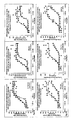
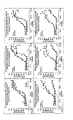
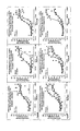
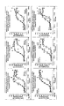
- Figures 5 A-L depict the dose response curves for human GPCR signaling and translocation in U20S cells, in the presence of compounds 1-12, for 30 min, 60 min, and 120 min, in one experiment.
- Figures 5 M-X depict the dose response curves for human GPCR signaling and translocation in U20S cells, in the presence of compounds 1-12, for 30 min, 60 min, and 120 min, in a second experiment.
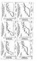
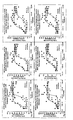
- Figures 6 A-L depict the dose response curves for rat GPCR signaling and translocation in CHO cells, in the presence of compounds 1-12, for 30 min, 60 min, and 120 min, in one experiment.
- Figures 6 M-X depict the dose response curves for rat GPCR signaling and translocation in CHO cells, in the presence of compounds 1-12, for 30 min, 60 min, and 120 min, in a second experiment.
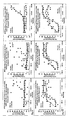
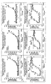
- Figures 7 A-L depict the dose response curves for rat GPCR signaling and translocation in U20S cells, in the presence of compounds 1-12, for 30 min, 60 min, and 120 min, in one experiment.
- Figures 7 M-X depict the dose response curves for rat GPCR signaling and translocation in U20S cells, in the presence of compounds 1-12, for 30 min, 60 min, and 120 min, in a second experiment.
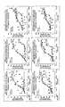
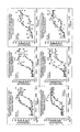
- Figure 8A depicts the dose response curves for MOR signaling and translocation in U20S cells, in the presence of DAMGO, for 30 min, 60 min, and 120 min, expressed as a % of the maximum response in the presence of morphine.
- Figure 8B depicts the dose response curves for MOR signaling and translocation in U20S cells, in the presence of morphine, for 30 min, 60 min, and 120 min, expressed as a % of the maximum response in the presence of morphine.
- Figure 8C depicts the dose response curves for MOR signaling and translocation in U20S cells, in the presence of DAMGO, for 30 min, 60 min, and 120 min, expressed as a % of the maximum response in the presence of DAMGO.
- Figure 8 D depicts the dose response curves for MOR signaling and translocation in U2OS cells, in the presence of morphine, for 30 min, 60 min, and 120 min, expressed as a % of the maximum response in the presence of DAMGO.
- a "DNA molecule” refers to the polymeric form of deoxyribonucleotides (adenine, guanine, thymine, or cytosine) in its either single stranded form, or a double-stranded helix. This term refers only to the primary and secondary structure of the molecule, and does not limit it to any particular tertiary forms. Thus, this term includes double-stranded DNA found, inter alia , in linear DNA molecules (e.g., restriction fragments), viruses, plasmids, and chromosomes.
- sequences may be described herein according to the normal convention of giving only the sequence in the 5' to 3' direction along the nontranscribed strand of DNA (i.e., the strand having a sequence homologous to the mRNA).
- Arrestin means all types of naturally occurring and engineered variants of arrestin, including, but not limited to, visual arrestin (sometimes referred to as Arrestin 1), cone arrestin (sometimes referred to as arrestin-4), ⁇ -arrestin 1 (sometimes referred to as Arrestin 2), and ⁇ -arrestin 2 (sometimes referred to as Arrestin 3).
- Modified arrestins for example arrestins that constitutively bind TMRs or GPCRs, are included.
- ⁇ AR is a GPCR termed a ⁇ -adrenergic receptor.
- Carboxyl-terminal tail means the carboxyl-terminal tail of a GPCR following membrane span 7. The carboxyl-terminal tail of many GPCRs begins shortly after the conserved NPXXY motif that marks the end of the seventh transmembrane domain (i.e. what follows the NPXXY motif is the carboxyl-terminal tail of the GPCR).
- the carboxyl-terminal tail may be relatively long (approximately tens to hundreds of amino acids), relatively short (approximately tens of amino acids), or virtually non-existent (less than approximately ten amino acids).
- “carboxyl-terminal tail” shall mean all three variants (whether relatively long, relatively short, or virtually non-existent), and may or may not contain palmitoylated cysteine residue(s).
- Class A receptors preferably do not translocate together with arrestin proteins to endocytic vesicles or endosomes in association with arrestin-GFP in HEK-293 cells.
- Class B receptors preferably do translocate together with arrestin proteins to endocytic vesicles or endosomes associated with arrestin-GFP in HEK-293 cells.
- Detectable molecule or “tag” or “label” means any molecule capable of detection by spectroscopic, photochemical, biochemical, immunochemical, electrical, radioactive, and optical means, including but not limited to, fluorescence, phosphorescence, and bioluminescence and radioactive decay.
- Detectable molecules include, but are not limited to, GFP, luciferase, ⁇ -galactosidase, rhodamine-conjugated antibody, and the like.
- Detectable molecules include radioisotopes, epitope tags, affinity labels, enzymes, fluorescent groups, chemiluminescent groups, and the like.
- Detectable molecules include molecules which are directly or indirectly detected as a function of their interaction with other molecule(s).
- Detectable molecules may be used to detect cellular molecules which are otherwise difficult to detect. For example, if detectably labeled, arrestin or a GPCR can be easily detected.
- the detectable label can be used to determine the localization of the labeled molecule.
- detectably labeled arrestin or GPCR facilitates the determination of the localization of the molecule, or related molecules, in the plasma membrane, pits, cytosol, endocytic vesicles, or endosomes, for example.
- GFP Green Fluorescent Protein which refers to various naturally occurring forms of GFP which may be isolated from natural sources or genetically engineered, as well as artificially modified GFPs. GFPs are well known in the art. See, for example, U.S. Patent Nos. 5,625,048 ; 5,777,079 ; and 6,066,476 . It is well understood in the art that GFP is readily interchangeable with other fluorescent proteins, isolated from natural sources or genetically engineered, including but not limited to, yellow fluorescent proteins (YFP), red fluorescent proteins (RFP), cyan fluorescent proteins (CFP), blue fluorescent proteins, luciferin, UV excitable fluorescent proteins, or any wave-length in between. As used herein, “GFP” shall mean all fluorescent proteins known in the art.
- Unknown or Orphan Receptor means a TMR, or a GPCR, whose function and/or ligands are unknown.
- Amino acid substitutions may also be introduced to substitute an amino acid with a particularly preferable property.
- a Cys may be introduced a potential site in order to allow formation of disulfide bridges with another Cys.
- a His may be introduced as a particularly "catalytic" residue (i.e., His can act as an acid or base and is the most common amino acid in biochemical catalysis).
- Pro may be introduced because of its particularly planar structure, which induces ⁇ -turns in the protein's structure.
- Two amino acid sequences are "substantially homologous" when at least about 70% of the amino acid residues (preferably at least about 80%, and most preferably at least about 90 or 95%) are identical, or represent conservative substitutions.
- Antagonist(s) include all agents that interfere with wild-type and/or modified TMR, or GPCR, binding to an agonist, wild-type and/or modified TMR, or GPCR, internalization, wild-type and/or modified TMR, or GPCR, binding arrestin, wild-type and/or modified TMR, or GPCR, endosomal localization, internalization, and the like, including agents that affect the wild-type and/or modified TMRs, including GPCRs, as well as agents that affect other proteins involved in wild-type and/or modified TMR, or GPCR, signaling, internalization, endosomal localization, and the like.
- Modified GPCR or “modified TMR” means a GPCR or TMR that has one or more modifications in the amino acid sequence. As such, the GPCR or TMR may be modified in whole or in part. These modifications in the amino acid sequence include mutations of one or more amino acids, insertion of one or more amino acids, deletion of one or more amino acids, and substitutions of one or more amino acids in which one or more amino acids are deleted and one or more amino acids are added in place of the deleted amino acids. Such modified GPCRs are described herein, as well as in U.S.S.N. 09/993,844 and U.S.S.N. 10/054,616 .
- GPCR means G protein-coupled receptor and includes GPCRs naturally occurring in nature, as well as GPCRs which have been modified.
- TMR transmembrane receptor and includes TMRs naturally occurring in nature, as well as TMRs which have been modified. GPCRs are an example of TMRs.
- Internalized TMR means a TMR that has undergone internalization.
- the internalized TMR may be located at any point in the internalization pathway. It presently does not have ability to bind to agonist and activate conventional signaling.
- the TMR may be a GPCR.
- the signaling may be G protein signaling.
- Internalization pathway means any cellular component of the internalization process, as well as any cellular structure implicated in the internalization process and subsequent processes, including but not limited to, arrestins, GRKs, GPCRs, AP-2 protein, clathrin, protein phosphatases, and the like.
- the polypeptides may be detected, for example, in the cytoplasm, at a cell membrane, in clathrin-coated pits, in endocytic vesicles, endosomes, any stages in between, and the like.
- TMR signaling means TMR induced activation of signaling. This may result in, for example, cAMP production.
- the TMR may be a GPCR.
- the signaling may be G protein signaling.
- G protein-coupled receptor kinase includes any kinase that has the ability to phosphorylate a GPCR. Splice variants, biologically active fragments, modified GRKs, and GRKs from animals and other organisms are included.
- Homo sapiens TMR means a naturally occurring TMR in a Homo sapiens .
- the TMR may be a GPCR.
- Naturally occurring TMR means a TMR that is present in nature.
- the TMR may be a GPCR.
- Oxygen ligand means a ligand compound that, upon binding to a receptor, leads to the perception of an odor including a synthetic compound and/or recombinantly produced compound including agonist and antagonist molecules.
- Optorant receptor means a receptor protein normally found on the surface of olfactory neurons which, when activated (normally by binding an odorant ligand) leads to the perception of an odor.
- Modulation includes at least an up-regulation or down-regulation of the expression, or an increase or decrease in activity of a protein.
- Modulation of a protein includes the up-regulation, down-regulation, increase or decrease in activity of a protein or compound that regulates a protein. Modulation also includes the regulation of the gene, the mRNA, or any other step in the synthesis of the protein of interest.
- An “overexpressed” protein refers to a protein that is expressed at levels greater than wild-type expression levels.
- Modified GRK means a GRK that has one or more modifications in the amino acid sequence at the C-terminus of the GRK.
- the modified GRK constitutively localizes to the plasma membrane.
- the GRK is modified by the addition of a CAAX motif.
- CAAX cysteine motif
- A is an aliphatic amino acid
- X is the C-terminal amino acid of the protein.
- a "constitutive" activity means an activity that occurs in the absence of agonist.
- the modified GRK constitutively localizes to the plasma membrane means that the modified GRK localizes to the plasma membrane in the absence of agonist.
- Reduced Internalization means that the TMR, or GPCR, internalization is reduced or delayed as compared with the TMR, or GPCR, internalization activated by a control compound.
- the control compound may be a natural agonist or natural ligand of the TMR, but may be any other compound used as a control.
- Reduced internalization may be demonstrated by a number of factors. For example, reduced internalization may be demonstrated by a decrease in the Max of internalization of the receptor as compared to the internalization stimulated by a natural agonist or natural ligand of the TMR. In another example, reduced internalization may be demonstrated by an increase in the EC50 of internalization of the TMR as compared to the internalization stimulated by a natural agonist or natural ligand of the TMR.
- the internalization would be reduced.
- the length of time that the TMR is required to be exposed to the compound in order to activate internalization is greater than the length of time that the TMR is required to be exposed to a natural agonist or natural ligand in order to activate internalization, then the internalization would be reduced.
- EC50 represents the median effective concentration of a compound; the smallest concentration required to produce 50% of a maximal stated effect.
- the "stated effect” means the effect of interest.
- the stated effect for example, may be activation of signaling, inhibition of signaling, amount of signaling, activation of internalization, inhibition of internalization, amount of internalization, or ligand binding.
- Max represents the maximal stated effect.
- the Max may be empirically determined or extrapolated.
- Bmax and Vmax are examples of Max.
- Max may be the maximal response as plotted on a response curve.
- cAMP production may be plotted with respect to different concentrations of a test compound to yield a dose response curve; the Max represents the maximal cAMP production on the dose response curve.
- translocation may be plotted with respect to different concentrations of a test compound to yield a dose response curve; the Max represents the maximal translocation on the dose response curve.
- control compound is a compound against which the test compound is compared.
- the control compound in the present invention may activate and internalize a TMR, or a GPCR.
- the control compound may be an agonist or ligand of a TMR, or a GPCR.
- a "dose response curve” represents a stated effect in the presence of different concentrations of a compound.
- the stated effect is preferably the signaling or internalization of a TMR, or a GPCR.
- “Measuring translocation of a TMR” means detection of the localization of a TMR in a cell.
- the TMR may be a GPCR.
- the TMR, or GPCR is detected by visualizing the TMR, arrestin, GRK, or another molecule.
- the molecule to be detected may be detectably labeled. Examples of detectably labeled molecules are described herein.
- the molecule is localized at the cell membrane, in pits, endosomes, endocytic vesicles, and/or in the cytosol. Methods of monitoring translocation include Transfluor, BRET, FRET, polarization microscopy, evanescent wave excitation microscopy, and standard or confocal microscopy.
- TMRs Useful TMRs, GPCRs, methods, and methods of detection, for example, are as described herein, in U.S.S.N. 08/869,568 (now issued U.S. Patent No. 5,891,646 ), 09/233,530 (now Issued U.S. Patent No. 6,110,693 ), 09/469,554 (now Issued U.S. Patent No.
- Quantitatively determining internalization can include any analytical method of measuring amount of TMR or GPCR internalization, including for example measuring the Intensity of pixels, amount of fluorescence, EC50, Max, dose response curve, or any other quantitative analysis for comparison known to those of skill in the art. Examples of quantitatively determining internalization are described herein. Various methods of detection, including but not limited to those as described herein, may be used for quantitatively determining internalization.
- TMR transmembrane receptor
- the Internalization is reduced if the TMR internalization in the presence of the test compound is reduced as compared to the TMR internalization in the presence of a control compound.
- the signaling is activated if the TMR signaling in the presence of the test compound is activated as compared to in the absence of agonist.
- Such a compound may increase signaling in response to a given dose of compound, decrease signal termination, and maintain responsiveness of a given TMR or GPCR to a given dose of compound.
- the present invention relates to methods of screening for a TMRA. Methods of determining if a compound activates signaling but has altered internalization properties are included.
- TMRAs are useful for a variety of reasons. While compounds which activate signaling and also activate internalization pathways are known, many of these have less than desirable characteristics, including certain side effects. Increased internalization of TMRs would result in fewer receptors available at the membrane or cell surface for interaction with agonist, which may be over repeated dosage. Reduced internalization of TMRs would result in more receptors available at the membrane or cell surface for interaction with agonist or TMRA. This reduced internalization may decrease the amount and/or frequency of agonist or TMRA that is necessary to activate signaling. Methods of identifying TMRAs are necessary. Methods of identifying these compounds would be useful in the treatment of numerous diseases.
- the present invention utilizes quantitative measurements of internalization.
- the quantitative measurements of internalization are necessary in the methods of identifying reduced internalization agonists as described herein.
- TMR or GPCR signaling may be quantitated by any of a number of indicators of TMR or GPCR signaling. These include quantitating the activity of members of any of a number of signaling pathways, GPCR dimerization, and the like.
- a “signaling pathway” refers to the modulation (e.g., stimulation or inhibition) of a cellular function/activity upon the binding of a ligand to the TMR or GPCR.
- Examples of such functions include mobilization of intracellular molecules that participate in a signal transduction pathway, e.g., phosphatidylinositol 4,5-bisphosphate, inositol 1,4,5-triphosphate, and adenylate cyclase; polarization of the plasma membrane; production or secretion of molecules; alteration in the structure of a cellular component; cell proliferation, e.g., synthesis of DNA; cell migration; cell differentiation; and cell survival.
- a signal transduction pathway e.g., phosphatidylinositol 4,5-bisphosphate, inositol 1,4,5-triphosphate, and adenylate cyclase
- polarization of the plasma membrane e.g., phosphatidylinositol 4,5-bisphosphate, inositol 1,4,5-triphosphate, and adenylate cyclase
- production or secretion of molecules e.g., alteration in the
- the specific pathway to be measured may depend on the specific TMR or GPCR of interest. Many methods of quantitating the activity of members of signaling pathways are known to one of skill in the art.
- intracellular second messenger generation can be measured directly.
- G-protein-regulated including cAMP, cyclic GMP, Calcium, a lipid, phosphatidylinositol, a hydrogen ion, an ion transport molecule, and the like.
- G proteins interact with a range of ion channels and are able to inhibit certain voltage-sensitive Ca ++ transients, as well as stimulating cardiac K + channels.
- enzymes such as adenylyl cyclase, phosphodiesterase, phospholipase C, protein kinase, phospholipase A 2 , and the like may be measured.
- the GTPase enzymatic activity by G proteins can be measured in plasma membrane preparations by determining the breakdown of ⁇ 32 P GTP using techniques that are known in the art (For example, see Signal Transduction: A Practical Approach. G. Milligan, Ed. Oxford University Press, Oxford Engl and).
- receptors that modulate cAMP are tested, it will be possible to use standard techniques for cAMP detection, such as competitive assays which quantitate [ 3 H]cAMP in the presence of unlabelled cAMP.
- Cyclic AMP turnover and metabolism the molecules involved in the turnover and metabolism of cyclic AMP (cAMP) as well as to the activities of these molecules, can be measured.
- Cyclic AMP is a second messenger produced in response to ligand-induced stimulation of certain TMRs or G protein coupled receptors. In the cAMP signaling pathway, binding of a ligand to a TMR or GPCR can lead to the activation of the enzyme adenyl cyclase, which catalyzes the synthesis of cAMP. The newly synthesized cAMP can in turn activate a cAMP-dependent protein kinase.
- This activated kinase can phosphorylate a voltage-gated potassium channel protein, or an associated protein, and lead to the inability of the potassium channel to open during an action potential.
- the inability of the potassium channel to open results in a decrease in the outward flow of potassium, which normally repolarizes the membrane of a neuron, leading to prolonged membrane depolarization.
- Inositol lipids can be extracted and analyzed using standard lipid extraction techniques. DAG can also be measured using thin-layer chromatography. Water soluble derivatives of all three inositol lipids (IP1, IP2, IP3) can also be quantitated using radiolabelling techniques or HPLC.
- PIP 2 phosphatidylinositol turnover and metabolism
- DAG 1,2-diacylglycerol
- IP 3 inositol 1,4,5-triphosphate
- IP 3 can diffuse to the endoplasmic reticulum surface where it can bind an IP 3 receptor, e.g., a calcium channel protein containing an IP 3 binding site. IP 3 binding can induce opening of the channel, allowing calcium ions to be released into the cytoplasm. IP 3 can also be phosphorylated by a specific kinase to form inositol 1,3,4,5-tetraphosphate (IP 4 ), a molecule which can cause calcium entry into the cytoplasm from the extracellular medium. IP 3 and IP 4 can subsequently be hydrolyzed very rapidly to the inactive products inositol 1,4-biphosphate (IP 2 ) and inositol 1,3,4-triphosphate, respectively.
- IP 2 inositol 1,4-biphosphate
- IP 2 inositol 1,3,4-triphosphate
- Protein kinase C is usually found soluble in the cytoplasm of the cell, but upon an increase in the intracellular calcium concentration, this enzyme can move to the plasma membrane where it can be activated by DAG.
- the activation of protein kinase C in different cells results in various cellular responses such as the phosphorylation of glycogen synthase, or the phosphorylation of various transcription factors, e.g., NF-kB.
- phosphatidylinositol activity refers to an activity of PIP 2 or one of its metabolites.
- the mobilization of intracellular calcium or the influx of calcium from outside the cell can be measured using standard techniques.
- the choice of the appropriate calcium indicator, fluorescent, bioluminescent, metallochromic, or Ca++-sensitive microelectrodes depends on the cell type and the magnitude and time constant of the event under study ( Borle (1990) Environ Health Perspect 84:45-56 ).
- As an exemplary method of Ca++ detection cells could be loaded with the Ca++ sensitive fluorescent dye fura-2 or indo-1, using standard methods, and any change in Ca++ measured using a fluorometer.
- Another exemplary high throughput calcium assay involves induction of a reporter gene operatively linked to a calcium-responsive promoter.
- a calcium flux resulting from the activation of a TMR or GPCR turns on the promoter which subsequently drives the expression of a reporter gene encoding a protein with an enzymatic activity that can be easily detected, preferably by a colorimetric or fluorescent assay.
- reporter proteins include: ⁇ -galactosidase, ⁇ -lactamase, chloramphenicol acetyltransferase (CAT), luciferase, green fluorescent protein (GFP) and its derivatives, among others.
- Reporter proteins can also be linked to other proteins whose expression is dependent upon the stimulation of TMRs or GPCRs.
- An illustrative example would be a fusion protein comprising luciferase sequence in frame with the open-reading frame of nuclear factor of activated T cells (NFAT). Since the transcription of NFAT requires the co-activation of calcium and protein kinases C signaling pathways acting downstream of TMRs or GPCRs, an effective coupling of heterotrimeric G protein to the receptors can then be measured by assaying NFAT-mediated luciferase activity.
- NFAT nuclear factor of activated T cells
- a preferred host cell is one of lymphoid or neuronal origin, such as Jurkat cells and pheochromocytoma PC12 cells.
- lymphoid or neuronal origin such as Jurkat cells and pheochromocytoma PC12 cells.
- NFAT and NFAT isoforms are present in a variety of cells including endothelial and myeloid cells (id).
- Reporter gene based assays of this invention measure the end stage of the above described cascade of events, e.g., transcriptional modulation.
- a reporter gene construct is inserted into the reagent cell in order to generate a detection signal dependent on receptor signaling.
- the reporter gene construct will include a reporter gene in operative linkage with one or more transcriptional regulatory elements responsive to the signal transduction activity of the target receptor, with the level of expression of the reporter gene providing the receptor-dependent detection signal.
- the amount of transcription from the reporter gene may be measured using any method known to those of skill in the art to be suitable. For example, specific mRNA expression may be detected using Northern blots or specific protein product may be identified by a characteristic stain or an intrinsic activity.
- the activation of phospholipase A2 can easily be quantitated using known techniques, including, for example, the generation of arachadonate in the cell.
- Such assay formats may be useful when the receptor of interest is a receptor tyrosine kinase.
- yeast transformed with the FGF receptor and a ligand which binds the FGF receptor could be screened using colony immunoblotting ( Lyons and Nelson (1984) Proc. Natl. Acad. Sci. USA 81:7426-7430 ) using anti-phosphotyrosine.
- tests for phosphorylation could be useful when a receptor which may not itself be a tyrosine kinase, activates protein kinases that function downstream in the signal transduction pathway.
- Multi-kinase cascades allow not only signal amplification but also signal divergence to multiple effectors that are often cell-type specific, allowing a growth factor to stimulate mitosis of one cell and differentiation of another.
- MAP kinase pathway that appears to mediate both mitogenic, differentiation and stress responses in different cell types. Stimulation of growth factor receptors results in Ras activation followed by the sequential activation of c-Raf, MEK, and p44 and p42 MAP kinases (ERK1 and ERK2). Activated MAP kinase then phosphorylates many key regulatory proteins, including p90 RSK and Elk-1 that are phosphorylated when MAP kinase translocates to the nucleus. Homologous pathways exist in mammalian and yeast cells. For instance, an essential part of the S.
- cerevisiae pheromone signaling pathway is comprised of a protein kinase cascade composed of the products of the STE11, STE7, and FUS3/KSS1 senes (the latter pair are distinct and functionally redundant). Accordingly, phosphorylation and/or activation of members of this kinase cascade can be detected and used to quantitate receptor engagement.
- Phosphotyrosine specific antibodies are available to measure increases in tyrosine phosphorylation and phospho-specific antibodies are commercially available (New England Biolabs, Beverly, Mass.).
- the response mediated by the receptor protein depends on the type of cell. For example, in some cells, binding of a ligand to the receptor protein may stimulate an activity such as release of compounds, gating of a channel, cellular adhesion, migration, differentiation, etc., through phosphatidylinositol or cyclic AMP metabolism and turnover while in other cells, the binding of the ligand will produce a different result.
- the protein is a TMR or GPCR and interacts with G proteins to produce one or more secondary signals, in a variety of intracellular signal transduction pathways, e.g., through phosphatidylinositol or cyclic AMP metabolism and turnover, in a cell.
- the signaling assays may use spectrally-encoded assay bead systems. More than one measure of TMR or GPCR activation may be determined; they may be measured, for example, sequentially or in parallel.
- Methods of detecting internalization are useful in the methods of the present invention. Such methods that are detected quantitatively are preferred. Methods that are optically detectable and quantitatable are preferred. Any method that will measure internalization of the TMR or GPCR by monitoring the localization of the TMR or GPCR or any molecule that Interacts with the TMR or GPCR in the internalization pathway may be used. Examples include Transfluor, BRET, FRET.
- provisional application 60/379,986 and U.S. provisional application 60/401,698 .
- Other methods useful in the present invention are described in U.S.S.N. 09/759,152 , U.S.S.N. 09/654,499 , WO 99/66324 , WO 01/46691 , WO 01/46694 , WO 98/00715 , and WO 02/46763 .
- Methods of detecting the intracellular location of the detectably labeled arrestin, the intracellular location of a detectably labeled TMR or GPCR, or interaction of the detectably labeled arrestin, or other member of a TMR or GPCR/arrestin complex with a TMR or GPCR or any other cell structure including for example, the concentration of arrestin or TMR or GPCR at a cell membrane, colocalization of arrestin with TMR or GPCR in endosomes, and concentration of arrestin or TMR or GPCR in clathrin coated pits, and the like, will vary dependent upon the detectable molecule(s) used.
- any optical method may be used where a change in the fluorescence, bioluminescence, or phosphorescence may be measured due to a redistribution or reorientation of emitted light.
- Such methods include, for example, polarization microscopy, BRET, FRET, evanescent wave excitation microscopy, and standard or confocal microscopy.
- arrestin may be conjugated to GFP and the arrestin GFP conjugate may be detected by confocal microscopy.
- arrestin may conjugated to a GFP and the TMR or GPCR may be conjugated to an immunofluorescent molecule, and the conjugates may be detected by confocal microscopy.
- arrestin may be conjugated to a GFP and the carboxy terminus of the TMR or GPCR may be conjugated to a luciferase and the conjugates may be detected by bioluminescence resonance emission technology.
- arrestin may be conjugated to a luciferase and TMR or GPCR may be conjugated to a GFP, and the conjugates may be detected by bioluminescence resonance emission technology.
- arrestin may be conjugated to a detectable molecule fragment (or portion, domain, and the like), TMR or GPCR may be conjugated to a detectable molecule fragment (or portion, domain, and the like), and the interaction of the two fragments can be detected.
- the TMR or GPCR or arrestin may be labeled with a detectable molecule which is specifically detectable upon localization at certain cellular locations.
- the methods of the present invention are directed to detecting TMR or GPCR activity. The methods of the present invention allow enhanced monitoring of the GPCR pathway in real time.
- the localization pattern of the detectable molecule is determined. In a further preferred embodiment, alterations of the localization pattern of the detectable molecule may be determined. The localization pattern may indicate cellular distribution of the detectable molecule. Certain methods of detection are described in U.S.S.N. 10/095,620, filed March 12, 2002 , which claims priority to U.S. Provisional Patent Application No: 60/275,339, filed March 13, 2001 . The quantitation of the detectable molecules is also described in U.S.S.N. 10/161,916 .
- Molecules may also be detected by their interaction with another detectably labeled molecule, such as an antibody.
- the TMRA is capable of activating TMR signaling while reducing TMR internalization.
- the method includes providing a cell including at least one TMR or a biologically active fragment thereof.
- the cell may also include arrestin or biologically active fragments thereof, and/or GRK or biologically active fragments thereof.
- a component or components of the cell may be detectably labeled,
- the label may be detectable as the result of an Interaction with another detectably labeled molecule, or the like.
- the detectably labeled molecule may be detected at the plasma membrane, in endosomes, endocytic vesicles, or in the cytosol, for example.
- the detectable molecule may be a radioisotope, an epitope tag, an affinity label, an enzyme, a fluorescent group, or a chemiluminescent group, for example.
- the method may include the additional steps of providing a agonist or ligand of a TMR, and modifying the agonist or ligand, for example.
- the TMR may be a GPCR.
- GPCRs useful in the present invention are listed in Figure 1 .
- the GPCR may be a class A receptor, a class B receptor, a ⁇ opioid, ⁇ 1 AR, ⁇ 2 AR, or dopamine receptor, for example.
- Certain ligands or agonists useful in the present invention are listed in Figure 3 , for example.
- the compound may be a modified natural ligand or natural agonist.
- the compound may be a known pharmaceutically relevant compound, or may be derived from a known pharmaceutically relevant compound.
- the cell may be exposed to a compound or compounds.
- the compound or compounds may be derived from a natural ligand or natural agonist.
- the compound or compounds may be present in a compound library.
- the signaling may be measured. Certain methods of measuring signaling are described herein, but other methods of measuring signaling may also be used. The measurement of the signaling may be useful in a quantitative determination of whether signaling is activated. A dose response curve for signaling may be determined. Signaling may be quantitated by measuring an intracellular effector, wherein said effector may be cAMP, cyclic GMP, Calcium, a lipid, phosphatidylinositol, a hydrogen ion, or an ion transport molecule. Signaling in the presence of the compound may be compared to signaling in the absence of compound to determine if the signaling is activated. The signaling may be activated for a longer time period after stimulation by the TMRA than the length of time of activation after stimulation by the natural ligand or natural agonist.
- the internalization may be measured. Certain methods of measuring internalization are described herein, but other methods of measuring internalization may also be used. The measurement of the internalization may be useful in a quantitative determination of whether internalization is reduced. The internalization may be reduced as compared to the TMRA internalization in the presence of a control compound. A dose response curve for signaling may be determined.
- Agonists and ligands of TMRs may be modified. Such modifications may result in modified chemical and/or physical properties of the compounds.
- the modified agonists and ligands may be analyzed by methods of the present invention to identify a TMRA.
- the modified agonists and ligands may be used to create a panel of compounds. This panel of compounds may be used in the methods as described herein to identify a TMRA.
- the TMR or GPCR sequence in a cell of the methods described herein may be a human TMR or GPCR, rat TMR or GPCR, mouse TMR or GPCR, pig TMR or GPCR, primate TMR or GPCR, or a TMR or GPCR from any species.
- the TMR or GPCR from a specific species may be preferred in the methods of the present invention.
- the methods of the present invention may be repeated; the repeat of method may use cells containing TMRs or GPCRs from different species than in the cells of the first method.
- a test compound may have TMRA properties in a method using cells including TMRs or GPCRs from a specific species.
- the same test compound may be a TMRA for TMRs or GPCRs of a certain species, but may or may not be a TMRA for the same TMR or GPCR of a different species. Repeating the methods may be useful to determine the preferred TMR or GPCR species for use in methods using cells containing that TMR or GPCR.
- DNA sequences may be expressed by operatively linking them to an expression control sequence in an appropriate expression vector and employing that expression vector to transform an appropriate unicellular host.
- the transforming DNA may or may not be integrated (covalently linked) into chromosomal DNA making up the genome of the cell.
- Such operative linking of a DNA sequence of this invention to an expression control sequence includes, if not already part of the DNA sequence, the provision of an initiation codon, ATG, in the correct reading frame upstream of the DNA sequence.
- a wide variety of host/expression vector combinations may be employed in expressing the DNA sequences described herein.
- Useful expression vectors may consist of segments of chromosomal, non-chromosomal and synthetic DNA sequences.
- Suitable vectors include derivatives of SV40 and known bacterial plasmids, e.g., E.
- phage DNAS e.g., the numerous derivatives of phage ⁇ , e.g., NM989, and other phage DNA, e.g., M13 and filamentous single stranded phage
- any of a wide variety of expression control sequencessequences that control the expression of a DNA sequence operatively linked to it - may be used in these vectors to express the DNA sequences in this invention.
- useful expression control sequences include, for example, the early or late promoters of SV40, CMV, vaccinia, polyoma or adenovirus, the lac system, the trp system, the TAC system, the TRC system, the LTR system, the major operator and promoter regions of phage ⁇ , the control regions of fd coat protein, the promoter for 3-phosphoglycerate kinase or other glycolytic enzymes, the promoters of acid phosphatase (e.g., Pho5), the promoters of the yeast ⁇ -mating factors, and other sequences known to control the expression of genes of prokaryotic or eukaryotic cells or their viruses, and various combinations thereof.
- a wide variety of unicellular host cells are also useful in expressing the DNA sequences described herein.
- These hosts may include well known eukaryotic and prokaryotic hosts, such as strains of E. coli, Pseudomonas, Bacillus, Streptomyces, fungi such as yeasts, plant cells, nematode cells, and animal cells, such as HEK-293, U20S, CHO, RI.I, B-W and L-M cells, African Green Monkey kidney cells (e.g., COS 1, COS 7, BSC1, BSC40, and BMT10), insect cells (e.g., Sf9), and human cells and plant cells in tissue culture.
- the host cells include a GRK-C20 and an arrestin.
- the host cells include a GRK, an arrestin, and a GPCR.
- Suitable unicellular hosts will be selected by consideration of, e.g., their compatibility with the chosen vector, their secretion characteristics, their ability to fold proteins correctly, and their fermentation requirements, as well as the toxicity to the host of the product encoded by the DNA sequences to be expressed, and the ease of purification of the expression products.
- analogs may be prepared from nucleotide sequences of the protein complex/subunit.
- Analogs, such as fragments may be produced, for example, by pepsin digestion.
- Other analogs, such as muteins can be produced by standard site-directed mutagenesis of coding sequences.
- Analogs exhibiting biological activity, such as small molecules, whether functioning as promoters or inhibitors may be identified by known in vivo and/or in vitro assays.
- a DNA sequence can be prepared synthetically rather than cloned.
- the DNA sequence can be designed with the appropriate codons for the amino acid sequence. In general, one will select preferred codons for the intended host if the sequence will be used for expression.
- the complete sequence is assembled from overlapping oligonucleotides prepared by standard methods and assembled into a complete coding sequence. See, e.g., Edge, Nature, 292:756 (1981 ); Nambair et al., Science, 223:1299 (1984 ); Jay et al., J. Biol. Chem., 259:6311 (1984 ).
- DNA sequences allow convenient construction of genes which will express analogs or "muteins".
- DNA encoding muteins can be made by site-directed mutagenesis of native or modified genes or cDNAs, and muteins can be made directly using conventional polypeptide synthesis.
- nucleic acids encoding the motifs may be at the 5' or 3' end of the nucleic acid, resulting in the presence of the motif at the N or C teriminus of the protein.
- the cells used in the methods described herein may comprise a conjugate of an arrestin protein and a detectable molecule.
- the cells may also comprise a conjugate of a GPCR and a detectable molecule.
- the GPCR may be modified to have enhanced phosphorylation by a GRK.
- the GPCR may be ⁇ 2 AR(Y326A), a GPCR listed in Figure 1 , an orphan GPCR, a modified GPCR, a taste receptor, a Class A GPCR, a Class B GPCR, a mutant GPCR, or a biologically active fragment thereof.
- arrestin naturally occurring and engineered variants, including but not limited to, visual arrestin, ⁇ arrestin 1 and ⁇ arrestin 2, may be used in the present invention.
- Detectable molecules that may be used to conjugate with the arrestin include, but are not limited to, molecules that are detectable by spectroscopic, photochemical, biochemical, immunochemical, electrical, radioactive, and optical means, including but not limited to bioluminescence, phosphorescence, and fluorescence.
- Detectable molecules include, but are not limited to, GFP, luciferase, ⁇ -galactosidase, rhodamine-conjugated antibody, and the like.
- Detectable molecules include radioisotopes, epitope tags, affinity labels, enzymes, fluorescent groups, chemiluminescent groups, and the like.
- Detectable molecules include molecules which are directly or indirectly detected as a function of their interaction with other molecule(s).
- detectable molecules should be a biologically compatible molecule and should not compromise the ability of the arrestin to interact with the GPCR system and the interaction of the arrestin with the GPCR system must not compromise the ability of the detectable molecule to be detected.
- Preferred detectable molecules are optically detectable molecules, including optically detectable proteins, such that they may be excited chemically, mechanically, electrically, or radioactively to emit fluorescence, phosphorescence, or bioluminescence. More preferred detectable molecules are inherently fluorescent molecules, such as fluorescent proteins, including, for example, Green Fluorescent Protein (GFP).
- the detectable molecule may be conjugated to the arrestin protein by methods as described in Barak et al. ( U.S. Patent Nos. 5,891,646 and 6,110,693 ).
- the detectable molecule may be conjugated to the arrestin at the front-end, at the back-end, or in the middle.
- the TMR or GPCR or biologically active fragments thereof may also be conjugated with a detectable molecule.
- the carboxyl-terminus of the TMR or GPCR is conjugated with a detectable molecule.
- a carboxyl-terminal tail conjugated or attached to a detectable molecule can be used in a carboxyl-terminal tail exchange to provide the detectably labeled GPCR.
- the detectable molecule used to conjugate with the TMRs or GPCRs may include those as described above, including, for example, optically detectable molecules, such that they may be excited chemically, mechanically, electrically, or radioactively to emit fluorescence, phosphorescence, or bioluminescence. Preferred optically detectable molecules may be detected by immunofluorescence, luminescence, fluorescence, and phosphorescence.
- the TMRs or GPCRs may be antibody labeled with an antibody conjugated to an immunofluorescence molecule or the TMRs or GPCRs may be conjugated with a luminescent donor.
- the TMRs or GPCRs may be conjugated with, for example, luciferase, for example, Renilla luciferase, or a rhodamine-conjugated antibody, for example, rhodamine-conjugated anti-HA mouse monoclonal antibody.
- the carboxyl-terminal tail of the GPCR may be conjugated with a luminescent donor, for example, luciferase.
- the GPCR preferably the carboxyl-terminal tail, also may be conjugated with GFP as described in L.
- Cells derived from different species may be useful in the present invention.
- Cells may be derived from rat, mouse, pig, primate, or human, for example.
- Cells derived from particular animals, for example higher primates, may be required to detect the reduced internalization in the methods described herein.
- cells derived from a particular species may be useful, or preferred.
- cells derived from mammals are useful.
- cells derived from rat, mouse, pig, primate, or human are useful.
- the cells of the present invention may express at least one GPCR.
- the cells may further comprise a conjugate of an arrestin protein and a detectable molecule.
- Useful cells include eukaryotic and prokaryotic cells, including, but not limited to, bacterial cells, yeast cells, fungal cells, insect cells, nematode cells, plant cells, and animal cells.
- Suitable animal cells include, but are not limited to, HEK-293 cells, U2OS cells, HeLa cells, COS cells, and various primary mammalian cells.
- An animal model expressing a conjugate of an arrestin and a detectable molecule throughout its tissues or within a particular organ or tissue type, may also be used.
- the cells of the present invention may express one modified protein that results in agonist-independent localization of TMRs or GPCRs to endocytic vesicles or endosomes.
- a substrate may have deposited thereon a plurality of cells of the present invention.
- the substrate may be any suitable biologically substrate, including but not limited to, glass, plastic, ceramic, semiconductor, silica, fiber optic, diamond, biocompatible monomer, or biocompatible polymer materials.
- Also included in the present disclosure are to methods of treating a human or non-human subject suffering from a GPCR-related disease, such as cardiovascular disease, heart failure, asthma, nephrogenic diabetes insipidus, or hypertension.
- a GPCR-related disease such as cardiovascular disease, heart failure, asthma, nephrogenic diabetes insipidus, or hypertension.
- Such treatment can be performed either by administering to a subject in need of such treatment, an amount of the compounds identified by the present method sufficient to treat the GPCR-related disease, or at least to lessen the symptoms thereof.
- TMR transmembrane receptor
- GPCR signaling was measured using AlphaScreenTM from PerkinElmer.
- U2OS Rr#7/ Human GPCR cells were plated at 25 ⁇ l/well into three IPA washed, 384-well Matrical plates (4,000 cells/ well final).
- U2OS Rr#7/ Rat GPCR cells were plated at 25 ⁇ l/well into three IPA washed, 384-well Matrical plates (4,000 cells/ well final). Incubated at 37°C overnight.
- Cell Growth Media MEM in addition to, 10% FBS, 10 mM Hepes, 2 mM Glutamine, 10 ⁇ g/ml Gentamicin, 0.4 mg/ml G418.
- Reagent and Compound Preparation all reagents were prepared using silinized labware from frozen stocks brought to room temperature. All values listed are final concentrations.
- Cell Media EMEM supplemented with 2 mM Glutamine, 10 mM Hepes, 0.1 % BSA.
- Compounds compounds 1-12 were serially diluted (1:3) from a starting concentration of 1 ⁇ M in EMEM with 10 mM Hepes, 2 mM Glutamine and 0.1% BSA. Diluted compounds were consolidated on a Greiner deep well 384 plate for ease of dispensing via Minitrak.
- Fix and Stain 1 ⁇ M DRAQ5 stain and 2% Formaldehyde.
- TMR transmembrane receptor
- Cells containing ⁇ opioid receptor, and arrestin-GFP were provided. Cells were exposed to morphine, the test compound. Cells were exposed to DAMGO, the control compound. The signaling was measured at 30 min, 60 min, and 120 min after exposure to the compound. Translocation was measured at 30 min, 60 min, and 120 min after exposure to the compound. It was then quantitatively determined that the MOR internalization was reduced in the presence of morphine, the test compound, as compared to the MOR internalization in the presence of DAMGO, the control compound. It was determined that signaling was activated in the presence of morphine, the test compound, as compared to signaling in the absence of agonist. Signaling and translocation were measured at multiple concentrations of compound.
- morphine is a TMRA. While morphine is a TMRA, it may be derivitized to form a compound which further reduces MOR internalization.
- ⁇ opioid receptor signaling was measured using AlphaScreenTM from PerkinElmer.
- CHO cells were seeded at 100,000/ml in 70 ml of Ex-Cell Media (suspension media) + 10% FBS + 2 mM Glutamine + 10 mM Hepes in a 125 ml spinner flask; 4.2 ml (1 e9 stock) baculovirus was added at seeding thus yielding 600 viral particles per cell (600MOI). Incubated at 37 °C overnight.
- Cell Media 35 ml EMEM (SFM) in addition to10 mM Hepes, 2 mM Glutamine; Add 35 ⁇ l (500 ⁇ M) IBMX (1:1000 dilution).
- Forskolin Media 1 ⁇ l (1 mM) forskolin into 62.7 ⁇ l EMEM (SFM) in addition to 10 mM Hepes, 2 mM Glutamine (1:63.7 dilution). Yields 1.57e-3 working concentration.
- Morphine 2 ⁇ l (10 mM) morphine into 93.2 ⁇ l Drug Media (1:47.6 dilution); Yields 2.1 e-4M working concentration. 2.1 e-4 M high concentration DAMGO (1:3) serial dilutions, 16 pts; 40 ⁇ l into 80 ⁇ l Drug media (using silinized tips and tip changes between dilutions).
- cAMP 1 ⁇ l 5mM cAMP into 179.7 ⁇ l Drug Media (1:180.7 dilution); Yields 2.8e-5 M working cAMP concentration.
- Donor Beads (1:8) 2 ml 10% Tween-20; (1:730) 29 ⁇ l (10 ⁇ M) Biotinylated-cAMP; (1:180.5) 88.6 ⁇ l (5 mg/ml) donor beads; 13.89 ml EMEM (SFM) in addition to10 mM Hepes, 2 mM Glutamine.
- Counted cells from spinner flask Pelleted cells and resuspended in 26 ml of cell media (26 ml is enough volume for 3 x 384 plate). Final cell concentration was 6,000 cells per 20 ⁇ l. 8 . Placed 4 ml of cell media in a separate tube (no cells). This was used for the cAMP serial dilutions. 9 . All subsequent steps are light sensitive and all plates and reagents remained under green filtered light or in the dark. 10 . Added 71.76 ⁇ l of acceptor beads to the resuspended cells and mixed gently. 11 . Added 11 ⁇ l of acceptor beads to the cell media (containing no cells) and mixed. 12 .
- a multidrop dispensed 20 ⁇ l of cells + acceptor to all wells except cAMP wells. 13 . Added 20 ⁇ l of plain cell media + acceptor to cAMP wells, using a BioHit pipette. 14 . Tapped plates gently to mix. 15 . Incubated plate for 30 min at room temperature. 16 . During incubation, made forskolin media. 17 . After 30 min, added 10 ⁇ l of the working concentration of forskolin to all wells except for the basal and cAMP wells using a multidrop instrument. 18 . Added 10 ⁇ l to basal and cAMP wells using a multidrop instrument. 19 . Tapped plates gently to mix. 20 .
- ⁇ opioid receptor (MOR) translocation was measured using TransfluorTM.
- U2OS-GFP-MOR stable cell line containing MOR and arrestin-GFP, was plated in each of 3 IPA washed, 384-well Matrical plates at 5,000 cells/25 ⁇ l.
- Growing media MEM in addition to, 10% FBS, 10 mM Hepes, 2 mM Glutamine. Incubate at 37 °C overnight.
Abstract
Description
- Methods of identifying Reduced Internalization Transmembrane Receptor Agonists are disclosed.
- Transmembrane receptors (TMRs) are proteins that span membranes and have the ability to interact with or bind a ligand, which may be a hormone, small molecule, lipid, nucleic acid, or peptide, for example. G protein-coupled receptors (GPCRs) are cell surface transmembrane proteins that translate hormone or ligand binding into intracellular signals, as do most TMRs. TMRs, or GPCRs, are found in all animals, insects, and plants. TMR, or GPCR, signaling plays a pivotal role in regulating various physiological functions including phototransduction, olfaction, neurotransmission, vascular tone, cardiac output, digestion, pain, and fluid and electrolyte balance. GPCRs, although involved in numerous physiological functions, share a number of common structural features. They contain seven membrane domains bridged by alternating intracellular and extracellular loops and an intracellular carboxyl-terminal tail of variable length.
- GPCRs and other transmembrane receptors have been implicated in a number of disease states, including, but not limited to: cardiac indications such as angina pectoris, essential hypertension, myocardial infarction, supraventricular and ventricular arrhythmias, congestive heart failure, atherosclerosis, renal failure, diabetes, respiratory indications such as asthma, chronic bronchitis, bronchospasm, emphysema, airway obstruction, upper respiratory indications such as rhinitis, seasonal allergies, inflammatory disease, inflammation in response to injury, rheumatoid arthritis, chronic inflammatory bowel disease, glaucoma, hypergastrinemia, gastrointestinal indications such as acid/peptic disorder, erosive esophagitis, gastrointestinal hypersecretion, mastocytosis, gastrointestinal reflux, peptic ulcer, Zollinger-Ellison syndrome, pain, obesity, bulimia nervosa, depression, obsessive-compulsive disorder, organ malformations (for example, cardiac malformations), neurodegenerative diseases such as Parkinson's Disease and Alzheimer's Disease, multiple sclerosis, Epstein-Barr infection and cancer.
- The magnitude of the physiological responses controlled by TMRs can be linked to the balance between TMR signaling and signal termination. The signaling of GPCRs and some other TMRs is controlled by a family of intracellular proteins called arrestins. GPCRs are an example of transmembrane receptors which bind arrestin, activate signaling, and the like. Arrestins bind activated GPCRs, including those that have been agonist-activated and especially those that have been phosphorylated by G protein-coupled receptor kinases (GRKs).
- Receptors, including GPCRs, have historically been targets for drug discovery and therapeutic agents because they bind ligands, hormones, and drugs with high specificity. Approximately fifty percent of the therapeutic drugs in use today target or interact directly with GPCRs. See e.g., Jurgen Drews, (2000) "Drug Discovery: A Historical Perspective," Science 287:1960-1964.
- TMRs, of which GPCRs are but one example, may be useful in the methods described herein.
- TMRs, or GPCRs, can activate intracellular signaling. GPCRs recruit and regulate the activity of intracellular heterotrimeric G proteins. The activated receptor typically induces a conformational change in the associated G protein α-subunit leading to release of GDP followed by binding of GTP. Subsequently, the GTP-bound form of the α-subunit dissociates from the receptor as well as from the stable βγ-dimer. Both the GTP-bound α-subunit and the released βγ-dimer can modulate several cellular signaling pathways. These include, among others, stimulation or inhibition of adenylate cyclases and activation of phospholipases, as well as regulation of potassium and calcium channel activity. GPCRs also may not solely act via heterotrimeric G proteins.
- Internalized TMRs, or GPCRs, are not responsive to agonists or ligands. Subsequent to agonist or ligand exposure, initial steps of internalization produce attenuation of the signaling ability of the TMR or GPCR that may involve uncoupling of the GPCR from its cognate heterotrimeric G-protein. The cellular mechanism mediating initial steps of agonist-specific internalization is a two-step process in which agonist-occupied receptors are phosphorylated by a kinase, for example a GPCR kinase (GRK), and then bind an arrestin protein. TMRs, of which GPCRs are but one example, may bind an arrestin protein, and subsequently be internalized. The type III TGF-beta receptor is an example of a TMR, other than a GPCR, that binds arrestin, and undergoes subsequent internalization and signaling down-regulation (Chen et al., 2003, Science 301:1394-1397).
- It is known, for example, that after agonists bind GPCRs, G-protein coupled receptor kinases (GRKs) phosphorylate intracellular domains of GPCRs. After phosphorylation, an arrestin protein associates with the GRK-phosphorylated receptor and uncouples the receptor from its cognate G protein. The interaction of the arrestin with the phosphorylated GPCR terminates GPCR signaling, initiates internalization, and produces a non-signaling receptor.
- The arrestin bound to the GPCR targets the GPCR to clathrin-coated pits or other cellular machinery for endocytosis (i.e., internalization) by functioning as an adaptor protein, which links the GPCR to components of the endocytic machinery, such as adaptor protein-2 (AP-2) and clathrin. The internalized GPCRs are dephosphorylated and are recycled back to the cell surface, or are retained within the cell and degraded. The stability of the interaction of arrestin with the GPCR is one factor that dictates the rate of
GPCR dephosphorylation, and recycling. The involvement of GPCR phosphorylation and dephosphorylation in the internalization process has been exemplified inU.S.S.N 09/933,844, filed November 5, 2001
There is a need for methods to identify compounds that can activate TMR signalling, or GPCR signalling, with reduced internalization of the TMR. Such compounds have increased signalling with decreased signal termination. - The present invention relates to methods of identifying a compound that activates TMR signalling with reduced internalization of the TMR. Considering the importance of modulation of TMR, of GPCR, activity in drug discovery and disease treatment, these methods for the identification of these compounds satisfies a great need.
This disclosure relates to methods of identifying a compound that activates TMR signalling with reduced internalization of the TMR.
In one aspect of the present invention, there is provided a method of identifying a test compound as a transmembrane receptor (TMR) agonist, wherein the TMR agonist (TMRA) is capable of activating TMR signalling while exhibiting reduced TMR internalization over a control compound, comprising the steps of: (a) providing a cell comprising at least one TMR, or a biologically active fragment thereof, wherein the cell further comprises arrestin, or a biologically active fragment thereof; (b) exposing the cell to at least one test compound; (c) measuring the signalling at two or more points in time; (d) measuring the translocation of the TMR at two or more points in time; and (e_ quantitatively determining if the internalization is reduced by comparing the TMR internalization in the presence of the test compound to the TMR internalization in the presence of the control compound, and wherein the signalling is activated as compared to TMR signalling in the absence of the test compound.
In a second aspect of the present invention, there is provided a method of identifying a test compound as a transmembrane receptor (TMR) agonist, wherein the TMR agonist (TMRA) is capable of activating TMR signalling while exhibiting reduced TMR internalization over a control compound, comprising the steps of: (a) providing a cell comprising at least one TMR, or a biologically active fragment thereof, wherein the cell further comprises arrestin, or a biologically active fragment thereof; (b) exposing the cell to at least one test compound; (c) measuring the signalling at two or more concentrations of the test compound; (d) measuring the translocation of the TMR at two or more concentrations of the test compound; and (e) quantitatively determining if the internalization is reduced by comparing the TMR internalization in the presence of the test compound to the TMR internalization in the presence of the control compound, and wherein the signalling is activated as compared to TMR signalling in the absence of the test compound.
In a third aspect of the invention, there is provided a method of identifying a test compound as a modified TMR agonist, wherein the modified TMRA is capable of activating TMR signalling while exhibiting reduced TMR internalization over an unmodified control compound, comprising the steps of: (a) providing an agonist or ligand of a TMR; (b) modifying the agonist or ligand to create a test compound; (c) providing a cell comprising at least one TMR, or a biologically active fragment thereof, wherein the cell further comprises arrestin, or a biologically active fragment thereof; (d) exposing the cell to the test compound; (e) measuring the signalling at two or more points in time; (f) measuring the translocation of the TMR at two or more points in time; and (g) quantitatively determining if the internalization is reduced by comparing the TMR internalization in the presence of the test compound to the TMR internalization in the presence of the control compound, and where in the signalling is activated as compared to TMR signalling in the absence of the test compound.
In a fourth aspect of the invention, there is provided a method of identifying a test compound as a modified TMR agonist, wherein the modified TMRA is capable of activating TMR signalling while exhibiting reduced TMR internalization over an unmodified control compound, comprising the steps of: (a) providing an agonist or ligand of a TMR; (b) modifying the agonist or ligand to create a test compound; (c) providing a cell comprising at least one TMR, or a biologically active fragment thereof, wherein the cell further comprises arrestin, or a biologically active fragment thereof; (d) exposing the cell to the test compound; (e) measuring the signalling at two or more concentrations of the test compound; (f) measuring the translocation of the TMR at two or more concentration of the test compound; and (g) quantitatively determining if the internalization is reduced by comparing the TMR internalization in the presence of the test compound to the TMR internalization in the presence of an unmodified control compound, and wherein the signalling is activated as compared to TMR signalling in the absence of the test compound. - The objects and advantages of the invention will be understood by reading the following detailed description in conjunction with the drawings in which:
-
Figure 1 is a list of GPCRs that may be used with the present invention. -
Figure 2 is a list of certain Class A and Class B GPCRs. -
Figure 3 lists some GPCR agonists or ligands useful in the present invention. -
Figure 4 A-L depict the dose response curves for human GPCR signalling and translocation in CHO cells, in the presence of compounds 1-12, for 30 in, 60 min, and 120 min, in one experiment.Figures 4 M-X depict the dose response curves for human GPCR signalling and translocation in CHO cells, in the presence of compounds 1012, for 30 min, 60 min, and 120 min, in a second experiment. -
Figures 5 A-L depict the dose response curves for human GPCR signaling and translocation in U20S cells, in the presence of compounds 1-12, for 30 min, 60 min, and 120 min, in one experiment.Figures 5 M-X depict the dose response curves for human GPCR signaling and translocation in U20S cells, in the presence of compounds 1-12, for 30 min, 60 min, and 120 min, in a second experiment. -
Figures 6 A-L depict the dose response curves for rat GPCR signaling and translocation in CHO cells, in the presence of compounds 1-12, for 30 min, 60 min, and 120 min, in one experiment.Figures 6 M-X depict the dose response curves for rat GPCR signaling and translocation in CHO cells, in the presence of compounds 1-12, for 30 min, 60 min, and 120 min, in a second experiment. -
Figures 7 A-L depict the dose response curves for rat GPCR signaling and translocation in U20S cells, in the presence of compounds 1-12, for 30 min, 60 min, and 120 min, in one experiment.Figures 7 M-X depict the dose response curves for rat GPCR signaling and translocation in U20S cells, in the presence of compounds 1-12, for 30 min, 60 min, and 120 min, in a second experiment. -
Figure 8A depicts the dose response curves for MOR signaling and translocation in U20S cells, in the presence of DAMGO, for 30 min, 60 min, and 120 min, expressed as a % of the maximum response in the presence of morphine.Figure 8B depicts the dose response curves for MOR signaling and translocation in U20S cells, in the presence of morphine, for 30 min, 60 min, and 120 min, expressed as a % of the maximum response in the presence of morphine.Figure 8C depicts the dose response curves for MOR signaling and translocation in U20S cells, in the presence of DAMGO, for 30 min, 60 min, and 120 min, expressed as a % of the maximum response in the presence of DAMGO.Figure 8 D depicts the dose response curves for MOR signaling and translocation in U2OS cells, in the presence of morphine, for 30 min, 60 min, and 120 min, expressed as a % of the maximum response in the presence of DAMGO. - In accordance with the present invention there may be employed conventional molecular biology, microbiology, immunology, and recombinant DNA techniques within the skill of the art. Such techniques are explained fully in the literature. See, e.g., Sambrook et al, "Molecular Cloning: A Laboratory Manual" (1989); "Current Protocols in Molecular Biology" Volumes I-III [Ausubel, R. M., ed. (1994)]; "Cell Biology: A Laboratory Handbook" Volumes I-III [J. E. Celis, ed. (1994))]; "Current Protocols in Immunology" Volumes I-III [Coligan, J. E., ed. (1994)]; "Oligonucleotide Synthesis" (M.J. Gait ed. 1984); "Nucleic Acid Hybridization" [B.D. Hames & S.J. Higgins eds. (1985)]; "Transcription And Translation" [B.D. Hames & S.J. Higgins, eds. (1984)]; "Animal Cell Culture" [R.I. Freshney, ed. (2000)]; "Immobilized Cells And Enzymes" [IRL Press, (1986)]; B. Perbal, "A Practical Guide To Molecular Cloning" (1984); Using Antibodies: A Laboratory Manual Portable Protocol No. I, Harlow, Ed and Lane, David (Cold Spring Harbor Press, 1998); Using Antibodies: A Laboratory Manual, Harlow, Ed and Lane, David (Cold Spring Harbor Press, 1999).
- Unless otherwise stated, the following terms used in the specification and claims have the meanings given below:
- A "DNA molecule" refers to the polymeric form of deoxyribonucleotides (adenine, guanine, thymine, or cytosine) in its either single stranded form, or a double-stranded helix. This term refers only to the primary and secondary structure of the molecule, and does not limit it to any particular tertiary forms. Thus, this term includes double-stranded DNA found, inter alia, in linear DNA molecules (e.g., restriction fragments), viruses, plasmids, and chromosomes. In discussing the structure of particular double-stranded DNA molecules, sequences may be described herein according to the normal convention of giving only the sequence in the 5' to 3' direction along the nontranscribed strand of DNA (i.e., the strand having a sequence homologous to the mRNA).
- "Arrestin" means all types of naturally occurring and engineered variants of arrestin, including, but not limited to, visual arrestin (sometimes referred to as Arrestin 1), cone arrestin (sometimes referred to as arrestin-4), β-arrestin 1 (sometimes referred to as Arrestin 2), and β-arrestin 2 (sometimes referred to as Arrestin 3). Modified arrestins, for example arrestins that constitutively bind TMRs or GPCRs, are included.
- "βAR" is a GPCR termed a β-adrenergic receptor.
- "Carboxyl-terminal tail" means the carboxyl-terminal tail of a GPCR following
membrane span 7. The carboxyl-terminal tail of many GPCRs begins shortly after the conserved NPXXY motif that marks the end of the seventh transmembrane domain (i.e. what follows the NPXXY motif is the carboxyl-terminal tail of the GPCR). The carboxyl-terminal tail may be relatively long (approximately tens to hundreds of amino acids), relatively short (approximately tens of amino acids), or virtually non-existent (less than approximately ten amino acids). As used herein, "carboxyl-terminal tail" shall mean all three variants (whether relatively long, relatively short, or virtually non-existent), and may or may not contain palmitoylated cysteine residue(s). - "Class A receptors" preferably do not translocate together with arrestin proteins to endocytic vesicles or endosomes in association with arrestin-GFP in HEK-293 cells.
- "Class B receptors" preferably do translocate together with arrestin proteins to endocytic vesicles or endosomes associated with arrestin-GFP in HEK-293 cells.
- "Detectable molecule" or "tag" or "label" means any molecule capable of detection by spectroscopic, photochemical, biochemical, immunochemical, electrical, radioactive, and optical means, including but not limited to, fluorescence, phosphorescence, and bioluminescence and radioactive decay. Detectable molecules include, but are not limited to, GFP, luciferase, β-galactosidase, rhodamine-conjugated antibody, and the like. Detectable molecules include radioisotopes, epitope tags, affinity labels, enzymes, fluorescent groups, chemiluminescent groups, and the like. Detectable molecules include molecules which are directly or indirectly detected as a function of their interaction with other molecule(s). Detectable molecules may be used to detect cellular molecules which are otherwise difficult to detect. For example, if detectably labeled, arrestin or a GPCR can be easily detected. The detectable label can be used to determine the localization of the labeled molecule. For example, detectably labeled arrestin or GPCR facilitates the determination of the localization of the molecule, or related molecules, in the plasma membrane, pits, cytosol, endocytic vesicles, or endosomes, for example.
- "GFP" means Green Fluorescent Protein which refers to various naturally occurring forms of GFP which may be isolated from natural sources or genetically engineered, as well as artificially modified GFPs. GFPs are well known in the art. See, for example,
U.S. Patent Nos. 5,625,048 ;5,777,079 ; and6,066,476 . It is well understood in the art that GFP is readily interchangeable with other fluorescent proteins, isolated from natural sources or genetically engineered, including but not limited to, yellow fluorescent proteins (YFP), red fluorescent proteins (RFP), cyan fluorescent proteins (CFP), blue fluorescent proteins, luciferin, UV excitable fluorescent proteins, or any wave-length in between. As used herein, "GFP" shall mean all fluorescent proteins known in the art. - "Unknown or Orphan Receptor" means a TMR, or a GPCR, whose function and/or ligands are unknown.
- Amino acid substitutions may also be introduced to substitute an amino acid with a particularly preferable property. For example, a Cys may be introduced a potential site in order to allow formation of disulfide bridges with another Cys. A His may be introduced as a particularly "catalytic" residue (i.e., His can act as an acid or base and is the most common amino acid in biochemical catalysis). Pro may be introduced because of its particularly planar structure, which induces β-turns in the protein's structure.
- Two amino acid sequences are "substantially homologous" when at least about 70% of the amino acid residues (preferably at least about 80%, and most preferably at least about 90 or 95%) are identical, or represent conservative substitutions.
- "Antagonist(s)" include all agents that interfere with wild-type and/or modified TMR, or GPCR, binding to an agonist, wild-type and/or modified TMR, or GPCR, internalization, wild-type and/or modified TMR, or GPCR, binding arrestin, wild-type and/or modified TMR, or GPCR, endosomal localization, internalization, and the like, including agents that affect the wild-type and/or modified TMRs, including GPCRs, as well as agents that affect other proteins involved in wild-type and/or modified TMR, or GPCR, signaling, internalization, endosomal localization, and the like.
- "Modified GPCR" or "modified TMR" means a GPCR or TMR that has one or more modifications in the amino acid sequence. As such, the GPCR or TMR may be modified in whole or in part. These modifications in the amino acid sequence include mutations of one or more amino acids, insertion of one or more amino acids, deletion of one or more amino acids, and substitutions of one or more amino acids in which one or more amino acids are deleted and one or more amino acids are added in place of the deleted amino acids. Such modified GPCRs are described herein, as well as in
U.S.S.N. 09/993,844 U.S.S.N. 10/054,616 - "GPCR" means G protein-coupled receptor and includes GPCRs naturally occurring in nature, as well as GPCRs which have been modified.
- "TMR" means transmembrane receptor and includes TMRs naturally occurring in nature, as well as TMRs which have been modified. GPCRs are an example of TMRs.
- "Internalized TMR" means a TMR that has undergone internalization. The internalized TMR may be located at any point in the internalization pathway. It presently does not have ability to bind to agonist and activate conventional signaling. The TMR may be a GPCR. The signaling may be G protein signaling.
- "Internalization pathway" means any cellular component of the internalization process, as well as any cellular structure implicated in the internalization process and subsequent processes, including but not limited to, arrestins, GRKs, GPCRs, AP-2 protein, clathrin, protein phosphatases, and the like. In the methods of assaying of the present invention, the polypeptides may be detected, for example, in the cytoplasm, at a cell membrane, in clathrin-coated pits, in endocytic vesicles, endosomes, any stages in between, and the like.
- "TMR signaling" means TMR induced activation of signaling. This may result in, for example, cAMP production. The TMR may be a GPCR. The signaling may be G protein signaling.
- "G protein-coupled receptor kinase" (GRK) includes any kinase that has the ability to phosphorylate a GPCR. Splice variants, biologically active fragments, modified GRKs, and GRKs from animals and other organisms are included.
- "Homo sapiens TMR" means a naturally occurring TMR in a Homo sapiens. The TMR may be a GPCR.
- "Naturally occurring TMR" means a TMR that is present in nature. The TMR may be a GPCR.
- "Odorant ligand" means a ligand compound that, upon binding to a receptor, leads to the perception of an odor including a synthetic compound and/or recombinantly produced compound including agonist and antagonist molecules.
- "Odorant receptor" means a receptor protein normally found on the surface of olfactory neurons which, when activated (normally by binding an odorant ligand) leads to the perception of an odor.
- "Modulation" includes at least an up-regulation or down-regulation of the expression, or an increase or decrease in activity of a protein. Modulation of a protein includes the up-regulation, down-regulation, increase or decrease in activity of a protein or compound that regulates a protein. Modulation also includes the regulation of the gene, the mRNA, or any other step in the synthesis of the protein of interest.
- An "overexpressed" protein refers to a protein that is expressed at levels greater than wild-type expression levels.
- "Modified GRK" means a GRK that has one or more modifications in the amino acid sequence at the C-terminus of the GRK. The modified GRK constitutively localizes to the plasma membrane. Preferably, the GRK is modified by the addition of a CAAX motif.
- "CAAX" motif means a four amino acid sequence, wherein C is cysteine; A is an aliphatic amino acid; and X is the C-terminal amino acid of the protein.
- A "constitutive" activity means an activity that occurs in the absence of agonist. For example, the modified GRK constitutively localizes to the plasma membrane means that the modified GRK localizes to the plasma membrane in the absence of agonist.
- "Reduced Internalization" means that the TMR, or GPCR, internalization is reduced or delayed as compared with the TMR, or GPCR, internalization activated by a control compound. The control compound may be a natural agonist or natural ligand of the TMR, but may be any other compound used as a control. Reduced internalization may be demonstrated by a number of factors. For example, reduced internalization may be demonstrated by a decrease in the Max of internalization of the receptor as compared to the internalization stimulated by a natural agonist or natural ligand of the TMR. In another example, reduced internalization may be demonstrated by an increase in the EC50 of internalization of the TMR as compared to the internalization stimulated by a natural agonist or natural ligand of the TMR. In a further example, if the amount of compound required to activate internalization is greater than the amount of natural agonist or natural ligand required to activate internalization, then the internalization would be reduced. In yet another example, if the length of time that the TMR is required to be exposed to the compound in order to activate internalization is greater than the length of time that the TMR is required to be exposed to a natural agonist or natural ligand in order to activate internalization, then the internalization would be reduced.
- "EC50" represents the median effective concentration of a compound; the smallest concentration required to produce 50% of a maximal stated effect.
- The "stated effect" means the effect of interest. The stated effect, for example, may be activation of signaling, inhibition of signaling, amount of signaling, activation of internalization, inhibition of internalization, amount of internalization, or ligand binding.
- A "Max" represents the maximal stated effect. The Max may be empirically determined or extrapolated. Bmax and Vmax are examples of Max. For example, Max may be the maximal response as plotted on a response curve. For example, in a measurement of signaling, cAMP production may be plotted with respect to different concentrations of a test compound to yield a dose response curve; the Max represents the maximal cAMP production on the dose response curve. As an additional example, in a measurement of translocation, translocation may be plotted with respect to different concentrations of a test compound to yield a dose response curve; the Max represents the maximal translocation on the dose response curve.
- A "control" compound is a compound against which the test compound is compared. Preferably, the control compound in the present invention may activate and internalize a TMR, or a GPCR. Preferably, the control compound may be an agonist or ligand of a TMR, or a GPCR.
- A "dose response curve" represents a stated effect in the presence of different concentrations of a compound. In the dose response curves in the present invention, the stated effect is preferably the signaling or internalization of a TMR, or a GPCR.
- "Measuring translocation of a TMR" means detection of the localization of a TMR in a cell. The TMR may be a GPCR. Preferably, the TMR, or GPCR, is detected by visualizing the TMR, arrestin, GRK, or another molecule. The molecule to be detected may be detectably labeled. Examples of detectably labeled molecules are described herein. Preferably, the molecule is localized at the cell membrane, in pits, endosomes, endocytic vesicles, and/or in the cytosol. Methods of monitoring translocation include Transfluor, BRET, FRET, polarization microscopy, evanescent wave excitation microscopy, and standard or confocal microscopy. Useful TMRs, GPCRs, methods, and methods of detection, for example, are as described herein, in
U.S.S.N. 08/869,568 (now issuedU.S. Patent No. 5,891,646 ),09/233,530 U.S. Patent No. 6,110,693 ),09/469,554 U.S. Patent No. 6,528,271 ),10/054,616 09/993,844 10/095,620 10/101,235 09/631,468 10/141,725 10/161,916 09/772,644 10/336,276 60/421,538 60/442,403 60/470,325 60/401,698 60/275,339 60/263,406 60/245,772 60,260,363 60/295,945 60/393,789 60/379,986 PCT applications PCT/US03/20838 , andPCT/US03/14581 . - "Quantitatively determining" internalization can include any analytical method of measuring amount of TMR or GPCR internalization, including for example measuring the Intensity of pixels, amount of fluorescence, EC50, Max, dose response curve, or any other quantitative analysis for comparison known to those of skill in the art. Examples of quantitatively determining internalization are described herein. Various methods of detection, including but not limited to those as described herein, may be used for quantitatively determining internalization.
- A "TMRA" means a transmembrane receptor (TMR) agonist, which is capable of activating TMR signaling while exhibiting reduced TMR internalization over a control compound. The Internalization is reduced if the TMR internalization in the presence of the test compound is reduced as compared to the TMR internalization in the presence of a control compound. The signaling is activated if the TMR signaling in the presence of the test compound is activated as compared to in the absence of agonist.
- There is a need for methods of identifying a compound that activates TMR or GPCR signaling with reduced internalization of the TMR or GPCR. Such a compound may increase signaling in response to a given dose of compound, decrease signal termination, and maintain responsiveness of a given TMR or GPCR to a given dose of compound.
- The present invention relates to methods of screening for a TMRA. Methods of determining if a compound activates signaling but has altered internalization properties are included.
- TMRAs are useful for a variety of reasons. While compounds which activate signaling and also activate internalization pathways are known, many of these have less than desirable characteristics, including certain side effects. Increased internalization of TMRs would result in fewer receptors available at the membrane or cell surface for interaction with agonist, which may be over repeated dosage. Reduced internalization of TMRs would result in more receptors available at the membrane or cell surface for interaction with agonist or TMRA. This reduced internalization may decrease the amount and/or frequency of agonist or TMRA that is necessary to activate signaling. Methods of identifying TMRAs are necessary. Methods of identifying these compounds would be useful in the treatment of numerous diseases.
- The present invention utilizes quantitative measurements of internalization. The quantitative measurements of internalization are necessary in the methods of identifying reduced internalization agonists as described herein.
- TMR or GPCR signaling may be quantitated by any of a number of indicators of TMR or GPCR signaling. These include quantitating the activity of members of any of a number of signaling pathways, GPCR dimerization, and the like. As used herein, a "signaling pathway" refers to the modulation (e.g., stimulation or inhibition) of a cellular function/activity upon the binding of a ligand to the TMR or GPCR. Examples of such functions include mobilization of intracellular molecules that participate in a signal transduction pathway, e.g.,
phosphatidylinositol 4,5-bisphosphate,inositol - The specific pathway to be measured may depend on the specific TMR or GPCR of interest. Many methods of quantitating the activity of members of signaling pathways are known to one of skill in the art.
- When screening for bioactivity of compounds, intracellular second messenger generation can be measured directly. A variety of intracellular effectors have been identified as being G-protein-regulated, including cAMP, cyclic GMP, Calcium, a lipid, phosphatidylinositol, a hydrogen ion, an ion transport molecule, and the like. In addition, G proteins interact with a range of ion channels and are able to inhibit certain voltage-sensitive Ca++ transients, as well as stimulating cardiac K+ channels. Additionally, enzymes such as adenylyl cyclase, phosphodiesterase, phospholipase C, protein kinase, phospholipase A2, and the like may be measured.
- In one embodiment, the GTPase enzymatic activity by G proteins can be measured in plasma membrane preparations by determining the breakdown of γ32 P GTP using techniques that are known in the art (For example, see Signal Transduction: A Practical Approach. G. Milligan, Ed. Oxford University Press, Oxford England). When receptors that modulate cAMP are tested, it will be possible to use standard techniques for cAMP detection, such as competitive assays which quantitate [3H]cAMP in the presence of unlabelled cAMP.
- "Cyclic AMP turnover and metabolism", the molecules involved in the turnover and metabolism of cyclic AMP (cAMP) as well as to the activities of these molecules, can be measured. Cyclic AMP is a second messenger produced in response to ligand-induced stimulation of certain TMRs or G protein coupled receptors. In the cAMP signaling pathway, binding of a ligand to a TMR or GPCR can lead to the activation of the enzyme adenyl cyclase, which catalyzes the synthesis of cAMP. The newly synthesized cAMP can in turn activate a cAMP-dependent protein kinase. This activated kinase can phosphorylate a voltage-gated potassium channel protein, or an associated protein, and lead to the inability of the potassium channel to open during an action potential. The inability of the potassium channel to open results in a decrease in the outward flow of potassium, which normally repolarizes the membrane of a neuron, leading to prolonged membrane depolarization.
- Certain receptors stimulate the activity of phospholipase C which stimulates the breakdown of
phosphatidylinositol - "Phosphatidylinositol turnover and metabolism", the molecules involved in the turnover and metabolism of
phosphatidylinositol 4,5-bisphosphate (PIP2) as well as to the activities of these molecules, can also be measured. PIP2 is a phospholipid found in the cytosolic leaflet of the plasma membrane. Binding of ligand to the receptor activates, in some cells, the plasma-membrane enzyme phospholipase C that in turn can hydrolyze PIP2 to produce 1,2-diacylglycerol (DAG) andinositol inositol inactive products inositol 1,4-biphosphate (IP2) andinositol - Protein kinase C is usually found soluble in the cytoplasm of the cell, but upon an increase in the intracellular calcium concentration, this enzyme can move to the plasma membrane where it can be activated by DAG. The activation of protein kinase C in different cells results in various cellular responses such as the phosphorylation of glycogen synthase, or the phosphorylation of various transcription factors, e.g., NF-kB. The language "phosphatidylinositol activity", as used herein, refers to an activity of PIP2 or one of its metabolites.
- The mobilization of intracellular calcium or the influx of calcium from outside the cell can be measured using standard techniques. The choice of the appropriate calcium indicator, fluorescent, bioluminescent, metallochromic, or Ca++-sensitive microelectrodes depends on the cell type and the magnitude and time constant of the event under study (Borle (1990) Environ Health Perspect 84:45-56). As an exemplary method of Ca++ detection, cells could be loaded with the Ca++ sensitive fluorescent dye fura-2 or indo-1, using standard methods, and any change in Ca++ measured using a fluorometer.
- Another exemplary high throughput calcium assay involves induction of a reporter gene operatively linked to a calcium-responsive promoter. In this method, a calcium flux resulting from the activation of a TMR or GPCR turns on the promoter which subsequently drives the expression of a reporter gene encoding a protein with an enzymatic activity that can be easily detected, preferably by a colorimetric or fluorescent assay. Commonly used reporter proteins include: β-galactosidase, β-lactamase, chloramphenicol acetyltransferase (CAT), luciferase, green fluorescent protein (GFP) and its derivatives, among others. Reporter proteins can also be linked to other proteins whose expression is dependent upon the stimulation of TMRs or GPCRs. An illustrative example would be a fusion protein comprising luciferase sequence in frame with the open-reading frame of nuclear factor of activated T cells (NFAT). Since the transcription of NFAT requires the co-activation of calcium and protein kinases C signaling pathways acting downstream of TMRs or GPCRs, an effective coupling of heterotrimeric G protein to the receptors can then be measured by assaying NFAT-mediated luciferase activity. This system has been employed to demonstrate a carbachol dependent luciferase response in cells co-expressing muscarinic m3 receptor and chimeric αq mutant i5q (Boss et al., J. Biol. Chem., 271: 10429-10482, 1996). In practice of this method, a preferred host cell is one of lymphoid or neuronal origin, such as Jurkat cells and pheochromocytoma PC12 cells. However, the choice of host cells is not limited to these two types, as NFAT and NFAT isoforms are present in a variety of cells including endothelial and myeloid cells (id).
- Reporter gene based assays of this invention measure the end stage of the above described cascade of events, e.g., transcriptional modulation. Accordingly, in practicing one embodiment of the assay, a reporter gene construct is inserted into the reagent cell in order to generate a detection signal dependent on receptor signaling. Typically, the reporter gene construct will include a reporter gene in operative linkage with one or more transcriptional regulatory elements responsive to the signal transduction activity of the target receptor, with the level of expression of the reporter gene providing the receptor-dependent detection signal. The amount of transcription from the reporter gene may be measured using any method known to those of skill in the art to be suitable. For example, specific mRNA expression may be detected using Northern blots or specific protein product may be identified by a characteristic stain or an intrinsic activity.
- Also, the activation of phospholipase A2 can easily be quantitated using known techniques, including, for example, the generation of arachadonate in the cell.
- In the case of certain receptors, it may be desirable to screen for changes in cellular phosphorylation. Such assay formats may be useful when the receptor of interest is a receptor tyrosine kinase. For example, yeast transformed with the FGF receptor and a ligand which binds the FGF receptor could be screened using colony immunoblotting (Lyons and Nelson (1984) Proc. Natl. Acad. Sci. USA 81:7426-7430) using anti-phosphotyrosine. In addition, tests for phosphorylation could be useful when a receptor which may not itself be a tyrosine kinase, activates protein kinases that function downstream in the signal transduction pathway. Likwise, it is noted that protein phosphorylation also plays a critical role in cascades that serve to amplify signals generated at the receptor. Multi-kinase cascades allow not only signal amplification but also signal divergence to multiple effectors that are often cell-type specific, allowing a growth factor to stimulate mitosis of one cell and differentiation of another.
- One such cascade is the MAP kinase pathway that appears to mediate both mitogenic, differentiation and stress responses in different cell types. Stimulation of growth factor receptors results in Ras activation followed by the sequential activation of c-Raf, MEK, and p44 and p42 MAP kinases (ERK1 and ERK2). Activated MAP kinase then phosphorylates many key regulatory proteins, including p90 RSK and Elk-1 that are phosphorylated when MAP kinase translocates to the nucleus. Homologous pathways exist in mammalian and yeast cells. For instance, an essential part of the S. cerevisiae pheromone signaling pathway is comprised of a protein kinase cascade composed of the products of the STE11, STE7, and FUS3/KSS1 senes (the latter pair are distinct and functionally redundant). Accordingly, phosphorylation and/or activation of members of this kinase cascade can be detected and used to quantitate receptor engagement. Phosphotyrosine specific antibodies are available to measure increases in tyrosine phosphorylation and phospho-specific antibodies are commercially available (New England Biolabs, Beverly, Mass.).
- The response mediated by the receptor protein depends on the type of cell. For example, in some cells, binding of a ligand to the receptor protein may stimulate an activity such as release of compounds, gating of a channel, cellular adhesion, migration, differentiation, etc., through phosphatidylinositol or cyclic AMP metabolism and turnover while in other cells, the binding of the ligand will produce a different result. Regardless of the cellular activity/response modulated by the receptor protein, it is universal that the protein is a TMR or GPCR and interacts with G proteins to produce one or more secondary signals, in a variety of intracellular signal transduction pathways, e.g., through phosphatidylinositol or cyclic AMP metabolism and turnover, in a cell.
- The signaling assays may use spectrally-encoded assay bead systems. More than one measure of TMR or GPCR activation may be determined; they may be measured, for example, sequentially or in parallel.
- Modified methods for detecting receptor-mediated signal transduction exist and one of skill in the art will recognize suitable methods that may be used to substitute for the example methods listed.
- Methods of detecting internalization are useful in the methods of the present invention. Such methods that are detected quantitatively are preferred. Methods that are optically detectable and quantitatable are preferred. Any method that will measure internalization of the TMR or GPCR by monitoring the localization of the TMR or GPCR or any molecule that Interacts with the TMR or GPCR in the internalization pathway may be used. Examples include Transfluor, BRET, FRET.
- Certain methods of detection useful in the present invention are described in
U.S. patent number 5,891,646 ,U.S. patent number 6,110,693 ,U.S.S.N. 09/631,468 U.S.S.N. 10/141,725 U.S.S.N. 10/161,916 09/469,554 U.S.S.N. 09/772,644 U.S.S.N. 09/993,844 U.S.S.N. 10/054,616 U.S.S.N. 10/095,620 U.S.S.N. 10/101,235 U.S. ,provisional application 60/393,789U.S. , andprovisional application 60/379,986U.S. . Other methods useful in the present invention are described inprovisional application 60/401,698U.S.S.N. 09/759,152 ,U.S.S.N. 09/654,499 WO 99/66324 WO 01/46691 WO 01/46694 WO 98/00715 WO 02/46763 - Methods of detecting the intracellular location of the detectably labeled arrestin, the intracellular location of a detectably labeled TMR or GPCR, or interaction of the detectably labeled arrestin, or other member of a TMR or GPCR/arrestin complex with a TMR or GPCR or any other cell structure, including for example, the concentration of arrestin or TMR or GPCR at a cell membrane, colocalization of arrestin with TMR or GPCR in endosomes, and concentration of arrestin or TMR or GPCR in clathrin coated pits, and the like, will vary dependent upon the detectable molecule(s) used.
- One skilled in the art readily will be able to devise detection methods suitable for the detectable molecule(s) used. For optically detectable molecules, any optical method may be used where a change in the fluorescence, bioluminescence, or phosphorescence may be measured due to a redistribution or reorientation of emitted light. Such methods include, for example, polarization microscopy, BRET, FRET, evanescent wave excitation microscopy, and standard or confocal microscopy.
- In a preferred embodiment arrestin may be conjugated to GFP and the arrestin GFP conjugate may be detected by confocal microscopy. In another preferred embodiment, arrestin may conjugated to a GFP and the TMR or GPCR may be conjugated to an immunofluorescent molecule, and the conjugates may be detected by confocal microscopy. In an additional preferred embodiment, arrestin may be conjugated to a GFP and the carboxy terminus of the TMR or GPCR may be conjugated to a luciferase and the conjugates may be detected by bioluminescence resonance emission technology. In a further preferred embodiment arrestin may be conjugated to a luciferase and TMR or GPCR may be conjugated to a GFP, and the conjugates may be detected by bioluminescence resonance emission technology. In a further preferred embodiment, arrestin may be conjugated to a detectable molecule fragment (or portion, domain, and the like), TMR or GPCR may be conjugated to a detectable molecule fragment (or portion, domain, and the like), and the interaction of the two fragments can be detected. In a further preferred embodiment, the TMR or GPCR or arrestin may be labeled with a detectable molecule which is specifically detectable upon localization at certain cellular locations. The methods of the present invention are directed to detecting TMR or GPCR activity. The methods of the present invention allow enhanced monitoring of the GPCR pathway in real time.
- In a preferred embodiment, the localization pattern of the detectable molecule is determined. In a further preferred embodiment, alterations of the localization pattern of the detectable molecule may be determined. The localization pattern may indicate cellular distribution of the detectable molecule. Certain methods of detection are described in
U.S.S.N. 10/095,620, filed March 12, 2002 U.S. Provisional Patent Application No: 60/275,339, filed March 13, 2001 U.S.S.N. 10/161,916 - Molecules may also be detected by their interaction with another detectably labeled molecule, such as an antibody.
- Additional methods of quantitating internalization may be used in accordance with the present invention.
- A method of identifying a TMRA is described below. The TMRA is capable of activating TMR signaling while reducing TMR internalization. The method includes providing a cell including at least one TMR or a biologically active fragment thereof. The cell may also include arrestin or biologically active fragments thereof, and/or GRK or biologically active fragments thereof. A component or components of the cell may be detectably labeled, The label may be detectable as the result of an Interaction with another detectably labeled molecule, or the like. The detectably labeled molecule may be detected at the plasma membrane, in endosomes, endocytic vesicles, or in the cytosol, for example. The detectable molecule may be a radioisotope, an epitope tag, an affinity label, an enzyme, a fluorescent group, or a chemiluminescent group, for example. The method may include the additional steps of providing a agonist or ligand of a TMR, and modifying the agonist or ligand, for example.
- The TMR may be a GPCR. Certain GPCRs useful in the present invention are listed in
Figure 1 . The GPCR may be a class A receptor, a class B receptor, a µ opioid, β1AR, β2AR, or dopamine receptor, for example. Certain ligands or agonists useful in the present invention are listed inFigure 3 , for example. The compound may be a modified natural ligand or natural agonist. The compound may be a known pharmaceutically relevant compound, or may be derived from a known pharmaceutically relevant compound. The cell may be exposed to a compound or compounds. The compound or compounds may be derived from a natural ligand or natural agonist. The compound or compounds may be present in a compound library. - The signaling may be measured. Certain methods of measuring signaling are described herein, but other methods of measuring signaling may also be used. The measurement of the signaling may be useful in a quantitative determination of whether signaling is activated. A dose response curve for signaling may be determined. Signaling may be quantitated by measuring an intracellular effector, wherein said effector may be cAMP, cyclic GMP, Calcium, a lipid, phosphatidylinositol, a hydrogen ion, or an ion transport molecule. Signaling in the presence of the compound may be compared to signaling in the absence of compound to determine if the signaling is activated. The signaling may be activated for a longer time period after stimulation by the TMRA than the length of time of activation after stimulation by the natural ligand or natural agonist.
- The internalization may be measured. Certain methods of measuring internalization are described herein, but other methods of measuring internalization may also be used. The measurement of the internalization may be useful in a quantitative determination of whether internalization is reduced. The internalization may be reduced as compared to the TMRA internalization in the presence of a control compound. A dose response curve for signaling may be determined.
- Agonists and ligands of TMRs may be modified. Such modifications may result in modified chemical and/or physical properties of the compounds. The modified agonists and ligands may be analyzed by methods of the present invention to identify a TMRA. The modified agonists and ligands may be used to create a panel of compounds. This panel of compounds may be used in the methods as described herein to identify a TMRA.
- The TMR or GPCR sequence in a cell of the methods described herein may be a human TMR or GPCR, rat TMR or GPCR, mouse TMR or GPCR, pig TMR or GPCR, primate TMR or GPCR, or a TMR or GPCR from any species. For certain TMRs or GPCRs, the TMR or GPCR from a specific species may be preferred in the methods of the present invention. The methods of the present invention may be repeated; the repeat of method may use cells containing TMRs or GPCRs from different species than in the cells of the first method. A test compound may have TMRA properties in a method using cells including TMRs or GPCRs from a specific species. The same test compound may be a TMRA for TMRs or GPCRs of a certain species, but may or may not be a TMRA for the same TMR or GPCR of a different species. Repeating the methods may be useful to determine the preferred TMR or GPCR species for use in methods using cells containing that TMR or GPCR.
- As is well known in the art, DNA sequences may be expressed by operatively linking them to an expression control sequence in an appropriate expression vector and employing that expression vector to transform an appropriate unicellular host. The transforming DNA may or may not be integrated (covalently linked) into chromosomal DNA making up the genome of the cell.
- Such operative linking of a DNA sequence of this invention to an expression control sequence, of course, includes, if not already part of the DNA sequence, the provision of an initiation codon, ATG, in the correct reading frame upstream of the DNA sequence.
- A wide variety of host/expression vector combinations may be employed in expressing the DNA sequences described herein. Useful expression vectors, for example, may consist of segments of chromosomal, non-chromosomal and synthetic DNA sequences. Suitable vectors include derivatives of SV40 and known bacterial plasmids, e.g., E. coli plasmids col EI, pCR1, pBR322, pMB9 and their derivatives, plasmids such as RP4; phage DNAS, e.g., the numerous derivatives of phage λ, e.g., NM989, and other phage DNA, e.g., M13 and filamentous single stranded phage DNA; yeast plasmids such as the 2µ plasmid or derivatives thereof; vectors useful in eukaryotic cells, such as vectors useful in insect or mammalian cells; vectors derived from combinations of plasmids and phage DNAs, such as plasmids that have been modified to employ phage DNA or other expression control sequences; and the like.
- Any of a wide variety of expression control sequencessequences that control the expression of a DNA sequence operatively linked to it - may be used in these vectors to express the DNA sequences in this invention. Such useful expression control sequences include, for example, the early or late promoters of SV40, CMV, vaccinia, polyoma or adenovirus, the lac system, the trp system, the TAC system, the TRC system, the LTR system, the major operator and promoter regions of phage λ, the control regions of fd coat protein, the promoter for 3-phosphoglycerate kinase or other glycolytic enzymes, the promoters of acid phosphatase (e.g., Pho5), the promoters of the yeast α-mating factors, and other sequences known to control the expression of genes of prokaryotic or eukaryotic cells or their viruses, and various combinations thereof.
- A wide variety of unicellular host cells are also useful in expressing the DNA sequences described herein. These hosts may include well known eukaryotic and prokaryotic hosts, such as strains of E. coli, Pseudomonas, Bacillus, Streptomyces, fungi such as yeasts, plant cells, nematode cells, and animal cells, such as HEK-293, U20S, CHO, RI.I, B-W and L-M cells, African Green Monkey kidney cells (e.g.,
COS 1,COS 7, BSC1, BSC40, and BMT10), insect cells (e.g., Sf9), and human cells and plant cells in tissue culture. In one aspect of the present invention, the host cells include a GRK-C20 and an arrestin. In a further aspect of the present invention, the host cells include a GRK, an arrestin, and a GPCR. - It will be understood that not all vectors, expression control sequences and hosts will function equally well to express the DNA sequences described herein. Neither will all hosts function equally well with the same expression system. However, one skilled in the art will be able to select the proper vectors, expression control sequences, and hosts without undue experimentation to accomplish the desired expression without departing from the scope of this invention. For example, in selecting a vector, the host must be considered because the vector must function in it. The vector's copy number, the ability to control that copy number, and the expression of any other proteins encoded by the vector, such as antibiotic markers, will also be considered.
- In selecting an expression control sequence, a variety of factors will normally be considered. These include, for example, the relative strength of the system, its controllability, and its compatibility with the particular DNA sequence or gene to be expressed, particularly as regards potential secondary structures. Suitable unicellular hosts will be selected by consideration of, e.g., their compatibility with the chosen vector, their secretion characteristics, their ability to fold proteins correctly, and their fermentation requirements, as well as the toxicity to the host of the product encoded by the DNA sequences to be expressed, and the ease of purification of the expression products.
- Considering these and other factors a person skilled in the art will be able to construct a variety of vector/expression control sequence/host combinations that will express the DNA sequences described herein on fermentation or in large scale animal culture.
- It is further intended that analogs may be prepared from nucleotide sequences of the protein complex/subunit. Analogs, such as fragments, may be produced, for example, by pepsin digestion. Other analogs, such as muteins, can be produced by standard site-directed mutagenesis of coding sequences. Analogs exhibiting biological activity, such as small molecules, whether functioning as promoters or inhibitors, may be identified by known in vivo and/or in vitro assays.
- As mentioned above, a DNA sequence can be prepared synthetically rather than cloned. The DNA sequence can be designed with the appropriate codons for the amino acid sequence. In general, one will select preferred codons for the intended host if the sequence will be used for expression. The complete sequence is assembled from overlapping oligonucleotides prepared by standard methods and assembled into a complete coding sequence. See, e.g., Edge, Nature, 292:756 (1981); Nambair et al., Science, 223:1299 (1984); Jay et al., J. Biol. Chem., 259:6311 (1984).
- Synthetic DNA sequences allow convenient construction of genes which will express analogs or "muteins". Alternatively, DNA encoding muteins can be made by site-directed mutagenesis of native or modified genes or cDNAs, and muteins can be made directly using conventional polypeptide synthesis.
- A general method for site-specific incorporation of unnatural amino acids into proteins is described in Christopher J. Noren, Spencer J. Anthony-Cahill, Michael C. Griffith, Peter G. Schultz, Science, 244:182-188 (April 1989). This method may be used to create analogs with unnatural amino acids.
- Additional motifs, such as epitope tags or sequences to aid in purification, may be incorporated into the nucleic acids. Preferably, the nucleic acids encoding the motifs may be at the 5' or 3' end of the nucleic acid, resulting in the presence of the motif at the N or C teriminus of the protein.
- The cells used in the methods described herein may comprise a conjugate of an arrestin protein and a detectable molecule. In these methods, the cells may also comprise a conjugate of a GPCR and a detectable molecule.
- The GPCR may be modified to have enhanced phosphorylation by a GRK. The GPCR may be β2AR(Y326A), a GPCR listed in
Figure 1 , an orphan GPCR, a modified GPCR, a taste receptor, a Class A GPCR, a Class B GPCR, a mutant GPCR, or a biologically active fragment thereof. - All forms of arrestin, naturally occurring and engineered variants, including but not limited to, visual arrestin,
βarrestin 1 andβarrestin 2, may be used in the present invention. - Detectable molecules that may be used to conjugate with the arrestin include, but are not limited to, molecules that are detectable by spectroscopic, photochemical, biochemical, immunochemical, electrical, radioactive, and optical means, including but not limited to bioluminescence, phosphorescence, and fluorescence. Detectable molecules include, but are not limited to, GFP, luciferase, β-galactosidase, rhodamine-conjugated antibody, and the like. Detectable molecules include radioisotopes, epitope tags, affinity labels, enzymes, fluorescent groups, chemiluminescent groups, and the like. Detectable molecules include molecules which are directly or indirectly detected as a function of their interaction with other molecule(s). These detectable molecules should be a biologically compatible molecule and should not compromise the ability of the arrestin to interact with the GPCR system and the interaction of the arrestin with the GPCR system must not compromise the ability of the detectable molecule to be detected. Preferred detectable molecules are optically detectable molecules, including optically detectable proteins, such that they may be excited chemically, mechanically, electrically, or radioactively to emit fluorescence, phosphorescence, or bioluminescence. More preferred detectable molecules are inherently fluorescent molecules, such as fluorescent proteins, including, for example, Green Fluorescent Protein (GFP). The detectable molecule may be conjugated to the arrestin protein by methods as described in Barak et al. (
U.S. Patent Nos. 5,891,646 and6,110,693 ). The detectable molecule may be conjugated to the arrestin at the front-end, at the back-end, or in the middle. - The TMR or GPCR or biologically active fragments thereof may also be conjugated with a detectable molecule. Preferably, the carboxyl-terminus of the TMR or GPCR is conjugated with a detectable molecule. A carboxyl-terminal tail conjugated or attached to a detectable molecule can be used in a carboxyl-terminal tail exchange to provide the detectably labeled GPCR.
- If the TMR or GPCR is conjugated with a detectable molecule, proximity of the TMR or GPCR with the arrestin may be readily detected. In addition, if the TMR or GPCR is conjugated with a detectable molecule, compartmentalization of the TMR or GPCR with the arrestin may be readily confirmed. The detectable molecule used to conjugate with the TMRs or GPCRs may include those as described above, including, for example, optically detectable molecules, such that they may be excited chemically, mechanically, electrically, or radioactively to emit fluorescence, phosphorescence, or bioluminescence. Preferred optically detectable molecules may be detected by immunofluorescence, luminescence, fluorescence, and phosphorescence.
- For example, the TMRs or GPCRs may be antibody labeled with an antibody conjugated to an immunofluorescence molecule or the TMRs or GPCRs may be conjugated with a luminescent donor. In particular, the TMRs or GPCRs may be conjugated with, for example, luciferase, for example, Renilla luciferase, or a rhodamine-conjugated antibody, for example, rhodamine-conjugated anti-HA mouse monoclonal antibody. Preferably, the carboxyl-terminal tail of the GPCR may be conjugated with a luminescent donor, for example, luciferase. The GPCR, preferably the carboxyl-terminal tail, also may be conjugated with GFP as described in L.
- S. Barak et al. "Internal Trafficking and Surface Mobility of a Functionally Intact β2-Adrenergic Receptor-Green Fluorescent Protein Conjugate", Mol. Pharm. (1997) 51, 177 - 184.
- Cells derived from different species may be useful in the present invention. Cells may be derived from rat, mouse, pig, primate, or human, for example. Cells derived from particular animals, for example higher primates, may be required to detect the reduced internalization in the methods described herein. In certain aspects of the invention, cells derived from a particular species may be useful, or preferred. In certain aspects of the invention, cells derived from mammals are useful. In certain aspects of the invention, cells derived from rat, mouse, pig, primate, or human are useful.
- The cells of the present invention may express at least one GPCR. The cells may further comprise a conjugate of an arrestin protein and a detectable molecule. Useful cells include eukaryotic and prokaryotic cells, including, but not limited to, bacterial cells, yeast cells, fungal cells, insect cells, nematode cells, plant cells, and animal cells. Suitable animal cells include, but are not limited to, HEK-293 cells, U2OS cells, HeLa cells, COS cells, and various primary mammalian cells. An animal model expressing a conjugate of an arrestin and a detectable molecule throughout its tissues or within a particular organ or tissue type, may also be used.
- The cells of the present invention may express one modified protein that results in agonist-independent localization of TMRs or GPCRs to endocytic vesicles or endosomes.
- A substrate may have deposited thereon a plurality of cells of the present invention. The substrate may be any suitable biologically substrate, including but not limited to, glass, plastic, ceramic, semiconductor, silica, fiber optic, diamond, biocompatible monomer, or biocompatible polymer materials.
- Also included in the present disclosure are to methods of treating a human or non-human subject suffering from a GPCR-related disease, such as cardiovascular disease, heart failure, asthma, nephrogenic diabetes insipidus, or hypertension. Such treatment can be performed either by administering to a subject in need of such treatment, an amount of the compounds identified by the present method sufficient to treat the GPCR-related disease, or at least to lessen the symptoms thereof.
- The following examples are presented in order to more fully illustrate the preferred embodiments of the invention. They should in no way be construed, however, as limiting the broad scope of the Invention.
- Cells containing human or rat GPCR, and arrestin-GFP were provided. Cells were exposed to 11 test compounds and 1 control compound. The signaling was measured at 30 min, 60 min, and 120 min after exposure to the compound. Translocation was measured at 30 min, 60 min, and 120 min after exposure to the compound. GPCR internalization was quantitatively determined in the presence of each test compound, as compared to the GPCR internalization in the presence of the control compound. See for example,
Fig. 4-7 . The GPCR internalization was reduced in the presence of certain compounds, as compared to the control compound. It was determined that signaling was activated in the presence of certain test compounds, as compared to signaling in the absence of agonist. Signaling and translocation were measured at multiple concentrations of compound. Dose response curves for signaling and translocation were generated. These experiments demonstrated that TMRAs could be identified by this method. - GPCR signaling was measured using AlphaScreen™ from PerkinElmer.
- The day before the experiment, two 125 ml spinner flasks of CHO cells were seeded at 100,000/ml in 90 ml of Ex-Cell Media (suspension media) + 10% FBS + 2 mM Glutamine + 10 mM Hepes in a 125 ml spinner flask. To
spinner flask # 1, Baculovirus with a pFastBac plasmid containing Human GPCR DNA was added to yield 75 viral particles per cell (75 MOI). Tospinner flask # 2, Baculovirus with a pFastBac plasmid containing Rat GPCR DNA was added to yield 250 viral particles per cell (250 MOI). Incubated at 37 °C overnight. - The day before the experiment, two 125 ml spinner flasks of
U2OS Rr# 7 cells were seeded at 200,000/ml in 120 ml of Ex-Cell Media (suspension media) +10% FBS + 2 mM Glutamine + 10 mM Hepes in a 125 ml spinner flask. Tospinner flask # 3, Baculovirus with a pFastBac plasmid containing Human GPCR DNA was added to yield 75 viral particles per cell (75 MOI). Tospinner flask # 4, Baculovirus with a pFastBac plasmid containing Rat GPCR DNA was added to yield 250 viral particles per cell (250 MOI). Incubated at 37°C overnight. - The day of the experiment: all reagents were prepared using silinized labware from frozen stocks brought to room temperature. Drug Media: EMEM supplemented with 2 mM Glutamine, 9% DMSO, 10 mM Hepes, 0.1 % BSA. Cell Media: EMEM supplemented with 500 µM IBMX. cAMP: starting cAMP concentration for addition to plate was 1 µM and was serial diluted 1:3 for 16 total concentrations. Acceptor Beads: 272 µl of 3.75 mg/ml Anti-cAMP Acceptor Beads. Donor Bead Mix: a. For U2OS Rr#7: To EMEM added 5 µM Glutamine, 0.6
% Tween 20, 3.3 nM biotinylated cAMP, 6.67 nM Streptavidin Donor Beads. b. For CHO: To EMEM add 5 µM Glutamine, 0.3% Tween 20, 3.3 nM biotinylated cAMP, 6.67 nM Streptavidin Donor Beads. Compounds: compounds 1-12 were serially diluted (1:3) from a starting concentration of 1 µM in EMEM with 10 mM Hepes, 2 mM Glutamine and 0.1% BSA. Diluted compounds were consolidated on a Greiner deep well 384 plate for ease of dispensing via Minitrak. - Procedure: Time course of 30, 60, and 120 minutes for each of the four conditions (CHO/ Human GPCR, CHO/ Rat GPCR,
U2OS Rr# 7/ Human GPCR, andU2OS Rr# 7 Rat GPCR). 1. Labeled 13 clean 384 well Perkin Elmer Optiplates (12 plates for Compounds and 1 plate for cAMP standard curve) for each condition at 3 time points. 2. Prepared Drug Media. 3. Prepared Cell Media. 4. Prepared cAMP serial dilutions. 5. Counted cells for each of the 4 spinner flasks. 6. Centrifuged two aliquots (human GPCR and rat GPCR) of 4.5e6 CHO cells and resuspended in 30 ml of Cell Media (final concentration was 150000 cells/mL). 7. Centrifuged two aliquots (human GPCR and rat GPCR) of 12e6U2OS Rr# 7 cells and resuspended in 30 ml of Cell Media (final concentration was 400000 cells/mL). 8. Added 1.1 µl of compound serial dilution curves from the 384 deep well Greiner plate to dry Optiplates using the Minitrak. 9. Added 1.1 µl of cAMP serial dilution standard curves to a dry Optiplate using a BioHit manual pipettor. All subsequent steps are light sensitive and all plates and reagents remained under green filtered light or in the dark. 10. Prepared Donor Bead Mix (made at least 30 minutes prior to use). 11. Added 65.2 µl of 3.75 mg/ml Anti-cAMP Acceptor beads to each of the four 30 ml of cells/ Cell Media. 12. Added 10.9 µl of 3.75 mg/ml Anti-cAMP Acceptor beads to 5 ml of Cell Media. 13. Added 20 µl/well of cells/cell media/ acceptor beads to the Compound serial dilutions using the Multidrop. 14. Added 20 µl/well of cell media/ acceptor beads to the cAMP serial dilutions. 15. Incubated at 37 °Celsius and started timers. 16. After 30 minutes, added 10 µl/well Donor Bead Mix to the first set of four plates using the donor bead mix corresponding to the cell type. 17. After 60 minutes, added 10 µl/well Donor Bead Mix to the second set of four plates and the cAMP standard curve control plate using the donor bead mix corresponding to the cell type. 18. After 120 minutes, manually added 10 µl/well Donor Bead Mix to the third set of four plates using the donor bead mix corresponding to the cell type. 19. Sealed with clear adhesive TopSeal (PerkinElmer). 20. Read plates on the AlphaFusion™ (PE) 12 hrs after the assay. Left plates at room temperature in dark until read time. 21. Agonist curves were back calculated against cAMP standard curves - Translocation measurement method
- GPCR translocation was measured using Transfluor™.
- The day before the experiment, six IPA washed 384 well Matrical plates were labeled according to human GPCR at 3 time points (30, 60, 120 minutes) and Rat GPCR at 3 time point (30, 60, 120 minutes).
U2OS Rr# 7 cells (5.6e6) in 35 ml of Cell Growth Media were transduced with Human GPCR via baculovirus to achieve 75 viral particles per cell (75 MOI).U2OS Rr# 7 cells (5.6e6) in 35 ml of Cell Growth Media* were transduced with Rat GPCR via baculovirus to achieve 250 viral particles per cell (250 MOI).U2OS Rr# 7/ Human GPCR cells were plated at 25 µl/well into three IPA washed, 384-well Matrical plates (4,000 cells/ well final).U2OS Rr# 7/ Rat GPCR cells were plated at 25 µl/well into three IPA washed, 384-well Matrical plates (4,000 cells/ well final). Incubated at 37°C overnight. Cell Growth Media: MEM in addition to, 10% FBS, 10 mM Hepes, 2 mM Glutamine, 10 µg/ml Gentamicin, 0.4 mg/ml G418. - The day of the experiment: Reagent and Compound Preparation: all reagents were prepared using silinized labware from frozen stocks brought to room temperature. All values listed are final concentrations. Cell Media: EMEM supplemented with 2 mM Glutamine, 10 mM Hepes, 0.1 % BSA. Compounds: compounds 1-12 were serially diluted (1:3) from a starting concentration of 1 µM in EMEM with 10 mM Hepes, 2 mM Glutamine and 0.1% BSA. Diluted compounds were consolidated on a Greiner deep well 384 plate for ease of dispensing via Minitrak. Fix and Stain: 1 µM DRAQ5 stain and 2% Formaldehyde.
- Procedure: Time course of 30, 60, and 120 minutes for each of the two conditions (
U2OS Rr# 7 Human GPCR andU2OS Rr# 7 Rat GPCR). 1. Prepared cell media. 2. Prepared compound serial dilution curves and consolidated in a 384 deep well Greiner plate. 3. Removed Cell growth media. 4. Added 25 µl/well Cell Media using the Multidrop. 5. Incubated for 30 minutes at 37 °C. 6. Added 1.3 µl of Compound serial dilutions from the 384 deep well Greiner plate to the six Matrical plates using the Minitrak. 7. Incubated at 37 °C. 8. Prepared Fix and Stain. 9. After 30 minutes, manually added 25 µl/well Fix and Stain to the first set of two plates. 10. After 60 minutes, manually added 25 µl/well Fix and Stain to the second set of two plates. 11. After 120 minutes, manually added 25 µl/well Fix and Stain to the third set of two plates. 12. Sealed plates with foil, adhesive seals. 13. Read on InCell Analyzer 3000, Generation1 and analyzed for f-grains. - Cells containing µ opioid receptor, and arrestin-GFP were provided. Cells were exposed to morphine, the test compound. Cells were exposed to DAMGO, the control compound. The signaling was measured at 30 min, 60 min, and 120 min after exposure to the compound. Translocation was measured at 30 min, 60 min, and 120 min after exposure to the compound. It was then quantitatively determined that the MOR internalization was reduced in the presence of morphine, the test compound, as compared to the MOR internalization in the presence of DAMGO, the control compound. It was determined that signaling was activated in the presence of morphine, the test compound, as compared to signaling in the absence of agonist. Signaling and translocation were measured at multiple concentrations of compound. Dose response curves for signaling and translocation were generated. The results are shown in
Fig. 8 . These experiments demonstrated that morphine is a TMRA. While morphine is a TMRA, it may be derivitized to form a compound which further reduces MOR internalization. - µ opioid receptor signaling was measured using AlphaScreen™ from PerkinElmer.
- The day before the experiment, CHO cells were seeded at 100,000/ml in 70 ml of Ex-Cell Media (suspension media) + 10% FBS + 2 mM Glutamine + 10 mM Hepes in a 125 ml spinner flask; 4.2 ml (1 e9 stock) baculovirus was added at seeding thus yielding 600 viral particles per cell (600MOI). Incubated at 37 °C overnight.
- The day of the experiment: all reagents were made up fresh on the day of the assay unless noted otherwise. Agonists were from frozen stocks. Buffer: EMEM (SFM) in addition to 10 mM Hepes, 2 mM Glutamine. Drug Media: 4.5
ml 100% DMSO; 45.5 ml EMEM (SFM) in addition to10 mM Hepes, 2 mM Glutamine (1:11 dilution); (1% = 1 g / 100 ml) 0.5 g BSA;Yields 10% DMSO, 1% BSA working concentration. Cell Media: 35 ml EMEM (SFM) in addition to10 mM Hepes, 2 mM Glutamine;Add 35 µl (500 µM) IBMX (1:1000 dilution). Forskolin Media: 1 µl (1 mM) forskolin into 62.7 µl EMEM (SFM) in addition to 10 mM Hepes, 2 mM Glutamine (1:63.7 dilution). Yields 1.57e-3 working concentration. 36 µl (1.57e-3M forskolin) into 17.96 ml EMEM (SFM) in addition to10 mM Hepes, 2 mM Glutamine (1:500 dilution); Yields 3.15e-6M working concentration. DAMGO: 2 µl (10 mM) DAMGO into 93.2 µl Drug Media (1:47.6 dilution); Yields 2.1e-4M working concentration. 2.1 e-4 M high concentration DAMGO (1:3) serial dilutions, 16 pts; 40 µl into 80 µl Drug media (using silinized tips and tip changes between dilutions). Morphine: 2 µl (10 mM) morphine into 93.2 µl Drug Media (1:47.6 dilution); Yields 2.1 e-4M working concentration. 2.1 e-4 M high concentration DAMGO (1:3) serial dilutions, 16 pts; 40 µl into 80 µl Drug media (using silinized tips and tip changes between dilutions). cAMP: 1 µl 5mM cAMP into 179.7 µl Drug Media (1:180.7 dilution); Yields 2.8e-5 M working cAMP concentration. 2.8e-5 M high concentration cAMP (1:3) serial dilutions, 16 pts; 40 µl into 80 µl Drug media (using silinized tips and tip changes between dilutions). Acceptor Beads: 71.76 µl (3.75 mg/ml) beads added to 26 ml cells + cell media (1:362.3) dilution. 11 µl (3.75 mg/ml) beads added to 4 ml cell media (for cAMP wells). Donor Beads: (1:8) 2ml 10% Tween-20; (1:730) 29 µl (10 µM) Biotinylated-cAMP; (1:180.5) 88.6 µl (5 mg/ml) donor beads; 13.89 ml EMEM (SFM) in addition to10 mM Hepes, 2 mM Glutamine. - Procedure: 1. Made drug media. 2. Made cell media (thoroughly vortex IBMX into solution). 3. Made DAMGO, morphine and cAMP serial dilutions in drug media (using a Biohit pipette). 4. Added 10 µl of each concentration into designated wells of a silinized, 384-well Remp polypropylene plate. 5. Added 10 µl of buffer (drug media) to basal wells and wells that will receive only a maximum concentration of forskolin. (stock plate). 6. Aspirated appropriate volume from the stock plate and transfered 1.5 µl of drug/buffer into each of three Optiplates (PE) using a Minitrack instrument (PE). 7. Counted cells from spinner flask. Pelleted cells and resuspended in 26 ml of cell media (26 ml is enough volume for 3 x 384 plate). Final cell concentration was 6,000 cells per 20 µl. 8. Placed 4 ml of cell media in a separate tube (no cells). This was used for the cAMP serial dilutions. 9. All subsequent steps are light sensitive and all plates and reagents remained under green filtered light or in the dark. 10. Added 71.76 µl of acceptor beads to the resuspended cells and mixed gently. 11. Added 11 µl of acceptor beads to the cell media (containing no cells) and mixed. 12. Using a multidrop, dispensed 20 µl of cells + acceptor to all wells except cAMP wells. 13. Added 20 µl of plain cell media + acceptor to cAMP wells, using a BioHit pipette. 14. Tapped plates gently to mix. 15. Incubated plate for 30 min at room temperature. 16. During incubation, made forskolin media. 17. After 30 min, added 10 µl of the working concentration of forskolin to all wells except for the basal and cAMP wells using a multidrop instrument. 18. Added 10 µl to basal and cAMP wells using a multidrop instrument. 19. Tapped plates gently to mix. 20. Incubated plate at 37 °
C 21. Started timers. (Times set for 30 min, 60 min and 120 min). 22. Made donor bead media and incubated at room temperature in a drawer (complete darkness). This was incubated for at least 30 min before it was added to the first assay plate. 23. Dispensed 10 µl of donor mix to the entire assay plate using a multidrop instrument when each plate's time was up. 24. Tapped plates gently to mix. 25. Sealed with clear adhesive TopSeal (PerkinElmer). 26. Read plates on the AlphaFusion™ (PE) 12 hrs after the assay. Left plates at room temp in dark until read time. 27. Curves were fit using MDL's LSW data analysis wizard. 28. Agonist curves were back calculated against cAMP standard curves. - Final Concentrations: DAMGO: Serial dilutions started at 1e-5M (1:3 16pts), Morphine: Serial dilutions started at 1e-5M (1:3 16pts), cAMP: Serial dilutions started at 1e-6M (1:3 16pts), Acceptor: 5 µg/ml, Forskolin: 1e-6M, Donor: 0.3% Tween-20; 3.3 nM Biotinylated-cAMP; 6.67µg/ml donor beads, Cells: 6,000/20 µl, DMSO: 0.36%, BSA: 0.036%
- Translocation measurement method
- µ opioid receptor (MOR) translocation was measured using Transfluor™.
- The day before the experiment, U2OS-GFP-MOR stable cell line, containing MOR and arrestin-GFP, was plated in each of 3 IPA washed, 384-well Matrical plates at 5,000 cells/25 µl. Growing media: MEM in addition to, 10% FBS, 10 mM Hepes, 2 mM Glutamine. Incubate at 37 °C overnight.
- The day of assay: Reagents Prep and dilutions. All reagents were made up fresh on the day of the assay unless noted otherwise. Agonists were from frozen stocks. All dilutions were performed with silinized tips and tip changes between dilutions. 1. Diluted DAMGO: 20 µl (1e-2) stock DAMGO into 180 µl of EMEM (SFM) in addition to 10 mM Hepes, 10% DMSO (1:100 dilution), 1% BSA (1 g/100 ml). Yields: 1e-3M DAMGO working concentration. Serial dilutions (1:3) of the 1 e-3M working stock (16pts) in a 96-well polypropylene, silinized plate (40 µl into 80 µl). 2. Diluted Morphine: 20 µl (1e-2) stock morphine into 180 µl of EMEM (SFM) in addition to10 mM Hepes, 10% DMSO (1:100 dilution), 1% BSA (1 g/100 ml). Yields: 1 e-3M morphine working concentration. Serial dilutions (1:3) of the 1e-3M working stock (16 pts) in a 96-well polypropylene, silinized plate (40 µl into 80 µl). 3. Added 2 µl of serial dilutions into designated wells of a silinized, polypropylene, Remp 384-well plate. 4. Added 2 µl of buffer
- EMEM (SFM) in addition to 10 mM Hepes, 10% DMSO, 1% BSA to basal wells. 5. Added 2 µl of high concentration from the serial dilutions to a block of wells (max agonist) to use in order to calculate statistics. 6. Using a multidrop, dispensed 15 µl buffer EMEM (SFM) in addition to 10 mM Hepes, 10% DMSO, 1% BSA to the entire plate containing the 2 µl. This brought the serial dilution, high concentration to 1.18e-4 M. 7. Tapped plate to mix. 8. Removed the 25 µl of growing media from cells and replaced with 23 µl EMEM (SFM) in addition to10 mM Hepes. (Minitrack instrument (PE)). 9. Aspirated 2 µl from the diluted drug plate and stamped onto the 23 µl in the assay plate. (Minitrack instrument (PE)). 10. Mixed using a 10 µl aspirate and dispense step. (Minitrack instrument (PE)). 11. Started timers. (Times were set for 30 min for plate1, 60 min for plate2 and 120 min for plate3) 12. Added 25 µl of 4% formaldehyde + 2 µM DRAQ5 nuclear dye to each assay plate when time has expired for each plate. (Final concentrations: 2% formaldehyde, 1 µM DRAQ5). 13. Sealed plates with foil, adhesive seals. 14. Read on InCell Analyzer 3000, Generation1 and analyzed for f-grains. 15. Curves were fit from MDL's LSW data analysis wizard.
- Attramadal, H. , Arriza, J. L. , Aoki, C. , Dawson, T. M. , Codina, J. , Kwatra, M. M. , Snyder, S. H. , Caron, M. G. & Lefkowitz, R. J. (1992) J. Biol. Chem. 267, 17882-17890
- Barak, L. S., Oakley, R. H., Laporte, S. A. and Caron, M. G. (2001) Proc. Natl.
- Barak, L. S. , Warabi, K. , Feng, X. , Caron, M. G. & Kwatra, M. M. (1999) J. Biol. Chem. 274, 7565-7569
- Barak, L. S. , Ferguson, S. S. , Zhang, J. & Caron, M. G. (1997) J. Biol. Chem. 272, 27497-27500
- Barak, L. S. , Ferguson, S. S. , Zhang, J. , Martenson, C. , Meyer, T. & Caron, M. G. (1997) Mol. Pharmacol. 51, 177-184
- Barak, L. S. , Menard, L. , Ferguson, S. S. , Colapietro, A. M. & Caron, M. G. (1995)
- Ferguson, S. S. , Barak, L. S. , Zhang, J. & Caron, M. G. (1996) Can. J. Physiol. Pharmacol. 74, 1095-1110
- Ferguson, S. S. , Menard, L. , Barak, L. S. , Koch, W. J. , Colapietro, A. M. & Caron, M. G. (1995) J. Biol. Chem. 270, 24782-24789
- Inglese, J., Koch, W.J., Caron, M.G., Lefkowitz, R.J. (1992) Nature 359:147-150
- Kim, K.-M., Valenzano, K. J., Robinson, S. R., Yao, W. D., Barak, L. S., Caron, M. G. (2001) J. Biol. Chem. 276: 37409-37414
- Laporte, S. A. , Oakley, R. H. , Holt, J. A. , Barak, L. S. & Caron, M. G. (2000) J. Biol. Chem. 275, 23120-23126
- Laporte, S. A. , Oakley, R. H. , Zhang, J. , Holt, J. A. , Ferguson, S. S. , Caron, M. G. & Barak, L. S. (1999) Proc. Natl.
- Menard, L. , Ferguson, S. S. , Zhang, J. , Lin, F. T. , Lefkowitz, R. J. , Caron, M. G. & Barak, L. S. (1997) Mol. Pharmacol. 51, 800-808
- Mhaouty-Kodja, S. , Barak, L. S. , Scheer, A. , Abuin, L. , Diviani, D. , Caron, M. G. & Cotecchia, S. (1999) Mol. Pharmacol. 55, 339-347
- Oakley, R. H., Laporte, S. A., Holt, J. A., Barak, L. S., Caron, M. G. (2001). J. Biol. Chem. 276: 19452-19460
- Oakley, R. H. , Laporte, S. A. , Holt, J. A. , Caron, M. G. & Barak, L. S. (2000) J. Biol. Chem. 275, 17201-17210
- Oakley, R. H. , Laporte, S. A. , Holt, J. A. , Barak, L. S. & Caron, M. G. (1999) J. Biol. Chem. 274, 32248-32257
- Zhang, J. , Barak, L. S. , Anborgh, P. H. , Laporte, S. A. , Caron, M. G. & Ferguson, S. S. (1999) J. Biol. Chem. 274, 10999-11006
- Zhang, J. , Barak, L. S. , Winkler, K. E. , Caron, M. G. & Ferguson, S. S. (1997) J. Biol. Chem. 272, 27005-27014
Claims (23)
- A method of identifying a test compound as a transmembrane receptor (TMR) agonist, wherein the TMR agonist (TMRA) is capable of activating TMR signaling while exhibiting reduced TMR internalization over a control compound, comprising the steps of:(a) providing a cell comprising at least one TMR, or a biologically active fragment thereof, wherein the cell further comprises arrestin, or a biologically active fragment thereof;(b) exposing the cell to at least one test compound;(c) measuring the signaling at two or more points in time;(d) measuring the translocation of the TMR at two or more points in time; and(e) quantitatively determining if the internalization is reduced by comparing the TMR internalization in the presence of the test compound to the TMR internalization in the presence of the control compound, and wherein the signaling is activated as compared to TMR signaling in the absence of the test compound.
- A method of identifying a test compound as a transmembrane receptor (TMR) agonist, wherein the TMR agonist (TMRA) is capable of activating TMR signaling while exhibiting reduced TMR internalization over a control compound, comprising the steps of:(a) providing a cell comprising at least one TMR, or a biologically active fragment thereof, wherein the cell further comprises arrestin, or a biologically active fragment thereof;(b) exposing the cell to at least one test compound;(c) measuring the signaling at two or more concentrations of the test compound;(d) measuring the translocation of the TMR at two or more concentrations of the test compound; and(e) quantitatively determining if the internalization is reduced by comparing the TMR internalization in the presence of the test compound to the TMR internalization in the presence of the control compound, and wherein the signaling is activated as compared to TMR signaling in the absence of the test compound.
- A method of identifying a test compound as a modified TMR agonist, wherein the modified TMRA is capable of activating TMR signaling while exhibiting reduced TMR internalization over an unmodified control compound, comprising the steps of:(a) providing an agonist or ligand of a TMR;(b) modifying the agonist or ligand to create a test compound;(c) providing a cell comprising at least one TMR, or a biologically active fragment thereof, wherein the cell further comprises arrestin, or a biologically active fragment thereof;(d) exposing the cell to the test compound;(e) measuring the signaling at two or more points in time;(f) measuring the translocation of the TMR at two or more points in time; and(g) quantitatively determining if the internalization is reduced by comparing the TMR internalization in the presence of the test compound to the TMR internalization in the presence of the control compound, and wherein the signaling is activated as compared to TMR signaling in the absence of the test compound.
- A method of identifying a test compound as a modified TMR agonist, wherein the modified TMRA is capable of activating TMR signaling while exhibiting reduced TMR internalization over an unmodified control compound, comprising the steps of:(a) providing an agonist or ligand of a TMR;(b) modifying the agonist or ligand to create a test compound;(c) providing a cell comprising at least one TMR, or a biologically active fragment thereof, wherein the cell further comprises arrestin, or a biologically active fragment thereof;(d) exposing the cell to the test compound;(e) measuring the signaling at two or more concentrations of the test compound;(f) measuring the translocation of the TMR at two or more concentrations of the test compound; and(g) quantitatively determining if the internalization is reduced by comparing the TMR internalization in the presence of the test compound to the TMR internalization in the presence of an unmodified control compound, and wherein the signaling is activated as compared to TMR signaling in the absence of the test compound.
- The method of any one of the preceding claims, wherein the TMR is a GPCR.
- The method of any one of the preceding claims, wherein the translocation of the TMR is measured by monitoring localization of a detectable molecule bound to the arrestin or the TMR.
- The method of any one of the preceding claims, wherein signaling is quantitated by measuring an intracellular effector, wherein said effector may be cAMP, cyclic GMP, Calcium, a lipid, phosphatidylinositol, a hydrogen ion, or an ion transport molecule.
- The method of any one of the preceding claims, wherein the control compound is a natural ligand or natural agonist.
- The method of any one of the preceding claims, wherein the test compound is a modified natural ligand or modified natural agonist.
- The method of any one of the preceding claims, wherein the test compound is a known pharmaceutically relevant compound, or is derived from a known pharmaceutically relevant compound.
- The method of any one of the preceding claims wherein the control compound is a known pharmaceutically relevant compound, or is derived from a known pharmaceutically relevant compound.
- The method of any one of the preceding claims, wherein the signaling is activated for a longer time period after stimulation by the test compound than the length of time of activation after stimulation by the control compound.
- The method of any one of the preceding claims, wherein the translocation of the TMR is measured by determining the localization of the TMR in the plasma membrane, pits, endosomes, endocytic vesicles, or cytosol.
- The method of any one of the preceding claims, wherein the translocation of the TMR is measured by visualization of a radioisotope, an epitope tag, an affinity label, an enzyme, a fluorescent group, or a chemiluminescent group attached to the arrestin or the TMR.
- The method of any one of the preceding claims, wherein the signaling is measured at the same time as the translocation is measured.
- The method of any one of the preceding claims, wherein the cell is exposed to the test compound once, and wherein the cell is not exposed a second time to the test compound.
- The method of any one of the preceding claims, wherein the method is repeated, and wherein the TMR in the repeat method is from a different species than in the original method.
- The method of claim 17, wherein the test compound that is a TMRA in the original method is not a TMRA in the repeat method, and wherein the repeat method contains a TMR from a different species.
- The method of any one of the preceding claims, wherein the test compound is from a combinatorial library.
- The method of any one of the preceding claims, wherein the signaling in the presence of the test compound is approximately equal to or greater than the signaling in the presence of the control compound.
- The method of any one of the preceding claims, wherein a dose response curve for the signaling measurement and a dose response curve for the translocation measurement are determined in the presence of different concentrations of the test compound.
- The method of claim 21, further comprising a comparison of the dose response curve for the signaling measurement to the dose response curve for the translocation measurement.
- The method of claim 21 or claim 22, wherein a second dose response curve for the signaling measurement and a second dose response curve for the translocation measurement are determined in the presence of the control compound.
Applications Claiming Priority (3)
| Application Number | Priority Date | Filing Date | Title |
|---|---|---|---|
| US42153802P | 2002-10-25 | 2002-10-25 | |
| US421538P | 2002-10-25 | ||
| PCT/US2003/033961 WO2004038005A2 (en) | 2002-10-25 | 2003-10-24 | Methods of identifying reduced internalization transmembrane receptor agonists |
Publications (3)
| Publication Number | Publication Date |
|---|---|
| EP1579212A2 EP1579212A2 (en) | 2005-09-28 |
| EP1579212A4 EP1579212A4 (en) | 2007-02-14 |
| EP1579212B1 true EP1579212B1 (en) | 2008-12-31 |
Family
ID=32176725
Family Applications (1)
| Application Number | Title | Priority Date | Filing Date |
|---|---|---|---|
| EP03777907A Expired - Lifetime EP1579212B1 (en) | 2002-10-25 | 2003-10-24 | Methods of identifying reduced internalization transmembrane receptor agonists |
Country Status (7)
| Country | Link |
|---|---|
| US (1) | US7297503B2 (en) |
| EP (1) | EP1579212B1 (en) |
| AT (1) | ATE419274T1 (en) |
| AU (1) | AU2003286697A1 (en) |
| DE (1) | DE60325640D1 (en) |
| TW (1) | TW200413722A (en) |
| WO (1) | WO2004038005A2 (en) |
Families Citing this family (7)
| Publication number | Priority date | Publication date | Assignee | Title |
|---|---|---|---|---|
| EP2336768A1 (en) * | 2003-07-09 | 2011-06-22 | Life Technologies Corporation | Method for assaying protein-protein interaction |
| US20070224615A1 (en) * | 2003-07-09 | 2007-09-27 | Invitrogen Corporation | Methods for assaying protein-protein interactions |
| WO2007121916A2 (en) | 2006-04-21 | 2007-11-01 | Dsm Ip Assets B.V. | Use of opioid receptor antagonists |
| EP2294081B1 (en) * | 2008-06-12 | 2016-12-14 | Centre For Addiction And Mental Health | Compositions and methods for modulating d1-d2 dopamine receptor interaction and function |
| AU2013345018A1 (en) | 2012-11-13 | 2015-05-14 | Regeneron Pharmaceuticals, Inc. | Anti-prokineticin receptor (PROKR) antibodies and uses thereof |
| CN108885204B (en) * | 2015-11-20 | 2021-10-26 | 新加坡科技研究局 | High throughput imaging-based method for predicting cell-type specific toxicity of xenobiotics with different chemical structures |
| CN108962208A (en) * | 2018-09-01 | 2018-12-07 | 哈尔滨工程大学 | A kind of three lobed flextensional transducers of conformal driving |
Family Cites Families (15)
| Publication number | Priority date | Publication date | Assignee | Title |
|---|---|---|---|---|
| EP0939902A2 (en) | 1996-07-01 | 1999-09-08 | Biosignal Inc. | Method of assaying compounds which affect the activity of g protein-coupled receptors based on measurement of receptor oligomerization |
| US6528271B1 (en) * | 1997-06-05 | 2003-03-04 | Duke University | Inhibition of βarrestin mediated effects prolongs and potentiates opioid receptor-mediated analgesia |
| US5891646A (en) * | 1997-06-05 | 1999-04-06 | Duke University | Methods of assaying receptor activity and constructs useful in such methods |
| WO1999066324A2 (en) | 1998-06-16 | 1999-12-23 | Biosignal Packard Inc. | A bioluminescence resonance energy transfer (bret) system and its use |
| FR2788602B1 (en) * | 1999-01-20 | 2002-06-07 | Inst Nat Sante Rech Med | SCREENING METHOD USEFUL FOR IDENTIFYING POTENTIAL LIGANDS FOR A RECEIVER CAPABLE OF INTERNALIZING |
| WO2001042786A2 (en) * | 1999-12-09 | 2001-06-14 | Cellomics, Inc. | System for cell based screening : cell spreading |
| AU2334801A (en) | 1999-12-22 | 2001-07-03 | Biosignal Packard Inc. | A bioluminescence resonance energy transfer (bret) system with broad spectral resolution between donor and acceptor emission wavelengths and its use |
| AU2137101A (en) | 1999-12-22 | 2001-07-03 | Biosignal Packard Inc. | A bioluminescence resonance energy transfer (bret) fusion molecule and method ofuse |
| US6893827B1 (en) * | 2000-02-07 | 2005-05-17 | Applera Corporation | Receptor function assay for G-protein coupled receptors and orphan receptors by reporter enzyme mutant complementation |
| WO2002046763A1 (en) | 2000-12-04 | 2002-06-13 | Novo Nordisk A/S | Method of identifying compounds capable of acting as agonists or antagonists of g-protein coupled receptors |
| US7279324B2 (en) * | 2001-01-23 | 2007-10-09 | Duke University | Nucleic acid encoding G-protein coupled receptor with modified DRY motif |
| US20030013137A1 (en) * | 2001-03-13 | 2003-01-16 | Barak Larry S. | Automated methods of detecting receptor activity |
| US20030182669A1 (en) * | 2002-03-19 | 2003-09-25 | Rockman Howard A. | Phosphoinositide 3-kinase mediated inhibition of GPCRs |
| JP2005525814A (en) | 2002-05-13 | 2005-09-02 | ノラック バイオサイエンシズ インコーポレイテッド | Constitutively translocated cell lines |
| JP2005535317A (en) | 2002-07-03 | 2005-11-24 | デューク ユニヴァーシティー | Method for screening compounds for GRK6 modulating activity |
-
2003
- 2003-10-24 AT AT03777907T patent/ATE419274T1/en not_active IP Right Cessation
- 2003-10-24 EP EP03777907A patent/EP1579212B1/en not_active Expired - Lifetime
- 2003-10-24 WO PCT/US2003/033961 patent/WO2004038005A2/en not_active Application Discontinuation
- 2003-10-24 AU AU2003286697A patent/AU2003286697A1/en not_active Abandoned
- 2003-10-24 US US10/693,164 patent/US7297503B2/en not_active Expired - Lifetime
- 2003-10-24 DE DE60325640T patent/DE60325640D1/en not_active Expired - Lifetime
- 2003-10-27 TW TW092129760A patent/TW200413722A/en unknown
Also Published As
| Publication number | Publication date |
|---|---|
| AU2003286697A1 (en) | 2004-05-13 |
| EP1579212A2 (en) | 2005-09-28 |
| ATE419274T1 (en) | 2009-01-15 |
| AU2003286697A8 (en) | 2004-05-13 |
| TW200413722A (en) | 2004-08-01 |
| WO2004038005A2 (en) | 2004-05-06 |
| DE60325640D1 (en) | 2009-02-12 |
| US7297503B2 (en) | 2007-11-20 |
| US20050009111A1 (en) | 2005-01-13 |
| WO2004038005A3 (en) | 2005-02-03 |
| EP1579212A4 (en) | 2007-02-14 |
Similar Documents
| Publication | Publication Date | Title |
|---|---|---|
| EP1392715B1 (en) | Improved systems for sensitive detection of g-protein coupled receptor and orphan receptor function using reporter enzyme mutant complementation | |
| AU2003228956B2 (en) | Constitutively translocating cell line | |
| US7488583B2 (en) | Fragment complementation assays for G-protein-coupled receptors and their signaling pathways | |
| US20090226949A1 (en) | Single-cell biosensor for the measurement of GPCR ligands in a test sample | |
| EP1444331B1 (en) | Novel cell-based assays for g-protein-coupled receptor-mediated activities | |
| US7163800B2 (en) | Methods of screening compositions for G protein-coupled receptor desensitization inhibitory activity | |
| Milligan et al. | G protein-coupled receptor fusion proteins in drug discovery | |
| EP1579212B1 (en) | Methods of identifying reduced internalization transmembrane receptor agonists | |
| EP1257818B1 (en) | Enzyme-based g-protein-coupled receptor assay | |
| Eglen | An overview of high throughput screening at G protein coupled receptors | |
| US20050136431A1 (en) | Methods of screening compositions for G protein-coupled receptor desensitization inhibitory activity | |
| Glebov | Role of palmitoylation in the serotonin receptor functioning | |
| EP1682577A2 (en) | Novel cell-based assays employing voltage and calcium dyes | |
| AU2002320053A1 (en) | A single-cell biosensor for the measurement of GPCR ligands in a test sample |
Legal Events
| Date | Code | Title | Description |
|---|---|---|---|
| PUAI | Public reference made under article 153(3) epc to a published international application that has entered the european phase |
Free format text: ORIGINAL CODE: 0009012 |
|
| 17P | Request for examination filed |
Effective date: 20050524 |
|
| AK | Designated contracting states |
Kind code of ref document: A2 Designated state(s): AT BE BG CH CY CZ DE DK EE ES FI FR GB GR HU IE IT LI LU MC NL PT RO SE SI SK TR |
|
| AX | Request for extension of the european patent |
Extension state: AL LT LV MK |
|
| DAX | Request for extension of the european patent (deleted) | ||
| RIN1 | Information on inventor provided before grant (corrected) |
Inventor name: WANG, SHUNTAI Inventor name: ECKHARDT, ALLEN, E. Inventor name: OAKLEY, ROBERT, H. Inventor name: LOOMIS, CARSON, R. |
|
| A4 | Supplementary search report drawn up and despatched |
Effective date: 20070112 |
|
| RIC1 | Information provided on ipc code assigned before grant |
Ipc: G01N 33/53 20060101ALI20070108BHEP Ipc: C07K 14/705 20060101AFI20070108BHEP Ipc: C07K 19/00 20060101ALI20070108BHEP Ipc: C12N 15/09 20060101ALI20070108BHEP Ipc: C12N 15/62 20060101ALI20070108BHEP Ipc: G01N 33/567 20060101ALI20070108BHEP |
|
| 17Q | First examination report despatched |
Effective date: 20070627 |
|
| RAP1 | Party data changed (applicant data changed or rights of an application transferred) |
Owner name: MDS ANALYTICAL TECHNOLOGIES (US) INC. |
|
| GRAP | Despatch of communication of intention to grant a patent |
Free format text: ORIGINAL CODE: EPIDOSNIGR1 |
|
| GRAS | Grant fee paid |
Free format text: ORIGINAL CODE: EPIDOSNIGR3 |
|
| GRAA | (expected) grant |
Free format text: ORIGINAL CODE: 0009210 |
|
| AK | Designated contracting states |
Kind code of ref document: B1 Designated state(s): AT BE BG CH CY CZ DE DK EE ES FI FR GB GR HU IE IT LI LU MC NL PT RO SE SI SK TR |
|
| REG | Reference to a national code |
Ref country code: GB Ref legal event code: FG4D Ref country code: CH Ref legal event code: EP |
|
| REF | Corresponds to: |
Ref document number: 60325640 Country of ref document: DE Date of ref document: 20090212 Kind code of ref document: P |
|
| REG | Reference to a national code |
Ref country code: IE Ref legal event code: FG4D |
|
| PG25 | Lapsed in a contracting state [announced via postgrant information from national office to epo] |
Ref country code: NL Free format text: LAPSE BECAUSE OF FAILURE TO SUBMIT A TRANSLATION OF THE DESCRIPTION OR TO PAY THE FEE WITHIN THE PRESCRIBED TIME-LIMIT Effective date: 20081231 Ref country code: SI Free format text: LAPSE BECAUSE OF FAILURE TO SUBMIT A TRANSLATION OF THE DESCRIPTION OR TO PAY THE FEE WITHIN THE PRESCRIBED TIME-LIMIT Effective date: 20081231 Ref country code: FI Free format text: LAPSE BECAUSE OF FAILURE TO SUBMIT A TRANSLATION OF THE DESCRIPTION OR TO PAY THE FEE WITHIN THE PRESCRIBED TIME-LIMIT Effective date: 20081231 |
|
| NLV1 | Nl: lapsed or annulled due to failure to fulfill the requirements of art. 29p and 29m of the patents act | ||
| PG25 | Lapsed in a contracting state [announced via postgrant information from national office to epo] |
Ref country code: RO Free format text: LAPSE BECAUSE OF FAILURE TO SUBMIT A TRANSLATION OF THE DESCRIPTION OR TO PAY THE FEE WITHIN THE PRESCRIBED TIME-LIMIT Effective date: 20081231 Ref country code: ES Free format text: LAPSE BECAUSE OF FAILURE TO SUBMIT A TRANSLATION OF THE DESCRIPTION OR TO PAY THE FEE WITHIN THE PRESCRIBED TIME-LIMIT Effective date: 20090411 Ref country code: BE Free format text: LAPSE BECAUSE OF FAILURE TO SUBMIT A TRANSLATION OF THE DESCRIPTION OR TO PAY THE FEE WITHIN THE PRESCRIBED TIME-LIMIT Effective date: 20081231 Ref country code: EE Free format text: LAPSE BECAUSE OF FAILURE TO SUBMIT A TRANSLATION OF THE DESCRIPTION OR TO PAY THE FEE WITHIN THE PRESCRIBED TIME-LIMIT Effective date: 20081231 |
|
| PG25 | Lapsed in a contracting state [announced via postgrant information from national office to epo] |
Ref country code: AT Free format text: LAPSE BECAUSE OF FAILURE TO SUBMIT A TRANSLATION OF THE DESCRIPTION OR TO PAY THE FEE WITHIN THE PRESCRIBED TIME-LIMIT Effective date: 20081231 Ref country code: SE Free format text: LAPSE BECAUSE OF FAILURE TO SUBMIT A TRANSLATION OF THE DESCRIPTION OR TO PAY THE FEE WITHIN THE PRESCRIBED TIME-LIMIT Effective date: 20090331 Ref country code: CZ Free format text: LAPSE BECAUSE OF FAILURE TO SUBMIT A TRANSLATION OF THE DESCRIPTION OR TO PAY THE FEE WITHIN THE PRESCRIBED TIME-LIMIT Effective date: 20081231 Ref country code: PT Free format text: LAPSE BECAUSE OF FAILURE TO SUBMIT A TRANSLATION OF THE DESCRIPTION OR TO PAY THE FEE WITHIN THE PRESCRIBED TIME-LIMIT Effective date: 20090601 |
|
| PG25 | Lapsed in a contracting state [announced via postgrant information from national office to epo] |
Ref country code: SK Free format text: LAPSE BECAUSE OF FAILURE TO SUBMIT A TRANSLATION OF THE DESCRIPTION OR TO PAY THE FEE WITHIN THE PRESCRIBED TIME-LIMIT Effective date: 20081231 |
|
| PG25 | Lapsed in a contracting state [announced via postgrant information from national office to epo] |
Ref country code: DK Free format text: LAPSE BECAUSE OF FAILURE TO SUBMIT A TRANSLATION OF THE DESCRIPTION OR TO PAY THE FEE WITHIN THE PRESCRIBED TIME-LIMIT Effective date: 20081231 |
|
| PLBE | No opposition filed within time limit |
Free format text: ORIGINAL CODE: 0009261 |
|
| STAA | Information on the status of an ep patent application or granted ep patent |
Free format text: STATUS: NO OPPOSITION FILED WITHIN TIME LIMIT |
|
| 26N | No opposition filed |
Effective date: 20091001 |
|
| PG25 | Lapsed in a contracting state [announced via postgrant information from national office to epo] |
Ref country code: BG Free format text: LAPSE BECAUSE OF FAILURE TO SUBMIT A TRANSLATION OF THE DESCRIPTION OR TO PAY THE FEE WITHIN THE PRESCRIBED TIME-LIMIT Effective date: 20090331 |
|
| PG25 | Lapsed in a contracting state [announced via postgrant information from national office to epo] |
Ref country code: MC Free format text: LAPSE BECAUSE OF NON-PAYMENT OF DUE FEES Effective date: 20091031 |
|
| REG | Reference to a national code |
Ref country code: CH Ref legal event code: PL |
|
| REG | Reference to a national code |
Ref country code: IE Ref legal event code: MM4A |
|
| PG25 | Lapsed in a contracting state [announced via postgrant information from national office to epo] |
Ref country code: IE Free format text: LAPSE BECAUSE OF NON-PAYMENT OF DUE FEES Effective date: 20091024 Ref country code: CH Free format text: LAPSE BECAUSE OF NON-PAYMENT OF DUE FEES Effective date: 20091031 Ref country code: GR Free format text: LAPSE BECAUSE OF FAILURE TO SUBMIT A TRANSLATION OF THE DESCRIPTION OR TO PAY THE FEE WITHIN THE PRESCRIBED TIME-LIMIT Effective date: 20090401 Ref country code: LI Free format text: LAPSE BECAUSE OF NON-PAYMENT OF DUE FEES Effective date: 20091031 |
|
| PG25 | Lapsed in a contracting state [announced via postgrant information from national office to epo] |
Ref country code: IT Free format text: LAPSE BECAUSE OF FAILURE TO SUBMIT A TRANSLATION OF THE DESCRIPTION OR TO PAY THE FEE WITHIN THE PRESCRIBED TIME-LIMIT Effective date: 20081231 |
|
| PG25 | Lapsed in a contracting state [announced via postgrant information from national office to epo] |
Ref country code: LU Free format text: LAPSE BECAUSE OF NON-PAYMENT OF DUE FEES Effective date: 20091024 |
|
| PG25 | Lapsed in a contracting state [announced via postgrant information from national office to epo] |
Ref country code: HU Free format text: LAPSE BECAUSE OF FAILURE TO SUBMIT A TRANSLATION OF THE DESCRIPTION OR TO PAY THE FEE WITHIN THE PRESCRIBED TIME-LIMIT Effective date: 20090701 |
|
| PG25 | Lapsed in a contracting state [announced via postgrant information from national office to epo] |
Ref country code: TR Free format text: LAPSE BECAUSE OF FAILURE TO SUBMIT A TRANSLATION OF THE DESCRIPTION OR TO PAY THE FEE WITHIN THE PRESCRIBED TIME-LIMIT Effective date: 20081231 |
|
| PG25 | Lapsed in a contracting state [announced via postgrant information from national office to epo] |
Ref country code: CY Free format text: LAPSE BECAUSE OF FAILURE TO SUBMIT A TRANSLATION OF THE DESCRIPTION OR TO PAY THE FEE WITHIN THE PRESCRIBED TIME-LIMIT Effective date: 20081231 |
|
| REG | Reference to a national code |
Ref country code: FR Ref legal event code: PLFP Year of fee payment: 13 |
|
| REG | Reference to a national code |
Ref country code: DE Ref legal event code: R082 Ref document number: 60325640 Country of ref document: DE Representative=s name: SSM SANDMAIR PATENTANWAELTE RECHTSANWALT PARTN, DE Ref country code: DE Ref legal event code: R082 Ref document number: 60325640 Country of ref document: DE Representative=s name: SCHWABE SANDMAIR MARX PATENTANWAELTE RECHTSANW, DE |
|
| REG | Reference to a national code |
Ref country code: FR Ref legal event code: PLFP Year of fee payment: 14 |
|
| REG | Reference to a national code |
Ref country code: FR Ref legal event code: PLFP Year of fee payment: 15 |
|
| REG | Reference to a national code |
Ref country code: FR Ref legal event code: PLFP Year of fee payment: 16 |
|
| PGFP | Annual fee paid to national office [announced via postgrant information from national office to epo] |
Ref country code: FR Payment date: 20200914 Year of fee payment: 18 |
|
| PGFP | Annual fee paid to national office [announced via postgrant information from national office to epo] |
Ref country code: GB Payment date: 20201014 Year of fee payment: 18 Ref country code: DE Payment date: 20201013 Year of fee payment: 18 |
|
| REG | Reference to a national code |
Ref country code: DE Ref legal event code: R119 Ref document number: 60325640 Country of ref document: DE |
|
| GBPC | Gb: european patent ceased through non-payment of renewal fee |
Effective date: 20211024 |
|
| PG25 | Lapsed in a contracting state [announced via postgrant information from national office to epo] |
Ref country code: GB Free format text: LAPSE BECAUSE OF NON-PAYMENT OF DUE FEES Effective date: 20211024 Ref country code: DE Free format text: LAPSE BECAUSE OF NON-PAYMENT OF DUE FEES Effective date: 20220503 |
|
| PG25 | Lapsed in a contracting state [announced via postgrant information from national office to epo] |
Ref country code: FR Free format text: LAPSE BECAUSE OF NON-PAYMENT OF DUE FEES Effective date: 20211031 |