EP1578791B1 - Inclusion bodies for the oral vaccination of animals - Google Patents
Inclusion bodies for the oral vaccination of animals Download PDFInfo
- Publication number
- EP1578791B1 EP1578791B1 EP03789657A EP03789657A EP1578791B1 EP 1578791 B1 EP1578791 B1 EP 1578791B1 EP 03789657 A EP03789657 A EP 03789657A EP 03789657 A EP03789657 A EP 03789657A EP 1578791 B1 EP1578791 B1 EP 1578791B1
- Authority
- EP
- European Patent Office
- Prior art keywords
- sequence
- liver fluke
- cysteine protease
- fluke cysteine
- hbv core
- Prior art date
- Legal status (The legal status is an assumption and is not a legal conclusion. Google has not performed a legal analysis and makes no representation as to the accuracy of the status listed.)
- Expired - Lifetime
Links
- 210000003000 inclusion body Anatomy 0.000 title claims abstract description 31
- 241001465754 Metazoa Species 0.000 title description 14
- 238000002255 vaccination Methods 0.000 title description 12
- 241000242711 Fasciola hepatica Species 0.000 claims abstract description 138
- 208000006275 fascioliasis Diseases 0.000 claims abstract description 120
- 229940126578 oral vaccine Drugs 0.000 claims abstract description 19
- 102000005927 Cysteine Proteases Human genes 0.000 claims description 92
- 108010005843 Cysteine Proteases Proteins 0.000 claims description 92
- 108090000623 proteins and genes Proteins 0.000 claims description 73
- 230000003197 catalytic effect Effects 0.000 claims description 61
- 235000018102 proteins Nutrition 0.000 claims description 56
- 102000004169 proteins and genes Human genes 0.000 claims description 56
- 101710132601 Capsid protein Proteins 0.000 claims description 41
- 241000588724 Escherichia coli Species 0.000 claims description 41
- 239000000427 antigen Substances 0.000 claims description 33
- 102000036639 antigens Human genes 0.000 claims description 33
- 108091007433 antigens Proteins 0.000 claims description 33
- 229960005486 vaccine Drugs 0.000 claims description 29
- 102000004196 processed proteins & peptides Human genes 0.000 claims description 20
- 108090000765 processed proteins & peptides Proteins 0.000 claims description 20
- 229920001184 polypeptide Polymers 0.000 claims description 19
- 239000002243 precursor Substances 0.000 claims description 14
- DHMQDGOQFOQNFH-UHFFFAOYSA-N Glycine Natural products NCC(O)=O DHMQDGOQFOQNFH-UHFFFAOYSA-N 0.000 claims description 12
- 239000004471 Glycine Substances 0.000 claims description 12
- 238000002360 preparation method Methods 0.000 claims description 10
- 239000002671 adjuvant Substances 0.000 claims description 6
- 125000003630 glycyl group Chemical group [H]N([H])C([H])([H])C(*)=O 0.000 claims description 5
- 244000005700 microbiome Species 0.000 claims description 4
- 239000003937 drug carrier Substances 0.000 claims description 2
- 125000003275 alpha amino acid group Chemical group 0.000 claims 6
- 238000004519 manufacturing process Methods 0.000 abstract description 21
- 239000012634 fragment Substances 0.000 description 74
- 108020004414 DNA Proteins 0.000 description 72
- 101710163270 Nuclease Proteins 0.000 description 43
- 239000013598 vector Substances 0.000 description 43
- 239000013612 plasmid Substances 0.000 description 42
- 210000004027 cell Anatomy 0.000 description 28
- 239000000499 gel Substances 0.000 description 25
- 230000029087 digestion Effects 0.000 description 24
- 229920002401 polyacrylamide Polymers 0.000 description 22
- 241000700159 Rattus Species 0.000 description 21
- 238000004458 analytical method Methods 0.000 description 19
- 238000002474 experimental method Methods 0.000 description 19
- 238000002649 immunization Methods 0.000 description 16
- 150000001413 amino acids Chemical class 0.000 description 15
- 238000000034 method Methods 0.000 description 15
- 239000000047 product Substances 0.000 description 14
- 238000002955 isolation Methods 0.000 description 13
- 239000000203 mixture Substances 0.000 description 13
- 244000045947 parasite Species 0.000 description 13
- 230000009545 invasion Effects 0.000 description 12
- 239000002773 nucleotide Substances 0.000 description 12
- 125000003729 nucleotide group Chemical group 0.000 description 12
- 230000002441 reversible effect Effects 0.000 description 11
- OPTASPLRGRRNAP-UHFFFAOYSA-N cytosine Chemical compound NC=1C=CNC(=O)N=1 OPTASPLRGRRNAP-UHFFFAOYSA-N 0.000 description 10
- 210000001035 gastrointestinal tract Anatomy 0.000 description 10
- 102000053602 DNA Human genes 0.000 description 9
- 210000004369 blood Anatomy 0.000 description 9
- 239000008280 blood Substances 0.000 description 9
- 230000036039 immunity Effects 0.000 description 9
- 108091008146 restriction endonucleases Proteins 0.000 description 9
- RWQNBRDOKXIBIV-UHFFFAOYSA-N thymine Chemical class CC1=CNC(=O)NC1=O RWQNBRDOKXIBIV-UHFFFAOYSA-N 0.000 description 9
- 108091034117 Oligonucleotide Proteins 0.000 description 8
- 102000035195 Peptidases Human genes 0.000 description 8
- 108091005804 Peptidases Proteins 0.000 description 8
- FAPWRFPIFSIZLT-UHFFFAOYSA-M Sodium chloride Chemical compound [Na+].[Cl-] FAPWRFPIFSIZLT-UHFFFAOYSA-M 0.000 description 8
- 230000001580 bacterial effect Effects 0.000 description 8
- 208000015181 infectious disease Diseases 0.000 description 8
- 102000004190 Enzymes Human genes 0.000 description 7
- 108090000790 Enzymes Proteins 0.000 description 7
- 239000004365 Protease Substances 0.000 description 7
- 210000000234 capsid Anatomy 0.000 description 7
- 229940088598 enzyme Drugs 0.000 description 7
- 230000028993 immune response Effects 0.000 description 7
- 239000000243 solution Substances 0.000 description 7
- 239000000872 buffer Substances 0.000 description 6
- 239000003814 drug Substances 0.000 description 6
- UYTPUPDQBNUYGX-UHFFFAOYSA-N guanine Chemical compound O=C1NC(N)=NC2=C1N=CN2 UYTPUPDQBNUYGX-UHFFFAOYSA-N 0.000 description 6
- 210000004400 mucous membrane Anatomy 0.000 description 6
- 210000002966 serum Anatomy 0.000 description 6
- 210000001519 tissue Anatomy 0.000 description 6
- 238000002965 ELISA Methods 0.000 description 5
- 108091028043 Nucleic acid sequence Proteins 0.000 description 5
- 241001494479 Pecora Species 0.000 description 5
- 101100184682 Saccharomyces cerevisiae (strain ATCC 204508 / S288c) MOT3 gene Proteins 0.000 description 5
- 235000001014 amino acid Nutrition 0.000 description 5
- 230000008859 change Effects 0.000 description 5
- 230000000295 complement effect Effects 0.000 description 5
- 229940104302 cytosine Drugs 0.000 description 5
- 229940079593 drug Drugs 0.000 description 5
- 210000003743 erythrocyte Anatomy 0.000 description 5
- 244000000050 gastrointestinal parasite Species 0.000 description 5
- 230000002163 immunogen Effects 0.000 description 5
- 210000004185 liver Anatomy 0.000 description 5
- 238000012163 sequencing technique Methods 0.000 description 5
- 241000894006 Bacteria Species 0.000 description 4
- 101000694973 Homo sapiens TATA-binding protein-associated factor 172 Proteins 0.000 description 4
- 108090001074 Nucleocapsid Proteins Proteins 0.000 description 4
- 102100028639 TATA-binding protein-associated factor 172 Human genes 0.000 description 4
- 108020005038 Terminator Codon Proteins 0.000 description 4
- 238000003556 assay Methods 0.000 description 4
- 210000000013 bile duct Anatomy 0.000 description 4
- 230000001419 dependent effect Effects 0.000 description 4
- 230000001939 inductive effect Effects 0.000 description 4
- 239000003112 inhibitor Substances 0.000 description 4
- 229920000136 polysorbate Polymers 0.000 description 4
- 238000012360 testing method Methods 0.000 description 4
- 229940113082 thymine Drugs 0.000 description 4
- XLYOFNOQVPJJNP-UHFFFAOYSA-N water Substances O XLYOFNOQVPJJNP-UHFFFAOYSA-N 0.000 description 4
- 241000283690 Bos taurus Species 0.000 description 3
- 241000196324 Embryophyta Species 0.000 description 3
- 241000585116 Galba truncatula Species 0.000 description 3
- 241000237858 Gastropoda Species 0.000 description 3
- 241000589989 Helicobacter Species 0.000 description 3
- 108010001336 Horseradish Peroxidase Proteins 0.000 description 3
- 206010067125 Liver injury Diseases 0.000 description 3
- 241000242594 Platyhelminthes Species 0.000 description 3
- 108010021757 Polynucleotide 5'-Hydroxyl-Kinase Proteins 0.000 description 3
- 102000008422 Polynucleotide 5'-hydroxyl-kinase Human genes 0.000 description 3
- 210000001744 T-lymphocyte Anatomy 0.000 description 3
- 230000002141 anti-parasite Effects 0.000 description 3
- 230000008901 benefit Effects 0.000 description 3
- 108020001778 catalytic domains Proteins 0.000 description 3
- 238000006243 chemical reaction Methods 0.000 description 3
- 229960005091 chloramphenicol Drugs 0.000 description 3
- WIIZWVCIJKGZOK-RKDXNWHRSA-N chloramphenicol Chemical compound ClC(Cl)C(=O)N[C@H](CO)[C@H](O)C1=CC=C([N+]([O-])=O)C=C1 WIIZWVCIJKGZOK-RKDXNWHRSA-N 0.000 description 3
- 238000011109 contamination Methods 0.000 description 3
- 239000000539 dimer Substances 0.000 description 3
- 201000010099 disease Diseases 0.000 description 3
- 208000037265 diseases, disorders, signs and symptoms Diseases 0.000 description 3
- 238000005516 engineering process Methods 0.000 description 3
- 230000004927 fusion Effects 0.000 description 3
- 244000144993 groups of animals Species 0.000 description 3
- 230000002489 hematologic effect Effects 0.000 description 3
- 231100000234 hepatic damage Toxicity 0.000 description 3
- 238000001727 in vivo Methods 0.000 description 3
- 230000008818 liver damage Effects 0.000 description 3
- 230000005012 migration Effects 0.000 description 3
- 238000013508 migration Methods 0.000 description 3
- 102000039446 nucleic acids Human genes 0.000 description 3
- 108020004707 nucleic acids Proteins 0.000 description 3
- 150000007523 nucleic acids Chemical class 0.000 description 3
- 230000008569 process Effects 0.000 description 3
- 108020001580 protein domains Proteins 0.000 description 3
- 239000011541 reaction mixture Substances 0.000 description 3
- 239000000725 suspension Substances 0.000 description 3
- 230000009885 systemic effect Effects 0.000 description 3
- UOHMMEJUHBCKEE-UHFFFAOYSA-N tetramethylbenzene Natural products CC1=CC=C(C)C(C)=C1C UOHMMEJUHBCKEE-UHFFFAOYSA-N 0.000 description 3
- 230000009261 transgenic effect Effects 0.000 description 3
- 229930024421 Adenine Natural products 0.000 description 2
- GFFGJBXGBJISGV-UHFFFAOYSA-N Adenine Chemical compound NC1=NC=NC2=C1N=CN2 GFFGJBXGBJISGV-UHFFFAOYSA-N 0.000 description 2
- 108090000565 Capsid Proteins Proteins 0.000 description 2
- 102100023321 Ceruloplasmin Human genes 0.000 description 2
- 108020004705 Codon Proteins 0.000 description 2
- 102000004163 DNA-directed RNA polymerases Human genes 0.000 description 2
- 108090000626 DNA-directed RNA polymerases Proteins 0.000 description 2
- 241000701533 Escherichia virus T4 Species 0.000 description 2
- 101150062199 MOT2 gene Proteins 0.000 description 2
- 241000701076 Macacine alphaherpesvirus 1 Species 0.000 description 2
- 241000699666 Mus <mouse, genus> Species 0.000 description 2
- 241000699670 Mus sp. Species 0.000 description 2
- 102100022760 Stress-70 protein, mitochondrial Human genes 0.000 description 2
- 229920004890 Triton X-100 Polymers 0.000 description 2
- 239000013504 Triton X-100 Substances 0.000 description 2
- 108010046334 Urease Proteins 0.000 description 2
- 229960000643 adenine Drugs 0.000 description 2
- 239000011543 agarose gel Substances 0.000 description 2
- 239000003242 anti bacterial agent Substances 0.000 description 2
- 229940088710 antibiotic agent Drugs 0.000 description 2
- 210000003719 b-lymphocyte Anatomy 0.000 description 2
- 239000007795 chemical reaction product Substances 0.000 description 2
- 239000002299 complementary DNA Substances 0.000 description 2
- 238000010276 construction Methods 0.000 description 2
- 230000006378 damage Effects 0.000 description 2
- 235000018927 edible plant Nutrition 0.000 description 2
- 235000013601 eggs Nutrition 0.000 description 2
- 238000010828 elution Methods 0.000 description 2
- 238000006911 enzymatic reaction Methods 0.000 description 2
- 239000013613 expression plasmid Substances 0.000 description 2
- 239000013604 expression vector Substances 0.000 description 2
- 210000003608 fece Anatomy 0.000 description 2
- 235000013305 food Nutrition 0.000 description 2
- 238000005534 hematocrit Methods 0.000 description 2
- 230000002440 hepatic effect Effects 0.000 description 2
- 208000002672 hepatitis B Diseases 0.000 description 2
- 210000000265 leukocyte Anatomy 0.000 description 2
- 210000004698 lymphocyte Anatomy 0.000 description 2
- 239000006166 lysate Substances 0.000 description 2
- 230000007246 mechanism Effects 0.000 description 2
- 230000003108 parasitologic effect Effects 0.000 description 2
- 230000037361 pathway Effects 0.000 description 2
- 239000008194 pharmaceutical composition Substances 0.000 description 2
- 230000026731 phosphorylation Effects 0.000 description 2
- 238000006366 phosphorylation reaction Methods 0.000 description 2
- 229920003023 plastic Polymers 0.000 description 2
- 230000010076 replication Effects 0.000 description 2
- 238000011160 research Methods 0.000 description 2
- 230000000405 serological effect Effects 0.000 description 2
- 235000020183 skimmed milk Nutrition 0.000 description 2
- 239000011780 sodium chloride Substances 0.000 description 2
- 238000012453 sprague-dawley rat model Methods 0.000 description 2
- 230000004936 stimulating effect Effects 0.000 description 2
- 238000003860 storage Methods 0.000 description 2
- 238000013518 transcription Methods 0.000 description 2
- 230000035897 transcription Effects 0.000 description 2
- 230000009466 transformation Effects 0.000 description 2
- 238000011282 treatment Methods 0.000 description 2
- 230000003612 virological effect Effects 0.000 description 2
- DGVVWUTYPXICAM-UHFFFAOYSA-N β‐Mercaptoethanol Chemical compound OCCS DGVVWUTYPXICAM-UHFFFAOYSA-N 0.000 description 2
- QKNYBSVHEMOAJP-UHFFFAOYSA-N 2-amino-2-(hydroxymethyl)propane-1,3-diol;hydron;chloride Chemical compound Cl.OCC(N)(CO)CO QKNYBSVHEMOAJP-UHFFFAOYSA-N 0.000 description 1
- QFVHZQCOUORWEI-UHFFFAOYSA-N 4-[(4-anilino-5-sulfonaphthalen-1-yl)diazenyl]-5-hydroxynaphthalene-2,7-disulfonic acid Chemical compound C=12C(O)=CC(S(O)(=O)=O)=CC2=CC(S(O)(=O)=O)=CC=1N=NC(C1=CC=CC(=C11)S(O)(=O)=O)=CC=C1NC1=CC=CC=C1 QFVHZQCOUORWEI-UHFFFAOYSA-N 0.000 description 1
- 241000282979 Alces alces Species 0.000 description 1
- 102000004400 Aminopeptidases Human genes 0.000 description 1
- 108090000915 Aminopeptidases Proteins 0.000 description 1
- 239000004475 Arginine Substances 0.000 description 1
- 241000283724 Bison bonasus Species 0.000 description 1
- 241000283707 Capra Species 0.000 description 1
- BVKZGUZCCUSVTD-UHFFFAOYSA-L Carbonate Chemical compound [O-]C([O-])=O BVKZGUZCCUSVTD-UHFFFAOYSA-L 0.000 description 1
- 241000282994 Cervidae Species 0.000 description 1
- 108091026890 Coding region Proteins 0.000 description 1
- 241000195493 Cryptophyta Species 0.000 description 1
- 102000012410 DNA Ligases Human genes 0.000 description 1
- 108010061982 DNA Ligases Proteins 0.000 description 1
- 108010014303 DNA-directed DNA polymerase Proteins 0.000 description 1
- 102000016928 DNA-directed DNA polymerase Human genes 0.000 description 1
- KCXVZYZYPLLWCC-UHFFFAOYSA-N EDTA Chemical compound OC(=O)CN(CC(O)=O)CCN(CC(O)=O)CC(O)=O KCXVZYZYPLLWCC-UHFFFAOYSA-N 0.000 description 1
- 241001198387 Escherichia coli BL21(DE3) Species 0.000 description 1
- 108091029865 Exogenous DNA Proteins 0.000 description 1
- 101710142246 External core antigen Proteins 0.000 description 1
- 206010016654 Fibrosis Diseases 0.000 description 1
- 108010034145 Helminth Proteins Proteins 0.000 description 1
- 206010061217 Infestation Diseases 0.000 description 1
- ONIBWKKTOPOVIA-BYPYZUCNSA-N L-Proline Chemical compound OC(=O)[C@@H]1CCCN1 ONIBWKKTOPOVIA-BYPYZUCNSA-N 0.000 description 1
- 101100301239 Myxococcus xanthus recA1 gene Proteins 0.000 description 1
- 125000001429 N-terminal alpha-amino-acid group Chemical group 0.000 description 1
- 241000244206 Nematoda Species 0.000 description 1
- 206010028980 Neoplasm Diseases 0.000 description 1
- 240000007594 Oryza sativa Species 0.000 description 1
- 235000007164 Oryza sativa Nutrition 0.000 description 1
- 241000192497 Oscillatoria Species 0.000 description 1
- 238000012408 PCR amplification Methods 0.000 description 1
- 241000588912 Pantoea agglomerans Species 0.000 description 1
- 241000935974 Paralichthys dentatus Species 0.000 description 1
- 208000030852 Parasitic disease Diseases 0.000 description 1
- 208000037581 Persistent Infection Diseases 0.000 description 1
- ONIBWKKTOPOVIA-UHFFFAOYSA-N Proline Natural products OC(=O)C1CCCN1 ONIBWKKTOPOVIA-UHFFFAOYSA-N 0.000 description 1
- 240000001987 Pyrus communis Species 0.000 description 1
- 241000293871 Salmonella enterica subsp. enterica serovar Typhi Species 0.000 description 1
- 241000555745 Sciuridae Species 0.000 description 1
- 102000012479 Serine Proteases Human genes 0.000 description 1
- 108010022999 Serine Proteases Proteins 0.000 description 1
- 108091081024 Start codon Proteins 0.000 description 1
- 208000010513 Stupor Diseases 0.000 description 1
- QAOWNCQODCNURD-UHFFFAOYSA-N Sulfuric acid Chemical compound OS(O)(=O)=O QAOWNCQODCNURD-UHFFFAOYSA-N 0.000 description 1
- 108010006785 Taq Polymerase Proteins 0.000 description 1
- 210000004241 Th2 cell Anatomy 0.000 description 1
- 241000242541 Trematoda Species 0.000 description 1
- 239000007983 Tris buffer Substances 0.000 description 1
- 108010015780 Viral Core Proteins Proteins 0.000 description 1
- 108020000999 Viral RNA Proteins 0.000 description 1
- 241000700605 Viruses Species 0.000 description 1
- 240000008042 Zea mays Species 0.000 description 1
- 235000016383 Zea mays subsp huehuetenangensis Nutrition 0.000 description 1
- 235000002017 Zea mays subsp mays Nutrition 0.000 description 1
- JLCPHMBAVCMARE-UHFFFAOYSA-N [3-[[3-[[3-[[3-[[3-[[3-[[3-[[3-[[3-[[3-[[3-[[5-(2-amino-6-oxo-1H-purin-9-yl)-3-[[3-[[3-[[3-[[3-[[3-[[5-(2-amino-6-oxo-1H-purin-9-yl)-3-[[5-(2-amino-6-oxo-1H-purin-9-yl)-3-hydroxyoxolan-2-yl]methoxy-hydroxyphosphoryl]oxyoxolan-2-yl]methoxy-hydroxyphosphoryl]oxy-5-(5-methyl-2,4-dioxopyrimidin-1-yl)oxolan-2-yl]methoxy-hydroxyphosphoryl]oxy-5-(6-aminopurin-9-yl)oxolan-2-yl]methoxy-hydroxyphosphoryl]oxy-5-(6-aminopurin-9-yl)oxolan-2-yl]methoxy-hydroxyphosphoryl]oxy-5-(6-aminopurin-9-yl)oxolan-2-yl]methoxy-hydroxyphosphoryl]oxy-5-(6-aminopurin-9-yl)oxolan-2-yl]methoxy-hydroxyphosphoryl]oxyoxolan-2-yl]methoxy-hydroxyphosphoryl]oxy-5-(5-methyl-2,4-dioxopyrimidin-1-yl)oxolan-2-yl]methoxy-hydroxyphosphoryl]oxy-5-(4-amino-2-oxopyrimidin-1-yl)oxolan-2-yl]methoxy-hydroxyphosphoryl]oxy-5-(5-methyl-2,4-dioxopyrimidin-1-yl)oxolan-2-yl]methoxy-hydroxyphosphoryl]oxy-5-(5-methyl-2,4-dioxopyrimidin-1-yl)oxolan-2-yl]methoxy-hydroxyphosphoryl]oxy-5-(6-aminopurin-9-yl)oxolan-2-yl]methoxy-hydroxyphosphoryl]oxy-5-(6-aminopurin-9-yl)oxolan-2-yl]methoxy-hydroxyphosphoryl]oxy-5-(4-amino-2-oxopyrimidin-1-yl)oxolan-2-yl]methoxy-hydroxyphosphoryl]oxy-5-(4-amino-2-oxopyrimidin-1-yl)oxolan-2-yl]methoxy-hydroxyphosphoryl]oxy-5-(4-amino-2-oxopyrimidin-1-yl)oxolan-2-yl]methoxy-hydroxyphosphoryl]oxy-5-(6-aminopurin-9-yl)oxolan-2-yl]methoxy-hydroxyphosphoryl]oxy-5-(4-amino-2-oxopyrimidin-1-yl)oxolan-2-yl]methyl [5-(6-aminopurin-9-yl)-2-(hydroxymethyl)oxolan-3-yl] hydrogen phosphate Polymers Cc1cn(C2CC(OP(O)(=O)OCC3OC(CC3OP(O)(=O)OCC3OC(CC3O)n3cnc4c3nc(N)[nH]c4=O)n3cnc4c3nc(N)[nH]c4=O)C(COP(O)(=O)OC3CC(OC3COP(O)(=O)OC3CC(OC3COP(O)(=O)OC3CC(OC3COP(O)(=O)OC3CC(OC3COP(O)(=O)OC3CC(OC3COP(O)(=O)OC3CC(OC3COP(O)(=O)OC3CC(OC3COP(O)(=O)OC3CC(OC3COP(O)(=O)OC3CC(OC3COP(O)(=O)OC3CC(OC3COP(O)(=O)OC3CC(OC3COP(O)(=O)OC3CC(OC3COP(O)(=O)OC3CC(OC3COP(O)(=O)OC3CC(OC3COP(O)(=O)OC3CC(OC3COP(O)(=O)OC3CC(OC3COP(O)(=O)OC3CC(OC3CO)n3cnc4c(N)ncnc34)n3ccc(N)nc3=O)n3cnc4c(N)ncnc34)n3ccc(N)nc3=O)n3ccc(N)nc3=O)n3ccc(N)nc3=O)n3cnc4c(N)ncnc34)n3cnc4c(N)ncnc34)n3cc(C)c(=O)[nH]c3=O)n3cc(C)c(=O)[nH]c3=O)n3ccc(N)nc3=O)n3cc(C)c(=O)[nH]c3=O)n3cnc4c3nc(N)[nH]c4=O)n3cnc4c(N)ncnc34)n3cnc4c(N)ncnc34)n3cnc4c(N)ncnc34)n3cnc4c(N)ncnc34)O2)c(=O)[nH]c1=O JLCPHMBAVCMARE-UHFFFAOYSA-N 0.000 description 1
- 238000002835 absorbance Methods 0.000 description 1
- 230000002378 acidificating effect Effects 0.000 description 1
- 239000004480 active ingredient Substances 0.000 description 1
- 230000003444 anaesthetic effect Effects 0.000 description 1
- 208000007502 anemia Diseases 0.000 description 1
- 238000010171 animal model Methods 0.000 description 1
- 230000005875 antibody response Effects 0.000 description 1
- 239000003146 anticoagulant agent Substances 0.000 description 1
- 229940127219 anticoagulant drug Drugs 0.000 description 1
- 210000000612 antigen-presenting cell Anatomy 0.000 description 1
- 230000000890 antigenic effect Effects 0.000 description 1
- ODKSFYDXXFIFQN-UHFFFAOYSA-N arginine Natural products OC(=O)C(N)CCCNC(N)=N ODKSFYDXXFIFQN-UHFFFAOYSA-N 0.000 description 1
- 208000010216 atopic IgE responsiveness Diseases 0.000 description 1
- 230000002238 attenuated effect Effects 0.000 description 1
- 210000000941 bile Anatomy 0.000 description 1
- 230000027455 binding Effects 0.000 description 1
- 230000000740 bleeding effect Effects 0.000 description 1
- 239000012152 bradford reagent Substances 0.000 description 1
- 210000004899 c-terminal region Anatomy 0.000 description 1
- 244000309466 calf Species 0.000 description 1
- 201000011510 cancer Diseases 0.000 description 1
- 239000006285 cell suspension Substances 0.000 description 1
- 230000007969 cellular immunity Effects 0.000 description 1
- 230000001684 chronic effect Effects 0.000 description 1
- 230000007882 cirrhosis Effects 0.000 description 1
- 208000019425 cirrhosis of liver Diseases 0.000 description 1
- 238000010367 cloning Methods 0.000 description 1
- 239000011248 coating agent Substances 0.000 description 1
- 238000000576 coating method Methods 0.000 description 1
- 150000001875 compounds Chemical class 0.000 description 1
- 238000012258 culturing Methods 0.000 description 1
- 238000002784 cytotoxicity assay Methods 0.000 description 1
- 231100000263 cytotoxicity test Toxicity 0.000 description 1
- 230000034994 death Effects 0.000 description 1
- 230000003111 delayed effect Effects 0.000 description 1
- 210000004443 dendritic cell Anatomy 0.000 description 1
- 238000013461 design Methods 0.000 description 1
- 235000005911 diet Nutrition 0.000 description 1
- 230000037213 diet Effects 0.000 description 1
- 230000004069 differentiation Effects 0.000 description 1
- 102000038379 digestive enzymes Human genes 0.000 description 1
- 108091007734 digestive enzymes Proteins 0.000 description 1
- 235000021186 dishes Nutrition 0.000 description 1
- 239000006185 dispersion Substances 0.000 description 1
- 238000002224 dissection Methods 0.000 description 1
- 239000003651 drinking water Substances 0.000 description 1
- 235000020188 drinking water Nutrition 0.000 description 1
- 229940126576 edible vaccine Drugs 0.000 description 1
- 238000000605 extraction Methods 0.000 description 1
- 210000004051 gastric juice Anatomy 0.000 description 1
- 230000012447 hatching Effects 0.000 description 1
- 230000036541 health Effects 0.000 description 1
- 244000000013 helminth Species 0.000 description 1
- 210000002443 helper t lymphocyte Anatomy 0.000 description 1
- 208000006454 hepatitis Diseases 0.000 description 1
- 231100000283 hepatitis Toxicity 0.000 description 1
- 101150023479 hsdS gene Proteins 0.000 description 1
- 230000001900 immune effect Effects 0.000 description 1
- 210000000987 immune system Anatomy 0.000 description 1
- 238000011534 incubation Methods 0.000 description 1
- 230000006698 induction Effects 0.000 description 1
- 238000002347 injection Methods 0.000 description 1
- 239000007924 injection Substances 0.000 description 1
- 230000003993 interaction Effects 0.000 description 1
- 230000000968 intestinal effect Effects 0.000 description 1
- 230000000366 juvenile effect Effects 0.000 description 1
- 101150109249 lacI gene Proteins 0.000 description 1
- 210000005228 liver tissue Anatomy 0.000 description 1
- 230000007774 longterm Effects 0.000 description 1
- 210000004072 lung Anatomy 0.000 description 1
- 239000012139 lysis buffer Substances 0.000 description 1
- 210000002540 macrophage Anatomy 0.000 description 1
- 235000009973 maize Nutrition 0.000 description 1
- 239000000463 material Substances 0.000 description 1
- 235000013372 meat Nutrition 0.000 description 1
- 238000002483 medication Methods 0.000 description 1
- 235000013336 milk Nutrition 0.000 description 1
- 239000008267 milk Substances 0.000 description 1
- 210000004080 milk Anatomy 0.000 description 1
- 230000004048 modification Effects 0.000 description 1
- 238000012986 modification Methods 0.000 description 1
- 230000009871 nonspecific binding Effects 0.000 description 1
- 230000005937 nuclear translocation Effects 0.000 description 1
- 230000003287 optical effect Effects 0.000 description 1
- 210000000056 organ Anatomy 0.000 description 1
- 230000036281 parasite infection Effects 0.000 description 1
- 230000001575 pathological effect Effects 0.000 description 1
- 230000035515 penetration Effects 0.000 description 1
- 230000008855 peristalsis Effects 0.000 description 1
- 210000003200 peritoneal cavity Anatomy 0.000 description 1
- 238000011458 pharmacological treatment Methods 0.000 description 1
- 229920000642 polymer Polymers 0.000 description 1
- 238000003752 polymerase chain reaction Methods 0.000 description 1
- 229940024999 proteolytic enzymes for treatment of wounds and ulcers Drugs 0.000 description 1
- 238000000746 purification Methods 0.000 description 1
- 230000000384 rearing effect Effects 0.000 description 1
- 230000004044 response Effects 0.000 description 1
- 238000012552 review Methods 0.000 description 1
- 235000009566 rice Nutrition 0.000 description 1
- 239000013049 sediment Substances 0.000 description 1
- 238000000926 separation method Methods 0.000 description 1
- 238000009589 serological test Methods 0.000 description 1
- 238000002415 sodium dodecyl sulfate polyacrylamide gel electrophoresis Methods 0.000 description 1
- 238000002798 spectrophotometry method Methods 0.000 description 1
- 238000001228 spectrum Methods 0.000 description 1
- 238000010561 standard procedure Methods 0.000 description 1
- 239000000758 substrate Substances 0.000 description 1
- 235000011149 sulphuric acid Nutrition 0.000 description 1
- 239000001117 sulphuric acid Substances 0.000 description 1
- 230000005030 transcription termination Effects 0.000 description 1
- 238000011426 transformation method Methods 0.000 description 1
- LENZDBCJOHFCAS-UHFFFAOYSA-N tris Chemical compound OCC(N)(CO)CO LENZDBCJOHFCAS-UHFFFAOYSA-N 0.000 description 1
- 108700026220 vif Genes Proteins 0.000 description 1
- 210000002845 virion Anatomy 0.000 description 1
- 238000005406 washing Methods 0.000 description 1
- 230000004580 weight loss Effects 0.000 description 1
- 210000002268 wool Anatomy 0.000 description 1
Images


Classifications
-
- C—CHEMISTRY; METALLURGY
- C07—ORGANIC CHEMISTRY
- C07K—PEPTIDES
- C07K14/00—Peptides having more than 20 amino acids; Gastrins; Somatostatins; Melanotropins; Derivatives thereof
- C07K14/005—Peptides having more than 20 amino acids; Gastrins; Somatostatins; Melanotropins; Derivatives thereof from viruses
-
- A—HUMAN NECESSITIES
- A61—MEDICAL OR VETERINARY SCIENCE; HYGIENE
- A61P—SPECIFIC THERAPEUTIC ACTIVITY OF CHEMICAL COMPOUNDS OR MEDICINAL PREPARATIONS
- A61P33/00—Antiparasitic agents
- A61P33/10—Anthelmintics
-
- A—HUMAN NECESSITIES
- A61—MEDICAL OR VETERINARY SCIENCE; HYGIENE
- A61K—PREPARATIONS FOR MEDICAL, DENTAL OR TOILETRY PURPOSES
- A61K39/00—Medicinal preparations containing antigens or antibodies
- A61K2039/54—Medicinal preparations containing antigens or antibodies characterised by the route of administration
- A61K2039/541—Mucosal route
- A61K2039/542—Mucosal route oral/gastrointestinal
-
- C—CHEMISTRY; METALLURGY
- C07—ORGANIC CHEMISTRY
- C07K—PEPTIDES
- C07K2319/00—Fusion polypeptide
-
- C—CHEMISTRY; METALLURGY
- C12—BIOCHEMISTRY; BEER; SPIRITS; WINE; VINEGAR; MICROBIOLOGY; ENZYMOLOGY; MUTATION OR GENETIC ENGINEERING
- C12N—MICROORGANISMS OR ENZYMES; COMPOSITIONS THEREOF; PROPAGATING, PRESERVING, OR MAINTAINING MICROORGANISMS; MUTATION OR GENETIC ENGINEERING; CULTURE MEDIA
- C12N2730/00—Reverse transcribing DNA viruses
- C12N2730/00011—Details
- C12N2730/10011—Hepadnaviridae
- C12N2730/10111—Orthohepadnavirus, e.g. hepatitis B virus
- C12N2730/10122—New viral proteins or individual genes, new structural or functional aspects of known viral proteins or genes
Definitions
- the topic of the present invention is an oral vaccine and the methods for its production.
- the present invention relates to the induction of an immune response, encompassing the administration of a vaccine through the gastrointestinal tract.
- the topics of the invention are used to obtain oral vaccines against the liver fluke.
- the liver fluke Fasciola hepatica
- the liver fluke is a parasite which undergoes a complex life cycle, whose intermediate host is a snail, Galba truncatula, and whose definitive hosts are: domestic animals (cattle, sheep, goats and even horses), wild animals (wisent, elk, deer, squirrels - Vaughan et al., 1997; Aust. Vet. J. 75 (11): 811-3 ), and man.
- the definitive host become infected by ingesting metacercaria (one of the developmental forms of the liver fluke) found on plants and in the water.
- liver fluke infections are a cause of hepatic damage, bile-duct damage, bile contamination, and bleeding. Liver fluke infections cause: anaemia, cirrhosis of the liver, and due to this a loss of productivity (significant weight loss, low milk production, low meat production, and low wool quality in sheep). More severe liver fluke infections can be fatal.
- Oral vaccines in contrast to pareneteral ones, can ensure not only systemic immunity, but mucous membrane immunity as well. The latter is of particular importance in preventing gastro-intestinal parasite infections.
- the growing interest in oral vaccines is also fuelled by their ease of administration, which enables them to be used on a massive scale ( Richter L., Kipp B.B., 1999, Curr. Top Microbiol. Immunol., 240, 159-176 ).
- First attempts have been made to produce oral vaccines. Description WO00/37610 describes vectors used to obtain immunogenic peptides, such as HBsAg, in edible plant tissues. Transgenic plants produced in this fashion are used to produce oral vaccines.
- Cysteine proteases are enzymes which play a significant role in host-parasite interactions. They take part in the parasite's feeding, its migration through the hosts's tissues, as well as in defence against the defensive mechanisms of the host's immune system. It is also known that during the long-term migration of the various developmental forms of the parasite in the host organism, an immune response is induced against the cysteine protease.
- Patent descriptions US6281345 (published 2001.08.28 ) and US6265198 (published 2001.07.24 ) describe pharmaceutical compositions containing nucleic acids, proteins, antibodies and inhibitors and their application in protecting animals against diseases caused by gastrointestinal parasites.
- Patent description US6066503 (published 2000.05.23 ) describes a solution relating to nucleotide sequences coding enzymes, aminopeptidases of gastrointestinal parasites, their antigens and functional equivalents, and their application as a vaccine aginst these parasites.
- Patent descriptions US6419923 (published 2002.07.22 ), US6365392 (published 2002.04.02 ), US5795768 (published 1998.08.18 ), US5792624 (published 1998.08.11 ), US5750391 (published 1998.05.12 ) and US5691186 (published 1997.11.25 ) describe methods for the production of cysteine protease proteins, compounds which inhibit the nematode cysteine protease, and their application in pharmaceutical compositions for the protection of animals against diseases caused by gastrointestinal parasites. Isolated, recombinant cells udergo expression.
- Patent description US5863775 (published 1999.01.26 ) describes a method for combating gastrointestinal parasites in animal hosts based on the oral administration to the animal-host of an anti-parasite protein in the form of a medication or food, such that the protein reaches the parasite.
- the anti-parasite protein may be an inhibitor of an enzyme of the parasite, for example an inhibitor of a digestive enzyme such as an inhibitor of cysteine protease.
- the anti-parasite protein is a protein undergoing expression in transgenic maize and rice, which are the feed for the host animals.
- the Hepatitis type B virus causes severe and chronic type B hepatitis and hepatic-cellular cancer (cited in Blumberg, 1997). HBV infections in people are accomPolish Academy of Sciencesied by the intensive and maintained detectable production of antibodies stimulated by the presence of the viral nucleocapsid (HBcAg) (Milich and McLachlan 1986, Milich et al., 1997a).
- the viral nucleocapsid is composed of repeating subunits of its structure, dimers of the capsid protein (Zhou and Standring, 1992), which is composed of 183 or 185 amino-acids depending on the immunological subtype of the virus (Tiollais et al., 1981).
- Two domains are found in the capsid protein. One is responsible for the co-operative process of nucleocapsid creation, and the other, a protoamine, located on the C-end of the amino-acids (ca. 40 C-end amino-acids) responsible for nucleic acid binding.
- the HBcAg protein is self-folding (Gallina et al., 1989; Birnbaum and Nassal, 1990). In vivo it agglomerates into capsids containing pre-genomic viral RNA and a DNA polymerase encoded by HBV (Bartenschlager and Schaller, 1992). The protein possesses nuclear translocation signals in its C-end domain (Yeh et al., 1990; Eckhardt et al., 1991).
- capsid molecules the first 140 N-terminal amino-acids suffice, wherein proline 138 is essential (Metzger and Bringas, 1998).
- proline 138 is essential
- a capsid structure has been determined, which in solution are composed of shortened sections of the core protein. (Crowther et al., 1994; Bottcher et al., 1997; Conway et al., 1997). It turns out that the capsid is formed from dimers of the core protein (see also Zhou and Standring, 1992).
- Capsid structure is dependent to a degree on the number of amino-acids of the core protein, which were removed from the C-end (Zlotnick et al., 1996). The mentioned characteristics are significant in the construction of hybrid capsids bearing additional antigens.
- HBcAg possesses a unique capability of inducing immune response, despite being an internal component of the HBV virion.
- HBcAg is the only antigen capable of inducing an immune response. It is known that it is enough to administer 25 ng of this antigen in an injection without any adjuvant to stimulate the production of antibodies (Milich et al., 1997b).
- the work of Milich and his colleagues, performed on mice have shown that the large immunogenic activity of HBcAg stems from the ability of this antigen to activate Bcells to produce antibodies directed against HBcAg, both in the presence of specific T cells, and without them (Milich and McLachlan, 1986).
- HBcAg-specific B cells from non-immunised mice perform the function of primary antigen-presenting cells, since they process and present the antigen to naive T cells in vivo, more actively than macrophages and dendritic cells (Milich et al., 1997a). HBcAg also effectively immunises T cells, particularly Th1 (Milich et al., 1987a).
- helper cells specific for HBcAg can interact with type B cells, specific for the surface antigen (HBsAg), stimulating them to produce appropriate antibodies (Milich et al., 1997b).
- nucleocapsid molecules and not secreted forms of the antigen (HBeAg).
- Fusion to the immunodominant internal domain of HBcAg causes a loss of the immunogenic and antigenic activity of HBcAg, while significantly stimulating that of the introduced foreign epitope (Schodel et al., 1990a; Clarke et al., 1991).
- the goal of the present invention is to deliver means which are effective and easily obtainable enough, such that they may be used in oral vaccination against the liver fluke, which would facilitate the acquisition of systemic immunity as well as mucous membrane immunity.
- the means according to the present invention should be easy to apply and lacking the other known disadvantages connected with the preparation and application of vaccines, such as the relatively high cost of preparation of the medication, and the possible danger of blood contamination during immunisation.
- Oral vaccines according to the present invention should ensure effective immunisation through the gastrointestinal tract.
- the next goal of the invention is the delivery of means which could be used in the production of an effective, easy to apply and easily available vaccine against the liver fluke.
- a particular goal of the invention is the production of chimaeric proteins, which would contain antigens of the cysteine protease of Fasciola hepatica and could be used to vaccinate against the liver fluke, and also tools facilitating their expression in E. coli.
- a particular goal of the invention is the production of means for the production of recombined proteins, which could be applied in the production of edible vaccines against the liver fluke.
- the first aspect of the invention are inclusion bodies containing a polypeptide produced in a genetically modified microorganism, containing the amino-acid sequence of the liver fluke cysteine protease sequence, possibly fused to the amino-acid sequence of the HBV core protein, for use as an oral vaccine against the liver fluke.
- a polypeptide produced in a genetically modified microorganism containing the amino-acid sequence of the liver fluke cysteine protease sequence, possibly fused to the amino-acid sequence of the HBV core protein, for use as an oral vaccine against the liver fluke.
- they are produced in E.coli.
- the polypeptide contains one of the following sequences:
- the polypeptide contains one of the sequences encoded by the sequences presented in Figures 7 to 15 .
- the next aspect of the invention is use of a polypeptide in the form of inclusion bodies containing the amino-acid sequence of the sequence of the liver fluke cysteine protease, possibly fused to the amino-acid sequence of the HBV core protein, in the preparation of a vaccine against the liver fluke.
- the polypeptide is produced in a genetically modified microorganism.
- the polypeptide contains one of the following sequences:
- the present invention possesses many advantages setting it apart from solutions known from the state of the technology.
- An extremely effective vaccine against the liver fluke has been obtained, which facilitates systemic immunity, and also mucous membrane immunity.
- a vaccine Due to the fact that the natural invasion pathway of the liver fluke is the gastrointestinal tract, a vaccine has been produced for oral administration, which induces an effective immune response at the natural flatworm penetration site.
- the vaccine produced is easy to apply and lacks the other known disadvantages connected with the preparation and application of classical vaccines, such as the high cost of preparation of the medication, and the danger of accidental blood contamination during the vaccination.
- the antigen expression occurs in bacterial cells.
- the simple design of the host and the ability to control the process of the expression of the vaccine antigen isolated in the form of inclusion bodies guarantees the homogeneity of the prepared medicaments, and thus precise dosage control.
- Figures 16-20 represent the levels of antibodies of the following classes: IgG1, IgG2a, IgG2b, IgE and IgA reacting with PC or ES of the liver fluke, where Figure 16 represents the level of IgG1 antibodies specific for PC; Figure 17 represents the level of IgG2a antibodies specific for PC, Figure 18 represents the level of IgG2b antibodies specific for PC, Figure 19 represents the level of IgE antibodies specific for PC, a Figure 20 represents the level of IgA antibodies specific for PC.
- IgG1, IgG2a, IgG2b, IgE and IgA reacting with PC or ES of the liver fluke where Figure 16 represents the level of IgG1 antibodies specific for PC; Figure 17 represents the level of IgG2a antibodies specific for PC, Figure 18 represents the level of IgG2b antibodies specific for PC, Figure 19 represents the level of IgE antibodies specific for PC, a Figure 20 represents the level of IgA antibodies specific for PC.
- Example 1 Production of a strain of E. coli expressing the vaccinating antigens.
- Plasmid pIGCmT7 (4056 bp.) ( Fig.1 ) was created in the laboratory of Prof. Andrzej Plucienniczak at the Institute of Biotechnology and Antibiotics in Warsaw based on plasmid pIGDM1 ( Mikiewicz D. et al., Plasmid 1997 ), which is a plasmid of the ColE1 group of Enterobacter agglomerans.
- Plasmid pIGCmT7 was created by the addition of a chloramphenicol resistance gene, a polylinker along with the T7 phage RNA polymerase promoter, and a sequence coding the transcription stop codon from the T7 phage.
- Non-expression bacterial plasmids To clone fragments following PCR amplification the pBluescript SK(-) plasmid from Stratagene was used (the sequence is available from Gene Bank, sccession No. X52324 S52394). This plasmid possesses the following: a Col E1 origin of replication, T3 and T7 phage RNA polymerase promoters flanking a polylinker and an ampicylin resistance gene.
- liver fluke cysteine protease Fasciola hepatica
- Fasciola hepatica the cDNA of the whole liver fluke cysteine protease (Fasciola hepatica) was isolated at the Institute of Biotechnology and Antibiotics, in cooperation with the Chair of Parasitology of the Agricultural University. The work was performed on a strain of Escherichia coli based on a plasmid bearing the gene coding the cysteine protease protein of Fasciola hepatica ( Kofta W, Mieszczanek J, Plucienniczak G, Wedrychowicz H., Successful DNA immunisation of rats against fasciolosis, Vaccine 2000; 18:2985-2990 ).
- liver fluke cysteine protease Fasciola hepatica
- the sequence coding the hepatitis type B virus core protein (HBV) of the strain ayw 4 The nucleotide sequence coding the HBV core protein ( ayw 4) was isolated by Prof. Plucienniczak. It is available from the Gene Bank, accession No. Z35716. A fragment of DNA coding the HBV core protein ( ayw 4) cloned into the HBV 321 plasmid. This is a PUC19 plasmid, containing the whole genome of HBV ayw 4 inserted into the sites created by digestion with the Bam HI restriction nuclease.
- the coordinates given in the descriptions of the production of vectors with inserted fragments refer to respectively to the coordinates in the attached sequence in the case of the liver fluke cysteine protease and of the sequence deposited in the American Gene Bank in the case of HBV ayw 4.
- a PCR reaction was used to amplify a fragment of DNA coding the C-terminal (catalytic) domain of this protease, from position 325 to 982.
- the MOT1 forward primer and MOT2 reverse primer were used.
- the primers used in the PCR were designed based on the sequence of the DNA coding the protease.
- the primers introduced restriction nuclease recognition sites, Bam HI and Nde I for the forward primer, and Hind III for the reverse.
- the amplified fragment was separated electrophoretically and isolated from the polyacrylamide gel.
- the presence of the cloned KAT_Nasza_MOT1_MOT2 insert was confirmed through restriction analysis, and the accuracy of the sequence was verified by sequencing.
- the fragment was digested out of the pBluescriptSK(-) vector using the Nde I and Hind III restriction enzymes, and deproteinated.
- the DNA fragment produced was ligated with the pIGCmT7 vector, digested with the same restriction enzymes, and deproteinated following digestion.
- the ligation product was used to transform E. coli cells of the strain NM522.
- the presence of the cloned KAT_Nasza_MOT1_MOT2_w_PIG insert in the pIGCm T7 vector was confirmed by restriction analysis.
- a plasmid containing the confirmed insert was transformed into E. coli cells of the strain BL21(DE3).
- Stage I PCR with the primers COREWP and COREWK was used to amplify a fragment of the gene coding the HBV ayw 4 core domain (from position 1903 to 2349) from the HBV321 plasmid containing the entire genome of HBV ayw 4..
- the primers used in the PCR were designed based on the sequence of the DNA coding the HBV ayw 4 core domain.
- the leading primer, COREWP elongates the DNA molecule at the 5', introducing recognition sites for the restriction nucleases Eco RI and Nde I .
- the DNA fragment produced was ligated to the pBluescript SK(-) vector digested with the same restriction enzymes and deproteinated following digestion.
- the ligation product was used to transform E. coli cells of the strain NM522.
- the presence of the insert was confirmed by restriction analysis.
- From the pBluescript SK(-) vector containing the cloned insert a fragment was digested out using the Hind III and Bgl II enzymes.
- the post-digestion mixture was separated electrophoretically on a agarose gel, from which the fragment corresponding to the pBluescript SK(-) vector containing the 97 bp HBV ayw 4 fragment was excised and eluted.
- the HBV ayw 4 core domain fragment was amplified from the HBV 321 plasmid containing entire genome of HBV ayw 4 using PCR, with the primers CXBAD and COREWK. Both primers were designed based on the sequence of the DNA coding the HBV ayw 4 core domain.
- the reverse primer, COREWK introdueces restriction sites as described above into the sequence of the HBV ayw 4 core domain fragment.
- Primer CXBAD was designed based on the sequence coding the HBV ayw4 core domain fragment. This primer ten exchanges adenine 1995 for guanine, eliminating a recognition site for the Xba I restriction nuclease, while maintaining the amino-acid sequence.
- the mixture was separated electrophoretically on a polyacrylamide gel. The amplified DNA fragment was eluted out of the gel and then digested with the Hind III and Bgl II restriction nucleases and then deproteinated.
- Stage III DNA fragments were ligated, which were the end products of procedures described in Stage I and II.
- the ligation product was used to transform E. coli cells of the strain NM522. Following the isolation of plasmid DNA from the transformed colonies of E. coli, the presence of the insert was confirmed using restriction analysis. The isolated plasmid was used as a template in two PCR reactions:
- COREWP forward
- CXHOBAM reverse
- the reverse primer was designed based on the sequence of HBV ayw 4 from position 2116 to 2142. This primer introduces the following changes into the nucleotide sequence: thumine 2128 is exchanged for cytosine, guanine 2130 is exchanged for cytosine, and adenine 2133 is exchanged for guanine.
- the changes introduced maintain the amino-acid sequence and introduce recognition sites for the restriction nucleases Xho I and Bam HI.
- primers CBAMAVR forward
- COREWK reverse
- the leading primer was designed based on the sequence of HBV ayw 4 from position 2120 to 2151.
- This primer introduces the same changes as primer CXHOBAM and additionally exchanges thymine 2143 for cytosine.
- the changes introduced using primer CXHOBAM facilitate the digestion of the DNA fragment, on the stretch on which it is complementary, with the restriction nucleases Xho I , Bam HI and Avr II.
- Stage IV Preparation of the "glycine bridge", a DNA fragment cloned into sites arising from digestion with the restriction enzymes Bam HI and Avr II in the construct obtained in Stage III.
- the ligation mixture was separated electrophoretically in a polyacrylamide gel.
- the DNA froagment of 73 bp. was isolated from the polyacrylamide gel through elution.
- the eluted DNA fragment was phosphorylated and then ligated with the vector obtained in Stage III.
- the vector was digested with restriction nucleases Avr II and Bam HI , and deproteinated prior to the ligation.
- the ligation product was used to transform E. coli cells of the strain NM522. following the isolation of plasmid DNA from the E. coli culture transformed with the vector, the presence of the cloned insert was confirmed using restriction analysis.
- Stage V Using PCR, the fragment coding the catalytic domain sequence of the liver fluke cysteine protease from position 325 to 982 was amplified from the plasmid containing the full sequence of this enzyme.
- the primers MOTEPW (forward) and MOTEKWIN (reverse) were used. The primers were designed based on the DNA sequence coding catalytic domain of the liver fluke cysteine protease.
- Primer MOTEPW elongates the 5' end of the DNA molecule and introduces a recognition site for the restriction nuclease Bgl I and a change in the nucleotide sequence. Cytosine 327 is exchanged for thymine. The change introduced maintains the amino-acid sequence.
- Primer MOTEKWIN elongates the 3' end of the DNA molecule, introducing recognition sites for the restriction nucleases Cla I and Bgl I . Following the PCR the mixture was separated electrophoretically on a polyacrylamide gel. The amplified DNA fragment of 700 bp was eluted from the polyacrylamide gel, and then digested with the restriction nuclease Bgl I and deproteinated. The DNA fragment prepared was ligated with the vector obtained in Stage IV, digested with restriction nuclease Sfi I and deproteinated after digestion. The ligation product was used to transform E. coli cells of the strain NM522. Following the isolation of plasmid DNA from cultures transformed E.
- a plasmid containing the confirmed insert was transformed into E. coli cells of the strain BL21 (DE3).
- Stage I Using PCR, a fragment of the gene coding the HBV ayw 4 core domain (from position 1903 to 2349) was amplified from plasmid HBV321 containing the entire genome HBV ayw 4 , using the primers COREWP and COREWK.
- the primers used were designed based on the DNA sequence coding the HBV ayw4 core domain fragment.
- the forward primer COREWP elongates the DNA molecule at the 5' end, introducing rescognition sites for the restriction nucleases Eco RI and Nde I and introduces changes in the nucleotide sequence, exchanging cytosines 1908 and 1914 for thymine, and introducing a recognition site for the restriction nuclease Cla I, while maintaining the amino-acid sequence.
- the reverse primer COREWK introduces recognition sites for the restriction nucleases Sac I and Hind III and a transcription stop sequence.
- the PCR reaction mix was separated electrophoretically on a polyacrylamide gel.
- the amplified DNA fragment was eluted from the polyacrylamide gel, and then digested with the restriction ezymes Cla I and Hind III , and deproteinated.
- the DNA fragment produced was ligated to a pBluescript SK(- ) vector digested with the same restriction nucleases and deproteinated following digestion.
- the ligation product was used to transform E. coli cells of the strain NM522. Following the isolation of plasmid DNA the presence of the insert was confirmed with restriction analysis, and the accuracy of the insert was confirmed by sequencing.
- the produced plasmid pBluescript SK(-) containing the cloned fragment of the gene coding HBV ayw 4 core domain was transformed into E. coli cells of the strain BZ37.
- the plasmids Following the isolation of the plasmids from the cultures of transformed E. coli of the strain BZ37, they were digested with the restriction nucleases Cla I and Hind III. The mixture was separated electrophoretically on a polyacrylamide gel, from which a fragment of 450 bp in length was first excised and then eluted.
- Stage II Using PCR, the gene coding catalytic domain of the liver fluke cysteine protease from position 324 to 982 was amplified from the plasmid containing the complete nucleotide sequence of this protease.
- the following primers were used: MOT1 (forward) and MOT3 (reverse).
- the rpimers were desgined based on the DNA sequence coding the catalytic domain of the liver fluke cysteine protease.
- Primer MOT1 elongates the 5' end of the DNA molecule, introducing recognition sites for the restriction nucleases Bam HI and Nde I , and a change in the nucleotide sequence.
- Cytosine 327 is exchanged for thymine.
- the change introduced maintains the amino-acid sequence.
- Primer MOT3 elongates the 3' end of the DNA molecule, introducing recognition sites for restriction nucleases Hind III and Cla I, and the termination codon TAA.
- the mixture was separated elecrophoretically on a polyacrylamide gel.
- the 688 bp long fragment was eluted from the polyacrylamide gel, and the digested with the restriction nucleases Bam HI and Hind III and deproteinated.
- the DNA fragment produced was ligated with a pBluescript SK(-) vector digested with the same restriction nucleases and deproteinated following the digestion.
- Stage III DNA fragments were ligated which were the end products of procedures described in Stages I and II.
- the ligation product was used to transform E. coli cells of the strain NM522. Following the isolation of plasmids from the cultures of transformed E . coli cells, the presence of the cloned MOTY_cat_Ckrd_w_PIG insert in the pIGCmT7 vector was confirmed using restriction analysis.
- a plasmid containing the confirmed insert was used to transform E. coli cells of the strain strain BL21(DE3).
- PCR Using PCR a fragment coding the precursor of the liver fluke cysteine protease (both the catalytic and leading domains) was amplified from position 52 to 982, from the plasmid containing the full nucleotide sequence of this protease.
- the primers MOTPRO1 (forward) and MOT3 (reverse) were used. The primers were designed based on the DNA sequence coding the precursor of the liver fluke cysteine protease.
- Primer MOTPRO1 elongates the 5' end of the DNA molecule, introducing recognition sites for the restriction nucleases Bam HI and Nde I .
- Primer MOT3 elongates the 3' end of the DNA molecule, introducing recognition sites for the restriction nucleases Hind III and Cla I, and the termination codon TAA. Following the PCR, the mixture was separated electrophoretically on a polyacrylamide gel. The amplified fragment, 960 bp long, was eluted from the polyacrylamide gel, and then digested with the restriction nucleases Bam HI and Hind III and deproteinated. The produced DNA fragment was ligated to the pBluescript SK(-) vector digested with the same restriction nucleases and deproteinated following the digestion. The ligation product was used to transform E . coli cells of the strain DH5 ⁇ . Following the isolation of plasmid DNA from transformed bacteria, restriction analysis was used to transform the presence of the Cala_Mot_N insert in the vector pBluescript SK(-).
- a fragment was digested using the restriction enzymes Nde I and Hind III DNA.
- the digestion mixture was separated electrophoretically on a polyacrylamide gel, from which a fragment of a length corresponding to the Cala_Mot_N insert was first excised, and then eluted.
- the produced DNA fragment was ligated to the pIGCmT7 vector digested with the same restriction nucleases and deproteinated following the digestion.
- the ligation product was used to transform cells of E. coli of the strain DH5 ⁇ . Following the isolation of plasmid DNA from cultures of transformed E.
- E. coli strain BL21(DE3) bacteria carrying an appropriate recombined plasmid were cultured on an LB medium containing chloramphenicol (20 ⁇ g/ml) at 37°C for 3 hours, after which it was diluted 1:100 with fresh LB medium, and following another three hours of shaking at 37°C expression was induced by the addition of isopropyl-thio- ⁇ -D-galactoside (IPTG) to a final concentration of 0,1 ⁇ g/ml. After 3 hours the bacteria were centrifuged down.
- IPTG isopropyl-thio- ⁇ -D-galactoside
- the cell suspension was suspended in lysis buffer (0.5 M NaCl, 0.05 M Tris HCl, 0.01 M EDTA, 0.005 M 2-Mercaptoethanol, 0.35 mg/ml) and incubated for 30 at 20°C. Triton X-100 was added to a final concentration of 1%. The suspension was sonificated and centrifuged. The inclusion body sediment was suspended twice in PBS buffer containing 1% Triton X-100, sonificated and centrifuged. The isolated inclusion bodies were washed twice with PBS. Finally the inclusion bodies were suspended in PBS buffer.
- lysis buffer 0.5 M NaCl, 0.05 M Tris HCl, 0.01 M EDTA, 0.005 M 2-Mercaptoethanol, 0.35 mg/ml
- the presence of the inclusion body fraction was confirmed by analyzing the samples electrophoretically in a 15% polyacrylamide gel (SDS-PAGE). Proteins were visualised with the Coomassie blue stain.
- Enzymes used in the cloning Taq DNA polymerase and buffer (Roche, ExPolish Academy of Sciencesd Long Template PCR System kit, Cat. No. 1 681 834); T4 phage DNA ligase and buffer (USB); T4 polynucleotide kinase and buffer (USB)
- thermocycler was a PTC-100 TM Programmable Thermal Controller MJ Research, Inc.
- sequencer used was supplied by Visible Genetics, Inc.; a kit from Amersham Cat. No. US79840 (according to the manufacturer's recommendations); Plasmid DNA purification kit by Kucharczyktechniki Elektroforetyczne.
- Bacterial strains strains of E. coli used in the work:
- the recognition sites for restriction enzymes are underlined. Following the digestion with these enzymes, the given insert was introduced into a vector. The start and termination codons are marked in boldface.
- Example 2 Analysis of the efficacy of F. hepatica cysteine protease expressed in the form of inclusion bodies in the immunisation of a host against invasions of this parasite.
- Rats of the Sprague Dawley strain (an inbred strain) were purchased from the Department of Laboratory Animals of the Institute Centre for Polish Mothers' Health, ul. Rzgowska 281/289, Lód ⁇ .
- the experiments were performed with the permission of the III Local Ethics Commission in Warsaw.
- the rats were put into groups of 4 individuals of the same sex, in standard plastic cages placed in a climate-controlled cabinet with HEPA filtered air, in natural photoperiod conditions.
- the rats were given water from 750 ml bottles (Tecniplast) and fed ad libitum with LSM diet.
- liver flukes were isolated from the bile ducts and liver tissue.
- Fasciola hepatica metacercaria culture Fasciola hepatica metacercaria culture.
- Eggs were isolated from live liver flukes originating from calf livers, and wre then stored in oligovcene water at 4°C. On the day of the planned infection of the snail, the eggs were placed underneath a strong light source for a length of time, which causes the hatching of the miracidia.
- the Galba truncatula snails were infected by placing them in shallow plastic wells along with 3 miracidia for long enough for the miracidia to penetrate the tissues of the host. After around 90 days of culturing, metacercaria were collected from the surface of the water and the bottom side of the lids of the rearing dishes.
- the blood collected was left for 2 hours at 37°C. After that time, it was placed into 4°C. The following day, the blood was centrifuged in a tabletop centrifuge and the clean serum was collected into Eppendorf tubes. The serum was stored at -70°C until the performance of the serological tests.
- the volume of the suspension administered to one animal was 250ml. 28 days later, rats in individual groups were given a second oral dose of an identical preparation, but containing 100 micrograms of protein. 28 following the second individual rats were infected orally with 30 metacercaria of Fasciola hepatica.
- liver flukes 35 days later, a large volume of blood was collected. The rats were put down, a dissection was carried out and the livers were analysed for the presence of the liver flukes.
- experiment IIa individual groups of animals were immunised with proteins, which were expressed in E. coli bacteria in the form of inclusion bodies.
- the level of protection in vaccinated groups was counted by comparing the average number of liver flukes present in the liver of a vaccinated animal to the average number of liver flukes in an individual from a control group, which orally received lysate from unrecombined E. coli.
- the protection level was calculated according to the formula: ( 1 - d / k ) * 100 % , where:
- HCT hematocrit
- RBC erythrocytes
- MCV average erythrocyte volume
- HGB haemoglobin content
- PHT platelets
- WBC leucocytes
- Subpopulations of leucocytes were determined by counting the number of cells on microscope slides of blood smears using the Pappenheim method.
- the enzymatic reaction was carried out after a triple rinse in TBS + Tween, using 3,3',5,5' - tetramethylbenzene (TMB) as a substrate.
- TMB 3,3',5,5' - tetramethylbenzene
- the enzymatic reaction was halted after 30 minutes by adding 50 microlitres 2M of sulphuric acid, and optical density was studied using an automatic ELISA plate reader (Dynatech Laboratories) at a wavelength of 405 nm.
- the analysis of the ELISA tests indicates that rats vaccinated orally, reacted to the invasion with an antibody response dependent on Th2 lymphocytes. It is thought that the immunological response expressed by Th2 cell-dependent antibodies is a defensive characteristic against helminth invasions.
- a great advantage and novelty of such a vaccine is the possibility of administering it in the form of a suspension, along with drinking water.
Landscapes
- Health & Medical Sciences (AREA)
- Chemical & Material Sciences (AREA)
- Organic Chemistry (AREA)
- Life Sciences & Earth Sciences (AREA)
- General Health & Medical Sciences (AREA)
- Veterinary Medicine (AREA)
- Medicinal Chemistry (AREA)
- Gastroenterology & Hepatology (AREA)
- Biochemistry (AREA)
- Pharmacology & Pharmacy (AREA)
- Nuclear Medicine, Radiotherapy & Molecular Imaging (AREA)
- Animal Behavior & Ethology (AREA)
- Tropical Medicine & Parasitology (AREA)
- Public Health (AREA)
- Virology (AREA)
- General Chemical & Material Sciences (AREA)
- Chemical Kinetics & Catalysis (AREA)
- Biophysics (AREA)
- Genetics & Genomics (AREA)
- Molecular Biology (AREA)
- Proteomics, Peptides & Aminoacids (AREA)
- Medicines Containing Antibodies Or Antigens For Use As Internal Diagnostic Agents (AREA)
- Peptides Or Proteins (AREA)
- Medicines Containing Material From Animals Or Micro-Organisms (AREA)
- Micro-Organisms Or Cultivation Processes Thereof (AREA)
- Acyclic And Carbocyclic Compounds In Medicinal Compositions (AREA)
- Pharmaceuticals Containing Other Organic And Inorganic Compounds (AREA)
Abstract
Description
- The topic of the present invention is an oral vaccine and the methods for its production. In general, the present invention relates to the induction of an immune response, encompassing the administration of a vaccine through the gastrointestinal tract. The topics of the invention are used to obtain oral vaccines against the liver fluke.
- The liver fluke (Fasciola hepatica) is a parasite which undergoes a complex life cycle, whose intermediate host is a snail, Galba truncatula, and whose definitive hosts are: domestic animals (cattle, sheep, goats and even horses), wild animals (wisent, elk, deer, squirrels - Vaughan et al., 1997; Aust. Vet. J. 75 (11): 811-3), and man. The definitive host become infected by ingesting metacercaria (one of the developmental forms of the liver fluke) found on plants and in the water. In the digestive tract, juvenile flukes undergo excystation, which penetrate the intestinal wall, the peritoneal cavity and the hepatic sac on their way to the hepatic tissues. After several weeks they reach the bile ducts, which is the habitat of the adult fluke. In addition to the bile ducts, invasions also occur in other tissues, i.e. the lungs. The disease caused by the liver fluke is described as fasciolosis. Liver fluke infections are a cause of hepatic damage, bile-duct damage, bile contamination, and bleeding. Liver fluke infections cause: anaemia, cirrhosis of the liver, and due to this a loss of productivity (significant weight loss, low milk production, low meat production, and low wool quality in sheep). More severe liver fluke infections can be fatal.
- Pharmacological treatment is expensive, and increasingly ineffective due to a developing resistance to drugs. This parasite's invasion in Poland reaches from 10% of the sheep population (the north-eastern regions) to 98% (in the foothills). In Australia it is thought that 40 million sheep and 6 million cattle graze on land endemic to the liver fluke. Farmers spend some $10 million annually on fasciolosis treatment, and production losses total another $50-80 million (Boray J.C., 1999; NSW Agriculture Agfact (2nd ed.) A0.9.57) Fasciola hepatica is placed among the most significant veterinary problems, due to the dispersion of the parasite throughout the world, and the wide spectrum of host organisms.
- It is thought that the best method for combating liver fluke infestations would be vaccinations. Their chief advantage is that they protect the host organism against damage in the pre-pathological period, at the same time ensuring immunity for many years. However, despite many attempts to construct vaccines against the liver fluke, none are available commercially.
- Oral vaccines, in contrast to pareneteral ones, can ensure not only systemic immunity, but mucous membrane immunity as well. The latter is of particular importance in preventing gastro-intestinal parasite infections. The growing interest in oral vaccines is also fuelled by their ease of administration, which enables them to be used on a massive scale (Richter L., Kipp B.B., 1999, Curr. Top Microbiol. Immunol., 240, 159-176). First attempts have been made to produce oral vaccines. Description
WO00/37610 - They are not without their disadvantages, however. It is difficult to ensurw a high and consistent enough dose of the vaccinating antigen. The level of the synthesised antigen varies in different tissues, and between individual transgenic plants.
- Descriptions
US 5770214 ,US 5811105 andUS5804194 relate to the application of attenuated strains of Salmonella typhi as an oral vaccine. - Description
US6290962, published 2001.09.18 , describes a method of inducing an immune response against Helicobacter based on the administration of an immunogenic amount of antigen, which is a Helicobacter urease or a subunit thereof. Similar oral vaccines containing recombined proteins of Helicobacter urease produced in E.coli cells are the topic of Polish patentsPL 179149 PL 174448 - The relative inexperience in oral vaccination, and the unpredictable fate of the antigen in the gastro-intestinal tract make the conditions for this type of immunisation less well defined than for parenteral immunisation. Furthermore, oral immunisation carries the risk of inducing food-borne resistance (McGhee J.R.et al., 1999. in : Mucosal ImmunologyOgra P.L., Lamm M.E., Bienenstock J., Mestecky J., Strober W., McGhee J.R., red), str. 485-505, Academic Press) .
- Description
WO 98/32427 published 1998.07.30 - Regardless of the pathway of administration, the form of the antigen to a large degree warrants how immunogenic it is.
- The acquisition of immunity as a result of vaccination requires adherence to an appropriate immunisation regime which encompasses the description of such parameters as the size and number of antigen doses, as well as the interval between immunisations. The acidic pH of gastric juices, proteolytic enzymes, and gut peristalsis force oral vaccine doses to be much higher than parenteral doses. At the same time, administering doses which are too high through the gastrointestinal tract can cause the above mentioned food-borne resistance. This does not mean, however, that low doses cannot cause this as well. To date, the basic mechanisms governing this phenomenon have not been discerned, and no standard method has been found for resolving the problem of induced resisitance. It is thought that the phenomenon of resistance is to some degree dependent on the above mentioned factors, meaning the size of the dose, immunisation schedule and type of antigen (Tacket C.O., Mason H.S., 1999, Microbes and ). In the light of research to date, it seems that the perspectives of obtaining universal adjuvants, optimal vaccine compositions, efficacious doses and immunisation schedules for oral vaccines are very distant. (Makela P.H., 2000, FEMS Microbiology Reviews 24 , 9-20).
- It seems that immunisation through the mucous membranes is conditional upon the multiple administration of an antigen in doses which are many times larger than effective doses for parenteral vaccination. Antigens forming multimers are more preferential. The effectiveness of adjuvant usage is also uncertain. In the case of edible plant vaccines the latter seems undesirable, but may be necessary for some antigens (Richter L., Kipp B.B, 1999, Curr. Top Microbiol. Immunol. 240, 159-176).
- In summary, from the nature of oral vaccinations, it stems that the determination of a protocol of effective immunisation through the gastrointestinal tract is much more difficult than for vaccination using traditional methods. For each individual vaccine, this is a separate challenge burdened with a high risk of failure.
- There are several indicators of the effectiveness of immunisation through the mucous membranes, including the gastrointestinal tract. These include the level of antigen-specific IgA class antibodies (S-IgA) connected with the mucous membranes, the serum titre of IgG and IgA class antibodies, the profile of IgG class antibodies and cytokin profile. In order to determine cellular immunity, lymphocyte cytotoxicity assays are used. To date, several liver fluke antigens have been used as vaccines (Wedrychowicz and Klockiewicz, 1994, Acta Parasitologica, 39:173). These include cysteine proteases. (Piacenza L. et al., 1998, Parasitol. Int., 47S:266).
- Cysteine proteases are enzymes which play a significant role in host-parasite interactions. They take part in the parasite's feeding, its migration through the hosts's tissues, as well as in defence against the defensive mechanisms of the host's immune system. It is also known that during the long-term migration of the various developmental forms of the parasite in the host organism, an immune response is induced against the cysteine protease.
- Kofta et al. used cysteine protease cDNA as a vaccine against liver fluke infections (Kofta et al., 2000, Vaccine, 18: 2985). Excellent results were obtained from this experiment.
- Patent descriptions
US6281345 (published 2001.08.28 ) andUS6265198 (published 2001.07.24 ) describe pharmaceutical compositions containing nucleic acids, proteins, antibodies and inhibitors and their application in protecting animals against diseases caused by gastrointestinal parasites. - Patent description
US6066503 (published 2000.05.23 ) describes a solution relating to nucleotide sequences coding enzymes, aminopeptidases of gastrointestinal parasites, their antigens and functional equivalents, and their application as a vaccine aginst these parasites. - In patent description
US5885814 (published 1999.03.23 ) a vaccine is presented against parasites of the gastrointestinal tract, containing a liver fluke serine protease. - Patent descriptions
US6419923 (published 2002.07.22 ),US6365392 (published 2002.04.02 ),US5795768 (published 1998.08.18 ),US5792624 (published 1998.08.11 ),US5750391 (published 1998.05.12 ) andUS5691186 (published 1997.11.25 ) describe methods for the production of cysteine protease proteins, compounds which inhibit the nematode cysteine protease, and their application in pharmaceutical compositions for the protection of animals against diseases caused by gastrointestinal parasites. Isolated, recombinant cells udergo expression. - Patent description
US5863775 (published 1999.01.26 ) describes a method for combating gastrointestinal parasites in animal hosts based on the oral administration to the animal-host of an anti-parasite protein in the form of a medication or food, such that the protein reaches the parasite. The anti-parasite protein may be an inhibitor of an enzyme of the parasite, for example an inhibitor of a digestive enzyme such as an inhibitor of cysteine protease. According to this solution, the anti-parasite protein is a protein undergoing expression in transgenic maize and rice, which are the feed for the host animals. - To date, there have been no references in literature to immunisation against F. hepatica invasions through the oral administration of liver fluke antigens in the form of inclusion bodies.
- The Hepatitis type B virus causes severe and chronic type B hepatitis and hepatic-cellular cancer (cited in Blumberg, 1997). HBV infections in people are accomPolish Academy of Sciencesied by the intensive and maintained detectable production of antibodies stimulated by the presence of the viral nucleocapsid (HBcAg) (Milich and McLachlan 1986, Milich et al., 1997a). The viral nucleocapsid is composed of repeating subunits of its structure, dimers of the capsid protein (Zhou and Standring, 1992), which is composed of 183 or 185 amino-acids depending on the immunological subtype of the virus (Tiollais et al., 1981). Two domains are found in the capsid protein. One is responsible for the co-operative process of nucleocapsid creation, and the other, a protoamine, located on the C-end of the amino-acids (ca. 40 C-end amino-acids) responsible for nucleic acid binding.
- The HBcAg protein is self-folding (Gallina et al., 1989; Birnbaum and Nassal, 1990). In vivo it agglomerates into capsids containing pre-genomic viral RNA and a DNA polymerase encoded by HBV (Bartenschlager and Schaller, 1992). The protein possesses nuclear translocation signals in its C-end domain (Yeh et al., 1990; Eckhardt et al., 1991).
- To create capsid molecules, the first 140 N-terminal amino-acids suffice, wherein proline 138 is essential (Metzger and Bringas, 1998). In several laboratories a capsid structure has been determined, which in solution are composed of shortened sections of the core protein. (Crowther et al., 1994; Bottcher et al., 1997; Conway et al., 1997). It turns out that the capsid is formed from dimers of the core protein (see also Zhou and Standring, 1992). It is thought that the dominant form is amolecule composed of 120-dimers (Bottcher et al., 1997; Conway et al., 1997), which expose amino-acids 78-83 to the outside(Conway et al., 1998b). From the models presented by (Bottcher et al., 1997; Conway et al., 1997; Conway et al., 1998a; Zlotnick et al., 1997), however, it is shown that the C- and N-ends of the core proteins, forming the capsid from shortened core proteins, are also accessible from the outside. Capsid structure is dependent to a degree on the number of amino-acids of the core protein, which were removed from the C-end (Zlotnick et al., 1996). The mentioned characteristics are significant in the construction of hybrid capsids bearing additional antigens.
- It has been shown that HBcAg possesses a unique capability of inducing immune response, despite being an internal component of the HBV virion. During many chronic infections, HBcAg is the only antigen capable of inducing an immune response. It is known that it is enough to administer 25 ng of this antigen in an injection without any adjuvant to stimulate the production of antibodies (Milich et al., 1997b). The work of Milich and his colleagues, performed on mice have shown that the large immunogenic activity of HBcAg stems from the ability of this antigen to activate Bcells to produce antibodies directed against HBcAg, both in the presence of specific T cells, and without them (Milich and McLachlan, 1986). HBcAg-specific B cells from non-immunised mice perform the function of primary antigen-presenting cells, since they process and present the antigen to naive T cells in vivo, more actively than macrophages and dendritic cells (Milich et al., 1997a). HBcAg also effectively immunises T cells, particularly Th1 (Milich et al., 1987a).
- Furthermore, it was determined that helper cells specific for HBcAg can interact with type B cells, specific for the surface antigen (HBsAg), stimulating them to produce appropriate antibodies (Milich et al., 1997b).
- These unique properties are possessed by nucleocapsid molecules, and not secreted forms of the antigen (HBeAg).
- Fusion to the immunodominant internal domain of HBcAg causes a loss of the immunogenic and antigenic activity of HBcAg, while significantly stimulating that of the introduced foreign epitope (Schodel et al., 1990a; Clarke et al., 1991).
- Despite the above described preliminary studies on producing a vaccine against the liver fluke, a real need still exists to produce effective tools for the easy production of effective vaccines immunising against this parasite.
- In the light of the above described state of technology, the goal of the present invention is to deliver means which are effective and easily obtainable enough, such that they may be used in oral vaccination against the liver fluke, which would facilitate the acquisition of systemic immunity as well as mucous membrane immunity. The means according to the present invention should be easy to apply and lacking the other known disadvantages connected with the preparation and application of vaccines, such as the relatively high cost of preparation of the medication, and the possible danger of blood contamination during immunisation. Oral vaccines according to the present invention should ensure effective immunisation through the gastrointestinal tract.
- The next goal of the invention is the delivery of means which could be used in the production of an effective, easy to apply and easily available vaccine against the liver fluke. A particular goal of the invention is the production of chimaeric proteins, which would contain antigens of the cysteine protease of Fasciola hepatica and could be used to vaccinate against the liver fluke, and also tools facilitating their expression in E. coli. A particular goal of the invention is the production of means for the production of recombined proteins, which could be applied in the production of edible vaccines against the liver fluke.
- Unexpectedly, the embodiment of such a described goal and a solution of the problems described in the state of technology, connected with proteins forming on the basis of exogenous DNA have been met in the present invention.
- The first aspect of the invention are inclusion bodies containing a polypeptide produced in a genetically modified microorganism, containing the amino-acid sequence of the liver fluke cysteine protease sequence, possibly fused to the amino-acid sequence of the HBV core protein, for use as an oral vaccine against the liver fluke. Preferably, they are produced in E.coli. Also preferably, the polypeptide contains one of the following sequences:
- the sequence of the catalytic domain of the liver fluke cysteine protease fused to the sequence of the HBV core protein,
- the sequence of the leading domain of the liver fluke cysteine protease fused to the sequence of the HBV core protein,
- the sequence of the catalytic domain of the liver fluke cysteine protease fused to the sequence of the HBV core protein and glycine residues, forming a glycine bridge structure,
- the sequence of the catalytic domain of the liver fluke cysteine protease,
- the sequence of the precursor protein of the liver fluke cysteine protease encompassing the catalytic and leading domains.
- Particularly preferably, the polypeptide contains one of the sequences encoded by the sequences presented in
Figures 7 to 15 . - The next aspect of the invention is use of a polypeptide in the form of inclusion bodies containing the amino-acid sequence of the sequence of the liver fluke cysteine protease, possibly fused to the amino-acid sequence of the HBV core protein, in the preparation of a vaccine against the liver fluke. Preferably, the polypeptide is produced in a genetically modified microorganism. Also preferably, the polypeptide contains one of the following sequences:
- the sequence of the catalytic domain of the liver fluke cysteine protease fused to the sequence of the HBV core protein,
- the sequence of the leading domain of the liver fluke cysteine protease fused to the sequence of the HBV core protein,
- the sequence of the catalytic domain of the liver fluke cysteine protease fused to the sequence of the HBV core protein and glycine residues, forming a glycine bridge structure,
- the sequence of the catalytic domain of the liver fluke cysteine protease,
- the sequence of the precursor protein of the liver fluke cysteine protease encompassing the catalytic and leading domains. Particularly preferably, the polypeptide contains one of the sequences encoded by the sequences presented in
Figures 7 to 15 .
The next aspect of the invention is also an oral vaccine against the liver fluke containing an antigen and possibly a pharmaceutically acceptable carrier and/or adjuvant, characterised in that as the antigen it contains a polypeptide in the form of inclusion bodies as defined above.
In the present invention, HbcAg was used as a carrier and adjuvant for the antigen of the catalytic domain of the liver fluke cysteine protease.
Two construction variants of constructs introduced into the expression vectors were used:- 1. Fusion of the 5' end of the sequence coding the HBV core protein with the 3' end of the sequence coding an appropriate fragment of the liver fluke cysteine protease,
- 2. Fusion of the fragment of the sequence coding the catalytic domain of the liver fluke cysteine protease with the sequence coding the HBV core protein through a "glycine bridge". This type of bond, according to the Kratz article P.5, causes the protein introduced into the HBV core protein to be exposed on its surface.
Vaccines are appropriate proteins, whose expression has been achieved in E. coli bacterial cells of the strain BL21(DE3) transformed with the pIGCmT7 expression plasmid containing a given insert. These proteins were isolated from the E. coli bacteria in the form of inclusion bodies. - The present invention possesses many advantages setting it apart from solutions known from the state of the technology. An extremely effective vaccine against the liver fluke, has been obtained, which facilitates systemic immunity, and also mucous membrane immunity.
- Due to the fact that the natural invasion pathway of the liver fluke is the gastrointestinal tract, a vaccine has been produced for oral administration, which induces an effective immune response at the natural flatworm penetration site. The vaccine produced is easy to apply and lacks the other known disadvantages connected with the preparation and application of classical vaccines, such as the high cost of preparation of the medication, and the danger of accidental blood contamination during the vaccination.
- The antigen expression occurs in bacterial cells. The simple design of the host and the ability to control the process of the expression of the vaccine antigen isolated in the form of inclusion bodies guarantees the homogeneity of the prepared medicaments, and thus precise dosage control.
- The attached figures facilitate a better understanding of the nature of the present invention.
-
Figure 1 represents a map of the pIGCmT7 plasmid, where ARG t-RNA is the gene for arginine tRNA for the AGA and AGG codons, ORI is origin of replication of the pPIGDM1 plasmid, Cm-R is the chloramphenicol resistance gene, T7 is a promoter, stop is a transcription termination signal; -
Figure 2 represents the nucleotide sequence of the whole liver fluke cysteine protease (Fasciola hepatica); -
Figure 3 represents the isolated protein of the catalytic domain of the liver fluke cysteine protease; Lanes: 1) Proteinmass standard LMW 14,4 kDa, 20,1 kDa, 30,0 kDa, 43,0 kDa, 67,0 kDa, 94,0 kDa, 2) Isolated protein of the catalytic domain of the liver fluke cysteine protease. -
Figure 4 represents the isolated precursor protein of the liver fluke cysteine protease (both the catalytic and leading domains); Lanes: 1) Proteinmass standard LMW 14,4 kDa, 20,1 kDa, 30,0 kDa, 43,0 kDa, 67,0 kDa, 94,0 kDa, 2) Isolated precursor protein of the liver fluke cysteine protease (both the catalytic and leading domains). -
Figure 5 represents the isolated protein of the catalytic domain of the liver fluke cysteine protease::fragment of the HBV core domain; Lanes: 1) Proteinmass standard LMW 14,4 kDa, 20,1 kDa, 30,0 kDa, 43,0 kDa, 67,0 kDa, 94,0 kDa, 2) Isolated protein of the catalytic domain of the liver fluke cysteine protease::fragment of the HBV core domain. -
Figure 6 represents isolated protein of the catalytic domain of the liver fluke cysteine protease::glycine bridge::fragment of the HBV core domain. Lanes: 1) Proteinmass standard LMW 14,4 kDa, 20,1 kDa, 30,0 kDa, 43,0 kDa, 67,0 kDa, 94,0 kDa; 2) Isolated protein of the catalytic domain of the liver fluke cysteine protease::glycine bridge::fragment of the HBV core domain. -
Figure 7 represents the CWekGlyKatwBluescr sequence coding the hybrid protein HBV core domain:: catalytic domain of the liver fluke cysteine protease, connected by a glycine bridge. The fragment was inserted into a Bluescript SK (-) vector into sites created by digestion with the NdeI and HindIII restriction nucleases. -
Figure 8 represents the CWekGlyKatwPIG sequence coding the hybrid protein HBV core domain::catalytic domain of the liver fluke cysteine protease, connected by a glycine bridge. The fragment was inserted into a pIGCmT7 vector into sites created by digestion with the NdeI and HindIII restriction nucleases. -
Figure 9 represents the Moty_cat_Ckrd sequence coding the hybrid protein HBV core domain::catalytic domain of the liver fluke cysteine protease. The fragment was inserted into a Bluescript SK (-) vector into sites created by digestion with the BamHI and SacI restriction nucleases. -
Figure 10 represents the KAT_Nasza_MOT1_MOT2 sequence coding the catalytic domain of the liver fluke cysteine protease. The fragment was inserted into a Bluescript SK (-) vector into sites created by digestion with the BamHI and HindIII restriction nucleases. -
Figure 11 represents the sequence coding the catalytic domain of the liver fluke cysteine protease. The fragment was inserted into a pIGCmT7 vector into sites created by digestion with the NdeI and HindIII restriction nucleases. -
Figure 12 represents the MOT_DO_C_POP_MOT1_MOT3_W_BLUESCRIPCIE sequence coding the catalytic domain of the liver fluke cysteine protease. The fragment was inserted into a Bluescript SK (-) vector into sites created by digestion with the BamHI and HindIII restriction nucleases. -
Figure 13 represents the MOT_DO_C_POP_MOT1_MOT3_W_PIG sequence coding the catalytic domain of the liver fluke cysteine protease. The fragment was inserted into a pIGCmT7 vector into sites created by digestion with the NdeI and HindIII restriction nucleases. -
Figure 14 represents the Moty_cat_Ckrd_w_PIG sequence coding the hybrid protein HBV core domain::catalytic domain of the liver fluke cysteine protease. The fragment was inserted into a pIGCmT7 vector into sites created by digestion with the NdeI and HindIII restriction nucleases. -
Figure 15 represents the Cala_Mot_N sequence coding the precursor protein of the liver fluke cysteine protease (both the catalytic and leading domain). The fragment was inserted into a pIGCmT7 vector into sites created by digestion with the NdeI and HindIII restriction nucleases. - The following figures represent the results of serological analyses in animals orally inoculated with medications according to the present invention.
Figures 16-20 represent the levels of antibodies of the following classes: IgG1, IgG2a, IgG2b, IgE and IgA reacting with PC or ES of the liver fluke, whereFigure 16 represents the level of IgG1 antibodies specific for PC;Figure 17 represents the level of IgG2a antibodies specific for PC,Figure 18 represents the level of IgG2b antibodies specific for PC,Figure 19 represents the level of IgE antibodies specific for PC, aFigure 20 represents the level of IgA antibodies specific for PC. - Below are example embodiments, as defined above in the present invention.
- The bacterial expression plasmid, pIGCmT7: Plasmid pIGCmT7 (4056 bp.) (
Fig.1 ) was created in the laboratory of Prof. Andrzej Plucienniczak at the Institute of Biotechnology and Antibiotics in Warsaw based on plasmid pIGDM1 (Mikiewicz D. et al., Plasmid 1997), which is a plasmid of the ColE1 group of Enterobacter agglomerans. Plasmid pIGCmT7 was created by the addition of a chloramphenicol resistance gene, a polylinker along with the T7 phage RNA polymerase promoter, and a sequence coding the transcription stop codon from the T7 phage. - Non-expression bacterial plasmids: To clone fragments following PCR amplification the pBluescript SK(-) plasmid from Stratagene was used (the sequence is available from Gene Bank, sccession No. X52324 S52394). This plasmid possesses the following: a Col E1 origin of replication, T3 and T7 phage RNA polymerase promoters flanking a polylinker and an ampicylin resistance gene.
- The sequence of the liver fluke cysteine protease (Fasciola hepatica): the cDNA of the whole liver fluke cysteine protease (Fasciola hepatica) was isolated at the Institute of Biotechnology and Antibiotics, in cooperation with the Chair of Parasitology of the Agricultural University. The work was performed on a strain of Escherichia coli based on a plasmid bearing the gene coding the cysteine protease protein of Fasciola hepatica (Kofta W, Mieszczanek J, Plucienniczak G, Wedrychowicz H., Successful DNA immunisation of rats against fasciolosis, Vaccine 2000; 18:2985-2990).
- A comparison to other protease sequences gathered at the Gene Bank became the basis for the differentiation of three coding regions in the nucleotide sequence of the protease: the signal, leading and catalytic fragment.
- The nucleotide sequence of the entire liver fluke cysteine protease (Fasciola hepatica) is attached
Fig.2 . - The sequence coding the hepatitis type B virus core protein (HBV) of the strain ayw4: The nucleotide sequence coding the HBV core protein (ayw4) was isolated by Prof. Plucienniczak. It is available from the Gene Bank, accession No. Z35716. A fragment of DNA coding the HBV core protein (ayw4) cloned into the HBV 321 plasmid. This is a PUC19 plasmid, containing the whole genome of HBV ayw4 inserted into the sites created by digestion with the BamHI restriction nuclease.
- Schematics of the production of vectors with inserts.
- The coordinates given in the descriptions of the production of vectors with inserted fragments refer to respectively to the coordinates in the attached sequence in the case of the liver fluke cysteine protease and of the sequence deposited in the American Gene Bank in the case of HBV ayw4.
- Schematics of the production of the KAT_Nasza_MOT1_MOT2 fragment inserted into the pIGCmT7 vector.
- From the plasmid containing the full nucleotide sequence of the liver fluke cysteine protease, a PCR reaction was used to amplify a fragment of DNA coding the C-terminal (catalytic) domain of this protease, from position 325 to 982. In the PCR, the MOT1 forward primer and MOT2 reverse primer were used. The primers used in the PCR were designed based on the sequence of the DNA coding the protease. The primers introduced restriction nuclease recognition sites, BamHI and NdeI for the forward primer, and HindIII for the reverse. The amplified fragment was separated electrophoretically and isolated from the polyacrylamide gel. (Dybczyński I, Phucienniczak A, A protocol for DNA fragment extraction from polyacryloamide gels, Biotechniques 1988; 6:924-926), and then digested with the BamHI and HindIII restriction nucleases and deproteinated. The DNA fragments produced were ligated with a pBluescriptSK(-) vector digested with the same restriction nucleases. The ligation product was transformed into E. coli cells of the strain NM522. The transformation method used is described by J. Sambrook et al. Chung C. T., Miller R. H., A rapid and convenient method for the preparation and storage of competent bacterial cells, Nucleic Acid. Res. 1988;16(8). Following the isolation of plasmid DNA from the transformed E. coli cells, the presence of the cloned KAT_Nasza_MOT1_MOT2 insert was confirmed through restriction analysis, and the accuracy of the sequence was verified by sequencing. The fragment was digested out of the pBluescriptSK(-) vector using the NdeI and HindIII restriction enzymes, and deproteinated. The DNA fragment produced was ligated with the pIGCmT7 vector, digested with the same restriction enzymes, and deproteinated following digestion. The ligation product was used to transform E. coli cells of the strain NM522. The presence of the cloned KAT_Nasza_MOT1_MOT2_w_PIG insert in the pIGCm T7 vector was confirmed by restriction analysis.
- A plasmid containing the confirmed insert was transformed into E. coli cells of the strain BL21(DE3).
- Schematics of the production of the CWekGlyKatwPIG fragment inserted into the pIGCmT7 vector
- Stage I: PCR with the primers COREWP and COREWK was used to amplify a fragment of the gene coding the HBV ayw4 core domain (from position 1903 to 2349) from the HBV321 plasmid containing the entire genome of HBV ayw4.. The primers used in the PCR were designed based on the sequence of the DNA coding the HBV ayw4 core domain. The leading primer, COREWP, elongates the DNA molecule at the 5', introducing recognition sites for the restriction nucleases EcoRI and NdeI. It also introduces changes in the nucleotide sequence, exchanging cytosines 1908 and 1914 for thymines, introducing a recognition site for the ClaI restriction nuclease, while maintaining the amino-acid sequence. The reverse primer, COREWK, elongates the DNA molecule at the 3' end, and introduces a recognition site for the SacI and HindIII restriction nucleases, as well as a TAA termination codon. Following PCR, the mixture was separated electrophoretically on a polyacrylamide gel. The amplified DNA fragment was eluted from the polyacrylamide gel and then digested with the EcoRI and HindIII restriction enzymes, and deproteinated. The DNA fragment produced was ligated to the pBluescript SK(-) vector digested with the same restriction enzymes and deproteinated following digestion. The ligation product was used to transform E. coli cells of the strain NM522. Following the isolation of plasmid DNA, the presence of the insert was confirmed by restriction analysis. From the pBluescript SK(-) vector containing the cloned insert, a fragment was digested out using the HindIII and BglII enzymes. The post-digestion mixture was separated electrophoretically on a agarose gel, from which the fragment corresponding to the pBluescript SK(-) vector containing the 97 bp HBV ayw4 fragment was excised and eluted.
- Stage II: The HBV ayw4 core domain fragment was amplified from the HBV 321 plasmid containing entire genome of HBV ayw4 using PCR, with the primers CXBAD and COREWK. Both primers were designed based on the sequence of the DNA coding the HBV ayw4 core domain. The reverse primer, COREWK introdueces restriction sites as described above into the sequence of the HBV ayw4 core domain fragment. Primer CXBAD was designed based on the sequence coding the HBV ayw4 core domain fragment. This primer ten exchanges adenine 1995 for guanine, eliminating a recognition site for the XbaI restriction nuclease, while maintaining the amino-acid sequence. Following the PCR the mixture was separated electrophoretically on a polyacrylamide gel. The amplified DNA fragment was eluted out of the gel and then digested with the HindIII and BglII restriction nucleases and then deproteinated.
- Stage III: DNA fragments were ligated, which were the end products of procedures described in Stage I and II. The ligation product was used to transform E. coli cells of the strain NM522. Following the isolation of plasmid DNA from the transformed colonies of E. coli, the presence of the insert was confirmed using restriction analysis. The isolated plasmid was used as a template in two PCR reactions:
- Using the primers COREWP (forward) and CXHOBAM (reverse). The reverse primer was designed based on the sequence of HBV ayw4 from position 2116 to 2142. This primer introduces the following changes into the nucleotide sequence: thumine 2128 is exchanged for cytosine, guanine 2130 is exchanged for cytosine, and adenine 2133 is exchanged for guanine.
- The changes introduced maintain the amino-acid sequence and introduce recognition sites for the restriction nucleases XhoI and BamHI.
- Using the primers CBAMAVR (forward) and COREWK (reverse). The leading primer was designed based on the sequence of HBV ayw4 from position 2120 to 2151. This primer introduces the same changes as primer CXHOBAM and additionally exchanges thymine 2143 for cytosine. The changes introduced using primer CXHOBAM facilitate the digestion of the DNA fragment, on the stretch on which it is complementary, with the restriction nucleases XhoI, BamHI and AvrII.
- The products of the above PCR reactions were separated electrophoretically on a polyacrylamide gel.
- In the first PCR, a DNA fragment of 251 bp. was amplified, and in the second PCR, a DNA fragment of 249 bp. was amplified. Both fragments were eluted from the polyacrylamide gels. The mixtures of both fragments were used as templates for PCR using the primers COREWP and COREWK. The mixtures from both PCRs were separated on polyacrylamide gels. The amplified DNA fragment of 477 bp. in length was isolated from the polyacrylamide gel through elution and then digested with the restriction enzymes NdeI and HindIII and deproteinated. The DNA fragment produced was ligated with the pIGCmT7 vector digested with the same restriction nucleases. The ligation product was used to transform E. coli cells of the strain NM522.
- Stage IV: Preparation of the "glycine bridge", a DNA fragment cloned into sites arising from digestion with the restriction enzymes BamHI and AvrII in the construct obtained in Stage III.
- Phosphorylation of the oligonucleotide SCORE3 SCORE2 using bacteriophage T4 polynucleotide kinase.
- Ligation of the phosphorylated oligonucleotide SCORE2 with oligonucleotide SCORE4 using the complementary portions of the sequence.
- Ligation of the phosphorylated oligonucleotide SCORE3 with oligonucleotide SCORE1 using the complementary portions of the sequence.
- Ligation of products obtained in
points 3. and 4. - The ligation mixture was separated electrophoretically in a polyacrylamide gel. The DNA froagment of 73 bp. was isolated from the polyacrylamide gel through elution. The eluted DNA fragment was phosphorylated and then ligated with the vector obtained in Stage III. The vector was digested with restriction nucleases AvrII and BamHI, and deproteinated prior to the ligation. The ligation product was used to transform E. coli cells of the strain NM522. following the isolation of plasmid DNA from the E. coli culture transformed with the vector, the presence of the cloned insert was confirmed using restriction analysis.
- Stage V: Using PCR, the fragment coding the catalytic domain sequence of the liver fluke cysteine protease from position 325 to 982 was amplified from the plasmid containing the full sequence of this enzyme. In this PCR the primers MOTEPW (forward) and MOTEKWIN (reverse) were used. The primers were designed based on the DNA sequence coding catalytic domain of the liver fluke cysteine protease. Primer MOTEPW elongates the 5' end of the DNA molecule and introduces a recognition site for the restriction nuclease BglI and a change in the nucleotide sequence. Cytosine 327 is exchanged for thymine. The change introduced maintains the amino-acid sequence. Primer MOTEKWIN elongates the 3' end of the DNA molecule, introducing recognition sites for the restriction nucleases ClaI and BglI. Following the PCR the mixture was separated electrophoretically on a polyacrylamide gel. The amplified DNA fragment of 700 bp was eluted from the polyacrylamide gel, and then digested with the restriction nuclease BglI and deproteinated. The DNA fragment prepared was ligated with the vector obtained in Stage IV, digested with restriction nuclease SfiI and deproteinated after digestion. The ligation product was used to transform E. coli cells of the strain NM522. Following the isolation of plasmid DNA from cultures transformed E. coli, restriction analysis was used to confirm the presence of the insert CWekGlyKatwPIG. The accuracy of the CWekGlyKatwPIG fragment cloned on the pIGCmT7 plasmid was confirmed using the primers PIGMSEK and T7SEK complementary to the polylinker sequence of plasmid pIGCmT7. In the sequencing of this construct, the primers MOTSEK1, MOTSEK4, and MOTSEK3 were used, which are complementary to the sequence of the catalytic domain of the liver fluke cysteine protease and oligonucleotide SCORE4.
- A plasmid containing the confirmed insert was transformed into E. coli cells of the strain BL21 (DE3).
- Stage I: Using PCR, a fragment of the gene coding the HBV ayw4 core domain (from position 1903 to 2349) was amplified from plasmid HBV321 containing the entire genome HBV ayw4, using the primers COREWP and COREWK. The primers used were designed based on the DNA sequence coding the HBV ayw4 core domain fragment. The forward primer COREWP elongates the DNA molecule at the 5' end, introducing rescognition sites for the restriction nucleases EcoRI and NdeI and introduces changes in the nucleotide sequence, exchanging cytosines 1908 and 1914 for thymine, and introducing a recognition site for the restriction nuclease ClaI, while maintaining the amino-acid sequence. The reverse primer COREWK introduces recognition sites for the restriction nucleases SacI and HindIII and a transcription stop sequence. The PCR reaction mix was separated electrophoretically on a polyacrylamide gel. The amplified DNA fragment was eluted from the polyacrylamide gel, and then digested with the restriction ezymes ClaI and HindIII, and deproteinated. The DNA fragment produced was ligated to a pBluescript SK(- ) vector digested with the same restriction nucleases and deproteinated following digestion. The ligation product was used to transform E. coli cells of the strain NM522. Following the isolation of plasmid DNA the presence of the insert was confirmed with restriction analysis, and the accuracy of the insert was confirmed by sequencing. The produced plasmid pBluescript SK(-) containing the cloned fragment of the gene coding HBV ayw4 core domain was transformed into E. coli cells of the strain BZ37. Following the isolation of the plasmids from the cultures of transformed E. coli of the strain BZ37, they were digested with the restriction nucleases ClaI and HindIII. The mixture was separated electrophoretically on a polyacrylamide gel, from which a fragment of 450 bp in length was first excised and then eluted.
- Stage II: Using PCR, the gene coding catalytic domain of the liver fluke cysteine protease from position 324 to 982 was amplified from the plasmid containing the complete nucleotide sequence of this protease. In the PCR, the following primers were used: MOT1 (forward) and MOT3 (reverse). The rpimers were desgined based on the DNA sequence coding the catalytic domain of the liver fluke cysteine protease. Primer MOT1 elongates the 5' end of the DNA molecule, introducing recognition sites for the restriction nucleases BamHI and NdeI, and a change in the nucleotide sequence. Cytosine 327 is exchanged for thymine. The change introduced maintains the amino-acid sequence. Primer MOT3 elongates the 3' end of the DNA molecule, introducing recognition sites for restriction nucleases HindIII and ClaI, and the termination codon TAA. Following the PCR, the mixture was separated elecrophoretically on a polyacrylamide gel. The 688 bp long fragment was eluted from the polyacrylamide gel, and the digested with the restriction nucleases BamHI and HindIII and deproteinated. The DNA fragment produced was ligated with a pBluescript SK(-) vector digested with the same restriction nucleases and deproteinated following the digestion. Following the isolation of plasmid DNA from the transformed E. coli, the presence of the insert MOT_DO_C_POP_MOT1_MOT3_W_BLUESCRIPCIE was confirmed using restriction analysis. From the pBluescript SK(-) vector containing the cloned insert, a DNA fragment was digested out using the restriction enzymes NdeI and HindIII. The digestion reaction mixture was separated electrophoretically on a polyacrylamide gel, from which a 679 bp fragment was first excised and then eluted. The DNA fragment obtained was ligated to the pIGCmT7 vector digested with the same restriction nucleases and deproteinated following the digestion. The ligation product was used to transform E. coli cells of the strain NM522. Following the isolation of plasmid DNA from ccolonies of transformed E. coli, restriction analysis was used to confirm the presence of the insert MOT_DO_C_POP_MOT1_MOT3_W_BLUESCRIPCIE cloned into the pIGCmT7 vector. Next, the produced vector was digested with the restriction nucleases ClaI and HindIII. The digestion reaction mixture was separated electrophoretically in an agarose gel, from which a fragment corresponding to the pIGCmT7 vector with a 678 bp fragment was first excised, and then eluted.
- Stage III: DNA fragments were ligated which were the end products of procedures described in Stages I and II. The ligation product was used to transform E. coli cells of the strain NM522. Following the isolation of plasmids from the cultures of transformed E. coli cells, the presence of the cloned MOTY_cat_Ckrd_w_PIG insert in the pIGCmT7 vector was confirmed using restriction analysis.
- A plasmid containing the confirmed insert was used to transform E. coli cells of the strain strain BL21(DE3).
- Schematic of the production of the fragment Cala_Mot_N inserted into the expression vector pIGCmT7.
- Using PCR a fragment coding the precursor of the liver fluke cysteine protease (both the catalytic and leading domains) was amplified from position 52 to 982, from the plasmid containing the full nucleotide sequence of this protease. In the PCR the primers MOTPRO1 (forward) and MOT3 (reverse) were used. The primers were designed based on the DNA sequence coding the precursor of the liver fluke cysteine protease. Primer MOTPRO1 elongates the 5' end of the DNA molecule, introducing recognition sites for the restriction nucleases BamHI and NdeI. Primer MOT3 elongates the 3' end of the DNA molecule, introducing recognition sites for the restriction nucleases HindIII and ClaI, and the termination codon TAA. Following the PCR, the mixture was separated electrophoretically on a polyacrylamide gel. The amplified fragment, 960 bp long, was eluted from the polyacrylamide gel, and then digested with the restriction nucleases BamHI and HindIII and deproteinated. The produced DNA fragment was ligated to the pBluescript SK(-) vector digested with the same restriction nucleases and deproteinated following the digestion. The ligation product was used to transform E. coli cells of the strain DH5α. Following the isolation of plasmid DNA from transformed bacteria, restriction analysis was used to transform the presence of the Cala_Mot_N insert in the vector pBluescript SK(-).
- From the pBluescript SK(-) vector containing the insert, a fragment was digested using the restriction enzymes NdeI and HindIII DNA. The digestion mixture was separated electrophoretically on a polyacrylamide gel, from which a fragment of a length corresponding to the Cala_Mot_N insert was first excised, and then eluted. The produced DNA fragment was ligated to the pIGCmT7 vector digested with the same restriction nucleases and deproteinated following the digestion. The ligation product was used to transform cells of E. coli of the strain DH5α. Following the isolation of plasmid DNA from cultures of transformed E. coli, restriction analysis was used tconfirm the presence of the Cala_Mot_N insert in the pIGCmT7 vector. The accuracy of the sequence of the Cala_Mot_N fragment inserted into the pIGCmT7 vector was verified by sequencing.
- The transformation of competent E. coli bacterial cells method was used, as described przez J. Sambrook et al. (supra).
- E. coli strain BL21(DE3) bacteria carrying an appropriate recombined plasmid were cultured on an LB medium containing chloramphenicol (20µg/ml) at 37°C for 3 hours, after which it was diluted 1:100 with fresh LB medium, and following another three hours of shaking at 37°C expression was induced by the addition of isopropyl-thio-β-D-galactoside (IPTG) to a final concentration of 0,1 µg/ml. After 3 hours the bacteria were centrifuged down. The cell suspension was suspended in lysis buffer (0.5 M NaCl, 0.05 M Tris HCl, 0.01 M EDTA, 0.005 M 2-Mercaptoethanol, 0.35 mg/ml) and incubated for 30 at 20°C. Triton X-100 was added to a final concentration of 1%. The suspension was sonificated and centrifuged. The inclusion body sediment was suspended twice in PBS buffer containing 1% Triton X-100, sonificated and centrifuged. The isolated inclusion bodies were washed twice with PBS. Finally the inclusion bodies were suspended in PBS buffer.
- The presence of the inclusion body fraction was confirmed by analyzing the samples electrophoretically in a 15% polyacrylamide gel (SDS-PAGE). Proteins were visualised with the Coomassie blue stain.
- Images of the electrophoretic separations of samples of isolated inclusion bodies are presented in
Figures 3 ,4 ,5 and6 . Spectrophotometric determination of protein concentration - The concentration of proteins was determined by measuring absorbance at λ=595 nm using Bradford reagent.
- Primers used to amplify DNA fragments were ordered from and made by the Perkin Elmer comPolish Academy of Sciencesy.
- Name: COREWP
5'-GGG GAA TTC ATA TGG ATA TCG ATC CTT ATA AAG AAT TTG GA- - Name: COREWK
5' -TCT CGA GCT CAA GCT TTT AAA CAA CAG TAG TCT CCG G- - Name: MOT1
5' -GGG GAT CCA TAT GGC TGT ACC CGA CAA AAT TGA C- - Name: MOT2
5' -AAA AGC TTA ATC GAT ATC ACT ACC GCT ACC CGG AAA TCG TGC CAC CAT C- - Name: MOT3
5' -AAA AGC TTA ATC GAT GCC GGA AAT CGT GCC- - NAME: SEQBLT3
5' -AAA TTA ACC CTC ACT AAA GG- - Name: MOTPRO1
5' -GGG GAT CCA TAT GTC GAA TGA TGA TTT GTG G- - Name: CXBAD
5' -GTA CGA GAT CTT CTG GAT AC- - Name: CXHOBAM
5' -TGC TGG ATC CTC GAG ATT AAC ACC CAC- - Name: CBAMAVR
5' -GTG TTA ATC TCG AGG ATC CAG CAC CTA GGG AC- - Name: SCORE1
5' -GAT CAA GGT GGT GGA GGT TCT GGT GGA GGA GGC CAA GG- - Name: SCORE2
5' -AGG CCA AGG AGG AGG TGG TTC AGG CGG TGG TGG AT- - Name: SCORE4
5' -CTA GAT CCA CCA CCG CCT GAA CCA CCT CCT CCT TGG CCT CCT- - Name: MOTEPW
5' -AAA GGC CAA GGA GGC GCT GTA CCC GAC AAA ATT GAC- - Name: MOTEKWIN
5' -AAA GGC CTC CTT GGC CAT CGA TGC CGG AAA TCG TGC C- - Name: SCORE3T
5' -TGG CCT CCT CCA CCA GAA CCT CCA CCA CCT T- - Name: MOTSEK1
5' -CCC GAC AAA ATT GAC TGG CG- - Name: MOTSEK2
5' -GGC AAC TCC TAA CTG CTT ATT G- - NAME: MOTSEK3
5' -GGA TTG TGA AAA ATA GTT GGG G- - Name: MOTSEK4
5' -CCC CAA CTA TTT TTC ACA ATC C- - Name: T7SEK
5' -CCA CAA CGG TTT CCC TCT AGA AA- - Name: PIGMSEK
5' -CGG TGG CAG CAG CCA ACT CA- - Enzymes used in the cloning: Taq DNA polymerase and buffer (Roche, ExPolish Academy of Sciencesd Long Template PCR System kit, Cat. No. 1 681 834); T4 phage DNA ligase and buffer (USB); T4 polynucleotide kinase and buffer (USB)
- Commercially available restriction nucleases were used.
- PCR: the PCR thermocycler was a PTC-100™ Programmable Thermal Controller MJ Research, Inc.
- Sequencing: the sequencer used was supplied by Visible Genetics, Inc.; a kit from Amersham Cat. No.
US79840 (according to the manufacturer's recommendations); Plasmid DNA purification kit by Kucharczyk Techniki Elektroforetyczne. - Protein concentration determination: Philips PU 8730 spectrophotometer.
- Documentation: Sony Digital Graphics Printer UP 0890 gel documentation kit and Grab it software.
- Bacterial strains: strains of E. coli used in the work:
- NM522 supE thi Δ (lac-proAB) hsd5 F'[proAB+ lacIq lacZΔ M15]
- DH5α supE44 ΔlacU169 (ϕ80 lacZΔM15) hsdR17 recA1 endA1 gyrA96 thi-1 relA1
- BL21(DE3) hsdS gal (λcIts857 ind1 Sam7 nin5 lacUV5-T7 gene 1)
- BZ37 dam- dcm -
- The sequences of the inserts introduced into the vectors are presented in
Figures 7-15 . - In the sequences, the recognition sites for restriction enzymes are underlined. Following the digestion with these enzymes, the given insert was introduced into a vector. The start and termination codons are marked in boldface.
- The experiment was performed on rats, since the immune and defensive response of these animals is similar to the bovine, the most common host for F. hepatica.
- Rats of the Sprague Dawley strain (an inbred strain) were purchased from the Department of Laboratory Animals of the Institute Centre for Polish Mothers' Health, ul. Rzgowska 281/289, Lódź.
- The experiments were performed with the permission of the III Local Ethics Commission in Warsaw. The rats were put into groups of 4 individuals of the same sex, in standard plastic cages placed in a climate-controlled cabinet with HEPA filtered air, in natural photoperiod conditions. The rats were given water from 750 ml bottles (Tecniplast) and fed ad libitum with LSM diet.
- Prior to the experiment the rats were acclimatised for 1 to 2 weeks. A parasitological analysis of the faeces was performed at this time, to determine any other invasions which might influence the course of the experiment.
- In the faeces studied, no developmental forms of parasites were noted.
- None of the rats died during the experiment, and the rats in the physiological control group demonstrated stability of haematological indicators, which would indicate that they did not undergo any serious infections.
- The in vivo collection of small volumes of blood (150 µl each time) during the course of the experiment was performed by incising the tip of the tail
- During the day of the sections, 5ml of blood was removed from the hearts of the rats under deep narcosis. Following the collection, the anaesthetic dose was increased, causing death. Following explantation of the livers, the liver flukes were isolated from the bile ducts and liver tissue.
- AT the W. Stefaski Institute of Parasitology POLISH ACADEMY OF SCIENCES, the full developmental cycle of Fasciola hepatica is maintained. Thus we have access to a culture of the intermediate host of the parasite, the snail Galba truncatula and their primary food, algae of the genus Oscillatoria.
- Eggs were isolated from live liver flukes originating from calf livers, and wre then stored in oligovcene water at 4°C. On the day of the planned infection of the snail, the eggs were placed underneath a strong light source for a length of time, which causes the hatching of the miracidia. The Galba truncatula snails were infected by placing them in shallow plastic wells along with 3 miracidia for long enough for the miracidia to penetrate the tissues of the host. After around 90 days of culturing, metacercaria were collected from the surface of the water and the bottom side of the lids of the rearing dishes.
- On the day of infecting the rats, metacercaria from different snails were collected into one test tube, from which the number of metacercaria needed to infect an individual were counted out and separated into another test tube.
- The blood collected was left for 2 hours at 37°C. After that time, it was placed into 4°C. The following day, the blood was centrifuged in a tabletop centrifuge and the clean serum was collected into Eppendorf tubes. The serum was stored at -70°C until the performance of the serological tests.
- Oral vaccination of both sexes of rats of the Sprague-Dawley strain with the cysteine protease of the liver fluke in inclusion bodies
- Individual groups of animals were immunised with proteins, which were expressed in E. coli bacteria in the form of inclusion bodies.
- Group 1 - immunised orally with the protein of the catalytic domain of the Fasciola hepatica cysteine protease (PC) encoded by the sequence KAT_Nasza_MOT1_MOT2.
- Group 2 - immunised orally with the protein precursor of Fasciola hepatica cysteine protease (PC) (both the leading and catalytic domains) encoded by the sequence Cala_Mot_N
- Group 3 - infection control; treated orally with proteins from lysed, untransformed bacteria
- Group 4 - physiological control.
- In the first experiment, the following treatments were administered orally to the rats:
- Group 1 - 300 micrograms proteins of the catalytic domain of the cysteine protease (PC) of Fasciola hepatica (coded by the sequence KAT_Nasza_MOT1_MOT2) in 0.9% NaCl;
- Group 2 - 300 micrograms of the precursor protein (both the leading and catalytic domains) PC of F. hepatica (coded by the sequence Cala_Mot_N) in 0.9% NaCl;
- Group 3 - 300 micrograms bacterial lysate proteins (E. coli), not transformed, in 0.9% NaCl
- Group 4 - 250 microlitres of 0.9% NaCl
- The volume of the suspension administered to one animal was 250ml. 28 days later, rats in individual groups were given a second oral dose of an identical preparation, but containing 100 micrograms of protein. 28 following the second individual rats were infected orally with 30 metacercaria of Fasciola hepatica.
- 35 days later, a large volume of blood was collected. The rats were put down, a dissection was carried out and the livers were analysed for the presence of the liver flukes.
- Experiments IIa and II b were carried out to ascertain whether it is possible to increase the protectiveness of the vaccine by a modification of the antigen.
- In experiment IIa, individual groups of animals were immunised with proteins, which were expressed in E. coli bacteria in the form of inclusion bodies.
- Group 1 - immunised orally with the construct: HBV core domain::catalytic domain of the liver fluke cysteine protease, encoded by the sequence Moty_Cat_Ckrd.
- Group 2 - physiological control
- In experiment IIb individual groups of animals were immunised with proteins, which were expressed in E. coli bacteria in the form of inclusion bodies.
- Group 1 - immunised orally with the construct: HBV core domain::catalytic domain of the liver fluke cysteine protease, joined by a glycine bridge, encoded by the sequence CWekGlyKatwPIG.
- Group 2 - physiological control
- The course of the experiments was identical to that described above.
- The level of protection in vaccinated groups was counted by comparing the average number of liver flukes present in the liver of a vaccinated animal to the average number of liver flukes in an individual from a control group, which orally received lysate from unrecombined E. coli.
- The protection level was calculated according to the formula:

where: - d - the average number of liver flukes present in an individual from the group vaccinated with the precursor (both the leading and catalytic domains, the sequence Cala_Mot_N) of cysteine protease or just the catalytic domain of this protease (the sequence KAT_Nasza_MOT1_MOT2).
- k - the average number of liver flukes present in an individual from the control group, who was given unrecombined bacteria.
- 20 microlitres of sampled blood were mixed with an anticoagulant and analysed automatically in the AutoCounter AC920 device (Swelab). The data obtained were the hematocrit (HCT), number of erythrocytes (RBC), average erythrocyte volume (MCV), haemoglobin content (HGB), average haemoglobin content per erythrocyte (MCH), number of platelets (PLT) and number of leucocytes (WBC).
- Subpopulations of leucocytes were determined by counting the number of cells on microscope slides of blood smears using the Pappenheim method.
- Rat sera collected during the experiment underwent an immunoenzymatic assay both individually, and pooled within the individual experimental groups.
- Flat-bottomed ELISA microplates were coated overnight at 4°C with excretion-secretion proteins of (ES) Fasciola hepatica at a concentration of 15 micrograms/ml in carbonate buffer (pH 9.6). 100 microlitres of the coating mixture were used per well. Next, the wells were washed three times in TBS (10 mM Tris, 0.15 M NaCl), with 0.05% Tween 40, pH 7.4. Non-specific binding was blocked by introducing 100 microlitres of 4% solution of skimmed milk in TBS into the wells, for two hours at room temperature. Next, a double wash was applied: TBS and TBS + Tween. Sera studied were then introduced into the wells, diluted 1:100 in a 2% skimmed milk solution in TBS. Each trial was repeated two or three times. The incubation lasted for 30 min. at 37°C. Subsequently, there were 4 washings in TBS + Tween. Next, a serum recognizing particular types of rat antibodies was added. In the case of IgG, IgM and IgE these were sera tagged with horseradish peroxidase (HRP), from the sheep (IgG, IgM) or mouse (IgE). In the case of anti-IgA we only had an untagged serum from the mouse, so we had to add a third, ovine anti-mouse IgG serum tagged with HRP. All of the sera were obtained from Serotec and were diluted for various tests, according to the manufacturer's instructions).
- The enzymatic reaction was carried out after a triple rinse in TBS + Tween, using 3,3',5,5' - tetramethylbenzene (TMB) as a substrate. The enzymatic reaction was halted after 30 minutes by adding 50 microlitres 2M of sulphuric acid, and optical density was studied using an automatic ELISA plate reader (Dynatech Laboratories) at a wavelength of 405 nm.
-
Table 1. Protection level Group No. of rats % protection Statistical significance Liver damage index 1 8 79 P<0.001 1,1 2 8 73 P<0.05 1,3 3 8 0 3,56 - The results of the parasitological experiment show that a double oral administration of recombined cysteine protease protein of Fasciola hepatica causes a very high level of immunity against the infections with this flatworm. From the analysis of liver damage, it is shown that in the case of rats immunised against PC, only a few liver flukes have undertaken their migration through this organ.
- To date there have been no mentions in specialist literature regarding the immunisation against an F. hepatica invasion through the oral administration of liver fluke antigens in the form of inclusion bodies.
- No significant differences were found in the number of platelets. The number of erythrocytes fell insignificantly two weeks following the infection, but the haemoatocrit did not change. The total number of leukocytes underwent a slight fluctuation, whereas the numbers of cells in individual subpopulations changed.
- The largest changes were exhibited by the eosinophyls in
group 3. A slight increase in these cells occurs already 14 days after the dose, whereas on day 28 post infection a serious eosinophylia develops, which maintains its level 42 days post invasion. The increase in the number of eosinophyls ingroups - Results obtained by the ELISA method.
- Results of the assays for the level of antibodies of the classes IgG1, IgG2a, IgG2b, IgE and IgA reacting with PC or ES of the liver fluke are presented in
Figures 15-20 . - The analysis of the ELISA tests indicates that rats vaccinated orally, reacted to the invasion with an antibody response dependent on Th2 lymphocytes. It is thought that the immunological response expressed by Th2 cell-dependent antibodies is a defensive characteristic against helminth invasions.
-
Table 2. Protection level Group L. szczurów % protekcji Istotnośé * statystyczna Indeks uszkodzeń watroby 1 8 61.6 P<0.05 1,4 2 8 0 4,1 -
Table 3. Protection level Group L. szczurów % protekcji Istotnośé * statystyczna Indeks uszkodzeń watroby 1 8 65.5 P<0.05 1,3 2 8 0 4,4 *the average number of liver flukes in the vaccinated group vs. the average number of liver flukes in the vaccinated group ("challenge" control) - To summarise the results obtained in the present experiments, it should be strongly stressed that in an indisputable way that an oral vaccine based on the Fasciola hepatica cysteine protease protein in the form of inclusion bodies may be effective in preventing the invasions of this flatworm.
- A great advantage and novelty of such a vaccine is the possibility of administering it in the form of a suspension, along with drinking water.
-
- <110> INSTYTUT BIOTECHNOLOGII I ANTYBIOTYKÓW INSTYTUT PARAZYTOLOGII PAN
- <120> Inclusion bodies for the oral vaccination of animals
- <130> PZ/0060/RW
- <150>
PL358081
<151> 2002-12-31 - <160> 10
- <170> PatentIn version 3.2
- <210> 1
<211> 991
<212> DNA
<213> Fasciola hepatica - <400> 1

- <210> 2
<211> 1198
<212> DNA
<213> synthetic - <400> 2

- <210> 3
<211> 1198
<212> DNA
<213> synthetic - <400> 3

- <210> 4
<211> 1127
<212> DNA
<213> synthetic - <400> 4

- <210> 5
<211> 699
<212> DNA
<213> synthetic - <400> 5


- <210> 6
<211> 694
<212> DNA
<213> Fasciola hepatica - <400> 6

- <210> 7
<211> 686
<212> DNA
<213> synthetic - <400> 7

- <210> 8
<211> 681
<212> DNA
<213> synthetic - <400> 8

- <210> 9
<211> 1116
<212> DNA
<213> synthetic - <400> 9

- <210> 10
<211> 952
<212> DNA
<213> synthetic - <400> 10
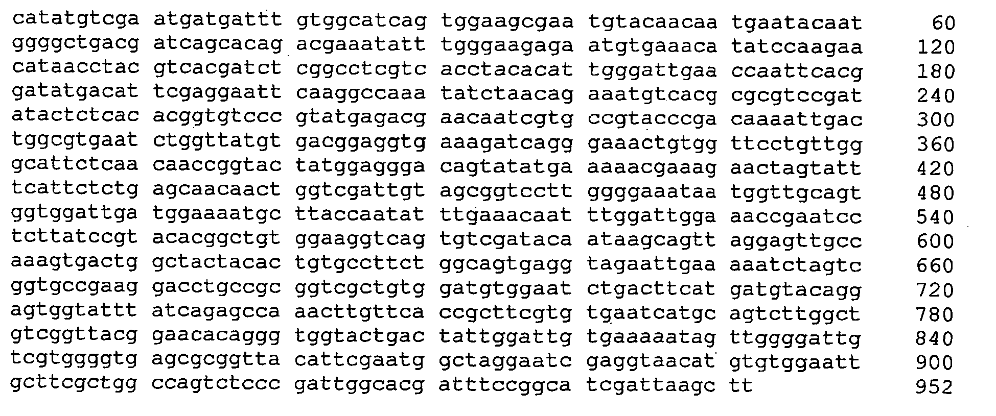
-
- <110> INSTYTUT BIOTECHNOLOGII I ANTYBIOTYKÓW INSTYTUT PARAZYTOLOGII PAN
- <120> Inclusion bodies for the oral vaccination of animals
- <130> PZ/0060/RW
- <140>
PCT/PL03/00151
<141> 2003-12-31 - <150>
PL358081
<151> 2002-12-31 - <160> 17
- <170> PatentIn version 3.1
- <210> 1
<211> 991
<212> DNA
<213> Fasciola hepatica - <220>
<221> gene
<222> (1)..(991)
<223> - <220>
<221> gene
<222> (1)..(991)
<223> liver fluke cysteine protease - <400> 1

- <210> 2
<211> 1198
<212> DNA
<213> artificial sequence - <220>
<223> CWekGlyKatwBluescr - <220>
<221> CDS
<222> (4)..(1194)
<223> sequence coding the hybrid protein HBV core domain:: catalytic do main of the liver fluke cysteine protease - <400> 2


- <210> 3
<211> 396
<212> PRT
<213> artificial sequence - <220>
<223> CWekGlyKatwBluescr - <400> 3


- <210> 4
<211> 1198
<212> DNA
<213> Artificial sequence - <220>
<223> CWekGlyKatwPIG - <220>
<221> CDS
<222> (4)..(1194)
<223> sequence coding the hybrid protein HBV core domain::catalytic dom ain of the liver fluke cysteine protease - <400> 4


- <210> 5
<211> 396
<212> PRT
<213> Artificial sequence - <220>
<223> CWekGlyKatwPIG - <400> 5


- <210> 6
<211> 1127
<212> DNA
<213> artificial sequence - <220>
<223> Moty_cat_Ckrd - <220>
<221> CDS
<222> (9)..(1115)
<223> sequence coding the hybrid protein HBV core domain::catalytic dom ain of the liver fluke cysteine protease - <400> 6


- <210> 7
<211> 368
<212> PRT
<213> artificial sequence - <220>
<223> Moty_cat_Ckrd - <400> 7


- <210> 8
<211> 699
<212> DNA
<213> artificial sequence - <220>
<223> KAT_Nasza_MOT1_MOT2 - <220>
<221> CDS
<222> (9)..(695)
<223> sequence coding the catalytic domain of the liver fluke cysteine protease - <400> 8


- <210> 9
<211> 228
<212> PRT
<213> artificial sequence - <220>
<223> KAT_Nasza_MOT1_MOT2 - <400> 9


- <210> 10
<211> 694
<212> DNA
<213> artificial sequence - <220>
<223> sequence coding the catalytic domain of the liver fluke cysteine protease - <220>
<221> CDS
<222> (4)..(690)
<223> sequence coding the catalytic domain of the liver fluke cysteine protease (fig.11 ) - <400> 10


- <210> 11
<211> 228
<212> PRT
<213> artificial sequence - <220>
<223> sequence coding the catalytic domain of the liver fluke cysteine protease - <400> 11

- <210> 12
<211> 686
<212> DNA
<213> artificial sequence - <220>
<223> MOT_DO_C_POP_MOT1_MOT3_W_BLUESCRIPCIE - <220>
<221> gene
<222> (1)..(686)
<223> - <400> 12


- <210> 13
<211> 681
<212> DNA
<213> artificial sequence - <220>
<223> MOT_DO_C_POP_MOT1_MOT3_W_PIG - <220>
<221> gene
<222> (1)..(681)
<223> - <400> 13

- <210> 14
<211> 1116
<212> DNA
<213> artificial sequence - <220>
<223> Moty_cat_Ckrd_w_PIG - <220>
<221> CDS
<222> (4)..(1110)
<223> sequence coding the hybrid protein HBV core domain::catalytic dom ain of the liver fluke cysteine protease - <400> 14


- <210> 15
<211> 368
<212> PRT
<213> artificial sequence - <220>
<223> Moty_cat_Ckrd_w_PIG - <400> 15

- <210> 16
<211> 952
<212> DNA
<213> artificial sequence - <220>
<223> Cala_Mot_N - <220>
<221> CDS
<222> (4)..(948)
<223> sequence coding the precursor protein of the liver fluke cysteine protease (both the catalytic and leading domain) - <400> 16

- <210> 17
<211> 314
<212> PRT
<213> artificial sequence - <220>
<223> Cala_Mot_N
<400> 17
Claims (12)
- Inclusion bodies containing a polypeptide containing the amino-acid sequence of the sequence of the liver fluke cysteine protease, possibly fused to the amino-acid sequence of the HBV core protein, for use as an oral vaccine against the liver fluke.
- Inclusion bodies according to Claim 1, characterised in that they are produced in E.coli.
- Inclusion bodies according to Claim 1, characterised in that the polypeptide contains one of the following sequences:- the sequence of the catalytic domain of the liver fluke cysteine protease fused to the sequence of the HBV core protein,- the sequence of the leading domain of the liver fluke cysteine protease fused to the sequence of the HBV core protein,- the sequence of the catalytic domain of the liver fluke cysteine protease fused to the sequence of the HBV core protein and glycine residues, forming a glycine bridge structure,- the sequence of the catalytic domain of the liver fluke cysteine protease,- the sequence of the precursor protein of the liver fluke cysteine protease encompassing the catalytic and leading domains.
- Inclusion bodies according to Claim 1, characterised in that the polypeptide contains one of the sequences encoded by the sequences presented in Figures 7 to 15.
- Use of a polypeptide in the form of inclusion bodies containing the amino-acid sequence of the sequence of the liver fluke cysteine protease, possibly fused to the amino-acid sequence of the HBV core protein, in the preparation of a vaccine against the liver fluke.
- Use according to Claim 5, characterised in that the polypeptide is produced in a genetically modified microorganism.
- Use according to Claim 5, characterised in that the polypeptide contains one of the following sequences:- the sequence of the catalytic domain of the liver fluke cysteine protease fused to the sequence of the HBV core protein,- the sequence of the leading domain of the liver fluke cysteine protease fused to the sequence of the HBV core protein,- the sequence of the catalytic domain of the liver fluke cysteine protease fused to the sequence of the HBV core protein and glycine residues, forming a glycine bridge structure,- the sequence of the catalytic domain of the liver fluke cysteine protease,- the sequence of the precursor protein of the liver fluke cysteine protease encompassing the catalytic and leading domains.
- Use according to Claim 5, characterised in that the polypeptide contains one of the sequences encoded by the sequences presented in Figures 7 to 15.
- An oral vaccine against the liver fluke containing the antigen and possibly pharmaceutically acceptable carrier and/or adjuvant, characterised in that as the antigen it contains a polypeptide in the form of inclusion bodies containing the amino-acid sequence of the sequence of the liver fluke cysteine protease, possibly fused to the amino-acid sequence of the HBV core protein.
- A vaccine according to Claim 9, characterised in that the polypeptide is produced in a genetically modified microorganism.
- A vaccine according to Claim 9, characterised in that the polypeptide contains one of the following sequences:- the sequence of the catalytic domain of the liver fluke cysteine protease fused to the sequence of the HBV core protein,- the sequence of the leading domain of the liver fluke cysteine protease fused to the sequence of the HBV core protein,- the sequence of the catalytic domain of the liver fluke cysteine protease fused to the sequence of the HBV core protein and glycine residues, forming a glycine bridge structure,- the sequence of the catalytic domain of the liver fluke cysteine protease,- the sequence of the precursor protein of the liver fluke cysteine protease encompassing the catalytic and leading domains.
- A vaccine according to Claim 9, characterised in that the polypeptide contains one of the sequences encoded by the sequences presented in Figures 7 to 15.
Applications Claiming Priority (3)
| Application Number | Priority Date | Filing Date | Title |
|---|---|---|---|
| PL2358081 | 2002-12-31 | ||
| PL358081A PL196114B1 (en) | 2002-12-31 | 2002-12-31 | Inclusion bodies as antigenes in oral immunization of animals |
| PCT/PL2003/000151 WO2004058816A2 (en) | 2002-12-31 | 2003-12-31 | Inclusion bodies for the oral vaccination of animals |
Publications (2)
| Publication Number | Publication Date |
|---|---|
| EP1578791A2 EP1578791A2 (en) | 2005-09-28 |
| EP1578791B1 true EP1578791B1 (en) | 2008-03-12 |
Family
ID=32678127
Family Applications (1)
| Application Number | Title | Priority Date | Filing Date |
|---|---|---|---|
| EP03789657A Expired - Lifetime EP1578791B1 (en) | 2002-12-31 | 2003-12-31 | Inclusion bodies for the oral vaccination of animals |
Country Status (7)
| Country | Link |
|---|---|
| EP (1) | EP1578791B1 (en) |
| AT (1) | ATE388962T1 (en) |
| AU (1) | AU2003294191B2 (en) |
| DE (1) | DE60319741T2 (en) |
| NZ (1) | NZ541291A (en) |
| PL (1) | PL196114B1 (en) |
| WO (1) | WO2004058816A2 (en) |
Families Citing this family (4)
| Publication number | Priority date | Publication date | Assignee | Title |
|---|---|---|---|---|
| NZ591025A (en) | 2008-08-21 | 2013-01-25 | Alvine Pharmaceuticals Inc | Formulation for oral administration of proteins |
| EP2251025A1 (en) * | 2009-05-12 | 2010-11-17 | Universitat Autònoma De Barcelona | Use of inclusion bodies as therapeutic agents |
| NL2023863B1 (en) * | 2019-09-20 | 2021-05-25 | Univ Griffith | Protein particles and uses thereof |
| CN116514993B (en) * | 2023-03-29 | 2023-09-29 | 中山大学附属第三医院 | Liver fluke oral spore vaccine and preparation method thereof |
Family Cites Families (2)
| Publication number | Priority date | Publication date | Assignee | Title |
|---|---|---|---|---|
| WO1994017820A1 (en) * | 1993-02-05 | 1994-08-18 | Daratech Pty. Ltd. | Polypeptides obtainable from species of fasciola, and vaccines, methods of treatment and dna sequences of the same |
| PL201419B1 (en) * | 2002-12-04 | 2009-04-30 | Inst Biotechnologii I Antybiot | Chimeric protein, sequence, construction, vegetable cell, method of obtaining chimeric protein and transgenic plant, transgenic plant, application of transgenic plant and chimeric protein |
-
2002
- 2002-12-31 PL PL358081A patent/PL196114B1/en unknown
-
2003
- 2003-12-31 NZ NZ541291A patent/NZ541291A/en not_active IP Right Cessation
- 2003-12-31 DE DE60319741T patent/DE60319741T2/en not_active Expired - Lifetime
- 2003-12-31 AU AU2003294191A patent/AU2003294191B2/en not_active Ceased
- 2003-12-31 WO PCT/PL2003/000151 patent/WO2004058816A2/en not_active Ceased
- 2003-12-31 EP EP03789657A patent/EP1578791B1/en not_active Expired - Lifetime
- 2003-12-31 AT AT03789657T patent/ATE388962T1/en not_active IP Right Cessation
Also Published As
| Publication number | Publication date |
|---|---|
| AU2003294191B2 (en) | 2010-11-25 |
| ATE388962T1 (en) | 2008-03-15 |
| AU2003294191A8 (en) | 2004-07-22 |
| PL358081A1 (en) | 2004-07-12 |
| DE60319741D1 (en) | 2008-04-24 |
| NZ541291A (en) | 2008-06-30 |
| WO2004058816A3 (en) | 2004-10-14 |
| DE60319741T2 (en) | 2009-04-02 |
| AU2003294191A1 (en) | 2004-07-22 |
| PL196114B1 (en) | 2007-12-31 |
| EP1578791A2 (en) | 2005-09-28 |
| WO2004058816A2 (en) | 2004-07-15 |
Similar Documents
| Publication | Publication Date | Title |
|---|---|---|
| JP2907813B2 (en) | Antigenic protein for preventing coccidiosis and vaccine containing the same | |
| CA2101070C (en) | Antigen-presenting capsid with fusion ms2-coat protein | |
| Lu et al. | Passive protection of shrimp against white spot syndrome virus (WSSV) using specific antibody from egg yolk of chickens immunized with inactivated virus or a WSSV-DNA vaccine | |
| JP2013513548A (en) | Methods and compositions for use in coccidiosis vaccines | |
| JP2009077713A (en) | NUCLEIC ACIDS ENCODING RECOMBINANT 56 AND 82 kDa ANTIGENS FROM GAMETOCYTES OF EIMERIA MAXIMA AND THEIR USE | |
| JPH03504336A (en) | Recombinant poxvirus and streptococcal M protein vaccines | |
| JPS60500215A (en) | Protective peptide antigen | |
| JP5132780B2 (en) | Fishery subunit vaccine | |
| Gao et al. | Saccharomyces cerevisiae oral immunization in mice using multi-antigen of the African swine fever virus elicits a robust immune response | |
| Ogrina et al. | Bacterial expression systems based on Tymovirus-like particles for the presentation of vaccine antigens | |
| AU2003294191B2 (en) | Inclusion bodies for the oral vaccination of animals | |
| CN110891597B (en) | Composition for preventing and treating Mycoplasma synoviae infection in poultry | |
| JPH02500327A (en) | Genetically engineered vaccines against mycobacteria | |
| JP2024512103A (en) | Highly attenuated replication-competent recombinant poxvirus as a vaccine platform and its use | |
| JPH02502878A (en) | Proteins and their production methods, DNA sequences, antibodies and their applications, poxviruses, transformed or infected cells and pharmaceutical formulations useful for the prevention of toxoplasmosis. | |
| JPH07508520A (en) | combination polypeptide antigen | |
| KR101127926B1 (en) | Iridovirus antigenic peptide and vaccine comprising the same | |
| CN115820679B (en) | A circular RNA vaccine against porcine epidemic diarrhea virus, its construction method and application | |
| CA2006412C (en) | Eimeria tenella vaccine | |
| CN111620954B (en) | Fusion protein for resisting African swine fever virus and preparation method and application thereof | |
| CN102847168A (en) | Design and construction of nucleic acid vaccine PV-Fn for preventing cow mastitis | |
| TWI386223B (en) | Duck hepatitis vaccine and its preparation method | |
| EP1603938B1 (en) | Inclusion bodies as antigens for the oral vaccination of animals against csfv | |
| Liew | Vaccination strategies of tropical diseases | |
| US20220323569A1 (en) | Plant-produced vlps and ric vaccines |
Legal Events
| Date | Code | Title | Description |
|---|---|---|---|
| PUAI | Public reference made under article 153(3) epc to a published international application that has entered the european phase |
Free format text: ORIGINAL CODE: 0009012 |
|
| 17P | Request for examination filed |
Effective date: 20050727 |
|
| AK | Designated contracting states |
Kind code of ref document: A2 Designated state(s): AT BE BG CH CY CZ DE DK EE ES FI FR GB GR HU IE IT LI LU MC NL PT RO SE SI SK TR |
|
| AX | Request for extension of the european patent |
Extension state: AL LT LV MK |
|
| DAX | Request for extension of the european patent (deleted) | ||
| 17Q | First examination report despatched |
Effective date: 20051004 |
|
| GRAP | Despatch of communication of intention to grant a patent |
Free format text: ORIGINAL CODE: EPIDOSNIGR1 |
|
| GRAS | Grant fee paid |
Free format text: ORIGINAL CODE: EPIDOSNIGR3 |
|
| GRAA | (expected) grant |
Free format text: ORIGINAL CODE: 0009210 |
|
| AK | Designated contracting states |
Kind code of ref document: B1 Designated state(s): AT BE BG CH CY CZ DE DK EE ES FI FR GB GR HU IE IT LI LU MC NL PT RO SE SI SK TR |
|
| REG | Reference to a national code |
Ref country code: GB Ref legal event code: FG4D |
|
| REG | Reference to a national code |
Ref country code: CH Ref legal event code: EP |
|
| REG | Reference to a national code |
Ref country code: IE Ref legal event code: FG4D |
|
| REF | Corresponds to: |
Ref document number: 60319741 Country of ref document: DE Date of ref document: 20080424 Kind code of ref document: P |
|
| PG25 | Lapsed in a contracting state [announced via postgrant information from national office to epo] |
Ref country code: FI Free format text: LAPSE BECAUSE OF FAILURE TO SUBMIT A TRANSLATION OF THE DESCRIPTION OR TO PAY THE FEE WITHIN THE PRESCRIBED TIME-LIMIT Effective date: 20080312 |
|
| PG25 | Lapsed in a contracting state [announced via postgrant information from national office to epo] |
Ref country code: AT Free format text: LAPSE BECAUSE OF FAILURE TO SUBMIT A TRANSLATION OF THE DESCRIPTION OR TO PAY THE FEE WITHIN THE PRESCRIBED TIME-LIMIT Effective date: 20080312 |
|
| NLV1 | Nl: lapsed or annulled due to failure to fulfill the requirements of art. 29p and 29m of the patents act | ||
| PG25 | Lapsed in a contracting state [announced via postgrant information from national office to epo] |
Ref country code: BE Free format text: LAPSE BECAUSE OF FAILURE TO SUBMIT A TRANSLATION OF THE DESCRIPTION OR TO PAY THE FEE WITHIN THE PRESCRIBED TIME-LIMIT Effective date: 20080312 Ref country code: SI Free format text: LAPSE BECAUSE OF FAILURE TO SUBMIT A TRANSLATION OF THE DESCRIPTION OR TO PAY THE FEE WITHIN THE PRESCRIBED TIME-LIMIT Effective date: 20080312 |
|
| PG25 | Lapsed in a contracting state [announced via postgrant information from national office to epo] |
Ref country code: ES Free format text: LAPSE BECAUSE OF FAILURE TO SUBMIT A TRANSLATION OF THE DESCRIPTION OR TO PAY THE FEE WITHIN THE PRESCRIBED TIME-LIMIT Effective date: 20080623 Ref country code: CZ Free format text: LAPSE BECAUSE OF FAILURE TO SUBMIT A TRANSLATION OF THE DESCRIPTION OR TO PAY THE FEE WITHIN THE PRESCRIBED TIME-LIMIT Effective date: 20080312 Ref country code: PT Free format text: LAPSE BECAUSE OF FAILURE TO SUBMIT A TRANSLATION OF THE DESCRIPTION OR TO PAY THE FEE WITHIN THE PRESCRIBED TIME-LIMIT Effective date: 20080818 Ref country code: SE Free format text: LAPSE BECAUSE OF FAILURE TO SUBMIT A TRANSLATION OF THE DESCRIPTION OR TO PAY THE FEE WITHIN THE PRESCRIBED TIME-LIMIT Effective date: 20080612 Ref country code: SK Free format text: LAPSE BECAUSE OF FAILURE TO SUBMIT A TRANSLATION OF THE DESCRIPTION OR TO PAY THE FEE WITHIN THE PRESCRIBED TIME-LIMIT Effective date: 20080312 |
|
| PG25 | Lapsed in a contracting state [announced via postgrant information from national office to epo] |
Ref country code: NL Free format text: LAPSE BECAUSE OF FAILURE TO SUBMIT A TRANSLATION OF THE DESCRIPTION OR TO PAY THE FEE WITHIN THE PRESCRIBED TIME-LIMIT Effective date: 20080312 Ref country code: RO Free format text: LAPSE BECAUSE OF FAILURE TO SUBMIT A TRANSLATION OF THE DESCRIPTION OR TO PAY THE FEE WITHIN THE PRESCRIBED TIME-LIMIT Effective date: 20080312 |
|
| ET | Fr: translation filed | ||
| PLBE | No opposition filed within time limit |
Free format text: ORIGINAL CODE: 0009261 |
|
| STAA | Information on the status of an ep patent application or granted ep patent |
Free format text: STATUS: NO OPPOSITION FILED WITHIN TIME LIMIT |
|
| PG25 | Lapsed in a contracting state [announced via postgrant information from national office to epo] |
Ref country code: DK Free format text: LAPSE BECAUSE OF FAILURE TO SUBMIT A TRANSLATION OF THE DESCRIPTION OR TO PAY THE FEE WITHIN THE PRESCRIBED TIME-LIMIT Effective date: 20080312 |
|
| 26N | No opposition filed |
Effective date: 20081215 |
|
| PG25 | Lapsed in a contracting state [announced via postgrant information from national office to epo] |
Ref country code: BG Free format text: LAPSE BECAUSE OF FAILURE TO SUBMIT A TRANSLATION OF THE DESCRIPTION OR TO PAY THE FEE WITHIN THE PRESCRIBED TIME-LIMIT Effective date: 20080612 Ref country code: EE Free format text: LAPSE BECAUSE OF FAILURE TO SUBMIT A TRANSLATION OF THE DESCRIPTION OR TO PAY THE FEE WITHIN THE PRESCRIBED TIME-LIMIT Effective date: 20080312 |
|
| PG25 | Lapsed in a contracting state [announced via postgrant information from national office to epo] |
Ref country code: MC Free format text: LAPSE BECAUSE OF NON-PAYMENT OF DUE FEES Effective date: 20081231 |
|
| REG | Reference to a national code |
Ref country code: CH Ref legal event code: PL |
|
| PG25 | Lapsed in a contracting state [announced via postgrant information from national office to epo] |
Ref country code: IT Free format text: LAPSE BECAUSE OF FAILURE TO SUBMIT A TRANSLATION OF THE DESCRIPTION OR TO PAY THE FEE WITHIN THE PRESCRIBED TIME-LIMIT Effective date: 20080312 |
|
| PG25 | Lapsed in a contracting state [announced via postgrant information from national office to epo] |
Ref country code: CY Free format text: LAPSE BECAUSE OF FAILURE TO SUBMIT A TRANSLATION OF THE DESCRIPTION OR TO PAY THE FEE WITHIN THE PRESCRIBED TIME-LIMIT Effective date: 20080312 |
|
| PG25 | Lapsed in a contracting state [announced via postgrant information from national office to epo] |
Ref country code: LI Free format text: LAPSE BECAUSE OF NON-PAYMENT OF DUE FEES Effective date: 20081231 Ref country code: CH Free format text: LAPSE BECAUSE OF NON-PAYMENT OF DUE FEES Effective date: 20081231 |
|
| PG25 | Lapsed in a contracting state [announced via postgrant information from national office to epo] |
Ref country code: HU Free format text: LAPSE BECAUSE OF FAILURE TO SUBMIT A TRANSLATION OF THE DESCRIPTION OR TO PAY THE FEE WITHIN THE PRESCRIBED TIME-LIMIT Effective date: 20080913 Ref country code: LU Free format text: LAPSE BECAUSE OF NON-PAYMENT OF DUE FEES Effective date: 20081231 |
|
| PG25 | Lapsed in a contracting state [announced via postgrant information from national office to epo] |
Ref country code: TR Free format text: LAPSE BECAUSE OF FAILURE TO SUBMIT A TRANSLATION OF THE DESCRIPTION OR TO PAY THE FEE WITHIN THE PRESCRIBED TIME-LIMIT Effective date: 20080312 |
|
| PG25 | Lapsed in a contracting state [announced via postgrant information from national office to epo] |
Ref country code: GR Free format text: LAPSE BECAUSE OF FAILURE TO SUBMIT A TRANSLATION OF THE DESCRIPTION OR TO PAY THE FEE WITHIN THE PRESCRIBED TIME-LIMIT Effective date: 20080613 |
|
| PGFP | Annual fee paid to national office [announced via postgrant information from national office to epo] |
Ref country code: GB Payment date: 20101223 Year of fee payment: 8 |
|
| PGFP | Annual fee paid to national office [announced via postgrant information from national office to epo] |
Ref country code: FR Payment date: 20120104 Year of fee payment: 9 Ref country code: IE Payment date: 20111220 Year of fee payment: 9 |
|
| PGFP | Annual fee paid to national office [announced via postgrant information from national office to epo] |
Ref country code: DE Payment date: 20111222 Year of fee payment: 9 |
|
| GBPC | Gb: european patent ceased through non-payment of renewal fee |
Effective date: 20121231 |
|
| REG | Reference to a national code |
Ref country code: IE Ref legal event code: MM4A |
|
| REG | Reference to a national code |
Ref country code: FR Ref legal event code: ST Effective date: 20130830 |
|
| REG | Reference to a national code |
Ref country code: DE Ref legal event code: R119 Ref document number: 60319741 Country of ref document: DE Effective date: 20130702 |
|
| PG25 | Lapsed in a contracting state [announced via postgrant information from national office to epo] |
Ref country code: IE Free format text: LAPSE BECAUSE OF NON-PAYMENT OF DUE FEES Effective date: 20121231 Ref country code: DE Free format text: LAPSE BECAUSE OF NON-PAYMENT OF DUE FEES Effective date: 20130702 |
|
| PG25 | Lapsed in a contracting state [announced via postgrant information from national office to epo] |
Ref country code: GB Free format text: LAPSE BECAUSE OF NON-PAYMENT OF DUE FEES Effective date: 20121231 Ref country code: FR Free format text: LAPSE BECAUSE OF NON-PAYMENT OF DUE FEES Effective date: 20130102 |