EP1577390B1 - Human cancer-related genes, the products encoded thereby and applications thereof - Google Patents
Human cancer-related genes, the products encoded thereby and applications thereof Download PDFInfo
- Publication number
- EP1577390B1 EP1577390B1 EP03782072A EP03782072A EP1577390B1 EP 1577390 B1 EP1577390 B1 EP 1577390B1 EP 03782072 A EP03782072 A EP 03782072A EP 03782072 A EP03782072 A EP 03782072A EP 1577390 B1 EP1577390 B1 EP 1577390B1
- Authority
- EP
- European Patent Office
- Prior art keywords
- laptm4b
- seq
- hepatocellular carcinoma
- cancer
- cells
- Prior art date
- Legal status (The legal status is an assumption and is not a legal conclusion. Google has not performed a legal analysis and makes no representation as to the accuracy of the status listed.)
- Expired - Lifetime
Links
Images

Classifications
-
- C—CHEMISTRY; METALLURGY
- C07—ORGANIC CHEMISTRY
- C07K—PEPTIDES
- C07K14/00—Peptides having more than 20 amino acids; Gastrins; Somatostatins; Melanotropins; Derivatives thereof
- C07K14/435—Peptides having more than 20 amino acids; Gastrins; Somatostatins; Melanotropins; Derivatives thereof from animals; from humans
- C07K14/46—Peptides having more than 20 amino acids; Gastrins; Somatostatins; Melanotropins; Derivatives thereof from animals; from humans from vertebrates
- C07K14/47—Peptides having more than 20 amino acids; Gastrins; Somatostatins; Melanotropins; Derivatives thereof from animals; from humans from vertebrates from mammals
- C07K14/4701—Peptides having more than 20 amino acids; Gastrins; Somatostatins; Melanotropins; Derivatives thereof from animals; from humans from vertebrates from mammals not used
- C07K14/4748—Tumour specific antigens; Tumour rejection antigen precursors [TRAP], e.g. MAGE
-
- G—PHYSICS
- G01—MEASURING; TESTING
- G01N—INVESTIGATING OR ANALYSING MATERIALS BY DETERMINING THEIR CHEMICAL OR PHYSICAL PROPERTIES
- G01N33/00—Investigating or analysing materials by specific methods not covered by groups G01N1/00 - G01N31/00
- G01N33/48—Biological material, e.g. blood, urine; Haemocytometers
- G01N33/50—Chemical analysis of biological material, e.g. blood, urine; Testing involving biospecific ligand binding methods; Immunological testing
- G01N33/53—Immunoassay; Biospecific binding assay; Materials therefor
- G01N33/575—Immunoassay; Biospecific binding assay; Materials therefor for cancer
- G01N33/5758—Immunoassay; Biospecific binding assay; Materials therefor for cancer involving compounds serving as markers for tumours, cancers or neoplasias, e.g. cellular determinants, receptors, heat shock/stress proteins, A-protein, oligosaccharides or metabolites
- G01N33/5759—Immunoassay; Biospecific binding assay; Materials therefor for cancer involving compounds serving as markers for tumours, cancers or neoplasias, e.g. cellular determinants, receptors, heat shock/stress proteins, A-protein, oligosaccharides or metabolites involving compounds localised on the membrane of tumour or cancer cells
-
- A—HUMAN NECESSITIES
- A61—MEDICAL OR VETERINARY SCIENCE; HYGIENE
- A61P—SPECIFIC THERAPEUTIC ACTIVITY OF CHEMICAL COMPOUNDS OR MEDICINAL PREPARATIONS
- A61P35/00—Antineoplastic agents
-
- C—CHEMISTRY; METALLURGY
- C07—ORGANIC CHEMISTRY
- C07K—PEPTIDES
- C07K14/00—Peptides having more than 20 amino acids; Gastrins; Somatostatins; Melanotropins; Derivatives thereof
- C07K14/82—Translation products from oncogenes
-
- C—CHEMISTRY; METALLURGY
- C07—ORGANIC CHEMISTRY
- C07K—PEPTIDES
- C07K16/00—Immunoglobulins [IG], e.g. monoclonal or polyclonal antibodies
- C07K16/18—Immunoglobulins [IG], e.g. monoclonal or polyclonal antibodies against material from animals or humans
- C07K16/28—Immunoglobulins [IG], e.g. monoclonal or polyclonal antibodies against material from animals or humans against receptors, cell surface antigens or cell surface determinants
- C07K16/30—Immunoglobulins [IG], e.g. monoclonal or polyclonal antibodies against material from animals or humans against receptors, cell surface antigens or cell surface determinants from tumour cells
-
- C—CHEMISTRY; METALLURGY
- C12—BIOCHEMISTRY; BEER; SPIRITS; WINE; VINEGAR; MICROBIOLOGY; ENZYMOLOGY; MUTATION OR GENETIC ENGINEERING
- C12Q—MEASURING OR TESTING PROCESSES INVOLVING ENZYMES, NUCLEIC ACIDS OR MICROORGANISMS; COMPOSITIONS OR TEST PAPERS THEREFOR; PROCESSES OF PREPARING SUCH COMPOSITIONS; CONDITION-RESPONSIVE CONTROL IN MICROBIOLOGICAL OR ENZYMOLOGICAL PROCESSES
- C12Q1/00—Measuring or testing processes involving enzymes, nucleic acids or microorganisms; Compositions therefor; Processes of preparing such compositions
- C12Q1/68—Measuring or testing processes involving enzymes, nucleic acids or microorganisms; Compositions therefor; Processes of preparing such compositions involving nucleic acids
- C12Q1/6876—Nucleic acid products used in the analysis of nucleic acids, e.g. primers or probes
- C12Q1/6883—Nucleic acid products used in the analysis of nucleic acids, e.g. primers or probes for diseases caused by alterations of genetic material
- C12Q1/6886—Nucleic acid products used in the analysis of nucleic acids, e.g. primers or probes for diseases caused by alterations of genetic material for cancer
-
- G—PHYSICS
- G01—MEASURING; TESTING
- G01N—INVESTIGATING OR ANALYSING MATERIALS BY DETERMINING THEIR CHEMICAL OR PHYSICAL PROPERTIES
- G01N33/00—Investigating or analysing materials by specific methods not covered by groups G01N1/00 - G01N31/00
- G01N33/48—Biological material, e.g. blood, urine; Haemocytometers
- G01N33/50—Chemical analysis of biological material, e.g. blood, urine; Testing involving biospecific ligand binding methods; Immunological testing
- G01N33/53—Immunoassay; Biospecific binding assay; Materials therefor
- G01N33/575—Immunoassay; Biospecific binding assay; Materials therefor for cancer
-
- G—PHYSICS
- G01—MEASURING; TESTING
- G01N—INVESTIGATING OR ANALYSING MATERIALS BY DETERMINING THEIR CHEMICAL OR PHYSICAL PROPERTIES
- G01N33/00—Investigating or analysing materials by specific methods not covered by groups G01N1/00 - G01N31/00
- G01N33/48—Biological material, e.g. blood, urine; Haemocytometers
- G01N33/50—Chemical analysis of biological material, e.g. blood, urine; Testing involving biospecific ligand binding methods; Immunological testing
- G01N33/53—Immunoassay; Biospecific binding assay; Materials therefor
- G01N33/575—Immunoassay; Biospecific binding assay; Materials therefor for cancer
- G01N33/5758—Immunoassay; Biospecific binding assay; Materials therefor for cancer involving compounds serving as markers for tumours, cancers or neoplasias, e.g. cellular determinants, receptors, heat shock/stress proteins, A-protein, oligosaccharides or metabolites
-
- C—CHEMISTRY; METALLURGY
- C07—ORGANIC CHEMISTRY
- C07K—PEPTIDES
- C07K2317/00—Immunoglobulins specific features
- C07K2317/70—Immunoglobulins specific features characterized by effect upon binding to a cell or to an antigen
- C07K2317/73—Inducing cell death, e.g. apoptosis, necrosis or inhibition of cell proliferation
-
- C—CHEMISTRY; METALLURGY
- C12—BIOCHEMISTRY; BEER; SPIRITS; WINE; VINEGAR; MICROBIOLOGY; ENZYMOLOGY; MUTATION OR GENETIC ENGINEERING
- C12Q—MEASURING OR TESTING PROCESSES INVOLVING ENZYMES, NUCLEIC ACIDS OR MICROORGANISMS; COMPOSITIONS OR TEST PAPERS THEREFOR; PROCESSES OF PREPARING SUCH COMPOSITIONS; CONDITION-RESPONSIVE CONTROL IN MICROBIOLOGICAL OR ENZYMOLOGICAL PROCESSES
- C12Q2600/00—Oligonucleotides characterized by their use
- C12Q2600/156—Polymorphic or mutational markers
-
- C—CHEMISTRY; METALLURGY
- C12—BIOCHEMISTRY; BEER; SPIRITS; WINE; VINEGAR; MICROBIOLOGY; ENZYMOLOGY; MUTATION OR GENETIC ENGINEERING
- C12Q—MEASURING OR TESTING PROCESSES INVOLVING ENZYMES, NUCLEIC ACIDS OR MICROORGANISMS; COMPOSITIONS OR TEST PAPERS THEREFOR; PROCESSES OF PREPARING SUCH COMPOSITIONS; CONDITION-RESPONSIVE CONTROL IN MICROBIOLOGICAL OR ENZYMOLOGICAL PROCESSES
- C12Q2600/00—Oligonucleotides characterized by their use
- C12Q2600/158—Expression markers
-
- G—PHYSICS
- G01—MEASURING; TESTING
- G01N—INVESTIGATING OR ANALYSING MATERIALS BY DETERMINING THEIR CHEMICAL OR PHYSICAL PROPERTIES
- G01N2333/00—Assays involving biological materials from specific organisms or of a specific nature
- G01N2333/435—Assays involving biological materials from specific organisms or of a specific nature from animals; from humans
- G01N2333/705—Assays involving receptors, cell surface antigens or cell surface determinants
Definitions
- the present invention relates to a human cancer-related gene, its encoded products and their applications in genetic engineering and protein engineering areas, as well as in medical diagnosis and treatment.
- Hepatocellular carcinoma is one of the most serious among cancer diseases. It is reported that the new cases of primary hepatocellular carcinoma exceeds over one million worldwide each year. 70% of the new cases occur in Asia, and about 40-45% of the worldwide new cases occur in China. The total number of new hepatocellular carcinoma cases every year in China is about 450,000, and the number is increasing, especially in those between ages 20-60. The high incidence, difficulty in early diagnosis, fast growing rate, high reoccurrence, and the high mortality rate make HCC a most malignant cancer. Most HCC patients have already progressed to the intermediate stage or late stage when diagnosed, and they can only survive for 3-6 months if without a proper treatment.
- Cancer is essentially a cellular hereditary disease. Although a great number of cancer-related genes have been discovered, the mechanisms of the cancerogenesis and the development remain to be elucidated.
- the known oncogenes can be divided into five categories according to the cellular localization and function of their encoded proteins: I. genes that encode growth factors, including sis, int-2, hst, fgf-5; II. genes that encode growth factor receptors, including erbB, erbB-2, fms, met, ros, and others; III.
- genes that encode signal transduction molecules in cytoplasm including abl, src, ras, raf, yes, fgr, fes, Ick, mos, and others; IV genes that encode regulatory molecules for cell proliferation and apoptosis, including bcl-1, bcl-2 and others; and V. genes that encode the nuclear DNA-binding proteins (transcription factors), such as myc, myb, fos, jun, B-lym, ski, ets, rel and others. It has been demonstrated that ras, src, myc, met and p53 etc. are the genes closely associated with HCC.
- the present invention derives from work on a novel human cancer-related gene designated as LAPTM4B. It comprises one of the following nucleotide sequences:
- SEQ ID N o : 6 is the allelic gene of SEQ ID N o : 1, consisting of 2264 bases. Its open reading frame starts from 17 to 1129 base. This sequence contains two tandemly arranged 19bp DNA segments, the sequence of which is gcttgg agctccagca gct. These 19bp DNA segments localized in nt 124 - nt 161 in SEQ ID N o : 6.
- the human cancer related LAPTM4B protein possesses the amino acid sequence of 4, and/or 5, and/or 7. Or it consists of the sequence 4, and/or the sequence 5, and/or the sequence 7 after one or several amino acid residues are replaced, deleted, or added. However, the above altered sequence 4, and/or the sequence 5, and/or the sequence 7 still have the same or similar activity to the unchanged sequence 4, and/or the sequence 5, and/or the sequence 7.
- Sequence 4 in the sequence listings consists of 317 amino acid residues encoded by the whole sequence of SEQ ID N o : 1. its molecular mass is 35kDa and the putative isoelectric point is 9.05.
- Sequence 5 in the sequence listings contains 226 amino acid residues encoded by the segment of bases from 274th to 954th in the SEQ ID N o : 1. Its molecular mass is 24kDa, and the putative isoelectric point is 4.65.
- the sequence 7 in the sequence listings is a protein containing 370 amino acid residues.
- LAPTM4B gene is widely expressed at different levels in sixteen normal tissues. Its transcriptive expression is very high in testis, cardiac muscle, and skeletal muscle, moderate in ovary, kidney, and pancreas, low in liver, spleen, small intestine, large intestine, and thymus, and is very low in lung and peripheral blood cells. In eight fetal tissues, the expression is high in heart, skeletal muscle, and kidney. In fetal livers, it is slightly higher than that in adult livers. However, its expression in some cancerous tissues is significantly upregulated. For instance, the Northern Blot analysis indicates that the transcription level in 87.3% (48/55) human hepatocellular carcinoma tissues is significantly higher than that in fetal livers and normal livers ( Figure 1-A ).
- LAPTM4B-35 protein expression is up regulated in some epithelium derived cancerous tissues and cell lines, such as stomach cancer, breast cancer, highly metastatic human lung cancer, and prostate cancer ( Figure 11 ).
- LAPTM4B-35 expression is greatly up regulated in cells of high metastasis potential compared with those of low metastasis potential. But in cell lines of human melanoma, either from in situ or metastatic cancer, it is not clearly expressed. Although LAPTM4B-35 is expressed at a low level in liver tissues of adult rats and mice, its expression is not obviously up regulated in either mouse ascetically qrown hepatocellular carcinoma or in the regenerated rat liver under a normal proliferation status.
- Table 1 Expression ratio of LAPTM4B-35 to LAPTM4B-24 in hepatocellular carcinoma tissue, paired non-cancerous liver tissue and normal liver tissue HCC PNL NL LAPTM4B-35 13.32 ⁇ 1.98 4.58 ⁇ 1.31 2.78 ⁇ 0.11 LAPTM4B-24 3.59 ⁇ 1.78 1.76 ⁇ 1.24 1.00 ⁇ 0.02 LAPTM4B-35/ LAPTM4B-24 (Ratio) 3.71 2.60 2.78 P ⁇ 0.01 HCC vs. PNL and NL
- LAPTM4B proteins in SEQ ID N o : 4, SEQ ID N o : 5 and SEQ ID N o : 7 have four fragments of membrane-spanning sequences, one N-glycosylation site, a typical lysosome and endosome targeting signals in the cytoplasmic region. They all belong to the protein superfamily of the tetratransmembrane proteins. However, they have various number of phosphorylation sites.
- SEQ ID N o : 4 LAPTM4B-35 can form a complex in plasma membrane with the integrin ⁇ 6 ⁇ 1 (Single specific receptor of laminin in the extracellular matrix) and the epidermal growth factor recceptor (EGFR) ( Figures 14-A , B, and C).
- LAPTM4B-35 may integrate in the plasma membrane the proliferation signals from both extracellular matrix and the growth factor. This can further elucidate molecular mechanism of the anchorage-dependent cell growth of normal eukaryotic cells, i.e. the eukaryotic cell growth needs not only the stimulating signal from the growth factor, but also a definite stimulating signal from extracellular matrix. It represents a break through in understanding the regulation mechanism of the cell proliferation.
- tyrosine group (Tyr 285 ) in the cytoplasmic region of LAPTM4B protein C terminal can be phosphorylated ( Figure 15-A ).
- the proliferation of transfected cells shows less dependence on the growth factor in serum, and the transfected cells can form large colonies in soft agar.
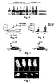
- Inoculation of NIH mouse with NIH3T3-AE cells can form a moderate malignant fibrosarcoma ( Figure 7 ), indicating the over expression of LAPTM4B-35 induces out-of-control cell proliferation.
- migration capability of the HLE-AE cells is strengthened and its capability to invade the Matrigel is remarkably enhanced, indicating that the over expression of LAPTM4B-35 accelerates the development of cell malignant phenotype.
- the cDNA of SEQ ID N o : 5 (an encoding sequence where 91 amino acids in the N terminal of LAPTM4B-35 is truncated) transfected mouse BHK, NIH3T3, and HLE hepatocellular carcinoma cell lines cannot survive for a long time.
- the result shows that the 91 amino acid sequence on the N terminal of SEQ ID N o : 4 LAPTM4B-35 protein play a crucial role in regulating cell proliferation.
- LAPTM4B-35 protein and LAPTM4B-24 protein have reciprocal, antagonistical functions in cell proliferation and survival.
- the overexpression of LAPTM4B-35 accelerates cellular malignant transformation, while the overexpression of LAPTM4B-24 facilitates the cell death.
- LAPTM4B gene may belong to the proto-oncogene family.
- inhibiting SEQ ID N o : 4 LAPTM4B-35 expression and strengthening SEQ ID N o: 5 LAPTM4B-24 expression may suppress the growth of hepatocellular carcinoma and reverse its malignant phenotype or progressively slow down its development.
- LAPTM4B-35 also promotes upregutation of the cell cycle regulators, such as cyclin D1 ( Figure 13-A ) and cyclin E ( Figure 13-B ), and also the over expression of some proto-oncogenes, such as c-Myc ( Figure 13-C ), c-Jun ( Figure 13-D ), and c-Fos ( Figure 13-E ) etc.
- the monoclonal and polyclonal antibodies for SEQ ID N o : 4 LAPTM4B-35 protein epitopes such as polyclonal LAPTM4B-EC2 232-241 -pAb for SEQ ID N o : 4 LAPTM4B-35 in the secondary extracellular region, polyclonal antibodies (LAPTM4B-N 1-99 -pAb and L-APTM4B-N 28-37 -pAb) for SEQ ID N o : 4 LAPTM4B-35 N terminal sequence, and monoclonal antibodies for LAPTM4B are important in studying the effects of LAPTM4B-35 and LAPTM4B-24 in cancer diagnosis and treatment ( Figures 2 , 3 , 8 , 11 , 12 , 14, 15 ).
- LAPTM4B-EC2 232-241 -pAb, lAPTM4B-N 1-99- pAb polyclonal antibodies and LAPTM4B-N 1-99 -mAb monoclonal antibody can be used to analyze LAPTM4B protein expression, intracellular localization, separation and purification, and protein-protein interaction. They can also be used to detect the antibody and antigen of LAPTM4B in blood ( Figure 8 ).
- LAPTM4B-EC2 232-241 -pAb can inhibit cancer cell proliferation ( Figure 12 ), Tyr 285 phosphorylation of LAPTM4B protein ( Figure 15-B ), and the phosphorylation and activation of signal molecules FAK ( Figure 16-A ) and MAPK ( Figure 16-B) .
- SEQ ID N o : 8 is the promoter sequence of LAPTM4B gene.
- the LAPTM4B gene promoter and the upstream sequence SEQ ID N o : 8 are cloned. There are no typical CCAAT (TTGCGCAAT), TATA cassettes in LAPTM4B gene promoter region. But various binding sites of transcription factors exist in the upstream region of LAPTM4B promoter, such as CREBP1/c-Jun, CEBP, PAX2/5/8, GATA, STAT, c-Ets-1, E2F, LYF-1, and c/v-Myb ( Figure 17A ). These transcription factors may specifically regulate LAPTM4B expression in cells of various tissues.
- LAPTM4B proteins The abnormal expression and activation of these transcription factors in cancer cells possibly lead to an unbalanced expression of LAPTM4B proteins.
- LAPTM4B proteins there are two highly homologous repeating sequences in the upstream domain of LAPTM4B promoter. It is worthwhile to study whether they are responsible to the regulation of LAPTM4B expression.
- a series of vectors consisting upstream region sequences of LAPTM4B promoter with different lengths-promoter-5' UTR-35bp encoding region-luciferase reporting gene is constructed, and these vectors are used to transfect into BEL7402 cells and HLE cells from human hepatocellular carcinoma HCC.
- the cells transfected with various vectors all show luciferase activity with various intensities, indicating the transcription activities in these segments.
- the smallest fragment is a DNA segment ( pGL-PF4 ) at about 38 bp in the upstream region of the transcription starting site. It possesses a basic promoting activity and functions as LAPTM4 core promoter.
- the activity of pGL-PF4 transfectant in BEL7402 is 20% of the reference promoter SV40, while the activity is low in HLE, only 6% of SV40 activity, about 1/3 of that in BEL7402.
- LATPM4B has two alleles, LATPM4B*1 and LATPM4B*2, i.e., SEQ ID N o : 6, is derived by PCR cloning.
- SEQ ID N o 6
- the difference between alleles * 1 and * 2 is the 19 bp sequence in the first exon 5' UTR.
- Allele * 1 has only one such sequence (nt 124-142), while * 2 contains two such sequences in a tightly tandem arrangement (124-142dup, taking G at the transcription starting site (TSS) as +1 numbering standard).
- LATPM4B genotypes detected in human population are * 1 / *1 , * 1 /* 2 and * 2 /* 2 , respectively ( Figure 10 ). Studies show that the risk of developing hepatocellular carcinoma for individuals with LATPM4B genotype * 2 /* 2 is 2.89 times higher than individuals with non-* 2 /* 2 type (Table 2).
- LATPM4B genotype in patients with esophagus carcinoma shows no difference from the normal population (Table 3). This indicates that LATPM4B * 2 /* 2 genotype correlates especially to susceptibility of hepatocellular carcinoma.
- LATPM4B allele LATPM4B* 2 provided by this invention can be used as a target to screen and diagnose people susceptible to hepatocellular carcinoma or having a high risk to develop hepatocellular carcinoma.
- using LATPM4B * 2 /* 2 genotype as a target to screen highly susceptible or high risk people can be more accurate..
- LATPM4B genotypes LATPM4B*2 encoded proteins or their antibodies, and/or LATPM4B extender and scavenger from human genome can all be used to screen people who are susceptible to hepatocellular carcinoma or having a high risk to develop hepatocellular carcinoma.
- the expression vectors containing sequences described in SEQ ID N o : 1, 2, 3, 6, 8, the transfection cell lines containing SEQ ID N o : 1, 2, 3, 6, 8 sequences, and the primers amplifying SEQ ID N o : 1, 2, 3, 6, 8 are all encompassed by this invention.
- hepatocellular carcinoma patients 50 males and 7 females, ranged in age from 35-70. Their average age was 54 ⁇ 6.0.
- the tissues tested came from surgically excided specimens.
- the blood samples for the control group were collected from 206 similarly aged people with no symptoms and no cancer according to clinic tests and from 209 new born babies' umbilical veins.
- the test tissues came from surgically excided specimens.
- Example 1 Northern Blot analysis of LAPTM4B expressions in four types of liver tissues at various proliferation and differentiation status.
- liver tissues at various proliferation and differentiation status were chosen. They were from normal adult livers (NL, with very little proliferation and high differentiation), fetus livers (FL, at vigorous proliferation and low differentiation), hepatocellular carcinoma (HCC, out of controled proliferation and abnormal differentiation), and paired non-cancerous livers (PNL, generally is of precancerous stage in an active proliferation status).
- the Northern Blot analysis was used to detect the transcription of gene in these tissues. RNA samples were extracted from 5 normal adult liver tissues freshly obtained from surgical excision: 5 liver tissues from abortive fetus, 55 HCC tissues, and 55 paired non-cancerous liver tissues. After electrophoretic separation, they were transferred to a nylon film and hybridized by Dig labeled probe.
- Band 1 was the sample from fetus livers.
- Band 2 was from the normal adult liver sample.
- Bands 3, 5, 7, and 9 were the samples from HCC.
- Bands 4, 6, 8, 10 were from PNL tissues.
- the results show that the expression of LAPTM4B in various liver tissues has the following order: HCC tissue > PNL tissue and fetus liver tissue > normal adult liver tissue.
- LC27 cDNA segment
- NL normal adult livers
- FL fetus livers
- HCC cancerous livers
- PNL paired non-cancerous liver
- the LC27 segment (426bp) was elongated by splicing homogenous sequences according to the EST to the 5' direction, and followed by RACE (rapid amplification of cDNA ends) and the high temperature RT-PCR techniques.
- Two full-length cDNA sequences i.e., SEQ ID N o . 2 and 3, were produced, and then confirmed by sequencing and BLAST program analysis.
- LAPTM4B gene at 5' terminal was obtained by biological informatics, and primers F1 and R1 were designed.
- LAPTM4B promoter and the upstream sequence was obtained by PCR using Platinum Pfx DNA polymerase. After Xho I and hind III enzyme cutting, they were inserted into pGL3-Basicvector to form pGL3-PF1, and its sequence was determined ( Figure 17 ).
- LAPTM4B promoter sequence As shown in Figure 17-A , no typical CCAAT (TTGCGCAAT) and TATA boxes were found in the LAPTM4B promoter sequence.
- transcription factors such as CREBP1/c-Jun, CEBP, PAX2/5/8, GATA, STAT, c-Ets-1, E2F, LYF-1, and c/v-Myb. They may function on regulation of LAPTM4B expression. In hepatocellular carcinoma, the abnormal expression and activation of these transcription factors possibly lead to an unbalanced expression of LAPTM4B proteins.
- the LAPTM4B upstream region contains two highly homologous repeating sequences. It is worthwhile to further study on whether they have any effect on LAPTM4B expression regulation.
- Genome DNA was extracted from blood lymphocytes or cancer tissue samples from surgical excision of hepatocellular carcinoma and esophagus carcinoma according to the standard phenol-chloroform method.
- LAPTM4B The promoter sequence and its anterior sequence in the first exon of LAPTM4B were then cloned from human genomic DNA by PCR.
- the pGL3-PF1 vectors constructed from various human genomic DNA were sequenced to screen the LAPTM4B alleles.
- the original LAPTM4B sequence was designated as LAPTM4B*1.
- the other one was designated as LAPTM4B*2, i.e., SEQ ID N o . 6 in the sequence listings.
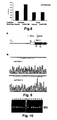
- Figure 9(A) shows the schematic diagrams of the LAPTM4B promoter and its first exon.
- the rectangle frame indicates the first exon
- the black color area represents the encoding area
- the white color is the non-coded area
- the gray area shows a 19bp DNA sequence.
- FIG. 9 shows the partial sequences of the LAPTM4B alleles and their sequencing graphic spectra.
- the underlined part is a 19bp DNA sequence.
- the results reveal that LAPTM4B*1 contains one copied 19bp DNA sequence and LAPTM4B*2 has two copied 19bp DNA sequences, which are linked in the non-coded area (nt -33 — -15) of the first exon of LAPTM4B*1.
- E2 (5' GCCGACTAGGGGACTGGCGGA 3') and R2 (5' CGAGAGCTCCGAGCTTCTGCC 3') primers were designed and synthesized.
- a partial sequence of the first exon of LAPTM4B was amplified by PCR using templates of genomic DNA from normal people, hepatocellular carcinoma, and esophagus carcinoma tissues. PCR conditions were as follows: 96°C pre-denature for 5 min; 94°C for 30 s, 68°C for 30 s, 72°C for 1 min, 35 cycles; 72°C for 5 min; then the PCR products were conducted to 2% Agarose gel electrophoresis analysis.
- Figure 10 shows LAPTM4 gene * 1 /* 1 , * 1 /* 2 , and * 2 /* 2 three types in human population.
- Example 3 Construction of the reporter plasmids and analysis of the promoter activity
- R1 were used to amplify by PCR, respectively, to construct vectors, pGL3-PF2, pGL3-PF3, and pGL3-PF4 which contain promoter segments with various lengths and luciferase gene. These constructs were identified by sequencing.
- HLE cells may play a regulating role in HLE cells and make the luciferase activities of pGL3-PF3 and pGL3-PF4 tranfectants remarkably different in HLE cells.
- the pGL3-PF3 activity is higher than that of pGL3-PF1 and pGL3-PF2 in both BEL-7402 and HLE cells, implying that some negatively regulatory factor exists.
- One or more of these negatively regulatory factors bind with the promoter upstream target sequence (-558 upstream) to induce a downregulated LAPTM4B gene expression. This suppressive effect was stronger in HLE cells than in 7402 cells. This means that HLE cells may contain some factors that strongly suppress the expression of LAPTM4B.
- the Northern Blot analysis presented in Figure 1-B also shows a low expression of LAPTM4B in HLE cells, supporting the above hypothesis.
- the pGL3-PF2 vector contains two DNA repeating fragments (-41- -328, -574 ⁇ -859), which is one more DNA fragment (-574 - -859) than pGL3-PF3.
- pGL3-PF3 exhibits higher activity than pGL3-PF2 in both cells. This result indicates that the two repeating sequences negatively regulate gene transcription. They have many potential binding sequences for the transcription factors and provide two binding sites for each negatively regulating factor. Since many transcription factors often form dimers, they have to bind with two target sequences to be able to function. In the case of pGL3-PF3, which can only provide one binding site, no function is shown. Since the pGL3-PF3 transfectant has a disinhibitory effect, its activity is higher than other vector transfectants.
- the tissue sample was placed on ice and cut into small pieces by scissors. 0.1 gram of wet tissue was selected and placed in a manually operated homogenizer. 1 mL lysis buffer was added in each tube and the mixture was thoroughly homogenized. The lysate was transferred to a fresh tube and centrifuged at 4°C, 12 000 g for 10 min to remove the debris. If cells are used, the cells in a culture dish were digested with 0.25% trypsin buffer, followed by two PBS rinses and centrifuged at 500g for 3 min. The cleared supernatant was collected, and the proteins in the supernatant were separated by SDS-PAGE electrophoresis, and then transferred to the NC membrane.
- the membrane was blocked at 4°C overnight with 5% non-fat powdered milk in a TBS buffer containing 0.05% Tween 20. Then it was incubated with the rabbit polyclonal antibody, LAPTM4B-EC2 232-241 -pAb (1:500 dilution) or mouse Anti-FLAG M2 monoclonal antibody (Sigma, 1:750 dilution) at room temperature for 2 hours, and then rinsed with TBS for three times. It was further incubated with a peroxidase-coupled second antibody (IgG), such as goat anti rabbit or goat anti mouse (1:3000 dilution), for 2 hrs, followed by three rinses with a TBS buffer (pH 8.0, containing 0.05% Tween 20).
- IgG peroxidase-coupled second antibody
- pGEMT-E2E7 plasmid as a template and the PCR method, a full length or partial cDNA, or the reading frame of LAPTM4B gene was amplified by PCR with primers A, or B and E, and the Pfx DNA polymerase.
- Bam HI enzyme cutting site (GGATCC) and ribosome binding site sequences (GCCACC) were introduced in primer A and B at 5' terminal and Eco RI enzyme cutting site (GAATTC) was incorporated in the primer E.
- the amplified products AE and BE were digested by restriction enzymes Bam HI and Eco RI, purified, and ligated into pcDNA 3.0 vector. They were transformed conventionally to DH5 E.
- pcDNA31AE contains a full-length ORF
- poDNA3 / BE contains the ORF starting from the second ATG to TAA.
- pcDNA3 / BE- encoded protein is missing 91 amino acids at the N terminal.
- the total viable cell numbers were determined by the acidic phosphatase method and the cell growth curve was plotted.
- the cell cycle was analyzed by the flow cytometry.
- the expression levels of cell cycle-regulating protein, including cyclin D1 and Cyclin E, and proto-oncogene products, including c-Myc, c-Fos, and c-Jun (transcription factors for regulating cell proliferation) were measured by the Western Blot analysis.
- HLE-AE cells Large colonies of HLE-AE cells were formed in the soft agar gel, which indicates that this gene participated in the regulation of cell proliferation and its over expression (activation) was related to the dysregulation of cell proliferation. Furthermore, the migrating capability of HLE-AE cells was also enhanced (The HLE-AE cells that migrated through the membrane pores were increased from 1216 ⁇ 403.8 for the control to 4082.5 ⁇ 748.8). Its capability to invade Matrigel was also greatly increased (from 25 ⁇ 12.73 cells for the control to 1325 ⁇ 424.26 cells). The results show that LAPTM4B over expression promotes the development of cell malignant phenotype.
- mice Six-week old male mice were randomly selected and divided into three groups: In the first control group, the mice were injected with physiological saline. In the second control group, the mice were inoculated with the pcDNA3 MOCK (no-load plasmid) transfected cells. In the test group, all the mice were inoculated with pcDNA3 IAE (a plasmid containing full-length cDNA) transfected NIH3T3 cells. Each mouse was subcutaneously inoculated with 2x10 6 cells. There were four to six mice in each group. The mice were sacrificed after 21 days inoculation and dissected.
- pcDNA3 MOCK no-load plasmid
- mice half of inoculated mice
- A, B the other two mice were identified as lymphatic tissue at the inoculated sites
- C, D the inoculated sites
- twelve mice in the two control groups showed no sign of tumor formation after being inoculated for 86 days.
- LAPTM4B may be a novel proto-oncogene.
- Example 7 Primary analysis of LAPTM4B antigen in the serum of patients with hepatocellular carcinoma by the ELISA method
- the microtiter for enzyme analysis was used to measure OD. at 490 nm and the antigen level was estimated. The results are shown in Figure 8 .
- the sera of patients with hepatocellular carcinoma contained higher level of LAPTM4B antigen than that from normal people, indicating that LAPTM4B has a potential to become a new marker for hepatocellular carcinoma diagnosis.
- Example 8 Functional determination of LAPTM4B protein in signal transduction by co-immunoprecipitation and antibody inhibition analysis
- the cell lysate was prepared according to the method in Example 4.
- the first antibody was added to the supernatant.
- 50 ⁇ L protein G-Agarose suspension was added and the mixture was shaken at 4°C for at least three hours or overnight.
- the immunocomplex precipitate was collected after centrifuging at 12000g for 20 seconds.
- the complex was re-suspended by adding 1 mL washing buffer I and shaken at 4°C for 20 min. The mixture was centrifuged at 12000g for 20 seconds and the supernatant was removed carefully. This step was repeated once.
- the complex was re-suspended by adding washing buffer II, shaken at 4°C fro 20 min., and centrifuged at 12000g for 20 seconds. The supernatant was removed carefully. The last two steps were repeated once.
- the complex was re-suspended by adding washing buffer III, shaken at 4°C fro 20 min, and followed by 12 000 g centrifugation for 20 seconds. The supernatant was removed completely. 50 ⁇ L 1xSDS loading buffer was added in the precipitate and the mixture was boiling in 100°C water bath for 5 min to denature and dissociate the immunocomplex in the sample. After 12000 g centrifugation for 20 second, the supernatant was removed and analyzed in SDS-PAGE apparatus.
- BEL-7402 cell was preincubated for 0, 10, 20, and 40 min, respectively, on LN-1 substance in serum free medium.
- Co-immunoprecipitation was performed with LAPTM4B-EC2-pAb from the cell lysate.
- the co-immunoprecipitates were respectively adsorbed by Protein G-Sephorose, centrifuged, and analyzed by 10% non-reductive SDS-PAGE. Then the phosphorylations of LAPTM4B, FAK and MAPK were analyzed separately by the Western Blot with p-Tyr mAb.
- BEL-7402 cells were preinoculated separately with LAPTM4B-EC2-pAb (15 ⁇ g/mL) and anti-Glut2 (15 ⁇ L/mL) antibodies at 37°C under 5% CO 2 for 2 hrs, and then seeded on LN-1 substance and incubated for indicating time.. Under the same conditions, the anti-Glut2 antibody treated cells and no antibody treated cells were used as control. The cell lysate in each group was analyzed by the Western Blot analysis with p-Tyr mAb. The inhibitory effects of various antibodies on phosphorylation of LAPTM4B were analyzed. The results show that LAPTM4B-35 was phosphorylated peakly when human hepatocellular carcinoma BEL-7402 cells were attached on laminin substrate.
- LAPTM4B-35 reached the highest level in 10 min after cell attachment ( Figure 15-A ). Meanwhile LAPTM4B-EC2-pAb could inhibit almost completely its phosphorylation ( Figure 15-B ), while the anti-Glut2 (an antibody against a non-related plasma membrane protein Glut2) showed no such inhibitory effect ( Figure 15-C ). On contrary, LAPTM4B-24 cannot be phosphorylated. The phosphorylation of LAPTM4B-35 Tyr 285 would form a binding site for signal molecules that contain SH2 domain. In the meantime, LAPTM4B-35 itself presents typical binding sites for signal molecules that contain SH3 domain.
- LAPTM4B-35 functions most likely as a very important docking protein of molecules for signal transduction or a special organizer of membrane microdomain. It could recruit related signal molecules inside or outside cells, so as to play pivotal roles in signal transduction associated with cell proliferation, differentiation and apoptosis.
- the attachment of human hepatocellular carcinoma cells on laminin substrate can also cause Tyrphosphorylation of the cytoplasmic signal molecule FAK ( Figure 16-A ), and the LAPTM4B-EC2-pAb and anti-integron a 6 mAb against the epitope of the extracellular region of ⁇ 6 both can prevent FAK phosphorylation without affecting the expression level of FAK protein by preincubating with BEL 7402 cells.
- LAPTM4B-35 can be potential targets of drugs for regulating cell proliferation, differentiation, and apoptosis.
- LAPTM4B genotypes in genomic DNA from blood of normal individuals and patients with hepatocellular carcinoma were detected by PCR.
- Two primers were designed and synthesized according to the flanking sequence of 19bp DNA sequence in LAPTM4B gene sequence 3:
- the partial sequence of the first exon was amplified using genomic DNA as a template.
- PCR conditions were as follows: 96°C pre-denature for 5 min, 94°C for 30 sec, 68°C for 30 sec, 72°C for 1 min, 35 cycles, 72°C extension for 5 min.
- PCR products were analyzed by 2% agarose gel electrophoresis and the results are shown in Figure 10 .
- the lanes 1, 6, 12, and 13 represent a 204 bp nucleotide segment in LAPTM4B*1 / *1.
- the lanes 5, 8, 9, 14, and 15 represent a 223 bp nucleotide segment in LAPTM4B*2 / *2.
- the lanes 2, 3, 4, 7, 10, and 11 represent 204 bp and 223 bp nucleotide segments in LAPTM4B*1 / *2.
- Line M is the marker.
- the results reveals that in the homozygous gene pair of * 1 /* 1 or * 2 /* 2 either the 204bp or 223bp DNA segment was amplified, while in * 1 /* 2 hybrid gene pair 204bp and 223bp DNA segments were both amplified simultaneously. Therefore, the genotype of LAPTM4B in Chinese population can be classified as LAPTM4B*1 / *1, *1 / *2, and * 2 /* 2 ( Figure 10 ).
- Example 10 Frequency distribution of LAPTM4B genotypes and alleles in normal people and patients with hepatocellular carcinoma
- the occurrence frequency of LAPTM4B genotypes in 209 normal Chinese and 57 patients with hepatocellular carcinoma was analyzed and compared in Table 2.
- the Hardy-Weinberg equation was used to get the expectancy analysis.
- the frequency of LAPTM4B allele * 1 and * 2 from patients with hepatocellular carcinoma differs significantly from that of normal people. Their ratios are 0.5175: 0.6746 and 0.4825: 0.3254, respectively.
- the occurrence frequencies of LAPTM4B allele * 1 and * 2 in a normal population are 0.6746 and 0.3253, while the occurrence frequency of LAPTM4B allele * 1 and * 2 in patients with hepatocellular carcinoma are 0.5175 and 0.4825.
- the occurrence frequency of * 2 /* 2 genotype in the hepatocellular carcinoma patient group is 26.32% as compared to 11.01 % in the control group, therefore its occurrence frequency is increased significantly ( p ⁇ 0.01).
- the analysis shows that the risk suffering from HCC of individuals in * 2 /* 2 genotype of is 2.89 times greater than that in other genotype in developing hepatocellular carcinoma.
- the LAPTM4B*2 / *2 genotype is correlated with the susceptibility of developing hepatocellular carcinoma.
- Example 11 Frequencies of genotype and allele in patients with esophagus carcinoma
- LAPTM4B genotype is related to the susceptibility of developing other cancers
- the genomic DNA from blood of 116 normal people and 109 patients with esophagus carcinoma from the same location were analyzed.
- LAPTM4B genotype distribution of patients with esophagus carcinoma is not significantly different from the control group of the normal population.
- LAPTM4B alleles are not related with the susceptibility of developing esophagus cancer.
- Example 12 LAPTM4B-35 expression in some epithelium sourced cancers
- the relationship between the LAPTM4B-35 protein expression and other cancers was studied by an immunohistochemical method.
- the fixed specimens from esophagus cancer, breast cancer, lung cancer, stomach cancer, colon cancer, and rectal cancer (positive) tissues and the (negative) control noncancerous tissues were obtained from surgical excision and treated according to the following steps:
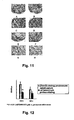
- the 11-A indicates a normal esophagus tissue (Negative)
- B is an esophagus cancer tissue (Positive)
- C is a normal breast tissue (Negative)
- D is the breast cancer tissue (Positive)
- E is a normal lung tissue (Negative)
- F is a lung cancer tissue (Positive)
- G is a normal stomach tissue (Negative)
- H is a stomach cancer tissue (Positive).
- LAPTM4B was clearly expressed in lung cancer, stomach cancer, and breast cancer tissues, while it was not expressed clearly in esophagus cancer and large intestine cancer.
- the proteins encoded by LAPTM4B gene could be possibly used as new markers in early diagnosis of some cancers.
- the efficiency and the accuracy of the early diagnosis of cancers, especially the primary hepatocellular carcinoma can be improved.
- LAPTM4B gene can be used as target gene in the cancer treatment.
- suppressing LAPTM4B-35 expression and promoting LAPTM4B-24 expression could inhibit the growth of hepatocellular carcinoma cells, reverse malignancy phenotype or delay its development.
- the expression products of LAPTM4B gene, LAPTM4B-35 could be inhibited by the newly developed siRNA interference technology.
- LAPTM4B -BE-cDNA could be recombinated in the engineered virus expression vector and be used in antitumor gene therapy through an up-regulation of LAPTM4B-24 expression.
- LAPTM4B-35 protein could also be used as a new target for pharmaceutical treatment.
- LAPTM4B-35 protein can function as an assembling platform for complex of cell signal transduction molecules, and it contains a number of binding sites for signal molecules, there is a great potential to develop various new medicines with LAPTM4B protein as targets.
- LAPTM4B-EC2-pAb antibody can inhibit tumor cell proliferation and block its signal transduction. Based on the discovery underlying this invention, further studies can be pursued on the possibility of using antibody to inhibit hepatocellular carcinoma and some other cancer development. After a better understanding on the effect, a humanized soluble single chain antibody could be developed for clinical treatment on HCC patients. Peptide vaccines could also be developed.
- the vaccines can be successfully made, it will not only help cure hepatocellular carcinoma and some other cancer, but also prevent cancerogenesis in the high risk population.
- many new anticancer approaches can be developed based on the embodiments of this invention. As important supplements for treatments of hepatocellular carcinoma and other cancers, this invention will help increase the cure rates of hepatocellular carcinoma and other cancers. This project would generate a significantly great impact on human society.
- LAPTM4B genotype of genomic DNA is genotyped.
- the relationship of various genotypes with the susceptibility to hepatocellular carcinoma as well as with other cancers is investigated. It is discovered that one of the genotypes, LAPTM4B* / *2, is correlated closely to hepatocellular carcinoma susceptibility. As a result, it provides a new and accurate criterion for screening people who are susceptible to primary hepatocellular carcinoma in the high risk population. It is of important significance to the assessment and prevention of high risk population from developing hepatocellular carcinoma.
Landscapes
- Health & Medical Sciences (AREA)
- Life Sciences & Earth Sciences (AREA)
- Chemical & Material Sciences (AREA)
- Immunology (AREA)
- Engineering & Computer Science (AREA)
- Organic Chemistry (AREA)
- Molecular Biology (AREA)
- General Health & Medical Sciences (AREA)
- Biochemistry (AREA)
- Medicinal Chemistry (AREA)
- Proteomics, Peptides & Aminoacids (AREA)
- Biomedical Technology (AREA)
- Genetics & Genomics (AREA)
- Urology & Nephrology (AREA)
- Hematology (AREA)
- Pathology (AREA)
- Analytical Chemistry (AREA)
- Biophysics (AREA)
- Cell Biology (AREA)
- Physics & Mathematics (AREA)
- Microbiology (AREA)
- Biotechnology (AREA)
- Zoology (AREA)
- General Physics & Mathematics (AREA)
- Food Science & Technology (AREA)
- Oncology (AREA)
- Wood Science & Technology (AREA)
- Gastroenterology & Hepatology (AREA)
- Bioinformatics & Cheminformatics (AREA)
- General Engineering & Computer Science (AREA)
- Toxicology (AREA)
- Hospice & Palliative Care (AREA)
- Animal Behavior & Ethology (AREA)
- Public Health (AREA)
- Nuclear Medicine, Radiotherapy & Molecular Imaging (AREA)
- General Chemical & Material Sciences (AREA)
- Chemical Kinetics & Catalysis (AREA)
- Pharmacology & Pharmacy (AREA)
- Veterinary Medicine (AREA)
- Medicines That Contain Protein Lipid Enzymes And Other Medicines (AREA)
Abstract
Description
- The present invention relates to a human cancer-related gene, its encoded products and their applications in genetic engineering and protein engineering areas, as well as in medical diagnosis and treatment.
- Cancer is the major health problem threatening human lives. Hepatocellular carcinoma (HCC) is one of the most serious among cancer diseases. It is reported that the new cases of primary hepatocellular carcinoma exceeds over one million worldwide each year. 70% of the new cases occur in Asia, and about 40-45% of the worldwide new cases occur in China. The total number of new hepatocellular carcinoma cases every year in China is about 450,000, and the number is increasing, especially in those between ages 20-60. The high incidence, difficulty in early diagnosis, fast growing rate, high reoccurrence, and the high mortality rate make HCC a most malignant cancer. Most HCC patients have already progressed to the intermediate stage or late stage when diagnosed, and they can only survive for 3-6 months if without a proper treatment.
- To elucidate the mechanism underlying cancerogenesis would help for cancer prevention, diagnosis and treatment. Early diagnosis is crucial for raising the curative rate and reducing the mortality. Currently used HCC-diagnostic marker, the serum AFP, has 30% of negative results in HCC patients, while some benign liver disease can cause a significant increase of AFP level in serum, creating some difficulty in differential diagnosis. It has been found that the hepatocarcinogenesis is related to individual hereditary susceptibility. Individuals with different genetic backgrounds possess different handling capability toward environmental carcinogens. This leads to different risk of suffering from cancer for individuals. It is the various genotypes and the genetic diversity that cause the different genetic susceptibility for cancerogenesis.
- Cancer is essentially a cellular hereditary disease. Although a great number of cancer-related genes have been discovered, the mechanisms of the cancerogenesis and the development remain to be elucidated. The known oncogenes can be divided into five categories according to the cellular localization and function of their encoded proteins: I. genes that encode growth factors, including sis, int-2, hst, fgf-5; II. genes that encode growth factor receptors, including erbB, erbB-2, fms, met, ros, and others; III. genes that encode signal transduction molecules in cytoplasm, including abl, src, ras, raf, yes, fgr, fes, Ick, mos, and others; IV genes that encode regulatory molecules for cell proliferation and apoptosis, including bcl-1, bcl-2 and others; and V. genes that encode the nuclear DNA-binding proteins (transcription factors), such as myc, myb, fos, jun, B-lym, ski, ets, rel and others. It has been demonstrated that ras, src, myc, met and p53 etc. are the genes closely associated with HCC.
- The present invention is defined in and by the appended claims.
- The present invention derives from work on a novel human cancer-related gene designated as LAPTM4B. It comprises one of the following nucleotide sequences:
- 1. The human cancer-related gene comprises one of the following nucleotide sequences:
- 1). SEQ ID No: 1, SEQ ID No: 2, SEQ ID No: 3 or SEQ ID No: 6 in the sequence listings;
- 2). Polynucleotides that encode the protein sequences of SEQ ID No: 4, SEQ ID No: 5, or SEQ ID N2. 7 in the sequence listings;
- 3). DNA sequences having more than 90% homology with the DNA sequences defined by SEQ ID No: 1, SEQ ID No: 2, SEQ ID No: 3 or SEQ ID No: 6 in the sequence listings. These DNA sequences can encode proteins having the same or similar functions.
- In the sequence listings, SEQ ID No: 6 is the allelic gene of SEQ ID No: 1, consisting of 2264 bases. Its open reading frame starts from 17 to 1129 base. This sequence contains two tandemly arranged 19bp DNA segments, the sequence of which is gcttgg agctccagca gct. These 19bp DNA segments localized in nt 124 - nt 161 in SEQ ID No: 6.
- The human cancer related LAPTM4B protein possesses the amino acid sequence of 4, and/or 5, and/or 7. Or it consists of the
sequence 4, and/or thesequence 5, and/or thesequence 7 after one or several amino acid residues are replaced, deleted, or added. However, the above alteredsequence 4, and/or thesequence 5, and/or thesequence 7 still have the same or similar activity to theunchanged sequence 4, and/or thesequence 5, and/or thesequence 7. -
Sequence 4 in the sequence listings consists of 317 amino acid residues encoded by the whole sequence of SEQ ID No: 1. its molecular mass is 35kDa and the putative isoelectric point is 9.05.Sequence 5 in the sequence listings contains 226 amino acid residues encoded by the segment of bases from 274th to 954th in the SEQ ID No: 1. Its molecular mass is 24kDa, and the putative isoelectric point is 4.65. Thesequence 7 in the sequence listings is a protein containing 370 amino acid residues. - LAPTM4B gene is widely expressed at different levels in sixteen normal tissues. Its transcriptive expression is very high in testis, cardiac muscle, and skeletal muscle, moderate in ovary, kidney, and pancreas, low in liver, spleen, small intestine, large intestine, and thymus, and is very low in lung and peripheral blood cells. In eight fetal tissues, the expression is high in heart, skeletal muscle, and kidney. In fetal livers, it is slightly higher than that in adult livers. However, its expression in some cancerous tissues is significantly upregulated. For instance, the Northern Blot analysis indicates that the transcription level in 87.3% (48/55) human hepatocellular carcinoma tissues is significantly higher than that in fetal livers and normal livers (
Figure 1-A ). In situ hybridization (Figure 2-A ), immunohistochemistry (Figure 2-B ), and immunocytochemistry (Figure 2-C ) also indicate that LAPTM4B gene expression is especially high in hepatocellular carcinoma tissues, while its expression is relatively low in paired non-cancerous liver tissues (Figures 2-A and 2-B ). Among the five cell lines from hepatoma tissues tested, all except for HLE, SMMC-7721, QGY-7701, BEL7402 and HG116 are expressed highly (Figure 1-B and Figure 2-C ). It is important that highly over expressed protein product in hepatocellular carcinoma tissue and hepatocellular carcinoma cell line is mainly SEQ ID No: 4 LAPTM4B-35, while SEQ ID No: 5 LAPTM4B-24 only shows a slightly up regulation in its expression level. This results in a remarkable increase in the ratio of LAPTM4B-35 to LAPTM4B-24 proteins in the hepatocellular carcinoma tissue (Figure 2-B ). Although the expressions of LAPTM4B-35 and LAPTM4B-24 are slightly increased in the paired non-cancerous tissue, their ratio is the same as that in the normal liver (See Table 1). This is probably a precancerous sign of hepatocellular carcinoma. In addition, the expression levels of mRNA and the protein of LAPTM4B gene is negatively correlated with the differentiation of the hepatocellular carcinoma tissue. The hepatocellular carcinoma tissues in low differentiation are expressed highly, while the ones in high differentiation are expressed relatively low (Figure 1-C ). The Western Blot and the immunohistochemical method are used to determine the relationship of LAPTM4B gene with other cancers. The results indicate that LAPTM4B-35 protein expression is up regulated in some epithelium derived cancerous tissues and cell lines, such as stomach cancer, breast cancer, highly metastatic human lung cancer, and prostate cancer (Figure 11 ). Moreover, in syngeneic human lung cancer and prostate cancer cell lines, LAPTM4B-35 expression is greatly up regulated in cells of high metastasis potential compared with those of low metastasis potential. But in cell lines of human melanoma, either from in situ or metastatic cancer, it is not clearly expressed. Although LAPTM4B-35 is expressed at a low level in liver tissues of adult rats and mice, its expression is not obviously up regulated in either mouse ascetically qrown hepatocellular carcinoma or in the regenerated rat liver under a normal proliferation status.Table 1: Expression ratio of LAPTM4B-35 to LAPTM4B-24 in hepatocellular carcinoma tissue, paired non-cancerous liver tissue and normal liver tissue HCC PNL NL LAPTM4B-35 13.32±1.98 4.58±1.31 2.78±0.11 LAPTM4B-24 3.59±1.78 1.76±1.24 1.00±0.02 LAPTM4B-35/ LAPTM4B-24 (Ratio) 3.71 2.60 2.78 P<0.01 HCC vs. PNL and NL - LAPTM4B proteins in SEQ ID No: 4, SEQ ID No: 5 and SEQ ID No: 7 have four fragments of membrane-spanning sequences, one N-glycosylation site, a typical lysosome and endosome targeting signals in the cytoplasmic region. They all belong to the protein superfamily of the tetratransmembrane proteins. However, they have various number of phosphorylation sites. The experiment shows that SEQ ID No: 4 LAPTM4B-35 can form a complex in plasma membrane with the integrin α6β1 (Single specific receptor of laminin in the extracellular matrix) and the epidermal growth factor recceptor (EGFR) (
Figures 14-A , B, and C). This complex is colocalized in cell plasma membrane. It is possible that LAPTM4B-35 may integrate in the plasma membrane the proliferation signals from both extracellular matrix and the growth factor. This can further elucidate molecular mechanism of the anchorage-dependent cell growth of normal eukaryotic cells, i.e. the eukaryotic cell growth needs not only the stimulating signal from the growth factor, but also a definite stimulating signal from extracellular matrix. It represents a break through in understanding the regulation mechanism of the cell proliferation. Experiments demonstrate that tyrosine group (Tyr285) in the cytoplasmic region of LAPTM4B protein C terminal can be phosphorylated (Figure 15-A ). When the cell is attaching onto the laminin substrate, its phosphorylation is increased sharply (Figure 15-A ) and can be completely inhibited by LAPTM4B-EC2-pAb antibody (Figure 15-B ), but the non-correlated antibody does not show any inhibitory effect (Figure 15-C ). After the phosphorylation, Tyr285 forms a site to bind with the SH2 domain of intracellular signal molecules. In the meantime, N terminal and C terminal sequences of LAPTM4B contain Pro-rich domains and binding sites of the typical SH3 domain. The above results indicate that SEQ ID No: 4 LAPTM4B-35 protein may be an important docking protein for signal transduction, or an organizer of the special microdomain in the plasma membrane. It can recruit related signal molecules from both inside and outside of the cells to complete the signal transduction for cell proliferation, differentiation, and apoptosis. Experimental results show that the transfection of mouse NIH3T3 cells and HLE human hepatocellular carcinoma cells by cDNA in SEQ ID No: 4 produces stable transfected and LAPTM4B-35 over expressed NIH3T3-AE and HLE-AE cell lines. The growth curves (Figure 4 ), the incorporation of 3H-TdR (Figure 5 ), and the cell numbers in S phase of cell cycle (Figure 6 ) all demonstrate that the rate of cell proliferation is greatly increased. Moreover, the proliferation of transfected cells shows less dependence on the growth factor in serum, and the transfected cells can form large colonies in soft agar. Inoculation of NIH mouse with NIH3T3-AE cells can form a moderate malignant fibrosarcoma (Figure 7 ), indicating the over expression of LAPTM4B-35 induces out-of-control cell proliferation. Also migration capability of the HLE-AE cells is strengthened and its capability to invade the Matrigel is remarkably enhanced, indicating that the over expression of LAPTM4B-35 accelerates the development of cell malignant phenotype. On the contrary, the cDNA of SEQ ID No: 5 (an encoding sequence where 91 amino acids in the N terminal of LAPTM4B-35 is truncated) transfected mouse BHK, NIH3T3, and HLE hepatocellular carcinoma cell lines cannot survive for a long time. The result shows that the 91 amino acid sequence on the N terminal of SEQ ID No: 4 LAPTM4B-35 protein play a crucial role in regulating cell proliferation. LAPTM4B-35 protein and LAPTM4B-24 protein have reciprocal, antagonistical functions in cell proliferation and survival. The overexpression of LAPTM4B-35 accelerates cellular malignant transformation, while the overexpression of LAPTM4B-24 facilitates the cell death. Their expression equilibration and regulation are pivotal to the carcinogenesis and progression of malignant cancer. LAPTM4B gene may belong to the proto-oncogene family. In cancer treatment, inhibiting SEQ ID No: 4 LAPTM4B-35 expression and strengthening SEQ ID No: 5 LAPTM4B-24 expression may suppress the growth of hepatocellular carcinoma and reverse its malignant phenotype or progressively slow down its development. Furthermore, the overexpression of LAPTM4B-35 also promotes upregutation of the cell cycle regulators, such as cyclin D1 (Figure 13-A ) and cyclin E (Figure 13-B ), and also the over expression of some proto-oncogenes, such as c-Myc (Figure 13-C ), c-Jun (Figure 13-D ), and c-Fos (Figure 13-E ) etc. - The monoclonal and polyclonal antibodies for SEQ ID No: 4 LAPTM4B-35 protein epitopes, such as polyclonal LAPTM4B-EC2232-241-pAb for SEQ ID No: 4 LAPTM4B-35 in the secondary extracellular region, polyclonal antibodies (LAPTM4B-N1-99-pAb and L-APTM4B-N28-37-pAb) for SEQ ID No: 4 LAPTM4B-35 N terminal sequence, and monoclonal antibodies for LAPTM4B are important in studying the effects of LAPTM4B-35 and LAPTM4B-24 in cancer diagnosis and treatment (
Figures 2 ,3 ,8 ,11 ,12 ,14, 15 ). For example, LAPTM4B-EC2232-241-pAb, lAPTM4B-N1-99-pAb polyclonal antibodies and LAPTM4B-N1-99-mAb monoclonal antibody can be used to analyze LAPTM4B protein expression, intracellular localization, separation and purification, and protein-protein interaction. They can also be used to detect the antibody and antigen of LAPTM4B in blood (Figure 8 ). Moreover, LAPTM4B-EC2232-241-pAb can inhibit cancer cell proliferation (Figure 12 ), Tyr285 phosphorylation of LAPTM4B protein (Figure 15-B ), and the phosphorylation and activation of signal molecules FAK (Figure 16-A ) and MAPK(Figure 16-B) . - SEQ ID No: 8 is the promoter sequence of LAPTM4B gene. To study the regulation of LAPTM4B gene expression, the LAPTM4B gene promoter and the upstream sequence SEQ ID No: 8 are cloned. There are no typical CCAAT (TTGCGCAAT), TATA cassettes in LAPTM4B gene promoter region. But various binding sites of transcription factors exist in the upstream region of LAPTM4B promoter, such as CREBP1/c-Jun, CEBP, PAX2/5/8, GATA, STAT, c-Ets-1, E2F, LYF-1, and c/v-Myb (
Figure 17A ). These transcription factors may specifically regulate LAPTM4B expression in cells of various tissues. The abnormal expression and activation of these transcription factors in cancer cells possibly lead to an unbalanced expression of LAPTM4B proteins. Moreover, there are two highly homologous repeating sequences in the upstream domain of LAPTM4B promoter. It is worthwhile to study whether they are responsible to the regulation of LAPTM4B expression. A series of vectors consisting upstream region sequences of LAPTM4B promoter with different lengths-promoter-5' UTR-35bp encoding region-luciferase reporting gene is constructed, and these vectors are used to transfect into BEL7402 cells and HLE cells from human hepatocellular carcinoma HCC. As shown inFigure 17 , the cells transfected with various vectors all show luciferase activity with various intensities, indicating the transcription activities in these segments. The smallest fragment is a DNA segment (pGL-PF4) at about 38 bp in the upstream region of the transcription starting site. It possesses a basic promoting activity and functions as LAPTM4 core promoter. The activity of pGL-PF4 transfectant in BEL7402 is 20% of the reference promoter SV40, while the activity is low in HLE, only 6% of SV40 activity, about 1/3 of that in BEL7402. These data partially reflect the natural activity of LAPTM4B promoter in these two cell lines. It is consistent with the Northern blot results, where mRNA expression is high in BEL-7402 cell line and low in HLE cell line. Additionally, pGL3-PF4 transfectant reveals dramatically different activities in these two cells. Its activity in BEL-7402 cells is 7 times higher than that in HLE cells. Apparently, different regulative mechanisms of LATPM4B gene transcription exist in BEL7402 and HLE cell lines. - In the work underlying this invention, the genome DNA is genotyped in order to determine the relationship between different LATPM4B genotypes and susceptibility of hepatocellular carcinoma. LATPM4B has two alleles, LATPM4B*1 and LATPM4B*2, i.e., SEQ ID No: 6, is derived by PCR cloning. As shown in
Figure 9 , the difference between alleles *1 and *2 is the 19 bp sequence in the first exon 5' UTR. Allele *1 has only one such sequence (nt 124-142), while *2 contains two such sequences in a tightly tandem arrangement (124-142dup, taking G at the transcription starting site (TSS) as +1 numbering standard). The insertion of the 19-bp sequence would eliminate the stop codon in 5'UTR in the corresponding *1 allele by a triplet shift. As a result, the open reading frame may be extended upstream by 53 amino acids at N terminus of the protein. The encoded protein by SEQ ID No: 6 then should contain 370 amino acid residues (SEQ ID No: 7). LATPM4B genotypes detected in human population are *1/*1, *1/*2 and *2/*2, respectively (Figure 10 ). Studies show that the risk of developing hepatocellular carcinoma for individuals with LATPM4B genotype *2/*2 is 2.89 times higher than individuals with non-*2/*2 type (Table 2). However, LATPM4B genotype in patients with esophagus carcinoma shows no difference from the normal population (Table 3). This indicates that LATPM4B *2/*2 genotype correlates especially to susceptibility of hepatocellular carcinoma. As a result, LATPM4B allele LATPM4B*2 provided by this invention can be used as a target to screen and diagnose people susceptible to hepatocellular carcinoma or having a high risk to develop hepatocellular carcinoma. Particularly, using LATPM4B *2/*2 genotype as a target to screen highly susceptible or high risk people can be more accurate.. *1/*1, *1/*2 and *2/*2 of LATPM4B genotypes, LATPM4B*2 encoded proteins or their antibodies, and/or LATPM4B extender and scavenger from human genome can all be used to screen people who are susceptible to hepatocellular carcinoma or having a high risk to develop hepatocellular carcinoma. - The expression vectors containing sequences described in SEQ ID No: 1, 2, 3, 6, 8, the transfection cell lines containing SEQ ID No: 1, 2, 3, 6, 8 sequences, and the primers amplifying SEQ ID No: 1, 2, 3, 6, 8 are all encompassed by this invention.
-
-
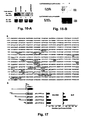
Figure 1-A is the Northern Blot analysis profiles indicating the transcriptive expression of the gene of this invention in normal human liver, normal fetus liver and hepatocellular carcinoma tissues. -
Figure 1-B is the Northern Blot analysis spectrum indicating the transcriptive expression of the gene of this invention in the human hepatocellular carcinoma cell lines. -
Figure 1-C is a diagram showing the relationship of the expression level of the gene of this invention in human hepatocellular carcinoma tissues and the cancer differentiation status. -
Figure 2-A is a diagram of hybridization in situ of hepatocellular carcinoma tissue. The LATPM4B mRNA in hepatocellular carcinoma nodule shows a strong positive staining. -
Figure 2-B is an immunohistochemical diagram, where LATPM4B protein in hepatocellular carcinoma nodule shows a strong positive staining. -
Figure 2-C is an immunocytochemical diagram, where LATPM4B protein is shown to exist in the transfected cells. -
Figure 3 presents a Western Blot analysis diagram, where the expression spectra of LATPM4B-35 and LATPM4B-24 proteins encoded by the gene of this invention are shown in the tissues of normal liver (NL), hepatocellular carcinoma (HCC), and paired non-cancerous liver (PNL). -
Figure 4 shows a growth curve of the accelerated proliferation of cDNA-transfected cells of this invention. -
Figure 5 shows a column diagram, where the DNA synthesis of LAPTM4B cDNA- transfected cells of this invention is increased. -
Figure 6 is a pie diagram showing an increase of cell numbers in S phase in cDNA- transfected cells of this invention (Flow cytometry analysis). -
Figure 7 shows the oncogenic effect of cDNA-transfected cells of this invention on mouse. -
Figure 8 is a histogram showing the level of the antigen in the serum of patients with hepatocellular carcinoma. -
Figure 9 shows the partial sequence of LAPTM4B alleles of this invention. -
Figure 10 shows the genotypes distribution of LAPTM4B alleles in human population. -
Figure 11 is an immunohistochemical diagram of various cancer tissues derived from epithelium. -
Figure 12 is a column diagram showing the inhibitory effect of LAPTM4B-EC2-pAb antibody on proliferation of hepatocellular carcinoma cells. -
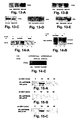
Figure 13-A , B, C, D, and E are the Western Blot analysis diagrams showing respectively that the expressions of cyclin D1, cyclin E, c-Myc, c-Fos, and c-Jun of cDNA- transfected cells of this invention are up-regulated. -
Figure 14-A , B, and C are the analytical diagrams of the co-immuno precipitation, revealing respectively the interactions of the gene product (LAPTM4B protein) with a 6β 1 integrin and with the epidermal growth factor receptor (EGFR). -
Figure 15-A , B, and C are the analytical diagrams of the immunoprecipitation, showing the Tyr phosphorylation of LAPTM4B protein and the inhibitory effect of LAPTM4B-EC2-pAb on Tyr phosphorylation. -
Figure 16-A and B are the analytical diagrams of the co-immuno precipitation showing respectively that LAPTM4B is involved in FAK-MAPK signal transduction pathway. -
Figure 17 is a plot showing the transcriptive activity of various fragments of LAPTM4B promoter of this invention. - 57 cases of hepatocellular carcinoma patients, 50 males and 7 females, ranged in age from 35-70. Their average age was 54±6.0. The tissues tested came from surgically excided specimens. The blood samples for the control group were collected from 206 similarly aged people with no symptoms and no cancer according to clinic tests and from 209 new born babies' umbilical veins.
109 esophagus cancer patients, 76 males and 33 females, ranged in age from 30-70. Their average age was 55±5.4. The test tissues came from surgically excided specimens. 116 people with no symptoms and no cancer, as determined by clinical examination, were selected as the control group S. Their blood samples were taken for testing. All the samples were extracted to obtain genomic DNA. - Chi-square (X2) measurement and single factor ANOVA variance were used to treat and analyze the data.
- Four types of liver tissues at various proliferation and differentiation status were chosen. They were from normal adult livers (NL, with very little proliferation and high differentiation), fetus livers (FL, at vigorous proliferation and low differentiation), hepatocellular carcinoma (HCC, out of controled proliferation and abnormal differentiation), and paired non-cancerous livers (PNL, generally is of precancerous stage in an active proliferation status). The Northern Blot analysis was used to detect the transcription of gene in these tissues. RNA samples were extracted from 5 normal adult liver tissues freshly obtained from surgical excision: 5 liver tissues from abortive fetus, 55 HCC tissues, and 55 paired non-cancerous liver tissues. After electrophoretic separation, they were transferred to a nylon film and hybridized by Dig labeled probe. The film was washed at 68°C and the hybridization signals were developed according to the manual. The results are shown in
Figure 1 .Band 1 was the sample from fetus livers.Band 2 was from the normal adult liver sample.Bands Bands - By using fluorescence differential display technique, an unknown gene cDNA segment (LC27) was obtained from differential display spectum in four types of human liver tissues in different proliferation and differentiation status, such as normal adult livers (NL), fetus livers (FL), cancerous livers (HCC), and paired non-cancerous liver (PNL). The LC27 segment (426bp) was elongated by splicing homogenous sequences according to the EST to the 5' direction, and followed by RACE (rapid amplification of cDNA ends) and the high temperature RT-PCR techniques. Two full-length cDNA sequences, i.e., SEQ ID No. 2 and 3, were produced, and then confirmed by sequencing and BLAST program analysis.
- The sequence of upstream region of the first exon of LAPTM4B gene at 5' terminal was obtained by biological informatics, and primers F1 and R1 were designed. Using human genomic DNA from HCC as the template, LAPTM4B promoter and the upstream sequence was obtained by PCR using Platinum Pfx DNA polymerase. After Xho I and hind III enzyme cutting, they were inserted into pGL3-Basicvector to form pGL3-PF1, and its sequence was determined (
Figure 17 ). - As shown in
Figure 17-A , no typical CCAAT (TTGCGCAAT) and TATA boxes were found in the LAPTM4B promoter sequence. In the upstream region of LAPTM4B promoter, there are many binding sites for a variety of transcription factors, such as CREBP1/c-Jun, CEBP, PAX2/5/8, GATA, STAT, c-Ets-1, E2F, LYF-1, and c/v-Myb. They may function on regulation of LAPTM4B expression. In hepatocellular carcinoma, the abnormal expression and activation of these transcription factors possibly lead to an unbalanced expression of LAPTM4B proteins. Moreover, the LAPTM4B upstream region contains two highly homologous repeating sequences. It is worthwhile to further study on whether they have any effect on LAPTM4B expression regulation. - Genome DNA was extracted from blood lymphocytes or cancer tissue samples from surgical excision of hepatocellular carcinoma and esophagus carcinoma according to the standard phenol-chloroform method.
- By using the same procedures for the promoter sequence cloning, two primers, F1: 5' GCGCTCGAGGCTCCAGGTG GAAGAGTGTGC 3' (inducing XhoI enzyme cutting site at 5' terminal sequence as indicated by underlining), and R1: 5' GCGAAGCTT GGACTTGGCCATGTGACCCG 3' (inducing HindIII enzyme cutting site at 5' terminal sequence as indicated by underlining), were designed and synthesized based on LAPTM4B gene sequence SEQ ID No. 3.
- The promoter sequence and its anterior sequence in the first exon of LAPTM4B were then cloned from human genomic DNA by PCR. The pGL3-PF1 vectors constructed from various human genomic DNA were sequenced to screen the LAPTM4B alleles. The original LAPTM4B sequence was designated as LAPTM4B*1. The other one was designated as LAPTM4B*2, i.e., SEQ ID No. 6 in the sequence listings.
Figure 9(A) shows the schematic diagrams of the LAPTM4B promoter and its first exon. The rectangle frame indicates the first exon, the black color area represents the encoding area, the white color is the non-coded area, and the gray area shows a 19bp DNA sequence. The horizontal line representing promoter part and F1, F2, R1, and R2 are where the four primers are located. "A" in the start codon ATG is defined as +1 in the sequence.Figure 9 (B) shows the partial sequences of the LAPTM4B alleles and their sequencing graphic spectra. The underlined part is a 19bp DNA sequence. The results reveal that LAPTM4B*1 contains one copied 19bp DNA sequence and LAPTM4B*2 has two copied 19bp DNA sequences, which are linked in the non-coded area (nt -33 — -15) of the first exon of LAPTM4B*1. - The sequence analyses indicate that LAPTM4B*2 and LAPTM4B*1 possess the same promoter. There is no difference in sequences between LAPTM4B alleles *1 and*2 promoters.
- E2 (5' GCCGACTAGGGGACTGGCGGA 3') and R2 (5' CGAGAGCTCCGAGCTTCTGCC 3') primers were designed and synthesized. A partial sequence of the first exon of LAPTM4B was amplified by PCR using templates of genomic DNA from normal people, hepatocellular carcinoma, and esophagus carcinoma tissues. PCR conditions were as follows: 96°C pre-denature for 5 min; 94°C for 30 s, 68°C for 30 s, 72°C for 1 min, 35 cycles; 72°C for 5 min; then the PCR products were conducted to 2% Agarose gel electrophoresis analysis.
Figure 10 shows LAPTM4 gene *1/*1, *1/*2, and *2/*2 three types in human population. - A series of vectors, that contain the upstream sequences with various length of the LAPTM4B promoter, 5'UTR, the 35bp encoding sequence in exon and the luciferase reporting gene, were constructed, i.e., the LAPTM4B gene promoter and the upstream sequence was cut by Xho I and Hind III enzyme and connected to pGL3-Basic vector to form pGL3-PF1, and identified by sequencing. Then pGL3-PF1 was used as a template, primers F2, F3, and F4 vs. R1 were used to amplify by PCR, respectively, to construct vectors, pGL3-PF2, pGL3-PF3, and pGL3-PF4 which contain promoter segments with various lengths and luciferase gene. These constructs were identified by sequencing.
- The sequences of these primers are as follows:
- F1: 5'GCGC'TCGAG GCTCCAGGTGGAAGAGTGTGC 3 (nt-1341--1321)
- F2: 5' GCGCTCGAG TAAAAACGCTGTGCCAGGCGT 3' (nt-881-461)
- F3: 5' CCGCTCGAG TACCGGAAGCACAGCGAGGAT 3' (nt -558- -538)
- F4: 5' GCGCTCGAG AGTAGAAGGGAAGAAAATCGC 3' (nt -38- -18)
- R1: 5' GCGAAGCTT GGACTTGGCCATGTGACCCG 3' (nt 172-191)
- These vectors were used to transfect BEL-7402 cells and HLE cells separately and the promoter activities were measured. As shown in
Figure 17b , the vector-transfected cells all have luciferase activities with different intensities. pGL3-PF3 showed similar activity in both BEL-7402 cells and HLE cells, which was about 27% of the SV40 promoter (pGL3-Promoter) activity. When comparing it with pGL3-PF4 activity, however, there was almost no difference in BEL-7402 cells. In HLE cells, pGL3-PF3 activity was 7 times higher than pGL3-PF4. As shown inFigure 17a , pGL3-PF3 (-41- --558) has many potential binding sites for transcription factors. One or many of them, especially c-Ets-1, may play a regulating role in HLE cells and make the luciferase activities of pGL3-PF3 and pGL3-PF4 tranfectants remarkably different in HLE cells. The pGL3-PF3 activity is higher than that of pGL3-PF1 and pGL3-PF2 in both BEL-7402 and HLE cells, implying that some negatively regulatory factor exists. One or more of these negatively regulatory factors bind with the promoter upstream target sequence (-558 upstream) to induce a downregulated LAPTM4B gene expression. This suppressive effect was stronger in HLE cells than in 7402 cells. This means that HLE cells may contain some factors that strongly suppress the expression of LAPTM4B. The Northern Blot analysis presented inFigure 1-B also shows a low expression of LAPTM4B in HLE cells, supporting the above hypothesis. The pGL3-PF2 vector contains two DNA repeating fragments (-41- -328, -574 ~ -859), which is one more DNA fragment (-574 - -859) than pGL3-PF3. pGL3-PF3 exhibits higher activity than pGL3-PF2 in both cells. This result indicates that the two repeating sequences negatively regulate gene transcription. They have many potential binding sequences for the transcription factors and provide two binding sites for each negatively regulating factor. Since many transcription factors often form dimers, they have to bind with two target sequences to be able to function. In the case of pGL3-PF3, which can only provide one binding site, no function is shown. Since the pGL3-PF3 transfectant has a disinhibitory effect, its activity is higher than other vector transfectants. - The tissue sample was placed on ice and cut into small pieces by scissors. 0.1 gram of wet tissue was selected and placed in a manually operated homogenizer. 1 mL lysis buffer was added in each tube and the mixture was thoroughly homogenized. The lysate was transferred to a fresh tube and centrifuged at 4°C, 12 000 g for 10 min to remove the debris. If cells are used, the cells in a culture dish were digested with 0.25% trypsin buffer, followed by two PBS rinses and centrifuged at 500g for 3 min. The cleared supernatant was collected, and the proteins in the supernatant were separated by SDS-PAGE electrophoresis, and then transferred to the NC membrane. The membrane was blocked at 4°C overnight with 5% non-fat powdered milk in a TBS buffer containing 0.05
% Tween 20. Then it was incubated with the rabbit polyclonal antibody, LAPTM4B-EC2 232-241-pAb (1:500 dilution) or mouse Anti-FLAG M2 monoclonal antibody (Sigma, 1:750 dilution) at room temperature for 2 hours, and then rinsed with TBS for three times. It was further incubated with a peroxidase-coupled second antibody (IgG), such as goat anti rabbit or goat anti mouse (1:3000 dilution), for 2 hrs, followed by three rinses with a TBS buffer (pH 8.0, containing 0.05% Tween 20). The last wash was with a buffer containing noTween 20. ECL (Santa Cruz) was used to expose the positive bands (performed as manufacturer's instructions). When two antibodies were sequencially hybridized in one membrane, the ECL exposed membrane was rinsed first with TBS followed by washing with 30 mL TBS (2% SDS and 210 µL β-mercaptoethanot) for 30 min at room temperature. The 30 min TBS rinse removed the previous antibody and its signal in the membrane, which then could be used for the second hybridization.Figure 3 shows that LAPTM4B-35 was over expressed in HCC tissues and HCC cell lines. - Using pGEMT-E2E7 plasmid as a template and the PCR method, a full length or partial cDNA, or the reading frame of LAPTM4B gene was amplified by PCR with primers A, or B and E, and the Pfx DNA polymerase. BamHI enzyme cutting site (GGATCC) and ribosome binding site sequences (GCCACC) were introduced in primer A and B at 5' terminal and EcoRI enzyme cutting site (GAATTC) was incorporated in the primer E. The amplified products AE and BE were digested by restriction enzymes BamHI and EcoRI, purified, and ligated into pcDNA 3.0 vector. They were transformed conventionally to DH5 E. coli and the positive clone was selected, and the constructed plasmid was sequenced for identifying. The constructed plasmids were named as pcDNA31AE and -BE, respectively. pcDNA3/AE contains a full-length ORF, while poDNA3/BE contains the ORF starting from the second ATG to TAA. Compared with pcDNA3/AE, pcDNA3/BE- encoded protein is missing 91 amino acids at the N terminal.
- Mouse BHK, NIH3T3 cell lines and a human hepatocellular carcinoma HLE cell line, in which the expression of LAPTM4B were all at very low level, were transfected by pcDNA3/AE or -BE, and clones that LAPTM4B expression were stable and high were selected. The total viable cell numbers were determined by the acidic phosphatase method and the cell growth curve was plotted. The cell cycle was analyzed by the flow cytometry. The expression levels of cell cycle-regulating protein, including cyclin D1 and Cyclin E, and proto-oncogene products, including c-Myc, c-Fos, and c-Jun (transcription factors for regulating cell proliferation) were measured by the Western Blot analysis. The results show that the cell proliferation was accelerated after being transfected by LAPTM4B-AE expressive plasmid (
Figure 4, 5, 6 ). Expressions of cyclin D1, cyclin E, c-Myc, c-Fos, and c-Jun were also greatly increased (Figure 13-A , B, C, D, E, respectively). But the dependence of growth on serum in LAPTM4B-35-overexpressed cells was greatly reduced (HLE-AE cell proliferation proceeded normally in 1 % FCS, but HLE and HLE-MOCK cells stop proliferation at the same condition). In the meantime, the anchorage-dependent cell growth of HLE-AE cells was clearly weakened. Large colonies of HLE-AE cells were formed in the soft agar gel, which indicates that this gene participated in the regulation of cell proliferation and its over expression (activation) was related to the dysregulation of cell proliferation. Furthermore, the migrating capability of HLE-AE cells was also enhanced (The HLE-AE cells that migrated through the membrane pores were increased from 1216±403.8 for the control to 4082.5±748.8). Its capability to invade Matrigel was also greatly increased (from 25±12.73 cells for the control to 1325±424.26 cells). The results show that LAPTM4B over expression promotes the development of cell malignant phenotype. On the contrary, BHK-BE, NIH3T3-BE, and HLE-BE cells transfected by LAPTM4B-BE expressive plasmid could not form clones. They were all dead within three weeks. These results demonstrate that LAPTM4B-24 plays antagonistic roles to LAPTM4B-35. - Six-week old male mice were randomly selected and divided into three groups: In the first control group, the mice were injected with physiological saline. In the second control group, the mice were inoculated with the pcDNA3 MOCK (no-load plasmid) transfected cells. In the test group, all the mice were inoculated with pcDNA3 IAE (a plasmid containing full-length cDNA) transfected NIH3T3 cells. Each mouse was subcutaneously inoculated with 2x10⁶ cells. There were four to six mice in each group. The mice were sacrificed after 21 days inoculation and dissected. As shown in
Figure 7 , two mice (half of inoculated mice) in the test group developed a clearly moderate malignant fibrosarcoma (A, B); the other two mice were identified as lymphatic tissue at the inoculated sites (C, D). In contrast, twelve mice in the two control groups showed no sign of tumor formation after being inoculated for 86 days. - The results in Examples 4, 5, and 6 indicate that LAPTM4B may be a novel proto-oncogene.
- 96 wells culture plates were coated with sera in various dilutions from HCC patients and normal people by known agreement at 4°C overnight. Each well was washed with 0.5% Tween-20 washing solution, and then 2% BSA was added for blocking at room temperature for 1 hour. Then LAPTM4B-EC2-pAb antibody in various dilutions was added and incubated for 2 hours at room temperature. The goat anti-rabbit antibody labeled by horseradish peroxidase (1:1000 times dilution) was added after PBS washing. After standing at room temperature for 2 hours and one PBS washing, 1 g/mL o-phenyldiamine was added for 10-15 minutes to develop color and H2SO4 was used to stop the reaction. The microtiter for enzyme analysis was used to measure OD. at 490 nm and the antigen level was estimated. The results are shown in
Figure 8 . Clearly, the sera of patients with hepatocellular carcinoma contained higher level of LAPTM4B antigen than that from normal people, indicating that LAPTM4B has a potential to become a new marker for hepatocellular carcinoma diagnosis. - The cell lysate was prepared according to the method in Example 4. The first antibody was added to the supernatant. After 1 hour's shaking at 4°C, 50 µL protein G-Agarose suspension was added and the mixture was shaken at 4°C for at least three hours or overnight. The immunocomplex precipitate was collected after centrifuging at 12000g for 20 seconds. The complex was re-suspended by adding 1 mL washing buffer I and shaken at 4°C for 20 min. The mixture was centrifuged at 12000g for 20 seconds and the supernatant was removed carefully. This step was repeated once. Then the complex was re-suspended by adding washing buffer II, shaken at 4°C fro 20 min., and centrifuged at 12000g for 20 seconds. The supernatant was removed carefully. The last two steps were repeated once. The complex was re-suspended by adding washing buffer III, shaken at 4°C fro 20 min, and followed by 12 000 g centrifugation for 20 seconds. The supernatant was removed completely. 50 µL 1xSDS loading buffer was added in the precipitate and the mixture was boiling in 100°C water bath for 5 min to denature and dissociate the immunocomplex in the sample. After 12000 g centrifugation for 20 second, the supernatant was removed and analyzed in SDS-PAGE apparatus.
- BEL-7402 cell was preincubated for 0, 10, 20, and 40 min, respectively, on LN-1 substance in serum free medium. Co-immunoprecipitation was performed with LAPTM4B-EC2-pAb from the cell lysate. The co-immunoprecipitates were respectively adsorbed by Protein G-Sephorose, centrifuged, and analyzed by 10% non-reductive SDS-PAGE. Then the phosphorylations of LAPTM4B, FAK and MAPK were analyzed separately by the Western Blot with p-Tyr mAb.
- BEL-7402 cells were preinoculated separately with LAPTM4B-EC2-pAb (15 µg/mL) and anti-Glut2 (15 µL/mL) antibodies at 37°C under 5% CO2 for 2 hrs, and then seeded on LN-1 substance and incubated for indicating time.. Under the same conditions, the anti-Glut2 antibody treated cells and no antibody treated cells were used as control. The cell lysate in each group was analyzed by the Western Blot analysis with p-Tyr mAb. The inhibitory effects of various antibodies on phosphorylation of LAPTM4B were analyzed. The results show that LAPTM4B-35 was phosphorylated peakly when human hepatocellular carcinoma BEL-7402 cells were attached on laminin substrate.
- The phosphorylation of LAPTM4B-35 reached the highest level in 10 min after cell attachment (
Figure 15-A ). Meanwhile LAPTM4B-EC2-pAb could inhibit almost completely its phosphorylation (Figure 15-B ), while the anti-Glut2 (an antibody against a non-related plasma membrane protein Glut2) showed no such inhibitory effect (Figure 15-C ). On contrary, LAPTM4B-24 cannot be phosphorylated. The phosphorylation of LAPTM4B-35 Tyr285 would form a binding site for signal molecules that contain SH2 domain. In the meantime, LAPTM4B-35 itself presents typical binding sites for signal molecules that contain SH3 domain. Therefore, LAPTM4B-35 functions most likely as a very important docking protein of molecules for signal transduction or a special organizer of membrane microdomain. It could recruit related signal molecules inside or outside cells, so as to play pivotal roles in signal transduction associated with cell proliferation, differentiation and apoptosis. Moreover, the attachment of human hepatocellular carcinoma cells on laminin substrate can also cause Tyrphosphorylation of the cytoplasmic signal molecule FAK (Figure 16-A ), and the LAPTM4B-EC2-pAb and anti-integron a 6 mAb against the epitope of the extracellular region of α6 both can prevent FAK phosphorylation without affecting the expression level of FAK protein by preincubating withBEL 7402 cells. Similarly, the attachment ofBEL 7402 cells on laminin substrate can also induce Tyr phosphorylation of the signal molecule MAPK(Figure 16-B) , and its phosphorylation can be inhibited by preincubating cells with LAPTM4B-EC2-pAb without changing the expression level of MAPK protein. These results indicate that the interaction between LAPTM4B-EC2 domain (the second extracellular region) andintegrin α 6 subunit plays an important role in triggering FAK-MAPK signaling pathway. - The results from Examples 4-8 suggest that LAPTM4B-35 can be potential targets of drugs for regulating cell proliferation, differentiation, and apoptosis.
- LAPTM4B genotypes in genomic DNA from blood of normal individuals and patients with hepatocellular carcinoma were detected by PCR. Two primers were designed and synthesized according to the flanking sequence of 19bp DNA sequence in LAPTM4B gene sequence 3:
- E2: 5' GCCGACTAGGGGACTGGCGGA 3'
- R2: 5' CGAGAGCTCCGAGCTTCTGCC 3'
- The partial sequence of the first exon was amplified using genomic DNA as a template. PCR conditions were as follows: 96°C pre-denature for 5 min, 94°C for 30 sec, 68°C for 30 sec, 72°C for 1 min, 35 cycles, 72°C extension for 5 min. PCR products were analyzed by 2% agarose gel electrophoresis and the results are shown in
Figure 10 . Thelanes lanes lanes Figure 10 ). - In this Example, the occurrence frequency of LAPTM4B genotypes in 209 normal Chinese and 57 patients with hepatocellular carcinoma was analyzed and compared in Table 2. The Hardy-Weinberg equation was used to get the expectancy analysis. The frequency of LAPTM4B allele *1 and *2 from patients with hepatocellular carcinoma differs significantly from that of normal people. Their ratios are 0.5175: 0.6746 and 0.4825: 0.3254, respectively. The occurrence frequencies of LAPTM4B allele *1 and *2 in a normal population are 0.6746 and 0.3253, while the occurrence frequency of LAPTM4B allele *1 and *2 in patients with hepatocellular carcinoma are 0.5175 and 0.4825. The occurrence frequency of genotype *1/*1 (p=0.029) and *2/*2 (p=0.003) in the group of hepatocellular carcinoma patient shows a significant statistical difference from its control group. In the hepatocellular carcinoma patient group, only 29.8% is of *1/*1, while in the normal control group, 45.93% is of *1/*1. The occurrence frequency of *2/*2 genotype in the hepatocellular carcinoma patient group is 26.32% as compared to 11.01 % in the control group, therefore its occurrence frequency is increased significantly (p < 0.01). The analysis shows that the risk suffering from HCC of individuals in *2/*2 genotype of is 2.89 times greater than that in other genotype in developing hepatocellular carcinoma. Thus, the LAPTM4B*2/*2 genotype is correlated with the susceptibility of developing hepatocellular carcinoma.
- As shown in Table 3, patients with different LAPTM4B genotypes did not show any differences in hepatocellular carcinoma Grade, stage, or HBV infection. 83.3% of the HCC patients have a positive HBV
Table 2: Distribution of LAPTM4B genotype in hepatocellular carcinoma patients and normal population N (%) P Value Control B (n=209) Hepatocellular carcinoma group (n=57) LAPTM4B genotype *1/*1 96 (45.93) 17(29.82) 0.029 a *1/*2 90 (43.06) 25 (43.86) 0.914 *2/*2 23 (11.01) 15 (26.32) 0.003 b Frequency of alleles *1 0.6746 0.5175 *2 0.3254 0.4825 a OR: 0.500, 95%Cl: 0.267 - 0.939; b OR: 2.888, 95%Cl:1.390 - 6.003 (OR risk suffering HCC, and 95%CI is confidence interval) Table 3: Clinical data of the hepatocellular carcinoma patients used in LAPTM4B genotype classification LAPTM4B Genotype P Value *1/*1 *1/*2 *2/*2 Total* number 17 25 15 Males 14 24 12 Females 3 1 3 NS Cancer Grade G1 0 2 0 G2 1 4 8 G3 7 7 4 G4 9 12 3 NS Cancer stage I 0 0 0 II 5 8 5 III 4 7 3 IV 8 10 7 NS HBV Infection Negative 1 4 4 Positive 13 16 10 No diagnosis 3 5 1 NS NS: No significant difference - To study if the LAPTM4B genotype is related to the susceptibility of developing other cancers, the genomic DNA from blood of 116 normal people and 109 patients with esophagus carcinoma from the same location were analyzed. As shown in Table 4, LAPTM4B genotype distribution of patients with esophagus carcinoma is not significantly different from the control group of the normal population. LAPTM4B alleles are not related with the susceptibility of developing esophagus cancer.
Table 4: Distributions of LAPTM4B genotypes of patients with esophagus carcinoma and normal population N(%) P Value Control group B (n=209) Control group S (n=116) Esophagus carcinoma (n=109) LAPTM4B genotype *1/*1 96 (45.93) 52 (44.83) 49 (44.95) >0.05 *1/*2 90 (43.06) 49 (42.24) 48 (44.04) >0.05 *2/*2 23 (11.01) 15 (12.93) 12 (11.01) >0.05 Allele frequency *1 0.6746 0.6595 0.6697 *2 0.3254 0.3405 0.3303 - The relationship between the LAPTM4B-35 protein expression and other cancers was studied by an immunohistochemical method. The fixed specimens from esophagus cancer, breast cancer, lung cancer, stomach cancer, colon cancer, and rectal cancer (positive) tissues and the (negative) control noncancerous tissues were obtained from surgical excision and treated according to the following steps:
- 1. Specimen dewaxing by xylene
- 2. Katocromy with different concentrations of ethanol, 100% — 95% — 90% - 80% — 70%. PBS was used to remove endogenous peroxidase
- 3. Antigen repairing by sodium citrate
- 4. PBS rinsing twice
- 5. Normal goat serum blocking
- 6. Incubating with LAPTM4B-N1-99-pAb at 37°C for 1 hour
- 7. PBS rinsing three times
- 8. Incubating with HRP labeled goat anti- rabbit antibody at 37°C for 1 hour
- 9. PBS rinsing three times
- 10. Developing color by DAB
- 11. Nuclear restaining with hematoxylin
- 12. Ascending dehydration by ethanol at different concentrations (70% - 80% - 90% - 95% - 100%
- 13. Mounting
- As shown in
Figure 11 , the 11-A indicates a normal esophagus tissue (Negative), B is an esophagus cancer tissue (Positive), C is a normal breast tissue (Negative), D is the breast cancer tissue (Positive), E is a normal lung tissue (Negative), F is a lung cancer tissue (Positive), G is a normal stomach tissue (Negative), and H is a stomach cancer tissue (Positive). As can be seen from the figures, LAPTM4B was clearly expressed in lung cancer, stomach cancer, and breast cancer tissues, while it was not expressed clearly in esophagus cancer and large intestine cancer. - The proteins encoded by LAPTM4B gene could be possibly used as new markers in early diagnosis of some cancers. By using the widely applied ELISA method in clinical tests, and the prepared related testing reagent kits, the efficiency and the accuracy of the early diagnosis of cancers, especially the primary hepatocellular carcinoma, can be improved.
- LAPTM4B gene can be used as target gene in the cancer treatment. suppressing LAPTM4B-35 expression and promoting LAPTM4B-24 expression could inhibit the growth of hepatocellular carcinoma cells, reverse malignancy phenotype or delay its development. For example, the expression products of LAPTM4B gene, LAPTM4B-35 could be inhibited by the newly developed siRNA interference technology. Furthermore, LAPTM4B-BE-cDNA could be recombinated in the engineered virus expression vector and be used in antitumor gene therapy through an up-regulation of LAPTM4B-24 expression. LAPTM4B-35 protein could also be used as a new target for pharmaceutical treatment. Since LAPTM4B-35 protein can function as an assembling platform for complex of cell signal transduction molecules, and it contains a number of binding sites for signal molecules, there is a great potential to develop various new medicines with LAPTM4B protein as targets. Moreover, our work has initially demonstrated that LAPTM4B-EC2-pAb antibody can inhibit tumor cell proliferation and block its signal transduction. Based on the discovery underlying this invention, further studies can be pursued on the possibility of using antibody to inhibit hepatocellular carcinoma and some other cancer development. After a better understanding on the effect, a humanized soluble single chain antibody could be developed for clinical treatment on HCC patients. Peptide vaccines could also be developed. If the vaccines can be successfully made, it will not only help cure hepatocellular carcinoma and some other cancer, but also prevent cancerogenesis in the high risk population. In summary, many new anticancer approaches can be developed based on the embodiments of this invention. As important supplements for treatments of hepatocellular carcinoma and other cancers, this invention will help increase the cure rates of hepatocellular carcinoma and other cancers. This project would generate a significantly great impact on human society.
- In specific examples of our work, LAPTM4B genotype of genomic DNA is genotyped. The relationship of various genotypes with the susceptibility to hepatocellular carcinoma as well as with other cancers is investigated. It is discovered that one of the genotypes, LAPTM4B*/*2, is correlated closely to hepatocellular carcinoma susceptibility. As a result, it provides a new and accurate criterion for screening people who are susceptible to primary hepatocellular carcinoma in the high risk population. It is of important significance to the assessment and prevention of high risk population from developing hepatocellular carcinoma.
-
- <110> Peking University
- <120> HUMAN CANCER-RELATED GENE, ITS ENCODEDPRODUCTS AND APPLICATIONS
- <130> EP35730FZpau
- <140>
EP 03 782 072.7
<141> 2005-06-24 - <150>
PCT/CN2003/001109
<151> 2003-12-24 - <150>
CN 03109786.3
<151> 2003-04-21 - <150>
CN 02158110.X
<151> 2002-12-24 - <160> 8
- <210> 1
<211> 954
<212> DNA
<213> Homo sapiens - <400> 1

- <210> 2
<211> 1440
<212> DNA
<213> Homo sapiens - <400> 2


- <210> 3
<211> 2169
<212> DNA
<213> Homo sapiens - <400> 3

- <210> 4
<211> 317
<212> PRT
<213> Homo sapiens - <400> 4


- <210> 5
<211> 226
<212> PRT
<213> Homo sapiens - <400> 5


- <210> 6
<211> 2264
<212> DNA
<213> Homo sapiens - <400> 6
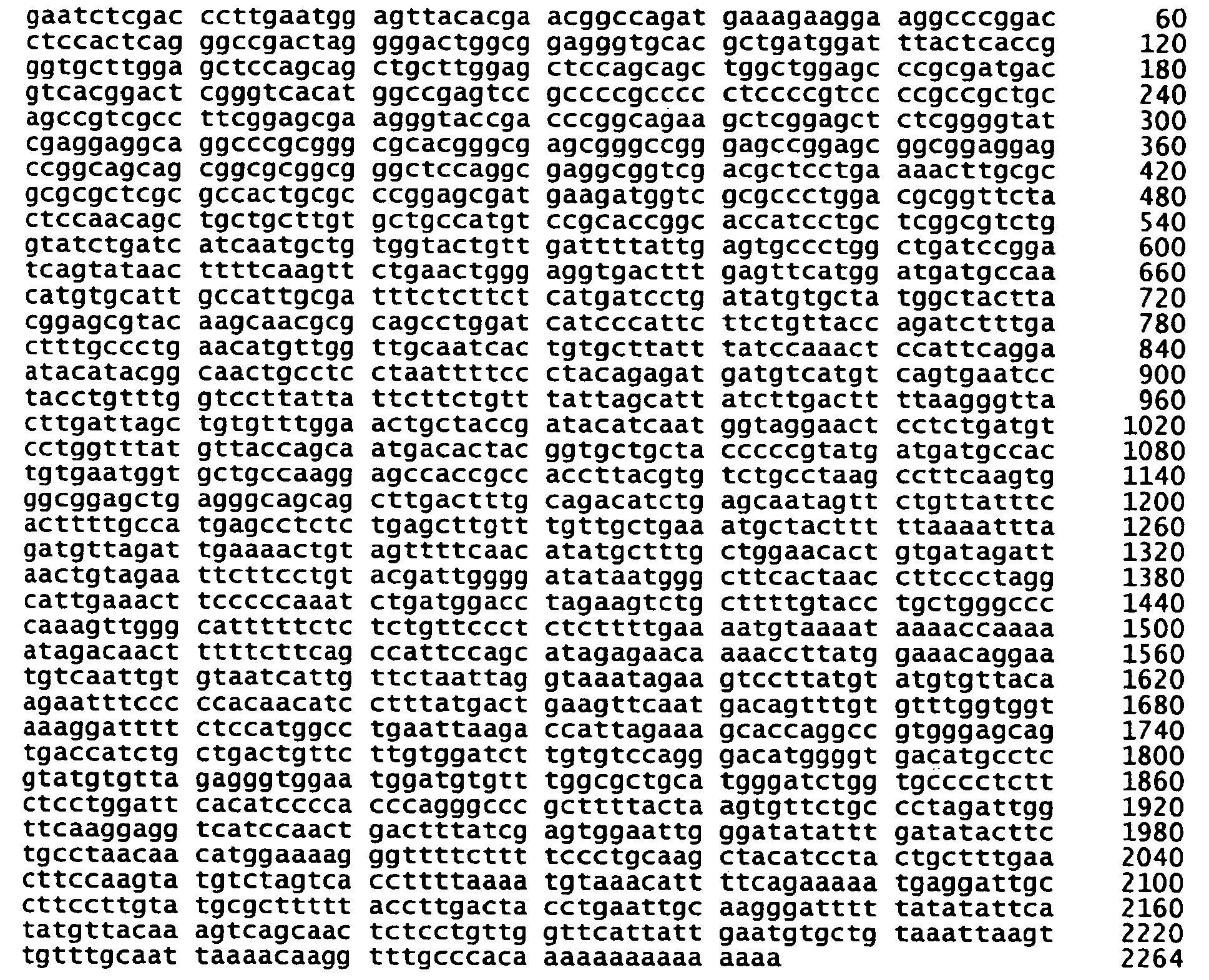
- <210> 7
<211> 370
<212> PRT
<213> Homo sapiens - <400> 7


- <210> 8
<211> 1341
<212> DNA
<213> Homo sapiens - <400> 8

Claims (3)
- Human cancer related nucleic acid consisting of(i) a nucleic acid sequence of SEQ ID NO: 6, or(ii) a nucleic acid that encodes SEQ ID NO: 7.
- A polypeptide according to SEQ ID NO: 7.
- Use of a human cancer related nucleic acid comprising(i) a nucleic acid sequence of SEQ ID NO: 6,(ii) a nucleic acid that encodes SEQ ID NO: 7,(iii) a nucleic acid sequence gcttggagctccagcagct, or(iv) a nucleic acid sequence gcttggagctccagcagctgcttggagctccagcagct; or a polypeptide according to SEQ ID NO:7,for the preparation of a diagnostic for identifying patients susceptible to hepatocellular carcinoma.
Priority Applications (1)
| Application Number | Priority Date | Filing Date | Title |
|---|---|---|---|
| EP10153116.8A EP2230306B1 (en) | 2002-12-24 | 2003-12-24 | Human cancer-relating genes, its encoded products and applications |
Applications Claiming Priority (5)
| Application Number | Priority Date | Filing Date | Title |
|---|---|---|---|
| CN 02158110 CN1239700C (en) | 2002-12-24 | 2002-12-24 | Human liver cancer related gene, encoded product and use thereof |
| CN02158110 | 2002-12-24 | ||
| CN03109786 | 2003-04-21 | ||
| CNB031097863A CN1328377C (en) | 2003-04-21 | 2003-04-21 | An allelomorphic gene of gene LAPTM 4B relevant to liver cancer of human, coded result and application |
| PCT/CN2003/001109 WO2004058971A1 (en) | 2002-12-24 | 2003-12-24 | Human cancer-relating genes, the products encoded thereby and applications thereof |
Related Child Applications (2)
| Application Number | Title | Priority Date | Filing Date |
|---|---|---|---|
| EP10153116.8A Division EP2230306B1 (en) | 2002-12-24 | 2003-12-24 | Human cancer-relating genes, its encoded products and applications |
| EP10153116.8 Division-Into | 2010-02-09 |
Publications (4)
| Publication Number | Publication Date |
|---|---|
| EP1577390A1 EP1577390A1 (en) | 2005-09-21 |
| EP1577390A4 EP1577390A4 (en) | 2006-05-10 |
| EP1577390B1 true EP1577390B1 (en) | 2011-02-09 |
| EP1577390B9 EP1577390B9 (en) | 2011-03-30 |
Family
ID=32683267
Family Applications (2)
| Application Number | Title | Priority Date | Filing Date |
|---|---|---|---|
| EP03782072A Expired - Lifetime EP1577390B9 (en) | 2002-12-24 | 2003-12-24 | Human cancer-related genes, the products encoded thereby and applications thereof |
| EP10153116.8A Expired - Lifetime EP2230306B1 (en) | 2002-12-24 | 2003-12-24 | Human cancer-relating genes, its encoded products and applications |
Family Applications After (1)
| Application Number | Title | Priority Date | Filing Date |
|---|---|---|---|
| EP10153116.8A Expired - Lifetime EP2230306B1 (en) | 2002-12-24 | 2003-12-24 | Human cancer-relating genes, its encoded products and applications |
Country Status (7)
| Country | Link |
|---|---|
| US (3) | US7910711B2 (en) |
| EP (2) | EP1577390B9 (en) |
| JP (1) | JP4617257B2 (en) |
| AT (1) | ATE498012T1 (en) |
| AU (1) | AU2003292867A1 (en) |
| DE (1) | DE60336015D1 (en) |
| WO (1) | WO2004058971A1 (en) |
Families Citing this family (3)
| Publication number | Priority date | Publication date | Assignee | Title |
|---|---|---|---|---|
| ATE498012T1 (en) | 2002-12-24 | 2011-02-15 | Univ Beijing | HUMAN GENES RELATED TO CANCER, PRODUCTS ENCODED THEREOF, AND APPLICATIONS THEREOF |
| US8152628B2 (en) * | 2005-08-01 | 2012-04-10 | Igt | Methods and devices for authentication and licensing in a gaming network |
| US8679598B2 (en) | 2010-10-08 | 2014-03-25 | Guardian Industries Corp. | Vacuum insulated glass (VIG) unit including nano-composite pillars, and/or methods of making the same |
Citations (1)
| Publication number | Priority date | Publication date | Assignee | Title |
|---|---|---|---|---|
| WO2002071928A2 (en) * | 2001-03-14 | 2002-09-19 | Millennium Pharmaceuticals, Inc. | Nucleic acid molecules and proteins for the identification, assessment, prevention, and therapy of ovarian cancer |
Family Cites Families (12)
| Publication number | Priority date | Publication date | Assignee | Title |
|---|---|---|---|---|
| JP3091870B2 (en) * | 1989-12-19 | 2000-09-25 | アメリカ合衆国 | Hepatocyte cancer oncogenes |
| US6878806B2 (en) * | 1997-03-14 | 2005-04-12 | Human Genome Sciences, Inc. | Human secreted protein HTEEB42 |
| AU4799499A (en) | 1998-07-24 | 2000-02-14 | Protegene Inc. | Human proteins having hydrophobic domains and dnas encoding these proteins |
| AU4021700A (en) * | 1999-03-22 | 2000-10-09 | Board Of Regents, The University Of Texas System | Methods of use of beta1-integrin inhibitors |
| CA2382015A1 (en) | 1999-08-17 | 2001-02-22 | Incyte Genomics, Inc. | Membrane associated proteins |
| EP1242443A4 (en) * | 1999-12-23 | 2005-06-22 | Nuvelo Inc | NEW NUCLEIC ACIDS AND POLYPEPTIDES |
| US6465620B1 (en) * | 2000-01-21 | 2002-10-15 | Hyseq, Inc. | Methods and materials relating to novel von Willebrand/Thrombospondin-like polypeptides and polynucleotides |
| AU2002246502A1 (en) | 2000-11-17 | 2002-08-06 | Hyseq, Inc. | Nucleic acids and polypeptides |
| US20050259483A1 (en) * | 2002-09-30 | 2005-11-24 | Oncotherapy Science, Inc. | Genes and polypeptides relating to prostate cancers |
| US20060188889A1 (en) * | 2003-11-04 | 2006-08-24 | Christopher Burgess | Use of differentially expressed nucleic acid sequences as biomarkers for cancer |
| ATE498012T1 (en) | 2002-12-24 | 2011-02-15 | Univ Beijing | HUMAN GENES RELATED TO CANCER, PRODUCTS ENCODED THEREOF, AND APPLICATIONS THEREOF |
| EP1677733A4 (en) * | 2003-10-03 | 2007-09-19 | Bayer Pharmaceuticals Corp | Gene expression profiles and methods of use |
-
2003
- 2003-12-24 AT AT03782072T patent/ATE498012T1/en not_active IP Right Cessation
- 2003-12-24 EP EP03782072A patent/EP1577390B9/en not_active Expired - Lifetime
- 2003-12-24 US US10/540,539 patent/US7910711B2/en not_active Expired - Fee Related
- 2003-12-24 WO PCT/CN2003/001109 patent/WO2004058971A1/en not_active Ceased
- 2003-12-24 JP JP2005509681A patent/JP4617257B2/en not_active Expired - Fee Related
- 2003-12-24 DE DE60336015T patent/DE60336015D1/en not_active Expired - Lifetime
- 2003-12-24 EP EP10153116.8A patent/EP2230306B1/en not_active Expired - Lifetime
- 2003-12-24 AU AU2003292867A patent/AU2003292867A1/en not_active Abandoned
-
2011
- 2011-02-01 US US13/019,297 patent/US20120148589A1/en not_active Abandoned
-
2016
- 2016-05-04 US US15/146,307 patent/US9470690B2/en not_active Expired - Fee Related
Patent Citations (1)
| Publication number | Priority date | Publication date | Assignee | Title |
|---|---|---|---|---|
| WO2002071928A2 (en) * | 2001-03-14 | 2002-09-19 | Millennium Pharmaceuticals, Inc. | Nucleic acid molecules and proteins for the identification, assessment, prevention, and therapy of ovarian cancer |
Non-Patent Citations (2)
| Title |
|---|
| DATABASE EMBL [online] 1 November 2002 (2002-11-01), STRAUSBERG R.: "Homo sapiens, Similar to putative integral membrane transporter, clone IMAGE:5264567, mRNA.", Database accession no. BC038117 * |
| LIU J ET AL.: "Biological function of a novel gene overexpressed in human hepatocellular carcinoma", CHINESE MEDICAL JOURNAL (ENGLISH EDITION), vol. 113, no. 10, 2000, pages 881 - 885, XP009008152 * |
Also Published As
| Publication number | Publication date |
|---|---|
| EP1577390A4 (en) | 2006-05-10 |
| EP2230306B1 (en) | 2016-05-04 |
| WO2004058971A1 (en) | 2004-07-15 |
| AU2003292867A1 (en) | 2004-07-22 |
| US20120148589A1 (en) | 2012-06-14 |
| US9470690B2 (en) | 2016-10-18 |
| EP1577390A1 (en) | 2005-09-21 |
| DE60336015D1 (en) | 2011-03-24 |
| EP1577390B9 (en) | 2011-03-30 |
| US7910711B2 (en) | 2011-03-22 |
| US20070026395A1 (en) | 2007-02-01 |
| JP4617257B2 (en) | 2011-01-19 |
| EP2230306A1 (en) | 2010-09-22 |
| JP2006522587A (en) | 2006-10-05 |
| US20160238608A1 (en) | 2016-08-18 |
| ATE498012T1 (en) | 2011-02-15 |
Similar Documents
| Publication | Publication Date | Title |
|---|---|---|
| Scanlan et al. | Antigens recognized by autologous antibody in patients with renal‐cell carcinoma | |
| Wissenbach et al. | Expression of CaT-like, a novel calcium-selective channel, correlates with the malignancy of prostate cancer | |
| EP0507852A1 (en) | Gene deleted in colorectal cancer of humans | |
| CN104619840A (en) | Fgfr2 fusion gene | |
| EP0822993B1 (en) | Diagnostic methods and gene therapy using reagents derived from the human metastasis suppressor gene kai1 | |
| JP2002506971A (en) | C-MYC coding region determinant binding protein (CRD-BP) and nucleic acid sequence thereof | |
| WO1999028459A1 (en) | Examination method, examination reagent and remedy for diseases caused by variation in lkb1 gene | |
| US6312890B1 (en) | Partial intron sequence of von hippel-lindau (VHL) disease gene and its use in diagnosis of disease | |
| US9470690B2 (en) | Human cancer-related gene, its encoded products and applications | |
| Li et al. | Differential display identifies overexpression of the USP36 gene, encoding a deubiquitinating enzyme, in ovarian cancer | |
| Odze et al. | Advanced gastroduodenal polyposis with ras mutations in a patient with familial adenomatous polyposis | |
| Salvi et al. | Germline and somatic c-met mutations in multifocal/bilateral and sporadic papillary renal carcinomas of selected patients | |
| US7364868B2 (en) | Krüppel-like transcriptional factor KLF4/GKLF and uses thereof | |
| US7776519B2 (en) | Use of the slug gene, or of the transcription or expression products thereof in the detection and/or treatment of cancerous cells | |
| WO2005075681A1 (en) | Method of examining sensitivity of cancer cell to anticancer agent | |
| WO2011091716A9 (en) | Epidermal growth factor receptor variant | |
| TW201204393A (en) | Diagnostic agent and therapeutic agent of cancer | |
| CN116622841B (en) | A molecular marker for lung cancer diagnosis, chemotherapy, or prognosis and its application | |
| Finch et al. | Cloning of the human uroplakin 1B cDNA and analysis of its expression in urothelial‐tumor cell lines and bladder‐carcinoma tissue | |
| Shen | Role of human intermediate form PRL receptor I-tail in the Pathogenesis of Breast Cancer | |
| US7888007B2 (en) | Cancer associated plexin B1 mutations | |
| Ahn | Role of receptor tyrosine kinase like orphan receptor 2 in treatment-resistant prostate cancer | |
| AU760490B2 (en) | Diagnostic methods and gene therapy using reagents derived from the human metastasis suppressor gene KAI1 | |
| JP2010051172A (en) | Marker for diagnosis of cancer and target molecule oslc1 for therapy | |
| Shayesteh | Defining a region of increased copy number of 3q26 in ovarian cancer |
Legal Events
| Date | Code | Title | Description |
|---|---|---|---|
| PUAI | Public reference made under article 153(3) epc to a published international application that has entered the european phase |
Free format text: ORIGINAL CODE: 0009012 |
|
| 17P | Request for examination filed |
Effective date: 20050624 |
|
| AK | Designated contracting states |
Kind code of ref document: A1 Designated state(s): AT BE BG CH CY CZ DE DK EE ES FI FR GB GR HU IE IT LI LU MC NL PT RO SE SI SK TR |
|
| AX | Request for extension of the european patent |
Extension state: AL LT LV MK |
|
| DAX | Request for extension of the european patent (deleted) | ||
| A4 | Supplementary search report drawn up and despatched |
Effective date: 20060328 |
|
| 17Q | First examination report despatched |
Effective date: 20061212 |
|
| R17C | First examination report despatched (corrected) |
Effective date: 20061212 |
|
| RIC1 | Information provided on ipc code assigned before grant |
Ipc: C12Q 1/68 20060101ALI20100602BHEP Ipc: C12N 15/12 20060101AFI20100602BHEP Ipc: G01N 33/68 20060101ALI20100602BHEP Ipc: G01N 33/574 20060101ALI20100602BHEP Ipc: C07K 14/435 20060101ALI20100602BHEP |
|
| RTI1 | Title (correction) |
Free format text: HUMAN CANCER-RELATED GENES, THE PRODUCTS ENCODED THEREBY AND APPLICATIONS THEREOF |
|
| GRAP | Despatch of communication of intention to grant a patent |
Free format text: ORIGINAL CODE: EPIDOSNIGR1 |
|
| GRAS | Grant fee paid |
Free format text: ORIGINAL CODE: EPIDOSNIGR3 |
|
| GRAA | (expected) grant |
Free format text: ORIGINAL CODE: 0009210 |
|
| AK | Designated contracting states |
Kind code of ref document: B1 Designated state(s): AT BE BG CH CY CZ DE DK EE ES FI FR GB GR HU IE IT LI LU MC NL PT RO SE SI SK TR |
|
| REG | Reference to a national code |
Ref country code: GB Ref legal event code: FG4D |
|
| REG | Reference to a national code |
Ref country code: CH Ref legal event code: EP |
|
| REG | Reference to a national code |
Ref country code: IE Ref legal event code: FG4D |
|
| RAP2 | Party data changed (patent owner data changed or rights of a patent transferred) |
Owner name: PEKING UNIVERSITY |
|
| RIN2 | Information on inventor provided after grant (corrected) |
Inventor name: LIN, MING,PEKING UNIVERSITY, DEPT. CELL BIOLOGY Inventor name: LIU, XINRONG,PEKING UNIVERSITY, DEPT. CELL BIOLOGY Inventor name: ZHOU, ROULI,PEKING UNIVERSITY, DEPT. CELL BIOLOGY Inventor name: ZHANG, QINGYUN,PEKING UNIVERSITY Inventor name: SHAO, GENZE,PEKING UNIVERSITY, DEPT. CELL BIOLOGY Inventor name: ZHANG, SHA,PEKING UNIVERSITY, DEPT. CELL BIOLOGY Inventor name: ZHANG, YE,PEKING UNIVERSITY, DEPT. CELL BIOLOGY Inventor name: RUI, JINGAN,PEKING UNIVERSITY Inventor name: JIN, YUEYING,PEKING UNIVERSITY, DEPT. CELL BIOLOGY |
|
| RIN2 | Information on inventor provided after grant (corrected) |
Inventor name: RUI, JINGAN Inventor name: LIU, XINRONG Inventor name: SHAO, GENZE Inventor name: ZHANG, QINGYUN Inventor name: ZHANG, YE Inventor name: ZHANG, SHA Inventor name: LIN, MING Inventor name: JIN, YUEYING Inventor name: ZHOU, ROULI |
|
| REF | Corresponds to: |
Ref document number: 60336015 Country of ref document: DE Date of ref document: 20110324 Kind code of ref document: P |
|
| REG | Reference to a national code |
Ref country code: DE Ref legal event code: R096 Ref document number: 60336015 Country of ref document: DE Effective date: 20110324 |
|
| REG | Reference to a national code |
Ref country code: NL Ref legal event code: VDEP Effective date: 20110209 |
|
| PG25 | Lapsed in a contracting state [announced via postgrant information from national office to epo] |
Ref country code: ES Free format text: LAPSE BECAUSE OF FAILURE TO SUBMIT A TRANSLATION OF THE DESCRIPTION OR TO PAY THE FEE WITHIN THE PRESCRIBED TIME-LIMIT Effective date: 20110520 Ref country code: GR Free format text: LAPSE BECAUSE OF FAILURE TO SUBMIT A TRANSLATION OF THE DESCRIPTION OR TO PAY THE FEE WITHIN THE PRESCRIBED TIME-LIMIT Effective date: 20110510 Ref country code: PT Free format text: LAPSE BECAUSE OF FAILURE TO SUBMIT A TRANSLATION OF THE DESCRIPTION OR TO PAY THE FEE WITHIN THE PRESCRIBED TIME-LIMIT Effective date: 20110609 Ref country code: SE Free format text: LAPSE BECAUSE OF FAILURE TO SUBMIT A TRANSLATION OF THE DESCRIPTION OR TO PAY THE FEE WITHIN THE PRESCRIBED TIME-LIMIT Effective date: 20110209 |
|
| PG25 | Lapsed in a contracting state [announced via postgrant information from national office to epo] |
Ref country code: CY Free format text: LAPSE BECAUSE OF FAILURE TO SUBMIT A TRANSLATION OF THE DESCRIPTION OR TO PAY THE FEE WITHIN THE PRESCRIBED TIME-LIMIT Effective date: 20110209 Ref country code: NL Free format text: LAPSE BECAUSE OF FAILURE TO SUBMIT A TRANSLATION OF THE DESCRIPTION OR TO PAY THE FEE WITHIN THE PRESCRIBED TIME-LIMIT Effective date: 20110209 Ref country code: BE Free format text: LAPSE BECAUSE OF FAILURE TO SUBMIT A TRANSLATION OF THE DESCRIPTION OR TO PAY THE FEE WITHIN THE PRESCRIBED TIME-LIMIT Effective date: 20110209 Ref country code: SI Free format text: LAPSE BECAUSE OF FAILURE TO SUBMIT A TRANSLATION OF THE DESCRIPTION OR TO PAY THE FEE WITHIN THE PRESCRIBED TIME-LIMIT Effective date: 20110209 Ref country code: AT Free format text: LAPSE BECAUSE OF FAILURE TO SUBMIT A TRANSLATION OF THE DESCRIPTION OR TO PAY THE FEE WITHIN THE PRESCRIBED TIME-LIMIT Effective date: 20110209 Ref country code: BG Free format text: LAPSE BECAUSE OF FAILURE TO SUBMIT A TRANSLATION OF THE DESCRIPTION OR TO PAY THE FEE WITHIN THE PRESCRIBED TIME-LIMIT Effective date: 20110509 Ref country code: FI Free format text: LAPSE BECAUSE OF FAILURE TO SUBMIT A TRANSLATION OF THE DESCRIPTION OR TO PAY THE FEE WITHIN THE PRESCRIBED TIME-LIMIT Effective date: 20110209 |
|
| PG25 | Lapsed in a contracting state [announced via postgrant information from national office to epo] |
Ref country code: DK Free format text: LAPSE BECAUSE OF FAILURE TO SUBMIT A TRANSLATION OF THE DESCRIPTION OR TO PAY THE FEE WITHIN THE PRESCRIBED TIME-LIMIT Effective date: 20110209 Ref country code: EE Free format text: LAPSE BECAUSE OF FAILURE TO SUBMIT A TRANSLATION OF THE DESCRIPTION OR TO PAY THE FEE WITHIN THE PRESCRIBED TIME-LIMIT Effective date: 20110209 |
|
| PG25 | Lapsed in a contracting state [announced via postgrant information from national office to epo] |
Ref country code: RO Free format text: LAPSE BECAUSE OF FAILURE TO SUBMIT A TRANSLATION OF THE DESCRIPTION OR TO PAY THE FEE WITHIN THE PRESCRIBED TIME-LIMIT Effective date: 20110209 Ref country code: SK Free format text: LAPSE BECAUSE OF FAILURE TO SUBMIT A TRANSLATION OF THE DESCRIPTION OR TO PAY THE FEE WITHIN THE PRESCRIBED TIME-LIMIT Effective date: 20110209 Ref country code: CZ Free format text: LAPSE BECAUSE OF FAILURE TO SUBMIT A TRANSLATION OF THE DESCRIPTION OR TO PAY THE FEE WITHIN THE PRESCRIBED TIME-LIMIT Effective date: 20110209 |
|
| PLBE | No opposition filed within time limit |
Free format text: ORIGINAL CODE: 0009261 |
|
| STAA | Information on the status of an ep patent application or granted ep patent |
Free format text: STATUS: NO OPPOSITION FILED WITHIN TIME LIMIT |
|
| 26N | No opposition filed |
Effective date: 20111110 |
|
| REG | Reference to a national code |
Ref country code: DE Ref legal event code: R097 Ref document number: 60336015 Country of ref document: DE Effective date: 20111110 |
|
| PG25 | Lapsed in a contracting state [announced via postgrant information from national office to epo] |
Ref country code: IT Free format text: LAPSE BECAUSE OF FAILURE TO SUBMIT A TRANSLATION OF THE DESCRIPTION OR TO PAY THE FEE WITHIN THE PRESCRIBED TIME-LIMIT Effective date: 20110209 |
|
| PG25 | Lapsed in a contracting state [announced via postgrant information from national office to epo] |
Ref country code: MC Free format text: LAPSE BECAUSE OF NON-PAYMENT OF DUE FEES Effective date: 20111231 |
|
| REG | Reference to a national code |
Ref country code: CH Ref legal event code: PL |
|
| REG | Reference to a national code |
Ref country code: FR Ref legal event code: ST Effective date: 20120831 |
|
| REG | Reference to a national code |
Ref country code: IE Ref legal event code: MM4A |
|
| PG25 | Lapsed in a contracting state [announced via postgrant information from national office to epo] |
Ref country code: CH Free format text: LAPSE BECAUSE OF NON-PAYMENT OF DUE FEES Effective date: 20111231 Ref country code: LI Free format text: LAPSE BECAUSE OF NON-PAYMENT OF DUE FEES Effective date: 20111231 Ref country code: IE Free format text: LAPSE BECAUSE OF NON-PAYMENT OF DUE FEES Effective date: 20111224 |
|
| PG25 | Lapsed in a contracting state [announced via postgrant information from national office to epo] |
Ref country code: FR Free format text: LAPSE BECAUSE OF NON-PAYMENT OF DUE FEES Effective date: 20120102 |
|
| PG25 | Lapsed in a contracting state [announced via postgrant information from national office to epo] |
Ref country code: LU Free format text: LAPSE BECAUSE OF NON-PAYMENT OF DUE FEES Effective date: 20111224 |
|
| PG25 | Lapsed in a contracting state [announced via postgrant information from national office to epo] |
Ref country code: TR Free format text: LAPSE BECAUSE OF FAILURE TO SUBMIT A TRANSLATION OF THE DESCRIPTION OR TO PAY THE FEE WITHIN THE PRESCRIBED TIME-LIMIT Effective date: 20110209 |
|
| PG25 | Lapsed in a contracting state [announced via postgrant information from national office to epo] |
Ref country code: HU Free format text: LAPSE BECAUSE OF FAILURE TO SUBMIT A TRANSLATION OF THE DESCRIPTION OR TO PAY THE FEE WITHIN THE PRESCRIBED TIME-LIMIT Effective date: 20110209 |
|
| PGFP | Annual fee paid to national office [announced via postgrant information from national office to epo] |
Ref country code: GB Payment date: 20171221 Year of fee payment: 15 |
|
| PGFP | Annual fee paid to national office [announced via postgrant information from national office to epo] |
Ref country code: DE Payment date: 20171220 Year of fee payment: 15 |
|
| REG | Reference to a national code |
Ref country code: DE Ref legal event code: R119 Ref document number: 60336015 Country of ref document: DE |
|
| GBPC | Gb: european patent ceased through non-payment of renewal fee |
Effective date: 20181224 |
|
| PG25 | Lapsed in a contracting state [announced via postgrant information from national office to epo] |
Ref country code: DE Free format text: LAPSE BECAUSE OF NON-PAYMENT OF DUE FEES Effective date: 20190702 |
|
| PG25 | Lapsed in a contracting state [announced via postgrant information from national office to epo] |
Ref country code: GB Free format text: LAPSE BECAUSE OF NON-PAYMENT OF DUE FEES Effective date: 20181224 |